Healthcare analytics, AI solutions for biological big data, providing an AI platform for the biotech, life sciences, medical and pharmaceutical industries, as well as for related technological approaches, i.e., curation and text analysis with machine learning and other activities related to AI applications to these industries.
In 1956, Francis Crick—co-discoverer of DNA’s helical structure—postulated what is now considered to be a central doctrine of the biological sciences stating that “The central dogma of molecular biology deals with the detailed residue-by-residue transfer of sequential information. It states that such information cannot be transferred back from protein to either protein or nucleic acid.” What Crick was suggesting was that DNA makes RNA and, in turn, RNA makes protein.
In the time since the initial proposal of the central dogma, scientists have come to understand that there are not only instances of reverse information flow from RNA to DNA, but chemical alterations to RNA structures that can have a profound effect on gene regulation. The discovery of these alterations has added a critical dimension to how scientists view the genetic code and recently spawned an entirely new field of study within molecular biology: the epitranscriptome.
Now, a recent study by scientists at the University of Chicago and Tel Aviv University has revealed evidence that provides a promising new lever in the control of gene expression. The researchers describe a small chemical modification to RNA that can significantly boost the conversion of genes to proteins.
The findings from this study were published recently in Nature through an article entitled “The dynamic N1-methyladenosine methylome in eukaryotic messenger RNA.”
“Epigenetics, the regulation of gene expression beyond the primary information encoded by DNA, was thought until recently to be mediated by modifications of proteins and DNA,” explained co-senior study author Gidi Rechavi, Ph.D., chair in oncology at Tel Aviv University’s Sackler Faculty of Medicine and head of the Cancer Research Center at Sheba Medical Center. “The new findings bring RNA to a central position in epigenetics.”
“This discovery further opens the window on a whole new world of biology for us to explore,” added co-senior study author Chuan He, Ph.D., professor in the department of chemistry and investigator within the Howard Hughes Medical Institute at the University of Chicago. “These modifications have a major impact on almost every biological process.”
Previously, Dr. He’s laboratory discovered the first RNA demethylase that reverses the most prevalent mRNA methylation N6-methyladenosine (m6A), implying that the addition and removal of the methyl group could dramatically affect these messengers and the outcome of gene expression—as also seen for DNA and histones—which subsequent research found to be true.
In the current study, the investigators described a second functional mRNA methylation, N1-methyladenosine (m1A). Like m6A, the small modification is evolutionarily conserved and common, present in humans, rodents, and yeast. However, its location and effect on gene expression reflect a new form of epitranscriptome control.
“The discovery of m1A is extremely important, not only because of its own potential in affecting biological processes but also because it validates the hypothesis that there is not just one functional modification,” Dr. He stated. “There could be multiple modifications at different sites where each may carry a distinct message to control the fate and function of mRNA.”
From their findings, the research team estimates that that m1A may be present on transcripts of more than one out of three expressed human genes—suggesting that m1A, like m6A, may be a mechanism by which cells rapidly boost the expression of hundreds or thousands of specific genes.
“mRNA is the perfect place to regulate gene expression because they can code information from transcription and directly impact translation—you can add a consensus sequence to a group of genes and use a modification of the sequence to readily control several hundred transcripts simultaneously,” Dr. He said. “If you want to rapidly change the expression of several hundred or a thousand genes, this offers the best way.”
The scientists were excited by their findings and have plans for future studies that will examine the role of m1A methylation in human development, for diseases such as diabetes and cancer, and its potential as a target for therapeutic uses.
“This study represents a breakthrough discovery in the exciting, nascent field of the ‘epitranscriptome,’ which is how RNAs are regulated, akin to the genome and the epigenome,” commented Christopher Mason, Ph.D., associate professor at Weill Cornell Medicine, who was not affiliated with the study. “What is important about this work is that m6A was recently found to enrich at the ends of genes, and now we know that m1A is what is helping regulate the beginning of genes, and this opens up many questions about revealing the ‘epitranscriptome code’ just like the histone code or the genetic code.”
The dynamic N1-methyladenosine methylome in eukaryotic messenger RNA
Gene expression can be regulated post-transcriptionally through dynamic and reversible RNA modifications. A recent noteworthy example is N6-methyladenosine (m6A), which affects messenger RNA (mRNA) localization, stability, translation and splicing. Here we report on a new mRNA modification, N1-methyladenosine (m1A), that occurs on thousands of different gene transcripts in eukaryotic cells, from yeast to mammals, at an estimated average transcript stoichiometry of 20% in humans. Employing newly developed sequencing approaches, we show that m1A is enriched around the start codon upstream of the first splice site: it preferentially decorates more structured regions around canonical and alternative translation initiation sites, is dynamic in response to physiological conditions, and correlates positively with protein production. These unique features are highly conserved in mouse and human cells, strongly indicating a functional role for m1A in promoting translation of methylated mRNA.
Figure 1: Development of m1A-seq to map a newly identified constituent of mammalian mRNA.
a, Chemical structures of m1A and m6A. Methyl groups (-CH3) are in red and the positive charge (+) on m1A is in blue. b, LC-MS/MS quantitation of m1A, m6A and Ψ in human and mouse mRNA isolated from the indicated cell types. …
Figure 3: m1A occurs in GC-rich sequence contexts and in genes with structured 5′ UTRs.
a, Sequence frequency logo for a set of 192 adenosines in peak areas that have a higher mismatch rate in immunoprecipitation relative to input (FC ≥ 6) in HepG2 demonstrates the GC-rich context of m1A. b, Length-adjusted minimum free energy…
Figure 5: m1A in mRNA is a dynamic modification that responds to changing physiological and stress conditions, and varies between tissues.
a, LC-MS/MS quantification of m1A (left, grey) and m6A (right, black) in mRNA of untreated and glucose-starved (upper panels) or heat shock-treated (lower panels) HepG2 cells, presented as percentage of unmodified A. Mean values ± s.e.m…
RNA modification discovery suggests new code for control of gene expression
A new cellular signal discovered by a team of scientists at the University of Chicago and Tel Aviv University provides a promising new lever in the control of gene expression. Gene expression study
The study, published online Feb. 10 in the journal Nature, describes a small chemical modification that can significantly boost the conversion of genes to proteins. Together with other recent findings, the discovery enriches a critical new dimension to the “Central Dogma” of molecular biology: the epitranscriptome.
“This discovery further opens the window on a whole new world of biology for us to explore,” said Chuan He, the John T. Wilson Distinguished Service Professor in Chemistry, Howard Hughes Medical Institute investigator and senior author of the study. “These modifications have a major impact on almost every biological process.”
The central dogma of molecular biology describes the cellular pathway where genetic information from DNA is copied into temporary RNA “transcripts,” which provide the recipe for the production of proteins. Since Francis Crick first postulated the theory in 1956, scientists have discovered a multitude of modifications to DNA and proteins that regulate this process.
Only recently, however, have scientists focused on investigating dynamic modifications that specifically target the RNA step. In 2011, He’s group discovered the first RNA demethylase that reverses the most prevalent mRNA methylation N6-methyladenosine, or m6A, implying that the addition and removal of the methyl group could dramatically affect these messengers and impact the outcome of gene expression, as also seen for DNA and histones. Subsequently, scientists discovered that the dynamic and reversible methylation of m6A dramatically controlled the metabolism and function of most cellular messenger RNA, and thus, the production of proteins.
In the new Nature study, researchers from UChicago and Tel Aviv University describe a second functional mRNA methylation, N1-methyladenosine, or m1A. Like m6A, the small modification is evolutionarily conserved and common, and present in humans, rodents and yeast, the authors found. But its location and effect on gene expression reflect a new form of epitranscriptome control and suggest an even larger cellular “control panel.”
“The discovery of m1A is extremely important, not only because of its own potential in affecting biological processes, but also because it validates the hypothesis that there is not just one functional modification,” He said. “There could be multiple modifications at different sites where each may carry a distinct message to control the fate and function of mRNA.”
The researchers estimated that m1A was present on transcripts of more than one out of three expressed human genes. Methylated genes exhibited enhanced translation compared to unmethlyated genes, producing protein levels nearly twice as high in all cell types. This increase suggests that m1A, like m6A, may be a mechanism by which cells rapidly boost the expression of hundreds or thousands of specific genes, perhaps during important processes such as cell division, differentiation or under stress.
“mRNA is the perfect place to regulate gene expression, because they can code information from transcription and directly impact translation; you can add a consensus sequence to a group of genes and use a modification of the sequence to readily control several hundred transcripts simultaneously,” He said. “If you want to rapidly change the expression of several hundred or a thousand genes, this offers the best way.”
However, despite their complementary effects, m1A and m6A exert their influence on mRNA through different pathways. While studies have found that m6A localizes predominantly to the tail of messenger RNA molecules, increasing their translation and turnover rate, m1A was found largely near the start codon of mRNA transcripts, where protein translation begins. The different mechanisms could allow for finer tuning of post-transcriptional gene expression, or the selective activation of particular genes in different physiological situations.
“This study represents a breakthrough discovery in the exciting, nascent field of the ‘epitranscriptome,’ which is how RNAs are regulated, akin to the genome and the epigenome,” said Christopher Mason, associate professor at Weill Cornell Medicine, who was not affiliated with the study. “What is important about this work is that m6A was recently found to enrich at the ends of genes, and now we know that m1A is what is helping regulate the beginning of genes, and this opens up many questions about revealing the ‘epitranscriptome code’ just like the histone code or the genetic code.”
Future studies will examine the role of m1A methylation in human development, diseases such as diabetes and cancer, and its potential as a target for therapeutic uses.
Citation: “The dynamic N1-methyladenosine methylome in eukaryotic messenger RNA,” Nature, Feb. 10, 2016, by Chuan He, Dan Dominissini, Sigrid Nachtergaele, Qing Dai, Dali Han, Wesley Clark, Guanqun Zheng, Tao Pan and Louis Dore from the University of Chicago, and Sharon Moshitch-Moshkovitz, Eyal Peer, Nitkan Kol, Moshe Shay Ben-Haim, Ayelet Di Segni, Mali Salmon-Divon, Oz Solomon, Eran Eyal, Vera Hershkovitz, Ninette Amariglio and Gideon Rechavi from Tel Aviv University. DOI: 10.1038/nature16998
Funding: National Institutes of Health, Howard Hughes Medical Institute, Flight Attendant Medical Research Institute, Israel Science Foundation, Israeli Centers of Excellence Program, Ernest and Bonnie Beutler Research Program, Chicago Biomedical Consortium, Damon Runyon Cancer Research Foundation and Kahn Family Foundation.
The meeting is aimed at bringing in students and postdocs as well as faculty involved in RNA modification and epitranscriptome research. In addition to talks, there will be a poster session and reception.
Pathology reports provide care teams with crucial diagnostic and prognostic data. For population-level research and quality assurance initiatives, it’s important that reports are consistently complete. But report consistency can also be important for the medical outcomes of a single person.
Today, I’ll discuss the unique experience of a friend of mine, and I’ll explain how his isolated incident is connected to large-scale initiatives in pathology reporting quality assurance.
Incomplete Pathology Reports
A friend of mine has particularly fair skin and is at thus at increased risk for skin cancers. His family physician has recommended he have various moles removed to prevent the development of possible skin cancers.
Over time, my friend and his family physician became troubled by the inconsistencies they found in comparing the pathology reports from his five different biopsies.
Five out of five pathology reports identified that the moles were dysplastic—irregular and potentially cancerous. Yet only three reports identified that this dysplasia was in his skin’s basal cells as opposed to squamous cells or melanocytes.
The the type of skin cells exhibiting dysplasia is important to clinical decision making: dysplasia affecting melanocytes is the basis for melanomas, the most serious forms of skin cancer. Lacking this information makes it more difficult for care teams to develop the comprehensive understanding of an individual’s medical history that guides clinical decision making.
In response to the incompleteness of the pathology reports, the family physician remarked, “I rarely have to query a pathologist about what’s in a report. It’s usually about what’s left out.”
And it’s not just one physician who’s seeking better reports.
Synoptic Reporting for Complete Consistency in Pathology
For over 20 years, the College of American Pathologists (CAP) has been developing protocols and templates to standardize pathology reporting, substantially improving the completeness and consistency of pathology reports through the use of the synoptic—i.e. structured—format.
Synoptic reporting uses coded data templates to produce standardized reports that are more complete and consistent than reports generated using narrative methods.
Cancer Care Ontario introduced synoptic reporting for pathologist across the province—the largest jurisdiction to do so. Clinicians have shown over-whelming support for this initiative, finding that synoptic reports have facilitated consistent intepretation of diagnostic and prognostic data.
The Canadian Partnership Against Cancer is building on Ontario’s success to help implement synoptic pathology reporting across Canada, strengthening provincial cancer registries and fostering cross-jurisdictional research and quality assurance initiatives.
Clinical research has continually found that the use of synoptic reporting improves the completeness of pathology reports [1, 2, 3, 4]. These conclusions have been upheld in research on synoptic reporting for surgery as well [5, 6].
An important goal of synoptic reporting implementation initiatives is equipping pathologists and surgeons with clinical documentation software that enables them to provide each other with specific, reliable and actionable data—through complete and consistent reports.
Common goals of spectroscopy applications such as studying the chemical or biological properties of a material often dictate the requirements of the measurement system’s lamp, power supply and monochromator.
Many common spectroscopic measurements require the coordinated operation of a detection instrument and light source, as well as data acquisition and processing. Integration of individual components can be challenging and various applications may have different requirements. Conventional lamp-based tunable light sources are a popular choice for applications requiring a measurement system with this degree of capability.
Many types of tunable light sources are available, with differences in individual component performance translating to the performance of the system as a whole. Tunable light sources are finding themselves to be an especially ideal system for one application in particular: quantum efficiency and spectral responsivity characterization of photonic sensors, such as solar cells.
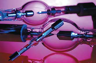
Xenon and mercury xenon lamps, two examples of DC arc lamps.
The tunable light source’s (TLS) versatility as both a broadband and high-resolution monochromatic light source makes the unit suitable for a variety of applications, such as the study of wavelength-dependent chemical or biological properties or wavelength-induced physical changes of materials. These light sources can also be used in color analysis and reflectivity measurements of materials for quality purposes.
Among their unique attributes, the TLS can produce monochromatic light from the UV to near-infrared (NIR). Lamp-based TLSs feature two major components: a light source and a monochromator. Common lamps used in TLSs are the DC arc lamp and quartz tungsten halogen (QTH) lamp. While both of these lamps have a broad emission spectrum, arc and QTH lamps differ in the characteristic wavelength emissions or relatively smooth shape of their spectral output curves, respectively. A stable power supply for the lamp is a critical component since most applications require high light output power stability1.
Smooth spectral output vs. monochromator throughput
DC arc lamps are excellent sources of continuous wave, broadband light. They consist of two electrodes (an anode and a cathode) separated by a gas such as neon, argon, mercury or xenon. Light is generated by ionizing the gas between the electrodes. The bright broadband emission from this short arc between the anode and cathode makes these lamps high-intensity point sources, capable of being collimated with the proper lens configuration.
DC arc lamps also offer the advantages of long lifetime, superior monochromator throughput (particularly in the UV range) and a smaller divergence angle. They are particularly well-suited for fiber coupling applications2. (See Figure 1.)
Figure 1. A xenon arc lamp housed in an Oriel Research lamp housing. Photo courtesy of Newport Corp.
Xenon (Xe) arc lamps, in particular, have a relatively smooth emission curve in the UV to visible spectrums, with characteristic wavelengths emitted from 380 to 750 nm. However, strong xenon peaks are emitted between 750 to 1000 nm.
Their sunlike emission spectrum and about 5800 K color temperature make them a popular choice for solar simulation applications. (See Figure 2.)
Arc lamps can have the following specialty characteristics:
• Ozone-free: Wavelength emissions below about 260 nm create toxic ozone. Ideally, an arc lamp is operated outdoors or in a room with adequate ventilation to protect the user from the ozone created.
• UV-enhanced: For applications requiring additional UV light intensity, UV-enhanced lamps should be used. These lamps provide the same visible to NIR performance of an arc lamp while providing high-intensity UV output due to changes in the material of the lamp’s glass envelope.
• High-stability: High-stability arc lamps are made of a higher quality cathode than that typically used for arc lamp construction. As a result, no arc wander occurs, allowing the lamp to maintain consistent output intensity throughout its lifetime.
Figure 2. The spectral output of 3000-W Xe and 250-W QTH lamps used in Oriel’s Tunable Light Sources.Photo courtesy of Newport Corp.
QTH lamps produce light by heating a filament wire with an electric current. The hot filament wire is surrounded by a vacuum or inert gas to prevent oxidation. QTH lamps are not very efficient at converting electricity to light, but they offer very accurate color reproduction due to their continuous blackbody spectrum. These lamps are a popular alternative to arc lamps due to their higher output intensity stability and lack of intense UV light emission, spectral emission lines in their output curve and toxic ozone production. These advantages over traditional DC arc lamps make QTH lamps preferable for radiometric and photometric applications as well as excitation sources of visible to NIR light. QTH lamps are also easier to handle and install, and produce a smooth output spectrum. Selecting the most appropriate lamp type is a matter of deciding which performance criteria are most important.
Constant current vs. constant power
The power supply is a vital component for operating a DC arc or QTH lamp with minimum light ripple. The lamps are operated in either constant current or constant power mode and are used in applications such as radiometric measurements, where a stable light output is required for accurate measurement. Providing stable electrical power to the lamp is important since fluctuations in the wavelength and output intensity of the light source impact the accuracy of measurement.
There is very little difference in the short-term output stability when operating an arc lamp or QTH lamp in constant current or constant power mode. However, the differences appear as the lamp ages. For arc lamps, even with a stable power supply, deposits on the inside of the lamp envelope are visible as the electrodes degrade, which causes an unstable arc position, changing the electrical characteristics of the arc lamp. The distance between the cathode and anode of the arc lamp increases, raising the lamp’s operating voltage. For QTH lamps, deposits on the inside of the lamp envelope are visible as the lamp filament degrades, changing the electrical and spectral characteristics of the lamp.
In power mode, the lamp is operated at a constant power setting. As the voltage cannot be changed, the current is raised or lowered to maintain the power at the same level. As the lamp ages, the radiant output decreases. However, lamp lifetime is prolonged.
In current mode, the lamp is operated at a constant current setting. As the voltage cannot be changed, the power is raised or lowered to maintain the current at the same level. As the lamp ages, the input power required for operation is increased. This results in greater output power, which, to some extent, may help compensate for a darkening lamp envelope. However, the lamp’s lifetime is greatly reduced due to the increase in power.
Although power supplies are highly regulated, there are factors beyond the control of the power supply that may affect light output. Some of these factors include lamp aging, ambient temperature fluctuations and filament erosion. For applications in which high stability light output intensity is especially critical, optical feedback control of power supply is suggested in order to compensate for such factors3. (See Figure 3.)
Figure 3. Oriel’s OPS Series Power Supplies offer the option of operating a lamp in constant power, constant current or intensity operation modes. Photo courtesy of Newport Corp.
Diffraction gratings narrow the wavelength band
Monochromators use diffraction gratings to spatially isolate and select a narrow band of wavelengths from a wider wavelength emitting light source. They are a valuable piece of equipment because they can be used to create quasi-monochromatic light and also take high precision spectral measurements. A high precision stepper motor is typically used to select the desired wavelength and switch between diffraction gratings quickly, without sacrificing instrument performance.
Determining which slit width to use is based on the trade-off between light throughput and the resolution required for measurement. A larger slit width allows for more light throughput. However, more light throughput results in poorer resolution. When choosing a slit width at which to operate the monochromator, both the input and output ports must be set to the same slit width. (See Figure 4.) Focused light enters the monochromator through the entrance slit, and is redirected by the collimating mirror toward the grating. The grating directs the light toward the focusing mirror, which then redirects the chosen wavelength toward the exit slit. At the exit slits, quasi-monochromatic light is emitted4.
Figure 4. A fixed width slit being installed into an Oriel Cornerstone 130 monochromator.Photo courtesy of Newport Corp.
Measuring quantum efficiencies
Measuring quantum efficiency (QE) over a range of different wavelengths to measure a device’s QE at each photon energy level is an ideal task for a tunable light source. The QE of a photoelectric material for photons with energy below the band gap is zero. The QE value of a light-sensing device such as a solar cell indicates the amount of current that the cell will produce when irradiated by photons of a particular wavelength. The principle of QE measurement is to count the proportion of carriers extracted from the material’s valence band to the number of photons impinging on the surface. To do this, it is necessary to shine a calibrated, tunable light on the cell, while simultaneously measuring the output current. The key to accurate measurement of the QE/internal photon to current efficiency is to accurately know how much scanning light is incident on the device under test and how much current is generated. Thus, measurement of light output with a NIST (National Institute of Standards and Technology) traceable calibrated detector is necessary prior to testing since illumination of an absolute optical power is required.
External quantum efficiency (EQE) is the ratio of the number of photons incident on a solar cell to the number of generated charge carriers. Internal quantum efficiency (IQE) also considers the internal efficiency — that is, the losses associated with the photons absorbed by nonactive layers of the cell. By comparison, EQE is much more straightforward to measure, and gives a direct parameter of how much output current will be contributed to the output circuit per incident photon at a given wavelength. IQE is a more in-depth parameter, taking into account the photoelectric efficiency of all composite layers of a material. In an IQE measurement, these losses from nonactive layers of the material are measured in order to calculate a net quantum efficiency — a much truer efficiency measurement.
Understanding the conversion efficiency as a function of the wavelength of light impingent on the cell makes QE measurement critical for materials research and solar cell design. With this data, the solar cell composition and topography can be modified to optimize conversion over the broadest possible range of wavelengths.
As a formula, it is given by IQE = EQE/(1 − R), where R is the reflectivity, direct and diffuse, of the solar cell. The IQE is an indication of the capacity of the active layers of the solar cell to make good use of the absorbed photons. It is always higher than the EQE, but should never exceed 100 percent, with the exception of multiple-exciton generation. Figure 5 illustrates how the tunable light source is used to illuminate the solar cell to perform an IQE measurement. The software controls all components of the measurement system, including the monochromator and data acquisition5.
Figure 5. A sample QE measurement system using the components of a tunable light source. Photo courtesy of Newport Corp.
To measure quantum efficiency in 10-nm wavelength steps, the slit size of the monochromator is typically hundreds of micron in width. The slit width is reduced approximately half if 5-nm wavelength increments are desired. However, output power of the monochromator is reduced by more than 50 percent if the slit width is halved. Lowering optical power impacts QE measurement since a solar cell responds to this diminished optical power with low output current. This can result in a poor signal-to-noise ratio, making a QE measurement error more likely. The detection of low current requires very sensitive equipment with the ability to measure current down to the pico-ampere level. To make for an easier signal measurement, optical power is typically increased. A DC arc source is the better choice for QE measurements made in 5-nm increments or lower due to the lamp’s arc size resulting in better monochromator throughput. However, a QTH lamp is the better choice if greater than 0.1 percent light stability is required, with the trade-off of not being able to measure in as precise wavelength increments as if an arc lamp was used.
Balance between optical power and resolution is an important consideration as it impacts the quality of the QE measurement. The selection of lamp type and monochromator specifications are important considerations for TLS design. To be considered a suitable component for the majority of spectroscopic applications, high-output power and stability, long lifetime of the lamp, and broadband spectral emission with high resolution capability are required for the TLS.
Meet the authors
John Park, new product development manager at Newport Corp., has designed and developed numerous spectroscopy instruments for the photonics industry for over 10 years. He holds two granted patents and is a graduate from University of California, Irvine, with a Ph.D. in electrical engineering; email: john.park@newport.com. Jeff Eng is a product specialist for Oriel Spectroscopy Products at Newport Corp. His work experience includes application support, business-to-business sales and marketing activity of photonic light sources and detectors. He is a graduate of Rutgers University; email: jeff.eng@newport.com.
4. J. M. Lerner and A. Thevenon (1988). The Optics of Spectroscopy. Edison, N.J.: Optical Systems/Instruments SA Inc.
5. K. Emery (2005). Handbook of Photovoltaic Science and Engineering, eds. A. Luque and S. Hegedus. Chapter 16: Measurement and characterization of solar cells and modules. Hoboken, N.J.: John Wiley & Sons Ltd.
Vanderbilt University Medical Center nephrologist and Associate Professor of Medicine Dr. William H. Fissell IV, is making major progress on a first-of-its kind device to free kidney patients from dialysis. He is building an implantable artificial kidney with microchip filters and living kidney cells that will be powered by a patient’s own heart. The bio-hybrid device will mimic a kidney to remove enough waste products, salt and water to keep a patient off dialysis,” said Fissell.
Fissell says the goal is to make it small enough, roughly the size of a soda can, to be implanted inside a patient’s body.
Nanotechnology
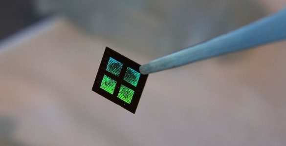
The key to the device is a microchip.
“It’s called silicon nanotechnology. It uses the same processes that were developed by the microelectronics industry for computers,” said Fissell.
The chips are affordable, precise and make ideal filters. Fissell and his team are designing each pore in the filter one by one based on what they want that pore to do. Each device will hold roughly fifteen microchips layered on top of each other.
But the microchips have another essential role beyond filtering.
“They’re also the scaffold in which living kidney cells will rest,” said Fissell.
An example of the microchip filter being used inside Fissell’s artificial kidney. (Vanderbilt University)
Living kidney cells
Fissell and his team use live kidney cells that will grow on and around the microchip filters. The goal is for these cells to mimic the natural actions of the kidney.
“We can leverage Mother Nature’s 60 million years of research and development and use kidney cells that fortunately for us grow well in the lab dish, and grow them into a bioreactor of living cells that will be the only ‘Santa Claus’ membrane in the world: the only membrane that will know which chemicals have been naughty and which have been nice. Then they can reabsorb the nutrients your body needs and discard the wastes your body desperately wants to get rid of,” said Fissell.
Avoiding organ rejection
Because this bio-hybrid device sits out of reach from the body’s immune response, it is protected from rejection.
“The issue is not one of immune compliance, of matching, like it is with an organ transplant,” said Fissell.
How it works
The device operates naturally with a patient’s blood flow.
The device operates naturally with a patient’s blood flow.
“Our challenge is to take blood in a blood vessel and push it through the device. We must transform that unsteady pulsating blood flow in the arteries and move it through an artificial device without clotting or damage.”
Fluid dynamics
And that’s where Vanderbilt biomedical engineer Amanda Buck comes in. Buck is using fluid dynamics to see if there are certain regions in the device that might cause clotting.
“It’s fun to go in and work in a field that I love, fluid mechanics, and get to see it help somebody,” said Buck.
She uses computer models to refine the shape of the channels for the smoothest blood flow. Then they rapidly prototype the new design using 3-D printing and test it to make the blood flow as smoothly as possible.
Vanderbilt biomedical engineer Amanda Buck is using fluid dynamics to see if there are certain regions in the device that might cause clotting.
Future human trials
Fissell says he has a long list of dialysis patients eager to join a future human trial. Pilot studies of the silicon filters could start in patients by the end of 2017.
“My patients are absolutely my heroes,” said Fissell. “They come back again and again and they accept a crushing burden of illness because they want to live. And they’re willing to put all of that at risk for the sake of another patient.”
Federal investment
The National Institutes of Health awarded a four-year, $6 million grant to Fissell and his research partner Shuvo Roy from the University of California at San Francisco. The two investigators are longtime collaborators on this research. In 2003, the kidney project attracted its first NIH funding, and in 2012 the Food and Drug Administration selected the project for a fast-track approval program. The work is supported by NIH grant 1U01EB021214-01.
The National Kidney Foundation reports that in 2012, Federal Medicare dollars paid more than $87 billion caring for kidney disease patients (not including prescription medications).
Desperate need
Transplant of a human kidney is the best treatment for kidney failure, but donor kidneys are in short supply. According to the U.S. Organ Procurement and Transplantation Network, although more than 100,000 patients in the United States are on the waiting list for a kidney transplant, last year only 17,108 received one.
In all, the National Kidney Foundation says more than 460,000 Americans have end-stage renal disease and every day, 13 people die waiting for a kidney.
One of the unexpected findings of the Human Genome Project was that over 98% of the human genome does not encode for proteins. Once dismissed as “junk” genomic material, non-protein-coding DNA is now appraised more highly.
Or to be more precise, at least some portions of non-protein-coding DNA are thought to serve important biological functions.
For example, some stretches of DNA give rise to a noncoding but still functional kind of RNA called microRNA. MicroRNAs have increasingly emerged in recent years as key regulators of biological processes and pathways.
During the years since their discovery, a key question in the biology of microRNAs has focused on the number of microRNAs encoded in the genome. Between 1993 and 2015, approximately 1,900 human genome loci were discovered to produce microRNAs and were added to miRBbase, the public database that catalogues and annotates microRNA molecules.
The cataloguing of microRNAs work has been pursued with extra urgency since 2004, the year the connection between microRNAs and human disease was first demonstrated. “When this connection was made, it launched a whole new field,” says Isidore Rigoutsos, Ph.D., professor of pathology, anatomy, and cell biology and director of the Computational Medicine Center at Thomas Jefferson University.
Another Set of MicroRNAs Emerge
“We wanted to know how many microRNA-producing loci really exist in humans,” recalls Dr. Rigoutsos. In a study published in 2015, Dr. Rigoutsos and colleagues analyzed datasets from 1,323 individuals that represented 13 different tissues and identified an additional 3,356 such genomic loci that produce (at least) 3,707 novel microRNs
“We basically tripled the number of locations in the human genome that are now known to encode microRNAs,” asserts Dr. Rigoutsos. Considering that each microRNA regulates up to hundreds of different mRNAs, and that each mRNA is regulated by tens of microRNAs, this finding adds a new layer of complexity to the regulatory dynamics of the human transcriptome.
The newly unveiled microRNAs and previously characterized microRNAs have distinct expression patterns. While 50–60% of the microRNAs previously deposited into the miRBase are expressed in multiple tissues, only about 10% of the newly discovered microRNAs are shared across multiple tissue types. Also, most of the newly found microRNAs show tissue-specific expression.
Using Argonaute CLIP-seq data, Dr. Rigoutsos and colleagues showed that similar percentages of the two sets of microRNAs were in complex with Argonaute proteins. “This shows that these novel microRNAs participate in RNA interference just as frequently as the miRBase microRNAs,” contends Dr. Rigoutsos.
In a comparative analysis between the human microRNA datasets and the chimpanzee, gorilla, orangutan, macaque, mouse, fruit fly, and mouse genomes, Dr. Rigoutsos and colleagues discovered that almost 95% of the newly unveiled microRNAs were primate-specific, and over 56% of them were found only in humans.
“We are seeing many human microRNAs that do not exist in the mouse,” states Dr. Rigoutsos. “This means that the mouse models engineered to capture human disease cannot recapitulate the interactions mediated by these microRNAs.
Interest in IsomiRs Grows
In the years since the biology of microRNAs started receiving increasing attention, the conventional view has been that one microRNA locus generates one microRNA. However, once deep sequencing became widely available, microRNA variants that showed differences at their 5′- or 3′-termini have been described.
“It was initially presumed that these variants were likely the result of the enzyme Dicer not being sufficiently accurate when processing microRNA precursors,” notes Dr. Rigoutsos. Subsequent research revealed that microRNAs are more dynamic than previously thought, with each precursor being able to generate multiple mature microRNA species known as isomiRs.
To gain insight into the biology of isomiRs, Dr. Rigoutsos and colleagues analyzed genomic datasets from 452 individuals participating in the 1000 Genomes Project. The datasets comprised five different populations and two races. In addition, each population was represented by an even number of men and women.
This collection allowed the abundance of microRNA isoforms to be examined with respect to population, gender, and race. “We found that isomiRs have expression profiles that are population-, race-, and gender-dependent,” informs Dr. Rigoutsos.
All the transcriptome data that this analysis was based on came from immortalized B cells. “These are cells that normally are not associated with gender differences, but molecularly we found, in these cells, differences between men and women of the same population and race,” explains Dr. Rigoutsos.
Expanding these observations to disease states, Dr. Rigoutsos and colleagues collected isomiR profiles from tissue affected by breast cancer, and compared them with isomiR profiles from control breast tissue. The investigators found that the isomiR profiles also depend on tissue state (healthy vs. diseased), on disease subtype, and on the patient’s race.
For example, their analysis identified several miR-183-5p isoforms that were upregulated in white triple-negative breast cancer patients compared to control breast samples, but not in black/African-American triple-negative breast cancer patients. In an in vitro phase of this study, three isoforms of this microRNA species were overexpressed in human breast cancer cell lines.
“We found very little overlap in the gene sets that were affected by each of these isoforms,” emphasizes Dr. Rigoutsos. Despite being generated simultaneously by the same locus, each of the three isoforms affected distinct groups of genes, thus exerting different effects on the transcriptome.
“As the relative abundance of these isoforms changes ever so slightly from patient to patient, it will affect the corresponding gene groups slightly differently,” concludes Dr. Rigoutsos. “In the process, it creates a new molecular background in each patient.”
MicroRNAs Point to Therapeutic Strategies against Colorectal Cancer
“We are using microRNAs as modulators to overcome chemotherapy resistance in colorectal cancer,” says Jingfang Ju, Ph.D., associate professor of pathology and co-director of translational research at Stony Brook University School of Medicine. Resistance to chemotherapy is one of the major challenges in the clinical management of malignancies, including colorectal cancer. Chemotherapy is usually unable to eliminate cancer stem cells, which may become even more resistant over time, and several microRNAs have been implicated in this process. “We reasoned that we could provide new modulatory approaches to target this small cell population and allow chemotherapy, radiotherapy, or immunotherapy to eliminate resistant populations or at least prolong long-term survival,” Dr. Ju said.
This image shows how miR-129 may function as a tumor suppressor in colorectal cancer. In this model, which has been proposed by researchers at Stony Brook University’s Translational Research Laboratory, miR-129 suppresses the protein expression of three critical targets—BCL2, TS, and E2F3. Downregulation of BCL2 activates the intrinsic apoptosis pathway by cleaving caspase-9 and caspase-3. Downregulation of TS and E2F3 inhibits cell proliferation by impacting the cell cycle. Consequently, miR-129 exerts a strong antitumor phenotype by induction of apoptosis and impairment of proliferation in tumor cells. [Mihriban Karaayvaz, Haiyan Zhai, Jingfang Ju]
In a retrospective study in which colorectal patient samples were used, Dr. Ju and colleagues revealed that hsa-miR-140-5p expression progressively decreases from normal tissues to primary colorectal cancer tissue, and that it shows a further decrease in liver and lymph node metastases. The experimental overexpression of hsa-miR-140-5p inhibited colorectal cancer stem cell growth by disrupting autophagy, and in a mouse model of disease it abolished tumor formation and metastasis.
In addition to hsa-miR-140-5p, Dr. Ju and colleagues recently identified hsa-miR-129 and found that it, too, has therapeutic potential. Specifically, they showed that hsa-miR-129 enhanced the sensitivity of colorectal cancer cells to 5-fluorouracil, pointing toward its ability to function as a tumor suppressor.
One of the mechanisms implicated in this process was the ability of miR-192 to inhibit protein translation of several important targets. These include Bcl-2 (B-cell lymphoma 2), a key anti-apoptotic protein; E2F3, a major cell cycle regulator; and thymidylate synthase, an enzyme that is inhibited by 5-fluorouracil.
The NIH recently awarded a $3 million grant to establish the Long Island Bioscience Hub (LIBH), which is part of the NIH’s Research Evaluation and Commercialization Hub (REACH) program and represents a partnership between the Center for Biotechnology, Stony Brook University, Cold Spring Harbor Laboratory, and Brookhaven National Laboratory. One of the technology development grants, as part of the first funding cycle of this initiative, will support a feasibility investigation of hsa-miR-129-based therapeutics in colon cancer, an effort led by Dr. Ju. “We are further exploring this novel mechanism,” states Dr. Ju. “We anticipate conducting pharmacokinetic studies and moving to a clinical trial in the future.”
MicroRNA Insights Gleaned from Host-Virus Interactions
At Mount Sinai Hospital’s Icahn School of Medicine, researchers used a codon-optimized version of VP55 produced from an adenovirus-based vector to study the impact of microRNA deletion on the response to virus infection. This image shows RNA in situ hybridization of fibroblasts expressing VP55 (top left), and that of mock-treated fibroblasts (bottom right). Ribosomal RNA, DNA, and microRNAs (miR-26) are depicted by red, blue (DAPI), and green fluorophores, respectively.
“We observed that when a poxvirus is artificially engineered to encode a microRNA, the microRNA is destroyed along with all the microRNAs from the host cell,” says Benjamin R. tenOever, Ph.D., professor of microbiology at the Icahn School of Medicine, Mount Sinai Hospital. Previously, Dr. tenOever’s group reported that a single vaccinia virus-encoded gene product, VP55, is sufficient to achieve this effect. The group also found that the protein adds nontemplate adenosines to the 3′-end of microRNAs associated with the RNA-induced silencing complex.
biology,” asserts Dr. tenOever.
In a recent study, Dr. tenOever and colleagues used a codon-optimized version of VP55 produced from an adenovirus-based vector to study the impact microRNA deletion would have on our normal response to virus infection. “We found that after administration of the vector and rapid ablation of microRNA expression, there is very little that happens over the first one to two days,” informs Dr. tenOever. During the first 24–48 hours after VP55 delivery, the elimination of cellular microRNAs impacted less than 0.35% of the over 11,000 genes expressed in the cell. After 9 days, however, almost 20% of the genes showed significant changes in expression.
“MicroRNAs are very powerful and influential in controlling the biology of the cell but they do so over the long term,” declares Dr. tenOever. These findings are in agreement with knowledge that has accumulated over the years about microRNA biology, which established that microRNAs play a central role in determining how cells differentiate during development.
“While microRNAs can act on hundreds of mRNAs, their action requires several days of fine-tuning to have long-term consequences,” adds Dr. tenOever. This finding suggests miRNAs are unable to significantly contribute to the acute response to virus infection.
The one exception to this observation was that, even though very few genes were affected in the first 48 hours after VP55 delivery, several genes encoding chemokines were impacted. These included chemokines responsible for recruiting antigen-presenting cells, neutrophils, and other immune cells.
An in vivo analysis of mouse lung tissue 48 hours after vector administration confirmed that several cytokines were specifically upregulated, resulting in immune cell infiltration following the degradation of all microRNAs. These results indicate that the acute viral infection is largely independent of microRNAs, and that microRNAs are primarily involved in the adaptive response to infection and other longer term processes.
MicroRNA Biomarkers Reveal Molecular Pathways of Kidney Damage
“Our approach involves looking at microRNAs from the perspective of biomarkers as a readout for kidney damage,” says Vishal S. Vaidya, Ph.D., associate professor of medicine and environmental health at Brigham and Women’s Hospital, Harvard Medical School, and Harvard T.H. Chan School of Public Health. “At the same time, we are exploring their utility as therapeutics.”
A large number of medications and occupational toxins cause kidney damage, but many tests to assess kidney function and damage are not sufficiently sensitive or specific, opening the need for novel diagnostic strategies. MicroRNAs, which are differentially expressed between healthy and diseased states, are promising as early biomarkers for impaired renal function.
“MicroRNAs can also provide information about which pathways are active and which targets can be druggable,” points out Dr. Vaidya.
In a study that used microRNAs and proteins to provide a combined biomarker signature, Dr. Vaidya and colleagues examined two patient cohorts, one presenting with acetaminophen-induced kidney injury and the other one with cisplatin-induced kidney damage. “Protein biomarkers provide sensitivity, and microRNAs offer mechanistic insight,” explains Dr. Vaidya.
This approach helped visualize metabolic pathways that are altered in the kidney during toxic injury. “The biggest challenge, from a therapeutic perspective, is that microRNAs regulate many mRNAs and, therefore, impact many proteins,” concludes Dr. Vaidya.
Is the Warburg effect an effect of deregulated space occupancy of methylome?
Larry H. Bernstein and Radoslov Rosov, co-curation
LPBI
UPDATED
8/20/2024
Editor Correction: One of the co-curators names on this article was misspelled. The co-curators should be Larry H. Bernstein and Radislov Rosov.
Therefore the correction version of the article title and authors should read
Is the Warburg effect an effect of deregulated space occupancy of methylome?
Larry H. Bernstein and Radoslov Rosov, co-curation
The editors of LPBI Group apologize for the confusion.
It would appear that pyruvate is directly used by cancer cell machinery for sustaining genome independence, and that CRISP-Cas9 system is essentially a modified CAD protein for making small bases.
13C-labeled biochemical probes for the study of cancer metabolism with dynamic nuclear polarization-enhanced magnetic resonance imaging
In recent years, advances in metabolic imaging have become dependable tools for the diagnosis and treatment assessment in cancer. Dynamic nuclear polarization (DNP) has recently emerged as a promising technology in hyperpolarized (HP) magnetic resonance imaging (MRI) and has reached clinical relevance with the successful visualization of [1-13C] pyruvate as a molecular imaging probe in human prostate cancer. This review focuses on introducing representative compounds relevant to metabolism that are characteristic of cancer tissue: aerobic glycolysis and pyruvate metabolism, glutamine addiction and glutamine/glutamate metabolism, and the redox state and ascorbate/dehydroascorbate metabolism. In addition, a brief introduction of probes that can be used to trace necrosis, pH changes, and other pathways relevant to cancer is presented to demonstrate the potential that HP MRI has to revolutionize the use of molecular imaging for diagnosis and assessment of treatments in cancer.
Since the hallmark discovery of the Warburg effect in cancer cells in the 1920s, it has been widely accepted that the metabolic properties of cancer cells are vastly different from those of normal cells [1]. Starting from the observation that many cancerous (neoplastic) cells have higher rates of glucose utilization and lactate production, the development of tools and methods to correlate specific cellular metabolic processes to different types of cancer cells has received increased research focus [2, 3]. Several imaging techniques are currently in use for this purpose, including radiography, scintigraphy, positron emission tomography (PET), single-photon emission computed tomography (SPECT), and magnetic resonance (MR) [4, 5].
For more than 30 years, MR has been a revolutionary diagnostic tool, used in a wide range of settings from the central nervous system to cardiomyopathies and cancers. MR imaging (MRI) can outline molecular and cellular processes with high spatial resolution. Typically, MRI of body tissues is achieved via contrast visualization of the protons (1H) of water, which are present in high abundance in living systems. This can be extended to MR spectroscopy (MRS), which can further differentiate between less abundant, carbon-bearing, biological metabolites in vivo utilizing 1Hs of these compounds [6, 7]. However, despite its usefulness in imaging whole body tissues, 1H MRS has low spectral resolution and poor sensitivity for these less abundant metabolites. In addition,13C MRS is increasingly difficult, in comparison to 1H MRS, in that both the gyromagnetic ratio (approximately 25 % of 1H) and natural abundance (1.1 % of 1H) are significantly lower, making the detection of carbon-bearing compounds difficult [8, 9]. The low spectral resolution of 1H MRS for metabolites can be addressed by using 13C-enriched compounds, and with this direct 13C MRS, metabolic processes can be traced, utilizing enriched tags on specific carbons in a given metabolite [10]. While enrichment of molecules in 13C can also moderately address the sensitivity limitation of MRS, recent work in hyperpolarization (HP) provides a means of dramatically increasing sensitivity and enhancing signals, well beyond that of the equilibrium state obtained via MRS. [11, 12]. The focus of this review will be the introduction of this approach in the setting of cancer metabolism, delineating probes of interest, which have been applied to study metabolic processes in vivo.
Obtaining a hyperpolarized probe
In MR, a desired target is placed in a magnetic field where the nuclear spins of molecules are aligned with or against the direction of the magnetic field. The nuclear spins have thus different energies, and an MR signal is detected upon relaxation of nuclear spins of higher energy. At thermal equilibrium, the number of spins aligned with the magnetic field nearly equals the number of spins opposing the direction of the magnetic field. Thus, at thermal equilibrium, spin polarization is in the order of >0.0005 % resulting in a limited signal. Signal increases on the order of 100,000-fold can be achieved by hyperpolarizing the system via the redistribution of the spin population levels found at equilibrium [10, 13]. There are several techniques that have been used to achieve hyperpolarization of various nuclei: spin-exchange optical pumping of 3He and 129Xe, parahydrogen-induced polarization (PHIP), and dissolution dynamic nuclear polarization (DNP) [11,14, 15]. Both PHIP and DNP techniques can polarize biologically relevant nuclei like 13C and 15N, although there is a wider range of molecules that can be targeted for hyperpolarization using dissolution DNP [14, 16–18].
The goal of DNP is the transfer of polarization from highly polarized unpaired electron spins to the nuclear spins of a desired target compound. This is achieved by applying an external magnetic field to a free-radical agent in order to polarize electron spins, followed by saturating the electron spin resonance via microwave irradiation in order to obtain polarization transfer. The free-radical agent is generally a stable organic compound that is compatible with aqueous buffers, which are used as solvent in order to obtain a homogeneous distribution of the radical [13]. Nearly 100 % of the electrons on the free-radical agent are polarized when the free-radical/solvent mixture is subjected to high magnetic fields (≥3.3 T) followed by rapid freezing to 1 K using liquid helium in order to obtain a sample frozen to an amorphous state, which is necessary for retention and transfer of polarization [18]. For biological applications, after transfer of electron spin polarization to the nuclei of interest has occurred, the preparation must exist in solution, which can be achieved utilizing a dissolution process in which the solid sample is rapidly melted via injection of a hot solvent, typically a biologically compatible buffer, into the frozen sample [13]. The dissolution process results in a liquid sample at room temperature, while still preserving the enhanced polarization obtained by the microwave irradiation of the frozen sample [8]. Additionally, the use of chelating agents (e.g., EDTA) with the solvent to eliminate trace metals and more recently the use of gadolinium (Gd) chelates with the DNP sample have been used to further enhance and retain polarization in the liquid sample, albeit with caution over potential toxic effects when applied in vivo and the potential for loss of hyperpolarization due to T1 shortening [11, 19, 20]. More in-depth exploration of the technical aspects of probe development has been previously reviewed [8, 11].
Considerations in probe selection and current research
The usefulness of a molecule for hyperpolarized MRS is dependent on the polarization lifetime of the nucleus of interest, and this property is determined by the spin-lattice relaxation constant (T1) [21]. Dipolar coupling, the magnetic field range, and molecular size can also affect the T1 of a given nucleus. In general, high magnetic fields and large molecular weights decrease the T1. Dipole-dipole coupling of 13C with 1H is common in biologically relevant molecules, and it shortens relaxation times; therefore, carbon atoms directly bound to 1H are generally not useful as probes for HP. For example, all carbons present in glucose (an important substrate in cancer cells) have relaxation times shorter than 2 s [22]. On the other hand, carbonyl carbons of biologically relevant molecules generally have T1’s above 20 s even at high magnetic fields like [1-13C] pyruvic acid, which has relaxation times of 67, 48, and 44 s at 3, 11.7, and 14.1 T, respectively [23–25]. Even carbons that are less oxidized than carbonyls, like the hemi-ketal in [2-13C] fructose have T1’s one order of magnitude higher than glucose carbons. Short spin-lattice relaxation times can sometimes be increased by deuterium enrichment of the sample. With this technique, protons that are directly bound to carbons are exchanged for deuterium atoms which results in the reduction of dipole-dipole relaxation, further preserving the hyperpolarized state [26]. This has resulted in increased T1’s of 13C nuclei in molecules such as glucose (T1 increased from 2 s to 10–14 s), providing the possibility of utilizing them in future metabolic studies [27–29]. Despite the effect of deuterium enrichment, research efforts have largely focused on developing carbonyl-bearing molecules as molecular imaging probes. The focus of this review is to introduce representative compounds relevant to metabolism that are characteristic of cancer tissue and have been applied in the work of multiple groups: aerobic glycolysis, glutamine addiction, and the redox state.
Pyruvate and aerobic glycolysis
Of particular interest to cancer metabolism is the increased conversion of glucose to lactate as a result of modulated aerobic glycolysis. This process, also known as the Warburg effect, is characteristic of many tumors with altered metabolism where pyruvate generated from glucose metabolism via glycolysis is preferentially converted to lactate by lactate dehydrogenase (LDH) as opposed to entering the tricarboxylic acid cycle [1]. With this phenotype, cancer cells show a preference for lactate fermentation even in the presence of oxygen, thus bypassing oxidative respiration for ATP generation. Because of this, pyruvate has been the preferred probe for HP MRS research since it is an intermediate metabolite in pathways characteristic of aberrant metabolism in cancer cells, including increased lactate production as a result of aerobic glycolysis where detection of HP pyruvate-derived lactate can be used as a marker for cancer and response to treatment [30, 31] as well as an intermediate in amino acid metabolism (e.g., interconversion to alanine via transamination with glutamate) (Fig. 1). In addition, as mentioned before, carbonyl carbons in pyruvate have long relaxation times where even the methyl carbon can have T1’s above 50 s after deuterium enrichment [32]. The interconversion of pyruvate to lactate has been exploited for MRI by using [1-13C] pyruvate and detecting the accumulation of increased lactate in cancerous tissue as compared to surrounding benign tissue. Increased conversion of pyruvate to lactate and alanine has been demonstrated to precede MYC-driven tumorigenesis by using HP [1-13C] pyruvate in murine models [33]. Furthermore, in the same study, a decrease in the flux of alanine was observed at the tumor stage while a decrease in lactate conversion was indicative of tumor regression [33]. In transgenic adenocarcinoma of mouse prostate (TRAMP) models, in vivo studies using HP [1-13C] pyruvate demonstrated that hyperpolarized pyruvate and its metabolic products can be used non-invasively and with high specificity to obtain a profile of the histologic grade of prostate cancers [34]. In vivo imaging following hyperpolarized pyruvate has also been used to evaluate the role of glutaminase and LDH in human lymphoma models [35] as well as to elucidate metabolism of pyruvate in breast cancer [36] and renal cell carcinoma with treatment [30, 37].
Flux of hyperpolarized [1-13C] pyruvate to [1-13C] lactate in prostate regions. a MR image from patient with prostate cancer showing regions of cancerous tissue and surrounding normal tissue. b–d Localized dynamic hyperpolarized [1-13C]pyruvate and [1-13C]lactate spectral from voxels overlapping the contralateral region of prostate (turquoise), a region of prostate cancer (yellow), and a vessel outside the prostate (green). Adapted with permission from ref. [43]
Early work that utilized HP pyruvate to assess the response of tumors to treatment was conducted in mice xenografted with EL-4 lymphoma cells and treated with etoposide, a topoisomerase inhibitor that causes rapid cell death [38, 39]. In this study, tumor necrosis was correlated to a decrease in the flux of hyperpolarized lactate which was suggested to be due to a decrease in NAD+ and NADH in the intracellular pool as well as loss of LDH function. More recently, HP [1-13C] pyruvate has been used as a biomarker to evaluate early response to radiation therapy in glioma tumors by observing a decrease in hyperpolarized lactate suggested to be a result of changes in tumor perfusion which can be detected between 24 and 96 h following treatment [40, 41]. HP [1-13C] pyruvate has also been used to detect early response to temozolomide (TMZ) treatment on human glioblastoma rat models [42]. The study successfully showed for the first time detection of response to TMZ therapy 1 day after TMZ administration. The continued reports on using HP pyruvate as an imaging probe for assessing treatment response indicate the potential of the compound to become a standard in the field. Moreover, these studies demonstrate the usefulness of HP [1-13C] pyruvate as a tool for early assessment of therapy response, which can improve treatment selection at the clinical level. Pyruvate has also been validated as a metabolic imaging marker for use in humans [43]. In a two-phase study, patients with biopsy-proven prostate cancer of various histological grades were injected with HP [1-13C] pyruvate. In the first phase, a maximum dose level was determined to establish pharmacological safety of the HP probe while still injecting enough pyruvate to allow visualization. This addressed one of the major challenges faced in translating HP MRI to clinical applications: the potential toxicity of compounds that must be injected into patients. In the second phase, metabolism of pyruvate was visualized in real time and differences in the ratio of [1-13C] lactate to [1-13C] pyruvate between identified cancerous regions and normal tissue regions were successfully observed (Fig. 1a–d). [1-13C] lactate in regions that did not contain tumor was not detected, confirming previous biopsy and preclinical studies that demonstrated low flux of [1-13C] pyruvate to lactate and low concentrations of lactate in benign prostate tissues [44, 45]. Preliminary results indicated the possibility of detecting previously unobserved cancerous regions by HP [1-13C] pyruvate, later confirmed to be Gleason 4+3 cancer by biopsy, though further investigation into the relationship between grade and metabolism in prostate cancer patients is needed. While there are challenges associated with translation to clinical use for HP [1-13C] pyruvate, the first in human study demonstrated the feasibility of hyperpolarization technology as a safe diagnostic tool and provides the potential for utilizing this approach in preclinical models with direct translation to the clinic.
Glutamine metabolism
Glutamine is an amino acid that plays an important cellular role as nitrogen donor in the form of an amide group for purine and pyrimidine biosynthesis, leaving a glutamate molecule in the process although glutamine can also be converted to glutamate by glutaminase in a reaction independent of nucleotide biosynthesis. Glutamate is the primary nitrogen donor for the biosynthesis of non-essential amino acids. Transaminases catalyze the transfer of the amine group from glutamate to α-ketoacids to synthesize alanine, aspartate (precursor for asparagine), serine (precursor for glycine and cysteine), ornithine (precursor for arginine), and proline which is derived from the glutamate carbon backbone. Glutamine is considered a non-essential amino acid as it can be recycled from glutamate and ammonia in a reaction catalyzed by glutamine synthetase; however, some cancer cells show increase consumption of glutamine and are unable to grow in the absence of exogenous glutamine [46, 47]. This metabolic characteristic of cells to require exogenous glutamine for growth has been termed “glutamine addiction” and has generated extensive research interest as an indicator of development of cancerous tissues [48]. In particular to the field of HP MRI, the conversion rate of glutamine to glutamate (Fig. 2) was explored in hepatocellular carcinoma (HCC) using a [5-13C] glutamine probe (Fig. 2) [49]. Using the ratio between [5-13C] glutamine and [5-13C] glutamate, it was demonstrated that HCC cells convert glutamine at a higher rate than normal cells supporting the notion of glutamine addiction. One important aspect of this study was the choice of [5-13C] glutamine as a probe as opposed to [1-13C] glutamine, which has a longer T1 (16.1 vs. 24.6 s at 9.4 T) [49, 50]. [5-13C] glutamine was selected because the chemical shift change obtained from [1-13C] in glutamine and glutamate is far too small, which could prevent proper identification and quantification of the peaks. This highlights the importance of understanding not only the target compound to be hyperpolarized but also the metabolic products to be detected and their resulting spectra in MR. This is further emphasized with studies that demonstrate the usefulness of [1-13C] glutamine as a source for [1-13C] glutamate in order to follow the metabolism of α-ketoglutarate to observe the metabolic state of the TCA cycle in transformed cells [51]. Furthermore, [1-13C] α-ketoglutarate has been hyperpolarized and used to visualize other metabolic events involving [1-13C] glutamate such as mutations in IDH1 expression in glioma tumors and pathways dependent on hypoxia-inducible factor (HIF) [51–53]. More recently, [5-13C] glutamine has been used to visualize the metabolism of liver cancer in vivo and in vitro, as well as the treatment response of prostate cancer cells in vitro [54]. Based on the promise of glutamine as a biomarker for cancer diagnosis and treatment response, extending the spin-lattice relaxation time of the [5-13C] glutamine has been researched and successfully accomplished. The facile synthesis of [5-13C-4-2H2] glutamine has been reported, and its study showed that by relying on the effect of deuterium enrichment to lessen dipolar coupling effects, the T1 of [5-13C] glutamine could be increased from approximately 15 to 30 s [55]. Visualization of real-time conversion of glutamine to glutamate in SF188 cells was achieved using this probe, demonstrating the promise of [5-13C-4-2H2] glutamine as a probe for molecular imaging of metabolic events in real time. Further investigation of this probe applied to in vivo preclinical models will lay the foundation for its clinical translational potential in the future.
Metabolism of [5-13C] glutamine to [5-13C] glutamate. a Time-dependent spectral data following conversion of [5-13C] glutamine to [5-13C] glutamate. The signals are from 13C-enriched [5-13C]glutamate at 181.5 ppm and [5-13C]glutamine at 178.5 ppm and from natural abundance 13C label in [1-13C]glutamate at 175.2 ppm and [1-13C]glutamine at 174.7 ppm. b Plot of the ratio of the signal intensities of [5-13C]glutamate/[5-13C]glutamine showing the ratio in hepatoma cells (shaded circle), cell lysate (square), and control (triangle). These results demonstrated that hepatoma cancer cells convert glutamine to glutamine at a higher rate than normal cells. Adapted with permission from ref. [49]
Dehydroascorbate as a redox sensor
Reactive oxygen species (ROS) like the hydroxyl radical, superoxide, and hydrogen peroxide have been shown to cause DNA damage and can lead to mutations that transform normal cells into cancerous cells [56]. The reduction/oxidation (redox) state, which is dependent on the balance between oxidizing equivalents like ROS and reducing cofactors, can provide insight into the physiological condition of the cell with respect to potential cancer transformations. Furthermore, the presence of ROS in tissue has been implicated to be a factor in developing resistance to radiation therapies [57]. During oxidative stress (i.e., when there is an increase in ROS), redox homeostasis is maintained by the action of antioxidant compounds, such as ascorbate (or vitamin C, VitC), which can scavenge for ROS and reduce the compounds to rid the cells of damaging agents [58]. In this process, ascorbate that is available to cells in high concentrations can be oxidized to dehydroascorbate (DHA) while reducing ROS. DHA can then be transported into the cell where DHA is reduced back to ascorbate resulting in a process of recycling ascorbate and DHA (Fig. 3) [59]. In this sense, the ratio of DHA to ascorbate can be used as a molecular marker to investigate the redox state and thus the physiological state of tissues. Additionally, conversion of DHA to ascorbate can be enzymatically catalyzed in an NADPH-dependent manner or via oxidation of glutathione (GSH) to glutathione sulfide (GSSG); thus, visualization of ascorbate/DHA metabolism offers a method for probing in vivo metabolism of NADPH as well as determination of GSSG to GSH ratio, both of which have been implicated to be indicators of oxidative stress in the cells, particularly for neurodegenerative, cardiovascular, and cancer diseases [60–62]. Hyperpolarized [1-13C] DHA was successfully used in murine models to detect increased reducing capacity in prostate cancer with the purpose of developing a non-invasive, early diagnostic tool for improving selection of treatment therapies [62, 63]. DHA demonstrates a relatively long T1 at clinically relevant field strengths (>50 s at 3 T) and adequate chemical shift separation between it and its metabolic product ascorbate (δ = 3.8 ppm). Increased reduction of HP [1-13C] DHA to ascorbate was observed in tumor tissue compared to normal tissue as well as other metabolic organs (Fig. 3). This was additionally demonstrated in lymphoma cells, further supporting the potential for using DHA as a probe in living systems [64]. A following study validated these results, and the correlation between increased DHA reduction and glutathione was established in vivo, thus showing the utility of [1-13C] DHA as a molecular imaging probe to detect events that go beyond the direct metabolism of DHA [63]. Notwithstanding the potential of HP DHA as a diagnostic probe, the toxicity of DHA remains to be validated. Earlier studies on mammalian cells showed DHA toxicity starting at 10 mM, while a study carried on rats demonstrated neurological effects of DHA starting at injections of 50 mg/kg [65, 66]. However, as outlined above, successful use of DHA injections in rats and mice for hyperpolarization has been demonstrated without reported side effects on the animals. More research is needed to determine the parameters regarding the toxicity of DHA in larger animal models using pure formulations to assess its potential for clinical trials. Further work in DHA could demonstrate its applicability for the study of ROS and redox changes in model systems.
Determination of redox state by imaging of HP [1-13C] ascorbate (VitC) and [1-13C] dehydroascorbate (DHA). Oxidative stress caused by ROS (1.) can be alleviated by oxidation of ascorbate to DHA (2.), and recycling of DHA to ascorbate can occur indirectly with oxidation of glutathione (3.) or directly with oxidation of NADH (4.). The ratio of [ascorbate] to [DHA] has been successfully used in mice models as a biomarker to determine pH in vivo. Adapted with permission from ref. [62]
Other metabolic imaging probes
While the three probes discussed earlier are the most well studied in metabolic events that are characteristic of cancer cells in general, other molecules have been evaluated in their potential to be used as biomarkers. Hyperpolarized bicarbonate (H13CO3−) has been successfully used to determine the pH in extracellular matrix of lymphoma tumors in mice, and a correlation between acidic environments and cancer was established [67]. The relaxation times for bicarbonate compounds at 3 T are between 34 and 50 s, which is enough time to detect the rapid conversion of H13CO3− and 13CO2 catalyzed by carbonic anhydrase [23]. The attractive feature of this probe is based on how ubiquitous acidic extracellular environments are to a wide variety of diseases; thus, HP bicarbonate has the potential for clinical translation beyond cancer research, though extensive work will be necessary to generate a preparation which will result in an adequate dose for the clinic [68, 69]. More recently, the potential of α-ketoisocaproate (KIC) as a molecular probe for in vivo detection of branched chain amino acid transaminase (BCAT) has been explored. BCAT catalyzes the conversion of KIC to leucine, and its expression has been suggested to correlate to genetic characterization of certain tumors. In a pilot study, HP α-keto-[1-13C]-isocaproate was shown to have a T1 of 100 s so its metabolism can be sensitively traced for over a minute after injection [70]. In the same study, metabolism of HP [1-13C] KIC to [1-13C] leucine by BCAT was observed in murine lymphoma tumor tissue but was absent in rat mammary adenocarcinoma with a correlation between BCAT expression and [1-13C] leucine signal detection [70]. Additionally, in the same models, [1-13C] pyruvate conversion to [1-13C] lactate and [1-13C] alanine was detected in both types of tumors. These findings show the promise of using [1-13C] KIC as a discriminative probe in addition to pyruvate in order to diagnose different types of cancer [71, 72]. Furthermore, the correlation between BCAT expression and [1-13C] leucine detection was also shown in rat brain tissue, confirming the usefulness of HP [1-13C] KIC in assessing BCAT activity in vivo [73]. Choline is another compound that has been evaluated as a molecular imaging probe since elevated choline and choline-derived metabolites have been correlated by 1H-MRS imaging to cancer in the brain, breast, colon, cervix, and prostate [74–76]. Despite its potential as a global marker for cancer because of the long T1 relaxation times that can be achieved with deuterium and 15N enrichment [77, 78], HP applications of 13C enriched choline are limited due to the small change in chemical shifts of choline and choline-derived metabolites as well as its potential toxicity [16, 79, 80]. It has been shown that choline toxicity occurs at doses of 53 mg/kg in mice, although a recent study successfully detected HP 13C choline in vivo without adverse effects in rats at doses of 50 mg/kg by using atropine to prevent cholinergic intoxication [81, 82] though metabolic products have been difficult to visualize in vivo. As mentioned earlier, the usefulness of glucose as a probe is limited due to the short relaxation times of all the carbons present in the molecule and although the T1’s can be increased through deuterium enrichment, the lifetime of the probe remains a hurdle for clinical applications [27, 28]. Thus, fructose (a pentose analog of glucose) has been successfully used as an alternative to probe glycolytic pathways [83] in TRAMP models where differences in HP [2-13C] fructose uptake and metabolism was visualized in tumor regions compared to surrounding normal tissues. Like choline, the limiting factor in the usefulness of [2-13C] fructose for in vivo studies is in small chemical shifts between the metabolite and its phosphorylated product. Finally, tumor necrosis can be used as a measure of treatment response, particularly early necrosis. HP [1,4-,13C] malate has been visualized in lymphoma mice models after injection of HP [1,4-13C] fumarate [84]. In normal cells, fumarate has a slow rate of transport into the mitochondria; however, in necrotic cells where the mitochondrial membrane is degraded, fumarase has access to the HP fumarate and its ubiquitous cofactor, water, thus facilitating rapid conversion to malate. Preliminary studies have shown the potential for its use in animal models though further work is required to determine the necessary density of necrotic cells for detection and the timings required for adequate visualization in patients.
Conclusions
The application of hyperpolarized 13C imaging has been extensively investigated in preclinical models, and the successful demonstration of HP [1-13C] pyruvate in patients with prostate cancer has validated the potential of HP MRI as a safe diagnostic and treatment assessment tool. Application of other probes beyond pyruvate is still in its infancy, particularly because of the need to further study the currently developed models under conditions that are relevant to a clinical setting (i.e., lower magnetic fields) as well as to study the necessary parameters (probe toxicity dose limits, safety limits for rapid injection) to withstand the necessary hurdles to translation. Nevertheless, these vast research findings are promising and indicate an eventual translation to humans. Furthermore, there is a large variety of biologically relevant molecules that have the potential to be hyperpolarized (Fig. 4), and molecular imaging of metabolic events in real time using not only one single probe but a combination of relevant probes could become an invaluable tool in elucidating so far undiscovered metabolic and proteomic interactions that play a role in cancer development and treatment. This gives HP MRI the great potential to revolutionize current molecular imaging technologies.
Metabolic pathways with compounds that can be used as molecular imaging probes for HP MRI. A wide variety of metabolic pathways have already been visualized or have the potential to be visualized using hyperpolarization technology that can be applied to different pathological states of the cell including cardiovascular disease and a large variety of cancers. 1. Metabolism of C1 (red dots) in pyruvate. Theasterisks on selected compounds represent enrichment of 13C in the second pass of pyruvate in TCA cycle. 2. Metabolism of C1 (brown dots) in DHA using a pool of NADPH derived from the pentose phosphate pathway. 3. Metabolism of C1 (blue dots) and C5 (green dots) of glutamine. 4. Metabolism of C1 and C4 (purple dots) of fumarate unrelated to TCA metabolites. 5. Metabolism of extracellular bicarbonate (gray dots). MTC1 monocarboxylate transporter 1, MTC4 monocarboxylate transporter 4,System ASC amino acid transporter, GLUTs glucose transporters, DCT dicarboxylate transporter, DHARdehydroascorbate reductase, GR glutathione reductase, GSH glutathione, GSSG glutathione disulfide,LDH lactate dehydrogenase, ALT alanine transaminase, CA carbonic anhydrase, PC pyruvate carboxylase,PDH pyruvate dehydrogenase, CS citrate synthase, GLS glutaminase, GLDH glutamate dehydrogenase,IDH isocitrate dehydrogenase, OGDC oxoglutarate dehydrogenase complex, SCS succinyl CoA synthetase, SQR succinate dehydrogenase, FH fumarate hydratase, MDH malate dehydrogenase, FUMfumarase. Cofactors have been omitted for brevity
Warburg O. On the origin of cancer cells. Science. 1956;123:309–14.View ArticlePubMed
Gatenby RA, Gillies RJ. Why do cancers have high aerobic glycolysis? Nat Rev Cancer. 2004;4(11):891–9.View ArticlePubMed
Vander Heiden MG, Cantley LC, Thompson CB. Understanding the Warburg effect: the metabolic requirements of cell proliferation. Science. 2009;324:1029–33.View ArticlePubMed CentralPubMed
Shie P, Cardarelli R, Brandon D, Erdman W, AbdulRahim N. Meta-analysis: comparison of F-18 fluorodeoxyglucose-positron emission tomography and bone scintigraphy in the detection of bone metastases in patients with breast cancer. Clin Nucl Med. 2008;33:97–101.View ArticlePubMed
Castillo M, Kwock L, Mukherji SK. Clinical applications of proton MR spectroscopy. Am J Neuroradiol. 1996;17:1–16.PubMed
Barker PB, Bizzi A, De Stefano N, Gullapalli R, Lin DD. Clinical MR spectroscopy: techniques and applications. Cambridge University Press; 2009.
Comment A, Merritt ME. Hyperpolarized magnetic resonance as a sensitive detector of metabolic function. Biochemistry. 2014;53:7333–57.View ArticlePubMed
Rider OJ, Tyler DJ. Clinical implications of cardiac hyperpolarized magnetic resonance imaging. J Cardiov Magn Reson. 2013;15:93.View Article
Golman K, Olsson LE, Axelsson O, Månsson S, Karlsson M, Petersson JS. Molecular imaging using hyperpolarized 13C. Br J Radiol. 2003;76 Suppl 2:S118–S27.View ArticlePubMed
Is the Warburg effect an effect of deregulated space occupancy of methylome? Larry H. Bernstein and Radoslav Bozov, …
It would be an interesting figure, although I am not sure anyone has been able to measure it, is the spatial distribution of lactate and pyruvate over the tumor as a function of diffusion distance such as a heat map to see if pyruvate and lactate levels have a gradiant over a solid tumor. I am not sure it would but interesting to see where tumor cells, which undergo Warburg type metabolic phenotype actually exist, if it is a function of angiogenesis or a function of the proliferative capacity of cells in situ.
Response by LHB…
Radoslav Bozov has repeatedly referred to the real problem of space/time in the required experimental view that is intractable, as seen by Erwin Schroedinger. It is confounded by
the restrictions imposed by research, and to an extent also the dilemma of location and velocity.
I think it is to an extent also inherent in the modern revelations of autophagy and apoptosis that were not part of the view in the mid 20th century. However, the work of B. Chance led to a substantially better understanding of the hydride transfer in the NAD/NADH. What is overlooked is the important role cited by NO Kaplan of NADPH/NADP vs NADH/NAD associated with synthetic and, alternatively, catabolic processes in the cell. What role the pyridine nucleotide transhydrogenase would play is anyones guess. In any case the proliferation of malignant cells is dependent on NADPH. This would limit the NAD/NADH related reactions. The effect in the cytoplasm is PYR –> LAC, with generation of NAD from NADH. In addition, the type of isoenzyme favored should be consequential. For instance, the M-type LDH does not form an abortive ternary complex LDH*NAD+*PYR. In addition, Bernstein, Everse and Grisham showed that in cancer there is an aberrant cytoplasmic MDH.
(Left): Control rabbit brain, showing neuropil near the CA1 band in the hippocampus. (Right): Vitrified rabbit brain, same location. Synapses, vesicles, and microfilaments are clear. The myelinated axon shows excellent preservation. (credit: Robert L. McIntyre and Gregory M. Fahy/Cryobiology)
The Brain Preservation Foundation (BPF) has announced that a team at 21st Century Medicine led by Robert McIntyre, PhD., has won the Small Mammal Brain Preservation Prize, which carries an award of $26,735.
The Small Mammalian Brain Preservation Prize was awarded after the determination that the protocol developed by McIntyre, termed Aldehyde-Stabilized Cryopreservation, was able to preserve an entire rabbit brain with well-preserved ultrastructure, including cell membranes, synapses, and intracellular structures such as synaptic vesicles (full protocol here).
The judges for the prize were Kenneth Hayworth, PhD., Brain Preservation Foundation President and neuroscientist at the Howard Hughes Medical Institute; and Prof.Sebastian Seung, PhD., Princeton Neuroscience Institute and Computer Science Department.
First preservation of the connectome
“This is a milestone in the development of brain preservation techniques: it is the first time that the preservation of the connectome has been demonstrated in a whole brain (prior to this only small parts of brains have been preserved to this level of detail),” said the BPF announcement.
“Current models of the brain suggest that the connectome contains all the information necessary for personal identity (i.e., memory and personality). This technique is not the same as conventional cryonics (rapidly freezing the brain), which has never demonstrated preservation of the ultrastructure of the brain. Thus the winning of this prize represents a significant advance in the methods available for large scale studies of the connectome and could lead to procedures that preserve a complete human brain.
Kenneth Hayworth (KH) (President of the Brain Preservation Foundation (BPF)) and Michael Shermer (member of BPF advisory board) witnessed (on Sept. 25, 2015) the full Aldehyde Stabilized Cryopreservation surgical procedure performed on this rabbit at the laboratories of 21 Century Medicine under the direction of 21CM lead researcher Robert McIntyre. This included the live rabbit’s carotid arteries being perfused with glutaraldehyde and subsequent perfusion with cryoprotectant agent (CPA). KH witnessed this rabbit brain being put in -135 degrees C storage, removal from storage the following day (verifying that it had vitrified solid), and KH witnessed all subsequent tissue processing steps involved in the evaluation process. (credit: The Brain Preservation Foundation)
“The key breakthrough was the rapid perfusion of a deadly chemical fixative (glutaraldehyde) through the brain’s vascular system, instantly stopping metabolic decay and fixing all proteins in place by covalent crosslinks. This stabilized the tissue and vasculature so that cryoprotectant could be perfused at an optimal temperature and rate. The result was an intact rabbit brain filled with such a high concentration of cryoprotectants that it could be stored as a solid ‘vitrified’ block at a temperature of -135 degrees Celsius.”
Winning the award also required that the procedure be published in a peer reviewed scientific publication. McIntyre satisfied this requirement and published the protocol in an open-access paper in the Journal of Cryobiology.
3D microscope evaluation of the rabbit brain tissue preservation (credit: Brain Preservation Foundation)
The Brain Preservation Foundation plans to continue to promote the idea that brain preservation following legal death, by using scientifically validated techniques, is a reasonable choice for consenting individuals to make. Focus now shifts to the final Large Mammal phase of the contest, which requires an intact pig brain to be preserved with similar fidelity in a manner that could be directly adapted to terminal patients in a hospital setting.
The 21st Century Medicine team has recently submitted to the BPF such a preserved pig brain for official evaluation. Lead researcher Robert McIntyre has started Nectome to further develop this method.
“Of course, [the demonstrated brain preservation procedure] is only useful if you think all the relevant information is preserved in the fixation,” said Anders Sandberg, PhD., of the Future of Humanity Institute/Oxford Martin School. “Protein states and small molecule chemical information may be messed up.”
Proponents of cryonics have long sought a technique that could put terminal patients into longterm stasis, the goal being a form of medical time travel in which patients are stabilized against decay with the hope of being biologically revived and cured by future technologies. Despite decades of research, this goal of reversible cryopreservation remains far out of reach — too much damage occurs during the cryopreservation itself.
This has led a new generation of researchers to focus on a more achievable and demonstrable goal–preservation of brain structure only. Specifically preservation of the delicate pattern of synaptic connections (the “connectome”) which neuroscience contends encodes a person’s memory and identity. Instead of biological revival, these new researchers often envision a future “synthetic revival” comprising nanometer-scale scanning of the preserved brain to serve as the basis for mind uploading.
This shift in focus toward “synthetic” revival has completely transformed the cryonics debate, opening up new avenues of research and bringing it squarely within the purview of today’s scientific investigation. Hundreds of neuroscience papers have detailed how memory and personality are encoded structurally in synaptic connections, and recent advances in connectome imaging and brain simulation can be seen as a preview of the synthetic revival technologies to come.
Until now, the crucial unanswered questions were “How well does cryonics preserve the brain’s connectome?” and “Are there alternatives/modifications to cryonics that might preserve the connectome better and in a manner that could be demonstrated today?” The Brain Preservation Prize was put forward in 2010 to spur research that could definitively answer these questions. Now, five years later, these questions have been answered: Traditional cryonics procedures were not able to demonstrate (to the BPF’s satisfaction) preservation of the connectome, but the newly invented “Aldehyde-Stabilized Cryopreservation” technique was.
This result directly answers what has for decades been the main skeptical and scientific criticism against cryonics –that it does not provably preserve the delicate synaptic circuitry of the brain. As such, this research sets the stage for renewed interest within the scientific community, and offers a potential challenge to medical researchers to develop a human surgical procedure based on these successful animal experiments.
Abstract of Aldehyde-stabilized cryopreservation
We describe here a new cryobiological and neurobiological technique, aldehyde-stabilized cryopreservation (ASC), which demonstrates the relevance and utility of advanced cryopreservation science for the neurobiological research community. ASC is a new brain-banking technique designed to facilitate neuroanatomic research such as connectomics research, and has the unique ability to combine stable long term ice-free sample storage with excellent anatomical resolution. To demonstrate the feasibility of ASC, we perfuse-fixed rabbit and pig brains with a glutaraldehyde-based fixative, then slowly perfused increasing concentrations of ethylene glycol over several hours in a manner similar to techniques used for whole organ cryopreservation. Once 65% w/v ethylene glycol was reached, we vitrified brains at −135 °C for indefinite long-term storage. Vitrified brains were rewarmed and the cryoprotectant removed either by perfusion or gradual diffusion from brain slices. We evaluated ASC-processed brains by electron microscopy of multiple regions across the whole brain and by Focused Ion Beam Milling and Scanning Electron Microscopy (FIB-SEM) imaging of selected brain volumes. Preservation was uniformly excellent: processes were easily traceable and synapses were crisp in both species. Aldehyde-stabilized cryopreservation has many advantages over other brain-banking techniques: chemicals are delivered via perfusion, which enables easy scaling to brains of any size; vitrification ensures that the ultrastructure of the brain will not degrade even over very long storage times; and the cryoprotectant can be removed, yielding a perfusable aldehyde-preserved brain which is suitable for a wide variety of brain assays.
Totally weird – IOW those “covalent bonds” act like a preservation matrix. So this brain indeed has been “fixed” – just at a smaller scale and level.
A couple of other factors:
* Quite a lot of the brain that counts (memory) may be on a larger scale than this – and may be preserved. While it is not, per the Connetome idea, at the macro axon scale – it is a general idea that at the molecular scale, something “plays” through the consciousness mechanism (Search = Hameroff Memory.)
I personally suspect a DNA like encoding in an as yet unproven language software. Perhaps even multiple “scale” functionality that would be a combination of organelle specialization (perhaps time perception) and THEN the inter-connectedness.
* As for personality, I know that that is entirely reproducible – in spite of such extreme complexity – but that is a proof for another day.
Just for kicks, note how the “search” code above results in prefabricated libraries being sent to your mind.
Gorden Russell –
You had me until I got to this part: “…a deadly chemical fixative (glutaraldehyde) through the brain’s vascular system…”
So this process perfectly preserves your brain after killer it dead.
So in the future it can be scanned and printed out into a perfect copy — but the copy won’t be you, it’ll be somebody else who is just like you. You will still be dead.
I’d rather be a live brain in a jar atop a robot wired into the spinal column so that I could still have all of my senses while awaiting the time a human body can be regrown.
CT
We have to differentiate on how we define “me” or “you”. Do we mean our memories (data) or consciousness (process). Our memories, personality, knowledge… alone (e.g. while we sleep and are unconscious)… are like fixed data until the brain (or a computer) begins to run and consciousness comes into existence .
We could copy the data to a computer (through scanning), which in the next step (after the simulation is beginning to operate) would create consciousness as well (defining itself as “me” or “you”). It wouldn’t be the same consciousness (process) due to other environmental inputs (and over time other memory/data- background). But the same is true for a biological based consciousness. My consciousness right now is not the consciousness anymore I had last year. It’s always a unique set-up.
From my point of view, the sentiment that there is some kind of metaphysical soul over an entire lifetime is an illusion based on the fact that we have memories, knowledge and personality (which we would have after the scanning process of our brain as well), that were formed in the past, and we are able to (subjectively altered) recreate it (and remember it) in our current state of consciousness. As a result we conclude, that we are/ have the same state of consciousness as the past me, which is (as I see it) an illusion.
So if we would be able to make a perfect copy of our brain that is able to create consciousness (in any kind of computer substrate, digital, analog or quantum) it wouldn’t be more or less the me (the consciousness) at the present than my future me in 5 minutes or years would be (in its biological form). From my point of view, the status quo wouldn’t change.
It is a copy because maybe one day they can do it without killing the original. The only way out of this conundrum was explained to me on this web site a while back in comments: if they substituted every neuron in my brain one at a time over a certain timescale so that eventually my brain would be synthetic, ‘”I” probably wouldn’t even notice.
But you are dreaming during your sleep.
Glutaraldehyde will put an end to all of your dreams.
A printed copy of you may have similar dreams, but not your dreams.
Alternative CRISPR discovered @MIT, Volume 2 (Volume Two: Latest in Genomics Methodologies for Therapeutics: Gene Editing, NGS and BioInformatics, Simulations and the Genome Ontology), Part 2: CRISPR for Gene Editing and DNA Repair
Alternative CRISPR Discovered @MIT
Reporter & Curator: Larry H. Bernstein, MD, FCAP
New breakthrough! – A better alternative CRISPR system just identified
CRISPR-Cas9 system has revolutionized the field of genome editing since its first application in human cells was reported in 2012. A recent publication in Cell reported the identification of a different CRISPR system with the potential for even simpler and more precise genome editing. The newly identified CRISPR-Cpf1 system mediates robust DNA interference with features different from Cas9. Cpf1 possesses several advantages over the currently used Cas9 system.
The Cpf1 system is simpler than Cas9 system as it requires only a single RNA for its DNA-cutting enzymatic activity.
Cpf1 cut has shot overhangs on the exposed ends, allowing more efficient and precise genome engineering; while Cas9 cut produces blunt ends that often undergo mutations when rejoined.
Cpf1 is smaller than Cas9, thus easier to deliver into the cells or tissues.
Cpf1 cut is far away from the recognition site, leaving space for further editing if mutation occurred at the cutting site.
The Cpf1 complex recognize very different PAM sequences than those of Cas9, adding more flexibility in choosing target sites.
These properties of Cpf1 and its potential with more precise gene editing expanded the application scope of CRISPR, from gene knock-out and knock-ins, genomic deletions, to even gene therapy.
•Cpf1 is a CRISPR-associated two-component RNA-programmable DNA nuclease
•Targeted DNA is cleaved as a 5-nt staggered cut distal to a 5′ T-rich PAM
•Two Cpf1 orthologs exhibit robust nuclease activity in human cells
Summary
The microbial adaptive immune system CRISPR mediates defense against foreign genetic elements through two classes of RNA-guided nuclease effectors. Class 1 effectors utilize multi-protein complexes, whereas class 2 effectors rely on single-component effector proteins such as the well-characterized Cas9. Here, we report characterization of Cpf1, a putative class 2 CRISPR effector. We demonstrate that Cpf1 mediates robust DNA interference with features distinct from Cas9. Cpf1 is a single RNA-guided endonuclease lacking tracrRNA, and it utilizes a T-rich protospacer-adjacent motif. Moreover, Cpf1 cleaves DNA via a staggered DNA double-stranded break. Out of 16 Cpf1-family proteins, we identified two candidate enzymes from Acidaminococcus and Lachnospiraceae, with efficient genome-editing activity in human cells. Identifying this mechanism of interference broadens our understanding of CRISPR-Cas systems and advances their genome editing applications.
Almost all archaea and many bacteria achieve adaptive immunity through a diverse set of CRISPR-Cas (clustered regularly interspaced short palindromicrepeats and CRISPR-associated proteins) systems, each of which consists of a combination of Cas effector proteins and CRISPR RNAs (crRNAs) (Makarova et al., 2011, Makarova et al., 2015). The defense activity of the CRISPR-Cas systems includes three stages: (1) adaptation, when a complex of Cas proteins excises a segment of the target DNA (known as a protospacer) and inserts it into the CRISPR array (where this sequence becomes a spacer); (2) expression and processing of the precursor CRISPR (pre-cr) RNA resulting in the formation of mature crRNAs; and (3) interference, when the effector module—either another Cas protein complex or a single large protein—is guided by a crRNA to recognize and cleave target DNA (or in some cases, RNA) (Horvath and Barrangou, 2010,Sorek et al., 2013, Barrangou and Marraffini, 2014). The adaptation stage is mediated by the complex of the Cas1 and Cas2 proteins, which are shared by all known CRISPR-Cas systems, and sometimes involves additional Cas proteins. Diversity is observed at the level of processing of the pre-crRNA to mature crRNA guides, proceeding via either a Cas6-related ribonuclease or a housekeeping RNaseIII that specifically cleaves double-stranded RNA hybrids of pre-crRNA and tracrRNA. Moreover, the effector modules differ substantially among the CRISPR-Cas systems (Makarova et al., 2011, Makarova et al., 2015,Charpentier et al., 2015). In the latest classification, the diverse CRISPR-Cas systems are divided into two classes according to the configuration of their effector modules: class 1 CRISPR systems utilize several Cas proteins and the crRNA to form an effector complex, whereas class 2 CRISPR systems employ a large single-component Cas protein in conjunction with crRNAs to mediate interference (Makarova et al., 2015).
Multiple class 1 CRISPR-Cas systems, which include the type I and type III systems, have been identified and functionally characterized in detail, revealing the complex architecture and dynamics of the effector complexes (Brouns et al., 2008, Marraffini and Sontheimer, 2008, Hale et al., 2009, Sinkunas et al., 2013,Jackson et al., 2014, Mulepati et al., 2014). Several class 2 CRISPR-Cas systems have also been identified and experimentally characterized, but they are all type II and employ homologous RNA-guided endonucleases of the Cas9 family as effectors (Barrangou et al., 2007, Garneau et al., 2010, Deltcheva et al., 2011, Sapranauskas et al., 2011, Jinek et al., 2012, Gasiunas et al., 2012). A second, putative class 2 CRISPR system, tentatively assigned to type V, has been recently identified in several bacterial genomes (http://www.jcvi.org/cgi-bin/tigrfams/HmmReportPage.cgi?acc=TIGR04330) (Schunder et al., 2013, Vestergaard et al., 2014, Makarova et al., 2015). The putative type V CRISPR-Cas systems contain a large, ∼1,300 amino acid protein called Cpf1 (CRISPR from Prevotella and Francisella 1). It remains unknown, however, whether Cpf1-containing CRISPR loci indeed represent functional CRISPR systems. Given the broad applications of Cas9 as a genome-engineering tool (Hsu et al., 2014, Jiang and Marraffini, 2015), we sought to explore the function of Cpf1-based putative CRISPR systems.
Here, we show that Cpf1-containing CRISPR-Cas loci of Francisella novicida U112 encode functional defense systems capable of mediating plasmid interference in bacterial cells guided by the CRISPR spacers. Unlike Cas9 systems, Cpf1-containing CRISPR systems have three features. First, Cpf1-associated CRISPR arrays are processed into mature crRNAs without the requirement of an additional trans-activating crRNA (tracrRNA) (Deltcheva et al., 2011, Chylinski et al., 2013). Second, Cpf1-crRNA complexes efficiently cleave target DNA proceeded by a short T-rich protospacer-adjacent motif (PAM), in contrast to the G-rich PAM following the target DNA for Cas9 systems. Third, Cpf1 introduces a staggered DNA double-stranded break with a 4 or 5-nt 5′ overhang.
To explore the suitability of Cpf1 for genome-editing applications, we characterized the RNA-guided DNA-targeting requirements for 16 Cpf1-family proteins from diverse bacteria, and we identified two Cpf1 enzymes fromAcidaminococcus sp. BV3L6 and Lachnospiraceae bacterium ND2006 that are capable of mediating robust genome editing in human cells. Collectively, these results establish Cpf1 as a class 2 CRISPR-Cas system that includes an effective single RNA-guided endonuclease with distinct properties that has the potential to substantially advance our ability to manipulate eukaryotic genomes.
Results
Figure 1
The Francisella novicida U112 Cpf1 CRISPR Locus Provides Immunity against Transformation of Plasmids Containing Protospacers Flanked by a 5′-TTN PAM
(A) Organization of two CRISPR loci found in Francisella novicida U112 (NC_008601). The domain architectures of FnCas9 and FnCpf1 are compared.
(B) Schematic illustrating the plasmid depletion assay for discovering the PAM position and identity. Competent E. coliharboring either the heterologous FnCpf1 locus plasmid (pFnCpf1) or the empty vector control were transformed with a library of plasmids containing the matching protospacer flanked by randomized 5′ or 3′ PAM sequences and selected with antibiotic to deplete plasmids carrying successfully targeted PAM. Plasmids from surviving colonies were extracted and sequenced to determine depleted PAM sequences.
(C and D) Sequence logo for the FnCpf1 PAM as determined by the plasmid depletion assay. Letter height at each position is measured by information content (C) or frequency (D); error bars show 95% Bayesian confidence interval.
(E) E. coli harboring pFnCpf1 provides robust interference against plasmids carrying 5′-TTN PAMs (n = 3; error bars represent mean ± SEM).
Cpf1-Containing CRISPR Loci Are Active Bacterial Immune Systems
The Cpf1-Associated CRISPR Array Is Processed Independent of TracrRNA
Cpf1 Is a Single crRNA-Guided Endonuclease
The RuvC-like Domain of Cpf1 Mediates RNA-Guided DNA Cleavage
Sequence and Structural Requirements for the Cpf1 crRNA
Cpf1-Family Proteins from Diverse Bacteria Share Common crRNA Structures and PAMs
Cpf1 Can Be Harnessed to Facilitate Genome Editing in Human Cells
In this work, we characterize Cpf1-containing class 2 CRISPR systems, classified as type V, and show that its effector protein, Cpf1, is a single RNA-guided endonuclease. Cpf1 substantially differs from Cas9—to date, the only other experimentally characterized class 2 effector—in terms of structure and function and might provide important advantages for genome-editing applications. Specifically, Cpf1 contains a single identified nuclease domain, in contrast to the two nuclease domains present in Cas9. The results presented here show that, in FnCpf1, inactivation of RuvC-like domain abolishes cleavage of both DNA strands. Conceivably, FnCpf1 forms a homodimer (Figure S2B), with the RuvC-like domains of each of the two subunits cleaving one DNA strand. However, we cannot rule out that FnCpf1 contains a second yet-to-be-identified nuclease domain. Structural characterization of Cpf1-RNA-DNA complexes will allow testing of these hypotheses and elucidation of the cleavage mechanism.
February 8, 2016 | When a geneticist stares down the 3 billion DNA base pairs of the human genome, searching for a clue to what’s gone awry in a single patient, it helps to narrow the field. One of the most popular places to look is the exome, the tiny fraction of our DNA―less than 2%―that actually codes for proteins. For patients with rare genetic diseases, which might be fully explained by one key mutation, many studies sequence the whole exome and leave all the noncoding DNA out. Similarly, personalized cancer tests, which can help bring to light unexpected treatment options, often sequence the tumor exome, or a smaller panel of protein-coding genes.
Unfortunately, we know that’s not the whole picture. “There are a substantial number of noncoding regions that are just as effective at turning off a gene as a mutation in the gene itself,” says Richard Sherwood, a geneticist at Brigham and Women’s Hospital in Boston. “Exome sequencing is not going to be a good proxy for what genes are working.”
Sherwood studies regulatory DNA, the vast segment of the genome that governs which genes are turned on or off in any cell at a given time. It’s a confounding area of genetics; we don’t even know how much of the genome is made up of these regulatory elements. While genes can be recognized by the presence of “start” and “stop” codons―sequences of three DNA letters that tell the cell’s molecular machinery which stretches of DNA to transcribe into RNA, and eventually into protein―there are no definite signs like this for regulatory DNA.
Instead, studies to discover new regulatory elements have been somewhat trial-and-error. If you suspect a gene’s activity might be regulated by a nearby DNA element, you can inhibit that element in a living cell, and see if your gene shuts down with it.
With these painstaking experiments, scientists can slowly work their way through potential regulatory regions―but they can’t sweep across the genome with the kind of high-throughput testing that other areas of genetics thrive on. “Previously, you couldn’t do these sorts of tests in a large form, like 4,000 of them at once,” says David Gifford, a computational biologist at MIT. “You would really need to have a more hypothesis-directed methodology.”
Recently, Gifford and Sherwood collaborated on a paper, published in Nature Biotechnology, which presents a new method for testing thousands of DNA loci for regulatory activity at once. Their assay, called MERA (multiplexed editing regulatory assay), is built on the recent technology boom in CRISPR-Cas9 gene editing, which lets scientists quickly and easily cut specific sequences of DNA out of the genome.
So far, their team, including lead author Nisha Rajagopal from Gifford’s lab, has used MERA to study the regulation of four genes involved in the development of embryonic stem cells. Already, the results have defied the accepted wisdom about regulatory DNA. Many areas of the genome flagged by MERA as important factors in gene expression do not fall into any known categories of regulatory elements, and would likely never have been tested with previous-generation methods.
“Our approach allows you to look away from the lampposts,” says Sherwood. “The more unbiased you can be, the more we’ll actually know.”
A New Kind of CRISPR Screen
In the past three years, CRISPR-Cas9 experiments have taken all areas of molecular biology by storm, and Sherwood and Gifford are far from the first to use the technology to run large numbers of tests in parallel. CRISPR screens are an excellent way to learn which genes are involved in a cellular process, like tumor growth or drug resistance. In these assays, scientists knock out entire genes, one by one, and see what happens to cells without them.
This kind of CRISPR screen, however, operates on too small a scale to study the regulatory genome. For each gene knocked out in a CRISPR screen, you have to engineer a strain of virus to deliver a “guide RNA” into the cellular genome, showing the vicelike Cas9 molecule which DNA region to cut. That works well if you know exactly where a gene lies and only need to cut it once—but in a high-throughput regulatory test, you would want to blanket vast stretches of DNA with cuts, not knowing which areas will turn out to contain regulatory elements. Creating a new virus for each of these cuts is hugely impractical.
The insight behind MERA is that, with the right preparation, most of the genetic engineering can be done in advance. Gifford and Sherwood’s team used a standard viral vector to put a “dummy” guide RNA sequence, one that wouldn’t tell Cas9 to cut anything, into an embryonic stem cell’s genome. Then they grew plenty of cells with this prebuilt CRISPR system inside, and attacked each one with a Cas9 molecule targeted to the dummy sequence, chopping out the fake guide.
Normally, the result would just be a gap in the CRISPR system where the guide once was. But along with Cas9, the researchers also exposed the cells to new, “real” guide RNA sequences. Through a DNA repair mechanism called homologous recombination, the cells dutifully patched over the gaps with new guides, whose sequences were very similar to the missing dummy code. At the end of the process, each cell had a unique guide sequence ready to make cuts at a specific DNA locus—just like in a standard CRISPR screen, but with much less hands-on engineering.
By using a large enough library of guide RNA molecules, a MERA screen can include thousands of cuts that completely tile a broad region of the genome, providing an agnostic look at anywhere regulatory elements might be hiding. “It’s a lot easier [than a typical CRISPR screen],” says Sherwood. “The day the library comes in, you just perform one PCR reaction, and the cells do the rest of the work.”
In the team’s first batch of MERA screens, they created almost 4,000 guide RNAs for each gene they studied, covering roughly 40,000 DNA bases of the “cis-regulatory region,” or the area surrounding the gene where most regulatory elements are thought to lie. It’s unclear just how large any gene’s cis-regulatory region is, but 40,000 bases is a big leap from the highly targeted assays that have come before.
“We’re now starting to do follow-up studies where we increase the number of guide RNAs,” Sherwood adds. “Eventually, what you’d like is to be able to tile an entire chromosome.”
Far From the Lampposts
Sherwood and Gifford tried to focus their assays on regions that would be rich in regulatory elements. To that end, they made sure their guide RNAs covered parts of the genome with well-known signs of regulatory activity, like histone markers and transcription factor binding sites. For many of these areas, Cas9 cuts did, in fact, shut down gene expression in the MERA screens.
But the study also targeted regions around each gene that were empty of any known regulatory features. “We tiled some other regions that we thought might serve as negative controls,” explains Gifford. “But they turned out not to be negative at all.”
The study’s most surprising finding was that several cuts to seemingly random areas of the genome caused genes to become nonfunctional. The authors named these DNA regions “unmarked regulatory elements,” or UREs. They were especially prevalent around the genes Tdgf1 and Zfp42, and in many cases, seemed to be every bit as necessary to gene activity as more predictable hits on the MERA screen.
These results caught the researchers so off guard that it was natural to wonder if MERA screens are prone to false positives. Yet follow-up experiments strongly supported the existence of UREs. Switching the guide RNAs from aTdgf1 MERA screen and aZfp42 screen, for example, produced almost no positive results: the UREs’ regulatory effects were indeed specific to the genes near them.
In a more specific test, the researchers chose a particular URE connected to Tdgf1, and cut it out of a brand new population of cells for a closer look. “We showed that, if we deleted that region from the genome, the cells lost expression of the gene,” says Sherwood. “And then when we put it back in, the gene became expressed again. Which was good proof to us that the URE itself was responsible.”
From these results, it seems likely that follow-up MERA screens will find even more unknown stretches of regulatory DNA. Gifford and Sherwood’s experiments didn’t try to cover as much ground around their target genes as they might have, because the researchers assumed that MERA would mostly confirm what was already known. At best, they hoped MERA would rule out some suspected regulatory regions, and help show which regulatory elements have the biggest effect on gene expression.
“We tended to prioritize regions that had been known before,” Sherwood says. “Unfortunately, in the end, our datasets weren’t ideally suited to discovering these UREs.”
Getting to Basic Principles
MERA could open up huge swaths of the regulatory genome to investigation. Compared to an ordinary CRISPR screen, says Sherwood, “there’s only upside,” as MERA is cheaper, easier, and faster to run.
Still, interpreting the results is not trivial. Like other CRISPR screens, MERA makes cuts at precise points in the genome, but does not tell cells to repair those cuts in any particular way. As a result, a population of cells all carrying the same guide RNA can have a huge variety of different gaps and scars in their genomes, typically deletions in the range of 10 to 100 bases long. Gifford and Sherwood created up to 100 cells for each of their guides, and sometimes found that gene expression was affected in some but not all of them; only sequencing the genomes of their mutated cells could reveal exactly what changes had been made.
By repeating these experiments many times, and learning which mutations affect gene expression, it will eventually be possible to pin down the exact DNA bases that make up each regulatory element. Future studies might even be able to distinguish between regulatory elements with small and large effects on gene expression. In Gifford and Sherwood’s MERA screens, the target genes were altered to produce a green fluorescent protein, so the results were read in terms of whether cells gave off fluorescent light. But a more precise, though expensive, approach would be to perform RNA sequencing, to learn which cuts reduced the cell’s ability to transcribe a gene into RNA, and by how much.
A MERA screen offers a rich volume of data on the behavior of the regulatory genome. Yet, as with so much else in genetics, there are few robust principles to let scientists know where they should be focusing their efforts. Histone markers provide only a very rough sketch of regulatory elements, often proving to be red herrings on closer examination. And the existence of UREs, if confirmed by future experiments, shows that we don’t yet even know which areas of the genome to rule out in the hunt for regulatory regions.
“Every dataset we get comes closer and closer to computational principles that let us predict these regions,” says Sherwood. As more studies are conducted, patterns may emerge in the DNA sequences of regulatory elements that link UREs together, or reveal which histone markers truly point toward regulatory effects. There might also be functional clues hidden in these sequences, hinting at what is happening on a molecular level as regulatory elements turn genes on and off in the course of a cell’s development.
For now, however, the data is still rough and disorganized. For better and for worse, high-throughput tools like MERA are becoming the foundation for most discoveries in genetics—and that means there is a lot more work to do before the regulatory genome begins to come into focus.
CORRECTED 2/9/16: Originally, this story incorrectly stated that only certain cell types could be assayed with MERA for reasons related to homologous recombination. In fact, the authors see no reason MERA could not be applied to any in vitro cell line, and hope to perform screens in a wide range of cell types. The text has been edited to correct the error.
Gene Editing for Exon 51: Why CRISPR Snipping might be better than Exon Skipping for DMD
Why CRISPR might be better than exon skipping for DMD: Snipping vs. skipping for DMD
By Lauren Martz, Senior Writer
Published on Thursday, January 21, 2016
As if to preempt the regulatory setbacks in Duchenne muscular dystrophy (DMD) that last week disappointed the field, a trio of preclinical studies emerged two weeks earlier showing that cutting out DMD mutations with gene editing might offer a viable alternative to the exon-skipping strategies that have dominated the pipeline. Now, the question is whether there’s reason to believe the mouse studies will translate any better to the clinic.
The studies, published Dec. 31 in Science, provide in vivo proof of concept for the first time that CRISPR-Cas9 used postnatally can have a disease-modifying effect. Despite the hype around its therapeutic promise, the technology has so far proved itself primarily in research applications, for example, in modifying cells for in vitro screening or creating animal models of disease.
RNA interference (RNAi) silences, or knocks down, the translation of a gene by inducing degradation of a gene target’s transcript. To advance RNAi applications, Thermo Fisher Scientific has developed two types of small RNA molecules: short interfering RNAs and microRNAs. The company also offers products for RNAi analysis in vitro and in vivo, including libraries for high-throughput applications.
Genes can be knocked down with RNA interference (RNAi) or knocked out with CRISPR-Cas9. RNAi, the screening workhorse, knocks down the translation of genes by inducing rapid degradation of a gene target’s transcript.
CRISPR-Cas9, the new but already celebrated genome-editing technology, cleaves specific DNA sequences to render genes inoperative. Although mechanistically different, the two techniques complement one another, and when used together facilitate discovery and validation of scientific findings.
RNAi technologies along with other developments in functional genomics screening were discussed by industry leaders at the recent Discovery on Target conference. The conference, which emphasized the identification and validation of novel drug targets and the exploration of unknown cellular pathways, included a symposium on the development of CRISPR-based therapies.
RNAi screening can be performed in either pooled or arrayed formats. Pooled screening provides an affordable benchtop option, but requires back-end deconvolution and deep sequencing to identify the shRNA causing the specific phenotype. Targets are much easier to identify using the arrayed format since each shRNA clone is in an individual well.
“CRISPR complements RNAi screens,” commented Ryan Raver, Ph.D., global product manager of functional genomics at Sigma-Aldrich. “You can do a whole genome screen with either small interfering RNA (siRNA) or small hairpin RNA (shRNA), then validate with individual CRISPRs to ensure it is a true result.”
A powerful and useful validation method for knockdown or knockout studies is to use lentiviral open reading frames (ORFs) for gene re-expression, for rescue experiments, or to detect whether the wild-type phenotype is restored. However, the ORF randomly integrates into the genome. Also, with this validation technique, gene expression is not acting under the endogenous promoter. Accordingly, physiologically relevant levels of the gene may not be expressed unless controlled for via an inducible system.
In the future, CRISPR activators may provide more efficient ways to express not only wild-type but also mutant forms of genes under the endogenous promoter.
Choice of screening technique depends on the researcher and the research question. Whole gene knockout may be necessary to observe a phenotype, while partial knockdown might be required to investigate functions of essential or lethal genes. Use of both techniques is recommended to identify all potential targets.
For example, recently, a whole genome siRNA screen on a human glioblastoma cell line identified a gene, known as FAT1, as a negative regulator of apoptosis. A CRISPR-mediated knockout of the gene also conferred sensitivity to death receptor–induced apoptosis with an even stronger phenotype, thereby validating FAT1’s new role and link to extrinsic apoptosis, a new model system.
Dr. Raver indicated that next-generation RNAi libraries that are microRNA-adapted might have a more robust knockdown of gene expression, up to 90–95% in some cases. Ultracomplex shRNA libraries help to minimize both false-negative and false-positive rates by targeting each gene with ~25 independent shRNAs and by including thousands of negative-control shRNAs.
Recently, a relevant paper emerged from the laboratory of Jonathan Weissman, Ph.D., a professor of cellular and molecular pharmacology at the University of California, San Francisco. The paper described how a new ultracomplex pooled shRNA library was optimized by means of a microRNA-adapted system. This system, which was able to achieve high specificity in the detection of hit genes, produced robust results. In fact, they were comparable to results obtained with a CRISPR pooled screen. Members of the Weissman group systematically optimized the promoter and microRNA contexts for shRNA expression along with a selection of guide strands.
Using a sublibrary of proteostasis genes (targeting 2,933 genes), the investigators compared CRISPR and RNAi pooled screens. Data showed 48 hits unique to RNAi, 40 unique to CRISPR, and an overlap of 21 hits (with a 5% false discovery rate cut-off). Together, the technologies provided a more complete research story.
Arrayed CRISPR Screens
Click Image To Enlarge +
Synthetic crRNA:tracrRNA reagents can be used in a similar manner to siRNA reagents for assessment of phenotypes in a cell population. Top row: A reporter cell line stably expressing Cas9 nuclease was transfected with GE Dharmacon’s Edit-R synthetic crRNA:tracrRNA system, which was used to target three positive control genes (PSMD7, PSMD14, and VCP) and a negative control gene (PPIB). Green cells indicate EGFP signaling occuring as a result of proteasome pathway disruption. Bottom row: A siGENOME siRNA pool targeting the same genes was used in the same reporter cell line.
“RNA screens are well accepted and will continue to be used, but it is important biologically that researchers step away from the RNA mechanism to further study and validate their hits to eliminate potential bias,” explained Louise Baskin, senior product manager, Dharmacon, part of GE Healthcare. “The natural progression is to adopt CRISPR in the later stages.”
RNAi uses the cell’s endogenous mechanism. All of the components required for gene knockdown are already within the cell, and the delivery of the siRNA starts the process. With the CRISPR gene-editing system, which is derived from a bacterial immune defense system, delivery of both the guide RNA and the Cas9 nuclease, often the rate limiter in terms of knockout efficiency, are required.
In pooled approaches, the cell has to either drop out or overexpress so that it is sortable, limiting the types of addressable biological questions. A CRISPR-arrayed approach opens up the door for use of other analytical tools, such as high-content imaging, to identify hits of interest.
To facilitate use of CRISPR, GE recently introduced Dharmacon Edit-R synthetic CRISPR RNA (crRNA) libraries that can be used to carry out high-throughput arrayed analysis of multiple genes. Rather than a vector- or plasmid-based gRNA to guide the targeting of the Cas9 cleavage, a synthetic crRNA and tracrRNA are used. These algorithm-designed crRNA reagents can be delivered into the cells very much like siRNA, opening up the capability to screen multiple target regions for many different genes simultaneously.
GE showed a very strong overlap between CRISPR and RNAi using this arrayed approach to validate RNAi screen hits with synthetic crRNA. The data concluded that CRISPR can be used for medium- or high-throughput validation of knockdown studies.
“We will continue to see a lot of cooperation between RNAi and gene editing,” declared Baskin. “Using the CRISPR mechanism to knockin could introduce mutations to help understand gene function at a much deeper level, including a more thorough functional analysis of noncoding genes.
“These regulatory RNAs often act in the nucleus to control translation and transcription, so to knockdown these genes with RNAi would require export to the cytoplasm. Precision gene editing, which takes place in the nucleus, will help us understand the noncoding transcriptome and dive deeper into how those genes regulate disease progression, cellular development and other aspects of human health and biology.”
Tool Selection
Click Image To Enlarge +
Schematic of a pooled shRNA screening workflow developed by Transomic Technologies. Cells are transduced, and positive or negative selection screens are performed. PCR amplification and sequencing of the shRNA integrated into the target cell genome allows the determination of shRNA representation in the population.
The functional genomics tool should fit the specific biology; the biology should not be forced to fit the tool. Points to consider include the regulation of expression, the cell line or model system, as well as assay scale and design. For example, there may be a need for regulatable expression. There may be limitations around the cell line or model system. And assay scale and design may include delivery conditions and timing to optimally complete perturbation and reporting.
“Both RNAi- and CRISPR-based gene modulation strategies have pros and cons that should be considered based on the biology of the genes being studied,” commented Gwen Fewell, Ph.D., chief commercial officer, Transomic Technologies.
RNAi reagents, which can produce hypomorphic or transient gene-suppression states, are well known for their use in probing drug targets. In addition, these reagents are enriching studies of gene function. CRISPR-Cas9 reagents, which produce clean heterozygous and null mutations, are important for studying tumor suppressors and other genes where complete loss of function is desired.
Timing to readout the effects of gene perturbation must be considered to ensure that the biological assay is feasible. RNAi gene knockdown effects can be seen in as little as 24–72 hours, and inducible and reversible gene knockdown can be realized. CRISPR-based gene knockout effects may become complete and permanent only after 10 days.
Both RNAi and CRISPR reagents work well for pooled positive selection screens; however, for negative selection screens, RNAi is the more mature tool. Current versions of CRISPR pooled reagents can produce mixed populations containing a fraction of non-null mutations, which can reduce the overall accuracy of the readout.
To meet the needs of varied and complex biological questions, Transomic Technologies has developed both RNAi and CRISPR tools with options for optimal expression, selection, and assay scale. For example, the company’s shERWOOD-UltramiR shRNA reagents incorporate advances in design and small RNA processing to produce increased potency and specificity of knockdown, particularly important for pooled screens.
Sequence-verified pooled shRNA screening libraries provide flexibility in promoter choice, in vitro formats, in vivo formats, and a choice of viral vectors for optimal delivery and expression in biologically relevant cell lines, primary cells or in vivo.
The company’s line of lentiviral-based CRISPR-Cas9 reagents has variable selectable markers such that guide RNA- and Cas9-expressing vectors, including inducible Cas9, can be co-delivered and selected for in the same cell to increase editing efficiency. Promoter options are available to ensure expression across a range of cell types.
“Researchers are using RNAi and CRISPR reagents individually and in combination as cross-validation tools, or to engineer CRISPR-based models to perform RNAi-based assays,” informs Dr. Fewell. “Most exciting are parallel CRISPR and RNAi screens that have tremendous potential to uncover novel biology.”
Converging Technologies
The convergence of RNAi technology with genome-editing tools, such as CRISPR-Cas9 and transcription activator-like effector nucleases, combined with next-generation sequencing will allow researchers to dissect biological systems in a way not previously possible.
“From a purely technical standpoint, the challenges for traditional RNAi screens come down to efficient delivery of the RNAi reagents and having those reagents provide significant, consistent, and lasting knockdown of the target mRNAs,” states Ross Whittaker, Ph.D., a product manager for genome editing products at Thermo Fisher Scientific. “We have approached these challenges with a series of reagents and siRNA libraries designed to increase the success of RNAi screens.”
Thermo Fisher’ provides lipid-transfection RNAiMax reagents, which effectively deliver siRNA. In addition, the company’s Silencer and Silencer Select siRNA libraries provide consistent and significant knockdown of the target mRNAs. These siRNA libraries utilize highly stringent bioinformatic designs that ensure accurate and potent targeting for gene-silencing studies. The Silencer Select technology adds a higher level of efficacy and specificity due to chemical modifications with locked nucleic acid (LNA) chemistry.
The libraries alleviate concerns for false-positive or false-negative data. The high potency allows less reagent use; thus, more screens or validations can be conducted per library.
Dr. Whittaker believes that researchers will migrate regularly between RNAi and CRISPR-Cas9 technology in the future. CRISPR-Cas9 will be used to create engineered cell lines not only to validate RNAi hits but also to follow up on the underlying mechanisms. Cell lines engineered with CRISPR-Cas9 will be utilized in RNAi screens. In the long term, CRISPR-Cas9 screening will likely replace RNAi screening in many cases, especially with the introduction of arrayed CRISPR libraries.
Validating Antibodies with RNAi
Unreliable antibody specificity is a widespread problem for researchers, but RNAi is assuaging scientists’ concerns as a validation method.
The procedure introduces short hairpin RNAs (shRNAs) to reduce expression levels of a targeted protein. The associated antibody follows. With its protein knocked down, a truly specific antibody shows dramatically reduced or no signal on a Western blot. Short of knockout animal models, RNAi is arguably the most effective method of validating research antibodies.
The method is not common among antibody suppliers—time and cost being the chief barriers to its adoption, although some companies are beginning to embrace RNAi validation.
“In the interest of fostering better science, Proteintech felt it was necessary to implement this practice,” said Jason Li, Ph.D., founder and CEO of Proteintech Group, which made RNAi standard protocol in February 2015. “When researchers can depend on reproducibility, they execute more thorough experiments and advance the treatment of human diseases and conditions.”
Junk DNA Kept in Good Repair by Nuclear Membrane
Heterochromatin has the dubious distinction of being called the “dark matter” of DNA, and it has even suffered the indignity of being dismissed as “junk DNA.” But it seems to get more respectful treatment inside the nucleus, where it has the benefit of a special repair mechanism. This mechanism, discovered by scientists based at the University of Southern California (USC), transports broken heterochromatin sequences from the hurly-burly of the heterochromatin domain so that they can be repaired in the relative peace and quiet of the nuclear periphery.
This finding suggests that the nuclear membrane is more versatile than is generally appreciated. Yes, it serves as a protective container for nuclear material, and it uses its pores to manage the transport of molecules in and out of the nucleus. But it may also play a special role in maintaining the integrity of heterochromatin, which tends to be overlooked because it consists largely of noncoding DNA, including repetitive stretches of no apparent function.
“Scientists are now starting to pay a lot of attention to this mysterious component of the genome,” said Irene E. Chiolo, Ph.D., an assistant professor at USC. “Heterochromatin is not only essential for chromosome maintenance during cell division; it also poses specific threats to genome stability. Heterochromatin is potentially one of the most powerful driving forces for cancer formation, but it is the ‘dark matter’ of the genome. We are just beginning to unravel how repair works here.”
Dr. Chilo led an effort to understand how heterochromatin stays in good repair, even though it is particularly vulnerable to a kind of repair error called ectopic recombination. This kind of error is apt to occur when flaws in repeated sequences undergo homologous recombination (HR) by means of double-strand break (DSB) repair. Specifically, repeated sequences tend to recombine with each other during DNA repair.
Working with the fruit fly Drosophila melanogaster, Dr. Chilo’s team observed that breaks in heterochromatin are repaired after damaged sequences move away from the rest of the chromosome to the inner wall of the nuclear membrane. There, a trio of proteins mends the break in a safe environment, where it cannot accidentally get tangled up with incorrect chromosomes.
The details appeared October 26 in Nature Cell Biology, in an article entitled, “Heterochromatic breaks move to the nuclear periphery to continue recombinational repair.”
“[Heterochromatic] DSBs move to the nuclear periphery to continue HR repair,” the authors wrote. “Relocalization depends on nuclear pores and inner nuclear membrane proteins (INMPs) that anchor repair sites to the nuclear periphery through the Smc5/6-interacting proteins STUbL/RENi. Both the initial block to HR progression inside the heterochromatin domain, and the targeting of repair sites to the nuclear periphery, rely on SUMO and SUMO E3 ligases.”
“We knew that nuclear membrane dysfunctions are common in cancer cells,” Dr. Chiolo said. “Our studies now suggest how these dysfunctions can affect heterochromatin repair and have a causative role in cancer progression.”
This study may help reveal how and why organisms become more predisposed to cancer as they age—the nuclear membrane progressively deteriorates as an organism ages, removing this bulwark against genome instability.
Next, Dr. Chiolo and her team will explore how the movement of broken sequences is accomplished and regulated, and what happens in cells and organisms when this membrane-based repair mechanism fails. Their ultimate goal is to understand how this mechanism functions in human cells and identify new strategies to prevent their catastrophic failure and cancer formation.
Gene Found that Regulates Stem Cell Number Production
The gene Prkci promotes the generation of differentiated cells (red). However if Prkci activity is reduced or absent, neural stem cells (green) are promoted. [In Kyoung Mah]
A scientific team from the University of Southern California (USC) and the University of California, San Diego have described an important gene that maintains a critical balance between producing too many and too few stem cells. Called Prkci, the gene influences whether stem cells self-renew to produce more stem cells, or differentiate into more specialized cell types, such as blood or nerves.
When it comes to stem cells, too much of a good thing isn’t necessarily a benefit: producing too many new stem cells may lead to cancer; making too few inhibits the repair and maintenance of the body.
In their experiments, the researchers grew mouse embryonic stem cells, which lacked Prkci, into embryo-like structures in the laboratory. Without Prkci, the stem cells favored self-renewal, generating large numbers of stem cells and, subsequently, an abundance of secondary structures.
Upon closer inspection, the stem cells lacking Prkci had many activated genes typical of stem cells, and some activated genes typical of neural, cardiac, and blood-forming cells. Therefore, the loss of Prkci can also encourage stem cells to differentiate into the progenitor cells that form neurons, heart muscle, and blood.
Prkci achieves these effects by activating or deactivating a well-known group of interacting genes that are part of the Notch signaling pathway. In the absence of Prkci, the Notch pathway produces a protein that signals to stem cells to make more stem cells. In the presence of Prkci, the Notch pathway remains silent, and stem cells differentiate into specific cell types.
These findings have implications for developing patient therapies. Even though Prkci can be active in certain skin cancers, inhibiting it might lead to unintended consequences, such as tumor overgrowth. However, for patients with certain injuries or diseases, it could be therapeutic to use small molecule inhibitors to block the activity of Prkci, thus boosting stem cell production.
“We expect that our findings will be applicable in diverse contexts and make it possible to easily generate stem cells that have typically been difficult to generate,” said Francesca Mariani, Ph.D., principal investigator at the Eli and Edythe Broad Center for Regenerative Medicine and Stem Cell Research at USC.
Their study (“Atypical PKC-iota Controls Stem Cell Expansion via Regulation of the Notch Pathway”) was published in a Stem Cell Reports.
Atypical PKC-iota Controls Stem Cell Expansion via Regulation of the Notch Pathway
In Kyoung Mah,1 Rachel Soloff,2,3 Stephen M. Hedrick,2 and Francesca V. Mariani1, *
The number of stem/progenitor cells available can profoundly impact tissue homeostasis and the response to injury or disease. Here, we propose that an atypical PKC, Prkci, is a key player in regulating the switch from an expansion to a differentiation/maintenance phase via regulation of Notch, thus linking the polarity pathway with the control of stem cell self-renewal. Prkci is known to influence symmetric cell division in invertebrates; however a definitive role in mammals has not yet emerged. Using a genetic approach, we find that loss of Prkci results in a marked increase in the number of various stem/progenitor cells. The mechanism used likely involves inactivation and symmetric localization of NUMB, leading to the activation of NOTCH1 and its downstream effectors. Inhibition of atypical PKCs may be useful for boosting the production of pluripotent stem cells, multipotent stem cells, or possibly even primordial germ cells by promoting the stem cell/progenitor fate.
The control of asymmetric versus symmetric cell division in stem and progenitor cells balances self-renewal and differentiation to mediate tissue homeostasis and repair and involves key proteins that control cell polarity. In the case of excess symmetric division, too many stem-cell-like daughter cells are generated that can lead to tumor initiation and growth. Conversely, excess asymmetric cell division can severely limit the number of cells available for homeostasis and repair (Go´mez-Lo´pez et al., 2014; Inaba and Yamashita, 2012). The Notch pathway has been implicated in controlling stem cell self-renewal in a number of different contexts (Hori et al., 2013). However, how cell polarity, asymmetric cell division, and the activation of determinants ultimately impinges upon the control of stem cell expansion and maintenance is not fully understood. In this study, we examine the role of an atypical protein kinase C (aPKC), PRKCi, in stem cell self-renewal and, in particular, determine whether PRKCi acts via the Notch pathway. PKCs are serine-threonine kinases that control many basic cellular processes and are typically classified into three subgroups—conventional, novel, and the aPKCs iota and zeta, which, in contrast to the others, are not activated by diacylglyceride or calcium. The aPKC proteins are best known for being central components of an evolutionarily conserved Par3-Par6-aPKC trimeric complex that controls cell polarity in C. elegans, Drosophila, Xenopus, zebrafish, and mammalian cells (Suzuki and Ohno, 2006).
Before Notch influences stem cell self-renewal, the regulation of cell polarity, asymmetric versus symmetric cell division, and the segregation of cell fate determinants such as NUMB may first be required (Knoblich, 2008). For example, mutational analysis in Drosophila has demonstrated that the aPKC-containing trimeric complex is required for maintaining polarity and for mediating asymmetric cell division during neurogenesis via activation and segregation of NUMB (Wirtz-Peitz et al., 2008). NUMB then functions as a cell fate determinant by inhibiting Notch signaling and preventing self-renewal (Wang et al., 2006). In mammals, the PAR3-PAR6-aPKC complex also can bind and phosphorylate NUMB in epithelial cells and can regulate the unequal distribution of Numb during asymmetric cell division (Smith et al., 2007). During mammalian neurogenesis, asymmetric division is also thought to involve the PAR3-PAR6-aPKC complex, NUMB segregation, and NOTCH activation (Bultje et al., 2009).
Mice deficient in Prkcz are grossly normal, with mild defects in secondary lymphoid organs (Leitges et al., 2001). In contrast, deficiency of the Prkci isozyme results in early embryonic lethality at embryonic day (E)9.5 (Seidl et al., 2013; Soloff et al., 2004). A few studies have investigated the conditional inactivation of Prkci; however, no dramatic changes in progenitor generation were detected in hematopoietic stem cells (HSCs) or the brain (Imai et al., 2006; Sengupta et al., 2011), although one study found evidence of a role for Prkci in controlling asymmetric cell division in the skin (Niessen et al., 2013). Analysis may be complicated by functional redundancy between the iota and zeta isoforms and/or because further studies perturbing aPKCs in specific cell lineages and/or at specific developmental stages are needed.
Here, we investigate the requirement of Prkci in mouse cells using an in vitro system that bypasses early embryonic lethality. Embryonic stem (ES) cells are used to make embryoid bodies (EBs) that develop like the early post-implantation embryo in terms of lineage specification and morphology and can also be maintained in culture long enough to observe advanced stages of cellular differentiation (Desbaillets et al., 2000). Using this approach, we provide genetic evidence that inactivation of Prkci signaling leads to enhanced generation of pluripotent cells and some types of multipotent stem cells, including cells with primordial germ cell (PGC) characteristics. In addition, we provide evidence that aPKCs ultimately regulate stem cell fate via the Notch pathway.
Figure 1. Prkci/ EBs Contain Cells with Pluripotency Characteristics (A and A0 ) Day (d) 12 heterozygous EBs have few OCT4/E-CAD+ cells, while null EBs contain many in clusters at the EB periphery. Inset: OCT4 (nucleus)/E-CAD (cytoplasm) double-positive cells. (B and B0 ) Adjacent sections in a null EB show that OCT4+ cells are likely also SSEA1+. (C) Dissociated day-12 Prkci/ EBs contain five to six times more OCT4+ and approximately three times more SSEA1+ cells than heterozygous EBs (three independent experiments). (D and D0 ) After 2 days in ES cell culture, no colonies are visible in null SSEA1 cultures while present in null SSEA1+ cultures (red arrows). (E–E00) SSEA1+ sorted cells can be maintained for many passages, 27+. (E) Prkci+/ sorted cells make colonies with differentiated cells at the outer edges (n = 27/35). (E0 ) Null cells form colonies with distinct edges (n = 39/45). (E00) The percentage of undifferentiated colonies is shown. ***p < 0.001. (F) Sorted null cells express stem cell and differentiation markers at similar levels to normal ES cells (versus heterozygous EBs) (three independent experiments). (G) EBs made from null SSEA1+ sorted cells express germ layer marker genes at the indicated days. Error bars indicate mean ± SEM, three independent experiments. Scale bars, 100 mm in (A, D, and E); 25 mm in (B). See also Figure S1.
RESULTS
Prkci/ Cultures Have More Pluripotent Cells Even under Differentiation Conditions First, we compared Prkci null EB development to that of Prkci/ embryos. Consistent with another null allele (Seidl et al., 2013), both null embryos and EBs fail to properly cavitate (Figures S1A and S1B). The failure to cavitate is unlikely to be due to the inability to form one of the three germ layers, as null EBs express germ-layer-specific genes (Figure S1E). A failure of cavitation could alternatively be caused by an accumulation of pluripotent cells. For example, EBs generated from Timeless knockdown cells do not cavitate and contain large numbers of OCT4-expressing cells (O’Reilly et al., 2011). In addition, EBs generated with Prkcz isoform knockdown cells contain OCT4+ cells under differentiation conditions (Dutta et al., 2011; Rajendran et al., 2013). Thus, we first evaluated ES colony differentiation by alkaline phosphatase (AP) staining. After 4 days without leukemia inhibitory factor (LIF), Prkci/ ES cell colonies retained crisp boundaries and strong AP staining. In contrast, Prkci+/ colonies had uneven colony boundaries with diffuse AP staining (Figures S1F–S1F00). To definitively detect pluripotent cells, day-12 EBs were assayed for OCT4 and E-CADHERIN (E-CAD) protein expression. Prkci+/ EBs had very few OCT4/E-CAD double-positive cells (Figure 1A); however, null EBs contained large clusters of OCT4/E-CAD double-positive cells, concentrated in a peripheral zone (Figure 1A0 ). By examining adjacent sections, we found that OCT4+ cells could also be positive for stage-specific embryonic antigen 1 (SSEA1) (Figures 1B and 1B0 ). Quantification by fluorescence-activated cell sorting (FACS) analysis showed that day-12 Prkci/ EBs had more OCT4+ and SSEA1+ cells than Prkci+/ EBs (Figure 1C). We did not find any difference between heterozygous and wild-type cells with respect to the number of OCT4+ or SSEA1+ cells or in their levels of expression for Oct4, Nanog, and Sox2 (Figures S1I, S1I0 and S1J). However, we did find that Oct4, Nanog, and Sox2 were highly upregulated in OCT4+ null cells (Figure S1G). Thus, together, these data indicate that Prkci/ EBs contain large numbers of pluripotent stem cells, despite being cultured under differentiation conditions.
Functional Pluripotency Tests If primary EBs have a pluripotent population with the capacity to undergo self-renewal, they can easily form secondary EBs (O’Reilly et al., 2011). Using this assay, we found that more secondary EBs could be generated from Prkci/ versus Prkci+/ EBs, especially at days 6, 10, and 16; even when plated at a low density to control for aggregation (Figure S1H). To test whether SSEA1+ cells could maintain pluripotency long term, FACS-sorted Prkci/ SSEA1+ and SSEA1 cells were plated at a low density and maintained under ES cell culture conditions. SSEA1 cells were never able to form identifiable colonies and could not be maintained in culture (Figure 1D). SSEA1+ cells, however, formed many distinct colonies after 2 days of culture, and these cells could be maintained for over 27 passages (Figures 1D0 , 1E0 , and 1E00). Prkci+/ SSEA1+ cells formed colonies that easily differentiated at the outer edge, even in the presence of LIF (Figure 1E). In contrast Prkci/ SSEA1+ cells maintained distinct round colonies (Figure 1E0 ). Next, we determined whether null SSEA1+ cells expressed pluripotency and differentiation markers similarly to normal ES cells. Indeed, we found that Oct4, Nanog, and Sox2 were upregulated in both null SSEA1+ EB cells and heterozygous ES cells. In addition, differentiated markers (Fgf5, T, Wnt3, and Afp) and tissue stem/progenitor cell markers (neural: Nestin, Sox1, and NeuroD; cardiac: Nkx2-5 and Isl1; and hematopoietic: Gata1 and Hba-x) were downregulated in both SSEA1+ cells and heterozygous ES cells (Figure 1F). SSEA1+ cells likely have a wide range of potential, since EBs generated from these cells expressed markers for all three germ layers (Figure 1G).
Figure 2. Prkci and Pluripotency Pathways (A) ERK1/2 phosphorylation (Y202/Y204) is reduced in null ES cells and early day (d)-6 null EBs compared to heterozygous EBs and strongly increased at later stages. The first lane shows ES cells activated (A) by serum treatment 1 day after serum depletion. (B) Quantification of pERK1/2 normalized to non-phosphorylated ERK1/2 (three independent experiments; mean ± SEM; **p < 0.01). (C) pERK1/2 Y202/Y204 is strongly expressed in the columnar epithelium of heterozygous EBs that have just cavitated. Null EBs have lower expression. OCT4 and pERK1/2 expression do not co-localize. Scale bar, 100 mm. (D) pERK1/2Y202/Y204 levels are lower in null SSEA1+ sorted cells than in heterozygous or in null day-12 EBs that have undergone further differentiation. pSTAT3 and STAT levels are unchanged. See also Figure S2.
ERK1/2 Signaling during EB Development Stem cell self-renewal has been shown to require the activation of the JAK/STAT3 and PI3K/AKT pathways and the inhibition of ERK1/2 and GSK3 pathways (Kunath et al., 2007; Niwa et al., 1998; Sato et al., 2004; Watanabe et al., 2006). We found that both STAT3 and phosphorylated STAT3 levels were not grossly altered and that the p-STAT3/STAT3 ratio was similar between heterozygous and null ES cells and EBs (Figures S2A and S2B). In addition we did not see any difference in AKT, pAKT, or b-CATENIN levels when comparing heterozygous to null ES cells or EBs (Figures S2A and S2C). Thus, the effects observed by the loss of Prkci are unlikely to be due to a significant alteration in the JAK/STAT3, PI3K/AKT, or GSK3 pathways.
Next, we investigated ERK1/2 expression and activation. Consistent with other studies showing ERK1/2 activation to be downstream of Prkci in some mammalian cell types (Boeckeler et al., 2010; Litherland et al., 2010), pERK1/2 was markedly inactivated in Prkci null versus heterozygous ES cells. In addition, during differentiation, null EBs displayed strong pERK1/2 inhibition early (until day 6). Later, pERK1/2 was activated strongly, as the EB began differentiating (Figures 2A and 2B). By immunofluorescence, pERK1/2 was strongly enriched in the columnar epithelium of control EBs, while overall levels were much lower in Prkci/ EBs (Figure 2C). In addition, high OCT4 expression correlated with a marked inactivation of pERK1/2 (Figure 2C). Next, we examined Prkci/ SSEA1+ cells by western blot. We found that SSEA1+ cells isolated from day-12 null EBs had pSTAT3 expression levels similar to whole EBs, while pERK1/2 levels were low (Figure 2D). Thus, these experiments indicate that the higher numbers of pluripotent cells in null EBs correlate with a strong inactivation of ERK1/2.
Neural Stem Cell Fate Is Favored in Prkci/ EBs It is well known that ERK/MEK inhibition is not sufficient for pluripotent stem cell maintenance (Ying et al., 2008); thus, other pathways are likely involved. Therefore, we used a TaqMan Mouse Stem Cell Pluripotency Panel (#4385363) on an OpenArray platform to investigate the mechanism of Prkci action. Day 13 and day 20 Prkci/ EBs expressed high levels of pluripotency and stemness markers versus heterozygous EBs, including Oct4, Utf1, Nodal, Xist, Fgf4, Gal, Lefty1, and Lefty2. However, interestingly, EBs also expressed markers for differentiated cell types and tissue stem cells, including Sst, Syp, and Sycp3 (neural-related genes), Isl1 (cardiac progenitor marker), Hba-x, and Cd34 (hematopoietic markers). Based on this first-pass test, we sought to determine whether loss of Prkci might favor the generation of neural, cardiac, and hematopoietic cell types and/or their progenitors.
Figure 3. Neural Stem Cell Populations Are Increased in Null EBs (A–C0 ) Prkci/ EBs (B) have more NESTINpositive cells than Prkci+/ EBs (A). (C and C0 ) MAP2 and TUJ1 are expressed in null EBs, similarly to heterozygous EBs (data not shown). (D) EBs were assessed for PAX6 expression, and the images were used for quantification (Figures S3A and S3B). The pixel count ratio of PAX6+ cells in null EBs (green) is substantially higher than that found in heterozygous EBs (black) (three independent experiments; mean ± SEM; *p < 0.05). (E–F000) Day 4 after RA treatment, Prkci/ EBs have more NESTIN- than TUJ1-positive neurons (E and F). However, null cells can still terminally differentiate into NEUROD-, NEUN-, and MAP2-positive cells (F0 –F000). Scale bars, 25 mm in (A and C) and 50 mm in (E). See also Figure S3. Ste
The Generation of Cardiomyocyte and Erythrocyte Progenitors Is Also Favored Next, we examined ISL1 expression (a cardiac stem cell marker) by immunofluorescence and found that Prkci/ EBs contained larger ISL1 clusters compared with Prkci+/ EBs; this was confirmed using an image quantification assay (Figures 4A, 4A0 , and 4C). Differentiated cardiac cells and ventral spinal neurons can also express ISL1 (Ericson et al., 1992); therefore, we also examined Nkx2-5 expression, a better stem cell marker and regulator of cardiac progenitor determination (Brown et al., 2004), by RT-PCR and immunofluorescence. In null EBs, Nkx2-5 was upregulated (Figure 4D). In addition, in response to RA, which can promote cardiac fates in vitro (Niebruegge et al., 2008), cells expressing NKX2-5 were more prevalent in null versus heterozygous EBs (Figures 4B and 4B0 ).The abundant cardiac progenitors found in null EBs were still capable of undergoing differentiation (Figures 4E–4F0 ).
Figure 4. Cardiomyocyte and Erythrocyte Progenitors Are Increased in Prkci/ EBs (A–F0 ) In (A, A0 , E, and E0 ), Prkci/ EBs cultured without LIF have more ISL1 (cardiac progenitor marker) and a-ACTININ-positive cells compared to heterozygous EBs. (C) At day (d) 9, the pixel count ratio for ISL1 expression indicates that null EBs (green) have larger ISL1 populations than heterozygous EBs (black) (three independent experiments, n = 20 heterozygous EBs, 21 null EBs total; mean ± SEM; *p < 0.05). In (B, B0 , D, F, and F0 ), RA treatment induces more NKX2-5 (both nuclear and cytoplasmic) and a-ACTININ expression in null EBs. Arrows point to fibers in (F0 ). (G) Null EBs (green) generate more beating EBs with RA treatment compared to heterozygous EBs (black) (four independent experiments; mean ± SEM; *p < 0.05, ***p < 0.001). (H) Dissociated null EBs of different stages (green) generate more erythrocytes in a colony-forming assay (CFU-E) (four independent experiments; mean ± SEM; **p < 0.01). (I) Examples of red colonies. (J) Gene expression for primitive HSC markers is upregulated in null EBs (relative to heterozygous EBs) (three independent experiments; mean ± SEM). Scale bars, 50 mm in (A, B, and E); 100 mm in (F), and 25 mm in (I). See also Figure S4. 6
Hba-x expression is restricted to yolk sac blood islands and primitive erythrocyte populations (Lux et al., 2008; Trimborn et al., 1999). Cd34 is also a primitive HSC marker (Sutherland et al., 1992). Next, we determined whether the elevated expression of these markers observed with OpenArray might represent higher numbers of primitive hematopoietic progenitors. Using a colony-forming assay (Baum et al., 1992), we found that red colonies (indicative of erythrocyte differentiation; examples in Figure 4I) were produced significantly earlier and more readily from cells isolated from null versus heterozygous EBs (Figure 4H). By quantitative real-time PCR, upregulation of Hba-x and Cd34 genes confirmed the OpenArray results (Figure 4J). In addition, we found Gata1, an erythropoiesis-specific factor, and Epor, an erythropoietin receptor that mediates erythroid cell proliferation and differentiation (Chiba et al., 1991), to be highly upregulated in null versus heterozygous EBs (Figure 4J). These data suggest that the loss of Prkci promotes the generation of primitive erythroid progenitors that can differentiate into erythrocytes.
To determine whether the aforementioned tissue stem cells identified were represented in the OCT4+ population that we described earlier, we examined the expression of PAX6, ISL1, and OCT4 in adjacent EB sections. We found that cells expressing OCT4 appeared to represent a distinct population from those expressing PAX6 and ISL1 (although some cells were PAX6 and ISL1 double-positive) (Figures S4A–S4C).
Prkci/ Cells Are More Likely to Inherit NUMB/aNOTCH1 Symmetrically The enhanced production of both pluripotent and tissue stem cells suggests that the mechanism underlying the action of Prkci in these different contexts is fundamentally similar. Because the Notch pathway controls stem cell self-renewal in many contexts (Hori et al., 2013), and because previous studies implicated a connection between PRKCi function and the Notch pathway (Bultje et al., 2009; Smith et al., 2007), we examined the localization and activation of a key player in the Notch pathway, NUMB, (Inaba and Yamashita, 2012). Differences in NUMB expression were first evident in whole EBs, where polarized expression was evident in the ectodermal and endodermal epithelia of heterozygous EBs, while Prkci/ EBs exhibited a more even distribution (Figures 5A–5B0 ). To more definitively determine the inheritance of NUMB during cell division, doublets undergoing telophase or cytokinesis were scored for symmetric (evenly distributed in both cells) or asymmetric (unequally distributed) NUMB localization (examples: Figures 5C and 5C0 ).
Because NUMB can be directly phosphorylated by aPKCs (both PRKCi and PRKCz) (Smith et al., 2007; Zhou et al., 2011), loss of Prkci might be expected to lead to decreased NUMB phosphorylation. Three NUMB phosphorylation sites—Ser7, Ser276, and Ser295—could be aPKC mediated (Smith et al., 2007). By immunofluorescence, we found that one of the most well-characterized sites (Ser276), was strongly inactivated in null versus heterozygous EBs, especially in the core (Figures 5F and 5G). Western analysis also confirmed that the levels of pNUMB (Ser276) were decreased in null versus heterozygous EBs (Figure S5F). Thus, genetic inactivation of Prkci leads to a marked decrease in the phosphorylation status of NUMB.
Notch pathway inhibition by NUMB has been observed in flies and mammals (Berdnik et al., 2002; French et al., 2002). Therefore, we investigated whether reduced Numb activity in Prkci/ EBs might lead to enhanced NOTCH1 activity and the upregulation of the downstream transcriptional readouts (Meier-Stiegen et al., 2010). An overall increase in NOTCH1 activation was supported by western blot analysis showing that the level of activated NOTCH1 (aNOTCH1) was strongly increased in day 6 and day 10 null versus heterozygous EBs (Figure S5G). This was supported by immunofluorescence in EBs, where widespread strong expression of aNOTCH1 was seen in most null cells (Figures 5I and 5I0 ), while in heterozygous EBs, this pattern was observed only in the OCT4+ cells (Figures 5H and 5H0 ).
Figure 5. Prkci/ Cells Preferentially Inherit Symmetric Localization of NUMB and aNOTCH1 and Notch Signaling Is Required for Stem Cell Self-Renewal in Null Cells (A–B0 ) In (A and B), day (d)-7 heterozygous EBs have polarized NUMB localization within epithelia and strong expression in the endoderm, while null EBs have a more even distribution. (A0 and B0 ) Enlarged views. (C and C0 ) Asymmetric and symmetric NUMB expression examples. (D) Doublets from day-10 null EBs have more symmetric inheritance when compared to day-10 heterozygous doublets (three independent experiments; mean ± SEM; **p < 0.01). A red line indicates a ratio of 1 (equal percent symmetric and asymmetric). (E) CD24high null doublets exhibited more symmetric NUMB inheritance than CD24high heterozygous doublets (three independent experiments; mean ± SEM; *p < 0.05). A red line indicates where the ratio is 1. (F and G) Decreased pNUMB (Ser276) is evident in the core of null versus heterozygous EBs (n = 10 of each genotype). (H–I0 ) In (H and I), aNOTCH1 is strongly expressed in heterozygous EBs, including both OCT4+ and OCT4 cells, while strong aNOTCH1 expression is predominant in OCT4+ cells of null EBs (n = 10 of each genotype)). (H0 and I0 ) Enlarged views of boxed regions. OCT4+ cells are demarcated with dotted lines. (J and J0 ) OCT4+ cells express HES5 strongly in the nucleus (three independent experiments). (K) Null doublets from dissociated EBs have more symmetric aNOTCH1 inheritance compared to heterozygous doublets (three independent experiments; mean ± SEM; **p < 0.01). A red line indicates where the ratio is 1. (L) CD24high Prkci/ doublets exhibit more symmetric aNOTCH1 than CD24high heterozygous doublets (three independent experiments; mean ± SEM; *p < 0.05). A red line indicates where the ratio is 1. (M and M0 ) Examples of asymmetric and symmetric aNOTCH1 localization. (N and O) Day-3 DMSO-treated null ES colonies show strong AP staining all the way to the colony edge in (N). Treatment with 3 mM DAPT led to more differentiation in (O). (P–R) OCT4 is strongly expressed in day-4 DMSO-treated null ES cultures (P). With DAPT (Q,R), OCT4 expression is decreased. (S) Working model: In daughter cells that undergo differentiation, PRKCi can associate with PAR3 and PAR6. NUMB is recruited and directly phosphorylated. The activation of NUMB then leads to an inhibition in NOTCH1 activation and stimulation of a differentiation/maintenance program. In the absence of Prkci, the PAR3/PAR6 complex cannot assemble (although it may do so minimally with Prkcz). NUMB asymmetric localization and phosphorylation is reduced. Low levels of pNUMB are not sufficient to block NOTCH1 activation, and activated NOTCH1 preserves the stem cell self-renewal program. We suggest that PRKCi functions to drive differentiation by pushing the switch from an expansion phase that is symmetric to a differentiation and/or maintenance phase that is predominantly asymmetric. In situations of low or absent PRKCi, we propose that the expansion phase is prolonged. Scale bars, 50 mm in (A, B, F, G, H, I, J, J0 , P–R); 200 mm in (A0 and B0 ); 25 mm in (C, C0 , M, and M0 ); and 100 mm in (H0 , I0 , N, and O). See also Figure S5.
Figure 6. Additional Inhibition of PRKCz Results in an Even Higher Percentage of OCT4-, SSEA1-, and STELLA-Positive Cells (A and A0 ) After day 4 without LIF, heterozygous ES cells undergo differentiation in the presence of Go¨6983, while null ES cells stay as distinct colonies in (A0 ). (B and B0 ) Go¨6983 stimulates an increase in OCT4+ populations in heterozygous EBs and an even larger OCT4+ population in null EBs in (B0 , insets: green and red channels separately). (C–D0 ) An even higher percentage of cells are OCT4+ (C and C0 ) and SSEA1+ (D and D0 ) with Go¨6983 treatment (day 12, three independent experiments). (E and F) More STELLA+ clusters containing a larger number of cells are present in drugtreated heterozygous EBs. (G and H) Null EBs also have more STELLA+ clusters and cells. Drug-treated null EBs exhibit a dramatic increase in the number of STELLA+ cells. (I–K) Some cells are double positive for STELLA and VASA in drug-treated null EBs (yellow arrows). There are also VASAonly (green arrows) and STELLA-only cells (red arrows) (three independent experiments). (L–P) Treatment with ZIP results in an increase in OCT4+ and STELLA+ cells. ZIP treatment also results in more cells that are VASA+ (three independent experiments); n = 11 for Prkci+/, and n = 13 for Prkci+/ + ZIP; n = 14 for Prkci/, and n = 20 for Prkci/ + ZIP; eight EBs assayed for both STELLA and VASA expression). Scale bars, 100 mm in (A and A0 ); 50 mm in (B and B0 ); and 25 mm in (E, I, and L).
DISCUSSION In this report, we suggest that Prkci controls the balance between stem cell expansion and differentiation/maintenance by regulating the activation of NUMB, NOTCH1, and Hes /Hey downstream effector genes. In the absence of Prkci, the pluripotent cell fate is favored, even without LIF, yet cells still retain a broad capacity to differentiate. In addition, loss of Prkci results in enhanced generation of tissue progenitors such as neural stem cells and cardiomyocyte and erythrocyte progenitors. In contrast to recent findings on Prkcz (Dutta et al., 2011), loss of Prkci does not appear to influence STAT3, AKT, or GSK3 signaling but results in decreased ERK1/2 activation. We hypothesize that, in the absence of Prkci, although ERK1/2 inhibition may be involved, it is the decreased NUMB phosphorylation and increased NOTCH1 activation that promotes stem and progenitor cell fate. Thus, we conclude that PRKCi, a protein known to be required for cell polarity, also plays an essential role in controlling stem cell fate and generation via regulating NOTCH1 activation.
Notch Activation Drives the Decision to Self-Renew versus Differentiate Notch plays an important role in balancing stem cell selfrenewal and differentiation in a variety of stem cell types and may be one of the key downstream effectors of Prkci signaling. Sustained Notch1 activity in embryonic neural progenitors has been shown to maintain their undifferentiated state (Jadhav et al., 2006). Similarly, sustained constitutive activation of NOTCH1 stimulates the proliferation of immature cardiomyocytes in the rat myocardium (Collesi et al., 2008). In HSCs, overexpression of constitutively active NOTCH1 in hematopoietic progenitors and stem cells supports both primitive and definitive HSC selfrenewal (Stier et al., 2002). Together, these studies suggest that activation and/or sustained Notch signaling can lead to an increase in certain tissue stem cell populations. Thus, a working model for how tissue stem cell populations are favored in the absence of Prkci involves a sequence of events that ultimately leads to Notch activation. Recent studies have shown that aPKCs can be found in a complex with NUMB in both Drosophila and mammalian cells (Smith et al., 2007; Zhou et al., 2011); hence, in our working model (Figure 5S), we propose that the localization and phosphorylation of NUMB is highly dependent on the activity of PRKCi. When Prkci is downregulated or absent (as shown here), cell polarity is not promoted, leading to diffuse distribution and decreased phosphorylation of NUMB. Without active NUMB, NOTCH1 activation is enhanced, Hes/Hey genes are upregulated, and stem/progenitor fate generation is favored. To initiate differentiation, polarization could be stochastically determined but could also be dependent on external cues such as the presentation of certain ligands or extracellular matrix (ECM) proteins (Habib et al., 2013). When PRKCi is active and the cell becomes polarized, a trimeric complex is formed with PRKCi, PAR3, and PAR6. Numb is then recruited and phosphorylated, leading to Notch inactivation, the repression of downstream Hes/Hey genes, and differentiation is favored (see Figure 5S). Support for this working model comes from studies in Drosophila showing that the aPKC complex is essential for Numb activation and asymmetric localization (Knoblich, 2008; Smith et al., 2007; Wang et al., 2006). Additional studies on mouse neural progenitors show that regulating Numb localization and Notch activation is critical for maintaining the proper number of stem/progenitor cells in balance with differentiation (Bultje et al., 2009). Thus, an important function for PRKCi may be to regulate the switch between symmetric expansion of stem/progenitor cells to an asymmetric differentiation/maintenance phase. In situations of low or absent PRKCi, we propose that the expansion phase is favored. Thus, temporarily blocking either, or both, of the aPKC isozymes may be a powerful approach for expanding specific stem/progenitor populations for use in basic research or for therapeutic applications.
Although we do not see changes in the activation status of the STAT3, AKT, or GSK3 pathway, loss of Prkci results in an inhibition of ERK1/2 (Figures 2A and 2B). This result is consistent with the findings that ERK1/2 inhibition is both correlated with and directly increases ES cell selfrenewal (Burdon et al., 1999). Modulation of ERK1/2 activity by Prkci has been observed in cancer cells and chondrocytes (Litherland et al., 2010; Murray et al., 2011). Although it is not clear whether a direct interaction exists between Prkci and ERK1/2, Prkcz directly interacts with ERK1/2 in the mouse liver and in hypoxia-exposed cells (Das et al., 2008; Peng et al., 2008). The Prkcz isozyme is still expressed in Prkci null cells but evidently cannot suf- ficiently compensate and activate the pathway normally. Furthermore, knocking down Prkcz function in ES cells does not result in ERK1/2 inhibition, suggesting that this isozyme does not impact ERK1/2 signaling in ES cells (Dutta et al., 2011). Therefore, although PRKCi may interact with ERK1/2 and be directly required for its activation, ERK1/2 inhibition could also be a readout for cells that are more stem-like. Further studies will be needed to address this question.
Utility of Inhibiting aPKC Function Loss of Prkci resulted in EBs that contained slightly more STELLA+ cells than EBs made from +/ cells. Furthermore, inhibition of both aPKC isozymes by treating Prkci null cells with the PKC inhibitor Go¨6983 or the more specific inhibitor, ZIP, strongly promoted the generation of large clusters of STELLA+ and VASA+ cells, suggesting that inhibition of both isozymes is important for PGC progenitor expansion (Figure 6). It is unclear what the mechanism for this might be; however, one possibility is that blocking both aPKCs is necessary to promote NOTCH1 activation in PGCs or in PGC progenitor cells that may ordinarily have strong inhibitions to expansion (Feng et al., 2014). Regardless of mechanism, the ability to generate PGC-like cells in culture is notoriously challenging, and our results provide a method for future studies on PGC specification and differentiation. Expansion of stem/progenitor pools may not be desirable in the context of cancer. Prkci has been characterized as a human oncogene, a useful prognostic cancer marker, and a therapeutic target for cancer treatment. Overexpression of Prkci is found in epithelial cancers (Fields and Regala, 2007), and Prkci inhibitors are being evaluated as candidate cancer therapies (Atwood et al., 2013; Mansfield et al., 2013). However, because our results show that Prkci inhibition leads to enhanced stem cell production in vitro, Prkci inhibitor treatment as a cancer therapy might lead to unintended consequences (tumor overgrowth), depending on the context and treatment regimen. Thus, extending our findings to human stem and cancer stem cells is needed.
In summary, here, we demonstrate that loss of Prkci leads to the generation of abundant pluripotent cells, even under differentiation conditions. In addition, we show that tissue stem cells such as neural stem cells, primitive erythrocytes, and cardiomyocyte progenitors can also be abundantly produced in the absence of Prkci. These increases in stem cell production correlate with decreased NUMB activation and symmetric NUMB localization and require Notch signaling. Further inhibition of Prkcz may have an additive effect and can enhance the production of PGC-like cells. Thus, Prkci (along with Prkcz) may play key roles in stem cell self-renewal and differentiation by regulating the Notch pathway. Furthermore, inhibition of Prkci and or Prkcz activity with specific small-molecule inhibitors might be a powerful method to boost stem cell production in the context of injury or disease.
New technology uncovered the stem cell niche in the bone marrow
Hematopoietic stem cells (HSCs) are so rare that it’s difficult to comprehensively localize dividing and non-dividing HSCs. Thus, there has controversy about their specific location in the bone marrow. A recent Nature publication reported that the HSCs resides mainly in perisinusoidal niches through out the bone marrow and there are no spatially distinct niches for dividing and non-dividing blood-forming stem cells. This group of researchers at UT Southwestern Medical Center started the generation of a GFP knock-in for the gene Ctnnal1, a generic marker for HSCs in mice (α-catulinGFP mice) and confirmed that α-catulin-GFP+c-kit+ cells represent blood-forming HSCs by showing that α-catulin-GFP+c-kit+ cells gave long term multi-lineage reconstitution of irradiated mice. Using a tissue-clearing technique and deep confocal imaging, they were able to image thousands of α-catulin-GFP+c-kit+ cells and see their relation to other cells. This publication improved the understanding of the microenvironment of HSCs in the bone marrow, which would significantly improve the safety and effectiveness of bone marrow transplantation.
Haematopoietic stem cells (HSCs) reside in a perivascular niche but the specific location of this niche remains controversial1. HSCs are rare and few can be found in thin tissue sections2, 3 or upon live imaging4, making it difficult to comprehensively localize dividing and non-dividing HSCs. Here, using a green fluorescent protein (GFP) knock-in for the gene Ctnnal1 in mice (hereafter denoted as α–catulinGFP), we discover that α–catulinGFP is expressed by only 0.02% of bone marrow haematopoietic cells, including almost all HSCs. We find that approximately 30% of α–catulin−GFP+c-kit+ cells give long-term multilineage reconstitution of irradiated mice, indicating thatα–catulin−GFP+c-kit+ cells are comparable in HSC purity to cells obtained using the best markers currently available. We optically cleared the bone marrow to perform deep confocal imaging, allowing us to image thousands of α–catulin–GFP+c-kit+ cells and to digitally reconstruct large segments of bone marrow. The distribution of α–catulin–GFP+c-kit+ cells indicated that HSCs were more common in central marrow than near bone surfaces, and in the diaphysis relative to the metaphysis. Nearly all HSCs contacted leptin receptor positive (Lepr+) and Cxcl12high niche cells, and approximately 85% of HSCs were within 10 μm of a sinusoidal blood vessel. Most HSCs, both dividing (Ki-67+) and non-dividing (Ki-67−), were distant from arterioles, transition zone vessels, and bone surfaces. Dividing and non-dividing HSCs thus reside mainly in perisinusoidal niches with Lepr+Cxcl12high cells throughout the bone marrow.
Figure 1: Deep imaging of α–catulin−GFP+ HSCs in digitally reconstructed bone marrow.close
a, Only 0.021 ± 0.006% of α–catulinGFP/+ bone marrow cells were GFP+ (n = 14 mice in 11 independent experiments). b, Nearly allα–catulin−GFP+c-kit+ bone marrow cells were CD150+CD48− (n = 9 mice in 3 independent experiments;
Extended Data Figure 3: α–catulin−GFP expression among haematopoietic cells is highly restricted to HSCs.
a, The frequency of α–catulin−GFP+ bone marrow cells in negative control α–catulin+/+ (WT) mice and α-catulinGFP/+ mice (n = 14 mice per genotype in 11 independent experiments). In all cases in this figure, percentages refer to the frequency of each population as a percentage of WBM cells. b, α–catulin−GFP+c-kit+ cells from Fig. 1b are shown (blue dots) along with all other bone marrow cells in the same sample (red dots). c, CD150+CD48−LSK HSCs express α–catulin−GFP but CD150−CD48−LSK MPPs do not (n = 17 mice in 12 independent experiments). A minority of the α–catulin−GFP+c-kit+ cells had high forward scatter, lacked reconstituting potential, and were gated out when isolating HSCs by flow cytometry and when identifying HSCs during imaging (see Extended Data Fig. 5for further explanation). d, Lin−c-kitlowSca-1lowCD127+CD135+ common lymphoid progenitors (CLPs), Lin−c-kit+Sca-1−CD34+CD16/32− common myeloid progenitors (CMPs), Lin−c-kit+Sca-1−CD34+CD16/32+ granulocyte-macrophage progenitors (GMPs), and Lin−c-kit+Sca-1−CD34−CD16/32− megakaryocyte-erythroid progenitors (MEPs) did not express α–catulin−GFP. α–catulinGFP/+ and control cell populations had similar levels of background GFP signals that accounted for fewer than 1% of the cells in each population (n = 9 mice per genotype in 2 independent experiments).
Extended Data Figure 7: HSC density is higher in the diaphysis as compared to the metaphysis.
a, Schematic of a femur showing the separation of epiphysis/metaphysis from diaphysis. We divided metaphysis from diaphysis at the point where the central sinus branched (see red line in panels a, f,and i). This is also the point at wh…
Extended Data Figure 9: Bone marrow blood vessel types can be distinguished based on vessel diameter, continuity of basal lamina, morphology, and position; and no difference in the distribution of HSCs in the bone marrow of male and female mice was detected.close
a, b, Schematic (a) and properties (b) of blood vessels in the bone marrow. Blood enters the marrow through arterioles that branch as they become smaller in diameter and approach the endosteum, where they connect to smaller diameter tra…
Since the first immune checkpoint–blocking monoclonal antibody was approved in the United States in 2011 for the treatment of advanced cancer, the rate of progress in the field of cancer immunotherapy has only accelerated. This mode of cancer treatment has yielded durable complete responses in a subset of patients with metastatic cancer for whom no other treatment was effective. It is a class of therapy that is not inherently cancer type–specific, and investigators are only beginning to understand why some cancers, such as melanoma, are more sensitive to immunotherapy than others. Although immunotherapy is not yet approved for the treatment of gastrointestinal cancers, it is already clear that many gastrointestinal cancers can be sensitive to it. We will review recent clinical trial results demonstrating this, and offer our perspective on the role that immunotherapy might play in the treatment of advanced gastrointestinal malignancies in the years ahead.
Introduction Immunotherapy can be defined as a therapeutic intervention that is focused on the immune system, as opposed to the cancer itself. Thus, it becomes the patient’s own immune response, rather than an exogenous drug, that acts directly against the disease. This approach to the treatment of cancer is viewed by many as a modern paradigm shift in oncology, in part because of recent successes of immune checkpoint blockade in diverse cancers.[1-3] It is important to keep in mind, however, that attempts to recruit the immune system in the effort against cancer are not new, and there is much to learn from early experiences in the field.
Immunotherapy has long been part of the standard treatment for early-stage cancers. For example, the intravesical Bacillus Calmette-Guérin vaccine and topical imiquimod are used to treat non–muscle-invasive bladder cancer and superficial basal cell carcinoma, respectively. Both of these agents are immunostimulants that function by activating immune cells in an antigen-nonspecific manner.[4,5] Their efficacy suggests that directing the immune response to a specific target is unnecessary in some cases, presaging disappointing efforts in therapeutic cancer vaccination designed to direct the immune system to targets associated with malignant cells.[6,7]
The experience with systemic immunotherapy for cancer in prior decades has been more controversial. High-dose interleukin (IL)-2 treatment for renal cell carcinoma and melanoma has led to extremely durable responses for a minority of patients, but has also led to excessive toxicity for others.[8] Without evidence of improved overall survival (OS) in a large randomized clinical trial, the precise setting for this therapy in patient care has been disputed. Nevertheless, IL-2 allowed the oncology community to glimpse both the potential efficacy and the potential harms of using the immune system to treat metastatic cancer.
Immune Checkpoint Blockade
Immune checkpoint blockade represents a class of anticancer agents that function by blocking inhibitory immune cell receptors. Among the most important members of this category are monoclonal antibodies (mAbs) that block cytotoxic T-lymphocyte–associated antigen 4 (CTLA-4) and programmed death 1 (PD-1) or its ligand PD-L1. After an antigen-presenting cell (APC) captures a tumor-associated antigen, it presents a portion of the antigen as a peptide to naive T cells in the context of a so-called immunologic synapse. Both stimulatory and inhibitory signaling between the T cell and the APC occur at this synapse. One inhibitory T-cell receptor that functions in this context is CTLA-4; therapeutically blocking CTLA-4 strengthens the immunogenic signal that the APC transmits to the T cell. Once the T cell is activated by the APC, it can then encounter a malignant cell presenting a cognate peptide and mediate its lysis. It is at this phase that the T cell encounters another set of inhibitory signals, including PD-L1 and PD-L2, which are both recognized by PD-1 on T cells. Anti–PD-1 mAbs block this interaction and thus enhance the ability of the activated T cell to lyse its target cell.
Immune checkpoint blockade as a means of treating cancer rose to prominence in 2010 when the anti–CTLA-4 mAb ipilimumab was found to improve median OS for patients with metastatic melanoma from 6.4 to 10 months.[7] This result was important for a number of reasons. First, ipilimumab was the first therapy to improve OS in this patient population in a phase III clinical trial. Second, since an independent study arm incorporated a therapeutic vaccine, it showed that such antigen-directed therapy did not add benefit in this context. Finally, it demonstrated that anti–CTLA-4 therapy can result in durable remissions.[9]
Following the unprecedented activity of CTLA-4 blockade, PD-1 blockade quickly rose to prominence. In fact, anti–PD-1 axis (ie, anti–PD-1 or anti–PD-L1) therapy showed response rates of over 40% in some melanoma studies,[1,10] and it has shown activity in a host of other malignancies, including non–small-cell lung cancer (NSCLC; response rate of 20%),[11,12] bladder cancer (response rate of over 40% in select patients),[3] and gastrointestinal malignancies, as discussed below.
The marked, but non-uniform, responses to checkpoint blockade triggered an international effort to identify biomarkers of response. PD-L1 expression in the tumor, whether on malignant cells or tumor-associated cells, was found to correlate with response to PD-1 axis blockade across a range of malignancies.[3,13,14] It should be noted, however, that a subset of tumors found to be PD-L1–negative did benefit from anti–PD-1 axis therapy, highlighting the fact that PD-L1 should not necessarily be used as a binary biomarker to predict response to therapy.
Although baseline PD-L1 expression correlates with response to PD-1 axis blockade, there is now evidence that genomic alterations may predict for response to checkpoint blockade more broadly. Whole-exome sequencing has demonstrated that mutation burden correlates with response to CTLA-4 blockade in melanoma,[15] and similar work revealed that mutation burden also correlates with response to PD-1 blockade in NSCLC.[16] It is not yet clear, however, that specific mutated sequences (so-called neoepitopes) reliably predict for response to any form of immunotherapy.[17] Such a finding, if prospectively validated, would enable clinicians to administer immunotherapy in much the same way that modern targeted therapies are used—based on the presence of discreet and predefined genetic lesions.
In addition, tumors that were responsive to checkpoint blockade were found to be more inflamed at baseline. For example, tumors rich in infiltrating T cells, and T helper 1 (Th1)-associated cytokines, were found to be particularly responsive.[18,19]
These findings do not only further our understanding of why immunotherapy is effective for some patients, but they also impact how immunotherapy will be used in the future. Therefore, they are of major significance as the field of immunotherapy begins to expand into gastrointestinal malignancies.
Pancreatic Cancer
Despite its historic intransigence, there are multiple lines of evidence indicating that pancreatic cancer can be responsive to immunotherapy. Pancreatic tumors have been found to exclude T cells at baseline in a manner that can be reversed.[20] Combination regimens designed to stimulate T cells with PD-L1 blockade and overcome T-cell exclusion via inhibition of the chemokine C-X-C ligand 12 (CXCL12) mediated tumor regression in an autochthonous animal model of pancreatic ductal adenocarcinoma.[21]
Based on clinical data, considering the paucity of responses to date, it is unlikely that anti–CTLA-4 therapy alone will have a role in the care of pancreatic cancer patients in the future. Nevertheless, there is instructive anecdotal evidence that even single-agent ipilimumab has activity among patients with pancreatic cancer. ….
Gastric Cancer
As with pancreatic cancer, responses to anti–CTLA-4 monotherapy in gastric carcinoma are rare and can be quite delayed. For example, in a phase II study of the anti–CTLA-4 mAb tremelimumab, 1 of 18 gastric cancer patients achieved a PR after 25 months on treatment.[30]
Consistent with other cancers, responses to PD-1 axis blockade in gastric cancer appear to be more frequent than responses to CTLA-4 blockade. Such results were anticipated by preclinical data showing that PD-L1 expression on gastric carcinoma cells, but not healthy gastric tissue or gastric adenomas, could induce T-cell apoptosis in a manner that was reversible with PD-L1–blocking mAbs.[31]
The anti–PD-1 mAb pembrolizumab is currently being tested in an ongoing phase I study of patients with adenocarcinoma of the stomach or gastroesophageal junction.[32] Preliminary results were presented at the European Society for Medical Oncology 2014 Congress. ….
Colorectal Cancer
There is extensive circumstantial data suggesting that colorectal cancer can respond to immune modulation. For example, colorectal cancer is generally associated with a relatively high mutation burden similar to other immune-responsive cancers, such as gastric and head and neck cancers.[33] In addition, there are reports associating immune signatures (eg, increased lymphocytes, especially cytotoxic and Th1 T cells, within the tumor or at the invasive margin) with improved prognosis.[34-36]
It is now apparent that two distinct immunologic subtypes of colorectal cancer exist, according to their mismatch repair (MMR) status. MMR deficiency occurs in approximately 4% of patients with metastatic colorectal cancer.[37] Tumors with MMR deficiency are rich in mutations that may be recognized as neoepitopes when presented to the adaptive immune system.[38,39] As would therefore be expected, MMR-deficient colorectal cancers are enriched for tumor-infiltrating lymphocytes.[40] This immunologic subtype of colorectal cancer represents an inherently sensitive population for T-cell stimulatory therapy. In a recently published phase II study of pembrolizumab,[41] 4 of 10 MMR-deficient patients had an immune-related objective response[23] vs 0 of 18 MMR-proficient patients. In an update presented at the 2015 American Society of Clinical Oncology Annual Meeting, which reported on 13 MMR-deficient and 25 MMR-proficient patients,[42] objective response rates were 62% and 0%, respectively. It is against this background that patients with MMR-deficient colorectal cancer will be evaluated for their response to pembrolizumab in phase II (Clinicaltrials.gov identifier: NCT02460198) and phase III (Clinicaltrials.gov identifier: NCT02563002) clinical trials; as well as for their response to durvalumab in an ongoing phase II study (Clinicaltrials.gov identifier: NCT02227667) we are currently conducting.
The Future of Immunotherapy in Gastrointestinal Cancers
We are optimistic that immunotherapy will become standard of care in at least a subset of gastrointestinal malignancies. In the near term, we anticipate that PD-1 axis blockade will be incorporated into the care of patients with gastroesophageal cancer and MMR-deficient colorectal cancer, and perhaps others, as it has been for patients with NSCLC and melanoma.
CTLA-4 and PD-1 are only two receptors among over a dozen known inhibitory and stimulatory T-cell receptors that can be targeted to augment antitumor T-cell activity.[45] There are thus innumerable combination regimens that can be designed to boost the already notable activity of checkpoint blockade. Furthermore, receptors on other immune cell populations can be activated or blocked to synergize with T-cell stimulatory therapy.[46] For example, current clinical trials are coupling the blockade of an inhibitory killer-cell immunoglobulin-like receptor on natural killer (NK) cells with anti–CTLA-4 (Clinicaltrials.gov identifier: NCT01750580) and anti–PD-1 (Clinicaltrials.gov identifier: NCT01714739) mAbs.
Given that tumor antigen–targeting mAbs (eg, cetuximab, trastuzumab) are approved or in clinical development for several types of gastrointestinal cancers,[47-49] there is interest in enhancing their efficacy through stimulation of immune cells. NK cells represent an attractive target for such a strategy, as they can mediate antibody-dependent cell-mediated cytotoxicity of malignant cells bound by tumor-targeting mAbs. In one such study that includes colorectal cancer patients, cetuximab is being combined with the anti-CD137 agonist mAb urelumab, which is designed to stimulate NK cells, in addition to T cells (Clinicaltrials.gov identifier: NCT02110082). …..
Although adoptive T-cell therapy is not yet ready for widespread clinical application, it has immense potential significance. Tran et al have effectively treated a patient with metastatic cholangiocarcinoma using CD4 T cells selected to recognize the product of a mutation specific to the patient’s tumor.[54] This type of adoptive transfer of selected, but unmodified, T cells has the notable limitation of being restricted to cancer-specific epitopes presented within patient-specific major histocompatibility complex (MHC) molecules. ….
The need for ex vivo manipulation to direct T cells to malignant cells in an MHC-independent manner can be circumvented using so-called bispecific T-cell engager (BiTE) technology. With this approach a therapeutic protein is constructed using mAb fragments specific to CD3 (present on the surface of T cells) and a molecule on the surface of the malignant cell. As with CAR technology, BiTEs have been studied primarily for the treatment of hematologic malignancies.[57] However, BiTEs that recognize the colorectal cancer–associated carcinoembryonic antigen have been developed,[58] and they will soon undergo clinical testing.
Most modern cancer immunotherapy is not inherently disease-specific. Furthermore, such treatments offer patients a chance at durable remissions, something not typically associated with cytotoxic chemotherapy or so-called targeted therapies. For these two reasons it is clear that, despite the remarkable successes to date, we are only at the start of an era in which the patient’s own immune system—with its unique combination of potency, specificity, and memory—begins to take the place of therapies that are designed to be directly toxic to malignant cells.
Lack of microsatellite instability in colon cancer dooms a Combination MEK/PD-L1 Inhibitor Trial
IMblaze370 a ‘great disappointment’ following promise in preclinical models
by Ian Ingram, Deputy Managing Editor, MedPage Today April 24, 2019
An immunotherapy and targeted therapy combination failed to improve survival over standard third-line therapy for patients with chemorefractory metastatic colorectal cancer (CRC) and microsatellite-stable disease, a phase III trial found.
Median overall survival with the PD-L1 inhibitor atezolizumab (Tecentriq) plus MEK inhibitor cobimetinib (Cotellic) was no better than treatment with regorafenib (Stivarga) for these patients (8.9 vs 8.5 months; HR 1.00, 95% Cl 0.73-1.38, P=0.99), reported Fortunato Ciardiello, MD, PhD, of Università degli Studi della Campania Luigi Vanvitelli in Naples, Italy, and colleagues.
And with a median overall survival of 7.1 months, atezolizumab alone was numerically worse than regorafenib (HR 1.19, 95% Cl 0.83-1.71, P=0.34), the researchers wrote in Lancet Oncology.
Median progression-free survival was 1.9 months in each of the atezolizumab arms versus 2.0 months in the regorafenib arm, and objective responses occurred in 3% of patients treated with atezolizumab-cobimetinib and in 2% of patients treated with each of the single agents.
“Although many patients with metastatic colorectal cancer who have tumors with high microsatellite instability benefit from clinical improvement after immune checkpoint inhibitor therapy, patients with microsatellite-stable tumors do not,” Ciardiello’s group wrote.
Only about 3% to 5% of CRC patients have microsatellite instability, a genetic marker for immunotherapy response that led to the FDA approval of the anti-PD-1 agents pembrolizumab (Keytruda) and nivolumab (Opdivo) and the anti–CTLA-4/PD-1 combination of ipilimumab (Yervoy) plus nivolumab for all solid tumor patients who harbor this genetic abnormality and have previously been treated with chemotherapy.
Mouse models of cobimetinib showed anti-tumor activity “while promoting the effector phenotype and longevity of tumor-infiltrating CD8+ T cells,” and an anti-MEK/PD-L1 combination had a synergistic effect that led to durable treatment responses and complete regression in some cases. A phase Ib trial that reported objective responses in 8% of CRC patients with microsatellite stable disease led to development of the phase III IMblaze370 trial.
“Despite the rationale supported by preclinical data, our results suggest that dual inhibition of the PD-L1 immune checkpoint and MAPK-mediated immune suppression is insufficient to generate anti-tumor immune responses in immune-excluded tumors, such as microsatellite-stable metastatic colorectal cancer,” the authors wrote. “This failure to generate a response could be because of alternative mechanisms to bypass the inhibition of the MAPK pathway by a MEK inhibitor.”
In an editorial that accompanied the study, Francesco Sclafani, MD, of the Institut Jules in Brussels, said the findings appear to put an end to the suggestion that MEK inhibition can overcome immune resistance in CRC patients with microsatellite-stable disease.
“There is great disappointment for the negative results of the IMblaze370 trial because of the scientific interest and general enthusiasm for the underlying biological rationale and supportive preliminary clinical findings,” he wrote. “Dwelling on potential reasons for such an unexpected failure is therefore imperative.”
Sclafani noted that the immunomodulatory effects of MEK inhibition are not actually a settled matter, with some data reporting “suppression of T lymphocyte proliferative response and antigen-specific expansion and impairment of antigen processing by dendritic cells,” which could account for the trial’s negative findings.
He also questioned the trial’s lack of a biomarker strategy and said that heterogeneous tumor characteristics in microsatellite-stable CRC may require “distinct immunomodulatory strategies” to restore immunogenicity and generate anti-tumor immune responses.
The investigators noted that a limitation of the study was that it was not designed to examine patient subgroups that may have been more likely to respond to the combination therapy.
From 2016 to 2017, the IMblaze370 study randomized 363 adult CRC patients 2:1:1 to the combination of 840-mg atezolizumab (IV every 2 weeks) plus 60-mg oral cobimetinib daily (days 1-21 of 28-day cycles), 1200-mg atezolizumab monotherapy (IV every 3 weeks), or 160-mg regorafenib monotherapy (days 1-21 of 28-day cycles). Patients were eligible if they had an Eastern Cooperative Oncology Group performance status of 0-1 and had progressed or were intolerant of ≥2 prior lines of systemic therapy. Enrollment of patients with microsatellite instability–high CRC was allowed, but capped at 5%.
Grade 3/4 adverse events (AEs) in the combination arm were twice as frequent as in the atezolizumab monotherapy arm (61% vs 31%, respectively), but similar to the regorafenib arm (58%). Common grade 3/4 AEs (>5%) in the combination arm included diarrhea (11%), increased blood creatine phosphokinase (7%), and anemia (6%).
Serious AEs occurred in 40% of patients in the combination arm versus 23% with regorafenib and 17% with atezolizumab alone. There were two therapy-related deaths with the combination arm due to sepsis and one in the regorafenib arm due to intestinal perforation.
The study was funded by Roche/Genentech.
Ciardiello disclosed financial relationships with Roche/Genentech, Merck Serono, Pfizer, Amgen, Servier, Lilly, Bayer, Bristol-Myers Squibb, and Celgene. Co-authors reported relationships with Roche/Genentech and various other industry entities.
Other posts on the correlation of Microsatellite Instability with PDL1 efficacy on this Open Access Journal include:


![Deep imaging of [agr]-catulin-GFP+ HSCs in digitally reconstructed bone marrow.](https://i0.wp.com/www.nature.com/nature/journal/v526/n7571/carousel/nature15250-f1.jpg?resize=210%2C200)
![[agr]-catulin-GFP expression among haematopoietic cells is highly restricted to HSCs.](https://i0.wp.com/www.nature.com/nature/journal/v526/n7571/images/nature15250-sf3.jpg?w=500)


 2012pharmaceutical
 sjwilliamspa