Feeds:
Posts
Comments

Archive for the ‘Medical Imaging Technology’ Category

Kurzweill Reports in Medical Science I

Curator: Larry H. Bernstein, MD, FCAP


E-coli bacteria found in some China farms and patients cannot be killed with antiobiotic drug of last resort

“One of the most serious global threats to human health in the 21st century” — could spread around the world, requiring “urgent coordinated global action”
November 20, 2015

http://www.kurzweilai.net/e-coli-bacteria-found-in-some-china-farms-and-patients-cannot-be-killed-with-antiobiotic-drug-of-last-resort

Colistin antibiotic overused in farm animals in China apparently caused E-coli bacteria to become completely resistant to treatment; E-coli strain has already spread to Laos and Malaysia (credit: Yi-Yun Liu et al./Lancet Infect Dis)

Widespread E-coli bacteria that cannot be killed with the antiobiotic drug of last resort — colistin — have been found in samples taken from farm pigs, meat products, and a small number of patients in south China, including bacterial strains with epidemic potential, an international team of scientists revealed in a paper published Thursday Nov. 19 in the journal The Lancet Infectious Diseases.

The scientists in China, England, and the U.S. found a new gene, MCR-1, carried in E-coli bacteria strain SHP45. MCR-1 enables bacteria to be highly resistant to colistin and other polymyxins drugs.

“The emergence of the MCR-1 gene in China heralds a disturbing breach of the last group of antibiotics — polymixins — and an end to our last line of defense against infection,” said Professor Timothy Walsh, from the Cardiff University School of Medicine, who collaborated on this research with scientists from South China Agricultural University.

Walsh, an expert in antibiotic resistance, is best known for his discovery in 2011 of the NDM-1 disease-causing antibiotic-resistant superbug in New Delhi’s drinking water supply. “The rapid spread of similar antibiotic-resistant genes such as NDM-1 suggests that all antibiotics will soon be futile in the face of previously treatable gram-negative bacterial infections such as E.coli and salmonella,” he said.

Likely to spread worldwide; already found in Laos and Malaysia

The MCR-1 gene was found on plasmids — mobile DNA that can be easily copied and transferred between different bacteria, suggesting an alarming potential to spread and diversify between different bacterial populations.

Structure of plasmid pHNSHP45 carrying MCR-1 from Escherichia coli strain SHP45 (credit: Yi-Yun Liu et al./Lancet Infect Dis)

“We now have evidence to suggest that MCR-1-positive E.coli has spread beyond China, to Laos and Malaysia, which is deeply concerning,” said Walsh.  “The potential for MCR-1 to become a global issue will depend on the continued use of polymixin antibiotics, such as colistin, on animals, both in and outside China; the ability of MCR-1 to spread through human strains of E.coli; and the movement of people across China’s borders.”

“MCR-1 is likely to spread to the rest of the world at an alarming rate unless we take a globally coordinated approach to combat it. In the absence of new antibiotics against resistant gram-negative pathogens, the effect on human health posed by this new gene cannot be underestimated.”

“Of the top ten largest producers of colistin for veterinary use, one is Indian, one is Danish, and eight are Chinese,” The Lancet Infectious Diseases notes. “Asia (including China) makes up 73·1% of colistin production with 28·7% for export including to Europe.29 In 2015, the European Union and North America imported 480 tonnes and 700 tonnes, respectively, of colistin from China.”

Urgent need for coordinated global action

“Our findings highlight the urgent need for coordinated global action in the fight against extensively resistant and pan-resistant gram-negative bacteria,” the journal paper concludes.

“The implications of this finding are enormous,” an associated editorial comment to the The Lancet Infectious Diseases paper stated. “We must all reiterate these appeals and take them to the highest levels of government or face increasing numbers of patients for whom we will need to say, ‘Sorry, there is nothing I can do to cure your infection.’”

Margaret Chan, MD, Director-General of the World Health Organization, warned in 2011 that “the world is heading towards a post-antibiotic era, in which many common infections will no longer have a cure and, once again, kill unabated.”

“Although in its 2012 World Health Organization Advisory Group on Integrated Surveillance of Antimicrobial Resistance (AGISAR) report the WHO concluded that colistin should be listed under those antibiotics of critical importance, it is regrettable that in the 2014 Global Report on Surveillance, the WHO did not to list any colistin-resistant bacteria as part of their ‘selected bacteria of international concern,’” The Lancet Infectious Diseases paper says, reflecting WHO’s inaction in Ebola-stricken African countries, as noted last September by the international medical humanitarian organization Médecins Sans Frontières.

Funding for the E-coli bacteria study was provided by the Ministry of Science and Technology of China and National Natural Science Foundation of China.


Abstract of Emergence of plasmid-mediated colistin resistance mechanism MCR-1 in animals and human beings in China: a microbiological and molecular biological study

Until now, polymyxin resistance has involved chromosomal mutations but has never been reported via
horizontal gene transfer. During a routine surveillance project on antimicrobial resistance in commensal Escherichia coli from food animals in China, a major increase of colistin resistance was observed. When an E coli strain, SHP45, possessing colistin resistance that could be transferred to another strain, was isolated from a pig, we conducted further analysis of possible plasmid-mediated polymyxin resistance. Herein, we report the emergence of the first plasmid-mediated polymyxin resistance mechanism, MCR-1, in Enterobacteriaceae.

The mcr-1 gene in E coli strain SHP45 was identified by whole plasmid sequencing and subcloning. MCR-1 mechanistic studies were done with sequence comparisons, homology modelling, and electrospray ionisation mass spectrometry. The prevalence of mcr-1 was investigated in E coli and Klebsiella pneumoniae strains collected from five provinces between April, 2011, and November, 2014. The ability of MCR-1 to confer polymyxin resistance in vivo was examined in a murine thigh model.

Polymyxin resistance was shown to be singularly due to the plasmid-mediated mcr-1 gene. The plasmid carrying mcr-1 was mobilised to an E coli recipient at a frequency of 10−1 to 10−3 cells per recipient cell by conjugation, and maintained in K pneumoniae and Pseudomonas aeruginosa. In an in-vivo model, production of MCR-1 negated the efficacy of colistin. MCR-1 is a member of the phosphoethanolamine transferase enzyme family, with expression in E coli resulting in the addition of phosphoethanolamine to lipid A. We observed mcr-1 carriage in E coli isolates collected from 78 (15%) of 523 samples of raw meat and 166 (21%) of 804 animals during 2011–14, and 16 (1%) of 1322 samples from inpatients with infection.

The emergence of MCR-1 heralds the breach of the last group of antibiotics, polymyxins, by plasmid-mediated resistance. Although currently confined to China, MCR-1 is likely to emulate other global resistance mechanisms such as NDM-1. Our findings emphasise the urgent need for coordinated global action in the fight against pan-drug-resistant Gram-negative bacteria.


Researchers discover signaling molecule that helps neurons find their way in the developing brain

November 20, 2015

http://www.kurzweilai.net/researchers-discover-signaling-molecule-that-helps-neurons-find-their-way-in-the-developing-brain

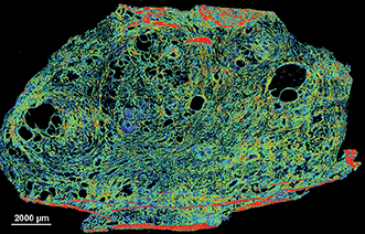
This image shows a section of the spinal cord of a mouse embryo. Neurons appear green. Commissural axons (which connect the two sides of the brain) appear as long, u-shaped threads, and the bottom, yellow segment of the structure represents the midline (between brain hemispheres). (credit: Laboratory of Brain Development and Repair/ The Rockefeller University)

Rockefeller University researchers have discovered a molecule secreted by cells in the spinal cord that helps guide axons (neuron extensions) during a critical stage of central nervous system development in the embryo. The finding helps solve the mystery: how do the billions of neurons in the embryo nimbly reposition themselves within the brain and spinal cord, and connect branches to form neural circuits?

Working in mice, the researchers identified an axon guidance factor, NELL2, and explained how it makes commissural axons (which connect the two sides of the brain).

The findings could help scientists understand what goes wrong in a rare disease called horizontal gaze palsy with progressive scoliosis. People affected by the condition often suffer from abnormal spine curvature, and are unable to move their eyes horizontally from side to side. The study was published Thursday Nov. 19 in the journal Science.


Abstract of Operational redundancy in axon guidance through the multifunctional receptor Robo3 and its ligand NELL2

Axon pathfinding is orchestrated by numerous guidance cues, including Slits and their Robo receptors, but it remains unclear how information from multiple cues is integrated or filtered. Robo3, a Robo family member, allows commissural axons to reach and cross the spinal cord midline by antagonizing Robo1/2–mediated repulsion from midline-expressed Slits and potentiating deleted in colorectal cancer (DCC)–mediated midline attraction to Netrin-1, but without binding either Slits or Netrins. We identified a secreted Robo3 ligand, neural epidermal growth factor-like-like 2 (NELL2), which repels mouse commissural axons through Robo3 and helps steer them to the midline. These findings identify NELL2 as an axon guidance cue and establish Robo3 as a multifunctional regulator of pathfinding that simultaneously mediates NELL2 repulsion, inhibits Slit repulsion, and facilitates Netrin attraction to achieve a common guidance purpose.

A sensory illusion that makes yeast cells self-destruct

A possible tactic for cancer therapeutics
November 20, 2015

http://www.kurzweilai.net/a-sensory-illusion-that-makes-yeast-cells-self-destruct


Effects of osmotic changes on yeast cell growth. (A) Schematic of the flow chamber used to create osmotic level oscillations for different periods of time. (B) Cell growth for these periods. The graphs show the average number of progeny cells (blue) before and after applying stress for different periods (gray shows orginal “no stress” line). The inset shows representative images of cells for two periods. (credit: Amir Mitchell et al./Science)

UC San Francisco researchers have discovered that even brainless single-celled yeast have “sensory biases” that can be hacked by a carefully engineered illusion — a finding that could be used to develop new approaches to fighting diseases such as cancer.

In the new study, published online Thursday November 19 in Science Express, Wendell Lim, PhD, the study’s senior author*, and his team discovered that yeast cells falsely perceive a pattern of osmotic levels (by applying potassium chloride) that alternate in eight minute intervals as massive, continuously increasing stress. In response, the microbes over-respond and kill themselves. (In their natural environment, salt stress normally gradually increases.)

The results, Lim says, suggest a whole new way of looking at the perceptual abilities of simple cells and this power of illusion could even be used to develop new approaches to fighting cancer and other diseases.

“Our results may also be relevant for cellular signaling in disease, as mutations affecting cellular signaling are common in cancer, autoimmune disease, and diabetes,” the researchers conclude in the paper. “These mutations may rewire the native network, and thus could modify its activation and adaptation dynamics. Such network rewiring in disease may lead to changes that can be most clearly revealed by simulation with oscillatory inputs or other ‘non-natural’ patterns.

“The changes in network response behaviors could be exploited for diagnosis and functional profiling of disease cells, or potentially taken advantage of as an Achilles’ heel to selectively target cells bearing the diseased network.”

https://youtu.be/CuDjZrM8xtA
UC San Francisco (UCSF) | Sensory Illusion Causes Cells to Self-Destruct

* Chair of the Department of Cellular and Molecular Pharmacology at UCSF, director of the UCSF Center for Systems and Synthetic Biology, and a Howard Hughes Medical Institute (HHMI) investigator.

** Normally, sensor molecules in a yeast cell detect changes in salt concentration and instruct the cell to respond by producing a protective chemical. The researchers found that the cells were perfectly capable of adapting when they flipped the salt stress on and off every minute or every 32 minutes. But to their surprise, when they tried an eight-minute oscillation of precisely the same salt level the cells quickly stopped growing and began to die off.


Abstract of Oscillatory stress stimulation uncovers an Achilles’ heel of the yeast MAPK signaling network

Cells must interpret environmental information that often changes over time. We systematically monitored growth of yeast cells under various frequencies of oscillating osmotic stress. Growth was severely inhibited at a particular resonance frequency, at which cells show hyperactivated transcriptional stress responses. This behavior represents a sensory misperception—the cells incorrectly interpret oscillations as a staircase of ever-increasing osmolarity. The misperception results from the capacity of the osmolarity-sensing kinase network to retrigger with sequential osmotic stresses. Although this feature is critical for coping with natural challenges—like continually increasing osmolarity—it results in a tradeoff of fragility to non-natural oscillatory inputs that match the retriggering time. These findings demonstrate the value of non-natural dynamic perturbations in exposing hidden sensitivities of cellular regulatory networks.

Google Glass helps cardiologists complete difficult coronary artery blockage surgery

November 20, 2015

http://www.kurzweilai.net/google-glass-helps-cardiologists-in-challenging-coronary-artery-blockage-surgery


Google Glass allowed the surgeons to clearly visualize the distal coronary vessel and verify the direction of the guide wire advancement relative to the course of the occluded vessel segment. (credit: Maksymilian P. Opolski et al./Canadian Journal of Cardiology

Cardiologists from the Institute of Cardiology, Warsaw, Poland have used Google Glass in a challenging surgical procedure, successfully clearing a blockage in the right coronary artery of a 49-year-old male patient and restoring blood flow, reports the Canadian Journal of Cardiology.

Chronic total occlusion, a complete blockage of the coronary artery, sometimes referred to as the “final frontier in interventional cardiology,” represents a major challenge for catheter-based percutaneous coronary intervention (PCI), according to the cardiologists.

That’s because of the difficulty of recanalizing (forming new blood vessels through an obstruction) combined with poor visualization of the occluded coronary arteries.

Coronary computed tomography angiography (CTA) is increasingly used to provide physicians with guidance when performing PCI for this procedure. The 3-D CTA data can be projected on monitors, but this technique is expensive and technically difficult, the cardiologists say.

So a team of physicists from the Interdisciplinary Centre for Mathematical and Computational Modelling of theUniversity of Warsaw developed a way to use Google Glass to clearly visualize the distal coronary vessel and verify the direction of the guide-wire advancement relative to the course of the blocked vessel segment.

Three-dimensional reconstructions displayed on Google Glass revealed the exact trajectory of the distal right coronary artery (credit: Maksymilian P. Opolski et al./Canadian Journal of Cardiology)

The procedure was completed successfully, including implantation of two drug-eluting stents.

“This case demonstrates the novel application of wearable devices for display of CTA data sets in the catheterization laboratory that can be used for better planning and guidance of interventional procedures, and provides proof of concept that wearable devices can improve operator comfort and procedure efficiency in interventional cardiology,” said lead investigatorMaksymilian P. Opolski, MD, PhD, of the Department of Interventional Cardiology and Angiology at the Institute of Cardiology, Warsaw, Poland.

“We believe wearable computers have a great potential to optimize percutaneous revascularization, and thus favorably affect interventional cardiologists in their daily clinical activities,” he said. He also advised that “wearable devices might be potentially equipped with filter lenses that provide protection against X-radiation.


Abstract of First-in-Man Computed Tomography-Guided Percutaneous Revascularization of Coronary Chronic Total Occlusion Using a Wearable Computer: Proof of Concept

We report a case of successful computed tomography-guided percutaneous revascularization of a chronically occluded right coronary artery using a wearable, hands-free computer with a head-mounted display worn by interventional cardiologists in the catheterization laboratory. The projection of 3-dimensional computed tomographic reconstructions onto the screen of virtual reality glass allowed the operators to clearly visualize the distal coronary vessel, and verify the direction of the guide wire advancement relative to the course of the occluded vessel segment. This case provides proof of concept that wearable computers can improve operator comfort and procedure efficiency in interventional cardiology.

Modulating brain’s stress circuity might prevent Alzheimer’s disease

Drug significantly prevented onset of cognitive and cellular effects in mice
November 17, 2015

http://www.kurzweilai.net/modulating-brains-stress-circuity-might-prevent-alzheimers-disease


Effect of drug treatment on AD mice in control group (left) or drug (right) on Ab plaque load. (credit: Cheng Zhang et al./Alzheimer’s & Dementia: The Journal of the Alzheimer’s Association)

In a novel animal study design that mimicked human clinical trials, researchers at University of California, San Diego School of Medicine report that long-term treatment using a small-molecule drug that reduces activity of  the brain’s stress circuitry significantly reduces Alzheimer’s disease (AD) neuropathology and prevents onset of cognitive impairment in a mouse model of the neurodegenerative condition.

The findings are described in the current online issue of the journal Alzheimer’s & Dementia: The Journal of the Alzheimer’s Association.

Previous research has shown a link between the brain’s stress signaling pathways and AD. Specifically, the release of a stress-coping hormone called corticotropin-releasing factor (CRF), which is widely found in the brain and acts as a neurotransmitter/neuromodulator, is dysregulated in AD and is associated with impaired cognition and with detrimental changes in tau protein and increased production of amyloid-beta protein fragments that clump together and trigger the neurodegeneration characteristic of AD.

“Our work and that of our colleagues on stress and CRF have been mechanistically implicated in Alzheimer’s disease, but agents that impact CRF signaling have not been carefully tested for therapeutic efficacy or long-term safety in animal models,” said the study’s principal investigator and corresponding author Robert Rissman, PhD, assistant professor in the Department of Neurosciences and Biomarker Core Director for the Alzheimer’s Disease Cooperative Study (ADCS).

The researchers determined that modulating the mouse brain’s stress circuitry mitigated generation and accumulation of amyloid plaques widely attributed with causing neuronal damage and death. As a consequence, behavioral indicators of AD were prevented and cellular damage was reduced.  The mice began treatment at 30-days-old — before any pathological or cognitive signs of AD were present — and continued until six months of age.

One particular challenge, Rissman noted, is limiting exposure of the drug to the brain so that it does not impact the body’s ability to respond to stress. “This can be accomplished because one advantage of these types of small molecule drugs is that they readily cross the blood-brain barrier and actually prefer to act in the brain,” Rissman said.

“Rissman’s prior work demonstrated that CRF and its receptors are integrally involved in changes in another AD hallmark, tau phosphorylation,” said William Mobley, MD, PhD, chair of the Department of Neurosciences and interim co-director of the Alzheimer’s Disease Cooperative Study at UC San Diego. “This new study extends those original mechanistic findings to the amyloid pathway and preservation of cellular and synaptic connections.  Work like this is an excellent example of UC San Diego’s bench-to-bedside legacy, whereby we can quickly move our basic science findings into the clinic for testing,” said Mobley.

Rissman said R121919 was well-tolerated by AD mice (no significant adverse effects) and deemed safe, suggesting CRF-antagonism is a viable, disease-modifying therapy for AD. Drugs like R121919 were originally designed to treat generalized anxiety disorder, irritable bowel syndrome and other diseases, but failed to be effective in treating those disorders.

Rissman noted that repurposing R121919 for human use was likely not possible at this point. He and colleagues are collaborating with the Sanford Burnham Prebys Medical Discovery Institute to design new assays to discover the next generation of CRF receptor-1 antagonists for testing in early phase human safety trials.

“More work remains to be done, but this is the kind of basic research that is fundamental to ultimately finding a way to cure — or even prevent —Alzheimer’s disease,” said David Brenner, MD, vice chancellor, UC San Diego Health Sciences and dean of UC San Diego School of Medicine. “These findings by Dr. Rissman and his colleagues at UC San Diego and at collaborating institutions on the Mesa suggest we are on the cusp of creating truly effective therapies.”


Abstract of Corticotropin-releasing factor receptor-1 antagonism mitigates beta amyloid pathology and cognitive and synaptic deficits in a mouse model of Alzheimer’s disease

Introduction: Stress and corticotropin-releasing factor (CRF) have been implicated as mechanistically involved in Alzheimer’s disease (AD), but agents that impact CRF signaling have not been carefully tested for therapeutic efficacy or long-term safety in animal models.

Methods: To test whether antagonism of the type-1 corticotropin-releasing factor receptor (CRFR1) could be used as a disease-modifying treatment for AD, we used a preclinical prevention paradigm and treated 30-day-old AD transgenic mice with the small-molecule, CRFR1-selective antagonist, R121919, for 5 months, and examined AD pathologic and behavioral end points.

Results: R121919 significantly prevented the onset of cognitive impairment in female mice and reduced cellular and synaptic deficits and beta amyloid and C-terminal fragment-β levels in both genders. We observed no tolerability or toxicity issues in mice treated with R121919.

Discussion: CRFR1 antagonism presents a viable disease-modifying therapy for AD, recommending its advancement to early-phase human safety trials.

Allen Institute researchers decode patterns that make our brains human
Conserved gene patterning across human brains provide insights into health and disease
November 17, 2015

http://www.kurzweilai.net/allen-institute-researchers-decode-patterns-that-make-our-brains-human


Percentage of known neuron-, astrocyte- and oligodendrocyte-enriched genes in 32 modules, ordered by proportion of neuron-enriched gene membership. (credit: Michael Hawrylycz et al./Nature Neuroscience)

Allen Institute researchers have identified a surprisingly small set of just 32 gene-expression patterns for all 20,000 genes across 132 functionally distinct human brain regions, and these patterns appear to be common to all individuals.

In research published this month in Nature Neuroscience, the researchers used data for six brains from the publicly available Allen Human Brain Atlas. They believe the study is important because it could provide a baseline from which deviations in individuals may be measured and associated with diseases, and could also provide key insights into the core of the genetic code that makes our brains distinctly human.

While many of these patterns were similar in human and mouse, many genes showed different patterns in human. Surprisingly, genes associated with neurons were most conserved (consistent) across species, while those for the supporting glial cells showed larger differences. The most highly stable genes (the genes that were most consistent across all brains) include those associated with diseases and disorders like autism and Alzheimer’s, and these genes include many existing drug targets.

These patterns provide insights into what makes the human brain distinct and raise new opportunities to target therapeutics for treating disease.

The researchers also found that the pattern of gene expression in cerebral cortex is correlated with “functional connectivity” as revealed by neuroimaging data from the Human Connectome Project.

“The human brain is phenomenally complex, so it is quite surprising that a small number of patterns can explain most of the gene variability across the brain,” says Christof Koch, Ph.D., President and Chief Scientific Officer at the Allen Institute for Brain Science. “There could easily have been thousands of patterns, or none at all. This gives us an exciting way to look further at the functional activity that underlies the uniquely human brain.”


Abstract of Canonical genetic signatures of the adult human brain

The structure and function of the human brain are highly stereotyped, implying a conserved molecular program responsible for its development, cellular structure and function. We applied a correlation-based metric called differential stability to assess reproducibility of gene expression patterning across 132 structures in six individual brains, revealing mesoscale genetic organization. The genes with the highest differential stability are highly biologically relevant, with enrichment for brain-related annotations, disease associations, drug targets and literature citations. Using genes with high differential stability, we identified 32 anatomically diverse and reproducible gene expression signatures, which represent distinct cell types, intracellular components and/or associations with neurodevelopmental and neurodegenerative disorders. Genes in neuron-associated compared to non-neuronal networks showed higher preservation between human and mouse; however, many diversely patterned genes displayed marked shifts in regulation between species. Finally, highly consistent transcriptional architecture in neocortex is correlated with resting state functional connectivity, suggesting a link between conserved gene expression and functionally relevant circuitry.

Read Full Post »

Laser Technology

Larry H. Bernstein, MD, FCAP, Curator

LPBI


Laser Focus World   www.laserfocusworld.com

Ultrafast lasers simplify fabrication of 3D hydrogel tissue scaffolds

Multimode holographic waveguides tackle in vivo biological imaging

Mid-infrared Lasers CMOS silicon-on-sapphire process produces broad mid-IR supercontinuum

Looking Back/Looking Forward: Positioning equipment—the challenge of building a solid foundation for optics
Stability and precision have been crucial for optics since the 19th century.
Jeff Hecht

Monolithic DFB QCL array aims at handheld IR spectral analysis
Many QCLs combined on a single chip demonstrate fully electronic wavelength tuning for stand-off IR spectroscopy of explosives and other materials.
Mark F. Witinski, Romain Blanchard, Christian Pfluegl, Laurent Diehl, Biao Li, Benjamin Pancy, Daryoosh Vakhshoori, and Federico Capasso

Quantum dots and silicon photonics combine in broadband tunable laser
A new wavelength-tunable laser diode combines quantum-dot technology and silicon photonics with large optical gains around the 1310 nm telecom window.
Tomohiro Kita and Naokatsu Yamamoto

Computer modeling boosts laser device development
A full quantitative understanding of laser devices is boosted by computer modeling, which is not only essential for efficient development processes, but also for identifying the causes of unexpected behavior.
Rüdiger Paschotta


Monolithic DFB QCL array aims at handheld IR spectral analysis
MARK F. WITINSKI, ROMAIN BLANCHARD, CHRISTIAN PFLUEGL, LAURENT DIEHL, BIAO LI, BENJAMIN PANCY, DARYOOSH VAKHSHOORI, and FEDERICO CAPASSO

Advances in infrared (IR) laser sources, optics, and detectors promise major new advances in areas of chemical analysis such as trace-gas monitoring, IR microscopy, industrial safety, and security.

One key type of photonic device that has yet to reach its full potential is a truly portable noncontact (standoff), chemically versatile analyzer for fast Fourier-transform infrared (FTIR)quality spectral examination of nearly any condensed-phase material. The unique challenges of standoff IR spectroscopy actually extend beyond advances in IR hardware, requiring the proper combination of several areas of expertise: cutting-edge optical design and laser fabrication, integrated laser electronics, thermally efficient hermetic packaging, statistical signal processing methods, and deep chemical knowledge.

At the core of the approach we have taken at Pendar Technologies is the monolithic distributed feedback (DFB) quantum-cascade laser (QCL) array. Invented in Federico Capasso’s group at Harvard University (Cambridge, MA) and licensed exclusively to Pendar, the continuously wavelength-tunable QCL array source is a highly stable broadband source that can be used for illumination in reflectance spectroscopy. Each element of the array is individually addressable and emits at a different wavelength by design.

The advantages of these QCL arrays over external-cavity (EC) QCLs stem from (1) the monolithic structure of QCL arrays and (2) their fully electronic wavelength tuning— that is, no moving gratings, allowing for much-higher-speed acquisition through improved amplitude and wavelength stability. When integrated into a system, the result is robust, stable, and field-deployable.

One of the key advances that has enabled this technology to be fielded is the high-yield fabrication of each laser ridge in the QCL array from a single wafer such that every channel simultaneously meets the specified wavelength, power, and single-mode suppression ratio. Each of these parameters is critical to both efficient beam combining and to obtaining high-quality molecular spectroscopy once integrated.

With these hurdles largely overcome, the payoff in terms of spectrometer performance lies largely in a demonstrated shot-to-shot amplitude stability in pulsed mode of <0.1%—a factor of 50 more stable than is typical for EC QCLs, even when used in the lab. Most importantly, the DFB QCL noise is random, and averages toward an Allan variance limit quickly such that detector-noise-limited, high-quality spectra can be obtained for trace levels (for example, 1–50 µg/cm2) of typical powders in just 100 ms.

More DFB array advantages While the stability advantage of DFBs vs. EC configurations has been well established, there are a few less-obvious aspects to DFB arrays that make them more suitable to real-world spectroscopy tools and, in particular, portable spectroscopy tools. For one, the laser array as a whole can maintain a 100% duty cycle while each laser in the array requires operation only over a 100/n (%) duty cycle, where n is the number of lasers in the array. Put another way, a laser array consisting of only pulsed QCLs can operate as a truly continuous-wave (CW) system, allowing for high-measurement duty cycle while possibly reducing the cost of fabrication.

In a related way, generating light for an array that has a 100% aggregate duty cycle (by using, for instance, 32 lasers at 3% duty cycle), the thermal heat-sinking requirements of the source are dramatically reduced. Indeed, our packaged prototypes do not even require active cooling to keep the system cool enough to run. A thermoelectric cooler is built into the package only to stabilize the temperature, which therefore stabilizes the 32 wavelengths (see Fig. 1).
FIGURE 1. A 200 cm-1 prototype QCL array with 32 QCLs is shown prior to beam combining and packaging (a), and experimental spectra from 32 adjacent QCLs are seen (b). (Courtesy of Pendar Technologies)
Finally, the arbitrary programmability of the QCL array opens up many new possibilities for experimental optimization. Certain lasers can be skipped, multiple lasers can fire at once, repetition rates and pulse durations can be set for each element, and so on. These advantages are only truly realized when the QCL array is instrumented into a full system.

Looking holistically at how best to integrate this new capability into a full system, it is critical to draft the link equations that govern the use of electrons to produce photons, the collection of photons scattered back, and finally the conversion from raw spectral information to chemical identification. In the case of mid-IR material identification, it becomes clear that three aspects are particularly consequential: (1) How broad a wavelength range is needed for the tool to be of maximum specificity without producing redundant or useless chemical information (that is, how many laser channels should be used, how should they be spaced with respect to one another, and over what total wavelength regime should they be spaced); (2) the mechanical and electro-optical design of the instrument; and (3) how to get the highest performance regressions against reference spectra while maintaining the high-speed identification that the QCL array actually enables.

With regard to the wavelength regions of interest (see Fig. 2), most of the spectral richness of an IR spectrum is centered in two bands, generally referred to as the functional group region (about 3.3–5.5 µm) and the fingerprint region (about 7–11 µm). The first is typically dominated by the stretch modes of certain common bond groups, while the latter includes bending modes of some functional groups as well as lower frequency modes that are characteristic of the macromolecule “backbone”—for instance, the torsional modes of a toluene ring found in many highly energetic materials. With support from the Department of Homeland Security (DHS)’s Widely Tunable Infrared Source (WTIRS) program and from the Army Research Lab, Pendar is developing a compact array module that fully covers 7–11 µm (900–1430 cm-1).

FIGURE 2. An assemblage of IR spectra of many common explosives shows that each has at least one unique absorption feature in the wavelength ranges selected. The blue shaded box indicates strong water interference in the troposphere. The figure intentionally spans beyond 1800 cm-1 so as to illustrate that no new information is gained for this chemical class by shifting the longwave-IR (LWIR) source further to the blue until the midwave-IR (MWIR) is reached.


System architecture drivers To maximize signal-to-noise (SNR) while minimizing the required acquisition time, the system architecture is driven by the following first-order considerations: 1. Increasing the laser power enabled by relaxed thermal constraints as the heat load is distributed over several modules (arrays) and laser waveguides. 2. Maximization of the measurement duty cycle enabled by the fast purely electronic control of the array, allowing close to zero-delay switching between lasers— that is, a laser is on at any time. This is also enabled by the distributed heat load among the laser units. 3. Improved source stability, wavelength accuracy, pulse-to-pulse amplitude, and frequency repeatability—all of which are needed to ensure that the source noise is not the limiting form of noise (compared to detector or speckle noise). Other researchers have studied the source-noise problem of commercial EC QCLs as well and concluded that the order-of-magnitude advantage in minimum detectable absorbance (MDA) offered by a DFB QCL carries through the full experiment.

Finally, once the spectra are digitized, the system must use complex chemometrics algorithms to ensure confident identification of threats in the presence of chemical clutter, deliberate interferents, and unknown backgrounds, without the intervention of an expert user. Our approach to real-time chemometrics is centered on the fact that for chemically cluttered situations, spectral libraries alone—no matter how large—cannot constitute the sole basis for chemometric analysis. Microphysics modeling and experimentation are also required, particularly in regard to crystal size distribution, clutter interactions, and chemical photolysis/reactions.

The key advance lies in the incorporation of chemical and physical understanding of the targets and their co-indicators. We are currently developing a four-tiered approach to the spectroscopic algorithms challenge:

1. Physics-based models. Reliable chemical detection from standoff measurements will involve transformation of the chemical signatures in the reference spectral library to reflect the physical and environmental conditions of the experiment. A physics-based model will thus be included in the detection algorithm to help us model the variability in a reference spectrum as a function of effects such as vapor pressure, deliquescence, photochemical lifetime, reactive lifetime, decomposition products, and so on to facilitate better comparison with the measured spectrum.

2. Situational effects. Effects of different substrates and their properties on the chemical signatures and the angular dependence of spectra that are not clearly linked to equations of physics and chemistry will be experimentally evaluated and included in the detection algorithm. In particular, experimentally measuring such variability will help us algorithmically model the variability of chemical signatures from some “gold standard” reference signature, which—in
addition to the physical model—will enable better detection strategies.

3. Feature-based classification. Extraction of relevant feature vectors from the reference library spectra and the knowledge of the chemistry to form a hierarchical decision tree that will help us provide different levels of classification based on the customer requirements. For instance, if a customer is only interested in finding out whether a given chemical is an explosive, then we might save on computational cost by avoiding searching through the leaves of the decision tree to find out the exact chemical.

4. Real-time atmospheric measurements. Once validated, the model will be suitable for field implementation by the inclusion of an integrated sensor suite that simultaneously records atmospheric pressure, temperature, relative humidity, solar flux, wind magnitude, and water-vapor mixing ratio. With these design drivers considered, Pendar recently completed the build of a handheld demonstration system.

Figure 3 shows the experimentally obtained spectra for two nonhazardous chemical targets as a function of stand-off distance. The yellow line in each panel shows the library FTIR (“true”) spectrum for each. Agreements of r2 > 0.9 were typical. With the prototype system as an extrapolation point, continued, focused advances in the technology are now underway to open myriad frontiers in molecular spectroscopy.


FIGURE 3. Standoff spectra of of acetaminophen and ibuprofen for three target distances. The black line shows the FTIR of the same using a diffuse reflectance accessory. The only data processing shown is the normalization of the curve areas to a common value.


ACKNOWLEDGEMENT Pendar Technologies was formed in August 2015 through a merger between Pendar Medical (Cambridge, MA), a portable spectroscopy company founded by Daryoosh Vakhshoori (who was previously at Ahura Scientific and CoreTek), and QCL sensing startup Eos Photonics (Cambridge, MA), a Harvard spinoff founded by professor Federico Capasso and his postdocs.


Quantum dots and silicon photonics combine in broadband tunable laser
TOMOHIRO KITA and NAOKATSU YAMAMOTO

A new wavelength-tunable laser diode combines quantum-dot (QD) technology and silicon photonics with large optical gains around the 1310 nm telecom window and is amenable to integration of other passive and active components towards a truly integrated photonic platform.

A new heterogeneous wavelength-tunable laser diode, configured using quantum dot (QD) and silicon photonics technology, leverages large optical gains in the 1000–1300 nm wavelength region using a scalable platform for highly integrated photonics devices. A cooperative research effort between Tohoku University (Sendai, Japan) and the National Institution of Information and Communication Technology (NICT; Tokyo, Japan) has resulted in the demonstration of broadband tuning of 44 nm around a 1230 nm center wavelength with an ultrasmall device footprint, with many more configurations with various performance metrics possible.

Recently developed high-capacity optical transmission systems use wavelength-division multiplexing (WDM) systems with dense frequency channels. Because the frequency channels in the conventional band (C-band) at 1530–1565 nm are overcrowded, the frequency utilization efficiency of such WDM systems becomes saturated. However, extensive and unexploited frequency resources are buried in the near-infrared (NIR) wavelength regions such as the thousand (T) and original (O) bands between 1000 and 1260 nm and 1260 and 1350 nm, respectively. Quantum dot-based optical gain media have various attractive characteristics, including ultrabroad optical gain bandwidths, high-temperature device stability, and small line width enhancement factors, as well as silicon photonic wire waveguides based on silicon-on-insulator (SOI) structures that are easily amenable to constructing highly integrated photonics devices.1-4

Quantum dot-based optical gain media have various attractive characteristics, including ultrabroad optical gain bandwidths, high-temperature device stability, and small linewidth enhancement factors, as well as silicon photonic wire waveguides based on silicon-on-insulator (SOI) structures that are easily amenable to constructing highly integrated photonics devices.1-4

The photonic devices used for shortrange data transmission are required to have a small footprint and low power consumption. Therefore, compact, low-power wavelength-tunable laser diodes are key devices for use in higher-capacity data transmission systems that have been designed to use these undeveloped frequency bands, and our heterogeneous tunable wavelength laser diode consisting of a QD optical gain medium and a silicon photonics external cavity is a promising candidate.5

Quantum dot optical amplifier Ultrabroadband optical gain media spanning the T- and O-band are effectively fabricated by using QD growth techniques on large-diameter gallium-arsenide (GaAs) substrates. Our sandwiched sub-nano-separator (SSNS) growth technique is a simple and efficient method for obtaining high-quality QDs (see Fig. 1).


FIGURE 1. A cross-section (a) shows a quantum dot (QD) device grown using the SSNS technique, resulting in a high-density, highquality QD structure (b) that is used to create a typical SOA (c) using QD optical gain.


In the SSNS method, three monolayers (each around 0.85 nm thick) of GaAs thin film are grown in an indium GaAs (InGaAs) quantum well (QW) under the QDs. We had previously observed many large, coalescent dots that could induce crystal defects in QD devices using a conventional growth technique without SSNS. Now, we can obtain high-density (8.2 × 1010 cm-2), high-quality QD structures since the SSNS technique successfully suppresses the formation of coalescent dots.

For single-mode transmission, a ridgetype semiconductor waveguide was fabricated for single-mode transmission. The cross-section of the semiconductor optical amplifier (SOA) has an anti-reflection (AR) coating facet to connect a silicon photonics chip with low reflection and a cleaved facet used as a reflecting mirror in the laser cavity.

To fabricate the SOA, the SSNS growth technique was combined with molecular beam epitaxy. Quantum dots comprised of indium arsenide (InAs) with 20–30 nm diameters were grown within an InGaAs QW. Seven of these QD layers are stacked to achieve broadband optical gain. Subsequently, this QD-SOA is used as an optical gain medium for the heterogeneous laser, which can be complemented by other communication technology devices such as a high-speed modulator, a two-mode laser, and a photoreceiver.6, 7

Silicon photonics ring resonator filter With the QD-SOA fabricated, a wavelength filter is fabricated next using silicon photonics techniques. It includes a spot-size converter that has a silicon oxide (SiOx) core and a tapered Si waveguide that connects the QD-SOA to the Si photonic wire waveguide while minimizing optical reflections and coupling losses (see Fig. 2).


FIGURE 2. A microscope image (a) shows a silicon-photonicsbased wavelength-tunable filter. In a transmittance analysis (b), the red and blue dotted lines indicate the transmittance of a small ring resonator with free spectral range FSR1 and a large ring resonator with FSR2, respectively, and the solid line indicates the product of each transmittance. The tuning wavelength range is determined from the FSR difference of the two rings. A smaller difference in the FSR provides a wider wavelength tuning range, even when the transmittance difference between the main and side peaks is small.


The wavelength-tunable filter consists of two ring resonators of different size. The Vernier effect of these two ring resonators allows only light of a specific wavelength to reflect to the QD-SOA. Furthermore, Tantalum micro-heaters formed above the resonators provide a means whereby the laser wavelength can be tuned through application of the thermooptic effect.

Essentially, the wavelength tuning operation of the double ring resonator wavelength filter is achieved through Vernier effects wherein a ring resonator acts as a wavelength filter with constant wavelength interval called the free spectral range (FSR), which is inversely proportional to the circumference of the ring. The tuning wavelength range is determined from the FSR difference of the two rings with FSR1 and FSR2.

A smaller difference in the FSR provides a wider wavelength tuning range, even when the transmittance difference between the main and side peaks is small. On the other hand, a sufficiently large transmittance difference is required to achieve stable single-mode lasing and is obtained using large FSR ring resonators.

Silicon photonics allows us to fabricate an ultrasmall ring resonator with large FSR because of the strong light confinement in the waveguide. The ring resonator consists of four circle quadrants and four straight lines and the radius of the circle was chosen to be 10 µm to avoid bending losses. The FSRs of the ring resonators and the coupling efficiency between the bus-waveguide and the ring resonator are optimized to obtain wide wavelength tuning range and sufficient transmittance difference.

The FSRs and the coupling efficiencies of the double ring resonators are designed to obtain a 50 nm wavelength tuning range and 1 dB transmittance difference. We have since fabricated various wavelength-tunable laser diodes, including a broadband tunable laser diode, a narrow spectral-linewidth tunable laser diode, and a high-power integrated tunable laser diode by using a silicon photonics wavelength filter and a commercially available C-band SOA.8, 9

The tunable laser diode Using stepper motor controllers, the QD-SOA—kept at approximately 25°C using a thermoelectric cooler—and the silicon photonics wavelength filter are butt-jointed (see Fig. 3). The lasing wavelength is controlled by the temperature of a micro-heater placed on the ring resonators. With physical footprints of 600 µm × 1 mm and 1 × 2 mm for the wavelength filter and the QD-SOA, respectively, the total device size of the tunable laser diode is just 1 × 3 mm.


FIGURE 3. A schematic shows how the heterogeneous wavelengthtunable laser diode is constructed.


Measured using a lensed fiber, the laser output from the cleaved facet of the QD-SOA shows single-mode lasing characteristics with a laser oscillation threshold current of 230 mA. Maximum fiber-coupled output power is 0.4 mW when the QD-SOA injection current is 500 mA. As the ring resonator temperature is increased by a heater with 2.1 mW/nm power consumption, the superimposed lasing spectra show a 44 nm wavelength tuning range with more than a 37 dB side-mode-suppression ratio between the ring resonator’s modes. The 44 nm wavelength tuning range of our heterogeneous QD/Si photonics wavelength-tunable laser is, to our knowledge, the broadest achieved to date. The 44 nm tuning range around 1230 nm corresponds to 8.8 THz in the frequency domain, which is far larger than the 4.4 THz frequency that is available within the C-band.

Our heterogeneous laser is suitable for use as a light source on a silicon photonics platform that includes other optical components such as high-speed modulators and germanium (Ge)-based detectors. In addition to application as a single-chip broadband optical transceiver for telecommunications, the laser could also be applied to biomedical imaging applications such as optical coherence tomography (OCT), considering the low absorption of NIR light at 1310 nm in the presence of water.

ACKNOWLEDGEMENTS This research was partially supported by the Strategic Information and Communications R&D Promotion Program (SCOPE), of Japan’s Ministry of Internal Affairs and Communications and a Grant-in-Aid for Scientific Research of the Japan Society for the Promotion of Science.

REFERENCES

1. Y. Arakawam and H. Sakaki, Appl. Phys. Lett., 40, 11, 939–941 (1982).

2. D. L. Huffaker et al., Appl. Phys. Lett., 73, 18, 2564–2566 (1998).

3. R. A. Soref, Proc. IEEE, 81, 12, 1687–1706 (1993).

4. B. Jalai and S. Fathpour, J. Lightwave Technol., 24, 12, 4600–4615 (2006).

5. T. Kita et al., Appl. Phys. Express, 8, 6, 062701 (2015).

6. N. Yamamoto et al., Jpn. J. Appl. Phys., 51, 2S, 02BG08 (2012).

7. N. Yamamoto et al., Proc. OFC, Los Angeles, CA, paper W2A.24 (Mar. 2015).

8. T. Kita et al., Appl. Phys. Lett., 106, 11, 111104 (2015).

9. N. Kobayashi et al., J. Lightwave Technol., 33, 6, 1241–1246 (2015).


Computer modeling boosts laser device development
RÜDIGER PASCHOTTA

A full quantitative understanding of laser devices is boosted by computer modeling, which is not only essential for efficient development processes, but also for identifying the causes of unexpected behavior.

Computer modeling can give valuable insight into the function of laser devices. It can even reveal internal details that could not be observed in experiments, and thus allows one to develop a comprehensive understanding from which laser development can enormously profit. For example, the performance potentials of certain technologies can be fully exploited and time-consuming and expensive iterations in the development process can be avoided. Some typical examples clarify the benefits of computer modeling for improved laser device development.

Example 1: Q-switched lasers

FIGURE 1. Evolution of the transverse beam profile (shown with a color scale) and the optical power (black circles, in arbitrary units) in an actively Q-switched laser is simulated with RP Fiber Power software using numerical beam propagation. The color scale is normalized for each round trip according to the timedependent optical power so that the variation of the beam diameter can be seen.

Example 2: Mode-locked lasers

Example 3: Ultrashortpulse fiber amplifiers

FIGURE 2. The evolution of pulse energy and forward ASE powers in a four-stage fiber amplifier system with various types of ASE suppression between the stages, calculated with a comprehensive computer model

FIGURE 3. Form-based software can be used to model laser devices such as a fiber amplifier. It is essential that such forms be made or modified by the user or by technical support, so that they can be tailored to specific applications.

…. more

Documentation and support For any modeling task, documentation of methods and results is essential. The documentation must not only explain details of the user interface, but must inform the user what kind of physical model was used, what simplifying assumptions were made, and what limitations need to be considered. Unfortunately, software documentation is often neglected. In case of doubt, competent technical support should be available—not only for helping with the handling of the software, but also offering detailed technical and scientific advice. For example, a beginner may find it difficult to decide which kind of model should be implemented for a certain purpose and which possibly disturbing effects need to be considered. Such support should come from a competent expert in the field rather than just a programmer.
Rüdiger Paschotta is founder and executive of RP Photonics Consulting, Bad Dürrheim, Germany; e-mail: paschotta@rp-photonics.com; www.rp-photonics.com


Read Full Post »

developments in medical spectroscopy

Larry H. Bernstein, MD, FCAP, Curator

LPBI


Using QCLs for MIR-Based Spectral Imaging — Applications in Tissue Pathology
A quantum cascade laser (QCL) microscope allows for fast data acquisition, real-time chemical imaging and the ability to collect only spectral frequencies of interest. Due to their high-quality, highly tunable illumination characteristics and excellent signal-to-noise performance, QCLs are paving the way for the next generation of mid-infrared (MIR) imaging methodologies.

Using QCLs for MIR-Based Spectral Imaging — Applications in Tissue Pathology

http://www.photonics.com/images/Web/Articles/2015/9/8/Imaging_Prostate.png


Efficient Spectroscopic Imaging Demonstrated In Vivo
Although optical spectroscopy is routinely used study molecules in cell samples, it is currently not practical to perform in vivo. Now, a converted Raman spectroscopy system has been used to reveal the chemical composition of living tissues in seconds.

Efficient Spectroscopic Imaging Demonstrated In Vivo

http://www.photonics.com/images2/EmailBlasts%5CSpectroscopy/2015/11/Efficient_Spectroscopic_Imaging_Demonstrated_In_Vivo.jpg


Broadband Laser Aimed at Cancer Detection
Covering a wide swath of the mid-infrared region, a new laser system offers greater spectral sensitivity.

Broadband Laser Aimed at Cancer Detection

http://www.photonics.com/images/Web/Articles/2015/9/25/REAS_molecular_1.jpg


Using QCLs for MIR-Based Spectral Imaging — Applications in Tissue Pathology

A quantum cascade laser (QCL) microscope allows for fast data acquisition, real-time chemical imaging and the ability to collect only spectral frequencies of interest. Due to their high-quality, highly tunable illumination characteristics and excellent signal-to-noise performance, QCLs are paving the way for the next generation of mid-infrared (MIR) imaging methodologies.

MICHAEL WALSH, UNIVERSITY OF ILLINOIS AT CHICAGO; MATTHEW BARRE & BENJAMIN BIRD, DAYLIGHT SOLUTIONS

H. Sreedhar*1, V. Varma*2, A. Graham3, Z. Richards1, F. Gambacorata4, A. Bhatt1,
P. Nguyen1, K. Meinke1, L. Nonn1, G. Guzman1, E. Fotheringham5, M. Weida5,
D. Arnone5, B. Mohar5, J. Rowlette5
1 Department of Bioengineering, University of Illinois at Chicago
2 Department of Pathology, University of Illinois at Chicago
3 Department of Bioengineering, University of Illinois at Urbana-Champaign
4 Department of Chemical Engineering, University of Illinois at Chicago
5 Daylight Solutions, San Diego
*Contributed Equally

Real-time, MIR chemical imaging microscopes could soon become powerful frontline screening tools for practicing pathologists. The ability to see differences in the biochemical makeup across a tissue sample greatly enhances a practioner’s ability to detect early stages of disease or disease variants. Today, this is accomplished much as it was 100 years ago — through the use of specially formulated stains and dyes in combination with white light microscopy. A new MIR, QCL-based microscope from Daylight Solutions enables real-time, nondestructive biochemical imaging of tissues without the need to perturb the sample with chemical or heat treatments, thus preserving the sample for follow-on fluorescence tagging, histochemical staining or other “omics” testing within the workflow.
MIR chemical imaging is a well-established absorbance spectroscopy technique; it senses the relative amount of light that molecules absorb due to their unique vibrational resonances falling within the MIR portion of the electromagnetic spectrum (i.e., wavelengths from approximately 2 to 15 µm). This absorption can be detected with a variety of MIR detector types and can provide detailed information about the sample’s chemical composition.

The most common instrument for this type of measurement is known as a Fourier transform infrared (FTIR) spectrometer. FTIR systems use a broadband MIR light source, known as a globar, to illuminate a sample; the absorption spectrum is generated by the use of interferometry. Throughout the past decade, FTIR systems have incorporated linear arrays and 2D focal plane arrays (FPAs) in a microscope configuration to enable a technique known as chemical imaging.

With this approach, the illumination beam is expanded across a sample area, and the data produced is transformed into a hyperspectral data cube — a 2D image of the sample with an absorption profile associated with every pixel. This is a very versatile technique that allows the detailed spatial distribution of chemical content to be analyzed across a sample. Recently, this technique has proved to be very useful within the biomedical imaging sector for label-free, biochemical analyses of cells, tissue and biofluids.

While FTIR microscopy now is established as a powerful technique for a wide variety of applications, the instruments used for this methodology are fundamentally limited by the brightness of the globar source. Users looking to maximize the signal-to-noise ratios, and the associated resolutions of the images produced are forced to use synchrotron facilities, which replace the globar light source with a MIR beam generated by a particle accelerator. This approach can yield excellent results but clearly is not practical for benchtop applications; it is particularly unfit for biomedical imaging applications within clinical settings.

The recent advent of QCLs has provided an ideal light source for next-generation MIR microscopy. They are compact, semiconductor-based lasers that produce high-brightness light in the MIR region. The devices can be manufactured in an external cavity configuration to provide broadly tunable output with a narrow spectral bandwidth at each frequency. In this configuration, a QCL can be tuned across the MIR spectrum to sequentially capture an absorption profile for chemical identification.

Daylight Solutions’ IR microscope incorporates a broadly tunable and high-brightness QCL light source (it is an order of magnitude brighter than a synchrotron), a set of high numerical aperture (NA) diffraction-limited objectives, and an uncooled microbolometer FPA into a compact, benchtop instrument, as shown in Figure 1. The instrument provides rapid, high-resolution chemical images across very large fields of view and also provides a real-time chemical imaging mode. By overcoming the physical size, camera cooling and data collection time requirements of FTIR-based instruments, the microscope is positioned to bring MIR microscopy beyond research settings and into clinical use.

Schematic of a quantum cascade laser (QCL) microscope.


Figure 1. Schematic of a quantum cascade laser (QCL) microscope. Courtesy of Daylight Solutions.


Dr. Michael Walsh of the University of Illinois at Chicago (UIC) conducts research within the pathology department’s Spectral Pathology Lab, which has been using the IR microscope for the past several months. Walsh has been focused on developing chemical imaging techniques, with the ultimate goal of improving diagnoses within the field of tissue pathology.

Currently, the state-of-the-art method-ology used for the diagnosis of most solid-organ diseases is to extract a tissue sample via a biopsy. Tissue inherently has very little contrast and needs to be stained with dyes or probes to visualize and identify cell types and tissue structures. The field of pathology is based on examining the stained tissues, typically using white light, to determine if the tissue morphology deviates from a normal pattern. If the tissue looks abnormal, the disease state may be further subclassified by grade or by predicted outcome. However, the field of pathology is limited by the information that can be derived from the stained tissues and the subjective interpretation of the tissue by a highly trained pathologist.

Spero microscope.


Spero microscope. Courtesy of Daylight Solutions.


UIC’s Spectral Pathology Lab is focused on identifying areas in pathology where current techniques fail, or where there is a need for additional diagnostic or prognostic information that can help improve patient care. Potentially, MIR imaging is a very valuable adjunct to the current practice of pathology. Rather than using only stains, MIR imaging can interrogate the entire biochemistry of the tissue and render a diagnosis in an objective fashion. Traditionally, MIR imaging with an FTIR system has been limited by slow data acquisition speeds and the need to collect the entire spectral data cube. QCL imaging with the Spero microscope has the potential to speed up the data acquisition of images obtained from a tissue sample and to collect only the spectral frequencies of interest. The device also provides real-time imaging of samples at 30 fps, which could allow pathologists to very rapidly identify areas of interest on a tissue biopsy in a manner that is similar to their current clinical workflows. Some examples of the comparison of FTIR-derived and QCL-derived images from multiple organ tissues of interest are presented.

H&E-stained image of a mouse brain section on IR reflective slide, with selected regions labeled: hypothalamus, thalamus, and dentate gyrus.
Figure 2.
(a) H&E-stained image of a mouse brain section on IR reflective slide, with selected regions labeled: hypothalamus, thalamus, and dentate gyrus. (b) Transflectance QCL IR image of same region, prior to staining, at 1652 cm−1, in which the thalamus is clearly distinguished from surrounding regions. (c) Same region at 1548 cm−1. (d) Same region at 1500 cm−1. Courtesy of University of Illinois at Chicago (UIC)/Spectral Pathology Lab.


A tissue section from a mouse brain was scanned using the Spero microscope’s high-magnification objective (12.5×; 0.7 NA; 1.4 × 1.4-µm pixels) at various MIR frequencies in transflection mode, as shown in Figure 2. The tissue then was stained using hematoxylin and eosin (H&E), the most common stain in histopathology, and is displayed in Figure 2a. Using the H&E stain, regions were identified in the brain (thalamus, dentate gyrus and hypothalamus) that correlated with structures in the IR image. By illuminating the tissue at various wavelengths, discrete tissue features exhibit contrast due to the difference in absorption, as highlighted in the IR images taken at 1652, 1548 and 1500 cm−1 in Figure 2b-d, respectively. The microscope also makes it possible to visualize tissue at these individual wavelengths in real time. The identification of cell types and their biochemical changes is of particular interest in neuropathology.

Transmission FTIR image of a 4-µm thick section from a human liver tissue microarray on barium fluoride at 1650 cm-1.
Figure 3.
(a) Transmission FTIR image of a 4-µm thick section from a human liver tissue microarray on barium fluoride at 1650 cm-1. The image was taken with 64 coadditions of successive scans. (b) Transmission image from the Spero microscope of the same tissue at 1652 cm-1, both baseline corrected between 1796 cm-1 and 904 cm-1. In both images, the bright white stripe dividing the tissue core roughly in half is a region of fibrosis (red arrow), while the rest of the tissue on either side is composed primarily of hepatocytes (blue arrow). Courtesy of UIC/Spectral Pathology Lab.


A single biopsy core obtained from human liver tissue was scanned in transmission mode on a barium fluoride substrate by an Agilent Cary 600 Series FTIR microscope (Figure 3a). The FTIR image was acquired using a 36× Cassegrain collecting objective and a 15× Cassegrain condenser for a pixel size of 2.2 × 2.2 µm. Figure 3b shows the same liver core acquired using the Spero microscope with the high-magnification collecting objective (12.5×, 0.7 NA) and condenser objective for a pixel size of 1.4 × 1.4 µm. High-definition IR imaging enables clear contrast and identification of the band of fibrosis in the center of the core and the surrounding regions of liver cells, known as hepatocytes, and is indicated within Figure 3a-b. Acquisition of IR imaging data at the diffraction limit enables chemical information to be recorded from tissue structures at the single-cell level, allowing accurate characterization of individual tissue components, different cell types, varied disease states or other aspects of a tissue section.

Averaged spectra for regions of interest corresponding to the hepatocytes and the fibrotic area on the FTIR image in Figure 3a.
Figure 4.
(a) Averaged spectra for regions of interest corresponding to the hepatocytes and the fibrotic area on the FTIR image in Figure 3a. Spectra have been truncated from 1800 to 900 cm-1, normalized to 1650 cm-1, and baseline corrected between 1796 and 904 cm-1. (b) Averaged spectra for regions of interest corresponding to the hepatocytes and the fibrotic area on the Spero microscope image in Figure 3b. Spectra have been normalized to 1652 cm-1 and baseline corrected between 1796 and 904 cm-1.


Figure 4 displays average spectra calculated from homogenous tissue regions that describe hepatocytes and fibrosis within the liver tissue core shown in Figure 3. The spectra acquired from both FTIR and QCL systems are very similar. Walsh is focused on developing spectral classifiers that can aid pathologists in making very difficult diagnoses in the precancerous stages of liver cancer.

H&E-stained section of human colon tissue, and FTIR (with 16 coadditions) and Spero microscope transmission images of a 4-µm thick serial section of the same sample on barium fluoride. FTIR image shown at 1650 cm-1, Spero microscope image shown at 1652 cm-1.
Figure 5.
H&E-stained section of human colon tissue, and FTIR (with 16 coadditions) and Spero microscope transmission images of a 4-µm thick serial section of the same sample on barium fluoride. FTIR image shown at 1650 cm-1, Spero microscope image shown at 1652 cm-1. The red circle indicates mucin, the green circle indicates malignant colon carcinoma epithelium, and the blue circle indicates fibroblastic stroma. The raw spectra (taken from single pixels in approximately the same location for each of the three tissue features) are shown below their respective IR images. The FTIR spectra were truncated to match the Spero microscope’s spectral range of 1800 to 900 cm-1. Courtesy of UIC/Spectral Pathology Lab.


Point spectra from individual pixels were obtained and compared from a human colon sample on barium fluoride scanned in transmission on the same FTIR and QCL systems, which is shown in Figure 5. A serial section was obtained and stained with H&E to identify the different tissue structures. Using the H&E image as a reference, spectra from mucin (red), malignant colon carcinoma epithelium (green) and fibroblastic stroma (blue) were collected from a single pixel at approximately the same location. The unprocessed QCL and FTIR spectra are shown directly beneath their respective images. The FTIR system has an FPA size of 128 × 128 detector elements, while the Spero system has a microbolometer of 480 × 480 detector elements. Therefore, the FTIR image was collected as a mosaic and then stitched together.

FTIR and Spero microscope spectra from a single pixel of mucin, from the tissue shown in Figure 5.
Figure 6.
(a) FTIR and Spero microscope spectra from a single pixel of mucin, from the tissue shown in Figure 5. (b) FTIR and Spero microscope spectra from a single pixel of malignant colon carcinoma epithelium, from the same tissue. (c) FTIR and Spero microscope spectra from a single pixel of fibroblastic stroma. All spectra have been normalized (FTIR to 1650 cm-1, Spero to 1652 cm-1) and baseline corrected between 1796 and 904 cm-1, with the FTIR spectra truncated to match the Spero microscope’s spectral range of 1800 to 900 cm-1. Note that pixels for each tissue feature were located in approximately the same region, and that the two images have different pixel sizes (2.2 × 2.2 µm for FTIR, 1.4 × 1.4 µm for Spero microscope). Courtesy of UIC/Spectral Pathology Lab.


The spectra obtained from the regions of interest depicted in Figure 5 were preprocessed, as shown in Figure 6. The data was peak height normalized to the Amide I band. The FTIR data and QCL data were processed using a simple, two-point linear baseline correction between 1796 and 904 cm−1. Figure 6a-c shows the processed data from single pixels looking at the biochemistry of mucin, malignant colon carcinoma epithelium and fibroblastic stroma, respectively. The spectra from the QCL and FTIR systems are very similar on an individual-pixel level.

Finally, Figure 7 shows the scan of a frozen prostate tissue section captured with the microscope. Once thawed, the system can quickly image these sections at a single frequency of interest. The real-time capabilities of the system combined with the capacity for scanning frozen samples could someday allow for the analysis of samples in a time-critical intraoperative setting.

Transflectance scan of a 5-µm frozen human prostate tissue section on Kevley low-emissivity substrate captured with the Spero microscope.


Figure 7. Transflectance scan of a 5-µm frozen human prostate tissue section on Kevley low-emissivity substrate captured with the Spero microscope. Visualized with a false color map at 1640 cm-1. Data was baseline corrected between 1796 and 904 cm-1. Courtesy of UIC/Spectral Pathology Lab. University of Illinois at Chicago — Spectral Pathology Lab members, from left to right: David Martinez, Francesca Gambacorta, Vishal Varma, Andrew Graham and Michael Walsh. Courtesy of Daylight Solutions.


While there has been significant interest in MIR imaging for pathology applications for a number of years1-5, the technology has lacked the maturity to be ready for clinical implementation due to slow scanning speeds, low spatial resolutions and by a lack of computational power to fully handle large multispectral datasets. The Spero microscope, coupled with modern computing power, overcomes these limitations. The information detailed above demonstrates that the quality of the images and spectra obtained from the instrument are similar to those offered by FTIR imaging methods but with the additional benefits associated with the use of a QCL-based system. Recent advances in large multielement FPAs6-8) and high-resolution imaging approaches9-11 for tissue pathology have made this a much more attractive approach for fast and detailed image acquisition. QCLs represent the next step toward clinical implementation — they have demonstrated fast data acquisition, live-imaging capabilities and the ability to collect only spectral frequencies of diagnostic value.

Meet the authors

Michael Walsh holds a PhD in biological sciences and is an assistant professor at the University of Illinois at Chicago in Chicago; email: walshm@uic.edu. Matthew Barre is the business development manager at Daylight Solutions in San Diego; email: mbarre@daylightsolutions.com. Benjamin Bird is an applications scientist at Daylight Solutions in San Diego; email: bbird@daylightsolutions.com.

References

1. D.C. Fernandez et al. (2005). Infrared spectroscopic imaging for histopathologic recognition. Nat Biotechnol, Vol. 23, Issue 4, pp. 469-474.

2. C. Matthaus et al. (2008). Chapter 10: Infrared and Raman microscopy in cell biology. Methods Cell Biol, Vol. 89, pp. 275-308.

3. C. Kendall et al. (2009). Vibrational spectroscopy: a clinical tool for cancer diagnostics. Analyst, Vol. 134, Issue 6, pp. 1029-1045.

4. C. Krafft et al. (2009). Disease recognition by infrared and Raman spectroscopy. J Biophotonics, Vol. 2, Issue 1-2, pp. 13-28.

5. F.L. Martin et al. (2010). Distinguishing cell types or populations based on the computational analysis of their infrared spectra. Nat Protoc, Vol. 5, Issue 11, pp. 1748-1760.


Broadband Laser Aimed at Cancer Detection

Covering a wide swath of the mid-infrared, a new system offers greater spectral sensitivity

BY JAMES F. LOWE, WEB MANAGING EDITOR, JAMES.LOWE@PHOTONICS.COM

MUNICH, Sept. 25, 2015 — Mid-infrared (MIR) light is rich with molecular “fingerprint” information that can be used to detect substances from atmospheric pollutants to cancer cells.

While some lasers already operate in this region, enabling a variety of spectroscopy applications, their linewidth is relatively narrow, which limits the types of substances they can detect at any given moment.

Now a team of researchers from Germany and Spain has developed a laser system with phase-coherent emission from 6.8 to 16.4 μm and output power of 0.1 W. That is broad and powerful enough, they said, to detect subtle signs of cancer early in its development.

Molecules absorb portions of the MIR spectrum in ways that are unique to their atomic structures, and their absorption patterns provide a means of identifying the molecules with great specificity, even in low concentrations.


Emission spectrum


The emission spectrum of the laser and corresponding molecular fingerprint regions. Courtesy of the Institute of Photonic Sciences (ICFO).


“Cancer causes subtle modification in protein structure and content within a cell,” said professor Dr. Jens Biegert, a group leader at the Institute of Photonic Sciences (ICFO) in Barcelona. “Looking at only a few nanometer range, the probability of detection is extremely low. But comparing many of such intervals, one can have an extremely high confidence level.”

The new laser system generates MIR pulses via difference-frequency generation driven by the nonlinearly compressed pulses of a Kerr-lens mode-locked Yb:YAG thin-disc oscillator. It features a repetition rate of 100 MHz and pulse durations of 66 fs — so short that the electric field oscillates only twice per pulse.

Lab


Staff scientist Dr. Ioachim Pupeza (left) and postdoctoral researcher Oleg Pronin helped develop a laser system that emits ultrashort pulses of mid-infrared light. These pulses can be used to detect trace molecules in gaseous and liquid media. Courtesy of Thorsten Naeser/Ludwig Maximilian University.


“Since we now possess a compact source of high-intensity and coherent infrared light, we have a tool that can serve as an extremely sensitive sensor for the detection of molecules, and is suitable for serial production,” said project leader Dr. Ioachim Pupeza, a staff scientist at Ludwig Maximilian University of Munich (LMU).

The LMU and ICFO researchers aim to use their MIR laser to identify and quantify disease markers in exhaled air. Many diseases, including some types of cancer, are thought to produce specific molecules that end up in the air expelled from the lungs.

“We assume that exhaled breath contains well over 1000 different molecular species,” said Dr. Alexander Apolonskiy, an LMU group leader.

However, the amount of molecular biomarkers present in exhaled breath is extraordinarily low, meaning a diagnostic tool would need to be capable of detecting concentrations of at least one part per billion. The next step will be to couple the new laser system with a novel amplifier that would increase its brightness and boost sensitivity one part per trillion.

Detecting MIR signatures

The laser’s output spans more than one octave. Until now, the researchers said, such broadband emission has only been available from large-scale synchrotron sources.

Other more compact MIR sources, such as quantum cascade lasers (QCLs), have narrower linewidths. Tuning them to different sensing bands is time consuming, and combining multiple QCLs emitting in different parts of the MIR would be cost-prohibitive, Biegert said.

Meanwhile, the laser system’s 100-MHz pulse train is hundreds to thousands of times more powerful than state-of-the-art frequency combs that emit in the same range, the researchers said.

Detecting broadband MIR signals presents its own problems, however. Detectors for this region have poor signal-to-noise ratios unless cooled with liquid nitrogen, the researchers said.

In this case, electro-optical sampling proved to be a better option. Well-established for the terahertz range, the technique is less common in the fingerprint region.

“In the MIR range, there are not many groups who have implemented this already, because you need a broadband, phase-stable MIR pulse and an ultrashort sample pulse at the same time, which is quite challenging,” Pupeza said.

Having solved that problem with their broadband laser, the team now could use electro-optical sampling to extract the data they wanted.

In a nutshell, the process works like this: The electric field of an MIR pulse alters the birefringence of a crystal. This change can be measured by observing how the polarization of slightly shorter near-infrared (NIR) pulse is changed while propagating through the same crystal at the same time. In the end, only the NIR pulse is measured directly.

“Therefore, one big advantage is low-noise detection in the NIR, even though one obtains information on spectral components in the MIR,” said Ioachim Pupeza. “You only need to perform a Fourier transform numerically to get the spectrum of the pulse once you have its electric field.”

http://www.photonics.com/Article.aspx?AID=57757
The research was published in Nature Photonics (doi: 10.1038/nphoton.2015.179).


Read Full Post »

Nanotechnology therapy for non-cancerous diseases

Larry H. Bernstein, MD, FCAP, Curator

LPBI

Nanotechnology in respiratory medicine

Albert Joachim Omlor1, Juliane Nguyen2, Robert Bals3 and Quoc Thai Dinh13

Respiratory Research 2015, 16:64  http://dx.doi.org:/10.1186/s12931-015-0223-5

http://respiratory-research.com/content/16/1/64

Like two sides of the same coin, nanotechnology can be both boon and bane for respiratory medicine. Nanomaterials open new ways in diagnostics and treatment of lung diseases. Nanoparticle based drug delivery systems can help against diseases such as lung cancer, tuberculosis, and pulmonary fibrosis. Moreover, nanoparticles can be loaded with DNA and act as vectors for gene therapy in diseases like cystic fibrosis. Even lung diagnostics with computer tomography (CT) or magnetic resonance imaging (MRI) profits from new nanoparticle based contrast agents. However, the risks of nanotechnology also have to be taken into consideration as engineered nanomaterials resemble natural fine dusts and fibers, which are known to be harmful for the respiratory system in many cases. Recent studies have shown that nanoparticles in the respiratory tract can influence the immune system, can create oxidative stress and even cause genotoxicity. Another important aspect to assess the safety of nanotechnology based products is the absorption of nanoparticles. It was demonstrated that the amount of pulmonary nanoparticle uptake not only depends on physical and chemical nanoparticle characteristics but also on the health status of the organism. The huge diversity in nanotechnology could revolutionize medicine but makes safety assessment a challenging task.

Keywords: Nanoparticles; Lung; Airways; Nanotoxicology; Biodistribution; Nanomedicine

Over the past years nanomaterials have found their way into more and more areas of life. Examples are new coatings and pigments, electronic devices as well as cosmetic products like sunscreens and toothpastes. On top of that, much effort is done to adopt nanotechnology for the treatment of human diseases. The term “Nano” refers to structures in the range of 1 to 100 nm. In contrast to nanoparticles, which have to measure between 1 and 100 nm in all dimensions, nanomaterials may consist of elements bigger than 100 nm but need to be structured in the nanoscale and exhibit characteristic features associated with their nanostructure [1]. In this context, the International Organization for Standardization defined the term nano-object as a material with one, two or three external dimensions in the nanoscale [2] (Fig. 1). Nanomaterials have an extremely high surface area to volume ratio. Therefore, some of them are very reactive or catalytically active. Moreover, in the nanoworld quantum effects become visible and lead to some of the unique properties of nanoparticles. Like viruses and cellular structures, some nanoparticles are able to self-assemble to more complex structures [3]. This makes them interesting candidates for novel drugs. On the other hand it is necessary to redefine toxicology because of nanotechnology. Unlike classical toxicology, where dose and composition matter, in nanotoxicology the focus has to be set on properties like morphology, size, size distribution, surface charge, and agglomeration state as well. Nanotechnology is important for respiratory medicine for several reasons. Firstly, it offers new approaches to treat diseases of the respiratory tract. However, as nanotechnology usage in consumer products, cosmetics, and medicine is continuously increasing, it is also pivotal to understand potentially adverse effects of nanomaterials on the respiratory system. Additionally, studying respiratory effects of manufactured nanomaterials helps to understand the impact of combustion exhaust and ultra-fine dusts on human health. On top of that, the lung is probably the most important gateway of nanoparticles to the human organism. For the assessment of safety in nanotechnology it is therefore also important to elucidate which nanoparticle properties determine pulmonary resorption and biodistribution (Fig. 2).

Fig. 1. Nano-Objects can be divided into nanoparticles, nanofibres and nanoplates depending on the number of external dimensions in the nanoscale

Nano-Objects can be divided into nanoparticles, nanofibres and nanoplates

Nano-Objects can be divided into nanoparticles, nanofibres and nanoplates

http://www.respiratory-research.com/content/figures/s12931-015-0223-5-1.gif

Fig. 2. The increasing use of nanotechnology affects respiratory medicine in three main areas. Firstly, nanotechnology enables more sophisticated options in therapy and diagnostics. Secondly, the use of nanomaterials can cause toxic effects in the respiratory system. Health risks associated with the use of nanomaterials are not fully understood and merit further investigation. Moreover, it will be essential to understand the effects of inhaled nanoparticles on extrapulmonary organs

nanotechnology affects respiratory medicine in three main areas

nanotechnology affects respiratory medicine in three main areas

http://www.respiratory-research.com/content/figures/s12931-015-0223-5-2.gif

Applications of nanotechnology in therapeutics and diagnostics

Although clinical application of nanotechnology in therapeutics and diagnostics is still rare, there are multiple promising candidates for future use in the field of respiratory medicine.

Drug delivery

Nanoparticles can act as vessels for drugs because they are small enough to reach almost any region of the human organism. Drugs can be bound chemically to the nanoparticles by a multitude of different linker molecules or by encapsulation. This allows better control of toxicokinetics. However, the main advantage is the capability of targeted drug delivery. The targeting can be active or passive. In case of tumor diseases, the leaky and immature vasculature of fast growing tumors can be taken advantage of in order to achieve passive targeting of chemotherapeutic loaded nanoparticles. This is called the enhanced permeability and retention (EPR) effect [4]. The first generation nano drug delivery systems rely entirely on the EPR effect. One example is Genoxol-PM, a polymeric paclitaxel loaded poly(lactic acid)-block-poly(ethylene glycol) micelle-formulation [5]. This nanocarrier has recently been tested in a phase II trial in patients with advanced non-small cell lung cancer (NSCLC). 43 patients were treated with four 3-week cycles of Genexol-PM at 230 mg/m2 on day 1 combined with gemcitabine 1000 mg/m2 on day 1 and day 8. With a response rate of 46.5 %, the therapy showed favorable antitumor activity. Moreover, emetogenicity was low. However, frequent grade 3/4 adverse events like neutropenia and pneumonia were observed [6]. The second generation nanoparticle drug delivery systems possess targeting ligands. These can be antibodies, aptamers, small molecules and proteins (Fig. 3). The attached ligands actively guide the nanoparticles and therefore the drugs to the tumor cells. Tumor specific monoclonal antibodies are already widely used in cancer therapy. Those antibodies can be attached to nanoparticles for active targeting. In a recent study polyglycolic acid nanoparticles, that were conjugated with cetuximab antibodies for targeting and loaded with the drug paclitaxel palmitate, were administered intravenously to mice with A549-luc-C8 lung tumors. The survival rate of these mice increased significantly compared to the control group [7]. Another approach involves aptamers as targeting agents. Aptamers are synthetic oligonucleotides that are capable of binding specific target structures. Their small size, their simple synthesis, and their lack of immunogenicity make them promising ligands for nanoparticles. Moreover, small molecules such as folate can be used for targeting tumor cells that express a high density of folate receptors. In addition, tumors often overexpress receptors for several proteins. Proteins like transferrin therefore are common targeting ligands [8]. These second generation nanocarriers are already used clinically against lung cancer with substances like Aurimmune Cyt-6091 and Bind-014. Aurimmune Cyt-6091 is a drug delivery system based on gold nanoparticles functionalized with polyethylene glycol (PEG) and tumor necrosis factor alpha (TNF-α). It has been used against adenocarcinoma of the lung in a phase I clinical trial. The TNF-α serves both as targeting and therapeutic agent in this case [9]. A phase II clinical trial for non-small cell lung cancer patients has been planned [10]. The nano drug delivery system Bind-014 is currently tested in a phase II clinical trial as second-line therapy for patients with non-small cell lung cancer [11]. Bind-014 nanoparticles consist of a polylactic acid (PLA) core, in which the anti-tumor drug docetaxel is physically entrapped. The particles are surface-decorated with PEG to reduce elimination from the immune system and contain ligands against prostate-specific membrane antigen (PSMA) for targeting. PSMA is expressed in prostate cancer cells and in the neovasculature of nonprostate solid tumors, such as NSCLC [12]. Preliminary data demonstrates, that Bind-014 is clinically active and well tolerated. It also showed promising effects on patients with KRAS mutations, where ordinary anti-tumor agents usually fail. Additionally, adverse effects like anemia, neutropenia and neuropathy were significantly reduced compared to solvent based docetaxel [13].

Four different strategies for active targeting of nanoparticle based drug delivery systems

Four different strategies for active targeting of nanoparticle based drug delivery systems

Fig. 3. Four different strategies for active targeting of nanoparticle based drug delivery systems are shown. The nanoparticles can be conjugated with tumor specific antibodies or aptamers. Additionally, small molecules, such as folate, as well as proteins, such as transferrin, can be used for targeting receptors that are overexpressed on tumors

http://www.respiratory-research.com/content/figures/s12931-015-0223-5-3.jpg

Nanoparticle based drug delivery also offers potential in other fields of respiratory medicine. In experiments with tuberculosis infected guinea pigs, it was demonstrated that inhaled alginate nanoparticles encapsulating isoniazid, rifampicin, and pyrazinamide showed better bioavailability and higher efficiency than oral drug medication [14]. Similar results were presented by Pandey et al. with the three antitubercular drugs encapsulated in poly (DL-lactide-co-glycolide) nanoparticles[15]. Moreover, another study demonstrated that pirfenidone loaded nanoparticles have higher anti-fibrotic efficacy in the treatment of mice with bleomycin-induced pulmonary fibrosis than dissolved pirfenidone [16].

Hyperthermia

Nanoparticle induced hyperthermia can be used to locally destroy tumor cells. Heat generation is usually achieved by two approaches, magnetic and photothermal hyperthermia. In magnetic hyperthermia, an extracorporeal coil creates an alternating magnetic field that heats magnetic nanoparticles inside a tumor. This increases the temperature in the tumor without affecting healthy tissue. A recent study assessed the effect of inhalable superparamagnetic iron oxide nanoparticles in a mouse model of NSCLC. Compared to the non-targeted nanoparticles, the epidermal growth factor receptor (EGFR) targeted nanoparticles showed significantly more effective tumor shrinkage after magnetic hyperthermia treatment [17]. The other approach, photothermal therapy uses laser radiation in the visible or near infrared spectrum and photosensitizing nanoparticles such as gold or graphene. A commercial product called auroshell is available for tumor therapy. Auroshell nanoparticles consist of a silica core surrounded by a thin layer of gold. The gold nanoshells are administered intravenously and accumulate in the tumor due to the EPR effect. Upon exposure of the tumor to a near infrared laser, the laser energy is efficiently converted to heat by the gold nanoshells [18]. This therapy, which is called AuroLase, is currently undergoing clinical trial in patients with primary and/or metastatic lung tumors [19] (Fig. 4).

Two different approaches of nanoparticle based hyperthermia therapy

Two different approaches of nanoparticle based hyperthermia therapy

Fig. 4. Two different approaches of nanoparticle based hyperthermia therapy are shown. a In magnetic hyperthermia, magnetic nanoparticles (MNP) are applied intravenously and accumulate inside the tumor. When an oscillating magnetic field is created by an extracorporeal coil the magnetic nanoparticles produce heat inside the tumor. b In photothermal hyperthermia, gold nanoshells (GNS) or similar photosensitizing nanoparticles are applied intravenously and accumulate inside the tumor. Upon exposure of the tumor to near infrared (NIR) laser radiation, the gold nanoshells convert the laser light into heat

http://www.respiratory-research.com/content/figures/s12931-015-0223-5-4.gif

Gene therapy

Like viruses, nanoparticles can be used as vectors for genes. But in contrast to viruses, they are less immunogenic and have higher DNA transport capacity. In a study, DNA loaded polyethylenimine nanoparticles were used in order to treat lipopolysaccharide induced acute lung injury in mice. After intravenous injection of the nanoparticles, the beta2-Adrenic Receptor genes in the nanoparticles led to a short lived transgene expression in alveolar epithelia cells. As a result the 5-day survival rate improved from 28 % to 64 %. The severity of the symptoms measured by alveolar fluid clearance, lung water content, histopathology, bronchioalveolar lavage cellularity, protein concentration, and inflammatory cytokines was also significantly attenuated [20]. DNA loaded nanoparticles are also promising candidates in the treatment of cystic fibrosis. It was shown in a clinical trial that nasal application of DNA nanoparticles is safe and evidently leads to vector gene transfer [21]. One major problem in this context is to overcome the mucus barrier. In a recent study, it was demonstrated that densely PEG-coated DNA nanoparticles can rapidly penetrate extracorporeal human cystic fibrosis and extracorporeal mouse airway mucus. In addition, those particles exhibited better gene transfer after intranasal administration to mice than conventional carriers [22].

Diagnostics

Nanoparticles have the potential to improve pulmonary x-ray diagnostics. Folic acid-modified dendrimer-entrapped gold nanoparticles were utilized as imaging probes for targeted CT imaging. In in-vitro and in-vivo tests, the nanoparticles were trapped in the lysosomes of folic acid receptor expressing lung adenocarcinoma cells (SPC-A1). It was possible to detect the tumor cells by micro-CT imaging after nanoparticle uptake. In addition, it was also shown that the particles possess good biocompatibility, with no impact on cell morphology, viability, cell cycle, and apoptosis [23]. Nanoparticles can also be used to enhance MR diagnostics of lung tissue. In experiments with intratracheal administration of Gadolinium-DOTA nanoparticles in mice, signal enhancements in several organs including the lung were measured with ultrashort-echo-time-proton-MRI. The signal change over time in the different organs demonstrated the passage of the nanoparticles from the lung to the blood, then to the kidneys, and finally to the bladder [24].

Toxicological aspects of nanomaterials

Toxic effects of nanoparticles are a major concern in pulmonary medicine. Especially ultrafine particles of low soluble, low toxic materials like titanium dioxide, carbon black, and polystyrene are overall more toxic and inflammatory than fine particles of the same material. This applies to both synthesized nanoparticles and natural dusts [25]. For nano related toxicity multiple mechanisms seem to be important. In the following, the interaction with the immune system, the creation of oxidative stress, and toxic effects on the genome are taken a closer look at. In order to correlate toxic effects with nanoparticle properties, it is necessary to thoroughly characterize the selected nanoparticles prior to administration.

Nanoparticle characterization

The most commonly used methods to characterize nanoparticles for toxicology studies are transmission electron microscopy (TEM) for size, morphology, and agglomeration, dynamic light scattering (DLS) for the size distribution of the particles, zeta potential measurement for nanoparticle surface charge, and x-ray diffraction (XRD) for the particles’ crystal structure. In some cases such as gold and silver nanoparticles, UV-vis spectroscopy can be used to determine size and size distribution due to a special size dependent optical activity [26]. Ideally, nanoparticle characterization is repeated after administration as changes of the nanoparticles during the application process are possible. In in-vitro experiments, nanoparticles are usually applied by mixing with cell culture medium. The dissolved components of the medium, especially the ions, lead to agglomeration and precipitation of many nanoparticles, causing significant changes in their physicochemical properties. Similar effects are to be expected when nanoparticles come into contact with surfactant or other biological fluids. It was shown, that some nanoparticles tend to form protein coronae in biological systems [27].

Effects on immune system and inflammation

Many nanoparticles possess properties that give them the potential to influence the immune system. In this context, nanoparticles’ ability to penetrate cellular boundaries, to escape phagocytation by macrophages, to act as haptens, and even to disturb the Th1/Th2 balance might be essential [28]. For carbon black nanoparticles, a recent study investigated the effects of inhalative exposure on mice with bleomycin-induced pulmonary fibrosis. The analysis of histology as well as cytokine expression suggested that the nanoparticles triggered an inhalation exacerbated lung inflammation. The author concluded that especially for people with pulmonary preconditions inhalation of nanoparticles can lead to serious health problems [29]. In this context, another study found out that PEGylated cationic shell-cross-linked knedel-like (cSCK) nanoparticles produced significantly less airway inflammation than non-PEGylated ones. This was explained by a change in endocytosis. In contrast to the clathrin-dependent endocytosis of non-PEGylated particles, the PEGylated cSCK nanoparticles showed a clathrin-independent route [30]. On the other hand, some nanomaterials exhibit impressive immune modulating activity. As an example, [Gd@C82(OH)22]n, a fullerene derivate with a gadolinium atom inside showed anticancer activity without being cytotoxic (Fig. 5). In vitro studies demonstrated that [Gd@C82(OH)22]n activated dendritic cells (DCs) and even induced phenotypic maturation of those cells. Moreover, the [Gd@C82(OH)22]n treated DCs also stimulated allogenic T cells in a Th1 characteristic. The effect of [Gd@C82(OH)22]n was comparable, probably even stronger than the effect of lipopolysaccharide (LPS) on DCs. The study also verified that the nanoparticles were free of LPS contamination. In-vivo experiments on ovalbumin (OVA) immunized mice showed enhanced immune responses comparable to the adjuvant effect of Alum on OVA mice. However, whereas Alum lead to a Th2 response pattern with IL-4, IL-5 and IL-10 upregulation, [Gd@C82(OH)22]n caused a Th1 pattern with upregulation of IFNγ [31]. Similar results were demonstrated in another study using a murine asthma model. OVA sensitized mice that were additionally treated with the nanomaterial graphene oxide during allergen sensitization had stronger airway remodeling and hyperresponsiveness than mice that have only been treated with OVA. The graphene oxide lead to a downregulation of Th2 dependent markers such as IL-4, IL-5, IL-13 IgE and IgG1 but increased Th1-associated IgG2a. Moreover, the graphene oxide increased the macrophage production of mammalian chinitases, chitinase-3-like protein 1 (CHI3L1), and AMCase, which could be the reason for the overall augmentation in airway remodeling and hyperresponsiveness [32]. However, this kind of immune modulation can also be utilized for therapeutic purposes. In a recent study a nanoparticle-based vaccine has been used to treat dust mite allergies in mice. The immune-modulating carriers were generated by loading dust mite allergen Der p2 and the potent Th1 adjuvant unmethylated cytosine-phosphate-guanine (CpG) into biodegradable poly(lactic-co-glycolic acid) (PLGA) polymer particles. Mice treated with those nanoparticles showed significantly lower airway hyperresponsiveness as well as lower IgE antibody levels after a 10 day intranasal Der p2 instillation compared to the control group. The authors conclude that this biodegradable nanoparticle-based vaccination strategy has significant potential for treating HDM allergies [33].

Gd@C82(OH)22 consists of a gadolinium atom (green) inside a 2 nm cage of carbon atoms (grey)

Gd@C82(OH)22 consists of a gadolinium atom (green) inside a 2 nm cage of carbon atoms (grey)

Fig. 5. Gd@C82(OH)22 consists of a gadolinium atom (green) inside a 2 nm cage of carbon atoms (grey). The hydroxyl groups (red) outside the cage are responsible for water solubility. In water the molecule forms aggregates [Gd@C82(OH)22]n with average size of 25 nm

http://www.respiratory-research.com/content/figures/s12931-015-0223-5-5.jpg

Oxidative stress and catalysis

Oxidative stress is often brought in context with nanotoxicology. It can be measured directly with dichlorofluorecein or indirectly by the upregulation of reactive oxygen species (ROS) eliminating enzymes like superoxide dismutase [34]. Another approach involves tests whether the nanoparticle dependent toxicity can be reduced by the application of an antioxidant. Widely used semiconductor materials such as lead sulfide nanoparticles may have the potential to generate oxidative stress in the lung. A recent study tested the toxicity of intratracheally applied 30 nm and 60 nm lead sulfide nanoparticles on rats. Oxidative damage was evaluated based on superoxide dismutase, total antioxidant capacity, and concentration of malondialdehyde. In addition to inflammatory responses, both 30 nm and 60 nm groups showed increased oxidative damage compared to control groups. The effect was significantly stronger for the 30 nm lead sulfide compared to the 60 nm nanoparticles [35]. Another nanomaterial which is associated with oxidative stress is nanosized titanium dioxide. Li et al. induced pulmonary injury in mice by daily intranasal instillation of suspended 294 nm TiO2 nanoparticles for 90 days, demonstrating that the rate of reactive oxygen species (ROS) generation increased with increasing TiO2 doses. Moreover lipid, protein and DNA peroxidation products were identified in elevated doses, which suggests that ROS dependent lung damage was significant in the nanoparticle treated animals [36]. Furthermore, in vitro tests on BEAS-2B and A549 lung cell lines demonstrated that the commonly used nanoparticles ZnO and Fe2O3 are very different in terms of creating oxidative stress. The Fe2O3nanoparticles with an average diameter of 39 nm were distributed in the cytoplasm, whereas the 63 nm ZnO nanoparticles were trapped in organelles such as the endosome. In contrast to the Fe2O3 nanoparticles the ZnO nanoparticles caused reactive oxygen species production as well as cell cycle arrest, cell apoptosis, mitochondrial dysfunction and glucose metabolism perturbation[37] (Table 1).

Table 1. Oxidative stress induction in respiratory tissue by different nanoparticles

Genotoxicity

Another important type of toxicity caused by nanoparticles is genotoxicity. A common method to quantify genotoxicity is the comet assay, which uses electrophoresis to detect DNA strand breaks. This assay was used in a recent study to check whether intratracheal instilled fullerene C60nanoparticles induced DNA damage in male rats. However, despite inflammatory responses and hemorrhages in the alveoli of the C60 treated rats, there was no significant increase in fractured DNA in their lung cells. Therefore, it was concluded that even at inflammation inducing doses, fullerene C60 nanoparticles have no potential for DNA damage in the lung cells of rats [38]. Similarly, another study demonstrated that intratracheal instillation of anatase TiO2 nanoparticles on rats did not result in genotoxicity. None of the TiO2 groups showed an increase in fractured DNA while the positive control with ethyl methanesulfonate exhibited significant increases [39]. In contrast to those results, Kyjovska ZO et al. found that even in low doses, where no inflammation occurs, Printex 90 carbon black nanoparticles induce genotoxicity in mice. There was no inflammation, cell damage and acute phase response, which means that the increased DNA strand breaks are related to direct DNA damage caused by the nanoparticles [40]. On the other hand, a recent study suggests that CeO2 nanoparticles may be even used as antioxidant and anti-genotoxic agents in the lung. After treatment with the oxidative stress-inducing agent KBrO3, BEAS-2B cells pretreated with the CeO2 nanoparticles showed significantly less intracellular ROS as well as a reduction in DNA damage compared to non-pretreated cells [41].

Biodistribution

Nanoparticle detection

Research on the biodistribution of nanoparticles requires tracking of the applied nanoparticles in the test animal. Conventional light microscopy is not able to detect nanoparticles because of Abbe’s law. Therefore, electron microscopic imaging is often required. However, light microscopy can be used to describe the nanoparticle induced changes in the cell morphology without being able to see the nanoparticles themselves. Additionally, nanoparticles can be indirectly made detectable in light microscopy by a method called autometallography. This is a silver staining that can be used to increase the size of several types of nanoparticles like gold, silver, and some metal sulfides and selenides in the histological section [42]. This technique was used to detect silver nanoparticles in the olfactory bulb and lateral brain ventricles of mice that had been intranasally treated with 25 nm silver nanoparticles [43].

Particle deposition and resorption in the respiratory tract

Most research about biodistribution of nanoparticles in organisms focuses on intravenous injection. However, nanoparticles were shown to be able to pass the blood air barrier of the lung. Whether or not nanoparticles can travel through the lung into the body seems to be size dependent. This was evaluated by injecting neutron activated radioactive gold nanoparticles of 1.4 nm and 18 nm intratracheally to rats. The bigger nanoparticles almost completely retained in the lung while significant amounts of the smaller 1.4 nm particles were found in blood, liver, skin and carcass 24 h after instillation [44]. Choi H. S. et al. applied nanoparticles of different size and charge to mice. The nanoparticles were tracked in different organs through fluorescence labeling. It was demonstrated that nanoparticles rapidly translocated to the mediastinal lymph nodes if they possess a hydrodynamic diameter of 34 nm or less and a neutral or anionic surface. Bigger and positively charged nanoparticles exhibited no significant uptake [45] (Fig. 6). In addition to physical parameters of the applied nanoparticles the health status of the exposed organism also seems to play an important role. A recent study showed that the distribution of oropharyngeal instilled 40 nm gold nanoparticles is influenced by additional LPS treatment. The gold content of organs was measured with inductively coupled plasma mass spectroscopy. BALB/C mice that had been oropharyngeal treated with LPS 24 h prior to the nanoparticle administration exhibited less gold content in their lungs than untreated mice. In both groups gold was detected in different organs. High concentrations were found in heart and thymus in the non LPS group, while the LPS treated mice accumulated most of the gold in the spleen. The author concluded that nanoparticle uptake may depend on medical preconditions [46].

Fig. 6. Pulmonary uptake of nanoparticles depends on size and surface charge. Positively charged nanoparticles and nanoparticles that are bigger than 34 nm cannot pass the epithelial barrier of the lung. Only small and not positively charged nanoparticles can translocate from the lung over blood and lymph system to the organism

Pulmonary uptake of nanoparticles depends on size and surface charge

Pulmonary uptake of nanoparticles depends on size and surface charge

http://www.respiratory-research.com/content/figures/s12931-015-0223-5-6.jpg

Conclusions

Over the last decade, major breakthroughs in nanotechnology have been achieved. It is only a matter of time before new nano based drugs reach respiratory medicine. Especially the fields of targeted drug delivery, gene therapy, and hyperthermia offer great potential for modern drugs. On the other hand the increased use of nanomaterials in all fields of life also bears the risk of exposure through inhalation. It is therefore essential to understand pulmonary toxicology of nanomaterials in all its facets. However, it is still very unclear why the toxic effects of nanoparticles in the respiratory tract are so inhomogeneous and not well predictable. In this context, not only local reactions of lung and airways but also nanoparticle uptake and distribution in the organism are important factors and therefore fields of current research. As only few nanoparticle compositions have been tested, it is questionable whether those results can be easily adapted to other nanoparticles. Because of the continuously increasing diversity of engineered nanoparticles, toxicology can hardly keep pace with the safety assessment of future products. Therefore, more attention should be set on this wide field of research.

Abbreviations

CHI3L1: Chitinase-3-like protein 1

cSCK: Cationic shell-cross-linked knedel-like

CT: Computer tompgraphy

DC: Dendritic cell

DLS: Dynamic light scattering

EGFR: Epidermal growth factor receptor

EPR: Enhanced permeability and retention

LPS: Lipopolysaccharide

MRI: Magnetic resonance imaging

NSCLC: Non-small-cell lung carcinoma

OVA: Ovalbumin

PEG: Polyethylene glycol

PLA: Polylactic acid

PLGA: Poly(lactic-co-glycolic acid)

PSMA: Prostate-specific membrane antigen

ROS: Reactive oxygen species

TEM: Transmission electron microscopy

TNF-α: Tumor necrosis factor alpha

XRD: X-ray diffraction

  1. Oberdorster G, Maynard A, Donaldson K, Castranova V, Fitzpatrick J, Ausman K et al..Principles for characterizing the potential human health effects from exposure to nanomaterials: elements of a screening strategy. Part Fibre Toxicol. 2005; 2:8.PubMed Abstract | BioMed Central Full TextOpenURL
  2. Nanotechnologies — Terminology and definitions for nano-objects — Nanoparticle, nanofibre and nanoplate, ISO/TS 27687:2008. [Accessed March 30, 2015]. Iso.org . 26-1-2012. Ref Type: Electronic Citation
  3. Grzelczak M, Vermant J, Furst EM, Liz-Marzan LM. Directed self-assembly of nanoparticles. ACS Nano. 2010; 4:3591-3605. PubMed Abstract | Publisher Full TextOpenURL
  4. Hobbs SK, Monsky WL, Yuan F, Roberts WG, Griffith L, Torchilin VP et al.. Regulation of transport pathways in tumor vessels: role of tumor type and microenvironment.Proc Natl Acad Sci U S A. 1998; 95:4607-4612. PubMed Abstract | Publisher Full TextOpenURL
  5. Sanna V, Pala N, Sechi M. Targeted therapy using nanotechnology: focus on cancer. Int J Nanomedicine. 2014; 9:467-483. PubMed Abstract | Publisher Full TextOpenURL
  6. Ahn HK, Jung M, Sym SJ, Shin DB, Kang SM, Kyung SY et al.. A phase II trial of Cremorphor EL-free paclitaxel (Genexol-PM) and gemcitabine in patients with advanced non-small cell lung cancer. Cancer Chemother Pharmacol. 2014; 74:277-282.PubMed Abstract | Publisher Full TextOpenURL
  7. Karra N, Nassar T, Ripin AN, Schwob O, Borlak J, Benita S. Antibody conjugated PLGA nanoparticles for targeted delivery of paclitaxel palmitate: efficacy and biofate in a lung cancer mouse model. Small. 2013; 9:4221-4236. PubMed Abstract |Publisher Full TextOpenURL
  8. Yu B, Tai HC, Xue W, Lee LJ, Lee RJ. Receptor-targeted nanocarriers for therapeutic delivery to cancer. Mol Membr Biol. 2010; 27:286-298. PubMed Abstract |Publisher Full TextOpenURL
  9. Libutti SK, Paciotti GF, Byrnes AA, Alexander HR, Gannon WE, Walker M et al.. Phase I and pharmacokinetic studies of CYT-6091, a novel PEGylated colloidal gold-rhTNF nanomedicine. Clin Cancer Res. 2010; 16:6139-6149. PubMed Abstract |Publisher Full TextOpenURL
  10. Aurimmune CYT-6091 phase II clinical trial planned [Accessed March 30, 2015]. 17-9-2014. Ref Type: Online Source
  11. A Phase 2 Study to Determine the Safety and Efficacy of BIND-014 (Docetaxel Nanoparticles for Injectable Suspension) as Second-line Therapy to Patients With Non-Small Cell Lung Cancer. [Accessed March 30, 2015]. ClinicalTrials.gov . 23-1-2015. Ref Type: Electronic Citation.
  12. Hrkach J, Von HD, Mukkaram AM, Andrianova E, Auer J, Campbell T et al.. Preclinical development and clinical translation of a PSMA-targeted docetaxel nanoparticle with a differentiated pharmacological profile. Sci Transl Med. 2012; 4:128ra39.PubMed Abstract | Publisher Full TextOpenURL
  13. Natale R, Socinski M, Hart L, Lipatov O, Spigel D, Gershenhorn B et al.. 41 Clinical activity of BIND-014 (docetaxel nanoparticles for injectable suspension) as second-line therapy in patients (pts) with Stage III/IV non-small cell lung cancer. Eur J Cancer. 2014; 50, Supplement 6:19. Publisher Full TextOpenURL
  14. Ahmad Z, Sharma S, Khuller GK. Inhalable alginate nanoparticles as antitubercular drug carriers against experimental tuberculosis. Int J Antimicrob Agents. 2005; 26:298-303. PubMed Abstract | Publisher Full TextOpenURL
  15. Pandey R, Sharma A, Zahoor A, Sharma S, Khuller GK, Prasad B. Poly (DL-lactide-co-glycolide) nanoparticle-based inhalable sustained drug delivery system for experimental tuberculosis. J Antimicrob Chemother. 2003; 52:981-986. PubMed Abstract| Publisher Full TextOpenURL

Pulmonary applications and toxicity of engineered nanoparticles.

Because of their unique physicochemical properties, engineered nanoparticles have the potential to significantly impact respiratory research and medicine by means of improving imaging capability and drug delivery, among other applications. These same properties, however, present potential safety concerns, and there is accumulating evidence to suggest that nanoparticles may exert adverse effects on pulmonary structure and function. The respiratory system is susceptible to injury resulting from inhalation of gases, aerosols, and particles, and also from systemic delivery of drugs, chemicals, and other compounds to the lungs via direct cardiac output to the pulmonary arteries. As such, it is a prime target for the possible toxic effects of engineered nanoparticles. The purpose of this article is to provide an overview of the potential usefulness of nanoparticles and nanotechnology in respiratory research and medicine and to highlight important issues and recent data pertaining to nanoparticle-related pulmonary toxicity.

PMID:
18641236
[PubMed – indexed for MEDLINE]
PMCID:
PMC2536798

Free PMC Article

The possibility of nanotechnology dramatically improving the health and quality of life of people throughout the world holds great promise. Predictions of beneficial effects of nanotechnology in numerous industrial, consumer, and medical applications have been promising. By no means an exhaustive list, these applications include those that may lead to more efficient water purification, stronger and lighter building materials, increased computing power and speed, improved generation and conservation of energy, and new tools for the diagnosis and treatment of disease. The optimistic outlook for a future improved by nanotechnology must be tempered, however, by the realization that relatively little is known about the potential adverse effects of nanomaterials on human health and the environment.

The definition of a nanoparticle is generally considered to be a particle with at least one dimension of 100 nm or less. As a result of their small size and unique physicochemical properties, the toxicological profiles of nanoparticles may differ considerably from those of larger particles composed of the same materials (15, 98). Furthermore, nanoparticles of different materials (e.g., gold, silica, titanium, carbon nanotubes, quantum dots) are not expected to interact with and affect biological systems in a similar fashion. As a result, it seems unlikely that the toxic potential and/or mechanisms of nanoparticles can be predicted or explained by any single unifying concept.

The respiratory system represents a unique target for the potential toxicity of nanoparticles due to the fact that in addition to being the portal of entry for inhaled particles, it also receives the entire cardiac output. As such, there is potential for exposure of the lungs to nanoparticles that are introduced to the body via the act of breathing and by any other exposure route that may result in systemic distribution, including dermal and gastrointestinal absorption and direct injection. Interest in the respiratory system as a target for the potential effects, both beneficial and adverse, of nanoparticles is reflected by the steady increase in the number of scientific publications on these subjects during the past decade (Fig. 1).

publications related to the pulmonary toxicity and applications of engineered nanoparticles

publications related to the pulmonary toxicity and applications of engineered nanoparticles

Scientific publications related to the pulmonary toxicity and applications of engineered nanoparticles. The number of articles published in each of the past 10 years was identified by searching the PubMed database

he purpose of this article is to complement and expand on previous reviews of the pulmonary effects of nanoparticles (11, 14, 34, 35) by providing an overview of potential applications of nanotechnology in pulmonary research and in diagnosis and treatment of disease. In addition, recent advances regarding the potential pulmonary toxicity of nanoparticles as assessed in human, experimental animal, and in vitro studies are discussed. For the purposes of this article, only intentionally engineered nanoparticles are considered; unintentionally generated (e.g., via combustion engines, grilling, welding) and naturally occurring nanoparticles (e.g., via forest fires or volcanic eruptions) are not included in this discussion.

NANOPARTICLES AND THE LUNG

There are myriad nanoparticles to which the respiratory system may be exposed.

There is the potential for the respiratory system to be exposed to a seemingly countless number of unique nanoparticles, essentially none of which has been sufficiently examined for potential toxicity at this time. A substantial number of nanoparticles are already present in the marketplace in consumer products such as sunscreens, cosmetics, and car wax, and many more are sure to follow (a comprehensive list is maintained and updated by the Project on Emerging Nanotechnologies at the Woodrow Wilson International Center for Scholars: http://www.wilsoncenter.org/nano). Although the toxicity of the majority of nanoparticles may prove to be minimal, the fact that there is any potential for adverse effects to result from exposure suggests that prudence is warranted.

Various types of nanoparticles exist including those that are carbon-based (e.g., nanotubes, nanowires, fullerenes) and metal-based (e.g., gold, silver, quantum dots, metal oxides such as titanium dioxide and zinc oxide) and those that are arguably more biological in nature (e.g., liposomes and viruses designed for gene or drug delivery). To demonstrate the complexity of the situation, it is worthwhile to consider the case of carbon nanotubes as an example. Carbon nanotubes can be: 1) produced and/or cleaned using one of several different methods; 2) produced using one of several different metal catalysts; 3) single- or multi-walled; 4) of various lengths; and 5) subjected to numerous surface modifications. The result of these permutations is that a vast number of unique carbon nanotubes can be derived, all of which fall under one broad category, namely the carbon nanotube. Dividing these into single-walled and multi-walled forms reduces the ambiguity only so much, and we are still left with potentially thousands of each type. Furthermore, as has been demonstrated in recent in vitro experiments (37), the potential for nanotube agglomeration or for adhesion of nanotubes to biological molecules and the resultant alteration of their reactivity must be considered. Needless to say, the variations in nanoparticle form and functionality, not only for carbon nanotubes but also for nanoparticles in general, present significant challenges in the assessment of their potential usefulness and toxicity.

Nanoparticle accumulation within the lung.

Nanoparticles may reach the lung via inhalation or systemic delivery and do so by incidental/accidental or intentional means. Intentional pulmonary administration is being examined as a means of nanoparticle delivery for imaging and therapeutic purposes and is discussed separately below. Incidental or accidental inhalation exposure to nanoparticles can be envisioned most likely to occur as a result of exposure to occupational aerosols during the production or packaging of nanoparticles or nanostructured materials (89). In addition to pulmonary effects resulting from such exposures, translocation and subsequent systemic exposure and accumulation are also possible and are being investigated. It should be noted that nanoparticles naturally tend to agglomerate into larger particles that can be microns in size, thereby reducing the likelihood of free nanoparticles being respired. However, surface modifications designed to limit particle-particle interactions and protein binding may reduce the tendency for nanoparticle agglomeration and increase the potential for inhalation and deposition within the lungs (131).

Incidental pulmonary exposure as a result of systemic delivery is likely inherent for any nanoparticle that is injected or that might be absorbed following dermal application or ingestion. Although no published human data pertaining to pulmonary accumulation of nanoparticles following systemic exposure were identified, several animal studies have demonstrated pulmonary accumulation of nanoparticles (or of drug-conjugated nanoparticles) by means of determining their quantity in total lung homogenate preparations following their ingestion or intravenous or subcutaneous injection (43, 71, 109, 138, 142, 155). None of these studies investigated whether systemically administered nanoparticles traversed the blood-air barrier to gain access to the interstitium or lung epithelium; however, this is not necessarily a requirement for beneficial (or detrimental) effects to ensue. Although the levels and duration of accumulation appear to vary for the different nanoparticles examined, these data highlight the potential for exposure of the lungs to nanoparticles via the systemic route.

PULMONARY APPLICATIONS OF NANOPARTICLES

Imaging and diagnostic applications.

Many improvements in imaging capabilities that will benefit basic and clinical pulmonary research and disease diagnosis can be envisioned through the application of nanotechnology. Advances that include the delivery of nanoparticle imaging agents to specific cells or tissues of interest, the development of nanoprobes for molecular imaging of disease pathways, and the development of better contrast agents are forthcoming (21, 22, 115). Quantum dots are one type of nanoparticle that is proving to be particularly useful for imaging and diagnostic purposes. These semiconductor nanocrystals have broad absorption spectra and narrow emission spectra, and as their fluorescence is dependent on their chemical composition and size, multiple quantum dots (each with a unique color emission) can be detected simultaneously. Moreover, their relatively large surface area provides the opportunity for attachment of peptides or antibodies that precisely target cell types or tissues for imaging, thereby increasing specificity and decreasing background. In this regard, Akerman et al. (2) demonstrated that quantum dots coated with a peptide that binds to membrane dipeptidase on pulmonary endothelial cells were detected in the lung but not in brain or kidney 5 min after intravenous administration in BALB/c mice. Furthermore, in a study using quantum dots conjugated to monoclonal antibodies, rapid and specific detection of respiratory syncytial virus infection was demonstrated in vitro and in the lungs of BALB/c mice in vivo (137). Quantum dots have also been used to study tumor cell extravasation into lung tissue in C57BL/6 mice (140), highlighting the utility of these nanoparticles in the study of tumor metastasis.

Other nanoprobes for pulmonary imaging and diagnostics are also being examined experimentally. A recent study by le Masne de Chermont et al. (78) demonstrated that inorganic luminescent nanoparticles can be optically excited before injection into mice to provide long-lasting imaging of the lung. This was particularly evident for the positively charged nanoparticles that were studied, as noninvasive external detection revealed significant pulmonary accumulation of these nanoparticles up to 1 h following intravenous injection (78).

Therapeutic applications.

The potential therapeutic applications of nanoparticles in respiratory and systemic diseases are numerous (20, 21, 112,115, 133). A considerable thrust of recent research has been focused on determining the suitability of nanoparticles of various types to serve as vectors for the pulmonary delivery of drugs or genes via inhalation or systemic administration, whereas other efforts have been directed toward developing and delivering nano-sized drug particles to the lung (Table 1). The majority of the studies reported to date have focused on the utility of these strategies for the treatment of pulmonary infection. As an example, gene transfer using intranasal administration of chitosan-DNA nanospheres was shown to prophylactically inhibit respiratory syncytial virus infection and to reduce allergic airway inflammation in mice when given prophylactically or therapeutically (74, 75). Moreover, nanoparticle-mediated intranasal delivery of short interfering RNA (siRNA) targeted against a specific viral gene, NS1, has also been shown to inhibit respiratory syncytial virus infection in mice and rats (72, 161).

Table 1.

logo-ajplung

The usefulness of nano-sized drug particles as treatment modalities in models of pulmonary infection has also been investigated. Inhalation of aerosolized nano-sized itraconazole resulted in significantly higher lung concentrations in mice than did oral administration (138) and was found to prophylactically inhibit invasive pulmonary aspergillosis and reduce infection-related deaths in mice, whereas oral drug administration did not (4, 59). In addition, Pandey et al. (110) demonstrated that a single inhalation of aerosolized poly (DL-lactide-co-glycolide) nanoparticles loaded with antitubercular drugs (isoniazid, rifampicin, or pyrazinamide) resulted in therapeutic plasma drug levels for up to 6 days in guinea pigs and found that repeated inhalations were as effective as more frequent oral administrations of free drug in treating experimental tuberculosis. A subsequent study revealed that a single subcutaneous injection of these antitubercular drug-containing nanoparticles in mice resulted in therapeutic plasma drug levels for up to 32 days and was more effective at reducing bacterial counts in the lungs and spleen than was daily oral administration of free drug (109). Finally, Zahoor et al. (158) reported that the same antitubercular drugs were more effective than free oral drugs when they were encapsulated in alginate nanoparticles and administered via inhalation to guinea pigs.

Other studies relevant to the potential utility of nano-sized drugs in disease treatment have examined siRNA-mediated suppression of target mRNA levels following intranasal administration of chitosan-based nanoparticles in mice (61) and the pharmacokinetics of lipid-coated nanoparticles of 5-fluorouracil in hamsters (58). Moreover, allergic airway inflammation in mice has been shown to be reduced by intravenous administration of polymer nanoparticles coated with a P-selectin inhibitor (67) and by intranasal administration of chitosan nanoparticles carrying theophylline (79). Importantly, Dames et al. (30) recently reported on the ability to externally direct inhaled magnetically charged iron oxide nanoparticles to specific areas of the lungs of mice without adversely affecting respiratory mechanics, demonstrating for the first time that targeted aerosol delivery to the lungs is achievable. Such an approach could prove to be beneficial in the treatment of localized lung infections or tumors.

Although the majority of the toxicity studies that are discussed below focused on nonbiodegradable nanoparticles such as metals and carbon nanotubes, nanoparticles designed for clinical pulmonary drug delivery will likely be biodegradable (133). In this regard, Dailey et al. (29) reported that intratracheal administration of biodegradable polymeric nanoparticles to BALB/c mice did not induce pulmonary inflammation (measured as bronchoalveolar lavage fluid neutrophil influx, protein content, and lactate dehydrogenase activity), whereas nonbiodegradable polystyrene nanoparticles did. In addition to the treatment of lung diseases, the inhalation route is being explored for the systemic delivery of drugs to treat a variety of nonpulmonary ailments. This is due in part to the large surface area of the lungs and the relatively high bioavailability of many small molecules when administered by this route (113). As discussed below, human studies have not demonstrated systemic translocation of nanoparticles following inhalation, although some animal studies suggest that it is possible. Indeed, experimental animal data demonstrating achievement of therapeutic plasma drug levels following inhalation of nanoparticle-encapsulated antitubercular drugs (109, 110, 158) indicate that this approach may be feasible. Efforts to develop safe and effective nanoparticles for aerosol delivery are ongoing (33, 41, 52, 53, 124, 130) and will undoubtedly lead to significant advances in the treatment of respiratory and systemic diseases.

A simplified depiction of potential factors that may influence the effects of engineered nanoparticles on the respiratory system.

A simplified depiction of potential factors that may influence the effects of engineered nanoparticles on the respiratory system.

Fig. 2. From: Pulmonary applications and toxicity of engineered nanoparticles.

A simplified depiction of potential factors that may influence the effects of engineered nanoparticles on the respiratory system.
Jeffrey W. Card, et al. Am J Physiol Lung Cell Mol Physiol. 2008 September;295(3):L400-L411.
…. more….

Studies in humans.

As summarized elsewhere (7, 107), inhaled particles of different sizes exhibit different fractional depositions within the human respiratory tract. Although inhaled ultrafine particles (<100 nm) deposit in all regions, tracheobronchial deposition is highest for particles <10 nm in size, whereas alveolar deposition is highest for particles approximately 10–20 nm in size (7, 107). Particles <20 nm in size also efficiently deposit in the nasopharyngeal-laryngeal region. Human studies of potential adverse pulmonary effects resulting from exposure to engineered nanoparticles appear to be limited, although a number of investigations into pulmonary deposition patterns of inhaled nanoparticles in the healthy and diseased lung have been conducted (5, 24, 28, 93). Computational models predict increased deposition of inhaled nanoparticles in diseased or constricted airways (44), and, consistent with this prediction, obstructive lung disease and asthma have both been demonstrated to increase their pulmonary retention (5, 24). Nonetheless, Pietropaoli et al. (114) did not observe differences between healthy and asthmatic subjects in respiratory parameters assessed up to 45 h after a 2-h inhalation of ultrafine carbon particles (up to 25 μg/m2), nor was airway inflammation observed in either group (measured as exhaled nitric oxide). Moreover, the same study reported that exposure of healthy subjects to a higher concentration of ultrafine carbon particles (50 μg/m2 for 2 h) resulted in decreased midexpiratory flow rate and carbon monoxide diffusing capacity 21 h after exposure, albeit still in the absence of airway inflammation (114). Thus nanoparticles may influence respiratory function and gas exchange without a concomitant induction of inflammation.

Several studies have also examined the potential for inhaled manufactured ultrafine particles (i.e., 99mtechnetium-labeled carbon nanoparticles) to translocate from the lungs to the systemic circulation in humans. This is an important issue to consider as inhaled engineered nanoparticles may exert adverse cardiovascular effects, similar to the proposed mechanism for the nanoparticulate fraction of urban air pollution (15, 40). All but one of the studies reported to date indicate that inhaled 99mtechnetium-labeled carbon nanoparticles are not detected outside of the lungs in appreciable quantities after inhalation (17, 91, 93, 100, 150, 151). However, as alluded to by Mills et al. (91), these findings do not indicate that other nanoparticles will behave in the same manner, nor do they rule out the possibility that nanoparticles may interact with and influence the vasculature. Moreover, the studies conducted to date have used a single inhalation exposure protocol, and it is possible that repeated exposures may result in greater pulmonary accumulation and translocation of significant quantities of nanoparticles to the circulation.

Studies in experimental animals.

Pulmonary effects resulting from airway administration of nanoparticles have been examined in a number of experimental animal studies, a summary of which is presented in Table 2. Although the primary outcomes of interest in the majority of these studies have been pulmonary inflammation and fibrosis, several have investigated distribution patterns within the lung and the potential translocation and systemic distribution of nanoparticles following pulmonary administration; these are summarized in Table 3. In addition to the endpoints listed in Tables 2 and and3,3, carcinogenic effects of inhaled nanoparticles (ultrafine particles) have, in some instances, been found to be more severe than those of larger size analogs. This is thought to result primarily from lung particle overload due to the inability of alveolar macrophages to recognize and/or clear particles of this size, leading to particle build up, chronic inflammation, fibrosis, and tumorigenesis. These effects are discussed in detail elsewhere (14, 101) and will not be covered here.

…..more…

Improvements in the diagnosis and treatment of respiratory diseases as a result of the application of nanotechnology are anticipated, and experimental evidence indicates that engineered nanoparticles have unique properties that may render them beneficial in visualizing disease processes earlier and in delivering therapeutics to the lung, possibly even to specific areas within the lung. Using the lungs as a portal of entry for nanoparticles in the treatment of systemic diseases is also being explored and holds tremendous promise. However, nanotechnology is not without its limitations, and of foremost concern is the current lack of knowledge regarding the potential toxicity of engineered nanoparticles. As has been summarized here, a considerable amount of data from in vitro and in vivo studies indicates that nanoparticles have the capacity to exert adverse pulmonary effects, although not all nanoparticles are equivalent in this regard. In addition, in vitro toxicities are not always predictive of in vivo effects or potencies and vice versa, underscoring the need for the continued development and refinement of a suitable testing strategy for assessing the pulmonary effects of nanoparticles. It is anticipated that continued investigation into the mechanisms underlying the adverse in vitro and in vivo effects summarized in this review and their relevance to human lung physiology and disease will lead to a better understanding of the potential hazards associated with nanoparticle exposure and to the development of safe and effective respiratory medical applications and therapeutics based on nanotechnology.

Read Full Post »

Peter Mueller, MD  Professor of Radiology @MGH & HMS – 2015 Synergy’s Honorary Award Recipient

Reporter: Aviva Lev-Ari, PhD, RN

Synergy Announces the Honorary Award Recipient for 2015

Synergy 2015 Honors

Peter Mueller, MD

Professor of Radiology

Division Head, Interventional Radiology

Massachusetts General Hospital

Harvard Medical School

Boston, MA

Peter Mueller, MD

Peter Mueller, MD

Peter Mueller completed his medical training at the University of Cincinnati, Ohio, USA. After that he was a resident in radiology at Massachusetts General Hospital, Department of Radiology, Boston, USA. In 1978 he started his interventional career in the GI radiology section at Massachusetts General Hospital. His mentor at that time was Joseph Ferrucci. Many of the procedures in non-vascular radiology, which are now considered routine, such as

  • percutaneous biopsy,
  • abscess drainage,
  • cholecystostomy,
  • gastrostomy,
  • biliary drainage,
  • benign biliary drainage and
  • percutaneous ablation of liver and renal tumours,

were either developed or further studied by the group of interventional radiologists that worked in this division. In the 1970s, 80s and 90s, the combination of imaging and intervention was just beginning and Prof. Mueller and his colleagues wrote many papers and gave many courses in these areas. His primary clinical and research interests are in interventional radiology, especially in biliary intervention, abscess drainage and percutaneous ablation of malignant tumours of the liver and kidney.

Over the years, Prof. Mueller has been intimately involved with novel techniques such as the Brown-Mueller T-Tack for use in percutaneous gastrostomy and percutaneous gastrojejunostomy and the Dawson-Mueller drainage catheter for fluid drainages. He has published well over 300 articles, several books and editorships, and given over 20 “named” lectures on interventional radiology. His Division of Abdominal Imaging and Interventional Radiology at Massachusetts General Hospital was one of the first in the United States to accept fellows from Europe, many of whom have gone on to distinguished careers in their homeland. This includes the recent President of CIRSE, Michael J. Lee.

More recently, he has become the Division Head of all Interventional Radiology at the MGH.

Prof. Mueller has been on the editorial boards of many radiology journals including

  • Radiology,
  • The American Journal of Roentgenology,
  • Clinical Radiology, and
  • Cardiovascular and Interventional Radiology.

He is the past Editor-in-Chief of Seminars in Interventional Radiology. He is the past President of the Society of Hepatobiliary Radiology, the New England Roentgen Ray Society, and the Society of Abdominal Radiology.

He has received an Honorary Membership of the European Society of Interventional Radiology and the European Radiology Society, Irish College of Medicine, the British College of Medicine and the Asian Society of Radiology.

He has received the Gold Medal from the British Interventional Radiology Society, and the Cardiovascular and Interventional Society of European Radiology (CIRSE); In addition, he has given the prestigious Dotter Lecture for the American Society of Interventional Radiology.

This year, Synergy honors Professor Mueller for his outstanding achievement and contribution to the field of Interventional Radiology.


SOURCE

From: Interventional Oncology 360 <newsletters@InterventionalOncology360.com>

Reply-To: <newsletters@InterventionalOncology360.com>

Date: Thursday, September 24, 2015 at 2:03 PM

To: Aviva Lev-Ari <AvivaLev-Ari@alum.berkeley.edu>

Subject: Synergy Announces the Honorary Award Recipient for 2015

Synergy 2015 – A Multidisciplinary Approach to Interventional Oncology

November 5-8, 2015, Eden Roc Hotel, Miami Beach, FL

This annual symposium offers attendees a review of a variety of oncological diseases combined with the latest developments in medical, interventional and surgical therapeutic options across multiple disciplines. A practical overview of how to incorporate emerging therapies into practice will be included with emphasis on the multidisciplinary approach needed to achieve the highest levels of success in the fight against cancer. New this year, is a one-day multidisciplinary symposium on Prostate Interventions (PAE) offering a comprehensive review on emerging topics and various aspects of Prostate Artery Embolization, combined with the latest developments in medical, surgical and interventional management of Benign Prostatic Hyperplasia and Prostate cancer. Leading experts from national and international programs will present the latest data and treatment innovations for oncological challenges in multiple organ systems with emphasis on implementation from diagnosis to treatment. The meeting will be didactic and interactive with panel discussions and instructive case presentations focused on hepatocellular carcinoma, lung cancer, metastatic colorectal cancer, cholangiocarcinoma and liver metastases, renal and prostate cancer, pancreatic cancer, neuroendocrine, musculoskeletal tumors and palliative treatment options. A Nursing Symposium will also be presented on the last day of the conference.


Statement of Need

In the past few years, interventional oncology has evolved into an important subspecialty as more interventional radiologists are actively involved in the management of oncologic patients. Unlike other procedures handled by interventional radiologists, interventional oncology requires an in-depth understanding of the different types of cancers, current standards and proper use of treatment choices and working with a multidisciplinary group. Synergy 2015 will be a forum for the convergence of the expertise and knowledge of various specialists involved in oncologic care to promote better understanding and improved outcomes of patient care.

Target Audience

Interventional radiologists, oncologists, radiation oncologists, transplant and oncologic surgeons, hepatologists, gastroenterologists, urologists and nurse practitioners/nurses, technologists and allied healthcare professionals.

Learning Objectives

At the completion of the course, attendees will be able to: • Identify the current oncological problems faced in a variety of organ systems • Implement modern multidisciplinary techniques for diagnosis and intervention in the treatment of cancer • Incorporate modern interventional radiology therapeutic techniques in cancer treatment • Examine the basics of BPH and the current management guidelines • Assess prostatic arterial vasculature • Identify the current status and challenges with Prostate Artery Embolization in the management of BPH and prostate cancer • Implement modern multidisciplinary techniques for diagnosis and intervention in the management of BPH

Accreditation

The University of Miami Leonard M. Miller School of Medicine is accredited by the ACCME to provide continuing medical education for physicians.

AGENDA

http://synergymiami.org/media/Synergy-2015-Brochure-2015-09-11.pdf

SOURCE

http://synergymiami.org/media/Synergy-2015-Brochure-2015-09-11.pdf


Read Full Post »

Holography inspired 3D free space display allows doctors to see a patient’s heart in mid-air during real time medical procedures

Reporter: Danut Dragot, PhD


An Israeli firm, http://www.realviewimaging.com/, has developed 3D holographic imaging technology that allows doctors to see a patient’s anatomy “floating” in mid-air during real time medical procedures. The company says successful trials of its system demonstrate that science fiction has become science fact. To properly illustrate its three dimensional, holographic technology, Realview Imaging has produced a video demonstrating what it says an observer would see in an operating theatre. The company says the technology gives surgeons an unprecedented look at their patient’s anatomy as they’re operating. Doctor Elchanan Bruckheimer helped develop it. “Doctors deal with patients. Patients are built of tissues and things that move. If we want to intervene and treat those things, looking at them as they actually are in real life, in real time, is definitely going to improve the way we perform our procedures, how successful we are in those procedures and the time it takes to do those procedures,” Bruckheimer said. The system combines two technologies. Realview’s co-founder Shaul Gelman says it begins with data from X-ray, MRI or ultrasound imaging, reproduced as a 3D hologram. And for doctors like Einat Birk, that makes a difference. “Instead of having two dimensional cuts through the heart we are able to see the heart floating in front of us, we are able to cut through it, to touch it, to see the interaction between the device and the tissue around it. And it was really a wonderful, enlightening experience that we’re never exposed to,” Birk said. RealView says it plans to launch its medical imaging system commerically in 2015. Recent progress on holography allows us to understand how 3D holographic imaging technology works [1-6]. As explained by an Atlanta cardiologist Dr. Randy Martin [7] the heart is an extraordinary machine that he passionately talk about the anatomy and physiology of the heart. The addition of the holographic display in the operating room of a heart surgeon is giving to professionals in the field a new display tool that is continously perfected for the best care of humans and for the more understanding of many intricacies of the human heart.
Source
http://www.ajc.com/news/lifestyles/health/3d-organ-holograms-bring-out-body-experience-surge/ncfKH/
REFERENCES
1. V. M. Bove, “Display Holography’s Digital Second Act,” Proc. IEEE, 100, 4, 918–928 (2012).
2. H. I. Bjelkhagen and D. Brotherton-Ratcliffe, Ultra-Realistic Imaging: Advanced Techniques in Analogue and Digital Colour Holography, Taylor & Francis Group, London, England (2013).
3. J. Khan et al., “A low-resolution 3D holographic volumetric display,” Proc. SPIE, 7723, 77231B-7 (2010).
4. J. Khan et al., “A real-space interactive holographic display based on a large-aperture HOE,” Proc. SPIE, 8644, 86440M (2013).
5. http://www.laserfocusworld.com/articles/print/volume-49/issue-07/features/biomedical-imaging-3d-digital-holograms-visualize-biomedical-applications.html
6. http://www.digitalholography.eu/varasto/05709964.pdf
7. https://www.youtube.com/watch?v=nSEbAJFuoRo

Read Full Post »

Treatments other than Chemotherapy for Leukemias and Lymphomas

Author, Curator, Editor: Larry H. Bernstein, MD, FCAP

2.5.1 Radiation Therapy 

http://www.lls.org/treatment/types-of-treatment/radiation-therapy

Radiation therapy, also called radiotherapy or irradiation, can be used to treat leukemia, lymphoma, myeloma and myelodysplastic syndromes. The type of radiation used for radiotherapy (ionizing radiation) is the same that’s used for diagnostic x-rays. Radiotherapy, however, is given in higher doses.

Radiotherapy works by damaging the genetic material (DNA) within cells, which prevents them from growing and reproducing. Although the radiotherapy is directed at cancer cells, it can also damage nearby healthy cells. However, current methods of radiotherapy have been improved upon, minimizing “scatter” to nearby tissues. Therefore its benefit (destroying the cancer cells) outweighs its risk (harming healthy cells).

When radiotherapy is used for blood cancer treatment, it’s usually part of a treatment plan that includes drug therapy. Radiotherapy can also be used to relieve pain or discomfort caused by an enlarged liver, lymph node(s) or spleen.

Radiotherapy, either alone or with chemotherapy, is sometimes given as conditioning treatment to prepare a patient for a blood or marrow stem cell transplant. The most common types used to treat blood cancer are external beam radiation (see below) and radioimmunotherapy.
External Beam Radiation

External beam radiation is the type of radiotherapy used most often for people with blood cancers. A focused radiation beam is delivered outside the body by a machine called a linear accelerator, or linac for short. The linear accelerator moves around the body to deliver radiation from various angles. Linear accelerators make it possible to decrease or avoid skin reactions and deliver targeted radiation to lessen “scatter” of radiation to nearby tissues.

The dose (total amount) of radiation used during treatment depends on various factors regarding the patient, disease and reason for treatment, and is established by a radiation oncologist. You may receive radiotherapy during a series of visits, spread over several weeks (from two to 10 weeks, on average). This approach, called dose fractionation, lessens side effects. External beam radiation does not make you radioactive.

2.5.2  Bone marrow (BM) transplantation

http://www.nlm.nih.gov/medlineplus/ency/article/003009.htm

There are three kinds of bone marrow transplants:

Autologous bone marrow transplant: The term auto means self. Stem cells are removed from you before you receive high-dose chemotherapy or radiation treatment. The stem cells are stored in a freezer (cryopreservation). After high-dose chemotherapy or radiation treatments, your stems cells are put back in your body to make (regenerate) normal blood cells. This is called a rescue transplant.

Allogeneic bone marrow transplant: The term allo means other. Stem cells are removed from another person, called a donor. Most times, the donor’s genes must at least partly match your genes. Special blood tests are done to see if a donor is a good match for you. A brother or sister is most likely to be a good match. Sometimes parents, children, and other relatives are good matches. Donors who are not related to you may be found through national bone marrow registries.

Umbilical cord blood transplant: This is a type of allogeneic transplant. Stem cells are removed from a newborn baby’s umbilical cord right after birth. The stem cells are frozen and stored until they are needed for a transplant. Umbilical cord blood cells are very immature so there is less of a need for matching. But blood counts take much longer to recover.

Before the transplant, chemotherapy, radiation, or both may be given. This may be done in two ways:

Ablative (myeloablative) treatment: High-dose chemotherapy, radiation, or both are given to kill any cancer cells. This also kills all healthy bone marrow that remains, and allows new stem cells to grow in the bone marrow.

Reduced intensity treatment, also called a mini transplant: Patients receive lower doses of chemotherapy and radiation before a transplant. This allows older patients, and those with other health problems to have a transplant.

A stem cell transplant is usually done after chemotherapy and radiation is complete. The stem cells are delivered into your bloodstream usually through a tube called a central venous catheter. The process is similar to getting a blood transfusion. The stem cells travel through the blood into the bone marrow. Most times, no surgery is needed.

Donor stem cells can be collected in two ways:

  • Bone marrow harvest. This minor surgery is done under general anesthesia. This means the donor will be asleep and pain-free during the procedure. The bone marrow is removed from the back of both hip bones. The amount of marrow removed depends on the weight of the person who is receiving it.
  • Leukapheresis. First, the donor is given 5 days of shots to help stem cells move from the bone marrow into the blood. During leukapheresis, blood is removed from the donor through an IV line in a vein. The part of white blood cells that contains stem cells is then separated in a machine and removed to be later given to the recipient. The red blood cells are returned to the donor.

Why the Procedure is Performed

A bone marrow transplant replaces bone marrow that either is not working properly or has been destroyed (ablated) by chemotherapy or radiation. Doctors believe that for many cancers, the donor’s white blood cells can attach to any remaining cancer cells, similar to when white cells attach to bacteria or viruses when fighting an infection.

Your doctor may recommend a bone marrow transplant if you have:

Certain cancers, such as leukemia, lymphoma, and multiple myeloma

A disease that affects the production of bone marrow cells, such as aplastic anemia, congenital neutropenia, severe immunodeficiency syndromes, sickle cell anemia, thalassemia

Had chemotherapy that destroyed your bone

2.5.3 Autologous stem cell transplantation

Phase II trial of 131I-B1 (anti-CD20) antibody therapy with autologous stem cell transplantation for relapsed B cell lymphomas

O.W Press,  F Appelbaum,  P.J Martin, et al.
http://www.thelancet.com/journals/lancet/article/PIIS0140-6736(95)92225-3/abstract

25 patients with relapsed B-cell lymphomas were evaluated with trace-labelled doses (2·5 mg/kg, 185-370 MBq [5-10 mCi]) of 131I-labelled anti-CD20 (B1) antibody in a phase II trial. 22 patients achieved 131I-B1 biodistributions delivering higher doses of radiation to tumor sites than to normal organs and 21 of these were treated with therapeutic infusions of 131I-B1 (12·765-29·045 GBq) followed by autologous hemopoietic stem cell reinfusion. 18 of the 21 treated patients had objective responses, including 16 complete remissions. One patient died of progressive lymphoma and one died of sepsis. Analysis of our phase I and II trials with 131I-labelled B1 reveal a progression-free survival of 62% and an overall survival of 93% with a median follow-up of 2 years. 131I-anti-CD20 (B1) antibody therapy produces complete responses of long duration in most patients with relapsed B-cell lymphomas when given at maximally tolerated doses with autologous stem cell rescue.

Autologous (Self) Transplants

http://www.leukaemia.org.au/treatments/stem-cell-transplants/autologous-self-transplants

An autologous transplant (or rescue) is a type of transplant that uses the person’s own stem cells. These cells are collected in advance and returned at a later stage. They are used to replace stem cells that have been damaged by high doses of chemotherapy, used to treat the person’s underlying disease.

In most cases, stem cells are collected directly from the bloodstream. While stem cells normally live in your marrow, a combination of chemotherapy and a growth factor (a drug that stimulates stem cells) called Granulocyte Colony Stimulating Factor (G-CSF) is used to expand the number of stem cells in the marrow and cause them to spill out into the circulating blood. From here they can be collected from a vein by passing the blood through a special machine called a cell separator, in a process similar to dialysis.

Most of the side effects of an autologous transplant are caused by the conditioning therapy used. Although they can be very unpleasant at times it is important to remember that most of them are temporary and reversible.

Procedure of Hematopoietic Stem Cell Transplantation

Hematopoietic stem cell transplantation (HSCT) is the transplantation of multipotent hematopoietic stem cells, usually derived from bone marrow, peripheral blood, or umbilical cord blood. It may be autologous (the patient’s own stem cells are used) or allogeneic (the stem cells come from a donor).

Hematopoietic Stem Cell Transplantation

Author: Ajay Perumbeti, MD, FAAP; Chief Editor: Emmanuel C Besa, MD
http://emedicine.medscape.com/article/208954-overview

Hematopoietic stem cell transplantation (HSCT) involves the intravenous (IV) infusion of autologous or allogeneic stem cells to reestablish hematopoietic function in patients whose bone marrow or immune system is damaged or defective.

The image below illustrates an algorithm for typically preferred hematopoietic stem cell transplantation cell source for treatment of malignancy.

An algorithm for typically preferred hematopoietic stem cell transplantation cell source for treatment of malignancy: If a matched sibling donor is not available, then a MUD is selected; if a MUD is not available, then choices include a mismatched unrelated donor, umbilical cord donor(s), and a haploidentical donor.

Supportive Therapies

2.5.4  Blood transfusions – risks and complications of a blood transfusion

  • Allogeneic transfusion reaction (acute or delayed hemolytic reaction)
  • Allergic reaction
  • Viruses Infectious Diseases

The risk of catching a virus from a blood transfusion is very low.

HIV. Your risk of getting HIV from a blood transfusion is lower than your risk of getting killed by lightning. Only about 1 in 2 million donations might carry HIV and transmit HIV if given to a patient.

Hepatitis B and C. The risk of having a donation that carries hepatitis B is about 1 in 205,000. The risk for hepatitis C is 1 in 2 million. If you receive blood during a transfusion that contains hepatitis, you’ll likely develop the virus.

Variant Creutzfeldt-Jakob disease (vCJD). This disease is the human version of Mad Cow Disease. It’s a very rare, yet fatal brain disorder. There is a possible risk of getting vCJD from a blood transfusion, although the risk is very low. Because of this, people who may have been exposed to vCJD aren’t eligible blood donors.

  • Fever
  • Iron Overload
  • Lung Injury
  • Graft-Versus-Host Disease

Graft-versus-host disease (GVHD) is a condition in which white blood cells in the new blood attack your tissues.

2.5.5 Erythropoietin

Erythropoietin, (/ɨˌrɪθrɵˈpɔɪ.ɨtɨn/UK /ɛˌrɪθr.pˈtɪn/) also known as EPO, is a glycoprotein hormone that controls erythropoiesis, or red blood cell production. It is a cytokine (protein signaling molecule) for erythrocyte (red blood cell) precursors in the bone marrow. Human EPO has a molecular weight of 34 kDa.

Also called hematopoietin or hemopoietin, it is produced by interstitial fibroblasts in the kidney in close association with peritubular capillary and proximal convoluted tubule. It is also produced in perisinusoidal cells in the liver. While liver production predominates in the fetal and perinatal period, renal production is predominant during adulthood. In addition to erythropoiesis, erythropoietin also has other known biological functions. For example, it plays an important role in the brain’s response to neuronal injury.[1] EPO is also involved in the wound healing process.[2]

Exogenous erythropoietin is produced by recombinant DNA technology in cell culture. Several different pharmaceutical agents are available with a variety ofglycosylation patterns, and are collectively called erythropoiesis-stimulating agents (ESA). The specific details for labelled use vary between the package inserts, but ESAs have been used in the treatment of anemia in chronic kidney disease, anemia in myelodysplasia, and in anemia from cancer chemotherapy. Boxed warnings include a risk of death, myocardial infarction, stroke, venous thromboembolism, and tumor recurrence.[3]

2.5.6  G-CSF (granulocyte-colony stimulating factor)

Granulocyte-colony stimulating factor (G-CSF or GCSF), also known as colony-stimulating factor 3 (CSF 3), is a glycoprotein that stimulates the bone marrow to produce granulocytes and stem cells and release them into the bloodstream.

There are different types, including

  • Lenograstim (Granocyte)
  • Filgrastim (Neupogen, Zarzio, Nivestim, Ratiograstim)
  • Long acting (pegylated) filgrastim (pegfilgrastim, Neulasta) and lipegfilgrastim (Longquex)

Pegylated G-CSF stays in the body for longer so you have treatment less often than with the other types of G-CSF.

2.5.7  Plasma Exchange (plasmapheresis)

http://emedicine.medscape.com/article/1895577-overview

Plasmapheresis is a term used to refer to a broad range of procedures in which extracorporeal separation of blood components results in a filtered plasma product.[1, 2] The filtering of plasma from whole blood can be accomplished via centrifugation or semipermeable membranes.[3] Centrifugation takes advantage of the different specific gravities inherent to various blood products such as red cells, white cells, platelets, and plasma.[4] Membrane plasma separation uses differences in particle size to filter plasma from the cellular components of blood.[3]

Traditionally, in the United States, most plasmapheresis takes place using automated centrifuge-based technology.[5] In certain instances, in particular in patients already undergoing hemodialysis, plasmapheresis can be carried out using semipermeable membranes to filter plasma.[4]

In therapeutic plasma exchange, using an automated centrifuge, filtered plasma is discarded and red blood cells along with replacement colloid such as donor plasma or albumin is returned to the patient. In membrane plasma filtration, secondary membrane plasma fractionation can selectively remove undesired macromolecules, which then allows for return of the processed plasma to the patient instead of donor plasma or albumin. Examples of secondary membrane plasma fractionation include cascade filtration,[6] thermofiltration, cryofiltration,[7] and low-density lipoprotein pheresis.

The Apheresis Applications Committee of the American Society for Apheresis periodically evaluates potential indications for apheresis and categorizes them from I to IV based on the available medical literature. The following are some of the indications, and their categorization, from the society’s 2010 guidelines.[2]

  • The only Category I indication for hemopoietic malignancy is Hyperviscosity in monoclonal gammopathies

2.5.8  Platelet Transfusions

Indications for platelet transfusion in children with acute leukemia

Scott Murphy, Samuel Litwin, Leonard M. Herring, Penelope Koch, et al.
Am J Hematol Jun 1982; 12(4): 347–356
http://onlinelibrary.wiley.com/doi/10.1002/ajh.2830120406/abstract;jsessionid=A6001D9D865EA1EBC667EF98382EF20C.f03t01
http://dx.doi.org:/10.1002/ajh.2830120406

In an attempt to determine the indications for platelet transfusion in thrombocytopenic patients, we randomized 56 children with acute leukemia to one of two regimens of platelet transfusion. The prophylactic group received platelets when the platelet count fell below 20,000 per mm3 irrespective of clinical events. The therapeutic group was transfused only when significant bleeding occurred and not for thrombocytopenia alone. The time to first bleeding episode was significantly longer and the number of bleeding episodes were significantly reduced in the prophylactic group. The survival curves of the two groups could not be distinguished from each other. Prior to the last month of life, the total number of days on which bleeding was present was significantly reduced by prophylactic therapy. However, in the terminal phase (last month of life), the duration of bleeding episodes was significantly longer in the prophylactic group. This may have been due to a higher incidence of immunologic refractoriness to platelet transfusion. Because of this terminal bleeding, comparison of the two groups for total number of days on which bleeding was present did not show a significant difference over the entire study period.

Clinical and Laboratory Aspects of Platelet Transfusion Therapy
Yuan S, Goldfinger D
http://www.uptodate.com/contents/clinical-and-laboratory-aspects-of-platelet-transfusion-therapy

INTRODUCTION — Hemostasis depends on an adequate number of functional platelets, together with an intact coagulation (clotting factor) system. This topic covers the logistics of platelet use and the indications for platelet transfusion in adults. The approach to the bleeding patient, refractoriness to platelet transfusion, and platelet transfusion in neonates are discussed elsewhere.

Pooled Platelets – A single unit of platelets can be isolated from every unit of donated blood, by centrifuging the blood within the closed collection system to separate the platelets from the red blood cells (RBC). The number of platelets per unit varies according to the platelet count of the donor; a yield of 7 x 10¹⁰ platelets is typical [1]. Since this number is inadequate to raise the platelet count in an adult recipient, four to six units are pooled to allow transfusion of 3 to 4 x 10¹¹ platelets per transfusion [2]. These are called whole blood-derived or random donor pooled platelets.

Advantages of pooled platelets include lower cost and ease of collection and processing (a separate donation procedure and pheresis equipment are not required). The major disadvantage is recipient exposure to multiple donors in a single transfusion and logistic issues related to bacterial testing.

Apheresis (single donor) Platelets – Platelets can also be collected from volunteer donors in the blood bank, in a one- to two-hour pheresis procedure. Platelets and some white blood cells are removed, and red blood cells and plasma are returned to the donor. A typical apheresis platelet unit provides the equivalent of six or more units of platelets from whole blood (ie, 3 to 6 x 10¹¹ platelets) [2]. In larger donors with high platelet counts, up to three units can be collected in one session. These are called apheresis or single donor platelets.

Advantages of single donor platelets are exposure of the recipient to a single donor rather than multiple donors, and the ability to match donor and recipient characteristics such as HLA type, cytomegalovirus (CMV) status, and blood type for certain recipients.

Both pooled and apheresis platelets contain some white blood cells (WBC) that were collected along with the platelets. These WBC can cause febrile non-hemolytic transfusion reactions (FNHTR), alloimmunization, and transfusion-associated graft-versus-host disease (ta-GVHD) in some patients.

Platelet products also contain plasma, which can be implicated in adverse reactions including transfusion-related acute lung injury (TRALI) and anaphylaxis. (See ‘Complications of platelet transfusion’ .)

Read Full Post »

A low

A small power x-ray source and its applications in medicine

Reporter: Danut Dragoi, PhD


In recent years, the scientists at UCLA Los Angeles CA http://www.nature.com/news/2008/081022/full/news.2008.1185.html have shown that simply peeling ordinary sticky tape in a vacuum can generate enough x-rays to take an image.

With the support from DARPA and private investors, a company was created, Tribogenics, to produce an unconventional x-ray source https://gigaom.com/2011/12/06/darpa-backed-start-up-builds-iphone-sized-x-ray-machines/ that doesn’t need vacuum, cooling system, and high voltage generator. The CEO of that company explains the applications in medicine: http://exponential.singularityu.org/medicine/dale-fox. The new x-ray source is non-expensive and affordable for more people in the field of medicine. Dale Fox, the CEO of Tribogenics company stated that about 4 billion people can benefit from this new x-ray source. The prospects for the new x-ray source and devices associated with are favorable for a steady growth, the year 2015 is a turning point in the production of the new devices for medical industry.

SOURCE

Camara, C. G., Escobar, J. V., Hird, J. R. & Putterman, S. J. Nature455, 1089–1092 (2008).

Nature 455, 1089-1092 (23 October 2008) | doi:10.1038/nature07378; Received 30 December 2007; Accepted 27 August 2008

Correlation between nanosecond X-ray flashes and stick–slip friction in peeling tape

Carlos G. Camara1,2, Juan V. Escobar1,2, Jonathan R. Hird1 & Seth J. Putterman1

  1. Department of Physics and Astronomy, University of California, Los Angeles, Los Angeles, California 90095, USA
  2. These authors contributed equally to this work.

Correspondence to: Carlos G. Camara1,2Juan V. Escobar1,2 Correspondence and requests for materials should be addressed to C.C. (Email: camara@physics.ucla.edu) or J.E. (Email: escobar@physics.ucla.edu).


Correlation between nanosecond X-ray flashes and stick–slip friction in peeling tape

Carlos G. Camara1,2, Juan V. Escobar1,2, Jonathan R. Hird1 & Seth J. Putterman1

  1. Department of Physics and Astronomy, University of California, Los Angeles, Los Angeles, California 90095, USA
  2. These authors contributed equally to this work.

Correspondence to: Carlos G. Camara1,2Juan V. Escobar1,2Correspondence and requests for materials should be addressed to C.C. (Email: camara@physics.ucla.edu) or J.E. (Email: escobar@physics.ucla.edu).

Top

Relative motion between two contacting surfaces can produce visible light, called triboluminescence1. This concentration of diffuse mechanical energy into electromagnetic radiation has previously been observed to extend even to X-ray energies2. Here we report that peeling common adhesive tape in a moderate vacuum produces radio and visible emission3, 4, along with nanosecond, 100-mW X-ray pulses that are correlated with stick–slip peeling events. For the observed 15-keV peak in X-ray energy, various models5, 6 give a competing picture of the discharge process, with the length of the gap between the separating faces of the tape being 30 or 300μm at the moment of emission. The intensity of X-ray triboluminescence allowed us to use it as a source for X-ray imaging. The limits on energies and flash widths that can be achieved are beyond current theories of tribology.

SOURCE


http://www.nature.com/news/2008/081022/full/news.2008.1185.html

http://www.nature.com/nature/journal/v455/n7216/full/nature07378.html

Read Full Post »

Ablation Techniques in Interventional Oncology

Author and Curator: Dror Nir, PhD

“Ablation is removal of material from the surface of an object by vaporization, chipping, or other erosive processes.”; WikipediA.

The use of ablative techniques in medicine is known for decades. By the late 90s, the ability to manipulate ablation sources and control their application to area of interest improved to a level that triggered their adaptation to cancer treatment. To date, ablation  is still a controversial treatment, yet steadily growing in it’s offerings to very specific cancer patients’ population.

The attractiveness in ablation as a form of cancer treatment is in the promise of minimal invasiveness, focused tissue destruction and better quality of life due to the ability to partially maintain viability of affected organs.  The main challenges preventing wider adaptation of ablative treatments are: the inability to noninvasively assess the level of cancerous tissue destruction during treatment; resulting in metastatic recurrence of the disease and the insufficient isolation of the treatment area from its surrounding.   This frequently results In addition, post-ablation salvage treatments are much more complicated. Since failed ablative treatment represents a lost opportunity to apply effective treatment to the primary tumor the current trend is to apply such treatments to low-grade cancers.

Nevertheless, the attractiveness of treating cancer in a focused way that preserves the long-term quality of life continuously feeds the development efforts and investments related to introduction of new and improved ablative treatments giving the hope that sometime in the future focused ablative treatment will reach its full potential.

The following paper reviews the main ablation techniques that are available for use today: Percutaneous image-guided ablation of bone and soft tissue tumours: a review of available techniques and protective measures.

Abstract

Background

Primary or metastatic osseous and soft tissue lesions can be treated by ablation techniques.

Methods

These techniques are classified into chemical ablation (including ethanol or acetic acid injection) and thermal ablation (including laser, radiofrequency, microwave, cryoablation, radiofrequency ionisation and MR-guided HIFU). Ablation can be performed either alone or in combination with surgical or other percutaneous techniques.

Results

In most cases, ablation provides curative treatment for benign lesions and malignant lesions up to 3 cm. Furthermore, it can be a palliative treatment providing pain reduction and local control of the disease, diminishing the tumor burden and mass effect on organs. Ablation may result in bone weakening; therefore, whenever stabilization is undermined, bone augmentation should follow ablation depending on the lesion size and location.

Conclusion

Thermal ablation of bone and soft tissues demonstrates high success and relatively low complication rates. However, the most common complication is the iatrogenic thermal damage of surrounding sensitive structures. Nervous structures are very sensitive to extremely high and low temperatures with resultant transient or permanent neurological damage. Thermal damage can cause normal bone osteonecrosis in the lesion’s periphery, surrounding muscular atrophy and scarring, and skin burns. Successful thermal ablation requires a sufficient ablation volume and thermal protection of the surrounding vulnerable structures.

Teaching points

Percutaneous ablations constitute a safe and efficacious therapy for treatment of osteoid osteoma.

Ablation techniques can treat painful malignant MSK lesions and provide local tumor control.

Thermal ablation of bone and soft tissues demonstrates high success and low complication rates.

Nerves, cartilage and skin are sensitive to extremely high and low temperatures.

Successful thermal ablation occasionally requires thermal protection of the surrounding structures.

For the purpose of this chapter we picked up three techniques:

Radiofrequency ablation

Straight or expandable percutaneously placed electrodes deliver a high-frequency alternating current, which causes ionic agitation with resultant frictional heat (temperatures of 60–100 ˚C) that produces protein denaturation and coagulation necrosis [8]. Concerning active protective techniques, all kinds of gas dissection can be performed. Hydrodissection is performed with dextrose 5 % (acts as an insulator as opposed to normal saline, which acts as a conductor). All kinds of skin cooling, thermal and neural monitoring can be performed.


Microwave ablation

Straight percutaneously placed antennae deliver electromagnetic microwaves (915 or 2,450 MHz) with resultant frictional heat (temperatures of 60–100 ˚C) that produces protein denaturation and coagulation necrosis [8]. Concerning active protective techniques, all kinds of gas dissection can be performed, whilst hydrodissection is usually avoided (MWA is based on agitation of water molecules for energy transmission). All kinds of skin cooling, thermal and neural monitoring can be performed.

Percutaneous ablation of malignant metastatic lesions is performed under imaging guidance, extended local sterility measures and antibiotic prophylaxis. Whenever the ablation zone is expected to extend up to 1 cm close to critical structures (e.g. the nerve root, skin, etc.), all the necessary thermal protection techniques should be applied (Fig. 3).

13244_2014_332_Fig3_HTML

a Painful soft tissue mass infiltrating the left T10 posterior rib. b A microwave antenna is percutaneously inserted inside the mass. Due to the proximity to the skin a sterile glove filled with cold water is placed over the skin. c CT axial scan 3 months

Irreversible Electroporation (IRE)

Each cell membrane point has a local transmembrane voltage that determines a dynamic phenomenon called electroporation (reversible or irreversible) [16]. Electroporation is manifested by specific transmembrane voltage thresholds related to a given pulse duration and shape. Thus, a threshold for an electronic field magnitude is defined and only cells with higher electric field magnitudes than this threshold are electroporated. IRE produces persistent nano-sized membrane pores compromising the viability of cells [16]. On the other hand, collagen and other supporting structures remain unaffected. The IRE generator produces direct current (25–45 A) electric pulses of high voltage (1,500–3,000 V).

Lastly we wish to highlight a method that is mostly used on patients diagnosed at intermediate or advanced clinical stages of Hepatocellular Carcinoma (HCC); transarterial chemoembolization  (TACE)

“Transcatheter arterial chemoembolization (also called transarterial chemoembolization or TACE) is a minimally invasive procedure performed in interventional radiology  to restrict a tumor’s blood supply. Small embolic particles coated with chemotherapeutic agents are injected selectively into an artery directly supplying a tumor. TACE derives its beneficial effect by two primary mechanisms. Most tumors within the liver are supplied by the proper hepatic artery, so arterial embolization preferentially interrupts the tumor’s blood supply and stalls growth until neovascularization. Secondly, focused administration of chemotherapy allows for delivery of a higher dose to the tissue while simultaneously reducing systemic exposure, which is typically the dose limiting factor. This effect is potentiated by the fact that the chemotherapeutic drug is not washed out from the tumor vascular bed by blood flow after embolization. Effectively, this results in a higher concentration of drug to be in contact with the tumor for a longer period of time. Park et al. conceptualized carcinogenesis of HCC as a multistep process involving parenchymal arterialization, sinusoidal capillarization, and development of unpaired arteries (a vital component of tumor angiogenesis). All these events lead to a gradual shift in tumor blood supply from portal to arterial circulation. This concept has been validated using dynamic imaging modalities by various investigators. Sigurdson et al. demonstrated that when an agent was infused via the hepatic artery, intratumoral concentrations were ten times greater compared to when agents were administered through the portal vein. Hence, arterial treatment targets the tumor while normal liver is relatively spared. Embolization induces ischemic necrosis of tumor causing a failure of the transmembrane pump, resulting in a greater absorption of agents by the tumor cells. Tissue concentration of agents within the tumor is greater than 40 times that of the surrounding normal liver.”; WikipediA

A recent open access research paper: Conventional transarterial chemoembolization versus drug-eluting bead transarterial chemoembolization for the treatment of hepatocellular carcinoma is discussing recent clinical approaches  related to this techniques.

Abstract

Background

To compare the overall survival of patients with hepatocellular carcinoma (HCC) who were treated with lipiodol-based conventional transarterial chemoembolization (cTACE) with that of patients treated with drug-eluting bead transarterial chemoembolization (DEB-TACE).

Methods

By an electronic search of our radiology information system, we identified 674 patients that received TACE between November 2002 and July 2013. A total of 520 patients received cTACE, and 154 received DEB-TACE. In total, 424 patients were excluded for the following reasons: tumor type other than HCC (n = 91), liver transplantation after TACE (n = 119), lack of histological grading (n = 58), incomplete laboratory values (n = 15), other reasons (e.g., previous systemic chemotherapy) (n = 114), or were lost to follow-up (n = 27). Therefore, 250 patients were finally included for comparative analysis (n = 174 cTACE; n = 76 DEB-TACE).

Results

There were no significant differences between the two groups regarding sex, overall status (Barcelona Clinic Liver Cancer classification), liver function (Child-Pugh), portal invasion, tumor load, or tumor grading (all p > 0.05). The mean number of treatment sessions was 4 ± 3.1 in the cTACE group versus 2.9 ± 1.8 in the DEB-TACE group (p = 0.01). Median survival was 409 days (95 % CI: 321–488 days) in the cTACE group, compared with 369 days (95 % CI: 310–589 days) in the DEB-TACE group (p = 0.76). In the subgroup of Child A patients, the survival was 602 days (484–792 days) for cTACE versus 627 days (364–788 days) for DEB-TACE (p = 0.39). In Child B/C patients, the survival was considerably lower: 223 days (165–315 days) for cTACE versus 226 days (114–335 days) for DEB-TACE (p = 0.53).

Conclusion

The present study showed no significant difference in overall survival between cTACE and DEB-TACE in patients with HCC. However, the significantly lower number of treatments needed in the DEB-TACE group makes it a more appealing treatment option than cTACE for appropriately selected patients with unresectable HCC.

Read Full Post »

20th Software Design for Medical Devices Summit this October 26 – 28 in Boston

Reporter: Aviva Lev-Ari, PhD, RN

Unlock IQPC’s knowledge toolbox and learn more from our recent interview with Brian Nantz, Senior Software Engineer at GE Healthcare one of our speakers at the 20th Software Design for Medical Devices Summit this October 26 – 28 in Boston.


www.SDMDConference.com 

Event Brochure

http://www.sdmdconference.com/media/1001866/1001866_Brochure.pdf

>> Exclusive Interview

Meet Our Speakers – Part One

In this interview Brian discusses:

• New and emerging technologies that he thinks will have a major impact on medical device software

• The biggest benefits of converting applications for mobile use

• The biggest challenges of mobile enabling applications

• and more!

Download the interview | Request a copy via email 


To hear more from Brian, attend his session on ‘Medical Devices in a Big Data World‘ at 4:00pm on Main Conference Day 1 (Wednesday, October 28, 2015). Download the agenda for more information on this session and the other sessions from GE Healthcare, Maquet USA, Medtronic, Greatbatch, Systelabs, Baxter International Inc., and many more.

 save up to $400! Register Online | Via Email | Call 1-800-882-8684


I look forward to seeing you in Boston this fall!

Warm Regards,

Dionne Vaz

Senior Marketing Manager

www.SDMDConference.com 

http://www.sdmdconference.com/media/1001866/48286.pdf

SOURCE

From: Dionne Vaz <enquiryIQPC@iqpc.com> on behalf of Dionne Vaz <enquiryIQPC@iqpc.com>

Reply-To: Dionne Vaz <enquiryIQPC@iqpc.com>

Date: Wednesday, July 22, 2015 at 1:16 PM

To: Aviva Lev-Ari <AvivaLev-Ari@alum.berkeley.edu>

Subject: Exclusive Interview with GE Healthcare on Medical Devices

Read Full Post »

« Newer Posts - Older Posts »