Feeds:
Posts
Comments

Archive for the ‘DNA repair’ Category

Biochemistry and Dysmetabolism of Aging and Serious Illness, Volume 2 (Volume Two: Latest in Genomics Methodologies for Therapeutics: Gene Editing, NGS and BioInformatics, Simulations and the Genome Ontology), Part 1: Next Generation Sequencing (NGS)

Biochemistry and Dysmetabolism of Aging and Serious Illness

Curator: Larry H. Bernstein, MD, FCAP


White Matter Lipids as a Ketogenic Fuel Supply in Aging Female Brain: Implications for Alzheimer’s Disease

Lauren P. Klosinski, Jia Yao, Fei Yin, Alfred N. Fonteh, Michael G. Harrington, Trace A. Christensen, Eugenia Trushina, Roberta Diaz Brinton
http://www.ebiomedicine.com/article/S2352-3964(15)30192-4/abstract      DOI: http://dx.doi.org/10.1016/j.ebiom.2015.11.002
Highlights
  • Mitochondrial dysfunction activates mechanisms for catabolism of myelin lipids to generate ketone bodies for ATP production.
  • Mechanisms leading to ketone body driven energy production in brain coincide with stages of reproductive aging in females.
  • Sequential activation of myelin catabolism pathway during aging provides multiple therapeutic targets and windows of efficacy.

The mechanisms underlying white matter degeneration, a hallmark of multiple neurodegenerative diseases including Alzheimer’s, remain unclear. Herein we provide a mechanistic pathway, spanning multiple transitions of aging, that links mitochondrial dysfunction early in aging with later age white matter degeneration. Catabolism of myelin lipids to generate ketone bodies can be viewed as an adaptive survival response to address brain fuel and energy demand. Women are at greatest risk of late-onset-AD, thus, our analyses in female brain address mechanisms of AD pathology and therapeutic targets to prevent, delay and treat AD in the sex most affected with potential relevance to men.


White matter degeneration is a pathological hallmark of neurodegenerative diseases including Alzheimer’s. Age remains the greatest risk factor for Alzheimer’s and the prevalence of age-related late onset Alzheimer’s is greatest in females. We investigated mechanisms underlying white matter degeneration in an animal model consistent with the sex at greatest Alzheimer’s risk. Results of these analyses demonstrated decline in mitochondrial respiration, increased mitochondrial hydrogen peroxide production and cytosolic-phospholipase-A2 sphingomyelinase pathway activation during female brain aging. Electron microscopic and lipidomic analyses confirmed myelin degeneration. An increase in fatty acids and mitochondrial fatty acid metabolism machinery was coincident with a rise in brain ketone bodies and decline in plasma ketone bodies. This mechanistic pathway and its chronologically phased activation, links mitochondrial dysfunction early in aging with later age development of white matter degeneration. The catabolism of myelin lipids to generate ketone bodies can be viewed as a systems level adaptive response to address brain fuel and energy demand. Elucidation of the initiating factors and the mechanistic pathway leading to white matter catabolism in the aging female brain provides potential therapeutic targets to prevent and treat demyelinating diseases such as Alzheimer’s and multiple sclerosis. Targeting stages of disease and associated mechanisms will be critical.

3. Results

  1. 3.1. Pathway of Mitochondrial Deficits, H2O2 Production and cPLA2 Activation in the Aging Female Brain
  2. 3.2. cPLA2-sphingomyelinase Pathway Activation in White Matter Astrocytes During Reproductive Senescence
  3. 3.3. Investigation of White Matter Gene Expression Profile During Reproductive Senescence
  4. 3.4. Ultra Structural Analysis of Myelin Sheath During Reproductive Senescence
  5. 3.5. Analysis of the Lipid Profile of Brain During the Transition to Reproductive Senescence
  6. 3.6. Fatty Acid Metabolism and Ketone Generation Following the Transition to Reproductive Senescence


4. Discussion

Age remains the greatest risk factor for developing AD (Hansson et al., 2006, Alzheimer’s, 2015). Thus, investigation of transitions in the aging brain is a reasoned strategy for elucidating mechanisms and pathways of vulnerability for developing AD. Aging, while typically perceived as a linear process, is likely composed of dynamic transition states, which can protect against or exacerbate vulnerability to AD (Brinton et al., 2015). An aging transition unique to the female is the perimenopausal to menopausal conversion (Brinton et al., 2015). The bioenergetic similarities between the menopausal transition in women and the early appearance of hypometabolism in persons at risk for AD make the aging female a rational model to investigate mechanisms underlying risk of late onset AD.

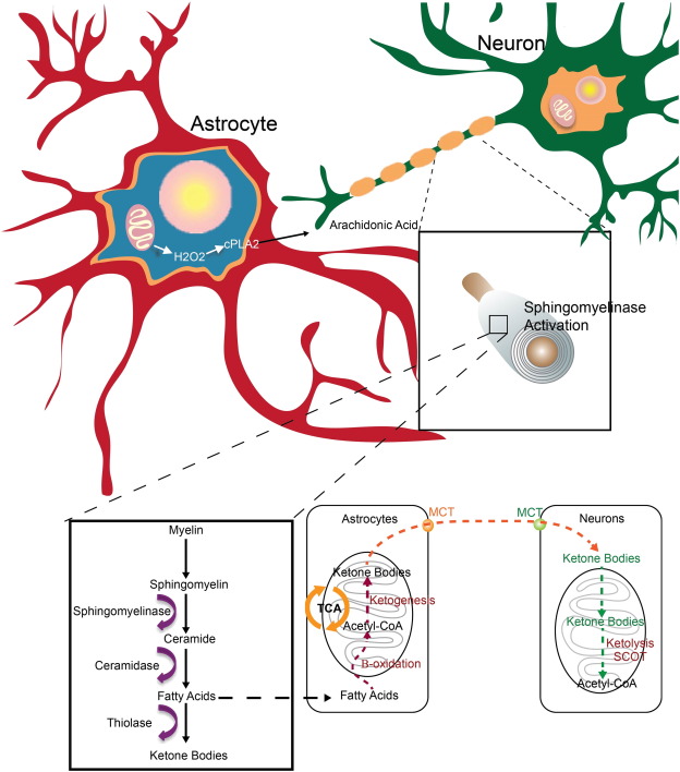
Findings from this study replicate our earlier findings that age of reproductive senescence is associated with decline in mitochondrial respiration, increased H2O2 production and shift to ketogenic metabolism in brain (Yao et al., 2010, Ding et al., 2013, Yin et al., 2015). These well established early age-related changes in mitochondrial function and shift to ketone body utilization in brain, are now linked to a mechanistic pathway that connects early decline in mitochondrial respiration and H2O2 production to activation of the cPLA2-sphingomyelinase pathway to catabolize myelin lipids resulting in WM degeneration (Fig. 12). These lipids are sequestered in lipid droplets for subsequent use as a local source of ketone body generation via astrocyte mediated beta-oxidation of fatty acids. Astrocyte derived ketone bodies can then be transported to neurons where they undergo ketolysis to generate acetyl-CoA for TCA derived ATP generation required for synaptic and cell function (Fig. 12).

Thumbnail image of Fig. 12. Opens large image

http://www.ebiomedicine.com/cms/attachment/2040395791/2053874721/gr12.sml

Fig. 12

Schematic model of mitochondrial H2O2 activation of cPLA2-sphingomyelinase pathway as an adaptive response to provide myelin derived fatty acids as a substrate for ketone body generation: The cPLA2-sphingomyelinase pathway is proposed as a mechanistic pathway that links an early event, mitochondrial dysfunction and H2O2, in the prodromal/preclinical phase of Alzheimer’s with later stage development of pathology, white matter degeneration. Our findings demonstrate that an age dependent deficit in mitochondrial respiration and a concomitant rise in oxidative stress activate an adaptive cPLA2-sphingomyelinase pathway to provide myelin derived fatty acids as a substrate for ketone body generation to fuel an energetically compromised brain.

Biochemical evidence obtained from isolated whole brain mitochondria confirms that during reproductive senescence and in response to estrogen deprivation brain mitochondria decline in respiratory capacity (Yao et al., 2009, Yao et al., 2010, Brinton, 2008a, Brinton, 2008b, Swerdlow and Khan, 2009). A well-documented consequence of mitochondrial dysfunction is increased production of reactive oxygen species (ROS), specifically H2O2 (Boveris and Chance, 1973, Beal, 2005, Yin et al., 2014, Yap et al., 2009). While most research focuses on the damage generated by free radicals, in this case H2O2 functions as a signaling molecule to activate cPLA2, the initiating enzyme in the cPLA2-sphingomyelinase pathway (Farooqui and Horrocks, 2006, Han et al., 2003, Sun et al., 2004). In AD brain, increased cPLA2 immunoreactivity is detected almost exclusively in astrocytes suggesting that activation of the cPLA2-sphingomyelinase pathway is localized to astrocytes in AD, as opposed to the neuronal or oligodendroglial localization that is observed during apoptosis (Sun et al., 2004, Malaplate-Armand et al., 2006, Di Paolo and Kim, 2011, Stephenson et al., 1996,Stephenson et al., 1999). In our analysis, cPLA2 (Sanchez-Mejia and Mucke, 2010) activation followed the age-dependent rise in H2O2 production and was sustained at an elevated level.

Direct and robust activation of astrocytic cPLA2 by physiologically relevant concentrations of H2O2 was confirmed in vitro. Astrocytic involvement in the cPLA2-sphingomyelinase pathway was also indicated by an increase in cPLA2 positive astrocyte reactivity in WM tracts of reproductively incompetent mice. These data are consistent with findings from brains of persons with AD that demonstrate the same striking localization of cPLA2immunoreactivity within astrocytes, specifically in the hippocampal formation (Farooqui and Horrocks, 2004). While neurons and astrocytes contain endogenous levels of cPLA2, neuronal cPLA2 is activated by an influx of intracellular calcium, whereas astrocytic cPLA2 is directly activated by excessive generation of H2O2 (Sun et al., 2004, Xu et al., 2003, Tournier et al., 1997). Evidence of this cell type specific activation was confirmed by the activation of cPLA2 in astrocytes by H2O2 and the lack of activation in neurons. These data support that astrocytic, not neuronal, cPLA2 is the cellular mediator of the H2O2 dependent cPLA2-sphingomyelinase pathway activation and provide associative evidence supporting a role of astrocytic mitochondrial H2O2 in age-related WM catabolism.

The pattern of gene expression during the shift to reproductive senescence in the female mouse hippocampus recapitulates key observations in human AD brain tissue, specifically elevation in cPLA2, sphingomyelinase and ceramidase (Schaeffer et al., 2010, He et al., 2010, Li et al., 2014). Further, up-regulation of myelin synthesis, lipid metabolism and inflammatory genes in reproductively incompetent female mice is consistent with the gene expression pattern previously reported from aged male rodent hippocampus, aged female non-human primate hippocampus and human AD hippocampus (Blalock et al., 2003, Blalock et al., 2004, Blalock et al., 2010, Blalock et al., 2011, Kadish et al., 2009, Rowe et al., 2007). In these analyses of gene expression in aged male rodent hippocampus, aged female non-human primate hippocampus and human AD hippocampus down regulation of genes related to mitochondrial function, and up-regulation in multiple genes encoding for enzymes involved in ketone body metabolism occurred (Blalock et al., 2003, Blalock et al., 2004, Blalock et al., 2010, Blalock et al., 2011, Kadish et al., 2009, Rowe et al., 2007). The comparability across data derived from aging female mouse hippocampus reported herein and those derived from male rodent brain, female nonhuman brain and human AD brain strongly suggest that cPLA2-sphingomyelinase pathway activation, myelin sheath degeneration and fatty acid metabolism leading to ketone body generation is a metabolic adaptation that is generalizable across these naturally aging models and are evident in aged human AD brain. Collectively, these data support the translational relevance of findings reported herein.

Data obtained via immunohistochemistry, electron microscopy and MBP protein analyses demonstrated an age-related loss in myelin sheath integrity. Evidence for a loss of myelin structural integrity emerged in reproductively incompetent mice following activation of the cPLA2-sphingomyelinase pathway. The unraveling myelin phenotype observed following reproductive senescence and aging reported herein is consistent with the degenerative phenotype that emerges following exposure to the chemotherapy drug bortezomib which induces mitochondrial dysfunction and increased ROS generation (Carozzi et al., 2010, Cavaletti et al., 2007,Ling et al., 2003). In parallel to the decline in myelin integrity, lipid droplet density increased. In aged mice, accumulation of lipid droplets declined in parallel to the rise in ketone bodies consistent with the utilization of myelin-derived fatty acids to generate ketone bodies. Due to the sequential relationship between WM degeneration and lipid droplet formation, we posit that lipid droplets serve as a temporary storage site for myelin-derived fatty acids prior to undergoing β-oxidation in astrocytes to generate ketone bodies.

Microstructural alterations in myelin integrity were associated with alterations in the lipid profile of brain, indicative of WM degeneration resulting in release of myelin lipids. Sphingomyelin and galactocerebroside are two main lipids that compose the myelin sheath (Baumann and Pham-Dinh, 2001). Ceramide is common to both galactocerebroside and sphingomyelin and is composed of sphingosine coupled to a fatty acid. Ceramide levels increase in aging, in states of ketosis and in neurodegeneration (Filippov et al., 2012, Blazquez et al., 1999, Costantini et al., 2005). Specifically, ceramide levels are elevated at the earliest clinically recognizable stage of AD, indicating a degree of WM degeneration early in disease progression (Di Paolo and Kim, 2011,Han et al., 2002, Costantini et al., 2005). Sphingosine is statistically significantly elevated in the brains of AD patients compared to healthy controls; a rise that was significantly correlated with acid sphingomyelinase activity, Aβ levels and tau hyperphosphorylation (He et al., 2010). In our analyses, a rise in ceramides was first observed early in the aging process in reproductively incompetent mice. The rise in ceramides was coincident with the emergence of loss of myelin integrity consistent with the release of myelin ceramides from sphingomyelin via sphingomyelinase activation. Following the rise in ceramides, sphingosine and fatty acid levels increased. The temporal sequence of the lipid profile was consistent with gene expression indicating activation of ceramidase for catabolism of ceramide into sphingosine and fatty acid during reproductive senescence. Once released from ceramide, fatty acids can be transported into the mitochondrial matrix of astrocytes via CPT-1, where β-oxidation of fatty acids leads to the generation of acetyl-CoA (Glatz et al., 2010). It is well documented that acetyl-CoA cannot cross the inner mitochondrial membrane, thus posing a barrier to direct transport of acetyl-CoA generated by β-oxidation into neurons. In response, the newly generated acetyl-CoA undergoes ketogenesis to generate ketone bodies to fuel energy demands of neurons (Morris, 2005,Guzman and Blazquez, 2004, Stacpoole, 2012). Because astrocytes serve as the primary location of β-oxidation in brain they are critical to maintaining neuronal metabolic viability during periods of reduced glucose utilization (Panov et al., 2014, Ebert et al., 2003, Guzman and Blazquez, 2004).

Once fatty acids are released from myelin ceramides, they are transported into astrocytic mitochondria by CPT1 to undergo β-oxidation. The mitochondrial trifunctional protein HADHA catalyzes the last three steps of mitochondrial β-oxidation of long chain fatty acids, while mitochondrial ABAD (aka SCHAD—short chain fatty acid dehydrogenase) metabolizes short chain fatty acids. Concurrent with the release of myelin fatty acids in aged female mice, CPT1, HADHA and ABAD protein expression as well as ketone body generation increased significantly. These findings indicate that astrocytes play a pivotal role in the response to bioenergetic crisis in brain to activate an adaptive compensatory system that activates catabolism of myelin lipids and the metabolism of those lipids into fatty acids to generate ketone bodies necessary to fuel neuronal demand for acetyl-CoA and ATP.

Collectively, these findings provide a mechanistic pathway that links mitochondrial dysfunction and H2O2generation in brain early in the aging process to later stage white matter degeneration. Astrocytes play a pivotal role in providing a mechanistic strategy to address the bioenergetic demand of neurons in the aging female brain. While this pathway is coincident with reproductive aging in the female brain, it is likely to have mechanistic translatability to the aging male brain. Further, the mechanistic link between bioenergetic decline and WM degeneration has potential relevance to other neurological diseases involving white matter in which postmenopausal women are at greater risk, such as multiple sclerosis. The mechanistic pathway reported herein spans time and is characterized by a progression of early adaptive changes in the bioenergetic system of the brain leading to WM degeneration and ketone body production. Translationally, effective therapeutics to prevent, delay and treat WM degeneration during aging and Alzheimer’s disease will need to specifically target stages within the mechanistic pathway described herein. The fundamental initiating event is a bioenergetic switch from being a glucose dependent brain to a glucose and ketone body dependent brain. It remains to be determined whether it is possible to prevent conversion to or reversal of a ketone dependent brain. Effective therapeutic strategies to intervene in this process require biomarkers of bioenergetic phenotype of the brain and stage of mechanistic progression. The mechanistic pathway reported herein may have relevance to other age-related neurodegenerative diseases characterized by white matter degeneration such as multiple sclerosis.

Blood. 2015 Oct 15;126(16):1925-9.    http://dx.doi.org:/10.1182/blood-2014-12-617498. Epub 2015 Aug 14.
Targeting the leukemia cell metabolism by the CPT1a inhibition: functional preclinical effects in leukemias.
Cancer cells are characterized by perturbations of their metabolic processes. Recent observations demonstrated that the fatty acid oxidation (FAO) pathway may represent an alternative carbon source for anabolic processes in different tumors, therefore appearing particularly promising for therapeutic purposes. Because the carnitine palmitoyl transferase 1a (CPT1a) is a protein that catalyzes the rate-limiting step of FAO, here we investigated the in vitro antileukemic activity of the novel CPT1a inhibitor ST1326 on leukemia cell lines and primary cells obtained from patients with hematologic malignancies. By real-time metabolic analysis, we documented that ST1326 inhibited FAO in leukemia cell lines associated with a dose- and time-dependent cell growth arrest, mitochondrial damage, and apoptosis induction. Data obtained on primary hematopoietic malignant cells confirmed the FAO inhibition and cytotoxic activity of ST1326, particularly on acute myeloid leukemia cells. These data suggest that leukemia treatment may be carried out by targeting metabolic processes.
Oncogene. 2015 Oct 12.   http://dx.doi.org:/10.1038/onc.2015.394. [Epub ahead of print]
Tumour-suppression function of KLF12 through regulation of anoikis.
Suppression of detachment-induced cell death, known as anoikis, is an essential step for cancer metastasis to occur. We report here that expression of KLF12, a member of the Kruppel-like family of transcription factors, is downregulated in lung cancer cell lines that have been selected to grow in the absence of cell adhesion. Knockdown of KLF12 in parental cells results in decreased apoptosis following cell detachment from matrix. KLF12 regulates anoikis by promoting the cell cycle transition through S phase and therefore cell proliferation. Reduced expression levels of KLF12 results in increased ability of lung cancer cells to form tumours in vivo and is associated with poorer survival in lung cancer patients. We therefore identify KLF12 as a novel metastasis-suppressor gene whose loss of function is associated with anoikis resistance through control of the cell cycle.
Mol Cell. 2015 Oct 14. pii: S1097-2765(15)00764-9. doi: 10.1016/j.molcel.2015.09.025. [Epub ahead of print]
PEPCK Coordinates the Regulation of Central Carbon Metabolism to Promote Cancer Cell Growth.
Phosphoenolpyruvate carboxykinase (PEPCK) is well known for its role in gluconeogenesis. However, PEPCK is also a key regulator of TCA cycle flux. The TCA cycle integrates glucose, amino acid, and lipid metabolism depending on cellular needs. In addition, biosynthetic pathways crucial to tumor growth require the TCA cycle for the processing of glucose and glutamine derived carbons. We show here an unexpected role for PEPCK in promoting cancer cell proliferation in vitro and in vivo by increasing glucose and glutamine utilization toward anabolic metabolism. Unexpectedly, PEPCK also increased the synthesis of ribose from non-carbohydrate sources, such as glutamine, a phenomenon not previously described. Finally, we show that the effects of PEPCK on glucose metabolism and cell proliferation are in part mediated via activation of mTORC1. Taken together, these data demonstrate a role for PEPCK that links metabolic flux and anabolic pathways to cancer cell proliferation.
Mol Cancer Res. 2015 Oct;13(10):1408-20.   http://dx.doi.org:/10.1158/1541-7786.MCR-15-0048. Epub 2015 Jun 16.
Disruption of Proline Synthesis in Melanoma Inhibits Protein Production Mediated by the GCN2 Pathway.
Many processes are deregulated in melanoma cells and one of those is protein production. Although much is known about protein synthesis in cancer cells, effective ways of therapeutically targeting this process remain an understudied area of research. A process that is upregulated in melanoma compared with normal melanocytes is proline biosynthesis, which has been linked to both oncogene and tumor suppressor pathways, suggesting an important convergent point for therapeutic intervention. Therefore, an RNAi screen of a kinase library was undertaken, identifying aldehyde dehydrogenase 18 family, member A1 (ALDH18A1) as a critically important gene in regulating melanoma cell growth through proline biosynthesis. Inhibition of ALDH18A1, the gene encoding pyrroline-5-carboxylate synthase (P5CS), significantly decreased cultured melanoma cell viability and tumor growth. Knockdown of P5CS using siRNA had no effect on apoptosis, autophagy, or the cell cycle but cell-doubling time increased dramatically suggesting that there was a general slowdown in cellular metabolism. Mechanistically, targeting ALDH18A1 activated the serine/threonine protein kinase GCN2 (general control nonderepressible 2) to inhibit protein synthesis, which could be reversed with proline supplementation. Thus, targeting ALDH18A1 in melanoma can be used to disrupt proline biosynthesis to limit cell metabolism thereby increasing the cellular doubling time mediated through the GCN2 pathway.  This study demonstrates that melanoma cells are sensitive to disruption of proline synthesis and provides a proof-of-concept that the proline synthesis pathway can be therapeutically targeted in melanoma tumors for tumor inhibitory efficacy. Mol Cancer Res; 13(10); 1408-20. ©2015 AACR.
SDHB-Deficient Cancers: The Role of Mutations That Impair Iron Sulfur Cluster Delivery.
BACKGROUND:  Mutations in the Fe-S cluster-containing SDHB subunit of succinate dehydrogenase cause familial cancer syndromes. Recently the tripeptide motif L(I)YR was identified in the Fe-S recipient protein SDHB, to which the cochaperone HSC20 binds.
METHODS:   In order to characterize the metabolic basis of SDH-deficient cancers we performed stable isotope-resolved metabolomics in a novel SDHB-deficient renal cell carcinoma cell line and conducted bioinformatics and biochemical screening to analyze Fe-S cluster acquisition and assembly of SDH in the presence of other cancer-causing SDHB mutations.

RESULTS:

We found that the SDHB(R46Q) mutation in UOK269 cells disrupted binding of HSC20, causing rapid degradation of SDHB. In the absence of SDHB, respiration was undetectable in UOK269 cells, succinate was elevated to 351.4±63.2 nmol/mg cellular protein, and glutamine became the main source of TCA cycle metabolites through reductive carboxylation. Furthermore, HIF1α, but not HIF2α, increased markedly and the cells showed a strong DNA CpG island methylator phenotype (CIMP). Biochemical and bioinformatic screening revealed that 37% of disease-causing missense mutations in SDHB were located in either the L(I)YR Fe-S transfer motifs or in the 11 Fe-S cluster-ligating cysteines.

CONCLUSIONS:

These findings provide a conceptual framework for understanding how particular mutations disproportionately cause the loss of SDH activity, resulting in accumulation of succinate and metabolic remodeling in SDHB cancer syndromes.


SR4 Uncouples Mitochondrial Oxidative Phosphorylation, Modulates AMPK-mTOR Signaling, and Inhibits Proliferation of HepG2 Hepatocarcinoma Cells

  1. L. Figarola, J. Singhal, J. D. Tompkins, G. W. Rogers, C. Warden, D. Horne, A. D. Riggs, S. Awasthi and S. S. Singhal.

J Biol Chem. 2015 Nov 3, [epub ahead of print]


CD47 Receptor Globally Regulates Metabolic Pathways That Control Resistance to Ionizing Radiation

  1. W. Miller, D. R. Soto-Pantoja, A. L. Schwartz, J. M. Sipes, W. G. DeGraff, L. A. Ridnour, D. A. Wink and D. D. Roberts.

J Biol Chem. 2015 Oct 9, 290 (41): 24858-74.


Knockdown of PKM2 Suppresses Tumor Growth and Invasion in Lung Adenocarcinoma

  1. Sun, A. Zhu, L. Zhang, J. Zhang, Z. Zhong and F. Wang.

Int J Mol Sci. 2015 Oct 15, 16 (10): 24574-87.


EglN2 associates with the NRF1-PGC1alpha complex and controls mitochondrial function in breast cancer

  1. Zhang, C. Wang, X. Chen, M. Takada, C. Fan, X. Zheng, H. Wen, Y. Liu, C. Wang, R. G. Pestell, K. M. Aird, W. G. Kaelin, Jr., X. S. Liu and Q. Zhang.

EMBO J. 2015 Oct 22, [epub ahead of print]


Mitochondrial Genetics Regulate Breast Cancer Tumorigenicity and Metastatic Potential.

Current paradigms of carcinogenic risk suggest that genetic, hormonal, and environmental factors influence an individual’s predilection for developing metastatic breast cancer. Investigations of tumor latency and metastasis in mice have illustrated differences between inbred strains, but the possibility that mitochondrial genetic inheritance may contribute to such differences in vivo has not been directly tested. In this study, we tested this hypothesis in mitochondrial-nuclear exchange mice we generated, where cohorts shared identical nuclear backgrounds but different mtDNA genomes on the background of the PyMT transgenic mouse model of spontaneous mammary carcinoma. In this setting, we found that primary tumor latency and metastasis segregated with mtDNA, suggesting that mtDNA influences disease progression to a far greater extent than previously appreciated. Our findings prompt further investigation into metabolic differences controlled by mitochondrial process as a basis for understanding tumor development and metastasis in individual subjects. Importantly, differences in mitochondrial DNA are sufficient to fundamentally alter disease course in the PyMT mouse mammary tumor model, suggesting that functional metabolic differences direct early tumor growth and metastatic efficiency. Cancer Res; 75(20); 4429-36. ©2015 AACR.


Cancer Lett. 2015 Oct 29. pii: S0304-3835(15)00656-4.    http://dx.doi.org:/10.1016/j.canlet.2015.10.025. [Epub ahead of print]
Carboxyamidotriazole inhibits oxidative phosphorylation in cancer cells and exerts synergistic anti-cancer effect with glycolysis inhibition.

Targeting cancer cell metabolism is a promising strategy against cancer. Here, we confirmed that the anti-cancer drug carboxyamidotriazole (CAI) inhibited mitochondrial respiration in cancer cells for the first time and found a way to enhance its anti-cancer activity by further disturbing the energy metabolism. CAI promoted glucose uptake and lactate production when incubated with cancer cells. The oxidative phosphorylation (OXPHOS) in cancer cells was inhibited by CAI, and the decrease in the activity of the respiratory chain complex I could be one explanation. The anti-cancer effect of CAI was greatly potentiated when being combined with 2-deoxyglucose (2-DG). The cancer cells treated with the combination of CAI and 2-DG were arrested in G2/M phase. The apoptosis and necrosis rates were also increased. In a mouse xenograft model, this combination was well tolerated and retarded the tumor growth. The impairment of cancer cell survival was associated with significant cellular ATP decrease, suggesting that the combination of CAI and 2-DG could be one of the strategies to cause dual inhibition of energy pathways, which might be an effective therapeutic approach for a broad spectrum of tumors.


Cancer Immunol Res. 2015 Nov;3(11):1236-47.    http://dx.doi.org:/10.1158/2326-6066.CIR-15-0036. Epub 2015 May 29.
Inhibition of Fatty Acid Oxidation Modulates Immunosuppressive Functions of Myeloid-Derived Suppressor Cells and Enhances Cancer Therapies.

Myeloid-derived suppressor cells (MDSC) promote tumor growth by inhibiting T-cell immunity and promoting malignant cell proliferation and migration. The therapeutic potential of blocking MDSC in tumors has been limited by their heterogeneity, plasticity, and resistance to various chemotherapy agents. Recent studies have highlighted the role of energy metabolic pathways in the differentiation and function of immune cells; however, the metabolic characteristics regulating MDSC remain unclear. We aimed to determine the energy metabolic pathway(s) used by MDSC, establish its impact on their immunosuppressive function, and test whether its inhibition blocks MDSC and enhances antitumor therapies. Using several murine tumor models, we found that tumor-infiltrating MDSC (T-MDSC) increased fatty acid uptake and activated fatty acid oxidation (FAO). This was accompanied by an increased mitochondrial mass, upregulation of key FAO enzymes, and increased oxygen consumption rate. Pharmacologic inhibition of FAO blocked immune inhibitory pathways and functions in T-MDSC and decreased their production of inhibitory cytokines. FAO inhibition alone significantly delayed tumor growth in a T-cell-dependent manner and enhanced the antitumor effect of adoptive T-cell therapy. Furthermore, FAO inhibition combined with low-dose chemotherapy completely inhibited T-MDSC immunosuppressive effects and induced a significant antitumor effect. Interestingly, a similar increase in fatty acid uptake and expression of FAO-related enzymes was found in human MDSC in peripheral blood and tumors. These results support the possibility of testing FAO inhibition as a novel approach to block MDSC and enhance various cancer therapies. Cancer Immunol Res; 3(11); 1236-47. ©2015 AACR.


Ionizing radiation induces myofibroblast differentiation via lactate dehydrogenase

  1. L. Judge, K. M. Owens, S. J. Pollock, C. F. Woeller, T. H. Thatcher, J. P. Williams, R. P. Phipps, P. J. Sime and R. M. Kottmann.

Am J Physiol Lung Cell Mol Physiol. 2015 Oct 15, 309 (8): L879-87.


Vitamin C selectively kills KRAS and BRAF mutant colorectal cancer cells by targeting GAPDH

  1. Yun, E. Mullarky, C. Lu, K. N. Bosch, A. Kavalier, K. Rivera, J. Roper, Chio, II, E. G. Giannopoulou, C. Rago, A. Muley, J. M. Asara, J. Paik, O. Elemento, Z. Chen, D. J. Pappin, L. E. Dow, N. Papadopoulos, S. S. Gross and L. C. Cantley.

Science. 2015 Nov 5, [epub ahead of print]


Down-regulation of FBP1 by ZEB1-mediated repression confers to growth and invasion in lung cancer cells

  1. Zhang, J. Wang, H. Xing, Q. Li, Q. Zhao and J. Li.

Mol Cell Biochem. 2015 Nov 6, [epub ahead of print]


J Mol Cell Cardiol. 2015 Oct 23. pii: S0022-2828(15)30073-0.     http://dx.doi.org:/10.1016/j.yjmcc.2015.10.002. [Epub ahead of print]
GRK2 compromises cardiomyocyte mitochondrial function by diminishing fatty acid-mediated oxygen consumption and increasing superoxide levels.

The G protein-coupled receptor kinase-2 (GRK2) is upregulated in the injured heart and contributes to heart failure pathogenesis. GRK2 was recently shown to associate with mitochondria but its functional impact in myocytes due to this localization is unclear. This study was undertaken to determine the effect of elevated GRK2 on mitochondrial respiration in cardiomyocytes. Sub-fractionation of purified cardiac mitochondria revealed that basally GRK2 is found in multiple compartments. Overexpression of GRK2 in mouse cardiomyocytes resulted in an increased amount of mitochondrial-based superoxide. Inhibition of GRK2 increased oxygen consumption rates and ATP production. Moreover, fatty acid oxidation was found to be significantly impaired when GRK2 was elevated and was dependent on the catalytic activity and mitochondrial localization of this kinase. Our study shows that independent of cardiac injury, GRK2 is localized in the mitochondria and its kinase activity negatively impacts the function of this organelle by increasing superoxide levels and altering substrate utilization for energy production.


Br J Pharmacol. 2015 Oct 27. doi: 10.1111/bph.13377. [Epub ahead of print]
All-trans retinoic acid protects against doxorubicin-induced cardiotoxicity by activating the Erk2 signalling pathway.
BACKGROUND AND PURPOSE:

Doxorubicin (Dox) is a powerful antineoplastic agent for treating a wide range of cancers. However, doxorubicin cardiotoxicity of the heart has largely limited its clinical use. It is known that all-trans retinoic acid (ATRA) plays important roles in many cardiac biological processes, however, the protective effects of ATRA on doxorubicin cardiotoxicity remain unknown. Here, we studied the effect of ATRA on doxorubicin cardiotoxicity and underlying mechanisms.

EXPERIMENTAL APPROACHES:

Cellular viability assays, western blotting and mitochondrial respiration analyses were employed to evaluate the cellular response to ATRA in H9c2 cells and primary cardiomyocytes. Quantitative PCR (Polymerase Chain Reaction) and gene knockdown were performed to investigate the underlying molecular mechanisms of ATRA’s effects on doxorubicin cardiotoxicity.

KEY RESULTS:

ATRA significantly inhibited doxorubicin-induced apoptosis in H9c2 cells and primary cardiomyocytes. ATRA was more effective against doxorubicin cardiotoxicity than resveratrol and dexrazoxane. ATRA also suppressed reactive oxygen species (ROS) generation, and restored the expression level of mRNA and proteins in phase II detoxifying enzyme system: Nrf2 (nuclear factor-E2-related factor 2), MnSOD (manganese superoxide dismutase), HO-1 (heme oxygenase1) as well as mitochondrial function (mitochondrial membrane integrity, mitochondrial DNA copy numbers, mitochondrial respiration capacity, biogenesis and dynamics). Both Erk1/2 (extracellular signal-regulated kinase1/2) inhibitor (U0126) and Erk2 siRNA, but not Erk1 siRNA, abolished the protective effect of ATRA against doxorubicin-induced toxicity in H9c2 cells. Remarkably, ATRA did not compromise the anticancer efficacy of doxorubicin in gastric carcinoma cells.

CONCLUSION AND IMPLICATION:

ATRA protected cardiomyocytes against doxorubicin-induced toxicity by activating the Erk2 pathway without compromising the anticancer efficacy of doxorubicin. Therefore, ATRA may be a promising candidate as a cardioprotective agent against doxorubicin cardiotoxicity.


Proteomic and Biochemical Studies of Lysine Malonylation Suggest Its Malonic Aciduria-associated Regulatory Role in Mitochondrial Function and Fatty Acid Oxidation

  1. Colak, O. Pougovkina, L. Dai, M. Tan, H. Te Brinke, H. Huang, Z. Cheng, J. Park, X. Wan, X. Liu, W. W. Yue, R. J. Wanders, J. W. Locasale, D. B. Lombard, V. C. de Boer and Y. Zhao.

Mol Cell Proteomics. 2015 Nov 1, 14 (11): 3056-71.


Foxg1 localizes to mitochondria and coordinates cell differentiation and bioenergetics

  1. Pancrazi, G. Di Benedetto, L. Colombaioni, G. Della Sala, G. Testa, F. Olimpico, A. Reyes, M. Zeviani, T. Pozzan and M. Costa.

Proc Natl Acad Sci U S A. 2015 Oct 27, 112(45): 13910-5.


Evidence of Mitochondrial Dysfunction within the Complex Genetic Etiology of Schizophrenia

  1. E. Hjelm, B. Rollins, F. Mamdani, J. C. Lauterborn, G. Kirov, G. Lynch, C. M. Gall, A. Sequeira and M. P. Vawter.

Mol Neuropsychiatry. 2015 Nov 1, 1 (4): 201-219.


Metabolic Reprogramming Is Required for Myofibroblast Contractility and Differentiation

  1. Bernard, N. J. Logsdon, S. Ravi, N. Xie, B. P. Persons, S. Rangarajan, J. W. Zmijewski, K. Mitra, G. Liu, V. M. Darley-Usmar and V. J. Thannickal.

J Biol Chem. 2015 Oct 16, 290 (42): 25427-38.


J Biol Chem. 2015 Oct 23;290(43):25834-46.    http://dx.doi.org:/10.1074/jbc.M115.658815. Epub 2015 Sep 4.
Kinome Screen Identifies PFKFB3 and Glucose Metabolism as Important Regulators of the Insulin/Insulin-like Growth Factor (IGF)-1 Signaling Pathway.

The insulin/insulin-like growth factor (IGF)-1 signaling pathway (ISP) plays a fundamental role in long term health in a range of organisms. Protein kinases including Akt and ERK are intimately involved in the ISP. To identify other kinases that may participate in this pathway or intersect with it in a regulatory manner, we performed a whole kinome (779 kinases) siRNA screen for positive or negative regulators of the ISP, using GLUT4 translocation to the cell surface as an output for pathway activity. We identified PFKFB3, a positive regulator of glycolysis that is highly expressed in cancer cells and adipocytes, as a positive ISP regulator. Pharmacological inhibition of PFKFB3 suppressed insulin-stimulated glucose uptake, GLUT4 translocation, and Akt signaling in 3T3-L1 adipocytes. In contrast, overexpression of PFKFB3 in HEK293 cells potentiated insulin-dependent phosphorylation of Akt and Akt substrates. Furthermore, pharmacological modulation of glycolysis in 3T3-L1 adipocytes affected Akt phosphorylation. These data add to an emerging body of evidence that metabolism plays a central role in regulating numerous biological processes including the ISP. Our findings have important implications for diseases such as type 2 diabetes and cancer that are characterized by marked disruption of both metabolism and growth factor signaling.


FASEB J. 2015 Oct 19.    http://dx.doi.org:/pii: fj.15-276360. [Epub ahead of print]
Perm1 enhances mitochondrial biogenesis, oxidative capacity, and fatigue resistance in adult skeletal muscle.

Skeletal muscle mitochondrial content and oxidative capacity are important determinants of muscle function and whole-body health. Mitochondrial content and function are enhanced by endurance exercise and impaired in states or diseases where muscle function is compromised, such as myopathies, muscular dystrophies, neuromuscular diseases, and age-related muscle atrophy. Hence, elucidating the mechanisms that control muscle mitochondrial content and oxidative function can provide new insights into states and diseases that affect muscle health. In past studies, we identified Perm1 (PPARGC1- and ESRR-induced regulator, muscle 1) as a gene induced by endurance exercise in skeletal muscle, and regulating mitochondrial oxidative function in cultured myotubes. The capacity of Perm1 to regulate muscle mitochondrial content and function in vivo is not yet known. In this study, we use adeno-associated viral (AAV) vectors to increase Perm1 expression in skeletal muscles of 4-wk-old mice. Compared to control vector, AAV1-Perm1 leads to significant increases in mitochondrial content and oxidative capacity (by 40-80%). Moreover, AAV1-Perm1-transduced muscles show increased capillary density and resistance to fatigue (by 33 and 31%, respectively), without prominent changes in fiber-type composition. These findings suggest that Perm1 selectively regulates mitochondrial biogenesis and oxidative function, and implicate Perm1 in muscle adaptations that also occur in response to endurance exercise.-Cho, Y., Hazen, B. C., Gandra, P. G., Ward, S. R., Schenk, S., Russell, A. P., Kralli, A. Perm1 enhances mitochondrial biogenesis, oxidative capacity, and fatigue resistance in adult skeletal muscle.


A conserved MADS-box phosphorylation motif regulates differentiation and mitochondrial function in skeletal, cardiac, and smooth muscle cells.
Exposure to metabolic disease during fetal development alters cellular differentiation and perturbs metabolic homeostasis, but the underlying molecular regulators of this phenomenon in muscle cells are not completely understood. To address this, we undertook a computational approach to identify cooperating partners of the myocyte enhancer factor-2 (MEF2) family of transcription factors, known regulators of muscle differentiation and metabolic function. We demonstrate that MEF2 and the serum response factor (SRF) collaboratively regulate the expression of numerous muscle-specific genes, including microRNA-133a (miR-133a). Using tandem mass spectrometry techniques, we identify a conserved phosphorylation motif within the MEF2 and SRF Mcm1 Agamous Deficiens SRF (MADS)-box that regulates miR-133a expression and mitochondrial function in response to a lipotoxic signal. Furthermore, reconstitution of MEF2 function by expression of a neutralizing mutation in this identified phosphorylation motif restores miR-133a expression and mitochondrial membrane potential during lipotoxicity. Mechanistically, we demonstrate that miR-133a regulates mitochondrial function through translational inhibition of a mitophagy and cell death modulating protein, called Nix. Finally, we show that rodents exposed to gestational diabetes during fetal development display muscle diacylglycerol accumulation, concurrent with insulin resistance, reduced miR-133a, and elevated Nix expression, as young adult rats. Given the diverse roles of miR-133a and Nix in regulating mitochondrial function, and proliferation in certain cancers, dysregulation of this genetic pathway may have broad implications involving insulin resistance, cardiovascular disease, and cancer biology.


Read Full Post »

Identifying Cancers and Resistance, Volume 2 (Volume Two: Latest in Genomics Methodologies for Therapeutics: Gene Editing, NGS and BioInformatics, Simulations and the Genome Ontology), Part 1: Next Generation Sequencing (NGS)

Identifying Cancers and Resistance

Larry H/ Bernstein, MD, FCAP, Curator

LPBI


New Way of Detecting Cancer

http://www.technologynetworks.com/Diagnostics/news.aspx?ID=185201

A new RNA test of blood platelets can be used to detect, classify and pinpoint the location of cancer by analysing a sample equivalent to one drop of blood.

Using this new method for blood-based RNA tests of blood platelets, researchers have been able to identify cancer with 96 per cent accuracy and classifying the type of cancer at an accuracy of 71 per cent.

“Being able to detect cancer at an early stage is vital. We have studied how a whole new blood-based method of biopsy can be used to detect cancer, which in the future renders an invasive cell tissue sample unnecessary in diagnosing lung cancer, for instance. In the study, nearly all forms of cancer were identified, which proves that blood-based biopsies have an immense potential to improve early detection of cancer,” according to Jonas Nilsson, cancer researcher at Umeå University and co-author of the article.

In the study, researchers from Umeå University, in collaborations with researchers from the Netherlands and the US, have investigated how a new method of blood-based RNA tests of the part of the blood called platelets could be used in detecting and classifying cancer.

The results show that blood platelets could constitute a complete and easily accessible blood-based source for sampling and hence be used in diagnosing cancer as well as in the choice of treatment method.

Blood samples from 283 individuals were studied of which 228 people had some form of cancer and 55 showed no evidence of cancer. By comparing the blood samples RNA profiles, researchers could identify the presence of cancer with an accuracy of 96 per cent among patients. Among the 39 patients in the study in which an early detection of cancer had been made, 100 per cent of the cases could be identified and classified.

In follow-up tests using the same method, researchers could identify the origin of tumours with a so far unsurpassed accuracy of 71 per cent in patients with diagnosed cancer in the lung, breast, pancreas, brain, liver, colon and rectum. The samples could also be sorted in subdivisions depending on molecular differences in the cancer form, which can be of great use in the choice of treatment method.


Blood Test Picks Out Prostate Cancer Drug Resistance

http://www.technologynetworks.com/Diagnostics/news.aspx?ID=185032

Scientists have developed a blood test that can identify key mutations driving resistance to a widely used prostate cancer drug, and identify in advance patients who will not respond to treatment.

The new research paves the way for information from a blood test to inform prostate cancer treatment in future, with only those patients whose cancers are free of resistance mutations taking the drug, abiraterone.

The study is also a proof of principle that tests for cancer DNA in the bloodstream can be used to detect drug resistance mutations – allowing patients who will not benefit from one drug to be given an alternative treatment instead.

Researchers at The Institute of Cancer Research, London, the Royal Marsden NHS Foundation Trust, and the University of Trento, Italy, analysed 274 blood samples from 97 patients using state-of-the-art DNA sequencing techniques.

They found that mutations in a gene called the androgen receptor (AR) predicted resistance to the prostate cancer drug abiraterone, and that patients with these mutations had poorer survival.

Abiraterone, which was discovered at the ICR, is now standard treatment for men with advanced prostate cancer – but while it is highly effective in many patients, 30-60% do not respond.

So researchers have been searching for a marker that will help predict in advance which men will benefit from the drug, and who should be given a different treatment.

Researchers discovered that men who harbour either a specific mutation or an increase in the number of copies of the AR gene, were 7.8 times less likely to have a reduction of more than 90% in their PSA levels, a widely used test to monitor the response of prostate cancer.

The study also found that in about 15% of men given abiraterone who did not have either mutation before starting treatment, this was acquired as the drug stopped working and appeared in the bloodstream several months before patients developed any symptoms.

Blood tests are particularly valuable in cancer patients because biopsies are often difficult to perform and can carry risks. Even when biopsies are possible, they only give a snapshot of cancer genetics in a small specific area, whereas blood tests can give information that is more representative of multiple different tumours around the body.

Dr Gerhardt Attard, Clinician Scientist at The Institute of Cancer Research, London, and Consultant Medical Oncologist at The Royal Marsden NHS Foundation Trust, said: “The discovery of abiraterone was an important step forward for patients with advanced prostate cancer, but we know it doesn’t work for all men, and we’ve been searching for a marker that will tell us in advance which men will benefit. We’re delighted to have developed a test that appears to predict very accurately whether a patient will respond to abiraterone, and that it can be performed on blood samples – removing the need to take a biopsy.

“We are now planning a clinical trial involving up to 600 men in which we aim to prospectively show that men who are positive with our test have significantly greater benefit from chemotherapy in preference to abiraterone or similar drugs. Critically, we believe that this sort of technology would be relatively straightforward to implement in NHS hospitals, making it accessible to a large number of patients. Additionally, looking at tumour DNA in the blood of patients could potentially give us an overall picture of why the cancer is progressing all over the body, unlike a biopsy that only tells us about the area sampled.”

Professor Paul Workman, Chief Executive of The Institute of Cancer Research, London, said: “Abiraterone has extended the lives of many thousands of men in the UK, with fewer side-effects than chemotherapy, and we are really proud that it was discovered here at the ICR. But we don’t stop at taking a new treatment like abiraterone to patients – it’s also essential to continue to conduct research on these new drugs, to make sure they are being used as effectively as possible.

“This new study finds that by analysing tumour DNA present in the bloodstream, we should be able to personalise treatment with abiraterone, so that only those who will benefit from the drug will receive it. It is the latest step forward in the new era we are in of precision cancer medicine, where rather than using a treatment in the hope that a patient will benefit, we can look at tumour DNA in advance and be much more confident that a particular treatment will work.”

Associate Professor Francesca Demichelis, leader of the Computational Oncology Group at the University of Trento, Italy, said:

“Being able to quantify tumor DNA from a blood sample and to characterize structural changes of the DNA that are predictive of treatment response is powerful. This is especially true as a blood-based test gives us a global picture of the patient’s tumor burden rather than a focal snapshot as with a standard biopsy.

“In principle the same computational approach we adopted in this study is applicable across tumor types. This study represents a step towards sensitive and specific detection of resistance to targeted therapies.”

Dr Emma Smith, Cancer Research UK’s senior science information officer, said:

“Abiraterone, a drug which Cancer Research UK helped to develop, gives men with advanced prostate cancer a much-needed treatment option and more time with their loved ones. But it doesn’t work for everyone.  If these important early results bear up in larger clinical trials it could lead to a test which would indicate which patients might benefit more from trying other therapies instead.”

Dr Iain Frame, Director of Research at Prostate Cancer UK said: “We know that a one-size-fits-all approach to treating prostate cancer doesn’t work, and research like this is crucially important in helping us understand which treatments will – and won’t – work best for each individual. When the clock is ticking for a man with advanced prostate cancer, finding out early that his treatment needs changing can not only save precious time, but can also help avoid unpleasant side effects from a treatment that longer works for him.

“Research like this wouldn’t be possible without the thousands of people who take part in Movember every year – and so with another campaign upon us, we can all do something to help the fight against prostate cancer.”

Read Full Post »

targeting of GSK-3α and GSK-3β

Larry H. Bernstein, MD, FCAP, Curator

LPBI


4′-((5-(2,3-Dihydrobenzo[b][1,4]dioxin-6-yl)-1,3,4-oxadiazol-2-yl-thio)-methyl)-4-fluorobiphenyl-2-carboxamide

by DR ANTHONY MELVIN CRASTO Ph.D

str1


Cas 1820758-44-8

C24 H18 F N3 O4 S

4′-((5-(2,3-Dihydrobenzo[b][1,4]dioxin-6-yl)-1,3,4-oxadiazol-2-yl-thio)-methyl)-4-fluorobiphenyl-2-carboxamide

https://pharmaceuticalintelligence.com/wp-admin/post-new.php


Glycogen synthase kinase-3 (GSK-3) is a constitutively active, ubiquitous serine/threonine kinase that takes part in a number of physiological processes ranging from glycogen metabolism to apoptosis. GSK-3 is a key mediator of various signaling pathways, such as the Wnt and the insulin/AKT signaling pathways.

Therefore, dysregulation of GSK-3 has been linked to various human diseases, such as cancer, diabetes, and neurodegenerative diseases.Two related isoforms of GSK-3 exist in mammals, GSK-3α and -β, which share a sequence identity within their catalytic domains of 98%.

Beyond the catalytic domains they show significant differences. Although these isoforms are structurally related, they are not functionally equivalent, and one cannot compensate for loss of the other.

The debate on the respective contributions of the isoforms GSK-3α and GSK-3β on the pathogenesis of different diseases is ongoing.

Various studies indicate that the therapies of certain diseases benefit from specific targeting of GSK-3α and GSK-3β. GSK-3α was recently identified as a differentiation target in acute myeloid leukemia (AML). AML is a hematopoietic malignancy defined by uncontrolled proliferation and disrupted myeloid differentiation. AML is the second most common form of leukemia in adults.

The current treatment of AML with conventional chemotherapy is very aggressive yet ineffective for the majority of patients with the disease.Thus, alternative targeted treatment approaches for AML are highly desirable. GSK-3α recently emerged as a potential target in this disease.

Abstract Image

http://pubs.acs.org/appl/literatum/publisher/achs/journals/content/jmcmar/0/jmcmar.ahead-of-print/acs.jmedchem.5b01200/20151105/images/medium/jm-2015-01200x_0015.gif


The challenge for glycogen synthase kinase-3 (GSK-3) inhibitor design lies in achieving high selectivity for one isoform over the other. The therapy of certain diseases, such as acute myeloid leukemia (AML), may require α-isoform specific targeting. The scorpion shaped GSK-3 inhibitors developed by our group achieved the highest GSK-3α selectivity reported so far but suffered from insufficient aqueous solubility. This work presents the solubility-driven optimization of our isoform-selective inhibitors using a scorpion shaped lead. Among 15 novel compounds, compound 27 showed high activity against GSK-3α/β with the highest GSK-3α selectivity reported to date. Compound 27was profiled for bioavailability and toxicity in a zebrafish embryo phenotype assay. Selective GSK-3α targeting in AML cell lines was achieved with compound 27, resulting in a strong differentiation phenotype and colony formation impairment, confirming the potential of GSK-3α inhibition in AML therapy.

Evaluation of Improved Glycogen Synthase Kinase-3α Inhibitors in Models of Acute Myeloid Leukemia

Clemens Schöpf Institute of Organic Chemistry and Biochemistry, Technische Universität Darmstadt, 64287 Darmstadt, Germany
Department of Pediatric Oncology, Dana-Farber Cancer Institute, Harvard Medical School, Boston, Massachusetts 02215, United States
J. Med. Chem., Article ASAP
Publication Date (Web): October 23, 2015   Copyright © 2015 American Chemical Society

http://pubs.acs.org/doi/abs/10.1021/acs.jmedchem.5b01200

http://pubs.acs.org/doi/suppl/10.1021/acs.jmedchem.5b01200/suppl_file/jm5b01200_si_001.pdf

compound 27 as a colorless solid. HPLC: 96%, tR = 6.93 min.

1H NMR (DMSO-d6, 500 MHz, 300 K): δ (ppm) = 4.32 (td, J = 5.2 Hz, J = 3.7 Hz, 4H), 4.60 (s, 2H), 7.05 (d, J = 8.4 Hz, 1H), 7.25 (dd, J = 9.1 Hz, J = 2.7 Hz, 1H), 7.31 (td, J = 8.6 Hz, J = 2.8 Hz, 1H), 7.38 (m, 3H), 7.41 (d, J = 2.0 Hz, 1H), 7.45 (dd, J = 8.4 Hz, J = 2.1 Hz, 1H), 7.49 (d, J = 8.2 Hz, 2H), 7.73 (s, 1H).

13C NMR (DMSO, 125 MHz, 300 K): δ (ppm) = 35.6, 64.1, 64.4, 114.3 (d, JC–F = 21 Hz), 115.0, 115.9 (d, JC–F = 21 Hz), 115.9, 118.1, 120.0, 128.6 (2C), 128.8 (2C), 132.0 (d, JC–F = 8 Hz), 134.8, 135.5, 138.9, 139.0 (d, JC–F = 7 Hz), 143.8, 146.7, 160.9 (d, JC–F = 247 Hz), 162.7, 164.9, 169.5.

EI-MS: m/z = 463 (100, [M+]), 464 (26, [M+ + H]), 465 (7, [M+ + 2H].


Read Full Post »

Irreconciliable Dissonance in Physical Space and Cellular Metabolic Conception

Irreconciliable Dissonance in Physical Space and Cellular Metabolic Conception

Curator: Larry H. Bernstein, MD, FCAP

Pasteur Effect – Warburg Effect – What its history can teach us today. 

José Eduardo de Salles Roselino

The Warburg effect, in reality the “Pasteur-effect” was the first example of metabolic regulation described. A decrease in the carbon flux originated at the sugar molecule towards the end of the catabolic pathway, with ethanol and carbon dioxide observed when yeast cells were transferred from an anaerobic environmental condition to an aerobic one. In Pasteur´s studies, sugar metabolism was measured mainly by the decrease of sugar concentration in the yeast growth media observed after a measured period of time. The decrease of the sugar concentration in the media occurs at great speed in yeast grown in anaerobiosis (oxygen deficient) and its speed was greatly reduced by the transfer of the yeast culture to an aerobic condition. This finding was very important for the wine industry of France in Pasteur’s time, since most of the undesirable outcomes in the industrial use of yeast were perceived when yeasts cells took a very long time to create, a rather selective anaerobic condition. This selective culture media was characterized by the higher carbon dioxide levels produced by fast growing yeast cells and by a higher alcohol content in the yeast culture media.

However, in biochemical terms, this finding was required to understand Lavoisier’s results indicating that chemical and biological oxidation of sugars produced the same calorimetric (heat generation) results. This observation requires a control mechanism (metabolic regulation) to avoid burning living cells by fast heat released by the sugar biological oxidative processes (metabolism). In addition, Lavoisier´s results were the first indications that both processes happened inside similar thermodynamics limits. In much resumed form, these observations indicate the major reasons that led Warburg to test failure in control mechanisms in cancer cells in comparison with the ones observed in normal cells.

[It might be added that the availability of O2 and CO2 and climatic conditions over 750 million years that included volcanic activity, tectonic movements of the earth crust, and glaciation, and more recently the use of carbon fuels and the extensive deforestation of our land masses have had a large role in determining the biological speciation over time, in sea and on land. O2 is generated by plants utilizing energy from the sun and conversion of CO2. Remove the plants and we tip the balance. A large source of CO2 is from beneath the earth’s surface.]

Biology inside classical thermodynamics places some challenges to scientists. For instance, all classical thermodynamics must be measured in reversible thermodynamic conditions. In an isolated system, increase in P (pressure) leads to increase in V (volume), all this occurring in a condition in which infinitesimal changes in one affects in the same way the other, a continuum response. Not even a quantic amount of energy will stand beyond those parameters.

In a reversible system, a decrease in V, under same condition, will led to an increase in P. In biochemistry, reversible usually indicates a reaction that easily goes either from A to B or B to A. For instance, when it was required to search for an anti-ischemic effect of Chlorpromazine in an extra hepatic obstructed liver, it was necessary to use an adequate system of increased biliary system pressure in a reversible manner to exclude a direct effect of this drug over the biological system pressure inducer (bile secretion) in Braz. J. Med. Biol. Res 1989; 22: 889-893. Frequently, these details are jumped over by those who read biology in ATGC letters.

Very important observations can be made in this regard, when neutral mutations are taken into consideration since, after several mutations (not affecting previous activity and function), a last mutant may provide a new transcript RNA for a protein and elicit a new function. For an example, consider a Prion C from lamb getting similar to bovine Prion C while preserving  its normal role in the lamb when its ability to change Human Prion C is considered (Stanley Prusiner).

This observation is good enough, to confirm one of the most important contributions of Erwin Schrodinger in his What is Life:

“This little book arose from a course of public lectures, delivered by a theoretical physicist to an audience of about four hundred which did not substantially dwindle, though warned at the outset that the subject matter was a difficult one and that the lectures could not be termed popular, even though the physicist’s most dreaded weapon, mathematical deduction, would hardly be utilized. The reason for this was not that the subject was simple enough to be explained without mathematics, but rather that it was much too involved to be fully accessible to mathematics.”

After Hans Krebs, description of the cyclic nature of the citrate metabolism and after its followers described its requirement for aerobic catabolism two major lines of research started the search for the understanding of the mechanism of energy transfer that explains how ADP is converted into ATP. One followed the organic chemistry line of reasoning and therefore, searched for a mechanism that could explain how the breakdown of carbon-carbon link could have its energy transferred to ATP synthesis. One of the major leaders of this research line was Britton Chance. He took into account that relatively earlier in the series of Krebs cycle reactions, two carbon atoms of acetyl were released as carbon dioxide ( In fact, not the real acetyl carbons but those on the opposite side of citrate molecule). In stoichiometric terms, it was not important whether the released carbons were or were not exactly those originated from glucose carbons. His research aimed at to find out an intermediate proteinaceous intermediary that could act as an energy reservoir. The intermediary could store in a phosphorylated amino acid the energy of carbon-carbon bond breakdown. This activated amino acid could transfer its phosphate group to ADP producing ATP. A key intermediate involved in the transfer was identified by Kaplan and Lipmann at John Hopkins as acetyl coenzyme A, for which Fritz Lipmann received a Nobel Prize.

Alternatively, under possible influence of the excellent results of Hodgkin and Huxley a second line of research appears. The work of Hodgkin & Huxley indicated that the storage of electrical potential energy in transmembrane ionic asymmetries and presented the explanation for the change from resting to action potential in excitable cells. This second line of research, under the leadership of Peter Mitchell postulated a mechanism for the transfer of oxide/reductive power of organic molecules oxidation through electron transfer as the key for the energetic transfer mechanism required for ATP synthesis.
This diverted the attention from high energy (~P) phosphate bond to the transfer of electrons. During most of the time the harsh period of the two confronting points of view, Paul Boyer and followers attempted to act as a conciliatory third party, without getting good results, according to personal accounts (in L. A. or Latin America) heard from those few of our scientists who were able to follow the major scientific events held in USA, and who could present to us later. Paul  Boyer could present how the energy was transduced by a molecular machine that changes in conformation in a series of 3 steps while rotating in one direction in order to produce ATP and in opposite direction in order to produce ADP plus Pi from ATP (reversibility).

However, earlier, a victorious Peter Mitchell obtained the result in the conceptual dispute, over the Britton Chance point of view, after he used E. Coli mutants to show H+ gradients in the cell membrane and its use as energy source, for which he received a Nobel Prize. Somehow, this outcome represents such a blow to Chance’s previous work that somehow it seems to have cast a shadow over very important findings obtained during his earlier career that should not be affected by one or another form of energy transfer mechanism.  For instance, Britton Chance got the simple and rapid polarographic assay method of oxidative phosphorylation and the idea of control of energy metabolism that brings us back to Pasteur.

This metabolic alternative result seems to have been neglected in the recent years of obesity epidemics, which led to a search for a single molecular mechanism required for the understanding of the accumulation of chemical (adipose tissue) reserve in our body. It does not mean that here the role of central nervous system is neglected. In short, in respiring mitochondria the rate of electron transport linked to the rate of ATP production is determined primarily by the relative concentrations of ADP, ATP and phosphate in the external media (cytosol) and not by the concentration of respiratory substrate as pyruvate. Therefore, when the yield of ATP is high as it is in aerobiosis and the cellular use of ATP is not changed, the oxidation of pyruvate and therefore of glycolysis is quickly (without change in gene expression), throttled down to the resting state. The dependence of respiratory rate on ADP concentration is also seen in intact cells. A muscle at rest and using no ATP has a very low respiratory rate.   [When skeletal muscle is stressed by high exertion, lactic acid produced is released into the circulation and is metabolized aerobically by the heart at the end of the activity].

This respiratory control of metabolism will lead to preservation of body carbon reserves and in case of high caloric intake in a diet, also shows increase in fat reserves essential for our biological ancestors survival (Today for our obesity epidemics). No matter how important this observation is, it is only one focal point of metabolic control. We cannot reduce the problem of obesity to the existence of metabolic control. There are numerous other factors but on the other hand, we cannot neglect or remove this vital process in order to correct obesity. However, we cannot explain obesity ignoring this metabolic control. This topic is so neglected in modern times that we cannot follow major research lines of the past that were interrupted by the emerging molecular biology techniques and the vain belief that a dogmatic vision of biology could replace all previous knowledge by a new one based upon ATGC readings. For instance, in order to display bad consequences derived from the ignorance of these old scientific facts, we can take into account, for instance, how ion movements across membranes affects membrane protein conformation and therefore contradicts the wrong central dogma of molecular biology. This change in protein conformation (with unchanged amino acid sequence) and/or the lack of change in protein conformation is linked to the factors that affect vital processes as the heart beats. This modern ignorance could also explain some major pitfalls seen in new drugs clinical trials and in a small scale on bad medical practices.

The work of Britton Chance and of Peter Mitchell have deep and sound scientific roots that were made with excellent scientific techniques, supported by excellent scientific reasoning and that were produced in a large series of very important intermediary scientific results. Their sole difference was to aim at very different scientific explanations as their goals (They have different Teleology in their minds made by their previous experiences). When, with the use of mutants obtained in microorganisms P Mitchell´s goal was found to survive and B Chance to succumb to the experimental evidence, all those excellent findings of B Chance and followers were directed to the dustbin of scientific history as an example of lack of scientific consideration.  [On the one hand, the Mitchell model used a unicellular organism; on the other, Chance’s work was with eukaryotic cells, quite relevant to the discussion.]

We can resume the challenge faced by these two great scientists in the following form: The first conceptual unification in bioenergetics, achieved in the 1940s, is inextricably bound up with the name of Fritz Lipmann. Its central feature was the recognition that adenosine triphosphate, ATP, serves as a universal energy  “currency” much as money serves as economic currency. In a nutshell, the purpose of metabolism is to support the synthesis of ATP. In microorganisms, this is perfect! In humans or mammals, or vertebrates, by the same reason that we cannot consider that gene expression is equivalent to protein function (an acceptable error in the case of microorganisms) this oversimplifies the metabolic requirement with a huge error. However, in case our concern is ATP chemistry only, the metabolism produces ATP and the hydrolysis of ATP pays for the performance of almost, all kinds of works. It is possible to presume that to find out how the flow of metabolism (carbon flow) led to ATP production must be considered a major focal point of research of the two contenders. Consequently, what could be a minor fall of one of the contenders, in case we take into account all that was found during their entire life of research, the real failure in B Chance’s final goal was amplified far beyond what may be considered by reason!

Another aspect that must be taken into account: Both contenders have in the scientific past a very sound root. Metabolism may produce two forms of energy currency (I personally don´t like this expression*) and I use it here because it was used by both groups in order to express their findings. Together with simplistic thermodynamics, this expression conveys wrong ideas): The second kind of energy currency is the current of ions passing from one side of a membrane to the other. The P. Mitchell scientific root undoubtedly have the work of Hodgkin & Huxley, Huxley &  Huxley, Huxley & Simmons

*ATP is produced under the guidance of cell needs and not by its yield. When glucose yields only 2 ATPs per molecule it is oxidized at very high speed (anaerobiosis) as is required to match cellular needs. On the other hand, when it may yield (thermodynamic terms) 38 ATP the same molecule is oxidized at low speed. It would be similar to an investor choice its least money yield form for its investment (1940s to 1972) as a solid support. B. Chance had the enzymologists involved in clarifying how ATP could be produced directly from NADH + H+ oxidative reductive metabolic reactions or from the hydrolysis of an enolpyruvate intermediary. Both competitors had their work supported by different but, sound scientific roots and have produced very important scientific results while trying to present their hypothetical point of view.

Before the winning results of P. Mitchell were displayed, one line of defense used by B. Chance followers was to create a conflict between what would be expected by a restrictive role of proteins through its specificity ionic interactions and the general ability of ionic asymmetries that could be associated with mitochondrial ATP production. Chemical catalyzed protein activities do not have perfect specificity but an outstanding degree of selective interaction was presented by the lock and key model of enzyme interaction. A large group of outstanding “mitochondriologists” were able to show ATP synthesis associated with Na+, K+, Ca2+… asymmetries on mitochondrial membranes and any time they did this, P. Mitchell have to display the existence of antiporters that exchange X for hydrogen as the final common source of chemiosmotic energy used by mitochondria for ATP synthesis.

This conceptual battle has generated an enormous knowledge that was laid to rest, somehow discontinued in the form of scientific research, when the final E. Coli mutant studies presented the convincing final evidence in favor of P. Mitchell point of view.

Not surprisingly, a “wise anonymous” later, pointed out: “No matter what you are doing, you will always be better off in case you have a mutant”

(Principles of Medical Genetics T D Gelehrter & F.S. Collins chapter 7, 1990).

However, let’s take the example of a mechanical wristwatch. It clearly indicates when the watch is working in an acceptable way, that its normal functioning condition is not the result of one of its isolated components – or something that can be shown by a reductionist molecular view.  Usually it will be considered that it is working in an acceptable way, in case it is found that its accuracy falls inside a normal functional range, for instance, one or two standard deviations bellow or above the mean value for normal function, what depends upon the rigor wisely adopted. While, only when it has a faulty component (a genetic inborn error) we can indicate a single isolated piece as the cause of its failure (a reductionist molecular view).

We need to teach in medicine, first the major reasons why the watch works fine (not saying it is “automatic”). The functions may cross the reversible to irreversible regulatory limit change, faster than what we can imagine. Latter, when these ideas about normal are held very clear in the mind set of medical doctors (not medical technicians) we may address the inborn errors and what we may have learn from it. A modern medical technician may cause admiration when he uses an “innocent” virus to correct for a faulty gene (a rather impressive technological advance). However, in case the virus, later shows signals that indicate that it was not so innocent, a real medical doctor will be called upon to put things in correct place again.

Among the missing parts of normal evolution in biochemistry a lot about ion fluxes can be found. Even those oscillatory changes in Ca2+ that were shown to affect gene expression (C. De Duve) were laid to rest since, they clearly indicate a source of biological information that despite the fact that it does not change nucleotides order in the DNA, it shows an opposing flux of biological information against the dogma (DNA to RNA to proteins). Another, line has shown a hierarchy, on the use of mitochondrial membrane potential: First the potential is used for Ca2+ uptake and only afterwards, the potential is used for ADP conversion into ATP (A. L. Lehninger). In fact, the real idea of A. L. Lehninger was by far, more complex since according to him, mitochondria works like a buffer for intracellular calcium releasing it to outside in case of a deep decrease in cytosol levels or capturing it from cytosol when facing transient increase in Ca2+ load. As some of Krebs cycle dehydrogenases were activated by Ca2+, this finding was used to propose a new control factor in addition to the one of ADP (B. Chance). All this was discontinued with the wrong use of calculus (today we could indicate bioinformatics in a similar role) in biochemistry that has established less importance to a mitochondrial role after comparative kinetics that today are seen as faulty.

It is important to combat dogmatic reasoning and restore sound scientific foundations in basic medical courses that must urgently reverse the faulty trend that tries to impose a view that goes from the detail towards generalization instead of the correct form that goes from the general finding well understood towards its molecular details. The view that led to curious subjects as bioinformatics in medical courses as training in sequence finding activities can only be explained by its commercial value. The usual form of scientific thinking respects the limits of our ability to grasp new knowledge and relies on reproducibility of scientific results as a form to surpass lack of mathematical equation that defines relationship of variables and the determination of its functional domains. It also uses old scientific roots, as its sound support never replaces existing knowledge by dogmatic and/or wishful thinking. When the sequence of DNA was found as a technical advance to find amino acid sequence in proteins it was just a technical advance. This technical advance by no means could be considered a scientific result presented as an indication that DNA sequences alone have replaced the need to study protein chemistry, its responses to microenvironmental changes in order to understand its multiple conformations, changes in activities and function. As E. Schrodinger correctly describes the chemical structure responsible for the coded form stored of genetic information must have minimal interaction with its microenvironment in order to endure hundreds and hundreds years as seen in Hapsburg’s lips. Only magical reasoning assumes that it is possible to find out in non-reactive chemical structures the properties of the reactive ones.

For instance, knowledge of the reactions of the Krebs cycle clearly indicate a role for solvent that no longer could be considered to be an inert bath for catalytic activity of the enzymes when the transfer of energy include a role for hydrogen transport. The great increase in understanding this change on chemical reaction arrived from conformational energy.

Again, even a rather simplistic view of this atomic property (Conformational energy) is enough to confirm once more, one of the most important contribution of E. Schrodinger in his What is Life:

“This little book arose from a course of public lectures, delivered by a theoretical physicist to an audience of about four hundred which did not substantially dwindle, though warned at the outset that the subject matter was a difficult one and that the lectures could not be termed popular, even though the physicist’s most dreaded weapon, mathematical deduction, would hardly be utilized. The reason for this was not that the subject was simple enough to be explained without mathematics, but rather that it was much too involved to be fully accessible to mathematics.”

In a very simplistic view, while energy manifests itself by the ability to perform work conformational energy as a property derived from our atomic structure can be neutral, positive or negative (no effect, increased or decreased reactivity upon any chemistry reactivity measured as work)

Also:

“I mean the fact that we, whose total being is entirely based on a marvelous interplay of this very kind, yet if all possess the power of acquiring considerable knowledge about it. I think it possible that this knowledge may advance to little just a short of a complete understanding -of the first marvel. The second may well be beyond human understanding.”

In fact, scientific knowledge allows us to understand how biological evolution may have occurred or have not occurred and yet does not present a proof about how it would have being occurred. It will be always be an indication of possible against highly unlike and never a scientific proven fact about the real form of its occurrence.

As was the case of B. Chance in its bioenergetics findings, we may get very important findings that indicates wrong directions in the future as was his case, or directed toward our past.

The Skeleton of Physical Time – Quantum Energies in Relative Space of S-labs

By Radoslav S. Bozov  Independent Researcher

WSEAS, Biology and BioSystems of Biomedicine

Space does not equate to distance, displacement of an object by classically defined forces – electromagnetic, gravity or inertia. In perceiving quantum open systems, a quanta, a package of energy, displaces properties of wave interference and statistical outcomes of sums of paths of particles detected by a design of S-labs.

The notion of S-labs, space labs, deals with inherent problems of operational module, R(i+1), where an imagination number ‘struggles’ to work under roots of a negative sign, a reflection of an observable set of sums reaching out of the limits of the human being organ, an eye or other foundational signal processing system.

While heavenly bodies, planets, star systems, and other exotic forms of light reflecting and/or emitting objects, observable via naked eye have been deduced to operate under numerical systems that calculate a periodic displacement of one relative to another, atomic clocks of nanospace open our eyes to ever expanding energy spaces, where matrices of interactive variables point to the problem of infinity of variations in scalar spaces, however, defining properties of minute universes as a mirror image of an astronomical system. The first and furthermost problem is essentially the same as those mathematical methodologies deduced by Isaac Newton and Albert Einstein for processing a surface. I will introduce you to a surface interference method by describing undetermined objective space in terms of determined subjective time.

Therefore, the moment will be an outcome of statistical sums of a numerical system extending from near zero to near one. Three strings hold down a dual system entangled via interference of two waves, where a single wave is a product of three particles (today named accordingly to either weak or strong interactions) momentum.

The above described system emerges from duality into trinity the objective space value of physical realities. The triangle of physical observables – charge, gravity and electromagnetism, is an outcome of interference of particles, strings and waves, where particles are not particles, or are strings strings, or  are waves waves of an infinite character in an open system which we attempt to define to predict outcomes of tomorrow’s parameters, either dependent or independent as well as both subjective to time simulations.

We now know that aging of a biological organism cannot be defined within singularity. Thereafter, clocks are subjective to apparatuses measuring oscillation of defined parameters which enable us to calculate both amplitude and a period, which we know to be dependent on phase transitions.

The problem of phase was solved by the applicability of carbon relative systems. A piece of diamond does not get wet, yet it holds water’s light entangled property. Water is the dark force of light. To formulate such statement, we have been searching truth by examining cooling objects where the Maxwell demon is translated into information, a data complex system.

Modern perspectives in computing quantum based matrices, 0+1 =1 and/or 0+0=1, and/or 1+1 =0, will be reduced by applying a conceptual frame of Aladdin’s flying anti-gravity carpet, unwrapping both past and future by sending a photon to both, placing present always near zero. Thus, each parallel quantum computation of a natural system approaching the limit of a vibration of a string defining 0 does not equal 0, and 1 does not equal 1. In any case, if our method 1+1 = 1, yet, 1 is not 1 at time i+1. This will set the fundamentals of an operational module, called labris operator or in simplicity S-labs. Note, that 1 as a result is an event predictable to future, while interacting parameters of addition 1+1 may be both, 1 as an observable past, and 1 as an imaginary system, or 1+1 displaced interactive parameters of past observable events. This is the foundation of Future Quantum Relative Systems Interference (QRSI), taking analytical technologies of future as a result of data matrices compressing principle relative to carbon as a reference matter rational to water based properties.

Goedel’s concept of loops exist therefore only upon discrete relative space uniting to parallel absolute continuity of time ‘lags’. ( Goedel, Escher and Bach: An Eternal Golden Braid. A Metaphorical Fugue on Minds and Machines in the Spirit of Lewis Carroll. D Hofstadter.  Chapter XX: Strange Loops, Or Tangled Hierarchies. A grand windup of many of the ideas about hierarchical systems and self-reference. It is concerned with the snarls which arise when systems turn back on themselves-for example, science probing science, government investigating governmental wrongdoing, art violating the rules of art, and finally, humans thinking about their own brains and minds. Does Gödel’s Theorem have anything to say about this last “snarl”? Are free will and the sensation of consciousness connected to Gödel’s Theorem? The Chapter ends by tying Gödel, Escher, and Bach together once again.)  The fight struggle in-between time creates dark spaces within which strings manage to obey light properties – entangled bozons of information carrying future outcomes of a systems processing consciousness. Therefore, Albert Einstein was correct in his quantum time realities by rejecting a resolving cube of sugar within a cup of tea (Henri Bergson 19th century philosopher. Bergson’s concept of multiplicity attempts to unify in a consistent way two contradictory features: heterogeneity and continuity. Many philosophers today think that this concept of multiplicity, despite its difficulty, is revolutionary.) However, the unity of time and space could not be achieved by deducing time to charge, gravity and electromagnetic properties of energy and mass.

Charge is further deduced to interference of particles/strings/waves, contrary to the Hawking idea of irreducibility of chemical energy carrying ‘units’, and gravity is accounted for by intrinsic properties of   anti-gravity carbon systems processing light, an electromagnetic force, that I have deduced towards ever expanding discrete energy space-energies rational to compressing mass/time. The role of loops seems to operate to control formalities where boundaries of space fluctuate as a result of what we called above – dark time-spaces.

Indeed, the concept of horizon is a constant due to ever expanding observables. Thus, it fails to acquire a rational approach towards space-time issues.

Richard Feynman has touched on issues of touching of space, sums of paths of particle traveling through time. In a way he has resolved an important paradigm, storing information and possibly studying it by opening a black box. Schroedinger’s cat is alive again, but incapable of climbing a tree when chased by a dog. Every time a cat climbs a garden tree, a fruit falls on hedgehogs carried away parallel to living wormholes whose purpose of generating information lies upon carbon units resolving light.

In order to deal with such a paradigm, we will introduce i+1 under square root in relativity, therefore taking negative one ( -1 = sqrt (i+1), an operational module R dealing with Wheelers foam squeezed by light, releasing water – dark spaces. Thousand words down!

What is a number? Is that a name or some kind of language or both? Is the issue of number theory possibly accountable to the value of the concept of entropic timing? Light penetrating a pyramid holding bean seeds on a piece of paper and a piece of slice of bread, a triple set, where a church mouse has taken a drop of tear, but a blood drop. What an amazing physics! The magic of biology lies above egoism, above pride, and below Saints.

We will set up the twelve parameters seen through 3+1 in classic realities:

–              discrete absolute energies/forces – no contradiction for now between Newtonian and Albert Einstein mechanics

–              mass absolute continuity – conservational law of physics in accordance to weak and strong forces

–              quantum relative spaces – issuing a paradox of Albert Einstein’s space-time resolved by the uncertainty principle

–              parallel continuity of multiple time/universes – resolving uncertainty of united space and energy through evolving statistical concepts of scalar relative space expansion and vector quantum energies by compressing relative continuity of matter in it, ever compressing flat surfaces – finding the inverse link between deterministic mechanics of displacement and imaginary space, where spheres fit within surface of triangles as time unwraps past by pulling strings from future.

To us, common human beings, with an extra curiosity overloaded by real dreams, value happens to play in the intricate foundation of life – the garden of love, its carbon management in mind, collecting pieces of squeezed cooling time.

The infinite interference of each operational module to another composing ever emerging time constrains unified by the Solar system, objective to humanity, perhaps answers that a drop of blood and a drop of tear is united by a droplet of a substance separating negative entropy to time courses of a physical realities as defined by an open algorithm where chasing power subdue to space becomes an issue of time.

Jose Eduardo de Salles Roselino

Some small errors: For intance an increase i P leads to a decrease in V ( not an increase in V)..


Radoslav S. Bozov  Independent Researcher

If we were to use a preventative measures of medical science, instruments of medical science must predict future outcomes based on observable parameters of history….. There are several key issues arising: 1. Despite pinning a difference on genomic scale , say pieces of information, we do not know how to have changed that – that is shift methylome occupying genome surfaces , in a precise manner.. 2. Living systems operational quo DO NOT work as by vector gravity physics of ‘building blocks. That is projecting a delusional concept of a masonry trick, who has not worked by corner stones and ever shifting momenta … Assuming genomic assembling worked, that is dealing with inferences through data mining and annotation, we are not in a position to read future in real time, and we will never be, because of the rtPCR technology self restriction into data -time processing .. We know of existing post translational modalities… 3. We don’t know what we don’t know, and that foundational to future medicine – that is dealing with biological clocks, behavior, and various daily life inputs ranging from radiation to water systems, food quality, drugs…

Read Full Post »

Cell Death Pathway Insights, Volume 2 (Volume Two: Latest in Genomics Methodologies for Therapeutics: Gene Editing, NGS and BioInformatics, Simulations and the Genome Ontology), Part 2: CRISPR for Gene Editing and DNA Repair

Cell Death Pathway Insights

Larry H. Bernstein, MD, FCAP, Curator

LPBI

Phosphorylation and activation of ubiquitin-specific protease-14 by Akt regulates the 1 ubiquitin-proteasome system

Daichao Xu1,2, Bing Shan1,4, Byung-Hoon Lee3,4, Kezhou Zhu1,4, Tao Zhang1,4, Huawang Sun1, 4 Min Liu1, Linyu Shi1, Wei Liang1, et al.
eLife 2015;10.7554/eLife.10510    DOI: http://dx.doi.org/10.7554/eLife.10510

In this study, we report that USP14 is an Akt substrate and that this phosphorylation activates the DUB activity of USP14 both in vitro and in cells. We also demonstrate that phosphorylation of USP14 is critical for Akt to control UPS and consequentially global protein degradation via the UPS.

Regulation of ubiquitin-proteasome system (UPS), which controls the turnover of short-lived proteins in eukaryotic cells, is critical in maintaining cellular proteostasis. Here we show that 40 USP14, a major deubiquitinating enzyme that regulates the UPS, is a substrate of Akt, a serine/threonine-specific protein kinase critical in mediating intracellular signaling transducer for growth factors. We report that Akt-mediated phosphorylation of USP14 at Ser432, which normally blocks its catalytic site in the inactive conformation, activates its deubiquitinating activity in vitro and in cells. We also demonstrate that phosphorylation of USP14 is critical for Akt to regulate proteasome activity and consequently global protein degradation. Since Akt can be activated by a wide range of growth factors and is under negative control by phosphoinosotide phosphatase PTEN, we suggest that regulation of UPS by Akt-mediated phosphorylation of USP14 may provide a common mechanism for growth factors to control global proteostasis and for promoting tumorigenesis in PTEN-negative cancer cells.

The ubiquitin-proteasome system (UPS), a major degradative mechanism in eukaryotic cells, is involved in the degradation of short-lived proteins as well as misfolded and damaged proteins 69 (Komander and Rape, 2012). The 26S proteasome specifically targets and degrades proteins conjugated to ubiquitin. Regulation of protein deubiquitination by deubiquitinating enzymes (DUBs) is recognized as an important regulatory step in the ubiquitin-proteasome system. USP14, a deubiquitinating enzyme reversibly associated with the proteasome, negatively regulates the activity of proteasomes by trimming ubiquitin chains on proteasome-bound substrates (Borodovsky et al., 2001; Koulich et al., 2008; Lee et al., 2010). Purified recombinant USP14 is largely inactive and can be highly activated when in association with proteasome (Hu 76 et al., 2005; Koulich et al., 2008; Lee et al., 2010). However, a significant fraction of USP14 is present intracellularly in a proteasome-free state (Koulich et al., 2008) and it is not clear if and how proteasome-free USP14 might serve a significant physiological function. Akt, a serine/threonine-specific protein kinase and an important intracellular signaling transducer for growth factors such as insulin, is involved in regulating cell proliferation, metabolism, transcription, migration and apoptosis (Manning and Cantley, 2007). The activity of Akt is regulated by PI(3,4,5)P3, a lipid product of the phosphoinositide 3-kinases (PI3Ks). The intracellular levels of PI(3,4,5)P3 are negatively regulated by phosphatases such as SHIP1/2 and PTEN. The latter, a phosphoinoside phosphatase, is encoded by a tumor suppressor gene that is mutated in human cancers at high frequency (Cantley and Neel, 1999). Akt has been reported to mediate the phosphorylation of many substrates that in turn regulate cell proliferation, metabolism, transcription, migration and apoptosis. However, very little is known about its role in the UPS, and furthermore no mechanistic link between Akt and UPS has been elucidated.

Two forms of USP14 have been determined crystallographically: the inactive free form and an adduct between Ub-aldehyde (Ubal) and USP14, which provides insight into the catalytically active state (Hu et al., 2005). The key difference between these two structures is in the position of the blocking loops, BL1 and BL2, which project over the catalytic cleft of USP14 and block the access of the C-terminal residues of ubiquitin in the inactive form (Figure 1A). In  Ubal-modified USP14, BL1 and BL2 are rearranged, thus exposing the cleft. In particular, Ser432, located within BL2, shifts its position over a distance of 3-5Å between the two states (Hu et al., 2005) (Figure 1B). Since Ser432 residue is located very close to a highly negatively charged patch (Figure 107 1C), we reasoned that when Ser432 residue was phosphorylated, the negatively charged phosphate group might induce a repulsive force, thereby inducing rearrangement of the BL2 loop and removing the inhibitory effect of this loop on the activity of USP14. The amino acid 110 sequences around Ser432 are highly evolutionarily conserved among USP14 orthologues 111 (Figure 1D) and Ser432 is predicted to be an Akt substrate by Scansite (http://scansite3.mit.edu/#home). We therefore tested the possibility that USP14 might be a substrate of activated Akt. We first examined the interaction between USP14 and Akt using a co-immunoprecipitation assay. As shown in Figure1-figure supplement 1A, when USP14 and Akt were overexpressed in HEK293T cells, their interaction was readily detectable. To test whether Akt could phosphorylate USP14, we overexpressed USP14 and an activated Akt (Myr-Akt) in HEK293T cells, and performed a quantitative phosphoproteomic analysis (Figure 118 1-figure supplement 1B). We identified four phosphorylation sites on USP14 when it was 119 expressed alone: Ser143, Ser230, Thr235, and Ser432 (Figure 1-figure supplement 1C-D). Notably, the phosphorylation levels of two of the four sites, Ser143 and Ser432, were increased considerably in cells expressing activated Akt (Figure 1E).

Figure 1. Structural basis of USP14 activation by phosphorylation of Ser432. (A) Detailed view of blocking loop 2 (BL2), which occludes the active site of USP14 (PDB access code 2AYN). The BL2 loop, which contains Ser432, is shown in stick model, in the apo form. (B) Combined ribbon representation and stick model showing a comparison of the conformations of the BL2 loop containing in the apo form (blue, PDB access code 2AYN) and in the USP14-Ubal adduct (orange, PDB access code 2AYO). In this drawing, the Ser432 and Cys114 residues are 504 shown in stick model, and the bound Ubal (a ubiquitin derivative in which the C-terminal 505 carboxylate is replaced by an aldehyde) in the complex is drawn in green. (C) A surface charge potential representation (contoured at ±7 kT/eV; blue/red) of USP14 (PDB accession 2AYN) showing that the S432 residue is very close to a highly negatively charged patch mainly formed by the acidic E188, D199 and E202 residues. When S432 is phosphorylated, the negatively charged phosphate group may induce a repulsive force, thereby relieving inhibition of the catalytic activity of USP14. (D) USP14 domain organization and sequence alignment of the Akt 511 phosphorylation site within USP14 orthologues from different species. Two blocking loops (BL1 512 and BL2) covering the USP14 active site are shown. The Akt phosphorylation site in USP14 from different species as predicted by Scansite. (E) S432 is the major phosphorylation site in USP14. HEK293T cells were treated as in Figure 1-figure supplement 1B, followed by ESI-MS analysis. Spectral counts were determined by ESI-MS. (F) Akt phosphorylates USP14 in vitro. Bacterially expressed and purified USP14 was incubated with active Akt in the presence of ATP. Reaction products were resolved by SDS-PAGE, and phosphorylated species were detected by a phospho-Ser antibody.

Since Ser432 residue is located very close to a highly negatively charged patch (Figure 107 1C), we reasoned that when Ser432 residue was phosphorylated, the negatively charged phosphate group might induce a repulsive force, thereby inducing rearrangement of the BL2 loop and removing the inhibitory effect of this loop on the activity of USP14. The amino acid sequences around Ser432 are highly evolutionarily conserved among USP14 orthologues (Figure 1D) and Ser432 is predicted to be an Akt substrate by Scansite (http://scansite3.mit.edu/#home). We therefore tested the possibility that USP14 might be a substrate of activated Akt. We first examined the interaction between USP14 and Akt using a co-immunoprecipitation assay. As shown in Figure1-figure supplement 1A, when USP14 and Akt were overexpressed in HEK293T cells, their interaction was readily detectable. To test whether Akt could phosphorylate USP14, we overexpressed USP14 and an activated Akt  (Myr-Akt) in HEK293T cells, and performed a quantitative phosphoproteomic analysis (Figure 1-figure supplement 1B). We identified four phosphorylation sites on USP14 when it was expressed alone: Ser143, Ser230, Thr235, and Ser432 (Figure 1-figure supplement 1C-D). Notably, the phosphorylation levels of two of the four sites, Ser143 and Ser432, were increased considerably in cells expressing activated Akt (Figure 1E).

To examine whether USP14 is a direct substrate for Akt, we conducted an in vitro kinase 123 assay using activated recombinant Akt and purified recombinant USP14 expressed in E. coli. We 124 found that co-incubation of USP14 and Akt led to modification of USP14 as detected by a pan phospho-Ser antibody (Figure 1F), suggesting that USP14 is a substrate for Akt.

Figure 1-figure supplement 1. Akt phosphorylates USP14. (A) Akt interacts with USP14. HEK293T cells were transfected with indicated plasmids for 24 h. The cell lysates were collected for co-immunoprecipitation and western blotting analysis. (B) Schematic representation of mass spectrometry assay to determine USP14 phosphorylation sites by Akt. (C) Four phosphorylation sites of USP14 were determined by mass spectrometry. (D) The representative MS/MS spectrum of phosphorylated tryptic peptide ‘SSSphosSGHYVSWVK’ of human USP14 protein. The peptide sequence ‘SSSphosSGHYVSWVK’ containing phosphorylated S432 was identified by shotgun analysis using mass spectrometry when USP14 was co-expressed with Myr-Akt in HEK293T cells. Fragmentation ion of the amide bond of the peptide result in formation of ‘b’ ion and ‘y’ ion series corresponding to the N-terminal and C-terminal fragments respectively. Representative ions with phosphorylation and H2O loss were manually labeled in red on the spectrum.

To determine if Ser143 and Ser432 were indeed phosphorylated by Akt, we used this pan phospho-Ser antibody as above and found phosphorylation of WT USP14, but not of S143A/S432A mutant USP14, after incubating with activated Akt in a kinase assay (Figure 2A). To differentiate the relative importance of Ser143 and Ser432 as phosphorylation sites by Akt, we overexpressed activated Akt (Myr-Akt) in HEK293T cells with WT, S143A, S432A or double S143A/S432A (AA) mutants. We found that S143A mutant showed partially reduced phosphorylation as compared to that of WT, whereas phosphorylation of the USP14 S432A mutant was significantly decreased and that of AA double mutant was completely eliminated (Figure 2B). These results suggested S432 as a major and S143 as a minor phosphorylation site of Akt.

The phosphorylation of USP14 by Akt was further confirmed using an Akt phosphorylation-consensus motif (R××S/T) antibody (Figure 2-figure supplement 1A). The reactivity of USP14 with pan phospho-Ser antibody was eliminated after incubation with lambda phosphatase (Figure 2C). Notably, the phosphorylation levels of USP14 were decreased in cells when treated with MK2206, an inhibitor of Akt (Figure 2D), or when serum deprived, a condition known to inactivate endogenous Akt (Zhang et al., 2015) (Figure 2D).

To further verify the phosphorylation of USP14 S432 by Akt, we developed a phospho-Ser432 specific antibody. Phosphorylation of S432 can be detected after incubation of WT, but not S432A mutant USP14, with recombinant activated Akt in a kinase reaction (Figure 145 2E). This was further confirmed by using phos-tag electrophoresis which can specifically retard the migration of phosphorylated protein species (Kinoshita et al., 2009) (Figure 2E). Expression of Myr-Akt also led to S342 phosphorylation of endogenous USP14 (Figure 2F). Treatment with  either MK2206 or AZD5363, two structurally unrelated Akt inhibitors, led to decrease of USP14 S432 phosphorylation levels (Figure 2-figure supplement 1B-C). Moreover, treatment with PI3K inhibitors, either Wortmannin or GDC0941, but not ERK1/2 inhibitor U0126, also significantly decreased the phosphorylation levels of USP14 S432 (Figure 2-figure supplement 152 1D-E). In addition, we tested growth factors such as IGF-1 or EGF, both of which are known to promote activation of Akt. We found that the treatment of IGF-1 or EGF resulted in phosphorylation of USP14 S432, which was blocked in cells pre-treated with MK2206 (Figure 155 2G-H). Finally, USP14 S432 is dramatically more phosphorylated in PTEN knockout mouse embryonic fibroblasts (MEFs), which carry high levels of Akt activity, than that of WT MEFs as determined by western blotting using the phospho-USP14(S432) antibody and phos-tag electrophoresis (Figure 2I), and the phosphorylation of USP14 S432 was blocked by Akt inhibitors (Figure 2-figure supplement 1F). From these results, we conclude that Ser432 of USP14 is a major phosphorylation site by Akt.

Figure 2. USP14 is phosphorylated at Ser432 by activated Akt. (A) In vitro phosphorylation 521 of USP14 at S432 by Akt. Bacterially expressed and purified wide type USP14 or AA mutant incubated with active Akt in the presence of ATP. Reaction products were resolved by SDS-PAGE, and phosphorylation was detected by the phospho-Ser antibody. (B) Akt phosphorylates USP14 at S432 in vivo. Western blot analysis of whole cell lysate and immunoprecipitates derived from HEK293T cells transfected with wild type USP14, USP14 S143A, USP14 S432A and USP14 S143A/S432A (AA) constructs using the phospho-Ser antibody. L.E., long exposure. (C) Immunoprecipitation (IP) and IB analysis of HEK293T cells transfected with HA-USP14 and Myr-Akt and preincubated with or without λ-phosphatase as indicated. (D) Inhibition of Akt decreased exogenous USP14 phosphorylation. HEK293T cells were transfected with Myc-USP14 for 20 h then treated with 1 μM MK2206 or deprived of serum for another 4 h before harvest. (E) In vitro kinase assay to detect Akt phosphorylation of USP14 by phospho-Ser432 specific antibody and phos-tag-containing gels. Bacterially expressed and purified wide type USP14 or S432A mutant was incubated with active Akt in the presence of ATP. The reaction products were resolved by SDS-PAGE, and USP14 phosphorylation was detected using an antibody that specifically recognizes Ser432 phosphorylation of USP14 or determined by differential migration on phos-tag gels. (F) In vivo detection of endogenous USP14 Ser432 phosphorylation by anti-p-Ser432 specific antibody. Western blot analysis of immunoprecipitates derived from H4 cells transfected with or without Myr-Akt plasmids using the anti-p-Ser432 specific antibody. (G, H) Phosphorylation of endogenous USP14 S432 upon 540 stimulation with IGF-1 or EGF. HEK293T cells were serum-starved and pre-treated with Akt inhibitor MK2206 (1 μM) for 30 min before stimulation with IGF-1 (100 ng/mL) for 30 min (G) or EGF (100 ng/mL) for 1 h (H). The cell lysates were immunoprecipitated with USP14 antibody and western-blotted with anti-p-S432 antibody. (I) Phosphorylation of endogenous USP14 S432 in Pten knockout cells with high activity of Akt. Lysates from MEFs with indicated genotypes 545 were immunoprecipitated with USP14 antibody and then western-blotted with p-S432 antibody. The differential migration of phospho-USP14 on phos-tag-containing gels was determined as shown in the bottom panel.

Activation of USP14 by Akt mediated phosphorylation Because bacterially expressed and purified USP14 protein exhibits very low catalytic activity (Lee et al., 2010), we tested whether Akt-mediated phosphorylation might activate the DUB activity of USP14. We compared the activity of recombinant USP14 in a Ub-AMC (ubiquitin-7-amido-4-methylcoumarin, a fluorogenic substrate) hydrolysis assay in the presence or absence of Akt. Bacterially expressed and purified USP14 (Figure 3-figure supplement 1) showed trace hydrolyzing activity towards Ub-AMC as reported (Lee et al., 2010), while USP14 incubated with Akt showed high activity (Figure 3A). To validate Akt-mediated activation of USP14 in cells, we co-expressed USP14 and Myr-Akt in HEK293T cells. USP14 immunoprecipitated from cells co-expressing activated Akt showed higher activity in Ub-AMC assay than that expressed alone (Figure 3B). On the other hand, USP14 isolated from HEK293T cells incubated with Akt inhibitor MK2206 showed reduced activity in Ub-AMC assay (Figure 3C). Moreover, USP14 isolated from HEK293T cells stimulated with IGF-1 showed higher
activity, which was suppressed when cells were pre-treated with MK2206 (Figure 3D). To determine the specific contribution of Ser432, we compared the activity of USP14 S432A mutant protein in Ub-AMC assay with that of WT in the presence of Akt, and found that the stimulating effect of Akt on the hydrolyzing activity of USP14 was largely blocked by S432A mutation (Figure 3E), but not by S143A mutation (Figure 3-figure supplement 2B).

To further characterize the effect of Ser432 phosphorylation, we expressed and purified recombinant S432E USP14 protein, which mimics the phosphorylation state of USP14, from E. coli (Figure 3-figure supplement 1) and analyzed its activity by Ub-AMC assay. Interestingly, we found that USP14 S432E mutant protein alone showed high levels of Ub-AMC hydrolyzing activity (Figure 3F). Consistent with S432 as the major phosphorylation site by Akt, double E mutant (S143E/S432E) showed almost the same levels of hydrolyzing activity as that of S432E single mutant and S143E mutation had no significant impact on the activity of USP14 (Figure 3-figure supplement 2C-D). To determine its enzyme kinetics, we incubated USP14 S432E mutant protein with increasing amounts of Ub-AMC (Figure 3-figure supplement 2E) and determined the Km value (Km = 26 μM) from the slope of a Lineweaver-Burk plot (Figure 3G).

We characterized the distributions of p-S432 USP14 and total USP14 with that of proteasome in Pten-/- MEFs using glycerol gradient centrifugation (Koulich et al., 2008). We found that majority of p-S432 USP14 was distributed in the fractions with lower molecular weight proteins and distinguishable from the fractions where larger protein complexes, such as proteasomes, were localized. On the other hand, unphosphorylated USP14 was found in the fractions where larger molecular weight complexes, such as proteasome, are known to be localized (Figure 3-figure supplement 2F). Thus, S432 phosphorylated and unphosphorylated USP14 might be distributed differently in the cells. We next determined whether phospho-mimetic mutant of USP14 could be further activated by interacting with proteasome. Interestingly, we found that the Ub-AMC hydrolytic activity of S432E mutant could be further 200 activated when incubated with proteasome in vitro (Figure 3H). Taken together, these results suggest that S432 phosphorylation and intraction with proteasome may be two different
regulatory mechanisms for USP14.

Figure 3. Phosphorylation of USP14 by Akt activates USP14 DUB activity. (A) Akt activates USP14 DUB activity in vitro. USP14 protein (1μg) was incubated with or without active Akt (1 μg) in kinase assay buffer in a total volume of 50 μL for 1 h at 30oC, then the reaction mixtures were subjected to Ub-AMC assay. RFU, relative fluorescence units. (B, C) Akt activates USP14 in cells. USP14 was immunoprecipitated from HEK293T cells co-expressed with activated Akt  (B) or treated with 10 μM MK2206 for 4h (C) and then eluted with HA-peptide following Ub-AMC hydrolysis assay. (D) Activation of USP14 by stimulating cells with IGF-1. HEK293T cells were serum-starved and pre-treated with or without Akt inhibitor MK2206 (1 μM) for 30 min before stimulation with IGF-1 (100ng/mL) for 30 min. USP14 was then immunoprecipitated and eluted with HA-peptide. The activity of USP14 was determined using Ub-AMC hydrolysis assay. (E) USP14 activation by Akt is blocked by S432A mutation. Ub-AMC hydrolysis assay of wide type USP14 or S432A mutant in the presence or absence of active Akt. (F) Ub-AMC hydrolysis assay of bacterially expressed and purified wide type USP14 or S432E mutant. (G) Lineweaver-Burk analysis of USP14 S432E, obtained by measuring the initial rates at varying Ub-AMC concentrations (see Figure 3-figure supplement 2E for reference). (H) The activity of phospho-mimetic USP14 mutant can be further stimulated by the presence of proteasome. Ub-AMC hydrolysis assay of wild type USP14 or S432E mutant in the presence or absence of Ub-VS-treated human proteasome [VS-proteasome (see Lee et al., 2010); 1 nM]. Ptsm, 26S proteasome.

Phosphorylation of USP14 promotes both K48 and K63 deubiquitination activity  To assess the impact of USP14 phosphorylation on its selectivity towards different types of 206 ubiquitin linkages, we incubated USP14 WT and S432E mutant protein with diubiquitin species of K48, K63 and linear linkages. Conversion to monomeric Ub was monitored via SDS-PAGE followed by western blotting. We observed significantly increased hydrolytic activity of S432E mutant, as compared to that of WT, towards both Lys48 and Lys63 diubiquitin, while linear diubiquitin was not readily cleaved by WT or mutant USP14 (Figure 4A-B and Figure 4-figure supplement 1A). Similarly, immunoprecipitated USP14 from cells showed significant activity toward both Lys48 and Lys63 diubiquitin, but not linear diubiquitin (Figure 4-figure supplement 1B-C). In contrast, S432A mutant immunoprecipitated from cells showed lower activity towards both Lys48 and Lys63 diubiquitin than that of WT (Figure 4C). Regulation of ubiquitin-proteasome system by Akt depends on phosphorylation of USP14. Since USP14 is a negative regulator of the UPS (Koulich et al., 2008; Lee et al., 2010; Lee et al., 2011) and we found USP14 can be phosphorylated and activated by Akt, we reasoned that 219 Akt-mediated activation of USP14 might lead to inhibition of the ubiquitin-proteasome system (UPS) and generally enhance the stability of many proteins. To this end, we generated a stable 221 cell line expressing GFP-CL1 (also known as GFPu), an engineered ubiquitin-dependent proteasome substrate widely used as a reporter for UPS activity (Bence et al., 2001; Kelly et al., 2007; Li et al., 2013; Liu et al., 2014) (Figure 5-figure supplement 1A-C). Treatment of cells with Akt inhibitors or serum deprivation or PI3K inhibitor, all of which can block Akt activity (Zhang et al., 2015), led to reduced level of GFP-CL1 as detected by both western blotting and fluorescence microscopy (Figure 5A-C and Figure 5-figure supplement 1D). Conversely, the expression of activated Akt (Myr-Akt) led to increased levels of GFP-CL1 protein. Treatment of WT H4 cells with IGF-1 or EGF also led to increased levels of GFP-CL1 protein (Figure 5D-G and Figure 5-figure supplement 1E). In contrast, in USP14 knockout H4 cells (generated using CRISPR/Cas9 technology, Figure 5-figure supplement 2A-D), the expression of Myr-Akt did not affect the levels of GFP-CL1 (Figure 5H). From these results, we conclude that Akt 232 negatively regulates the UPS in an USP14-dependent manner.

We next tested the importance of USP14 phosphorylation for Akt to regulate UPS. We found that in contrast to USP14 WT reconstituted H4 cells, USP14 AA mutant reconstituted H4 cells showed no increase in the accumulation of GFP-CL1 in response to the expression of activated Akt (Figure 5-figure supplement 2E and Figure 5I). As a control, we found that the expression of Akt had no effect on a ubiquitin-independent substrate of the proteasome, C-terminal ornithine decarboxylase-GFP (GFP-cODC) (Hoyt et al., 2005; Kelly et al., 2007; Lee et al., 2010) (Figure 5-figure supplement 2F-G), suggesting that Akt does not inhibit the UPS through a general inhibition of the proteasome itself. Taken together, these data show that 241 phosphorylation of USP14 by Akt is important for this kinase to negatively regulate the UPS in a ubiquitin-dependent manner.

Phosphorylation of USP14 regulates global protein degradation To further understand the physiological roles of Akt-mediated USP14 phosphorylation and subsequently activation, we sought to study the impact of USP14 phosphorylation on global protein degradation. Since the loss of USP14 accelerates cellular proteolysis (Koulich et al., 2008; Lee et al., 2010), we performed a quantitative proteomic analysis to determine the levels of proteins in WT H4 cells, H4 USP14-KO cells, and H4 USP14-KO cells complemented with WT USP14, S143A/S432A (AA) or S143D/S432D (DD) mutants. Using an isobaric TMT labeling approach, our mass spectrometry analysis identified 18,400 peptides with high confidence (q<0.01), corresponding to 3,648 proteins with a minimum of two peptides from each protein. 2,763 proteins, which were quantified in at least 2 replicates, were subjected to further analysis. We found the global protein patterns of H4 USP14-KO cells were similar to those of H4 USP14 KO-AA cells, but distinct from those of WT H4 cells. We identified a common set of 87 proteins that were reduced in H4 KO cells as compared to H4 WT cells or to H4 KO cells complemented with WT USP14 (KO-WT) (Figure 6, Lane1-2). The levels of these proteins were also significantly reduced in H4 KO-AA cells (Figure 6, Lane 3). Importantly, the levels of this set of 87 proteins in H4 KO-DD cells were significantly higher than that of H4 KO-AA cells (Figure 6, Lane 4).

Figure 6. Phosphorylation of USP14 regulates global protein degradation. The quantitative 605 analysis of proteome change in USP14 knockout or USP14 mutant cells were performed by 606 TMT-isobaric labeling followed by shotgun analysis. The heat map was plotted based on the set of 87 proteins that are down-regulated greater than or equal to 1.2 fold in H4 KO cells compared to H4 WT cells or to H4 KO cells complemented with WT USP14 (KO-WT). The log base 2 of average ratios was plotted as indicated.

To verify that the identified changes in protein abundance were due to proteasomal degradation, we treated H4 KO-AA cells with proteasome inhibitor MG132 and analyzed the protein level change of these 87 proteins. We found that the levels of these proteins increased significantly in MG132-treated KO-AA cells compared to that of control KO-AA cells (Figure 6, Lane 5), suggesting that these proteins were indeed subject to an increased rate of proteasome degradation with expression of non-phosphorylatable USP14. Interestingly, the top hit on this list of 87 proteins that were differentially regulated upon the loss of USP14 is mTOR, a central established regulator of cellular metabolism and tumorigenesis. We confirmed the role of USP14 on the levels of mTOR by western blotting. We found that the levels of mTOR were reduced inH4 KO and H4 KO cells complemented with USP14 AA mutant, but restored upon the expression of USP14 DD mutant (Figure 6-figure supplement 1). Taken together, our results suggest that phosphorylation of USP14 may provide a mechanism for Akt to regulate global protein degradation through the proteasome, which in turn may control key cellular pathways involved in regulating metabolism and tumorigenesis.

NF-kB-Independent Role of IKKa/IKKb in Preventing RIPK1 Kinase-Dependent Apoptotic and Necroptotic Cell Death during TNF Signaling

Yves Dondelinger, Sandrine Jouan-Lanhouet, Tatyana Divert, …, Emmanuel Dejardin, Peter Vandenabeele, Mathieu J.M. Bertrand
Molecular Cell 2015; 60, 1–14    http://dx.doi.org/10.1016/j.molcel.2015.07.032

Highlights

  1.  IKKa/IKKb prevent RIPK1 kinase-dependent death independently of NF-kB activation
  2.  IKKa/IKKb directly phosphorylate RIPK1 in TNFR1 complex I
  3.  Impaired phosphorylation of RIPK1 correlates with enhanced binding to FADD/caspase-8
  4.  IKK kinase inhibition induces TNF-mediated RIPK1 kinasedependent cell death in vivo

In Brief Dondelinger et al. describe an unexpected NF-kB-independent function of the IKK complex in protecting against TNF-induced RIPK1 kinase-dependent cell death. In TNFR1 complex I, IKKa/ IKKb directly phosphorylates RIPK1, leading to a reduction in RIPK1’s ability to bind FADD/caspase-8 and to induce apoptosis.

TNF is a master pro-inflammatory cytokine. Activation of TNFR1 by TNF can result in both RIPK1-independent apoptosis and RIPK1 kinase-dependent apoptosisornecroptosis.Thesecelldeathoutcomes are regulated by two distinct checkpoints during TNFR1 signaling. TNF-mediated NF-kB-dependent induction of pro-survival or anti-apoptotic molecules is a well-known late checkpoint in the pathway, protecting cells from RIPK1-independent death. On the other hand, the molecular mechanism regulating the contribution of RIPK1 to cell death is far less understood. We demonstrate here that the IKK complex phosphorylates RIPK1 at TNFR1 complex I and protects cells from RIPK1 kinase-dependent death, independent of its function in NF-kB activation. We provide in vitro and in vivo evidence that inhibition of IKKa/IKKb or its upstream activators sensitizes cells to death by inducing RIPK1 kinase-dependent apoptosis or necroptosis. We therefore report on an unexpected, NF-kB-independent role for the IKK complex in protecting cells from RIPK1-dependent death downstream of TNFR1.

The IkB kinase (IKK) complex, composed of the regulatory subunit NEMO (also known as IKKg) and the two catalytic subunits IKKa and IKKb, plays a central role in the induction of immune and inflammatory responses as well as in promoting cell survival and tumorigenesis (Baldwin, 2012; Baud and Karin, 2009; Hayden and Ghosh, 2012; Liu et al., 2012). Its activation constitutes the ignition phase of the canonical NF-kB pathway, which
ultimately results in the translocation of NF-kB dimers to the nucleus, where they promote transcription of a myriad of genes involved in inflammation, survival, and tumorigenesis.

TNF is a master pro-inflammatory cytokine, and inappropriate TNF signaling has been demonstrated to drive many inflammatory diseases. Activation of TNFR1 by TNF promotes inflammation either directly by activating the canonical NF-kB pathway or indirectly by promoting cell death, which exacerbates inflammation by releasing damage-associated molecular patterns (DAMPs) as well as by affecting the permeability of the bodily barriers to microbes (Pasparakis and Vandenabeele, 2015). In most cell types, activation of TNFR1 does not induce death but triggers canonical NF-kB-dependent transcriptional upregulation of genes encoding pro-survival and pro-inflammatory molecules. Ligation of TNF to trimeric TNFR1 induces the rapid assembly of a plasma membrane-bound signaling complex, known as complex I, that contains TRADD, RIPK1, and the E3 ubiquitin ligases TRAF2, cIAP1, cIAP2, and LUBAC (Walczak, 2011). The conjugation of ubiquitin chains to RIPK1 by cIAP1/ cIAP2 generates binding sites for TAB2/TAB3 and NEMO and allows further recruitment and activation of TAK1 and IKKa/ IKKb (Bertrand et al., 2008; Ea et al., 2006; Gerlach et al., 2011; Kanayama et al., 2004; Mahoney et al., 2008; Wu et al., 2006). TAK1 activates the IKK complex by phosphorylation, resulting in the rapid and selective IKK-mediated phosphorylation of IkBa and in its subsequent ubiquitylation-dependent proteasomal degradation. IkBa degradation then permits translocation of the NF-kB heterodimer p50/p65 to the nucleus, where it induces transcription of multiple responsive genes, including pro-survival genes such as cFLIP (Hayden and Ghosh, 2014). The anti-apoptotic potential of cFLIP resides in its ability to counteract activation of caspase-8 from a cytosolic TRADD-FADD-caspase-8 cytosolic complex, named complex IIa, which is believed to originate from complex I internalization (Irmler et al., 1997; Micheau and Tschopp, 2003; Wang et al., 2008; Wilson et al., 2009). Accordingly, TNFR1-mediated RIPK1-independent apoptosis requires inhibition of the NF-kB response (Van Antwerp et al., 1996), commonly obtained in vitro by the use of pharmacological inhibitors of transcription or translation, respectively, Actinomycin D (ActD) and cycloheximide (CHX).

The NF-kB-mediated induction of pro-survival/anti-apoptotic molecules is, however, not the only cell death checkpoint in the TNFR1 pathway (O’Donnell and Ting, 2011). Indeed, altering activation of the canonical NF-kB pathway by inhibiting components located upstream of IkBa, namely, cIAP1/cIAP2, TAK1, and NEMO, was reported to further sensitize cells to death by additionally inducing RIPK1-dependent death (Dondelinger et al., 2013; Legarda-Addison et al., 2009; O’Donnell et al., 2012). Depending on the cellular context, activated RIPK1 accelerates cell death either by promoting assembly of a RIPK1FADD-caspase-8 cytosolic apoptotic complex, referred to as complex IIb (Wilson et al., 2009), or by promoting necroptosis via activation of the RIPK3-MLKL pathway (Pasparakis and Vandenabeele, 2015). Although initiated by cIAP1/cIAP2-mediated ubiquitylation of RIPK1 in complex I, the last molecular step in the regulation of this early RIPK1 kinase-dependent cell death checkpoint is currently unknown. In this study, we demonstrate that RIPK1 is a bona fide substrate of IKKa and IKKb and that IKKa/IKKb-mediated phosphorylation of RIPK1 in complex I protects cells from RIPK1 kinase-dependent death.

NEMO Deficiency and IKKa/IKKb Double Deficiencies Induce TNFR1-Mediated RIPK1 Kinase-Dependent Apoptosis We previously reported that the ubiquitin chains conjugated to RIPK1 by cIAP1/cIAP2 do not constitute the ultimate step regulating the contribution of RIPK1 to TNF-induced cell death. Indeed, genetic or pharmacological inhibition of TAK1 also drivesRIPK1-dependentdeathwithoutaffectingRIPK1ubiquitylation in complex I (Dondelinger et al., 2013). In this study, we investigated the role of the IKK complex in the regulation of this cell death checkpoint. Indeed, the IKK complex lies between TAK1andIkBainthepathway,andalthoughexpressionofaproteasome-resistant form of IkBa (IkBaSR) induces RIPK1-independent apoptosis (Dondelinger et al., 2013), NEMO deficiency was reported to sensitize cells to TNF-induced death by additionally promoting RIPK1-dependent apoptosis (Legarda-Addison et al., 2009). In absence of cIAP1/cIAP2 or TAK1, TNF-mediated RIPK1-dependent apoptosis was shown to rely on RIPK1 kinase activity (Dondelinger et al., 2013; Wang et al., 2008). To test whether this is also true in absence of NEMO, we first stimulated NEMO-deficient mouse embryonic fibroblasts (MEFs) with TNF in the absence or presence of Nec-1, a RIPK1 kinase inhibitor. Interestingly, we found that Nec-1 greatly, but not entirely,protectedNemo/MEFsfromTNF-induced apoptosis, as monitored by cell permeability, caspase-3 activity, and caspase-3 and caspase-8 processing (Figures 1A–1D, 1K, and 1L). These results indicated that, similarly as cIAP1/cIAP2 and TAK1, NEMO also regulates both RIPK1 kinase-dependent and RIPK1-independent cell death checkpoints downstream of TNFR1. To test whether this protective function of NEMO
reflects its role as adaptor protein recruiting IKKa and IKKb to TNFR1 complex I, we next stimulated Ikka/, Ikkb/, and Ikka//Ikkb/ MEFs with TNF. Interestingly, while IKKa or IKKb single deficiency had little effect on apoptosis induction (Figures 1E–1H), their combined depletion mimicked the phenotype observed in the Nemo/ MEFs (Figures 1I–1L), suggesting redundant roles of IKKa and IKKb downstream of NEMO in preventing RIPK1-dependent apoptosis. To exclude the possibility that the phenotypes observed in the various MEF genotypes were originating from intrinsic defects due to clonal expansion, we confirmed our findings in Ripk1+/+ and Ripk1/ MEFs depleted of IKK proteins by siRNA (Figure S1). Of note, NEMO siRNA had little effect on cell death induction in these experiments, probably due to the poor efficiency in repressing NEMO.

Figure 1. NEMO Deficiency and IKKa/IKKb Double Deficiencies Induce TNFR1-Mediated RIPK1 Kinase-Dependent Apoptosis (A–L)MEFsoftheindicatedgenotypesweretreatedwith20ng/mlhTNFinthepresenceorabsenceofNec-1,andcelldeath(A,C,E,G,andI)andcaspaseactivity (B, D, F, H, and J) were measured in function of time, respectively, by SytoxGreen positivity and DEVD-AMC fluorescence. Protein levels were determined by immunoblotting in unstimulated cells (K) or 15 hr poststimulation with the indicated compounds (L). Forthe celldeath results, error bars represent theSEM ofthreeindependent experiments. Forthe caspase-3activity results, error bars represent SDof triplicates of one representative experiment. See also Figure S1.

IKKa and IKKb Mediate Their Protective Effect on RIPK1 via Their Enzymatic Activities Because IKKa and IKKb are serine/threonine kinases, we next evaluated the requirement of their enzymatic activities for their ability to repress RIPK1-dependent apoptosis. To do so, we tested the effect of five different IKK inhibitors on TNF-induced death and found that all of them led to a combination of RIPK1 kinase-dependent and RIPK1-independent death, as observed in the Ikka//Ikkb/ MEFs (Figures S2A and S2B). We further confirmed RIPK1 kinase-dependent apoptosis induction using TPCA-1 (Figures 2A–2C), as this inhibitor had no effect on TNF-induced death in Ikka//Ikkb/ MEFs (Figure S2B). TPCA-1 was used at 5 mM, a concentration reported to inhibit both IKKa and IKKb kinase activities (IC50 = 400 nM and 17.9 nM for IKKa and IKKb, respectively) (Podolin et al., 2005). We demonstrated that the apoptotic cell death was mostly depending on RIPK1 kinase activity by either co-incubating cells with Nec-1 (Figures 2A–2C) or by stimulating RIPK1 kinase-dead-expressing MEFs (Ripk1 K45A)(Figures 2D and 2E) (Berger et al., 2014). Importantly, Nec-1 had no effect in Ripk1 K45A MEFs, excluding any off-target effect (Figures S2E andS2F). Of note, similar results were obtained upon pharmacological inhibition of cIAP1/cIAP2 or TAK1 (Figures 2F, 2G, S2C, and S2D). In line with a role of IKKa and IKKb downstream of cIAP1/cIAP2, TAK1, and NEMO in the pathway, we tested the effect of TPCA-1 on TNF-induced death in ciap1/2/, Tak1/, and Nemo/ MEFs and found no additional effect (Figures 2H–2K). Together, these results indicate that the kinase activities of IKKa/IKKb regulate, downstream of cIAP1/cIAP2, TAK1, and NEMO, both RIPK1 kinasedependent and RIPK1-independent cell death checkpoints.

Figure 2. IKKa and IKKb Mediate Their Protective Effect on RIPK1 via Their Enzymatic Activities (A, B,and D–K)Ripk1+/+ or MEFsof theindicated genotypes weretreated with20ng/ml hTNF inthepresenceof theindicated compounds, and celldeath (A,D,F, H, I, J, K) and caspase-3 activity (B, E, G) were measured in function of time, respectively, by SytoxGreen positivity and DEVD-AMC fluorescence. (C) Protein levels in wild-type MEFs determined by immunoblotting 4 hr poststimulation. Forthecell death results,error bars represent the SEMof three independent experiments. Forthe caspase-3 activity results, error bars represent SDoftriplicates of one representative experiment. See also Figure S2.

IKKa/IKKb Protect Cells from RIPK1-Dependent Apoptosis Independently of NF-kB We previously demonstrated that, in absence of cIAP1/cIAP2 or TAK1, RIPK1 contribution to TNF-induced death is regulated independently of a defect in the canonical NF-kB-dependent upregulation of pro-survival genes (Dondelinger et al., 2013). Moreover, NEMO was also reported to inhibit RIPK1 activation in an NF-kB-independent manner (Legarda-Addison et al., 2009; O’Donnell et al., 2012). IKKa and IKKb are best known for their roles in NF-kB activation, but NF-kB-independent functions have also been reported (Hinz and Scheidereit, 2014). To confirm that IKKa and IKKb regulate RIPK1 activation independently of the canonical NF-kB response, we took two different approaches. In the first one, we tested the effect of inhibiting IKKa/IKKb in conditions where the NF-kB response is prevented by incubating the cells with the translational inhibitor CHX. In the second, we used p65/ MEFs, which are defective for canonical NF-kB activation (Beg et al., 1995). As previously reported (Wang et al., 2008), apoptosis induced by TNF+CHX occurred with a slow kinetic and independently of RIPK1 kinase activity (Figures 3A–3C). Remarkably, a pretreatment with TPCA-1 greatly sensitized cells to apoptosis, and this sensitization was prevented by Nec-1 (Figures 3A– 3C, S3A, and S3B). Similar results were obtained when stimu
lating NF-kB-deficient p65/ MEFs with TNF and TPCA-1 (Figures 3D–3F) or in combination with TAK1 and cIAP1/ cIAP2 inhibitors (Figures S3C and S3D). Together, these results demonstrate that RIPK1-independent and -dependent apoptotic pathways are regulated by two different cell death checkpoints downstream of TNFR1 and that IKKa/IKKb regulate both of them in NF-kB-dependent and -independent manners, respectively.

Figure 3. IKKa/IKKb Protect Cells from RIPK1-Dependent Apoptosis Independently of NF-kB (A,B,D,andE)Ripk1+/+ (AandB)orp65/(DandE)MEFswerestimulatedwith20ng/mlhTNFinthepresenceoftheindicatedcompounds,andcelldeath(Aand D) and caspase activity (B and E) were measured in function of time, respectively, by SytoxGreen positivity and DEVD-AMC fluorescence. (C and F) Ripk1+/+ (C) or p65/ (F) MEFs were stimulated for, respectively, 15 hr and 8 hr with the indicated compounds, and protein levels were determined by immunoblotting. Forthe celldeath results, error bars represent theSEM ofthreeindependent experiments. Forthe caspase-3activity results, error bars represent SDof triplicates of one representative experiment. See also Figure S3.

Defective RIPK1 Phosphorylation in Complex I Correlates with RIPK1 Kinase-Dependent Contribution to TNF-Induced Apoptosis Knowing that the IKK complex physically interacts with RIPK1 in complex I, we hypothesized that the kinase-dependent role of IKKa/IKKb in preventing RIPK1 kinase-dependent apoptosis results from its ability to phosphorylate RIPK1. To test this hypothesis, we analyzed whether RIPK1 is phosphorylated in complex I and whether its phosphorylation state is altered in conditions affecting activation of IKKa/IKKb, but not when the pathway is inhibited downstream of IKKa/IKKb. Because RIPK1 is highly ubiquitylated in complex I (which prevents the detection by immunoblot of potential mobility shifts resulting from its phosphorylation), we removed the ubiquitin chains conjugated to RIPK1 by incubating complex I, pulled-down using FLAG-TNF, with the deubiquitylase USP2 (Figure 4A). By doing so, we observed that the pool of deubiquitylated RIPK1 was running at a higher molecular weight than normal and confirmed, by l-phosphatase treatment, that this mobility shift was resulting from phosphorylation, but not auto-phosphorylation since it was not inhibited by Nec-1 or in Ripk1 K45A MEFs (Figures 4A–4C). Remarkably, and in line with the model of cIAP1/ cIAP2-mediated ubiquitylation-dependent recruitment of TAK1 and NEMO/IKKa/IKKb to complex I and with our cell death results, we found that RIPK1 phosphorylation in complex I is affected in ciap1/2/, Tak1/, Nemo/, and Ikka//Ikkb/, but not in Ikka/, Ikkb/, and p65/ MEFs or in MEFs preincubated with CHX (Figures 4D, 4E, 4G, and 4H). Importantly, IKK activity is greatly affected (as observed by IkBa phosphorylation) in all conditions in which we observed impaired RIPK1 phosphorylation, thereby further demonstrating the link between RIPK1 phosphorylation and IKK enzymatic activities (Figure 4E). Defective RIPK1 phosphorylation in complex I was also observed following pharmacological inhibition of cIAP1/ cIAP2, TAK1, or IKKa/IKKb (Figure 4F).

Figure 4. Defective RIPK1 Phosphorylation in Complex I Correlates with RIPK1 Kinase-Dependent Contribution to TNF-Induced Apoptosis (A–H) Ripk1+/+ MEFs (A, B, F, and H) or MEFs with the indicated genotype (C, D, E, and G) were stimulated for 5 min with 2 mg/ml FLAG-hTNF in the presence or absence of the indicated compounds. TNFR1 complex I was then FLAG immunoprecipitated, incubated with the deubiquitylating enzyme USP2 or lambda phosphatase (l PPase) when indicated, and RIPK1 ubiquitylation and phosphorylation finally analyzed by immunoblotting. * indicates an aspecific band. See also Figure S7

Direct Phosphorylation of RIPK1 by IKKa/IKKb Prevents RIPK1 from Integrating Complex IIb To test the direct contribution of IKKa/IKKb to RIPK1 phosphorylation, we next performed in vitro kinase assays using recombinant proteins and included Nec-1 in the reactions to prevent RIPK1 autophosphorylation. We found that both IKKa and IKKb directly phosphorylated full-length RIPK1 or a mutated version lacking the death and RHIM domain (RIPK11–479)(Figures 5A, 5B, and S4A). Of note, RIPK1 phosphorylation by IKKb induced a mobility shift of RIPK1 not detected when using IKKa (Figures 5A and 5B), suggesting some specificity in the residues phosphorylated by both kinases. In line with our cellular data, TPCA-1 repressed, although with different efficiencies, the direct phosphorylation of RIPK1 by IKKa and IKKb. In contrast, recombinant TAB1/TAK1 did notlead to detectable RIPK1 phosphorylation by autoradiography (Figure 5C).

We next tested the consequence of genetic or pharmacological inhibition of IKKa/IKKb, and of the resulting defective phosphorylation of RIPK1 in complex I, on the ability of RIPK1 to integrate the cytosolic caspase-8-activating complex IIb. We found, by performing FADD and caspase-8 immunoprecipitations, that inhibition of IKKa/IKKb enzymatic activities resulted in the binding of RIPK1 to FADD and caspase-8, a process relying on RIPK1 kinase activity (Figures 5D–5G). In contrast, CHX pre-treatment, which does not affect phosphorylation of RIPK1 in complex I (Figure 4H), led to much less recruitment of RIPK1 to FADD/caspase-8, and this recruitment was not inhibited by Nec-1 (Figures 5F, 5G, and S4B). Of note, association of TRADD with FADD/caspase-8 was not observed under these conditions. Together, these results suggest that IKKa/IKKb-mediated phosphorylation of RIPK1 either represses RIPK1 kinase activity or interferes with RIPK1’s ability to bind complex IIb components.

Figure 5. Direct Phosphorylation of RIPK1 by IKKa/IKKb Prevents RIPK1 from Integrating Complex IIb (A–C) Recombinant GST-IKKa, GST-IKKb, or GST-TAB1-TAK1 fusion protein was incubated with a recombinant truncated (GST-RIPK11–479) or full-length (GST-RIPK1FL) form of RIPK1 in a radioactive in vitro kinase assay in the presence of the indicated inhibitors. Phosphorylation was revealed by SDS-PAGE followed by autoradiography. (D–G) MEFs with the indicated genotype (D and E) or Ripk1+/+ MEFs (F and G) were pre-incubated with zVAD-fmk and with the indicated compounds for 30 min and then stimulated with 20 ng/ml hTNF. After 4 hr, complex II was isolated by FADD or caspase-8 immunoprecipitation and RIPK1 binding revealed by immunoblotting. See also Figure S4.

IKKa/IKKb Mediate In Vivo Protection to RIPK1 KinaseDependent Death To test the in vivo relevance of our in vitro findings, we evaluated the contribution of RIPK1 kinase activity to two different mouse models of TNF-induced death. In the first one, we injected Ripk1K45A/K45A and Ripk1+/+ littermates with TNF in association with D-galactosamine. In this well-known model of acute hepatitis, TNF-mediated hepatocyte apoptosis is reported to result from transcriptional inhibition (Decker and Keppler, 1974), thereby affecting the NF-kB pathway downstream of IKKa/ IKKb. In accordance with our in vitro results, we found that Ripk1K45A/K45A mice were not protected from TNF-induced lethality and apoptotic liver damage, as monitored by survival curves, blood levels of aspartate transaminase/alanine transaminase (AST/ALT), caspase-3 activation in the liver by DEVDase assays, and active caspase-3 staining (Figures 6A–6E). Blood levels of lactate dehydrogenase (LDH), a marker of necrosis, were not upregulated by TNF+GalN injection (Figure 6F).

In the next model, we injected Ripk1K45A/K45A and Ripk1+/+ littermates with a sub-lethal dose of TNF (5 mg) in presence or absence of TPCA-1 (10 mg/kg) to inhibit the canonical NF-kB pathway at the level of IKKa/IKKb. Remarkably, while TPCA-1 hadnotoxicity onitsown(FiguresS5AandS5B),itscombination with TNF resulted in the rapid death of all Ripk1+/+, but no Ripk1K45A/K45A, mice (Figure 6G). Accordingly, Ripk1K45A/K45A mice were protected from TNF-induced hypothermia and had no increase in serum levels of ASL/ALT or caspase-3 activation in the liver (Figures 6H–6L). In contrast to TNF+GalN injection, TNF+TPCA-1 led to a substantial increase of LDH levels in the serum that was also absent in Ripk1K45A/K45A mice, suggesting additional necroptosis induction (Figure 6M). Importantly, RIPK1 kinase inhibition by co-administration of Nec-1s (Degterev et al., 2013), a modified and more stable version of Nec-1, in C57BL/6J mice also significantly delayed the death and injury induced by TNF+TPCA-1 injection (Figures S5C–S5I).

Together, these in vivo results demonstrate TNF-mediated RIPK1-independent and RIPK1 kinase-dependent hepatocyte apoptosis in condition of NF-kB inhibition downstream or at the level of IKKa/IKKb, respectively.

Figure 6. IKKa/IKKb Mediate In Vivo Protection to RIPK1 Kinase-Dependent Death (A) Cumulative survival rates of littermate Ripk1+/+ and Ripk1K45A/K45A C57BL/6J females injected with GalN 15 min prior to injection with mTNF (n = 5). (B, C, and F) Blood AST (B), ALT (C), and LDH (F) levels determined 3 hr post-TNF injection (Ripk1+/+ n = 3, and Ripk1K45A/K45A n = 4). (D and E) Caspase-3 activity in liver samples (Ripk1+/+ n = 3, and Ripk1K45A/K45A n = 4) isolated 3 hr post-TNF injection and determined by Ac-DEVD-AMC fluorescence assay (D) or anti-cleaved caspase-3 staining (E). (G) Cumulative survival rates of littermate Ripk1+/+ and Ripk1K45A/K45A C57BL/6J females injected with TPCA-1 20 min prior to injection with mTNF (n = 5). (H) Body temperature as a function of time. (I, J, and M) Blood AST (I), ALT (J), and LDH (M) levels determined 3 hr post-TNF injection (n = 4). (K and L) Caspase-3 activity in liver samples (n = 4) isolated 3 hr post-TNF injection and determined by Ac-DEVD-AMC fluorescence assay (K) or anti-cleaved caspase-3 staining (L). Scale bar, 25 mm. Error bars represent the SEM of the indicated n values. See also Figure S5.

IKKa/IKKb Protect Cells from RIPK1 Kinase-Dependent Necroptosis Independently of NF-kB Our in vivo results suggested that TNF+TPCA-1 additionally induced necroptosis in the injected mice. To test the possibility that IKKa/IKKb also regulates RIPK1 kinase-dependent necroptosis independently of NF-kB, we in vitro stimulated MEFs with TNF+CHX in the presence of the pan caspase inhibitor zVAD-fmk and of TPCA-1. As shown in Figure 7A,TNF-mediated
necroptosis induced by TNF+CHX+zVAD is fully repressed by Nec-1 but still greatly enhanced by additionally inhibiting IKKa/ IKKb with TPCA-1 (Figures 7A and S6A). The mouse fibrosarcoma cell line L929sAhFAS is a prototypic model for necroptosis since these cells succumb by necroptosis upon single TNF stimulation. While inhibiting NF-kB by CHX sensitized these cells to necroptosis, the sensitization was again enhanced when IKKa/ IKKb was additionally inhibited by TPCA-1 (Figures 7B and S6B). Our results therefore demonstrate that IKKa/IKKb prevent RIPK1 kinase-dependent apoptosis and necroptosis downstream of TNFR1 independently of their known function in protecting the cells from death by mediating NF-kB-dependent upregulation of pro-survival/anti-death genes.

RIPK1 kinase-dependent necroptosis relies on the downstream activation of the RIPK3-MLKL pathway (Cho et al., 2009; He et al., 2009; Pasparakis and Vandenabeele, 2015;Sun et al., 2012; Zhang et al., 2009; Zhao et al., 2012). To further characterize the contribution of RIPK3 to the lethality resulting from the in vivo injection of TNF+TPCA-1, we challenged Ripk3+/+ and Ripk3/ littermates with this trigger. Contrary to Ripk1K45A/K45A mice, Ripk3/ mice were greatly, but not entirely, protected from death and hypothermia induced by TNF+TPCA-1 (Figures 7C and 7D). Interestingly, the protection was not originating from the liver, as RIPK3 deficiency did not prevent liver damage (Figures 7E–7H). Instead, RIPK3 deficiency prevented the increased of LDH levels in the blood, resulting from necrosis of undefined organ(s) (Figure 7I). These in vivo results therefore suggest that the lethality induced by TNF+TPCA-1 results from both RIPK1 kinase-dependent apoptosis and necroptosis.

Figure 7. IKKa/IKKb Protect Cells from RIPK1 Kinase-Dependent Necroptosis Independently of NF-kB (A and B) Ripk1+/+ MEFs (A) and L929sAhFas cells (B) were stimulated with hTNF (20 ng/ml in A and 33 pg/ml in B) in the presence of the indicated compounds, and cell death was measured as a function of time by SytoxGreen positivity. (C) Cumulative survival rates of littermate Ripk3+/+ and Ripk3/ C57BL/6J females injected with TPCA-1 20 min prior to injection with mTNF (Ripk3+/+ n = 4, and Ripk3/ n = 7). (D) Body temperature as a function of time. (E, F, and I) Blood AST (E), ALT (F), and LDH (I) levels determined 3 hr post-TNF injection (Ripk3+/+ n = 4, and Ripk3/ n = 3). (G and H) Caspase-3 activity in liver samples (Ripk3+/+ n = 4, and Ripk3/n = 3) isolated 3 hr post-TNF injection and determined by Ac-DEVD-AMC fluorescence assay (G) or anti-cleaved caspase-3 staining (H). Scale bar,25mm.Fortheinvitrocell death results,error bars represent the SEM of three independent experiments. For the in vivo results,error bars represent the SEM of the indicated n values

Sensing of TNF by TNFR1 at the cell surface can paradoxically result in the activation of signaling pathways with opposite consequences: cell survival or cell death. The fact that survival is the dominant outcome in most cell types indicates the existence of molecular mechanisms actively repressing TNFR1-mediated cell death.Two major mechanisms have been reported to control cell death downstream of TNFR1 (O’Donnell and Ting, 2011).The first identified one is well characterized and consists in a relatively slow process involving the NF-kB-dependent induction of pro-survival/anti-death molecules, such as cFLIP (Karin and Lin, 2002; Kreuz et al., 2001; Liu et al., 1996; Micheau et al., 2001; Panayotova-Dimitrova et al., 2013; Van Antwerp et al., 1996; Wang et al., 1998). The second one, which is less understood and more recently reported, is believed to take place at an earlier stage following TNFR1 activation and is shown to be independent of the NF-kB response (Dondelinger et al., 2013; Legarda-Addison et al., 2009; O’Donnell et al., 2007, 2012; Wang et al., 2008). Interestingly, while the first checkpoint regulates slow apoptosis by inhibiting activation of complex IIa (TRADD-FADD-caspase-8), the second one regulates the contribution of RIPK1 to cell death by either preventing RIPK1 from integrating the apoptotic complex IIb (RIPK1-FADD-casapase-8) or by limiting its contribution to the necrosome (RIPK1RIPK3-MLKL) (Cho et al., 2009; He et al., 2009; Sun et al., 2012; Vanlangenakker et al., 2011; Wang et al., 2008; Wilson et al., 2009; Zhang et al., 2009; Zhao et al., 2012). It has long been thought that IKKa/IKKb inhibits TNF-induced cell death through activation of the NF-kB pathway. In this study, we provide evidences that IKKa and IKKb also regulate cell death by direct phosphorylation of RIPK1 at the level of TNFR1 complex I.

TNF-induced RIPK1-dependent apoptosis was first described in conditions affecting cIAP1/cIAP2-mediated RIPK1 ubiquitylation (Bertrand et al., 2008; O’Donnell et al., 2007; Petersen et al., 2007; Wang et al., 2008), which led to the hypothesis that the ubiquitin chains on RIPK1 were directly preventing its binding to FADD, keeping RIPK1 in a survival modus. This‘‘direct’’effect, however, has later been challenged. Binding of the adaptor proteins TABs and NEMO to RIPK1 ubiquitin chains allows recruitment of TAK1 and of IKKa/IKKb to TNFR1 complex I (Ea et al., 2006; Li et al., 2006; Wu et al., 2006), and TAK1 inhibition was shown to result in TNF-mediated RIPK1-dependent apoptosis without affecting RIPK1 ubiquitylation status (Dondelinger et al., 2013). We show here that RIPK1 is phosphorylated in complex I and that affecting RIPK1 ubiquitylation by cIAP1/ cIAP2depletion directlyimpactsitsphosphorylation.Incontrast, TAK1, NEMO, or IKKa/IKKb depletion affects RIPK1 phosphorylation and induces RIPK1-dependent cell death but does not alter its ubiquitylation state in complex I. Together, these results indicate that ubiquitylation and phosphorylation of RIPK1 occur sequentially and that RIPK1 phosphorylation regulates its killing potential. Because activation of the IKK complex lies downstream of TAK1 but upstream of IkBa and p65, our results suggest a model in which IKKa/IKKb constitute the last step in the regulation of the RIPK1 cell death checkpoint (graphical abstract). Indeed, p65 deletion, expression of IkBaSR, or CHX pre-treatment induces TNF-mediated RIPK1-independent apoptosis and does not alter RIPK1 ubiquitylation or phosphorylation in complex I.

RIPK1 enzymatic activity is needed for the integration of RIPK1 to complex IIb and to the necrosome, which respectively drives apoptosis or necroptosis under TNF-stimulated conditions (Cho et al., 2009; Dondelinger et al., 2013; He et al., 2009; Wang et al., 2008). The precise role of RIPK1 kinase activity in these processes remains unclear but may involve autophosphorylation-driven conformational changes allowing increased binding of RIPK1 to the death complex components. The kinase activity of RIPK1 therefore requires active repression to avoid unnecessary cell death. A recent report suggests that RIPK1 phosphorylation on Ser89 suppresses its kinase activity (McQuade et al., 2013). It is therefore tempting to speculate that IKK-mediated phosphorylation of RIPK1 in complex I affects RIPK1 kinase activity. Alternatively, the phosphorylation of RIPK1 by IKKs may directly affect binding of RIPK1 to the death complex components or facilitate its dissociation from complex I. We performed mass spectrometry analysis to identify the residues of RIPK1 phosphorylated by IKKa and IKKb and found several sites, but not Ser89 (Figures S4C and S4D). Unfortunately, we were unable to demonstrate the direct physiological relevance of the identified phosphorylation sites due to the fact that all Ripk1/ reconstituted MEFs, even those with WT RIPK1 (irrespective of RIPK1 expression levels), started to succumb upon single TNF stimulation (data not shown), a problem previously reported (Gentleetal., 2011). The fact that the combined repression of IKKa and IKKb is needed to induce RIPK1 kinase-dependent death, and that the phosphorylation by each kinase results in different RIPK1 mobility shifts when run on gels, may indicate that phosphorylation on several residues is required to negatively regulate RIPK1.

IKKa and IKKb are best known for their roles in NF-kB activation, but NF-kB-independent functions have also been reported, some of which are even implicated in cell fate decisions (Hinz and Scheidereit, 2014). Using pharmacological inhibition of IKKa/IKKb in a p65-deficient background, or together with CHX, we demonstrated an NF-kB-independent function of IKKa/IKKb in protecting cells from TNF-induced RIPK1 kinasedependent apoptosis and necroptosis. In vivo, we demonstrate that TNF induces apoptosis of hepatocytes independently of RIPK1 when the NF-kB pathway is affected downstream of IKKa/IKKb (TNF+GalN). In contrast, pharmacological inhibition of the NF-kB pathway at the level of IKKa/IKKb (TNF+TPCA-1) sensitizesmicetoTNF-inducedshock,whichisaccompaniedby RIPK1 kinase-dependent, but RIPK3-independent, apoptosis of hepatocytes and RIPK1/RIPK3-dependent cellular death, presumably necroptosis, in undefined organs. We can indeed not formally rule out the possibility that the increase in serum LDH levels originates from secondary necrosis of apoptotic cells. Importantly, genetic and chemical inhibition of RIPK1 enzymatic activity protected the mice from TNF-induced cellular damage and death. These results therefore demonstrate the in vivo roles of IKKa/IKKb in protecting cells from RIPK1 kinase-dependent death.

The LUBAC complex, which includes its component Sharpin, is recruited to complex I during TNF signaling, and the inactivating mouse Sharpin cpdm mutation was reported to cause multi-organinflammationresultingfromTNF-mediated RIPK1kinase-dependent death (Berger et al., 2014; Kumari et al., 2014; Rickard et al., 2014). In line with our results, we found that TNF-mediated RIPK1 kinase-dependent death of mouse dermal fibroblasts (MDFs) isolated from Sharpincpdm mice is associated with defective RIPK1 phosphorylation in complex I (Figures S7A and S7B), probably resulting from the altered recruitment of IKK proteins to complex I, as previously reported for other LUBAC components (Haas et al., 2009). The genetic disruption of Nemo, Ikka, Ikkb, orIkka/Ikkb in mice results in early lethality with massive cellular death in several organs, such as the liver, the skin, and, in the case of Ikka//b/ mice, the nervous system (Hu et al., 1999; Li et al., 1999, 2000; Rudolph et al., 2000; Takeda et al., 1999). So far, these phenotypes have exclusively been explained by defects in NF-kB activation, but our study indicates that RIPK1 activation probably contributes to these pathological conditions. In the same line, RIPK1 kinase-dependent apoptosis may drive the spontaneous development of hepatocellular carcinoma observed in mice ablated of Nemo in the liver parenchymal cells (Luedde et al., 2007). Testing the contribution of RIPK1 to those phenotypes is an exciting future challenge, which may open doors for the use of chemical inhibitors of RIPK1 in the treatment of human diseases associated with IKK malfunctions, such as incontinentia pigmenti (Conte et al., 2014).

Translocation of interleukin-1β into a vesicle intermediate in autophagy-mediated secretion
Min Zhang1, Sam Kenny2, Liang Ge1, Ke Xu2 and Randy Schekman1*
eLife 2015;10.7554/eLife.11205    http://dx.doi.org/10.7554/eLife.11205

In this study, we probed the organelle association and molecular requirements for the secretion of one such unconventional cargo protein, IL-1β. Using surrogate cell lines rather than macrophages to reconstitute autophagy-mediated secretion of IL-1β (Figure 1), we find mature IL-1β localized to the lumen of the membrane in early intermediates and mature autophagosomes (Figures 2-4, 6). This surprising location may help to explain how mature IL-1β is secreted in a soluble form to the cell surface (Figure 9C). However, localization to the lumen between the two membranes of the autophagosome would require that IL-1β is translocated from the cytoplasm across the membrane precursor of a phagophore, rather than being engulfed as the phagophore membrane matures by closure into an autophagosome.

The exact route by which the autophagosome delivers mature IL-1β to the cell surface as well as how it avoids fusion with degradative lysosome remains obscure, possibly involving interaction with the multi-vesicular body or some form of lysosome as a prelude to fusion at the cell surface (Figure 9C), and this process may require selective recruitment of membrane sorting and targeting factors such as Rabs and SNAREs.

Recent evidence suggests that autophagy facilitates the unconventional secretion of the pro-inflammatory cytokine interleukin 1β (IL-1β). Here, we reconstituted an autophagy-regulated secretion of mature IL-1β (m-IL-1β) in non-macrophage cells. We found that cytoplasmic IL-1β associates with the autophagosome and m-IL-1β enters into the lumen of a vesicle intermediate but not into the cytoplasmic interior formed by engulfment of the autophagic membrane. In advance of secretion, m-IL-1β appears to be translocated across a membrane in an event that may require m-IL-1β to be unfolded or remain conformationally flexible and is dependent on two KFERQ-like motifs essential for the association of IL-1β with HSP90. A vesicle, possibly a precursor of the phagophore, contains translocated m-IL-1β and later turns into an autophagosome in which m-IL-1β resides within the intermembrane space of the double-membrane structure. Completion of IL-1β secretion requires Golgi reassembly and stacking proteins (GRASPs) and multi-vesicular body (MVB) formation.

Most eukaryotic secretory proteins with an N-terminal signal peptide are delivered through the classical secretion pathway involving an endoplasmic reticulum (ER)-to-Golgi apparatus itinerary (Lee et al., 2004; Schatz and Dobberstein, 1996). However, a substantial number of secretory proteins lack a classical signal peptide, called leaderless cargoes, and are released by unconventional means of secretion (Nickel and Rabouille, 2009; Nickel and Seedorf, 2008). The range of unconventional secretory cargoes encompasses angiogenic growth factors, inflammatory cytokines and extracellular matrix components etc. most of which play essential roles for development, immune surveillance and tissue organization (Nickel, 2003; Rabouille et al., 2012). Unlike a unified route for classical protein secretion, leaderless cargoes undergoing unconventional secretion employ multiple means of protein delivery, the details of which are largely unknown (Ding et al., 2012; Nickel, 2010; Rabouille et al., 2012; Zhang and Schekman, 2013).

IL-1β is one of the most intensely investigated cargoes of unconventional secretion. A biologically inactive 31 kDa precursor, pro-IL-1β, is made following initiation of the NF-κB signaling cascade. Pro-IL-1β is subsequently converted into the active form, the 17 kDa mature IL-1β, by the pro-inflammatory protease caspase-1 which is activated, in response to extracellular stimuli, after its recruitment to a multi-protein complex called the inflammasome (Burns et al., 2003; Cerretti et al., 1992; Rathinam et al., 2012; Thornberry et al., 1992). Interpretation of the mechanism of unconventional secretion of IL-1β is complicated by the fact that one of the physiologic reservoirs of this cytokine, macrophages, undergoes pyroptotic death and cell lysis under conditions of inflammasome activation of caspase-1. Indeed, many reports including two recent publications make the case for cell lysis as a means of release of mature IL-1β (Liu et al., 2014; Shirasaki et al., 2014). In contrast, other reports demonstrate proper secretion of mature IL-1β without cell lysis in, for example, neutrophils, which are nonetheless dependent on the inflammasome response to activate caspase-1 and secrete mature IL-1β (Chen et al., 2014).

Macroautophagy (hereafter autophagy) is a fundamental mechanism for bulk turnover of intracellular components in response to stresses such as starvation, oxidative stress and pathogen invasion (Mizushima and Levine, 2010; Yang and Klionsky, 2010). The process is characterized by the formation of a double-membrane vesicle, called the autophagosome, through the elongation and closure of a cup-shaped membrane precursor, termed the phagophore, to engulf cytoplasmic cargoes (Hamasaki et al., 2013; Lamb et al., 2013). Completion of autophagosome formation requires a sophisticated protein-vesicle network organized by autophagic factors, such as autophagy-related (ATG) proteins, and target membranes (Feng et al., 2014; Mizushima et al., 2011). Besides the degradative function, autophagy or ATG proteins have recently been implicated in multiple secretory pathways including the delivery of leaderless cargoes undergoing unconventional secretion, such as the mammalian pro-inflammatory cytokines IL-1β and IL-18, the nuclear factor HMGB1, and the yeast acyl coenzyme A-binding protein Acb1, to the extracellular space (Bruns et al., 2011; Dupont et al., 2011; Duran et al., 2010; Manjithaya and Subramani, 2011; Pfeffer, 2010; Subramani and Malhotra, 2013). The Golgi reassembly and stacking protein(s) GRASP(s) (GRASP55 and GRASP65 in mammals, dGRASP in Drosophila, GrpA in Dictyostelium and Grh1 in yeast) are required for autophagy-regulated unconventional secretion (Giuliani et al., 2011; Kinseth et al., 2007; Levi and Glick, 2007; Manjithaya et al., 2010).

Dupont et al. (2011) documented a role for autophagy in the secretion of mature IL-1β (Dupont et al., 2011), but how a protein sequestered within an autophagosome could be exported as a soluble protein was unexplained. Here, we sought to understand how conditions of starvation-induced autophagy could localize IL-1β into an autophagosomal membrane. We reconstituted the autophagy-regulated secretion of IL-1β in cultured cell lines and detected a vesicle intermediate, possibly an autophagosome precursor, containing mature IL-1β. Three-dimensional (3D) Stochastic Optical Reconstruction Microscopy (STORM) demonstrated that, after entering into the autophagosome, IL-1β colocalizes with LC3 on the autophagosomal membrane, which, together with an antibody accessibility assay and observations from biochemical assays, implies a topological distribution in the intermembrane space of the autophagosome. This distribution of IL-1β explains the mechanism accounting for its secretion as a soluble protein through either a direct fusion of autophagosome with the plasma membrane or via the MVB pathway. Quite aside from the possible complication of cell lysis, another body of work has suggested an unconventional pathway for the proper secretion of IL-1β. Pro-IL-1β lacks a typical signal peptide and the propeptide is processed in the cytosol rather than the ER (Rubartelli et al., 1990; Singer et al., 1988). Although mature IL-1β appears to be incorporated into a vesicular transport system, secretion is not blocked by Brefeldin A, a drug that blocks the traffic of standard secretory proteins form the Golgi apparatus (Rubartelli et al., 1990). Multiple mechanisms have been implicated in the unconventional secretion of IL-1β, including autophagy, secretory lysosomes, multi-vesicular body (MVB) formation and micro-vesicle shedding (Andrei et al., 1999; Andrei et al., 2004; Brough et al., 2003; Lopez-Castejon and Brough, 2011; MacKenzie et al., 2001; Qu et al., 2007; Verhoef et al., 2003). However, a clear demonstration of the mechanism for the entry of IL-1β into a vesicular carrier, e.g. the autophagosome, is lacking.

Reconstitution of autophagy-regulated IL-1β secretion

A dual effect of autophagy has been proposed on the secretion of IL-1β in macrophages (Deretic et al., 2012; Jiang et al., 2013). On one hand, induction of autophagy directly promotes IL-1β secretion after inflammasome activation by incorporating it into the autophagosomal carrier (Dupont et al., 2011). On the other hand, autophagy indirectly dampens IL-1β secretion by degrading components of the inflammasome as well as reducing endogenous triggers for inflammasome assembly, including reactive oxygen species (ROS) and damaged components, which are required for the activation of caspase-1 and the production of active IL-1β (Harris et al., 2011; Nakahira et al., 2011; Shi et al., 2012; Zhou et al., 2011).

To focus our study specifically on the role of autophagy in IL-1β secretion, we reconstituted a stage of IL-1β secretion downstream of inflammasome activation by co-expressing pro-IL-1β (p-IL-1β) and pro-caspase-1 (p-caspase-1) in non-macrophage cells. As shown in Figure 1A, the generation and secretion (~5%) of mature IL-1β (m-IL-1β) was achieved by co-expression of p-IL-1β and p-caspase-1 in HEK293T cells. Mature IL-1β was not produced or secreted without p-caspase-1, whereas a low level of secreted p-IL-1β (~0.2%) was detected with or without the expression of p-caspase-1. Furthermore, little cell lysis occurred during the treatment we used to induce IL-1β secretion: Much less precursor than mature IL-1β and little cytoplasmic tubulin was detected released into the cell supernatant during the 2 h incubation in starvation medium (Figure 1A). Starvation, a condition that stimulates autophagy, enhanced IL-1β secretion (~3 fold) and reduced the level of IL-1β in the cell lysates (Figure 1A, B). Inhibition of autophagy by the phosphatidylinositol 3-kinase (PI3K) inhibitors 3-methyladenine (3-MA) or wortmannin (Wtm) blocked IL-1β secretion activated by starvation and caused the accumulation of mature IL-1β in the cell (Figure 1B). Likewise, in an autophagy-deficient cell line, Atg5 knockout (KO) mouse embryo fibroblasts (MEFs) (Mizushima et al., 2001), IL-1β secretion was reduced and failed to respond to starvation (Figure 1C). Moreover, IL-1β secretion was also inhibited in a dose-dependent manner in the presence of an ATG4B mutant (C74A) (Fujita et al., 2008), or after the depletion of ATG2A and B (Velikkakath et al., 2012), or FIP200 (Hara et al., 2008), which block autophagosome biogenesis at different stages (Figure 1D-F). Therefore, the reconstituted system recapitulates the autophagy-regulated secretion of IL-1β.

In macrophages, MVB formation and GRASP proteins are required for IL-1β secretion (Dupont et al., 2011; Qu et al., 2007). Inhibiting MVB formation by depletion of the ESCRT components, hepatocyte growth factor receptor substrate (Hrs) or TSG101, compromised secretion of IL-1β and CD63, an exosome marker (Figure 1-figure supplement 1A). Knockdown of the GRASP55 or GRASP65 also led to the reduction of IL-1β secretion (Figure 1- figure supplement 1B). Therefore, in addition to functions required for autophagy, the secretion of IL-1β in HEK293T cells depends on GRASP proteins and at least two proteins implicated in MVB formation, as reported previously (Dupont et al., 2011; Qu et al., 2007).

Figure 1 Reconstitution of autophagy-regulated IL-1β secretion in cultured cells (A) Reconstitution of starvation-induced IL-1β secretion in HEK293T cells. HEK293T cells were transfected with a single plasmid encoding p-IL-1β or together with the p-caspase-1 plasmid. After transfection (24 h), the cells were either treated in regular (DMEM) or starvation (EBSS) medium for 2 h. The medium and cells were collected separately and immunoblot was performed to determine the level of indicated proteins. (B) PI3K inhibitors 3-methyladenine (3-MA) or wortmannin (Wtm) inhibit IL-1β secretion. HEK293T cells transfected with p-IL-1β and p-caspase-1 plasmids were cultured in DMEM, EBSS, or EBSS containing 10 mM 3-MA or 20 nM wortmannin for 2 h. The medium and cells were collected separately and immunoblot was performed as shown in (A). (C) IL-1β secretion is blocked in Atg5 KO MEFs. Control WT or Atg5 KO MEFs were transfected with p-IL-1β and p-caspase-1 plasmids. After transfection (24 h), the cells were either cultured in DMEM or EBSS for 2 h followed by immunoblot as shown in (A). (D) IL-1β secretion is inhibited by the ATG4B mutant (C74A). HEK293T cells were transfected with plasmids encoding p-IL-1β, p-caspase-1 and different amounts of ATG4B (C74A) plasmid DNA as indicated. After transfection (24 h), cells were starved in EBSS for 2 h followed by immunoblot as shown in (A). (E) Knockdown of Atg2 reduces IL-1β secretion. HEK293T cells were transfected with control siRNA or siRNAs against Atg2A, Atg2B alone or both. After transfection (48 h), the cells were transfected with p-IL-1β and p-caspase-1 plasmids. After another 24 h, the cells were starved in EBSS for 2 h followed by immunoblot as shown in (A). (F) Knockdown of FIP200 reduces IL-1β secretion. HEK293T cells were transfected with control siRNA or FIP200 siRNA. IL-1β secretion under starvation conditions was determined as shown in (E). Quantification of IL-1β secretion was calculated as the ratio between the amount of IL-1β in the medium and the total amount (the sum of IL-1β in both medium and lysate).

Figure 1- figure supplement 1  Depletion of ESCRT or GRASPs affects IL-1β secretion HEK293T cells were transfected with indicated siRNAs (Hrs (ESCRT-0) (A), Tsg101 (ESCRT-I) (A), GRASP55 (B) or GRASP65 (B)). After transfection (48 h), the cells were transfected with p-IL-1β and p-caspase-1 plasmids. After another 24 h, the cells were starved in EBSS for 2 h followed by immunoblot as shown in Figure 1A. Quantification of IL-1β secretion was calculated as the ratio between the amount of IL-1β in the medium and the total amount (the sum of IL-1β in both medium and lysate).

IL-1β transits through an autophagosomal carrier during secretion.

To study if autophagy directly regulates IL-1β secretion, we employed a three-step membrane fractionation procedure as described previously (Figure 2A)(Ge et al., 2013). We first performed a differential centrifugation to obtain 3k, 25k and 100k membrane pellet fractions. Both IL-1β and the lipidated form of LC3 (LC3-II), a protein marker of autophagosome, were mainly enriched in the 25k membrane fraction (Figure 2B). We then separated the 25k membrane through a sucrose step gradient ultracentrifugation where both IL-1β and LC3-II co-distributed in the L fraction at the boundary between 0.25 M and 1.1 M layer of sucrose (Figure 2B). Further fractionation of the L fraction using an OptiPrep gradient showed co-fractionation of IL-1β with LC3-II (Figure 2C). To confirm the presence of IL-1β in the autophagosome, we performed immunoisolation of LC3-positive autophagosomes from the 25k fraction and found that IL-1β, especially the mature form, co-sedimented with autophagosomes (Figure 2D). Consistent with our observations, a recent study also showed a colocalization of IL-1β and LC3 in the form of puncta in macrophages (Dupont et al., 2011). These data demonstrate that at least a fraction of intracellular mature IL-1β associates with the autophagosome, possibly related to its role in IL-1β secretion.

Figure 2 IL-1β vesicles co-fractionate with LC3 vesicles (A) Membrane fractionation scheme. Briefly, HEK293T cells transfected with p-IL-1β and p-caspase-1 plasmids were starved in EBSS for 2 h, collected and homogenized. Cell lysates were subjected to differential centrifugations at 3,000×g (3k), 25,000×g (25k) and 100,000×g (100k). The level of IL-1β in each membrane fraction was determined by immunoblot. The 25k pellet, in which IL-1β was mainly enriched, was selected and a sucrose gradient ultracentrifugation was performed to separate membranes in the 25k pellet to the L (light) and P (pellet) fractions. The L fraction, which contained the majority of IL-1β, was further resolved on an OptiPrep gradient after which ten fractions from the top were collected. (B, C) Immunoblot was performed to examine the distribution of IL-1β, LC3 as well as the indicated membrane markers in the indicated membrane fractions. T, top; B, bottom (D) HEK293T cells transfected with p-IL-1β, p-caspase-1 and FLAG-tagged LC3-I plasmids were starved in EBSS for 2 h. LC3 positive membranes were immunoisolated with anti-FLAG agarose from the 25k pellet and the presence of IL-1β was determined by immunoblot analysis. FT, flowthrough.

To determine if IL-1β is localized to the phagophore in the absence of autophagosome completion, we fractionated membranes from ATG2-depleted cells, which are deficient in phagophore elongation and therefore fail to form mature autophagosomes (Velikkakath et al., 2012), and examined the distribution of LC3-II, which remains attached to immature phagophore membranes, and mature and precursor IL-1β. We performed the three-step fractionation described above. In control cells, IL-1β co-distributed with LC3-II in all three steps (Figure 3). Depletion of ATG2 did not affect the co-fractionation of IL-1β and LC3-II (Figure 3), indicating that IL-1β enters into the phagophore membrane before the completion of the autophagosome.

Figure 3 IL-1β co-distributes with LC3 in Atg2-depleted cells (A) HEK293T cells were transfected with siRNAs against Atg2A and Atg2B followed with p-IL-1β and 739 p-caspase-1 plasmids as shown in Figure 1E. The cells were starved in EBSS for 2 h. Membrane fractions (3k, 25k, 100k (×g), L and P) were separated from the post-nuclear supernatant as depicted in Figure 2B. (B) Ten membrane fractions were collected from the OptiPrep gradient ultracentrifugation as depicted in Figure 2C. Immunoblot was performed to examine the distribution of IL-1β, LC3 as well as the indicated membrane markers. T, top; B, bottom.

Autophagosome formation is not required for entry of IL-1β into vesicles

We asked how IL-1β enters into the autophagosome. One possibility is engulfment through the closure of the phagophore membrane during autophagosome maturation as in the capture of autophagic cargo. In this scenario, closure of the phagophore to complete autophagosome formation would be required to sequester IL-1β away from the cytoplasm. Alternatively, we considered the possibility that IL-1β may be translocated through a membrane into the lumen of the phagophore envelope and be sequestered from the cytoplasm even before the mature autophagosome is sealed. To test this possibility, we performed proteinase K protection experiments with the membranes from ATG2-depleted cells (Figure 4A). In control cells, p62 (an autophagic cargo) and a fraction of LC3-II (which was encapsulated after autophagosome completion), as well as mature IL-1β, were largely resistant to proteinase K digestion similar to the ER luminal protein, protein disulfide isomerase (PDI). In contrast, SEC22B, a membrane anchored SNARE protein exposed to the cytoplasm, was sensitive to proteinase K digestion (Figure 4A). Triton X-100 treatment permeabilized the membrane and rendered all proteins tested sensitive to proteinase K digestion (Figure 4A). This demonstrated that the majority of membrane localized IL-1β was sequestered within an organelle, likely the autophagosome, as demonstrated by the fractionation results of Figures 2 and 3. However, the result did not pinpoint where within the autophagosome IL-1β was housed. In ATG2-depleted cells, p62 and LC3-II remained sensitive to proteinase K digestion, consistent with the hypothesis that ATG2 is essential for maturation and closure of the autophagosome (Figure 4A). However, in the same samples the majority of IL-1β resisted degradation by proteinase K treatment (Figure 4A), except on addition of Triton X-100 to permeabilize membranes. Although the precursor form of IL-1β remained associated with isolated autophagosome and phagophore membranes (Figure 3), the protein was degraded when membranes from normal and ATG2-depleted cells were treated with protease in the presence or absence of Triton X-100 (data not shown). Thus, the mature but not the precursor IL-1β appears to be transported into the phagophore.

A most recent study showed that small, closed double-membrane structures could be observed in ATG2-depleted cells (Kishi-Itakura et al., 2014). To rule out the possibility that IL-1β was engulfed by the small closed autophagosomes, we employed Atg5 KO MEFs in which the phagophore could not be closed (Kishi-Itakura et al., 2014; Mizushima et al., 2001). Similar to what we observed in ATG2-depleted cells, IL-1β was protected from proteinase K digestion in membranes from Atg5 KO MEFs (Figure 4B). In addition, IL-1β was sequestered within vesicles in FIP200 (another early factor in phagophore development (Hara et al., 2008)) knockdown cells (Figure 4C). These data indicate that the entry of IL-1β into the vesicle carrier is not dependent on the formation of the autophagosome. These results are inconsistent with a role for engulfment of IL-1β by the maturing phagophore and suggest instead that IL-1β may be translocated across a membrane into a vesicle precursor of the phagophore, possibly at a very early stage in the development of the organelle.

Figure 4 Closure of the autophagosome is not required for the entry of IL-1β into vesicles (A) HEK293T cells were transfected with siRNAs against Atg2A and Atg2B followed by transfection with p-IL-1β and p-caspase-1 plasmids as shown in Figure 1E. The cells were starved in EBSS for 2 h and proteinase K digestion was performed with the 25k membrane fractions. (B) Atg5 WT, KO MEFs were transfected with p-IL-1β and p-caspase-1 plasmids as shown in Figure 1B. The cells were starved in EBSS for 2 h followed by proteinase K digestion as shown in (A). 752 (C) HEK293T cells were transfected with siRNA against FIP200 followed by analysis of membrane entry of 753 IL-1β as shown in (A). The level of proteinase K protection was calculated as the percentage of the total protein. Error bars represent standard deviations of at least three experiments.

Entry of IL-1β into the vesicle carrier requires protein conformational flexibility

We then sought to test if IL-1β could directly translocate across the membrane of a vesicle carrier. As protein unfolding is usually required for protein translocation, we adopted an approach used in many other circumstances wherein a targeted protein is fused to dihydrofolate reductase (DHFR), an enzyme whose three-dimensional structure is stabilized by the folate derivative aminopterin, hence providing a chemical ligand to impede the unfolding process (Backhaus et al., 2004; Eilers and Schatz, 1986; Wienhues et al., 1991). We first determined the secretion of the DHFR-fused IL-1β. As shown in Figure 5A, secretion of a mature IL-1β-DHFR fusion protein was enhanced by starvation similar to the untagged counterpart. Importantly, IL-1β-DHFR secretion was reduced in a dose-dependent manner in the presence of aminopterin (Figure 5B). Of notice, treatment of aminopterin did not completely abolish IL-1β secretion perhaps due to a cell death-induced release of IL-1β at high concentrations of aminopterin, as indicated by the release of a low level of tubulin into the medium fraction (Figure 5B). As a control, aminopterin did not reduce the secretion of untagged IL-1β, confirming its specific effect on DHFR (Figure 5- figure supplement 1). Fractionation of cells 185 incubated with aminopterin showed a reduced level of IL-1β in the membrane fraction with a corresponding 186 increase in the cytosol fraction (Figure 5C). The residual DHFR-tagged IL-1β associated with membranes from aminopterin-treated cells was sensitive to proteinase K digestion (Figure 5D), indicating that this pool of membrane-associated IL-1β did not translocate into the lumen of the vesicle. The data suggest that entry of IL-1β into a vesicle carrier involves a process of protein unfolding and translocation.

Figure 5  Protein unfolding is required for the entry of IL-1β into vesicles (A) Secretion of DHFR-tagged IL-1β. HEK293T cells were transfected with p-IL-1β-DHFR and p-caspase-1 plasmids. After transfection (24 h), the cells were treated with DMEM or EBSS for 2 h. Release of IL-1β was determined as shown in Figure 1. (B) Secretion of IL-1β-DHFR was inhibited by aminopterin. HEK293T cells were transfected with p-IL-1β-DHFR and p-caspase-1 plasmids. After transfection (24 h), the cells were treated with EBSS, or EBSS containing different concentrations of aminopterin as indicated for 15 min followed by determination of IL-1β secretion as shown in (A). Quantification of IL-1β secretion was calculated as the ratio between the amount of IL-1β in the medium and the total amount (the sum of IL-1β in both medium and lysate). (C) Less IL-1β enters into membrane in the presence of aminopterin. HEK293T cells were transfected with p-IL-1β-DHFR and p-caspase-1 plasmids. After transfection (24 h), the cells were either untreated or treated with 5 μM aminopterin in EBSS for 2 h. The membrane fraction was collected from the top fractions of a Nycodenz density gradient resolved from membranes in a 25k pellet as described in Material and Methods. The cytosolic fraction was collected as the supernatant after 100k×g centrifugation. All fractions were analyzed by immunoblotting using indicated antibodies. (D) IL-1β-DHFR is not protected from proteinase K in the presence of aminopterin. Nycodenz -floated membrane fraction collected as shown in (C) was subjected to proteinase K digestion and then analyzed by immunoblotting using indicated antibodies.

Figure 5- figure supplement 1 Secretion of IL-1β is not affected by aminopterin HEK293T cells were transfected with p-IL-1β and p-caspase-1 plasmids. After transfection (24 h), the cells were treated with EBSS, or EBSS containing different concentrations of aminopterin as indicated for 15 min followed by determination of IL-1β secretion as shown in Figure 1 (A).

IL-1β colocalizes with LC3 on the autophagosome envelope

If IL-1β is directly translocated across the membrane of a vesicle intermediate, fusion of these vesicles to form a double-membrane autophagosome would deposit IL-1β in the lumen between the two membranes of the autophagosome. To visualize the subcellular localization of IL-1β, we employed U2OS cells, which formed 194 large and distinct autophagosomes after starvation. U2OS cells co-expressing p-IL-1β and p-caspase-1 secreted IL-1β in a starvation-enhanced and PI3K-dependent manner similar to HEK293T cells (Figure 6- figure supplement 1). To prepare for the subsequent fluorescence imaging, we also employed a FLAG-tagged m-IL-1β, which allowed us to directly determine the topological localization of the m-IL-1β. Secretion of m-IL-1β-FLAG from U2OS cells was stimulated by starvation and dependent on PI3K (Figure 6- figure supplement 1).

To determine the topological distribution of IL-1β, we first performed confocal immunofluorescence labeling experiments. After starvation, cells were exposed to 40 μg/ml of digitonin to permeabilize the plasma membrane, harvested and washed with cold PBS to remove the excess cytosolic m-IL-1β-FLAG. In cells expressing either p-IL-1β and p-caspase-1, or m-IL-1β alone, LC3 and IL-1β were observed by confocal microscopy to localize together or adjacent to one another on the edge of ring-shaped autophagosomes (Figure 6- figure supplement 2). To further resolve these ring structures, we employed 3D STORM (Huang et al., 2008; Rust et al., 2006) super-resolution microscopy (Hell, 2007; Huang et al., 2010) (Figure 6 and Figure 6- figure supplements 3, 4 and Videos 1 and 2). Ring-shaped autophagosomes positive for LC3 (cyan) formed after starvation. Some IL-1β (magenta) also organized in ring-shaped structures that co-localized with LC3 (Figure 6 and Figure 6- figure supplement 3). Around 18 ring structures of IL-1β accounting for ~5% of the total IL-1β signal were observed in each cell. A 3D virtual Z-stack analysis confirmed the spatial co-distribution of LC3 and IL-1β on a ball-shaped vesicle (Video 1 and 2). The diameter of the structures double-labeled with LC3 and IL-1β are ~700 nm (larger structures up to 2 μm in diameter were also found) which is comparable to the size of the autophagosome. Occasionally, we also found IL-1β localized in the center of the ring structure, where cytoplasmic autophagic cargoes fill, surrounded by LC3 (Figure 6-figure supplement 4). This portion of IL-1β was possibly being engulfed by the autophagosome.

The visual detection of IL-1β localized to ring-shaped autophagosomes is consistent with our biochemical assays that place IL-1β in the intermembrane space between the outer and inner membrane of the autophagosome. We devised a further visual test of this conclusion using selective permeabilization of cell surface and intracellular membranes with digitonin and saponin, respectively (Figure 6-figure supplement 5). We compared antibody accessibility to IL-1β and DFCP1, a marker located on the cytosolic surface of the omegasome (a harbor for the phagophore) in both WT and Atg5 KO cells. Consistent with a cytosolic surface localization, DFCP1 was readily labeled in cells treated with digitonin alone (selectively permeabilizes the plasma membrane) in both WT and Atg5 KO cells (Figure 6-figure supplement 5A-E). In contrast, IL-1β was accessible to the antibody only after treatment with digitonin and saponin (gently permeabilizes the endomembrane) (Figure 6-figure supplement 5A, B and F) in WT cells. This by itself would not distinguish localization of IL-1β to the intermembrane space vs the cytoplasmic enclosed space of a mature autophagosome. However, in Atg5 KO cells where the phagophore precursor envelope remains open and exposed to the cytosol, saponin treatment was necessary to expose IL-1β to antibody and roughly half of the labeled structures coincided with the phagophore marker DFCP1 (Figure 6-figure supplement 5C, D and F). This visual assay further confirms the intermembrane localization of IL-1β in the phagophore and 231 autophagosome.

Figure 6 Topological localization of IL-1β in the autophagosomal carrier determined by STORM U2OS cells were transfected with a plasmid containing the expression cassette of FLAG-tagged mature IL-1β (m-IL-1β-FLAG). After transfection (24 h), the cells were starved in EBSS for 1 h followed by immunofluorescence labeling with mouse monoclonal anti-LC3 and rabbit polyclonal anti-FLAG antibodies. STORM analysis imaging and data analysis were performed as described in Materials and Methods. Cyan, LC3; Magenta, IL-1β; Bars: 2 μm (original image) and 500 nm (magnified inset)

Figure 6- figure supplement 1  Secretion of IL-1β in U2OS cells 795 U2OS cells were transfected with plasmids encoding the p-IL-1β and p-caspase-1 (first 4 lanes) or m-IL-1β-FLAG (last 4 lanes). After transfection (24 h), the cells were untreated or starved in the absence or presence of indicated PI3K inhibitors (3-MA or wortmannin (Wtm)) followed by measurement of secretion as indicated in Figure 1 (A) and (B). α-m-IL-1β, IL-1b antibody; α-FLAG, FLAG antibody

Figure 6- figure supplement 2 Localization of IL-1β determined by confocal microscopy U2OS cells were transfected with plasmids encoding the p-IL-1β and p-caspase-1 (A) or m-IL-1β-FLAG (B). After transfection (24 h), the cells were starved for 1 h followed by immunofluorescence labeling and confocal 804 microscopy analysis. Bar: 10 μm

Figure 6- figure supplement 3  Extra images for Figure 6  Bars: 2 μm (original image) and 500 nm (magnified inset)

Figure 6- figure supplement 4  A minority of IL-1β engulfed by autophagosome  U2OS cells were transfected and treated followed by STORM analysis as shown in Figure 6. Arrow head points to the autophagosome with engulfed IL-1β. Bar: 2 μm

Figure 6- figure supplement 5  Determination of the topological localization of IL-1β in the autophagosome and phagophore  (A, C) Diagrams of autophagosome (A)/phagophore (B) and omegasome, antibody accessibility for each possible situation of IL-1β localization, and summaries of the antibody accessibility of m-IL-1β-FLAG (red) and EGFP-DFCP1 (green) are illustrated. (B, D) U2OS cells (B) and Atg5 KO MEFs (D) were transfected with plasmids encoding the m-IL-1β-FLAG and EGFP-DFCP1. After transfection (24 h), the cells were starved in EBSS for 1 h followed by digitonin treatment and fixation (see Materials and Methods). The cells were either labeled with anti-FLAG (to label IL-1β) and anti-EGFP (to label EGFP-DFCP1) antibodies (Digitonin) or further treated with Saponin followed by antibody labeling (Digitonin+Saponin). Images were acquired by confocal microscopy. Bar: 10 μm 825 (E) Quantification of the percentage of EGFP-DFCP1 labeled by EGFP antibody. Percentage was counted by 826 the ratio of puncta numbers of antibody labeled EGFP-DFCP1 and EGFP-DFCP1 according to the EGFP signal. Error bars are standard deviations of more than 50 cells in two independent experiments. (F) Quantification of the puncta number for m-IL-1β-FLAG puncta (red) and those colocalized with DFCP1 (yellow). Error bars are standard deviations of more than 50 cells in two independent experiments.

Video 1 832 3D section of the magnified structure in Figure 6 (upper one) 833 The virtual Z-section thickness is 150 nm, and the step size is 50 nm. Cyan, LC3; Magenta, IL-1β; Bar 500 nm 834 835 Video 2 836 3D section of the magnified structure in Figure 6 (lower one) 837 The virtual Z-section thickness is 150 nm, and the step size is 50 nm. Cyan, LC3; Magenta, IL-1β; Bar 500 nm

Two KFERQ-like motifs are required for the entry of IL-1β into the vesicle carrier

In chaperone-mediated autophagy (CMA), cargoes are recognized by a KFERQ sequence motif for transport into the lysosome (Dice et al., 1986; Kaushik and Cuervo, 2012). We analyzed the primary sequence of IL-1β and found three KFERQ-like motifs on IL-1β including 127LRDEQ131, 132QKSLV136 and 198QLESV202 (Figure 7A). We mutated the glutamine, which has been shown to be essential for the function of the motif, as well as an adjacent amino acid in each motif (E130Q131, Q132K133 and Q198L199) to alanines and examined the secretion efficiency of these mutants. The 130-131AA mutant did not affect secretion of IL-1β (Figure 7B). However, the Q132K133 and Q198L199 mutations were both defective in secretion of mature IL-1β which instead accumulated in the cytoplasmic fraction (Figure 7B). A low level of release of the pro-forms persisted as seen with WT and mutant protein (Figure 7B). The cytoplasmic mature forms of the mutant proteins were less abundant in the membrane fraction compared with the WT mature IL-1β (Figure 7C, compare the lanes without proteinase K treatment). In addition, the membrane associated mutant IL-1β remained proteinase K accessible (less than 10% of protection compared with ~45% of WT IL-1β), demonstrating that these two KFERQ-like motifs are required for the membrane translocation of IL-1β (Figure 7C). Equal amounts of WT and mutant p-IL-1β associated with the membrane but both remained largely proteinase K accessible (Figure 7C).

Figure 7 Mutation of the KFERQ-like motif affects IL-1β secretion and entry into vesicles (A) Protein sequence of IL-1β. The yellow region indicates mature IL-1β. Three KFERQ-like motifs (aa127-131, aa132-136 and aa198-202) are highlighted in red underlined bold. (B) Secretion of IL-1β mutants. HEK293T cells were transfected with p-IL-1β-DHFR and p-caspase-1 plasmids. After transfection (24 h), the cells were either treated with DMEM or EBSS for 2 h. Secretion of 845 IL-1β mutant proteins was detected by immunoblot. (C) IL-1β mutant 132-133AA or 198-199AA is accessible to proteinase K digestion. HEK293T cells were transfected with plasmids encoding p-caspase-1 and IL-1β mutant 132-133AA or 198-199AA. After transfection (24 h), the cells were treated with EBSS for 2 h. The 25k membrane fraction was collected and subjected to proteinase K digestion assay and then analyzed by immunoblot using indicated antibodies. The level of proteinase K protection was calculated as the percentage of the total protein. Error bars represent standard deviations of at least three experiments.

HSP90 is required for the entry of IL-1β into the vesicle intermediate 

The chaperone protein HSC70 and HSP90 have been reported to function in chaperone-mediated autophagy (CMA) (Kaushik and Cuervo, 2012; Majeski and Dice, 2004). HSP70 has also been implicated in autophagy and stress responses (Murphy, 2013). We performed shRNA-mediated knockdown of the three chaperone proteins to assess their potential role in the membrane translocation of IL-1β. Knockdown of Hsp90, but not of Hsp70 or Hsc70 substantially reduced IL-1β secretion (Figure 8A). As a control, knockdown of Hsc70 compromised CMA as indicated by the stabilization of a CMA cargo, GAPDH (Figure 8-figure supplement 1A). Moreover, secretion of mature IL-1β was inhibited in a dose-dependent manner by an HSP90 inhibitor geldanamycin (Figure 8B). In both experiments, mature IL-1β accumulated in the cytosol fraction at the expense of secretion. Knockdown of Hsp90 also rendered IL-1β accessible to proteinase K digestion (Figure 8C), consistent with a role for HSP90 in the translocation of IL-1β as opposed to some later secretion event. Furthermore, in a co-immunoprecipitation assay, HSP90 associated with m-IL-1β but not the translocation-deficient mutants Q132K133 and Q198L199 (Figure 8D). Although p-IL-1β also formed a complex with HSP90, the efficiency appeared lower than for m-IL-1β. These results suggest that HSP90 binds to a region of the mature IL-1β, including the essential residues Q132K133 and Q198L199, to promote the translocation event. Cleavage of p-IL-1β by caspase-1 may potentiate the recruitment of HSP90 to the mature form of IL-1β however chaperone binding is not required for this proteolytic event (Figures 8D and 7B).

In the CMA pathway, HSC70 and HSP90 play different roles. HSC70 binds to cargoes and delivers them into 266 the lysosome as well as disassembling LAMP2A oligomers, whereas HSP90 is required for the oligomerization and stability of LAMP2A (Bandyopadhyay et al., 2008; Chiang et al., 1989). Co-immunoprecipitation indicated that IL-1β associates with HSP90 but not HSC70 (Figure 8-figure supplement 1B). In addition, knockdown of Lamp2A compromised CMA but did not affect the secretion of IL-1β, and disruption of the lysosome did not result in the release of IL-1β from the membrane carrier (Figure 8-figure supplement 1C-E). These data suggest that the translocation of IL-1β into the vesicle carrier is mechanistically distinct from CMA.

Figure 8 HSP90 is involved in the entry of IL-1β into vesicles (A) Knockdown of Hsp90 inhibits IL-1β secretion. HEK293T cells were transduced with lentivirus carrying control (Ctrl) shRNA or shRNA against Hsc70, Hsp90 or Hsp70. Then the cells were transfected with p-IL-1β and p-caspase-1 plasmids. After transfection (24 h), the cells were cultured in EBSS for 2 h followed by determination of IL-1β secretion by immunoblot. (B) IL-1β secretion is reduced in the presence of HSP90 inhibitor geldanamycin. HEK293T cells were transfected with p-IL-1β and p-caspase-1 plasmids. After transfection (24 h), the cells were treated with EBSS containing different concentrations of geldanamycin as indicated. Immunoblot was performed as shown in Figure 1. Quantification of IL-1β secretion was calculated as the ratio between the amount of IL-1β in the medium and the total amount (the sum of IL-1β in both medium and lysate). (C) IL-1β remains accessible to proteinase K in Hsp90 knockdown cells. HEK293T cells were transduced with lentivirus carrying control (Ctrl) shRNA or shRNA against Hsp90. Then the cells were transfected with p-IL-1β and p-caspase-1 plasmids. After transfection (24 h), the cells were cultured in EBSS for 2 h. The 25k membrane fraction was collected and digested with proteinase K and then analyzed by immunoblotting using indicated antibodies. (D) Association of HSP90 with IL-1β WT and mutants. HEK293T cells transfected with p-caspase-1 and IL-1β mutant 132-133AA or 198-199AA were starved in EBSS for 2 h. Immunoprecipitation (IP) with anti-HSP90 antibody coupled to protein G-agarose was performed, followed by an immunoblot with anti-IL-1β and anti-HSP90 antibodies.

Figure 8- figure supplement 1 Translocation of IL-1β is mechanistically different from CMA (A) Knockdown of Hsc70 reduces CMA. HEK293T cells transduced with lentivirus carrying control (Ctrl) shRNA or shRNA against Hsc70 were incubated with regular medium (-CMA) or DMEM (+CMA) in the presence of 20 μg/ml cycloheximide for 24 h. The cells were lysed and analyzed by immunoblotting using indicated antibodies. For quantification, the ratio of GAPDH and tubulin was calculated and normalized by that in control (-CMA) treatment which was set as one. (B) Co-immunoprecipitation of HSC70 or HSP90 with IL-1β. HEK293T cells transfected with m-IL-1β-FLAG were starved in EBSS for 2 h. Immunoprecipitation (IP) with anti-HSC70 or anti-HSP90 antibody coupled to protein A/G-agarose was performed, followed by an immunoblot with indicated antibodies. (C) Knockdown of Lamp2 blocks CMA. HEK293T cells were transfected with control or LAMP2 siRNA. After transfection (48 h), the cells were trypsinized and plated. After 24 h, siRNA transfection was repeated. After another 48 h, the cells were trypsinized and plated. After 24 h, the cells were incubated with regular medium (-CMA) or DMEM (+CMA) in the presence of 20 μg/ml cycloheximide for 24 h. The cells were lysed and analyzed by immunoblotting using indicated antibodies. For quantification, the ratio of GAPDH and Tubulin was calculated and normalized by that in control (-CMA) treatment which was set as one. (D) Knockdown of LAMP2 does not affect IL-1β secretion. HEK293T cells were transfected with control or LAMP2 siRNA as show in (C). After the second siRNA transfection (24h), the cells were transfected with m-IL-1β-FLAG plasmid. After transfection (24 h), the cells were either cultured in DMEM or EBSS for 2 h followed by determination of IL-1β secretion by immunoblot as shown in Figure 1A. Quantification of IL-1β secretion was calculated as the ratio between the amount of IL-1β in the medium and the total amount (the sum of IL-1β in both medium and lysate). (E) Level of IL-1β in the membrane fraction was not affected by lysosome disruption. HEK293T cells 897 transfected with m-IL-1β were cultured in EBSS for 2 h and then treated with DMSO or 0.5 mM glycyl-L-phenylalanine-2-naphthylamide (GPN) for 10 min. The membrane fraction was collected from the top fractions of a Nycodenz density gradient resolved from membranes in a 25k pellet as described in Material and Methods. Both membrane fraction and cell lysis were analyzed by immunoblotting using indicated antibodies.

We next asked if starvation regulated the association between HSP90 and IL-1β. We performed an HSP90 co-immunoprecipitation experiment with cytosol prepared from cells grown in nutrient-rich or starvation conditions (Figure 9A). Starvation led to a ~2.5 fold increase of the association of HSP90 and IL-1β (Figure 9A). This increase was likely not due to starvation-stimulated processing of p-IL-1β because starvation had no effect on the cleavage of mutant forms of IL-1β unable to bind HSP90 (Figure 7B). Starvation led to a ~ 2 fold increase in the membrane localization and cytosolic depletion of mature IL-1β (Figure 9B). Starvation may stimulate the recruitment of a complex of m-IL-1β/HSP90 to the membrane responsible for IL-1β translocation (Figure 9B).

Figure 9 Induction of autophagy enhances the membrane incorporation of IL-1β (A) Starvation enhances the association of IL-1β with HSP90. HEK293T cells transfected with p-IL-1β and p-caspase-1 were cultured in DMEM or EBSS for 2 h. Immunoprecipitation with anti-HSP90 antibody was performed followed by an immunoblot with anti-IL-1β and anti-HSP90 antibodies. (B) Starvation promotes the entry of IL-1β into the membrane fraction. HEK293T cells transfected with p-IL-1β and p-caspase-1 were cultured in DMEM or EBSS for 2 h. The membrane fraction was collected from the top fractions of a Nycodenz density gradient resolved from membranes in a 25k pellet as described in Material and Methods. The cytosolic fraction was collected as the supernatant after 100k×g centrifugation. Immunoblot was performed to determine the levels of IL-1β in both fractions. (C) A proposed model for autophagy-mediated IL-1β secretion. Cytosolic IL-1β associates with HSP90 which facilitates the translocation of IL-1β into the lumen of a vesicle carrier which later either turns into a  phagophore and an autophagosome or fuses with them. IL-1β localizes between the outer and inner membrane after the double membrane autophagosome forms. The topological distribution ensures the secretion of IL-1β in a soluble form. The IL-1β-containing autophagosome may directly fuse with the plasma membrane or further fuse with a MVB followed by fusion with the plasma membrane.

Genetic and cell biological studies have implicated autophagy in the transport of several leaderless cargoes to the extracellular space (Bruns et al., 2011; Dupont et al., 2011; Duran et al., 2010; Manjithaya et al., 2010). Unconventional secretory cargoes, such as IL-1β and Acb1, have been shown to have overlapping requirements with formation of the autophagosome or its precursor suggesting that the autophagosome may physically convey these cargo proteins to the cell surface. A key question is if and how these cargoes engage the autophagosome and how this structure exports soluble cargo molecules. In this study, we probed the organelle association and molecular requirements for the secretion of one such unconventional cargo protein, IL-1β. Using surrogate cell lines rather than macrophages to reconstitute autophagy-mediated secretion of IL-1β (Figure 1), we find mature IL-1β localized to the lumen of the membrane in early intermediates and mature autophagosomes (Figures 2-4, 6). This surprising location may help to explain how mature IL-1β is secreted in a soluble form to the cell surface (Figure 9C). However, localization to the lumen between the two membranes of the autophagosome would require that IL-1β is translocated from the cytoplasm across the membrane precursor of a phagophore, rather than being engulfed as the phagophore membrane matures by closure into an autophagosome. Our evidence suggests that IL-1β must unfold or be held in an unfolded state to promote membrane translocation (Figure 5) and that a complex sorting signal in the mature portion of IL-1β interacts with HSP90 to deliver the chaperone and its cargo to a site on a phagophore precursor membrane where the mature species is translocated (Figures 7-9).

The unconventional secretory cargo fibroblast growth factor 2 (FGF2) has been shown to directly translocate across the plasma membrane as a folded protein without the apparent aid of chaperones (Backhaus et al., 2004; Steringer et al., 2015). Unlike FGF2, the entry of IL-1β into the autophagosomal carrier appears to be dependent on protein unfolding in a conformational state that may be fostered by the association of HSP90 with two KFERQ-like sequences within the mature portion of IL-1β (Figure 5 and 8). This translocation mechanism appears superficially similar to another delivery process termed HSC70-dependent CMA in which autophagic cargoes bearing KFERQ targeting motifs are directed into the lysosome for degradation. Indeed, using a cell-free approach to study the import of CMA cargo into isolated lysosomes, Salvador et al. (2000) reported that DHFR fused to a CMA cargo is blocked in translocation by addition of methotrexate, a drug that stabilizes DHFR to unfolding, just as we have shown that IL-1β fused to DHFR is blocked in cells treated with a cell permeable folate analog, aminopterin (Wei et al., 2013). In our fractionation study, IL-1β distributed in LC3-positive autophagosomal carriers that were separated from the lysosome marker LAMP2, the proposed receptor or channel for uptake of CMA cargo (Kaushik and Cuervo, 2012)(Figure 2B). This observation, together with the involvement of a different chaperone i.e. HSP90, suggests distinct routes for IL-1β and cargoes of the CMA pathway.

The target membrane for IL-1β translocation may be a vesicle that could fuse with or form the autophagosome. We find that mature IL-1β can be detected within protease inaccessible membranes in cells blocked early in the autophagic pathway (e.g. ATG5 null cells and cells depleted of FIP200, both of which block at a stage prior to the lipidation of LC3). The identity of the vesicle carrier is unknown and could be any one of those reported to supply membrane to the formation of the autophagosome (Ge et al., 2014a; Lamb et al., 2013). Although we have ruled out the involvement of LAMP2A IL-1β translocation, it is likely that a membrane receptor locating on the membrane of the vesicle carrier, perhaps a functional equivalent of LAMP2A, recruits the protein complex of HSP90 and IL-1β, therefore designating the correct membrane targeting of IL-1β. In addition, a protein conducting channel may be involved in the translocation of IL-1β into the membrane. It seems unlikely that a standard translocation channel, such as SEC61, is involved in this import process, but no current evidence bears on this point.

The exact route by which the autophagosome delivers mature IL-1β to the cell surface as well as how it avoids fusion with degradative lysosome remains obscure, possibly involving interaction with the multi-vesicular body or some form of lysosome as a prelude to fusion at the cell surface (Figure 9C), and this process may require selective recruitment of membrane sorting and targeting factors such as Rabs and SNAREs. Fusion of the autophagosome directly with the plasma membrane would lead to the release of soluble IL-1β available to trigger an inflammatory response in the surrounding tissue. If mature IL-1β were engulfed within the cytoplasmic interior of the autophagosome, fusion of this organelle at the cell surface might release an intact vesicle corresponding to the inner membrane-enclosed cytoplasmic compartment of the autophagosome. We found mature IL-1β secreted by macrophages or in our surrogate cell system to be completely soluble, thus inconsistent with the engulfment model (data not shown). An alternative possibility may be that the autophagosome fuses with another intracellular organelle such as the MVB or the lysosome under conditions where the inner membrane of the autophagosome is degraded. If so, mature IL-1β would be available for secretion if the combined organelle (amphisome, Figure 9C) fused with the plasma membrane. However, for this model to be viable, the mature IL-1β released on dissolution of the autophagosome inner membrane would have to withstand proteolytic attack such as may be encountered in an amphisome or lysosome. Because mature IL-1β is clearly sensitive to proteolysis (Figure 4), thus any compartment engaged in presenting autophagosomal content to the cell surface must be depleted of proteases. The nature of the organelle that delivers autophagosome content to the plasma membrane may be probed by selective ablation of different Rab proteins, e.g. Rab11, Rab27 and Rab35, which appear to be required for fusion of the MVB with the cell surface (Hsu et al., 2010; Ostrowski et al., 2010; Savina et al., 2002), or Rab27a and Rab38, implicated in the fusion of lysosomes at the cell surface (Blott and Griffiths, 2002; Hume et al., 2001; Jager et al., 2000.

Read Full Post »

Vitamin D debates

Larry H. Bernstein, MD, FCAP, Curator

LPBI

Article ID #196: Vitamin D debates. Published on 11/7/2015

WordCloud Image Produced by Adam Tubman


Vitamin D: Time for Rational Decision-Making

JoAnn E. Manson, MD, DrPH

Hello. This is Dr JoAnn Manson, professor of medicine at Harvard Medical School and Brigham and Women’s Hospital. I would like to talk with you about the vitamin D dilemma. The question of whether to screen routinely for vitamin D deficiency or to recommend high-dose vitamin D supplementation for our patients continues to be one of the most perplexing and vexing issues in clinical practice, and many clinicians are seeking guidance on these issues.

There appears to be a growing disconnect between the observational studies and the randomized clinical trials of vitamin D. For example, the observational studies are showing a fairly consistent relationship between low blood levels of vitamin D and an increased risk for heart disease, cancer, diabetes, and many other chronic diseases. Yet, the randomized clinical trials of vitamin D supplementation to date have been generally disappointing. This includes several randomized trials published over the past few months, including a meta-analysis[1] of randomized trials of vitamin D supplementation showing minimal, if any, benefit in terms of lowering blood pressure; a trial[2] of high-dose vitamin D supplementation showing no clear benefit for muscle strength, bone mineral density, or even the risk for falls; and, most recently, a randomized trial[3] of vitamin D supplementation with and without calcium showing no clear benefit in reducing the risk for colorectal adenomas. The latter trial was very recently published in the New England Journal of Medicine.

The Institute of Medicine (IOM)[4] and the US Preventive Services Task Force[5] do not endorse routine universal screening for vitamin D deficiency. They also recommend more moderate intakes [of vitamin D]. For example, the IOM recommends 600-800 IU a day for adults and also recommends avoiding daily intakes above 4000 IU, which has been set as the tolerable upper intake level.

However, it is important to keep in mind that these are public health population guidelines for a generally healthy population, and they by no means preclude individual decision-making by the clinician in the context of a patient who may have health conditions or risk factors that would indicate a benefit from targeted screening for vitamin D deficiency or higher-dose supplementation. For example, some patients may have higher vitamin D requirements. This may include patients with bone health problems (osteoporosis, osteomalacia) or poor diets, those who spend minimal time outdoors, those with malabsorption syndromes, or those who take medications that may interfere with vitamin D metabolism (glucocorticoids, anticonvulsant medications, and antituberculosis drugs). Therefore, overall, there is a role for individualized decision-making, in terms of screening for vitamin D deficiency in patients who have bone health problems or special risk factors, and even treating with higher doses of vitamin D, which may go above 4000 IU a day in patients who have higher requirements.

In the next several years, large-scale, randomized trials of vitamin D supplementation, including high-dose vitamin D supplementation, will be completed—and these results will be published. They will help to inform clinical decision-making, so stay tuned for those results.

Thank you so much for your attention. This is JoAnn Manson.

References

  1. Beveridge LA, Struthers AD, Khan F, et al. D-PRESSURE Collaboration. Effect of vitamin D supplementation on blood pressure: a systematic review and meta-analysis incorporating individual patient data. JAMA Intern Med. 2015;175:745-754. Abstract
  2. Hansen KE, Johnson RE, Chambers KR, et al. Treatment of vitamin D insufficiency in postmenopausal women: a randomized clinical trial. JAMA Intern Med. 2015;175:1612-1621. Abstract
  3. Baron JA, Barry EL, Mott LA, et al. A trial of calcium and vitamin D for the prevention of colorectal adenomas. N Engl J Med. 2015;373:1519-1530. Abstract
  4. Institute of Medicine. Dietary Reference Intakes for Calcium and Vitamin D. Washington, DC: National Academies Press; 2011. http://iom.nationalacademies.org/Reports/2010/Dietary-Reference-Intakes-for-Calcium-and-Vitamin-D.aspx Accessed October 28, 2015.
  5. US Preventive Services Task Force. Final Recommendation Statement: Vitamin D Deficiency: Screening, 2014.http://www.uspreventiveservicestaskforce.org/Page/Document/RecommendationStatementFinal/vitamin-d-deficiency-screening Accessed October 28, 2015.


Isn’t there much more to this than the debates entail?

The vitamin D hormone and its nuclear receptor: molecular actions and disease states.
 J Endocrinol. 1997 Sep;154 Suppl:S57-73.      http://dx.doi.org:/10.1677/joe.0.154S057

Vitamin D plays a major role in bone mineral homeostasis by promoting the transport of calcium and phosphate to ensure that the blood levels of these ions are sufficient for the normal mineralization of type I collagen matrix in the skeleton. In contrast to classic vitamin D-deficiency rickets, a number of vitamin D-resistant rachitic syndromes are caused by acquired and hereditary defects in the metabolic activation of the vitamin to its hormonal form, 1,25-dihydroxyvitamin D3 (1,25(OH)2D3), or in the subsequent functions of the hormone in target cells. The actions of 1,25(OH)2D3 are mediated by the nuclear vitamin D receptor (VDR), a phosphoprotein which binds the hormone with-high affinity and regulates the expression of genes via zinc finger-mediated DNA binding and protein-protein interactions. In hereditary hypocalcemic vitamin D-resistant rickets (HVDRR), natural mutations in human VDR that confer patients with tissue insensitivity to 1,25(OH)2D3 are particularly instructive in revealing VDR structure function relationships. These mutations fall into three categories: (i) DNA binding/nuclear localization, (ii) hormone binding and (iii) heterodimerization with retinoid X receptors (RXRs). That all three classes of VDR mutations generate the HVDRR phenotype is consistent with a basic model of the active receptor as a DNA-bound, 1,25(OH)2D3-liganded heterodimer of VDR and RXR. Vitamin D responsive elements (VDREs) consisting of direct hexanucleotide repeats with a spacer of three nucleotides have been identified in the promoter regions of positively controlled genes expressed in bone, such as osteocalcin, osteopontin, beta 3-integrin and vitamin D 24-OHase. The 1,25(OH)2D3 ligand promotes VDR-RXR heterodimerization and specific, high affinity VDRE binding, whereas the ligand for RXR, 9-cis retinoic acid (9-cis RA), is capable of suppressing 1,25(OH)2D3-stimulated transcription by diverting RXR to form homodimers. However, initial 1,25(OH)2D3 liganding of a VDR monomer renders it competent not only to recruit RXR into a heterodimer but also to conformationally silence the ability of its RXR partner to bind 9-cis RA and dissociate the heterodimer. Additional probing of protein-protein interactions has revealed that VDR also binds to basal transcription factor IIB (TFIIB) and, in the presence of 1,25(OH)2D3, an RXR-VDR-TFIIB ternary complex can be created in solution. Moreover, for transcriptional activation by 1,25(OH)2D3, both VDR and RXR require an intact short amphipathic alpha-helix, known as AF-2, positioned at their extreme C-termini. Because the AF-2 domains participate neither in VDR-RXR heterodimerization nor in TFIIB association, it is hypothesized that they contact, in a ligand-dependent fashion, transcriptional coactivators such as those of the steroid receptor coactivator family, constituting yet a third protein-protein interaction for VDR. Therefore, in VDR-mediated transcriptional activation, 1,25(OH)2D3 binding to VDR alters the conformation of the ligand binding domain such that it: (i) engages in strong heterodimerization with RXR to facilitate VDRE binding, (ii) influences the RXR ligand binding domain such that it is resistant to the binding of 9-cis RA but active in recruiting coactivator to its AF-2 and (iii) presents the AF-2 region in VDR for coactivator association. The above events, including bridging by coactivators to the TATA binding protein and associated factors, may position VDR such that it is able to attract TFIIB and the balance of the RNA polymerase II transcription machinery, culminating in repeated transcriptional initiation of VDRE-containing, vitamin D target genes. Such a model would explain the action of 1,25(OH)2D3 to elicit bone remodeling by stimulating osteoblast and osteoclast precursor gene expression, while concomitantly triggering the termination of its hormonal signal by inducing the 24-OHase catabolizing enzyme.


Classic nutritional rickets is caused by the simultaneous deprivation of sunlight exposure and dietary vitamin D. As depicted in Fig. 1, the pathways comprising the metabolic activation of the vitamin to its hormonal form and subsequent functions in target tissues present a number of additional steps where defects elicit vitamin D-resistant rachitic syndromes. Two of these disorders involve the inadequate bioactivation of 25-hydroxy¬ vitamin D3 (25(OH)D3) to 1,25-dihydroxyvitamin D3 (l,25(OH)2D3) by the kidney as catalyzed by the 1-OHase enzyme (Fig. 1).

Figure 1 Bioactivation of vitamin D3 and actions of the 1,25(OH)2D3 hormonal metabolite on intestine, bone and kidney, along with related rachitic syndromes. The production of 1,25(OH)2D3 is depicted in the lowet portion and its functions on mineral ttansport in target cells ate pictured in the upper portion. Defects eliciting rachitic syndromes ate boxed, with the televant mutated gene and chromosomal location denoted where appropriate

Acquired chronic renal failure results in renal rickets and secondary hyperparathyroidism (renal osteodystrophy) when the compromising of renal mass reduces 1-OHase activity (Haussier oc McCain 1977). The etiology of pseudo-vitamin D-deficiency rickets (PDDR) apparently involves a hereditary defect in the gene coding for the 1-OHase enzyme (Labuda et al. 1992). Interestingly, the PDDR locus is resolvable from that of the vitamin D receptor (VDR) but maps very close to it on chromosome 12 in the 12ql3—14 region (Labuda et al. 1992). Recently, a cDNA was cloned for the rat 1-OHase (St-Arnaud et al. 1996) and it is expected that the human renal 1-OHase gene will soon be cloned and its chromosomal location determined. The likelihood that both the gene encoding the enzyme that generates the l,25(OH)2D3 hormone and the cognate hormone receptor gene lie in close proximity on chromosome 12 invites speculation about the evolution of the vitamin D ligand receptor system. The traditional actions of vitamin D, via its l,25(OH)2D3 hormonal metabolite, are to effect calcium and phosphate homeostasis to ensure the deposition of bone mineral on type I collagen matrix (summarized in Fig. 1).

Figure 1 Bioactivation of vitamin D3 and actions of the 1,25(OH)2D3 hormonal metabolite on intestine, bone and kidney, along with related rachitic syndromes. The production of 1,25(OH)2D3 is depicted in the lowet portion and its functions on mineral ttansport in target cells ate pictured in the upper portion. Defects eliciting rachitic syndromes ate boxed, with the televant mutated gene and chromosomal location denoted where appropriate


l,25(OH)2D3 stimulates intestinal calcium and phosphate absorption, bone calcium and phosphate résorption, and renal calcium and phosphate reabsorption, all resulting in a sufficient CaP04 ion product to precipitate hydroxyapatite. Failure to achieve normal bone mineral accretion by these mechanisms leads to rachitic syndromes. Recently, a breakthrough has occurred in our understand¬ ing of what was originally known as hypophosphatemic vitamin D-resistant rickets, a familial disorder of renal phosphate wasting more appropriately referred to as dominant X-linked hypophosphatemic (HYP) rickets (Fig. 1). The gene defect responsible for HYP rickets has been fine mapped in the Xp22T region, harboring a gene identified as PEX, or phosphate regulating gene with homologies to endopeptidases located on the X-chromosome (Francis et al. 1995). One hypothesis is that PEX codes for an endopeptidase that apparently correctly processes a peptide precursor to yield a novel, as yet unidentified, phosphate retaining hormone. The normal function of this hormone may be to oppose the action of parathyroid hormone (PTH) and stimulate phosphate reabsorption by the renal tubule by inducing the Na -phosphate cotransporter. However, the existence of tumor-induced osteomalacia, an acquired disorder that closely resembles the phosphate wasting of HYP rickets and is characterized by low circulating l,25(OH)2D3 (Parker et al. 1981), combined with renal cross-transplantation (Nesbitt et al. 1992) and parabiosis (Meyer et al. 1989) studies in normal and hyp mice, indicates strongly that the HYP phenotype is caused by excessive amounts of a phosphaturic hormone in the circulation. This humoral peptide is distinct from PTH and has been named phosphatonin (Cai et al. 199A, Econs & Drezner 1994). Thus, instead of PEX mutations result¬ ing in insufficient generation of a novel phosphate retaining peptide, they may instead elicit the appearance of abnormally high circulating levels of phosphatonin, with the normal role of the PEX gene product postulated to be the proteolytic inactivation of this phosphaturic principle. Most germane to the vitamin D endocrine system is the fact that serum l,25(OH)2D3 levels are inappropriately low for the prevailing phosphate concentrations in HYP rickets and patients can be cured with a therapeutic combination of phosphate and l,25(OH)2D3 (Harrel et al. 1985). Because it is well known that hypophosphatemia stimulates l,25(OH)2D3 production (Hughes et al. 1975), the PEX/phosphatonin system might constitute yet another regulatory loop in maintaining normal phosphate homeostasis. One could hypothesize that under hypo- phosphatemic conditions, when l,25(OH)2D3 levels are elevated, the sterol hormone not only increases intestinal phosphate absorption (Fig. 1) and suppresses PTH synthesis (DeMay et al. 1992) to conserve phosphate, but also induces the PEX gene product (Rowe et al. 1996) to cleave phosphatonin and further promote renal phosphate reclamation. l,25(OH)2D3 is primarily recognized as a calcémic hormone, perhaps due to the abundance of dietary phosphate, or because calcium homeostasis is more vitamin D-dependent than the regulation of extracellular phos¬ phate. Regardless of the mechanism, traditional vitamin D-deficiency and clinically significant defects in the vitamin D receptor lead invariably to hypocalcemia and secondary hyperparathyroidism, with phosphate being somewhat less affected. As illustrated in Fig. 1, target tissue insensitivity to l,25(OH)2D3 is known as hereditary hypocalcémie vitamin D-resistant rickets (HVDRR) and is caused by defects in the gene on chromosome 12 coding for the VDR. A review of the etiology of HVDRR and the natural mutations in the VDR that confer tissue insensitivity and clinical resistance to l,25(OH)2D3 is particularly instructive in illuminating the physiologic relevance of the l,25(OH)-,D3-VDR hormone-receptor complex as well as structure/function relationships in the receptor itself.

Natural mutations in the nuclear vitamin D receptor Clinically significant hereditary hypocalcémie vitamin D-resistant rickets is an autosomal recessive disorder resulting in a phenotype characterized by severe bowing of the lower extremities, short stature and, often, alopecia (Rut et al. 199A). The serum chemistry in HVDRR includes frank hypocalcemia, secondary hyperpara¬ thyroidism, elevated alkaline phosphatase, variable hypophosphatemia and markedly increased l,25(OH)2D3. The symptoms of HVDRR, with the exception of alopecia, mimic classic vitamin D-deficiency rickets, suggesting that VDR not only mediates the bone mineral homeostatic actions of vitamin D but may also participate in the differentiation of hair follicles in utero. Recently, VDR knockout mice have been created (Yoshizawa et al. 1996), revealing apparently normal hétérozygotes but severely affected homozygotes (VDR-/-), 90% ofwhich die within 8—10 weeks. Surviving mice lose their hair and possess low bone mass, hypocalcemia, hypophosphatemia and 10-fold elevated l,25(OH)2D3 coincident with extremely low 24,25(OH)2D3. All of these parameters in the VDR knockout mouse mimic the phenotype of patients with HVDRR, confirming that VDR normally mediates all of the bone mineral regulating functions of vitamin D. Interestingly, although natural point mutations in other receptors related to VDR, such as thyroid hormone receptor ß (TRß) (Collingwood et al. 1994), are charac¬ terized by dominant negative receptors that generate the thyroid hormone resistant phenotype in the heterozygotic context, no natural, dominant negative mutations have yet been identified in HVDRR patients (Whitfield et al. 1996). Thus, all HVDRR cases studied to date are homozygous for the particular VDR mutation.

Figure 2 Natural mutations in the human vitamin D receptor leading to 1,25(OH)2D¡ hormone resistance. See text for details and citations. N37, K91 and E92 are not sites of VDR natural mutations, but are so designated because they ate heterodimerization contacts that lie within the DNA binding domain (Hsieh et al. 1995, Rastinejad et al. 1995). The eight cysteine residues (C) that tetrahedrally coordinate two zinc atoms in the finger sttucture are also denoted.

Figure 2 illustrates a number of point mutations in VDR that have been detected in HVDRR patients (reviewed in Rut et al. 199A, Haussler et al. 1995). Three of these genetic alterations result in nonsense mutations that introduce stop codons in VDR (K73stop, Q152sfo|> and Y295stop), creating truncated VDRs that lack both hormone- and DNA-binding (heterodimerization) capacities and are associated with unstable mRNAs. More revealing are the series of missense mutations (Fig. 2) that can be classified according to three of the basic molecular functions of VDR: (i) DNA binding/nuclear localization by the N-terminal zinc finger region, (ii) l,25(OH)2D3 hormone binding by the C-terminal domain and (iii) heterodimerization with retinoid X receptors (RXRs) through subregions of the C-terminal domain. As depicted schematically in Fig. 2 and discussed in detail later, VDR is a ligand-dependent transcription factor that controls gene expression by heterodimerizing with RXR and associating specifically with vitamin D responsive elements (VDREs) in target genes. Since VDR is a member of the steroid, retinoid, thyroid hormone receptor superfamily, and belongs to the VDR/retinoic acid receptor (RAR)/TR subfamily of RXR heterodimerizing species (Haussler et al. 1991), it is reasonable to draw from data on RAR and TR for comparison with VDR.

The greatest number of VDR natural mutations char¬ acterized to date are localized to the DNA binding, zinc finger region (Fig. 2). The first two discovered, G33D and R73Q (Hughes et al. 1988), reside at the ‘tips’ of the fingers and affect charge—charge interactions between VDR and the phosphate backbone of DNA. When viewed in toto, the zinc finger region mutations in HVDRR (Fig. 2) have the following two general prop¬ erties: (i) they occur in residues conserved across the entire nuclear receptor superfamily and (ii) most lie within -helices on the C-terminal side of the first and second fingers which are intimately involved in DNA base recognition and phosphate backbone contacts respectively (Rastinejad et al. 1995). These observations suggest that many of the clinically significant mutations in VDR which are still compatible with life may not greatly perturb the fundamental structure of the DNA binding domain of the receptor, but instead compromise its ability to recog¬ nize DNA with specificity and high affinity. Whether HVDRR cases with mutations in zinc finger region residues unique to VDR will be uncovered depends upon the properties of such alterations, which could range from innocuous to lethal.

Mutations located within the hormone binding domain of VDR also elicit the HVDRR phenotype (Fig. 2), including R274L (Kristjansson et al. 1993) and H305Q (Malloy et al 1995). Transcriptional activation by R274L and H305G VDR is attenuated as a result of inefficient l,25(OH)2D3 binding, ranging from severe in the case of R274L to a modest increase in Kd for H305Q. In both instances, transcriptional activation is restored when the dose of l,25(OH)2D3 is raised to pharmacologie levels (10 m) in transfection experiments (Kristjansson et al. 1993, Malloy et al. 1995). Our laboratory has recently characterized two novel VDR hormone binding domain mutations in HVDRR patients, I314S and R391C, that significantly affect the heterodimerization of VDR with RXR (Whitfield et al. 1996). Both of these C-terminal replacements (Fig. 2), however, do display some degree of what may be a hormone binding deficit, a phenomenon not observable in typical in vitro ligand binding kinetic assays at 4 °C. Thus, only at 37 °C in intact cells do R391C and I314S exhibit apparent slight and significant impairment of l,25(OH)2D3 high affinity retention respectively (Whitfield et al. 1996). Further, the two mutations in question are situated in or adjacent to heptad repeats (Fig. 2), hypothetical coiled-coil-like structures that were originally proposed to participate in the heterodimerization of VDR, RAR, and TR with RXR (Forman & Samuels 1990, Nakajima et al. 1994). Consist¬ ent with this concept, both R391C and I314S VDRs do not bind RXR with normal affinity when assayed in vitro, with the greatest impairment of heterodimerization occur¬ ring with R391C (affinity reduced by one order of magnitude) (Whitfield et al. 1996). Additional evidence supporting blunted RXR heterodimerization by these two mutant VDRs is provided by transfection experiments in restored to that of normal fibroblasts when fibroblasts from patients harboring either the R391C or the I314S mutation are cotransfected with exogenous RXR. Yet this apparent RXR rescue of the mutated VDRs requires approximately 10-fold elevated l,25(OH)2D3 doses com¬ pared with the response to hormone in normal fibroblasts (Whitfield et al. 1996). This latter observation reveals that the hormone binding and heterodimerization functions of VDR are not entirely separable, an aspect which is also apparent from fundamental biochemical analysis of the hormone dependency of VDR-RXR heterodimer binding to VDREs as discussed in detail below.

Understanding the molecular properties of natural VDR mutations in HVDRR allows us to comprehend why the patients respond differentially to therapy with massive doses of l,25(OH)2D3, or suitable analogs. For example, cases with zinc finger region aberrations are unresponsive to the hormone because DNA binding is precluded by the absence of structural complemen¬ tarity between VDR and the VDRE, regardless of the l,25(OH)2D3 liganding or heterodimerization of the receptor in solution. Conversely, patients harboring mutations in the hormone binding/heterodimerization domain can be responsive to pharmacologie doses of l,25(OH)2D3 or analogs, even though the hormone already is increased in the circulation because of the hypocalcemia caused by tissue insensitivity. For example, patient I314S was essentially cured by excess vitamin D metabolite, indicating that compensating for the hormone binding deficit was able to override the milder heterodimerization defect and allow sufficient VDRE binding by the VDR-RXR heterodimer. Conversely, patient R391C responded only modestly to treatment with excess l,25(OH)2D3 analog, presumably because the fundamental heterodimerization defect could not be overcome and therefore normal VDRE binding could not be achieved (Whitfield et al. 1996).

The final insights gained from the natural VDR mutations summarized in Fig. 2 are structural in nature. We have discussed previously that the zinc finger mutations are confined to absolutely conserved residues. In the crystal structure of the DNA binding domain heterodimers of RXRa and TRß (Rastinejad et al. 1995), the lysine and arginine residues corresponding to K45 and R50 in human VDR (hVDR) make direct base contacts with DNA, while the arginines corresponding to R73 and R80 in hVDR make direct DNA phosphate backbone contacts. That mutations in these four residues are clinically important in the etiology of HVDRR argues for structural congruity between the VDR finger region and that of TR. Rastinejad et al. (1995) have extended this assumption to include a modeling of RXR-TR vs RXRVDR bound to DNA which accommodates the fact that TR binds as a heterodimer to a direct hexanucleotide repeat spaced by four nucleotides (DR+4), while VDR binds as a heterodimer to a similar set of half elements spaced by three nucleotides (DR+3). In addition to verifying the common protein-DNA interfaces, their modeling predicts that hVDR residues N37 in the first finger and K91/E92 C-terminal of the second finger (see Fig. 2) engage in heterodimeric contacts with residues in the second zinc finger ofRXR to form effectively a stable, DNA-supported heterodimer. Indeed, recent site-directed mutational studies (Hsieh et al. 1995) indicate that the alteration ofK91 and E92 in hVDR in fact grossly reduces transactivation while moderately attenuating hetero¬ dimerization and DNA binding, thus confirming the importance of K91 and E92. An additional surprising finding was that the K91/E92 double mutant manifested dominant negative characteristics (Hsieh et al. 1995), distinguishing it from the natural HVDRR replacements discussed above. Apparently, the K91/E92 mutant VDR is able to bind DNA sufficiently through its native zinc finger and strong heterodimerization function in the ligand binding domain such that it can block binding by wild type receptor, but is rendered inactive in stimulating transcription because of a presumed conformational per¬ turbation initiated by unstable or improper alignment of the heterodimer on the VDRE.

Based upon recently reported X-ray crystal structures of the ligand binding domains of ligand-occupied hRARy (Renaud et al. 1995), agonist-occupied rat TRa, (Wagner et al. 1995) and unoccupied, but dimeric hRXRa (Bourguet et al. 1995), it is also possible to incorporate the HVDRR mutations in the hormone binding domain (Fig. 2) into a hypothetical structural context. Figure 3 constitutes a schematic compilation of the existing crystallographic data and compares them with natural and artificially generated mutations in hVDR. At the top of Fig. 3, the residue numbers for VDR in the ligand binding domain appear in relation to the older heptad repeat nomenclature (heptads 1—9, dotted boxes). At least some of these heptads, particularly heptads 4 and 9, are thought to facilitate heterodimerization (Nakajima et al. 1994). The El region is a highly conserved area that supports heterodimerization (Whitfield et al. 1995è). The helices depicted schematically in Fig. 3 (open boxes) are those determined for hRARy; this general pattern of -helices and ß-strands (solid boxes) appears to be well conserved across the TR, RAR and RXR members of the subfamily crystallized thus far (Bourguet et al. 1995, Renaud et al. 1995, Wagner et al. 1995). Although the heterodimerization domains have yet to be elucidated by structural analysis, the homodimerization domain of RXR is comprised of helices 7, 9 and 10 (Fig. 3 and Bourguet et al. 1995). Flanking the dimerization region are clusters of ligand binding contacts, shown for RAR and TR in Fig. 3, which paint a picture of hormone binding involving helices 3, 5, 11 and 12 plus portions of helices 6 and 7 along with their intervening loop, as well as the loop between ß-strands 1 and 2.

Figure 3 Hormone binding (R274L and H305Q) and heterodimerization (I314S and R391C) natural mutations in VDR that confer the HVDRR phenotype are positioned in the context of retinoid and thyroid hormone receptor subfamily ligand binding domain structures. See text for details and citations.

As summarized in Fig. 3 and discussed by Whitfield et al. (1995a, 1996), a number of artificially generated mutants in hVDR support the con¬ cept that the dimerization and honnone binding regions in VDR are well aligned with those in RXR, RAR and TR. Of even greater interest and relevance to the present monograph, the four clinically important hVDR mutants under consideration correspond to pertinent locations in the known structures of the retinoid and thyroid hormone receptor ligand binding domains. We postulate that this general structural organization represents that of the VDR ligand binding domain. As shown in Fig. 3, the pure hormone binding mutant hVDRs, namely R274L and H305Q, are located precisely within ligand clusters in helix 5 and in the loop between helix 6 and 7 respectively. I314S, which endows hVDR with combined defects in hormone retention and heterodimerization, lies within helix 7 at a presumed interface of ligand binding and dimerization activities of the receptor (Fig. 3). Finally, R391C is positioned well within the helix 10 dimerization surface, but not far removed from C-terminal ligand binding contacts that are likely influenced by replacement of this amino acid in hVDR. Thus, at least within the context of the assumed structural organization of VDR derived from that of other subfamily members, the I314S and R391C mutations are situated precisely where they would be predicted to lie, given the biological properties of the mutant receptors and the phenotype of the patients. These results not only have profound implications con¬ cerning the putative structure of VDR in relation to its closest relatives, but prove unequivocally that the calcémic actions of l,25(OH)2D3 are mediated by the vitamin D receptor, existing as a l,25(OH)2D3-liganded heterodimer with RXR that is bound to DNA.

Physiology and cellular actions of l,25(OH)2D3

In order to delineate the physiologic roles for the vitamin D hormone, it is appropriate first to place the VDR mediator into the context of vitamin D metabolism and cellular actions. Figure 4 summarizes the integration of vitamin D metabolism and cellular actions introduced in Fig. 1, with physiologic regulatory events now super¬ imposed on the metabolic pathway and the inclusion of an expanded list of physiologic actions for the 1,25( )2 4 hormone. The conversion of vitamin D3 to 25(OH)D3 by the liver is a constitutive metabolic step, followed by the 1-hydroxylation of25(OH)D3 to l,25(OH)2D3, a reaction under exquisite control (Haussler & McCain 1977). When blood calcium is low, activation of this latter step occurs, either as a result of the hypocalcémie state per se, or in response to elevated PTH, each of which serves indepen¬ dently to enhance renal 1-OHase activity. Low phosphate is also capable of separately upregulating the 1-OHase enzyme. To limit activation, the hormonal product, l,25(OH)2D3, effects an ultra-short feedback loop to suppress its own biosynthesis in the kidney and also represses PTH synthesis to remove the peptide hormone stimulus of the 1-OHase via a longer feedback loop (Fig. 4). However, the dominant negative feedback controls of 1-OHase activity appear to result from the concerted actions of l,25(OH)2D3 to stimulate bone mineral résorption and to promote intestinal calcium and phosphate absorption, which together elicit an increase in blood calcium and phosphate levels, each of which down-regulates the 1-OHase.

Figure 4 Vitamin D metabolism and cellulat actions, mediated by the VDR-RXR heterodimer binding to a VDRE

The process by which l,25(OH)2D3 causes bone remodeling is complex, involving stimulation of osteoclast differentiation and osteoblastic production of osteopontin, both of which activate résorption in part through the recognition of bone matrix osteopontin by osteoclast surface avß3-integrin. The résorption effect is supported by l,25(OH)2D3-elicited suppression of bone formation via the induction of osteocalcin and the repression of type I collagen. This latter insight that the normal function of osteocalcin is to curtail bone matrix formation arises from the creation of osteocalcin knockout mice (Ducy et al. 1996). In addition to stimulating the transcription of bone-related genes such as osteopontin and osteocalcin, the l,25(OH)2D3 hormone also induces its own eatab¬ olism in kidney as well as other target tissues like bone by enhancing the expression of the vitamin D-24-OHase enzyme. 24-Hydroxylation of l,25(OH)2D3 is the first step in deactivating the hormone, which is eventually metabolized by side chain cleavage to calcitróle acid (Haussler 1986). Thus, the synthesis of l,25(OH),D3 is not only governed by feedback mechanisms that sense l,25(OH)2D3, calcium, PTH and phosphate concentrations, but the hormone induces the termination of its own signal in target tissues, qualifying l,25(OH)2D3 as a bonafide hormone by any definition.

Figure 4 Vitamin D metabolism and cellulat actions, mediated by the VDR-RXR heterodimer binding to a VDRE

As introduced in the section on HVDRR, mediation of the cellular functions of l,25(OH)2D3 requires that VDR bind the hormonal ligand specifically and with high affinity (Fig. 4). Upon such binding, VDR becomes hyperphosphorylated (Jurutka et al. 1993, Haussler et al. 1994) and recruits RXR into a hetero¬ dimeric complex that binds strongly to DNA (Fig. 4). The l,25(OH)2D3-hganded RXR-VDR heterocomplex selectively recognizes VDREs in the promoter regions of positively controlled genes such as osteocalcin (MacDonald et al. 1991), osteopontin (Noda et al. 1990), vitamin D-24-OHase (Ohyama et al. 199A) and ß3-integrin (Cao et al. 1993). Negative VDREs (Haussler et al. 1995) exist in the 5′-regions of the genes for type I collagen (Pavlin et al. 199A), bone sialoprotein (Li & Sodek 1993), PTH (DeMay et al. 1992) and PTH-related peptide (Falzon 1996, Kremer et al. 1996). The mechanisms whereby VDR accomplishes positive and negative control of DNA transcription after VDRE association are not well under¬ stood, although substantial progress has been made in comprehending the stimulation of transcription as detailed in later sections of this article. Moreover, as summarized in Fig. 5, a number of VDREs have been definitively characterized. The prototypical VDBJS is found in the osteocalcin gene, consisting of an imperfect direct repeat of hexanucleotide estrogen responsive element (ERE)-like, half-sites with a spacer of three nucleotides (DR+3). Classic EREs possess a central GT core at positions 3 and 4 of the hexanucleotide, but this feature is only partially conserved in the six natural positive VDREs listed in Fig. 5. There is, however, absolute conservation of the A in position 6 of the 5′ half-element and of the G at position 2 of the 3′ half-element. A preliminary working consensus for the positive VDRE can be derived from these natural VDREs (see boxed sequence in Fig. 5). This generaliz¬ ation is supported, in part, by PCR experiments that were designed to select, from random oligonucleotides, the highest affinity DNA ligand for the RXR-VDR heterodimer (Nishikawa et al. 1994, Colnot et al. 1995).

Figure 5 Natural vitamin D responsive elements (DR+3s) in genes positively tegulated by l,25(OH)2D3. The consensus VDREs are based on either sequence comparisons (boxed) or a selection of random sequences (at bottom).

The random selection process yields an identical VDRE 5′ half-element of GGGTCA (Fig. 5, bottom), which is also a preferred RXR target when RXR homodimers bind to DNA (Yang et al. 1995). This observation is in concert with the conclusion (Jin & Pike 1996) that, with respect to association ofRXR-VDR with VDREs, RXR lies on the 5′ half-element whereas VDR is situated on the 3′ half-element. Examination of both consensus sequences suggests that the G at position 3 of the spacer is important in VDR binding, a deduction consistent with the finding (MacDonald et al. 1991) that this base is partially protected by RXR-VDR in methylation interference assays. How¬ ever, interesting differences arise when one compares the most frequently encountered 3′ half-element bases in natural VDREs, namely the GGGGCA composite which actually occurs in human osteocalcin, with the GGTTCA random consensus selection for the 3′ half-element (Fig. 5). Clearly, GGTTCA represents a potent VDR binding site, a supposition that is bolstered by the fact that osteopontin, which possesses a perfect DR+3 of GGTTCA, is the highest affinity VDRE we have tested (data not shown). Intriguingly, Ts at positions 3 and 4 in the 3′ VDR half-site occur infrequently in the balance of natural VDREs (Fig. 5). The paucity of Ts in the 3′ half-element could be related to a need for varying potency of VDREs in regulated genes, or may even provide for a repertoire of different VDR conformations that could be induced by contact with distinct 3′ half-site core sequences. This postulated range of VDR conforma¬ tions might endow the receptor with the ability to recruit a variety of different coactivators and corepressors, or even to favor the binding of one vitamin D metabolite ligand over another. Irrespective of the above considerations, it is evident that the primary VDRE is a DR+3 recognition site in DNA that directs the VDR to the promoter region of l,25(OH)2D3 regulated genes, ultimately altering the functions of target cells as a result of transcriptional control of gene expression.

Significance of lipophilic ligands in the association of RXR-VDR with DNA

Dimeric complexes are a feature commonly employed in the regulation of eukaryotic transcriptional systems. This process of protein dimerization often will generate novel heterodimeric complexes which display highly cooperative binding to DNA as well as an altered target sequence specificity (Glass 1994). Among the classical steroid hormone receptors, dimerization results in the formation of symmetrical homodimeric protein complexes on palindromic DNA half sites. Dimerization has been shown to be mediated in part by residues within the DNA binding domain of the receptor (Luisi et al. 1991) and is enhanced by residues within the ligand binding domain (Falwell et al. 1990). The other subfamily of nuclear hormone receptors, including VDR, TR and RAR, apparently binds with highest affinity to direct repeat elements either as homodimers or, more commonly, as heterodimers with RXR (Kliewer et al. 1992). In both subgroups of nuclear receptors, protein-protein interactions serve to align the DNA binding domains so that they are optimally positioned to bind to their specific DNA target sequences (Kurokawa et al. 1993, Perlmann et al. 1993, Rastinejad et al. 1995). The ligand binding region of these receptors is multifunctional, in that this domain not only binds the cognate ligand, but also it possesses a dimerization surface as well as the ligand-dependent transactivation function, AF-2 (Gronemeyer 1991, Chambón 1994). The dimerization surface consists of packed helices which are stabilized by hydrophobic heptad repeats interspersed throughout the structure. Ligand apparently can influence different functional components, including the dimerization interface, and the activating AF-2 domain (Renaud et al. 1995, Wagner et al. 1995). Therefore, a likely role for ligand is to regulate the association and dissociation of dimeric protein complexes and hence regulate specific binding to DNA target sequences.

In this regard the following three questions remain regarding l,25(OH)2D3-mediated control of positively regulated genes: (i) does VDR bind as a homodimer (Freedman et al. 1994, Nishikawa et al. 1994) as well as a heterodimer to DR+3 VDREs? (ii) What is the effect of the l,25(OH)2D3 ligand on VDR or VDR-RXR binding to VDREs? (iii) What role does 9-cis retinoic acid, the RXR ligand, play m RXR-VDR binding to VDREs and enhanced transcription of l,25(OH)2D3-responsive genes? It is generally accepted that TR forms homodimers as well as heterodimers with RXR on thyroid hormone responsive elements (TREs), although recent data suggest that the TR homodimer, when unoccupied by thyroid hormone, operates as a repressor of transcription (Chin & Yen 1996, Schulman et al. 1996). Thyroid hormone is proposed to dissociate TR homodimers to facilitate TRRXR heterodimerization on the TRE and stimulate transcription. In contrast, RAR does not appear to be capable of forming homodimers on DR+5 retinoic acid responsive elements (RAREs) (Perlmann et al. 1996), instead cooperating exclusively with RXR in RARE association and vitamin A metabolite-responsive transcrip¬ tion. When present in excess in gel mobility shift DNA binding assays in vitro, both TR and RAR display RXR heterodimeric association with their respective hormone responsive elements (HREs) in the absence of added lipophilic ligand. These in vitro studies are consistent with immunocytochemical data indicating that, unlike classic steroid honnone receptors that reside in the cytoplasm complexed with Hsp-90 and other proteins in their unoccupied state, unliganded TR, RAR and VDR (Clemens et al. 1988) exist in the nucleus in general association with DNA. These findings have led to the dogma that ligand is not required for TR, RAR and VDR to associate with target HREs. Indeed, we have observed that addition of 260 ng baculovirus-expressed hVDR to a gel shift reaction generates weak homodimeric VDR as well as strong VDR-RXR-heterodimeric binding to a rat osteocalcin VDRE probe, both of which are independent of the presence of l,25(OH)2D3 (Nakajima et al. 1994). However, in vivo footprinting experiments (Blanco et al. 1996, Chen et al. 1996) have led to the conclusion that, at least in the case of RAR-RXR heterodimers, RAR ligands are required for RARE binding. We, therefore, sought to devise an in vitro gel shift assay that would more accurately reflect the in vivo situation, primarily consisting of the use of physiologic salt (0-15 m KCl) concentrations and limited amounts of partially purified, baculovirusexpressed VDR and RXRs (Thompson et al. 1997). Utilizing this assay, we have addressed the three questions regarding VDR/RXR listed above, namely heterodimer versus homodimer, the potential role of l,25(OH)2D3 and the effect of 9-cis retinoic acid (9-cis RA).

When 20 ng VDR (~ 10 nM) or 20 ng VDR plus 20 ng RXR are incubated with either the rat osteocalcin or mouse osteopontin VDREs (see Fig. 5), no DNA-bound homodimeric VDR species is apparent, but a VDRE complexed VDR-RXR heterodimer occurs that is strik¬ ingly dependent upon the presence of the l,25(OH)2D3 ligand (Thompson et al. 1997). Thus, at receptor levels approaching that in a typical target cell, a VDR liganddependent heterodimer with RXR is the preferred VDRE binding species. Only when VDR or VDR plus RXR levels are raised to 100 ng of each receptor with the mouse osteopontin VDRE (Thompson et al. 1997), or 260 ng with the weaker rat osteocalcin VDRE (Nakajima et al. 1994), can faint homodimers of VDR bound to the probe be visualized. In addition, at these greater amounts ofreceptors, neither the VDR homodimer nor the VDRRXR heterocomplexes are modulated significantly by inclusion of l,25(OH)2D3 in the incubation (Thompson et al. 1997). We, therefore, conclude that higher receptor levels in vitro generate artifactual VDR homodimers as well as attenuate the normal physiological ligand dependence of VDR-RXR binding to the VDRE. To explain seemingly ligand-independent VDR-RXR association with the VDRE, we postulate the existence of a subpopulation of VDR that is unstably activated in the absence of l,25(OH)2D3 (Schulman et al. 1996) and therefore capable of heterodimerization to generate a positive gel mobility shift under conditions of vast receptor excess. In contrast, our physiologically relevant gel shift assay at <10nM receptor levels and 0-15 m KCl reflects the presumed in vivo events of ligand triggered heterodimerization (Blanco et al. 1996, Chen et al. 1996), and extends earlier in vitro data showing that l,25(OH)2D3 enhances VDRRXR complex formation (Sone et al. 1991, MacDonald et al. 1993, Ohyama et al. 1994).

Next, we tested the effect of 9-cis RA in this gel shift assay. A spectrum of data exists on the role of 9-cis RA in l,25(OH)2D3-stimulated transcription, including demon¬ stration of synergistic action with l,25(OH)2D3 (Carlberg et al. 1993, Schrader et al. 1994, Kato et al. 1995, Sasaki et al. 1995), negligible action (Ferrara et al. 1994), or an inhibitory effect (MacDonald et al. 1993, Jin & Pike 1994, Lemon & Freedman 1996). These marked differences likely result from varying transfection and ligand addition protocols, as well as cell and species specificity. Employing the physiological gel shift procedure with biochemically defined components, we obtained clear evidence that 9-cis RA is a potent inhibitor of l,25(OH)2D3-enhanced, VDR-RXR binding to VDREs such as osteocalcin, with dramatic attenuation by the retinoid occurring at concentrations as low as 10 m (Thompson et al. 1997). Previous gel shift data had also hinted at 9-cis RA inhibition (MacDonald et al. 1993, Cheskis & Freedman 1994), even though higher concentrations of 9-cis RA were utilized in these earlier studies. One somewhat puzzling finding, however, was that the suppressive effect of 9-cis RA seemed more pronounced in vitro than in transfected cells, where retinoid inhibition of l,25(OH)2D3-stimulated transcription is significant, but 50% or less in magnitude (MacDonald et al. 1993). This suggested that multiple pathways may exist for the assembly of the RXR-VDR heterocomplex in vivo. To probe for distinct routes of assembly, we varied the order of addition ofVDR, RXR, l,25(OH)2D3 and 9-cis RA in the gel shift assay for VDRE binding (Thompson et al. 1997). The results showed that 9-cis RA is a potent inhibitor of VDR-RXR heterodimerization on the VDRE in all situations except when VDR alone is preincubated with l,25(OH)2D3 followed by addition of RXR (Thompson et al. 1997). To explain these data, we have developed the model depicted in Fig. 6, which hypothesizes two alternative allosteric pathways for the interaction ofVDR-RXR with the VDRE.

Figure 6 Model of two different allosteric pathways for VDR-RXR-1,25(OH)2D3 binding to DNA.

In pathway A (Fig. 6), l,25(OH)2D3 occupies monomeric VDR, altering the conformation of the ligand binding domain such that it recruits RXR for heterodimeric binding to DNA and subsequent VDRE recognition. Importantly, we pos¬ tulate that previously occupied VDR conformationally influences RXR in the resulting heterodimer such that it is incapable of being liganded by 9-cis RA (pathway A, Fig. 6). This action to abolish RXR ligand responsiveness both silences the ability of 9-cis RA spuriously to trigger vitamin D hormone signal transduction, and prevents 9-cis RA from dissociating the RXR-VDR complex in order to divert RXR for retinoid signal transduction. On the other hand, as illustrated in pathway (Fig. 6), we propose that RXR exists in a different, 9-cis RA-receptive, allosteric state in most other circumstances, such as when present as a monomer, in an apoheterodimer with VDR, or even when the apoheterodimer of RXR and VDR is subsequently liganded with l,25(OH)2D3. This latter species of RXR-VDR-l,25(OH)2D3 (pathway B) is hypothesized to be fully competent in VDRE recognition, but the 9-cis RA binding function of the RXR partner has not been conformationally repressed, rendering this form sensitive to dissociation by 9-cis RA, which would then favor the formation of retinoid-occupied RXR homo¬ dimers. Therefore, unless VDR monomers are first occu¬ pied by l,25(OH)2D3 (pathway A), 9-cis RA can operate to divert or dissociate RXR and direct it to form RXR homodimers (pathway B). It is tempting to speculate that the l,25(OH),D3-liganded heterodimer in pathway A is more potent in transcriptional stimulation than the analogous species in pathway B, perhaps because the AF-2 function of the RXR partner is allosterically activated only in the former instance. The l,25(OH)2D3-occupied VDR-RXR in pathway has the advantage of flexible regulation because it is effectively a two-ligand switch. It likely occurs in vivo because, as stated above, the fact that 9-cis RA blunting significant but incomplete suggests that at least two populations of RXR-VDR heterodimers exist. Finally, when our model (Fig. 6) is compared with those for RXR-RAR and RXR-TR (Forman et al. 1995), it is evident that VDR is closer in mechanism of action to the TR, where 9-cis RA inhibits TR signal transduction by diversion of BJÍR (Lehmann et al. 1993). Also analogous is the fact that thyroid hormone occupation of the TR partner abolishes 9-cis RA binding to the RXR counter¬ part (Forman et al. 1995). Finally, the action of RXRPJ\R heterodimers seems to be fundamentally different from that of RXR-VDR in that RAR liganding by a retinoid facilitates RXR occupation by its retinoid ligand, resulting in cooperative stimulation of gene transcription by the repertoire of vitamin A metabolites.

VDR protein-protein interactions that effect gene transcription

Although we now have at least a rudimentary understand¬ ing of ligand-induced VDR binding to a VDRE, the next logical question is how does VDR regulate the machinery for gene transcription? In the basal state ofDNA transcrip¬ tion, the TATA-box binding protein (TBP) and its associated factors (TAFs) are bound to the TATA box at approximately position — 20 in the 5′ region of controlled genes, but the frequency of transcriptional initiations is very low because the RNA polymerase II-basal transcription factor IIB (TFIIB) enzyme complex is not stably associated with TBP-TAFs. The recruitment of the TFIIB-RNA polymerase II complex appears to be the rate limiting step in preinitiation complex formation, and is stimulated dramatically when a transacting factor or factors bind to upstream enhancers. In a process involving DNA looping, transactivators are thought to attract TFIIB and also interact with TAFs, forming a stable preinitiation complex that executes repeated rounds of productive transcription. Recent data indicate that the activation function in the hormone binding domain of the estrogen receptor, AF-2, associates specifically with a TAF known as TAFn30 (Jacq et al. 1994) and that the estrogen receptor (ER) binds to TFIIB in vitro (lng et al. 1992). In collaboration with Ozato and associates and Tsai and O’Malley, we have observed that hVDR also specifically associates with hTFIIB (Blanco et al. 1995). In this work, Blanco et al. (1995) showed that VDR binds to a TFIIB-glutathione S transferase fusion protein linked to glutathione-laden beads. Additionally, it was observed that both TRa and RARa interact with hTFIIB (Blanco et al. 1995), but that RXR does so only very weakly (P W Jurutka, L S Remus and M R Haussler, unpublished results). This last result suggests that, while the ligand binding partners in the VDR/TR/RAR subfamily provide a hard-wired connection to the assembly and en¬ hancement of the transcription machinery, the RXR partner is not primarily engaged in TFIIB contact.

Independent data obtained by MacDonald et al. (1995) using the powerful yeast two-hybrid system to detect protein-protein interactions also revealed that hVDR binds efficiently to TFIIB. Moreover, MacDonald et al. (1995) further exploited the yeast two-hybrid system to prove that, while hVDR and RXR interact, no homodimeric association occurs for hVDR alone, providing further evidence against the existence of physiologically significant VDR homodimers. Utilizing fusion protein technology, they also showed that VDR interacts directly with RXR to form a heterodimer in solution in the absence of DNA and, further, that this process was enhanced 8-fold by the presence of l,25(OH)2D3 hor¬ mone (MacDonald et al. 1995). Because hVDR-TFIIB association is not dependent upon the l,25(OH)2D, ligand (Blanco et al. 1995, MacDonald et al. 1995), the role of l,25(OH)2D3 can now be further resolved to an early participation in conforming VDR such that it attracts RXR followed by the targeting of the resulting RXR-VDR heterodimer to VDREs (see Fig. 6).

Figure 6 Model of two different allosteric pathways for VDR-RXR-1,25(OH)2D3 binding to DNA.

Interestingly, the presence of BJCR further facilitates VDR-TFIIB association, especially in the presence of l,25(OH)2D3 (PW Jurutka, LS Remus and MR Haussler, unpublished results). In fact, because of its capacity to enhance VDR-RXR heterodimerization, the l,25(OH)2D3 ligand is capable ofgenerating high levels of an RXR-VDR-TFIIB ternary complex in solution, sig¬ nificantly in excess ofthat occurring with either RXR and TFIIB or even with VDR and TFIIB (P W Jurutka, L S Remus and M R Haussler, unpublished results). These data not only reaffirm the interaction ofVDR with TFIIB, but also they imply that the l,25(OH)2D3-liganded VDR-RXR complex is the most efficient binder of TFIIB. This latter effect may be the result of positive conformational influences of RXR on liganded VDR, since VDR is the primary attachment moiety for TFIIB.

Because VDR-TFIIB interactions have been detected either in vitro or in the yeast system where certain mammalian cell restrictions may be relaxed, it was import¬ ant to confirm the relevance ofVDR-TFIIB association in mammalian cells. Blanco et al. (1995) have reported functional studies which, for the first time, show the interaction ofTFIIB with a member ofthe steroid receptor superfamily in ligand-dependent activation oftranscription in intact cells. In pluripotent PI9 mouse embryonal carcinoma cells, transfection of hVDR or hTFIIB alone produced no better than a 2-fold induction of VDREluciferase reporter expression by l,25(OH)2D3. However, when transfected together, hVDR and hTFIIB mediated a synergistic transcriptional response of approximately 30-fold when l,25(OH)2D3 was added, an effect which was absolutely dependent on the presence of the VDRE in the luciferase construct. It should be noted that the VDR-TFIIB positive cooperation appears to be cellspecific because similar experiments in contact-inhibited NIH/3T3 Swiss mouse embryo cells resulted in squelching of transcription by TFIIB. Therefore, in more differentiated cells, perhaps including osteoblasts or fibro¬ blasts, accessory coactivators may be present to modulate TFIIB or bridge between VDR and TFIIB.

In summary, VDR and TFIIB are hypothesized to exist in a multi-subunit transcription complex which also con¬ tains TAFs and/or coactivators that may be promoter- or tissue-specific. Further characterization of this complex will require the discovery of cell type and promoterspecific components via transfection and biochemical interaction studies. Ultimately, an in vitro transcription system must be devised which utilizes defined components to replicate faithfully l,25(OH)2D3-stimulated gene expression.

One subdomain of VDR that likely interacts with coactivators and/or basal transcription factors is the extreme C-terminus. We have previously shown that 403 hVDR, a truncated receptor that lacks the C-terminal 25 amino acids, binds l,25(OH),D3 ligand with reasonable affinity and heterodimerizes normally with RXR, but is devoid of transcriptional activity (Nakajima et al. 1994). These data suggest that VDR contains a transcriptional activation domain near its C-terminus.

Figure 7 The extreme C-terminal amino acid sequence compared across the nuclear receptor superfamily: VDR appears to share the ligand-dependent transcription activation function (AF-2). AR, androgen receptor; CR, glucocorticoid receptor; PR, progesterone receptor.

Indeed, as illustrated in Fig. 7, the region of VDR from residues 416 to 422 possesses a high degree of similarity to the analogous sequences in the entire nuclear receptor superfamily. One hallmark of this conserved sequence is the glutamic acid residue at position 420 of hVDR (Fig. 7) included in a consensus of (where cp=a hydrophobic amino acid) for this domain (Renaud et al. 1995, Wagner et al. 1995). Allowing for conservative replace¬ ments, it seems virtually certain that hVDR forms an amphipathic helix (corresponding to helix 12 in the other receptors) surrounding glutamic acid-420 that is analogous to the ligand-dependent activation function (AF-2) char¬ acterized for TR (Barettino et al. 199A), RAR (Renaud et al. 1995), RXR (Leng et al. 1995) and ER (Danielian et al. 1992). Although this AF-2 domain is capable of autonomously activating transcription (Leng et al. 1995), that such activity is modest may be because of the fact that the AF-2 region is proposed to operate in a liganddependent fashion, involving a structural rearrangement to reposition the AF-2 for both intramolecular and intermolecular protein—protein interactions. Specifically, based upon the crystal structure of unoccupied RXR (Bourguet et al. 1995) and liganded RAR (Renaud et al. 1995) and TR (Wagner et al. 1995), helix 12/AF-2 appears to protrude outward from the more globular ensemble of helices 1—11 in the absence ofligand, such that it is unable to interact efficiently with coactivator/transcription factor. Upon liganding, a conformational signal is then transmit¬ ted to helix 12 that causes it to fold back on helix 11 and attach to the face of the globular ligand binding domain. The pivoting of helix 12 seemingly accomplishes two feats that mediate ligand-activated transcription by the receptor: (i) closing of a ‘door’ on the channel through which the lipophilic ligand enters the internal binding pocket of the receptor, and (ii) locking helix 12 into a stable confor¬ mation that facilitates its interaction with coactivator/ transcription factor. Ligand binding contacts on or near helix 12 (see Fig. 3) probably are significant in maintaining this active positioning of helix 12, essentially trapping ligand in the binding pocket to effect more sustained transactivation events.

Figure 7 The extreme C-terminal amino acid sequence compared across the nuclear receptor superfamily: VDR appears to share the ligand-dependent transcription activation function (AF-2). AR, androgen receptor; CR, glucocorticoid receptor; PR, progesterone receptor.

In order to evaluate the relevance of the above proposed mechanism for VDR action, we (Jurutka et al. 1997) have altered E-420 and L-417 (see Fig. 7) individually to alanine residues, which preserves the putative -helical character of this region. The altered VDRs bind ligand near-normally, with only a mild increase (about 3-fold) in the Kd for the E420A receptor. Both E420A and L417A hVDRs also heterodimerize efficiently with RXR and associate with VDREs similarly to wild-type hVDR, yet their capacity for l,25(OH)2D3-stimulated transcription is abolished, even at high doses ofligand (Jurutka et al. 1997). These point mutations, therefore, identify a C-terminal AF-2 in VDR which corresponds to similar activation domains in other nuclear receptor superfamily members. Because VDR interacts with TFIIB, one of the first questions we asked was whether the VDR AF-2 consti¬ tutes a contact site for this basal transcription factor. Although some very preliminary evidence existed for an association between TFIIB and the C-terminus of hVDR (MacDonald et al. 1995), we observed that neither the E420A nor the L417A mutant VDRs are impaired in their interaction with TFIIB as probed with glutathione-S transferase—TFIIB fusion protein binding technology (Jurutka et al. 1997). Thus, the domain(s) of VDR that interfaces with TFIIB apparently lies elsewhere in the receptor, possibly in the N-terminal portion of the ligand-binding region (Blanco et al. 1995), in the hinge (MacDonald et al. 1995), or in the vicinity of the DNA-binding zinc fingers.

The present experiments with VDR are in concert with recent insight into the function of AF-2 in other nuclear receptors, which is to recruit coactivators of the type of steroid receptor coactivator-1 (SRC-1) (Oñate et al. 1995). A number of candidate coactivators have been isolated in addition to SRC-1 (Halachmi et al. 199A, Baniahmad et al. 1995, CavaiUes et at. 1995, Lee et al. 1995, Hong et al. 1996) and, in several cases, interaction with nuclear receptors requires intact AF-2 core regions (Baniahmad et al. 1995, CavaiUes et al. 1995). Moreover, AF-2 mutations act as dominant negative receptors, for example in the case of hRARy (Renaud et al. 1995). Indeed, we have observed that VDR AF-2 mutants E420A and L417A exhibit dominant negative properties with respect to transcriptional activation (Jurutka et al. 1997). Such AF-2 altered receptors are inactive transcriptionally, but can bind l,25(OH)2D3 ligand and heterodimerize normally on VDREs, the consequence being competition with wild-type VDR-RXR heterodimers for VDRE binding. These data argue that the AF-2 of the primary VDR partner in an RXR-VDR heterodimer is absolutely required for the mediation of l,25(OH)2D3-activated transcription, not only for its intrinsic activation potential, but also because of its presumed role in stabilizing the retention of l,25(OH)2D3 ligand in the VDR binding pocket.

Figure 6 Model of two different allosteric pathways for VDR-RXR-1,25(OH)2D3 binding to DNA.

What part, if any, is played by the AF-2 domain (Fig. 7) of the RXR ‘silent’ partner in the RXR-VDRl,25(OH)2D3 signal transduction pathway? To investigate this phenomenon, AF-2 truncated mutants of RXRa or RXRß were created and tested for their ability to function as dominant negative modulators of l,25(OH)2D3- stimulated transcription (Blanco et al. 1996). Because previous data with RXR-RAR control of gene expression seemed to indicate that the RXR AF-2 was dispensable (Durand et al. 1994), we were surprised to find that AF-2 truncated RXRs were potent dominant negative effectors of l,25(OH)2D3 action in transfected cells (Blanco et al. 1996). We, therefore, conclude that although the RXR ‘silent’ partner in VDR signaling apparently is not occupied by retinoid ligand (see Fig. 6), its AF-2 does play an active role in transcriptional stimulation. A similar conclusion has also been reached recently by two other groups studying RXR-RAR action (Chen et al. 1996, Schulman et al. 1996), with the use of RAR-specific ligands precluding ligand binding by the RXR partner. However, Schulman et al. (1996) have introduced a caveat to the above theory as they point out that AF-2-truncated RXRs in heterodimers become strong, constitutive binders of corepressors like the silencing mediators of retinoid and thyroid hormone receptors (SMRTs). Thus, an alternative explanation to an active coactivator-binding role for RXR AF-2 in heterodimers is that it plays a more passive role in excluding corepressors. In this latter scenario, truncation or point mutation (Schulman et al. 1996) of RXR AF-2 generates spurious corepressor binding rather than compromising coactivator contact. Only additional research into coactivator and corepressor associations of VDR-RXR heterodimers will resolve this issue.

General mechanism for vitamin D hormone action on transcription

In order to provide a working hypothesis for l,25(OH)2D3 action at the molecular level, we have developed the model illustrated in Fig. 8. It is based primarily on data from our laboratory and others studying 1,25(OH)2D3 and VDR, and it relies on the assumed similarities between VDR action and that of TR and RAR. VDR is proposed to exist in target cell nuclei, perhaps very weakly associ¬ ated with DNA, in a monomeric, inactive conformation with the C-terminal AF-2 domain extended away from the hormone binding cavity. Upon liganding with l,25(OH)2D3, VDR assumes an active conformation, with the AF-2 pivoted into correct position for both ligand retention and coactivator contact. In addition, the hormone facilitates interaction of VDR and RXR through a stabilized heterodimerization interface. In turn, 1,25(OH)2D3-occupied VDR may itselffunction as a kind of allosteric regulator of RXR, perhaps by conveying a confonnational signal through the juxtapositioned dimer¬ ization domains to induce the AF-2 ofRXR into an active conformation for coactivator binding. As discussed above (see Fig. 6), the joining of preliganded VDR and unliganded RXR apparently renders the RXR partner unresponsive to binding and either activation or dissocia¬ tion by 9-cis RA. Alternatively, if 9-cis RA encounters RXR monomer first (Fig. 8), or binds to RXR that is complexed with VDR in an apoheterodimer (Fig. 6), the retinoid is able to divert the RXR to generate homo¬ dimers and effectively blunt l,25(OH)2D3-driven transcription In the primary activation pathway pictured in Fig. 8, the RXR-VDR-l,25(OH)2D3 complex recognizes and targets the genes to be controlled through high affinity association with the VDRE in a gene promoter region. Coactivators that are presumed to bind to VDR and RXR AF-2 s are then postulated to link with TAFs/TBP, thereby looping out DNA 5′ of the TATA box. This series of events positions VDR such that it can independently recruit TFIIB to the promoter complex, a process that initiates the assembly of the RNA polymerase II holoenzyme into the preinitiation complex. Precedents exist for transcription factors independently attracting TFIIB, such as hepatocyte nuclear factor-4 (Malik & Karathanasis 1996), as well as for a sequential, two-step pathway for activator-stimulated transcriptional initiation (Struhl 1996, Stargell & Struhl 1996). Using the latter model as an analogy, the VDR activator would contact both TBP/ TAFs (via – coactivator bridges) and TFIIB in order to initiate RNA polymerase II holoenzyme assembly. The order of attachment of these two ‘arms’ of activation has not been determined but, at least, in the case of acidic activators, recruitment to the TATA element precedes interaction with components of the initiation complex (Stargell & Struhl 1996). It is of interest that the mechan¬ ism of l,25(OH)2D3 action depicted in Fig. 8 is not only essential for induction of bone remodeling and other vitamin D functions, but is also self-limiting via 24-OHase induction. In addition, these actions of l,25(OH),D, would be blunted under conditions within a cell where 9-cis RA concentrations dominate over those of l,25(OH)2D3.

Figure 8 Model for transcriptional activation by 1,25(OH)2D3 on the promoter of a target gene

The above described molecular mechanism whereby the vitamin D hormone controls gene expression requires further experimental evaluation. To advance our under¬ standing of the structure/function relationships in VDR, a physical characterization of the structure of VDR via X-ray crystallography will be required. Furthermore, in order to comprehend the genomic action of vitamin D in calcium homeostatic and other target cells, it will be necessary to elucidate the detailed involvement of various RXR isoforms, specific TAFs and novel coactivators/ corepressors that might influence the regulation of differ¬ ent vitamin D-controlled promoters. This information in its entirety should assist in determining the potential role for VDR and l,25(OH)2D3 in the pathophysiology of osteoporosis and other endocrine-related bone diseases.

References

Baniahmad C, Nawaz Z, Baniahmad A, Gleeson MAG, Tsai M-J & O’Malley BW 1995 Enhancement of human estrogen receptor activity by SPT6: a potential coactivator. Molecular Endocrinology 9 34-43.

Barettino D, Ruiz MdMV & Stunnenberg HG 1994 Characterization of the ligand-dependent transactivation domain of thyroid hormone receptor. EMBOJournal 13 3039-3049.

Blanco JCG, Wang I-M, Tsai SY, Tsai MJ, O’Malley BW, Jurutka PW, Haussler MR & Ozato 1995 Transcription factor TFIIB and the vitamin D receptor cooperatively activate ligand-dependent transcription. Proceedings of the National Academy of Sciences of the USA 92 1535-1539.

Blanco JCG, Dey A, Leid M, Minucci S, Park B-K, Jurutka PW, Haussler MR & Ozato 1996 Inhibition of ligand induced promoter occupancy in vivo by a dominant negative RXR. Genes to Cells 1 209-221.

Bourguet W, Ruff M, Chambón , Gronemeyer & Moras D 1995 Crystal structure of the ligand-binding domain of the human nuclear receptor RXR-a. Nature 375 377-382.

Cai Q, Hodgson SF, Kao PC, Lennon VA, Klee GG, Zinmeister AR & Kumar R 1994 Inhibition of renal phosphate transport by a tumor product in a patient with oncogeneic osteomalacia. New England Journal of Medicine 330 1645-1649.

Cao X, Ross FP, Zhang L, MacDonald PN, Chappel J & Teitelbaum SL 1993 Cloning of the promoter for the avian integrin ß3 subunit gene and its regulation by 1,25-dihydroxyvitamin D3. Journal of Biological Chemistry 268 27371-27380.

Carlberg C, Bendik I, Wyss A, Meier E, Sturzenbecker LJ, Grippo JF & Hunziker W 1993 Two nuclear signalling pathways for vitamin D. Nature 361 657-660.

more…


The structure of the nuclear hormone receptors.
  • 1Department of Human Biological Chemistry and Genetics, University of Texas Medical Branch at Galveston, 77555-0645, USA.

Steroids. 1999 May;64(5):310-9.   http://www.ncbi.nlm.nih.gov/pubmed/10406480

The functions of the group of proteins known as nuclear receptors will be understood fully only when their working three-dimensional structures are known. These ligand-activated transcription factors belong to the steroid-thyroid-retinoid receptor superfamily, which include the receptors for steroids, thyroid hormone, vitamins A- and D-derived hormones, and certain fatty acids. The majority of family members are homologous proteins for which no ligand has been identified (the orphan receptors). Molecular cloning and structure/function analyses have revealed that the members of the superfamily have a common functional domain structure. This includes a variable N-terminal domain, often important for transactivation of transcription; a well conserved DNA-binding domain, crucial for recognition of specific DNA sequences and protein:protein interactions; and at the C-terminal end, a ligand-binding domain, important for hormone binding, protein: protein interactions, and additional transactivation activity. Although the structure of some independently expressed single domains of a few of these receptors have been solved, no holoreceptor structure or structure of any two domains together is yet available. Thus, the three-dimensional structure of the DNA-binding domains of the glucocorticoid, estrogen, retinoic acid-beta, and retinoid X receptors, and of the ligand-binding domains of the thyroid, retinoic acid-gamma, retinoid X, estrogen, progesterone, and peroxisome proliferator activated-gamma receptors have been solved. The secondary structure of the glucocorticoid receptor N-terminal domain, in particular the taul transcription activation region, has also been studied. The structural studies available not only provide a beginning stereochemical knowledge of these receptors, but also a basis for understanding some of the topological details of the interaction of the receptor complexes with coactivators, corepressors, and other components of the transcriptional machinery. In this review, we summarize and discuss the current information on structures of the steroid-thyroid-retinoid receptors.


 Cellular retinoid-binding proteins.
Ong DE1.  Author information
Arch Dermatol. 1987 Dec;123(12):1693-1695a.

A number of specific carrier proteins for members of the vitamin A family have been discovered. Two of these proteins bind all-trans-retinol and are found within cells important in vitamin A metabolism or function. These two proteins have considerable sequence homology and have been named cellular retinol-binding protein (CRBP) and cellular retinol-binding protein, type II (CRBP [II]). A third intracellular protein, cellular retinoic acid-binding protein (CRABP) also is structurally similar but binds only retinoic acid. Although retinol appears to be bound quite similarly by the two retinol-binding proteins, subtle differences are apparent that appear to be related to the different functions of the two proteins. That, coupled with the specific cellular locations of the two proteins, suggests their roles. Cellular retinol-binding protein appears to have several roles, including (1) delivering retinol to specific binding sites within the nucleus and (2) participating in the transepithelial movement of retinol across certain blood-organ barriers. In contrast, CRBP (II) appears to be involved in the intestinal absorption of vitamin A and, in particular, may direct retinol to a specific esterifying enzyme, resulting in the production of fatty acyl esters of retinol that are incorporated into chylomicrons for release to the lymph. Like CRBP, CRABP can deliver its ligand retinoic acid to specific binding sites within the nucleus, sites different from those for retinol. The nuclear binding of retinol and retinoic acid may be part of the mechanism by which vitamin A directs the state of differentiation of epithelial tissue.


Interaction of the Retinol/Cellular Retinol-binding Protein Complex with Isolated Nuclei and Nuclear Components
GENE LIAU, DAVID E. ONG, and FRANK CHYTIL
Department of Biochemistry, Vanderbilt University School of Medicine, Nashville, Tennessee 37232
http://jcb.rupress.org/content/91/1/63.full.pdf

Retinol (vitamin A alcohol) is involved in the proper differentiation of epithelia. The mechanism of this involvement is unknown. We have previously reported that purified cellular retinol-binding protein (CRBP) will mediate specific binding of retinol to nuclei isolated from rat liver. We now report that pure CRBP delivers retinol to the specific nuclear binding sites without itself remaining bound. Triton X-100-treated nuclei retain the majority of these binding sites. CRBP is also capable of delivering retinol specifically to isolated chromatin with no apparent loss of binding sites, as compared to whole nuclei . CRBP again does not remain bound after transferring retinol to the chromatin binding sites. When isolated nuclei are incubated with [ 3H]retinol-CRBP, sectioned, and autoradiographed, specifically bound retinol is found distributed throughout the nuclei . Thus, CRBP delivers retinol to the interior of the nucleus, to specific binding sites which are primarily, if not solely, on the chromatin . The binding of retinol to these sites may affect gene expression.

Early histological studies have clearly shown that when animals become vitamin A deficient various epithelial tissues of these animals lose the ability to maintain proper differentiation (1) . However, providing retinol (vitamin A alcohol) to the animal permits tissue repair, with improperly differentiated cells rapidly replaced by normal cells (2) . This indicates that vitamin A has an essential role in cellular differentiation . The action of retinol appears to be mediated by a specific intracellular protein called cellular retinol-binding protein (CRBP). CRBP binds retinol with great avidity and specificity and has been detected in a number oftissues (3, 4) . Recently, CRBP was purified and partially characterized (5, 6) . It is distinct from the well-known serum retinol transport protein called retinol-binding protein (5, 7) . That CRBP plays an important role in the action of vitamin A is suggested by the following observations: It is found complexed with retinol in vivo (4, 8). It binds cis-isomers of retinol with a specificity that parallels the in vivo activity of these isomers (9), Finally, if retinol is first complexed with CRBP, the retinol can bind to the nucleus in a specific and saturable manner (10) . In this study we compare the interaction of the CRBP-retinol complex with isolated nuclei to its interaction with isolated chromatin and follow the fate of both the protein and the ligand . The nuclear binding sites for retinol were localized using autoradiography .

……

The experiments described here were designed to gain insight concerning the still unknown molecular mechanisms by which retinol exerts its effects on the differentiation of epithelia. Alterations in genomic expression appear to be induced in animals fed a retinol-deficient diet, as shown by changes in nuclear RNA synthesis observed in vivo (27-30) as well as in vitro (13) . A working hypothesis has been used that retinol, being a small molecule, might exert its action in a way similar to the accepted model for the mode of action of steroid hormones in differentiation . This model involves binding of the steroid hormone inside the target cell to a specific binding protein called a receptor . The resulting cytoplasmic ligand receptor complex, after undergoing a not fully understood conformational change, translocates to the nucleus . The receptor protein can then be detected in nuclear extracts by its ability to bind specifically the steroid hormone. The receptor steroid complex has been shown to interact with chromatin. Such interaction is believed to lead to an altered expression of the genome, which is the basis for the steroid hormone-induced differentiation (31) .

The steroid hormone model has been used profitably to investigate the mode of retinol action . Indeed, a specific binding protein for retinol, CRBP, was discovered to be present in many tissues (3) . Moreover, after purifying this protein to homogeneity, it was demonstrated that CRBP is able to deliver retinol to the nucleus in a specific manner (10) .

However, we report here a unique feature which appears to be distinct from the steroid hormone model. Using retinol CRBP complex in which the radioactive label is on the protein, we find that CRBP delivers retinol in a specific manner to the nucleus; the retinol associates with chromatin, but the protein itself does not remain bound. This conclusion is based on the observation that the radioactively-labeled protein is still able to deliver retinol inside the nucleus, but it cannot be recovered with the nucleus, in contrast to steroid hormone receptors.

The interaction of the specifically delivered retinol appears to be primarily with chromatin. The outer nuclear envelope is apparently not significantly involved in the interaction as Triton X-100-treated nuclei retain 70% of the retinol binding sites found in intact nuclei. It is still possible that the isolated chromatin and the Triton-treated nuclei contain some of the nuclear matrix and that it is actually the matrix which contains the specific binding sites for retinol. However, preliminary evidence indicates that the specific binding sites remain with a soluble chromatin preparation prepared by mild nuclease digest of nuclei rather than with the nuclear matrix. That the CRBP is necessary for delivering retinol to the nucleus is clearly documented by autoradiography. Free retinol, not bound to CRBP, binds nonspecifically to the nuclei, and to chromatin, and autoradiography shows indiscriminate localization of retinol in the lipid-rich nuclear membrane areas.

The data presented here invite the proposal that the retinolCRBP complex enters the nucleus in some manner which is apparently not dependent on the nuclear membrane. The complex then recognizes a limited number (generally an order of magnitude greater than for steroid hormones) of specific sites on the chromatin where the transfer of retinol from CRBP to these sites takes place. The sites were not detectable and may not be accessible if the retinol is free from CRBP. After the transfer CRBP does not remain associated with the specific sites . The functional significance of the specific interaction between retinol and chromatin remains to be demonstrated .


Inhibition of vitamin D receptor-retinoid X receptor-vitamin D response element complex formation by nuclear extracts of vitamin D-resistant New World primate cells.
Most New World primate (NWP) genera evolved to require high circulating levels of steroid hormones and vitamin D. We hypothesized that an intracellular vitamin D binding protein (IDBP), present in both nuclear and cytoplasmic fractions of NWP cells, or another protein(s) may cause or contribute to the steroid hormone-resistant state in NWP by disruption of the receptor dimerization process and/or by interference of receptor complex binding to the consensus response elements present in the enhancer regions of steroid-responsive genes. We employed electromobility shift assay (EMSA) to screen for the presence of proteins capable of binding to the vitamin D response element (VDRE). Nuclear and post-nuclear extracts were prepared from two B-lymphoblastoid cell lines known to be representative of the vitamin D-resistant and wild type phenotypes, respectively. The extracts were compared for their ability to retard the migration of radiolabeled double stranded oligomers representative of the VDREs of the human osteocalcin and the mouse osteopontin gene promoters. A specific, retarded band containing VDR-RXR was identified when wild type cell but not when vitamin D-resistant cell nuclear extract was used in the binding reaction with either probe. In addition, vitamin D-resistant cell nuclear extract contained a protein(s) which was bound specifically to the VDRE and was capable of completely inhibiting VDR-RXR-VDRE complex formation; these effects were not demonstrated with nuclear extract from the wild type cell line or with the post-nuclear extract of the vitamin D-resistant cell line. We conclude that a VDRE-binding protein(s), distinct from IDBP and present in nuclear extract of cells from a prototypical vitamin D-resistant NWP, is capable of inhibiting normal VDR-RXR heterodimer binding to the VDRE.
Reversing Bacteria-Induced Vitamin D Receptor Dysfunction to Treat Chronic Disease: Why Vitamin D Supplementation Can Be Immunosuppressive, Potentially Leading to Pathogen Increase
by J.C. Waterhouse, PhD
Recent attempts to increase vitamin D supplementation to prevent and treat chronic disease have arisen primarily out of observations of low vitamin D levels (25-D) being associated with a variety of diseases. However, new research indicates that these low vitamin D levels are often the result rather than the cause of the disease process, just as in the autoimmune disease, sarcoidosis. Trevor Marshall, PhD, recently summarized this alternative perspective on vitamin D, in a session he co-chaired at the 6th International Congress on Autoimmunity. He and his colleagues presented in silico* and clinical data from the last eight years, indicating that intraphagocytic bacteria are able to block the vitamin D receptor (VDR), and this leads to abnormally low measured vitamin D levels. A second consequence of the bacteria-induced VDR blockage is inhibition of innate immunity. By blocking the VDR, bacteria are able to cause persistent infection and inflammation and thus cause many chronic diseases. Short-term symptom reduction observed from vitamin D supplementation appears to be due to immune suppression by precursor forms of vitamin D that add to the bacterial blockage of the VDR. In silico data also indicates that high levels of vitamin D metabolites suppress antimicrobial peptide production by binding to other nuclear receptors (e.g., thyroid-alpha-1, glucocorticoid). Increasingly, epidemiological, geographical and clinical data are lending support to this model of disease. Studies using more advanced cell culture and molecular techniques are confirming the presence of previously undetected bacteria, including biofilm and cell wall deficient bacteria, as well as “persisters.” A greater understanding of how bacteria resist standard antibiotic approaches is also being gained. A protocol has been developed that is successfully restoring VDR and innate immune function with a VDR agonist and eliminating pathogens with low-dose, pulsed combinations of antibiotics. Immunopathological reactions (a.k.a., Jarisch-Herxheimer reactions) occur due to increased pro-inflammatory cytokines resulting from bacterial killing. The result is an exacerbation of symptoms with each dose of antibiotic, but improvement occurs over the long-term. Remission is being achieved in numerous chronic conditions, including many autoimmune diseases and fibromyalgia, as well as many diseases of aging. Although vitamin D ingestion is avoided as part of this protocol, the evidence indicates that the net result of the protocol is improved vitamin D receptor activation.

Introduction
Vitamin D is a topic of increasing interest and has been implicated in many physiological processes beyond its initially recognized role in calcium absorption and metabolism.1 Vitamin D is found in supplements and a few foods (e.g., fish, liver, egg yolk, fortified products). The majority of vitamin D is produced in the skin when exposed to UV radiation from sunlight. But some have begun advocating consumption of levels of vitamin D above the RDA, and some advocate very high levels, ranging from 1,000 to 5,000 IU or more daily.2 Vitamin D is a secosteroid, with a close resemblance in structure to immunosuppressive steroids. Levels of the various vitamin D metabolites are the result of complex feedback mechanisms involving multiple enzymes and receptors, indicating that it is regulated more like a steroid than a nutrient.1

Short-term symptom reduction has sometimes been observed through increases in sun exposure 3,4 or vitamin D supplementation.5 However, this appears to be due to the anti-inflammatory effect arising from immune suppression, analogous to the effect of a steroid, such as prednisone. If one were to assume that the inflammation is purely pathological, this might be considered beneficial, but evidence that has been accumulating over many decades indicates that inflammation in most chronic diseases is occurring in response to undetected chronic bacterial infection (see below). Since immune suppression can promote the increase of pathogens, the effect of vitamin D supplementation is not likely to be harmless in this situation, but appears to have long-term effects associated with increased levels of bacterial pathogens. The role of this microbiota in producing the inflammation and oxidative stress observed in so many diseases will be discussed near the end of this article.6-8

Vitamin D from food or sun is first converted to 25-D (25-hydroxyvitamin-D) and then converted in a second step to the active 1,25D form (1,25-dihydroxyvitamin-D) that is able to activate the vitamin D receptor (VDR). The type of vitamin D usually measured in the blood is the precursor form, 25-D, rather than 1,25-D, the form that activates the receptor. Activation of the vitamin D receptor is extremely important, as it has numerous effects, including effects on the immune system1 and cancer.9,10 However, recent research indicates that increasing vitamin D via supplementation or sun exposure is not the way to achieve more VDR activation in chronic disease, due to blockage of the VDR by bacterial products.6 This insight has been put to use in a new model of chronic disease and a new protocol.6,8,11-14

A New Perspective on Vitamin D and a New Treatment Approach

Trevor Marshall, PhD, (Murdoch University, Australia) has developed a model of chronic autoimmune and inflammatory diseases in which intraphagocytic bacteria cause disease by producing a substance that binds to and blocks the VDR.1 One such substance has been already identified providing proof of principle.1

  • The VDR is important for adequate innate immune function, including the production of numerous antimicrobial peptides.15

These include

  1. cathelicidin and
  2. beta-defensin,

two of the body’s own arsenal of internally produced antibiotics.

Thus, VDR blockage would seem to be an excellent bacterial strategy, as it would lead to poor innate immune system function and further growth of bacteria and other pathogens. A functioning VDR also appears to be important in controlling cell growth and metastasis, so as to help prevent and control cancerous growths.9,10

A protocol based on this model of disease has been achieving a high rate of improvement/remissions in a wide array of conditions.6,11-14,16-18 It involves the use of

  • a VDR agonist, olmesartan, which is able to activate the VDR effectively and safely.

In addition, low dosages of combinations of select pulsed antibiotics are used to eliminate the bacteria, which also helps restore VDR functioning. The protocol also involves avoidance of vitamin D supplementation. When faced with VDR dysfunction, the evidence indicates that

  • attempting to increase 25-D only adds to the dysregulation of the vitamin D metabolites without being able to adequately overcome the bacteria-induced VDR blockage.6,8

Too much vitamin D can be harmful in two ways, according to Marshall’s work.1,6

  1. In silico data from highly sophisticated molecular modeling shows that high vitamin D levels can block the VDR and thus block innate immune function.18 In addition,
  2. high levels of various vitamin D metabolites can affect thyroid-alpha-1, glucorticoid, and androgen receptors and disrupt hormonal control and further affect innate immune function.1

Thus, any short-term symptom reduction from high levels of vitamin D that may occur is probably occurring at the cost of long-term pathogen increase. This has been supported by observations of patient’s responses over time. In the short-term, even for ten years or more in some cases, the person may feel better with high vitamin D intake. But in the long-term, the chronic infection progresses, because the high 25-D is only adding to the bacterial blockage of the VDR and the suppression of bacterial killing.18

Symptoms increase when the immune system is better able to kill the pathogens, due to the high levels of inflammatory cytokine levels that occur. This is called the immunopathological reaction or Jarisch-Herxheimer reaction.6,11 The symptoms range from pain and fatigue to cognitive impairment and depression, but include numerous other symptoms characteristic of the underlying inflammatory condition.6,11 By suppressing the immune response, vitamin D supplementation may suppress these symptoms in the short-term and may even result in a sort of dependence on vitamin D supplementation or sun exposure to keep the symptoms at bay.

The long-term efficacy of the protocol (sometimes called the Marshall Protocol or MP) in activating the VDR is also supported by improved or stabilized bone density, which is typical in patients on the protocol, if the RDA of calcium is consumed. The protocol replaces vitamin D supplementation with use of the VDR agonist olmesartan (120 to 160 mg in divided doses) and reduces the level of bacteria blocking the VDR with antibiotics and, in this way, is apparently effective in activating the VDR.6,12

Marshall proposes that vitamin D receptor blockage results in the low levels of 25-D that have been observed in numerous diseases. The precursor, 25-D form is the form that is most frequently measured. The VDR blockage typically leads to dysregulation of metabolite levels, and one effect is down-regulation of the conversion of vitamin D to 25-D.1 Thus, according to this perspective, low 25-D levels are the result, not a cause, of the disease process. It follows that a low serum 25-D is not indicative of a true vitamin D deficiency in this situation. Both laboratory19 and clinical findings20 have supported the existence of an apparently similar type of down-regulation of conversion to 25-D.

At the same time that low 25-D is observed, high 1,25-D levels are also usually observed. In fact, elevated 1,25-D has been shown to be a good indicator of inflammatory and autoimmune disease.13,16 When interpreting the results, however, it should be remembered that samples must be frozen until analyzed for accurate 1,25-D results. And occasionally, in cases of quite advanced disease or elderly patients, 1,25-D will be low as well, yet still be consistent with VDR blockage and inflammatory disease.21

Marshall’s protocol was first used to treat sarcoidosis. It is well established that a dysregulation of vitamin D levels, often with very high 1,25-D and low 25-D, occurs in this condition.22 Marshall’s and other’s work has confirmed that this dysregulation also occurs in a wide range of other diseases.12,13,23,24 This pattern of high 1,25-D and low 25-D also exists in VDR knockout mice.25 These mice are genetically engineered to lack a VDR, a situation analogous to a bacteria-blocked VDR.

The very complex relationships among genes, metabolites, enzymes, and receptors that Marshall recently summarized1,6 show that vitamin D is not a mere nutrient. In fact, the active form is a secosteroid transcriptional factor. It is part of a highly regulated and complex system influencing many aspects of metabolism and immune function. There are several feedback and feedforward pathways that influence the levels of various vitamin D forms that Marshall reviewed in depth.1

Marshall was recently invited to co-chair a session on vitamin D at the 6th Annual International Autoimmunity Conference, and he gave one of the keynote presentations of the session.6 Several other presentations were given that support the protocol and model. For example, Perez presented data on treatment response in 20 autoimmune conditions that support Marshall’s model.11 The autoimmune diseases successfully treated in this open-label trial include rheumatoid arthritis, systemic lupus erythematosis, diabetes type 1 and 2, psoriasis, Hashimoto’s thyroiditis, Sjogren’s syndrome, scleroderma, uveitis, myasthenia gravis, and ankylosing spondylitis. Chronic fatigue syndrome and fibromyalgia were shown to respond to the protocol in another presentation.17 And another study indicated that dysregulation of nuclear receptors in the endometrium by vitamin D, along with chronic bacterial infection, can help explain the higher prevalence of some autoimmune diseases in women.26

Epidemiological and Short-Term Clinical and Experimental Data
The in silico and clinical data discussed above provide strong evidence for Marshall’s model, and some might argue it is more reliable than epidemiological and short-term evidence. It is widely recognized that there are many limitations inherent in epidemiological and short-term experimental data due to difficulties in obtaining relevant and accurate results. Confounding factors and the inability to assess the effects of long-term immune suppression from high levels of vitamin D make the results less reliable.13,21 Experiments using animal models have the problem of genetic differences and different disease causation methods.1,13Studies of supplementation are often not randomized and thus are subject to unknown confounding factors that may affect the choice to take vitamin D supplements.13 Furthermore, sun exposure is hard to quantify and is often left out of the analyses. Any of the above can lead to invalid conclusions.

Despite this, a number of recent studies that may be relevant will be discussed here to show that there is much independent support for Marshall’s model among these types of studies. In addition, some lesser-known aspects of some of the studies used to support a high vitamin D intake will be reviewed, which cast doubt on some of their conclusions.

Cancer and All-Cause Mortality
In the case of cancer prevention, a recent randomized controlled trial of calcium and vitamin D by Lappe et al.27 is used to support vitamin D supplementation. However, it has a number of serious limitations. One problem is the assumption that removing the data from the first year is justified. If one looks at Figure 1, in the article by Lappe et al,27 in which the data from the first year was included, there is very little difference between calcium and vitamin D vs. calcium alone throughout the study period. No group of patients was given vitamin D alone. Also, there is not yet long-term data on incidence, since the study lasted only four years. Any reduced incidence may reflect delay in diagnosis. In addition, long-term survival may not ultimately improve. In fact, patients taking vitamin D might even die sooner (see below). In addition to the above critique, a number of published comments have also taken issue with this trial, pointing to other problems and limitations.9,28

Another recent study29 reported finding barely significant lower cancer rates in premenopausal women (95% confidence interval, 0.42-1.0) who consumed more vitamin D. However, they found a marginally significant higher rate of moderately differentiated tumors in postmenopausal women who had higher vitamin D intake. And since postmenopausal women make up a much higher proportion of breast cancer cases, this is particularly concerning. This is just one example of the rather inconclusive, mixed data on vitamin D supplementation that becomes apparent when the vitamin D studies are looked at as a whole (see Discussion section in ref. 29). Even the benefit for premenopausal women is questionable. Bertone-Johnson et al.30 pointed out a quite plausible rationale for the existence of a bias toward low estrogen in those who choose to take vitamin D supplements.

A number of limitations found in the other studies are used as a basis for supporting vitamin D supplementation. For instance, the data is rarely long-term enough and rarely covers all the effects possible. Although there may be an appearance of benefit in the short-term or for subsets of the populations studied, a large, long-term prospective study showed no effect of 25-D on the overall cancer mortality rate in the long-term.31 Freedman et al.31 even showed a suggestion of a negative effect of higher vitamin D levels. There was a non-significant increase in overall mortality in the two groups with 25-D at higher levels (80 to <100 nmol/L: Risk Ratio = 1.21, 95% CI =0.83 to 1.78; =100 nmol/L: Risk Ratio = 1.35; 95% CI = 0.78 to 2.31, where 100 nmol/L corresponds to about 40 ng/ml).

This is in accord with a study in prostate cancer32 (also see discussion in ref. 21) and one in pancreatic cancer33 that found higher cancer rates when 25-D was high. Cancer rates increased among patients with a 25-D level above approximately 32 ng/ml. Evidence regarding solar radiation and geographical/latitudinal analyses are also used as evidence, yet solar radiation has many other effects besides raising 25-D.34,35 Many other relevant factors, such as pathogen distributions, climate effects on pathogen spread36,37 and host susceptibility,38 diet, and pollution levels also vary with geographical location.

It was recently pointed out in the Bulletin of the World Health Organization that high 25-D has been found to be associated with greater cancer risk in some studies.39 Studies mentioned, included one that found that there was a higher rate of many internal cancers in patients who have a type of skin cancer that is considered to be the best indicator of long-term sun exposure.40 Another study discussed failed to find a geographical pattern that would support a protective effect of increased 25-D.41 On the whole, in these epidemiological studies, the data is mixed and inconsistent, which is to be expected when there are so many unknown confounding factors affecting 25-D levels and disease incidence that may bias the results.13 In addition, a recent large prospective study presented evidence suggesting that circulating 25-D concentrations may be associated with increased risk of aggressive prostate cancer.42 For all types of prostate cancer, the data failed to support the hypothesis that higher vitamin D decreases prostate cancer risk.42

Studies looking at overall mortality benefits of vitamin D are sometimes misleading at first glance. In the large meta-analysis done recently on the effect of vitamin D and calcium on mortality rates,43 the abstract attributes reduced mortality to vitamin D, yet the only statistically significant results were for calcium together with vitamin D. Another serious problem is that most of the studies analyzed in the meta-analysis were only a few years in duration, so long-term effects on mortality and morbidity could not be accurately assessed.

Bone Density, Parathyroid Hormone
Another area that should be re-evaluated is the negative association between parathyroid hormone and 25-D levels. This association is often used to assert that high levels of 25-D (e.g., 40 –50 ng/ml or more) are optimal. Aloia et al.44 has pointed out that the studies that conclude these high levels of vitamin D are needed fail to require adequate calcium intake, and that is why such high levels are suggested. It should also be considered whether both low 25-D and high PTH are due to the disease process rather than the low 25-D causing the elevated PTH. In addition, only a small percentage of patients with low 25-D have elevated PTH. The low 25-D may be indicating a systemic chronic bacterial infection, and the abnormally high PTH levels in a small percentage of patients may merely be pointing to those cases in which bacteria have infected the parathyroid gland to a greater degree.

In a study comparing vitamin D supplementation with calcium supplementation,45“the effect of calcium on bone loss was blunted in subjects with the highest levels of serum 25OH vitamin D [25-D].” This last finding is supportive of Marshall’s in silico work indicating that high 25-D actually blocks the VDR.6,18 The largest meta-analysis so far clearly showed benefit from calcium supplementation; however, benefit for vitamin D was much less clear.46 No significant benefit for fracture risk was found when comparing vitamin D and calcium to calcium alone, though some differences were found between vitamin D levels.

Another factor that needs to be considered is whether immune suppression is the cause of bone density improvement when high vitamin D levels are used. Immunosuppressive drugs that lower TNF-alpha using antibodies can improve bone density by reducing inflammation.47 High levels of vitamin D supplementation can also lower TNF-alpha48 and suppress the immune response. Thus, it is possible that an increase in bone density from vitamin D supplementation could be the result of immune suppression via TNF reduction, rather than correction of a vitamin D deficiency. TNF-lowering drugs such as infliximab (Remicade) increase risk of cancer and tuberculosis. Thus, the desirability of improving bone density through immune suppression is questionable. This immunosuppressive effect of vitamin D may even explain what seems to be a beneficial effect on falls and muscle strength of elevating vitamin D through supplementation.21 This may be only a symptom reduction in the short-term and may be harmful in the long-term due to the immune suppression.

Autoimmune Disease
In the area of autoimmune disease, the data is equally mixed, and sometimes the larger, more recent studies fail to show any effect of vitamin D levels. For example, a recent large study failed to find an association between serum 25-D levels and the incidence of systemic lupus erythematosis and rheumatoid arthritis.49 Research has found that the average age at which patients acquired rheumatoid arthritis is 12 years earlier in Mexico than in Canada and pointed to the possible role of infectious agents in causing the disease.50 And clearly this study does not support the idea that sun exposure is beneficial for rheumatoid arthritis, since Mexico gets far more sun than Canada.

Although some studies in type 2 diabetes have indicated vitamin D supplementation may be preventive,51 these studies were not randomized and thus are subject to many known and unknown confounding factors affecting a parent’s decision to give a child supplemental vitamin D.13 And even if it were clearly established that vitamin D supplementation reduced the incidence of diabetes in infants and small children, that would not mean that it would help in established disease or older patients, nor would it necessarily mean it is the optimal way to achieve diabetes prevention and long-term health. The positive response of both type 1 and type 2 diabetes patients to the Marshall Protocol11 indicates research on the role of bacteria in diabetes should be a priority.

Influenza and Colds
It has been proposed that vitamin D levels’ decline in winter best accounts for the seasonality of colds and influenza52 and that this potentially supports the need for increased supplementation.52,53 However, new evidence indicates that changes in the viral coat properties can account for the seasonal outbreaks at higher latitudes.36,37 Effects on the airways in dry, cold climates also appear to increase susceptibility to viral and bacterial infections in winter and could contribute to higher winter prevalence of respiratory infections in cold climates.38

Another important point is that the patients being followed on the Marshall Protocol include a number of individuals who report that during the worst period of their chronic illness, they had few or no colds or flu-like illnesses, sometimes for many years at a time. And sometimes this low rate of colds was apparent even years before their illness. This has also been reported in Parkinson’s disease, with the decrease in viral respiratory infections also occurring several years before the disease was diagnosed.54 Thus, even if future research were to establish that vitamin D supplementation reduced colds and influenza, this is by no means an adequate argument for its use. The above observations in chronically ill patients indicate that observing a reduction in respiratory viral infections is not always a sign of good overall health.

Indications of Long-Term Negative Effects of Vitamin D Supplementation
Brannon et al.55 pointed out in a recent report from a roundtable discussion of vitamin D data needs that many studies so far have not yet adequately investigated potential negative consequences such as soft tissue calcification. Vitamin D has been implicated in arterial calcification in the past56 as well as other negative effects.13 The report by the roundtable of vitamin D experts expressed concern that many studies may be shortsighted with regard to adverse outcomes.55

A disturbing new study showed a highly significant correlation (p=0.007) between increased vitamin D intake from food and supplements and the volume of brain lesions shown by MRI in elderly adults.57 In the multivariable regression model, vitamin D intake retained its significant correlation with brain lesion volume even after the effects of calcium were statistically removed. However, calcium did not retain a significant independent correlation with the lesions when the study controlled for vitamin D. Thus, the analysis points to vitamin D supplementation as the key factor in higher lesion volume in this study. These types of brain lesions have been linked to adverse effects in many studies, e.g., stroke,58 psychiatric disorders,59,60 brain atrophy,61 and earlier death.62 Interestingly, the levels of vitamin D intake were not particularly high by some standards, with the highest intake estimated at 1015 mg daily (mean of 341 mg), about half coming from supplements and the rest from food.

The correlation between vitamin D intake and brain lesions seems to lend further support to Marshall’s work. In another study, the finding that over a three-year period, a small percentage of patients were found to have a slight regression of their brain lesions,63 leaves room for hope that the lesions are potentially reversible. Reversibility would be in accord with the improvement of depression and cognitive deficits and other neurological symptoms reported in patients on the Marshall Protocol.6,64

Elusive Bacterial Pathogens Are Detected with Improved Methods
Over many decades, researchers have reported evidence that hard-to-detect bacterial infections are the cause of many diseases,65,66 including autoimmune disease,65-68 cardiovascular disease,69-71 and even cancer.72-77 Some have noted the recent trend toward finding more infectious causes of disease and suggested this is likely to increase in the coming years.6,71,77-80

Recently, Barry Marshall received the Nobel Prize for discovering that the bacteria Helicobacter pylori causes ulcers. And it is now known that H. pylori is a causal factor in stomach cancer.77

New techniques using 16s ribosomal RNA shotgun sequencing,81,82 as well as more advanced culturing and observational techniques65,66,80,83-85 are suggesting that, up until now, most microbiologists have failed to detect a large percentage of potential disease-causing agents. “Persister” cells have been identified that escape antibiotic treatment.86 Cell wall deficient organisms have long been studied,65-66and just recently, advances have been made in understanding their structure and in culturing techniques.80 Research is also indicating that a bacterial biofilm-like microbiota of multiple species even exists within human cells.6,8

Bacteria that grow on a surface in a multi-species community, protected by both a biofilm and the combined effect of their individual resistance strategies, have been a growing area of research.79 Bacterial biofilms have been found to cause the non-healing ulcers in diabetics and may be successfully treated using novel approaches, thus reducing the need for limb amputation.88

Other examples of studies detecting unexpected bacterial pathogens include work linking pathogens in amniotic fluid to pre-term birth89 and research showing numerous previously undetected species in the biofilms that coat prosthetic hip joints.82 Many species of bacteria have been in wounds that were previously undetected using older techniques.81 Macfarlane et al.90 used a combination of more advanced techniques to study bacteria in biofilm communities in patients with Barrett’s esophagus, a pre-cancerous condition. Their methods revealed significant differences between patients and controls in the types and numbers of bacterial species, differences that were previously undetected using older techniques.

Increasingly, inflammation is observed in chronic diseases ranging from depression to cardiovascular disease and cancer.87 The above trends, when combined with observations of bacteria in numerous diseases6,13,65,66,71,91 and the success of the anti-bacterial protocol developed by Marshall6,8,11,13 suggest an extensive role for previously unidentified chronic bacterial infections.

Research is also supporting the ineffectiveness of most standard antibiotic protocols against these bacteria70 and suggesting why other approaches may work better. For instance, some antibiotics target cell walls, and this actually promotes the production of cell wall deficient forms of bacteria that resist many antibiotics.80 Furthermore, many antibiotics are known to inhibit phagocytosis and other aspects of the immune response when taken at high, constant dosages.92

The ability of bacteriostatic antibiotics such as clindamycin to be effective at low doses has been documented.93,94 The survival of “persister” cells mean that pulsed antibiotics are likely to be more effective.86 And fascinating investigations of biofilm communities have revealed many ways in which bacteria can resist antibiotics when used in traditional ways.95 The existence of communities of many bacterial species means that combinations of antibiotics are probably needed to be effective against all the species present. Thus, there is increasing support for the use of pulsed, low dosages of combinations of bacteriostatic antibiotics as used in the anti-bacterial protocol discussed here.

What is particularly encouraging is that the effectiveness of Marshall’s protocol in many systemic chronic disease indicates that these elusive pathogens do respond to select currently available bacteriostatic antibiotics when innate immune function is restored through restoring vitamin D receptor function.6,11 Not only do the bacterial infections appear to resolve, the evidence so far suggests that the improved immune response leads to reduced viral, fungal, and protozoal infections as well.

Conclusions
In silico and clinical data indicate that it is likely that associations between low vitamin D levels and chronic diseases are not evidence of deficiency, but result from a bacteria-induced blockage of the vitamin D receptor, leading to down-regulation of 25-D levels.1,6 According to this model of chronic disease, the short-term benefits sometimes perceived with high vitamin D levels are not due to correction of a vitamin D deficiency but due to suppression of bacterial killing and the immunopathological reaction that accompanies it. Data on reversal of a range of inflammatory and autoimmune diseases through an anti-bacterial protocol that includes vitamin D avoidance and a VDR agonist support this view.6,11

As discussed in detail above, it appears that increasing vitamin D supplementation is not the answer to these chronic diseases and is likely to be counter-productive. Other researchers have also raised concerns regarding vitamin D supplementation’s potential adverse effects. Potential dangers include increased aortic calcification55,56 and brain lesions shown by MRI57 (also see above). In addition, some studies have even found evidence of increased danger from cancer in association with higher levels of vitamin D.32,33,39,40,42

Many have been attracted to the area of vitamin D research, recognizing interesting patterns and responses to supplementation that at first seemed to indicate widespread deficiency and, at the very least, indicate that vitamin D plays a powerful role in physiological processes. Great strides have been made in the last 30 years by scientists with a range of perspectives, and this has led to great excitement and a laudable commitment to use that knowledge to help patients.

However, new genomic and molecular research and the positive response to a new anti-bacterial protocol that involves the avoidance of vitamin D indicate the need for a reappraisal of the data gathered so far. It appears that attempting to raise 25-D through vitamin D supplementation or sun exposure is not the right approach to many, if not most, common chronic diseases. Instead, as discussed above, the evidence supports the effectiveness of a new protocol in restoring vitamin D receptor function, which appears to be a crucial factor in recovery.

One of the most commendable attributes of a truly objective scientist is the willingness to be open to changing long-held positions in the light of new evidence. It will be interesting to see how many have this all-too-rare quality, as research and discussion of vitamin D and the VDR continues. It is to be hoped that the tremendous healing potential likely to be available from eliminating the pathogens that cause chronic disease will inspire an especially high level of open-minded discussion and cooperation.

Caution: The immunopathological reactions from killing the high levels of bacteria that have accumulated in chronically ill patients can be severe and even life-threatening, and thus the Marshall Protocol must be done very carefully and slowly, according to the guidelines.7,96 For the sake of safety, antibiotics must be started at quite low dosages, starting with only one antibiotic. Health care providers are responsible for the use of this information. Neither Autoimmunity Research, Inc., nor the author assume responsibility for the use or misuse of this protocol.

Note: Neither the author, Prof. Marshall, nor the non-profit Autoimmunity Research, Inc. have any financial connection with any product or lab mentioned with regard to the Marshall Protocol. The information needed to implement the Marshall Protocol is available free of charge fromwww.AutoimmunityResearch.org.

Vitamin D3 and Its Nuclear Receptor Increase the Expression and Activity of the Human Proton-Coupled Folate Transporter

Folates are essential for nucleic acid synthesis and are particularly required in rapidly proliferating tissues, such as intestinal epithelium and hemopoietic cells. Availability of dietary folates is determined by their absorption across the intestinal epithelium, mediated by the proton-coupled folate transporter (PCFT) at the apical enterocyte membranes. Whereas transport properties of PCFT are well characterized, regulation of PCFT gene expression remains less elucidated. We have studied the mechanisms that regulate PCFT promoter activity and expression in intestine-derived cells. PCFT mRNA levels are increased in Caco-2 cells treated with 1,25-dihydroxyvitamin D3 (vitamin D3) in a dose-dependent fashion, and the duodenal rat Pcft mRNA expression is induced by vitamin D3 ex vivo. The PCFTpromoter region is transactivated by the vitamin D receptor (VDR) and its heterodimeric partner retinoid X receptor-α (RXRα) in the presence of vitamin D3. In silico analyses predicted a VDR response element (VDRE) in the PCFT promoter region −1694/−1680. DNA binding assays showed direct and specific binding of the VDR:RXRα heterodimer to the PCFT(−1694/−1680), and chromatin immunoprecipitations verified that this interaction occurs within living cells. Mutational promoter analyses confirmed that the PCFT(−1694/−1680) motif mediates a transcriptional response to vitamin D3. In functional support of this regulatory mechanism, treatment with vitamin D3 significantly increased the uptake of [3H]folic acid into Caco-2 cells at pH 5.5. In conclusion, vitamin D3 and VDR increase intestinal PCFT expression, resulting in enhanced cellular folate uptake. Pharmacological treatment of patients with vitamin D3 may have the added therapeutic benefit of enhancing the intestinal absorption of folates.

Folates are water-soluble B vitamins that act as one-carbon donors required for purine biosynthesis and for cellular methylation reactions. They are essential for de novo synthesis of nucleic acids, and thus for production and maintenance of new cells, particularly in rapidly dividing tissues such as bone marrow and intestinal epithelium (Kamen, 1997). Adequate dietary folate availability is especially important during periods of rapid cell division, such as during pregnancy and infancy. Folate deficiency has been associated with reduced erythropoiesis, which can lead to megaloblastic anemia in both children and adults (Ifergan and Assaraf, 2008). Deficiency of folate availability in pregnant women has been linked to neural tube defects, such as spina bifida, in children (Pitkin, 2007). This has prompted the application of folate supplementation schemes either as pills or via fortification of grain products with folates (Eichholzer et al., 2006). Folates have also been proposed to act as protective agents against colorectal neoplasia, although contradictory results have also been reported (Sanderson et al., 2007).

The availability of diet-derived folates is primarily determined by the rate of their uptake into the epithelial cells of the intestine, mediated by the proton-coupled folate transporter (PCFT, gene symbol SLC46A1), localized at the apical brush-border membranes of enterocytes (Subramanian et al., 2008a). PCFT is an electrogenic transporter that functions optimally at a low pH (Qiu et al., 2006;Umapathy et al., 2007). Despite being abundantly expressed in enterocytes, the second folate transporter, termed reduced folate carrier (RFC, gene symbolSLC19A1), has recently been shown not to play an important role in intestinal folate absorption (Zhao et al., 2004; Wang et al., 2005).

The human PCFT gene resides on chromosome 17, contains 5 exons, and is expressed as two prominent mRNA isoforms of 2.1 and 2.7 kilobase pairs (Qiu et al., 2006). Mutations in the PCFT gene have been associated with hereditary folate malabsorption, a rare autosomal recessive disorder (Qiu et al., 2006; Zhao et al., 2007). The PCFT protein is predicted to have a structure harboring 12 transmembrane domains (Qiu et al., 2007; Subramanian et al., 2008a). Although the transport function of PCFT has been studied extensively, relatively little is known about the regulation of PCFT gene expression. PCFT promoter activity has been shown possibly to be epigenetically regulated by its methylation status in human tumor cell lines (Gonen et al., 2008). Furthermore, both the PCFT mRNA expression levels and PCFT promoter activity positively correlate with the level of differentiation of colon-derived Caco-2 cells (Subramanian et al., 2008b).

In addition to its well known roles in regulating calcium homeostasis and bone mineralization, 1,25-dihydroxyvitamin D3 (vitamin D3), the biologically active metabolite of vitamin D, executes many other important functions, particularly in the intestine. For example, vitamin D3 promotes the integrity of mucosal tight junctions (Kong et al., 2008). Many effects of vitamin D3 are mediated via its action as a ligand for the vitamin D receptor (VDR; gene symbol NR1I1), a member of the nuclear receptor family of transcription factors (Dusso et al., 2005). VDR typically regulates gene expression by directly interacting with so-called direct repeat-3 (DR-3; a direct repeat of AGGTCA-like hexamers separated by three nucleotides) motifs within the target promoters, as a heterodimer with another nuclear receptor, retinoid X receptor-α (RXRα; gene symbol NR2B1) (Haussler et al., 1997). Genetic variants of VDR have been associated with inflammatory bowel disease (Simmons et al., 2000; Naderi et al., 2008). Similarly to folates, both VDR and its ligand vitamin D3 have been proposed to be protective against intestinal neoplasia (Ali and Vaidya, 2007). Dietary folate intake has been suggested to regulate gene expression of the components of the vitamin D system, possibly via epigenetic control through the function of folates as methyl donors (Cross et al., 2006). Several intestinally expressed transporter genes, such as those encoding the multidrug resistance protein 1 and multidrug resistance-associated protein 2, have recently been shown to be induced by vitamin D3 (Fan et al., 2009). We investigated whether vitamin D3 regulates the expression of the PCFT gene, encoding a transporter crucial for intestinal folate absorption. The human well polarized enterocyte-derived Caco-2 cells exhibit many of the characteristics associated with mature enterocytes and were used here to investigate the effects of vitamin D3 on PCFT gene expression and folate transport activity.

……..

Vitamin D3 regulates the expression of its target genes primarily by acting as an agonistic ligand for its DNA-binding nuclear receptor VDR, although nongenomic actions by vitamin D3 have also been described previously (Christakos et al., 2003;Dusso et al., 2005). VDR, an important regulator of differentiation and proliferation of enterocytes, typically activates gene expression by heterodimerizing with its nuclear receptor partner RXRα. VDR:RXRα heterodimers then directly bind to DR-3-like elements on the target genes. It should be noted that other modes of VDR-mediated regulation, either via direct interaction with other DNA-binding factors or through nongenomic actions, have also been reported (Dusso et al., 2005).

Here we demonstrate that VDR is a ligand-dependent transactivator of the humanPCFT gene, coding for a vital transporter for intestinal absorption of dietary folates. PCFT mRNA is also abundantly expressed in the liver (Qiu et al., 2006). However, VDR is expressed at very low levels in primary human hepatocytes or hepatocyte-derived cell lines (Gascon-Barre et al., 2003; data not shown), suggesting that VDR-mediated regulation of the PCFT gene may not occur in hepatocytes.

Endogenous PCFT mRNA levels were induced by vitamin D3 in a dose-dependent manner in Caco-2 cells (Fig. 1A). This increase was not further enhanced by cotreatment of cells with the RXRα ligand 9-cis retinoic acid (data not shown), consistent with a previous report that VDR:RXRα heterodimers, at least in some promoter contexts, may not respond to RXRα ligands (Forman et al., 1995). Alternatively, saturating levels of RXRα ligands may already be endogenously present in cells in these experimental conditions. In transient transfection assays, the PCFT promoter fragment −2231/+96 exhibited significant response to exogenous expression of VDR alone in the presence of its ligand (Fig. 2), most probably supported by endogenously expressed RXRα in Caco-2 cells.

Supporting the importance of the VDR:RXRα heterodimer formation for PCFTpromoter regulation, the luciferase values were further significantly elevated upon exogenous expression of RXRα. Exogenous expression of VDR in the absence of vitamin D3 did not notably influence the activity of the PCFT(−2231/+96) promoter, indicating ligand-dependence of VDR action. In deletional transfection analysis, the strongest induction in response to VDR and RXRα in the presence of their ligands was achieved with the PCFT(−2231/+96) promoter fragment (Fig. 3A). Induction of the shortest deletion variant tested [PCFT(−843/+96)luc] was approximately 50% of that achieved for the PCFT(−2231/+96), indicating that this more proximal region is likely to contain further DNA elements mediating a response to vitamin D3. However, in our current study, we focused on the distal region between the nucleotides −2231 and −1674 upstream of the transcriptional start site of the human PCFT gene, which confers maximal response to vitamin D3. In our computational analysis, we identified a putative VDRE within the PCFTpromoter region between nucleotides −1694 and −1680. We have not so far been successful in identifying further binding sites for the VDR:RXRα heterodimer in the more proximal region of the PCFT promoter. It may be that, in addition to direct DNA-binding to the PCFT(−1694/−1680) element identified here, VDR may also affect PCFT promoter activity indirectly, via interactions with other DNA-binding factors. For example, it has been proposed that the p27Kip1 gene is regulated by VDR via response elements for unrelated DNA-binding transcription factors Sp1 and NF-Y (Huang et al., 2004).

Both endogenously expressed and recombinant VDR and RXRα bound to thePCFT(−1694/−1680) element specifically and as obligate heterodimers (Fig. 4). The interaction between VDR and this region of the PCFT promoter within living cells treated with VDR and RXRα ligands was confirmed by chromatin immunoprecipitation tests (Fig. 5). Heterologous promoter assays proved that thePCFT(−1694/−1680) element can function as an independent VDR response element. The significant decrease in VDR:RXRα-mediated induction upon mutagenesis of the PCFT(−1694/−1680) element confirmed that it is an important functional mediator of the effect (Fig. 6, A and B).

Although we observed vitamin D3-mediated increase of rat Pcft mRNA expression ex vivo (Fig. 1C), the rat Pcft promoter (chromosome 10; GenBank accession number NW_047336) exhibits no significant overall homology with the humanPCFT promoter over the proximal 3000-bp regions. This suggests that despite the divergence of the promoter sequences between human and rodent PCFT/Pcftgenes, the functional response to vitamin D3 is conserved.

The activation of PCFT gene transcription by VDR also translates into an increase in PCFT protein function. Vitamin D3 treatment of Caco-2 cells led to significantly increased uptake of folate across the apical membrane, in a dose-dependent manner (Fig. 7). In keeping with the fact that PCFT strongly prefers an acidic milieu for its transport function (Qiu et al., 2006; Nakai et al., 2007; Unal et al., 2009), we only observed vitamin D3-stimulated transport activity at pH5.5, but not at neutral pH. These data strongly suggest that vitamin D3-mediated transcriptional activation of PCFT gene expression leads to an increase of PCFT transport function. Consistent with our model, mRNA expression of the other known folate carrier expressed in Caco-2 cells, RFC, which functions efficiently at neutral pH (Ganapathy et al., 2004; Wang et al., 2004), was not affected by vitamin D3treatment (Fig. 1B). It has been reported that vitamin D3-induced gene expression increases as Caco-2 cells differentiate (Cui et al., 2009). Thus, our current findings on VDR-mediated regulation of PCFT expression provide a possible molecular mechanism for a prior observation that folate uptake into Caco-2 cells is enhanced upon confluence-associated differentiation (Subramanian et al., 2008b).

Our results suggest that intestinal folate absorption may be enhanced by an increase in dietary vitamin D3 intake. Food products are often supplemented with folates, because of their proposed beneficial health effects. Based on our current study, supplementation of vitamin D3 may enhance the intestinal absorption of folates. PCFT also transports the antifolate drug methotrexate (MTX) (Inoue et al., 2008; Yuasa et al., 2009) widely used in the treatment of autoimmune diseases and cancer. MTX interferes with folate metabolism by competitively inhibiting the enzyme dihydrofolate reductase. Our results may further suggest a potential mechanism to increase intestinal absorption of MTX via simultaneous treatment with vitamin D3, thereby affecting the bioavailability of MTX. Patients suffering from inflammatory bowel disease are frequently on long-term treatment with calcium and vitamin D3 as a prophylaxis against osteopenia and osteoporosis (Lichtenstein et al., 2006). This patient group is frequently treated with folates (in the case of folate deficiency) or MTX (as a second-line immunosuppressant) (Rizzello et al., 2002). MTX therapy per se requires prophylactic administration of folates, and these patients often receive additional calcium/vitamin D3. Our current results may warrant a closer investigation into potential drug-drug interactions between pharmacologically administered vitamin D3, MTX, and folates. Taking into account the previous report that folates regulate the expression of genes involved in vitamin D3 metabolism, it may be that folate and vitamin D3 homeostasis are closely interlinked through such mutual regulatory interactions.


Innate immune response and Th1 inflammation
http://mpkb.org/home/pathogenesis/innate_immunity

The innate immune response is the body’s first line of defense against and non-specific way for responding to bacterial pathogens.1 Located in the nucleus of a variety of cells, the Vitamin D nuclear receptor (VDR) plays a crucial, often under-appreciated, role in the innate immune response.

When functioning properly, the VDR transcribes between hundreds2 and thousands of genes3including those for the proteins known as the antimicrobial peptides. Antimicrobial peptides are “the body’s natural antibiotics,” crucial for both prevention and clearance of infection.4The VDR also expresses the TLR2 receptor, which is expressed on the surface of certain cells and recognizes foreign substances.

The body controls activity of the VDR through regulation of the vitamin D metabolites. 25-hydroxyvitamin D (25-D) antagonizes or inactivates the Receptor while 1,25-dihydroxyvitamin D (1,25-D) agonizes or activates the Receptor.

Greater than 36 types of tissue have been identified as having a Vitamin D Receptor.5

Another component of the innate immune response is the release of inflammatory cytokines. The result is what medicine calls inflammation, which generally leads to an increase in symptoms.

Before the Human Microbiome Project, scientists couldn’t link bacteria to inflammatory diseases. But with the advent of DNA sequencing technology, scientists have detected many of the bacteria capable of generating an inflammatory response. All diseases of unknown etiology are inflammatory diseases.

Nuclear receptors and ligands

Nuclear receptors are a class of proteins found within the interior of cells that are responsible for sensing the presence of hormones and certain other molecules. A unique property of nuclear receptors which differentiate them from other classes of receptors is their ability to directly interact with and control the expression of genomic DNA. Some of the molecules (or ligands) which bind the nuclear receptor activate (agonize) it and some inactivate (antagonize) it.

It is commonly accepted that most ligands, approximately 95% to 98%, inactivate the nuclear receptors. Since the nuclear receptors play a significant role in the immune response, this factor alone may explain why so many drugs and substances found in food and drink are immunosuppressive.

Because the expression of a large number of genes is regulated by nuclear receptors, ligands that activate these receptors can have profound effects on the organism. Many of these regulated genes are associated with various diseases which explains why the molecular targets of approximately 13% of FDA approved drugs are nuclear receptors.6

Different cell types have different nuclear receptors. One of the nuclear receptors seen in immune cells is the Vitamin D Receptor (VDR). The VDR has two endogenous or “native” ligands, which are also the two main forms of vitamin D in the human body: 25-hydroxyvitamin D (25-D) and 1,25-dihydroxyvitamin D (1,25-D). Non-native or exogenous ligands can also inactivate or activate a nuclear receptor, depending on its molecular structure.

Ligands compete to dock at nuclear receptors. When is a given kind of ligand such as 25-D as opposed to 1,25-D more likely to bind to the VDR? It depends. 1,25-D tends to be much less common than 25-D – by a factor of 1,000 or more – so it binds to the receptor much more infrequently. A greater concentration of a given molecule can displace competing molecules off the nuclear receptor. Affinity occurs in logarithmic fashion, which is to say that it operates on the basis of a sliding scale. In short, an increase in 1,25-D and a decrease in 25-D can tilt the odds in favor of 1,25-D, and vise versa.

Affinity as well as the question of whether a ligand inactivates or activates a nuclear receptor can all be validated using in silicomodeling. Although less precise, it is also possible to measure these properties in vitro.

Activated by 1,25-D and inactivated by 25-D, the Vitamin D nuclear receptor (VDR) transcribes a number of genes crucial to the function of the innate immune response.

Role of Vitamin D Receptor in innate immunity

Vitamin D/VDR have multiple critical functions in regulating the response to intestinal homeostasis, tight junctions, pathogen invasion, commensal bacterial colonization, antimicrobe peptide secretion, and mucosal defense…. The involvement of Vitamin D/VDR in anti-inflammation and anti-infection represents a newly identified and highly significant activity for VDR.

Jun Sun 7

When activated by 1,25-D, the Vitamin D Receptor (also called the calcitriol receptor) transcribes thousands of genes.8 It is commonly known that the VDR functions in regulating calcium metabolism.9 It is becoming increasingly clear, however, that the clinically accepted role of the Vitamin D metabolites, that of regulating calcium homeostasis, is just a small subset of the functions actually performed by these hormones. 

Transcription of antimicrobial peptides

One of the VDR’s key functions is the transcription of antimicrobial peptides.10 11 See below.  

Other antimicrobial activity of the VDR

Additionally, when the VDR is activated, TLR2 is expressed.12 TLR2 is a receptor, which is expressed on the surface of certain cells and recognizes native or foreign substances, and passes on appropriate signals to the cell and/or the nervous system.

When activated TLR2 allows the immune system to recognize gram-positive bacteria, including Staphylococcus aureus13 14Chlamydia pneumoniae15 and Mycoplasma pneumoniae.16 TLR2 also protects from intracellular infections such as Mycobacteria tuberculosis.17  

Antimicrobial peptides

The antimicrobial peptides (AMPs), of which there are hundreds, are families of proteins, which have been called “the body’s natural antibiotics,” crucial for both prevention and clearance of infection. AMPs are broad-spectrum, responding to pathogens in a non-specific manner.18

For example, consider cathelicidin, a protein transcribed the VDR, which not unlike a Swiss Army knife, has many different functions. Because it can be differentially spliced, the cathelicidin protein itself can respond to a range of very different microbial challenges. In humans, the cathelicidin antimicrobial peptide gene encodes an inactive precursor protein (hCAP18) that is processed to release a 37amino-acid peptide (LL-37) from the C-terminus. LL-37 is susceptible to proteolitic processing by a variety of enzymes, generating many different cathelicidin-derived peptides, each of which has specific targets. For example, LL-37 is generated in response toStaphylococcus aureus, yet LL-37 represents 20% of the cathelicidin-derived peptides, with the smaller peptides being much more abundant and able to target even more diverse microbial forms.19

AMPs have been documented to kill bacteria and disrupt their function through the following modes of action:

  • interfering with metabolism
  • targeting cytoplasmic components
  • disrupting membranes
  • act as chemokines and/or induce chemokine production, which directs traffic of bacteria

Also, AMPs aid in recovery from infection by:

  • promoting wound healing
  • inhibiting inflammation

In many cases, the exact mechanism by which antimicrobial peptides kill bacteria is unknown. In contrast to many conventional antibiotics including those used by the Marshall Protocol, AMPs appear to be bacteriocidal (a killer of bacteria) instead of bacteriostatic (an inhibitor of bacterial growth).

Two of the more significant families of AMPs are cathelicidin and the beta-defensins. Of these two families, cathelicidin is the most common.

The full extent by which microbes interfere with AMP expression is the subject of a rapidly growing body of research.20 21 22

Antimicrobial peptides target fungi and viruses

The antimicrobial peptides play a role in mitigating the virulence of the virome and other non-bacterial infectious agents. In addition to its antibacterial activity, alpha-defensin human neutrophil peptide-1 inhibits HIV and influenza virus entry into target cells.23 It diminishes HIV replication and can inactivate cytomegalovirus, herpes simplex virus, vesicular stomatitis virus and adenovirus.24 In addition to killing both gram positive and gram-negative bacteria, human beta-defensins HBD-1, HDB-2, and HBD-3 have also been shown to kill the opportunistic yeast species Candida albicans.25 Cathelicidin also possesses antiviral and antifungal activity.26 27

In other words, there is a reason why this group of proteins are named antimicrobial peptides rather than antibacterial peptides.

Unexpected antimicrobial peptides

There are now several examples of substances believed to cause disease, which have since been proven to be part of host defense.

  • amyloid beta (amyloid-β) – In a seminal 2010 study, a team of Harvard researchers showed that amyloid beta – the hallmark of Alzheimer’s disease – can act as an antimicrobial peptide, having antimicrobial activity against eight common microorganisms, including Streptococcus, Staphylococcus aureus, and Listeria.28 This led study author Rudolph E. Tanzi, PhD to conclude that amyloid beta is “the brain’s protector.” However, a 2010 study suggests that toxic levels of amyloid beta “dramatically suppresses VDR expression.” This suggests that overexpression of amyloid beta serves the interests of at least some microbes.29Read more.
  • certain human prion proteins   

Evolutionarily conserved

The TLR2/1 and cathelicidin-vitamin D pathway has long played a “powerful force” in protecting the body against infection. This is evidenced by the fact that the Alu short interspersed element (SINE), which transcribes the vitamin D receptor binding element (VDRE), has been evolutionarily conserved for 55-60 million years, but not prior.30 The differences in this pathway between humans/primates and other mammals call into question animal models that try to emulate the vitamin D system and indeed the immune system.

Inflammation

Another component of the innate immune response is inflammation, the universal initial response of the organism to any injurious agent.31 Inflammation is a systemic physiological process fundamental for survival.32 The identification of bacteria and other pathogens triggers the release of inflammatory cytokines. These cytokines include interferon-gamma, tumor necrosis factor-alpha (TNF-alpha), and Nuclear Factor-kappa B (NF-kappaB). Cytokines are regulatory proteins, such as the interleukins and lymphokines, that are released by cells of the immune system and act as intercellular mediators in the generation of an immune response. The result is what medicine calls inflammation, which generally leads to an increase in symptoms.

Th1/Th17 inflammation

One key type of inflammation is the Th1/Th17 (T-helper) inflammatory response. In the interests of concision, the Th1/Th17, on this site and others, the Th1/Th17 response is referred to as the Th1 response. This reaction occurs in response to intracellular pathogens, which according to the Marshall Pathogenesis, play a driving force in chronic disease.

All Th1 diseases are marked by an inflammatory response

Before the Human Microbiome Project, scientists couldn’t consistently link bacteria to inflammatory diseases. But with the advent of DNA sequencing technology, scientists have detected many of the bacteria capable of generating an inflammatory response. All diseases of unknown etiology are inflammatory diseases.

An inflammatory immune response—one of the body’s primary means to protect against infection—defines multiple established infectious causes of chronic diseases, including some cancers. Inflammation also drives many chronic conditions that are still classified as (noninfectious) autoimmune or immune-mediated (e.g., systemic lupus erythematosus, rheumatoid arthritis, Crohn’s disease). Both [the innate and adaptive immune systems] play critical roles in the pathogenesis of these inflammatory syndromes. Therefore, inflammation is a clear potential link between infectious agents and chronic diseases.

Siobhán M. O’Connor et al. 33

Th2 inflammation

According to the Marshall Pathogenesis, generally speaking, any activity of the Th2 cytokines in chronic disease is a result of the primary Th1-inducing pathogens.

Many palliative therapies interfere with inflammation

While inflammation is associated with disease, inflammation often serves an invaluable role as the immune system fights off chronic pathogens. Numerous medications artificially suppress inflammation including anti-TNF drugs, interferon, corticosteroids, antifungals, and anti-pyreutics. While interfering with the inflammatory response typically reduces immunopathology and makes a patient feel less symptomatic in the near term, doing so allows the bacteria which cause chronic disease to proliferate.

The release of cytokines appears to be essential for recovery after an infection. One study found that the cytokine TNF-alpha – which is blocked by anti-TNF drugs – is necessary for the proper expression of acquired specific resistance following infection withMycobacterium tuberculosis.34 35 36 Another effect of the use of TNF blockers is to break or reduce the formation of granuloma, one of the body’s mechanisms to control bacterial pathogens.37

Commensal microbes

The host innate immune defense system is highly active in healthy tissue.38 Commensal bacteria can activate innate immune responses.39 40

Keywords:
References
2 Ramagopalan SV, Heger A, Berlanga AJ, Maugeri NJ, Lincoln MR, Burrell A, Handunnetthi L, Handel AE, Disanto G, Orton SM, Watson CT, Morahan JM, Giovannoni G, Ponting CP, Ebers GC, Knight JC A ChIP-seq defined genome-wide map of vitamin D receptor binding: associations with disease and evolution. Genome Res. 2010;20:1352-60.
3 , 8 Wang TT, Tavera-Mendoza LE, Laperriere D, Libby E, MacLeod NB, Nagai Y, Bourdeau V, Konstorum A, Lallemant B, Zhang R, Mader S, White JH Large-scale in silico and microarray-based identification of direct 1,25-dihydroxyvitamin D3 target genes. Mol Endocrinol. 2005;19:2685-95.
4 , 18 Zasloff M Antimicrobial peptides of multicellular organisms. Nature. 2002;415:389-95.
6 Overington JP, Al-Lazikani B, Hopkins AL How many drug targets are there? Nat Rev Drug Discov. 2006;5:993-6.
7 Sun J Vitamin D and mucosal immune function. Curr Opin Gastroenterol. 2010;:.
9 Li YC, Bolt MJ, Cao LP, Sitrin MD Effects of vitamin D receptor inactivation on the expression of calbindins and calcium metabolism. Am J Physiol Endocrinol Metab. 2001;281:E558-64.
10 Wang TT, Nestel FP, Bourdeau V, Nagai Y, Wang Q, Liao J, Tavera-Mendoza L, Lin R, Hanrahan JW, Mader S, White JH Cutting edge: 1,25-dihydroxyvitamin D3 is a direct inducer of antimicrobial peptide gene expression. J Immunol. 2004;173:2909-12.
12 Schauber J, Dorschner RA, Coda AB, Büchau AS, Liu PT, Kiken D, Helfrich YR, Kang S, Elalieh HZ, Steinmeyer A, Zügel U, Bikle DD, Modlin RL, Gallo RL Injury enhances TLR2 function and antimicrobial peptide expression through a vitamin D-dependent mechanism. J Clin Invest. 2007;117:803-11.
14 González-Zorn B, Senna JP, Fiette L, Shorte S, Testard A, Chignard M, Courvalin P, Grillot-Courvalin C Bacterial and host factors implicated in nasal carriage of methicillin-resistant Staphylococcus aureus in mice. Infect Immun. 2005;73:1847-51.
15 Cao F, Castrillo A, Tontonoz P, Re F, Byrne GI Chlamydia pneumoniae–induced macrophage foam cell formation is mediated by Toll-like receptor 2. Infect Immun. 2007;75:753-9.
16 Chu HW, Jeyaseelan S, Rino JG, Voelker DR, Wexler RB, Campbell K, Harbeck RJ, Martin RJ TLR2 signaling is critical for Mycoplasma pneumoniae-induced airway mucin expression. J Immunol. 2005;174:5713-9.
17 Carlos D, Frantz FG, Souza-Júnior DA, Jamur MC, Oliver C, Ramos SG, Quesniaux VF, Ryffel B, Silva CL, Bozza MT, Faccioli LHTLR2-dependent mast cell activation contributes to the control of Mycobacterium tuberculosis infection. Microbes Infect.2009;11:770-8.
20 Chakraborty K, Ghosh S, Koley H, Mukhopadhyay AK, Ramamurthy T, Saha DR, Mukhopadhyay D, Roychowdhury S, Hamabata T, Takeda Y, Das S Bacterial exotoxins downregulate cathelicidin (hCAP-18/LL-37) and human beta-defensin 1 (HBD-1) expression in the intestinal epithelial cells. Cell Microbiol. 2008;10:2520-37.


Role of Dihydroxyvitamin D3 and Its Nuclear Receptor in Novel Directed Therapies for Cancer

S. Ondková, D. Macejová and J. Brtko
Gen. Physiol. Biophys. (2006), 25, 339—353   http://www.gpb.sav.sk/2006_04_339.pdf

Dihydroxyvitamin D3 is known to affect broad spectrum of various biochemical and molecular biological reactions in organisms. Research on the role and function of nuclear vitamin D receptors (VDR) playing a role as dihydroxyvitamin D3 inducible transcription factor belongs to dynamically developing branches of molecular endocrinology. In higher organisms, full functionality of VDR in the form of heterodimer with nuclear 9-cis retinoic acid receptor is essential for biological effects of dihydroxyvitamin D3. This article summarizes selected effects of biologically active vitamin D3 acting through their cognate nuclear receptors, and also its potential use in therapy and prevention of various types of cancer.

……

Vitamin D family consists of 9,10-secosteroids which differ in their side-chain structures. They are classified into five forms: D2, ergocalciferol; D3, cholecalciferol; D4, 22,23-dihydroergocalciferol; D5, sitosterol (24-ethylcholecalciferol) and D6, stigmasterol (Napoli et al. 1979). The main forms are vitamin D2 (ergocalciferol: plant origin) and vitamin D3 (cholecalciferol: animal origin). Both 25-hydroxyvitamin D2 and 1α,25-dihydroxyvitamin D2 have been evaluated for their biological functions. Vitamin D itself is a prohormone that is metabolically converted to the biologically active metabolite, 1,25-dihydroxyvitamin D3 in kidney. This vitamin D3, currently considered a steroid hormone, activates its cognate nuclear receptor (vitamin D receptor or VDR) which alter transcription rates of the target genes responsible for its biological responses. In general, vitamin D is essential for mineral homeostasis, for absorption and utilization of both calcium and phosphate and it aids in the mobilization of bone calcium and maintenance of serum calcium concentrations. Through these function, it plays an important role in ensuring proper functioning of muscles, nerves, blood clotting, cell growth and energy utilization. It has been proposed that vitamin D is also important for insulin and prolactin secretion, immune and stress responses, melanin synthesis and for differentiation of skin and blood cells (Lips 2006). Vitamin D metabolites also play a role in the prevention of auto-immune diseases and cancer (Pinette et al. 2003; Dusso et al. 2005). The steroid hormone 1α,25-dihydroxyvitamin D3 (calcitriol) exerts biological responses by interaction with both the well-characterized nuclear receptor (VDRnuc) responsible for activation gene transcription and not fully characterized membrane-associated protein/receptor (VDRmem) involved in generating a variety of rapid, non-genotropic responses (Evans 1988; Norman et al. 2002).

Vitamin D metabolism

Vitamin D, the “sunshine” vitamin, is synthesized under the influence of ultraviolet light in the skin. Many mammals have provitamin D (7-dehydrocholesterol) which is converted to provitamin D3 in their skin. When human skin is exposed to sunlight, the UV-B photons (wavelengths 290–315 nm) interact with 7-dehydrocholesterol causing photolysis and cleavage of the B-ring of the steroid structure, which upon thermoisomerization yields a secosteroid. Thus, provitamin D3 which is inherently unstable rapidly converts by a temperature-dependent process to vitamin D3 (MacLaughlin et al. 1982; Holick 1994). Vitamin D3 enters the blood circulation and binds to vitamin D binding protein (DBP) (Haddad et al. 1993) which carries vitamin D3 to liver and kidney for bioactivation (Wikvall 2001). In the first activation step, vitamin D3 is hydroxylated by the enzyme 25-hydroxylase to 25- hydroxyvitamin D3 mainly in the liver. This metabolite is present in the circulation at the concentration of more than 0.05 µmol/l (20 ng/ml). In the second step, the biologically active hormone 1α,25-dihydroxyvitamin D3 is generated by hydroxylation of 25-hydroxyvitamin D3 at 1α-position in kidney. The enzyme 1α-hydroxylase has been shown to be also present in keratinocytes and prostate epithelial cells, suggesting that those organs may also be able to generate 1α,25-dihydroxyvitamin D3 from 25-dihydroxyvitamin D3 (Schwartz et al. 1998). The activity of 1α-hydroxylase in the kidney serves as the major control point in production of the active hormone. The active metabolite 1α,25-dihydroxyvitamin D3 is present in human plasma at the concentration ranging from 0.05 to 0.15 nmol/l (20–60 pg/ml) (Hartwell et al. 1987; Gross et al. 1996). In general, 90 to 100% of the most human being vitamin D requirement comes from exposure to sunlight (Holick 2003) and the rest of the vitamin D3 content is obtained from diet (Malloy and Feldman 1999). The catabolism of vitamin D occurs by further hydroxylation of 25-dihydroxyvitamin D3 by 24-hydroxylase to yield 24,25-dihydroxyvitamin D3. The 24-hydroxylase is ubiquitous enzyme and is expressed in all the cells expressing VDR. This enzyme is regulated by parathyroid hormone and 1α,25-dihydroxyvitamin D3. The major significance of 24-hydroxylation is inactivation of vitamin D (Nishimura et al. 1994; Brenza and DeLuca 2000). The combinations of 1,25-dihydroxyvitamin D3 with inhibitors of 24-hydroxylase such as ketoconazole or liarozole may enhance its antitumour effects in prostate cancer therapy.

Vitamin D3 receptor

More than 2000 synthetic analogues of the biological active form of vitamin D, 1α,25-dihydroxyvitamin D3, are presently known. Basically, all of them interfere with the molecular switch of nuclear 1α,25-dihydroxyvitamin D3 signalling, which is the complex of the VDR, the retinoid X receptor (RXR), and a 1α,25-dihydroxyvitamin D3 response element (VDRE) (Carlberg 2003).

VDR is the only nuclear protein that binds the biologically most active vitamin D metabolite, 1α,25-dihydroxyvitamin D3, with high affinity (Kd = 0.1 nmol/l). This classifies the VDR into the classical endocrine receptor subgroup of the nuclear receptor superfamily, which also contains the nuclear receptors for hormones as retinoic acid, thyroid hormone, estradiol, progesterone, testosterone, cortisol, and aldosterol (Carlberg 1995). Similarly, like other biologically active ligand for nuclear hormone receptors, 1,25-dihydroxyvitamin D3 can modulate expression of selected ion transport protein genes (Van Baal et al. 1996; Hudecova et al. 2004).

The VDR was first isolated after trancfection of COS-1 cells with cloned sequences of complementary DNA that was isolated from human intestine (Baker et al. 1988). VDR has been found in more than 30 tissues including intestine, colon, breast, lung, ovary, bone, kidney, parathyroid gland, pancreatic β-cells, monocytes, keratinocytes, and many cancer cells, suggesting that the vitamin D endocrine system may also be involved in regulating the immune systems, cellular growth, differentiation and apoptosis (Jones et al. 1998). The active form of vitamin D binds to intracellular receptors that then function as transcription factors to modulate gene expression. Like the receptors for other steroid hormones and thyroid hormones, the VDR has specific hormone-binding and DNA-binding domains. It contains two zinc finger structures forming a characteristic DNA-binding domain (DBD) of 66 amino acids and a carboxy-terminal ligand-binding domain (LBD) of approximately 300 amino acids, which is formed by 12 α-helices. Ligand binding causes a conformational change within the LBD, in which helix 12, the most carboxy-terminal α-helix, closes the ligand-binding pocket via a “mouse-trap like” intramolecular folding (Moras and Gronemeyer 1998). Moreover, the LBD is involved in a variety of interactions with nuclear proteins, such as other nuclear receptors, corepressor and coactivator proteins. These ligand-triggered protein-protein interactions are the central molecular event of nuclear 1α,25-dihydroxyvitamin D3 signalling.

….

Role of vitamin D3 in cancer

Some of biologically active ligands for nuclear receptors exert tumour-suppressive activity, and they have therapeutical exploitation due to their antiproliferative and apoptosis-inducing effects (Brtko and Thalhamer 2003).

…..

During the last decade, evidence for vitamin D3 effects has been accumulating not only for prostate cancer (Feldman et al. 1995; Ma et al. 2004) but also for colon cancer (Cross et al. 1997; Bischof et al. 1998). 1α-hydroxylase was found to be Vitamin D and Cancer Treatment 347 expressed and active in colorectal cancer (Bareis et al. 2001; Cross et al. 2001; Tangpricha et al. 2001; Ogunkolade et al. 2002) and ovarian cancer (Miettinen et al. 2004). In both colon and also lung tumours, CYP24A1 mRNA was significantly up-regulated, while VDR mRNA was generally down-regulated when compared to respective normal tissues. When the level of VDR in 12 malignant colonic tumours was compared with that of adjacent normal tissue, in 9 cases out of 12, expression of VDR in tumours was decreased. However, in that study, the expression of CYP24A1 was not assessed. It has also been shown that, at least in human colon cancer cell lines, the level of VDR correlates with the degree of cell differentiation (Shabahang et al. 1993; Anderson et al. 2006).

Recently, it has been suggested that actually 20–30% of colorectal cancer incidence might be due to insufficient exposure to sunlight. This fact was strengthen by correlation between reduced colorectal cancer incidence and sunlight exposure, low skin pigmentation, nutritional vitamin D intake and high serum levels of 25- hydroxyvitamin D3 (Grant and Garland 2003). In the colon at least, CYP27B1 and VDR expression was described to be actually elevated during early tumour progression and that described dual positivity was found in many, but not all the tumour cells. In human colon tumours, CYP24 mRNA is quite highly expressed and the studies also demonstrated that with the exception of differentiated Caco-2 cells, CYP24 activity is constitutively present or can be induced by 1α,25- dihydroxyvitamin D3. During tumour progression in the colon, not only VDR but CYP27B1 and CYP24 expression were found to be increased in tumour tissues (Bareis et al. 2001; Bises et al. 2004).

Androgens, retinoids, glucocorticoids, estrogens and agonists of peroxisome proliferator-activated receptor directly or indirectly have reasonable impact on vitamin D signalling pathways, and vice versa. It was proposed that sex hormones might reduce colorectal cancer risk (McMichael and Potter 1980). The studies suggested that current and long-term use of estrogens is associated with a substantial decrease in risk of fatal colon cancer. The mechanism, however, by which estrogens could inhibit colonic tumour growth, remains an enigma. There are at least two distinct estrogen receptors in the human body: ERα and ERβ. In the normal human colon, ERβ is widely regarded to be the predominant subtype (CampbellThompson et al. 2001). In a recently terminated pilot study together with Strang Cancer Prevention Centre at Rockefeller University (NY, USA), tissues from postmenopausal women receiving 17β-estradiol for expression of CYP27B1 by real time RT-PCR were examined. CYP27B1 was found to be elevated significantly in all subjects after receiving 17β-estradiol for 4 weeks.

Amplification of chromosomal region 20q12q13 containing the CYP24A1 gene has been reported in ovarian cancer, as well (Tanner et al. 2000). Although inhibition of ovarian cancer cell growth by 1α,25-dihydroxyvitamin D3 has been reported (Saunders et al. 1992, 1995), a clinical trial testing the efficacy of 1α,25- dihydroxyvitamin D3 combined with isotretinoin in treating 22 epithelial ovarian cancer patients for 74 weeks has not produced positive results (Rustin et al. 1996).

Read Full Post »

DNA Replication

Larry H. Bernstein, MD, FCAP, Curator

LPBI


Decades Old DNA Replication Models Called into Question

http://www.genengnews.com/gen-news-highlights/decades-old-dna-replication-models-called-into-question/81251929/


Decades Old DNA Replication Models Called into Question

http://www.genengnews.com/media/images/GENHighlight/102252_web8123122217.jpg

A series of electron micrographs show the barrel-shaped helicase, which is the enzyme that separates the two DNA strands, along with other components of the replisome, including polymerase-epsilon (green).[Brookhaven National Laboratory]

  • It may be time to update biology texts to reflect newly published data from a collaborative team of scientists at Rockefeller University, Stony Brook University, and the U.S. Department of Energy’s Brookhaven National Laboratory. Using cutting-edge electron microscopy (EM) techniques, the investigators gathered the first ever images of the fully assembled replisome, providing new insight into the molecular mechanisms of replication.

    “Our finding goes against decades of textbook drawings of what people thought the replisome should look like,” remarked co-senior author Michael O’Donnell, Ph.D., professor and head of Rockefeller’s Laboratory of DNA Replication. “However, it’s a recurring theme in science that nature does not always turn out to work the way you thought it did.”

    “Our finding goes against decades of textbook drawings of what people thought the replisome should look like,” remarked co-senior author Michael O’Donnell, Ph.D., professor and head of Rockefeller’s Laboratory of DNA Replication. “However, it’s a recurring theme in science that nature does not always turn out to work the way you thought it did.”

http://www.genengnews.com/Media/images/GENHighlight/102254_web2322915422.jpg

Previously (left), the replisome’s two polymerases (green) were assumed to be below the helicase (tan), the enzyme that splits the DNA strands. The new images reveal one polymerase is located at the front of the helicase, causing one strand to loop backward as it is copied (right). [Brookhaven National Laboratory]

The researcher’s findings focused on the replisome found in eukaryotic organisms, a category that includes a broad swath of living things, including humans and other multicellular organisms. Over the past several decades, there has been an array of data describing the individual components comprising the complex nature of replisome. Yet, until now no pictures existed to show just how everything fit together.

“This work is a continuation of our long-standing research using electron microscopy to understand the mechanism of DNA replication, an essential function for every living cell,” explained co-senior author Huilin Li, Ph.D., biologist with joint appointments at Brookhaven Lab and Stony Brook University. “These new images show the fully assembled and fully activated ‘helicase’ protein complex—which encircles and separates the two strands of the DNA double helix as it passes through a central pore in the structure—and how the helicase coordinates with the two ‘polymerase’ enzymes that duplicate each strand to copy the genome.”

The image and implications from this study were described in a paper entitled “The architecture of a eukaryotic replisome,” published recently through Nature Structural & Molecular Biology.

Traditional models of DNA replication show the helicase enzyme moving along the DNA, separating the two strands of the double helix, with two polymerases located at the back where the DNA strand is split. In this configuration, the polymerases would add nucleotides to the side-by-side split ends as they move out of the helicase to form two new complete double helix DNA strands. However, the images that the researchers collected of intact replisomes revealed that only one of the polymerases is located at the back of the helicase. The other is on the front side of the helicase, where the helicase first encounters the double-stranded helix. This means that while one of the two split DNA strands is acted on by the polymerase at the back end, the other has to thread itself back through or around the helicase to reach the front-side polymerase before having its new complementary strand assembled.

“DNA replication is one of the most fundamental processes of life, so it is every biochemist’s dream to see what a replisome looks like,” stated lead author Jingchuan Sun, EM biologist in Dr. Li’s laboratory. “Our lab has expertise and a decade of experience using electron microscopy to study DNA replication, which has prepared us well to tackle the highly mobile therefore very challenging replisome structure. Working together with the O’Donnell lab, which has done beautiful, functional studies on the yeast replisome, our two groups brought perfectly complementary expertise to this project.”

The positioning of one polymerase at the front of the helicase suggests that it may have an unforeseen function—the possibilities of which the collaborative group of scientists is continuing to study. Whatever the function the offset polymerase ends up having, Drs. Li and O’Donnell hope that it will not only provide them better insight into the replication machinery but that they may uncover useful information that can be exploited for disease intervention.

“Clearly, further studies will be required to understand the functional implications of the unexpected replisome architecture reported here,” the scientists concluded.


RELATED CONTENT


Fifth Histone Found to Recruit Proteins for DNA Repair   

http://www.genengnews.com/gen-news-highlights/fifth-histone-found-to-recruit-proteins-for-dna-repair/81251895/

Scientists at the University of Copenhagen say they have located a previously unknown function for histones, which allows for an improved understanding of how cells protect and repair DNA damages. This new discovery may be of great importance to the treatment of diseases caused by cellular changes such as cancer and immune deficiency syndrome.

The study (“Histone H1 couples initiation and amplification of ubiquitin signaling after DNA damage”) is published in Nature.

“I believe that there’s a lot of work ahead. It’s like opening a door onto a previously undiscovered territory filled with lots of exciting knowledge. The histones are incredibly important to many of the cells’ processes as well as their overall wellbeing,” said Niels Mailand, Ph.D., from the Novo Nordisk Foundation Center for Protein Research at the Faculty of Health and Medical Science.

Histones enable the tight packaging of DNA strands within cells. The strands are two meters in length and the cells usually about 100,000 times smaller. Generally speaking, there are five types of histones. Four of them are core histones and they are placed like beads on the DNA strands, which are curled up like a ball of wool within the cells. The role of the histones is already well described in research, and in addition to enabling the packaging of the DNA strands they also play a central part in practically every process related to the DNA-code, including repairing possibly damaged DNA.

The four core histones have tails and, among other things, they signal damage to the DNA and thus attract the proteins that help repair the damage. Between the histone “yarn balls” we find the fifth histone, Histone H1, but up until now its function has not been thoroughly examined.

Using a mass spectrometer, Dr. Mailand and his team have discovered that, surprisingly, the H1 histone also helps summon repair proteins.

“In international research, the primary focus has been on the core histones and their functionality, whereas little attention has been paid to the H1 histone, simply because we weren’t aware that it too influenced the repair process. Having discovered this function in the H1 constitutes an important piece of the puzzle of how cells protect their DNA, and it opens a door onto hitherto unknown and highly interesting territory,” noted Dr. Mailand.

He expects the discovery to lead to increased research into Histone H1 worldwide, which will lead to increased knowledge of cells’ abilities to repair possible damage to their DNA and thus increase our knowledge of the basis for diseases caused by cellular changes. It will also generate more knowledge about the treatment of these diseases.

“By mapping the function of the H1 histone, we will also learn more about the repair of DNA damages on a molecular level. In order to provide the most efficient treatment, we need to know how the cells prevent and repair these damages,” point out Dr. Mailand.


Cover All the Bases for Oligonucleotide Analysis

Stephen Luke

Synthetic oligonucleotides have emerged as promising therapeutic agents for the treatment of a variety of diseases, including viral infections and cancer. Researchers are looking at several classes of nucleic acids, such as antisense oligonucleotides, small interfering RNAs (siRNAs), and aptamers, for therapeutic applications.

However, various impurities – product-related, in the starting materials, and arising from incomplete capping of coupling reactions – must be identified and removed and postsynthesis processing must be monitored. Thus, a key challenge in the development and manufacture of oligonucleotide therapeutics is to establish analytical methods that are capable of separating and identifying impurities.

Exploring Better Options for Oligonucleotide LC Separations


Table 1. Options for oligonucleotide LC separations

Ion-pair, reversed-phase separation of the trityl-on oligos and is relatively simple to perform. This method separates the full-length target oligo, which still has the dMT group attached, from the deprotected failure sequences. The analytical information obtained is limited, so this is generally considered a purification method.

An alternate method, ion-exchange separations of the trityl-off, deprotected oligos uses the negative charge on the backbone of the oligo to facilitate the separation. Resolution is good for the shorter oligos but decreases with increasing chain length. Aqueous eluents are used but oligos are highly charged, and high concentrations of salt are needed to achieve elution from the column, making the technique unsuitable for use with LC/MS.

Finally, ion-pair, reversed-phase separation of the trityl-off, deprotected oligos makes use of organic solvents and mobile phase additives such as TEAA (triethylammonium acetate) or TEA-HFIP (triethylamine and hexafluoroisopropanol) to ion-pair with the negatively charged phosphodiester backbone of the oligonucleotide. High-performance columns deliver excellent resolution. What’s more, methods with volatile mobile phase constituents such as TEA-HFIP are suitable for use with LC/MS, providing useful information to help characterize oligonucleotide structures and sequences.

In Table 1 we summarize some of the options for oligonucleotide analysis by liquid chromatography.

Designed for ion-pair, reversed-phase separation of the trityl-off, deprotected oligos using either TEAA or TEA-HFIP mobile phases –Agilent AdvanceBio Oligonucleotide columns meet these challenges.

more….   http://www.genengnews.com/gen-articles/cover-all-the-bases-for-oligonucleotide-analysis/5594/

Read Full Post »

Shattered chromosome (chromothripsis) cures woman of immune disease

Reporter: Aviva Lev-Ari, PhD, RN


Call it a scientific oddity—or a medical miracle. A girl who grew up with a serious genetic immune disease was apparently cured in her 30s by one of her chromosomes shattering into pieces and reassembling. Scientists traced the woman’s improvement to the removal of a harmful gene through this scrambling of DNA in one of her blood stem cells—a recently identified phenomenon that until now had only been linked to cancer.


As a child, the woman was suffering from WHIM (warts, hypogammaglobulinemia, infections, and myelokathexis) syndrome, and extremely rare disease—only about 60 cases are known worldwide—patients live into adulthood, but they can develop lung scarring, hearing loss, and other health problems from the frequent infections.


In 2003, researchers linked WHIM to a gene called CXCR4, which codes for a cell surface protein that immune cells use to recognize chemical messengers called chemokines. In WHIM, patients have one normal copy of CXCR4 and a defective copy that causes the receptor to be overactive—it doesn’t shut off when it’s supposed to.


This somehow causes white blood cells to “get stuck” in the bone marrow instead of entering the bloodstream, says Philip Murphy, an immunologist at the National Institute of Allergy and Infectious Diseases in Bethesda, Maryland. Murphy and colleagues have studied WHIM patients at the National Institutes of Health’s (NIH’s) Clinical Center to better understand the disease and develop a possible treatment with a drug that inhibits CXCR4. Two years ago, they heard from a woman who said she wanted to bring in her two daughters for evaluation because they had inherited the disease.


The woman was that first WHIM patient, now 59 years old. Her two daughters in their early 20s did indeed have classic symptoms of WHIM, such as warts on their hands, and their blood cells carried the mutation in CXCR4 that usually causes it. But as for their mother, “When we asked a very simple question—‘How was she doing?’—she said she was fine,” Murphy recalls. She had not had warts or serious infections since her late 30s. “At this point we got very, very interested,” he says.


The NIH team began sleuthing. To its surprise, the woman’s white blood cells no longer had the faulty CXCR4 mutation, although other cell types still carried it. Examining the chromosomes in her apparently normal white blood cells, they found an anomaly: One copy of chromosome 2 was about 15% shorter than the other copy. Whole-genome sequencing revealed that it had become scrambled and lost a chunk that included the defective CXCR4and 163 other normal genes.


The explanation seems to be chromothripsis, a phenomenon discovered only 4 years ago in a leukemia patient and occasionally seen in other cancers. A chromosome somehow shatters during cell replication, then reassembles with the pieces in a different order. Presumably, the cells typically die as a result of this damage, Murphy says. If the cell survives, the scrambled genes may contribute to cancer.


In this case, however, the chromosome shattering seems to have occurred in a blood stem cell, which then replicated to give her a supply of normal white blood cells. The missing copy ofCXCR4 also appears to explain why the cells now constitute all of her white blood cells, the NIH researchers say. They showed that transplants of stem cells lacking one copy of CXCR4engraft better in mice than stem cells with two normal copies or a normal copy and the WHIM version, they report online today in Cell.

Source: news.sciencemag.org

See on Scoop.itCardiovascular Disease: PHARMACO-THERAPY

Read Full Post »

Genome-wide detection of DNA double-stranded breaks induced by engineered nucleases – Nature Biotech

Reporter: Aviva Lev-Ari, PhD, RN


Frock et al, 2014

Although great progress has been made in the characterization of the off-target effects of engineered nucleases, sensitive and unbiased genome-wide methods for the detection of off-target cleavage events and potential collateral damage are still lacking. Here we describe a linear amplification–mediated modification of a previously published high-throughput, genome-wide, translocation sequencing (HTGTS) method that robustly detects DNA double-stranded breaks (DSBs) generated by engineered nucleases across the human genome based on their translocation to other endogenous or ectopic DSBs. HTGTS with different Cas9:sgRNA or TALEN nucleases revealed off-target hotspot numbers for given nucleases that ranged from a few or none to dozens or more, and extended the number of known off-targets for certain previously characterized nucleases more than tenfold. We also identified translocations between bona fide nuclease targets on homologous chromosomes, an undesired collateral effect that has not been described previously. Finally, HTGTS confirmed that the Cas9D10A paired nickase approach suppresses off-target cleavage genome-wide.

Source: www.nature.com

See on Scoop.itCardiovascular Disease: PHARMACO-THERAPY

Read Full Post »

Capturing ten-color ultra-sharp images of synthetic DNA structures resembling numerals 0 to 9

Reporter: Aviva Lev-Ari, PhD, RN


See on Scoop.itCardiovascular and vascular imaging

A new microscopy method could enable scientists to generate snapshots of dozens of different biomolecules at once in a single human cell, a team from the Wyss Institute of Biologically Inspired Engineering at Harvard University reported Sunday in Nature Methods.


Such images could shed light on complex cellular pathways and potentially lead to new ways to diagnose disease, track its prognosis, or monitor the effectiveness of therapies at a cellular level.


Cells often employ dozens or even hundreds of different proteins and RNA molecules to get a complex job done. As a result, cellular job sites can resemble a busy construction site, with many different types of these tiny cellular workers coming and going. Today’s methods typically only spot at most three or four types of these tiny workers simultaneously. But to truly understand complex cellular functions, it’s important to be able to visualize most or all of those workers at once, said Peng Yin, Ph.D., a Core Faculty member at the Wyss Institute and Assistant Professor of Systems Biology at Harvard Medical School.


To capture ultrasharp images of biomolecules, they had to overcome laws of physics that stymied microscopists for most of the last century. When two objects are closer than about 200 nanometers apart — about one five-hundredth the width of a human hair — they cannot be distinguished using a traditional light microscope: the viewer sees one blurry blob where in reality there are two objects.


Since the mid-1990s, scientists have developed several ways to overcome this problem using combinations of specialized optics, special fluorescent proteins or dyes that tag cellular components.


Ralf Jungmann, Ph.D., now a Postdoctoral Fellow working with Yin at the Wyss Institute and Harvard Medical School, helped develop one of those super-resolution methods, called DNA-PAINT, as a graduate student. DNA-PAINT can create ultrasharp snapshots of up to three cellular workers at once by labeling them with different colored dyes.


To visualize cellular job sites with crews of dozens of cellular workers, Yin’s team, including Jungmann, Maier Avendano, M.S., a graduate student at Harvard Medical School, and Johannes Woehrstein, a postgraduate research fellow at the Wyss Institute, modified DNA-PAINT to create a new method called Exchange-PAINT.


Exchange-PAINT relies on the fact that DNA strands with the correct sequence of letters, or nucleotides, bind specifically to partner strands with complementary sequences. The researchers label a biomolecule they want to visualize with a short DNA tag, then add to the solution a partner strand carrying a fluorescent dye that lights up only when the two strands pair up. When that partner strand binds the tagged biomolecule, it lights up, then lets go, causing the biomolecule to “blink” at a precise rate the researchers can control. The researchers use this blinking to obtain ultrasharp images.


To test Exchange-PAINT, the researchers created 10 unique pieces of folded DNA, or DNA origami, that resembled the numerals 0 through 9. These numerals could be resolved with less than 10 nanometers resolution, or one-twentieth of the diffraction limit.


The team was able to use Exchange-PAINT to capture clear images of the 10 different types of miniscule DNA origami structures in one image. They also used the method to capture detailed, ultrasharp images of fixed human cells, with each color tagging an important cellular component — microtubules, mitochondria, Golgi apparatus, or peroxisomes.

See on wyss.harvard.edu

Read Full Post »

« Newer Posts - Older Posts »