Feeds:
Posts
Comments

Archive for the ‘Population Health Management, Genetics & Pharmaceutical’ Category

Not applying evidence-based medicine drives up the costs of screening for breast-cancer in the USA

Author: Dror Nir


Costs for breast screening are being driven higher by increased use of new imaging technologies such as digital mammography and MRI, workflows incorporating 2nd and 3rd remote-readings as quality control measure, use of computer-aided detection (CAD) applications and growth in aged population.

According to recent publication in JAMA, 40% of the annual spending is for screening women ages 75 and older. Under existing guidelines routine screening is not recommended for this age group since “There is insufficient evidence to assess the benefits and harms of screening mammography”

The study population comprised women of 66 to 100 years old. “Forty-two percent of the women in the study were younger than age 75, and almost 60% of this group had one or more screening mammograms. Women ages 75 to 85 represented 40% of the total; 42% of this group had mammograms. Women 85 years and older represented 18% of the total; only 15% of this group had mammograms. Women with multiple chronic health conditions were much less likely to have a mammogram (26.6%) than healthy women (47.4%).”

“Abstract

The Cost of Breast Cancer Screening in the Medicare Population.

Cary P. Gross, MD; Jessica B. Long, MPH; Joseph S. Ross, MD, MHS; Maysa M. Abu-Khalaf, MD; Rong Wang, PhD; Brigid K. Killelea, MD, MPH; Heather T. Gold, PhD; Anees B. Chagpar, MD, MA, MPH, MSc; Xiaomei Ma, PhD

JAMA Intern Med. 2013;():1-7. doi:10.1001/jamainternmed.2013.1397. Published online January 7, 2013

Background  Little is known about the cost to Medicare of breast cancer screening or whether regional-level screening expenditures are associated with cancer stage at diagnosis or treatment costs, particularly because newer breast cancer screening technologies, like digital mammography and computer-aided detection (CAD), have diffused into the care of older women.

Methods Using the linked Surveillance, Epidemiology, and End Results–Medicare database, we identified 137 274 women ages 66 to 100 years who had not had breast cancer and assessed the cost to fee-for-service Medicare of breast cancer screening and workup during 2006 to 2007. For women who developed cancer, we calculated initial treatment cost. We then assessed screening-related cost at the Hospital Referral Region (HRR) level and evaluated the association between regional expenditures and workup test utilization, cancer incidence, and treatment costs.

Results In the United States, the annual costs to fee-for-service Medicare for breast cancer screening-related procedures (comprising screening plus workup) and treatment expenditures were $1.08 billion and $1.36 billion, respectively. For women 75 years or older, annual screening-related expenditures exceeded $410 million. Age-standardized screening-related cost per beneficiary varied more than 2-fold across regions (from $42 to $107 per beneficiary); digital screening mammography and CAD accounted for 65% of the difference in screening-related cost between HRRs in the highest and lowest quartiles of cost. Women residing in HRRs with high screening costs were more likely to be diagnosed as having early-stage cancer (incidence rate ratio, 1.78 [95% CI, 1.40-2.26]). There was no significant difference in the cost of initial cancer treatment per beneficiary between the highest and lowest screening cost HRRs ($151 vs $115; P = .20).

Conclusions The cost to Medicare of breast cancer screening exceeds $1 billion annually in the fee-for-service program. Regional variation is substantial and driven by the use of newer and more expensive technologies; it is unclear whether higher screening expenditures are achieving better breast cancer outcomes.”

The study is mainly addressing the difference in costs between different regions of referrals. It would be interesting to explore the situation in the age group of 40 to 66 years old.

Written by:  Dr. Dror Nir, PhD.

Read Full Post »

Whole-body imaging as cancer screening tool; answering an unmet clinical need?

Author: Dror Nir, PhD

Sometimes technologies that were developed to answer clinical needs in a certain area are migrated to perform in a totally inappropriate area. A good example which I discussed several times in my posts is PSA.

Cancer patients’ prognoses, strongly depend on accurate tumor staging. It is also a prerequisite for therapy choice and planning. Whole-body imaging is frequently used in patients with advanced malignant diseases including presence of metastases as these may occur in any anatomic region. It is important to note that classifying a patient as harboring a potentially advanced disease is based on biopsy results of Sentinel Lymph-Nodes and not on imaging. Moreover, referring a patient to a whole-body imaging is a choice of the treating practitioner! Clearly, when the choice of treatment includes administration of drugs, the type of drugs to be used is determined by the characteristics of the primary tumor.

To date, the use of whole-body imaging for post treatment follow-up can be considered as anecdotal.

The most-used technologies for whole-body imaging are computed tomography (CT), positron emission tomography (PET) and MRI. The performance of these systems in detection of cancer metastases of more than 1cm in diameter is very similar and in general quite good, dependent on the primary disease and the body locations of the metastases. Alas, each of these modalities has its strengths and weakness in different cancer and different body locations. Therefore, in the last decade, combined modalities such as PET-CT and recently PET/MRI were introduced. In some cases [1-6] these are reported to show sensitivity of more than 90%.

To demonstrate the level of information produced during whole-body imaging procedure here is an example (taken from Whole-body MRI and PET-CT in the management of cancer patients). This resource includes additional, educating examples:

330_2006_183_Fig1_HTML

Fig. 1

From multimodality to single-step examination. Restaging in a 29-year-old woman treated for breast cancer and with newly elevated tumor markers and bone pain. 1a, 1b Radiograms of the skeleton were normal, but bone scintigraphy showed a pathological tracer uptake in the right pubic bone (arrow). Abdominal ultrasound exhibited a suspicious mass. 1c, 1d CT revealed tumor recurrence in the right breast and confirmed hepatic metastasis. 2a, 2b T1-weighted whole-body MRI depicted a metastasis in the right pubic bone (circle). 2c, 2d HASTE images of the thorax showed the tumor recurrence in the right breast (arrow) and dynamic contrast enhanced studies of the abdomen unmasked the liver metastasis

Before addressing the issue of using whole-body imaging as a screening tool I would like to draw attention to existence of other methods for screening and post treatment follow-up of cancer patients; e.g. detecting levels of cancer-specific bio-markers in the blood or urine or, in case of advanced disease, detecting the level of tumor cells circulating in the blood as presented in: Circulating Tumor Cells versus Imaging—Predicting Overall Survival in Metastatic Breast Cancer by G. Thomas Budd et.al.

Abstract

Purpose: The presence of ≥5 circulating tumor cells (CTC) in 7.5 mL blood from patients with measurable metastatic breast cancer before and/or after initiation of therapy is associated with shorter progression-free and overall survival. In this report, we compared the use of CTCs to radiology for prediction of overall survival.

Experimental Design: One hundred thirty-eight metastatic breast cancer patients had imaging studies done before and a median of 10 weeks after the initiation of therapy. All scans were centrally reviewed by two independent radiologists using WHO criteria to determine radiologic response. CTC counts were determined ∼4 weeks after initiation of therapy. Specimens were analyzed at one of seven laboratories and reviewed by a central laboratory.

Results: Inter-reader variability for radiologic responses and CTC counts were 15.2% and 0.7%, respectively. The median overall survival of 13 (9%) patients with radiologic nonprogression and ≥5 CTCs was significantly shorter than that of the 83 (60%) patients with radiologic nonprogression and <5 CTCs (15.3 versus 26.9 months; P = 0.0389). The median overall survival of the 20 (14%) patients with radiologic progression and <5 CTCs was significantly longer than the 22 (16%) patients with ≥5 CTCs that showed progression by radiology (19.9 versus 6.4 months; P = 0.0039).

Conclusions: Assessment of CTCs is an earlier, more reproducible indication of disease status than current imaging methods. CTCs may be a superior surrogate end point, as they are highly reproducible and correlate better with overall survival than do changes determined by traditional radiology.

I would like first to present the following publication that could explain why people can easily be drawn why whole-body screening is an effective way to detect early cancers:

Enthusiasm for cancer screening in the United States by Schwartz LM, Woloshin S, Fowler FJ Jr, Welch HG SO, JAMA. 2004; 291(1):71.:

“ CONTEXT: Public health officials, physicians, and disease advocacy groups have worked hard to educate individuals living in the United States about the importance of cancer screening.

OBJECTIVE: To determine the public’s enthusiasm for early cancer detection.

DESIGN, SETTING, AND PARTICIPANTS: Survey using a national telephone interview of adults selected by random digit dialing, conducted from December 2001 through July 2002. Five hundred individuals participated (women aged>or =40 years and men aged>or =50 years; without a history of cancer).

MAIN OUTCOME MEASURES: Responses to a survey with 5 modules: a general screening module (eg, value of early detection, total-body computed tomography); and 4 screening test modules: Papanicolaou test; mammography; prostate-specific antigen (PSA) test; and sigmoidoscopy or colonoscopy.

RESULTS: Most adults (87%) believe routine cancer screening is almost always agood idea and that finding cancer early saves lives (74% said most or all the time). Less than one third believe that there will be a time when they will stop undergoing routine screening. A substantial proportion believe that an 80-year-old who chose not to be tested was irresponsible: ranging from 41% with regard to mammography to 32% for colonoscopy. Thirty-eight percent of respondents had experienced at least 1 false-positive screening test; more than 40% of these individuals characterized that experience as “very scary” or the “scariest time of my life.” Yet, looking back, 98% were glad they had had the initial screening test. Most had a strong desire to know about the presence of cancer regardless of its implications: two thirds said they would want to be tested for cancer even if nothing could be done; and 56% said they would want to be tested for what is sometimes termed pseudodisease (cancers growing so slowly that they would never cause problems during the person’s lifetime even if untreated). Seventy-three percent of respondents would prefer to receive a total-body computed tomographic scan instead of receiving 1000 dollars in cash.

CONCLUSIONS: The public is enthusiastic about cancer screening. This commitment is not dampened by false-positive test results or the possibility that testing could lead to unnecessary treatment. This enthusiasm creates an environment ripe for the premature diffusion of technologies such as total-body computed tomographic scanning, placing the public at risk of over testing and overtreatment.”

Whole-body screening is promoted as a one-stop shop for painlessly detecting hidden cancer and preventing cancer-related deaths. It is big business in the United States and in Canada where private clinics have begun offering full-body diagnostic procedures for a fee. The tests and procedures are often marketed to healthy people as a way to scan for hidden abnormalities or cancers, affording people the peace of mind that they are in good health [7 – 9].

When used in this manner, the evidence shows that whole-body cancer screening offers no proven health benefits and that it, in fact, exposes people to a number of unnecessary health risks. The problem I see is that the public is not exposed to “scientific publications” but is exposed to commercial ones!


References

  1. FDG PET and PET/CT: EANM procedure guidelines for tumor PET imaging: version 1.0
  2. Cancer of the Prostate, Testicles and Penis
  3. Gynecologic Cancers
  4. Malignant Melanoma
  5. Molecular Imaging in Cancer
  6. Pre-clinical whole-body fluorescence imaging: Review of instruments, methods and applications
  7. Full body CT scan for screening
  8. Screening for Cancer with PET and PET/CT: Potential and Limitations 
  9. http://www.privatescan.nl/total-bodyscan

Written by: Dror Nir, PhD

Read Full Post »

Reporter: Aviva Lev-Ari, PhD, RN

Tool Identifies Risk in Stenting ACS Patients

By Todd Neale, Senior Staff Writer, MedPage Today

Published: November 19, 2012
Reviewed by Dori F. Zaleznik, MD; Associate Clinical Professor of Medicine, Harvard Medical School, Boston and Dorothy Caputo, MA, BSN, RN, Nurse Planner

A new, easy-to-calculate risk score developed for patients with non-ST-segment elevation acute coronary syndromes (ACS) undergoing percutaneous coronary intervention (PCI) had better prognostic accuracy than other widely used risk scores, researchers found.

The ACUITY-PCI risk score includes six variables — insulin-treated diabetes, renal insufficiency, baseline cardiac biomarker elevation or ST-segment deviation, presence of a bifurcation lesion, small vessel/diffuse coronary artery disease, and extent of coronary artery disease, according to Gregg Stone, MD, of Columbia University Medical Center in New York City, and colleagues.

The 1-year rate of death or MI significantly increased from 5.3% in the lowest risk tertile to 9.1% in the middle tertile to 19% in the highest tertile (P<0.001), the researchers reported in the November issue of JACC: Cardiovascular Interventions.

Discrimination and calibration were greater with the ACUITY-PCI score than with other established scores.

“Although the TIMI and the GRACE scores have been shown to be valuable prognostic tools at the time of hospital admission for selecting pharmacological strategies and identifying those patients most likely to benefit from an invasive strategy, they have not been optimized for patients undergoing PCI and, thus, have relatively poor prognostic power to further risk stratify acute coronary syndrome patients undergoing PCI,” Stone and colleagues wrote.

“The ACUITY-PCI score is therefore intended to supplement the TIMI and GRACE scores when an invasive strategy has been undertaken and PCI is being considered.”

The researchers created the risk score using data from 1,692 patients enrolled in the angiographic substudy of the ACUITY trial, which was a comparison of heparin plus a glycoprotein IIb/IIIa inhibitor, bivalirudin (Angiomax) plus a glycoprotein IIb/IIIa inhibitor, or bivalirudin alone in patients with ACS undergoing an early invasive strategy. They then validated the score using another 846 patients from the same study.

Multivariate analysis revealed six variables that were significantly associated with 1-year mortality and MI and were included in the score. The researchers assigned points based on the strength of the predictor:

  • Insulin-treated diabetes (12 points)
  • Renal insufficiency (12 points)
  • Baseline cardiac biomarker elevation or ST-segment deviation (8 points)
  • Bifurcation lesion (4 points)
  • Small vessel/diffuse coronary artery disease (2 points)
  • Extent of coronary artery disease (1 point for each 10 mm of disease)

The C-statistic for the risk score — a measure of discrimination — was 0.67 in the derivation cohort and 0.70 in the validation cohort. In the validation cohort, the chi-square statistic for calibration was 6.2 and the index of separation was 0.44.

All of those values were better than those seen for four other established risk scores — TIMI, GRACE, SYNTAX, and Clinical SYNTAX. In addition, the net reclassification improvement with the new score ranged from 9% to 38% and the integrated discrimination index varied from 1.9% to 2.7%.

The researchers noted that the ACUITY-PCI score also was a good predictor of 1-year definite or probable stent thrombosis, with a C-statistic of 0.72.

In another study in the same journal, George Dangas, MD, PhD, of Mount Sinai Medical Center in New York City, and colleagues — including Stone — reported on the development of a risk score specifically for stent thrombosis in patients with ACS undergoing PCI.

The study included 6,139 patients from the HORIZONS-AMI and ACUITY trials, which included those with ST-segment elevation MI (STEMI) in the former trial and those with non-STEMI and unstable angina in the latter. The researchers used 4,093 patients for the derivation cohort and 2,046 for the validation cohort.

The risk score included 10 variables that were significantly associated with the risk of Academic Research Consortium-defined definite or probable stent thrombosis at 1 year:

  • Type of acute coronary syndrome (4 points for STEMI, 2 points for non-ST-segment elevation ACS with ST deviation, and 1 point for non-ST-segment elevation ACS without ST changes)
  • Current smoking (1 point)
  • Insulin-dependent diabetes (2 points)
  • Prior PCI (1 point)
  • Baseline platelet count (1 point for 250 to 400 K/µL and 2 points for more than 400 K/µL)
  • Absence of pre-PCI heparin therapy (1 point)
  • Aneurysmal/ulcerated lesion (2 points)
  • Baseline TIMI flow grade 0/1 (1 point)
  • Final TIMI flow grade less than 3 (1 point)
  • Number of treated vessels (1 point for two vessels and 2 points for three vessels)

Scores from 1 to 6 are considered low risk, 7 to 9 are intermediate risk, and 10 or higher are high risk.

The rates of stent thrombosis at 1 year were 1.36%, 3.06%, and 9.18% across the three risk tertiles in the derivation cohort (P<0.001 for trend), with a similar trend seen in the validation cohort.

The C-statistics were 0.67 in the derivation cohort and 0.66 in the validation cohort. Performance was comparable for events occurring both early (within the first 30 days) and late (from 1 month to 1 year).

“We believe that the development and initial validation of this stent thrombosis risk score can be a useful tool for both clinical practice and future clinical investigation (future analyses of trials or registries), as it can be a simple way to risk stratify patients immediately following a procedure,” Dangas and colleagues wrote. “The risk score could also be used in the informed consent process to better inform patients of their individual risk of stent thrombosis.”

But Ron Waksman, MD, and Israel Barbash, MD, of MedStar Washington Hospital Center in Washington, D.C., noted some limitations of the tool, including the pooling of different types of patients, the exclusion of important variables associated with stent thrombosis risk, and the use of mostly first-generation drug-eluting stents in the trials.

“It is imperative that the user of such a prediction tool be aware of its capabilities and performance, as well as its limitations, in various clinical scenarios,” they wrote in an accompanying editorial.

“A newly developed risk score for stent thrombosis should be robust and should be tested across broad study populations, stents, and antiplatelet regimens. A new model should also be validated in a setting different from the one in which it was derived,” they wrote. “Unfortunately, this is not the case with the newly proposed model.”

“Until such an encompassing tool is developed and validated,” they wrote, “one should rely on the known stent thrombosis risk factors and tailor an appropriate treatment for each patient.”

The ACUITY trial was funded by The Medicines Company and Nycomed.

Stone has served as a consultant to Abbott Vascular, Boston Scientific, Medtronic, and The Medicines Company. His co-authors reported relationships with Abbott, Regado, Ortho McNeil, Janssen, Merck, Maya Medical, AstraZeneca, Sanofi/Bristol-Myers Squibb, Eli Lilly, and Daiichi Sankyo.

The HORIZONS-AMI trial was supported by the Cardiovascular Research Foundation, with grant support from Boston Scientific and The Medicines Company.

Dangas has received speaker honoraria from AstraZeneca, Bristol-Myers Squibb, The Medicines Company, sanofi-aventis, and Abbott Vascular. His co-authors reported relationships with sanofi-aventis, The Medicines Company, Abbott Vascular, Bristol-Myers Squibb, Cordis, AstraZeneca, Daiichi Sankyo, Eli Lilly, Maquet, Roche, Boehringer Ingelheim, Liposcience, Merck, Pozen, Gilead Sciences, WebMD, the NIH, Pfizer, Johnson & Johnson, Schering-Plough, Merck Sharpe and Dohme, GlaxoSmithKline, Regado Biosciences, Boston Scientific, and Bristol-Myers Squibb/Sanofi.

Waksman and Barbash reported that they had no conflicts of interest.

From the American Heart Association:

Primary source: JACC: Cardiovascular Interventions
Source reference:
Palmerini T, et al “A new score for risk stratification of patients with acute coronary syndromes undergoing percutaneous coronary intervention: the ACUITY-PCI (Acute Catheterization and Urgent Intervention Triage Strategy-Percutaneous Coronary Intervention) risk score” JACC Cardiovasc Interv 2012; 5: 1108-1116.

Additional source: JACC: Cardiovascular Interventions
Source reference:
Dangas G, et al “Development and validation of a stent thrombosis risk score in patients with acute coronary syndromes” JACC Cardiovasc Interv 2012; 5: 1097-1105.

Additional source: JACC: Cardiovascular Interventions
Source reference:
Waksman R, Barbash I “The appropriate use of risk scores” JACC Cardiovasc Interv 2012; 5: 1106-1107.

Todd Neale

Senior Staff Writer

Todd Neale, MedPage Today Staff Writer, got his start in journalism at Audubon Magazine and made a stop in directory publishing before landing at MedPage Today. He received a B.S. in biology from the University of Massachusetts Amherst and an M.A. in journalism from the Science, Health, and Environmental Reporting program at New York University. He is based atMedPage Today headquarters in Little Falls, N.J.

SOURCE:

http://www.medpagetoday.com/Cardiology/AcuteCoronarySyndrome/36010

Read Full Post »

Reporter and Curator: Dr. Sudipta Saha, Ph.D.

A number of novel genes have been identified in association with a variety of endocrine phenotypes over the last few years. However, although mutations in a number of genes have been described in association with disorders such as

  • hypogonadotropic hypogonadism,
  • congenital hypopituitarism,
  • disorders of sex development, and
  • congenital hyperinsulinism,

these account for a minority of patients with these conditions, suggesting that many more genes remain to be identified.

How will these novel genes be identified? Monogenic disorders can arise as a result of genomic microdeletions or microduplications, or due to single point mutations that lead to a functional change in the relevant protein. Such disorders may also result from altered expression of a gene, and hence altered dosage of the protein. Candidate genes may be identified by utilizing naturally occurring or transgenic mouse models, and this approach has been particularly informative in the elucidation of the genetic basis of a number of disorders.

Other approaches include the identification of chromosomal rearrangements using conventional karyotyping techniques, as well as novel assays such as array comparative genomic hybridization (CGH) and single nucleotide polymorphism oligonucleotide arrays (SNP arrays). These molecular methods usually result in the identification of gross abnormalities as well as submicroscopic deletions and duplications, and eventually to the discovery of single gene defects that are associated with a particular phenotype.

However, there is no doubt that the major advances in novel gene identification will be made as a result of the sequencing of the genome of affected individuals and comparison with control data that are already available. Chip techniques allow hybridization of DNA or RNA to hundreds of thousands of probes simultaneously. Microarrays are being used for mutational analysis of human disease genes.

Complete sequencing of genomes or sequencing of exons that encode proteins (exome sequencing) is now possible, and will lead to the elucidation of the etiology of a number of human diseases in the next few years. High-throughput, high-density sequencing using microarray technology potentially offers the option of obtaining rapid, accurate, and relatively inexpensive sequence of large portions of the genome. One such technique is oligo-hybridization sequencing, which relies on the differential hybridization of target DNA to an array of oligonucleotide probes. This technique is ideally suited to the analysis of DNA from patients with defined disorders, such as disorders of sex development and retinal disease, but suffers from a relatively high false positive rate and failure to detect insertions and deletions.

It is often difficult to perform studies in humans, and so the generation of animal models may be valuable in understanding the etiology and pathogenesis of disease. A number of naturally occurring mouse models have led to the identification of corresponding candidate genes in humans, with mutations subsequently detected in human patients. More frequently, genes of interest are often deleted and lead to the generation of disease models.

In general, mouse models correlate well with human disease; however species-specific defects need to be taken into account. Additionally, the transgenic models could be used to manipulate a condition, with the potential for new therapies. The advent of conditional transgenesis has led to an exponential increase in our understanding of how the mutation of a single gene impacts on a single organ. Using technology such as inducible gene expression systems, the effect of switching on or switching off a gene at a particular stage in development can be determined.

Advances in genomics will also have a major impact on therapeutics. Micro RNAs (miRNA) are small non-coding RNAs that regulate gene expression by targeting mRNAs of protein coding genes or non-coding RNA transcripts. Micro RNAs also have an important role in developmental and physiological processes and can act as tumor suppressors or oncogenes in the ontogenesis of cancers. The use of small interfering RNA (siRNA) offers promise of novel therapies in a range of conditions, such as cystic fibrosis and Type II autosomal dominant IGHD. Elucidation of the genetic basis of disease also allows more direct targeting of therapy. For instance, children with permanent neonatal-onset diabetes mellitus (PNDM) due to mutations in SUR1 or KIR6.2 were previously treated with insulin but have now been shown to respond well to sulfonylureas, thereby allowing the cessation of insulin therapy.

Finally, we are now entering the era of pharmacogenetics when the response of an individual to various therapeutic agents may be determined by their genotype. For example, a polymorphism in the GH receptor that results in deletion of exon 3 may be associated with an improved response to GH. Thus the elucidation of the genetic basis of many disorders will aid their management, and permit the tailoring of therapy in individual patients.

Source References:

http://www.frontiersin.org/Genomic_Endocrinology/10.3389/fendo.2011.00011/full


Read Full Post »

Breast Cancer: Genomic profiling to predict Survival: Combination of Histopathology and Gene Expression Analysis

Reporter: Aviva Lev-Ari, PhD, RN

Some assays that gauge cancer-related signatures can’t factor in tissue architecture, while other assessments that are good at gauging tissue architecture, provide mostly qualitative tumor data. To reconcile these differences, researchers led by Yinyin Yuan of Cancer Research UK decided to combine histopathological and gene expression analysis to show that quantitative image analysis of the cellular environment inside tumors can bolster the ability of genomic profiling to predict survival in breast cancer patients. This approach, too, though, has its limitations.

For instance, molecular assays that gauge cancer-related signatures are challenged by their inability to factor in tissue architecture and the results are confounded by genomic information from the different types of cells inside the tumor other than cancer cells. Meanwhile, traditional histopathological assessments are good at gauging tissue architecture and differentiating cellular heterogeneity, but mostly provide qualitative tumor data and are too time consuming to be applied in large-scale studies.

Recognizing these weaknesses, researchers led by Yinyin Yuan of Cancer Research UK decided to combine histopathological and gene expression analysis to show that quantitative image analysis of the cellular environment inside tumors can bolster the ability of genomic profiling to predict survival in breast cancer patients. “All technologies have some sort of weakness. That’s why when we combined two types of assays — image and microarray — we get a more reliable readout,” Yuan says.

As they report in Science Translational Medicine, Yuan and her colleagues gathered histopathological information from hematoxylin and eosin-stained images as well as gene expression and copy-number variation data on a discovery set of 323 samples and on a validation set of 241 samples from patients with estrogen receptor-negative breast cancer. Using the discovery sample set, the investigators developed an image-processing method to differentiate the cells inside tumor samples as cancerous, lymphocytic, or stromal. They then tested this technique on the validation sample.

Once Yuan and colleagues had an accurate picture of the types of cells in the tumor samples, they used image analysis to correct copy-number data — as it is influenced by cellular heterogeneity — and developed an algorithm to determine patients’ HER2 status better than copy-number analysis can.

Using the image-processing method, the researchers stratified the discovery and validation sample sets into lymphocytic infiltration-high and lymphocytic infiltration-low groups — as past studies have suggested that high lymphocytic infiltration is linked to better patient outcomes.

When the image analysis was compared to the pathological scores of the samples, the discovery set showed no difference in patient outcomes, but the assessments disagreed with regard to the outcomes of the lymphocytic infiltration-low group in the validation cohort.

Hypothesizing that integrating the gene expression signatures and quantitative image analysis would improve survival prediction, the study investigators combined them. “The gene expression classifier had 67 percent cross-validation accuracy in predicting disease-specific deaths, the image-based classifier had 75 percent, and the integrated classifier reached 86 percent,” the study authors write.

Finally, Yuan and her colleagues applied the image analysis to develop a quantitative score that determines whether specific types of cells are tightly clustered — a high score — or are randomly scattered — a low score. In stromal cells, this approach could discern that breast cancer patients with a high or low score had a “significantly better outcome” than patients whose scores fell in the medium range.

Ultimately, Yuan and her colleagues show that their image processing avoids the biases of manual pathological assessments and accurately quantifies cellular composition and tissue architecture not accounted for by molecular tests. The researchers’ computational approach is also faster than traditional pathological techniques. “These two sets of samples can be done in a day,” Yuan says.
According to the study authors, the limitation of the image processing technique is, of course, that it requires matched molecular and image data.

    Turna Ray is the editor of GenomeWeb’s Pharmacogenomics Reporter. She covers pharmacogenomics, personalized medicine, and companion diagnostics. E-mail her here or follow her GenomeWeb Twitter account at @PGxReporter.

Read Full Post »

Reporter: Aviva Lev-Ari, RN

Whole-genome reconstruction and mutational signatures in gastric cancer.

ABSTRACT:

BACKGROUND: Gastric cancer is the second highest cause of global cancer mortality. To explore the complete repertoire of somatic alterations in gastric cancer, we combined massively-parallel short read and DNA-PET sequencing to present the first whole-genome analysis of two gastric adenocarcinomas, one with

  • chromosomal instability and the other with
  • microsatellite instability.

RESULTS:

Integrative analysis and de novo assemblies revealed the architecture of a wild-type KRAS amplification, a common driver event in gastric cancer. We discovered three distinct mutational signatures in gastric cancer – against a genome-wide backdrop of oxidative and microsatellite instability-related mutational signatures, we identified the first exome-specific mutational signature. Further characterization of the impact of these signatures by combining sequencing data from 40 complete gastric cancer exomes and targeted screening of an additional 94 independent gastric tumours uncovered ACVR2A, RPL22 and LMAN1 as recurrently mutated genes in microsatellite instability-positive gastric cancer and PAPPA as a recurrently mutated gene in TP53 wild-type gastric cancer.

CONCLUSIONS:

These results highlight how whole-genome cancer sequencing can uncover information relevant to tissue-specific carcinogenesis that would otherwise be missed from exome-sequencing data.


Read Full Post »

Reporter: Larry H Bernstein, MD, FCAP

Big Data in Genomic Medicine

Image Source: Created by Noam Steiner Tomer 7/31/2020

Pathologists May Be Healthcare’s Rock Stars of Big Data in Genomic Medicine’s ’Third Wave’

Published: December 17 2012

Pathologists are positioned to be the primary interpreters of big data as genomic medicine further evolves

Pathologists and clinical laboratory managers may be surprised to learn that at least one data scientist has proclaimed pathologists the real big data rock stars of healthcare. The reason has to do with the shift in focus of genomic medicine from therapeutics and presymptomatic disease assessment to big data analytics.

In a recent posting published at Forbes.com, data scientist Jim Golden heralded the pronouncement of Harvard pathologist Mark S. Boguski, M.D., Ph.D., FACM. He declared that “The time of the $1,000 genome meme is over!”

DNA Sequencing Systems and the $1,000 Genome

Golden has designed, built, and programmed DNA sequencing devices. He apprenticed under the Human Genome program and spent 15 years working towards the $1,000 genome. “I’m a believer,” he blogged. “[That’s] why I was so intrigued [by Boguski’s remarks].

Boguski is Associate Professor of Pathology at the Center for Biomedical Informatics at Harvard Medical School and the Department of Pathology at Beth Israel Deaconess Medical Center. It was in a presentation at a healthcare conference in Boston that Boguski pronounced that it is time for the $1,000 genome to go.

“Big data analytics” will be required for translational medicine to succeed in the Third Wave of Genetic Medicine. That’s the opinion of Mark S. Boguski, M.D., Ph.D., who is a pathologist-informatist at Harvard Medical School and Beth Israel Deaconess Medical Center. Boguski predicts that pathologists are positioned to become the “rock stars” of big data analytics. For pathologists and clinical laboratory administrators, that means that big computer power will become increasingly important for all medical laboratories. (Photo by Medicine20Congress.com.)

Both Golden and Boguski acknowledged the benefits generated by the race to the $1,000 genome. Competition to be first to achieve this milestone motivated scientists and engineers to swiftly drive down the cost of decoding DNA. The result was a series of advances in instrumentation, chemistry, and biology.

Pathologists and Big Data Analytics

“Our notions about how genome science and technology would improve health and healthcare have changed,” Boguski wrote in an editorial published at Future Medicine. He then noted that the focus has shifted to big data analytics.

In the editorial, Boguski described the phases of development of genomic medicine as “waves.” The first wave occurred during the mid- to late-1990s. It focused on single- nucleotide polymorphisms (SNP) and therapeutics.

Medical Laboratories Have Opportunity to Perform Presymptomatic Testing

The second wave focused on presymptomatic testing for disease risk assessment and Genome Wide Association Studies (GWAS). Researchers expected this data to help manage common diseases.

The first two waves of medical genomics were conducted largely by the pharmaceutical industry, as well as with  primary care and public health communities, according to Boguski. Considerable optimism accompanied each wave of medical genomics.

“Despite the earlier optimism, progress in improving human health has been modest and incremental, rather than paradigm-shifting,” noted Boguski, who wrote that,to date, only a handful of genome-derived drugs have reached the market. He further observed that products such as direct-to-consumer genomic testing have proved more educational and recreational than medical.

“Third Wave” of Genomic Medicine

It was rapid declines in the cost of next-generation DNA sequencing technologies that now has triggered the third wave of genomic medicine. Its focus is postsymptomatic genotyping for individualized and optimized disease management.

“This is where genomics is likely to bring the most direct and sustained impact on healthcare for several reasons,” stated Boguski. “Genomics technologies enable disease diagnosis of sufficient precision to drive both cost-effective [patient] management and better patient outcomes. Thus, they are an essential part of the prescription for disruptive healthcare reform.”

Boguski reiterated the case for the value of laboratory medicine. He stated the following critical—but often overlooked—points, each of which is familiar to pathologists and clinical laboratory managers:

1. Pathologist-directed, licensed clinical laboratory testing has a major effect on clinical decision-making.

2. Medical laboratory testing services account for only about 2% of healthcare expenditures in the United States.

3. Medical laboratory services strongly influence the remaining 98% of costs through the information they provide on the prevention, diagnosis, treatment, and management of disease.

Molecular Diagnostics Reaching Maturity for Clinical Laboratory Testing

“Genome analytics are just another technology in the evolution of molecular diagnostics,” Boguski declared in his editorial.

Read more: Pathologists May Be Healthcare’s Rock Stars of Big Data in Genomic Medicine’s ’Third Wave’ | Dark Daily http://www.darkdaily.com/pathologists-may-be-healthcare%e2%80%99s-rock-stars-of-big-data-in-genomic-medicines-third-wave-1217#ixzz2FL24IRAA

English: Created by Abizar Lakdawalla.

English: Created by Abizar Lakdawalla. (Photo credit: Wikipedia)

English: Workflow for DNA nanoball sequencing

English: Workflow for DNA nanoball sequencing (Photo credit: Wikipedia)

DNA sequence

DNA sequence (Photo credit: Wikipedia)

Big Data: water wordscape

Big Data: water wordscape (Photo credit: Marius B)

Comment & Response

Right now the cost of the testing and the turnaround times are not favorable. It is going to take a decade or more for clinical labs to catch up. For some time it will be send out tests to Quest, LabCorp, and State or University lab consortia.

The power of the research technology is pushing this along, but for Personalized Medicine the testing should be coincident with the patient visit, and the best list of probable issues should be accessible on the report screen. The EHR industry is dominated by 2 companies that I see have no interest in meeting the needs of the physicians. The payback has to be on efficient workflow, accurate assessment of the record, and timely information. The focus for 25 years has been on billing structure. But even the revised billing codes (ICD10) can’t be less than 5 years out-of-date because of improvements in the knowledge base and improvements in applied math algorithms.

The medical record still may have information buried min a word heap, and the laboratory work is a go-to-you know where sheet with perhaps 15 variables on a page, with chemistry and hematology, immunology, blood bank, and microbiology on separate pages. The ability of the physician to fully digest the information with “errorless” discrimination is tested, and the stress imposed by the time for each patient compromises performance. There is work going on in moving proteomics along to a high throughput system for improved commercial viability, that was reported by Leigh Anderson a few years ago. The genomics is more difficult, but the genomics is partly moving to rapid micropanel tools.

In summary, there are 3 factors:

1. Automation and interpretation
2. Integration into the EHR in real time and usable by a physician.
3. The sorting out of the highest feature “predictors” and classifying them into clinically meaningful sets and subsets.

When this is done, then the next generation of recoding will be in demand.
The Automated Malnutrition Assessment
Gil David1, Larry Bernstein2, Ronald R. Coifman1

1Department of Mathematics, Program in Applied Mathematics,
Yale University, New Haven, CT 06510, USA,
2Triplex Consulting, Trumbull, CT 06611

Abstract

Introduction: We propose an automated nutritional assessment (ANA) algorithm that provides a method for malnutrition risk prediction with high accuracy and reliability.

Materials and Methods: The database used for this study is a file of 432 patients, where each patient is described by 4 laboratory parameters and 11 clinical parameters. A malnutrition risk assessment of low (1), moderate (2) or high (3) was assigned by a dietitian for each patient. An algorithm for data organization and classification via characteristic metrics is proposed. For each patient, the algorithm characterizes its unique profile and builds a characteristic metric to identify similar patients who are mapped into a classification.

Results: The algorithm assigned a malnutrition risk level for each patient based on different training sizes that were taken out of the data.

Our method resulted in an average error (distance between the automated score and the real score) of 0.386, 0.3507, 0.3454, 0.34 and 0.2907 for 10%, 30%, 50%, 70% and 90% training sizes, respectively.

Our method outperformed the compared method even when our method used a smaller training set then the compared method. In addition, we show that the laboratory parameters themselves are sufficient for the automated risk prediction and organize the patients into clusters that correspond to low, low-moderate, moderate, moderate-high and high risk areas.

Discussion: The problem of rapidly identifying risk and severity of malnutrition is crucial for minimizing medical and surgical complications. These are not easily performed or adequately expedited. We characterize for each patient a unique profile and map similar patients into a classification. We also find that the laboratory parameters themselves are sufficient for the automated risk prediction.

Keywords: Network Algorithm, unsupervised classification, malnutrition screening, protein energy malnutrition (PEM), malnutrition risk, characteristic metric, characteristic profile, data characterization, non-linear differential diagnosis.

Read Full Post »

Breakthrough Digestive Disorders Research: Conditions affecting the Gastrointestinal Tract.

Reporter: Aviva Lev-Ari, PhD, RN


Forthcoming Electronic Book on

Metabolism and MetabolOMICS, 2013

Larry H. Bernstein, MD, FCAP and Ritu Saxena, Ph.D., Editors

Book will cover innovations in

  • Digestive Disorders GENOMICS,
  • Pharmaco-Therapy for gut infalmmation,
  • Genetic Immunology,
  • Enzymatic-therapy,
  • Bacterial infection in the gut and pharmaco-therapies
  • Cancer Biology and Therapy

of the following most common digestive disorders today

In the meantime, we are sharing the encouraging news, that is, that the symptoms of digestive disorders can be alleviated, and often completely eliminated, with the right combination of medication, dietary changes, exercise, weight loss, stress reduction and surgery.

It’s all detailed in an important new research report from Johns Hopkins — rated #1 of America’s best hospitals for 21 consecutive years 1991-2011 by U.S. News & World Report.

The 2013 Johns Hopkins Digestive Disorders White Paper

Johns Hopkins Digestive Disorders White Paper

Your Digestive Expert, H. Franklin Herlong, M.D. Adjunct Professor of MedicineJohns Hopkins University School of Medicine

The expertise you need, in language you can understand and use

In The 2013 Johns Hopkins Digestive Disorders White Paper, you will discover exciting advances and the most useful, current information to help you prevent or treat conditions affecting the digestive tract.

You’ll find a thorough overview of what the medical field knows about upper and lower digestive tract disorders (including everything from gastroesophageal reflux disease [GERD] to peptic ulcers, and irritable bowel syndrome to colorectal polyps) and conditions that affect the liver, gallbladder and pancreas.

You will learn how to prevent these diseases and, when symptoms arise, the best ways for you and your doctor to diagnose and treat them. The Johns Hopkins White Papers redefine the term “informed consumer.” In The 2013 Johns Hopkins Digestive Disorders White Paper, specialists from Johns Hopkins University School of Medicine report in depth on the latest digestive disorders prevention strategies and treatments. Thousands of Americans rely on Johns Hopkins expertise to help them manage their digestive disorders.

In The 2013 Johns Hopkins Digestive Disorders White Paper you’ll get a thorough overview of what the medical field knows about the most common digestive disorders today. You’ll find a wealth of news you can use about:

  • Celiac disease
  • Constipation
  • Crohn’s disease
  • Diarrhea
  • Diverticulosis and diverticulitis
  • Gallstones
  • Gastritis
  • GERD
  • Hiatal hernia
  • Irritable bowel syndrome
  • Ulcerative colitis
  • Ulcers

and more…

Timely Information Backed by Johns Hopkins Resources and Expertise

The symptoms of digestive disorders can be alleviated, and often completely eliminated, with the right combination of medication, dietary changes, exercise, weight loss, stress reduction and, as a last resort, surgery.

Learning as much as possible about the causes, effects and treatments for your digestive disorder is the first step toward living a fuller life with minimal discomfort and physical limitations.

The 2013 Johns Hopkins Digestive Disorders White Paper is designed to help you ensure the best outcome. Use what you learn to help you:

  • Recognize and respond to symptoms and changes as they occur.
  • Communicate effectively with your doctor, ask informed questions and understand the answers.
  • Make the right decisions, based on an understanding of the newest drugs, the latest treatments and the most promising research.
  • Take control over your condition and act out of knowledge rather than fear.

Tips for optimal digestive health

  • Maybe It’s Not “Just Heartburn”: Occasional heartburn can be treated with over-the-counter antacids. But if you have any of these symptoms, talk to your doctor to rule out more serious problems.
  • Should You Try Probiotics? Evidence is mounting that these “friendly bacteria” can help treat many digestive problems, such as IBS and Crohn’s disease. See how they work and are used, and whether they might relieve your gastrointestinal issues.
  • New Ways to Look Inside: The benefits and drawbacks of patient-friendly imaging tools including the “video pill” and virtual colonoscopy. How do state-of-the-art tools compare with established diagnostic exams?
  • Making Friends with Fiber: Getting enough dietary fiber is an easy way to prevent or treat a wide variety of digestive complaints. See which foods deliver the most fiber.
  • How to Avoid a Foodborne Illness: Follow these guidelines to choose, store, prepare and serve food in ways that minimize the health risks that result in 76 million infections and 325,000 hospitalizations annually.

SOURCE:

http://www.johnshopkinshealthalerts.com/contact_us/

Read Full Post »

Reporter and Curator: Dr. Sudipta Saha, Ph.D.

Increasing evidence derived from experimental and clinical studies suggests that the hypothalamic–pituitary–thyroid axis (HPT) and the hypothalamic–pitutitary–ovarian axis (HPO) are physiologically related and act together as a unified system in a number of pathological conditions. The suggestion that specific thyroid hormone receptors at the ovarian level might regulate reproductive function, as well as the suggested influence of estrogens at the higher levels of the HPT axis, seems to integrate the reciprocal relationship of these two major endocrine axes. Both hyper- and hypothyroidism may result in menstrual disturbances. In hyperthyroidism the most common manifestation is simple oligomenorrhea. Anovulatory cycles are very common. Increased bleeding may also occur, but it is rare. Hypothyroidism in girls can cause alterations in the pubertal process; this is usually a delay, but occasionally it can result in pseudoprecocious puberty. In mature women hypothyroidism usually is associated with abnormal menstrual cycles characterized mainly by polymenorrhea, especially anovulatory cycles, and an increase in fetal wastage.

Thyrotoxicosis and the female reproductive system
Increased sex hormone-binding globulin (SHBG) concentration is characteristic of hyperthyroidism so much so that this globulin is used as a test of thyroid function, reflecting the tissue response to the thyroid hormones. Serum levels of estradiol and testosterone should thus be interpreted with this fact in mind because their total amounts are increased out of proportion to the free levels. Also, women with thyrotoxicosis have a decrease in the metabolic clearance rates of testosterone and of estradiol, an increase in the 5α/5β-reduced metabolites in the urine, and an increase in catechol estrogens in the urine at the expense of estriol and other 16-hydroxylated estrogen metabolites. Southern et al. have found an increase in the peripheral aromatization of androgens to estrogens in some thyrotoxic women. Other studies, however, did not confirm these findings and suggested that any increase in the peripheral aromatization of androgens is likely to be due to an increase in the peripheral blood flow and and not to the direct effect of T4 on the aromatase complex. One of the earliest clinical changes observed in thyrotoxicosis was the occurrence of amenorrhea, which was first reported in 1840. Amenorrhea has been reported frequently since then, but a number of other changes in menstrual cycles have been noted, including anovulation, oligomenorrhea, and menometrorrhagia, which is more common in hypothyroidism. Whether these changes are due to a direct action of T4 on the ovary and uterus or on the pituitary and hypothalamus or both is uncertain. The effect of T4 on fertility is less well established, although the disturbances in menstrual cycles will obviously disturb fertility. With therapy, the menstrual cycles return to their regular pattern for the individual. In summary, thyrotoxicosis occurring in prepubertal girls may result in slightly delayed menarche. In adult women, the effects of thyrotoxicosis on the reproductive system are seen on the hypothalamic–pituitary axis with alterations in gonadotropin release and also in the circulating levels of SHBG, which alter steroid metabolism or biologic activity. These effects produce the variable clinical picture seen in women with thyrotoxicosis.

Hyperthyroidism and physical development
Children born with neonatal Graves’ disease have no defects in the reproductive system that can be related to this disease. Physical development is normal, however, and skeletal growth is often accelerated without a modification of final height. The delay in puberty may be related to the impact of thyrotoxicosis on body composition (i.e., decreased percentage of body fat), which is thought to be related to the onset of puberty and menarche. Rarely, thyrotoxicosis may occur in children in association with polyostotic fibrous dysplasia, cafe au lait pigmentation, and precocious puberty (McCune-Albright syndrome). Precosity has been described in one-third of the affected girls; and large, unilateral follicular cysts may be present and presumably are responsible for the sexual precocity of the gonadotropin-independent type. Thyrotoxicosis results from single or multiple thyroid adenomas.

Hyperthyroidism and menstrual cycle
Thyrotoxicosis occurring before puberty has been reported to delay sexual maturation and the onset of menses, although Saxena did note that, in thyrotoxic girls, the mean age of menarche was slightly advanced over that of their control population without endocrine disease. The association of thyrotoxicosis and precocity in this disorder appears to be coincidental. Although ovulatory menstrual cycles occur in women with thyrotoxicosis, menstrual disturbances are common. Therefore, the possibility of pregnancy should be considered in an amenorrheic thyrotoxic woman. This is important because many of these women may receive treatment with radioactive iodine, which is contraindicated in pregnancy. Oligomenorrhea is the most common abnormality, and it may progress to amenorrhea, first described in the classic study by von Basedow. Polymenorrhea is distinctly less frequent, in comparison to its occurrence in hypothyroidism. The frequency of these menstrual disorders varies in different series. Benson and Dailey found that out of 221 hyperthyroid patients 58% had oligomenorrhea or amenorrhea and 5% polymenorrhea. This is in general agreement with other older studies such as those of Goldsmith et al. More recently, in India Joshi et al. found menstrual irregularities in 64.7% of hyperthyroid women, compared to 17.2% of healthy controls. These irregularities sometimes preceded thyroid dysfunction. However, Krassas et al. found irregular cycles in only 21.5% out of 214 thyrotoxic patients. These discrepant results may be attributed to either genetic and other factors or to more delayed diagnosis in India than in Greece. The weight loss and psychologic disturbances (primarily anxiety) seen in thyrotoxicosis may also contribute to sexual dysfunction. Thyrotoxic women frequently present with increased LH, FSH, and estrogen levels; and the gonadotropin response to gonadotropin releasing hormone (GnRH) is increased, although the mid-cycle LH peak may be reduced or absent.

Hypothyroidism and the female reproductive system
Production of SHBG is decreased in hypothyroidism. As a result, serum estradiol and testosterone concentrations are reduced, although free levels of these hormones remain normal. The metabolism of both androgens and estrogens is also altered in hypothyroidism. Androgen secretion is decreased, and the metabolic transformation of testosterone shifts toward androstenedione rather than androsterone, the reverse of that seen in hyperthyroidism. With respect to estradiol and estrone, hypothyroidism favors metabolism of these steroids via 16α-hydroxylation over 2-oxygenation, resulting in increased formation of estriol at the expense of 2-hydroxyestrone and its derivative, 2-methohyestrone. The alterations in steroid metabolism disappear when the euthyroid state is restored. Serum FSH and LH values are usually normal, but the midcycle FSH and LH surge may be blunted or absent. In postmenopausal women, serum FSH and LH concentrations may be somewhat lower than expected, and the response to GnRH may be reduced. The anovulation is reflected in the frequent finding of aproliferative endometrium on endometrial biopsy. TRα-1 and TRβ-1 receptors have been found in follicular fluid. Earlier work indicated that thyroxine enhanced the action of gonadotropins on luteinization and progestin secretion by cultured granulosa cells, and it has been recently noted that in a group of infertile women, those with elevated TSH levels had a higher incidence of out-of-phase biopsies than women with normal TSH. The defects in hemostasis reported in hypothyroidism, such as decreased levels of factors VII, VIII, IX, and XI, may also contribute to the pathogenesis of polymenorrhea. Ovulation and conception can occur in mild hypothyroidism, but in the past those pregnancies that did occur were often associated with abortions in the first trimester, stillbirths, or prematurity. Recent studies indicate these events may be less common but that gestational hypertension occurs often in pregnant women with untreated hypothyroidism. Pregnancy occurring in women with myxedema has been reported to be uncommon, but this is somewhat hard to document and may be the result of anovulation. The use of L-thyroxine is not helpful in treating euthyroid patients for infertility, menstrual irregularity, or the premenstrual syndrome. Some myxedematous women will present with amenorrhea and galactorrhea and elevated serum prolactin concentrations. Thus, thyroid evaluation should be an essential part of the work-up in any person with galactorrhea. If hypothyroidism is the cause, the amenorrhea and galactorrhea and elevated serum prolactin will disappear promptly with thyroxine therapy. There is an increased incidence of Hashimoto’s thyroiditis in individuals with Turner’s syndrome, and, although a chromosomal linkage between autoimmune disease and the X chromosome has been suggested, this has not been confirmed. Inherited abnormalities in serum TBG are X-linked, and patients with Turner’s syndrome may have low serum TBG values. Women with hypothyroidism have decreased metabolic clearance rates of androstenedione and estrone and increased peripheral aromatization. The ratio of 5α/5β metabolites of androgens is decreased in hypothyroid women, and there is an increase in the excretion of estriol and a decrease in the excretion of 2-hydroxyestrone and its derivative 2-methoxyestrone.

Hypothyroidism and physical development
The reproductive tract appears to develop normally in cretins; thus, hypothyroidism during fetal life does not appear to affect the normal development of the reproductive tract. Hypothyroidism in prepubertal years generally leads to short stature and may lead to a delay in sexual maturity.

Hypothyroidism and the menstrual cycle
An interesting syndrome described by Kendle and Van Wyk and Grumbach occurs not infrequently: it is characterized by precocious menstruation, galactorrhea, and sella enlargement in girls with juvenile hypothyroidism. The cause is thought to be an overlap in the pituitary production of TSH and gonadotropins, with the latter causing early ovarian secretion of estrogens and subsequent endometrial stimulation with vaginal bleeding. Prolactin levels are elevated, leading to galactorrhea. The estrogen and progesterone response of the ovary to human chorionic gonadotropin is increased, possibly from prolactin induction of ovarian LH receptors. In this way hyperprolactinemia may sensitize the ovaries to the low circulating gonadotropin levels present prepubertally. However, there is no pubertal increase in the adrenal production of androgen precursors, so that axillary and pubic hair are usually not apparent. Therapy with thyroxine in proper dosage results in prompt alleviation of the symptoms. In adult women, hypothyroidism results in changes in cycle length and amount of bleeding and has been reported in association with the ovarian hyperstimulation syndrome.108 In an Indian study, 68.2% of hypothyroid women had menstrual abnormalities, compared to 12.2% of healthy controls. Menorrhagia is a frequent complaint and is probably due to estrogen breakthrough bleeding secondary to anovulation, which is frequent in severe hypothyroidism. Some investigators have reported a high incidence of thyroid hypofunction in women with premenstrual syndrome. Most of the thyroid disease was subclinical hypothyroidism, defined as an augmented response of TSH to TRH. Many of the affected women were reported to have complete relief of premenstrual syndrome (PMS) symptoms with L-T4 therapy.

Source References:

http://www.ncbi.nlm.nih.gov/pubmed/10818393


Read Full Post »

Telling NO to Cardiac Risk

DDAH Says NO to ADMA(1); The DDAH/ADMA/NOS Pathway(2)

Author-Writer-Reporter:  Stephen J. Williams, PhD

Endothelium-derived nitric oxide (NO) has been shown to be vasoprotective.  Nitric oxide enhances endothelial cell survival, inhibits excessive proliferation of vascular smooth muscle cells, regulates vascular smooth muscle tone, and prevents platelets from sticking to the endothelial wall.  Together with evidence from preclinical and human studies, it is clear that impairment of the NOS pathway increases risk of cardiovascular disease (3-5).

This post contains two articles on the physiological regulation of nitric oxide (NO) by an endogenous NO synthase inhibitor asymmetrical dimethylarginine (ADMA) and ADMA metabolism by the enzyme DDAH(1,2).  Previous posts on nitric oxide, referenced at the bottom of the page, provides excellent background and further insight for this posting. In summary plasma ADMA levels are elevated in patients with cardiovascular disease and several large studies have shown that plasma ADMA is an independent biomarker for cardiovascular-related morbidity and mortality(6-8).

admacardiacrisk

admaeffects

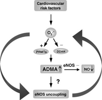
Figure 1 A. Cardiac risks of ADMA B. Effects of ADMA (Photo credit: Wikipedia)

ADMA Production and Metabolism

Nuclear proteins such as histones can be methylated on arginine residues by protein-arginine methyltransferases, enzymes which use S-adenosylmethionine as methyl groups.  This methylation event is thought to regulate protein function, much in the way of protein acetylation and phosphorylation (9).  And much like phosphorylation, these modifications are reversible through methylesterases.   The proteolysis of these arginine-methyl modifications lead to the liberation of free guanidine-methylated arginine residues such as L-NMMA, asymmetric dimethylarginine (ADMA) and symmetrical methylarginine (SDMA).

The first two, L-NMMA and ADMA, have been shown to inhibit the activity of the endothelial NOS.  This protein turnover is substantial: for instance the authors note that each day 40% of constitutive protein in adult liver is newly synthesized protein. And in several diseases, such as muscular dystrophy, ischemic heart disease, and diabetes, it has been known since the 1970’s that protein catabolism rates are very high, with corresponding increased urinary excretion of ADMA(10-13).  Methylarginines are excreted in the urine by cationic transport.  However, the majority of ADMA and L-NMMA are degraded within the cell by dimethylaminohydrolase (DDAH), first cloned and purified in rat(14).

endogenous NO inhibitors from pubchem

Figure 2.  Endogenous inhibitors of NO synthase.  Chemical structures generated from PubChem.

DDAH

DDAH specifically hydrolyzes ADMA and L-NMMA to yield citruline and demethylamine and usually shows co-localization with NOS. Pharmacologic inhibition of DDAH activity causes accumulation of ADMA and can reverse the NO-mediated bradykinin-induced relaxation of human saphenous vein.

Two isoforms have been found in human:

  • DDAH1 (found in brain and kidney and associated with nNOS) and
  • DDAH2 (highly expressed in heart, placenta, and kidney and associated with eNOS).

DDAH2 can be upregulated by all-trans retinoic acid (atRA can increase NO production).  Increased reactive oxygen species and possibly homocysteine, a risk factor for cardiovascular disease, can decrease DDAH activity(15,16).

  • The importance of DDAH activity can also be seen in transgenic mice which overexpress DDAH, exhibiting increased NO production, increased insulin sensitivity, and reduced vascular resistance  (17).  Likewise,
  • Transgenic mice, null for the DDAH1, showed increase in blood pressure, decreased NO production, and significant increase in tissue and plasma ADMA and L-NMMA.

amdanosfigure

Figure 3.  The DDAH/ADMA/NOS cycle. Figure adapted from Cooke and Ghebremarian (1).

As mentioned in the article by Cooke and Ghebremariam, the authors state: the weight of the evidence indicates that DDAH is a worthy therapeutic target. Agents that increase DDAH expression are known, and 1 of these, a farnesoid X receptor agonist, is in clinical trials

http://www.interceptpharma.com

An alternate approach is to

  • develop an allosteric activator of the enzyme.  Although
  • development of an allosteric activator is not a typical pharmaceutical approach, recent studies indicate that this may be achievable aim(18).

References:

1.            Cooke, J. P., and Ghebremariam, Y. T. : DDAH says NO to ADMA.(2011) Arteriosclerosis, thrombosis, and vascular biology 31, 1462-1464

2.            Tran, C. T., Leiper, J. M., and Vallance, P. : The DDAH/ADMA/NOS pathway.(2003) Atherosclerosis. Supplements 4, 33-40

3.            Niebauer, J., Maxwell, A. J., Lin, P. S., Wang, D., Tsao, P. S., and Cooke, J. P.: NOS inhibition accelerates atherogenesis: reversal by exercise. (2003) American journal of physiology. Heart and circulatory physiology 285, H535-540

4.            Miyazaki, H., Matsuoka, H., Cooke, J. P., Usui, M., Ueda, S., Okuda, S., and Imaizumi, T. : Endogenous nitric oxide synthase inhibitor: a novel marker of atherosclerosis.(1999) Circulation 99, 1141-1146

5.            Wilson, A. M., Shin, D. S., Weatherby, C., Harada, R. K., Ng, M. K., Nair, N., Kielstein, J., and Cooke, J. P. (2010): Asymmetric dimethylarginine correlates with measures of disease severity, major adverse cardiovascular events and all-cause mortality in patients with peripheral arterial disease. Vasc Med 15, 267-274

6.            Kielstein, J. T., Impraim, B., Simmel, S., Bode-Boger, S. M., Tsikas, D., Frolich, J. C., Hoeper, M. M., Haller, H., and Fliser, D. : Cardiovascular effects of systemic nitric oxide synthase inhibition with asymmetrical dimethylarginine in humans.(2004) Circulation 109, 172-177

7.            Kielstein, J. T., Donnerstag, F., Gasper, S., Menne, J., Kielstein, A., Martens-Lobenhoffer, J., Scalera, F., Cooke, J. P., Fliser, D., and Bode-Boger, S. M. : ADMA increases arterial stiffness and decreases cerebral blood flow in humans.(2006) Stroke; a journal of cerebral circulation 37, 2024-2029

8.            Mittermayer, F., Krzyzanowska, K., Exner, M., Mlekusch, W., Amighi, J., Sabeti, S., Minar, E., Muller, M., Wolzt, M., and Schillinger, M. : Asymmetric dimethylarginine predicts major adverse cardiovascular events in patients with advanced peripheral artery disease.(2006) Arteriosclerosis, thrombosis, and vascular biology 26, 2536-2540

9.            Kakimoto, Y., and Akazawa, S.: Isolation and identification of N-G,N-G- and N-G,N’-G-dimethyl-arginine, N-epsilon-mono-, di-, and trimethyllysine, and glucosylgalactosyl- and galactosyl-delta-hydroxylysine from human urine. (1970) The Journal of biological chemistry 245, 5751-5758

10.          Inoue, R., Miyake, M., Kanazawa, A., Sato, M., and Kakimoto, Y.: Decrease of 3-methylhistidine and increase of NG,NG-dimethylarginine in the urine of patients with muscular dystrophy. (1979) Metabolism: clinical and experimental 28, 801-804

11.          Millward, D. J.: Protein turnover in skeletal muscle. II. The effect of starvation and a protein-free diet on the synthesis and catabolism of skeletal muscle proteins in comparison to liver. (1970) Clinical science 39, 591-603

12.          Goldberg, A. L., and St John, A. C.: Intracellular protein degradation in mammalian and bacterial cells: Part 2. (1976) Annual review of biochemistry 45, 747-803

13.          Dice, J. F., and Walker, C. D.: Protein degradation in metabolic and nutritional disorders. (1979) Ciba Foundation symposium, 331-350

14.          Ogawa, T., Kimoto, M., and Sasaoka, K.: Purification and properties of a new enzyme, NG,NG-dimethylarginine dimethylaminohydrolase, from rat kidney. (1989) The Journal of biological chemistry 264, 10205-10209

15.          Ito, A., Tsao, P. S., Adimoolam, S., Kimoto, M., Ogawa, T., and Cooke, J. P.: Novel mechanism for endothelial dysfunction: dysregulation of dimethylarginine dimethylaminohydrolase. (1999) Circulation 99, 3092-3095

16.          Stuhlinger, M. C., Tsao, P. S., Her, J. H., Kimoto, M., Balint, R. F., and Cooke, J. P. : Homocysteine impairs the nitric oxide synthase pathway: role of asymmetric dimethylarginine.(2001) Circulation 104, 2569-2575

17.          Sydow, K., Mondon, C. E., Schrader, J., Konishi, H., and Cooke, J. P.: Dimethylarginine dimethylaminohydrolase overexpression enhances insulin sensitivity. (2008) Arteriosclerosis, thrombosis, and vascular biology 28, 692-697

18.          Zorn, J. A., and Wells, J. A.: Turning enzymes ON with small molecules. (2010) Nature chemical biology 6, 179-188

Other research papers on Nitric Oxide and Cardiac Risk  were published on this Scientific Web site as follows:

The Nitric Oxide and Renal is presented in FOUR parts:

Part I: The Amazing Structure and Adaptive Functioning of the Kidneys: Nitric Oxide

Part II: Nitric Oxide and iNOS have Key Roles in Kidney Diseases

Part III: The Molecular Biology of Renal Disorders: Nitric Oxide

Part IV: New Insights on Nitric Oxide donors

Cardiac Arrhythmias: A Risk for Extreme Performance Athletes

What is the role of plasma viscosity in hemostasis and vascular disease risk?

Cardiovascular Risk Inflammatory Marker: Risk Assessment for Coronary Heart Disease and Ischemic Stroke – Atherosclerosis.

Endothelial Dysfunction, Diminished Availability of cEPCs, Increasing CVD Risk for Macrovascular Disease – Therapeutic Potential of cEPCs

Biochemistry of the Coagulation Cascade and Platelet Aggregation – Part I

Nitric Oxide Function in Coagulation

Read Full Post »

« Newer Posts - Older Posts »