Feeds:
Posts
Comments

Archive for the ‘Resident-cell-based’ Category

Thymosin References

Reporter: Aviva Lev-Ari, PhD, RN


http://onlinelibrary.wiley.com/doi/10.1111/nyas.2012.1270.issue-1/issuetoc

1


Use of the cardioprotectants thymosin β4 and dexrazoxane during congenital heart surgery: proposal for a randomized, double-blind, clinical trial (pages 59–65) Daniel Stromberg, Tia Raymond, David Samuel, David Crockford, William Stigall, Steven Leonard, Eric Mendeloff and Andrew Gormley
Article first published online: 10 OCT 2012 | DOI: 10.1111/j.1749-6632.2012.06710.x

2


Cardiac repair with thymosin β4 and cardiac reprogramming factors (pages 66–72) Deepak Srivastava, Masaki Ieda, Jidong Fu and Li Qian
Article first published online: 10 OCT 2012 | DOI: 10.1111/j.1749-6632.2012.06696.x

NMR structural studies of thymosin α1 and β-thymosins (pages 73–78) David E. Volk, Cynthia W. Tuthill, Miguel-Angel Elizondo-Riojas and David G. Gorenstein
Article first published online: 10 OCT 2012 | DOI: 10.1111/j.1749-6632.2012.06656.x

4

Thymosin β4 sustained release from poly(lactide-co-glycolide) microspheres: synthesis and implications for treatment of myocardial ischemia (pages 112–119) Jeffrey E. Thatcher, Tré Welch, Robert C. Eberhart, Zoltan A. Schelly and J. Michael DiMaio
Article first published online: 10 OCT 2012 | DOI: 10.1111/j.1749-6632.2012.06681.x

Corrigendum for Ann. N.Y. Acad. Sci. 2012. 1254: 57–65 (page 121) Article first published online: 10 OCT 2012 | DOI: 10.1111/j.1749-6632.2012.06793.x
This article corrects:
A bird’s-eye view of cell therapy and tissue engineering for cardiac regeneration
Vol. 1254, Issue 1, 57–65, Article first published online: 30 APR 2012

Annals of the New York Academy of Sciences

Volume 1194 Thymosins in Health and Disease: 2nd International Symposium

Pages ix–xi, 1–230, May 2010

http://www.wiley.com/WileyCDA/WileyTitle/productCd-1573319104.html?cid=RSS_WILEY2_LIFEMED

http://onlinelibrary.wiley.com/doi/10.1111/nyas.2010.1194.issue-1/issuetoc

6. Preface to Thymosins in Health and Disease (pages ix–xi) Enrico Garaci and Allan L. Goldstein
Article first published online: 3 MAY 2010 | DOI: 10.1111/j.1749-6632.2010.05493.x

7.
Thymosin β4 and cardiac repair (pages 87–96) Santwana Shrivastava, Deepak Srivastava, Eric N. Olson, J. Michael DiMaio and Ildiko Bock-Marquette
Article first published online: 3 MAY 2010 | DOI: 10.1111/j.1749-6632.2010.05468.x

8.
Thymosin β4 facilitates epicardial neovascularization of the injured adult heart (pages 97–104) Nicola Smart, Catherine A. Risebro, James E. Clark, Elisabeth Ehler, Lucile Miquerol, Alex Rossdeutsch, Michael S. Marber and Paul R. Riley
Article first published online: 3 MAY 2010 | DOI: 10.1111/j.1749-6632.2010.05478.x

9.
Thymosin β4 enhances repair by organizing connective tissue and preventing the appearance of myofibroblasts (pages 118–124) H. Paul Ehrlich and Sprague W. Hazard III
Article first published online: 3 MAY 2010 | DOI: 10.1111/j.1749-6632.2010.05483.x

10. Thymosin β4: a key factor for protective effects of eEPCs in acute and chronic ischemia (pages 105–111) Rabea Hinkel, Ildiko Bock-Marquette, Antonis K. Hazopoulos and Christian Kupatt
Article first published online: 3 MAY 2010 | DOI: 10.1111/j.1749-6632.2010.05489.x
Corrected by:
Corrigendum for Ann. N. Y. Acad. Sci. 1194: 105–111
Vol. 1205, Issue 1, 284, Article first published online: 14 SEP 2010

11.

Thymosin β4: a candidate for treatment of stroke? (pages 112–117) Daniel C. Morris, Michael Chopp, Li Zhang and Zheng G. Zhang
Article first published online: 3 MAY 2010 | DOI: 10.1111/j.1749-6632.2010.05469.x

12. Prothymosin α as robustness molecule against ischemic stress to brain and retina (pages 20–26) Hiroshi Ueda, Hayato Matsunaga, Hitoshi Uchida and Mutsumi Ueda
Article first published online: 3 MAY 2010 | DOI: 10.1111/j.1749-6632.2010.05466.x

13.
Thymosin β4 and cardiac repair (pages 87–96) Santwana Shrivastava, Deepak Srivastava, Eric N. Olson, J. Michael DiMaio and Ildiko Bock-Marquette
Article first published online: 3 MAY 2010 | DOI: 10.1111/j.1749-6632.2010.05468.x

14.

Thymosin β4 facilitates epicardial neovascularization of the injured adult heart (pages 97–104) Nicola Smart, Catherine A. Risebro, James E. Clark, Elisabeth Ehler, Lucile Miquerol, Alex Rossdeutsch, Michael S. Marber and Paul R. Riley
Article first published online: 3 MAY 2010 | DOI: 10.1111/j.1749-6632.2010.05478.x

15.

Thymosin β4: a key factor for protective effects of eEPCs in acute and chronic ischemia (pages 105–111) Rabea Hinkel, Ildiko Bock-Marquette, Antonis K. Hazopoulos and Christian Kupatt
Article first published online: 3 MAY 2010 | DOI: 10.1111/j.1749-6632.2010.05489.x
Corrected by:
Corrigendum for Ann. N. Y. Acad. Sci. 1194: 105–111
Vol. 1205, Issue 1, 284, Article first published online: 14 SEP 2010

16.

Thymosin β4: a candidate for treatment of stroke? (pages 112–117) Daniel C. Morris, Michael Chopp, Li Zhang and Zheng G. Zhang
Article first published online: 3 MAY 2010 | DOI: 10.1111/j.1749-6632.2010.05469.x

17.Thymosin β4: structure, function, and biological properties supporting current and future clinical applications (pages 179–189) David Crockford, Nabila Turjman, Christian Allan and Janet Angel
Article first published online: 3 MAY 2010 | DOI: 10.1111/j.1749-6632.2010.05492.x

18.

The effect of thymosin treatment of venous ulcers (pages 207–212) G. Guarnera, A. DeRosa and R. Camerini, on behalf of 8 European sites
Article first published online: 3 MAY 2010 | DOI: 10.1111/j.1749-6632.2010.05490.x

19.
A randomized, placebo-controlled, single and multiple dose study of intravenous thymosin β4 in healthy volunteers (pages 223–229) Dennis Ruff, David Crockford, Gino Girardi and Yuxin Zhang
Article first published online: 3 MAY 2010 | DOI: 10.1111/j.1749-6632.2010.05474.x

Read Full Post »

Reporter: Aviva Lev-Ari, PhD, RN

Cancer Diagnostics by Genomic Sequencing: ‘No’ to Sequencing Patient’s DNA, ‘No’ to Sequencing Patient’s Tumor, ‘Yes’ to focus on Gene Mutation Aberration & Analysis of Gene Abnormalities

How to Tailor Cancer Therapy to the particular Genetics of a patient’s Cancer

THIS IS A SERIES OF FOUR POINTS OF VIEW IN SUPPORT OF the Paradigm Shift in Human Genomics

‘No’ to Sequencing Patient’s DNA, ‘No’ to Sequencing Patient’s Tumor, ‘Yes’ to focus on Gene Mutation Aberration & Analysis of Gene Abnormalities

PRESENTED in the following FOUR PARTS. Recommended to be read in its entirety for completeness and arrival to the End Point of Present and Future Frontier of Research in Genomics

Part 1:

Research Paradigm Shift in Human Genomics – Predictive Biomarkers and Personalized Medicine

http://pharmaceuticalintelligence.com/2013/01/13/paradigm-shift-in-human-genomics-predictive-biomarkers-and-personalized-medicine-part-1/

Part 2:

LEADERS in the Competitive Space of Genome Sequencing of Genetic Mutations for Therapeutic Drug Selection in Cancer Personalized Treatment

http://pharmaceuticalintelligence.com/2013/01/13/leaders-in-genome-sequencing-of-genetic-mutations-for-therapeutic-drug-selection-in-cancer-personalized-treatment-part-2/

Part 3:

Personalized Medicine: An Institute Profile – Coriell Institute for Medical Research

http://pharmaceuticalintelligence.com/2013/01/13/personalized-medicine-an-institute-profile-coriell-institute-for-medical-research-part-3/

Part 4:

The Consumer Market for Personal DNA Sequencing

http://pharmaceuticalintelligence.com/2013/01/13/consumer-market-for-personal-dna-sequencing-part-4/


Part 3:

Personalized Medicine: Institute Profile – Coriell Institute for Medical Research

Coriell Institute for Medical Research, founded in 1953 and based in Camden, New Jersey, is an independent non-profit research center dedicated to the study of the human genome. Expert staff and pioneering programs in the fields of personalized medicine, cell biology, cytogenetics, genotyping, and biobanking drive our mission.

The emerging field of personalized medicine draws upon a person’s genomic information to tailor treatments and prescription drug dosing to optimize health outcomes. The Coriell Personalized Medicine Collaborative® (CPMC®) research study is seeking to understand the usefulness of genetic risk and pharmacogenomics in clinical decision-making and healthcare management.

Coriell has a distinguished history in cell biology. We are building upon this expertise by playing an important role in induced pluripotent stem (iPS) cell research. Induced pluripotent stem cells are powerful cells which can be made from skin or blood cells, and they are revolutionizing the way human disease is studied and how drugs are developed. Skin cells from a patient diagnosed with heart disease are being genetically reprogrammed into stem cells, and then transformed into beating cardiac cells. Researchers can now examine the heart-diseased cells to better understand the progression of heart disease and develop treatments and cures. Drug efficacy and safety can also be tested in this laboratory environment, providing an efficient model of drug discovery that delivers drugs to patients sooner. This technology, called “disease in a dish,” offers researchers the potential to study the myriad of human diseases, including Alzheimer’s disease, muscular dystrophy, and diabetes.

In addition to pioneering cutting-edge research initiatives, Coriell offers custom research services – including cell culture, cytogenetic analyses, and molecular biology – to the scientific community. Furthermore, Coriell’s Genotyping and Microarray Center is one of the nation’s largest centers, with high-throughput DNA analysis, CLIA-certified genotyping platforms systems from Illumina and Affymetrix.

Essential to the Institute’s support of international scientific research is the Coriell Biobank. From this renowned cell bank, we manage and distribute the world’s most diverse collection of cell lines, DNA, and other biological resources. The Coriell Biobank provided support to the Human Genome Project, a worldwide program to map the entire human genome, and to the International HapMap Project, a project providing an efficient tool to identify disease-causing genes.

The Coriell Cell Repositories provide essential research reagents to the scientific community by establishing, verifying, maintaining, and distributing cell cultures and DNA derived from cell cultures. These collections, supported by funds from the National Institutes of Health (NIH) and several foundations, are extensively utilized by research scientists around the world.

The Business Aspects of the Institute

  • Personalized Medicine

DNA, Genes, and SNPs

What is the CPMC Study?

CPMC Technology

CPMC FAQs

CPMC Advisors and Partners

Stem Cells

Induced Pluripotent Stem (iPS) Cells

iPS Cell Research at Coriell

Biobank Catalog

Working with Coriell

  • Research Services

Overview

Biobanking

Cell Culture

Cytogenetics

Genotyping & Microarray

Molecular Biology

Research Design & Expertise

Stem Cells

Quality at Coriell

  • BioBanking

Overview

What is a Biobank?

How Coriell Banks Cells

Biobank Technology

Biobank Catalog

Working with Coriell

http://www.coriell.org/

http://www.coriell.org/assets/pdfs/discover-winter2012.pdf

http://www.ccr.coriell.org/

http://www.coriell.org/about/coriell-faqs


What is the Coriell Institute of Medical Research?

Founded in 1953, Coriell Institute for Medical Research is an independent, non-profit research organization dedicated to the study of the human genome and to supporting national and international research by providing biomaterials from its renowned biobank.

How did the Coriell Institute start?

Lewis L. Coriell, MD, PhD, a virology researcher and pediatrician, recognized the need for scientific research that would translate into better patient care. After seeing how his research helped to bring the Salk vaccine to polio patients across our nation, Dr. Coriell founded the South Jersey Medical Research Foundation. It was renamed the Institute for Medical Research in 1966 to recognize its broader reach, and, in 1985, to honor Dr. Coriell’s retirement, his name was added. For a look at our history, visit our timeline.

http://www.coriell.org/about/our-history

About the Founder

“You set up an experiment to test the theory, and most of the time it’s not the way you thought it would be. But that’s the way you learn. You go from hypothesis to hypothesis. And it’s exciting because that’s the way we learn to treat, to diagnose, and to prevent illness.”

Lewis L. Coriell, MD, PhD
Virologist and Pediatrician
June 19, 1911 – June 19, 2001

Lewis L. Coriell was born in the farming community of Sciotoville, in southern Ohio. While he was still a young child, his family moved to Montana toward more promising agricultural opportunities. It has been written that “the aspects of character, personality, temperament, and intellect that marked Dr. Coriell’s exceptional professional life… can easily be traced to his Montana upbringing.”i

Education and Early Career

Beginning his academic journey at the University of Montana, Lewis Coriell completed undergraduate studies in biology and subsequently earned a master’s degree in bacteriology and immunology in 1936. That same year, he married fellow student Ester Lentz; they would remain by each other’s side for the next 60 years. The newlyweds moved to the University of Kansas so he could pursue doctoral studies in immunology. While there, Dr. Coriell published his first article on an aspect of science he would revolutionize: The storage of cells by freezing them. Lewis Coriell earned his doctorate in 1940 and was awarded his medical degree in 1942. The young researcher was drawn to the field of virology – the study of viruses as they evolve and infect. At this time, bacterial infections presented themselves most often in children. This combination led Dr. Coriell to seek out a residency in pediatrics. As none were immediately available, he chose a cardiology residency at Henry Ford Hospital in Detroit. MI. As it happens, the Coriells’ time in Detroit was brief.

By 1943, World War II was raging and Dr. Coriell was called to service with the United States Army Medical Command’s Biological Research Division at Fort Detrick, MD. It was here that his research in cell cultivation began. After the war, Dr. Coriell began his ideal pediatric residency under Dr. Joseph Stokes, Jr., physician-in-chief at Children’s Hospital of Philadelphia (CHOP).  To his delight, Dr. Stokes placed great emphasis on research and was instrumental in attracting federal funds to research childhood disease at his institution.  The ability to translate research into patient care inspired Dr. Coriell.  He saw how research was essential to the treatment of his patients suffering the devastating effects of viruses like small pox, mumps, and polio.

Adventures in Cell Culture

By the time Dr. Coriell arrived in Philadelphia, virologists knew they had to grow viruses in cell culture to prepare purified viruses for the manufacture of vaccines. However, contamination was rife in the laboratory and proving to be a major obstacle. At CHOP, along with his colleagues, Dr. Coriell perfected the technique to culture human tissue in a sterile host that does not produce its own antibodies. The ability to sustain living human cells in culture, and keep them from being contaminated, led to a key breakthrough in polio research – it enabled scientists to grow the polio virus and work toward the first vaccine.

Moving to Camden and Taking on Polio

By the early 1950’s, an acute infectious disease called polio was spreading from person to person very quickly across the United States, striking fear into citizens, costing children their lives and crippling those who survived. In 1949, Dr. Coriell arrived in Camden, NJ, as medical director of Camden Municipal Hospital, one of the country’s last infectious disease hospitals and home to the majority of the region’s polio patients. In 1951, Dr. Coriell was appointed field director of the Polio Prevention Study and directed the successful gamma globulin field trials.

By 1954, the Salk polio vaccine could be made in large quantities and was ready for human clinical trials. Based on his success shepherding the gamma globulin field trials, Dr. Coriell was chosen by the National Poliomyelitis Foundation to evaluate the Salk polio virus vaccine clinical trials in New Jersey, Pennsylvania, Maryland, and Virginia. The success of the evaluation program led to the release of the Salk vaccine on the national level. Before the trials began in 1955, approximately 20,000 new polio cases were being reported each year. By 1960, cases were reduced to 3,000 per year. By 1979, that number was just 10 each year. Recognizing his contribution, Dr. Coriell received the 1957 International Poliomyelitis Congress Presidential Medal. Soon after, he became chairman of the Committee on the Control of Infectious Diseases of the American Academy of Pediatrics which formulated the vaccination procedures for all children in this critical period.

In 1953, Dr. Coriell initiated a campaign to build the first non-profit academic medical research institute in South Jersey. Under his guidance, the Institute for Medical Research began research in cancer, human cytogenetics, infectious diseases, and methods to improve cell culture techniques. The history of the Institute’s accomplishments included Dr. Coriell’s foresight in calling for the establishment of a central tissue culture bank and cell registry to certify and maintain cell cultures. It began with a partnership with the National Institutes of Health to create the first standardized cell repository. Today, the Institute is home to the world’s most diverse collection of cell lines and DNA samples available to researchers.

Working with his colleague, Dr. Gary McGarrity, Dr. Coriell applied infection control technology – specifically laminar flow – to create the laminar flow hood that is vital to infection control in laboratories, operating rooms, and hospital rooms around the world.

Dr. Coriell’s pioneering techniques for characterizing, freezing, and storing non-contaminated cell cultures in liquid nitrogen constitute one of the greatest contributions to modern human genetics.

Retirement

Dr. Coriell retired in 1985. To honor the occasion, the institute he founded was renamed the Coriell Institute for Medical Research. He remained involved in several ways, as a member of the board and often speaking with groups about the Institute’s history. Following his retirement, Dr. Coriell was elected president of the prestigious College of Physicians of Philadelphia, the oldest medical society in America. Dr. Coriell is the only New Jersey physician to receive this honor.

Dr. Coriell, a pioneering researcher and physician, died on June 19, 2001, in Southern New Jersey. It was his 90th birthday.

A Legacy in Science

Dr. Coriell’s accomplishments in science are indeed many. Perhaps Dr. Coriell’s most enduring legacy was his generosity in knowledge and his ability to bring scientists together to explore research questions and collaborate on solutions. Several important names in science were drawn to join or spend time at the Institute; they included Warren W. Nichols, Ray Dutcher, Richard Mulivor, Etienne Lasfargues, Jesse Charney, Arthur Greene, Daniel Moore, and collaboration with Drs. Albert Levan and Joe Hin Tijo, who first discovered that humans have 46 chromosomes.

Dr. Coriell also created an institute that is a well-respected resident of the Greater Philadelphia region and known as a leader in research worldwide.

Coriell Today

Dr. Coriell’s vision is now our vision. Today, Coriell staff and scientists collaborate on scientific ideas and programs to improve human health.

The Coriell Personalized Medicine Collaborative® research study is studying the utility of using your genetic information to tailor treatments and medications for you. And building on Dr. Coriell’s innovations in cell biology, we are playing an important role in cutting-edge stem cell research to unlock the code of human disease, including Parkinson’s and heart disease. Coriell offers a range of custom research services that have long supported national and international science. In the field of biobanking, Coriell supports research all over the world from its renowned and diverse cell collections.

Our innovation today is a testament to Dr. Coriell’s pioneering past. More importantly, our innovation is a commitment to your future.

i O’Donnell, John. Coriell; The Coriell Institute for Medical Research and a Half Century of Science. Massachusetts: SHP, 2002.

Where is the Coriell Institute located?

Coriell is located at 403 Haddon Avenue, Camden, NJ 08103. For directions, click here
We recommend that you park at 3 Cooper Plaza, a parking garage associated with the hospital, located directly across the street from Coriell. There is also a second hospital parking lot located on Benson Street, which is a block from the Institute.

For what is the Coriell Institute known?

Coriell Institute is a leader in the emerging field of personalized medicine – often called genome-informed medicine – which is the practice of using genetic information to better understand a patient’s risk for disease and response to medications. The Coriell Personalized Medicine Collaborative is a research study designed to study the utility of genetic information in clinical decision-making and patient care.

Coriell is also playing an important role in exploring the promise of induced pluripotent stem (iPS) cell  biotechnologies. [Pluripotent refers to how cells can grow into many different types of cells.] We can take skin cells and reprogram them – essentially turn back time – to behave like a stem cell. These cells can then be triggered, using specific proteins, to become cardiac cells, neurons (brain cells), or insulin-producing pancreatic cells, amongst others. Over the years, Coriell has developed an extraordinary expertise in the culture of human cells, and much of the standard practices in cell culture were developed at Coriell. This includes the techniques for freezing and thawing cells, and sterile handling of cultures. As a result of our cell biology expertise, scientists from every major research center in the world draw upon the Coriell Cell Repositories, maintained in the world’s leading biobank, which contains cell lines and DNA representing approximately 650 diseases.

Who is on the Coriell Institute staff?

Coriell is home to approximately 120 scientific and operational staff. Michael Christman, PhD, is Coriell’s President and CEO; he is an expert in genomics and genetics.  Joseph L. Mintzer is Coriell’s Executive Vice President and COO and manages the fiscal and operational aspect of the institute. Meet the rest of the Coriell leadership team here.

Who is on the Coriell Institute Board of Trustees?

Coriell is guided by a diverse Board of Trustees that includes corporate, medical, financial, and philanthropic leaders. Chairman of the Coriell Board is Robert P. Kiep III. Learn more about the Coriell Board of Trustees here.

How is Coriell Institute funded?

Coriell Institute has an annual operating budget of $17 million, about $11 million of which comes from federally- and state-funded grants and contracts. Private and corporate philanthropy provides the seed money to initiate new programs in science at Coriell – science that has the opportunity to advance discoveries in research which may not be occurring at other research institutes.

How can I support the research mission of Coriell Institute?

While the majority of Coriell’s operating revenue is derived from federally- and state-funded grants and contracts, the Institute also relies on private, foundation, and corporate philanthropy. Your support can advance the emerging field of personalized medicine to improve the practice of medicine. Your support also allows Coriell to pursue and support research in adult stem cell biology and genomics seeking to unlock the code of human disease. 
There are many ways to give to Coriell: Outrights gifts, through your workplace giving programs, planned giving, volunteering your time and expertise, or attending or hosting a Coriell event. Visit our fund development page to learn more about how you can support scientific research.

How does Coriell Institute support international research?

The Coriell Cell Repositories offers essential research materials to the scientific community by establishing, verifying, maintaining, and distributing cell cultures and DNA. Since the first NIH-sponsored repository was established in 1964 – Coriell has distributed hundreds of thousands of cell lines and DNA samples to researchers in 64 countries. More than 7,000 peer-reviewed papers have been published citing almost 12,000 Coriell Repository samples.

What research services does Coriell Institute provide? 
Coriell offers several best-in-class custom research services.

Coriell’s Genotyping and Microarray Center – one of the nation’s largest centers and CLIA-certified in 48 states – is a high-capacity facility with high-throughput systems from Affymetrix and Illumina.

The Coriell Institute Cytogenetics Laboratory is a state-of-the-art facility that combines conventional and molecular cytogenetic analyses with copy number and loss of heterozygosity (LOH) analyses by microarray. The laboratory is equipped with a network of five Applied Spectral Imaging work-stations that are used to perform G-banded karyotyping, and Fluorescent In Situ Hybridization (FISH).

Coriell also offers many preparative and diagnostic nucleic acid and molecular biology services, all subject to extensive quality controls.

And, the Coriell biobank is regarded as the most diverse collection of cell lines and DNA available to the international research community.

Does Coriell Institute engage in gene therapy or stem cell clinical trials?

Coriell Institute does not pursue research using human embryonic stem cells, nor do we conduct clinical trials on stem cell technologies. If you are interested in gene therapy or stem cell-related clinical trials, please visit http://www.clinicaltrials.gov.

What education does Coriell offer?

Coriell offers a course in cell culture: Advanced biology coupled with the history, theory, and techniques of maintaining live cells in long-term culture is offered to students.

Coriell also invites a limited number of motivated students into the Institute to participate in a Summer Experience program to gain insight into the workings of an independent research institute

How can I stay informed on what is happening at Coriell Institute?

Sign up for our email updates and you’ll receive periodic research news, notable donations, and upcoming events. Visit our Media Center regularly to read the latest news articles and Coriell press releases.

How can I get a quick overview of Coriell Institute?

Read our Coriell Fast Facts for a basic introduction to the Institute. For more information, explore the About section of our website.

Are Coriell Institute scientists and staff available for speaking engagements?

As their schedules permit, Coriell’s scientific and operational staffs enjoy the opportunity to highlight the work occurring at Coriell. Many hold joint faculty appointments at our region’s universities and teach an array of topics from business management and healthcare policy to the science of cell culture and stem cell research.

Coriell also participates in several outreach programs each year, including science festivals and conferences. We also host tours of our laboratories for business and governmental leaders and middle school and high school students.

16. Is Coriell Institute affiliated with Cooper Medical School of Rowan University?
Yes; Coriell is looking forward to welcoming the new medical school and will be integral in teaching genetics and genomics to the next generation of healthcare providers.

Fig3a-200

The Power of Stem Cell Science

The promise of stem cell research lays in its application in understanding the progression of human disease, the ability to cure disease and reverse injury, and to better target therapies to optimize our health outcomes. Induced pluripotent stem (iPS) cell technology has the ability to revolutionize the way human disease is studied. Creating iPS cell lines from various rare and common disease states, as well as from various populations, will open the doors for pre-clinical research studies.

Fig3b-200

Let Our Expertise Make Your Research a Success

Coriell offers a range of custom research services that have long supported national and international science. Whether you are requesting a cell line for your research studies or submitting DNA samples for genotyping analysis, Coriell is committed to providing you with flexible, innovative, and results-oriented research services. Our laboratories are built to foster scientific collaboration, and your research will benefit from this collaborative environment.

Coriell’s Biobank and Cell Culture Laboratory have established the gold standard in the cryopreservation of biomaterials and the capacity to support varied research worldwide. The diverse collections of biological specimens managed by Coriell offer the scientific community the highest quality specimens, which are necessary for successful research endeavors. Since the first repository – a National Institutes of Health collection – was established at Coriell in 1964, hundreds of thousands of cell lines and DNA samples have been distributed to researchers in 64 countries; more than 7,000 peer-reviewed papers have been published citing almost 12,000 biospecimens from the Coriell Biobank.

Fig3c-200

Making Medicine Personalized for You

Our health is determined by many factors: the genetics we inherit; our innate personal traits of race, age and gender; our individual behavior; our family and community networks; and at the macro level, our economic, cultural, and environmental conditions. These factors are different for every person and will change over their lifespan. So too is a person’s experience with disease and how they respond to drugs or other medical interventions. Personalized medicine intends to make medical treatment as individual as the biology of one’s disease.

Personalized medicine has the potential to offer patients and their doctors several advantages, including:

The ability to make better informed clinical decisions.

A higher probability of desired health outcomes by using better-targeted therapies.

The reduced probability of adverse reactions from medications and treatments.

A focus on prevention and prediction of disease, rather than reaction to it.

Earlier disease intervention.

Reduced healthcare costs.

Fig3d-200

Preserving cells today for research tomorrow

Dr. Lewis Coriell’s pioneering techniques for characterizing, freezing, and storing cell cultures in liquid nitrogen constitute one of the greatest contributions to modern human research. Today, the Coriell Biobank is regarded as the most diverse collection of cell lines and DNA available to the international research community. In addition to these high-quality biospecimens, Coriell also maintains tissue, plasma, serum, urine, and cerebrospinal fluid.

Few organizations have the history of innovations in repository science that have been developed and implemented at Coriell. For nearly 60 years, Coriell has set the standard in biobanking services, including the experimental design, collection, processing, distribution, cryogenic preservation, and information management of human biomaterials used in research. By developing and maintaining biorepositories as national and international resources for the study of human diseases, aging, and neurological disease, Coriell is committed to providing the scientific community with well-characterized, cell cultures and DNA preparations, annotated with rich phenotypic data.

Catalog Collections

NIGMS Human Genetic Repository 
The Human Genetic Cell Repository, sponsored by the National Institute of General Medical Sciences, provides scientists around the world with resources for cell and genetic research. The samples include highly characterized cell lines and high quality DNA. Repository samples represent a variety of disease states, chromosomal abnormalities, apparently healthy individuals and many distinct human populations.

NINDS Human Genetics DNA and Cell Line Repository 
The National Institute of Neurological Disorders and Stroke is committed to gene discovery, as a strategy for identifying the genetic causes and correlates of nervous system disorders. The NINDS Human Genetics DNA and Cell Line Repository banks samples from subjects with cerebrovascular disease, epilepsy, motor neuron disease, Parkinsonism, and Tourette Syndrome, as well as controls.

NIA Aging Cell Repository 
Sponsored by the National Institute on Aging (NIA), the AGING CELL REPOSITORY, is a resource facilitating cellular and molecular research studies on the mechanisms of aging and the degenerative processes associated with it. The cells in this resource have been collected over the past three decades using strict diagnostic criteria and banked under the highest quality standards of cell culture. Scientists use the highly-characterized, viable, and contaminant-free cell cultures from this collection for research on such diseases as Alzheimer disease, progeria, Parkinsonism, Werner syndrome, and Cockayne syndrome.

NHGRI Sample Repository for Human Genetic Research 
The National Human Genome Research Institute (NHGRI) led the National Institutes of Health’s (NIH) contribution to the International Human Genome Project, which had as its primary goal the sequencing of the human genome. This project was successfully completed in April 2003. Now, the NHGRI’s mission has expanded to encompass a broad range of studies aimed at understanding the structure and function of the human genome and its role in health and disease.

American Diabetes Association, GENNID Study 
The purpose of the American Diabetes Association (ADA), GENNID Study (Genetics of non-insulin dependent diabetes mellitus, NIDDM) is to establish a national database and cell repository consisting of information and genetic material from families with well-documented NIDDM. The GENNID Study will provide investigators with the information and samples necessary to conduct genetic linkage studies and locate the genes for NIDDM.

The Autism Research Resource 
The State of New Jersey funded the initiation of a genetic resource to support the study of autism in families where more than one child is affected or where one child is affected and one demonstrates another significant and related developmental disorder. This resource now receives continuing support from the Coriell Institute for Medical Research. An open bank of anonymously collected materials documented by a detailed clinical diagnosis forms the basis of this growing database of information about the disease.

IPBIR Repository 
The purpose of the IPBIR – Integrated Primate Biomaterials and Information Resource is to assemble, characterize, and distribute high-quality DNA samples of known provenance with accompanying demographic, geographic, and behavioral information in order to stimulate and facilitate research in primate genetic diversity and evolution, comparative genomics, and population genetics.

HD Community BioRepository 
HD Community BioRepository is a secure, centralized repository that stores and distributes quality-controlled, reliable research reagents. Huntingtin DNAs are now available and antibodies, antigenic peptides, cell lines, and hybridomas will be added soon.

USIDNET Repository 
The USIDNET DNA and Cell Repository has been established as part of an NIH-funded program – the US Immunodeficiency Network (www.usidnet.org) – to provide a resource of DNA and functional lymphoid cells obtained from patients with various primary immunodeficiency diseases. These uncommon disorders include patients with defects in T cell, B cell and/or granulocyte function as well as patients with abnormalities in antibodies/immunoglobulins, complement and other host defense mechanisms.

CDC Cell and DNA Repository 
The Genetic Testing Reference Material Coordination Program of the Centers for Disease Control and Prevention (CDC) and the Coriell Institute for Medical Research announce the availability of samples derived from transformed cell lines for use in molecular genetic testing. The DNA samples prepared from these reference cell lines are available through the Coriell Cell Repositories. Diseases include cystic fibrosis (CF), 5′ 10′ methylenetetrahydrofolate reductase deficiency (MTHFR), HFE-associated hereditary hemochromatosis, Huntington disease (HD), fragile X syndrome, Muenke syndrome, connexin 26-associated deafness, and alpha-thalassemia.

Leiomyosarcoma Cell and DNA Repository 
The Leiomyosarcoma Cell and DNA Repository has been established with an award from the National Leiomyosarcoma Foundation. This foundation provides leadership in supporting research of Leiomyosarcoma, improving treatment outcomes of those affected by this disease as well as fostering awareness in the medical community and general public.

COHORT Project 
The Cooperative Huntington’s Observational Trial Repository has been established as a resource for the discovery of information related to Huntington’s disease and its causes, progressioin, treatments, and possible cures. This is a growing bank for DATA and SPECIMENS to accelerate research on Huntington’s disease.

YERKES Repository 
The Yerkes National Primate Research Center of Emory University is an international leader in biomedical and behavioral research. For more than seven decades, the Yerkes Research Center has been dedicated to advancing scientific understanding of primate biology, behavior, veterinary care and conservation, and to improving human health and well-being.

NEI-AREDS Genetic Repository 
The Age-Related Eye Disease Study was designed to learn about macular degeneration and cataract, two leading causes of vision loss in older adults. The study looked at how these two diseases progress and what their causes may be. In addition, the study tested certain vitamins and minerals to find out if they can help to prevent or slow these diseases. Participants in the study did not have to have either disease. (Enrollment was completed in January 1998.) Eleven medical centers in the United States took part in the study, and more than 4,700 people across the country were enrolled in AREDS. The study was supported by the National Eye Institute, part of the Federal government’s National Institutes of Health. The clinical trial portion of the study also received support from Bausch & Lomb Pharmaceuticals and was completed in October 2001. Learn about the results of the clinical trial on the National Eye Institute’s website: http://www.nei.nih.gov/amd/.

The Wistar Institute 
The Wistar Institute collection at Coriell contains cell lines that have been developed by Wistar scientists. These materials are offered for non-commercial research conducted by universities, government agencies and academic research centers. The Wistar Institute collection currently contains a group of hybridomas that produce monoclonal antibodies that are useful in influenza research and vaccine development. Melanoma cell lines, derived from patients with a wide range of disease ranging from mild dysplasia to advanced metastatic cancer, will be added shortly. More information on The Wistar Institute, its research and scientists can be found at www.wistar.org.

J. Craig Venter Institute Human Reference Genome (HuRef) 
The Human Reference Genetic Material Repository makes available DNA from a single individual, J. Craig Venter, whose genome has been sequenced and assembled. The DNA samples are prepared from a lymphoblastoid cell line established at Coriell Cell Repositories from a sample of peripheral blood. The DNA samples are available in 50 microgram aliquots. The lymphoblastoid cell line is not available for distribution..

Read Full Post »

Reporter: Aviva Lev-Ari, PhD, RN

Scar Tissue In Damaged Hearts Reprogrammed By Gene Therapy Into Healthy Heart Muscle

Article Date: 08 Jan 2013 – 0:00 PST

A cocktail of three specific genes can reprogram cells in the scars caused by heart attacks into functioning muscle cells, and the addition of a gene that stimulates the growth of blood vessels enhances that effect, said researchers from Weill Cornell Medical College, Baylor College of Medicine and Stony Brook University Medical Center in a report that appears online in the Journal of the American Heart Association. 

“The idea of reprogramming scar tissue in the heart into functioning heart muscle was exciting,” said Dr. Todd K. Rosengart, chair of the Michael E. DeBakey Department of Surgery at BCM and the report’s corresponding author. “The theory is that if you have a big heart attack, your doctor can just inject these three genes into the scar tissue during surgery and change it back into heart muscle. However, in these animal studies, we found that even the effect is enhanced when combined with the VEGF gene.” 

“This experiment is a proof of principle,” said Dr. Ronald G. Crystal, chairman and professor of genetic medicine at Weill Cornell Medical College and a pioneer in gene therapy, who played an important role in the research. “Now we need to go further to understand the activity of these genes and determine if they are effective in even larger hearts.” 

During a heart attack, blood supply is cut off to the heart, resulting in the death of heart muscle. The damage leaves behind a scar and a much weakened heart. Eventually, most people who have had serious heart attacks will develop heart failure

Changing the scar into heart muscle would strengthen the heart. To accomplish this, during surgery, Rosengart and his colleagues transferred three forms of the vascular endothelial growth factor (VEGF) gene that enhances blood vessel growth or an inactive material (both attached to a gene vector) into the hearts of rats. Three weeks later, the rats received either Gata4, Mef 2c and Tbx5 (the cocktail of transcription factor genes called GMT) or an inactive material. (A transcription factor binds to specific DNA sequences and starts the process that translates the genetic information into a protein.) 

The GMT genes alone reduced the amount of scar tissue by half compared to animals that did not receive the genes, and there were more heart muscle cells in the animals that were treated with GMT. The hearts of animals that received GMT alone also worked better as defined by ejection fraction than those who had not received genes. (Ejection fraction refers to the percentage of blood that is pumped out of a filled ventricle or pumping chamber of the heart.) 

The hearts of the animals that had received both the GMT and the VEGF gene transfers had an ejection fraction four times greater than that of the animals that had received only the GMT transfer. 

Rosengart emphasizes that more work needs to be completed to show that the effect of the VEGF is real, but it has real promise as part of a new treatment for heart attack that would minimize heart damage. 

“We have shown both that GMT can effect change that enhances the activity of the heart and that the VEGF gene is effective in improving heart function even more,” said Dr. Crystal. 

The idea started with the notion of induced pluripotent stem cells – reprograming mature specialized cells into stem cells that are immature and can differentiate into different specific cells needed in the body. Dr. Shinya Yamanaka and Sir John B. Gurdon received the Nobel Prize in Medicine and Physiology for their work toward this goal this year. 

However, use of induced pluripotent stem cells has the potential to cause tumors. To get around that, researchers in Dallas and San Francisco used the GMT cocktail to reprogram the scar cells into cardiomyocytes (cells that become heart muscle) in the living animals. 

Now Rosengart and his colleagues have gone a step farther – encouraging the production of new blood vessels to provide circulation to the new cells.

REFERENCES:

Others who took part in this work include Megumi Mathison, Ronald Gersch, Ahmed Nasser, Sarit Lilo, Mallory Korman, Mitchell Fourman, Kenneth Shroyer, Jianchang Yang, Yupo Ma, all of Stony Brook University Medical Center and Neil Hackett of Weill Cornell Medical College.
Funding for this work came from the generosity of James and Lisa Cohen.
Weill Cornell Medical College

CITATIONS:

MLA

n.p. “Scar Tissue In Damaged Hearts Reprogrammed By Gene Therapy Into Healthy Heart Muscle.” Medical News Today. MediLexicon, Intl., 8 Jan. 2013. Web.
9 Jan. 2013. <http://www.medicalnewstoday.com/releases/254618.php>

APA

n.p. (2013, January 8). “Scar Tissue In Damaged Hearts Reprogrammed By Gene Therapy Into Healthy Heart Muscle.” Medical News Today. Retrieved from
http://www.medicalnewstoday.com/releases/254618.php.

SOURCE:

http://www.medicalnewstoday.com/releases/254618.php 

Read Full Post »

Cardiac Surgery Theatre in China vs. in the US: Cardiac Repair Procedures, Medical Devices in Use, Technology in Hospitals, Surgeons’ Training and Cardiac Disease Severity”

 Interviewer: Aviva Lev-Ari, PhD, RN

Article ID #11: Cardiac Surgery Theatre in China vs. in the US: Cardiac Repair Procedures, Medical Devices in Use, Technology in Hospitals, Surgeons’ Training and Cardiac Disease Severity”. Published on 1/8/2013

WordCloud Image Produced by Adam Tubman


First segment: Interview with Dr. LCR, Cardiac Surgeon,

Interviews with Scientific Leaders Series

This is the first segment on this subject, in the Interviews with Scientific Leaders Series on our Open Access Online Scientific Journal.

This Segment and the following to be published in this Open Access Online Scientific Journal, are based on an e-mail exchange with a prominent Cardiac Surgeon who worked in the US and in China in Cardiac Surgery Theatres. The identity of the surgeon, I shall conceal. The opening segment provides background, the volume of procedures and the general overview of the medical devices in use.

Following segments will be based on an exchange of Question and Answers (Q&A) which I will be presenting to our Surgeon interviewee and his answers to these specific questions.

I plan to cover the following topics:

  • Cardiac Repair Procedures
  • Medical Devices in Use
  • Technology in Hospitals
  • Surgeons’ Training and
  • Cardiac Disease Severity

Background

Dr. LCR, M.D., F.R.C.S.(C), F.A.C.S., Cardiothoracic & Vascular Surgery is the Cardiac Surgeon in this Interview with Scientific Leaders.

Dr. LCR was born in Hong Kong, SAR, China and came to the US in 1972 for higher education and became a US citizen since 1979.  He is a US medical school graduate, trained general surgeon (ABS re-certified till 12/2014) and Canadian trained cardiothoracic surgeon (ABTS re-certified till 12/2021). Dr. LCR is also a Fellow of The American College of Surgeons (F.A.C.S.) and an active member of The Society of Thoracic Surgeons (STS) since 1996. He practiced cardiothoracic and vascular surgery in the US between 1992 and 2007 when he accepted the invitation of the Foreign Experts Bureau of the Chinese government to teach/work cardiovascular surgery in China and has just returned to the US two month ago.

During those five and a half years in China, Dr. LCR worked at some of the top and largest cardiovascular programs (West China Hospital of Sichuan University at the city of Chengdu, 1,700 cardiac cases/year.

Dr. LCR worked in Guangdong Provincial Cardiovascular Institute at the city of Guangzhou, the third or fourth largest cardiac program in China, with 3,792 cardiac cases in 2011).

Dr. LCR has also authored or co-authored at least 6 scientific articles when he was in China, all published in the US cardiac journals.

Dr. LCR speaks two Chinese dialects fluently and read and write Chinese at an advanced level.

Below, we present the personal observation and opinions regarding “How the Operating Rooms (OR) are equipped and run in China and the US.”

Dr. LCR was professor of thoracic surgery at West China Hospital of Sichuan University from 06/2007 to 04/2008), the largest hospital in China, with 4,200 beds on one campus (there are three other campuses).

The hospital has 80 some OR’s and the out-patient department saw 2.5 million out-patients the year he was there.  The department of Cardiac Surgery performed 1,700 cardiac surgical cases in 2007, with 4 OR’s.

All the major US cardiac surgery vendors were represented, prosthetic heart valves, sutures,etc.. For some “Reason” we only used St. Jude Medical‘s mechanical valves, and we must have put in more than 1,200 to 1,400 valves. They were sold to the Chinese patients the same price as they were sold in the US, about US$ 3,000 each (or 21,00 CNY), about 3.6 million USD of biz for St. Jude, just from a division of the hospital.

The top two heart surgery centers are located in Beijing. Fuwei hospital did 9,700 heart surgery, and the other Aszhen hospital did close to 6,000 in 2011.

The last hospital Dr. LCR worked for as an attending/consultant surgeon until September 2012, The Guangdong Provincial General Hospital (2,400 beds)-The Guangdong Provincial Cardiovascular Institute (480 beds) is probably the third or fourth largest heart surgery center in China, did 3,782 cardiac surgical cases in 2011, most likely exceeded 4,000 in 2012.

If you add the coronary stents put in by the cardiologists in China , the biz for the medical device vendors is immense. For every one coronary bypass we did, the cardiologists must have inserted 20 or more stents. Without a doubt — China is and will be the biggest market for a lot of things, including medical devices, and you are going to the right place. Good luck.

The Next segment will present Dr. LCR’s answers to specific questions I will be e-mailing him of the following topics:

  • Cardiac Repair Procedures
  • Medical Devices in Use
  • Technology in Hospitals
  • Surgeons’ Training and
  • Cardiac Disease Severity


Read Full Post »

Reporter: Aviva Lev-Ari, PhD, RN


Nature. 2012 Dec 5. doi: 10.1038/nature11682. [Epub ahead of print]

Mammalian heart renewal by pre-existing cardiomyocytes.

Senyo SESteinhauser MLPizzimenti CLYang VKCai LWang MWu TDGuerquin-Kern JLLechene CPLee RT.

Source

Cardiovascular Division, Department of Medicine, Brigham and Women’s Hospital and Harvard Medical School, Cambridge, Massachusetts 02139, USA.

Abstract

Although recent studies have revealed that heart cells are generated in adult mammals, the frequency of generation and the source of new heart cells are not yet known. Some studies suggest a high rate of stem cell activity with differentiation of progenitors to cardiomyocytes. Other studies suggest that new cardiomyocytes are born at a very low rate, and that they may be derived from the division of pre-existing cardiomyocytes. Here we show, by combining two different pulse-chase approaches-genetic fate-mapping with stable isotope labelling, and multi-isotope imaging mass spectrometry-that the genesis of cardiomyocytes occurs at a low rate by the division of pre-existing cardiomyocytes during normal ageing, a process that increases adjacent to areas of myocardial injury. We found that cell cycle activity during normal ageing and after injury led to polyploidy and multinucleation, but also to new diploid, mononucleate cardiomyocytes. These data reveal pre-existing cardiomyocytes as the dominant source of cardiomyocyte replacement in normal mammalian myocardial homeostasis as well as after myocardial injury.

PMID: 23222518


http://www.ncbi.nlm.nih.gov/pubmed/23222518
December 17, 2012

Source of New Heart Cell Growth Discovered

A study in mice suggests that new heart cells arise from pre-existing heart cells and that the renewal process slows with age. The findings may lead to improved regenerative therapy for people with heart damage.

Image of mouse heart cells with brightly colored nuclei.

Dividing heart cells in newborn mice incorporate a tracer that can be seen in the cells’ nuclei. The color scale at the bottom shows the intensity of the tracer signal, with higher intensity toward the right side. Image by Senyo et al., courtesy of Nature.

The heart’s muscle cells, called cardiomyocytes, don’t readily replenish themselves. So an injured heart isn’t easy to mend. After a heart attack, a significant number of cardiomyocytes die. This jeopardizes heart function and can lead to chronic heart failure and possibly death. To help heal damaged hearts, scientists have been searching for a group of cells in the heart that can replenish damaged tissue.

Recent research has shown that the human heart generates new cardiomyocytes throughout its lifespan, but how frequently the cells are generated and where they come from is still debated. Studying heart tissue and cell turnover rate is technically very challenging. Some research has hinted that new cells can arise from progenitor cells at a fairly high rate. Other work has suggested that pre-existing cardiomyocytes divide at a fairly low rate to give rise to new cells.

A team led by Dr. Richard T. Lee of Brigham and Women’s Hospital and Harvard Medical School applied novel technology to investigate heart cell regeneration in mice. They used a technique called multi-isotope imaging mass spectrometry (MIMS). MIMS can detect nonradioactive stable isotope tracers. In contrast to most other tracers, these don’t alter biochemical reactions and aren’t harmful to the organism.

The scientists incorporated a rare stable isotope of nitrogen, nitrogen-15 (15N), into thymidine—one of the building blocks of DNA. When cells divide, the [15N] thymidine is taken up and added to new DNA. It can then be seen in the cells’ nuclei using MIMS. The work was supported in part by several NIH institutes, including the National Institute on Aging (NIA) and National Heart, Lung and Blood Institute (NHLBI). The study appeared online on December 5, 2012, in Nature.

To study cell turnover at different ages, the scientists gave 3 groups of mice [15N] thymidine for 8 weeks starting at day 4 (newborn), 10 weeks (young adult) or 22 months (old adult). To distinguish which types of cells created new cardiomyocytes, they performed similar experiments in mice genetically engineered with fluorescent tags to mark cardiomyocytes.

The scientists found that new heart cells were generated from pre-existing cardiomyocytes rather than progenitor cells. They estimated a yearly renewal rate of less than 1% during normal, healthy conditions. The rate of cell regeneration, they found, declined with age.

The team next used MIMS to study cell turnover following a heart attack. In the 8 weeks after the damage, roughly 3% of heart cells regenerated in the area next to the injured site. However, the researchers also noted that many cells had taken up 15N but not completed cell division.

“Our data show that adult cardiomyocytes are primarily responsible for the generation of new cardiomyocytes and that as we age, we lose some capacity to form new heart cells,” Lee says. “This means that we are losing our potential to rebuild the heart in the latter half of life, just when most heart disease hits us. If we can unravel why this occurs, we may be able to unleash some heart regeneration potential.”

—by Miranda Hanson, Ph.D.

RELATED LINKS:

Reference: Nature. 2012 Dec 5. doi: 10.1038/nature11682. [Epub ahead of print]. PMID: 23222518.


Read Full Post »

Reporter: Aviva Lev-Ari, PhD, RN

International Consortium Finds 15 Novel Risk Loci for Coronary Artery Disease

“lipid metabolism and inflammation as key biological pathways involved in the genetic pathogenesis of CAD”

Themistocles Assimes from Stanford University Medical Center said in a statement that these findings begin to clear up its role. “Our network analysis of the top approximately 240 genetic signals in this study seems to provide evidence that genetic defects in some pathways related to inflammation are a cause,” he said.

On this Open Access Online Scientific Journal, lipid metabolism and inflammation were researched and exposed in the following entries.

However, it is ONLY,  these 15 Novel Risk Loci for Coronary Artery Disease published on 12/3/2012 that provides the genomics loci and the genetic explanation for the following empirical results obtained in the recent research on Cardiovascular diseases, as present in the second half of this post, below.

Special Considerations in Blood Lipoproteins, Viscosity, Assessment and Treatment

http://pharmaceuticalintelligence.com/2012/11/28/special-considerations-in-blood-lipoproteins-viscosity-assessment-and-treatment/

What is the role of plasma viscosity in hemostasis and vascular disease risk?

http://pharmaceuticalintelligence.com/2012/11/28/what-is-the-role-of-plasma-viscosity-in-hemostasis-and-vascular-disease-risk/

PIK3CA mutation in Colorectal Cancer may serve as a Predictive Molecular Biomarker for adjuvant Aspirin therapy

http://pharmaceuticalintelligence.com/2012/11/28/pik3ca-mutation-in-colorectal-cancer-may-serve-as-a-predictive-molecular-biomarker-for-adjuvant-aspirin-therapy/

Peroxisome proliferator-activated receptor (PPAR-gamma) Receptors Activation: PPARγ transrepression for Angiogenesis in Cardiovascular Disease and PPARγ transactivation for Treatment of Diabetes

http://pharmaceuticalintelligence.com/2012/11/13/peroxisome-proliferator-activated-receptor-ppar-gamma-receptors-activation-pparγ-transrepression-for-angiogenesis-in-cardiovascular-disease-and-pparγ-transactivation-for-treatment-of-dia/

Positioning a Therapeutic Concept for Endogenous Augmentation of cEPCs — Therapeutic Indications for Macrovascular Disease: Coronary, Cerebrovascular and Peripheral

http://pharmaceuticalintelligence.com/2012/08/29/positioning-a-therapeutic-concept-for-endogenous-augmentation-of-cepcs-therapeutic-indications-for-macrovascular-disease-coronary-cerebrovascular-and-peripheral/

Cardiovascular Risk Inflammatory Marker: Risk Assessment for Coronary Heart Disease and Ischemic Stroke – Atherosclerosis.

http://pharmaceuticalintelligence.com/2012/10/30/cardiovascular-risk-inflammatory-marker-risk-assessment-for-coronary-heart-disease-and-ischemic-stroke-atherosclerosis/

The Essential Role of Nitric Oxide and Therapeutic NO Donor Targets in Renal Pharmacotherapy

http://pharmaceuticalintelligence.com/2012/11/26/the-essential-role-of-nitric-oxide-and-therapeutic-no-donor-targets-in-renal-pharmacotherapy/

Nitric Oxide Function in Coagulation

http://pharmaceuticalintelligence.com/2012/11/26/nitric-oxide-function-in-coagulation/Nitric Oxide Function in Coagulation

15 Novel Risk Loci for Coronary Artery Disease

December 03, 2012

NEW YORK (GenomeWeb News) – A large-scale association analysis of coronary artery disease has detected 15 new loci associated with risk of the disease, bringing the total number of known risk alleles to 46. As the international CARDIoGRAMplusC4D Consortium reported in Nature Genetics yesterday, the study also found that lipid metabolism and inflammation pathways may play a part in coronary artery disease pathogenesis.

“The number of genetic variations that contribute to heart disease continues to grow with the publication of each new study,” Peter Weissberg from the British Heart Foundation, a co-sponsor of the study, said in a statement. “This latest research further confirms that blood lipids and inflammation are at the heart of the development of atherosclerosis, the process that leads to heart attacks and strokes.”

For its study, the consortium, which was comprised of more than 180 researchers, performed a meta-analysis of data from the 22,233 cases and 64,762 controls of the CARDIoGRAM genome-wide association study and of the 41,513 cases and 65,919 controls from 34 additional studies of people of European and South Asian descent. Using the custom Metabochip array from Illumina, the team tested SNPs for disease association in those populations. The SNPs that reached significance in that stage of the study were then replicated using data from a further four studies.

From this, the team identified 15 new loci with genome-wide significance for risk of coronary artery disease, in addition to known risk loci.

The consortium also reported an additional 104 SNPs that appeared to be associated with coronary artery disease but did not meet the cut-off for genome-wide significance.

Then looking to other known risk factors for coronary artery disease, like blood pressure and diabetes, the researchers assessed whether any of those risk factors were associated with the risk loci. Of the 45 known risk loci, 12 were associated with blood lipid content and five with blood pressure. And while people with type 2 diabetes have a higher risk of developing coronary artery disease, none of the known risk loci were linked to diabetic traits.

An analysis of the pathways that SNPs linked to coronary artery disease fall in revealed that many of them are involved in lipid metabolism and inflammation pathways — 10 risk loci were found to be involved in lipid metabolism. “Our network analysis identified lipid metabolism and inflammation as key biological pathways involved in the genetic pathogenesis of CAD,” the researchers wrote in the paper. “Indeed, there was significant crosstalk between the lipid metabolism and inflammation pathways identified.”

The role of inflammation in coronary artery disease has been up for debate — a debate centering on whether it is a cause or a consequence of the disease — and study author Themistocles Assimes from Stanford University Medical Center said in a statement that these findings begin to clear up its role. “Our network analysis of the top approximately 240 genetic signals in this study seems to provide evidence that genetic defects in some pathways related to inflammation are a cause,” he said.

Related Stories

SOURCE:

http://www.genomeweb.com//node/1159041?hq_e=el&hq_m=1424172&hq_l=3&hq_v=09187c3305


GWAS, Meta-Analyses Uncover New Coronary Artery Disease Risk Loci

March 07, 2011

By a GenomeWeb staff reporter

NEW YORK (GenomeWeb News) – Three new studies — including the largest meta-analysis yet of coronary artery disease — have identified dozens of coronary artery disease risk loci in European, South Asian, and Han Chinese populations. All three papers appeared online yesterday in Nature Genetics.

For the first meta-analysis, members of a large international consortium known as the Coronary Artery Disease Genome-wide Replication and Meta-Analysis study, or CARDIoGRAM, sifted through data on more than 135,000 individuals from the UK, US, Europe, Iceland, and Canada. In so doing, they tracked down nearly two-dozen new and previously reported coronary artery disease risk loci.

Because only a few of these loci have been linked to other heart disease-related risk factors such as high blood pressure, those involved say the work points to yet unexplored heart disease pathways.

“[W]e have discovered several new genes not previously known to be involved in the development of coronary heart disease, which is the main cause of heart attacks,” co-corresponding author Nilesh Samani, a cardiology researcher affiliated with the University of Leicester and Glenfield Hospital, said in a statement. “Understanding how these genes work, which is the next step, will vastly improve our knowledge of how the disease develops, and could ultimately help to develop new treatments.”

Samani and his co-workers identified the loci by bringing together data on 22,233 individuals with coronary artery disease and 64,762 unaffected controls. The participants, all of European descent, had been sampled through 14 previous genome-wide association studies and genotyped at an average of about 2.5 million SNPs each. The team then assessed the top candidate SNPs found in this initial analysis in another 56,582 individuals (roughly half of whom had coronary artery disease).

The search not only confirmed associations between coronary artery disease and 10 known loci, but also uncovered associations with 13 other loci. All but three of these were distinct from loci previously implicated in other heart disease risk factors such as hypertension or cholesterol levels, researchers noted.

Consequently, those involved in the study say that exploring the biological functions of the newly detected genes could offer biological clues about how heart disease develops — along with strategies for preventing and treating it.

The genetic complexity of coronary artery disease being revealed by such studies has diagnostic implications as well, according to some.

“Each new gene identified brings us a small step closer to understanding the biological mechanisms of cardiovascular disease development and potential new treatments,” British Heart Foundation Medical Director Peter Weissberg, who was not directly involved in the new studies, said in a statement. “However, as the number of genes grows, it takes us further away from the likelihood that a simple genetic test will identify those most of risk of suffering a heart attack or a stroke.”

Meanwhile, researchers involved with Coronary Artery Disease Genetics Consortium did their own meta-analysis using data collected from four GWAS to find five coronary artery-associated loci in European and South Asian populations.

The group initially looked at 15,420 individuals with coronary artery disease — including 6,996 individuals from South Asia and 8,424 from Europe — and 15,062 unaffected controls. Participants were genotyped at nearly 575,000 SNPs using Illumina BeadChips. Most South Asian individuals tested came from India and Pakistan, researchers noted, while European samples came from the UK, Italy, Sweden, and Germany.

For the validation phase of the study, the team focused in on 59 SNPs at 50 loci from the discovery group that seemed most likely to yield authentic new disease associations. These variants were assessed in 10 replication groups comprised of 21,408 individuals with coronary artery disease and 19,185 individuals without coronary artery disease.

All told, researchers found five loci that seem to influence coronary artery disease risk in the European and South Asian populations: one locus each on chromosomes 7, 11, and 15, along with a pair of loci on chromosome 10.

The team didn’t see significant differences in the frequency or effect sizes of these newly identified variants between the European and South Asian populations, though they emphasized that their approach may have missed some potential risk variants, particularly in those of South Asian descent.

“[C]urrent genome-wide arrays may not capture all important variants in South Asians,” they explained, “Nevertheless, all of the known and new variants were significantly associated with [coronary artery disease] risk in both the European and South Asian populations in the current study, indicating the importance of genes associated with [coronary artery disease] beyond the European ancestry groups in which they were first defined.”

Finally, using a three-stage discovery, validation, and replication GWAS approach, Chinese researchers identified a single coronary artery disease risk variant in the Han Chinese population.

In this first phase of that study, researchers tested samples from 230 cases and 230 controls from populations in Beijing and in China’s Hubei province that were genotyped at Genentech and CapitalBio using Affymetrix Human SNP5.0 arrays.

From the nearly three-dozen SNPs identified in the first stage of the study, they narrowed in on nine suspect variants. After finding linkage disequilibrium between two of the variants, they did validation testing on eight of these in 572 individuals with coronary artery disease and 436 unaffected controls, all from Hubei province.

That analysis implicated a single chromosome 6 SNP called rs6903956 in coronary artery disease — a finding the team ultimately replicated in another group of 2,668 coronary artery disease cases and 3,917 controls from three independent populations in Hubei, Shandong province, and northern China.

The team’s subsequent experiments suggest that the newly detected polymorphism, which falls within a putative gene called C6orf105 on chromosome 6, curbs the expression of this gene. The functional consequences of this shift in expression, if any, are yet to be determined.

Because C6orf105 shares some identity and homology with an androgen hormone inducible gene known as AIG1, those involved in the study argue that it may be worthwhile to investigate possible ties between C6orf105 expression, androgen signaling, and coronary artery disease.

“Androgen has previously been reported to be associated the pathogenesis of atherosclerosis,” they wrote. “Future studies are needed to explore whether C6orf105 expression can be induced by androgen and to further determine the potential mechanism of [coronary artery disease] associated with decreased C6orf105 expression.”

 SOURCE:

Read Full Post »

Peroxisome proliferator-activated receptor (PPAR-gamma) Receptors Activation: PPARγ transrepression for Angiogenesis in Cardiovascular Disease and PPARγ transactivation for Treatment of Diabetes

Peroxisome proliferator-activated receptor (PPAR-gamma) Receptors Activation: PPARγ transrepression  for Angiogenesis in Cardiovascular Disease and PPARγ transactivation for Treatment of Diabetes


UPDATED on 11/27/2018

A new combination drug therapy for CVD patients with co-morbidity of DM2 is presented in the following article, representing different mechanism of actions, pathways and a novel treatment proposed in 2018:

Cardiovascular (CV) Disease and Diabetes: New ACC Guidelines for use of two major new classes of diabetes drugs — sodium-glucose cotransporter type 2 (SGLT2) inhibitors and glucagon-like peptide 1 receptor agonists (GLP-1RAs) for reduction of adverse outcomes

https://pharmaceuticalintelligence.com/2018/11/27/cardiovascular-cv-disease-and-diabetes-new-acc-guidelines-for-use-of-two-major-new-classes-of-diabetes-drugs-sodium-glucose-cotransporter-type-2-sglt2-inhibitors-and-glucagon-like/

The title of this article

Peroxisome proliferator-activated receptor (PPAR-gamma) Receptors Activation: PPARγ transrepression  for Angiogenesis in Cardiovascular Disease and PPARγ transactivation for Treatment of Diabetes

represents an explanation for pathways and mechanism of actions of combination drug therapy novel in its conceptualization in 2013.


The research is presented in the following three parts. References for each part are at the end.


PART I:             Genetics and Biochemistry of Peroxisome proliferator-activated receptor

Reporter: Aviva Lev-Ari, PhD, RN

PART II:             Peroxisome proliferator-activated receptors as stimulants of angiogenesis in cardiovascular disease and diabetes

Reporter: Aviva Lev-Ari, PhD, RN

PART III:            PPAR-gamma Role in Activation of eNOS: The Cardiovascular Benefit

Author and Curator: Aviva Lev-Ari, PhD, RN


PART I:

Genetics and Biochemistry of Peroxisome proliferator-activated receptor

PPAR -alpha and -gamma pathways

In the field of molecular biology, the peroxisome proliferator-activated receptors (PPARs) are a group of nuclear receptor proteins that function as transcription factors regulating the expression of genes.[1] PPARs play essential roles in the regulation of cellular differentiation, development, and metabolism (carbohydrate, lipid, protein), and tumorigenesis[2] of higher organisms.[3][4]

Three types of PPARs have been identified: alpha, gamma, and delta (beta):[3]

Physiological function

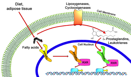
All PPARs heterodimerize with the retinoid X receptor (RXR) and bind to specific regions on the DNA of target genes. These DNA sequences are termed PPREs (peroxisome proliferator hormone response elements). The DNA consensus sequence is AGGTCANAGGTCA, with N being a random nucleotide. In general, this sequence occurs in the promotor region of a gene, and, when the PPAR binds its ligand, transcription of target genes is increased or decreased, depending on the gene. The RXR also forms a heterodimer with a number of other receptors (e.g., vitamin D and thyroid hormone).

The function of PPARs is modified by the precise shape of their ligand-binding domain (see below) induced by ligand binding and by a number of coactivator and corepressor proteins, the presence of which can stimulate or inhibit receptor function, respectively.[9]

Endogenous ligands for the PPARs include free fatty acids and eicosanoids. PPARγ is activated by PGJ2 (a prostaglandin). In contrast, PPARα is activated by leukotriene B4. PPARγ activation by agonist RS5444 may inhibit anaplastic thyroid cancer growth.[10]

Peroxisome proliferator-activated receptor

Peroxisome proliferator-activated receptor (Photo credit: Wikipedia)

Genetics

The three main forms are transcribed from different genes:

  •                PPARα – chromosome 22q12-13.1 (OMIM 170998)
  •                PPARβ/δ – chromosome 6p21.2-21.1 (OMIM 600409)
  •                PPARγ – chromosome 3p25 (OMIM 601487).

Hereditary disorders of all PPARs have been described, generally leading to a loss in function and concomitant lipodystrophy, insulin resistance, and/or acanthosis nigricans.[11] Of PPARγ, a gain-of-function mutation has been described and studied (Pro12Ala) which decreased the risk of insulin resistance; it is quite prevalent (allele frequency 0.03 – 0.12 in some populations).[12] In contrast, pro115gln is associated with obesity. Some other polymorphisms have high incidence in populations with elevated body mass indexes.

SOURCE:

http://en.wikipedia.org/wiki/Peroxisome_proliferator-activated_receptor


Mechanism of action

Thiazolidinediones or TZDs act by activating PPARs (peroxisome proliferator-activated receptors), a group of nuclear receptors with greatest specificity for PPARγ (gamma). The endogenous ligands for these receptors are free fatty acids (FFAs) and eicosanoids. When activated, the receptor binds to DNA in complex with the retinoid X receptor (RXR), another nuclear receptor, increasing transcription of a number of specific genes and decreasing transcription of others.

PPARγ transactivation

Thiazolidinedione ligand dependent transactivation is responsible for the majority of anti-diabetic effects.

The activated PPAR/RXR dimer binds to peroxisome proliferator hormone response elements upstream of target genes in complex with a number of coactivators such as nuclear receptor coactivator 1 and CREB binding protein, this causes upregulation of genes (for a full list see PPARγ:

TZDs also increase the synthesis of certain proteins involved in fat and glucose metabolism, which reduces levels of certain types of lipids, and circulating free fatty acids. TZDs generally decrease triglycerides and increase high-density lipoprotein cholesterol (HDL-C) and low-density lipoprotein cholesterol (LDL-C). Although the increase in LDL-C may be more focused on the larger LDL particles, which may be less atherogenic, the clinical significance of this is currently unknown. Nonetheless, rosiglitazone, a certain glitazone, was suspended from allowed use by medical authorities in Europe, as it has been linked to an increased risk of heart attack and stroke.[3]

PPARγ transrepression

Thiazolidinedione ligand dependent transrepression mediates the majority of anti-inflammatory effects.

Binding of PPARγ to coactivators appears to reduce the levels of coactivators available for binding to pro-inflammatory transcription factors such as NF-κB, this causes a decrease in transcription of a number of pro inflammatory genes, including various interleukins and tumour necrosis factors.

SOURCE:

http://en.wikipedia.org/wiki/Thiazolidinedione

 1. Waki H, Yamauchi T, Kadowaki T (February 2010). “[Regulation of differentiation and hypertrophy of adipocytes and adipokine network by PPARgamma]” (in Japanese). Nippon Rinsho 68 (2): 210–6. PMID 20158086.

2. Panigrahy D, Singer S, Shen LQ, et al. (2002). “PPARγ ligands inhibit primary tumor growth and metastasis by inhibiting angiogenesis”. J. Clin. Invest. 110 (7): 923–32. doi:10.1172/JCI15634. PMC 151148. PMID 12370270.

3. NHS: Avandia diabetes drug suspended, Friday 24th September 2010


Members of the class

The chemical structure of thiazolidinedione

Chemically, the members of this class are derivatives of the parent compound thiazolidinedione, and include:

  •                Rosiglitazone (Avandia), which was put under selling restrictions in the US and withdrawn from the market in            Europe due to an increased risk of cardiovascular events.
  •                Pioglitazone (Actos), France and Germany have suspended the sale of the diabetes drug Actos after a study suggested the drug, also known as pioglitazone, could raise the risk of bladder cancer.[4]
  •                Troglitazone (Rezulin), which was withdrawn from the market due to an increased incidence of drug-induced hepatitis.

Experimental agents include netoglitazone, an antidiabetic agent, rivoglitazone, and the early non-marketed thiazolidinedione ciglitazone.

Replacing one oxygen atom in a thiazolidinedione with an atom of sulfur gives a rhodanine.

SOURCE:

http://en.wikipedia.org/wiki/Thiazolidinedione

PART II:

Peroxisome proliferator-activated receptors as Stimulants of Angiogenesis in Cardiovascular Disease and Diabetes

In 2009 in Diabetes Metab Syndr Obes a seminal paper was published on the topic by  Desouza, Rentschler and Fonseca. (2009). This work constitutes Part II. http://www.ncbi.nlm.nih.gov/pmc/articles/PMC3048019/

Mechanisms by which PPARs may stimulate angiogenesis

PPARs seem to have a protective role in ischemic tissues, including brain, cardiac and skin. A part of this may be by stimulating angiogenesis and improving blood supply. Hypoxia is a trigger for the development of angiogenesis. One of the key mediators in hypoxia-induced angiogenesis is hypoxia inducible factor (HIF-1), which is induced in hypoxic cells and binds to hypoxia response element (HRE). HIF-1 mediates the transcriptional activation of several genes that promote angiogenesis, including VEGF, angiopoeitin (Ang-1, Ang-2), and matrix metalloproteinases (MMP-2, MMP-9).55 15-deoxy-delta(12, 14)-prostaglandin J(2) (15d-PGJ(2)), a PPAR-γ agonist, has been shown to induce HIF-1 expression and thereby angiogenesis (Figure 1).34 However pioglitazone has been shown to suppress the induction of HIF-1.56 Conditions that influence the stimulation or suppression of HIF activation by PPAR-γ are largely unknown.

Several studies suggest that eNOS synthase activation is required for angiogenesis that may be protective under certain conditions.5759 In one study pioglitazone reduced the myocardial infarct size in part via activation of eNOS.60 PPAR-α activation has also been shown to protect the type 2 diabetic rat myocardium against ischemia-reperfusion injury via the activation of the NO pathway (Table 1, Figure 1).61 However, stimulation of the inducible nitric oxide (iNOS) pathway can lead to undesirable angiogenesis that may be contribute to pathological states such as proliferative retinopathy. PPARs in fact have been shown to suppress iNOS expression, thereby suppressing undesirable angiogenesis.62,63 Here again the factors that allow for activation of eNOS and suppression of iNOS is largely unknown.

The most studied pathway by which PPARs may stimulate angiogenesis is the VEGF pathway. VEGF can stimulate angiogenesis via stimulation of the ERK1/2 pathway. PPAR-β/δ activation has been shown to increase VEGF expression and thereby stimulate angiogenesis (Figure 1).26 In some studies PPAR-α and PPAR-γ have also been shown to increase VEGF expression.47,48 However the majority of studies still show that PPAR activation suppresses VEGF expression. The end result of whether PPAR activation suppresses or stimulates VEGF expression seems to lie in the pathological condition in which its actions are observed (Figure 1). It is likely that PPAR activation results in increased VEGF expression in conditions where new blood vessel formation is required, such as ischemic skin flaps, brain, or cardiac tissue ischemia. On the other hand, pathological angiogenesis such as in the eye or within an atherosclerotic plaque is suppressed by PPAR activation via a suppression of VEGF (Figure 1).

An external file that holds a picture, illustration, etc.<br /><br /><br /><br /><br /><br /><br /> Object name is dmso-2-165f1.jpg Object name is dmso-2-165f1.jpg

Mechanisms by which PPARs effect angiogenesis.

Table 1

Effect of PPARs on angiogenesis

Recently some studies indicate that PPARs may increase the expression and activation of the phosphatidylinositol-3-kinase (PI3K/AKT) pathway.61,64 The PI3K/AKT pathway stimulates angiogenesis.59,65 Again the majority of studies show that PPAR activation inhibits PI3K/AKT activation.

It is very likely that a large amount of variation found in different studies is due to the use of agonists and antagonists of the PPAR receptors that exhibit direct PPAR-independent effects. Most study designs do not distinguish between direct effects and indirect effects of various pharmacological agonists/antagonist used. Fibrates and TZDs have both been shown to have direct independent effects on inflammation, proliferation and angiogenesis. Hence it is difficult to conclude that all the pro and antiangiogenic effects seen in various studies are a result of PPAR activation exclusively.

Clinical significance and conclusions

Some compounds such as TZDs and fibrates are routinely used in patients with diabetes, dyslipidemia, and cardiovascular disease. Other compounds such as partial agonists or dual agonists of PPAR-α and PPAR-γ are in development. The effects of these newer compounds, on angiogenesis and cardiovascular disease are yet to be determined. Current evidence from clinical trials suggest a mixed picture. TZD treatment in patients with type 2 diabetes has been shown to be associated with macular edema. On the other hand, the FIELD study using fenofibrate showed a decrease in the need for laser treatments in patients with diabetic retinopathy. The PROACTIVE study showed that pioglitazone trended to decrease certain cardiovascular endpoints. In some studies, rosiglitazone increased the risk of cardiovascular events. In other studies such as ACCORD and VADT, TZD treatment was not associated with increased cardiovascular event risk. Several factors, including the study design, PPAR receptor affinity, and the PPAR-independent actions of these compounds, possibly play a role in the differences in results seen. The duration of the pathological state and the vasculature of the effected organ likely play a role in whether PPARs prove beneficial or harmful. In conclusion it may be prudent to summarize that at this point the evidence suggests that PPARs can either stimulate or inhibit angiogenesis, depending on the biological context and pathological process.

Clinical Trials: Controversial Research Results

Peroxisome proliferator-activated receptors (PPARs) are a group of nuclear hormone receptors that regulate lipid and glucose metabolism. PPAR-α agonists such as fenofibrate and PPAR-γ agonists such as the thiozolidinediones have been used to treat dyslipidemia and insulin resistance in diabetes. Over the past few years research has discovered the role of PPARs in the regulation of inflammation, proliferation, and angiogenesis. Clinical trials looking at the effect of PPAR agonists on cardiovascular outcomes have produced controversial results. Studies looking at angiogenesis and proliferation in various animal models and cell lines have shown a wide variation in results. This may be due to the differential effects of PPARs on proliferation and angiogenesis in various tissues and pathologic states. This review discusses the role of PPARs in stimulating angiogenesis. It also reviews the settings in which stimulation of angiogenesis may be either beneficial or harmful.

affect inflammation, proliferation, immune function and angiogenesis.3 There are three PPAR isotypes, PPAR-α, PPAR-β/δ, and PPAR-γ. They form heterodimers with the retinoid X receptors and bind to specific DNA sequences, called peroxisome proliferator response elements (PPRE), in the promoter regions of their target genes. PPARs exhibit isotype-specific tissue expression patterns. PPAR-α is primarily expressed in organs with significant fatty acid catabolism. PPAR-β/δ is expressed in nearly all cell types and the level of expression seems to depend on the amount of angiogenesis, cell proliferation, and differentiation occurring in that specific tissue.4 PPAR-γ is found in adipose tissue and at lower levels in immune cells vascular tissue and some organs. PPAR-γ exists in two protein isoforms, PPAR-γ1 and PPAR-γ2, with different lengths of the N-terminal. The PPAR-γ2 isoform is predominantly expressed in adipose tissue, whereas PPAR-γ1 is relatively widely expressed.5 Expression of each isoform is driven by a specific promoter that confers the distinct tissue expression patterns. There are also two other mRNA variants of PPAR-γ, proteins identical to PPAR-γ1: PPAR-γ3, which is restricted to macrophages, adipose tissue, and colon, and PPAR-γ4, the tissue distribution of which is unclear at this time.5 Human PPAR-γ plays a critical physiological role as a central transcriptional regulator of both adipogenic and lipogenic programs. Its transcriptional activity is induced by the binding of endogenous and synthetic lipophilic ligands, which has led to the determination of many roles for PPAR-γ in pathological states such as type 2 diabetes, atherosclerosis, inflammation, and cancer.

The role of PPARs has traditionally been recognized as antiproliferative and antiangiogenic in a large number of disease states including cancer and cardiovascular disease.4 These studies have led to clinical trials with PPAR agonists to evaluate their benefits in cancer and cardiovascular disease. The results of some of these trials especially in cardiovascular disease have been mixed and hence controversial.

The results obtained with a PPAR-γ agonist pioglitazone do suggest a better impact on the lipid profile compared to rosiglitazone (the former lowers triglyceride significantly and has less adverse effects on low-density lipoprotein [LDL] cholesterol), and at least a mixed result (the primary composite endpoint was not reduced significantly but myocardial infarction, stroke, and death were reduced by 16%), in an outcome trial – PROspective pioglitAzone Clinical Trial In macroVascular Events (PROACTIVE).6 Rosiglitazone on the other hand was found to increase cardiovascular events in a large restrospective analysis study.7

This has led to a lot of recent research into PPARs that is contrary to the traditional literature in their role as inhibitors of angiogenesis. This review will examine the role and evidence of PPARs as promoters of angiogenesis, the mechanisms involved, and the implications thereof.

SOURCE:

 Desouza, Rentschler and Fonseca. (2009).

Angiogenesis is described as the formation of new capillaries from the existing vasculature. This process involves the breakdown of the extracellular matrix and formation of an endothelial tube. Angiogenesis is an important physiologic process in the female reproductive cycle, wound healing, and bone formation. Angiogenesis is also a crucial step in several disease states including cancer, diabetic retinopathy, rheumatoid arthritis, stroke, and ischemic coronary artery disease.810 Neoangiogenesis has harmful as well as beneficial effects in the setting of type 2 diabetes and cardiovascular disease.10 In the setting of diabetes, there is abnormal regulation and signaling of vascular endothelial growth factor (VEGF) and its receptor Flk-1.11 This may lead to increased levels of circulating VEGF, resulting in increased permeability of vascular structures throughout the body. In the retina, this results in the formation of protein-rich exudates containing VEGF that induces a local inflammatory response resulting in capillary sprouting. A similar process might take place in the arterial wall, thereby promoting capillary sprouting and plaque destabilization.12 At the same time, the lack of Flk-1 activation in endothelial cells and abnormal VEGF-dependent activation of monocytes impair the arteriogenic response that requires monocyte recruitment, and monocyte and endothelial cell migration and proliferation.11 This could lead to a deficient angiogenic response in ischemic tissue. VEGF/Flk-1 signaling may also be required for bone marrow release of circulating endothelial progenitor cells that play a role in endothelial function and arteriogenesis.13 The abnormal release of endothelial progenitors could further reduce arteriogenic response. This has therapeutic implications in terms of vascularization and survival of skin grafts in patients with diabetes as well as vascularization of the ischemic myocardium. An important mechanism by which PPARs seem to regulate angiogenesis is via VEGF.11,12 It would therefore appear that PPARs have a role in regulating both beneficial and harmful effects of angiogenesis thereby leading to controversial results (Figure 1).

The other factor influencing the results of angiogenesis studies is the use of PPAR agonists that have pleotropic effects. PPAR-α agonists such as fibrates stimulate pathways that do not depend on PPAR-α.14 PPAR-γ agonists such as thiozolidinediones (TZDs) have PPARγ independent actions on proliferative and inflammatory pathways.14 Therefore to conclude that the effects of commonly used PPAR agonists on angiogenesis are specifically due to PPAR activation is at best controversial.15

Although the majority of studies point towards the antiproliferative, antiangiogenic properties of PPAR-α, this may be due to the use of fibrates as agonists in these experiments. A lot more research needs to be done using methods such as spontaneous PPAR-α activation, overexpression, silencing and knockout mice, rather than using chemical agonists and antagonists which might have pleotropic effects unrelated to PPAR-α.


PPAR-γ and angiogenesis

PPAR-γ is probably the most studied PPAR, likely due to the use and development of several PPAR-γ agonists such as thiozolidinediones in the treatment of type 2 diabetes. Endogenous ligands for PPAR-γ include long chain polyunsaturated fatty acids and their derivatives, 15-deoxy-Δ12, 14-prostaglandin J2 (15d-PGJ2).4 Other natural ligands include nitrolinoleic acids. 15d-PGJ2 has been found to upregulate the expression of PPAR-γ and also the DNA binding and transcriptional activity.34 Synthetic ligands include TZDs and various nonsteroidal anti-inflammatory drugs.35

Studies supporting antiproliferative properties of PPAR-γ

PPAR-γ has widespread effects involving, inflammation, atherosclerosis, obesity, diabetes, and cancer.36 PPAR-γ agonists directly inhibit tumor cell growth, induce cell differentiation, and apoptosis in various cancer types (Table 1).37 TZDs have been shown to decrease post angioplasty neointimal hyperplasia in both animals and humans (Table 1).38,39 PPAR-γ ligands have been shown to inhibit and stimulate angiogenesis (Table 1). Inhibition by PPAR-γ ligands can occur through direct effects on the endothelium or through indirect effects on the net balance of proangiogenic and antiangiogenic mediators.37 PPAR-γ expressed in choroidal endothelial cells inhibits the differentiation and proliferation of those cells.38,39 Rosiglitazone inhibited endothelial cell proliferation and migration and decreased VEGF-induced tubule formation in human umbilical vein endothelial cells.40,41 In another study PPAR-γ ligands stimulated endothelial cell caspase-mediated apoptosis.42 15d-PGJ2, an endogenous ligand of PPAR-γ, induces growth inhibition, differentiation, and apoptosis of tumor cells.43 PPAR-γ activation interrupts NF-kβ signaling with subsequent blockade of proinflammatory gene expression.43 Pioglitazone and rosiglitazone inhibit the effects of growth factors such as bFGF and VEGF. Endothelial cell migration is also inhibited by both compounds.44 Thus natural and synthetic ligands of PPAR-γ exhibit antiangiogenic properties under certain conditions.

Studies supporting proangiogenic role of PPAR-γ

However, PPAR- ligands have also been shown to stimulate the angiogenic pathway (Table 1). In bovine aortic endothelial cells, prolonged treatment with troglitazone increased VEGF and endothelial nitric oxide (NO) production with no change in endothelial nitric oxide synthase (eNOS) expression.45 In cultured rat myofibroblasts, activation of PPAR-γ by troglitazone and 15-dPGJ2 induced VEGF expression and augmented tubule formation.46 In mice treated with rosiglitazone, angiogenesis was stimulated in adipose tissue with increased expression of VEGF and angiopoeitin-4 (Ang-4). Ang-4 stimulated endothelial cell growth and tubule formation. 47 In rats with focal cerebral ischemia, rosiglitazone treatment enhanced neurologic improvement and reduced the infarct size by reducing caspase-3 activity, increasing the number of endothelial cells, and increasing eNOS expression.48 In the setting of diabetes, PPAR-γ agonists may promote revascularization of ischemic tissue. Diabetic mice with induced unilateral hind limb ischemia, when treated with pioglitazone showed normalization of VEGF, upregulation of eNOS activity, and partial restoration of blood flow recovery.49 In mice treated with pioglitazone, VEGR-receptor-2 positive EPCs were upregulated and migratory capacity was increased. In vivo angiogenesis was increased 2-fold.50 In an endothelial/interstitial cell co-culture assay, treatment with PPAR-γ agonists stimulated production of VEGF. In the same study, corneas treated with the same PPAR-γ agonists increased phosphorylation of eNOS.20

Few studies have evaluated angiogenesis in humans. Pioglitazone treatment has been shown to increase serum VEGF, IL-8, and angiogenin levels in patients with type 2 diabetes.51 In another study thiozolidinedione use in patients with type 2 diabetes was associated with diabetic macular edema.52

PGC-1α and angiogenesis

Peroxisome proliferator-activated receptor (PPAR)-gamma coactivator 1alpha (PGC-1α) is a nuclear transcriptional coactivator that regulates several important metabolic processes, including mitochondrial biogenesis, adaptive thermogenesis, respiration, insulin secretion and gluconeogenesis. 53 PGC-1α also co-activates PPAR-α, PPAR-β/δ, and PPAR-γ which are important transcription factors of genes regulating lipid and glucose metabolism.53 Recently Arany and colleagues have shown that PGC-1α stimulates angiogenesis in ischemic tissues. Using a combination of muscle cell assays and genetically modified mice that over or underexpess PGC-1α, they showed that PGC-1α is a powerful inducer of VEGF expression. PGC-1α did not involve HIF-1 but activated the nuclear receptor, estrogen-related receptor-α (ERR-α).33 PGC-1α−/− mice are viable, suggesting that PGC-1α is not essential in embryonic vascularization but they show a striking failure to reconstitute blood flow in a normal manner to the limb after an ischaemic insult.54 Transgenic expression of PGC-1α in skeletal muscle is protective against ischemic insults. This suggests that PGC-1α plays a more important role in a disease state rather than a physiologically healthy state.


PART III: PPAR-gamma Role in Activation of eNOS: The Cardiovascular Benefit

Author and Curator: Aviva Lev-Ari, PhD, RN


Mechanism of Action (MOA) for ElectEagle‘s component 3

Treatment Regime with PPAR-gamma agonists (TZDs)

For ElectEagle‘s component 1:


Lev-Ari, A., (2012 X). Clinical Trials Results for Endothelin System: Pathophysiological role in Chronic Heart Failure, Acute Coronary Syndromes and MI – Marker of Disease Severity or Genetic Determination?

http://pharmaceuticalintelligence.com/2012/10/19/clinical-trials-results-for-endothelin-system-pathophysiological-role-in-chronic-heart-failure-acute-coronary-syndromes-and-mi-marker-of-disease-severity-or-genetic-determination/


Lev-Ari, A., (2012W). Endothelin Receptors in Cardiovascular Diseases: The Role of eNOS Stimulation

http://pharmaceuticalintelligence.com/2012/10/04/endothelin-receptors-in-cardiovascular-diseases-the-role-of-enos-stimulation/


Lev-Ari, A., (2012V). Inhibition of ET-1, ETA and ETA-ETB, Induction of NO production, stimulation of eNOS and Treatment Regime with PPAR-gamma agonists (TZD): cEPCs Endogenous Augmentation for Cardiovascular Risk Reduction – A Bibliography

http://pharmaceuticalintelligence.com/2012/10/04/inhibition-of-et-1-eta-and-eta-etb-induction-of-no-production-and-stimulation-of-enos-and-treatment-regime-with-ppar-gamma-agonists-tzd-cepcs-endogenous-augmentation-for-cardiovascular-risk-reduc/

For ElectEagle‘s component 2:


Lev-Ari, A. (2012L).. Cardiovascular Disease (CVD) and the Role of agent alternatives in endothelial Nitric Oxide Synthase (eNOS) Activation and Nitric Oxide Production

http://pharmaceuticalintelligence.com/2012/07/19/cardiovascular-disease-cvd-and-the-role-of-agent-alternatives-in-endothelial-nitric-oxide-synthase-enos-activation-and-nitric-oxide-production/

Lev-Ari, A. (2012i). Bystolic’s generic Nebivolol – positive effect on circulating Endothelial Proginetor Cells endogenous augmentation

http://pharmaceuticalintelligence.com/2012/07/16/bystolics-generic-nebivolol-positive-effect-on-circulating-endothilial-progrnetor-cells-endogenous-augmentation/

Three indications for PPAR-gamma agonist (TZD): Experimental agents include netoglitazone, an antidiabetic agent, rivoglitazone, and the early non-marketed thiazolidinedione ciglitazone

  •                Antisclerosis, angiogenic progenitor cell differentiation and endogenous augmentation of cEPCs
  •                Stimulation of eNOS
  •                Decrease insulin resistance

Classic indication: action to decrease insulin resistance. PPAR-gamma receptors are complex and modulate the expression of the genes involved in lipid and glucose metabolism, insulin signal transduction and adipocyte and other tissue differentiation. TZDs have significant effects on vascular endothilium, the immune system, the ovaries, and tumor cells. Some of these responses may be independent of the PPAR-gamma pathway (Nolte and Karam, 2004). TZDs are ligands of PPAR-gamma receptors part of the steroid (estrogen receptor ligands) and thyroid superfamily of nuclear receptors found in muscle, liver and adipocytes. In the gold standard of all pharmacology books, the cardinal indication for TZDs is action to decrease insulin resistance (Nolte and Karam, 2004 in Katzung). However, the recent research has proposed two new indications for Rosiglitazone in addition to the original insulin sensitivity reduction indication.

As implied in Part I of the ElectEagle Project, TDZs were selected for a new indication in the domain of modulation of atherosclerosis (Verma and Szmitko, 2006), (Li et al., 2004)and facilitation of the differentiation of angiogenic progenitor cells, inhibition of vascular smooth muscle, proliferation and migration to improve endothelial function (Wang et al., 2004).

The following three seminal papers on the function of TDZs in modulation of vascular disease served as an inspiration for our extension of their new indication for TDZs in the anti-atherosclerosis domain into the cEPCs endogenous augmentation proposed treatment area.

Verma S, Szmitko, PE, (2006). The vascular biology of peroxisome proliferator-activated receptors: Modulation of atherosclerosis. Can J Cardiol, 22 (Suppl B):12B-17B.

Wang C-H, Ciliberti N, Li S-H, Szmitko PE, Weisel RD, Fedak PWM, Al-Omran M, Cherng W-J, Li R-K, Stanford WL, Verma S., (2004). Rosiglitazone facilitates angiogenic progenitor cell differentiation toward endothelial lineage: a new paradigm in glitazone pleiotropy. Circulation, 109:1392-1400.

Li AC, Binder CJ, Gutierrez A, Brown KK, Plotkin CR, Pattison JW, Valledor AF, Davis RA, Wilson TM, Wizttum JL, Palinski W, Glass CK., (2004). Differential inhibition of macrophage foam-cell formation and atherosclerosis in mice by PPAR alpha, beta/delta, and gamma. J. Clin. Invest., 114:1564-1576.

Namely, in the ElectEagleProject, a finely tuned interpretation is provided. We assume that TZDs may have a potential therapeutic effect on augmentation of cEPCs in a significant way should a combination drug therapy be designed to include Rosiglitazoneand two other drugsonewhich inhibits receptors ETA and ETA-ETB and the other which induces eNOS. TDZs were selected for a new indication related to anti-atherosclerosis, however, we extend and emphasize TZDs function in cell differentiation and cell migration of EPCs following encouraging results by Wang et al., (2004). Thus, we are shifting the indication from atherosclerosis and peripheral vascular disease to cardiovascular and CAD.

In 2005 a new indication for TZDs emerged from new finding about the PPAR-gamma receptors function in cell nitric oxide (NO) release without increasing the expression of endothelial nitric oxide synthase (eNOS) (Polikandriotis et al., 2005). This is an important finding for the drug combination components selected for ElectEagleProject. This subject is covered in the following section, Role of PPAR-gamma in eNOS stimulation.

Mechanism of action (MOA) for ElectEagle‘s components 2 & 3

Role of PPAR-gamma in eNOS stimulation

Polikandriotis et al. (2005), recently reported that the peroxisome proliferator-activated receptor gamma (PPARgamma) ligands 15-deoxy-Delta(12,14)-prostaglandin J2 (15d-PGJ2) and ciglitazone increased cultured endothelial cell nitric oxide (NO) release without increasing the expression of endothelial nitric oxide synthase (eNOS). Their study was designed to characterize further the molecular mechanisms underlying PPARgamma-ligand-stimulated increases in endothelial cell NO production.

Their methods and Results: Treating human umbilical vein endothelial cells (HUVEC) with PPARgamma ligands (10 micromol/L 15d-PGJ2, ciglitazone, or rosiglitazone) for 24 hours increased NOS activity and NO release. In selected studies, HUVEC were treated with PPARgamma ligands and with the PPARgamma antagonist GW9662 (2 micromol/L), which fully inhibited stimulation of a luciferase reporter gene, or with small interfering RNA to PPARgamma, which reduced HUVEC PPARgamma expression. Treatment with either small interfering RNA to PPARgamma or GW9662 inhibited 15d-PGJ2-, ciglitazone-, and rosiglitazone-induced increases in endothelial cell NO release. Rosiglitazone and 15d-PGJ2, but not ciglitazone, increased heat shock protein 90-eNOS interaction and eNOS ser1177 phosphorylation. The heat shock protein 90 inhibitor geldanamycin attenuated 15d-PGJ2- and rosiglitazone-stimulated NOS activity and NO production. Their Conclusion: These findings further clarify mechanisms involved in PPARgamma-stimulated endothelial cell NO release and emphasize that individual ligands exert their effects through distinct PPARgamma-dependent mechanisms

Originally, Rosiglitazone was indicated as an adjunct to diet and exercise to improve glycemic control in patients with type 2 diabetes mellitus who are already treated with combination rosiglitazone and metformin or who are not adequately controlled on metformin alone. As a result of the FDA drug recall of Rosiglitazone, we suggest here several alternatives: Experimental agents include netoglitazone, an antidiabetic agent, rivoglitazone, and the early non-marketed thiazolidinedione ciglitazone

In the ElectEagle project, Rosiglitazone was identified for a new indication – as a PPAR-gamma agonist implicated with efficacy for endogenous augmentation of cEPCs which serves as a biomarker for CVD risk reduction — an extension of the anti-atherosclerosis indication or the confinement to perileral vascular endothelium (Verma & Szmitko), (Wang et al., 2004), (Li et al., 2004).

Polikandriotis et al. (2005) is a very import publication for ElectEagle project for the following critical five reasons:

  •                Polikandriotis et al. (2005) clarify the mechanism of action of PPAR-gamma agonists at the protein level in a set of novel experiments, thus contributes to the understanding of the physiological process of the mechanism of action of PPAReceptor-gamma and its relations to L-arginine: NO pathway and its impact in many areas of research, notably vascular biology.
  •                Polikandriotis et al. (2005) compare two PPAR-gamma agonist agents and confirm Rosiglitazone to be the more potent among the two for the experiments described above
  •                Polikandriotis et al. (2005) identify Rosiglitazone capability to stimulate endothelial cell NO release, which is a third indication for Rosiglitazone.
  •                The combination drug therapy selected in May 2006, for the ElectEagle project involved three drugs. Two of which where a PPAR-gamma agonist, specifically, Rosiglitazone. The other drug was an eNOS agonist to stimulate NO production and reuptake. By identifying Rosiglitazone capability to stimulate endothelial cell NO release, Polikandriotis et al. (2005) offer reassurance for the selection of Rosiglitazone in the first place, and further more we became aware that it will exert synergies with the drug chosen as an eNOS agonist.
  •                In the ElectEagle project, a new experiment is called for following Polikandriotis et al. (2005) findings on Rosiglitazone impact on NO release. It will be needed to measure the incremental induction of NO release resulting from a combination therapy which includes an eNOS agonist and a PPAR-gamma agonist implicated in 2005 with stimulant effects on the release NO.

Moncada & Higgs, (2006) explain that the low concentrations of NO generated by eNOS protect against atherosclerosis by promoting vasodilatation, inhibiting leucocyte and platelet adhesion and/or aggregation and smooth muscle cell proliferation. However, higher concentrations of NO generated by iNOS promote atherosclerosis, either directly or via the formation of NO adducts, such as peroxynitrite. Such a paradox in the action of NO was apparent from their experiments some years ago, in which they found that the acute vascular injury in the ileum and colon following administration of lipopolysaccharide is aggravated by early treatment with a NO synthase inhibitor, whereas delayed administration of such a compound provides protection against the damage to the intestinal vasculature (Laszlo et al., 1994). A prominent example of this comes from experiments in Apo-Emutant mice in which the concomitant knocking out of eNOS leads to an increase in atherosclerosis, while the knocking out of iNOS reduces atherosclerosis (Moncada, 2005).

Research Goals in characterization of ElectEagle Version I


Provided rationale for agent selection for

ElectEagle Version I – Component 3: Treatment Regime with PPAR-gamma agonists (TZD)

agent selection: Rosiglitazone

As a result of the FDA drug recall of Rosiglitazone, we suggest here several alternatives: Experimental agents include netoglitazone, an antidiabetic agentrivoglitazone, and the early non-marketed thiazolidinedione ciglitazone


Retionale:            See discussion on TZDs MOA, above

a-priori postulates presented in Part I for Component 3: PPAR-gamma

  • dose concentration dependence on PPAReceptor-gamma – confirmed by a study for Rosiglitazone and a study for Ciglitazone
PPAReceptor-gamma agonists time concentration dependence manner dose concentration dependencemanner time and dose dose 
Rosiglitazone Polikandriotis et al., (2005) maximum recommended daily dose of 8 mg to 2,000 mg.
Ciglitazone   Polikandriotis et al., (2005)


Proposed integration plan for ElectEagle’s Version I with CVD patients current medication regimen for selective medical diagnoses

Blood Pressure Medicine:

Beta blockers, Verapamil (Calan), Reserpine (Hydropes), Clonidine (Catapres), Methyldopa (Aldomet)

Diuretics:

Thiazides, Spironolactone (Aldactone), Hydralazine

Antidepressants:

Prozac, Lithium, MOA’s, Tricyclics

Stomach Medicine:

Tagamet and Zantac, plus other compounds containing Cimetidine and Ranitidine or associated compounds in Anticholesterol Drugs

Antipsychotics:

Chlorpromazine (Thorazine), Pimozide (Orap), Thiothixine (Navane), Thiordazine (Mellaril), Sulpiride, Haloperidol (haldol), Fluphenazine (Modecate, Prolixin)

Heart Medicine:

Clofibrate (Atromid), Gemfibrozil, Diagoxin

Hormones:

Estrogen, Progesterone, Proscar, Casodex, Eulexin, Corticosteroids Gonadotropin releasing antagonists: Zoladex and Lupron

Cytotoxic agents:

Cyclophosphamide, Methotrexate, Roferon Non-steroidal anti-inflammatories

Others-

Alprazolam, Amoxapine, Chlordiazepoxide, Sertraline, Paroxetine, Clomipramine, Fluvoxamine, Fluoxetine, Imipramine, Doxepine, Desipramine, Clorprothixine, Bethanidine, Naproxen, Nortriptyline, Thioridazine, Tranylcypromine, Venlafaxine, Citalopram.

INTERACTIONS for Nebivolol – Component 2

Calcium Antagonists:

Caution should be exercised when administering beta-blockers with calcium antagonists of the verapamil or diltiazem type because of their negative effect on contractility and atrio-ventricular conduction. Exaggeration of these effects can occur particularly in patients with impaired ventricular function and/or SA or AV conduction abnormalities. Neither medicine should therefore be administered intravenously within 48 hours of discontinuing the other.

Anti-arrhythmics:

Caution should be exercised when administering beta-blockers with Class I anti-arrhythmic drugs and amiodarone as their effect on atrial conduction time and their negative inotropic effect may be potentiated. Such interactions can have life threatening consequences.

Clonidine:

Beta-blockers increase the risk of rebound hypertension after sudden withdrawal of chronic clonidine treatment.

Digitalis:

Digitalis glycosides associated with beta-blockers may increase atrio-ventricular conduction times. Nebivolol does not influence the kinetics of digoxin & clinical trials have not shown any evidence of an interaction.

Special note: Digitalisation of patients receiving long term beta-blocker therapy may be necessary if congestive cardiac failure is likely to develop. The combination can be considered despite the potentiation of the negative chronotropic effect of the two medicines. Careful control of dosages and of individual patient’s response (notably pulse rate) is essential in this situation.

Insulin & Oral Antidiabetic drugs:

Glucose levels are unaffected, however symptoms of hypoglycemia may be masked.

Anaesthetics:

Concomitant use of beta-blockers & anaesthetics e.g. ether, cyclopropane & trichloroethylene may attenuate reflex tachycardia & increase the risk of hypotension

Medical Diagnoses Current medication regiment ET-1, ETA and ETA-ETBinhibition eNOS agonistsproduction stimulation of NO PPAR-gamma agonist (TZD) PPAR-gamma agonist (TZD) as eNOS stimulant
CAD patients Beta blockers, ACEI, ARB, CCB, Diagoxin, Coumadin yes yes yes
Endothelial Dysfunction in DM patients with or without Erectile Dysfunction Insulin yes yes yes yes
Atherosclerosis patients: Arteries and or veins AntihypertensiveCoumadin yes yes yes yes
pre-stenting treatment phase Beta blockers, Verapamil (Calan), Reserpine (Hydropes), Clonidine (Catapres), Methyldopa (Aldomet) yes yes yes
post-stenting treatment phase Antiplatelets yes yes
if stent is a Bare Mesh stent (BMS) CoumadinBeta blockers yes yes
if stent is Drug Eluting stent (DES) antibiotics yes
if stent is EPC antibody coated yes yes
post CABG patients CoumadinBeta blockers, Verapamil(Calan), Reserpine (Hydropes), Clonidine (Catapres), Methyldopa (Aldomet) yes yes
CVD patients on blood thinner Coumadin yes yes yes

Conclusions

  •       Most favorable and unexpected to us was finding in the literature new indications for TDZs as stimulators of eNOS, in addition to the new indication for atherosclerosis besides the classic indication in pharmacology books, being in the reduction of insulin resistance. Reassuring our selection of Rosiglitazone. As a result of the FDA recoll, the drug substitute will be an Experimental agents include netoglitazone, an antidiabetic agentrivoglitazone, and the early non-marketed thiazolidinedione ciglitazone 
  •       Most favorable and unexpected to us was finding in the literature new indications for beta blockers as NO stimulant, nebivolol, a case in point, thus, fulfilling two indications in one drug along the direction of the study to identify eNOS agonists.
  •       The following combination of drugs was selected for ElectEagle Version I

Bosentan (Tracleer), Oral: 62.5 mg tablets

Nebivolol, Oral: 5mg once daily

Experimental agents include netoglitazone, an antidiabetic agent, rivoglitazone, and the early non-marketed thiazolidinedione ciglitazone

  •       We confirmed time and dose concentrations postulating apriori in most cases. Additional literature searches will benefit the project for the three drugs selected
  •       We have identified Inhibition of ET-1, ETA and ETA-ETB as one of the agent in the drug combination. The entire literature on cEPCs does not implicate Endothelin with impact on eEPCs while it is known that mechanical stress increase its secretion, this type of stress is implicated with hypertension. To leave out ET-1 from the cEPCs function in CVD risk equates to leaving out Thrombin from the coagulation cascade. ElectEagle Version I corrects that ommission.

REFERENCES for PART I:

1. Michalik L, Auwerx J, Berger JP, Chatterjee VK, Glass CK, Gonzalez FJ, Grimaldi PA, Kadowaki T, Lazar MA, O’Rahilly S, Palmer CN, Plutzky J, Reddy JK, Spiegelman BM, Staels B, Wahli W (2006). “International Union of Pharmacology. LXI. Peroxisome proliferator-activated receptors”. Pharmacol. Rev. 58 (4): 726–41. doi:10.1124/pr.58.4.5. PMID 17132851.

2. Belfiore A, Genua M, Malaguarnera R (2009). “PPAR-gamma Agonists and Their Effects on IGF-I Receptor Signaling: Implications for Cancer”. PPAR Res 2009: 830501. doi:10.1155/2009/830501. PMC 2709717. PMID 19609453.

3.a b Berger J, Moller DE (2002). “The mechanisms of action of PPARs”. Annu. Rev. Med. 53: 409–35. doi:10.1146/annurev.med.53.082901.104018. PMID 11818483.

4 Feige JN, Gelman L, Michalik L, Desvergne B, Wahli W (2006). “From molecular action to physiological outputs: peroxisome proliferator-activated receptors are nuclear receptors at the crossroads of key cellular functions”. Prog. Lipid Res. 45 (2): 120–59. doi:10.1016/j.plipres.2005.12.002. PMID 16476485.

5 Tyagi S, Gupta P, Saini AS, Kaushal C, Sharma S (October 2011). “The peroxisome proliferator-activated receptor: A family of nuclear receptors role in various diseases”. J Adv Pharm Technol Res 2 (4): 236–40. doi:10.4103/2231-4040.90879. PMC 3255347. PMID 22247890.

6 Dreyer C, Krey G, Keller H, Givel F, Helftenbein G, Wahli W (1992). “Control of the peroxisomal beta-oxidation pathway by a novel family of nuclear hormone receptors”. Cell 68 (5): 879–87. doi:10.1016/0092-8674(92)90031-7. PMID 1312391.

7 Issemann I, Green S (1990). “Activation of a member of the steroid hormone receptor superfamily by peroxisome proliferators”. Nature 347 (6294): 645–50. doi:10.1038/347645a0. PMID 2129546.

8 Schmidt A, Endo N, Rutledge SJ, Vogel R, Shinar D, Rodan GA (1992). “Identification of a new member of the steroid hormone receptor superfamily that is activated by a peroxisome proliferator and fatty acids”. Mol. Endocrinol. 6 (10): 1634–41. doi:10.1210/me.6.10.1634. PMID 1333051.

9 Yu S, Reddy JK (2007). “Transcription coactivators for peroxisome proliferator-activated receptors”. Biochim. Biophys. Acta 1771 (8): 936–51. doi:10.1016/j.bbalip.2007.01.008. PMID 17306620.

10 Marlow LA, Reynolds LA, Cleland AS, Cooper SJ, Gumz ML, Kurakata S, Fujiwara K, Zhang Y, Sebo T, Grant C, McIver B, Wadsworth JT, Radisky DC, Smallridge RC, Copland JA (February 2009). “Reactivation of suppressed RhoB is a critical step for the inhibition of anaplastic thyroid cancer growth”. Cancer Res. 69 (4): 1536–44. doi:10.1158/0008-5472.CAN-08-3718. PMC 2644344. PMID 19208833.

11 Meirhaeghe A, Amouyel P (2004). “Impact of genetic variation of PPARgamma in humans”. Mol. Genet. Metab. 83 (1-2): 93–102. doi:10.1016/j.ymgme.2004.08.014. PMID 15464424.

12 Buzzetti R, Petrone A, Ribaudo MC, Alemanno I, Zavarella S, Mein CA, Maiani F, Tiberti C, Baroni MG, Vecci E, Arca M, Leonetti F, Di Mario U (2004). “The common PPAR-gamma2 Pro12Ala variant is associated with greater insulin sensitivity”. Eur. J. Hum. Genet. 12 (12): 1050–4. doi:10.1038/sj.ejhg.5201283. PMID 15367918.

13 Zoete V, Grosdidier A, Michielin O (2007). “Peroxisome proliferator-activated receptor structures: ligand specificity, molecular switch and interactions with regulators”. Biochim. Biophys. Acta 1771 (8): 915–25. doi:10.1016/j.bbalip.2007.01.007. PMID 17317294.

REFERENCES for PART II:

Part II is based on the following:

Cyrus V Desouza, Lindsey Rentschler, and Vivian Fonseca

Peroxisome proliferator-activated receptors as stimulants of angiogenesis in cardiovascular disease and diabetes Diabetes Metab Syndr Obes. 2009; 2: 165–172. Published online 2009 September 25 PMCID: PMC3048019

http://www.ncbi.nlm.nih.gov/pmc/articles/PMC3048019/

1. Wee CC, Hamel MB, Huang A, Davis RB, Mittleman MA, McCarthy EP. Obesity and undiagnosed diabetes in the US. Diabetes Care. 2008;31:1813–1815. [PMC free article] [PubMed]

2. Westphal SA. Obesity, abdominal obesity, and insulin resistance. Clin Cornerstone. 2008;9:23–31. [PubMed]

3. Calkin AC, Thomas MC. PPAR agonists and cardiovascular disease in diabetes. PPAR Res. 2008;245410

4. Duan SZ, Ivashchenko CY, Usher MG, Mortensen RM. PPAR-gamma in the cardiovascular system. PPAR Res. 2008;745804

5. Knouff C, Auwerx J. Peroxisome proliferator-activated receptor-gamma calls for activation in moderation: lessons from genetics and pharmacology. Endocr Rev. 2004;25:899–918. [PubMed]

6. Erdmann E, Dormandy J, Wilcox R, Massi-Benedetti M, Charbonnel B. PROactive 07: pioglitazone in the treatment of type 2 diabetes: results of the PROactive study. Vasc Health Risk Manag. 2007;3:355–370. [PMC free article] [PubMed]

7. Nissen SE, Wolski K. Effect of rosiglitazone on the risk of myocardial infarction and death from cardiovascular causes. N Engl J Med. 2007;356:2457–2471. [PubMed]

8. Folkman J. Angiogenesis in cancer, vascular, rheumatoid and other disease. Nat Med. 1995;1:27–31. [PubMed]

9. Carmeliet P, Baes M. Metabolism and therapeutic angiogenesis. N Engl J Med. 2008;358:2511–2512. [PubMed]

10. Martin A, Komada MR, Sane DC. Abnormal angiogenesis in diabetes mellitus. Med Res Rev. 2003;23:117–145. [PubMed]

11. Simons M. Angiogenesis, arteriogenesis, and diabetes: paradigm reassessed? J Am Coll Cardiol. 2005;46:835–837. [PubMed]

12. Qaum T, Xu Q, Joussen AM, et al. VEGF-initiated blood-retinal barrier breakdown in early diabetes. Invest Ophthalmol Vis Sci. 2001;42:2408–2413. [PubMed]

13. Pitchford SC, Furze RC, Jones CP, Wengner AM, Rankin SM. Differential mobilization of subsets of progenitor cells from the bone marrow. Cell Stem Cell. 2009;4:62–72. [PubMed]

14. Jandeleit-Dahm KA, Calkin A, Tikellis C, Thomas M. Direct antiatherosclerotic effects of PPAR agonists. Curr Opin Lipidol. 2009;20:24–29. [PubMed]

15. Pozzi A, Ibanez MR, Gatica AE, et al. Peroxisomal proliferator-activated receptor-alpha-dependent inhibition of endothelial cell proliferation and tumorigenesis. J Biol Chem. 2007;282:17685–17695. [PubMed]

16. Grabacka M, Reiss K. Anticancer properties of PPARalpha – effects on cellular metabolism and inflammation. PPAR Res. 2008;930705

17. Scott R, O’Brien R, Fulcher G, et al. Effects of fenofibrate treatment on cardiovascular disease risk in 9,795 individuals with type 2 diabetes and various components of the metabolic syndrome: the Fenofibrate Intervention and Event Lowering in Diabetes (FIELD) study. Diabetes Care. 2009;32:493–498. [PMC free article] [PubMed]

18. Kasai T, Miyauchi K, Yokoyama T, Aihara K, Daida H. Efficacy of peroxisome proliferative activated receptor (PPAR)-alpha ligands, fenofibrate, on intimal hyperplasia and constrictive remodeling after coronary angioplasty in porcine models. Atherosclerosis. 2006;188:274–280. [PubMed]

19. Gizard F, Amant C, Barbier O, et al. PPAR alpha inhibits vascular smooth muscle cell proliferation underlying intimal hyperplasia by inducing the tumor suppressor p16INK4a. J Clin Invest. 2005;115:3228–3238. [PMC free article] [PubMed]

20. Biscetti F, Gaetani E, Flex A, et al. Selective activation of peroxisome proliferator-activated receptor (PPAR)alpha and PPAR gamma induces neoangiogenesis through a vascular endothelial growth factor-dependent mechanism. Diabetes. 2008;57:1394–1404. [PubMed]

21. Biscetti F, Gaetani E, Flex A, et al. Peroxisome proliferator-activated receptor alpha is crucial for iloprost-induced in vivo angiogenesis and vascular endothelial growth factor upregulation. J Vasc Res. 2009;46:103–108. [PubMed]

22. Fauconnet S, Lascombe I, Chabannes E, et al. Differential regulation of vascular endothelial growth factor expression by peroxisome proliferator-activated receptors in bladder cancer cells. J Biol Chem. 2002;277:23534–23543. [PubMed]

23. Wang N. PPAR-delta in Vascular Pathophysiology. PPAR Res. 2008;164163

24. Berry DC, Noy N. All-trans-retinoic acid represses obesity and insulin resistance by activating both PPAR{beta}/{delta} and RAR. Mol Cell Biol. 2009;29:3286–3296. [PMC free article] [PubMed]

25. Stephen RL, Gustafsson MC, Jarvis M, et al. Activation of peroxisome proliferator-activated receptor delta stimulates the proliferation of human breast and prostate cancer cell lines. Cancer Res. 2004;64:3162–3170. [PubMed]

26. Piqueras L, Reynolds AR, Hodivala-Dilke KM, et al. Activation of PPARbeta/delta induces endothelial cell proliferation and angiogenesis. Arterioscler Thromb Vasc Biol. 2007;27:63–69. [PubMed]

27. Gaudel C, Schwartz C, Giordano C, Abumrad NA, Grimaldi PA. Pharmacological activation of PPARbeta promotes rapid and calcineurin-dependent fiber remodeling and angiogenesis in mouse skeletal muscle. Am J Physiol Endocrinol Metab. 2008;295:E297–E304. [PubMed]

28. Yoshinaga M, Kitamura Y, Chaen T, et al. The simultaneous expression of peroxisome proliferator-activated receptor delta and cyclooxygenase-2 may enhance angiogenesis and tumor venous invasion in tissues of colorectal cancers. Dig Dis Sci. 2009;54:1108–1114. [PubMed]

29. He T, Lu T, d’Uscio LV, Lam CF, Lee HC, Katusic ZS. Angiogenic function of prostacyclin biosynthesis in human endothelial progenitor cells. Circ Res. 2008;103:80–88. [PMC free article] [PubMed]

30. Muller-Brusselbach S, Komhoff M, Rieck M, et al. Deregulation of tumor angiogenesis and blockade of tumor growth in PPARbeta-deficient mice. Embo J. 2007;26:3686–3698. [PMC free article] [PubMed]

31. Muller R, Komhoff M, Peters JM, Muller-Brusselbach S. A Role for PPARbeta/delta in Tumor Stroma and Tumorigenesis. PPAR Res. 2008;534294

32. Wang D, Wang H, Guo Y, et al. Crosstalk between peroxisome proliferator-activated receptor delta and VEGF stimulates cancer progression. Proc Natl Acad Sci U S A. 2006;103:19069–19074. [PMC free article] [PubMed]

33. Hollingshead HE, Killins RL, Borland MG, et al. Peroxisome proliferator-activated receptor-beta/delta (PPARbeta/delta) ligands do not potentiate growth of human cancer cell lines. Carcinogenesis. 2007;28:2641–2649. [PubMed]

34. Kim EH, Surh YJ. 15-deoxy-Delta12, 14-prostaglandin J2 as a potential endogenous regulator of redox-sensitive transcription factors. Biochem Pharmacol. 2006;72:1516–1528. [PubMed]

35. Lehmann JM, Lenhard JM, Oliver BB, Ringold GM, Kliewer SA. Peroxisome proliferator-activated receptors alpha and gamma are activated by indomethacin and other non-steroidal anti-inflammatory drugs. J Biol Chem. 1997;272:3406–3410. [PubMed]

36. Pershadsingh HA. Peroxisome proliferator-activated receptor-gamma: therapeutic target for diseases beyond diabetes: quo vadis? Expert Opin Investig Drugs. 2004;13:215–228.

37. Giaginis C, Tsantili-Kakoulidou A, Theocharis S. Peroxisome proliferator-activated receptor-gamma ligands: potential pharmacological agents for targeting the angiogenesis signaling cascade in cancer. PPAR Res. 2008;431763

38. Rosmarakis ES, Falagas ME. Effect of thiazolidinedione therapy on restenosis after coronary stent implantation: a meta-analysis of randomized controlled trials. Am Heart J. 2007;154:144–150. [PubMed]

39. Desouza CV, Gerety M, Hamel FG. Long-term effects of a PPAR-gamma agonist, pioglitazone, on neointimal hyperplasia and endothelial regrowth in insulin resistant rats. Vascul Pharmacol. 2007;46:188–194. [PubMed]

40. Panigrahy D, Singer S, Shen LQ, et al. PPARgamma ligands inhibit primary tumor growth and metastasis by inhibiting angiogenesis. J Clin Invest. 2002;110:923–932. [PMC free article] [PubMed]

41. Sheu WH, Ou HC, Chou FP, Lin TM, Yang CH. Rosiglitazone inhibits endothelial proliferation and angiogenesis. Life Sci. 2006;78:1520–1528. [PubMed]

42. Bishop-Bailey D, Hla T. Endothelial cell apoptosis induced by the peroxisome proliferator-activated receptor (PPAR) ligand 15-deoxy-Delta12, 14-prostaglandin J2. J Biol Chem. 1999;274:17042–17048. [PubMed]

43. Giri S, Rattan R, Singh AK, Singh I. The 15-deoxy-delta12,14-prostaglandin J2 inhibits the inflammatory response in primary rat astrocytes via down-regulating multiple steps in phosphatidylinositol 3-kinase-Akt-NF-kappaB- p300 pathway independent of peroxisome proliferator-activated receptor gamma. J Immunol. 2004;173:5196–5208. [PubMed]

44. Aljada A, O’Connor L, Fu YY, Mousa SA. PPAR gamma ligands, rosiglitazone and pioglitazone, inhibit bFGF- and VEGF-mediated angiogenesis. Angiogenesis. 2008;11:361–367. [PubMed]

45. Cho DH, Choi YJ, Jo SA, Jo I. Nitric oxide production and regulation of endothelial nitric-oxide synthase phosphorylation by prolonged treatment with troglitazone: evidence for involvement of peroxisome proliferator-activated receptor (PPAR) gamma-dependent and PPARgamma-independent signaling pathways. J Biol Chem. 2004;279:2499–2506. [PubMed]

46. Chintalgattu V, Harris GS, Akula SM, Katwa LC. PPAR-gamma agonists induce the expression of VEGF and its receptors in cultured cardiac myofibroblasts. Cardiovasc Res. 2007;74:140–150. [PubMed]

47. Gealekman O, Burkart A, Chouinard M, Nicoloro SM, Straubhaar J, Corvera S. Enhanced angiogenesis in obesity and in response to PPAR-gamma activators through adipocyte VEGF and ANGPTL4 production. Am J Physiol Endocrinol Metab. 2008;295:E1056–E1064. [PMC free article] [PubMed]

48. Chu K, Lee ST, Koo JS, et al. Peroxisome proliferator-activated receptor-gamma-agonist, rosiglitazone, promotes angiogenesis after focal cerebral ischemia. Brain Res. 2006;1093:208–218. [PubMed]

49. Huang PH, Sata M, Nishimatsu H, Sumi M, Hirata Y, Nagai R. Pioglitazone ameliorates endothelial dysfunction and restores ischemia-induced angiogenesis in diabetic mice. Biomed Pharmacother. 2008;62:46–52. [PubMed]

50. Gensch C, Clever YP, Werner C, Hanhoun M, Bohm M, Laufs U. The PPAR-gamma agonist pioglitazone increases neoangiogenesis and prevents apoptosis of endothelial progenitor cells. Atherosclerosis. 2007;192:67–74. [PubMed]

51. Vijay SK, Mishra M, Kumar H, Tripathi K. Effect of pioglitazone and rosiglitazone on mediators of endothelial dysfunction, markers of angiogenesis and inflammatory cytokines in type-2 diabetes. Acta Diabetol. 2009;46:27–33. [PubMed]

52. Fong DS, Contreras R. Glitazone use associated with diabetic macular edema. Am J Ophthalmol. 2009;147:583–586. e1. [PubMed]

53. Finck BN, Kelly DP. Peroxisome proliferator-activated receptor gamma coactivator-1 (PGC-1) regulatory cascade in cardiac physiology and disease. Circulation. 2007;115:2540–2548. [PubMed]

54. Arany Z, Foo SY, Ma Y, et al. HIF-independent regulation of VEGF and angiogenesis by the transcriptional coactivator PGC-1alpha. Nature. 2008;451:1008–1012. [PubMed]

55. Hickey MM, Simon MC. Regulation of angiogenesis by hypoxia and hypoxia-inducible factors. Curr Top Dev Biol. 2006;76:217–257. [PubMed]

56. Lee KS, Kim SR, Park SJ, et al. Peroxisome proliferator activated receptor-gamma modulates reactive oxygen species generation and activation of nuclear factor-kappaB and hypoxia-inducible factor 1alpha in allergic airway disease of mice. J Allergy Clin Immunol. 2006;118:120–127. [PubMed]

57. Chen J, Cui X, Zacharek A, Roberts C, Chopp M. eNOS mediates TO90317 treatment-induced angiogenesis and functional outcome after stroke in mice. Stroke. 2009;40:2532–2538. [PMC free article] [PubMed]

58. Howell K, Costello CM, Sands M, Dooley I, McLoughlin P. L-arginine promotes angiogenesis in the chronically hypoxic lung: a novel mechanism ameliorating pulmonary hypertension. Am J Physiol Lung Cell Mol Physiol. 2009;296:L1042–L1050. [PubMed]

59. Namkoong S, Kim CK, Cho YL, et al. Forskolin increases angiogenesis through the coordinated cross-talk of PKA-dependent VEGF expression and Epac-mediated PI3K/Akt/eNOS signaling. Cell Signal. 2009;21:906–915. [PubMed]

60. Yasuda S, Kobayashi H, Iwasa M, et al. Antidiabetic drug pioglitazone protects the heart via activation of PPAR-{gamma} receptors, PI3-kinase, Akt, and eNOS pathway in a rabbit model of myocardial infarction. Am J Physiol Heart Circ Physiol. 2009;296:H1558–H1565. [PubMed]

61. Bulhak AA, Jung C, Ostenson CG, Lundberg JO, Sjoquist PO, Pernow J. PPAR-alpha activation protects the type 2 diabetic myocardium against ischemia-reperfusion injury: involvement of the PI3-Kinase/Akt and NO pathway. Am J Physiol Heart Circ Physiol. 2009;296:H719–H727. [PubMed]

62. Cuzzocrea S, Pisano B, Dugo L, et al. Rosiglitazone, a ligand of the peroxisome proliferator-activated receptor-gamma, reduces acute inflammation. Eur J Pharmacol. 2004;483:79–93. [PubMed]

63. Tao L, Liu HR, Gao E, et al. Antioxidative, antinitrative, and vasculoprotective effects of a peroxisome proliferator-activated receptor-gamma agonist in hypercholesterolemia. Circulation. 2003;108:2805–2811. [PubMed]

64. Pedchenko TV, Gonzalez AL, Wang D, DuBois RN, Massion PP. Peroxisome proliferator-activated receptor beta/delta expression and activation in lung cancer. Am J Respir Cell Mol Biol. 2008;39:689–696. [PMC free article] [PubMed]

65. Ma J, Sawai H, Ochi N, et al. PTEN regulate angiogenesis through PI3K/Akt/VEGF signaling pathway in human pancreatic cancer cells. Mol Cell Biochem. 2009 May 13; Epub ahead of print.

66. Panigrahy D, Kaipainen A, Huang S, et al. PPARalpha agonist fenofibrate suppresses tumor growth through direct and indirect angiogenesis inhibition. Proc Natl Acad Sci U S A. 2008;105:985–990. [PMC free article] [PubMed]

67. Minutoli L, Antonuccio P, Polito F, et al. Peroxisome proliferator activated receptor beta/delta activation prevents extracellular regulated kinase 1/2 phosphorylation and protects the testis from ischemia and reperfusion injury. J Urol. 2009;181:1913–1921. [PubMed]

68. Lim HJ, Lee S, Park JH, et al. PPAR delta agonist L-165041 inhibits rat vascular smooth muscle cell proliferation and migration via inhibition of cell cycle. Atherosclerosis. 2009;202:446–454. [PubMed]

69. Borland MG, Foreman JE, Girroir EE, et al. Ligand activation of peroxisome proliferator-activated receptor-beta/delta inhibits cell proliferation in human HaCaT keratinocytes. Mol Pharmacol. 2008;74:1429–1442. [PMC free article] [PubMed]

70. Piqueras L, Sanz MJ, Perretti M, et al. Activation of PPAR{beta}/{delta} inhibits leukocyte recruitment, cell adhesion molecule expression, and chemokine release. J Leukoc Biol. 2009;86:115–122. [PubMed]

REFERENCES for PART III:


Inhibition of ET-1, ETA and ETA-ETB, Induction of NO production, stimulation of eNOS and Treatment Regime with PPAR-gamma agonists (TZD): cEPCs Endogenous Augmentation for Cardiovascular Risk Reduction – A Bibliography

http://pharmaceuticalintelligence.com/2012/10/04/inhibition-of-et-1-eta-and-eta-etb-induction-of-no-production-and-stimulation-of-enos-and-treatment-regime-with-ppar-gamma-agonists-tzd-cepcs-endogenous-augmentation-for-cardiovascular-risk-reduc/


Additional References to Studies on PPAR-Gamma


Repository on BioInfoBank Library on Peroxisome proliferator-activated receptor

http://lib.bioinfo.pl/paper:11030710


Repository on Science.gov on Peroxisome proliferator-activated receptor

http://www.science.gov/topicpages/e/exhibits+ppargamma+ligand.html


On this Open Access OnLine Scientific Journal, Dr. Lev-Ari’s research on Pharmaco-Therapy of Cardiovascular Diseases includes the following:


Lev-Ari, A., (2012 X). Clinical Trials Results for Endothelin System: Pathophysiological role in Chronic Heart Failure, Acute Coronary Syndromes and MI – Marker of Disease Severity or Genetic Determination?

http://pharmaceuticalintelligence.com/2012/10/19/clinical-trials-results-for-endothelin-system-pathophysiological-role-in-chronic-heart-failure-acute-coronary-syndromes-and-mi-marker-of-disease-severity-or-genetic-determination/


Lev-Ari, A., (2012W). Endothelin Receptors in Cardiovascular Diseases: The Role of eNOS Stimulation

http://pharmaceuticalintelligence.com/2012/10/04/endothelin-receptors-in-cardiovascular-diseases-the-role-of-enos-stimulation/


Lev-Ari, A., (2012V). Inhibition of ET-1, ETA and ETA-ETB, Induction of NO production, stimulation of eNOS and Treatment Regime with PPAR-gamma agonists (TZD): cEPCs Endogenous Augmentation for Cardiovascular Risk Reduction – A Bibliography

http://pharmaceuticalintelligence.com/2012/10/04/inhibition-of-et-1-eta-and-eta-etb-induction-of-no-production-and-stimulation-of-enos-and-treatment-regime-with-ppar-gamma-agonists-tzd-cepcs-endogenous-augmentation-for-cardiovascular-risk-reduc/


Lev-Ari, A., (2012U). Cardiovascular Outcomes: Function of circulating Endothelial Progenitor Cells (cEPCs): Exploring Pharmaco-therapy targeted at Endogenous Augmentation of cEPCs

http://pharmaceuticalintelligence.com/2012/08/28/cardiovascular-outcomes-function-of-circulating-endothelial-progenitor-cells-cepcs-exploring-pharmaco-therapy-targeted-at-endogenous-augmentation-of-cepcs/

Lev-Ari, A., (2012T). Endothelial Dysfunction, Diminished Availability of cEPCs, Increasing CVD Risk for Macrovascular Disease – Therapeutic Potential of cEPCs

http://pharmaceuticalintelligence.com/2012/08/27/endothelial-dysfunction-diminished-availability-of-cepcs-increasing-cvd-risk-for-macrovascular-disease-therapeutic-potential-of-cepcs/

Lev-Ari, A., (2012S). Vascular Medicine and Biology: CLASSIFICATION OF FAST ACTING THERAPY FOR PATIENTS AT HIGH RISK FOR MACROVASCULAR EVENTS Macrovascular Disease – Therapeutic Potential of cEPCs

http://pharmaceuticalintelligence.com/2012/08/24/vascular-medicine-and-biology-classification-of-fast-acting-therapy-for-patients-at-high-risk-for-macrovascular-events-macrovascular-disease-therapeutic-potential-of-cepcs/

Lev-Ari, A. (2012L).. Cardiovascular Disease (CVD) and the Role of agent alternatives in endothelial Nitric Oxide Synthase (eNOS) Activation and Nitric Oxide Production

http://pharmaceuticalintelligence.com/2012/07/19/cardiovascular-disease-cvd-and-the-role-of-agent-alternatives-in-endothelial-nitric-oxide-synthase-enos-activation-and-nitric-oxide-production/

Lev-Ari, A. (2012a). Resident-cell-based Therapy in Human Ischaemic Heart Disease: Evolution in the PROMISE of Thymosin beta4 for Cardiac Repair

http://pharmaceuticalintelligence.com/2012/04/30/93/

Lev-Ari, A. (2012b). Triple Antihypertensive Combination Therapy Significantly Lowers Blood Pressure in Hard-to-Treat Patients with Hypertension and Diabetes

http://pharmaceuticalintelligence.com/2012/05/29/445/

Lev-Ari, A. (2012h). Macrovascular Disease – Therapeutic Potential of cEPCs: Reduction Methods for CV Risk

http://pharmaceuticalintelligence.com/2012/07/02/macrovascular-disease-therapeutic-potential-of-cepcs-reduction-methods-for-cv-risk/

Lev-Ari, A. (2012j) Mitochondria Dysfunction and Cardiovascular Disease – Mitochondria: More than just the “powerhouse of the cell”

http://pharmaceuticalintelligence.com/2012/07/09/mitochondria-more-than-just-the-powerhouse-of-the-cell/

Lev-Ari, A. (2012i). Bystolic’s generic Nebivolol – positive effect on circulating Endothelial Proginetor Cells endogenous augmentation

http://pharmaceuticalintelligence.com/2012/07/16/bystolics-generic-nebivolol-positive-effect-on-circulating-endothilial-progrnetor-cells-endogenous-augmentation/


Electronic versions NOT available for:

Lev-Ari, A. & Abourjaily, P. (2006a) “An Investigation of the Potential of circulating Endothelial Progenitor Cells (cEPC) as a Therapeutic Target for Pharmacologic Therapy Design for Cardiovascular Risk Reduction.”Part I: Macrovascular Disease – Therapeutic Potential of cEPCs – Reduction methods for CV risk. Part II: (2006b) Therapeutic Strategy for cEPCs Endogenous Augmentation: A Concept-based Treatment Protocol for a Combined Three Drug Regimen. Part III: (2006c) Biomarker for Therapeutic Targets of Cardiovascular Risk Reduction by cEPCs Endogenous Augmentation using New Combination Drug Therapy of Three Drug Classes and Several Drug Indications. Northeastern University, Boston, MA 02115

Lev-Ari, A. (2007) Heart Vasculature Regeneration and Protection of Coronary Artery Endothelium and Smooth Muscle: A Concept-based Pharmacological Therapy of a Combined Three Drug Regimen. Bouve College of Health Sciences, Northeastern University, Boston, MA 02115


Read Full Post »

Image

Author and Reporter: Ritu Saxena, Ph.D.

Introduction

Blood vessels arise from endothelial precursors that are thin, flat cells lining the inside of blood vessels forming a monolayer throughout the circulatory system. ECs are defined by specific cell surface markers including CD31, CD34, CD105, VE-cadherin, vascular endothelial growth factor receptor 1 [VEGFR-1], VEGFR-2, Tie-1, Tie-2) that characterize their phenotype. Angiogenesis is the growth of new blood vessels from preexisting ones and is required for growth and repair. Malignancy is a pathological scenario that requires angiogenesis. The definite cellular origin of adult blood vessel-forming cells necessary for neoangiogenesis has been unknown. Weissman and fellow coworkers in their previous work indicated that the address of these cells might be local, residing in non-circulating tissue. Also, very low numbers of cells with endothelial characteristics and high proliferative potential have been reported in umbilical cord blood or in peripheral blood. The function of circulating endothelial progenitor cells and pharmacotherapy targeted at the      endogenous augmentation of these cells for their use in cardiovascular repair has been discussed in detail in a post authored by Aviva Lev-Ari on August 28, 2012.

Research

Scientists at the University of Helsinki, Finland, wanted to find out if there exists a rare vascular endothelial stem cell (VESC) population that is capable of producing very high numbers of endothelial daughter cells, and can lead to neovascular growth in adults.  They were not only able to define the characteristic cells responsible for giving rise of blood vessels in adults, but took a leap forward by generating blood vessels from a single cells from the VESC population. (Figure:  VESCs discovered that reside at the blood vessel wall endothelium. These are a small population of CD117+ ECs capable of self-renewal.  Image Courtesy: Fang et al, 2012).

The VESCs, as explained by the Fang and coworkers, reside in the blood vessel wall endothelium and constitute a small subpopulation within CD117+ (c-kit+) endothelial cells (ECs). These cells are capable of undergoing clonal expansion unlike the surrounding ECs that bear limited proliferating potential. VESC discovered in this study were found to a have a certain characteristic phenotype defined by the presence of a few surface proteins. The authors utilized the technique of FACS (Fluorescence Activated Cell Sorting) to isolate the cells capable of undergoing clonal expansion. The sorting was performed against endothelial-specific protein markers CD31 and CD15, and against CD117 and Sca-1 molecules that are expressed by many adult stem cell types including hematopoietic stem cells (HSCs) and prostate and mammary gland stem cells. The experimental results defined the surface characteristics or the phenotype of the isolated cells to be lin2CD31+CD105+Sca1+CD117+A.  A single VESC cell isolated from the endothelial population was able to generate functional blood vessels that connected to host circulation after transplantation in mouse. In cell culture, these cells were shown to generate tens of millions of daughter endothelial cells. Also, within cell culture, the isolated VESCs showed long-term self-renewal properties, bearing similarity to adult stem cells. The self-renewal capacity of VESCs was evident even in vivo, when the ‘isolated’ ECs containing VESCs retained the capacity to generate functional blood vessels during serial transplantations. The transplanted ECs were monitored with the help of Green Fluorescent protein (GFP). Fluorescent blood vessels were observed in secondary, tertiary, and quaternary transplants providing direct evidence that the GFP-tagged ECs contained VESCs with self-renewal capacity.

Furthermore, the cell culture and animal experiment results were supported by the observation that abundant CD117+ ECs were discovered in human malignant melanomas and invasive breast cancer samples.

Research relevance

The discovery of VESCs is seminal and could be of tremendous therapeutic potential. It could be useful in the following ways leading way for related research endeavors including-

  • Cell-based therapies: VESCs could be used in cell-based therapies for cardiovascular repair to restore tissue vascularization i.e., the daughter cells arising from VESCs at the target site could assist in repair by generation of  neoangiogenic ECs required for the formation of blood vessels.
  • Therapeutic target: VESCs could serve as a possible cellular and molecular target to restrain angiogenesis by inhibiting endothelial-cell proliferation thereby blocking cancer progression.

Sources:

Fang S et al, Generation of Functional Blood Vessels from a Single c- kit + Adult Vascular Endothelial Stem Cell. PLoS Biol. 2012;10(10):e1001407. http://www.ncbi.nlm.nih.gov/pubmed/23091420

News Brief: http://www.business-standard.com/generalnews/news/scientists-discover-new-blood-vessel-generating-cells/69329/

Related reading:

Cardiovascular and endothelial cells

Statins’ Nonlipid Effects on Vascular Endothelium through eNOS Activation Curator, Author,Writer, Reporter: Larry Bernstein, MD, FCAP

Cardiovascular Outcomes: Function of circulating Endothelial Progenitor Cells (cEPCs): Exploring Pharmaco-therapy targeted at Endogenous Augmentation of cEPCs Author and Curator: Aviva Lev-Ari, PhD, RN

Vascular Medicine and Biology: Macrovascular Disease – Therapeutic Potential of cEPCs Curator and Author: Aviva Lev-Ari, PhD, RN

Repair damaged blood vessels in heart disease, stroke, diabetes and trauma: Cellular Reprogramming amniotic fluid-derived cells into Endothelial Cells Reporter: Aviva Lev-Ari, PhD, RN

Stem cells in therapy

A possible light by Stem cell therapy in painful dark of Osteoarthritis” – Kartogenin, a small molecule, differentiates stem cells to chondrocyte, healthy cartilage cells Author and Reporter: Anamika Sarkar, Ph.D and Ritu Saxena, Ph.D.

Human embryonic pluripotent stem cells and healing post-myocardial infarction Author: Larry H. Bernstein, MD

Stem cells create new heart cells in baby mice, but not in adults, study shows Reporter: Aviva Lev-Ari, PhD, RN

Stem cells for the rescue of mitochondrial dysfunction in Parkinson’s disease Reporter: Ritu Saxena, Ph.D.

Stem Cell Research — The Frontier is at the Technion in Israel Reporter: Aviva Lev-Ari, PhD, RN

Research articles by MA Gaballa, PhD

Harris DT, Badowski M, Nafees A, Gaballa MA. The potential of Cord Blood Stem Cells for Use in Regenerative Medicine. Expert Opinion in Biological Therapy 2007. Sept 7(9): 1131-22.

Furfaro E, Gaballa MA. Do adult stem cells ameliorate the damaged myocardium?. Human cord blood as a potential source of stem cells. Current Vascular Pharmacology 2007, 5; 27-44.

Read Full Post »

Endothelin Receptors in Cardiovascular Diseases: The Role of eNOS Stimulation 

Author and Curator of an Investigator Initiated Study: Aviva Lev-Ari, PhD, RN

A Three Component Method for Endogenous Augmentation of cEPCs

Macrovascular Disease: The  Therapeutic Potential of cEPCs

Observations on Intellectual Property Development For an Unrecognized Future Fast Acting Therapy for Patients at High Risk for Macrovascular events

ElectEagle represents a discovery of a novel “multimarker biomarker” for cardiovascular disease that innovates on four counts.

First, it proposes new therapeutic indications for acceptable drugs.

Second, it defines a specific combination of therapeutic agents, thus, it put forth a proprietary drug combination.

Third, it targets receptor systems that have not been addressed in the context of cEPCs augmentation methods. Chiefly, modulation of the following three-targeted receptor systems: (a) inhibition of ET-1, ETA and ETA-ETB receptors by antagonists (b) induction of eNOS, by agonists and NO stimulation and (c) upregulation of PPAReceptor-gamma by agonists (TZD). While (b) and (c) are implicated as having favorable effects of cEPCs count, each exerting its effect by a different pathway, it is suggested in this project that (a) might be identify to be the more powerful of the three markers. Our method, ElectEagle is the FIRST to postulate the following: (1) time concentration dependence on eNOS reuptake (2) dose concentration dependence on NO production (3) time and dose concentration dependence for ET-1, ETA and ETA-ETB inhibition, and (4) dose concentration dependence on PPAReceptor-gamma. Points First, Second and Third are covered in Part II where a special focus is placed on ET-1, ETA and ETA-ETB receptors.

Fourth, ElectEagle proposes a platform with triple modes of delivery and use of the test, as described in Part III. The triple modes are as follows: (A) an automated platform from a centralized lab with integration to Lab’s information management system. (B) a point-of-care testing device with appropriate display of test results (small benchtop analyzers in PCP office). (C) a device used for home monitoring of analytes (the hand-held device facilitates rapid read of scores and their translation to drug concentration of each of the three therapeutic agents, with computation of the three drug concentrations done by the device. Thus, it offers quicker optimization of treatment.  ElectEagle is the FIRST to propose a CVD patient kit, hand-held device, which calculates on demand an adjustable therapeutic regimen as a function of cEPCs count biomarker. In this regard, a similarity to the pump, in management of blood sugar in DM patients, exists. Since there is a high co-morbidity between DM and CVD, our methods, ElectEagle may eventually become a targeted therapy for the DM Type 2 population.

Postulates of Multiple Indications for the Method Presented: Positioning of a Therapeutic Concept for Endogenous Augmentation of cEPCs

Potential Therapeutic Indications for ElectEagle

ElectEagle can become the drug therapy of choice for the following indications:

  •       CAD patients
  •       Endothelial Dysfunction in DM patients with or without Erectile   Dysfunction
  •       Atherosclerosis patients: Arteries and or veins
  •       pre-stenting treatment phase
  •       post-stenting treatment phase
  •       if stent is a Bare Metal stent (BMS)
  •       if stent is Drug Eluting stent (DES)
  •       if stent is EPC antibody coated (the ElectEagle method increase cEPCs generation in vitro) so availability of cEPCs is increased
  •       post CABG patients (the ElectEagle enhances healing by endogenous augmentation of cEPCs)
  •       target sub segments of CVD patients on blood thinner drugs (the ElectEagle does not require treatment with antiplatelet agents, it is suitable for all patients on Coumadin. This population have a counter indication for antiplatelet agents which is a follow up treatment after stent implantation for 30 days, with stent-eluting long term regimen of antiplatelet agents, 6 months and in some cases indefinitely (Tung, 2006).
  •       ElectEagle is based on systemic therapeutics (versus the localized stent solution requiring multiple and even overlapping stents)
  •       ElectEagle will be having potential in two contexts

1.  Coronary disease

2.  Periphery vascular disease

Comparative analysis of endogenous and exogenous cEPCs augmentation methods:

A. endogenous augmentation method properties:

  •    temporal – while drug therapy in use – drug action is interruptible
  •    time concentration on eNOS reuptake
  •    dose concentration on NO production
  •    time and dose concentration manner for ETB inhibition
  •    dose concentration on PPAR-gamma

B.  cell-based and other exogenous methods

  • permanent colonization till apoptosis if no repeated attempts of re-transfer, re-implantation as the protocol usually has several stages

ElectEagle will be resulting in potential delay of stenting implantation. Patients that are target for stenting may benefit form ElectEagle that will facilitate and accelerate healing after the stent is in place. EPC antibody coated stents will work if and only if the patient has more that just low cEPCs, most patient undergoing stenting tend to have low level of cEPC. The ElectEagle method can be coupled with that type of new stents, called Genous, now in clinical trials (HEALING II, III). These stents enhance the body ability in mobilization of cEPCs, only. However, if the initial population of cEPCs is low, an endogenous fast acting cell augmentation method is needed for pretreatment before the PCI procedure with Genous is scheduled.

Mechanism of action (MOA) for ElectEagle‘s component 1

Inhibition of ET-1, ETA and ETA-ETB

Source for vasodilators substances in the endothelium are PGI2 and NO. A potent vasoconstrictor peptide is the endothelin family, first isolated in the aortic endothelial cells.

Endothelins: Biosynthesis, Structure & Clearance

Three isoforms of endothelin (ET) have been identified. ET-1, ET-2 and ET-3. Each isoform is the product of a different gene and is synthesized as a prepro form that is processed to a propeptide and then to the mature peptide. Endothelin-converting enzyme (ECE) converts a prepro into a mature peptide. Each ET is a 21-amino-acid peptide containing two disulfide bridges. ETs are widely distributed in the body. ET-1 is the predominant ET secreted by the vascular endothelium. It is also produced by neurons and astrocytes in CNS and in endometrial, renal mesangial, sertoli, breast epithelial and other cells. ETs are present in the blood in low concentrations, they act locally in a paracrine or autocrine fashion rather than as circulating hormones.

Expression of ET-1 gene is increased by Growth Factors and cytokines, transforming factor-beta (TGF-beta) and interleukin 1 (IL-1), vasoactive substances including angiotensin II and vasopressing and mechanical stress. Expression is inhibited by NO, prostacyclin and ANP (source for vasodilators substances in the endothelium are PGI2 and NO.) Clearance of ETs from the circulation is rapid and involves enzymatic degradation by NEP 24.11 and clearance by the ETB receptor.

Endothelins: Action

ET exerts many actions on the body. In particular dose-dependent vasoconstriction in most vascular beds. Intravenous administration of ET-1 causes a rapid decease in BP followed by a prolonged increase. The depressor response results PGI2 and NO release from the vascular endothelium. The pressor response is due to direct constriction of vascular smooth muscle. ETs exert direct positive inotropic and chronotropic actions on the heart and are potent coronary vasoconstrictors. ETs actions on other organ is described in (Reid, 2004). ETs interact with several endocrine systems, increase secretion of renin, aldosterone, vasopressin and Atrial natriuretic peptide (ANP.) Action exerted on CNS and PNS, GI system, liver, GU, reproductive system, eye, skeletal and skin. ET-1 is a potent mitogen for vascular smooth muscle cells, cardiac myocytes and glomerular mesangial cells.

ET receptors are present in many tissues and organs, blood vessel wall, cardiac muscle, CNS, lung, kidney, adrenal, spleen, and GI. The signal transduction mechanism triggered by binding of ET-1 to its receptors, ETA & ETB includes effects of stimulation of phospholipase C, formation of inositol triphosphate and release of calcium from the ER which results in vasoconstriction. Stimulation of PGI2 and NO synthesis result in decreased intracellular calcium concentration and vasodilation.

Two receptor subtypes, ETA & ETB have been cloned and sequenced. ETA receptors have a high affinity for ET-1 and a low affinity for ET-3 and are located on smooth muscle cells, where they mediate vasoconstriction. ETB receptors have an equal affinity for ET-1 and ET-3 and are located on vascular ECs, where they mediate release of PGI2 and NO. Both receptor types belong to the G protein-coupled seven-transmembrane domain family of receptors.

Inhibitors of Endothelin Synthesis & Action

ETs can be blocked with receptor antagonists and with drugs that block the Endothelin-converting enzyme (ECE), Endothelin-converting enzyme inhibitors (ECEI). Two receptor subtypes, ETA & ETB can be blocked selectively, or both can be blocked with nonselective ETA – ETB antagonists. Bosentan is a nonselective antagonist, available both intravenously and orally. It blocks the initial transient depressor (ETB ) and the prolonged pressor (ETA) responses to intravenous ET. Oral ET antagonists are available for research purposes. The formation of Endothelin-converting enzyme (ECE) can be blocked with Phosphoramidon. The therapeutic potential of ECEI is similar to that of the ET receptor antagonist, Bosentan, an active competitive inhibitor of ET [it has teratogenic and hepatotexic effects].

Physiologic & Pathologic Roles of Endothelin Antagonists

Systemic administration of ET receptor antagonists or ECEI causes vasodilation and decreases arterial pressure in human and in experimental animals. Intra-arterial administration of the drugs also causes slow-onset forearm vasodilation in humans. This is an evidence that the endothelin system participates in the regulation of vascular tone, even under resting conditions (Reid, 2004).

There is evidence that ETs participate in CVD, including hypertension, cardiac hypertrophy, CHF, atherosclerosis, CAD, MI. ETs have been implicated in pulmonary diseases, PA HTN, asthma, renal diseases. Increased ET levels was found in the blood, increased expression of ET mRNA in endothelial or vascular smooth muscle cells and the responses to administration of ET antagonists. ET antagonists have potential for treatment of these diseases. In clinical trials, Bosentanand other nonselective antagonists as well as ETA selective antagonists produce beneficial effects on hemodynamics and symptoms of CHF, PA HTN and essential HTN (Sütsch et al., 1998), (Haynes, 1996), (Lahav et al., 1999). Currently, it is approved for use in pulmonary hypertension (Benowitz, 2004).

ElectEagle Project Drug combination Therapy has selected Bosentan or other nonselective ET antagonists as well as ETA selective antagonists to enhance the effects an eNOS agonist and a PPAR-gamma agonist will have on CVD patient’s propensity to achieve beneficial effects for endogenous augmentation of cEPCs. The impact the ETs have on the body is of a very wide range and of a most important from a physiological point of view, respectively, we did not leave Big ET-1 out of the therapeutic treatment design.

Proposed integration plan for ElectEagle’s Version I with CVD patients current medication regimen for selective medical diagnoses

Blood Pressure Medicine:

Beta blockers, Verapamil (Calan), Reserpine (Hydropes), Clonidine (Catapres), Methyldopa (Aldomet)

Diuretics:

Thiazides, Spironolactone (Aldactone), Hydralazine

Antidepressants:

Prozac, Lithium, MOA’s, Tricyclics

Stomach Medicine:

Tagamet and Zantac, plus other compounds containing Cimetidine and Ranitidine or associated compounds in Anticholesterol Drugs

Antipsychotics:

Chlorpromazine (Thorazine), Pimozide (Orap), Thiothixine (Navane), Thiordazine (Mellaril), Sulpiride, Haloperidol (haldol), Fluphenazine (Modecate, Prolixin)

Heart Medicine:

Clofibrate (Atromid), Gemfibrozil, Diagoxin

Hormones:

Estrogen, Progesterone, Proscar, Casodex, Eulexin, Corticosteroids Gonadotropin releasing antagonists: Zoladex and Lupron

Cytotoxic agents:

Cyclophosphamide, Methotrexate, Roferon Non-steroidal anti-inflammatories

Others

Alprazolam, Amoxapine, Chlordiazepoxide, Sertraline, Paroxetine, Clomipramine, Fluvoxamine, Fluoxetine, Imipramine, Doxepine, Desipramine, Clorprothixine, Bethanidine, Naproxen, Nortriptyline, Thioridazine, Tranylcypromine, Venlafaxine, Citalopram.

INTERACTIONS for Nebivolol

Calcium Antagonists:

Caution should be exercised when administering beta-blockers with calcium antagonists of the verapamil or diltiazem type because of their negative effect on contractility and atrio-ventricular conduction. Exaggeration of these effects can occur particularly in patients with impaired ventricular function and/or SA or AV conduction abnormalities. Neither medicine should therefore be administered intravenously within 48 hours of discontinuing the other.

Anti-arrhythmics:

Caution should be exercised when administering beta-blockers with Class I anti-arrhythmic drugs and amiodarone as their effect on atrial conduction time and their negative inotropic effect may be potentiated. Such interactions can have life threatening consequences.

Clonidine:

Beta-blockers increase the risk of rebound hypertension after sudden withdrawal of chronic clonidine treatment.

Digitalis:

Digitalis glycosides associated with beta-blockers may increase atrio-ventricular conduction times. Nebivolol does not influence the kinetics of digoxin & clinical trials have not shown any evidence of an interaction.

Special note: Digitalisation of patients receiving long term beta-blocker therapy may be necessary if congestive cardiac failure is likely to develop. The combination can be considered despite the potentiation of the negative chronotropic effect of the two medicines. Careful control of dosages and of individual patient’s response (notably pulse rate) is essential in this situation.

Insulin & Oral Antidiabetic drugs:

Glucose levels are unaffected, however symptoms of hypoglycemia may be masked.

Anaesthetics:

Concomitant use of beta-blockers & anaesthetics e.g. ether, cyclopropane & trichloroethylene may attenuate reflex tachycardia & increase the risk of hypotension

Testing ElectEagle’s a-priori postulates presented in Part I

a-priori postulates presented in Part I for Component 1:ET-1, ETA and ETA-ETB inhibition

  • time and dose concentration dependence for ETA and ETA-ETB inhibition

 In the literature we found evidence for dose concentration dependence manner (Reid, 2004).


ETA and ETA-ETB inhibitor time concentration dependence manner dose concentration dependencemanner time and dose dose 
Bosentan   (Reid, 2004)   62.5, 125 mg tablets

a-priori postulates presented in Part I for Component 2: NO, eNOS induction and stimulation

  • time concentration dependence on eNOS reuptake
  • dose concentration dependence on NO production

In the literature we found evidence for dose concentration dependence manner

Ach, Histamine, Genistein, ACEI, Fenofibrates, NEBIVOLOL, Calcium channel blocker, Enzyme S-nitrosylation

In the literature we found evidence for time concentration dependence manner:

Ach, BRL37344, a 3-adrenoceptor agonist

In the literature we found evidence for time and dose concentration dependence manner:

Histamine

NO, eNOS AgonistsStimulate phosphorylation of eNOS at serine 1177, 1179, 116 Conversion of L-arginine toL-citrulline time concentration dependence manner dose concentration dependencemanner time and dose dose (nmol·mg

of protein-1)

Grovers et al., (2002)

A23187       (5µM)
Acetylcholine Xu et al., (2002) Sanchez et al., (2006)   (1µM)
5-Hydroxytryptamine       (1µM)
VEGF (       (20ng/ml)
Bradykinin       (1µM)
Histamine   McDuffie et al., (1999) McDuffie et al., (2000) (10µM)
genistein   Liu et al., (2004)   (1µM)
ACEI   Skidgel et al., (2006)    
Fenofibrates   Asai et al., (2006)    
BRL37344, a 3-adrenoceptor agonist Pott et al., (2005)      
NEBIVOLOLß1-selective adrenergic receptor antagonist with nitric oxide (NO)–mediation for vasodilation


  Ritter et al., (2006)    
Calcium channel blocker   Church and Fulton, (2006),    
Enzyme S-nitrosylation   Erwin et al., (2006)    


a-priori postulates presented in Part I for Component 3: PPAR-gamma

  • dose concentration dependence on PPAReceptor-gamma – confirmed by a study for Rosiglitazone and a study for Ciglitazone
PPAReceptor-gamma agonists time concentration dependence manner dose concentration dependencemanner time and dose dose 
Rosiglitazone   Polikandriotis et al., (2005)   maximum recommended daily dose of 8 mg to 2,000 mg.
Ciglitazone Polikandriotis et al., (2005)    


Development of an Experimental Treatment Protocol for

ElectEagle Version I

Therapeutic Strategy for cEPCs Endogenous Augmentation for measuring the number of circulating Endothelial Progenitor Cells (cEPCs) before and after a newly design treatment with Pharmacological agents

Component 1: Inhibition of ET-1, ETA and ETA-ETB

Bosentan (Tracleer) Oral: 62.5, 125 mg tablets


Component 2: Induction of NO production and stimulation of eNOS

Nebivolol – ß1-selective adrenergic receptor antagonist with nitric oxide (NO)– mediation for vasodilation

A single daily dose of 5 mg was appropriate, with no evident advantage at 10 mg (Van Nueten et al.,1997)

Component 3: Treatment Regime with PPAR-gamma agonists (TZD)

A Substitute for Rosiglitazone, 2-8 mg once daily

The combination drug therapy for endogenous augmentation of cEPCs in CVD patients for achievement of reduction in risk for macrovascular events is recommended to be applied for Clinical Trial Phase One in the following regimen:

Use the following combination of drugs for the following Stages

Bosentan (Tracleer), Oral: 62.5 mg tablets

Nebivolol, Oral: 5mg once daily

A substitute for Rosiglitazone, 8 mg once daily


Stage 1: ET-1 Antagonist Effect on eEPC

1.0 Measurement of the Baseline of number of cEPC

1.1 Administer ET-1 antagonist for 10 days

1.2 Measurement of number of cEPC after 10 days of treatment with ET-1 antagonist

Stage 2: Nitric Oxide Effect on cEPC

2.0 Measurement of number of cEPC obtained in 1.2

2.1 Administer Nitric Oxide Agonist for 10 days

2.2 Measurement of number of cEPC after 10 days of

treatment with Nitric Oxide Agonist

Stage 3: Comparison of ET-1 and NO Effects on cEPC Proliferation

3.0 Comparison of number of cEPC in 1.2 to 2.2

¨     IF number of cEPC in 1.2 > number of cEPC in 2.2

-> continue 1.1 only

[ET-1 antagonist more effective for proliferation of cEPC than NO Agonist]

3.1.1      Measurement of number of cEPC every 10 days

¨     IF number of cEPC in 1.2 < number of cEPC in 2.2

-> continue 2.1 only

[ET-1 antagonist less effective for proliferation of cEPC than NO Agonist]

3.2.1      Measurement of number of cEPC every 10 days

¨     IF number of cEPC in 1.2 = number of cEPC in 2.2

-> continue 1.1 AND 2.1

[ET-1 antagonist equal NO Agonist in effectiveness for proliferation of cEPC]

-> Administer a Combination therapy of ET-1 antagonist and NO Agonist for 10 days

3.3.1      Measurement of number of cEPC every 10 days

Stage 4: ET-1 and/or NO Effect on Cardiovascular (CV) Events

q      After 12 months Comparison of CV events in patient population in

Stage 3.1, 3.2, 3.3

  • Cardiovascular events in patients in 3.1
  • Cardiovascular events in patients in 3.2
  • Cardiovascular events in patients in 3.3

Conclusions

  •       Most favorable and unexpected to us was finding in the literature new indications for TDZs as stimulators of eNOS, in addition to the new indication for atherosclerosis besides the classic indication in pharmacology books, being in the reduction of insulin resistance. Reassuring our selection of a substitute for Rosiglitazone.
  •       Most favorable and unexpected to us was finding in the literature new indications for beta blockers as NO stimulant, nebivolol, a case in point, thus, fulfilling two indications in one drug along the direction of the study to identify eNOS agonists.
  •       The following combination of drugs was selected for ElectEagle Version I

Bosentan (Tracleer), Oral: 62.5 mg tablets

Nebivolol, Oral: 5mg once daily

A Substitute for Rosiglitazone, 8 mg once daily

  •       We confirmed time and dose concentrations postulating apriori in most cases. Additional literature searches will benefit the project for the three drugs selected
  •       We have identified Inhibition of ET-1, ETA and ETA-ETB as one of the agent in the drug combination. The entire literature on cEPCs does not implicate Endothelin with impact on eEPCs while it is known that mechanical stress increase its secretion, this type of stress is implicated with hypertension. To leave out ET-1 from the cEPCs function in CVD risk equates to leaving out Thrombin from the coagulation cascade. ElectEagle Version I corrects that ommission. 

REFERENCES

Benowitz, NL., (2004). Antihypertensive Agents. Chapter 11 in Katzung, BG., Basic & Clinical Pharmacology. McGraw-Hill, 9th Edition, pp. 160-183.

Haynes WG, Ferro CJ, O’Kane KP, Somerville D, Lomax CC, Webb DJ, (1996). Systemic endothelin receptor blockade decreases peripheral vascular resistance and blood pressure in humans. Circulation, 15;93(10):1860-70. 

N S Kirkby, P W F Hadoke, A J Bagnall, and D J Webb (2008)

The endothelin system as a therapeutic target in cardiovascular disease: great expectations or bleak house? Br J Pharmacol. 2008 March; 153(6): 1105–1119.

Ohkita Mamoru, Masashi Tawa, Kento Kitada and Yasuo Matsumura (2012). Pathophysiological Roles of Endothelin Receptors in Cardiovascular Diseases,  J Pharmacol Sci 119, 302 – 313 (2012)

Reid, Ian A., (2004). Vasoactive Peptides. Chapter 17 in Katzung, BG., Basic & Clinical Pharmacology. McGraw-Hill, 9th Edition, pp. 281 – 297, in particular, Endothelins, pp. 290-293.

  For a comprehensive Bibliography on the Three Therapeutic Componenets and the pathophysiology of Cardiovascular Disease, follow this link:

Inhibition of ET-1, ETA and ETA-ETB, Induction of NO production, stimulation of eNOS and Treatment Regime with PPAR-gamma agonists (TZD): cEPCs Endogenous Augmentation for Cardiovascular Risk Reduction – A Bibliography

http://pharmaceuticalintelligence.com/2012/10/04/inhibition-of-et-1-eta-and-eta-etb-induction-of-no-production-and-stimulation-of-enos-and-treatment-regime-with-ppar-gamma-agonists-tzd-cepcs-endogenous-augmentation-for-cardiovascular-risk-reduc/

 Other aspects of Nitric Oxide involvement in biological systems in humans are covered in the following posts on this site:

Nitric Oxide in bone metabolism July 16, 2012

Author: Aviral Vatsa PhD, MBBS

http://pharmaceuticalintelligence.com/2012/07/16/nitric-oxide-in-bone-metabolism/?goback=%2Egde_4346921_member_134751669


Nitric Oxide production in Systemic sclerosis July 25, 2012

Curator: Aviral Vatsa, PhD, MBBS

http://pharmaceuticalintelligence.com/2012/07/25/nitric-oxide-production-in-systemic-sclerosis/?goback=%2Egde_4346921_member_138370383


Nitric Oxide Signalling Pathways August 22, 2012 by

Curator/ Author: Aviral Vatsa, PhD, MBBS

http://pharmaceuticalintelligence.com/2012/08/22/nitric-oxide-signalling-pathways/?goback=%2Egde_4346921_member_151245569


Nitric Oxide: a short historic perspective August 5, 2012

Author/Curator: Aviral Vatsa PhD, MBBS

http://pharmaceuticalintelligence.com/2012/08/05/nitric-oxide-a-short-historic-perspective-7/


Nitric Oxide: Chemistry and function August 10, 2012

Curator/Author: Aviral Vatsa PhD, MBBS

http://pharmaceuticalintelligence.com/2012/08/10/nitric-oxide-chemistry-and-function/?goback=%2Egde_4346921_member_145137865


Nitric Oxide and Platelet Aggregation August 16, 2012 by

Author: Dr. Venkat S. Karra, Ph.D.

http://pharmaceuticalintelligence.com/2012/08/16/no-and-platelet-aggregation/?goback=%2Egde_4346921_member_147475405


The rationale and use of inhaled NO in Pulmonary Artery Hypertension and Right Sided Heart Failure August 20, 2012

Author: Larry Bernstein, MD

http://pharmaceuticalintelligence.com/2012/08/20/the-rationale-and-use-of-inhaled-no-in-pulmonary-artery-hypertension-and-right-sided-heart-failure/

Nitric Oxide: The Nobel Prize in Physiology or Medicine 1998 Robert F. Furchgott, Louis J. Ignarro, Ferid Murad August 16, 2012

Reporter: Aviva Lev-Ari, PhD, RN

http://pharmaceuticalintelligence.com/2012/08/16/nitric-oxide-the-nobel-prize-in-physiology-or-medicine-1998-robert-f-furchgott-louis-j-ignarro-ferid-murad/


Coronary Artery Disease – Medical Devices Solutions: From First-In-Man Stent Implantation, via Medical Ethical Dilemmas to Drug Eluting Stents August 13, 2012

Author: Aviva Lev-Ari, PhD, RN

http://pharmaceuticalintelligence.com/2012/08/13/coronary-artery-disease-medical-devices-solutions-from-first-in-man-stent-implantation-via-medical-ethical-dilemmas-to-drug-eluting-stents/


Nano-particles as Synthetic Platelets to Stop Internal Bleeding Resulting from Trauma

August 22, 2012

Reported by: Dr. V. S. Karra, Ph.D.

http://pharmaceuticalintelligence.com/2012/08/22/nano-particles-as-synthetic-platelets-to-stop-internal-bleeding-resulting-from-trauma/

Cardiovascular Disease (CVD) and the Role of agent alternatives in endothelial Nitric Oxide Synthase (eNOS) Activation and Nitric Oxide Production July 19, 2012

Curator and Research Study Originator: Aviva Lev-Ari, PhD, RN

http://pharmaceuticalintelligence.com/2012/07/19/cardiovascular-disease-cvd-and-the-role-of-agent-alternatives-in-endothelial-nitric-oxide-synthase-enos-activation-and-nitric-oxide-production/

Macrovascular Disease – Therapeutic Potential of cEPCs: Reduction Methods for CV Risk

July 2, 2012

An Investigation of the Potential of circulating Endothelial Progenitor Cells (cEPCs) as a Therapeutic Target for Pharmacological Therapy Design for Cardiovascular Risk Reduction: A New Multimarker Biomarker Discovery

Curator: Aviva Lev-Ari, PhD, RN

http://pharmaceuticalintelligence.com/2012/07/02/macrovascular-disease-therapeutic-potential-of-cepcs-reduction-methods-for-cv-risk/


Bone remodelling in a nutshell June 22, 2012

Author: Aviral Vatsa, Ph.D., MBBS

http://pharmaceuticalintelligence.com/2012/06/22/bone-remodelling-in-a-nutshell/

Targeted delivery of therapeutics to bone and connective tissues: current status and challenges- Part, September  

AuthorL Aviral Vatsa, PhD, September 23, 2012

http://pharmaceuticalintelligence.com/2012/09/23/targeted-delivery-of-therapeutics-to-bone-and-connective-tissues-current-status-and-challenges-part-i/

Calcium dependent NOS induction by sex hormones: Estrogen

Curator: S. Saha, PhD, October 3, 2012

http://pharmaceuticalintelligence.com/2012/10/03/calcium-dependent-nos-induction-by-sex-hormones/


Nitric Oxide and Platelet Aggregation,

Author V. Karra, PhD, August 16, 2012

http://pharmaceuticalintelligence.com/2012/08/16/no-and-platelet-aggregation/

Bystolic’s generic Nebivolol – positive effect on circulating Endothelial Progenitor Cells endogenous augmentation

Curator: Aviva Lev-Ari, PhD, July 16, 2012

http://pharmaceuticalintelligence.com/?s=Nebivolol


Endothelin Receptors in Cardiovascular Diseases: The Role of eNOS Stimulation

Author: Aviva Lev-Ari, PhD, 10/4/2012

http://pharmaceuticalintelligence.com/2012/10/04/endothelin-receptors-in-cardiovascular-diseases-the-role-of-enos-stimulation/


Inhibition of ET-1, ETA and ETA-ETB, Induction of NO production, stimulation of eNOS and Treatment Regime with PPAR-gamma agonists (TZD): cEPCs Endogenous Augmentation for Cardiovascular Risk Reduction – A Bibliography

Curator: Aviva Lev-Ari, 10/4/2012.

http://pharmaceuticalintelligence.com/2012/10/04/inhibition-of-et-1-eta-and-eta-etb-induction-of-no-production-and-stimulation-of-enos-and-treatment-regime-with-ppar-gamma-agonists-tzd-cepcs-endogenous-augmentation-for-cardiovascular-risk-reduc/


Nitric Oxide Nutritional remedies for hypertension and atherosclerosis. It’s 12 am: do you know where your electrons are?

Author and Reporter: Meg Baker, 10/7/2012.

http://pharmaceuticalintelligence.com/2012/10/07/no-nutritional-remedies-for-hypertension-and-atherosclerosis-its-12-am-do-you-know-where-your-electrons-are/

Drug Information

Component 1: Inhibition of ET-1, ETA and ETA-ETB

Bosentan (Tracleer)

BACKGROUND: Although local inhibition of the generation or actions of endothelin-1 has been shown to cause forearm vasodilatation, the systemic effects of endothelin receptor blockade in healthy humans are unknown. We therefore investigated the cardiovascular effects of a potent peptide endothelin ETA/B receptor antagonist, TAK-044, in healthy men. METHODS AND RESULTS: Two randomized, placebo-controlled, crossover studies were performed. In nine subjects, TAK-044 (10 to 1000 mg IV over a 15-minute period) caused sustained dose-dependent peripheral vasodilatation and hypotension. Four hours after infusion of the highest dose (1000 mg), there were decreases in mean arterial pressure of 18 mm Hg and total peripheral resistance of 665 AU and increases in heart rate of 8 bpm and cardiac index of 0.9 L x min(-1) x m(-2) compared with placebo. TAK-044 caused a rapid, dose-dependent increase in plasma immunoreactive endothelin (from 3.3 to 35.7 pg/mL within 30 minutes after 1000 mg). In a second study in eight subjects, intravenous administration of TAK-044 at doses of 30, 250, and 750 mg also caused peripheral vasodilatation, and all three doses abolished local forearm vasoconstriction to brachial artery infusion of endothelin-1. Brachial artery infusion of TAK-044 caused local forearm vasodilation. CONCLUSIONS: The endothelin ETA/B receptor antagonist TAK-044 decreases peripheral vascular resistance and, to a lesser extent, blood pressure; increases circulating endothelin concentrations; and blocks forearm vasoconstriction to exogenous endothelin-1. These results suggest that endogenous generation of endothelin-1 plays a fundamental physiological role in maintenance of peripheral vascular tone and blood pressure. The vasodilator properties of endothelin receptor antagonists may prove valuable therapeutically (Haynes et al., 1996).

http://www.tracleer-pph.com/

http://www.medicinenet.com/script/main/art.asp?articlekey=44221&pf=3&page=1

GENERIC NAME: BOSENTAN – ORAL (boh-SEN-tan)

BRAND NAME(S): Tracleer

WARNING: This medication may cause serious liver problems. Your doctor should monitor your liver function closely to decrease your risk of liver-related side effects. Tell your doctor immediately if you notice any of these symptoms of liver problems: nausea, vomiting, stomach pain, unusual tiredness, and yellowing eyes or skin. These effects, if they occur, may go away over time (are reversible). This medication must not be used during pregnancy because it can cause fetal harm (e.g., birth defects). See the pregnancy warning information below (in Precautions section).

USES: Bosentan is used to treat a condition of high blood pressure in the lungs (pulmonary arterial hypertension). It works by causing the blood vessels (arteries) in the lungs to relax and expand, thus decreasing the pressure.

HOW TO USE: Before using, review the bosentan Medication Guide for information on the safe use of this medicine. Take this medication by mouth usually twice daily in the morning and evening with or without food; or as directed by your doctor. The dosage is based on your medical condition and response to therapy. Your doctor may recommend to gradually increase your dose over time so your body may better adjust to the effects of this drug. Do not stop taking this medication without consulting your doctor. Some conditions may become worse when the drug is abruptly stopped. Your dose may need to be gradually decreased.

SIDE EFFECTS: Headache, nose/throat irritation, itching, flushing, or stomach upset may occur. If any of these effects persist or worsen, notify your doctor or pharmacist promptly. Tell your doctor immediately if any of these unlikely but serious side effects occur: irregular heartbeat, unusual tiredness and weakness, swelling of the feet or ankles, trouble breathing, dizziness or lightheadedness. If you notice any of the following very serious side effects of liver problems, stop taking bosentan and consult your doctor immediately: vomiting, stomach pain, yellowing eyes or skin. A serious allergic reaction to this drug is unlikely, but seek immediate medical attention if it occurs. Symptoms of a serious allergic reaction include: rash, itching, swelling, dizziness, severe trouble breathing. If you notice other effects not listed above, contact your doctor or pharmacist.

PRECAUTIONS: Tell your doctor your medical history, especially of: liver problems, blood disorders (e.g., anemia), any allergies. Caution is advised when using this drug in the elderly because they may be more sensitive to the effects of the drug. This medication must not be used during pregnancy because it may cause fetal harm. If you are pregnant or think you may be pregnant, do not take this medication and consult your doctor immediately. It is recommended that you use two reliable forms of birth control while taking this medicine. It is also recommended to have a pregnancy test done before treatment and every month during treatment with this drug. It is not known whether this drug passes into breast milk. Because of the potential risk to the infant, breast-feeding while using this drug is not recommended.

DRUG INTERACTIONS: This drug is not recommended for use with: cyclosporine, glyburide. Ask your doctor or pharmacist for more details. Tell your doctor of all prescription and nonprescription medication you may use, especially: azole antifungals (e.g., itraconazole, ketoconazole), statins for high cholesterol (e.g., lovastatin, simvastatin), HIV protease inhibitors (e.g., indinavir, ritonavir), tacrolimus. This medication may decrease the effectiveness of combination-type birth control pills. This can result in pregnancy. You may need to use an additional form of reliable birth control while using this medication. Consult your doctor or pharmacist for details. Do not start or stop any medicine without doctor or pharmacist approval.

OVERDOSE: If overdose is suspected, contact your local poison control center or emergency room immediately. US residents can call the US national poison hotline at 1-800-222-1222. Canadian residents should call their local poison control center directly.

NOTES: Do not share this medication with others. Laboratory and/or medical tests (e.g., liver function tests- LFT’s, blood tests) will be performed to monitor your progress and for side effects.

MISSED DOSE: If you miss a dose, use it as soon as you remember. If it is near the time of the next dose, skip the missed dose and resume your usual dosing schedule. Do not double the dose to catch up.

STORAGE: Store at room temperature between 68 and 77 degrees F (20 and 25 degrees C) away from light and moisture. Brief storage between 59 and 86 degrees F (15 and 30 degrees C) is permitted.

MEDICAL ALERT: Your condition can cause complications in a medical emergency. For enrollment information call MedicAlert at 1-800-854-1166 (USA), or 1-800-668-1507

Read Full Post »

Curator of an Investigator Initiated Study: Aviva Lev-Ari, PhD, RN

Inhibition of ET-1, ETA and ETA-ETB, Induction of NO production,  stimulation of eNOS and Treatment Regime with PPAR-gamma agonists (TZD): cEPCs Endogenous Augmentation for Cardiovascular Risk Reduction – A Bibliography

Alonso D, Radomski MW, (2003). Nitric oxide, platelet function, myocardial infarction and reperfusion therapies. Heart Fail Rev., 8:47–54.

Anthony MS, Clarkson TB, Williams JK, (1998). Effects of soy isoflavones on atherosclerosis: potential mechanisms. Am J Clin Nutr., 68(6 Suppl):1390S–1393S.

Benowitz, NL., (2004). Antihypertensive Agents. Chapter 11 in Katzung, BG., Basic & Clinical Pharmacology. McGraw-Hill, 9th Edition, pp. 160-183.

Bisoendial RJ, et al. (2003). Restoration of endothelial function by increasing high-density lipoprotein in subjects with isolated low high-density lipoprotein. Circulation, 107:2944–2948.

Blair A, Shaul PW, Yuhanna IS, Conrad PA, Smart EJ., (1999). Oxidized low density lipoprotein displaces endothelial nitric-oxide synthase (eNOS) from plasmalemmal caveolae and impairs eNOS activation. J. Biol. Chem., 274:32512–32519.

Brixius K, Song Q, Malick A, Boelck B, Addicks K, Bloch W, Mehlhorn U, Schwinger R, (2006). eNOS is not activated by nebivolol in human failing myocardium. Life Sci., 2006 Apr 25

Broeders MAW, Doevendans PA, Bekkers BCAM, Bronsaer R, van Gorsel E, Heemskerk JWM. oude Egbrink MGA, van Breda E, Reneman RS, van der Zee R, (2000). Nebivolol: A Third-Generation ß-Blocker That Augments Vascular Nitric Oxide Release, Endothelial ß2-Adrenergic Receptor–Mediated Nitric Oxide Production.Circulation,102:677.

Brown BG, et al. Simvastatin and niacin, antioxidant vitamins, or the combination for the prevention of coronary disease. N. Engl. J. Med., 345:1583–1592.

Brugada P, Brugada J, Brugada R, (2001). Dealing with biological variation in the Brugada syndrome. Eur. Heart J., 22(24): 2231 – 2232.

Caulin-Glaser T, Garcia-Cardena G, Sarrel P, Sessa WC, Bender JR., (1997). 17 beta-estradiol regulation of human endothelial cell basal nitric oxide release, independent of cytosolic Ca2+ mobilization. Circ. Res., 81:885–892.

Cheng Y, Wang M, Yu Y, Lawson J, Funk CD, and Fitzgerald GA., (2006). Cyclooxygenases, microsomal prostaglandin E synthase-1, and cardiovascular function. J. Clin. Invest., 116:1391-1399

Church JE, Fulton D., (2006). Differences in eNOS activity because of subcellular localization are dictated by phosphorylation state rather than the local calcium environment. J Biol Chem., 2006 Jan 20;281(3):1477-88. Epub 2005 Oct 28.

Dessy C, Saliez J, Ghisdal P, Daneau G, Lobysheva II, Frerart F, Belge C, Jnaoui K, Noirhomme P, Feron O, Balligand JL, (2005). Endothelial {beta}3-Adrenoreceptors Mediate Nitric Oxide-Dependent Vasorelaxation of Coronary Microvessels in Response to the Third-Generation {beta}-Blocker Nebivolol. Circulation, 112(8): 1198 – 1205.

Dimmeler S, Aicher A, Vasa M, Mildner-Rihm C, Adler K, Tiemann M, Rutten H, Fichtlscherer S, Martin H, Zeiher AM, (2001). HMG-CoA reductase inhibitors (statins) increase endothelial progenitor cells via the PI3-kinase/Akt pathway. J Clin Invest., 108:391–397.

Dobrydneva Y, Williams RL, Morris GZ, Blackmore PF, (2002). Dietary phytoestrogens and their synthetic structural analogues as calcium channel blockers in human platelets. J Cardiovasc Pharmacol, 40:399–410.

Duarte J, Ocete MA, Perez-Vizcaino F, Zarzuelo A, Tamargo J, (1997). Effect of tyrosine kinase and tyrosine phosphatase inhibitors on aortic contraction and induction of nitric oxide synthase. Eur J Pharmacol, 338:25–33.

Erwin PA, Mitchell DA, Sartoretto J, Marletta MA, Michel T., (2006). Subcellular Targeting and Differential S-Nitrosylation of Endothelial Nitric-oxide Synthase. J. Biol. Chem., 281:1, 151-157.

George T. and P. Ramwell, (2004). Nitric Oxide, Donors, & Inhibitors. Chapter 19 in Katzung, BG., Basic & Clinical Pharmacology. McGraw-Hill, 9th Edition, pp. 313 – 318.

Gong M, et al., (2003). HDL-associated estradiol stimulates endothelial NO synthase and vasodilation in an SR-BI-dependent manner. J. Clin. Invest., 111:1579–1587.

Gonzalez E, Kou R, Lin AJ, Golan DE, Michel T., (2002). Subcellular Targeting and Agonist-induced Site-specific Phosphorylation of Endothelial Nitric-oxide Synthase. J. Biol. Chem., 277;42:39554-39560.

Goon, P.K.Y. Lip G.Y.H, Boos, CJ, Stonelake, PS, Blann, AD. (2006). Circulating Endothelial Cells, Endothelial Progenitor Cells, and Endothelial Microparticles in Cancer, Neoplasia, 8:79-88.

Gottstein N, Ewins BA, Eccleston C, Hubbard GP, Kavanagh IC, Minihane AM, Weinberg PD, Rimbach G, (2003). Effect of genistein and daidzein on platelet aggregation and monocyte and endothelial function. Br J Nutr, 89:607–616

Grovers R, Bevers L, De Bree P, Rabelink TJ, (2002). Endothelial nitric oxide synthase activity is linked to its presence at cell–cell contacts. Biochem. J., 361 (193–201) (Printed in Great Britain)

Haynes WG, Ferro CJ, O’Kane KP, Somerville D, Lomax CC, Webb DJ, (1996). Systemic endothelin receptor blockade decreases peripheral vascular resistance and blood pressure in humans. Circulation, 15;93(10):1860-70. 

Iaccarino G, Cipolletta E, Fiorillo A, AnnecchiaricoM, Ciccarelli M, Cimini V, Koch WJ, B. Trimarco B, (2002). {beta}2-Adrenergic Receptor Gene Delivery to the Endothelium Corrects Impaired Adrenergic Vasorelaxation in Hypertension. Circulation, 106(3): 349 – 355.

Jordan J, Tank J, Stoffels, Franke MG, Christensen NJ, Luft CF, Boschmann M, (2001). Interaction between {beta}-Adrenergic Receptor Stimulation and Nitric Oxide Release on Tissue Perfusion and Metabolism.J. Clin. Endocrinol. Metab., 86(6): 2803 – 2810.

Kalinowski L, Dobrucki LW, Szczepanska-Konkel M, Jankowski M, Martyniec L, Angielski S, Malinski, T, (2003). Third-Generation {beta}-Blockers Stimulate Nitric Oxide Release From Endothelial Cells Through ATP Efflux: A Novel Mechanism for Antihypertensive Action. Circulation, 107(21): 2747 – 2752. 

N S Kirkby, P W F Hadoke, A J Bagnall, and D J Webb (2008). The endothelin system as a therapeutic target in cardiovascular disease: great expectations or bleak house? Br J Pharmacol. 2008 March; 153(6): 1105–1119.

Kleinman, ME, Blei, F, Gurtner, GC, (2005). Circulating Endothelial Progenitor Cells and Vascular Anomalies, Lymphatic Research and Biology, 3;4: 234-239.

Koshimizu T-A, Nasa Y, Tanoue A, Oikawa R, Kawahara Y, Kiyono Y, Adachi T, Tanaka T, Kuwaki T, Mori T, Takeo S, Okamura H, Tsujimoto G., (2006). V1a vasopressin receptors maintain normal blood pressure by regulating circulating blood volume and baroreflex sensitivity. PNAS, 103;20: 7807-7812.

Kotamraju S, Hogg N, Joseph J, Keefer LK, Kalyanaraman B, (2001). Inhibition of oxidized low-density lipoprotein-induced apoptosis in endothelial cells by nitric oxide. Peroxyl radical scavenging as an antiapoptotic mechanism. J Biol Chem, 276:17316–17323.

Kuvin JT, et al., (2002). A novel mechanism for the beneficial vascular effects of high-density lipoprotein cholesterol: enhanced vasorelaxation and increased endothelial nitric oxide synthase expression. Am. Heart J., 144:165–172.

Lahav R, Heffner G, Patterson PH., (1999). An endothelin receptor B antagonist inhibits growth and induces cell death in human melanoma cells in vitro and in vivo. PNAS, 96;20: 11496-11500.

Lantin-Hermoso RL, et al., (1997). Estrogen acutely stimulates nitric oxide synthase activity in fetal pulmonary artery endothelium. Am. J. Physiol., 273:L119–L126.

Laszlo, F, Whittle BJR, Moncada S., (1994). Time dependent enhancement or inhibition of endotoxin-induced vascular injury in rat intestine by nitric oxide synthase inhibitors. Br. J. Pharmacol., 111, 1309–1315.

Laufs U, Werner N, Link A, Endres M, Wassmann S, Jurgens K, Miche E, Bohm M, Nickenig G, (2003). Physical training increases endothelial progenitor cells, inhibits neointima formation, and enhances angiogenesis. Circulation, 109:220 –226.

Li AC, Binder, CJ, Gutierrez, A, Brown, KK, Plotkin, CR, Pattison, JW, Valledor, AF, Davis, RA, Willson, TM, Witztum, JL, Palinski, W, Glass, CK. (2004). Differential inhibition of macrophage foam-cell formation and atherosclerosis in mice by PPAR-alpha, Beta/delta, and gamma. J. Clin. Invest., 114:1564-1576.

Li XP, et al., (2000). Protective effect of high density lipoprotein on endothelium-dependent vasodilatation. Int. J. Cardiol., 73:231–236.

Liu D, Homan LL, Joseph, Dillon JS., (2004). Genistein Acutely Stimulates Nitric Oxide Synthesis in Vascular Endothelial Cells by a Cyclic Adenosine 5′-Monophosphate-Dependent Mechanism, Endocrinology, 145:12, 5532-5539.

Llevadot J, Murasawa S, Kureishi Y, Uchida S, Masuda H, Kawamoto A, Walsh K, Isner JM, Asahara T, (2001). HMG-CoA reductase inhibitor mobilizes bone marrow-derived endothelial progenitor cells. J Clin Invest., 108:399–405.

McDuffie JE, Coaxum SD, Maleque MA, (1999) 5-Hydroxytryptamine evokes endothelial nitric oxide synthase activation in bovine aortic endothelial cell cultures. Proceedings of the Society for Experimental Biology and Medicine, 221, 386-390.

McDuffie JE, Motley ED, Limbird LE, Maleque, MA, (2000). 5-Hydroxytryptamine Stimulates Phosphorylation of p44/p42 Mitogen-Activated Protein Kinase Activation in Bovine Aortic Endothelial Cell Cultures. Journal of Cardiovascular Pharmacology, 35(3):398-402.

McEniery CM, Schmitt M, Qasem A, Webb DJ, Avolio AP, Wilkinson IB, Cockcroft JR, (2004). Nebivolol Increases Arterial Distensibility In Vivo. Hypertension, 44(3): 305 – 310.

Mason RP, Kalinowski L, Jacob RF, Jacoby AM, Malinski BT, (2005). Nebivolol Reduces Nitroxidative Stress and Restores Nitric Oxide Bioavailability in Endothelium of Black Americans. Circulation, 112(24): 3795 – 3801.

McDuffie JE, Motley ED, Limbird LE, Maleque, MA, (2000). 5-Hydroxytryptamine Stimulates Phosphorylation of p44/p42 Mitogen-Activated Protein Kinase Activation in Bovine Aortic Endothelial Cell Cultures. Journal of Cardiovascular Pharmacology, 35(3):398-402.

Mineo C, Yuhanna IS, Quon MJ, Shaul PW., (2003). HDL-induced eNOS activation is mediated by Akt and MAP kinases. J. Biol. Chem., 278:9142–9149.

Mollnau H, Schulz E, Daiber A, Baldus S, Oelze M, August M, Wendt M, Walter U, Geiger C, Agrawal R, Kleschyov AL, Meinertz T. Munzel T, (2003). Nebivolol Prevents Vascular NOS III Uncoupling in Experimental Hyperlipidemia and Inhibits NADPH Oxidase Activity in Inflammatory Cells. Arterioscler. Thromb. Vasc. Biol., 23(4): 615 – 621.

Moncada S., (2006). Adventures in vascular biology: a tale of two mediators. Phil. Trans. R. Soc. B 29 May 2006 vol. 361 no. 1469 735-759

Moncada S, and Higgs EA, (2006). The discovery of nitric oxide and its role in vascular biology. British Journal of Pharmacology, 147, S193–S201

Mukherjee S, Baksi S, Dart RA, Gollub S, Lazar J, Nair C, Schroeder D, Woolf SH, (2003). {beta}-Blockers With Vasodilatory Actions. Chest, 124(4): 1621 – 1621.

Murakami H, Murakami R, Kambe F, Cao X, Takahashi R, Asai T, Hirai T, Numaguchi Y, Okumura K, Seo H, Murohara T., (2006). Fenofibrate activates AMPK and increases eNOS phosphorylation in HUVEC. Biochem Biophys Res Commun., 341(4):973-8. Epub 2006 Jan 24.

Nebivolol is a long-acting, cardioselective beta-blocker currently licensed for the treatment of hypertension.

Nebivolol

http://www.intekom.com/pharm/adcock/nebilet.html – retrieved on 6/20/2006

Nestel PJ, Yamashita T, Sasahara T, Pomeroy S, Dart A, Komesaroff P, Owen A, Abbey M, (1997). Soy isoflavones improve systemic arterial compliance but not plasma lipids in menopausal and perimenopausal women. Arterioscler Thromb Vasc Biol 17:3392–3398.

Nofer J-R, et al., (2004). HDL induces NO-dependent vasorelaxation via the lysophospholipid receptor S1P3. J. Clin. Invest.,113:569–581.

Nolte MS and JH Karam, (2004). Pancreatic Hormones & Antidiabetic Drugs. Chapter 41 in Katzung, BG., Basic & Clinical Pharmacology. McGraw-Hill, 9th Edition, pp.693-715, in particular, Thiazolidinediones, pp.709-710, 713.

Ohkita Mamoru, Masashi Tawa, Kento Kitada and Yasuo Matsumura (2012). Pathophysiological Roles of Endothelin Receptors in Cardiovascular Diseases,          J Pharmacol Sci 119, 302 – 313 (2012)

Polikandriotis JA, Mazzella LJ, Rupnow HL, Hart CM, (2005). Peroxisome proliferator-activated receptor gamma ligands stimulate endothelial nitric oxide production through distinct peroxisome proliferator-activated receptor gamma-dependent mechanisms. Arterioscler Thromb Vasc Biol., 25(9):1810-6. Epub 2005 Jul 14.

Pott C, Steinritz D, Bölck B, Mehlhorn U, Brixius K, Schwinger RHG, BlochW., (2006). eNOS translocation but not eNOS phosphorylation is dependent on intracellular Ca2+ in human atrial myocardium. Am J Physiol Cell Physiol 290: C1437-C1445.

Ramet ME, et al., (2003). High-density lipoprotein increases the abundance of eNOS protein in human vascular endothelial cells by increasing its half-life. J. Am. Coll. Cardiol., 41:2288–2297.

Reid, Ian A., (2004). Vasoactive Peptides. Chapter 17 in Katzung, BG., Basic & Clinical Pharmacology. McGraw-Hill, 9th Edition, pp. 281 – 297, in particular, Endothelins, pp. 290-293.

Richardson SM, Maleque MA, Motley ED., (2003). 3-Morpholinosyndnonimine inhibits 5-hydroxytryptamine-induced phosphorylation of nitric oxide synthase in endothelial cells.Cell Mol Biol.,49(8):1385-1389.

Ritter JM, Ferro A, Chowienczyk PJ., (2006). Relation between beta-adrenoceptor stimulation and nitric oxide synthesis in vascular control. Eur J Clin Pharmacol., 62 (Supplement 13):109-113.

Rosenzweig A., (2005). Circulating Endothelial Progenitors – Cells as Biomarkers. NEJM., 353;10: 1055-1057.

Rubins et al., (1999). Gemfibrozil for the secondary prevention of coronary heart disease in men with low levels of high-density lipoprotein cholesterol. Veterans Affairs High-Density Lipoprotein Cholesterol Intervention Trial Study Group. N. Engl. J. Med., 341:410–418. 

Sanchez FA, Savalia NB, Duran RG, Lal BK, Boric MP, Duran WN., (2006). Functional significance of differential eNOS translocation. Am J Physiol Heart Circ Physiol., May 5; [Epub ahead of print]

Satake N, Shibata S, (1999). The potentiating effect of genistein on the relaxation induced by isoproterenol in rat aortic rings. Gen Pharmacol, 33:221–227. Shaul PW., (2002). Regulation of endothelial nitric oxide synthase: location, location, location. Annu. Rev. Physiol., 64:749–774.

Shaul, PW and Mineo, C, (2004). HDL action on the vascular wall: is the answer NO? J Clin Invest., 15; 113(4): 509–513.

Shin WS, Hong YH, Peng HB, De Caterina R, Libby P, Liao JK, (1996). Nitric oxide attenuates vascular smooth muscle cell activation by interferon. The role of constitutive NF-B activity. J Biol Chem, 271:11317–11324.

Skidgel RA, Stanislavjevic S, Erdos EG., (2006). Kinin- and angiotensin-converting enzyme (ACE) inhibitor-mediated nitric oxide production in endothelial cells. Biol Chem., 387(2):159-65.

Spieker et al., (2002). High-density lipoprotein restores endothelial function in hypercholesterolemic men. Circulation, 105:1399–1402.

Spyridopoulos I, Haendeler J, Urbich C, Brummendorf TH, Oh H, Schneider MD, Zeiher AM, Dimmeler S, (2004). Statins enhance migratory capacity by upregulation of the telomere repeat-binding factor TRF2 in endothelial progenitor cells. Circulation, 110:3136 –3142.

Squadrito F, Altavilla D, Crisafulli A, Saitta A, Cucinotta D, Morabito N, D’Anna R, Corrado F, Ruggeri P, Frisina N, Squadrito G, (2003). Effect of genistein on endothelial function in postmenopausal women: a randomized, double-blind, controlled study. Am J Med, 114:470–476.

Sütsch G, Kiowski W, Yan X-W, Hunziker P, Christen S, Strobel W, Kim J-H, Rickenbacher P, Bertel O., (1998). Short-Term Oral Endothelin-Receptor Antagonist Therapy in Conventionally Treated Patients With Symptomatic Severe Chronic Heart Failure. Circulation, 98:2262-2268

Uittenbogaard A, Shaul PW, Yuhanna IS, Blair A, Smart EJ., (2000). High density lipoprotein prevents oxidized low density lipoprotein-induced inhibition of endothelial nitric-oxide synthase localization and activation in caveolae. J. Biol. Chem., 275:11278–11283.

van der Schouw YT, de Kleijn MJ, Peeters PH, Grobbee DE, (2000). Phyto-oestrogens and cardiovascular disease risk. Nutr Metab Cardiovasc Dis., 10:154–167.

Van Nueten L, Dupont AG, Vertommen C, Goyvaerts H, Robertson JI., (1997). A dose-response trial of nebivolol in essential hypertension. J Hum Hypertens.,11(2):139-44.

Vasa M, Fichtlscherer S, Adler K, Aicher A, Martin H, Zeiher AM, Dimmeler S. (2001a). Increase in circulating endothelial progenitor cells by statin therapy in patients with stable coronary artery disease. Circulation, 103:2885–2890.

Verma S, Szmitko, PE, (2006). The vascular biology of peroxisome proliferator-activated receptors: Modulation of atherosclerosis. Can J Cardiol, 22 (Suppl B):12B-17B.

Walker HA, Dean TS, Sanders TA, Jackson G, Ritter JM, Chowienczyk PJ, (2001). The phytoestrogen genistein produces acute nitric oxide-dependent dilation of human forearm vasculature with similar potency to 17ß-estradiol. Circulation, 103:258–262.

Walter DH, Rittig K, Bahlmann FH, Kirchmair R, Silver M, Murayama T, Nishimura H, Losordo DW, Asahara T, Isner JM, (2002). Statin therapy accelerates reendothelialization: a novel effect involving mobilization and incorporation of bone marrow-derived endothelial progenitor cells. Circulation, 105:3017–3024.

Wang C-H, Ciliberti N, Li S-H, Szmitko PE, Weisel RD, Fedak PWM, Al-Omran M, Cherng W-J, Li R-K, Stanford WL, Verma S., (2004). Rosiglitazone facilitates angiogenic progenitor cell differentiation toward endothelial lineage: a new paradigm in glitazone pleiotropy. Circulation, 109:1392-1400.

Werner N, Junk S, Laufs L, Link A, Walenta K, Bohm M, Nickenig G., (2003). Intravenous transfusion of endothelial progenitor cells reduces neointima formation after vascular injury. Circ Res., 93:e17– e24.

Wilson PW, Abbott RD, Castelli WP, (1988). High density lipoprotein cholesterol and mortality. The Framingham Heart Study.Arteriosclerosis, 8:737–741.

Xu H-L, Feinstein DL, Santizo RA, Koenig HM, Pelligrino DA., (2002).Agonist-specific differences in mechanisms mediating eNOS-dependent pial arteriolar dilation in rats. Am J Physiol Heart Circ Physiol., 282:H237-H243 

Yu J, Rudic RD, Sessa WC, (2002). Nitric oxide-releasing aspirin decreases vascular injury by reducing inflammation and promoting apoptosis. Lab Invest, 82:825–832.

Yuhanna IS, et al., (2001). High-density lipoprotein binding to scavenger receptor-BI activates endothelial nitric oxide synthase. Nat. Med., 7:853–857.

Zeiher AM, Schachlinger V, Hohnloser SH, Saurbier B, Just H., (1994). Coronary atherosclerotic wall thickening and vascular reactivity in humans. Elevated high-density lipoprotein levels ameliorate abnormal vasoconstriction in early atherosclerosis. Circulation, 89:2525–2532.

Read Full Post »

« Newer Posts - Older Posts »