Feeds:
Posts
Comments

Archive for the ‘Ecosystems & Industrial Concentration in the Medical Device Sector’ Category

Transplantation of modified human adipose derived stromal cells expressing VEGF165

Author: Larry H. Bernstein, MD, FCAP and Curator: Aviva Lev-Ari, PhD, RN

http://pharmaceuticalintelligence.com/2013-11-03/larryhbern/Transplantation of modified human adipose derived stromal cells expressing VEGF165 

This contribution to the series on stem cells and regenerative medicine deals with transplantation of modified human adipose tissue to repair  ischemic damaged skeletal muscle by apparently increase neovascularization, essentrial for laying down the circulation that feeds the tissue.

Transplantation of modified human adipose derived stromal cells expressing VEGF165 results in more efficient angiogenic response in ischemic skeletal muscle

Evgeny K Shevchenko1*, Pavel I Makarevich12, Zoya I Tsokolaeva1,Maria A Boldyreva1, Veronika Yu Sysoeva2, Vsevolod A Tkachuk23 andYelena V Parfyonova12
1Laboratory of angiogenesis, Russian Cardiology Research and Production Complex;  2Lomonosov Moscow State University; 3Laboratory of molecular endocrinology, Russian Cardiology Research and Production Complex, Moscow,  Russia.
Journal of Translational Medicine 2013, 11:138.   http://www.translational-medicine.com/content/11/1/138   http://dx.doi.org/10.1186/1479-5876-11-138
This is an Open Access article distributed under the terms of the Creative Commons Attribution License   http://creativecommons.org/licenses/by/2.0

Abstract

Background

Modified cell-based angiogenic therapy has become a promising novel strategy for ischemic heart and limb diseases. Most studies focused on myoblast, endothelial cell progenitors or bone marrow mesenchymal stromal cells transplantation. Yet adipose-derived stromal cells (in contrast to bone marrow) are abundantly available and can be easily harvested during surgery or liposuction. Due to high paracrine activity and availability ADSCs appear to be a preferable cell type for cardiovascular therapy. Still neither genetic modification of human ADSC nor in vivo therapeutic potential of modified ADSC have been thoroughly studied. Presented work is sought to evaluate angiogenic efficacy of modified ADSCs transplantation to ischemic tissue.

Materials and methods

Human ADSCs were transduced using recombinant adeno-associated virus (rAAV) serotype 2 encoding human VEGF165. The influence of genetic modification on functional properties of ADSCs and their angiogenic potential in animal models were studied.

Results

We obtained AAV-modified ADSC with substantially increased secretion of VEGF (VEGF-ADSCs). Transduced ADSCs retained their adipogenic and osteogenic differentiation capacities and adhesion properties.

  • The level of angiopoetin-1 mRNA was significantly increased in VEGF-ADSC compared to unmodified cells yet
    • expression of FGF-2, HGF and urokinase did not change.

Using matrigel implant model in mice it was shown that

  • VEGF-ADSC substantially stimulated implant vascularization with paralleling increase of capillaries and arterioles.

In murine hind limb ischemia test we found

  • significant reperfusion and revascularization after intramuscular transplantation of VEGF-ADSC compared to controls with no evidence of angioma formation.

Conclusions

Transplantation of AAV-VEGF- gene modified hADSC resulted in stronger therapeutic effects in the ischemic skeletal muscle and may be a promising clinical treatment for therapeutic angiogenesis.

Keywords:

Therapeutic angiogenesis; Cell therapy; Gene modified cells; Adipose stromal cells; Vascular endothelial growth factor; Adeno-associated virus; Ischemia

Background

Despite advances in revascularization techniques, the treatment of ischemic heart and limb diseases remains a worldwide problem. Therapeutic angiogenesis represents alternative new strategy for ischemia resolution that utilizes regenerative capacity of human body and

  • stimulates natural process of
  1. vessel growth,
  2. remodeling and
  3. tissue revascularization [1].

Commonly adopted approaches for therapeutic angiogenesis include

  • direct introduction of recombinant growth factors and gene therapy.

Yet clinical trials have shown several drawbacks of these modalities. Thus low efficacy of recombinant protein administration is explained by

  • dissemination after injection and
  • rapid degradation of therapeutic agent, which
  • requires multiple and long-term infusions thus
    • leading to tremendous expenses [2,3].

Delivery of cDNA coding angiogenic factors via different expression mammalian vectors (plasmids, recombinant viruses) was found more feasible and allowed to achieve great improvement in some cases yet

  • efficacy was still not high enough especially in double blind placebo controlled trials [4].

Many authors discussed possible reasons of gene therapy low efficacy and most of them are univocal to emphasize transfection efficacy and transient transgene expression after plasmid delivery. This can be circumvented by administration of viral vectors but their use is limited due to possible danger of insertional mutagenesis and immune reactions [5,6].

Recently, autologous transplantation of bone marrow stromal cells or endothelial progenitor cells has been shown to enhance angiogenesis and peripheral blood flow [7–9]. However,

  • the regenerative capacity of these cells decreases with age and
  • in patients with co-morbidities such as diabetes mellitus which reduces efficacy of autologous cell administration, and
  • limited cell viability after transplantation into ischemic tissues also restricts their angiogenic potential [10–12].

It was shown in several experimental studies that this problem could be circumvented by gene modified cell therapy strategy utilizing stem or progenitor cells overexpressing angiogenic proteins [13,14]. To develop a feasible and potent gene modified cell therapy for ischemic diseases

  1. the cells should be both effective and accessible in large numbers as well as
  2. the chosen viral vector should be both safe and effective in terms of gene delivery.

The majority of experimental studies have evaluated gene modified bone marrow stromal cells or endothelial progenitor cells for ischemia treatment [15–17]. However, cells extracted from bone marrow or peripheral blood after mobilization are available in limited numbers and as for bone marrow cells painful aspiration procedure is required.

In contrast to bone marrow or myoblasts, stromal fraction of adipose tissue contains an abundant population of multipotent stem cells that can be easily harvested in high numbers by minimally invasive surgical techniques [18–21]. These adipose –derived stromal cells (ADSCs)

  • share common properties with bone marrow stromal cells and represent a very convenient object for therapeutic use.

However the best development of ADSC for angiogenic therapy still needs to be determined.

As for genetic modification of cells the choice of safe and effective gene transfer vector as well as the appropriate transgene determines the quality and safety of the cell product affecting the efficacy of modified cell based therapy. Recombinant adeno-associated viruses (rAAV) are one of the most promising and versatile tools in this field due to

  1. low immunogenicity and
  2. high transduction potency in vitro

in many types of both – dividing and non-dividing mammalian cells. Besides that until now no human disease caused by AAV has been identified [22].

In this study we genetically modified human ADSCs with a key regulator of angiogenesis – VEGF165 [23] via rAAV-transduction and then evaluated effects of rAAV-transduction and VEGF165 overexpression on human ADSC

  1. growth,
  2. differentiation capacity,
  3. adhesion and
  4. angiogenic factor expression as well as
  5. revascularization and
  6. functional improvement

after intramuscular injection in a mice hind limb model.

Methods

Cell culture

(refer to doi:10.1186/1479-5876-11-138)

DNA constructs production of rAAV particles and cell transduction

(refer to doi:10.1186/1479-5876-11-138)

Western blotting and ELISA

(refer to doi:10.1186/1479-5876-11-138)

ADSC proliferation activity assay

To assess population doubling time (PDT) of gene modified (transduced with rAAV at passage 1) or untreated ADSC (passage 2) seeded on 6-well plates (2 × 104 cells/well). After a 9 day incubation average cell numbers for three wells were obtained using a hemocytometer chamber. PDT was calculated as follows:

 PDT=(log2)*t/(log(Nt/N0))

 where t is period of incubation (hours), Nt – endpoint amount of cells, N0 – initial number of cells.

ADSC cell cycle stage analysis by flow cytometry

(refer to doi:10.1186/1479-5876-11-138)

ModFit LT 3.2 software (Verity Software House, USA) was used for analysis of cell distribution over cell cycle stages according to intensity of propidium iodide fluorescence in a wavelength range of 600–625 nm (excitation wavelength – 488 nm). Results are presented as a percentage of cells in S + G2/M stages.

Apoptosis assay

Analysis of spontaneous apoptosis frequency in ADSC culture was performed using Annexin-V FITC Apoptosis Kit (Invitrogen, USA) according to manufacturer’s protocol.

Adipogenic, osteogenic and endothelial differentiation of ADSC

To confirm adipogenesis intracellular lipid droplets were detected using Oil red O staining reagent (Millipore, USA) 2 weeks after induction. To confirm osteogenesis Alizarine Red C staining was used to detect extracellular matrix mineralization 2 and 3 weeks post induction. Endothelial cells were stained for CD31 and VEGFR2 surface antigens and cell counts were obtained using flow cytometry.

Cell attachment assay

(refer to doi:10.1186/1479-5876-11-138)

Flow cytometry

Antigen expression analysis was performed on cell sorter MoFlo (DakoCytomation, Denmark) or flow cytometry scanner BD FACS CantoTM II (BD Pharmingen, USA). 10 000 events were acquired and analyzed for antigen expression.

Quantitative polymerase chain reaction

Quantitative polymerase chain reaction (qPCR) was performed using primers specific for human VEGF165, ANGPT1, HGF, FGF2 and PLAU mRNAs.

Animals

8–10 week-old male BALB/c NUDE mice

Matrigel plug assay

(Refer to doi:10.1186/1479-5876-11-138)

Hind limb ischemia model

Ten week-old male BALB/с NUDE mice were anaesthetized by intraperitoneal injection of 0.3 ml of 2.5% avertin. Femoral artery was separated in its distal part and ligated proximal to its popliteal bifurcation (keeping v. femoralis and n. ischiadicus intact). ADSC, GFP-ADSC or VEGF-ADSC (5×10⁵ cells per animal) were resuspended in 150 μl of PBS, and injected in 3 equally divided doses tom. tibialis anterior, m. gastrocnemius and m. biceps femoris to generate three experimental animal groups: “GFP-ADSC”, “VEGF-ADSC”, “ADSC” (14 animals per group). PBS (150 μl) was injected in negative control “PBS” group. Blood flow was subsequently measured by laser Doppler imaging.

Laser doppler imaging

(Refer to doi:10.1186/1479-5876-11-138)

Muscle explants

M. tibialis anterior explant culture was prepared on matrigel according to Jang et al. [26] protocol and cultured in M199 medium (Gibco, USA), containing 2% FBS. At day 3 and 7 medium was collected for determination of human VEGF165 concentration by ELISA.

Specimen preparation and histological analysis

At designated period (day 20 for muscles, day 14 for matrigel plugs) animals were sacrificed by lethal isoflurane dose followed by cervical dislocation. Afterwards m. tibialis anterior or matrigel implants respectively were harvested,

For muscle necrosis analysis we used routine hematoxylin-eosin staining of formalin-fixed muscle sections. Necrotic tissue was defined by loss of fiber morphology, cytoplasm disruption, inflammatory cells infiltration and fibrosis.

Statistical analysis

Results were analyzed in Statsoft Statistica 6.0 (Statsoft, USA).

Results

Effective transduction of human ADSC by adeno-associated virus serotype 2

Low-passage human ADSC obtained from different donors were transduced using rAAV encoding GFP to assess gene delivery efficacy. Transduced to total cells ratio was counted by flow cytometry. GFP-positive ADSC (GFP-ADSC) were detected as early as day 2 after viral infection. Maximum number of positive cells (65.6±3%) and highest GFP-fluorescence intensity was reached by day 4–5 (Figure 1). GFP signal was detectable for at least 30 days. At day 15 and 30 flow cytometry showed that 45±2% and 25±1.5% of ADSC were GFP-positive respectively.

Figure 1. Human ADSC transduction by recombinant adeno-associated virus

Figure 1. Human ADSC transduction by recombinant adeno-associated virus

A. GFP-positive cell count by FACS in GFP-ADSC culture at day 4 after transduction by rAAV. B.Representative image of GFP-positive human ADSC (green) transduced by rAAV, 100 × magnification.

Increase of VEGF expression and secretion after rAAV transduction of human ADSC

To obtain gene modified ADSC we constructed rAAV vector encoding human VEGF165. In ADSC transduced by rAAV-VEGF (VEGF-ADSC) VEGF165 mRNA level increased 80±15-fold compared to basal expression in unmodified ADSC or GFP-ADSC (Figure 2A). Protein production was analyzed by Western blotting and ELISA. Data presented at Figure 2B, C shows that in VEGF-ADSC secretion of VEGF increased 45-50-fold (4.5±1.8 ng/ml/105 cells) compared to unmodified cells (0.1±0.02 ng/ml/105 cells) or GFP-ADSC (0.09±0.02 ng/ml/105cells). VEGF concentration in conditioned medium decreased over time during VEGF-ADSC cultivation but remained 30-fold higher (2.9±1.1 ng/ml/105 cells) than in controls (0.09 ± 0.02 ng/ml/105 cells) at day 30 post transduction. Material from a total of 10 donors was used to obtain mean values of VEGF expression increase.

 Figure 2. Validation of VEGF165 expression in AAV-modified VEGF-ADSC.
Figure 2. Validation of VEGF165 expression in AAV-modified VEGF-ADSC.

A. VEGFA expression level in human ADSC 10 days after AAV transduction determined by quantitative PCR. B, C. Analysis of VEGF secretion by GFP-ADSC, VEGF-ADSC and unmodified cells using ELISA (B) and immunoblotting (C). In immunosorbent assay protein content was determined in conditioned media samples obtained at days 7 and 30 post genetic modification of ADSC.

rAAV-mediated modification of human ADSC suppresses their proliferation activity yet does not influence apoptosis

We found that proliferation rate of VEGF-ADSC and GFP-ADSC was reduced compared to unmodified cells (Figure 3A). ADSC population doubling time was 61.3±7 h, while for GFP-ADSC and VEGF-ADSC it was 116.9±11 and 145.4±12 h respectively (n=5, p<0.01 vs unmodified cells). At the same time spontaneous apoptosis rate in all three cell cultures was comparable and comprised about 2±0.5% of total cell population.

Figure 3. . Proliferation of gene modified ADSC.

Figure 3. Proliferation of gene modified ADSC.
Population doubling time in GFP-ADSC, VEGF-ADSC and ADSC cultures. Data of five serial runs. B. Cells distribution in S-G2 cell cycle stages according to cytometry analysis of GFP-ADSC, VEGF-ADSC and ADSC. Data of three serial runs.

Analysis of cell cycle stages distribution in ADSC, GFP-ADSC and VEGF-ADSC cultures (Figure 3B) showed that number of cells in S-G2 stages was more than 1.5-fold lower in modified cells: GFP-ADSC (16±4% cells) and VEGF-ADSC (13±6% cells) compared to unmodified ADSC (25±3% cells; n=3; p<0.05 vs unmodified cells).

ADSC adhesion does not change after genetic modification

Interactions with extracellular matrix proteins play important role in incorporation and integration to recipient’s tissue, cell viability and their functional properties upon transplantation. ADSC did adhere on main extracellular protein collagen type 1 as well as vitronectin and fibronectin while almost none of cells attached to laminin-coated plastic. We did not observe statistically significant differences in adhesion properties between ADSC, GFP-ADSC and VEGF-ADSC cultures (Figure 4).

Figure 4. Data from comparative study of ADSC, GFP-ADSC and VEGF-ADSC adhesion on culture plates

 Figure 4. Data from comparative study of ADSC, GFP-ADSC and VEGF-ADSC adhesion on culture plates coated by collagen 1, vitronectin, fibronectin or laminin (n=4).

Modified ADSC retain their adipogenic, osteogenic and endothelial differentiation potential in vitro

To analyze potential influence of viral transduction and transgene overexpression on differentiation capacity of gene modified cells we performed experiments on adipogenic and osteogenic differentiation of ADSC.

Microscopic analysis of gene modified and untreated ADSC stained with Oil Red O reagent after 14 days of incubation in adipogenic media showed >30% of differentiated (visualized by intracellular lipid droplets accumulation) cells (Figure 5). Oil Red O+ cell count did not reveal statistically significant differences in both GFP-ADSC (33.7±8.1%) and VEGF-ADSC (34.1±11.5%) as well as unmodified ADSC (34.3±11.7%). Similar results were obtained in osteogenic differentiation assay of ADSC. It was confirmed by Alizarin Red C staining that detects extracellular matrix mineralization. At 14 days of incubation in osteogenic media we detected dye-positive cells in ADSC, GFP-ADSC, VEGF-ADSC culture. At day 21 it was followed by dramatic increase of extracellular matrix calcification in both – modified and untreated cells without significant differences (Figure 5).

Figure 5. Adipogenic and osteogenic differentiation of gene modified ADSC

Figure 5. Adipogenic and osteogenic differentiation of gene modified ADSC

Representative images of ADSC and VEGF-ADSC cultures stained by Oil Red O (lipid droplets detection, kjadipogenic differentiation, 100 × magnification) and Alizarine Red C (matrix mineralization, osteogenic differentiation, 100 × magnification for “day 14” and 50 × magnification for “day 21”) reagents after incubation in specific differentiation medium, n=3.

Taking into account mitogenic activity of VEGF we analyzed possible effect of genetic modification and VEGF overexpression on endothelial cell fraction in VEGF-ADSC. Using flow cytometry we determined amount of cells that carry CD31 and VEGFR2 endothelial markers in ADSC, GFP-ADSC and VEGF-ADSC (rAAV-modified at passage 1) cultures at passage 2. Less than 1.5% of CD31, VEGFR2-positive cells were detected in all three populations. Subsequently modified and untreated ADSC at passage 2 that reached >90% confluency were subject to incubation in EGM-2 medium to stimulate endothelial differentiation. After 14 days of cultivation in EGM-2 repeated analysis of CD31 and VEGFR2 expression showed that percentage of endothelial marker-positive cells did not change and remained about 1% in all assayed cultures.

Level of angiopoietin-1 mRNA increases in VEGF-ADSC

Using qPCR we studied potential impact of genetic modification and augmented VEGF secretion on expression activity of hepatocyte growth factor (HGF), fibroblast growth factor-2 (FGF2), angiopoietin-1 (ANGPT-1) and urokinase (PLAU) genes in VEGF-ADSC. As shown in Figure 6 we did not find any changes in FGF2 and HGF expression in GFP-ADSC and VEGF-ADSC compared to ADSC. We found a 3-fold increase in urokinase expression in VEGF-ADSC yet it was not statistically significant. At the same time increase of ANGPT-1 expression in VEGF-ADSC was significant and 5.3±0.6-fold higher than in unmodified cells or GFP-ADSC (n=6, p<0.05).

Figure 6. Comparison of ANGPT1, FGF2, PLAU and HGF genes expression

Figure 6. Comparison of ANGPT1, FGF2, PLAU and HGF genes expression by quantitative PCR in GFP-ADSC, VEGF-ADSC and unmodified ADSC. Charts represent relative expression for assayed genes from a total of 6 runs.

Analysis of VEGF and PDGF receptors expression on ADSC surface

Analysis of VEGF receptors expression on human ADSC was carried out to assess possible autocrine action of VEGF on VEGF-ADSC functional properties. Flow cytometry of ADSC and VEGF-ADSC (at passage 1–2) from different donors stained for VEGF receptor 1 and 2 showed <1% of positive cells (Figure 7). Taking into account observation of Ball et al. which indicated platelet-derived growth factor receptors (PDGFRα and PDGFRβ) as facultative receptors for VEGF165 [27] we analyzed the presence of cells which expressed PDGFRβ in human ADSC culture. Using specific monoclonal antibodies and subsequent flow cytometry we found that >90% of human ADSC were positive for PDGFRβ (Figure 7).

Figure 7. Analysis of VEGF and PDGF receptors expression on ADSC surface.

Figure 7. Analysis of VEGF and PDGF receptors expression on ADSC surface. VEGFR1, VEGFR2 or PDGFRβ-positive cell count by flow cytometry in ADSC culture.

Increased vascularisation of matrigel implants after VEGF-ADSC transplantation

We used matrigel plug assay to determine angiogenic properties of gene modified ADSC in vivo. At day 14 matrigel implants were harvested and subject to histological analysis (Figure 8). In negative control group we found only small sporadic capillaries (<1 capillary per FOV) were detected while in “ADSC”, “GFP-ADSC” and “VEGF-ADSC” groups formation of vessel network was more evident. Vessel counts revealed a 2.7-fold increase of CD31-positive vessels in group “VEGF-ADSC” (88.1±10.4 vessels per FOV) compared to “GFP-ADSC” (31.3±6.2 vessels per FOV) and “ADSC” (34.5±11.6 per FOV). Number of smooth muscle actin (SMA)-positive vessels was also 2.5-fold higher in “VEGF-ADSC” (1.7±0.24 vessels per FOV) than in “GFP-ADSC” (0.7±0.3 vessels per FOV) and “ADSC” (0.7±0.2 vessels per FOV). Thus capillaries/SMA+vessels ratio did not vary among experimental groups.

Figure 8. Effect of VEGF-ADSC or ADSC on vascularization of matrigel implants in nude mice.

Figure 8. Effect of VEGF-ADSC or ADSC on vascularization of matrigel implants in nude mice.
A.Representative images of matrigel sections from “VEGF-ADSC” and “ADSC” groups stained by antibodies against murine CD31 and SMA, 100× magnification. B. Capillaries and arterioles count in matrigel implants.

Blood flow recovery after VEGF-ADSC transplantation into ischemic murine limb

Perfusion assessment in hind limb ischemia model showed maximum blood flow recovery in “VEGF-ADSC” group (Figure 9). By day 20 spontaneous reperfusion of ischemic limb in «PBS» group was feeble and did not exceed 30%. In contrast we observed evident augmentation of blood supply in three experimental groups that received cell injections. At the end of experiment perfusion in “ADSC” and “GFP-ADSC” groups reached 50% and 55% respectively. Blood flow recovery after VEGF-ADSC transplantation was much more effective. At day 12 perfusion in group “VEGF-ADSC” significantly exceeded values in “ADSC” and “GFP-ADSC” by 15-20% and towards the end of experiment (day 20) it reached 80-90%. Thus transplantation of ADSC overexpressing VEGF was more effective than of untreated or GFP-ADSC.

Figure 9. Reperfusion of murine ischemic limb after ADSC administration.

Figure 9. Reperfusion of murine ischemic limb after ADSC administration.
A. Representative laser-doppler scans of subcutaneous blood flow in mice from “ADSC” and “VEGF-ADSC” groups obtained at days 4 and 20 after ischemia induction and cell transplantation. B. Dynamics of blood flow recovery in ischemic limbs within 20 days after intramuscular injection of ADSC, GFP-ADSC, VEGF-ADSC or PBS.

Transplantation of VEGF-ADSC reduces necrosis and stimulates stable vessel formation in ischemic muscle

Histological analysis of hematoxylin-eosin stained m. tibialis anterior specimens obtained at day 20 after and cell transplantation showed significant decrease in necrotic tissue span in «VEGF-ADSC» group (31.3±7%) compared to «ADSC» and «GFP-ADSC» groups (54.3±8.4% and 55.63±6.8%). Animals that received PBS injection as a negative control were characterized by the highest muscle necrosis span that reached 84±6.7% (Figure 10).

Figure 10. Morphometric analysis of tissue necrosis in ischemic muscle from study group animals.

Figure 10. Morphometric analysis of tissue necrosis in ischemic muscle from study group animals.
A. Images of hematoxylin-eosin stained m. tibialis anterior sections. Necrotic tissue is marked by black line. (N* – necrotic tissue, B* – border zone, H* – healthy or regenerating tissue). B. Representative images of muscle tissue from different zones of section. Labels: star – vasa in normal muscle tissue with; black dot – inflammatory demarcation zone between anucleic disrupted tissue and regenerating muscle fibers; triangle – regenerating round-shaped muscle fibers with multiple centrally located nuclei. C. Statistical data of necrotic tissue area in “PBS”, “ADSC”, “GFP-ADSC” and “VEGF-ADSC” groups. Measurements made in 4–5 animals per group.

To assess vascular density muscle tissue sections were stained by specific antibodies against mouse CD31 and SMA (Figure 11). Vessel count showed that in “ADSC” and “GFP-ADSC” groups capillary and arteriolar densities were similar reaching 129±11 and 125±14 capillaries/FOV, 1.35±0.12 and 1.37±0.09 arterioles/FOV respectively. In specimens from animals that received VEGF-ADSC capillary density was 189±19 per FOV (p<0.05) with arteriolar density of 3.1±0.2 per FOV (p<0.01). Furthermore, we found that arterioles/CD31+ vessels ratio was similar in all experimental groups and slightly higher in group “VEGF-ADSC” (1% vs 1.6%). In addition morphometric analysis of muscle tissue from group “VEGF-ADSC” did not reveal angioma or abnormal vessel formation.

Figure 11. Vascularization of murine ischemic muscles after ADSC administration.

Figure 11. Vascularization of murine ischemic muscles after ADSC administration.
A. Representative images ofm. tibialis anterior sections from “VEGF-ADSC” and “ADSC” groups stained by antibodies against murine CD31 and SMA, 100× magnification. B. Capillaries and arterioles count in m. tibialis anterior sections. Counts made in 5–6 animals per group.

ADSC retain viability and transgene expression after transplantation into ischemic muscle

To evaluate viability of transplanted ADSC after injection into ischemic tissue m. tibialis anterior specimens from “GFP-ADSC” group were harvested at day 7 after induction of ischemia and cell transplantation. Frozen muscle sections were analyzed using fluorescence microscopy that allowed to detect GFP-positive cells distributed throughout muscle (Figure 12A).

1479-5876-11-138-12  Figure 12. Human ADSC viability and VEGF expression

Figure 12. Human ADSC viability and VEGF expression after transplantation to ischemic murine muscle. 

A. Representative image of m. tibialis anterior section from “GFP-ADSC” group obtained at day 7 after ischemia induction and GFP-ADSC injection, 50× magnification. GFP-positive cells are distributed in tissue around injection site.B. Analysis of VEGF165 content by ELISA in explants culture medium from “ADSC”, “GFP-ADSC”, “VEGF-ADSC” groups obtained at days 3 and 20 after cell trasplantation.

Data from experimental studies indicates that prolonged expression of therapeutic transgene is essential for effective stimulation of angiogenesis and ischemic tissue recovery. Muscle explant model was carried out to confirm the presence of viable and functionally active human ADSC overexpressing VEGF in ischemic muscle at hind limb ischemia experiment endpoint. M. tibialis anteriorwere harvested from “ADSC”, “GFP-ADSC” and “VEGF-ADSC” group animals at day 3 and 20 after cell transplantation and cultured as explant in matrigel. In culture medium samples collected after 3 days of “VEGF-ADSC” explant incubation (obtained at day 3 after cell transplantation) human VEGF165 concentration determined by ELISA reached 2.86±0.21 ng/ml (Figure 12B). Protein concentration was expectedly lower (0.145±0.015 ng/ml) in conditioned medium from muscle explants harvested at day 20. In addition comparison of VEGF concentration in culture medium samples collected at day 3 and 7 post incubation of explant culture revealed accumulation of VEGF. It indirectly confirms presence of functionally active human VEGF-ADSC in ischemic muscle up to 20 days post transplantation. In contrast to “VEGF-ASDC” human VEGF165 concentration in explant cultures from “GFP-ADSC” and “ADSC” groups was below limit of detection.

 Discussion

Gene modified cell-based therapy for ischemic disorders: myocardium infarction and limb ischemia is a rapidly evolving trend in experimental and regenerative medicine. Promoting angiogenesis in ischemic tissues via paracrine action of transplanted modified cells is an emerging alternative modality for patients who are unsuitable for surgical and interventional revascularization. Still choice of

  1. appropriate cell type,
  2. angiogenic factor and
  3. gene delivery tool

are crucial issues for efficacy and safety of the method.

Regarding type of cells there are certain issues concerning their derivation and preparation prior to grafting. Thus, embryonic stem cells application is doubtful due to

  1. ethical reasons,
  2. potential risks of teratogenesis and
  3. immune response to their differentiated progenies [28].

Use of endothelial progenitor cells from peripheral blood and bone marrow are limited by

  • expensive procedures of isolation and difficulties in obtaining sufficient amount of cells.

Regarding the latter point it is known that prolonged incubation of cells in vitro prior to transplantation is associated with

  1. potential risks of malignancy,
  2. proliferation decrease and
  3. commitment to terminal differentiation.

Use of skeletal myoblasts or bone marrow derived mesenchymal stromal cells (BMMSC) is associated with painful isolation procedure of muscle biopsy and suprailiac puncture respectively.

ADSC used in our study share a lot of similar properties and characteristics with BMMSC, while they are easier to obtain in sufficient quantity using minimally invasive liposuction procedure. Various data suggests that up to 1.5 × 106 adipose stromal cells can be isolated from 1 ml of adipose tissue [29,30]. This allows to reduce the time of cell propagation in vitro prior to transplantation. As for therapeutic angiogenesis,

human ADSC produce a wide spectrum of biologically active molecules – angiogenic growth factors, cytokines, proteases etc. [31,32].

Multiple experimental studies accumulate data on relatively high therapeutic potential of ADSC for tissue regeneration and stimulation of angiogenesis [21,33,34]. However well-known reduction of cell regenerative potential with age and among patients with severe co-morbidities is also relevant for ADSC. Donor age-associated decrease of proliferation activity and differentiation capabilities was shown for human ADSC [35,36]. Angiogenic potential of ADSC also decreases with ageing and is characterized by reduced secretion of

  • VEGF,
  • HGF,
  • angiopoietin-1 and other angiogenic factors [37].

Thus, attempts to improve regenerative potential of ADSC are reasonable.

We have shown high efficacy of rAAV-mediated genetic modification of human ADSC. Using rAAV encoding VEGF165 we obtained human ADSC with increased level of VEGF165 secretion which retained for at least 30 days. VEGF-A and particularly its most abundant 165-amino acid isoform triggers multiple reactions promoting new vessel formation and growth [23] that supported our choice of therapeutic gene in presented study. Observed gradual decrease of transgene expression can be attributed to proliferation activity of ADSC together with known episomal subsistence of rAAV [38]. Moreover cellular mechanism of addressed

  • methylation can be activated after transduction leading to
  • suppression of cytomegalovirus promoter which triggers
  • transgene expression in our vector [39].

Potential influence of genetic modification and transgene expression on cell behavior and functional activity is frequently kept out of consideration while this issue is of great importance, especially for potential clinical application. We examined possible effects of rAAV-transduction and VEGF overexpression on functional properties of ADSC which included

  • proliferation,
  • spontaneous apoptosis,
  • adhesion and
  • differentiation capability.

We observed a decline in ADSC proliferation after modification by rAAV that was evident by

  • increase of population doubling time as well as
  • decrease in number of cells in S–G2 stages of cell cycle.

At the same time spontaneous apoptosis rate did not exceed 2% in modified and unmodified cells. These results contribute to previously published data that showed transient cell cycle arrest after AAV transduction of embryonic fibroblasts and BMMSC [40]. This effect was observed whenever

  • wild-type,
  • recombinant or
  • genome-empty AAV particles were used.

It was suggested that changes in expression profile and decreased proliferation were related to initial stage of virus entry and caused by capsid proteins interaction with cellular signaling pathways [40]. Growth inhibitory effect was transient and

  • proliferation restored to normal level over time of cell passaging [41].

It appears that proliferation decline of rAAV-modified ADSC occurs by a common mechanism.

ADSC are known to be able to differentiate into

  • adipocytes, chondrocytes, osteoblasts, myocytes, neural cells, cardiomyocytes, endothelial and liver cells
  • when cultured in special induction medium [42,43].

Analyzing data from our differentiation experiments we concluded that rAAV-mediated genetic modification of human ADSC and VEGF overexpression did not alter their adipogenic and osteogenic differentiation properties.

There are several observations indicating ability of ADSC for endothelial differentiation [44,45] as well as evidence for presence of small amount of endothelial cells in ADSC population at early passages [18,19]. In our experiments we did not find an increase in amount of cells positive for endothelial markers CD31 and VEGFR2 in VEGF-ADSC compared to unmodified ADSC population. This suggests that VEGF overexpression

  • neither induces endothelial differentiation of modified ADSC
  • or stimulates proliferation of preexisting endothelial cells in ADSC culture.

Adhesion tests conducted in our study were based on a fact that

  • interaction with extracellular matrix proteins is a key factor
  • that contributes to cell viability and integration into host tissue after transplantation [46].

We found that both modified and untreated ADSC showed very common adhesion on collagen type 1, vitronectin and fibronectin. Thus we can suggest that

  • rAAV-mediated genetic modification did not alter expression of adhesion molecules on cell surface of ADSC.

Our results showing low ADSC adhesion on laminin are not surprising taking into account published observations which indicate diminished or lack of α6, α7 and ß1 integrins expression in ADSC-components of α6/ß1 and α7/ß1 receptors for laminin [47,48].

Since VEGF can regulate multiple signaling pathways [23] we next determined whether expression of HGF, FGF2, urokinase and angiopoietin-1 might be altered in VEGF-ADSC. HGF and FGF2 are mitogens and chemoattractants for both endothelial and mural cells and directly participate in angio- and arteriogenesis [4]. Angiopoietin-1 is characterized as a stabilizing factor that provides formation of functionally mature vessel network [49]. Urokinase plasminogen activator is a key regulator of extracellular proteolysis which is

  • responsible for cleavage activation of growth factors and migration of endothelial cells during vessel growth [50,51].

We found almost 3-fold yet not statistically significant increase of urokinase expression while expression of HGF and FGF2 did not change. Another interesting finding is a 5-fold increase of angiopoietin-1 expression in VEGF–ADSC compared to GFP–ADSC or unmodified cells. We assumed that up-regulation of angiopoietin-1 expression occurs due to autocrine action of VEGF165 produced by VEGF-ADSC. However according to our data supported by other studies [30,52] cultured human ADSC population contains <1% of cells that express receptors to VEGF165 – VEGFR1 and VEGFR2. At the same time we found that

  • >90% of ADSC carry receptor to platelet-derived growth factor – PDGFRβ.
There is a published observation that
  • PDGFRα and PDGFRβ can act as a facultative receptor for VEGF [27].
  • it is also known that PDGFR activation leads to increase of angiopoietin-1 expression [53].

Considering that more than 90% of human ADSC are PDGFRβ-positive

  • we can speculate that increased expression of angiopoetin-1 in VEGF-ADSC could be attributed to PDGFRβ-mediated autocrine action of VEGF.
In our study we evaluated therapeutic potential of gene modified human ADSC in terms of their ability to induce angiogenesis in ischemic muscle tissue. It was found that matrigel implants after transplantation of VEGF-ADSC had higher vascular density than after delivery of untreated cells or ADSC transduced by a reporter gene. Along with

  • capillary formation we also found
  • proportional increase in amount of mature blood vessels characterized by smooth-muscle wall.

This can occur due to the fact that cells transplanted in matrigel produce other angiogenic factors besides VEGF that can promote vessel maturation and stabilization.

Key angiogenic property of cell therapies in experimental study is ability to induce reperfusion of ischemic tissue in appropriate animal models. We used hind limb ischemia model to show that

  • VEGF-ADSC transplantation led to significantly higher perfusion restoration than
  • after untreated of GFP-transduced cell administration.

It was also found that intramuscular injection of VEGF-ADSC had a tissue-protective effect and led to vivid decrement of necrosis span. VEGF is known to be significant antiapoptotic factor that can enhance cell survival. We suggest that

  • increased VEGF content during the first days after onset of acute ischemia and cells administration leads to promotion of cell survival and thus to reduction of necrotic disruption in muscle tissue.

We should also point that during the experiment we did not observe any blood flow decrease after cell administration or rapid “plateau” formation like it was previously described for plasmid-mediated gene delivery due to short-term transgene expression [4]. This can be explained by

  • presence of viable and functionally active ADSC that produced VEGF throughout the experiment.

In our muscle explant experiments we showed that VEGF-ADSC retain functional activity even at long terms after injection (up to 27 days) and produce VEGF in detectable quantities. Thus we can confidently attribute

  • tissue protection and restoration of blood flow in mice that received VEGF-ADSC to increased long-term VEGF production by modified cells.

As for decrease of human VEGF content in murine tissue by day 20 we suggest that cells undergo apoptosis over time. Besides that methylation of CMV promoter which drives VEGF expression in our vector could take place. Taking into account that Nude mice were used we find it hard to assume possible rejection of transplanted cells as far as this animal strain lacks T-cells immunity which plays a crucial role in graft rejection. Still, it seems that produced amount of VEGF is sufficient to trigger angiogenesis and relief tissue ischemia via restoration of blood flow.

Histological analysis of ischemic muscle injected with modified VEGF-ADSC revealed that

  • capillary density was significantly higher than in specimens from animals that received untreated cells or GFP-ADSC.

We noticed that this increase was not only due to higher capillary count, but also to SMA-positive blood vessels of arteriolar type. Furthermore arteriole/capillary ratio was constant throughout experimental groups that indicated formation of a stable mature vascular network. Thus, despite high level of VEGF produced by modified ADSC we did not observe any evidence for abnormal tumour-like vascular structures in muscle as it was previously shown e.g. in studies of adenovirus-mediated delivery of VEGF gene [54]. In contrast to matrigel implants experiment in case of skeletal muscle we do not state that increase of vascular density in experimental groups was only due to de novo formed vessels. Besides promoting endothelial cell proliferation VEGF also prevents endothelial apoptosis leading to survival of preexisting vessels. There was surely a vast amount of persisted capillaries in the muscles due to VEGF anti-apoptotic effect of VEGF.

It is often speculated that low efficacy reported in clinical trials using gene delivery of VEGF alone can be explained by its high mitogenic activity which is not supported by vessel stabilizing stimuli and consequently ends up with dissociation of formed capillaries [55]. This led to a concept of combined gene delivery

  • indicating that combinations of angiogenic and vascular stabilizing factors should be used to treat ischemic tissues [55–58].

Cell therapy for ischemic disorders has a valuable advantage since transplanted cells produce a whole “cocktail” of biologically active molecules which render combined effect in impaired tissue. We suggest that stable vessel formation observed in our study is

  • mediated by aforementioned ADSC ability to produce a wide spectrum of angiogenic factors
  • including ones responsible for vessel stabilization and maturation: angiopoietin-1, TGF-β, PDGF,
  • which can act synergistically with increased production of VEGF165 by modified cells.

Besides that, genetic modification can alter cell’s expression profile. Observed increase in expression of angiopoietin-1 in VEGF-ADSC can further contribute to

  • formation of mature vascular network that also
  • supports therapeutic effect of transplanted cells.
Increased concentration of VEGF in ischemic tissue plays a substantial role in vessel stabilization and therapeutic effect if maintained over a significant period of time, which was achieved in our study and exceeded a substantial term of 3 weeks.

Conclusions

Thus we can conclude that human ADSC with their accessibility and angiogenic paracrine activity is an appropriate and preferable type of cells for therapeutic angiogenesis. Obtained results indicate that relatively safe rAAV holds great potential for gene transfer into human ADSC. Taken together, we suggest that

  • the use of AAV-modified ADSC overexpressing VEGF165 is a feasible and effective approach for stimulation of stable vascular network formation in ischemic muscle and

can be implied for therapeutic angiogenesis or tissue-engineered transplants. Further study and improvements in vector design, regulated transgene expression, cell preparation and propagation conditions are still to be completed to allow clinical application of modified cell-based therapeuticals.

Abbreviations

ADSC: Adipose derived stromal cells; BMMSC: Bone marrow derived mesenchymal stem cells; CGS: Cell growth supplement; DMEM: Dulbecco’s modified Eagle’s medium; ELISA: Enzyme-linked immunosorbent assay; FBS: Fetal bovine serum; FGF2: Fibroblast growth factor 2; FOV: Field of view; GFP: Green fluorescent protein; HEK293T: Human embryonic kidney 293 T; HGF: Hepatocyte growth factor; PDGF: Platelet derived growth factor; PDT: Population doubling time; PBS: Phosphate buffer saline; rAAV: Recombinant adeno-associated virus; SMA: Smooth muscle actin; VEGF: Vascular endothelial growth factor

Read Full Post »

Progenitor Cell Transplant for MI and Cardiogenesis  (Part 1

Author and Curator: Larry H. Bernstein, MD, FCAP
and
Curator: Aviva Lev-Ari, PhD, RN
This article is Part I of a review of three perspectives on stem cell transplantation onto a substantial size of infarcted myocardium to generate cardiogenesis in tissue that is composed of both repair fibroblasts and cardiomyocytes, after essentially nontransmural myocardial infarct.

Progenitor Cell Transplant for MI and Cardiogenesis (Part 1)

Larry H. Bernstein, MD, FCAP and Aviva Lev-Ari, PhD, RN

http://pharmaceuticalintelligence.com/2013/10/28/progenitor-cell-transplant-for-mi-and-cardiogenesis/

Source of Stem Cells to Ameliorate Damage Myocardium (Part 2)

Larry H. Bernstein, MD, FCAP and Aviva Lev-Ari, PhD, RN

http://pharmaceuticalintelligence.com/2013-10-29/larryhbern/Source_of_Stem_Cells_to_Ameliorate_ Damaged_Myocardium/

An Acellular 3-Dimensional Collagen Scaffold Induces Neo-angiogenesis
 (Part 3)

Larry H. Bernstein, MD, FCAP and Aviva Lev-Ari, PhD, RN

http://pharmaceuticalintelligence.com/2013-10-29/larryhbern/An_Acellular_3-Dimensional_Collagen_Scaffold _Induces_Neo-angiogenesis/

The same approach is considered for stroke in one of these studies.  These are issues that need to be considered
  1. Adult stem cells
  2. Umbilical cord tissue sourced cells
  3. Sheets of stem cells
  4. Available arterial supply at the margins
  5. Infarct diameter
  6. Depth of ischemic necrosis
  7. Distribution of stroke pressure
  8. Stroke volume
  9. Mean Arterial Pressure (MAP)
  10. Location of infarct
  11. Ratio of myocytes to fibrocytes
  12. Coexisting heart disease and, or
  13. Comorbidities predisposing to cardiovascular disease, hypertension
  14. Inflammatory reaction against the graft

Transplantation of cardiac progenitor cell sheet onto infarcted heart promotes cardiogenesis and improves function

L Zakharova1, D Mastroeni1, N Mutlu1, M Molina1, S Goldman2,3, E Diethrich4, and MA Gaballa1*
1Center for Cardiovascular Research, Banner Sun Health Research Institute, Sun City, AZ; 2Cardiology Section, Southern Arizona VA Health Care System, and 3Department of Internal Medicine, The University of Arizona, Tucson, AZ; and 4Arizona Heart Institute, Phoenix, AZ
Cardiovascular Research (2010) 87, 40–49   http://dx.doi.org/10.1093/cvr/cvq027

Abstract

Aims

Cell-based therapy for myocardial infarction (MI) holds great promise; however, the ideal cell type and delivery system have not been established. Obstacles in the field are the massive cell death after direct injection and the small percentage of surviving cells differentiating into cardiomyocytes. To overcome these challenges we designed a novel study to deliver cardiac progenitor cells as a cell sheet.

Methods and results

Cell sheets composed of rat or human cardiac progenitor cells (cardiospheres), and cardiac stromal cells were transplanted onto the infarcted myocardium after coronary artery ligation in rats. Three weeks later, transplanted cells survived, proliferated, and differentiated into cardiomyocytes (14.6 ± 4.7%). Cell sheet transplantation suppressed cardiac wall thinning and increased capillary density (194 ± 20 vs. 97 ± 24 per mm2, P < 0.05) compared with the untreated MI. Cell migration from the sheet was observed along the necrotic trails within the infarcted area. The migrated cells were located in the vicinity of stromal-derived factor (SDF-1) released from the injured myocardium, and about 20% of these cells expressed CXCR4, suggesting that the SDF-1/CXCR4 axis plays, at least, a role in cell migration. Transplantation of cell sheets resulted in a preservation of cardiac contractile function after MI, as was shown by a greater ejection fraction and lower left ventricular end diastolic pressure compared with untreated MI.

Conclusion

The scaffold-free cardiosphere-derived cell sheet approach seeks to efficiently deliver cells and increase cell survival.These transplanted cells effectively rescue myocardium function after infarction by promoting not only neovascular-ization but also inducing a significant level of cardiomyogenesis
Keywords  Myocardial infarction • Cardiac progenitor cells • Cardiospheres • Cardiac regeneration • Contractility

Introduction

Despite advances in cardiac treatment after myocardial infarction (MI), congestive heart failure remains the number one killer world-wide. MI results in an irreversible loss of functional cardiomyocytes followed by scar tissue formation. To date, heart transplant remains the gold standard for treatment of end-stage heart failure, a procedure which will always be limited by the availability of a donor heart. Hence, developing a new form of therapy is vital.
A number of adult non-cardiac progenitor cells have been tested for myocardial regeneration, including skeletal myoblasts,1 bone-marrow2, and endothelial progenitor cells.3,4 In addition, several cardiac resident stem cell populations have been characterized based on the expression of stem cell marker proteins.5–8 Among these, the c-Kit+ population has been reported to promote myocardial repair.5,9 Recently, an ex vivo method to expand cardiac-derived progenitor cells from human myocardial biopsies and murine hearts was developed.10 Using this approach, undifferentiated cells (or cardiospheres) grow as self-adherent clusters from postnatal atrium or ventricular biopsy specimens.11
To date, the most common technique for cell delivery is direct injection into the infarcted myocardium.12 This approach is inefficient because more than 90% of the delivered cells die by apoptosis and only a small number of the survived cells differentiated into cardiomyocytes.13 An alternative approach to cell delivery is a biodegradable scaffold-based engineered tissue.14,15 This approach has the clear advantage in creating tissue patches of different shapes and sizes and in creating a beating heart by decellularization technology.16 Advances are being made to overcome the issue of small patch thickness and to minimize possible toxicity of the degraded substances from the scaffold.15 Recently, scaffold-free cell sheets were created from fibroblasts, mesenchymal cells, or neonatal myocytes.17,18 Transplantation of these sheets resulted in a limited improvement in cardiac function due to induced neovascularization and angiogenesis through secretion of angiogenic factors.17–19 However, few of those progenitor cells have differentiated into cardiomyocytes.17 The need to improve cardiac contractile function suggests focusing on cells with higher potential to differentiate to cardiomyocytes with an improved delivery method.
In the present study, we report a cell-based therapeutic strategy that surpasses limitation inherent in previously used methodologies. We have created a scaffold-free sheet composed of cardiac progenitor cells (cardiospheres) incorporated into a layer of cardiac stromal cells. The progenitor cells survived when transplanted as a cell sheet onto the infarcted area, improved cardiac contractile functions, and supported recovery of damaged myocardium by promoting not only vascularization but also a significant level of cardiomyogenesis. We also showed that cells from a sheet can be recruited to the site of injury driven, at least partially, by the stromal-derived factor (SDF-1) gradient.

Methods

Detailed methods are provided in the Supplementary Methods

Animals

Three-month-old Sprague Dawley male rats were used. Rats were randomly placed into four groups:
(1) sham-operated rats, n = 12;
(2) MI, n = 12;
(3) MI treated with rat sheet, n = 10; and
(4) MI treated with human sheet, n = 10.

Myocardial infarction

MI was created by the ligation of the left coronary artery.20 Animals were intubated and ventilated using a small animal ventilator (Harvard Apparatus). A left thoracotomy was performed via the third intercostal rib, and the left coronary artery was ligated. The extent of infarct was verified by measuring the area at risk: heart was perfused with PBS containing 4 mg/mL Evans Blue as previously described by our laboratory.20 The area at risk was estimated by recording the size of the under-perfused (pale-colored) area of myocardium (see Supplementary material online, Figure S1). Only animals with an area at risk >30% were used in the present study. Post-mortem infarct size was measured using triphenyl tetrazolium chloride staining as previously described by our laboratory.20

Isolation of cardiosphere-forming cells

Cardiospheres were generated as described10 from atrial tissues obtained from:
(1) human atrial resection samples obtained from patients (aged from 53 to 73 years old) undergoing cardiac bypass surgery at Arizonam Heart Hospital (Phoenix, AZ) in compliance with Institutional Review Board protocol (n = 10),
(2) 3-month-old SD rats (n = 10). Briefly, tissues were cut into 1–2 mm3 pieces and tissue fragments were cultured ‘as explants’ in a complete explants medium for 4 weeks (Supplementary Methods).
Cell sheet preparation, labelling, handling, and transplantation
Cardiosphere-forming cells (CFCs) combined with cardiac stromal cells were seeded on double-coated plates (poly-L-lysine and collagen type IV from human placenta) in cardiosphere growing medium (Supplementary Methods). The sheets created from the same cell donors were divided into two groups,
one for transplantation and the other for characterization by immunostaining and RT–PCR (Supplementary Methods).
Prior to transplantation, rat cell sheets were labelled with 2 mM 1,1-dioctadecyl-3,3,3,3-tetramethylindocarbocyanine, DiI, for tracking transplanted cells in rat host myocardium (Molecular Probes, Eugene, OR). Sheets created using human cells were transplanted unlabelled. Sheets were gently peeled off the collagen-coated plate and folded twice to form four layers. The entire sheet with 200 ml of media was
  • gently aspirated into the pipette tip,
  • transferred to the supporting polycarbonate filter (Costar) and
  • spread off by adding media drops on the sheet (Figure 2A).
Polycarbonate filter was used as a flexible mechanical support for cell sheet to facilitate handling during the transplantation. Immediately after LAD occlusion, the cell sheet was transplanted onto the infarcted area, allowed to adhere to the ventricle for 5–7 min, and the filter was removed before closing the chest (Figure 2A).

Cardiac function

Three weeks after MI, closed-chest in vivo cardiac function was measured using a Millar pressure conductance catheter system (Millar Instruments, Houston, TX) (Supplementary Methods).

Cell sheet survival, engraftment, and cell migration

Rat host myocardium and cell sheet composition after transplantation were characterized by immunostaining (Supplementary Methods). Rat-originated cells were traced by DiI, while human-originated cells were identified by immunostaining with anti-human nuclei or human lamin antibodies.
  1. To assess sheet-originated cardiomyocytes within the host myocardium, the number of cells positive for both human nuclei and myosin heavy chain (MHC) (human sheet); or both DiI and MHC (rat sheet) were counted.
  2. To assess sheet-originated capillaries within the rat host myocardium, the number of cells positive for both human nuclei and von Willebrand factor (vWf) (human sheet); or both DiI and vWf (rat sheet) were counted. Cells were counted in five microscopic fields within cell sheet and area of infarct (n = 5). The number of cells expressing specific markers was normalized to the total number of cells determined by 40,6-diamidino-2-phenylindole staining of the nuclei DNA.
  3. To assess the survival of transplanted cells, sections were stained with Ki-67 antibody followed by fluorescent detection and caspase 3 primary antibodies followed by DAB detection (Supplementary Methods).
  4. To evaluate human sheet engraftment, sections were stained with human lamin antibody followed by fluorescent detection (Supplementary Methods).
  5. Rat host inflammatory response to the transplanted human cell sheet 21 days after transplantation was evaluated by counting tissue mononuclear phagocytes and neutrophils (Supplementary Methods).

Imaging

Images were captured using Olympus IX70 confocal microscope (Olympus Corp, Tokyo, Japan) equipped with argon and krypton lasers or Olympus IX-51 epifluorescence microscope using excitation/emission maximum filters: 490/520 nm, 570 /595 nm, and 355 /465 nm. Images were processed using DP2-BSW software (Olympus Corp).

Statistics

All data are represented as mean ± SE Significance (P < 0.05) was deter-mined using ANOVA (StatView).

Results

Generation of cardiospheres

Cardiospheres were generated from atrial tissue explants. After 7–14 days in culture, a layer of stromal cells arose from the attached explants (Supplementary material online, Figure S2a). CFCs, small phase-bright single cells, emerged from explants and bedded down on the stromal cell layer (Supplementary material online, Figure S2b).
  • After 4 weeks, single CFCs, as well as cardiospheres (spherical colonies generated from CFCs) were observed (Supplementary material online, Figure S2c).
Cellular characteristics of cardiospheres in vitro
Immunocytochemical analysis of dissociated cardiospheres revealed that
  • 30% of cells were c-Kitþ indicating that the CFCs maintain multi-potency. About
  • 22 and 28% of cells expressed a, b-MHC and cardiac troponin I, respectively.
These cells represent an immature cardiomyocyte population because they were smaller (10–15 pm in length vs. 60–80 pm for mature cardiomyocytes) and no organized structure of MHC was detected. Furthermore
  • 17% of the cells expressed a-smooth muscle actin (SMA) and
  • 6% were positive for vimentin,
    • both are mesenchymal cell markers (Supplementary material online, Figure S3a and b).
  • Less then 5% of cells were positive for endothelial cell marker; vWf.
Cell characteristics of human cardiospheres are similar to those from rat tissues (Supplementary material online, Figure S3c).
Cardiospheres were further characterized based on the expression of c-Kit antigen. RT–PCR analysis was performed on both c-Kitþ and c-Kit2 subsets isolated from re-suspended cardiospheres. KDR, kinase domain protein receptor, was recently identified as a marker for cardiovascular lineage progenitors in differentiating embryonic stem cells.21 Here, we found that
  • the c-Kitþ cells were also Nkx2.5 and GATA4-positive, but were low or negative for KDR (Supplementary material online, Figure S3d). In contrast,
  • c-Kit2 cells strongly expressed KDR and GATA4, but were negative for Nkx2.5.
  • Both c-Kitþ and c-Kit2 subsets did not express Isl1, a marker for multipotent secondary heart field progenitors.22
Characteristics of cell sheet prior to transplantation
The cell sheet is a layer of cardiac stromal cells in which the cardiospheres were incorporated at a frequency of 21 ± 0.5 spheres per 100,000 viable cells (Figure 1A). The average diameter of cardiospheres within a sheet was 0.13 ± 0.02 mm and their average area was 0.2 ± 0.06 mm2 (Figure 1A). After sheets were peeled off the plate, it exhibited a heterogeneous thickness ranging from 0.05– 0.1 mm (n 1/4 10), H&E staining (Figure1B) and Masson’s Trichrome staining (Figure 1C) of the sheet sections revealed tissue-like organized structures composed of muscle tissue intertwined with streaks of collagen with no necrotic core. Based on the immunostaining results, sheet compiled of several cell types including
  • SMAþ cardiac stromal cells (50%),
  • MHCþ cardiomyocytes (20%), and
  • vWfþ endothelial cells (10%) (Figure 1D and E).
  • 15% of the sheet-forming cells were c-Kitþ suggesting the cells multipotency (Figure 1E).
  • Cells within the sheet expressed gap-junction protein C43, an indicator of electromechanical coupling between cells (Figure 1D).
  • 40% of cells were positive for the proliferation marker Ki-67 suggesting an active cell cycle state (Figure 1D, middle panel).
Human sheet expressed genes
  1. known to be upregulated in undifferentiated cardiovascular progenitors such as c-Kit and KDR;
  2. cardiac transcription factors Nkx2.5 and GATA4; genes related to adhesion, cell homing, and
  3. migration such as ICAM (intercellular adhesion molecule), CXCR4 (receptor for SDF-1), and
  4. matrix metalloprotease 2 (MMP2).
No expression of Isl1 was detected in human sheet (Figure 1F).
sheet transplant on MI_Image_2
Figure 1 Cell sheet characteristics. (A) Fully formed cell sheet. Arrow indicates integrated cardiosphere. (B) H&E staining; pink colour (arrowhead) indicates cytosol and blue (arrows) indicates nuclear stain. Note that there is no necrotic core within the cell sheet. (C) Masson’s Trichrome staining of sheet section. Arrowhead indicates collagen deposition within the sheet. (D and E) Sheet sections were labelled with antibodies against following markers: (D) vWf (green), Ki-67 (green), C43 (green); (E) c-Kit (green), MHC (red), SMA (red) as indicated on top of each panel. Nuclei were labelled with blue fluorescence of 40,6-diamidino-2-phenylindole (DAPI). (F) Gene expression analysis of the cell sheet. Scale bars, 200 pm (A) or 50 pm (B–E).

Cell sheet survival and proliferation

Two approaches were used to track transplanted cells in the host myocardium.
  • rat cell sheets were labelled with red fluorescent dye, DiI, prior to the transplantation.
  • the sheet created from human cells (human sheet) were identified in rat host myocardium by immunostaining with human nuclei antibodies.
DiI-labelling together with trichrome staining showed engraftment of the cardiosphere-derived cell sheet to the infarcted myocardium (Figure 2B–D). In vivo sheets grew into a stratum with heterogeneous thickness ranging from 0.1–0.5 mm over native tissue. The percentage of Ki-67þ cells within the sheet was 37.5 ± 6.5 (Figure 2F) whereas host tissue was mostly negative (except for the vasculature).
To assess the viability of transplanted cells, the heart sections were stained with the apoptosis marker, caspase 3. A low level of caspase 3 was detected within the sheet, suggesting that the majority of transplanted cells survived after transplantation (Figure 2G).
sheet transplant on MI_Image_3
Figure 2 Transplantation and growth of cell sheet after transplantation.
(A) Sheet transplantation onto infarcted heart. Detached cell sheet on six-well plate (left); cell sheet folded on filter (middle); and transplanted onto left ventricle (right). Scale bar 2 mm. DiI-labelled cell sheets grafted above MI area at day 3
(B) and day 21
(C) after transplantation.
(D) LV section of untreated MI rat at day 21 showing no significant red fluorescence background.
Bottom row (B–D) demonstrates the enlargement of box-selected area of corresponding top panels.
(E) Similar sections stained with Masson’s Trichrome. Section of rat (F) or human (G) sheet treated rat at day 21 after MI.
(F) Section was stained with antibody against Ki-67 (green). Cell sheet was pre-labelled with DiI (red). Nuclei stained with blue fluorescence of DAPI.
(G) Section was double stained with human nuclei (blue) and caspase 3 (brown, arrows) antibodies and counterstained with eosin.
Asterisks (**) indicate cell sheet area. Scale bars 200 mm (B–D, top row), 100 mm (B–D, bottom row, and E) or 50 mm (F, G).
Identification of inflammatory response
Twenty-one days after transplantation of human cell sheet, inflammatory response of rat host was examined. Transplantation of human sheet on infarcted rats reduced the number of mononuclear phagocytes (ED1-like positive cells) compared with untreated MI control (Supplementary material online, Figure S4a–e and l). In addition, the number of neutrophils was similar in both control untreated MI and sheet-treated sections (Supplementary material online, Figure S4f–k and m). These data suggest that at 21 days post transplantation, human cell sheet was not associated with significant infiltration of host immune cells.

Cell sheet engraftment and migration

Development of new vasculature was determined in cardiac tissue sections by co-localization of DiI labelling and vWf staining (Figure 3C). Three weeks after transplantation, the capillary density of ischaemic myocardium in the sheet-treated group significantly increased compared with MI animals (194 ± 20 vs. 97 ± 24 per mm2, P < 0.05, Figure 3A and B). The capillaries originated from the sheet ranged in diameter from 10 to 40 jim (n 1/4 30). A gradient in capillary density was observed with higher density in the sheet area which was decreased towards underlying infarcted myocardium. Mature blood vessels were identified within the sheet area and in the underlying myocardium in close proximity to the sheet evident by vWf and SMA double staining (Figure 3D).
sheet transplant on MI_Image_4
Figure 3 Neovascularization of infarcted wall. (A) Frozen tissue sections stained with vWf antibody (green). LV section of control (sham), infarcted (MI), and MI treated with cell sheet (sheet) rats. Scale bar, 100 jim. (B) Capillary density decreased in the MI compared with sham (*P < 0.05) and improved after cell sheet treatment (#P < 0.05). (C) Neovascularization within cell sheet area was recognized by co-localization of DiI- (red) and vWf (green) staining. Scale bar 100 jim. (D) Mature blood vessels (arrows) were identified by co-localization of SMA (red) and vWf (green) staining. Scale bar 50 jim.
Furthermore, 3 weeks after transplantation, a large number of labelled human nuclei positive or DiI-labelled cells were detected deep within the infarcted area indicating cell migration from the epicardial surface to the infarct (Figure 4A, B, and D). Minor or no migration was detected when the cell sheet was transplanted onto non-infarcted myocardium, sham control (Figure 4C). To evaluate engraftment of sheet-originated cells, sections were labelled with anti-human nuclear lamin antibody. Quantification of engraftment was performed using two approaches: fluorescence intensity and cell counting. Fluorescence intensity of the signal was analysed and compared for different areas of myocardium (Figure 4E–J). Since the transplanted sheets are created by human cells and are stained with human nuclear lamin-labelled with green fluorescence, the signal intensity of the sheet is set to 100% (100% of cells are lamin-positive). Myocardial area with no or limited number of labelled cells had the lowest level of fluorescence signal (13%, or 3.2 ± 1.4% of total number of cells), while
  1. the area where the cell migrated from the sheet to the infarcted myocardium had higher signal intensity (47%, or 11.9 ± 1.7% of total number of cells), indicating a higher number of sheet-originated cells are engrafted in the infarcted area.) (Figure 4K and L).
  2. Migrated cells were positive for KDR (Supplementary material online, Figure S5).
sheet transplant on MI_Image_5
Figure 4 Engraftment quantification of cells migrated from the sheet into the infarcted area of MI. Animals were treated with rat (A) or human (B–F) sheets. Cardiomyocytes were labelled with MHC antibody (A, green or B, red). Rat sheet-originated cells were identified with DiI-labelling, red (A). Arrows indicate the track of migrating cells. Human sheet-originated cells were identified by immunostaining with human nuclei antibody followed by secondary antibodies conjugated with either Alexa 488 (B, E and F, green) or AP (C, D, blue). No migration was detected when the cell sheet was transplanted onto non-infarcted myocardium (C). Heart sections were counterstained with eosin, pink (C–D). Higher magnification of area selected in the box is presented (D, right). Immunofluorescence of sheet (green) grafted to the myocardium surface (E) or cells migrated to the infarction area (F). Fluorescence profiles acrossthe cell sheet itself(G, box 1), area underlying cell sheet (I, box 2) and infarction areawith migrated cells (F, box 3). Mean fluorescence intensityofthe grafted human (K) cells was determined by outlining the region of interest (ROI) and subtracting the background fluorescence for the same region. Fluorescence intensity was normalized to the area of ROI (ii 1/4 6). (L) Percent engraftment was defined as number of lamin-positive cells divided by total number of cells per ROI. ‘M’, myocardium,’S’ sheet, ‘I’ infarction. Scale bars 100 mm (A–C, D, left, E and F), or 50 mm (D, right).
To elucidate a possible mechanism of cell migration, sections were stained to detect SDF1 and its unique receptor CXCR4. The migration patterns of cells from the sheet coincided with SDF-1 expression. Within 3 days after MI, SDF-1 was expressed in the injured myocardium (Figure 5A). At 3 weeks after MI and sheet transplantation, SDF-1 was co-localized with the migrated labelled cells (Figure 5B). PCR analysis revealed CXCR4 expression in cell sheet before transplantation (Figure 1F). However, after transplantation only a fraction of migrated cells expressed CXCR4 (Figure 5C).
sheet transplant on MI_Image_6
Figure 5 Migration of sheet-originated cells into the infarcted area. Confocal images of MI animals treated with sheets from rats (A and B) or human (C). SDF1 (green) was detected at border zone of the infarct at day 3 (A) and day 21 (B). Rat sheet-originated cells were identified with DiI-labelling (red). Note co-localization of DiI-positive sheet-originated cells with SDF1 at 21 days after MI (B). Human cells were identified by immunostaining with human nuclei antibody, red, (C). Note human cells that migrated to the area of infarct express CXCR4 (green) (C). Scale bar, 200 mm (A, B) or 50 mm (C). ‘M’, myocardium, ‘S’ sheet, ‘I’ infarct.

3.7 Cardiac regeneration

The differentiation of migrating cells into cardiomyocytes was evident by the co-localization of MHC staining with either human nuclei (Figure 6A) or DiI (Figure 6B and C). In contrast to the immature cardiomyocyte-like cells within the pre-transplanted cell sheet, the migrated and newly differentiated cells within the myocardium were about 30–50 mm in size and co-expressed C43 (see Supplementary material online, Figure S6). Cardiomyogenesis within the infarcted myocardium was observed in the sheets created from either rat or human cells.
sheet transplant on MI_Image_6
Figure 6 Cardiac regeneration. Sections of MI animals treated with human (A) or rat (B, C) sheets. Human sheet was identified by immunostaining with human nuclei antibody (green). Section was double-stained with MHC (red) antibody. Newly formed cardiomyocytes was identified by co-localization of human nuclei and MHC (yellow, arrow). (B) Rat sheet-originated cells were identified by DiI labelling (red). Section was double-stained with MHC (green) antibody. Newly formed cardiomyocytes were detected by co-localization of DiI with MHC (yellow, arrows). (C) Higher magnification of area selected in the boxes (B). Scale bars 200 mm (B), or 20 mm (A, C). ‘M’, myocardium, ‘S’ sheet, ‘I’ infarct.

Cell sheet improved cardiac contractile function and retarded LV remodelling after MI

Closed-chest in vivo cardiac function was derived from left ventricle (LV) pressure–volume loops (PV loops), which were measured using a solid-state Millar conductance catheter system. MI resulted in a characteristic decline in LV systolic parameters and an increase in diastolic parameters (Table 1). Cell sheet treatment improved both systolic and diastolic parameters (Table 1). Specifically, load-dependent parameters of systolic function: ejection fraction (EF), dP/dTmax, and cardiac index (CI) were decreased in MI rats and increased towards sham control with the cell sheet treatment (Table 1). Diastolic function parameters, dP/dTmin, relaxation constant (Tau), EDV, and EDP were increased in the MI rats and returned towards sham control parameters after sheet treatment (Table 1). However, load-independent systolic function, Emax, was decreased after MI. Treatment with human sheet improved Emax, while treatment with rat sheet had no effect (Table 1). Treatment with either rat or human sheets retarded LV remodelling; as such that it increased the ratio of anteriolateral wall thickness/LV inner diameter (t/Di) and wall thickness/LV outer diameter (t/Do) (see Supplementary material online, Table S3). However, human sheets appear to further improve LV remodelling compared with rat sheets as indicated by increased ratio of wall thickness to ventricular diameter and decreased both EDV and EDP (Table 1 and see Supplementary material online, Table S3).
Table 1 Hemodynamic parameters
Table 1. hemodynamic parameters

Discussion

The majority of the cardiac progenitor cells delivered using our scaffold-free cell sheet survived after transplantation onto the infarcted heart. A significant percentage of transplanted cells migrated from the cell sheet to the site of infarction and differentiated into car-diomyocytes and vasculature leading to improving cardiac contractile function and retarding LV remodelling. Thus, delivery of cardiac progenitor cells together with cardiac mesenchymal cells in a form of scaffold-free cell sheet is an effective approach for cardiac regeneration after MI.
Consistent with previous studies,5,11 here we showed that cardio-spheres are composed of multipotent precursors, which have the capacity to differentiate to cardiomyocytes and other cardiac cell types. When we fractioned cardiospheres based on c-Kit expression, we identified two subsets: Kitþ /KDR2/low/Nkx2.5þ and Kit2/KDRþ/ Nkx2.52(Supplementary material online, Figure S3d), which are likely reflecting cardiac and vascular progenitors.20
In the present study, delivery of cardiac progenitor cells as a cell sheet facilitates cell survival after transplantation. Necrotic cores, commonly observed in tissue engineered patches,23,24 are absent in cardiosphere sheets prior to transplantation (Figure 1B and C). Poor cell survival is caused by multiple processes such as: ischemia from the lack of vasculature and anoikis due to cell detachment from sub-strate.25 A possible mechanism of cell survival within the sheet is the induction of neo-vessels soon after transplantation due to the presence of endothelial cells within the sheet before transplantation (Figure 10). The cell sheet continued to grow in vivo (Figure 2B and C), suppressed cardiac wall thinning, and prevented LV remodelling at 21 days after transplantation (see Supplementary material online, Table S3). This maybe due to the induction of neovascularization (Figure 3), which may prevents ischemia-induced cell death (Figure 2G). Another likely mechanism of cell survival is that the cells within the scaffold-free sheet maintained cell-to-cell adhesion16 as shown by ICAM expression (Figure 1F). The cells also exhibit C43-positive junctions (Figure 10, see Supplementary material online, Figure S6), which may facilitate electromechanical coupling between the transplanted cells and the native myocardium.
We observed cell migration from the sheet to the infarcted myocardium (Figure 4A and B, E and F), which may be facilitated by the strong expression of MMP2 in the cell sheet (Figure 1F). Although, the mechanism of cardiac progenitor cell migration remains unclear, previous observations showed that SDF-1 is upregulated after MI and plays a role in bone-marrow and cardiac stem cell migration.26,27 Our data suggest that SDF-1-CXCR4 axis plays, at least in part, a role in cardiac progenitor cell migration from cell sheet to the infarcted myocardium. This conclusion is based on the following observations: (1) cell sheet expresses CXCR4 prior to transplantation (Figure 1F), (2) migrated cells are located in the vicinity of SDF-1 release (Figure 5A and B), and (3) about 20% of migrated cells expressed CXCR4. Note, not all the migrated cells expressed CXCR4 suggesting other mechanisms are involved in cell migration (Figure 5C).
Here we report that implanting cardiosphere-generated cell sheet onto infarcted myocardium not only improved vascularization but also promoted cardiogenesis within the infarcted area (Figure 6). A larger number of newly formed cardiomyocytes were found deep within the infarct compared with the cell sheet periphery. Notably the transplantation of the cell sheet resulted in a significant improvement of the cardiac contractile function after MI, as was shown by an increase of EF and decrease of LV end diastolic pressure (Table 1).
The beneficial effect of cell sheet is, in part, due to the presence of a large number of activated cardiac mesenchymal stromal cells (myofibroblasts) within the sheet. Myofibroblasts are known to provide a mechanical support for grafted cells, facilitating contraction28 and to induce neovascularization through the release of cytokines.17 In addition, mesenchymal cells are uniquely immunotolerant. In xenograft models unmatched mesenchymal cells transplanted to the heart of immunocompetent rats were shown to suppress host immune response29 presumably due to inhibition of T-cell activation.30 Consistently with previous study from our laboratory,31 here, we demonstrated host tolerance to the cell sheet 21 days after MI. Finally, phase II and III clinical trials are currently undergoing in which allogeneic MSCs are used to treat MI in patients (Osiris Therapeutic, Inc.).
In summary, our results show that cardiac progenitor cells can be delivered as a cell sheet, composed of a layer of cardiac stromal cells impregnated with cardiospheres. After transplantation, cells from the cell sheet migrated to the infarct, partially driven by SDF-1 gradient, and differentiated into cardiomyocytes and vasculature. Transplantation of cell sheet was associated with prevention of LV remodelling, reconstitution of cardiac mass, reversal of wall thinning, and significant improvement in cardiac contractile function after MI. Our data also suggest that strategies, which utilize undigested cells, intact cell–cell interactions, and combined cell types such as our scaffold-free cell sheet should be considered in designing effective cell therapy.

References

Fuchs JR, Nasseri BA, Vacanti JP, Fauza DO. Postnatal myocardial augmentation with skeletal myoblast-based fetal tissue engineering. Surgery 2006;140:100–107.
Orlic D, Kajstura J, Chimenti S, Bodine DM, Leri A, Anversa P. Bone marrow stem cells regenerate infarcted myocardium. Pediatr Transplant 2003;7(Suppl. 3):86–88.
Kawamoto A, Tkebuchava T, Yamaguchi J, Nishimura H, Yoon YS, Milliken C et al. Intramyocardial transplantation of autologous endothelial progenitor cells for therapeutic neovascularization of myocardial ischemia. Circulation 2003;107:461–468.
Iwasaki H, Kawamoto A, Ishikawa M, Oyamada A, Nakamori S, Nishimura H et al. Dose-dependent contribution of CD34-positive cell transplantation to concurrent vasculogenesis and cardiomyogenesis for functional regenerative recovery after myocardial infarction. Circulation 2006;113:1311–1325.
Beltrami AP, Barlucchi L, Torella D, Baker M, Limana F, Chimenti S et al. Adult cardiac stem cells are multipotent and support myocardial regeneration. Cell 2003;114: 763–776.
Oh H, Bradfute SB, Gallardo TD, Nakamura T, Gaussin V, Mishina Y et al. Cardiac progenitor cells from adult myocardium: homing, differentiation, and fusion after infarction. Proc Natl Acad Sci USA 2003;100:12313–12318.
Laugwitz KL, Moretti A, Lam J, Gruber P, Chen Y, Woodard S et al. Postnatal isl1+ cardioblasts enter fully differentiated cardiomyocyte lineages. Nature 2005;433: 647–653.
Pfister O, Mouquet F, Jain M, Summer R, Helmes M, Fine A et al. CD31- but Not CD31+ cardiac side population cells exhibit functional cardiomyogenic differentiation. Circ Res 2005;97:52–61.
Dawn B, Stein AB, Urbanek K, Rota M, Whang B, Rastaldo R et al. Cardiac stem cells delivered intravascularly traverse the vessel barrier, regenerate infarcted myocardium, and improve cardiac function. Proc Natl Acad Sci USA 2005;102:3766–3771.


Read Full Post »

Contributions to Cardiomyocyte Interactions and Signaling

Author and Curator: Larry H Bernstein, MD, FCAP

and

Curator: Aviva Lev-Ari, PhD, RN

Introduction

This is Part II of the ongoing research in the Lee Laboratory, concerned with Richard T Lee’s dissection of the underlying problems that will lead to a successful resolution of myocardiocyte regeneration unhampered by toxicity, and having a suffuciently sustained effect for an evaluation and introduction to the clinic.  This would be a milestone in the treatment of heart failure, and an alternative to transplantation surgery.  This second presentation focuses on the basic science work underpinning the therapeutic investigations.  It is work that, if it was unsupported and did not occur because of insufficient funding, the Part I story could not be told.

Cardiomyocyte hypertrophy and degradation of connexin43 through spatially restricted autocrine/paracrine heparin-binding EGF

J Yoshioka, RN Prince, H Huang, SB Perkins, FU Cruz, C MacGillivray, DA Lauffenburger, and RT Lee *
Cardiovascular Division, Department of Medicine, Brigham and Women’s Hospital, Harvard Medical School, Boston, MA; and Biological Engineering Division, MIT, Cambridge, MA
PNAS 2005; 302(30):10622-10627.  http://pnas.org/cgi/doi/10.1073/pnas.0501198102

Growth factor signaling can affect tissue remodeling through autocrine/paracrine mechanisms. Recent evidence indicates that EGF receptor transactivation by heparin-binding EGF (HB-EGF) contributes to hypertrophic signaling in cardiomyocytes. Here, we show that HB-EGF operates in a spatially restricted circuit in the extracellular space within the myocardium, revealing the critical nature of the local microenvironment in intercellular signaling. This highly localized microenvironment of HB-EGF signaling was demonstrated with 3D morphology, consistent with predictions from a computational model of EGF signaling. HB-EGF secretion by a given cardiomyocyte in mouse left ventricles led to cellular hypertrophy and reduced expression of connexin43 in the overexpressing cell and in immediately adjacent cells but not in cells farther away. Thus, HB-EGF acts as an autocrine and local paracrine cardiac growth factor that leads to loss of gap junction proteins within a spatially confined microenvironment. These findings demonstrate how cells can coordinate remodeling with their immediate neighboring cells with highly localized extracellular EGF signaling. Within 3D tissues, cells must coordinate remodeling in response to stress or growth signals, and this communication may occur by direct contact or by secreted signaling molecules. Cardiac hypertrophy is a physiological response that enables the heart to adapt to an initial stress; however, hypertrophy can ultimately lead to the deterioration in cardiac function and an increase in cardiac arrhythmias. Although considerable progress has been made in elucidating the molecular pathogenesis of cardiac hypertrophy, the precise mechanisms guiding the hypertrophic process remain unknown. Recent evidence suggests that myocardial heparin-binding (HB)-epidermal growth factor participates in the hypertrophic response. In cardiomyocytes, hypertrophic stimuli markedly increase expression of the HB-EGF gene, suggesting that HB-EGF can act as an autocrine trophic factor that contributes to cellular growth. HB-EGF is first synthesized as a membrane-anchored form (proHB-EGF), and subsequent ectodo-main shedding at the cell surface releases the soluble form of HB-EGF. Soluble HB-EGF is a diffusible factor that can be captured by the receptors to activate the intracellular EGF receptor signaling cascade. Indeed, EGF receptor (EGFR) transactivation, triggered by shedding of HB-EGF from the cell surface, plays an important role in cardiac hypertrophy resulting from pressure overload in the aortic-banding model. EGFR activation can occur through autocrine and paracrine signaling. In autocrine signaling, a cell produces and responds to the same signaling molecules. Paracrine signaling molecules can target groups of distant cells or act as localized mediators affecting only cells in the immediate environment of the signaling cell. Thus, although locally produced HB-EGF may travel through the extra-cellular space, it may also be recaptured by the EGFR close to the point where it was released from the cell surface. The impact of spatially localized microenvironments of signaling could be extensive heterogeneous tissue remodeling, which can be particularly important in an electrically coupled tissue like myocardium. Interestingly, recent data suggest that EGF can regulate protea-some-dependent degradation of connexin43 (Cx43), a major trans-membrane gap junction protein, in liver epithelial cells, along with a rapid inhibition of cell–cell communication through gap junctions. One of the critical potential myocardial effects of HB-EGF could therefore be to increase degradation of Cx43 and reduce electrical stability of the heart. Reduced content of Cx43 is commonly observed in chronic heart diseases such as hypertrophy, myocardial infarction, and failure. Thus, we hypothesized that HB-EGF signals may operate in a spatially restricted local circuit in the extracellular space. We also hypothesized that HB-EGF secretion by a given cardiomyocyte could create a local remodeling microenvironment of decreased Cx43 within the myocardium. To explore whether HB-EGF signaling is highly spatially constrained, we took advantage of the nonuniform gene transfer to cardiac myocytes in vivo, normally considered a pitfall of gene therapy. We also performed computational modeling to predict HB-EGF dynamics and developed a 3D approach to measure cardiomyocyte hypertrophy.

Results

Autocrine HB-EGF and Cardiomyocyte Growth.

To assess the effects of gene transfer of HB-EGF on cardiomyocyte hypertrophy, cells were infected with adenoviral vectors expressing GFP alone (Ad-GFP) or HB-EGF and GFP (Ad-HB-EGF). At this level of infection, 99% of cardiomyocytes were transduced. The incidence of apoptotic cell death (sub-G1 fraction) was not different between Ad-GFP cells, suggesting that expression of GFP by the adenoviral vector was not cardiotoxic in these conditions. Western analysis by using an anti-HB-EGF antibody confirmed successful gene transfer of HB-EGF in cardiomyocytes (18 ± 5-fold, n = 4, P < 0.01); HB-EGF appeared electrophoretically as several bands from 15 to 30 kDa (Fig. 1A). The strongest band corresponds to the soluble 20-kDa form of HB-EGF. To confirm that Ad-HB-EGF results in cellular hypertrophy, cell size and protein synthesis were measured. Ad-HB-EGF enlarged cardiomyocytes compared with Ad-GFP-infected cells by phase-contrast microscopy (24 ± 10% increase in cell surface area, n = 27, P < 0.05) and with flow cytometry analysis (26 ± 10% increase of Ad-GFP infected cells, P < 0.01, Fig. 1B). Overexpression of HB-EGF increased total protein synthesis in cardiomyocytes as measured by [3H]leucine uptake (34 ± 6% of Ad-GFP, n = 6, P < 0.01, Fig. 1C). Uninfected cells within the same dish (and thus sharing the same culture media) did not develop hypertrophy. Additionally, medium from cultures previously infected with Ad-HB-EGF for 48 h was collected and applied to adenovirus-free cultures. Conditioned medium from Ad-HB-EGF-infected cardiomyocytes failed to stimulate hypertrophy in naive cardiomyocytes (Fig. 1C), and there were no significant differences in cell size between noninfected cells from Ad-GFP and Ad-HB-EGF dishes. These results suggest that HB-EGF acts primarily as an autocrine growth factor in cardiomyocytes in vitro.
Because the dilution factor in culture media is important for autocrine/paracrine signaling, we determined the concentration of soluble HB-EGF in the conditioned medium and the effective concentration to stimulate cardiomyocyte growth. HB-EGF levels in the conditioned medium from Ad-HB-EGF dishes were 258 ± 73 pg/ml (n = 4), whereas HB-EGF levels from Ad-GFP dishes (n = 8) were below the limit of detection (6.7 pg/ml). The addition of 300 pg/ml of exogenous recombinant HB-EGF into fresh media failed to stimulate hypertrophy in cardiomyocytes as measured by [3H]leucine uptake (-12 ± 5.0% compared with control, n = 5,P = not significant), but 2,000 pg/ml of recombinant HB-EGF did result in a significant effect (+24 ± 5.5% compared with control, n = 6, P < 0.05). This comparison implies that the local concentration of autocrine ligand is substantially greater than that indicated by a bulk measurement of conditioned media, consistent with previous experimental and theoretical studies.

Fig. 1. Effects of gene transfer of HB-EGF on rat neonatal cardiomyocyte growth.

(A) Cells were infected with adenoviral vectors expressing GFP (Ad-GFP), or HB-EGF and GFP (Ad-HB-EGF). Western analysis showed the successful gene transfer of HB-EGF. (8) FACS analysis of 5,000 cardiomyocytes demonstrated that overexpression of HB-EGF produced a 26 ± 10% increase in cell size that was significantly greater than the overex-pression of GFP. Bar graphs with errors represent mean ± SEM from three independent experiments. **, P < 0.01 vs. Ad-HB-EGF-nonin-fected cells and Ad-GFP nonin-fected cells. , P < 0.05 vs. Ad-GFP infected cells. (C) Overexpression of HB-EGF resulted in a 34 ± 6% increase in [3H]leucine uptake compared with Ad-GFP (n = 6), whereas conditioned medium from Ad-HB-EGF cells caused an only insignificant increase. **, P < 0.05 vs. Ad-GFP control and conditioned medium Ad-GFP.

 Effects of HB-EGF on Cx43 Content in Cultured Cardiomyocytes

Because EGF can induce degradation of the gap junction protein Cx43 in other cells, we then determined whether Cx43 is regulated by HB-EGF in cardiomyocytes. Fig. 2A shows a representative immunoblot from three separate experiments in which Cx43 migrated as three major bands at 46, 43, and 41 kDa, as reported in ref. 16. Overexpression of HB-EGF decreased total Cx43 content (27 ± 11% compared with Ad-GFP, n = 4, P < 0.05) without affecting the intercellular adhesion protein, N-cadherin. The phosphorylation of ERK1/2, an intracellular signaling kinase downstream of EGFR transactivation, was augmented by HB-EGF (3.2 ± 1.0-fold compared with Ad-GFP, n = 4, P < 0.05). Northern analysis showed that HB-EGF did not reduce Cx43 gene expression, suggesting that HB-EGF decreases Cx43 by posttranslational modification (Fig. 2B). AG 1478 (10 iLM), a specific inhibitor of EGFR tyrosine kinase, abolished the effect of HB-EGF on Cx43 (Fig. 2C), indicating that the decrease in Cx43 content depends on EGFR transactivation by HB-EGF. The conditioned medium from Ad-HB-EGF-infected cells did not change expression of Cx43 in naive cells, even though ERK1/2 was slightly activated by the conditioned medium (Fig. 1D). These data are consistent with the hypertrophy data presented above, demonstrating that HB-EGF can act as a predominantly autocrine factor both in hypertrophy and in the reduction of Cx43 content in cardiomyocytes.

Computational Analysis Predicts HB-EGF Autocrine/Paracrine Signaling in Vivo.

Although these in vitro experiments showed HB-EGF as a predominantly autocrine cardiac growth factor, HB-EGF signaling in vivo takes place in a very different environment. Therefore, we sought to determine the extent that soluble HB-EGF may travel in the interstitial space of the myocardium with a simple 2D model of HB-EGF diffusion (Fig. 3A). An approximate geometric representation of myocytes in cross-section is a square (15 x 15 iLm), with each of the corners occupied by a capillary (diameter 5 iLm). The cell shape was chosen so that the extracellular matrix width (0.5 iLm), in which soluble HB-EGF is free to diffuse, was constant around all tissue features. This model geometry is based on a square array of capillaries; although a hexagonal pattern of capillary distribution is commonly accepted, the results are not expected to be substantially different with this simpler construction, because both have four capillaries surrounding each myocyte. The model represents a single central cell that is releasing HB-EGF at a constant rate, Rgen, (approximated from the HB-EGF concentration measurement in conditioned medium) into the extracellular space. HB-EGF then can diffuse throughout this space, or enter a capillary and leave the system. This system is governed by
  • the diffusion equation at steady state (DV2C = 0),
  • the boundary condition for the ligand producing cell (—DVC = Rgen),
  • the boundary condition for all other cells (—DVC = 0), and
  • the capillary boundary condition (DVC = h(C — Cblood)).

C denotes HB-EGF concentration, D is the diffusivity constant, h is the mass transfer coefficient, and Cblood is the concentration of HB-EGF in the blood, approximated to be zero.  The numerical solution in Fig. 3B illustrates that HB-EGF remained localized around the cell which produced it and did not diffuse farther because of the sink-like effect of the capillaries. The maximum concentration of soluble HB-EGF achieved is 0.27 nM, which is near the threshold level of HB-EGF measured to stimulate cardiomyocyte growth (2,000 pg/ml). Therefore, the central HB-EGF-producing cell only signals to its four adjacent neighbors where the HB-EGF concentration reaches this threshold. However, if the model geometry is altered to reflect a 50% and 150% increase in cross-sectional area in all cells because of hypertrophy, estimated from 1 and 4 weeks of transverse aortic constriction, the maximum concentration achieved increases slightly to 0.29 nM and 0.37 nM, respectively. As the cell width increases, HB-EGF must diffuse farther to reach a capillary, exposing adjacent cells to a higher concentration during hypertrophy. However, no additional cells are exposed to HB-EGF. 

Fig. 3. Computational modeling of HB-EGF diffusion in the myocardium.

Red areas represent capillaries, green represents the HB-EGF ligand producing cell, pink represents adjacent cells, and white is an extracellular matrix where HB-EGF is free to diffuse. (A) The model geometry where HB-EGF is generated by the ligand-producing cell at a constant rate, Rgen, and diffuses throughout the extracellular space or enters a capillary and leaves the system with a mass transfer coefficient, h. (B) Numerical solution of the steady-state HB-EGF concentration profile with Rgen = 10 cell-1s-1, D = 0.7 µm2/s, and h = 0.02 µm/s, where concentration is shown by the color scale and height depicted. The maximum concentration achieved with the stated parameters was 0.27 nM from a capillary. Myocyte length was assumed to be 100 µm.

The driving force that determined the extent to which HB-EGF traveled was the rate of HB-EGF transfer into the capillaries and the diffusivity of HB-EGF. The exact mechanism of macromolecule transport into capillaries is unknown; however, it is most likely through diffusion, transcytosis, or a combination of the two. In the case of diffusion, the mass transfer coefficient governing the flux of HB-EGF through the capillary wall is coupled to the diffusivity of HB-EGF, whereas the terms are uncoupled for the case of transcytosis. Therefore, this model assessed transcytosis as a conservative scenario for HB-EGF localization. Parameter perturbation with uncoupled diffusion and capillary mass transfer showed that HB-EGF remained localized around the origin of production and diffused only to immediate neighbors for mass transfer coefficients >0.002 µm/s. For values <0.002 µm/s, HB-EGF diffused distances more than two cells away from the origin. Although the actual mass transfer coefficient of ligands in the size range of HB-EGF is unknown, values for O2 (0.02 µm/s, 0.032 kDa) (19) and LDL (1.7 x 10⁻⁵ µm/s, 2,000–3,000 kDa) (20) have been reported, and we assumed HB-EGF is in the upper end of that range due to its small size. HB-EGF also binds to EGFRs, the extracellular matrix, and cell surface heparan sulfate proteoglycans. EGFR binding and internalization could serve to further localize HB-EGF. The number of extracellular binding sites does not affect the steady-state HB-EGF concentration profile if this binding is reversible. However, these binding sites could serve to localize HB-EGF as the cell begins to produce the ligand by slowing the travel of HB-EGF to the capillaries in the approach to the steady state, or as a source of HB-EGF as the cell slows or stops HB-EGF production. At a diffusivity of 0.7 µm2/s (21), HB-EGF traveled only one cell away, but traveled approximately five cells away at 51.8 µm2/s (22), with a peak concentration below the estimated threshold for stimulating.

Overexpression of HB-EGF Causes Hypertrophy on the Infected Cell and Its Immediate Neighbor in Vivo.

To explore whether HB-EGF signals operate in a spatially restricted local circuit in the in vivo myocardial extracellular space as predicted by computational modeling, adenoviral vectors were injected directly into the left ventricular free wall in 26 male mice (Ad-GFP, n = 12; Ad-HB-EGF, n = 14). Of the 26 mice, 5 (4 Ad-GFP and 1 Ad-HB-EGF) mice died after the surgery. Gene expression was confirmed as positive cellular fluorescence in the presence of GFP, allowing determination of which cells were infected at 7 days (Fig. 4A). Immunohis-tochemical staining revealed that HB-EGF was localized on the Ad-HB-EGF-infected cell membrane or in the extracellular space around the overexpressing cell (Fig. 4A). For comparison, remote cells were defined as noninfected cells far (15–20 cell dimensions) from the adenovirus-infected area and in the same field as infected cells. Conventional 2D cross-sectional analysis blinded to treatment group (Fig. 4B) showed that Ad-GFP-infected cells (n = 102) resulted in no cellular hypertrophy compared with noninfected, adjacent (n = 92), or remote (n = 97) cells (2D myocyte cross-sectional area, 250 ± 7 versus 251 ± 7 or 255 ± 6 µm2, respectively). These data suggest that expression of GFP in these conditions does not cause cellular hypertrophy. However, overexpression of HB-EGF caused hypertrophy in both Ad-HB-EGF-infected cells (a 41 ± 5% increase of Ad-GFP-infected cells, n = 119, P < 0.01) and noninfected adjacent cells (a 33 ± 5% increase of Ad-GFP-adjacent cells, n = 97, P < 0.01) compared with remote cells (n = 109). Because 2D analysis of cardiomyocyte hypertrophy can be influenced by the plane of sectioning, we then developed a 3D histology approach that allowed reconstruction of cardiomyocytes in situ (Fig. 4C). We performed an independent 3D histology analysis of cardiomyocytes to determine cell volumes, blinded to treatment group (Fig. 4B). The volumes of both HB-EGF-infected cells (n = 19, 42,700 ± 4,000 µm3) and their adjacent cells (n = 11, 33,500 ± 3,300 µm3) were significantly greater than volumes of remote cells (n = 13, 18,600 ± 1,700 µm3, P < 0.01 vs. HB-EGF-infected cells and P < 0.05 vs. HB-EGF-adjacent cells, Fig. 4D). In contrast, cells treated with Ad-GFP (n = 12) showed no hypertrophy in the Ad-GFP-adjacent (n = 10) or remote cells (n = 9). These data demonstrate that HB-EGF acts as both an autocrine and local paracrine growth factor within myocardium, as predicted by computational modeling.

Degradation of Cx43 Through Local Autocrine/Paracrine HB-EGF

To determine whether the spatially confined effect of HB-EGF reduces local myocardial Cx43 in vivo, Cx43 was assessed with immunohistochemistry and confocal fluorescence imaging. Cells infected with Ad-HB-EGF had significant decreases in Cx43 immunoreactive signal compared with Ad-GFP cells, consistent with the results of in vitro immunoblotting (Fig. 5A). Quantitative digital image analyses of Cx43 in a total of 22 fields in 6 Ad-HB-EGF hearts and 19 fields in 4 Ad-GFP hearts were analyzed (Fig. 5B). Although Ad-GFP-infected cells showed immunoreactive Cx43 at the appositional membrane, overexpression of HB-EGF increased Cx43 in intracellular vesicle-like components (Fig. 5C), with reduced gap junction plaques (percent Cx43 area per cell area, 52 ± 8% of Ad-GFP control, P < 0.01). These data suggest that reduced expression of Cx43 can be attributed to an increased rate of internalization and degradation in gap junction plaques in cardiomyocytes. Interestingly, HB-EGF secretion by a given cardiomyocyte caused a 37 ± 13% reduction of Cx43 content in its adjacent cells compared with GFP controls (P < 0.05). As degradation of Cx43 may accompany structural changes with marked rearrangement of intercellular connections.  In contrast to Cx43, there was no significant difference in total area occupied by N-cadherin immunoreactive signal in between Ad-GFP (n = 19) and Ad-HB-EGF hearts (1.8 ± 0.5-fold compared with Ad-GFP, n = 17, P = not significant), indicating that HB-EGF has a selective effect on Cx43. Taken together, these data show that HB-EGF leads to cardiomyocyte hypertrophy and degradation of Cx43 in the infected cell and its immediately adjacent neighbors because of autocrine/ paracrine signaling. It should be noted, however, that quantifying the Cx43 from immunostaining could be limited by a nonlinear relation between the amount of Cx43 present and the area of staining.

Fig. 4. Effects of gene transfer of HB-EGF on cardiomyocyte hypertrophy in vivo.

(A) Adenoviral vectors (Ad-GFP or Ad-HB-EGF) were injected into the left ventricular free wall in mice. Myocytes were grouped as infected or noninfected on the basis of GFP fluorescence. Overex-pression of HB-EGF was confirmed by im-munohistochemistry. The presented image was pseudocolored with blue from that stained with Alexa Fluor 555 for the presence of HB-EGF. (Scale bars: 20 sm.) (B) 2D cross-sectional area of cardiomyo-cytes was measured in infected and non-infected cells in the same region of the same animal. Overexpression of HB-EGF caused cellular hypertrophy in both infected and adjacent cells. **, P < 0.01 vs. Ad-GFP infected; , P < 0.01 vs. Ad-HB-EGF remote; and §, P < 0.01 vs. Ad-GFP adjacent cells. GFP (infected 102 cells, adjacent 92 cells, and remote 97 cells from 5 mice), and HB-EGF (infected 119 cells, adjacent 97 cells, and remote 109 cells from 7 mice). The 3D histology also revealed cellular hypertrophy in both Ad-HB-EGF-infected cell and its adjacent cell. **, P < 0.01 vs. Ad-GFP infected; , P < 0.01; and *, P < 0.05 vs. Ad-HB-EGF remote cells. GFP (infected 12 cells, adjacent 10 cells, and remote 9 cells), and HB-EGF (infected 19 cells, adjacent 11 cells, and remote 13 cells). Statistical analysis was performed with one-way ANOVA. (C) Sample image of extracted myocytes in three dimensions.

Discussion

We have demonstrated in this study that HB-EGF secreted by cardiomyocytes leads to cellular growth and reduced expression of the principal ventricular gap junction protein Cx43 in a local autocrine/paracrine manner. Although proHB-EGF is biologically active as a juxtacrine growth factor that can signal to immediately neighboring cells in a nondiffusible manner, several studies have revealed the crucial role of metalloproteases in the enzymatic conversion of proHB-EGF to soluble HB-EGF, which binds to and activates the EGFR. Hypertrophic stimuli such as mechanical strain and G protein-coupled receptors agonists mediate cardiac hypertrophy through the shedding of membrane-bound proHB-EGF. Thus, an autocrine/paracrine loop, which requires the diffusible, soluble form of HB-EGF, is necessary for subsequent transactivation of the EGFR to produce the hypertrophic response.

To our knowledge, there have been no previous reports concerning the spatial extent of autocrine/paracrine ligand distribution and signaling in myocardial tissue. A theoretical analysis by Shvartsman et al. predicted, from computational modeling in an idealized cell culture environment, that autocrine ligands may remain highly localized, even within subcellular distances; this prediction has support from experimental data in the EGFR system. In contrast, a theoretical estimate by Francis and Palsson has suggested that cytokines might effectively communicate larger distances, approximated to be 200–300 m from the point of release. However, these studies have all focused on idealized cell culture systems, so our combined experimental and computational investigation here aimed at understanding both in vitro and in vivo situations offers insight.
Our computational model of diffusion in the extracellular space predicts that HB-EGF acts as both an autocrine and spatially restricted paracrine growth factor for neighboring cells. We studied the responses of the signaling cell and its immediate neighbors compared with more distant cells. For a paracrine signal to be delivered to its proper target, the secreted signaling molecules cannot diffuse too far; in vitro experiments, in fact, indicated that HB-EGF acts as a predominantly autocrine signal in cell culture, where diffusion into the medium is relatively unconstrained.
In contrast, in the extracellular space of the myocardium, HB-EGF is localized around the source of production because of tissue geometry, thereby acting in a local paracrine or autocrine manner only. Indeed, our results from in vivo gene transfer demonstrated that both the cell releasing soluble HB-EGF and its surrounding cells undergo hypertrophy. This localized conversation between neighboring cells may allow remodeling to be fine-tuned on a highly spatially restricted level within the myocardium and in other tissues.

Common genetic variation at the IL1RL1locus regulates IL-33/ST2 signaling

JE Ho, Wei-Yu Chen, Ming-Huei Chen, MG Larson, ElL McCabe, S Cheng, A Ghorbani, E Coglianese, V Emilsson, AD Johnson,….. CARDIoGRAM Consortium, CHARGE Inflammation Working Group, A Dehghan, C Lu, D Levy, C Newton-Cheh, CHARGE Heart Failure Working Group, …. JL Januzzi, RT Lee, and TJ Wang J Clin Invest Oct 2013; 123(10):4208-4218.  http://dx.doi.org/10.1172/JCI67119

Abstract and Introduction

The suppression of tumorigenicity 2/IL-33 (ST2/IL-33) pathway has been implicated in several immune and inflammatory diseases. ST2 is produced as 2 isoforms. The membrane-bound isoform (ST2L) induces an immune response when bound to its ligand, IL-33. The other isoform is a soluble protein (sST2) that is thought to be a decoy receptor for IL-33 signaling. Elevated sST2 levels in serum are associated with an increased risk for cardiovascular disease. We investigated the determinants of sST2 plasma concentrations in 2,991 Framing­ham Offspring Cohort participants. While clinical and environmental factors explained some variation in sST2 levels, much of the variation in sST2 production was driven by genetic factors. In a genome-wide associ­ation study (GWAS), multiple SNPs within IL1RL1 (the gene encoding ST2) demonstrated associations with sST2 concentrations. Five missense variants of IL1RL1 correlated with higher sST2 levels in the GWAS and mapped to the intracellular domain of ST2, which is absent in sST2. In a cell culture model, IL1RL1 missense variants increased sST2 expression by inducing IL-33 expression and enhancing IL-33 responsiveness (via ST2L). Our data suggest that genetic variation in IL1RL1 can result in increased levels of sST2 and alter immune and inflammatory signaling through the ST2/IL-33 pathway. Suppression of tumorigenicity 2 (ST2) is a member of the IL-1 receptor (IL-1R) family that plays a major role in immune and inflammatory responses. Alternative promoter activation and splicing produces both a membrane-bound protein (ST2L) and a soluble form (sST2). The transmembrane form of ST2 is selectively expressed on Th2- but not Th1-type T cells, and bind­ing of its ligand, IL-33, induces Th2 immune responses.  In contrast, the soluble form of ST2 acts as a decoy receptor by sequestering IL-33. The IL-33/ST2 pathway has important immunomodulatory effects. Clinically, the ST2/IL-33 signaling pathway participates in the pathophysiology of a number of inflammatory and immune diseases related to Th2 activation, including asthma, ulcera­tive colitis, and inflammatory arthritis. ST2 expression is also upregulated in cardiomyocytes in response to stress and appears to have cardioprotective effects in experimental studies. As a biomarker, circulating sST2 concentrations have been linked to worse prognosis in patients with heart failure, acute dyspnea, and acute coronary syndrome, and also predict mortality and incident cardiovascular events in individuals without existing cardiovascular disease. Both sST2 and its transmembrane form are encoded by IL-1R– like 1 (IL1RL1). Genetic variation in this pathway has been linked to a number of immune and inflammatory diseases. The contribution of IL1RL1 locus variants to interindividual variation in sST2 has not been investigated. The emergence of sST2 as an important predictor of cardiovascular risk and the important role outside of the ST2/IL-33 pathway in inflammatory diseases highlight the value of understanding genetic determinants of sST2. The fam­ily-based FHS cohort provides a unique opportunity to examine the heritability of sST2 and to identify specific variants involved using a genome-wide association study (GWAS). Thus, we per­formed a population-based study to examine genetic determinants of sST2 concentrations, coupled with experimental studies to elu­cidate the underlying molecular mechanisms.

Results

Clinical characteristics of the 2,991 FHS participants are presented in Supplemental Table 1 (supplemental material available online with this article; doi:10.1172/JCI67119DS1). The mean age of participants was 59 years, and 56% of participants were women. Soluble ST2 concentrations were higher in men compared with those in women (P < 0.001). Soluble ST2 concentrations were positively associated with age, systolic blood pressure, body-mass index, antihypertensive medication use, and diabetes mellitus (P < 0.05 for all). Together, these variables accounted for 14% of the variation in sST2 concentrations. The duration of hypertension or diabetes did not materially influence variation in sST2 concentra­tions. After additionally accounting for inflammatory conditions, clinical variables accounted for 14.8% of sST2 variation.

Heritability of sS72.

The age- and sex-adjusted heritability (h2) of sST2 was 0.45 (P = 5.3 x 10⁻¹⁶), suggesting that up to 45% of the vari­ation in sST2 not explained by clinical variables was attributable to genetic factors. Multivariable adjustment for clinical variables pre­viously shown to be associated with sST2 concentrations (21) did not attenuate the heritability estimate (adjusted h2 = 0.45, P = 8.2 x 10⁻¹⁶). To investigate the influence of shared environmental fac­tors, we examined the correlation of sST2 concentrations among 603 spousal pairs and found no significant correlation (r = 0.05, P = 0.25).

Genetic correlates of sS72.

We conducted a GWAS of circulating sST2 concentrations. Quantile-quantile, Manhattan, and regional linkage disequilibrium plots are shown in Supplemental Figures 1–3.  All genome-wide significant SNPs were located in a 400-kb linkage disequilibrium block that included IL1RL1 (the gene encoding ST2), IL1R1, IL1RL2, IL18R1, IL18RAP, and SLC9A4 (Figure 1). Results for 11 genome-wide significant “indepen­dent” SNPs, defined as pairwise r2 < 0.2, are shown in Table 1. In aggregate, these 11 “independent” genome-wide significant SNPs across the IL1RL1 locus accounted for 36% of heritability of sST2. In conditional analyses, 4 out of the 11 SNPs remained genome-wide significant, independent of each other (rs950880, rs13029918, rs1420103, and rs17639215), all within the IL1RL1 locus. The most significant SNP (rs950880, P = 7.1 x 10–94) accounted for 12% of the residual interindividual variability in circulating sST2 concentrations. Estimated mean sST2 concen­trations were 43% higher in major homozygotes (CC) compared with minor homozygotes (AA). Tree loci outside of the IL1RL1 locus had suggestive associations with sST2 (P < 1 x 10⁻⁶) and are displayed in Supplemental Table 3.

In silico association with expression SNPs.

The top 10 sST2 SNPs (among 11 listed in Table 1) were explored in collected gene expression databases. There were 5 genome-wide significant sST2 SNPs associated with gene expression across a variety of tissue types (Table 2). Specifically, rs13001325 was associated with IL1RL1 gene expression (the gene encoding both soluble and transmembrane ST2) in several subtypes of brain tissue (prefrontal cortex, P = 1.95 x 10⁻¹²; cerebellum, P = 1.54 x 10⁻⁵; visual cortex, P = 1.85 x 10⁻⁷). The CC genotype of rs13001325 was associated with a higher IL1RL1 gene expression level as well as a higher circulating sST2 concentration when compared with the TT genotype (Supplemental Figure 4). Other ST2 variants were significantly associated with IL18RAP (P = 8.50 x 10–41, blood) and IL18R1 gene expression (P = 2.99 x 10⁻¹², prefrontal cortex).

In silico association with clinical phenotypes in published data. 

The G allele of rs1558648 was associated with lower sST2 concentra­tions in the FHS (0.88-fold change per G allele, P = 3.94 x 10⁻¹⁶) and higher all-cause mortality (hazard ratio [HR] 1.10 per G allele, 95% CI 1.03–1.16, P = 0.003) in the CHARGE consortium, which observed 8,444 deaths in 25,007 participants during an average fol­low-up of 10.6 years (22). The T allele of rs13019803 was associated with lower sST2 concentrations in the FHS (0.87-fold change per G allele, P = 5.95 x 10⁻²⁰), higher mortality in the CHARGE consor­tium (HR 1.06 per C allele, 95% CI 1.01–1.12, P = 0.03), and higher risk of coronary artery disease (odds ratio 1.06, 95% CI 1.00–1.11, P = 0.035) in the CARDIoGRAM consortium, which included 22,233 individuals with coronary artery disease and 64,762 controls (23). In relating sST2 SNPs to other clinical phenotypes (including blood pressure, body-mass index, lipids, fasting glucose, natriuretic peptides, C-reactive protein, and echocardiographic traits) in pre­viously published studies, we found nominal associations with C-reactive protein for 2 SNPs (Supplemental Table 4).

Putative functional variants.

Using GeneCruiser, we examined nonsynonymous SNPs (nSNPs) (missense variants) that had at least suggestive association with sST2 (P < 1 x 10⁻⁴), includ­ing SNPs that served as proxies (r2 = 1.0) for nSNPs within the 1000 Genomes Pilot 1 data set (ref. 24 and Table 3). There were 6 missense variants located within the IL1RL1 gene, 5 of which had genome-wide significant associations with sST2 concen­trations, including rs6749114 (proxy for rs10192036, Q501K), rs4988956 (A433T), rs10204137 (Q501R), rs10192157 (T549I), rs10206753 (L551S), and rs1041973 (A78AE). Base substitutions and corresponding amino acid changes for these coding muta­tions are listed in Table 3. In combination, these 6 missense muta­tions accounted for 5% of estimated heritability, with an effect estimate of 0.23 (standard error [s.e.] 0.02, P = 2.4 x 10⁻²⁰). When comparing major homozygotes with minor homozygotes, the esti­mated sST2 concentrations for these missense variants differed by 11% to 15% according to genotype (Supplemental Table 5). In conditional analyses, intracellular and extracellular variants appeared to be independently associated with sST2. For instance, in a model containing rs4988956 (A433T) and rs1041973 (A78E), both SNPs remained significantly associated with sST2 (P = 2.61 x 10⁻²⁴ and P = 7.67 x 10⁻¹⁵, respectively). In total, missence variants added little to the proportion of sST2 variance explained by the 11 genome-wide significant nonmissense variants listed in Table 1. In relating these 6 missense variants to other clinical phenotypes in large consortia, we found an association with asthma for 4 out of the 6 variants (lowest P = 4.8 x 10⁻¹² for rs10204137) (25).

Homology map of IL1RL1 missense variants and ST2 structure.

Of the 6 missense variants mapping to IL1RL1, 5 were within the cytoplas­mic Toll/IL-1R (TIR) domain of the transmembrane ST2 receptor (Figure 2A), and these intracellular variants are thus not part of the circulating sST2 protein. Of these cytoplasmic domain variants, A433T was located within the “box 2” region of sequence conserva­tion, described in the IL-1R1 TIR domain as important for IL-1 sig­naling . Q501R/K was within a conserved motif called “box 3,” but mutants of IL-1R1 in box 3 did not significantly affect IL-1 signaling in previous experiments (26). Both T549I and L551S were near the C terminus of the transmembrane ST2 receptor and were not predicted to alter signaling function based on previous exper­iments with the IL-1R . The A78E SNP was located within the extracellular domain of ST2 and is thus present in both the sST2 isoform and the transmembrane ST2 receptor. In models of the ST2/IL-33/IL-1RAcP complex derived from a crystal structure of the IL-1RII/IL-1β/IL-1RAcP complex (protein data bank ID 1T3G and 3O4O), A78E was predicted to be located on a surface loop within the first immunoglobulin-like domain (Figure 2B), distant from the putative IL-33 binding site or the site of interaction with IL-1RAcP. There were 2 rare extracellular variants that were not cap­tured in our GWAS due to low minor allele frequencies (A80E, MAF 0.008; A176T, MAF 0.002). Both were distant from the IL-33 bind­ing site on homology mapping and unlikely to affect IL-33 binding.

Functional effects of IL1RL1 missense variants on sST2 expression and promoter activity.

Since 5 of the IL1RL1 missense variants asso­ciated with sST2 levels mapped to the intracellular domain of ST2L and hence are not present on sST2 itself, we hypothesized that these missense variants exert effects via intracellular mecha­nisms downstream of ST2 transmembrane receptor signaling to regulate sST2 levels. To investigate the effect of IL1RL1 missense variants (identified by GWAS) on sST2 expression, stable cell lines expressing WT ST2L, IL1RL1 variants (A78E, A433T, T549I, Q501K, Q501R, and L551S), and a construct containing the 5 IL1RL1 intracellular domain variants (5-mut) were generated. Expression of ST2L mRNA and protein (detected in membrane fractions) was confirmed (Supplemental Figures 5 and 6). Eight different stable clones in each group were analyzed to reduce bias from clonal selection. Intracellular domain variants (A433T, T549I, Q501K, Q501R, L551S, and 5-mut), but not the extracellular domain variant (A78E), were associated with increased basal sST2 expression when compared with WT expression (P < 0.05 for all, Figure 3A). sST2 expression was highest in the 5-mut construct, suggesting that intracellular ST2L variants cooperatively regulate sST2 levels. This same pattern was consistent across different cell types (U937, Jurkat T, and A549 cells; Supplemental Figure 7). These findings suggest that intracellular domain variants of the transmembrane ST2 receptor may functionally regulate downstream signaling.   IL1RL1 transcription may occur via two alternative promoters (proximal vs. distal), which leads to differential expression of the soluble versus membrane-bound ST2 proteins. Similar to the sST2 protein expression results above, the intracellular domain variants, but not the extracellular domain variant, were associated with higher basal proximal promoter activity. Dis­tal promoter activity was also increased for most intracellular domain variants (Supplemental Figure 8).

IL1RL1 intracellular missense variants resulted in higher IL-33 pro­tein levels.

In addition to upregulation of sST2 protein levels, IL1RL1 intracellular missense variants caused increased basal IL-33 protein expression (Figure 3B), suggesting a possible autoregulatory loop whereby IL-33 signaling positively induces sST2 expression. IL-33 induced sST2 protein expression in cells expressing both WT and IL1RL1 missense variants. Interest­ingly, this effect was particularly pronounced in the A433T and Q501R variants (Supplemental Figure 9A).

Enhanced IL-33 responsiveness is mediated by IL-113 in A433T and Q501R variants.

Interaction among IL-33, sST2, and IL-113.

Inhibition of IL-113 by anti–IL-113 mAb reduced basal expression of sST2 (Supplemental Figure 11A). Blocking of IL-33 by sST2 did not reduce the induction of IL-113 levels by the IL1RL1 variants (Supplemental Figure 11B). Furthermore, inhibition of IL-113 by anti–IL-113 reduced the basal IL-33 levels. IL-33 itself upregulated sST2 levels, which in turn reduced IL-33 levels (Supplemental Figure 11C). Our results revealed that both IL-33 and IL-113 drive sST2 expression and that IL-113 acts as an upstream inducer of IL-33 and maintains IL-33 expression by intracellular IL1RL1 vari­ants (Supplemental Figure 11D). This suggests that IL1RL1 vari­ants upregulated sST2 mainly through IL-33 autoregulation and that the enhanced IL-33 responsiveness by A433T and Q501R was mediated by IL-113 upregulation.

IL1RL1 missense variants modulate ST2 signaling pathways

The effect of IL1RL1 missense variants on known ST2 downstream regulatory pathways, including NF-KB, AP-1/c-Jun, AKT, and STAT3 , was examined in the presence and absence of IL-33 (Figure 4 and Supplemental Figure 12). The IL1RL1 intracellular missense vari­ants (A433T, T549I, Q501K, Q501R, and L551S) were associated with higher basal phospho–NF-KB p65 and phospho–c-Jun levels (Figure 4, A and B). Consistent with enhanced IL-33 responsive­ness in A433T and Q501R cells, levels of IL-33–induced NF-KB and c-Jun phosphorylation were enhanced in these 2 variants (Figure 4, B and D). In contrast, A433T and Q501R variants showed lower basal phospho-AKT levels (Figure 4E). ……….. The majority of sST2 gene variants in our study were located within or near IL1RL1, the gene coding for both transmembrane ST2 and sST2. IL1RL1 resides within a linkage disequilibrium block of 400 kb on chromosome 2q12, a region that includes a number of other cytokines, including IL-18 receptor 1 (IL18R1) and IL-18 receptor accessory protein (IL18RAP). Polymorphisms in this gene cluster have been associated previously with a num­ber of immune and inflammatory conditions, including asthma, celiac disease, and type 1 diabetes mellitus . Many of these variants were associated with sST2 concentrations in our analysis (Supplemental Table 6). The immune effects of ST2 are corroborated by experimental evidence: membrane-bound ST2 is selectively expressed on Th2- but not Th1-type T helper cells, and activation of the ST2/IL-33 axis elaborates Th2 responses. In general, the allergic phenotypes above are thought to be Th2-mediated processes, in contrast to atherosclerosis, which appears to be a Th1-driven process.

Fig 2  Models of ST2 illustrate IL1RL1 missense variant locations.

Figure 2 Models of ST2 illustrate IL1RL1 missense variant locations.

Models of the (A) intracellular TIR domain (ST2-TIR) and the (B) extracellular domain (ST2-ECD) of ST2 (protein data bank codes 3O4O and 1T3G, respectively). Domains of ST2 are shown in yellow, with identified mis-sense SNP positions represented as red spheres and labels. Note that positions 549 and 551 are near the C terminus of ST2, which is not defined in the crystal structure (protein data bank ID 1T3G, shown as dashed black line in A). Arrows point toward the transmembrane domain, which is also not observed in crystal structures.

Fig 3 IL1RL1 intracellular missense variants resulted in higher sST2 and IL-33.

Figure 3   IL1RL1 intracellular missense variants resulted in higher sST2 and IL-33.

Media from KU812 cells expressing WT and IL1RL1 missense variants were collected for ELISA analysis of (A) sST2, (B) IL-33, and (C) IL-113 levels. Horizontal bars indicate mean values, and symbols represent indi­vidual variants. *P < 0.05, **P < 0.01 vs. WT. (D) Effect of anti–IL-113 mAb on IL-33–induced sST2 expression. Dashed line indicates PBS-treated cells as referent group. Error bars represent mean ± SEM from 2 independent experiments. *P < 0.05 vs. IL-33.

Fig 4  IL1RL1 missense variants modulated ST2 signaling pathways

Figure 4  IL1RL1 missense variants modulated ST2 signaling pathways. 

KU812 cells expressing WT or IL1RL1 variants were treated with PBS or IL-33. Levels of the following phosphorylated proteins were detected in cell lysates using ELISA: (A and B) phospho-NF-KB p65; (C and D) phospho-c-Jun activity; and (E and F) phospho-AKT. (A, C, and E) White bars represent basal levels, and (B, D, and F) gray bars represent relative fold increase (compared with PBS-treated group) after IL-33 treatment. *P < 0.05 vs. WT; **P < 0.01 vs. PBS-treated group. Dashed line in B, D, and F represents PBS-treated cells as referent group. Error bars represent mean ± SEM from 2 independent experiments. Fig 5   IL-33–induced sST2 expression is enhanced with mTOR inhibition and occurs via ST2L-dependent signaling.

Figure 5  IL-33–induced sST2 expression is enhanced with mTOR inhibition and occurs via ST2L-dependent signaling.

(A) sST2 mRNA expression in KU812 cells after treatment with DMSO, IL-33, or IL-33 plus signal inhibitors (wortmannin, LY294002, rapamycin, PD98059, SP60125, BAY11-7082, or SR11302). (B) ST2L mRNA and (C) sST2 mRNA expression in KU812 cells treated with PBS (white columns), rapamycin (rapa), anti-ST2 mAb, IL-33, IL-33 plus anti-ST2, IL-33 plus rapamycin, IL-33 plus rapamycin plus anti-ST2 mAb, or rapamycin plus anti-ST2. (D) IL33 mRNA expression in KU812 cells after treatment with DMSO, signal inhibitors, IL-33 plus signal inhibitors, and IL-1n plus signal inhibitors. *P < 0.05 vs. PBS-treated group; #P < 0.05 vs. IL-33–treated group; &P < 0.05 vs. IL-1n–treated group. Error bars represent mean ± SEM from 2 independent experiments. (E) A schematic model illustrating the regulation of sST2 expression by IL1RL1 missense variants through enhanced induction of IL-33 via enhanced NF-KB and AP-1 signaling and enhanced IL-33 responsiveness via increasing ST2L expression.

Quantitating subcellular metabolism with multi-isotope imaging mass spectrometry

ML Steinhauser, A Bailey, SE Senyo, C Guillermier, TS Perlstein, AP Gould, RT Lee, and CP Lechene
Department of Medicine, Divisions of Cardiovascular Medicine & Genetics, Brigham and Women’s Hospital, Harvard Medical School & Harvard Stem Cell Institute Division of Physiology and Metabolism, Medical Research Council National Institute for Medical Research, Mill Hill, London, UK National Resource for Imaging Mass Spectroscopy
Nature 2012;481(7382): 516–519.   http://dx. do.org/10.1038/nature10734

Mass spectrometry with stable isotope labels has been seminal in discovering the dynamic state of living matter, but is limited to bulk tissues or cells. We developed multi-isotope imaging mass spectrometry (MIMS) that allowed us to view and measure stable isotope incorporation with sub-micron resolution. Here we apply MIMS to diverse organisms, including Drosophila, mice, and humans. We test the “immortal strand hypothesis,” which predicts that during asymmetric stem cell division chromosomes containing older template DNA are segregated to the daughter destined to remain a stem cell, thus insuring lifetime genetic stability. After labeling mice with 15N-thymidine from gestation through post-natal week 8, we find no 15N label retention by dividing small intestinal crypt cells after 4wk chase. In adult mice administered 15N-thymidine pulse-chase, we find that proliferating crypt cells dilute label consistent with random strand segregation. We demonstrate the broad utility of MIMS with proof-of-principle studies of lipid turnover in Drosophila and translation to the human hematopoietic system. These studies show that MIMS provides high-resolution quantitation of stable isotope labels that cannot be obtained using other techniques and that is broadly applicable to biological and medical research. MIMS combines ion microscopy with secondary ion mass spectrometry (SIMS), stable isotope reporters, and intensive computation (Supplemental Fig 1). MIMS allows imaging and measuring stable isotope labels in cell domains smaller than one micron cubed. We tested the potential of MIMS to quantitatively track DNA labeling with 15N-thymidine in vitro. In proliferating fibroblasts, we detected label incorporation within the nucleus by an increase in the 15N/14N ratio above natural ratio (Fig 1a). The labeling pattern resembled chromatin with either stable isotope-tagged thymidine or thymidine analogs (Fig 1b). We measured dose-dependent incorporation of 15N-thymidine over three orders of magnitude (Fig 1d, Supplemental Fig 2). We also tracked fibroblast division after a 24-hour label-free chase (Fig 1d,e, Supplemental Fig 3). Cells segregated into two populations, one indistinguishable from control cells suggesting no division, the other with halving of label, consistent with one division during chase. We found similar results by tracking cell division in vivo in the small intestine (Fig 1f,g, Supplemental Figs 4–6). We measured dose-dependent 15N-thymidine incorporation within nuclei of actively dividing crypt cells (Fig 1g, Supplemental Fig 4), down to a dose of 0.1µg/ g (Supplemental Fig 2). The cytoplasm was slightly above natural ratio, likely due to low level soluble 15N-thymidine or mitochondrial incorporation (Supplemental Fig 2). We measured halving of label with each division during label-free chase (Supplemental Fig 6). We then tested the “immortal strand hypothesis,” a concept that emerged from autoradiographic studies and that predicted long-term label retaining cells in the small intestinal crypt. It proposes that asymmetrically dividing stem cells also asymmetrically segregate DNA, such that older template strands are retained by daughter cells that will remain stem cells and newer strands are passed to daughters committed to differentiation (Supplemental Fig 7)5,6. Modern studies continue to argue both for or against the hypothesis, leading to the suggestion that definitive resolution of the debate will require a new experimental approach. Although prior evidence suggests a concentration of label-retaining cells in the +4 anatomic position, we searched for DNA label retention irrespective of anatomic position or molecular identity. We labeled mice with 15N-thymidine for the first 8 wks of life when intestinal stem cells are proposed to form. After a 4-wk chase, mice received bromodeoxyuridine (BrdU) for 24h prior to sacrifice to identify proliferating cells(Fig 2a, Supplemental Fig 8: Exp 1), specifically crypt base columnar (CBC) cells and transit amplifying cells (TA) (Supplemental Fig 9), which cycle at a rate of one and two times per 24h, respectively (Supplemental Fig 10). All crypt cell nuclei were highly labeled upon completion of 15N-thymidine; after a four-week chase, however, we found no label retention by non-Paneth crypt cells (Fig 2b–f; n=3 mice, 136 crypts analysed). 15N-labeling in BrdU−/15N+ Paneth and mesenchymal cells was equivalent to that measured at pulse completion (Fig2b,c) suggesting quiescence during the chase (values above 15N/14N natural ratio: Paneth pulse=107.8 +/− 5.0% s.e.m. n=51 vs Paneth pulse-chase=96.3+/−2.8% s.e.m. n=218; mesenchymal pulse=92.0+/−5.0% s.e.m. n=89 vs mesenchymal pulse-chase=90.5+/ −2.2% s.e.m. n=543). The number of randomly selected crypt sections was sufficient to detect a frequency as low as one label-retaining stem cell per crypt irrespective of anatomic location within the crypt. Because each anatomic level contains approximately 16 circumferentially arrayed cells, a 2-dimensional analysis captures approximately 1/8th of the cells at each anatomic position (one on each side of the crypt; Supplemental Fig 9a). Therefore, assuming only 1 label-retaining stem cell per crypt we should have found 17 label-retaining cells in the 136 sampled crypts (1/8th of 136); we found 0 (binomial test p<0.0001). The significance of this result held after lowering the expected frequency of label-retaining cells by 25% to account for the development of new crypts, a process thought to continue into adulthood. In three additional experiments, using shorter labeling periods and including in utero development, we also found no label-retaining cells in the crypt other than Paneth cells (Supplemental Fig 8, Exps 2–4).
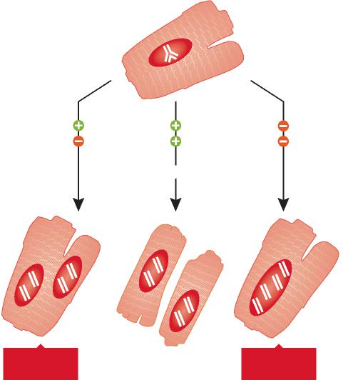
Fig 1 post-natal human DNA synthesis in the heart

In recent years, several protocols have been developed experimentally in an attempt to identify novel therapeutic interventions aiming at the reduction of infarct size and prevention of short and long term negative ventricular remodeling following ischemic myocardial injury. Three main strategies have been employed and a significant amount of work is being conducted to determine the most effective form of action for acute ischemic heart failure. The delivery of bone marrow progenitor cells (BMCs) has been highly controversial, but recent clinical data have shown improvement in ventricular performance and clinical outcome. These observations have not changed the nature of the debate concerning the efficacy of this cell category for the human disease and the mechanisms involved in the impact of BMCs on cardiac structure and function. Whether BMCs transdifferentiate and acquire the cardiomyocyte lineage has faced strong opposition and data in favor and against this possibility have been reported. However, this is the only cell class which has been introduced in the treatment of heart failure in patients and large clinical trials are in progress.
Human embryonic stem cells (ESCs) have repeatedly been utilized in animal models to restore the acutely infarcted myocardium, but limited cell engraftment, modest ability to generate vascular structures, teratoma formation and the apparent transient beneficial effects on cardiac hemodynamics have questioned the current feasibility of this approach clinically. Tremendous efforts are being performed to reduce the malignant tumorigenic potential of ESCs and promote their differentiation into cardiomyocytes with the expectation that these extremely powerful cells may be applied to human beings in the future. Additionally, the study of ESCs may provide unique understanding of the mechanisms of embryonic development that may lead to therapeutic interventions in utero and the correction of congenital malformations.
The recognition that a pool of primitive cells with the characteristics of stem cells resides in the myocardium and that these cells form myocytes, ECs and SMCs has provided a different perspective of the biology of the heart and mechanisms of cardiac homeostasis and tissue repair. Regeneration implies that dead cells are replaced by newly formed cells restoring the original structure of the organ. In adulthood, this process occurs during physiological cell turnover, in the absence of injury. However, myocardial damage interferes with recapitulation of cell turnover and restitutio ad integrum of the organ. Because of the inability of the adult heart to regenerate itself after infarction, previous studies have promoted tissue repair by injecting exogenously expanded CPCs in proximity of the necrotic myocardium or by activating resident CPCs through the delivery of growth factors known to induce cell migration and differentiation. These strategies have attenuated ventricular dilation and the impairment in cardiac function and in some cases have decreased animal mortality.

Although various subsets of CPCs have been used to reconstitute the infarcted myocardium and different degrees of muscle mass regeneration have been obtained, in all cases the newly formed cardiomyocytes possessed fetal-neonatal characteristics and failed to acquire the adult cell phenotype. In the current study, to enhance myocyte growth and differentiation, we have introduced cell therapy together with the delivery of self-assembly peptide nanofibers to provide a specific and prolonged local myocardial release of IGF-1. IGF-1 increases CPC growth and survival in vitro and in vivo and this effect resulted here in a major increase in the formation of cardiomyocytes and coronary vessels, decreasing infarct size and restoring partly cardiac performance. This therapeutic approach was superior to the administration of CPCs or NF-IGF-1 only. Combination therapy appeared to be additive; it promoted myocardial regeneration through the activation and differentiation of resident and exogenously delivered CPCs. Additionally, the strategy implemented here may be superior to the utilization of BMCs for cardiac repair. CPCs are destined to form myocytes, and vascular SMCs and ECs and, in contrast to BMCs, do not have to transdifferentiate to acquire cardiac cell lineages. Transdifferentiation involves chromatin reorganization with activation and silencing of transcription factors and epigenetic modifications.

Selected References

  1. Hsieh PC, Davis ME, Gannon J, MacGillivray C, Lee RT. Controlled delivery of PDGF-BB for myocardial protection using injectable self-assembling peptide nanofibers. J Clin Invest 2006;116:237–248. [PubMed: 16357943]
  2. Davis ME, Hsieh PC, Takahashi T, Song Q, Zhang S, Kamm RD, Grodzinsky AJ, Anversa P, Lee RT. Local myocardial insulin-like growth factor 1 (IGF-1) delivery with biotinylated peptide nanofibers improves cell therapy for myocardial infarction. Proc Natl Acad Sci USA 2006;103:8155–8160. [PubMed: 16698918]
  3. Beltrami AP, Barlucchi L, Torella D, Baker M, Limana F, Chimenti S, Kasahara H, Rota M, Musso E, Urbanek K, Leri A, Kajstura J, Nadal-Ginard B, Anversa P. Adult cardiac stem cells are multipotent and support myocardial regeneration. Cell 2003;114:763–776. [PubMed: 14505575]
  4. Rota M, Padin-Iruegas ME, Misao Y, De Angelis A, Maestroni S, Ferreira-Martins J, Fiumana E, Rastaldo R, Arcarese ML, Mitchell TS, Boni A, Bolli R, Urbanek K, Hosoda T, Anversa P, Leri A, Kajstura J. Local activation or implantation of cardiac progenitor cells rescues scarred infarcted myocardium improving cardiac function. Circ Res 2008;103:107–116. [PubMed: 18556576]

Cardiac anatomy.

Figure 2.  Cardiac anatomy.

(A and B) Cardiac weights and infarct size. R and L correspond, respectively, to the number of myocytes remaining and lost after infarction. (C–G) LV dimensions. Sham-operated: SO. *Indicates P<0.05 vs SO; **vs untreated infarcts (UN); †vs infarcts treated with CPCs; ‡vs infarcts treated with NF-IGF-1.

Ventricular function

Figure 3.  Ventricular function.

Combination therapy (CPC-NF-IGF-1) attenuated the most the negative impact of myocardial infarction on cardiac performance. See Figure 2 for symbols.

Endothelial Cells Promote Cardiac Myocyte Survival and Spatial Reorganization: Implications for Cardiac Regeneration

Daria A. Narmoneva, Rada Vukmirovic, Michael E. Davis, Roger D. Kamm,  and Richard T. Lee
Cardiovascular Division, Department of Medicine, Brigham and Women’s Hospital, Harvard Medical School, Boston, and the Division of Biological Engineering, Massachusetts Institute of Technology, Cambridge, MA
Circulation. 2004 August 24; 110(8): 962–968.        http://dx.doi.org/10.1161/01.CIR.0000140667.37070.07

Background

Endothelial-cardiac myocyte (CM) interactions play a key role in regulating cardiac function, but the role of these interactions in CM survival is unknown. This study tested the hypothesis that endothelial cells (ECs) promote CM survival and enhance spatial organization in a 3-dimensional configuration.

Methods and Results

Microvascular ECs and neonatal CMs were seeded on peptide hydrogels in 1 of 3 experimental configurations:

  1. CMs alone,
  2. CMs mixed with ECs (coculture), or
  3. CMs seeded on preformed EC networks (prevascularized).

Capillary-like networks formed by ECs promoted marked CM reorganization along the EC structures, in contrast to limited organization of CMs cultured alone. The presence of ECs markedly inhibited CM apoptosis and necrosis at all time points. In addition, CMs on preformed EC networks resulted in significantly less CM apoptosis and necrosis compared with simultaneous EC-CM seeding (P<0.01, ANOVA). Furthermore, ECs promoted synchronized contraction of CMs as well as connexin 43 expression.

Conclusions

These results provide direct evidence for a novel role of endothelium in survival and organization of nearby CMs. Successful strategies for cardiac regeneration may therefore depend on establishing functional CM-endothelium interactions.

Keywords:  endothelium; cardiomyopathy; heart failure; tissue

Introduction

Recent studies suggest that the mammalian heart possesses some ability to regenerate itself through several potential mechanisms, including generation of new cardiomyocytes (CMs) from extracardiac progenitors, CM proliferation, or fusion with stem cells with subsequent hybrid cell division. These mechanisms are insufficient to regenerate adequate heart tissue in humans, although some vertebrates can regenerate large volumes of injured myocardium.
Several approaches in cell transplantation and cardiac tissue engineering have been investigated as potential treatments to enhance cardiac function after myocardial injury. Implantation of skeletal muscle cells, bone marrow cells, embryonic stem cell-derived CMs, and myoblasts can enhance cardiac function. Cell-seeded grafts have been used instead of isolated cells for in vitro cardiac tissue growth or in vivo transplantation. These grafts can develop a high degree of myocyte spatial organization, differentiation, and spontaneous and coordinated contractions. On implantation in vivo, cardiac grafts can integrate into the host tissue and neovascularization can develop. However, the presence of scar tissue and the death of cells in the graft can limit the amount of new myocardium formed, most likely due to ischemia. Therefore, creating a favorable environment to promote survival of transplanted cells and differentiation of progenitor cells remains one of the most important steps in regeneration of heart tissue.
One of the key factors for myocardial regeneration is revascularization of damaged tissue. In the normal heart, there is a capillary next to almost every CM, and endothelial cells (ECs) outnumber cardiomyocytes by ≈3:1. Developmental biology experiments reveal that myocardial cell maturation and function depend on the presence of endocardial endothelium at an early stage. Experiments with inactivation or overexpression of vascular endothelial growth factor (VEGF) demonstrated that at later stages, either an excess or a deficit in blood vessel formation results in lethality due to cardiac dysfunction. Both endocardium and myocardial capillaries have been shown to modulate cardiac performance, rhythmicity, and growth. In addition, a recent study showed the critical importance of CM-derived VEGF in paracrine regulation of cardiac morphogenesis. These findings and others highlight the significance of interactions between CMs and endothelium for normal cardiac function. However, little is known about the specific mechanisms for these interactions, as well as the role of a complex, 3-dimensional organization of myocytes, ECs, and fibroblasts in the maintenance of healthy cardiac muscle.
The critical relation of CMs and the microvasculature suggests that successful cardiac regeneration will require a strategy that promotes survival of both ECs and CMs. The present study explored the hypothesis that ECs (both as preexisting capillary-like structures and mixed with myocytes at the time of seeding) promote myocyte survival and enhance spatial reorganization in a 3-dimensional configuration. The results demonstrate that CM interactions with ECs markedly decrease myocyte death and show that endothelium may be important not only for the delivery of blood and oxygen but also for the formation and maintenance of myocardial structure.

Methods

  • Three-Dimensional Culture
  • Immunohistochemistry and Cell Death Assays
  • Evaluation of Contractile Areas

Results

  • EC-CM Interactions Affect Myocyte Reorganization
  • ECs Improve Survival of CMs
  • Preformed Endothelial Networks Promote Coordinated, Spontaneous Contractions
  • ECs Promote Cx43 Expression

EC-CM Interactions Affect Myocyte Reorganization

To explore interactions between CMs and ECs in 3-dimensional culture, we used peptide hydrogels, a tissue engineering scaffold. Cells seeded on the surface of the hydrogel attach and then migrate into the hydrogel. When CMs alone were used, cells attached on day 1 and then formed small clusters of cells at days 3 and 7 (Figure 1). In contrast, when CMs were seeded together with ECs, cells formed interconnected linear networks, as commonly seen with ECs in 3-dimensional culture environments, with increasing spatial organization from day 1 to day 7 (Figure 1).

Figure 1.  ECs promote CM reorganization. 

When CMs were cultured alone (left column), they aggregated into sparse clusters. When CMs were cultured with ECs (center), cells organized into capillary-like networks. There was no difference in morphological appearance between coculture or prevascularized cultures (not shown) and ECs alone (right column). Bar=100 μm. Abbreviations are as defined in text.

To establish whether preformed endothelial networks enhanced the organization of myocytes, we also seeded ECs 1 day before myocytes were added. These ECs formed similar interconnected networks in the absence of myocytes; preforming the vascular network did not lead to significant differences in morphology (data not shown). Furthermore, to exclude the possibility that the increasing cell density of added ECs caused the spatial organization, we also performed control experiments with varying numbers and combinations of cells; there was no effect of doubling or halving cell numbers, indicating that the spatial organization effect was specifically due to ECs. To establish that both myocytes and ECs were forming networks together, we performed immunofluorescence studies with specific antibodies, as well as analysis of cross sections of CM-EC cocultures, whereby cells were labeled with CellTracker dyes before seeding. Immunofluorescent staining demonstrated that >95% of CMs were present within these networks, suggesting that CMs preferentially migrate to or survive better near ECs (Figure 2).

Figure 2.  CMs appear on outside of endothelial networks.

CMs appear on outside of endothelial networks. High-magnification, double-immunofluorescence image of structures formed in EC-CM coculture at day 7 demonstrating CMs (sarcomeric actinin, red) spread on top of ECs (von Willebrand factor, green) with no myocytes present outside structure. Bar=100 μm. Abbreviations are as defined in text.

The analysis of cross sections demonstrated the presence of what appeared to be EC-derived, tubelike structures (Figure 3), with myocytes spread on the outer part of the capillary wall. Along with the capillary-like structures, clusters of intermingled cells (both myocytes and ECs) not containing the lumen were also observed (not shown). However, when the lumen was present, ECs were always on the inner side and myocytes on the outer side of the structure.

Figure 3.  ECs form tubelike structures with myocytes spreading on outer wall.

Cross section of paraffin-embedded sample of 3-day coculture of myocytes (red) and ECs (green) incubated in CellTracker dye before seeding on hydrogel. Bar=50 μm. Abbreviations are as defined in text.

In CM-fibroblast cocultures, cells rapidly (within 24 hours) formed large clusters consisting of cells of both types (not shown). At later time points, fibroblast proliferation resulted in their migration outside the clusters and spreading on the hydrogel without any pattern. However, in contrast to EC-CM cocultures, CMs remained in the clusters and demonstrated only limited spreading. Immunofluorescent staining revealed that there was no orientation of myocytes relative to the fibroblasts in the clusters. In cultures with EC-conditioned medium, myocyte morphology and spatial organization remained similar to those of myocyte controls.

ECs Improve Survival of CMs

To test the hypothesis that ECs promote CM survival, we assessed apoptosis and necrosis in the 3-dimensional cultures. Quantitative analyses of CMs positive for TUNEL and necrosis staining demonstrated significantly decreased myocyte apoptosis and necrosis when cultured with ECs, compared with CM-only cultures (Figure 4, P<0.01). This effect was observed at all 3 time points, although the decreased necrosis was most pronounced at day 1. In addition, CMs seeded on the preformed EC networks had a lower rate of apoptosis at day 1 relative to same-time seeding cultures (P<0.05, post hoc test), suggesting that early EC-CM interactions provided by the presence of well-attached and prearranged ECs may further promote CM survival. In contrast to the ECs, cardiac fibroblasts did not affect myocyte survival (P>0.05, Figure 4), with ratios for myocyte apoptosis and necrosis in the myocyte-fibroblast cocultures being similar to those for myocyte-only controls. However, addition of EC-conditioned medium resulted in a significant decrease in apoptosis and necrosis ratios of myocytes (P<0.01). Interestingly, the effect of conditioned medium on myocyte necrosis was similar in magnitude to the effect of ECs, whereas myocyte apoptosis ratios in the conditioned-medium group were only partially decreased compared with those in the presence of ECs. These results suggest that the prosurvival effect of ECs on CMs may not only be merely due to the local interactions between myocytes and ECs during myocyte attachment but may also involve direct signaling between myocytes and ECs.

Figure 4.  ECs prolong survival of CMs

Top, dual immunostaining of CMs and EC-myocyte prevascularized groups at day 3 in culture, with TUNEL-positive cells in red; green indicates sarcomeric actinin; blue, DAPI. Bottom, presence of ECs decreased CM apoptosis and necrosis, both in coculture conditions and when cultures were prevascularized by seeding with ECs 1 day before CMs (mean±SD, P<0.01). EC-conditioned medium decreased myocyte apoptosis and necrosis (P<0.01), whereas fibroblasts did not have any effect (P>0.05). *Different from myocytes alone; **different from EC-myocyte coculture and pre-vascularized. Bar=100 μm. Abbreviations are as defined in text.

Preformed Endothelial Networks Promote Coordinated, Spontaneous Contractions

In the prevascularized group with preformed vascular structures, synchronized, spontaneous contractions of large areas (Figure 5, top panels) were detected as early as days 2 to 3after seeding, in contrast to the coculture group, wherein such contractions were observed on days 6 to 7. In CM-only cultures, beating of separate cells and small cell clusters was also detected at days 2 to 3, similar to that in the prevascularized group. However, the average area of synchronized beating at day 3 in the myocyte-only group (3.5±0.5×102 μm2) was nearly 3 orders of magnitude smaller than the synchronously contracting area in the prevascularized group (4.3±2.5×105 μm2, mean±SD, n=5). These data suggest that ECs promote synchronized CM contraction, particularly when vascular networks are already formed.

Figure 5.  ECs promote large-scale, synchronized contraction of CMs.

Left, phase-contrast video of beating areas in CM-only and prevascularized groups (day 3). Right, motion analysis of video showing regions of synchronized contractions (connected areas in purple are contracting synchronously) and nonmoving areas in blue. Bars=100 μm. Abbreviations are as defined in text.

ECs Promote Cx43 Expression

Staining for Cx43 showed striking differences in the distribution pattern of this gap junction protein between EC-CM cocultures and CMs cultured alone. In myocyte-only cultures, Cx43 expression was barely detectable at day 1 (not shown); at days 3 and 7, Cx43 expression was sparse throughout the cell clusters (Figure 6). In the presence of ECs (in both coculture and prevascularized groups), Cx43 staining was evident at day 1, both between ECs and distributed among CMs. As early as day 3 in culture, patches of localized junction-like Cx43, in addition to diffuse staining, were observed for myocytes in the coculture group (Figure 6). In the prevascularized group at day 3, wherein spontaneous contractions were already observed, more junction-like patches of Cx43 were observed compared with the coculture group, indicating electrical connections between myocytes (Figure 6). In addition to junctions between myocytes, there was also evidence of Cx43 localized at the interface between ECs and myocytes (Figure 6) detected in both the coculture group (at day 7) and the preculture group (as early as day 3). When myocytes and myocyte-EC coculture groups were cultured for 3 days with or without addition of 100 ng/mL of neutralizing anti-mouse VEGF antibody (R&D Systems), we observed no differences in either apoptosis or Cx43 staining between VEGF antibody-containing cultures and controls.

Figure 6. ECs promote Cx43 expression

Cultures at 3 days immunostained for Cx43 (red) and anti-sarcomeric actinin (green); nuclei are stained with DAPI (blue). For CMs alone (left), Cx43 staining is diffuse and sparse, with no evidence of gap junctions; for coculture (center), both diffuse (yellow arrow) and patchlike (thin, white arrow) Cx43 staining is observed; for prevascularized (right), increased patchlike staining indicates presence of gap junctions. Thick arrow-heads indicate junctions between myocytes and ECs. Bar=50 μm. Abbreviations are as defined in text.

nihms-134922.jpg  endothelial-myocyte figs_Image_1

nihms-134922.jpg  endothelial-myocyte figs_Image_2

nihms-134922.jpg  endothelial-myocyte figs_Image_3

nihms-134922.jpg  endothelial-myocyte figs_Image_4

nihms-134922.jpg  endothelial-myocyte figs_Image_5

nihms-134922.jpg  endothelial-myocyte figs_Image_6

Endothelial-Cardiomyocyte Interactions in Cardiac Development and Repair: Implications for Cardiac Regeneration

Patrick C.H. Hsieh, Michael E. Davis, Laura K. Lisowski, and Richard T. Lee

Cardiovascular Division, Department of Medicine, Brigham and Women’s Hospital, Harvard Medical School, Boston, MA
Annu Rev Physiol.    PMC 2009 September 30

The ongoing molecular conversation between endothelial cells and cardiomyocytes is highly relevant to the recent excitement in promoting cardiac regeneration. The ultimate goal of myocardial regeneration is to rebuild a functional tissue that closely resembles mature myocardium, not just to improve systolic function transiently. Thus, regenerating myocardium will require rebuilding the vascular network along with the cardiomyocyte architecture. Here we review evidence demonstrating crucial molecular interactions between endothelial cells and cardiomyocytes. We first discuss endothelial-cardiomyocyte interactions during embryonic cardiogenesis, followed with morphological and functional characteristics of endothelial-cardiomyocyte interactions in mature myocardium. Finally, we consider strategies exploiting endothelial-cardiomyocyte interplay for cardiac regeneration.

Signaling from Cardiomyocytes to Endothelial Cells

The examples of neuregulin-1, NF1, and PDGF-B demonstrate that signals from endothelial cells regulate the formation of primary myocardium. Similarly, signaling from myocardial cells to endothelial cells is also required for cardiac development. Two examples of myocardial-to-endothelial signaling are vascular endothelial growth factor (VEGF)-A and angiopoietin-1.

VASCULAR ENDOTHELIAL GROWTH FACTOR-A

VEGF-A is a key regulator of angiogenesis during embryogenesis. In mice, a mutation in VEGF-A causes endocardial detachment from an underdeveloped myocardium. A mutation in VEGF receptor-2 (or Flk-1) also results in failure of the endocardium and myocardium to develop (18). Furthermore, cardiomyocyte-specific deletion of VEGF-A results in defects in vasculogenesis/angiogenesis and a thinned ventricular wall, further confirming reciprocal signaling from the myocardial cell to the endothelial cell during cardiac development. Interestingly, this cardiomyocyte-selective VEGF-A-deletion mouse has underdeveloped myocardial microvasculature but preserved coronary artery structure, implying a different signaling mechanism for vasculogenesis/angiogenesis in the myocardium and in the epicardial coronary arteries.
Cardiomyocyte-derived VEGF-A also inhibits cardiac endocardial-to-mesenchymal transformation. This process is essential in the formation of the cardiac cushions and requires delicate control of VEGF-A concentration. A minimal amount of VEGF initiates endocardial-to-mesenchymal transformation, whereas higher doses of VEGF-A terminate this transformation. Interestingly, this cardiomyocyte-derived VEGF-A signaling for endocardial-to-mesenchymal transformation may be controlled by an endothelial-derived feedback mechanism through the calcineurin/NFAT pathway (24), demonstrating the importance of endothelial-cardiomyocyte interactions for cardiac morphogenesis.

ANGIOPOIETIN-1

—Another mechanism of cardiomyocyte control of endothelial cells during cardiac development is the angiopoietin-Tie-2 system. Both angiopoietin-1 and angiopoietin-2 may bind to Tie-2 receptors in a competitive manner, but with opposite effects: Angiopoietin-1 activates the Tie-2 receptor and prevents vascular edema, whereas angiopoietin-2 blocks Tie-2 phosphorylation and increases vascular permeability. During angiogenesis/vasculogenesis, angiopoietin-1 is produced primarily by pericytes, and Tie-2 receptors are expressed on endothelial cells. Angiopoietin-1 regulates the stabilization and maturation of neovasculature; genetic deletion of angiopoietin-1 or Tie-2 causes a defect in early vasculogenesis/angiogenesis and is lethal.
Cardiac endocardium is one of the earliest vascular components (along with the dorsal aorta and yolk sac vessels) and the adult heart can be regarded as a fully vascularized organ, angiopoietin-Tie-2 signaling may also be required for early cardiac development. Indeed, mice with mutations in Tie-2 have underdeveloped endocardium and myocardium. These Tie-2 knockout mice display defects in the endocardium but have normal vascular morphology at E10.5, suggesting that the endocardial defect is the fundamental cause of death. In addition, a recent study showed that overexpression, and not deletion, of angiopoietin-1 from cardiomyocytes caused embryonic death between E12.5-15.5 due to cardiac hemorrhage. The mice had defects in the endocardium and myocardium and lack of coronary arteries, suggesting that, as with VEGF-A, a delicate control of angiopoietin-1 concentration is critical for early heart development.

ENDOTHELIAL-CARDIOMYOCYTE INTERACTIONS IN NORMAL CARDIAC FUNCTION

Cardiac Endothelial Cells Regulate Cardiomyocyte Contraction

The vascular endothelium senses the shear stress of flowing blood and regulates vascular smooth muscle contraction. It is therefore not surprising that cardiac endothelial cells—the endocardial endothelial cells as well as the endothelial cells of intramyocardial capillaries— regulate the contractile state of cardiomyocytes. Autocrine and paracrine signaling molecules released or activated by cardiac endothelial cells are responsible for this contractile response (Figure 2).

NITRIC OXIDE

Three different nitric oxide synthase isoenzymes synthesize nitric oxide (NO) from L-arginine. The neuronal and endothelial NO synthases (nNOS and eNOS, respectively) are expressed in normal physiological conditions, whereas the inducible NO synthase is induced by stress or cytokines. Like NO in the vessel, which causes relaxation of vascular smooth muscle, NO in the heart affects the onset of ventricular relaxation, which allows for a precise optimization of pump function beat by beat. Although NO is principally a paracrine effector secreted by cardiac endothelial cells, cardiomyocytes also express both nNOS and eNOS. Endothelial expression of eNOS exceeds that in cardiomyocytes by greater than 4:1. Cardiomyocyte autocrine eNOS signaling can regulate β-adrenergic and muscarinic control of contractile state.
Barouch et al. demonstrated that cardiomyocyte nNOS and eNOS may have opposing effects on cardiac structure and function. Using mice with nNOS or eNOS deficiency, they found that nNOS and eNOS have not only different localization in cardiomyocytes but also opposite effects on cardiomyocyte contractility; eNOS localizes to caveolae and inhibits L-type Ca2+ channels, leading to negative inotropy, whereas nNOS is targeted to the sarcoplasmic reticulum and facilitates Ca2+ release and thus positive inotropy (31). These results demonstrate that spatial confinement of different NO synthase isoforms contribute independently to the maintenance of cardiomyocyte structure and phenotype.
As indicated above, mutation of neuregulin or either of two of its cognate receptors, erbB2 and erbB4, causes embryonic death during mid-embryogenesis due to aborted development of myocardial trabeculation . Neuregulin also appears to play a role in fully developed myocardium. In adult mice, cardiomyocyte-specific deletion of erbB2 leads to dilated cardiomyopathy. Neuregulin from endothelial cells may induce a negative inotropic effect in isolated rabbit papillary muscles. This suggests that, along with NO, the neuregulin signaling pathway acts as an endothelial-derived regulator of cardiac inotropism.  In fact, the negative inotropic effect of neuregulin may require NO synthase because L-NMMA, an inhibitor of NO synthase, significantly attenuates the negative inotropy of neuregulin.

Studies to date indicate that cardiac regeneration in mammals may be feasible, but the response is inadequate to preserve myocardial function after a substantial injury. Thus, understanding how normal myocardial structure can be regenerated in adult hearts is essential. It is clear that endothelial cells play a role in cardiac morphogenesis and most likely also in survival and function of mature cardiomyocytes. Initial attempts to promote angiogenesis in myocardium were based on the premise that persistent ischemia could be alleviated. However, it is also possible that endothelial-cardiomyocyte interactions are essential in normal cardiomyocyte function and for protection from injury. Understanding the molecular and cellular mechanisms controlling these cell-cell interactions will not only enhance our understanding of the establishment of vascular network in the heart but also allow the development of new targeted therapies for cardiac regeneration by improving cardiomyocyte survival and maturation.

Endothelial-cardiomyocyte assembly

Figure 1.  Endothelial-cardiomyocyte assembly in adult mouse myocardium.
Normal adult mouse myocardium is stained with intravital perfusion techniques to demonstrate cardiomyocyte (outlined in red) and capillary (green; stained with isolectin-fluorescein) assembly. Nuclei are blue (Hoechst). Original magnification: 600X

Endothelial dysfunction

Intramyocardial Fibroblast – Myocyte Communication

Rahul Kakkar, M.D. and Richard T. Lee, M.D.
From the Cardiology Division, Massachusetts General Hospital and the Cardiovascular Division, Brigham and Women’s Hospital, Department of Medicine, Harvard Medical School, Boston, MA
Circ Res. 2010 January 8; 106(1): 47–57.    http://dx.doi.org/10.1161/CIRCRESAHA.109.207456

Cardiac fibroblasts have received relatively little attention compared to their more famous neighbors, the cardiomyocytes. Cardiac fibroblasts are often regarded as the “spotters”, nonchalantly watching the cardiomyocytes do the real weight-lifting, and waiting for a catastrophe that requires their actions. However, emerging data now reveal the fibroblast as not only a critical player in the response to injury, but also as an active participant in normal cardiac function.
Interest in cardiac fibroblasts has grown with the recognition that cardiac fibrosis is a prominent contributor to diverse forms of myocardial disease. In the early 1990’s, identification of angiotensin receptors on the surface of cardiac fibroblasts linked the renin-angiotensin-aldosterone system directly with pathologic myocardial and matrix extracellular remodeling.  Fibroblasts were also revealed as a major source of not only extracellular matrix, but the proteases that regulate and organize matrix. New research has uncovered paracrine and well as direct cell-to-cell interactions between fibroblasts and their cardiomyocyte neighbors, and cardiac fibroblasts appear to be dynamic participants in ventricular physiology and pathophysiology.
This review will focus on several aspects of fibroblast-myocyte communication, including mechanisms of paracrine communication.  Ongoing efforts at regeneration of cardiac tissue focus primarily on increasing the number of cardiomyocytes in damaged myocardium. Although getting cardiomyocytes into myocardium is an important goal, understanding intercellular paracrine communication between different cell types, including endothelial cells but also fibroblasts, may prove crucial to regenerating stable myocardium that responds to physiological conditions appropriately.

An area of active research in cardiovascular therapeutics is the attempt to engineer, ex vivo, functional myocardial tissue that may be engrafted onto areas of injured ventricle. Recent data suggests that the inclusion of cardiac fibroblasts in three-dimensional cultures greatly enhances the stability and growth of the nascent myocardium. Cardiac fibroblasts when included in polymer scaffolds seeded with myocytes and endothelial cells have the ability to promote and stabilize vascular structures. Naito and colleagues constructed three dimensional cultures of neonatal rat cell isolates on collagen type I and Matrigel (a basement membrane protein mixture), and isolates of a mixed cell population versus a myocyte-enriched population were compared. The mixed population cultures, which contained a higher fraction of cardiac fibroblasts than the myocyte-enriched cultures, displayed improved contractile force generation and greater inotropic response despite an equivalent overall cell number. Greater vascularity was also seen in the mixed-pool cultures.(160) Building on this, Nichol and colleagues demonstrated that in a self-assembling nanopeptide scaffold, embedded rat neonatal cardiomyocytes exhibit greater cellular alignment and reduced apoptosis when cardiac fibroblasts were included in the initial culture. A similar result was noted when polymer scaffolds were pre-treated with cardiac fibroblasts before myocyte seeding, suggesting a persistent paracrine effect. These data reinforce the concept that engineering functional myocardium, either in situ or ex vivo will require attention to the nature of cell-cell interactions, including fibroblasts.

To date, a broad initial sketch of cardiac fibroblast-myocyte interactions has been drawn. Future studies in this field will better describe these interactions. How do multiple paracrine factors interact to produce a cohesive and coordinated communication scheme? What are the changes in coordinated bidirectional signaling that during development promotes myocyte progenitor proliferation but have different roles in the adult? Might fibroblasts actually be required for improved cardiac repair and regeneration?
Recent studies have begun to apply genetic and cellular fate-mapping techniques to document the origins of cardiac fibroblasts, the dynamic nature of their population, and how that population may be in flux during time of injury or pressure overload. It is crucial to define on a more specific molecular basis the origins and fates of cardiac fibroblasts. Do fibroblasts that have been resident within the ventricle since development fundamentally differ from those that arise from endothelial transition or that infiltrate from the bone marrow during adulthood? Do fibroblasts with these different origins behave differently or take on different roles in the face of ventricular strain or injury?

Our understanding of the nature of the cardiac fibroblast is evolving from the concept of the fibroblast as a bystander that causes unwanted fibrosis to the picture of a more complex role of fibroblasts in the healthy as well as diseased heart. The pathways used by cardiac fibroblasts to communicate with their neighboring myocytes are only partially described, but the data to date indicate that these pathways will be important for cardiac repair and regeneration.

. Paracrine bidirectional cardiac fibroblast-myocyte crosstalk

Figure 2. Paracrine bidirectional cardiac fibroblast-myocyte crosstalk

Under biomechanical overload, cardiac fibroblasts and myocytes respond to an altered environment via multiple mechanisms including integrin-extracellular matrix interactions and renin-angiotensin-aldosterone axis activation. Cardiac fibroblasts increase synthesis of matrix proteins and secrete a variety of paracrine factors that can stimulate myocyte hypertrophy. Cardiac myocytes similarly respond by secreting a conglomerate of factors. Hormones such as TGFβ1, FGF-2, and the IL-6 family members LIF and CT-1 have all been implicated in this bidirectional fibroblast-myocyte hormonal crosstalk.

Read Full Post »

Notable Contributions to Regenerative Cardiology by Richard T. Lee – Part I


Author and Curator: Larry H Bernstein, MD, FCAP

and

Article Commissioner: Aviva Lev-Ari, PhD, RD


Introduction

This presentation is a two part discussion of selected articles of a large body of research from Dr. Richard T. Lee, at Harvard Medical School’s Lee Laboratory and Brigham & Womens Hospital.  This work is innovative in the field of stem cell research and myocardial regeneration.  It devolves the complex cellular processes that are involved in the management of a cell transforming from a progenitor to a functional cardiomyocyte.  The cell engineering involves investigating interactions between a cell placed into the layer derived from the interstitial layer between viable cardiomyocytes.  This is only possible from a through actionable knowledge of the mechanism involved in the transformation process, which has occupied the Lee Laboratory for many years.  Part II will cover the cellular mechanisms underlying the conceptual approach to cardiac myocyte regeneration.

The Lee Laboratory uses emerging biotechnologies to discover and design new approaches to cardiovascular diseases. A central theme of the laboratory is that merging bioengineering and molecular biology approaches can yield novel approaches. Thus, the Lee Laboratory works at this interface using a broad variety of techniques in genomics, imaging, nanotechnology, physiology, cell biology, and molecular biology. The approach is to understand problems and design solutions in the laboratory and then demonstrate the effectiveness of these solutions in vivo. Ongoing projects in the laboratory include studies of cardiac regeneration, diabetic vascular disease, wound healing, heart failure, and cardiac hypertrophy.

Richard T. Lee is Professor of Medicine at Harvard Medical School and lecturer in Biological Engineering at the Massachusetts Institute of Technology. He is a 1979 graduate of Harvard College in Biochemical Sciences and received his M.D. from Cornell University Medical College in 1983.  He went on to complete his residency in 1986 and cardiology fellowship in 1989, both at Brigham and Women’s Hospital in Boston, and he obtained post-doctoral training at MIT in Bioengineering.

Dr. Lee is certified by the American Board of Internal Medicine in cardiovascular disease and is a Fellow of the American College of Cardiology. He is Leader of the Cardiovascular Program of the Harvard Stem Cell Institute.  He is a member of the Editorial Boards of the journals Circulation Research, Journal of Clinical Investigation, and Circulation, and has published over 180 peer-reviewed articles based on his research, which combines approaches in biotechnology and molecular biology to discover new avenues to manage and treat heart disease.

Regeneration of the heart

Matthew L. Steinhauser, Richard T. Lee
Division of Cardiovascular Medicine, Department of Medicine, Brigham and Women’s Hospital, Harvard Medical School, and (2)             Harvard Stem Cell Institute, Cambridge, MA
EMBO Mol Med 2011; 3: 701–712   http://dx.doi.org/10.1002/emmm.201100175

The death of cardiac myocytes diminishes the heart’s pump function and is a major cause of heart failure. With the exception of heart transplantation and implantation of mechanical ventricular assist devices, current therapies do not address the central problem of decreased pumping capacity owing to a depleted pool of cardiac myocytes. The field is evolving in two important directions. First, although endogenous mammalian cardiac regeneration clearly seems to decline rapidly after birth, it may still persist in adulthood. The careful elucidation of the cellular and molecular mechanisms of endogenous heart regeneration may therefore provide an opportunity for developing therapeutic interventions that amplify this process. Second, recent breakthroughs have enabled reprogramming of cells that were apparently terminally differentiated, either by dedifferentiation into pluripotent stem cells or by trans-differentiation into cardiac myocytes.
The longstanding paradigm held that the mammalian heart is a terminally differentiated organ, incapable of replenishing any myocyte attrition. During the past decade, however, studies revealed not only that mammalian cardiac myocytes retain some capacity for division (Beltrami et al, 2001), but also identified endogenous cardiac progenitor cells in the heart (Beltrami et al, 2003) or bone marrow (Orlic et al, 2001). These cells retain some potential for differentiation into the cellular components of the heart, including endothelial cells, smooth muscle cells and cardiac myocytes.
If progenitor cells residing in the adult are capable of producing new heart cells, the therapeutic delivery of such progenitors might facilitate the generation of de novo functional myocardium. In this context, cell-based therapies for the heart have been rapidly translated into the clinic to treat heart disease, but randomized clinical trials with bone marrow progenitors have shown at best modest improvements in ventricular function (Martin-Rendon et al, 2008). In short, the promise of complete cardiac regeneration has not yet been realized.  Therefore, it is worth revisiting both the foundations of cardiac regeneration and highlight recent advances that may portend future directions in the field.
We will first define the problem, that is elucidating the scope of endogenous mammalian regeneration and, by extension, the scale of the regenerative deficit. We will then summarize current regenerative approaches, including both cell-based therapies and pharmacoregenerative strategies. In this context, we will summarize the many challenges that stand in the way of cardiac regeneration, both endogenous repair processes and exogenous regenerative therapies.
The regenerative deficit of the mammalian heart is obvious when compared with organisms such as zebrafish and newts, which demonstrate a remarkable survival capacity after removal of up to 20% of the heart by transection of the ventricular apex. Pre-existing cardiac myocytes adjacent to the site appear to undergo a process of dedifferentiation, characterized by dissolution of sarcomeric structures. This is followed by incorporation of deoxyribonucleic acid (DNA) synthesis markers (e.g. nucleotide analogues) consistent with proliferation. Ultimately, newly generated cardiac myocytes are functionally integrated with the preexisting myocardium. The heart is left with little residual evidence of the injury, thus providing a natural example of complete myocardial regeneration.

Evidence for heart regeneration in mammals

During embryonic development and the early post-natal period, mice also demonstrate a remarkable regenerative capacity. Embryos heterozygous for a cardiac myocyte-specific null mutation in the x-linked holocytochrome c synthase (Hccs) gene demonstrate cardiac myocyte replacement during foetal development (Drenckhahn et al, 2008): when one of two X-chromosomes is randomly inactivated in each female somatic cell, approximately 50% of the cardiac myocytes are rendered Hccs-null and hence dysfunctional. Proliferative functional Hccs-expressing cardiac myocytes compensate for dysfunctional Hccs-null myocytes, such that, at birth, 90% of the heart is derived from myocytes containing one functional Hccs allele. However, after the first week in post-natal mice, injured myocardium is largely replaced by fibrosis and scarring.  Distinguishing whether the adult mammalian heart is incapable of cardiac myocyte replacement or whether it retains a low-level capacity for repair is therefore fundamentally important. This is the basis for an evolving view of a more plastic mammalian heart.
Arguments against the age old view of the terminally differentiated quiescent cardiac myocyte:

  1. evidence supporting cardiac myocyte plasticity relied on mathematical modelling of the myocyte population based on cytometric indices. (the measured average volume increase of cardiac myocytes was calculated to fall short of the increase predicted by the observed volumetric changes in the whole heart
  • changes in heart volume could not be explained by hypertrophy alone, and that cardiac myocyte hyperplasia contributed to changes in heart mass, but the conclusions relied on a number of assumptions about myocyte size and DNA content.
  • detecting cell cycle markers such as Ki67 or the incorporation of nucleotide analogues (e.g. iododeoxyuridine or 3H-thymidine) into newly synthesized DNA further support the notion that the mammalian heart may generate new myocytes
  • human cardiac myocytes can reenter the cell cycle, but the described rates of this phenomenon differ by more than one order of magnitude
  • experiments, made possible by nuclear arms testing in the middle of the 20th century, provide the most convincing evidence for post-natal human cardiac myocyte turnover.
  • the period of nuclear testing serves as a historical DNA labelling pulse, and the period after the test ban treaty serves as a chase.
  • the genomic DNA of cells generated during either the pulse or the chase reflect the earth’s atmospheric 14C concentration at that point in time, which allows investigators to date the age of cardiac myocytes by measuring the concentration of 14C in their nuclei
  1. Listed are a number of problems in detecting the generation of cardiac myocytes:
  • small errors may magnify projections of absolute yearly or lifetime myocyte turnover
  • mis-identification of cellular components by light microscopy
  • autofluorescence of myocardium, which complicates any method that relies on the detection of a fluorescent signal
  • confounders could also affect the 14C dating method, because it requires the isolation of cardiac myocyte nuclei by digestion and flow cytometric sorting

The heart also presents a unique challenge compared to other organs owing to the propensity of cardiac myocytes to synthesize DNA during S-phase without completing either mitosis and/or cytokinesis (Fig 1).

Figure 1. The majority of post-natal human DNA synthesis in the heart does not lead to new myocyte formation.

Cardiac myocytes can complete S-phase, followed by mitosis and cytokinesis (centre) resulting in myocyte doubling. Cardiac myocytes can also complete mitosis without cytokinesis (left), resulting in a binucleated cell. Cardiac myocytes can also undergo chromosomal replication without completing either mitosis or cytokinesis (right), resulting in polyploidy nuclei. By the completion of post-natal development, the majority of human myocyte nuclei contain ~4n chromosomal copies.

During early post-natal development, for example, the majority of rodent cardiac myocytes and an estimated 25–57% of human cardiac myocytes become binucleated. By adulthood, most cardiac myocyte nuclei have also become polyploid with at least one or two additional rounds of chromosomal replication.

The ploidy state of cardiac myocytes may increase with myocardial hypertrophy or injury, which could be mistaken for myocyte division. Conversely, hearts that have been unloaded by implantation of a ventricular assist device may have a lower percentage of polyploid myocytes, because more 2n cardiac myocytes are being generated. These aspects of cardiac myocyte biology inevitably represent potential confounders that must be considered in any quantification of cardiac myocyte formation.

Defining the cellular source of new cardiac myocytes

The majority of reports suggest some endogenous capacity for cardiac myocyte renewal, which has generated a broad focus on finding the cellular source of newly generated cardiac myocytes.  Newly generated adult mammalian cardiac myocytes may arise from an endogenous pool of progenitor cells after injury. The Lee Laboratory developed a genetic lineage mapping approach to quantify progenitor-dependent cardiac myocyte turnover (Fig 2) (Hsieh et al, 2007). In the bitransgenic MerCreMer/ZEG inducible cardiac myocyte reporter mouse, mature cardiac myocytes undergo an irreversible genetic switch from constitutive 3-galactosidase expression to green fluorescent protein (GFP) expression upon tamoxifen pulse. During a chase period, we evaluated the effect of myocardial injury on the proportion of GFP+ or 3-gal+ cardiac myocytes. Pressure overload or myocardial infarction resulted in a significant reduction in the percentage of GFP+ cardiac myocytes and a corresponding increase in the percentage of B-gal+ cardiac myocytes, consistent with repletion of the myocyte pool by B-gal— expressing progenitors. This approach cannot directly identify the molecular identity or anatomic location of the progenitor pool.
One approach to characterizing the molecular phenotype of cardiac progenitors is to study cardiac embryologic develop-ment, guided by the assumption that developmental paradigms are recapitulated during post-natal repair. When examined through a developmental lens, an increasingly detailed picture emerges of the soluble and transcriptional signals that guide the cardiogenic programme from gastrulation (formation of distinct germ layers) through the ultimate maturation of cardiac myocytes. The induction of mesoderm posterior (MESP)-1 expression by brachyury-expressing primitive mesodermal cells is a proximal require¬ment for the ultimate production of differentiated heart cells. As the developing embryo grows beyond the germ layer phase, its developing heart receives cells from distinct anatomic progeni¬tor sources: the 1st and 2nd heart fields provide the majority of the myocardium, with some contribution from epicardial progenitors.
Also, Certain fields may be preferentially marked by specific transcription factors;

  • the first heart field by T-box transcription factor 5 (Tbx5)
  • the second heart field by
    • Lim-homeodomain protein Islet1 (Isl1)
    • and epicardial progenitors by Wilms tumour-1 (WT1) or
    • T-box transcription factor 18 (Tbx18)
    • identified by embryonic lineage tracing or analysis of gene silencing include
      • homeobox protein nkx2.5
      • myocyte enhancer factor 2C (Mef2c)
      • GATA4
      • there is no consensus yet about the molecular identity of post-natal mammalian cardiac progenitor cells or ‘adult cardiac stem cells’

Figure 2. Lineage-mapping in the adult heart.

Left: Theoretical progenitor lineage-mapping is depicted. Progenitors would be selectively marked by fluorescent protein expression. After injury, the appearance of fluorescently labelled cardiac myocytes would support the concept that these progenitors were contributing to new myocyte formation. Right: Differentiated cell (cardiac myocyte) lineage-mapping. Upon treatment of the MerCreMer-ZEG mouse with OH-tamoxifen, approximately 80% of the cardiac myocytes undergo a permanent switch from I3-galactosidase to GFP expression. The dilution of the GFPþ cardiac myocyte pool after injury is consistent with repletion by I3-galþ progenitors.

A number of laboratories have identified cell populations within the post-natal mouse, which fulfil some criteria of cardiac progenitors:

  • expression of a developmentally important gene (isl-1(Laugwitz et al, 2005))
  • specific cell surface receptor profile (c-kit (Beltrami et al, 2003)
  • or sca-1 (Oh et al, 2003))
  • capacity to actively exclude Hoechst dye (so-called side population cells (Martin et al, 2004)) or based on the outgrowth of typical spherical colonies in tissue culture 

In general, the label of ‘cardiac stem cell’ results from the observation of self-propagation and cardiac myocyte transdifferentiation when exposed to cardiogenic conditions in vitro or when delivered in vivo after injury. However, the field will benefit from careful in vivo lineage tracing studies—without ex vivo culture steps—to study if and how a given cell type contributes to cardiac myocyte replenishment during either normal homeostasis or after injury (Fig 2). The lack of such publications to date owes in part to the lack of specificity of many stem cell markers (Fig 3).

Figure 3. Possible recapitulation of developmental paradigms by endogenous post-natal cardiac stem cells.

Between mesodermal development and the emergence of cardiac myocytes, cardiovascular progenitors express a number of markers that have also been detected in the various post-natal cardiac stem cell (CSC) preparations. Expression as measured by messenger RNA (mRNA) or protein expression is denoted with (þ). Absent expression is denoted by (-). Blank1/4 untested.

Moving towards a regenerative therapy

The therapeutic challenge is considerable: a typical large myocardial infarction that leads to heart failure will kill around 1 billion cardiac myocytes,  roughly a quarter of the heart’s myocytes. A possible therapeutic approach would coax an endogenous stem cell population or an exogenously delivered cell-based therapy to replace lost cardiac myocytes in a coordinated fashion. Amongst the myriad of potential cell-based therapies, no clear winning strategy has so far emerged (Segers & Lee, 2008).

Bone marrow derived progenitors

Conflicting studies sparked excitement and also uncertainty about a possible adult cardiogenic progenitor originating outside of the heart. A post-mortem examination of male heart transplant patients who had received female donor hearts found that approximately 10% of -sarcomeric actin-positive cardiac myocytes had Y-chromosomes, and two cases in which a bone marrow cell population with a higher density of the cell surface receptor c-kit, showed repopulation of murine cardiac myocytes after experimental myocardial infarction. A number of studies that followed failed to demonstrate similar rates of chimerism in transplanted hearts or potency of bone marrow stem cell.  However, some therapeutic effect was observed even in studies with no detectable transdifferentiation.

Figure 4. The challenge of regenerating the heart.

Both exogenously delivered cell therapies and progenitors in the endogenous niche encounter a similar hostile environment after myocardial injury, often including inadequate blood supply (ischemia), inflammation and fibrosis/scarring. Regenerative pathways may be activated by as yet unknown paracrine pathways, responsible for recruiting progenitors from the niche, stimulating proliferation and coaxing differentiation.  Cell-based therapy using autologous bone marrow
progenitors was rapidly translated into the clinic to treat human ischemic heart disease. A number of randomized trials, using bone marrow mononuclear cells have been performed and most studies demonstrated modest cell therapy-mediated improvements in ventricular function.

Pluripotent stem cells

Embryonic stem (ES) cells represent the prototypical stem cells with the hallmarks of clonogenicity, self-renewal and pluripotency. The potency of these cells also represents a real safety concern, given their tendency to form teratomas. One approach to overcoming this prohibitive safety problem has been to generate pluripotent-derived progenitors that have already committed to a cardiogenic pathway. As a proof-of-principle example of such a strategy, cells with an expression profile of Oct4, stage-specific embryonic antigen 1 (SSEA-1) and MESP1 demonstrated some regenerative potency when delivered therapeutically in a primate infarct model, without detectable teratoma formation. One could envision a similar strategy using cardiogenic intermediates that express any of the previously mentioned transcription factors associated with cardiac progenitors or cell surface markers such as the receptor for vascular endothelial growth factor (Flk1/KDR). Yet, such a strategy should still demonstrate both substantial preclinical efficacy without tumorigenicity before human translation. If such criteria are met, ES-derived therapies have the potential of providing ‘off-the-shelf’ cardiac myocytes to treat acute myocardial infarctions or chronic heart failure.
A second approach, which may also obviate the risk of teratomas, is to generate a pure population of ES-derived cardiac myocytes for therapeutic delivery either as a cell suspension or after ex vivo tissue engineering. There has already been enormous progress during the past decade in defining the factors and transcription signals to differentiate cardiac myocytes from ES-cells. As discussed in greater detail, cardiac myocyte development is dictated by the time and dose-dependent exposure to a series of growth factors from the wingless-type MMTV integration site (Wnt), fibroblast growth factor (FGF), bone morphogenetic protein (BMP) and nodal families. Several laboratories have successfully generated ES-derived preparations with more than 50% of functional cardiac myocytes.  The most realistic future for such technical advances may be as an unlimited source of cardiac myocytes for engineering myocardial grafts.
The generation of induced pluripotent stem (iPS) cells may overcome two important limitations of ES cells: ethical concerns about harvesting ES cells from embryos and graft rejection

  • iPS cells can be custom-engineered from a patient’s stromal cells for autologous transplantation.
  • immunogenecity in syngeneically transplanted iPS cells, suggests that the immune system cannot yet be discounted in the development of iPS-based therapies

The initial protocols for iPS cell generation involved retro-viral-mediated expression of four stem-cell genes.
But virally reprogrammed cells may harbour an associated risk of neoplastic conversion. Alternative reprogramming strategies, such as the use of small molecules (Shi et al, 2008) or non-viral gene modifying approaches (Warren et al, 2010) will probably be a necessary component of any future therapeutic strategies. However, the most important lesson from these landmark studies may be the remarkable plasticity of even the most terminally differentiated cells when exposed to the right combination of signals.

Tissue engineering

Historically, the greatest challenge in tissue engineering has been an adequate supply of oxygen and nutrients for metabolically active tissues such as the heart. One approach has been to engineer thin cardiac sheets, which can then be stacked for in vivo delivery. Although these layered sheets demonstrate some degree of electromechanical coordination and neovascularization in vivo, it is not clear yet if such an approach can be optimized to yield full-thickness myocardium with an adequate blood supply. The addition of non-myocyte cellular components, such as fibroblasts and endothelial cells, leads to the formation of primitive vascular structures within engineered grafts, but the electro-mechanical properties are not sufficient for normal functionality.

Circumventing cell-based therapy with pharmacoregeneration?

A short-term goal may be to exploit paracrine signalling to amplify the existent endogenous regenerative response. Recent cell transplantation experiments conducted in our laboratory, using an inducible cre-based genetic lineage mapping approach, tested the hypothesis that cell-based therapies might exert proregenerative effects via a paracrine mechanism (Loffredo et al, 2011) (Fig 5).  Consistent with some prior studies, we found no evidence for transdifferentiation of exogenously delivered bone marrow cells into cardiac myocytes. However, we did find increased generation of cardiac myocytes from endogenous progenitors in mice, which were administered c-kit+ bone marrow cells but not mesenchymal stem cells. This finding suggests paracrine signalling between exogenously delivered cells and endogenous resident progenitors. It provides a rationale for therapeutic interventions aimed at activating progenitors or recruiting them from their niche to the injury site.

Figure 5. Proposed of action for cell-based therapies.

In theory, exogenously delivered cells may directly differentiate into endothelial cells, smooth muscle cells and cardiac myocytes. They may also release paracrine factors which may result in non-regenerative effects, such as immunomodulation, angiogenesis or cardioprotection. Recent work from our laboratory suggests that a dominant mechanism achieved with bone marrow progenitor therapy may be via the activation of endogenous progenitor recruitment (Loffredo et al, 2011).

Controlling the mitotic activity of mononucleated cardiac myocytes may provide an alternative approach to replenishing cardiac myocytes. A major concern with systemic growth factor therapy, however, is the potential for mitogenic effects that may impact other organs. Thus, the future of pharmacologic regeneration may lie in the local delivery of engineered proteins and small molecules that target 

Future directions

In this review, we have described the current status of research on cardiac regeneration, highlighting important recent discoveries and ongoing controversies. The initial hope that a cell progenitor would emerge with the capacity to regenerate the injured mammalian heart in the same manner that bone marrow may be reconstituted has not been realized.
Cardiac myocyte regeneration may lie in the local delivery of engineered proteins and small molecules that target specific survival, growth and differentiation pathways.

Pending issues

Dissect the mechanistic differences between adult mammals with limited regenerative capacity and organisms, such as neonatal mice, zebrafish and newts, that demonstrate unambiguous cardiac myocyte regeneration. Understanding these differences may reveal new pathways that can be therapeutically targeted to achieve more robust regeneration.

Complete molecular and functional characterization of endogenous cardiac myocyte progenitors. Multiple laboratories have isolated progenitors from the heart with different molecular characteristics. What are the in vivo functional roles of these progenitors? Do the observed molecular differences between these isolated cells represent functionally distinct cell types?

Identify paracrine signalling pathways responsible for activation and recruitment of endogenous cardiac myocyte progenitors. This may facilitate a pharmacoregenerative therapy, in which treatment with a protein or small molecule would hold the promise of amplifying endogenous regeneration.

Refine reprogramming strategies to more efficiently produce mature cardiac myocytes, both in vitro and in vivo. The ultimate bioengineering goal is to produce a pure population of mature, fully functional cardiac myocytes for ex vivo tissue engineering (or) to control the proliferation and differentiation of endogenous cell populations or exogenously delivered cell therapies such that scar tissue is replaced by myocardium. These different strategies are unified by an underlying requirement to understand the fundamental pathways involved in cardiac myocyte differentiation and maturation.

There is reason for optimism for a regenerative medicine approach to heart failure, given the intense research efforts and the capacity of higher organisms, including the neonatal mouse, to regenerate myocardium. Perhaps the most important issue in this field is identifying which findings are consistently supported by multiple experimental approaches. Ultimately, the findings that are easily reproduced by most or all laboratories will most likely benefit patients.

Selected references

Hsieh et al, 2007.  Hsieh PC, Segers VF, Davis ME, MacGillivray C, Gannon J, Molkentin JD, Robbins J, Lee RT (2007) Evidence from a genetic fate-mapping study that stem cells refresh adult mammalian cardiomyocytes after injury. Nat Med 13: 970¬974
Laugwitz et al, 2005.  Laugwitz KL, Moretti A, Lam J, Gruber P, Chen Y, Woodard S, Lin LZ, Cai CL, Lu MM, Reth M et al (2005) Postnatal isl1þ cardioblasts enter fully differentiated cardiomyocyte lineages. Nature 433: 647-653
Beltrami et al, 2003.  Beltrami AP, Barlucchi L, Torella D, Baker M, Limana F, Chimenti S, Kasahara H, Rota M, Musso E, Urbanek K et al (2003) Adult cardiac stem cells are multipotent and support myocardial regeneration. Cell 114: 763¬776
Oh et al, 2003. Oh H, Bradfute SB, Gallardo TD, Nakamura T, Gaussin V, Mishina Y, Pocius J, Michael LH, Behringer RR, Garry DJ et al (2003) Cardiac progenitor cells from adult myocardium: homing, differentiation, and fusion after infarction. Proc Natl Acad Sci USA 100: 12313-12318
Segers & Lee, 2008.  Segers VF, Lee RT (2008) Stem-cell therapy for cardiac disease. Nature 451:937-942
Loffredo et al, 2011.  Loffredo FS, Steinhauser ML, Gannon J, Lee RT (2011) Bone marrow-derived cell therapy stimulates endogenous cardiomyocyte progenitors and promotes cardiac repair. Cell Stem Cell 8: 389-398.
Shi et al, 2008.  Shi Y, Desponts C, Do JT, Hahm HS, Scholer HR, Ding S (2008) Induction of pluripotent stem cells from mouse embryonic fibroblasts by Oct4 and Klf4 with small-molecule compounds. Cell Stem Cell 3: 568-574.
Warren et al, 2010.  Warren L, Manos PD, Ahfeldt T, Loh YH, Li H, Lau F, Ebina W, Mandal PK, Smith ZD, Meissner A et al (2010) Highly efficient reprogramming to pluripotency and directed differentiation of human cells with synthetic modified mRNA. Cell Stem Cell 7: 618-630.

Mammalian Heart Renewal by Preexisting Cardiomyocytes

SE Senyo, ML Steinhauser, CL Pizzimenti, VK. Yang, Lei Cai, Mei Wang, …,and Richard T. Lee
Cardiovascular and Genetics Divisions, Brigham and Women’s Hospital and Harvard Medical School,
INSERM, Orsay (Fr), Institut Curie, Laboratoire de Microscopie Ionique, Orsay (Fr), National Resource for Imaging Mass Spectrometry, Harvard Stem Cell Institute
Nature. 2013 January 17; 493(7432): 433–436.  http://dx.doi.org/10.1038/nature11682

Although recent studies have revealed that heart cells are generated in adult mammals, the frequency and source of new heart cells is unclear. Some studies suggest a high rate of stem cell activity with differentiation of progenitors to cardiomyocytes. Other studies suggest that new cardiomyocytes are born at a very low rate, and that they may be derived from division of pre-existing cardiomyocytes. Thus, the origin of cardiomyocytes in adult mammals remains unknown. Here we combined two different pulse-chase approaches — genetic fate-mapping with stable isotope labeling and Multi-isotope Imaging Mass Spectrometry (MIMS). We show that genesis of cardiomyocytes occurs at a low rate by division of pre-existing cardiomyocytes during normal aging, a process that increases by four-fold adjacent to areas of myocardial injury. Cell cycle activity during normal aging and after injury led to polyploidy and multinucleation, but also to new diploid, mononucleated cardiomyocytes. These data reveal pre-existing cardiomyocytes as the dominant source of cardiomyocyte replacement in normal mammalian myocardial homeostasis as well as after myocardial injury.

Despite intensive research, fundamental aspects of the mammalian heart’s capacity for self-renewal are actively debated. Estimates of cardiomyocyte turnover range from less than 1% per year to more than 40% per year. Turnover has been reported to either decrease or increase with age, while the source of new cardiomyocytes has been attributed to both division of existing myocytes and to progenitors residing within the heart or in exogenous niches such as bone marrow. Controversy persists regarding the plasticity of the adult heart in part due to methodological challenges associated with studying slowly replenished tissues. Toxicity attributed to radiolabeled thymidine and halogenated nucleotide analogues limits the duration of labeling and may produce direct biological effects. The challenge of measuring cardiomyocyte turnover is further compounded by the faster rate of turnover of cardiac stromal cells relative to cardiomyocytes.

We used Multi-isotope Imaging Mass Spectrometry (MIMS) to study cardiomyocyte turnover and to determine whether new cardiomyocytes are derived from preexisting myocytes or from a progenitor pool (Fig 1a). MIMS uses ion microscopy and mass spectrometry to generate high resolution quantitative mass images and localize stable isotope reporters in domains smaller than one micron cubed15,16,17. MIMS generates 14N quantitative mass images by measuring the atomic composition of the sample surface with a lateral resolution of under 50nm and a depth resolution of a few atomic layers. Cardiomyocyte cell borders and intracellular organelles were easily resolved (Fig 1b). Regions of interest can be analyzed at higher resolution, demonstrating cardiomyocyte-specific subcellular ultrastructure, including sarcomeres (Fig 1c, Supplemental Fig 1a). In all subsequent analyses, cardiomyocyte nuclei were identified by their location within sarcomere-containing cells, distinguishing them from adjacent stromal cells.
An immense advantage of MIMS is the detection of nonradioactive stable isotope tracers. As an integral part of animate and inanimate matter, they do not alter biochemical reactions and are not harmful to the organism18. MIMS localizes stable isotope tracers by simultaneously quantifying multiple masses from each analyzed domain; this enables the generation of a quantitative ratio image of two stable isotopes of the same element15. The incorporation of a tracer tagged with the rare stable isotope of nitrogen (15N) is detectable with high precision by an increase in 15N:14N above the natural ratio (0.37%). Nuclear incorporation of 15N-thymidine is evident in cells having divided during a one-week labeling period, as observed in the small intestinal epithelium, which turns over completely in 3–5 days16 (Fig 1d); in contrast, 15N-thymidine labeled cells are rarely observed in the heart (Fig 1e) after 1 week of labeling. In subsequent studies, small intestine was used as a positive control to confirm label delivery.
To evaluate for an age-related change in cell cycle activity, we administered 15N-thymidine for 8 weeks to three age groups of C57BL6 mice starting at day-4 (neonate), at 10-weeks (young adult) and at 22-months (old adult) (Supplemental Fig 2). We then performed MIMS analysis (Fig 2a, b, Supplemental Fig 3). In the neonatal group, 56% (±3% s.e.m., n=3 mice) of cardiomyocytes demonstrated 15N nuclear labeling, consistent with the well-accepted occurrence of cardiomyocyte DNA synthesis during post-natal development19. We observed a marked decline in the frequency of 15N-labeled cardiomyocyte nuclei (15N+CM) in the young adult (neonate= 1.00%15N+CM/day ±0.05 s.e.m. vs young adult=0.015% 15N+CM/ day ±0.003 s.e.m., n=3 mice/group, p<0.001) (Fig 2a, c; Supplemental Fig 3). We found a further reduction in cardiomyocyte DNA synthesis in old mice (young adult=0.015%15N+CM/day ±0.003 s.e.m. vs. old adult=0.007 %15N+CM/day ±0.002 s.e.m., n=3/group, p<0.05) (Fig 2c). The observed pattern of nuclear 15N-labeling in cardiomyocytes is consistent with the known chromatin distribution pattern in cardiomyocytes20 (Supplemental Fig 1b) and was measured at levels that could not be explained by DNA repair (Supplemental Fig 4). Extrapolating DNA synthesis measured in cardiomyocytes over 8 weeks yields a yearly rate of 5.5% in the young adult and 2.6% in the old mice. Given that cardiomyocytes are known to undergo DNA replication without completing the cell cycle19,21,22, these calculations represent the upper limit of cardiomyocyte generation under normal homeostatic conditions, indicating a low rate of cardiogenesis.
To test whether cell cycle activity occurred in preexisting cardiomyocytes or was dependent on a progenitor pool, we performed 15N-thymidine labeling of double-transgenic MerCreMer/ZEG mice, previously developed for genetic lineage mapping (Fig 3a)23,24. MerCreMer/ZEG cardiomyocytes irreversibly express green fluorescent protein (GFP) after treatment with 4OH-tamoxifen, allowing pulse labeling of existing cardiomyocytes with a reproducible efficiency of approximately 80%. Although some have reported rare GFP expression by non-cardiomyocytes with this approach25, we did not detect GFP expression in interstitial cells isolated from MerCreMer/ZEG hearts nor did we detect GFP expression by Sca1 or ckit-expressing progenitors in histological sections (Supplemental Fig 5). Thus, during a chase period, cardiomyocytes generated from progenitors should be GFP−, whereas cardiomyocytes arising from preexisting cardiomyocytes should express GFP at a frequency similar to the background rate induced by 4OH-tamoxifen. We administered 4OH-tamoxifen for two weeks to 8 wk-old mice (n=4); during a subsequent 10-week chase, mice received 15N-thymidine via osmotic minipump.

We next used MIMS and genetic fate mapping to study myocardial injury. Cardiomyocyte GFP labeling was induced in MerCreMer/ZEG mice with 4OH-tamoxifen. Mice then underwent experimental myocardial infarction or sham surgery followed by continuous labeling with 15N-thymidine for 8wks. The frequency of 15N-labeled cardiomyocytes in sham-operated mice was similar to prior experiments in unoperated mice (yearly projected rates: sham=6.8%; unoperated=4.4%), but increased significantly adjacent to infarcted myocardium (total 15N+ cardiomyocyte nuclei: MI=23.0% vs sham=1.1%, Fig 4a–b, Supplemental Fig 8). We examined GFP expression, nucleation and ploidy status of 15N-labeled cardiomyocytes and surrounding unlabeled cardiomyocytes. We found a significant dilution of the GFP+ cardiomyocyte pool at the border region as previously shown23,24 (67% vs. 79%, p<0.05, Table 2, Supplemental Fig 9); however, 15N+ myocytes demonstrated a similar frequency of GFP expression compared to unlabeled myocytes (71% vs. 67%, Fisher’s exact=n.s.), suggesting that DNA synthesis was primarily occurring in pre-existing cardiomyocytes. Of 15N-labeled cardiomyocytes, approximately 14% were mononucleated and diploid consistent with division of pre-existing cardiomyocytes (Supplemental Fig 6, 7). We observed higher DNA content (>2N) in the remaining cardiomyocytes as expected with compensatory hypertrophy after injury. Thus, in the 8wks after myocardial infarction, approximately 3.2% of the cardiomyocytes adjacent to the infarct had unambiguously undergone division (total 15N+ × mononucleated diploid fraction = 23% × 0.14 = 3.2%). The low rate of cardiomyocyte cell cycle completion is further supported by the absence of detectable Aurora B Kinase, a transiently expressed cytokinesis marker, which was detected in rapidly proliferating small intestinal cells but not in cardiomyocytes (Supplemental Fig 10). We also considered the possibility that a subset of 15N+ myocytes that were multinucleated and/or polyploid resulted from division followed by additional rounds of DNA synthesis without division. However, quantitative analysis of the 15N+ population did not identify a subpopulation that had accumulated additional 15N-label as would be expected in such a scenario (Supplemental Fig 11). Together, these data suggest that adult cardiomyocytes retain some capacity to reenter the cell cycle, but that the majority of DNA synthesis after injury occurs in preexisting cardiomyocytes without completion of cell division.
If dilution of the GFP+ cardiomyocyte pool cannot be attributed to division and differentiation of endogenous progenitors, do these data exclude a role for progenitors in the adult mammalian heart? These data could be explained by preferential loss of GFP+ cardiomyocytes after injury, a process that we have previously considered but for which we have not found supporting evidence23. Such an explanation excludes a role for endogenous progenitors in cardiac repair and would be consistent with data emerging from lower vertebrates8,26 and the neonatal mouse27 in which preexisting cardiomyocytes are the cellular source for cardiomyocyte repletion. A second possibility to explain the dilution of the GFP+ cardiomyocyte pool is that injury stimulates progenitor differentiation without division; inevitably, this would lead to exhaustion of the progenitor pool, which if true could explain the limited regenerative potential of the adult mammalian heart.

In summary, this study demonstrates birth of cardiomyocytes from preexisting cardiomyocytes at a projected rate of approximately 0.76%/year (15N+ annual rate × mononucleated diploid fraction = 4.4% × 0.17) in the young adult mouse under normal homeostatic conditions, a rate that declines with age but increases by approximately four-fold after myocardial injury in the border region. This study shows that cardiac progenitors do not play a significant role in myocardial homeostasis in mammals and suggests that their role after injury is also limited.

Engineering insulin-like growth factor-1 for local delivery

T Tokunou, R Miller, P Patwari, ME Davis, VFM Segers, AJ Grodzinsky, and RT Lee
Cardiovascular Division, Department of Medicine, Brigham and Women’s Hospital, Harvard Medical School, Boston, MA and Biological Engineering, MIT, Cambridge, MA
FASEB J. 2008 June ; 22(6): 1886–1893.   http://dx.doi.org/10.1096/fj.07-100925

Insulin-like growth factor-1 (IGF-1) is a small protein that promotes cell survival and growth, often acting over long distances. Although for decades IGF-1 has been considered to have therapeutic potential, systemic side effects of IGF-1 are significant, and local delivery of IGF-1 for tissue repair has been a long-standing challenge. In this study, we designed and purified a novel protein, heparin-binding IGF-1 (Xp-HB-IGF-1), which is a fusion protein of native IGF-1 with the heparin-binding domain of heparin-binding epidermal growth factor-like growth factor. Xp-HB-IGF-1 bound selectively to heparin as well as the cell surfaces of 3T3 fibroblasts, neonatal cardiac myocytes and differentiating ES cells. Xp-HB-IGF-1 activated the IGF-1 receptor and Akt with identical kinetics and dose response, indicating no compromise of biological activity due to the heparin-binding domain. Because cartilage is a proteoglycan-rich environment and IGF-1 is a known stimulus for chondrocyte biosynthesis, we then studied the effectiveness of Xp-HB-IGF-1 in cartilage. Xp-HB-IGF-1 was selectively retained by cartilage explants and led to sustained chondrocyte proteoglycan biosynthesis compared to IGF-1. These data show that the strategy of engineering a “long-distance” growth factor like IGF-1 for local delivery may be useful for tissue repair and minimizing systemic effects.

INSULIN-LIKE GROWTH FACTOR-1 (IGF-1) is a growth factor well known as an important mediator of cell growth and differentiation. IGF-1 stimulates several signaling pathways through the tyrosine kinase IGF-1 receptor, including phosphatidylinositol (PI) 3-kinase and mitogen-activated protein kinases (MAPKs). PI3-kinase has many downstream targets, including the kinase Akt, and activation of Akt promotes survival, proliferation, and growth.
IGF-1 has been extensively studied for its therapeutic potential in tissue repair and regeneration. IGF-1 is a small and highly diffusible protein that can act over long distances. However, systemic administration of IGF-1 has significant side effects as well as the potential to promote diabetic retinopathy and cancer. Therefore, local delivery of IGF-1 has been a longstanding challenge. Here, we describe the design of a new protein, formed by fusion of IGF-1 with the heparin-binding (HB) domain of heparin-binding epidermal growth factor-like growth factor (HB-EGF). HB-EGF binds selectively to glycosaminoglycans through its highly positively charged heparin-binding domain.

Thus, we hypothesized that engineering IGF-1 to bind to glycosaminoglycans could provide selective delivery of IGF-1 to cell surfaces or to specific tissues. We demonstrate that this heparin-binding IGF-1 (Xp-HB-IGF-1) can bind to cell surfaces as well as the proteoglycan-rich tissue of cartilage; furthermore, Xp-HB-IGF-1 prolongs the stimulation of chondrocyte biosynthesis, demonstrating its potential for tissue specific repair.

Purification of Xp-HB-IGF-1

Figure 1A—C shows the constructs for Xp-HB-IGF-1 and the control Xp-IGF-1 fusion proteins.

IGF-1 has 3 disulfide bonds and includes 70 amino acids. The IGF-1 fusion proteins both contain polyhistidine tags for protein purification and Xpress tags for protein detection. The expected molecular masses of Xp-HB-IGF-1 and Xp-IGF-1 are 14,018 and 11,548 Da, respectively. Xp-HB-IGF-1 has the HB domain on the N terminus of IGF-1. The HB domain has 21 amino acids and includes 12 positively charged amino acids. Final purification of the new fusion proteins after refolding was performed with RP-HPLC (Fig. 1D, E). Identification of the correctly folded protein was performed as described previously and confirmed with bioactivity assays. These 3 IGF-1s (Xp-HB-IGF-1, Xp-IGF-1, and unmodified IGF-1) yielded similar intensities.

Xp-HB-IGF-1 binds to heparin and cell surfaces

1. Xp-HB-IGF-1 binds selectively to heparin compared with Xp-IGF-1 (Fig. 2A).
2. Xp-HB-IGF-1 bound to 3T3 fibroblast cells when treated with 10 and 100 nM concentrations.
3. Xp-HB-IGF-1 binds with neonatal cardiac myocytes, with clear selective binding of Xp-HB-IGF-1 (Fig 2C)
4. These results are consistent with binding of this HB domain to heparan sulfate in the submicromolar range
5. Xp-HB-IGF-1 was readily detected on the surfaces of ES cells in embryoid bodies — which contain multiple cell types.
6. There is more Xpress epitope tag in Xp-HB-IGF-1 group than the Xp-IGF-1 group, suggesting that Xp-HB-IGF-1 binds with heparan sulfate on the cell surface.

Xp-HB-IGF-1 bioactivity

Bioassays for IGF-1 receptor phosphorylation and Akt activation were performed. Control IGF-1, Xp-HB-IGF-1, and Xp-IGF-1 all activated the IGF-1 receptor of neonatal cardiac myocytes dose-dependently and induced Akt phosphorylation identically (Fig. 3A), and they  activated Akt with a similar time course (Fig. 3B), indicating — addition of the heparin-binding domain does not interfere with the bioactivity of IGF-1.

  1. Xp-HB-IGF-1 transport in cartilage
  2. Cartilage is a proteoglycan-rich tissue, and chondrocytes respond to IGF-1 with increased extracellular matrix synthesis (19). Because prolonged local stimulation of IGF-1 signaling could thus be beneficial for cartilage repair, we studied the ability of Xp-HB-IGF-1 to bind to cartilage.
  3. Xp-HB-IGF-1 is selectively retained by cartilage, while Xp-IGF-1 is rapidly lost.
  4. Xp-HB-IGF-1 can bind to cartilage after chondroitin sulfate digestion

To explore the possibility of nonspecific binding of Xp-HB-IGF-1 to glycosaminoglycans other than heparan sulfate, we studied the binding of Xp-HB-IGF-1 after chondroitinase ABC digestion.
Xp-HB-IGF-1 retention is not mediated by the pool of chondroitin sulfated proteoglycans in the cartilage matrix.

  1. Xp-HB-IGF-1 increases chondrocyte biosynthesis
  2. Xp-HB-IGF-1, which is selectively retained in the cartilage, stimulates chondrocyte biosynthesis over a more sustained period.

DISCUSSION

In this study, we describe a novel IGF-1 protein, Xp-HB-IGF-1, which binds to proteoglycan-rich tissue and cell surfaces but has the same bioactivity as IGF-1. Our data indicate that Xp-HB-IGF-1 can activate the IGF-1 receptor and Akt and thus that the heparin-binding domain does not interfere with interactions of IGF-1 and its receptor. IGF-1 has four domains: B domain (aa 1–29), C domain (aa 30 – 41), A domain (aa 42–62) and D domain (aa 63–70), with the C domain playing the most important role in binding to the IGF-1 receptor. Replacement of the entire C domain causes a 30-fold decrease in affinity for the IGF-1 receptor. Thus, the addition of the heparin-binding domain to the N terminus of IGF-1 was not anticipated to interfere with interactions with the IGF-1 C domain.
Both extracellular matrix and cell surfaces are rich in proteoglycans and can serve as reservoirs for proteoglycan-binding growth factors. A classic example is the fibroblast growth factor-2 (FGF-2) system, where a low-affinity, high-capacity pool of proteoglycan receptors serves as a reservoir of FGF-2 for its high-affinity receptor. Our experiments suggest that Xp-HB-IGF-1 could function in some circumstances in a similar manner, since Xp-HB-IGF-1 is selectively retained on cell surfaces. Many growth factors are known to interact with heparan sulfate, including HB-EGF (10-12), FGF-2 (26), vascular endothelial growth factor-A (VEGF-A), transforming growth factor beta (TGF-β) (28), platelet-derived growth factors (PDGFs), and hepatocyte growth factor (HGF). However, other proteins such as nerve growth factor (NGF), which induces differentiation and reduces apoptosis of neurons, does not have the heparin-binding domain. Thus, the strategy of engineering growth factors for selective matrix or cell surface binding could be used for other growth factors.
IGF-1 can also bind with extracellular matrix via IGF binding proteins (IGFBPs); in the circulation, at least 99% of IGF-1 is bound to IGFBPs (IGFBP-1 to −6). Further experiments are necessary to determine whether addition of a heparin-binding domain to IGF-1 changes interactions with IGFBPs and whether this changes its biological activity.
IGF-1 can promote the synthesis of cartilage extracellular matrix and inhibit cartilage degradation (19); however, a practical mode of IGF-1 delivery to cartilage has yet to be developed (33). Heparan sulfate proteoglycans are prevalent in the pericellular matrix of cartilage, particularly as chains on perlecan and syndecan-2, and are known to bind other ligands such as FGF-2 (34). Our experiments suggest that Xp-HB-IGF-1 protein can bind with matrix and increase local, long-term bioavailability to chondrocytes and thus may improve cartilage repair.

Selected References

Hameed M, Orrell RW, Cobbold M, Goldspink G, Harridge SD. Expression of IGF-I splice variants in young and old human skeletal muscle after high resistance exercise. J. Physiol 2003;547:247–254. [PubMed: 12562960]
Shavlakadze T, Winn N, Rosenthal N, Grounds MD. Reconciling data from transgenic mice that overexpress IGF-I specifically in skeletal muscle. Growth Horm. IGF Res 2005;15:4–18. [PubMed: 15701567]
Milner SJ, Francis GL, Wallace JC, Magee BA, Ballard FJ. Mutations in the B-domain of insulin-like growth factor-I influence the oxidative folding to yield products with modified biological properties. Biochem. J 1995;308(Pt 3):865–871. [PubMed: 8948444]
Milner SJ, Carver JA, Ballard FJ, Francis GL. Probing the disulfide folding pathway of insulin-like growth factor-I. Biotechnol. Bioeng 1999;62:693–703. [PubMed: 9951525]
Bonassar LJ, Grodzinsky AJ, Srinivasan A, Davila SG, Trippel SB. Mechanical and physicochemical regulation of the action of insulin-like growth factor-I on articular cartilage. Arch. Biochem. Biophys 2000;379:57–63. [PubMed: 10864441]
Denley A, Cosgrove LJ, Booker GW, Wallace JC, Forbes BE. Molecular interactions of the IGF system. Cytokine Growth Factor Rev 2005;16:421–439. [PubMed: 15936977]
Musaro A, Dobrowolny G, Rosenthal N. The neuroprotective effects of a locally acting IGF-1 isoform. Exp. Gerontol 2007;42:76–80. [PubMed: 16782294]
Farndale RW, Buttle DJ, Barrett AJ. Improved quantitation and discrimination of sulphated glycosaminoglycans by use of dimethylmethylene blue. Biochim. Biophys. Acta 1986;883:173–177. [PubMed: 3091074]
Yayon A, Klagsbrun M, Esko JD, Leder P, Ornitz DM. Cell surface, heparin-like molecules are required for binding of basic fibroblast growth factor to its high affinity receptor. Cell 1991;64:841– 848. [PubMed: 1847668]
Martin P. Wound healing—aiming for perfect skin regeneration. Science 1997;276:75–81. [PubMed: 9082989]

Figure 1.  Construction and purification of a new Xp-HB-IGF-1 fusion protein.

Figure 1.  Construction and purification of a new Xp-HB-IGF-1 fusion protein.

A) The heparin binding domain of HB-EGF was inserted N-terminal to IGF-1 to generate the fusion protein. The construct included the hexahistidine and Xpress tags from the pTrcHis vector for purification and detection. B) The resulting amino acid sequence of HB-IGF-1. C) Schematic for the structure of HB-IGF-1. Red circles: positively charged amino acids; blue circles: negatively charged amino acids; yellow circles: cysteines. The arrow shows the HB domain. In this figure the epitope tags are not shown. D, E) Representative reverse-phase high-performance liquid chromatography (RP-HPLC) elution profiles with single peaks containing correctly folded protein. Readings of optical density at 214 nm are in blue; readings at 280 nm are in red; elution is by acetonitrile (ACN) gradient. F) After RP-HPLC, Coomassie blue staining and Western blot analysis demonstrate isolation of single bands containing Xpress-tagged protein. The right panel shows that the Western blot analysis of IGF-1, and the two engineered IGF-1 proteins yield similar results using an anti-IGF-1 antibody.

Protein Therapeutics for Cardiac Regeneration after Myocardial Infarction

Vincent F.M. Segers and Richard T. Lee
Provasculon Inc, 14 Cambridge Center, and Harvard Stem Cell Institute and the Cardiovascular Division, Department of Medicine, Brigham and Women’s Hospital, Harvard Medical School, Cambridge, MA
J Cardiovasc Transl Res. 2010 October ; 3(5): 469–477.   http://dx.doi./10.1007/s12265-010-9207-5.

Although most medicines have historically been small molecules, many newly approved drugs are derived from proteins. Protein therapies have been developed for treatment of diseases in almost every organ system, including the heart. Great excitement has now arisen in the field of regenerative medicine, particularly for cardiac regeneration after myocardial infarction. Every year, millions of people suffer from acute myocardial infarction, but the adult mammalian myocardium has limited regeneration potential. Regeneration of the heart after myocardium infarction is therefore an exciting target for protein therapeutics.  

In this review, we discuss different classes of proteins that have therapeutic potential to regenerate the heart after myocardial infarction. Protein candidates have been described that induce angiogenesis, including fibroblast growth factors and vascular endothelial growth factors, although thus far clinical development has been disappointing. Chemotactic factors that attract stem cells, e.g. hepatocyte growth factor and stromal cell derived factor-1, may also be useful. Finally, neuregulins and periostin are proteins that induce cell cycle reentry of cardiomyocytes, and growth factors like IGF-1 can induce growth and differentiation of stem cells. As our knowledge of the biology of regenerative processes and the role of specific proteins in these processes increases, the use of proteins as regenerative drugs could develop as a cardiac therapy.
Keywords: protein therapeutics; myocardial infarction; regeneration; heart failure

The current standard of care for MI is early reperfusion of the occluded vessel with angioplasty or thrombolysis to reverse ischemia and increase the number of surviving myocytes. Efforts to decrease delays between onset of symptoms and reperfusion have resulted in decreased morbidity and mortality, but the maximal benefit of early reperfusion has reached a point close to practical limits. Besides early reperfusion therapy, ACE inhibitors and beta-blockers are used to prevent remodeling after MI and progression to heart failure. Both ACE inhibitors and beta-blockers improve long term survival but no therapies besides cardiac transplantation are currently available that restore cardiac function.
In the last decade, a large number of pre-clinical and clinical studies have been published on the potential use of stem cells for cardiac regeneration after MI. Different stem cell types have been shown to improve cardiac function in animal studies and can induce a small but potentially significant increase in ejection fraction in clinical studies. Stem cell therapy is a promising treatment option for heart failure, but numerous technical challenges and gaps in our understanding of stem cell behavior may limit translation to the clinic.
With the advent of biotechnology, protein and peptide drugs are becoming increasingly important in modern medicine. Drugs based on naturally-occurring proteins have the advantage of efficacy based on a mechanism of action refined by millions of years of biological evolution. Though promising as therapeutics, proteins might behave differently when used at pharmacological instead of physiological concentrations with an increase in adverse effects on other organs. Proteins used as therapeutics have been modified in different ways to limit immunogenicity and rapid degradation in plasma and tissues.
We discuss four different classes of proteins that could potentially benefit patients with MI (Figure 1); all of these proteins have been shown to improve cardiac function in animal models of MI or heart failure. They include angiogenic growth factors, proteins that increase recruitment of progenitor cells to the heart, proteins that induce mitosis of existing myocytes, and proteins that increase differentiation and growth of stem cells and myocytes. As more is learned about cardiac regeneration and why mammals lack sufficient myocardial regeneration, more proteins are likely to be added to this list of candidates.

A decade of extensive research on cardiac stem cell biology revealed 1 protein (G-CSF) that can be used to mobilize hematopoietic stem cells and just 2 proteins with chemotactic properties on stem cells: SDF-1 on endothelial progenitor cells and HGF on cardiac stem cells. Another protein that has been identified as a stem cell attractant is monocyte chemotactic protein-3 which attracts mesenchymal stem cells [42]. It is unknown if local administration of MCP-3 improves cardiac function. Identification of new stem cell chemotactic proteins is important because it could lead to the development of new and feasible therapeutics for treatment of MI and heart failure. At the same time, the true regenerative potential of most stem cells remains highly controversial; indicating that even if a chemotactic factor attracting stem cells to the heart is identified, formation of functional myocardial is still a challenging task.

Proteins like periostin and neuregulin which stimulate mitosis of surviving myocytes can partially restore the damage inflicted by MI. However, some requirements have to be met before this will result in a viable therapy. An inherent selectivity for myocytes would also allow for systemic delivery as opposed to the use of more complicated local delivery methods. An important factor to consider is the duration of the signal necessary to induce mitosis in a significant number of myocytes. A protein that induces cell cycle reentry in a significant fraction of myocytes with a single pulse has more therapeutical potential than a protein that needs sustained or repeated delivery. Ideally, pro-mitotic proteins will be not only specific for myocytes in general but might also be specific for myocytes in the border zone of the MI. This has drawbacks, among which is that formation of new myocytes, either by stem cell differentiation or by myocyte mitosis, carries an increased risk of ventricular arrhythmias.

Figure 1. Regeneration of the heart by 4 different classes of proteins

Figure 1. Regeneration of the heart by 4 different classes of proteins

See text for details. A) FGF and VEGF increase angiogenesis. B) G-CSF mobilizes bone marrow hematopoietic stem cells and SDF-1 attracts endothelial progenitor cells. HGF attracts cardiac stem cells. C) Neuregulin and periostin can induce division of adult cardiomyocytes. D) IGF-1 induces maturation and differentiation of cardiac stem cells.

Read Full Post »

Myocardial Infarction: The New Definition After Revascularization

Reporter: Aviva Lev-Ari, PhD, RN


UPDATED on 7/31/2014

Myocardial Ischemia Symptoms

Reporter: Aviva Lev-Ari, PhD, RN

http://pharmaceuticalintelligence.com/2014/07/29/myocardial-ischemia-symptoms/


VIEW VIDEO

Gregg Stone, MD

Co-DIrector, Medical Research & Education Division Cardiovascular Research Foundation

http://www.medpagetoday.com/Cardiology/MyocardialInfarction/42256?xid=nl_mpt_DHE_2013-10-15&goback=%2Egmr_4346921%2Egde_4346921_member_5795830612724035588#%21

Primary source: Journal of the American College of Cardiology
Source reference: Moussa I, et al “Consideration of a new definition of clinically relevant myocardial infarction after coronary revascularization: an expert consensus document from the Society for Cardiovascular Angiography and Interventions (SCAI)” J Am Coll Cardiol2013; 62: 1563-1570.

Additional source: Journal of the American College of Cardiology
Source reference:White H “Avatar of the universal definition of periprocedural myocardial infarction” J Am Coll Cardiol 2013; 62: 1571-1574.

Moussa reported that he had no conflicts of interest.

Stone is a consultant for Boston Scientific, Eli Lilly, Daiichi Sankyo, and AstraZeneca. The other authors reported relationships with Guerbet, The Medicines Company, Bristol-Myers Squibb/Sanofi, Merck, Maya Medical, AstraZeneca, Abbott Vascular, Regado Biosciences, Janssen Pharma, Lilly/Daiichi Sankyo, St. Jude Medical, Medtronic, Terumo, Bridgepoint/Boston Scientific, Gilead, Boston Scientific, Eli Lilly, and Daiichi Sankyo.

White is co-chairman for the Task Force for the Universal Definiton of Myocardial Infarction; has received research grants from sanofi-aventis, Eli Lilly, The Medicines Company, the NIH, Pfizer, Roche, Johnson & Johnson, Schering-Plough, Merck Sharpe & Dohme, AstraZeneca, GlaxoSmithKline, Daiichi Sankyo Pharma Development, and Bristol-Myers Squibb; and has served on advisory boards for AstraZeneca, Merck Sharpe & Dohme, Roche, and Regado Biosciences.

WASHINGTON, DC — A “clinically meaningful” definition of MI following PCI or CABG is urgently needed to replace the arbitrarily chosen “universal definition” proposed in recent years that has no relevance to patients and may be muddying clinical-trial results. Those are the conclusions of a new expert consensus document released Monday by the Society of Cardiovascular Angiography and Interventions (SCAI)[1].

The notion of a “universal definition of MI” was first proposed in 2000 and updated in 2007 and 2012. The 2012 document defines a PCI-related MI as an increase in cardiac troponin (cTn) of more than five times the upper limit of normal (ULN) during the first 48 hours postprocedure plus specific clinical or ECG features. Post-CABG, the definition is a cTn increase of >10 times the ULN, plus different clinical or ECG features.

The problem, lead author Dr Issam Moussa (Mayo Clinic, Jacksonville, FL) told heartwire , is that these cutoffs were arbitrarily chosen and not based on any hard evidence that these biomarker levels spelled a poor prognosis. Moreover, “overnight, the rate of MI went from 5% following these procedures to 20% to 30%!” he said.

The SCAI committee, in its new document, focuses on post-PCI procedures and highlights the importance of acquiring baseline cardiac biomarkers and differentiating between patients with elevated baseline CK-MB (or cTn) in whom biomarker levels are stable or falling, as well as those in whom it hasn’t been established whether biomarkers are changing.

SCAI’s Proposed Clinically Meaningful MI Definitions

Group Definition
Normal baseline CK-MB CK-MB rise of >10x ULN or >5x ULN with new pathologic Q-waves in at least 2 contiguous leads or new persistent left bundle branch block
OR
In the absence of baseline CK-MB, a cTn rise of >70x ULN or a rise of>35 ULN plus new pathologic Q-waves in at least 2 contiguous leads or new persistent left bundle branch block
Elevated baseline biomarkers that are stable or falling A CK-MB or cTn rise that is equal (by an absolute increment) to the definitions described for patients with normal CK-MB at baseline.
Elevated baseline biomarkers that have not been shown to be stable or falling A CK-MB or cTn rise that is equal (by an absolute increment) to the definitions described for patients with normal CK-MB at baseline
Plus
New ST-segment elevation or depression
Plus
New-onset or worsening heart failure or sustained hypotension or other signs of a clinically relevant MI.

Moussa is quick to emphasize that these new clinically meaningful definitions have limited evidence to support them—and most of what exists supports CK-MB definitions, not cTn—but that the new document is based on the best scientific evidence available.

“We don’t want to come out with a definitive statement” saying this is the final word on MI definitions,” he stressed. “There is more science that needs to be done and there remains more uncertainty. We framed this to be inclusive and also to open the field for discussion.”

His hope is that this will lead to important changes in how patients are managed and money is spent. Currently, patients with clinically meaningless biomarker elevations may become unnecessarily panicked over news that they’ve had a “heart attack,” while hospital stays may be extended and further tests ordered on the basis of these results.

Moussa et al’s proposal also has important implications for clinical trials, he continued. Currently, for studies that include periprocedural MIs as an individual end point or as part of a composite end point, the very high number of biomarker-defined “MIs” collected in the trial could potentially overwhelm the true impact of any given therapy. “You are really using an end point that is truly not relevant to patients. . . . This could really affect the whole hypothesis.”

He’s expecting some push-back from cardiologists and academics, particularly those who championed the need for the universal definition in the first place, but believes most people will welcome a clinically meaningful definition.

“I think many in the medical community will accept this because they have not really been using the universal definition in their day-to-day practice anyhow.” What’s more, the National Cardiovascular Data Registry (NCDR) does not include the reporting of MI postangiography, in part because of concerns that the universal definition of MI overestimates the true incidence of this problem. “I think many in the community will look at this definition as more reflective of the true incidence of MI after angioplasty, and if it’s accepted, they are more likely to report it to databases like NCDR and use it to reflect quality-of-care processes.”

http://www.medscape.com/viewarticle/812533?nlid=35983_2105&src=wnl_edit_medp_card&uac=93761AJ&spon=2

  • ESC/ACCF/AHA/WHF Expert Consensus Document

Circulation.2012; 126: 2020-2035  Published online before print August 24, 2012,doi: 10.1161/​CIR.0b013e31826e1058

Third Universal Definition of Myocardial Infarction

  1. Kristian Thygesen;
  2. Joseph S. Alpert;
  3. Allan S. Jaffe;
  4. Maarten L. Simoons;
  5. Bernard R. Chaitman;
  6. Harvey D. White
  7. the Writing Group on behalf of the Joint ESC/ACCF/AHA/WHF Task Force for the Universal Definition of Myocardial Infarction
  1. *Corresponding authors/co-chairpersons: Professor Kristian Thygesen, Department of Cardiology, Aarhus University Hospital, Tage-Hansens Gade 2, DK-8000 Aarhus C, Denmark. Tel: +45 7846-7614; fax: +45 7846-7619: E-mail: kristhyg@rm.dk. Professor Joseph S. Alpert, Department of Medicine, Univ. of Arizona College of Medicine, 1501 N. Campbell Ave., P.O. Box 245037, Tucson AZ 85724, USA, Tel: +1 520 626 2763, Fax: +1 520 626 0967, E-mail: jalpert@email.arizona.edu. Professor Harvey D. White, Green Lane Cardiovascular Service, Auckland City Hospital, Private Bag 92024, 1030 Auckland, New Zealand. Tel: +64 9 630 9992, Fax: +64 9 630 9915, E-mail: harveyw@adhb.govt.nz.

Table of Contents

  • Abbreviations and Acronyms. . . . . . . . . . . . . . . . . . . .2021

  • Definition of Myocardial Infarction. . . . . . . . . . . . . . .2022

  • Criteria for Acute Myocardial Infarction. . . . . . . . . . . .2022

  • Criteria for Prior Myocardial Infarction. . . . . . . . . . . .2022

  • Introduction. . . . . . . . . . . . . . . . . . . . . . . . . . . . . . . . . .2022

  • Pathological Characteristics of Myocardial Ischaemia and Infarction. . . . . . . . . . . . . . . . . . . . . . . . . . . . . . . .2023

  • Biomarker Detection of Myocardial Injury With Necrosis. . .2023

  • Clinical Features of Myocardial Ischaemia and Infarction. . .2024

  • Clinical Classification of Myocardial Infarction. . . .2024
    • Spontaneous Myocardial Infarction (MI Type 1). . . .2024

    • Myocardial Infarction Secondary to an Ischaemic Imbalance (MI Type 2). . . . . . . . . . . . . . . . . . . . . . . .2024

    • Cardiac Death Due to Myocardial Infarction (MI Type 3). .2025

    • Myocardial Infarction Associated With Revascularization Procedures (MI Types 4 and 5). . . . . . . . . . . . . . . . . . …

New Definition for MI After Revascularization

Published: Oct 14, 2013 | Updated: Oct 15, 2013

By Todd Neale, Senior Staff Writer, MedPage Today
Reviewed by Zalman S. Agus, MD; Emeritus Professor, Perelman School of Medicine at the University of Pennsylvania and Dorothy Caputo, MA, BSN, RN, Nurse Planner

The Society for Cardiovascular Angiography and Interventions (SCAI) has released a new definition for myocardial infarction (MI) following coronary revascularization aimed at identifying only those events likely to be related to poorer patient outcomes.

In the new criteria — published as an expert consensus document inCatheterization and Cardiovascular Interventions and the Journal of the American College of Cardiology — creatine kinase-myocardial band (CK-MB) is the preferred cardiac biomarker over troponin, and much greater elevations are required to define a clinically relevant MI compared with the universal definition of MI proposed in 2007 and revised in 2012.

Also, the new definition uses the same biomarker elevation thresholds to identify MIs following both percutaneous coronary intervention (PCI) and coronary artery bypass grafting (CABG), whereas the universal definition has different thresholds for events following the two procedures.

“What we’ve really tried to emphasize in this classification scheme is the primary link between biomarker elevations and prognosis,” according to Gregg Stone, MD, of Columbia University Medical Center and the Cardiovascular Research Foundation in New York City, one of the authors of the document.

“In the universal definition of MI, they even acknowledged that their criteria were arbitrary,” Stone said in an interview. “We’ve tried to reduce the arbitrariness of the cutoff values that we selected so that the researcher, academician, clinician, hospital administrator, etc., can be confident that these levels that we’re recommending are the ones that are associated with a worse prognosis for patients suffering periprocedural complications.”

The Change

The existing universal definition for MI defines events following PCI according to an increase in cardiac troponin to greater than five times the 99th percentile upper reference limit (URL) within 48 hours when baseline levels are normal, with confirmation by electrocardiogram (ECG), imaging, or symptoms.

For CABG-related MI, the increase must be more than 10 times the 99th percentile URL within 48 hours when baseline levels are normal, with confirmation by ECG, angiography, or imaging.

But, Stone and colleagues wrote, the relationship between that degree of troponin elevation after a revascularization procedure and prognosis is not as strong as the association between a CK-MB elevation and patient outcomes.

Using a small elevation in troponin to define a post-procedure MI could find myocardial necrosis that is unlikely to be associated with poor clinical outcomes, which could have far-reaching implications, they wrote.

“Widespread adoption of an MI definition not clearly linked to subsequent adverse events such as mortality or heart failure may have serious consequences for the appropriate assessment of devices and therapies, may affect clinical care pathways, and may result in misinterpretation of physician competence,” they wrote.

To address that issue, the expert panel convened by SCAI sought to define clinically relevant MI after PCI or CABG.

A clinically relevant MI is defined in the new document based on an increase of at least 10 times the upper limit of normal in the level of CK-MB within 48 hours after a revascularization procedure when baseline levels are normal.

When the CK-MB level is not available, then an increase in troponin I or T of at least 70 times the upper limit of normal can be used to define a clinically relevant MI, according to the authors.

However, if an ECG shows new pathologic Q-waves in at least two contiguous leads or a new persistent left bundle branch block, then the thresholds can be lowered to at least five times and at least 35 times the upper limit of normal for CK-MB and troponin, respectively.

Further guidance is provided for identifying clinically relevant post-procedure MIs when the cardiac biomarker levels are elevated at baseline.

Dueling Definitions

Co-chairman of the Task Force for the Universal Definition of Myocardial Infarction, Harvey White, DSc, of Auckland City Hospital in Auckland, New Zealand, noted some limitations of the new definition, including the lack of a requirement for ischemic symptoms.

“Ischemic symptoms have always been a basic tenet of the diagnosis of MI, and it should be no different for a [PCI-related] MI,” he wrote in an accompanying editorial.

In addition, with the use of such large elevations in biomarker levels in the new definition, “there will be very few PCI-related events identified, and an opportunity to improve patient outcomes may be lost,” he wrote.

Troponin should remain the preferred biomarker over CK-MB, White argued, pointing to variability in and analytical issues with CK-MB assays, the need for sex-specific cutoffs for CK-MB levels, the need for higher thresholds of CK-MB to determine abnormalities because all individuals have circulating levels of the biomarker, and the reduced sensitivity and specificity of CK-MB.

Also, he said, CK-MB is becoming increasingly unavailable at medical centers.

“With CK-MB becoming obsolete, troponin will become the gold standard, and CK-MB will no longer have a role in defining PCI injury and infarction in clinical practice,” White wrote.

Stone admitted that troponin ultimately might be preferable to CK-MB because of its greater specificity, although the evidence does not yet support it.

“I think there’s a general desirability to move to troponins, although when you look at the data that’s out there it’s much stronger correlating CK-MB elevations to subsequent prognosis,” he said. “I think a lot of the troponin elevations are just noise or troponins are just too sensitive.”

Room for Both?

White noted in his editorial that “the rationale for the SCAI definition has been well articulated by its authors and may be appropriate in an individual trial, but it should not supplant the universal definition of MI,” he wrote.

When asked whether the new definition would replace the universal definition, Stone said there is a place for both sets of criteria.

“We would propose the clinically relevant definition be the one that is used to make most substantial decisions right now, [such as] trade-offs between efficacy and safety for new drugs and devices, in judging hospital systems and physicians, etc.,” he said. “But I do think there’s value in both, and they will both continue to evolve over time as new data becomes evident.”

http://www.medpagetoday.com/Cardiology/MyocardialInfarction/42256?xid=nl_mpt_DHE_2013-10-15&goback=%2Egmr_4346921%2Egde_4346921_member_5795830612724035588#%21 

Articles citing 

Third Universal Definition of Myocardial Infarction

  • Improved long-term clinical outcomes in patients with ST-elevation myocardial infarction undergoing remote ischaemic conditioning as an adjunct to primary percutaneous coronary interventionEur Heart J. 2013;0:eht369v1-eht369

  • The role of myeloperoxidase (MPO) for prognostic evaluation in sensitive cardiac troponin I negative chest pain patients in the emergency departmentEuropean Heart Journal: Acute Cardiovascular Care. 2013;2:203-210,
  • Coronary artery bypass grafting or percutaneous revascularization in acute myocardial infarction?Interact CardioVasc Thorac Surg. 2013;0:ivt381v1-ivt381,
  • Ischemic Conditioning as an Adjunct to Percutaneous Coronary InterventionCirc Cardiovasc Interv. 2013;6:484-492,
  • High sensitivity cardiac troponin in patients with chest painBMJ. 2013;347:f4222,
  • Chest Pain and Palpitations: Taking a Closer LookCirculation. 2013;128:271-277,
  • An Updated Definition of Stroke for the 21st Century: A Statement for Healthcare Professionals From the American Heart Association/American Stroke AssociationStroke. 2013;44:2064-2089,
  • Factors Influencing the 99th Percentile of Cardiac Troponin I Evaluated in Community-Dwelling Individuals at 70 and 75 Years of AgeClin. Chem.. 2013;59:1068-1073,
  • Detection and management of asymptomatic myocardial injury after noncardiac surgeryEuropean Journal of Preventive Cardiology.2013;0:2047487313494294v1-2047487313494294,
  • Postoperative Troponin Screening: A Cardiac Cassandra?Circulation. 2013;127:2253-2256,
  • Remote Ischemic Preconditioning Improves Outcome at 6 Years After Elective Percutaneous Coronary Intervention: The CRISP Stent Trial Long-term Follow-upCirc Cardiovasc Interv. 2013;6:246-251,
  • Outcomes for Clinical Studies Assessing Drug and Revascularization Therapies for Claudication and Critical Limb Ischemia in Peripheral Artery DiseaseCirculation. 2013;127:1241-1250,
  • Prevalence, Incidence, and Implications of Silent Myocardial Infarctions in Patients With Diabetes MellitusCirculation. 2013;127:965-967,
  • 2013 ACCF/AHA Key Data Elements and Definitions for Measuring the Clinical Management and Outcomes of Patients With Acute Coronary Syndromes and Coronary Artery Disease: A Report of the American College of Cardiology Foundation/American Heart Association Task Force on Clinical Data Standards (Writing Committee to Develop Acute Coronary Syndromes and Coronary Artery Disease Clinical Data Standards)Circulation. 2013;127:1052-1089,
  • Clin. Chem.. 2013;59:574-576,
  • Percutaneous Coronary Intervention Versus Optimal Medical Therapy for Prevention of Spontaneous Myocardial Infarction in Subjects With Stable Ischemic Heart DiseaseCirculation. 2013;127:769-781,
  • Frequency of Myocardial Infarction and Its Relationship to Angiographic Collateral Flow in Territories Supplied by Chronically Occluded Coronary ArteriesCirculation. 2013;127:703-709,
  • The Power of More Than OneCirculation. 2013;127:665-667,
  • The curious life of the biomarkerJournal of the American Dental Association. 2013;144:126-128,
  • Persistent Increases in Cardiac Troponin Concentrations As Measured with High-Sensitivity Assays after Acute Myocardial InfarctionClin. Chem.. 2013;59:443-445,
  • 2013 ACCF/AHA Guideline for the Management of ST-Elevation Myocardial Infarction: A Report of the American College of Cardiology Foundation/American Heart Association Task Force on Practice GuidelinesCirculation. 2013;127:e362-e425,

Read Full Post »

Innovation: Drug Discovery, Medical Devices and Digital Health

Curator:  Larry H. Bernstein, MD, FCAP

The following discussuions are related to postings presenting on innovation by Dr. Aviva Lav-Ari.   It is painfull on this week that the Federal Funding for research necessary for maintaining a fruitful and dominant position of US universities and scientific organizations is hanging on the vine.  What resources will be available to ripen the fruit?  Despite the serious fracturing of serious issues debated in the republican “Tea Parrty” led House of Representatives, The actual productivity of scientific discovery has increased with falling budgets since the Vietnam War, mainly because of great postdocs and great mentoring – in both “ivy league”, fluorishing non-ivy league (Duke, Vanderbilt, University of Chicago),  and strong state and land-grant universities.  The difference now is that states are struggling with budgets and the decline of municipalities, and research is no longer an individual exploring an idea because of the need for many scientists with different technologies and different approaches to collaborate, across worldwide and state borders.  Michelangelo as an example.  3-D printing revolution.

This Will Save Us Years — Lean LaunchPad for Life Science Oct 14, 2013

Steve Blank
Part 1 of this post described the issues in the drug discovery. Part 2 covered medical devices and digital health. Part 3 described what we’re going to do about it.

This is post is a brief snapshot of our progress.

Vitruvian is one of the 28 teams in the class. The team members are:

Dr. Hobart Harris Chief of General Surgery, Vice-Chair of the Department of Surgery, and a Professor of Surgery at UCSF. Dr. Harris is also a Principal Investigator in the UCSF Surgical Research Laboratory at San Francisco General Hospital.
Dr. David Young, Professor of Plastic Surgery at UCSF. His area of expertise includes wound healing, microsurgery, and reconstruction after burns and trauma. His research interests include the molecular mechanisms of wound healing and the epidemiology and treatment of soft tissue infections.
Sarah Seegal is at One Medical.  Sarah is interested in increasing the quality and accessibility of healthcare services. Sarah worked with Breakthrough.com to connect individuals with professional therapists for online sessions.
Cindy Chang is an Enzymologist investigating novel enzymes involved in biofuel and chemical synthesis in microbes at LS9

Vitruvian’s first product, MyoSeal, promotes wound repair via biocompatible microparticles plus a fibrin tissue sealant that has been shown to prevent incisional hernias through enhanced wound healing. The team believed that surgeons would embrace the product and pay thousands to use it. In week 2 of the class 14 of their potential customers (surgeons) told the team otherwise.
Watch and find out how the Lean LaunchPad class saved them years.
https://media.licdn.com/mpr/mpr/shrink_80_80/p/8/000/1c3/112/01bd323.jpg

10d0de1 Vitruvian Man by Leonardo da Vinci
Image: A derived drawing from Vitruvian Man by Leonardo da Vinci, via Wikimedia Commons

Lessons Learned – Get out of the building
https://www.linkedin.com/today/post/article/20131014134545-95015-this-will-save-us-years-lean-launchpad-for-life-science?trk=cha-feed-art-title-217
Read more Steve Blank posts at http://www.steveblank.com

What Michelangelo Can Teach Us about Innovation and Competition

Daniel Burrus  Oct 14, 2013

On a recent trip to Italy I had the opportunity to visit both Florence and Rome, and to see the work of some of history’s greatest artists, including Michelangelo.
In Florence, I saw David, Michelangelo’s amazing sculpture. I also refreshed my memory about the history of that sculpture which is a great story of innovation, courage, and reinvention. Historians have well documented the fact that Michelangelo was very competitive with other artists. When other sculptures looked at the large piece of marble that was selected for this sculpture that was being commissioned, they decided it was not a good piece of marble and would be too difficult to work with. So they passed on it.
But not Michelangelo. He said he could do it and he took it on. At that moment, he began to separate himself from the competition and he began his strategy to redefine sculpting. Therefore, he became the competition.
And that’s what business needs to do. In Michelangelo’s case, all of the depictions of David in the David and Goliath story, up to that point, depicted David as a very young boy. And, of course, he was clothed. Additionally, all of the sculptures up to that point were human-sized or slightly bigger. They weren’t overly large.
So Michelangelo did something very different from his peers. He did the opposite and created a 17-foot tall David, made him an adult, and kept him unclothed. The only thing he had with him was his slingshot to get Goliath.
After working each day on David, he would study cadavers to learn more of how the human body worked. Taking what he learned and applying it to his work, he became the first sculptor to show veins and arteries and detailed muscle structures.
The result, of course, was absolute mastery. Anyone who has ever seen David understands that.
Michelangelo changed everyone’s view. He redefined what sculpting was about and set a new standard. In other words, he went beyond the competition.
Years passed and Michelangelo had done some drawings and some paintings, but he considered himself, first and foremost, a sculptor. However, the Pope decided that he wanted Michelangelo to paint the ceiling of the Sistine Chapel. Interestingly, Michelangelo didn’t want to do it because he considered himself a sculptor. In a note to the Pope, Michelangelo even signed it, “The Sculptor, Michelangelo,” pointing out the fact that he wasn’t a painter; he was a sculptor. When the Pope wouldn’t take “no” for an answer, Michelangelo left Rome.
The Pope sent guards to get him and bring him back, essentially forcing him into painting the Sistine Chapel. So Michelangelo reluctantly agreed.
At that time, all of his competition was painting pictures in 2D. In other words, paintings were flat with no depth to them.
Anyone who has ever seen the ceiling of the Sistine Chapel knows that Michelangelo, once again, redefined what art was by putting in amazing—even by today’s standards—depth and 3D effects. Essentially, he once again went beyond the competition. As a matter of fact, while he was working on the Sistine Chapel, other great artists of the day would sneak in during Michelangelo’s breaks just to look at his techniques. They were floored, literally, by what he was doing. And from that point on, other artists started to incorporate depth and 3D techniques into their paintings.
So what’s the moral of the story? Look at what your competition is doing … and don’t do that. Why? Because they are already doing it.
Instead, raise the bar. Look at what the best of the best are doing … and then go beyond them. Think bigger. Don’t compete. Create. Innovate.
*****
DANIEL BURRUS is considered one of the world’s leading technology forecasters and innovation experts, and is the founder and CEO of Burrus Research, a research and consulting firm that monitors global advancements in technology driven trends to help clients understand how technological, social and business forces are converging to create enormous untapped opportunities. He is the author of six books including The New York Times best seller Flash Foresight.

3D Printing Is Turning the Impossible Into the Possible

Daniel Burrus      Aug 22, 2013

1299592  3-D Printing

Thanks to 3D Printing, you can!
I have been covering 3D Printing (also called Additive Manufacturing) for over 20 years in my Technotrends Newsletter,and at first the technology was used for rapid prototyping. Over the past few years, however, rapid advances in processing power, storage, and bandwidth have catapulted this technology into a tool for manufacturing finished products that include jewelry, shoes, dresses, car dashboards, parts for jet engines, jawbones for humans, replacement parts for synthesizers, and much more.
When people first hear that you can manufacture something by printing it, they have a hard time visualizing it. Think of it this way:
  • 3D printers build things by depositing material, typically plastic or metal, layer by layer, until the prototype or final product is finished.
  • When the design is downloaded into the printer, a laser creates a layer of material and fuses it.
  • Then it adds another layer and fuses it…and then another and another…until the object is completed.
For example, a Belgian company, LayerWise, used 3D printing to create a jawbone that was recently implanted into an 83-year-old woman. An Australian company, Inventech, has created what they call their 3D BioPrinters to print tissue structures using human tissue. And Bespoke Innovations is using 3D printing to create prosthetic limb castings.
This amazing technology can also be used for on-demand printing of spare parts—something the U.S. military is already doing in the field. Knowing this,
  • it is not hard to see that in the future, a manufacturer could sell a machine or system to a company, and as part of their maintenance and support contract they can put their 3D printer on-site with the licensed software to print replacement parts as needed.
On a smaller level, it is easy to see that service mechanics will have portable 3D printers in their vans or at their main office. Original equipment manufacturers (OEM) will most likely sell and license these printers to their dealer network.
In addition, there are already a number of companies including Shapeways and Quirky that will use their 3D printers to print the design you send them, and then they’ll ship the final product to you. It’s not hard to see that at some point Amazon will provide this service too.
3D printing will definitely become more commonplace in the near future thanks to its many benefits, including the ability to print the complete part without assembly and the ability to print complex inner structures too difficult to be machined. Additionally, the entire process produces much less waste than traditional manufacturing where large amounts of material have to be trimmed away from the usable part.
Whether you call it 3D Printing or Additive Manufacturing, it is advancing quickly on a global level and offers something that up until recently was impossible: On-demand, anytime, anywhere, by anyone manufacturing.

Related references at Pharmaceutical Intelligence:

Healthcare Startups Accelerator is Reaching Out: Deadline November 11, 2013
Reporter: Aviva Lev-Ari, PhD, RN
24 New MacArthur Fellows: 13 men and 11 women — Now so-called “Geniuses”
Reporter: Aviva Lev-Ari, PhD, RN
Biopharma Industry: The Leaders are Massachusetts-based
Reporter: Aviva Lev-Ari, PhD, RN
Stent Design and Thrombosis: Bifurcation Intervention, Drug Eluting Stents (DES) and Biodegrable Stents
Curator: Aviva Lev-Ari, PhD, RN
Cardiovascular Original Research: Cases in Methodology Design for Content Curation and Co-Curation
Author: Aviva Lev-Ari, PhD, RN
Emerging Clinical Applications for Cardiac CT: Plaque Characterization, SPECT Functionality, Angiogram’s and Non-Invasive FFR
Curators: Justin D Pearlman, MD, PhD, FACC and Aviva Lev-Ari, PhD, RN
Fractional Flow Reserve (FFR) & Instantaneous wave-free ratio (iFR): An Evaluation of Catheterization Lab Tools for Ischemic Assessment
Reporters: Justin D Pearlman, MD, PhD, FACC and Aviva Lev-Ari, PhD, RN
Precision Medicine: The Future of Medicine?
Reporter: Aviva Lev-Ari, PhD, RN

Read Full Post »

The importance of spatially-localized and quantified image interpretation in cancer management

Writer & reporter: Dror Nir, PhD

I became involved in the development of quantified imaging-based tissue characterization more than a decade ago. From the start, it was clear to me that what clinicians needs will not be answered by just identifying whether a certain organ harbors cancer. If imaging devices are to play a significant role in future medicine, as a complementary source of information to bio-markers and gene sequencing the minimum value expected of them is accurate directing of biopsy needles and treatment tools to the malignant locations in the organ.  Therefore, the design goal of the first Prostate-HistoScanning (“PHS”) version I went into the trouble of characterizing localized volume of tissue at the level of approximately 0.1cc (1x1x1 mm). Thanks to that, the imaging-interpretation overlay of PHS localizes the suspicious lesions with accuracy of 5mm within the prostate gland; Detection, localisation and characterisation of prostate cancer by prostate HistoScanning(™).

I then started a more ambitious research aiming to explore the feasibility of identifying sub-structures within the cancer lesion itself. The preliminary results of this exploration were so promising that it surprised not only the clinicians I was working with but also myself. It seems, that using quality ultrasound, one can find Imaging-Biomarkers that allows differentiation of inside structures of a cancerous lesions. Unfortunately, for everyone involved in this work, including me, this scientific effort was interrupted by financial constrains before reaching maturity.

My short introduction was made to explain why I find the publication below important enough to post and bring to your attention.

I hope for your agreement on the matter.

Quantitative Imaging in Cancer Evolution and Ecology

Robert A. Gatenby, MD, Olya Grove, PhD and Robert J. Gillies, PhD

From the Departments of Radiology and Cancer Imaging and Metabolism, Moffitt Cancer Center, 12902 Magnolia Dr, Tampa, FL 33612. Address correspondence to  R.A.G. (e-mail: Robert.Gatenby@Moffitt.org).

Abstract

Cancer therapy, even when highly targeted, typically fails because of the remarkable capacity of malignant cells to evolve effective adaptations. These evolutionary dynamics are both a cause and a consequence of cancer system heterogeneity at many scales, ranging from genetic properties of individual cells to large-scale imaging features. Tumors of the same organ and cell type can have remarkably diverse appearances in different patients. Furthermore, even within a single tumor, marked variations in imaging features, such as necrosis or contrast enhancement, are common. Similar spatial variations recently have been reported in genetic profiles. Radiologic heterogeneity within tumors is usually governed by variations in blood flow, whereas genetic heterogeneity is typically ascribed to random mutations. However, evolution within tumors, as in all living systems, is subject to Darwinian principles; thus, it is governed by predictable and reproducible interactions between environmental selection forces and cell phenotype (not genotype). This link between regional variations in environmental properties and cellular adaptive strategies may permit clinical imaging to be used to assess and monitor intratumoral evolution in individual patients. This approach is enabled by new methods that extract, report, and analyze quantitative, reproducible, and mineable clinical imaging data. However, most current quantitative metrics lack spatialness, expressing quantitative radiologic features as a single value for a region of interest encompassing the whole tumor. In contrast, spatially explicit image analysis recognizes that tumors are heterogeneous but not well mixed and defines regionally distinct habitats, some of which appear to harbor tumor populations that are more aggressive and less treatable than others. By identifying regional variations in key environmental selection forces and evidence of cellular adaptation, clinical imaging can enable us to define intratumoral Darwinian dynamics before and during therapy. Advances in image analysis will place clinical imaging in an increasingly central role in the development of evolution-based patient-specific cancer therapy.

© RSNA, 2013


Introduction

Cancers are heterogeneous across a wide range of temporal and spatial scales. Morphologic heterogeneity between and within cancers is readily apparent in clinical imaging, and subjective descriptors of these differences, such as necrotic, spiculated, and enhancing, are common in the radiology lexicon. In the past several years, radiology research has increasingly focused on quantifying these imaging variations in an effort to understand their clinical and biologic implications (1,2). In parallel, technical advances now permit extensive molecular characterization of tumor cells in individual patients. This has led to increasing emphasis on personalized cancer therapy, in which treatment is based on the presence of specific molecular targets (3). However, recent studies (4,5) have shown that multiple genetic subpopulations coexist within cancers, reflecting extensive intratumoral somatic evolution. This heterogeneity is a clear barrier to therapy based on molecular targets, since the identified targets do not always represent the entire population of tumor cells in a patient (6,7). It is ironic that cancer, a disease extensively and primarily analyzed genetically, is also the most genetically flexible of all diseases and, therefore, least amenable to such an approach.

Genetic variations in tumors are typically ascribed to a mutator phenotype that generates new clones, some of which expand into large populations (8). However, although identification of genotypes is of substantial interest, it is insufficient for complete characterization of tumor dynamics because evolution is governed by the interactions of environmental selection forces with the phenotypic, not genotypic, properties of populations as shown, for example, by evolutionary convergence to identical phenotypes among cave fish even when they are from different species (9–11). This connection between tissue selection forces and cellular properties has the potential to provide a strong bridge between medical imaging and the cellular and molecular properties of cancers.

We postulate that differences within tumors at different spatial scales (ie, at the radiologic, cellular, and molecular [genetic] levels) are related. Tumor characteristics observable at clinical imaging reflect molecular-, cellular-, and tissue-level dynamics; thus, they may be useful in understanding the underlying evolving biology in individual patients. A challenge is that such mapping across spatial and temporal scales requires not only objective reproducible metrics for imaging features but also a theoretical construct that bridges those scales (Fig 1).

P1a

Figure 1a: Computed tomographic (CT) scan of right upper lobe lung cancer in a 50-year-old woman.

P1b

Figure 1b: Isoattenuation map shows regional heterogeneity at the tissue scale (measured in centimeters).

 cd

Figure 1c & 1d: (c, d)Whole-slide digital images (original magnification, ×3) of a histologic slice of the same tumor at the mesoscopic scale (measured in millimeters) (c) coupled with a masked image of regional morphologic differences showing spatial heterogeneity (d). 

p1e

Figure 1e: Subsegment of the whole slide image shows the microscopic scale (measured in micrometers) (original magnification, ×50).

p1f

Figure 1f: Pattern recognition masked image shows regional heterogeneity. In a, the CT image of non–small cell lung cancer can be analyzed to display gradients of attenuation, which reveals heterogeneous and spatially distinct environments (b). Histologic images in the same patient (c, e) reveal heterogeneities in tissue structure and density on the same scale as seen in the CT images. These images can be analyzed at much higher definition to identify differences in morphologies of individual cells (3), and these analyses reveal clusters of cells with similar morphologic features (d, f). An important goal of radiomics is to bridge radiologic data with cellular and molecular characteristics observed microscopically.

To promote the development and implementation of quantitative imaging methods, protocols, and software tools, the National Cancer Institute has established the Quantitative Imaging Network. One goal of this program is to identify reproducible quantifiable imaging features of tumors that will permit data mining and explicit examination of links between the imaging findings and the underlying molecular and cellular characteristics of the tumors. In the quest for more personalized cancer treatments, these quantitative radiologic features potentially represent nondestructive temporally and spatially variable predictive and prognostic biomarkers that readily can be obtained in each patient before, during, and after therapy.

Quantitative imaging requires computational technologies that can be used to reliably extract mineable data from radiographic images. This feature information can then be correlated with molecular and cellular properties by using bioinformatics methods. Most existing methods are agnostic and focus on statistical descriptions of existing data, without presupposing the existence of specific relationships. Although this is a valid approach, a more profound understanding of quantitative imaging information may be obtained with a theoretical hypothesis-driven framework. Such models use links between observable tumor characteristics and microenvironmental selection factors to make testable predictions about emergent phenotypes. One such theoretical framework is the developing paradigm of cancer as an ecologic and evolutionary process.

For decades, landscape ecologists have studied the effects of heterogeneity in physical features on interactions between populations of organisms and their environments, often by using observation and quantification of images at various scales (12–14). We propose that analytic models of this type can easily be applied to radiologic studies of cancer to uncover underlying molecular, cellular, and microenvironmental drivers of tumor behavior and specifically, tumor adaptations and responses to therapy (15).

In this article, we review recent developments in quantitative imaging metrics and discuss how they correlate with underlying genetic data and clinical outcomes. We then introduce the concept of using ecology and evolutionary models for spatially explicit image analysis as an exciting potential avenue of investigation.


Quantitative Imaging and Radiomics

In patients with cancer, quantitative measurements are commonly limited to measurement of tumor size with one-dimensional (Response Evaluation Criteria in Solid Tumors [or RECIST]) or two-dimensional (World Health Organization) long-axis measurements (16). These measures do not reflect the complexity of tumor morphology or behavior, and in many cases, changes in these measures are not predictive of therapeutic benefit (17). In contrast, radiomics (18) is a high-throughput process in which a large number of shape, edge, and texture imaging features are extracted, quantified, and stored in databases in an objective, reproducible, and mineable form (Figs 1, 2). Once transformed into a quantitative form, radiologic tumor properties can be linked to underlying genetic alterations (the field is called radiogenomics) (19–21) and to medical outcomes (22–27). Researchers are currently working to develop both a standardized lexicon to describe tumor features (28,29) and a standard method to convert these descriptors into quantitative mineable data (30,31) (Fig 3).

p2

Figure 2: Contrast-enhanced CT scans show non–small cell lung cancer (left) and corresponding cluster map (right). Subregions within the tumor are identified by clustering pixels based on the attenuation of pixels and their cumulative standard deviation across the region. While the entire region of interest of the tumor, lacking the spatial information, yields a weighted mean attenuation of 859.5 HU with a large and skewed standard deviation of 243.64 HU, the identified subregions have vastly different statistics. Mean attenuation was 438.9 HU ± 45 in the blue subregion, 210.91 HU ± 79 in the yellow subregion, and 1077.6 HU ± 18 in the red subregion.


p3

Figure 3: Chart shows the five processes in radiomics.

Several recent articles underscore the potential power of feature analysis. After manually extracting more than 100 CT image features, Segal and colleagues found that a subset of 14 features predicted 80% of the gene expression pattern in patients with hepatocellular carcinoma (21). A similar extraction of features from contrast agent–enhanced magnetic resonance (MR) images of glioblastoma was used to predict immunohistochemically identified protein expression patterns (22). Other radiomic features, such as texture, can be used to predict response to therapy in patients with renal cancer (32) and prognosis in those with metastatic colon cancer (33).

These pioneering studies were relatively small because the image analysis was performed manually, and the studies were consequently underpowered. Thus, recent work in radiomics has focused on technical developments that permit automated extraction of image features with the potential for high throughput. Such methods, which rely heavily on novel machine learning algorithms, can more completely cover the range of quantitative features that can describe tumor heterogeneity, such as texture, shape, or margin gradients or, importantly, different environments, or niches, within the tumors.

Generally speaking, texture in a biomedical image is quantified by identifying repeating patterns. Texture analyses fall into two broad categories based on the concepts of first- and second-order spatial statistics. First-order statistics are computed by using individual pixel values, and no relationships between neighboring pixels are assumed or evaluated. Texture analysis methods based on first-order statistics usually involve calculating cumulative statistics of pixel values and their histograms across the region of interest. Second-order statistics, on the other hand, are used to evaluate the likelihood of observing spatially correlated pixels (34). Hence, second-order texture analyses focus on the detection and quantification of nonrandom distributions of pixels throughout the region of interest.

The technical developments that permit second-order texture analysis in tumors by using regional enhancement patterns on dynamic contrast-enhanced MR images were reviewed recently (35). One such technique that is used to measure heterogeneity of contrast enhancement uses the Factor Analysis of Medical Image Sequences (or FAMIS) algorithm, which divides tumors into regions based on their patterns of enhancement (36). Factor Analysis of Medical Image Sequences–based analyses yielded better prognostic information when compared with region of interest–based methods in numerous cancer types (19–21,37–39), and they were a precursor to the Food and Drug Administration–approved three-time-point method (40). A number of additional promising methods have been developed. Rose and colleagues showed that a structured fractal-based approach to texture analysis improved differentiation between low- and high-grade brain cancers by orders of magnitude (41). Ahmed and colleagues used gray level co-occurrence matrix analyses of dynamic contrast-enhanced images to distinguish benign from malignant breast masses with high diagnostic accuracy (area under the receiver operating characteristic curve, 0.92) (26). Others have shown that Minkowski functional structured methods that convolve images with differently kernelled masks can be used to distinguish subtle differences in contrast enhancement patterns and can enable significant differentiation between treatment groups (42).

It is not surprising that analyses of heterogeneity in enhancement patterns can improve diagnosis and prognosis, as this heterogeneity is fundamentally based on perfusion deficits, which generate significant microenvironmental selection pressures. However, texture analysis is not limited to enhancement patterns. For example, measures of heterogeneity in diffusion-weighted MR images can reveal differences in cellular density in tumors, which can be matched to histologic findings (43). Measures of heterogeneity in T1- and T2-weighted images can be used to distinguish benign from malignant soft-tissue masses (23). CT-based texture features have been shown to be highly significant independent predictors of survival in patients with non–small cell lung cancer (24).

Texture analyses can also be applied to positron emission tomographic (PET) data, where they can provide information about metabolic heterogeneity (25,26). In a recent study, Nair and colleagues identified 14 quantitative PET imaging features that correlated with gene expression (19). This led to an association of metagene clusters to imaging features and yielded prognostic models with hazard ratios near 6. In a study of esophageal cancer, in which 38 quantitative features describing fluorodeoxyglucose uptake were extracted, measures of metabolic heterogeneity at baseline enabled prediction of response with significantly higher sensitivity than any whole region of interest standardized uptake value measurement (22). It is also notable that these extensive texture-based features are generally more reproducible than simple measures of the standardized uptake value (27), which can be highly variable in a clinical setting (44).


Spatially Explicit Analysis of Tumor Heterogeneity

Although radiomic analyses have shown high prognostic power, they are not inherently spatially explicit. Quantitative border, shape, and texture features are typically generated over a region of interest that comprises the entire tumor (45). This approach implicitly assumes that tumors are heterogeneous but well mixed. However, spatially explicit subregions of cancers are readily apparent on contrast-enhanced MR or CT images, as perfusion can vary markedly within the tumor, even over short distances, with changes in tumor cell density and necrosis.

An example is shown in Figure 2, which shows a contrast-enhanced CT scan of non–small cell lung cancer. Note that there are many subregions within this tumor that can be identified with attenuation gradient (attenuation per centimeter) edge detection algorithms. Each subregion has a characteristic quantitative attenuation, with a narrow standard deviation, whereas the mean attenuation over the entire region of interest is a weighted average of the values across all subregions, with a correspondingly large and skewed distribution. We contend that these subregions represent distinct habitats within the tumor, each with a distinct set of environmental selection forces.

These observations, along with the recent identification of regional variations in the genetic properties of tumor cells, indicate the need to abandon the conceptual model of cancers as bounded organlike structures. Rather than a single self-organized system, cancers represent a patchwork of habitats, each with a unique set of environmental selection forces and cellular evolution strategies. For example, regions of the tumor that are poorly perfused can be populated by only those cells that are well adapted to low-oxygen, low-glucose, and high-acid environmental conditions. Such adaptive responses to regional heterogeneity result in microenvironmental selection and hence, emergence of genetic variations within tumors. The concept of adaptive response is an important departure from the traditional view that genetic heterogeneity is the product of increased random mutations, which implies that molecular heterogeneity is fundamentally unpredictable and, therefore, chaotic. The Darwinian model proposes that genetic heterogeneity is the result of a predictable and reproducible selection of successful adaptive strategies to local microenvironmental conditions.

Current cross-sectional imaging modalities can be used to identify regional variations in selection forces by using contrast-enhanced, cell density–based, or metabolic features. Clinical imaging can also be used to identify evidence of cellular adaptation. For example, if a region of low perfusion on a contrast-enhanced study is necrotic, then an adaptive population is absent or minimal. However, if the poorly perfused area is cellular, then there is presumptive evidence of an adapted proliferating population. While the specific genetic properties of this population cannot be determined, the phenotype of the adaptive strategy is predictable since the environmental conditions are more or less known. Thus, standard medical images can be used to infer specific emergent phenotypes and, with ongoing research, these phenotypes can be associated with underlying genetic changes.

This area of investigation will likely be challenging. As noted earlier, the most obvious spatially heterogeneous imaging feature in tumors is perfusion heterogeneity on contrast-enhanced CT or MR images. It generally has been assumed that the links between contrast enhancement, blood flow, perfusion, and tumor cell characteristics are straightforward. That is, tumor regions with decreased blood flow will exhibit low perfusion, low cell density, and high necrosis. In reality, however, the dynamics are actually much more complex. As shown in Figure 4, when using multiple superimposed sequences from MR imaging of malignant gliomas, regions of tumor that are poorly perfused on contrast-enhanced T1-weighted images may exhibit areas of low or high water content on T2-weighted images and low or high diffusion on diffusion-weighted images. Thus, high or low cell densities can coexist in poorly perfused volumes, creating perfusion-diffusion mismatches. Regions with poor perfusion with high cell density are of particular clinical interest because they represent a cell population that is apparently adapted to microenvironmental conditions associated with poor perfusion. The associated hypoxia, acidosis, and nutrient deprivation select for cells that are resistant to apoptosis and thus are likely to be resistant to therapy (46,47).

p4

Figure 4: Left: Contrast-enhanced T1 image from subject TCGA-02-0034 in The Cancer Genome Atlas–Glioblastoma Multiforme repository of MR volumes of glioblastoma multiforme cases. Right: Spatial distribution of MR imaging–defined habitats within the tumor. The blue region (low T1 postgadolinium, low fluid-attenuated inversion recovery) is particularly notable because it presumably represents a habitat with low blood flow but high cell density, indicating a population presumably adapted to hypoxic acidic conditions.

Furthermore, other selection forces not related to perfusion are likely to be present within tumors. For example, evolutionary models suggest that cancer cells, even in stable microenvironments, tend to speciate into “engineers” that maximize tumor cell growth by promoting angiogenesis and “pioneers” that proliferate by invading normal issue and co-opting the blood supply. These invasive tumor phenotypes can exist only at the tumor edge, where movement into a normal tissue microenvironment can be rewarded by increased proliferation. This evolutionary dynamic may contribute to distinct differences between the tumor edges and the tumor cores, which frequently can be seen at analysis of cross-sectional images (Fig 5).

p5a

Figure 5a: CT images obtained with conventional entropy filtering in two patients with non–small cell lung cancer with no apparent textural differences show similar entropy values across all sections. 

p5b

Figure 5b: Contour plots obtained after the CT scans were convolved with the entropy filter. Further subdividing each section in the tumor stack into tumor edge and core regions (dotted black contour) reveals varying textural behavior across sections. Two distinct patterns have emerged, and preliminary analysis shows that the change of mean entropy value between core and edge regions correlates negatively with survival.

Interpretation of the subsegmentation of tumors will require computational models to understand and predict the complex nonlinear dynamics that lead to heterogeneous combinations of radiographic features. We have exploited ecologic methods and models to investigate regional variations in cancer environmental and cellular properties that lead to specific imaging characteristics. Conceptually, this approach assumes that regional variations in tumors can be viewed as a coalition of distinct ecologic communities or habitats of cells in which the environment is governed, at least to first order, by variations in vascular density and blood flow. The environmental conditions that result from alterations in blood flow, such as hypoxia, acidosis, immune response, growth factors, and glucose, represent evolutionary selection forces that give rise to local-regional phenotypic adaptations. Phenotypic alterations can result from epigenetic, genetic, or chromosomal rearrangements, and these in turn will affect prognosis and response to therapy. Changes in habitats or the relative abundance of specific ecologic communities over time and in response to therapy may be a valuable metric with which to measure treatment efficacy and emergence of resistant populations.


Emerging Strategies for Tumor Habitat Characterization

A method for converting images to spatially explicit tumor habitats is shown in Figure 4. Here, three-dimensional MR imaging data sets from a glioblastoma are segmented. Each voxel in the tumor is defined by a scale that includes its image intensity in different sequences. In this case, the imaging sets are from (a) a contrast-enhanced T1 sequence, (b) a fast spin-echo T2 sequence, and (c) a fluid-attenuated inversion-recovery (or FLAIR) sequence. Voxels in each sequence can be defined as high or low based on their value compared with the mean signal value. By using just two sequences, a contrast-enhanced T1 sequence and a fluid-attenuated inversion-recovery sequence, we can define four habitats: high or low postgadolinium T1 divided into high or low fluid-attenuated inversion recovery. When these voxel habitats are projected into the tumor volume, we find they cluster into spatially distinct regions. These habitats can be evaluated both in terms of their relative contributions to the total tumor volume and in terms of their interactions with each other, based on the imaging characteristics at the interfaces between regions. Similar spatially explicit analysis can be performed with CT scans (Fig 5).

Analysis of spatial patterns in cross-sectional images will ultimately require methods that bridge spatial scales from microns to millimeters. One possible method is a general class of numeric tools that is already widely used in terrestrial and marine ecology research to link species occurrence or abundance with environmental parameters. Species distribution models (48–51) are used to gain ecologic and evolutionary insights and to predict distributions of species or morphs across landscapes, sometimes extrapolating in space and time. They can easily be used to link the environmental selection forces in MR imaging-defined habitats to the evolutionary dynamics of cancer cells.

Summary

Imaging can have an enormous role in the development and implementation of patient-specific therapies in cancer. The achievement of this goal will require new methods that expand and ultimately replace the current subjective qualitative assessments of tumor characteristics. The need for quantitative imaging has been clearly recognized by the National Cancer Institute and has resulted in formation of the Quantitative Imaging Network. A critical objective of this imaging consortium is to use objective, reproducible, and quantitative feature metrics extracted from clinical images to develop patient-specific imaging-based prognostic models and personalized cancer therapies.

It is increasingly clear that tumors are not homogeneous organlike systems. Rather, they contain regional coalitions of ecologic communities that consist of evolving cancer, stroma, and immune cell populations. The clinical consequence of such niche variations is that spatial and temporal variations of tumor phenotypes will inevitably evolve and present substantial challenges to targeted therapies. Hence, future research in cancer imaging will likely focus on spatially explicit analysis of tumor regions.

Clinical imaging can readily characterize regional variations in blood flow, cell density, and necrosis. When viewed in a Darwinian evolutionary context, these features reflect regional variations in environmental selection forces and can, at least in principle, be used to predict the likely adaptive strategies of the local cancer population. Hence, analyses of radiologic data can be used to inform evolutionary models and then can be mapped to regional population dynamics. Ecologic and evolutionary principles may provide a theoretical framework to link imaging to the cellular and molecular features of cancer cells and ultimately lead to a more comprehensive understanding of specific cancer biology in individual patients.


Essentials

  • • Marked heterogeneity in genetic properties of different cells in the same tumor is typical and reflects ongoing intratumoral evolution.
  • • Evolution within tumors is governed by Darwinian dynamics, with identifiable environmental selection forces that interact with phenotypic (not genotypic) properties of tumor cells in a predictable and reproducible manner; clinical imaging is uniquely suited to measure temporal and spatial heterogeneity within tumors that is both a cause and a consequence of this evolution.
  • • Subjective radiologic descriptors of cancers are inadequate to capture this heterogeneity and must be replaced by quantitative metrics that enable statistical comparisons between features describing intratumoral heterogeneity and clinical outcomes and molecular properties.
  • • Spatially explicit mapping of tumor regions, for example by superimposing multiple imaging sequences, may permit patient-specific characterization of intratumoral evolution and ecology, leading to patient- and tumor-specific therapies.
  • • We summarize current information on quantitative analysis of radiologic images and propose future quantitative imaging must become spatially explicit to identify intratumoral habitats before and during therapy.

Disclosures of Conflicts of Interest: R.A.G. No relevant conflicts of interest to disclose. O.G. No relevant conflicts of interest to disclose.R.J.G. No relevant conflicts of interest to disclose.


Acknowledgments

The authors thank Mark Lloyd, MS; Joel Brown, PhD; Dmitry Goldgoff, PhD; and Larry Hall, PhD, for their input to image analysis and for their lively and informative discussions.

Footnotes

  • Received December 18, 2012; revision requested February 5, 2013; revision received March 11; accepted April 9; final version accepted April 29.
  • Funding: This research was supported by the National Institutes of Health (grants U54CA143970-01, U01CA143062; R01CA077575, andR01CA170595).

References

  1. ↵
    1. Kurland BF,
    2. Gerstner ER,
    3. Mountz JM,
    4. et al

    . Promise and pitfalls of quantitative imaging in oncology clinical trials. Magn Reson Imaging2012;30(9):1301–1312.

  2. ↵
    1. Levy MA,
    2. Freymann JB,
    3. Kirby JS,
    4. et al

    . Informatics methods to enable sharing of quantitative imaging research data. Magn Reson Imaging2012;30(9):1249–1256.

  3. ↵
    1. Mirnezami R,
    2. Nicholson J,
    3. Darzi A

    . Preparing for precision medicine. N Engl J Med 2012;366(6):489–491.

  4. ↵
    1. Yachida S,
    2. Jones S,
    3. Bozic I,
    4. et al

    . Distant metastasis occurs late during the genetic evolution of pancreatic cancer. Nature 2010;467(7319):1114–1117.

  5. ↵
    1. Gerlinger M,
    2. Rowan AJ,
    3. Horswell S,
    4. et al

    . Intratumor heterogeneity and branched evolution revealed by multiregion sequencing. N Engl J Med2012;366(10):883–892.

  6. ↵
    1. Gerlinger M,
    2. Swanton C

    . How Darwinian models inform therapeutic failure initiated by clonal heterogeneity in cancer medicine. Br J Cancer2010;103(8):1139–1143.

  7. ↵
    1. Kern SE

    . Why your new cancer biomarker may never work: recurrent patterns and remarkable diversity in biomarker failures. Cancer Res2012;72(23):6097–6101.

  8. ↵
    1. Nowell PC

    . The clonal evolution of tumor cell populations. Science1976;194(4260):23–28.

  9. ↵
    1. Greaves M,
    2. Maley CC

    . Clonal evolution in cancer. Nature2012;481(7381):306–313.

    1. Vincent TL,
    2. Brown JS

    . Evolutionary game theory, natural selection and Darwinian dynamics. Cambridge, England: Cambridge University Press, 2005.

  10. ↵
    1. Gatenby RA,
    2. Gillies RJ

    . A microenvironmental model of carcinogenesis. Nat Rev Cancer 2008;8(1):56–61.

  11. ↵
    1. Bowers MA,
    2. Matter SF

    . Landscape ecology of mammals: relationships between density and patch size. J Mammal 1997;78(4):999–1013.

    1. Dorner BK,
    2. Lertzman KP,
    3. Fall J

    . Landscape pattern in topographically complex landscapes: issues and techniques for analysis. Landscape Ecol2002;17(8):729–743.

  12. ↵
    1. González-García I,
    2. Solé RV,
    3. Costa J

    . Metapopulation dynamics and spatial heterogeneity in cancer. Proc Natl Acad Sci U S A2002;99(20):13085–13089.

  13. ↵
    1. Patel LR,
    2. Nykter M,
    3. Chen K,
    4. Zhang W

    . Cancer genome sequencing: understanding malignancy as a disease of the genome, its conformation, and its evolution. Cancer Lett 2012 Oct 27. [Epub ahead of print]

  14. ↵
    1. Jaffe CC

    . Measures of response: RECIST, WHO, and new alternatives. J Clin Oncol 2006;24(20):3245–3251.

  15. ↵
    1. Burton A

    . RECIST: right time to renovate? Lancet Oncol2007;8(6):464–465.

  16. ↵
    1. Lambin P,
    2. Rios-Velazquez E,
    3. Leijenaar R,
    4. et al

    . Radiomics: extracting more information from medical images using advanced feature analysis. Eur J Cancer 2012;48(4):441–446.

  17. ↵
    1. Nair VS,
    2. Gevaert O,
    3. Davidzon G,
    4. et al

    . Prognostic PET 18F-FDG uptake imaging features are associated with major oncogenomic alterations in patients with resected non-small cell lung cancer. Cancer Res2012;72(15):3725–3734.

    1. Diehn M,
    2. Nardini C,
    3. Wang DS,
    4. et al

    . Identification of noninvasive imaging surrogates for brain tumor gene-expression modules. Proc Natl Acad Sci U S A 2008;105(13):5213–5218.

  18. ↵
    1. Segal E,
    2. Sirlin CB,
    3. Ooi C,
    4. et al

    . Decoding global gene expression programs in liver cancer by noninvasive imaging. Nat Biotechnol 2007;25(6):675–680.

  19. ↵
    1. Tixier F,
    2. Le Rest CC,
    3. Hatt M,
    4. et al

    . Intratumor heterogeneity characterized by textural features on baseline 18F-FDG PET images predicts response to concomitant radiochemotherapy in esophageal cancer. J Nucl Med2011;52(3):369–378.

  20. ↵
    1. Pang KK,
    2. Hughes T

    . MR imaging of the musculoskeletal soft tissue mass: is heterogeneity a sign of malignancy? J Chin Med Assoc2003;66(11):655–661.

  21. ↵
    1. Ganeshan B,
    2. Panayiotou E,
    3. Burnand K,
    4. Dizdarevic S,
    5. Miles K

    . Tumour heterogeneity in non-small cell lung carcinoma assessed by CT texture analysis: a potential marker of survival. Eur Radiol 2012;22(4):796–802.

  22. ↵
    1. Asselin MC,
    2. O’Connor JP,
    3. Boellaard R,
    4. Thacker NA,
    5. Jackson A

    . Quantifying heterogeneity in human tumours using MRI and PET. Eur J Cancer2012;48(4):447–455.

  23. ↵
    1. Ahmed A,
    2. Gibbs P,
    3. Pickles M,
    4. Turnbull L

    . Texture analysis in assessment and prediction of chemotherapy response in breast cancer. J Magn Reson Imaging doi:10.1002/jmri.23971 2012. Published online December 13, 2012.

  24. ↵
    1. Kawata Y,
    2. Niki N,
    3. Ohmatsu H,
    4. et al

    . Quantitative classification based on CT histogram analysis of non-small cell lung cancer: correlation with histopathological characteristics and recurrence-free survival. Med Phys2012;39(2):988–1000.

  25. ↵
    1. Rubin DL

    . Creating and curating a terminology for radiology: ontology modeling and analysis. J Digit Imaging 2008;21(4):355–362.

  26. ↵
    1. Opulencia P,
    2. Channin DS,
    3. Raicu DS,
    4. Furst JD

    . Mapping LIDC, RadLex™, and lung nodule image features. J Digit Imaging 2011;24(2):256–270.

  27. ↵
    1. Channin DS,
    2. Mongkolwat P,
    3. Kleper V,
    4. Rubin DL

    . The Annotation and Image Mark-up project. Radiology 2009;253(3):590–592.

  28. ↵
    1. Rubin DL,
    2. Mongkolwat P,
    3. Kleper V,
    4. Supekar K,
    5. Channin DS

    . Medical imaging on the semantic web: annotation and image markup. Presented at the AAAI Spring Symposium Series, Semantic Scientific Knowledge Integration, Palo Alto, Calif, March 26–28, 2008.

  29. ↵
    1. Goh V,
    2. Ganeshan B,
    3. Nathan P,
    4. Juttla JK,
    5. Vinayan A,
    6. Miles KA

    . Assessment of response to tyrosine kinase inhibitors in metastatic renal cell cancer: CT texture as a predictive biomarker. Radiology 2011;261(1):165–171.

  30. ↵
    1. Miles KA,
    2. Ganeshan B,
    3. Griffiths MR,
    4. Young RC,
    5. Chatwin CR

    . Colorectal cancer: texture analysis of portal phase hepatic CT images as a potential marker of survival. Radiology 2009;250(2):444–452.

  31. ↵
    1. Haralick RM,
    2. Shanmugam K,
    3. Dinstein I

    . Textural features for image classification. IEEE Trans Syst Man Cybern 1973;3(6):610–621.

  32. ↵
    1. Yang X,
    2. Knopp MV

    . Quantifying tumor vascular heterogeneity with dynamic contrast-enhanced magnetic resonance imaging: a review. J Biomed Biotechnol 2011;2011:732848.

  33. ↵
    1. Frouin F,
    2. Bazin JP,
    3. Di Paola M,
    4. Jolivet O,
    5. Di Paola R

    . FAMIS: a software package for functional feature extraction from biomedical multidimensional images. Comput Med Imaging Graph 1992;16(2):81–91.

  34. ↵
    1. Frouge C,
    2. Guinebretière JM,
    3. Contesso G,
    4. Di Paola R,
    5. Bléry M

    . Correlation between contrast enhancement in dynamic magnetic resonance imaging of the breast and tumor angiogenesis. Invest Radiol 1994;29(12):1043–1049.

    1. Zagdanski AM,
    2. Sigal R,
    3. Bosq J,
    4. Bazin JP,
    5. Vanel D,
    6. Di Paola R

    . Factor analysis of medical image sequences in MR of head and neck tumors. AJNR Am J Neuroradiol 1994;15(7):1359–1368.

  35. ↵
    1. Bonnerot V,
    2. Charpentier A,
    3. Frouin F,
    4. Kalifa C,
    5. Vanel D,
    6. Di Paola R

    . Factor analysis of dynamic magnetic resonance imaging in predicting the response of osteosarcoma to chemotherapy. Invest Radiol 1992;27(10):847–855.

  36. ↵
    1. Furman-Haran E,
    2. Grobgeld D,
    3. Kelcz F,
    4. Degani H

    . Critical role of spatial resolution in dynamic contrast-enhanced breast MRI. J Magn Reson Imaging2001;13(6):862–867.

  37. ↵
    1. Rose CJ,
    2. Mills SJ,
    3. O’Connor JPB,
    4. et al

    . Quantifying spatial heterogeneity in dynamic contrast-enhanced MRI parameter maps. Magn Reson Med2009;62(2):488–499.

  38. ↵
    1. Canuto HC,
    2. McLachlan C,
    3. Kettunen MI,
    4. et al

    . Characterization of image heterogeneity using 2D Minkowski functionals increases the sensitivity of detection of a targeted MRI contrast agent. Magn Reson Med2009;61(5):1218–1224.

  39. ↵
    1. Lloyd MC,
    2. Allam-Nandyala P,
    3. Purohit CN,
    4. Burke N,
    5. Coppola D,
    6. Bui MM

    . Using image analysis as a tool for assessment of prognostic and predictive biomarkers for breast cancer: how reliable is it? J Pathol Inform2010;1:29–36.

  40. ↵
    1. Kumar V,
    2. Nath K,
    3. Berman CG,
    4. et al

    . Variance of SUVs for FDG-PET/CT is greater in clinical practice than under ideal study settings. Clin Nucl Med2013;38(3):175–182.

  41. ↵
    1. Walker-Samuel S,
    2. Orton M,
    3. Boult JK,
    4. Robinson SP

    . Improving apparent diffusion coefficient estimates and elucidating tumor heterogeneity using Bayesian adaptive smoothing. Magn Reson Med 2011;65(2):438–447.

  42. ↵
    1. Thews O,
    2. Nowak M,
    3. Sauvant C,
    4. Gekle M

    . Hypoxia-induced extracellular acidosis increases p-glycoprotein activity and chemoresistance in tumors in vivo via p38 signaling pathway. Adv Exp Med Biol 2011;701:115–122.

  43. ↵
    1. Thews O,
    2. Dillenburg W,
    3. Rösch F,
    4. Fellner M

    . PET imaging of the impact of extracellular pH and MAP kinases on the p-glycoprotein (Pgp) activity. Adv Exp Med Biol 2013;765:279–286.

  44. ↵
    1. Araújo MB,
    2. Peterson AT

    . Uses and misuses of bioclimatic envelope modeling. Ecology 2012;93(7):1527–1539.

    1. Larsen PE,
    2. Gibbons SM,
    3. Gilbert JA

    . Modeling microbial community structure and functional diversity across time and space. FEMS Microbiol Lett2012;332(2):91–98.

    1. Shenton W,
    2. Bond NR,
    3. Yen JD,
    4. Mac Nally R

    . Putting the “ecology” into environmental flows: ecological dynamics and demographic modelling. Environ Manage 2012;50(1):1–10.

  45. ↵
    1. Clark MC,
    2. Hall LO,
    3. Goldgof DB,
    4. Velthuizen R,
    5. Murtagh FR,
    6. Silbiger MS

    .Automatic tumor segmentation using knowledge-based techniques. IEEE Trans Med Imaging 1998;17(2):187–201.

Read Full Post »

Curator: Tilda Barliya PhD

Original article by: Philip Tinnefeld (2)

The ability to detect a single chemical at extremely low concentrations and high contamination is vital for earlier disease diagnosis (I). Detecting a single chemical does not only help to detect disease earlier, it is also vital for toxin/drug screening, athlete drug monitoring, environmental surveillance, and homeland security. 

To  detect a single molecule the observation volume must host only one molecule of  interest during the measurement (2). It is impossible to carry out single-molecule detection at concentrations exceeding the picomolar to low-nanomolar range, because the observation volume becomes populated by more than one fluorescent molecule as concentration increases.

This concentration barrier is crucial when using a Single-Molecule Technology such as:

  • protein-protein interactions
  • Enzyme-substrate analysis
  • DNA sequencing
  • etc

Jérôme Wenger and colleagues at AixMarseille Université in France, alongside collaborators at the ICFO-Institut de Ciencies Fotoniques and ICREA-Institució  Catalana de Recerca i Estudis Avançats in Spain, describe a system that brings two complementary nanophotonic approaches  together to achieve single-molecule detection with a high signal-to-noise ratio at a micromolar concentration (1, 2).

In recent years, two alternative nanophotonic approaches were used:

  1. Light is confined to the near-field using nanoapertures of subwavelength dimensions (typically holes in a thin aluminum or gold film on a silica substrate) that do not allow light to propagate through.  Figure 1a
  2. Enhance the excitation field  locally using a nanoantenna that converts  freely propagating optical radiation into localized energy in the form of surface plasmons.  By reducing the volume of the hot spot, it is possible to obtain enormous fluorescence enhancements up to more than 1,000-fold when compared with the case of an isolated fluorophore in solution Figure 1b

The first approach (Figure 1a)  limitations were that although this approach reduces the observation volume and enables single-molecule detection in the micromolar range, further reduction of nanoaperture size has detrimental effects on the fluorescence signal because of the decreased excitation intensity and fluorescence quenching caused by the metal film.

It did however, has some encouraging results: This method has been used, for instance, to monitor the activity of the 
enzyme polymerase1 and follow DNA sequencing in real time; and for visualizing a ribosome in action (3).

The second approach (Figure 1b) limitations were that this approach suffers from the background fluorescence of molecules far away from the hot spot but still within the diffraction-limited excited area.

Nanophotonic approach to enhance single molecule detection

“nanoantenna-in-box”

Wenger and colleagues synergistically combine these two approaches and place a nanoantenna within the spatial confinement of a nanoaperture (Fig. 1c), to which they called “nanoantenna-in the-box.

To demonstrate the capability of their device, they cover it with a solution containing a fluorescent dye and analyse the fluorescence signal coming out from the hot spot using fluorescence correlation spectroscopy (FCS).

The FCS technique measures the number of molecules in the effective observation volume as these diffuse in and out of it, as well as the brightness per molecule.

Limitations:

1. There are, however, a few limitations that  have to be considered. Further reduction of the gap size may result in gaps being too small for biomolecules to enter.

2. The antenna-in-box device exhibits a large surface-to-volume ratio and sticky molecules might adsorb onto the gold surface, especially in the gap, which might lead to rapid surface degradation, surfaceinduced artefacts and other background.

To overcome some of the limitations, the authors used an approach that takes advantage of a recently developed DNA-scaffolded, gap-nanoantenna might represent a viable solution. The two nanoparticles forming the gap-nanoantenna are attached to a self-assembled DNA nanostructure that provides handles to place the biomolecule of interest in the hot spot.

In summary:

“Whatever direction the research field might take, it seems as though the development of sophisticated coverslips in which nanophotonic structures such as the antenna-in-box set-up are incorporated may unlock more potential for single- molecule detection than developing more powerful microscopes”.

Ref:

I. By: Grace Rattue. Nanotechnology For Detecting Diseases Earlier.  http://www.medicalnewstoday.com/articles/245820.php

1. Deep Punj, Mathieu Mivelle, Satish Babu Moparthi, Thomas S. van Zanten, Hervé Rigneault, Niek F. van Hulst, María F. García-Parajó & Jérôme Wenger. A plasmonic ‘antenna-in-box’ platform for enhanced single-molecule analysis at micromolar concentrations. Nature Nanotechnology 8, 512–516 (2013) doi:10.1038/nnano.2013.98. http://www.nature.com/nnano/journal/v8/n7/full/nnano.2013.98.html?WT.ec_id=NNANO-201307

2. By: Philip Tinnefeld. Single-Molecule Detection: Breaking the concentration barrier.  http://www.nature.com/nnano/journal/v8/n7/full/nnano.2013.122.html

3. Sotaro Uemura.,  Colin Echeverría Aitken., , Jonas Korlach.,  Benjamin A. Flusberg., Stephen W. Turner & Joseph D. Puglisi. Real-time tRNA transit on single translating ribosomes at codon resolution. Nature 464, 1012–1017 (2010).  http://www.nature.com/nature/journal/v464/n7291/full/nature08925.html

4. G. P. Acuna, F. M. Möller, P. Holzmeister, S. Beater, B. Lalkens, P. Tinnefeld. Fluorescence Enhancement at Docking Sites of DNA-Directed Self-Assembled Nanoantennas.  Science 26 October 2012: Vol. 338 no. 6106 pp. 506-510.  http://www.sciencemag.org/content/338/6106/506.abstract

Read Full Post »

The Affordable Care Act: A Considered Evaluation.

Part I.  The legislative act (ACA) and the model for implementation (Insurance Gateways).

Writer and Curator: Larry H. Bernstein, MD, FCAP
and
Curator and Editor: Aviva Lev-Ari, PhD, RN 
This discussion is composed as two distinct chapters.  The first is a clarification of what is contained in the Affordable Care Act (ACA), the model of care it is crafted from, the insurance mandate, the inclusion of groups considered high risk and uninsured, the inclusion of groups low risk and uninsured, and the economics involved in going from a fractured for profit health care industry to a more stable coverage for patients.  The second is taken from selected articles on the care process and the cost and consequences for improving quality at lower cost.   There are inherent problems at looking at this from a systems point of view, mainly impacted by the relationship of providers to hospitals and clinics, and by the relationships of insurers to the patients and providers in an Accountable Care Organization (ACO) model.
This article has the following two parts:

Part I. The legislative act (ACA) and the model for implementation (Insurance Gateways).

Part II.  The Implementation of the ACA, Impact on Physicians and Patients, and the Dis-Ease of the Accountable Care Organizations.

Part I

The legislative act (ACA) and the model for implementation (Insurance Gateways)

A. Access and Coverage of Healthcare Reform Mandate

About 2.5 million young adults from age 19 to 25 attained health coverage as a result of the Affordable Care Act, which took effect in September 2010, according to the U.S. Department of Health and Human Services. Prior to the law’s approval, some 13.7 million young adults were uninsured, nearly one-third of the nation’s total uninsured population, according to the nonprofit Kaiser Family Foundation.
Employer-sponsored health insurance forms the backbone of our health insurance system. This leaves small businesses difficult to provide their workers with comprehensive coverage. In 2007, only 25 percent of employees in small businesses had coverage through their own employers, compared with 74 percent of workers in large firms. Moreover, there are few sources of affordable coverage outside the employer-based system, leaving millions of employees in small businesses uninsured or with inadequate health insurance. In 2007, half as many workers in small businesses were uninsured or underinsured compared to employees in large businesses. Congressional health reform bills to reform the health system include provisions specifically aimed at helping small businesses and their employees gain access to affordable, comprehensive coverage.  Then there is another issue since the “Great Recession” of 2008, that there is no stable coverage for an unemployed workforce and indigent families with competing needs for food and health.  (Kaiser Health News, 2009; 67).
The law created insurance exchanges to close the gap.  Employer interest in insurance exchanges is growing. The Kaiser Family Foundation found that 29% of employers with 5,000 or more employees are considering private exchanges as an option for buying healthcare coverage for their employees. A day later, consulting firm Towers Watson released its Health Care Changes Ahead survey, which found that 37% of employers think private exchanges are a reasonable alternative to traditional employer coverage in 2014.
See Figure.  M. M. Doty, S. R. Collins, S. D. Rustgi, and J. L. Nicholson, Out of Options: Why So Many Workers in Small Businesses Lack Affordable Health Insurance, and How Health Care Reform Can Help, The Commonwealth Fund, September 2009.

Changes in Health Insurance Coverage in the Great Recession, 2007-2010

This issue brief examines changes in health insurance coverage over the last decade, with a focus on how changes in the economy, particularly during the “Great Recession” of 2007 to 2009, have affected coverage and the number of uninsured. The paper finds that the number of uninsured grew substantially during the first recession of the decade, increasing by 5 million people from 2000 to 2004; increased more slowly during the brief recovery, growing by 2.1 million people from 2004 to 2007; and then again rose significantly during the Great Recession, rising by 5.7 million people since 2007.
The paper also finds that coverage, especially for children, through the Medicaid and Children’s Health Insurance Programs helped to prevent even more people from being uninsured. While the number of uninsured children declined in recent years, the number of uninsured adults rose. The only notable drop in uninsured adults was for young adults ages 19-25 in 2010, most likely due to the provision of the health reform law that permits young adults to stay on their parents’ insurance. The paper also considers trends in coverage by work status, race and ethnicity, citizenship status and geographical region.
http://kff.org/medicaid/issue-brief/changes-in-health-insurance-coverage-in-the/

Uninsured adults with chronic conditions or disabilities: gaps in public insurance programs.

Pizer SD, Frakt AB, Iezzoni LI. US Department of Veterans Affairs in Boston, MA. 

Health Aff (Millwood). 2009;28(6):w1141-50. http://dx.doi.org/10.1377/hlthaff.28.6.w1141
http://www.ncbi.nlm.nih.gov/pubmed/19843552
Among nonelderly U.S. adults (ages 25-61), uninsurance rates increased from 13.7 percent in 2000 to 16.0 percent in 2005. Despite the existence of public insurance programs, rates remained high for low-income people reporting serious health conditions (25 percent across years) or disabilities (15 percent). Previous research has established that low-income workers, those facing more stringent Medicaid eligibility requirements, and people employed by smaller firms are more likely than others to lack health insurance. Residents of southern states had even higher rates (32 percent with health conditions, 22 percent with disabilities). Those who did not belong to a federally mandated Medicaid eligibility category were about twice as likely as others to be uninsured overall, and uninsurance among this group increased more rapidly over time.
To address this growing problem, President Barack Obama and leaders in Congress passed health insurance reform legislation that is still taking shape. A common feature of the major proposals at this point is that coverage would be expanded by building on existing arrangements. This approach allows people to keep their current insurance if they wish to do so. The Medicaid program is particularly complicated because it is jointly financed and operated by the federal and state governments and because each state has implemented it differently.
See Table 1.

Ultimately, if Congress decides not to eliminate categorical eligibility restrictions, our results indicate that the preservation of eligibility expansions for people with disabilities or chronic conditions would target a population that is particularly vulnerable to uninsurance and its deleterious effects on health.

How Many Are Underinsured? Trends Among U.S. Adults, 2003 And 2007

Cathy Schoen, Sara R. Collins, Jennifer L. Kriss and Michelle M. Doty
Health Aff 2008; 27(4) w298-w309  http://dx.doi.org/10.1377/hlthaff.27.4.w298
With health insurance moving toward greater patient cost sharing, this study finds a sharp increase in the number of underinsured people. Based on indicators of cost exposure relative to income, as of 2007 an estimated twenty-five million insured people ages 19–64 were underinsured—a 60 percent increase since 2003. The rate of increase was steepest among those with incomes above 200 percent of poverty, where underinsurance rates nearly tripled. In total, 42 percent of U.S. adults were underinsured or uninsured. The underinsured report high levels of access problems and financial stress. The findings underscore the need for policy attention to benefit design, to assure care and affordability.
See Table 1 and Table 2
About seven in ten underinsured adults had annual incomes below $40,000 or below 300 percent of poverty—similar to the income distribution of the uninsured. In contrast, nearly two-thirds of those with more adequate insurance had incomes above $40,000. Underinsured adults were more likely than either of the other two groups to have health problems.
Based on a composite access indicator that included going without at least one of four needed medical care services, more than half of the underinsured and two-thirds of the uninsured reported cost-related access problems during the year. Among adults with at least one chronic health problem, half of uninsured adults and two in five underinsured adults said that they skipped doses of or did not fill a prescription for their condition because of cost—double to triple the rate reported by those insured all year, not underinsured.

Healthcare Costs: Another Top 1% Issue

By Chris Kaiser, Cardiology Editor, MedPage Today  Sep 11, 2013  http://www.medpagetoday.com/TheGuptaGuide/PublicHealth/41539

In the U.S., the top 1% of patients ranked by their healthcare expenses accounted for 21% of total healthcare expenditures in 2010, with an annual mean expenditure of $87,570, according to 2010 Medical Expenditure Panel Survey from the Agency for Healthcare Research and Quality in Rockville, Md.  In addition, the top 5% of the U.S. population ranked by healthcare expenses accounted for half of the total of healthcare expenditures, with an annual mean expenditure of $40,876, wrote Steven B. Cohen, PhD, and Namrata Uberoi, MPH, in the Statistical Brief No. 421.  Both of these figures are down from 1996, when the top 1% accounted for 28% of the total healthcare expenditures and the top 5% accounted for slightly more than half.  The total healthcare expenditures for 2010 were $1.26 trillion.

It is important that policy makers are aware of the the “concentration of healthcare expenditures … to help discern the factors most likely to drive healthcare spending and the characteristics of the individuals who incur them,” the authors noted.

Overall, there was a huge divide between the top and bottom 50% of the population in terms of total healthcare expenses. The top 50% accounted for 97% of total healthcare costs, while the lower 50% accounted for only 3% of the total healthcare expenditures.  In terms of income status,

  • the top 5% of those designated as poor accounted for 57% of the total healthcare expenditures, with an annual mean expenditure of $46,600, while
  • the top 5% of those in the highest income group accounted for 45% of the total healthcare expenditures, with an annual mean expenditure of $40,800.

The report also broke down healthcare spending by the number of chronic conditions, age, race/ethnicity, sex, and insurance. The survey found that chronic diseases take a big chunk of healthcare dollars.

The top 5% of those with four or more chronic conditions accounted for 30% of all healthcare expenditures, with an annual mean of $82,000 — a figure that is

  • seven times higher than those in the top 5% with no chronic diseases and nearly
  • three times higher than the top 5% with one chronic condition.

A report from 2012 found that Medicare could cut up to 10% of its spending if it focused on chronic disease prevention and coordinated care for those with chronic conditions.   Conditioned on insurance coverage status, the uninsured had the most concentrated levels of healthcare expenditures and the lowest annual mean expenses. Regarding public insurance, the top 5% accounted for 56% of the total healthcare expenditures.

Virtually every state experienced deteriorating access to care for adults over the past decade

GM Kenney, S Zuckerman, D Goin, S McMorrow, Urban Institute  May 2012

We use the Behavioral Risk Factor Surveillance System (BRFSS) to examine state-level changes in three key access indicators over the past decade. Specifically, we explore changes in the likelihood of having unmet medical needs due to cost, receiving a routine checkup, and receiving a dental visit for all nonelderly adults and for the subgroup of uninsured adults. We also consider differentials in access between uninsured and insured adults within each state in 2010, and how these differences are reflected in the relationship between access to care and state-level uninsurance rates.

We find that the deterioration in access to care observed in national trends during the past decade was evident in virtually every state in the country. Similarly, consistent with the national trends, the situation deteriorated more for the uninsured than for other adults in most states, which exacerbated the differentials in access and use between the insured and uninsured that had prevailed at the beginning of the previous decade. At the end of the decade, the uninsured in every state were at a dramatic disadvantage relative to the insured across the three access measures we examined. This analysis suggests that the potential benefits of the coverage expansion in the Affordable Care Act (ACA) are large and exist in every state.

We also found that states with higher uninsurance rates have worse access to care for all three measures, which implies that these states have the most to gain from the ACA. In particular, the ACA coverage expansion has the potential to reduce unmet needs due to costs and other cost-related barriers, problems that are more severe in states with high uninsurance rates.

DOCUMENTATION ON THE URBAN INSTITUTE’S AMERICAN COMMUNITY SURVEY-HEALTH INSURANCE POLICY SIMULATION MODEL (ACS-HIPSM)

Matthew Buettgens, Dean Resnick, Victoria Lynch, and Caitlin Carroll    May 21, 2013

We use the Urban Institute’s American Community Survey – Health Insurance Policy Simulation Model (ACS-HIPSM) to estimate the effects of the Affordable Care Act on the non-elderly at the state and local level. This model builds off of the Urban Institute’s base HIPSM, which uses the Current Population Survey (CPS) as its core data set, matched to several other data sets including the Medical Expenditure Panel Survey-Household Component (MEPS-HC), to simulate changes under ACA. To create HIPSM-ACS, we apply the core behavioral components of the base HIPSM to ACS records to exploit the much larger sample size for more precise estimates at the state and sub-state level. The modeling on the ACS-HIPSM produces projections of coverage changes related to state Medicaid expansions, new health insurance options, subsidies for the purchase of health insurance, and insurance market reforms (see Appendix 1 for more detail on HIPSM).

We simulate eligibility for Medicaid/CHIP and subsidies using the Urban Institute Health Policy Center’s ACS Medicaid/CHIP Eligibility Simulation Model, which builds on the model developed for the CPS ASEC by Dubay and Cook.  (Dubay, L. and A. Cook. 2009. “How Will the Uninsured be Affected by Health Reform?” Washington, DC: Kaiser Commission on Medicaid and the Uninsured.)

We simulate both pre-ACA eligibility and the MAGI-based eligibility introduced by the ACA. This allows us to simulate different scenarios for Medicaid maintenance-of-eligibility under the ACA. The distinction between pre-ACA eligible and newly eligible is also important in determining the share of a beneficiary’s costs paid by the federal government.

Using the three-year pooled sample, the model simulates eligibility for comprehensive Medicaid and CHIP coverage or subsidy using available information on the regulations for implementing the ACA, including the amount and extent of income disregards for eligibility pathways that do not change under the ACA and for maintenance-of-eligibility for each program and state in place as of approximately June 2010.

Under the ACA income eligibility is based on the IRS tax definition of modified adjusted gross income (MAGI), which includes the following types of income for everyone who is not a tax-dependent child: wages, business income, retirement income, investment income, Social Security, alimony, unemployment compensation, and financial and educational assistance (see Modeling Unemployment Compensation in the appendix). MAGI also includes the income of any dependent children9 required to file taxes, which for 2009 is wage income greater than $5,700 and investment income greater than $950. To compute family income as a ratio of the poverty level, we sum the person-level MAGI across the tax unit.

Current eligibility is determined based on state rules for 2010. State rules include income thresholds for the appropriate family7 size, asset tests, parent/family status, and the amount and extent of disregards8, for each program and state in place as of the middle of 2010 .

we estimate two separate probit models, each with the following covariates:

  1. Age Category: 0 – 5, 6 – 18, 19 – 44, 45 – 64.
  2. Health Status
  3. Worker Status (Household Level)
  4. Wage (Logarithmic Transformation)
  5. HIU Income to Poverty Threshold Ratio
  6. Number of Children
  7. Presence of a child in Public Coverage
  8. Citizenship Status
  9. Number of Adults in the Family

The dependent variable is an indicator of non-group non-exchange policy holder status. Again we compare each respondent’s predicted probability to a standard uniform random number and assign enrollment in the non-group non-exchange to those observations with probabilities that exceed the random number. Appendix Table 5 shows the overall new enrollment in the non-group non-exchange coming out of our model. It shows that the large majority of non-group enrollees outside the exchange are expected to come from single-person policyholders.

We develop a model, again based on HIPSM output, to predict which single ESI policy holders in the ACS are likely to switch to a family plan. We restrict our model to HIUs in which there is at least one single policy holder and at least one other member of the HIU that could potentially be covered by an ESI family plan. The eligible dependents include those with baseline non-group or uninsurance that had not already taken up coverage in a previous model. Note that we only model moving from an individual plan to a family plan; we did not model adding a dependent to a current family plan. Within the eligible group of single ESI policy holders, we use the following covariates to estimate the probability that they will switch to a family ESI policy:

  1. HIU Type: Individual, Unmarried with child, Married without Child, or married with children
  2. Age Category: 0 – 5, 6 – 18, 19 – 44, 45 – 64.
  3. Health Status
  4.  Worker Status (Individual Level)
  5. •Wage (Logarithmic Transformation)
  6. •HIU Income to Poverty Threshold Ratio
  7. •HIU Income to Poverty Threshold Categories (<138% FPL, 138% – 200% FPL, 200% – 300% FPL, 300% – 400% FPL, 400%+ FPL)
  8. •Number of Children
  9.  Presence of a child in Public Coverage
  10.  Citizenship Status
  11.  Firm Size
  12.  Education Status

These estimates assume that the ACA is fully implemented with the Medicaid expansion in all states and that the same basic implementation decisions are made across the states. At the time of writing, even states such as Massachusetts which have been on the forefront of ACA implementation had not finalized their plans, so any modeling of variation in state decisions would necessarily involve a lot of guesswork. Also, it will take several years for enrollment in new programs such as the exchanges and Medicaid expansion to ramp up so the full effects that are estimated under the simulation model would not be felt until 2016 or later. Enrollment in the initial years would also be affected by state and federal decisions. For example, in the proposed rules released by HHS in January 2012, the deadline for establishing unified eligibility and enrollment between Medicaid and the exchange was pushed back to 2015.

Health insurance status change and emergency department use among US adults.

Ginde AA, Lowe RA, Wiler JL.
Department of Emergency Medicine, University of Colorado School of Medicine, Aurora, CO.   http://www.ncbi.nlm.nih.gov/pubmed/22450213 
Arch Intern Med. 2012 Apr 23;172(8):642-7.   http://dx.doi.org/10.1001/archinternmed
Recent events have increased the instability of health insurance coverage. We compared emergency department (ED) use by newly insured vs continuously insured adults and by newly uninsured vs continuously uninsured adults. Overall, 20.7% of insured adults and 20.0% of uninsured adults had at least 1 ED visit. However, 29.5% of newly insured adults compared with 20.2% of continuously insured adults had at least 1 ED visit. Similarly, 25.7% of newly uninsured adults compared with 18.6% of continuously uninsured adults had at least 1 ED visit. After adjusting for demographics, socioeconomic status, and health status, recent health insurance status change was independently associated with greater ED use for newly insured adults (incidence rate ratio [IRR], 1.32; 95% CI, 1.22-1.42 vs continuously insured adults) and for newly uninsured adults (IRR, 1.39; 95% CI, 1.26-1.54 vs continuously uninsured adults). Among newly insured adults, this association was strongest for Medicaid beneficiaries (IRR, 1.45) but was attenuated for those with private insurance (IRR, 1.24) (P < .001 for interaction). Recent changes in health insurance status for newly insured adults and for newly uninsured adults were associated with greater ED use.

Health Insurance and Access to Health Care in the United States

Catherine Hoffman, Julia Paradise
Annals of the New York Academy of Sciences 2008; 1136.    http://dx.doi.org/10.1196/annals.1425.007 
Reducing the Impact of Poverty on Health and Human Development: Scientific Approaches pages 149–160, June 2008

In the United States, where per capita health care costs are the highest in the world and continue to escalate, health insurance has become nearly essential. Having reasonable access to health care rests on many factors: the availability of health services in a community and personal care-seeking behavior, for example. However, these and other factors are often trumped by whether a person can afford the costs of needed care. Health insurance enables access to care by protecting individuals and families against the high and often unexpected costs of medical care, as well as by connecting them to networks and systems of health care providers.
Health insurance, poverty, and health are all interconnected in the United States. This article synthesizes a large and compelling body of health services research, finding a strong association between health insurance coverage and access to primary and preventive care, the treatment of acute and traumatic conditions, and the medical management of chronic illness. Moreover, by improving access to care, health insurance coverage is also fundamentally important to better health care and health outcomes. Research connects being uninsured with adverse health outcomes, including declines in health and function, preventable health problems, severe disease at the time of diagnosis, and premature mortality.
Most working-age adults obtain health coverage for themselves and their dependents as a benefit of employment. However, this benefit has been gradually eroding as health premiums, in tandem with higher health care costs, grow at a rate far outpacing rates of general inflation and wages. In 2005, 61% of the nonelderly had insurance through an employer, down from 66% in 2000.1 Low-wage workers are far less likely than higher-wage workers to have access to job-based coverage. In 2005, more than half of workers in poor families and more than a third of those in near-poor families had no offer of job-based coverage in the family.2 When it is available, health insurance is often unaffordable for low-income people, whose household budgets are strained to meet food, housing, and other basic needs.

Figure 1. Health insurance coverage of the nonelderly population, 2006.

http://onlinelibrary.wiley.com/store/10.1196/annals.1425.007/asset/image_n/NYAS_1136007_f1.gif    Source: Kaiser Commission on Medicaid and the Uninsured/Urban Institute analysis of Current Population Survey, March 2007.
Those with Medicaid coverage are the most likely to be in fair or poor health because the program’s eligibility requirements include being severely disabled and/or low-income (fig. 2).

Figure 2. Percentage of U.S. nonelderly population reporting fair or poor health, by income and insurance status, 2006.

http://onlinelibrary.wiley.com/store/10.1196/annals.1425.007/asset/image_t/NYAS_1136007_f2_thumb.gif       Source: Kaiser Commission on Medicaid and the Uninsured/Urban Institute analysis of Current Population Survey, March 2007.
The model for healthcare reform was selected from that enacted in Massachusetts. Important statements from the Massachusetts Act are as follows:
to promote patient-centeredness by, including, but not limited to, establishing

  • 1137 mechanisms to conduct patient outreach and education on the necessity and benefits of care
  • 1138 coordination, including group visits and chronic disease self-management programs;
  • 1139 demonstrating an ability to effectively involve patients in care transitions to improve the
  • 1140 continuity and quality of care across settings,
  • 1146 establishing mechanisms to protect patient provider choice,

Individual Mandate

A provision called the individual mandate, requires all Americans to buy some form of health insurance. Whether it is constitutional was in question before the Supreme Court. While the mandate is separate from the provision allowing young adults up to the age of 26 to be covered under their parents’ policies, the court could have decided to scrap the entire law — instead of just the mandate — leaving millions of young adults in the lurch. The mandate was upheld.

For many young adults, affording health insurance on their own will be particularly difficult.  The unemployment rate for young adults age 16 to 24 was 16.4% in March, twice the national average for the population as a whole.  And many of those who do find jobs, often aren’t being offered health benefits.  Less than a quarter, or 24%, of workers between the ages of 19 and 25 were offered health insurance by their employers in 2010, down from 34% in 2000, according to the Employee Benefit Research Institute, an independent public policy organization. Meanwhile, nearly 57% of the rest of the working population between the ages of 26 and 64 were covered.

B. Economics of Universal Delivery of Care – Stakeholders’ Trade offs

There is no question that repealing the Affordable Care Act would cause health costs to skyrocket, particularly for seniors who rely on Medicare to help pay for their healthcare.
According to a new report released by the Kaiser Family Foundation, a healthcare analysis non-profit, repealing the Affordable Care Act would be disastrous for seniors, who would be forced to pay higher premiums, prescription drug costs, and copayments.
According to the report, if health care reform is repealed:
  • Medicare Part A deductibles and copayments would increase.
  • Part B premiums would go up.
  • Savings from closing the Part D donut hole would be eliminated, and the gap in prescription drug coverage would be reopened; under the Affordable Care Act, an estimated 3.6 million Medicare Part D beneficiaries saved an average of $600 each in 2011 once they hit the donut hole, and the donut hole will be closed by 2020.
  • Free preventive services would be eliminated; under the Affordable Care Act, seniors can now get many preventive services for free, including an annual wellness visit, mammograms and other cancer screenings, and other important health services.

U.S. Faces Crisis in Cancer Care

http://www.biosciencetechnology.com/videos/2013/09/us-faces-crisis-cancer-care?et_cid=3474892&et_rid=442219320

Wed, 09/11/2013

Delivery of cancer care in the U.S. is facing a crisis stemming from a combination of factors—a growing demand for such care, a shrinking oncology work force, rising costs of cancer care, and the complexity of the disease and its treatment, says a new report from the Institute of Medicine. The report recommends ways to respond to these challenges and improve cancer care delivery, including by strengthening clinicians’ core competencies in caring for patients with cancer, shifting to team-based models of care, and communicating more effectively with patients.

Adding to stresses on the system is the complexity of cancer and its treatment, which has grown in recent years with the development of new therapies targeting specific abnormalities often present only in subsets of patients. Incorporating this new information into clinical care is challenging, the report says. Given the disease’s complexity, clinicians, patients, and patients’ families can find it difficult to formulate care plans with the necessary speed, precision, and quality; as a result, decisions about cancer care are often not sufficiently evidence-based.

Another challenge is the cost of cancer care, which is rising faster than other sectors of medicine, having increased from $72 billion in 2004 to $125 billion in 2010, says the report.  The single largest insurer for those over 65, the Centers for Medicare and Medicaid Services (CMS), is struggling financially.

The report recommends strategies for improving the care of cancer patients, grounded in six components of high-quality cancer care. The components are ordered based on the priority level with which they should be addressed.

  1. Engaged patients. The cancer care system should support patients in making informed medical decisions that are consistent with their needs, values, and preferences. Cancer care teams should provide patients and their families with understandable information about the cancer prognosis and the benefits, harms, and costs of treatments. The National Cancer Institute, the Centers for Medicare and Medicaid Services, and other stakeholders should improve the develop­ment and dissemination of this critical informa­tion, using decision aids when possible.  Patients with advanced cancer face specific communication and decision-making needs, and cancer care teams need to discuss their options, such as revisiting and implementing advance care plans. However, these difficult conversations do not occur as often as they should; recent studies found that 65 percent to 80 percent of cancer patients with poor prognoses incorrectly believed their treatment could result in a cure.
  2. An adequately staffed, trained, and coordinated work force. New models of team-based care are an effective way to promote coordinated cancer care and to respond to existing work-force shortages and demographic changes. And to achieve high-quality cancer care, the work force must include enough clinicians with essential core competencies for treating patients with cancer. Professional organizations that represent those who care for patients with cancer should define these core competencies, and organizations that deliver cancer care should ensure their clinicians have those skills.
  3. Evidence-based cancer care. A high-quality cancer care delivery system uses results from scientific research to inform medical decisions, but currently many medical decisions are not supported by sufficient evidence, the report says. Clinical research should gather evidence of the benefits and harms of various treatment options so that patients and their cancer care teams can make more informed treatment decisions. Research should also capture the impacts of treatment regimens on quality of life, symptoms, and patients’ overall experience with the disease. Additional research is needed on cancer interventions for older adults and those with multiple chronic diseases. The current system is poorly prepared to address the complex care needs of these patients.
  4. A learning health care information technology system for cancer care. A system is needed that can “learn” by enabling real-time analysis of data from cancer patients in a variety of care settings to improve knowledge and inform medical decisions. Professional organizations and the U.S. Department of Health and Human Services should develop and implement the learning health care system, and payers should create incentives for clinicians to participate as it develops.
  5. Translation of evidence into practice, quality measurement, and performance improvement. Tools and initiatives should be delivered to help clinicians quickly incorporate new medical knowledge into routine care. And quality measures are needed to provide a standardized way to assess the quality of cancer care delivered. These measures have the potential to drive improvements in care, inform patients, and influence clinician behavior and reimbursement.
  6. Accessible and affordable cancer care. Currently there are major disparities in access to cancer care among individuals who are of lower socio-economic status, are racial or ethnic minorities, lack health insurance coverage, and are older. HHS should develop a national strategy that leverages existing commu­nity interventions to provide accessible and afford­able cancer care, the report says. To improve the affordability of care, professional societies should publicly disseminate evidence-based information about cancer care practices that are unnecessary or where the harm may outweigh the benefits. CMS and other payers should design and evaluate new payment models that incentivize cancer care teams to provide care based on the best available evidence and that aligns with their patients’ needs. The current fee-for-service reimbursement system encourages a high volume of care, but fails to reward the provision of high-quality care.

Institute of Medicine Calls for Immediate Reforms in Health Care (2012)

By Kimberly Scott, Managing Editor, G2 Intelligence
A new report from the Institute of Medicine released Sept. 6 calls for a broad range of reforms to make timely changes to the U.S. health care system that would provide high-quality care at lower cost. “Unmanageable” complexity in the science and administration of health care, coupled with costs that have increased at a greater rate than the economy as a whole for 31 of the past 40 years, make the status quo “untenable,” said Best Care at Lower Cost: The Path to Continuously Learning Health Care in America.
“If unaddressed, the current shortfalls in the performance of the nation’s health care system will deepen on both quality and cost dimensions, challenging the well-being of Americans now and potentially far into the future,” the report said.
The report, which follows a series of IOM studies on various aspects of the U.S. health care system, was written by the IOM’s 18-member Committee on the Learning Healthcare System in America. It was sponsored by the Blue Shield of California Foundation, the Charina Endowment Fund, and the Robert Wood Johnson Foundation.
A theme of the report is that “health care now must be a team sport,” Smith said. Physicians in private practice interact with as many as 229 other physicians in 117 practices for their Medicare patients, he said. An elderly patient with multiple chronic diseases can be on up to 19 medications a day, he said. About 30 percent of health care spending in 2009, an estimated $750 billion, was wasted on
  • unnecessary services,
  • excessive administrative costs,
  • fraud, and other problems, the report said.
An estimated 75,000 deaths might have been avoided in 2005 if every state had delivered care at the quality of the best-performing state, it said.
The report is available at http://www.iom.edu

Graphical Excursion into National Healthcare Expenditures

Dan Munro, Forbes
According to the Deloitte Center for Health Solutions, this number has been historically underreported – by a significant amount. In their report (The Hidden Costs of U.S. Health Care), they cite two important components that have not been included in tradtional calculations. The first is out-of-pocket spending by consumers on professional services and the second is the “imputed value of supervisory care provided to a friend or family member.” Using a conservative annual growth rate of 4% (from Deloitte’s baseline year of 2010), here’s what Deloitte suggests is our real NHE.

 NHEbyDCforHS1  NHE annual growth rate of 4%

http://blogs-images.forbes.com/danmunro/files/2012/12/NHEbyDCforHS1.png

The Kaiser Family Foundation also provided a comparison of cumulative increases in health insurance premiums – relative to Workers’ Contributions, Inflation and Workers’ Earnings (from 2000 to 2012).

percentageincreasekff  % increase in HI premiums

http://blogs-images.forbes.com/danmunro/files/2012/12/percentageincreasekff.png

Another annual chart is Medscape’s Physician Compensation Report: 2012 Results (slide #2 – 2011 data).

salaries1  physician compensation  (Medscape)

For those that may be relying exclusively on the transformative effects of PPACA (Obamacare) – this chart highlights the nominal impact of PPACA reform on our National Healthcare Expenditure. It’s from a Commonwealth Fund Issue Brief (May, 2010) – The Impact of Health Reform on Health System Spending (Exhibit #3 – page 5).
NHE-BeforeAfter   nominal impact of PPACA reform on our National Healthcare Expenditure  (Commonwealth Fund)
This last one from Mary Meeker’s landmark report – USA, Inc. (slide #111) – is definitely not new but it is foundational. It compares per capita costs and life expectancy across all 34 OECD member countries using OECD data from 2009.
cost1  per capita costs and life expectancy across all 34 OECD member countries using OECD data from 2009.

C. Political Divisions – Destiny of Healthcare Reform

An Oncology Perspective on the Supreme Courts Pending Decision Regarding the Affordable Care Act

By SK Stranne, MG Halgren, P Shughart. Washington, DC.
Beginning on March 26, 2012, the Supreme Court of the United States heard oral arguments regarding challenges to the recent federal health care reform legislation. The Court scheduled this unusually lengthy series of arguments to last for three days—a reflection of both the high stakes and the complexity of the legal issues involved.  We provide a summary of the questions under consideration by the Supreme Court regarding the health care reform legislation, and we explore how the pending decision on this high-profile matter may impact the oncology community.
Congress enacted the reforms through two separate bills. The two laws, the Patient Protection and Affordable Care Act[1] and the Health Care and Education Reconciliation Act of 2010,[2] have become known collectively as the Affordable Care Act (ACA). The Court is not charged with deciding whether the ACA is good health care policy, only constitutionality.

Issues Before the Court

[1] whether Congress has exceeded its powers with respect to two specific provisions of the ACA
One of these provisions is the law’s requirement that individuals maintain a minimum level of health insurance, which is often referred to as the “minimum coverage requirement” or the “individual mandate.” The other contested provision is the law’s expansion of eligibility and financial support for the Medicaid program, through which the federal government provides grants to state governments to help fund health insurance for the poor.
[2] the Obama administration contended that two powers delegated to Congress each provide sufficient authority for the minimum coverage requirement
[a] Immediately preceding the minimum coverage requirement in the text of the ACA itself, Congress offered its own lengthy justification of why the Commerce Clause, which is a provision in the Constitution that delegates to Congress the power of regulating commerce among the states, authorizes this individual mandate.
[b] the problem is … as much as they say, ‘Well, we are not in the market,’ … [the uninsured] haven’t been able to meet the bill for cancer, and the rest of us end up paying because these people are getting cost-free health care.” Ruth Bader Ginsberg.
[c] the Constitution’s Taxing and Spending Clause also gives Congress authority to enact the minimum coverage requirement and collect a penalty for noncompliance via federal income tax returns.
The arguments in favor of the ACA’s Medicaid expansion relied on the Taxing and Spending Clause and also on the Appropriations Clause, both of which are generally regarded as giving Congress significant discretion in dictating how federal funds are spent. However, the Court has previously indicated that Congress may not use its spending power to unduly coerce the states. The ACA’s opponents argued that the Medicaid expansion is unconstitutionally coercive because it attaches new terms (ie, the requirement to cover more people) to substantial existing funds (ie, the grants the federal government already gives to the states for the original Medicaid program and its various pre-ACA expansions). Due to the size of the Medicaid program, the argument goes, the states have no real alternative but to continue participating in Medicaid under the ACA’s terms.
The severability discussion concerns whether the Court would strike only the provision in question, only the provision in question plus some closely related provisions, or the entire ACA. The arguments on this issue mainly addressed the minimum coverage requirement and focused on the degree to which certain provisions of the ACA are linked with that provision and what Congress would have intended to occur if the provision were found unconstitutional.

Convergence is Coming: A Brave New World

KPMG Report  by Liam Walsh
Healthcare payers, providers and life sciences companies should be thinking beyond transformation and focus more on convergence and the implications of operating in a collaborative and integrated healthcare delivery model.  This has come about because
  • the business of healthcare is changing to an ‘outcomes-based’ system
  • that compensates organizations based on the effectiveness of a product or service, not as a consumable.
The result is a driver of consolidation, and participants will fall substantially over the next decade. It is expected that the evolving system will bring about significant benefits with a more effective system when the dust settles.  However,patients will have less choice in the market, either due to services having been consolidated with one provider or because payer incentives drive patients to more cost-effective options. But the rapid development of a digitalized data handling with introduction of superior analytics, and moving more information onto ‘smart devices’ is already beginning to transform the way we source, deliver and pay for healthcare services.  The restructuring is transforming the healthcare business models.

Transforming Healthcare: From Volume to Value

KPMG Healthcare & Pharmaceuticals  Sept 2012
Over the next decade, all parts of the healthcare services and life sciences industry will need to change, from revenue based on volume to revenue based on value, to be sustainable and cost effective.  The emphasis on sustainability requires
  • contracting for healthcare value and
  • improving the productivity of the healthcare workforce.
Given the current high costs and variable outcomes, the U.S. healthcare system is undergoing an unprecedented transformation.

Bundle with Care — Rethinking Medicare Incentives for Post–Acute Care Services

Judith Feder, Ph.D.

n engl j med  2013; 369(5):400
Although health policy experts disagree on many issues, they largely agree on the shortcomings of fee-for-service payment. The inefficiency of a payment method that rewards increases in service volume, regardless
of health benefit, has become practically indefensible. But replacing discrete payments for each service with bundled payment for a set of services does not simply promote efficiency; it also potentially promotes
skimping on care or avoidance of costly patients.
The Medicare program already has considerable experience not only with capitation payments to health plans for the full range of Medicare services but also with bundled payments for sets of services: inpatient hospital services are bundled into “stays,” skilled-nursing-facility (SNF) services are bundled into “days,” and home-health-agency (HHA) services are bundled into “episodes.”
The tip-off to the risk involved in offering powerful incentives for these providers to keep costs low is the presence of extremely high and varied profits, in a service area devoid of standards for high-quality care. In 2010, SNFs and HHAs earned profits of 19%, on average, and the top quarter earned in excess of 27%.
In theory these high and widely varying profits might reflect variations in efficiency. But two factors other than relative efficiency probably explain these margins. First is that classification of patients into payment categories for rate-setting purposes
  • is not sufficiently precise to eliminate variation in expected costs among the patients within a category.
Second is the long history of patient selection in nursing homes and recent evidence that the HHAs with the highest profit margins
  • provide fewer visits, despite serving patients with greater measured care needs.
Given the weakness of patient classification and quality norms, policymakers would do well to heed previous advice that, in these circumstances, a hybrid approach better balances efficiency and appropriate care.
Rather than replace fee for service with a single-payment system, I believe we should rely ona hybrid approach in which both savings and risk are shared. Providers would receive a share, rather than the full amount, of any excess payments over the actual costs incurred. Similarly, Medicare would pay a share of any provider costs that exceeded the amount of prospective payments. To encourage efficiency, the system would ensure that providers could earn a sufficient share of profits but would also bear the larger share of losses.
Sharing savings and risk would essentially produce for Medicare, which sets payment rates administratively, profit levels similar to those a competitive market would provide. When some providers are earning  excessive profits in a market, others will offer services at lower prices (earning lower profits) to attract more business. Sharing savings and risk gives Medicare a means of keeping profits high enough to maintain access for beneficiaries, while narrowing the range of profit levels closer to those a competitive market would produce.

Study: Bigger hospitals drive cost increases

By MATT DOBIAS | 5/7/12
For everyone out there worried that President Barack Obama’s health reform law will spur monopolies and make it easier for hospitals to raise their prices, a new study says it’s already happening, and it’s not because of the health law.
A study in the May edition of Health Affairs finds that hospitals’ power to win steep payment increases — and insurers’ relative inability to resist — varies quite a bit from one market to another and from one kind of hospital or hospital network to another. Reputation, location and the type of medical services provided play a role.

State Laws Hinder Obamacare Effort To Enroll Uninsured

President Barack Obama has set aside $67 million to make it easier to enroll in his health-care overhaul. Laws pushed by Republicans in 12 states may keep that from happening. Under the Affordable Care Act, the U.S. government plans to pay a network of local groups known as navigators to explain the law’s new coverage options to the uninsured and guide them through its online insurance markets (Bloomberg News: Nussbaum and Wayne, 8/23/2013).

Modern Healthcare: Reform Update: Employers Take Closer Look At Private Insurance Exchanges

With public small-business insurance exchanges opening Oct. 1, two studies released this week show employer interest in private insurance exchanges is growing. …
  1. the Kaiser Family Foundation found that 29% of employers with 5,000 or more employees are considering private exchanges as an option for buying healthcare coverage for their employees.
  2. A day later, consulting firm Towers Watson released its Health Care Changes Ahead survey, which found that 37% of employers think private exchanges are a reasonable alternative to traditional employer coverage in 2014 (Block, 8/22).

D. Looking in on the ACOs

ObamaCare’s Health-Insurance Sticker Shock

By Merrill Matthews and Mark E Litow, Forbes
Thanks to mandates that take effect in 2014, premiums in individual markets will shoot up.
Central to ObamaCare are requirements that

  1. (1) health insurers accept everyone who applies (guaranteed issue),
  2. (2) cannot charge more based on serious medical conditions (modified community rating), and
  3. (3) include numerous coverage mandates that force insurance to pay for many often uncovered medical conditions.

Guaranteed issue incentivizes people to forgo buying a policy until they get sick and need coverage (and then drop the policy after they get well).  While ObamaCare imposes a financial penalty—

  • —to discourage people from gaming the system,
  • it is too low to be a real disincentive.

The result will be insurance pools that are smaller and sicker, and therefore more expensive.
How do we know these requirements will have such a negative impact on premiums? Eight states—New Jersey, New York, Maine, New Hampshire, Washington, Kentucky, Vermont and Massachusetts—enacted guaranteed issue and community rating in the mid-1990s and wrecked their individual (i.e., non-group) health-insurance markets.
States won’t experience equal increases in their premiums under ObamaCare.  Ironically, citizens in states that have acted responsibly over the years by adhering to standard actuarial principles and limiting the (often politically motivated) mandates will see the biggest increases, because their premiums have typically been the lowest.
While ObamaCare won’t take full effect until 2014, health-insurance premiums in the individual market are already rising, and not just because of routine increases in medical costs. Insurers are adjusting premiums now in anticipation of the guaranteed-issue and community-rating mandates starting next year. There are newly imposed mandates, such as the coverage for children up to age 26, and what qualifies as coverage is much more comprehensive and expensive. Consolidation in the hospital system has been accelerated by ObamaCare and its push for Accountable Care Organizations.
Unlike the federal government, health insurers can’t run perpetual deficits. Something will have to give, which will likely open the door to making health insurance a public utility completely regulated by the government.

Health Insurance Premiums Will Rise

Merrill Matthews, Resident Scholar at Institute for Policy Innovation, Forbes
Subsidies cover a portion of the cost of health insurance, up to a maximum out of pocket for the family. The amount of the subsidy is based both on the cost of coverage and income. There has been a lot of head scratching over how to deal with the fact

  • that a family’s income can vary significantly within a year, up or down, in ways no one predicted at the beginning of the year.

So how does the government determine the correct level of subsidy? The PPACA has so many unknowns in the mix that actuaries don’t know how much to charge. This is a problem for setting annual rates.

Traditionally in the individual market, where people buy their own (i.e., non-group) health coverage, applicants sign a contract and the insurance company guarantees that premium for a year. No more. Health insurers started sending out notices in January informing insurance brokers and agents that

  • the companies will no longer guarantee that premium rate.

After carefully evaluating its individual market and rates, Aetna decided to discontinue its offer of an initial 12-month rate guarantee. This change applies to policies with a January 15, 2013 or later effective date, in all states where plans are sold. Existing members who are currently in a rate guarantee period will not be affected. Aetna published a notice saying in part, “While the policies will not have a 12-month rate guarantee, we fully expect the rates to stay the same until December 31, 2013.” While that announcement may alleviate the concerns of some, Aetna is not the only company ending the rate guarantee.
While the individual market has been relatively small (about 19 million people, according to the Employee Benefit Research Institute) compared to those with employer-based coverage (about 156 million), most honest analysts expect millions of employers to drop coverage and dump their employees into the individual market.

ACOs Can Save Medicare $$$, Study Finds

By David Pittman, Washington Correspondent, MedPage Today. Aug 27, 2013
An accountable care organization (ACO) established by a private insurer reduced costs of care for Medicare enrollees, a study in Massachusetts found.  Providers participating in the Alternative Quality Contract (AQC) — an early commercial ACO backed by Blue Cross Blue Shield of Massachusetts — reduced spending on Medicare beneficiaries by 3.4% after 2 years compared with enrollee costs at nonparticipating providers, ( Journal of the American Medical Association).

Medicare enrollees served by 11 provider groups in the AQC from 2007-2010 were compared with Medicare patients served by non-AQC providers. The study looked at quarterly medical spending and five quality measures, such as avoidable hospitalizations and 30-day readmissions. The AQC started in 2009 with providers bearing a financial risk for spending in excess of a global budget, gaining from spending below the budget, and receiving rewards for meeting performance targets.
Per-enrollee spending was $150 higher for patients of AQC providers than for those of non-AQC providers before the ACO took effect in 2009. Year-1 savings weren’t significant (P=0.18), but

  • by year 2, the AQC lowered Medicare beneficiary spending by 3.4% and the difference in spending between the AQC and non-AQC providers had dropped to $51 (P=0.02)

Savings came from reductions in outpatient services, including

  • office visits,
  • emergency department visits,
  • minor procedures,
  • imaging, and lab tests.

Also, savings were greater in patients with five or more conditions (P=0.002). Previous research showed the AQC reduced quarterly spending on Blue Cross patients by $27 per enrollee in year two.
ACOs have received sour press of late as nine of 32 pioneer ACOs — Medicare’s first and most advanced ACO provider groups — told the agency last month they want to leave the program. Despite that outlook and ACOs’ struggles to achieve consistent cost savings, Medicare-led ACOs (253) now outnumber commercial ACOs (235), according to a recent report from the consulting group Leavitt Partners.

New Care Models Look at Social Factors in Health

By David Pittman, Washington Correspondent, MedPage Today. Aug 22, 2013
Models such as PCMHs, ambulatory intensive care units, and medical neighborhoods should thrive on connecting patients’ clinical care with broader social services that can help provide better housing and other benefits. (ReportingOnHealth.org)
“The medical neighborhood coordinates care for patients at a community level, working with organizations in the community that can help expand the impact of healthcare and, more specifically, focus on the social determinants,” Manchanda (founder and president of HealthBegins) said. “And this fits more into the model of community-centered health home.” (Medicaid Medical Home)

  • Lack of access to good housing, places to exercise, safe neighborhoods, and health food sources make people more vulnerable to heart disease, diabetes, obesity, and other diseases.

Evidence is growing linking people’s physical environments and social conditions to their health. Three in four doctors wished the healthcare system would pay for the cost associated with connecting patients with needed social services. That aspect of the situation is improving with advent of PCMHs and other delivery models which pay for the care coordination of the most at-risk patients. This will be addressed by electronic medical records (EMRs) will help collect social history if EMR vendors provide an avenue for it to be requested and stored. The facilitation of internet communications will allow clinicians to share data with social services about their patients, and connect with patients themselves.

What Do Employers Want From Hospitals? The Rules of the Road

Aegis Health Group. 2013; 5(7).
Corporate America has long viewed the healthcare system as one of the biggest drains on the economy—and on the profitability of businesses nationwide. With the advent of Accountable Care Organizations as the model of the future for managing overall population health, hospitals are ideally positioned to harness this opportunity

  • to build profitable partnerships with employers.

In this paper hospital executives will learn about new approaches to this challenge along with some simple, tried-and-true rules of the road for attaining mutually beneficial partnerships with employers.

Why does Corporate America think the current state of healthcare is a quagmire – and that they are in the middle of it?

COST OF POOR HEALTH IN BILLIONS

 Medical & Pharmaceutical     $227
Wage Replacement                  $117
Lost Productivity                        $232

They are ready to take control of the issues and turn them from business detractors to business advantages. Consider this:

  • »» According to the 17th annual Towers Watson Employer survey on “Purchasing Value in Healthcare,” employee healthcare costs have increased 42 percent since 2007.
  • »» Total costs average more than $11,600 per employee each year, with employers paying out 34 percent more compared to just five years ago.
  • »» Healthcare now costs employers $576 billion annually.
  • »» These dollars relate not only to insurance premiums and the actual cost of care provided, but absenteeism and lost productivity when workers either do not show up or perform marginally on the job due to illness.

On the flip side workers have felt the sting as well. With more employers scaling back benefits or selecting higher-deductible plans, employee out-of-pocket expenses and payroll deductions for premiums increased 82 percent, averaging $5,000 per year according to the same Towers Watson survey. The escalation of healthcare costs almost mirrors the increasingly poor health of U.S. adults. Only one in seven workers are of a normal weight and free from any chronic health conditions, such as diabetes, hypertension or heart disease.
A full 62 percent of employers want to increase employee wellness and preventive health programs. Hospitals are well positioned to provide

  • the medical talent, best practices and expertise required for a comprehensive workforce health initiative (WHI).

As the country moves toward an accountable care model of healthcare delivery, the timing has never been better for hospitals to take a leadership role in developing population health programs in the workplace and beyond.

Employee View: Who provides the greatest value in healthcare?

Primary Care                  60%
Prescription Drugs        50%
Hospitals                         47%
Specialty Care               46%
Wellness Programs      43%
Health Insurance
Plans                               39%
Retail Clinics                  31%
In a Deloitte Center for Health Solutions survey in 2012, employers ranked primary care and hospitals as providing the most value to the healthcare system. Yet it is not unusual for 30 percent of employees to report they have no primary care physician. These are consumers who may be at significant risk for hidden health problems that may become chronic conditions later on. Employers have a vested interest in linking these employees with a primary care doctor sooner rather than later.

What are the Six Sigma Elements of an Effective Workforce Health Initiative?

The most effective workforce health initiatives take a data-driven approach to enhancing the health of a defined population. The five key steps in the Six Sigma process actually reflect the major tactics of a WHI and population health strategy.

 48-Graph-4-30_2012  Age-Adjusted Prevalence of Cardiovascular Disease Risk Factors in Adults, U.S., 1961–2011

49-Graph-4-31_2012  hypertension, treated awareness

52-Graph-4-35_2012  Total Economic Costs of the Leading Diagnostic Groups, U.S., 2009

278px-Preventable_causes_of_death

8443-exhibit-2-7  nonelderly population uninsured

8443-exhibit-2-8  nonelderly uninsured under ACA with all states expanding Medicaid

8443-exhibit-2-3  increase in medicaid_CHIP all states expanding medicaid

Causes_of_death_by_age_group

correlates of in-hospital mortality

healthprices  time price of HC over 50 years

fs310_graph3  leading causes of death by income class worldwide

FUSA_INFOGRAPHIC_50-state-medicaid-expansion_rev_06-27-13_FACEBOOKCOVER

milliman1   2012 Milliman Medical Index

hhs_medicare_docs   participating in and billing Medicare

Read Full Post »

The Affordable Care Act: A Considered Evaluation. The Implementation of the ACA, Impact on Physicians and Patients, and the Dis-Ease of the Accountable Care Organizations.

The Affordable Care Act: A Considered Evaluation. Part II: The Implementation of the ACA, Impact on Physicians and Patients, and the Dis-Ease of the Accountable Care Organizations.

Writer and Curator: Larry H. Bernstein, MD, FCAP

and

Curator and Editor: Aviva Lev-Ari, PhD, RN 

Article ID #78: The Affordable Care Act: A Considered Evaluation. The Implementation of the ACA, Impact on Physicians and Patients, and the Dis-Ease of the Accountable Care Organizations. Published on 9/13/2013

WordCloud Image Produced by Adam Tubman

INTRODUCTION

This discussion is the second of two distinct chapters. The first is a clarification of what is contained in the Accountable Care Act (ACA), the model of care it is crafted from, the insurance mandate, the inclusion of groups considered high risk and uninsured, the inclusion of groups low risk and uninsured, and the economics involved in going from a fractured for profit health care industry to a more stable coverage for patients with problems in creating a new workable model from an actuarial standpoint, with the built in complexity of not just age, but education, achievement in the workforce, and a consolidating hospital and eldercare industry, the unpredictability of disease evolution, and add on the multicultural and social structures, as well as rapidly evolving communications and computational platforms needed to transform the U.S. Healthcare system.. The second is taken from selected articles on the care process in the New England Journal of Medicine about the cost and consequences for improving quality at lower cost. Dr. Justin Pearlman has chosen this topic to become as the Second Chapter in the Cardiovascular Disease Volume and Dr. Aviva Lev Ari has selected the sub-universe of sources been elaborated on in this Chapter

There are inherent problems at looking at this from a systems point of view, mainly impacted by the relationship of providers to hospitals and clinics, and by the relationships of insurers to the patients and providers in an Accountable Care Organization (ACO) model. These relationships have been evolving for many decades, first with the increased availability of highly skilled medical specialists trained in numerous university-based programs funded by Training Grants from the National Institutes of Health, then a high concentration of these skilled physicians in metropolitan locations, where there was an adequate patient-base for developing groups of refering physicians. Prior to WWII, there were many Asian physicians receiving their postgraduate training in the U.K. The number of foreign graduates coming to the U.S. Increased enormously with the opportunities that opened up in U.S. The first change in medical education that created a science-based professional came after the Flexner Report in 1910, sponsored by the Carnegie Endowment. Many aspects of the present-day American medical profession stem from the Flexner Report and its aftermath.The Report (also called Carnegie Foundation Bulletin Number Four) called on American medical schools to enact higher admission and graduation standards, and to adhere strictly to the protocols of mainstream science in their teaching and research. Joseph Goldberger discovered the cause of pellagra in 1916.  When the 1918 influenza pandemic struck Washington, physicians from the then PHS laboratory were pressed into service treating patients in the District of Columbia because so many local doctors fell ill.

goldberger 1916 Pellagra

http://www.nih.gov/about/lmedia/goldberger.jpg

In 1930, the Ransdell Act changed the name of the Hygienic Laboratory to National Institute (singular) of Health (NIH) and authorized the establishment of fellowships for research into basic biological and medical problems. The roots of this act extended to 1918, when chemists who had worked with the Chemical Warfare Service in World War I sought to establish an institute in the private sector to apply fundamental knowledge in chemistry to problems of medicine. In 1926, after no philanthropic patron could be found to endow such an institute, the proponents joined with Louisiana Senator Joseph E. Ransdell to seek federal sponsorship. The truncated form in which the bill was finally enacted in 1930 reflected the harsh economic realities imposed by the Great Depression. Nonetheless, this legislation marked a change in the attitude of the U.S. scientific community toward public funding of medical research.

bengston_lg nurse in bacteriology lab of NIH

http://history.nih.gov/exhibits/history/assets/images/bengston_lg.jpg

cholera_sm cholera epidemic of 19th century (Koch bacillus)

http://history.nih.gov/exhibits/history/assets/images/cholera_sm.jpg

Vaccines and therapies to deal with tropical diseases were also critically important to the WWII war effort by the PHS. At the NIH’s Rocky Mountain Laboratory in Hamilton, Montana, yellow fever and typhus vaccines were prepared for military forces. In Bethesda as well as through grants to investigators at universities a synthetic substitute for quinine was sought to treat malaria.  Research in the Division of Chemotherapy revealed that sodium deficiency was the critical element leading to death after burns or traumatic shock. This led to the widespread use of oral saline therapy as a first-aid measure on the battlefield. NIH and military physiologists collaborated on research into problems related to high altitude flying. As the war drew to a close, PHS officials guided through Congress the 1944 Public Health Service Act, which defined the shape of medical research in the post-war world. Two provisions in particular had an impact on the NIH. First, in 1946 the successful grants program of the NCI was expanded to the entire NIH. From just over $4 million in 1947, the program grew to more than $100 million in 1957 and to $1 billion in 1974. The entire NIH budget expanded from $8 million in 1947 to more than $1 billion in 1966. Between 1955 and 1968. In this period, there was expansion of the NIH extramural budget, as well, and the grants dispursed were in support of developing the medical faculty of the future. It has nothing to do with then organization of the practice of medicine, but it has contributed much to the widespread quality of american medical education.

flowchart_sm NIH 1949

http://history.nih.gov/exhibits/history/assets/images/flowchart_sm.jpg

 As the cost of healthcare was increasing, mainly after the Korean and Vietnam War periods, there was a medically initiated concept of a National not-for-profit health maintenance organization (HMO), which would be modeled after the likes of Mayo Clinic, Cleveland Clinic, the Kaiser Permanente Plan, and Geisinger. But the insurance industry was already mature, and the hospitals were closely tied to Aetna, CIGNA, and Blue Cross Blue Shields, which had the actuarial pieces needed. Then an HMO industry emerged with a for-profit motive. As the U.S. Became enmesshed in two military engagements in Iraq and Afganistan for a full decade, there was a fierce competition between the need to support military requirements and the need to support the welfare of the community, with brilliant accelerated achievements that brought the Human Genome Project to a successful conclusion in 2003, and from that emerged advances in both clinical laboratory diagnostics and imaging, and which portends to continuing significant advances in treatments in cardiology, surgery, endocrinology, and cancer. In order to succeed, there has been a redesign or rearrangement of how these services are delivered, with a business model intended to – in time – bring down costs, and to also improve quality. Ironically, there is an insufficiency of primary care physicians, even considering internal medicine, pediatrics, obstetrics, and general surgery, as well as osteopathic physicians.

Part I. The Establishment, Structure, and Nature of the Accountable Care Act (ACA)

Part II. The Implementation of the ACA, Impact on Physicians and Patients, and the Dis-Ease of the Accountable Care
Organizations.

Failure to Launch? The Independent Payment Advisory Board’s Uncertain Prospects

Jonathan Oberlander, Ph.D., and Marisa Morrison, B.A.
N Engl J Med 2013; 369:105-107July 11, 2013 http://dx.doi.org/10.1056/NEJMp1306051

The Affordable Care Act (ACA) established the IPAB as a 15-member, nonelected board. Among other duties, the IPAB is empowered to recommend changes to Medicare if projected per-beneficiary spending growth exceeds specified targets. If Congress does not enact legislation containing those proposals or alternative policies that achieve the same savings, the IPAB’s recommendations are to be implemented by the secretary of health and human services. President Obama has proposed strengthening the board’s role by lowering the Medicare spending targets that would trigger IPAB action.

Because the board is prohibited by law from making recommendations that raise revenues, increase cost sharing of Medicare beneficiaries, or restrict benefits and eligibility, it is expected to focus on savings from medical providers. In January 2013, the GOP adopted a House rule declaring that the IPAB “shall not apply” in the current Congress, thereby rejecting the special procedures that the ACA had established for congressional consideration of IPAB recommendations.

On April 30, the chief actuary of the Centers for Medicare and Medicaid Services released a report projecting Medicare spending growth during 2011–2015. According to the report, per-person Medicare spending will grow at an average rate of 1.15% during that period, far below the target growth rate set by the ACA — the average of the Consumer Price Index (CPI) and the Medical CPI (see graph).

8443-exhibit-2-3 increase in medicaid_CHIP all states expanding medicaid50-Graph-4-33_2012 Hospitalization Rates for Heart Failure, Ages 45–64 and 65 and Older, U.S., 1971–2010

8443-exhibit-2-7 nonelderly population uninsured52-Graph-4-35_2012 Total Economic Costs of the Leading Diagnostic Groups, U.S., 2009

http://www.nejm.org/na101/home/literatum/publisher/mms/journals/content/nejm/2013/nejm_2013.369.issue-2/nejmp1306051/20130708/images/small/nejmp1306051_f1.gif

Projected Growth in Medicare Per Capita Spending, the Consumer Price Index (CPI), and the Medical CPI, 2011–2015.

       healthprices time price of HC over 50 yearsjournal.pmed.0020133.g001 Global Mortality and Burden of Disease Attributable to Cardiovascular Diseases and Their Major Risk Factors for People 30 y of Age and Older

NHEbyDCforHS1 NHE annual growth rate of 4%      percentageincreasekff % increase in HI premiums

journal.pmed.0020133.t001 Risk and Socioeconomic Variables Used in the Analysis     T1.large uninsured by health and disability by region 2000-2005

T3.large uninsured by medicaid eligibility        T5 Characteristics Of Insurance, By Insurance Adequacy, Among Insured Adults Ages 19–64, 2007

The rate of increase in Medicare expenditures per enrollee has slowed since 2006, and because Medicare spending growth has moderated, the IPAB will be irrelevant to cost containment. 3 years after the ACA’s enactment, the IPAB still has no members. If no members are appointed, the power to recommend changes to Medicare when spending targets are exceeded does not disappear: it reverts to the secretary of health and human services.

The board’s appeal lies largely in its aspiration to remove politics from Medicare — to create a policymaking process that is informed by experts and insulated from pressures outside their professional overview. If Medicare spending growth accelerates, the IPAB’s role could expand. But that future is uncertain.

Causes_of_death_by_age_group

The Road Ahead for the Affordable Care Act

John E. McDonough, Dr.P.H.
N Engl J Med 2012; 367:199-201 http://dx.doi.org/10.1056/NEJMp1206845
http://www.nejm.org/doi/full/10.1056/NEJMp1206845

The Affordable Care Act (ACA), the U.S. health care reform law enacted in 2010, was upheld as constitutional by the U.S. Supreme Court on June 28, 2012. As a result of the Court’s ruling –

  • the individual responsibility requirement (the individual mandate to obtain insurance coverage),
  • insurance reforms such as the elimination of coverage exclusions for preexisting conditions,
  • the establishment of state health insurance exchanges, and
  • the provision of private health insurance subsidies

stand unaltered despite the Court-ordered switch in the basis for constitutional legitimacy from the Commerce Clause to Congress’s taxing authority.

One consequential outcome of the ruling is the continuing benefit, and harm averted, for millions of Americans from ACA provisions that have already been implemented. Those benefiting include more than 6 million young adults enrolled in their parents’ insurance plans, 5.2 million Medicare enrollees who have saved on prescription-drug costs because of the shrinking Part D “doughnut hole,” 600,000 new adult Medicaid enrollees in seven states that have already expanded Medicaid eligibility, 12.8 million consumers who will receive more than $1 billion in insurance-premium rebates, and many others.

Also undisturbed are the ACA’s numerous system reforms, such as accountable care organizations, patient-centered medical homes, the Prevention and Public Health Fund, and the Patient-Centered Outcomes Research Institute. Since the ACA’s passage, health system innovation has surged — a dynamic that would have been undermined by a negative Court ruling.

The biggest change involves Medicaid. The ACA required that Medicaid serve nearly all legal residents with incomes below 138% of the federal poverty level. As a result, there is a new inequity in the health system: by 2014, all Americans will have guaranteed access to affordable health insurance except adults with incomes below the poverty level who were previously ineligible for Medicaid (those with incomes between 100 and 138% of the poverty level will be allowed to obtain coverage through insurance exchanges). States have strong economic incentives to expand Medicaid, since the federal government will pay 100% of expansion costs between 2014 and 2016. By 2020, the federal share will drop to no less than 90% — much more generous than the 50 to 83% that the federal government contributes for traditional Medicaid and the Children’s Health Insurance Plan.

The current implementation queue includes writing definitions and rules for private health insurance markets, clarifying rules for determining required “essential health benefits,” explaining how employer-responsibility provisions will be devised, and much more. The ACA is the first U.S. law to attempt comprehensive reform touching nearly every aspect of our health system. The law addresses far more than coverage, including health system quality and efficiency, prevention and wellness, the health care workforce, fraud and abuse, long-term care, biopharmaceuticals, elder abuse and neglect, the Indian Health Service, and other matters.

Encouraging competition among health plans, even if one of them is “public,” will also fail to solve the cost problem. With the exception of highly integrated organizations, such as Kaiser Permanente, health plans have only two tools to control costs: financial disincentives for patients and fee reductions for providers. Acceptable out-of-pocket maximums, however, vitiate economic incentives to restrain use, particularly for expensive care such as inpatient care. Unable to alter provider behavior, health plans primarily try to avoid enrolling people who are likely to need costly care.

Budget Sequestration and the U.S. Health Sector

McDonough J.E.N Engl J Med 2013; 368:1269-1271 http://dx.doi.org/10.1056/NEJMp1303266

In August 2011, in an agreement to raise the nation’s debt ceiling, bipartisan majorities in the House and Senate approved the Budget Control Act of 2011 (BCA) to reduce the deficit by $1.2 trillion between 2013 and 2021. The BCA established a threat of across-the-board cuts, or “sequestration,” if the Joint Select Committee on Deficit Reduction failed to approve, and Congress to enact, alternative reductions. Sequestration became operational on March 1. Of the $1.2 trillion in cuts, $216 billion will be reductions in debt-service payments, and the remaining $984 billion will be split evenly over 9 years at $109 billion per year, and further adjusted and split evenly between cuts to national defense and nondefense functions at $42.667 billion each.

T2.large Adults Ages 19–64 Who Were Uninsured And Underinsured, By Various Characteristics, 2003 And 2007   T3.large uninsured by medicaid eligibility

The $42.667 billion per year in nondefense cuts will not fall equally on all health-related government programs. Nonexempt and nondefense discretionary funding faces reductions of 7.6 to 8.2% in this fiscal year; certain programs such as Medicare and community health centers will have 2% reductions; and certain programs such as Medicaid and the Veterans Health Administration are exempt.

nejmp1303266_t1 Impact of Budget Sequestration on Key Federal Health and Safety Programs,

Impact of Budget Sequestration on Key Federal Health and Safety Programs, Fiscal Year 2013.

http://www.nejm.org/na101/home/literatum/publisher/mms/journals/content/nejm/2013/nejm_2013.368.issue-14/nejmp1303266/20130618/images/small/nejmp1303266_t1.gif

Medicare funding will be cut by 2% ($11.08 billion) through reductions in payments to hospitals, physicians, and other health care providers, as well as insurers participating in Medicare Advantage (Part C). The BCA prohibits cuts affecting premiums for Medicare Parts B and D, cost sharing, Part D subsidies, and Part A trust-fund revenues. The sequestration cuts arrive just as Medicare is beginning to fully implement the savings and cuts required by the Affordable Care Act (ACA), which the Congressional Budget Office estimates will slow Medicare’s rate of growth by $716 billion between 2013 and 2022. The National Institutes of Health (NIH) faces an 8.2% across-the-board reduction for the 7 months remaining in fiscal 2013, equaling cuts of $1.55 billion.

The Centers for Disease Control and Prevention (CDC), which is still recovering from major budget reductions in 2011, anticipates effective reductions of 8 to 10% for the remainder of the year. The American Public Health Association has projected that the reductions could result in 424,000 fewer HIV tests (the CDC funded 3.26 million in 2010) and 50,000 fewer immunizations for adults and children (from a baseline of about 300 million), elimination of tuberculosis programs in 11 states, and shutting down of the National Healthcare Safety Network.

Unaffected for all 9 years of the sequester are most expenses associated with the ACA. Medicaid is exempt, as is funding for its expansion, beginning next January, to all lower-income Americans in states that choose to participate. Also exempt are private insurance subsidies that will be available next January through new health insurance exchanges, because they were designed as refundable tax credits, another BCA-exempt category. Finally, the Children’s Health Insurance Plan, the Supplemental Nutrition Assistance Program, Temporary Assistance to Needy Families, and Supplemental Security Income are all exempt.

Threading the Needle ‹ Medicaid and the 113th Congress

fs310_graph3 leading causes of death by income class worldwideFUSA_INFOGRAPHIC_50-state-medicaid-expansion_rev_06-27-13_FACEBOOKCOVER

Rosenbaum S.N Engl J Med 2012; 367:2368-2369 http://dx.doi.org/10.1056/NEJMp1213901

Medicaid is a veteran of decades of warfare over its size and cost. Nevertheless, the program now plays a vital role in the U.S. health care system and a foundational role in health care reform. The central question, as we approach a major debate over U.S. spending and federal deficits, is how to preserve this role and shield Medicaid from crippling spending reductions. The Budget Control Act, which provides the initial framework for this debate, insulates Medicaid from sequestration. Budgetary protections for Medicaid date to the 1980s, but today’s politics are less tolerant of programs for poor and vulnerable populations. Medicaid is also at a deep political disadvantage. Medicaid is unequaled among federal grant programs: more than 60 million children and adults rely on the program, and it’s projected to grow to 80 million beneficiaries by 2020 if all states adopt the eligibility expansion in the Affordable Care Act (ACA). Medicaid’s cost is driven by high enrollment, not excessive per capita spending.2 As a result, there’s very little money to wring out of Medicaid without shaking its structure in ways that reduce basic coverage. Medicaid is part of the base on which health care reform rests; if it is not expanded per the ACA, the nation will lose its chance at near-universal health insurance coverage, which is essential to achieving systemwide savings and halting a $50 billion annual cost shift to insurers and patients. Deep federal spending reductions could lead states to abandon Medicaid expansion as a result of a confluence of factors —

  • the still-fragile nature of many state economies,
  • the continuing ideological opposition to Medicaid expansion, and
  • the Supreme Court decision to permit states to opt out of such expansion altogether.

Considerable evidence shows its effectiveness: most recently, a study by Sommers et al. documented its positive effects on health and health care. Experts in Medicaid spending also acknowledge the program’s operational efficiencies, achieved by states through the aggressive use of managed care and strict controls on spending for long-term care. Much of the health care that Medicaid beneficiaries receive is furnished through safety-net providers such as community health centers, which are highly efficient and accustomed to operating on tight budgets with only limited access to costly specialty care. Furthermore, Medicaid’s physician payments are substantially lower than those from commercial insurers and Medicare — a disparity that unfortunately limits provider participation even as it helps to keep per capita spending low. Indeed, the CBO has found that insuring the poor through Medicaid will cost 50% less per capita than doing so through tax-subsidized private insurance plans offered through state health insurance exchanges.

nejmp1306051_f1 Projected Growth in Medicare Per Capita Spending, the Consumer Price Index (CPI), and the Medical CPI, 2011–2015

The essential task is to thread the needle by accelerating efficiency reforms in health care payment and organization that, in turn, can generate savings over time while not damaging Medicaid’s role as a pillar of health care reform. Of particular importance is a heightened focus, begun under the ACA, on reforms that emphasize community care for millions of severely disabled children and adults, including patients who are dually enrolled in Medicare and Medicaid and who rely heavily on long-term institutional care.

The Shortfalls of ‘Obamacare’

Wilensky G.R. N Engl J Med 2012; 367:1479-1481 http://dx.doi.org/10.1056/NEJMp1210763

U.S. health care suffers from three major problems: millions of people go without insurance, health care costs are rising at unaffordable rates, and the quality of care is not what it should be. The Affordable Care Act (ACA) primarily addresses the first — and easiest — of these problems by expanding coverage to a substantial number of the uninsured. Solutions to the other two remain aspirations. The ACA’s primary accomplishment is that approximately 30 million previously uninsured people may end up with coverage — about half with subsidized private coverage purchased in the mostly yet-to-be-formed state insurance exchanges and the other half through Medicaid expansions. The law’s most controversial provision remains the individual mandate, which requires people either to have insurance coverage or to pay a penalty. The penalty for not having insurance is very small, particularly for younger people with modest incomes. It would have been smarter to mimic Medicare’s policies: seniors who don’t purchase the voluntary parts of Medicare covering physician services and outpatient prescription drugs during the first year in which they lack comparable coverage must pay a penalty for every month they have gone without coverage whenever they finally do purchase it.

Despite widespread recognition that fee-for-service reimbursement rewards providers for the quantity and complexity of services and encourages fragmentation in care delivery, the ACA retains all the predominantly fee-for-service reimbursement strategies currently used in Medicare. Much of the coverage expansion is financed through Medicare budget savings, which are produced by reducing the fees paid by Medicare to institutional providers such as hospitals, home care agencies, and nursing homes — but using the same perverse reimbursement system currently in place. Reducing payments to institutional providers should not be confused with lowering the cost of providing care.

The ACA also provides Medicare “productivity adjustments,” which assume that inflation adjustments can be reduced over time because institutions will become more productive, whether or not hospitals and other providers actually find ways to increase their productivity. Unless these institutions find ways to reduce costs, lower Medicare reimbursements will force providers to bargain for higher payments from private insurers. And eventually, seniors’ access to services will be threatened. The Medicare actuary expects that 15% of institutional providers will lose money on their Medicare business by 2019, and the proportion will increase to 25% by 2030 — a situation that he calls unsustainable

Most troubling, the ACA contains no reform of the way physicians are paid, which is the most dysfunctional part of the Medicare program. Through the Resource-Based Relative Value Scale, physicians are reimbursed on the basis of service codes, and payment for each physician service is reduced whenever aggregate spending on physician services exceeds a prespecified limit. This system disregards whether clinicians are providing low-cost, high-value care for patients. Given physicians’ key role in providing patient care, it’s impossible to imagine a reformed delivery system without one that rewards them for providing clinically appropriate care efficiently.

What is needed are reforms that create clear financial incentives that promote value over volume, with active engagement by both consumers and the health care sector. Market-friendly reforms require empowering individuals, armed with good information and nondistorting subsidies, to choose the type of Medicare delivery system they want. Being market-friendly means allowing seniors to buy more expensive plans if they wish, by paying the extra cost out of pocket, or to buy coverage in health plans with more tightly structured delivery systems at lower prices if that’s what suits them. 

Financing Graduate Medical Education — Mounting Pressure for Reform

John K. Iglehart N Engl J Med 2012; 366:1562-1563 http:dx.doi.org/10.1056/NEJMp1114236
http://www.nejm.org/doi/full/10.1056/NEJMp1114236

Disparate voices from the White House, a national fiscal commission, Congress, a Medicare advisory body, private foundations, and academic medical leaders are advocating changes to Medicare’s investment in graduate medical education (GME), which currently totals $9.5 billion annually. They offer various prescriptions, including reducing federal support, developing new achievement measures for which GME programs should be held accountable, and seeking independent assessment of the governance and financing of training programs.

The influential GME community has withstood most past efforts to change Medicare’s GME policies. But recognizing today’s more challenging political environment, the Association of American Medical Colleges (AAMC) has begun discussing alternative methods of financing GME that could better align training with the future health care delivery system and address U.S. workforce needs. The association is also examining the influence of student debt on the enrollment of a diverse student body.

When Congress enacted Medicare in 1965, it assigned to the program functions that reached well beyond its mission of financing health care for the elderly. One function was supporting GME, at least until the society at large undertook “to bear such education costs in some other way.” Almost 50 years later, Medicare remains the largest supporter of GME, providing both direct payments to hospitals that cover medical education expenses related to the care of Medicare patients (about $3 billion per year) and an indirect medical education (IME) adjustment to teaching hospitals for the added patient-care costs associated with training (about $6.5 billion).

In its 2013 budget, unveiled on February 13, 2012, the Obama administration proposed reducing Medicare’s IME adjustment by $9.7 billion over 10 years, beginning in 2014, citing a report from the Medicare Payment Advisory Commission (MedPAC) indicating that Medicare’s IME adjustments “significantly exceed the actual added patient care costs these hospitals incur.” The administration also proposed that the secretary of health and human services be granted the authority to assess GME programs’ performance in instilling in residents the necessary skills to promote high-quality health care. Similarly, MedPAC had recommended redirecting about half the IME adjustments ($3.5 billion) into “incentive payments” that GME programs could earn by meeting performance standards. The Obama budget would also eliminate coverage of the IME expenses of free-standing children’s hospitals with pediatric residency programs — which do not treat Medicare patients — reducing their federal support by 66% (to $88 million). Moreover, Congress has revealed its uncertainty over how to change federal workforce policy. In the Affordable Care Act (ACA), Congress emphasized the importance of expanding the primary care workforce. But legislators rejected the AAMC’s call to expand the number of Medicare-funded GME positions by 15% in response to reported physician shortages in some specialties.

On December 21, seven senators — Democrats Michael Bennet (CO), Jeff Bingaman (NM), Mark Udall (CO), and Tom Udall (NM) and Republicans Mike Crapo (ID), Chuck Grassley (IA), and Jon Kyl (AZ) — sent a letter to the Institute of Medicine (IOM) encouraging it to “conduct an independent review of the governance and financing of our system of [GME].” They urged the IOM to explore subjects including accreditation; reimbursement policy; the use of GME to better predict and ensure adequate workforce supply in terms of type of provider, specialty, and demographic mix; GME’s role in care of the underserved; and use of GME to ensure the creation of a workforce with the skills necessary for addressing future health care needs. The senators emphasized their interest “in IOM’s observations about the uneven distribution of GME funding across states based on need and capacity, and how to address this inequity.” In an interview, Bingaman said he initiated the letter for the same reasons he had championed creation of a National Health Care Workforce Commission as part of the ACA: to strengthen the government’s resolve to do “a more credible job of assessing workforce shortages” and because he believes Medicare’s GME policies are “outmoded.”

The priorities cited in the IOM letter parallel some of the recommendations of a group of academic medical leaders who gathered at two conferences underwritten by the Josiah Macy Jr. Foundation. At the first conference, in October 2010, the top recommendation was that “an independent external review of the goals, governance, and financing of the GME system should be undertaken by the Institute of Medicine, or a similar body.”3 George Thibault, president of the Macy Foundation, says the group concluded that “because GME is a public good and is significantly financed with public dollars, the GME system must be accountable to the needs of the public.” Acknowledging that some people in academic medicine “favor a behind-the-scenes discussion of GME reform alternatives,” Thibault noted, “I believe we should be upfront, providing examples of change that could influence the thinking of policymakers.” The foundation awarded the IOM $750,000 — about half the support it needs for the GME study.

Among subjects under discussion are the collection of more data highlighting the importance of the safety-net functions and unique services of academic medical centers and the creation of a long-term vision for GME financing that is more closely aligned with emerging care delivery models, such as accountable care organizations. The association is also revisiting a potential financial model under which all health care payers would explicitly cover GME expenses. Private insurers maintain that they accomplish this implicitly by paying teaching hospitals more for clinical services than they pay most other hospitals. GME leaders think one possibility would be to include the costs of residency training when calculating premium amounts for products sold through health insurance exchanges. Similarly, a recent Carnegie Foundation report asserted that “GME redesign demands . . . a more broad-based, less politicized flow of funds.”

Dr. Darrell Kirch noted, CEO of AAMC, “A significant step forward is the announcement by the ACGME [Accreditation Council for Graduate Medical Education] describing major changes in how the nation’s residency programs will be accredited in the future, putting in place an outcomes-based evaluation system by which new physicians will be measured for their competency in performing the essential tasks necessary for clinical practice in the 21st century.”

Achieving Health Care Reform — How Physicians Can Help

Elliott S. Fisher, M.D., M.P.H., Donald M. Berwick, M.D., M.P.P., and Karen Davis, Ph.D.
N Engl J Med 2009; 360:2495-2497 http://dx.doi.org/10.1056/NEJMp0903923
http://www.nejm.org/doi/full/10.1056/NEJMp0903923

The recent commitment by several major stakeholders — including the American Medical Association — to slowing the growth of health care spending is a promising development. But the controversy about whether the organizations actually agreed to a 1.5-percentage-point reduction in annual spending growth is just one indication that success is still far from assured.

Two threats in particular put reform at risk: conflicting doctrines (regarding the creation of a new public insurance option and government support for comparative-effectiveness studies) and opposition to change among some current stakeholders. In the face of this uncertainty, physicians have a choice: to wait and see what happens or to lead the change our country needs. We’d prefer the latter.

The first level is aims. For health care reform, we propose that physicians, through their advocacy, help lead the country to embrace the so-called triple aim: better experience of care (safe, effective, patient-centered, timely, efficient, and equitable), better health for the population, and lower total per capita costs.

The second level is the design of the care processes that affect the patient — clinical “microsystems.” Health care microsystems are famously unreliable, variable in costs, and often unsafe. Physicians, through their participation in quality-improvement initiatives in their practices and hospitals, can and should lead the needed changes in the systems of care in which they work, to make them safer, more reliable, more patient-centered, and more affordable.

However, neither physicians nor anyone else on the front lines can improve care much on their own. Their most important source of support for improvement is the third level described by the IOM — the health care organizations that house almost all clinical microsystems and can ensure coordination among them. We need organizations large enough to be accountable for the full continuum of patients’ care as well as for achieving the triple aim. We will create a high-performing health care system only if integrated delivery systems become the mainstay of organizational design. Organizations could be virtually integrated, such as networks of independent physicians sharing electronic health records and administrative and clinical support for care management and quality improvement, or structurally integrated, such as multispecialty group practices or staff-model health maintenance organizations. Fostering the development of such accountable care organizations need not be disruptive to patients or providers: almost all physicians already work within natural referral networks that provide the vast majority of care to patients seen by the primary care physicians within the network.

Innovators-Prescription-New-Wave-of-Disruptive-Models-in-Healthcare

The IOM’s fourth level is the environment, which includes the payment, regulatory, legal, and educational systems. On this front, too, we need physician advocacy. The United States cannot achieve the triple aim without health insurance for everyone. Integrated delivery systems that are accountable for populations won’t thrive unless payment systems encourage their development and unless we change the laws and regulations — including proscriptions of gainsharing and anti-kickback rules — that prevent cooperation among health care professionals and organizations.

If stakeholders can agree on such a vision of health care reform, perhaps we could shift our focus from the conflict over whether a new public plan should be created to a more constructive insistence that all health plans, whether public or private, focus on the development of professionally led, integrated systems.

If health care providers and suppliers could actually achieve this reduction in growth rates, the federal government would harvest about $1.1 trillion in savings over the 11-year period — enough, perhaps, to close the deal on affordable health insurance for all. Others would also see savings: $497 billion for employers, $529 billion for state and local governments, and $671 billion for households. One simple way for physicians to start contributing to this goal is by reassessing and scaling back, where appropriate, their use of clinical practices now listed as “overused” by the National Quality Forum’s National Priorities Partnership.

Editor-in-Chief Eric J. Topol, MD, interviews Secretary of Health and Human Services (HHS) Kathleen Sebelius

Medscape

Editor’s Note: On the eve of the first anniversary of the Supreme Court’s ruling to uphold most provisions of the Affordable Care Act (ACA), Medscape Editor-in-Chief Eric J. Topol, MD, questioned Secretary of Health and Human Services (HHS) Kathleen Sebelius about the act’s effect on medical technology, clinical trial participation, genetic testing, primary care, and patient safety.

Introduction

Dr. Topol: We are experiencing a digital revolution in which technological advances are putting healthcare where it should be: in the hands of patients. How is the ACA helping to foster medical innovation?
Secretary Sebelius: A recent New York Times column, “Obamacare’s Other Surprise,”[1] by Thomas L. Friedman, echoes what we’ve been hearing from healthcare providers and innovators: Data that support medical decision-making and collaboration, dovetailing with new tools in the Affordable Care Act, are spurring the innovation necessary to deliver improved healthcare for more people at affordable prices.
Today we are focused on driving a smarter healthcare system with an emphasis on the quality — not quantity — of care. The healthcare law includes many tools to increase transparency, avoid costly mistakes and hospital readmissions, keep patients healthy, and test new payment and care delivery models, like Accountable Care Organizations (ACOs). Health information technology is a critical underpinning to this larger strategy.
In May we reached an important milestone in the adoption of health information technology. More than half of all doctors and other eligible providers, and nearly 80% of hospitals, are using electronic health records (EHRs) to improve care, an increase of at least 200% since 2008. Also in May, we announced a $1 billion challenge to help jump-start innovative projects that test creative ways to deliver high-quality medical care and lower costs to people enrolled in Medicare and Medicaid, following 81 Health Care Innovation Awards that HHS awarded last year.
Dr. Topol: Physicians have long lamented the lack of participation by patients in clinical trials, but the ACA is opening the door for greater participation by allowing patients to keep their health insurance while participating in clinical research. Are patients even aware that this provision now exists? How do you see it affecting clinical trial participation in the future?
Secretary Sebelius: In 2014, thanks to the ACA, insurance companies will no longer be able to deny patients from participating in an approved clinical trial for treatment of cancer or another life-threatening disease or condition, nor can they deny or limit the coverage of routine patient costs for items or services in connection with trial participation. For many patients, access to cutting-edge medicine available through clinical trials can increase their likelihood of survival. This is an important protection for patients that not only could have a life-altering impact, but it’s also one that serves to facilitate participation in research that is critical to expanding our knowledge base and finding cures and treatments for those illnesses that threaten the lives of Americans each day.
Dr. Topol: One of the intentions of the ACA is to increase the primary care workforce. This is critical as we approach 2014, when more Americans than ever will have either private insurance or Medicaid. Have you seen any movement in the primary care workforce? Are there concerns that there aren’t enough clinicians available to meet the forthcoming patient load?
Secretary Sebelius: Primary care providers are critical to ensuring better coordinated care and better health outcomes for all Americans. To meet the health needs of Americans, the Obama Administration has made the recruitment, training, and retention of primary care professionals a top priority.
Together, the ACA, the American Recovery and Reinvestment Act of 2009, and ongoing federal investments in the healthcare workforce have led to significant progress in training new primary care providers — such as physicians, nurse practitioners, and physician assistants — and encouraging primary care providers to practice in underserved areas, including:
Nearly tripling the National Health Service Corps;
Increasing the number of medical residents, nurse practitioners, and physician assistants trained in primary care, including placing over 1500 new primary care providers in underserved areas;
Creating primary care payment incentives for providers; and Redistributing unused residency positions and directing those slots for the training of primary care physicians.
Additionally, the ACA is modernizing the primary care training infrastructure, creating new primary care clinical training opportunities, supporting primary care practice, and improving payment and financial incentives for coordinated care.
Improving Hospital Safety
Dr. Topol: George Orwell once said that the hospital is the antechamber to the tomb. That was written decades ago, and unfortunately there’s still truth to that today. One in 4 hospital patients in America have a problem with medical mistakes, contract hospital-acquired infections, and experience medication errors. The ACA last year began linking Medicare payments to quality of patient care, offering financial incentives to hospitals that improve patient care. How is this working? Have there been any meaningful care improvements over the past year?
Secretary Sebelius: The ACA includes steps to improve the quality of healthcare and, in so doing, lowers costs for taxpayers and patients. This means avoiding costly mistakes and readmissions, keeping patients healthy, rewarding quality instead of quantity, and creating the health information technology infrastructure that enables new payment and delivery models to work. These reforms and investments will build a healthcare system that will ensure quality care for generations to come.
Already we have made significant progress:
Healthcare Spending Is Slowing
Secretary Sebelius: Medicare spending per beneficiary grew just 0.4% per capita in fiscal year 2012, continuing the pattern of very low growth in 2010 and 2011. Medicaid spending per beneficiary also decreased 0.9% in 2011, compared with 0.6% growth in 2010. Average annual increases in family premiums for employer-sponsored insurance were 6.2% from 2004 to 2008, 5.6% from 2009 to 2012, and 4.5% in 2012 alone.
Health Outcomes Are Improving and Adverse Events Are Decreasing
Secretary Sebelius: Several programs tie Medicare reimbursement for hospitals to their readmission rates, when patients have to come back into the hospital within 30 days of being discharged. Additionally, as part of a new ACA initiative, clinicians at some hospitals have reduced their early elective deliveries to close to zero, meaning fewer at-risk newborns and fewer admissions to the NICU.
Providers Are Engaged
Secretary Sebelius: In 2012, we debuted the Medicare Shared Savings Program and the Pioneer Accountable Care Organization Model. These programs encourage providers to invest in redesigning care for higher-quality and more efficient service delivery, without restricting patients’ freedom to go to the Medicare provider of their choice.
Over 250 organizations are participating in Medicare ACOs, serving approximately 4 million, or 8%, of Medicare beneficiaries. As existing ACOs choose to add providers and as more organizations join the program, participation in ACOs is expected to grow. ACOs are estimated to save up to $940 million in the first 4 years.
Bundle with Care ‹ Rethinking Medicare Incentives for Post­Acute Care Services

Feder J. N Engl J Med 2013; 369:400-401

A Medicare payment approach in which savings and risk are shared may achieve a better balance of cost, quality, and access than a system of single bundled payments, at least until our capacity to measure patients’ care needs and outcomes is sufficiently robust.

Healthcare Reform 2014: Mandated Coverage, Insurance Exchanges, and Employer Requirements

3 of 5 in Series: The Essentials of Healthcare Reform
http://www.dummies.com/how-to/content/healthcare-reform-2014-mandated-coverage-insurance.html

The Affordable Care Act federal and state officials are working with leaders in the health and insurance industries to restructure our nation’s healthcare system. That restructuring means most Americans will be required to have health insurance and most businesses will be required to offer it to their employees. It also means the creation of another kind of insurance plan called a health insurance exchange.

The government will require most Americans to have health insurance by 2014. The government has enacted this provision as a way to get healthy people who don’t feel the need to pay for coverage to buy insurance. That way, the healthy people can help fund the cost of people who require more medical care.

Several states filed, and lost, a suit against the federal government saying that it is unconstitutional to make individual citizens to buy health insurance.

If you don’t have coverage and you’re not in one of the groups that is an exception to the rule, you’ll pay a penalty. You may not be required to purchase health insurance if you

  • Face financial hardships.
  • Have been uninsured for less than three months.
  • Have religious objections.
  • Are American Indian.
  • Are a prison inmate.
  • Are an undocumented immigrant.

If you’re penalized, the amount you’ll be fined will go up each year for the first three years. In 2014, you’ll pay $95 or 1 percent of your taxable income, whichever is greater. In 2015, the fine will be $325 or 2 percent of taxable income, and in 2016 the penalty will be $695 or 2.5 percent of income. Each year after 2016, the government will refigure the fine based on a cost-of-living adjustment.

To help you meet the cost of mandated insurance, the government will offer premium credits and cost sharing subsidies if you and your family meet certain income guidelines and if you enroll in one of the new state-run insurance exchanges.

If your income falls between 133 and 400 percent of the federal poverty level (FPL), you could receive premium credits that will lower the maximum amount of premium you have to pay for your coverage.

  • There will be a catastrophic plan for people under 30 and for those who are exempt from mandated coverage.

States don’t have to set up the exchanges. If a state chooses not to, the federal government can come in and create them. States that do opt for exchanges will decide whether they’ll be run by a government or not-for-profit entity.

Health Care Reform — Why So Much Talk and So Little Action?

Victor R. Fuchs, Ph.D
N Engl J Med 2009; 360:208-209 http://dx.doi.org/10.1056/NEJMp0809733
http://www.nejm.org/doi/full/10.1056/NEJMp0809733

First, many organizations and individuals prefer the status quo. This category includes health insurance companies; manufacturers of drugs, medical devices, and medical equipment; companies that employ mostly young, healthy workers and therefore have lower health care costs than they would if required to help subsidize care for the poor and the sick; high-income employees, whose health insurance is heavily subsidized through a tax exemption for the portion of their compensation spent on health insurance; business leaders and others who are ideologically opposed to a larger role of government; highly paid physicians in some surgical and medical specialties; and workers who mistakenly believe that their employment-based insurance is a gift from their employer rather than an offset to their potential take-home pay.

Second, as Niccoló Machiavelli presciently wrote in 1513, “There is nothing more difficult to manage, more dubious to accomplish, nor more doubtful of success . . . than to initiate a new order of things. The reformer has enemies in all those who profit from the old order and only lukewarm defenders in all those who would profit from the new order.”

Third, our country’s political system renders Machiavelli’s Law of Reform particularly relevant in the United States, where many potential “choke points” offer opportunities to stifle change. The problem starts in the primary elections in so-called safe congressional districts, where special-interest money can exert a great deal of influence because of low voter turnout. The fact that Congress has two houses increases the difficulty of passing complex legislation, especially when several committees may claim jurisdiction over portions of a bill. Also, a supermajority of 60% may be needed to force a vote in the filibuster-prone Senate.

Fourth, reformers have failed to unite behind a single approach. Disagreement among reformers has been a major obstacle to substantial reform since early in the last century. According to historian Daniel Hirshfield, “Some saw health insurance primarily as an educational and public health measure, while others argued that it was an economic device to precipitate a needed reorganization of medical practice. . . . Some saw it as a device to save money for all concerned, while others felt sure that it would increase expenditures significantly.” These differences in objectives persist to this day.

Health insurers are opening stores alongside department stores, other typical mall tenants.

Jayne O’Donnell , USA TODAY
 http://www.usatoday.com/story/news/nation/2013/09/12/health-insurance-sales-retail-stores-malls/2789897/

,The new health law known as the Affordable Care Act means most uninsured Americans are required to have insurance beginning March 31 or pay a penalty at tax time in 2015.

Insurers need to sign up as many healthy, younger people as they can to pay for all of the older, sick customers they will be taking on. The law prohibits insurers from denying people insurance because of pre-existing health problems and limits how much more they can charge older than younger people.

So, for the first time, insurers are fiercely competing to attract individual consumers and turning to traditional retail marketing techniques to do so, luring them into stores with special events and using splashy advertising. As any retailer knows, they have the greatest chance of converting shoppers to customers once they have them in their retail locations or on their sites.

The Medical Breakthrough Nobody’s Talking About

Toby Cosgrove, CEO and President at Cleveland Clinic

http://www.linkedin.com/today/post/article/20130912184535-205372152-the-medical-breakthrough-nobody-s-talking-about

The latest medical breakthrough hasn’t gotten much press, but it’s changing medicine even as we speak. It’s the dawning realization that healthcare is not about how many patients you can see, how many tests and procedures you can order, or how much you can charge for these things. The breakthrough is the understanding that healthcare is a value proposition, which means getting patients the right care, at the right time, in the right place. It’s a matter of focusing on outcomes and cost, so that more Americans will start getting what they pay for in healthcare dollars.

Value-based care focuses on two targets: outcomes and cost. Until recently, providers pursued these goals separately, with doctors concentrating on outcomes and the administrators trying to control costs. Value-based care does something different. It works to bring these targets into alignment. The caregivers in a value-based provider work with cost-experts as a team to simultaneously improve outcomes and lower expenses.

Doctors, hospitals and payers are partners in the move to value-based care. The Affordable Care Act includes incentives for providers to improve outcomes and lower costs. But this is one breakthrough that will take time for implementation nationwide. Providers who make the transition early will be rewarded with more satisfied patients, lower expenses and pride in a job well done.

Six-Month Enforcement Delay After Guidance

According to AAMC, the language in the final rule requires that the order to admit a patient be written by a practitioner “who has admitting privileges at the hospital,” something that few residents have as they are not considered members of the hospital’s medical staff.

AAMC said it brought the issue to CMS’s attention during an Open Door Forum call Aug. 15. The agency acknowledged it did not intend to prohibit residents from admitting patients, and said it would be issuing a Q&A. However, AAMC said until the issue can be resolved “to the satisfaction of the teaching hospital community,” CMS should make clear to all contractors that no inpatient admission should be denied because it was ordered by a resident while under the supervision of an attending physician.

AAMC said CMS should delay enforcing the new requirements for at least six months following the release of the guidance so hospitals will have sufficient time to understand the rules, educate physicians and others, and ensure that they have put in place the mechanisms that are needed to comply with the new requirements.

“As short inpatient stays have been a focus of audits by [Recovery Audit Contractors], hospitals feel especially at risk for failure to properly implement CMS requirements,” AAMC said.

The letter is available at http://op.bna.com/hl.nsf/r?Open=nwel-9auqls.

Read Full Post »

« Newer Posts - Older Posts »