Healthcare analytics, AI solutions for biological big data, providing an AI platform for the biotech, life sciences, medical and pharmaceutical industries, as well as for related technological approaches, i.e., curation and text analysis with machine learning and other activities related to AI applications to these industries.
A Genetic Switch to Control Female Sexual Behavior, Volume 2 (Volume Two: Latest in Genomics Methodologies for Therapeutics: Gene Editing, NGS and BioInformatics, Simulations and the Genome Ontology), Part 2: CRISPR for Gene Editing and DNA Repair
Reporter and Curator: Dr. Sudipta Saha, Ph.D.
In an African cichlid fish, Astatotilapia burtoni, fertile females select a mate and perform a stereotyped spawning / mating routine, offering quantifiable behavioral outputs of neural circuits. A male fish attracts a fertile female by rapidly quivering his brightly colored body. If she chooses him, he guides her back to his territory, where he quivers some more as she pecks at fish egg–colored spots on his anal fin. Next, she lays eggs and quickly scoops them up in her mouth. With a mouthful of eggs, she continues pecking at the male’s spots, “believing” them to be eggs to be collected. As she does, he releases sperm from near his anal fin, which she also gathers. This fertilizes the eggs, and she carries the embryos in her mouth for two weeks as they develop.
But, the question was how these females can time their reproduction to coincide with when they are fertile. The female fish will not approach or choose males until they are ready to reproduce, so there must be something in their brains that signals when sexual behavior will be required. The scientists began by considering signaling molecules previously associated with sexual behavior and reproduction, and showed that PGF2α injection activates a naturalistic pattern of sexual behavior in female Astatotilapia burtoni. They would engage in mating behavior even if they were non-fertile, doing the quiver dance with males, but wouldn’t actually lay eggs since they had none.
The scientists also identified cells in the brain that transduce the prostaglandin signal to mate and showed that the gonadal steroid 17α, 20β-dihydroxyprogesterone modulates mRNA levels of the putative receptor for PGF2α. The scientists keyed in on a receptor for PGF2α in the preoptic area (POA) within the hypothalamus of the brain, a region involved in sexual behavior across animals. They suspected that when PGF2α levels elevated in the fish, the molecule attaches to this receptor and triggers sexual behavior. Then they used CRISPR/Cas9 to generate PGF2α receptor knockout fish. This gene deletion or knockout uncoupled the sexual behavior from fertility status to prove that the receptor of PGF2α is necessary for the initiation of sexual behavior.
The finding has parallels across all vertebrates, and might influence the understanding of social behavior in humans. The next steps for this work will involve understanding other behaviors that are regulated by this receptor, and the finding provides insight into both the evolution of reproduction and sexual behaviors. In mammals and other vertebrates, PGF2α promotes the onset of labor and motherly behaviors, and this present research, coupled with other studies, suggests that PGF2α signaling has a common ancestral function associated with birth and its related behaviors.
Announcement from LPBI Group: key code LPBI16 for Exclusive Discount to attend Boston’s Discovery on Target (September 19-22, 2016, CRISPR: Mechanisms to Applications on 9/19/2016)
Leaders in Pharmaceutical Business Intelligence (LPBI) Group is a Media Partner of CHI for CHI’s 14th AnnualDiscovery on Target taking place September 19 – 22, 2016 in Boston.
As a proud partner of this event, Leaders in Pharmaceutical Business Intelligence Group has secured a special discounted price for you to attend, resulting in a $200 discount on a commercial registration and $100 discount on an academic registration!
*This offer is valid for new registrants only, does not apply to previously registered attendees or short courses, and cannot be combined with any other offer. You must mention key code LPBI16 to receive this discount.
Don’t miss your opportunity to network with 1,100+ of your peers at this year’s event. Special early registration savings are currently available through Friday, August 12.
Leaders in Pharmaceutical Business Intelligence (LPBI) Group is a Media Partner of CHI for CHI’s 14th AnnualDiscovery on Target taking place September 19 – 22, 2016 in Boston.
As a proud partner of this event, Leaders in Pharmaceutical Business Intelligence Group has secured a special discounted price for you to attend, resulting in a $200 discount on a commercial registration and $100 discount on an academic registration!
*This offer is valid for new registrants only, does not apply to previously registered attendees or short courses, and cannot be combined with any other offer. You must mention key code LPBI16 to receive this discount.
Don’t miss your opportunity to network with 1,100+ of your peers at this year’s event. Special early registration savings are currently available through Friday, June 3.
Preliminary Agenda Available and Exclusive Discount to attend Understanding CRISPR: Mechanisms to Applications Symposium in Boston (September 19, 2016)
Reporter: Aviva Lev-Ari, PhD, RN
Preliminary Agenda Available and Exclusive Discount to attend Understanding CRISPR: Mechanisms to Applications Symposium in Boston (September 19, 2016), Volume 2 (Volume Two: Latest in Genomics Methodologies for Therapeutics: Gene Editing, NGS and BioInformatics, Simulations and the Genome Ontology), Part 2: CRISPR for Gene Editing and DNA Repair
Preliminary Agenda Available and Exclusive Discount to attend Understanding CRISPR: Mechanisms to Applications Symposium in Boston (September 19, 2016)
The agenda is now available for CHI’s 2nd Annual Understanding CRISPR: Mechanisms to Applications Symposium (part of the 14th AnnualDiscovery on Target taking place September 19 – 22, 2016 in Boston.) Visit http://www.DiscoveryOnTarget.com/Crispr-Therapies for complete details and registration pricing.
Gene editing is rapidly progressing from being a research/screening tool to one that promises important applications downstream in drug development, cell and gene therapy. Cambridge Healthtech Institute’s symposium on Understanding CRISPR: Mechanisms to Applications will bring together experts to talk about how gene editing works and where it can be best applied. How does the CRISPR (Clustered Regularly Interspaced Short Palindromic Repeats)/Cas system, compare to other gene editing tools? What do we now know about the biology of CRISPR and what lessons have we learnt from working with RNA interference (RNAi)? Scientists will discuss new findings in CRISPR mechanisms and share their experiences leveraging the utility of CRISPR-based gene editing.
Talks include:
Comparing Arrayed siRNA and CRISPR Approaches Towards Functional Genomics Screening
Getting from Alpha to Omega: Successfully Conceptualizing, Starting and Finishing CRISPR/Cas Screens
Genome Editing-Enabled HTS Assays for Genetically Inherited Disease Drug Discovery
Use of CRISPR and Other Genomic Technologies to Advance Drug Discovery
Vignettes From the Bench: CRISPR Engineering Lymphoma Lines
PANEL DISCUSSION: Will Interference from CRISPR Silence RNAi?
MicroRNA Target Site Editing of Chondrocyte Master-Regulators in Primary Human Cells Using CRISPR-Cas9
Massively Parallel Combinatorial Genetic Perturbation Screening with CRISPR-Cas9 in Human Cells
The Scientist’s Guide to CRISPR Law
About Discovery on Target:
Discovery on Target showcases current and emerging “hot” targets for the pharmaceutical industry. Spanning four days, the event showcases current and emerging “hot” targets for the pharmaceutical industry. Over 1,100+ attendees (from 26 countries) composed of scientists/technologists, executives, directors, and managers from biopharma, academic, and healthcare organizations participate annually. The 2016 event is comprised of 17 conference tracks, 1 training seminar, 5 symposia, 16 short courses, 50+ interactive breakout discussion groups, an exhibit hall of 50+ companies, 100+ posters, and dedicated networking sessions. The 14th AnnualDiscovery on Target event assembles an impressive group of 300+ distinguished speakers who look forward to sharing their knowledge, best practices, and expertise with all attendees.
As a proud partner of this event, Leaders in Pharmaceutical Business Intelligence (LPBI) Group has secured a special discounted price for you to attend, resulting in a $200 discount on a commercial registration and $100 discount on an academic registration!
*This offer is valid for new registrants only, does not apply to previously registered attendees or short courses, and cannot be combined with any other offer. You must mention key code LPBI16 to receive this discount.
Don’t miss your opportunity to network with 1,100+ of your peers at this year’s event. Special early registration savings are currently available through Friday, June 3.
We look forward to seeing you this September in Boston, Massachusetts.
Sincerely,
Bethany Gray
Senior Marketing Manager
Cambridge Healthtech Institute
bgray@healthtech.com
For sponsorship & exhibit information, please contact: Jon Stroup, Sr Business Development Manager, 781-972-5483, jstroup@healthtech.com
Preliminary Agenda Available and Exclusive Discount to attend Boston’s Discovery on Target (September 2016)
Dear Colleague:
The preliminary agenda is now available for CHI’s 14th AnnualDiscovery on Target taking place September 19 – 22, 2016 in Boston. Visit http://www.DiscoveryOnTarget.com for complete details and registration pricing.
Discovery on Target showcases current and emerging “hot” targets for the pharmaceutical industry. Spanning four days, the event showcases current and emerging “hot” targets for the pharmaceutical industry. Over 1,100+ attendees (from 26 countries) composed of scientists/technologists, executives, directors, and managers from biopharma, academic, and healthcare organizations participate annually. The 2016 event is comprised of 17 conference tracks, 1 training seminar, 5 symposia, 16 short courses, 50+ interactive breakout discussion groups, an exhibit hall of 50+ companies, 100+ posters, and dedicated networking sessions. The 14th AnnualDiscovery on Target event assembles an impressive group of 300+ distinguished speakers who look forward to sharing their knowledge, best practices, and expertise with all attendees.
As a proud partner of this event, Leaders in Pharmaceutical Business Intelligence Group has secured a special discounted price for you to attend, resulting in a $200 discount on a commercial registration and $100 discount on an academic registration!
*This offer is valid for new registrants only, does not apply to previously registered attendees or short courses, and cannot be combined with any other offer. You must mention key code LPBI16 to receive this discount.
Don’t miss your opportunity to network with 1,100+ of your peers at this year’s event. Special early registration savings are currently available through Friday, June 3.
We look forward to seeing you this September in Boston, Massachusetts.
Sincerely,
Bethany Gray
Senior Marketing Manager
Cambridge Healthtech Institute
bgray@healthtech.com
For sponsorship & exhibit information, please contact: Jon Stroup, Sr Business Development Manager,
(+1) 781-972-5483, jstroup@healthtech.com
Gene Editing with CRISPR gets Crisper, Volume 2 (Volume Two: Latest in Genomics Methodologies for Therapeutics: Gene Editing, NGS and BioInformatics, Simulations and the Genome Ontology), Part 2: CRISPR for Gene Editing and DNA Repair
Gene Editing with CRISPR gets Crisper
Curators: Larry H. Bernstein, MD, FCAP and Aviva Lev-Ari, PhD, RN
CRISPR Moves from Butchery to Surgery
More Genomes Are Going Under the CRISPR Knife, So Surgical Standards Are Rising
The Dharmacon subsidary of GE Healthcare provides the Edit-R Lentiviral Gene Engineering platform. It is based on the natural S. pyrogenes system, but unlike that system, which uses a single guide RNA (sgRNA), the platform uses two component RNAs, a gene-specific CRISPR RNA (crRNA) and a universal trans-activating crRNA (tracrRNA). Once hybridized to the universal tracrRNA (blue), the crRNA (green) directs the Cas9 nuclease to a specific genomic region to induce a double- strand break.
Scientists recently convened at the CRISPR Precision Gene Editing Congress, held in Boston, to discuss the new technology. As with any new technique, scientists have discovered that CRISPR comes with its own set of challenges, and the Congress focused its discussion around improving specificity, efficiency, and delivery.
In the naturally occurring system, CRISPR-Cas9 works like a self-vaccination in the bacterial immune system by targeting and cleaving viral DNA sequences stored from previous encounters with invading phages. The endogenous system uses two RNA elements, CRISPR RNA (crRNA) and trans-activating RNA (tracrRNA), which come together and guide the Cas9 nuclease to the target DNA.
Early publications that demonstrated CRISPR gene editing in mammalian cells combined the crRNA and tracrRNA sequences to form one long transcript called asingle-guide RNA (sgRNA). However, an alternative approach is being explored by scientists at the Dharmacon subsidiary of GE Healthcare. These scientists have a system that mimics the endogenous system through a synthetic two-component approach thatpreserves individual crRNA and tracrRNA. The tracrRNA is universal to any gene target or species; the crRNA contains the information needed to target the gene of interest.
Predesigned Guide RNAs
In contrast to sgRNAs, which are generated through either in vitro transcription of a DNA template or a plasmid-based expression system, synthetic crRNA and tracrRNA eliminate the need for additional cloning and purification steps. The efficacy of guide RNA (gRNA), whether delivered as a sgRNA or individual crRNA and tracrRNA, depends not only on DNA binding, but also on the generation of an indel that will deliver the coup de grâce to gene function.
“Almost all of the gRNAs were able to create a break in genomic DNA,” said Louise Baskin, senior product manager at Dharmacon. “But there was a very wide range in efficiency and in creating functional protein knock-outs.”
To remove the guesswork from gRNA design, Dharmacon developed an algorithm to predict gene knockout efficiency using wet-lab data. They also incorporated specificity as a component of their algorithm, using a much more comprehensive alignment tool to predict potential off-target effects caused by mismatches and bulges often missed by other alignment tools. Customers can enter their target gene to access predesigned gRNAs as either two-component RNAs or lentiviral sgRNA vectors for multiple applications.
“We put time and effort into our algorithm to ensure that our guide RNAs are not only functional but also highly specific,” asserts Baskin. “As a result, customers don’t have to do any design work.”
MilliporeSigma’s CRISPR Epigenetic Activator is based on fusion of a nuclease-deficient Cas9 (dCas9) to the catalytic histone acetyltransferase (HAT) core domain of the human E1A-associated protein p300. This technology allows researchers to target specific DNA regions or gene sequences. Researchers can localize epigenetic changes to their target of interest and see the effects of those changes in gene expression.
Knockout experiments are a powerful tool for analyzing gene function. However, for researchers who want to introduce DNA into the genome, guide design, donor DNA selection, and Cas9 activity are paramount to successful DNA integration.MilliporeSigma offers two formats for donor DNA: double-stranded DNA (dsDNA) plasmids and single-stranded DNA (ssDNA) oligonucleotides. The most appropriate format depends on cell type and length of the donor DNA. “There are some cell types that have immune responses to dsDNA,” said Gregory Davis, Ph.D., R&D manager, MilliporeSigma.
The ssDNA format can save researchers time and money, but it has a limited carrying capacity of approximately 120 base pairs.In addition to selecting an appropriate donor DNA format, controlling where, how, and when the Cas9 enzyme cuts can affect gene-editing efficiency. Scientists are playing tug-of-war, trying to pull cells toward the preferred homology-directed repair (HDR) and away from the less favored nonhomologous end joining (NHEJ) repair mechanism.One method to achieve this modifies the Cas9 enzyme to generate a nickase that cuts only one DNA strand instead of creating a double-strand break. Accordingly, MilliporeSigma has created a Cas9 paired-nickase system that promotes HDR, while also limiting off-target effects and increasing the number of sequences available for site-dependent gene modifications, such as disease-associated single nucleotide polymorphisms (SNPs).“The best thing you can do is to cut as close to the SNP as possible,” advised Dr. Davis. “As you move the double-stranded break away from the site of mutation you get an exponential drop in the frequency of recombination.”
Ribonucleo-protein Complexes
Another strategy to improve gene-editing efficiency, developed by Thermo Fisher, involves combining purified Cas9 protein with gRNA to generate a stable ribonucleoprotein (RNP) complex. In contrast to plasmid- or mRNA-based formats, which require transcription and/or translation, the Cas9 RNP complex cuts DNA immediately after entering the cell. Rapid clearance of the complex from the cell helps to minimize off-target effects, and, unlike a viral vector, the transient complex does not introduce foreign DNA sequences into the genome.
To deliver their Cas9 RNP complex to cells, Thermo Fisher has developed a lipofectamine transfection reagent called CRISPRMAX. “We went back to the drawing board with our delivery, screened a bunch of components, and got a brand-new, fully optimized lipid nanoparticle formulation,” explained Jon Chesnut, Ph.D., the company’s senior director of synthetic biology R&D. “The formulation is specifically designed for delivering the RNP to cells more efficiently.”
Besides the reagent and the formulation, Thermo Fisher has also developed a range of gene-editing tools. For example, it has introduced the Neon® transfection system for delivering DNA, RNA, or protein into cells via electroporation. Dr. Chesnut emphasized the company’s focus on simplifying complex workflows by optimizing protocols and pairing everything with the appropriate up- and downstream reagents.
From Mammalian Cells to Microbes
One of the first sources of CRISPR technology was the Feng Zhang laboratory at the Broad Institute, which counted among its first licensees a company called GenScript. This company offers a gene-editing service called GenCRISPR™ to establish mammalian cell lines with CRISPR-derived gene knockouts.
“There are a lot of challenges with mammalian cells, and each cell line has its own set of issues,” said Laura Geuss, a marketing specialist at GenScript. “We try to offer a variety of packages that can help customers who have difficult-to-work-with cells.” These packages include both viral-based and transient transfection techniques.
However, the most distinctive service offered by GenScript is its microbial genome-editing service for bacteria (Escherichia coli) and yeast (Saccharomyces cerevisiae). The company’s strategy for gene editing in bacteria can enable seamless knockins, knockouts, or gene replacements by combining CRISPR with lambda red recombineering. Traditionally one of the most effective methods for gene editing in microbes, recombineering allows editing without restriction enzymes through in vivo homologous recombination mediated by a phage-based recombination system such as lambda red.
On its own, lambda red technology cannot target multiple genes, but when paired with CRISPR, it allows the editing of multiple genes with greater efficiency than is possible with CRISPR alone, as the lambda red proteins help repair double-strand breaks in E. coli. The ability to knockout different gene combinations makes Genscript’s microbial editing service particularly well suited for the optimization of metabolic pathways.
Pooled and Arrayed Library Strategies
Scientists are using CRISPR technology for applications such as metabolic engineering and drug development. Yet another application area benefitting from CRISPR technology is cancer research. Here, the use of pooled CRISPR libraries is becoming commonplace. Pooled CRISPR libraries can help detect mutations that affect drug resistance, and they can aid in patient stratification and clinical trial design.
Pooled screening uses proliferation or viability as a phenotype to assess how genetic alterations, resulting from the application of a pooled CRISPR library, affect cell growth and death in the presence of a therapeutic compound. The enrichment or depletion of different gRNA populations is quantified using deep sequencing to identify the genomic edits that result in changes to cell viability.
MilliporeSigma provides pooled CRISPR libraries ranging from the whole human genome to smaller custom pools for these gene-function experiments. For pharmaceutical and biotech companies, Horizon Discovery offers a pooled screening service, ResponderSCREEN, which provides a whole-genome pooled screen to identify genes that confer sensitivity or resistance to a compound. This service is comprehensive, taking clients from experimental design all the way through to suggestions for follow-up studies.
Horizon Discovery maintains a Research Biotech business unit that is focused on target discovery and enabling translational medicine in oncology. “Our internal backbone gives us the ability to provide expert advice demonstrated by results,” said Jon Moore, Ph.D., the company’s CSO.
In contrast to a pooled screen, where thousands of gRNA are combined in one tube, an arrayed screen applies one gRNA per well, removing the need for deep sequencing and broadening the options for different endpoint assays. To establish and distribute a whole-genome arrayed lentiviral CRISPR library, MilliporeSigma partnered with the Wellcome Trust Sanger Institute. “This is the first and only arrayed CRISPR library in the world,” declared Shawn Shafer, Ph.D., functional genomics market segment manager, MilliporeSigma. “We were really proud to partner with Sanger on this.”
Pooled and arrayed screens are powerful tools for studying gene function. The appropriate platform for an experiment, however, will be determined by the desired endpoint assay.
The QX200 Droplet Digital PCR System from Bio-Rad Laboratories can provide researchers with an absolute measure of target DNA molecules for EvaGreen or probe-based digital PCR applications. The system, which can provide rapid, low-cost, ultra-sensitive quantification of both NHEJ- and HDR-editing events, consists of two instruments, the QX200 Droplet Generator and the QX200 Droplet Reader, and their associated consumables.
Finally, one last challenge for CRISPR lies in the detection and quantification of changes made to the genome post-editing. Conventional methods for detecting these alterations include gel methods and next-generation sequencing. While gel methods lack sensitivity and scalability, next-generation sequencing is costly and requires intensive bioinformatics.
To address this gap, Bio-Rad Laboratories developed a set of assay strategies to enable sensitive and precise edit detection with its Droplet Digital PCR (ddPCR) technology. The platform is designed to enable absolute quantification of nucleic acids with high sensitivity, high precision, and short turnaround time through massive droplet partitioning of samples.
Using a validated assay, a typical ddPCR experiment takes about five to six hours to complete. The ddPCR platform enables detection of rare mutations, and publications have reported detection of precise edits at a frequency of <0.05%, and of NHEJ-derived indels at a frequency as low as 0.1%. In addition to quantifying precise edits, indels, and computationally predicted off-target mutations, ddPCR can also be used to characterize the consequences of edits at the RNA level.
According to a recently published Science paper, the laboratory of Charles A. Gersbach, Ph.D., at Duke University used ddPCR in a study of muscle function in a mouse model of Duchenne muscular dystrophy. Specifically, ddPCR was used to assess the efficiency of CRISPR-Cas9 in removing the mutated exon 23 from the dystrophin gene. (Exon 23 deletion by CRISPR-Cas9 resulted in expression of the modified dystrophin gene and significant enhancement of muscle force.)
Quantitative ddPCR showed that exon 23 was deleted in ~2% of all alleles from the whole-muscle lysate. Further ddPCR studies found that 59% of mRNA transcripts reflected the deletion.
“There’s an overarching idea that the genome-editing field is moving extremely quickly, and for good reason,” asserted Jennifer Berman, Ph.D., staff scientist, Bio-Rad Laboratories. “There’s a lot of exciting work to be done, but detection and quantification of edits can be a bottleneck for researchers.”
The gene-editing field is moving quickly, and new innovations are finding their way into the laboratory as researchers lay the foundation for precise, well-controlled gene editing with CRISPR.
Researchers utilized a systems biology approach to develop new methods to assess drug sensitivity in cells. [The Institute for Systems Biology]
Understanding how cells respond and proliferate in the presence of anticancer compounds has been the foundation of drug discovery ideology for decades. Now, a new study from scientists at Vanderbilt University casts significant suspicion on the primary method used to test compounds for anticancer activity in cells—instilling doubt on methods employed by the entire scientific enterprise and pharmaceutical industry to discover new cancer drugs.
“More than 90% of candidate cancer drugs fail in late-stage clinical trials, costing hundreds of millions of dollars,” explained co-senior author Vito Quaranta, M.D., director of the Quantitative Systems Biology Center at Vanderbilt. “The flawed in vitro drug discovery metric may not be the only responsible factor, but it may be worth pursuing an estimate of its impact.”
The Vanderbilt investigators have developed what they believe to be a new metric for evaluating a compound’s effect on cell proliferation—called the DIP (drug-induced proliferation) rate—that overcomes the flawed bias in the traditional method.
The findings from this study were published recently in Nature Methods in an article entitled “An Unbiased Metric of Antiproliferative Drug Effect In Vitro.”
For more than three decades, researchers have evaluated the ability of a compound to kill cells by adding the compound in vitro and counting how many cells are alive after 72 hours. Yet, proliferation assays that measure cell number at a single time point don’t take into account the bias introduced by exponential cell proliferation, even in the presence of the drug.
“Cells are not uniform, they all proliferate exponentially, but at different rates,” Dr. Quaranta noted. “At 72 hours, some cells will have doubled three times and others will not have doubled at all.”
Dr. Quaranta added that drugs don’t all behave the same way on every cell line—for example, a drug might have an immediate effect on one cell line and a delayed effect on another.
The research team decided to take a systems biology approach, a mixture of experimentation and mathematical modeling, to demonstrate the time-dependent bias in static proliferation assays and to develop the time-independent DIP rate metric.
“Systems biology is what really makes the difference here,” Dr. Quaranta remarked. “It’s about understanding cells—and life—as dynamic systems.”This new study is of particular importance in light of recent international efforts to generate data sets that include the responses of thousands of cell lines to hundreds of compounds. Using the
Cancer Cell Line Encyclopedia (CCLE) and
Genomics of Drug Sensitivity in Cancer (GDSC) databases
will allow drug discovery scientists to include drug response data along with genomic and proteomic data that detail each cell line’s molecular makeup.
“The idea is to look for statistical correlations—these particular cell lines with this particular makeup are sensitive to these types of compounds—to use these large databases as discovery tools for new therapeutic targets in cancer,” Dr. Quaranta stated. “If the metric by which you’ve evaluated the drug sensitivity of the cells is wrong, your statistical correlations are basically no good.”
The Vanderbilt team evaluated the responses from four different melanoma cell lines to the drug vemurafenib, currently used to treat melanoma, with the standard metric—used for the CCLE and GDSC databases—and with the DIP rate. In one cell line, they found a glaring disagreement between the two metrics.
“The static metric says that the cell line is very sensitive to vemurafenib. However, our analysis shows this is not the case,” said co-lead study author Leonard Harris, Ph.D., a systems biology postdoctoral fellow at Vanderbilt. “A brief period of drug sensitivity, quickly followed by rebound, fools the static metric, but not the DIP rate.”
Dr. Quaranta added that the findings “suggest we should expect melanoma tumors treated with this drug to come back, and that’s what has happened, puzzling investigators. DIP rate analyses may help solve this conundrum, leading to better treatment strategies.”
The researchers noted that using the DIP rate is possible because of advances in automation, robotics, microscopy, and image processing. Moreover, the DIP rate metric offers another advantage—it can reveal which drugs are truly cytotoxic (cell killing), rather than merely cytostatic (cell growth inhibiting). Although cytostatic drugs may initially have promising therapeutic effects, they may leave tumor cells alive that then have the potential to cause the cancer to recur.
The Vanderbilt team is currently in the process of identifying commercial entities that can further refine the software and make it widely available to the research community to inform drug discovery.
An unbiased metric of antiproliferative drug effect in vitro
In vitro cell proliferation assays are widely used in pharmacology, molecular biology, and drug discovery. Using theoretical modeling and experimentation, we show that current metrics of antiproliferative small molecule effect suffer from time-dependent bias, leading to inaccurate assessments of parameters such as drug potency and efficacy. We propose the drug-induced proliferation (DIP) rate, the slope of the line on a plot of cell population doublings versus time, as an alternative, time-independent metric.
Researchers develop a technique to direct chromosome recombination with CRISPR/Cas9, allowing high-resolution genetic mapping of phenotypic traits in yeast.
Researchers used CRISPR/Cas9 to make a targeted double-strand break (DSB) in one arm of a yeast chromosome labeled with a green fluorescent protein (GFP) gene. A within-cell mechanism called homologous repair (HR) mends the broken arm using its homolog, resulting in a recombined region from the site of the break to the chromosome tip. When this cell divides by mitosis, each daughter cell will contain a homozygous section in an outcome known as “loss of heterozygosity” (LOH). One of the daughter cells is detectable because, due to complete loss of the GFP gene, it will no longer be fluorescent.REPRINTED WITH PERMISSION FROM M.J. SADHU ET AL., SCIENCE
When mapping phenotypic traits to specific loci, scientists typically rely on the natural recombination of chromosomes during meiotic cell division in order to infer the positions of responsible genes. But recombination events vary with species and chromosome region, giving researchers little control over which areas of the genome are shuffled. Now, a team at the University of California, Los Angeles (UCLA), has found a way around these problems by using CRISPR/Cas9 to direct targeted recombination events during mitotic cell division in yeast. The team described its technique today (May 5) in Science.
“Current methods rely on events that happen naturally during meiosis,” explained study coauthor Leonid Kruglyak of UCLA. “Whatever rate those events occur at, you’re kind of stuck with. Our idea was that using CRISPR, we can generate those events at will, exactly where we want them, in large numbers, and in a way that’s easy for us to pull out the cells in which they happened.”
Generally, researchers use coinheritance of a trait of interest with specific genetic markers—whose positions are known—to figure out what part of the genome is responsible for a given phenotype. But the procedure often requires impractically large numbers of progeny or generations to observe the few cases in which coinheritance happens to be disrupted informatively. What’s more, the resolution of mapping is limited by the length of the smallest sequence shuffled by recombination—and that sequence could include several genes or gene variants.
“Once you get down to that minimal region, you’re done,” said Kruglyak. “You need to switch to other methods to test every gene and every variant in that region, and that can be anywhere from challenging to impossible.”
But programmable, DNA-cutting champion CRISPR/Cas9 offered an alternative. During mitotic—rather than meiotic—cell division, rare, double-strand breaks in one arm of a chromosome preparing to split are sometimes repaired by a mechanism called homologous recombination. This mechanism uses the other chromosome in the homologous pair to replace the sequence from the break down to the end of the broken arm. Normally, such mitotic recombination happens so rarely as to be impractical for mapping purposes. With CRISPR/Cas9, however, the researchers found that they could direct double-strand breaks to any locus along a chromosome of interest (provided it was heterozygous—to ensure that only one of the chromosomes would be cut), thus controlling the sites of recombination.
Combining this technique with a signal of recombination success, such as a green fluorescent protein (GFP) gene at the tip of one chromosome in the pair, allowed the researchers to pick out cells in which recombination had occurred: if the technique failed, both daughter cells produced by mitotic division would be heterozygous, with one copy of the signal gene each. But if it succeeded, one cell would end up with two copies, and the other cell with none—an outcome called loss of heterozygosity.
“If we get loss of heterozygosity . . . half the cells derived after that loss of heterozygosity event won’t have GFP anymore,” study coauthor Meru Sadhu of UCLA explained. “We search for these cells that don’t have GFP out of the general population of cells.” If these non-fluorescent cells with loss of heterozygosity have the same phenotype as the parent for a trait of interest, then CRISPR/Cas9-targeted recombination missed the responsible gene. If the phenotype is affected, however, then the trait must be linked to a locus in the recombined, now-homozygous region, somewhere between the cut site and the GFP gene.
By systematically making cuts using CRISPR/Cas9 along chromosomes in a hybrid, diploid strain ofSaccharomyces cerevisiae yeast, picking out non-fluorescent cells, and then observing the phenotype, the UCLA team demonstrated that it could rapidly identify the phenotypic contribution of specific gene variants. “We can simply walk along the chromosome and at every [variant] position we can ask, does it matter for the trait we’re studying?” explained Kruglyak.
For example, the team showed that manganese sensitivity—a well-defined phenotypic trait in lab yeast—could be pinpointed using this method to a single nucleotide polymorphism (SNP) in a gene encoding the Pmr1 protein (a manganese transporter).
Jason Moffat, a molecular geneticist at the University of Toronto who was not involved in the work, toldThe Scientist that researchers had “dreamed about” exploiting these sorts of mechanisms for mapping purposes, but without CRISPR, such techniques were previously out of reach. Until now, “it hasn’t been so easy to actually make double-stranded breaks on one copy of a pair of chromosomes, and then follow loss of heterozygosity in mitosis,” he said, adding that he hopes to see the approach translated into human cell lines.
Applying the technique beyond yeast will be important, agreed cell and developmental biologist Ethan Bier of the University of California, San Diego, because chromosomal repair varies among organisms. “In yeast, they absolutely demonstrate the power of [this method],” he said. “We’ll just have to see how the technology develops in other systems that are going to be far less suited to the technology than yeast. . . . I would like to see it implemented in another system to show that they can get the same oomph out of it in, say, mammalian somatic cells.”
Kruglyak told The Scientist that work in higher organisms, though planned, is still in early stages; currently, his team is working to apply the technique to map loci responsible for trait differences between—rather than within—yeast species.
“We have a much poorer understanding of the differences across species,” Sadhu explained. “Except for a few specific examples, we’re pretty much in the dark there.”
Linkage and association studies have mapped thousands of genomic regions that contribute to phenotypic variation, but narrowing these regions to the underlying causal genes and variants has proven much more challenging. Resolution of genetic mapping is limited by the recombination rate. We developed a method that uses CRISPR to build mapping panels with targeted recombination events. We tested the method by generating a panel with recombination events spaced along a yeast chromosome arm, mapping trait variation, and then targeting a high density of recombination events to the region of interest. Using this approach, we fine-mapped manganese sensitivity to a single polymorphism in the transporter Pmr1. Targeting recombination events to regions of interest allows us to rapidly and systematically identify causal variants underlying trait differences.
Thank you, David, for the kind words and comments. We agree that the most immediate applications of the CRISPR-based recombination mapping will be in unicellular organisms and cell culture. We also think the method holds a lot of promise for research in multicellular organisms, although we did not mean to imply that it “will be an efficient mapping method for all multicellular organisms”. Every organism will have its own set of constraints as well as experimental tools that will be relevant when adapting a new technique. To best help experts working on these organisms, here are our thoughts on your questions.
You asked about mutagenesis during recombination. We Sanger sequenced 72 of our LOH lines at the recombination site and did not observe any mutations, as described in the supplementary materials. We expect the absence of mutagenesis is because we targeted heterozygous sites where the untargeted allele did not have a usable PAM site; thus, following LOH, the targeted site is no longer present and cutting stops. In your experiments you targeted sites that were homozygous; thus, following recombination, the CRISPR target site persisted, and continued cutting ultimately led to repair by NHEJ and mutagenesis.
As to the more general question of the optimal mapping strategies in different organisms, they will depend on the ease of generating and screening for editing events, the cost and logistics of maintaining and typing many lines, and generation time, among other factors. It sounds like in Drosophila today, your related approach of generating markers with CRISPR, and then enriching for natural recombination events that separate them, is preferable. In yeast, we’ve found the opposite to be the case. As you note, even in Drosophila, our approach may be preferable for regions with low or highly non-uniform recombination rates.
Finally, mapping in sterile interspecies hybrids should be straightforward for unicellular hybrids (of which there are many examples) and for cells cultured from hybrid animals or plants. For studies in hybrid multicellular organisms, we agree that driving mitotic recombination in the early embryo may be the most promising approach. Chimeric individuals with mitotic clones will be sufficient for many traits. Depending on the system, it may in fact be possible to generate diploid individuals with uniform LOH genotype, but this is certainly beyond the scope of our paper. The calculation of the number of lines assumes that the mapping is done in a single step; as you note in your earlier comment, mapping sequentially can reduce this number dramatically.
This is a lovely method and should find wide applicability in many settings, especially for microorganisms and cell lines. However, it is not clear that this approach will be, as implied by the discussion, an efficient mapping method for all multicellular organisms. I have performed similar experiments in Drosophila, focused on meiotic recombination, on a much smaller scale, and found that CRISPR-Cas9 can indeed generate targeted recombination at gRNA target sites. In every case I tested, I found that the recombination event was associated with a deletion at the gRNA site, which is probably unimportant for most mapping efforts, but may be a concern in some specific cases, for example for clinical applications. It would be interesting to know how often mutations occurred at the targeted gRNA site in this study.
The wider issue, however, is whether CRISPR-mediated recombination will be more efficient than other methods of mapping. After careful consideration of all the costs and the time involved in each of the steps for Drosophila, we have decided that targeted meiotic recombination using flanking visible markers will be, in most cases, considerably more efficient than CRISPR-mediated recombination. This is mainly due to the large expense of injecting embryos and the extensive effort and time required to screen injected animals for appropriate events. It is both cheaper and faster to generate markers (with CRISPR) and then perform a large meiotic recombination mapping experiment than it would be to generate the lines required for CRISPR-mediated recombination mapping. It is possible to dramatically reduce costs by, for example, mapping sequentially at finer resolution. But this approach would require much more time than marker-assisted mapping. If someone develops a rapid and cheap method of reliably introducing DNA into Drosophila embryos, then this calculus might change.
However, it is possible to imagine situations where CRISPR-mediated mapping would be preferable, even for Drosophila. For example, some genomic regions display extremely low or highly non-uniform recombination rates. It is possible that CRISPR-mediated mapping could provide a reasonable approach to fine mapping genes in these regions.
The authors also propose the exciting possibility that CRISPR-mediated loss of heterozygosity could be used to map traits in sterile species hybrids. It is not entirely obvious to me how this experiment would proceed and I hope the authors can illuminate me. If we imagine driving a recombination event in the early embryo (with maternal Cas9 from one parent and gRNA from a second parent), then at best we would end up with chimeric individuals carrying mitotic clones. I don’t think one could generate diploid animals where all cells carried the same loss of heterozygosity event. Even if we could, this experiment would require construction of a substantial number of stable transgenic lines expressing gRNAs. Mapping an ~20Mbp chromosome arm to ~10kb would require on the order of two-thousand transgenic lines. Not an undertaking to be taken lightly. It is already possible to perform similar tests (hemizygosity tests) using D. melanogaster deficiency lines in crosses with D. simulans, so perhaps CRISPR-mediated LOH could complement these deficiency screens for fine mapping efforts. But, at the moment, it is not clear to me how to do the experiment.
CRISPR-Cas9 Screening by Horizon Discovery, Cambridge, UK – HDx™ Reference Standards, Volume 2 (Volume Two: Latest in Genomics Methodologies for Therapeutics: Gene Editing, NGS and BioInformatics, Simulations and the Genome Ontology), Part 2: CRISPR for Gene Editing and DNA Repair
CRISPR-Cas9 Screening by Horizon Discovery, Cambridge, UK – HDx™ Reference Standards
Reporters: David Orchard-Webb, PhD and Aviva Lev-Ari, PhD, RN
They have leveraged our cutting-edge technologies to build a ground-breaking platform for Immuno-Oncology drug discovery.
Their collection of high throughput assays allows you to rapidly identify single agents or combinations that affect T cell and natural killer (NK) cell function.
Gene-Editing in Immune Cells
Gene-editing in immune cells for your Immuno-Oncology project is now even faster than before. Leveraging the latest genome engineering tools, including CRISPR-Cas9, we’re able to precisely edit over 10 pre-characterized T-cell, B-cell, and monocytes lines.
Power Your Immune-Oncology Projects with Custom Engineered Immune Cells
Gene-editing in immune cell lines can be challenging due to low targeting efficiency and difficulties in single cell derivation of suspension cells. Horizon has validated 10+ immune cell lines including THP-1, Jurkat and NALM-6 cells for gene-editing projects. CRISPR, rAAV and ZFN gene-editing technologies are available depending on project requirements.
Take advantage of the largest panel of pre-characterized immune cell lines available and benefit from Horizon’s exceptional knowhow and experience in completing over 2,000 gene-editing projects.
CRISPR: Genome Editing and Cancer was ranked 7th on the List of Disruptive Dozen Technologies @2016 World Medical Innovation Forum
Reporter: Aviva Lev-Ari, PhD, RN
2.1.5.20 CRISPR: Genome Editing and Cancer was ranked 7th on the List of Disruptive Dozen Technologies @2016 World Medical Innovation Forum, Volume 2 (Volume Two: Latest in Genomics Methodologies for Therapeutics: Gene Editing, NGS and BioInformatics, Simulations and the Genome Ontology), Part 2: CRISPR for Gene Editing and DNA Repair
Disruptive Dozen Technologies: The Future of Cancer Therapies @2016 World Medical Innovation Forum: CANCER, April 25-27, 2016, Westin Hotel, Boston
Cardiovascular Diseases, Volume Four: Regenerative and Translational Medicine: The Therapeutics Promise for Cardiovascular Diseases, on Amazon since 12/26/2015
Cancer Therapies: Metabolic, Genomics, Interventional, Immunotherapy and Nanotechnology in Therapy Delivery (Series C Book 2) – on Amazon since 5/18/2017
The Immune System, Stress Signaling, Infectious Diseases and Therapeutic Implications: VOLUME 2: Infectious Diseases and Therapeutics and VOLUME 3: The Immune System and Therapeutics (Series D: BioMedicine & Immunology) Kindle Edition – on Amazon.com since September 4, 2017
The VOICES of Patients, Hospitals CEOs, Health Care Providers, Caregivers and Families: Personal Experience with Critical Care and Invasive Medical Procedures – on Amazon.com since 10/16/2017
Volume 1: The VOICES of Patients, HealthCare Providers, Care Givers and Families: Personal Experience with Critical Care and Invasive Medical Procedures – COMPLETED
Author, Curator and Editor: Larry H Bernstein, MD, FCAP and Gail Thornton, PhD(c)
Recent Progress in Gene Editing Error Reduction, Volume 2 (Volume Two: Latest in Genomics Methodologies for Therapeutics: Gene Editing, NGS and BioInformatics, Simulations and the Genome Ontology), Part 2: CRISPR for Gene Editing and DNA Repair
Recent Progress in Gene Editing Error Reduction
Larry H. Bernstein, MD, FCAP, Curator
LPBI
Advances in Genome Editing
Researchers develop a CRISPR-based technique that efficiently corrects point mutations without cleaving DNA.
Illustration of DNA ligase, one of the cell proteins involved in repairing double-strand breaks in DNA WIKIMEDIA; WASHINGTON UNIVERSITY SCHOOL OF MEDICINE IN ST. LOUIS, TOM ELLENBERGER
Most genetic diseases in humans are caused by point mutations—single base errors in the DNA sequence. However, current genome-editing methods cannot efficiently correct these mutations in cells, and often cause random nucleotide insertions or deletions (indels) as a byproduct. Now, researchers at Harvard University have modified CRISPR/Cas9 technology to get around these problems, creating a new “base editor,” described today (April 20) in Nature, which permanently and efficiently converts cytosine (C) to uracil (U) bases with low error in human and mouse cell lines.
“There are a lot of genetic diseases where you would want, in essence, to swap bases in and out,” said Jacob Corn, scientific director of the Innovative Genomics Initiative at the University of California, Berkeley, who was not involved in the research. “Trying to get this to work is one of the big challenges in the field, and I think this is a really exciting approach.”
To date, CRISPR/Cas9 genome-editing approaches have relied on a cellular mechanism called homology-directed repair, which is triggered by double-strand breaks in DNA. Researchers supply cells with a template containing the desired sequence, make a targeted double-strand break with the Cas9 enzyme, and then wait to see whether homology-directed repair incorporates the template to reconnect the strands. Unfortunately, this method is inefficient (incorporation is rare) and often introduces new errors in the form of random indels around the break, making it impractical for therapeutic correction of point mutations.
So researchers at Harvard, led by chemist and chemical biologist David Liu, tried a different approach. First, they inactivated part of Cas9 so that it couldn’t make the double-strand break. They then tethered Cas9 to an enzyme called cytidine deaminase that directly catalyzes conversion of C to U (essentially an equivalent of thymine, T), without DNA cleavage. Sending this machinery into cells creates a mismatched pair at the target, comprising the newly introduced U, and an original guanine base (G) on the opposite strand. “This [mismatch] distorts the DNA,” Liu explained. “It creates a funny little bulge that doesn’t look normal.”
The bulge alerts a different cellular repair mechanism, mismatch repair, which removes one of the mismatched bases and replaces it with the complement to the remaining one. Without any information about which base is incorrect, mismatch repair produces the desired G to A conversion about 50 percent of the time; the rest of the time it converts the U back into a C.
But mismatch repair does incorporate further information when available: it detects tiny breaks in the DNA backbone called nicks. “Cells have evolved mismatch repair machinery to prioritize old DNA over newly synthesized DNA,” said Liu. “Newly synthesized DNA tends to have some nicks in it. So we reasoned that we could manipulate mismatch repair to favor correcting the DNA strand that we don’t want, namely the strand containing the G.”
The team again modified Cas9, this time so that it would create a nick in the nonedited, G-containing strand, while leaving the edited, U-containing strand intact. “Now the cell says, ‘Aha, there’s a mismatch here, and the base at fault must be the G, because that must be a newly synthesized strand because it has a nick in it,’” said Liu. “It will preferentially correct that G, using the other strand as a template.”
Using the technique at six loci in human cells, the team reported a targeted base correction rate of up to 37 percent, with only around 1 percent of the sequences showing indels. By contrast, a normal Cas9 editing technique tested on three of those loci showed less than one percent efficiency, and more than four percent formation of indels. The researchers also demonstrated the technique’s potential to correct disease-associated mutations by converting a variant of APOE, a gene linked with Alzheimer’s, into a lower risk version in mouse cells.
“By engineering this Cas9, they’ve figured out a really nice way to trick the cell into preferring pathways that it would normally not prefer,” said Corn. However, because the method is currently only able to convert C-G to U-A (i.e., T-A) base pairs, and in some cases edits other C bases in the immediate vicinity of the target, “it’s certainly not a panacea,” he cautioned. “It doesn’t mean that you can now cure every genetic disease out there. But there are probably going to be quite a few that fit into this category.”
The University of Oxford’s Tudor Fulga called the technique “an extremely ingenious idea” to get around inefficient homology-directed repair, and to reduce unwanted indel formation. “I think this will set up a paradigm shift in the field,” he told The Scientist. “It is very likely that the impact of Cas9-mediated base editing is going to be massive—both in terms of answering basic research questions and in genome engineering–based therapeutic applications.”
Also appearing in Nature today are two studies addressing a potential alternative to Cas9: the Cpf1 enzyme. CRISPR/Cpf1 creates “sticky ends”—overhangs in cleaved DNA that leave unpaired bases either side of the break—rather than the blunt ends made by Cas9’s double-strand DNA cleavage.
Emmanuelle Charpentier and colleagues at the Max Planck Institute for Infection Biology in Germany have shown that, unlike Cas9, Cpf1 processes RNA in addition to cleaving DNA. Meanwhile, Zhiwei Huang of the Harbin Institute of Technology, China, and colleagues have described the crystal structure of CRISPR/Cpf1.
“Sticky ends are more efficient [than blunt ends] for DNA repair in cells,” Huang told The Scientist. “We believe that [understanding] the structure of Cpf1 will help us not only to know the working mechanism of Cpf1 but also to design more specific and more efficient genome-editing tools.”
D. Dong et al., “The crystal structure of Cpf1 in complex with CRISPR RNA,” Nature, doi:10.1038/nature17944, 2016.
I. Fonfara et al., “The CRISPR-associated DNA-cleaving enzyme Cpf1 also processes precursor CRISPR RNA,” Nature, doi:10.1038/nature17945, 2016.
A.C. Komor et al., “Programmable editing of a target base in genomic DNA without double-stranded DNA cleavage,” Nature, doi:10.1038/nature17946, 2016.
Programmable editing of a target base in genomic DNA without double-stranded DNA cleavage
Current genome-editing technologies introduce double-stranded (ds) DNA breaks at a target locus as the first step to gene correction1, 2. Although most genetic diseases arise from point mutations, current approaches to point mutation correction are inefficient and typically induce an abundance of random insertions and deletions (indels) at the target locus resulting from the cellular response to dsDNA breaks1, 2. Here we report the development of ‘base editing’, a new approach to genome editing that enables the direct, irreversible conversion of one target DNA base into another in a programmable manner, without requiring dsDNA backbone cleavage or a donor template. We engineered fusions of CRISPR/Cas9 and a cytidine deaminase enzyme that retain the ability to be programmed with a guide RNA, do not induce dsDNA breaks, and mediate the direct conversion of cytidine to uridine, thereby effecting a C→T (or G→A) substitution. The resulting ‘base editors’ convert cytidines within a window of approximately five nucleotides, and can efficiently correct a variety of point mutations relevant to human disease. In four transformed human and murine cell lines, second- and third-generation base editors that fuse uracil glycosylase inhibitor, and that use a Cas9 nickase targeting the non-edited strand, manipulate the cellular DNA repair response to favour desired base-editing outcomes, resulting in permanent correction of ~15–75% of total cellular DNA with minimal (typically ≤1%) indel formation. Base editing expands the scope and efficiency of genome editing of point mutations.
The CRISPR/Cas9 technique is revolutionizing genetic research: scientists have already used it to engineer crops, livestock and even human embryos, and it may one day yield new ways to treat disease.
But now one of the technique’s pioneers thinks that he has found a way to make CRISPR even simpler and more precise. In a paper published in Cell on 25 September, a team led by synthetic biologist Feng Zhang of the Broad Institute in Cambridge, Massachusetts, reports the discovery of a protein1 called Cpf1 that may overcome one of CRISPR/Cas9’s few limitations; although the system works well for disabling genes, it is often difficult to truly edit them by replacing one DNA sequence with another.
The CRISPR/Cas9 system evolved as a way for bacteria and archaea to defend themselves against invading viruses. It is found in a wide range of these organisms, and uses an enzyme called Cas9 to cut DNA at a site specified by ‘guide’ strands of RNA. Researchers have turned CRISPR/Cas9 into a molecular-biology powerhouse that can be used in other organisms. The cuts made by the enzyme are repaired by the cell’s natural DNA-repair processes.
Good, better, best?
CRISPR is much simpler than previous gene-editing methods, but Zhang thought there was still room for improvement.
So he and his colleagues searched the bacterial kingdom to find an alternative to the Cas9 enzyme commonly used in laboratories. In April, they reported that they had discovered a smaller version of Cas9 in the bacterium Staphylococcus aureus2. The small size makes the enzyme easier to shuttle into mature cells — a crucial destination for some potential therapies.
The team was also intrigued by Cpf1, a protein that looks very different from Cas9, but is present in some bacteria with CRISPR. The scientists evaluated Cpf1 enzymes from 16 different bacteria, eventually finding two that could cut human DNA.
They also uncovered some curious differences between how Cpf1 and Cas9 work. Cas9 requires two RNA molecules to cut DNA; Cpf1 needs only one. The proteins also cut DNA at different places, offering researchers more options when selecting a site to edit. “This opens up a lot of possibilities for all the things we could not target before,” says epigeneticist Luca Magnani of Imperial College London.
Cpf1 also cuts DNA in a different way. Cas9 cuts both strands in a DNA molecule at the same position, leaving behind what molecular biologists call ‘blunt’ ends. But Cpf1 leaves one strand longer than the other, creating a ‘sticky’ end. Blunt ends are not as easy to work with: a DNA sequence could be inserted in either end, for example, whereas a sticky end will only pair with a complementary sticky end.
“The sticky ends carry information that can direct the insertion of the DNA,” says Zhang. “It makes the insertion much more controllable.”
Zhang’s team is now working to use these sticky ends to improve the frequency with which researchers can replace a natural DNA sequence. Cuts left by Cas9 tend to be repaired by sticking the two ends back together, in a relatively sloppy repair process that can leave errors. Although it is possible that the cell will instead insert a designated, new sequence at that site, that kind of repair occurs at a much lower frequency. Zhang hopes that the unique properties of how Cpf1 cuts may be harnessed to make such insertions more frequent.
For Bing Yang, a plant biologist at the Iowa State University in Ames, this is the most exciting aspect of Cpf1. “Boosting the efficiency would be a big step for plant science,” he says. “Right now, it is a major challenge.”
Will the new enzyme surpass Cas9 in popularity? “It’s too early to tell,” says Zhang. “It certainly has some distinct advantages.” The CRISPR/Cas9 system is so popular — and potentially lucrative — that it has sparked a fierce patent fight between the University of California, Berkeley, and the Broad Institute and its ally, the Massachusetts Institute of Technology in Cambridge. Zhang says that his lab will make the CRISPR/Cpf1 components available to academic researchers, as it has done with its CRISPR/Cas9 tools.
For now, the results stand as a testament that researchers still have more to learn from the genome-editing systems that bacteria have evolved. “This study powerfully demonstrates that the natural evolutionary diversity of CRISPR systems is rich with potential solutions to the challenges facing the use of genome-editing agents,” says David Liu, a chemical biologist at Harvard University in Cambridge. (Zhang and Liu are both scientific advisers to Editas Medicine, a company in Cambridge that aims to develop CRISPR-based therapies.)
Microbiologist John van der Oost of Wageningen University in the Netherlands, who collaborated on the latest study with Zhang, plans to keep searching for new methods. “You never know whether one of these systems will be suitable for genome editing,” he says. “There are still surprises ahead of us.”
•Cpf1 is a CRISPR-associated two-component RNA-programmable DNA nuclease
•Targeted DNA is cleaved as a 5-nt staggered cut distal to a 5′ T-rich PAM
•Two Cpf1 orthologs exhibit robust nuclease activity in human cells
The microbial adaptive immune system CRISPR mediates defense against foreign genetic elements through two classes of RNA-guided nuclease effectors. Class 1 effectors utilize multi-protein complexes, whereas class 2 effectors rely on single-component effector proteins such as the well-characterized Cas9. Here, we report characterization of Cpf1, a putative class 2 CRISPR effector. We demonstrate that Cpf1 mediates robust DNA interference with features distinct from Cas9. Cpf1 is a single RNA-guided endonuclease lacking tracrRNA, and it utilizes a T-rich protospacer-adjacent motif. Moreover, Cpf1 cleaves DNA via a staggered DNA double-stranded break. Out of 16 Cpf1-family proteins, we identified two candidate enzymes from Acidaminococcus and Lachnospiraceae, with efficient genome-editing activity in human cells. Identifying this mechanism of interference broadens our understanding of CRISPR-Cas systems and advances their genome editing applications.
CRISPR–Cas systems that provide defence against mobile genetic elements in bacteria and archaea have evolved a variety of mechanisms to target and cleave RNA or DNA1. The well-studied types I, II and III utilize a set of distinct CRISPR-associated (Cas) proteins for production of mature CRISPR RNAs (crRNAs) and interference with invading nucleic acids. In types I and III, Cas6 or Cas5d cleaves precursor crRNA (pre-crRNA)2, 3, 4, 5 and the mature crRNAs then guide a complex of Cas proteins (Cascade-Cas3, type I; Csm or Cmr, type III) to target and cleave invading DNA or RNA6, 7, 8, 9, 10, 11, 12. In type II systems, RNase III cleaves pre-crRNA base-paired withtrans-activating crRNA (tracrRNA) in the presence of Cas9 (refs 13, 14). The mature tracrRNA–crRNA duplex then guides Cas9 to cleave target DNA15. Here, we demonstrate a novel mechanism in CRISPR–Cas immunity. We show that type V-A Cpf1 from Francisella novicida is a dual-nuclease that is specific to crRNA biogenesis and target DNA interference. Cpf1 cleaves pre-crRNA upstream of a hairpin structure formed within the CRISPR repeats and thereby generates intermediate crRNAs that are processed further, leading to mature crRNAs. After recognition of a 5′-YTN-3′ protospacer adjacent motif on the non-target DNA strand and subsequent probing for an eight-nucleotide seed sequence, Cpf1, guided by the single mature repeat-spacer crRNA, introduces double-stranded breaks in the target DNA to generate a 5′ overhang16. The RNase and DNase activities of Cpf1 require sequence- and structure-specific binding to the hairpin of crRNA repeats. Cpf1 uses distinct active domains for both nuclease reactions and cleaves nucleic acids in the presence of magnesium or calcium. This study uncovers a new family of enzymes with specific dual endoribonuclease and endonuclease activities, and demonstrates that type V-A constitutes the most minimalistic of the CRISPR–Cas systems so far described.
The crystal structure of Cpf1 in complex with CRISPR RNA
The CRISPR–Cas systems, as exemplified by CRISPR–Cas9, are RNA-guided adaptive immune systems used by bacteria and archaea to defend against viral infection1, 2, 3, 4, 5, 6, 7. The CRISPR–Cpf1 system, a new class 2 CRISPR–Cas system, mediates robust DNA interference in human cells1, 8, 9, 10. Although functionally conserved, Cpf1 and Cas9 differ in many aspects including their guide RNAs and substrate specificity. Here we report the 2.38 Å crystal structure of the CRISPR RNA (crRNA)-bound Lachnospiraceae bacterium ND2006 Cpf1 (LbCpf1). LbCpf1 has a triangle-shaped architecture with a large positively charged channel at the centre. Recognized by the oligonucleotide-binding domain of LbCpf1, the crRNA adopts a highly distorted conformation stabilized by extensive intramolecular interactions and the (Mg(H2O)6)2+ ion. The oligonucleotide-binding domain also harbours a looped-out helical domain that is important for LbCpf1 substrate binding. Binding of crRNA or crRNA lacking the guide sequence induces marked conformational changes but no oligomerization of LbCpf1. Our study reveals the crRNA recognition mechanism and provides insight into crRNA-guided substrate binding of LbCpf1, establishing a framework for engineering LbCpf1 to improve its efficiency and specificity for genome editing
2.1.4.5 CRISPR and Human Embryo, Volume 2 (Volume Two: Latest in Genomics Methodologies for Therapeutics: Gene Editing, NGS and BioInformatics, Simulations and the Genome Ontology), Part 2: CRISPR for Gene Editing and DNA Repair
Chinese team genetically modifies human embryo, using CRISPR gene-editing technique
Photos showing injection of a human 3PN zygote (left) and development to 8 to 16-cell stage in vitro (right). Scale bar = 100 μm. (credit: Xiangjin Kang et al./Journal of Assisted Reproduction and Genetics)
Chinese researchers have genetically modified a human embryo using CRISPR/Cas9, the gene editing technique, using embryos that carried an extra set of chromosomes (so they were not viable) — hoping to learn more about the possibility of producing human babies that would be immune to HIV.
The Chinese team reports in the Journal of Assisted Reproduction and Genetics that they obtained 213 fertilized eggs from a fertility clinic, which had been deemed unsuitable for in vitro therapy.* They used the eggs to study a mutation that causes damage to an immune cell gene called CCR5 (this type of cell, when damaged naturally, has been found to lead to HIV resistance).
Aside from a previous study in China last year that involved editing human embryo genes (see Researchers in China have created genetically modified human embryos), most of the world has decided to hold off doing this controversial research. “We believe that any attempt to generate genetically modified humans through the modification of early embryos needs to be strictly prohibited until we can resolve both ethical and scientific issues,” the researchers say in their paper.
Failed experiment
The team reports that just 4 out of 26 of the embryos that were edited were modified successfully — some still contained genes that had not been modified, and others had resulted in unexpected gene mutations.
“This paper doesn’t look like it offers much more than anecdotal evidence that it works in human embryos, which we already knew,” George Daley, a stem-cell biologist at Children’s Hospital Boston, told Nature. “It’s certainly a long way from realizing the intended potential” — a human embryo with all its copies of CCR5 inactivated.
“It just emphasizes that there are still a lot of technical difficulties to doing precision editing in human embryo cells,” Xiao-Jiang Li, a neuroscientist at Emory University, also said in the Nature article . He thinks that researchers should work out these kinks in non-human primates, for example, before continuing to modify the genomes of human embryos using techniques such as CRISPR.
* The women who had donated the eggs all gave permission for the embryos to be used for genetic research, on condition that the embryos would not be allowed to mature into a human being (all of the embryos were destroyed after three days).
Abstract of Introducing precise genetic modifications into human 3PN embryos by CRISPR/Cas-mediated genome editing
Purpose
As a powerful technology for genome engineering, the CRISPR/Cas system has been successfully applied to modify the genomes of various species. The purpose of this study was to evaluate the technology and establish principles for the introduction of precise genetic modifications in early human embryos.
Methods
3PN zygotes were injected with Cas9 messenger RNA (mRNA) (100 ng/μl) and guide RNA (gRNA) (50 ng/μl). For oligo-injections, donor oligo-1 (99 bp) or oligo-2 (99 bp) (100 ng/μl) or dsDonor (1 kb) was mixed with Cas9 mRNA (100 ng/μl) and gRNA (50 ng/μl) and injected into the embryos.
Results
By co-injecting Cas9 mRNA, gRNAs, and donor DNA, we successfully introduced the naturally occurring CCR5Δ32 allele into early human 3PN embryos. In the embryos containing the engineeredCCR5Δ32 allele, however, the other alleles at the same locus could not be fully controlled because they either remained wild type or contained indel mutations.
Conclusions
This work has implications for the development of therapeutic treatments of genetic disorders, and it demonstrates that significant technical issues remain to be addressed. We advocate preventing any application of genome editing on the human germline until after a rigorous and thorough evaluation and discussion are undertaken by the global research and ethics communities.
This is some of the most interesting, important and cutting edge research on the planet IMO.
I am all for ethical research in this field. The research AS described in this report does not cross the unethical line IMO.
If we let Nature take its course without any Human or external intervention, I suspect that in millions of years, Humans would evolve into a species that is extremely long lived, with throughout exceptional health and intellect.
Clearly, this choice would be unrealistic in many obvious ways.
The Natural choice is to embrace our Natural ability/duty to safely, ethically, yet expediently, improve the longevity, health and intellect of our Human species.
2
It’s not simply based in a desire to have super-children – remember that the PRC did everything in it’s power to crush the traditional clan system in China.
Rather, it’s based on fundamentally different values when it comes to what it means to be a human. In China, traditional religious notions mingle with Atheist practicality to produce a culture which thinks it can judge people as superior or inferior. The west shared this point of view until very recently, relatively.
The revulsion against the Nazis was motivated by a disgust towards the unnatural and the synthetic – hypocritically, despite having eugenics programs of their own which continued after the fall of Nazi Germany, the rest of the world used Germany as a sacrifice to Gaia. To make matters even muddier, Operation Paperclip assured that the USA was infected by the German elite.
Just like the Nazis, the Chinese are motivated by lofty ideals of the perfect human. The world at large doesn’t condemn or punish them for their political repression, their work camps, or their censorship. Germany didn’t apologize to gays until 2001, and it still hasn’t apologized to trade unionists. Instead the world condemns the Nazis and Chinese for trying to make a perfect person.
This has nothing to do with human rights or dignity, and everything to do with social conservatism and a ‘nature is best; god gave you cancer’ mentality. Our biology determines the reality we experience, and how we can interact with that reality. Sentimentalism demands that we all feel the same – or else there is no empathy, as in the modern west.
All this ‘ethical debate’ amounts to is a way to prevent individuals from pursuing their own biological destinies. To muddy the waters, and tie together human rights and state contorol – as if you can’t have one without the other.
Western humanism is being left in the dust – in a few decades, the average westerner isn’t going to be in the running for anything but a Darwin Award. Regulation will have driven everyone with any ambition or imagination further east and west – to China and the pacific ocean. Note seasteading.
SJ Williams
“The team reports that just 4 out of 26 of the embryos that were edited were modified successfully — some still contained genes that had not been modified, and others had resulted in unexpected gene mutations.
“This paper doesn’t look like it offers much more than anecdotal evidence that it works in human embryos, which we already knew,” George Daley, a stem-cell biologist at Children’s Hospital Boston, told Nature. ”
Now a company in Iowa, Exemplar Genetics can reproducibly inject and create cloned mammal embryos with Somatic Cell Nuclear Transfer and have been doing it for a while.
Lots of Crispr talks at last AACR also
AACR 2016: CRISPR as a Screening Tool for Drug Targets
AACR 2016 CRISPR in Drug Discovery Symposium Recap [AACR]
NEW ORLEANS—Once again the quality of research presented at the annual American Association for Cancer Research (AACR) meeting transcends excellence. Researchers from around the globe have descended on the city of New Orleans to exchange ideas that they all hope will lead them down the path toward the next impactful cancer therapeutic. There is certainly no shortage of innovative presentations that vary in topics from cancer epigenetics to the latest molecular diagnostic techniques that are poised to reshape clinical medicine.
Yet, genome editing still continues to lead the pack as one of the most exciting revolutions in cancer research over the past several decades. In particular, the CRISPR/Cas9 system has been associated with an extraordinary number of research publications since its initial use as a genome engineering tool in 2012. Now, genome editing using the CRISPR/Cas9 nuclease is empowering real genetic analyses within human cell cultures. Because this technique can be used to inactivate a set of genes completely or regulate their expression, it becomes feasible to identify the molecular mechanisms that control a particular pathway, especially as it relates to disease states, such as cancer.
Researchers are continually looking for new ways to apply CRISPR technology, and identifying synthetic lethal mutations of oncogenic targets holds enormous potential for the development of novel drug targets. Moreover, identifying genes that mediate the sensitivity of cancer cells to existing drugs may also provide valuable insight into possible drug resistance mechanisms.
In one of the major symposiums at AACR—titled CRISPR in Drug Discovery—a group of investigators presented data that showed how the genome editing technology could be employed as a screening tool for identifying or validating druggable cancer genes. Chairperson of the session and initial presenter David Sabatini, M.D., Ph.D., member of the Whitehead Institute and professor of biology at MIT, led the audience through his work on genetic screens of human cells for studying cancer. One of the primary goals of Dr. Sabatini’s work is to identify the absolutely essential genes that cancer cells need to survive.
Dr. Sabatini discussed his laboratory’s work on developing large libraries [approximately 180,000 single guide RNAs (sgRNAs)] for CRISPR/Cas9 genetic screens, which his group used on a variety of cancer cell lines in association studies to determine which genes and pathways were most important. Interestingly, he found that pathologically similar cancer lines had very distinct molecular signatures, with only a few genes being absolutely essential among all cancer types. The Whitehead team found that the search to find genes indispensable for a single type pf cancer is often very useful for drug target identification.
Next, Christopher Vakoc, M.D., Ph.D., associate professor at Cold Spring Harbor Laboratory (CSHL), discussed his work using CRISPR/Cas9 as a scanning and screening tool for mapping functional protein domains. Dr. Vakoc accomplished his work studying chromosome biology and using CRISPR to find essential chromatin regulators. Moreover, with the power of the CRISPR screening and scanning, Dr. Vakoc and his laboratory team are continuing to annotate methodically the functional relevance of protein domains associated with gene regulation and signal transduction for various forms of cancer.
Specifically, Dr. Vakoc showed previously that inhibition of the transcriptional coactivator BRD4 had a positive effect on leukemia in mice. Using CRISPR technology, he has been able to reveal the precise 3D binding domains of various proteins that are essential to cancer cells, including BRD4. The CSHL team is now beginning to use this powerful genome editing tool to uncover fundamental cancer control mechanisms, as well as possible new drug targets.
The final presentation of the CRISPR in Drug Discovery session was given by Johnathan Weissman, Ph.D., professor at the University of California, San Francisco (UCSF) School of Medicine. Dr. Weissman provided data on his work using catalytically inactive Cas9 as a CRISPR inhibition and activation tool—in other words “breaking” the DNA cutting mechanism. This afforded the UCSF team precise and fine-tuned gene expression. In essence, an inactive Cas9 enzyme is often fused to the guide RNA and recruited to the target DNA sequence. Instead of cutting the DNA strands, the fusion molecule manipulates transcription of the target DNA.
Dr. Weissman and his team have generated a vastly improved CRISPRi/a library from almost 2700 genes that helps them define the relationship between gene expression and phenotype. Using CRISPRi/a, Dr. Weissman’s laboratory was able to modulate gene expression over approximately a 1000-fold range, which enables them to identify essential genes and regulators of complex pathways.
Using CRISPRi/a for drug discovery allows researchers to identify drug targets rapidly and validate them. With this approach, Dr. Weismann’s lab has begun work on developing antichaperone cancer therapeutics. Chaperones, like heat shock protein 70 (HSP70), have been linked to the development of cancer drug resistance, and new therapeutics could help to resensitize the cancer cells to existing chemotherapy compounds.
Common wisdom often points to the idea that those at the top for an extended time will falter sooner or later. There may come a day when that is true for CRISPR, although it doesn’t seem to be anytime soon, as the potential this technology possesses is just too great not to tap into or ignore.







 2012pharmaceutical
 sjwilliamspa


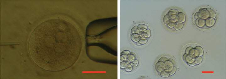
This is a lovely method and should find wide applicability in many settings, especially for microorganisms and cell lines. However, it is not clear that this approach will be, as implied by the discussion, an efficient mapping method for all multicellular organisms. I have performed similar experiments in Drosophila, focused on meiotic recombination, on a much smaller scale, and found that CRISPR-Cas9 can indeed generate targeted recombination at gRNA target sites. In every case I tested, I found that the recombination event was associated with a deletion at the gRNA site, which is probably unimportant for most mapping efforts, but may be a concern in some specific cases, for example for clinical applications. It would be interesting to know how often mutations occurred at the targeted gRNA site in this study.
The wider issue, however, is whether CRISPR-mediated recombination will be more efficient than other methods of mapping. After careful consideration of all the costs and the time involved in each of the steps for Drosophila, we have decided that targeted meiotic recombination using flanking visible markers will be, in most cases, considerably more efficient than CRISPR-mediated recombination. This is mainly due to the large expense of injecting embryos and the extensive effort and time required to screen injected animals for appropriate events. It is both cheaper and faster to generate markers (with CRISPR) and then perform a large meiotic recombination mapping experiment than it would be to generate the lines required for CRISPR-mediated recombination mapping. It is possible to dramatically reduce costs by, for example, mapping sequentially at finer resolution. But this approach would require much more time than marker-assisted mapping. If someone develops a rapid and cheap method of reliably introducing DNA into Drosophila embryos, then this calculus might change.
However, it is possible to imagine situations where CRISPR-mediated mapping would be preferable, even for Drosophila. For example, some genomic regions display extremely low or highly non-uniform recombination rates. It is possible that CRISPR-mediated mapping could provide a reasonable approach to fine mapping genes in these regions.
The authors also propose the exciting possibility that CRISPR-mediated loss of heterozygosity could be used to map traits in sterile species hybrids. It is not entirely obvious to me how this experiment would proceed and I hope the authors can illuminate me. If we imagine driving a recombination event in the early embryo (with maternal Cas9 from one parent and gRNA from a second parent), then at best we would end up with chimeric individuals carrying mitotic clones. I don’t think one could generate diploid animals where all cells carried the same loss of heterozygosity event. Even if we could, this experiment would require construction of a substantial number of stable transgenic lines expressing gRNAs. Mapping an ~20Mbp chromosome arm to ~10kb would require on the order of two-thousand transgenic lines. Not an undertaking to be taken lightly. It is already possible to perform similar tests (hemizygosity tests) using D. melanogaster deficiency lines in crosses with D. simulans, so perhaps CRISPR-mediated LOH could complement these deficiency screens for fine mapping efforts. But, at the moment, it is not clear to me how to do the experiment.