Healthcare analytics, AI solutions for biological big data, providing an AI platform for the biotech, life sciences, medical and pharmaceutical industries, as well as for related technological approaches, i.e., curation and text analysis with machine learning and other activities related to AI applications to these industries.
Glassman Lecture “From Bacterial Adaptive Immunity to the Future of Genome Engineering” Jennifer A. Doudna, University of California, Berkeley; Howard Hughes Medical Institute
Reporter: Aviva Lev-Ari, PhD, RN
Jennifer A. Doudna – From Bacterial Adaptive Immunity to the Future of Genome Engineering
2.1.5.17 Glassman Lecture “From Bacterial Adaptive Immunity to the Future of Genome Engineering” Jennifer A. Doudna, University of California, Berkeley; Howard Hughes Medical Institute, Volume 2 (Volume Two: Latest in Genomics Methodologies for Therapeutics: Gene Editing, NGS and BioInformatics, Simulations and the Genome Ontology), Part 2: CRISPR for Gene Editing and DNA Repair
Glassman Lecture
“From Bacterial Adaptive Immunity to the Future of Genome Engineering”
Jennifer A. Doudna, University of California, Berkeley; Howard Hughes Medical Institute
Introduction by Jennifer Lippincott-Schwartz, Group Leader, Howard Hughes Medical Institute Janelia Research Campus
Lecture Abstract:
The advent of facile genome engineering using the bacterial RNA-guided CRISPR-Cas9 system in animals and plants is transforming biology. I will present a brief history of CRISPR biology from its initial discovery through the elucidation of the CRISPR-Cas9 enzyme mechanism, providing the foundation for remarkable developments using this technology to modify, regulate or visualize genomic loci in a wide variety of cells and organisms. These results highlight a new era in which genomic manipulation is no longer a bottleneck to experiments, paving the way to both fundamental discoveries in biology, with applications in all branches of biotechnology, and strategies for curing human genetic disease. Recent results regarding the molecular mechanism of Cas9 and its use for targeted cell-based therapies will be discussed.
Jennifer Doudna is the Li Ka Shing Chancellor’s Chair in Biomedical and Health Sciences as well as Professor of Molecular and Cell Biology and Professor of Chemistry at UC Berkeley and an Investigator of the Howard Hughes Medical Institute. Prof. Doudna’s research seeks to understand how RNA molecules control the expression of genetic information. Her research led to insights about CRISPR-Cas9-mediated bacterial immunity that enabled her lab and that of collaborator Emmanuelle Charpentier to re-design this system for efficient genome engineering in animals and plants, creating a transformative technology that is revolutionizing the fields of genetics, molecular biology and medicine. She is a member of the National Academy of Sciences, the American Academy of Arts and Sciences, the National Academy of Medicine and the National Academy of Inventors. She is a recipient of awards including the NSF Waterman Award, the FNIH Lurie Prize, the Paul Janssen Award for Biomedical Research, the Breakthrough Prize in Life Sciences, the Princess of Asturias Award (Spain), the Gruber Prize in Genetics, the Massry Prize, the Gairdner Award, the Nakasone Prize, the Heineken Prize and the L’Oreal-UNESCO International Prize for Women in Science.
About the Glassman Lecture:
The Glassman Lecture is held in honor of the late Harold N. Glassman who left a generous bequest to the MBL which resulted in the establishment of the Harold N. Glassman fund, the income from which is used to support an annual Friday Evening Lecture on an important topic in biological research.
Updated: First-in-Man: CRISPR, the Genome Editing Technology is Nearing Human Trials: Human T cells will soon be Modified using the CRISPR Technique in a Clinical Trial to attack Cancer Cells, Volume 2 (Volume Two: Latest in Genomics Methodologies for Therapeutics: Gene Editing, NGS and BioInformatics, Simulations and the Genome Ontology), Part 2: CRISPR for Gene Editing and DNA Repair
Updated: First-in-Man: CRISPR, the Genome Editing Technology is Nearing Human Trials: Human T cells will soon be Modified using the CRISPR Technique in a Clinical Trial to attack Cancer Cells
Curator: Aviva Lev-Ari, PhD, RN
UPDATED on 12/18/2016
NATURE|NEWS
CRISPR gene-editing tested in a person for the first time
The move by Chinese scientists could spark a biomedical duel between China and the United States.
A Chinese group has become the first to inject a person with cells that contain genes edited using the revolutionary CRISPR–Cas9 technique.
On 28 October, a team led by oncologist Lu You at Sichuan University in Chengdu delivered the modified cells into a patient with aggressive lung cancer as part of a clinical trial at the West China Hospital, also in Chengdu.
Statement on NIH funding of research using gene-editing technologies in human embryos
IMAGE SOURCE
Crystal structure of the Cas9 gene-editing enzyme (light blue) in complex with an RNA guide (red) and its target DNA (yellow).Wong, Broad Institute of Harvard and MIT, Cambridge, MA
Genomic editing is an area of research seeking to modify genes of living organisms to improve our understanding of gene function and advance potential therapeutic applications to correct genetic abnormalities. Researchers in China have recently described their experiments in a nonviable human embryo to modify the gene responsible for a potentially fatal blood disorder using a gene-editing technology called CRISPR/Cas9.
Genomic editing is already widely studied in a variety of organisms. For example, CRISPR/Cas9 has greatly shortened the time it takes to produce knockout mouse models of disease, enabling researchers to study more easily the underlying genetic causes of those diseases. This technology is also being used to develop the next generation of antimicrobials, which can specifically target harmful strains of bacteria and viruses. In the first clinical application of genomic editing, a related genome editing technique (using a zinc finger nuclease) was used to create HIV-1 resistance in human immune cells, bringing HIV viral load down to undetectable levels in at least one individual. All of these examples of research using genomic editing technologies can and are being funded by NIH.
However, NIH will not fund any use of gene-editing technologies in human embryos. The concept of altering the human germline in embryos for clinical purposes has been debated over many years from many different perspectives, and has been viewed almost universally as a line that should not be crossed. Advances in technology have given us an elegant new way of carrying out genome editing, but the strong arguments against engaging in this activity remain. These include the serious and unquantifiable safety issues, ethical issues presented by altering the germline in a way that affects the next generation without their consent, and a current lack of compelling medical applications justifying the use of CRISPR/Cas9 in embryos.
Practically, there are multiple existing legislative and regulatory prohibitions against this kind of work. The Dickey-Wicker amendment prohibits the use of appropriated funds for the creation of human embryos for research purposes or for research in which human embryos are destroyed (H.R. 2880, Sec. 128). Furthermore, the NIH Guidelines state that the Recombinant DNA Advisory Committee,“…will not at present entertain proposals for germ line alteration”. It is also important to note the role of the U.S. Food and Drug Administration (FDA) in this arena, which applies not only to federally funded research, but to any research in the U.S. The Public Health Service Act and the Federal Food, Drug, and Cosmetic Act give the FDA the authority to regulate cell and gene therapy products as biological products and/or drugs, which would include oversight of human germline modification. During development, biological products may be used in humans only if an investigational new drug application is in effect (21 CFR Part 312).
NIH will continue to support a wide range of innovations in biomedical research, but will do so in a fashion that reflects well-established scientific and ethical principles.
Francis S. Collins, M.D., Ph.D.
Director, National Institutes of Health
Emerging Biotechnologies and the Role of the NIH RAC
June 16, 2016
Next week, we mark the 40th anniversary of the first publication of the NIH Guidelines governing experiments using recombinant DNA. On June 23rd, 1976, former NIH Director Dr. Donald Frederickson announced that all NIH funded and conducted research involving recombinant DNA would be expected to follow the NIH Guidelines, noting both the great potential benefits that could arise from this new technology and the lack of certainty about the risks.
We have come a long way in 40 years, and the use of recombinant DNA is ubiquitous in research, medicine, and many other aspects of our daily lives. Recently, the NIH announcedrevisions to the NIH Guidelines that included amending the criteria and process for how human gene transfer protocols would be selected for review by the Recombinant DNA Advisory committee (RAC), limiting in depth review and public discussion only for exceptional cases.
Just such an exceptional case comes before the RAC during their meeting this week. During the June 21-22 meeting, the RAC will review a protocol involving the first-in-human use of gene editing via CRISPR/Cas9 technology. This T cell immunotherapy protocol involves the use of CRISPR/Cas9 to edit two genes in T cells also modified to express T cell receptors targeting myeloma, melanoma, and sarcoma tumor cells. Consideration of this study underlines the purpose of changing the RAC process: to better use the collective breadth of experience of the RAC members in reviewing gene transfer trials and novel technologies that pose unknown risks, exactly as described by Dr. Frederickson four decades ago.
Researchers in the field of gene transfer are excited by the potential of utilizing CRISPR/Cas9 to repair or delete mutations that are involved in numerous human diseases in less time and at a lower cost than earlier gene editing systems. While the application of new gene editing technologies in this field has great potential to improve human health, it is not without concerns. In a previous statement, NIH Director, Dr. Francis Collins, reiterated NIH’s commitment to support innovations in biomedical research in a fashion that reflects well-established scientific and ethical principles. Having a body such as the RAC available to publicly discuss the scientific, safety, and ethical implications of such cutting-edge experiments helps to ensure we are living up to that commitment.
As the application of biotechnology innovations moves closer to the clinical realm, we are confident that the changes we have made to the NIH Guidelines will enable the RAC to devote its full resources to where they are most needed. And as science continues to evolve, we will strive for parallel evolution in our policies to make oversight of research commensurate with the risks involved.
I encourage you to either attend the upcoming RAC meeting in person, or through the NIH Videocast to learn more about the exciting advances being made in the field of gene transfer. Information about the RAC meeting, including an agenda and the meeting location can be found on the OSP website.
On 21 June, an advisory committee at the US National Institutes of Health (NIH), NIH’s Recombinant DNA Research Advisory Committee (RAC), which reviewed the proposal. CRISPR, approved a proposal to use CRISPR–Cas9 to help augment cancer therapies that rely on enlisting a patient’s T cells, a type of immune cell.
the main challenge will be overcoming CRISPR’s propensity for ‘off-target’ edits. These are instances in which the system cuts or mutates unintended parts of the genome. And despite precautions, the immune system could still attack the edited cells.
Potential Conflict of Interest
During the RAC meeting, one of the committee’s greatest concerns was a potential conflict of interest. Among other financial involvements,
Immunologist Carl June at the University of Pennsylvania, who is a science adviser on the project, says that it could begin by the end of the year. June has ties to the pharmaceutical company Novartis, holds patents on T-cell technologies, and could stand to benefit from the success of this trial. June declined to give details on the exact nature of his conflicts of interest, but says that his university is taking steps to manage it, such as preventing him from being involved in selecting patients. Several RAC reviewers suggested that the University of Pennsylvania not be allowed to recruit patients at all and to leave it to other institutions: this language did not make it into their final approval.
However, the RAC members say they are being extra careful with this study. “Penn has a very extensive conflict and has a history,” says Laurie Zoloth, a bioethicist at Northwestern University in Evanston, Illinois. Looming over the discussion is the name Jesse Gelsinger, who died at age 18 while participating in an early gene-therapy trial conducted by researchers at the University of Pennsylvania in 1999.
A subsequent investigation found numerous problems with the study, including unreported animal data on the therapy’s ill effects and the fact that the investigators had a financial stake in the study’s outcome. The incident is generally considered to have set gene therapy back by decades. “Any first use in humans we have to be extraordinarily careful,” Zoloth says. So a lot is riding on this trial.
Mildred Cho, a bioethicist at Stanford University in California and an RAC member, says that safety work in animals for a new therapy will take researchers only so far. “Often we have to take the leap of faith.”
“Cell therapies [for cancer] are so promising but the majority of people who get these therapies have a disease that relapses,” says study leader Edward Stadtmauer, a physician at the University of Pennsylvania in Philadelphia. Gene editing could improve such treatments and eliminate some of their vulnerabilities to cancer and the body’s immune system, he says.
This first trial is small and designed to test whether CRISPR is safe for use in people, rather than whether it effectively treats cancer or not. It will be funded by a US$250-million immunotherapy foundation formed in April by former Facebook president Sean Parker. The trial itself does not yet have a budget. The University of Pennsylvania will manufacture the edited cells, and will recruit and treat patients alongside centres in California and Texas.
The researchers will remove T cells from 18 patients with several types of cancers and perform three CRISPR edits on them. One edit will insert a gene for a protein engineered to detect cancer cells and instruct the T cells to target them, and a second edit removes a natural T-cell protein that could interfere with this process. The third is defensive: it will remove the gene for a protein that identifies the T cells as immune cells and prevent the cancer cells from disabling them. The researchers will then infuse the edited cells back into the patient.
Pancreatic Cancer Modeling using Retrograde Viral Vector Delivery and IN-Vivo CRISPR/Cas9-mediated Somatic Genome Editing, Volume 2 (Volume Two: Latest in Genomics Methodologies for Therapeutics: Gene Editing, NGS and BioInformatics, Simulations and the Genome Ontology), Part 2: CRISPR for Gene Editing and DNA Repair
Pancreatic Cancer Modeling using Retrograde Viral Vector Delivery and IN-Vivo CRISPR/Cas9-mediated Somatic Genome Editing
Curators: Larry H. Benstein, MD, FCAP and Aviva Lev-Ari, PhD, RN
Little is known about how these alterations contribute to the development of metastatic and therapy-refractory PDAC. Given the inability to test gene function in human cancers in vivo, genetically engineered mouse models represent tractable and biologically relevant systems with which to interrogate the molecular determinants of each stage of pancreatic cancer development.
Identification of the mutations that drive the development of human pancreatic cancer combined with the ability to alter gene function in mice has enabled the development of genetically engineered murine PDAC models. Transgenic expression of Cre-recombinase in pancreatic cells of loxP-Stop-loxP (LSL)KrasG12Dknock-in mice (KrasLSL-G12D/+) results in deletion of the transcriptional/translational Stop element, expression of oncogenic KrasG12D, and development of lesions that closely resemble early stage human pancreatic intraepithelial neoplasms (PanINs) (Hingorani et al. 2003). Concomitant expression of a point mutantp53 allele, deletion ofp53, deletion of Cdkn2a, and/or deletion of Smad4 allow(s) for the development of invasive and metastatic PDAC
(Aguirre et al. 2003; Hingorani et al. 2005; Bardeesy et al. 2006a,b; Gidekel Friedlander et al. 2009; Whittle et al. 2015).
Pancreatic ductal adenocarcinoma (PDAC) is a genomically diverse, prevalent, and almost invariably fatal malignancy. Although conventional genetically engineered mouse models of human PDAC have been instrumental in understanding pancreatic cancer development, these models are much too labor-intensive, expensive, and slow to perform the extensive molecular analyses needed to adequately understand this disease. Here we demonstrate that retrograde pancreatic ductal injection of either adenoviral-Cre or lentiviral-Cre vectors allows titratable initiation of pancreatic neoplasias that progress into invasive and metastatic PDAC. To enable in vivo CRISPR/Cas9-mediated gene inactivation in the pancreas, we generated a Cre-regulated Cas9 allele and lentiviral vectors that express Cre and a single-guide RNA. CRISPR-mediated targeting of Lkb1 in combination with oncogenic Kras expression led to selection for inactivating genomic alterations, absence of Lkb1 protein, and rapid tumor growth that phenocopied Cre-mediated genetic deletion of Lkb1. This method will transform our ability to rapidly interrogate gene function during the development of this recalcitrant cancer.
Keywords: CRISPR, genome editing, mouse model, pancreatic cancer
Pancreatic ductal adenocarcinoma (PDAC) is an almost uniformly lethal tumor type that is projected to become the second leading cancer killer in the United States by 2030 (Rahib et al. 2014). PDAC patients have a 5-year survival rate of ∼5%, underscoring the need for novel approaches to accelerate the molecular characterization of this disease. Although high-prevalence mutations have been identified in pancreatic cancer, these tumors also incur low-frequency mutations and genomic alterations, interact with their extensive and complex stromal environment, and undergo poorly characterized changes in their gene expression programs (Biankin et al. 2012; Waddell et al. 2015). Despite the potential importance of these molecular and cellular changes, very little is known about how these alterations contribute to the development of metastatic and therapy-refractory PDAC. Given the inability to test gene function in human cancers in vivo, genetically engineered mouse models represent tractable and biologically relevant systems with which to interrogate the molecular determinants of each stage of pancreatic cancer development.
Identification of the mutations that drive the development of human pancreatic cancer combined with the ability to alter gene function in mice has enabled the development of genetically engineered murine PDAC models. Transgenic expression of Cre-recombinase in pancreatic cells of loxP-Stop-loxP (LSL)KrasG12D knock-in mice (KrasLSL-G12D/+) results in deletion of the transcriptional/translational Stop element, expression of oncogenic KrasG12D, and development of lesions that closely resemble early stage human pancreatic intraepithelial neoplasms (PanINs) (Hingorani et al. 2003). Concomitant expression of a point mutant p53 allele, deletion of p53, deletion of Cdkn2a, and/or deletion of Smad4 allow(s) for the development of invasive and metastatic PDAC (Aguirre et al. 2003; Hingorani et al. 2005; Bardeesy et al. 2006a,b;Gidekel Friedlander et al. 2009; Whittle et al. 2015).
These in vivo models have been instrumental in our understanding of the genetic determinants of cancer progression as well as the functional interactions of neoplastic cells with the immune system and stromal environment. However, using conventional genetically engineered autochthonous mouse models of PDAC to interrogate gene function is complicated by inherent practical and biological limitations of these systems. Existing mouse models typically fail to model the adult onset of pancreatic cancer and induce genomic alteration in nearly every cell in the pancreas. In the decade since the first genetically engineered PDAC models were developed, few technical advances have been made, and generating the mice required to investigate a gene of interest in the established PDAC models remains a time-consuming and costly endeavor (Aguirre et al. 2003; Hingorani et al. 2003,2005; Saborowski et al. 2014).
Systems that enable in vivo functional interrogation of genes in pancreatic cancer without the financial and temporal cost of generating new mouse alleles and incorporating them into increasingly complex mouse models could have an extremely broad impact on pancreatic cancer research. To functionally investigate the molecular changes that drive each step of pancreatic cancer development, it would be desirable to have a system in which the timing of tumor initiation and the number of lesions that form in the adult pancreas can be controlled, the number of germline-encoded alleles is minimized, and genes of interest can be eliminated without having to generate a conditional allele and breed it into a complex genetically engineered mouse model.
Here we describe methods for the direct delivery of viral vectors to the pancreas and transgenic mouse lines to allow CRISPR/Cas9-mediated genomic alterations in pancreatic cells in vivo. These systems allow titratable initiation of pancreatic tumors in adult mice and functional interrogation of candidate genes in pancreatic cancer in vivo.
Pancreatic retrograde ductal injection of Adeno-Cre or Lenti-Cre induces widespread recombination and initiates the development of PanINs and ductal adenocarcinoma. (A) Diagram of the pancreatic retrograde ductal injection procedure. (B) Images of retrograde …
Chronic pancreatitis is associated with an increased incidence of PDAC in humans, and experimental evidence from mouse models suggests that adult cells may be refractory to transformation in the absence of pancreatic inflammation (Guerra et al. 2007; Raimondi et al. 2010). Therefore, we assessed whether retrograde ductal viral infection induces pancreatitis. Both Ad-Cre and Lenti-Cre infection induced acute pancreatitis with morphological evidence of ADM and focal replacement of acinar cells by infiltrating mononuclear cells (Supplemental Figs. 1C,D, 4C). The induction of pancreatitis is consistent with the efficient tumor initiation observed in adult animals following retrograde ductal injection of viral-Cre.
Diverse expansion potential and metastatic ability of viral-Cre-initiated PDAC. (A) DTCs can be detected in the peritoneal cavity of Ad-Cre-infected KPTmice. Viable (DAPInegative), lineage-negative (CD31, CD45, Ter119, F4/80)negative cells are shown. …
CRISPR/Cas9 enables in vivo genetic alteration in pancreatic cancer. (A) Schematic of the Cre-regulatedCas9 allele. (CAGGS) Cytomegalovirus immediate–early enhancer/chicken β-actin promoter; (hSpCas9) human codon-optimized Streptococcus…
Somatic genome engineering enables rapid generation of genetically defined pancreatic cancer mouse models
To assess the impact of Lkb1 deletion on pancreatic cancer, we performed retrograde ductal injections of KT;H11LSL-Cas9/+, control KT, and KT;Lkb1flox/flox mice with Lenti-sgLkb1/Cre as well as KT;H11LSL-Cas9/+ mice with lentiviral vectors containing negative control sgRNAs. Control KT mice infected with Lenti-sgLkb1/Cre as well as KT;H11LSL-Cas9/+ mice infected with a Lenti-sgRNA/Cre vector containing either a nontargeting sgRNA (sgNT) or an sgRNA targeting an inert region of the genome (sgNeo) formed only rare Tomatopositive lesions (Fig. 3F–H). Both Lenti-sgLkb1/Cre-infected KT;H11LSL-Cas9/+ and KT;Lkb1flox/flox mice had extensive tumor growth as early as 2 mo after tumor initiation (Fig. 3I,J, respectively).
All three groups of negative control mice developed only rare PanIN lesions within almost completely normal pancreata (Fig. 4A–C; Supplemental Fig. 9A; data not shown). The tumors that formed in Lenti-sgLkb1/Cre-infectedKT;H11LSL-Cas9/+ mice were cystic lesions comprised of Tomatopositive, CK19positive tall cuboidal to columnar epithelial cells with otherwise bland cytological features (Fig. 4D; Supplemental Fig. 9B). Some smaller lesions also contained high levels of mucin (Supplemental Fig. 9B). Importantly, these features were histologically indistinguishable from those found in KT;Lkb1flox/flox mice infected with Lenti-sgLkb1/Cre (Fig. 4E; Supplemental Fig. 9C). Lenti-sgLkb1/Cre-infected KT;H11LSL-Cas9/+ mice also had a substantially higher tumor burden when compared with all three groups of negative control mice (Fig. 4F).
Cas9-mediated targeting of Lkb1 in the pancreas promotes tumor growth. (A) Control Lenti-sgLkb1/Cre-infected KT mice (n= 5) have small clusters of Tomatopositive cells and very rare ADMs and PanINs. (B,C) KT;H11LSL-Cas9/+ mice infected with Lenti-sgNT/Cre …
Diverse indels at the targetedLkb1 locus were specifically detected in the neoplastic cells from these tumors (Fig. 5A,B; Supplemental Fig. 9F). The indels were all frameshift mutations or large exon deletions that included the splice acceptor or donor regions of Lkb1exon 6, further supporting the strong selective advantage of Lkb1 inactivation (Fig. 5B,C). Consistent with the presence of CRISPR/Cas9-induced frameshift mutations and large deletions, Lkb1 protein was absent from most neoplastic cells in the Lenti-sgLkb1/Cre-induced tumors in KT;H11LSL-Cas9/+ mice (Fig. 5D,E; Supplemental Fig. 9G).
Cas9-mediated targeting leads to the formation of pancreatic tumors harboring deleterious Lkb1mutations and lacking Lkb1 protein. (A) Genomic cleavage detection assay detected indels in the targeted Lkb1 locus in Tomatopositive cells isolated from Lenti-sgLkb1/Cre-infected …
The development of systems that accelerate our ability to investigate pancreatic carcinogenesis at the molecular level will be a critical step toward overcoming the dismal rate of successful treatment and low survival rate of patients with this recalcitrant cancer. The ability to induce pancreatic cancer using viral vectors will be instrumental in understanding the mechanisms that sustain tumor growth, lead to metastatic spread, and drive drug resistance. Our CRISPR/Cas9-based model should allow any gene of interest to be inactivated in pancreatic cancer in vivo without the need to generate any new mouse alleles. The method developed in this study could have a profound impact on both basic and translational pancreatic cancer research. Collectively, these methods will enable a more rapid and complete understanding of the molecular regulators of all aspects of pancreatic tumorigenesis and complement the strength of existing genetically engineered models, human patient-derived xenograft models, and studies on human and murine cell lines.
Retrograde ductal injection of Adeno-Cre and Lenti-Cre vectors allows titratable pancreatic tumor initiation in the adult pancreas. This removes the requirement for the transgenic Cre(ER) alleles used in conventional genetically engineered pancreatic cancer models and enables sparse rather than widespread expression of oncogenic Kras and deletion of tumor suppressor genes, more closely recapitulating the initiating events in human PDAC. The inclusion of a fluorescent Cre reporter in transgenic Cre(ER)-induced PDAC models leads to the fluorescent labeling of not only the neoplastic cells but also most of the nontransformed pancreatic epithelial cells, making unequivocal distinction of neoplastic from normal cells difficult.
The ability to induce PDAC in Cre-lox models without having to include a transgenic Cre line will make the molecular investigation of pancreatic cancer more rapid and less expensive (Supplemental Table 2). Additionally, with the ease of generating pancreatic tumors now approaching that of lung tumors (via intranasal or intratracheal injection), comparing the impact of the same genetic alterations on each cancer type should become standard practice and will uncover the commonalities and differences between these lethal adenocarcinomas.
PDAC models often generate tumor masses of unknown clonal origin. By incorporating a multicolor fluorescent reporter, we were able to mark individual clonal lesions and identify the relationship between primary tumors and metastases. Unexpectedly, some late time point mice had only one or two large cancers, underscoring the dramatic heterogeneity in expansion potential of pancreatic lesions initiated with identical engineered genetic events.
Bacterial immune system may be utilized as a tool harboring an impressive recording capacity, Volume 2 (Volume Two: Latest in Genomics Methodologies for Therapeutics: Gene Editing, NGS and BioInformatics, Simulations and the Genome Ontology), Part 2: CRISPR for Gene Editing and DNA Repair
Bacterial immune system may be utilized as a tool harboring an impressive recording capacity
Curator: Larry H. Bernstein, MD, FCAP
LPBI
Creating a DNA Record with CRISPR
Researchers repurpose a bacterial immune system to be a molecular recording device.
Utilizing the bacterial CRISPR/Cas adaptive immune system, researchers at Harvard have developed a method for permanently recording molecular events in living cells, according to a report published inSciencetoday (June 9). The system integrates specific synthetic DNA elements into the bacterial genomes in temporally-ordered arrays, which, once sequenced, can provide a readout of the bacteria’s timeline of DNA events.
“The importance of the work is in providing a proof of principle: that a fascinating bacterial immune system may be utilized as a tool harboring an impressive recording capacity,” said microbiologist Udi Qimron of Tel Aviv University who was not involved in the work.
The CRISPR/Cas system works by snipping short DNA elements from the genomes of infecting viruses, integrating those elements into the bacterium’s genome (at the CRISPR locus), and using the RNAs produced from the integrated elements to direct destruction of the corresponding virus. In essence, the bacterium keeps a DNA account of its viral foes, and uses it against them.
Integration of these viral DNA elements—or oligomers—into the CRISPR locus is nonrandom: the most recent viral elements are consistently integrated ahead of older viral elements in the array. Harvard’sGeorge Church and colleagues considered that this temporal ordering of integration could form the basis of a molecular recording device. If defined synthetic DNA oligomers could be integrated into CRISPR loci just as viral elements are, then sequencing the cells’ CRISPR loci would provide a log of which oligomers the cells had been exposed to and when, the researchers reasoned.
To test this idea, the team used an E. coli strain that contained a CRISPR DNA locus and a stripped-down version of the Cas protein machinery. The minimal machinery consisted of inducible versions of Cas1 and Cas2—enzymes required for integrating the DNA oligomers—but lacked all the Cas machinery required for virus destruction. The researchers found that, by introducing specific synthetic DNA sequences into these cells in a timed manner (different oligomers on different days, for example), the resulting sequences of the CRISPR loci did indeed accurately reflect the order in which the oligomers had been introduced.
“It’s the first demonstration of the ordered acquisition of intentionally introduced DNA sequences,” said bioengineer Adam Arkin of the University of California, Berkeley, who did not participate in the work.
Using directed evolution, the team went on to create new versions of Cas1 and Cas2 that could integrate oligomers in a subtly different and discernable way (though still temporally ordered) to that of wildtype Cas1 and 2. Putting these modified Cas enzymes under the control of a different inducer allowed the team to record DNA events in two different modes—depending on which versions of Cas1 and 2 were operational.
“Essentially, we’re measuring concentrations of nucleic acids,” said Church. “Ideally it would be messenger RNAs but in this case it is DNA. . . . This is a proof of concept on the way to other things,” he added.Church suggested, for example, that if a CRISPR/Cas system were to be combined with a reverse transcriptase—an enzyme that converts RNA to DNA—in cells or animals, it could be used to provide a record of which messenger RNAs are expressed, when.
Another possibility, suggested Arkin, is to use CRISPR/Cas-engineered bacteria to provide information about the other microorganisms present in an environment—be that the soil, the human gut, or wherever.
“[The bacteria] could kill a few neighboring [bugs], secrete an enzyme that cleaved their DNA, and express a competence system to take that DNA in,” Arkin said. “That sounds insane, but there are bacteria who do that naturally,” he added. The foreign microbial DNA could then be incorporated and logged at the bacteria’s CRISPR locus, he explained.
Such applications are as-yet distant possibilities, but the new paper, said Arkin, “conceptually sets the flag in the ground and says, ‘Here’s how we should move forward.’”
S.L. Shipman et al., “Molecular recordings by directed CRISPR spacer acquisition,” Science,doi:10.1126/science.aaf1175, 2016.
Molecular recordings by directed CRISPR spacer acquisition
Seth L. Shipman1,2,3,*, Jeff Nivala1,3,*, Jeffrey D. Macklis2, George M. Church1,3,†
The ability to write a stable record of identified molecular events into a specific genomic locus would enable the examination of long cellular histories and have many applications, ranging from developmental biology to synthetic devices. We show that the type I-E CRISPR-Cas system of E. coli can mediate acquisition of defined pieces of synthetic DNA. We harnessed this feature to generate records of specific DNA sequences into a population of bacterial genomes. We then applied directed evolution to alter the recognition of a protospacer adjacent motif by the Cas1-Cas2 complex, which enabled recording in two modes simultaneously. We used this system to reveal aspects of spacer acquisition, fundamental to the CRISPR-Cas adaptation process. These results lay the foundations of a multimodal intracellular recording device.
Genome Engineering: The CRISPR-Cas Revolution, August 17 – 20, 2016, Cold Spring Harbor Laboratory
Reporter: Aviva Lev-Ari, PhD, RN
2.1.5.18 Genome Engineering: The CRISPR-Cas Revolution, August 17 – 20, 2016, Cold Spring Harbor Laboratory, Volume 2 (Volume Two: Latest in Genomics Methodologies for Therapeutics: Gene Editing, NGS and BioInformatics, Simulations and the Genome Ontology), Part 2: CRISPR for Gene Editing and DNA Repair
Jennifer Doudna, University of California, Berkeley/HHMI
Maria Jasin, Memorial Sloan Kettering Cancer Center, NY
Jonathan Weissman, UCSF/HHMI
You are cordially invited to participate in the second meeting on Genome Engineering: The CRISPR-Cas Revolution at Cold Spring Harbor Laboratory. The meeting will begin with dinner and the first session on the evening of Wednesday, August 17, 2016, and will conclude with lunch on Saturday, August 20.
The specific goal for this meeting is to foster fruitful and creative interactions between researchers interested in applying these systems to genome engineering and related advances in a wide variety of organisms, together with scientists studying the basic biology of CRISPR-Cas and related bacterial defense systems.
This meeting will consist of six oral sessions plus one poster session; speakers will be invited as well as selected from submitted abstracts.
Topics:
• CRISPR Biology
• Genome Editing
• Gene Drives
• DNA Repair
• Model and Industrial Organisms
• Technology Development
• Stem Cells/Cancer
• Genome-Wide Screens
Pre-Meeting Workshop – Wednesday, August 17 from 9 to 6 pm (see below for payment options)
Technical Workshop 9 a.m. to 12:30 p.m.
Lunch 12:30 to 2 p.m.
Commercial Exhibits/Demos 2 to 4 p.m.
Short Talks by Graduate Students 4 to 6 p.m.
Dinner 6 to 7:30 p.m.
Confirmed Chairs:
Ethan Bier, University of California, San Diego
Jacob Corn, University of California, Berkeley
Joseph DeRisi, HHMI/University of California, San Francisco
James Haber, Brandeis University
Danwei Huangfu, Memorial Sloan-Kettering Cancer Center
J. Keith Joung, Massachusetts General Hospital
Jin-Soo Kim, Seoul National University, South Korea
Silvana Konermann, Massachusetts Institute of Technology (Feng Zhang Laboratory)
Nancy Maizels, University of Washington
Luciano Marraffini, The Rockefeller University
Aviv Regev, Broad Institute of MIT and Harvard
David Sabatini, Whitehead Institute for Biomedical Research
William Skarnes, Wellcome Trust Sanger Institute, UK
Julianne Smith, Cellectis, France
Erik Sontheimer, University of Massachusetts Medical School
Frank Stegmeier, KSQ Therapeutics
Francesca Storici, Georgia Tech
Fyodor Urnov, Sangamo BioSciences, Inc.
Abstracts should contain interesting and exciting material and must be submitted electronically by the abstract deadline. Selection of material for oral and poster presentation will be made by the organizers and individual session chairs. Status (talk/poster) of abstracts will be posted on our web site as soon as decisions have been made by the organizers.
We are eager to have as many young people as possible attend since they are likely to benefit most from this meeting. We have applied for funds from government and industry to partially support graduate students and postdocs. Apply in writing to Val Pakaluk stating need for financial support – preference is given to those submitting abstracts.
Novel Discoveries in Molecular Biology and Biomedical Science, Volume 2 (Volume Two: Latest in Genomics Methodologies for Therapeutics: Gene Editing, NGS and BioInformatics, Simulations and the Genome Ontology), Part 1: Next Generation Sequencing (NGS)
Novel Discoveries in Molecular Biology and Biomedical Science
Curator: Larry H. Bernstein, MD, FCAP
UPDATED on 6/1/2016
The following is a collection of current articles on noncoding DNA, synthetic genome engineering, protein regulation of apoptosis, drug design, and geometrics.
No longer ‘junk DNA’ — shedding light on the ‘dark matter’ of the genome
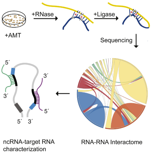
A new tool called “LIGR-Seq” enables scientists to explore in depth what non-coding RNAs actually do in human cells May 23, 2016
he LIGR-seq method for global-scale mapping of RNA-RNA interactions in vivo to reveal unexpected functions for uncharacterized RNAs that act via base-pairing interactions (credit: University of Toronto)
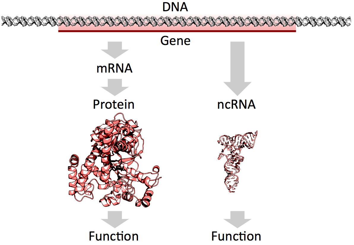
What used to be dismissed by many as “junk DNA” has now become vitally important, as accelerating genomic data points to the importance of non-coding RNAs (ncRNAs) — a genome’s messages that do not specifically code for proteins — in development and disease.
But our progress in understanding these molecules has been slow because of the lack of technologies that allow for systematic mapping of their functions.
Now, professor Benjamin Blencowe’s team at the University of Toronto’s Donnelly Centre has developed a method called “LIGR-seq” that enables scientists to explore in depth what ncRNAs do in human cells.
The study, described in Molecular Cell, was published on May 19, along with two other papers, in Molecular Cell and Cell, respectively, from Yue Wan’s group at the Genome Institute of Singapore and Howard Chang’s group at Stanford University in California, who developed similar methods to study RNAs in different organisms.
Of the 3 billion letters in the human genome, only two per cent make up the protein-coding genes. The genes are copied, or transcribed, into messenger RNA (mRNA) molecules, which provide templates for building proteins that do most of the work in the cell. Much of the remaining 98 per cent of the genome was initially considered by some as lacking in functional importance. However, large swaths of the non-coding genome — between half and three quarters of it — are also copied into RNA.
So then what might the resulting ncRNAs do? That depends on whom you ask. Some researchers believe that most ncRNAs have no function, that they are just a by-product of the genome’s powerful transcription machinery that makes mRNA. However, it is emerging that many ncRNAs do have important roles in gene regulation — some ncRNAs act as carriages for shuttling the mRNAs around the cell, or provide a scaffold for other proteins and RNAs to attach to and do their jobs.
But the majority of available data has trickled in piecemeal or through serendipitous discovery. And with emerging evidence that ncRNAs could drive disease progression, such as cancer metastasis, there was a great need for a technology that would allow a systematic functional analysis of ncRNAs.
“Up until now, with existing methods, you had to know what you are looking for because they all require you to have some information about the RNA of interest. The power of our method is that you don’t need to preselect your candidates; you can see what’s occurring globally in cells, and use that information to look at interesting things we have not seen before and how they are affecting biology,” says Eesha Sharma, a PhD candidate in Blencowe’s group who, along with postdoctoral fellow Tim Sterne-Weiler, co-developed the method.
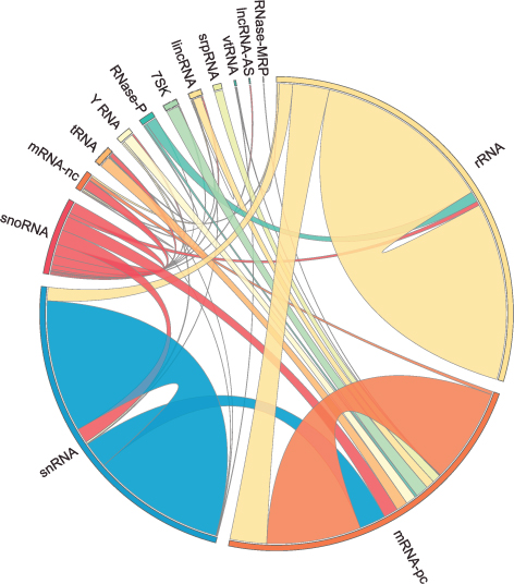
The human RNA-RNA interactome, showing interactions detected by LIGR-seq (credit: University of Toronto)
The new ‘‘LIGation of interacting RNA and high-throughput sequencing’’ (LIGR-seq) tool captures interactions between different RNA molecules. When two RNA molecules have matching sequences — strings of letters copied from the DNA blueprint — they will stick together like Velcro. With LIGR-seq, the paired RNA structures are removed from cells and analyzed by state-of-the-art sequencing methods to precisely identify the RNAs that are stuck together.
“Most researchers in the life sciences agree that there’s an urgent need to understand what ncRNAs do. This technology will open the door to developing a new understanding of ncRNA function,” says Blencowe, who is also a professor in the Department of Molecular Genetics.
Not having to rely on pre-existing knowledge will boost the discovery of RNA pairs that have never been seen before. Scientists can also, for the first time, look at RNA interactions as they occur in living cells, in all their complexity, unlike in the juices of mashed up cells that they had to rely on before. This is a bit like moving on to explore marine biology from collecting shells on the beach to scuba-diving among the coral reefs, where the scope for discovery is so much bigger.
Actually, ncRNAs come in multiple flavors: there’s rRNA, tRNA, snRNA, snoRNA, piRNA, miRNA, and lncRNA, to name a few, where prefixes reflect the RNA’s place in the cell or some aspect of its function. But the truth is that no one really knows the extent to which these ncRNAs control what goes on in the cell, or how they do this.
Discoveries
Nonetheless, the new technology developed by Blencowe’s group has been able to pick up new interactions involving all classes of RNAs and has already revealed some unexpected findings.
The team discovered new roles for small nucleolar RNAs (snoRNAs), which normally guide chemical modifications of other ncRNAs. It turns out that some snoRNAs can also regulate stability of a set of protein-coding mRNAs. In this way, snoRNAs can also directly influence which proteins are made, as well as their abundance, adding a new level of control in cell biology.
And this is only the tip of the iceberg; the researchers plan to further develop and apply their technology to investigate the ncRNAs in different settings.
“We would like to understand how ncRNAs function during development. We are particularly interested in their role in the formation of neurons. But we will also use our method to discover and map changes in RNA-RNA interactions in the context of human diseases,” says Blencowe.
Abstract of Global Mapping of Human RNA-RNA Interactions
The majority of the human genome is transcribed into non-coding (nc)RNAs that lack known biological functions or else are only partially characterized. Numerous characterized ncRNAs function via base pairing with target RNA sequences to direct their biological activities, which include critical roles in RNA processing, modification, turnover, and translation. To define roles for ncRNAs, we have developed a method enabling the global-scale mapping of RNA-RNA duplexes crosslinked in vivo, “LIGation of interacting RNA followed by high-throughput sequencing” (LIGR-seq). Applying this method in human cells reveals a remarkable landscape of RNA-RNA interactions involving all major classes of ncRNA and mRNA. LIGR-seq data reveal unexpected interactions between small nucleolar (sno)RNAs and mRNAs, including those involving the orphan C/D box snoRNA, SNORD83B, that control steady-state levels of its target mRNAs. LIGR-seq thus represents a powerful approach for illuminating the functions of the myriad of uncharacterized RNAs that act via base-pairing interactions.
Understanding the unknown functions of these genes may lead to the creation of new diagnostic tests for clinical laboratories and anatomic pathology groups
Once again, J. Craig Venter, PhD, is charting new ground in gene sequencing andgenomic science. This time his research team has built upon the first synthetic cell they created in 2010 to build a more sophisticated synthetic cell. Their findings from this work may give pathologists and medical laboratory scientists new tools to diagnose disease.
Recently the research team at the J. Craig Venter Institute (JCVI) and Synthetic Genomics, Inc. (SGI) published their latest findings. Among the things they learned is that science still does not understand the functions of about a third of the genes required for their synthetic cells to function.
JCVI-syn3.0 Could Radically Alter Understanding of Human Genome
Based in La Jolla, Calif., and Rockville, Md., JCVI is a not-for-profit research institute aiming to advance genomics. Building upon its first synthetic cell—Mycoplasma mycoides (M. mycoides) JCVI-syn1.0, which JCVI constructed in 2010—the same team of scientists created the first minimal synthetic bacterial cell, which they calledJCVI-syn3.0. This new artificial cell contains 531,560 base pairs and just 473 genes, which means it is the smallest genome of any organism that can be grown in laboratory media, according to a JCVI-SGI statement.
For pathologists and medical laboratory leaders, the creation of a synthetic life form is a milestone toward better understanding genome sequencing and how this new knowledge may help advance both diagnostics and therapeutics.
“What we’ve done is important because it is a step toward completely understanding how a living cell works,” Clyde Hutchison III, PhD, told New Scientist. “If we can really understand how the cell works, then we will be able to design cells efficiently for the production of pharmaceutical and other useful products.” Hutchison is Professor Emeritus of Microbiology and Immunology at the University of North Carolina at Chapel Hill, Distinguished Professor at the J. Craig Venter Institute, a member of the National Academy of Sciences, and a fellow of the American Academy of Arts and Sciences.
Clyde Hutchison, III, PhD (above), Professor Emeritus of Microbiology and Immunology at the University of North Carolina at Chapel Hill and Distinguished Professor at the J. Craig Venter Institute, stated that his team’s “goal is to have a cell for which the precise biological function of every gene is known.” (Photo credit: JCVI.)
Understanding a Gene’s True Purpose
According to the JCVI researchers, 149 genes have no known purpose. They are, however, necessary for life and health.
“We know about two-thirds of the essential biology, and we’re missing a third,” stated J. Craig Venter, PhD, Founder and CEO of JCVI, in a story published by MedPage Today.
This knowledge is based upon decades of research. JCVI seeks to create a minimal cell operating system to understand biology, while also providing what the JCVI statement called a “chassis for use in industrial applications.”
What Do these Genes Do Anyway?
The JCVI team found that among most genes’ biological functions:
“JCVI-syn3.0 is a working approximation of a minimal cellular genome—a compromise between a small genome size and a workable growth rate for an experimental organism. It retains almost all the genes that are involved in the synthesis and processing of macromolecules. Unexpectedly, it also contains 149 genes with unknown biological functions, suggesting the presence of undiscovered functions that are essential for life,” the researchers told the journal Science.
More research is needed, the scientists say, into the 149 genes that appear to lack specific biologic functions.
Unlocking Mystery of the 149 Genes Could Lead to Advances in Genomic Science
“Finding so many genes without a known function is unsettling, but it’s exciting because it’s left us with much still to learn. It’s like the ‘dark matter’ of biology,” said Alistair Elfick, PhD, Chair of Synthetic Biological Engineering, University of Edinburgh, UK, in the New Scientist article.
Studies such as JCVI’s research is key to broadening understanding and framing appropriate questions about scientific, ethical, and economic implications of synthetic biology.
The creation of a synthetic cell will have a profound and positive impact on understanding of biology and how life works, JCVI said.
Such research may inspire new whole genome synthesis tools and semi-automated processes that could dramatically affect clinical laboratory procedures. It also could lead to new techniques and tools for advanced vaccine and pharmaceuticals, JCVI pointed out.
No single technique has set the molecular biology field ablaze with excitement and potential like the CRISPR-Cas9 genome editing system has following its introduction only a few short years ago. The following articles represent the flexibility of this technique to potentially treat a host of genetic disorders and possibly even prevent the onset of disease.
Scientists recently convened at the CRISPR Precision Gene Editing Congress, held in Boston, to discuss the new technology. As with any new technique, scientists have discovered that CRISPR comes with its own set of challenges, and the Congress focused its discussion around improving specificity, efficiency, and delivery.
With a staggering number of papers published in the past several years involving the characterization and use of the CRISPR/Cas9 gene editing system, it is surprising that researchers are still finding new features of the versatile molecular scissor enzyme.
If a Cas9 nuclease variant could be engineered that was less grabby, it might loosen its grip on DNA sequences throughout the genome—except those sequences representing on-target sites. That’s the assumption that guided a new investigation by researchers at Massachusetts General Hospital.
The gene-editing technology known as CRISPR-Cas9 is starting to raise expectations in the therapeutic realm. In fact, CRISPR-Cas9 and other CRISPR systems are moving so close to therapeutic uses that the technology’s ethical implications are starting to attract notice.
Published: Tuesday, May 24, 2016 A comparison of synthetic gene-activating Cas9 proteins can help guide research and development of therapeutic approaches.
The CRISPR-Cas9 system has come to be known as the quintessential tool that allows researchers to edit the DNA sequences of many organisms and cell types. However, scientists are also increasingly recognizing that it can be used to activate the expression of genes. To that end, they have built a number of synthetic gene activating Cas9 proteins to study gene functions or to compensate for insufficient gene expression in potential therapeutic approaches.
“The possibility to selectively activate genes using various engineered variants of the CRISPR-Cas9 system left many researchers questioning which of the available synthetic activating Cas9 proteins to use for their purposes. The main challenge was that all had been uniquely designed and tested in different settings; there was no side-by-side comparison of their relative potentials,” said George Church, Ph.D., who is Core Faculty Member at the Wyss Institute for Biologically Inspired Engineering at Harvard University, leader of its Synthetic Biology Platform, and Professor of Genetics at Harvard Medical School. “We wanted to provide that side-by-side comparison to the biomedical research community.”
In a study published on 23 May in Nature Methods, the Wyss Institute team reports how it rigorously compared and ranked the most commonly used artificial Cas9 activators in different cell types from organisms including humans, mice and flies. The findings provide a valuable guide to researchers, allowing them to streamline their endeavors.
The team also included Wyss Core Faculty Member James Collins, Ph.D., who also is the Termeer Professor of Medical Engineering & Science and Professor of Biological Engineering at the Massachusetts Institute of Technology (MIT)’s Department of Biological Engineering and Norbert Perrimon, Ph.D., a Professor of Genetics at Harvard Medical School.
Gene activating Cas9 proteins are fused to variable domains borrowed from proteins with well-known gene activation potentials and engineered so that the DNA editing ability is destroyed. In some cases, the second component of the CRISPR-Cas9 system, the guide RNA that targets the complex to specific DNA sequences, also has been engineered to bind gene-activating factors.
“We first surveyed seven advanced Cas9 activators, comparing them to each other and the original Cas9 activator that served to provide proof-of-concept for the gene activation potential of CRISPR-Cas9. Three of them, provided much higher gene activation than the other candidates while maintaining high specificities toward their target genes,” said Marcelle Tuttle, Research Fellow at the Wyss and a co-lead author of the study.
The team went on to show that the three top candidates were comparable in driving the highest level of gene expression in cells from humans, mice and fruit flies, irrespective of their tissue and developmental origins. The researchers also pinpointed ways to further maximize gene activation employing the three leading candidates.
“In some cases, maximum possible activation of a target gene is necessary to achieve a cellular or therapeutic effect. We managed to cooperatively enhance expression of specific genes when we targeted them with three copies of a top performing activator using three different guide RNAs,” said Alejandro Chavez, Ph.D., a Postdoctoral Fellow and the study’s co-first author.
“The ease of use of CRISPR-Cas9 offers enormous potential for development of genome therapeutics. This study provides valuable new design criteria that will help enable synthetic biologists and bioengineers to develop more effective targeted genome engineering technologies in the future,” said Wyss Institute Founding Director Donald Ingber, M.D., Ph.D., who is the Judah Folkman Professor of Vascular Biology at Harvard Medical School and the Vascular Biology Program at Boston Children’s Hospital, and also Professor of Bioengineering at the Harvard John A. Paulson School of Engineering and Applied Sciences.
Engineering T Cells to Functionally Cure HIV-1 Infection
Despite the ability of antiretroviral therapy to minimize human immunodeficiency virus type 1 (HIV-1) replication and increase the duration and quality of patients’ lives, the health consequences and financial burden associated with the lifelong treatment regimen render a permanent cure highly attractive. Although T cells play an important role in controlling virus replication, they are themselves targets of HIV-mediated destruction. Direct genetic manipulation of T cells for adoptive cellular therapies could facilitate a functional cure by generating HIV-1–resistant cells, redirecting HIV-1–specific immune responses, or a combination of the two strategies. In contrast to a vaccine approach, which relies on the production and priming of HIV-1–specific lymphocytes within a patient’s own body, adoptive T-cell therapy provides an opportunity to customize the therapeutic T cells prior to administration. However, at present, it is unclear how to best engineer T cells so that sustained control over HIV-1 replication can be achieved in the absence of antiretrovirals. This review focuses on T-cell gene-engineering and gene-editing strategies that have been performed in efforts to inhibit HIV-1 replication and highlights the requirements for a successful gene therapy–mediated functional cure.
Automated top-down design technique simplifies creation of DNA origami nanostructures
Nanoparticles for drug delivery and cell targeting, nanoscale robots, custom-tailored optical devices, and DNA as a storage medium are among the possible applications
May 27, 2016
The boldfaced line, known as a spanning tree, follows the desired geometric shape of the target DNA origami design method, touching each vertex just once. A spanning tree algorithm is used to map out the proper routing path for the DNA strand. (credit: Public Domain)
MIT, Baylor College of Medicine, and Arizona State University Biodesign Institute researchers have developed a radical new top-down DNA origami* design method based on a computer algorithm that allows for creating designs for DNA nanostructures by simply inputting a target shape.
DNA origami (using DNA to design and build geometric structures) has already proven wildly successful in creating myriad forms in 2- and 3- dimensions, which conveniently self-assemble when the designed DNA sequences are mixed together. The tricky part is preparing the proper DNA sequence and routing design for scaffolding and staple strands to achieve the desired target structure. Typically, this is painstaking work that must be carried out manually.
The new algorithm, which is reported together with a novel synthesis approach in the journal Science, promises to eliminate all that and expands the range of possible applications of DNA origami in biomolecular science and nanotechnology. Think nanoparticles for drug delivery and cell targeting, nanoscale robots in medicine and industry, custom-tailored optical devices, and most interesting: DNA as a storage medium, offering retention times in the millions of years.**
Shape-shifting, top-down software
Unlike traditional DNA origami, in which the structure is built up manually by hand, the team’s radical top-down autonomous design method begins with an outline of the desired form and works backward in stages to define the required DNA sequence that will properly fold to form the finished product.
“The Science paper turns the problem around from one in which an expert designs the DNA needed to synthesize the object, to one in which the object itself is the starting point, with the DNA sequences that are needed automatically defined by the algorithm,” said Mark Bathe, an associate professor of biological engineering at MIT, who led the research. “Our hope is that this automation significantly broadens participation of others in the use of this powerful molecular design paradigm.”
The algorithm, which is known as DAEDALUS (DNA Origami Sequence Design Algorithm for User-defined Structures) after the Greek craftsman and artist who designed labyrinths that resemble origami’s complex scaffold structures, can build any type of 3-D shape, provided it has a closed surface. This can include shapes with one or more holes, such as a torus.
A simplified version of the top-down procedure used to design scaffolded DNA origami nanostructures. It starts with a polygon corresponding to the target shape. Software translates a wireframe version of this structure into a plan for routing DNA scaffold and staple strands. That enables a 3D DNA-based atomic-level structural model that is then validated using 3D cryo-EM reconstruction. (credit: adapted from Biodesign Institute images)
With the new technique, the target geometric structure is first described in terms of a wire mesh made up of polyhedra, with a network of nodes and edges. A DNA scaffold using strands of custom length and sequence is generated, using a “spanning tree” algorithm — basically a map that will automatically guide the routing of the DNA scaffold strand through the entire origami structure, touching each vertex in the geometric form once. Complementary staple strands are then assigned and the final DNA structural model or nanoparticle self-assembles, and is then validated using 3D cryo-EM reconstruction.
The software allows for fabricating a variety of geometric DNA objects, including 35 polyhedral forms (Platonic, Archimedean, Johnson and Catalan solids), six asymmetric structures, and four polyhedra with nonspherical topology, using inverse design principles — no manual base-pair designs needed.
To test the method, simpler forms known as Platonic solids were first fabricated, followed by increasingly complex structures. These included objects with nonspherical topologies and unusual internal details, which had never been experimentally realized before. Further experiments confirmed that the DNA structures produced were potentially suitable for biological applications since they displayed long-term stability in serum and low-salt conditions.
Biological research uses
The research also paves the way for designing nanoscale systems mimicking the properties of viruses, photosynthetic organisms, and other sophisticated products of natural evolution. One such application is a scaffold for viral peptides and proteins for use as vaccines. The surface of the nanoparticles could be designed with any combination of peptides and proteins, located at any desired location on the structure, in order to mimic the way in which a virus appears to the body’s immune system.
The researchers demonstrated that the DNA nanoparticles are stable for more than six hours in serum, and are now attempting to increase their stability further.
The nanoparticles could also be used to encapsulate the CRISPR-Cas9 gene editing tool. The CRISPR-Cas9 tool has enormous potential in therapeutics, thanks to its ability to edit targeted genes. However, there is a significant need to develop techniques to package the tool and deliver it to specific cells within the body, Bathe says.
This is currently done using viruses, but these are limited in the size of package they can carry, restricting their use. The DNA nanoparticles, in contrast, are capable of carrying much larger gene packages and can easily be equipped with molecules that help target the right cells or tissue.
The most exciting aspect of the work, however, is that it should significantly broaden participation in the application of this technology, Bathe says, much like 3-D printing has done for complex 3-D geometric models at the macroscopic scale.
* DNA origami brings the ancient Japanese method of paper folding down to the molecular scale. The basics are simple: Take a length of single-stranded DNA and guide it into a desired shape, fastening the structure together using shorter “staple strands,” which bind in strategic places along the longer length of DNA. The method relies on the fact that DNA’s four nucleotide letters—A, T, C, & G stick together in a consistent manner — As always pairing with Ts and Cs with Gs.
The DNA molecule in its characteristic double stranded form is fairly stiff, compared with single-stranded DNA, which is flexible. For this reason, single stranded DNA makes for an ideal lace-like scaffold material. Further, its pairing properties are predictable and consistent (unlike RNA).
** A single gram of DNA can store about 700 terabytes of information — an amount equivalent to 14,000 50-gigabyte Blu-ray disks — and could potentially be operated with a fraction of the energy required for other information storage options.
Essential role of miRNAs in orchestrating the biology of the tumor microenvironment
MicroRNAs (miRNAs) are emerging as central players in shaping the biology of the Tumor Microenvironment (TME). They do so both by modulating their expression levels within the different cells of the TME and by being shuttled among different cell populations within exosomes and other extracellular vesicles. This review focuses on the state-of-the-art knowledge of the role of miRNAs in the complexity of the TME and highlights limitations and challenges in the field. A better understanding of the mechanisms of action of these fascinating micro molecules will lead to the development of new therapeutic weapons and most importantly, to an improvement in the clinical outcome of cancer patients. Keywords: Exosomes, microRNAs, Tumor microenvironment, Cancer
While cancer treatment and survival have improved worldwide, the need for further understanding of the underlying tumor biology remains. In recent years, there has been a significant shift in scientific focus towards the role of the tumor microenvironment (TME) on the development, growth, and metastatic spread of malignancies. The TME is defined as the surrounding cellular environment enmeshed around the tumor cells including endothelial cells, lymphocytes, macrophages, NK cells, other cells of the immune system, fibroblasts, mesenchymal stem cells (MSCs), and the extracellular matrix (ECM). Each of these components interacts with and influences the tumor cells, continually shifting the balance between pro- and anti-tumor phenotype. One of the predominant methods of communication between these cells is through extracellular vesicles and their microRNA (miRNA) cargo. Extracellular vesicles (EVs) are between 30 nm to a few microns in diameter, are surrounded by a phospholipid bilayer membrane, and are released from a variety of cell types into the local environment. There are three well characterized groups of EVs: 1) exosomes, typically 30–100 nm, 2) microvesicles (or ectosomes), typically 100–1000 nm, and 3) large oncosomes, typically 1–10 μm. Each of these categories has a distinctly unique biogenesis and purpose in cellcell communication despite the fact that current laboratory methods do not always allow precise differentiation. EVs are found to be enriched with membrane-bound proteins, lipid raft-associated and cytosolic proteins, lipids, DNA, mRNAs, and miRNAs, all of which can be transferred to the recipient cell upon fusion to allow cell-cell communications [1]. Of these, miRNAs have been of particular interest in cancer research, both as modifiers of transcription and translation as well as direct inhibitors or enhancers of key regulatory proteins. These miRNAs are a large family of small non-coding RNAs (19–24 nucleotides) and are known to be aberrantly expressed, both in terms of content as well as number, in both the tumor cells and the cells of the TME. Synthesis of these mature miRNA is a complex process, starting with the transcription of long, capped, and polyadenylated pri-miRNA by RNA polymerase II. These are cropped into a 60–100 nucleotide hairpinstructure pre-miRNA by the microprocessor, a heterodimer of Drosha (a ribonuclease III enzyme) and DGCR8 (DiGeorge syndrome critical region gene 8). The premiRNA is then exported to the cytoplasm by exportin 5, cleaved by Dicer, and separated into single strands by helicases. The now mature miRNA are incorporated into the RNA-induced silencing complex (RISC), a cytoplasmic effector machine of the miRNA pathway. The primary mechanism of action of the mature miRNA-RISC complex is through their binding to the 3’ untranslated region, or less commonly the 5’ untranslated region, of target mRNA, leading to protein downregulation either via translational repression or mRNA degradation. More recently, it has been shown that miRNAs can also upregulate the expression of target genes [2]. MiRNA genes are mostly intergenic and are transcribed by independent promoters [3] but can also be encoded by introns, sharing the same promoter of their host gene [4]. MiRNAs undergo the same regulatory mechanisms of any other protein coding gene (promoter methylation, histone modifications, etc.…) [5, 6]. Interestingly, each miRNA may have contradictory effects both within varying tumor cell lines and within different cells of the TME. In this review, we provide a state-of-the-art description of the key role that miRNAs have in the communication between tumor cells and the TME and their subsequent effects on the malignant phenotype. Finally, this review has made every effort to clarify, whenever possible, whether the reference is to the −3p or the -5p miRNA. Whenever such clarification has not been provided, this indicates that it was not possible to infer such information from the cited bibliography.
Angiogenesis and miRNAs Cellular plasticity, critical in the development of malignancy, includes the many diverse mechanisms elicited by cancer cells to increase their malignant potential and develop increasing treatment resistance. One such mechanism, angiogenesis, is critical to the development of metastatic disease, affecting both the growth of malignant cells locally and their survival at distant sites. In the last ten years, miRNAs, often packaged in tumor cell-derived exosomes, have emerged as important contributors to the complicated regulation and balance of pro- and anti-angiogenic factors.
Most commonly, miRNAs derived from cancer cells have oncogenic activity, promoting angiogenesis and tumor growth and survival. The most-well characterized of the pro-angiogenic miRNAs, the miR-17-92 cluster encoding six miRNAs (miR-17, −18a, −19a, −19b, −20a, and −92a), is found on chromosome 13, and is highly conserved among vertebrates [7]. The complex and multifaceted functions of the miR-17-92 cluster are summarized in Fig. 1. Amplification, both at the genetic and RNA level, of miR-17-92 was initially found in several lymphoma cell lines and has subsequently been observed in multiple mouse tumor models [7].
Central role of the miR-17-92 cluster in the biology of the TME. The miR-17-92 cluster encoding miR-17, −18a, −19b, −20a, and -92a is upregulated in multiple tumor types and interacts with various components of the TME to finely “tune” the TME through a complex combination of pro- and anti-tumoral effects
Most commonly, miRNAs derived from cancer cells have oncogenic activity, promoting angiogenesis and tumor growth and survival. The most-well characterized of the pro-angiogenic miRNAs, the miR-17-92 cluster encoding six miRNAs (miR-17, −18a, −19a, −19b, −20a, and −92a), is found on chromosome 13, and is highly conserved among vertebrates [7]. The complex and multifaceted functions of the miR-17-92 cluster are summarized in Fig. 1. Amplification, both at the genetic and RNA level, of miR-17-92 was initially found in several lymphoma cell lines and has subsequently been observed in multiple mouse tumor models [7]. Up-regulation of this particular locus has further been confirmed in miRnome analysis across multiple different tumor types, including lung, breast, stomach, prostate, colon, and pancreatic cancer [8]. The miR-17-92 cluster is directly activated by Myc and modulates a variety of downstream transcription factors important in cell cycle regulation and apoptosis including activation of E2F family and Cyclin-dependent kinase inhibitor (CDKN1A) and downregulation of BCL2L11/BIM and p21 [7]. In addition to promoting cell cycle progression and inhibiting apoptosis, the miR-17-92 cluster also downregulates thrombospondin-1 (Tsp1) and connective tissue growth factor (CTGF), important antiangiogenic proteins [7]. Similarly, microvesicles from colorectal cancer cells contain miR-1246 and TGF-β which are transferred to endothelial cells to silence promyelocytic leukemia protein (PML) and activate Smad 1/5/8 signaling promoting proliferation and migration [9]. Likewise, lung cancer cell line derived microvesicles contain miR-494, in response to hypoxia, which targets PTEN in the endothelial cells promoting angiogenesis through the Akt/eNOS pathway [10]. Lastly, exosomal miR-135b from multiple myeloma cells suppresses the HIF-1/FIH-1 pathway in endothelial cells, increasing angiogenesis [11]. A summary of the studies showing the functions of exosomal miRNAs in shaping the biology of the TME is provided in Table 1.
Table 1
Actions of exosomal miRNAs exchanged between cells of the TME
The most common target of anti-angiogenic therapy is VEGF, and not unsurprisingly, multiple miRNAs (including miR-9, miR-20b, miR-130, miR-150, and miR-497) promote angiogenesis through the induction of the VEGF pathway. The most studied of these is the up-regulation of miR-9 which has been linked to a poor prognosis in multiple tumor types, including breast cancer, non-small cell lung cancer, and melanoma [12]. The two oncogenes MYC and MYCN activate miR-9 and cause E-cadherin downregulation resulting in the upregulated transcription of VEGF [13]. In addition, miR-9 has been shown to upregulate the JAK-STAT pathway, supporting endothelial cell migration and tumor angiogenesis [13]. Both amplification of miR-20b and miR-130 as well as miR-497 suppression regulate VEGF through hypoxia inducible factor 1α (HIF-1α) supporting increased angiogenesis [14, 15, 16, 17]. …..
The pivotal discovery in 2012 by Mitra et al. laid the ground-work for our current knowledge on the interactions between tumor-derived miRNAs and fibroblasts. In combination, the down-regulation of miR-214 and miR-31 and the up-regulation of miR-155 trigger the reprogramming of quiescent fibroblasts to CAFs [32]. As expected, the reverse regulation of these miRNAs reduced the migration and invasion of co-cultured ovarian cancer cells [32]. While the pathway of miR-155’s involvement in CAF biology is still being elucidated, the pathways of miR-214 and miR-31 have been established. In endometrial cancer, miR-31 was found to target the homeobox gene SATB2, leading to enhanced tumor cell migration and invasion [33]. MiR-214 similarly has an inverse correlation with its chemokine target, C-C motif Ligand 5 (CCL5) [32]. CCL5 secretion has been associated with enhanced motility, invasion, and metastatic potential through NF-κB-mediated MMP9 activation and through generation and differentiation of myeloid-derived suppressor cells (MDSCs) [34, 35, 36]. Furthermore, miR-210 and miR-133b overexpression and miR-149 suppression have been subsequently found to independently trigger the conversion to CAFs, possibly through paracrine stimulation, and to additionally promote EMT in prostate and gastric cancer, respectively [37, 38,39]. MiR-210 additionally enlists monocytes and encourages angiogenesis [37]. …
Another function of CAFs is the destruction of the ECM and its remodeling with a tumor-supportive composition and structure which includes modulation of specific integrins and metalloproteinases as some of the most studied miRNA targets. The 23 matrix metalloproteinases (MMPs) are critical in the ECM degradation, disruption of the growth signal balance, resistance to apoptosis, establishment of a favorable metastatic niche, and promotion of angiogenesis [54]. As expected, miRNAs have been found to regulate the actions of MMPs, together working to promote cancer cell growth, invasiveness, and metastasis. In HCC, MMP2 and 9 expression is up-regulated by miR-21 via PTEN pathway downregulation. Similarly, in cholangiocarcinoma it was observed that reduced levels of miR-138 induced up-regulation of RhoC, leading to increased levels of the same two MMPs [55, 56]. ….
As has been shown throughout this review, miRNAs have an important and varied effect on human carcinogenesis by shaping the biology of the TME towards a more permissive pro-tumoral phenotype. The complex events leading to such an outcome are currently quite universally defined as the “educational” process of cancer cells on the surrounding TME. While the initial focus was on the direction from the cancer cell to the surrounding TME, increasingly interest is centered on the implications of a more dynamic bidirectional exchange of genetic information. MiRNAs represent only part of the cargo of the extracellular vesicles, but an increasing scientific literature points towards their pivotal role in creating the micro-environmental conditions for cancer cell growth and dissemination. The nearby future will have to address several questions still unanswered. First, it is absolutely necessary to clarify which miRNAs and to what extent they are involved in this process. The contradictory results of some studies can be explained by the differences in tumor-types and by different concentrations of miRNAs used for functional studies. Understanding whether different concentrations of the same miRNA elicit different target effects and therefore changes the biology of the TME, will represent a significant consideration in the development of this field. It is certainly very attractive (especially in an attempt to develop new and desperately needed better cancer biomarkers) to think that concentrations of miRNAs within the TME are reflected systemically in the circulating levels of that same miRNA, however this has not yet been irrefutably demonstrated. Moreover, the study of the paracrine interactions among different cell populations of the TME and their reciprocal effects has been limited to two, maximum three cell populations. This is still way too far from describing the complexity of the TME and only the development of new tridimensional models of the TME will be able to cast a more conclusive light on such complexity. Finally, the pharmacokinetics of miRNA-containing vesicles is in its infancy at best, and needs to be further developed if the goal is development of new therapies based on the use of exosomic miRNAs. Therefore, the future of miRNA research, particularly in its role in the TME, holds still a lot of questions that need answering. However, for these exact same reasons, this is an incredibly exciting time for research in this field. We can envision a not too far future in which these concerns will be satisfactorily addressed and our understanding of the role of miRNAs within the TME will allow us to use them as new therapeutic weapons to successfully improve the clinical outcome of cancer patients.
Researchers at the Walter and Eliza Hall Institute in Australia have discovered a new way to trigger cell death that could lead to drugs to treat cancer and autoimmune disease.
Programmed cell death (a.k.a. apoptosis) is a natural process that removes unwanted cells from the body. Failure of apoptosis can allow cancer cells to grow unchecked or immune cells to inappropriately attack the body.
The protein known as Bak is central to apoptosis. In healthy cells, Bak sits in an inert state but when a cell receives a signal to die, Bak transforms into a killer protein that destroys the cell.
Triggering the cancer-apoptosis trigger
Institute researchers Sweta Iyer, PhD, Ruth Kluck, PhD, and colleagues unexpectedly discovered that an antibody they had produced to study Bak actually bound to the Bak protein and triggered its activation. They hope to use this discovery to develop drugs that promote cell death.
The researchers used information about Bak’s three-dimensional structure to find out precisely how the antibody activated Bak. “It is well known that Bak can be activated by a class of proteins called ‘BH3-only proteins’ that bind to a groove on Bak. We were surprised to find that despite our antibody binding to a completely different site on Bak, it could still trigger activation,” Kluck said. “The advantage of our antibody is that it can’t be ‘mopped up’ and neutralized by pro-survival proteins in the cell, potentially reducing the chance of drug resistance occurring.”
Drugs that target this new activation site could be useful in combination with other therapies that promote cell death by mimicking the BH3-only proteins. The researchers are now working with collaborators to develop their antibody into a drug that can access Bak inside cells.
Their findings have just been published in the open-access journal Nature Communications. The research was supported by the National Health and Medical Research Council, the Australian Research Council, the Victorian State Government Operational Infrastructure Support Scheme, and the Victorian Life Science Computation Initiative.
Abstract of Identification of an activation site in Bak and mitochondrial Bax triggered by antibodies
During apoptosis, Bak and Bax are activated by BH3-only proteins binding to the α2–α5 hydrophobic groove; Bax is also activated via a rear pocket. Here we report that antibodies can directly activate Bak and mitochondrial Bax by binding to the α1–α2 loop. A monoclonal antibody (clone 7D10) binds close to α1 in non-activated Bak to induce conformational change, oligomerization, and cytochrome c release. Anti-FLAG antibodies also activate Bak containing a FLAG epitope close to α1. An antibody (clone 3C10) to the Bax α1–α2 loop activates mitochondrial Bax, but blocks translocation of cytosolic Bax. Tethers within Bak show that 7D10 binding directly extricates α1; a structural model of the 7D10 Fab bound to Bak reveals the formation of a cavity under α1. Our identification of the α1–α2 loop as an activation site in Bak paves the way to develop intrabodies or small molecules that directly and selectively regulate these proteins.
“Cure” is a word that’s dominated the rhetoric in the war on cancer for decades. But it’s a word that medical professionals tend to avoid. While the American Cancer Society reports that cancer treatment has improved markedly over the decades and the five-year survival rate is impressively high for many cancers, oncologists still refrain from declaring their cancer-free patients cured. Why?
Patients are declared cancer-free (also called complete remission) when there are no more signs of detectable disease.
However, minuscule clusters of cancer cells below the detection level can remain in a patient’s body after treatment. Moreover, such small clusters of straggler cells may undergo metastasis, where they escape from the initial tumor into the bloodstream and ultimately settle in a distant site, often a vital organ such as the lungs, liver or brain.
When a colony of these metastatic cells reaches a detectable size, the patient is diagnosed with recurrent metastatic cancer. About one in three breast cancer patients diagnosed with early-stage cancer later develop metastatic disease, usually within five years of initial remission.
By the time metastatic cancer becomes evident, it is much more difficult to treat than when it was originally diagnosed.
What if these metastatic cells could be detected earlier, before they established a “foothold” in a vital organ? Better yet, could these metastatic cancer cells be intercepted, preventing them them from lodging in a vital organ in the first place?
The implant is a tiny porous polymer disc (basically a miniature sponge, no larger than a pencil eraser) that can be inserted just under a patient’s skin. Implantation triggers the immune system’s “foreign body response,” and the implant starts to soak up immune cells that travel to it. If the implant can catch mobile immune cells, then why not mobile metastatic cancer cells?
We gave implants to mice specially bred to model metastatic breast cancer. When the mice had palpable tumors but no evidence of metastatic disease, the implant was removed and analyzed.
Cancer cells were indeed present in the implant, while the other organs (potential destinations for metastatic cells) still appeared clean. This means that the implant can be used to spot previously undetectable metastatic cancer before it takes hold in an organ.
For patients with cancer in remission, an implant that can detect tumor cells as they move through the body would be a diagnostic breakthrough. But having to remove it to see if it has captured any cancer cells is not the most convenient or pleasant detection method for human patients.
Detecting cancer cells with noninvasive imaging
There could be a way around this, though: a special imaging method under development at Northwestern University called Inverse Spectroscopic Optical Coherence Tomography (ISOCT). ISOCT detects molecular-level differences in the way cells in the body scatter light. And when we scan our implant with ISOCT, the light scatter pattern looks different when it’s full of normal cells than when cancer cells are present. In fact, the difference is apparent when even as few as 15 out of the hundreds of thousands of cells in the implant are cancer cells.
There’s a catch – ISOCT cannot penetrate deep into tissue. That means it is not a suitable imaging technology for finding metastatic cells buried deep in internal organs. However, when the cancer cell detection implant is located just under the skin, it may be possible to detect cancer cells trapped in it using ISOCT. This could offer an early warning sign that metastatic cells are on the move.
This early warning could prompt doctors to monitor their patients more closely or perform additional tests. Conversely, if no cells are detected in the implant, a patient still in remission could be spared from unneeded tests.
The ISOCT results show that noninvasive imaging of the implant is feasible. But it’s a method still under development, and thus it’s not widely available. To make scanning easier and more accessible, we’re working to adapt more ubiquitous imaging technologies like ultrasound to detect tiny quantities of tumor cells in the implant.
Besides providing a way to detect tiny numbers of cancer cells before they can form new tumors in other parts of the body, our implant offers an even more intriguing possibility: diverting metastatic cells away from vital organs, and sequestering them where they cannot cause any damage.
In our mouse studies, we found that metastatic cells got caught in the implant before they were apparent in vital organs. When metastatic cells eventually made their way into the organs, the mice with implants still had significantly fewer tumor cells in their organs than implant-free controls. Thus, the implant appears to provide a therapeutic benefit, most likely by taking the metastatic cells it catches out of the circulation, preventing them from lodging anywhere vital.
Interestingly, we have not seen cancer cells leave the implant once trapped, or form a secondary tumor in the implant. Ongoing work aims to learn why this is. Whether the cells can stay safely immobilized in the implant or if it would need to be removed periodically will be important questions to answer before the implant could be used in human patients.
What the future may hold
For now, our work aims to make the implant more effective at drawing and detecting cancer cells. Since we tested the implant with metastatic breast cancer cells, we also want to see if it will work on other types of cancer. Additionally, we’re studying the cells the implant traps, and learning how the implant interacts with the body as a whole. This basic research should give us insight into the process of metastasis and how to treat it.
In the future (and it might still be far off), we envision a world where recovering cancer patients can receive a detector implant to stand guard for disease recurrence and prevent it from happening. Perhaps the patient could even scan their implant at home with a smartphone and get treatment early, when the disease burden is low and the available therapies may be more effective. Better yet, perhaps the implant could continually divert all the cancer cells away from vital organs on its own, like Iron Man’s electromagnet that deflects shrapnel from his heart.
This solution is still not a “cure.” But it would transform a formidable disease that one out of three cancer survivors would otherwise ultimately die from into a condition with which they could easily live.
New PSA Test Examines Protein Structures to Detect Prostate Cancers
5/16/2016 by Cleveland Clinic
A promising new test is detecting prostate cancer more precisely than current tests, by identifying molecular changes in the prostate specific antigen (PSA) protein, according to Cleveland Clinic research presented today at the American Urological Association annual meeting.
The study – part of an ongoing multicenter prospective clinical trial – found that the IsoPSATM test can also differentiate between high-risk and low-risk disease, as well as benign conditions.
Although widely used, the current PSA test relies on detection strategies that have poor specificity for cancer – just 25 percent of men who have a prostate biopsy due to an elevated PSA level actually have prostate cancer, according to the National Cancer Institute – and an inability to determine the aggressiveness of the disease.
The IsoPSA test, however, identifies prostate cancer in a new way. Developed by Cleveland Clinic, in collaboration with Cleveland Diagnostics, Inc., IsoPSA identifies the molecular structural changes in protein biomarkers. It is able to detect cancer by identifying these structural changes, as opposed to current tests that simply measure the protein’s concentration in a patient’s blood.
“While the PSA test has undoubtedly been one of the most successful biomarkers in history, its limitations are well known. Even currently available prostate cancer diagnostic tests rely on biomarkers that can be affected by physiological factors unrelated to cancer,” said Eric Klein, M.D., chair of Cleveland Clinic’s Glickman Urological & Kidney Institute. “These study results show that using structural changes in PSA protein to detect cancer is more effective and can help prevent unneeded biopsies in low-risk patients.”
The clinical trial involves six healthcare institutions and 132 patients, to date. It examined the ability of IsoPSA to distinguish patients with and without biopsy-confirmed evidence of cancer. It also evaluated the test’s precision in differentiating patients with high-grade (Gleason = 7) cancer from those with low-grade (Gleason = 6) disease and benign findings after standard ultrasound-guided biopsy of the prostate.
Substituting the IsoPSA structure-based composite index for the standard PSA resulted in improvement in diagnostic accuracy. Compared with serum PSA testing, IsoPSA performed better in both sensitivity and specificity.
“We took an ‘out of the box’ approach that has shown success in detecting prostate cancer but also has the potential to address other clinically important questions such as clinical surveillance of patients after treatment,” said Mark Stovsky, M.D., staff member, Cleveland Clinic Glickman Urological & Kidney Institute’s Department of Urology. Stovsky has a leadership position (Chief Medical Officer) and investment interest in Cleveland Diagnostics, Inc. “In general, the clinical utility of prostate cancer early detection and screening tests is often limited by the fact that biomarker concentrations may be affected by physiological processes unrelated to cancer, such as inflammation, as well as the relative lack of specificity of these biomarkers to the cancer phenotype. In contrast, clinical research data suggests that the IsoPSA assay can interrogate the entire PSA isoform distribution as a single stand-alone diagnostic tool which can reliably identify structural changes in the PSA protein that correlate with the presence or absence and aggressiveness of prostate cancer.”
Point of Care, Highly Accurate Cervical Cancer Screening
Fifty-five million times a year, American women go to their gynecologist for a Pap Smear. After waiting a few weeks for the results, more than 3.5 million of them are called back to the physician for a follow up visualization of the cervix. Beyond the stress related to possibly having cancer, the women are then subjected to a colposcopic exam, and all too often, a painful biopsy. Then more stressful waiting for a final diagnosis from the pathologist.
Cervical cancer develops slowly, allowing for successful treatment, when identified on time. Regions with high screening compliancy have low mortality rates from this cancer. In the US, for instance, where screening rates are close to 90%, only 4,200 women die from cervical cancer, annually, or 2.6 women per 100,000. However, the screening process in the developed world is long, complicated and not optimized.
In developing regions however, cervical cancer is a leading cause of women death. Over 85% of the total deaths from this cancer are in developing countries. Regions suffering from low screening rates include not only Africa, India and China, but many Eastern European countries as well. According to an OECD report from 2014, the cervical cancer screening rates in Romania and Hungary are as low as 14.6% and 36.7% respectively. The mortality rates in these countries are high, 16 in 100,000 women in Romania and 7.7 in 100,000 in Hungary.
The current screening process for cervical cancer detection is long, beginning with a Pap or HPV test. Cytology results take weeks to receive. A positive result requires follow-up testing by colposcopy and often biopsy. In countries where there is little access to medical care, or where screening compliancy is low, the chances of successful detection via this multi-step process are small. Developing regions and non-compliant countries require a point of care diagnostic method, which eliminates the need for return visits.
Additional limitations to cervical cancer screening are the low sensitivity and specificity rates of Pap tests and the high false positive rates of HPV test, leading to unnecessary colposcopies. Both cytology and colposcopy testing are highly dependent on operator proficiency for accurate diagnosis.
Biop has developed a new technology for the optimization of this process, into one, three minute, painless optical scan. The vaginal probe uses advanced optical, imaging and non-imaging technologies to identify and classify epithelium based cancers and pre-cancerous lesions. The probe is inserted into the vaginal canal, and scans the entire cervix. The resulting images and optical signatures created from the light, and captured by the sensors, are analyzed by the proprietary algorithm. The result is two pictures, on the physician’s screen; a high resolution photograph of the patient’s cervix, immediately next to a hot/cold map indicating a precise classification and location of any diseased lesions.
Deep learning applied to drug discovery and repurposing
Deep neural networks for drug discovery (credit: Insilico Medicine, Inc.)
Scientists from Insilico Medicine, Inc. have trained deep neural networks (DNNs) to predict the potential therapeutic uses of 678 drugs, using gene-expression data obtained from high-throughput experiments on human cell lines from Broad Institute’s LINCS databases and NIH MeSH databases.
The supervised deep-learning drug-discovery engine used the properties of small molecules, transcriptional data, and literature to predict efficacy, toxicity, tissue-specificity, and heterogeneity of response.
“We used LINCS data from Broad Institute to determine the effects on cell lines before and after incubation with compounds, co-author and research scientist Polina Mamoshina explained to KurzweilIAI.
“We used gene expression data of total mRNA from cell lines extracted and measured before incubation with compound X and after incubation with compound X to identify the response on a molecular level. The goal is to understand how gene expression (the transcriptome) will change after drug uptake. It is a differential value, so we need a reference (molecular state before incubation) to compare.”
The research is described in a paper in the upcoming issue of the journal Molecular Pharmaceutics.
Helping pharmas accelerate R&D
Alex Zhavoronkov, PhD, Insilico Medicine CEO, who coordinated the study, said the initial goal of their research was to help pharmaceutical companies significantly accelerate their R&D and increase the number of approved drugs. “In the process we came up with more than 800 strong hypotheses in oncology, cardiovascular, metabolic, and CNS spaces and started basic validation,” he said.
The team measured the “differential signaling pathway activation score for a large number of pathways to reduce the dimensionality of the data while retaining biological relevance.” They then used those scores to train the deep neural networks.*
“This study is a proof of concept that DNNs can be used to annotate drugs using transcriptional response signatures, but we took this concept to the next level,” said Alex Aliper, president of research, Insilico Medicine, Inc., lead author of the study.
Via Pharma.AI, a newly formed subsidiary of Insilico Medicine, “we developed a pipeline for in silico drug discovery — which has the potential to substantially accelerate the preclinical stage for almost any therapeutic — and came up with a broad list of predictions, with multiple in silico validation steps that, if validated in vitro and in vivo, can almost double the number of drugs in clinical practice.”
Despite the commercial orientation of the companies, the authors agreed not to file for intellectual property on these methods and to publish the proof of concept.
Deep-learning age biomarkers
According to Mamoshina, earlier this month, Insilico Medicine scientists published the first deep-learned biomarker of human age — aiming to predict the health status of the patient — in a paper titled “Deep biomarkers of human aging: Application of deep neural networks to biomarker development” by Putin et al, in Aging; and an overview of recent advances in deep learning in a paper titled “Applications of Deep Learning in Biomedicine” by Mamoshina et al., also in Molecular Pharmaceutics.
Insilico Medicine is located in the Emerging Technology Centers at Johns Hopkins University in Baltimore, Maryland, in collaboration with Datalytic Solutions and Mind Research Network.
* In this study, scientists used the perturbation samples of 678 drugs across A549, MCF-7 and PC-3 cell lines from the Library of Integrated Network-Based Cellular Signatures (LINCS) project developed by the National Institutes of Health (NIH) and linked those to 12 therapeutic use categories derived from MeSH (Medical Subject Headings) developed and maintained by the National Library of Medicine (NLM) of the NIH.
To train the DNN, scientists utilized both gene level transcriptomic data and transcriptomic data processed using a pathway activation scoring algorithm, for a pooled dataset of samples perturbed with different concentrations of the drug for 6 and 24 hours. Cross-validation experiments showed that DNNs achieve 54.6% accuracy in correctly predicting one out of 12 therapeutic classes for each drug.
One peculiar finding of this experiment was that a large number of drugs misclassified by the DNNs had dual use, suggesting possible application of DNN confusion matrices in drug repurposing. FutureTechnologies Media Group | Video presentation Insilico medicine
Abstract of Deep learning applications for predicting pharmacological properties of drugs and drug repurposing using transcriptomic data
Deep learning is rapidly advancing many areas of science and technology with multiple success stories in image, text, voice and video recognition, robotics and autonomous driving. In this paper we demonstrate how deep neural networks (DNN) trained on large transcriptional response data sets can classify various drugs to therapeutic categories solely based on their transcriptional profiles. We used the perturbation samples of 678 drugs across A549, MCF-7 and PC-3 cell lines from the LINCS project and linked those to 12 therapeutic use categories derived from MeSH. To train the DNN, we utilized both gene level transcriptomic data and transcriptomic data processed using a pathway activation scoring algorithm, for a pooled dataset of samples perturbed with different concentrations of the drug for 6 and 24 hours. When applied to normalized gene expression data for “landmark genes,” DNN showed cross-validation mean F1 scores of 0.397, 0.285 and 0.234 on 3-, 5- and 12-category classification problems, respectively. At the pathway level DNN performed best with cross-validation mean F1 scores of 0.701, 0.596 and 0.546 on the same tasks. In both gene and pathway level classification, DNN convincingly outperformed support vector machine (SVM) model on every multiclass classification problem. For the first time we demonstrate a deep learning neural net trained on transcriptomic data to recognize pharmacological properties of multiple drugs across different biological systems and conditions. We also propose using deep neural net confusion matrices for drug repositioning. This work is a proof of principle for applying deep learning to drug discovery and development.
A novel nanoscale organic transistor-based biosensor that can detect molecules associated with neurodegenerative diseases and some types of cancer has been developed by researchers at the National Nanotechnology Laboratory (LNNano) in Brazil.
The transistor, mounted on a glass slide, contains the reduced form of the peptide glutathione (GSH), which reacts in a specific way when it comes into contact with the enzyme glutathione S-transferase (GST), linked to Parkinson’s, Alzheimer’s and breast cancer, among other diseases.
Sensitive water-gated copper phthalocyanine (CuPc) thin-film transistor (credit: Rafael Furlan de Oliveira et al./Organic Electronics)
“The device can detect such molecules even when they’re present at very low levels in the examined material, thanks to its nanometric sensitivity,” explained Carlos Cesar Bof Bufon, Head of LNNano’s Functional Devices & Systems Lab (DSF).
Bufon said the system can be adapted to detect other substances by replacing the analytes (detection compounds). The team is working on paper-based biosensors to further lower the cost, improve portability, and facilitate fabrication and disposal.
The research is published in the journal Organic Electronics.
Abstract of Water-gated phthalocyanine transistors: Operation and transduction of the peptide–enzyme interaction
The use of aqueous solutions as the gate medium is an attractive strategy to obtain high charge carrier density (1012 cm−2) and low operational voltages (<1 V) in organic transistors. Additionally, it provides a simple and favorable architecture to couple both ionic and electronic domains in a single device, which is crucial for the development of novel technologies in bioelectronics. Here, we demonstrate the operation of transistors containing copper phthalocyanine (CuPc) thin-films gated with water and discuss the charge dynamics at the CuPc/water interface. Without the need for complex multilayer patterning, or the use of surface treatments, water-gated CuPc transistors exhibited low threshold (100 ± 20 mV) and working voltages (<1 V) compared to conventional CuPc transistors, along with similar charge carrier mobilities (1.2 ± 0.2) x 10−3 cm2 V−1 s−1. Several device characteristics such as moderate switching speeds and hysteresis, associated with high capacitances at low frequencies upon bias application (3.4–12 μF cm−2), indicate the occurrence of interfacial ion doping. Finally, water-gated CuPc OTFTs were employed in the transduction of the biospecific interaction between tripeptide reduced glutathione (GSH) and glutathione S-transferase (GST) enzyme, taking advantage of the device sensitivity and multiparametricity.
The study offers understanding of potential therapeutic targets.
Building on data from The Cancer Genome Atlas (TCGA) project, a multi-institutional team of scientists have completed the first large-scale “proteogenomic” study of breast cancer, linking DNA mutations to protein signaling and helping pinpoint the genes that drive cancer. Conducted by members of the National Cancer Institute’s Clinical Proteomic Tumor Analysis Consortium (CPTAC), including Baylor College of Medicine, Broad Institute of MIT and Harvard, Fred Hutchinson Cancer Research Center, New York University Langone Medical Center, and Washington University School of Medicine, the study takes aim at proteins, the workhorses of the cell, and their modifications to better understand cancer.
Appearing in the Advance Online Publication of Nature, the study illustrates the power of integrating genomic and proteomic data to yield a more complete picture of cancer biology than either analysis could do alone. The effort produced a broad overview of the landscape of the proteome (all the proteins found in a cell) and the phosphoproteome (the sites at which proteins are tagged by phosphorylation, a chemical modification that drives communication in the cell) across a set of 77 breast cancer tumors that had been genomically characterized in the TCGA project. Although the TCGA produced an extensive catalog of somatic mutations found in cancer, the effects of many of those mutations on cellular functions or patients’ outcomes are unknown.
In addition, not all mutated genes are true “drivers” of cancer — some are merely “passenger” mutations that have little functional consequence. And some mutations are found within very large DNA regions that are deleted or present in extra copies, so winnowing the list of candidate genes by studying the activity of their protein products can help identify therapeutic targets. “We don’t fully understand how complex cancer genomes translate into the driving biology that causes relapse and mortality,” said Matthew Ellis, director of the Lester and Sue Smith Breast Center at Baylor College of Medicine and a senior author of the paper.
“These findings show that proteogenomic integration could one day prove to be a powerful clinical tool, allowing us to traverse the large knowledge gap between cancer genomics and clinical action.” In this study, the researchers at the Broad Institute analyzed breast tumors using accurate mass, high-resolution mass spectrometry, a technology that extends the coverage of the proteome far beyond the coverage that can be achieved by traditional antibody-based methods. This allowed them to scale their efforts and quantify more than 12,000 proteins and 33,000 phosphosites, an extremely deep level of coverage.
Scripps scientists have designed a drug candidate that decreases growth of breast cancer cells.
In a development that could lead to a new generation of drugs to precisely treat a range of diseases, scientists from the Florida campus of The Scripps Research Institute (TSRI) have for the first time designed a drug candidate that decreases the growth of tumor cells in animal models in one of the hardest to treat cancers—triple negative breast cancer.
“This is the first example of taking a genetic sequence and designing a drug candidate that works effectively in an animal model against triple negative breast cancer,” said TSRI Professor Matthew Disney. “The study represents a clear breakthrough in precision medicine, as this molecule only kills the cancer cells that express the cancer-causing gene—not healthy cells. These studies may transform the way the lead drugs are identified—by using the genetic makeup of a disease.”
The study, published by the journal Proceedings of the National Academy of Sciences, demonstrates that the Disney lab’s compound, known as Targaprimir-96, triggers breast cancer cells to kill themselves via programmed cell death by precisely targeting a specific RNA that ignites the cancer.
Short-Cut to Drug Candidates
While the goal of precision medicine is to identify drugs that selectively affect disease-causing biomolecules, the process has typically involved time-consuming and expensive high-throughput screens to test millions of potential drug candidates to identify those few that affect the target of interest. Disney’s approach eliminates these screens.
The new study uses the lab’s computational approach called Inforna, which focuses on developing designer compounds that bind to RNA folds, particularly microRNAs.
MicroRNAs are short molecules that work within all animal and plant cells, typically functioning as a “dimmer switch” for one or more genes, binding to the transcripts of those genes and preventing protein production. Some microRNAs have been associated with diseases. For example, microRNA-96, which was the target of the new study, promotes cancer by discouraging programmed cell death, which can rid the body of cells that grow out of control.
In the new study, the drug candidate was tested in animal models over a 21-day course of treatment. Results showed decreased production of microRNA-96 and increased programmed cell death, significantly reducing tumor growth. Since targaprimir-96 was highly selective in its targeting, healthy cells were unaffected.
In contrast, Disney noted, a typical cancer therapeutic targets and kills cells indiscriminately, often leading to side effects that can make these drugs difficult for patients to tolerate.
Benjamin Zealley and Aubrey D.N.J. de Grey Commentary on Some Recent Theses Relevant to Combating Aging: June 2015
Cancer Autoantibody Biomarker Discovery and Validation Using Nucleic Acid Programmable Protein Array Jie Wang, PhD, Arizona State University
Currently in the United States, many patients with cancer do not benefit from population-based screening due to challenges associated with the existing cancer screening scheme. Blood-based diagnostic assays have the potential to detect diseases in a non-invasive way. Proteins released from small early tumors may only be present intermittently and are diluted to tiny concentrations in the blood, making them difficult to use as biomarkers. However, they can induce autoantibody (AAb) responses, which can amplify the signal and persist in the blood even if the antigen is gone. Circulating autoantibodies are a promising class of molecules that have the potential to serve as early detection biomarkers for cancers. This PhD thesis aims to screen for autoantibody biomarkers for the early detection of two deadly cancers, basal-like breast cancer and lung adenocarcinoma. First, a method was developed to display proteins in both native and denatured conformations on a protein array. This method adopted a novel protein tag technology, called a HaloTag, to immobilize proteins covalently on the surface of a glass slide. The covalent attachment allowed these proteins to endure harsh treatment without becoming dissociated from the slide surface, which enabled the profiling of antibody responses against both conformational and linear epitopes. Next, a plasma screening protocol was optimized to increase significantly the signal-to-noise ratio of protein array–based AAb detection. Following this, the AAb responses in basal-like breast cancer were explored using nucleic acid programmable protein arrays (NAPPA) containing 10,000 full-length human proteins in 45 cases and 45 controls. After verification in a large sample set (145 basal-like breast cancer cases, 145 controls, 70 non-basal breast cancer) by enzyme-linked immunosorbent assay (ELISA), a 13-AAb classifier was developed to differentiate patients from controls with a sensitivity of 33% at 98% specificity. A similar approach was also applied to the lung cancer study to identify AAbs that distinguished lung cancer patients from computed tomography–positive benign pulmonary nodules (137 lung cancer cases, 127 smoker controls, 170 benign controls). In this study, two panels of AAbs were discovered that showed promising sensitivity and specificity. Six out of eight AAb targets were also found to have elevated mRNA levels in lung adenocarcinoma patients using TCGA data. These projects as a whole provide novel insights into the association between AAbs and cancer, as well as general B cell antigenicity against self-proteins.
Comment: There are two widely supported models for cancer development and progression—the clonal evolution (CE) model and the cancer stem cell (CSC) model. Briefly, the former claims that most or all cells in a tumor contribute to its maintenance; as newer and more aggressive clones develop by random mutation, they become responsible for driving growth. The range of different mutational profiles generated is assumed to be large enough to account for disease recurrence after therapy (due to rare resistant clones) and metastasis (clones arising with the ability to travel to distant sites). The CSC model instead asserts that a small number of mutated stem cells are the origin of the primary cell mass, drive metastasis through the intermittent release of undifferentiated, highly mobile progeny, and account for recurrence due to a generally quiescent metabolic profile conferring potent resistance to chemotherapy. In either case, the immunological visibility of an early tumor may be highly sporadic. Clones arising early in CE differ little in proteomic terms from healthy host cells; those that do trigger a response are unlikely to have acquired robust resistance to immune attack, so are destroyed quickly in favor of their stealthier brethren. Likewise, CSCs share some of the immune privilege of normal stem cells and, due to their inherent ability to produce differentiated progeny with distinct proteomic signatures, are partially protected from attacks on their descendants. Consequently, such well-hidden cells may remain in the body for years to decades. The autoantibody panel developed in this study for basal-like breast cancer exhibits exceptional specificity despite a comparatively small training set. Given its ease of application, this suggests great promise for a more exhaustively trained classifier as a populationlevel screening tool.
Condition-Specific Differential Sub-Network Analysis for Biological Systems Deepali Jhamb, PhD, Indiana University
Biological systems behave differently under different conditions. Advances in sequencing technology over the last decade have led to the generation of enormous amounts of condition-specific data. However, these measurements often fail to identify low-abundance genes and proteins that can be biologically crucial. In this work, a novel textmining system was first developed to extract condition-specific proteins from the biomedical literature. The literaturederived data was then combined with proteomics data to construct condition-specific protein interaction networks. Furthermore, an innovative condition-specific differential analysis approach was designed to identify key differences, in the form of sub-networks, between any two given biological systems. The framework developed here was implemented to understand the differences between limb regenerationcompetent Ambystoma mexicanum and regeneration-deficient Xenopus laevis. This study provides an exhaustive systems-level analysis to compare regeneration competent and deficient sub-networks to show how different molecular entities inter-connect with each other and are rewired during the formation of an accumulation blastema in regenerating axolotl limbs. This study also demonstrates the importance of literature-derived knowledge, specific to limb regeneration, to augment the systems biology analysis. Our findings show that although the proteins might be common between the two given biological conditions, they can have a high dissimilarity based on their biological and topological properties in the sub-network. The knowledge gained from the distinguishing features of limb regeneration in amphibians can be used in future to induce regeneration chemically in mammalian systems. The approach developed in this dissertation is scalable and adaptable to understanding differential sub-networks between any two biological systems. This methodology will not only facilitate the understanding of biological processes and molecular functions that govern a given system, but will also provide novel intuitions about the pathophysiology of diseases/conditions.
Comment: We have long advocated a principle of directly comparing young and old bodies as a means to identify the classes of physical damage that accumulate in the body during aging. This approach circumvents our ignorance of the full etiology of each particular disease manifestation, a phenomenally difficult question given the ethical issues of experimenting on human subjects, the lengthy ‘‘incubation time’’ of aging-related diseases, and the complex interconnections between their risk factors—innate and environmental. Repairing such damage has the potential to prevent pathology before symptoms appear, an approach now becoming increasingly mainstream.11 However, a naı¨ve comparison faces a number of difficulties, even given a sufficiently large sample set to compensate for inter-individual variation. Most importantly, the causal significance of a given species cannot be reliably determined from its simple prevalence.12 The catalytic nature of cell biology means that those entities whose abundance changes the most profoundly in absolute terms are quite unlikely to be the drivers of that change and may even spontaneously revert to baseline levels in the absence of on-going stimulation. Meanwhile, functionality is often heavily influenced independently of abundance by post-translational modifications that may escape direct detection. Sub-network analysis uses computational means to identify groups of genes and/or proteins that vary in a synchronized way with some parameter, indicating functional connectivity. The application of methods such as those developed here to the comparison of a wide range of younger and older conditions will facilitate the identification of processes—not merely individual factors—that are impaired with age, and thus will help greatly in identifying the optimal points for intervention.
Development of a Light Actuated Drug Delivery-on-Demand System Chase Linsley, PhD, University of California, Los Angeles
The need for temporal–spatial control over the release of biologically active molecules has motivated efforts to engineer novel drug delivery-on-demand strategies actuated via light irradiation. Many systems, however, have been limited to in vitro proof-of-concept due to biocompatibility issues with the photo-responsive moieties or the light wavelength, intensity, and duration. To overcome these limitations, the objective of this dissertation was to design a light-actuated drug delivery-on-demand strategy that uses biocompatible chromophores and safe wavelengths of light, thereby advancing the clinical prospects of light-actuated drug delivery-on-demand systems. This was achieved by: (1) Characterizing the photothermal response of biocompatible visible light and near-infrared-responsive chromophores and demonstrating the feasibility and functionality of the light actuated on-demand drug delivery system in vitro; and (2) designing a modular drug delivery-on-demand system that could control the release of biologically active molecules over an extended period of time. Three biocompatible chromophores—Cardiogreen, Methylene Blue, and riboflavin—were identified and demonstrated significant photothermal response upon exposure to near-infrared and visible light, and the amount of temperature change was dependent upon light intensity, wavelength, as well as chromophore concentration. As a proof-of-concept, pulsatile release of a model protein from a thermally responsive delivery vehicle fabricated from poly(N-isopropylacrylamide) was achieved over 4 days by loading the delivery vehicle with Cardiogreen and irradiating with near-infrared light. To extend the useful lifetime of the light-actuated drug delivery-on-demand system, a modular, reservoir-valve system was designed. Using poly(ethylene glycol) as a reservoir for model small molecule drugs combined with a poly(N-isopropylacrylamide) valve spiked with chromophore-loaded liposomes, pulsatile release was achieved over 7 days upon light irradiation. Ultimately, this drug delivery strategy has potential for clinical applications that require explicit control over the presentation of biologically active molecules. Further research into the design and fabrication of novel biocompatible thermally responsive delivery vehicles will aid in the advancement of the light-actuated drug delivery-on-demand strategy described here. Comment: Our combined comments on this thesis and the next one appear after the next abstract.
Light-Inducible Gene Regulation in Mammalian Cells Lauren Toth, PhD, Duke University
The growing complexity of scientific research demands further development of advanced gene regulation systems. For instance, the ultimate goal of tissue engineering is to develop constructs that functionally and morphologically resemble the native tissue they are expected to replace. This requires patterning of gene expression and control of cellular phenotype within the tissue-engineered construct. In the field of synthetic biology, gene circuits are engineered to elucidate mechanisms of gene regulation and predict the behavior of more complex systems. Such systems require robust gene switches that can quickly turn gene expression on or off. Similarly, basic science requires precise genetic control to perturb genetic pathways or understand gene function. Additionally, gene therapy strives to replace or repair genes that are responsible for disease. The safety and efficacy of such therapies require control of when and where the delivered gene is expressed in vivo.
Unfortunately, these fields are limited by the lack of gene regulation systems that enable both robust and flexible cellular control. Most current gene regulation systems do not allow for the manipulation of gene expression that is spatially defined, temporally controlled, reversible, and repeatable. Rather, they provide incomplete control that forces the user to choose to control gene expression in either space or time, and whether the system will be reversible or irreversible. The recent emergence of the field of optogenetics—the ability to control gene expression using light—has made it possible to regulate gene expression with spatial, temporal, and dynamic control. Light-inducible systems provide the tools necessary to overcome the limitations of other gene regulation systems, which can be slow, imprecise, or cumbersome to work with. However, emerging light-inducible systems require further optimization to increase their efficiency, reliability, and ease of use.
Initially, we engineered a light-inducible gene regulation system that combines zinc finger protein technology and the light-inducible interaction between Arabidopsis thaliana plant proteins GIGANTEA (GI) and the light oxygen voltage (LOV) domain of FKF1. Zinc finger proteins (ZFPs) can be engineered to target almost any DNA sequence through tandem assembly of individual zinc finger domains that recognize a specific 3-bp DNA sequence. Fusion of three different ZFPs to GI (GI-ZFP) successfully targeted the fusion protein to the specific DNA target sequence of the ZFP. Due to the interaction between GI and LOV, co-expression of GI-ZFP with a fusion protein consisting of LOV fused to three copies of the VP16 transactivation domain (LOV-VP16) enabled blue-light dependent recruitment of LOV-VP16 to the ZFP target sequence. We showed that placement of three to nine copies of a ZFP target sequence upstream of a luciferase or enhanced green fluorescent protein (eGFP) transgene enabled expression of the transgene in response to blue light. Gene activation was both reversible and tunable on the basis of duration of light exposure, illumination intensity, and the number of ZFP binding sites upstream of the transgene. Gene expression could also be patterned spatially by illuminating the cell culture through photomasks containing various patterns.
Although this system was useful for controlling the expression of a transgene, for many applications it is useful to control the expression of a gene in its natural chromosomal position. Therefore, we capitalized on recent advances in programmed gene activation to engineer an optogenetic tool that could easily be targeted to new, endogenous DNA sequences without re-engineering the light inducible proteins. This approach took advantage of CRISPR/Cas9 technology, which uses a gene-specific guide RNA (gRNA) to facilitate Cas9 targeting and binding to a desired sequence, and the light-inducible heterodimerizers CRY2 and CIB1 from Arabidopsis thaliana to engineer a lightactivated CRISPR/Cas9 effector (LACE) system. We fused the full-length (FL) CRY2 to the transcriptional activator VP64 (CRY2FL-VP64) and the amino-terminal fragment of CIB1 to the amino, carboxyl, or amino and carboxyl terminus of a catalytically inactive Cas9. When CRY2-VP64 and one of the CIBN/dCas9 fusion proteins are expressed with a gRNA, the CIBN/dCas9 fusion protein localizes to the gRNA target. In the presence of blue light, CRY2FL binds to CIBN, which translocates CRY2FL-VP64 to the gene target and activates transcription. Unlike other optogenetic systems, the LACE system can be targeted to new endogenous loci by solely manipulating the specificity of the gRNA without having to re-engineer the light-inducible proteins. We achieved light-dependent activation of the IL1RN, HBG1/2, or ASCL1 genes by delivery of the LACE system and four gene-specific gRNAs per promoter region. For some gene targets, we achieved equivalent activation levels to cells that were transfected with the same gRNAs and the synthetic transcription factor dCas9-VP64. Gene activation was also shown to be reversible and repeatable through modulation of the duration of blue light exposure, and spatial patterning of gene expression was achieved using an eGFP reporter and a photomask.
Finally, we engineered a light-activated genetic ‘‘on’’ switch (LAGOS) that provides permanent gene expression in response to an initial dose of blue light illumination. LAGOS is a lentiviral vector that expresses a transgene only upon Cre recombinase–mediated DNA recombination. We showed that this vector, when used in conjunction with a light-inducible Cre recombinase system, could be used to express MyoD or the synthetic transcription factor VP64- MyoD in response to light in multiple mammalian cell lines, including primary mouse embryonic fibroblasts. We achieved light-mediated up-regulation of downstream myogenic markers myogenin, desmin, troponin T, and myosin heavy chains I and II as well as fusion of C3H10T1/2 cells into myotubes that resembled a skeletal muscle cell phenotype. We also demonstrated LAGOS functionality in vivo by engineering the vector to express human VEGF165 and human ANG1 in response to light. HEK 293T cells stably expressing the LAGOS vector and transiently expressing the light-inducible Cre recombinase proteins were implanted into mouse dorsal window chambers. Mice that were illuminated with blue light had increased micro-vessel density compared to mice that were not illuminated. Analysis of human vascular endothelial growth factor (VEGF) and human ANG1 levels by enzyme-linked immunosorbent assay (ELISA) revealed statistically higher levels of VEGF and ANG1 in illuminated mice compared to non-illuminated mice.
In summary, the objective of this work was to engineer robust light-inducible gene regulation systems that can control genes and cellular fate in a spatial and temporal manner. These studies combine the rapid advances in gene targeting and activation technology with natural light-inducible plant protein interactions. Collectively, this thesis presents several optogenetic systems that are expected to facilitate the development of multicellular cell and tissue constructs for use in tissue engineering, synthetic biology, gene therapy, and basic science both in vitro and in vivo.
Comment: Although it is easy to characterize technological progress as following in the wake of scientific discoveries, the reverse is almost equally true; advances in technique open the door to types of experiment previously intractable or impossible. Such is currently the case for the field of optically controlled biotechnology, which has exploded into prominence, particularly over the last half-decade. Light of an appropriate wavelength can penetrate mammalian tissues to a depth of up to a couple of centimeters, rendering much of the living body accessible to optical study and control—still more if the detector/source is integrated into an endoscopic or fiber optic probe. Techniques borrowed from the semiconductor industry allow patterns of illumination to be controlled down to the nanometer scale, ideal for addressing individual cells. The highly controlled time course of such experiments, as compared to traditional means of gene activation, such as the addition of a chemical agent to the medium, eliminates confounding variables, and simplifies data analysis. Furthermore, this level of immediate control opens the door to closed-loop systems where the activity of entities under optical control can be continuously tuned in relation to some parameter(s). In the first of these two illuminating theses, a vehicle is developed that permits light-driven release of a small molecule. Such a system could be employed to target a systemically administered antibiotic or anti-neoplastic agent to a site of infection or cancer while sparing other bodily tissues from toxicity. Because most modern drugs cannot be produced in the body, even given arbitrarily good control of cellular biochemistry, this technique will have lasting value in numerous clinical contexts. In the second thesis, the level of precision achieved is even more profound; the CRISPR/Cas9 system has received much recent attention13 in its own right for its capacity to target arbitrary genetic sequences without an arduous protein-engineering step. The LACE system described stands to permit genetic manipulation with almost arbitrarily good spatial, temporal, and genomic site-specific control, using only means available to a typical university laboratory.
Targeting T Cells for the Immune-Modulation of Human Diseases Regina Lin, PhD, Duke University
Dysregulated inflammation underlies the pathogenesis of a myriad of human diseases ranging from cancer to autoimmunity. As coordinators, executers, and sentinels of host immunity, T cells represent a compelling target population for immune-modulation. In fact, the antigen-specificity, cytotoxicity, and promise of long-lived of immune-protection make T cells ideal vehicles for cancer immunotherapy. Interventions for autoimmune disorders, on the other hand, aim to dampen T cell–mediated inflammation and promote their regulatory functions. Although significant strides have been made in targeting T cells for immune modulation, current approaches remain less than ideal and leave room for improvement. In this dissertation, I seek to improve on current T cell-targeted immunotherapies, by identifying and pre-clinically characterizing their mechanisms of action and in vivo therapeutic efficacy.
CD8+ cytotoxic T cells have potent anti-tumor activity and therefore are leading candidates for use in cancer immunotherapy. The application of CD8+ T cells for clinical use has been limited by the susceptibility of ex vivo– expanded CD8+ T cells to become dysfunctional in response to immunosuppressive microenvironments. To enhance the efficacy of adoptive cell transfer therapy (ACT), we established a novel microRNA (miRNA)-targeting approach that augments CTL cytotoxicity and preserves immunocompetence. Specifically, we screened for miRNAs that modulate cytotoxicity and identified miR-23a as a strong functional repressor of the transcription factor Blimp-1, which promotes CTL cytotoxicity and effector cell differentiation. In a cohort of advanced lung cancer patients, miR- 23a was up-regulated in tumor-infiltrating CD8+ T cells, and its expression correlated with impaired anti-tumor potential of patient CD8+ T cells. We determined that tumor-derived transforming growth factor-b (TGF-b) directly suppresses CD8+ T cell immune function by elevating miR-23a and down-regulating Blimp-1. Functional blockade of miR-23a in human CD8+ T cells enhanced granzyme B expression; and in mice with established tumors, immunotherapy with just a small number of tumor-specific CD8+ T cells in which miR-23a was inhibited robustly hindered tumor progression. Together, our findings provide a miRNA-based strategy that subverts the immunosuppression of CD8+ T cells that is often observed during adoptive cell transfer tumor immunotherapy and identify a TGF-bmediated tumor immune-evasion pathway
Having established that miR-23a-inhibition can enhance the quality and functional resilience of anti-tumor CD8+ T cells, especially within the immune-suppressive tumor microenvironment, we went on to interrogate the translational applicability of this strategy in the context of chimeric antigen receptor (CAR)-modified CD8+ T cells. Although CAR T cells hold immense promise for ACT, CAR T cells are not completely curative due to their in vivo functional suppression by immune barriers—such as TGF-b—within the tumor microenvironment. Because TGF-b poses a substantial immune barrier in the tumor microenvironment, we sought to investigate whether inhibiting miR-23a in CAR T cells can confer immune competence to afford enhanced tumor clearance. To this end, we retrovirally transduced wild-type and miR-23a–deficient CD8+ T cells with the EGFRvIII-CAR, which targets the PepvIII tumorspecific epitope expressed by glioblastomas (GBM). Our in vitro studies demonstrated that while wild-type EGFRvIIICAR T cells were vulnerable to functional suppression by TGF-b, miR-23a abrogation rendered EGFRvIII-CAR T cells immune-resistant to TGF-b. Rigorous preclinical studies are currently underway to evaluate the efficacy of miR-23adeficient EGFRvIII-CAR T cells for GBM immunotherapy.
Last, we explored novel immune-suppressive therapies by the biological characterization of pharmacological agents that could target T cells. Although immune-suppressive drugs are classical therapies for a wide range of autoimmune diseases, they are accompanied by severe adverse effects. This motivated our search for novel immunesuppressive agents that are efficacious and lack undesirable side effects. To this end, we explored the potential utility of subglutinol A, a natural product isolated from the endophytic fungus Fusarium subglutinans. We showed that subglutinol A exerts multimodal immune-suppressive effects on activated T cells in vitro. Subglutinol A effectively blocked T cell proliferation and survival, while profoundly inhibiting pro-inflammatory interferon-c (IFN-c) and interleukin-17 (IL-17) production by fully differentiated effector Th1 and Th17 cells. Our data further revealed that subglutinol A might exert its anti-inflammatory effects by exacerbating mitochondrial damage in T cells, but not in innate immune cells or fibroblasts. Additionally, we demonstrated that subglutinol A significantly reduced lymphocytic infiltration into the footpad and ameliorated footpad swelling in the mouse model of Th1-driven delayed-type hypersensitivity. These results suggest the potential of subglutinol A as a novel therapeutic for inflammatory diseases.
Comment: Immunotherapy is among the most promising approaches to cancer treatment, having the specificity and scope to selectively target transformed cells wherever they may reside within the body and the potential to install a permanent defense against disease recurrence. By the time a typical cancer is clinically diagnosed, however, it has already found means to survive a prolonged period of potential immune attack. The mechanisms by which tumors evade immune surveillance are beginning to be elucidated,15,16 and include both direct suppression of effector cells and progressive editing of the host’s immune repertoire to disfavor future attack. It is inherently difficult to interfere with these defenses directly, due to the selection pressures in genetically heterogeneous neoplastic tissue. Much effort is thus being focused on methods for rendering therapeutically delivered immune cells resistant to their effects. The cytokine TGF-b is paradoxically known to function as both a tumor suppressor in healthy tissue and as a tumorderived species associated with multiple cancer-promoting activities, including enhanced immune evasion. This work identifies the pathway by which TGF-b compromises cytotoxic T cell function in the tumor microenvironment, and demonstrates an effective method for blocking this signal. In many clinical cases, however, editing of the patient’s immune repertoire has already removed or rendered anergic those immune cells able to recognize their cancer. Thus, the finding that blocking TGF-b signaling also appears to enhance the effectiveness of CAR-modified T cells— engineered with an antibody fragment targeting them with high affinity to a particular tumor-associated epitope—is a welcome addition to these already promising results.
Novel Fibonacci and non-Fibonacci structure in the sunflower: results of a citizen science experiment
Jonathan Swinton, Erinma Ochu, The MSI Turing’s Sunflower Consortium
This citizen science study evaluates the occurrence of Fibonacci structure in the spirals of sunflower (Helianthus annuus) seedheads. This phenomenon has competing biomathematical explanations, and our core premise is that observation of both Fibonacci and non-Fibonacci structure is informative for challenging such models. We collected data on 657 sunflowers. In our most reliable data subset, we evaluated 768 clockwise or anticlockwise parastichy numbers of which 565 were Fibonacci numbers, and a further 67 had Fibonacci structure of a predefined type. We also found more complex Fibonacci structures not previously reported in sunflowers. This is the third, and largest, study in the literature, although the first with explicit and independently checkable inclusion and analysis criteria and fully accessible data. This study systematically reports for the first time, to the best of our knowledge, seedheads without Fibonacci structure. Some of these are approximately Fibonacci, and we found in particular that parastichy numbers equal to one less than a Fibonacci number were present significantly more often than those one more than a Fibonacci number. An unexpected further result of this study was the existence of quasi-regular heads, in which no parastichy number could be definitively assigned.
Introduction
Fibonacci structure can be found in hundreds of different species of plants [1]. This has led to a variety of competing conceptual and mathematical models that have been developed to explain this phenomenon. It is not the purpose of this paper to survey these: reviews can be found in [1–4], with more recent work including [5–10]. Instead, we focus on providing empirical data useful for differentiating them.
These models are in some ways now very mathematically satisfying in that they can explain high Fibonacci numbers based on a small number of plausible assumptions, though they are not so satisfying to experimental scientists [11]. Despite an increasingly detailed molecular and biophysical understanding of plant organ positioning [12–14], the very parsimony and generality of the mathematical explanations make the generation and testing of experimental hypotheses difficult. There remains debate about the appropriate choice of mathematical models, and whether they need to be central to our understanding of the molecular developmental biology of the plant. While sunflowers provide easily the largest Fibonacci numbers in phyllotaxis, and thus, one might expect, some of the stronger constraints on any theory, there is a surprising lack of systematic data to support the debate. There have been only two large empirical studies of spirals in the capitulum, or head, of the sunflower: Weisse [15] and Schoute [16], which together counted 459 heads; Schoute found numbers from the main Fibonacci sequence 82% of the time and Weise 95%. The original motivation of this study was to add a third replication to these two historical studies of a widely discussed phenomenon. Much more recently, a study of a smaller sample of 21 seedheads was carried out by Couder [17], who specifically searched for non-Fibonacci examples, whereas Ryan et al. [18] studied the arrangement of seeds more closely in a small sample of Helianthus annuus and a sample of 33 of the related perennial H. tuberosus.
Neither the occurrence of Fibonacci structure nor the developmental biology leading to it are at all unique to sunflowers. As common in other species, the previous sunflower studies found not only Fibonacci numbers, but also the occasional occurrence of the double Fibonacci numbers, Lucas numbers and F4 numbers defined below [1]. It is worth pointing out the warning of Cooke [19] that numbers from these sequences make up all but three of the first 17 integers. This means that it is particularly valuable to look at specimens with large parastichy numbers, such as the sunflowers, where the prevalence of Fibonacci structure is at its most striking.
Neither Schoute nor Weisse reported their precise technique for assigning parastichy numbers to their samples, and it is noteworthy that neither author reported any observation of non-Fibonacci structure. One of the objectives of this study was to rigorously define Fibonacci structure in advance and to ensure that the assignment method, though inevitably subjective, was carefully documented.
This paper concentrates on the patterning of seeds towards the outer rim of sunflower seedheads. The number of ray florets (the ‘petals’, typically bright yellow) or the green bracts behind them tends to have a looser distribution around a Fibonacci number. In the only mass survey of these, Majumder & Chakravarti [20] counted ray florets on 1002 sunflower heads and found a distribution centred on 21.
This citizen science study evaluates the occurrence of Fibonacci structure in the spirals of sunflower (Helianthus annuus) seedheads. This phenomenon has competing biomathematical explanations, and our core premise is that observation of both Fibonacci and non-Fibonacci structure is informative for challenging such models. We collected data on 657 sunflowers. In our most reliable data subset, we evaluated 768 clockwise or anticlockwise parastichy numbers of which 565 were Fibonacci numbers, and a further 67 had Fibonacci structure of a predefined type. We also found more complex Fibonacci structures not previously reported in sunflowers. This is the third, and largest, study in the literature, although the first with explicit and independently checkable inclusion and analysis criteria and fully accessible data. This study systematically reports for the first time, to the best of our knowledge, seedheads without Fibonacci structure. Some of these are approximately Fibonacci, and we found in particular that parastichy numbers equal to one less than a Fibonacci number were present significantly more often than those one more than a Fibonacci number. An unexpected further result of this study was the existence of quasi-regular heads, in which no parastichy number could be definitively assigned.
Incorporation of irregularity into the mathematical models of phyllotaxis is relatively recent: [17] gave an example of a disordered pattern arising directly from the deterministic model while more recently the authors have begun to consider the effects of stochasticity [10,21]. Differentiating between these models will require data that go beyond capturing the relative prevalence of different types of Fibonacci structure, so this study was also designed to yield the first large-scale sample of disorder in the head of the sunflower.
The Fibonacci sequence is the sequence of integers 1,2,3,5,8,13,21,34,55,89,144… in which each member after the second is the sum of the two preceding. The Lucas sequence is the sequence of integers 1,3,4,7,11,18,29,47,76,123… obeying the same rule but with a different starting condition; the F4 sequence is similarly 1,4,5,9,14,23,37,60,97,…. The double Fibonacci sequence 2,4,6,10,16,26,42,68,110,… is double the Fibonacci sequence. We say that a parastichy number which is any of these numbers has Fibonacci structure. The sequencesF5=1,5,6,11,17,28,45,73,… and F8=1,8,9,17,26,43,69,112… also arise from the same rule, but as they had not been previously observed in sunflowers we did not include these in the pre-planned definition of Fibonacci structure for parsimony. One example of adjacent pairs from each of these sequences was, in fact, observed but both examples are classified as non-Fibonacci below. A parastichy number which is any of 12,20,33,54,88,143 is also not classed as having Fibonacci structure but is distinguished as a Fibonacci number minus one in some of the analyses, and similarly 14,22,35,56,90,145 as Fibonacci plus one.
When looking at a seedhead such as in figure 1 the eye naturally picks out at least one family of parastichies or spirals: in this case, there is a clockwise family highlighted in blue in the image on the right-hand side.
Figure 5 plots the individual pairs observed. On the reference line, the ratio of the numbers is equal to the golden ratio so departures from the line mark departures from Fibonacci structure, which are less evident in the more reliable photoreviewed dataset. It can be seen from table 3 that Fibonacci pairings dominate the dataset.
Observed pairings of Fibonacci types of clockwise and anticlockwise parastichy numbers. Other means any parastichy number which neither has Fibonacci structure nor is Fibonacci ±1. Of all the Fibonacci ±1/Fibonacci pairs, only sample 191, a (21,20) pair, was not close to an adjacent Fibonacci pair.
One typical example of a Fibonacci pair is shown in figure 6, with a double Fibonacci case infigure 1 and a Lucas one in figure 7. There was no photoreviewed example of an F4 pairing. The sole photoreviewed assignment of a parastichy number to the F4 sequence was the anticlockwise parastichy number 37 in sample 570, which was relatively disordered. The clockwise parastichy number was 55, lending support to the idea this may have been a perturbation of a (34,55) pattern. We also found adjacent members of higher-order Fibonacci series. Figures 8 and 9 each show well-ordered examples with parastichy counts found adjacent in the F5 and F8 series, respectively: neither of these have been previously reported in the sunflower.
Sunflower 095. An (89,55) example with 89 clockwise parastichies and 55 anticlockwise ones, extending right to the rim of the head. Because these are clear and unambiguous, the other parastichy families which are visible towards the centre are not counted here.
Sunflower 667. Anticlockwise parastichies only, showing competing parastichy families which are distinct but in some places overlapping.
Our core results are twofold. First, and unsurprisingly, Fibonacci numbers, and Fibonacci structure more generally, are commonly found in the patterns in the seedheads of sunflowers. Given the extent to which Fibonacci patterns have attracted pseudo-scientific attention [33], this substantial replication of limited previous studies needs no apology. We have also published, for the first time, examples of seedheads related to the F5 and F8 sequences but by themselves they do not add much to the evidence base. Our second core result, though, is a systematic survey of cases where Fibonacci structure, defined strictly or loosely, did not appear. Although not common, such cases do exist and should shed light on the underlying developmental mechanisms. This paper does not attempt to shed that light, but we highlight the observations that any convincing model should explain. First, the prevalence of Lucas numbers is higher than those of double Fibonacci numbers in all three large datasets in the literature, including ours, and there are sporadic appearances of F4, F5 and F8 sequences. Second, counts near to but not exactly equal to Fibonacci structure are also observable: we saw a parastichy count of 54 more often than the most common Lucas count of 47. Sometimes, ambiguity arises in the counting process as to whether an exact Fibonacci-structured number might be obtained instead, but there are sufficiently many unambiguous cases to be confident this is a genuine phenomenon. Third, among these approximately Fibonacci counts, those which are a Fibonacci number minus one are significantly more likely to be seen than a Fibonacci number plus one. Fourth, it is not uncommon for the parastichy families in a seedhead to have strong departures from rotational symmetry: this can have the effect of yielding parastichy numbers which have large departures from Fibonacci structure or which are completely uncountable. This is related to the appearance of competing parastichy families. Fifth, it is common for the parastichy count in one direction to be more orderly and less ambiguous than that in the other. Sixth, seedheads sometimes possess completely disordered regions which make the assignment of parastichy numbers impossible. Some of these observations are unsurprising, some can be challenged by different counting protocols, and some are likely to be easily explained by the mathematical properties of deformed lattices, but taken together they pose a challenge for further research.
It is in the nature of this crowd-sourced experiment with multiple data sources that it is much easier to show variability than it is to find correlates of that variability. We tried a number of cofactor analyses that found no significant effect of geography, growing conditions or seed type but if they do influence Fibonacci structure, they are likely to be much easier to detect in a single-experimenter setting.
We have been forced by our results to extend classifications of seedhead patterns beyond structured Fibonacci to approximate Fibonacci ones. Clearly, the more loose the definition of approximate Fibonacci, the easier it is to explain away departures from model predictions. Couder [17] found one case of a (54,87) pair that he interpreted as a triple Lucas pair 3×(18,29). While mathematically true, in the light of our data, it might be more compellingly be thought of as close to a (55,89) ideal than an exact triple Lucas one. Taken together, this need to accommodate non-exact patterns, the dominance of one less over one more than Fibonacci numbers, and the observation of overlapping parastichy families suggest that models that explicitly represent noisy developmental processes may be both necessary and testable for a full understanding of this fascinating phenomenon. In conclusion, this paper provides a testbed against which a new generation of mathematical models can and should be built.
Use of CRISPR & RNAi for Drug Discovery, CHI’s World PreClinical Congress – Europe, November 14-15, 2016, Lisbon, Portugal
Reporter: Aviva Lev-Ari, PhD, RN
Use of CRISPR & RNAi for Drug Discovery, CHI’s World PreClinical Congress – Europe, November 14-15, 2016, Lisbon, Portugal, Volume 2 (Volume Two: Latest in Genomics Methodologies for Therapeutics: Gene Editing, NGS and BioInformatics, Simulations and the Genome Ontology), Part 2: CRISPR for Gene Editing and DNA Repair
Cambridge Healthtech Institute’s inaugural conference on Use of CRISPR & RNAi for Drug Discovery will cover the latest in the use of CRISPR (Clustered Regularly Interspaced Short Palindromic Repeats)/Cas9-based gene editing and RNA interference (RNAi) for use in drug discovery and for developing novel drug therapies. It will bring together experts from all aspects of basic science and clinical research to talk about how and where gene editing and RNAi can be best applied. The talks will discuss how the CRISPR/Cas system compares to RNAi and if they can be used in a complementary fashion. Scientists and clinicians will share best practices on everything from assay design to data analysis when conducting low and high throughput screens and generating cellular models, both in vitro and in vivo, using CRISPR/Cas9, siRNA (small interfering RNA), and shRNA (short hairpin RNA) for functional screening, pathway analysis, target discovery, disease modeling, and for enabling cell and viral therapies.
Preliminary Agenda
CREATING MODELS AND CELL LINES USING CRISPR/Cas9
Applications of CRISPR/Cas9 for Drug Discovery
Lorenz Mayr, Ph.D., Vice President and Global Head, Biological Reagents & Assay Development, Discovery Sciences, AstraZeneca R&D
Generation of in vivo Preclinical Models with the CRISPR/Cas9 System
Danilo Maddalo, Ph.D., Lab Head, ONC Pharmacology, Novartis Institutes for BioMedical Research, Novartis Pharma AG
FUNCTIONAL SCREENS USING CRISPR & RNAi
Large Scale CRIPSR Screens for Discovery of Genotype Specific Combination Therapies
Roderick Beijersbergen, Ph.D., Group Leader, Netherlands Cancer Institute and Head, NKI Robotics and Screening Center
GPCR-Mediated cAMP as an Immune Checkpoint in Cancer Identified by RNAi Screening
Tillmann Michels, Head of Research Group, Immune Checkpoint Inhibitors, Department of Interventional Immunology, Regensburg Center for Interventional Immunology; Member, Department of Translational Immunology, German Cancer Research Center
A Loss of Function RNAi Screen to Identify New Pathways and Genes Essential for Nucleolar Stress Response
Kaylene J. Simpson, Ph.D., Associate Professor and Head, Victorian Centre for Functional Genomics ACRF Translational RPPA Platform, Peter MacCallum Cancer Centre, Australia
Pooled CRISPR-Cas9 Screening: A Powerful Functional Genomics Tool for Translating Biological Entities Into Druggable Targets
Ralph Garippa, Ph.D., Director, RNAi & Gene Editing Core Facility, Sloan-Kettering Institute, Memorial Sloan-Kettering Cancer Center
Systematic Comparison of CRISPR/Cas9 and shRNA Screens For the Identification of Drug Targets and Essential Genes
Michael Bassik, Ph.D., Assistant Professor, Department of Genetics, Stanford University
BUILDING THE CRISPR TOOLBOX
Chemogenomics Using CRISPR – Old Tricks for New Dogs
Dominic Hoepfner, Ph.D., Researcher, Developmental & Molecular Pathways, Novartis Institutes for BioMedical Research, Novartis Pharma AG
MicroRNA Target Site Editing by CRISPR-Cas9 in Primary Human Chondrocytes
Christine Seidl, Ph.D., Post-Doctoral Research Associate, Cell Signaling, Kennedy Institute of Rheumatology, Oxford University
Technology Panel: Trends in CRISPR & RNAi Technologies
This panel will bring together 4-5 technical experts from leading technology and service companies to discuss trends and improvements in library design, assay reagents
For questions or suggestions about the meeting, please contact:
Tanuja Koppal, Ph.D.
Conference Director
Cambridge Healthtech Institute
E: tkoppal@healthtech.com
For sponsorship and exhibit sales information, including sponsored podium presentations, contact:
Companies A-K:
Ilana Quigley
Senior Manager, Business Development
Cambridge Healthtech Institute
T: (+1) 781-972-5457
E: iquigley@healthtech.com
Companies L-Z
Joseph Vacca
Associate Director, Business Development
Cambridge Healthtech Institute
T: (+1) 781-972-5431
E: jvacca@healthtech.com
At this year’s meeting there was a palpable buzz around subjects ranging from microbiomics to the tumor microenvironment and cancer vaccines, big data to in vitro and in vivo modeling and drug delivery (to name just a few).
Josh P. Roberts
2.1.3.8 AACR2016 – Cancer immunotherapy, Volume 2 (Volume Two: Latest in Genomics Methodologies for Therapeutics: Gene Editing, NGS and BioInformatics, Simulations and the Genome Ontology), Part 2: CRISPR for Gene Editing and DNA Repair
By all accounts, this year’s American Association of Cancer Research’s 2016 annual meeting (AACR) was dominated by immunotherapy writ large. Which is not to say the meeting was devoid of other topics – there was certainly palpable buzz around subjects ranging from microbiomics to the tumor microenvironment and cancer vaccines, big data to in vitro and in vivo modeling and drug delivery (to name just a few), that kept the meeting’s 19,500 attendees rapt in New Orleans April 16-20. Yet in the 12-13 years that David R. Soto-Pantoja, Ph.D., has been attending AACR, the Wake Forest University School of Medicine assistant professor of Cancer Biology has “never seen anything take over so much like immunotherapy.”
That being said, it’s not always easy to put cancer research into neat little boxes. Researchers interested in cell signaling, and oncologists concerned with genomics, may have found themselves in sessions dedicated to finding and exploiting neo-antigens (one of immunotherapy’s buckets).
Immunotherapy Buckets
For years there have been certain pillars used to target cancer: chemotherapy, surgery, radiation therapy, and more recently targeted therapies. “And now immunotherapy,” says Wafik El-Deiry, M.D., Ph.D., Deputy Director for Translational Research at the Fox Chase Cancer Center. “This is an emerging and expanding field that is going to be very intensely explored in every direction you can imagine. I wouldn’t even try to pretend that we know what all the buckets are at this point.”
One known bucket that isfast being filled is the area of checkpoint inhibitors (checkpoint blockades). Over the past few years several therapeutics have been developed that had to do with getting the immune system to either better recognize, better target, or better fight, a tumor, with some success. Recall that last year former president Jimmy Carter, earlier diagnosed with metastatic melanoma, was declared “cancer free” following treatment with radiation and an antibody targeted against the immune programmed cell death – 1 (PD-1) antigen. PD-1 keeps the immune cells in check; blockading PD-1 de-inhibits, or “releases the brakes” of these cells and allow them free rein to attack the cancer. Encouraging results from several trials and follow-ups of PD-1 and other checkpoint inhibitors, such as anti-PDL-1 and anti-CTLA-4, were presented. These are in some cases being combined for even greater efficacy.
In addition to trials, “a lot of people, whether in posters or in other smaller talks, delve into the scientific mechanism at the cellular and immunological level of how those worked,” remarked Emil Lou, M.D., Ph.D., assistant professor of Medicine in the Division of Hematology, Oncology and Transplantation at the University of Minnesota.
Another bucket receiving many contributions is the area of CAR-T cells: T cells imbued with a chimeric antigen receptor. T cells that have been harvested from a patient are given a receptor that will recognize a new protein – CD-19, found on B cells, for example – expanded, and re-infused (adoptively transferred) back into the patient to attack the cancer — B cell lymphomas, in the present example. There were many hurdles and pitfalls to be overcome – from finding the right antigen and designing the CAR, to controlling the CAR-T cell’s response – yet the field “has gone at such a rapid pace that it’s very clinically relevant,” says Dr. Lou.
Much familiar (and not-so-familiar) technology, such as high content (phenotypic) screening platforms like the IntelliCyt, is being leveraged to help with array testing, selection, and even manufacturing of CAR-T cells, says Janette Phi, the company’s CBO.
Miltenyi Biotec touted their CliniMACS Prodigy, a benchtop-sized automated cell processing and separation platform, for manufacturing CAR-T cells. It can take a patient’s cells from apheresis and selection on beads, through viral transduction to final product “in 8-10 days,” says clinical instrument specialist Kevin Longin. “It’s a GMP lab without a GMP lab.”
But making bespoke CAR-Ts is a “very expensive approach,” notes Dr. Soto-Pantoja. There were discussions at the meeting about generating off-the-shelf CAR-T cells.
Gene Editing
One way to do this is to use gene editing techniques such as CRISPR/Cas9 to knock out the proteins that could cause rejection of these foreign cells by the patient’s own immune system, or graft-versus-host disease (in which the introduced cells treat the host as foreign).
CRISPR has really become a hot buzz word, says Dr. Lou. “More from the basic science side, and slowly making its way into the clinical talks – it’s not ready for prime time.” While gene editing spins off a different conversation about ethics – people are hesitant about its capacity to create designer babies — he notes that “in cancer it’s helpful to be able to study and reproduce the genes that are driving cancer, in the lab.”
CRISPR is not just for knocking genes in or out, either. In his plenary talk, MIT’s W. M. Keck Career Development Professor of Biomedical Engineering Feng Zhang, Ph.D., discussed a host of other uses to which his lab and others have put the CRISPR/Cas9 system and its relatives. They can be used to create selective transcriptional activation or repression, for example, to recruit epigenetic writers, erasers, and readers, to edit RNA transcripts, and even to identify non-coding regions of the genome.
Neo-antigens and Other Biomarkers
Gene therapy and gene editing are not the only ways in which the complement of expressed proteins is altered. Among the hallmarks of cancer is genetic mutation of oncogenes, tumor suppressor genes, and others – whether as point mutations, duplications, insertions and deletions (indels), or fusions. Because they’re mutated and encode other amino acids, they are “foreign” and may be recognized by the immune system as such, Dr. El-Deiry points out. “One needs to analyze these neo-antigens — to figure out what they are – as well as to analyze the various immune cell subsets that may react with those neo-antigens.” There are many tools that researchers are using to do just that, and there was no shortage of vendors from instrument and assay manufacturers and software developers to service providers – in areas like next-generation genomic sequencing (for both genomics and transcriptomics) and its associated preparatory and analytics, flow cytometry, and antibody development, to name just a few – vying for the attention of AACR conventioneers.
Neo-antigens are, of course, only one mark of a cancerous cell or tissue. In fact, most biomarkers used to diagnose or track cancers in the lab or the clinic rely on “signatures” — collections of multiple markers, each of which in and of themselves may be considered within the normal range but taken together (at the levels expressed) correlate with disease, prognosis, or likely response to treatment. Many panels, available on different platforms, are currently approved for clinical testing and others are working toward that goal.
It’s important to realize that cancers are often not static – they evolve, often as the result of treatment, sometimes selecting for a resistant population. “We’re trying to overcome the mistake of treating a patient’s cancer, after their tumor has grown despite different types of chemotherapies, based on what the information was at the time of diagnosis, when in reality the tumor has potentially transformed into something different,” relates Dr. Lou. Serial biopsies under the auspices of clinical trials, mostly in lung cancer, have revealed the evolution of cancer genomics and an understanding of how to better target therapy to the patient’s tumor at the time of recurrence and progression.
But taking a biopsy is an invasive and sometimes risky procedure.
Liquid Biopsy
It has been known for many years that cells, nucleic acids, and even vesicles derived from tumors can be found in blood and other fluids. “The idea that we can biopsy less invasively by using blood-based biomarkers is really coming to maturity just in the last two years or so,” says Dr. Lou. There is currently a lot of excitement about the possibility of using these as liquid biopsies to “provide a window into the cancer,” says Shane Booth, D. Phil., CTO of Angle LLC. They can look for the presence or evolution of a tumor, for example to monitor the effect of therapy.
Only a single circulating tumor cell (CTC) platform, CELLSEARCH, has thus far been approved for clinical use. Dr. Booth estimates that there are currently perhaps 20-30 different companies with technologies to isolate CTCs, most (like CELLSEARCH) based on affinity capture or (like Angle’s Parsortix platform) on “very sophisticated, bleeding-edge filtration techniques”. These differ from each other in a variety of ways including whether the platform performs analysis, whether it is specific or agnostic to the type of cancer, whether cells can be recovered (and whether they can be recovered alive) for downstream use, the purity of CTCs, and the sample preparation required.
Several companies are offering platforms or services to look at cell free DNA (cfDNA, aka circulating tumor DNA, ctDNA). Trovagene, for example, has assays to examine DNA in blood or urine for common BRAF, KRAS, and EGFR mutations. Meanwhile Nanostring Technologies uses digital molecular barcoding to multiplex hundreds of assays from single molecules without amplification.
Caris Life Sciences’ ADAPT Biotargeting System can profile the different proteins, miRNAs, and DNA found in exosomes. “It’s still early times, but these kinds of test will in the future be used to try and make some predictions about prognosis or response to therapy,” says Dr. El-Deiry.
Personalized Medicine
If there was a theme (implicitly) pervading AACR 2016 more than that of immunotherapy it was that of personalized medicine (aka precision medicine, individualized medicine). From genomic sequencing to determine whether a patient will benefit from (or be harmed by) a given therapy, to examining the microenvironment in which a tumor is found, one size no longer fits all.
The challenge, notes Dr. Soto-Pantoja, is to take the results seen in cases such as checkpoint inhibitor therapy, in which about one third of patients are seen to benefit, and figure out how to extend that to other patients and apply that to other types of cancer.
The 16th annual EmTech MIT – A Place of Inspiration, October 18-20, 2016, Cambridge, MA, Volume 2 (Volume Two: Latest in Genomics Methodologies for Therapeutics: Gene Editing, NGS and BioInformatics, Simulations and the Genome Ontology), Part 1: Next Generation Sequencing (NGS)
The 16th annual EmTech MIT – A Place of Inspiration, October 18-20, 2016, Cambridge, MA
Conference Location: Entire 6th floor of Building E14
EmTech MIT Brings The Award-Winning Journalism of MIT Technology Review To Life
The 16th annual EmTech MIT gathers preeminent thought leaders, researchers and business leaders to examine the most significant themes in emerging technologies, including:












 2012pharmaceutical
 sjwilliamspa