Feeds:
Posts
Comments

Archive for the ‘Cardiovascular Pharmacogenomics’ Category

CRACKING THE CODE OF HUMAN LIFE: Recent Advances in Genomic Analysis and Disease – Part IIC

CRACKING THE CODE OF HUMAN LIFE: Recent Advances in Genomic Analysis and Disease – Part IIC

Author: Larry H. Bernstein, MD, FCAP, Triplex Medical Science


Part I: The Initiation and Growth of Molecular Biology and Genomics – Part I From Molecular Biology to Translational Medicine: How Far Have We Come, and Where Does It Lead Us?

http://pharmaceuticalintelligence.com/wp-admin/post.php?post=8634&action=edit&message=1

Part II: CRACKING THE CODE OF HUMAN LIFE is divided into a three part series.

Part IIA. “CRACKING THE CODE OF HUMAN LIFE: Milestones along the Way” reviews the Human Genome Project and the decade beyond.

http://pharmaceuticalintelligence.com/2013/02/12/cracking-the-code-of-human-life-milestones-along-the-way/

Part IIB. “CRACKING THE CODE OF HUMAN LIFE: The Birth of BioInformatics & Computational Genomics” lays the manifold multivariate systems analytical tools that has moved the science forward to a groung that ensures clinical application.

http://pharmaceuticalintelligence.com/2013/02/13/cracking-the-code-of-human-life-the-birth-of-bioinformatics-and-computational-genomics/

Part IIC. “CRACKING THE CODE OF HUMAN LIFE: Recent Advances in Genomic Analysis and Disease “ will extend the discussion to advances in the management of patients as well as providing a roadmap for pharmaceutical drug targeting.

http://pharmaceuticalintelligence.com/2013/02/14/cracking-the-code-of-human-life-recent-advances-in-genomic-analysis-and-disease/

To be followed by:
Part III will conclude with Ubiquitin, it’s role in Signaling and Regulatory Control.


Part IIC of series on CODE OF HUMAN LIFE
CRACKING THE CODE OF HUMAN LIFE: Recent Advances in Genomic Analysis and Disease

This final paper of Part II concludes a thorough review of the scientific events leading to the discovery of the human genome, the purification and identification of the components of the chromosome and the DNA structure and role in regulation of embryogenesis, and potential targets for cancer.

The first two articles, Part IIA, Part IIB,  go into some depth to elucidate the problems and breakthoughs encountered in the Human Genome Project, and the construction of a 3-D model necessary to explain interactions at a distance.

Part IIC, the final article, is entirely concerned with clinical application of this treasure trove of knowledge to resolving diseases of epigenetic nature in the young and the old, chronic inflammatory diseases, autoimmune diseases, infectious disease, gastrointestinal disorders, neurological and neurodegenerative diseases, and cancer.


CRACKING THE CODE OF HUMAN LIFE: Recent Advances in Genomic Analysis and Disease – Part IIC


1. Gene Links to Heart Disease


Recently, large studies have identified some of the genetic basis for important common diseases such as heart disease and diabetes, but most of the genetic contribution to them remains undiscovered. Now researchers at the University of Massachusetts Amherst led by biostatistician Andrea Foulkes have applied sophisticated statistical tools to existing large databases to reveal substantial new information about genes that cause such conditions as high cholesterol linked to heart disease.

Foulkes says, “This new approach to data analysis provides opportunities for developing new treatments.” It also advances approaches

  • to identifying people at greatest risk for heart disease. Another important point is that our method is straightforward to use with freely
  • available computer software and can be applied broadly to advance genetic knowledge of many diseases.

The new analytical approach she developed with cardiologist Dr. Muredach Reilly at the University of Pennsylvania and others is called “Mixed modeling of Meta-Analysis P-values” or MixMAP. Because it makes use of existing public databases, the powerful new method

  • represents a low-cost tool for investigators.
  • MixMAP draws on a principled statistical modeling framework and the vast array of summary data now available from genetic association
  • studies to formally test at a new, locus-level, association.

While that traditional statistical method looks for one unusual “needle in a haystack” as a possible disease signal, Foulkes and colleagues’

  • new method uses knowledge of DNA regions in the genome that are likely to
  • contain several genetic signals for disease variation clumped together in one region.
  • Thus, it is able to detect groups of unusual variants rather than just single SNPs, offering a way to “call out” gene
  • regions that have a consistent signal above normal variation.

http://Science.com/Science News/Identify Genes Linked to Heart Disease/

2. Apolipoprotein(a) Genetic Sequence Variants

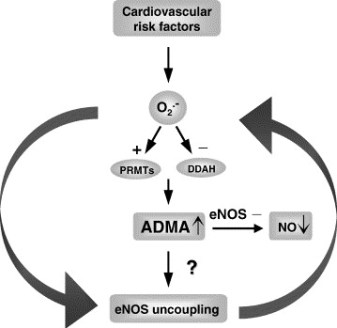
The LPA gene codes for apolipoprotein(a), which, when linked with low-density lipoprotein particles, forms lipoprotein(a) [Lp(a)] —

  • a well-studied molecule associated with coronary artery disease (CAD). The Lp(a) molecule has both atherogenic and thrombogenic effects in vitro , but the extent to which these translate to differences in how atherothrombotic disease presents is unknown.

LPA contains many single-nucleotide polymorphisms, and 2 have been identified by previous groups as being strongly associated with

  • levels of Lp(a) and, as a consequence, strongly associated with CAD.

However, because atherosclerosis is thought to be a systemic disease, it is unclear to what extent Lp(a) leads to atherosclerosis in other arterial beds (eg, carotid, abdominal aorta, and lower extremity),

  • as well as to other thrombotic disorders (eg, ischemic/cardioembolic stroke and venous thromboembolism).

Such distinctions are important, because therapies that might lower Lp(a) could potentially reduce forms of atherosclerosis beyond the coronary tree.

To answer this question, Helgadottir and colleagues compiled clinical and genetic data on the LPA gene from thousands of previous

  • participants in genetic research studies from across the world. They did not have access to Lp(a) levels, but by knowing the genotypes for
  • 2 LPA variants, they inferred the levels of Lp(a) on the basis of prior associations between these variants and Lp(a) levels. [1]

Their studies included not only individuals of white European descent but also a significant proportion of black persons, in order to

  • widen the generalizability of their results.

Their main findings are that LPA variants (and, by proxy, Lp(a) levels) are associated with

  • CAD,
  • peripheral arterial disease,
  • abdominal aortic aneurysm,
  • number of CAD vessels,
  • age at onset of CAD diagnosis, and
  • large-artery atherosclerosis-type stroke.

They did not find an association with

  • cardioembolic or small-vessel disease-type stroke;
  • intracranial aneurysm;
  • venous thrombosis;
  • carotid intima thickness; or,
  • in a small subset of individuals, myocardial infarction.

Apolipoprotein(a) Genetic Sequence Variants Associated With Systemic Atherosclerosis and Coronary Atherosclerotic Burden but Not With Venous Thromboembolism. Helgadottir A, Gretarsdottir S, Thorleifsson G, et al.    J Am Coll Cardiol. 2012;60:722-729

English: Structure of the LPA protein. Based o...

English: Structure of the LPA protein. Based on PyMOL rendering of PDB 1i71. (Photo credit: Wikipedia)

Micrograph of an artery that supplies the hear...

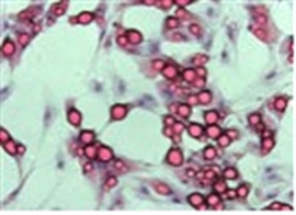
Micrograph of an artery that supplies the heart with significant atherosclerosis and marked luminal narrowing. Tissue has been stained using Masson’s trichrome. (Photo credit: Wikipedia)

Genomic Blueprint of the Heart

Scientists at the Gladstone Institutes have revealed the precise order and timing of hundreds of genetic “switches” required to construct a fully

  • functional heart from embryonic heart cells — providing new clues into the genetic basis for some forms of congenital heart disease.

In a study being published online today in the journal Cell, researchers in the laboratory of Gladstone Senior Investigator Benoit Bruneau, PhD,

  • employed stem cell technology, next-generation DNA sequencing and computing tools to piece together the instruction manual, or “genomic
  • blueprint” for how a heart becomes a heart. These findings offer renewed hope for combating life-threatening heart defects such as arrhythmias (irregular heart beat) and ventricular septal defects (“holes in the heart”).

ScienceDaily (Sep. 13, 2012)

They approach heart formation with a wide-angle lens by

  • looking at the entirety of the genetic material that gives heart cells their unique identity.

The news comes at a time of emerging importance for the biological process called “epigenetics,” in which a non-genetic factor impacts a cell’s genetic

  • makeup early during development — but sometimes with longer-term consequences. All of the cells in an organism contain the same DNA, but the
  • epigenetic instructions encoded in specific DNA sequences give the cell its identity. Epigenetics is of particular interest in heart formation, as the
  • incorrect on-and-off switching of genes during fetal development can lead to congenital heart disease — some forms of which may not be apparent until adulthood.

the scientists took embryonic stem cells from mice and reprogrammed them into beating heart cells by mimicking embryonic development in a petri dish. Next, they extracted the DNA from developing and mature heart cells, using an advanced gene-sequencing technique called ChIP-seq that lets scientists “see” the epigenetic signatures written in the DNA.

Map of Heart Disease Death Rates in US White M...

Map of Heart Disease Death Rates in US White Males from 2000-2004 (Photo credit: Wikipedia)

Estimated propability of death or non-fatal my...

Estimated propability of death or non-fatal myocardial-infarction over one year corresponding ti selectet values of the individual scores. Ordinate: individual score, abscissa: Propability of death or non-fatal myocardial infarction in 1 year (in %) (Photo credit: Wikipedia)

simply finding these signatures was only half the battle — we next had to decipher which aspects of heart formation they encoded

To do that, we harnessed the computing power of the Gladstone Bioinformatics Core. This allowed us to take the mountains of data collected from

  • gene sequencing and organize it into a readable, meaningful blueprint for how a heart becomes a heart.”

http://ScienceDaily.org/Scientists Map the Genomic Blueprint of the Heart.  ScienceDaily.

Performance of transcription factor identification tools from differential gene expression data

A three step process is a clear way to establish belief in the performance of transcription factor identification tools

  • from differential gene expression data.
  • identify several types of differential gene expression data sets where the stimulus or trigger is clearly know
  • identify the transcription factors most likely associated with the sets expression data.
  • perform an upstream analysis from the identified transcription factor.

If the transcription factor and upstream analysis tools can trace the signal cascade back to the stimulus, the tools are

  • clearly producing relevant results, and belief in the performance of the analysis tools is established.

At this point, the tools can be directed with confidence to more challenging analyses such as

  • developed resistance or pathway elucidation.

The performance of IPA‘s new Transcription Factor and Upstream analysis tools was evaluated on the following datasets (processing details below):

  • TGFb stimulation, 1 hour, A549 lung adenocarcinoma cell line
  • BMP2 stimulation, 1 hour, Mouse Embryonic Stem Cell E14Tg2A.4
  • TNFa stimulation, 1 hour primary murine hepatocytes

For each of the above datasets, an upstream analysis from the identified transcription factors correctly identified the stimulus. IPA’s tools were very

  • easy to use and the
  • analysis time for the above experiments was less than one minute.

The performance, speed, and ease of use can only be characterized as very good, perhaps leading to breakthroughs when extended and used creatively. Ingenuity’s new transcription factor analysis tool in IPA, coupled with Ingenuity’s established upstream grow tools,  should be strongly considered for every lab analyzing differential expression data.

http://www.ncbi.nlm.nih.gov/geo/query/acc.cgi?acc=GSE17896

http://www.ncbi.nlm.nih.gov/projects/geo/query/acc.cgi?acc=GSE2639

http://www.ncbi.nlm.nih.gov/geo/query/acc.cgi?acc=GSE19272

Differential expression data was obtained from CEL files using the Matlab functions:

affyrma, genelowvalfilter, genevarfilter, mattest, and mavolcanoplot.

Rick Stanton, Pathway Analysis Consultant Ingenuity.com

3. miR-200a regulates Nrf2 activation by targeting Keap1 mRNA in breast cancer cells.

Eades G, Yang M, Yao Y, Zhang Y, Zhou Q. J Biol Chem. 2011 Nov 25;286(47):40725-33. Epub 2011 Sep 16.
http://JBiolChem.com/miR-200a regulates Nrf2 activation by targeting Keap1 mRNA in breast cancer cells.

NF-E2-related factor 2 (Nrf2) is an important transcription factor that

  • activates the expression of cellular detoxifying enzymes.

Nrf2 expression is largely regulated through the association of Nrf2 with Kelch-like ECH-associated protein 1 (Keap1), which

  • results in cytoplasmic Nrf2 degradation.

Conversely, little is known concerning the regulation of Keap1 expression. Until now, a regulatory role for microRNAs (miRs) in controlling Keap1 gene expression had not been characterized. By using miR array-

  • based screening, we observed miR-200a silencing in breast cancer cells and
  • demonstrated that upon re-expression, miR-200a
  • targets the Keap1 3′-untranslated region (3′-UTR), leading to Keap1 mRNA degradation. Loss of this regulatory mechanism may
  • contribute to the dysregulation of Nrf2 activity in breast cancer. Previously, we have identified epigenetic repression of miR-200a

in breast cancer cells. Here, we find that treatment with epigenetic therapy, the histone deacetylase inhibitor suberoylanilide hydroxamic acid, restored miR-200a expression and reduced Keap1 levels. This reduction in Keap1 levels corresponded with

  • Nrf2 nuclear translocation
  • and activation of Nrf2-dependent NAD(P)H-quinone oxidoreductase 1 (NQO1) gene transcription.

Moreover, we found that Nrf2 activation inhibited the anchorage-independent growth of breast cancer cells. Finally, our in vitro observations were confirmed in a model of carcinogen-induced mammary hyperplasia in vivo. In conclusion, our study demonstrates

  • that miR-200a regulates the Keap1/Nrf2 pathway in mammary epithelium, and we find that epigenetic therapy can restore miR-200a
  • regulation of Keap1 expression,
  • reactivating the Nrf2-dependent antioxidant pathway in breast cancer.

Nuclear factor-like 2  (erythroid-derived 2, also known as NFE2L2 or Nrf2, is a transcription factor that in humans is encoded by the NFE2L2 gene.[1])  NFE2L2 induces the expression of various genes including those that encode for several antioxidant enzymes, and it may play a physiological role in the regulation of oxidative stress. Investigational drugs that target NFE2L2 are of interest as potential therapeutic interventions for

  • oxidative-stress related pathologies.

4. Highly active zinc finger nucleases by extended modular assembly

MS Bhakta, IM Henry, DG Ousterout, KT Das, et al.  Corresponding author; email: djsegal@ucdavis.edu
http://CSHNLpress.com/Highly active zinc finger nucleases by extended modular assembly

Zinc finger nucleases (ZFNs) are important tools for genome engineering. Despite intense interest by many academic groups,

  • the lack of robust non-commercial methods has hindered their widespread use. The modular assembly (MA) of ZFNs from
  • publicly-available one-finger archives provides a rapid method to create proteins that can recognize a very broad spectrum of DNA sequences.

However, three- and four-finger arrays often fail to produce active nucleases. Efforts to improve the specificity of the one-finger archives have not increased the success rate above 25%, suggesting that the MA method might

  • be inherently inefficient due to its insensitivity to context-dependent effects.

Here we present the first systematic study on the effect of array length on ZFN activity.  ZFNs composed of six-finger MA arrays produced mutations at 15 of 21 (71%) targeted

  • loci in human and mouse cells. A novel Drop-Out Linker scheme was used to rapidly assess three- to six-finger combinations,
  • demonstrating that shorter arrays could improve activity in some cases. Analysis of 268 array variants revealed that half of

MA ZFNs of any array composition that exceed an ab initio

  • B-score cut-off of 15 were active.
  • MA ZFNs are able to target more DNA sequences with higher success rates than other methods.

This article is distributed exclusively by Cold Spring Harbor Laboratory Press for the first six months after the full-issue publication date http://genome.cshlp.org/site/misc/terms.xhtml
After six months, it is available under a Creative Commons License (Attribution-NonCommercial 3.0 Unported License), as described at
http://creativecommons.org/licenses/by-nc/3.0/Highly_active_zinc_finger_nucleases_by_extended_ modular_assembly/

PERSONALIZED MEDICINE in the Pipeline

These insightful reviews are based on the strategic data and insights from Thomson Reuters Cortellis™ for Competitive Intelligence.  (A Review of April-June 2012).

http://ThomsonReuters.com/DIFFERENTIATED INNOVATION: PERSONALIZED MEDICINE IN THE PIPELINE/ Cortellis™ for Competitive Intelligence/APRIL-JUNE 2012

The majority of diseases are complex and multi-factorial, involving multiple genes interacting with environmental factors. At the genetic level,

  • information from genome-wide association studies that elucidate common patterns of genetic variation across various human populations,
  • in addition to profiling, technologies can be utilized in discovery research to provide snapshots of genes and expression profiles that are controlled
  • by the same regulatory mechanism and are altered between healthy and diseased states.

The characterization of genes that are abnormally expressed in disease tissues could further be employed as

  • diagnostic markers,
  • prognostic indicators of efficacy and/or toxicity, or as
  • targets for therapeutic intervention.

As the defining catalyst that exponentially paved the way for personalized medicine, information from the published genome sequence revealed that much of the genetic variations in humans are concentrated in about 0.1 percent of the over 3 billion base pairs in the haploid DNA. Most of these variations involve substitution of a single nucleotide for another at a given location in the genetic sequence, known as single nucleotide polymorphism (SNP).

  • Combinations of linked SNPs aggregate together to form haplotypes and
  • together these serve as markers for locating genetic variations in DNA sequences.

SNPs located within the protein-coding region of a gene or within the control regions of DNA that regulate a gene’s activity could

  • have a substantial effect on the encoded protein and thus influence phenotypic outcomes.

Analyzing SNPs between patient population cohorts could highlight specific genotypic variations which can be correlated with specific phenotypic variations in disease predisposition and drug responses.

Prior to the genomic revolution, many of the established therapies were directed against less than 500 drug targets, with many of the top selling drugs acting on well defined protein pathways. However, the sequencing of the human genome has massively expanded the pool of molecular targets that could be exploited in unmet medical needs and currently, of the approximately 22,300 protein-coding genes in the human code, it has been estimated that up to 3000 are druggable. Furthermore, genomic technologies such as

  • high-throughput sequencing
  • and transcription profiling,

can be used to identify and validate biologically relevant target molecules, or can be applied to cell-based and mice disease models or directly to in vivo human tissues,

  • helping to correlate gene targets with phenotypic traits of complex diseases.

This is particularly important, as

  • insufficient validation of target gene/proteins in complex diseases may be a contributing factor in the decline in R&D productivity.

Personalized medicine no doubt is already having a tremendous impact on drug development pipelines. According to a study conducted by the Tufts Center for the Study of Drug Development, more than 90 percent of biopharmaceutical companies now utilize at least some

  • genomics-derived targets in their drug discovery programs.

However, pipeline analysis from Cortellis for Competitive Intelligence suggests that there is still a scientific gap that has resulted in difficulty optimizing these novel genomic targets into the clinical R&D portfolios of major pharmaceutical companies, particularly outside the oncology field. Selected examples of personalized medicine product candidates in clinical development include (see TABLE 4).

Table 4: Selected Personalized Medicines in Clinical Development
(DATA are Derived from Cortellis for Competitive Intelligence & Thomson Reuters IntegritySM)
http://Thomson Reuters.com/Cortellis for Competitive Intelligence/IntegritySM/Table_4_Selected_Personalized_Medicines_in_Clinical_Development/

PHARMA MATTERS | SPOTLIGHT ON… PERSONALIZED MEDICINE

The paucity of actual targeted therapy examples, especially outside oncology, suggest

  • that integration of the personalized medicine paradigm into biopharmaceutical R&D is still fraught with challenges.

Despite the fact that the Human genome Project has been completed for over ten years, the broader application of genomics with drug development

  • still remains unrealized, and is hampered by a number of scientific challenges. One of the major obstacles stems from
  • incomplete association of genomic alterations with complex disease pathways and the phenotypic consequences.

As the modality of most complex diseases are multi-factorial, understanding how each genomic driver event plays a role in disease and the

  • interaction/interdependence with other genetic and environmental factors is important for
  • determining the rationale for targeted prevention or treatment of the disease.

Mutations found in Melanomas may shed light on Cancer Growth

Gina Kolata. New York Times.
http://NewYorkTimes.com/mutations_found_in_melanomas_may_shed-light_on_how_cancers_grow/

Mutations in Melanoma are in regions that control genes, not in the genes themselves. The mutations are exactly the type caused by exposure to ultraviolet light.  The findings are reported in two papers in http://Science.com/ScienceExpress/

The findings do not suggest new treatments, but they help explain how melanomas – and possibly – other cancers – develop and what drives their growth. This is a modification found in the “dark matter”, according to Dr. Levi A. Garraway,  the 99 percent of DNA in a region that regulates genes. A small control region was mutated in 7 out of 10 of the tumors, commonly of one or two tiny changes.
A German Team led by Rajiv Kumar (Heidelberg) and Dirk Schadendorf (Essen) looked at a family whose members tended to get melanomas.  Their findings indicate that those inherited with the mutations might be born with cells that have taken the first step toward cancer.
The mutations spur cells to make telomerase, that keeps the cells immortal by preventing them from losing the ends of their chromosome, the telomere. Abundant telomerase occurs in 90 percent of cancers, according to Immaculata De Vivo at Harvard Medical School.
The importance of the findings is that the mechanism of telomerase involvement in cancer is now within view. But it is not clear how to block the telomerase production in cancer cells.
 
A slight mutation in the matched nucleotides c...

A slight mutation in the matched nucleotides can lead to chromosomal aberrations and unintentional genetic rearrangement. (Photo credit: Wikipedia)


Comment

This discussion addresses the issues raised about the direction to follow in personalized medicine. Despite the amount of work necessary to bring the clarity that is sought after, the experiments and experimental design is most essential.

  • The arrest of ciliogenesis in ovarian cancer cell lines compared to wild type (WT) ovarian epithelial cells, and
  •  The link to suppressing ciliogenesis by AURA protein and CHFR at the base of the cilium, which disappears at mitosis or with proliferation.
  •  There is no accumulation by upregulation of PDGF under starvation by the cancer cells compared to the effect in WT OSE.

Here we have a systematic combination of signaling events tied to changes in putative biomarkers that occur synchronously in Ov cancer cell lines.

These changes are identified with changes in

  • proliferation,
  • loss of ciliary structure, and
  • proliferation.

In this described scenario,

  • WT OSE cells would be arrested, and
  • it appears that they would take the path to apoptosis (under starvation).

Even without more information, this cluster is what one wants to have in a “syndromic classification”. The information used to form the classification entails the identification of strong ‘signaling-related’ biomarkers. The Gli2 peptide has to be part of this.

In principle, a syndromic classification would be ideally expected to have no less than 64 classes. If the classification is “weak”, then the class frequencies would be close to what one would expect in the WT OSE. In this case, in reality,

  • several combinatorial classes would have low frequency, and
  • others would be quite high.

This obeys the classification rules established by feature identification, and the information gain described by Solomon Kullback and extended by Akaike.

Does this have to be the case for all different cancer types? I don’t think so. The cells are different in ontogenesis.  In this case, even the WT OSE have mesenchymal features and so, are not fully directed to epithelial expression.  This happens to be the case in actual anatomic expression of the ovary.  On the other hand, one would expect shared features of the

  • ovary,
  • testes,
  • thyroid,
  • adrenals, and
  • pituitary.

There is biochemical expression in terms of their synthetic function – TPN organs. I would have to put the liver into that broad class. Other organs – skeletal muscle & heart – transform substrate into energy or work.  (Where you might also put intestinal smooth muscle).

They have to have different biomarker expressions, even though they much less often don’t form neoplasms. (Bone is not just a bioenergetic force. It is maintained by muscle action. It forms sarcomas. But there has to be a balance between bone removal by osteoclasts and refill by osteoblasts.)

Viewpoint: What we have learned

  1. The Watson-Crick model proposed in 1953 is limited for explaining fully genome effects
  2. The Pauling triplex model may have been prescient because of a more full anticipation of molecular bonding variants
  3. A more adequate triple-helix model has been proposed and is consistent with a compact genome in the nucleus

The structure of the genome is not as we assumed – based on the application of Fractal Geometry.  Current body of evidence is building that can reveal a more complete view of genome function.

  • transcription
  • cell regulation
  • mutations

Summary

I have just completed a most comprehensive review of the Human Genome Project. There are key research collaborations, problems in deciphering the underlying structure of the genome, and there are also both obstacles and insights to elucidating the complexity of the final model.

This is because of frequent observations of molecular problems in folding and other interactions between nucleotides that challenge the sufficiency of the original DNA model proposed by Watson and Crick. This has come about because of breakthrough innovation in technology and in computational methods.

Radoslav Bozov •

Molecular biology and growth was primarily initiated on biochemical structural paradigms aiming to define functional spatial dynamics of molecules via assignation of various types of bondings – covalent and non-covalent – hydrogen, ionic , dipole-dipole, hydrophobic interactions.

  • Lab techniques based on z/m paradigm allowed separation, isolation and identification of bio substances with a general marker identity finding correlation between physiological/cellular states.
  • The development of electronic/x-ray technologies allowed zooming in nano space without capturing time.
  • NMR technology identified the existence of space topology of initial and final atomic states giving a highly limited light on time – energy axis of atomic interactions.
  • Sequence technology and genomic perturbations shed light on uncertainty of genomic dynamics and regulators of functional ever expanding networks.
  • Transition state theory coupled to structural complexity identification and enzymatic mechanisms ran up parallel to work on various phenomena of strings of nucleotides (oligomers and polymers) – illusion/observation of constructing models on the dynamics of protein-dna-rna interference.
  • The physical energetic constrains of biochemistry were inapplicable in open biological systems. Biologists have accepted observation as a sole driver towards re-evaluating models.
  • The separation of matter and time constrains emerged as deviation of energy and space constrains transforming into the full acceptance of code theory of life. One simple thing was left unnoticed over time –
  • the amount of information of quantum matter within a single codon is larger than that of a single amino acid. This violated all physical laws/principles known to work with a limited degree of certainty.
  • The limited amount of information analyzed by conventional sequence identity led to the notion of applicability of statistical measures of and PCR technology. Mutations were identified over larger scale of data.
  • Quantum chemistry itself is being limited due discrete space/energy constrains, thus it transformed into concepts/principles in biology that possess highly limited physical values whatsoever.
  • The central dogma is partially broken as a result of
  1. regulatory constrains
  2. epigenetic phenomena and
  3. iRNA.

Large scale code computational data run into uncertainty of the processes of evolution and its consequence of signaling transformation. All drugs were ‘lucky based’ applicability and/or discovery with largely unpredictable side effect over time.

Other Related articles on this Open Access Online Sceintific Journal include the following:

Big Data in Genomic Medicine  lhb

http://pharmaceuticalintelligence.com/2012/12/17/big-data-in-genomic-medicine/

BRCA1 a tumour suppressor in breast and ovarian cancer – functions in transcription, ubiquitination and DNA repair S Saha    http://pharmaceuticalintelligence.com/2012/12/04/brca1-a-tumour-suppressor-in-breast-and-ovarian-cancer-functions-in-transcription-ubiquitination-and-dna-repair/

Computational Genomics Center: New Unification of Computational Technologies at Stanford A Lev-Ari  http://pharmaceuticalintelligence.com/2012/12/03/computational-genomics-center-new-unification-of-computational-technologies-at-stanford/

Personalized medicine gearing up to tackle cancer ritu saxena     http://pharmaceuticalintelligence.com/2013/01/07/personalized-medicine-gearing-up-to-tackle-cancer/

Differentiation Therapy – Epigenetics Tackles Solid Tumors sj Williams     http://pharmaceuticalintelligence.com/2013/01/03/differentiation-therapy-epigenetics-tackles-solid-tumors/

Mechanism involved in Breast Cancer Cell Growth: Function in Early Detection & Treatment A Lev-Ari   http://pharmaceuticalintelligence.com/2013/01/17/mechanism-involved-in-breast-cancer-cell-growth-function-in-early-detection-treatment/

The Molecular pathology of Breast Cancer Progression tilde barliya      http://pharmaceuticalintelligence.com/2013/01/10/the-molecular-pathology-of-breast-cancer-progression/

Gastric Cancer: Whole-genome reconstruction and mutational signatures A Lev-Ari     http://pharmaceuticalintelligence.com/2012/12/24/gastric-cancer-whole-genome-reconstruction-and-mutational-signatures-2/

Paradigm Shift in Human Genomics – Predictive Biomarkers and Personalized Medicine – Part 1 (pharmaceuticalintelligence.com) A Lev-Ari                  http://pharmaceuticalintelligence.com/2013/01/13/paradigm-shift-in-human-genomics-predictive-biomarkers-and-personalized-medicine-part-1/

LEADERS in Genome Sequencing of Genetic Mutations for Therapeutic Drug Selection in Cancer Personalized Treatment: Part 2 A Lev-Ari
http://pharmaceuticalintelligence.com/2013/01/13/leaders-in-genome-sequencing-of-genetic-mutations-for-therapeutic-drug-selection-in-cancer-personalized-treatment-part-2/

Personalized Medicine: An Institute Profile – Coriell Institute for Medical Research: Part 3 A Lev-Ari   http://pharmaceuticalintelligence.com/2013/01/13/personalized-medicine-an-institute-profile-coriell-institute-for-medical-research-part-3/

Harnessing Personalized Medicine for Cancer Management, Prospects of Prevention and Cure: Opinions of Cancer Scientific Leaders @ http://pharmaceuticalintelligence.com ALA    http://pharmaceuticalintelligence.com/2013/01/13/7000/Harnessing Personalized Medicine for Cancer Management, Prospects of Prevention and Cure: Opinions of Cancer Scientific Leaders/

GSK for Personalized Medicine using Cancer Drugs needs Alacris systems biology model to determine the in silico effect of the inhibitor in its “virtual clinical trial” A Lev-Ari     http://pharmaceuticalintelligence.com/2012/11/14/gsk-for-personalized-medicine-using-cancer-drugs-needs-alacris-systems-biology-model-to-determine-the-in-silico-effect-of-the-inhibitor-in-its-virtual-clinical-trial/

Recurrent somatic mutations in chromatin-remodeling and ubiquitin ligase complex genes in serous endometrial tumors S Saha   http://pharmaceuticalintelligence.com/2012/11/19/recurrent-somatic-mutations-in-chromatin-remodeling-and-ubiquitin-ligase-complex-genes-in-serous-endometrial-tumors/

Personalized medicine-based cure for cancer might not be far away ritu saxena   http://pharmaceuticalintelligence.com/2012/11/20/personalized-medicine-based-cure-for-cancer-might-not-be-far-away/

Human Variome Project: encyclopedic catalog of sequence variants indexed to the human genome sequence A Lev-Ari
http://pharmaceuticalintelligence.com/2012/11/24/human-variome-project-encyclopedic-catalog-of-sequence-variants-indexed-to-the-human-genome-sequence/

Prostate Cancer Cells: Histone Deacetylase Inhibitors Induce Epithelial-to-Mesenchymal Transition sjwilliams
http://pharmaceuticalintelligence.com/2012/11/30/histone-deacetylase-inhibitors-induce-epithelial-to-mesenchymal-transition-in-prostate-cancer-cells/

Inspiration From Dr. Maureen Cronin’s Achievements in Applying Genomic Sequencing to Cancer Diagnostics A Lev-Ari
http://pharmaceuticalintelligence.com/2013/01/10/inspiration-from-dr-maureen-cronins-achievements-in-applying-genomic-sequencing-to-cancer-diagnostics/

The “Cancer establishments” examined by James Watson, co-discoverer of DNA w/Crick, 4/1953 A Lev-Ari
http://pharmaceuticalintelligence.com/2013/01/09/the-cancer-establishments-examined-by-james-watson-co-discover-of-dna-wcrick-41953/

Directions for genomics in personalized medicine lhb    http://pharmaceuticalintelligence.com/2013/01/27/directions-for-genomics-in-personalized-medicine/

How mobile elements in “Junk” DNA promote cancer. Part 1: Transposon-mediated tumorigenesis. Sjwilliams
http://pharmaceuticalintelligence.com/2012/10/31/how-mobile-elements-in-junk-dna-prote-cancer-part1-transposon-mediated-tumorigenesis/

Mitochondria: More than just the “powerhouse of the cell” eritu saxena   http://pharmaceuticalintelligence.com/2012/07/09/mitochondria-more-than-just-the-powerhouse-of-the-cell/

Mitochondrial fission and fusion: potential therapeutic targets? Ritu saxena    http://pharmaceuticalintelligence.com/2012/10/31/mitochondrial-fission-and-fusion-potential-therapeutic-target/

Mitochondrial mutation analysis might be “1-step” away ritu saxena     http://pharmaceuticalintelligence.com/2012/08/14/mitochondrial-mutation-analysis-might-be-1-step-away/

mRNA interference with cancer expression lhb    http://pharmaceuticalintelligence.com/2012/10/26/mrna-interference-with-cancer-expression/

Read Full Post »

2013 Genomics: The Era Beyond the Sequencing of the Human Genome: Francis Collins, Craig Venter, Eric Lander, et al.

Curator: Aviva Lev-Ari, PhD, RN This image has an empty alt attribute; its file name is ArticleID-23.png

Reporter:  Aviva Lev-Ari, PhD, RN

Call for Open-Access Publishing in Genomics

January 14, 2013

SAN DIEGO (GenomeWeb News) – Open-access datasets, software, and bioinformatics strategies have become more or less de rigueur in genomics research.

But the field may also be poised to change the way other sorts of information from scientific studies is conveyed to other researchers and to the broader public, according to open-access proponent Michael Eisen, a computational and evolutionary biology researcher at the University of California, Berkeley.

Eisen, a Public Library of Science co-founder, spoke during the morning plenary session here at the International Plant and Animal Genome Conference.

In his presentation, he argued that the inability to freely and unreservedly access the full text of all genome studies performed to date may have led to missed opportunities for the field.

Using the bacteriophage phiX174 genome sequence as an example, he proposed that the general thinking in the genomics field has developed in ways that promote open-access to sequence data and related software. But, he said, the same type of access is not necessarily available for those interested in delving into the details and rationale behind genomics studies, since the corresponding papers may not be accessible in an open-access format.

The UK Medical Research Council Laboratory of Molecular Biology‘s Frederick Sanger and colleagues described the phiX174 sequence in 1977, in a publication that’s generally considered to be the first genome paper. The sequence data presented in that study is now freely available, Eisen explained, in part owing to the advent of sequence databases such as the European Molecular Biology Laboratory Nucleotide Sequence database or the National Center for Biotechnology Information’s sequence database, GenBank.

During the past decade or more, funding agency requirements and pressure from within the genomics community have contributed to the widespread adoption of these and other public genomics resources and repositories.

As these databases have grown and become accepted within the genomics community, Eisen argued that they have spurred the development of computational methods for analyzing genome sequences and datasets that may not have existed otherwise. “Imagine where we would be had we not made the fortunate decision to liberate genome sequences,” he said.

But analogous strategies for combing through text from genomics studies in their entirety have not developed in the same manner, according to Eisen, who noted that the text of the phiX174 genome paper remains behind a pay wall.

“We’ve allowed [journal access] and [data access] to follow very different fates,” said Eisen, who says there are ways to use the information housed within the scientific literature more easily and productively.

He urged attendees to consider publishing their own work in open-access publications. Beyond that, though, Eisen also noted that the community is well positioned to influence the ways in which research information is disseminated, since genomics data increasingly serves as a resource for other spheres of research.


Read Full Post »

Consumer Market for Personal DNA Sequencing: Part 4

Reporter: Aviva Lev-Ari, PhD RN

FDA Warning for the Leader of Consumer Market for Personal DNA Sequencing Part 4

Word Cloud by Daniel Menzin

This Part 4 of the series on Present and Future Frontier of Research in Genomics has been 

UPDATED on 12/6/2013

23andMe Suspends Health Interpretations

December 06, 2013

Direct-to-consumer genetic testing company 23andMe hasstopped offering its health-related test to new customers, bringing it in line with a request from the US Food and Drug Administration.

In letter sent on Nov. 22, FDA said that 23andMe had not adequately responded to its concerns regarding the validity of their Personal Genome Service. The letter instructed 23andMe to “immediately discontinue marketing” the service until it receives authorization from the agency.

According to a post at the company’s blog from CEO Anne Wojcicki, 23andMe customers who purchased their kits on or after Nov. 22 “will not have access to health-related results.” They will, though, have access to ancestry information and their raw genetic data. Wojcicki notes that the customers may have access to the health interpretations in the future depending on FDA marketing authorization. Those customers are also being offered a refund.

Customers who purchased their kits before Nov. 22 will have access to all reports.

“We remain firmly committed to fulfilling our long-term mission to help people everywhere have access to their own genetic data and have the ability to use that information to improve their lives,” a notice at the 23andMe site says.

In a letter appearing in the Wall Street Journal earlier this week, FDA Commissioner Margaret Hamburg wrote that the agency “supports the development of innovative tests.” As an example, she pointed to its recent clearance of sequencing-based testsfrom Illumina.

She added that the agency also understands that some consumers do want to know more about their genomes and their genetic risk of disease, and that a DTC model would let consumers take an active role in their health.

“The agency’s desire to review these particular tests is solely to ensure that they are safe, do what they claim to do and that the results are communicated in a way that a consumer can understand,” Hamburg said.

In a statement, 23andMe’s Wojcicki says that the company remains committed to its ethos of allowing people access to their genetic information. “Our goal is to work cooperatively with the FDA to provide that opportunity in a way that clearly demonstrates the benefit to people and the validity of the science that underlies the test,” Wojcicki adds.

SOURCE

UPDATED on 11/27/2013

FDA Tells Google-Backed 23andMe to Halt DNA Test Service

VIEW VIDEO

http://www.bloomberg.com/news/2013-11-25/fda-tells-google-backed-23andme-to-halt-dna-test-service.html

FDA Letter to 23andME

Department of Health and Human Services logoDepartment of Health and Human Services

Public Health Service
Food and Drug Administration
10903 New Hampshire Avenue
Silver Spring, MD 20993

Nov 22, 2013

Ann Wojcicki
CEO
23andMe, Inc.
1390 Shoreline Way
Mountain View, CA 94043
Document Number: GEN1300666
Re: Personal Genome Service (PGS)
WARNING LETTER
Dear Ms. Wojcicki,
The Food and Drug Administration (FDA) is sending you this letter because you are marketing the 23andMe Saliva Collection Kit and Personal Genome Service (PGS) without marketing clearance or approval in violation of the Federal Food, Drug and Cosmetic Act (the FD&C Act).
This product is a device within the meaning of section 201(h) of the FD&C Act, 21 U.S.C. 321(h), because it is intended for use in the diagnosis of disease or other conditions or in the cure, mitigation, treatment, or prevention of disease, or is intended to affect the structure or function of the body. For example, your company’s website at http://www.23andme.com/health (most recently viewed on November 6, 2013) markets the PGS for providing “health reports on 254 diseases and conditions,” including categories such as “carrier status,” “health risks,” and “drug response,” and specifically as a “first step in prevention” that enables users to “take steps toward mitigating serious diseases” such as diabetes, coronary heart disease, and breast cancer. Most of the intended uses for PGS listed on your website, a list that has grown over time, are medical device uses under section 201(h) of the FD&C Act. Most of these uses have not been classified and thus require premarket approval or de novo classification, as FDA has explained to you on numerous occasions.
Some of the uses for which PGS is intended are particularly concerning, such as assessments for BRCA-related genetic risk and drug responses (e.g., warfarin sensitivity, clopidogrel response, and 5-fluorouracil toxicity) because of the potential health consequences that could result from false positive or false negative assessments for high-risk indications such as these. For instance, if the BRCA-related risk assessment for breast or ovarian cancer reports a false positive, it could lead a patient to undergo prophylactic surgery, chemoprevention, intensive screening, or other morbidity-inducing actions, while a false negative could result in a failure to recognize an actual risk that may exist. Assessments for drug responses carry the risks that patients relying on such tests may begin to self-manage their treatments through dose changes or even abandon certain therapies depending on the outcome of the assessment. For example, false genotype results for your warfarin drug response test could have significant unreasonable risk of illness, injury, or death to the patient due to thrombosis or bleeding events that occur from treatment with a drug at a dose that does not provide the appropriately calibrated anticoagulant effect. These risks are typically mitigated by International Normalized Ratio (INR) management under a physician’s care. The risk of serious injury or death is known to be high when patients are either non-compliant or not properly dosed; combined with the risk that a direct-to-consumer test result may be used by a patient to self-manage, serious concerns are raised if test results are not adequately understood by patients or if incorrect test results are reported.
Your company submitted 510(k)s for PGS on July 2, 2012 and September 4, 2012, for several of these indications for use. However, to date, your company has failed to address the issues described during previous interactions with the Agency or provide the additional information identified in our September 13, 2012 letter for(b)(4) and in our November 20, 2012 letter for (b)(4), as required under 21 CFR 807.87(1). Consequently, the 510(k)s are considered withdrawn, see 21 C.F.R. 807.87(1), as we explained in our letters to you on March 12, 2013 and May 21, 2013.  To date, 23andMe has failed to provide adequate information to support a determination that the PGS is substantially equivalent to a legally marketed predicate for any of the uses for which you are marketing it; no other submission for the PGS device that you are marketing has been provided under section 510(k) of the Act, 21 U.S.C. § 360(k).
The Office of In Vitro Diagnostics and Radiological Health (OIR) has a long history of working with companies to help them come into compliance with the FD&C Act. Since July of 2009, we have been diligently working to help you comply with regulatory requirements regarding safety and effectiveness and obtain marketing authorization for your PGS device. FDA has spent significant time evaluating the intended uses of the PGS to determine whether certain uses might be appropriately classified into class II, thus requiring only 510(k) clearance or de novo classification and not PMA approval, and we have proposed modifications to the device’s labeling that could mitigate risks and render certain intended uses appropriate for de novo classification. Further, we provided ample detailed feedback to 23andMe regarding the types of data it needs to submit for the intended uses of the PGS.  As part of our interactions with you, including more than 14 face-to-face and teleconference meetings, hundreds of email exchanges, and dozens of written communications, we provided you with specific feedback on study protocols and clinical and analytical validation requirements, discussed potential classifications and regulatory pathways (including reasonable submission timelines), provided statistical advice, and discussed potential risk mitigation strategies. As discussed above, FDA is concerned about the public health consequences of inaccurate results from the PGS device; the main purpose of compliance with FDA’s regulatory requirements is to ensure that the tests work.
However, even after these many interactions with 23andMe, we still do not have any assurance that the firm has analytically or clinically validated the PGS for its intended uses, which have expanded from the uses that the firm identified in its submissions. In your letter dated January 9, 2013, you stated that the firm is “completing the additional analytical and clinical validations for the tests that have been submitted” and is “planning extensive labeling studies that will take several months to complete.” Thus, months after you submitted your 510(k)s and more than 5 years after you began marketing, you still had not completed some of the studies and had not even started other studies necessary to support a marketing submission for the PGS. It is now eleven months later, and you have yet to provide FDA with any new information about these tests.  You have not worked with us toward de novo classification, did not provide the additional information we requested necessary to complete review of your 510(k)s, and FDA has not received any communication from 23andMe since May. Instead, we have become aware that you have initiated new marketing campaigns, including television commercials that, together with an increasing list of indications, show that you plan to expand the PGS’s uses and consumer base without obtaining marketing authorization from FDA.
Therefore, 23andMe must immediately discontinue marketing the PGS until such time as it receives FDA marketing authorization for the device. The PGS is in class III under section 513(f) of the FD&C Act, 21 U.S.C. 360c(f). Because there is no approved application for premarket approval in effect pursuant to section 515(a) of the FD&C Act, 21 U.S.C. 360e(a), or an approved application for an investigational device exemption (IDE) under section 520(g) of the FD&C Act, 21 U.S.C. 360j(g), the PGS is adulterated under section 501(f)(1)(B) of the FD&C Act, 21 U.S.C. 351(f)(1)(B).  Additionally, the PGS is misbranded under section 502(o) of the Act, 21 U.S.C. § 352(o), because notice or other information respecting the device was not provided to FDA as required by section 510(k) of the Act, 21 U.S.C. § 360(k).
Please notify this office in writing within fifteen (15) working days from the date you receive this letter of the specific actions you have taken to address all issues noted above. Include documentation of the corrective actions you have taken. If your actions will occur over time, please include a timetable for implementation of those actions. If corrective actions cannot be completed within 15 working days, state the reason for the delay and the time within which the actions will be completed. Failure to take adequate corrective action may result in regulatory action being initiated by the Food and Drug Administration without further notice. These actions include, but are not limited to, seizure, injunction, and civil money penalties.
We have assigned a unique document number that is cited above. The requested information should reference this document number and should be submitted to:
James L. Woods, WO66-5688
Deputy Director
Patient Safety and Product Quality
Office of In vitro Diagnostics and Radiological Health
10903 New Hampshire Avenue
Silver Spring, MD 20993
If you have questions relating to this matter, please feel free to call Courtney Lias, Ph.D. at 301-796-5458, or log onto our web site at www.fda.gov for general information relating to FDA device requirements.
Sincerely yours,
/S/
Alberto Gutierrez
Director
Office of In vitro Diagnostics
and Radiological Health
 Center for Devices and Radiological Health

SOURCE

http://www.fda.gov/ICECI/EnforcementActions/WarningLetters/2013/ucm376296.htm

Cancer Diagnostics by Genomic Sequencing: ‘No’ to Sequencing Patient’s DNA, ‘No’ to Sequencing Patient’s Tumor, ‘Yes’ to focus on Gene Mutation Aberration & Analysis of Gene Abnormalities

Symposia

http://aaas.confex.com/aaas/2013/webprogram/start.html

Personal Genetics: An Intersection Between Science, Society, and Policy

Saturday, February 16, 2013: 8:30 AM-11:30 AM

Room 203 (Hynes Convention Center)

On 26 June 2000, scientists announced the completion of a rough draft of the human genome, the result of the $3 billion publicly funded Human Genome Project. In the decade since, the cost of genome sequencing has plummeted, coinciding with the development of deep sequencing technologies and allowing, for the first time, personalized genetic medicine. The advent of personal genetics has profound implications for society that are only beginning to be discussed, even as the technologies are rapidly maturing and entering the market. This symposium will focus on how the genomic revolution may affect our society in coming years and how best to reach out to the general public on these important issues. How has the promise of genomics, as stated early in the last decade, matched the reality we observe today? What are the new promises — and pitfalls — of genomics and personal genetics as of 2013? What are the ethical implications of easy and inexpensive human genome sequencing, particularly with regard to ownership and control of genomic datasets, and what stakeholder interests must be addressed? How can the scientific community engage with the public at large to improve understanding of the science behind these powerful new technologies? The symposium will comprise three 15-minute talks from representatives of relevant sectors (academia/education, journalism, and industry), followed by a 45-minute panel discussion with the speakers.

Organizer:

Peter Yang, Harvard University

Co-organizers:

Brenna Krieger, Harvard University

and Kevin Bonham, Harvard University

Discussant:

James Thornton, Harvard University

Speakers:


Ting Wu, Harvard University

Personal Genetics and Education

Mary Carmichael, Boston Globe

The Media and the Personal Genetics Revolution

Brian Naughton, 23andMe Inc.

Commercialization of Personal Genomics: Promise and Potential Pitfalls

Mira Irons, Children’s Hospital Boston

Personal Genomic Medicine: How Physicians Can Adapt to a Genomic World

Sheila Jasanoff, Harvard University

Citizenship and the Personal Genomics

Jonathan Gitlin, National Human Genome Research Institute

Personal Genomics and Science Policy

THIS IS A SERIES OF FOUR POINTS OF VIEW IN SUPPORT OF the Paradigm Shift in Human Genomics

How to Tailor Cancer Therapy to the particular Genetics of a patient’s Cancer

‘No’ to Sequencing Patient’s DNA, ‘No’ to Sequencing Patient’s Tumor, ‘Yes’ to focus on Gene Mutation Aberration & Analysis of Gene Abnormalities PRESENTED in the following FOUR PARTS. Recommended to be read in its entirety for completeness and arrival to the End Point of Present and Future Frontier of Research in Genomics

Part 1:

Research Paradigm Shift in Human Genomics – Predictive Biomarkers and Personalized Medicine

http://pharmaceuticalintelligence.com/2013/01/13/paradigm-shift-in-human-genomics-predictive-biomarkers-and-personalized-medicine-part-1/

Part 2:

LEADERS in the Competitive Space of Genome Sequencing of Genetic Mutations for Therapeutic Drug Selection in Cancer Personalized Treatment

http://pharmaceuticalintelligence.com/2013/01/13/leaders-in-genome-sequencing-of-genetic-mutations-for-therapeutic-drug-selection-in-cancer-personalized-treatment-part-2/

Part 3:

Personalized Medicine: An Institute Profile – Coriell Institute for Medical Research

http://pharmaceuticalintelligence.com/2013/01/13/personalized-medicine-an-institute-profile-coriell-institute-for-medical-research-part-3/

Part 4:

The Consumer Market for Personal DNA Sequencing


Part 4:

The Consumer Market for Personal DNA Sequencing

How does 23andMe genotype my DNA?

Technology and Standards

23andMe is a DNA analysis service providing information and tools for individuals to learn about and explore their DNA. We use the Illumina OmniExpress Plus Genotyping BeadChip (shown here). In addition to the variants already included on the chip by Illumina, we’ve included our own, customized set of variants relating to conditions and traits that are interesting. Technical information on the performance of the chip can be found on Illumina’s website.

All of the laboratory testing for 23andMe is done in a CLIA-certified laboratory.

Once our lab receives your sample, DNA is extracted from cheek cells preserved in your saliva. The lab then copies the DNA many times — a process called “amplification” — growing the tiny amount extracted from your saliva until there is enough to be genotyped.

In order to be genotyped, the amplified DNA is “cut” into smaller pieces, which are then applied to our DNA chip, a small glass slide with millions of microscopic “beads” on its surface (read more about this technology). Each bead is attached to a “probe”, a bit of DNA that matches one of the approximately one million genetic variants that we test. The cut pieces of your DNA stick to the matching DNA probes. A fluorescent signal on each probe provides information that can tell us which version of that genetic variant your DNA corresponds to.

Although the human genome is estimated to contain about 10-30 million genetic variants, many of them are correlated due to their proximity to each other. Thus, one genetic variant is often representative of many nearby variants, and the approximately one million variants on our genotyping chip provide very good coverage of common variation across the entire genome.

Our research team has also hand-picked tens of thousands of additional genetic variants linked to various conditions and traits in the scientific literature to analyze on our genotyping chip. As a result we can provide you with personal genetic information available only through 23andMe.

Genetics service 23andMe announced some new cash in the bank today with a $50 million raise from Yuri Milner, 23andMe CEO Anne Wojcicki, Google’s Sergey Brin (who also happens to be Wojcicki’s husband), New Enterprise Associates, MPM Capital, and Google Ventures.

With today’s new funding also comes the reduction of the price of its genome analysis service to $99. This isn’t special holiday pricing (as 23andMe has run repeatedly in the past) the company tells me, but rather what its normal pricing will be from now on.

This move is overdue, at least as far as 23andMe’s business model is concerned. Just yesterday TechCrunch Conference Chair Susan Hobbs told me she was waiting for another $99 pricing deal to buy the Personal Genome Analysis product. Sure 23andMe has experimented with various pricing models, including subscription, since its founding in 2007, but had been at an official and prohibitive $299 price point until today. It’s also apparently been rigorously beta-testing various price points in the past couple of weeks, at some point experimenting with some lower than $99.

For comparison, the company’s original pricing began at $999 and offered subscribers just 14 health and trait reports versus today’s 244 reports, as well as genetic ancestry information. Natera, Counsyl and Pathway Genomics are also in the genomics space, but they work by offering their services through doctors rather than direct to consumer.

Since the company’s launch five years ago, it’s had 180K civilians profile their DNA, and representative Catherine Afarian tells us that, post-price drop and funding, its goal is to reach a million customers in 2013. This is a supremely ambitious goal considering it wants to turn an average user acquisition rate of 36K per year into one of 820K in one year alone.

But Afarian isn’t fazed and brings up how the company once sold out 20k in $99 account inventory on something called “DNA Day.” “Once we can offer the service at $99 it means the average American will buy in,” she said.

That $299 was too pricey, according to Hobbs, but $99 might be just right. She said the $99 price point, which yes, is less than an iPhone, was the main factor in her decision to buy in. “23andMe is more ‘nice-to-know’ information rather than ‘need-to-know’ information. It’s nice to know your ancestry. It’s more of a need to know that you are predisposed genetically for a type of cancer, so that you may take precautionary measures,” she said, implying that the data given by 23andMe isn’t necessarily vital medical information, or actionable when it is. While 23andMe can give you indicators about certain disease risks, it doesn’t close the loop, as in tell you what to do to prevent these diseases.

“Its [utility] depends on your genetic data,” said Afarian when I asked her about the usefulness of the product. “If you’ve got a Factor 5 that puts you at risk for clotting, you might want to invest in anti-clotting socks. [And] there’s always something about themselves that people didn’t know.”

Hobbs said eventually that she wouldn’t buy it, but only because she was looking into more exact lineage information for her little girl, and you need a Y chromosome in all DNA tests to show paternal lineage. Afarian also countered this hesitation, saying that what makes 23andMe unique is that it’s not only looking at just your Y or your mitochondrial DNA, but also your autosomal DNA, which does show some patrilineal information for females who lack that precious Y.

While still sort of a novelty, the potential for 23andMe goes beyond lineage and hopefully that extra $50 million will go further than keeping the price low and into research. The company hopes that a million users will result in a giant database of 23andWe genetic info that can be used to spot trends, like which genes mean a higher risk of diabetes/cancer, etc. Which is great if it happens but for now remains a pipe dream for 23andMe/We.

http://techcrunch.com/2012/12/11/23andnotme/

12/13/2012 @ 5:23PM |6,471 views

What Is 23andMe Really Selling: The Moral Quandary At The Center Of The Personalized Genomics Revolution

This week, 23andme, the personalized genomics company founded by Anne Wojcicki, wife of Google co-founder Sergey Brin, got an influx of investment cash ($50 million). According to their press release, they are using the money to bring the cost of their genetic test down to $99 (it was previously $299) which, they hope, will inspire the masses to get tested.

So should the masses indulge?

I prefer a quantified self approach to this question. At the heart of the quantified self-movement lies a very simple idea: metrics make us better. For devotees, this means “self-tracking,” using everything from the Nike fuel band to the Narcissism Personality Index to gather large quantities of personal data and—the bigger idea—use that data to improve performance.

If you consider that performance suffers when health suffers then a genetic test can been seen as a kind of metric used to improve performance. This strikes me as the best way to evaluate this idea and leads us to ask the same question about personalized genomics that the quantified self movement asks about every other metric: will it improve performance.

Arguments rage all over the place on this one, but the short answer is that SNP tests—which is the kind of DNA scan 23andme relies upon— don’t tell us all that much (yet).  They analyze a million genes out of three billion total and the impact those million play in long term-health outcomes is still in dispute. For example, the nature/nurture split is normally viewed at 30/70—meaning environmental factors play a far more significant role in long-term health outcomes than genetics.

Moreover, all of the performance metrics used by the quantified self movement are used to for behavior modification—to drive self-improvement. Personalized genomics isn’t there yet. As Stanford University’s Nobel Prize-winning RNA researcher Andy Fire once told me, “if someone off the street is looking for pointers on how to live a healthier life, there’s nothing these tests will tell you besides basic physician advice like ‘eat right, don’t smoke and get plenty of exercise.’”

And even with more well-regarded SNP tests, like the ones that examine the BRCA 1 and 2 markers for breast cancer—which  . NYU Langone Medical Center bioethicist Arthur Caplan explains it like this, “Say you test positive for a breast cancer disposition—then what are you going to do? The only preventative step you can take is to chop off your breasts.”

So if prevention is not available the only thing left is fear and anxiety. Unfortunately, in the past few decades, there have been hundreds of studies linking stress to everything from immunological disorders to heart disease to periodonitic troubles. So while finding out you may be at risk for Parkinson’s may make you feel informed, that knowledge isn’t going to stop you from developing the disease—but the resulting stress may contribute to a host of other complications.

This brings up a different question: if personalized genomics can’t yet help us much and could possibly hurt us—where’s the upside?

Turns out there’s a big upside: Citizen science. SNP tests are not yet viable because we need more info. 23andme talks about the “power of one million people,” meaning, if one million take these tests then the resulting genetic database could lead to big research breakthroughs and these could lead to all sorts of health/performance improvements.

This is what 23andme is really selling for $99 bucks a pop—a crowdsourced shot at unraveling a few more DNA mysteries.

And this also means that the question at the heart of the personalized genomics industry is not about metrics at all—it’s about morals: Should I risk my health for the greater good?

http://www.forbes.com/sites/stevenkotler/2012/12/13/what-is-23andme-really-selling-the-moral-quandary-at-the-center-of-the-personalized-genomics-revolution/

You can browse your data for all of the variants we test using the Browse Raw Data feature, or download your data here.

before you buy (59) »

What unexpected things might I learn?

How does 23andMe genotype my DNA?

Can I use the saliva collection kit for infants and toddlers?

getting started (20) »

When and how do I get my data?

How do I collect saliva samples?

How long will it take for my sample to reach the lab?

account/profile settings (20) »

Which Ancestry setting in My Profile should I choose?

How do I use Browse Raw Data?

What do the options under the “Account” link in the upper right-hand corner control?

product features (145) »

I know that a particular person is my relative. What’s the probability that we share a sufficient amount of DNA to be detected by Relative Finder?

What is the average percent DNA shared for different types of cousins?

How does Relative Finder estimate the Predicted Relationship?

research initiatives (8) »

What do I get in return for taking surveys?

What is your research goal?

What is 23andMe Research?

https://customercare.23andme.com/categories/20021003-faqs

https://customercare.23andme.com/home

REFERENCES

http://www.foundationmedicine.com/diagnostics-publications.php

http://www.coriell.org/media-center/publications

Http://www.coriell.org/assets/pdfs/gronowski_etal_coriellinstitute_clinicalchemistry2011_humantissuesinresearch.pdf

http://scholar.google.com/scholar?start=10&q=Gene+Mutation+Aberration+%26+Analysis+of+Gene+Abnormalities&hl=en&as_sdt=0,22&as_vis=1

Read Full Post »

Reporter: Aviva Lev-Ari, PhD, RN

Cancer Diagnostics by Genomic Sequencing: ‘No’ to Sequencing Patient’s DNA, ‘No’ to Sequencing Patient’s Tumor, ‘Yes’ to focus on Gene Mutation Aberration & Analysis of Gene Abnormalities

How to Tailor Cancer Therapy to the particular Genetics of a patient’s Cancer

THIS IS A SERIES OF FOUR POINTS OF VIEW IN SUPPORT OF the Paradigm Shift in Human Genomics

‘No’ to Sequencing Patient’s DNA, ‘No’ to Sequencing Patient’s Tumor, ‘Yes’ to focus on Gene Mutation Aberration & Analysis of Gene Abnormalities

PRESENTED in the following FOUR PARTS. Recommended to be read in its entirety for completeness and arrival to the End Point of Present and Future Frontier of Research in Genomics

Part 1:

Research Paradigm Shift in Human Genomics – Predictive Biomarkers and Personalized Medicine

http://pharmaceuticalintelligence.com/2013/01/13/paradigm-shift-in-human-genomics-predictive-biomarkers-and-personalized-medicine-part-1/

Part 2:

LEADERS in the Competitive Space of Genome Sequencing of Genetic Mutations for Therapeutic Drug Selection in Cancer Personalized Treatment

http://pharmaceuticalintelligence.com/2013/01/13/leaders-in-genome-sequencing-of-genetic-mutations-for-therapeutic-drug-selection-in-cancer-personalized-treatment-part-2/

Part 3:

Personalized Medicine: An Institute Profile – Coriell Institute for Medical Research

http://pharmaceuticalintelligence.com/2013/01/13/personalized-medicine-an-institute-profile-coriell-institute-for-medical-research-part-3/

Part 4:

The Consumer Market for Personal DNA Sequencing

http://pharmaceuticalintelligence.com/2013/01/13/consumer-market-for-personal-dna-sequencing-part-4/


Part 3:

Personalized Medicine: Institute Profile – Coriell Institute for Medical Research

Coriell Institute for Medical Research, founded in 1953 and based in Camden, New Jersey, is an independent non-profit research center dedicated to the study of the human genome. Expert staff and pioneering programs in the fields of personalized medicine, cell biology, cytogenetics, genotyping, and biobanking drive our mission.

The emerging field of personalized medicine draws upon a person’s genomic information to tailor treatments and prescription drug dosing to optimize health outcomes. The Coriell Personalized Medicine Collaborative® (CPMC®) research study is seeking to understand the usefulness of genetic risk and pharmacogenomics in clinical decision-making and healthcare management.

Coriell has a distinguished history in cell biology. We are building upon this expertise by playing an important role in induced pluripotent stem (iPS) cell research. Induced pluripotent stem cells are powerful cells which can be made from skin or blood cells, and they are revolutionizing the way human disease is studied and how drugs are developed. Skin cells from a patient diagnosed with heart disease are being genetically reprogrammed into stem cells, and then transformed into beating cardiac cells. Researchers can now examine the heart-diseased cells to better understand the progression of heart disease and develop treatments and cures. Drug efficacy and safety can also be tested in this laboratory environment, providing an efficient model of drug discovery that delivers drugs to patients sooner. This technology, called “disease in a dish,” offers researchers the potential to study the myriad of human diseases, including Alzheimer’s disease, muscular dystrophy, and diabetes.

In addition to pioneering cutting-edge research initiatives, Coriell offers custom research services – including cell culture, cytogenetic analyses, and molecular biology – to the scientific community. Furthermore, Coriell’s Genotyping and Microarray Center is one of the nation’s largest centers, with high-throughput DNA analysis, CLIA-certified genotyping platforms systems from Illumina and Affymetrix.

Essential to the Institute’s support of international scientific research is the Coriell Biobank. From this renowned cell bank, we manage and distribute the world’s most diverse collection of cell lines, DNA, and other biological resources. The Coriell Biobank provided support to the Human Genome Project, a worldwide program to map the entire human genome, and to the International HapMap Project, a project providing an efficient tool to identify disease-causing genes.

The Coriell Cell Repositories provide essential research reagents to the scientific community by establishing, verifying, maintaining, and distributing cell cultures and DNA derived from cell cultures. These collections, supported by funds from the National Institutes of Health (NIH) and several foundations, are extensively utilized by research scientists around the world.

The Business Aspects of the Institute

  • Personalized Medicine

DNA, Genes, and SNPs

What is the CPMC Study?

CPMC Technology

CPMC FAQs

CPMC Advisors and Partners

Stem Cells

Induced Pluripotent Stem (iPS) Cells

iPS Cell Research at Coriell

Biobank Catalog

Working with Coriell

  • Research Services

Overview

Biobanking

Cell Culture

Cytogenetics

Genotyping & Microarray

Molecular Biology

Research Design & Expertise

Stem Cells

Quality at Coriell

  • BioBanking

Overview

What is a Biobank?

How Coriell Banks Cells

Biobank Technology

Biobank Catalog

Working with Coriell

http://www.coriell.org/

http://www.coriell.org/assets/pdfs/discover-winter2012.pdf

http://www.ccr.coriell.org/

http://www.coriell.org/about/coriell-faqs


What is the Coriell Institute of Medical Research?

Founded in 1953, Coriell Institute for Medical Research is an independent, non-profit research organization dedicated to the study of the human genome and to supporting national and international research by providing biomaterials from its renowned biobank.

How did the Coriell Institute start?

Lewis L. Coriell, MD, PhD, a virology researcher and pediatrician, recognized the need for scientific research that would translate into better patient care. After seeing how his research helped to bring the Salk vaccine to polio patients across our nation, Dr. Coriell founded the South Jersey Medical Research Foundation. It was renamed the Institute for Medical Research in 1966 to recognize its broader reach, and, in 1985, to honor Dr. Coriell’s retirement, his name was added. For a look at our history, visit our timeline.

http://www.coriell.org/about/our-history

About the Founder

“You set up an experiment to test the theory, and most of the time it’s not the way you thought it would be. But that’s the way you learn. You go from hypothesis to hypothesis. And it’s exciting because that’s the way we learn to treat, to diagnose, and to prevent illness.”

Lewis L. Coriell, MD, PhD
Virologist and Pediatrician
June 19, 1911 – June 19, 2001

Lewis L. Coriell was born in the farming community of Sciotoville, in southern Ohio. While he was still a young child, his family moved to Montana toward more promising agricultural opportunities. It has been written that “the aspects of character, personality, temperament, and intellect that marked Dr. Coriell’s exceptional professional life… can easily be traced to his Montana upbringing.”i

Education and Early Career

Beginning his academic journey at the University of Montana, Lewis Coriell completed undergraduate studies in biology and subsequently earned a master’s degree in bacteriology and immunology in 1936. That same year, he married fellow student Ester Lentz; they would remain by each other’s side for the next 60 years. The newlyweds moved to the University of Kansas so he could pursue doctoral studies in immunology. While there, Dr. Coriell published his first article on an aspect of science he would revolutionize: The storage of cells by freezing them. Lewis Coriell earned his doctorate in 1940 and was awarded his medical degree in 1942. The young researcher was drawn to the field of virology – the study of viruses as they evolve and infect. At this time, bacterial infections presented themselves most often in children. This combination led Dr. Coriell to seek out a residency in pediatrics. As none were immediately available, he chose a cardiology residency at Henry Ford Hospital in Detroit. MI. As it happens, the Coriells’ time in Detroit was brief.

By 1943, World War II was raging and Dr. Coriell was called to service with the United States Army Medical Command’s Biological Research Division at Fort Detrick, MD. It was here that his research in cell cultivation began. After the war, Dr. Coriell began his ideal pediatric residency under Dr. Joseph Stokes, Jr., physician-in-chief at Children’s Hospital of Philadelphia (CHOP).  To his delight, Dr. Stokes placed great emphasis on research and was instrumental in attracting federal funds to research childhood disease at his institution.  The ability to translate research into patient care inspired Dr. Coriell.  He saw how research was essential to the treatment of his patients suffering the devastating effects of viruses like small pox, mumps, and polio.

Adventures in Cell Culture

By the time Dr. Coriell arrived in Philadelphia, virologists knew they had to grow viruses in cell culture to prepare purified viruses for the manufacture of vaccines. However, contamination was rife in the laboratory and proving to be a major obstacle. At CHOP, along with his colleagues, Dr. Coriell perfected the technique to culture human tissue in a sterile host that does not produce its own antibodies. The ability to sustain living human cells in culture, and keep them from being contaminated, led to a key breakthrough in polio research – it enabled scientists to grow the polio virus and work toward the first vaccine.

Moving to Camden and Taking on Polio

By the early 1950’s, an acute infectious disease called polio was spreading from person to person very quickly across the United States, striking fear into citizens, costing children their lives and crippling those who survived. In 1949, Dr. Coriell arrived in Camden, NJ, as medical director of Camden Municipal Hospital, one of the country’s last infectious disease hospitals and home to the majority of the region’s polio patients. In 1951, Dr. Coriell was appointed field director of the Polio Prevention Study and directed the successful gamma globulin field trials.

By 1954, the Salk polio vaccine could be made in large quantities and was ready for human clinical trials. Based on his success shepherding the gamma globulin field trials, Dr. Coriell was chosen by the National Poliomyelitis Foundation to evaluate the Salk polio virus vaccine clinical trials in New Jersey, Pennsylvania, Maryland, and Virginia. The success of the evaluation program led to the release of the Salk vaccine on the national level. Before the trials began in 1955, approximately 20,000 new polio cases were being reported each year. By 1960, cases were reduced to 3,000 per year. By 1979, that number was just 10 each year. Recognizing his contribution, Dr. Coriell received the 1957 International Poliomyelitis Congress Presidential Medal. Soon after, he became chairman of the Committee on the Control of Infectious Diseases of the American Academy of Pediatrics which formulated the vaccination procedures for all children in this critical period.

In 1953, Dr. Coriell initiated a campaign to build the first non-profit academic medical research institute in South Jersey. Under his guidance, the Institute for Medical Research began research in cancer, human cytogenetics, infectious diseases, and methods to improve cell culture techniques. The history of the Institute’s accomplishments included Dr. Coriell’s foresight in calling for the establishment of a central tissue culture bank and cell registry to certify and maintain cell cultures. It began with a partnership with the National Institutes of Health to create the first standardized cell repository. Today, the Institute is home to the world’s most diverse collection of cell lines and DNA samples available to researchers.

Working with his colleague, Dr. Gary McGarrity, Dr. Coriell applied infection control technology – specifically laminar flow – to create the laminar flow hood that is vital to infection control in laboratories, operating rooms, and hospital rooms around the world.

Dr. Coriell’s pioneering techniques for characterizing, freezing, and storing non-contaminated cell cultures in liquid nitrogen constitute one of the greatest contributions to modern human genetics.

Retirement

Dr. Coriell retired in 1985. To honor the occasion, the institute he founded was renamed the Coriell Institute for Medical Research. He remained involved in several ways, as a member of the board and often speaking with groups about the Institute’s history. Following his retirement, Dr. Coriell was elected president of the prestigious College of Physicians of Philadelphia, the oldest medical society in America. Dr. Coriell is the only New Jersey physician to receive this honor.

Dr. Coriell, a pioneering researcher and physician, died on June 19, 2001, in Southern New Jersey. It was his 90th birthday.

A Legacy in Science

Dr. Coriell’s accomplishments in science are indeed many. Perhaps Dr. Coriell’s most enduring legacy was his generosity in knowledge and his ability to bring scientists together to explore research questions and collaborate on solutions. Several important names in science were drawn to join or spend time at the Institute; they included Warren W. Nichols, Ray Dutcher, Richard Mulivor, Etienne Lasfargues, Jesse Charney, Arthur Greene, Daniel Moore, and collaboration with Drs. Albert Levan and Joe Hin Tijo, who first discovered that humans have 46 chromosomes.

Dr. Coriell also created an institute that is a well-respected resident of the Greater Philadelphia region and known as a leader in research worldwide.

Coriell Today

Dr. Coriell’s vision is now our vision. Today, Coriell staff and scientists collaborate on scientific ideas and programs to improve human health.

The Coriell Personalized Medicine Collaborative® research study is studying the utility of using your genetic information to tailor treatments and medications for you. And building on Dr. Coriell’s innovations in cell biology, we are playing an important role in cutting-edge stem cell research to unlock the code of human disease, including Parkinson’s and heart disease. Coriell offers a range of custom research services that have long supported national and international science. In the field of biobanking, Coriell supports research all over the world from its renowned and diverse cell collections.

Our innovation today is a testament to Dr. Coriell’s pioneering past. More importantly, our innovation is a commitment to your future.

i O’Donnell, John. Coriell; The Coriell Institute for Medical Research and a Half Century of Science. Massachusetts: SHP, 2002.

Where is the Coriell Institute located?

Coriell is located at 403 Haddon Avenue, Camden, NJ 08103. For directions, click here
We recommend that you park at 3 Cooper Plaza, a parking garage associated with the hospital, located directly across the street from Coriell. There is also a second hospital parking lot located on Benson Street, which is a block from the Institute.

For what is the Coriell Institute known?

Coriell Institute is a leader in the emerging field of personalized medicine – often called genome-informed medicine – which is the practice of using genetic information to better understand a patient’s risk for disease and response to medications. The Coriell Personalized Medicine Collaborative is a research study designed to study the utility of genetic information in clinical decision-making and patient care.

Coriell is also playing an important role in exploring the promise of induced pluripotent stem (iPS) cell  biotechnologies. [Pluripotent refers to how cells can grow into many different types of cells.] We can take skin cells and reprogram them – essentially turn back time – to behave like a stem cell. These cells can then be triggered, using specific proteins, to become cardiac cells, neurons (brain cells), or insulin-producing pancreatic cells, amongst others. Over the years, Coriell has developed an extraordinary expertise in the culture of human cells, and much of the standard practices in cell culture were developed at Coriell. This includes the techniques for freezing and thawing cells, and sterile handling of cultures. As a result of our cell biology expertise, scientists from every major research center in the world draw upon the Coriell Cell Repositories, maintained in the world’s leading biobank, which contains cell lines and DNA representing approximately 650 diseases.

Who is on the Coriell Institute staff?

Coriell is home to approximately 120 scientific and operational staff. Michael Christman, PhD, is Coriell’s President and CEO; he is an expert in genomics and genetics.  Joseph L. Mintzer is Coriell’s Executive Vice President and COO and manages the fiscal and operational aspect of the institute. Meet the rest of the Coriell leadership team here.

Who is on the Coriell Institute Board of Trustees?

Coriell is guided by a diverse Board of Trustees that includes corporate, medical, financial, and philanthropic leaders. Chairman of the Coriell Board is Robert P. Kiep III. Learn more about the Coriell Board of Trustees here.

How is Coriell Institute funded?

Coriell Institute has an annual operating budget of $17 million, about $11 million of which comes from federally- and state-funded grants and contracts. Private and corporate philanthropy provides the seed money to initiate new programs in science at Coriell – science that has the opportunity to advance discoveries in research which may not be occurring at other research institutes.

How can I support the research mission of Coriell Institute?

While the majority of Coriell’s operating revenue is derived from federally- and state-funded grants and contracts, the Institute also relies on private, foundation, and corporate philanthropy. Your support can advance the emerging field of personalized medicine to improve the practice of medicine. Your support also allows Coriell to pursue and support research in adult stem cell biology and genomics seeking to unlock the code of human disease. 
There are many ways to give to Coriell: Outrights gifts, through your workplace giving programs, planned giving, volunteering your time and expertise, or attending or hosting a Coriell event. Visit our fund development page to learn more about how you can support scientific research.

How does Coriell Institute support international research?

The Coriell Cell Repositories offers essential research materials to the scientific community by establishing, verifying, maintaining, and distributing cell cultures and DNA. Since the first NIH-sponsored repository was established in 1964 – Coriell has distributed hundreds of thousands of cell lines and DNA samples to researchers in 64 countries. More than 7,000 peer-reviewed papers have been published citing almost 12,000 Coriell Repository samples.

What research services does Coriell Institute provide? 
Coriell offers several best-in-class custom research services.

Coriell’s Genotyping and Microarray Center – one of the nation’s largest centers and CLIA-certified in 48 states – is a high-capacity facility with high-throughput systems from Affymetrix and Illumina.

The Coriell Institute Cytogenetics Laboratory is a state-of-the-art facility that combines conventional and molecular cytogenetic analyses with copy number and loss of heterozygosity (LOH) analyses by microarray. The laboratory is equipped with a network of five Applied Spectral Imaging work-stations that are used to perform G-banded karyotyping, and Fluorescent In Situ Hybridization (FISH).

Coriell also offers many preparative and diagnostic nucleic acid and molecular biology services, all subject to extensive quality controls.

And, the Coriell biobank is regarded as the most diverse collection of cell lines and DNA available to the international research community.

Does Coriell Institute engage in gene therapy or stem cell clinical trials?

Coriell Institute does not pursue research using human embryonic stem cells, nor do we conduct clinical trials on stem cell technologies. If you are interested in gene therapy or stem cell-related clinical trials, please visit http://www.clinicaltrials.gov.

What education does Coriell offer?

Coriell offers a course in cell culture: Advanced biology coupled with the history, theory, and techniques of maintaining live cells in long-term culture is offered to students.

Coriell also invites a limited number of motivated students into the Institute to participate in a Summer Experience program to gain insight into the workings of an independent research institute

How can I stay informed on what is happening at Coriell Institute?

Sign up for our email updates and you’ll receive periodic research news, notable donations, and upcoming events. Visit our Media Center regularly to read the latest news articles and Coriell press releases.

How can I get a quick overview of Coriell Institute?

Read our Coriell Fast Facts for a basic introduction to the Institute. For more information, explore the About section of our website.

Are Coriell Institute scientists and staff available for speaking engagements?

As their schedules permit, Coriell’s scientific and operational staffs enjoy the opportunity to highlight the work occurring at Coriell. Many hold joint faculty appointments at our region’s universities and teach an array of topics from business management and healthcare policy to the science of cell culture and stem cell research.

Coriell also participates in several outreach programs each year, including science festivals and conferences. We also host tours of our laboratories for business and governmental leaders and middle school and high school students.

16. Is Coriell Institute affiliated with Cooper Medical School of Rowan University?
Yes; Coriell is looking forward to welcoming the new medical school and will be integral in teaching genetics and genomics to the next generation of healthcare providers.

Fig3a-200

The Power of Stem Cell Science

The promise of stem cell research lays in its application in understanding the progression of human disease, the ability to cure disease and reverse injury, and to better target therapies to optimize our health outcomes. Induced pluripotent stem (iPS) cell technology has the ability to revolutionize the way human disease is studied. Creating iPS cell lines from various rare and common disease states, as well as from various populations, will open the doors for pre-clinical research studies.

Fig3b-200

Let Our Expertise Make Your Research a Success

Coriell offers a range of custom research services that have long supported national and international science. Whether you are requesting a cell line for your research studies or submitting DNA samples for genotyping analysis, Coriell is committed to providing you with flexible, innovative, and results-oriented research services. Our laboratories are built to foster scientific collaboration, and your research will benefit from this collaborative environment.

Coriell’s Biobank and Cell Culture Laboratory have established the gold standard in the cryopreservation of biomaterials and the capacity to support varied research worldwide. The diverse collections of biological specimens managed by Coriell offer the scientific community the highest quality specimens, which are necessary for successful research endeavors. Since the first repository – a National Institutes of Health collection – was established at Coriell in 1964, hundreds of thousands of cell lines and DNA samples have been distributed to researchers in 64 countries; more than 7,000 peer-reviewed papers have been published citing almost 12,000 biospecimens from the Coriell Biobank.

Fig3c-200

Making Medicine Personalized for You

Our health is determined by many factors: the genetics we inherit; our innate personal traits of race, age and gender; our individual behavior; our family and community networks; and at the macro level, our economic, cultural, and environmental conditions. These factors are different for every person and will change over their lifespan. So too is a person’s experience with disease and how they respond to drugs or other medical interventions. Personalized medicine intends to make medical treatment as individual as the biology of one’s disease.

Personalized medicine has the potential to offer patients and their doctors several advantages, including:

The ability to make better informed clinical decisions.

A higher probability of desired health outcomes by using better-targeted therapies.

The reduced probability of adverse reactions from medications and treatments.

A focus on prevention and prediction of disease, rather than reaction to it.

Earlier disease intervention.

Reduced healthcare costs.

Fig3d-200

Preserving cells today for research tomorrow

Dr. Lewis Coriell’s pioneering techniques for characterizing, freezing, and storing cell cultures in liquid nitrogen constitute one of the greatest contributions to modern human research. Today, the Coriell Biobank is regarded as the most diverse collection of cell lines and DNA available to the international research community. In addition to these high-quality biospecimens, Coriell also maintains tissue, plasma, serum, urine, and cerebrospinal fluid.

Few organizations have the history of innovations in repository science that have been developed and implemented at Coriell. For nearly 60 years, Coriell has set the standard in biobanking services, including the experimental design, collection, processing, distribution, cryogenic preservation, and information management of human biomaterials used in research. By developing and maintaining biorepositories as national and international resources for the study of human diseases, aging, and neurological disease, Coriell is committed to providing the scientific community with well-characterized, cell cultures and DNA preparations, annotated with rich phenotypic data.

Catalog Collections

NIGMS Human Genetic Repository 
The Human Genetic Cell Repository, sponsored by the National Institute of General Medical Sciences, provides scientists around the world with resources for cell and genetic research. The samples include highly characterized cell lines and high quality DNA. Repository samples represent a variety of disease states, chromosomal abnormalities, apparently healthy individuals and many distinct human populations.

NINDS Human Genetics DNA and Cell Line Repository 
The National Institute of Neurological Disorders and Stroke is committed to gene discovery, as a strategy for identifying the genetic causes and correlates of nervous system disorders. The NINDS Human Genetics DNA and Cell Line Repository banks samples from subjects with cerebrovascular disease, epilepsy, motor neuron disease, Parkinsonism, and Tourette Syndrome, as well as controls.

NIA Aging Cell Repository 
Sponsored by the National Institute on Aging (NIA), the AGING CELL REPOSITORY, is a resource facilitating cellular and molecular research studies on the mechanisms of aging and the degenerative processes associated with it. The cells in this resource have been collected over the past three decades using strict diagnostic criteria and banked under the highest quality standards of cell culture. Scientists use the highly-characterized, viable, and contaminant-free cell cultures from this collection for research on such diseases as Alzheimer disease, progeria, Parkinsonism, Werner syndrome, and Cockayne syndrome.

NHGRI Sample Repository for Human Genetic Research 
The National Human Genome Research Institute (NHGRI) led the National Institutes of Health’s (NIH) contribution to the International Human Genome Project, which had as its primary goal the sequencing of the human genome. This project was successfully completed in April 2003. Now, the NHGRI’s mission has expanded to encompass a broad range of studies aimed at understanding the structure and function of the human genome and its role in health and disease.

American Diabetes Association, GENNID Study 
The purpose of the American Diabetes Association (ADA), GENNID Study (Genetics of non-insulin dependent diabetes mellitus, NIDDM) is to establish a national database and cell repository consisting of information and genetic material from families with well-documented NIDDM. The GENNID Study will provide investigators with the information and samples necessary to conduct genetic linkage studies and locate the genes for NIDDM.

The Autism Research Resource 
The State of New Jersey funded the initiation of a genetic resource to support the study of autism in families where more than one child is affected or where one child is affected and one demonstrates another significant and related developmental disorder. This resource now receives continuing support from the Coriell Institute for Medical Research. An open bank of anonymously collected materials documented by a detailed clinical diagnosis forms the basis of this growing database of information about the disease.

IPBIR Repository 
The purpose of the IPBIR – Integrated Primate Biomaterials and Information Resource is to assemble, characterize, and distribute high-quality DNA samples of known provenance with accompanying demographic, geographic, and behavioral information in order to stimulate and facilitate research in primate genetic diversity and evolution, comparative genomics, and population genetics.

HD Community BioRepository 
HD Community BioRepository is a secure, centralized repository that stores and distributes quality-controlled, reliable research reagents. Huntingtin DNAs are now available and antibodies, antigenic peptides, cell lines, and hybridomas will be added soon.

USIDNET Repository 
The USIDNET DNA and Cell Repository has been established as part of an NIH-funded program – the US Immunodeficiency Network (www.usidnet.org) – to provide a resource of DNA and functional lymphoid cells obtained from patients with various primary immunodeficiency diseases. These uncommon disorders include patients with defects in T cell, B cell and/or granulocyte function as well as patients with abnormalities in antibodies/immunoglobulins, complement and other host defense mechanisms.

CDC Cell and DNA Repository 
The Genetic Testing Reference Material Coordination Program of the Centers for Disease Control and Prevention (CDC) and the Coriell Institute for Medical Research announce the availability of samples derived from transformed cell lines for use in molecular genetic testing. The DNA samples prepared from these reference cell lines are available through the Coriell Cell Repositories. Diseases include cystic fibrosis (CF), 5′ 10′ methylenetetrahydrofolate reductase deficiency (MTHFR), HFE-associated hereditary hemochromatosis, Huntington disease (HD), fragile X syndrome, Muenke syndrome, connexin 26-associated deafness, and alpha-thalassemia.

Leiomyosarcoma Cell and DNA Repository 
The Leiomyosarcoma Cell and DNA Repository has been established with an award from the National Leiomyosarcoma Foundation. This foundation provides leadership in supporting research of Leiomyosarcoma, improving treatment outcomes of those affected by this disease as well as fostering awareness in the medical community and general public.

COHORT Project 
The Cooperative Huntington’s Observational Trial Repository has been established as a resource for the discovery of information related to Huntington’s disease and its causes, progressioin, treatments, and possible cures. This is a growing bank for DATA and SPECIMENS to accelerate research on Huntington’s disease.

YERKES Repository 
The Yerkes National Primate Research Center of Emory University is an international leader in biomedical and behavioral research. For more than seven decades, the Yerkes Research Center has been dedicated to advancing scientific understanding of primate biology, behavior, veterinary care and conservation, and to improving human health and well-being.

NEI-AREDS Genetic Repository 
The Age-Related Eye Disease Study was designed to learn about macular degeneration and cataract, two leading causes of vision loss in older adults. The study looked at how these two diseases progress and what their causes may be. In addition, the study tested certain vitamins and minerals to find out if they can help to prevent or slow these diseases. Participants in the study did not have to have either disease. (Enrollment was completed in January 1998.) Eleven medical centers in the United States took part in the study, and more than 4,700 people across the country were enrolled in AREDS. The study was supported by the National Eye Institute, part of the Federal government’s National Institutes of Health. The clinical trial portion of the study also received support from Bausch & Lomb Pharmaceuticals and was completed in October 2001. Learn about the results of the clinical trial on the National Eye Institute’s website: http://www.nei.nih.gov/amd/.

The Wistar Institute 
The Wistar Institute collection at Coriell contains cell lines that have been developed by Wistar scientists. These materials are offered for non-commercial research conducted by universities, government agencies and academic research centers. The Wistar Institute collection currently contains a group of hybridomas that produce monoclonal antibodies that are useful in influenza research and vaccine development. Melanoma cell lines, derived from patients with a wide range of disease ranging from mild dysplasia to advanced metastatic cancer, will be added shortly. More information on The Wistar Institute, its research and scientists can be found at www.wistar.org.

J. Craig Venter Institute Human Reference Genome (HuRef) 
The Human Reference Genetic Material Repository makes available DNA from a single individual, J. Craig Venter, whose genome has been sequenced and assembled. The DNA samples are prepared from a lymphoblastoid cell line established at Coriell Cell Repositories from a sample of peripheral blood. The DNA samples are available in 50 microgram aliquots. The lymphoblastoid cell line is not available for distribution..

Read Full Post »

Author & Curator: Aviva Lev-Ari, PhD, RN

Article ID #16: Paradigm Shift in Human Genomics – Predictive Biomarkers and Personalized Medicine – Part 1. Published on 1/13/2013

WordCloud Image Produced by Adam Tubman

Cancer Diagnostics by Genomic Sequencing: ‘No’ to Sequencing Patient’s DNA, ‘No’ to Sequencing Patient’s Tumor, ‘Yes’ to focus on Gene Mutation Aberration & Analysis of Gene Abnormalities

How to Tailor Cancer Therapy to the particular Genetics of a patient’s Cancer

THIS IS A SERIES OF FOUR POINTS OF VIEW IN SUPPORT OF the Paradigm Shift in Human Genomics

‘No’ to Sequencing Patient’s DNA, ‘No’ to Sequencing Patient’s Tumor, ‘Yes’ to focus on Gene Mutation Aberration & Analysis of Gene Abnormalities

PRESENTED in the following FOUR PARTS. Recommended to be read in its entirety for completeness and arrival to the End Point of Present and Future Frontier of Research in Genomics

Part 1:

Research Paradigm Shift in Human Genomics – Predictive Biomarkers and Personalized Medicine

Part 2:

LEADERS in the Competitive Space of Genome Sequencing of Genetic Mutations for Therapeutic Drug Selection in Cancer Personalized Treatment

http://pharmaceuticalintelligence.com/2013/01/13/leaders-in-genome-sequencing-of-genetic-mutations-for-therapeutic-drug-selection-in-cancer-personalized-treatment-part-2/

Part 3:

Personalized Medicine: An Institute Profile – Coriell Institute for Medical Research

http://pharmaceuticalintelligence.com/2013/01/13/personalized-medicine-an-institute-profile-coriell-institute-for-medical-research-part-3/

Part 4:

The Consumer Market for Personal DNA Sequencing

http://pharmaceuticalintelligence.com/2013/01/13/consumer-market-for-personal-dna-sequencing-part-4/


Part 1:

Research Paradigm Shift in Human Genomics – Predictive Biomarkers and Personalized Medicine


In Part 1, we will address the following FIVE DIRECTIONS in Genomics Research

  • ‘No’ to Sequencing Patient’s DNA, ‘No’ to Sequencing Patient’s Tumor, ‘Yes’ to focus on Gene Mutation Aberration & Analysis of Gene Abnormalities
  • Sequencing DNA from individual cells vs “humans as a whole.” Sequencing DNA from individual cells is changing the way that researchers think of humans as a whole.
  • Promising Research Directions By Watson, 1/10/2013
  • Disruption of Cancer Metabolism targeted by Metabolic Gatekeeper
  • Molecular Analysis of the different Stages of  Cancer Progression for Targeting Therapy

First:

Predictive Biomarkers and Personalized Medicine

No to Sequencing Patient’s DNA, No to Sequencing Patient’s Tumor, Yes to focus on Gene Mutation Aberration & Analysis of Gene Abnormalities


MD Anderson Research

targeted agents matched with tumor molecular aberrations.

Molecular analysis

Patients whose tumors had an aberration were treated with matched targeted therapy, compared with those of consecutive patients who were not treated with matched targeted therapy

Results

40.2% – 1 or more aberration.

In 1 aberration , matched tx higher response rate  27% vs 5%

Longer time ot treatment failure  TTF 5.2 vs. 2.2

Longer survival  13.4 vs. 9 months

Pt. w/1 mutation (molecular aberrationMatched targeted therapy associated with longer TTF vs. prior systemic therapy 5.2 vs. 3.1

matched therapy was an independent factor predicting response superior to TTF

Conclusion

Not randomized study, and patients had diverse tumor types and a median of 5 prior therapies,  results suggest that identifying specific molecular abnormalities and choosing therapy based on these abnormalities is relevant in phase I clinical trials

Clin Cancer Res. 2012 Nov 15;18(22):6373-83. doi: 10.1158/1078-0432.CCR-12-1627. Epub 2012 Sep 10.

Personalized medicine in a phase I clinical trials program: the MD Anderson Cancer Center initiative.

Tsimberidou AM, Iskander NG, Hong DS, Wheler JJ, Falchook GS, Fu S, Piha-Paul S, Naing A, Janku F, Luthra R, Ye Y, Wen S, Berry D, Kurzrock R.

Source

Department of Investigational Cancer Therapeutics, Phase I Clinical Trials Program, The University of Texas MD Anderson Cancer Center, Houston, Texas 77030, USA. atsimber@mdanderson.org

http://www.ncbi.nlm.nih.gov/pubmed?term=22966018


Opinion by Dr. Pierluigi Scalia, 1/11/2013.

The fact of using nanotechnology in order to target and treat abnormal cancer cells and tissues adds a powerful weapon towards eradicating the disease in the foreseeable future. However, focusing on weapons when we still have not found a reliable way to build that personalized “shooting target” (Cancer Fingerprinting) still constitutes, in my opinion, the single most relevant barrier to the adoption of Personalized treatments.

http://pharmaceuticalintelligence.com/2013/01/09/nanotechnology-personalized-medicine-and-dna-sequencing/

Ritu Saxena’s interview

http://pharmaceuticalintelligence.com/2013/01/07/personalized-medicine-gearing-up-to-tackle-cancer/

Other studies supporting this perspective


p53 gene deletion predicts for poor survival and non-response to therapy with purine analogs in chronic B-cell leukemias


Chromosome aberrations in solid tumors


Chromosome aberrations in B-cell chronic lymphocytic leukemia: reassessment based on molecular cytogenetic analysis


Multivariate analysis of prognostic factors in CLL: clinical stage, IGVH gene mutational status, and loss or mutation of the p53 gene are independent prognostic factors


Clonal analysis of delayed karyotypic abnormalities and gene mutations in radiation-induced genetic instability.


Comprehensive genetic characterization of CLL: a study on 506 cases analysed with chromosome banding analysis, interphase FISH, IgVH status and …


Detection of aberrations of the p53 alleles and the gene transcript in human tumor cell lines by single-strand conformation polymorphism analysis


Genetic aberrations detected by comparative genomic hybridization are associated with clinical outcome in renal cell carcinoma


VH mutation status, CD38 expression level, genomic aberrations, and survival in chronic lymphocytic leukemia


Microarray gene expression profiling of B-cell chronic lymphocytic leukemia subgroups defined by genomic aberrations and VH mutation status


… nucleophosmin (NPM1) predicts favorable prognosis in younger adults with acute myeloid leukemia and normal cytogenetics: interaction with other gene mutations


Transformation of follicular lymphoma to diffuse large cell lymphoma is associated with a heterogeneous set of DNA copy number and gene expression alterations

[DOC] Pax 6 Gene Research and the Pancreas


Molecular analysis of the cyclin-dependent kinase inhibitor gene p27/Kip1 in human malignancies

Molecular genetic analysis of oligodendroglial tumors shows preferential allelic deletions on 19q and 1p.

Cytogenetic analysis of soft tissue sarcomas: recurrent chromosome abnormalities in malignant peripheral nerve sheath tumors (MPNST)

Radiation-induced genomic instability: delayed cytogenetic aberrations and apoptosis in primary human bone marrow cells

SOURCES

Search:

Gene Mutation Aberration & Analysis of Gene Abnormalities

http://scholar.google.com/scholar?start=20&q=Gene+Mutation+Aberration+%26+Analysis+of+Gene+Abnormalities&hl=en&as_sdt=0,22&as_vis=1

Second:

Sequencing DNA from individual cells vs “humans as a whole.”

Sequencing DNA from individual cells is changing the way that researchers think of humans as a whole.

The ability to sequence single cells meant that researchers could take another approach. Working with a team at the Chinese sequencing powerhouse BGI, Auton sequenced nearly 200 sperm cells and was able to estimate the recombination rate for the man who had donated them. The work is not yet published, but Auton says that the group found an average of 24.5 recombination events per sperm cell, which is in line with estimates from indirect experiments2. Stephen Quake, a bioengineer at Stanford University in California, has performed similar experiments in 100 sperm cells and identified several places in the genome in which recombination is more likely to occur. The location of these recombination ‘hotspots’ could help population biologists to map the position of genetic variants associated with disease.

Quake also sequenced half a dozen of those 100 sperm in greater depth, and was able to determine the rate at which new mutations arise: about 30 mutations per billion bases per generation3, which is slightly higher than what others have found. “It’s basically the population biology of a sperm sample,” Quake says, and it will allow researchers to study meiosis and recombination in greater detail.

Fig1a

SOURCES:

http://www.nature.com/news/genomics-the-single-life-1.11710#/genome

Nature 491, 27–29 (01 November 2012) doi:10.1038/491027a

http://pharmaceuticalintelligence.com/2012/11/05/every-sperm-is-sacred-sequencing-dna-from-individual-cells-vs-humans-as-a-whole/


Third:

Promising Research Directions By Watson, 1/10/2013

The main reason drugs that target genetic glitches are not cures is that cancer cells have a work-around. If one biochemical pathway to growth and proliferation is blocked by a drug — the cancer cells activate a different, equally effective pathway.

Watson advocates a different approach: targeting features that all cancer cells, especially those in metastatic cancers, have in common.

A protein in cells called Myc. It controls more than 1,000 other molecules inside cells, including many involved in cancer. Studies suggest that turning off Myc causes cancer cells to self-destruct in a process called apoptosis.

cancer biologist Hans-Guido Wendel of Sloan-Kettering. “Blocking production of Myc is an interesting line of investigation. I think there’s promise in that.”

Personalized medicine” that targets a patient’s specific cancer-causing mutation

Watson wrote, may be “the inherently conservative nature of today’s cancer research establishments.”

http://pharmaceuticalintelligence.com/2013/01/09/the-cancer-establishments-examined-by-james-watson-co-discover-of-dna-wcrick-41953/


Opinion by Dr. Stephen Willliams, 1/11/2013

Kudos to both Watson and Weinstein for stating we really need to delve into tumor biology to determine functional pathways (like metabolism) which are a common feature of the malignant state ( also see my posting on differentiation therapy).

http://pharmaceuticalintelligence.com/2013/01/09/the-cancer-establishments-examined-by-james-watson-co-discover-of-dna-wcrick-41953/

http://pharmaceuticalintelligence.com/2013/01/03/differentiation-therapy-epigenetics-tackles-solid-tumors/

Fourth:

Disruption of Cancer Metabolism targeted by Metabolic Gatekeeper

Fig2a

Figure’s SOURCE:

Figure brought to my attention by Dr. Tilda Barlyia, 1/10/2013

http://blogs.nature.com/spoonful/2012/12/metabolic-gatekeeper-provides-new-target-for-disrupting-cancer-metabolism.html

Author: Yevgeniy Grigoryev

In the 1920s, the German physiologist Otto Warburgproposed that cancer cells generate energy in ways that are distinct from normal cells. Healthy cells mainly metabolize sugar via respiration in the mitochondria, switching only to glycolysis in the cytoplasm when oxygen levels are low. In contrast, cancer cells rely on glycolysis all the time, even under oxygen-rich scenarios. This shift in how energy is produced—the so-called ‘Warburg effect’, as the observation came to be known—is now recognized as a primary driver of tumor formation, but a mechanistic explanation for the phenomenon has remained elusive.

Now, researchers have implicated a chromatin regulator known as SIRT6 as a key mediator of the switch to glycolysis in cancer cells, a finding that could lead to new therapeutic modalities. “This work is very significant for the cancer field,” says Andrei Seluanov, a cancer biologist at the University of Rochester in New York State who studies SIRT6 but was not involved in the latest study. “It establishes the role ofSIRT6 as a tumor suppressor and shows that SIRT6 loss leads to tumor formation in mice and humans.”

SIRT6 encodes one of seven mammalian proteins called sirtuins, a group of histone deacetylases that play a role in regulating metabolism, lifespan and aging. SIRT1—which is activated by resveratrol, a molecule found in the skin of red grapes—is perhaps the best known sirtuin, but several of the others are now the focus of active investigation as therapeutic targets for a range of conditions, from metabolic syndrome tocancer. Just last month, for example, a paper in Nature Medicine demonstrated that SIRT6 plays an important role in heart disease.

Six years ago, a team led by Raul Mostoslavsky, a molecular biologist at the Massachusetts General Hospital Cancer Center in Boston, first showed that SIRT6 protects mice from DNA damage and had anti-aging properties. In 2010, the same team established SIRT6 as a critical regulator of glycolysis. Now,reporting today in Cell, Mostoslavsky and his colleagues have shown that SIRT6 function is lost in cancer cells—thus, definitively establishing SIRT6 as a potent tumor suppressor.

In the latest study, the researchers showed that mouse embryonic cells genetically engineered to lackSIRT6 proliferated much faster than normal cells, growing from 5,000 cells to 200,000 cells in three days. In contrast, SIRT6-expressiong cells grew at less than half that rate over the same time period. When injected into adult mice, these SIRT6-deficient cells also rapidly formed tumors, but this tumor growth was reversed when the scientists put SIRT6 back into the cells.

“Our study provides a proof-of-concept that inhibiting glycolysis in SIRT6-deficient cells and tumors could provide a potential therapeutic approach to combat cancer,” says Mostoslavsky. “Additionally, SIRT6 may be a valuable prognostic biomarker for cancer detection.”

Currently, there are no approved anti-glycolytic drugs against cancer. However, the latest findings indicate that pharmacologically elevating SIRT6 levels might help keep tumor growth at bay. And there’s preliminary data to suggest that the work will translate from the bench to the clinic: looking at a range of cancers from human patients, Mostoslavsky’s team showed that the higher the level of SIRT6 the better the prognosis and the longer the survival times.

SOURCE:

Fifth:

Molecular Analysis of the different Stages of  Cancer Progression: The Example of Breast Cancer 

Fig2b

Figure’s SOURCE:

The molecular pathology of breast cancer progression

Alessandro Bombonati1 and Dennis C Sgroi1,2* Journal of Pathology, J Pathol 2011; 223: 307–317

(wileyonlinelibrary.com) DOI: 10.1002/path.2808

http://onlinelibrary.wiley.com/store/10.1002/path.2808/asset/2808_ftp.pdf;jsessionid=26C2C424E6948A5FAF3CBADBA385184A.d02t04v=1&t=hi26qzd4&s=a8a4aadb3fc6d448080c0ef3c67415b8277145aa

Post by Dr. Tilda Barlyia and Comments on   “The Molecular Pathology of Breast Cancer Progression”

http://pharmaceuticalintelligence.com/2013/01/10/the-molecular-pathology-of-breast-cancer-progression/

Conclusion

The Paradigm Shift in Human Genomics will follow the following FIVE DIRECTIONS:

  • No to Sequencing Patient’s DNA, No to Sequencing Patient’s Tumor, Yes to focus on Gene Mutation Aberration & Analysis of Gene Abnormalities
  • Sequencing DNA from individual cells vs “humans as a whole.” Sequencing DNA from individual cells is changing the way that researchers think of humans as a whole.
  • Promising Research Directions By Watson, 1/10/2013
  • Disruption of Cancer Metabolism targeted by Metabolic Gatekeeper
  • Molecular Analysis of the different Stages of  Cancer Progression for Targeting Therapy

Read Full Post »

Reduction in Inappropriate Therapy and Mortality through ICD Programming

Reporter: Dr Aviral Vatsa, PhD, MBBS


Based on: Moss et al

BACKGROUND

The implantable cardioverter–defibrillator (ICD) is highly effective in reducing mortality among patients at risk for fatal arrhythmias, but inappropriate ICD activations are frequent, with potential adverse effects.

METHODS

We randomly assigned 1500 patients with a primary-prevention indication to receive an ICD with one of three programming configurations. The primary objective was to determine whether programmed high-rate therapy (with a 2.5-second delay before the initiation of therapy at a heart rate of ≥200 beats per minute) or delayed therapy (with a 60-second delay at 170 to 199 beats per minute, a 12-second delay at 200 to 249 beats per minute, and a 2.5-second delay at ≥250 beats per minute) was associated with a decrease in the number of patients with a first occurrence of inappropriate antitachycardia pacing or shocks, as compared with conventional programming (with a 2.5-second delay at 170 to 199 beats per minute and a 1.0-second delay at ≥200 beats per minute).

RESULTS

During an average follow-up of 1.4 years, high-rate therapy and delayed ICD therapy, as compared with conventional device programming, were associated with reductions in a first occurrence of inappropriate therapy (hazard ratio with high-rate therapy vs. conventional therapy, 0.21; 95% confidence interval [CI], 0.13 to 0.34; P<0.001; hazard ratio with delayed therapy vs. conventional therapy, 0.24; 95% CI, 0.15 to 0.40; P<0.001) and reductions in all-cause mortality (hazard ratio with high-rate therapy vs. conventional therapy, 0.45; 95% CI, 0.24 to 0.85; P=0.01; hazard ratio with delayed therapy vs. conventional therapy, 0.56; 95% CI, 0.30 to 1.02; P=0.06). There were no significant differences in procedure-related adverse events among the three treatment groups.

CONCLUSIONS

Programming of ICD therapies for tachyarrhythmias of 200 beats per minute or higher or with a prolonged delay in therapy at 170 beats per minute or higher, as compared with conventional programming, was associated with reductions in inappropriate therapy and all-cause mortality during long-term follow-up. (Funded by Boston Scientific; MADIT-RIT ClinicalTrials.gov number, NCT00947310.)

Ref

Reduction in Inappropriate Therapy and Mortality through ICD Programming

Arthur J. Moss, M.D., Claudio Schuger, M.D., Christopher A. Beck, Ph.D., Mary W. Brown, M.S., David S. Cannom, M.D., James P. Daubert, M.D., N.A. Mark Estes, III, M.D., Henry Greenberg, M.D., W. Jackson Hall, Ph.D., David T. Huang, M.D., Josef Kautzner, M.D., Ph.D., Helmut Klein, M.D., Scott McNitt, M.S., Brian Olshansky, M.D., Morio Shoda, M.D., David Wilber, M.D., and Wojciech Zareba, M.D., Ph.D. for the MADIT-RIT Trial Investigators

N Engl J Med 2012; 367:2275-2283December 13, 2012DOI: 10.1056/NEJMoa1211107

Read Full Post »

Reporter: Aviva Lev-Ari, PhD, RN


Nature. 2012 Dec 5. doi: 10.1038/nature11682. [Epub ahead of print]

Mammalian heart renewal by pre-existing cardiomyocytes.

Senyo SESteinhauser MLPizzimenti CLYang VKCai LWang MWu TDGuerquin-Kern JLLechene CPLee RT.

Source

Cardiovascular Division, Department of Medicine, Brigham and Women’s Hospital and Harvard Medical School, Cambridge, Massachusetts 02139, USA.

Abstract

Although recent studies have revealed that heart cells are generated in adult mammals, the frequency of generation and the source of new heart cells are not yet known. Some studies suggest a high rate of stem cell activity with differentiation of progenitors to cardiomyocytes. Other studies suggest that new cardiomyocytes are born at a very low rate, and that they may be derived from the division of pre-existing cardiomyocytes. Here we show, by combining two different pulse-chase approaches-genetic fate-mapping with stable isotope labelling, and multi-isotope imaging mass spectrometry-that the genesis of cardiomyocytes occurs at a low rate by the division of pre-existing cardiomyocytes during normal ageing, a process that increases adjacent to areas of myocardial injury. We found that cell cycle activity during normal ageing and after injury led to polyploidy and multinucleation, but also to new diploid, mononucleate cardiomyocytes. These data reveal pre-existing cardiomyocytes as the dominant source of cardiomyocyte replacement in normal mammalian myocardial homeostasis as well as after myocardial injury.

PMID: 23222518


http://www.ncbi.nlm.nih.gov/pubmed/23222518
December 17, 2012

Source of New Heart Cell Growth Discovered

A study in mice suggests that new heart cells arise from pre-existing heart cells and that the renewal process slows with age. The findings may lead to improved regenerative therapy for people with heart damage.

Image of mouse heart cells with brightly colored nuclei.

Dividing heart cells in newborn mice incorporate a tracer that can be seen in the cells’ nuclei. The color scale at the bottom shows the intensity of the tracer signal, with higher intensity toward the right side. Image by Senyo et al., courtesy of Nature.

The heart’s muscle cells, called cardiomyocytes, don’t readily replenish themselves. So an injured heart isn’t easy to mend. After a heart attack, a significant number of cardiomyocytes die. This jeopardizes heart function and can lead to chronic heart failure and possibly death. To help heal damaged hearts, scientists have been searching for a group of cells in the heart that can replenish damaged tissue.

Recent research has shown that the human heart generates new cardiomyocytes throughout its lifespan, but how frequently the cells are generated and where they come from is still debated. Studying heart tissue and cell turnover rate is technically very challenging. Some research has hinted that new cells can arise from progenitor cells at a fairly high rate. Other work has suggested that pre-existing cardiomyocytes divide at a fairly low rate to give rise to new cells.

A team led by Dr. Richard T. Lee of Brigham and Women’s Hospital and Harvard Medical School applied novel technology to investigate heart cell regeneration in mice. They used a technique called multi-isotope imaging mass spectrometry (MIMS). MIMS can detect nonradioactive stable isotope tracers. In contrast to most other tracers, these don’t alter biochemical reactions and aren’t harmful to the organism.

The scientists incorporated a rare stable isotope of nitrogen, nitrogen-15 (15N), into thymidine—one of the building blocks of DNA. When cells divide, the [15N] thymidine is taken up and added to new DNA. It can then be seen in the cells’ nuclei using MIMS. The work was supported in part by several NIH institutes, including the National Institute on Aging (NIA) and National Heart, Lung and Blood Institute (NHLBI). The study appeared online on December 5, 2012, in Nature.

To study cell turnover at different ages, the scientists gave 3 groups of mice [15N] thymidine for 8 weeks starting at day 4 (newborn), 10 weeks (young adult) or 22 months (old adult). To distinguish which types of cells created new cardiomyocytes, they performed similar experiments in mice genetically engineered with fluorescent tags to mark cardiomyocytes.

The scientists found that new heart cells were generated from pre-existing cardiomyocytes rather than progenitor cells. They estimated a yearly renewal rate of less than 1% during normal, healthy conditions. The rate of cell regeneration, they found, declined with age.

The team next used MIMS to study cell turnover following a heart attack. In the 8 weeks after the damage, roughly 3% of heart cells regenerated in the area next to the injured site. However, the researchers also noted that many cells had taken up 15N but not completed cell division.

“Our data show that adult cardiomyocytes are primarily responsible for the generation of new cardiomyocytes and that as we age, we lose some capacity to form new heart cells,” Lee says. “This means that we are losing our potential to rebuild the heart in the latter half of life, just when most heart disease hits us. If we can unravel why this occurs, we may be able to unleash some heart regeneration potential.”

—by Miranda Hanson, Ph.D.

RELATED LINKS:

Reference: Nature. 2012 Dec 5. doi: 10.1038/nature11682. [Epub ahead of print]. PMID: 23222518.


Read Full Post »

Nitric Oxide and it’s impact on Cardiothoracic Surgery

Author, curator: Tilda Barliya PhD


In the past few weeks we’ve had extensive in-depth series about nitric oxide (NO) and it’s role in renal function and donors in renal disorders, coagulation, endothelium and hemostasis. This inspired this new post regarding the impact of NO on cardiothoratic surgery.  You can read and follow up on these posts here: http://pharmaceuticalintelligence.com/category/nitric-oxide-in-health-and-disease/

Atherosclerosis in the form of peripheral arterial disease (PAD) affects approximately eight million Americans, which includes 12 to 20% of individuals over the age of 65.  Approximately 20% of patients with PAD have typical symptoms of lower extremity claudication, rest pain, ulceration, or gangrene, and one-third have atypical exertional symptoms. Persons with PAD have impaired function and quality of life even if they do not report symptoms and experience a decline in lower extremity function over time. Cardiovascular disease is the major cause of death in patients with intermittent claudication; the annual rate of cardiovascular events (myocardial infarction, stroke, or death from cardiovascular causes) is 5 to 7%.  Thus, PAD represents a significant source of morbidity and mortality. (1) (http://www.medscape.com/viewarticle/569812).

Several options exist for treating atherosclerotic lesions, including:

  • percutaneous transluminal angioplasty with and without stenting,
  • endarterectomy
  • bypass grafting

Unfortunately, patency rates for each of these procedures continue to be suboptimal secondary to the development of neointimal hyperplasia. A universal feature of all vascular surgical procedures is the removal of or damage to the endothelial cell monolayer that occurs whether the procedure performed is endovascular or open. This endothelial damage leads to a decreased or absent production of nitric oxide (NO) at the site of injury.

noendoschematic

he relationship between NO and the cardiovascular system has proven to be a landmark discovery, and the scientists credited for its discovery were awarded the Nobel Prize in Medicine in 1998. Since its discovery, NO has proven to be one of the most important molecules in vascular homeostasis. In fact, the term endothelial dysfunction has now become synonymous with the reduced biologic activity of NO.

NO produced by endothelial cells has been shown to have many beneficial effects on the vasculature.

As described above,

  • NO stimulates vascular smooth muscle cells (VSMC) relaxation, which leads to vessel vasodilatation.  
  • NO has opposite beneficial affects on endothelial cells compared with VSMCs.
  • Whereas NO stimulates endothelial cell proliferation and prevents endothelial cell apoptosis,  it inhibits VSMC growth and migration  and stimulates VSMC apoptosis.  
  • NO also has many thromboresistant properties, such as inhibition of platelet aggregation, adhesion, and activation;  inhibition of leukocyte adhesion and migration;  and inhibition of matrix formation

 As stated before, the endothelial cell monolayer is often removed or damaged during the time of vascular procedures, which leads to a local decrease in the production of NO. It is now understood that this loss of local NO synthesis by endothelial cells at the site of vascular injury is one of the inciting events that allows platelet aggregation, inflammatory cell infiltration, and VSMC proliferation and migration to occur in excess, which, taken together, leads to neointimal hyperplasia.

Reendothelialization of the injured artery can restore proper function to the artery and potentially halt the restenotic process. Many studies have attempted to improve the patency of bypass grafts and stents by coating them with endothelial cells in the hope that this would restore the thromboresistant nature of native blood vessels.

Unfortunately, although it has been possible to coat these devices with endothelial cells, these cells do not behave like normal endothelial cells and their NO production is often diminished or absent. Because the vasoprotective properties of endothelial cells are largely carried out by NO alone, investigators are engaged in research to improve the bioavailability of NO at the site of vascular injury in an attempt to reduce the risk of thrombosis and restenosis after successful revascularization. The overall goal of using a NO-based approach is to reproduce the same thromboresistive moiety observed with normal NO production.

Why of delivering NO to the injured site:

  • Systemic delivery
  • Local delivery

Systemic Delivery

One simple mechanism by which to deliver NO to the body is via inhalational therapy. Inhaled NO has been used clinically in the past to selectively reduce pulmonary vascular resistance in patients with pulmonary hypertension, as well as a potential therapy for patients with acute respiratory distress syndrome. Because the gas is delivered only to the pulmonary system and has a very short half-life, it was thought that there would be no systemic effects of the drug. Subsequently, studies in the mid- to late 1990s suggested that inhaled NO had beneficial antiplatelet and antileukocyte properties without adverse systemic side effects (2,3)

To test if inhaled NO had any beneficial systemic properties specifically on the vasculature, Lee and colleagues evaluated the effect of inhaled NO on neointimal hyperplasia in rats undergoing carotid balloon injury, Unfortunately, the treatment was required for the full 2 weeks to see any difference between the treatment and the control group, thereby limiting its clinical utility.

Despite some of the early animal studies, investigations with healthy human volunteers failed to reproduce these findings.I t was speculated that despite the obvious effects of inhaled NO on the pulmonary vasculature, systemic bioavailability could not be reliably achieved because of the immediate binding and depletion of NO by hemoglobin as soon as it entered the systemic circulation.

Hamon and colleagues tested the ability of orally supplementing l-arginine (2.25%), the precursor to NO, in the drinking water of rabbits to reduce the formation of neointimal hyperplasia after injuring the iliac arteries with a balloon.  This amount of l-arginine is approximately sixfold higher than normal daily intake. When the arteries were studied 4 weeks after injury, the l-arginine-fed group exhibited less neointimal hyperplasia and greater acetylcholine-induced relaxation compared with the control animals. The authors speculated that the improved outcomes were due to increased bioavailability of NO secondary to the l-arginine-supplemented diets. To test the ability of this supplemented diet to reduce neointimal hyperplasia in a vein bypass graft model, Davies and colleagues fed rabbits l-arginine (2.25%) 7 days prior to and 28 days after common carotid vein bypass grafts. A 51% decrease in the formation of neointimal hyperplasia was demonstrated in the l-arginine-fed groups, and their vein grafts exhibited preserved NO-mediated relaxation.

Despite some of the positive findings in animals, similar studies in humans have failed to show any benefit with l-arginine supplementation. Shiraki and colleagues studied the effects of short-term high-dose l-arginine on restenosis after PTCA.  Thirty-four patients undergoing cardiac catheterization and PTCA for angina pectoris received 500 mg of l-arginine administered through the cardiac catheter immediately prior to PTCA and 30 g per day of l-arginine administered via the peripheral vein for 5 days after PTCA. No significant statistical differences in restenosis were observed between the two groups (34% vs 44%). The authors speculated that the lack of effect was secondary to the fact that although the levels of l-arginine in the plasma increased significantly, NO and cyclic guanosine monophosphate (cGMP) did not. (4)

Table 1.  Comparison of Different Nitric Oxide Donor Drugs Currently Used for Clinical or Research Purposes
Drug Mechanism of NO Release Unique Properties
Diazeniumdiolates Spontaneous when in contact with physiologic fluidsNO release follows first-order kinetics Stable as solidsVarious reliable half-lives depending on the structure of the nucleophile it is attached to
Nitrosamines can form as by-products
S-Nitrosothiols Copper ion-mediated decomposition Stable as a solid
Direct reaction with ascorbate Must be protected from light
Homeolytic cleavage by light Present in circulating blood
Potential for unlimited NO release
Sydnonimines Requires enzymatic cleavage by liver esterases to form active metabolite Stable as a solidMust be protected from light
Requires molecular oxygen as an electron acceptor Requires alkaline pHReleases superoxide as a by-product, which may have negative effects
l-Arginine Substrate for NOS genes Stable as a solid
Ease of administration
Dependent on presence of NOS for NO production
Sodium nitroprusside Requires a one-electron reduction to release NO Stable as a solid
Must be protected from light
Light can induce NO release Must be given intravenously
Releases cyanide as a by-product
Organic nitrates Either by enzymatic cleavage or nonenzymatic bioactivation with sulfhydryl or thiol groups Stable as a solid
Must be protected from light
Ease of administration
Development of tolerance limits efficacy
NO-releasing aspirin Require enzymatic cleavage to break the covalent bond between the aspirin and the NO moiety Stable as a solid
Ease of administration
Inherent benefits of aspirin also
Does not affect systemic blood pressure

Despite the ease of administration, the reliability of drug delivery, and the relative safety of these NO-donating drugs, there are limitations associated with systemic administration. One such limitation is that NO is rapidly inactivated by hemoglobin in the circulating blood, resulting in limited bioavailability. Furthermore, in attempts to increase the amount of drug delivered to obtain the desired clinical effect, unwanted systemic circulatory effects (eg, vasodilation) and unwanted hemostatic effects (eg, bleeding) often preclude administration of biologically effective doses of NO.

Because NO produces systemic side effects, lower doses of NO have been used in many of the human studies. One of the reasons for the differences observed between the animal studies and the human studies was the 10- to 50-fold lower doses of drugs used in the human studies compared with the animal studies. Thus, local delivery of NO may achieve improved results.

Local Delivery

The local delivery of drugs allows for the administration of the maximally effective dose of a drug without the unwanted systemic side effects. Because the target vessels are easily accessible during most vascular procedures, a local pharmacologic approach to administer a drug during the intervention can be easily performed.

Suzuki and colleagues performed a prospective, randomized, single-center clinical trial. (7)

The study population consisted of patients with symptomatic ischemic heart disease who were undergoing coronary artery stent placement. After stent deployment, l-arginine (600 mg/6 mL) or saline (6 mL) was locally delivered via a catheter over 15 minutes. The patients were followed with serial angiography and intravascular ultrasonography to assess for neointimal thickness for up to 6 months. The authors found that in the l-arginine-treated groups, there was slightly less neointimal volume, but this was not statistically significant.

Because it was not known if the addition of l-arginine actually translated to increased NO production, several studies have focused on the addition of NO donors directly to the site of injury.However, Critics of some of the highlighted animal studies point out that the evaluation of neointimal hyperplasia was performed radiographically, which could be subjectively biased. Furthermore, infusing the drug through a catheter for an extended period of time during the procedure to achieve an effect is not clinically feasible. Because of this, other studies have aimed to develop a clinically applicable approach to deliver NO locally to the site of injury.

  • Hydrogels
  • Vascular grafts
  • Gene therapy

represents another method by which to locally increase the level of NO at the site of vascular injury, tested in different multiple creative animal models. Thought, most of this studies shown great preliminary results, only the gene therapy moved forward into randomized clinical trial in humans using gene therapy to reduce neointimal hyperplasia.

In December 2000, the Recombinant DNA Advisory Committee at the National Institutes of Health voted unanimously to proceed with the first phase of clinical evaluation of iNOS lipoplex-mediated gene transfer, called REGENT-1: Restenosis Gene Therapy Trial. (8). The primary objective of this multicenter, prospective, single-blind, dose escalation study was to obtain safety and tolerability information of iNOS-lipoplex gene therapy for reducing restenosis following coronary angioplasty. As of 2002, 27 patients had been enrolled overseas and the process had been determined to be safe. To date, no results have been published as it appears that this trial lost its funding and closed. On April 5, 2002, a notification was issued that the trial had been closed without enrolling any individuals in the United States.

Unfortunately, despite the promising findings shown with NOS therapy, the field of gene therapy has been mottled by two widely known complications. One case occurred as the result of administering a large viral load that led to the death of a patient. In addition, in France, there were at least two cases of malignancy following retroviral gene therapy.  (9)

Summary

Atherosclerosis in the form of coronary artery disease and peripheral vascular disease continues to be a major source of morbidity and mortality. Unfortunately, the procedures and materials that are currently used to alleviate these disease states are temporary at best because of the inevitable injury to the native endothelium and the subsequent impairment of NO release. Since the discovery of NO and its role in vascular biology, a main focus in vascular research has been to create novel mechanisms to use NO to combat neointimal hyperplasia. To date, numerous animal studies have restored NO production to the vasculature and have shown that this inhibits neointimal hyperplasia, improves patency rates, and is safe to the animal. Clinical studies using these novel NO-releasing compounds in humans are on the horizon.

Ref:

1. Daniel A. Popowich, Vinit Varu, Melina R. Kibbe. Nitric Oxide: What a Vascular Surgeon Needs to Know. Vascular. 2007;15(6):324-335. (http://www.medscape.com/viewarticle/569812).

2.  Gries A, Bode C, Peter K, et al. Inhaled nitric oxide inhibits human platelet aggregation, P-selectin expression, and fibrinogen binding in vitro and in vivo Circulation 1998;97:1481-7.

3.  Lee JS, Adrie C, Jacob HJ, et al. Chronic inhalation of nitric oxide inhibits neointimal formation after balloon-induced arterial injury Circ Res 1996;78:337-42.

4.  Shiraki T, Takamura T, Kajiyama A, et al. Effect of short-term administration of high dose l-arginine on restenosis after percutaneous transluminal coronary angioplasty J Cardiol 2004;44:13-20.

5. David A. Fullerton, MD, Robert C. McIntyre, Jr, MD. Inhaled Nitric Oxide: Therapeutic Applications in Cardiothoracic Surgery. Ann Thorac Surg 1996;61:1856-1864. http://ats.ctsnetjournals.org/cgi/content/abstract/61/6/1856

6. Owen I.Miller,Swee Fong Tang, Anthony Keech,Nicholas B.Pigott, Elaine Beller and David S. Celemajer.  Inhaled nitric oxide and prevention of pulmonary hypertension after congenital heart surgery: a randomised double-blind study. The Lancet,2000:356; 9240 Pages 1464 – 1469,  http://www.thelancet.com/journals/lancet/article/PIIS0140-6736(00)02869-5/abstract

7. Suzuki T, Hayase M, Hibi K, et al. Effect of local delivery of l-arginine on in-stent restenosis in humans Am J Cardiol 2002;89:363-7.

8. von der Leyen HE, Chew N. Nitric oxide synthase gene transfer and treatment of restenosis: from bench to bedside Eur J Clin Pharmacol 2006;62:83-89

9.  Barbato JE, Tzeng E. iNOS gene transfer for graft disease Trends Cardiovasc Med 2004;14:267-72.

10. E. Matevossian, A. Novotny, C. Knebel, T. Brill, M. Werner, I. Sinicina, M. Kriner, M. Stangl, S. Thorban, and N. Hüser. The Effect of Selective Inhibition of Inducible Nitric Oxide Synthase on Cytochrome P450 After Liver Transplantation in a Rat Model. Transplantation Proceedings 2008, 40, 983–985. http://211.144.68.84:9998/91keshi/Public/File/29/40-4/pdf/1-s2.0-S0041134508004181-main.pdf

Read Full Post »

Telling NO to Cardiac Risk

DDAH Says NO to ADMA(1); The DDAH/ADMA/NOS Pathway(2)

Author-Writer-Reporter:  Stephen J. Williams, PhD

Endothelium-derived nitric oxide (NO) has been shown to be vasoprotective.  Nitric oxide enhances endothelial cell survival, inhibits excessive proliferation of vascular smooth muscle cells, regulates vascular smooth muscle tone, and prevents platelets from sticking to the endothelial wall.  Together with evidence from preclinical and human studies, it is clear that impairment of the NOS pathway increases risk of cardiovascular disease (3-5).

This post contains two articles on the physiological regulation of nitric oxide (NO) by an endogenous NO synthase inhibitor asymmetrical dimethylarginine (ADMA) and ADMA metabolism by the enzyme DDAH(1,2).  Previous posts on nitric oxide, referenced at the bottom of the page, provides excellent background and further insight for this posting. In summary plasma ADMA levels are elevated in patients with cardiovascular disease and several large studies have shown that plasma ADMA is an independent biomarker for cardiovascular-related morbidity and mortality(6-8).

admacardiacrisk

admaeffects

Figure 1 A. Cardiac risks of ADMA B. Effects of ADMA (Photo credit: Wikipedia)

ADMA Production and Metabolism

Nuclear proteins such as histones can be methylated on arginine residues by protein-arginine methyltransferases, enzymes which use S-adenosylmethionine as methyl groups.  This methylation event is thought to regulate protein function, much in the way of protein acetylation and phosphorylation (9).  And much like phosphorylation, these modifications are reversible through methylesterases.   The proteolysis of these arginine-methyl modifications lead to the liberation of free guanidine-methylated arginine residues such as L-NMMA, asymmetric dimethylarginine (ADMA) and symmetrical methylarginine (SDMA).

The first two, L-NMMA and ADMA, have been shown to inhibit the activity of the endothelial NOS.  This protein turnover is substantial: for instance the authors note that each day 40% of constitutive protein in adult liver is newly synthesized protein. And in several diseases, such as muscular dystrophy, ischemic heart disease, and diabetes, it has been known since the 1970’s that protein catabolism rates are very high, with corresponding increased urinary excretion of ADMA(10-13).  Methylarginines are excreted in the urine by cationic transport.  However, the majority of ADMA and L-NMMA are degraded within the cell by dimethylaminohydrolase (DDAH), first cloned and purified in rat(14).

endogenous NO inhibitors from pubchem

Figure 2.  Endogenous inhibitors of NO synthase.  Chemical structures generated from PubChem.

DDAH

DDAH specifically hydrolyzes ADMA and L-NMMA to yield citruline and demethylamine and usually shows co-localization with NOS. Pharmacologic inhibition of DDAH activity causes accumulation of ADMA and can reverse the NO-mediated bradykinin-induced relaxation of human saphenous vein.

Two isoforms have been found in human:

  • DDAH1 (found in brain and kidney and associated with nNOS) and
  • DDAH2 (highly expressed in heart, placenta, and kidney and associated with eNOS).

DDAH2 can be upregulated by all-trans retinoic acid (atRA can increase NO production).  Increased reactive oxygen species and possibly homocysteine, a risk factor for cardiovascular disease, can decrease DDAH activity(15,16).

  • The importance of DDAH activity can also be seen in transgenic mice which overexpress DDAH, exhibiting increased NO production, increased insulin sensitivity, and reduced vascular resistance  (17).  Likewise,
  • Transgenic mice, null for the DDAH1, showed increase in blood pressure, decreased NO production, and significant increase in tissue and plasma ADMA and L-NMMA.

amdanosfigure

Figure 3.  The DDAH/ADMA/NOS cycle. Figure adapted from Cooke and Ghebremarian (1).

As mentioned in the article by Cooke and Ghebremariam, the authors state: the weight of the evidence indicates that DDAH is a worthy therapeutic target. Agents that increase DDAH expression are known, and 1 of these, a farnesoid X receptor agonist, is in clinical trials

http://www.interceptpharma.com

An alternate approach is to

  • develop an allosteric activator of the enzyme.  Although
  • development of an allosteric activator is not a typical pharmaceutical approach, recent studies indicate that this may be achievable aim(18).

References:

1.            Cooke, J. P., and Ghebremariam, Y. T. : DDAH says NO to ADMA.(2011) Arteriosclerosis, thrombosis, and vascular biology 31, 1462-1464

2.            Tran, C. T., Leiper, J. M., and Vallance, P. : The DDAH/ADMA/NOS pathway.(2003) Atherosclerosis. Supplements 4, 33-40

3.            Niebauer, J., Maxwell, A. J., Lin, P. S., Wang, D., Tsao, P. S., and Cooke, J. P.: NOS inhibition accelerates atherogenesis: reversal by exercise. (2003) American journal of physiology. Heart and circulatory physiology 285, H535-540

4.            Miyazaki, H., Matsuoka, H., Cooke, J. P., Usui, M., Ueda, S., Okuda, S., and Imaizumi, T. : Endogenous nitric oxide synthase inhibitor: a novel marker of atherosclerosis.(1999) Circulation 99, 1141-1146

5.            Wilson, A. M., Shin, D. S., Weatherby, C., Harada, R. K., Ng, M. K., Nair, N., Kielstein, J., and Cooke, J. P. (2010): Asymmetric dimethylarginine correlates with measures of disease severity, major adverse cardiovascular events and all-cause mortality in patients with peripheral arterial disease. Vasc Med 15, 267-274

6.            Kielstein, J. T., Impraim, B., Simmel, S., Bode-Boger, S. M., Tsikas, D., Frolich, J. C., Hoeper, M. M., Haller, H., and Fliser, D. : Cardiovascular effects of systemic nitric oxide synthase inhibition with asymmetrical dimethylarginine in humans.(2004) Circulation 109, 172-177

7.            Kielstein, J. T., Donnerstag, F., Gasper, S., Menne, J., Kielstein, A., Martens-Lobenhoffer, J., Scalera, F., Cooke, J. P., Fliser, D., and Bode-Boger, S. M. : ADMA increases arterial stiffness and decreases cerebral blood flow in humans.(2006) Stroke; a journal of cerebral circulation 37, 2024-2029

8.            Mittermayer, F., Krzyzanowska, K., Exner, M., Mlekusch, W., Amighi, J., Sabeti, S., Minar, E., Muller, M., Wolzt, M., and Schillinger, M. : Asymmetric dimethylarginine predicts major adverse cardiovascular events in patients with advanced peripheral artery disease.(2006) Arteriosclerosis, thrombosis, and vascular biology 26, 2536-2540

9.            Kakimoto, Y., and Akazawa, S.: Isolation and identification of N-G,N-G- and N-G,N’-G-dimethyl-arginine, N-epsilon-mono-, di-, and trimethyllysine, and glucosylgalactosyl- and galactosyl-delta-hydroxylysine from human urine. (1970) The Journal of biological chemistry 245, 5751-5758

10.          Inoue, R., Miyake, M., Kanazawa, A., Sato, M., and Kakimoto, Y.: Decrease of 3-methylhistidine and increase of NG,NG-dimethylarginine in the urine of patients with muscular dystrophy. (1979) Metabolism: clinical and experimental 28, 801-804

11.          Millward, D. J.: Protein turnover in skeletal muscle. II. The effect of starvation and a protein-free diet on the synthesis and catabolism of skeletal muscle proteins in comparison to liver. (1970) Clinical science 39, 591-603

12.          Goldberg, A. L., and St John, A. C.: Intracellular protein degradation in mammalian and bacterial cells: Part 2. (1976) Annual review of biochemistry 45, 747-803

13.          Dice, J. F., and Walker, C. D.: Protein degradation in metabolic and nutritional disorders. (1979) Ciba Foundation symposium, 331-350

14.          Ogawa, T., Kimoto, M., and Sasaoka, K.: Purification and properties of a new enzyme, NG,NG-dimethylarginine dimethylaminohydrolase, from rat kidney. (1989) The Journal of biological chemistry 264, 10205-10209

15.          Ito, A., Tsao, P. S., Adimoolam, S., Kimoto, M., Ogawa, T., and Cooke, J. P.: Novel mechanism for endothelial dysfunction: dysregulation of dimethylarginine dimethylaminohydrolase. (1999) Circulation 99, 3092-3095

16.          Stuhlinger, M. C., Tsao, P. S., Her, J. H., Kimoto, M., Balint, R. F., and Cooke, J. P. : Homocysteine impairs the nitric oxide synthase pathway: role of asymmetric dimethylarginine.(2001) Circulation 104, 2569-2575

17.          Sydow, K., Mondon, C. E., Schrader, J., Konishi, H., and Cooke, J. P.: Dimethylarginine dimethylaminohydrolase overexpression enhances insulin sensitivity. (2008) Arteriosclerosis, thrombosis, and vascular biology 28, 692-697

18.          Zorn, J. A., and Wells, J. A.: Turning enzymes ON with small molecules. (2010) Nature chemical biology 6, 179-188

Other research papers on Nitric Oxide and Cardiac Risk  were published on this Scientific Web site as follows:

The Nitric Oxide and Renal is presented in FOUR parts:

Part I: The Amazing Structure and Adaptive Functioning of the Kidneys: Nitric Oxide

Part II: Nitric Oxide and iNOS have Key Roles in Kidney Diseases

Part III: The Molecular Biology of Renal Disorders: Nitric Oxide

Part IV: New Insights on Nitric Oxide donors

Cardiac Arrhythmias: A Risk for Extreme Performance Athletes

What is the role of plasma viscosity in hemostasis and vascular disease risk?

Cardiovascular Risk Inflammatory Marker: Risk Assessment for Coronary Heart Disease and Ischemic Stroke – Atherosclerosis.

Endothelial Dysfunction, Diminished Availability of cEPCs, Increasing CVD Risk for Macrovascular Disease – Therapeutic Potential of cEPCs

Biochemistry of the Coagulation Cascade and Platelet Aggregation – Part I

Nitric Oxide Function in Coagulation

Read Full Post »

« Newer Posts - Older Posts »