Feeds:
Posts
Comments

Posts Tagged ‘medicine’

A medical record folder being pulled from the ...

A medical record folder being pulled from the records (Photo credit: Wikipedia)

E-Medical Records Get A Mobile, Open-Sourced Overhaul By White House Health Design Challenge Winners


Reporter: Larry H. Bernstein, MD, FCAP

Designer Fund and the White House’s Health Design Challenge

Larry H. Bernstein, MD, FCAP

The winners of Designer Fund and the White House’s Health Design Challenge have created beautiful, comprehensible, mobile versions. Soon, a combination of the best of the submissions will be open-sourced and implemented as the record format for the Veterans Affair Administration and its 6 million patients.

The challenge was launched in November by the White House and a new community of philanthropic angel investors called Designer Fund. Directed by five-year Facebook designer Ben Blumenfeld and 500 Startups founding team member Enrique Allen, Designer Fund aims to advise and back designer-led startups with a positive social impact.

The Health Design Challenge to redesign the electronic medical record (EMR) was a huge success, pulling in

  • 230 submissions
  • compared to 80 submissions in a previous White House health challenge.

Blumenfeld says that “from a quantity standpoint it was amazing, but from a quality standpoint too. People thought through all sorts of ways for the electronic medical record

  • to expand and live on mobile, and
  • have preventative care in there too.”

The overall winner  solved many of the biggest problems with the existing EMR. Those include

  • medication plans that are tough to understand,
  • unintuitive formatting,
  • impersonal statistics, and
  • the general feel of a decades-old print-out.

It will be the basis of the open-sourced final version of the downloadable medical record that other healthcare providers could adopt.

Nightingale creates an obvious hierarchy for all your health info, and uses styling to make it easy to read.
Patients are shown their statistics on a scale from “concerning” to “doing well” instead of as raw numbers

Rather than only showing your latest lab results, Nightingale puts them in context of your past tests to show how you’re trending. That’s critical, because

  • if your latest results says your cholesterol is too high but
  • the trend shows it’s coming down quickly,
  • you’re actually taking the right steps and shouldn’t make drastic changes.

Graphical timetables in Nightingale make it obvious when to take which medications. Nightingale is

  • accessible from mobile so
  • you can always check your dosage schedule,
  • which will help people make sure they take the meds on schedule.
  • set email and phone alerts to remind you it’s pill-popping time.

Mobile was a big theme among the top submissions.

Studio TACK, which took second place,

  • laid out ailments on a body map that could be viewed on your phone.

Josh Hemley’s M.ed won best medication design by creating a browsable deck of mobile medicine cards.

The challenge’s winners will split $50,000 in cash. Beyond that, Blumenfeld says healthcare companies he’s talked to are calling the winner’s showcase

  • “the perfect place to recruit from.”

But hopefully the winners see the real prize is helping 6 million VA patients and more truly understand their health

An electronic medical record example

An electronic medical record example (Photo credit: Wikipedia)

Image representing Enrique Allen as depicted i...

Image by Enrique Allen via CrunchBase

Read Full Post »

State of the art in oncologic imaging of lungs.

Author and Curator: Dror Nir, PhD

This is the second post in a series in which I will address the state of the art in oncologic imaging based on a review paper; Advances in oncologic imaging†‡ that provides updates on the latest approaches to imaging of 5 common cancers: breast, lung, prostate, colorectal cancers, and lymphoma. This paper is published at CA Cancer J Clin 2012. © 2012 American Cancer Society.

The paper gives a fair description of the use of imaging in interventional oncology based on literature review of more than 200 peer-reviewed publications.

In this post I summaries the chapter on lung cancer imaging.

Lung Cancer Imaging

“Lung cancer remains the most common cause of death from cancer worldwide, having resulted in 1.38 million deaths (18.2% of all cancer deaths) in 2008.48 It also represents the leading cause of death in smokers and the leading cause of cancer mortality in men and women in the United States. In 2012, it was estimated that 226,160 new cases of lung cancer would be diagnosed (accounting for about 14% of cancer diagnoses) and that lung cancer would cause 160,340 deaths (about 29% of cancer deaths in men and 26% of cancer deaths in women) in the United States.1 The 1-year relative survival rate for the disease increased from 35% to 43% from 1975 through 1979 to 2003 through 2006.49 The 5-year survival rate is 53% for disease that is localized when first detected, but only 15% of lung cancers are diagnosed at this early stage.”

For cancer with such poor survival rates removal of the primary lesion by surgery at an early-stage disease is the best option. The current perception in regards to lung cancr is that patients may have subclinical disease for years before presentation. It is also known that early lung cancer lesions; adenocarcinoma in situ (AIS) and minimally invasive adenocarcinoma (MIA) are slow-growing, doubling time which can exceed 2 years.52 But, since at present, no lung cancer early-detection biomarker is clinically available, the diagnosis of this disease is primarily based on symptoms, and detection often occurs after curative intervention and when it’s already too late – see: Update on biomarkers for the detection of lung cancer and also Diagnosing lung cancer in exhaled breath using gold nanoparticles. Until biomarker is found, the burden of screening for this disease is on imaging.

“AIS and MIA generally appear as a single peripheral ground-glass nodule on CT. A small solid component may be present if areas of alveolar collapse or fibroblastic proliferation are present,50, 51 but any solid component should raise concern for a more invasive lesion (Fig. 8). Growth over time on imaging can often be difficult to assess due to the long doubling time of these AIS and MIA, which can exceed 2 years.52 However, indicators other than growth, such as air bronchograms, increasing density, and pleural retraction within a ground-glass nodule are suggestive of AIS or MIA.

CT image shows a ground glass nodule, which is the typical appearance of AIS, in the right upper lobe.

CT image shows a ground glass nodule, which is the typical appearance of AIS, in the right upper lobe.


CT (A) demonstrated extensive consolidation with air bronchograms in the left upper lobe, which at surgical resection were found to represent adenocarcinoma of mixed subtype with predominate (70%) mucinous bronchioloalveolar subtype. PET imaging in the same patient (B) demonstrated uptake in the lingula higher than expected for bronchioloalveolar carcinoma and probably due to secondary inflammation/infection. CT (C) obtained 3 years after images (A) and (B) demonstrated biopsy-proven recurrent soft-tissue mass near surgical site. Fused FDG/PET images (D) demonstrate no uptake in the area. This finding is consistent with the decreased uptake usually seen in tumors of bronchioloalveolar histology (new terminology of MIA).

CT (A) demonstrated extensive consolidation with air bronchograms in the left upper lobe, which at surgical resection were found to represent adenocarcinoma of mixed subtype with predominate (70%) mucinous bronchioloalveolar subtype. PET imaging in the same patient (B) demonstrated uptake in the lingula higher than expected for bronchioloalveolar carcinoma and probably due to secondary inflammation/infection. CT (C) obtained 3 years after images (A) and (B) demonstrated biopsy-proven recurrent soft-tissue mass near surgical site. Fused FDG/PET images (D) demonstrate no uptake in the area. This finding is consistent with the decreased uptake usually seen in tumors of bronchioloalveolar histology (new terminology of MIA).

In August 2011 the results of the “National Lung Screening Trial “ which was funded by the National Cancer Institute (NCI) were published in NEJM; Reduced Lung-Cancer Mortality with Low-Dose Computed Tomographic Screening. This randomized study results showed that with low-dose CT screening of high-risk persons, there was a significant reduction of 20% in the mortality rate from lung cancer as compared to chest radiographs screening.

Based on these results one can find the following information regarding Lung Cancer Screening on the NCI web-site:

“Three screening tests have been studied to see if they decrease the risk of dying from lung cancer.

The following screening tests have been studied to see if they decrease the risk of dying from lung cancer:

  • Chest x-ray: An x-ray of the organs and bones inside the chest. An x-ray is a type of energy beam that can go through the body and onto film, making a picture of areas inside the body.
  • Sputum cytology: Sputum cytology is a procedure in which a sample of sputum (mucus that is coughed up from the lungs) is viewed under a microscope to check for cancer cells.
  • Low-dose spiral CT scan (LDCT scan): A procedure that uses low-dose radiation to make a series of very detailed pictures of areas inside the body. It uses an x-ray machine that scans the body in a spiral path. The pictures are made by a computer linked to the x-ray machine. This procedure is also called a low-dose helical CT scan.

Screening with low-dose spiral CT scans has been shown to decrease the risk of dying from lung cancer in heavy smokers.

A lung cancer screening trial studied people aged 55 years to 74 years who had smoked at least 1 pack of cigarettes per day for 30 years or more. Heavy smokers who had quit smoking within the past 15 years were also studied. The trial used chest x-rays or low-dose spiral CT scans (LDCT) scans to check for signs of lung cancer.

LDCT scans were better than chest x-rays at finding early-stage lung cancer. Screening with LDCT also decreased the risk of dying from lung cancer in current and former heavy smokers.

A Guide is available for patients and doctors to learn more about the benefits and harms of low-dose helical CT screening for lung cancer.

Screening with chest x-rays or sputum cytology does not decrease the risk of dying from lung cancer.

Chest x-ray and sputum cytology are two screening tests that have been used to check for signs of lung cancer. Screening with chest x-ray, sputum cytology, or both of these tests does not decrease the risk of dying from lung cancer.”

The authors of Advances in oncologic imaging†‡ found out that for pre-treatment staging and post treatment follow-up of lung cancer patients mainly involves CT (preferably contrast enhanced, FDG PET and PET/CT. “Integrated PET/CT has been found to be more accurate than PET alone, CT alone, or visual correlation of PET and CT for staging NSCLC (Non-small-cell lung carcinoma).59 “

The standard treatment of choice for localized disease remains surgical resection with or without chemo-radiation therapy (stage dependant). “The current recommendations for routine follow-up after complete resection of NSCLC are as follows: for 2 years following surgery a contrast-enhanced chest CT scan every 4 to 6 months and then yearly non-contrast chest CT scans.62 Detection of recurrence on CT is the primary goal in the initial years, and therefore, optimally, a contrast-enhanced scan should be obtained to evaluate the mediastinum. In subsequent years, when identifying an early second primary lung cancer becomes of more clinical importance, a non-contrast CT chest scan suffices to evaluate the lung parenchyma.

CT (A) of 78-year-old male who was status post–left lobe lobectomy and left upper lobe wedge resection shows recurrent nodule at the surgical resection site. Fused PET/CT (B) demonstrates increased [18F]FDG uptake in the corresponding nodule at the surgical resection site consistent with recurrent tumor.

CT (A) of 78-year-old male who was status post–left lobe lobectomy and left upper lobe wedge resection shows recurrent nodule at the surgical resection site. Fused PET/CT (B) demonstrates increased [18F]FDG uptake in the corresponding nodule at the surgical resection site consistent with recurrent tumor.

In patients undergoing chemotherapies: “ [18F]FDG PET response correlates with histologic response.63 [18F]FDG PET scan data can provide an early readout of response to chemotherapy in patients with advanced-stage lung cancer.64

In patients treated by recently developed “Targeted Therapies” such as Radiofrequency ablation (RFA) the authors found out that PET/CT is the preferred imaging modality for post treatment follow-up.

“ Most patients treated with pulmonary ablation will have had a pre-procedure CT or a fusion PET/CT scan, which allows more precise anatomic localization of abnormalities seen on PET. Generally, either CT or PET/CT is performed within a few weeks of the procedure to provide a new baseline to which future images can be compared to assess for changes in size, degree of enhancement or [18F]FDG avidity.67”

CT (A) demonstrates new left upper lobe mass representing new primary NSCLC in a patient who had a status post–right pneumonectomy for a prior NSCLC. CT (B) obtained in the same patient 2 weeks after radiofrequency ablation (RFA) demonstrates the postablation density in the left upper lobe. Fused PET/CT (C) obtained 4 months after RFA demonstrates mild [18F]FDG uptake at RFA site in the left upper lobe consistent with posttreatment inflammation. Fused PET/CT (D) obtained 7 months after RFA demonstrates new focal [18F]FDG uptake at post-RFA-opacity consistent with recurrent tumor.

CT (A) demonstrates new left upper lobe mass representing new primary NSCLC in a patient who had a status post–right pneumonectomy for a prior NSCLC. CT (B) obtained in the same patient 2 weeks after radiofrequency ablation (RFA) demonstrates the postablation density in the left upper lobe. Fused PET/CT (C) obtained 4 months after RFA demonstrates mild [18F]FDG uptake at RFA site in the left upper lobe consistent with posttreatment inflammation. Fused PET/CT (D) obtained 7 months after RFA demonstrates new focal [18F]FDG uptake at post-RFA-opacity consistent with recurrent tumor.

Prostate Cancer Imaging

To be followed…

Other research papers related to the management of Lung cancer were published on this Scientific Web site:

Diagnosing lung cancer in exhaled breath using gold nanoparticles

Lung Cancer (NSCLC), drug administration and nanotechnology

Non-small Cell Lung Cancer drugs – where does the Future lie?

Comprehensive Genomic Characterization of Squamous Cell Lung Cancers

Read Full Post »

State of the art in oncologic imaging of breast.

Author-Writer: Dror Nir, PhD

Screen Shot 2021-07-19 at 7.28.07 PM

Word Cloud By Danielle Smolyar

In the coming posts I will address the state of the art in oncologic imaging based on a review paper; Advances in oncologic imaging†‡ that provides updates on the latest approaches to imaging of 5 common cancers: breast, lung, prostate, colorectal cancers, and lymphoma. This paper is published at CA Cancer J Clin 2012. © 2012 American Cancer Society.

The paper gives a fair description of the use of imaging in interventional oncology based on literature review of more than 200 peer-reviewed publications.

In this post I summaries the chapter on breast cancer imaging.

Breast Cancer Imaging

As a start the authors describes the evolution in the ACS imaging guidelines for breast cancer screening. Most interesting to learn is how age limits are changing. The most recent: “In 2010, the Society of Breast Imaging and the Breast Imaging Commission of the ACS issued recommendations for breast cancer screening to provide guidance in light of the controversies and emerging technologies.5 These recommendations were based on multiple prospective randomized trials as well as population-based experience.

Recommendations for screening with non-mammographic imaging are based not on evidence showing mortality reduction but largely on surrogate indicators, i.e., tumor size and nodal status, suggesting improved survival compared with women who are not screened.” I have referred to these guidelines in my recent post: Not applying evidence-based medicine drives up the costs of screening for breast-cancer in the USA.

As long as imaging interpretation is based mainly on observations related to lesion morphology:

“The imaging characteristics of malignant lesions are nonspecific and usually do not allow a definitive diagnosis. When a biopsy is recommended based on mammography, it has a 25% to 45% likelihood of resulting in a diagnosis of carcinoma.11 Similar positive predictive values are reported for biopsies recommended based on MRI.”

It is worthwhile noting that these results do not reflect purely the specificity of the imaging device but rather the specificity of the whole workflow; i.e imaging, biopsy and histopathology. All imaging techniques have false negatives: Mammography screening of general population misses approximately 20% of the cancers. This rate increases as breast density increases. MRI is not applied to general population. When applied to highly suspicious cases MRI misses ~10% of the invasive cancers. Although ultrasound has proven to be useful in detecting cancer especially in women with dense breasts: Automated Breast Ultrasound System (‘ABUS’) for full breast scanning: The beginning of structuring a solution for an acute need! Based on the literature reviewed by the authors of this paper they do not recommend routine sonography for these women.

For women with locally advanced breast cancer (Fig. 2) who undergo neoadjuvant therapy before breast surgery, the authors recommends post-treatment staging using MRI, which has been found to predict complete response with sensitivity above 60% and specificity as high as 90%.26

A 27-year-old female with locally advanced poorly differentiated invasive ductal carcinoma underwent evaluation of extent of disease before starting neoadjuvant chemotherapy. Sagittal fat-suppressed T1-weighted postcontrast MR images demonstrate an almost 6-cm heterogeneously enhancing mass (A) involving the skin of the lower breast (arrow) with (B) right axillary (arrow) and (C) right internal mammary adenopathy (arrow).

A 27-year-old female with locally advanced poorly differentiated invasive ductal carcinoma underwent evaluation of extent of disease before starting neoadjuvant chemotherapy. Sagittal fat-suppressed T1-weighted postcontrast MR images demonstrate an almost 6-cm heterogeneously enhancing mass (A) involving the skin of the lower breast (arrow) with (B) right axillary (arrow) and (C) right internal mammary adenopathy (arrow).

Same is recommended for women who have undergone lumpectomy if the surgical margins are positive. As post therapy follow-up, a new baseline mammogram of the treated breast is recommended followed by annual mammography.

In regards to emerging technology the following are discussed: Mammographic tomosynthesis – see also Improving Mammography-based imaging for better treatment planning

Contrast-enhanced digital mammography – “involves the injection of iodinated contrast material, as is done for computed tomography (CT); this enables hypervascular lesions to be seen with modified mammography technology, potentially providing the same information obtained through MRI. Little has been published on the clinical application of this technology, but diagnostic accuracy better than that of mammography and approaching that of MRI has been reported.31, 32”

MR choline spectroscopy – has been shown to improve the positive predictive value of breast MRI and may be useful in reducing the number of lesions that require biopsy (Fig. 4).33 Studies of spectroscopy have reported sensitivities of 70% to 100% and specificities of 67% to 100% in the detection of breast cancer. Decreasing choline concentrations may also be a useful indication of tumor response to treatment before any change in tumor volume can be detected.34, 35 Technical factors have limited the use of spectroscopy to lesions 1 cm in size or larger.”

Sagittal fat-suppressed T1-weighted postcontrast MR image is shown (A) of the right breast of a 48-year-old female who was status post–contralateral mastectomy for DCIS with the spectroscopy voxel placed over an enhancing mass (arrow). The magnified spectrum (B) demonstrated no choline peak. Biopsy yielded fibroadenoma.

Sagittal fat-suppressed T1-weighted postcontrast MR image is shown (A) of the right breast of a 48-year-old female who was status post–contralateral mastectomy for DCIS with the spectroscopy voxel placed over an enhancing mass (arrow). The magnified spectrum (B) demonstrated no choline peak. Biopsy yielded fibroadenoma.

Diffusion-weighted MRI (DW-MRI) – “adding DW-MRI data to other imaging characteristics of lesions on breast MRI may increase the positive predictive value of the examination, in turn decreasing the number of benign lesions requiring biopsy for diagnosis.” See also Imaging: seeing or imagining? (Part 2).

Axial T1-weighted fat-suppressed postcontrast MR image is shown (A) of the left breast of a 42-year-old female with biopsy-proven contralateral cancer undergoing evaluation of disease extent. An enhancing mass (arrow) was seen in the left breast. This mass (arrow) was also demonstrated on the axial diffusion-weighted MR image (B). Biopsy yielded fibroadenoma with atypical ductal hyperplasia and lobular carcinoma in situ.

Axial T1-weighted fat-suppressed postcontrast MR image is shown (A) of the left breast of a 42-year-old female with biopsy-proven contralateral cancer undergoing evaluation of disease extent. An enhancing mass (arrow) was seen in the left breast. This mass (arrow) was also demonstrated on the axial diffusion-weighted MR image (B). Biopsy yielded fibroadenoma with atypical ductal hyperplasia and lobular carcinoma in situ.

Ultrasound-elastography – “Ultrasound elastography has been reported to differentiate benign from malignant breast lesions with sensitivities of 78% to 100% and specificities of 21% to 98%.39 When added to other US techniques, it may improve radiologists’ performance in distinguishing malignant breast lesions.”

Positron emission tomography (PET) – “alone or combined with CT, allows noninvasive, quantitative assessment of biochemical and functional processes at the molecular level in the body. It is most often performed with the radiolabeled glucose analogue [18F] fluorodeoxyglucose ([18F]FDG) to detect the elevated glucose metabolism that is a hallmark of cancer. In breast cancer, its utility depends on the pretest probability for advanced disease, and thus the clinical stage.” The authors found that the use of [18F] FDG PET to patients with stage I and II disease is “limited”. Specifically, they claim that it is not sufficiently accurate for axillary nodal staging in this subset of patients.40 The did find enough evidence to recommend the use of FDG PET in patients with advanced disease: “where it accurately defines disease extent,41 and frequently eliminates the need for other imaging tests, and provides an early readout of treatment response as well as prognostic information.”

Combined PET/MRI is mentioned as a promising technology for predicting response to therapy “but this remains to be proven”.

Positron emission mammography (PEM) – “adapts full-body PET imaging to the breast. In a multicenter study, the interpretation of PEM in conjunction with mammographic and clinical findings yielded a sensitivity of 91% and a specificity of 93% for breast cancer.47 “. However, the authors mention that its use for screening (applying to healthy women) has been criticized because of the need to administer a radioactive tracer.

Lung Cancer Imaging

To be followed…

Other research papers related to the management of breast cancer were published on this Scientific Web site:

The unfortunate ending of the Tower of Babel construction project and its effect on modern imaging-based cancer patients’ management

 Automated Breast Ultrasound System (‘ABUS’) for full breast scanning: The beginning of structuring a solution for an acute need!

Introducing smart-imaging into radiologists’ daily practice.

Will Bio-Tech make Medical Imaging redundant?

Improving Mammography-based imaging for better treatment planning

Not applying evidence-based medicine drives up the costs of screening for breast-cancer in the USA.

New Imaging device bears a promise for better quality control of breast-cancer lumpectomies – considering the cost impact

Harnessing Personalized Medicine for Cancer Management, Prospects of Prevention and Cure: Opinions of Cancer Scientific Leaders @ http://pharmaceuticalintelligence.com

Predicting Tumor Response, Progression, and Time to Recurrence

“The Molecular pathology of Breast Cancer Progression”

Personalized medicine gearing up to tackle cancer

Whole-body imaging as cancer screening tool; answering an unmet clinical need?

What could transform an underdog into a winner?

Mechanism involved in Breast Cancer Cell Growth: Function in Early Detection & Treatment

Nanotech Therapy for Breast Cancer

A Strategy to Handle the Most Aggressive Breast Cancer: Triple-negative Tumors

Optical Coherent Tomography – emerging technology in cancer patient management

Breakthrough Technique Images Breast Tumors in 3-D With Great Clarity, Reduced Radiation

Closing the Mammography gap

Imaging: seeing or imagining? (Part 1)

Imaging: seeing or imagining? (Part 2)


Read Full Post »

Inspiration From Dr. Maureen Cronin’s Achievements in Applying Genomic Sequencing to Cancer Diagnostics

Reporter: Aviva Lev-Ari, PhD, RN

WordCloud Image Produced by Adam Tubman

presented from our Research Category on 

Interviews with Scientific Leaders


Posted by: Patrick Terry on October 18, 2011
Related to: Healthcare Trends, In Vitro Diagnostics

LinkedIn Facebook Twitter Delicious
 

The Tale of Two Tests

“It was the best of times, it was the worst of times, it was the age of wisdom, it was the age of foolishness, it was the epoch of belief, it was the epoch of incredulity, it was the season of Light, it was the season of Darkness, it was the spring of hope, it was the winter of despair, we had everything before us, we had nothing before us, we were all going direct to heaven, we were all going direct the other way – in short, the period was so far like the present period, that some of its noisiest authorities insisted on its being received, for good or for evil, in the superlative degree of comparison only.” —Opening paragraph of A Tale of Two Cities by Charles Dickens

During a presentation last week at Scientia by Dr. Maureen Cronin, Sr. VP of Product Development at Foundation Medicine, I was awakened again to what wonderful people I have worked with in my career in the age of the genome.

When I step back and ponder what makes me passionate about the technological innovation that permits us to quantitate biology using advanced measurement science to optimize the practice of medicine and improve patient outcomes, I get fired up and energized to be an agent of change. I am compelled to be purposely disruptive; I get impatient and want to force the legacy systems of evidence review, pricing and reimbursement, regulatory policy, and established health care practice into obsolescence. I want to make the situation clear, stark in contrast – so we can all easily recognize, in the age of wisdom, what foolishness we have allowed to continue to operate to our own detriment. When medical need and the suffering of patients demand a new level of urgency and responsiveness from us all, how is it that we allow an ever increasingly refractory system of health care delivery to exist? How is it that, while truly compelling scientific advancements are taking place, we continue to construct more and more obstacles in their path to helping patients. We need to recognize that the revolution is upon us!

When I look back on my small role in the life science arena, or dare to be introspective about my own aspirations and that of like-minded individuals striving to tackle the daunting challenge of alleviating the burden of disease by creating companies, products, and services, I am filled with a sense of hopefulness. I come to the revelation that true innovation, inspiration, activism, and even zealotry by a small handful of people will enable us to tear down the perverse legacy systems, the misaligned incentives and disincentives that prevent us from doing the right thing for those living with disease, and especially the losing battles we are waging against cancer and rare diseases.

I was lucky to be associated with two companies and two molecular tests that resemble the multilevel tensions and opportunities that Charles Dickens so eloquently described in his novel. Dr. Cronin’s contributions toGenomic Health, Inc., which I co-founded, and Foundation Medicine, which I advised, are examples of what I think are extremely important innovative approaches in the practice of molecular oncology. The Tale of Two Tests is being written.

Read more posts from Patrick Terry

Tags: , , 

http://www.scientiaadv.com/blog/2011/10/18/inspiration-from-maureen-cronin-achievements-in-cancer-diagnostics/

Read Full Post »

New Imaging device bears a promise for better quality control of breast-cancer lumpectomies – considering the cost impact

Author and Curator: Dror Nir, PhD

Couple of days ago I have posted on breast-cancer mammography screening and associated costs; Not applying evidence-based medicine drives up the costs of screening for breast-cancer in the USA. Treatment of breast-cancer represents much heavier cost-burden. According to the following publication: Variability in Reexcision Following Breast Conservation Surgery made in JAMA: “Failure to achieve appropriate margins at the initial operation will require additional surgery with re-excision rate estimates ranging from 30% to 60%. These additional operations can produce considerable psychological, physical, and economic stress for patients and delay use of recommended adjuvant therapies. A high percentage (10%-36%) of women requiring reexcision undergo total mastectomy. Thus, the effect of reexcision on altering a patient’s initial treatment of choice is significant.”

 Considering that ~70% of the 285,000 new patients diagnosed with breast cancer each year undergoes lumpectomy, this data represents significant cost. Not to mention morbidity, stress and reduce quality of life for the patients. In my post Optical Coherent Tomography – emerging technology in cancer patient management I discussed the potential of OCT in controlling the quality of lumpectomies in-situ. A workflow that represents potential to reduce the costs of repeated lumpectomies.

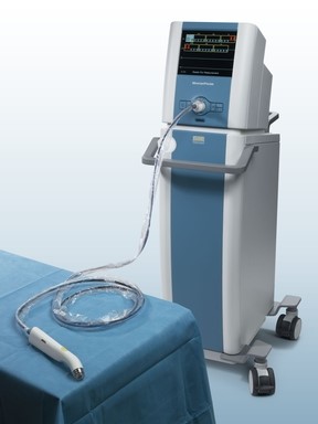
Last week, Dune Medical Devices, Inc., the company that developed the MarginProbeTM System, an intra-operative tissue assessment device to be used as accessory during lumpectomies of early-stage breast cancer, has received Premarket Approval (PMA) by the United States Food and Drug Administration.

MarginProbe system

marginProbe

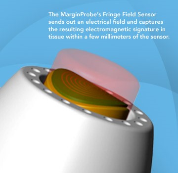
FDA approval of the MarginProbe System was based on a 664 patient prospective, multi-center, randomized, double arm study to evaluate the effectiveness of MarginProbe in identifying cancerous tissue along the margins of removed breast tissue during initial lumpectomy procedures. MarginProbe, which uses electromagnetic “signatures” to identify healthy and cancerous tissue, was found to be over three times more effective in finding cancer on the margin during lumpectomy, compared to traditional intra-operative imaging and palpation assessment. This enabled surgeons to significantly reduce the number of patients with positive margins following initial surgery.

The following publication gives an idea on the clinical performance of MarginProbe:

“J Surg Res. 2010 May 15;160(2):277-81. doi: 10.1016/j.jss.2009.02.025. Epub 2009 Mar 31.

Diagnostic performance of a novel device for real-time margin assessment in lumpectomy specimens.

Pappo I, Spector R, Schindel A, Morgenstern S, Sandbank J, Leider LT, Schneebaum S, Lelcuk S, Karni T.

Source

Department of General Surgery, Assaf Harofeh Medical Center, Zrifin, Israel. pappo@zahav.net.il

Abstract

BACKGROUND:

Margin status in breast lumpectomy procedures is a prognostic factor for local recurrence and the need to obtain clear margins is often a cause for repeated surgical procedures. A recently developed device for real-time intraoperative margin assessment (MarginProbe; Dune Medical Devices, Caesarea, Israel), was clinically tested. The work presented here looks at the diagnostic performance of the device.

METHODS:

The device was applied to freshly excised lumpectomy and mastectomy specimens at specific tissue measurement sites. These measurement sites were accurately marked, cut out, and sent for histopathologic analysis. Device readings (positive or negative) were compared with histology findings (namely malignant, containing any microscopically detected tumor, or nonmalignant) on a per measurement site basis. The sensitivity and specificity of the device was computed for the full dataset and for additional relevant subgroups.

RESULTS:

A total of 869 tissue measurement sites were obtained from 76 patients, 753 were analyzed, of which 165 were cancerous and 588 were nonmalignant. Device performance on relatively homogeneous sites was: sensitivity 1.00 (95% CI: 0.85-1), specificity 0.87 (95% CI: 0.83-0.90). Performance for the full dataset was: sensitivity 0.70 (95% CI: 0.63-0.77), specificity 0.70 (95% CI: 0.67-0.74). Device sensitivity was estimated to change from 56% to 97% as the cancer feature size increased from 0.7 mm to 6.6 mm. Detection rate of samples containing pure DCIS clusters was not different from rates of samples containing IDC.

CONCLUSIONS:

The device has high sensitivity and specificity in distinguishing between normal and cancer tissue even down to small cancer features.

Copyright (c) 2010 Elsevier Inc. All rights reserved.

PMID: 19628225”

Imagine how cost effective breast cancer management can be if it will involve systems such as these in addition to the systems I discussed in some of my previous posts, for example: What could transform an underdog into a winner?

Written by: Dror Nir, PhD.

Read Full Post »

Not applying evidence-based medicine drives up the costs of screening for breast-cancer in the USA

Author: Dror Nir


Costs for breast screening are being driven higher by increased use of new imaging technologies such as digital mammography and MRI, workflows incorporating 2nd and 3rd remote-readings as quality control measure, use of computer-aided detection (CAD) applications and growth in aged population.

According to recent publication in JAMA, 40% of the annual spending is for screening women ages 75 and older. Under existing guidelines routine screening is not recommended for this age group since “There is insufficient evidence to assess the benefits and harms of screening mammography”

The study population comprised women of 66 to 100 years old. “Forty-two percent of the women in the study were younger than age 75, and almost 60% of this group had one or more screening mammograms. Women ages 75 to 85 represented 40% of the total; 42% of this group had mammograms. Women 85 years and older represented 18% of the total; only 15% of this group had mammograms. Women with multiple chronic health conditions were much less likely to have a mammogram (26.6%) than healthy women (47.4%).”

“Abstract

The Cost of Breast Cancer Screening in the Medicare Population.

Cary P. Gross, MD; Jessica B. Long, MPH; Joseph S. Ross, MD, MHS; Maysa M. Abu-Khalaf, MD; Rong Wang, PhD; Brigid K. Killelea, MD, MPH; Heather T. Gold, PhD; Anees B. Chagpar, MD, MA, MPH, MSc; Xiaomei Ma, PhD

JAMA Intern Med. 2013;():1-7. doi:10.1001/jamainternmed.2013.1397. Published online January 7, 2013

Background  Little is known about the cost to Medicare of breast cancer screening or whether regional-level screening expenditures are associated with cancer stage at diagnosis or treatment costs, particularly because newer breast cancer screening technologies, like digital mammography and computer-aided detection (CAD), have diffused into the care of older women.

Methods Using the linked Surveillance, Epidemiology, and End Results–Medicare database, we identified 137 274 women ages 66 to 100 years who had not had breast cancer and assessed the cost to fee-for-service Medicare of breast cancer screening and workup during 2006 to 2007. For women who developed cancer, we calculated initial treatment cost. We then assessed screening-related cost at the Hospital Referral Region (HRR) level and evaluated the association between regional expenditures and workup test utilization, cancer incidence, and treatment costs.

Results In the United States, the annual costs to fee-for-service Medicare for breast cancer screening-related procedures (comprising screening plus workup) and treatment expenditures were $1.08 billion and $1.36 billion, respectively. For women 75 years or older, annual screening-related expenditures exceeded $410 million. Age-standardized screening-related cost per beneficiary varied more than 2-fold across regions (from $42 to $107 per beneficiary); digital screening mammography and CAD accounted for 65% of the difference in screening-related cost between HRRs in the highest and lowest quartiles of cost. Women residing in HRRs with high screening costs were more likely to be diagnosed as having early-stage cancer (incidence rate ratio, 1.78 [95% CI, 1.40-2.26]). There was no significant difference in the cost of initial cancer treatment per beneficiary between the highest and lowest screening cost HRRs ($151 vs $115; P = .20).

Conclusions The cost to Medicare of breast cancer screening exceeds $1 billion annually in the fee-for-service program. Regional variation is substantial and driven by the use of newer and more expensive technologies; it is unclear whether higher screening expenditures are achieving better breast cancer outcomes.”

The study is mainly addressing the difference in costs between different regions of referrals. It would be interesting to explore the situation in the age group of 40 to 66 years old.

Written by:  Dr. Dror Nir, PhD.

Read Full Post »

Author: Dr Anayo Unachukwu, MBBS, LLM


Modern society is built on the twin pillars of Judaeo-Christian jurisprudence and Graeco-Roman civilisation. The jurisprudence has provided us with the moral[1], ethical and legal system that has sustained the civilisation. The civilisation prides itself with the legacy it bequeathed modern society-science. Revolution in science has often been preceded by revolution in measurement. The scientific endeavours have led to delineating, characterising, validating and reliably predicting and identifying the origin of concepts, phenomena, etc encountered by mankind and seeking remedies for these with some degree of certainty and accuracy. The prodigious advances in science and technology within the ethico-legal framework that stemmed from it have brought with it advantages in medical science and related fields. These pervade our every day environment; and we are all witnesses to the consequences of its innovation and creativity. Given the new found confidence in these fields, there have been attempts by medical and behavioural scientists to stretch its research frontiers further in providing succinct explanation to often complex human behaviour and interaction within social spheres. The science behind these quests usually has aural of purity-value free and bereft of ideology. However as Rose puts it: “It may well be that it is only in the periods of social and intellectual crisis that we can glimpse the interconnections between science and social system.[2]

One of the preoccupations of prehistoric man has been attempts at classification and categorisation; this heuristic pursuit-through generalisation-is a way of navigating the complex environment he finds himself. Attempts at generalisation by extrapolating from medical model to account for mental disorders[3] have not altogether been helpful. This could be due to lack of clarity in understanding the aetiological underpinnings; stigma and labeling intrinsic to the sufferers and skepticisms that surround the practice of psychiatry.

The mentally disordered offender raises questions which have troubled theologians, moral philosophers and lawyers throughout the Christian era.[4] It is not only these professions that grapple with this group of offenders; as Eastman puts it psychopaths[5] are the moral and scientific junctional case between the mad and the bad, both in terms of treatment and of punishment.[6]

Psychopathy[7] as a construct throws up more questions than answers-origin, remedial attempts[8] and heuristic generalisation as regards prognostic inferences. There is larger debate on whether psychopathy and personality disorder[9] are primarily legal or diagnostic categories, or neither.[10] Given that psychopathy straddles interpersonal and social domain in everyday human endeavours which impacts on both clinical and legal settings; one would have thought that a complete delineation and categorisation would have been understood by relevant practitioners[11] involved, but this appears not to be the case. Some researchers both clinical and non clinical strongly disagree on the nosological certainty of psychopathy and its inclusion in the medical classificatory system.

However, all agrees that the disorder generates a lot of controversy; and the emotive[12] issue that it provokes-social, psychological, economic and legal-reverberates further afield way beyond the field of clinical sciences.

From social policy perspective, other characteristics of psychopathy notwithstanding, criminal and violent behaviours are the main hallmarks of the disorder.[13] Psychopaths are high risk offenders with many criminogenic needs (i.e., personal characteristics correlated with recidivism).[14] In fact, recent research efforts have shown that there is a relationship between psychopathy and both general and violent crimes.[15] The criminogenic derivative that stem from the disorder had a legal ringing endorsement.[16]

In contrast to the relationship between psychopathy and rate of recidivism; Gibbens et al[17] eight-year comparative follow-up study on criminal psychopaths (diagnosed as ‘severe cases’) and criminal non psychopaths as regards their reconviction rates showed insignificant differences in the reconviction rate for both groups. The conclusion from this study was broadly similar to a two year follow-up study by Walker et al[18] which compared the rates of reconviction amongst psychopaths and non psychopaths who were diagnosed with severe mental illness, schizophrenia and those diagnosed as subnormal (learning disability); the rate of recidivism for both groups were not statistically significant.

Without making an extraordinary reading of the follow-up studies, Cooke et al[19] had argued that criminal behaviour should not play a central role in the diagnosis of psychopathy; instead such behaviour is best viewed as a secondary feature; or sequel of the disorder. This view is consistent with Cleckley’s original classical description of psychopathy where criminal behaviour was not intrinsic to the disorder; the motivation of the behaviour when it is present is usually obscure.[20] Criminality and psychopathy are not the same construct; the core features of the disorder-glib charm, callousness, arrogance, shallow affect, deceitfulness, and lack of empathy do not necessarily involve or imply criminal behaviour.[21] Why then is there such a great emphasis in diagnosis of this disorder? The answer partly lies in the society we live. There appears to be an inverse relationship between the tolerance of risk[22] in a given society and its level of affluence. There is a political imperative driven by popular lay press for a ‘risk free’ society; such an unrealistic expectation has led to the bourgeoning of research in risk instruments for assessment and management of risks. The trend in the wider society, technological advances and rise in consumerism has all further fueled this gusto-risk intolerance. Risk assessment as a concept is not a legal issue per se as competence to stand trial or criminal responsibility; however, it forms important part of different decision making in law. As Fennell puts it, there is a role redefinition whereby risk management has assumed increasing centrality in the role of psychiatrists and other mental health professionals, and philosophies of risk management now permeate decision-making in both the psychiatric system and penal system.[23] It is a given that psychopaths are involved in violent crimes and they recidivate for both violent and non violent crime compared to non psychopath; hence violence risk is an “indirect” legal issue often relevant to ultimate decisions but rarely defining the ultimate issue specifically.[24] Therapeutic jurisprudence[25] partly relies on risk assessment and management in deciding on disposal options available when dealing with mentally disordered offenders. As Leacock and Sparks remarked; Risk…is a fashionable idea—one whose moment would appears to have arrived.[26] Risk is now so integral to every day discourse given the high profile homicides; most notable was the killing of Jonathan Zito (in England) by a stranger, Christopher Clunis, who suffered from schizophrenia. As a result, the risk management/public protection agenda is now prominent-in England and Wales-and since 1992, health authorities have a duty to carry out an independent inquiry in any case of homicide or suicide by a mentally disordered person in their care.[27]

Conclusion


Mentally disordered offenders-like the poor-will always be in our midst. Labelling and stigmatising this group as a whole may be unhelpful at best and amounts to discrimination. Managing risk out every day discourse is naivete to the extreme, given that in any society a trade-off has to be made as far as risk goes. Criminality as a construct should not be synonymous with psychopathy, given that not all psychopaths offend and not all criminals are psychopaths.


[1] Psychopathy which under the Mental Health Act 1983 is classified as a mental disorder is seen by some philosophers, lawyers, clinicians and the society at large as essentially a moral disorder. See Maibon, H.L. The Mad, the Bad, and the Psychopath. Neuroethics (2008) 1:167-184. DOI 10.1007/s12152-008-9013-9.

[2] Rose, S., Rose, H. (1971). The myth of the neutrality of science. In Watson Fuller (ed). The SOCIAL IMPACT OF MODERN BIOLOGY.Routledge & Keegan Paul. London. p.233

[3] The adoption of this term was recommended in the Report of the Royal Commission on the law relating to Mental Illness and Mental deficiency 1954-1957 (Cmnd 169 (1957)) Pt 3 paras 146-198. The Commission recommended its use as a general term covering all forms of mental ill-health or disability. S (1) of the amended Mental Health Act 2007 gives a broader description of mental disorder-any disorder of the mind; ‘mentally disordered’ shall be construed accordingly.

[4] Walker, N. (1968) Crime and Insanity in England. vol. I. Edinburgh University Press. p. 6

[5] Psychopathy is a personality disorder defined by a cluster of interpersonal, affective, and lifestyle characteristics that results in serious, negative consequences for society.

[6] Eastman, N (2002). The Ethics of Clinical Risk Assessment and Management:Developing Law and the Role of Mental Health Professionals. In Gray, N,  Lain, J,  Noaks, L. (Eds.). Criminal Justice, Mental Health and the Politics of Risk.(pp. 49-66). Cavendish Publishing Limited.

[7] The adverb psychopathic means literally ‘psychically damaged’ and was introduced in the 19th century Germany to cover all forms of psychopathology. From the outset it should be distinguished from psychopathic disorder which is a legal concept. Authors disagree on its definition; sometimes the usage is in vernacular, and this paints a derogatory picture. See Gunn, J. and Robertson G. (1976b) Psychopathic Personality: a conceptual problem. Psychological Medicine, 6, 63-4 (386).

[8] Cleckley, H. (1941). The mask of insanity. St. Louis, MO: Mosby. The present day conceptualisation of psychopathy is credited to the systematic writings and observation of Cleckley. He expressed concerns about the progressive nature of the disorder, social malady it posed to the society and therapeutic nihilism associated with it.

[9] The defining characteristic of personality disorder more than any symptom is the incomprehensibility and inaccessibility that the sufferer provokes in other people. It is classified as a mental disorder by both two international classificatory systems- World Health Organisation. (1992) International Classification of Disease and Related Health Problems: ICD-10. Geneva: WHO; and American Psychiatric Association. (1994) Diagnostic and Statistical Manual of Mental Disorders. DSM-IV. WashingtonDC: Mental Health Service.

[10] Ogloff, J.R.P., & Lyon, D.R. (1998). Legal issues associated with the concept of psychopathy. In D.J. Cooke, A.E. Forth, & R. D. Hare (Eds) Psychopathy: theory, research, and implications for society (pp. 401-422). Dordrecht, The Netherlands: Kluwer.

[11] Unfortunately among various practitioners not just legal practitioners, but most clinicians the conceptual cousins of psychopathy-sociopathy, dissocial personality and antisocial personality have been mistaken for psychopathy. See Gacono, C.B., Nieberding, R., Owen, A., Rubel, J., & Bodholdt, R. (2000). Treating juvenile and adult offenders with conduct disorder, antisocial and psychopathic personalities. In J. Ashford, B. Sales, & W.Reid (Eds.), Treating clients with special needs. WashingtonDC: American Psychological Association.

[12] Hare, R.D. (1996a). Psychopathy: A construct whose time has come. Criminal Justice Behavior. 23, 25-54.

[13] Patrick, C.J. (2006). Handbook of Psychopathy The Guilford Press. p.555

[14] Zinger, I., & Forth, A.E. (1998). Psychopathy and Canadian criminal proceedings: The potential for human right abuses. Canadian Journal of Criminology, 40, 237-276.

[15] Salekin, R.T., Rogers, R., & Sewell, K. W. (1996). A review and meta-analysis of the Psychopathy Checklist and Psychopathy checklist-Revised. Clinical Psychology: Science and Practice, 3, 203-215

[16] Carraher v HM Advocate 1946 JC 108 at 117, per Lord Normand: it is to my mind descriptive rather of a typical criminal than a person…regarded as being possessed of diminished responsibility.

[17] Gibbens, T.C.N., Pond, D.A. and Stafford-Clark, ‘A Follow-up Study of Criminal Psychopaths’ in (1959) 105, Journal of Mental Science, 108 ff.

[18] Walker, N., McCabe, S. (1973) Crime and Insanity in England. Vol. II. p.212. Edinburgh University Press.

[19] Cooke, D. J., Michie, C., Hart, S. D., et al (2004) Reconstructing psychopathy: Clarifying the significance of antisocial and socially deviant behavior in the diagnosis of psychopathic personality disorder. Journal of Personality Disorders, 18, 337-357.

[20] Cleckley,H. (1988) The Mask of Sanity (5th edn). Mosby. p.279

[21] Hare, R.D. (1998). Psychopathy, affect, and behaviour. In D.J. Cooke, A.E.Forth, & R.D. (Eds.), Psychopathy, theory, research, and implication for society (pp104-137). Dordrecht, the Netherlands: Kluwer.

[22] Risk is defined as the uncertainty of outcome whether opportunity or negative threat, of actions and event

[23] P, Fennell (2002) .The Ethics of Clinical Risk Assessment and Management:Developing Law and the Role of Mental Health Professionals. In Gray, N,  Lain, J,  Noaks, L. (Eds.). Criminal Justice, Mental Health and the Politics of Risk.(pp. 49-66). Cavendish Publishing Limited.

[24] Roesch, R., & McLachlan, K. (2007). Clinical Forensic Psychology and Law. Ashgate Publishing Ltd.

[25] Wexler, D., & Winck, B. (Eds.) (1996). Law in a therapeutic key. Durham, NC: Carolina Academic Press. At p. 32 describe it as a concept that addresses the issue of how knowledge, theories and insight of the mental health disciplines can help shape the development of law.

[26] R.D. Mackay (2002). Criminal Justice, Mental Health and the Politics of Risk. Crim. L.R. 2002, Sep, 765-767.

[27] Fennell, P (2010). HISTORY AND CONTEXT OF MENTAL HEALTH LAW AND POLICY. In Gostin, L., Bartlett, P., McHale, J., Mackay, R. (eds). PRINCIPLE OF MENTAL HEALTH POLICY AND LAW. Oxford University Press. p.53.


Read Full Post »

Whole-body imaging as cancer screening tool; answering an unmet clinical need?

Author: Dror Nir, PhD

Sometimes technologies that were developed to answer clinical needs in a certain area are migrated to perform in a totally inappropriate area. A good example which I discussed several times in my posts is PSA.

Cancer patients’ prognoses, strongly depend on accurate tumor staging. It is also a prerequisite for therapy choice and planning. Whole-body imaging is frequently used in patients with advanced malignant diseases including presence of metastases as these may occur in any anatomic region. It is important to note that classifying a patient as harboring a potentially advanced disease is based on biopsy results of Sentinel Lymph-Nodes and not on imaging. Moreover, referring a patient to a whole-body imaging is a choice of the treating practitioner! Clearly, when the choice of treatment includes administration of drugs, the type of drugs to be used is determined by the characteristics of the primary tumor.

To date, the use of whole-body imaging for post treatment follow-up can be considered as anecdotal.

The most-used technologies for whole-body imaging are computed tomography (CT), positron emission tomography (PET) and MRI. The performance of these systems in detection of cancer metastases of more than 1cm in diameter is very similar and in general quite good, dependent on the primary disease and the body locations of the metastases. Alas, each of these modalities has its strengths and weakness in different cancer and different body locations. Therefore, in the last decade, combined modalities such as PET-CT and recently PET/MRI were introduced. In some cases [1-6] these are reported to show sensitivity of more than 90%.

To demonstrate the level of information produced during whole-body imaging procedure here is an example (taken from Whole-body MRI and PET-CT in the management of cancer patients). This resource includes additional, educating examples:

330_2006_183_Fig1_HTML

Fig. 1

From multimodality to single-step examination. Restaging in a 29-year-old woman treated for breast cancer and with newly elevated tumor markers and bone pain. 1a, 1b Radiograms of the skeleton were normal, but bone scintigraphy showed a pathological tracer uptake in the right pubic bone (arrow). Abdominal ultrasound exhibited a suspicious mass. 1c, 1d CT revealed tumor recurrence in the right breast and confirmed hepatic metastasis. 2a, 2b T1-weighted whole-body MRI depicted a metastasis in the right pubic bone (circle). 2c, 2d HASTE images of the thorax showed the tumor recurrence in the right breast (arrow) and dynamic contrast enhanced studies of the abdomen unmasked the liver metastasis

Before addressing the issue of using whole-body imaging as a screening tool I would like to draw attention to existence of other methods for screening and post treatment follow-up of cancer patients; e.g. detecting levels of cancer-specific bio-markers in the blood or urine or, in case of advanced disease, detecting the level of tumor cells circulating in the blood as presented in: Circulating Tumor Cells versus Imaging—Predicting Overall Survival in Metastatic Breast Cancer by G. Thomas Budd et.al.

“Abstract

Purpose: The presence of ≥5 circulating tumor cells (CTC) in 7.5 mL blood from patients with measurable metastatic breast cancer before and/or after initiation of therapy is associated with shorter progression-free and overall survival. In this report, we compared the use of CTCs to radiology for prediction of overall survival.

Experimental Design: One hundred thirty-eight metastatic breast cancer patients had imaging studies done before and a median of 10 weeks after the initiation of therapy. All scans were centrally reviewed by two independent radiologists using WHO criteria to determine radiologic response. CTC counts were determined ∼4 weeks after initiation of therapy. Specimens were analyzed at one of seven laboratories and reviewed by a central laboratory.

Results: Inter-reader variability for radiologic responses and CTC counts were 15.2% and 0.7%, respectively. The median overall survival of 13 (9%) patients with radiologic nonprogression and ≥5 CTCs was significantly shorter than that of the 83 (60%) patients with radiologic nonprogression and <5 CTCs (15.3 versus 26.9 months; P = 0.0389). The median overall survival of the 20 (14%) patients with radiologic progression and <5 CTCs was significantly longer than the 22 (16%) patients with ≥5 CTCs that showed progression by radiology (19.9 versus 6.4 months; P = 0.0039).

Conclusions: Assessment of CTCs is an earlier, more reproducible indication of disease status than current imaging methods. CTCs may be a superior surrogate end point, as they are highly reproducible and correlate better with overall survival than do changes determined by traditional radiology. “

I would like first to present the following publication that could explain why people can easily be drawn why whole-body screening is an effective way to detect early cancers:

Enthusiasm for cancer screening in the United States by Schwartz LM, Woloshin S, Fowler FJ Jr, Welch HG SO, JAMA. 2004; 291(1):71.:

“ CONTEXT: Public health officials, physicians, and disease advocacy groups have worked hard to educate individuals living in the United States about the importance of cancer screening.

OBJECTIVE: To determine the public’s enthusiasm for early cancer detection.

DESIGN, SETTING, AND PARTICIPANTS: Survey using a national telephone interview of adults selected by random digit dialing, conducted from December 2001 through July 2002. Five hundred individuals participated (women aged>or =40 years and men aged>or =50 years; without a history of cancer).

MAIN OUTCOME MEASURES: Responses to a survey with 5 modules: a general screening module (eg, value of early detection, total-body computed tomography); and 4 screening test modules: Papanicolaou test; mammography; prostate-specific antigen (PSA) test; and sigmoidoscopy or colonoscopy.

RESULTS: Most adults (87%) believe routine cancer screening is almost always agood idea and that finding cancer early saves lives (74% said most or all the time). Less than one third believe that there will be a time when they will stop undergoing routine screening. A substantial proportion believe that an 80-year-old who chose not to be tested was irresponsible: ranging from 41% with regard to mammography to 32% for colonoscopy. Thirty-eight percent of respondents had experienced at least 1 false-positive screening test; more than 40% of these individuals characterized that experience as “very scary” or the “scariest time of my life.” Yet, looking back, 98% were glad they had had the initial screening test. Most had a strong desire to know about the presence of cancer regardless of its implications: two thirds said they would want to be tested for cancer even if nothing could be done; and 56% said they would want to be tested for what is sometimes termed pseudodisease (cancers growing so slowly that they would never cause problems during the person’s lifetime even if untreated). Seventy-three percent of respondents would prefer to receive a total-body computed tomographic scan instead of receiving 1000 dollars in cash.

CONCLUSIONS: The public is enthusiastic about cancer screening. This commitment is not dampened by false-positive test results or the possibility that testing could lead to unnecessary treatment. This enthusiasm creates an environment ripe for the premature diffusion of technologies such as total-body computed tomographic scanning, placing the public at risk of over testing and overtreatment.”

Whole-body screening is promoted as a one-stop shop for painlessly detecting hidden cancer and preventing cancer-related deaths. It is big business in the United States and in Canada where private clinics have begun offering full-body diagnostic procedures for a fee. The tests and procedures are often marketed to healthy people as a way to scan for hidden abnormalities or cancers, affording people the peace of mind that they are in good health [7 – 9].

When used in this manner, the evidence shows that whole-body cancer screening offers no proven health benefits and that it, in fact, exposes people to a number of unnecessary health risks. The problem I see is that the public is not exposed to “scientific publications” but is exposed to commercial ones!


References

  1. FDG PET and PET/CT: EANM procedure guidelines for tumor PET imaging: version 1.0
  2. Cancer of the Prostate, Testicles and Penis
  3. Gynecologic Cancers
  4. Malignant Melanoma
  5. Molecular Imaging in Cancer
  6. Pre-clinical whole-body fluorescence imaging: Review of instruments, methods and applications
  7. Full body CT scan for screening
  8. Screening for Cancer with PET and PET/CT: Potential and Limitations 
  9. http://www.privatescan.nl/total-bodyscan

Written by: Dror Nir, PhD

Read Full Post »

Differentiation Therapy – Epigenetics Tackles Solid Tumors

Author-Writer: Stephen J. Williams, Ph.D.

Updated 4/27/2021

Screen Shot 2021-07-19 at 7.04.21 PM

Word Cloud By Danielle Smolyar

Genetic and epigenetic events within a cell which promote a block in normal development or differentiation coupled with unregulated proliferation are hallmarks of neoplastic transformation.  Differentiation therapy is a chemotherapeutic strategy directed at re-activating endogenous cellular differentiation programs in a tumor cell therefore driving the cancerous cell to a state closer resembling the normal or preneoplastic cell and therefore incurring loss of the tumorigenic phenotype.

This post will deal with:

  • Agents such as histone deacetylase inhibitors (HDACi), retinoids, and PPARϒ agonists which have been shown to reactivate terminal differentiation programs in solid tumors
  • Clinical trials in solid tumors
  • Issues regarding the use of differentiation therapy in solid tumors

This post is a follow-up post to Histone Deacetylase Inhibitors Induce Epithelial-to-Mesenchymal Transition in Prostate Cancer Cells

To put the need for alternate chemotherapeutic strategies in perspective, one is referred to the National Cancer Statistics from http://www.cancer.gov show that 33% of cancer patients, treated with standard cytolytic chemotherapy, will still die within five years (i.e. one in three will die within 5 years).  However the addition of the differentiation agent retinoic acid to standard chemotherapy regimen for treatment of acute promyelocytic leukemia (APML) had improved 5 year survival rates from a range of 50-80% up to near 90% complete remission rates while 75% become disease free, an astonishing success story.  For a review of APML please be referred to http://en.wikipedia.org/wiki/Acute_promyelocytic_leukemia.  Briefly, APML is predominantly a result of the chromosomal translocation producing a fusion gene between the promyelocytic leukemia (PML) and RARα receptor genes.  The PML-RARα fusion protein recruits transcriptional repressors, histone deacetylases (HDACs), and DNA methyltransferases.  Treatment with pharmacologic doses of retinoic acid dissociates the PML-RARα from HDACs and results in degradation of PML-RARα, eventually resulting in the differentiation of the myeloid cells in APML.

Dr. Igor Matushansky of Columbia University believes such differentiation therapy could be useful in soft tissue sarcomas, due to the existence of a connective tissue (mesenchymal) stem cell,  in vitro methods which can differentiate these cells into mature tissues, and, from a gene clustering analysis his group had performed, correlation of expression signatures of each liposarcoma subtype throughout the adipocytic differentiation spectrum, including early differentiated to more mature differentiated cells(1).   A parallel study by Riester and colleagues had been able to classify breast tumors and liposarcomas along a phylogenetic tree showing solid tumors can be reclassified based on cell of origin via expression patterns(2).  In addition, other solid tumors, such as ovarian cancer are easily classified, based both on pathologic, histologic, and expression analysis into well and poorly differentiated tumors, correlating differentiation status with prognosis.

Compound Classes which have potential in

differentiation therapy for solid tumors

A. Histone Deacetylase Inhibitors (HDACi)

In eukaryotes, epigenetic post-translational modification of histones is critical for regulation of chromatin structure and gene expression.  Histone deacetylation leads to chromatin compaction and is associated with transcriptional repression of tumor suppressors, cell growth and differentiation.  Therefore, HDACi are promising anti-tumor agents as they may affect the cell cycle, inhibit proliferation, stimulate differentiation and induce apoptotic cell death (3). In a review by Kniptein and Gore, entinostat was found to be a well-tolerated HDACi that demonstrates promising therapeutic potential in both solid and hematologic malignancies(4). The path to the discovery of suberoylanilide hydroxamic acid (SAHA, vorinostat) began over three decades ago with our studies designed to understand why dimethylsulfoxide causes terminal differentiation of the virus-transformed cells, murine erythroleukemia cells. SAHA can cause growth arrest and death of a broad variety of transformed cells both in vitro and in vivo at concentrations that have little or no toxic effects on normal cells (for references see (5). In fact, treatment of MCF-7 breast carcinoma cells with SAHA resulted in morphologic changes resembling epithelial mammary differentiation(6).

HDAC inhibitors

Figure.  Structures of some HDACi used in clinical trials for cancer (see section below)

hdacwithsaha

Figure.  HDAC with SAHA

B. Retinoids

Vitamin A and retinoids play significant roles in basic physiological processes such as vision, reproduction, growth, development, hematopoiesis and immunity (7). Retinoids are the natural derivatives and synthetic analogs of vitamin A. They have been shown to prevent mammary carcinogenesis in rodents (8), to inhibit the growth of human cancer cells in vitro  (9,10) and be effective chemopreventive and chemotherapeutic agents in a variety of human epithelial and hematopoietic tumors (11-14).

Retinoids cannot be synthesized de novo by higher animals and consequently must be consumed in the diet. The two sources of retinoids are animal products that contain retinol and retinyl esters, and plant-derived carotenoids (provitamin A). b-carotene is the most potent vitamin A precursor and has been shown to be an active inhibitor of both tumor initiation and promotion (15).

A major function of retinol, relevant to cancer, is its function as an antioxidant. The antioxidant properties of vitamin A have been shown both in vitro and in vivo (16,17). Retinol deficiency causes oxidative damage to liver mitochondria in rats that can be reversed by vitamin A supplementation (18). A caveat to this is in vitro and in vivo evidence of chronic hypervitaminosis A inducing oxidative DNA damage, as well (19-21). Therefore, it is evident that maintaining the vitamin A concentration within a physiological range is critical to normal cell function because either a deficiency or an excess of vitamin A induces oxidative stress (22). Retinoic acids (RA) (all-trans, 9-cis and 13-cis) are the major biologically active retinoids and exert their effects by regulation of gene expression by binding two families of ligand-activated nuclear retinoid receptors (23). Retinoic acid receptors (RARs) and retinoid X receptors (RXRs) regulate the transcription of a large number of target genes that contain retinoic acid response elements (RAREs) in their promoters. Many of these genes are involved in cancer (13,24) and differentiation (24-26).

Several lines of evidence suggest involvement of defects in retinol signaling in cancer, from the observation that a vitamin A-deficient (VAD) diet leads to an increase in the number of spontaneous and chemically induced tumors in animals (27-29) to the observation that RA itself can induce  differentiation and inhibit the growth of many tumor cells (30-32), as well as the identification that components of the RA signaling pathway are absent in cancer cells (33). Vitamin A and its metabolites have been proposed to have a dual effect in cancer prevention, as antioxidants (16,17,19,34) and differentiating agents (35-37). as it is well accepted that retinoid signaling is integral in maintaining the differentiated state of many cell types (13,38). Additionally, current rationale for chemoprevention with retinoids is based, in part, on the hypothesis that some tumors, may arise due to loss of normal somatic differentiation during tissue repair.

C. PPARϒ Agonists

Peroxisome proliferator-activated receptor ϒ (PPARϒ) is a member of the steroid hormone receptor superfamily that responds to changes in lipid and glucose homeostasis but has increasing roles in differentiation and tumorigenesis. The first PPAR (PPARα) was discovered during the search of a molecular target for a group of agents then referred to as peroxisome proliferators, as they increased peroxisomal numbers in rodent liver tissue, apart from improving insulin sensitivity.  One of the first agents, developed in the early 80’s for treatment of hyperlipidemia and hperlipoproteinemia, was clofibrate.  All PPAR subtypes heterodimerize with the retinoid-x-receptor (RXR) and, upon binding of ATRA, activate target genes.

PPARϒ agonists have shown potential as a therapeutic in a variety of cancer types including bladder cancer (39), colon cancer(40),  breast cancer(41), prostate cancer(42).  There are numerous studies showing that PPARϒ agonists have anti-tumorigenic activity via anti-proliferative, pro-differentiation and anti-angiogenic mechanisms of action(43). For example, Papi et al. observed that agonists for the retinoid X receptor (6-OH-11-O-hydroxyphenanthrene), retinoic acid receptor (all-trans retinoic acid (RA)) and peroxisome proliferator-activated receptor (PPAR)-γ (pioglitazone (PGZ)), reduce the survival of MS generated from breast cancer tissues and MCF7 cells, but not from normal mammary gland or MCF10 cells(44) with concomitant upregulation of differentiation markers.

A great website for further information on PPAR is Dr. Jack Vanden Heuvel, Professor of Toxicology at Penn State University at http://ppar.cas.psu.edu/general_information.html.

D. Trabectedin

Trabectedin (ecteinascidin-743 (ET-743); Yondelis) is derived from the Caribbean tunicate Ecteinascidia turbinacta has antitumor activity by binding to the DNA minor groove thus disrupting binding of transcription factors and inhibiting DNA synthesis.  However, it has also been shown, in myxoid liposarcoma (MLS) cells, to cause dissociation of transcription factor TLS-CHOP from promoter sequences resulting in downregulation of target genes such as CHOP, PTX3 and FN1 and induces an adipogenic differentiation program by enhancing activation of CAAT/enhancer binding protein (C/EBP) family of genes.  In MLS, TLS-CHOP sequesters C/EBPβ resulting in block of differentiation programs while trabectedin disrupts this association freeing up C/EBPβ to act as transcriptional activator of genes related to differentiation.

Ongoing Cancer Clinical Trials with HDAC Inhibitors

The following is a listing of some clinical trials using histone deacetylase inhibitors in combination with approved chemotherapeutics in various tumors.  This data was taken from the New Medicine Oncology Knowledge Base ( at http://www.nmok.net).

hdactrial1 hdactrial2

Issues and Future of Differentiation-based Therapy

In the review by Filemon Dela Cruz and Igor Matushansky(1), the authors suggest that, like days of old of cytotoxic monotherapy, differentiation therapy would not evolve as a simplistic one-size-fits –all but mirror an extremely complicated process.  Therefore they suggest three theoretical mechanisms in which differentiation therapy may occur:

  1. Cancer directed differentiation: differentiation pathways are activated without correcting the underlying oncogenic mechanisms which produced the initial differentiation block
  2. Cancer reverted differentiation: correction of the underlying oncogenic mechanism results in restoration of endogenous differentiation pathways
  3. Cancer diverted differentiation: cancer cell is redirected to an earlier stage of differentiation

Finally the authors suggest that “the potential for reversion of the malignant cancer phenotype to a more benign, or at the very least a lower grade of biological aggressiveness, may serve as a critical clinical and biologic transition of a uniformly fatal cancer into one more amenable to management or to treatment using conventional therapeutic approaches.”

References:

1.            Cruz, F. D., and Matushansky, I. (2012) Oncotarget 3, 559-567

2.            Riester, M., Stephan-Otto Attolini, C., Downey, R. J., Singer, S., and Michor, F. (2010) PLoS computational biology 6, e1000777

3.            Seidel, C., Schnekenburger, M., Dicato, M., and Diederich, M. (2012) Genes & nutrition 7, 357-367

4.            Knipstein, J., and Gore, L. (2011) Expert opinion on investigational drugs 20, 1455-1467

5.            Marks, P. A. (2007) Oncogene 26, 1351-1356

6.            Munster, P. N., Troso-Sandoval, T., Rosen, N., Rifkind, R., Marks, P. A., and Richon, V. M. (2001) Cancer research 61, 8492-8497

7.            Napoli, J. L. (1999) Biochim Biophys Acta 1440, 139-162

8.            Moon, R., Metha, R., and Rao, K. (1994) Retinoids and cancer in experimental animals. in The Retinoids: Biology, Chemistry, and Medicine (Sporn, M., Roberts, A., and Goodman, D. eds.), 2 Ed., Raven Press, New York. pp 573-596

9.            De Luca, L. M. (1991) Faseb J 5, 2924-2933

10.          Gudas, L. J. (1992) Cell Growth Differ 3, 655-662

11.          Degos, L., and Parkinson, D. (1995) Retinoids in Oncology, Springer-Verlag, Berlin

12.          Lotan, R. (1996) Faseb J 10, 1031-1039

13.          Zhang, D., Holmes, W. F., Wu, S., Soprano, D. R., and Soprano, K. J. (2000) J Cell Physiol 185, 1-20

14.          Fontana, J. A., and Rishi, A. K. (2002) Leukemia 16, 463-472

15.          Suda, D., Schwartz, J., and Shklar, G. (1986) Carcinogenesis 7, 711-715

16.          Ciaccio, M., Valenza, M., Tesoriere, L., Bongiorno, A., Albiero, R., and Livrea, M. A. (1993) Arch Biochem Biophys 302, 103-108

17.          Palacios, A., Piergiacomi, V. A., and Catala, A. (1996) Mol Cell Biochem 154, 77-82

18.          Barber, T., Borras, E., Torres, L., Garcia, C., Cabezuelo, F., Lloret, A., Pallardo, F. V., and Vina, J. R. (2000) Free Radic Biol Med 29, 1-7

19.          Borras, E., Zaragoza, R., Morante, M., Garcia, C., Gimeno, A., Lopez-Rodas, G., Barber, T., Miralles, V. J., Vina, J. R., and Torres, L. (2003) Eur J Biochem 270, 1493-1501

20.          Omenn, G. S., Goodman, G. E., Thornquist, M. D., Balmes, J., Cullen, M. R., Glass, A., Keogh, J. P., Meyskens, F. L., Jr., Valanis, B., Williams, J. H., Jr., Barnhart, S., Cherniack, M. G., Brodkin, C. A., and Hammar, S. (1996) J Natl Cancer Inst 88, 1550-1559

21.          Murata, M., and Kawanishi, S. (2000) J Biol Chem 275, 2003-2008

22.          Schwartz, J. L. (1996) J Nutr 126, 1221S-1227S

23.          Chambon, P. (1996) Faseb J 10, 940-954

24.          Freemantle, S. J., Kerley, J. S., Olsen, S. L., Gross, R. H., and Spinella, M. J. (2002) Oncogene 21, 2880-2889

25.          Collins, S. J., Robertson, K. A., and Mueller, L. (1990) Mol Cell Biol 10, 2154-2163

26.          Grunt, T. W., Somay, C., Oeller, H., Dittrich, E., and Dittrich, C. (1992) J Cell Sci 103 ( Pt 2), 501-509

27.          Lasnitzki, I. (1955) Br J Cancer 9, 434-441

28.          Moore, T. (1965) Proc Nutr Soc 24, 129-135

29.          Saffiotti, U., Montesano, R., Sellakumar, A. R., and Borg, S. A. (1967) Cancer 20, 857-864

30.          Strickland, S., and Mahdavi, V. (1978) Cell 15, 393-403

31.          Breitman, T. R., Selonick, S. E., and Collins, S. J. (1980) Proc Natl Acad Sci U S A 77, 2936-2940

32.          Breitman, T. R., Collins, S. J., and Keene, B. R. (1981) Blood 57, 1000-1004

33.          Niles, R. M. (2000) Nutrition 16, 573-576

34.          Monagham, B., and Schmitt, F. (1932) J Biol Chem 96, 387-395

35.          Miller, W. H., Jr. (1998) Cancer 83, 1471-1482

36.          Miyauchi, J. (1999) Leuk Lymphoma 33, 267-280

37.          Reynolds, C. P. (2000) Curr Oncol Rep 2, 511-518

38.          Ortiz, M. A., Bayon, Y., Lopez-Hernandez, F. J., and Piedrafita, F. J. (2002) Drug Resist Updat 5, 162-175

39.          Mansure, J. J., Nassim, R., and Kassouf, W. (2009) Cancer biology & therapy 8, 6-15

40.          Osawa, E., Nakajima, A., Wada, K., Ishimine, S., Fujisawa, N., Kawamori, T., Matsuhashi, N., Kadowaki, T., Ochiai, M., Sekihara, H., and Nakagama, H. (2003) Gastroenterology 124, 361-367

41.          Stoll, B. A. (2002) Eur J Cancer Prev 11, 319-325

42.          Smith, M. R., and Kantoff, P. W. (2002) Investigational new drugs 20, 195-200

43.          Rumi, M. A., Ishihara, S., Kazumori, H., Kadowaki, Y., and Kinoshita, Y. (2004) Current medicinal chemistry. Anti-cancer agents 4, 465-477

44.          Papi, A., Guarnieri, T., Storci, G., Santini, D., Ceccarelli, C., Taffurelli, M., De Carolis, S., Avenia, N., Sanguinetti, A., Sidoni, A., Orlandi, M., and Bonafe, M. (2012) Cell death and differentiation 19, 1208-1219

Updated 4/27/2021

Epizyme’s EZH2 blocker boosts immuno-oncology response in prostate cancer models

Source: https://www.fiercebiotech.com/research/epizyme-s-ezh2-blocker-boosts-immuno-oncology-response-prostate-cancer-models

cancer cell surrounded by killer T cells
Inhibiting EZH2 either genetically or with a chemical inhibitor signaled the immune system to respond to PD-1 inhibition in prostate cancer. (NIH)

The protein EZH2 has long been known as a major driver of prostate cancer because of its ability to inactivate genes that would normally suppress tumor growth. Now, a team at Cedars-Sinai Cancer has shown in preclinical models of the disease that blocking EZH2 reduces resistance to immune-boosting checkpoint inhibitors—and they did it with the help of Epizyme, which won FDA approval for the first EZH2 blocker last year.

The Cedars-Sinai team inhibited EZH2 in preclinical prostate cancer models, activating interferon-stimulated genes in the immune system. The interferons then boosted the immune response and reversed resistance to drugs that inhibit the checkpoint PD-1, they reported in the journal Nature Cancer.

By inhibiting EZH2 either genetically or with a chemical inhibitor donated by Epizyme, the researchers used a technique called “viral mimicry” to “reopen” parts of the genome that are typically inactive, they explained in a statement. That signaled the immune system to respond to PD-1 inhibition.

Checkpoint inhibitors have been approved to treat several cancer types, but they’ve been largely disappointing in prostate cancer. Hence several research groups have been exploring combination strategies. They include the University of Texas MD Anderson Cancer Center, which published research in 2019 showing early evidence that combining checkpoint inhibition with anti-TGF-beta drug could be effective in prostate cancer.

More recently, bispecific antibodies have shown early promise in prostate cancer. Last September, Amgen presented data from a phase 1 study of AMG 160, a bispecific targeting PSMA and CD3 on T cells. The company said that 68.6% of patients experienced a decline in PSA, and eight out of 15 patients evaluated showed stable disease.

Regeneron is also developing a bispecific antibody for prostate cancer, targeting PSMA and CD28. The drug is being tested as a solo therapy and in combination with Regeneron’s PD-1 inhibitor Libtayo in a phase 1/2 clinical trial enrolling men with metastatic castration-resistant prostate cancer.

As for Epizyme’s EZH2 inhibitor, Tazverik, its path to market hasn’t been perfectly smooth. An advisory committee to the FDA questioned its efficacy and safety in its initial indication, metastatic or locally advanced epithelioid sarcoma. Still, the company got the go-ahead to market the drug in adult patients with the rare cancer last January. Then the FDA added follicular lymphoma to the label in June. The drug’s takeoff has been slower than expected, however, largely because the pandemic has prevented face-to-face interactions between the sales force and physicians.

The company is currently testing Tazverik in several other cancer types, including as a combination with standard-of-care treatments in castration-resistant prostate cancer.

Other research papers on Cancer and Cancer Therapeutics were published on this Scientific Web site as follows:

Histone Deacetylase Inhibitors Induce Epithelial-to-Mesenchymal Transition in Prostate Cancer Cells

PIK3CA mutation in Colorectal Cancer may serve as a Predictive Molecular Biomarker for adjuvant Aspirin therapy

Nanotechnology Tackles Brain Cancer

Response to Multiple Cancer Drugs through Regulation of TGF-β Receptor Signaling: a MED12 Control

Personalized medicine-based cure for cancer might not be far away

GSK for Personalized Medicine using Cancer Drugs needs Alacris systems biology model to determine the in silico effect of the inhibitor in its “virtual clinical trial”

Lung Cancer (NSCLC), drug administration and nanotechnology

Non-small Cell Lung Cancer drugs – where does the Future lie?

Cancer Innovations from across the Web

arrayMap: Genomic Feature Mining of Cancer Entities of Copy Number Abnormalities (CNAs) Data

How mobile elements in “Junk” DNA promote cancer. Part 1: Transposon-mediated tumorigenesis.

Cancer Genomics – Leading the Way by Cancer Genomics Program at UC Santa Cruz

Closing the gap towards real-time, imaging-guided treatment of cancer patients.

Closing the gap towards real-time, imaging-guided treatment of cancer patients.

mRNA interference with cancer expression

Search Results for ‘cancer’ on this web site

Cancer Genomics – Leading the Way by Cancer Genomics Program at UC Santa Cruz

Closing the gap towards real-time, imaging-guided treatment of cancer patients.

Lipid Profile, Saturated Fats, Raman Spectrosopy, Cancer Cytology

mRNA interference with cancer expression

Pancreatic cancer genomes: Axon guidance pathway genes – aberrations revealed

Biomarker tool development for Early Diagnosis of Pancreatic Cancer: Van Andel Institute and Emory University

Is the Warburg Effect the cause or the effect of cancer: A 21st Century View?

Crucial role of Nitric Oxide in Cancer

Targeting Glucose Deprived Network Along with Targeted Cancer Therapy Can be a Possible Method of Treatment

Read Full Post »

A perspective on where we are on carcinogenesis, cancer variability and predictors of time to recurrence and future behavior

Author: Larry H Bernstein, MD, FCAP

Predicting Tumor Response, Progression, and Time to Recurrence

Word Cloud by Daniel Menzin

I.     Background

In “Tumor Imaging and Targeting: Predicting Tumor Response to Treatment: Where we stand? “ ( Dec 13, 2012) Dr. Ritu Saxena  attempts to integrate three posts and to embed all comments made to all three papers, allowing the reader a critically thought compilation of evidence-based medicine and scientific discourse.

Dr. Dror Nir authored a post on October 16th titled “Knowing the tumor’s size and location, could we target treatment to THE ROI by applying imaging-guided intervention?” The article attracted over 20 comments from readers including researchers and oncologists debating the following issues:

imaging technologies in cancer

  • tumor size, and
  • tumor response to treatment.

The debate lead to several new posts authored by:

Dr. Bernstein’s (What can we expect of tumor therapeutic response),

Dr. Saxena, the Author of this post’s, (Judging ‘tumor response’-there is more food for thought) and

Dr. Lev-Ari’s post on Personalized Medicine: Cancer Cell Biology and Minimally Invasive Surgery (MIS) http://pharmaceuticalintelligence.com/2012/12/01/personalized-medicine-cancer-cell-biology-and-minimally-invasive-surgery-mis/#comment-5269

The post was a compilation of the views of authors representing different specialties including research and medicine. In medicine: Pathology, Oncology Surgery and Medical Imaging, are represented.

Dror Nir added a fresh discussion in “New clinical results supports Imaging-guidance for targeted prostate biopsy” based on a study of “Artemis”, a system that is adjunct to ultrasound and performs 3D Imaging and Navigation for Prostate Biopsy by Eigen (a complementary post to “Imaging-guided biopsies: Is there a preferred strategy to choose?”).

Image fusion is the process of combining multiple images from various sources into a single representative image. Ultrasound is the imaging modality used to guide Artemis in performing the biopsies. In this study MRI is used to overcome the “blindness” regarding tumor location.  This supports the detection reliability issue made in his “ Imaging-guided biopsies: Is there a preferred strategy to choose?” and  “Fundamental challenge in Prostate cancer screening.”

This makes the case that In the future, MRI-ultrasound fusion for lesion targeting is likely to result in fewer and more accurate prostate biopsies than the present use of systematic biopsies with ultrasound guidance alone.   Nevertheless, we haven’t completed the case for prediction of recurrence, even if we may eliminate the unnecessary consequences of radical prostatectomy.

Let’s look a little further. A discussion opens up more questions for discussion. I just read an interesting related article. The door has opened  wider.

II.               Novel technology to detect cancer in early stages

A. nanoparticles

Researchers have developed novel technology to detect the tumors in the body in early stages with the help of nanoparticles .( Nature Biotechnology).

Cancer cells produce many of the proteins that could be used as biomarkers to detect the cancer in the body but the amount of these proteins is not up to the mark or they may get diluted in the body of the patients making it nearly impossible to detect them in early stages.

This new technology has been developed by the researchers from MIT . Nanoparticles (brown) coated with peptides (blue) cleaved by enzymes (green) at the disease site. Peptides than come into the urine to be detected by mass spectrometry. (Credit: Justin H. Lo/MIT)

In this technology, nanoparticles will interact with the tumor proteins helping to make thousands of biomarkers secreted by the cancer cells. We had this ‘aha’ moment: What if you could deliver something that could amplify the signal?”

  • Scientists administered ‘synthetic biomarkers’ having peptides bonded to the nanoparticles and
  • the particles interact with the protease enzymes often found in large quantities in cancer cells

as they help them to cut the proteins normally holding the cells in place and to spread in other parts of the body.

Researchers found that the proteases break down hundreds of peptides from the nanoparticles and release them in the bloodstream. These peptides are then excreted in the urine, where the process of mass spectrometry could help to detect such peptides.

These “Synthetic biomarkers” perform three functions in vivo:

  1. they target sites of disease,
  2. sample dysregulated protease activities and
  3. emit mass-encoded reporters into host urine (for multiplexed detection by MS).

According to Bhatia, this biomarker amplification technology could also be used to manage the advancement of the disease and to check the response of the tumors to the drugs.

Reference:

Kwong, G., von Maltzahn, G., Murugappan, G., Abudayyeh, O., Mo, S., Papayannopoulos, I., Sverdlov, D., Liu, S., Warren, A., Popov, Y., Schuppan, D., & Bhatia, S. (2012). Mass-encoded synthetic biomarkers for multiplexed urinary monitoring of disease Nature Biotechnology  http://dx.doi.org:/10.1038/nbt.2464

IIB    Synthetic Nucleosides

J Gong and SJ Sturla published “A Synthetic Nucleoside Probe that Discerns a DNA Adduct from Unmodified DNA” in JACS Communications on web 4/03/2007).  They state that biologically reactive chemicals alkylate DNA and induce structural modifications in the form of covalent adducts that can persist, escape repair, and serve as templates for polymerase-mediated DNA synthesis. Therefore, correlating chemical structures and quantitative levels of adducts with toxicity is essential for targeting specific agents to carcinogenesis.

  • DNA adducts are formed at exceedingly low levels.
  • Minor lesions may have greater biological impact than more abundant products.
  • New molecular approaches for addressing specific low-abundance adducts are needed

They describe the first example of a synthetic nucleoside that may serve as the chemical basis for a probe of a bulky carcinogen-DNA adduct

IIC.  MicroRNAs caused by DNA methylation

Another molecular approach “ A microRNA DNA methylation signature for human cancer metastasis” was published in PNAS [2008;105(36):13556-13561)] by A Lujambio , Calin GA, Villanueva A et al.

Different sets of miRNAs are usually deregulated in different cancers, and some miRNAs are aberrantly methylated and silenced, causing tumorigenesis. The authors

  • identified aberrantly methylated and silenced miRNAs that are cancer-specific
  • using miRNA microarray techniques.

Functional analyses for the selected genes proved that these miRNAs act on C-MYC, E2F3, CDK6 and TGIF2, resulting in metastasis through aberrant methylation of the miRNAs. The authors suggest that these may be applicable to advance research in the clinical setting.

III.              New methods require advanced mathematical prediction methods

A.  First Case …ProsVue PSA

One of the most elegant papers I have seen in several years  has been published in Clinical Biochemistry (CLB–12-00159), by Mark J. Sarnoa1 and Charles S. Davis2. [1Vision Biotechnology Consulting, 19833 Fortuna Del Este Road, Escondido, CA 92029, USA (mjsarno@att.net), 2CSD Biostatistics, Inc., San Diego, CA, 4860 Barlows Landing Cove, San Diego, CA 92130, USA (chuck@csdbiostat.com)]

Robustness of ProsVue™ linear slope for prediction of prostate cancer recurrence: Simulation studies on effects of analytical imprecision and sampling time variation.
Keywords: ProsVue, slope, prostate cancer, random variates.
Financial support for the investigation was provided by Iris Molecular Diagnostics

Abstract: Objective: The ProsVue assay measures

  • serum total prostate-specific antigen (PSA) over three time points post-radical prostatectomy and
  • calculates rate of change expressed as linear slope. Slopes ≤2.0 pg/ml/month are associated with reduced risk for prostate cancer recurrence.

However, an indicator based on measurement at multiple time points, calculation of slope, and relation of slope to a binary cutpoint may be subject to effects of analytical imprecision and sampling time variation.

They performed simulation studies to determine the presence and magnitude of such effects.

Design and Methods: Using data from a two-site precision study and a multicenter retrospective clinical trial of 304 men, they carried out simulation studies to assess whether analytical imprecision and sampling time variation can drive misclassification of patients with stable disease or classification switching for patients with clinical recurrence.

Results:

  • Analytical imprecision related to expected PSA values in a stable disease population results in ≤1.2% misclassifications.
  • For recurrent populations, an analysis taking into account correlation between sampling time points demonstrated that classification switching across the 2.0 pg/ml/month cutpoint occurs at a rate ≤11%.
  • Lastly, sampling time variation across a wide range of scenarios results in 99.7% retention of proper classification for stable disease patients with linear slopes up to the 75th percentile of the distribution.

Conclusions:

  • These results demonstrate the robustness of the ProsVue assay and the linear slope indicator.
  • Further, these simulation studies provide a potential framework for evaluation of future assays that may rely on the rate of change principle

The ProsVue Assay has been cleared for commercial use by the US Food and Drug Administration (FDA) as “a prognostic marker in conjunction with clinical evaluation as an aid in

  • identifying those patients at reduced risk for recurrence of prostate cancer for the eight year period following prostatectomy.”

The assay measures

  1. serum total prostate specific antigen (PSA) in post-RP samples and
  2. calculates rate of change of PSA over the sampling period,

expressing the outcome as linear slope. The assay is novel in at least a few respects.

  • the assay is optimized to identify patients at reduced risk for recurrence.

In order to demonstrate efficacy for this indication, the assay employs the immuno-polymerase chain reaction (immuno-PCR) to achieve sensitivity

  • an order of magnitude lower than existing “ultrasensitive” PSA assays.

The improved sensitivity allows quantification of PSA at levels exhibited in stable disease (<5 pg/ml), which have been historically below the

measurement range of ultrasensitive assays.

Secondly, the assay is the first to receive clearance based on

  • linear slope of tumor marker concentration versus time post-surgery.
  • Specifically, PSA is measured in three samples taken between 1.5 and 20 months post-RP and
  • the slope calculated using simple least squares regression.
  • The calculated slope is compared to a threshold of 2.0 pg/ml/month with values at or below the threshold associated with reduced risk for PCa recurrence.

Does analytical imprecision present a potential risk for misclassification by driving errors in the calculated slope that result in classification switching?  Since excursions of precision can occur as point sources in single sampling points or in cumulative effect from the three sampling points, the question is worthy of consideration. They carried out studies

  • to address these questions specific to ProsVue and also
  • provide a potential framework for evaluation of future assays.
  • Similarly, does variation in the time at which samples are taken drive errors resulting in classification switching?

Both questions require evaluating the robustness of the ProsVue Assay and are properly presented for clinical chemists and physicians evaluating use of the assay in clinical practice. Furthermore, since future diagnostic assays may employ the rate of change principle, it is important to develop statistical methods to evaluate effects of variation.

The point is that more sophisticated methods are needed to measure scarce analytes associated with risk for eventual clinical events.

  • Accurate measurement at post-RP levels to identify patients with reduced risk of recurrence represents a new development.
  • Furthermore, measurement of PSA at multiple time points and calculation of rate of change using linear regression extends application of the analyte markedly beyond traditional use.

Such use presents certain questions of variation effects.

Their results indicate that analytical imprecision in the range of concentrations exhibited in patients at reduced risk for recurrence (the focus of the assay) presents no significant risk of misclassification.

  • Classification switching in this population occurs at a frequency of ≤1.2%.
  • Slopes for recurrent patients and clinical classification are substantively insensitive to analytical variation even in a subpopulation of recurrent patients with slowly rising PSA values.
  • Sampling time variation negligibly affects clinical classification for stable disease patients with slopes at and below the 75th percentile.

Table 1. Side-effects and effects on recovery ...

Table 1. Side-effects and effects on recovery of treatments for newly diagnosed prostate cancer. The Prostate Brachytherapy Advisory Group: http://www.prostatebrachytherapyinfo.net (Photo credit: Wikipedia)

_____________________________________________________________________________________________________

IIIB. Other interesting developments are going to need further development and validation.

For instance, research has been published online in the journal Cancer Cell, reports a cellular component that is involved in mobility of cancer to other body parts and inhibition of which could increase the tumor formation. These investigators worked on various animal models including chicken, zebrafish and mouse, and patient samples and have found a cellular component; Prrx1 that stops the cancer cells from staying in organs.  Epithelial-mesenchymal transition (EMT) is the process that is required by the cancer cells to spread to other organs. This process helps the cells to become mobile and move with the bloodstream. These cells must lose their mobility before attaching to other body parts.

In the final analysis the cells have to lose the component Prrx1 to lose mobility and to become stationary. Researchers wrote, “Prrx1 loss reverts EMT & induces stemness, both required for metastatic colonization.”  Consequently,  Prrx1 has to be turned off for these cells to group together to form other tumours.” It has been found that the tumors with elevated levels of Prrx1 cannot form new tumors.

IIIC.  PXR and AhR Nuclear Receptor Activation

  • The primary mechanism of cytochrome P450 induction is via increased gene transcription which typically occurs through nuclear receptor activation.
  • The most common nuclear receptors involved in the induction of drug metabolizing enzymes include the pregnane X receptor (PXR), the aryl hydrocarbon receptor (AhR), and the constitutive androstane receptor (CAR) which are known to regulate CYP3A4, CYP1A2 and CYP2B6, respectively.
  • An industry survey of current practices and recommendations (Chu et al., (2009) Drug Metab Dispos 37: 1339-1354) indicates 64% of survey respondents routinely use nuclear receptor transactivation assays to assess the potential of test compounds to cause enzyme induction
  • ‘Because reporter assays are relatively high throughput and cost effective, they can be a valuable tool in drug discovery.’(Chu V, Einolf HJ, Evers R, Kumar G, et.  (2009) Drug Metab Dispos 37; 1339-1354)
  • Luciferase reporter gene assay

No cytotoxicity was observed for any of six compounds at the concentration range tested with the exception of troglitazone for which cytotoxicity was observed at the highest concentration of 50μM.  This data point was excluded in this instance and not used for calculating the Emax or EC50.

In brief, CAR and PXR regulate distinct but overlapping sets of target genes, which include certain phase 1 P450 enzymes (e.g., CYP2B, CYP3A, and CYP2C), phase II conjugation enzymes such as UDP glucuronosyltransferase UGT1A1 and sulfotransferase SULT2A, and phase III transporters such as P-glycoprotein (MDR-1). The AhR receptor has been shown to regulate the expression of CYP1A.

Will this be combined with the other methods for drug selection and prediction of drug free survival?

I have mentioned an improved molecular assay of PSA at the pcg/ml level that is approved for use with an acceptable linear prediction of survival for 8 years post radical prostatectomy.  Then there is a report of a method of measuring nanoparticles in urine, to amplify the signal detected by mass spectrometry. This new technology has been developed by the researchers from MIT and led by Sangeeta Bhatia at MIT. (Novel technology to detect cancer in early stages, Nature Biotechnology, Dec 16, 2012).  There is still another recent report about using gene expression profiles to predict breast cancer, and a number of articles have shown variability in breast cancer types.   I view with reservations until I can see long term predictions of prognosis.

IIID.  Prediction of Breast Cancer Metastasis by Gene Expression Profiles

The report in Cancer Informatics (http://www.la-press.com; open access)  by M Burton, M Thomassen, Q Tan, and TA Kruse is “Prediction of Breast Cancer Metastasis by Gene Expression Profiles: A Comparison of Metagenes and Single Genes.”   The authors state “The diversity of microarray platforms has made the full validation of gene expression  profiles across studies difficult and, the classification accuracies are rarely validated in multiple independent datasets. The individual genes between such lists may not match, but genes with comparable function are included across gene lists. However,  genes can be grouped together as metagenes (MGs) based on common characteristics such as pathways, regulation, or genomic location. Such MGs might be used as features in building a predictive model applicable for classifying independent data.”

Microarray gene expression analysis has in several previous studies been applied to elucidate the relation between clinical outcome and gene expression patterns in breast cancer and has demonstrated improvement of recurrence prediction. In some studies, genes in such profiles might be fully or partially missing in the test data used for validation due to the choice of microarray platform or the presence of missing values associated with a given probe.

To overcome the obstacles, these authors propose that individual genes could be considered part of a larger network such that their expression being controlled by the expression level of other genes or that a group of genes belong to a specific pathway performing a well-defined task. These genes may be controlled by the same transcription factor or located in the same chromosomal region. In fact these groupings have been collected in public databases (the Kyoto Encyclopedia of Genes and Genomes (KEGG), the Molecular Signature Database (MsigDB), the Gene Ontology database (GO)). This could be upregulation or deregulation of pathways associated with metastasis. Metastasis progressionas well as tumor grading (in breast cancer) are associated with accumulated mutations in several genes, leading to amplification or inactivation of genes.

Several studies have defined metagene/gene modules derived from microarray data using various methods such as penalized matrix decomposition which clusters similar genes but without similar expression profiles – hierarchical clustering, correlation, or combining a priori protein-protein interactions with microarray gene expression data defining interaction networks as features. Few studies have attempted to use such predefined gene sets for prediction models.

Their study compared the performance of either metagene- or single gene-based feature sets and classifiers using random forest and two support vector machines for classifier building. The performance within the same dataset, feature set validation performance, and validation performance of entire classifiers in strictly independent datasets were assessed by 10 times repeated 10-fold cross validation, leave-one-out cross validation, and one-fold validation, respectively. To test the significance of the performance difference between MG- and SG-features/classifiers, we used a repeated down-sampled binomial test approach.

They found MG- and SG-feature sets are transferable and perform well for training and testing prediction of metastasis outcome in strictly independent data sets, both between different and within similar microarray platforms.  Further, The study showed that MG- and SG-feature sets perform equally well in classifying independent data. Furthermore, SG-classifiers significantly outperformed MG-classifier when validation is conducted between datasets using similar platforms, while no significant performance difference was found when validation was performed between different platforms.

  • The MG- and SG-classifiers had similar performance when conducting classifier validation in independent data based on a different microarray platform.
  • The latter was also true when only validating sets of MG- and SG-features in independent datasets, both between and within similar and different platforms.

This study appears to be unique in the same way that the PCa prediction study is unique in that genome-based expression patterns are used to classify and predict metastatic potential.

These studies have the potential to materialize into practice changing behavior.

IIIE. Colon Cancer and Treatment Recurrence

Cancer scientists led by Dr. John Dick at the Princess Margaret Cancer Centre have found a way to follow single tumour cells and observe their growth over time. By using special immune-deficient mice to propagate human colorectal cancer, they found that genetic mutations, regarded by many as the chief suspect driving cancer growth, are only one piece of the puzzle. The team discovered that biological factors and cell behaviour — not only genes — drive tumour growth, contributing to therapy failure and relapse. The findings are published December 13 online ahead of print in Science, are “a major conceptual advance in understanding tumour growth and treatment response” according to Dr. Dick.

[1] only some cancer cells are responsible for keeping the cancer growing.

[2] these kept the cancer growing for long time periods (up to 500 days of repeated tumour transplantation)

[3] a class of propagating cancer cells that could lie dormant before being activated.

[4] the mutated cancer genes were identical for all of these different cell behaviours.

[5] given chemotherapy the long-term propagating cells were generally sensitive to treatment, but dormant cells were not killed by drug treatment.

[6] these became activated andpropagated new tumour.

IV. Related References

Diagnostic efficiency of carcinoembryonic antigen and CA125 in the cytological evaluation of effusions.
M M Pinto, L H Bernstein, R A Rudolph, D A Brogan, M Rosman
Arch Pathol Lab Med 1992; 116(6):626-631 ; ICID: 825503

Medically significant concentrations of prostate-specific antigen in serum assessed.
L H Bernstein, R A Rudolph, M M Pinto, N Viner, H Zuckerman
Clin Chem 1990; 36(3):515-518 ; ICID: 825497

Entropy and Information Content of Laboratory Test Results
R T Vollmer
Am J Clin Pathol.  2007;127(1):60-65.

Abstract

This article introduces the use of information theoretic concepts such as entropy, S, for the evaluation of laboratory test results, and it offers a new measure of information, 1 – S,
which tells us just how far toward certainty a laboratory test result can predict a binary outcome. The derived method is applied to the serum markers troponin I and
prostate-specific antigen and to histologic grading of HER-2/neu staining, to cytologic diagnosis of cervical specimens, and to the measurement of tumor thickness in malignant
melanoma. Not only do the graphic results provide insight for these tests, they also validate prior conclusions. Thus, this information theoretic approach shows promise for
evaluating and understanding laboratory test results.

A map of protein-protein interactions involving calmodulin. Protein-protein interactions are both numerous and incredibly complex, and they can be mapped using the Database of
Interacting Proteins (DIP). This image depicts a DIP map for the protein calmodulin. The interactions with the most confidence are drawn with wider connecting lines. This diagram
highlights one level of complexity involved in understanding the downstream effects of gene regulation and expression.

Related article

Read Full Post »

« Newer Posts - Older Posts »