Feeds:
Posts
Comments

Posts Tagged ‘medicine’

New clinical results supports Imaging-guidance for targeted prostate biopsy

Author and Curator: Dror Nir, PhD

Last week, I came across an interesting abstract related to work that is carried-out in UCLA for several years now by Prof. Lenny Marks. Lenny participated to the development of “Artemis”. Artemis is a system that is adjunct to ultrasound and performs 3D Imaging and Navigation for Prostate Biopsy by Eigen. I thought that this deserves a complementary post to Imaging-guided biopsies: Is there a preferred strategy to choose? which I posted few weeks ago

Artemis

When men present with risk parameters for harboring prostate cancer, they are advised to undergo a transrectal ultrasound guided prostate biopsy (TRUS biopsy). Over one million biopsies are carried out in the USA ever year.

The indications for a prostate biopsy in the USA are:

·         Raised PSA above 2.5ng/ml

·         Raised age-specific PSA

·         Family history of prostate cancer

·         High PSA density > 0.15ng/ml/cc

·         High PSA velocity> 0.75 ng/ml/year or doubling time <3 years

·         Abnormal digital rectal examination

Overall, men undergoing systematic trans-rectal ultrasound (TRUS) guided biopsy of 12 cores of prostatic tissue have approximately 1 in 4 probability of being diagnosed with prostate cancer. Of these, about half are diagnosed with low risk disease. A known problem with the current practice of TRUS biopsy, is that it is performed blind – the operator does not know where the cancer is. Therefore, many low risk cancers that do not need treating are detected and many high risk cancers are missed or incorrectly classified.

The abstract below is reporting the results of a clinical study, aimed to evaluate the potential added value in using Artemis and ultrasound-MRI image fusion when performing TRUS biopsies, as a method and system to allow urologists to progress from blind biopsies to biopsies, which are mapped, targeted and tracked.

Image fusion is the process of combining multiple images from various sources into a single representative image. Ultrasound is the imaging modality used to guide Artemis in performing the biopsies. In this study MRI is used to overcome the “blindness” regarding tumor location. More on MRI’s cancer detection reliability  can be found in my posts Imaging-guided biopsies: Is there a preferred strategy to choose? and Today’s fundamental challenge in Prostate cancer screening.

Source

Curr Opin Urol. 2013 Jan;23(1):43-50. doi: 10.1097/MOU.0b013e32835ad3ee.

MRI-ultrasound fusion for guidance of targeted prostate biopsy.

Marks L, Young S, Natarajan S.  Department of Urology, David Geffen School of Medicine bCenter for Advanced Surgical and Interventional Technology, University of California, Los Angeles, Los Angeles, California, USA.


Abstract

PURPOSE OF REVIEW:

Prostate cancer (CaP) may be detected on MRI. Fusion of MRI with ultrasound allows urologists to progress from blind, systematic biopsies to biopsies, which are mapped, targeted and tracked. We herein review the current status of prostate biopsy via MRI/ultrasound fusion.

RECENT FINDINGS:

Three methods of fusing MRI for targeted biopsy have been recently described: MRI-ultrasound fusion, MRI-MRI fusion (‘in-bore’ biopsy) and cognitive fusion. Supportive data are emerging for the fusion devices, two of which received US Food and Drug Administration approval in the past 5 years: Artemis (Eigen, USA) and Urostation (Koelis, France). Working with the Artemis device in more than 600 individuals, we found that targeted biopsies are two to three times more sensitive for detection of CaP than nontargeted systematic biopsies; nearly 40% of men with Gleason score of at least 7 CaP are diagnosed only by targeted biopsy; nearly 100% of men with highly suspicious MRI lesions are diagnosed with CaP; ability to return to a prior biopsy site is highly accurate (within 1.2 ± 1.1 mm); and targeted and systematic biopsies are twice as accurate as systematic biopsies alone in predicting whole-organ disease.

SUMMARY:

In the future, MRI-ultrasound fusion for lesion targeting is likely to result in fewer and more accurate prostate biopsies than the present use of systematic biopsies with ultrasound guidance alone.

Written by: Dror Nir, PhD.

Read Full Post »

Telling NO to Cardiac Risk

DDAH Says NO to ADMA(1); The DDAH/ADMA/NOS Pathway(2)

Author-Writer-Reporter:  Stephen J. Williams, PhD

Endothelium-derived nitric oxide (NO) has been shown to be vasoprotective.  Nitric oxide enhances endothelial cell survival, inhibits excessive proliferation of vascular smooth muscle cells, regulates vascular smooth muscle tone, and prevents platelets from sticking to the endothelial wall.  Together with evidence from preclinical and human studies, it is clear that impairment of the NOS pathway increases risk of cardiovascular disease (3-5).

This post contains two articles on the physiological regulation of nitric oxide (NO) by an endogenous NO synthase inhibitor asymmetrical dimethylarginine (ADMA) and ADMA metabolism by the enzyme DDAH(1,2).  Previous posts on nitric oxide, referenced at the bottom of the page, provides excellent background and further insight for this posting. In summary plasma ADMA levels are elevated in patients with cardiovascular disease and several large studies have shown that plasma ADMA is an independent biomarker for cardiovascular-related morbidity and mortality(6-8).
admacardiacrisk

admaeffects

Figure 1 A. Cardiac risks of ADMA B. Effects of ADMA (Photo credit: Wikipedia)

ADMA Production and Metabolism

Nuclear proteins such as histones can be methylated on arginine residues by protein-arginine methyltransferases, enzymes which use S-adenosylmethionine as methyl groups.  This methylation event is thought to regulate protein function, much in the way of protein acetylation and phosphorylation (9).  And much like phosphorylation, these modifications are reversible through methylesterases.   The proteolysis of these arginine-methyl modifications lead to the liberation of free guanidine-methylated arginine residues such as L-NMMA, asymmetric dimethylarginine (ADMA) and symmetrical methylarginine (SDMA).

The first two, L-NMMA and ADMA, have been shown to inhibit the activity of the endothelial NOS.  This protein turnover is substantial: for instance the authors note that each day 40% of constitutive protein in adult liver is newly synthesized protein. And in several diseases, such as muscular dystrophy, ischemic heart disease, and diabetes, it has been known since the 1970’s that protein catabolism rates are very high, with corresponding increased urinary excretion of ADMA(10-13).  Methylarginines are excreted in the urine by cationic transport.  However, the majority of ADMA and L-NMMA are degraded within the cell by dimethylaminohydrolase (DDAH), first cloned and purified in rat(14).

endogenous NO inhibitors from pubchem

Figure 2.  Endogenous inhibitors of NO synthase.  Chemical structures generated from PubChem.

DDAH

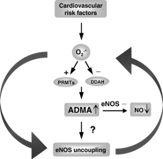
DDAH specifically hydrolyzes ADMA and L-NMMA to yield citruline and demethylamine and usually shows co-localization with NOS. Pharmacologic inhibition of DDAH activity causes accumulation of ADMA and can reverse the NO-mediated bradykinin-induced relaxation of human saphenous vein.

Two isoforms have been found in human:

  • DDAH1 (found in brain and kidney and associated with nNOS) and
  • DDAH2 (highly expressed in heart, placenta, and kidney and associated with eNOS).

DDAH2 can be upregulated by all-trans retinoic acid (atRA can increase NO production).  Increased reactive oxygen species and possibly homocysteine, a risk factor for cardiovascular disease, can decrease DDAH activity(15,16).

  • The importance of DDAH activity can also be seen in transgenic mice which overexpress DDAH, exhibiting increased NO production, increased insulin sensitivity, and reduced vascular resistance  (17).  Likewise,
  • Transgenic mice, null for the DDAH1, showed increase in blood pressure, decreased NO production, and significant increase in tissue and plasma ADMA and L-NMMA.

amdanosfigure

Figure 3.  The DDAH/ADMA/NOS cycle. Figure adapted from Cooke and Ghebremarian (1).

As mentioned in the article by Cooke and Ghebremariam, the authors state: the weight of the evidence indicates that DDAH is a worthy therapeutic target. Agents that increase DDAH expression are known, and 1 of these, a farnesoid X receptor agonist, is in clinical trials

http://www.interceptpharma.com

An alternate approach is to

  • develop an allosteric activator of the enzyme.  Although
  • development of an allosteric activator is not a typical pharmaceutical approach, recent studies indicate that this may be achievable aim(18).

References:

1.            Cooke, J. P., and Ghebremariam, Y. T. : DDAH says NO to ADMA.(2011) Arteriosclerosis, thrombosis, and vascular biology 31, 1462-1464

2.            Tran, C. T., Leiper, J. M., and Vallance, P. : The DDAH/ADMA/NOS pathway.(2003) Atherosclerosis. Supplements 4, 33-40

3.            Niebauer, J., Maxwell, A. J., Lin, P. S., Wang, D., Tsao, P. S., and Cooke, J. P.: NOS inhibition accelerates atherogenesis: reversal by exercise. (2003) American journal of physiology. Heart and circulatory physiology 285, H535-540

4.            Miyazaki, H., Matsuoka, H., Cooke, J. P., Usui, M., Ueda, S., Okuda, S., and Imaizumi, T. : Endogenous nitric oxide synthase inhibitor: a novel marker of atherosclerosis.(1999) Circulation 99, 1141-1146

5.            Wilson, A. M., Shin, D. S., Weatherby, C., Harada, R. K., Ng, M. K., Nair, N., Kielstein, J., and Cooke, J. P. (2010): Asymmetric dimethylarginine correlates with measures of disease severity, major adverse cardiovascular events and all-cause mortality in patients with peripheral arterial disease. Vasc Med 15, 267-274

6.            Kielstein, J. T., Impraim, B., Simmel, S., Bode-Boger, S. M., Tsikas, D., Frolich, J. C., Hoeper, M. M., Haller, H., and Fliser, D. : Cardiovascular effects of systemic nitric oxide synthase inhibition with asymmetrical dimethylarginine in humans.(2004) Circulation 109, 172-177

7.            Kielstein, J. T., Donnerstag, F., Gasper, S., Menne, J., Kielstein, A., Martens-Lobenhoffer, J., Scalera, F., Cooke, J. P., Fliser, D., and Bode-Boger, S. M. : ADMA increases arterial stiffness and decreases cerebral blood flow in humans.(2006) Stroke; a journal of cerebral circulation 37, 2024-2029

8.            Mittermayer, F., Krzyzanowska, K., Exner, M., Mlekusch, W., Amighi, J., Sabeti, S., Minar, E., Muller, M., Wolzt, M., and Schillinger, M. : Asymmetric dimethylarginine predicts major adverse cardiovascular events in patients with advanced peripheral artery disease.(2006) Arteriosclerosis, thrombosis, and vascular biology 26, 2536-2540

9.            Kakimoto, Y., and Akazawa, S.: Isolation and identification of N-G,N-G- and N-G,N’-G-dimethyl-arginine, N-epsilon-mono-, di-, and trimethyllysine, and glucosylgalactosyl- and galactosyl-delta-hydroxylysine from human urine. (1970) The Journal of biological chemistry 245, 5751-5758

10.          Inoue, R., Miyake, M., Kanazawa, A., Sato, M., and Kakimoto, Y.: Decrease of 3-methylhistidine and increase of NG,NG-dimethylarginine in the urine of patients with muscular dystrophy. (1979) Metabolism: clinical and experimental 28, 801-804

11.          Millward, D. J.: Protein turnover in skeletal muscle. II. The effect of starvation and a protein-free diet on the synthesis and catabolism of skeletal muscle proteins in comparison to liver. (1970) Clinical science 39, 591-603

12.          Goldberg, A. L., and St John, A. C.: Intracellular protein degradation in mammalian and bacterial cells: Part 2. (1976) Annual review of biochemistry 45, 747-803

13.          Dice, J. F., and Walker, C. D.: Protein degradation in metabolic and nutritional disorders. (1979) Ciba Foundation symposium, 331-350

14.          Ogawa, T., Kimoto, M., and Sasaoka, K.: Purification and properties of a new enzyme, NG,NG-dimethylarginine dimethylaminohydrolase, from rat kidney. (1989) The Journal of biological chemistry 264, 10205-10209

15.          Ito, A., Tsao, P. S., Adimoolam, S., Kimoto, M., Ogawa, T., and Cooke, J. P.: Novel mechanism for endothelial dysfunction: dysregulation of dimethylarginine dimethylaminohydrolase. (1999) Circulation 99, 3092-3095

16.          Stuhlinger, M. C., Tsao, P. S., Her, J. H., Kimoto, M., Balint, R. F., and Cooke, J. P. : Homocysteine impairs the nitric oxide synthase pathway: role of asymmetric dimethylarginine.(2001) Circulation 104, 2569-2575

17.          Sydow, K., Mondon, C. E., Schrader, J., Konishi, H., and Cooke, J. P.: Dimethylarginine dimethylaminohydrolase overexpression enhances insulin sensitivity. (2008) Arteriosclerosis, thrombosis, and vascular biology 28, 692-697

18.          Zorn, J. A., and Wells, J. A.: Turning enzymes ON with small molecules. (2010) Nature chemical biology 6, 179-188

Other research papers on Nitric Oxide and Cardiac Risk  were published on this Scientific Web site as follows:

The Nitric Oxide and Renal is presented in FOUR parts:

Part I: The Amazing Structure and Adaptive Functioning of the Kidneys: Nitric Oxide

Part II: Nitric Oxide and iNOS have Key Roles in Kidney Diseases

Part III: The Molecular Biology of Renal Disorders: Nitric Oxide

Part IV: New Insights on Nitric Oxide donors

Cardiac Arrhythmias: A Risk for Extreme Performance Athletes

What is the role of plasma viscosity in hemostasis and vascular disease risk?

Cardiovascular Risk Inflammatory Marker: Risk Assessment for Coronary Heart Disease and Ischemic Stroke – Atherosclerosis.

Endothelial Dysfunction, Diminished Availability of cEPCs, Increasing CVD Risk for Macrovascular Disease – Therapeutic Potential of cEPCs

Biochemistry of the Coagulation Cascade and Platelet Aggregation – Part I

Nitric Oxide Function in Coagulation

Read Full Post »

Histone Deacetylase Inhibitors Induce Epithelial-to-Mesenchymal Transition in Prostate Cancer Cells(1)

Screen Shot 2021-07-19 at 7.44.44 PM

Word Cloud By Danielle Smolyar

Authors: Dejuan Kong, Aamir Ahmad, Bin Bao, Yiwei Li, Sanjeev Banarjee, Fazlul H. Sarkar, Wayne State University School of Medicine

Reporter-Curator: Stephen J. Williams, Ph.D.

Clinically, there has not been much success in treating solid tumors with histone deacetylase inhibitors (HDACi). Histone acetylation and deacetylation play an important role in transcriptional regulation of genes and increased activity is associated with many cancers, therefore it was thought that HDAC inhibition might be fruitful as a therapy.  There have been several phase I and II clinical trials using HDACi for treatment of various malignancies, including hematological and solid malignancies(2), with most success seen in hematologic malignancies such as cutaneous T-cell lymphoma and peripheral T-cell lymphoma and little or no positive outcome with solid tumors.  Many mechanisms of resistance to HDACi in solid tumors have been described, most of which are seen with other chemotherapeutics such as increased multidrug resistance gene MDR1, increased anti-apoptotic proteins and activation of cell survival pathways(3).

A report in PLOS One by Dr. Dejuan Kong, Dr. Fazlul Sarkar, and colleagues from Wayne State University School of Medicine, demonstrate another possible mechanism of resistance to HDACi in prostate cancer, by induction of the epithelial-to-mesenchymal transition (EMT), which has been associated with the development of resistance to chemotherapies in other malignancies of epithelial origin(4,5).

EMT is an important differentiation process in embryogenesis and felt to be important in progression of cancer.  Epithelial cells will acquire a mesenchymal morphology (on plastic this looks like a cuboidal epithelial cell gaining a more flattened, elongated, tri-corner morphology; see paper Figure 1) and down-regulate epithelial markers such as cytokeratin, up-regulation of mesenchymal markers, increased migration and invasiveness in standard assays, and increased resistance to chemotherapeutics, and similarity to cancer stem cells(6-10).

ImageFigure 1. HDACis led to the induction of EMT phemotype. (A and B) PC3 cells treated with TSA and SAHA for 24 h at indicated doses.  The photomicrographs of PC3 cells treated with TSA and SAHA exhibited a fibroblastic-type phenotype, while cells treated with DMAO control displayed rounded epithelial cell morphology (original magnification, x 100). (C) Treated PC3 cells show increased mesenchymal markers vimentin and ZEB1 and F-actin reorganization.  Figure taken from Kong, D., Ahmad, A., Bao, B., Li, Y., Banerjee, S., and Sarkar, F. H. (2012) PloS one 7, e45045

In this study the authors found that treatment of prostate carcinoma cells with two different HDACis (trichostatin A (TSA) and suberoylanilide hydroxamic acid (SAHA)) induced EMT phenotype mediated through up-regulation of transcription factors ZEB1, ZEB2 and Slug, increased expression of mesenchymal markers vimentin, N-cadherin and fibronectin by promoting histone 3 acetylation on gene promoters.  In addition TSA increased the stem cell markers Sox2 and Nanog with concomitant EMT morphology and increased cell motility.

Below is the abstract of this paper(1):

ABSTRACT

Clinical experience of histone deacetylase inhibitors (HDACIs) in patients with solid tumors has been disappointing; however, the molecular mechanism of treatment failure is not known. Therefore, we sought to investigate the molecular mechanism of treatment failure of HDACIs in the present study. We found that HDACIs Trichostatin A (TSA) and Suberoylanilide hydroxamic acid (SAHA) could induce epithelial-to-mesenchymal transition (EMT) phenotype in prostate cancer (PCa) cells, which was associated with changes in cellular morphology consistent with increased expression of transcription factors ZEB1, ZEB2 and Slug, and mesenchymal markers such as vimentin, N-cadherin and Fibronectin. CHIP assay showed acetylation of histone 3 on proximal promoters of selected genes, which was in part responsible for increased expression of EMT markers. Moreover, TSA treatment led to further increase in the expression of Sox2 and Nanog in PCa cells with EMT phenotype, which was associated with cancer stem-like cell (CSLC) characteristics consistent with increased cell motility. Our results suggest that HDACIs alone would lead to tumor aggressiveness, and thus strategies for reverting EMT-phenotype to mesenchymal-to-epithelial transition (MET) phenotype or the reversal of CSLC characteristics prior to the use of HDACIs would be beneficial to realize the value of HDACIs for the treatment of solid tumors especially PCa.

Highlights of the research include:

  • TSA and SAHA induce morphologic changes  in prostate carcinoma LNCaP and PC3 cells related to EMT by microscopy as well as accumulation of mesenchymal markers ZEB1, vimentin, and F-actin reorganization shown by immunofluorescence microscopy and increased expression of these markers shown by real-time PCR
  • Western blotting showed TSA treatment resulted in hyperacetyulation of histone 3 whi8le CHIP analysis revealed increased histone 3 acetylation on the promoters of vimentin, ZEB2, Slug, and MMP2
  • Western analysis revealed that HDACi not only induced EMT but increased the expression of cancer stem cell markers associated with increased motility such as Sox2 and Nanog.  Increased cell migration was measured by Transwell migration assays and increased cell motility was measured via cell detachment assays

1.            Kong, D., Ahmad, A., Bao, B., Li, Y., Banerjee, S., and Sarkar, F. H. (2012) PloS one 7, e45045

2.            Bertino, E. M., and Otterson, G. A. (2011) Expert opinion on investigational drugs 20, 1151-1158

3.            Robey, R. W., Chakraborty, A. R., Basseville, A., Luchenko, V., Bahr, J., Zhan, Z., and Bates, S. E. (2011) Molecular pharmaceutics 8, 2021-2031

4.            Wang, Z., Li, Y., Kong, D., Banerjee, S., Ahmad, A., Azmi, A. S., Ali, S., Abbruzzese, J. L., Gallick, G. E., and Sarkar, F. H. (2009) Cancer research 69, 2400-2407

5.            Wang, Z., Li, Y., Ahmad, A., Azmi, A. S., Kong, D., Banerjee, S., and Sarkar, F. H. (2010) Drug resistance updates : reviews and commentaries in antimicrobial and anticancer chemotherapy 13, 109-118

6.            Hugo, H., Ackland, M. L., Blick, T., Lawrence, M. G., Clements, J. A., Williams, E. D., and Thompson, E. W. (2007) Journal of cellular physiology 213, 374-383

7.            Thiery, J. P. (2002) Nature reviews. Cancer 2, 442-454

8.            Kong, D., Banerjee, S., Ahmad, A., Li, Y., Wang, Z., Sethi, S., and Sarkar, F. H. (2010) PloS one 5, e12445

9.            Kong, D., Li, Y., Wang, Z., and Sarkar, F. H. (2011) Cancers 3, 716-729

10.          Bao, B., Wang, Z., Ali, S., Kong, D., Li, Y., Ahmad, A., Banerjee, S., Azmi, A. S., Miele, L., and Sarkar, F. H. (2011) Cancer letters 307, 26-36

Other research papers on Cancer and Cancer Therapeutics were published on this Scientific Web site as follows:

PIK3CA mutation in Colorectal Cancer may serve as a Predictive Molecular Biomarker for adjuvant Aspirin therapy

Nanotechnology Tackles Brain Cancer

Response to Multiple Cancer Drugs through Regulation of TGF-β Receptor Signaling: a MED12 Control

Personalized medicine-based cure for cancer might not be far away

GSK for Personalized Medicine using Cancer Drugs needs Alacris systems biology model to determine the in silico effect of the inhibitor in its “virtual clinical trial”

Lung Cancer (NSCLC), drug administration and nanotechnology

Non-small Cell Lung Cancer drugs – where does the Future lie?

Cancer Innovations from across the Web

arrayMap: Genomic Feature Mining of Cancer Entities of Copy Number Abnormalities (CNAs) Data

How mobile elements in “Junk” DNA promote cancer. Part 1: Transposon-mediated tumorigenesis.

Cancer Genomics – Leading the Way by Cancer Genomics Program at UC Santa Cruz

Closing the gap towards real-time, imaging-guided treatment of cancer patients.

Closing the gap towards real-time, imaging-guided treatment of cancer patients.

mRNA interference with cancer expression

Search Results for ‘cancer’ on this web site

Cancer Genomics – Leading the Way by Cancer Genomics Program at UC Santa Cruz

Closing the gap towards real-time, imaging-guided treatment of cancer patients.

Lipid Profile, Saturated Fats, Raman Spectrosopy, Cancer Cytology

mRNA interference with cancer expression

Pancreatic cancer genomes: Axon guidance pathway genes – aberrations revealed

Biomarker tool development for Early Diagnosis of Pancreatic Cancer: Van Andel Institute and Emory University

Is the Warburg Effect the cause or the effect of cancer: A 21st Century View?

Crucial role of Nitric Oxide in Cancer

Targeting Glucose Deprived Network Along with Targeted Cancer Therapy Can be a Possible Method of Treatment

Read Full Post »

FDA Guidelines For Developmental and Reproductive Toxicology (DART) Studies for Small Molecules. Author-Writer: Stephen J. Williams, Ph.D.

This posting is a follow-up on the Report on the Fall Mid-Atlantic Society of Toxicology Meeting “Reproductive Toxicology of Biologics: Challenges and Considerations post and gives a brief synopsis of the current state of FDA regulatory guidelines with respect to DART studies on small molecule (non-biological based) therapeutics.    The following is adapted from the book Principles and Methods of Toxicology by Dr. A Wallace Hayes (1) and is an excellent reference on reproductive toxicology and testing methods.

Chemical insult occurs to the human reproductive system at a multitude of stages in development and the life cycle, leading to the extensive testing which must be performed to diligently the reproductive and development toxicity of a chemical/drug.  Abnormalities and toxic manifestations in the offspring may result from insult to the adult reproductive (either female or male) and neuroendocrine systems, as well as damage to the embryo resulting in embryolethality, fetus at any period during organogenesis, juvenile development or, in the case of certain antibody therapies, immune system development.  The latter, toxic insult to the developing immune system could possibly be manifested as either an immune defect in the newborn or, later in life, as tolerance to said therapy.  It is estimated that exposure to the pregnant woman, of either environmental contaminants or drug, is significant.  It is estimated that a mother may be taking an average of 8-9 different drug preparations, mostly over the counter preparations such as antacids, vitamin preparations, cathartics etc. with the maximal drug intake occurring between 24 and 36 weeks of gestation.

Toxic insult to the developing embryo is dependent on

  • Fetal development stage during drug/chemical exposure
  • Maternal/placental xenobiotic metabolism
  • Pharmacokinetic parameters affecting bioavailability and fetal/maternal drug binding

The following table shows the dependency of developmental stage to teratogenicity: adapted from J. Manson, H. Zenick, and R.D. Costlow from Principles and Methods of Toxicology.

Developmental Stage Major Susceptibility
Preimplantation Embryolethality
Organogenesis Births defects; embryolethality
Fetal Growth retardation, fetal death, functional deficits
Neonatal Growth retardation, nervous system alterations, immune and endocrine systems

It is not generally accepted that there is a dose dependency of teratogenesis however most teratogens have specific mechanisms of action and teratogenic effects occur at much lower doses than result in maternal toxicity.   However, the developmental toxicity may be manifested later in life, including as reproductive toxicity affecting adult fertility and familial generations.

FDA Guidelines for DART Studies on Non-Biologics (Small Molecule Therapeutics)

The basic design for DART studies incorporate the aforementioned principles of tetralogy:

  • developmental stage of fetal exposure
  • parental effects on reproduction and development
  • toxicity may be manifested over multiple generations including fertility rates

Therefore two designs are generally used for DART studies

  1. exposure across several generations
  2. exposure during one generation

FDA requires one control group and two treatment groups, and evaluation of at least two species.  However, most studies will use two rodent and one nonrodent species.

Multigenerational Design

Multigenerational DART studies are conducted for compounds likely to concentrate in the body following long-term exposure.  Examples of types of compounds include pesticides and food additives.

Figure 1.  General Design of a Multigenerational DART study.  Weanlings (30-30 days of age) from the parental generation are treated for a period up to 60 days. At 100-120 days of parental generation, animals are mated.  Fx = filialx .

Three Segment, Single Generation Tests

The single generation design is more suitable for DART studies on drugs, as most therapeutic would be taken over short periods (during pregnancy) and have relatively short half-lives in the body.  FDA guidelines separate these studies in three phases:

I.            Phase I: evaluation of fertility and general reproductive performance

II.            Phase II: assessment of teratogenicity and embryotoxicity

III.            Phase III: peri- and postnatal evaluations.

All figures are adapted from Principles and Methods of Toxicology.(1)

FDA guidelines Guidance for Industry Reproductive and Developmental Toxicities —Integrating Study Results to Assess Concerns can be found at: http://www.fda.gov/downloads/Drugs/GuidanceComplianceRegulatoryInformation/Guidances/ucm079240.pdf

FDA Guideline for reproductive toxicity testing for small molecule therapeutics can be found at:

http://www.fda.gov/downloads/Drugs/GuidanceComplianceRegulatoryInformation/Guidances/ucm074950.pdf

1.            Hayes, A. W. (1986) Principles and Methods of Toxicology, Raven Press, New York

Other research papers on Pharmaceutical Intelligence and Reproductive Biology, Bio Insrumentation, Endocrinology Genetics were published on this Scientific Web site as follows

Non-small Cell Lung Cancer drugs – where does the Future lie?

Reboot evidence-based medicine and reconsider the randomized, placebo-controlled clinical trial

Every sperm is sacred: Sequencing DNA from individual cells vs “humans as a whole.”

Leptin and Puberty

Gene Trap Mutagenesis in Reproductive Research

Genes involved in Male Fertility and Sperm-egg Binding

Hope for Male Contraception: A small molecule that inhibits a protein important for chromatin organization can cause reversible sterility in male mice

Pregnancy with a Leptin-Receptor Mutation

The contribution of comparative genomic hybridization in reproductive medicine

Sperm collide and crawl the walls in chaotic journey to the ovum

Impact of evolutionary selection on functional regions: The imprint of evolutionary selection on ENCODE regulatory elements is manifested between species and within human populations

Biosimilars: CMC Issues and Regulatory Requirements

Biosimilars: Intellectual Property Creation and Protection by Pioneer and by Biosimilar Manufacturers

Assisted Reproductive Technology Cycles and Cumulative Birth Rates

Innovations in Bio instrumentation in Reproductive Clinical and Male Fertility Labs in the US

Increased risks of obesity and cancer, Decreased risk of type 2 diabetes: The role of Tumor-suppressor phosphatase and tensin homologue (PTEN)

Guidelines for the welfare and use of animals in cancer research

Every sperm is sacred: Sequencing DNA from individual cells vs “humans as a whole.”

Read Full Post »

What could transform an underdog into a winner?

Author and Curator: Dror Nir, PhD

Many feedbacks to my last post reflected radiologists’ perception of ultrasound as a low-tech, unreliable imaging device.

Ultrasounds most manifested limitation by radiologists is that its performance is too-much user-dependent. This opinion finds support in numerous clinical studies concluding that ultrasound-based assessment of a cancer patient varies with the operator.

How come that an imaging technology that is not only  low-cost, simple to operate and risk-free to the patient, but has also gained a leading position in certain domain, like obstetrics,  is perceived as the underdog when it comes  to cancer assessment? Could it be because of its positioning as a “multi-purpose” system, which requires only very basic training?

If indeed this is the case, it doesn’t require “rocket-science” to turn it around. It only needs designing dedicated ultrasound machines who offer a comprehensive solution to one specific clinical need. Using such machines will require highly skilled operators who will enjoy a superior workflow, reporting tools and proven clinical guidelines.

The unsatisfactory reality of mammography-based breast cancer screening, as evident by epidemiology data and expert-panels’ reports, opens the opportunity to transform ultrasound into a winner in the niche-market of breast cancer screening and diagnosis. It’s a significant market that justifies the investment in ultrasound systems dedicated to detection and characterisation of breast cancer lesions.

No doubt, that the ability to provide accurate and standardized interpretation of such ultrasound systems’ scans is a pre-requisite. Ultrasound-based tissue characterisation is a must for any application aiming at standardized image interpretation. A sample out-of present ultrasound-based technologies aiming at providing some level of tissue-characterisation are listed below. Recent clinical studies show promising results using these technologies. It is worth watching carefully to see if any of those could be part of a future ultrasound-based solution to breast cancer screening.

Solid Breast Lesions: Clinical Experience with US-guided Diffuse Optical Tomography Combined with Conventional US

Results: Of the 136 biopsied lesions, 54 were carcinomas and 82 were benign. The average total hemoglobin concentration in the malignant group was 223.3 μmol/L ± 55.8 (standard deviation), and the average hemoglobin concentration in the benign group was 122.5 μmol/L ± 80.6 (P = .005). When the maximum hemoglobin concentration of 137.8 μmol/L was used as the threshold value, the sensitivity, specificity, positive predictive value, negative predictive value, and accuracy of DOT with US localization were 96.3%, 65.9%, 65.0%, 96.4%, and 76.5%, respectively. The sensitivity, specificity, positive predictive value, negative predictive value, and accuracy of conventional US were 96.3%, 92.6%, 89.7%, 97.4%, and 93.4%, respectively. The sensitivity, specificity, positive predictive value, negative predictive value, and accuracy of conventional US combined with DOT were 100%, 93.9%, 91.5%, 100%, and 96.3%, respectively.

Conclusion: US-guided DOT combined with conventional US improves accuracy compared with DOT alone.

Breast Lesions: Quantitative Elastography with Supersonic Shear Imaging—Preliminary Results


Results: All breast lesions were detected at Supersonic Shear Imaging. Malignant lesions exhibited a mean elasticity value of 146.6 kPa ± 40.05 (standard deviation), whereas benign ones had an elasticity value of 45.3 kPa ± 41.1 (P < .001). Complicated cysts were differentiated from solid lesions because they had elasticity values of 0 kPa (no signal was retrieved from liquid areas).

Conclusion: Supersonic Shear Imaging provides quantitative elasticity measurements, thus adding complementary information that potentially could help in breast lesion characterization with B-mode US.

 Distinguishing Benign from Malignant Masses at Breast US: Combined US Elastography and Color Doppler US—Influence on Radiologist Accuracy

Results: The Az of B-mode US, US elastography, and Doppler US (average, 0.844; range, 0.797–0.876) was greater than that of B-mode US alone (average, 0.771; range, 0.738–0.798) for all readers (P = .001 for readers 1, 2, and 3; P < .001 for reader 4; P = .002 for reader 5). When both elastography and Doppler scores were negative, leading to strict downgrading, the specificity increased for all readers from an average of 25.3% (75.4 of 298; range, 6.4%–40.9%) to 34.0% (101.2 of 298; range, 26.5%–48.7%) (P < .001 for readers 1, 2, 4, and 5; P = .016 for reader 3) without a significant change in sensitivity.

Conclusion: Combined use of US elastography and color Doppler US increases both the accuracy in distinguishing benign from malignant masses and the specificity in decision-making for biopsy recommendation at B-mode US.

Evaluation of breast lesions by contrast enhanced ultrasound: Qualitative and quantitative analysis

A 57-year-old woman with a no-palpable lesion in the outer upper quadrant of left breast. (a) Gray scale image show an indistinct, hypo-echoic lesion. (b) Contrast enhanced image obtained 35 s after contrast agent injection showing a homogeneously and hyper-enhanced lesion. (c) Micro flow image obtained 38 s after contrast agent injection showing the enhanced mass with several radial vessels (arrow). (d) The time-intensity curve analysis show the peak intensity is 145.69 (intensity/1000), time to peak is 15.08 s, ascending slope is 8.98, descending slope is 1.03, the area under the curve is 7783.34. Pathologic analyses show this is an invasive ductal carcinoma.


Results: Histopathologic analysis of the 91 lesions revealed 44 benign and 47 malignant. For qualitative analysis, benign and malignant lesions differ significantly in enhancement patterns (p < 0.05). Malignant lesions more often showed heterogeneous and centripetal enhancement, whereas benign lesions mainly showed homogeneous and centrifugal enhancement. The detectable rate of peripheral radial or penetrating vessels was significantly higher in malignant lesions than in benign ones (p < 0.001). For quantitative analysis, malignant lesions showed significantly higher (p = 0.031) and faster enhancement (p = 0.025) than benign ones, and its time to peak was significantly shorter (p = 0.002). The areas under the ROC curve for qualitative, quantitative and combined analysis were 0.910 (Az1), 0.768 (Az2) and 0.926(Az3) respectively. The values of Az1 and Az3 were significantly higher than that for Az2 (p = 0.024 and p = 0.008, respectively). But there was no significant difference between the values of Az1 and Az3 (p = 0.625).

Conclusions: The diagnostic performance of qualitative and combined analysis was significantly higher than that for quantitative analysis. Although quantitative analysis has the potential to differentiate benign from malignant lesions, it has not yet improved the final diagnostic accuracy.

 Breast HistoScanning: the development of a novel technique to improve tissue characterization during breast ultrasound

Results: In 17 normal testing volumes, 3% of isolated voxels were classified as abnormal. In 15 abnormal testing volumes, the subclassifiers differentiated between malignant and benign tissue. BHS in benign tissue showed <1% abnormal voxels in cyst, hamartoma, papilloma and benign fibrosis. The fibroadenomas differed showing <5% and <24% abnormal voxels. Abnormal voxels in cancers increased with the volume of cancer at pathology.

Conclusions: HistoScanning reliably discriminated normal from abnormal tissue and could distinguish between benign and malignant lesions.

Written by: Dror Nir, PhD

Read Full Post »

Report on the Fall Mid-Atlantic Society of Toxicology Meeting “Reproductive Toxicology of Biologics: Challenges and Considerations.  Author, Reporter: Stephen J. Williams, Ph.D.

The fall 2012 Meeting of the Mid-Atlantic Society of Toxicology (MASOT) focused on the challenges and solutions in developing proper Development and Reproductive Toxicology (DART) studies with regards to the newer classes of bio-therapeutics such as vaccines, antibody-based therapies, and viral-based therapies.  The full meeting and MASOT links can be found at http://www.masot.org.   The overall synopsis of the meeting talks agreed, that although the general aim and design of DART studies for biological are very similar to DART studies for small molecule therapeutics, it is more necessary to take into consideration the pharmacodynamics, pharmacokinetic differences between biologics and small molecules.   In addition it is imperative to use pharmacologically-relevant species, such as non-rodent (guinea pig and non-human primate). The meeting was highlighted by the keynote speaker, Dr. A. Wallace Hayes, renowned board-certified toxicologist, committee and expert panel member for National Academy of Sciences, NIEHS, EPA and Department of Defense, and editor of well-known textbooks including Principles and Methods of Toxicology.  Dr. Hayes discussed a timeline of milestones in the field of toxicology.

The following are the meeting talk abstracts as well as notes for each presenter.

What’s So Different About DART Assessment of Biologics? Christopher Bowman Ph.D., DABT (Pfizer, Inc.)

Abstract:  The aim of developmental and reproductive toxicity (DART) safety assessment of a biologic is no different from that of a small molecule. Both cases consist of evaluating the potential for maternal toxicity, pre- and postnatal development toxicity (including juvenile toxicity) and effects of fertility (reproduction).  The differences lie in the in the product attributes of a specific biologic, the pharmacological response, the potential for undesirable toxicities and how these product attributes influence and are influenced by the biology.  Thus the primary challenge for developing a DART strategy for a biologic are derived from the complexities of these biomolecules and how that dictates a case-by-case strategy for appropriately evaluating the potential for developmental and reproductive toxicity. Most protein biologics have very limited potential for off-target toxicities, but this is not necessarily the case for other modalities such as anti-sense oligonucleotides and antibody-drug-conjugates.  In these cases, off-target toxicities can be a major feature of the DART safety assessment.  The most noticeable difference in DART assessment of biologics is the need to conduct these studies in pharmacologically relevant species and how that can influence the overall nonclinical strategy (including DART).  This has led to increased use of non-human primates as a model system and led to optimizations of this model for this purpose and revisions to international guidelines.

Notes:   Dr. Bowman emphasized the need to understand the type of biological you are testing and to both devise DART studies based on this information, additional endpoint you may want, as well as carefully choosing the correct species most relevant to the biologic.  He highlighted general differences between small molecules versus a biologic with respect to their pharmacology.  These differences are summarized in the Table below:

  Small Molecule Biologic-based therapy
Species specificity Low High
Route of administration Usually oral Parental
ADME (PK, bio-distribution etc.) Wide distribution Low distribution

He noted that clinical trials for biologics rarely include reproductive toxicity so the preclinical DART study is of utmost importance.  He also emphasized that currently, the FDA requires two species for DART testing of small molecule therapies (usually one rodent and one non-rodent).  However this is not possible with many biologics as species is to be taken in consideration when designing a meaningful DART study.  Study designs can be like most DART studies but want to have a steady exposure during fetal organogenesis, use high doses (10 times the clinical dose) to achieve maximal pharmacology, confirm exposure to fetus and to F1 generation, and determine embryolethality.  Some biologics like interferon and insulin-growth factor receptor (IGFR) antagonists are fetal abortifactants. In fact Lucentis (Ranibizumab) and Macugen (Pegaptanib) were approved with no or little DART studies, however these drugs showed reproductive toxicity, resulting in warning concerning pregnancy on the label. Also important is the effect on the immune system and reproductive system of offspring, as well as the pharmacodynamics profile in the offspring.

Species Selection for Reproductive and Developmental Toxicity Testing of Biologics; Elise M. Lewis, Ph.D. (Charles River Preclinical Services)

Abstract:  Regulatory guidelines for developmental and reproductive toxicology studies require selection of “relevant” animal models as determined by kinetic, pharmacological, and preceding toxicological data.  Rats, mice, and rabbits are the preferred animal models for these studies based on historical experience and well-established procedures and study protocols.  However, due to species specificity and immunogenicity issues, developmental and reproductive toxicology testing for biologics is limited to a pharmacologically relevant animal model as described in the ICH s6 guideline.

Notes:  Dr. Lewis notes that DART studies in guinea pigs and hamsters represent a cost effective alternative to large animal models as well as the benefit of shorter duration and ability to assess mating behavior.  She also notes that reproductive toxicology of vaccines should be done in an animal model that can elicit an immune-response to the vaccine, especially to determine any maternal-fetal interaction.  For example, a vaccine may be directed to a maternal protein which when suppressed, may negatively impact the developing fetus.  However it is important to remember that guinea pigs can spontaneously abort so it is good to have proper control arms of a substantial size in order to statistically determine the impact of those spontaneous abortions.


Placental Transfer of an Adnectin Protein During Organogenesis in Guinea Pigs Using a Radiolabeled Methodology; Lakshmi Sivaraman, Ph.D. (Bristol-Myers Squibb)

Abstract:  Knowledge regarding the placental transfer of large molecular weight therapeutics is important to support the enrollment of women of childbearing potential in clinical trials.  There is limited information in the scientific literature that reports the extent to which the conceptus is exposed to these large molecules during organogenesis.  Placental transfer of large therapeutics has been difficult to quantify, due to limited blood volumes that can be obtained from the embryo, as well as insufficient assay sensitivity.  Thus, it is possible that embryos are exposed to pharmacologically active concentrations after maternal drug exposure. We have adopted a radiolabeled approach to quantitate embryo-fetal exposure of a novel protein therapeutic platform (adnectins). Adnectins are fibronectin-based proteins containing domains engineered to bind to targets of therapeutic interests.

Notes: Adnectins molecular weight is typically less than monoclonal antibodies and while IgG is not transferred in great quantity past the placental barrier there have been studies in human indicating maternal-fetal transfer of monoclonal antibodies.  This is particularly important for two reasons:  the monoclonal interacts with a target important in development, or the fetal immune system could be augmented.  Their work will be published in Drug Metabolism and Disposition.  In general Dr. Siveraman engineered a radiolabel on adnectin and used different detection methods to quantify the fetal exposure to a single maternal dose.  Dr. Siverman was able to detect radiolabel in the fetus however it is not clear whether this is a significant amount.

Reproductive Toxicity Testing for Biological Products in Nonhuman Primates: Evolution and Current Perspectives: Gary J. Chellman, Ph.D., DABT (Charles River Preclinical Services)

Notes:  Dr. Chellman gave a review of the current trends being driven by regulatory agencies with regard to nonhuman primate DART studies of biopharmaceuticals.  He noted that an advantage using nonhuman primates were the close physiologic resemblance to humans and because a large animal could monitor pregnancy over time using ultrasound technology.  In general, Dr. Chellman spoke about new study designs which not only reduce the number of animals required but also significantly reduce costs.  For example, a DART study which cost upward of $750,000 now can be done for as little as $350,000.  Dr. Kary Thompson of Bristol Myers Squibb then gave a talk about use of these new enhanced designs to determine reproductive toxicity issues with ipilimumab (Yervoy).

Other research papers on Pharmaceutical Intelligence and Reproductive Biology, Bio Insrumentation, Endocrinology Genetics were published on this Scientific Web site as follows

Non-small Cell Lung Cancer drugs – where does the Future lie?

Reboot evidence-based medicine and reconsider the randomized, placebo-controlled clinical trial

Every sperm is sacred: Sequencing DNA from individual cells vs “humans as a whole.”

Leptin and Puberty

Gene Trap Mutagenesis in Reproductive Research

Genes involved in Male Fertility and Sperm-egg Binding

Hope for Male Contraception: A small molecule that inhibits a protein important for chromatin organization can cause reversible sterility in male mice

Pregnancy with a Leptin-Receptor Mutation

The contribution of comparative genomic hybridization in reproductive medicine

Sperm collide and crawl the walls in chaotic journey to the ovum

Impact of evolutionary selection on functional regions: The imprint of evolutionary selection on ENCODE regulatory elements is manifested between species and within human populations

Biosimilars: CMC Issues and Regulatory Requirements

Biosimilars: Intellectual Property Creation and Protection by Pioneer and by Biosimilar Manufacturers

Assisted Reproductive Technology Cycles and Cumulative Birth Rates

Innovations in Bio instrumentation in Reproductive Clinical and Male Fertility Labs in the US

Increased risks of obesity and cancer, Decreased risk of type 2 diabetes: The role of Tumor-suppressor phosphatase and tensin homologue (PTEN)

Guidelines for the welfare and use of animals in cancer research

Every sperm is sacred: Sequencing DNA from individual cells vs “humans as a whole.”


Read Full Post »

Stephen J. Williams, Ph.D. Writer, Curator

Rational Design of Allosteric Inhibitors and Activators Using the Population-Shift Model: In Vitro Validation and Application to an Artificial Biosensor.(1)

The population-shift mechanism allows for the re-engineering of biosensors utilizing the concept of allosterism to allow for a structure-based switching on/off capacity into biosensors, “smart-biomaterials, and other artificial biotechnologies.  A fundamental problem in the design of valuable biosensors has been limited number of biomolecules that produce enough signal (for example emission of light, etc.) upon binding to its target.  However this issue has been resolved with the development of biosensors in which target binding is transduced into a quantifiable optical or electrochemical signal after coupling with conformational changes in the receptor (for review see (2)).

There are a few advantages to this biosensor design:

  • Works well in complex samples, such as blood, serum; Low background noise from nonspecific adsorption from interfering biomolecules
  • Supports real-time monitoring- allosteric biosensors do not rely on additional reagents and are rapidly reversible
  • Binding of the receptors is dependent on an unfavorable conformational change, so there is possibility to fine tune this conformational switch.

Concept of allosterism

Allosterism is generally defined as a change in the activity and conformation of an enzyme/protein resulting from the binding of a compound at a site on the enzyme other than the active binding site.  Allosterism plays a critical role in the control and integration of molecular events in biological systems.  Frequently, allosterism is seen with multisubunit proteins/enzymes, where subunit interaction is necessary for allosteric effects, and is distal to the binding site.  Examples of allosteric systems include hemoglobin, phosphofructose kinase and many

NAD+ -dependent dehydrogenases.  For example, the binding of O2 to hemoglobin is enhanced the binding of addition O2, the Bohr effect (the affinity of hemoglobin to O2 depends on H+), and the metabolic product diphosphoglycerate regulates O2 binding.

Types of DNA Biosensors

DNA-based biosensors rely on the hybridization of complementary DNA.  Many optical biosensors based on the phenomenon of surface plasmon resonance (SPR) utilize a property of and other materials; specifically that a thin layer of gold on a high refractive index glass surface can absorb laser light, producing electron waves (surface plasmons) on the gold surface. This occurs only at a specific angle and wavelength of incident light and is highly dependent on the surface of the gold, such that binding of a target analyte to a receptor on the gold surface produces a measurable signal.

Electrochemical biosensors are normally based on enzymatic catalysis of a reaction that produces or consumes electrons (such enzymes are rightly called redox enzymes). The sensor substrate usually contains three electrodes; a reference electrode, a working electrode and a counter electrode. The target analyte is involved in the reaction that takes place on the active electrode surface, and the reaction may cause either electron transfer across the double layer (producing a current) or can contribute to the double layer potential (producing a voltage). We can either measure the current (rate of flow of electrons is now proportional to the analyte concentration) at a fixed potential or the potential can be measured at zero current (this gives a logarithmic response). The label-free and direct electrical detection of small peptides and proteins is possible by their intrinsic charges using bio-functional ion-sensitive field-effect transistors.

Piezoelectric sensors utilize crystals which undergo an elastic deformation when an electrical potential is applied to them. An alternating potential produces a standing wave in the crystal at a characteristic frequency. This frequency is highly dependent on the elastic properties of the crystal, such that if a crystal is coated with a biological recognition element the binding of a (large) target analyte to a receptor will produce a change in the resonance frequency, which gives a binding signal. In a mode that uses surface acoustic waves (SAW), the sensitivity is greatly increased.

Type Biological Element Transducer
OpticalFiber Optics

Surface plasmon resonance

Biomolecular interactionAnalysis

Raman spectroscopy

DNA Optical fiberResonant mirror

BIAcore

SERG probe

Electrochemical DNA Carbon paste electrodes
Piezoelectric     FrequencyAcoustics DNA CrystalsCrystals

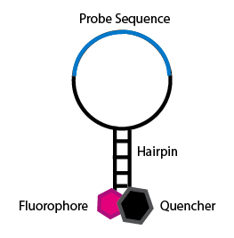
The most popular of optical DNA biosensors is molecular beacons, DNA probes containing a fluorescent moiety and a quencher of on the same DNA strand. This probe has an internal complementary sequence so as the DNA folds into a secondary structure, most likely a stem-loop or hairpin structure, so the fluor and quencher are held in close proximity, quenching the fluorescent signal.  Target hybridization opens up the stem-loop structure, thereby emitting the fluorescent signal. A typical molecular beacon probe is 25 nucleotides long. A typical molecular beacon structure can be divided in 4 parts:

  • Loop: This is the 18–30 base pair region of the molecular beacon which is complementary to the target sequence.
  • Stem: The beacon stem is formed by the attachment, to both termini of the loop, of two short (5 to 7 nucleotide residues) oligonucleotides that are complementary to each other.
  • 5′ fluorophore: At the 5′ end of the molecular beacon, a fluorescent dye is covalently attached.
  • 3′ quencher (non fluorescent): The quencher dye is covalently attached to the 3′ end of the molecular beacon. When the beacon is in closed loop shape, the quencher resides in proximity to the fluorophore, which results in quenching the fluorescent emission of the latter.

Structure of a molecular beacon. Description and figure from Wikipedia (5).

Common applications of DNA biosensors include cDNA microarray and Affymetrix GeneChip™ technology.

Ricci et al. provide a proof-of –principle paper to demonstrate how allosteric switching can be introduced into biosensors(1). The authors engineered allosteric inhibition into a molecular beacon by the addition of two single-stranded tails that serve as an allosteric site where binding of an inhibitor sequence would bridge the two tails and prevent target binding (holding the probe in the inactivated state).  Using this approach the authors demonstrated over a three-fold increase in the dynamic range of the beacon.

The authors also demonstrated this effect, with an allosterically activated biosensor in which “allosteric activation was engineered into a molecular beacon using one single-stranded tail as an allosteric binding site.  The activator sequence binding to this tail partially invades the stem, destabilizing the nonbinding state and thus improving the target affinity.”  Thus this population-shift mechanism allows for the design of sensors that can be allosterically activated using activators that destabilize the beacon’s nonbinding conformation, increasing the beacon’s dynamic range without compromising target specificity. Finally the authors suggest that population-shift mechanisms can be engineered into many different types of “switching” biosensors including aptamer-based and protein-based sensors (3,4).

1.            Ricci, F., Vallee-Belisle, A., Porchetta, A., and Plaxco, K. W. (2012) Journal of the American Chemical Society 134, 15177-15180

2.            Vallee-Belisle, A., and Plaxco, K. W. (2010) Current opinion in structural biology 20, 518-526

3.            White, R. J., Rowe, A. A., and Plaxco, K. W. (2010) The Analyst 135, 589-594

4.            Kohn, J. E., and Plaxco, K. W. (2005) Proceedings of the National Academy of Sciences of the United States of America 102,   10841-10845

5.            http://en.wikipedia.org/wiki/Molecular_beacon

Other research papers on Biosensors were published on this Scientific Web site as follows:

Measuring glucose without needle pricks: nano-sized biosensors made the test easy

New Definition of MI Unveiled, Fractional Flow Reserve (FFR)CT for Tagging Ischemia

New Drug-Eluting Stent Works Well in STEMI

Sensor detects glucose in saliva, tears for diabetes testing

Synthesizing Synthetic Biology: PLOS Collections

Competition in the Ecosystem of Medical Devices in Cardiac and Vascular Repair: Heart Valves, Stents, Catheterization Tools and Kits for Open Heart and Minimally Invasive Surgery (MIS)

Read Full Post »

The unfortunate ending of the Tower of Babel construction project and its effect on modern imaging-based cancer patients’ management

The unfortunate ending of the Tower of Babel construction project and its effect on modern imaging-based cancer patients’ management

Curator: Dror Nir, PhD


The story of the city of Babel is recorded in the book of Genesis 11 1-9. At that time, everyone on earth spoke the same language.

Picture: Pieter Bruegel the Elder: The Tower of Babel_(Vienna)

It is probably safe to assume that medical practitioners at that time were reporting the status of their patients in a standard manner. Although not mentioned, one might imagine that, at that time, ultrasound or MRI scans were also reported in a standard and transferrable manner. The people of Babel noticed the potential in uniform communication and tried to build a tower so high that it would  reach the gods. Unfortunately, God did not like that, so he went down (in person) and confounded people’s speech, so that they could not understand each another. Genesis 11:7–8.

This must be the explanation for our inability to come to a consensus on reporting of patients’ imaging-outcome. Progress in development of efficient imaging protocols and in clinical management of patients is withheld due to high variability and subjectivity of clinicians’ approach to this issue.

Clearly, a justification could be found for not reaching a consensus on imaging protocols: since the way imaging is performed affects the outcome, (i.e. the image and its interpretation) it takes a long process of trial-and-error to come up with the best protocol.  But, one might wonder, wouldn’t the search for the ultimate protocol converge faster if all practitioners around the world, who are conducting hundreds of clinical studies related to imaging-based management of cancer patients, report their results in a standardized and comparable manner?

Is there a reason for not reaching a consensus on imaging reporting? And I’m not referring only to intra-modality consensus, e.g. standardizing all MRI reports. I’m referring also to inter-modality consensus to enable comparison and matching of reports generated from scans of the same organ by different modalities, e.g. MRI, CT and ultrasound.

As developer of new imaging-based technologies, my personal contribution to promoting standardized and objective reporting was the implementation of preset reporting as part of the prostate-HistoScanning product design. For use-cases, as demonstrated below, in which prostate cancer patients were also scanned by MRI a dedicated reporting scheme enabled matching of the HistoScanning scan results with the prostate’s MRI results.

The MRI reporting scheme used as a reference is one of the schemes offered in a report by Miss Louise Dickinson on the following European consensus meeting : Magnetic Resonance Imaging for the Detection, Localisation, and Characterisation of Prostate Cancer: Recommendations from a European Consensus Meeting, Louise Dickinson a,b,c,*, Hashim U. Ahmed a,b, Clare Allen d, Jelle O. Barentsz e, Brendan Careyf, Jurgen J. Futterer e, Stijn W. Heijmink e, Peter J. Hoskin g, Alex Kirkham d, Anwar R. Padhani h, Raj Persad i, Philippe Puech j, Shonit Punwani d, Aslam S. Sohaib k, Bertrand Tomball,Arnauld Villers m, Jan van der Meulen c,n, Mark Emberton a,b,c,

http://www.europeanurology.com/article/S0302-2838(10)01187-5

Image of MRI reporting scheme taken from the report by Miss Louise Dickinson

The corresponding HistoScanning report is following the same prostate segmentation and the same analysis plans:


Preset reporting enabling matching of HistoScanning and MRI reporting of the same case.

It is my wish that already in the near-future, the main radiology societies (RSNA, ESR, etc..) will join together to build the clinical Imaging’s “Tower of Babel” to effectively address the issue of standardizing reporting of imaging procedures. This time it will not be destroyed…:-)

Read Full Post »

Knowing the tumor’s size and location, could we target treatment to THE ROI by applying imaging-guided intervention?

Knowing the tumor’s size and location, could we target treatment to THE ROI by applying imaging-guided intervention?

Author: Dror Nir, PhD

Advances in techniques for cancer lesions’ detection and localisation [1-6] opened the road to methods of localised (“focused”) cancer treatment [7-10].  An obvious challenge on the road is reassuring that the imaging-guided treatment device indeed treats the region of interest and preferably, only it.

A step in that direction was taken by a group of investigators from Sunnybrook Health Sciences Centre, University of Toronto, Ontario, Canada who evaluate the feasibility and safety of magnetic resonance (MR) imaging–controlled transurethral ultrasound therapy for prostate cancer in humans [7]. Their study’s objective was to prove that using real-time MRI guidance of HIFU treatment is possible and it guarantees that the location of ablated tissue indeed corresponds to the locations planned for treatment. Eight eligible patients were recruited.


The setup


Treatment protocol


The result


“There was excellent agreement between the zone targeted for treatment and the zone of thermal injury, with a targeting accuracy of ±2.6 mm. In addition, the temporal evolution of heating was very consistent across all patients, in part because of the ability of the system to adapt to changes in perfusion or absorption properties according to the temperature measurements along the target boundary.”


Technological problems to be resolved in the future:

“Future device designs could incorporate urinary drainage during the procedure, given the accumulation of urine in the bladder during treatment.”

“Sufficient temperature resolution could be achieved only by using 10-mm-thick sections. Our numeric studies suggest that 5-mm-thick sections are necessary for optimal three-dimensional conformal heating and are achievable by using endorectal imaging coils or by performing the treatment with a 3.0-T platform.”

Major limitation: “One of the limitations of the study was the inability to evaluate the efficacy of this treatment; however, because this represents, to our knowledge, the first use of this technology in human prostate, feasibility and safety were emphasized. In addition, the ability to target the entire prostate gland was not assessed, again for safety considerations. We have not attempted to evaluate the effectiveness of this treatment for eradicating cancer or achieving durable biochemical non-evidence of disease status.”

References

  1. SIMMONS (L.A.M.), AUTIER (P.), ZATURA (F.), BRAECKMAN (J.G.), PELTIER (A.), ROMICS (I.), STENZL (A.), TREURNICHT (K.), WALKER (T.), NIR (D.), MOORE (C.M.), EMBERTON (M.). Detection, localisation and characterisation of prostate cancer by Prostate HistoScanning.. British Journal of Urology International (BJUI). Issue 1 (July). Vol. 110, Page(s): 28-35
  2. WILKINSON (L.S.), COLEMAN (C.), SKIPPAGE (P.), GIVEN-WILSON (R.), THOMAS (V.). Breast HistoScanning: The development of a novel technique to improve tissue characterization during breast ultrasound. European Congress of Radiology (ECR), A.4030, C-0596, 03-07/03/2011.
  3. Hebert Alberto Vargas, MD, Tobias Franiel, MD,Yousef Mazaheri, PhD, Junting Zheng, MS, Chaya Moskowitz, PhD, Kazuma Udo, MD, James Eastham, MD and Hedvig Hricak, MD, PhD, Dr(hc) Diffusion-weighted Endorectal MR Imaging at 3 T for Prostate Cancer: Tumor Detection and Assessment of Aggressiveness. June 2011 Radiology, 259,775-784.
  4. Wendie A. Berg, Kathleen S. Madsen, Kathy Schilling, Marie Tartar, Etta D. Pisano, Linda Hovanessian Larsen, Deepa Narayanan, Al Ozonoff, Joel P. Miller, and Judith E. Kalinyak Breast Cancer: Comparative Effectiveness of Positron Emission Mammography and MR Imaging in Presurgical Planning for the Ipsilateral Breast Radiology January 2011 258:1 59-72.
  5. Anwar R. Padhani, Dow-Mu Koh, and David J. Collins Reviews and Commentary – State of the Art: Whole-Body Diffusion-weighted MR Imaging in Cancer: Current Status and Research Directions Radiology December 2011 261:3 700-718
  6. Eggener S, Salomon G, Scardino PT, De la Rosette J, Polascik TJ, Brewster S. Focal therapy for prostate cancer: possibilities and limitations. Eur Urol 2010;58(1):57–64).
  7. Rajiv Chopra, PhD, Alexandra Colquhoun, MD, Mathieu Burtnyk, PhD, William A. N’djin, PhD, Ilya Kobelevskiy, MSc, Aaron Boyes, BSc, Kashif Siddiqui, MD, Harry Foster, MD, Linda Sugar, MD, Masoom A. Haider, MD, Michael Bronskill, PhD and Laurence Klotz, MD. MR Imaging–controlled Transurethral Ultrasound Therapy for Conformal Treatment of Prostate Tissue: Initial Feasibility in Humans. October 2012 Radiology, 265,303-313.
  8. Black, Peter McL. M.D., Ph.D.; Alexander, Eben III M.D.; Martin, Claudia M.D.; Moriarty, Thomas M.D., Ph.D.; Nabavi, Arya M.D.; Wong, Terence Z. M.D., Ph.D.; Schwartz, Richard B. M.D., Ph.D.; Jolesz, Ferenc M.D.  Craniotomy for Tumor Treatment in an Intraoperative Magnetic Resonance Imaging Unit. Neurosurgery: September 1999 – Volume 45 – Issue 3 – p 423
  9. Medel, Ricky MD,  Monteith, Stephen J. MD, Elias, W. Jeffrey MD, Eames, Matthew PhD, Snell, John PhD, Sheehan, Jason P. MD, PhD, Wintermark, Max MD, MAS, Jolesz, Ferenc A. MD, Kassell, Neal F. MD. Neurosurgery: Magnetic Resonance–Guided Focused Ultrasound Surgery: Part 2: A Review of Current and Future Applications. October 2012 – Volume 71 – Issue 4 – p 755–763
  10. Bruno Quesson PhD, Jacco A. de Zwart PhD, Chrit T.W. Moonen PhD. Magnetic resonance temperature imaging for guidance of thermotherapy. Journal of Magnetic Resonance Imaging, Special Issue: Interventional MRI, Part 1, Volume 12, Issue 4, pages 525–533, October 2000

Writer: Dror Nir, PhD

Read Full Post »

Author and Reporter: Anamika Sarkar, Ph.D.

Targeted therapies are proven approaches in Cancer and other complicated diseases. Degrees of activation of measured EGFR and ERB2/HER2 in cancer cells are thought of one of the ways to identify the scale of aggressiveness of cancer in tissues.  There are drugs, mostly for breast cancer, which targets inhibition of these receptors. Lapatinib (Tykerb, GSK – see Source for other targeted drugs) is the first drug which inhibits both EGFR and ERB2/HER2 gave hope to cancer patients, especially advanced ERB2-postive or metastatic breast cancer patients. Despite of proven high efficacy, Lapatinib didn’t show promising results in clinical responses due to acquired resistance.

Komurov et. al. (Mol. Systems.Biol., 2012) used network analysis along with experimental findings on cultured human breast cancer cell lines (SKBR3) and showed that a large part of acquired resistance to Lapatinib is due to  increased levels of activated states of glucose deprivation signaling network. The authors cultured ERB2-positive SKBR3 cells with increasing doses of Lapatinib, to make the control cell lines for analyzing their experimental results in comparison with (SKBR3- R),SKBR3-Resistant cells. Their Western Blot analysis showed that Lapatinib was successful to inhibit down signaling pathways to ERB2 and EGFR in both control and resistant cells however fails to induce apoptotic pathways in resistant cells when compared with the controlled cells.

To identify other factors which can influence the differential effects of Lapatinib on controlled and resistant cell lines, Komurov et. al. used a data biased random walk network analysis method called Netwalk (Komurov et. al. PLOS Comp Biol., 2010). Their method is data driven and based on comparative network analysis of gene expressions at different conditions rather than network analysis at one gene level. Their network analysis identified presence of high levels of genes which act as compensatory mechanisms for glucose deprivation (as shown in Figure 2 of the paper Komurov et. al. (2012) Figure 2). They showed validation of their network analysis findings using Western Blot analysis (as shown in Figure 3 of the paper Komurov et.al. (2012) Figure 3).


The authors’ results not only show a nice elegant way of finding new information using network analysis and experimental techniques together, but also points out an important concept which can be future of cancer therapy. Their results show that along with targeting mutated Oncogenes eg., EGFR and ERB2/HER2 as in case of Lapatinib, additional way of controlling the pathway of deprivation of glucose, can achieve better clinical responses for cancer patients with aggressive levels of cancer. Targeting glucose or pathways of glucose can be tricky, because of its ubiquitous links to many physiological functions, including metabolism. However, the levels at which these pathways need to be targeted to achieve certain positive responses at in-vitro, supported by systems biology methods, and then in-vivo studies can be informative.  Moreover, targeting many parts in the network in smaller amounts, along with targeted cancer drugs, may produce interesting results.

Sources:

Komurov et.al. (2012) : http://www.ncbi.nlm.nih.gov/pubmed/22864381

A News and Views on Lapatinib (2005) : http://www.emilywaltz.com/Herceptin.pdf

Komurov et.al. (2010) – Article published on methods of Netwalk : http://www.ncbi.nlm.nih.gov/pubmed/20808879

Read Full Post »

« Newer Posts - Older Posts »