Feeds:
Posts
Comments

Posts Tagged ‘Larry H. Bernstein’

Subtitle: Nitric Oxide, Peroxinitrite, and NO donors in Renal Function Loss

Curator and Author: Larry H. Bernstein, MD, FCAP

The Nitric Oxide and Renal is presented in FOUR parts:

Part I: The Amazing Structure and Adaptive Functioning of the Kidneys: Nitric Oxide

Part II: Nitric Oxide and iNOS have Key Roles in Kidney Diseases

Part III: The Molecular Biology of Renal Disorders: Nitric Oxide

Part IV: New Insights on Nitric Oxide donors 

Conclusion to this series is presented in

The Essential Role of Nitric Oxide and Therapeutic NO Donor Targets in Renal Pharmacotherapy

The criticality of renal function is easily overlooked until significant loss of nephron mass is overtly seen.  The kidneys become acutely and/or chronically dysfunctional in metabolic,  systemic inflammatory and immunological diseases of man.  We have described how the key role that nitric oxide and the NO synthases (eNOS and iNOS) play competing roles in reduction or in the genesis of reactive oxygen  species.  There is  a balance to be struck between pro- and anti-oxidative as well as inflammatory elements for avoidance of diseases, specifically involving the circulation, but effectively not limited to any organ system.  In this discussion we shall look at kidney function, NO and NO donors.  This is an extension of a series of posts on NO and NO related disorders.

Part IV. New Insights on NO donors


This study investigated the involvement of nitric oxide (NO) into the irradiation-induced increase of cell attachment. These experiments explored the cellular mechanisms of low-power laser therapy.

HeLa cells were irradiated with a monochromatic visible-tonear infrared radiation (600–860 nm, 52 J/m2) or with a diode laser (820 nm, 8–120 J/m2) and the number of cells attached to a glass matrix was counted after 30 minute incubation at 37oC. The NO donors

  • sodium nitroprusside (SNP),
  • glyceryl trinitrate (GTN), or
  • sodium nitrite (NaNO2)

were added to the cellular suspension before or after irradiation. The action spectra and the concentration and fluence dependencies obtained were compared and analyzed. The well-structured action spectrum for the increase of the adhesion of the cells, with maxima at 619, 657, 675, 740, 760, and 820 nm, points to the existence of a photoacceptor responsible for the enhancement of this property (supposedly cytochrome c oxidase, the terminal respiratory chain enzyme), as well as signaling pathways between the cell mitochondria, plasma membrane, and nucleus. Treating the cellular suspension with SNP before irradiation significantly modifies the action spectrum for the enhancement of the cell attachment property (band maxima at 642, 685, 700, 742, 842, and 856 nm).

The action of SNP, GTN, andNaNO2 added before or after irradiation depends on their concentration and radiation fluence.

The NO donors added to the cellular suspension before irradiation eliminate the radiation induced increase in the number of cells attached to the glass matrix, supposedly by way of binding NO to cytochrome c oxidase. NO added to the suspension after irradiation can also inhibit the light-induced signal downstream. Both effects of NO depend on the concentration of the NO donors added. The results indicate that NO can control the irradiation-activated reactions that increase the attachment of cells.

Karu TI, Pyatibrat LV, and Afanasyeva NI. Cellular Effects of Low Power Laser Therapy Can be Mediated by Nitric Oxide. Lasers Surg. Med 2005; 36:307–314.

Interferon a-2b (IFN-a) effect on barrier function of renal tubular epithelium


IFNa treatment can be accompanied by impaired renal function and capillary leak.  This study shows IFNa produced dose-dependent and time-dependent decrease in transepithelial resistance (TER) ameliorated by tyrphostin, an inhibitor of phosphotyrosine kinase with increased expression of occludin and E-cadherin.  In conclusion, IFNa can directly affect barrier function in renal epithelial cells via overexpression or missorting of the junctional proteins occludin and E-cadherin.

Lechner J, Krall M, Netzer A, Radmayr C, et al.  Effects of interferon a-2b on barrier function and junctional complexes of renal proximal tubular LLC-pK1 cells. Kidney Int 1999; 55:2178-2191.

Ischemia-reperfusion injury


The pathophysiology of acute renal failure (ARF) is complex and not well understood. Numerous models of ARF suggest that oxygen-derived reactive species are important in renal ischemia-reperfusion (I-R) injury, but the nature of the mediators is still controversial. Treatment with

  • oxygen radical scavengers,
  • antioxidants, and
  • iron chelators such as
  1.                   superoxide dismutase,
  2.                   dimethylthiourea,
  3.                   allopurinol, and
  4.                    deferoxamine

are protective in some models, and suggest a role for the hydroxyl radical formation. However, these compounds are not protective in all models of I-R injury, and direct evidence for the generation of hydroxyl radical is absent. Furthermore, these inhibitors have another property in common. They all directly scavenge or inhibit the formation of peroxynitrite (ONOO−), a highly toxic species derived from nitric oxide (NO) and superoxide. Thus, the protective effects seen with these inhibitors may be due in part to their ability to inhibit ONOO− formation.

Even though reactive oxygen species are thought to participate in ischemia-reperfusion (I-R) injury,  induction of inducible nitric oxide synthase (iNOS) and production of high levels of nitric oxide (NO) also contribute to this injury. NO can combine with superoxide to form the potent oxidant peroxynitrite (ONOO−). NO and ONOO− were investigated in a rat model of renal I-R injury using the selective iNOS inhibitor L-N6-(1-iminoethyl)lysine (L-NIL).

I-R surgery significantly increased plasma creatinine levels to 1.9 ± 0.3 mg/dl (P < .05) and caused renal cortical necrosis. L-NIL administration (3 mg/kg) in animals subjected to I-R significantly decreased plasma creatinine levels to 1.2 ± 0.10 mg/dl (P < .05 compared with I-R) and reduced tubular damage.

ONOO− formation was evaluated by detecting 3-nitrotyrosine-protein adducts, a stable biomarker of ONOO− formation.

  1. The kidneys from I-R animals had increased levels of 3-nitrotyrosine-protein adducts compared with control animals
  2. L-NIL-treated rats (3 mg/kg) subjected to I-R showed decreased levels of 3-nitrotyrosine-protein adducts.

These results support the hypothesis that iNOS-generated NO mediates damage in I-R injury possibly through ONOO− formation.

In summary, 3-nitrotyrosine-protein adducts were detected in renal tubules after I-R injury. Selective inhibition of iNOS by L-NIL

  • decreased injury,
  • improved renal function, and
  • decreased apparent ONOO− formation.

Reactive nitrogen species should be considered potential therapeutic targets in the prevention and treatment of renal I-R injury.

Walker LM, Walker PD, Imam SZ, et al. Evidence for Peroxynitrite Formation in Renal Ischemia-Reperfusion Injury: Studies with the Inducible Nitric Oxide Synthase InhibitorL-N6-(1-Iminoethyl)-lysine1.  2000.

Role of TNFa independent of iNOS

Renal failure is a frequent complication of sepsis, mediated by renal vasoconstrictors and vasodilators. Endotoxin induces several proinflammatory cytokines, among which tumor necrosis factor (TNF) is thought to be of major importance. Tumor necrosis factor (TNF) has been suggested to be a factor in the acute renal failure in sepsis or endotoxemia. Passive immunization by anti-TNFa prevented development of septic shock in animal experiments. The development of ARF involves excessive intrarenal vasoconstriction.

Recent studies also suggest involvement of nitric oxide (NO), generated by inducible NO synthase (iNOS), in the pathogenesis of endotoxin-induced renal failure. TNF-a leads to a decrease in glomerular filtration rate (GFR). The present study tested the hypothesis that the role of TNF-a in endotoxic shock related ARF is mediated by iNOS-derived NO.


An injection of lipopolysaccharide (LPS) constituent of gram-negative bacteria to wild-type mice resulted in a 70% decrease in glomerular filtration rate (GFR) and in a 40% reduction in renal plasma flow (RPF) 16 hours after the  injection.

The results occurred independent of

  • hypotension,
  • morphological changes,
  • apoptosis, and
  • leukocyte accumulation.

In mice pretreated with TNFsRp55, only a 30% decrease in GFR was observed without a significant change in RPF as compared with controls.


Effect of TNFsRp55 (10 mg/kg IP) on renal function in wild-type mice.

Mice were pretreated with TNFsRp55 for one hour before the administration of 5 mg/kg intraperitoneal endotoxin. GFR (A) and RPF (B) were determined 16 hours thereafter. Data are expressed as mean 6, SEM, N 5 6. *P , 0.05 vs. Control; §P , 0.05 vs. LPS, by ANOVA.


The serum NO concentration was significantly lower in endotoxemic wild-type mice pretreated with TNFsRp55, as compared with untreated endotoxemic wild-type mice. In LPS-injected iNOS knockout mice and wild-type mice treated with a selective iNOS inhibitor, 1400W, the development of renal failure was similar to that in wild-type mice. As in wild-type mice,TNFsRp55 significantly attenuated the decrease in GFR (a 33% decline, as compared with 75% without TNFsRp55) without a significant change in RPF in iNOS knockout mice given LPS.


These results demonstrate a role of TNF in the early renal dysfunction (16 h) in a septic mouse model independent of

  • iNOS,
  • hypotension,
  • apoptosis,
  • leukocyte accumulation,and
  • morphological alterations,

thus suggesting renal hypoperfusion secondary to an imbalance between, as yet to be defined renal vasoconstrictors and vasodilators.

Knotek M, Rogachev B, Wang W,….., Edelstein CL, Dinarello CA, and Schrier RW.  Endotoxemic renal failure in mice: Role of tumor necrosis factor independent of inducible nitric oxide synthase. Kidney International 2001; 59:2243–2249

Ischemic acute renal failure

Inflammation plays a major role in the pathophysiology of acute renal failure resulting from ischemia. In this review, we discuss the contribution of endothelial and epithelial cells and leukocytes to this inflammatory response. The roles of cytokines/chemokines in the injury and recovery phase are reviewed. The ability of the mouse kidney to be protected by prior exposure to ischemia or urinary tract obstruction is discussed as a potential model to emulate as we search for pharmacologic agents that will serve to protect the kidney against injury.

the inflammatory mediators produced by tubular epithelial cells and activated leukocytes in renal ischemia/reperfusion (I/R) injury.

Tubular epithelia produce TNF-a, IL-1, IL-6, IL-8, TGF-b, MCP-1, ENA-78, RANTES, and fractalkines, whereas leukocytes produce TNF-a, IL-1, IL-8, MCP-1, ROS, and eicosanoids. The release of these chemokines and cytokines serve as effectors for a positive feedback pathway enhancing inflammation and cell injury

the cycle of tubular epithelial cell injury and repair following renal ischemia/reperfusion.

Tubular epithelia are typically cuboidal in shape and apically-basally polarized; the Na+/K+-ATPase localizes to basolateral plasma membranes, whereas cell adhesion molecules, such as integrins localize basally. In response to ischemia reperfusion, the Na+/K+-ATPase appears apically, and integrins are detected on lateral and basal plasma membranes.

Some of the injured epithelial cells undergo necrosis and/or apoptosis detaching from the underlying basement membrane into the tubular space where they contribute to tubular occlusion. Viable cells that remain attached,

  • dedifferentiate,
  • spread, and
  • migrate to
  • repopulate the denuded basement membrane.

With cell proliferation, cell-cell and cell-matrix contacts are restored, and the epithelium redifferentiates and repolarizes, forming a functional, normal epithelium

Inflammation is a significant component of renal I/R injury, playing a considerable role in its pathophysiology. Although significant progress has been made in defining the major components of this process, the complex cross-talk between endothelial cells, inflammatory cells, and the injured epithelium with each generating and often responding to cytokines and chemokines is not well understood. In addition, we have not yet taken full advantage of the large body of data on inflammation in other organ systems.

Furthermore, preconditioning the kidney to afford protection to subsequent bouts of ischemia may serve as a useful model challenging us to therapeutically mimic endogenous mechanisms of protection. Understanding the inflammatory response prevalent in ischemic kidney injury will facilitate identification of molecular targets for therapeutic intervention.

Bonventre JV and Zuk A. Ischemic acute renal failure: An inflammatory disease? Forefronts in Nephrology  2002;.. :480-485

Gene expression profiles in renal proximal tubules

In kidney disease renal proximal tubular epithelial cells (RPTEC) actively contribute to the progression of tubulointerstitial fibrosis by mediating both an inflammatory response and via epithelial-to-mesenchymal transition. Using laser capture microdissection we specifically isolated RPTEC from cryosections of the healthy parts of kidneys removed owing to renal cell carcinoma and from kidney biopsies from patients with proteinuric nephropathies. RNA was extracted and hybridized to complementary DNA microarrays after linear RNA amplification. Statistical analysis identified 168 unique genes with known gene ontology association, which separated patients from controls.

Besides distinct alterations in signal-transduction pathways (e.g. Wnt signalling), functional annotation revealed a significant upregulation of genes involved in

  • cell proliferation and cell cycle control (like insulin-like growth factor 1 or cell division cycle 34),
  • cell differentiation (e.g. bone morphogenetic protein 7),
  • immune response,
  • intracellular transport and
  • metabolism

in RPTEC from patients.

On the contrary we found differential expression of a number of genes responsible for cell adhesion (like BH-protocadherin) with a marked downregulation of most of these transcripts. In summary, our results obtained from RPTEC revealed a differential regulation of genes, which are likely to be involved in

  • either pro-fibrotic or
  • tubulo-protective mechanisms

in proteinuric patients at an early stage of kidney disease.

Rudnicki M, Eder S, Perco P, Enrich J, et al. Gene expression profiles of human proximal tubular epithelial cells in proteinuric nephropathies. Kidney International 2006; xx:1-11.

Kidney International advance online publication, 20 December 2006; doi:10.1038/sj.ki.5002043. http://www.kidney-international.org


Oxidative stress involved in diabetic nephropathy

Diabetic Nephropathy (DN) poses a major health problem. There is strong evidence for a potential role of the eNOS gene. The aim of this case control study was to investigate the possible role of genetic variants of the endothelial Nitric Oxide Synthase (eNOS) gene and oxidative stress in the pathogenesis of nephropathy in patients with diabetes mellitus.

The study included 124 diabetic patients;

  • 68 of these patients had no diabetic nephropathy (group 1) while
  • 56 patients exhibited symptoms of diabetic nephropathy (group 2).
  • Sixty two healthy non-diabetic individuals were also included as a control group.

Blood samples from subjects and controls were analyzed to investigate the eNOS genotypes and to estimate the lipid profile and markers of oxidative stress such as malondialdehyde (MDA) and nitric oxide (NO). No significant differences were found in the frequency of eNOS genotypes between diabetic patients (either in group 1 or group 2) and controls (p >0.05).

Also, no significant differences were found in the frequency of eNOS genotypes between group 1 and group 2 (p >0.05).

Both group 1 and group 2 had significantly higher levels of nitrite and MDA when compared with controls (all p = 0.0001). Also group 2 patients had significantly higher levels of nitrite and MDA when compared with group 1 (p = 0.02, p = 0.001 respectively).

The higher serum level of the markers of oxidative stress in diabetic patients particularly those with diabetic nephropathy suggest that oxidative stress and not the eNOS gene polymorphism is involved in the pathogenesis of the diabetic nephropathy in this subset of patients

Badawy A, Elbaz R, Abbas AM, Ahmed Elgendy A, et al. Oxidative stress and not endothelial Nitric Oxide Synthase gene polymorphism involved in diabetic nephropathy. Journal of Diabetes and Endocrinology 2011; 2(3): 29-35.

Metformin in renal ischemia reperfusion


Renal ischemia plays an important role in renal impairment and transplantation.

Metformin is a biguanide used in type 2 diabetes, it inhibits hepatic glucose production and increases peripheral insulin sensitivity.  While the mode of action of metformin is incompletely understood, it appears to have anti-inflammatory and antioxidant effects involved in its beneficial effects on insulin resistance.

Control, Sham, ischemia/reperfusion (I/R) and Metformin treated I /R groups

A renal I/R injury was done by a left renal pedicle occlusion to induce ischemia for 45 min followed by 60 min of reperfusion with contralateral nephrectomy. Metformin pretreated I/R rats in a dose of 200 mg/kg/day for three weeks before ischemia induction.

Nitric oxide (NO), tumor necrosis factor alpha (TNF α) , catalase (CAT) and reduced glutathione (GSH) activities were determined in renal tissue, while creatinine clearance (CrCl) , blood urea nitrogen (BUN) were measured and 5 hour urinary volume and electrolytes were estimated .

BUN and CrCl levels in the I/R group were significantly higher than in control rats (p<0.05) table (1).

Table 1: Creatinine clearance (Cr Cl) and blood urea nitrogen( BUN) levels in control and test groups. Mean ± SD.

Groups CrCl (ml/min) BUN mg/dl
Control group 1.30 ±0.11 14.30±0.25
Sham group+metformin 1.27±0.09 15.70±0.19
I/R group P1 1.85±0.25<0.001*** 28.00±0.62<0.001***
I/R+metformin group P2 P3 1.55±0.220.001**0.028* 18.10±1.00<0.001***<0.001***

P1: Statistical significance between control group and saline treated I/R group.

P2 Statistical significance between control group and Metformin treated I/R group.

P3 Statistical significance between saline treated I/R group and Metformin treated I/R group.

When metformin was administered before I/R, BUN and CrCl levels were still significantly higher than control group but their elevation were significantly lower in comparison to I/R group alone (P<0.05).

TNF α and NO levels were significantly higher in the I/R group than those of the control group (Table 2).

Pre-treatment with metformin significantly lowered their levels in comparison to I/R group (P<0.05).


Table 2: Tumour necrosis factior α (TNF α)and inducible nitric oxide (iNO)levels in control and test groups. (Mean ± SD).

Groups TNF α (pmol/mg tissue) iNO nmol/ mg tissue
Control group 17.60 ±5.98 2.54 ± 0.82
Sham group+ metformin 16.70 ±5.50 2.35 ±0.80
I/R group P1 54. 00±6.02<0.001*** 4.50±0.89<0.001**
I/R+ metformin group P2 P3 39 ± 14.01<0.001***0.006** 3.53±0.950.02*0.03*

P1: Statistical significance between control group and saline treated I/R group.

P2 Statistical significance between control group and Metformin treated I/R group.

P3 Statistical significance between saline treated I/R group and Metformin treated I/R group

These results showed significant increase in

  • NO,
  • TNF α,
  • BUN ,
  • CrCl and

significant decrease in

  • urinary volume ,
  • electrolytes,
  • CAT and
  • GSH activities

in the I/R group than those in the control group.

Metformin

  1. decreased significantly NO, TNF α, BUN and CrCl while
  2. increased urinary volume, electrolytes, CAT and GSH activities.

Lipid peroxidation is related to I/R induced tissue injury. Production of inducible NO synthase (NOS) under lipid peroxidation and inflammatory conditions results in the induction of NO

which react with

  • O2
  • liberating peroxynitrite (OONO–).

NO itself inactivates the antioxidant enzyme system CAT and GSH.

Alteration in NO synthesis have been observed in other kidney injuries as nephrotoxicity and acute renal failure induced by endotoxins. Treatment with iNOS inhibitors improved renal function and decreased peroxynitrite radical which is believed to be responsible for the shedding of proximal convoluted tubules in I/R.

Metformin produced anti-inflammatory renoprotective effect on CrCl and diuresis in renal I/R injury.


Malek HA. The possible mechanism of action of metformin in renal ischemia reperfusion in rats.  The Pharma Research Journal 2011; 6(1):42-49.

Possible role of NO donors in ARF


The L-arginine-nitric oxide (NO) pathway has been implicated in many physiological functions in the kidney, including

  • regulation of glomerular hemodynamics,
  • mediation of pressure-natriuresis,
  • maintenance of medullary perfusion,
  • blunting of tubuloglomerular feedback (TGF),
  • inhibition of tubular sodium reabsorption and
  • modulation of renal sympathetic nerve activity.

Its net effect in the kidney is to promote natriuresis and diuresis, contributing to adaptation to variations of dietary salt intake and maintenance of normal blood pressure.

RAS Renal hemodynamics Sodium balance
  Medullary perfusion                                           
  Pressure-natriuresis  
     
Salt intake Tubulo-glomerular feedback Blood pressure
  Tubular sodium reabsorption  
     
Blood pressure Renal sympathetic activity Regulation
Extrarenal factors Intrarenal functions Physiological roles


Role of nitric oxide in renal physiology. RAS, renin-angiotensin system

Nitric oxide has been implicated in many physiologic processes that influence both acute and long-term control of kidney function. Its net effect in the kidney is to promote natriuresis and diuresis, contributing to adaptation to variations of dietary salt intake and maintenance of normal blood pressure. A pretreatment with nitric oxide donors or L-arginine may prevent the ischemic acute renal injury. In chronic kidney diseases, the systolic blood pressure is correlated with the plasma level of asymmetric dimethylarginine, an endogenous inhibitor of nitric oxide synthase.

A reduced production and biological action of nitric oxide is associated with an elevation of arterial pressure, and conversely, an exaggerated activity may represent a compensatory mechanism to mitigate the hypertension.

JongUn Lee. Nitric Oxide in the Kidney : Its Physiological Role and Pathophysiological Implications. Electrolyte & Blood Pressure 2008; 6:27-34.

Renal Hypoxia and Dysoxia following Reperfusion


Acute renal failure (ARF) is a common condition which develops in 5% of hospitalized patients. Of the patients who develop ARF, ~10% eventually require renal replacement therapy. Among critical care patients who have acute renal failure and survive, 2%-10% develop terminal renal failure and require long-term dialysis.

The kidneys are particularly susceptible to ischemic injury in many clinical conditions such as renal transplantation, treatment of suprarenal aneurysms, renal artery reconstructions, contrast-agent induced nephropathy, cardiac arrest, and shock. One reason for renal sensitivity to ischemia is that the kidney microvasculature is highly complex and must meet a high energy demand. Under normal, steady state conditions, the oxygen (O2) supply to the renal tissues is well in excess of oxygen demand.

Under pathological conditions, the delicate balance of oxygen supply versus demand is easily disturbed due to the unique arrangement of the renal microvasculature and its increasing numbers of diffusive shunting pathways.

The renal microvasculature is serially organized, with almost all descending vasa recta emerging from the efferent arterioles of the juxtamedullary glomeruli. Adequate tissue oxygenation is thus partially dependent on the maintenance of medullary perfusion by adequate cortical perfusion. This, combined with the low amount of medullary blood flow (~10% of total renal blood flow) in the U-shaped microvasculature of the medulla allows O2 shunting between the descending and ascending vasa recta and contributes to the high sensitivity of the medulla and cortico-medullary junction to decreased O2 supply.

Whereas past investigations have focused mainly on tubular injury as the main cause of ischemia-related acute renal failure, increasing evidence implicates alterations in the intra-renal microcirculation pathway and in the O2 handling. Indeed, although acute tubular necrosis (ATN) has classically been believed to be the leading cause of ARF, data from biopsies in patients with ATN have shown few or no changes consistent with tubular necrosis. The role played by microvascular dysfunction, however, has generated increasing interest. The complex pathophysiology of ischemic ARF includes the inevitable reperfusion phase associated with oxidative stress, cellular dysfunction and altered signal transduction.

During this process, alterations in oxygen transport pathways can result in cellular hypoxia and/or dysoxia. In this context, the distinction between hypoxia and dysoxia is that cellular hypoxia refers to the condition of decreased availability of oxygen due to inadequate convective delivery from the microcirculation. Cellular dysoxia, in contrast, refers to a pathological condition where the ability of mitochondria to perform oxidative phosphorylation is limited, regardless of the amount of available oxygen. The latter condition is associated with mitochondrial failure and/or activation of alternative pathways for oxygen consumption. Thus, we would expect that an optimal balance between oxygen supply and demand is essential to reducing damage from renal ischemia-reperfusion (I/R) injury (Figure 1).

Complex interactions exist between tubular injury, microvascular injury, and inflammation after renal I/R. On the one hand, insults to the tubule cells promotes the liberation of a number of inflammatory mediators, such as TNF-á, IL-6, TGF-â, and chemotactic cytokines (RANTES, monocyte chemotactic protein-1, ENA-78, Gro-á, and IL-8). On the other hand, chemokine production can promote leukocyte-endothelium interactions and leukocyte activation, resulting in renal blood flow impairment and the expansion of tubular damage.

renal hemodynamics and electrolyte reabsorption

Adequate medullary tissue oxygenation, in terms of balanced oxygen supply and demand, is dependent on the maintenance of medullary perfusion by adequate cortical perfusion and also on the high rate of O2 consumption required for active electrolyte transport. Furthermore, renal blood flow is closely associated with renal sodium transport.

In addition to having a limited O2 supply due to the anatomy of the microcirculation anatomy, the sensitivity of the medulla to hypoxic conditions results from this high O2 consumption. Renal sodium transport is the main O2-consuming function of the kidney and is closely linked to renal blood flow for sodium transport, particularly in the thick ascending limbs of the loop of Henle and the S3 segments of the proximal tubules.

Medullary renal blood flow is also highly dependent on cortical perfusion, with almost all descending vasa recta emerging from the efferent arteriole of juxta medullary glomeruli. A profound reduction in cortical perfusion can disrupt medullary blood flow and lead to an imbalance between O2 supply and O2 consumption. On theother hand, inhibition of tubular reabsorption by diuretics increases medullary pO2 by decreasing the activity of Na+/K+-ATPases and local O2 consumption.


Mitochondrial activity and NO-mediated O2 consumption

The medulla has been found to be the main site of production of NO in the kidney. In addition to the actions described above, NO appears to be a key regulator of renal tubule cell metabolism by inhibiting the activity of the Na+-K+-2Cl– cotransporter and reducing Na+/H+ exchange. Since superoxide (O2–) is required to inhibit solute transport activity, it was assumed that these effects were mediated by peroxynitrite (OONO–). Indeed, mitochondrial nNOS upregulation, together with an increase in NO production, has been shown to increase mitochondrial peroxynitrite generation, which in turn, can induce cytochrome c release and promote apoptosis. NO has also been shown to directly compete with O2 at the mitochondrial level. These findings support the idea that NO acts as an endogenous regulator to match O2 supply to O2 consumption, especially in the renal medulla.

NO reversibly binds to the O2 binding site of cytochrome oxidase, and acts as a potent, rapid, and reversible inhibitor of cytochrome oxidase in competition with molecular O2. This inhibition could be dependent on the O2 level, since the IC50 (the concentration of NO that reduces the specified response by half) decreases with reduction in O2 concentration. The inhibition of electron flux at the cytochrome oxidase level switches the electron transport chain to a reduced state, and consequently leads to depolarization of the mitochondrial membrane potential and electron leakage.


To summarize, while the NO/O2 ratio can act as a regulator of cellular O2 consumption by matching decreases in O2 delivery to decreases in cellular O2 cellular, the inhibitory effect of NO on mitochondrial respiration under hypoxic conditions further impairs cellular aerobic metabolism This leads to a state of “cytopathic hypoxia,” as described in the sepsis literature.

Only cell-secreted NO competes with O2 and to regulate mitochondrial respiration. In addition to the 3 isoforms (eNOS, iNOS, cnNOS), an α-isoform of neuronal NOS, the mitochondrial isoform (mNOS) located in the inner mitochondrial membrane, has also been shown to regulate mitochondrial respiration.

These data support a role for NO in the balanced regulation of renal O2 supply and O2 consumption after renal I/R However, the relationships between the determinants of O2 supply, O2 consumption, and renal function, and their relation to renal damage remain largely unknown.

Sustained endothelial activation

Ischemic renal failure leads to persistent endothelial activation, mainly in the form of endothelium-leukocyte interactions and the activation of adhesion molecules.

This persistent activation can

  • compromise renal blood flow,
  • prevent the recovery of adequate tissue oxygenation, and
  • jeopardize tubular cell survival despite the initial recovery of renal tubular function.

A 30-50% reduction in microvascular density was seen 40 weeks after renal ischemic injury in a rat model. Vascular rarefaction has been proposed to induce chronic hypoxia resulting in tubulointerstitial fibrosis via the molecular activation of fibrogenic factors such as

  • transforming growth factor (TGF)-β,
  • collagen, and
  • fibronectin,

all of which may play an important role in the progression of chronic renal disease.

Adaptation to hypoxia


Over the last decade, the role of hypoxia-inducible factors (HIFs) in O2 supply and adaptation to hypoxic conditions has found increasing support. HIFs are O2-sensitive transcription factors involved in O2-dependent gene regulation that mediate cellular adaptation to O2 deprivation and tissue protection under hypoxic conditions in the kidney.

NO generation can promote HIF-1α accumulation in a cGMP-independent manner. However, Hagen et al. (2003) showed that NO may reduce the activation of HIF in hypoxia via the inhibitory effect of NO on cytochrome oxidase. Therefore, it seems that NO has pleiotropic effects on HIF expression, with various responses related to different pathways.

HIF-1α upregulates a number of factors implicated in cytoprotection, including angiogenic growth factors, such as

  • vascular endothelial growth factors (VEGF),
  • endothelial progenitor cell recruitment via the endothelial expression of SDF-1,
  • heme-oxygenase-1 (HO-1), and
  • erythropoietin (EPO), and
  • vasomotor regulation.

HO-1 produces carbon monoxide (a potent vasodilator) while degrading heme, which may preserve tissue blood flow during reperfusion. Thus, it has been suggested that

  • the induction of HO-1 can protect the kidney from ischemic damage by decreasing oxidative damage and NO generation.
  • in addition to its anti-apoptotic properties, EPO may protect the kidney from ischemic damage by restoring the renal microcirculation

(by stimulating the mobilization and differentiation of progenitor cells toward an endothelial phenotype and by inducing NO release from eNOS).


Pharmacological interventions

Use of pharmacological interventions which act at the microcirculatory level may be a successful strategy to overcome ischemia-induced vascular damage and prevent ARF.

Activated protein C (APC), an endogenous vitamin K-dependent serine protease with multiple biological activities, may meet these criteria. Along with antithrombotic and profibrinolytic properties, APC can reduce the chemotaxis and interactions of leukocytes with activated endothelium. However, renal dysfunction was not improved in the largest study published so far. In addition, APC has been discontinued by Lilly for the use intended in severe sepsis.

  •  neither drugs with renal vasodilatory effects (i.e., dopamine, fenoldopam, endothelin receptors blockers, adenosine antagonists) or
  • agents that decrease renal oxygen consumption (i.e., loop diuretics) have been shown to protect the kidney from ischemic damage.

We have to bear in mind that a magic bullet to treat the highly complex condition of which is renal I/R is not in sight.  We can expect that understanding the balance between O2 delivery and O2 consumption, as well as the function of O2-consuming pathways (i.e., mitochondrial function, reactive oxygen species generation) will be central to this treatment strategy.

Take home point


The deleterious effects of NO are thought to be associated with the NO generated by the induction of iNOS and its contribution to oxidative stress both resulting in vascular dysfunction and tissue damage. Ischemic injury also leads to structural damage to the endothelium and leukocyte infiltration. Consequently, renal tissue hypoxia is proposed to promote the initial tubular damage, leading to acute organ dysfunction.

Comment: I express great appreciation for refeering to this work, which does provide enormous new insights into hypoxia-induced acute renal failure, and ties together the anatomy, physiology, and gene regulation through signaling pathways.

Ince C, Legrand M, Mik E , Johannes T, Payen D. Renal Hypoxia and Dysoxia following Reperfusion of the Ischemic Kidney. Molecular Medicine (Proof) 2008; pp36. www.molmed.org

English: Major cellular sources of ROS in livi...

English: Major cellular sources of ROS in living cells. Novo and Parola Fibrogenesis & Tissue Repair 2008 1:5 doi:10.1186/1755-1536-1-5 (Photo credit: Wikipedia)

Figure 1

Figure 1 (Photo credit: Libertas Academica)

The reaction mechanism of Nitric oxide synthase

The reaction mechanism of Nitric oxide synthase (Photo credit: Wikipedia)

Nitric oxide and non-hemodynamic functions of the kidney


One of the major scientific advances in the past decade in understanding of the renal function and disease is the prolific growth of literature incriminating nitric oxide (NO) in renal physiology and pathophysiology. NO was first shown to be identical with endothelial derived relaxing factor (EDRF) in 1987 and this was followed by a rapid flurry of information defining the significance of NO in not only vascular physiology and hemodynamics but also in neurotransmission, inflammation and immune defense systems.

Although most actions of NO are mediated by cyclic guanosine monophosphate (cGMP) signaling, S-nitrosylation of cysteine residues in target  proteins constitutes another well defined non-cGMP dependent mechanism of NO effects.

Recent years have witnessed a phenomenal scientific interest in the vascular biology, particularly the relevance of nitric oxide (NO) in cardiovascular and renal physiology and pathophysiology. Although hemodynamic actions of NO received initial attention, a variety of non-hemodynamic actions are now known to be mediated by NO in the normal kidney,which include

  • tubular transport of electrolyte and water,
  • maintenance of acid-base homeostasis,
  • modulation of glomerular and interstitial functions,
  • renin-angiotensin activation and
  • regulation of immune defense mechanism in the kidney.

Table 1 : Functions of NO in the kidney

1. Renal macrovascular and microvascular dilatation (afferent > efferent)

2. Regulation of mitochondrial respiration.

3. Modulation renal medullary blood flow

4. Stimulation of fluid, sodium and HCO3 – reabsorption in the proximal tubule

5. Stimulation of renal acidification in proximal tubule by stimulation of NHE activity

6. Inhibition of Na+, Cl- and HCO3 – reabsorption in the mTALH

7. Inhibition of Na+ conductance in the CCD

8. Inhibition of H+-ATPase in CCD

One of the renal regulatory mechanisms related to maintenance of arterial blood pressure involves the phenomenon of pressure-natriuresis in response to elevation of arterial pressure. This effect implies inhibition of tubular sodium reabsorption resulting in natriuresis, in an effort to lower arterial pressure. Experimental evidence indicates that intra-renal NO modulates pressure natriuresis.

Furthermore many studies have confirmed the role of intra renal NO in mediating tubulo-glomerular feedback (TGF).  In vivo micropuncture studies have shown that NO derived from nNOS in macula densa specifically inhibits the TGF responses leading to renal afferent arteriolar vasoconstriction in response to sodium reabsorption in the distal tubule. Other recent studies support the inhibitory role of NO from eNOS and iNOS in mTALH segment on TGF effects.

Recent observations in vascular biology have yielded new information that endothelial dysfunction early in the course might contribute to the pathophysiology of acute renal failure. Structural and functional changes in the vascular endothelium are demonstrable in early ischemic renal failure. Altered NO production and /or decreased bioavailability of NO comprise the endothelial dysfunction in acute renal failure.

Several studies have indicated imbalance of NOS activity with enhanced expression and activity of iNOS and decreased eNOS in ischemic kidneys. The imbalance results from enhanced iNOS activity and attenuated eNOS activity in the kidney.

Many experimental studies support a contributory role for NO in glomerulonephritis (GN). Evidence from recent studies pointed out that NO may be involved in peroxynitrite formation, pro-inflammatory chemokines and signaling pathways in addition to direct glomerular effects that promote albumin permeability in GN.

Although originally macrophages and other leukocytes were first considered as the source renal NO production in GN, it is now clear iNOS derived NO from glomerular mesangial cells are the primary source of NO in GN.

In most pathological states, the role of NO is

  • dependent on the stage of the disease,
  • the nitric oxide synthase (NOS) isoform involved and
  • the presence or absence of other modifying intrarenal factors.

Additionally NO may have a dual role in several disease states of the kidney such as

  • acute renal failure,
  • inflammatory nephritides,
  • diabetic nephropathy and
  • transplant rejection.

A rapidly growing body of evidence supports a critical role for NO in tubulointerstitial nephritis (TIN). In the rat model of autoimmune TIN, Gabbai et al. demonstrated increased iNOS expression in the kidney and NO metabolites in urine and plasma. However the effects of iNOS on renal damage in TIN seem to have a biphasic effect- since iNOS specific inhibitors (eg. L-Nil) are renoprotective in the acute phase while they actually accelerated the renal damage in the chronic phase. Thus chronic NOS inhibition is used to induce chronic tubulointerstitial injury and fibrosis along with mild glomerulosclerosis and hypertension.

Major pathways of L-arginine metabolism.

  • L-arginine may be metabolized by the urea cycle enzyme arginase to L-ornithine and urea
  • by arginine decarboxylase to agmatine and CO2 or
  • by NOS to nitric oxide (NO) and L-citrulline.

Adapted from Klahr S: Can L-arginine manipulation reduce renal disease?  Semin Nephrol 1999; 61:304-309.

It is obvious that kidney is not only a major source of arginine and nitric oxide but NO plays an important role in the water and electrolyte balance and acid-base physiology and many other homeostatic functions in the kidney. Unfortunately we are far from a precise understanding of the significance of NO alterations in various disease states primarily due to conflicting data from the existing literature.

Therapeutic potential for manipulation of L-arginine- nitric oxide axis in renal disease states has been discussed. More studies are required to elucidate the abnormalities in NO

metabolism in renal diseases and to confirm the therapeutic potential of L-arginine.

Sharma SP. Nitric oxide and the kidney. Indian J Nephrol 2004;14: 77-84

Inhibition of Constitutive Nitric Oxide Synthase


Excess NO generation plays a major role in the hypotension and systemic vasodilatation characteristic of sepsis. Yet the kidney response to sepsis is characterized by vasoconstriction resulting in renal dysfunction.

We have examined the roles of inducible nitric oxide synthase (iNOS) and endothelial NOS (eNOS) on the renal effects of lipopolysaccharide administration by comparing the effects of specific iNOS inhibition, L-N6-(1-iminoethyl)lysine (L-NIL), and 2,4-diamino-6-hydroxy-pyrimidine vs. nonspecific NOS inhibitors (nitro-L-arginine-methylester). cGMP responses to carbamylcholine (CCh) (stimulated, basal) and sodium nitroprusside in isolated glomeruli were used as indices of eNOS and guanylate cyclase (GC) activity, respectively.

LPS significantly decreased blood pressure and GFR (P =0.05) and inhibited the cGMP response to CCh. GC activity was reciprocally increased. L-NIL and 2,4-diamino-6-hydroxy-pyrimidine administration prevented the decrease in GFR, restored the normal response to CCh, and GC activity was normalized. In vitro application of L-NIL also restored CCh responses in LPS glomeruli.

Neuronal NOS inhibitors verified that CCh responses reflected eNOS activity. L-NAME, a nonspecific inhibitor, worsened GFR, a reduction that was functional and not related to glomerular thrombosis, and eliminated the CCh response. No differences were observed in eNOS mRNA expression among the experimental groups.

Selective iNOS inhibition prevents reductions in GFR, whereas nonselective inhibition of NOS further decreases GFR. These findings suggest that the decrease in GFR after LPS is due to local inhibition of eNOS by iNOS, possibly via NO autoinhibition.

Schwartz D, Mendonca M, Schwartz I, Xia Y, et al. Inhibition of Constitutive Nitric Oxide Synthase (NOS) by Nitric Oxide Generated by Inducible NOS after Lipopolysaccharide Administration Provokes Renal Dysfunction in Rats. J. Clin. Invest. 1997; 100:439–448.

Salt-Sensitivity and Hypertension

Renin-angiotensin system (RAS) plays a key role in

  • the regulation of renal function,
  • volume of extracellular fluid and
  • blood pressure.

The activation of RAS also induces oxidative stress, particularly superoxide anion (O2-) formation. Although the involvement of O2 – production in the pathology of many diseases has been long known, recent studies also strongly suggest its physiological regulatory function of many organs including the kidney.

However, a marked accumulation of O2- in the kidney alters normal regulation of renal function and may contribute to the development of salt-sensitivity and hypertension. In the kidney, O2- acts as vasoconstrictor and enhances tubular sodium reabsoption.

Nitric oxide (NO), another important radical that exhibits opposite effects than O2 -, is also involved in the regulation of kidney function. O2- rapidly interacts with NO and thus, when O2- production increases, it diminishes the bioavailability of NO leading to the impairment of organ function.

As the activation of RAS, particularly the enhanced production of angiotensin II, can induce both O2- and NO generation, it has been suggested that physiological interactions of RAS, NO and O2- provide a coordinated regulation of kidney function.

The imbalance of these interactions is critically linked to the pathophysiology of salt-sensitivity and hypertension.

Kopkan L, Červenka L. Renal Interactions of Renin-Angiotensin System, Nitric Oxide and Superoxide Anion: Implications in the Pathophysiology of Salt-Sensitivity and Hypertension. Physiol. Res. 2009; 58 (Suppl. 2): S55-S67.


Epicrisis


In this review I attempted to evaulate complex and still incomplete and conflicting conclusions from many studies.  I thus broke the report into three major portions:

1 The kidney and its anatomy, physiology, and ontogeny.

2 The pathological disease variation affecting the kidney

a  a tie in to eNOS and iNos, nitric oxide, cGMP and glutaminase – in acute renal failure, hypertension, chronic renal failure, dialysis

the pathology of acute tubular necrosis, glomerular function, efferent arteriolar and kidney medullary circulatory impairment, and cast formation related to Tamm Horsfall protein

b   The role of NO, eNOS and iNOS in disorders of the lund alveolar cell and subendothelial matrix, and of liver disease also affecting the kidney, and the heart.

c   Additional references

3.  a          Acute renal failure, oxidate stress, ischemia-reperfusion injury, tubulointerstitial chronic inflammation

b          Additional references

4.    Nitric oxide donors – opportunities for therapeutic targeting?

As we see this in as full a context as possible, it is hard to distinguish the cart from the horse.  We know that there is an unquestionable role of NO, and a competing balance to be achieved between eNOS, iNOS, an effect on tubular water and ion-cation reabsorptrion, a role of TNFa, and consequently an impofrtant role in essential/malignant hypertension, with the size of the effect related to the stage of disorder, the amount of interstitial fibrosis, the remaining nephron population, the hypertonicity of the medulla, the vasodilation of the medularry circullation, and the renin-angiotensin-aldosterone system.  Substantial data and multiple patientys with many factors per patient would be need to extract the best model using a supercomputer.

Read Full Post »

Subtitle:  Nitric Oxide, Peroxinitrite, and NO donors in Renal Function Loss

Curator and Author: Larry H. Bernstein, MD, FCAP

This is the last of a series of articles on renal function, the nephron, the vasculature of the kidney, the importance of nitric oxide, the multiple and opposing roles of the NOS isoforms in kidney function, and the impairment of renal function in acute and chronic disease.

The Nitric Oxide and Renal is presented in FOUR parts:

Part I: The Amazing Structure and Adaptive Functioning of the Kidneys: Nitric Oxide

Part II: Nitric Oxide and iNOS have Key Roles in Kidney Diseases

Part III: The Molecular Biology of Renal Disorders: Nitric Oxide

Part IV: New Insights on Nitric Oxide donors 

Conclusion to this series is presented in

The Essential Role of Nitric Oxide and Therapeutic NO Donor Targets in Renal Pharmacotherapy

The Essential Role of Nitric Oxide and Therapeutic NO Donor Targets in Renal Pharmacotherapy

Loop of Henle (Grey's Anatomy book)

Loop of Henle (Grey’s Anatomy book) (Photo credit: Wikipedia)

The first section notes the embryonic recapitulation of the evolution of the kidney in the emergence of oxygen dependent species from the deep oceans and swampland to a lung breathing and terrestrial existence.  The excretory kidney that all mammals rely on for elimination of nitrogenous wastes, mainly composed of urea, is a metanephric kidney that develops in the fifth week of human embryonic life.  The kidneys are paired organs capped by the adrenal glands that lie in the lower posterior abdominal wall supplied by the renal arteries that branch from the aorta.

The kidney has grossly and outer cortex that surrounds the medulla.  The cortex is highly vascular containing all of the glomerular structures that receive circulation from an afferent arteriole and return circulation by way of an efferent arteriole.  The efferent arteriole can become thickened by sustained high pressure.   The glomerulus has a globular structure with a double membrane through which no cells pass, but the plasma is filtered, gaining entry into the tubular nephron.  With aging there can be a loss of half of the original nephrons, but the measurement of significant loss is not necessarily measured as decreased glomerular filtration rate (GFR)(120 ml/min) until a loss of 1/3 to 1/2 of kidney mass.  The metanephric kidney requires passage of large amounts of  fluid volume.  The cortex is high-energy and high mitochondrial content because of its concentrated circulation and the pressure need to drive filtration (passive).   The next phase of urine formation begins in the cortex, in the proximal convoluted tubule, with the filtrate passing into the descending limb, in which there is active reabsorption of glucose by a transporter, exchange of Na/K by Na/K ATPase, excretion of urea and secretion of uric acid, and regulation of pH by carbonic anhydrase.  This is not in itself sufficient to manage the formation of urine that is concentrated, contains nitrogenous waste, and returns key analytes to the circulation.

The medulla contains interstitium, the tubular nephron that consists of descending limb, the Loop of Henley, ascending limb, and the collecting ducts that empty into the medullary pyramids.
The interstitium develops a high pressure bacause of the concentration of Na that is pumped in proximally, and a volume of water that is removed from the adjacent distal end and the collecting ducts under the influence of antidiuretic hormone (ADH).    When the circulation and the interstitial pressures are hypoxemic in the medulla, acute medullary necrosis ensues, largely the hallmark of acute tubular necrosis.  When the kidney cortex becomes ischemic, partly as a result microthrombi in the arterioles and/or the glomerular capillaries, acute cortical necrosis ensues.

The second section is a description of the role of nitric oxide (NO) in renal function.  The earliest concern was focused on the NO and eNOS in the cortex, with the rich vascular bed.  The generation of NO by eNOS is essential for vasodilatation.   Then, far more interest lie in the role of NO in tubular and interstitial function.  The tubules are exposed to large changes in fluid flow pressures and to ionic stresses of the adjacent interstitial tissue.    The interest was catalyzed by the wartime crush injuries that led to acute tubular necrosis with an initial 80%  mortality, tht has been improved by fluid resuscitation, with caveats.

Nitric oxide (NO) and its metabolite, peroxynitrite (ONOO-), are involved in renal tubular cell injury.  However, while eNOS generated NO is beneficial, inducible iNOS generated NO is a large player in ATM.   We learn that both forms have an important counter-balancing effect.  NO/ONOO- has an effect of reducing cell adhesion to the basement membrane. It is not the NO, but the converted ONOO- peroxynitrite that has the most damaging effect.  The damage to tubular epithelium does signal repair, but the damaged cells feed into the tubule and are precursor of Tamm Horsfall protein (TFP), which obstructs flow.  The exposure to NO donor SIN-1  caused a dose-dependent impairment in cell-matrix adhesion, that was obtained in different cell types and matrix proteins.  In a seminal paper, the authors concluded that ONOO- generated in the tubular epithelium during ischemia/reperfusion has the potential to impair the adhesion properties of tubular cells, which then may contribute to the tubular obstruction in ARF.

In addition, inflammatory cytokines, released early in sepsis, cause Proximal Tubular Epithelial Cells (PTEC) cytoskeletal damage and alter integrin-dependent cell-matrix adhesion.   After exposure of human PTEC to tumor necrosis factor-α, interleukin-1α, and interferon-γ, the actin cytoskeleton was disrupted , and the cytokines induced shedding of viable, apoptotic, and necrotic PTEC. The shedding was dependent on NO synthesized by inducible NO synthase (iNOS) produced as a result of cytokine actions on PTEC. The major ligand involved in cell anchorage was laminin, probably through interactions with the integrin α3β1, which was downregulated by the cytokines, but this was independent of NO, so hypotension and ischemia is not involved.  Furthermore, the apoptotic effects of proinflammatory stimuli mainly are largely due to the expression of inducible NO synthase.  Other expereiments indicated:

  • Exposure to SIN-1, which generated peroxynitrite (ONOO)  produced a concentration- and time-dependent delayed cell death
  • a critical threshold concentration (>440 nM/min) was required for . NO to produce significant cell injury

N acetyl cysteine (NAC) given within the first 3 h posttreatment further delayed cell death and increased the intracellular thiol level in SIN-1 but not . NO-exposed cells but cell injury from . NO was independent of cGMP, caspases, and superoxide or peroxynitrite formation.  Exposure of non-. NO-producing cells to . NO or peroxynitrite results in delayed cell death, which, although occurring by different mechanisms, is mediated by the loss of intracellular redox balance.

The third section attends to renal diseases.  The best studied type is ARF.  The mechanisms of ARF involve both vascular and tubular factors. An ischemic insult to the kidney will in general be the cause of the ARF. While a decrease in renal blood flow with diminished oxygen and substrate delivery to the tubule cells is an important ischemic factor, it must be remembered that a relative increase in oxygen demand by the tubule is also a factor in renal ischemia.  In vitro studies using chemical anoxia have revealed abnormalities in the proximal tubule cytoskeleton that are associated with translocation of Na+/K+-ATPase from the basolateral to the apical membrane. A comparison of cadaveric transplanted kidneys with delayed versus prompt graft function has also provided important results regarding the role of Na+/K+-ATPase in ischemic renal injury.   A translocation of Na+/K+-ATPase from the basolateral membrane to the cytoplasm may explain the decrease in tubular sodium reabsorption that occurs with ARF.  Calpain-mediated breakdown products of the actin-binding protein spectrin have been shown to occur with renal ischemia. Calpain activity was also demonstrated to be increased during hypoxia in isolated proximal tubules.  The existence of proteolytic pathways involving cysteine proteases, namely calpain and caspases, may explain the decrease in proximal tubule sodium reabsorption and increased FENa secondary to proteolytic uncoupling of Na+/K+-ATPase from its basolateral membrane anchoring proteins, but not the fall in GFR.   An antisense oligonucleotide was shown to block the upregulation of iNOS and afford functional protection against acute renal ischemia. Thus, when isolated proximal tubules from iNOS, eNOS, and neuronal NO synthase (nNOS) knockout mice were exposed to hypoxia, only the tubules from the iNOS knockout mice were protected against hypoxia. This suggests that  the protective role of vascular eNOS may be more important than the deleterious effect of iNOS at the tubule level during renal ischemia

The last section is the NO donors as therapeutic targets.  IFNa produced dose-dependent and time-dependent decrease in transepithelial resistance (TER) ameliorated by tyrphostin, an inhibitor of phosphotyrosine kinase with increased expression of occludin and E-cadherin. Therefore, IFNa can directly affect barrier function in renal epithelial cells via overexpression or missorting of the junctional proteins occludin and E-cadherin.

There is agreement that oxygen-derived reactive species are important in renal ischemia-reperfusion (I-R) injury. Treatment with oxygen radical scavengers, antioxidants, and iron chelators such as superoxide dismutase, dimethylthiourea, allopurinol, and deferoxamine are protective. They all directly scavenge or inhibit the formation of peroxynitrite (ONOO−), a highly toxic species derived from nitric oxide (NO) and superoxide. Thus, the protective effects seen with these inhibitors may be due in part to their ability to inhibit ONOO− formation. However, induction of inducible nitric oxide synthase (iNOS) and production of high levels of nitric oxide (NO) also contribute to this injury. NO can combine with superoxide to form the potent oxidant peroxynitrite (ONOO−).  L-NIL administered  to animals subjected to I-R significantly decreased plasma creatinine levels to 1.2 ± 0.10 mg/dl and reduced tubular damage.  3-nitrotyrosine-protein adducts were detected in renal tubules after I-R injury. Selective inhibition of iNOS by L-NIL decreased injury, improved renal function, and decreased apparent ONOO− formation. Therefore, reactive nitrogen species should be considered potential therapeutic targets in the prevention and treatment of renal I-R injury.

NO functions to promote natriuresis and diuresis, contributing to adaptation to variations of dietary salt intake and maintenance of normal blood pressure. A pretreatment with nitric oxide donors or L-arginine may prevent the ischemic acute renal injury. In chronic kidney diseases, the systolic blood pressure is correlated with the plasma level of asymmetric dimethylarginine, an endogenous inhibitor of nitric oxide synthase. A reduced production and biological action of nitric oxide is associated with an elevation of arterial pressure, and conversely, an exaggerated activity may represent a compensatory mechanism to mitigate the hypertension.

Adequate medullary tissue oxygenation, in terms of balanced oxygen supply and demand, is dependent on the maintenance of medullary perfusion by adequate cortical perfusion and also on the high rate of O2 consumption required for active electrolyte transport.  The sensitivity of the medulla to hypoxic conditions results from high O2 consumption. Renal sodium transport is the main O2-consuming function of the kidney and is closely linked to renal blood flow for sodium transport, particularly in the thick ascending limbs of the loop of Henle and the S3 segments of the proximal tubules. The medulla has been found to be the main site of production of NO in the kidney. In addition to the actions described above, NO appears to be a key regulator of renal tubule cell metabolism by inhibiting the activity of the Na+-K+-2Cl- cotransporter and reducing Na+/H+ exchange.

NO reversibly binds to the O2 binding site of cytochrome oxidase, and acts as a potent, rapid, and reversible inhibitor of cytochrome oxidase in competition with molecular O2.  This inhibition could be dependent on the O2 level, since the IC50 (the concentration of NO that reduces the specified response by half) decreases with reduction in O2 concentration. The inhibition of electron flux at the cytochrome oxidase level switches the electron transport chain to a reduced state, and consequently leads to depolarization of the mitochondrial membrane potential.  While the NO/O2 ratio can act as a regulator of cellular O2 consumption by matching decreases in O2 delivery to decreases in cellular O2 cellular, the inhibitory effect of NO on mitochondrial respiration under hypoxic conditions further impairs cellular aerobic metabolism.

HIFs are O2-sensitive transcription factors involved in O2-dependent gene regulation that mediate cellular adaptation to O2 deprivation and tissue protection under hypoxic conditions in the kidney.  The induction of HO-1 can protect the kidney from ischemic damage by decreasing oxidative damage and NO generation. Finally, in addition to its anti-apoptotic properties, EPO may protect the kidney from ischemic damage by restoring the renal microcirculation by stimulating the mobilization and differentiation of progenitor cells toward an endothelial phenotype and by inducing NO release from eNOS.

The kidney is not only a major source of arginine and nitric oxide but NO plays an important role in the

  • water and electrolyte balance and
  • acid-base physiology and
  • many other homeostatic functions in the kidney.

We know that there is an unquestionable role of NO, and a competing balance to be achieved between eNOS, iNOS, an effect on tubular water and ion-cation reabsorption, a role of TNFa, and consequently an important role in essential/malignant hypertension, with the size of the effect related to the stage of disorder, the amount of interstitial fibrosis, the remaining nephron population, the hypertonicity of the medulla, the vasodilation of the medullary circulation, and the renin-angiotensin-aldosterone system.

Histologie et physiologie glomérulaire, vue ps...

(Photo credit: Wikipedia)

English: Nephron, Diagram of the urine formati...

English: Nephron, Diagram of the urine formation. The number inside tubular urin concentration in mOsm/l – when ADH acts Polski: Nefron, Schemat tworzenia moczu. Cyfry wewnątrz kanalików oznaczają lokalne stężenie w mOsm/l – gdy działa ADH (dochodzi do zagęszczania moczu). (Photo credit: Wikipedia)

Read Full Post »

Coagulation: Transition from a familiar model tied to Laboratory Testing, and the New Cellular-driven Model

Coagulation: Transition from a familiar model tied to Laboratory Testing, and the New Cellular-driven Model


Curator: Larry H. Bernstein, MD, FCAP 

Short Title: Coagulation viewed from Y to cellular biology.

PART I.

Summary: This portion of the series on PharmaceuticalIntelligence(wordpress.com) isthe first of a three part treatment of the diverse effects on platelets, the coagulation cascade, and protein-membrane interactions.  It is highly complex as the distinction between intrinsic and extrinsic pathways become blurred as a result of  endothelial shear stress, distinctly different than penetrating or traumatic injury.  In addition, other factors that come into play are also considered.  The second part will be directed toward low flow states, local and systemic inflammatory disease, oxidative stress, and hematologic disorders, bringing NO and the role of NO synthase in the process.   A third part will be focused on management of these states.

Coagulation Pathway

The workhorse tests of the modern coagulation laboratory, the prothrombin time (PT) and the activated partial thromboplastin time (aPTT), are the basis for the published extrinsic and intrinsic coagulation pathways.  This is, however, a much simpler model than one encounters delving into the mechanism and interactions involved in hemostasis and thrombosis, or in hemorrhagic disorders.

We first note that there are three components of the hemostatic system in all vertebrates:

  • Platelets,
  • vascular endothelium, and
  • plasma proteins.

The liver is the largest synthetic organ, which synthesizes

  • albumin,
  • acute phase proteins,
  • hormonal and metal binding proteins,
  • albumin,
  • IGF-1, and
  • prothrombin, mainly responsible for the distinction between plasma and serum (defibrinated plasma).

According to WH Seegers [Seegers WH,  Postclotting fates of thrombin.  Semin Thromb Hemost 1986;12(3):181-3], prothrombin is virtually all converted to thrombin in clotting, but Factor X is not. Large quantities of thrombin are inhibited by plasma and platelet AT III (heparin cofactor I), by heparin cofactor II, and by fibrin.  Antithrombin III, a serine protease, is a main inhibitor of thrombin and factor Xa in blood coagulation. The inhibitory function of antithrombin III is accelerated by heparin, but at the same time antithrombin III activity is also reduced. Heparin retards the thrombin-fibrinogen reaction, but otherwise the effectiveness of heparin as an anticoagulant depends on antithrombin III in laboratory experiments, as well as in therapeutics. The activation of prothrombin is inhibited, thereby inactivating  any thrombin or other vulnerable protease that might otherwise be generated. [Seegers WH, Antithrombin III. Theory and clinical applications. H. P. Smith Memorial Lecture. Am J Clin Pathol. 1978;69(4):299-359)].  With respect to platelet aggregation, platelets aggregate with thrombin-free autoprothrombin II-A. Aggregation is dependent on an intact release mechanism since inhibition of aggregation occurred with adenosine, colchicine, or EDTA. Autoprothrombin II-A reduces the sensitivity of platelets to aggregate with thrombin, but enhances epinephrine-mediated aggregation. [Herman GE, Seegers WH, Henry RL. Autoprothrombin ii-a, thrombin, and epinephrine: interrelated effects on platelet aggregation. Bibl Haematol 1977;44:21-7.]

A tetrapeptide, residues 6 to 9 in normal prothrombin, was isolated from the NH(2)-terminal, Ca(2+)-binding part of normal prothrombin. The peptide contained two residues of modified glutamic acid, gamma-carboxyglutamic acid. This amino acid gives normal prothrombin the Ca(2+)-binding ability that is necessary for its activation.

Abnormal prothrombin, induced by the vitamin K antagonist, dicoumarol, lacks these modified glutamic acid residues and that this is the reason why abnormal prothrombin does not bind Ca(2+) and is nonfunctioning in blood coagulation. [Stenflo J, Fernlund P, Egan W, Roepstorff P. Vitamin K dependent modifications of glutamic acid residues in prothrombin.  Proc Natl Acad Sci U S A. 1974;71(7):2730-3.]

Interestingly, a murine monoclonal antibody (H-11) binds a conserved epitope found at the amino terminal of the vitamin K-dependent blood proteins prothrombin, factors VII and X, and protein C. The sequence of polypeptide recognized contains 2 residues of gamma-carboxyglutamic acid, and binding of the antibody is inhibited by divalent metal ions.  The antibody bound specifically to a synthetic peptide corresponding to residues 1-12 of human prothrombin that was synthesized as the gamma-carboxyglutamic acid-containing derivative, but binding to the peptide was not inhibited by calcium ion. This suggested that binding by divalent metal ions is not due simply to neutralization of negative charge by Ca2+. [Church WR, Boulanger LL, Messier TL, Mann KG. Evidence for a common metal ion-dependent transition in the 4-carboxyglutamic acid domains of several vitamin K-dependent proteins. J Biol Chem. 1989;264(30):17882-7.]

Role of vascular endothelium.

I have identified the importance of prothrombin, thrombin, and the divalent cation Ca 2+ (1% of the total body pool), mention of heparin action, and of vitamin K (inhibited by warfarin).  Endothelial functions are inherently related to procoagulation and anticoagulation. The subendothelial matrix is a complex of many materials, most important related to coagulation being collagen and von Willebrand factor.

What about extrinsic and intrinsic pathways?  Tissue factor, when bound to factor VIIa, is the major activator of the extrinsic pathway of coagulation. Classically, tissue factor is not present in the plasma but only presented on cell surfaces at a wound site, which is “extrinsic” to the circulation.  Or is it that simple?

Endothelium is the major synthetic and storage site for von Willebrand factor (vWF).  vWF is…

  • secreted from the endothelial cell both into the plasma and also
  • abluminally into the subendothelial matrix, and
  • acts as the intercellular glue binding platelets to one another and also to the subendothelial matrix at an injury site.
  • acts as a carrier protein for factor VIII (antihemophilic factor).
  • It  binds to the platelet glycoprotein Ib/IX/V receptor and
  • mediates platelet adhesion to the vascular wall under shear. [Lefkowitz JB. Coagulation Pathway and Physiology. Chapter I. in Hemostasis Physiology. In ( ???), pp1-12].

Ca++ and phospholipids are necessary for all of the reactions that result in the activation of prothrombin to thrombin. Coagulation is initiated by an extrinsic mechanism that

  • generates small amounts of factor Xa, which in turn
  • activates small amounts of thrombin.

The tissue factor/factorVIIa proteolysis of factor X is quickly inhibited by tissue factor pathway inhibitor (TFPI).The small amounts of thrombin generated from the initial activation feedback

  • to create activated cofactors, factors Va and VIIIa, which in turn help to
  • generate more thrombin.
  • Tissue factor/factor VIIa is also capable of indirectly activating factor X through the activation of factor IX to factor IXa.
  • Finally, as more thrombin is created, it activates factor XI to factor XIa, thereby enhancing the ability to ultimately make more thrombin.


Coagulation cascade

Coagulation cascade (Photo credit: Wikipedia)

Coagulation Cascade

The procoagulant plasma coagulation cascade has traditionally been divided into the intrinsic and extrinsic pathways. The Waterfall/Cascade model consists of two separate initiations,

  • intrinsic (contact)and
    • The intrinsic pathway is initiated by a complex activation process of the so-called contact phase components,
      • prekallikrein,
      • high-molecular weight kininogen (HMWK) and
      • factor XII

Activation of the intrinsic pathway is promoted by non-biological surfaces, such as glass in a test tube, and is probably not of physiological importance, at least not in coagulation induced by trauma.

Instead, the physiological activation of coagulation is mediated exclusively via the extrinsic pathway, also known as the tissue factor pathway.

  • extrinsic pathways

Tissue factor (TF) is a membrane protein which is normally found in tissues. TF forms a procoagulant complex with factor VII, which activates factor IX and factor X.

  • common pathway which ultimately merge at the level of Factor Xa

Regulation of thrombin generation. Coagulation is triggered (initiation) by circulating trace amounts of fVIIa and locally exposed tissue factor (TF). Subsequent formations of fXa and thrombin are regulated by a tissue factor pathway inhibitor (TFPI) and antithrombin (AT). When the threshold level of thrombin is exceeded, thrombin activates platelets, fV, fVIII, and fXI to augment its own generation (propagation).

Activated factors IX and X (IXa and Xa) will activate prothrombin to thrombin and finally the formation of fibrin. Several of these reactions are much more efficient in the presence of phospholipids and protein cofactors factors V and VIII, which thrombin activates to Va and VIIIa by positive feedback reactions.

We depict the plasma coagulation emphasizing the importance of membrane surfaces for the coagulation processes. Coagulation is initiated when tissue factor (TF), an integral membrane protein, is exposed to plasma. TF is expressed on subendothelial cells (e.g. smooth muscle cells and fibroblasts), which are exposed after endothelium damage. Activated monocytes are also capable of exposing TF.

A small amount, approximately 1%, of activated factor VII (VIIa) is present in circulating blood and binds to TF. Free factor VIIa has poor enzymatic activity and the initiation is limited by the availability of its cofactor TF. The first steps in the formation of a blood clot is the specific activation of factor IX and X by the TF-VIIa complex. (Initiation of coagulation: Factor VIIa binds to tissue factor and activates factors IX and X). Coagulation is propagated by procoagulant enzymatic complexes that assemble on the negatively charged membrane surfaces of activated platelets. (Propagation of coagulation: Activation of factor X and prothrombin).  Once thrombin has been formed it will activate the procofactors, factor V and factor VIII, and these will then assemble in enzyme complexes. Factor IXa forms the tenase complex together with its cofactor factor VIIIa, and factor Xa is the enzymatic component of the prothrombinase complex with factor Va as cofactor.

Activation of protein C takes place on the surface of intact endothelial cells. When thrombin (IIa) reaches intact endothelium it binds with high affinity to a specific receptor called thrombomodulin. This shifts the specific activity of thrombin from being a procoagulant enzyme to an anticoagulant enzyme that activates protein C to activated protein C (APC).  The localization of protein C to the thrombin-thrombomodulin complex can be enhanced by the endothelial protein C receptor (EPCR), which is a transmembrane protein with high affinity for protein C.  Activated protein C (APC) binds to procoagulant surfaces such as the membrane of activated platelets where it finds and degrades the procoagulant cofactors Va and VIIIa, thereby shutting down the plasma coagulation.  Protein S (PS) is an important nonenzymatic  cofactor to APC in these reactions. (Degradation of factors Va and VIIIa).

Blood Coagulation (Thrombin) and Protein C Pat...

Blood Coagulation (Thrombin) and Protein C Pathways (Blood_Coagulation_and_Protein_C_Pathways.jpg) (Photo credit: Wikipedia)

The common theme in activation and regulation of plasma coagulation is the reduction in dimensionality. Most reactions take place in a 2D world that will increase the efficiency of the reactions dramatically. The localization and timing of the coagulation processes are also dependent on the formation of protein complexes on the surface of membranes. The coagulation processes can also be controlled by certain drugs that destroy the membrane binding ability of some coagulation proteins – these proteins will be lost in the 3D world and not able to form procoagulant complexes on surfaces.

Activated protein C resistance

Activated protein C resistance (Photo credit: Wikipedia)

Assembly of proteins on membranes – making a 3D world flat

  • The timing and efficiency of coagulation processes are handled by reduction in dimensionality
  • – Make 3 dimensions to 2 dimensions
  • Coagulation proteins have membrane binding capacity
  • Membranes provide non-coagulant and procoagulant surfaces
  • – Intact cells/activated cells
  • Membrane binding is a target for anticoagulant drugs
  • – Anti-vitamin K (e.g. warfarin)

Modern View

It can be divided into the phases of initiation, amplification and propagation.

  • In the initiation phase, small amounts of thrombin can be formed after exposure of tissue factor to blood.
  • In the amplification phase, the traces of thrombin will be inactivated or used for amplification of the coagulation process.

At this stage there is not enough thrombin to form insoluble fibrin. In order to proceed further thrombin  activates platelets, which provide a procoagulant surface for the coagulation factors. Thrombin will also activate the vital cofactors V and VIII that will assemble on the surface of activated platelets. Thrombin can also activate factor XI, which is important in a feedback mechanism.

In the final step, the propagation phase, the highly efficient tenase and prothrombinase complexes have been assembled on the membrane surface. This yields large amounts of thrombin at the site of injury that can cleave fibrinogen to insoluble fibrin. Factor XI activation by thrombin then activates factor IX, which leads to the formation of more tenase complexes. This ensures enough thrombin is formed, despite regulation of the initiating TF-FVIIa complex, thus ensuring formation of a stable fibrin clot. Factor XIII stabilizes the fibrin clot through crosslinking when activated by thrombin.

Platelet Aggregation

The activities of adenylate and guanylate cyclase and cyclic nucleotide 3′:5′-phosphodiesterase were determined during the aggregation of human blood platelets with

  • thrombin, ADP
  • arachidonic acid and
  • epinephrine

[Aggregation is dependent on an intact release mechanism since inhibition of aggregation occurred with adenosine, colchicine, or EDTA.  (Herman GE, Seegers WH, Henry RL. Autoprothrombin ii-a, thrombin, and epinephrine: interrelated effects on platelet aggregation. Bibl Haematol 1977;44:21-7)].

The activity of guanylate cyclase is altered to a much larger degree than adenylate cyclase, while cyclic nucleotide phosphodiesterase activity remains unchanged. During the early phases of thrombin- and ADP-induced platelet aggregation a marked activation of the guanylate cyclase occurs whereas aggregation induced by arachidonic acid or epinephrine results in a rapid diminution of this activity. In all four cases, the adenylate cyclase activity is only slightly decreased when examined under identical conditions.

Platelet aggregation induced by a wide variety of aggregating agents including collagen and platelet isoantibodies results in the “release” of only small amounts (1–3%) of guanylate cyclase and cyclic nucleotide phosphodiesterase and no adenylate cyclase. The guanylate cyclase and cyclic nucleotide phosphodiesterase activities are associated almost entirely with the soluble cytoplasmic fraction of the platelet, while the adenylate cyclase is found exclusively in a membrane bound form. ADP and epinephrine moderately inhibit guanylate and adenylate cyclase in subcellular preparations, while arachidonic and other unsaturated fatty acids moderately stimulate (2–4-fold) the former.

  1. The platelet guanylate cyclase activity during aggregation depends on the nature and mode of action of the inducing agent.
  2. The membrane adenylate cyclase activity during aggregation is independent of the aggregating agent and is associated with a reduction of activity and
  3. Cyclic nucleotide phosphodiesterase remains unchanged during the process of platelet aggregation and release.

Furthermore, these observations suggest a role for unsaturated fatty acids in the control of intracellular cyclic GMP levels. Arachidonic acid, once deemed essential, is a derivative of linoleic acid. (Barbera AJ. Cyclic nucleotides and platelet aggregation effect of aggregating agents on the activity of cyclic nucleotide-metabolizing enzymes. Biochimica et Biophysica Acta (BBA) 1976; 444 (2): 579–595. http://dx.doi.org/10.1016/0304-4165(76)90402-5).

Leukocyte and platelet adhesion under flow

The basic principles concerning mechanical stress demonstrated by Robert Hooke (1635-1703) proved to be essential for the understanding of pathophysiological mechanisms in the vascular bed.

In physics, stress is the internal distribution of forces within a body that balance and react to the external loads applied to it. Stress is a 2nd order tensor. The hemodynamic conditions inside blood vessels lead to the development of superficial stresses near the vessel walls, which can be divided into two categories:

a) circumferential stress due to pulse pressure variation inside the vessel;

b) shear stress due to blood flow.

The direction of the shear stress vector is determined by the direction of the blood flow velocity vector very close to the vessel wall. Shear stress is applied by the blood against the vessel wall. Friction is the force applied by the wall to the blood and has a direction opposite to the blood flow. The tensions acting against the vessel wall are likely to be determined by blood flow conditions. Shear stresses are most complicated during turbulent flow, regions of flow recirculation or flow separation.

The notions of shear rate and fluid viscosity should be first clearly apprehended, since they are crucial for the assessment and development of shear stress. Shear rate is defined as the rate at which adjacent layers of fluid move with respect to each other, usually expressed as reciprocal seconds. The size of the shear rate gives an indication of the shape of the velocity profile for a given situation.  The determination of shear stresses on a surface is based on the fundamental assumption of fluid mechanics, according to which the velocity of fluid upon the surface is zero (no-slip condition). Assuming that the blood is an ideal Newtonian fluid with constant viscosity, the flow is steady and laminar and the vessel is straight, cylindrical and inelastic, which is not the case. Under ideal conditions a parabolic velocity profile could be assumed.

The following assumptions have been made:

  1. The blood is considered as a Newtonian fluid.
  2. The vessel cross sectional area is cylindrical.
  3. The vessel is straight with inelastic walls.
  4. The blood flow is steady and laminar.

The Haagen-Poisseuille equation indicates that shear stress is directly proportional to blood flow rate and inversely proportional to vessel diameter.

Viscosity is a property of a fluid that offers resistance to flow, and it is a measure of the combined effects of adhesion and cohesion. It increases as temperature decreases. Blood viscosity (non-Newtonian fluid) depends on shear rate, which is determined by blood platelets, red cells, etc. Moreover, it is slightly affected by shear rate changes at low levels of hematocrit. In contrast, as hematocrit increases, the effect of shear rate changes on blood viscosity becomes greater. Blood viscosity measurement is required for the accurate calculation of shear stress in veins or microcirculation.

It has to be emphasised that the dependence of blood viscosity on hematocrit is more pronounced in the microcirculation than in larger vessels, due to hematocrit variations observed in small vessels (lumen diameter <100 Ìm). The significant change of hematocrit in relation to vessel diameter is associated with the tendencyof red blood cells to travel closer to the centre of the vessels. Thus, the greater the decrease in vessel lumen, the smaller the number of red blood cells that pass through, resulting in a decrease in blood viscosity.

Shear stress and vascular endothelium

Endothelium responds to shear stress through various pathophysiological mechanisms depending on the kind and the magnitude of shear stresses. More specifically, the exposure of vascular endothelium to shear forces in the normal value range stimulates endothelial cells to release agents with direct or indirect antithrombotic properties, such as prostacyclin, nitric oxide (NO), calcium, thrombomodulin, etc.  The possible existence of so-called “mechanoreceptors” has provoked a number of research groups to propose receptors which “translate” mechanical forces into biological signals.

Under normal shear conditions, endothelial as well as smooth muscle cells have a rather low rate of proliferation. Changes in shear stress magnitude activate cellular proliferation mechanisms as well as vascular remodeling processes. More specifically, a high grade of shear stress increases wall thickness and expands the vessel’s diameter, so that shear stress values return to their normal values. In contrast, low shear stress induces a reduction in vessel diameter. Shear stresses stimulate vasoregulatory mechanisms which, together with alterations of arterial diameter, serves to maintain a mean shear stress level of about 15 dynes/cm2. The presence of low shear stresses is frequently accompanied by unstable flow conditions (e.g. turbulence flow, regions of blood recirculation, “stagnant” blood areas).

(Papaioannou TG, Stefanadis C. Vascular Wall Shear Stress: Basic Principles and Methods. Hellenic J Cardiol 2005; 46: 9-15.)

Leukocyte adhesion under flow in the microvasculature is mediated by binding between cell surface receptors and complementary ligands expressed on the surface of the endothelium. Leukocytes adhere to endothelium in a two-step mechanism: rolling (primarily mediated by selectins) followed by firm adhesion (primarily mediated by integrins). These investigators simulated the adhesion of a cell to a surface in flow, and elucidated the relationship between receptor–ligand functional properties and the dynamics of adhesion using a computational method called ‘‘Adhesive Dynamics.’’ This relationship was expressed in a one-to-one map between the biophysical properties of adhesion molecules and various adhesive behaviors.

Behaviors that are observed in simulations include firm adhesion, transient adhesion (rolling), and no adhesion. They varied the dissociative properties, association rate, bond elasticity, and shear rate and found that the unstressed dissociation rate, kro, and the bond interaction length, γ, are the most important molecular properties controlling the dynamics of adhesion.

(Chang KC, Tees DFJ and Hammer DA. The state diagram for cell adhesion under flow: Leukocyte rolling and firm adhesion. PNAS 2000; 97(21):11262-11267.)

The study of the effect of leukocyte adhesion on blood flow in small vessels is of primary interest to understand the resistance changes in venular microcirculation when blood is considered as a homogeneous Newtonian fluid. When studying the effect of leukocyte adhesion on the non-Newtonian Casson fluid flow of blood in small venules; the Casson model represents the effect of red blood cell aggregation. In this model the blood vessel is considered as a circular cylinder and the leukocyte is considered as a truncated spherical protrusion in the inner side of the blood vessel. Numerical simulations demonstrated that for a Casson fluid with hematocrit of 0.4 and flow rate Q = 0:072 nl/s, a single leukocyte increases flow resistance by 5% in a 32 m diameter and 100 m long vessel. For a smaller vessel of 18 m, the flow resistance increases by 15%.

(Das B, Johnson PC, and Popel AS. Computational fluid dynamic studies of leukocyte adhesion effects on non-Newtonian blood flow through microvessels. Biorheology  2000; 37:239–258.)

Biologists have identified many of the molecular constituents that mediate adhesive interactions between white blood cells, the cell layer that lines blood vessels, blood components, and foreign bodies. However, the mechanics of how blood cells interact with one another and with biological or synthetic surfaces is quite complex: owing to the deformability of cells, the variation in vessel geometry, and the large number of competing chemistries present (Lipowski et al., 1991, 1996).

Adhesive interactions between white blood cells and the interior surface of the blood vessels they contact is important in inflammation and in the progression of heart disease. Parallel-plate microchannels have been useful in characterizing the strength of these interactions, in conditions that are much simplified over the complex environment these cells experience in the body. Recent computational and experimental work by several laboratories have attempted to bridge this gap between behavior observed in flow chamber experiments, and cell surface interactions observed in the microvessels of anesthetized animals.

We have developed a computational simulation of specific adhesive interactions between cells and surfaces under flow. In the adhesive dynamics formulation, adhesion molecules are modeled as compliant springs. One well-known model used to describe the kinetics of single biomolecular bond failure is due to Bell, which relates the rate of dissociation kr to the magnitude of the force on the bond F. The rate of formation directly follows from the Boltzmann distribution for affinity. The expression for the binding rate must also incorporate the effect of the relative motion of the two surfaces. Unless firmly adhered to a surface, white blood cells can be effectively modeled as rigid spherical particles, as evidenced by the good agreement between bead versus cell in vitro experiments (Chang and Hammer, 2000).

Various in vitro, in vivo, and computational methods have been developed to understand the complex range of transient interactions between cells, neighboring cells, and bounding surfaces under flow. Knowledge gained from studying physiologically realistic flow systems may prove useful in microfluidic applications where the transport of blood cells and solubilized, bioactive molecules is needed, or in miniaturized diagnostic devices where cell mechanics or binding affinities can be correlated with clinical pathologies.

(King MR. Cell-Surface Adhesive Interactions in Microchannels and Microvessels.   First International Conference on Microchannels and Minichannels. 2003, Rochester, NY. Pp 1-6. ICMM2003-1012.

P-selectin role in adhesion of leukocytes and sickle cells blocked by heparin

Vascular occlusion is responsible for much of the morbidity associated with sickle cell disease. Although the underlying cause of sickle cell disease is a single nucleotide mutation that directs the production of an easily polymerized hemoglobin protein, both the erythrocyte sickling caused by hemoglobin polymerization and the interactions between a proadhesive population of sickle cells and the vascular endothelium are essential to vascular occlusion.

Interactions between sickle cells and the endothelium use several cell adhesion molecules. Sickle red cells express adhesion molecules including integrin, CD36, band 3 protein, sulfated glycolipid, Lutheran protein, phosphatidylserine, and integrin-associated protein. The proadhesive sickle cells may bind to endothelial cell P-selectin, E-selectin, vascular cell adhesion molecule-1 (VCAM-1), CD36, and integrins. Activation of endothelial cells by specific agonists enhances adhesion by inducing the expression of cellular adhesion molecules and by causing cell contraction, which exposes extracellular matrix proteins, such as thrombospondin (TSP), laminin, and fibronectin. Initial events likely involve the adhesion of sickle erythrocytes to activated endothelial cells under laminar flow. The resultant adhesion of cells to the vascular wall creates nonlaminar and arrested flow, which propagates vascular occlusion by both static and flow adhesion mechanisms. It is likely too that the distinct mechanisms of adhesion and of regulation of endothelial cell adhesivity pertain under dissimilar types of flow.

The expression of adhesion molecules by endothelial cells is affected by cell agonists such as thrombin, histamine, tumor necrosis factor  (TNF-), interleukin 1 (IL-1), platelet activating factor (PAF), erythropoietin, and vascular endothelial growth factor (VEGF), and by local environmental factors such as hypoxia, reperfusion, flow, as well as by sickle erythrocytes themselves. An important effector in sickle cell vascular occlusion is thrombin. Increased thrombin activity correlates with sickle cell disease pain episodes. In addition to generating fibrin clot, thrombin also acts on specific thrombin receptors on endothelial cells and platelets. Work from our laboratory has demonstrated that thrombin treatment causes a rapid increase of endothelial cell adhesivity for sickle erythrocytes under static conditions

We have also reported that sickle cell adhesion to endothelial cells under static conditions involves P-selectin. Although P-selectin plays a major role in the tethering, rolling, and firm adhesion of leukocytes to activated endothelial cells, its contribution to the initial steps is singular and essential to the overall adhesion process. Upon stimulation of endothelial cells by thrombin, P-selectin rapidly translocates from Weibel-Palade bodies to the luminal surface of the cells. Others have shown that sickle cell adhesion is decreased by unfractionated heparin, but the molecular target of this inhibition has not been defined. We postulated that the adhesion of sickle cells to P-selectin might be the pathway blocked by unfractionated heparin. Heparin is known to block certain types of tumor cell adherence, TSP-independent sickle cell adherence, and coagulation processes that are active in sickle cell disease. In one uncontrolled study, prophylactic administration of heparin reduced the frequency of sickle cell pain crises. The role of P-selectin in the endothelial adhesion of sickle red blood cells, the capacity of heparin to block selected P-selectin–mediated adhesive events, and the effect of heparin on sickle cell adhesion suggest an association among these findings.

We postulate that, in a manner similar to that seen for neutrophil adhesion, P-selectin may play a role in the tethering and rolling adhesion of sickle cells. As with neutrophils, integrins may then mediate the firm adhesion of rolling sickle erythrocytes. The integrin  is expressed on sickle reticulocytes and can mediate adhesion to endothelial cells, possibly via endothelial VCAM-4. The endothelial integrin, V3, also mediates sickle cell adhesion to endothelial cells. Other 1 and 3 integrins may also fulfill this role.

In this report we demonstrate that the flow adherence of sickle cells to thrombin-treated human vascular endothelial cells also uses P-selectin and that this component of adhesion is inhibited by unfractionated heparin. We also demonstrate that sickle cells adhere to immobilized recombinant P-selectin under flow conditions. This adhesion too was inhibited by unfractionated heparin, in a concentration range that is clinically attainable. These findings and the general role of P-selectin in initiating adhesion of blood cells to the endothelium suggest that unfractionated heparin may be useful in preventing painful vascular occlusion. A clinical trial to test this hypothesis is indicated.

(Matsui NM, Varki A, and Embury SH.  Heparin inhibits the flow adhesion of sickle red blood cells to P-selectin  Blood. 2002; 100:3790-3796)

Related articles

Read Full Post »

Ubiquinin-Proteosome pathway, autophagy, the mitochondrion, proteolysis and cell apoptosis

Original description - :Cartoon representation...

Original description – :Cartoon representation of ubiquitin protein, highlighting the secondary structure. α-helices are coloured in blue and the β-sheet in green. The normal attachment point for a further ubiquitin molecule in polyubiquitin chain formation, lysine 48, is shown in pink. :Image was created using PyMOL (Photo credit: Wikipedia)

Ubiquinin-Proteosome pathway, autophagy, the mitochondrion, proteolysis and cell apoptosis

Larry H Bernstein, MD, FACP, Curator, Reporter, AEW

The work reviewed follows a seminal contribution by two Israeli and an American molecular biologists who shared the Nobel Prize in Chemistry in 2004.

The Royal Swedish Academy of Sciences awarded the Nobel Prize in Chemistry for 2004 “for the discovery of ubiquitin-mediated protein degradation” jointly to Aaron Ciechanover Technion – Israel Institute of Technology, Haifa, Israel, Avram Hershko Technion – Israel Institute of Technology, Haifa, Israel and Irwin Rose – University of California, Irvine, USA.

Aaron Ciechanover, born 1947 (57 years) in Haifa, Israel (Israeli citizen) received a Doctor’s degree in medicine in 1975 at Hebrew University of Jerusalem, and in biology in 1982 at the Technion (Israel Institute of Technology), Haifa. He is a Distinguished Professor at the Center for Cancer and Vascular Biology, and the Rappaport Faculty of Medicine and Research Institute at the Technion, Haifa,
Israel.

Avram Hershko, born 1937 (67 years) in Karcag, Hungary (Israeli citizen) earned the Doctor’s degree in medicine in 1969 at the Hadassah and the Hebrew University Medical School, Jerusalem.  He is a Distinguished Professor at the Rappaport Family Institute for Research in Medical Sciences at the Technion (Israel Institute of Technology), Haifa, Israel.

Irwin Rose, born 1926 (78 years) in New York, USA (American citizen) achieved a Doctor’s degree in 1952 at the University of Chicago, USA. Specialist at the Department of Physiology and Biophysics, College of Medicine, University of California, Irvine, USA.

Proteins labelled for destruction
Proteins build up all living things: plants, animals and therefore us humans. In the past few decades biochemistry has come a long way towards explaining how the cell produces all its various proteins. But as to the breaking down of proteins, not so many researchers were interested. Aaron Ciechanover, Avram Hershko and Irwin Rose went against the stream and at the beginning of the 1980s discovered one of the cell’s most important cyclical processes, regulated protein degradation. For this, they are being rewarded
with the 2004 Nobel Prize in Chemistry.

The label consists of a molecule called ubiquitin. This fastens to the protein to be destroyed, accompanies it to the proteasome where it is recognised as the key in a lock, and signals that a protein is on the way for disassembly. Shortly before the protein is squeezed into the proteasome, its ubiquitin label is disconnected for re-use.

Aaron Ciechanover, Avram Hershko and Irwin Rose have brought us to realise that the cell functions as a highly-efficient checking station where proteins are built up and broken down at a furious rate. The degradation is not indiscriminate but takes place through a process that is controlled in detail so that the proteins to be broken down at any given moment are given a molecular label, a ‘kiss of death’, to be dramatic. The labelled proteins are then fed into the cells’ “waste disposers”, the so called proteasomes, where they are chopped into small pieces and destroyed.

Animation (Plug in requirement: Flash Player 6)

Thanks to the work of the three Laureates it is now possible to understand at  molecular level how the cell controls a number of central processes by breaking down certain proteins and not others. Examples of processes governed by ubiquitin-mediated protein degradation are cell division, DNA repair, quality control of newly-produced proteins, and important parts of the immune defence. When the degradation does not work correctly, we fall ill. Cervical cancer and cystic fibrosis are two examples. Knowledge of
ubiquitin-mediated protein degradation offers an opportunity to develop drugs against these diseases and others.

Aaron Ciechanover and Ronen Ben-Saadon. N-terminal ubiquitination: more protein substrates join in. TRENDS in Cell Biology 2004; 14 (3):103-106.

The ubiquitin–proteasome system (UPS) is involved in selective targeting of innumerable cellular proteins through a complex pathway that plays important roles in a broad array of processes. An important step in the proteolytic cascade is specific recognition of the substrate by one of many ubiquitin ligases, E3s, which is followed by generation of the polyubiquitin degradation signal. For most substrates, it is believed that the first ubiquitin moiety is conjugated, through its C-terminal Gly76 residue, to an 1-NH2 group of an internal Lys residue. Recent findings indicate that, for several proteins, the first ubiquitin moiety is fused linearly to the a-NH2 group of the N-terminal residue.

The ubiquitin–proteasome system (UPS). Ubiquitin is first activated to a high-energy intermediate by E1. It is then transferred to a member of the E2 family of enzymes. From E2 it can be transferred directly to the substrate (S, red) that is bound specifically to a member of the ubiquitin ligase family of proteins, E3

  • (a). This occurs when the E3 belongs to the RING finger family of ligases. In the case of a HECT-domain-containing ligase
  • (b), the activated ubiquitin is transferred first to the E3 before it is conjugated to the E3-bound substrate . Additional ubiquitin moieties are added successively to the previously conjugated moiety to generate a polyubiquitin chain.
  • The polyubiquitinated substrate binds to the 26S proteasome complex (comprising 19S and 20S sub-complexes): the substrate is degraded to short peptides, and free and reusable ubiquitin is released through the activity of de-ubiquitinating enzymes (DUBs).

Ubiquitination on an internal lysine and on the N-terminal residue of the target substrate.

  • (a) The first ubiquitin moiety is conjugated, through its C-terminal Gly76 residue, to the 1-NH2 group of an internal lysine residue of the target substrate (Kn).
  • (b) The first ubiquitin moiety is conjugated to a free a-NH2 group of the N-terminal residue, X.
  • In both cases, successive addition of activated ubiquitin moieties to internal Lys48 on the previously conjugated ubiquitin moiety leads to the synthesis of a  polyubiquitin chain that serves as the degradation signal for the 26S proteasome


A UPS Autophagy Review

Summary: This discussion is another in a series discussing mitochondrial metabolism, energetics and regulatory function, and dysfunction, and the process leading to apoptosis and a larger effect on disease, with a specific targeting of neurodegeneration. Why neurological and muscle damage are more sensitive than other organs is not explained easily, but recall in the article on mitochondrial oxidation-reduction reactions and repair that there are organ specific differences in the rates of organelle mutation errors and in the rates of repair. In addition, consider the effect of iron-binding in the function of the cell, and Ca2+ binding in the creation of the mechanic work or signal transmission carried out by the neuromuscular system. We target the previously mentioned role of ubiquitin-proteosome, and interaction with autophagy, mitophagy, and disease.

Keywords: autophagy, ubiquitin-proteosome, UPS, protein degradation, defective organelle removal, selective degradation, E3, neurodegenerative disease, mitochondria, mitophagy, proteolysis, ribosomes, apoptosis, Ca++, rapamycin, TORC1, atg1p kinase, ubiqitization, trafficking pathways, unfolded protein response (UPS), p52/sequestrome, IC3, nitrogen starvation, acetaldehyde dehydrogenase (Ald6p), Ut1hp, toxisomes, Pex3/14 proteins, Bax, E3 Ligase, TRAP1, TNF-a, NFkB.

Ubiquitin-Proteosome Pathway
Three recent papers, describing three apparently independent biological processes, highlight the role of the ubiquitin-proteasome system as a major, however selective, proteolytic and regulatory pathway. Using specific inhibitors to the proteasome, Rock et al. (1994) demonstrate a role for this protease in the degradation of the major bulk of cellular proteins. They also showed that antigen processing requires the ubiquitin-activating enzyme, El. This indicates that antigen processing is both ubiquitin dependent and proteasome dependent. Furthermore, inhibitors to the proteasome prevent tumor necrosis factor a (TNFa)-induced activation of mature NFKB and its entry into the nucleus. The two studies clearly demonstrate that the ubiquitin-proteasome system is involved not only in complete destruction of its protein substrates, but also in limited proteolysis and posttranslational processing in which biologically active peptides or fragments are generated. In addition, the unstable c-Jut but not the stable v-Jun, is multiubiquitinated and degraded. The escape of the oncogenic v-Jun from ubiquitin-dependent degradation suggests a novel route to malignant transformation. Presented here is a review of the components, mechanisms of action, and cellular physiology of the ubiquitin-proteasome pathway.

Experimental evidence implicates the ubiquitin system in the degradation of

  • mitotic cyclins,
  • oncoproteins,
  • the tumor suppressor protein p53,
  • several cell surface receptors,
  • transcriptional regulators, and
  • mutated and damaged proteins.

Some of the proteolytic processes occur throughout the cell cycle, whereas others are tightly programmed and occur following cell cycle-dependent posttranslational modifications of the components involved. Signaling and degradation of other proteins (cell surface receptors, for example) may occur only following structural changes or modification(s) in the target molecule that results from ligand binding. Cell cycle-and modification-dependent degradation, as well the ability of the system to destroy completely or only partially its protein substrates, reflects the complexity involved in regulated intracellular protein degradation.

Enzymes of the System
The reaction occurs in two distinct steps:

  1. signaling of the protein by covalent attachment of multiple ubiquitin molecules and
  2. degradation of the targeted protein with the release of free and reutilizable ubiquitin.

Conjugation of ubiquitin to proteins destined for degradation proceeds, in general, in a three-step mechanism.

  1. Initially, the C-terminal Gly of ubiquitin is activated by ATP to a high energy thiol ester intermediate in a reaction catalyzed by the ubiquitin-activating enzyme, El.
  2. Following activation, E2 (ubiquitin carrier protein or ubiquitin-conjugating enzyme [USC]) transfers ubiquitin from El to the substrate that is bound to a ubiquitin-protein ligase, E3.
  3. Here an isopeptide bond is formed between the activated C-terminal Gly of ubiquitin and an c-NH2 group of a Lys residue of the substrate.

As E3 enzymes specifically synthesized by processive transfer of ubiquitin moieties to Lys-48 of the previous (and already conjugated) ubiquitin molecule. In many cases, E2 transfers activated ubiquitin directly to the protein substrate. Thus, E2 enzymes also play an important role in substrate recognition, although, in most cases, this modification is of the monoubiquitin type.

The Ubiquitin-Mediated Proteolytic Pathway
(1) Activation of ubiquitin by El and E2.
(2) Binding of the protein substrate to E3.
(3) EP dependent but EM independent monoubiquitination.
(4) EP-dependent but EM independent polyubiquitination?
(5) Ed-dependent polyubiquitination.
(6) Degradation of ubiquitin-protein conjugate by the 26s protease.
(7) “Correction” function of C-terminal hydrolase(s).
(6) Release of ubiquitin from terminal proteolytic products by &terminal hydrolase(s).

It is essential for the system that ubiquitin recycles. This function is carried out by ubiquitin C-terminal hydrolases (isopeptidases). In protein degradation, hydrolase(s) is required to release ubiquitin from isopeptide linkage with Lys residues of the protein substrate at the final stage of the proteolytic process. A ubiquitin C-terminal hydrolytic activity is also required to disassemble polyubiquitin chains linked to the protein substrate, following or during the degradative process. A “proofreading” function has been proposed for hydrolases to release free protein from “incorrectly” ubiquitinated proteins. Another possibility is that ubiquitin C-terminal hydrolases are required for trimming polyubitin chains.

Hydrolases are probably required for the processing of biosynthetic precursors of ubiquitin, since most ubiquitin genes are arranged either in linear polyubiquitin arrays or are fused to ribosomal proteins. Yet another hydrolase may be required for the removal of extra amino acid residues that are encoded by certain genes at the C-termini of some polyubiquitin molecules. Ubiquitin C-terminal hydrolases may have other functions as well. High energy El-ubiquitin and E2-ubiquitin thiol esters may react with intracellular nucleophiles (such as glutathione or polyamines). Such reactions may lead to rapid depletion of free ubiquitin unless such side products are rapidly cleaved.

Recognition of Substrates
Short-lived proteins contain a region enriched with Pro, Glu, Ser, and Thr (PEST region). However, it has not been shown that this region indeed serves as a consensus proteolysis targeting signal. An interesting problem involves the evolution of the N-end rule pathway and its physiological roles. Proteins that are derived from processing of polyproteins (Sindbis virus RNA polymerase, for example) may contain destabilizing N-termini and thus are proteolyzed via the N-end rule pathway.

Using a “synthetic lethal” screen, Ota and Varshavsky attempted to isolate a mutant that requires the N-end rule pathway for viability. They characterized an extragenic suppressor of the mutation and found that it encodes a protein with a strong correlation to protein phosphotyrosine phosphatase. The target protein or the connection between dephosphorylation of phosphotyrosine and the N-end rule pathway is still obscure. In an additional study, these researchers have shown that a missense mutation in SLNI, a member of a two-component signal transduction system in yeast, is lethal in the absence, but not in the presence, of the N-end rule pathway. Further studies are required to isolate the target protein and identify the signal transduction pathway.

Two recent studies have shed light on the role of the ubiquitin system and the proteasome in the process. Michalek et al. (1993) have shown that a mutant cell that harbors a thermolabile El cannot present peptides derived from ovalbumin following inactivation of the enzyme. In contrast, presentation of a minigene-expressed antigene peptide or presentation of exogenous similar peptide was not perturbed at the nonpermissive temperature. The important conclusion of the researchers is that the processing of the protein to peptides requires the complete ubiquitin pathway. In a complementary study, Rock et al. (1994) have shown that inhibitors that block the chymotryptic activity of the proteasome also block antigen presentation, most probably by inhibiting proteolysis of the antigen (ovalbumin). Thus, it appears that processing of MHC restricted class I antigens requires both ubiquitination and subsequent degradation by the proteasome. It is likely that the proteasome catalyzes processing of these antigens as part of the 26s protease complex.
Ciechanover A. The Ubiquitin-Proteasome Proteolytic Pathway. Cell 1994; 79:13-21.
Regulation of autophagy
The protein content of the cell is determined by the balance between protein synthesis and protein degradation. At constant intracellular protein concentration, i.e. at steady state, rates of protein synthesis and degradation are equal. Although turnover of protein results in energy dissipation, regulation at the level of protein degradation effectively controls protein levels.
Intracellular proteins to be degraded in the lysosomes can get access to these organelles by the following processes:

  • macroautophagy,
  • microautophagy,
  • crinophagy and selective,
  • chaperonin mediated, direct uptake of proteins.

Overview of the involvement of signal transduction in the regulation of macroautophagic proteolysis by amino acids and cell swelling.

  1. Amino acids (AA) stimulate a protein kinase cascade via a plasma membrane receptor.
  2. Receptor activation results in activation of PtdIns 3-kinase (PI3K), possibly via a heterotrimeric Gái3 protein.
  3. followed by activation of PKC-æ, PKB/Akt, p70S6 kinase (p70S6k) and finally phosphorylation of ribosomal protein S6 (S6P).
  4. The GDP-bound form of Gái3 is required for autophagic sequestration, whereas the GTP-bound form is inhibitory.
  5. The constitutively formed phosphatidylinositol 3-phosphate (PI3P) is also required for autophagic sequestration. Therefore,

inhibition of PtdIns 3-kinase activity by

  • wortmannin (W),
  • LY294002 (LY) or
  • 3-methyladenine (3MA) prevents autophagic sequestration.

Activation of PKC-æ and PKB/Akt is mediated by the 3,4- and 3,4,5-phosphate forms of phosphatidylinositol (PI3,4P2 and PI3,4,5P3) that are produced upon activation of PtdIns 3-kinase.

As a result of this, the first step of the macroautophagic pathway is

  • inhibited by components of the cascade that are downstream of PtdIns 3-kinase.
  • inhibition of this downstream cascade by rapamycin (RAPA) accelerates autophagic sequestration.
  • cell swelling potentiates the effect of amino acids via a change in the receptor owing to membrane stretch.

Furthermore, the site of action of the different effectors of the cytoskeleton (okadaic acid, cytochalasin, nocodazole, vinblastin and colchicine) are indicated.

  • AVi,
  • initial autophagic vacuole;
  • AVd,
  • mature degradative autophagic vacuole,
  • ER, endoplasmic reticulum.

The rate of proteolysis , an important determinant of the intracellular protein content, and part of its degradation occurs in the lysosomes and is mediated by macroautophagy. In liver, macroautophagy is very active and almost completely accounts for starvation-induced proteolysis. Factors inhibiting this process include

  • amino acids,
  • cell swelling and
  • insulin.

In the mechanisms controlling macroautophagy, protein phosphorylation plays an important role.

  • Activation of a signal transduction pathway, ultimately
  • leading to phosphorylation of ribosomal protein S6,
  • accompanies Inhibition of macroautophagy.

Components of this pathway may include

  • a heterotrimeric Gi3-protein,
  • phosphatidylinositol 3-kinase and
  • p70S6 kinase.

Selectivity of Autophagy
It has been assumed for a long time that macroautophagy is a non-selective process, in which macromolecules are randomly degraded in the same ratio as they occur in the cytoplasm . However, recent observations strongly suggest that this may not always be the case, and that macroautophagy can be selective. Lysosomal protein degradation can selectively occur via ubiquitin-dependent and -independent pathways. In the perfused liver, although autophagic breakdown of protein and RNA (mainly ribosomal RNA) is sensitive to inhibition by amino acids and insulin, glucagon accelerates proteolysis but has no effect on RNA degradation.

Another example of selective autophagy is the degradation of superfluous peroxisomes in hepatocytes from clofibrate-treated rats. When hepatocytes from these rats, in which the number of peroxisomes is greatly increased, are incubated in the absence of amino acids to ensure maximal flux through the macroautophagic pathway, peroxisomes are degraded at a relative rate that exceeds that of any other component in the liver cell. The accelerated degradation of peroxisomes was sensitive to inhibition by 3-methyladenine, a specific autophagic sequestration inhibitor. Interestingly, the accelerated removal of peroxisomes was prevented by long-chain but not short-chain fatty acids. Since long-chain fatty acids are substrates for peroxisomal â-oxidation, this indicates that these organelles are removed by autophagy when they are functionally redundant.  Our hypothesis is that acylation (palmitoylation?) of a peroxisomal membrane protein protects the peroxisome against autophagic sequestration.

Under normal conditions macroautophagy may be largely unselective and serves, for example, to produce amino acids for gluconeogenesis and the synthesis of essential proteins in starvation. When cell structures are functionally redundant or when they become damaged, the autophagic system is able to recognize this and is able to degrade the structure concerned. As yet, nothing is known about the recognition signals. A possibility is that ubiquitination of membrane proteins is required to mark the structure to be degraded for autophagic sequestration.

Ubiquitin may be involved in macroautophagy
Ubiquitin not only contributes to extralysosomal proteolysis but is also involved in autophagic protein degradation. Thus, in fibroblasts ubiquitin–protein conjugates can be found in the lysosomes, as shown by immunohistochemistry and immunogold electron microscopy. Free ubiquitin can also be found inside lysosomes. Accumulations of ubiquitin–protein conjugates in filamentous, presumably lysosomal, structures are also found in a large number of neurodegenerative diseases. Mallory bodies in the liver of alcoholics also contain ubiquitin–protein conjugates.

This presence of ubiquitin–protein conjugates in filamentous inclusions in neurons and other cells can be caused by a defect in the extralysosomal ubiquitin-dependent proteolytic pathway. However, it is also possible that these filamentous inclusions represent an attempt of the cell to get rid of unwanted material (proteins, organelles) via autophagy. Direct evidence that ubiquitin may be involved in the control of macroautophagy came from experiments with CHO cells with a temperature-sensitive mutation in the ubiquitin-activating enzyme E1. Wild-type cells increased their rate of proteolysis in response to stress (amino acid depletion, increased temperature). This was prevented by the acidotropic agent ammonia or by the autophagic sequestration inhibitor 3-methyladenine, indicating that the accelerated proteolysis occurred by autophagy. In the mutant cells, there was no such increase in proteolysis in response to stress at the restrictive temperature.

Autophagy and carcinogenesis
In cancer development, cell growth is mainly induced by inhibition of protein degradation, since differences in the rate of protein synthesis between tumorigenic cells and their normal counterparts are rather small. A striking example of how reduced autophagic proteolysis can contribute to cell growth can be found in the development of liver carcinogenesis. This decrease in autophagic flux results from a decrease in the rate of autophagic sequestration and is already detectable in the early preneoplastic stage. Autophagic flux is then hardly inhibitable by amino acids nor is it inducible by catabolic stimuli
and declines in the more advanced stage of cancer development to a rate of less than 20% of that seen in normal hepatocytes. The fact that the addition of 3-methyladenine to hepatocytes from normal rats increased hepatocyte viability to the same level as observed for the tumour cells strongly suggests that the fall in autophagic proteolysis contributes to the rapid growth rate of these cells and gives them a selective advantage over the normal hepatocytes.

Underlying control mechanisms for autophagy are gradually being unravelled. It is perhaps not surprising that protein phosphorylation and signal transduction are key elements in these mechanisms. The discovery of an amino acid receptor in the plasma membrane of the hepatocyte with a signal transduction pathway coupled to it may have important repercussions, not only for the control of macroautophagy but also for the control of other pathways.

It remains to be seen whether the details of the mechanisms controlling the process in yeast are similar to those in mammalian cells. For example, it is not known whether amino acids are able to control the process as they do in mammalian cells.

Blommaart EFC, Luiken JJFP, Meijer AJ. Autophagic proteolysis: control and specificity. Histochemical Journal (1997); 29:365–385.
A Novel Type of Selective Autophagy
Eukaryotic cells use autophagy and the ubiquitin–proteasome system (UPS) as their major protein degradation pathways. Whereas the UPS is required for the rapid degradation of proteins when fast adaptation is needed, autophagy pathways selectively remove protein aggregates and damaged or excess organelles. However, little is known about the targets and mechanisms that provide specificity to this process. Here we show that mature ribosomes are rapidly degraded by autophagy upon nutrient starvation in Saccharomyces cerevisiae. Surprisingly, this degradation not only occurs by a nonselective mechanism, but also involves a novel type of selective autophagy, which we term ‘ribophagy’. A genetic screen revealed that selective degradation of ribosomes requires catalytic activity of the Ubp3p/Bre5p ubiquitin protease. Although Ubp3p and Bre5p cells strongly accumulate 60S ribosomal particles upon starvation, they are proficient in starvation sensing and in general trafficking and autophagy pathways. Moreover, ubiquitination of several ribosomal subunits and/or ribosome associated proteins was specifically enriched in Ubp3p cells, suggesting that the regulation of ribophagy by ubiquitination may be direct. Interestingly, Ubp3p cells are sensitive to rapamycin and nutrient starvation, implying that selective degradation of ribosomes is functionally important in vivo. Taken together, our results suggest a link between ubiquitination and the regulated degradation of mature ribosomes by autophagy.
Kraft C, Deplazes A, Sohrmann M,Peter M. Mature ribosomes are selectively degraded upon starvation by an autophagy pathway requiring the Ubp3p/Bre5p ubiquitin protease. Nature Cell Biology 2008; 10(5): 603-609. DOI: 10.1038/ncb1723.  www.nature.com/naturecellbiology

Mitochondrial Failure and Protein Degradation

Progressive mitochondrial failure is tightly associated with the the development of most age-related human diseases including neurodegenerative diseases, cancer, and type 2 diabetes.

This tight connection results from the double-edged sword of mitochondrial respiration, which is responsible for generating both ATP and ROS, as well as from risks that are inherent to mitochondrial biogenesis. To prevent and treat these diseases, a precise understanding of the mechanisms that maintain functional mitochondria is necessary. Mitochondrial protein quality control is one of the mechanisms that protect mitochondrial integrity, and increasing evidence implicates the cytosolic ubiquitin/proteasome system (UPS) as part of this surveillance network. In this review, we will discuss our current understanding of UPS-dependent mitochondrial protein degradation, its roles in diseases progression, and insights into future studies.

While mitochondria have their own genome, about 99% of the roughly 1000 mitochondrial proteins are encoded in the nuclear genome. Most mitochondrial proteins are therefore

  • synthesized in the cytoplasm,
  • unfolded,
  • transported across one or both mitochondrial membranes,
  • then refolded and/or assembled into complexes (Tatsuta, 2009).

Failure of this complex series of events generates unfolded or misfolded proteins within mitochondria, often disrupting critical functions.

Mitochondrial oxidative phosphorylation generates usable cellular energy in the form of ATP, but also produces reactive oxygen species (ROS) . ROS tend to react quickly, so their predominant sites of damage are mitochondrial macromolecules that are localized nearby the source of ROS production.

Exposure to oxidative stress facilitates misfolding and aggregation of these mitochondrial proteins, leading to disassembly of protein complexes and eventual loss of mitochondrial integrity.

The clearance of misfolded and aggregated proteins is constantly needed to maintain functional mitochondria.
There are several systems promoting this turnover.

  1. Mitophagy, a selective mitochondrial autophagy, mediates a bulk removal of damaged mitochondria.
  2. mitochondria intrinsically contain proteases in each of their compartments and these proteases recognize misfolded mitochondrial proteins and mediate their degradation.

Accumulating evidence shows that the ubiquitin proteasome system (UPS) plays an important role in mitochondrial protein degradation. At various cellular sites, the UPS is involved in protein degradation. With the help of ubiquitin E1–E2–E3 enzyme cascades, target proteins destined for destruction are marked by conjugation of K48-linked poly-ubiquitin chain. This poly-ubiquitinated protein is then targeted to the proteasome for degradation.

Cells treated with proteasome inhibitors exhibit elevated levels of ubiquitinated mitochondrial proteins, suggesting the potentially important roles of the proteasome on mitochondrial protein degradation. Studies have also identified mitochondrial substrates of the UPS.

  • Fzo1, an outer mitochondrial membrane (OMM) protein involved in mitochondrial fusion, is partially dependent on the proteasome for its degradation in yeast.
  • The F box protein Mdm30 mediates ubiquitination of Fzo1 by Skp1-Cullin-F-boxMdm30 ligase, which leads to proteasomal degradation.

The UPS has also been implicated in mitochondrial protein degradation in higher organisms. In mammals,

  • the OMM proteins mitofusin 1 and 2 (Mfn1/2; the mammalian orthologs of Fzo1) and Mcl1 are polyubiquitinated and degraded by the proteasome.
  • VDAC1, Tom20 and Tom70 were also suggested as targets of proteasomal degradation as they are stabilized by proteasome inhibition.
  •  inactivation of the proteasome also induces accumulation of intermembrane space (IMS) proteins and, consistent with this, the proteasome plays a role in degradation of the IMS protein, Endonuclease G.

Turnover of some inner mitochondrial membrane (IMM) proteins is also dependent upon the proteasome. Uncoupling proteins (UCPs) 2 and 3 exhibit an unusually short half-life compared with other IMM proteins, and Brand and colleagues showed that inactivation of the proteasome prevents their turnover in vivo and in a reconstituted in vitro system. Finally, mitochondrial matrix proteins can also be degraded by the proteasome.

Cdc48/p97 is involved in many cellular processes through its role in protein degradation and is targeted to different subcellular sites by adaptor proteins. For example, Cdc48/p97 is recruited to the endoplasmic reticulum with the help of two adaptor proteins, Npl4 and Ufd1. This implies the existence of specific adaptors that recruit Cdc48/p97 to mitochondria. Consistent with this notion, the authors recently identified a mitochondrial adaptor protein for Cdc48, which we named Vms1 (VCP/Cdc48-associated mitochondrial stress responsive 1). Vms1 interacts with Cdc48/p97 and Npl4, but not with Ufd1, which indicates that the Cdc48/p97–Npl4–Ufd1 complex functions in ER protein degradation while the Vms1–Cdc48/p97–Npl4 complex acts in mitochondria. In agreement with this notion, overexpression of Cdc48 or Npl4 rescues the Vms1 mutant phenotype while Ufd1 has no effect.

Normally, Vms1 is cytoplasmic. Upon mitochondrial stress, however, Vms1 recruits Cdc48 and Npl4 to mitochondria. In agreement with the role of Cdc48/p97 in OMM protein degradation, loss of the Vms1 system results in accumulation of ubiquitin-conjugated proteins in purified mitochondria as well as stabilization of Fzo1 under mitochondrial stress conditions. Accumulation of damaged and misfolded mitochondrial proteins disturbs the normal physiology of the mitochondria, leading to mitochondrial dysfunction. As expected, the Vms1 mutants progressively lose mitochondrial respiratory activity, eventually leading to cell death. The VMS1 gene is broadly conserved in eukaryotes, implying an important functional role in a wide range of organisms. The C. elegans Vms1 homolog exhibits a similar pattern of mitochondrial stress responsive translocation and is required for normal lifespan. Additionally, mammalian Vms1 also forms a stable complex with p97. Combining these observations, the authors conclude that Vms1 is a conserved component of the UPS-dependent mitochondrial protein quality control system.

The UPS regulates mitochondrial dynamics and initiation of mitophagy
The UPS regulates mitochondrial dynamics. Major proteins involved in mitochondrial fission or fusion (e.g. Mfn1/2, Drp1 and Fis1) are degraded by the UPS.  MITOL, a mitochondrial E3 ubiquitin ligase, is required for Drp1-dependent mitochondrial fission as depletion or inactivation of MITOL blocks mitochondrial fragmentation. Moreover, knockdown of USP30, an OMM-localized deubiquitinating enzyme, induces an elongated mitochondrial morphology, suggesting a defect in fission. Through this regulatory process, the UPS controls mitochondrial dynamics. Parkin, an E3 ligase involved in mitophagy, utilizes the UPS to enhance mitochondrial fission through degradation of components of the fusion machinery. By facilitating fragmentation of damaged mitochondria, which is essential for initiation of mitophagy, Parkin stimulates mitophagy. The underlying mechanisms linking the UPS to the regulation of mitochondrial dynamics remain unclear.

Accumulation of aberrant proteins and human diseases
In neurodegenerative diseases wherein aberrant pathological proteins accumulate throughout the cell, including sites in mitochondria. Amyloid precursor protein (APP), a protein associated with Alzheimer’s disease, accumulates within mitochondria and is implicated in blockade of mitochondrial protein import. A, a neurotoxic APP cleavage product, can also facilitate the formation of the mitochondrial permeability transition pore (mPTP) by binding to mPTP components VDAC1, CypD and ANT, which provokes cell death.
-Synuclein, a protein associated with the development of Parkinson’s disease, is targeted to the IMM where it binds to the mitochondrial respiratory complex I and impairs its function. -Synuclein interferes with mitochondrial dynamics as its unique interaction with the mitochondrial membrane disturbs the fusion process. Finally, in Huntington’s disease, increased association of the mutant huntingtin protein with mitochondria can impair mitochondrial trafficking. Moreover, accumulation of mutant huntingtin protein disrupts cristae structure and it facilitates mitochondrial fragmentation by activation of Drp1. These examples demonstrate the crucial importance of prompt removal of dysfunctional and/or aberrant proteins in maintaining functional mitochondria.

UPS-mediated mitochondrial protein degradation.
Misfolded and/or damaged mitochondrial proteins destined for proteasomal degradation in the cytosol are recruited to the outer mitochondrial membrane (OMM) from each mitochondrial compartment by unknown mechanisms. Upon reaching the OMM, these proteins are presented to the proteasome through a series of events. They are K48 polyubiquitinated by the cytoplasmic (e.g. Parkin) or mitochondrial ubiquitin E3 ligases. For proteasomal degradation, polyubiquitinated mitochondrial substrate proteins need to be retrotranslocated to the cytoplasm, probably, either by the proteasome per se or by the help of UPS components such as Vms1, Cdc48/p97 and Npl4. Following dislocation to the cytoplasm, these substrate proteins are degraded by the proteasome.

Treatment of diseases that arise from defects in protein quality control will depend on greater depth in our understanding of this process, which could contribute to the development of novel therapeutic approaches. For instance, both mutant SOD1, a misfolded mitochondrial protein associated with the onset of amyotrophic lateral sclerosis, and polyglutamine expanded ataxin-3, a pathogenic protein causing Machado-Joseph disease, are ubiquitinated by MITOL and then degraded by the proteasome. Facilitating the proteasomal degradation of these aberrant proteins might therefore efficiently control diseases progression and, eventually, cure the diseases. Answering these questions would partially unveil the mysterious physiology of mitochondria, which, in turn, would facilitate the development of therapeutics to prevent and cure devastating human diseases.

Heo JM, Rutter J. Ubiquitin-dependent mitochondrial protein degradation. The International Journal of Biochemistry & Cell Biology 2011; 43:1422– 1426. http://www.elsevier.com/locate/biocel
UPS Inhibitors and Apoptotic Machinery
Over the past decade, the promising results of UPSIs (UPS inhibitors) in eliciting apoptosis in various cancer cells, and the approval of the first UPSI (Bortezomib/Velcade/PS-341) for the treatment of multiple myeloma have raised interest in assessing the death program activated upon proteasomal blockage. Several reports indicate that UPSIs stimulate apoptosis in malignant cells by operating at multiple levels, possibly by inducing different types of cellular stress. Normally cellular stress signals converge on the core elements of the apoptotic machinery to trigger the cellular demise. In addition to eliciting multiple stresses, UPSIs can directly operate on the core elements of the apoptotic machinery to control their abundance. Alterations in the relative levels of anti and pro-apoptotic factors can render cancer cells more prone to die in response to other anti-cancer treatments. Aim of the present review is to discuss those core elements of the apoptotic machinery that are under the control of the UPS.

The UPS (Ubquitin-Proteasome System)
To fulfill the protein-degradation process two branches, operating at different levels, principally comprise the UPS.

  • The first branch is formed by the enzymatic activities responsible for delivering the substrate to the degradative machinery: the targeting branch.
  • The second branch is represented by the proteolytic machinery, which ultimately fragments the protein substrate into small oligopeptides.

Oligopeptides are further digested to single amino acids by cytosolic proteases.
It is important to remember that conjugation of ubiquitin to a specific protein is not sufficient to determine its degradation. In fact, mono-ubiquitination or poly-monoubiquitination and in certain cases also poly-ubiquitination of proteins are post-translational modifications related to various cellular functions including DNA repair or membrane trafficking . To deliver polypeptides for proteasomal degradation poly-ubiquitin chains of more than 4 ubiquitins must be assembled through lysine-48 linkages.

There are 3 catalytic sites for each polyubiquitin chain. These sites show specific requirements in terms of substrate specificities and catalytic activities, and they are identified as

  1. trypsin-like, which prefer to cleave after hydrophobic bonds, chymotrypsin-like, which cleave at basic residues and
  2. postglutamyl peptide hydrolase-like or
  3. caspase-like activities, which cut after acidic amino acid.

Each proteasome active site uses the side chain hydroxyl group of an NH2-terminal threonine as the catalytic nucleophile, a mechanism that distinguishes the proteasome from other cellular proteases. The presence of substrate proteolysis small size peptides ranging from 3 to 22 residues are generated. Alternative catalytic sites guarantees the efficient processing of several different substrates.

UPS Inhibitors
By UPS inhibitors (UPSI) we mean small molecules that share the ability to target and inhibit specific activities of the UPS, causing the accumulation of poly-ubiquitinated proteosomal substrates. UPSIs are heterogeneous compounds and among them bortezomib is the only one used in clinical practice.

PR-171, a modified peptide related to the natural product epoxomicin, is composed of two key elements:

  1. a peptide portion that selectively binds with high affinity in the substrate binding pocket(s) of the proteasome and
  2. an epoxyketone pharmacophore that stereospecifically interacts with the catalytic threonine residue and irreversibly inhibits enzyme activity.

In comparison to bortezomib, PR-171 exhibits equal potency, but greater selectivity, for the chymotrypsin-like activity of the proteasome. In cell culture PR-171 is more cytotoxic than bortezomib. In mice PR-171 is well tolerated and shows stronger anti-tumor activity when compared with bortezomib . Clinical studies are in progress to test the safety of PR-171 at different dose levels on some hematological cancers.

Cell Death by UPSI
In vitro experiments have unambiguously established that incubation of neoplastic cells with UPSIs including bortezomib triggers their death. Apoptosis or type I cell death relies on the timed activation of caspases, a group of cysteine proteases, which cleave selected cellular substrates after aspartic residues. Two main apoptotic pathways keep in check caspase activation.

The turnover of a large number of cellular proteins is under the control of the UPS. Thus in principle any proteosomal substrate could contribute directly or indirectly to the cell death phenotype. This is perfectly exemplified by two master regulators of cell life and death, p53 and NFkB.  UPSIs cause

  • NF-kB inhibition through reduced IkB degradation and,
  • in opposition; they promote stabilization and accumulation of p53.

c-FLIP is the most important element of the extrinsic pathway under the direct control of the UPS. Two different FLIP isoforms exist:

  1. c-FLIPL (Long) and
  2. c-FLIPS (Short).

c-FLIPL is highly homologus to caspase-8 and contains two tandem repeat Death Effector Domains (DED) and a catalytically inactive caspase-like domain. Both FLIPs can be degraded by the UPS; however they display distinct half-lives and the unique C terminus of c-FLIPS possesses a destabilizing function. The regulation of c-FLIP levels in response to UPSIs is rather controversial. Some reports indicate that UPSIs can reduce c-FLIP levels and in this manner synergize with TRAIL to promote apoptosis.

UPSIs activate multiple cellular responses and different stress signals that ultimately cause cell death. For this reason they represent broad inducers of apoptosis. In addition, since many of the available UPSIs alter the proteolytic activity of the proteasome, they represent non-specific modulators of the expression/activity of various components of the apoptotic machinery. Paradoxically they can simultaneously favor the accumulation of pro- and anti-apoptotic factors.
Brancolini C. Inhibitors of the Ubiquitin-Proteasome System and the Cell Death Machinery: How Many Pathways are Activated? Current Molecular Pharmacology, 2008; 1:24-37.

Mitochondrial Quality Control
The PINK1–Parkin pathway plays a critical role in mitochondrial quality control by selectively targeting damaged mitochondria for autophagy. The AAA-type ATPase p97 acts downstream of PINK1 and Parkin to segregate fusion-incompetent mitochondria for turnover. [Tanaka et al. (2010. J. Cell Biol. doi: 10.1083/jcb.201007013)]. p97 acts by targeting the mitochondrial fusion-promoting factor mitofusin for degradation through an endoplasmic reticulum–associated degradation (ERAD)-like mechanism.

Pallanck LJ. Culling sick mitochondria from the herd. J Cell Biol 2012;191(7):1225–1227. http://www.jcb.org/cgi/doi/10.1083/jcb.201011068

PINK1 and Parkin and Parkinson’s Disease

Studies of the familial Parkinson disease-related proteins PINK1 and Parkin have demonstrated that these factors promote the fragmentation and turnover of mitochondria following treatment of cultured cells with mitochondrial depolarizing agents. Whether PINK1 or Parkin influence mitochondrial quality control under normal physiological conditions in dopaminergic neurons, a principal cell type that degenerates in Parkinson disease, remains unclear. To address this matter, we developed a method to purify and characterize neural subtypes of interest from the adult Drosophila brain.

Using this method, we find that dopaminergic neurons from Drosophila parkin mutants accumulate enlarged, depolarized mitochondria, and that genetic perturbations that promote mitochondrial fragmentation and turnover rescue the mitochondrial depolarization and neurodegenerative phenotypes of parkin mutants. In contrast, cholinergic neurons from parkin mutants accumulate enlarged depolarized mitochondria to a lesser extent than dopaminergic neurons, suggesting that a higher rate of mitochondrial damage, or a deficiency in alternative mechanisms to repair or eliminate damaged mitochondria explains the selective vulnerability of dopaminergic neurons in Parkinson disease.

Our study validates key tenets of the model that PINK1 and Parkin promote the fragmentation and turnover of depolarized mitochondria in dopaminergic neurons. Moreover, our neural purification method provides a foundation to further explore the pathogenesis of Parkinson disease, and to address other neurobiological questions requiring the analysis of defined neural cell types.

Burmana JL, Yua S, Poole AC, Decala RB , Pallanck L. Analysis of neural subtypes reveals selective mitochondrial dysfunction in dopaminergic neurons from parkin mutants.

Autophagy in Parkinson’s Disease.
Parkinson’s disease is a common neurodegenerative disease in the elderly. To explore the specific role of autophagy and the ubiquitin-proteasome pathway in apoptosis, a specific proteasome inhibitor and macroautophagy inhibitor and stimulator were selected to investigate pheochromocytoma (PC12) cell lines transfected with human mutant (A30P) and wildtype (WT) -synuclein.

The apoptosis ratio was assessed by flow cytometry. LC3, heat shock protein 70 (hsp70) and caspase-3 expression in cell culture were determined by Western blot. The hallmarks of apoptosis and autophagy were assessed with transmission electron microscopy. Compared to the control group or the rapamycin (autophagy stimulator) group, the apoptosis ratio in A30P and WT cells was significantly higher after treatment with inhibitors of the proteasome and macroautophagy. The results of Western blots for caspase-3 expression were similar to those of flow cytometry; hsp70 protein was significantly higher in the proteasome inhibitor group than in control, but in the autophagy inhibitor and stimulator groups, hsp70 was similar to control. These findings show that inhibition of the proteasome and autophagy promotes apoptosis, and the macroautophagy stimulator rapamycin reduces the apoptosis ratio. And inhibiting or stimulating autophagy has less impact on hsp70 than the proteasome pathway.

In conclusion, either stimulation or inhibition of macroautophagy, has less impact on hsp70 than on the proteasome pathway. This study found that rapamycin decreased apoptotic cells in A30P cells independent of caspase-3 activity. Although several lines of evidence recently demonstrated crosstalk between autophagy and caspase-independent apoptosis, we could not confirm that autophagy activation protects cells from caspase-independent cell death. Undoubtedly, there are multiple connections between the apoptotic and autophagic processes.

Inhibition of autophagy may subvert the capacity of cells to remove damaged organelles or to remove misfolded proteins, which would favor apoptosis. However, proteasome inhibition activated macroautophagy and accelerated apoptosis. A likely explanation is inhibition of the proteasome favors oxidative reactions that trigger apoptosis, presumably through

1. a direct effect on mitochondria, and
2. the absence of NADPH2 and ATP

which may deinhibit the activation of caspase-2 or MOMP. Another possibility is that aggregated proteins induced by proteasome inhibition increase apoptosis.

Yang F, Yanga YP, Maoa CJ, Caoa BY, et al. Role of autophagy and proteasome degradation pathways in apoptosis of PC12 cells overexpressing human -synuclein. Neuroscience Letters 2009; 454:203–208. doi:10.1016/j.neulet.2009.03.027. http://www.elsevier.com/locate/neulet

Parkin-dependent Ubiquitination of Endogenous Bax 

Autosomal recessive loss-of-function mutations within the PARK2 gene functionally inactivate the E3 ubiquitin ligase parkin, resulting in neurodegeneration of catecholaminergic neurons and a familial form of Parkinson disease. Current evidence suggests both a mitochondrial function for parkin and a neuroprotective role, which may in fact be interrelated. The antiapoptotic effects of Parkin have been widely reported, and may involve fundamental changes in the threshold for apoptotic cytochrome c release, but the substrate(s) involved in Parkin dependent protection had not been identified. Here, we demonstrate the Parkin-dependent ubiquitination of endogenous Bax comparing primary cultured neurons from WT and Parkin KO mice and using multiple Parkin-overexpressing cell culture systems. The direct ubiquitination of purified Bax was also observed in vitro following incubation with recombinant parkin. The authors found that Parkin prevented basal and apoptotic stress induced translocation of Bax to the mitochondria. Moreover, an engineered ubiquitination-resistant form of Bax retained its apoptotic function, but Bax KO cells complemented with lysine-mutant Bax did not manifest the antiapoptotic effects of Parkin that were observed in cells expressing WT Bax. These data suggest that Bax is the primary substrate responsible for the antiapoptotic effects of Parkin, and provide mechanistic insight into at least a subset of the mitochondrial effects of Parkin.

Johnson BN, Berger AK, Cortese GP, and LaVoie MJ. The ubiquitin E3 ligase Parkin regulates the proapoptotic function of Bax. PNAS 2012, pp 6. http://www.pnas.org/cgi/doi/10.1073/pnas.1113248109
Parkin Promotes Mitochondrial Loss in Autophagy
Parkin, an E3 ubiquitin ligase implicated in Parkinson’s disease, promotes degradation of dysfunctional mitochondria by autophagy. Using proteomic and cellular approaches, we show that upon translocation to mitochondria, Parkin activates the ubiquitin–proteasome system (UPS) for widespread degradation of outer membrane proteins. This is evidenced by an increase in K48-linked polyubiquitin on mitochondria, recruitment of the 26S proteasome and rapid degradation of multiple outer membrane proteins. The degradation of proteins by the UPS occurs independently of the autophagy pathway, and inhibition of the 26S proteasome completely abrogates Parkin-mediated mitophagy in HeLa, SH-SY5Y and mouse cells. Although the mitofusins Mfn1 and Mfn2 are rapid degradation targets of Parkin, degradation of additional targets is essential for mitophagy. These results indicate that remodeling of the mitochondrial outer membrane proteome is important for mitophagy, and reveal a causal link between the UPS and autophagy, the major pathways for degradation of intracellular substrates.

Chan NC, Salazar AM, Pham AH, Sweredoski MJ, et al. Broad activation of the ubiquitin–proteasome system by Parkin is critical for mitophagy. Human Molecular Genetics 2011; 20(9): 1726–1737. doi:10.1093/hmg/ddr048.

TRAP1 and TBP7 Interaction in Refolding of Damaged Proteins
TRAP1 is a mitochondrial antiapoptotic heat shock protein. The information available on the TRAP1 pathway describes just a few well-characterized functions of this protein in mitochondria. However, our group’s use of mass spectrometry analysis identified TBP7, an AAA-ATPase of the 19S proteasomal subunit, as a putative TRAP1-interacting protein. Surprisingly, TRAP1 and TBP7 co-localize in the endoplasmic reticulum (ER), as demonstrated by biochemical and confocal/electron microscopy analyses, and directly interact, as confirmed by FRET analysis. This is the first demonstration of TRAP1 presence in this cellular compartment. TRAP1 silencing by shRNAs, in cells exposed to thapsigargin-induced ER stress, correlates with up-regulation of BiP/Grp78, thus suggesting a role of TRAP1 in the refolding of damaged proteins and in ER stress protection. Consistently, TRAP1 and/or TBP7 interference enhanced stress-induced cell death and increased intracellular protein ubiquitination. These experiments led us to hypothesize an involvement of TRAP1 in protein quality control for mistargeted/misfolded mitochondria-destined proteins, through interaction with the regulatory proteasome protein TBP7. Remarkably, the expression of specific mitochondrial proteins decreased upon TRAP1 interference as a consequence of increased ubiquitination. The proposed TRAP1 network has an impact in vivo, since it is conserved in human colorectal cancers, is controlled by ER-localized TRAP1 interacting with TBP7 and provides a novel model of ER-mitochondria crosstalk.

Amoroso MR, Matassa DS, Laudiero G, Egorova AV. TRAP1 AND THE PROTEASOME REGULATORY PARTICLE TBP7/Rpt3 INTERACT IN THE ENDOPLASMIC RETICULUM AND CONTROL CELLULAR UBIQUITINATION OF SPECIFIC MITOCHONDRIAL PROTEINS. Cell Death and Differentiation 2012; pp? DOI : 10.1038/cdd.2011.128

VMS1 and Mitochondrial Protein Degradation
We show that Ydr049 (renamed VCP/Cdc48-associated mitochondrial stress-responsive—Vms1), a member of an unstudied pan-eukaryotic protein family, translocates from the cytosol to mitochondria upon mitochondrial stress. Cells lacking Vms1 show progressive mitochondrial failure, hypersensitivity to oxidative stress, and decreased chronological life span. Both yeast and mammalian Vms1 stably interact with Cdc48/VCP/p97, a component of the ubiquitin/proteasome system with a well-defined role in endoplasmic reticulum-associated protein degradation (ERAD), wherein misfolded ER proteins are degraded in the cytosol. We show that oxidative stress triggers mitochondrial localization of Cdc48 and this is dependent on Vms1. When this system is impaired by mutation of Vms1,

  • ubiquitin-dependent mitochondrial protein degradation,
  • mitochondrial respiratory function,and
  • cell viability are compromised.

We demonstrate that Vms1 is a required component of an evolutionarily conserved system for mitochondrial protein degradation, which is
necessary to maintain

  • mitochondrial,
  • cellular, and
  • organismal viability.

Heo JM, Livnat-Levanon N, Taylor EB, Jones KT. A Stress-Responsive System
for Mitochondrial Protein Degradation. Molecular Cell 2010; 40:465–480.
DOI 10.1016/j.molcel.2010.10.021

Mitochondrial Protein Degradation
The biogenesis of mitochondria and the maintenance of mitochondrial functions depends on an autonomous proteolytic system in the organelle which is highly conserved throughout evolution. Components of this system include processing

  • peptidases and
  • ATP-dependent proteases, as well as
  • molecular chaperone proteins and
  • protein complexes with apparently regulatory functions.

While processing peptidases mediate maturation of nuclear-encoded mitochondrial preproteins, quality control within various subcompartments of mitochondria is ensured by ATP-dependent proteases which selectively remove non-assembled or misfolded polypeptides. Moreover, these proteases appear to control the activity- or steady-state levels of specific regulatory proteins and thereby ensure mitochondrial genome integrity, gene expression and protein assembly.

Kaser M and Langer T. Protein degradation in mitochondria. CELL & DEVELOPMENTAL BIOLOGY 2000; 11:181–190. doi: 10.1006/10.1006/scdb.2000.0166.

RING finger E3s

Ubiquitin-ligases or E3s are components of the ubiquitin proteasome system (UPS) that coordinate the transfer of ubiquitin to the target protein. A major class of ubiquitin-ligases consists of RING-finger domain proteins that include the substrate recognition sequences in the same polypeptide; these are known as single-subunit RING finger E3s. We are studying a particular family of RING finger E3s, named ATL, that contain a transmembrane domain and the RING-H2 finger domain; none of the member of the family contains any other previously described domain. Although the study of a few members in A. thaliana and O. sativa has been reported, the role of this family in the life cycle of a plant is still vague.

To provide tools to advance on the functional analysis of this family we have undertaken a phylogenetic analysis of ATLs in twenty-four plant genomes. ATLs were found in all the 24 plant species analyzed, in numbers ranging from 20–28 in two basal species to 162 in soybean. Analysis of ATLs arrayed in tandem indicates that sets of genes are expanding in a species-specific manner. To
get insights into the domain architecture of ATLs we generated 75 pHMM LOGOs from 1815 ATLs, and unraveled potential protein-protein interaction regions by means of yeast two-hybrid assays. Several ATLs were found to interact with DSK2a/ubiquilin through a region at the amino-terminal end, suggesting that this is a widespread interaction that may assist in the mode of action of ATLs; the region was traced to a distinct sequence LOGO. Our analysis provides significant observations on the evolution and expansion of the ATL family in addition to information on the domain structure of this class of ubiquitin-ligases that may be involved in plant adaptation to environmental stress.

Aguilar-Hernandez V, Aguilar-Henonin L, Guzman P. Diversity in the Architecture of ATLs, a Family of Plant Ubiquitin-Ligases, Leads to Recognition and Targeting of Substrates in Different Cellular Environments. PLoS ONE 2011; 6(8): e23934. doi:10.1371/journal.pone.0023934
UPS Proteolytic Function Inadequate in Proteinopathies
Proteinopathies are a family of human disease caused by toxic aggregation-prone proteins and featured by the presence of protein aggregates in the affected cells. The ubiquitin-proteasome system (UPS) and autophagy are two major intracellular protein degradation pathways. The UPS mediates the targeted degradation of most normal proteins after performing their normal functions as well as the removal of abnormal, soluble proteins. Autophagy is mainly responsible for degradation of defective organelles and the bulk degradation of cytoplasm during starvation. The collaboration between the UPS and autophagy appears to be essential to protein quality control in the cell.

UPS proteolytic function often becomes inadequate in proteinopathies which may lead to activation of autophagy, striving to remove abnormal proteins especially the aggregated forms. HADC6, p62, and FoxO3 may play an important role in mobilizing this proteolytic consortium. Benign measures to enhance proteasome function are currently lacking; however, enhancement of autophagy via pharmacological intervention and/or lifestyle change has shown great promise in alleviating bona fide proteinopathies in the cell and animal models. These pharmacological interventions are expected to be applied clinically to treat human proteinopathies in the near future.

Zheng Q, Li J, Wang X. Interplay between the ubiquitin-proteasome system and
autophagy in proteinopathies. Int J Physiol Pathophysiol Pharmacol 2009;1:127-142. http://www.ijppp.org/IJPPP904002

Ubiquitin-associated Protein-Protein Interactions

Applicability of in vitro biotinylated ubiquitin for evaluation of endogenous ubiquitin conjugation and analysis of ubiquitin-associated protein-protein interactions has been investigated. Incubation of rat brain mitochondria with biotinylated ubiquitin followed by affinity chromatography on avidin-agarose, intensive washing, tryptic digestion of proteins bound to the affinity sorbent and their mass spectrometry analysis resulted in reliable identification of 50 proteins belonging to mitochondrial and extramitochondrial compartments. Since all these proteins were bound to avidin-agarose only after preincubation of the mitochondrial fraction with biotinylated ubiquitin, they could therefore be referred to as specifically bound proteins. A search for specific
ubiquitination signature masses revealed several extramitochondrial and intramitochondrial ubiquitinated proteins representing about 20% of total number of proteins bound to avidin-agarose. The interactome analysis suggests that the identified non-ubiquitinated proteins obviously form tight complexes either with ubiquitinated proteins or with their partners and/or mitochondrial membrane components. Results of the present study demonstrate that the use of biotinylated ubiquitin may be considered as the method of choice for in vitro evaluation of endogenous ubiquitin-conjugating machinery in particular
subcellular organelles and changes in ubiquitin/organelle associated interactomes. This may be useful for evaluation of changes in interactomes induced by protein ubiquitination.

Buneeva OA, Medvedeva MV, Kopylov AT, Zgoda VG, Medvedev AE. Use of Biotinylated Ubiquitin for Analysis of Rat Brain Mitochondrial Proteome and Interactome. Int J Mol Sci 2012; 13: 11593-11609; doi:10.3390/ijms130911593
IL-6 regulation on mitochondrial remodeling/dysfunction

Muscle protein turnover regulation during cancer cachexia is being rapidly defined, and skeletal muscle mitochondria function appears coupled to processes regulating muscle wasting. Skeletal muscle oxidative capacity and the expression of proteins regulating mitochondrial biogenesis and dynamics are disrupted in severely cachectic ApcMin/+ mice. It has not been determined if these changes occur at the onset of cachexia and are necessary for the progression of muscle wasting. Exercise and anti-cytokine therapies have proven effective in preventing cachexia development in tumor bearing mice, while their effect on mitochondrial content, biogenesis and dynamics is not well understood.

The purposes of this study were to

1) determine IL-6 regulation on mitochondrial remodeling/dysfunction during the progression of cancer cachexia and
2) to determine if exercise training can attenuate mitochondrial dysfunction and the induction of proteolytic pathways during IL-6 induced cancer cachexia.

ApcMin/+ mice were examined during the progression of cachexia, after systemic interleukin (IL)-6r antibody treatment, or after IL-6 over-expression with or without exercise. Direct effects of IL-6 on mitochondrial remodeling were examined in cultured C2C12 myoblasts.

Mitochondrial content was not reduced during the initial development of cachexia, while muscle PGC-1α and fusion (Mfn1, Mfn2) protein expression was repressed.

With progressive weight loss mitochondrial content decreased, PGC-1α and fusion proteins were further suppressed, and fission protein (FIS1) was induced.

IL-6 receptor antibody administration after the onset of cachexia

  • improved mitochondrial content,
  • PGC-1α,
  • Mfn1/Mfn2 and
  • FIS1 protein expression.

IL-6 over-expression in pre-cachectic mice

  • accelerated body weight loss and muscle wasting, without reducing mitochondrial content,
  • while PGC-1α and Mfn1/Mfn2 protein expression was suppressed
  • and FIS1 protein expression induced.

Exercise normalized these IL-6 induced effects. C2C12 myotubes administered IL-6 had

  • increased FIS1 protein expression,
  • increased oxidative stress, and
  • reduced PGC-1α gene expression
  • without altered mitochondrial protein expression.

Altered expression of proteins regulating mitochondrial biogenesis and fusion are early events in the initiation of cachexia regulated by IL-6, which precede the loss of muscle mitochondrial content. Furthermore, IL-6 induced mitochondrial remodeling and proteolysis can be rescued with moderate exercise training even in the presence of high circulating IL-6 levels.

White JP, Puppa MJ, Sato S, Gao S. IL-6 regulation on skeletal muscle mitochondrial remodeling during cancer cachexia in the ApcMin/+ mouse. Skeletal Muscle 2012; 2:14-30.
http://www.skeletalmusclejournal.com/content/2/1/14

Starvation-induced Autophagy
Upon starvation cells undergo autophagy, a cellular degradation pathway important in the turnover of whole organelles and long lived proteins. Starvation-induced protein degradation has been regarded as an unspecific bulk degradation process. We studied global protein dynamics during amino acid starvation-induced autophagy by quantitative mass spectrometry and were able to record nearly 1500 protein profiles during 36 h of starvation. Cluster analysis of the recorded protein profiles revealed that cytosolic proteins were degraded rapidly, whereas proteins annotated to various complexes and organelles were degraded later at different time periods. Inhibition of protein degradation pathways identified the lysosomal/autophagosomal system as the main degradative route.

Thus, starvation induces degradation via autophagy, which appears to be selective and to degrade proteins in an ordered fashion and not completely arbitrarily as anticipated so far.

Kristensen AR, Schandorff S, Høyer-Hansen M, Nielsen MO, et al. Ordered Organelle Degradation during Starvation-induced Autophagy. Molecular & Cellular Proteomics 2008; 7:2419–2428.

Skeletal Muscle Macroautophagy
Skeletal muscles are the agent of motion and one of the most important tissues responsible for the control of metabolism. Coordinated movements are allowed by the highly organized structure of the cytosol of muscle fibers (or myofibers), the multinucleated and highly specialized cells of skeletal muscles involved in contraction. Contractile proteins are assembled into repetitive structures, the basal unit of which is the sarcomere, that are well packed into the myofiber cytosol. Myonuclei are located at the edge of the myofibers, whereas the various organelles such as mitochondria and sarcoplasmic reticulum are embedded among the myofibrils. Many different changes take place in the cytosol of myofibers during catabolic conditions:

  • proteins are mobilized
  • organelles networks are reorganized for energy needs
  • the setting of myonuclei can be modified.

Further,

  • strenuous physical activity,
  • improper dietary regimens and
  • aging

lead to mechanical and metabolic damages of myofiber organelles, especially mitochondria, and contractile proteins. During aging the protein turnover is slowed down, therefore it is easier to accumulate aggregates of dysfunctional proteins. Therefore, a highly dynamic tissue such as skeletal muscle requires a rapid and efficient system for the removal of altered organelles, the elimination of protein aggregates, and the disposal of toxic products.

The two major proteolytic systems in muscle are the ubiquitin-proteasome and the autophagy-lysosome pathways. The proteasome system requires

  • the transcription of the two ubiquitin ligases (atrogin-1 and MuRF1) and
  • the ubiquitination of the substrates.

Therefore, the ubiquitin-proteasome system can provide the rapid elimination of single proteins or small aggregates. Conversely, the autophagic system is able to degrade entire organelles and large proteins aggregates. In the autophagy-lysosome system, double-membrane vesicles named autophagosomes are able to engulf a portion of the cytosol and fuse with lysosomes, where their content is completely degraded by lytic enzymes.

The autophagy flux can be biochemicaly monitored following LC3 lipidation and p62 degradation. LC3 is the mammalian homolog of the yeast Atg8 gene, which is lipidated when recruited for the double-membrane commitment and growth. p62 (SQSTM-1) is a polyubiquitin-binding protein involved in the proteasome system and that can either reside free in the cytosol and nucleus or occur within autophagosomes and lysosomes. The GFP-LC3 transgenic mouse model allows easy detection of autophagosomes by simply monitoring the presence of bright GFP-positive puncta inside the myofibrils and beneath the plasma membrane of the myofibers, thus investigate the activation of autophagy in skeletal muscles with different contents of slow and fast-twitching myofibers and in response to stimuli such as fasting. For example, in the fast-twiching extensor digitorum longus muscle few GFP-LC3 dots were observed before starvation, while many small GFP-LC3 puncta appeared between myofibrils and in the perinuclear regions after 24 h starvation. Conversely, in the slow-twitching soleus muscle, autophagic puncta were almost absent in standard condition and scarcely induced after 24 h starvation.
Autophagy in Muscle Homeostasis
The autophagic flux was found to be increased during certain catabolic conditions, such as fasting, atrophy , and denervation , thus contributing to protein breakdown. Food deprivation is one of the strongest stimuli known to induce autophagy in muscle. Indeed skeletal muscle, after the liver, is the most responsive tissue to autophagy activation during food deprivation. Since muscles are the biggest reserve of amino acids in the body, during fasting autophagy has the vital role to maintain the amino acid pool by digesting muscular protein and organelles. In mammalian cells, mTORC1, which consists of

  • mTOR and
  • Raptor,

is the nutrient sensor that negatively regulates autophagy.

During atrophy, protein breakdown is mediated by atrogenes, which are under the forkhead box O (FoxO) transcription factors control, and activation of autophagy seems to aggravate muscle loss during atrophy. In vivo and in vitro studies demonstrated that several genes coding for components of the autophagic machinery, such as

  • LC3,
  • GABARAP,
  • Vps34,
  • Atg12 and
  • Bnip3,

are controlled by FoxO3 transcription factor. FoxO3 is able to regulate independently the ubiquitin-proteasome system and the autophagy-lysosome machinery in vivo and in vitro. Denervation is also able to induce autophagy in skeletal muscle, although at a slower rate than fasting. This effect is mediated by RUNX1, a transcription factor upregulated during autophagy; the lack of RUNX1 results in excessive autophagic flux in denervated muscle and leads to atrophy. The generation of Atg5 and Atg7 muscle-specific knockout mice have shown that with suppression of autophagy both models display muscle weakness and atrophy and a significant reduction of weight, which is correlated with the important loss of muscle tissue due to an atrophic condition. An unbalanced autophagy flux is highly detrimental for muscle, as too much induces atrophy whereas too little leads to muscle weakness and degeneration. Muscle wasting associated with autophagy inhibition becomes evident and symptomatic only after a number of altered proteins and dysfunctional organelles are accumulated, a condition that becomes evident after months or even years. On the other hand, the excessive increase of autophagy flux is able to induce a rapid loss of muscle mass (within days or weeks).
Alterations of autophagy are involved in the pathogenesis of several myopathies and dystrophies.

The maintenance of muscle homeostasis is finely regulated by the balance between catabolic and anabolic process. Macroautophagy (or autophagy) is a catabolic process that provides the degradation of protein aggregation and damaged organelles through the fusion between autophagosomes and lysosomes. Proper regulation of the autophagy flux is fundamental for the homeostasis of skeletal muscles during physiological situations and in response to stress. Defective as well as excessive autophagy is harmful for muscle health and has a pathogenic role in several forms of muscle diseases.
Grumati P, Bonaldo P. Autophagy in Skeletal Muscle Homeostasis and in Muscular Dystrophies. Cells 2012, 1, 325-345; doi:10.3390/cells1030325. ISSN 2073-4409. http://www.mdpi.com/journal/cells

Parkinson’s Disease Mutations
Mutations in parkin, a ubiquitin ligase, cause early-onset familial Parkinson’s disease (AR-JP). How Parkin suppresses Parkinsonism remains unknown. Parkin was recently shown to promote the clearance of impaired mitochondria by autophagy, termed mitophagy. Here, we show that Parkin promotes mitophagy by catalyzing mitochondrial ubiquitination, which in turn recruits ubiquitin-binding autophagic components, HDAC6 and p62, leading to mitochondrial clearance.

During the process, juxtanuclear mitochondrial aggregates resembling a protein aggregate-induced aggresome are formed. The formation of these “mito-aggresome” structures requires microtubule motor-dependent transport and is essential for efficient mitophagy. Importantly, we show that AR-JP–causing Parkin mutations are defective in supporting mitophagy due to distinct defects at

  • recognition,
  • transportation, or
  • ubiquitination of impaired mitochondria,

thereby implicating mitophagy defects in the development of Parkinsonism. Our results show that impaired mitochondria and protein aggregates are processed by common ubiquitin-selective autophagy machinery connected to the aggresomal pathway, thus identifying a mechanistic basis for the prevalence of these toxic entities in Parkinson’s disease.
Lee JY,Nagano Y, Taylor JP,Lim KL, and Yao TP. Disease-causing mutations in Parkin impair mitochondrial ubiquitination, aggregation, and HDAC6-dependent mitophagy. J Cell Biol 2010; 189(4):671-679. http://www.jcb.org/cgi/doi/10.1083/jcb.201001039

Drosophila Parkin Requires PINK1

Loss of the E3 ubiquitin ligase Parkin causes early onset Parkinson’s disease, a neurodegenerative disorder of unknown etiology. Parkin has been linked to multiple cellular processes including

  • protein degradation,
  • mitochondrial homeostasis, and
  • autophagy;

however, its precise role in pathogenesis is unclear. Recent evidence suggests that Parkin is recruited to damaged mitochondria, possibly affecting

  • mitochondrial fission and/or fusion,
  • to mediate their autophagic turnover.

The precise mechanism of recruitment and the ubiquitination target are unclear. Here we show in Drosophila cells that PINK1 is required to recruit Parkin to dysfunctional mitochondria and promote their degradation. Furthermore, PINK1 and Parkin mediate the ubiquitination of the profusion factor Mfn on the outer surface of mitochondria. Loss of Drosophila PINK1 or parkin causes an increase in Mfn abundance in vivo and concomitant elongation of mitochondria. These findings provide a molecular mechanism by which the PINK1/Parkin pathway affects mitochondrial fission/fusion as suggested by previous genetic interaction studies. We hypothesize that Mfn ubiquitination may provide a mechanism by which terminally damaged mitochondria are labeled and sequestered for degradation by autophagy.

Ziviani E, Tao RN, and Whitworth AJ. Drosophila Parkin requires PINK1 for mitochondrial translocation and ubiquitinates Mitofusin. PNAS 2010. Pp6 http://www.pnas.org/cgi/doi/10.1073/pnas.0913485107

Dynamin-related protein 1 (Drp1) in Parkinson’s
Mutations in Parkin, an E3 ubiquitin ligase that regulates protein turnover, represent one of the major causes of familial Parkinson’s disease (PD), a neurodegenerative disorder characterized by the loss of dopaminergic neurons and impaired mitochondrial functions. The underlying mechanism by which pathogenic parkin mutations induce mitochondrial abnormality is not fully understood. Here we demonstrate that Parkin interacts with and subsequently ubiquitinates dynamin-related protein 1 (Drp1), for promoting its proteasome-dependent degradation. Pathogenic mutation or knockdown of Parkin inhibits the ubiquitination and degradation of Drp1, leading to an increased level of Drp1 for mitochondrial fragmentation. These results identify Drp1 as a novel substrate of Parkin and suggest a potential mechanism linking abnormal Parkin expression to mitochondrial dysfunction in the pathogenesis of PD.

Wang H, Song P, Du L, Tian W. Parkin ubiquitinates Drp1 for proteasome-dependent degradation: implication of dysregulated mitochondrial dynamics in Parkinson’s disease.
JBC Papers in Press. Published on February 3, 2011 as Manuscript M110.144238. http://www.jbc.org/cgi/doi/10.1074/jbc.M110.144238

Pink1, Parkin, and DJ-1 Form a Complex
Mutations in the genes PTEN-induced putative kinase 1 (PINK1), PARKIN, and DJ-1 cause autosomal recessive forms of Parkinson disease (PD), and the Pink1/Parkin pathway regulates mitochondrial integrity and function. An important question is whether the proteins encoded by these genes function to regulate activities of other cellular compartments. A study in mice, reported by Xiong et al. in this issue of the JCI, demonstrates that Pink1, Parkin, and DJ-1 can form a complex in the cytoplasm, with Pink1 and DJ-1 promoting the E3 ubiquitin ligase activity of Parkin to degrade substrates via the proteasome (see the related article, doi:10.1172/ JCI37617).

This protein complex in the cytosol may or may not be related to the role of these proteins in regulating mitochondrial function or oxidative stress in vivo.
Three models for the role of the PPD complex. In this issue of the JCI, Xiong et al. report that Pink1, Parkin, and DJ-1 bind to each other and form a PPD E3 ligase complex in which Pink1 and DJ-1 modulate Parkin-dependent ubiquitination and subsequent degradation of substrates via the proteasome. Previous work suggests that the Pink1/Parkin pathway regulates mitochondrial integrity and promotes mitochondrial fission in Drosophila.

(A) Parkin and DJ-1 may be recruited to the mitochondrial outer membrane during stress and interact with Pink1. These interactions may facilitate the ligase activity of Parkin, thereby facilitating the turnover of molecules that regulate mitochondrial dynamics and mitophagy. The PPD complex may have other roles in the cytosol that result in degradative ubiquitination and/or relay information from mitochondria to other cellular compartments.
(B) Alternatively, Pink1 may be released from mitochondria after cleavage to interact with DJ-1 and Parkin in the cytosol.
A and B differ in the site of action of the PPD complex and the cleavage status of Pink1.
The complex forms on the mitochondrial outer membrane potentially containing full-length Pink1 in A, and in the cytosol with cleaved Pink1 in B.
Lack of DJ-1 function results in phenotypes that are distinct from the mitochondrial phenotypes observed in null mutants of Pink1 or Parkin in Drosophila. Thus, although the PPD complex is illustrated here as regulating mitochondrial fission, the role of DJ-1 in vivo remains to be clarified.
(C) It is also possible that the action occurs in the cytosol and is independent of the function of Pink1/Parkin in regulating mitochondrial integrity and function.

The Xiong et al. study offers an entry point for explorations of the role of Pink1, Parkin, and DJ-1 in the cytoplasm. It remains to be shown whether Parkin, in complex with Pink1 and DJ-1, carries out protein degradation in vivo.

Li H, and Guo M. Protein degradation in Parkinson disease revisited: it’s complex. commentaries. J Clin Invest.  doi:10.1172/JCI38619. http://www.jci.org

Xiong, H., et al. Parkin, PINK1, and DJ-1 form a ubiquitin E3 ligase complex promoting unfolded protein degradation. J. Clin. Invest. 2009; 119:650–660.

 Mitochondrial Ubiquitin Ligase, MITOL, protects neuronal cells

Nitric oxide (NO) is implicated in neuronal cell survival. However, excessive NO production mediates neuronal cell death, in part via mitochondrial dysfunction. Here, we report that the mitochondrial ubiquitin ligase, MITOL, protects neuronal cells from mitochondrial damage caused by accumulation of S-nitrosylated microtubule associated protein 1B-light chain 1 (LC1). S-nitrosylation of LC1 induces a conformational change that serves both to activate LC1 and to promote its ubiquination by MITOL, indicating that microtubule
stabilization by LC1 is regulated through its interaction with MITOL. Excessive NO production can inhibit MITOL, and MITOL inhibition resulted in accumulation of S-nitrosylated LC1 following stimulation of NO production by calcimycin and N-methyl-D-aspartate. LC1 accumulation under these conditions resulted in mitochondrial dysfunction and neuronal cell death. Thus, the balance between LC1 activation by S-nitrosylation and down-regulation by MITOL is critical for neuronal cell survival. Our findings may contribute significantly to an understanding of the mechanisms of neurological diseases caused by nitrosative stress-mediated mitochondrial dysfunction.

Yonashiro R, Kimijima Y, Shimura T, Kawaguchi K, et al. Mitochondrial ubiquitin ligase MITOL blocks S-nitrosylated MAP1B-light chain 1-mediated mitochondrial dysfunction and neuronal cell death. PNAS; 2012. pp 6. http://www.pnas.org/cgi/doi/10.1073/pnas.1114985109

Ubiquitin–Proteasome System in Neurodegeneration
A common histopathological hallmark of most neurodegenerative diseases is the presence of aberrant proteinaceous inclusions inside affected neurons. Because these protein aggregates are detected using antibodies against components of the ubiquitin–proteasome system (UPS), impairment of this machinery for regulated proteolysis has been suggested to be at the root of neurodegeneration. This hypothesis has been difficult to prove in vivo owing to the lack of appropriate tools. The recent report of transgenic mice with ubiquitous expression of a UPS-reporter protein should finally make it possible to test in vivo the role of the UPS in neurodegeneration.

Hernandez F, Dıaz-Hernandez M, Avila J and Lucas JJ. Testing the ubiquitin–proteasome hypothesis of neurodegeneration in vivo. TRENDS in Neurosciences 2004; 27(2): 66-68.

ALP in Parkinson’s
The ubiquitin-proteasome system (UPS) and autophagy-lysosome pathway (ALP) are the two most important mechanisms that normally repair or remove abnormal proteins. Alterations in the function of these systems to degrade misfolded and aggregated proteins are being increasingly recognized as playing a pivotal role in the pathogenesis of many neurodegenerative disorders such as Parkinson’s disease. Dysfunction of the UPS has been already strongly implicated in the pathogenesis of this disease and, more recently, growing interest has been shown in identifying the role of ALP in neurodegeneration. Mutations of a-synuclein and the increase of intracellular concentrations of non-mutant a-synuclein have been associated with Parkinson’s disease phenotype.

The demonstration that a-synuclein is degraded by both proteasome and autophagy indicates a possible linkage between the dysfunction of the UPS or ALP and the occurrence of this disorder.The fact that mutant a-synucleins inhibit ALP functioning by tightly binding to the receptor on the lysosomal membrane for autophagy pathway further supports the assumption that impairment of the ALP may be related to the development of Parkinson’s disease.

In this review, we summarize the recent findings related to this topic and discuss the unique role of the ALP in this neurogenerative disorder and the putative therapeutic potential through ALP enhancement.

Pan Y, Kondo S, Le W, Jankovic J. The role of autophagy-lysosome pathway in
neurodegeneration associated with Parkinson’s disease. Brain 2008; 131: 1969-1978. doi:10.1093/brain/awm318.

Ubiquitin-Proteasome System in Parkinson’s

There is growing evidence that dysfunction of the mitochondrial respiratory chain and failure of the cellular protein degradation machinery, specifically the ubiquitin-proteasome system, play an important role in the pathogenesis of Parkinson’s disease. We now show that the corresponding pathways of these two systems are linked at the transcriptomic level in Parkinsonian substantia nigra. We examined gene expression in medial and lateral substantia nigra (SN) as well as in frontal cortex using whole genome DNA oligonucleotide microarrays. In this study, we use a hypothesis-driven approach in analysing microarray data to describe the expression of mitochondrial and ubiquitin-proteasomal system (UPS) genes in Parkinson’s disease (PD).

Although a number of genes showed up-regulation, we found an overall decrease in expression affecting the majority of mitochondrial and UPS sequences. The down-regulated genes include genes that encode subunits of complex I and the Parkinson’s-disease-linked UCHL1. The observed changes in expression were very similar for both medial and lateral SN and also affected the PD cerebral cortex. As revealed by “gene shaving” clustering analysis, there was a very significant correlation between the transcriptomic profiles of both systems including in control brains.

Therefore, the mitochondria and the proteasome form a higher-order gene regulatory network that is severely perturbed in Parkinson’s disease. Our quantitative results also suggest that Parkinson’s disease is a disease of more than one cell class, i.e. that it goes beyond the catecholaminergic neuron and involves glia as well.

Duke DC, Moran LB, Kalaitzakis ME, Deprez M, et al. Transcriptome analysis reveals link between proteasomal and mitochondrial pathways in Parkinson’s disease. Neurogenetics 2006; 7:139-148.
Bax Degradation a Novel Mechanism for Survival in Bcl-2 overexpressed cancer cells
The authors previously reported that proteasome inhibitors were able to overcome Bcl-2-mediated protection from apoptosis, and now show that inhibition of the proteasome activity in Bcl-2-overexpressing cells accumulates the proapoptotic Bax protein to mitochondrial cytoplasm, where it interacts to Bcl-2 protein. This event was followed by release of mitochondrial cytochrome c into the cytosol and activation of caspase-mediated apoptosis. In contrast, proteasome inhibition did not induce any apparent changes in Bcl-2 protein levels. In addition, treatment with a proteasome inhibitor increased levels of ubiquitinated forms of Bax protein, without any effects on Bax mRNA expression. They also established a cell-free Bax degradation assay in which an in vitro-translated, 35S-labeled Bax protein can be degraded by a tumor cell protein extract, inhibitable by addition of a proteasome inhibitor or depletion of the proteasome or ATP. The Bax degradation activity can be reconstituted in the proteasome-depleted supernatant by addition of a purified 20S proteasome or proteasome-enriched fraction. Finally, by using tissue samples of human prostate adenocarcinoma, they demonstrated that increased levels of Bax degradation correlated well with decreased levels of Bax protein and increased Gleason scores of prostate cancer. These studies strongly suggest that ubiquitin-proteasome-mediated Bax degradation is a novel survival mechanism in human cancer cells and that selective targeting of this pathway should provide a unique approach for treatment of human cancers, especially those overexpressing Bcl-2.
In the current study, These investigators report that

  • (i) proteasome inhibition results in Bax accumulation before release of cytochrome c and induction of apoptosis, which is associated with the ability of proteasome inhibitors to overcome Bcl-2-mediated antiapoptotic function;
  • (ii) Bax is regulated by an ATP-ubiquitin-proteasome-dependent degradation pathway; and
  • (iii) decreased levels of Bax protein correlate with increased levels of Bax degradation in aggressive human prostate cancer.

Li B and Dou QP. Bax degradation by the ubiquitin-proteasome-dependent pathway: Involvement in tumor survival and progression. PNAS 2000; 97(8): 3851-3855. http://www.pnas.org

p97 and DBeQ, ATP-competitive p97 inhibitor
A major limitation to current studies on the biological functions of p97/Cdc48 is that there is no method to rapidly shut off its ATPase activity. Given the range of cellular processes in which Cdc48 participates, it is difficult to determine whether any particular phenotype observed in the existing mutants is due to a direct or indirect effect. Moreover, inhibition of p97 activity in animal cells by siRNA or expression of a dominant-negative version is challenged by its high abundance and is not suited to evaluating proximal phenotypic effects of p97 loss of function.

A specific small-molecule inhibitor of p97 would provide an important tool to investigate diverse functions of this essential ATPase associated with diverse cellular activities (AAA) ATPase and to evaluate its potential to be a therapeutic target in human disease. Cancer cells may be particularly sensitive to killing by suppression of protein degradation mechanisms, because they may exhibit a heightened dependency on these mechanisms to clear an elevated burden of quality-control substrates. For example, some cancers produce high levels of a specific protein that is a prominent quality-control substrate (e.g., Ig light chains in multiple myeloma) or produce high levels of reactive oxygen species, which can result in excessive protein damage via oxidation. Therefore, a specific p97 inhibitor would be a valuable research tool to investigate p97 function in cells.

We carried out a high-throughput screen to identify inhibitors of p97 ATPase activity. Dual-reporter cell lines that simultaneously express p97-dependent and p97-independent proteasome substrates were used to stratify inhibitors that emerged from the screen. N2,N4-dibenzylquinazoline-2,4-diamine (DBeQ) was identified as a selective,potent, reversible, and ATP-competitive p97 inhibitor.

DBeQ blocks multiple processes that have been shown by RNAi to depend on p97, including degradation of ubiquitin fusion degradation and endoplasmic reticulum-associated degradation pathway reporters, as well as autophagosome maturation. DBeQ also potently inhibits cancer cell growth and is more rapid than a proteasome inhibitor at mobilizing the executioner caspases-3 and -7.

Simultaneous inhibition of proteasome and histone deacetylase 6 (HDAC6) [which is required for autophagy results in synergistic killing of multiple myeloma cells]. Interestingly, more than one dozen human clinical trials (www.clinicaltrials.gov) combine bortezomib with the broad-spectrum HDAC inhibitor vorinostat, which is active toward HDAC6. Targeting p97
may provide an alternative route to achieving the same objective. Our results provide a rationale for targeting p97 in cancer therapy. Future work will provide molecular insight into how inhibition of p97 activity by DBeQ results in apoptosis and could strengthen the rationale for a p97-targeted cancer therapeutic.

Chou TF, Brown SJ, Minond D, Nordin BE, et al. Reversible inhibitor of p97, DBeQ, impairs both ubiquitin-dependent and autophagic protein clearance pathways. PNAS 2011; pp 6 http://www.pnas.org/cgi/doi/10.1073/pnas.1015312108

The causes of various neurodegenerative diseases, particularly sporadic cases, remain unknown, but increasing evidence suggests that these diseases may share similar molecular and cellular mechanisms of pathogenesis. One prominent feature common to most neurodegenerative diseases is the accumulation of misfolded proteins in the form of insoluble protein aggregates or inclusion bodies. Although these aggregates have different protein compositions, they all contain ubiquitin and proteasome subunits, implying a failure of the ubiquitin-proteasome system (UPS) in the removal of misfolded proteins.

A direct link between UPS dysfunction and neurodegeneration has been
provided by recent findings that genetic mutations in UPS components cause several rare, familial forms of neurodegenerative diseases. Furthermore, it is becoming increasingly clear that oxidative stress, which results from aging or exposure to environmental toxins, can directly damage UPS components, thereby contributing to the pathogenesis of sporadic forms of neurodegenerative diseases.

Aberrations in the UPS often result in defective proteasome-mediated protein degradation, leading to accumulation of toxic proteins and eventually to neuronal cell death. Interestingly, emerging evidence has begun to suggest that impairment in substrate-specific components of the UPS, such as E3 ubiquitin-protein ligases, may cause aberrant ubiquitination and neurodegeneration in a proteasome-independent manner. This provides an overview of the molecular components of the UPS and their impairment in familial and sporadic forms of neurodegenerative diseases, and summarizes present knowledge about the pathogenic mechanisms of UPS dysfunction in neurodegeneration.

Molecular mechanisms of protein ubiquitination and degradation by the UPS. Ubiquitination involves a highly specific enzyme cascade in which

  • ubiquitin (Ub) is first activated by the ubiquitinactivating enzyme (E1),
  • then transferred to an ubiquitin-conjugating enzyme (E2), and
  • finally covalently attached to the substrate by an ubiquitin-protein ligase (E3).

Ubiquitination is a reversible posttranslational modification in which the removal of Ub is mediated by a deubiquitinating enzyme (DUB).

  • Substrate proteins can be either monoubiquitinated or polyubiquitinated through successive conjugation of Ub moieties to an internal lysine residue in Ub.
  • K48-linked poly-Ub chains are recognized by the 26S proteasome, resulting in degradation of the substrate and recycling of Ub.
  • Monoubiquitination or K63-linked polyubiquitination plays a number of regulatory roles in cells that are proteasome-independent.

Parkin

Loss-of-function mutations in parkin, a 465-amino-acid RING-type E3 ligase, were first identified as the cause for autosomal recessive juvenile Parkinsonism (AR-JP) and subsequently found to account for ~50% of all recessively transmitted early-onset PD cases. Interestingly, patients with parkin mutations do not exhibit Lewy body pathology.

Possible pathogenic mechanisms by which impaired UPS components cause neurodegeneration. Genetic mutations or oxidative stress from aging and/or exposure to environmental toxins have been shown to impair the ubiquitination machinery (particularly E3 ubiquitin-protein ligases) and deubiquitinating enzymes (DUBs), resulting in abnormal ubiquitination. Depending on the type of ubiquitination affected, the impairment could cause neurodegeneration through two different mechanisms.

In the first model, aberrant K48-linked polyubiquitination resulting from impaired E3s or DUBs alters protein degradation by the proteasome, leading to accumulation of toxic proteins and subsequent neurodegeneration. The proteasomes could be directly damaged by oxidative stress or might be inhibited by protein aggregation, which exacerbates the neurotoxicity.

In the second model, aberrant monoubiquitination or K63-linked polyubiquitination resulting from impaired E3s or DUBs alters crucial non-proteasomal functions, such as gene transcription and protein trafficking, thereby causing neurodegeneration without protein aggregation.

These two models are not mutually exclusive because a single E3 or DUB enzyme, such as parkin or UCH-L1, could regulate more than one type of ubiquitination. In addition, abnormal ubiquitination and neurodegeneration could also result from mutation or oxidative stress-induced structural changes in the protein substrates that alter their recognition and degradation by the UPS.

Lian Li and Chin LS. IMPAIRMENT OF THE UBIQUITIN-PROTEASOME SYSTEM: A COMMON PATHOGENIC MECHANISM IN NEURODEGENERATIVE DISORDERS. In The Ubiquitin Proteasome System…Chapter 23. (Eds: Eds: Mario Di Napoli and Cezary Wojcik) 553-577 © 2007 Nova Science Publishers, Inc. ISBN 978-1-60021-749-4.
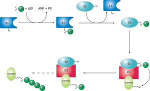
filedesc Schematic diagram of the ubiquitylati...

filedesc Schematic diagram of the ubiquitylation system. Created by Roger B. Dodd (Photo credit: Wikipedia)


Current Noteworthy Work

Nassif M and Hetz C.  Autophagy impairment: a crossroad between neurodegeneration and tauopathies.  BMC Biology 2012; 10:78. http://www.biomedcentral.com/1741-7007/10/78

Impairment of protein degradation pathways such as autophagy is emerging as a consistent and transversal pathological phenomenon in neurodegenerative diseases, including Alzheimer´s, Huntington´s, and Parkinson´s disease. Genetic inactivation of autophagy in mice has demonstrated a key role of the pathway in maintaining protein homeostasis in the brain, triggering massive neuronal loss and the accumulation of abnormal protein inclusions.  A paper in Molecular Neurodegeneration from Abeliovich´s group now suggests a role for phosphorylation of Tau and the activation of glycogen synthase kinase 3β (GSK3β) in driving neurodegeneration in autophagy-deficient neurons. We discuss the implications of this study for understanding the factors driving neurofibrillary tangle formation in Alzheimer´s disease and tauopathies.

Cajee UF, Hull R and Ntwasa M. Modification by Ubiquitin-Like Proteins: Significance in Apoptosis and Autophagy Pathways. Int. J. Mol. Sci. 2012, 13, 11804-11831; doi:10.3390/ijms130911804

Ubiquitin-like proteins (Ubls) confer diverse functions on their target proteins. The modified proteins are involved in various biological processes, including DNA replication, signal transduction, cell cycle control, embryogenesis, cytoskeletal regulation,
metabolism, stress response, homeostasis and mRNA processing. Modifiers such as SUMO, ATG12, ISG15, FAT10, URM1, and UFM have been shown to modify proteins thus conferring functions related to programmed cell death, autophagy and regulation of
the immune system. Putative modifiers such as Domain With No Name (DWNN) have been identified in recent times but not fully characterized. In this review, we focus on cellular processes involving human Ubls and their targets.

Aloy P. Shaping the future of interactome networks. (A report of the third Interactome Networks Conference, Hinxton, UK, 29 August-1 September 2007). Genome Biology 2007; 8:316 (doi:10.1186/gb-2007-8-10-316)

Complex systems are often networked, and biology is no exception. Following on from the genome sequencing projects,
experiments show that proteins in living organisms are highly connected, which helps to explain how such great complexity
can be achieved by a comparatively small set of gene products. At a recent conference on interactome networks held outside
Cambridge, UK, the most recent advances in research on cellular networks were discussed. This year’s conference focused on
identifying the strengths and weaknesses of currently resolved interaction networks and the techniques used to determine
them – reflecting the fact that the field of mapping interaction networks is maturing.

Peroutka RJ, Orcutt SJ, Strickler JE, and Butt TR. SUMO Fusion Technology for Enhanced Protein Expression and Purification in Prokaryotes and Eukaryotes. Chapter 2. in T.C. Evans, M.-Q. Xu (eds.), Heterologous Gene Expression in E. coli, Methods in Molecular Biology 705:15-29. DOI 10.1007/978-1-61737-967-3_2, © Springer Science+Business Media, LLC 2011

The preparation of sufficient amounts of high-quality protein samples is the major bottleneck for structural proteomics. The use of recombinant proteins has increased significantly during the past decades. The most commonly used host, Escherichia coli, presents many challenges including protein misfolding, protein degradation, and low solubility. A novel SUMO fusion technology appears to enhance protein expression and solubility (www.lifesensors.com). Efficient removal of the SUMO tag by SUMO protease in vitro facilitates the generation of target protein with a native N-terminus. In addition to its physiological relevance in eukaryotes, SUMO can be used as a powerful biotechnology tool for enhanced functional protein expression in prokaryotes and eukaryotes.

Juang YC, Landry MC, et al. OTUB1 Co-opts Lys48-Linked Ubiquitin Recognition to Suppress E2 Enzyme Function. Molecular Cell 2012; 45: 384–397. DOI 10.1016/j.molcel.2012.01.011

Ubiquitylation entails the concerted action of E1, E2, and E3 enzymes. We recently reported that OTUB1, a deubiquitylase, inhibits the DNA damage response independently of its isopeptidase activity. OTUB1 does so by blocking ubiquitin transfer by UBC13, the cognate E2 enzyme for RNF168. OTUB1 also inhibits E2s of the UBE2D and UBE2E families. Here we elucidate the structural mechanism by which OTUB1 binds E2s to inhibit ubiquitin transfer. OTUB1 recognizes ubiquitin-charged E2s through contacts with both donor ubiquitin and the E2 enzyme. Surprisingly, free ubiquitin associates with the canonical distal ubiquitin-binding site on OTUB1 to promote formation of the inhibited E2 complex. Lys48 of donor ubiquitin lies near the OTUB1 catalytic site and the C terminus of free ubiquitin, a configuration that mimics the products of Lys48-linked ubiquitin chain cleavage. OTUB1 therefore co-opts Lys48-linked ubiquitin chain recognition to suppress ubiquitin conjugation and the DNA damage response.

Hunter T. The Age of Crosstalk: Phosphorylation, Ubiquitination, and Beyond. Molecular Cell  2007; 28:730-738. DOI 10.1016/ j.molcel.2007.11.019.

Crosstalk between different types of posttranslational modification is an emerging theme in eukaryotic biology. Particularly prominent are the multiple connections between phosphorylation and ubiquitination, which act either positively or negatively in both directions to regulate these processes.

Tu Y, Chen C, et al. The Ubiquitin Proteasome Pathway (UPP) in the regulation of cell cycle control and DNA damage repair and its implication in tumorigenesis. Int J Clin Exp Pathol 2012;5(8):726-738. www.ijcep.com /ISSN:1936-2625/IJCEP1208018

Accumulated evidence supports that the ubiquitin proteasome pathway (UPP) plays a crucial role in protein
metabolism implicated in the regulation of many biological processes such as cell cycle control, DNA damage
response, apoptosis, and so on. Therefore, alterations for the ubiquitin proteasome signaling or functional impairments
for the ubiquitin proteasome components are involved in the etiology of many diseases, particularly in cancer
development.The authors discuss the ubiquitin proteasome pathway in the regulation of cell cycle control and DNA
damage response, the relevance for the altered regulation of these signaling pathways in tumorigenesis, and finally
assess and summarize the advancement for targeting the ubiquitin proteasome pathway in cancer therapy.

Cebollero E , Reggiori F  and Kraft C.  Ribophagy: Regulated Degradation of Protein Production Factories. Int J Cell Biol. 2012; 2012: 182834. doi:  10.1155/2012/182834 (online).

During autophagy, cytosol, protein aggregates, and organelles are sequestered into double-membrane vesicles called autophagosomes and delivered to the lysosome/vacuole for breakdown and recycling of their basic components. In all eukaryotes this pathway is important for adaptation to stress conditions such as nutrient deprivation, as well as to regulate intracellular homeostasis by adjusting organelle number and clearing damaged structures. For a long time, starvation-induced autophagy has been viewed as a nonselective transport pathway; however, recent studies have revealed that autophagy is able to selectively engulf specific structures, ranging from proteins to entire organelles. In this paper, we discuss recent findings on the mechanisms and physiological implications of two selective types of autophagy: ribophagy, the specific degradation of ribosomes, and reticulophagy, the selective elimination of portions of the ER.

Lee JH, Yu WH,…, Nixon RA.  Lysosomal Proteolysis and Autophagy Require Presenilin 1 and Are Disrupted by Alzheimer-Related PS1 Mutations. Cell 2010; 141, 1146–1158. DOI 10.1016/j.cell.2010.05.008.

Macroautophagy is a lysosomal degradative pathway essential for neuron survival. Here, we show that macroautophagy requires the Alzheimer’s disease (AD)-related protein presenilin-1 (PS1). In PS1 null blastocysts, neurons from mice hypomorphic for PS1 or
conditionally depleted of PS1, substrate proteolysis and autophagosome clearance during macroautophagy are prevented as a result of a selective impairment of autolysosome acidification and cathepsin activation. These deficits are caused by failed PS1-dependent
targeting of the v-ATPase V0a1 subunit to lysosomes. N-glycosylation of the V0a1 subunit, essential for its efficient ER-to-lysosome delivery, requires the selective binding of PS1 holoprotein to the unglycosylated subunit and the  sec61alpha/ oligosaccharyltransferase complex. PS1 mutations causing early-onset AD produce a similar lysosomal/autophagy phenotype in
fibroblasts from AD patients. PS1 is therefore essential for v-ATPase targeting to lysosomes, lysosome acidification, and proteolysis during autophagy. Defective lysosomal proteolysis represents a basis for pathogenic protein accumulations and neuronal cell death in AD and suggests previously unidentified therapeutic targets.

Pohl C and Jentsch S. Midbody ring disposal by autophagy is a post-abscission event of cytokinesis. nature cell biology 2009; 11 (1): 65-70.  DOI: 10.1038/ncb1813.

At the end of cytokinesis, the dividing cells are connected by an intercellular bridge, containing the midbody along with a single,
densely ubiquitylated, circular structure called the midbody ring (MR). Recent studies revealed that the MR serves as a target
site for membrane delivery and as a physical barrier between the prospective daughter cells. The MR materializes in telophase,
localizes to the intercellular bridge during cytokinesis, and moves asymmetrically into one cell after abscission. Daughter
cells rarely accumulate MRs of previous divisions, but how these large structures finally disappear remains unknown.
Here, we show that MRs are discarded by autophagy, which involves their sequestration into autophagosomes and delivery to
lysosomes for degradation. Notably, autophagy factors, such as the ubiquitin adaptor p62 and the ubiquitin-related protein Atg8 , associate with the MR during abscission, suggesting that autophagy is coupled to cytokinesis. Moreover, MRs accumulate in cells of patients with lysosomal storage disorders, indicating that defective MR disposal is characteristic of these diseases. Thus our findings suggest that autophagy has a broader role than previously assumed, and that cell renovation by clearing from superfluous large macromolecular assemblies, such as MRs, is an important autophagic function.


Hanai JI, Cao P, Tanksale P, Imamura S, et al. The muscle-specific ubiquitin ligase atrogin-1/MAFbx mediates statin-induced muscle toxicity. The Journal of Clinical Investigation  2007; 117(12):3930-3951.    http://www.jci.org

Statins inhibit HMG-CoA reductase, a key enzyme in cholesterol synthesis, and are widely used to treat hypercholesterolemia.
These drugs can lead to a number of side effects in muscle, including muscle fiber breakdown; however, the mechanisms of muscle injury by statins are poorly understood. We report that lovastatin induced the expression of atrogin-1, a key gene involved in skeletal muscle atrophy, in humans with statin myopathy, in zebrafish embryos, and in vitro in murine skeletal muscle cells. In cultured mouse myotubes, atrogin-1 induction following lovastatin treatment was accompanied by distinct morphological changes, largely absent in
atrogin-1 null cells. In zebrafish embryos, lovastatin promoted muscle fiber damage, an effect that was closely mimicked by knockdown of zebrafish HMG-CoA reductase. Moreover, atrogin-1 knockdown in zebrafish embryos prevented lovastatin-induced muscle injury. Finally, overexpression of PGC-1α, a transcriptional coactivator that induces mitochondrial biogenesis and protects against the development of muscle atrophy, dramatically prevented lovastatin-induced muscle damage and abrogated atrogin-1 induction both in fish and in cultured mouse myotubes. Collectively, our human, animal, and in vitro findings shed light on the molecular mechanism of statin-induced myopathy and suggest that atrogin-1 may be a critical mediator of the muscle
damage induced by statins.

Inami Y, Waguri S, Sakamoto A, Kouno T, et al.  Persistent activation of Nrf2 through p62 in hepatocellular carcinoma cells. J. Cell Biol. 2011; 193(2): 275–284. http://www.jcb.org/cgi/doi/10.1083/jcb.201102031

Macroautophagy (hereafter referred to as autophagy) is a cellular degradation system in which cytoplasmic components, including
organelles, are sequestered by double membrane structures called autophagosomes and the sequestered materials are
degraded by lysosomal hydrolases for supply of amino acids and for cellular homeostasis. Although autophagy has generally been considered nonselective, recent studies have shed light on another indispensable role for basal autophagy in cellular homeostasis, which is mediated by selective degradation of a specific substrate(s).  p62 is a ubiquitously expressed cellular protein that is conserved in metazoa but not in plants and fungi, and recently it has been known as one of the selective substrates for autophagy.
This protein is localized at the autophagosome formation site  and directly interacts with LC3, an autophagosome localizing protein . Subsequently, the p62 is incorporated into the autophagosome and then degraded. Therefore, impaired autophagy is accompanied by
accumulation of p62 followed by the formation of p62 and ubiquitinated protein aggregates because of the nature of both self- oligomerization and ubiquitin binding of p62.


Kima K, Khayrutdinov BI, Leeb CK, et al. Solution structure of the Zβ domain of human DNA-dependent activator of IFN-regulatory factors and its binding modes to B- and Z-DNAs. PNAS 2010; Early Edition ∣ pp 6. www.pnas.org/cgi/doi/10.1073/pnas.1014898107

The DNA-dependent activator of IFN-regulatory factors (DAI), also known as DLM-1/ZBP1, initiates an innate immune response by binding to foreign DNAs in the cytosol. For full activation of the immune response, three DNA binding domains at the N terminus are required: two Z-DNA binding domains (ZBDs), Zα and Zβ, and an adjacent putative B-DNA binding domain. The crystal structure of the Zβ domain of human DAI (hZβDAI) in complex with Z-DNA revealed structural features distinct from other known Z-DNA binding proteins, and it was classified as a group II ZBD. To gain structural insights into the DNA binding mechanism of hZβDAI, the solution structure of the free hZβDAI was solved, and its bindings to B- and Z-DNAs were analyzed by NMR spectroscopy. Compared to the Z-DNA–bound structure, the conformation of free hZβDAI has notable alterations in the α3 recognition helix, the “wing,” and Y145, which are critical in Z-DNA recognition. Unlike some other Zα domains, hZβDAI appears to have conformational flexibility, and structural adaptation is required for Z-DNA binding. Chemical-shift perturbation experiments revealed that hZβDAI also binds weakly to B-DNA via a different binding mode. The C-terminal domain of DAI is reported to undergo a conformational change on B-DNA binding; thus, it is possible that these changes are correlated. During the innate immune response, hZβDAI is likely to play an active role in binding to DNAs in both B and Z conformations in the recognition of foreign DNAs.


Epicrisis

This extensive review leaves little left unopened. We have seen the central role that the UPS system plays in normal organelle proteolysis in concert with autophagy. Impaired ubiquitination occurs from aging, and/or toxins, under oxidative stress involving E3s or DUBs.

This leads to altered gene transcripton, altered protein trafficking, and plays a role in neurodegenative disease, muscle malfunction, and cancer as well.

English: A cartoon representation of a lysine ...

English: A cartoon representation of a lysine 48-linked diubiquitin molecule. The two ubiquitin chains are shown as green cartoons with each chain labelled. The components of the linkage are indicated and shown as orange sticks. Image was created using PyMOL from PDB id 1aar. (Photo credit: Wikipedia)

Different forms of protein ubiquitylation

Different forms of protein ubiquitylation (Photo credit: Wikipedia)

Related articles

Read Full Post »

Nitric Oxide has a Ubiquitous Role in the Regulation of Glycolysis – with a Concomitant Influence on Mitochondrial Function


Reporter, Editor, and Topic Co-Leader: Larry H. Bernstein, MD, FACP, Clinical Pathologist and Biochemist


Apoptosis signaling pathways

Apoptosis signaling pathways (Photo credit: AJC1)

This discussion is a followup on a series of articles elucidating the importance of NO, eNOS, iNOS, cardiovascular and vascular endothelium effects, and therapeutic targets.

This mechanism of action and signaling actions have been introduced so that we identify endocrine, paracrine, and such effects in the normal, stressed, and dysfunctional state. The size and breadth of this vital adaptive process is now further explored.

The title is short, befitting a subtitle.  The full topic may be considered “Nitric Oxide has a ubiquitous role in the regulation of glycolysis -with a concomitant influence on mitochondrial function that is active in endothelium, platelets, vascular smooth muscle and neural cells and the balance has a role in chronic inflammation, asthma, hypertension, sepsis and cancer”.

Vascular endothelium

Vascular endothelium (Photo credit: Wikipedia)

Related articles


Nitric Oxide Synthase

Nitric Oxide Synthase (Photo credit: Wikipedia)


Nitric Oxide has a ubiquitous role in the regulation of glycolysis with a concomitant influence on mitochondrial function that is active in endothelium, platelets, vascular smooth muscle and neural cells and the balance has a role in chronic inflammation, asthma, hypertension, sepsis and cancer.

Uncoupling of aerobic glycolysis
Potential cytotoxic mediators of endothelial cell (EC) apoptosis include increased formation of reactive oxygen and nitrogen species (ROSRNS) during the atherosclerotic process. Nitric oxide (NO) has a biphasic action on oxidative cell killing with low concentrations protecting against cell death, whereas higher concentrations are cytotoxic. High levels of NO can be produced by inducible nitric-oxide synthase in response to cytokine stimulation, primarily from macrophages, and elevated levels of NO is injurious to endothelium.Ccytochrome c release and caspase activation are involved in NO induced apoptosis. ROS also induces mitochondrial DNA damage in ECs, and this damage is accompanied by a decrease in mitochondrial RNA (mtRNA) transcripts, mitochondrial protein synthesis, and cellular ATP levels. Mitochondria have been recognized to play a pivotal role in the signaling cascade of apoptosis leading to atherosclerosis-induced damage in endothelial cells.
The processes involved in the signaling pathways leading to apoptosis are complex but have some degree of convergence between cell types including those in the vasculature. Release of cytochrome c from mitochondria is a proapoptotic signal, which activates several downstream signaling events including formation of the apoptosome and activation of caspases. Ubiquinol cytochrome c reductase (complex III) is a site for ROS formation, and cytochrome c oxidase (complex IV) is a target for the interaction of NO in mitochondria.
The impact of the inhibition of mitochondrial protein synthesis is particularly important in NO-dependent cytotoxicity, and depends also on other factors such as glycolysis. These authors examined whether the inhibition of mitochondrial protein synthesis by chloramphenicol increases the susceptibility of endothelial cells to undergo NO-dependent apoptosis in glucose-free media. Bovine aortic endothelial cells were treated with chloramphenicol, which resulted in a decreased ratio of mitochondrial complex IV to cytochrome c and increased oxidant production in the cell. Inhibition of mitochondrial protein synthesis was associated with a greater susceptibility of the cells to apoptosis induced by NO in glucose-free medium.
Inhibition of mitochondrial protein synthesis results in increased endothelial cell susceptibility to nitric oxide-induced apoptosis. A Ramachandran, DR Moellering, E Ceaser, S Shiva, J Xu, and V Darley-Usmar. PNAS May 14, 2002: 99(10): 6643–6648 http://www.pnas.orgcgidoi10.1073pnas.102019899

Nitric oxide (NO) is a ubiquitous signaling molecule whose physiological roles mediated through the activation of the soluble guanylate cyclase are now clearly recognized. At physiological concentrations, NO also inhibits the mitochondrial enzyme cytochrome c oxidase (complex IV) in competition with oxygen, and recently we have suggested that the interplay between the two gases allows this enzyme to act as an oxygen sensor in cells. In addition, NO plays a variety of patho-physiological roles, some of which also may be the consequence of its action at a mitochondrial level. We have characterized the sequence of events that follow inhibition of complex IV by continuous exposure to NO.
The mitochondrion is a key organelle in the control of cell death. Nitric oxide (NO) inhibits complex IV in the respiratory chain and is reported to possess both proapoptotic and antiapoptotic actions. We investigated the effects of continuous inhibition of respiration by NO on mitochondrial energy status and cell viability. Serum-deprived human T cell leukemia (Jurkat) cells were exposed to NO at a concentration that caused continuous and complete (;85%) inhibition of respiration. Serum deprivation caused progressive loss of mitochondrial membrane potential (Dcm) and apoptotic cell death. In the presence of NO, Dcm was maintained compared to controls, and cells were protected from apoptosis. Similar results were obtained by using staurosporin as the apoptotic stimulus. As exposure of serum-deprived cells to NO progressed (>5 h), however, Dcm fell, correlating with the appearance of early apoptotic features and a decrease in cell viability. Glucose deprivation or iodoacetate treatment of cells in the presence of NO resulted in a collapse of Dcm, demonstrating involvement of glycolytic ATP in its maintenance. Under these conditions cell viability also was decreased. Treatment with oligomycin and or bongkrekic acid indicated that the maintenance of Dcm during exposure to NO is caused by reversal of the ATP synthase and other electrogenic pumps. Thus, blockade of complex IV by NO initiates a protective action in the mitochondrion to maintain Dcm; this results in prevention of apoptosis. It is likely that during cellular stress involving increased generation of NO this compound will trigger a similar sequence of events, depending on its concentration and duration of release. (mitochondrial membrane potential ; apoptosis ; necrosis)

The effect of nitric oxide on cell respiration: A key to understanding its role in cell survival or death. B Beltra, A Mathur, MR Duchen, JD. Erusalimsky, and S Moncada. PNAS Dec 19, 2000; 97(26):4602–14607.

Another study by this group shows that inhibition of respiration by exogenous nitric oxide (NO) in Jurkat cells leads to mitochondrial membrane hyperpolarization dependent on the utilization of glycolytic ATP by the F1Fo-ATPase and other transporters acting in reverse mode. This process also occurs in astrocytes, which are highly glycolytic cells, but not in neurons , which do not invoke glycolysis to maintain ATP concentrations. In addition, this hyperpolarization correlates with protection against apoptotic cell death. Others found an early phase of mitochondrial hyperpolarization after treatment of a variety of cells with different pro-apoptotic stimuli, which precedes the generation of free. At present, no satisfactory explanation has been proposed to explain the mechanism of hyperpolarization, the reasons why free radicals are released from the mitochondrion, or the connection of these phenomena with apoptosis.
The authors surmise that a pro-apoptotic stimulus, anti-Fas Ab, leads to release of endogenous NO from Jurkat cells in sufficient amounts to inhibit cell respiration and cause a hyperpolarization dependent on the reversal of the F1Fo-ATPase. Moreover, the reduction of the mitochondrial electron transport chain, after inhibition of cytochrome oxidase by NO, leads to generation of superoxide anion (O2). They suggest the process is a cellular defense response that may be overcome by pro-apoptotic mechanisms that occur in parallel.

Inhibition of mitochondrial respiration by endogenous nitric oxide: A critical step in Fas signaling. B Beltran, M Quintero, E Garcıa-Zaragoza, E O’Connor, JV. Esplugues, and Salvador Moncada. PNAS June 25, 2002 99(13): 8892–8897. http://www.pnas.orgcgidoi10.1073pnas.092259799

Nitric oxide has been shown to render cells resistant to oxidative stress. Mechanisms proposed for the ability of nitric oxide to protect cells against oxidative stress include reactions of nitric oxide and the induction of adaptive responses that require protein synthesis. Nitric oxide forms iron complexes preventing the formation of strong oxidants. In addition, reactions of nitric oxide with lipid and or organic radicals protect against membrane peroxidation and peroxidative chemistry-induced cell injury. Exposure to low, nonlethal doses of nitric oxide induces adaptive responses that render cells resistant to lethal concentrations of nitric oxide and or peroxides, such as, the induction of hemoxygenase-1 (HO-1) and Mn superoxide dismutase. The up-regulation of HO-1 was accompanied by an increase in ferritin to account for the release of iron from HO-1, indicating a role of both iron heme and nonheme iron for peroxide-mediated cellular injury. Further, nitric oxide, by regulating critical mitochondrial functions such as respiration, membrane potential, and release of cytochrome c, is able to trigger defense mechanisms against cell death induced by pro-apoptotic stimuli.
This study investigates the potential contribution of nitric oxide’s ability to protect cells from oxidative stress, low steady state levels of nitric oxide generated by endothelial nitric oxide synthase (eNOS) and the mechanisms of protection against H2O2. Spontaneously transformed human ECV304 cells, which normally do not express eNOS, were stably transfected with a green fluorescent-tagged eNOS cDNA. The eNOS-transfected cells were found to be resistant to injury and delayed death following a 2-h exposure to H2O2 (50–150 mM). Inhibition of nitric oxide synthesis abolished the protective effect against H2O2 exposure. The ability of nitric oxide to protect cells depended on the presence of respiring mitochondria. ECV3041 eNOS cells with diminished mitochondria respiration are injured to the same extent as non-transfected ECV304 cells, and recovery of mitochondrial respiration restores the ability of nitric oxide to protect against H2O2-induced death. Nitric oxide had a profound effect in cell metabolism, because ECV3041eNOS cells had lower steady state levels of ATP and higher utilization of glucose via the glycolytic pathway than ECV304 cells. However, the protective effect of nitric oxide against H2O2 exposure is not reproduced in ECV304 cells after treatment with azide and oligomycin suggesting that the dynamic regulation of respiration by nitric oxide represent a critical and unrecognized primary line of defense against oxidative stress.

Dynamic regulation of metabolism and respiration by endogenously produced nitric oxide protects against oxidative stress. E Paxinou, M Weisse, Q Chen, JM Souza, et al. PNAS Sept 25, 2001; 98( 20): 11575–11580. http://www.pnas.orgycgiydoiy10.1073ypnas.201293198.

Nitric oxide (NO) mediates a variety of biological effects including relaxation of blood vessels, cytotoxicity of activated macrophages, and formation of cGMP by activation of glutamate receptors of neurons. NO has also been implicated for such pathophysiological conditions as destruction of tumor cells by macrophages, rheumatoid arthritis, and focal brain ischemia. Some of these effects of NO are associated with hypoxic conditions. O2 radicals and ions that result from reactivity of NO are presumed to be involved in NO cytotoxicity. These investigators report that adaptive cellular response controlled by the transcription factor hypoxia-inducible factor 1 (HIF-1) in hypoxia is suppressed by NO. Induction of erythropoietin and glycolytic aldolase A mRNAs in hypoxically cultured Hep3B cells, a human hepatoma cell line, was completely and partially inhibited, respectively, by the addition of sodium nitroprusside (SNP), which spontaneously releases NO. A reporter plasmid carrying four hypoxia-response element sequences connected to the luciferase structural gene was constructed and transfected into Hep3B cells. Inducibly expressed luciferase activity in hypoxia was inhibited by the addition of SNP and two other structurally different NO donors, S-nitroso-Lglutathione and 3-morpholinosydnonimine, giving IC50 values of 7.8, 211, and 490 mM, respectively. Inhibition by SNP was also observed in Neuro 2A and HeLa cells, indicating that the inhibition was not cell-type-specific. The vascular endothelial growth factor promoter activity that is controlled by HIF-1 was also inhibited by SNP (IC50 5 6.6 mM). Induction generated by the addition of cobalt ion (this treatment mimics hypoxia) was also inhibited by SNP (IC50 5 2.5 mM). Increased luciferase activity expressed by cotransfection of effector plasmids for HIF-1a or HIF-1a-like factor in hypoxia was also inhibited by the NO donor. We also showed that the inhibition was performed by blocking an activation step of HIF-1a to a DNA-binding form.
Inhibition of hypoxia-inducible factor 1 activity by nitric oxide donors in hypoxia. K Sogawa, K Numayama-Tsuruta, M Ema, M Abe, et al. Proc. Natl. Acad. Sci. USA (Biochemistry) June 1998; 95:7368–7373. 1998. The National Academy of Sciences 0027-8424.98.957368-6. http:yywww.pnas.org.

The role of nitrogen metabolism in the survival of prolonged periods of waterlogging was investigated in highly flood-tolerant, nodulated Lotus japonicus plants. Alanine production revealed to be a critical hypoxic pathway. Alanine is the only amino acid whose biosynthesis is not inhibited by nitrogen deficiency resulting from RNA interference silencing of nodular leghemoglobin. The metabolic changes that were induced following waterlogging can be best explained by the activation of alanine metabolism in combination with the modular operation of a split tricarboxylic acid pathway. The sum result of this metabolic scenario is the accumulation of alanine and succinate and the production of extra ATP under hypoxia. The importance of alanine metabolism is discussed with respect to its ability to regulate the level of pyruvate, and this and all other changes are discussed in the context of current models concerning the regulation of plant metabolism.
Glycolysis and the Tricarboxylic Acid Cycle Are Linked by Alanine Aminotransferase during Hypoxia Induced by Waterlogging of Lotus japonicus[W][OA]. M Rocha, F Licausi, WL Arau´ jo, A Nunes-Nesi, et al. Plant Physiology Mar 2010; 152: 1501–1513. http://www.plantphysiol.org 2010 Amer Soc Plant Biologists

DNA damage occurs in ischemia, excitotoxicity, inflammation, and other disorders that affect the central nervous system (CNS). Extensive DNA damage triggers cell death and in the mature CNS, this occurs primarily through activation of the poly(ADP-ribose) polymerase-1 (PARP-1) cell death pathway. PARP-1 is an abundant nuclear enzyme that, when activated by DNA damage, consumes nicotinamide adenine dinucleotide (NAD)+ to form poly(ADP-ribose) on acceptor proteins. The PARP-1 activation leads to cell death. We used mouse astrocyte cultures to explore the bioenergetic effects of NAD+ depletion by PARP-1 and the role of NAD+ depletion in this cell death program. PARP-1 activation led to a rapid but incomplete depletion of astrocyte NAD+, a near-complete block in glycolysis, and eventual cell death. Repletion of intracellular NAD restored glycolytic function and prevented cell death. The addition of non-glucose substrates to the medium, pyruvate, glutamate, or glutamine, also prevented astrocyte death after PARP-1 activation.
These findings suggest a sequence of events in which NAD+ depletion is a key event linking DNA damage to metabolic impairment and cell deathm. A similar scenario has been proposed by Zong et al. (2004), based on the finding that cell types that depend on aerobic glycolysis for ATP production exhibit a particularly high sensitivity to DNA damage and PARP-1 activation. In mature brain, glucose is normally the dominant metabolic substrate due to relatively slow transport of other metabolites across the blood– brain barrier. Oncein brain, glucose may be metabolized directly by neurons and glia or may be metabolized to lactate in glia and thelactate subsequently shuttled to neurons for oxidative metabolism (Dringen et al., 1993; Pellerin and Magistretti,1994; Wender et al., 2000; Dienel and Cruz, 2004). In either case, a block in glycolytic flux produced by NAD depletion will block energy metabolism in both neurons and glia in brain. Interestingly, the lactate shuttle hypothesis raises the possibility that activation of PARP-1 selectively in astroglia might also block energy metabolism in neurons.

These studies suggest PARP-1 activation leads to rapid depletion of the cytosolic but not the mitochondrial NAD+ pool. Depletion of the cytosolic NAD+ pool renders the cells unable to utilize glucose as a metabolic substrate. Under conditions where glucose is the only available metabolic substrate, this leads to cell death. This cell death pathway is particularly germane to brain because glucose is normally the only metabolic substrate that is transported rapidly across the blood–brain barrier. © 2004 Wiley-Liss, Inc.
Key words: mitochondria; permeability transition; poly(ADP-ribose) polymerase; ischemia; peroxynitrite
NAD+as a metabolic link between DNA damage and cell death. DNA damage induced by alkylating agents, oxidative stress, or other agents causes PARP-1 activation. PARP-1 activation leads to depletion in cytosolic NAD with, initially, a relative preservation of mitochondrial NAD and mitochondrial function. The depletion in cytosolic NAD+ blocks glycolysis, and in cells in which glucose is the primary energy substrate, this in turn leads to a block in substrate flux to mitochondria. The resulting mitochondrial dysfunction leads to mitochondrial permeability transition (MPT) and subsequent downstream events culminating in cell death.
NAD+ as a Metabolic Link Between DNA Damage and Cell Death. W Ying, CC Alano, P Garnier, and RA Swanson. Journal of Neuroscience Research 2005;79:216–223
Key words: glycolysis, mitochondrial energy production, nitric oxide
Abbreviations: NO, nitric oxide; SNAP, S-nitroso-N-acetylpenicyllamine; SNP, sodium nitroprusside.
The results indicate that: 1) in porcine platelets NO is able to diminish mitochondrial energy production through the inhibition of cytochrome oxidase, 2) the inhibitory effect of NO on platelet secretion (but not aggregation) can be attributed to the reduction of mitochondrial energy production.
Nitric oxide (NO) has been increasingly recognized as an important intra- and intercellular messenger molecule with a physiological role in vascular relaxation, platelet physiology, neurotransmission and immune responses (Moncada et al., 1991; Radomski et al., 1996; Szabó, 1996; Riedel et al., 1999; Titheradge 1999). In vitro NO is a strong inhibitor of platelet adhesion and aggregation (Radomski et al., 1996; Riedel et al., 1999;nSogo et al., 2000). In the blood stream, platelets remain in contact with NO that is permanently released from the endothelial cells and from activated macrophages (Moncada et al., 1991; Riedel et al., 1999; Titheradge 1999). It has been suggested that the activated platelet itself is able to produce NO (Lantoine et al., 1995; Zhou et al., 1995; Radomski et al., 1996). The mechanism responsible for the inhibitory effect of NO on platelet responses is not entirely clear. It is believed that the main intracellular target for NO in platelets is soluble cytosolic guanylate cyclase (Waldman & Walter 1989; Schmidt et al., 1993; Wang et al., 1998). NO activates the enzyme (Schmidt et al., 1993). Thus, elevated intracellular cGMP level inhibits platelet activation. There are suggestions, however, that elevated cGMP may not be the only intracellular factor directly involved in the inhibition of platelet activation (Gordge et al., 1998; Sogo et al., 2000; Beghetti et al., 2003).
Platelets are fairly active metabolically and have a total ATP turnover rate of about 3–8 times that of resting mammalian muscle (Akkerman, 1978; Akkerman et al., 1978; Holmsen, 1981; Niu et al., 1996). Platelets contain mitochondria which enable these cells to produce energy both in the oxidative and anaerobic way (Holmsen, 1981). Under aerobic conditions, ATP is produced by aerobic glycolysis using glucose or glycogen which can account for 30–50% of total ATP production, and by oxidative metabolism using glucose and glycogen (6–11%), amino-acids (7%) or free fatty acids (20–40%) (Holmsen 1981; Guppy et al., 1990; Niu et al., 1996).
The inhibition of mitochondrial respiration by removing oxygen or by respiratory chain blockers (antimycin A, cyanide, rotenone) results in the stimulation of glycolytic flux (Guppy et al., 1990). This phenomenon is known as Pasteur effect and indicates that in platelets glycolysis and mitochondrial respiration are tightly functionally connected (Akkerman, 1978; Holmsen, 1981; Guppy et al., 1995; Niu et al., 1996). It has been reported that the activation of human platelets by high concentration of thrombin is accompanied by an acceleration of lactate production and an increase in oxygen consumption (Akkerman & Holmsen, 1981; Niu et al., 1996).
The results presented here suggest that also porcine blood platelets stimulated by collagen produce more lactate. This indicates that both glycolytic and oxidativeATP production supports platelet responses. This also indicates that blocking of energy production in platelets may decrease their responses. It is well established that platelet responses have different metabolic energy (ATP) requirements increasing in the order: aggregation< dense and alfa granule secretion < acid hydrolase secretion (Holmsen et al., 1982; Verhoeven et al., 1984; Morimoto & Ogihara, 1996).
The present results indicate that exogenously added NO (in the form of NO donors)stimulates glycolysis in intact porcine platelets. Since in platelets glycolysis and mitochondrial respiration are tightly functionally connected, this can be interpreted to mean that the stimulatory effectof NO on glycolysis in intact platelets may be produced by non-functional mitochondria.This can be really the case since NO donors are able to inhibit both mitochondrial respiration and platelet cytochrome oxidase. Interestingly, the concentrations of NO donors inhibiting mitochondrial respiration and cytochrome oxidase were similar to those stimulating glycolysis in intact platelets.
Studies performed on intact J774 cells have shown that mitochondrial complex I is inhibited only after a prolonged (6–18 h) exposure to NO and that this inhibition appears to result from S-nitrosylation of critical thiols in the enzyme complex (Clementi et al., 1998). Further studies are needed to establish whether long term exposure of platelets to NO affects Mitochondrial complexes I and II.
Comparison of the concentrations of SNP and SNAP affecting cytochrome oxidase activityand mitochondrial respiration with those reducing the platelet responses indicates that NO cannot significantly reduce platelet aggregation through the inhibition of oxidative energy production. By contrast, the concentrations of the NO donors inhibiting platelet secretion, mitochondrial respiration and cytochrome oxidase were similar. This and the fact that the platelet release reaction strongly depends on the oxidative energy production may suggest that in porcine platelets NO can affect platelet secretion through the inhibition of mitochondrial energy production at the step of cytochrome oxidase.

Taking into account that platelets may contain NO synthase and are able to produce significant amounts of NO (Berkels et al., 1997)it seems possible that nitric oxide can function in these cells as a physiological regulator of mitochondrial energy production.
Nitric oxide and platelet energy metabolism. M Tomasiak, H Stelmach, T Rusak and J Wysocka. Acta Biochimica Polonica 2004; 51(3):789–803

These authors previously investigated the bioenergetic consequences of activating J774.A1 macrophages (MФ) with interferon (IFN)γ and lipopolysaccharide (LPS) and found that there is a nitric oxide (NO)-dependent mitochondrial impairment and stabilization of hypoxia inducible factor (HIF)-1α, which synergize to activate glycolysis and generate large
quantities of ATP. We now demonstrate, using TMRM fluorescence and time-lapse confocal microscopy, that these cells maintain a high mitochondrial membrane potential (ΔΨm) despite the complete inhibition of respiration. The maintenance of high ΔΨm is due to the utilization of a significant proportion of glycolytically generated ATP as a defence mechanism against cell death. This is achieved by the reverse functioning of FoF1-ATP synthase and adenine nucleotide translocase (ANT). Treatment of activated MФ with inhibitors of either of these enzymes, but not with inhibitors of the respiratory chain complexes I to IV, led to a collapse in ΔΨm and to an immediate increase in intracellular [ATP], due to the prevention of ATP hydrolysis by the FoF1-ATP synthase. This collapse in ΔΨm was followed by translocation of Bax from cytosol to the mitochondria, release of cytochrome c into the cytosol, activation of caspase 3 and 9 and subsequent apoptotic cell death. Our results indicate that during inflammatory activation “glycolytically competent cells” such as MФ utilize significant amounts of the glycolytically-generated ATP to maintain ΔΨm and thereby prevent apoptosis.

Activated macrophages utilize glycolytic ATP to maintain mitochondrial membranepotential and prevent apoptotic cell death. A Garedew, SO Henderson, S Moncada. Cell Death and Differentiation. 2010. DOI : 10.1038/cdd.2010.27
The effects of the sodium nitroprusside (SNP), a nitric oxide (NO) donor clinically used in the treatment of hypertensive emergencies on the energy production of rat reticulocytes were investigated. Rat reticulocyte-rich red blood cell suspensions were aerobically incubated without (control) or in the presence of different concentrations of SNP (0.1, 0.25, 0.5, 1.0 mM). SNP decreased total and coupled, but increased uncoupled oxygen consumption. This was accompanied by the stimulation of glycolysis, as measured by increased glucose consumption and lactate accumulation. Levels of all glycolytic intermediates indicate stimulation of hexokinase-phosphofructo kinase (HK-PFK), glyceraldehyde 3-phosphate dehydrogenase (GAPD) and pyruvate kinase (PK) activities in the presence of SNP. Due to the decrease of coupled oxygen consumption in the presence of SNP, ATP production via oxidative phosphorylation was significantly diminished. Simultaneous increase of glycolytic ATP production was not enough to provide constant ATP production. In addition, SNP significantly decreased ATP level, which was accompanied with increased ADP and AMP levels. However, the level of total adenine nucleotides was significantly lower, which was the consequence of increased catabolism of adenine nucleotides (increased hypoxanthine level). ATP/ADP ratio and adenylate energy charge level were significantly decreased. In conclusion, SNP induced inhibition of oxidative phosphorylation, stimulation of glycolysis, but depletion of total energy production in rat reticulocytes. These alterations were accompanied with instability of energy status.

Effects of Exogenous Donor of Nitric Oxide – Sodium Nitroprusside on Energy Production of Rat Reticulocytes. SD MALETIĆ, L M DRAGIĆEVIĆ-DJOKOVIĆ, BI OGNJANOVIĆ, RV ŽIKIĆ, AŠ ŠTAJN, MB SPASIĆ.
Physiol. Res. 2004;53: 439-447.

Key points to take from this:
1. The role of NO in regulating cellular death is in many organs and central to this function is the stabilization of mitochondria through sufficient levels of NO. High levels of eNO leads to mitochondrial dysfunction that increases the dependence of ATP generated from glycolysis.
2. This is accompanied by inhibition of oxidative phosphorylation and stimulation of glycolysis, which brings the discussion to a different domain – cancer growth and Warburgh Effect.
3. This is accompanied by PPAR activation, cytoplasmic NAD+ depletion, and inhibition of glycolysis (critical in cells dependent on aerobic glycolysis), depletion of total energy production, and apoptosis.
4. Maintenance of high glycolytic generation of ATP is essential for cellular defense, but the oxygen consumption is uncoupled.
5. NO donors inhibiting mitochondrial respiration and cytochrome oxidase are similar to those stimulating glycolysis

More    (pharmaceuticalintelligence.com)

Read Full Post »

Interaction of Nitric Oxide and Prostacyclin in Vascular Endothelium

Author: Larry H. Bernstein, MD


This review identifies the complex interactions and underlying regulatory balances/imbalances between the mechanism of vasorelaxation and vasoconstriction of vascular endothelium by way of nitric oxide (NO), prostacyclin, in response to oxidative stress and intimal injury.

A series of related topics have been covered in this Scientific Web Site: http://pharmaceuticalintelligence.com on the Nitric Oxide series:
Nitric oxide: role in Cardiovascular health and disease
The rationale and use of inhaled NO in Pulmonary Artery Hypertension and Right Sided Heart Failure

Imbalance of Autonomic Tone: The Promise of Intravascular Stimulation of Autonomics

Intravascular Stimulation of Autonomics: A Letter from Dr. Michael Scherlag

Will these findings impact the Clopidogrel market?

Positioning a Therapeutic Concept for Endogenous Augmentation of cEPCs — Therapeutic Indications for Macrovascular Disease: Coronary, Cerebrovascular and Peripheral

Cardiovascular Outcomes: Function of circulating Endothelial Progenitor Cells (cEPCs): Exploring Pharmaco-therapy targeted at Endogenous Augmentation of cEPCs

Endothelial Dysfunction, Diminished Availability of cEPCs, Increasing CVD Risk for Macrovascular Disease – Therapeutic Potential of cEPCs

Outcomes in High Cardiovascular Risk Patients: Prasugrel (Effient) vs. Clopidogrel (Plavix); Aliskiren (Tekturna) added to ACE or added to ARB

Vascular Medicine and Biology: CLASSIFICATION OF FAST ACTING THERAPY FOR PATIENTS AT HIGH RISK FOR MACROVASCULAR EVENTS Macrovascular Disease – Therapeutic Potential of cEPCs

Ethical Considerations in Studying Drug Safety — The Institute of Medicine Report

Nitric Oxide Signalling Pathways

Age-Dependent Depression in Circulating Endothelial Progenitor Cells in Coronary Artery Bypass Grafting Patients

Nitric Oxide: The Nobel Prize in Physiology or Medicine 1998 Robert F. Furchgott, Louis J. Ignarro, Ferid Murad

Nitric Oxide and Platelet Aggregation

Obstructive coronary artery disease diagnosed by RNA levels of 23 genes – CardioDx heart disease test wins Medicare coverage

Coronary Artery Disease – Medical Devices Solutions: From First-In-Man Stent Implantation, via Medical Ethical Dilemmas to Drug Eluting Stents

Nitric Oxide: Chemistry and function

Nitric Oxide: a short historic perspective

Human Embryonic-Derived Cardiac Progenitor Cells for Myocardial Repair

Nitric Oxide production in Systemic sclerosis

Cardiovascular Disease (CVD) and the Role of agent alternatives in endothelial Nitric Oxide Synthase (eNOS) Activation and Nitric Oxide Production

Endothelial Differentiation and Morphogenesis of Cardiac Precursors

Bystolic’s generic Nebivolol – positive effect on circulating Endothelial Progenitor Cells endogenous augmentation

Nitric Oxide in bone metabolism

Circulating Endothelial Progenitor Cells Milestones in the research on Circulating Endothelial Progenitor Cells as diagnostic markers of cardiovascular risk have been reported in NEJM

Transcription Factor Lyl-1 Critical in Producing Early T-Cell Progenitors

Macrovascular Disease – Therapeutic Potential of cEPCs: Reduction Methods for CV Risk

Treatment of Refractory Hypertension via Percutaneous Renal Denervation

Triple Antihypertensive Combination Therapy Significantly Lowers Blood Pressure in Hard-to-Treat Patients with Hypertension and Diabetes

Study Finds Low Methylation Regions Prone to Structural Mutation

Atherosclerosis, studied extensively, has common features in the aorta, coronary, carotid and peripheral arteries. There is commonality in morphology of atheromatous plaques, the degree of thrombus formation and types of ensuing ischemic events, therefore, plaque disruption, the degree of thrombus formation and types of ensuing ischemic coronary syndromes of patients have been extensively studied. A concept of unstable atherosclerotic plaques that are amenable to rupture is central to acute myocardial infarction, stroke, aortic aneurysm and sequellae, and peripheral artery disease. These unstable atherosclerotic plaques: plaques with an unstable morphology are essential to the onset of unstable coronary artery disease. Plaque formation results from complex cellular interactions in the intima of arteries, which take place between resident cells of the vessel wall (smooth muscle cells and endothelial cells) and cells of the immune system (leukocytes). Local flow disturbances, extracellular matrix composed of lipids and debris are defining characteristics of the ‘atheroma’.
Inflammation and repair are key events in the natural course of atherosclerosis, and inflammatory cells have profound effects on the integrity of the plaques . Smooth muscle cells produce by far most of the extracellular matrix components of a plaque. Transforming growth factor beta (TGF-β) is one of the most potent stimulators of connective tissue production by smooth muscle cells, and large amounts of this growth factor are detected in restenosis lesions after PTCA.
Atherosclerotic plaque rupture – pathologic basis of plaque stability and instability. AC Newby, P Libby, and AC. van der Wal. Cardiovasc Res 1999; 41 (2): 334-344. doi: 10.1016/S0008-6363(98)00276-4. Vascular endothelium acts as a paracrine/endocrine organ by secreting a wide range of biologically active mediators that play a key role in regulating immune responses, vascular tone and coagulation, and act on adjacent smooth muscle cells, monocytes, macrophages, fibroblasts and organ specific cells.

The key culprits include:

VASODILITATION

  • vasodilators (prostacycline (PGI2),
  • nitric oxide (NO),
  • endothelium-derived relaxing factor (EDRF) ) and
  • cardiac specific natriuretic peptides;

The chemical nature of EDRF was not known until Palmer, Ferrige and Moncada demonstrated that nitric oxide accounted for most if not all of the biological activity of EDRF. Both EDRF and NO act through the stimulation of soluble guanylate cyclase and subsequent formation of cyclic GMP (cGMP). Cyclic GMP activates cGMP-dependent protein kinases and leads to dephosphorylation of myosin light chains and muscle relaxation.

Palmer RM, Ferrige AG, Moncada S. Nitric oxide release accounts for the biological activity of endothelium-derived relaxing factor. Nature 1987; 327:524-6.

Waldman SA, Murad F. Cyclic GMP synthesis and function. Pharma Rev 1987; 39:163-96.

Gryglewski RJ, Botting RM, Vane JR. Mediators produced by the endothelial cell. Hypertension 1988; 12:530-48.

VASOCONSTRICTION

  • vasoconstrictors (endothelin-1 (ET), thromboxane A2, prostaglandin H2 and components of renin-angiotensin system);
  • pro- and anti-thrombotic factors (tissue factor, platelet activating factor (PAF), von Willebrand factor (vWf ) );
  • fibrinolytic activators and inhibitors (tissue plasminogen activator (t-PA), plasminogen activator inhibitor-1 (PAI-1));
  • arachidonate metabolites (prostanoids)( Prostanoids are cyclic lipid mediators which arise from enzymic cyclooxygenation of linear polyunsaturated fatty acids, e.g. arachidonic acid (20:4 n 6, AA). Biologically active prostanoids deriving from AA include stable prostaglandins (PGs), e.g. PGE(2), PGF(2alpha), PGD(2), PGJ(2) as well as labile prostanoids, i.e. PG endoperoxides (PGG(2), PGH(2)), thromboxane A(2) (TXA(2)) and prostacyclin (PGI(2)). Stable PGs regulate smooth muscle tone and also modulate inflammatory and immune reactions in this context. PG endoperoxides are intermediates in biosynthesis of all prostanoids. Gryglewski RJ. Prostacyclin among prostanoids. Pharmacol Rep. 2008 Jan-Feb;60(1):3-11.
  • leukocyte adhesion molecules (intercellular adhesion molecule-1 (ICAM-1), vascular cell adhesion molecule-1 (VCAM-1), E-selectin, P-selectin);
  • multiple cytokines transforming growth factor, proinflammatory and anti-inflammatory mediators, tumour necrosis factor, chemokines and steroids.
  • HU Rehman. Vascular Endothelium as an Endocrine Organ. Proc R Coll Physicians Edinb 2001; 31:149-154.

Prostanoids are cyclic lipid mediators which arise from enzymic cyclooxygenation of linear polyunsaturated fatty acids (n-6 PUFAs), e.g. arachidonic acid (20:4 n 6, AA). Labile Thromboxane A(2) (TXA(2)) ( half life of 30 s at 37 degrees C), generated by platelets has powerful vasoconstrictor, cytotoxic and thrombogenic properties.

Prostacyclin (PGI(2)) with a half life of 4 min at 37 degrees C  is produced by the vascular wall (predominantly by the endothelium) and is a physiological antagonist of TXA(2), and is a powerful cytoprotective agent that exerts its action through activation of adenylate cyclase. PGI(2) acts in concert with the system consisting of NO synthase (eNOS)/nitric oxide free radical (NO)/guanylate cyclase/cyclic-GMP. Both cyclic nucleotides (c-AMP and c-GMP) act in synergy as two energetic fists which defend the cellular machinery from being destroyed by endogenous or exogenous aggressors.

Recently, a new partner has been recognized in this endogenous defensive squadron, i.e. a system involving heme oxygenase (HO-1) and carbon monoxide (CO). The expanding knowledge on the pharmacological steering of this enzymic triad (PGI(2)-S/eNOS/HO-1) is likely to contribute to the rational therapy of many systemic diseases such as

  • atherosclerosis,
  • diabetes mellitus,
  • arterial hypertension or
  • Alzheimer diseases.

The discovery of prostacyclin broadened our pathophysiological horizon, and by itself opened new therapeutic possibilities.Gryglewski RJ. Prostacyclin among prostanoids. Pharmacol Rep. 2008 Jan-Feb;60(1):3-11.

Circulating and tissue endothelin immunoreactivity correlate with the severity of atherosclerosis. In patients presenting with stable and unstable angina, circulating endothelin concentrations are elevated in the setting of acute myocardial infarction. Plasma endothelin concentrations are also elevated in patients with moderate to severe congestive heart failure and correlate with the severity of the symptoms.

The vasculature of the kidney is about ten times more sensitive than that of other organs to the vasoconstrictor effects of ET-1. Endothelin affects several aspects of renal function, including

  • vasoconstriction,
  • regulation of tubule function,
  • cellular proliferation and
  • matrix production.

Endothelin is also involved in the pathogenesis of glomerulosclerosis.
Komuro I, Kurihara H, Sugiyama T et al. Endothelin stimulates c-fos and c-myc expression and proliferation of vascular smooth muscle cells. FEBS Lett 1988; 238:249-52. Miyauchi T, Yanagisawa M, Tomizawa T et al. Increased plasma concentrations of endothelin-1 and big endothelin-1 in acute myocardial infarction. Lancet 1989; 2(8653):53-4.

Physical or chemical damage to the cell membrane results in the release of prostacyclin. Other stimulators of prostacyclin release are

  • bradykinin,
  • thrombin,
  • serotonin,
  • platelet-derived growth factor (PDGF),
  • interleukin-1 and
  • adenine nucleotides.

Prostacyclin acts in a paracrine manner, on the abluminal side causing relaxation of the underlying smooth muscle and in the lumen, preventing platelets clumping onto the endothelium. Vasodilator and anti-platelet actions of prostacyclin are mediated by an increase in the concentrations of cyclic adenosine monophosphate (AMP) in smooth muscle cells and platelets. Aspirin and similar substances prevent prostacyclin formation, but have little effect on normal blood pressure.

This discussion has outlined how the vascular system establishes a tight regulation over the production of these endothelium derived vasoactive factors. Its loss allows local or generalized modifications of the vascular tone. This dysregulation is involved in the pathogenesis of

  • hypertension,
  • atherosclerosis,
  • diabetes mellitus and
  • other vasospastic disorders.

HU Rehman. Vascular Endothelium as an Endocrine Organ. Proc R Coll Physicians Edinb 2001; 31:149-154.

Epicrisis

This discussiion is at the limit of comprehension in one sitting. There is much investigation into the complex issue involving interacting biological regulatory pthaways that intersect among major disease categories, which makes targeting of therapies better, but also limited in the ability to achieve meaningful success. That is a topic for further discussion.

I can say with confidence that the story here is incomplete. The final effective solution will require that we tie together the legs of a three legged stool.

The first leg is genomic, which has not reaped as much benefit as we would like, but the successes generate epectations of better.

The second stool is in the opposing balance of nucleotides involved with vascular tone, such as GMP and the action of prostacyclins.

The third led is off the ledger, but will be developed in detail. This involves the essential role of sulfur, in H2S and in the methylation process, in the interpretation of homocysteine levels in the circulation, in the action of Acetyl CoA, S-adenosyl methionine, and GSH, which requires a knowledge of palnt and protein sources of sulfur amino acids, and variable soil conditions.

There also needs to be an understanding of commonalites between

  • infection,
  • metabolic syndrome,
  • immune-mediated inflammatory disease,
  • HIV wasting, and
  • cancer cachexia as importantly as their separatory features.

There are a few more resources to finish the discussion of NO and prostacyclin. Atherogenesis leads to decreased bioactivity of NO and this, in turn, can precipitate enhanced cell adhesion, proliferation, vasoconstriction and accelerate the generation of atherosclerotic lesions.

It is possible that some of the detrimental effects of atherosclerosis on the NO pathway result from the generation of secondary oxidants such as peroxynitrite, a product of the reaction of NO with superoxide. The pharmacologic strategies including

  • the stimulation of generation of endogenous NO,
  • NO-replacement therapy and
  • decreasing oxidative stress may be useful for ameliorating the clinical course of atherosclerosis.

MW Radomski, E Salas. Nitric oxide— biological mediator, modulator and factor of injury: its role in the pathogenesis of atherosclerosis.Atherosclerosis 1995; 118: S69–S80. (Supplement) http://dx.doi.org/10.1016/0021-9150(95)90075-6,

  • The interactions between endothelium-derived nitric oxide (NO) and prostacyclin as inhibitors of platelet aggregation were examined.
  • Porcine aortic endothelial cells released NO in quantities sufficient to account for the inhibition of platelet aggregation
  • small amounts of prostacyclin and EDRF which synergistically inhibited platelet aggregation.
  • A reciprocal potentiation of both the anti- and the disaggregating activity was also observed between low concentrations of prostacyclin and authentic NO or EDRF released from endothelial cells.
  • It is likely that interactions between prostacyclin and NO released by the endothelium play a role in the homeostatic regulation of platelet-vessel wall interactions.

MW Radomski, RMJ Palmer & S Moncada. The anti-aggregating properties of vascular endothelium: interactions between prostacyclin and nitric oxide.  Br. J. Pharmac (1987), 92, 639-646.

The activity of guanylate cyclase is altered to a much larger degree than adenylate cyclase, while cyclic nucleotide phosphodiesterase activity remains unchanged. During the early phases of thrombin- and ADP-induced platelet aggregation a marked activation of the guanylate cyclase occurs whereas aggregation induced by arachidonic acid or epinephrine results in a rapid diminution of this activity.
AJ Barbera. Cyclic nucleotides and platelet aggregation effect of aggregating agents on the activity of cyclic nucleotide-metabolizing enzymes. Biochimica et Biophysica Acta (BBA) – General Subjects 1976; 444(2):579–595.
http://dx.doi.org/10.1016/0304-4165(76)90402-5,

  • We examined the effect of endothelin-1 (ET-1) on basal and isoprenaline-enhanced L-type Ca2P current (ICaL) in guinea-pig ventricular myocytes under nystatin-perforated patch configuration.
  • ET-1 at concentrations of 5 nm had little effect on basal ICa,L, but Ica,L was enhanced by isoprenaline (500 nM) and was significantly attenuated by 5 nm ET-1 by more than 50 %. This effect was reversed upon washout. ICaL enhanced by forskolin was also decreased by ET-1.
  • The inhibitory effect of ET-1 against isoprenaline was completely blocked by the ETA receptor antagonist BQ-123 (1 /LM).
  • Although ET-1 has been shown to activate specific protein kinase C (PKC) isoforms, a significant inhibitory effect of ET-1 was maintained in the presence of the PKC inhibitor bisindolylmaleimide (20 nM). The nitric oxide (NO) donor SIN-1 (10 AM) attenuated but failed to prevent the ET- 1 effect.
  • In summary, our results demonstrate that ET-1 has no effects on basal ica,L. However, it exerts a potent inhibitory effect against isoprenaline-enhanced Ica,L. This effect is mediated through ETA receptors coupled to PTX-sensitive G-proteins and occurs in the presence of PKC inhibition and NO generation.

GP Thomas, SM Sims, M Karmazyn. Differential effects of endothelin- 1 on basal and isoprenaline-enhanced Ca2+ current in guinea-pig ventricular myocytes.  Journal of Physiology 1997;503(1):55-65

(L-arginine methyl ester) L-NAME-sensitive component of endothelium-dependent relaxation was investigated in the preconstricted femoral arteries during isometric conditions as a difference between acetylcholine-induced relaxation before and after acute NG-nitro-L-arginine methyl ester pre-treatment (L-NAME, 10-5 mol/l). Acetylcholine induced vasorelaxation of SHR was significantly greater than that in control WKY. There was a significant positive correlation between BP and L-NAME-sensitive component of relaxation of the femoral artery. There si absence of endothelial dysfunction in the femoral artery of adult borderline and spontaneously hypertensive rats and gradual elevation of L-NAME-sensitive component of vasorelaxation with increasing blood pressure.

A Puzserovap, Z Csizmadiova, I Bernatova. Effect of Blood Pressure on L-NAME-sensitive Component of Vasorelaxation in Adult Rats.  Physiol. Res. 2007:56 (Suppl. 2): S77-S84.

This review concerns the role of nitric oxide (NO) in the pathogenesis of different models of experimental hypertension (NO-deficient, genetic, salt-dependent), which are characterized by a wide range of etiology. Although the contribution of NO may vary between different models of hypertension, a unifying characteristic of these models is the presence of oxidative stress that participates in the maintenance of elevated arterial pressure and seems to be a common denominator underlying endothelial dysfunction in various forms of experimental hypertension. Besides the imbalance between the endothelial production of vasorelaxing and vasoconstricting compounds as well as the relative insufficiency of vasodilator systems to compensate augmented vasoconstrictor systems, there were found numerous structural and functional abnormalities in blood vessels and heart of hypertensive animals.

J Torok. Participation of Nitric Oxide in Different Models of Experimental Hypertension. Physiol. Res. 2008; 57: 813-825.

Cardiac NO–sGC-cGMP signaling blunts cardiac stress responses, including pressure overload–induced hypertrophy. The latter itself depresses signaling through this pathway by reducing NO generation and enhancing cGMP hydrolysis. Pressure overload depresses NO/heme-dependent sGC activation in the heart, consistent with enhanced oxidation. The data reveal a novel additional mechanism for reduced NO-coupled sGC activity related to dynamic shifts in membrane microdomain localization, with Cav3-microdomains protecting sGC from heme-oxidation and facilitating NO responsiveness. Translocation of sGC out of this domain favors sGC oxidation and contributes to depressed NO-stimulated sGC activity.

EJ Tsai, Y Liu, N Koitabashi, D Bedja, et al. Pressure-Overload–Induced Subcellular
Relocalization/Oxidation of Soluble Guanylyl Cyclase in the Heart Modulates Enzyme Stimulation. Circ Res. 2012;110:295-303.

From the studies in several laboratories, we suggest the following mechanisms for the possible regulation of guanylate cyclase activity:

  • factors that could alter the apparent cooperative nature of the enzyme,
  • interactions of metal ions with the substrate or enzyme,
  • factors that could overcome inhibition by ATP,
  • mechanisms that could regulate the interconversion of latent and active forms of the enzyme,
  • possible translocation of particulate and soluble forms of the enzyme, and
  • induction or repression of the enzyme.

H Kimura, F Murada. Two forms of guanylate cyclase in mammalian tissues and possible mechanisms for their regulation. Metabolism 1975; 24(3): 439–445.


Read Full Post »

The Automated Second Opinion Generator

Author: Larry H. Bernstein, MD, FCAP

Gil David and Larry Bernstein have developed a first generation software agent under the supervision of Prof. Ronal Coifman, in the Yale University Applied Mathematics Program that is the equivalent of an intelligent EHR Dashboard that learns.  What is a Dashboard?   A Dashboard is a visual display of essential metrics. The primary purpose is to gather information and generate the metrics relatively quickly, and analyze it, meeting the highest standard of accuracy.  This invention is a leap across traditional boundaries of Health Information Technology in that it integrates and digests extractable information sources from the medical record using the laboratory, the extractable vital signs, EKG, for instance, and documented clinical descriptors to form one or more  provisional diagnoses describing the patient status by inference from a nonparametric network algorithm.  This is the first generation of a “convergence” of medicine and information science.  The diagnoses are complete only after review of thousands of records to which diagnoses are first provided, and then training the algorithm, and validating the software by applying to a second set of data, and reviewing the accuracy of the diagnoses.

The only limitation of the algorithm is sparsity of data in some subsets, which doesn’t permit a probability calculation until sufficient data is obtained.  The limitation is not so serious because it does not disable the system from recognizing at least 95 percent of the information used in medical decision-making, and adequately covers the top 15 medical diagnoses.  An example of this exception would be the diagnosis of alpha or beta thalassemia, with a microcytic picture (MCV low) and RBC high with a low Hgb).  The accuracy is very high because the anomaly detection used for classifying the data creates aggregates that have common features.  The aggregates themselves are consistent within separatory  rules that pertain to any class.  As the model grows, however, there is unknown potential for there to be prognostic, as well as diagnostic information within classes (subclasses), and a further potential to uncover therapeutic differences within classes – which will be made coherent with new classes of drugs (personalized medicine) that are emerging from the “convergence” of genomics, metabolomics, and translational biology.

The fact that such algorithms have already been used for limited data sets and unencumbered diagnoses in many cases using the approach of studies with inclusions and exclusions common for clinical trials, the approach has proved ever more costly when used outside the study environment.   The elephant in the room is age-related co-morbidities and co-existence of obesity, lipid derangements, renal function impairment, genetic and environmental factors that are hidden from view.  The approach envisioned is manageable, overcoming these obstacles, and handles both inputs and outputs with considerable ease.

We anticipate that the effect of implementing this artificial intelligence diagnostic amplifier would result in higher physician productivity at a time of great human resource limitation(s), safer prescribing practices, rapid identification of unusual patients, better assignment of patients to observation, inpatient beds, intemsive care, or referral to clinic, shortened length of patients ICU and bed days.  If the observation of systemic issues in “To err is human” is now 10 years old with marginal improvement at great cost, this should be a quantum leap forward for the patient, the physician, the caregiving team, and the society that adopts it.


Read Full Post »

Scale‑Free Diagnosis of AMI from Clinical Laboratory Values

Author: Larry H. Bernstein, MD, FCAP


Scale‑Free Diagnosis of AMI from Clinical Laboratory Values

William P. Fisher, Jr., Larry H. Bernstein, Thomas A Naegele, Arden

Forrey, Asadullah Qamar, Joseph Babb, Eugene W. Rypka, Donna Yasick

Objective. Clinicians are often challenged with interpreting myriads of laboratory test results with few resources for knowing which values are most relevant, when any given value indicates a need for action, or how urgent the need for action is. The arrival of the electronic health record creates a context in which computational resources for meeting these challenges will be readily available. The purpose of this study was to evaluate the feasibility of employing probabilistic conjoint (Rasch) measurement models for creating the needed scale‑free standard measures and data quality standards.

Methods. Pathology data from 144 clients suspected of suffering myocardial infarctions were obtained. Thirty indicators were converted from their original values to ratings indicating a worsening of condition. These conversions took advantage of the fact that serial measurement of creatine kinase (CK; EC 2.7.3.2) isoenzyme MB (CK‑MB) and lactic dehydrogenase (LD; EC 1.1.1.27) isoenzyme 1 (LD‑1) in serum have characteristic evolutions in acute myocardial infarction (AMI). CK‑MB concentration begins to rise within 4 to 8 hours, peaks at 12 to 24 hours, and returns to normal within 48 to 72 hours. LD‑1 becomes elevated as early as 8 to 24 hours after infarction, and reaches a peak in 48 to 72 hours. However, the ratio of serum activity of LD‑1/total LD may be more definitive than LD‑1 activity itself. While these are most important in ECG negative AMI, they are not by themselves a “gold standard” for diagnosis.

The additional information and functionality required for such standards, including probabilistic estimates of scale parameters whose values do not depend on the calibrating sample and the capacity to deal with missing data, were sought by fitting the data to a Rasch partial credit model. This model estimates separate rating step values for each group of items sharing a common rating structure, en route to testing the hypothesis that the items work together to delineate a unidimensional measurement continuum defined by the repetition of a single unit quantity.

Results. Twenty of the 30 items were identified as delineating a unidimensional continuum.  Client measurement reliability was 0.90, and item calibration reliability was 0.96. Overall model fit is indicated by the client information‑ weighted mean square fit (infit) statistic (mean = .94, SD = .34) and  outlier‑ sensitive mean square fit (outfit) statistic (mean = 1.02, SD = .72), and the item infit (mean = .99, SD = .41) and outfit (mean = 1.04, SD = .72). The data‑to‑ model global fit is also indicated by the chi‑square of 3094.5, with 164 maximum independent parameters, 2766 maximum degrees of  freedom, and a probability (statistical significance) of less than .01 that this ora greater chi‑square would be observed with perfect data‑model fit.

Discussion. The analysis identified the 20 values most relevant to the diagnosis of AMI; these data may also support the construction of a unidimensional measure of AMI severity. If the construct supports both diagnostic and severity inferences, then the clinical action needed and its urgency will be indicated by the client’s measure. Similar analyses of data from other diagnostic groups will determine the extent to which lab value item relevance and hierarchies vary across diagnoses; such variation will be crucial to determining computer‑based decision support algorithms, which will match individual clients’ data with specific diagnostic profiles. Further analyses will also demonstrate the extent to which diagnosis is affected by missing data.


Read Full Post »

Human embryonic pluripotent stem cells and healing post-myocardial infarction

Curator: Larry H. Bernstein, MD, FCAP
I present a followup based on several recent posts related to the promise of using induced human pluripotent stem cells for repair of ischemia damaged myocardium postinfarct and related effect of heart failure (HF).  There has been a change in the concept of cardiovascular risk related to the emergent knowledge of the biology underlying oxidative stress.  The more recent discovery of the relationship between ongoing inflammation and clinical outcomes has led to a variety of blood-based assays which may impart additional knowledge about an individual’s propensity for future cardiovascular events (1). Vascular injury and repair are significantly mediated by circulating endothelial progenitor cells (1).  Circulating progenitor endothelial cells are defined by co-expression of the markers CD34, CD309 (VEGFR-2/KDR) which are measured by pre-enrichment flow cytometry with specific identification of cell markers (CD34, CD133) and endothelial cell antigens (KDR/VEGFR-2, CD31) (2), used in the assessment of various diseases and physiological states.  Improvements in flow cytometry include the Attune® cytometer, which enables the collection of more than 4,000,000 live white blood cell (WBC) events in just 35 minutes (3). Using these methods of analyses, it became evident that circulating endothelial progenitor cells have angiogenic potential.

Activators and inhibitors have been tested for their ability to modulate angiogenesis in early phase clinical trials, and in the case of anti-Flk1 antibodies clinical utility has been demonstrated for anti-tumor strategies (4). Extending this concept further, we pose that just as the progenitor role invoked for angiogenesis, transcriptional networks and interactions are involved in the morphogenesis of the developing vertebrate heart. The identities of crucial regulators involved in defined events in cardio-genesis are being uncovered at a rapid rate. Tissue development and regeneration involve tightly coordinated and integrated processes: selective proliferation of resident stem and precursor cells, differentiation into target somatic cell type, and spatial morphological organization. (4, 5, 6). However, our ability to cross the divide between knowledge and change has not been easy, as reported by Aviva Lev-Ari (7).  In a two-day-old mouse, a heart attack causes active stem cells to grow new heart cells; a few months later, the heart is mostly repaired. But in an adult mouse, recovery from such an attack leads to classic after-effects: scar tissue, permanent loss of function and life-threatening arrhythmias (7, 8).

Myocardial cell replacement therapies are hampered by a paucity of sources for human cardiomyocytes and by the expected immune rejection of allogeneic cell grafts. The success using dermal fibroblasts from HF patients reprogrammed by retroviral delivery of Oct4, Sox2, and Klf4 or by using an excisable polycistronic lentiviral vector resulted in HF-hiPSCs induced to differentiate into cardiomyocytes (HF-hiPSC-CMs)(9). Multi-electrode array recordings revealed adequate responses to stimulation.  Further study with in vivo transplantation in the rat heart revealed the ability of the HF-hiPSC-CMs to engraft, survive, and structurally integrate with host cardiomyocytes and within 48 hours the tissues were beating together. Human-induced pluripotent stem cells thus can be established from patients with advanced heart failure and coaxed to differentiate into cardiomyocytes, which can integrate with host cardiac tissue (10).  The success of the approach rests on modifying the myocardial electro-physiological substrate using cell grafts genetically engineered to express specific ionic channels (11). The expressed potassium channels alter the local myocardial electrophysiological properties by reducing cardiac automaticity and prolonging refractoriness.  The key feature involves reprogramming a patient’s own skin cells by delivering three genes followed by a small molecule called valproic acid to the cell nucleus (12).

An alternative approach avoiding the caveats of limited graft survival, is to stimulate a resident source, restricted homing to the site of injury and host immune rejection (13). Thymosin β4 restores vascular potential to adult epicardial-derived progenitor cells with injury.  Specifically, it activates adult progenitors to re-express a key embryonic epicardial gene, Wilm’s tumour 1 (Wt1).  It was inferred that embryonic reprogramming would mobilize this cell population and differentiation would give rise to de novo cardiomyocytes. Delivery of Tβ4, in conjunction with GMT (an acronym for three genes that normally guide embryonic heart development), into the damaged region resulted in reduction of scar area and improvement in cardiac function compared to GMT or Tβ4 alone. Thymosin-beta4 facilitates cardiac repair after infarction by promoting cell migration and myocyte survival. Additionally, the tetra peptide Ac-SDKP was reported to reduce left ventricular fibrosis in hypertensive rats, reverse fibrosis and inflammation in rats with MI, and stimulate both in vitro and in vivo angiogenesis. Effects of Ac-SDKP, such as the enhancement of angiogenesis and the decrease in inflammation and collagenase activity, are similar to those described for thymosin-beta4. However, there are conflicting reports (14-18).

There are other studies that show promise.  There has been the first infusion of stem cells into the coronary artery (19). This result was at least as effective as intramyocardial injection in limiting LV remodeling and improving both regional and global LV function. The intracoronary route appears to be superior in terms of uniformity of cell distribution, myocyte regeneration, and amount of viable tissue in the risk region. Another finds that down regulation of leukocyte HIF-1? Expression resulted in decreased recruitment of WBC to the sites of inflammation and improvement in cardiac function following MI (20).  Irradiated 6-to 8-week-old C57/BL6J mice received 50 000 GFP HIF-1? or scramble siRNA transfected hematopoietic stem cells. Down regulation of HIF-1? suppressed WBC cytokine receptors CCR1,-2, and-4, which are necessary for WBC mobilization and recruitment to inflammatory cytokines following MI.  There also have been cited limitations to success in older patients (21). The findings suggest that coronary artery disease and cardiac remodeling in chronic ischemia has a significant negative correlation between the age of the patient and the number of migrated ckit-positive cells.

Lymphocytes infiltrate and react with ischemia damaged heart tissue, which can impair proper tissue healing.  In a study with isoproterenol induced myocardial necrosis TNF-α, IFN-γ and CCL-5, but not FOXP3 + expression, was increased in draining lymph nodes, indicating that the observed lymphocyte population that proliferated in response to cardiac components presented a pro-inflammatory and pro-fibrotic profile.  The group was rendered tolerant by myocardial gavage and expressed cardiac FOXP3 + earlier than did the control group, and showed a milder inflammatory infiltrate, lower MMP-9 expression, less collagen deposition, and improved cardiac performance when compared to animals that received only isoproterenol administration (22).  Patients with acute myocardial infarction show high circulating levels of neuropeptide Substance P (SP) and NK1-positive cells that co express Progenitor Cell (PC) antigen, such as CD34, KDR, and CXCR4. Moreover, NK1-expressing PC is abundant in infarcted hearts, highlighting the role of SP in reparative neovascularization (23). Do CD4 + T cells become activated and influence wound healing after experimental MI?   To study the role of CD4 + T cells in wound healing and remodeling, CD4 + T-cell- deficient mice (CD4 knockout [KO], MHCII) and T-cell receptor-transgenic OT-II. Within the infarcted myocardium, CD4 KO mice displayed higher total numbers of leukocytes and proinflammatory monocytes (18.3±3.0 104/mg WT versus 75.7±17.0 10 4/mg CD4 KO, P<0.05), and MHCII and OT-II mice displayed significantly greater mortality. Collagen matrix formation in the infarct zone was severely disturbed in CD4 KO and MHCII mice, as well as in OT-II mice (24).

Thus, it appears that CD4T cells become activated after MI and facilitate wound healing of the myocardium. Inflammation and immune responses are integral components in he healing process after myocardial infarction. Importantly, dendritic cell (DC) infiltration occurs in the infarcted heart.  In concert with the previous two studies, DC-ablated infarcts had enhanced monocyte/ macrophage recruitment. Among these cells, marked infiltration of proinflammatory Ly6C high monocytes and F4/80 + CD206 – M1 macrophages and, conversely, impaired recruitment of anti-inflammatory Ly6C low monocytes and F4/80 + CD206 + M2 macrophages in the infarcted myocardium were identified in the DC-ablated group compared with the control group (25). Thus, the DC is a potent immunoprotective regulator during the post-infarction healing process via its control of monocyte/macrophage homeostasis.  Despite the recent successes, there are a number of interlocking and possibly explanatory processes to control in the mix.

What about medical therapies?  Here too there is a factor in engaging eNOS or iNOS activity as detailed in the presentation by Aviva Lev-Ari (26).  60–70% of major cardiovascular events cannot be prevented with current approaches focused on LDL, such as statin therapy, and low HDL levels are particularly common in males with early-onset atherosclerosis.  She makes the point that there is compelling evidence that HDL is not solely a marker of lower risk of cardiovascular disease but instead is a mediator of vascular health.

Aviva Lev-Ari examines the phytoestrogen, Genistein, and other drugs. Genistein acutely stimulates Nitric Oxide synthesis in vascular Endothelial cells by a cyclic adenosine 5′-monophosphate-dependent mechanism (Liu et al., 2004). The intracellular signaling pathways for activation of eNOS by genistein were shown independent of PI3K/Akt or ERK/MAPK but depended on the cAMP/PKA cascade. In addition, the genistein action on eNOS was not inhibited by an ER antagonist and was unrelated to tyrosine kinase inhibition. Furthermore, genistein has antiatherogenic effects and inhibits proliferation of vascular endothelial and smooth muscle cells, and in vitro studies suggest a protective role of genistein in the vasculature.  In Liu et al., (2004) study, genistein acted directly on BAECs and HUVECs to activate eNOS and NO production through nongenomic mechanisms in whole vascular endothelial cells.  In addition, 5-hydroxytryptamine evokes endothelial nitric oxide synthase activation.  In this example, eNOS co-localizes with PECAM-1, but not with VE-cadherin and plakoglobin at the intercellular junctions of the endothelium.

Finally, activation of endothelial nitric oxide synthase (eNOS) resulted in the production of nitric oxide (NO) that mediates the vasorelaxing properties of endothelial cells.  The responses were effectively blocked by a 5-HT1B receptor antagonist, a 5-HT1B/5-HT2 receptor antagonist, and eNOS selective antagonists, L-Nomega -monomethyl-L-arginine (L-NMMA) and L-N omega-iminoethyl-L-ornithine (L-NIO). This lends credence to a 5-HT1B receptor/eNOS pathway, accounting in part for the activation of eNOS by 5-HT.  Finally, a third-generation ß-blocker augments vascular Nitric Oxide release. Nebivolol increases vascular NO productionby causing a rise in endothelial free [Ca2+]i and endothelial NO synthase–dependent NO production. It is a ß1-selective adrenergic receptor antagonist with proposed nitric oxide (NO)–mediated vasodilating properties. Nevertheless, it appears that not nebivolol, but its metabolites augment NO production (Broeders et al., 2000).  These findings reveal new insights into interaction with eNOS in vascular therapy: [1] new indications for TDZs as stimulators of eNOS; [2] new indications for beta blockers as NO stimulant. Nebivolol is a vasodilator, thus functions as an antihypertensive.

References:

1.  Saha S. Innovations in Bio-instrumentation for Measurement of Circulating Progenitor Endothelial Cells in Human Blood.  Pharma Intell. July 8, 2012. http://pharmaceuticalintelligence.com/2012/07/08/innovations-in-bio-instrumentation-for-measurement-of-circulating-progenitor-endothelial-cells-in-human-blood/

(http://www.ncbi.nlm.nih.gov/pubmed/19124422)

2.  Ibid (http://www.ncbi.nlm.nih.gov/pubmed/20381496).

3.  Ibid (http://zh.invitrogen.com/etc/medialib/files/Cell-Analysis/PDFs.Par.54318.File.tmp/CO24210-Human-CEC_cancer.pdf)

4. Saha S. Endothelial Differentiation and Morphogenesis of Cardiac Precursors. Pharma Intelligence. July 17, 2012.

5. Ibid (http://circres.ahajournals.org/content/90/5/509.full).

6. Ibid (http://www.ncbi.nlm.nih.gov/pubmed/22669846).

7.  Aviva-Lev-Ari.  Stem cells create new heart cells in baby mice, but not in adults, study shows.Aug 3, 2012. Pharma Intelligence.

8.  Krishna Ramanujan http://www.news.cornell.edu/stories/July12/HeartStemCells.html

9. Saha S. Human Embryonic-Derived Cardiac Progenitor Cells for Myocardial Repair.  Pharma Intelligence. Aug 1, 2012.

10.  Zwi-Dantsis L, Huber I, Habib M, Winterstern A, (..), Gepstein L. Derivation and cardiomyocyte differentiation of induced pluripotent stem cells from heart failure patients. Eur Heart J. 2012 May 22. [Epub ahead of print]  (VBL RX, Inc. Tel Aviv, http://www.vblrx.com).

11.  Yankelson L, Feld Y, Bressler-Stramer T, Itzhaki I,(..), Gepstein L. Cell therapy for modification of the myocardial electrophysiological substrate. Circulation. 2008 Feb 12; 117(6):720-31. Epub 2008 Jan 22.

12.  Huber I, Itzhaki I, Caspi O, Arbel G, (..), Gepstein L. Identification and selection of cardiomyocytes during human embryonic stem cell differentiation. FASEB J. 2007 Aug; 21(10):2551-63. Epub 2007 Apr 13.

13.  Aviva Lev-Ari. Resident-cell-based Therapy in Human Ischaemic Heart Disease: Evolution in the PROMISE of Thymosin beta4 for Cardiac Repair. Pharma Intelligence. April 30, 2012.

14.  Ibid. Shrivastava S, Srivastava D, Olson EN, DiMaio JM, Bock-Marquette I.

Ann N Y Acad Sci. 2010 Apr; 1194:87-96.

15.  Ibid.  Smart N, Risebro CA, Clark JE, Ehler E, (..), Riley PR, Thymosin beta4 facilitates epicardial neovascularization of the injured adult heart.  Ann N Y Acad Sci. 2010 Apr;1194:97-104

16.   Ibid. Smart N, Bollini S, Dubé KN, Vieira JM, (..) Riley PR. Nature. 2011 Jun 8; 474(7353):640-4 

17.   Ibid. Zhou B, Honor LB, Ma Q, Oh JH, (..), Pu WT. Thymosin beta 4 treatment after myocardial infarction does not reprogram epicardial cells into cardiomyocytes.  J Mol Cell Cardiol. 2012 Jan; 52(1):43-7. Epub 2011 Aug 26.

18.   Ibid. Scientists Report that Process of Converting Non-Beating Heart Cells into Functional, Beating Heart Cells is Enhanced Using Thymosin Beta 4 in Conjunction with Gene Therapy.  Regenerx Biopharmaceuticals, Inc. Nature. Apr. 18, 2012

19.  Li, Q., Guo, Y., Ou, Q., Chen, N., (…), Bolli, R. Intracoronary administration of cardiac stem cells in mice: A new, improved technique for cell therapy in murine models.  Basic Research in Cardiology 2011; 106 (5), pp. 849-864.

20. Dong, F., Khalil, M.,Kiedrowski, M.,O’Connor, C.,(..) ,Penn, M.S. Critical role for leukocyte hypoxia inducible factor-1α expression in post-myocardial infarction left ventricular remodeling.  Circulation Research   2010; 106 (3) , pp. 601-610

21. Aghila Rani, K.G.,Jayakumar, K.,Sarma, P.S., Kartha, C.C. Clinical determinants of ckit-positive cardiac cell yield in coronary disease. Asian Cardiovascular and Thoracic Annals 2009; 17 (2), pp. 139-142.

22. Ramos, G.C., Dalbó, S., Leite, D.P.,Goldfeder, E. ,(..), Assreuy, J. The autoimmune nature of post-infarct myocardial healing: Oral tolerance to cardiac antigens as a novel strategy to improve cardiac healing. Autoimmunity 2012; 45 (3), pp. 233-244.

23.  Amadesi, S., Reni, C., Katare, R., Meloni, M., (…),Madeddu, P. Role for substance P-based nociceptive signaling in progenitor cell activation and angiogenesis during ischemia in mice and in human subjects. Circulation 2012; 125 (14) , pp. 1774-1786.

24. Hofmann, U.,Beyersdorf, N.,Weirather, J.,Podolskaya, A.(..), Frantz, S. Activation of CD4 + T lymphocytes improves wound healing and survival after experimental myocardial infarction in mice. Circulation 2012; 125 (13) , pp. 1652-1663.

25. Anzai, A., Anzai, T.,Nagai, S., Maekawa, Y., (…), Fukuda, K. Regulatory role of dendritic cells in postinfarction healing and left ventricular remodeling. Circulation 2012; 125 (10), pp. 1234-1245

26. Lev-Ari A. Cardiovascular Disease (CVD) and the Role of agent alternatives in endothelial Nitric Oxide Synthase (eNOS) Activation and Nitric Oxide Production. Pharma Intelligence. July 19, 2012.

27.  Ibid. Li AC, Binder CJ, Gutierrez A, Brown KK, (..), Glass CK. Differential inhibition of macrophage foam-cell formation and atherosclerosis in mice by PPAR-alpha, Beta/delta, and gamma.  J Clin Invest 2004; 114:1564-1576.

28.  Ibid. Broeders MAW, Doevendans PA, Bekkers BCAM, (…), van der Zee R. Nebivolol: A Third-Generation ß-Blocker That Augments Vascular Nitric Oxide Release, Endothelial ß2-Adrenergic Receptor–Mediated Nitric Oxide Production. Circulation 2000; 102:677.

Read Full Post »

« Newer Posts