Feeds:
Posts
Comments

Posts Tagged ‘Heart disease’

Contributions to the Study of the Etiology of a Cardiovascular Disorder:

Congenital Heart Disease (CHD) at Birth and into Adulthood: The Role of Spontaneous Mutations

Curator: Aviva Lev-Ari, PhD, RN


THE ETIOLOGY OF Congenital Heart Disease (CHD)

Congenital heart disease is a problem with the heart’s structure and function that is present at birth.

Causes

Congenital heart disease (CHD) can describe a number of different problems affecting the heart. It is the most common type of birth defect. Congenital heart disease causes more deaths in the first year of life than any other birth defects.

Congenital heart disease is often divided into two types: cyanotic (blue skin color caused by a lack of oxygen) and non-cyanotic. The following lists cover the most common congenital heart diseases:

Cyanotic:

Non-cyanotic:

These problems may occur alone or together. Most children with congenital heart disease do not have other types of birth defects. However, heart defects can be part of genetic and chromosome syndromes. Some of these syndromes may be passed down through families.

Examples include:

Often, no cause for the heart disease can be found. Congenital heart diseases continue to be investigated and researched. Drugs such as retinoic acid for acne, chemicals, alcohol, and infections (such as rubella) during pregnancy can contribute to some congenital heart problems.

Poorly controlled blood sugar in women who have diabetes during pregnancy has also been linked to a high rate of congenital heart defects.

Symptoms

Symptoms depend on the condition. Although congenital heart disease is present at birth, the symptoms may not appear right away.

Defects such as coarctation of the aorta may not cause problems for many years. Other problems, such as a small ventricular septal defect (VSD), may never cause any problems. Some people with a VSD have a normal activity level and lifespan.

Exams and Tests

Most congenital heart defects are found during a pregnancy ultrasound. When a defect is found, a pediatric heart doctor, surgeon, and other specialists can be there when the baby is delivered. Having medical care ready at the delivery can mean the difference between life and death for some babies.

Which tests are done on the baby depend on the defect, and the symptoms.

Treatment

Which treatment is used, and how well the baby responds to it, depends on the condition. Many defects need to be followed carefully. Some will heal over time, while others will need to be treated.

Some congenital heart diseases can be treated with medication alone. Others need to be treated with one or more heart surgeries.

Prevention

Women who are expecting should get good prenatal care:

  • Avoid alcohol and illegal drugs during pregnancy.
  • Tell your doctor that you are pregnant before taking any new medicines.
  • Have a blood test early in your pregnancy to see if you are immune to rubella. If you are not immune, avoid any possible exposure to rubella and get vaccinated right after delivery.
  • Pregnant women who have diabetes should try to get good control over their blood sugar levels.

Certain genes may play a role in congenital heart disease. Many family members may be affected. Talk to your health care provider about genetic screening if you have a family history of congenital heart disease.

The Role of Spontaneous Mutations – The Genes and The Pathways:

Contributing Researchers’ Bio

Richard P. Lifton, M.D., Ph.D.
HHMI INVESTIGATOR
1994– Present
Yale School of Medicine
Education
bullet icon B.A., biological sciences, Dartmouth College
bullet icon M.D., Stanford University School of Medicine
bullet icon Ph.D., biochemistry, Stanford University
Member
bullet icon National Academy of Sciences
bullet icon Institute of Medicine
bullet icon Association of American Physicians
bullet icon Lasker Award Jury
bullet icon American Academy of Arts and Sciences
Awards
bullet icon Homer Smith Award, American Society of Nephrology
bullet icon Richard Bright Award, American Society of Hypertension
bullet icon The Basic Research Prize, American Heart Association
bullet icon Robert Tigerstedt Award, International Society of Hypertension
bullet icon A.N. Richards Award, International Society of Nephrology
bullet icon Wiley Prize in Biomedical Sciences
Richard P. Lifton, M.D., Ph.D.
Richard P. Lifton

Twenty years ago, when Richard Lifton first proposed using genetic methods to study the causes of high blood pressure, his approach was not uniformly accepted. Such a complicated condition, critics thought, would not lend itself to traditional genetic tactics, which try to link a disease to alterations in a single gene.

Since then, Lifton has proved his detractors wrong many times over. Lifton has identified more than 20 genes associated with blood pressure, cardiovascular disease, and bone density, and he has characterized mutations that cause either extreme hypertension (high blood pressure) or hypotension (low blood pressure) in people.

More significantly, he has shown that severe blood pressure problems can be caused by mutations in genes that regulate the amount of sodium chloride the kidney allows to flow into the blood. When these genes falter in severe hypertension cases, salt levels rise, blood volume increases, the heart pumps harder, and blood pressure surges. With excessive hypotension, the opposite occurs. Today, his findings have changed how doctors treat hypertension, which affects approximately 1 billion people worldwide and is the most prevalent cardiovascular disease risk factor.

At the time Lifton started looking for blood pressure genes, scientists and clinicians did not know if the brain, cardiovascular system, adrenal gland, or kidney was the primary source of the problem. Cardiologists tended to consider the heart or the vascular system as the blood pressure regulator. Others thought the adrenal gland hormone aldosterone, which regulates blood salt and potassium levels, was the master controller.

To better understand hypertension’s pathophysiology, Lifton borrowed the concept behind classic fruit fly genetics and applied it to humans. Scientists would treat insects with mutagens and see dramatic effects in progeny wing shape or eye color and then find the gene that caused the altered trait. Since mutagenesis experiments cannot be performed in humans, Lifton instead sought the most extreme cases of severe blood pressure disease. A person with hypertension needing treatment has blood pressure readings above 140/90. But Lifton was interested in rare individuals with both very high and low measurements.

Physicians and scientists throughout the world have contacted him. “Today, people even find me on the Internet,” he says. Lifton studies the families, determines inheritance patterns, takes blood samples, and ultimately localizes genes and mutations responsible for their conditions. He estimates he has collected blood samples from more than 10,000 people.

“I always have been struck by how willing people are to participate in research when a disease runs in their families,” Lifton said. “They know how the disease impacts their family and hope research might lead to benefits to future generations in their family and in others, too.”

In 1994, Lifton first showed that a mutation in the kidney (in a sodium channel) could cause severe hypertension. “It was the first paper to demonstrate a mutation intrinsic to the kidney was critical for blood pressure homeostasis,” Lifton said. Since then, he has found mutations in 10 kidney genes that raise blood pressure and mutations in 9 kidney genes that lower blood pressure. All the mutations affect how the kidney regulates salt levels in the blood.

Collectively, his work provided the scientific underpinnings for new national hypertension treatment guidelines. They recommend that most patients with hypertension take drugs called diuretics, which lower blood pressure by reducing kidney salt reabsorption. Reabsorption is when the kidney returns salt, glucose, and other plasma components back into the bloodstream after it has removed substances it will excrete in the urine.

“Before these recommendations, hypertension treatment used to be completely empiric,” Lifton said, with doctors choosing among 70 different drugs that acted on the heart, blood vessels, or elsewhere, and seeing what worked for individual patients. His research also revealed the reason for a major side effect of diuretics, which is that patients crave and inadvertently consume excess salt, defeating the drug’s purpose. Such patients now are given another drug that represses their desire to eat salt.

Although hypertension treatment has improved in the past two decades, less than a third of patients have their blood pressure adequately controlled because drugs do not work. As a result, they are more likely to have a heart attack or stroke. To bring better antihypertensive drugs to market, Lifton uses his knowledge about the kidney gene pathway and other novel cardiovascular disease genes he has discovered and collaborates with pharmaceutical industry scientists.

Meanwhile, utilizing the new tools of genomics, which analyze many genes simultaneously, Lifton is searching for variations in the genes he first identified in rare cases to determine their possible contributions to blood pressure problems in the general population. Such research could lead to individualized treatment based on a genetic profile. With these new technologies, it may also be possible to prevent hypertension before damage occurs.

Lifton pursued medicine and research because he was inspired as a boy by President John Kennedy’s call to public service. “Working with patients to understand human disease,” he said “and advancing knowledge and treatment is an enterprise of infinite fascination and reward.”

Dr. Lifton is also Sterling Professor of Genetics and Internal Medicine at Yale School of Medicine.


RESEARCH ABSTRACT SUMMARY:
Richard Lifton uses genetic approaches to identify the genes and pathways that contribute to common human diseases, including cardiovascular, renal, and bone disease.

View Research Abstractsmall arrow

Photo: Gayle Zucker

Christine E. Seidman, M.D. – Bio
HHMI INVESTIGATOR
1994– Present
Brigham and Women’s Hospital
Education
bullet icon B.S., biochemistry, Harvard University
bullet icon M.D., George Washington University
Member
bullet icon American Academy of Arts and Sciences
bullet icon American Heart Association Distinguished Scientists (Council on Basic Cardiovascular Science)
bullet icon Johns Hopkins University Society of Scholars
bullet icon Institute of Medicine, National Academy of Sciences
bullet icon National Academy of Sciences
Awards
bullet icon American Heart Association, Basic Science Prize
bullet icon American Society for Clinical Investigation Award
bullet icon Bristol-Myers Squibb Award for Distinguished Achievement in Cardiovascular Research
bullet icon Robert J. and Claire Pasarow Foundation Award in Cardiovascular Research
bullet icon Grand Prix Lefoulon-Delalande, Institute of France
bullet icon Schottenstein Prize in Cardiovascular Science, Ohio State University
Christine E. Seidman, M.D.Christine E. Seidman

Though no one in her family was a physician, Christine Seidman always wanted to be a doctor. But the word had a slightly different meaning for her than it does for most. “To me, that was a person who was medically trained and took care of sick people, but who also really understood why they got sick…Some people think you’re a physician or a scientist. To me, they’re synonymous. I still think that.”

Seidman—who goes by Kricket (thanks to a young cousin who couldn’t pronounce “Christine”)—met her husband and research partner, Jon, when they were both undergraduates at Harvard. “We had a lab research project that we had to design and have approved. My group’s project was not approved. So they split up our group and reassigned us to other projects, and I got assigned to Jon’s group.”

The two were married during Seidman’s junior year. After graduation and medical school, Seidman headed to Johns Hopkins for her residency and internship “because it spoke science to me.” She was there for three years before moving to Boston, where she did a cardiology fellowship at Massachusetts General Hospital before finishing her training in Baltimore.

At MGH, Seidman worked with a group led by the late Edgar Haber, trying to isolate and clone the genes for adrenergic receptors, which are important in cardiovascular physiology. She then became interested in atrial natriuretic peptide, or ANP. Released by the heart, ANP regulates salt and water in the bloodstream to reduce blood pressure. A partial amino acid sequence of this natriuretic peptide had just been published, and Seidman was intrigued; studying ANP had broad implications for treatment of high blood pressure. “As a cardiologist, you think this might cure hypertension.”

She moved to her husband’s lab at Harvard Medical School, where she cloned the ANP cDNA and gene. The two have worked together ever since, studying the effects of genetic variation in heart disease.

In 1998, she began studying disorders of heart muscle. Seidman’s work began with familial hypertrophic cardiomyopathy (HCM), which increases heart thickness and predisposes to the development of heart failure and sudden death. HCM is the most common cause of sudden death on the athletic field; it also affects many more people than originally thought. Seidman used genetic approaches to discover mutations that altered proteins involved in heart muscle contraction. This work enabled the development of models that can help researchers understand the mechanisms by which mutations cause disease. The work also allowed for gene-based diagnosis of HCM.

Seidman’s group also has identified gene mutations that cause dilated cardiomyopathy and congenital heart malformations.

To understand how gene mutations affect heart structure and function, Seidman’s laboratory does much of their work in mouse models. “If you know that a gene abnormality causes disease, you ought to be able to stick that gene into a cell and figure out the pathways it affects and what it does. But we don’t have any cell lines in cardiology. So we put the genes into mice and let them get heart disease and then study the heart.”

Most recently, Seidman used mice genetically destined for heart disease and a gene-sequencing technique called PMAGE to identify hundreds of early-acting genes that could be responsible for hypertrophic cardiomyopathy. This type of work could help scientists define the pathways that lead to cellular changes in this disease and other cardiac diseases, as well as identify targets for potential drug therapies.

“PMAGE represents an approach for mechanistic understanding of cardiac disease,” she says. “It’s a really in-depth way to look for genes that change early and cause responses that ultimately equal disease. We ought to be able to learn from these changes and perhaps alter them, so as to prevent or diminish the subsequent development of disease. While today this approach makes use of animal models, it will be equally powerful when applied to study diseased heart tissues from patients.”

The Seidmans have three children—14-year-old Gregor; 21-year-old Seth, a history major at Brown; and 25-year-old Nika, a medical student at Harvard. They live in Milton, Massachusetts, which Seidman likes because of its relatively rural flavor.

Outside the lab, “I am into heavy-duty gardening,” she says. “It’s more like landscape architecture. I think in my next life, I’ll be a botanist.”

Dr. Seidman is also Professor of Genetics and Medicine at Harvard Medical School and Director of the Cardiovascular Genetics Center at Brigham and Women’s Hospital, Boston.


RESEARCH ABSTRACT SUMMARY:
Christine Seidman is interested in understanding the genetic basis of human cardiovascular disorders such as cardiomyopathy (hypertrophic and dilated), heart failure, and congenital heart malformations. Using experimental models that are engineered to carry human mutations, her lab examines the consequences of mutations on cardiac biology that lead to clinical manifestations of disease. She hopes to combine knowledge of genetic etiologies and molecular mechanisms to improve therapeutic opportunities for patients.

View Research Abstractsmall arrowPhoto: Justin Knight

HOWARD HUGES MEDICAL INSTITUTE ANNOUNCEMENT:


MAY 12, 2013
Spontaneous Mutations Play a Key Role in Congenital Heart Disease

Every year, thousands of babies are born with severely malformed hearts, disorders known collectively as congenital heart disease. Many of these defects can be repaired though surgery, but researchers don’t understand what causes them or how to prevent them. New research shows that about 10 percent of these defects are caused by genetic mutations that are absent in the parents of affected children.

Although genetic factors contribute to congenital heart disease, many children born with heart defects have healthy parents and siblings, suggesting that new mutations that arise spontaneously—known as de novomutations—might contribute to the disease. “Until recently, we simply didn’t have the technology to test for this possibility,” says Howard Hughes Medical Institute (HHMI) investigator~Richard Lifton. Lifton, who is at Yale School of Medicine, together with Christine Seidman, an HHMI investigator at Brigham and Women’s Hospital and colleagues at Columbia, Mt. Sinai, and the University of Pennsylvania, collaborated to study congenital heart disease through the National Heart Lung and Blood Institute’s Pediatric Cardiac Genomics Consortium.


“The mutations in patients with congenital heart disease were found much more frequently in genes that are highly expressed in the developing heart.”
Christine E. Seidman

Using robust sequencing technologies developed in recent years, the researchers compared the protein-coding regions of the genomes of children with and without congenital heart disease and their parents, and found that new mutations could explain about 10 percent of severe cases. The results demonstrated that mutations in several hundred different genes contribute to this trait in different patients, but were concentrated in a pathway that regulates key developmental genes. These genes affect the epigenome, a system of chemical tags that modifies gene expression. The findings were published online in the journal Nature on May 12, 2013.

For the current study, the investigators began with 362 families consisting of two healthy parents with no family history of heart problems and a child with severe congenital heart disease. By comparing genomes within families, they could pinpoint mutations that were present in each child’s DNA, but not in his or her parents. The team also studied 264 healthy families to compare de novo mutations in the genomes of healthy children.

The team focused their gene-mutation search on the exome – the small fraction of each person’s genome that encodes proteins, where disease-causing mutations are most likely to occur. Children with and without congenital heart disease had about the same number of de novomutations — on average, slightly less than one protein-altering mutation each. However, the locations of those mutations were markedly different in the two groups. “The mutations in patients with congenital heart disease were found much more frequently in genes that are highly expressed in the developing heart,” Seidman says.

The differences became more dramatic when the researchers zeroed in on mutations most likely to impair protein function, such as those that would cause a protein to be cut short. Children with severe congenital heart disease were 7.5 times more likely than healthy children to have a damaging mutation in genes expressed in the developing heart.

The researchers found mutations in a variety of genes, but one cellular pathway was markedly enriched in the children with heart defects. That pathway helps regulate gene activity by affecting how DNA is packaged inside cells. The body’s DNA is wrapped around proteins called histones, and chemical tags called methyl groups are added to histones to control which genes are turned on and off. In children with congenital heart disease, the team found an excess of mutations in genes that affect histone methylation at two sites that are known to regulate key developmental genes.

Overall, the researchers found that de novo mutations contribute to 10 percent of cases of severe congenital heart disease. Roughly a third of this contribution is from the histone-methylation pathway, Lifton says. He also notes that a mutation in just one copy of a gene in this pathway was enough to markedly increase the risk of a heart defect.

Direct sequencing of protein-coding regions of the human genomes to hunt down de novo mutations has only been applied to one other common congenital disease—autism. In that analysis, Lifton and his colleagues at Yale, as well as HHMI investigator Evan Eichler and colleagues at University of Washington, found mutations in some of the same genes mutated in congenital heart disease, and the same histone modification pathway appears to play a major role in autism as well, raising the possibility that this pathway may be perturbed in a variety of congenital disorders, Lifton says.

Even if the disease can’t be prevented, identifying the mutations responsible for severe heart defects might help physicians better care for children with congenital heart disease. “After we repair the hearts of these children, some children do great and some do poorly,” Seidman says. Researchers have long suspected that this might be due to differences in the underlying causes of the disease. Understanding those variations might help doctors improve outcomes for their patients.

HARVARD MEDICAL SCHOOL NEWS:
Spontaneous Mutations – Findings clarify genetic puzzle in heart condition that affects thousands of newborns each year
May 15, 2013

3D computer generated image of chromosomes. Image: cdascher/iStock3D computer generated image of chromosomes. Image: cdascher/iStock

Every year, thousands of babies are born with severely malformed hearts, disorders known collectively as congenital heart disease. Many of these defects can be repaired though surgery, but researchers don’t understand what causes them or how to prevent them.

Although genetic factors contribute to congenital heart disease, new research shows that about 10 percent of these defects are caused by genetic mutations that are absent in the parents and siblings of affected children, suggesting that new mutations that arise spontaneously—known as de novo mutations—might contribute to the disease.

“Until recently, we simply didn’t have the technology to test for this possibility,” said Richard Lifton, chair of the department of genetics at Yale School of Medicine.

Lifton, who is also a Howard Hughes Medical Institute (HHMI) investigator, together with Christine Seidman, a Harvard Medical School professor of genetics at Brigham and Women’s Hospital, as well as colleagues at Columbia, Mt. Sinai and the University of Pennsylvania, collaborated to study congenital heart disease through the National Heart Lung and Blood Institute’s Pediatric Cardiac Genomics Consortium.

Overall, the researchers found that of the de novo mutations that contribute to 10 percent of severe congenital heart disease cases, roughly a third are from the histone-methylation pathway. Lifton noted that a mutation in just one copy of a gene in this pathway was enough to markedly increase the risk of a heart defect.

Direct sequencing of protein-coding regions of the human genomes to hunt down de novo mutations has only been applied to one other common congenital disease — autism. In that analysis, Lifton and his colleagues at Yale, as well as HHMI investigator Evan Eichler and colleagues at University of Washington, found mutations in some of the same genes mutated in congenital heart disease. The same histone modification pathway appears to play a major role in autism as well, raising the possibility that this pathway may be perturbed in a variety of congenital disorders, Lifton said.

Even if the disease can’t be prevented, identifying the mutations responsible for severe heart defects might help physicians better care for children with congenital heart disease.

“After we repair the hearts of these children, some children do great and some do poorly,” Seidman said.

Researchers have long suspected that this might be due to differences in the underlying causes of the disease. Understanding those variations might help doctors improve outcomes for their patients.

Histone-methylation pathway research

Using robust sequencing technologies developed in recent years, the researchers compared the protein-coding regions of the genomes of children with and without congenital heart disease and their parents, and found that new mutations could explain about 10 percent of severe cases.

The results demonstrated that mutations in several hundred different genes contribute to this trait in different patients, but were concentrated in a pathway that regulates key developmental genes. These genes affect the epigenome, a system of chemical tags that modifies gene expression. The findings were published online in the journal Nature on May 12, 2013.

For the current study, the investigators began with 362 families consisting of two healthy parents with no family history of heart problems and a child with severe congenital heart disease. By comparing genomes within families, they could pinpoint mutations that were present in each child’s DNA, but not in his or her parents.

The team also studied 264 healthy families to compare de novo mutations in the genomes of healthy children.

Christine SeidmanChristine SeidmanThe team focused their gene-mutation search on the exome — the small fraction of each person’s genome that encodes proteins, where disease-causing mutations are most likely to occur. Children with and without congenital heart disease had about the same number of de novomutations — on average, slightly less than one protein-altering mutation each. However, the locations of those mutations were markedly different in the two groups.

“The mutations in patients with congenital heart disease were found much more frequently in genes that are highly expressed in the developing heart,” said Seidman, who is also an HHMI investigator.

The differences became more dramatic when the researchers zeroed in on mutations most likely to impair protein function, such as those that would cause a protein to be cut short. Children with severe congenital heart disease were 7.5 times more likely than healthy children to have a damaging mutation in genes expressed in the developing heart.

The researchers found mutations in a variety of genes, but one cellular pathway was markedly enriched in the children with heart defects. That pathway helps regulate gene activity by affecting how DNA is packaged inside cells. The body’s DNA is wrapped around proteins called histones, and chemical tags called methyl groups are added to histones to control which genes are turned on and off.

In children with congenital heart disease, the team found an excess of mutations in genes that affect histone methylation at two sites that are known to regulate key developmental genes.

Adapted from HHMI news release.

 http://hms.harvard.edu/news/spontaneous-mutations-5-15-13

Read Full Post »

Treatment, Prevention and Cost of Cardiovascular Disease: Current & Predicted Cost of Care and the Potential for Improved Individualized Care Using Clinical Decision Support Systems

Author, and Content Consultant to e-SERIES A: Cardiovascular Diseases: Justin Pearlman, MD, PhD, FACC

Author and Curator: Larry H Bernstein, MD, FACP

and

Curator: Aviva Lev-Ari, PhD, RN

This article has the following FIVE parts:

1. Forecasting the Impact of Heart Failure in the United States : A Policy Statement From the American Heart Association

2. A Case Study from the GENETIC CONNECTIONS — In The Family: Heart Disease Seeking Clues to Heart Disease in DNA of an Unlucky Family

3. Arterial Stiffness and Cardiovascular Events : The Framingham Heart Study

4. Arterial Elasticity in Quest for a Drug Stabilizer: Isolated Systolic Hypertension
caused by Arterial Stiffening Ineffectively Treated by Vasodilatation Antihypertensives

5. Clinical Decision Support Systems: Realtime Clinical Expert Support — Biomarkers of Cardiovascular Disease : Molecular Basis and Practical Considerations


1. Forecasting the Impact of Heart Failure in the United States : A Policy Statement From the American Heart Association

PA Heidenreich, NM Albert, LA Allen, DA Bluemke, J Butler, et al. Circulation: Heart Failure 2013;6.
Print ISSN: 1941-3289, Online ISSN: 1941-3297.

Heart failure (HF) poses a major burden on productivity and cost of national healthcare expenditures

  • among older Americans, more are hospitalized for HF than for any other medical condition.

As the population ages, the prevalence of HF is expected to increase.

The purpose of this report is to

  • provide an in-depth look at how the changing demographics in the United States will impact the prevalence and cost of care for HF for different US populations.

 Projections of HF Prevalence

Prevalence estimates for HF were determined from

 Projections of the US Population With HF From 2010 to 2030 for Different Age Groups

Year

All ages

18-44 y

45-64 y

65-79 y

> 80

2012 5 813 262 396 578 1 907 141 2 192 233 1 317 310
2015 6 190 606 402 926 1 949 669 2 483 853 1 354 158
2020 6 859 623 417 600 1 974 585 3 004 002 1 463 436
2025 7 644 674 434 635 1 969 852 3 526 347 1 713 840
2030 8 489 428 450 275 2 000 896 3 857 729 2 180 528

Future Costs of HF

The future costs of HF were estimated by methods developed by the American Heart Association

  • project the prevalence and costs of HF from 2012 to 2030
  • factor out  the costs attributable to comorbid conditions.

The model does this by assuming that

(1) HF prevalence percentages will remain constant by age, sex, and race/ethnicity;

(2) the costs of technological innovation will rise at the current rate.

HF prevalence and costs (direct and indirect) were projected using the following steps:

1. HF prevalence and average cost per person were estimated by age group (18–44, 45–64, 65–79, ≥80 years), gender (male, female), and race/ethnicity (white non-Hispanic, white Hispanic, black, other) [32]. The initial HF cost per person and rate of increase in cost was determined for each demographic group, as a percentage of total healthcare expeditures.

2. Inflation is separately addressed by correcting dollar values from Medical Expenditure Panel Survey (MEPS) to 2010 dollars.

3. Nursing home spending triggered an adjustment. The estimates project the incremental cost of care attributable to heart failure (HF).

4. Total HF population prevalence and costs were projected by multiplying the US Census–projected population of each demographic group by the percentage prevalence and average cost

5. The total work loss and home productivity loss costs were generated by multiplying per capita work days lost attributable to HF by (1) prevalence of HF, (2) the probability of employment given HF (for work loss costs only), (3) mean per capita daily earnings, and (4) US Census population projection counts.

Projections of Indirect Costs

Indirect costs of lost productivity from morbidity and premature mortality were estimated as detailed below.
Morbidity costs represent the value of lost earnings attributable to HF and include loss of work among

  • currently employed individuals and those too sick to work, as well as
  • home productivity loss, which is the value of household services performed by household members who do not receive pay for the services.

Total Costs Attributable to Heart Failure (HF)

Projections of Total Cost of Care ($ Billions) for HF for Different Age Groups of the US Population

Year All 18–44 45–64 65–79 ≥ 80
2012
Medical 20.9 0.33 3.67 8.46 8.42
Indirect: Morbidity 5.42 0.52 1.92 2.05 0.93
Indirect: Mortality 4.35 0.66 2.53 0.98 0.18
Total 30.7 1.51 8.12 11.5 9.53
2020
Medical 31.1 0.43 4.58 14.2 11.8
Indirect: Morbidity 7.09 0.66 2.20 3.11 1.12
Indirect: Mortality 5.39 0.79 2.89 1.49 0.22
Total 43.6 1.88 9.67 18.8 13.2
2030
Medical 53.1 0.59 5.86 23.3 23.4
Indirect: Morbidity 9.80 0.91 2.54 4.48 1.87
Indirect: Mortality 6.84 0.98 3.32 2.16 0.37
Total 69.7 2.48 11.7 29.9 25.6

Excludes HF care costs that have been attributed to comorbid conditions.

Cost of Care

Total medical costs are projected to increase from $20.9 billion in 2012 to $53.1 billion in 2030, a 2.5-fold increase. Assuming continuation of current hospitalization practices, the majority (80%) of the costs stem from

  • hospitalization. Also, the majority of increase is from directs costs. Indirect costs are expected to rise as well, but at a lower rate, from $9.8 billion to $16.6 billion, an increase of 69%.

Direct costs (cost of medical care) are expected to increase at a faster rate than indirect costs because of premature deaths and lost productivity.

The total cost of HF (direct and indirect costs) is expected to increase in 2030 from the current $30.7 billion to at least $69.8 billion. This will amount to $244 for every US adult in 2030.

Thus the burden of HF for the US healthcare system will grow substantially during the next 18 years if current trends continue.

It is estimated that

  • by 2030, the prevalence of HF in the United States will increase by 25%, to 3.0%.
  • >8 million people in the US (1 in every 33) will have HF by 2030.
  • the projected total direct medical costs of HF between 2012 and 2030 (in 2010 dollars) will increase from $21 billion to $53 billion.
  • Total costs, including indirect costs for HF, are estimated to increase from $31 billion in 2012 to $70 billion in 2030.
  • If one assumes all costs of cardiac care for HF patients are attributable to HF
    (no cost attribution to comorbid conditions), the 2030 projected cost estimates of treating patients with HF will be 3-fold higher ($160 billion in direct costs).

Projections can be lowered if action is taken to reduce the health and economic burden of HF. Strategies, plans, and implementation to prevent HF and improve the efficiency of care are needed.

Causes and Stages of HF

If the projections for accelerating HF costs are to be avoided, attention to the different causes of HF and their risk factors is warranted.
HF is a clinical syndrome that results from a variety of cardiac disorders

  1. idiopathic dilated cardiomyopathy
  2. cardiac valvular disease
  3. pericarditis or pericardial effusion
  4. ischemic heart disease
  5. primary or secondary hypertension
  6. renovascular disease
  7. advanced liver disease with decreased venous return
  8. pulmonary hypertension
  9. prolonged hypoalbuminemia with generalized interstitial edema
  10. diabetic nephropathy
  11. heart muscle infiltration disease such as primary or secondary amyloidosis
  12. myocarditis
  13. rhythm disorders
  14. congenital diseases
  15. accidental trauma (war, chest trauma)
  16. toxicities (methamphetamine, cocaine, heavy metals, chemotherapy)

HF generally causes symptoms:

  • shortness of breath
  • fatigue
  • swelling (edema)
  • inability to lay flat (orthopnea, paroxysmal nocturnal dyspnea)
  • possibly cough, wheezing

In the Western world the predominant causes of HF are:

  • coronary artery disease
  • valvular disease
  • hypertension
  • viral, alcohol, methamphetamine or other drug  toxicity cardiomyopathy
  • stress (catechol toxicity, takotsubo “broken heart” cardiomyopathy)
  • atrial fibrillation/rapid heart rates
  • thyroid disease

In 2001, the American College of Cardiology and AHA practice guidelines for chronic HF promoted a classification system that encompasses 4 stages of HF.

  • Stage A: Patients at high risk for developing HF in the future but no functional or structural heart disorder.
  • Stage B: a structural heart disorder but no symptoms.
  • Stage C: previous or current symptoms of heart failure, manageable with medical treatment.
  • Stage D: advanced disease requiring hospital-based support, a heart transplant or palliative care.

Stages A and B are considered precursors to the clinical HF and are meant

  1. to alert healthcare providers to known risk factors for HF and
  2. the available therapies aimed at mitigating disease progression.

Stage A patients have risk factors for HF hypertension, atherosclerotic heart disease, and/or diabetes mellitus.

Patients with stage B are asymptomatic patients who have  developed structural heart disease from a variety of potential insults to the heart muscle such as myocardial infarction or valvular heart disease.

Stages C and D represent the symptomatic phases of HF, with stage C manageable and stage D failing medical management, resulting in marked symptoms at rest or with minimal activity despite optimal medical therapy.

Therapeutic interventions include:

  • dietary salt restriction and diuretics
  • medications known to prolong survival (beta blockers, ACE inhibitors, aldosterone inhibitors)
  • implantable devices such as pacemakers and defibrillators
  • stoppage of tobacco, toxic drugs, excess alcohol

Classic demographic risk factors for the development of HF include

  • older age, male gender, ethnicity, and low socioeconomic status.
  • comorbid disease states contribute to the development of HF
    • Ischemic heart disease
    • Hypertension

Diabetes mellitus, insulin resistance, and obesity are also linked to HF development,

  • with diabetes mellitus increasing the risk of HF by ≈2-fold in men and up to 5-fold in women.

Smoking remains the single largest preventable cause of disease and premature death in the United States.

Translation of Scientific Evidence into Clinical Practice

In multiple studies, failures to apply evidence-based management strategies are blamed for avoidable hospitalizations and/or deaths from HF

Improved implementation of guidelines can delay, mitigate or prevent the onset of HF, and improve survival. Performance improvement programs have facilitated the implementation of evidence-based therapies in both hospital and ambulatory care settings.

Care transition programs by hospitals have become more widespread

  • in an effort to reduce avoidable readmissions.

The interventions used by these programs include

  • initiating discharge planning early in the course of hospital care,
  • actively involving patients and families or caregivers in the plan of care,
  • providing new processes and systems that ensure patient understanding of the plan of care before discharge from the hospital, and
  • improving quality of care by continually monitoring adherence to national evidence-based guidelines with appropriate adaptations for individual differences in needs and responses.

In multiple studies,adherence to the HF plan of care was associated with reduced all-cause mortality as well as HF hospitalization.

It is anticipated that care transition programs may increase appropriate admissions while decreasing inappropriate admissions

This would have a potentially benenficial impact on the 30-day all-cause readmission rate that has become

  • a focus of public reporting in pay for performance.

More than a quarter of Medicare spending occurs in the last year of life, and

  • the costs of care during the last 6 months for a patient with HF have been increasing (11% from 2000 to 2007).

Improving end-of-life care cost effectiveness for patients with stage D HF will require ongoing

  • improved prediction of outcomes
  • integration of multiple aspects of care
  • educated examination of alternatives and priorities
  • improved decision-making
  • unbiased allocation of resources and coverage for this process rather than unbalanced coverage favoring catastrophic care

Palliative care, including formal hospice care, is increasingly advocated for patients with advanced HF.
Offering palliative care to patients with HF may lead to

  • more conservative (and less expensive) treatment
  • consistent with many patients’ goals for care

The use of hospice services is growing among the HF population,

  • HF now the second most common reason for entering hospice
  • but hospice declaration may impose automated restrictions on care that can impose an impediment to election of hospice

A recent study of patients in hospice care found that

  • patients with HF were more likely than patients with cancer to use hospice services longer than 6 months or to be discharged from hospice care alive.

Highlights:

1. Increasing incidence and costs of care for heart failure projected from 2012 to 2030

2. Direct costs rising at greater rate than indirect costs

3. American Heart Association has defined 4 stages of HF, the last 2 of which are advanced

4. Stages C & D are clinically overt and contribute to rehospitalization

5. Stage D accounts for a significant use of end-of-life hospice care

6. There are evidence-based guidelines for the provision of coordinated care that are not widely applied at present

Basic questions raised:

1. If stages A & B are under the radar, then what measures can best trigger the use of evidence-based guidelines for care?
2. Why are evidence-based guidelines commonly not deployed?

  • Flaws in the “evidence” due to bias, design errors, limted ability to extrapolate to the patients it should address
  • Delays in education, convincing of caretakers, and deployment
  • Inadequate resources
  • Financial or other disincentives

The arguments for introducing coordinated care and for evidence-based guidelines is strong.

Arguments AGAINST slavish imposition of evidence based medicine include genetic individuality (what is best on average is not necessarily best for each genetically and behaviorly distinct individual). Strict adherence to evidence-based guidelines also stifles innovative explorations. None-the-less, deviations from evidence-based plans should be cautious, well-documented, and well-informed, not due to mal-aligned incentives, ignorance, carelessness or error.

The question of when and how to intervene most cost effectively is unanswered. If some patients are salt-sensitive as a contribution to the prevalence of hypertension and heart failure, should EVERYONE be salt restricted or should there be a more concerted effort to define who is salt sensitive? What if it proved more cost-effective to restrict salt intake for everyone, even though many might be fine with high sodium intake, and some might even benefit from or require high sodium intake? Is it reasonable to impose costs, hurdles, even possible harm on some as a cheaper way to achieve “greater good”?
These issues are highly relevant to the proposed emphasis on holistic solutions.

2. A Case Study from the GENETIC CONNECTIONS — In The Family: Heart Disease Seeking Clues to Heart Disease in DNA of an Unlucky Family

By GINA KOLATA   2013.05.13  New York Times

Scientists are studying the genetic makeup of the Del Sontro family for

  • telltale mutations or aberrations in the DNA.

Robin Ashwood, one of Mr. Del Sontro’s sisters, found out she had extensive heart disease even though her electrocardiograms was normal. Six of her seven siblings also have heart disease, despite not having any of the traditional risk factors. Then, after a sister, just 47 years old, found out she had advanced heart disease, Mr. Del Sontro, then 43, went to a cardiologist. An X-ray of his arteries revealed the truth. Like his grand-father, his mother, his four brothers and two sisters, he had heart disease.

Now he and his extended family have joined an extraordinary federal research project that is using genetic sequencing to find factors that increase the risk of heart disease beyond the usual suspects — high cholesterol, high blood pressure, smoking and diabetes.“We don’t know yet how many pathways there are to heart disease,” said Dr. Leslie Biesecker, who directs the study Mr. Del Sontro joined. “That’s the power of genetics. To try and dissect that.”

“I had bought the dream: if you just do the right things and eat the right things, you will be O.K.,” said Mr. Del Sontro, whose cholesterol and blood pressure are reassuringly low.

3. Arterial Stiffness and Cardiovascular Events : The Framingham Heart Study

GF Mitchell, Shih-Jen Hwang, RS Vasan, MG Larson.

Circulation. 2010;121:505-511.  http://circ.ahajournals.org/content/121/4/505
http://dx.doi.org/10.1161/CIRCULATIONAHA.109.886655

Various measures of arterial stiffness and wave reflection have been proposed as cardiovascular risk markers.
Prior studies have not assessed relations of a comprehensive panel of stiffness measures to prognosis.
First-onset major cardiovascular disease events in relation to arterial stiffness

  • pulse wave velocity [PWV]
  • wave reflection
    • augmentation index
    • carotid-brachial pressure amplification)
  • central pulse pressure

were analyzed  in 2232 participants (mean age, 63 years; 58% women) in the Framingham Heart Study by a proportional hazards model. During median follow-up of 7.8 (range, 0.2 to 8.9) years,

  • 151 of 2232 participants (6.8%) experienced an event.

In multivariable models adjusted for

  • age
  • sex
  • systolic blood pressure
  • use of antihypertensive therapy
  • total and high-density lipoprotein cholesterol concentrations
  • smoking
  • presence of diabetes mellitus

higher aortic PWV was associated with a 48% increase in cardiovascular disease risk (95% confidence interval, 1.16 to 1.91 per SD; P 0.002).

After PWV was added to a standard risk factor model, integrated discrimination improvement was 0.7% (95% confidence interval, 0.05% to 1.3%; P 0.05).

In contrast,

  • augmentation index,
  • central pulse pressure, and
  • pulse pressure amplification

were not related to cardiovascular disease outcomes in multivariable models.

Higher aortic stiffness assessed by PWV

  • is associated with increased risk for a first cardiovascular event.

Aortic PWV improves risk prediction when added to standard risk factors and may represent

  • a valuable biomarker of cardiovascular disease risk

We shall here visit a recent article by Justin D. Pearlman and Aviva Lev-Ari, PhD, RN, on

Pros and Cons of Drug Stabilizers for Arterial  Elasticity as an Alternative or Adjunct to Diuretics and Vasodilators in the Management of Hypertension, titled

4. Hypertension and Vascular Compliance: 2013 Thought Frontier – An Arterial Elasticity Focus

http://pharmaceuticalintelligence.com/2013/05/11/arterial-elasticity-in-quest-for-a-drug-stabilizer-isolated-systolic-hypertension-caused-by-arterial-stiffening-ineffectively-treated-by-vasodilatation-antihypertensives/

Speaking at the 2013 International Conference on Prehypertension and Cardiometabolic Syndrome, meeting cochair Dr Reuven Zimlichman (Tel Aviv University, Israel) argued that there is a growing number of patients for whom the conventional methods are inappropriate for

  • the definitions of hypertension
  • the risk-factor tables used to guide treatment

Most antihypertensives today work by producing vasodilation or decreasing blood volume which may be

  • ineffective treatments for patients in whom average arterial diameter and circulating volume are not the causes of hypertension and as targets of therapy may promote decompensation

In the future, he predicts, “we will have to start looking for a totally different medication that will aim to

  • improve or at least to stabilize arterial elasticity: medication that might affect factors that determine the stiffness of the arteries, like collagen, like fibroblasts.

Those are not the aim of any group of antihypertensive medications today.”

Zimlichman believes existing databases could be used to develop algorithms that focus on

  • inelasticity as a mechanism of hypertensive disease

He also points out that

  • ambulatory blood-pressure-monitoring devices can measure elasticity

http://www.theheart.org/article/1502067.do

A related article was published on the relationship between arterial stiffening and primary hypertension.

Arterial stiffening provides sufficient explanation for primary hypertension.

KH Pettersen, SM Bugenhagen, J Nauman, DA Beard, SW Omholt.

By use of empirically well-constrained computer models describing the coupled function of the baroreceptor reflex and mechanics of the circulatory system, we demonstrate quantitatively that

  • arterial stiffening seems sufficient to explain age-related emergence of hypertension.

Specifically,

  • the empirically observed chronic changes in pulse pressure with age
  • the capacity of hypertensive individuals to regulate short-term changes in blood pressure becomes impaired

The results suggest that a major target for treating chronic hypertension in the elderly  may include

  • the reestablishment of a proper baroreflex response.

http://arxiv.org/abs/1305.0727v2?goback=%2Egde_4346921_member_240018699

5. Clinical Decision Support Systems: Realtime Clinical Expert Support: Biomarkers of Cardiovascular Disease — Molecular Basis and Practical Considerations

RS Vasan.  Circulation. 2006;113:2335-2362

http://dx.doi.org/10.1161/CIRCULATIONAHA.104.482570

http://circ.ahajournals.org/content/113/19/2335

Substantial data indicate that CVD is a life course disease that begins with the evolution of risk factors that contribute to

  • subclinical atherosclerosis.

Subclinical disease culminates in overt CVD. The onset of CVD itself portends an adverse prognosis with greater

  • risks of recurrent adverse cardiovascular events, morbidity, and mortality.

Clinical assessment alone has limitations. Clinicians have used additional tools to aid clinical assessment and to enhance their ability to identify the “vulnerable” patient at risk for CVD, as suggested by a recent National Institutes of Health (NIH) panel.

Biomarkers are one such tool to better identify high-risk individuals, to diagnose disease conditions promptly for diagnosis, prognosis, and treatment guidance.

Biological marker (biomarker): A laboratory test value that is objectively measured and evaluated as an indicator of

  1. normal biological processes,
  2. pathogenic processes, or
  3. pharmacological responses to a therapeutic intervention.

Type 0 biomarker: A marker of the natural history of a disease

  • Type 0 correlates longitudinally with known clinical indices/predicts outcomes.

Type I biomarker: A marker that captures the effects of a therapeutic intervention

  • Type I assesses an aspect of treatment mechanism of action.

Type 2 biomarker (surrogate end point):  A marker intended to predict outcomes on the basis of

  • epidemiologic
  • therapeutic
  • pathophysiologic or
  • other scientific evidence.

With biomarkers monitoring disease progression or response to therapy, the patient can serve as  his or her own control (follow-up values may be compared to baseline  values).

Costs may be less important for prognostic markers when they are largely restricted to people with disease (total cost=cost per person x number to be tested, plus down-stream costs). Some biomarkers (e.g., an exercise stress test) may be used for both diagnostic and prognostic purposes.

Generally there are cost differences in establishing a prognostic value versus diagnostic value of a biomarker:

  • prognostic utility typically requires a large sample and a prospective design, whereas
  • diagnostic value often can be determined with a smaller sample in a cross-sectional design

Regardless of the intended use, it is important to remember that biomarkers that do not change disease management

  • cannot affect patient outcome and therefore
  • are unlikely to be cost-effective (judged in terms of quality-adjusted life-years gained).

Typically, for a biomarker to change management, it is important to have evidence that risk reduction strategies should vary with biomarker levels, and/or biomarker-guided management achieves advantages over a management scheme that ignores the biomarker levels.

Typically it means that biomarker levels should be modifiable by therapy.

Gil David and Larry Bernstein have developed, in consultation with Prof. Ronald Coifman, in the Yale University Applied Mathematics Program, a software system that is the equivalent of an intelligent Electronic Health Records Dashboard that

  • provides empirical medical reference and
  • suggests quantitative diagnostics options.

The current design of the Electronic Medical Record (EMR) is a
linear presentation of portions of the record

  • by services
  • by diagnostic method, and
  • by date

to cite examples.

This allows perusal through a graphical user interface (GUI) that

  • partitions the information or necessary reports in a workstation entered by keying to icons.
  • presents decision support

Examples of data partitions include:

  • history
  • medications
  • laboratory reports
  • imaging
  • EKGs

The introduction of a DASHBOARD adds presentation of

  • drug reactions
  • allergies
  • primary and secondary diagnoses, and
  • critical information

about any patient the care giver needing access to the record.

A basic issue for such a tool is what information is presented and how it is displayed.

A determinant of the success of this endeavor is if it

  • facilitates workflow
  • facilitates decision-making process
  • reduces medical error.

Continuing work is in progress in extending the capabilities with model datasets, and sufficient data based on the assumption that computer extraction of data from disparate sources will, in the long run, further improve this process.

For instance, there is synergistic value in finding coincidence of:

  • ST shift on EKG
  • elevated cardiac biomarker (troponin)
  • in the absence of substantially reduced renal function.

Similarly, the conversion of hematology based data into useful clinical information requires the establishment of problem-solving constructs based on the measured data.

The most commonly ordered test used for managing patients worldwide is the hemogram that often incorporates

  • morphologic review of a peripheral smear
  • descriptive statistics

While the hemogram has undergone progressive modification of the measured features over time the subsequent expansion of the panel of tests has provided a window into the cellular changes in the

  • production
  • release
  • or suppression

of the formed elements from the blood-forming organ into the circulation. In the hemogram one can view data reflecting the characteristics of a broad spectrum of medical conditions.

Progressive modification of the measured features of the hemogram has delineated characteristics expressed as measurements of

  • size
  • density, and
  • concentration

resulting in many characteristic features of classification. In the diagnosis of hematological disorders

  • proliferation of marrow precursors
  • domination of a cell line
  • suppression of hematopoiesis

Other dimensions are created by considering

  • the maturity and size of the circulating cells.

The application of rules-based, automated problem solving should provide a valid approach to

  • the classification and interpretation of the data used to determine a knowledge-based clinical opinion.

The exponential growth of knowledge since the mapping of the human genome enabled by parallel advances in applied mathematics that have not been a part of traditional clinical problem solving.

As the complexity of statistical models has increased

  • the dependencies have become less clear to the individual.

Contemporary statistical modeling has a primary goal of finding an underlying structure in studied data sets.
The development of an evidence-based inference engine that can substantially interpret the data at hand and

  • convert it in real time to a “knowledge-based opinion”

could improve clinical decision-making by incorporating into the model

  • multiple complex clinical features as well as onset and duration .

An example of a difficult area for clinical problem solving is found in the diagnosis of Systemic Inflammatory Response Syndrome (SIRS) and associated sepsis. SIRS is a costly diagnosis in hospitalized patients.   Failure to diagnose it in a timely manner increases the financial and safety hazard.  The early diagnosis of SIRS/sepsis is made by the application of defined criteria by the clinician.

  • temperature
  • heartrate
  • respiratory rate and
  • WBC count

The application of those clinical criteria, however, defines the condition after it has developed, leaving unanswered the hope for

  • a reliable method for earlier diagnosis of SIRS.

The early diagnosis of SIRS may possibly be enhanced by the measurement of proteomic biomarkers, including

  • transthyretin
  • C-reactive protein
  • procalcitonin
  • mean arterial pressure

Immature granulocyte (IG) measurement has been proposed as a

  • readily available indicator of the presence of granulocyte precursors (left shift).

The use of such markers, obtained by automated systems in conjunction with innovative statistical modeling, provides

  • a promising support to early accurate decision making.

Such a system aims to reduce medical error by utilizing

  • the conjoined syndromic features of disparate data elements .

How we frame our expectations is important. It determines

  • the data we collect to examine the process.

In the absence of data to support an assumed benefit, there is no proof of validity at whatever cost.

Potential arenas of benefit include:

  • hospital operations
  • nonhospital laboratory studies
  • companies in the diagnostic business
  • planners of health systems

The problem stated by LL  WEED in “Idols of the Mind” (Dec 13, 2006):
“ a root cause of a major defect in the health care system is that, while we falsely admire and extol the intellectual powers of highly educated physicians, we do not search for the external aids their minds require.” Hospital information technology (HIT) use has been focused on information retrieval, leaving

  • the unaided mind burdened with information processing.

We deal with problems in the interpretation of data presented to the physician, and how the situation could be improved through better

  • design of the software that presents data .

The computer architecture that the physician uses to view the results is more often than not presented

  • as the designer would prefer, and not as the end-user would like.

In order to optimize the interface for physician, the system could have a “front-to-back” design, with the call up for any patient

  • A dashboard design that presents the crucial information that the physician would likely act on in an easily accessible manner
  • Each item used has to be closely related to a corresponding criterion needed for a decision.

Feature Extraction.

Eugene Rypka contributed greatly to clarifying the extraction of features in a series of articles, which

  • set the groundwork for the methods used today in clinical microbiology.

The method he describes is termed S-clustering, and

  • will have a significant bearing on how we can view laboratory data.

He describes S-clustering as extracting features from endogenous data that

  • amplify or maximize structural information to create distinctive classes.

The method classifies by taking the number of features with sufficient variety to generate maps.

The mapping is done by

  • a truth table NxN of messages and choices
  • each variable is scaled to assign values for each message choice.

For example, the message for an antibody titer would be converted from 0 + ++ +++ to 0 1 2 3.

Even though there may be a large number of measured values, the variety is reduced by this compression, even though it may represent less information.

The main issue is

  • how a combination of variables falls into a table to convey meaningful information.

We are concerned with

  • accurate assignment into uniquely variable groups by information in test relationships.

One determines the effectiveness of each variable by its contribution to information gain in the system. The reference or null set is the class having no information.  Uncertainty in assigning to a classification can be countered by providing sufficient information.

One determines the effectiveness of each variable by its contribution to information gain in the system. The possibility for realizing a good model for approximating the effects of factors supported by data used

  • for inference owes much to the discovery of Kullback-Liebler distance or “information”, and Akaike
  • found a simple relationship between K-L information and Fisher’s maximized log-likelihood function.

In the last 60 years the application of entropy comparable to

  • the entropy of physics, information, noise, and signal processing,
  • developed by Shannon, Kullback, and others
  • integrated with modern statistics,
  • as a result of the seminal work of Akaike, Leo Goodman, Magidson and Vermunt, and work by Coifman

Akaike pioneered recognition that the choice of model influence results in a measurable manner. In particular, a larger number of variables promotes further explanations of variance, such that a model selection criterion is important that penalizes for the number of variables when success is measured by explanation of variance.

Gil David et al. introduced an AUTOMATED processing of the data available to the ordering physician and

  • can anticipate an enormous impact in diagnosis and treatment of perhaps half of the top 20 most common
  • causes of hospital admission that carry a high cost and morbidity.

For example:

  1. anemias (iron deficiency, vitamin B12 and folate deficiency, and hemolytic anemia or myelodysplastic syndrome);
  2. pneumonia; systemic inflammatory response syndrome (SIRS) with or without bacteremia;
  3. multiple organ failure and hemodynamic shock;
  4. electrolyte/acid base balance disorders;
  5. acute and chronic liver disease;
  6. acute and chronic renal disease;
  7. diabetes mellitus;
  8. protein-energy malnutrition;
  9. acute respiratory distress of the newborn;
  10. acute coronary syndrome;
  11. congestive heart failure;
  12. hypertension
  13. disordered bone mineral metabolism;
  14. hemostatic disorders;
  15. leukemia and lymphoma;
  16. malabsorption syndromes; and
  17. cancer(s)[breast, prostate, colorectal, pancreas, stomach, liver, esophagus, thyroid, and parathyroid].
  18. endocrine disorders
  19. prenatal and perinatal diseases

Rudolph RA, Bernstein LH, Babb J: Information-Induction for the diagnosis of
myocardial infarction. Clin Chem 1988;34:2031-2038.

Bernstein LH (Chairman). Prealbumin in Nutritional Care Consensus Group.

Measurement of visceral protein status in assessing protein and energy
malnutrition: standard of care. Nutrition 1995; 11:169-171.

Bernstein LH, Qamar A, McPherson C, Zarich S, Rudolph R. Diagnosis of myocardial infarction:
integration of serum markers and clinical descriptors using information theory.
Yale J Biol Med 1999; 72: 5-13.

Kaplan L.A.; Chapman J.F.; Bock J.L.; Santa Maria E.; Clejan S.; Huddleston D.J.; Reed R.G.;
Bernstein L.H.; Gillen-Goldstein J. Prediction of Respiratory Distress Syndrome using the
Abbott FLM-II amniotic fluid assay. The National Academy of Clinical Biochemistry (NACB)
Fetal Lung Maturity Assessment Project.  Clin Chim Acta 2002; 326(8): 61-68.

Bernstein LH, Qamar A, McPherson C, Zarich S. Evaluating a new graphical ordinal logit method
(GOLDminer) in the diagnosis of myocardial infarction utilizing clinical features and laboratory
data. Yale J Biol Med 1999; 72:259-268.

Bernstein L, Bradley K, Zarich SA. GOLDmineR: Improving models for classifying patients with
chest pain. Yale J Biol Med 2002; 75, pp. 183-198.

Ronald Raphael Coifman and Mladen Victor Wickerhauser. Adapted Waveform Analysis as a Tool for Modeling, Feature Extraction, and Denoising. Optical Engineering, 33(7):2170–2174, July 1994.

R. Coifman and N. Saito. Constructions of local orthonormal bases for classification and regression.
C. R. Acad. Sci. Paris, 319 Série I:191-196, 1994.

Realtime Clinical Expert Support and validation System

We have developed a software system that is the equivalent of an intelligent Electronic Health Records Dashboard that provides empirical medical reference and suggests quantitative diagnostics options. The primary purpose is to gather medical information, generate metrics, analyze them in realtime and provide a differential diagnosis, meeting the highest standard of accuracy. The system builds its unique characterization and provides a list of other patients that share this unique profile, therefore

  • utilizing the vast aggregated knowledge (diagnosis, analysis, treatment, etc.) of the medical community.
  • The main mathematical breakthroughs are provided by accurate patient profiling and inference methodologies
  • in which anomalous subprofiles are extracted and compared to potentially relevant cases.

As the model grows and its knowledge database is extended, the diagnostic and the prognostic become more accurate and precise.
We anticipate that the effect of implementing this diagnostic amplifier would result in

  • higher physician productivity at a time of great human resource limitations,
  • safer prescribing practices,
  • rapid identification of unusual patients,
  • better assignment of patients to observation, inpatient beds,
    intensive care, or referral to clinic,
  • shortened length of patients ICU and bed days.

The main benefit is a

  1. real time assessment as well as
  2. diagnostic options based on comparable cases,
  3. flags for risk and potential problems

as illustrated in the following case acquired on 04/21/10. The patient was diagnosed by our system with severe SIRS at a grade of 0.61 .

Graphical presentation of patient status

The patient was treated for SIRS and the blood tests were repeated during the following week. The full combined record of our system’s assessment of the patient, as derived from the further hematology tests, is illustrated below. The yellow line shows the diagnosis that corresponds to the first blood test (as also shown in the image above). The red line shows the next diagnosis that was performed a week later.

Progression changes in patient ICU stay with SIRS

The MISSIVE(c) system, by Justin Pearlman, is an alternative approach that includes not only automated data retrieval and reformatting of data for decision support, but also an integrated set of tools to speed up analysis, structured for quality and error reduction, couplled to facilitated report generation, incorporation of just-in-time knowledge and group expertise, standards of care, evidence-based planning, and both physician and patient instruction.

See also in Pharmaceutical Intelligence:

The Cost Burden of Disease: U.S. and Michigan.CHRT Brief. January 2010. @www.chrt.org

The National Hospital Bill: The Most Expensive Conditions by Payer, 2006. HCUP Brief #59.

Rudolph RA, Bernstein LH, Babb J: Information-Induction for the diagnosis of myocardial infarction. Clin Chem 1988;34:2031-2038.

Bernstein LH, Qamar A, McPherson C, Zarich S, Rudolph R. Diagnosis of myocardial infarction:
integration of serum markers and clinical descriptors using information theory.
Yale J Biol Med 1999; 72: 5-13.

Kaplan L.A.; Chapman J.F.; Bock J.L.; Santa Maria E.; Clejan S.; Huddleston D.J.; Reed R.G.;
Bernstein L.H.; Gillen-Goldstein J. Prediction of Respiratory Distress Syndrome using the Abbott FLM-II amniotic fluid assay. The National Academy of Clinical Biochemistry (NACB) Fetal Lung Maturity Assessment Project.  Clin Chim Acta 2002; 326(8): 61-68.

Bernstein LH, Qamar A, McPherson C, Zarich S. Evaluating a new graphical ordinal logit method (GOLDminer) in the diagnosis of myocardial infarction utilizing clinical features and laboratory
data. Yale J Biol Med 1999; 72:259-268.

Bernstein L, Bradley K, Zarich SA. GOLDmineR: Improving models for classifying patients with chest pain. Yale J Biol Med 2002; 75, pp. 183-198.

Ronald Raphael Coifman and Mladen Victor WickerhauserAdapted Waveform Analysis as a Tool for Modeling, Feature Extraction, and Denoising.
Optical Engineering 1994; 33(7):2170–2174.

R. Coifman and N. SaitoConstructions of local orthonormal bases for classification and regressionC. R. Acad. Sci. Paris, 319 Série I:191-196, 1994.

W Ruts, S De Deyne, E Ameel, W Vanpaemel,T Verbeemen, And G Storms. Dutch norm data for 13 semantic categoriesand 338 exemplars. Behavior Research Methods, Instruments,
& Computers 2004; 36 (3): 506–515.

De Deyne, S Verheyen, E Ameel, W Vanpaemel, MJ Dry, WVoorspoels, and G Storms.  Exemplar by feature applicability matrices and other Dutch normative data for semantic
concepts.
  Behavior Research Methods 2008; 40 (4): 1030-1048

Landauer, T. K., Ross, B. H., & Didner, R. S. (1979). Processing visually presented single words: A reaction time analysis [Technical memorandum].  Murray Hill, NJ: Bell Laboratories.
Lewandowsky , S. (1991).

Weed L. Automation of the problem oriented medical record. NCHSR Research Digest Series DHEW. 1977;(HRA)77-3177.

Naegele TA. Letter to the Editor. Amer J Crit Care 1993;2(5):433.

Sheila Nirenberg/Cornell and Chethan Pandarinath/Stanford, “Retinal prosthetic strategy with the capacity to restore normal vision,” Proceedings of the National Academy of Sciences.

Other related articles published in this Open Access Online Scientific Journal include the following:

http://pharmaceuticalintelligence.com/2012/08/13/the-automated-second-opinion-generator/

http://pharmaceuticalintelligence.com/2012/09/21/the-electronic-health-record-how-far-we-
have-travelled-and-where-is-journeys-end/

http://pharmaceuticalintelligence.com/2013/02/18/the-potential-contribution-of-
informatics-to-healthcare-is-more-than-currently-estimated/

http://pharmaceuticalintelligence.com/2013/05/04/cardiovascular-diseases-decision-support-
systems-for-disease-management-decision-making/?goback=%2Egde_4346921_member_239739196

http://pharmaceuticalintelligence.com/2012/08/13/demonstration-of-a-diagnostic-clinical-
laboratory-neural-network-agent-applied-to-three-laboratory-data-conditioning-problems/

http://pharmaceuticalintelligence.com/2012/12/17/big-data-in-genomic-medicine/

http://pharmaceuticalintelligence.com/2013/02/13/cracking-the-code-of-human-life-
the-birth-of-bioinformatics-and-computational-genomics/

http://pharmaceuticalintelligence.com/2013/04/28/genetics-of-conduction-disease-
atrioventricular-av-conduction-disease-block-gene-mutations-transcription-excitability-
and-energy-homeostasis/

http://pharmaceuticalintelligence.com/2012/12/10/identification-of-biomarkers-that-
are-relatedto-the-actin-cytoskeleton/

http://pharmaceuticalintelligence.com/2012/08/14/regression-a-richly-textured-method-
for-comparison-and-classification-of-predictor-variables/

http://pharmaceuticalintelligence.com/2012/08/02/diagnostic-evaluation-of-sirs-by-
immature-granulocytes/

http://pharmaceuticalintelligence.com/2012/08/01/automated-inferential-diagnosis-
of-sirs-sepsis-septic-shock/

http://pharmaceuticalintelligence.com/2012/08/12/1815/

http://pharmaceuticalintelligence.com/2012/08/15/1946/

http://pharmaceuticalintelligence.com/2013/05/13/vinod-khosla-20-doctor-included-speculations-
musings-of-a-technology-optimist-or-technology-will-replace-80-of-what-doctors-do/

http://pharmaceuticalintelligence.com/2013/05/05/bioengineering-of-vascular-and-tissue-models/

The Heart: Vasculature Protection – A Concept-based Pharmacological Therapy including THYMOSIN
Aviva Lev-Ari, PhD, RN 2/28/2013
http://pharmaceuticalintelligence.com/2013/02/28/the-heart-vasculature-protection-a-concept-
based-pharmacological-therapy-including-thymosin/

FDA Pending 510(k) for The Latest Cardiovascular Imaging Technology
Aviva Lev-Ari, PhD, RN 1/28/2013
http://pharmaceuticalintelligence.com/2013/01/28/fda-pending-510k-for-the-latest-
cardiovascular-imaging-technology/

PCI Outcomes, Increased Ischemic Risk associated with Elevated Plasma Fibrinogen not
Platelet Reactivity    Aviva Lev-Ari, PhD, RN 1/10/2013
http://pharmaceuticalintelligence.com/2013/01/10/pci-outcomes-increased-ischemic-risk-
associated-with-elevated-plasma-fibrinogen-not-platelet-reactivity/

The ACUITY-PCI score: Will it Replace Four Established Risk Scores — TIMI, GRACE, SYNTAX,
and Clinical SYNTAX   Aviva Lev-Ari, PhD, RN 1/3/2013
http://pharmaceuticalintelligence.com/2013/01/03/the-acuity-pci-score-will-it-replace-four-
established-risk-scores-timi-grace-syntax-and-clinical-syntax/

Coronary artery disease in symptomatic patients referred for coronary angiography: Predicted by
Serum Protein Profiles    Aviva Lev-Ari, PhD, RN 12/29/2012
http://pharmaceuticalintelligence.com/2012/12/29/coronary-artery-disease-in-symptomatic-
patients-referred-for-coronary-angiography-predicted-by-serum-protein-profiles/

New Definition of MI Unveiled, Fractional Flow Reserve (FFR)CT for Tagging Ischemia
Aviva Lev-Ari, PhD, RN 8/27/2012
http://pharmaceuticalintelligence.com/2012/08/27/new-definition-of-mi-unveiled-
fractional-flow-reserve-ffrct-for-tagging-ischemia/

Herceptin Fab (antibody) - light and heavy chains

Herceptin Fab (antibody) – light and heavy chains (Photo credit: Wikipedia)

Personalized Medicine

Personalized Medicine (Photo credit: Wikipedia)

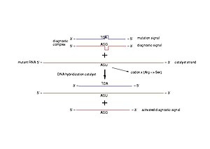
Diagnostic of pathogenic mutations. A diagnost...

Diagnostic of pathogenic mutations. A diagnostic complex is a dsDNA molecule resembling a short part of the gene of interest, in which one of the strands is intact (diagnostic signal) and the other bears the mutation to be detected (mutation signal). In case of a pathogenic mutation, the transcribed mRNA pairs to the mutation signal and triggers the release of the diagnostic signal (Photo credit: Wikipedia)

Read Full Post »

Finding the Genetic Links in Common Disease:  Caveats of Whole Genome Sequencing Studies

Writer and Reporter: Stephen J. Williams, Ph.D.

In the November 23, 2012 issue of Science, Jocelyn Kaiser reports (Genetic Influences On Disease Remain Hidden in News and Analysis)[1] on the difficulties that many genomic studies are encountering correlating genetic variants to high risk of type 2 diabetes and heart disease.  At the recent American Society of Human Genetics annual 2012 meeting, results of several DNA sequencing studies reported difficulties in finding genetic variants and links to high risk type 2 diabetes and heart disease.  These studies were a part of an international effort to determine the multiple genetic events contributing to complex, common diseases like diabetes.  Unlike Mendelian inherited diseases (like ataxia telangiectasia) which are characterized by defects mainly in one gene, finding genetic links to more complex diseases may pose a problem as outlined in the article:

  • Variants may be so rare that massive number of patient’s genome would need to be analyzed
  • For most diseases, individual SNPs (single nucleotide polymorphisms) raise risk modestly
  • Hard to find isolated families (hemophilia) or isolated populations (Ashkenazi Jew)
  • Disease-influencing genes have not been weeded out by natural selection after human population explosion (~5000 years ago) resulted in numerous gene variants
  • What percentage variants account for disease heritability (studies have shown this is as low as 26% for diabetes with the remaining risk determined by environment)

Although many genome-wide-associations studies have found SNPs that have causality to increasing risk diseases such as cancer, diabetes, and heart disease, most individual SNPs for common diseases raise risk by about only 20-40% and would be useless for predicting an individual’s chance they will develop disease and be a candidate for a personalized therapy approach.  Therefore, for common diseases, investigators are relying on direct exome sequencing and whole-genome sequencing to detect these medium-rare risk variants, rather than relying on genome-wide association studies (which are usually fine for detecting the higher frequency variants associated with common diseases).

Three of the many projects (one for heart risk and two for diabetes risk) are highlighted in the article:

1.  National Heart, Lung and Blood Institute Exome Sequencing Project (ESP)[2]: heart, lung, blood

  • Sequenced 6,700 exomes of European or African descent
  • Majority of variants linked to disease too rare (as low as one variant)
  • Groups of variants in the same gene confirmed link between APOC3 and higher risk for early-onset heart attack
  • No other significant gene variants linked with heart disease

2.  T2D-GENES Consortium: diabetes

Sequenced 5,300 exomes of type 2 diabetes patients and controls from five ancestry groups
SNP in PAX4 gene associated with disease in East Asians
No low-frequency variant with large effect though

3.  GoT2D: diabetes

  • After sequencing 2700 patient’s exomes and whole genome no new rare variants above 1.5% frequency with a strong effect on diabetes risk

A nice article by Dr. Sowmiya Moorthie entitled Involvement of rare variants in common disease can be found at the PGH Foundation site http://www.phgfoundation.org/news/5164/ further discusses this conundrum,  and is summarized below:

“Although GWAs have identified many SNPs associated with common disease, they have as yet had little success in identifying the causative genetic variants. Those that have been identified have only a weak effect on disease risk, and therefore only explain a small proportion of the heritable, genetic component of susceptibility to that disease. This has led to the common disease-common variant hypothesis, which predicts that common disease-causing genetic variants exist in all human populations, but each individual variant will necessarily only have a small effect on disease susceptibility (i.e. a low associated relative risk).

An alternative hypothesis is the common disease, many rare variants hypothesis, which postulates that disease is caused by multiple strong-effect variants, each of which is only found in a few individuals. Dickson et al. in a paper in PLoS Biology postulate that these rare variants can be indirectly associated with common variants; they call these synthetic associations and demonstrate how further investigation could help explain findings from GWA studies [Dickson et al. (2010) PLoS Biol. 8(1):e1000294][3].  In simulation experiments, 30% of synthetic associations were caused by the presence of rare causative variants and furthermore, the strength of the association with common variants also increased if the number of rare causative variants increased. “

one_of_many rare variants

Figure from Dr. Moorthie’s article showing the problem of “finding one in many”.

(please   click to enlarge)

Indeed, other examples of such issues concerning gene variant association studies occur with other common diseases such as neurologic diseases and obesity, where it has been difficult to clearly and definitively associate any variant with prediction of risk.

For example, Nuytemans et. al.[4] used exome sequencing to find variants in the vascular protein sorting 3J (VPS35) and eukaryotic transcription initiation factor 4  gamma1 (EIF4G1) genes, tow genes causally linked to Parkinson’s Disease (PD).  Although they identified novel VPS35 variants none of these variants could be correlated to higher risk of PD.   One EIF4G1 variant seemed to be a strong Parkinson’s Disease risk factor however there was “no evidence for an overall contribution of genetic variability in VPS35 or EIF4G1 to PD development”.

These negative results may have relevance as companies such as 23andme (www.23andme.com) claim to be able to test for Parkinson’s predisposition.  To see a description of the LLRK2 mutational analysis which they use to determine risk for the disease please see the following link: https://www.23andme.com/health/Parkinsons-Disease/. This company and other like it have been subjects of posts on this site (Personalized Medicine: Clinical Aspiration of Microarrays)

However there seems to be more luck with strategies focused on analyzing intronic sequence rather than exome sequence. Jocelyn Kaiser’s Science article notes this in a brief interview with Harry Dietz of Johns Hopkins University where he suspects that “much of the missing heritability lies in gene-gene interactions”.  Oliver Harismendy and Kelly Frazer and colleagues’ recent publication in Genome Biology  http://genomebiology.com/content/11/11/R118 support this notion[5].  The authors used targeted resequencing of two endocannabinoid metabolic enzyme genes (fatty-acid-amide hydrolase (FAAH) and monoglyceride lipase (MGLL) in 147 normal weight and 142 extremely obese patients.

These patients were enrolled in the CRESCENDO trial and patients analyzed were of European descent. However, instead of just exome sequencing, the group resequenced exome AND intronic sequence, especially focusing on promoter regions.   They identified 1,448 single nucleotide variants but using a statistical filter (called RareCover which is referred to as a collapsing method) they found 4 variants in the promoters and intronic areas of the FAAH and MGLL genes which correlated to body mass index.  It should be noted that anandamide, a substrate for FAAH, is elevated in obese patients. The authors did note some issues though mentioning that “some other loci, more weakly or inconsistently associated in the original GWASs, were not replicated in our samples, which is not too surprising given the sample size of our cohort is inadequate to replicate modest associations”.

PLEASE WATCH VIDEO on the National Heart, Lung and Blood Institute Exome Sequencing Project

https://www.youtube.com/watch?v=-Qr5ahk1HEI

REFERENCES

http://www.phgfoundation.org/news/5164/  PHG Foundation

1.            Kaiser J: Human genetics. Genetic influences on disease remain hidden. Science 2012, 338(6110):1016-1017.

2.            Tennessen JA, Bigham AW, O’Connor TD, Fu W, Kenny EE, Gravel S, McGee S, Do R, Liu X, Jun G et al: Evolution and functional impact of rare coding variation from deep sequencing of human exomes. Science 2012, 337(6090):64-69.

3.            Dickson SP, Wang K, Krantz I, Hakonarson H, Goldstein DB: Rare variants create synthetic genome-wide associations. PLoS biology 2010, 8(1):e1000294.

4.            Nuytemans K, Bademci G, Inchausti V, Dressen A, Kinnamon DD, Mehta A, Wang L, Zuchner S, Beecham GW, Martin ER et al: Whole exome sequencing of rare variants in EIF4G1 and VPS35 in Parkinson disease. Neurology 2013, 80(11):982-989.

5.            Harismendy O, Bansal V, Bhatia G, Nakano M, Scott M, Wang X, Dib C, Turlotte E, Sipe JC, Murray SS et al: Population sequencing of two endocannabinoid metabolic genes identifies rare and common regulatory variants associated with extreme obesity and metabolite level. Genome biology 2010, 11(11):R118.

Other posts on this site related to Genomics include:

Cancer Biology and Genomics for Disease Diagnosis

Diagnosis of Cardiovascular Disease, Treatment and Prevention: Current & Predicted Cost of Care and the Promise of Individualized Medicine Using Clinical Decision Support Systems

Ethical Concerns in Personalized Medicine: BRCA1/2 Testing in Minors and Communication of Breast Cancer Risk

Genomics & Genetics of Cardiovascular Disease Diagnoses: A Literature Survey of AHA’s Circulation Cardiovascular Genetics, 3/2010 – 3/2013

Genomics-based cure for diabetes on-the-way

Personalized Medicine: Clinical Aspiration of Microarrays

Late Onset of Alzheimer’s Disease and One-carbon Metabolism

Genetics of Disease: More Complex is How to Creating New Drugs

Genetics of Conduction Disease: Atrioventricular (AV) Conduction Disease (block): Gene Mutations – Transcription, Excitability, and Energy Homeostasis

Centers of Excellence in Genomic Sciences (CEGS): NHGRI to Fund New CEGS on the Brain: Mental Disorders and the Nervous System

Cancer Genomic Precision Therapy: Digitized Tumor’s Genome (WGSA) Compared with Genome-native Germ Line: Flash-frozen specimen and Formalin-fixed paraffin-embedded Specimen Needed

Mitochondrial Metabolism and Cardiac Function

Pancreatic Cancer: Genetics, Genomics and Immunotherapy

Issues in Personalized Medicine in Cancer: Intratumor Heterogeneity and Branched Evolution Revealed by Multiregion Sequencing

Quantum Biology And Computational Medicine

Personalized Cardiovascular Genetic Medicine at Partners HealthCare and Harvard Medical School

Centers of Excellence in Genomic Sciences (CEGS): NHGRI to Fund New CEGS on the Brain: Mental Disorders and the Nervous System

LEADERS in Genome Sequencing of Genetic Mutations for Therapeutic Drug Selection in Cancer Personalized Treatment: Part 2

Consumer Market for Personal DNA Sequencing: Part 4

Personalized Medicine: An Institute Profile – Coriell Institute for Medical Research: Part 3

Whole-Genome Sequencing Data will be Stored in Coriell’s Spin off For-Profit Entity


Read Full Post »

On Devices and On Algorithms: Arrhythmia after Cardiac Surgery Prediction and ECG Prediction of Paroxysmal Atrial Fibrillation Onset

Author, and Content Consultant to e-SERIES A: Cardiovascular Diseases: Justin Pearlman, MD, PhD, FACC

and

Article Curator: Aviva Lev-Ari, PhD, RN

Cleveland Clinic research spurs a device that could predict arrhythmia after cardiac surgery

April 30, 2013 9:03 am by  |

ECG

Heart doctors at the Cleveland Clinic  hope to give doctors a way to tell which patients might develop arrhythmia after cardiac surgery.

Atrial fibrillation (AFIB) is one of the most common complications of heart surgeries, and also occurs as a complication of elevated alcohol use, high blood pressure, valve disease or thyroid disease. Atrial fibrillation consists of the round parts of the Valentine heart (the atria) shivering chaotically instead of beating rhythmically. Atrial fibrillation is a common arrhythmia, eventually affecting 20% of adults. There are 3 varieties: paroxysmal (intermittent), persistent (continual) and permanent (unremitting).  When AFIB lasts longer than 24-48 hours the risk of forming a blood clot in the atria rises, which in turn can cause a stroke or a heart attack. AFIB often results in fast heart rates which may cause low blood pressure and its possible consequences (organ injury, heart attack). Also, prolonged fast rates weaken the heart (reversible rate-related cardiomyopathy), which can persist for months after regaining target ranges for the heart rates (target for rate control is 60-80/minute instead of the fast rates of 100-180/min that are common with untreated AFIB).

A scoring system (CHADS2) can predict who may suffer from a stroke due to AFIB that lasts >24-48 hours, and in particular, who may benefit from longterm anticoagulation (blood thinners to interfere with clot formation). A pill-in-the-pocket can stop AFIB within hours.  Amiodarone, a highly toxic medication (10% long-term uses face side effects of serious damage to liver, lung, thyroid or eyes), is often prescribed “off-label” (without FDA endorsement) because it is 70% effective in preventing AFIB recurrence, and it has less anticontractility (weakening of the strength of heart beats) than most other rhythm medications. Then next most effective medication for suppression of AFIB long-term is sotalol, which reduces the strength of heart contraction (may not be tolerated by patients with severe heart failure) and it prolongs QT interval of repolarization after each heartbeat, a risk factor for a deadly rhythm called torsades de pointes. Interventional cures (“AFIB ablation”) have been developed to prevent recurrences.

Predicting AFIB may have several benefits: (1) potentially, earlier use of pill-in-the-pocket could prevent episodes rather that wait for them to occur, get noticed, and then treated, as only ~50% of AFIB episodes are noticed by the patient, according to electrographic monitor reports; (2) surrogate endpoint (prediction of onset) may offer useful guidance as to sufficiency of a suppressive therapy to enable lower dosing of toxic treatments; (3)  surrogate endpoint (prediction of onset) may offer useful guidance as to sufficient lowering of alohol intake, sufficient control of blood pressure, sufficient control of thyroid abnormalities, and other prevention opportunities; (4) surrogate endpoints may facilitate AFIB ablation.

Work done in the lab of Dr. C. Allen Bashour indicated that most patients who experience atrial fibrillation after heart surgery show clues beforehand in the form of subtle changes in their ECG readings that aren’t detected with the way they’re monitored now.

Rindex Medical is commercializing a tool that would enable physicians to predict which patients will experience AF so they can receive prophylactic treatment before it occurs.

“Right now they basically guess, or treat everyone prophylactically,” said co-founder Alex Arrow. “Some clinicians say they have an intuition about who will get it, but it’s mostly guesswork.”

Rindex’s A-50 AF Prediction System uses algorithms developed at the Clinic to analyze a patient’s ECG signals through 17 steps and produce a score, from 1 to 100, of how likely that patient is to experience AF. Arrow said the final product will be a touch-screen monitor that displays a score and tracks the score over a nine-hour period.

The Redwood City, California, company has been issued the first of its patents for the device and the exclusive license from Cleveland Clinic to develop the technology. Self-funded by Arrow and co-founders Denis Hickey and Lucas Fairfield, Rindex has a working prototype and is making progress on preparations for its 510(k) application. Arrow said the company shouldn’t need to raise a series A until it’s ready for a clinical trial.

Many other research groups have explored ways to predict AF in its various forms from natriuretic peptides to ECG changes, but no method has been established as reliably for this purpose.

Read more: http://medcitynews.com/2013/04/cleveland-clinic-research-spurs-a-device-that-could-predict-arrhythmias-after-cardiac-surgery/#ixzz2ScbxIyW0

http://medcitynews.com/2013/04/cleveland-clinic-research-spurs-a-device-that-could-predict-arrhythmias-after-cardiac-surgery/?goback=%2Egde_1503357_member_237204073

Dec 13, 2012

ECG predicts atrial fibrillation onset

Atrial fibrillation (AF), the most common cardiac arrhythmia, is categorized by different forms. One sub-type is paroxysmal AF (PAF), which refers to episodes of arrhythmia that generally terminate spontaneously after no more than a few days. Although the underlying causes of PAF are still unknown, it’s clear that predicting the onset of PAF would be hugely beneficial, not least because it would enable the application of treatments to prevent the loss of sinus rhythm.

Many research groups are tackling the issue of predicting the onset of PAF. Now, however, researchers in Spain have developed a method that assesses the risk of PAF at least one hour before its onset. To date, the approach has not only successfully discriminated healthy individuals and PAF patients, but also distinguished patients far from and close to PAF onset (Physiol. Meas. 33 1959).

“The ability to assess the risk of arrhythmia at least one hour before its onset is clinically relevant,” Arturo Martinez from the University of Castilla-La Mancha told medicalphysicsweb. “Our method assesses the P-wave feature time course from single-lead long-term ECG recordings. Using a single ECG lead reduces the computational burden, paving the way for a real-time system in future.”

Analysing sinus rhythm

If the heart is beating normally, the sinus rhythm observed on an ECG will contain certain generic features, such as a P-wave that reflects the atrial depolarization and a large characteristic R peak flanked by two minima representing the depolarization of the heart’s right and left ventricles. If an irregular heart beat is suspected, an ECG will be used and typical findings include the absence of a P-wave.

“We hypothesized that different stages of AF could be identified when analysing long-term recordings extracted from patients prone to AF,” commented Martinez. “Our method differs to others in that we also use just one single lead to detect small differences in features from the P-wave time course.”

P for paroxysmal

Martinez and his collaborators, Raul Alcaraz and Jose Rieta, studied 24-hour Holter ECG recordings from 24 patients in whom PAF had been detected for the first time. For each patient, the longest sinus rhythm interval in the recording was selected, and the two hours preceding the onset of PAF were analysed. These readings were compared with those from 28 healthy individuals. In all cases, only the trace from the V1 ECG lead was considered.

A major challenge for the researchers was to extract the P-wave from the baseline noise. To overcome this, they used an automatic delineator algorithm based on a phasor transform that determines the precise time point relating to the onset, peak and offset of the P-wave. The authors described this algorithm in a previous research paper (Physiol. Meas. 31 1467).

“All of the recordings in our study were visually supervised by expert cardiologists who corrected the P-wave fiducial points when needed,” said Martinez. “Even in the presence of noise, which generated an incredible amount of P-wave distortion, our delineator provided location errors lower than 8 ms.”

In order to assess which time course features might be useful to predict the onset of PAF, the researchers analysed a number of variables. First, they examined factors representing the duration of the P-wave (Pdur), such as the distance between the P-wave onset and peak (Pini) and the distance between the P-wave peak and its offset (Pter). They then studied factors relating P- to R-waves, such as the distance between the two waves’ peaks (PRk) and, finally, beat-to-beat P-wave factors, such as the distance between two consecutive P-wave onset points (PPon).

“The most remarkable trends were provided by the features measuring P-wave duration,” report the authors in their paper. “Pduridentified appropriately 84.21% of all the analysed patients, obtaining a discriminant accuracy of 90.79% and 83.33% between healthy subjects and PAF patients far from PAF and close to PAF, respectively. The metrics related to the PR interval showed the most limited ability to identify patient groups.”

About the author

Jacqueline Hewett is a freelance science and technology journalist based in Bristol, UK.

http://medicalphysicsweb.org/cws/article/research/51820

Original Article

Physiol Meas. 2010 Nov;31(11):1467-85. doi: 10.1088/0967-3334/31/11/005. Epub 2010 Sep 24.

Application of the phasor transform for automatic delineation of single-lead ECG fiducial points.

Martínez AAlcaraz RRieta JJ.

Source

Innovation in Bioengineering Research Group, University of Castilla La Mancha, Spain. arturo.martinez@uclm.es

Abstract

This work introduces a new single-lead ECG delineator based on phasor transform. The method is characterized by its robustness, low computational cost and mathematical simplicity. It converts each instantaneous ECG sample into a phasor, and can precisely manage P and T waves, which are of notably lower amplitude than the QRS complex. The method has been validated making use of synthesized and real ECG sets, including the MIT-BIH arrhythmia, QT, European ST-T and TWA Challenge 2008 databases. Experiments with the synthesized recordings reported precise detection and delineation performances in a wide variety of ECGs, with signal-to-noise ratios of 10 dB and above. For real ECGs, the QRS detection was characterized by an average sensitivity of 99.81% and positive predictivity of 99.89%, for all the analyzed databases (more than one million beats). Regarding delineation, the maximum localization error between automatic and manual annotations was lower than 6 ms and its standard deviation was in agreement with the accepted tolerances for expert physicians in the onset and offset identification for QRS, P and T waves. Furthermore, after revising and reannotating some ECG recordings by expert cardiologists, the delineation error decreased notably, becoming lower than 3.5 ms, on average, and reducing by a half its standard deviation. This new proposed strategy outperforms the results provided by other well-known delineation algorithms and, moreover, presents a notably lower computational cost.

SOURCES:

Original Database

MIT-BIH Polysomnographic Database

This database is described in

Ichimaru Y, Moody GB. Development of the polysomnographic database on CD-ROM. Psychiatry and Clinical Neurosciences 53:175-177 (April 1999).

Please cite this publication when referencing this material, and also include the standard citation for PhysioNet:

Goldberger AL, Amaral LAN, Glass L, Hausdorff JM, Ivanov PCh, Mark RG, Mietus JE, Moody GB, Peng C-K, Stanley HE. PhysioBank, PhysioToolkit, and PhysioNet: Components of a New Research Resource for Complex Physiologic Signals. Circulation 101(23):e215-e220 [Circulation Electronic Pages; http://circ.ahajournals.org/cgi/content/full/101/23/e215]; 2000 (June 13).

The MIT-BIH Polysomnographic Database is a collection of recordings of multiple physiologic signals during sleep. Subjects were monitored in Boston’s Beth Israel Hospital Sleep Laboratory for evaluation of chronic obstructive sleep apnea syndrome, and to test the effects of constant positive airway pressure (CPAP), a standard therapeutic intervention that usually prevents or substantially reduces airway obstruction in these subjects. The database contains over 80 hours’ worth of four-, six-, and seven-channel polysomnographic recordings, each with an ECG signal annotated beat-by-beat, and EEG and respiration signals annotated with respect to sleep stages and apnea. For further information, see Signals and Annotations.

The database consists of 18 records, each of which includes 4 files:

Sleep/apneaannotations Beatannotations Signals Header View waveforms *
slp01a.st slp01a.ecg slp01a.dat slp01a.hea
slp01b.st slp01b.ecg slp01b.dat slp01b.hea
slp02a.st slp02a.ecg slp02a.dat slp02a.hea
slp02b.st slp02b.ecg slp02b.dat slp02b.hea
slp03.st slp03.ecg slp03.dat slp03.hea
slp04.st slp04.ecg slp04.dat slp04.hea
slp14.st slp14.ecg slp14.dat slp14.hea
slp16.st slp16.ecg slp16.dat slp16.hea
slp32.st slp32.ecg slp32.dat slp32.hea
slp37.st slp37.ecg slp37.dat slp37.hea
slp41.st slp41.ecg slp41.dat slp41.hea
slp45.st slp45.ecg slp45.dat slp45.hea
slp48.st slp48.ecg slp48.dat slp48.hea
slp59.st slp59.ecg slp59.dat slp59.hea
slp60.st slp60.ecg slp60.dat slp60.hea
slp61.st slp61.ecg slp61.dat slp61.hea
slp66.st slp66.ecg slp66.dat slp66.hea
slp67x.st slp67x.ecg slp67x.dat slp67x.hea

(*) You may follow these links to view the signals and st annotations using either WAVE (under Linux, SunOS, or Solaris) or WVIEW (under MS-Windows). To do so successfully, you must have configured your browser to use wavescript (for WAVE) or wvscript (for WVIEW) as a helper application, as described in the WAVE User’s Guide(see the section titled WAVE and the Web) and in Setting up WVSCRIPT.

Andrew Walsh observed that the calibration originally provided for the BP signal of record slp37 is incorrect (since it yielded negative BPs). slp37.hea now contains an estimated BP calibration that yields more plausible BPs; these should not be regarded as accurate, however, since there is no independent calibration standard available for this recording.

SOURCE:
Original Article
Proc Inst Mech Eng H. 2010;224(1):27-42.

Finding events of electrocardiogram and arterial blood pressure signals via discrete wavelet transform with modified scales.

Ghaffari AHomaeinezhad MRAkraminia MDavaeeha M.

Source

Cardiovascular Research Group, Department of Mechanical Engineering, K. N. Toosi University of Technology, Tehran, Iran.

Abstract

A robust electrocardiogram (ECG) wave detection-delineation algorithm that can be applied to all ECG leads is developed in this study on the basis of discrete wavelet transform (DWT). By applying a new simple approach to a selected scale obtained from DWT, this method is capable of detecting the QRS complex, P-wave, and T-wave as well as determining parameters such as start time, end time, and wave sign (upward or downward). In the proposed method, the selected scale is processed by a sliding rectangular window of length n and the curve length in each window is multiplied by the area under the absolute value of the curve. In the next step, an adaptive thresholding criterion is conducted on the resulted signal. The presented algorithm is applied to various databases including the MIT-BIH arrhythmia database, European ST-T database, QT database, CinC Challenge 2008 database as well as high-resolution Holter data gathered in the DAY Hospital. As a result, the average values of sensitivity and positive prediction Se = 99.84 per cent and P+ = 99.80 per cent were obtained for the detection of QRS complexes with an average maximum delineation error of 13.7, 11.3, and 14.0 ms for the P-wave, QRS complex, and T-wave respectively. The presented algorithm has considerable capability in cases of a low signal-to-noise ratio, high baseline wander, and in cases where QRS complexes and T-waves appear with abnormal morphologies. Especially, the high capability of the algorithm in the detection of the critical points of the ECG signal, i.e. the beginning and end of the T-wave and the end of the QRS complex was validated by the cardiologist and the maximum values of 16.4 and 15.9 ms were recognized as absolute offset error of localization respectively. Finally, in order to illustrate an alternative capability of the algorithm, it is applied to all 18 subjects of the MIT-BIH polysomnographic database and the end-systolic and end-diastolic points of the blood pressure waveform were extracted and values of sensitivity and positive prediction Se = 99.80 per cent and P+ = 99.86 per cent were obtained for the detection of end-systolic, end-diastolic pulses.

http://www.ncbi.nlm.nih.gov/pubmed/20225455

Original Article

A robust wavelet-based multi-lead electrocardiogram delineation algorithm

  • a Department of Mechanical Engineering, K.N. Toosi University of Technology, Tehran, Iran
  • b CardioVascular Research Group (CVRG), Iran
  • c Non-invasive Cardiac Electrophysiology Laboratory, DAY Hospital, Tehran, Iran

Abstract

A robust multi-lead ECG wave detection-delineation algorithm is developed in this study on the basis of discrete wavelet transform (DWT). By applying a new simple approach to a selected scale obtained from DWT, this method is capable of detecting QRS complex, P-wave and T-wave as well as determining parameters such as start time, end time, and wave sign (upward or downward). First, a window with a specific length is slid sample to sample on the selected scale and the curve length in each window is multiplied by the area under the absolute value of the curve. In the next step, a variable thresholding criterion is designed for the resulted signal. The presented algorithm is applied to various databases including MIT-BIH arrhythmia database, European ST-T Database, QT Database, CinC Challenge 2008 Database as well as high resolution Holter data of DAY Hospital. As a result, the average values of sensitivity and positive predictivity Se = 99.84% and P+ = 99.80% were obtained for the detection of QRS complexes, with the average maximum delineation error of 13.7 ms, 11.3 ms and 14.0 ms for P-wave, QRS complex and T-wave, respectively. The presented algorithm has considerable capability in cases of low signal-to-noise ratio, high baseline wander, and abnormal morphologies. Especially, the high capability of the algorithm in the detection of the critical points of the ECG signal, i.e. the beginning and end of T-wave and the end of the QRS complex was validated by cardiologists in DAY hospital and the maximum values of 16.4 ms and 15.9 ms were achieved as absolute offset error of localization, respectively.

Abbreviations

  • ACL, area-curve length;
  • ECG, electrocardiogram;
  • DWT, discrete wavelet transform;
  • QTDB, QT database;
  • MITDB, MIT-BIH arrhythmia database; 
  • TWADB, T-wave alternans database;
  • CSEDB, common standards for electrocardiography database;
  • EDB, European ST-T database;
  • P+, positive predictivity (%);
  • Se,sensitivity (%);
  • FIR, finite-duration impulse response;
  • LE, location error;
  • CHECK#0, procedure of evaluating obtained results using MIT annotation files;
  • CHECK#1, procedure of evaluating obtained results consulting with a control cardiologist;
  • CHECK#2, procedure of evaluating obtained results consulting with a control cardiologist and also at least with 3 residents

Keywords

  • ECG delineation;
  • Discrete wavelet transform;
  • Variable threshold;
  • Validation

Figures and tables from this article:

Full-size image (14 K)
Fig. 1. FIR filter-bank implementation to generate discrete wavelet transform based on à trous algorithm.
Full-size image (12 K)
Fig. 2. Graphical representation of the logic of the proposed simple transformation for detecting onset and offset edges. In case I, both area and curve length are minimum, (ACLI < ACLII ≤ ACLIII).
Full-size image (58 K)
Fig. 3. The flow-chart of the proposed wavelet-aided electrocardiogram delineation algorithm (rectangle: operation, ellipse: result).
Full-size image (113 K)
Fig. 4. An excerpted segment from a total delineated ECG. Delineated (a) P-waves, (b) QRS complexes and (c) T-waves. (Circles: edges of event, triangles: peak of events, Partition A: lead I, Partition B: lead II).
Full-size image (76 K)
Fig. 5. Procedure of detecting and delineating of P and T-waves using ACL signal between two successive QRS complexes. (a) Simultaneously depiction of ACL, original ECG and the corresponding selected DWT scale, (b) QRS delineation, and (c) P and T-waves delineation.
SOURCE:

Volume 31, Issue 10, December 2009, Pages 1219–1227

http://www.sciencedirect.com/science/article/pii/S1350453309001647

Other related articles published on this Open Access Online Scientific Journal include the following:

Sustained Cardiac Atrial Fibrillation: Management Strategies by Director of the Arrhythmia Service and Electrophysiology Lab at The Johns Hopkins Hospital   http://pharmaceuticalintelligence.com/2012/10/16/sustained-cardiac-atrial-fibrillation-management-strategies-by-director-of-the-arrhythmia-service-and-electrophysiology-lab-at-the-johns-hopkins-hospital/

Cardiac Arrhythmias: A Risk for Extreme Performance Athletes                                                                                                                                                       http://pharmaceuticalintelligence.com/2012/08/08/cardiac-arrhythmias-a-risk-for-extreme-performance-athletes/

Acute Chest Pain/ER Admission: Three Emerging Alternatives to Angiography and PCI    http://pharmaceuticalintelligence.com/2013/03/10/acute-chest-painer-admission-three-emerging-alternatives-to-angiography-and-pci/

Dilated Cardiomyopathy: Decisions on implantable cardioverter-defibrillators (ICDs) using left ventricular ejection fraction (LVEF) and Midwall Fibrosis: Decisions on Replacement using late gadolinium enhancement cardiovascular MR (LGE-CMR)
http://pharmaceuticalintelligence.com/2013/03/10/dilated-cardiomyopathy-decisions-on-implantable-cardioverter-defibrillators-icds-using-left-ventricular-ejection-fraction-lvef-and-midwall-fibrosis-decisions-on-replacement-using-late-gadolinium/

Ablation Devices Market to 2016 – Global Market Forecast and Trends Analysis by Technology, Devices & Applications
http://pharmaceuticalintelligence.com/2012/12/23/ablation-devices-market-to-2016-global-market-forecast-and-trends-analysis-by-technology-devices-applications/

Read Full Post »

Reporter: Aviva Lev-Ari, PhD, RN

Can Coronary Artery Anomalies Be Detected on CT Calcium Scoring Studies?

Academic Radiology, 04/11/2013  Review Article

Maddux PT et al. – The purpose of this study is to determine whether coronary artery anomalies can be detected on noncontrast computed tomography (CT) coronary artery calcium scoring (CCS) studies. Benign and malignant coronary artery anomalies can be detected with relatively high accuracy on noncontrast–enhanced CCS studies. CCS studies should be reviewed for signs of coronary artery anomalies in order to identify malignant variants with possible impact on patient management.

Methods

  • A total of 126 patients (mean age 62 years; 35 women) underwent noncontrast CCS and contrast enhanced coronary CT angiography (cCTA).
  • Thirty–three patients were diagnosed with a coronary anomaly on cCTA, whereas coronary anomalies were excluded in 93.
  • Two observers (reader 1 [R1] and reader 2 [R2]), blinded to patient information independently evaluated each CCS study for: 1) visibility of coronary artery origins, 2) detection of coronary anomalies, and 3) benign or malignant (ie, interarterial) course.
  • Using cCTA as the reference standard, sensitivity, specificity, positive predictive value (PPV), and negative predictive value (NPV) of CCS studies for detecting coronary anomalies were calculated.

Results

  • Of the 33 coronary anomalies, 16 were benign and 17 malignant.
  • Based on noncontrast CCS studies, R1 and R2 correctly identified the left main origin in 123/126 (97.6%) and 121/126 (96%) patients; the left anterior descending origin in 125/126 (99.2%) and 122/126 (96.8%); the circumflex origin in 120/126 (95.2%) and 105/126 (83.3%); and the right coronary artery origin in 117/126 (92.9%) and 103/126 (81.7%), respectively.
  • R1 and R2 identified 34 and 27 coronary anomalies and classified 19 and 15 as malignant, respectively.
  • Interobserver reproducibility for detection of coronary anomalies was good (k = 0.76).
  • Interobserver agreement for detection of malignant variants was even stronger (k = 0.80).
  • On average, coronary artery anomalies were diagnosed with 85.2% sensitivity, 96.4% specificity, 90.5% PPV, and 94.1% NPV on noncontrast CCS studies.

http://www.mdlinx.com/radiology/news-article.cfm/4559774/?xml

http://www.mdlinx.com/radiology/news-article.cfm/4559774/cardiac-ct-coronary-anomaly-coronary-anatomy#ixzz2QrCXwP2q

Advanced CT reconstruction improves cardiac plaque assessment

By Eric Barnes, AuntMinnie.com staff writer

April 12, 2013 — Automated plaque assessment in coronary CT angiography (CCTA) is a promising new way to evaluate a patient’s plaque burden quickly and noninvasively — but it won’t be quick or accurate without the use of advanced iterative reconstruction, according to researchers from Massachusetts General Hospital in Boston.

Automated techniques are still in their infancy, but once they become more reliable they promise to greatly improve risk assessment and management compared with, for example, calcium scoring, by precisely quantifying the amount of coronary artery plaque — fibrotic, lipid core, and calcium — that is present.

“We know the plaque volume and characteristics … are at least as important as the presence of calcium,” said Dr. Stefan Puchner in an interview with AuntMinnie.com. “If we could make plaque assessment more accurate, we could implement all this stuff in our daily practice.”

The process isn’t accurate today. Automated plaque quantification requires significant time for radiologists to fix the incorrectly drawn vessel wall boundaries, making it impractical for routine use. Manually drawing the boundaries would actually take about a day’s work for each patient, so automation is the only way forward, Puchner said. The group wanted to determine if an advanced reconstruction algorithm might produce fewer errors and make semiautomated plaque estimation practical.

In a study that reconstructed ex vivo coronary vessel segments using three different reconstruction methods, the study team found that, indeed, accuracy in plaque quantification depended on the reconstruction algorithm, as well as vessel size and the extent of calcifications. Using advanced reconstruction, fewer corrections were needed to the vessel wall segmentation, Puchner reported at the 2013 European Congress of Radiology (ECR) in Vienna. Specifically, they compared the use of automated vessel assessment using model-based iterative reconstruction (MBIR, GE Healthcare) compared with an earlier IR algorithm, advanced statistical iterative reconstruction (ASIR, GE), or conventional filtered back projection (FBP) reconstruction.

Cross section of a noncalcified plaque reconstructed with the three different algorithms

Cross section of a noncalcified plaque reconstructed with the three different algorithms (left to right: FBP, ASIR, MBIR). No significant differences can be seen between the three algorithms in terms of correct delineation of the plaque borders. All images courtesy of Dr. Stephan Puchner.

For subjects, the group examined three ex vivo human hearts imaged with CCTA and reconstructed with FBP, ASIR, and MBIR. An automated plaque quantification tool (Vitrea Cardiac Solutions, Vital) was applied to each of the three reconstruction algorithms to fit the outer and inner vessel wall boundaries in nine “triplets” constituting 27 vessels. Only the first 40 mm of the contrast-filled vessels was used for analysis.

Each coronary cross section for which the software assigned incorrect boundaries was tallied and corrected in a blinded manner. The group then compared the number of vessel wall corrections between the different reconstruction algorithms using a Chi-square test.

Cross sections reconstructed with ASIR (middle) and MBIR (right) are correctly delineated by the software

Cross ection of a calcified plaque reconstructed with the three different algorithms (left to right: FBP, ASIR, MBIR). In this case, FBP shows an incorrect delineation of the inner vessel wall boundary, including parts of the calcified plaque. In contrast, the vessel wall boundaries in the cross sections reconstructed with ASIR and MBIR are correctly delineated by the software.

“Our analysis included the percentage of corrections between the three algorithms, and a per-vessel comparison of the percentage of corrections between the three algorithms,” Puchner said in his presentation.

In all, the study comprised 2,295 cross sections in 0.5-mm increments from nine coronary vessels, combined into 765 coregistered triplets evaluated with the three algorithms. Overall, 31% of the cross sections needed boundary corrections, he said. Outer vessel wall boundary corrections were needed in 400 cross sections, and inner vessel boundaries were needed in 381 cross sections.

Only in the cross section reconstructed with MBIR (right) are the boundaries correctly delineated

Cross section of a calcified plaque reconstructed with the three different algorithms (left to right: FBPR, ASIR, MBIR). In this case, FBP and ASIR show an incorrect delineation of the inner vessel wall boundary, including the whole or parts of the calcified plaque. Only in the cross section reconstructed with MBIR are the boundaries correctly delineated by the software.

The percentage of corrected cross sections was lower for MBIR (24.1%) versus ASIR (32.4%, p = 0.0003) and FBP (36.6%, p < 0.0001) — but the differences were only marginal between ASIR and FBP, he said.

“We found that MBIR works much better than the conventional algorithms … significantly reducing the number of corrections needed compared to FBP and ASIR, whereas the difference between the two other algorithms was not significant,” Puchner said.

The use of MBIR significantly reduced the need for vessel wall boundary corrections compared with other reconstruction algorithms, particularly at the site of calcifications.

Automated segmentation is certainly faster than manual processing, Puchner said. Just on the three cases used in the study and in the analysis of the proximal 40 mm of each vessel, use of the software saved about three hours compared with what manual segmentation would have required. There is significant processing time required to create MBIR reconstructions, he acknowledged, but in those cases, it’s the technologists, not the physicians, who are spending the additional time, he said.

“The next step will be to look at it in an in vivo environment, to see this application in a beating heart,” Puchner told AuntMinnie.com. And to test other applications and other iterative reconstruction schemes, of course.

“I’m pretty sure that the other newer algorithms will have similar effects, because overall some studies have shown that the use of newer algorithms reduces blooming effects and other stuff that makes it difficult for the software to delineate it correctly,” he said. With manual segmentation, radiologists tend to overcorrect for older reconstruction algorithms and undercorrect for newer techniques, “but if the software does it, the software is much more dependent on image quality, and it makes a difference if it was reconstructed with the newer algorithms or the older algorithms.”

Automated plaque measurements will also have to be compared with assessments in other modalities such as intravascular ultrasound, and even to histology using the donor hearts, he said.

Related Reading

MBIR finds same nodules as ASIR, at fraction of dose, December 13, 2012

MBIR tops ASIR for ultralow-dose CT enterography, November 6, 2012

Study pinpoints optimal ASIR blend for stomach cancer, November 6, 2012

MBIR takes on ASIR in low-dose chest CT, November 6, 2012

Iterative reconstruction cuts CT dose for urinary stone disease, August 20, 2012
Copyright © 2013 AuntMinnie.com

http://www.auntminnie.com/index.aspx?sec=sup&sub=cto&pag=dis&itemid=103108&wf=5397

http://www.mdlinx.com/radiology/news-article.cfm/4559774/?xml

Coronary CT Angiography in the ED

For an analysis of the finding of ROMICAT II Trial:
Hoffmann U, et al; ROMICAT-II Investigators. Coronary CT angiography versus standard evaluation in acute chest pain. N Engl J Med. 2012 Jul 26;367(4):299-308.
go to:

Acute Chest Pain/ER Admission: Three Emerging Alternatives to Angiography and PCI – Corus CAD, hs cTn, CCTA, Curator: Aviva Lev-Ari, PhD, RN, 3/10/2013

It is well known that taking a good history and physical, getting a non-ischemic EKG, and serial cardiac biomarkers, results in a risk of death/AMI of <5% in 30 days. Patients, in whom you still suspect have CAD, should undergo provocative testing within the next 72 hours based on the AHA/ACC guidelines. Their guidelines deem provocative testing as including:

  • Exercise treadmill stress test,
  • Myocardial perfusion scan,
  • Stress echocardiography, and/or
  • Coronary CT angiography (CCTA).

Myocardial perfusion scans and stress echos have a sensitivity of 85–90% and specificity of 75–80%. In contrast, CCTA’s have been shown to have a sensitivity of 93-97% and specificity of 80-90%.

Recently two landmark trials were published in NEJM discussing the use of CCTA in the emergency department.
ACRIN-PA Trial: Litt HI, et al. CT angiography for safe discharge of patients with possible acute coronary syndromes. N Engl J Med. 2012 Apr 12;366(15):1393-403. PMID: 22449295
What they did: 
  •  Non-inferiority study
  • 5 Pennsylvania EDs
  • 1,370 patients, Age > 30 years
  • Inclusion criteria: TIMI score of 0–2, EKG without ischemic changes, and negative first set of Cardiac Biomarkers
  • Randomized 2 patients to CCTA arm (908 patients) for every 1 patient to Standard Stress arm (462 patients)

Primary Outcome:

  • MI or Death from CAD at 30 days

Secondary Outcomes:

  • Rate of discharge from ED
  • Length of stay (LOS) in ED
  • Rate of detection of CAD
  • Resource utilization

What they found:

  • 640/908 pts (70.5%) who underwent CCTA had coronary stenosis of <50% and none had MI or death due to CAD at 30 days
  • Discharge from ED 49.6% with CCTA vs 22.7% with standard stress arm
  • ED LOS 18 hr in CCTA arm vs 24.8 hr in standard stress arm

Conclusion: CCTA allows early discharge of low to intermediate risk patients presenting to the ED with possible ACS.

ROMICAT II Trial:  Hoffmann U, et al; ROMICAT-II Investigators. Coronary CT angiography versus standard evaluation in acute chest pain. N Engl J Med. 2012 Jul 26;367(4):299-308.PMID: 22830462
What they did:
  • Randomized controlled trial
  • 9 EDs in the US
  • 1,000 patients with acute chest pain with ages 40–74 years
  • CCTA (501 patients) versus Standard Evaluation (499 patients)

Primary Outcome:

  • Hospital length of stay

Secondary Outcomes:

  • Cardiovascular events at 28 days
  • Rate of discharge from ED
  • Time to diagnosis
  • Cost
  • Utilization of resources

What they found:

  • Hospital LOS decreased by 7.6 hr in CCTA group
  • Rate of discharge from ED 47% in CCTA arm vs 12% in Standard Evaluation Arm
  • No difference in cardiovascular events at 28 days
  • Cost was similar between two groups $4,289 CCTA vs $4,060 in Standard arm

Conclusion: CCTA decreases length of stay without an increase in rate of cardiovascular events.

Some discussion points worth mentioning:
  • CCTA with 0 lesions is NEGATIVE: These patients can certainly be discharged home with primary care follow up with a nearly 100% NPV for ACS/AMI.
  • CCTA with <50% lesion is NOT NEGATIVE: This patient has CAD. It may not be clinically significant, but we can see plaques. 2/3 of AMIs occur from plaques that have <50% stenosis. Certainly we can start risk factor modification with beta blockers, ASA, and statins, but there are no studies looking at how this group of patients will do long term.
  • CCTAs are anatomic studies and not functional studies. Identified lesions will lead to more diagnostic tests, which is one of the big arguments against CCTA. CCTA identifies CAD more often than standard stress modalities, which leads to more heart catheterizations and PCIs.
  • As the number of CT slice increases, radiation dose decreases:
    1. A 64 slice CT = 10 – 15 mSv of radiation
    2. A 128 slice CT = 5 – 10 mSv of radiation
    3. A 256 slice CT = 1 – 5 mSv of radiation
    4. In contrast, a single-view CXR = 0.02 mSV of radiation
  • There is currently an ongoing National Heart, Lung, and Blood Institute-funded trial called the PROMISE (Prospective Multi-center Imaging Study for Evaluation of Chest Pain) Study with 10,000 patients. Patients with symptoms suggestive of CAD will be randomized to a CCTA vs usual care with a functional test.  What’s interesting about this study is it is being performed in the offices of primary care physicians and cardiologists rather than EDs. The study authors hypothesize that medically optimizing patients identified, as having non-obstructive CAD will yield improved long-term outcomes.
It is well known that in low risk patients, doing a good H&P, having a negative EKG (no ischemic changes), and negative serial cardiac biomarkers gives us about 99% NPV & 99% sensitivity for ACS/AMI. This is even without additional testing, such as CCTAs.So are CCTAs worth the cost and potential harms in this low-risk group to add another 1% to the 99% NPV and 99% sensitivity rates? In my opinion, that answer is NO.
Additional References:
  1. Jancin B. Comparing Technologies for Imaging Chest Pain in the ED.  ACEP News 2013 Mar; 32(3): 1 – 11.
  2. Goldstein JA. A Randomized Controlled Trial of Multi-Slice Coronary Computed Tomography for Evaluation of Acute Chest Pain.  JACC 2007;49: 863 – 71.  PMID: 17320744
  3. Goldstein JA. The CT-STAT (Coronary Computed Tomographic Angiography for Systematic Triage of Acute Chest Pain Patients to Treatment) Trial.  JACC 2011 Sept; 58: 1414 – 22. PMID: 21939822
  4. Hulten E. Outcomes After Coronary Computed Tomography Angiography in the Emergency Department:  A Systematic Review and Meta-Analysis of Randomized, Controlled Trials.  JACC 2013 Feb; 61: 880 – 92.  PMID: 23395069 
  1. Posted by 

Read Full Post »

Mitochondrial Dysfunction and Cardiac Disorders

Curator: Larry H Bernstein, MD, FACP

This article is the THIRD in a four-article Series covering the topic of the Roles of the Mitochondria in Cardiovascular Diseases. They include the following;

  • Mitochondria and Cardiovascular Disease: A Tribute to Richard Bing, Larry H Bernstein, MD, FACP

http://pharmaceuticalintelligence.com/2013/04/14/chapter-5-mitochondria-and-cardiovascular-disease/

  • Mitochondrial Metabolism and Cardiac Function, Larry H Bernstein, MD, FACP

http://pharmaceuticalintelligence.com/2013/04/14/mitochondrial-metabolism-and-cardiac-function/

  • Mitochondrial Dysfunction and Cardiac Disorders, Larry H Bernstein, MD, FACP

http://pharmaceuticalintelligence.com/2013/04/14/mitochondrial-dysfunction-and-cardiac-disorders/

http://pharmaceuticalintelligence.com/2013/04/14/reversal-of-cardiac-mitochondrial-dysfunction/

Mitochondrial Metabolism in Impaired Cardiac Function

Mitochondrial Dysfunction and the Heart

Chronically elevated plasma free fatty acid levels in heart failure are associated with
  • decreased metabolic efficiency and cellular insulin resistance.
The mitochondrial theory of aging (MTA) and the free-radical theory of aging (FRTA) are closely related.
They were in fact proposed by the same researcher about 20 years apart. MTA adds
  • the mitochondria and its production of free radicals
  • into the concept that free-radicals damage DNA over time.
Tissue hypoxia, resulting from low cardiac output with or independent of endothelial impairment,
This dysfunctional state causes loss of mitochondrial mass. Therapies aimed at protecting mitochondrial function
  • have shown promise in patients and animal models with heart failure that will be the subject of Chapter III.

Myocardial function in hypertension

Genetic variation in vitamin D-dependent signaling
  • is associated with congestive heart failure in human subjects with hypertension.
Functional polymorphisms were selected from five candidate genes:
  1. CYP27B1,
  2. CYP24A1,
  3. VDR,
  4. REN and
  5. ACE.
Using the Marshfield Clinic Personalized Medicine Research Project,
  • 205 subjects with hypertension and congestive heart failure,
  • 206 subjects with hypertension alone and
  • 206 controls (frequency matched by age and gender) were genotyped.
In the context of hypertension, a SNP in CYP27B1 was associated with congestive heart failure
(odds ratio: 2.14 for subjects homozygous for the C allele; 95% CI: 1.05–4.39).
Genetic variation in vitamin D biosynthesis is associated with increased risk of heart failure.
RA Wilke, RU Simpson, BN Mukesh, SV Bhupathi, et al. Genetic variation in CYP27B1 is associated

Heart Failure and Coronary Circulation

There is a decrease in resting and peak stress myocardial function in chronic heart failure patients,
  • with recovery of skeletal muscle phosphocreatine following exercise induced by perhexiline treatment.
This suggested that mitochondrial deficiencies, caused by excessive free fatty acids (FFAs)
  • underlie a common cardiac and skeletal muscle myopathy in heart failure patients.
Tissue hypoxia in chronic heart failure from inadequate circulation in heart failure
  • increases the oxidative stress in lean body mass and in the heart itself.
The heterodimeric transcription factor hypoxia-inducible factor (HIF)-1
  • induces changes in the transcription of genes that encode proteins involved in the adaptation to hypoxia.
HIF-1 activity depends on levels of the HIF-1a subunit, which has a short half-life.
HIF-1a increases in rats with experimentally induced myocardial infarction together with elevated levels of
  • GLUT1 and haemoxygenase-1 in the peri-infarct region of the heart
The cardiac metabolic response to hypoxia is considered to be
  • a return to a pattern of fetal metabolism, in which
  • carbohydrates predominate as substrates for energy metabolism.
The reliance on carbohydrate energy source is thought to be a result of  the downregulation of PPARa with a decreased activity of
The sarcolemmal fatty acid transporter protein (FATP) levels are also decreased with palmitate oxidation,
  • transitioning away from fatty acid metabolism proportional to the degree of cardiac impairment.
The hypoxic changes of heart failure drives a switch toward
  1. glycolysis and glucose oxidation
  2. restriction of myocardial fatty acid uptake.
Nevertheless, late in the progression of heart failure, substrate metabolism is insufficient to support cardiac function, because
  • the hypoxic failing heart is no longer able to oxidize fats and may also be insulin resistant.
The author surmises that mitochondrial dysfunction caused by tissue hypoxia might be mediated by the
  • proapoptotic protein BCL2/adenovirus E1B 19kDa interacting protein (Bnip)3.
It  is strongly upregulated in response to hypoxia. In the isolated, perfused rat heart, Bnip3 expression was
  • induced after 1h of hypoxia, with Bnip3 integrating into the mitochondria of hypoxic ventricular myocytes.
This resulted in mitochondrial defects associated with
  1. opening of the permeability transition pore, leading to
  2. loss of inner membrane integrity and
  3. loss of mitochondrial mass.

Mitochondrial Dysfunction caused by Bnip3 Precedes Cell Death.

Experimentally induced myocardial ischemia had evidence of contractile dysfunction but preserved viability. A progressive
  • decline in circulating levels of endothelial progenitor cells was documented 3 months following instrumentation (P<0.001).
Quantitative polymerase chain reaction analysis revealed that
  • chronic myocardial ischemia produced a biphasic response in both
    • hypoxic-inducible factor 1 and
    • stromal-derived factor 1 mRNA expression.
While initially unregulated, a gradual decline was observed over time (from day 45 to 90), in
  • hypoxic-inducible factor 1 and
  • stromal-derived factor 1 mRNA expression .
On serial assessment, endothelial progenitor cell migration was progressively impaired in response to chemo-attractant gradients of:
  1. vascular endothelial growth factor (10-200 ng/mL)
  2. and stromal cell-derived factor-1 (10-100 ng/mL) .
Decreased circulating levels and migratory dysfunction of bone marrow derived endothelial progenitor cells
  • were documented in a reproducible clinically relevant model of myocardial ischemia.

Nitric Oxide (NO) in Myocardial Ischemia and Infarct

Nitric oxide (NO) is a free radical with an unpaired electron; it is an important physiologic messenger,
  • produced by nitric oxide synthases, which catalyze the reaction l-arginine to citrulline and NO.
The constitutive isoforms exists in neuronal and endothelial cells and is calcium dependent. Calcium binds to calmodulin and
  1. the calcium calmodulin complex activates the constitutive NO synthase that releases NO,
  2. relaxing smooth muscle cells through activation of guanylate cyclase and the production cGMP.
Therefore, the NO produced has a negative inotropic effect on the heart and is instrumental in the autoregulation of the coronary circulation.
The inducible form of NO synthase (iNOS), mostly produced in macrophages, is activated by cytokines and endotoxin. It eliminates intracellular pathogens,
damaging cells by inhibiting
  1. ATP production
  2. oxidative phosphorylation
  3. DNA synthesis.
In infection, lipopolysaccharide released from bacterial walls, stimulates production of iNOS. The large amount of NO produced
  • causes extensive vasodilation and hypotension.
We sought to assess whether oxidation products of
  • nitric oxide (NO), nitrite (NO2−) and nitrate (NO3−), referred to as NOx,
  • are released by the heart of patients after acute myocardial infarction (AMI) and
  • whether NOx can be determined in peripheral blood of these patients.
Previously we reported that in experimental myocardial infarction (rabbits) NOx is released mainly by inflammatory cells
  • (macrophages) in the myocardium 3 days after onset of  ischemia.
NOx is formed in heart muscle from NO; It originates through the activity of the inducible form of nitric oxide synthase (iNOS).
Eight patients with acute anterior MI and an equal number of controls were studied. Coronary venous blood was obtained by
coronary sinus catheterization; NOx concentrations in coronary sinus, in arterial and peripheral venous plasma were measured.
Left ventricular end-diastolic pressure was determined. Measurements were carried out 24, 48 and 72 h after onset of symptoms.
The type and location of coronary arterial lesions were determined by coronary angiography. Plasma NO3− was reduced to NO2−
by nitrate reductase before determination of NO2− concentration by chemiluminescence.
The results provided evidence that in patients with acute anterior MI, the myocardial production of nitrite and nitrate (NOx) was increased,
  • as well as the coronary arterial–venous difference.
Increased NOx production by the infarcted heart accounted for the increase of NOx concentration in arterial and the peripheral venous plasma.
The peak elevation of NOx occurred on days 2 and 3 after onset of the symptoms, suggesting that NOx production was at least in part the result of
  • production of NO by inflammatory cells (macrophages) in the heart.
The appearance of oxidative products of NO (NO2− and NO3−) in peripheral blood of patients with acute MI is
  • the result of their increased release from infarcted heart during the inflammatory phase of myocardial ischemia.
Further studies are needed to define the clinical value of these observations.
K Akiyama,  A Kimura, H Suzuki, Y Takeyama, …. R Bing.  Production of oxidative products of nitric oxide in infarcted human heart.  J Am Coll Cardiol. 1998;32(2):373-379.   http://dx.doi.org/10.1016/S0735-1097(98)00270-8
OPA1 Mutation and Late-Onset Cardiomyopathy
No cardiac disorders have been described in patients with OPA1 or similar mutations
  • involving the fission/fusion genes as seen in inherited maladies like Charcot–Marie–Tooth disease.
Our results indicate that, at least for OPA1, cardiac abnormalities are not completely
  • manifest until the development of blindness.

The OPA1-mutant mice survived more than 1 year and appeared healthy.

In patients with these diseases, reduced cardiac function may go undetected
secondary to reduced physical activity secondary to loss of vision.
It would be expected that patients with such mutations would have impaired cardiac reserve with
  • reduced ability to respond to high-stress disease states such as myocardial infarction and sepsis.
The OPA1-mutant mice have reduced cardiac reserve, as shown by
  • the lack of response to isoproterenol or to ischemia/reperfusion injury,
This suggests that patients with OPA1 and related inherited mitochondrial diseases
  • should be screened for abnormalities of cardiac function.
Le Chen; T Liu; A Tran; Xiyuan Lu; …AA. Knowlton. OPA1 Mutation and Late-Onset Cardiomyopathy:
Mitochondrial Dysfunction and mtDNA Instability.  http://jaha.ahajournals.org/content/1/5/e003012.full

Oxidative Stress and Mitochondria in the Failing Heart

The major problem in tissue hypoxia in the failing heart is oxidative stress. Reactive oxygen species (ROS), including
  • superoxide,
  • hydroxyl radicals and
  • hydrogen peroxide,
are generated by a number of cellular processes, including
  • mitochondrial electron transport,
  • NADPH oxidase and
  • xanthine dehydrogenase/xanthine oxidase.
The low availability of oxygen, the final receptor of mitochondrial electron transport (ET), results in
  • electron accumulation in the ET chain as the complexes become highly reduced.
A number of experimental and clinical studies have suggested that ROS generation is
  • enhanced in heart failure because of electron leak, and complexes I and II
  • are implicated as the primary sites of this loss.
Prolonged oxidative stress in cardiac failure results in damage to mitochondrial DNA.
The continued ROS generation and consequent cellular injury leads to functional decline.
Thus, mitochondria are both the sources and targets of a cycle of ROS-mediated injury in the failing heart.
Mice with a cardiac/skeletal muscle specific deficiency in the scavenger enzyme superoxide dismutase
  • developed progressive congestive heart failure
  • with defects in mitochondrial respiration.
Oxidative stress in these mice also caused specific morphological changes in cardiac mitochondria
  • characterized by decreased ATP levels,
  • impaired contractility,
  • dramatically restricted exercise capacity and
  • decreased survival.
This was in part corrected by treatment with the antioxidant superoxide dismutase mimetic, namely
  • manganese5,10,15,20-tetrakis-(4-benzoic acid)-porphyrin.
EUK-8, a superoxide dismutase and catalase mimetic improved survival and contractile parameters in a mutant mouse model
  • of pressure overload-induced oxidative stress and heart failure and in wild-type mice subjected to pressure overload.
In addition, mitochondria show
  • functional impairment and
  • morphological disorganization
in the left ventricle of Hypertrophic Cardiomyopathy (HCM)  patients without baseline systolic dysfunction.
These mitochondrial changes were associated with impaired myocardial contractile and relaxation reserves.
A strategy to protect the heart against oxidative stress could lie with
  • the modulation of mitochondrial electron transport itself.
Mild mitochondrial uncoupling may offer a potential cardioprotective effect by decreasing ROS production
  • preventing electron accumulation at complex III and
  • the Fe–S centres of complex I, and may therefore

mtDNA, Autophagy, and Heart Failure

Mitochondria are evolutionary endosymbionts derived from bacteria and contain DNA similar to bacterial DNA.
Mitochondria damaged by external haemodynamic stress are degraded by the autophagy/lysosome system in cardiomyocytes.
Mitochondrial DNA (mtDNA) that escapes from autophagy cell-autonomously leads to Toll-like receptor (TLR) 9-mediated
  • inflammatory responses in cardiomyocytes and
  • is capable of inducing myocarditis and dilated cardiomyopathy.
Cardiac-specific deletion of lysosomal deoxyribonuclease (DNase) II showed no cardiac phenotypes under baseline conditions,
but increased mortality and caused severe myocarditis and dilated cardiomyopathy 10 days after treatment with pressure overload.
Early in the pathogenesis, DNase II-deficient hearts showed
  • infiltration of inflammatory cells
  • increased messenger RNA expression of inflammatory cytokines
  • accumulation of mitochondrial DNA deposits in autolysosomes in the myocardium.
Administration of inhibitory oligodeoxynucleotides against TLR9, which is known to be activated by bacterial DNA6, or ablation of Tlr9
  • attenuated the development of cardiomyopathy in DNase II-deficient mice.
Furthermore, Tlr9 ablation
  • improved pressure overload-induced cardiac dysfunction and
  • inflammation even in mice with wild-type Dnase2a alleles.
These data provide new perspectives on the mechanism of genesis of chronic inflammation in failing hearts.
T Oka, S Hikoso, O Yamaguchi, M Taneike, T Takeda, T Tamai, et al.  Mitochondrial DNA that escapes from autophagy causes inflammation and heart failure.

Mitochondrial Dysfunction Increases Expression of Endothelin-1 and Induces Apoptosis

We developed an in vitro model of mitochondrial dysfunction using rotenone, a mitochondrial respiratory chain complex I inhibitor, and studied
  • preproendothelin-1 gene expression and apoptosis.
Rotenone greatly increased the gene expression of preproendothelin-1 in cardiomyocytes.
This result suggests that the gene expression of preproendothelin-1 is induced by the mitochondrial dysfunction.
Furthermore, treatment of cardiomyocytes with rotenone induced an elevation of caspase-3 activity, and caused a marked
  • increase in DNA laddering, an indication of apoptosis.
In conclusion, it is suggested that mitochondrial impairment in primary cultured cardiomyocytes induced by rotenone in vitro,
  • mimics some of the pathophysiological features of heart failure in vivo, and that ET-1 may have a role in myocardial dysfunction
    • with impairment of mitochondria in the failing heart.

Summary

This review focused on the evidence accumulated to the effect that mitochondria are key players in
  • the progression of congestive heart failure (CHF).
Mitochondria are the primary source of energy in the form of adenosine triphosphate that fuels the contractile apparatus,
  • essential for the mechanical activity and the Starling Effect of the heart.
We evaluate changes in mitochondrial morphology and alterations in the main components of mitochondrial energetics, such as
  • substrate utilization and
  • oxidative phosphorylation,
in the context of their contribution to the chronic energy deficit and mechanical dysfunction in HF.
REFERENCES
Zachman AL, Page JM, Prabhakar G, Guelcher SA, and Sung HJ, “Elucidation of adhesion-dependent spontaneous apoptosis in macrophages using phase separated PEG/polyurethane films.”
Acta Biomater. 2012 Nov 2.    http://dx.doi.org/pii: S1742-7061(12)00530-2. 10.1016/j.actbio.2012.10.038.    http://www.ncbi.nlm.nih.gov/pubmed/23128157

Other Related articles published on this Open Access Scientific Journal, include the following:

Perspectives on Nitric Oxide in Disease Mechanisms: The Nitric Oxide Discovery, Function, and Targeted Therapy  Opportunities, 2013, Aviral Vatsa, PhD and Larry H Bernstein, MD, FACP, Editors, Amazon e-Books (forthcoming). http://pharmaceuticalintelligence.com/biomed-e-books/perspectives-on-nitric-oxide-in-disease-mechanisms-v2/

Mitochondria: More than just the “powerhouse of the cell” Ritu Saxena, Ph.D. Consultants: Aviva Lev-Ari, PhD, RN and Pnina G. Abir-Am, PhD 7/9/2012

http://pharmaceuticalintelligence.com/2012/07/09/mitochondria-more-than-just-the-powerhouse-of-the-cell/

Mitochondrial dynamics and cardiovascular diseases, Ritu Saxena, PhD 11/14/2012
http://pharmaceuticalintelligence.com/2012/11/14/mitochondrial-dynamics-and-cardiovascular-diseases/

Mitochondrial Damage and Repair under Oxidative Stress, Larry H Bernstein, MD, FACP 10/28/2012
http://pharmaceuticalintelligence.com/2012/10/28/mitochondrial-damage-and-repair-under-oxidative-stress/

Mitochondria: Origin from oxygen free environment, role in aerobic glycolysis, metabolic adaptation, Larry H Bernstein, MD, FACP 9/26/2012

http://pharmaceuticalintelligence.com/2012/09/26/mitochondria-origin-from-oxygen-free-environment-role-in-aerobic-glycolysis-metabolic-adaptation/

Ca2+ signaling: transcriptional control, Larry H Bernstein, MD, FACP 3/6/2-13
http://pharmaceuticalintelligence.com/2013/03/06/ca2-signaling-transcriptional-control/

MIT Scientists on Proteomics: All the Proteins in the Mitochondrial Matrix identified, Aviva Lev-Ari, PhD, RN 2/3/2013
http://pharmaceuticalintelligence.com/2013/02/03/mit-scientists-on-proteomics-all-the-proteins-in-the-mitochondrial-matrix-identified/

Nitric Oxide has a ubiquitous role in the regulation of glycolysis -with a concomitant influence on mitochondrial function, Larry H Bernstein, MD, FACP 9/16/2012
http://pharmaceuticalintelligence.com/2012/09/16/nitric-oxide-has-a-ubiquitous-role-in-the-regulation-of-glycolysis-with-a-concomitant-influence-on-mitochondrial-function/

Ubiquinin-Proteosome pathway, autophagy, the mitochondrion, proteolysis and cell apoptosis, Larry H Bernstein, MD, FACP 2/14/2013
http://pharmaceuticalintelligence.com/2013/02/14/ubiquinin-proteosome-pathway-autophagy-the-mitochondrion-proteolysis-and-cell-apoptosis-reconsidered/

Low Bioavailability of Nitric Oxide due to Misbalance in Cell Free Hemoglobin in Sickle Cell Disease – A Computational Model   Anamika Sarkar, PhD 11/9/2012
http://pharmaceuticalintelligence.com/2012/11/09/low-bioavailability-of-nitric-oxide-due-to-misbalance-in-cell-free-hemoglobin-in-sickle-cell-disease-a-computational-model/

The rationale and use of inhaled NO in Pulmonary Artery Hypertension and Right Sided Heart Failure, , Larry H Bernstein, MD, FACP 8/20/2012

http://pharmaceuticalintelligence.com/2012/08/20/the-rationale-and-use-of-inhaled-no-in-pulmonary-artery-hypertension-and-right-sided-heart-failure/

Clinical Trials Results for Endothelin System: Pathophysiological role in Chronic Heart Failure, Acute Coronary Syndromes and MI – Marker of Disease Severity or Genetic Determination? Aviva Lev-Ari, PhD, RN 10/19/2012

http://pharmaceuticalintelligence.com/2012/10/19/clinical-trials-results-for-endothelin-system-pathophysiological-role-in-chronic-heart-failure-acute-coronary-syndromes-and-mi-marker-of-disease-severity-or-genetic-determination/

Endothelin Receptors in Cardiovascular Diseases: The Role of eNOS Stimulation, Aviva Lev-Ari, PhD, RN 10/4/2012

http://pharmaceuticalintelligence.com/2012/10/04/endothelin-receptors-in-cardiovascular-diseases-the-role-of-enos-stimulation/

Inhibition of ET-1, ETA and ETA-ETB, Induction of NO production, stimulation of eNOS and Treatment Regime with PPAR-gamma agonists (TZD): cEPCs Endogenous Augmentation for Cardiovascular Risk Reduction – A Bibliography, Aviva Lev-Ari, PhD, RN 10/4/2012

http://pharmaceuticalintelligence.com/2012/10/04/inhibition-of-et-1-eta-and-eta-etb-induction-of-no-production-and-stimulation-of-enos-and-treatment-regime-with-ppar-gamma-agonists-tzd-cepcs-endogenous-augmentation-for-cardiovascular-risk-reduc/

Genomics & Genetics of Cardiovascular Disease Diagnoses: A Literature Survey of AHA’s Circulation Cardiovascular Genetics, 3/2010 – 3/2013, L H Bernstein, MD, FACP and Aviva Lev-Ari,PhD, RN  3/7/2013

http://pharmaceuticalintelligence.com/2013/03/07/genomics-genetics-of-cardiovascular-disease-diagnoses-a-literature-survey-of-ahas-circulation-cardiovascular-genetics-32010-32013/

Cardiovascular Disease (CVD) and the Role of agent alternatives in endothelial Nitric Oxide Synthase (eNOS) Activation and Nitric Oxide Production, Aviva Lev-Ari, PhD, RN 7/19/2012

http://pharmaceuticalintelligence.com/2012/07/19/cardiovascular-disease-cvd-and-the-role-of-agent-alternatives-in-endothelial-nitric-oxide-synthase-enos-activation-and-nitric-oxide-production/

Cardiovascular Risk Inflammatory Marker: Risk Assessment for Coronary Heart Disease and Ischemic Stroke – Atherosclerosis.

Aviva Lev-Ari, PhD, RN 10/30/2012

http://pharmaceuticalintelligence.com/2012/10/30/cardiovascular-risk-inflammatory-marker-risk-assessment-for-coronary-heart-disease-and-ischemic-stroke-atherosclerosis/

Cholesteryl Ester Transfer Protein (CETP) Inhibitor: Potential of Anacetrapib to treat Atherosclerosis and CAD, Aviva Lev-Ari, PhD, RN 4/7/2013

http://pharmaceuticalintelligence.com/2013/04/07/cholesteryl-ester-transfer-protein-cetp-inhibitor-potential-of-anacetrapib-to-treat-atherosclerosis-and-cad/

Hypertriglyceridemia concurrent Hyperlipidemia: Vertical Density Gradient Ultracentrifugation a Better Test to Prevent Undertreatment of High-Risk Cardiac Patients, Aviva Lev-Ari, PhD, RN  4/4/2013

http://pharmaceuticalintelligence.com/2013/04/04/hypertriglyceridemia-concurrent-hyperlipidemia-vertical-density-gradient-ultracentrifugation-a-better-test-to-prevent-undertreatment-of-high-risk-cardiac-patients/

Fight against Atherosclerotic Cardiovascular Disease: A Biologics not a Small Molecule – Recombinant Human lecithin-cholesterol acyltransferase (rhLCAT) attracted AstraZeneca to acquire AlphaCore, Aviva Lev-Ari, PhD, RN 4/3/2013

http://pharmaceuticalintelligence.com/2013/04/03/fight-against-atherosclerotic-cardiovascular-disease-a-biologics-not-a-small-molecule-recombinant-human-lecithin-cholesterol-acyltransferase-rhlcat-attracted-astrazeneca-to-acquire-alphacore/

High-Density Lipoprotein (HDL): An Independent Predictor of Endothelial Function & Atherosclerosis, A Modulator, An Agonist, A Biomarker for Cardiovascular Risk, Aviva Lev-Ari, PhD, RN 3/31/2013

http://pharmaceuticalintelligence.com/2013/03/31/high-density-lipoprotein-hdl-an-independent-predictor-of-endothelial-function-artherosclerosis-a-modulator-an-agonist-a-biomarker-for-cardiovascular-risk/

Peroxisome proliferator-activated receptor (PPAR-gamma) Receptors Activation: PPARγ transrepression for Angiogenesis in Cardiovascular Disease and PPARγ transactivation for Treatment of Diabetes, Aviva Lev-Ari, PhD, RN 11/13/2012

http://pharmaceuticalintelligence.com/2012/11/13/peroxisome-proliferator-activated-receptor-ppar-gamma-receptors-activation-pparγ-transrepression-for-angiogenesis-in-cardiovascular-disease-and-pparγ-transactivation-for-treatment-of-dia/

Sulfur-Deficiciency and Hyperhomocysteinemia, L H Bernstein, MD, FACP

http://pharmaceuticalintelligence.com/2013/04/04/sulfur-deficiency-and-hyperhomocusteinemia/

Mitochondrial metabolism and cardiac function, L H Bernstein, MD, FACP
Cardiotoxicity and Cardiomyopathy Related to Drugs Adverse Effects, L H Bernstein, MD, FACP
Lp(a) Gene Variant Association, L H Bernstein, MD, FACP

Predicting Drug Toxicity for Acute Cardiac Events, L H Bernstein, MD, FACP

Amyloidosis with Cardiomyopathy, L H Bernstein, MD, FACP

Mitochondria and Cardiovascular Disease: A Tribute to Richard Bing, Larry H Bernstein, MD, FACP

http://pharmaceuticalintelligence.com/2013/04/14/chapter-5-mitochondria-and-cardiovascular-disease/

Mitochondrial Metabolism and Cardiac Function, Larry H Bernstein, MD, FACP

http://pharmaceuticalintelligence.com/2013/04/14/mitochondrial-metabolism-and-cardiac-function/

Mitochondrial Dysfunction and Cardiac Disorders, Larry H Bernstein, MD, FACP

http://pharmaceuticalintelligence.com/2013/04/14/mitochondrial-dysfunction-and-cardiac-disorders/

Reversal of Cardiac mitochondrial dysfunction, Larry H Bernstein, MD, FACP

http://pharmaceuticalintelligence.com/2013/04/14/reversal-of-cardiac-mitochondrial-dysfunction/

Related articles

Diagram taken from the paper "Dissection ...

Diagram taken from the paper “Dissection of mitochondrial superhaplogroup H using coding region SNPs” (Photo credit: Asparagirl)

English: Treatment Guidelines for Chronic Hear...

English: Treatment Guidelines for Chronic Heart Failure (Photo credit: Wikipedia)

Nitric Oxide Synthase

Nitric Oxide Synthase (Photo credit: Wikipedia)

Apoptosis Network

Apoptosis Network (Photo credit: sjcockell)

Read Full Post »

Mitochondria and Cardiovascular Disease

Curator: Larry H Bernstein, MD, FACP

This article is the FIRST in a four-article Series covering the topic of the Roles of the Mitochondria in Cardiovascular Diseases. They include the following;

  • Mitochondria and Cardiovascular Disease: A Tribute to Richard Bing, Larry H Bernstein, MD, FACP

http://pharmaceuticalintelligence.com/2013/04/14/chapter-5-mitochondria-and-cardiovascular-disease/

  • Mitochondrial Metabolism and Cardiac Function, Larry H Bernstein, MD, FACP

http://pharmaceuticalintelligence.com/2013/04/14/mitochondrial-metabolism-and-cardiac-function/

  • Mitochondrial Dysfunction and Cardiac Disorders, Larry H Bernstein, MD, FACP

http://pharmaceuticalintelligence.com/2013/04/14/mitochondrial-dysfunction-and-cardiac-disorders/

http://pharmaceuticalintelligence.com/2013/04/14/reversal-of-cardiac-mitochondrial-dysfunction/

Richard Bing was one of the founders of IACS. He was the first president of the International Study Group for Research in Cardiac Metabolism 1969 – 1973.
Then he was elected as Lifetime President of that organization which became the International Society for Heart Research.  In 2001 he was recipient of the first of the Academy’s most prestigious Medals of Merit. On October 12, Richard J. Bing, an APS member since 1942, celebrated his 100th birthday and his scientific colleagues acknowledged the milestone in editorials published in several journals.
A movie documentary was chosen to be aired
Richard Bing published over 500 peer reviewed articles on topics ranging from cardiac metabolism in congestive heart failure to echocardiographic studies of the posterior left ventricular wall in experimental myocardial infarction. He was the first to define the physiology of congenital heart disease (Helen Taussig
and Richard Bing, Blue Baby operation, patent foramen ovale, ductus arteriosus, veno-arterial mixing) by threading a catheter into the heart.
In addition to his scientific contributions, Richard Bing wrote more than 200 compositions of music including
the Missa (Chanted Mass) which was performed in the Cathedral of Saint Stefans in Vienna, Austria on October 30, 1993.
  • Bing also wrote a number of non-medical books including a novel and several short stories.
1. Cheng, T. O. Happy 100th Birthday to Dr. Richard John Bing, Int J Cardiol 137(2): 87-101, 2009.
2. Taegtmeyer, H. Richard Bing at 100: Reflections on a Lion in Winter. J Mol Cell Cardiol 47(5): 562-4, 2009.
3.  Taegtmeyer, H. A Lion at Rest: Richard Bing dies at 101. Circ Res. 2011;108:9-11.
Print ISSN: 0009-7330. Online ISSN: 1524-4571  http://circres.ahajournals.org/content/108/1/9
This discussion is another in a series discussing
  • mitochondrial metabolism,
  • energetics
  • regulatory function, and dysfunction,
  • the process leading to apoptosis
  • a larger effect on disease.
Recall with regard to mitochondrial oxidation-reduction reactions and repair that there are
  • organ specific differences in the rates of organelle mutation errors and in the rates of repair.
Consider also the effect of
  • iron-binding in the function of the cell,
  • Ca2+ binding in the creation of the mechanical work.
The protein content of the cell is determined by
  • the balance between protein synthesis and protein degradation.
At constant intracellular protein concentration, i.e. at steady state,
  • rates of protein synthesis and degradation are equal.
The Ubiquitin-Mediated Proteolytic Pathway is involved in
  • complete destruction of its protein substrates, but
  • in limited proteolysis and posttranslational processing
  • it generates biologically active peptides or fragments.
It is essential for the system that ubiquitin recycles. This function is carried out by
    • ubiquitin C-terminal hydrolases (isopeptidases).
Although turnover of protein results in energy dissipation, regulation
  • at the level of protein degradation effectively controls protein levels.
In the mechanisms controlling macroautophagy,
  • protein phosphorylation plays an important role.
Activation of a signal transduction pathway accompanies inhibition of macroautophagy.
Components of this pathway include
  1. a heterotrimeric Gi3-protein,
  2. phosphatidylinositol 3-kinase
  3. p70S6 kinase.
Autophagy, or mitophagy in mitochondria, is a process
  • central to consider in cardiac dysfunction –
either acute coronary syndrome, or chronic congestive heart failure.

 References

Mitochondrial dynamics and cardiovascular diseases    Ritu Saxena
http://pharmaceuticalintelligence.com/2012/11/14/mitochondrial-dynamics-and-cardiovascular-diseases/

Mitochondrial Damage and Repair under Oxidative Stress   larryhbern
http://pharmaceuticalintelligence.com/2012/10/28/mitochondrial-damage-and-repair-under-oxidative-stress/

Mitochondria: Origin from oxygen free environment, role in aerobic glycolysis, metabolic adaptation   larryhbern
http://pharmaceuticalintelligence.com/2012/09/26/mitochondria-origin-from-oxygen-free-environment-role-in-aerobic-glycolysis-metabolic-adaptation/

Ca2+ signaling: transcriptional control     larryhbern
http://pharmaceuticalintelligence.com/2013/03/06/ca2-signaling-transcriptional-control/

MIT Scientists on Proteomics: All the Proteins in the Mitochondrial Matrix identified  Aviva Lev-Ari
http://pharmaceuticalintelligence.com/2013/02/03/mit-scientists-on-proteomics-all-the-proteins-in-the-mitochondrial-matrix-identified/

Nitric Oxide has a ubiquitous role in the regulation of glycolysis -with a concomitant influence on mitochondrial function    larryhbern
http://pharmaceuticalintelligence.com/2012/09/16/nitric-oxide-has-a-ubiquitous-role-in-the-regulation-of-glycolysis-with-a-concomitant-influence-on-mitochondrial-function/

Ubiquinin-Proteosome pathway, autophagy, the mitochondrion, proteolysis and cell apoptosis  larryhbern
http://pharmaceuticalintelligence.com/2013/02/14/ubiquinin-proteosome-pathway-autophagy-the-mitochondrion-proteolysis-and-cell-apoptosis-reconsidered/

Low Bioavailability of Nitric Oxide due to Misbalance in Cell Free Hemoglobin in Sickle Cell Disease – A Computational Model   Anamika Sarkar
http://pharmaceuticalintelligence.com/2012/11/09/low-bioavailability-of-nitric-oxide-due-to-misbalance-in-cell-free-hemoglobin-in-sickle-cell-disease-a-computational-model/

The rationale and use of inhaled NO in Pulmonary Artery Hypertension and Right Sided Heart Failure    larryhbern
http://pharmaceuticalintelligence.com/2012/08/20/the-rationale-and-use-of-inhaled-no-in-pulmonary-artery-hypertension-and-right-sided-heart-failure/

Mitochondria and Cardiovascular Disease: A Tribute to Richard Bing, Larry H Bernstein, MD, FACP

http://pharmaceuticalintelligence.com/2013/04/14/chapter-5-mitochondria-and-cardiovascular-disease/

Mitochondrial Metabolism and Cardiac Function, Larry H Bernstein, MD, FACP

http://pharmaceuticalintelligence.com/2013/04/14/mitochondrial-metabolism-and-cardiac-function/

Mitochondrial Dysfunction and Cardiac Disorders, Larry H Bernstein, MD, FACP

http://pharmaceuticalintelligence.com/2013/04/14/mitochondrial-dysfunction-and-cardiac-disorders/

Reversal of Cardiac mitochondrial dysfunction, Larry H Bernstein, MD, FACP

http://pharmaceuticalintelligence.com/2013/04/14/reversal-of-cardiac-mitochondrial-dysfunction/


Related articles

Related Pharmaceutical Intelligence articles to follow

Occurrence of the term signal transduction in ...

Occurrence of the term signal transduction in papers since 1977. These figures were extracted through an analysis of the papers contained within the MEDLINE database. (Photo credit: Wikipedia)

Overview of signal transduction pathways invol...

Overview of signal transduction pathways involved in apoptosis, also known as “programmed cell death”. (Photo credit: Wikipedia)

English: I (John Schmidt) made this diagram.

English: I (John Schmidt) made this diagram. (Photo credit: Wikipedia)

Read Full Post »

Curator: Aviva Lev-Ari, PhD, RN

Evidence of HDL Modulation of eNOS in Humans

 Whereas the functional link between HDL and eNOS has been appreciated only recently, the relationship between HDL and endothelium-dependent vasodilation has been known for some time. In studies of coronary vasomotor responses to acetylcholine, it was noted in 1994 that patients with elevated HDL have greater vasodilator and attenuated vasoconstrictor responses (Zeiher et al., 1994).

Circulation, 89:2525–2532.

Studies of flow-mediated vasodilation of the brachial artery have also shown that HDL cholesterol is an independent predictor of endothelial function (Li et al., 2000).

Int. J. Cardiol., 73:231–236

Induction of NO Production and Stimulation of eNOS

Mechanism of Action (MOA) for Nitric Oxide (NO) and endothelial Nitric Oxide Syntase (eNOS) are described in George T. and P. Ramwell, (2004). Nitric Oxide, Donors, & Inhibitors. Chapter 19 in Katzung, BG., Basic & Clinical Pharmacology. McGraw-Hill, 9th Edition, pp. 313 – 318

http://books.google.com/books/about/Basic_and_Clinical_Pharmacology.html?id=4O7ghcthkt4C

The direct, short-term impact of HDL on endothelial function also has recently been investigated in humans. One particularly elegant study recently evaluated forearm blood flow responses in individuals who are heterozygous for a loss-of-function mutation in the ATP-binding cassette transporter 1 (ABCA1) gene. Compared with controls, ABCA1 heterozygotes (six men and three women) had HDL levels that were decreased by 60%, their blood flow responses to endothelium-dependent vasodilators were blunted, and endothelium-independent responses were unaltered. After a 4-hour infusion of apoAI/phosphatidylcholine disks, their HDL level increased threefold and endothelium-dependent vasomotor responses were fully restored (Bisoendial et al., 2003). It has also been observed that endothelial function is normalized in hypercholesterolemic men with normal HDL levels shortly following the administration of apoAI/phosphatidylcholine particles (Spieker et al., 2002).

Circulation, 105:1399–1402.

Thus, evidence is now accumulating that HDL is a robust positive modulator of endothelial NO production in humans (Shaul & Mineo, 2004).

J Clin Invest., 15; 113(4): 509–513.

HDL is more than an eNOS Agonist

 In addition to the modulation of NO production by signaling events that rapidly dictate the level of enzymatic activity, important control of eNOS involves changes in the abundance of the enzyme. In a clinical trial by the Karas laboratory of niacin therapy in patients with low HDL levels (nine males and two females), flow-mediated dilation of the brachial artery was improved in association with a rise in HDL of 33% over 3 months (Kuvin et al., 2002).

Am. Heart J., 144:165–172.

They also demonstrated that eNOS expression in cultured human endothelial cells is increased by HDL exposure for 24 hours. They further showed that the increase in eNOS is related to an increase in the half-life of the protein, and that this is mediated by PI3K–Akt kinase and MAPK (Ramet et al., 2003).

J. Am. Coll. Cardiol., 41:2288–2297.

Thus, the same mechanisms that underlie the acute activation of eNOS by HDL appear to be operative in upregulating the expression of the enzyme.

The current understanding of the mechanism by which HDL enhances endothelial NO production is summarized in Shaul & Mineo (2004), Figure 1.

J Clin Invest., 15; 113(4): 509–513.

It describes the mechanism of action for HDL enhancement of NO production by eNOS in vascular endothelium.

(a)   HDL causes membrane-initiated signaling, which stimulates eNOS activity. The eNOS protein is localized in cholesterol-enriched (orange circles) plasma membrane caveolae as a result of the myristoylation and palmitoylation of the protein. Binding of HDL to SR-BI via apoAI causes rapid activation of the nonreceptor tyrosine kinase src, leading to PI3K activation and downstream activation of Akt kinase and MAPK. Akt enhances eNOS activity by phosphorylation, and independent MAPK-mediated processes are additionally required (Duarte, et al., 1997). .Eur J Pharmacol, 338:25–33. HDL also causes an increase in intracellular Ca2+ concentration (intracellular Ca2+ store shown in blue; Ca2+ channel shown in pink), which enhances binding of calmodulin (CM) to eNOS. HDL-induced signaling is mediated at least partially by the HDL-associated lysophospholipids SPC, S1P, and LSF acting through the G protein–coupled lysophospholipid receptor S1P3. HDL-associated estradiol (E2) may also activate signaling by binding to plasma membrane–associated estrogen receptors (ERs), which are also G protein coupled. It remains to be determined if signaling events are also directly mediated by SR-BI (Yuhanna et al., 2001), (Nofer et al., 2004), (Gong et al., 2003), (Mineo et al., 2003).

Nat. Med.7:853–857.

J. Clin. Invest.,113:569–581.

J. Clin. Invest., 111:1579–1587.

J. Biol. Chem., 278:9142–9149.

(b)   HDL regulates eNOS abundance and subcellular distribution. In addition to modulating the acute response, the activation of the PI3K–Akt kinase pathway and MAPK by HDL upregulates eNOS expression (open arrows). HDL also regulates the lipid environment in caveolae (dashed arrows). Oxidized LDL (OxLDL) can serve as a cholesterol acceptor (orange circles), thereby disrupting caveolae and eNOS function. However, in the presence of OxLDL, HDL maintains the total cholesterol content of caveolae by the provision of cholesterol ester (blue circles), resulting in preservation of the eNOS signaling module (Ramet et al., 2003), (Blair et al., 1999), (Uittenbogaard et al., 2000).

J. Am. Coll. Cardiol., 41:2288–2297.

J. Biol. Chem., 274:32512–32519.

J. Biol. Chem., 275:11278–11283.

Source for HDL-eNOS Figure: Shaul & Mineo (2004).


HDL enhances NO production by eNOS in vascular endothelium.

FIGURE SOURCE:

Shaul, PW and Mineo, C, (2004). HDL action on the vascular wall: is the answer NO? J Clin Invest., 15; 113(4): 509–513.

eNOS is not Activated by Nebivolol in Human Failing Myocardium.

Nebivolol is a highly selective beta(1)-adrenoceptor blocker with additional vasodilatory properties, which may be due to an endothelial-dependent beta(3)-adrenergic activation of the endothelial nitric oxide synthase (eNOS). beta(3)-adrenergic eNOS activation has been described in human myocardium and is increased in human heart failure. Therefore, this study investigated whether nebivolol may induce an eNOS activation in cardiac tissue. Immunohistochemical stainings were performed using specific antibodies against eNOS translocation and eNOS serine(1177) phosphorylation in rat isolated cardiomyocytes, human right atrial tissue (coronary bypass-operation), left ventricular non-failing (donor hearts) and failing myocardium after application of the beta-adrenoceptor blockers nebivolol, metoprolol and carvedilol, as well as after application of BRL 37344, a specific beta(3)-adrenoceptor agonist. BRL 37344 (10 muM) significantly increased eNOS activity in all investigated tissues (either via translocation or phosphorylation or both). None of the beta-blockers (each 10 muM), including nebivolol, increased either translocation or phosphorylation in any of the investigated tissues. In human failing myocardium, nebivolol (10 muM) decreased eNOS activity. In conclusion, nebivolol shows a tissue-specific eNOS activation. Nebivolol does not activate the endothelial eNOS in end-stage human heart failure and may thus reduce inhibitory effects of NO on myocardial contractility and on oxidative stress formation. This mode of action may be of advantage when treating heart failure patients.

Brixius K, Song Q, Malick A, Boelck B, Addicks K, Bloch W, Mehlhorn U, Schwinger R, (2006). eNOS is not activated by nebivolol in human failing myocardium.

Life Sci. 2006 Apr 25

REFERENCES

Brixius K, Song Q, Malick A, Boelck B, Addicks K, Bloch W, Mehlhorn U, Schwinger R, (2006). eNOS is not activated by nebivolol in human failing myocardium.

Life Sci. 2006 Apr 25

Mineo C, Yuhanna IS, Quon MJ, Shaul PW., (2003). HDL-induced eNOS activation is mediated by Akt and MAP kinases. J. Biol. Chem., 278:9142–9149.

Shaul, PW and Mineo, C, (2004). HDL action on the vascular wall: is the answer NO? J Clin Invest., 15; 113(4): 509–513.

Read Full Post »

Acute Chest Pain/ER Admission: Three Emerging Alternatives to Angiography and PCI – Corus CAD, hs cTn, CCTA

Curator: Aviva Lev-Ari, PhD, RN

We examine the emergence of Alternatives to Angiography and PCI as most common strategy for ER admission with listed cause of Acute Chest Pain. The Goal is to use methods that will improve the process to identify for an Interventional procedure only the patients that a PCI is a must to have.

Alternative #1: Corus®  CAD

Alternative #2: High-Sensitivity Cardiac Troponins in Acute Cardiac Care

Alternative #3: Coronary CT Angiography for Acute Chest Pain


After presenting the Three alternatives, the Editorial by R.F. Redberg, Division of Cardiology, UCSF, will be analyzed.
  • Alternative #1:  First-Line Test to Help Clinicians Exclude Obstructive CAD as a Cause of the Patient’s Symptoms

Corus®  CAD, a blood-based  gene expression test, demonstrated high accuracy with both a high negative predictive value (96 percent) and high sensitivity (89 percent) for assessing  obstructive coronary artery disease  (CAD) in a population of patients referred for stress testing with myocardial perfusion imaging (MPI).

COMPASS enrolled stable patients with symptoms suggestive of CAD who had been referred for MPI at 19 U.S. sites.  A blood sample was obtained in all 431 patients prior to MPI and Corus CAD gene expression testing was performed with study investigators blinded to Corus CAD test results.Following MPI, patients underwent either invasive coronary angiography orcoronary CT angiography, gold-standard anatomical tests for the diagnosis of coronary artery disease.

A Blood Based Gene Expression Test for Obstructive Coronary Artery Disease Tested in Symptomatic Non-Diabetic Patients Referred for Myocardial Perfusion Imaging: The COMPASS Study

http://pharmaceuticalintelligence.com/2012/08/14/obstructive-coronary-artery-disease-diagnosed-by-rna-levels-of-23-genes-cardiodx-heart-disease-test-wins-medicare-coverage/

  • Alternative #2: High-Sensitivity Cardiac Troponins in Acute Cardiac Care

Recommendations for the use of cardiac troponin (cTn) measurement in acute cardiac care have recently been published.[1] Subsequently, a high-sensitivity (hs) cTn T assay was introduced into routine clinical practice.[2] This assay, as others, called highly sensitive, permits measurement of cTn concentrations in significant numbers of apparently illness-free individuals. These assays can measure cTn in the single digit range of nanograms per litre (=picograms per millilitre) and some research assays even allow detection of concentrations <1 ng/L.[2–4] Thus, they provide a more precise calculation of the 99th percentile of cTn concentration in reference subjects (the recommended upper reference limit [URL]). These assays measure the URL with a coefficient of variation (CV) <10%.[2–4]The high precision of hs-cTn assays increases their ability to determine small differences in cTn over time. Many assays currently in use have a CV >10% at the 99th percentile URL limiting that ability.[5–7] However, the less precise cTn assays do not cause clinically relevant false-positive diagnosis of acute myocardial infarction (AMI) and a CV <20% at the 99th percentile URL is still considered acceptable.[8]

We believe that hs-cTn assays, if used appropriately, will improve clinical care. We propose criteria for the clinical interpretation of test results based on the limited evidence available at this time.

References

1. Thygesen K, Mair J, Katus H, Plebani M, Venge P, Collinson P, Lindahl B,

Giannitsis E, Hasin Y, Galvani M, Tubaro M, Alpert JS, Biasucci LM, Koenig W,

Mueller C, Huber K, Hamm C, Jaffe AS; Study Group on Biomarkers in Cardiology

of the ESC Working Group on Acute Cardiac Care. Recommendations

for the use of cardiac troponin measurement in acute cardiac care. Eur Heart J

2010;31:2197–2204.

2. Saenger AK, Beyrau R, Braun S, Cooray R, Dolci A, Freidank H, Giannitsis E,

Gustafson S, Handy B, Katus H, Melanson SE, Panteghini M, Venge P, Zorn M,

Jarolim P, Bruton D, Jarausch J, Jaffe AS. Multicenter analytical evaluation of a highsensitivity

troponin T assay. Clin Chim Acta 2011;412:748–754.

3. Zaninotto M, Mion MM, Novello E, Moretti M, Delprete E, Rocchi MB, Sisti D,

Plebani M. Precision performance at low levels and 99th percentile concentration

of the Access AccuTnI assay on two different platforms. Clin Chem Lab Med 2009;

47:367–371.

4. Todd J, Freese B, Lu A, Held D, Morey J, Livingston R, Goix P. Ultrasensitive flowbased

immunoassays using single-molecule counting. Clin Chem 2007;53:

1990–1995.

5. van de Kerkhof D, Peters B, Scharnhorst V. Performance of Advia Centaur

second-generation troponin assay TnI-Ultra compared with the first-generation

cTnI assay. Ann Clin Biochem 2008;45:316–317.

6. Lam Q, Black M, Youdell O, Spilsbury H, Schneider HG. Performance evaluation

and subsequent clinical experience with the Abbott automated Architect STAT

Troponin-I assay. Clin Chem 2006;52:298–300.

7. Tate JR, Ferguson W, Bais R, Kostner K, Marwick T, Carter A. The determination

of the 99th percentile level for troponin assays in an Australian reference population.

Ann Clin Biochem 2008;45:275–288.

8. Jaffe AS, Apple FS, Morrow DA, Lindahl B, Katus HA. Being rational about (im)-

precision: a statement from the Biochemistry Subcommittee of the Joint European

Society of Cardiology/American College of Cardiology Foundation/

American Heart Association/World Heart Federation Task Force for the definition of myocardial infarction. Clin Chem 2010;56:921–943.

To the Editor:

Hoffmann et al. (July 26 issue)1 conclude that, among patients with low-to-intermediate-risk acute coronary syndromes, the incorporation of coronary computed tomographic angiography (CCTA) improves the standard evaluation strategy.2 However, it may be difficult to generalize their results, owing to different situations on the two sides of the Atlantic and the availability of high-sensitivity troponin T assays in Europe. In the United States, the Food and Drug Administration has still not approved a high-sensitivity troponin test, and patients in the Rule Out Myocardial Infarction/Ischemia Using Computer Assisted Tomography (ROMICAT-II) trial only underwent testing with the conventional troponin T test. As we found in the biomarker substudy in the ROMICAT-I trial, a single high-sensitivity troponin T test at the time of CCTA accurately ruled out acute myocardial infarction (negative predictive value, 100%) (Table 1TABLE 1Results of High-Sensitivity Troponin T Testing for the Diagnosis of Acute Coronary Syndromes in ROMICAT-I.).3 In addition, patients with acute myocardial infarction can be reliably identified, with up to 100% sensitivity, with the use of two high-sensitivity measurements of troponin T within 3 hours after admission.4,5

It seems plausible to assume that the incorporation of high-sensitivity troponin T assays in this trial would have outperformed CCTA. Therefore, it is important to assess the performance of such testing and compare it with routine CCTA testing in terms of length of stay in the hospital and secondary end points, especially cumulative costs and major adverse coronary events at 28 days.

Mahir Karakas, M.D.
Wolfgang Koenig, M.D.
University of Ulm Medical Center, Ulm, Germany
wolfgang.koenig@uniklinik-ulm.de

References

  1. Hoffmann U, Truong QA, Schoenfeld DA, et al. Coronary CT angiography versus standard evaluation in acute chest pain. N Engl J Med 2012;367:299-308

  2. Redberg RF. Coronary CT angiography for acute chest pain. N Engl J Med 2012;367:375-376

  3. Januzzi JL Jr, Bamberg F, Lee H, et al. High-sensitivity troponin T concentrations in acute chest pain patients evaluated with cardiac computed tomography. Circulation2010;121:1227-1234

  4. Keller T, Zeller T, Ojeda F, et al. Serial changes in highly sensitive troponin I assay and early diagnosis of myocardial infarction. JAMA 2011;306:2684-2693

  5. Thygesen K, Mair J, Giannitsis E, et al. How to use high-sensitivity cardiac troponins in acute cardiac care. Eur Heart J 2012;33:2252-2257

Author/Editor Response

In response to Karakas and Koenig: we agree that high-sensitivity troponin T assays may permit more efficient care of low-risk patients presenting to the emergency department with acute chest pain1 and may also have the potential to identify patients with unstable angina because cardiac troponin T levels are associated with the degree and severity of coronary artery disease.2 Hence, high-sensitivity troponin T assays performed early may constitute an efficient and safe gatekeeper for imaging. CCTA, however, may be useful for ruling out coronary artery disease in patients who have cardiac troponin T levels above the 99th percentile but below levels that are diagnostic for myocardial infarction. The hypothesis that high-sensitivity troponin T testing followed by CCTA, as compared with other strategies, may enable safe and more efficient treatment of patients in the emergency department who are at low-to-moderate risk warrants further assessment. The generalizability of our data to clinical settings outside the United States may also be limited because of differences in the risk profile of emergency-department populations and the use of nuclear stress imaging.3

Udo Hoffmann, M.D., M.P.H.
Massachusetts General Hospital, Boston, MA
uhoffmann@partners.org

W. Frank Peacock, M.D.
Baylor College of Medicine, Houston, TX

James E. Udelson, M.D.
Tufts Medical Center, Boston, MA

Since publication of their article, the authors report no further potential conflict of interest.

References

  1. Than M, Cullen L, Reid CM, et al. A 2-h diagnostic protocol to assess patients with chest pain symptoms in the Asia-Pacific region (ASPECT): a prospective observational validation study. Lancet 2011;377:1077-1084

  2. Januzzi JL Jr, Bamberg F, Lee H, et al. High-sensitivity troponin T concentrations in acute chest pain patients evaluated with cardiac computed tomography. Circulation2010;121:1227-1234

  3. Peacock WF. The value of nothing: the consequence of a negative troponin test. J Am Coll Cardiol 2011;58:1340-1342

  • Alternative #3: Coronary CT Angiography for Acute Chest Pain

The Study concluded:

There was increased diagnostic testing and higher radiation exposure in the CCTA group, with no overall reduction in the cost of care. 

Coronary CT Angiography versus Standard Evaluation in Acute Chest Pain

Udo Hoffmann, M.D., M.P.H., Quynh A. Truong, M.D., M.P.H., David A. Schoenfeld, Ph.D., Eric T. Chou, M.D., Pamela K. Woodard, M.D., John T. Nagurney, M.D., M.P.H., J. Hector Pope, M.D., Thomas H. Hauser, M.D., M.P.H., Charles S. White, M.D., Scott G. Weiner, M.D., M.P.H., Shant Kalanjian, M.D., Michael E. Mullins, M.D., Issam Mikati, M.D., W. Frank Peacock, M.D., Pearl Zakroysky, B.A., Douglas Hayden, Ph.D., Alexander Goehler, M.D., Ph.D., Hang Lee, Ph.D., G. Scott Gazelle, M.D., M.P.H., Ph.D., Stephen D. Wiviott, M.D., Jerome L. Fleg, M.D., and James E. Udelson, M.D. for the ROMICAT-II Investigators

N Engl J Med 2012; 367:299-308 July 26, 2012DOI: 10.1056/NEJMoa1201161

BACKGROUND

It is unclear whether an evaluation incorporating coronary computed tomographic angiography (CCTA) is more effective than standard evaluation in the emergency department in patients with symptoms suggestive of acute coronary syndromes.

METHODS

In this multicenter trial, we randomly assigned patients 40 to 74 years of age with symptoms suggestive of acute coronary syndromes but without ischemic electrocardiographic changes or an initial positive troponin test to early CCTA or to standard evaluation in the emergency department on weekdays during daylight hours between April 2010 and January 2012. The primary end point was length of stay in the hospital. Secondary end points included rates of discharge from the emergency department, major adverse cardiovascular events at 28 days, and cumulative costs. Safety end points were undetected acute coronary syndromes.

RESULTS

The rate of acute coronary syndromes among 1000 patients with a mean (±SD) age of 54±8 years (47% women) was 8%. After early CCTA, as compared with standard evaluation, the mean length of stay in the hospital was reduced by 7.6 hours (P<0.001) and more patients were discharged directly from the emergency department (47% vs. 12%, P<0.001). There were no undetected acute coronary syndromes and no significant differences in major adverse cardiovascular events at 28 days. After CCTA, there was more downstream testing and higher radiation exposure. The cumulative mean cost of care was similar in the CCTA group and the standard-evaluation group ($4,289 and $4,060, respectively; P=0.65).

CONCLUSIONS

In patients in the emergency department with symptoms suggestive of acute coronary syndromes, incorporating CCTA into a triage strategy improved the efficiency of clinical decision making, as compared with a standard evaluation in the emergency department, but it resulted in an increase in downstream testing and radiation exposure with no decrease in the overall costs of care. (Funded by the National Heart, Lung, and Blood Institute; ROMICAT-II ClinicalTrials.gov number, NCT01084239.)

http://www.nejm.org/doi/full/10.1056/NEJMoa1201161#t=abstract

REFERENCES

  1. Roe MT, Harrington RA, Prosper DM, et al. Clinical and therapeutic profile of patients presenting with acute coronary syndromes who do not have significant coronary artery disease. Circulation 2000;102:1101-1106

  2. Miller JM, Rochitte CE, Dewey M, et al. Diagnostic performance of coronary angiography by 64-row CT. N Engl J Med 2008;359:2324-2336

  3. Budoff MJ, Dowe D, Jollis JG, et al. Diagnostic performance of 64-multidetector row coronary computed tomographic angiography for evaluation of coronary artery stenosis in individuals without known coronary artery disease: results from the prospective multicenter ACCURACY (Assessment by Coronary Computed Tomographic Angiography of Individuals Undergoing Invasive Coronary Angiography) trial. J Am Coll Cardiol 2008;52:1724-1732

  4. Marano R, De Cobelli F, Floriani I, et al. Italian multicenter, prospective study to evaluate the negative predictive value of 16- and 64-slice MDCT imaging in patients scheduled for coronary angiography (NIMISCAD-Non Invasive Multicenter Italian Study for Coronary Artery Disease). Eur Radiol 2009;19:1114-1123
  5. Meijboom WB, Meijs MF, Schuijf JD, et al. Diagnostic accuracy of 64-slice computed tomography coronary angiography: a prospective, multicenter, multivendor study. J Am Coll Cardiol 2008;52:2135-2144
  6. Hoffmann U, Bamberg F, Chae CU, et al. Coronary computed tomography angiography for early triage of patients with acute chest pain: the ROMICAT (Rule Out Myocardial Infarction using Computer Assisted Tomography) trial. J Am Coll Cardiol 2009;53:1642-1650

  7. Hollander JE, Chang AM, Shofer FS, et al. One-year outcomes following coronary computerized tomographic angiography for evaluation of emergency department patients with potential acute coronary syndrome. Acad Emerg Med 2009;16:693-698

  8. Rubinshtein R, Halon DA, Gaspar T, et al. Usefulness of 64-slice cardiac computed tomographic angiography for diagnosing acute coronary syndromes and predicting clinical outcome in emergency department patients with chest pain of uncertain origin. Circulation2007;115:1762-1768

  9. Schlett CL, Banerji D, Siegel E, et al. Prognostic value of CT angiography for major adverse cardiac events in patients with acute chest pain from the emergency department: 2-year outcomes of the ROMICAT trial. JACC Cardiovasc Imaging 2011;4:481-491

  10. Goldstein JA, Chinnaiyan KM, Abidov A, et al. The CT-STAT (Coronary Computed Tomographic Angiography for Systematic Triage of Acute Chest Pain Patients to Treatment) trial. J Am Coll Cardiol 2011;58:1414-1422

  11. Litt HI, Gatsonis C, Snyder B, et al. CT angiography for safe discharge of patients with possible acute coronary syndromes. N Engl J Med 2012;366:1393-1403

  12. Shreibati JB, Baker LC, Hlatky MA. Association of coronary CT angiography or stress testing with subsequent utilization and spending among Medicare beneficiaries. JAMA2011;306:2128-2136

  13. Hoffmann U, Truong QA, Fleg JL, et al. Design of the Rule Out Myocardial Ischemia/Infarction Using Computer Assisted Tomography: a multicenter randomized comparative effectiveness trial of cardiac computed tomography versus alternative triage strategies in patients with acute chest pain in the emergency department. Am Heart J2012;163:330-338

  14. Abbara S, Arbab-Zadeh A, Callister TQ, et al. SCCT guidelines for performance of coronary computed tomographic angiography: a report of the Society of Cardiovascular Computed Tomography Guidelines Committee. J Cardiovasc Comput Tomogr 2009;3:190-204

  15. Gerber TC, Carr JJ, Arai AE, et al. Ionizing radiation in cardiac imaging: a science advisory from the American Heart Association Committee on Cardiac Imaging of the Council on Clinical Cardiology and Committee on Cardiovascular Imaging and Intervention of the Council on Cardiovascular Radiology and Intervention. Circulation 2009;119:1056-1065

  16. von Ballmoos MW, Haring B, Juillerat P, Alkadhi H. Meta-analysis: diagnostic performance of low-radiation-dose coronary computed tomography angiography. Ann Intern Med2011;154:413-420[Erratum, Ann Intern Med 2011;154:848.]

  17. Achenbach S, Marwan M, Ropers D, et al. Coronary computed tomography angiography with a consistent dose below 1 mSv using prospectively electrocardiogram-triggered high-pitch spiral acquisition. Eur Heart J 2010;31:340-346

  18. Than M, Cullen L, Reid CM, et al. A 2-h diagnostic protocol to assess patients with chest pain symptoms in the Asia-Pacific region (ASPECT): a prospective observational validation study. Lancet 2011;377:1077-1084

In the EDITORIAL by Redberg RF. Dr. Redberg, Cardiology Division, UCSF made the following points in:

Coronary CT angiography for acute chest pain. N Engl J Med 2012;367:375-376

  • Six million people present to ER annually with Acute Chest Pain, most have other diseases that Heart.
  • Current diagnostic methods lead to admission to the hospital, unnecessary stays and over-treatment – improvement of outcomes is needed.
  • Rule Out Myocardial Infarction Using Computer Assisted Tomography II (ROMICAT-II) 100 patients were randomly assigned to CCTA group or Standard Diagnosis Procedures Group in the ER which involved Stress Test in 74%.

CRITIQUE and Study FLAWS in MGH Study:

  • ROMICAT-II enrolled patients only during “weekday daytime hours, no weekend or nights when the costs are higher.
  • Assumption that a diagnostic test must be done before discharge for low-to-intermediate-risk patients is unproven and probably unwarranted.. No evidence that the tests performed let to improved outcomes.
  • Events rate for patient underwent CCTA, Stress test or no testing at al were less that 1% to have an MI, no one died. Thus, it is impossible to assign a benefit to the CCTA Group. So very low rates were observed in other studies
  • CCTA patients were exposed to substantial dose of Radiation, , contrast die,
  • Patients underwent ECG and Negative Troponin, no evidence that additional testing further reduced the risk.
  • Average age of patients: 54, 47% women.Demographic Characteristics with low incidence of CAD, NEJM, 1979; 300:1350-8
  • Risk of Cancer from radiation in younger population is higher, same in women.
  • Hoffmann’s Study: Radiation burden was clinically significant: Standard Evaluation Group: (4.7+-8.4 mSv), CCTA: (13.9+-10.4 mSv), exposure of 10 mSv have been projected to lead to 1 death from Cancer per 2000 persons, Arch Intern Med 2009; 169:2071-7
  • Middle Age women, increased risk of Breast Cancer from radiation, Arch Intern Med 2012 June 11 (ePub ahead of Print)
  • ROMICAT-II study: discharge diagnosis Acute Coronary Syndrome – less than 10%
  • CCTA Group: more tests, more radiation, more interventions tht the standard-evaluation group.
  • Choose Wisely Campaign – order test only when the benefit will exceed the risks

Dr. Redberd advocates ECG and Troponin, if NORMAL, no further testing.

Read Full Post »

Nanotechnology and Heart Disease

Author and Curator:  Tilda Barliya PhD

Cardiovascular disease is the most common cause of death worldwide and will become even more prevalent as the population ages. New therapeutic targets are being identified as a result of emerging insights into disease mechanisms, and new strategies are also being tested, possibly leading to new treatment options. Improving diagnosis is also crucial, because by detecting disease early, the focus could be shifted from treatment to prevention (1).

Mortality rates for cardiovascular disease have improved, but there are inequalities across the UK

The World Health Organization estimates that more than 17 million people died from cardiovascular diseases in 2008. In the U.S., about 785,000 people will have new heart attacks this year and 470,000 will suffer recurrent ones. While more patients are surviving such events, about two-thirds don’t make complete recoveries and are vulnerable to heart failure (2).

Heart and vascular disease is the number one killer in most industrialized nations, and costs countries billions in health care, and lost wages. Nanotechnology, biotechnology, robotics, and stem cells are reinvigorating the development of artificial components of the cardiovascular system. We’ve seen hearts grown from stem cells in labs, artificial mechanical hearts, companies spending millions to develop artificial blood, and now even artificial vascular tubes which act more like the real thing. Combined with upcoming advances in robotic and micro-surgery, medicine could be on the path to conquering its public enemy number one.

Nanotechnology offers several tools and advantages in cardiovascular science which are in the areas of diagnosis, imaging, and tissue engineering.

including:

  • treating defective heart valves
  • detecting and treat arterial plaque
  • understanding at a sub-cellular level how heart tissue functions in both healthy  and damaged organs, which can help researchers design better treatments

Examples:

Robert Langer, Omid Farokhzad and colleagues have developed nanoparticles that can cling to artery walls and slowly release medicine, an advance that potentially provides an alternative to drug-releasing stents in some patients with cardiovascular disease. The particles, dubbed “nanoburrs” because they are coated with tiny protein fragments that allow them to stick to target proteins, can be designed to release their drug payload over several days (3, 4). The nanoburrs are targeted to a specific structure, known as the basement membrane, which lines the arterial walls and is only exposed when those walls are damaged. Therefore, the nanoburrs could be used to deliver drugs to treat atherosclerosis and other inflammatory cardiovascular diseases. In the current study, the team used paclitaxel, a drug that inhibits cell division and helps prevent the growth of scar tissue that can clog arteries

Prof. Erkki Ruoslahti and other researchers from UC Santa Barbara have developed a nanoparticle that can attack plaque –– a major cause of cardiovascular disease (5).  These lipid-based micelles target the p32 receptors known to overexpress in plaques. To accomplish the research, the team induced atherosclerotic plaques in mice by keeping them on a high-fat diet. They then intravenously injected these mice with the micelles, which were allowed to circulate for three hours.

Clinical Trials:

Nanotechnology creates artificial artery for clinical trials

Researchers at London Royal Free Hospital are hoping to save limbs and lives with the creation of their new artificial artery. Unlike current artery replacements, this grafting substance was created using nanotechnology and can pulse with the natural movements of the body. That pulsing will allow the polymer tube to be used in very small grafts (<8mm), giving hope that damaged arteries which would normally lead to amputations or heart attacks can now be treated (6). The clinical study should have started by the end of 2010. No further information is currently available on this clinical trial.

The new artificial artery material was developed by Professors George Hamilton (vascular surgery) and Alexander Seifalian (nanotechnology and tissue repair). The substance is a polymer which has been embedded with different types of special molecules. Some of these molecules aid circulation, others encourage stem cells to coat its walls. That coating is very important and may allow the artificial tissue to bond better with the body and promote long term health. Most importantly though, the design of the artificial vascular tissue is resistant to clotting and can pulse.

Summary:

Research of heart disease is progressing on several levels simultaniously. It is believed that nanotechnology may offer several advantages in detecting and treating several heart conditions, however, they have yet to progressed into the clinical trials.

Quoting Dr. Tal Dvir: ” Many current experimental approaches to heart attack involve supplying growth factors, drugs, stem cells and other therapeutic agents to the scarred, dying tissue. Some of these compounds, such as periostin and neuregulin, have been shown in animal models to enhance heart regeneration and improve cardiac function. But the existing delivery approaches are all invasive, involving direct injections into the heart, catheter procedures, or surgical placement of implants that release the necessary factors.

The ultimate goal is to have the particles release compounds that promote regeneration. One approach is to release factors that attract the patient’s own stem cells, avoiding the need for tissue-engineered patches. But to date, no one’s gotten stem cells to differentiate efficiently into cardiomyocytes”

REFERENCES

1. http://www.nature.com/nature/supplements/insights/cardiovascular/index.html

2. Novel Cure for Ailing Hearts. http://online.wsj.com/article/SB10000872396390443537404577577002440205144.html

3. Chan JM., Zhang L., Tong R., Ghosh D., Gao W., Liao G., Yuet KP., Gray D., Rhee JW., Cheng J., Golomb G., Libby P, Langer R and Farokhzad OC. Spatiotemporal controlled delivery of nanoparticles to injured vasculature. Proc Natl Acad Sci U S A. 2010 Feb 2;107(5):2213-8.  http://www.pnas.org/content/107/5/2213.long

4. Chan JM., Rhee JW., Drum CL., Bronson RT., Golomb G., Langer R and Farokhzad OC. In vivo prevention of arterial restenosis with paclitaxel-encapsulated targeted lipid-polymeric nanoparticles. Proc Natl Acad Sci U S A. 2011 Nov 29;108(48):19347-52.

http://www.pnas.org/content/108/48/19347.long

5. Hamzah J., Kotamraju VR., Seo JW., Agemy L., Fogel V., Mahakian LM., Peters D., Roth L., Gagnon MK., Ferrara KW and Ruoslahti E. Specific penetration and accumulation of a homing peptide within atherosclerotic plaques of apolipoprotein E-deficient mice. Proc Natl Acad Sci U S A. 2011 Apr 26;108(17):7154-9http://www.pnas.org/content/108/17/7154.long

6. Written By: http://singularityhub.com/2010/01/05/nanotechnology-creates-artificial-artery-for-clinical-trials/

7. Ikaria® Commences Global Registration Trial for Bioabsorbable Cardiac Matrix. http://www.prnewswire.com/news-releases/ikaria-commences-global-registration-trial-for-bioabsorbable-cardiac-matrix-136581753.html.

8. Posted by: Prof. Lev-Ari :”Arteriogenesis and Cardiac Repair: Two Biomaterials – Injectable Thymosin beta4 and Myocardial Matrix Hydrogel” http://pharmaceuticalintelligence.com/2013/02/27/arteriogenesis-and-cardiac-repair-two-biomaterials-injectable-thymosin-beta4-and-myocardial-matrix-hydrogel/

Read Full Post »

« Newer Posts - Older Posts »