Feeds:
Posts
Comments

Posts Tagged ‘Clinical trial’

Day and Night Variation in Melatonin Level affects Plasma Membrane Redox System in Red Blood Cells

Author: Shilpa Chakravarty, PhD

Melatonin is a well-established antioxidant and sleep-regulating hormone. Over the past fifty years, its efficiency as a regulator of circadian rhythm and several other physiological functions has been studied extensively in different species. As a free-radical scavenger, melatonin has shown its activity in coordination with its circadian nature. One of the most important biomarkers of oxidative stress studied in red blood cells is the plasma membrane redox system (PMRS).

As a part of the research activity, PMRS activity has been summarised in this article. The experiments with PMRS and ascorbate free-radical reductase (AFR reductase) have been conducted in vitro.

The study was carried out on 61 healthy individuals of both sexes (aged 20-30) having no acute or chronic diseases (such as diabetes mellitus, asthma, or tuberculosis) or any organ dysfunction and had not taken any medication. Blood samples were collected at two different timings at 10:00AM and 10:00PM.  Red blood cell-membrane, was in retrospect a good experimental system to try to extract and isolate membrane proteins for biochemical assays. Two factors that have favoured it for experimental use are availability and simplicity. Results from its study have been replicated in every other mammalian cell type, and in some crucial points, the patterns shown by RBC
proteins have led the way to such interpretations of extensive physiological studies.

PMRS transfers electrons from extracellular substates to intracellular electron acceptors incorporating AFR reductase. An increase in PMRS activity indicates the ability of the cell to combat oxidative damage.The aging of human red cells may well be attributed to free radical induced oxidative damage. Maintenance of redox state of sulphydryl residues and reduction of lipid hydroperoxides at the expense of electron donors, such as ascorbate and NADH, is essential for normal energy metabolism in the cell. The neutralisation of oxidants also involves some membrane proteins that comprise the PMRS. The rise in PMRS activity is required to maintain a balanced NAD+/NADH ratio that is essential for normal energy metabolism. It leads to cell survival and membrane homeostasis under stress conditions and during calorie restriction in eukaryotes. The day and night variation in PMRS activity shows that the antioxidative behaviour of melatonin is also influenced by its circadian mode of action. While melatonin is an effective antioxidant against cellular toxicity, it also increases the PMRS activity in red blood cells at night. During the day, when the pineal secretion is low, the PMRS activity is also suppressed.

However, if subjected to in vitro treatment with melatonin, at such a concentration that lies close to the maximal melatonin level in the plasma (maximal secretion of melatonin occurs during the scotopic phase of the day), PMRS increases in red blood cells. This shows that the circadian nature of the hormone not only pertains to its pineal production but also to exogenous administration of the drug.

REFERENCES

  1. Chakravarty S,  Rizvi SI (2012) Modulation of human erythrocyte redox status by melatonin: A protective mechanism against oxidative damage. Neurosci Lett. 518:32-35.
  2. Karasek M,  Winczyk K (2006) Melatonin in humans. Neurosci Lett. 518:32-35.
  3. Hardeland R, Pandi-Perumal SR (2005) Melatonin, a potent agent in antioxidative defense: Actions as a natural food constituent, gastrointestinal factor, drug and prodrug. Nutr Metab. (Lond) 2:22.
  4. Hardeland R,  Coto-Montes A, Poeggeler B (2003)  Circadian rhythms, oxidative stress and antioxidative defense mechanisms. Chronobiol Int. 20:921-962.
  5. Hyun D.H., Hernandez J.O., Mattson M.P., de Cabo R., (2006)  The plasma membrane redox system in aging, Ageing Res. Rev. 209–220.
  6. Hyun D.H., Emerson S.S., Jo D.G., Mattson M.P., de Cabo R., (2006) Calorie restriction up-regulates the plasma membrane redox system in brain cells and suppresses oxidative stress during aging, Proceedings of the National Academy of Sciences of the United States of  America 103: 19908–19912.
  7. Rizvi S.I., Jha R., Maurya P.K., (2006)  Erythrocyte plasma membrane redox system in human aging, Rejuvenation Research 9: 470–474.

Read Full Post »

New development in measuring mechanical properties of tissue

Author – Writer: Dror Nir, PhD

Measuring the effects induced onto imaging by the mechanical properties of tissue is a common approach to differentiate tissue abnormalities. In previous posts I discussed the applicability of imaging applications that visualize variations in tissue stiffness; e.g. ultrasound-elastography and MRI-elastography as aid in the diagnosis workflow of cancer. Today, I would like to report on a recent publication made in SPIE Newsroom describing an optical-imaging system to measure tissue stiffness at high resolution. I think that such emerging technologies should be followed up as they bear promise to bridge deficiencies of the traditional modalities currently in use.

Reporting on: Optical elastography probes mechanical properties of tissue at high resolution

By: David Sampson, Kelsey Kennedy, Robert McLaughlin and Brendan Kennedy

Information published at: SPIE Newsroom – Biomedical Optics & Medical Imaging

Probing the micro-mechanical properties of tissue using optical imaging might offer new surgical tools that enable improved differentiation of tissue pathologies, such as cancer or atherosclerosis.

11 January 2013, SPIE Newsroom. DOI: 10.1117/2.1201212.004605

Elastography is an emerging branch of medical imaging that uses mechanical contrast to better characterize tissue pathology than can be achieved with structural imaging alone. It achieves this by imaging a tissue’s response to mechanical loading. Although commercial products based on ultrasonography and magnetic resonance imaging (MRI) have been available for several years, these new modalities offer superior tissue differentiation deep in the human body. However, elastography is limited by its low resolution compared with the length scales relevant to many diseases. Increasing the resolution with optical techniques might offer new opportunities for elastography in medical imaging and surgical guidance.

An elastography system requires a means of loading the tissue to cause deformation and an imaging system with sufficient sensitivity and range to capture this deformation. Implicit in these requirements is access to the tissue of interest. Optical elastography has previously been largely based on schemes that suit small tissue samples rather than intact tissue in living humans. Additionally, such schemes have not had the sensitivity or range to produce high-fidelity images of mechanical properties. We have addressed both these issues in our recent work, developing the means to access tissues in vivo and improve the sensitivity and range of optical elastography using phase-sensitive optical coherence tomography as the underlying modality. The use of optical coherence tomography to perform elastography has come to be referred to as optical coherence elastography.1

To make optical coherence elastography on human subjects feasible, we designed an annular piezoelectric loading transducer (see Figure 1), through which we could simultaneously image, enabling the first in vivo dynamic optical coherence elastography on human subjects.2 We were subsequently able to extend this to three dimensions (see Figure 2), in collaboration with Stephen Boppart’s group at the University of Illinois at Urbana-Champaign.3 This extension took advantage of the high speed of spectral-domain optical coherence tomography, and the maturity of phase-sensitive detection techniques originally developed for Doppler flowmetry and microangiography.

Figure 1. Schematic (left) and photograph (right) of the annular load transducer and imaging optics for in vivo optical coherence elastography.


Figure 2. 2D images of in vivo human skin selected from 3D stacks. (a) Optical coherence tomography image and (b) the same image overlaid by the 2D dynamic elastogram recorded at 125Hz load frequency, highlighting the greater strain in the epidermis. Reprinted in modified form with permission.3

For general access to tissues in the body, optical coherence elastography faces two basic limitations. The free-space probe requires miniaturization for versatile access to tissue in confined or convoluted geometries. We addressed this in studies of the elastic properties of human airways using catheter-based anatomical optical coherence tomography.4


Figure 3. (a) Schematic diagram of needle optical coherence elastography. The phase difference Δφ=φ1– φ2 determines the displacement, d, when scaled by the wavelength, λ, and refractive index, n. (b) Needle and pig trachea. (c) Local displacement versus distance, with tissue boundaries indicated by red stars. (d) Representative histology. Reprinted in modified form with permission.6

More fundamentally, optical coherence tomography can only penetrate, at best, 1–2mm into most tissues, limiting it to superficial applications. To address this issue, we combined optical coherence elastography with needle probes, an active research area in our group (see Figure 3).5 We conveniently use the needle probe itself to deform the tissue during insertion.6 The deformation ahead of the needle tip depends on the mechanical properties of the tissue encountered, as well as on the nearby tissue environment, particularly on any interfaces ahead of it. We measure the local sub-micrometer displacement of the tissue between two positions of the moving needle probe. We plot this displacement versus distance ahead of the probe: see Figure 3(c). The slope of the displacement at location z is a measure of the local strain. A change in slope signifies a change in tissue stiffness; the steeper the slope, the softer the tissue (other things being equal). Figure 3 highlights this effect in a layered sample of pig trachea. The positions of the changes in slope correlate well with the tissue interfaces shown in the accompanying histology: see Figure 3(d).

The other key area of improvement we have focused on is lowering the optical coherence elastography noise floor by increasing the detection sensitivity, which is vital to make clinical imaging practical. We firstly showed that Gaussian-smoothed, weighted-least squares strain estimation improved the sensitivity of estimates by up to 12dB over conventional finite-difference methods.7 Next, we showed that performance could be further improved at low optical coherence tomo- graphy signal-to-noise ratios (and, therefore, at greater depths in tissue) by employing a 2D Fourier transform technique.8Combined with other system refinements, these improvements have enabled us to reach a displacement sensitivity of 300pm for typical optical coherence tomography signal-to-noise ratios in tissue, with room for improvement.

The Young’s modulus of soft tissue varies from kPa to tens of MPa, whereas the scattering coefficient of such tissues—which is largely responsible for determining the contrast of optical coherence tomography—is typically in the range 2–20mm−1. This apparent native advantage in mechanical over optical contrast (see the example in Figure 4), combined with the maturation of optical coherence elastography methods, bodes well for the future. In our group, we are pursuing tumor-margin identification using elastography; others have begun to consider corneal elastography,9, 10 and still others are examining shear wave schemes with the aim of probing Young’s modulus much deeper in tissues.11,12


Figure 4. Optical coherence tomography (a) and optical coherence elastography (b) images of the same phantom with two inclusions visible, showing enhanced mechanical over scattering contrast.

Optical elastography currently sits at a similar stage of development as ultrasound elastography did in 1999. Based on a similar trajectory, this field will rapidly expand over the next decade. Our recent results point to the first convincing applications of optical elastography being just around the corner.

We acknowledge funding for this work from Perpetual Trustees, the Raine Medical Research Foundation, the Cancer Council of Western Australia, the Australian Research Council, the National Health and Medical Research Council (Australia), and the National Breast Cancer Foundation (Australia).


David Sampson

Optical+Biomedical Engineering Laboratory
School of Electrical, Electronic and Computer Engineering

and
Centre for Microscopy, Characterisation and Analysis
The University of Western Australia


Perth, Australia
Kelsey Kennedy, Robert McLaughlin, Brendan Kennedy

Optical+Biomedical Engineering Laboratory
School of Electrical, Electronic and Computer Engineering
The University of Western Australia

Perth, Australia

References:
1. J. Schmitt, OCT elastography: imaging microscopic deformation and strain of tissue, Opt. Express 3(6), p. 199-211, 1998.doi:10.1364/OE.3.000199
2. B. F. Kennedy, T. R. Hillman, R. A. McLaughlin, B. C. Quirk, D. D. Sampson, In vivo dynamic optical coherence elastography using a ring actuator, Opt. Express 17(24), p. 21762-21772, 2009.doi:10.1364/OE.17.021762
3. B. F. Kennedy, X. Liang, S. G. Adie, D. K. Gerstmann, B. C. Quirk, S. A. Boppart, D. D. Sampson, In vivo three-dimensional optical coherence elastography, Opt. Express 19(7), p. 6623-6634, 2011.doi:10.1364/OE.19.006623
4. J. P. Williamson, R. A. McLaughlin, W. J. Noffsingerl, A. L. James, V. A. Baker, A. Curatolo, J. J. Armstrong, Elastic properties of the central airways in obstructive lung diseases measured using anatomical optical coherence tomography, Am. J. Resp. Crit. Care 183(5), p. 612-619, 2011.doi:10.1164/rccm.201002-0178OC
5. R. A. McLaughlin, B. C. Quirk, A. Curatolo, R. W. Kirk, L. Scolaro, D. Lorenser, P. D. Robbins, B. A. Wood, C. M. Saunders, D. D. Sampson, Imaging of breast cancer with optical coherence tomography needle probes: Feasibility and initial results, IEEE J. Sel. Topics Quantum Electron. 18(3), p. 1184-1191, 2012. doi:10.1109/JSTQE.2011.2166757
6. K. M. Kennedy, B. F. Kennedy, R. A. McLaughlin, D. D. Sampson, Needle optical coherence elastography for tissue boundary detection, Opt. Lett. 37(12), p. 2310-2312, 2012. doi:10.1364/OL.37.002310
7. B. F. Kennedy, S. H. Koh, R. A. McLaughlin, K. M. Kennedy, P. R. T. Munro, D. D. Sampson, Strain estimation in phase-sensitive optical coherence elastography, Biomed. Opt. Express 3(8), p. 1865-1879, 2012.doi:10.1364/BOE.3.001865
8. B. F. Kennedy, M. Wojtkowski, M. Szkulmowski, K. M. Kennedy, K. Karnowski, D. D. Sampson, Improved measurement of vibration amplitude in dynamic optical coherence elastography, Biomed. Opt. Express 3(12), p. 3138-3152, 2012. doi:10.1364/BOE.3.003138
9. R. K. Manapuram, S. R. Aglyamov, F. M. Monediado, M. Mashiatulla, J. Li, S. Y. Emelianov, K. V. Larin, In vivo estimation of elastic wave parameters using phase-stabilized swept source optical coherence elastography, J. Biomed. Opt. 17(10), p. 100501, 2012.doi:10.1117/1.JBO.17.10.100501
10. W. Qi, R. Chen, L. Chou, G. Liu, J. Zhang, Q. Zhou, Z. Chen, Phase-resolved acoustic radiation force optical coherence elastography, J. Biomed. Opt. 17(11), p. 110505, 2012. doi:10.1117/1.JBO.17.11.110505
11. C. Li, G. Guan, S. Li, Z. Huang, R. K. Wang, Evaluating elastic properties of heterogeneous soft tissue by surface acoustic waves detected by phase-sensitive optical coherence tomography, J. Biomed. Opt. 17(5), p. 057002, 2012. doi:10.1117/1.JBO.17.5.057002
12. M. Razani, A. Mariampillai, C. Sun, T. W. H. Luk, V. X. D. Yang, M. C. Kolios, Feasibility of optical coherence elastography measurements of shear wave propagation in homogeneous tissue equivalent phantoms,Biomed. Opt. Express 3(5), p. 972-980, 2012. doi:10.1364/BOE.3.00097

Read Full Post »

Clinical Trials on Transcatheter Aortic Valve Replacement (TAVR) to be conducted by American College of Cardiology and the Society of Thoracic Surgeons

Curator: Aviva Lev-Ari, PhD, RN

This image has an empty alt attribute; its file name is ArticleID-25.png

WordCloud Image Produced by Adam Tubman

UPDATED on 6/22/2017

  • by Nicole Lou, Reporter, MedPage Today/CRTonline.org June 21, 2017

Action Points

  • Off-label transcatheter aortic valve replacement (TAVR) was associated with higher in-hospital, 30-day, and 1-year mortality rates compared with on-label TAVR use, but after adjustment, 1-year mortality was similar in the two groups.
  • Note that approximately one in 10 TAVR patients in the United States have received the procedure for an off-label indication.

SOURCE

1 in 10 TAVR Procedures Done Off-Label Despite early risks vs on-label use, ‘acceptable results’ cited from registry

https://www.medpagetoday.com/Cardiology/CHF/66173?xid=nl_mpt_DHE_2017-06-22&eun=g99985d0r&pos=1

UPDATED on 11/24/2013

Second Generation Transcatheter Aortic Valve Shown to Successfully Address TAVR Complications

Results of the REPRISE II trial reported at TCT 2013
November 4, 2013
heart valve repair hybrid or cath lab reprise II boston scientific lotus tct
November 4, 2013 — In a clinical trial of the Boston Scientific Lotus valve, a second-generationtranscatheter aortic valve, the device demonstrated low rates of complications that are sometimes seen in transcatheter aortic valve replacement (TAVR), including challenges with positioning, post-procedure paravalvular aortic regurgitation, vascular complications and stroke.
The findings were presented at the 25th annual Transcatheter Cardiovascular Therapeutics scientific symposium (TCT 2013).
The valve studied in REPRISE II is fully retrievable and repositionable with an adaptive seal intended to minimize paravalvular regurgitation, a complication that has been associated with higher mortality among patients undergoing TAVR. In this prospective, single-arm, multicenter study, symptomatic patients at high risk for surgery received the Lotus valve to treat calcific aortic stenosis.
The trial enrolled 120 patients; mean age was 84.4±5.3 years, 56.7 percent were female and 75.8 percent were considered New York Heart Association (NYHA) Class III or IV. The mean Society of Thoracic Surgeons score was 7.1±4.6 percent and all patients were confirmed by their site heart team to be at high risk for surgery due to frailty or associated comorbidities.
The valve was successfully implanted in all 120 patients with valve repositioning and retrieval performed as needed. There was no embolization, ectopic valve deployment or need for implantation of a second prosthetic valve.
The primary device performance endpoint was the mean aortic valve pressure gradient at 30 days compared to a performance goal of 18 mmHg; the primary safety endpoint was 30-day mortality. The primary device performance endpoint was met with a 30 day mean aortic valve pressure gradient of 11.5±5.2 mmHg; mean effective orifice area was 1.7±0.4 cm2.
All cause mortality and disabling stroke were low at 30 days (4.2 percent and 1.7 percent, respectively). Additional clinical event rates were consistent with those reported for other valves. Aortic regurgitation at 30 days was negligible in 99 percent of patients (78.3 percent none, 5.2 percent trace and 15.5 percent mild). The total stroke rate, disabling and non-disabling, was 5.9 percent, which is the same as the rate as the Edward’s Sapien valve’s performance in the PARTNER trial.
“These findings suggest this valve, which is a differentiated, second generation TAVR device, will be a valuable addition for the treatment of severe aortic stenosis,” said Ian Meredith, MBBS, Ph.D., director, Monash HEART, executive director, Monash Cardiovascular Research Centre, professor of medicine, Monash University in Melbourne, Australia, and lead investigator of the study.

“This is the first time the societies have ever filed for an investigational device exemption,” former ACC president Ralph Brindis is quoted as saying. “The goal of the effort is to gain reimbursement for an expanded set of procedures with Sapien to make the device accessible to more patients.”

Two medical societies jump into clinical trial effort for TAVR tech – FierceMedicalDevices http://www.fiercemedicaldevices.com/story/two-medical-societies-jump-clinical-trial-effort-tavr-tech/2013-02-12#ixzz2Kjk7MHEi

The new trials will mean that reimbursement will now be possible for some of these uses when patients are enrolled in the clinical trials. According to Mack, the NCD “took off-label use off the table. If you are a cynic this is good, but if you’re a practitioner this is tying your hands.”

http://www.forbes.com/sites/larryhusten/2013/02/12/two-medical-societies-break-new-ground-to-test-medical-device/

According to Forbes, STS president Michael Mack told The Gray Sheet (a subscription-only publication) that the first trial will look at alternatives to transfemoral approaches in 1,000 patients who couldn’t otherwise have aortic valve surgery. There was a coordinated effort to develop a trial protocol, worked out between the CACC, STS, CMS, Edwards and the FDA. Expanded uses require an FDA label, he noted, and the only way to do that is to conduct a clinical trial with an IDE in hand.

So why would expanded TAVR uses be necessary? Well, the procedure has become very much in demand, and physicians already began pursuing off-label uses once they learned the initial TAVR procedure, Brindis told Forbes. The magazine notes that the entrance of both the STS and ACC into TAVR clinical trials greatly expands the TVT registry that they run, which tracks TAVR use in the United States to help physicians comply with Medicare’s National Coverage Decision for TAVR.

TAVR is indeed a hot space. St. Jude Medical ($STJ) won a CE mark for its Portico transcatheter heart valve late last fall, and Edwards’ Sapien competes with Medtronic‘s ($MDT) CoreValve in Europe. And smaller companies such as Micro Interventional Devices are working hard to develop surgical tools designed to enable TAVR procedures.

Brindis and Mack said that the ACC and STS worked closely with CMS,the FDA, and Edwards to develop the trial protocol. In the trial, patients not eligible for aortic valve surgery will receive TAVR through transapical and transaortic approaches and will be compared with the results of patients in the original PARTNER A trial who received TAVR through the transapical approach. Mack concedes that the trial design is not idea. “There is no perfect comparator,” he acknowledged.

http://www.forbes.com/sites/larryhusten/2013/02/12/two-medical-societies-break-new-ground-to-test-medical-device/

Other experts in the field contacted by CardioBrief agreed that the challenges of trial design in this situation are quite formidable. Randomized trials are not always feasible and, in some situations, may be unethical. The IDE is an attempt to balance the need for rational clinical trials, on the one hand, and the growing pressure to perform off-label procedures. It should be noted that an important safeguard for patients remains in place: all potential TAVR patients will still need to be evaluated by both a cardiologist and a cardiac surgeon as part of the “heart team” approach mandated by the FDA and the NCD.

http://www.forbes.com/sites/larryhusten/2013/02/12/two-medical-societies-break-new-ground-to-test-medical-device/

The ACC and STS are now working to gain FDA approval to perform two more studies. One would examine the role of alternative approaches in the high-risk population eligible for surgery. The second would study valve-in-valve TAVR procedures. Both studies also present challenging problems of trial design. Mack said he anticipates FDA approval of these protocols in the next few months.

Edwards agreed in principle to fund the clinical trials. An Edwards representative confirmed that the company planned to support these new trials, but the details have not yet been hammered out.

 Mack states that the power and scope of the TVT registry actually makes it easier for ACC and STS to move forward than Edwards. Further, Mack believes that some indications are like “orphan” indications that are medically but not commercially compelling.

http://www.forbes.com/sites/larryhusten/2013/02/12/two-medical-societies-break-new-ground-to-test-medical-device/

Two medical societies jump into clinical trial effort for TAVR tech – FierceMedicalDevices http://www.fiercemedicaldevices.com/story/two-medical-societies-jump-clinical-trial-effort-tavr-tech/2013-02-12#ixzz2KjheTKHN

Larry Husten, wrote on  5/04/2012 in Forbes,  The final decision earlier this week by the Centers for Medicare & Medicaid Services (CMS) to provide reimbursement for TAVR was the latest step in a long, ongoing process that, for once, didn’t appear broken, and, in fact, represented an unusual consensus among physicians, regulators, insurers, and other involved parties In his article

Politics and Transcatheter Aortic Valve Replacement

From the first early stages of its development, the prospect of transcatheter aortic valve replacement (TAVR) provoked two broad and competing fears:

  1. Regulatory safeguards would kill a promising new technology, denying its life-saving benefits to many thousands of desperately sick people.
  2. The stampede to stake a claim in a promising, highly lucrative new territory would lead to the exploitation and mistreatment of many thousands of desperately sick people.

http://www.forbes.com/sites/larryhusten/2012/05/04/politics-and-transcatheter-aortic-valve-replacement/

Scott Gottlieb, a conservative activist who is a former FDA deputy commissioner and CMS adviser, concludes that the CMS ruling means “that for costly procedures, Washington will be making more of these choices for us.” In a posting on the American Enterprise Institute’s The Enterprise Blog, Gottlieb writes that the decision “is a vivid example of how our healthcare is going to get reimbursed now that Washington calls more of the shots.”

http://www.forbes.com/sites/larryhusten/2012/05/04/politics-and-transcatheter-aortic-valve-replacement/

CMS  has insisted that doctors who perform the procedure have adequate training and that the hospitals where the procedures are performed have sufficient experience and adequate facilities. Perhaps Scott Gottlieb, MD would be happy to send an elderly relative for TAVR  to a local community hospital with little experience in the procedure. It was precisely to avoid this scenario that the American College of Cardiology and the Society of Thoracic Surgeons supported CMS in this coverage decision. I fail to see how anyone would benefit by widespread proliferation of TAVR by novice operators at inexperienced centers.

  • Physicians,
  • Regulators,
  • Insurers,
  • CMS,
  • Medical Device Manufactures
  • ACC, and
  • STS

will be cooperating in the College of Cardiology and Society of Thoracic Surgeons newly announced involvement in Clinical Trials on broader use of transcatheter aortic valve replacement (TAVR) procedure to include new patients that this procedure will be indicated for and CMS reimbursed.

Other aspects of the Procedure, and the role EdwardsSciences played in the development and the Industry Leadership it holds in the US, are covered in several articles on this Open Access Online Scientific Journal, including the following:

August 7, 2012 – Transcatheter Aortic Valve Implantation (TAVI): risk for stroke and suitability for surgery

http://pharmaceuticalintelligence.com/2012/08/07/transcatheter-aortic-valve-implantation-tavi-risky-and-costly-2/

August 2, 2012 – Transcatheter Aortic Valve Implantation (TAVI): Risky and Costly

http://pharmaceuticalintelligence.com/2012/08/02/transcatheter-aortic-valve-implantation-tavi-risky-and-costly/

June 4, 2012 – Investigational Devices: Edwards Sapien Transcatheter Aortic Valve Transapical Deployment http://pharmaceuticalintelligence.com/2012/06/04/investigational-devices-edwards-sapien-transcatheter-heart-valve/

June 10, 2012 — Investigational Devices: Edwards Sapien Transcatheter Aortic Heart Valve Replacement Transfemoral Deployment http://pharmaceuticalintelligence.com/2012/06/10/investigational-devices-edwards-sapien-transcatheter-aortic-heart-valve-replacement-transfemoral-deployment/

1/29/2013 — Direct Flow Medical Wins European Clearance for Catheter Delivered Aortic Valve

http://pharmaceuticalintelligence.com/2013/01/29/direct-flow-medical-wins-european-clearance-for-catheter-delivered-aortic-valve/

6/19/2012 Executive Compensation and Comparator Group Definition in the Cardiac and Vascular Medical Devices Sector: A Bright Future for Edwards Lifesciences Corporation in the Transcatheter Heart Valve Replacement Market

http://pharmaceuticalintelligence.com/2012/06/19/executive-compensation-and-comparator-group-definition-in-the-cardiac-and-vascular-medical-devices-sector-a-bright-future-for-edwards-lifesciences-corporation-in-the-transcatheter-heart-valve-replace/

Read Full Post »

State of the art in oncologic imaging of Lymphoma.

Author and Curator: Dror Nir, PhD

This is the last post in a series in which I will address the state of the art in oncologic imaging based on a review paper; Advances in oncologic imaging†‡ that provides updates on the latest approaches to imaging of 5 common cancers: breast, lung, prostate, colorectal cancers, and lymphoma. This paper is published at CA Cancer J Clin 2012. © 2012 American Cancer Society.

The paper gives a fair description of the use of imaging in interventional oncology based on literature review of more than 200 peer-reviewed publications. In this post I summaries the chapter on imaging used in management of Lymphoma.

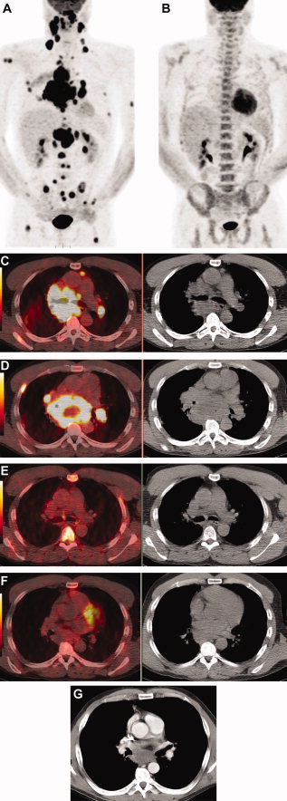
The traditional tasks of imaging in the management of lymphoma include: staging, assessing response to therapy and confirming it reaching an end-point and detecting recurrence. The leading imaging modality is PET/CT. In their literature review the authors include several references claiming that the clinical outcome of lymphoma patients has improved significantly due to better prognosis – largely related to better disease characterization and identification of prognostic markers in recent years. Adoption of functional imaging that improved pre-treatment staging and assessment of the response to treatment contributed as well to this outcome 178 .179 “Most of the recent progress in management of lymphoma occurred after the widespread introduction of [18F]FDG PET and PET/CT. Accordingly, [18F]FDG PET is now part of the revised lymphoma response criteria.180 “


A 46-year-old male with diffuse large B cell lymphoma, stage IV was studied. Baseline maximum intensity projection (MIP) positron emission tomography (PET) image with [18F]fluorodeoxyglucose ([18F]FDG) (A) shows widespread disease, which is essentially resolved on interim scan after 4 cycles of chemotherapy (B). The interim scan also shows increased [18F]FDG uptake in bone marrow related to administration of granulocyte colony-stimulating factor (GCSF). (C,D) Transaxial CT and PET/CT fusion images at baseline show abnormal [18F]FDG uptake in extensive mediastinal and hilar lymphadenopathy as well as in bone lesions in a right rib and the right scapula. On interim scan (E,F) abnormal [18F]FDG uptake at all of these sites has resolved although residual enlarged lymph nodes remain. The sites are better seen on a contrast-enhanced CT (G) and measure up to 5.3 cm × 3.6 cm. Chemotherapy was continued for a total of 8 cycles. At the time of writing, the patient remained disease-free after 9 years of follow-up.

A 46-year-old male with diffuse large B cell lymphoma, stage IV was studied. Baseline maximum intensity projection (MIP) positron emission tomography (PET) image with [18F]fluorodeoxyglucose ([18F]FDG) (A) shows widespread disease, which is essentially resolved on interim scan after 4 cycles of chemotherapy (B). The interim scan also shows increased [18F]FDG uptake in bone marrow related to administration of granulocyte colony-stimulating factor (GCSF). (C,D) Transaxial CT and PET/CT fusion images at baseline show abnormal [18F]FDG uptake in extensive mediastinal and hilar lymphadenopathy as well as in bone lesions in a right rib and the right scapula. On interim scan (E,F) abnormal [18F]FDG uptake at all of these sites has resolved although residual enlarged lymph nodes remain. The sites are better seen on a contrast-enhanced CT (G) and measure up to 5.3 cm × 3.6 cm. Chemotherapy was continued for a total of 8 cycles. At the time of writing, the patient remained disease-free after 9 years of follow-up.


Subsequent to their acknowledgment of PET/CT as the most promising imaging modality for management of Lymphoma, the authors focused their review to on its role in this disease pathway. It being well understood that the clinical utility of [18F]FDG PET in lymphoma “depends on the intensity of radiotracer uptake in disease sites, which will affect the test accuracy for staging and characterizing residual masses after completion of therapy, as well as the role of the test in response assessment. The intensity of [18F]FDG uptake in lymphoma is determined by tumor histology, grade (eg, indolent versus aggressive NHL)”,181, 182  At the end of their extensive review the authors do mention that PET/MRI might become an important player in the management of this disease, especially in pediatric cases.

 Other research papers related to the management of Lymphoma were published on this Scientific Web site:

Imatinib (Gleevec) May Help Treat Aggressive Lymphoma: Chronic Lymphocytic Leukemia (CLL)

Paradigm Shift in Human Genomics – Predictive Biomarkers and Personalized Medicine – Part 1

Predicting Tumor Response, Progression, and Time to Recurrence

Cancer Innovations from across the Web


Read Full Post »

State of the art in oncologic imaging of Colorectal cancers.

Author-Writer: Dror Nir, PhD

Screen Shot 2021-07-19 at 7.31.39 PM

Word Cloud By Danielle Smolyar

This is the fourth post in a series in which I will address the state of the art in oncologic imaging based on a review paper; Advances in oncologic imaging†‡ that provides updates on the latest approaches to imaging of 5 common cancers: breast, lung, prostate, colorectal cancers, and lymphoma. This paper is published at CA Cancer J Clin 2012. © 2012 American Cancer Society.

The paper gives a fair description of the use of imaging in interventional oncology based on literature review of more than 200 peer-reviewed publications. In this post I summaries the chapter on colorectal cancer imaging. It reviews current and developing radiologic practices in CRC with respect to screening, preoperative evaluation, surveillance, and post-treatment re-staging.

Colorectal cancer (CRC) is an example to successful imaging-based screening evident by noticeable reduction in mortality rates. The 5-year survival rate of CRC patients diagnosed at an early stage is 90%.1 121 According to this review; “(CRC) is the third most common cancer worldwide and the second most frequent cause of cancer death in the United States. The American Cancer Society estimates that 143,460 new cases of CRC will be diagnosed and 51,690 deaths from CRC will occur in the United States in 2012.120 Because of screening and removal of premalignant polyps, incidence rates have declined over the last 3 decades.”

The authors found out that the increased use of CT in CRC screening has the potential of reducing its costs and associated tisks 122 In addition, use of DW-MRI enabled better outcomes of CRC liver metastasis treatment as it enables tailored localized treatment of such lesions.123 124 Finally, the authors found that: “MRI for staging of rectal cancer has become standard practice and, in some instances, is performed in lieu of surgeon-performed endorectal US (ERUS), providing the radiologist with an even greater role in the management of patients with CRC.125 “

 Screening

“CRC is a largely preventable disease, as the progression of the adenoma-carcinoma sequence of mutations is slow and leaves ample time to intervene. Nonetheless, approximately 41% of the population (in the USA) eligible for screening remains unscreened. 126 Most screening is performed using non-imaging tests”

Any of these screening strategies will reduce mortality from CRC.126, 127 

Among imaging tests used for screening, barium enema has seen a continual decline in usage, at least in part due to the landmark study showing that this test detected only 39% of polyps identified at colonoscopy, including only 48% of those > 1 cm in size.131 The recent (and largest, with > 2500 patients) multicenter CT colonography (CTC, also known as virtual colonoscopy) screening study, performed by the American College of Radiology Imaging Network, found that CTC had sensitivity of 90% and similar specificity for polyps > 9 mm, and the number of centers using CTC has increased.122 Widespread deployment of CTC remains hindered, in part, by the 2009 decision of the Center for Medicare and Medicaid Service (CMS) to deny reimbursement based on 1) potential radiation risk, 2) impact of detection of extracolonic findings, and 3) efficacy in the 65 years and older age group of concern to CMS. Data from studies reported after this decision put CTC in a good position to be reconsidered for reimbursement. The median estimated effective dose is currently 5 to 6 mSv, a dose far less than that received from a standard CT exam and even comparable to or lower than that received from a barium enema examination. In fact, the radiation dose from CTC is equivalent to that received from cosmic radiation in a 1-year period.132 Extra-colonic findings occur in 7% to 11% of cases and lead to extra examinations in about 6% with a relevant new diagnosis made in 2.5%, according to the experience of the largest screening center in the United States.133 Furthermore, when detection of extracolonic cancers and aortoiliac aneurysms is included along with CRC screening, CT colonography (CTC) has been shown to be more clinically effective and more cost-effective than optical colonoscopy.134 In an observational study, CTC accuracy was maintained in patients aged 65 to 79 years, who were compared to the overall general population sample. In the older patients, CTC remained a safe and effective modality and program outcome measures, such as colonoscopy referral and extracolonic work-up rates, remained similar to those in other screened groups.135“

 Detection and Characterization

“Diagnosis and clinical staging of primary colonic adenocarcinoma is most often accomplished by combining colonoscopy with biopsy and performing cross-sectional imaging to detect metastatic disease.

Although CT and MRI are widely used for preoperative whole-body staging, they are not recommended first-line methods for detection of primary lesions. In contradistinction, CTC has matured into an excellent diagnostic method for detection of CRC. Data drawn largely from screening studies tell us that its sensitivity for polyps > 10 mm is 90% or greater, and that it will detect nearly every cancer. In fact, a recent meta-analysis of more than 11,000 patients indicated that CTC had sensitivity of 96.1% (398 of 414) for CRC, and when cathartic cleansing and fecal tagging were used, no cancers were missed (Fig. 16).137 Detection of flat cancers remains a challenge with CTC as compared with endoscopic methods in which mucosal surface details are better appreciated. CTC not only detects CRC, but with its cross-sectional depiction also allows characterization of tumors using the TNM staging system138 with reasonable T- and N-stage accuracies of 83% and 80%, respectively.139 CTC is an operator-dependent technique that has shown great variability between radiologists with different degrees of training. Computer-aided detection (CAD) was developed for this reason and because 10,000 to 15,000 images must be scrutinized for each large adenoma detected. In a screening cohort of 3077 consecutive asymptomatic adults, stand-alone CAD had sensitivities of 97% and 100% for advanced neoplasia and cancer, respectively.140

Coronal reformatted CT scan of the abdomen and pelvis shows a left colon primary adenocarcinoma causing colonic obstruction.

Coronal reformatted CT scan of the abdomen and pelvis shows a left colon primary adenocarcinoma causing colonic obstruction.

Three-dimensional rendering from CT colonography shows a right colon adenocarcinoma which was stage T1N0.

Three-dimensional rendering from CT colonography shows a right colon adenocarcinoma which was stage T1N0.

With magnetic resonance colonography (MRC), detection of masses is limited because techniques employing air cause susceptibility artifacts, and those employing dark-lumen techniques with water-filling and intravenous gadolinium are under scrutiny because of concerns about the potential risk of nephrogenic systemic fibrosis. In addition, in the largest screening study, the sensitivity of MRC was only 70% in patients with colorectal lesions more than 10 mm in size.141

Imaging plays a critical role in detecting liver metastases in order to properly stage and treat the patient with colorectal cancer. NCCN guidelines recommend contrast-enhanced CT or MRI.142 “

MRI is the most promising imaging modality for management of rectal cancer.

Staging of this cancer is primarily accomplished with US, typically performed by surgeons. MRI using phased-array coils provides complete visualization of the pelvic anatomy and, especially, the circumferential resection margin, an important landmark for the standard total mesorectal excision.

In an MRI of rectal carcinoma, the T2-weighted axial image shows rectal mass penetrating the wall and extending to the left posterolateral mesorectal fascia (also known as the circumferential resection margin).

In an MRI of rectal carcinoma, the T2-weighted axial image shows rectal mass penetrating the wall and extending to the left posterolateral mesorectal fascia (also known as the circumferential resection margin).


 The MERCURY study125established the near equivalence of MRI to histopathology for identification of this margin, an important advantage of MRI over ERUS, with which the margin is not routinely visualized.147 T- and N- stage accuracies of MRI (87% and 74%, respectively) were similar to those of ERUS (82% and 74%, respectively).148 Accurate lymph node identification remains a problem for MRI. Toward this end, a new albumin-bound gadolinium agent has shown some promise, and further results are awaited.149”

 Role of Imaging in Assessing Treatment Response

“Imaging plays a critical role in 1) determining response to systemic and loco-regional treatment of liver metastases, 2) assessing response to local treatment and restaging rectal cancer primary lesions, and 3) detecting and assessing the treatment response of extra-hepatic metastatic disease. Systemic treatment (and in some centers, hepatic artery infusion) of non-resectable liver metastases with chemotherapy aims at reduction of the metastatic burden, which, occasionally may allow attempts at curative liver resection.

Due to the limitations of CT with regard to soft tissue contrast and fatty liver. MRI has greater sensitivity for remaining (or new) lesions < 1.0 cm due to its superior soft tissue contrast. In a recent meta-analysis of 25 eligible studies, MRI showed higher sensitivity than CT on a per-patient basis (P = .05) and on a per-lesion basis as well (P = .0001). With its 81.1% sensitivity and 97.2% specificity, MRI is thus the preferred modality.151 Nonetheless, under the current NCCN guidelines, CT remains the preferred modality.142 

Loco-regional (“liver-directed”) therapies include radiofrequency, microwave ablation, transarterial chemo- or particle embolization and irreversible electroporation. With these treatments, responding lesions can actually increase in size, and simple size criteria are no longer sufficient to determine response. The European Association for the Study of the Liver has issued new criteria to assess viability of remaining tumor based on enhancing residual volume by multiphase CT or MRI.152 However, the field is rapidly changing and there is no consensus on the optimal imaging strategy following loco-regional therapy.

Recent meta-analyses of randomized controlled trials comparing low-intensity and high-intensity surveillance programs have shown advantages for more intense follow-up in Stages I-III disease;170-173 however, controversies remain regarding the optimal surveillance strategy. “

Lymphoma Imaging

To be followed…

Other research papers related to the management of Colorectal cancer were published on this Scientific Web site:

PIK3CA mutation in Colorectal Cancer may serve as a Predictive Molecular Biomarker for adjuvant Aspirin therapy

Personalized Medicine: Cancer Cell Biology and Minimally Invasive Surgery (MIS)

Read Full Post »

State of the art in oncologic imaging of lungs.

Author and Curator: Dror Nir, PhD

This is the second post in a series in which I will address the state of the art in oncologic imaging based on a review paper; Advances in oncologic imaging†‡ that provides updates on the latest approaches to imaging of 5 common cancers: breast, lung, prostate, colorectal cancers, and lymphoma. This paper is published at CA Cancer J Clin 2012. © 2012 American Cancer Society.

The paper gives a fair description of the use of imaging in interventional oncology based on literature review of more than 200 peer-reviewed publications.

In this post I summaries the chapter on lung cancer imaging.

Lung Cancer Imaging

“Lung cancer remains the most common cause of death from cancer worldwide, having resulted in 1.38 million deaths (18.2% of all cancer deaths) in 2008.48 It also represents the leading cause of death in smokers and the leading cause of cancer mortality in men and women in the United States. In 2012, it was estimated that 226,160 new cases of lung cancer would be diagnosed (accounting for about 14% of cancer diagnoses) and that lung cancer would cause 160,340 deaths (about 29% of cancer deaths in men and 26% of cancer deaths in women) in the United States.1 The 1-year relative survival rate for the disease increased from 35% to 43% from 1975 through 1979 to 2003 through 2006.49 The 5-year survival rate is 53% for disease that is localized when first detected, but only 15% of lung cancers are diagnosed at this early stage.”

For cancer with such poor survival rates removal of the primary lesion by surgery at an early-stage disease is the best option. The current perception in regards to lung cancr is that patients may have subclinical disease for years before presentation. It is also known that early lung cancer lesions; adenocarcinoma in situ (AIS) and minimally invasive adenocarcinoma (MIA) are slow-growing, doubling time which can exceed 2 years.52 But, since at present, no lung cancer early-detection biomarker is clinically available, the diagnosis of this disease is primarily based on symptoms, and detection often occurs after curative intervention and when it’s already too late – see: Update on biomarkers for the detection of lung cancer and also Diagnosing lung cancer in exhaled breath using gold nanoparticles. Until biomarker is found, the burden of screening for this disease is on imaging.

“AIS and MIA generally appear as a single peripheral ground-glass nodule on CT. A small solid component may be present if areas of alveolar collapse or fibroblastic proliferation are present,50, 51 but any solid component should raise concern for a more invasive lesion (Fig. 8). Growth over time on imaging can often be difficult to assess due to the long doubling time of these AIS and MIA, which can exceed 2 years.52 However, indicators other than growth, such as air bronchograms, increasing density, and pleural retraction within a ground-glass nodule are suggestive of AIS or MIA.

CT image shows a ground glass nodule, which is the typical appearance of AIS, in the right upper lobe.

CT image shows a ground glass nodule, which is the typical appearance of AIS, in the right upper lobe.


CT (A) demonstrated extensive consolidation with air bronchograms in the left upper lobe, which at surgical resection were found to represent adenocarcinoma of mixed subtype with predominate (70%) mucinous bronchioloalveolar subtype. PET imaging in the same patient (B) demonstrated uptake in the lingula higher than expected for bronchioloalveolar carcinoma and probably due to secondary inflammation/infection. CT (C) obtained 3 years after images (A) and (B) demonstrated biopsy-proven recurrent soft-tissue mass near surgical site. Fused FDG/PET images (D) demonstrate no uptake in the area. This finding is consistent with the decreased uptake usually seen in tumors of bronchioloalveolar histology (new terminology of MIA).

CT (A) demonstrated extensive consolidation with air bronchograms in the left upper lobe, which at surgical resection were found to represent adenocarcinoma of mixed subtype with predominate (70%) mucinous bronchioloalveolar subtype. PET imaging in the same patient (B) demonstrated uptake in the lingula higher than expected for bronchioloalveolar carcinoma and probably due to secondary inflammation/infection. CT (C) obtained 3 years after images (A) and (B) demonstrated biopsy-proven recurrent soft-tissue mass near surgical site. Fused FDG/PET images (D) demonstrate no uptake in the area. This finding is consistent with the decreased uptake usually seen in tumors of bronchioloalveolar histology (new terminology of MIA).

In August 2011 the results of the “National Lung Screening Trial “ which was funded by the National Cancer Institute (NCI) were published in NEJM; Reduced Lung-Cancer Mortality with Low-Dose Computed Tomographic Screening. This randomized study results showed that with low-dose CT screening of high-risk persons, there was a significant reduction of 20% in the mortality rate from lung cancer as compared to chest radiographs screening.

Based on these results one can find the following information regarding Lung Cancer Screening on the NCI web-site:

“Three screening tests have been studied to see if they decrease the risk of dying from lung cancer.

The following screening tests have been studied to see if they decrease the risk of dying from lung cancer:

  • Chest x-ray: An x-ray of the organs and bones inside the chest. An x-ray is a type of energy beam that can go through the body and onto film, making a picture of areas inside the body.
  • Sputum cytology: Sputum cytology is a procedure in which a sample of sputum (mucus that is coughed up from the lungs) is viewed under a microscope to check for cancer cells.
  • Low-dose spiral CT scan (LDCT scan): A procedure that uses low-dose radiation to make a series of very detailed pictures of areas inside the body. It uses an x-ray machine that scans the body in a spiral path. The pictures are made by a computer linked to the x-ray machine. This procedure is also called a low-dose helical CT scan.

Screening with low-dose spiral CT scans has been shown to decrease the risk of dying from lung cancer in heavy smokers.

A lung cancer screening trial studied people aged 55 years to 74 years who had smoked at least 1 pack of cigarettes per day for 30 years or more. Heavy smokers who had quit smoking within the past 15 years were also studied. The trial used chest x-rays or low-dose spiral CT scans (LDCT) scans to check for signs of lung cancer.

LDCT scans were better than chest x-rays at finding early-stage lung cancer. Screening with LDCT also decreased the risk of dying from lung cancer in current and former heavy smokers.

A Guide is available for patients and doctors to learn more about the benefits and harms of low-dose helical CT screening for lung cancer.

Screening with chest x-rays or sputum cytology does not decrease the risk of dying from lung cancer.

Chest x-ray and sputum cytology are two screening tests that have been used to check for signs of lung cancer. Screening with chest x-ray, sputum cytology, or both of these tests does not decrease the risk of dying from lung cancer.”

The authors of Advances in oncologic imaging†‡ found out that for pre-treatment staging and post treatment follow-up of lung cancer patients mainly involves CT (preferably contrast enhanced, FDG PET and PET/CT. “Integrated PET/CT has been found to be more accurate than PET alone, CT alone, or visual correlation of PET and CT for staging NSCLC (Non-small-cell lung carcinoma).59 “

The standard treatment of choice for localized disease remains surgical resection with or without chemo-radiation therapy (stage dependant). “The current recommendations for routine follow-up after complete resection of NSCLC are as follows: for 2 years following surgery a contrast-enhanced chest CT scan every 4 to 6 months and then yearly non-contrast chest CT scans.62 Detection of recurrence on CT is the primary goal in the initial years, and therefore, optimally, a contrast-enhanced scan should be obtained to evaluate the mediastinum. In subsequent years, when identifying an early second primary lung cancer becomes of more clinical importance, a non-contrast CT chest scan suffices to evaluate the lung parenchyma.

CT (A) of 78-year-old male who was status post–left lobe lobectomy and left upper lobe wedge resection shows recurrent nodule at the surgical resection site. Fused PET/CT (B) demonstrates increased [18F]FDG uptake in the corresponding nodule at the surgical resection site consistent with recurrent tumor.

CT (A) of 78-year-old male who was status post–left lobe lobectomy and left upper lobe wedge resection shows recurrent nodule at the surgical resection site. Fused PET/CT (B) demonstrates increased [18F]FDG uptake in the corresponding nodule at the surgical resection site consistent with recurrent tumor.

In patients undergoing chemotherapies: “ [18F]FDG PET response correlates with histologic response.63 [18F]FDG PET scan data can provide an early readout of response to chemotherapy in patients with advanced-stage lung cancer.64

In patients treated by recently developed “Targeted Therapies” such as Radiofrequency ablation (RFA) the authors found out that PET/CT is the preferred imaging modality for post treatment follow-up.

“ Most patients treated with pulmonary ablation will have had a pre-procedure CT or a fusion PET/CT scan, which allows more precise anatomic localization of abnormalities seen on PET. Generally, either CT or PET/CT is performed within a few weeks of the procedure to provide a new baseline to which future images can be compared to assess for changes in size, degree of enhancement or [18F]FDG avidity.67”

CT (A) demonstrates new left upper lobe mass representing new primary NSCLC in a patient who had a status post–right pneumonectomy for a prior NSCLC. CT (B) obtained in the same patient 2 weeks after radiofrequency ablation (RFA) demonstrates the postablation density in the left upper lobe. Fused PET/CT (C) obtained 4 months after RFA demonstrates mild [18F]FDG uptake at RFA site in the left upper lobe consistent with posttreatment inflammation. Fused PET/CT (D) obtained 7 months after RFA demonstrates new focal [18F]FDG uptake at post-RFA-opacity consistent with recurrent tumor.

CT (A) demonstrates new left upper lobe mass representing new primary NSCLC in a patient who had a status post–right pneumonectomy for a prior NSCLC. CT (B) obtained in the same patient 2 weeks after radiofrequency ablation (RFA) demonstrates the postablation density in the left upper lobe. Fused PET/CT (C) obtained 4 months after RFA demonstrates mild [18F]FDG uptake at RFA site in the left upper lobe consistent with posttreatment inflammation. Fused PET/CT (D) obtained 7 months after RFA demonstrates new focal [18F]FDG uptake at post-RFA-opacity consistent with recurrent tumor.

Prostate Cancer Imaging

To be followed…

Other research papers related to the management of Lung cancer were published on this Scientific Web site:

Diagnosing lung cancer in exhaled breath using gold nanoparticles

Lung Cancer (NSCLC), drug administration and nanotechnology

Non-small Cell Lung Cancer drugs – where does the Future lie?

Comprehensive Genomic Characterization of Squamous Cell Lung Cancers

Read Full Post »

State of the art in oncologic imaging of breast.

Author-Writer: Dror Nir, PhD

Screen Shot 2021-07-19 at 7.28.07 PM

Word Cloud By Danielle Smolyar

In the coming posts I will address the state of the art in oncologic imaging based on a review paper; Advances in oncologic imaging†‡ that provides updates on the latest approaches to imaging of 5 common cancers: breast, lung, prostate, colorectal cancers, and lymphoma. This paper is published at CA Cancer J Clin 2012. © 2012 American Cancer Society.

The paper gives a fair description of the use of imaging in interventional oncology based on literature review of more than 200 peer-reviewed publications.

In this post I summaries the chapter on breast cancer imaging.

Breast Cancer Imaging

As a start the authors describes the evolution in the ACS imaging guidelines for breast cancer screening. Most interesting to learn is how age limits are changing. The most recent: “In 2010, the Society of Breast Imaging and the Breast Imaging Commission of the ACS issued recommendations for breast cancer screening to provide guidance in light of the controversies and emerging technologies.5 These recommendations were based on multiple prospective randomized trials as well as population-based experience.

Recommendations for screening with non-mammographic imaging are based not on evidence showing mortality reduction but largely on surrogate indicators, i.e., tumor size and nodal status, suggesting improved survival compared with women who are not screened.” I have referred to these guidelines in my recent post: Not applying evidence-based medicine drives up the costs of screening for breast-cancer in the USA.

As long as imaging interpretation is based mainly on observations related to lesion morphology:

“The imaging characteristics of malignant lesions are nonspecific and usually do not allow a definitive diagnosis. When a biopsy is recommended based on mammography, it has a 25% to 45% likelihood of resulting in a diagnosis of carcinoma.11 Similar positive predictive values are reported for biopsies recommended based on MRI.”

It is worthwhile noting that these results do not reflect purely the specificity of the imaging device but rather the specificity of the whole workflow; i.e imaging, biopsy and histopathology. All imaging techniques have false negatives: Mammography screening of general population misses approximately 20% of the cancers. This rate increases as breast density increases. MRI is not applied to general population. When applied to highly suspicious cases MRI misses ~10% of the invasive cancers. Although ultrasound has proven to be useful in detecting cancer especially in women with dense breasts: Automated Breast Ultrasound System (‘ABUS’) for full breast scanning: The beginning of structuring a solution for an acute need! Based on the literature reviewed by the authors of this paper they do not recommend routine sonography for these women.

For women with locally advanced breast cancer (Fig. 2) who undergo neoadjuvant therapy before breast surgery, the authors recommends post-treatment staging using MRI, which has been found to predict complete response with sensitivity above 60% and specificity as high as 90%.26

A 27-year-old female with locally advanced poorly differentiated invasive ductal carcinoma underwent evaluation of extent of disease before starting neoadjuvant chemotherapy. Sagittal fat-suppressed T1-weighted postcontrast MR images demonstrate an almost 6-cm heterogeneously enhancing mass (A) involving the skin of the lower breast (arrow) with (B) right axillary (arrow) and (C) right internal mammary adenopathy (arrow).

A 27-year-old female with locally advanced poorly differentiated invasive ductal carcinoma underwent evaluation of extent of disease before starting neoadjuvant chemotherapy. Sagittal fat-suppressed T1-weighted postcontrast MR images demonstrate an almost 6-cm heterogeneously enhancing mass (A) involving the skin of the lower breast (arrow) with (B) right axillary (arrow) and (C) right internal mammary adenopathy (arrow).

Same is recommended for women who have undergone lumpectomy if the surgical margins are positive. As post therapy follow-up, a new baseline mammogram of the treated breast is recommended followed by annual mammography.

In regards to emerging technology the following are discussed: Mammographic tomosynthesis – see also Improving Mammography-based imaging for better treatment planning

Contrast-enhanced digital mammography – “involves the injection of iodinated contrast material, as is done for computed tomography (CT); this enables hypervascular lesions to be seen with modified mammography technology, potentially providing the same information obtained through MRI. Little has been published on the clinical application of this technology, but diagnostic accuracy better than that of mammography and approaching that of MRI has been reported.31, 32”

MR choline spectroscopy – has been shown to improve the positive predictive value of breast MRI and may be useful in reducing the number of lesions that require biopsy (Fig. 4).33 Studies of spectroscopy have reported sensitivities of 70% to 100% and specificities of 67% to 100% in the detection of breast cancer. Decreasing choline concentrations may also be a useful indication of tumor response to treatment before any change in tumor volume can be detected.34, 35 Technical factors have limited the use of spectroscopy to lesions 1 cm in size or larger.”

Sagittal fat-suppressed T1-weighted postcontrast MR image is shown (A) of the right breast of a 48-year-old female who was status post–contralateral mastectomy for DCIS with the spectroscopy voxel placed over an enhancing mass (arrow). The magnified spectrum (B) demonstrated no choline peak. Biopsy yielded fibroadenoma.

Sagittal fat-suppressed T1-weighted postcontrast MR image is shown (A) of the right breast of a 48-year-old female who was status post–contralateral mastectomy for DCIS with the spectroscopy voxel placed over an enhancing mass (arrow). The magnified spectrum (B) demonstrated no choline peak. Biopsy yielded fibroadenoma.

Diffusion-weighted MRI (DW-MRI) – “adding DW-MRI data to other imaging characteristics of lesions on breast MRI may increase the positive predictive value of the examination, in turn decreasing the number of benign lesions requiring biopsy for diagnosis.” See also Imaging: seeing or imagining? (Part 2).

Axial T1-weighted fat-suppressed postcontrast MR image is shown (A) of the left breast of a 42-year-old female with biopsy-proven contralateral cancer undergoing evaluation of disease extent. An enhancing mass (arrow) was seen in the left breast. This mass (arrow) was also demonstrated on the axial diffusion-weighted MR image (B). Biopsy yielded fibroadenoma with atypical ductal hyperplasia and lobular carcinoma in situ.

Axial T1-weighted fat-suppressed postcontrast MR image is shown (A) of the left breast of a 42-year-old female with biopsy-proven contralateral cancer undergoing evaluation of disease extent. An enhancing mass (arrow) was seen in the left breast. This mass (arrow) was also demonstrated on the axial diffusion-weighted MR image (B). Biopsy yielded fibroadenoma with atypical ductal hyperplasia and lobular carcinoma in situ.

Ultrasound-elastography – “Ultrasound elastography has been reported to differentiate benign from malignant breast lesions with sensitivities of 78% to 100% and specificities of 21% to 98%.39 When added to other US techniques, it may improve radiologists’ performance in distinguishing malignant breast lesions.”

Positron emission tomography (PET) – “alone or combined with CT, allows noninvasive, quantitative assessment of biochemical and functional processes at the molecular level in the body. It is most often performed with the radiolabeled glucose analogue [18F] fluorodeoxyglucose ([18F]FDG) to detect the elevated glucose metabolism that is a hallmark of cancer. In breast cancer, its utility depends on the pretest probability for advanced disease, and thus the clinical stage.” The authors found that the use of [18F] FDG PET to patients with stage I and II disease is “limited”. Specifically, they claim that it is not sufficiently accurate for axillary nodal staging in this subset of patients.40 The did find enough evidence to recommend the use of FDG PET in patients with advanced disease: “where it accurately defines disease extent,41 and frequently eliminates the need for other imaging tests, and provides an early readout of treatment response as well as prognostic information.”

Combined PET/MRI is mentioned as a promising technology for predicting response to therapy “but this remains to be proven”.

Positron emission mammography (PEM) – “adapts full-body PET imaging to the breast. In a multicenter study, the interpretation of PEM in conjunction with mammographic and clinical findings yielded a sensitivity of 91% and a specificity of 93% for breast cancer.47 “. However, the authors mention that its use for screening (applying to healthy women) has been criticized because of the need to administer a radioactive tracer.

Lung Cancer Imaging

To be followed…

Other research papers related to the management of breast cancer were published on this Scientific Web site:

The unfortunate ending of the Tower of Babel construction project and its effect on modern imaging-based cancer patients’ management

 Automated Breast Ultrasound System (‘ABUS’) for full breast scanning: The beginning of structuring a solution for an acute need!

Introducing smart-imaging into radiologists’ daily practice.

Will Bio-Tech make Medical Imaging redundant?

Improving Mammography-based imaging for better treatment planning

Not applying evidence-based medicine drives up the costs of screening for breast-cancer in the USA.

New Imaging device bears a promise for better quality control of breast-cancer lumpectomies – considering the cost impact

Harnessing Personalized Medicine for Cancer Management, Prospects of Prevention and Cure: Opinions of Cancer Scientific Leaders @ http://pharmaceuticalintelligence.com

Predicting Tumor Response, Progression, and Time to Recurrence

“The Molecular pathology of Breast Cancer Progression”

Personalized medicine gearing up to tackle cancer

Whole-body imaging as cancer screening tool; answering an unmet clinical need?

What could transform an underdog into a winner?

Mechanism involved in Breast Cancer Cell Growth: Function in Early Detection & Treatment

Nanotech Therapy for Breast Cancer

A Strategy to Handle the Most Aggressive Breast Cancer: Triple-negative Tumors

Optical Coherent Tomography – emerging technology in cancer patient management

Breakthrough Technique Images Breast Tumors in 3-D With Great Clarity, Reduced Radiation

Closing the Mammography gap

Imaging: seeing or imagining? (Part 1)

Imaging: seeing or imagining? (Part 2)


Read Full Post »

Reporter: Aviva Lev-Ari, PhD, RN


Multi-Scale Study of Cancer

Friday, February 15, 2013: 8:00 AM-9:30 AM

Room 207 (Hynes Convention Center)

Despite the identification of hundreds more potential targets by genome sequencing, the pharmaceutical industry has been faced with a decline in the production of new successful drugs. Mathematical models provide a framework in which to interpret the vast amount of experimental data generated to suggest experiments necessary to test new biological theories leading to development of efficient drugs. Genetic mutations occurring at a subcellular level manifest themselves in a complex, multi-scale cancer process as functional changes at the cellular and tissue scales. Integration of mathematical modeling approaches with statistical analysis of the experimental and clinical data leads to breakthroughs in understanding of the cancer growth, which is translated into novel patient-specific therapies and development of new drugs. When applied to experimental data, statistical techniques can reveal whether a particular intervention produces a significant response or whether a correlation exists between observable phenomena. Establishing why such correlations arise requires verification of a large number of hypotheses using modeling. For example, models offer greater scope for understanding normal and diseased colorectal crypts, for testing and identifying new therapeutic targets in colon cancer, and for predicting their impacts. Speakers have been selected for their contribution to the field and their ability to communicate to audiences with diverse backgrounds and scientific interests.

Organizer:

Mark Alber, University of Notre Dame

Co-Organizer:

Jill P. Mesirov, Broad Institute of Massachusetts Institute of Technology and Harvard University

Moderator:

Mark Alber, University of Notre Dame

Discussants:

Mark Alber, University of Notre Dame

and Jeremy Gunawardena, Harvard Medical School

Speakers:


Philip Maini, University of Oxford

Hybrid Approach to Multi-Scale Modeling of Cancer

Martin Nowak, Harvard University

Dynamics of Targeted Cancer Therapy

Kathleen Wilkie, Tufts University School of Medicine

Modeling in Cancer Immunology


The Benefits of Randomized Experiments for Science and Society

Friday, February 15, 2013: 1:00 PM-2:30 PM

Room 206 (Hynes Convention Center)

Random experiments have allowed researchers to make advances in physical science, social science, industry, and health that have led to better policy and benefits to society. Randomized experiments use random assignment of study units to allow for learning when variation exists among the units and to estimate causal effects free of confounding by the selection of people into treatment conditions in social experiments. As a result, researchers now routinely test medical, educational, behavioral, and policy interventions with randomized experiments to identify effective interventions. This session provides examples of such uses and benefits of randomized experiments. The first paper describes applications of randomized experimentation to the study of politics, how experimentation has evolved in political science, and examples of its benefits to policy. The second paper describes examples of randomized evaluations of public health and education initiatives in developing countries. These experiments found that low-cost interventions could yield substantial health benefits, but paying teachers for performance had more mixed results. In behavioral health, treatment often follows a course of care, and determining the optimal course for a patient is challenging. The third paper describes how recent statistical advances have led to new experimental designs that allow the researcher to test for courses of care that yield optimal outcomes. It also gives an example of the use of such designs.

Organizer:

Daniel McCaffrey, RAND Corp.

Speakers:


Arthur Lupia, University of Michigan

Experimenting with Politics


Michael Kremer, Harvard University

Experimenting with Public Health and Education in the Developing World


Susan Murphy, University of Michigan

Experimenting To Improve Clinical Practice


Clinical Trial and Error: Beauty and the Beast

Friday, February 15, 2013: 3:00 PM-4:30 PM

Room 309 (Hynes Convention Center)

Independent clinical trials are fundamental to improving the results, safety, and cost-effectiveness of health care. Because of costs, the scope of these trials is often limited to new products. Important clinical interventions, surgery, physiotherapy, and rehabilitation are of less interest to commercial sponsors seeking primarily drug and device authorization. Whereas investigator-driven studies now find support in the United States and European Union, they cannot match the resources available for commercial clinical trials. Public and charitable funding typically backs health care–orientated trials independently of manufacturers, but is often much smaller in size. Results matter. When we do get such new evidence, it allows us to look back: how good was the preclinical research to start with? While evidence-based medicine is the health care imperative of the 21st century, it remains difficult to generate the evidence base for pressing public health challenges. This session unites key stages and world authorities in the clinical trials process to explain why and what must change. With expertise on leading animal and human studies to end products, speakers will provide new insights into how clinical trials are conducted. Their common cause will be to argue that greater international cooperation and standards are required. That said, effective communication of clinical and societal benefits must be better coordinated to citizens, patients, health-care professionals, researchers, policy-makers, and society as a whole.

Organizer:

Aidan Gilligan, SciCom–Making Sense of Science

Co-Organizer:

Thomas Hartung, Johns Hopkins University

Moderator:

Klaus Bock, Danish Ministry of Science, Technology & Innovation

Discussants:

Wilson M. Compton, U.S. Department of Health and Human Services

and Anne Glover, European Commission

Speakers:


Thomas Hartung, Johns Hopkins University

Look Back in Anger? What Clinical Trials Tell Us About Preclinical Research


Jay Siegel, Janssen Research and Development

Product-Orientated Versus Health Care–Orientated Clinical Trials


Paul J. Kenny, The Scripps Research Institute

The Latest Clinical Trial Research on Brain Reward Systems


Stroke Research: New Concepts and Innovative Solutions

Friday, February 15, 2013: 3:00 PM-4:30 PM

Room 208 (Hynes Convention Center)

Stroke has become a major challenge for medicine and health policy. It is the third largest cause of death in many countries and the largest single cause of severe disability. It is predicted that stroke will account for 6.2 percent of the total burden of illness in 2020. In the last two decades, research and new technology have brought significant advances in the diagnosis and treatment of stroke. For instance, thrombolytic drugs have revolutionized stroke care and improved recovery from the disease. New imaging techniques are used to study structural and functional changes at the cellular and molecular level in the brain. Nevertheless, improvement of reperfusion, reduction of brain damage, and induction of endogenous protection and repair deserve increased efforts to overcome the “translational roadblock“ and bring new therapies. By pooling together multidisciplinary and cross-sectorial expertise, the European Stroke Network drives the generation of new hypothesis and solutions for stroke, with a view toward developing successful strategies for brain protection and repair. The network has established important transatlantic research cooperation with the Canadian Stroke Network and other groups. The symposium will give an opportunity to exchange ideas and identify common scientific challenges that need to be solved to speed up the discovery of new treatments.

Organizer:

Virginija Dambrauskaite, European Commission, Directorate General for Research and Innovation

Co-Organizer:

Ruxandra Draghia-Akli, European Commission, Directorate General for Research and Innovation

Moderator:

Ruxandra Draghia-Akli, European Commission, Directorate General for Research and Innovation

Discussant:

Walter J. Koroshetz, National Institute of Neurological Disorders and Stroke

Speakers:


Costantino Iadecola, Weill Cornell Medical College

Great Expectations: The Promise of the Neurovascular Unit for Stroke Therapy


Molly Shoichet, University of Toronto

Engineering Meets Medicine: Innovative Strategies To Overcome Stroke


Stephen Meairs, University Medical Center Mannheim, University of Heidelberg

The European Stroke Network: A Platform for Overcoming the Translational Roadblock


Stem Cell–Based Bioartificial Tissues and Organs

Monday, February 18, 2013: 9:45 AM-11:15 AM

Room 207 (Hynes Convention Center)

Transplantations of artificial organs have become a reality. They require skills not only in surgery but also in biomaterials, bioengineering, stem cell culture and biology, tumor biology, and molecular biology. In this session, a team behind artificial trachea transplantation in a patient and the analysis thereof will talk about the efforts made to take modern medicine to the next level.

Organizer:

Sabina Bossi, Karolinska Institute

Moderator:

Ana Teixeira, Swedish Medical Nanoscience Center

Discussant:

Doris Taylor, University of Minnesota

Speakers:


Paolo Macchiarini, Karolinska Institutet

Regenerative Biotechnological Treatment


Ola Hermanson, Karolinska Institutet

Molecular and Cellular Effects of Transplanting Artificial Organs


Philipp Jungebluth, The Cardiothoracic Surgery Network and Karolinska Institutet; Paolo Macchiarini, Karolinska Institute

Clinical Transplantation of a Tissue-Engineered Airway


Engineering the Nervous System: Solutions to Restore Sight, Hearing, and Mobility

Sunday, February 17, 2013: 1:30 PM-4:30 PM

Room 302 (Hynes Convention Center)

The fields of neuroprosthetics and bioengineering offer hope for many that they may one day be able to see the world around them, walk on their own, or grasp, touch, and hear normally. To restore or enable these functions, a multitude of interfaces and approaches are necessary. This symposium highlights pioneering researchers from Europe and the United States who view the nervous system as engineerable and are working to make a new generation of bionic devices feasible. They are decoding motor commands and developing soft, flexible electronics that interface with the spinal cord and auditory brainstem; genetically engineering the human auditory and visual systems; and coming close to perfecting cortico-spinal prosthetics to reclaim voluntary locomotion after complete spinal cord injury. The scientists are optimistic for the future, yet recognize the challenges in getting these technologies out of the lab and into clinical settings. Live demonstrations at the intersection of engineering and neuroscience will show how close we are to entering a new era in the way science interfaces with the nervous system.

Organizer:

Sanna Fowler, Ecole Polytechnique Fédérale de Lausanne

Speakers:


Silvestro Micera, Ecole Polytechnique Fédérale de Lausanne

Controlling a Prosthetic Hand with Peripheral Neural Interfaces


Grégoire Courtine, Ecole Polytechnique Fédérale de Lausanne

Walking Again After Spinal Cord Injury


Konstantina M. Stankovic, Harvard Medical School

Treating Deafness with Better Vision: Cellular-Level Optical Imaging of the Inner Ear


Joan Miller, Harvard Medical School

Saving Sight in Retinal Disease


Philippe Renaud , Microsystems Lab, LMIS4

Challenges: The Electrical Neurostimulation and Recordings with Microelectrode Arrays

Read Full Post »

Improving Mammography-based imaging for better treatment planning

Author and Curator: Dror Nir, PhD

Many of the comments made on my last posts: New Imaging device bears a promise for better quality control of breast-cancer lumpectomies – considering the cost impact , Not applying evidence-based medicine drives up the costs of screening for breast-cancer in the USA, Optical Coherent Tomography – emerging technology in cancer patient management

Were related to the benefit of better planning lumpectomies based on priory knowledge of the lesions’ size and location. The main challenge in using imaging as a tool for treatment planning is the devices’ specificity; i.e. the probability of identifying healthy tissue as malignant. The reason is the wish to avoid unnecessarily large excisions. When discussing the use of “popular” non-ionizing devises such as MRI in breast-imaging; e.g. my post introducing smart-imaging into radiologists’ daily practice I mentioned the low specificity of MRI in that respect. So, if one is reluctant to use ultrasound, for example due to its large inter observer variability and want to rely on the Mammography based work-flow, what new modalities are available?

Stereoscopic digital mammography

Stereoscopic digital mammography (SDM) uses a pair of digital mammograms, taken from slightly different angles and a stereo display workstation loaded with image-analysis software, to create a three-dimensional image of the internal structure of each breast. The resulting stereo image reveals more detail within the breast tissue than a standard two-dimensional mammogram.

Stero-mammography

Radiologist interacting with a stereo digital mammography workstation. (Credit: Image courtesy of Radiological Society of North America)

Recently published materials:

Abstract

Stereoscopic Digital Mammography: Improved Specificity and Reduced Rate of Recall in a Prospective Clinical Trial

Carl J. D’Orsi, MD, David J. Getty, PhD, Ioannis Sechopoulos, PhD, Mary S. Newell, MD, Kathleen R. Gundry, MD, Sandra R. Bates, MD, Robert M. Nishikawa, PhD, Edward A. Sickles, MD, Andrew Karellas, PhD and Ellen M. D’Orsi, RT(R)(M)

Purpose: To compare stereoscopic digital mammography (DM) with standard DM for the rate of patient recall and the detection of cancer in a screening population at elevated risk for breast cancer.

 Materials and Methods: Starting in September 2004 and ending in December 2007, this prospective HIPAA-compliant, institutional review board–approved screening trial, with written informed consent, recruited female patients at elevated risk for breast cancer (eg, personal history of breast cancer or breast cancer in a close relative). A total of 1298 examinations from 779 patients (mean age, 58.6 years; range, 32–91 years) comprised the analyzable data set. A paired study design was used, with each enrolled patient serving as her own control. Patients underwent both DM and stereoscopic DM examinations in a single visit, findings of which were interpreted independently by two experienced radiologists, each using a Breast Imaging Reporting and Data System (BI-RADS) assessment (BI-RADS category 0, 1, or 2). All patients determined to have one or more findings with either or both modalities were recalled for standard diagnostic evaluation. The results of 1-year follow-up or biopsy were used to determine case truth.

 Results: Compared with DM, stereoscopic DM showed significantly higher specificity (91.2% [1167 of 1279] vs 87.8% [1123 of 1279]; P = .0024) and accuracy (90.9% [1180 of 1298] vs 87.4% [1135 of 1298]; P = .0023) for detection of cancer. Sensitivity for detection of cancer was not significantly different for stereoscopic DM (68.4% [13 of 19]) compared with DM (63.2% [12 of 19], P .99). The recall rate for stereoscopic DM was 9.6% (125 of 1298) and that for DM was 12.9% (168 of 1298) (P = .0018).

Conclusion: Compared with DM, stereoscopic DM significantly improved specificity for detection of cancer, while maintaining comparable sensitivity. The recall rate was significantly reduced with stereoscopic DM compared with DM.


Digital Breast Tomosynthesis

Digital Breast Tomosynthesis (DBT) entered the Breast-Imaging field few years ago. Early 2011, a digital mammography device by Hologic aimed at improving the specificity of mammography was approved by the FDA as a tool that may improve cancer detection while reducing the number of patient recalls.

The DBT device takes 15 successive images, each at a slightly different angle along an arc across the breast. The concept is simple: What is hidden behind fibroglandular tissue in one image might be visible in another if the angle is slightly different (Figure bellow).

Figure. (a) A suspicious lesion seen on standard 2D digital mammography (far left). (b) After examining multiple slices generated using breast tomosynthesis (5 images), the lesion seen on 2D (far left) is determined to be a false positive. (Images courtesy of Hologic. Used with permission.)

Figure. (a) A suspicious lesion seen on standard 2D digital mammography (far left). (b) After examining multiple slices generated using breast tomosynthesis (5 images), the lesion seen on 2D (far left) is determined to be a false positive. (Images courtesy of Hologic. Used with permission.)

Out of recent publications related to the incorporation of DBT in breast-cancer management I picked up the following two:

Abstract

Comparison of Digital Mammography Alone and Digital Mammography Plus Tomosynthesis in a Population-based Screening Program

Per Skaane, MD, PhD, Andriy I. Bandos, PhD, Randi Gullien, RT, Ellen B. Eben, MD, Ulrika Ekseth, MD, Unni Haakenaasen, MD, Mina Izadi, MD, Ingvild N. Jebsen, MD, Gunnar Jahr, MD, Mona Krager, MD, Loren T. Niklason, PhD, Solveig Hofvind, PhD and David Gur, ScD


Purpose: To assess cancer detection rates, false-positive rates before arbitration, positive predictive values for women recalled after arbitration, and the type of cancers detected with use of digital mammography alone and combined with tomosynthesis in a large prospective screening trial.

 Materials and Methods: A prospective, reader- and modality-balanced screening study of participants undergoing combined mammography plus tomosynthesis, the results of which were read independently by four different radiologists, is under way. The study was approved by a regional ethics committee, and all participants provided written informed consent. The authors performed a preplanned interim analysis of results from 12631 examinations interpreted by using mammography alone and mammography plus tomosynthesis from November 22, 2010, to December 31, 2011. Analyses were based on marginal log-linear models for binary data, accounting for correlated interpretations and adjusting for reader-specific performance levels by using a two-sided significance level of .0294.

 Results: Detection rates, including those for invasive and in situ cancers, were 6.1 per 1000 examinations for mammography alone and 8.0 per 1000 examinations for mammography plus tomosynthesis (27% increase, adjusted for reader; P = .001). False-positive rates before arbitration were 61.1 per 1000 examinations with mammography alone and 53.1 per 1000 examinations with mammography plus tomosynthesis (15% decrease, adjusted for reader; P < .001). After arbitration, positive predictive values for recalled patients with cancers verified later were comparable (29.1% and 28.5%, respectively, with mammography alone and mammography plus tomosynthesis; P = .72). Twenty-five additional invasive cancers were detected with mammography plus tomosynthesis (40% increase, adjusted for reader; P < .001). The mean interpretation time was 45 seconds for mammography alone and 91 seconds for mammography plus tomosynthesis (P < .001).

 Conclusion: The use of mammography plus tomosynthesis in a screening environment resulted in a significantly higher cancer detection rate and enabled the detection of more invasive cancers.

©RSNA, 2013, Clinical trial registration no. NCT01248546

Summary of the results:

  • Twenty-five additional invasive cancers were detected with mammography plus tomosynthesis — a 40% increase.
  • Detection rates, including those for invasive and in situ can­cers, were 6.1 per 1,000 examinations for mammography alone and eight per 1,000 examinations for mammography plus tomosynthesis — a 27% increase.
  • False-positive rates before arbitration were 61.1 per 1,000 examinations with mammography alone and 53.1 per 1,000 examinations with mammography plus tomosynthe­sis — a 15% decrease.
  • After arbitration, positive predictive values for recalled patients with cancers verified later were comparable (29.1% with mammography alone and 28.5% with mam­mography plus tomosynthesis).
  • Mean interpretation time was 45 seconds for mammography alone and 91 seconds for mammography plus tomosynthesis.


Abstract

Digital Breast Tomosynthesis versus Supplemental Diagnostic Mammographic Views for Evaluation of Noncalcified Breast Lesions

Margarita L. Zuley, MD, Andriy I. Bandos, PhD,  Marie A. Ganott, MD, Jules H. Sumkin, DO, Amy E. Kelly, MD, Victor J. Catullo, MD, Grace Y. Rathfon, MD, Amy H. Lu, MD and David Gur, ScD

Purpose: To compare the diagnostic performance of breast tomosynthesis versus supplemental mammography views in classification of masses, distortions, and asymmetries.

 Materials and Methods: Eight radiologists who specialized in breast imaging retrospectively reviewed 217 consecutively accrued lesions by using protocols that were HIPAA compliant and institutional review board approved in 182 patients aged 31–60 years (mean, 50 years) who underwent diagnostic mammography and tomosynthesis. The lesions in the cohort included 33% (72 of 217) cancers and 67% (145 of 217) benign lesions. Eighty-four percent (182 of 217) of the lesions were masses, 11% (25 of 217) were asymmetries, and 5% (10 of 217) were distortions that were initially detected at clinical examination in 8% (17 of 217), at mammography in 80% (173 of 217), at ultrasonography (US) in 11% (25 of 217), or at magnetic resonance imaging in 1% (2 of 217). Histopathologic examination established truth in 191 lesions, US revealed a cyst in 12 lesions, and 14 lesions had a normal follow-up. Each lesion was interpreted once with tomosynthesis and once with supplemental mammographic views; both modes included the mediolateral oblique and craniocaudal views in a fully crossed and balanced design by using a five-category Breast Imaging Reporting and Data System (BI-RADS) assessment and a probability-of-malignancy score. Differences between modes were analyzed with a generalized linear mixed model for BI-RADS–based sensitivity and specificity and with modified Obuchowski-Rockette approach for probability-of-malignancy–based area under the receiver operating characteristic (ROC) curve.

 Results: Average probability-of-malignancy–based area under the ROC curve was 0.87 for tomosynthesis versus 0.83 for supplemental views (P < .001). With tomosynthesis, the false-positive rate decreased from 85% (989 of 1160) to 74% (864 of 1160) (P < .01) for cases that were rated BI-RADS category 3 or higher and from 57% (663 of 1160) to 48% (559 of 1160) for cases rated BI-RADS category 4 or 5 (P < .01), without a meaningful change in sensitivity. With tomosynthesis, more cancers were classified as BI-RADS category 5 (39% [226 of 576] vs 33% [188 of 576]; P = .017) without a decrease in specificity.

 Conclusion: Tomosynthesis significantly improved diagnostic accuracy for noncalcified lesions compared with supplemental 

 Additional reading

Diagnostic Mammography: Identifying Minimally Acceptable Interpretive Performance Criteria

Breast Cancer: Assessing Response to Neoadjuvant Chemotherapy by Using US-guided Near-Infrared Tomography

Bilateral Contrast-enhanced Dual-Energy Digital Mammography: Feasibility and Comparison with Conventional Digital Mammography and MR Imaging in Women with Known Breast Carcinoma

Other research papers on imaging in cancer patients’ management and cancer-therapeutics were published on this Scientific Web site as follows:


The unfortunate ending of the Tower of Babel construction project and its effect on modern imaging-based cancer patients’ management


Closing the gap towards real-time, imaging-guided treatment of cancer patients.


The unfortunate ending of the Tower of Babel construction project and its effect on modern imaging-based cancer patients’ management


Closing the gap towards real-time, imaging-guided treatment of cancer patients.


Tumor Imaging and Targeting: Predicting Tumor Response to Treatment: Where we stand?


Automated Breast Ultrasound System (‘ABUS’) for full breast scanning: The beginning of structuring a solution for an acute need!


Introducing smart-imaging into radiologists’ daily practice.


Will Bio-Tech make Medical Imaging redundant?


Optical Coherent Tomography – emerging technology in cancer patient management


Personalized Pancreatic Cancer Treatment Option


Understand how DNA damage is repaired—knowledge that could result in the development of new and more effective drugs for cancer treatment


LEADERS in Genome Sequencing of Genetic Mutations for Therapeutic Drug Selection in Cancer Personalized Treatment: Part 2


Scientists use natural agents for prostate cancer bone metastasis treatment


Harnessing Personalized Medicine for Cancer Management, Prospects of Prevention and Cure: Opinions of Cancer Scientific Leaders @ http://pharmaceuticalintelligence.com


Predicting Tumor Response, Progression, and Time to Recurrence


“The Molecular pathology of Breast Cancer Progression”


Personalized medicine gearing up to tackle cancer


Whole-body imaging as cancer screening tool; answering an unmet clinical need?


Read Full Post »

Author & Curator: Aviva Lev-Ari, PhD, RN

Article ID #16: Paradigm Shift in Human Genomics – Predictive Biomarkers and Personalized Medicine – Part 1. Published on 1/13/2013

WordCloud Image Produced by Adam Tubman

Cancer Diagnostics by Genomic Sequencing: ‘No’ to Sequencing Patient’s DNA, ‘No’ to Sequencing Patient’s Tumor, ‘Yes’ to focus on Gene Mutation Aberration & Analysis of Gene Abnormalities

How to Tailor Cancer Therapy to the particular Genetics of a patient’s Cancer

THIS IS A SERIES OF FOUR POINTS OF VIEW IN SUPPORT OF the Paradigm Shift in Human Genomics

‘No’ to Sequencing Patient’s DNA, ‘No’ to Sequencing Patient’s Tumor, ‘Yes’ to focus on Gene Mutation Aberration & Analysis of Gene Abnormalities

PRESENTED in the following FOUR PARTS. Recommended to be read in its entirety for completeness and arrival to the End Point of Present and Future Frontier of Research in Genomics

Part 1:

Research Paradigm Shift in Human Genomics – Predictive Biomarkers and Personalized Medicine

Part 2:

LEADERS in the Competitive Space of Genome Sequencing of Genetic Mutations for Therapeutic Drug Selection in Cancer Personalized Treatment

http://pharmaceuticalintelligence.com/2013/01/13/leaders-in-genome-sequencing-of-genetic-mutations-for-therapeutic-drug-selection-in-cancer-personalized-treatment-part-2/

Part 3:

Personalized Medicine: An Institute Profile – Coriell Institute for Medical Research

http://pharmaceuticalintelligence.com/2013/01/13/personalized-medicine-an-institute-profile-coriell-institute-for-medical-research-part-3/

Part 4:

The Consumer Market for Personal DNA Sequencing

http://pharmaceuticalintelligence.com/2013/01/13/consumer-market-for-personal-dna-sequencing-part-4/


Part 1:

Research Paradigm Shift in Human Genomics – Predictive Biomarkers and Personalized Medicine


In Part 1, we will address the following FIVE DIRECTIONS in Genomics Research

  • ‘No’ to Sequencing Patient’s DNA, ‘No’ to Sequencing Patient’s Tumor, ‘Yes’ to focus on Gene Mutation Aberration & Analysis of Gene Abnormalities
  • Sequencing DNA from individual cells vs “humans as a whole.” Sequencing DNA from individual cells is changing the way that researchers think of humans as a whole.
  • Promising Research Directions By Watson, 1/10/2013
  • Disruption of Cancer Metabolism targeted by Metabolic Gatekeeper
  • Molecular Analysis of the different Stages of  Cancer Progression for Targeting Therapy

First:

Predictive Biomarkers and Personalized Medicine

No to Sequencing Patient’s DNA, No to Sequencing Patient’s Tumor, Yes to focus on Gene Mutation Aberration & Analysis of Gene Abnormalities


MD Anderson Research

targeted agents matched with tumor molecular aberrations.

Molecular analysis

Patients whose tumors had an aberration were treated with matched targeted therapy, compared with those of consecutive patients who were not treated with matched targeted therapy

Results

40.2% – 1 or more aberration.

In 1 aberration , matched tx higher response rate  27% vs 5%

Longer time ot treatment failure  TTF 5.2 vs. 2.2

Longer survival  13.4 vs. 9 months

Pt. w/1 mutation (molecular aberrationMatched targeted therapy associated with longer TTF vs. prior systemic therapy 5.2 vs. 3.1

matched therapy was an independent factor predicting response superior to TTF

Conclusion

Not randomized study, and patients had diverse tumor types and a median of 5 prior therapies,  results suggest that identifying specific molecular abnormalities and choosing therapy based on these abnormalities is relevant in phase I clinical trials

Clin Cancer Res. 2012 Nov 15;18(22):6373-83. doi: 10.1158/1078-0432.CCR-12-1627. Epub 2012 Sep 10.

Personalized medicine in a phase I clinical trials program: the MD Anderson Cancer Center initiative.

Tsimberidou AM, Iskander NG, Hong DS, Wheler JJ, Falchook GS, Fu S, Piha-Paul S, Naing A, Janku F, Luthra R, Ye Y, Wen S, Berry D, Kurzrock R.

Source

Department of Investigational Cancer Therapeutics, Phase I Clinical Trials Program, The University of Texas MD Anderson Cancer Center, Houston, Texas 77030, USA. atsimber@mdanderson.org

http://www.ncbi.nlm.nih.gov/pubmed?term=22966018


Opinion by Dr. Pierluigi Scalia, 1/11/2013.

The fact of using nanotechnology in order to target and treat abnormal cancer cells and tissues adds a powerful weapon towards eradicating the disease in the foreseeable future. However, focusing on weapons when we still have not found a reliable way to build that personalized “shooting target” (Cancer Fingerprinting) still constitutes, in my opinion, the single most relevant barrier to the adoption of Personalized treatments.

http://pharmaceuticalintelligence.com/2013/01/09/nanotechnology-personalized-medicine-and-dna-sequencing/

Ritu Saxena’s interview

http://pharmaceuticalintelligence.com/2013/01/07/personalized-medicine-gearing-up-to-tackle-cancer/

Other studies supporting this perspective


p53 gene deletion predicts for poor survival and non-response to therapy with purine analogs in chronic B-cell leukemias


Chromosome aberrations in solid tumors


Chromosome aberrations in B-cell chronic lymphocytic leukemia: reassessment based on molecular cytogenetic analysis


Multivariate analysis of prognostic factors in CLL: clinical stage, IGVH gene mutational status, and loss or mutation of the p53 gene are independent prognostic factors


Clonal analysis of delayed karyotypic abnormalities and gene mutations in radiation-induced genetic instability.


Comprehensive genetic characterization of CLL: a study on 506 cases analysed with chromosome banding analysis, interphase FISH, IgVH status and …


Detection of aberrations of the p53 alleles and the gene transcript in human tumor cell lines by single-strand conformation polymorphism analysis


Genetic aberrations detected by comparative genomic hybridization are associated with clinical outcome in renal cell carcinoma


VH mutation status, CD38 expression level, genomic aberrations, and survival in chronic lymphocytic leukemia


Microarray gene expression profiling of B-cell chronic lymphocytic leukemia subgroups defined by genomic aberrations and VH mutation status


… nucleophosmin (NPM1) predicts favorable prognosis in younger adults with acute myeloid leukemia and normal cytogenetics: interaction with other gene mutations


Transformation of follicular lymphoma to diffuse large cell lymphoma is associated with a heterogeneous set of DNA copy number and gene expression alterations

[DOC] Pax 6 Gene Research and the Pancreas


Molecular analysis of the cyclin-dependent kinase inhibitor gene p27/Kip1 in human malignancies

Molecular genetic analysis of oligodendroglial tumors shows preferential allelic deletions on 19q and 1p.

Cytogenetic analysis of soft tissue sarcomas: recurrent chromosome abnormalities in malignant peripheral nerve sheath tumors (MPNST)

Radiation-induced genomic instability: delayed cytogenetic aberrations and apoptosis in primary human bone marrow cells

SOURCES

Search:

Gene Mutation Aberration & Analysis of Gene Abnormalities

http://scholar.google.com/scholar?start=20&q=Gene+Mutation+Aberration+%26+Analysis+of+Gene+Abnormalities&hl=en&as_sdt=0,22&as_vis=1

Second:

Sequencing DNA from individual cells vs “humans as a whole.”

Sequencing DNA from individual cells is changing the way that researchers think of humans as a whole.

The ability to sequence single cells meant that researchers could take another approach. Working with a team at the Chinese sequencing powerhouse BGI, Auton sequenced nearly 200 sperm cells and was able to estimate the recombination rate for the man who had donated them. The work is not yet published, but Auton says that the group found an average of 24.5 recombination events per sperm cell, which is in line with estimates from indirect experiments2. Stephen Quake, a bioengineer at Stanford University in California, has performed similar experiments in 100 sperm cells and identified several places in the genome in which recombination is more likely to occur. The location of these recombination ‘hotspots’ could help population biologists to map the position of genetic variants associated with disease.

Quake also sequenced half a dozen of those 100 sperm in greater depth, and was able to determine the rate at which new mutations arise: about 30 mutations per billion bases per generation3, which is slightly higher than what others have found. “It’s basically the population biology of a sperm sample,” Quake says, and it will allow researchers to study meiosis and recombination in greater detail.

Fig1a

SOURCES:

http://www.nature.com/news/genomics-the-single-life-1.11710#/genome

Nature 491, 27–29 (01 November 2012) doi:10.1038/491027a

http://pharmaceuticalintelligence.com/2012/11/05/every-sperm-is-sacred-sequencing-dna-from-individual-cells-vs-humans-as-a-whole/


Third:

Promising Research Directions By Watson, 1/10/2013

The main reason drugs that target genetic glitches are not cures is that cancer cells have a work-around. If one biochemical pathway to growth and proliferation is blocked by a drug — the cancer cells activate a different, equally effective pathway.

Watson advocates a different approach: targeting features that all cancer cells, especially those in metastatic cancers, have in common.

A protein in cells called Myc. It controls more than 1,000 other molecules inside cells, including many involved in cancer. Studies suggest that turning off Myc causes cancer cells to self-destruct in a process called apoptosis.

cancer biologist Hans-Guido Wendel of Sloan-Kettering. “Blocking production of Myc is an interesting line of investigation. I think there’s promise in that.”

Personalized medicine” that targets a patient’s specific cancer-causing mutation

Watson wrote, may be “the inherently conservative nature of today’s cancer research establishments.”

http://pharmaceuticalintelligence.com/2013/01/09/the-cancer-establishments-examined-by-james-watson-co-discover-of-dna-wcrick-41953/


Opinion by Dr. Stephen Willliams, 1/11/2013

Kudos to both Watson and Weinstein for stating we really need to delve into tumor biology to determine functional pathways (like metabolism) which are a common feature of the malignant state ( also see my posting on differentiation therapy).

http://pharmaceuticalintelligence.com/2013/01/09/the-cancer-establishments-examined-by-james-watson-co-discover-of-dna-wcrick-41953/

http://pharmaceuticalintelligence.com/2013/01/03/differentiation-therapy-epigenetics-tackles-solid-tumors/

Fourth:

Disruption of Cancer Metabolism targeted by Metabolic Gatekeeper

Fig2a

Figure’s SOURCE:

Figure brought to my attention by Dr. Tilda Barlyia, 1/10/2013

http://blogs.nature.com/spoonful/2012/12/metabolic-gatekeeper-provides-new-target-for-disrupting-cancer-metabolism.html

Author: Yevgeniy Grigoryev

In the 1920s, the German physiologist Otto Warburgproposed that cancer cells generate energy in ways that are distinct from normal cells. Healthy cells mainly metabolize sugar via respiration in the mitochondria, switching only to glycolysis in the cytoplasm when oxygen levels are low. In contrast, cancer cells rely on glycolysis all the time, even under oxygen-rich scenarios. This shift in how energy is produced—the so-called ‘Warburg effect’, as the observation came to be known—is now recognized as a primary driver of tumor formation, but a mechanistic explanation for the phenomenon has remained elusive.

Now, researchers have implicated a chromatin regulator known as SIRT6 as a key mediator of the switch to glycolysis in cancer cells, a finding that could lead to new therapeutic modalities. “This work is very significant for the cancer field,” says Andrei Seluanov, a cancer biologist at the University of Rochester in New York State who studies SIRT6 but was not involved in the latest study. “It establishes the role ofSIRT6 as a tumor suppressor and shows that SIRT6 loss leads to tumor formation in mice and humans.”

SIRT6 encodes one of seven mammalian proteins called sirtuins, a group of histone deacetylases that play a role in regulating metabolism, lifespan and aging. SIRT1—which is activated by resveratrol, a molecule found in the skin of red grapes—is perhaps the best known sirtuin, but several of the others are now the focus of active investigation as therapeutic targets for a range of conditions, from metabolic syndrome tocancer. Just last month, for example, a paper in Nature Medicine demonstrated that SIRT6 plays an important role in heart disease.

Six years ago, a team led by Raul Mostoslavsky, a molecular biologist at the Massachusetts General Hospital Cancer Center in Boston, first showed that SIRT6 protects mice from DNA damage and had anti-aging properties. In 2010, the same team established SIRT6 as a critical regulator of glycolysis. Now,reporting today in Cell, Mostoslavsky and his colleagues have shown that SIRT6 function is lost in cancer cells—thus, definitively establishing SIRT6 as a potent tumor suppressor.

In the latest study, the researchers showed that mouse embryonic cells genetically engineered to lackSIRT6 proliferated much faster than normal cells, growing from 5,000 cells to 200,000 cells in three days. In contrast, SIRT6-expressiong cells grew at less than half that rate over the same time period. When injected into adult mice, these SIRT6-deficient cells also rapidly formed tumors, but this tumor growth was reversed when the scientists put SIRT6 back into the cells.

“Our study provides a proof-of-concept that inhibiting glycolysis in SIRT6-deficient cells and tumors could provide a potential therapeutic approach to combat cancer,” says Mostoslavsky. “Additionally, SIRT6 may be a valuable prognostic biomarker for cancer detection.”

Currently, there are no approved anti-glycolytic drugs against cancer. However, the latest findings indicate that pharmacologically elevating SIRT6 levels might help keep tumor growth at bay. And there’s preliminary data to suggest that the work will translate from the bench to the clinic: looking at a range of cancers from human patients, Mostoslavsky’s team showed that the higher the level of SIRT6 the better the prognosis and the longer the survival times.

SOURCE:

Fifth:

Molecular Analysis of the different Stages of  Cancer Progression: The Example of Breast Cancer 

Fig2b

Figure’s SOURCE:

The molecular pathology of breast cancer progression

Alessandro Bombonati1 and Dennis C Sgroi1,2* Journal of Pathology, J Pathol 2011; 223: 307–317

(wileyonlinelibrary.com) DOI: 10.1002/path.2808

http://onlinelibrary.wiley.com/store/10.1002/path.2808/asset/2808_ftp.pdf;jsessionid=26C2C424E6948A5FAF3CBADBA385184A.d02t04v=1&t=hi26qzd4&s=a8a4aadb3fc6d448080c0ef3c67415b8277145aa

Post by Dr. Tilda Barlyia and Comments on   “The Molecular Pathology of Breast Cancer Progression”

http://pharmaceuticalintelligence.com/2013/01/10/the-molecular-pathology-of-breast-cancer-progression/

Conclusion

The Paradigm Shift in Human Genomics will follow the following FIVE DIRECTIONS:

  • No to Sequencing Patient’s DNA, No to Sequencing Patient’s Tumor, Yes to focus on Gene Mutation Aberration & Analysis of Gene Abnormalities
  • Sequencing DNA from individual cells vs “humans as a whole.” Sequencing DNA from individual cells is changing the way that researchers think of humans as a whole.
  • Promising Research Directions By Watson, 1/10/2013
  • Disruption of Cancer Metabolism targeted by Metabolic Gatekeeper
  • Molecular Analysis of the different Stages of  Cancer Progression for Targeting Therapy

Read Full Post »

« Newer Posts - Older Posts »