Feeds:
Posts
Comments

Posts Tagged ‘Clinical trial’

From AUA2013: “Histoscanning”- aided template biopsies for patients with previous negative TRUS biopsies

Reporter: Dror Nir, PhD

This year’s AUA takes place in San Diego, USA.

Wednesday, May 08, 2013 10:30 AM-12:30 PM
SDCC: Room 8
Prostate Cancer: Detection & Screening (V)
Moderated Poster
Funding: none
2209: “Histoscanning”- aided template biopsies for patients with previous negative TRUS biopsies.
Oleg Apolikhin; Andrey Sivkov; Gennady Efremov; Nikolay Keshishev; Oleg Zhukov; Andrey Koryakin

Abstract: 2209
Introduction and Objectives
One of the biggest problems in the diagnosis of prostate cancer (PCa), which distinguishes it from many other solid tumors, is the difficulty of tumor imaging by means of standard visualization techniques. A transrectal ultrasound (TRUS) biopsy is mostly performed on the basis of risen PSA and is often blind – tissue specimens are taken from standard zones. Biopsy under MRI control is technically and logistically complicated and expensive, while TRUS can`t always differentiate the suspicious areas. A TRUS-based innovative technique, “Histoscanningâ€� is used in our centre for PCa identification and targeted biopsy.

Methods
Prior to template biopsy we have performed Histoscanning to 31 patients, with previous one to six negative TRUS biopsies and persistent clinical suspicion of PCa (elevated PSA, high-grade prostatic intraepithelial neoplasia (HPIN) in 4 cores or suspicious TRUS findings). Age range was 51 – 75, with PSA values 3,8 – 14,3 ng/ml. Prostate size range 22-67cc. Most of the patients (n-26) from this group received therapy with 5α-reductase inhibitors for 6 months or more. Depending on the gland size, 10-14 standardized cores were taken + 4 additional cores from the suspicious zones marked on Histoscanning report.

Results
Histopathology identified PCa in 13 out of 31 patients , adenocarcinomas with Gleason score ranging 6-8. In 11 patients with no signs of PCa we found HPIN or low-grade PIN. Comparing histology reports with Histoscanning mapping, in 8 PCa cases we found high correlation of this method with histopathological study on the amount and location of tumor lesions and in 5 cases Histoscanning showed greater spread of lesions, with good correlation of the tumor location.

Conclusions
Due to the effectiveness, ease of use and the short time required for data processing, Histoscanning is a promising method for more effective targeted biopsy of the prostate.

As a result of ongoing research, we aim to evaluate sensitivity and specificity of the method, fuse it with MRI, to create a 3D model for biopsy or surgery. In the future, this data could be used for decision making on the nerve-sparing prostatectomy and minimally invasive focal treatments such as cryoablation, high-intensity focused ultrasound, radiofrequency or laser ablation.

Date & Time: May 8, 2013 10:30 AM
Session Title: Prostate Cancer: Detection & Screening (V)
Sources of Funding: none

Personal note:

On the authors’ intention to fuse HistoScanning with MRI: The authors report a very compelling clinical benefit just from using HistoScanning for guiding their biopsies. HistoScanning itself results in a 3D mapping of the prostate and the suspicious locations inside.

3D mapping of the prostate by HistoScanning analysis following motorised TRUS. the colored locations represents tissue suspicious for being cancer.

3D mapping of the prostate by HistoScanning analysis following motorised TRUS. the colored locations represents tissue suspicious for being cancer.

Fusing ultrasound & MRI images is prone to image-registration errors (e.g. due to differences in the prostate’s shape-distortion by the probe) which are larger than the accuracy sought for when performing biopsy or nerve-sparing surgery. I recommend anyone who wishes to guide biopsies and treatment based on MRI and therefore is in need for good level of localized-MRI interpretation, to rely on dedicated MRI interpretation applications and not intra-modalities image fusion.

In addition, major benefits of using HistoScanning for managing prostate cancer patients are the accessibility; A urologist can perform himself, at any time he chooses and at any place, simplicity; it only requires routine TRUS, patient-friendly; it lasts less than a minute and does not require anesthesia and low-cost; it’s ultrasound! Mixing HistoScanning with MRI will certainly eliminate these.

Read Full Post »

Ultrasound-based Screening for Ovarian Cancer

Author: Dror Nir, PhD

Occasionally, I check for news on ovarian cancer screening. I do that for sentimental reasons; I started the HistoScanning project aiming to develop an effective ultrasound-based screening solution for this cancer.

As awareness for ovarian cancer is highest in the USA, I checked for the latest news on the NCI web-site. I found that to-date: “There is no standard or routine screening test for ovarian cancer. Screening for ovarian cancer has not been proven to decrease the death rate from the disease.

Screening for ovarian cancer is under study and there are screening clinical trials taking place in many parts of the country. Information about ongoing clinical trials is available from the NCI Web site.”

I also found that:

Estimated new cases and deaths from ovarian cancer in the United States in 2013:

  • New cases: 22,240
  • Deaths: 14,030

To get an idea on the significance of these numbers, lets compare them to the numbers related to breast cancer:

Estimated new cases and deaths from breast cancer in the United States in 2013:

  • New cases: 232,340 (female); 2,240 (male)
  • Deaths: 39,620 (female); 410 (male)

Death rate of ovarian cancer patients is almost 4 times higher than the rate in breast cancer patients!

Therefore, I decided to raise awareness to the results achieved for ovarian HistoScanning in a double-blind multicenter European study that was published in European Radiology three years ago. The gynecologists who recruited patients to this study used standard ultrasound machines of GE-Medical. I would like as well to disclose that I am one of the authors of this paper:

A new computer-aided diagnostic tool for non-invasive characterisation of malignant ovarian masses: results of a multicentre validation study, Olivier Lucidarme et.al., European Radiology, August 2010, Volume 20, Issue 8, pp 1822-1830

Abstract

Objectives

To prospectively assess an innovative computer-aided diagnostic technology that quantifies characteristic features of backscattered ultrasound and theoretically allows transvaginal sonography (TVS) to discriminate benign from malignant adnexal masses.

Methods

Women (n = 264) scheduled for surgical removal of at least one ovary in five centres were included. Preoperative three-dimensional (3D)-TVS was performed and the voxel data were analysed by the new technology. The findings at 3D-TVS, serum CA125 levels and the TVS-based diagnosis were compared with histology. Cancer was deemed present when invasive or borderline cancerous processes were observed histologically.

Results

Among 375 removed ovaries, 141 cancers (83 adenocarcinomas, 24 borderline, 16 cases of carcinomatosis, nine of metastases and nine others) and 234 non-cancerous ovaries (107 normal, 127 benign tumours) were histologically diagnosed. The new computer-aided technology correctly identified 138/141 malignant lesions and 206/234 non-malignant tissues (98% sensitivity, 88% specificity). There were no false-negative results among the 47 FIGO stage I/II ovarian lesions. Standard TVS and CA125 had sensitivities/specificities of 94%/66% and 89%/75%, respectively. Combining standard TVS and the new technology in parallel significantly improved TVS specificity from 66% to 92% (p < 0.0001).

table 3

table 4

An example of an ovary considered to be normal with TVS.

An example of an ovary considered to be normal with TVS.

The same TVS false-negative ovary with OVHS-detected foci of malignancy. The presence of an adenocarcinoma was confirmed histologically.

The same TVS
false-negative ovary with OVHS-detected foci of malignancy. The presence of an
adenocarcinoma was confirmed histologically.

Conclusions

Computer-aided quantification of backscattered ultrasound is  highly sensitive for the diagnosis of malignant ovarian masses.

 Personal note:

Based on this study a promising offer for ultrasound-based screening method for ovarian cancer was published in:  Int J Gynecol Cancer. 2011 Jan;21(1):35-43. doi: 10.1097/IGC.0b013e3182000528.: Mathematical models to discriminate between benign and malignant adnexal masses: potential diagnostic improvement using ovarian HistoScanning. Vaes E, Manchanda R, Nir R, Nir D, Bleiberg H, Autier P, Menon U, Robert A.

Regrettably, the results of these studies were never transformed into routine clinical products due to financial reasons.

Other research papers related to the management of Prostate cancer were published on this Scientific Web site:

Beta-Blockers help in better survival in ovarian cancer

Ovarian Cancer and fluorescence-guided surgery: A report

Role of Primary Cilia in Ovarian Cancer

Squeezing Ovarian Cancer Cells to Predict Metastatic Potential: Cell Stiffness as Possible Biomarker

BRCA1 a tumour suppressor in breast and ovarian cancer – functions in transcription, ubiquitination and DNA repair

Warning signs may lead to better early detection of ovarian cancer


Read Full Post »

Reporter: Aviva Lev-Ari, PhD, RN


Cambridge Healthtech Institute’s Inaugural

Next-Generation Sequencing in Medicine
Part of the Fifth Annual ADAPT: Accelerating Development & Advancing Personalized Therapy
November 4-6, 2013 | Boston Cambridge Marriott Hotel | Cambridge, MA

The latest developments in NGS technologies provide opportunities for clinicians and drug developers to guide patient care, improve clinical development, and advance personalized medicine.  But adoption of NGS tests in the clinic requires assay development, clinical and analytical validation, CLIA certification, quality control, and standards development. Cambridge Healthtech Institute’s Inaugural Next-Generation Sequencing in Medicine meeting will address assay development and validation, case studies in using NGS in clinical care, utility of sequencing in clinical drug development, regulatory and reimbursement issues, as well as potential of NGS as companion diagnostics.

Topics Include:

  • NGS as a clinical test: assay development, validation and quality control
  • Clinical utility of sequencing in guiding patient care
  • Interpretation of clinical NGS data
  • Utility of NGS in drug development and clinical trials
  • Diagnostic potential of whole genome sequencing
  • Role of NGS in advancing personalized medicine

Scientists who wish to present their knowledge and expertise to their colleagues are asked to submit an abstract.  Remember to specify the conference that you are interested in and please provide your full contact information.

All proposals are subject to review by the Scientific Advisory Committee to ensure the highest quality of the conference program.

If you are interested in presenting, please click here to submit your proposal.

Deadline for submission is Friday, April 26, 2013

For more information, please contact:Julia Boguslavsky
Executive Director, Conferences
E-mail: juliab@healthtech.com

For sponsorship information, please contact:Ilana Quigley
Manager, Business Development
E-mail: iquigley@healthtech.com
Phone: 781-972-5457

Read Full Post »

2013 – YEAR OF THE ULTRASOUND

Author – Writer: Dror Nir, PhD

To those of you who did not know, 2013 is the year of the ultrasound: http://www.ultrasound2013.org/. This initiative was launched by AIUM and its objectives:

  • Raise awareness of the value and benefits of ultrasound among patients, health care providers, and insurers
  • Provide ultrasound education and evidence-based guidelines for health care providers
  • Educate insurers about the cost savings and patient benefits associated with performing an ultrasound study when scientific evidence supports its potential effectiveness compared to other imaging modalities
  • Educate patients about the benefits of ultrasound as the appropriate imaging modality for their care
  • Encourage the incorporation of ultrasound into medical education

 Quoting from the ultrasound first web-site:

The initiative is designed to call attention to the safe, effective, and affordable advantages of ultrasound as an alternative to other imaging modalities that are more costly and/or emit radiation. For a growing number of clinical conditions, ultrasound has been shown to be equally effective in its diagnostic capability, with a distinct advantage in safety and cost over computed tomography and magnetic resonance imaging. Despite this advantage, evidence suggests that ultrasound is vastly underutilized. Ultrasound First focuses on educating health care workers, medical educators, insurers, and patients of the benefits of ultrasound in medical care. “There is growing support and public awareness for the need to reduce and carefully monitor patients’ exposure to radiation during medical imaging. The use of ultrasound as an alternative imaging modality will help achieve that goal while reducing cost,” states AIUM President Alfred Abuhamad, MD. “Many health care workers and insurers are unacquainted with the range of conditions for which ultrasound has been shown to have superior diagnostic capabilities. Disseminating this knowledge to health care workers and incorporating ultrasound in medical protocols where scientific evidence has shown its diagnostic efficacy will undoubtedly improve patient safety and reduce cost. The time to act is now.”

 A primary component of Ultrasound First is providing clinical evidence for the use of ultrasound. To that aim, the Journal of Ultrasound in Medicine has launched a special feature, the Sound Judgment Series, consisting of invited articles highlighting the clinical value of using ultrasound first in specific clinical diagnoses where ultrasound has shown comparative or superior value. Clinical conditions that will be addressed in the series include postmenopausal bleeding, right lower quadrant pain, pelvic pain, right upper quadrant pain, and shoulder pain, among others. This series will serve as an important educational resource for health care workers and educators.  On the clinical evidence page one can find reasoning for why ultrasound first. Not much related to cancer diagnosis and management. The only interesting claim is: “Ultrasound-guided surgery: Its use to remove tumors from women who have palpable breast cancer is much more successful than standard surgery in excising all the cancerous tissue while sparing as much healthy tissue as possible.”

In support of this initiative The Journal of Ultrasound in Medicine has launched a special series, Sound Judgment, comprised of invited articles highlighting the clinical value of using ultrasound first in specific clinical diagnoses where ultrasound has shown comparative or superior value. So far it includes only two items related to management of cancer: Sonography of Facial Cutaneous Basal Cell Carcinoma, A First-line Imaging Technique; by Ximena Wortsman, MD, and Quantitative Assessment of Tumor Blood Flow Changes in a Murine Breast Cancer Model After Adriamycin Chemotherapy Using Contrast-Enhanced Destruction-Replenishment Sonography; by Jian-Wei Wang, MD et. al. The devoted readers of our Open Access Scientific Journal might find the article by Dr. Wortsman, MD bringing complementary information to a previous post of mine: Virtual Biopsy – is it possible?. Qouting from this article: “Cutaneous basal cell carcinoma is the most common cancer in human beings, and the face is its most frequent location. Basal cell carcinoma is rarely lethal but can generate a high degree of disfigurement. Of all imaging techniques, sonography has proven to support the diagnosis and provide detailed anatomic data on extension in all axes, the exact location, vascularity, and deeper involvement. This information can be used for improving management and the cosmetic results of patients.”

 The article gives clear presentation of the problem and includes demonstrative pictures:

f1

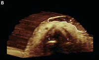
Figure: Basal cell carcinoma with dermal involvement (transverse view, nasal tip). Grayscale sonography (A) and 3-dimensional reconstruction (B, 5- to 8-second sweep) show a 10.1-mm (wide) × 1.4-mm (deep) well-defined hypoechoic oval lesion (between markers in A and outlined in B) that affects the dermis (d) of the left nasal wing. Notice the hyperechoic spots (arrowheads) within the lesion. The nasal cartilage (c) is unremarkable; asterisk indicates basal cell carcinoma.

Basal cell carcinoma with dermal and subcutaneous involvement (transverse view, frontal region). A, Grayscale sonography shows a 11.4-mm (wide) × 6.6-mm (deep) well-defined oval hypoechoic lesion that involves the dermis (d) and subcutaneous tissue (st). There are hyperechoic spots (arrowheads) within the tumor. B, Color Doppler sonography shows increased vascularity within the tumor (asterisk). C, Three-dimensional sonographic reconstruction (5- to 8-second sweep) highlights the lesion (asterisk, outlined); b indicates bony margin of the skull.

Basal cell carcinoma with dermal and subcutaneous involvement (transverse view, frontal region). A, Grayscale sonography shows a 11.4-mm (wide) × 6.6-mm (deep) well-defined oval hypoechoic lesion that involves the dermis (d) and subcutaneous tissue (st). There are hyperechoic spots (arrowheads) within the tumor. B, Color Doppler sonography shows increased vascularity within the tumor (asterisk). C, Three-dimensional sonographic reconstruction (5- to 8-second sweep) highlights the lesion (asterisk, outlined); b indicates bony margin of the skull.

f3

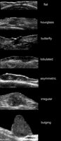
Figure: Pleomorphic presentations of basal cell carcinoma lesions on grayscale sonography (transverse views). Notice the variable shapes of the tumors.

f4

Figure: Frequently, blood flow can be detected within the tumor and its periphery, with slow-flow arteries or veins. The latter vascular data can orient the clinician about the distribution and amount of blood flow that he or she will face during surgery. Despite the fact that basal cell carcinomas usually do not present high vascularity, it should be kept in mind that many of basal cell carcinoma operations are performed in the offices of clinicians and not in the main operating rooms of large hospitals. Nevertheless, the finding of high vascularity within a clinically diagnosed basal cell carcinoma may suggest another type of skin cancer that could occasionally mimic basal cell carcinoma, such as squamous cell carcinoma, Merkel cell carcinoma, or a metastatic tumor. The above figure presents variable degrees of vascularity in basal cell carcinoma lesions going from hypovascular to hypervascular on color and power Doppler sonography (transverse views). 

f5

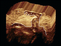
Figure: The depth correlation between sonography (variable frequency) and histologic analysis in facial basal cell carcinoma has been reported to be excellent. Thus, the intraclass correlation coefficient for comparing thickness for the two methods (sonography and histologic analysis) that has been described in literature is 0.9 (intraclass correlation coefficient values ≥0.9 are very good; 0.70–0.89 are good; 0.50–0.69 are moderate; 0.30–049 are mediocre; and ≤0.29 are bad). Two rare sonographic artifacts have been described in basal cell carcinoma. One is the “angled border” that is produced by an inflammatory giant cell reaction underlying the tumor, which may falsely increase the apparent size of the tumor. The other is the “blurry border,” which is produced by large hypertrophy of the sebaceous glands surrounding the lesion. According to the literature, both artifacts can be recognized by a well-trained operator. The figure above presents the sonographic involvement of deeper layers such as the nasal cartilage and orbicularis muscles in the face is of critical importance and may change the decision about the type of surgery. Basal cell carcinoma with nasal cartilage involvement (3-dimensional reconstruction, 5- to 8-second sweep, transverse view, left nasal wing). Notice the extension of the tumor (asterisk, outlined) to the nasal cartilage region (c); d indicates dermis.

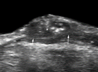
Basal cell carcinoma with involvement of the orbicularis muscle of the eyelid (m). Grayscale sonography (transverse view, right lower eyelid) shows that the tumor (asterisk) affects the muscle layer (arrows).

Basal cell carcinoma with involvement of the orbicularis muscle of the eyelid (m). Grayscale sonography (transverse view, right lower eyelid) shows that the tumor (asterisk) affects the muscle layer (arrows).

Read Full Post »

Larry H Bernstein, MD, FCAP
Pharmaceutical Intelligence

UPDATED 4/23/2020:  New Design for Phase 1 pediatric oncology trials to expedite dose escalation studies.

Clinical Trials Revisited

http://pharmaceuticalintelligence.com/2013/04/03/clinical-trials-revisit/

Cancer Clinical Trials of Tomorrow

Advances in genomics and cancer biology will alter the design of human cancer studies

By Tomasz M. Beer | April 1, 2013   The Scientist
We stand on the cusp of significant change in the fundamental structure of cancer clinical trials, as the emphasis begins to shift from large-scale studies of relatively unselected patients to smaller studies testing more narrowly targeted therapies in molecularly characterized populations.
The previous (and still current) generation of trials established the cancer treatment standards used today. Trials that demonstrated the value of combination chemotherapy in the adjuvant treatment of breast cancer are an excellent example. Meticulous development of treatment regimens through Phase 1 and Phase 2 trials, followed by large-scale comparisons of the new regimens to established treatment protocols, have defined the modern practice of oncology for the last 4 decades. Future cancer clinical trials will be very different from those of the past, adopting a more personalized, sometimes called “precision,” approach.
It is, of course, not entirely true that past clinical trials did not include efforts to target treatments to the right patients. Where possible, targeted therapies are already being implemented. Using the presence of endocrine receptors to guide endocrine therapy for breast cancer was one of the first forays into molecular selection of patients. Unfortunately, the ability to select subgroups of patients for study has been severely curtailed by a still-limited knowledge of human cancer biology.
This is rapidly changing, however, thanks to advances in genomics and comprehensive cancer biology research over the last decade. Large-scale efforts, such as The Cancer Genome Atlas, are comprehensively defining many of the crucial molecular characteristics of human malignancies by illuminating genetic alterations that are clinically and biologically important, and which, by virtue of their functional roles, are viable targets for cancer treatment. At the same time, the ability to design small-molecule inhibitors of specific cancer targets is rapidly accelerating. In 2011, two new agents exemplified the power of these trends: crizotinib was approved for the treatment of lung cancers that harbor a specific mutation in the ALK gene, and vemurafenib was approved for the treatment of melanomas with a specific BRAF mutation. In both cases, the drugs were approved along with companion diagnostic tests that identify patients with the target mutation, who are therefore likely to benefit from treatment.

Smaller, more precise trials ahead

Clinical trials are being transformed by these trends. It will not happen overnight, as the knowledge of cancer biology and the availability of targeted agents are uneven. Unselected populations of patients will still be studied, but it is inevitable that there will be a rise in the number of trials that incorporate molecular tumor testing prior to treatment, with treatment selection informed by the molecular features of each individual’s cancer. Such personalized trials have the potential to yield better outcomes by increasing the probability of response and to employ less toxic therapies by increasingly targeting cancer-specific functions, rather than normal proliferative functions.
To the extent that targeted therapies will prove more effective when given to selected patients, clinical trials should get dramatically smaller. Trial size is largely driven by how effective the treatment is expected to be, so fewer participants are needed when the therapeutic benefit is larger. But the promise of smaller trials will not to be universal; for example, when two targeted agents are compared to one another in the same molecularly selected population, the differences in efficacy may be small and larger trials will be required.
As approaches to cancer treatment advance, there will need to be continual engagement with patients and with cancer survivors.
Furthermore, smaller trials may not necessarily move faster or be easier to complete, as they will require the “right patients,” who may be hard to find. Many of the mutations that represent promising targets are present in a minority of tumors. Today, molecular characterization of tumors is often done as part of the screening process for each trial. Many, and sometimes most, of the patients prove ineligible, making this approach frustrating and difficult to carry out. A better avenue of attack would be to make comprehensive molecular characterization of tumors a routine part of establishing a patient’s eligibility for a range of therapies. With the plummeting cost of genomic analysis, one can envision a day in the near future when a complete cancer genome (and perhaps other molecular evaluations) becomes a standard component of an initial diagnostic evaluation. Patients will be armed with molecular information about their own tumors, and thus able to make more-informed decisions about standard and investigational therapies that match the mutations driving their cancer.

New challenges

The road to personalized and targeted treatment strategies will offer new challenges. For rare targets that are present in a minority of cases across many different types of cancers, one will have to consider clinical trials that include a number of different cancers. There are many design pitfalls to such trials, chiefly the additional clinical and molecular heterogeneity introduced by the inclusion of more than one cancer type. Despite these challenges, it will inevitably make sense in some settings to select patients who share a particular tumor biology, regardless of the tissue of origin.
Another major challenge is how to combine targeted therapies to improve clinical outcomes. To date, targeted therapies have not been able to cure advanced solid tumors. Clinical benefits, while sometimes quite impressive when compared to marginally effective treatments, still fall far short. It stands to reason that redundant survival and growth pathways enable tumors to overcome therapies that inhibit a single target. The simultaneous inhibition of relevant redundant pathways may yield dramatically better results, but will also dramatically increase the complexity of molecularly personalized clinical trials.
As approaches to cancer treatment advance, there will need to be continual engagement with patients and with cancer survivors. Fewer than 5 percent of adult cancer patients participate in a clinical trial. To carry out meaningful clinical trials in the future, that number must increase. This will be most important for treatments that target relatively rare mutations; a large number of potential volunteers will have to be screened to identify a sufficient number who harbor the relevant target. To succeed, we must partner with a much larger fraction of cancer patients.
Designing and executing future cancer clinical trials will not be easy, but physician-scientists are armed with a fast-growing body of omics-informed knowledge with which to surmount these hurdles.
Tomasz M. Beer is deputy director of the Knight Cancer Institute and a professor of medicine at Oregon Health & Science University in Portland. He is the coauthor of Cancer Clinical Trials: A Commonsense Guide to Experimental Cancer Therapies and Clinical Trials. Written for people living with cancer, the book is accompanied by a blog (www.cancer-clinical-trials.com) that seeks to disseminate knowledge about clinical trials.

Tags

tumor suppression, tumor heterogeneity, genetics & genomics, disease/medicine, clinical trials, chemotherapy, cancer genomics and cancer

UPDATED 4/23/2020:  New Design for Phase 1 pediatric oncology trials to expedite dose escalation studies.


REVIEW

Ushering in the next generation of precision trials for pediatric cancer

Steven G. DuBois, Laura B. Corson, Kimberly Stegmaier, Katherine A. Janeway

Science  15 Mar 2019:Vol. 363, Issue 6432, pp. 1175-1181 DOI: 10.1126/science.aaw4153


Abstract

Cancer treatment decisions are increasingly based on the genomic profile of the patient’s tumor, a strategy called “precision oncology.” Over the past few years, a growing number of clinical trials and case reports have provided evidence that precision oncology is an effective approach for at least some children with cancer. Here, we review key factors influencing pediatric drug development in the era of precision oncology. We describe an emerging regulatory framework that is accelerating the pace of clinical trials in children as well as design challenges that are specific to trials that involve young cancer patients. Last, we discuss new drug development approaches for pediatric cancers whose growth relies on proteins that are difficult to target therapeutically, such as transcription factors.

Some terms from the bibliography:

3+3 design: A commonly used rule-based design for phase 1 clinical trials in which patients are enrolled in cohorts of three patients, and decisions to increase or decrease the dose level for the next three participants are based on toxicities observed in those three patients.


Basket trial: A precision oncology trial design in which patients with many different cancer types are enrolled, the tumor is tested for a set of biomarkers of interest, and then patients are assigned to one of several clinical trial subprotocols based on the presence of a biomarker corresponding to a particular molecularly targeted therapy.


Bayesian model–based trial designs: A broad class of trial designs that use data known before the trial as well as data obtained during the conduct of the trial to adapt trial parameters as more information becomes available

Continual reassessment method: One example of a Bayesian model–based trial design in which an initial mathematical model of the relationship between drug dose and probability of unacceptable toxicity is continually updated as new information becomes available to assign subsequent patients to a dose anticipated to have an unacceptable toxicity rate below a set rate.

First-in-child trial: The first clinical trial of a specific agent to include a pediatric population, traditionally considered patients <18 years of age.


Rolling 6 design: A variation of the 3+3 design in which up to six participants may be enrolled to a dosing cohort before enrollment pauses to assess toxicity.

Safety run-in: An initial component of a phase 2 or phase 3 trial in which a small group of patients are treated with a previously untested regimen to evaluate toxicity before opening the trial to a larger group of participants.

Umbrella trial: A precision oncology trial design in which patients with a specific cancer type are enrolled, tumor is tested for a set of biomarkers of interest, and then patients are assigned to one of several clinical trial subprotocols based on the presence of a biomarker corresponding to a particular molecularly targeted therapy.


In this review article, DuBois et al describe new paradigms for pediatric precision oncology trial design and how these designs should be contrasted with the old models and differentiate from the design for these types of trials in the adult.  As the genomic landscape of pediatric tumors is becoming clearer (1, 2) the authors noticed two themes which are becoming evident:

  1. Pediatric cancers harbor certain genomic mutations rarely seen in adult cancers
  2. Pediatric cancers share some genomic alterations and mutational gene signatures with adult tumors

However there is only a small number of pediatric clinical trials to investigate if specific genetic mutations predict outcome to a given personalized therapy.

            Thus, there an urgent need for precision clinical trials in pediatric cancers.

Several reviews have described numerous ongoing and recently completed trials however most are phase 1 dose escalation trials including basket trials and umbrella trials but based on previous data from adult trials using the same precision drug.  For example, pediatric trials involving the TRK inhibitor laratrectinib in tumors harboring a NTRK fusion gene or a pediatric crizotinib trial for pediatric glioblastomas having an ALK fusion protein have shown great success yet most of the early phase 1 work was based on adults or carried out in a way that does not take advantage of the new regulatory framework designed to expedite new drugs for adult precision medicines.

Speeding up the early phase trials in pediatric cancers: new trial design paradigms

Dose escalation phase I trials have, traditionally been the starting point for clinical development of new pediatric anticancer drugs however these first in child trials have seriously lagged their adult counterparts by many years.  These trials relied on the standard 3 x 3  or rolling six trial design, and doses escalated until a pediatric MTD  (maximum tolerated dose) was achieved.  In recent years new precision medicine pediatric trial design has been adopted to expedite the process, based on the fundamental shift in thinking that many new oncology agents will not have a true MTD when tested in adults.

Doses in phase 1 trials for targeted therapies like those in precision medicine are usually escalated based on considerations other than toxicity, like pharmacodynamics or biomarker analysis.  A pediatric phase 1 dose escalation trial may require more subjects than an adult trial.  But

although these newer approaches to early-phase trial design more efficiently establish a pediatric dose, they do little to advance our understanding of with patients are most likely to benefit from a new therapy.

Thus the need for good biomarkers to be included early on in these initial trial designs.  For example, Dana Farber’s first in child clinical trial NCT03654716, a Phase 1 Study of the Dual MDM2/MDMX Inhibitor ALRN-6924 in Pediatric Cancer (as a possible treatment for resistant (refractory) solid tumor, brain tumor, lymphoma or leukemia), are reducing the time children are waiting for entry into a trial, as unselected patients can enroll and the biomarker, increased MDM2 expression is used to determine those patients who go on to phase 2 dose escalation. In other cases, such as NCI Children’s Oncology Group basket trials, they have completely supplanted formal phase 1 trial design and instead incorporated molecularly targeted therapies based on adult doses but adjusted for patient size.  The use of combinations with traditional therapies in trial design is also helping to speed up the process for enrollment.  The authors also suggest that tumor profiling is pertinent however should be put in trial design so the costs to patients can be covered by the trial funds.


Figure 1Fig. 1 Evolution of precision trials for pediatric cancer.

Illustration: Kellie Holoski/Science

Source: Ushering in the next generation of precision trials for pediatric cancer BY STEVEN G. DUBOIS, LAURA B. CORSON, KIMBERLY STEGMAIER, KATHERINE A. JANEWAY SCIENCE 15 MAR 2019 : 1175-1181 https://science.sciencemag.org/content/363/6432/1175


  1. S. N. Gröbner, B. C. Worst, J. Weischenfeldt, I. Buchhalter, K. Kleinheinz, V. A. Rudneva, P. D. Johann, G. P. Balasubramanian, M. Segura-Wang, S. Brabetz, S. Bender, B. Hutter, D. Sturm, E. Pfaff, D. Hübschmann, G. Zipprich, M. Heinold, J. Eils, C. Lawerenz, S. Erkek, S. Lambo, S. Waszak, C. Blattmann, A. Borkhardt, M. Kuhlen, A. Eggert, S. Fulda, M. Gessler, J. Wegert, R. Kappler, D. Baumhoer, S. Burdach, R. Kirschner-Schwabe, U. Kontny, A. E. Kulozik, D. Lohmann, S. Hettmer, C. Eckert, S. Bielack, M. Nathrath, C. Niemeyer, G. H. Richter, J. Schulte, R. Siebert, F. Westermann, J. J. Molenaar, G. Vassal, H. Witt, B. Burkhardt, C. P. Kratz, O. Witt, C. M. van Tilburg, C. M. Kramm, G. Fleischhack, U. Dirksen, S. Rutkowski, M. Frühwald, K. von Hoff, S. Wolf, T. Klingebiel, E. Koscielniak, P. Landgraf, J. Koster, A. C. Resnick, J. Zhang, Y. Liu, X. Zhou, A. J. Waanders, D. A. Zwijnenburg, P. Raman, B. Brors, U. D. Weber, P. A. Northcott, K. W. Pajtler, M. Kool, R. M. Piro, J. O. Korbel, M. Schlesner, R. Eils, D. T. W. Jones, P. Lichter, L. Chavez, M. Zapatka, S. M. Pfister, ICGC PedBrain-Seq Project, ICGC MMML-Seq Project, The landscape of genomic alterations across childhood cancers. Nature 555, 321–327 (2018). 10.1038/nature25480pmid:29489754


2.  X. Ma, Y. Liu, Y. Liu, L. B. Alexandrov, M. N. Edmonson, C. Gawad, X. Zhou, Y. Li, M. C. Rusch, J. Easton, R. Huether, V. Gonzalez-Pena, M. R. Wilkinson, L. C. Hermida, S. Davis, E. Sioson, S. Pounds, X. Cao, R. E. Ries, Z. Wang, X. Chen, L. Dong, S. J. Diskin, M. A. Smith, J. M. Guidry Auvil, P. S. Meltzer, C. C. Lau, E. J. Perlman, J. M. Maris, S. Meshinchi, S. P. Hunger, D. S. Gerhard, J. Zhang, Pan-cancer genome and transcriptome analyses of 1,699 paediatric leukaemias and solid tumours. Nature 555, 371–376 (2018). 10.1038/nature25795pmid:29489755

Related articles
Clinical Trials (journal)

Clinical Trials (journal) (Photo credit: Wikipedia)

Contemporary Clinical Trials

Contemporary Clinical Trials (Photo credit: Wikipedia)

Cover of "Cancer Biology (3rd Edition)"
Cover of Cancer Biology (3rd Edition)

Read Full Post »

New Ecosystem of Cancer Research: Cross Institutional Team Science

Curator: Aviva Lev-Ari, PhD, RN

This image has an empty alt attribute; its file name is ArticleID-32.png

WordCloud Image Produced by Adam Tubman

SOURCE: Time Magazine, April 1, 2013: How to Cure Cancer by Bill Saporito

Key argument: Now the Cure for Cancer is possible thanks to the following innovations in the Division of Labor of the research process among institution.

1.  New Cancer Dream Teams deliver better results faster, better understand the metabolic changes of pancreatic cells.

Team Leader: Dan Von Hoff – A five phase parallel process of the Cancer Research endeavor: One tumor researched by FIVE Labs in parallel

  • Penn surgeon, Jeffrey Drebin removes tissue from a cancerous pancreas. Tissue is carried to Hospital Lab where it is prepared for analysis and frozen for preservation.
  • a piece will go to Princeton for metabolomic profiling, amino acids, sugar glutamine and up to 300 metabolites.
  • a piece will go to John Hopkins for DNA analysis by sequence analysis
  • a piece will go to Translational Genomics for chromosome analysis
  • a piece will go to Salk Institute for a look at the stellate (star shape. tissue repair function, also plays a role in cancer) cells – gene expression analysis Lab

Joint Lab work: Superior to any research ever known.

2. Drug agents in development for therapy targeting the genetic mutations

  • reactivate the body’s immune system
  • cut off a tumor’s blood or energy supply
  • restart apoptosis

3. New Biomarkers

  • Allows to identify, target and track cancer cells – PI3K mutation One pathway – three women’s Cancers: Ovarian, endometrial, Breast CA.
  • Dream Team led by

– Dr. Gordon Mills of MD Anderson, PI3K pathway investigator

Teams Science include:

– Women’s cancer specialist from MGH

– Dana Farber (Harvard)

– Vanderbilt University

– Columbia University

– BIDMC

– Memorial Sloan Kettering

Dream Teams results are better than Big Pharma: 95% failure rate for new oncology drugs 50% of Phase III trials – don’t cut it to FDA approval.

Dream Teams will launch Trial as soon as geneticists and biochemists match mutation to drug compound.

Big Targets: Pancreas, Breast Cancer, Lung Cancer

Example: Human trial at FIVE institutions (28-person team) with TWO unapproved drugs from TWO companies with one year of discovery

PARP inhibitor from AstraZeneca was combined with PI3K inhibitor from Novartis to combat BRCA1 gene mutation that develops ovarian cancer and triple negative Breast Cancer. Two unapproved drugs are combined. Result was without precedent.

4. Design and built of a smart chip device to trap circulating tumor cells (CTCs) in a blood sample – early identification of metastasis

5. Better chances of Five-year Survival Rates

  • 1975-1977 – 49%
  • 1978-1989 – 56%
  • 2002 – 2008 – 68%

6. More Americans who have a History of Cancer are alive today than in the past

[including Cancer-free and in-treatment]

  • 2004 – 10.8 millions
  • 2008 – 12 millions
  • 2012 – 13.7 millions

7. There are 94 millions ex-smokers in the US – elevated risk for lung Cancer. 175,00 new lung cancers diagnosed every year. MD Anderson is developing a simple blood test for protein marker that could detect lung cancer earlier than it is found, test to be used in combination with diagnostic imaging and risk models

8. Probability of developing some type of Cancer over one’s lifetime:

  • Men – 1 in 2
  • Women – 1 in 3

9. Funding of Dream Team Science by Stand Up to Cancer ( SU2C) Hollywood investment in Cancer Research

10. Cancer Statistics in the US

  • 2013: 580,350 will die of Cancer, NCI figures and 1.7 millions will be diagnosed, numbers will grow as population ages (1.4 millions in 2006)
  • 2013L Leading Types of Cancer: Prostate, Breast, Lung &Bronchus (~250,000 each type), colon (~100,000)
  • Cost of Cancer in 2008: Medical – $77.4 Billion, lost productivity – $124 Billion

11. Research at John Hopkins is focused on studying the the enzymatic on/off switches of gene expression including mutated genes that produce cancer cells.

12. Memorial Sloan Kettering Cancer Center – extensive research on Epigenetics, New epigenetic drugs can shrink tumors. Complete remission is experienced by patients treated with drugs that nudges T Cells.

Cancer is a complexed disease.

Read Full Post »

Personalized Medicine: Clinical Aspiration of Microarrays

Reporter, Writer: Stephen J. Williams, Ph.D.

 In this month’s Science, Mike May (at http://www.sciencemag.org/site/products/lst_20130215.xhtml) describes some of the challenges and successes in introducing microarray analysis to the clinical setting.  Traditionally used for investigational research, microarray is now being developed, customized and used for biomarker analysis, prognostic and predictive value, in a disease-specific manner.

Challenges in data interpretation

      In an interview with Seth Crosby, director of the Genome Technology Access Center at Washington University School of Medicine in St. Louis, “the biggest challenge” in moving microarray to the clinical setting is data interpretation.  The current technology makes it possible to evaluate expression of thousands of genes from a patient’s sample however as Crosby describes is assigning clinical relevance to the data.  For example Crosby explains that Washington University had validated a panel of 45 oncology genes by next generation sequencing and are using these genes to develop diagnostic tests to screen patient tumors for the purpose of determining a personalized therapeutic strategy. Seth Crosby noted it took “hundreds of Ph.D. and M.D. hours” to sift through the hundreds of papers to determine which genes were relevant to a specific cancer type. However, he notes, that once we better understand which changes in the patient’s genome are related to a specific disease we will be able to narrow down the list and be able to produce both economical and more disease-relevant microarrays.

Is this aberration pathogenic or not?

     Microarrays are becoming an invaluable tool in cytogenetics, as eluded by Andy Last, executive vice president of the genetic analysis business unit at Affymetrix.  Certain diseases like Down syndrome have well characterized chromosomal alterations like additions or deletions of parts or entire chromosomes.  According to Affymetrix, the most common use of microarrays is for determining copy number variation.  However according to James Clough, vice president of clinical and genomic services at Oxford Gene Technology, given the hundreds of syndromes associated with chromosomal rearrangements, the challenge will be to determine if a small chromosomal aberration has pathologic significance, given that microarray affords much higher diagnostic yield and speed of analysis than traditional microscopic techniques.  To address this challenge, Oxford Gene Technologies, PerkinElmer, Affymetrix, and Agilent all have custom designed microarrays to evaluate disease specific copy number and SNP (single nucleotide polymorphism) microarrays.  For example PerkinElmer designed OncoChip™ to evaluate copy number variation in more than 1.800 cancer genes.  Agilent makes microarrays that evaluates both copy number variation such as its CGH (comparative genomic hybridization) plus SNP microarrays.  Patricia Barco, product manager for cytogenetics at Agilent, notes these arrays can be used in prenatal and postnatal research and cancer, and “can be customized from more than 28 million probes in our library”.

Custom Tools and Software to Handle the Onslaught of Big Data

     There is a need for FDA approved diagnostic tools based on microarrays. Pathwork Diagnostic’s has one such tool (the Pathwork Tissue of Origin test), which uses 2,000 transcript markers and a proprietary computational algorithm to determine from expression analysis, the tissue of origin of a patient’s tumor.  Pathwork also provides a fast, custom turn-around analytical service for pathologists who encounter difficult to interpret samples.  Illumina provides the Infinium HumanCore BeadChip family of microarrays, which can determine genetic variations for purposes of biological tissue banking.  This system uses a set of over 300,000 SNP probes plus 240,000 exome-based markers.

     Tools have also been developed to validate microarray results.  A common validation strategy is the use of quantitative real-time PCR to verify the expression changes seen on the microarray.  Life Technologies developed the TaqMan OpenArray Real Time PCR plates, which have 3,072 wells and can be custom-formatted using their library of eight million validated TaqMan assays.

Making Sense of the Big Data: Bridging the Knowledge Gap using Bioinformatics

          The use of microarray has spurned industries devoted to developing the bioinformatics software to analyze the massive amounts of data and provide clinical significance.  For example companies such as Expression Analysis use their bioinformatics software to provide pathway analysis for microarray data in order to translate the data into the biology.  Using such strategies can also validate the design of microarrays for various diseases.

Foundation Medicine, Inc., a molecular information company, provides cancer genomics test solutions. It offers FoundationOne, an informative genomic profile to identify a patient’s individual molecular alterations and match them with relevant targeted therapies and clinical trials. The company’s product enables physicians to recommend treatment options for patients based on the molecular subtype of their cancer.

The Canadian Bioinformatics Workshops series recently offered a course on using bioinformatic approaches to analyze clinical data generated from microarray approaches (http://bioinformatics.ca/workshops/2012/bioinformatics-cancer-genomics-bicg).   The course objectives are described below:

Course Objectives

Cancer research has rapidly embraced high throughput technologies into its research, using various microarray, tissue array, and next generation sequencing platforms. The result has been a rapid increase in cancer data output and data types. Now more than ever, having the bioinformatic skills and knowledge of available bioinformatic resources specific to cancer is critical. The CBW will host a 5-day workshop covering the key bioinformatics concepts and tools required to analyze cancer genomic data sets. Participants will gain experience in genomic data visualization tools which will be applied throughout the development of the skills required to analyze cancer -omic data for gene expression, genome rearrangement, somatic mutations and copy number variation. The workshop will conclude with analyzing and conducting pathway analysis on the resultant cancer gene list and integration of clinical data.

Successful Examples of Clinical Ventures Integrating Bioinformatics in Cancer Treatment Decision –Making

The University of Pavia, Italy developed a fully integrated oncology bioinformatics workflow as described on their website and at the ESMO 2012 Congress meeting:

http://abstracts.webges.com/viewing/view.php?congress=esmo2012&congress_id=370&publication_id=2530

ESMO

ONCO-I2B2 PROJECT: A BIOINFORMATICS TOOL INTEGRATING –OMICS AND CLINICAL DATA TO SUPPORT TRANSLATIONAL RESEARCH

Abstract:

2530

Congress:

ESMO 2012

Type:

Abstract

Topic:

Translational research

Authors:

A. Zambelli, D. Segagni, V. Tibollo, A. Dagliati, A. Malovini, V. Fotia, S. Manera, R. Bellazzi; Pavia/IT

  • Body

The ONCO-i2b2 project, supported by the University of Pavia and the Fondazione Salvatore Maugeri (FSM), aims at supporting translational research in oncology and exploits the software solutions implemented by the Informatics for Integrating Biology and the Bedside (i2b2) research centre, an initiative funded by the NIH Roadmap National Centres for Biomedical Computing. The ONCO-i2b2 software is designed to integrate the i2b2 infrastructure with the FSM hospital information system and the Bruno Boerci Biobank, in order to provide well-characterized cancer specimens along with an accurate patients clinical data-base. The i2b2 infrastructure provides a web-based access to all the electronic medical records of cancer patients, and allow researchers analyzing the vast amount of biological and clinical information, relying on a user-friendly interface. Data coming from multiple sources are integrated and jointly queried.

In 2011 at AIOM Meeting we reported the preliminary experience of the ONCO-i2b2 project, now we’re able to present the up and running platform and the extended data set. Currently, more than 4400 specimens are stored and more than 600 of breast cancer patients give the consent for the use of specimens in the context of clinical research, in addition, more than 5000 histological reports are stored in order to integrate clinical data.

Within the ONCO-i2b2 project is possible to query and merge data regarding:

• Anonymous patient personal data;

• Diagnosis and therapy ICD9-CM subset from the hospital information system;

• Histological data (tumour SNOMED and TNM codes) and receptor profile testing (Her2, Ki67) from anatomic pathology database;

• Specimen molecular characteristics (DNA, RNA, blood, plasma and cancer tissues) from the Bruno Boerci Biobank management system.

The research infrastructure will be completed by the development of new set of components designed to enhance the ability of an i2b2 hive to utilize data generated by NGS technology, providing a mechanism to apply custom genomic annotations. The translational tool created at FSM is a concrete example regarding how the integration of different information from heterogeneous sources could bring scientific research closer to understand the nature of disease itself and to create novel diagnostics through handy interfaces.

Disclosure

All authors have declared no conflicts of interest.

NCI has under-taken a similar effort under the Recovery Act (the full text of the latest report is taken from their website http://www.cancer.gov/aboutnci/recovery/recoveryfunding/investmentreports/bioinformatics:

Cancer Bioinformatics: Recovery Act Investment Report

November 2009

Public Health Burden of Cancer

Cancer is the second leading cause of death in the United States after heart disease. In 2009, it is estimated that nearly 1.5 million new cases of invasive cancer will be diagnosed in this country and more than 560,000 people will die of the disease.

To learn more, visit:

Cancer Bioinformatics Program Overview

Over the past five years, NCI’s Center for Biomedical Informatics and Information Technology (CBIIT) has led the effort to develop and deploy the cancer Biomedical Informatics Grid® (caBIG) in partnership with the broader cancer community.  The caBIG network is designed to enable the integration and exchange of data among researchers in the laboratory and the clinic, simplify collaboration, and realize the potential of information-based (personalized) medicine in improving patient outcomes. caBIG has connected major components of the cancer community, including NCI-designated Cancer Centers, participating institutions of the NCI Community Cancer Centers Program (NCCCP), and numerous large-scale scientific endeavors, as well as basic, translational, and clinical researchers at public and private institutions across the United States and around the world.  Beyond cancer research, caBIG capabilities—infrastructure, standards, and tools—provide a prototype for linking other disease communities and catalyzing a new 21st-century biomedical ecosystem that unifies research and care. ARRA funding will allow NCI to accelerate the ongoing development of the Cancer Knowledge Cloud and Oncology Electronic Health Records (EHRs) initiatives, thereby providing for continued job creation in the areas of biomedical informatics development and application as well as healthcare delivery.

The caBIG Cancer Knowledge Cloud: Extending the Research Infrastructure

The Cancer Knowledge Cloud is a virtual biomedical capability that utilizes caBIG tools, infrastructure, and security frameworks to integrate distributed individual and organizational data, software applications, and computational capacity throughout the broad cancer research and treatment community. The Cancer Knowledge Cloud connects, integrates, and facilitates sharing of the diverse primary data generated through basic and clinical research and care delivery to enable personalized medicine. The cloud includes information generated through large-scale research projects such as The Cancer Genome Atlas (TCGA), the cancer Human Biobank (caHUB) tissue acquisition network, the NCI Functional Biology Consortium, the NCI Patient Characterization Center, and the NCI Preclinical Development Pipeline, academic and industry counterparts to these projects, and clinical observations (from entities such as the NCCCP) captured in oncology-extended Electronic Health Records.  Through the use of the caBIG Data Sharing and Security Framework, the Cloud will support appropriate sharing of information, supporting in silico hypothesis generation and testing, and enabling a learning healthcare system.

A caBIG-Based Rapid-Learning Healthcare System: Incorporating Oncology-Extended Electronic Healthcare Records (EHRs)

The 21st-century Cancer Knowledge Cloud will connect individuals, organizations, institutions, and their associated information within an information technology-enabled cycle of discovery, development, and clinical care—the paradigm of a rapid-learning healthcare system. This will transform these disconnected sectors into a system that is personalized, preventive, pre-emptive, and patient-participatory.  To be realized, this model requires the adoption of standards-based EHRs. Presently, however, no certified oncology-based EHR exists, and fewer than 3 percent of oncologists with outpatient-based practices utilize EHRs. caBIG has recently established a collaboration with the American Society of Clinical Oncology (ASCO) to develop an oncology-specific EHR (caEHR) specification based on open standards already in use in the oncology community that will utilize caBIG standards for interoperability. NCI will implement an open-source version of this specification to validate the specification and to provide a free alternative to sites that choose not to purchase a commercial system. The launch customer for the caEHR will be NCCCP participating sites. NCI will work with appropriate entities to provide a mechanism for certifying that caEHR implementations are consistent with the NCI/ASCO specification.

Bards Cancer Institute has another clinical bioinformatics program to support their clinical efforts:

Clinical Bioinformatics Program in Oncology at Barts Cancer Institute at Barts and the London School of Medicine

http://www.bci.qmul.ac.uk/cancer-bioinformatics

BCI HomeCancer Bioinformatics

Bards

Why we focus on Cancer Bioinformatics

Bioinformatics is a new interdisciplinary area involving biological, statistical and computational sciences. Bioinformatics will enable cancer researchers not only to manage, analyze, mine and understand the currently accumulated, valuable, high-throughput data, but also to integrate these in their current research programs. The need for bioinformatics will become ever more important as new technologies increase the already exponential rate at which cancer data are generated.

What we do

  • We work alongside clinical and basic scientists to support the cancer projects within BCI.  This is an ideal partnership between scientific experts, who know the research questions that will be relevant from a cancer biologist or clinician’s perspective, and bioinformatics experts, who know how to develop the proposed methods to provide answers.
  • We also conduct independent bioinformatics research, focusing on the development of computational and integrative methods, algorithms, databases and tools to tackle the analysis of the high volumes of cancer data.
  • We also are actively involved in the development of bioinformatics educational courses at BCI. Our courses offer a unique opportunity for biologists to gain a basic understanding in the use of bioinformatics methods to access and harness large complicated high-throughput data and uncover meaningful information that could be used to understand molecular mechanisms and develop novel targeted therapeutics/diagnostic tools.

Developing Criteria for Genomic Profiling in Lung Cancer:

A Report from U.S. Cancer Centers

In a report by Pao et. al., a group of clinicians organized a meeting to standardize some protocols for the integration of microarray and genomic data from lung cancer patients into the clinical setting.[1]  There has been ample evidence that adenocarcinomas could be classified into “clinically relevant molecular subsets” based on distinct genomic changes.  For example EGFR (epidermal growth factor receptor) exon 19 deletions and exon 21 point mutations predict sensitivity to tyrosine kinase inhibitors (TKIs) like gefitinib, whereas exon 20 insertions predict primary resistance[2].

However, as the authors note, “mutational profiling has not been widely accepted or adopted into practice in thoracic oncology”.  

     Therefore, a multi-institutional workshop was held in 2009 among participants from Massachusetts General Hospital (MGH) Cancer Center, Memorial Sloan-Kettering Cancer Center (MSKCC), the Dana-Farber/Bingham & Women’s Cancer Center (DF/BWCC), the M.D. Anderson Cancer Center (VICC), and the Vanderbilt-Ingram Cancer Center (VICC) to discuss their institutes molecular profiling programs with emphasis on:

·         Organization/workflow

·         Mutation detection technologies

·         Clinical protocols and reporting

·         Patient consent

In addition to the aforementioned challenges, the panel discussed further issues for developing improved science-driven criteria for determining targeted therapies including:

1)      Including pathologists into criteria development as pathology departments are usually the main repositories for specimens

2)      Developing integrated informatics systems

3)      Standardizing new target validation methodology across cancer centers

 References

1.            Pao W, Kris MG, Iafrate AJ, Ladanyi M, Janne PA, Wistuba, II, Miake-Lye R, Herbst RS, Carbone DP, Johnson BE et al: Integration of molecular profiling into the lung cancer clinic. Clinical cancer research : an official journal of the American Association for Cancer Research 2009, 15(17):5317-5322.

2.            Wu JY, Wu SG, Yang CH, Gow CH, Chang YL, Yu CJ, Shih JY, Yang PC: Lung cancer with epidermal growth factor receptor exon 20 mutations is associated with poor gefitinib treatment response. Clinical cancer research : an official journal of the American Association for Cancer Research 2008, 14(15):4877-4882.

Other posts on this website on Cancer and Genomics include:

Read Full Post »

Melatonin and its effect on acetylcholinesterase activity in erythrocytes

Author: S. Chakravarty, PhD


Objective: The study was conducted to see the effect of melatonin on the activity of acetylcholinesterase in red blood cells.

Mammalian red blood cells contain membrane-bound acetylcholinesterase which acts as biomarkers of oxidative imbalance. Melatonin is a powerful free radical scavenger and upregulates several antioxidant enzymes to reduce oxidative stress. Being an effective antioxidant, it may initiate variation in erythrocyte acetylcholinesterase activity.

The study was carried out on twenty-nine subjects of both sexes who gave their informed consent for the use of their blood samples for the study (Chakravarty and Rizvi, 2011a). The red cells isolated from blood collected at two different timings of the day, viz., 10:00 a.m. and 10:00 p.m.,were subjected to in vitro treatment with melatonin in a dose-dependant manner followed by the assay of enzyme activity (Ellman et al., 1961).

Acetylcholinesterase (AChE) is also found on the red blood cell membranes, where it constitutes the Yt blood group of antigen, which is a blood-group determining protein. AChE has the features of a secreted rather than a transmembrane protein because it lacks long hydrophobic stretches, other than that which forms the signal peptide (Li et al., 1991). Besides, acetylcholinesterase activity in erythrocytes may be considered as a marker of central cholinergic status (Kaizer et al., 2008). AChE shows highest activity in the immature rat brain is at 6.00 a.m. and lowest after midnight, which undergoes a reversal after attaining maturity (Moudgil and Kanungo, 1973). The enzyme also exhibits annual changes in its activity (Lewandowski, 2008). Acetylcholinesterase activity has been used to for studying the activity pattern of human erythrocytes (Prall et al., 1998). Free radicals and increased oxidative stress have been found to reduce AChE activity (Molochkina et al., 2005). This indicates that melatonin may have some relation with the circadian rhythmicity of acetylcholinesterase activity.

The concentration-dependant assay of AChE activity in red cells bear close relation with the circadian rhythm in humans thus sharing a similar conclusion with that mentioned by Moudgil and Kanungo (Moudgil and Kanungo, 1973). The effect of melatonin on enzyme functions in erythrocytes follows rhythmic modulation with day/night cycle. The samples obtained in the morning exhibit significantly higher activity of acetylcholinesterase than those obtained during the night-time. The samples collected at two different timings of the day show different response to in vitro melatonin treatment. The rise in AChE activity is more pronounced at low doses of melatonin. Our results indicate significant increase in acetylcholinesterase activity in diurnal as well as nocturnal blood samples at different concentrations of exogenous melatonin (Rizvi and Chakravarty, 2011). At supraphysiological doses, the enzyme activity exhibits no significant change, owing to the prooxidative influence exerted by melatonin (Marchiafava and Longoni, 1999).

Acetylcholinesterase activity is affected by the hydrophobic environment of the cell membrane and depends on the plasma membrane fluidity and surface charge of the cell (Klajnert et al., 2004).  The activity of AChE depends largely on the biophysical features of membrane. Oxidative stress decreases the fluidity of membrane lipid bilayer, thus affecting its normal functions (Goi et al., 2005).  Such are the ill-effects of oxidative radicals that tend to increase with aging. The decrease in AChE correlates significantly with age-induced oxidative stress (Jha and Rizvi, 2009).  On the basis of our study we conclude that melatonin modulates acetylcholinesterase activity in erythrocytes. The rhythmicity observed in the activity of acetylcholinesterase in response to the melatonin confirms our opinion on the relationship between the enzyme function, pineal secretion and pharmacological dosage of the indole antioxidant.

References:

  1. Chakravarty S, Rizvi SI, Circadian modulation of sodium-potassium ATPase and sodium-proton exchanger in human erythrocytes: in vitro effect of melatonin. <a href=”80-6. “http://www.ncbi.nlm.nih.gov/pubmed/21366966
  2. Ellman GL, Courtney KD,      Andres Jr V, Featherstone RM, A new and rapid colorimeteric determination of acetylcholinesterase activity. Biochem Pharmacol 1961; 7(2): 88–95.
  3. Goi G, Cazzola R,      Tringali C, Massaccesi L, Volpe SR, Rondanelli M, Ferrari      E, Herrera      CJ, Cestaro      B, Lombardo      A, Venerando      B, Erythrocyte membrane alterations during      ageing affect beta-D-glucuronidase and neutral sialidase in elderly      healthy subjects. Exp Gerontol 2005; 40(3): 219-25.
  4. http://www.ncbi.nlm.nih.gov/pubmed/?term=alterations+during++++++ageing+affect+beta-D-glucuronidase+and+neutral+sialidase+in+elderly++++++healthy+subjects.
  5. Jha R, Rizvi SI, Age-dependant  decline in erythrocyte acetylcholinesterase activity: correlation with oxidative stress. Biomed Pap Med Fac Univ Palacky Olomouc Czech Repub 2009; 153(3):195–8.
  6. http://www.ncbi.nlm.nih.gov/pubmed/19851431
  7. Kaizer RR, Correa MC, Gris LR, Da Rosa CS, Bohrer D, Morsch VM, Schetinger MR, Effect of long-term exposure to aluminum on the acetylcholinesterase activity in the central nervous system and erythrocytes. Neurochem Res 2008; 33(11):2294-301.
  8. http://www.ncbi.nlm.nih.gov/pubmed/?term=Effect+of+long-term+exposure+to+aluminum+on+the+acetylcholinesterase+activity+in+the+central+nervous+system+and+erythrocytes.
  9. Klajnert B, Sadowska M,      Bryszewska M, The effect of polyamidoamine dendrimers on human erythrocyte membrane acetylcholinesterase activity. Bioelectrochem 2004; 65(1): 23-6.
  10. http://www.ncbi.nlm.nih.gov/pubmed/?term=The+effect+of+polyamidoamine+dendrimers+on+human+erythrocyte+membrane+acetylcholinesterase+activity.
  11. Lewandowski MH, Annual changes of circadian acetylcholinesterase activity in the brain stem compared to locomotor activity of the mouse under LD 12/12. J Interdisiplinary Cycle Res 1990; 21 (1): 25-32.
  12. http://www.tandfonline.com/doi/abs/10.1080/09291019009360023?journalCode=nbrr19
  13. Li Y, Camp      S, Rachinsky TL, Getman D, Taylor P, Gene structure of mammalian acetylcholinesterase. Alternative exons dictate tissue-specific expression. J Biol Chem 1991; 266(34): 23083–90.
  14. http://www.ncbi.nlm.nih.gov/pubmed/?term=Gene+structure+of+mammalian+acetylcholinesterase.+Alternative+exons+dictate+tissue-specific+expression
  15. Marchiafava PL, Longoni B, Melatonin as an antioxidant in retinal photoreceptors. J Pineal Res 1999; 26(3): 184-89.
  16. http://www.ncbi.nlm.nih.gov/pubmed/10231733
  17. Molochkina EM, Zorina OM, Fatkullina LD, Goloschapov AN, Burlakova EB, H2O2 modifies membrane structure and activity of acetylcholinesterase. Chem Biol Interact 2005; 157-158(1): 401-4.
  18. http://www.ncbi.nlm.nih.gov/pubmed/?term=H2O2+modifies+membrane+structure+and+activity+of+acetylcholinesterase.
  19. Moudgil VK, Kanungo MS, Effect of age on the circadian rhythm of acetylcholinesterase of the brain of the rat. Comp Gen Pharmacol 1973; 4(14):127-30.
  20. http://www.ncbi.nlm.nih.gov/pubmed/4770270
  21. Prall YG, Gambhir KK, Ampy FR, Acetylcholinesterase: an enzymatic marker of human red blood cell aging. Life Sci 1998; 63(3): 177-84.
  22. http://www.ncbi.nlm.nih.gov/pubmed/?term=Acetylcholinesterase%3A+an+enzymatic+marker+of+human+red+blood+cell+aging
  23. Rizvi SI, Chakravarty S, Modulation of acetylcholiesterase activity by melatonin in red blood cells. Acta Endocrinologica (Buc), 2011; 8(3): 311-16..


Read Full Post »

Cancer drugs proving worth

Reporter: Larry H. Bernstein, MD, FCAP


INSIGHT – Cancer drugs proving worth earlier in testing
By Bill Berkrot and Ransdell Pierson | Reuters – Mon, Feb 25, 20


http://ph.news.yahoo.com/insight-cancer-drugs-proving-worth-earlier-testing-060402726–finance.html?goback=%2Egde_72923_member_217729560/


NEW YORK (Reuters) – Michael Weitz was out of options. The Californian had endured chemotherapy, radiation and surgery but his lung cancer still spread to his bones and brain. He was  entered into a Phase I study – the earliest stage of human testing for a new medicine – of crizotinib. The drug works for about 4 percent of advanced lung cancer patients with a mutated form of a protein called ALK.


Weitz, now 55,  is cancer-free after three years of taking the drug now sold by Pfizer as Xalkori after an unusually swift development process.


It typically has taken a decade and $1 billion to bring a new treatment to market. But in the last two years a handful of cancer drugs – including Onyx Pharmaceutical Inc’s Kyprolis for multiple myeloma, Roche’s Zelboraf for melanoma, and Pfizer’s Xalkori – were approved in about half that time because of improved genetic screening, more definitive Phase I trials and the dire need for new, effective treatments.


“We hope to be able to shave years off the time it takes to get final approval and save hundreds of millions of dollars per drug,” said Robert Schneider, director of translational cancer research at New York University Cancer Institute.


Smoking lung cancer

Smoking lung cancer (Photo credit: Wikipedia)

High rates of lung cancer (indicated in this m...

High rates of lung cancer (indicated in this map by brown colors) are highly correlated with the Stroke Belt. (Photo credit: Wikipedia)


Read Full Post »

Arteriogenesis and Cardiac Repair: Two Biomaterials – Injectable Thymosin beta4 and Myocardial Matrix Hydrogel

Curator: Aviva Lev-Ari, PhD, RN


Thymosin beta 4 (Tβ4)

is a highly conserved, 43-amino acid acidic peptide (pI 4.6) that was first isolated from bovine thymus tissue over 25 years ago. It is present in most tissues and cell lines and is found in high concentrations in blood platelets, neutrophils, macrophages, and other lymphoid tissues. Tβ4 has numerous physiological functions, the most prominent of which being the regulation of actin polymerization in mammalian nucleated cells and with subsequent effects on actin cytoskeletal organization, necessary for cell motility, organogenesis, and other important cellular events.

Recently,

  • Tβ4 was shown to be expressed in the developing heart and found to stimulate migration of cardiomyocytes and endothelial cells, promote survival of cardiomyocytes (Nature, 2004), and most recently
  • to play an essential role in all key stages of cardiac vessel development: vasculogenesis, angiogenesis, and arteriogenesis (Nature 2006).

These results suggest that Tβ4 may have significant therapeutic potential in humans to protect myocardium and promote cardiomyocyte survival in the acute stages of ischemic heart disease.

RegeneRx Biopharmaceuticals, Inc. is developing Tβ4 for the treatment of patients with acute myocardial infarction (AMI). Such efforts presented will include the formulation, development, and manufacture of a suitable drug product for use in the clinic, the performance of nonclinical pharmacology and toxicology studies, and the implementation of a phase 1 clinical protocol to assess the safety, tolerability, and the pharmacokinetics of Tβ4 in healthy volunteers.


SOURCE:
EXPLORATIONS with THYMOSIN beta4 FOR INDUCING ADULT EPICARDIAL PROGENETOR MOBILIZATION AND NEOVASCULARIZATION is presented in
Resident-cell-based Therapy in Human Ischaemic Heart Disease: Evolution in the PROMISE of Thymosin beta4 for Cardiac Repair

http://pharmaceuticalintelligence.com/2012/04/30/93/

Clinical Study Data of Thymosin beta 4 Presented

Published on October 3, 2009 at 5:10 AM

REGENERX BIOPHARMACEUTICALS, INC. (NYSE Amex:RGN) today reported on several clinical studies with Thymosin beta 4 (Tβ4) presented the Second International Symposium on Thymosins in Health and Disease, in Catania, Italy. The following are synopses of the presentations:

Myocardial Development of RGN-352 (Injectable Tβ4 Peptide)

David Crockford, RegeneRx’s vice president for clinical and regulatory affairs presented an overview of the biological properties that support Tβ4’s near term and long term clinical applications. Mr. Crockford noted that special emphasis is being placed on the development of RGN-352 for the systemic (injectable) treatment of patients with ST-elevation myocardial infarction (STEMI) in combination with percutaneous coronary intervention, the current standard of care in most western countries for this common type of heart attack. The goal with RGN-352 is to prevent or repair continued damage to cardiac tissue post-heart attack, when such tissue around the damaged site remains at risk.

Dr. Dennis Ruff, vice president and medical director of ICON, and principal investigator, presented the most current results on the Phase I safety study with RGN-352 entitled, “A Randomized, Double-blind, Placebo-controlled, Dose-response Phase I Study of the Safety and Tolerability of the Intravenous Administration of Thymosin Beta 4 and its Pharmacokinetics After Single and Multiple Doses in Healthy Volunteers.” Dr. Ruff discussed key aspects of the study and concluded with, “There were no dose limiting or serious adverse events throughout the dosing period. Synthetic Tβ4 administered intravenously up to 1260 mg, and for up to 14 days, appears to be well tolerated with low incidence of adverse events and no evidence of serious adverse events.”

http://www.news-medical.net/news/20091003/Clinical-study-data-of-Thymosin-beta-4-presented.aspx

RegeneRx Receives Notice of Allowance from Chinese Patent Office for Treatment and Prevention of Heart Disease

RegeneRx Receives Notice of Allowance from Chinese Patent Office for Treatment and Prevention of Heart Disease

February 7, 2013 — Rockville, Md.

RegeneRx Biopharmaceuticals, Inc. (OTC Bulletin Board: RGRX) (“the Company” or “RegeneRx”) today announced that it has received a Notice of Allowance of a Chinese patent application for uses of Thymosin beta 4 (TB4) for treating, preventing, inhibiting or reducing heart tissue deterioration, injury or damage in a subject with heart failure disease. Claims also include uses for restoring heart tissue in those subjects. The patent will expire July 26, 2026.

http://www.regenerx.com/wt/page/pr_1360265259

Active Research on Thymosins in Cardiovascular Disease Reported in 2010 and 2012 Annual Conference on Thymosins, Proceedings by NY Academy of Sciences

Use of the cardioprotectants thymosin β4 and dexrazoxane during congenital heart surgery: proposal for a randomized, double-blind, clinical trial

Neonates and infants undergoing heart surgery with cardioplegic arrest experience both inflammation and myocardial ischemia-reperfusion (IR) injury. These processes provoke myocardial apoptosis and oxygen-free radical formation that result in cardiac injury and dysfunction. Thymosin β4 (Tβ4) is a naturally occurring peptide that has cardioprotective and antiapoptotic effects. Similarly, dexrazoxane provides cardioprotection by reduction of toxic reactive oxygen species (ROS) and suppression of apoptosis. We propose a pilot pharmacokinetic/safety trial of Tβ4 and dexrazoxane in children less than one year of age, followed by a randomized, double-blind, clinical trial of Tβ4 or dexrazoxane versus placebo during congenital heart surgery. We will evaluate postoperative time to resolution of organ failure, development of low cardiac output syndrome, length of cardiac ICU and hospital stays, and echocardiographic indices of cardiac dysfunction. Results could establish the clinical utility of Tβ4 and/or dexrazoxane in ameliorating ischemia-reperfusion injury during congenital heart surgery.[1]

Cardiac repair with thymosin β4 and cardiac reprogramming factors

Heart disease is a leading cause of death in newborns and in adults. We previously reported that the G-actin–sequestering peptide thymosin β4 promotes myocardial survival in hypoxia and promotes neoangiogenesis, resulting in cardiac repair after injury. More recently, we showed that reprogramming of cardiac fibroblasts to cardiomyocyte-like cells in vivo after coronary artery ligation using three cardiac transcription factors (Gata4/Mef2c/Tbx5) offers an alternative approach to regenerate heart muscle. We have combined the delivery of thymosin β4 and the cardiac reprogramming factors to further enhance the degree of cardiac repair and improvement in cardiac function after myocardial infarction. These findings suggest that thymosin β4 and cardiac reprogramming technology may synergistically limit damage to the heart and promote cardiac regeneration through the stimulation of endogenous cells within the heart.[2]

NMR structural studies of thymosin α1 and β-thymosins

Thymosin proteins, originally isolated from fractionation of thymus tissue, represent a class of compounds that we now know are present in numerous other tissues, are unrelated to each other in a genetic sense, and appear to have different functions within the cell. Thymosin α1 (generic drug name thymalfasin; trade name Zadaxin) is derived from a precursor molecule, prothymosin, by proteolytic cleavage, and stimulates the immune system. Although the peptide is natively unstructured in aqueous solution, the helical structure has been observed in the presence of trifluoroethanol or unilamellar vesicles, and these studies are consistent with the presence of a dynamic helical structure whose sides are not completely hydrophilic or hydrophobic. This helical structure may occur in circulation when the peptide comes into contact with membranes. In this report, we discuss the current knowledge of the thymosin α1 structure and similar properties of thymosin β4 and thymosin β9, in different environments.[3]

Thymosin β4 sustained release from poly (lactide-co-glycolide) microspheres: synthesis and implications for treatment of myocardial ischemia

 A sustained release formulation for the therapeutic peptide thymosin β4 (Tβ4) that can be localized to the heart and reduce the concentration and frequency of dose is being explored as a means to improve its delivery in humans. This review contains concepts involved in the delivery of peptides to the heart and the synthesis of polymer microspheres for the sustained release of peptides, including Tβ4. Initial results of poly(lactic-co-glycolic acid) microspheres synthesized with specific tolerances for intramyocardial injection that demonstrate the encapsulation and release of Tβ4 from double-emulsion microspheres are also presented.[4]
Thymosin β4 and cardiac repair
Hypoxic heart disease is a predominant cause of disability and death worldwide. As adult mammals are incapable of cardiac repair after infarction, the discovery of effective methods to achieve myocardial and vascular regeneration is crucial. Efforts to use stem cells to repopulate damaged tissue are currently limited by technical considerations and restricted cell potential. We discovered that the small, secreted peptide thymosin β4 (Tβ4) could be sufficiently used to inhibit myocardial cell death, stimulate vessel growth, and activate endogenous cardiac progenitors by reminding the adult heart on its embryonic program in vivo. The initiation of epicardial thickening accompanied by increase of myocardial and epicardial progenitors with or without infarction indicate that the reactivation process is independent of injury. Our results demonstrate Tβ4 to be the first known molecule able to initiate simultaneous myocardial and vascular regeneration after systemic administration in vivo. Given our findings, the utility of Tβ4 to heal cardiac injury may hold promise and warrant further investigation.[7]
Thymosin β4 facilitates epicardial neovascularization of the injured adult heart
Ischemic heart disease complicated by coronary artery occlusion causes myocardial infarction (MI), which is the major cause of morbidity and mortality in humans (http://www.who.int/cardiovascular_diseases/resources/atlas/en/index.html). After MI the human heart has an impaired capacity to regenerate and, despite the high prevalence of cardiovascular disease worldwide, there is currently only limited insight into how to stimulate repair of the injured adult heart from its component parts. Efficient cardiac regeneration requires the replacement of lost cardiomyocytes, formation of new coronary blood vessels, and appropriate modulation of inflammation to prevent maladaptive remodeling, fibrosis/scarring, and consequent cardiac dysfunction. Here we show that thymosin β4 (Tβ4) promotes new vasculature in both the intact and injured mammalian heart. We demonstrate that limited EPDC-derived endothelial-restricted neovascularization constitutes suboptimal “endogenous repair,” following injury, which is significantly augmented by Tβ4 to increase and stabilize the vascular plexus via collateral vessel growth. As such, we identify Tβ4 as a facilitator of cardiac neovascularization and highlight adult EPDCs as resident progenitors which, when instructed by Tβ4, have the capacity to sustain the myocardium after ischemic damage.[8]
Thymosin β4 enhances repair by organizing connective tissue and preventing the appearance of myofibroblasts
Incisional wounds in rats treated locally with thymosin β4 (Tβ4) healed with minimal scaring and without loss in wound breaking strength. Treated wounds were significantly narrower in width. Polarized light microscopy treated wounds had superior organized collagen fibers, displaying a red birefringence, which is consistent with mature connective tissue. Control incisions had randomly organized collagen fibers, displaying green birefringence that is consistent with immature connective tissue. Immunohistology treated wounds had few myofibroblasts and fibroblasts with α smooth muscle actin (SMA) stained stress fibers. Polyvinyl alcohol sponge implants placed in subcutaneous pockets received either carrier or 100 μg of Tβ4 on days 2, 3, and 4. On day 14, treated implants revealed longer, thicker collagen fiber bundles with intense yellow-red birefringence by polarized light microscopy. In controls, fine, thin collagen fiber bundles were arranged in random arrays with predominantly green birefringence. Controls contained mostly myofibroblasts, while few myofibroblasts appeared in Tβ4 treated implants. Electron microscopy confirmed both cell types and the degree of collagen fiber bundle organization. Our results demonstrate that Tβ4 treated wounds appear to mature earlier and heal with minimal scaring.[9]
Thymosin β4: a key factor for protective effects of eEPCs in acute and chronic ischemia
Acute myocardial infarction is still one of the leading causes of death in the industrial nations. Even after successful revascularization, myocardial ischemia results in a loss of cardiomyocytes and scar formation. Embryonic EPCs (eEPCs), retroinfused into the ischemic region of the pig heart, provided rapid paracrine benefit to acute and chronic ischemia in a PI-3K/Akt-dependent manner. In a model of acute myocardial ischemia, infarct size and loss of regional myocardial function decreased after eEPC application, unless cell pre-treatment with thymosin β4 shRNA was performed. Thymosin ß4 peptide retroinfusion mimicked the eEPC-derived improvement of infarct size and myocardial function. In chronic ischemia (rabbit model), eEPCs retroinfused into the ischemic hindlimb enhanced capillary density, collateral growth, and perfusion. Therapeutic neovascularization was absent when thymosin ß4 shRNA was introduced into eEPCs before application. In conclusion, eEPCs are capable of acute and chronic ischemia protection in a thymosin ß4 dependent manner. [10]
Thymosin β4: a candidate for treatment of stroke?
Neurorestorative therapy is the next frontier in the treatment of stroke. An expanding body of evidence supports the theory that after stroke, certain cellular changes occur that resemble early stages of development. Increased expression of developmental proteins in the area bordering the infarct suggest an active repair or reconditioning response to ischemic injury. Neurorestorative therapy targets parenchymal cells (neurons, oligodendrocytes, astrocyes, and endothelial cells) to enhance endogenous neurogenesis, angiogenesis, axonal sprouting, and synaptogenesis to promote functional recovery. Pharmacological treatments include statins, phosphodiesterase 5 inhibitors, erythropoietin, and nitric oxide donors that have all improved funtional outcome after stroke in the preclinial arena. Thymosin β4 (Tβ4) is expressed in both the developing and adult brain and it has been shown to stimulate vasculogenesis, angiogenesis, and arteriogenesis in the postnatal and adult murine cardiac myocardium. In this manuscript, we describe our rationale and techniques to test our hypothesis that Tβ4 may be a candidate neurorestorative agent. [11]
Prothymosin α as robustness molecule against ischemic stress to brain and retina

Following stroke or traumatic damage, neuronal death via both necrosis and apoptosis causes loss of functions, including memory, sensory perception, and motor skills. As necrosis has the nature to expand, while apoptosis stops the cell death cascade in the brain, necrosis is considered to be a promising target for rapid treatment for stroke. We identified the nuclear protein, prothymosin alpha (ProTα) from the conditioned medium of serum-free culture of cortical neurons as a key protein-inhibiting necrosis. In the culture of cortical neurons in the serum-free condition without any supplements, ProTα inhibited the necrosis, but caused apoptosis. In the ischemic brain or retina, ProTα showed a potent inhibition of both necrosis and apoptosis. By use of anti-brain-derived neurotrophic factor or anti-erythropoietin IgG, we found that ProTα inhibits necrosis, but causes apoptosis, which is in turn inhibited by ProTα-induced neurotrophins under the condition of ischemia. From the experiment using anti-ProTα IgG or antisense oligonucleotide for ProTα, it was revealed that ProTα has a pathophysiological role in protecting neurons in stroke.[12]

 
Thymosin β4 and cardiac repair
Hypoxic heart disease is a predominant cause of disability and death worldwide. As adult mammals are incapable of cardiac repair after infarction, the discovery of effective methods to achieve myocardial and vascular regeneration is crucial. Efforts to use stem cells to repopulate damaged tissue are currently limited by technical considerations and restricted cell potential. We discovered that the small, secreted peptide thymosin β4 (Tβ4) could be sufficiently used to inhibit myocardial cell death, stimulate vessel growth, and activate endogenous cardiac progenitors by reminding the adult heart on its embryonic program in vivo. The initiation of epicardial thickening accompanied by increase of myocardial and epicardial progenitors with or without infarction indicate that the reactivation process is independent of injury. Our results demonstrate Tβ4 to be the first known molecule able to initiate simultaneous myocardial and vascular regeneration after systemic administration in vivo. Given our findings, the utility of Tβ4 to heal cardiac injury may hold promise and warrant further investigation.[13]
Thymosin β4 facilitates epicardial neovascularization of the injured adult heart
schemic heart disease complicated by coronary artery occlusion causes myocardial infarction (MI), which is the major cause of morbidity and mortality in humans (http://www.who.int/cardiovascular_diseases/resources/atlas/en/index.html). After MI the human heart has an impaired capacity to regenerate and, despite the high prevalence of cardiovascular disease worldwide, there is currently only limited insight into how to stimulate repair of the injured adult heart from its component parts. Efficient cardiac regeneration requires the replacement of lost cardiomyocytes, formation of new coronary blood vessels, and appropriate modulation of inflammation to prevent maladaptive remodeling, fibrosis/scarring, and consequent cardiac dysfunction. Here we show that thymosin β4 (Tβ4) promotes new vasculature in both the intact and injured mammalian heart. We demonstrate that limited EPDC-derived endothelial-restricted neovascularization constitutes suboptimal “endogenous repair,” following injury, which is significantly augmented by Tβ4 to increase and stabilize the vascular plexus via collateral vessel growth. As such, we identify Tβ4 as a facilitator of cardiac neovascularization and highlight adult EPDCs as resident progenitors which, when instructed by Tβ4, have the capacity to sustain the myocardium after ischemic damage. [14]
Thymosin β4: a key factor for protective effects of eEPCs in acute and chronic ischemia

Acute myocardial infarction is still one of the leading causes of death in the industrial nations. Even after successful revascularization, myocardial ischemia results in a loss of cardiomyocytes and scar formation. Embryonic EPCs (eEPCs), retroinfused into the ischemic region of the pig heart, provided rapid paracrine benefit to acute and chronic ischemia in a PI-3K/Akt-dependent manner. In a model of acute myocardial ischemia, infarct size and loss of regional myocardial function decreased after eEPC application, unless cell pre-treatment with thymosin β4 shRNA was performed. Thymosin ß4 peptide retroinfusion mimicked the eEPC-derived improvement of infarct size and myocardial function. In chronic ischemia (rabbit model), eEPCs retroinfused into the ischemic hindlimb enhanced capillary density, collateral growth, and perfusion. Therapeutic neovascularization was absent when thymosin ß4 shRNA was introduced into eEPCs before application. In conclusion, eEPCs are capable of acute and chronic ischemia protection in a thymosin ß4 dependent manner.[15]

 
Thymosin β4: a candidate for treatment of stroke?
Neurorestorative therapy is the next frontier in the treatment of stroke. An expanding body of evidence supports the theory that after stroke, certain cellular changes occur that resemble early stages of development. Increased expression of developmental proteins in the area bordering the infarct suggest an active repair or reconditioning response to ischemic injury. Neurorestorative therapy targets parenchymal cells (neurons, oligodendrocytes, astrocyes, and endothelial cells) to enhance endogenous neurogenesis, angiogenesis, axonal sprouting, and synaptogenesis to promote functional recovery. Pharmacological treatments include statins, phosphodiesterase 5 inhibitors, erythropoietin, and nitric oxide donors that have all improved funtional outcome after stroke in the preclinial arena. Thymosin β4 (Tβ4) is expressed in both the developing and adult brain and it has been shown to stimulate vasculogenesis, angiogenesis, and arteriogenesis in the postnatal and adult murine cardiac myocardium. In this manuscript, we describe our rationale and techniques to test our hypothesis that Tβ4 may be a candidate neurorestorative agent.[16]
Thymosin β4: structure, function, and biological properties supporting current and future clinical applications

Published studies have described a number of physiological properties and cellular functions of thymosin β4 (Tβ4), the major G-actin-sequestering molecule in mammalian cells. Those activities include the promotion of cell migration, blood vessel formation, cell survival, stem cell differentiation, the modulation of cytokines, chemokines, and specific proteases, the upregulation of matrix molecules and gene expression, and the downregulation of a major nuclear transcription factor. Such properties have provided the scientific rationale for a number of ongoing and planned dermal, corneal, cardiac clinical trials evaluating the tissue protective, regenerative and repair potential of Tβ4, and direction for future clinical applications in the treatment of diseases of the central nervous system, lung inflammatory disease, and sepsis. A special emphasis is placed on the development of Tβ4 in the treatment of patients with ST elevation myocardial infarction in combination with percutaneous coronary intervention.[17]

The effect of thymosin treatment of venous ulcers

Venous ulcers are responsible for about 70% of the chronic ulcers of the lower limbs. Standard of care includes compression, dressings, debridement of devitalized tissue, and infection control. Thymosin beta 4 (Tβ4), a synthetic copy of the naturally occurring 43 amino-acid peptide, has been found to have wound healing and anti-inflammatory properties, and is thought to exert its therapeutic effect through promotion of keratinocyte and endothelial cell migration, increased collagen deposition, and stimulation of angiogenesis. To assess the safety, tolerability, and efficacy of topically administered Tβ4 in patients with venous stasis ulcers, a double-blind, placebo-controlled, dose-escalation study was conducted in eight European sites (five in Italy and three in Poland) that enrolled and randomized 73 patients. The safety profile of all doses of administered Tβ4 was deemed acceptable and comparable to placebo. Efficacy findings from this Phase 2 study suggest that a Tβ4 dose of 0.03% may have the potential to accelerate wound healing and that complete wound healing can be achieved within 3 months in about 25% of the patients, especially among those whose wounds are small to moderate in size or mild to moderate in severity.[18]

A randomized, placebo-controlled, single and multiple dose study of intravenous thymosin β4 in healthy volunteers

Synthetic thymosin beta 4 (Tβ4) may have a potential use in promoting myocardial cell survival during acute myocardial infarction. Four cohorts, with 10 healthy subjects each, were given a single intravenous dose of placebo or synthetic Tβ4. Cohorts received ascending doses of either 42, 140, 420, or 1260 mg. Following safety review, subjects were given the same dose regimen daily for 14 days. Safety evaluations, incidence of Treatment-Emergent Adverse Events, and pharmacokinetic parameters were evaluated. Adverse events were infrequent, and mild or moderate in intensity. There were no dose limiting toxicities or serious adverse events. Pharmacokinetic profile for single dose showed a dose proportional response, and an increasing half-life with increasing dose. Synthetic Tβ4 given intravenously as a single dose or in multiple daily doses for 14 days over a dose range of 42–1260 mg was well tolerated with no evidence of dose limiting toxicity. Further development for use in cardiac ischemia should be considered.[19]

Safety and Efficacy of an Injectable Extracellular Matrix Hydrogel for Treating Myocardial Infarction

  1. Sonya B. Seif-Naraghi1,*,
  2. Jennifer M. Singelyn1,*,
  3. Michael A. Salvatore2,
  4. Kent G. Osborn1,
  5. Jean J. Wang1,
  6. Unatti Sampat1,
  7. Oi Ling Kwan1,
  8. G. Monet Strachan1,
  9. Jonathan Wong3,
  10. Pamela J. Schup-Magoffin1,
  11. Rebecca L. Braden1,
  12. Kendra Bartels1,
  13. Jessica A. DeQuach2,
  14. Mark Preul4,
  15. Adam M. Kinsey2,
  16. Anthony N. DeMaria1,
  17. Nabil Dib1 and
  18. Karen L. Christman1,†

+Author Affiliations

  1. 1University of California, San Diego, La Jolla, CA 92093, USA.
  2. 2Ventrix, Inc., San Diego, CA 92109, USA.
  3. 3Biologics Delivery Systems, Irwindale, CA 91706, USA.
  4. 4Barrow Neurological Institute, Phoenix, AZ 85013, USA.

+Author Notes

  • ↵* These authors contributed equally to this work.
  1. ↵†To whom correspondence should be addressed. E-mail: christman@eng.ucsd.edu

ABSTRACT

New therapies are needed to prevent heart failure after myocardial infarction (MI). As experimental treatment strategies for MI approach translation, safety and efficacy must be established in relevant animal models that mimic the clinical situation. We have developed an injectable hydrogel derived from porcine myocardial extracellular matrix as a scaffold for cardiac repair after MI. We establish the safety and efficacy of this injectable biomaterial in large- and small-animal studies that simulate the clinical setting. Infarcted pigs were treated with percutaneous transendocardial injections of the myocardial matrix hydrogel 2 weeks after MI and evaluated after 3 months. Echocardiography indicated improvement in cardiac function, ventricular volumes, and global wall motion scores. Furthermore, a significantly larger zone of cardiac muscle was found at the endocardium in matrix-injected pigs compared to controls. In rats, we establish the safety of this biomaterial and explore the host response via direct injection into the left ventricular lumen and in an inflammation study, both of which support the biocompatibility of this material. Hemocompatibility studies with human blood indicate that exposure to the material at relevant concentrations does not affect clotting times or platelet activation. This work therefore provides a strong platform to move forward in clinical studies with this cardiac-specific biomaterial that can be delivered by catheter.

  • Copyright © 2013, American Association for the Advancement of Science
Citation: S. B. Seif-Naraghi, J. M. Singelyn, M. A. Salvatore, K. G. Osborn, J. J. Wang, U. Sampat, O. L. Kwan, G. M. Strachan, J. Wong, P. J. Schup-Magoffin, R. L. Braden, K. Bartels, J. A. DeQuach, M. Preul, A. M. Kinsey, A. N. DeMaria, N. Dib, K. L. Christman, Safety and Efficacy of an Injectable Extracellular Matrix Hydrogel for Treating Myocardial Infarction.

RELATED RESOURCES ON SCIENCE SITES

In Science Translational Medicine

REFERENCES OF THYMOSIN IN CARDIOVASCULAR DISEASE

Thymosins in Health and Disease II: 3rd International Symposium on The Emerging Clinical Applications of Tymosin beta 4 in Cardiovascular Disease

Annals of the New York Academy of Sciences, October 2012 Volume 1270 Pages vii-ix, 1–121.

Allan L. Goldstein, Enrico Garaci, Editors, Thymosins in Cardiovascular Disease, November 2012, Wiley-Blackwell

http://onlinelibrary.wiley.com/doi/10.1111/nyas.2012.1270.issue-1/issuetoc

http://www.wiley.com/WileyCDA/WileyTitle/productCd-1573319104.html?cid=RSS_WILEY2_LIFEMED

1


Use of the cardioprotectants thymosin β4 and dexrazoxane during congenital heart surgery: proposal for a randomized, double-blind, clinical trial (pages 59–65) Daniel Stromberg, Tia Raymond, David Samuel, David Crockford, William Stigall, Steven Leonard, Eric Mendeloff and Andrew Gormley
Article first published online: 10 OCT 2012 | DOI: 10.1111/j.1749-6632.2012.06710.x

2


Cardiac repair with thymosin β4 and cardiac reprogramming factors (pages 66–72) Deepak Srivastava, Masaki Ieda, Jidong Fu and Li Qian
Article first published online: 10 OCT 2012 | DOI: 10.1111/j.1749-6632.2012.06696.x

3 NMR structural studies of thymosin α1 and β-thymosins (pages 73–78) David E. Volk, Cynthia W. Tuthill, Miguel-Angel Elizondo-Riojas and David G. Gorenstein
Article first published online: 10 OCT 2012 | DOI: 10.1111/j.1749-6632.2012.06656.x

4

Thymosin β4 sustained release from poly(lactide-co-glycolide) microspheres: synthesis and implications for treatment of myocardial ischemia (pages 112–119) Jeffrey E. Thatcher, Tré Welch, Robert C. Eberhart, Zoltan A. Schelly and J. Michael DiMaio
Article first published online: 10 OCT 2012 | DOI: 10.1111/j.1749-6632.2012.06681.x

5 Corrigendum for Ann. N.Y. Acad. Sci. 2012. 1254: 57–65 (page 121) Article first published online: 10 OCT 2012 | DOI: 10.1111/j.1749-6632.2012.06793.x
This article corrects:
A bird’s-eye view of cell therapy and tissue engineering for cardiac regeneration
Vol. 1254, Issue 1, 57–65, Article first published online: 30 APR 2012

Thymosins in Health and Disease: 2nd International Symposium,
Annals of the New York Academy of Sciences, May 2010 Volume 1194 Pages ix–xi, 1–230 

http://onlinelibrary.wiley.com/doi/10.1111/nyas.2010.1194.issue-1/issuetoc

6. Preface to Thymosins in Health and Disease (pages ix–xi) Enrico Garaci and Allan L. Goldstein
Article first published online: 3 MAY 2010 | DOI: 10.1111/j.1749-6632.2010.05493.x

7.
Thymosin β4 and cardiac repair (pages 87–96) Santwana Shrivastava, Deepak Srivastava, Eric N. Olson, J. Michael DiMaio and Ildiko Bock-Marquette
Article first published online: 3 MAY 2010 | DOI: 10.1111/j.1749-6632.2010.05468.x

8.
Thymosin β4 facilitates epicardial neovascularization of the injured adult heart (pages 97–104) Nicola Smart, Catherine A. Risebro, James E. Clark, Elisabeth Ehler, Lucile Miquerol, Alex Rossdeutsch, Michael S. Marber and Paul R. Riley
Article first published online: 3 MAY 2010 | DOI: 10.1111/j.1749-6632.2010.05478.x

9.
Thymosin β4 enhances repair by organizing connective tissue and preventing the appearance of myofibroblasts (pages 118–124) H. Paul Ehrlich and Sprague W. Hazard III
Article first published online: 3 MAY 2010 | DOI: 10.1111/j.1749-6632.2010.05483.x

10. Thymosin β4: a key factor for protective effects of eEPCs in acute and chronic ischemia (pages 105–111) Rabea Hinkel, Ildiko Bock-Marquette, Antonis K. Hazopoulos and Christian Kupatt
Article first published online: 3 MAY 2010 | DOI: 10.1111/j.1749-6632.2010.05489.x
Corrected by:
Corrigendum for Ann. N. Y. Acad. Sci. 1194: 105–111
Vol. 1205, Issue 1, 284, Article first published online: 14 SEP 2010

11.

Thymosin β4: a candidate for treatment of stroke? (pages 112–117) Daniel C. Morris, Michael Chopp, Li Zhang and Zheng G. Zhang
Article first published online: 3 MAY 2010 | DOI: 10.1111/j.1749-6632.2010.05469.x

12. Prothymosin α as robustness molecule against ischemic stress to brain and retina (pages 20–26) Hiroshi Ueda, Hayato Matsunaga, Hitoshi Uchida and Mutsumi Ueda
Article first published online: 3 MAY 2010 | DOI: 10.1111/j.1749-6632.2010.05466.x

13.
Thymosin β4 and cardiac repair (pages 87–96) Santwana Shrivastava, Deepak Srivastava, Eric N. Olson, J. Michael DiMaio and Ildiko Bock-Marquette
Article first published online: 3 MAY 2010 | DOI: 10.1111/j.1749-6632.2010.05468.x

14.

Thymosin β4 facilitates epicardial neovascularization of the injured adult heart (pages 97–104) Nicola Smart, Catherine A. Risebro, James E. Clark, Elisabeth Ehler, Lucile Miquerol, Alex Rossdeutsch, Michael S. Marber and Paul R. Riley
Article first published online: 3 MAY 2010 | DOI: 10.1111/j.1749-6632.2010.05478.x

15.

Thymosin β4: a key factor for protective effects of eEPCs in acute and chronic ischemia (pages 105–111) Rabea Hinkel, Ildiko Bock-Marquette, Antonis K. Hazopoulos and Christian Kupatt
Article first published online: 3 MAY 2010 | DOI: 10.1111/j.1749-6632.2010.05489.x
Corrected by:
Corrigendum for Ann. N. Y. Acad. Sci. 1194: 105–111
Vol. 1205, Issue 1, 284, Article first published online: 14 SEP 2010

16.

Thymosin β4: a candidate for treatment of stroke? (pages 112–117) Daniel C. Morris, Michael Chopp, Li Zhang and Zheng G. Zhang
Article first published online: 3 MAY 2010 | DOI: 10.1111/j.1749-6632.2010.05469.x

17.Thymosin β4: structure, function, and biological properties supporting current and future clinical applications (pages 179–189) David Crockford, Nabila Turjman, Christian Allan and Janet Angel
Article first published online: 3 MAY 2010 | DOI: 10.1111/j.1749-6632.2010.05492.x

18.

The effect of thymosin treatment of venous ulcers (pages 207–212) G. Guarnera, A. DeRosa and R. Camerini, on behalf of 8 European sites
Article first published online: 3 MAY 2010 | DOI: 10.1111/j.1749-6632.2010.05490.x

19.
A randomized, placebo-controlled, single and multiple dose study of intravenous thymosin β4 in healthy volunteers (pages 223–229) Dennis Ruff, David Crockford, Gino Girardi and Yuxin Zhang
Article first published online: 3 MAY 2010 | DOI: 10.1111/j.1749-6632.2010.05474.x

Other related articles on this Open Access Online Scientific Journal include the following:

Gene Therapy Into Healthy Heart Muscle: Reprogramming Scar Tissue In Damaged Hearts

http://pharmaceuticalintelligence.com/2013/01/09/gene-therapy-into-healthy-heart-muscle-reprogramming-scar-tissue-in-damaged-hearts/

Human Embryonic-Derived Cardiac Progenitor Cells for Myocardial Repair

http://pharmaceuticalintelligence.com/2012/08/01/human-embryonic-derived-cardiac-progenitor-cells-for-myocardial-repair/

Human embryonic pluripotent stem cells and healing post-myocardial infarction

http://pharmaceuticalintelligence.com/2012/08/07/human-embryonic-pluripotent-stem-cells-and-healing-post-myocardial-infarction/

Resident-cell-based Therapy in Human Ischaemic Heart Disease: Evolution in the PROMISE of Thymosin beta4 for Cardiac Repair

http://pharmaceuticalintelligence.com/2012/04/30/93/

Heart Renewal by pre-existing Cardiomyocytes: Source of New Heart Cell Growth Discovered

http://pharmaceuticalintelligence.com/2012/12/23/heart-renewal-by-pre-existing-cardiomyocytes-source-of-new-heart-cell-growth-discovered/

Absorb™ Bioresorbable Vascular Scaffold: An International Launch by Abbott Laboratories

http://pharmaceuticalintelligence.com/2012/09/29/absorb-bioresorbable-vascular-scaffold-an-international-launch-by-abbott-laboratories/

Heart patients’ skin cells turned into healthy heart muscle cells

http://pharmaceuticalintelligence.com/2012/06/04/heart-patients-skin-cells-turned-into-healthy-heart-muscle-cells/

Telling NO to Cardiac Risk

http://pharmaceuticalintelligence.com/2012/12/10/telling-no-to-cardiac-risk/

Read Full Post »

« Newer Posts - Older Posts »