Feeds:
Posts
Comments

Posts Tagged ‘Conditions and Diseases’

Tumor Imaging and Targeting: Predicting Tumor Response to Treatment: Where we stand?

Author and curator: Ritu Saxena, Ph.D.

Article ID #9: Tumor Imaging and Targeting: Predicting Tumor Response to Treatment: Where we stand?. Published on 12/13/2012

WordCloud Image Produced by Adam Tubman


This post attempts to integrate three posts and to embed all comments made to all three papers, allowing the reader a critically thought compilation of evidence-based medicine and scientific discourse.

Dr. Dror Nir authored a post on October 16th titled “Knowing the tumor’s size and location, could we target treatment to THE ROI by applying imaging-guided intervention?” The article attracted over 20 comments from readers including researchers and oncologists debating the following issues:

  • imaging technologies in cancer
  • tumor size, and
  • tumor response to treatment.

The debate lead to several new posts authored by:

This post is a compilation of the views of authors representing different specialties including research and medicine. In medicine: Pathology, Oncology Surgery and Medical Imaging, are represented.

Dr. Nir’s post talked about an advanced technique developed by the researchers at Sunnybrook Health Sciences Centre, University of Toronto, Canada for cancer lesions’ detection and image-guided cancer treatment in the specific Region of Interest (ROI). The group was successfully able to show the feasibility and safety of magnetic resonance imaging (MRI) – controlled transurethral ultrasound therapy for prostate cancer in eight patients.

The dilemma of defining the Region of Interest for imaging-based therapy

Dr. Bernstein, one of the authors at Pharmaceuticalintelligence.com, a Fellow of the American College of Pathology, reiterated the objective of the study stating that “Their study’s objective was to prove that using real-time MRI guidance of HIFU treatment is possible and it guarantees that the location of ablated tissue indeed corresponds to the locations planned for treatment.” He expressed his opinion about the study by bringing into focus a very important issue i.e., given the fact that the part surrounding the cancer tissue is in the transition state, challenge in defining a ROI that could be approached by imaging-based therapy. Regarding the study discussed, he states – “This is a method demonstration, but not a proof of concept by any means.  It adds to the cacophany of approaches, and in a much larger study would prove to be beneficial in treatment, but not a cure for serious prostate cancer because it is unlikely that it can get beyond the margin, and also because there is overtreatment at the cutoff of PSA at 4.0. I think that the pathologist has to see the tissue, and the standard in pathology now is for any result that is cancer, two pathologists or a group sitting together should see it. It’s not an easy diagnosis.”

“The crux of the matter in terms of capability is that the cancer tissue, adjacent tissue, and the fibrous matrix are all in transition to the cancerous state. It is taught to resect leaving “free margin”, which is better aesthetically, and has had success in breast surgery. The dilemma is that the patient may return, but how soon?” concludes Dr. Larry.

Dr. Nir responded, “The philosophy behind lumpectomy is preserving quality of life. It was Prof. Veronesi (IEO) who introduced this method 30 years ago noticing that in the majority of cases; the patient will die from something else before presenting recurrence of breast cancer. It is well established that when the resection margins are declared by a pathologist (as good as he/she could be) as “free of cancer”, the probability of recurrence is much lower than otherwise. He explains further, “The worst enemy of finding solutions is doing nothing while using the excuse of looking for the “ultimate solution.” Personally, I believe in combining methods and improving clinical assessment based on information fusion. Being able to predict, and then timely track the response to treatment is a major issue that affects survival and costs!

In this discussion my view is expressed, below.

  • The paper that discusses imaging technique had the objective of finding out whether real-time MRI guidance of treatment was even possible and if yes, whether the treatment could be performed in accurate location of the ROI? The data reveals they were pretty successful in accomplishing their objective and of course that gives hope to the imaging-based targeted therapies.
  • Whether the ROI is defined properly and if it accounts for the real tumor cure, is a different question. Role of pathologists and the histological analysis and what they bring to the table cannot be ruled out, and the absence of a defined line between the tumor and the stromal region in the vicinity is well documented. However, that cannot rule out the value and scope of imaging-based detection and targeted therapy. After all, it is seminal in guiding minimally invasive surgery.
  • As another arm of personalized medicine-based cure for cancer, molecular biologists at MD Anderson have suggested molecular and genetic profiling of the tumor to determine genetic aberrations on the basis of which matched-therapy could be recommended to patients.
  • When phase I trial was conducted, the results were encouraging and the survival rate was better in matched-therapy patients compared to unmatched patients. Therefore, every time there is more to consider when treating a cancer patient and who knows a combination of views of oncologists, pathologists, molecular biologists, geneticists, surgeons would device improvised protocols for diagnosis and treatment. It is always going to be complicated and generalizations would never give an answer. Smart interpretations of therapies – imaging-based or others would always be required!

To read additional comments, including those from Dr. Williams, Dr. Lev-Ari, refers to:

Knowing the tumor’s size and location, could we target treatment to THE ROI by applying imaging-guided intervention? Author and Reporter: Dror Nir, Ph.D.

Dr. Lev-Ari in her paper linked three fields that bear weight in the determination of Tumor Response to Therapy:

  • Personalized Medicine
  • Cancer Cell Biology, and
  • Minimally Invasive Surgery (MIS)

Her objectives were to address research methodology, the heterogeneity innate to Cancer Cell Biology and Treatment choice in the Operating Room — all are related to the topic at hand: How to deliver optimal care with least invasive intervention course.

Any attempt aimed at approaching this desirable result, called Personalized Medicine,  involves engagement in three strategies:

  • prediction of Patient’s reaction to Drug induction
  • design of Clinical Trials to validate drug efficacy on small subset of patients predicted to react favorable to drug regimen, increasing validity and reliability
  • Genetical identification of patients at no need to have a drug administered if non sensitivity to the drug has been predicted

These method are to be applied to a list of 56 leading Cancer types.

While the executive task of the clinician remains to assess the differentiation in Tumor Response to Treatment, pursuit of  individualized histopathology, as well as tumor molecular, genetic and functional characteristics has to take into consideration the “total” individual patients’ characteristics: age, co-morbidities, secondary risks and allergies to drugs.

In Dr. Lev-Ari’s paper Minimally Invasive Treatment (MIT) is compared with Minimally Invasive Surgery (MIS) applied for tumor resection.  In many cases MIS is not the right surgical decision, yet, it is applied for a corollary of patient-centered care considerations. At present, facing the unknown of the future behavior of the tumor as its response to therapeutics bearing uncertainty related to therapy outcomes.

Forget me not – says the ‘Stroma’

Dr. Brücher, the author of review on tumor response criteria, expressed his views on the topic. He remembers that 10 years ago, every cancer researcher stated – “look at the tumor cells only – forget the stroma”. However, the times have changed, “now, everyone knows that it is a system we are looking at, and viewing and analyzing only tumor cells is really not enough.”

He went on to state “if we would be honest, we would have to declare that all data, which had been produced 8-13 years ago, dealing with laser capture microdissection, would need a rescrutinization, because the influence of the stroma was ‘forgotten’.”

He added, “the surgeon looks at the ‘free margin’ in a kind of reductionable model, the pathologist is more the control instance. I personally see the pathologist as ‘the control instance’ of surgical quality. Therefore, not the wish of the surgeon is important, the objective way of looking into problems or challenges. Can a pathologist always state if a R0-resection had been performed?”

What is the real RO-resection?

There have been many surrogate marker analysis, says Dr. Brücher, and that a substantially well thought through structured analysis has never been done: mm by mm and afterwards analyzing that by a ROC analysis. For information on genetic markers on cancer, refer to the following post by Dr. Lev-Ari’s: Personalized Medicine: Cancer Cell Biology and Minimally Invasive Surgery (MIS)

He also stated that there is no gold standard to compare the statistical ROC analysis to. Often it is just declared and stated but it is still not clear what the real RO-resection is?

He added, “in some organs it is very difficult and we all (surgeons, pathologists, clinicians) that we always get to the limit, if we try interpreting the R-classification within the 3rd dimension.”

Dr. Brücher explains regarding resectability classification, “If lymph nodes are negative it does not mean, lymph nodes are really negative. For example, up to 38% upper GI cancers have histological negative lymph nodes, but immunohistochemical positive lymph nodes. And, Stojadinovic et al have also shown similar observations at el in colorectal cancer. So the 4th dimension of cancer – the lymph nodes / the lymphatic vessel invasion are much more important than just a TNM classification, which unfortunately does often not reflect real tumor biology.”

The discussion regarding the transition state of the tumor surrounding tissue and the ‘free margin’ led to a bigger issue, the heterogeneity of tumors.

Dr. Bernstein quoted a few lines from the review article titled “Tumor response criteria: are they appropriate?, authored by Dr Björn LDM Brücher et al published in Future Oncology in 2012.

  • Tumor heterogeneity is a ubiquitous phemomenon. In particular, there are important differences among the various types of gastrointestinal (GI) cancers in terms of tumor biology, treatment response and prognosis.
  • This forms the principal basis for targeted therapy directed by tumor-specific testing at either the gene or protein level. Despite rapid advances in our understanding of targeted therapy for GI cancers, the impact on cancer survival has been marginal.
  • Can tumor response to therapy be predicted, thereby improving the selection of patients for cancer treatment?
  • In 2000, the NCI with the European Association for Research and Treatment of Cancer, proposed a replacement of 2D measurement with a decrease in the largest tumor diameter by 30% in one dimension. Tumor response as defined would translate into a 50% decrease for a spherical lesion
  • We must rethink how we may better determine treatment response in a reliable, reproducible way that is aimed at individualizing the therapy of cancer patients.
  • We must change the tools we use to assess tumor response. The new modality should be based on empirical evidence that translates into relevant and meaningful clinical outcome data.
  • This becomes a conundrum of sorts in an era of ‘minimally invasive treatment’.
  • Integrated multidisciplinary panel of international experts – not sure that that will do it.

Dr. Bernstein followed up by authoring a separate post on tumor response. His views on tumor response criteria have been quoted in the following paragraphs:

Can tumor response to therapy be predicted?

The goal is not just complete response. Histopathological response seems to be related post-treatment histopathological assessment but it is not free from the challenge of accurately determining treatment response, as this method cannot delineate whether or not there are residual cancer cells. Functional imaging to assess metabolic response by 18-fluorodeoxyglucose PET also has its limits, as the results are impacted significantly by several variables:

• tumor type
• sizing
• doubling time
• anaplasia?
• extent of tumor necrosis
• type of antitumor therapy and the time when response was determined.

The new modality should be based on individualized histopathology as well as tumor molecular, genetic and functional characteristics, and individual patients’ characteristics, a greater challenge in an era of ‘minimally invasive treatment’.

This listing suggests that for every cancer the following data has to be collected (except doubling time). If there were five variables, the classification based on these alone would calculate to be very sizable based on Eugene Rypka’s feature extraction and classification.

But looking forward, time to remission and disease free survival are additionally important. Treatment for cure is not the endpoint, but the best that can be done is to extend the time of survival to a realistic long term goal and retain a quality of life.

For detailed discussion on the topic of tumor response and comments refer to the following posts:

What can we expect of tumor therapeutic response?

Author: Larry H. Bernstein, MD, FCAP

Judging ‘Tumor response’-there is more food for thought

Reporter: Ritu Saxena, Ph.D.

Additional Sources:

Research articles:

Brücher BLDM  et al. Tumor response criteria: are they appropriate? Future Oncol. August Vol. 8, No. 8, Pages 903-906 (2012).

Brücher BLDM, Piso P, Verwaal V et al. Peritoneal carcinomatosis: overview and basics. Cancer Invest.30(3),209–224 (2012).


Brücher BLDM, Swisher S, Königsrainer A et al. Response to preoperative therapy in upper gastrointestinal cancers. Ann. Surg. Oncol.16(4),878–886 (2009).


Miller AB, Hoogstraten B, Staquet M, Winkler A. Reporting results of cancer treatment. Cancer47(1),207–214 (1981).


Therasse P, Arbuck SG, Eisenhauer EA et al. New guidelines to evaluate the response to treatment in solid tumors. European Organization for Research and Treatment of Cancer, National Cancer Institute of the United States, National Cancer Institute of Canada. J. Natl Cancer Inst.92(3),205–216 (2000).


Brücher BLDM, Becker K, Lordick F et al. The clinical impact of histopathological response assessment by residual tumor cell quantification in esophageal squamous cell carcinomas. Cancer106(10),2119–2127 (2006).
Read Full Post »

Telling NO to Cardiac Risk

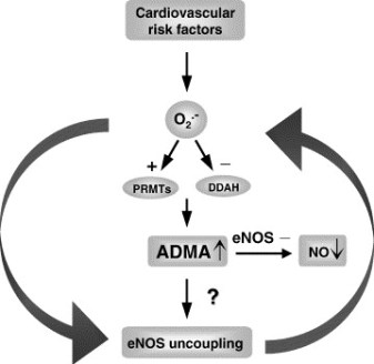
DDAH Says NO to ADMA(1); The DDAH/ADMA/NOS Pathway(2)

Author-Writer-Reporter:  Stephen J. Williams, PhD

Endothelium-derived nitric oxide (NO) has been shown to be vasoprotective.  Nitric oxide enhances endothelial cell survival, inhibits excessive proliferation of vascular smooth muscle cells, regulates vascular smooth muscle tone, and prevents platelets from sticking to the endothelial wall.  Together with evidence from preclinical and human studies, it is clear that impairment of the NOS pathway increases risk of cardiovascular disease (3-5).

This post contains two articles on the physiological regulation of nitric oxide (NO) by an endogenous NO synthase inhibitor asymmetrical dimethylarginine (ADMA) and ADMA metabolism by the enzyme DDAH(1,2).  Previous posts on nitric oxide, referenced at the bottom of the page, provides excellent background and further insight for this posting. In summary plasma ADMA levels are elevated in patients with cardiovascular disease and several large studies have shown that plasma ADMA is an independent biomarker for cardiovascular-related morbidity and mortality(6-8).

admacardiacrisk

admaeffects

Figure 1 A. Cardiac risks of ADMA B. Effects of ADMA (Photo credit: Wikipedia)

ADMA Production and Metabolism

Nuclear proteins such as histones can be methylated on arginine residues by protein-arginine methyltransferases, enzymes which use S-adenosylmethionine as methyl groups.  This methylation event is thought to regulate protein function, much in the way of protein acetylation and phosphorylation (9).  And much like phosphorylation, these modifications are reversible through methylesterases.   The proteolysis of these arginine-methyl modifications lead to the liberation of free guanidine-methylated arginine residues such as L-NMMA, asymmetric dimethylarginine (ADMA) and symmetrical methylarginine (SDMA).

The first two, L-NMMA and ADMA, have been shown to inhibit the activity of the endothelial NOS.  This protein turnover is substantial: for instance the authors note that each day 40% of constitutive protein in adult liver is newly synthesized protein. And in several diseases, such as muscular dystrophy, ischemic heart disease, and diabetes, it has been known since the 1970’s that protein catabolism rates are very high, with corresponding increased urinary excretion of ADMA(10-13).  Methylarginines are excreted in the urine by cationic transport.  However, the majority of ADMA and L-NMMA are degraded within the cell by dimethylaminohydrolase (DDAH), first cloned and purified in rat(14).

endogenous NO inhibitors from pubchem

Figure 2.  Endogenous inhibitors of NO synthase.  Chemical structures generated from PubChem.

DDAH

DDAH specifically hydrolyzes ADMA and L-NMMA to yield citruline and demethylamine and usually shows co-localization with NOS. Pharmacologic inhibition of DDAH activity causes accumulation of ADMA and can reverse the NO-mediated bradykinin-induced relaxation of human saphenous vein.

Two isoforms have been found in human:

  • DDAH1 (found in brain and kidney and associated with nNOS) and
  • DDAH2 (highly expressed in heart, placenta, and kidney and associated with eNOS).

DDAH2 can be upregulated by all-trans retinoic acid (atRA can increase NO production).  Increased reactive oxygen species and possibly homocysteine, a risk factor for cardiovascular disease, can decrease DDAH activity(15,16).

  • The importance of DDAH activity can also be seen in transgenic mice which overexpress DDAH, exhibiting increased NO production, increased insulin sensitivity, and reduced vascular resistance  (17).  Likewise,
  • Transgenic mice, null for the DDAH1, showed increase in blood pressure, decreased NO production, and significant increase in tissue and plasma ADMA and L-NMMA.

amdanosfigure

Figure 3.  The DDAH/ADMA/NOS cycle. Figure adapted from Cooke and Ghebremarian (1).

As mentioned in the article by Cooke and Ghebremariam, the authors state: the weight of the evidence indicates that DDAH is a worthy therapeutic target. Agents that increase DDAH expression are known, and 1 of these, a farnesoid X receptor agonist, is in clinical trials

http://www.interceptpharma.com

An alternate approach is to

  • develop an allosteric activator of the enzyme.  Although
  • development of an allosteric activator is not a typical pharmaceutical approach, recent studies indicate that this may be achievable aim(18).

References:

1.            Cooke, J. P., and Ghebremariam, Y. T. : DDAH says NO to ADMA.(2011) Arteriosclerosis, thrombosis, and vascular biology 31, 1462-1464

2.            Tran, C. T., Leiper, J. M., and Vallance, P. : The DDAH/ADMA/NOS pathway.(2003) Atherosclerosis. Supplements 4, 33-40

3.            Niebauer, J., Maxwell, A. J., Lin, P. S., Wang, D., Tsao, P. S., and Cooke, J. P.: NOS inhibition accelerates atherogenesis: reversal by exercise. (2003) American journal of physiology. Heart and circulatory physiology 285, H535-540

4.            Miyazaki, H., Matsuoka, H., Cooke, J. P., Usui, M., Ueda, S., Okuda, S., and Imaizumi, T. : Endogenous nitric oxide synthase inhibitor: a novel marker of atherosclerosis.(1999) Circulation 99, 1141-1146

5.            Wilson, A. M., Shin, D. S., Weatherby, C., Harada, R. K., Ng, M. K., Nair, N., Kielstein, J., and Cooke, J. P. (2010): Asymmetric dimethylarginine correlates with measures of disease severity, major adverse cardiovascular events and all-cause mortality in patients with peripheral arterial disease. Vasc Med 15, 267-274

6.            Kielstein, J. T., Impraim, B., Simmel, S., Bode-Boger, S. M., Tsikas, D., Frolich, J. C., Hoeper, M. M., Haller, H., and Fliser, D. : Cardiovascular effects of systemic nitric oxide synthase inhibition with asymmetrical dimethylarginine in humans.(2004) Circulation 109, 172-177

7.            Kielstein, J. T., Donnerstag, F., Gasper, S., Menne, J., Kielstein, A., Martens-Lobenhoffer, J., Scalera, F., Cooke, J. P., Fliser, D., and Bode-Boger, S. M. : ADMA increases arterial stiffness and decreases cerebral blood flow in humans.(2006) Stroke; a journal of cerebral circulation 37, 2024-2029

8.            Mittermayer, F., Krzyzanowska, K., Exner, M., Mlekusch, W., Amighi, J., Sabeti, S., Minar, E., Muller, M., Wolzt, M., and Schillinger, M. : Asymmetric dimethylarginine predicts major adverse cardiovascular events in patients with advanced peripheral artery disease.(2006) Arteriosclerosis, thrombosis, and vascular biology 26, 2536-2540

9.            Kakimoto, Y., and Akazawa, S.: Isolation and identification of N-G,N-G- and N-G,N’-G-dimethyl-arginine, N-epsilon-mono-, di-, and trimethyllysine, and glucosylgalactosyl- and galactosyl-delta-hydroxylysine from human urine. (1970) The Journal of biological chemistry 245, 5751-5758

10.          Inoue, R., Miyake, M., Kanazawa, A., Sato, M., and Kakimoto, Y.: Decrease of 3-methylhistidine and increase of NG,NG-dimethylarginine in the urine of patients with muscular dystrophy. (1979) Metabolism: clinical and experimental 28, 801-804

11.          Millward, D. J.: Protein turnover in skeletal muscle. II. The effect of starvation and a protein-free diet on the synthesis and catabolism of skeletal muscle proteins in comparison to liver. (1970) Clinical science 39, 591-603

12.          Goldberg, A. L., and St John, A. C.: Intracellular protein degradation in mammalian and bacterial cells: Part 2. (1976) Annual review of biochemistry 45, 747-803

13.          Dice, J. F., and Walker, C. D.: Protein degradation in metabolic and nutritional disorders. (1979) Ciba Foundation symposium, 331-350

14.          Ogawa, T., Kimoto, M., and Sasaoka, K.: Purification and properties of a new enzyme, NG,NG-dimethylarginine dimethylaminohydrolase, from rat kidney. (1989) The Journal of biological chemistry 264, 10205-10209

15.          Ito, A., Tsao, P. S., Adimoolam, S., Kimoto, M., Ogawa, T., and Cooke, J. P.: Novel mechanism for endothelial dysfunction: dysregulation of dimethylarginine dimethylaminohydrolase. (1999) Circulation 99, 3092-3095

16.          Stuhlinger, M. C., Tsao, P. S., Her, J. H., Kimoto, M., Balint, R. F., and Cooke, J. P. : Homocysteine impairs the nitric oxide synthase pathway: role of asymmetric dimethylarginine.(2001) Circulation 104, 2569-2575

17.          Sydow, K., Mondon, C. E., Schrader, J., Konishi, H., and Cooke, J. P.: Dimethylarginine dimethylaminohydrolase overexpression enhances insulin sensitivity. (2008) Arteriosclerosis, thrombosis, and vascular biology 28, 692-697

18.          Zorn, J. A., and Wells, J. A.: Turning enzymes ON with small molecules. (2010) Nature chemical biology 6, 179-188

Other research papers on Nitric Oxide and Cardiac Risk  were published on this Scientific Web site as follows:

The Nitric Oxide and Renal is presented in FOUR parts:

Part I: The Amazing Structure and Adaptive Functioning of the Kidneys: Nitric Oxide

Part II: Nitric Oxide and iNOS have Key Roles in Kidney Diseases

Part III: The Molecular Biology of Renal Disorders: Nitric Oxide

Part IV: New Insights on Nitric Oxide donors

Cardiac Arrhythmias: A Risk for Extreme Performance Athletes

What is the role of plasma viscosity in hemostasis and vascular disease risk?

Cardiovascular Risk Inflammatory Marker: Risk Assessment for Coronary Heart Disease and Ischemic Stroke – Atherosclerosis.

Endothelial Dysfunction, Diminished Availability of cEPCs, Increasing CVD Risk for Macrovascular Disease – Therapeutic Potential of cEPCs

Biochemistry of the Coagulation Cascade and Platelet Aggregation – Part I

Nitric Oxide Function in Coagulation

Read Full Post »

Personalized Medicine: Cancer Cell Biology and Minimally Invasive Surgery (MIS)

Curator: Aviva Lev-Ari, PhD, RN

In the field of Cancer Research, Translational Medicine  will become Personalized Medicine when each of the cancer type, below will have a Genetic Marker allowing the Clinical Team to use the marker for:

  • prediction of Patient’s reaction to Drug induction
  • design of Clinical Trials to validate drug efficacy on small subset of patients predicted to react favorable to drug regimen, increasing validity and reliability
  • Genetical identification of patients at no need to have a drug administered if non sensitivity to the drug has been predicted

Current urgent need exists for Identification of Genetic Markers to predict Patient’s reaction to Drugs Induction for the following types of Cancer:


The executive task of the clinician remains to assess the differentiation in Tumor Response to Treatment.

Review of limitations for the current existing Tools used by clinicians in to be found in:

Brücher BLDM, Bilchik A, Nissan A, Avital I & Stojadinovic A. Can tumor response to therapy be predicted, thereby improving the selection of patients for cancer treatment?  Future Oncology 2012; 8(8): 903-906 , DOI 10.2217/fon.12.78 (doi:10.2217/fon.12.78)   The heterogeneity is a problem that will take at least another decade to unravel because of the number of signaling pathways and the crosstalk that is specifically at issue.

Future Oncology August 2012, Vol. 8, No. 8, Pages 903-906 ,

It is suggested that the new modality should be based on individualized histopathology as well as tumor molecular, genetic and functional characteristics, and individual patients’ characteristics. The new modality should be based on empirical evidence that translates into relevant and meaningful clinical outcome data.

Cancer is in particular a difficult to treat tissue type pathology. In “Tumor response criteria: are they appropriate?” that concern is addressed as follows:

“This becomes a conundrum of sorts in an era of ‘minimally invasive treatment’. One frequently encountered example is that of a patient with chronic gastric reflux and an ultrasound-staged T3N1 distal esophageal adenocarcinoma, who had complete sonographic tumor response to neoadjuvant chemoradiation. The physician may declare that, the tumor having disappeared, the patient requires no further treatment. The surgical oncologist recommends resection, recognizing the fact that up to 20% or more of these complete responders will have identifiable nests of tumor beyond the mucosal scar within the specimen – in other words: residual tumor. In other cases, patients with clinical, sonographic, functional (PET) and histopathological ‘complete’ tumor response to induction therapy experience recurrence within the first 2 years of resection, reminding us of the intricacy and enigma of tumor biology. We have yet to develop the tools needed to consistently delineate the response of a tumor to multimodality therapy.”

This described reality in the Oncology Operating Room is coupled with new trends in invasive treatment of tumor resection.

Minimally Invasive Surgery (MIS) vs. conventional surgery dissection applied to cancer tissue with the known pathophysiology of recurrence and remission cycles has its short term advantages. However, in many cases MIS is not the right surgical decision, yet, it is applied for a corollary of patient-centered care considerations. At present, facing the unknown of the future behavior of the tumor as its response to therapeutics bearing uncertainty related to therapy outcomes.

An increase in the desirable outcomes of MIS as a modality of treatment, will be strongly assisted in the future, with anticipated progress to be made in the field of Cancer Research, Translational Medicine and Personalized Medicine, when each of the cancer types, above,  will already have a Genetic Marker allowing the Clinical Team to use the marker(s) for:

  • prediction of Patient’s reaction to Drug induction
  • design of Clinical Trials to validate drug efficacy on small subset of patients predicted to react favorable to drug regimen, increasing validity and reliability
  • Genetical identification of patients at no need to have a drug administered if non sensitivity to the drug has been predicted by the genetic marker.

REFERENCES

Tumor response criteria: are they appropriate?

Björn LDM Brücher*1,2, Anton Bilchik2,3, Aviram Nissan2,4, Itzhak Avital2,5 & Alexander Stojadinovic2,6

 
Treatment for cure is not the endpoint, but the best that can be done is to extend the time of survival to a realistic long term goal and retain a quality of life.
 
Brücher BLDM, Piso P, Verwaal V et al. Peritoneal carcinomatosis: overview and basics. Cancer Invest.30(3),209–224 (2012).
 
Brücher BLDM, Swisher S, Königsrainer A et al. Response to preoperative therapy in upper gastrointestinal cancers. Ann. Surg. Oncol.16(4),878–886 (2009).
 
Miller AB, Hoogstraten B, Staquet M, Winkler A. Reporting results of cancer treatment. Cancer47(1),207–214 (1981).


Other research papers on Cancer and Cancer Therapeutics were published on this Scientific Web site as follows:

What can we expect of tumor therapeutic response?

PIK3CA mutation in Colorectal Cancer may serve as a Predictive Molecular Biomarker for adjuvant Aspirin therapy

Nanotechnology Tackles Brain Cancer

Response to Multiple Cancer Drugs through Regulation of TGF-β Receptor Signaling: a MED12 Control

Personalized medicine-based cure for cancer might not be far away

GSK for Personalized Medicine using Cancer Drugs needs Alacris systems biology model to determine the in silico effect of the inhibitor in its “virtual clinical trial”

Lung Cancer (NSCLC), drug administration and nanotechnology

Non-small Cell Lung Cancer drugs – where does the Future lie?

Cancer Innovations from across the Web

arrayMap: Genomic Feature Mining of Cancer Entities of Copy Number Abnormalities (CNAs) Data

How mobile elements in “Junk” DNA promote cancer. Part 1: Transposon-mediated tumorigenesis.

Cancer Genomics – Leading the Way by Cancer Genomics Program at UC Santa Cruz

Closing the gap towards real-time, imaging-guided treatment of cancer patients.

Closing the gap towards real-time, imaging-guided treatment of cancer patients.

mRNA interference with cancer expression

Search Results for ‘cancer’ on this web site

Cancer Genomics – Leading the Way by Cancer Genomics Program at UC Santa Cruz

Closing the gap towards real-time, imaging-guided treatment of cancer patients.

Lipid Profile, Saturated Fats, Raman Spectrosopy, Cancer Cytology

mRNA interference with cancer expression

Pancreatic cancer genomes: Axon guidance pathway genes – aberrations revealed

Biomarker tool development for Early Diagnosis of Pancreatic Cancer: Van Andel Institute and Emory University

Is the Warburg Effect the cause or the effect of cancer: A 21st Century View?

Crucial role of Nitric Oxide in Cancer

Targeting Glucose Deprived Network Along with Targeted Cancer Therapy Can be a Possible Method of Treatment


See comment written for:

Knowing the tumor’s size and location, could we target treatment to THE ROI by applying…..

http://pharmaceuticalintelligence.com/2012/10/16/knowing-the-tumors-size-and-location-could-we-target-treatment-to-the-roi-by-applying-imaging-guided-intervention/

24 Responses

  1. GREAT work.

    I’ll read and comment later on

  2. Highlights of The 2012 Johns Hopkins Prostate Disorders White Paper include:

    A promising new treatment for men with frequent nighttime urination.
    Answers to 8 common questions about sacral nerve stimulation for lower urinary tract symptoms.
    Surprising research on the link between smoking and prostate cancer recurrence.
    How men who drink 6 cups of coffee a day or more may reduce their risk of aggressive prostate cancer.
    Should you have a PSA screening test? Answers to important questions on the controversial USPSTF recommendation.
    Watchful waiting or radical prostatectomy for men with early-stage prostate cancer? What the research suggests.
    A look at state-of-the-art surveillance strategies for men on active surveillance for prostate cancer.
    Locally advanced prostate cancer: Will you benefit from radiation and hormones?
    New drug offers hope for men with metastatic castrate-resistant prostate cancer.
    Behavioral therapy for incontinence: Why it might be worth a try.

    You’ll also get the latest news on benign prostatic enlargement (BPE), also known as benign prostatic hyperplasia (BPH) and prostatitis:
    What’s your Prostate Symptom Score? Here’s a quick quiz you can take right now to determine if you should seek treatment for your enlarged prostate.
    Your surgical choices: a close look at simple prostatectomy, transurethral prostatectomy and open prostatectomy.
    New warnings about 5-alpha-reductase inhibitors and aggressive prostate cancer.

  3. Promising technique.

    INCORE pointed out in detail about the general problem judging response and the stil missing quality in standardization:

    http://www.futuremedicine.com/doi/abs/10.2217/fon.12.78?url_ver=Z39.88-2003&rfr_id=ori:rid:crossref.org&rfr_dat=cr_pub%3dwww.ncbi.nlm.nih.gov

    I did research in response evaluation and prediction for about 15y now and being honest: neither the clinical, nor the molecular biological data proved significant benefit in changing a strategy in patient diagnosis and / or treatment. I would state: this brings us back on the ground and not upon the sky. Additionally it means: we have to ´work harder on that and the WHO has to take responsibility: clinicians use a reponse classification without knowing, that this is just related to “ONE” experiment from the 70′s and that this experiment never had been rescrutinized (please read the Editorial I provided – we use a clinical response classification since more than 30 years worldwide (Miller et al. Cancer 1981) but it is useless !

  4. Dr. BB

    Thank you for your comment.
    Dr. Nir will reply to your comment.
    Regarding the Response Classification in use, it seems that the College of Oncology should champion a task force to revisit the Best Practice in use in this domain and issue a revised version or a new effort for a a new classification system for Clinical Response to treatment in Cancer.

  5. I’m sorry that I was looking for this paper again earlier and didn’t find it. I answered my view on your article earlier.

    This is a method demonstration, but not a proof of concept by any means. It adds to the cacophany of approaches, and in a much larger study would prove to be beneficial in treatment, but not a cure for serious prostate cancer because it is unlikely that it can get beyond the margin, and also because there is overtreatment at the cutoff of PSA at 4.0. There is now a proved prediction model that went to press some 4 months ago. I think that the pathologist has to see the tissue, and the standard in pathology now is for any result that is cancer, two pathologist or a group sitting together should see it. It’s not an easy diagnosis.

    Björn LDM Brücher, Anton Bilchik, Aviram Nissan, Itzhak Avital, & Alexander Stojadinovic. Tumor response criteria: are they appropriate? Future Oncol. (2012) 8(8), 903–906. 10.2217/FON.12.78. ISSN 1479-6694.

    ..Tumor heterogeneity is a ubiquitous phemomenon. In particular, there are important differences among the various types of gastrointestinal (GI) cancers in terms of tumor biology, treatment response and prognosis.

    ..This forms the principal basis for targeted therapy directed by tumor-specific testing at either the gene or protein level. Despite rapid advances in our understanding of targeted therapy for GI cancers, the impact on cancer survival has been marginal.

    ..Can tumor response to therapy be predicted, thereby improving the selection of patients for cancer treatment?

    ..In 2000 theNCI with the European Association for Research and Treatment of Cancer, proposed a replacement of 2D measurement with a decrease in the largest tumor diameter by 30% in one dimension. Tumor response as defined would translate into a 50% decrease for a spherical lesion

    ..We must rethink how we may better determine treatment response in a reliable, reproducible way that is aimed at individualizing the therapy of cancer patients.

    ..we must change the tools we use to assess tumor response. The new modality should be based on empirical evidence that translates into relevant and meaningful clinical outcome data.

    ..This becomes a conundrum of sorts in an era of ‘minimally invasive treatment’.

    ..integrated multidisciplinary panel of international experts – not sure that that will do it

    Several years ago i heard Stamey present the totality of his work at Stanford, with great disappointment over hsPSA that they pioneered in. The outcomes were disappointing.

    I had published a review of all of our cases reviewed for 1 year with Marguerite Pinto.
    There’s a reason that the physicians line up outside of her office for her opinion.
    The review showed that a PSA over 24 ng/ml is predictive of bone metastasis. Any result over 10 was as likely to be prostatitis, BPH or cancer.

    I did an ordinal regression in the next study with Gustave Davis using a bivariate ordinal regression to predict lymph node metastasis using the PSA and the Gleason score. It was better than any univariate model, but there was no followup.

    I reviewed a paper for Clin Biochemistry (Elsevier) on a new method for PSA, very different than what we are familiar with. It was the most elegant paper I have seen in the treatment of the data. The model could predict post procedural time to recurrence to 8 years.

    • I hope we are in agreement on the fact that imaging guided interventions are needed for better treatment outcome. The point I’m trying to make in this post is that people are investing in developing imaging guided intervention and it is making progress.

      Over diagnosis and over treatment is another issue altogether. I think that many of my other posts are dealing with that.

  6. Tumor response criteria: are they appropriate?
    Future Oncology 2012; 8(8): 903-906 , DOI 10.2217/fon.12.78 (doi:10.2217/fon.12.78)
    Björn LDM Brücher, Anton Bilchik, Aviram Nissan, Itzhak Avital & Alexander Stojadinovic
    Tumor heterogeneity is a problematic because of differences among the metabolic variety among types of gastrointestinal (GI) cancers, confounding treatment response and prognosis.
    This is in response to … a group of investigators from Sunnybrook Health Sciences Centre, University of Toronto, Ontario, Canada who evaluate the feasibility and safety of magnetic resonance (MR) imaging–controlled transurethral ultrasound therapy for prostate cancer in humans. Their study’s objective was to prove that using real-time MRI guidance of HIFU treatment is possible and it guarantees that the location of ablated tissue indeed corresponds to the locations planned for treatment.
    1. There is a difference between expected response to esophageal or gastric neoplasms both biologically and in expected response, even given variability within a class. The expected time to recurrence is usually longer in the latter case, but the confounders are – age at time of discovery, biological time of detection, presence of lymph node and/or distant metastasis, microscopic vascular invasion.
    2. There is a long latent period in abdominal cancers before discovery, unless a lesion is found incidentally in surgery for another reason.
    3. The undeniable reality is that it is not difficult to identify the main lesion, but it is difficult to identify adjacent epithelium that is at risk (transitional or pretransitional). Pathologists have a very good idea about precancerous cervical neoplasia.

    The heterogeneity rests within each tumor and between the primary and metastatic sites, which is expected to be improved by targeted therapy directed by tumor-specific testing. Despite rapid advances in our understanding of targeted therapy for GI cancers, the impact on cancer survival has been marginal.

    The heterogeneity is a problem that will take at least another decade to unravel because of the number of signaling pathways and the crosstalk that is specifically at issue.

    I must refer back to the work of Frank Dixon, Herschel Sidransky, and others, who did much to develop a concept of neoplasia occurring in several stages – minimal deviation and fast growing. These have differences in growth rates, anaplasia, and biochemical. This resembles the multiple “hit” theory that is described in “systemic inflammatory” disease leading to a final stage, as in sepsis and septic shock.
    In 1920, Otto Warburg received the Nobel Prize for his work on respiration. He postulated that cancer cells become anaerobic compared with their normal counterpart that uses aerobic respiration to meet most energy needs. He attributed this to “mitochondrial dysfunction. In fact, we now think that in response to oxidative stress, the mitochondrion relies on the Lynen Cycle to make more cells and the major source of energy becomes glycolytic, which is at the expense of the lean body mass (muscle), which produces gluconeogenic precursors from muscle proteolysis (cancer cachexia). There is a loss of about 26 ATP ~Ps in the transition.
    The mitochondrial gene expression system includes the mitochondrial genome, mitochondrial ribosomes, and the transcription and translation machinery needed to regulate and conduct gene expression as well as mtDNA replication and repair. Machinery involved in energetics includes the enzymes of the Kreb’s citric acid or TCA (tricarboxylic acid) cycle, some of the enzymes involved in fatty acid catabolism (β-oxidation), and the proteins needed to help regulate these systems. The inner membrane is central to mitochondrial physiology and, as such, contains multiple protein systems of interest. These include the protein complexes involved in the electron transport component of oxidative phosphorylation and proteins involved in substrate and ion transport.
    Mitochondrial roles in, and effects on, cellular homeostasis extend far beyond the production of ATP, but the transformation of energy is central to most mitochondrial functions. Reducing equivalents are also used for anabolic reactions. The energy produced by mitochondria is most commonly thought of to come from the pyruvate that results from glycolysis, but it is important to keep in mind that the chemical energy contained in both fats and amino acids can also be converted into NADH and FADH2 through mitochondrial pathways. The major mechanism for harvesting energy from fats is β-oxidation; the major mechanism for harvesting energy from amino acids and pyruvate is the TCA cycle. Once the chemical energy has been transformed into NADH and FADH2 (also discovered by Warburg and the basis for a second Nobel nomination in 1934), these compounds are fed into the mitochondrial respiratory chain.
    The hydroxyl free radical is extremely reactive. It will react with most, if not all, compounds found in the living cell (including DNA, proteins, lipids and a host of small molecules). The hydroxyl free radical is so aggressive that it will react within 5 (or so) molecular diameters from its site of production. The damage caused by it, therefore, is very site specific. The reactions of the hydroxyl free radical can be classified as hydrogen abstraction, electron transfer, and addition.
    The formation of the hydroxyl free radical can be disastrous for living organisms. Unlike superoxide and hydrogen peroxide, which are mainly controlled enzymatically, the hydroxyl free radical is far too reactive to be restricted in such a way – it will even attack antioxidant enzymes. Instead, biological defenses have evolved that reduce the chance that the hydroxyl free radical will be produced and, as nothing is perfect, to repair damage.
    Currently, some endogenous markers are being proposed as useful measures of total “oxidative stress” e.g., 8-hydroxy-2’deoxyguanosine in urine. The ideal scavenger must be non-toxic, have limited or no biological activity, readily reach the site of hydroxyl free radical production (i.e., pass through barriers such as the blood-brain barrier), react rapidly with the free radical, be specific for this radical, and neither the scavenger nor its product(s) should undergo further metabolism.
    Nitric oxide has a single unpaired electron in its π*2p antibonding orbital and is therefore paramagnetic. This unpaired electron also weakens the overall bonding seen in diatomic nitrogen molecules so that the nitrogen and oxygen atoms are joined by only 2.5 bonds. The structure of nitric oxide is a resonance hybrid of two forms.
    In living organisms nitric oxide is produced enzymatically. Microbes can generate nitric oxide by the reduction of nitrite or oxidation of ammonia. In mammals nitric oxide is produced by stepwise oxidation of L-arginine catalyzed by nitric oxide synthase (NOS). Nitric oxide is formed from the guanidino nitrogen of the L-arginine in a reaction that consumes five electrons and requires flavin adenine dinucleotide (FAD), flavin mononucleotide (FMN) tetrahydrobiopterin (BH4), and iron protoporphyrin IX as cofactors. The primary product of NOS activity may be the nitroxyl anion that is then converted to nitric oxide by electron acceptors.
    The thiol-disulfide redox couple is very important to oxidative metabolism. GSH is a reducing cofactor for glutathione peroxidase, an antioxidant enzyme responsible for the destruction of hydrogen peroxide. Thiols and disulfides can readily undergo exchange reactions, forming mixed disulfides. Thiol-disulfide exchange is biologically very important. For example, GSH can react with protein cystine groups and influence the correct folding of proteins, and it GSH may play a direct role in cellular signaling through thiol-disulfide exchange reactions with membrane bound receptor proteins (e.g., the insulin receptor complex), transcription factors (e.g., nuclear factor κB), and regulatory proteins in cells. Conditions that alter the redox status of the cell can have important consequences on cellular function.
    So the complexity of life is not yet unraveled.

    Can tumor response to therapy be predicted, thereby improving the selection of patients for cancer treatment?
    The goal is not just complete response. Histopathological response seems to be related post-treatment histopathological assessment but it is not free from the challenge of accurately determining treatment response, as this method cannot delineate whether or not there are residual cancer cells. Functional imaging to assess metabolic response by 18-fluorodeoxyglucose PET also has its limits, as the results are impacted significantly by several variables:

    • tumor type
    • sizing
    • doubling time
    • anaplasia?
    • extent of tumor necrosis
    • type of antitumor therapy and the time when response was determined.
    The new modality should be based on individualized histopathology as well as tumor molecular, genetic and functional characteristics, and individual patients’ characteristics, a greater challenge in an era of ‘minimally invasive treatment’.
    This listing suggests that for every cancer the following data has to be collected (except doubling time). If there are five variables, the classification based on these alone would calculate to be very sizable based on Eugene Rypka’s feature extraction and classification. But looking forward, time to remission and disease free survival are additionally important. Treatment for cure is not the endpoint, but the best that can be done is to extend the time of survival to a realistic long term goal and retain a quality of life.

    Brücher BLDM, Piso P, Verwaal V et al. Peritoneal carcinomatosis: overview and basics. Cancer Invest.30(3),209–224 (2012).
    Brücher BLDM, Swisher S, Königsrainer A et al. Response to preoperative therapy in upper gastrointestinal cancers. Ann. Surg. Oncol.16(4),878–886 (2009).
    Miller AB, Hoogstraten B, Staquet M, Winkler A. Reporting results of cancer treatment. Cancer47(1),207–214 (1981).
    Therasse P, Arbuck SG, Eisenhauer EA et al. New guidelines to evaluate the response to treatment in solid tumors. European Organization for Research and Treatment of Cancer, National Cancer Institute of the United States, National Cancer Institute of Canada. J. Natl Cancer Inst.92(3),205–216 (2000).
    Brücher BLDM, Becker K, Lordick F et al. The clinical impact of histopathological response assessment by residual tumor cell quantification in esophageal squamous cell carcinomas. Cancer106(10),2119–2127 (2006).

    • Dr. Larry,

      Thank you for this comment.

      Please carry it as a stand alone post, Dr. Ritu will refer to it and reference it in her FORTHCOMING pst on Tumor Response which will integrate multiple sources.

      Please execute my instruction

      Thank you

    • Thank you Larry for this educating comment. It explains very well why the Canadian investigators did not try to measure therapy response!

      What they have demonstrated is the technological feasibility of coupling a treatment device to an imaging device and use that in order to guide the treatment to the right place.

      the issue of “choice of treatment” to which you are referring is not in the scope of this publication.
      The point is: if one treatment modality can be guided, other can as well! This should encourage others, to try and develop imaging-based treatment guidance systems.

  7. The crux of the matter in terms of capability is that the cancer tissue, adjacent tissue, and the fibrous matrix are all in transition to the cancerous state. It is taught to resect leaving “free margin”, which is better aesthetically, and has had success in breast surgery. The dilemma is that the patient may return, but how soon?

    • Correct. The philosophy behind lumpectomy is preserving quality of life. It was Prof. Veronesi (IEO) who introduced this method 30 years ago noticing that in the majority of cases, the patient will die from something else before presenting recurrence of breast cancer..

      It is well established that when the resection margins are declared by a pathologist (as good as he/she could be) as “free of cancer”, the probability of recurrence is much lower than otherwise.

  8. Dr. Larry,

    To assist Dr. Ritu, PLEASE carry ALL your comments above into a stand alone post and ADD to it your comment on my post on MIS

    Thank you

  9. Great post! Dr. Nir, can the ultrasound be used in conjunction with PET scanning as well to determine a spatial and functional map of the tumor. With a disease like serous ovarian cancer we typically see an intraperitoneal carcimatosis and it appears that clinicians are wanting to use fluorogenic probes and fiberoptics to visualize the numerous nodules located within the cavity Also is the technique being used mainy for surgery or image guided radiotherapy or can you use this for detecting response to various chemotherapeutics including immunotherapy.

    • Ultrasound can and is actually used in conjunction with PET scanning in many cases. The choice of using ultrasound is always left to the practitioner! Being a non-invasive, low cost procedure makes the use of ultrasound a non-issue. The down-side is that because it is so easy to access and operate, nobody bothers to develop rigorous guidelines about using it and the benefits remains the property of individuals.

      In regards to the possibility of screening for ovarian cancer and characterising pelvic masses using ultrasound I can refer you to scientific work in which I was involved:

      1. VAES (E.), MANCHANDA (R), AUTIER, NIR (R), NIR (D.), BLEIBERG (H.), ROBERT (A.), MENON (U.). Differential diagnosis of adnexal masses: Sequential use of the Risk of Malignancy Index and a novel computer aided diagnostic tool. Published in Ultrasound in Obstetrics & Gynecology. Issue 1 (January). Vol. 39. Page(s): 91-98.

      2. VAES (E.), MANCHANDA (R), NIR (R), NIR (D.), BLEIBERG (H.), AUTIER (P.), MENON (U.), ROBERT (A.). Mathematical models to discriminate between benign and malignant adnexal masses: potential diagnostic improvement using Ovarian HistoScanning. Published in International Journal of Gynecologic Cancer (IJGC). Issue 1. Vol. 21. Page(s): 35-43.

      3. LUCIDARME (0.), AKAKPO (J.-P.), GRANBERG (S.), SIDERI (M.), LEVAVI (H.), SCHNEIDER (A.), AUTIER (P.), NIR (D.), BLEIBERG (H.). A new computer aided diagnostic tool for non-invasive characterisation of malignant ovarian masses: Results of a multicentre validation study. Published in European Radiology. Issue 8. Vol. 20. Page(s): 1822-1830.

      Dror Nir, PhD
      Managing partner

      BE: +32 (0) 473 981896
      UK: +44 (0) 2032392424

      web: http://www.radbee.com/
      blogs: http://radbee.wordpress.com/ ; http://www.MedDevOnIce.com

  10. totally true and i am very thankfull for these briliant comments.

    Remember: 10years ago: every cancer researcher stated: “look at the tumor cells only – forget the stroma”. The era of laser-captured tumor-cell dissection started. Now , everyone knows: it is a system we are looking at and viewing and analyzing tumor cells only is really not enough.

    So if we would be honest, we would have to declare, that all data, which had been produced 13-8years ago, dealing with laser capture microdissection, that al these data would need a re-scrutinization, cause the influence of the stroma was “forgotten”. I ‘d better not try thinking about the waisted millions of dollars.

    If we keep on being honest: the surgeon looks at the “free margin” in a kind of reductionable model, the pathologist is more the control instance. I personally see the pathologist as “the control instance” of surgical quality. Therefore, not the wish of the surgeon is important, the objective way of looking into problems or challenges. Can a pathologist always state, if a R0-resection had been performed ?

    The use of the Resectability Classification:
    There had been many many surrogate marker analysis – nothing new. BUT never a real substantial well tought through structured analysis had been done: mm by mm by mm by mm and afterwards analyzing that by a ROC analysis. BUt against which goldstandard ? If you perform statistically a ROC analysis – you need a golstandard to compare to. Therefore what is the real R0-resectiòn? It had been not proven. It just had been stated in this or that tumor entity that this or that margin with this margin free mm distance or that mm distance is enough and it had been declared as “the real R0-classification”. In some organs it is very very difficult and we all (surgeons, pathologists, clinicians) that we always get to the limit, if we try interpretating the R-classification within the 3rd dimension. Often it is just declared and stated.

    Otherwise: if lymph nodes are negative it does not mean, lymph nodes are really negative, cause up to 38% for example in upper GI cancers have histological negative lymph nodes, but immunohistochemical positive lymph nodes. And this had been also shown by Stojadinovic at el analyzing the ultrastaging in colorectal cancer. So the 4th dimension of cancer – the lymph nodes / the lymphatic vessel invasion are much more important than just a TNM classification, which unfortunately does often not reflect real tumor biology.

    AS we see: cancer has multifactorial reasons and it is necessary taking the challenge performing high sophisticated research by a multifactorial and multidisciplinary manner.

    Again my deep and heartly thanks for that productive and excellent discussion !

    • Dr. BB,

      Thank you for your comment.

      Multidisciplinary perspectives have illuminated the discussion on the pages of this Journal.

      Eager to review Dr. Ritu’s forthcoming paper – the topic has a life of its own and is embodied in your statement:

      “the 4th dimension of cancer – the lymph nodes / the lymphatic vessel invasion are much more important than just a TNM classification, which unfortunately does often not reflect real tumor biology.”

    • Thank you BB for your comment. You have touched the core limitation of healthcare professionals: how do we know that we know!

      Do we have a reference to each of the test we perform?

      Do we have objective and standardise quality measures?

      Do we see what is out-there or are we imagining?

      The good news: Everyday we can “think” that we learned something new. We should be happy with that, even if it is means that we learned that yesterday’s truth is not true any-more and even if we are likely to be wrong again…:)

      But still, in the last decades, lots of progress was made….

  11. Dr. Nir,
    I thoroughly enjoyed reading your post as well as the comments that your post has attracted. There were different points of view and each one has been supported with relevant examples in the literature. Here are my two cents on the discussion:
    The paper that you have discussed had the objective of finding out whether real-time MRI guidance of treatment was even possible and if yes, and also if the treatment could be performed in accurate location of the ROI? The data reveals they were pretty successful in accomplishing their objective and of course that gives hope to the imaging-based targeted therapies.
    Whether the ROI is defined properly and if it accounts for the real tumor cure, is a different question. Role of pathologists and the histological analysis they bring about to the table cannot be ruled out, and the absence of a defined line between the tumor and the stromal region in the vicinity is well documented. However, that cannot rule out the value and scope of imaging-based detection and targeted therapy. After all, it is seminal in guiding minimally invasive surgery. As another arm of personalized medicine-based cure for cancer, molecular biologists at MD Anderson have suggested molecular and genetic profiling of the tumor to determine genetic aberrations on the basis of which matched-therapy could be recommended to patients. When phase I trial was conducted, the results were obtained were encouraging and the survival rate was better in matched-therapy patients compared to unmatched patients. Therefore, everytime there is more to consider when treating a cancer patient and who knows a combination of views of oncologists, pathologists, molecular biologists, geneticists, surgeons would device improvised protocols for diagnosis and treatment. It is always going to be complicated and generalizations would never give an answer. Smart interpretations of therapies – imaging-based or others would always be required!

    Ritu

    • Dr. Nir,
      One of your earlier comments, mentioned the non invasiveness of ultrasound, thus, it’s prevalence in use for diagnosis.

      This may be true for other or all areas with the exception of Mammography screening. In this field, an ultrasound is performed only if a suspected area of calcification or a lump has been detected in the routine or patient-initiated request for ad hoc mammography secondery to patient complain of pain or patient report of suspected lump.

      Ultrasound in this field repserents ascalation and two radiologists review.

      It in routine use for Breast biopsy.

    • Thanks Ritu for this supporting comment. The worst enemy of finding solutions is doing nothing while using the excuse of looking for the “ultimate solution” . Personally, I believe in combining methods and improving clinical assessment based on information fusion. Being able to predict, and then timely track the response to treatment is a major issue that affects survival and costs!

Judging the ‘Tumor response’-there is more food for thought

http://pharmaceuticalintelligence.com/2012/12/04/judging-the-tumor-response-there-is-more-food-for-thought/

13 Responses

  1. Dr. Sanexa
    you have brought up an interesting and very clinically relevant point: what is the best measurement of response and 2) how perspectives among oncologists and other professionals differ on this issues given their expertise in their respective subspecialties (immunologist versus oncologist. The advent of functional measurements of tumors (PET etc.) seems extremely important in the therapeutic use AND in the development of these types of compounds since usually a response presents (in cases of solid tumors) as either a lack of growth of the tumor or tumor shrinkage. Did the authors include an in-depth discussion of the rapidity of onset of resistance with these types of compounds?
    Thanks for the posting.

  2. Dr. Williams,
    Thanks for your comment on the post. The editorial brings to attention a view that although PET and other imaging methods provide vital information on tumor growth, shrinkage in response to a therapy, however, there are more aspects to consider including genetic and molecular characteristics of tumor.
    It was an editorial review and the authors did not include any in-depth discussion on the rapidity of onset of resistance with these types of compounds as the focus was primarily on interpreting tumor response.
    I am glad you found the contents of the write-up informative.
    Thanks again!
    Ritu

  3. Thank you for your wonderful comment and interpretation. Dr.Sanexa made a brilliant comment.

    May I allow myself putting my finger deeper into this wound ? Cancer patients deserve it.

    It had been already pointed out by international experts from Munich, Tokyo, Hong-Kong and Houston, dealing with upper GI cancer, that the actual response criteria are not appropriate and moreover: the clinical response criteria in use seem rather to function as an alibi, than helping to differentiate and / or discriminate tumor biology (Ann Surg Oncol 2009):

    http://www.ncbi.nlm.nih.gov/pubmed/19194759

    The response data in a phase-II-trial (one tumor entity, one histology, one treatment, one group) revealed: clinical response evaluation according to the WHO-criteria is not appropriate to determine response:

    http://www.ncbi.nlm.nih.gov/pubmed/15498642

    Of course, there was a time, when it seemed to be useful and this also has to be respected.

    There is another challenge: using statistically a ROC and resulting in thresholds. This was, is and always be “a clinical decision only” and not the decision of the statistician. The clinician tells the statistician, what decision, he wants to make – the responsibility is enormous. Getting back to the roots:
    After the main results of the Munich-group had been published 2001 (Ann Surg) and 2004 (J Clin Oncol):

    http://www.ncbi.nlm.nih.gov/pubmed/11224616

    http://www.ncbi.nlm.nih.gov/pubmed/14990646

    the first reaction in the community was: to difficult, can’t be, not re-evaluated, etc.. However, all evaluated cut-offs / thresholds had been later proven to be the real and best ones by the MD Anderson Cancer Center in Houston, Texas. Jaffer Ajani – a great and critical oncologist – pushed that together with Steve Swisher and they found the same results. Than the upper GI stakeholders went an uncommon way in science: they re-scrutinized their findings. Meanwhile the Goldstandard using histopathology as the basis-criterion had been published in Cancer 2006.

    http://www.ncbi.nlm.nih.gov/pubmed/16607651

    Not every author, who was at the authorlist in 2001 and 2004 wanted to be a part of this analysis and publication ! Why ? Everyone should judge that by himself.

    The data of this analysis had been submitted to the New England Journal of Medicine. In the 2nd review stage process, the manuscript was rejected. The Ann Surg Oncol accepted the publication: the re-scrutinized data resulted in another interesting finding: in the future maybe “one PET-scan” might be appropriate predicting the patient’s response.

    Where are we now ?

    The level of evidence using the response criteria is very low: Miller’s (Cancer 1981) publication belonged to ”one single” experiment from Moertel (Cancer 1976). During that time, there was no definition of “experiences” rather than “oncologists”. These terms had not been in use during that time.

    Additionally they resulted in a (scientifically weak) change of the classification, published by Therasse (J Natl Cancer Inst 2000). Targeted therapy did not result in a change so far. In 2009, the international upper GI experts sent their publication of the Ann Surg Oncol 2009 to the WHO but without any kind of reaction.

    Using molecular biological predictive markers within the last 10years all seem to have potential.

    http://www.ncbi.nlm.nih.gov/pubmed/20012971

    http://www.ncbi.nlm.nih.gov/pubmed/18704459

    http://www.ncbi.nlm.nih.gov/pubmed/17940507

    http://www.ncbi.nlm.nih.gov/pubmed/17354029

    But, experts are aware: the real step breaking barriers had not been performed so far. Additionally, it is very important in trying to evaluate and / predict response, that not different tumor entities with different survival and tumor biology are mixed together. Those data are from my perspective not helpful, but maybe that is my own Bias (!) of my view.

    INCORE, the International Consortium of Research Excellence of the Theodor-Billroth-Academy, was invited publishing the Editorial in Future Oncology 2012. The consortium pointed out, that living within an area of ‘prove of principle’ and also trying to work out level of evidence in medicine, it is “the duty and responsibility” of every clinician, but also of the societies and institutions, also of the WHO.

    Complete remission is not the only goal, as experts dealing with ‘response-research’ are aware. It is so frustrating for patients and clinicians: there is a rate of those patients with complete remission, who develop early recurrence ! This reflects, that complete remission cannot function as the only criterion describing response !

    Again, my heartly thanks, that Dr.Sanexa discussed this issue in detail.
    I hope, I found the way explaining the way of development and evaluating response criteria properly and in a differentiated way of view. From the perspective of INCORE:

    “an interdisciplinary initiative with all key stake¬holders and disciplines represented is imperative to make predictive and prognostic individualized tumor response assessment a modern-day reality. The integrated multidisciplinary panel of international experts need to define how to leverage existing data, tissue and testing platforms in order to predict individual patient treatment response and prognosis.”

  4. Dr. Brucher,

    First of all thanks for expressing your views on the ‘tumor response’ in a comprehensive way. You are the first author of the editorial review one of the prominent people who has taken part in the process of defining tumor response and I am glad that you decided to write a comment on the writeup.
    The topic has been explained well in an immaculate manner and that it further clarifies the need for the perfect markers that would be able to evaluate and predict tumor response. There are, as you mentioned, some molecular markers available including VEGF, cyclins, that have been brought to focus in the context of squamous cell carcinoma.

    It would be great if you could be the guest author for our blog and we could publish your opinion (comment on this blog post) as a separate post. Please let us know if it is OK with you.

    Thanks again for your comment
    Ritu

  5. Thank you all to the compelling discussions, above.

    Please review the two sources on the topic I placed at the bottom of the post, above as post on this Scientific Journal,

    All comments made to both entries are part of thisvdiscussion, I am referring to Dr. Nir’s post on size of tumor, to BB comment to Nir’s post, to Larry’ Pathologist view on Tumors and my post on remission and minimally invasive surgery (MIS).

    Great comments by Dr. Williams, BB and wonderful topic exposition by Dr. Ritu.

  6. Aviva,
    Thats a great idea. I will combine all sources referred by you, the post on tumor imaging by Dr. Nir and the comments made on the these posts including Dr. Brucher’s comments in a new posts.
    Thanks
    Ritu

    • Great idea, ask Larry, he has written two very long important comments on this topic, one on Nir’s post and another one, ask him where, if it is not on MIS post. GREAT work, Ritu, integration is very important. Dr, Williams is one of our Gems.

    • Assessing tumour response it is not an easy task!Because tumours don’t change,but happilly our knowlege(about them) does really change,is everchanging(thans god!).In the past we had the Recist Criteria,then the Modified Recist Criteria,becausa of Gist and other tumors.At this very moment,these are clearly insuficient.We do need more ,new validated facing the reality of nowadays.A great,enormoust post Dr Ritu!Congratulations!


Read Full Post »

Histone Deacetylase Inhibitors Induce Epithelial-to-Mesenchymal Transition in Prostate Cancer Cells(1)

Screen Shot 2021-07-19 at 7.44.44 PM

Word Cloud By Danielle Smolyar

Authors: Dejuan Kong, Aamir Ahmad, Bin Bao, Yiwei Li, Sanjeev Banarjee, Fazlul H. Sarkar, Wayne State University School of Medicine

Reporter-Curator: Stephen J. Williams, Ph.D.

Clinically, there has not been much success in treating solid tumors with histone deacetylase inhibitors (HDACi). Histone acetylation and deacetylation play an important role in transcriptional regulation of genes and increased activity is associated with many cancers, therefore it was thought that HDAC inhibition might be fruitful as a therapy.  There have been several phase I and II clinical trials using HDACi for treatment of various malignancies, including hematological and solid malignancies(2), with most success seen in hematologic malignancies such as cutaneous T-cell lymphoma and peripheral T-cell lymphoma and little or no positive outcome with solid tumors.  Many mechanisms of resistance to HDACi in solid tumors have been described, most of which are seen with other chemotherapeutics such as increased multidrug resistance gene MDR1, increased anti-apoptotic proteins and activation of cell survival pathways(3).

A report in PLOS One by Dr. Dejuan Kong, Dr. Fazlul Sarkar, and colleagues from Wayne State University School of Medicine, demonstrate another possible mechanism of resistance to HDACi in prostate cancer, by induction of the epithelial-to-mesenchymal transition (EMT), which has been associated with the development of resistance to chemotherapies in other malignancies of epithelial origin(4,5).

EMT is an important differentiation process in embryogenesis and felt to be important in progression of cancer.  Epithelial cells will acquire a mesenchymal morphology (on plastic this looks like a cuboidal epithelial cell gaining a more flattened, elongated, tri-corner morphology; see paper Figure 1) and down-regulate epithelial markers such as cytokeratin, up-regulation of mesenchymal markers, increased migration and invasiveness in standard assays, and increased resistance to chemotherapeutics, and similarity to cancer stem cells(6-10).

ImageFigure 1. HDACis led to the induction of EMT phemotype. (A and B) PC3 cells treated with TSA and SAHA for 24 h at indicated doses.  The photomicrographs of PC3 cells treated with TSA and SAHA exhibited a fibroblastic-type phenotype, while cells treated with DMAO control displayed rounded epithelial cell morphology (original magnification, x 100). (C) Treated PC3 cells show increased mesenchymal markers vimentin and ZEB1 and F-actin reorganization.  Figure taken from Kong, D., Ahmad, A., Bao, B., Li, Y., Banerjee, S., and Sarkar, F. H. (2012) PloS one 7, e45045

In this study the authors found that treatment of prostate carcinoma cells with two different HDACis (trichostatin A (TSA) and suberoylanilide hydroxamic acid (SAHA)) induced EMT phenotype mediated through up-regulation of transcription factors ZEB1, ZEB2 and Slug, increased expression of mesenchymal markers vimentin, N-cadherin and fibronectin by promoting histone 3 acetylation on gene promoters.  In addition TSA increased the stem cell markers Sox2 and Nanog with concomitant EMT morphology and increased cell motility.

Below is the abstract of this paper(1):

ABSTRACT

Clinical experience of histone deacetylase inhibitors (HDACIs) in patients with solid tumors has been disappointing; however, the molecular mechanism of treatment failure is not known. Therefore, we sought to investigate the molecular mechanism of treatment failure of HDACIs in the present study. We found that HDACIs Trichostatin A (TSA) and Suberoylanilide hydroxamic acid (SAHA) could induce epithelial-to-mesenchymal transition (EMT) phenotype in prostate cancer (PCa) cells, which was associated with changes in cellular morphology consistent with increased expression of transcription factors ZEB1, ZEB2 and Slug, and mesenchymal markers such as vimentin, N-cadherin and Fibronectin. CHIP assay showed acetylation of histone 3 on proximal promoters of selected genes, which was in part responsible for increased expression of EMT markers. Moreover, TSA treatment led to further increase in the expression of Sox2 and Nanog in PCa cells with EMT phenotype, which was associated with cancer stem-like cell (CSLC) characteristics consistent with increased cell motility. Our results suggest that HDACIs alone would lead to tumor aggressiveness, and thus strategies for reverting EMT-phenotype to mesenchymal-to-epithelial transition (MET) phenotype or the reversal of CSLC characteristics prior to the use of HDACIs would be beneficial to realize the value of HDACIs for the treatment of solid tumors especially PCa.

Highlights of the research include:

  • TSA and SAHA induce morphologic changes  in prostate carcinoma LNCaP and PC3 cells related to EMT by microscopy as well as accumulation of mesenchymal markers ZEB1, vimentin, and F-actin reorganization shown by immunofluorescence microscopy and increased expression of these markers shown by real-time PCR
  • Western blotting showed TSA treatment resulted in hyperacetyulation of histone 3 whi8le CHIP analysis revealed increased histone 3 acetylation on the promoters of vimentin, ZEB2, Slug, and MMP2
  • Western analysis revealed that HDACi not only induced EMT but increased the expression of cancer stem cell markers associated with increased motility such as Sox2 and Nanog.  Increased cell migration was measured by Transwell migration assays and increased cell motility was measured via cell detachment assays

1.            Kong, D., Ahmad, A., Bao, B., Li, Y., Banerjee, S., and Sarkar, F. H. (2012) PloS one 7, e45045

2.            Bertino, E. M., and Otterson, G. A. (2011) Expert opinion on investigational drugs 20, 1151-1158

3.            Robey, R. W., Chakraborty, A. R., Basseville, A., Luchenko, V., Bahr, J., Zhan, Z., and Bates, S. E. (2011) Molecular pharmaceutics 8, 2021-2031

4.            Wang, Z., Li, Y., Kong, D., Banerjee, S., Ahmad, A., Azmi, A. S., Ali, S., Abbruzzese, J. L., Gallick, G. E., and Sarkar, F. H. (2009) Cancer research 69, 2400-2407

5.            Wang, Z., Li, Y., Ahmad, A., Azmi, A. S., Kong, D., Banerjee, S., and Sarkar, F. H. (2010) Drug resistance updates : reviews and commentaries in antimicrobial and anticancer chemotherapy 13, 109-118

6.            Hugo, H., Ackland, M. L., Blick, T., Lawrence, M. G., Clements, J. A., Williams, E. D., and Thompson, E. W. (2007) Journal of cellular physiology 213, 374-383

7.            Thiery, J. P. (2002) Nature reviews. Cancer 2, 442-454

8.            Kong, D., Banerjee, S., Ahmad, A., Li, Y., Wang, Z., Sethi, S., and Sarkar, F. H. (2010) PloS one 5, e12445

9.            Kong, D., Li, Y., Wang, Z., and Sarkar, F. H. (2011) Cancers 3, 716-729

10.          Bao, B., Wang, Z., Ali, S., Kong, D., Li, Y., Ahmad, A., Banerjee, S., Azmi, A. S., Miele, L., and Sarkar, F. H. (2011) Cancer letters 307, 26-36

Other research papers on Cancer and Cancer Therapeutics were published on this Scientific Web site as follows:

PIK3CA mutation in Colorectal Cancer may serve as a Predictive Molecular Biomarker for adjuvant Aspirin therapy

Nanotechnology Tackles Brain Cancer

Response to Multiple Cancer Drugs through Regulation of TGF-β Receptor Signaling: a MED12 Control

Personalized medicine-based cure for cancer might not be far away

GSK for Personalized Medicine using Cancer Drugs needs Alacris systems biology model to determine the in silico effect of the inhibitor in its “virtual clinical trial”

Lung Cancer (NSCLC), drug administration and nanotechnology

Non-small Cell Lung Cancer drugs – where does the Future lie?

Cancer Innovations from across the Web

arrayMap: Genomic Feature Mining of Cancer Entities of Copy Number Abnormalities (CNAs) Data

How mobile elements in “Junk” DNA promote cancer. Part 1: Transposon-mediated tumorigenesis.

Cancer Genomics – Leading the Way by Cancer Genomics Program at UC Santa Cruz

Closing the gap towards real-time, imaging-guided treatment of cancer patients.

Closing the gap towards real-time, imaging-guided treatment of cancer patients.

mRNA interference with cancer expression

Search Results for ‘cancer’ on this web site

Cancer Genomics – Leading the Way by Cancer Genomics Program at UC Santa Cruz

Closing the gap towards real-time, imaging-guided treatment of cancer patients.

Lipid Profile, Saturated Fats, Raman Spectrosopy, Cancer Cytology

mRNA interference with cancer expression

Pancreatic cancer genomes: Axon guidance pathway genes – aberrations revealed

Biomarker tool development for Early Diagnosis of Pancreatic Cancer: Van Andel Institute and Emory University

Is the Warburg Effect the cause or the effect of cancer: A 21st Century View?

Crucial role of Nitric Oxide in Cancer

Targeting Glucose Deprived Network Along with Targeted Cancer Therapy Can be a Possible Method of Treatment

Read Full Post »

Subtitle: The Balance of Nitric Oxide, Peroxinitrite, and NO donors in Maintenance of Renal Function

Curator and Author: Larry H. Bernstein, MD, FCAP

The Nitric Oxide and Renal is presented in FOUR parts:

Part I: The Amazing Structure and Adaptive Functioning of the Kidneys: Nitric Oxide

Part II: Nitric Oxide and iNOS have Key Roles in Kidney Diseases

Part III: The Molecular Biology of Renal Disorders: Nitric Oxide

Part IV: New Insights on Nitric Oxide donors 

Conclusion to this series is presented in

The Essential Role of Nitric Oxide and Therapeutic NO Donor Targets in Renal Pharmacotherapy

Evolution of kidney function

The Emergence of  a Mammalian Kidney as Subterranian Life Crawls from Sea to Land
In fish the nerves that activate breathing take a short journey from an ancient part of the brain, the brain stem, to the throat and gills. For the ancient tadpole, the nerve controlling a reflex related to hiccup in man served a useful purpose, allowing the entrance to the lung to remain open when breathing air but closing it off when gulping water – which would then be directed only to the gills. For humans and other mammals it provides a bit of evidence of our common ancestry. DNA evidence has pinned iguanas and chameleons as the closest relatives to snakes.

In utero, we develop three separate kidneys in succession, absorbing the first two before we wind up with the embryonic kidney that will become our adult kidney. The first two of these reprise embryonic kidneys of ancestral forms, and in the proper evolutionary order. The pronephric kidney does not function in human and other mammalian embryos. It disappears and gives rise to the Mesonephric kidney. This kidney filters wastes from the blood and excretes them to the outside of the body via a pair of tubes called the mesonephric ducts (also “Wolffian ducts”). The mesonephric kidney goes on to develop into the adult kidney of fish and amphibians. This kidney does function for a few weeks in the human embryo, but then disappears as our final kidney forms, which is the Metanephric kidney.

The Metanephric kidney begins developing about five weeks into gestation, and consists of an organ that filters wastes from the blood and excretes them to the outside through a pair ureters. In the embryo, the wastes are excreted directly into the amniotic fluid. The metanephric kidney is the final adult kidney of reptiles, birds, and mammals. The first two kidneys resemble, in order, those of primitive aquatic vertebrates (lampreys and hagfish) and aquatic or semiaquatic vertebrates (fish and amphibians): an evolutionary order. The explanation, then, is that we go through developmental stages that show organs resembling those of our ancestors.

Take a step back and we see that fresh water fish have glomerular filtration. Cardiac contraction provides the pressure to force the water, small molecules, and ions into the glomerulus as nephric filtrate. The essential ingredients are then reclaimed by the tubules, returning to the blood in the capillaries surrounding the tubules. The amphibian kidney also functions chiefly as a device for excreting excess water. But the problem is to conserve water, not eliminate it. The frog adjusts to the varying water content of its surroundings by adjusting the rate of filtration at the glomerulus. When blood flow through the glomerulus is restricted, a renal portal system is present to carry away materials reabsorbed through the tubules. Bird kidneys function like those of reptiles (from which they are descended). Uric acid is also their chief nitrogenous waste.

All mammals share our use of urea as their chief nitrogenous waste. Urea requires much more water to be excreted than does uric acid. Thus, mammals produce large amounts of nephric filtrate but are able to reabsorb most of this in the tubules. Even so, humans lose several hundred ml each day in flushing urea out of the body. In his hypothesis of the evolution of renal function Homer Smith proposed that the formation of glomerular nephron and body armor had been adequate for the appearance of primitive vertebrates in fresh water and that the adaptation of homoisotherms to terrestrial life was accompanied by the appearance of the loop of Henle.

In the current paper, the increase in the arterial blood supply and glomerular filtration rate and the sharp elevation of the proximal reabsorption are viewed as important mechanisms in the evolution of the kidney. The presence of glomeruli in myxines and of nephron loops in lampreys suggests that fresh water animals used the preformed glomerular apparatus of early vertebrates, while mechanisms of urinary concentration was associated with the subdivision of the kidney into the renal cortex and medulla. The principles of evolution of renal functions can be observed at several levels of organizations in the kidney.
Natochin YV. Evolutionary aspects of renal function. Kidney International 1996; 49: 1539–1542; doi:10.1038/ki.1996.220.
Smith HW: From Fish to Philosopher. Boston, Little, Brown, 1953.

The Kidney: Anatomy and Physiology

The kidney lies in the lower abdomen capped by the adrenal glands. It has an outer cortex and an inner medulla. The basic unit is the nephron, which filters blood at the glomerulus, and not only filters urine eliminating mainly urea, also uric acid, and other nitrogenous waste, but also reabsorbs Na+ in exchange for H+/(reciprocal K+) through the carbonic anhydrase of the epithelium. In addition, it serves as a endocrine organ and receptor through the renin-angiotensin/aldosterone system, sensitivity to water loss controlled by distal acting antidiuretic hormone, and is sensitive to the natriuretic peptides of the heart. The kidney is an elegant structure with a high concentration of glomeruli in the cortex, and in the medulla one finds a U-shaped tube that is critical in functioning of a countercurrent multiplier system with a descending limb, Loop of Henley, and ascending limb.

The glomerulus is a dense ball of capillaries (glomerular capillaries) that branches from the afferent arteriole that enters the nephron. Because blood in the glomerular capillaries is under high pressure, substances in the blood that are small enough to pass through the pores (fenestrae, or endothelial fenestrations) in the capillary walls are forced out and into the encircling glomerular capsule. The glomerular capsule is a cup-shaped body that encircles the glomerular capillaries and collects the material (filtrate) that is forced out of the glomerular capillaries. The filtrate collects in the interior of the glomerular capsule, the capsular space, which is an area bounded by an inner visceral layer (that faces the glomerular capillaries) and an outer parietal layer.

The glomerular filtrate passes into the proximal convoluted tubule (PCT),  a winding tube in the renal cortex.  The PCT is mitochondria roch and has a high-energy yield.  The large surface area of these cells support their functions of reabsorption and secretion. The filtrate passes down the descending tubule and reaches the Loop of Henle. The  loop is shaped like a hairpin and consists of a descending limb that drops into the renal medulla and an ascending limb that rises back into the renal cortex.  The distal convoluted tubule (DCT) coils within the renal cortex and empties into the collecting duct.   In the final portions of the DCT and the collecting duct, there are cells that respond to the hormones aldosterone and antidiuretic hormone (ADH), and there are cells that secrete H+ in an effort to maintain proper pH.

The juxtaglomerular apparatus (JGA) is an area of the nephron where the afferent arteriole and the initial portion of the distal convoluted tubule are in close contact. Here, specialized smooth muscle cells of the afferent arteriole, called granular juxtaglomerular (JG) cells, act as mechanoreceptors that monitor blood pressure in the afferent arteriole. In the adjacent distal convoluted tubule, specialized cells, called macula densa, act as chemoreceptors that monitor the concentration of Na+ and Cl– in the urine inside the tubule. Together, these cells help regulate blood pressure and the production of urine in the nephron.

The operation of the human nephron consists of three processes:

  • Glomerular filtration
  • Tubular reabsorption
  • Tubular secretion

The net filtration pressure (NFP) determines the quantity of filtrate that is forced into the glomerular capsule. The NFP, estimated at about 10 mm Hg, is the sum of pressures that promote filtration less the sum of those that oppose filtration. The following contribute to the NFP:

  • The glomerular hydrostatic pressure (blood pressure in the glomerulus) promotes filtration.
  • The glomerular osmotic pressure  is created as a result of the movement of water and solutes out of the glomerular capillaries. The increase in the concentration of solutes in the glomerular capillaries draws into the glomerular capillaries.
  • The capsular hydrostatic pressure inhibits filtration. This pressure develops as water collects in the glomerular capsule.

As the filtrate flows through the glomerulus into the descending limb, glucose is reabsorbed  to a threshhold maximum, and H+ is converted by the carbonic anhydrase  to water and CO2, except with serious acidemia, in which K+ is reabsorbed with H+ loss to the filtrate, resulting in a hyperkalemia. In the descending limb Na+ is absorbed into the interstitium, and the hypertonic interstitium draws water back for circulation, regulated by the action of ADH on the epithelium of the ascending limb. The result in terms of basic urinary clearance, the volume of urine loss is moderated by the amount needed for circulation (10 units of whole blood) without dehydration, and an amount sufficient for metabolite loss (including drug metabolites). The urine flows into the kidney pelvis and flow down the ureters.

The reabsorption of most substances from the tubule to the interstitial fluids requires a membrane-bound transport protein that carries these substances across the tubule cell membrane by active transport. When all of the available transport proteins are being used, the rate of reabsorption reaches a transport maximum (Tm), and substances that cannot be transported are lost in the urine.

The blood reaches the glomerulus by way of the afferent arteriole and leaves by way of the efferent arteriole. In a book by the Harvard Pathologist Shields Warren on diabetes he made a distinction between hypertension and diabetes in that efferent arteriolar sclerosis is present in both, but diabetes is uniquely identified by afferent arteriolar sclerosis. In diabetes you also have a typical glomerulosclerosis, which might be related to the same hyalinization found in the pancreatic islets – a secondary amyloidosis.

The renal artery for each kidney enters the renal hilus and successively branches into segmental arteries and then into interlobar arteries, which pass between the renal pyramids toward the renal cortex. The interlobar arteries then branch into the arcuate arteries, which curve as they pass along the junction of the renal medulla and cortex. Branches of the arcuate arteries, called interlobular arteries, penetrate the renal cortex, where they again branch into afferent arterioles, which enter the filtering mechanisms, or glomeruli, of the nephrons.

Summary

The criticality of renal function is traced to the emergence of animal forms from the sea to land, and its evolutionary change is recapitulated in the embryo.  We have already described the key role that nitric oxide and the NO synthases play in reduction of oxidative stress, and we have seen that a balance has to be struck between pro- and anti-oxidative as well as inflammatory elements for avoidance of diseases, specifically involving the circulation, but effectively not limited to any organ system. In addition, we have noted the importance of oxidative stress and modifications in mitochondrial function in oncogenesis related to a reliance on aerobic glycolysis to support both energy and synthetic activities in growth and proliferation of the “cancer” cell, that becomes more like a cancer “prototype” than its forebears.  In this discussion we pay attention to kidney function, and what follows is the adaptive role of NO and NO donors. This is an extension of a series of posts on NO and NO related disorders.

Frontal section through the kidney

Frontal section through the kidney (Photo credit: Wikipedia)

Structures of the kidney: Renal pyramid Interl...

Structures of the kidney: Renal pyramid Interlobar artery Renal artery Renal vein Renal hylum Renal pelvis Ureter Minor calyx Renal capsule Inferior extremity Superior extremity Interlobar vein Nephron Renal sinus Major calyx Renal papilla Renal column (no distinction for red/blue (oxygenated or not) blood, arteriole is between capilaries and larger vessels) (Photo credit: Wikipedia)

Distribution of blood vessels in cortex of kid...

Distribution of blood vessels in cortex of kidney. (Although the figure labels the efferent vessel as a vein, it is actually an arteriole.) (Photo credit: Wikipedia)

English: Nephron, Diagram of the urine formati...

English: Nephron, Diagram of the urine formation. The number inside tubular urine concentration in mOsm/l – when ADH acts  (Photo credit: Wikipedia)

Anatomy of the Kidneys. CliffsNotes. www.cliffsnotes.com/…/Anatomy-of-the-Kidneys

Read Full Post »

What could transform an underdog into a winner?

Author and Curator: Dror Nir, PhD

Many feedbacks to my last post reflected radiologists’ perception of ultrasound as a low-tech, unreliable imaging device.

Ultrasounds most manifested limitation by radiologists is that its performance is too-much user-dependent. This opinion finds support in numerous clinical studies concluding that ultrasound-based assessment of a cancer patient varies with the operator.

How come that an imaging technology that is not only  low-cost, simple to operate and risk-free to the patient, but has also gained a leading position in certain domain, like obstetrics,  is perceived as the underdog when it comes  to cancer assessment? Could it be because of its positioning as a “multi-purpose” system, which requires only very basic training?

If indeed this is the case, it doesn’t require “rocket-science” to turn it around. It only needs designing dedicated ultrasound machines who offer a comprehensive solution to one specific clinical need. Using such machines will require highly skilled operators who will enjoy a superior workflow, reporting tools and proven clinical guidelines.

The unsatisfactory reality of mammography-based breast cancer screening, as evident by epidemiology data and expert-panels’ reports, opens the opportunity to transform ultrasound into a winner in the niche-market of breast cancer screening and diagnosis. It’s a significant market that justifies the investment in ultrasound systems dedicated to detection and characterisation of breast cancer lesions.

No doubt, that the ability to provide accurate and standardized interpretation of such ultrasound systems’ scans is a pre-requisite. Ultrasound-based tissue characterisation is a must for any application aiming at standardized image interpretation. A sample out-of present ultrasound-based technologies aiming at providing some level of tissue-characterisation are listed below. Recent clinical studies show promising results using these technologies. It is worth watching carefully to see if any of those could be part of a future ultrasound-based solution to breast cancer screening.

Solid Breast Lesions: Clinical Experience with US-guided Diffuse Optical Tomography Combined with Conventional US

Results: Of the 136 biopsied lesions, 54 were carcinomas and 82 were benign. The average total hemoglobin concentration in the malignant group was 223.3 μmol/L ± 55.8 (standard deviation), and the average hemoglobin concentration in the benign group was 122.5 μmol/L ± 80.6 (P = .005). When the maximum hemoglobin concentration of 137.8 μmol/L was used as the threshold value, the sensitivity, specificity, positive predictive value, negative predictive value, and accuracy of DOT with US localization were 96.3%, 65.9%, 65.0%, 96.4%, and 76.5%, respectively. The sensitivity, specificity, positive predictive value, negative predictive value, and accuracy of conventional US were 96.3%, 92.6%, 89.7%, 97.4%, and 93.4%, respectively. The sensitivity, specificity, positive predictive value, negative predictive value, and accuracy of conventional US combined with DOT were 100%, 93.9%, 91.5%, 100%, and 96.3%, respectively.

Conclusion: US-guided DOT combined with conventional US improves accuracy compared with DOT alone.

Breast Lesions: Quantitative Elastography with Supersonic Shear Imaging—Preliminary Results


Results: All breast lesions were detected at Supersonic Shear Imaging. Malignant lesions exhibited a mean elasticity value of 146.6 kPa ± 40.05 (standard deviation), whereas benign ones had an elasticity value of 45.3 kPa ± 41.1 (P < .001). Complicated cysts were differentiated from solid lesions because they had elasticity values of 0 kPa (no signal was retrieved from liquid areas).

Conclusion: Supersonic Shear Imaging provides quantitative elasticity measurements, thus adding complementary information that potentially could help in breast lesion characterization with B-mode US.

 Distinguishing Benign from Malignant Masses at Breast US: Combined US Elastography and Color Doppler US—Influence on Radiologist Accuracy

Results: The Az of B-mode US, US elastography, and Doppler US (average, 0.844; range, 0.797–0.876) was greater than that of B-mode US alone (average, 0.771; range, 0.738–0.798) for all readers (P = .001 for readers 1, 2, and 3; P < .001 for reader 4; P = .002 for reader 5). When both elastography and Doppler scores were negative, leading to strict downgrading, the specificity increased for all readers from an average of 25.3% (75.4 of 298; range, 6.4%–40.9%) to 34.0% (101.2 of 298; range, 26.5%–48.7%) (P < .001 for readers 1, 2, 4, and 5; P = .016 for reader 3) without a significant change in sensitivity.

Conclusion: Combined use of US elastography and color Doppler US increases both the accuracy in distinguishing benign from malignant masses and the specificity in decision-making for biopsy recommendation at B-mode US.

Evaluation of breast lesions by contrast enhanced ultrasound: Qualitative and quantitative analysis

A 57-year-old woman with a no-palpable lesion in the outer upper quadrant of left breast. (a) Gray scale image show an indistinct, hypo-echoic lesion. (b) Contrast enhanced image obtained 35 s after contrast agent injection showing a homogeneously and hyper-enhanced lesion. (c) Micro flow image obtained 38 s after contrast agent injection showing the enhanced mass with several radial vessels (arrow). (d) The time-intensity curve analysis show the peak intensity is 145.69 (intensity/1000), time to peak is 15.08 s, ascending slope is 8.98, descending slope is 1.03, the area under the curve is 7783.34. Pathologic analyses show this is an invasive ductal carcinoma.


Results: Histopathologic analysis of the 91 lesions revealed 44 benign and 47 malignant. For qualitative analysis, benign and malignant lesions differ significantly in enhancement patterns (p < 0.05). Malignant lesions more often showed heterogeneous and centripetal enhancement, whereas benign lesions mainly showed homogeneous and centrifugal enhancement. The detectable rate of peripheral radial or penetrating vessels was significantly higher in malignant lesions than in benign ones (p < 0.001). For quantitative analysis, malignant lesions showed significantly higher (p = 0.031) and faster enhancement (p = 0.025) than benign ones, and its time to peak was significantly shorter (p = 0.002). The areas under the ROC curve for qualitative, quantitative and combined analysis were 0.910 (Az1), 0.768 (Az2) and 0.926(Az3) respectively. The values of Az1 and Az3 were significantly higher than that for Az2 (p = 0.024 and p = 0.008, respectively). But there was no significant difference between the values of Az1 and Az3 (p = 0.625).

Conclusions: The diagnostic performance of qualitative and combined analysis was significantly higher than that for quantitative analysis. Although quantitative analysis has the potential to differentiate benign from malignant lesions, it has not yet improved the final diagnostic accuracy.

 Breast HistoScanning: the development of a novel technique to improve tissue characterization during breast ultrasound

Results: In 17 normal testing volumes, 3% of isolated voxels were classified as abnormal. In 15 abnormal testing volumes, the subclassifiers differentiated between malignant and benign tissue. BHS in benign tissue showed <1% abnormal voxels in cyst, hamartoma, papilloma and benign fibrosis. The fibroadenomas differed showing <5% and <24% abnormal voxels. Abnormal voxels in cancers increased with the volume of cancer at pathology.

Conclusions: HistoScanning reliably discriminated normal from abnormal tissue and could distinguish between benign and malignant lesions.

Written by: Dror Nir, PhD

Read Full Post »

Author and Reporter: Anamika Sarkar, Ph.D.

Among many important roles of Nitric oxide (NO), one of the key actions is to act as a vasodilator and maintain cardiovascular health. Induction of NO is regulated by signals in tissue as well as endothelium.

Importance of NO has been nicely reviewed in the article  “Discovery of NO and its effects of vascular biology”. Other articles which are good readings for the importance of NO are  – a) regulation of glycolysis b) NO in cardiovascular disease c) NO and Immune responses Part I and Part II d) NO signaling pathways (Also, please see Source for more articles on NO and its significance).

The rate of production of NO has been established to be dependent on Wall Shear Stress (WSS) (Mashour and Broock, Brain Res., 1999) . Many mathematical models have been developed as 2D diffusion models to predict distribution of NO transport in single vessels, eg. arterioles (Please see Sources for references ).

Chen et. al. (Med. Biol. Eng. Comp., 2011) developed a 3-D model consisting of two branched arterioles and nine capillaries surrounding the vessels. Their model not only takes into account of the 3-D volume, but also branching effects on blood flow (Please see Fig 1 and Fig 2 from Chen et. al. 2011 ).

Image

Fig. 1 Blood phase separation with vascular branching. RBC
fractional flow in daughter branch alpha is not necessarily equal
to that in branch beta

Image

The mathematical model considers dynamic characteristics related to blood flow, blood vessel structures and transport mechanism in the wall. The authors have considered effects of branching and ratio of diameters between blood vessels of parent and children to determine the fractional blood flow which gets distributed in the network. These branching effects of the vessels will also affect the blood volume or RBC (Red Blood Cell), hence NO consumption in the blood. Parameters in the model are either obtained or fitted with experimental results from literature. Their model assumes a linear relationship of NO production with wall shear stress which in turn will be regulated by blood flow determined by branching characteristics of blood vessels. Moreover, the mathematical model includes transport of NO through the blood vessels in the tissue (in the defined volume of the model) as diffusion model,. The model was solved using Finite Elements method using the software COMSOL.

Their model results show that wall shear stress changes depending upon the distribution of RBC in the microcirculations of blood vessels, which leads to differential production of NO along the vascular network. Levels of NO at vascular walls can be less in branches which receive more blood flow, due to the balance between higher consumption of NO by RBC and production of NO due to high wall stress.  Their 3-D simulations showed the importance of capillaries such that NO can be concentrated in tissues far away in distance from arterioles facilitating much controlled NO regulation.

Though, the 3-D model developed by Chen et. al., (2011) is an idealized mathematical model of blood flow with production and consumption of NO, depending upon WSS, yet it shows importance of structure of blood vessels in distributions of NO in vessels and tissues. Such a model with proper extension to larger network can give more insights into differential distributions of NO as a function of blood flow and wall shear stress. As nano-medicine become sophisticated in years to come, information of distribution of NO in tissues and blood vessels can help the medicine to be more targeted.

Sources:

Chen et.al. (2011) : http://www.ncbi.nlm.nih.gov/pubmed/21431938

Mashour and Broock, Brain Res., 1999: http://www.ncbi.nlm.nih.gov/pubmed?term=10526117

Mathematical Modes of NO Distribution in 2-D

Other research on Nitric Oxide and Vascular Biology on this Scientific Web Site include the following:

Nitric Oxide and Immune Responses: Part 1

Curator and Reporter: Aviral Vatsa, 10/18/2012

http://pharmaceuticalintelligence.com/2012/10/18/nitric-oxide-and-immune-responses-part-1/

Clinical Trials Results for Endothelin System: Pathophysiological role in Chronic Heart Failure, Acute Coronary Syndromes and MI – Marker of Disease Severity or Genetic Determination?

Curator: Aviva Lev-Ari, 10/19/2012

http://pharmaceuticalintelligence.com/2012/10/19/clinical-trials-results-for-endothelin-system-pathophysiological-role-in-chronic-heart-failure-acute-coronary-syndromes-and-mi-marker-of-disease-severity-or-genetic-determination/

Nitric Oxide and Sepsis, Hemodynamic Collapse, and the Search for Therapeutic Options

Curator and Reporter: Larry Bernstein, MD, 10/20/2012

http://pharmaceuticalintelligence.com/2012/10/20/nitric-oxide-and-sepsis-hemodynamic-collapse-and-the-search-for-therapeutic-options/

Mitochondrial Damage and Repair under Oxidative Stress

Curator: Larry H Bernstein, MD, FCAP, 10/28/2012

http://pharmaceuticalintelligence.com/2012/10/28/mitochondrial-damage-and-repair-under-oxidative-stress/

Nitric Oxide and Immune Responses: Part 2

Curator: Aviral Vatsa, PhD, MBBS, 10/28/2012

http://pharmaceuticalintelligence.com/2012/10/28/nitric-oxide-and-immune-responses-part-2/

Differential Distribution of Nitric Oxide – A 3-D Mathematical Model

Author: Anamika Sarkar, PhD, 10/28/2012

http://pharmaceuticalintelligence.com/2012/10/28/differential-distribution-of-nitric-oxide-a-3-d-mathematical-model/

Statins’ Nonlipid Effects on Vascular Endothelium through eNOS Activation

Curator, EAW: Larry Bernstein, 10/8/2012

http://pharmaceuticalintelligence.com/2012/10/08/statins-nonlipid-effects-on-vascular-endothelium-through-enos-activation/

Nitric Oxide Nutritional remedies for hypertension and atherosclerosis. It’s 12 am: do you know where your electrons are?

Author and Reporter: Meg Baker, 10/7/2012.

http://pharmaceuticalintelligence.com/2012/10/07/no-nutritional-remedies-for-hypertension-and-atherosclerosis-its-12-am-do-you-know-where-your-electrons-are/

Inhibition of ET-1, ETA and ETA-ETB, Induction of NO production, stimulation of eNOS and Treatment Regime with PPAR-gamma agonists (TZD): cEPCs Endogenous Augmentation for Cardiovascular Risk Reduction – A Bibliography

Curator: Aviva Lev-Ari, 10/4/2012.

http://pharmaceuticalintelligence.com/2012/10/04/inhibition-of-et-1-eta-and-eta-etb-induction-of-no-production-and-stimulation-of-enos-and-treatment-regime-with-ppar-gamma-agonists-tzd-cepcs-endogenous-augmentation-for-cardiovascular-risk-reduc/

Coronary Artery Disease – Medical Devices Solutions: From First-In-Man Stent Implantation, via Medical Ethical Dilemmas to Drug Eluting Stents August 13, 2012

Author: Aviva Lev-Ari, PhD, RN, 8/13/2012

http://pharmaceuticalintelligence.com/2012/08/13/coronary-artery-disease-medical-devices-solutions-from-first-in-man-stent-implantation-via-medical-ethical-dilemmas-to-drug-eluting-stents/

Vascular Medicine and Biology: CLASSIFICATION OF FAST ACTING THERAPY FOR PATIENTS AT HIGH RISK FOR MACROVASCULAR EVENTS Macrovascular Disease – Therapeutic Potential of cEPCs

Curator; Aviva Lev-Ari, PhD, RN, 8/24/2012

http://pharmaceuticalintelligence.com/2012/08/24/vascular-medicine-and-biology-classification-of-fast-acting-therapy-for-patients-at-high-risk-for-macrovascular-events-macrovascular-disease-therapeutic-potential-of-cepcs/


Read Full Post »

Reporter: Aviva Lev-Ari, PhD, RN

Screen Shot 2021-07-19 at 7.01.57 PM
Word Cloud By Danielle Smolyar
GASTRIC CANCER

Methylation Subtypes and Large-Scale Epigenetic Alterations in Gastric Cancer

  1. Hermioni Zouridis1,*,†,
  2. Niantao Deng1,2,*,
  3. Tatiana Ivanova1,
  4. Yansong Zhu1,
  5. Bernice Wong3,
  6. Dan Huang4,
  7. Yong Hui Wu1,5,
  8. Yingting Wu6,7,
  9. Iain Beehuat Tan2,8,
  10. Natalia Liem9,
  11. Veena Gopalakrishnan1,
  12. Qin Luo1,
  13. Jeanie Wu5,
  14. Minghui Lee5,
  15. Wei Peng Yong9,10,
  16. Liang Kee Goh1,
  17. Bin Tean Teh1,3,4,
  18. Steve Rozen6,11 and
  19. Patrick Tan1,5,9,12,‡

+Author Affiliations


  1. 1Cancer and Stem Cell Biology Program, Duke-NUS Graduate Medical School, 8 College Road, Singapore 169857, Singapore.

  2. 2NUS Graduate School for Integrative Sciences and Engineering, National University of Singapore, 5 Lower Kent Ridge Road, Singapore 119074, Singapore.

  3. 3National Cancer Centre Singapore–Van Andel Research Institute Translational Research Laboratory, Department of Medical Sciences, National Cancer Centre, 11 Hospital Drive, Singapore 169610, Singapore.

  4. 4Laboratory of Cancer Genetics, Van Andel Research Institute, Grand Rapids, MI 49503, USA.

  5. 5Cellular and Molecular Research, National Cancer Centre, Singapore 169610, Singapore.

  6. 6Neuroscience and Behavioural Disorders, Duke-NUS Graduate Medical School, Singapore 169857, Singapore.

  7. 7Singapore-MIT Alliance, National University of Singapore, Singapore 119074, Singapore.

  8. 8Division of Medical Oncology, National Cancer Centre, Singapore 169610, Singapore.

  9. 9Cancer Science Institute of Singapore, National University of Singapore, Singapore 119074, Singapore.

  10. 10National Cancer Institute Singapore, National University Hospital, Singapore 119228, Singapore.

  11. 11Department of Psychiatry and Behavioral Sciences, Duke University Medical Center, Durham, NC 27710, USA.

  12. 12Genome Institute of Singapore, 60 Biopolis Street, Genome 02-01, Singapore 138672, Singapore.

+Author Notes

  • ↵* These authors contributed equally to this work.

  • ↵† Present address: LabConnect, LLC, 2910 First Avenue South, Suite 200, Seattle, WA 98134, USA.

  1. ↵‡To whom correspondence should be addressed. E-mail: gmstanp@duke-nus.edu.sg

ABSTRACT

Epigenetic alterations are fundamental hallmarks of cancer genomes. We surveyed the landscape of DNA methylation alterations in gastric cancer by analyzing genome-wide CG dinucleotide (CpG) methylation profiles of 240 gastric cancers (203 tumors and 37 cell lines) and 94 matched normal gastric tissues. Cancer-specific epigenetic alterations were observed in 44% of CpGs, comprising both tumor hyper- and hypomethylation. Twenty-five percent of the methylation alterations were significantly associated with changes in tumor gene expression. Whereas most methylation-expression correlations were negative, several positively correlated methylation-expression interactions were also observed, associated with CpG sites exhibiting atypical transcription start site distances and gene body localization. Methylation clustering of the tumors revealed a CpG island methylator phenotype (CIMP) subgroup associated with widespread hypermethylation, young patient age, and adverse patient outcome in a disease stage–independent manner. CIMP cell lines displayed sensitivity to 5-aza-2′-deoxycytidine, a clinically approved demethylating drug. We also identified long-range regions of epigenetic silencing (LRESs) in CIMP tumors. Combined analysis of the methylation, gene expression, and drug treatment data suggests that certain LRESs may silence specific genes within the region, rather than all genes. Finally, we discovered regions of long-range tumor hypomethylation, associated with increased chromosomal instability. Our results provide insights into the epigenetic impact of environmental and biological agents on gastric epithelial cells, which may contribute to cancer.

Sci Transl Med 17 October 2012: 
Vol. 4, Issue 156, p. 156ra140 
Sci. Transl. Med. DOI: 10.1126/scitranslmed.3004504
 

Methylation-based Stomach Cancer Subtypes

October 17, 2012

NEW YORK (GenomeWeb News) – A new study in Science Translational Medicine is highlighting the epigenetic subtypes that exist within stomach cancer.

“Our results strongly demonstrate that gastric cancer is not one disease but a conglomerate of multiple diseases, each with a different underlying biology and hallmark features,” senior author Patrick Tan, a cancer researcher with the Duke-National University of Singapore Graduate Medical School, said in a statement.

“If gastric cancer is the result of multiple interacting factors, including both environmental factors and host genetic factors, we need better ways to diagnosis and treat it,” added Tan, who is also affiliated with Singapore’s National Cancer Centre and the Genome Institute of Singapore.

Tan and colleagues based in Singapore and the US did array-based DNA methylation analyses on more than 200 gastric tumors and dozens of gastric cancer lines. Their subsequent analyses of these methylation profiles indicated that stomach cancers have many stretches of sequence with higher or lower levels of methylation compared with nearly 100 matched normal stomach samples.

Within the tumor and cell lines, the analysis revealed subsets of gastric cancer with distinct methylation profiles that appear to be prognostically important.

In particular, a group of tumors known as CIMP (CpG island methylator phenotype) tumors, which show excess methylation at some cytosine and guanine-rich regions of the genome, tended to turn up in younger gastric cancer patients and those with poor outcomes.

On the other hand, results of the study also hint that the pronounced methylation shifts in these CIMP gastric cancers could also render them more vulnerable to demethylating compounds.

“Gastric cancer is a heterogenous disease with individual patients often displaying markedly different responses to the same treatment,” Tan said. “Improving gastric cancer clinical outcomes will require molecular approaches capable of subdividing patients into biologically similar subgroups, and designing subtype-specific therapies for each group.”

Previous genomic studies have started to unravel the range of somatic mutations and other genetic alterations that can contribute to gastric adenocarcinoma, the researchers noted. Less is known about the epigenetic features of the often deadly disease, which is especially common in some Asian populations, though some studies have identified specific genes with unusual epigenetic profiles in gastric cancer.

In an effort to more fully understand the epigenetic features of stomach cancer, Tan and his colleagues used Illumina Infinium arrays to profile cytosine methylation patterns in tumor samples from 203 individuals with gastric cancer, along with matched normal stomach tissue samples for 94 of the patients.

Using a similar strategy, the group also measured genome-wide methylation patterns in 37 stomach cancer cell lines.

When they compared methylation profiles across the samples, the researchers saw that some 44 percent of the CpG sites tested had higher- or lower-than-usual cytosine methylation levels that were specific to the stomach cancer. Around a quarter of these seemed to coincide with either jumps or — more frequently — dips in gene expression in the tumors, they reported.

A subset of the tumors had especially high levels of CpG island methylation, the team found. Follow-up analyses indicated that these tumors — which comprise an apparent CIMP sub-group of the stomach cancer — were more commonly found in young patients and/or those with poor survival outcomes.

Over-represented amongst the genes in highly methylated regions of CIMP tumors were genes implicated in stem cell-related processes, researchers noted, as were sites recognized by the histone regulating Polycomb repressive complex.

“Taken collectively,” they wrote, “these results suggest that CIMP tumors may represent a clinically and biologically distinct sub-group of gastric cancers.”

Moreover, in one of its follow-up experiments the team found that it was possible to curb the proliferation of seven gastric cancer-derived cell lines in the CIMP sub-group using a demethylating drug called 5-aza-2′-deoxycytidine, or 5-Aza-dC — an effect they did not see in 10 non-CIMP cell lines treated with the drug.

Based on findings from their methylation and gene expression profiling in gastric cancer so far, the study authors argued that an improved appreciation of the methylome-based sub-types present in the disease might aid future efforts to improve stomach cancer diagnosis and treatment options.

“[A]dditional work will focus on developing simple diagnostic tests to detect gastric cancer at earlier stages, plus drugs and drug targets that might exhibit high potency against different molecular subtypes of disease,” Tan said in a statement.

Read Full Post »

Atrial Fibrillation: The Latest Management Strategies

Reporter: Aviva Lev-Ari, PhD, RN

UPDATED on 8/5/2013

Ischemic strokes are the most common type of AFib-related stroke5 and can be extremely debilitating.6,7 It’s important to help your patients understand the risk of ischemic stroke and how you can help lower that risk.

Nearly 9 out of 10 AFib-related strokes are ischemic, and most are cardioembolic5,8,9

  • Cardioembolic strokes are most commonly caused by AFib9,10
  • Hemorrhagic strokes account for approximately 10% of AFib-related strokes5
  • AFib-related ischemic strokes are primarily caused by an embolus formed in the left atrial appendage of the heart11

Ischemic strokes can be devastating, often resulting in irreversible brain damage2

  • Debilitating effects of a stroke include paralysis, slurred speech, and memory loss12
    • Every second, ≈32,000 brain cells can die due to hypoxia from lack of blood flow4
    • In 1 minute, nearly 2 million brain cells can die—increasing the risk of disability or death2-4
  • Severely disabling stroke is frequently rated by patients as equivalent to or worse than death13

Strokes are a leading cause of disability in the US14

The good news is you can significantly reduce your AFib patients’ risk of ischemic stroke with anticoagulation therapy.11,15,16 By keeping them appropriately anticoagulated, you can help your patients avoid the devastation of ischemic stroke.11

AFib=atrial fibrillation.

References

  1. Types of stroke. Johns Hopkins Medicine Web site. http://www.hopkinsmedicine.org/healthlibrary/printv.aspx?d=85,P00813. Accessed August 9, 2012.
  2. Maas MB, Safdieh JE. Ischemic stroke: pathophysiology and principles of localization. Hospital Physician Neurology Board Review Manual. 2009;13:1-16.http://www.turner-white.com/pdf/brm_Neur_V13P1.pdf. Accessed February 1, 2013.
  3. Rosamond WD, Folsom AR, Chambless LE, et al. Stroke incidence and survival among middle-aged adults: 9-year follow-up of the Atherosclerosis Risk in Communities (ARIC) cohort. Stroke. 1999;30:736-743.
  4. Saver JL. Time is brain—quantified. Stroke. 2006;37:263-266.
  5. Mercaldi CJ, Ciarametaro M, Hahn B, et al. Cost efficiency of anticoagulation with warfarin to prevent stroke in Medicare beneficiaries with nonvalvular atrial fibrillation. Stroke. 2011;42:112-118.
  6. Vemmos KN, Tsivgoulis G, Spengos K, et al. Anticoagulation influences long-term outcome in patients with nonvalvular atrial fibrillation and severe ischemic stroke. Am J Geriatr Pharmacother. 2004;2:265-273.
  7. Lin HJ, Wolf PA, Kelly-Hayes M, et al. Stroke severity in atrial fibrillation. The Framingham Study. Stroke. 1996;27:1760-1764.
  8. Grau AJ, Weimar C, Buggle F, et al. Risk factors, outcome, and treatment in subtypes of ischemic stroke: the German Stroke Data Bank. Stroke. 2001;32:2559-2566.
  9. Bogousslavsky J, Van Melle G, Regli F, Kappenberger L. Pathogenesis of anterior circulation stroke in patients with nonvalvular atrial fibrillation: the Lausanne Stroke Registry. Neurology. 1990;40:1046-1050.
  10. Freeman WD, Aguilar MI. Prevention of cardioembolic stroke. Neurotherapeutics. 2011;8:488-502.
  11. Fuster V, Rydén LE, Cannom DS, et al. ACC/AHA/ESC 2006 Guidelines for the Management of Patients With Atrial Fibrillation—executive summary: a report of the American College of Cardiology/American Heart Association Task Force on Practice Guidelines and the European Society of Cardiology Committee for Practice Guidelines (Writing Committee to Revise the 2001 Guidelines for the Management of Patients With Atrial Fibrillation): developed in collaboration with the European Heart Rhythm Association and the Heart Rhythm Society. Circulation. 2006;114:700-752.
  12. Effects of stroke. American Stroke Association Web site. http://www.strokeassociation.org/STROKEORG/AboutStroke/EffectsofStroke/Effects-of-Stroke_UCM_308534_SubHomePage.jsp. Accessed December 8, 2012.
  13. Gage BF, Cardinalli AB, Owens DK. The effect of stroke and stroke prophylaxis with aspirin or warfarin on quality of life. Arch Intern Med. 1996;156:1829-1836.
  14. Centers for Disease Control and Prevention (CDC). Prevalence of Stroke—United States, 2006-2010. MMWR Morb Mortal Wkly Rep. 2012;61:379-382.
  15. Singer DE, Chang Y, Fang MC, et al. The net clinical benefit of warfarin anticoagulation in atrial fibrillation. Ann Intern Med. 2009;151:297-305.
  16. Lip GYH, Andreotti F, Fauchier L, et al. Bleeding risk assessment and management in atrial fibrillation patients: a position document from the European Heart Rhythm Association, endorsed by the European Society of Cardiology Working Group on Thrombosis. Europace. 2011;13:723-746.

SOURCE

http://www.medscape.com/infosite/afib/public/

Straightforward, informed answers to your most important questions about living

with atrial fibrillation – the most common sustained cardiac arrhythmia.

Written by

Hugh G. Calkins, M.D., Director of the Arrhythmia Service

and Electrophysiology Lab at The Johns Hopkins Hospital,

and Ronald Berger, M.D.,

If you’ve ever run up a flight of stairs, chased a tennis ball across the court, or reacted in fright at a scary movie, you know what a pounding heart feels like…

But for the 2.3 million Americans who suffer from atrial fibrillation (AF or AFib), a racing heart is a way of life. Simple tasks like getting out of bed in the morning or rising from a chair can cause dizziness, weakness, shortness of breath, or heart palpitations. For these people, AF severely impairs quality of life – and even when symptoms stemming from AF are mild, the disorder can seriously impact health, increasing the risk of stroke and heart failure.

AF can be a debilitating even deadly condition. Fortunately, it can be successfully managed – but there are various approaches for treating AF or preventing a recurrence. How do you and your doctor choose which approach is right for you?

If you or a loved one has AF, there are so many questions: Do I need an anticoagulant… should I be taking medication to control my heart rate… will my symptoms respond to cardioversion… if I need an antiarrhythmic drug to control AF episodes, which one should I take… when is an ablation procedure appropriate… and more.

It’s critically important to learn everything you can now — so you can partner with your doctor effectively, ask the right questions, and understand the answers.

To help you, we asked two eminent experts at Johns Hopkins to share their expertise and hands-on experience with arrhythmia patients in an important new report, Atrial Fibrillation: The Latest Management Strategies.

Dr. Hugh Calkins and Dr. Ronald Berger are ideally positioned to help you understand and manage your AF. Together with their colleagues at Johns Hopkins, they perform approximately 2,000 electrophysiology procedures and 200 pulmonary vein isolation procedures for atrial fibrillation each year.

Hugh Calkins, M.D. is the Nicholas J. Fortuin, M.D. Professor of Cardiology, Professor of Pediatrics, and Director of the Arrhythmia Service, the Electrophysiology Lab, and the Tilt Table Diagnostic Lab at The Johns Hopkins Hospital. He has clinical and research interests in the treatment of cardiac arrhythmias with catheter ablation, the role of device therapy for treating ventricular arrhythmias, the evaluation and management of syncope, and the study of arrhythmogenic right ventricular dysplasia.

Ronald Berger, M.D., Ph.D., a Professor of Medicine and Biomedical Engineering at Johns Hopkins, is Director of the Electrophysiology Fellowship Program at The Johns Hopkins Hospital. He serves on the editorial board for two major journals in the cardiovascular field and has written and coauthored more than 100 articles and book chapters.

Atrial Fibrillation: The Latest Management Strategies is now available to you in a digital PDF download and print version.

“I feel like my heart is going to jump out of my chest…” 

An arrhythmia is an abnormality in the timing or pattern of the heartbeat, causing the heart to beat too rapidly, too slowly, or irregularly. Sounds pretty straightforward, but there’s a lot we don’t know about why the heart rhythm goes awry… or the best way to treat it.

In Atrial Fibrillation: The Latest Management Strategies, we focus on what we DO know. In page after page of this comprehensive report, we address your most serious concerns about living with AF, such as:

  • I don’t have any symptoms. Is my problem definitely AF?
  • Can drinking alcohol trigger or worsen AF?
  • Is every person who has AF at risk for a stroke?
  • If my doctor suspects AF, will I have to wear an implantable or event monitor to be sure?
  • Why does AF often show up later in life?
  • What would you recommend to the older patient – 75 and older – who has AF but no bothersome symptoms?
  • What do you recommend for the person with longstanding persistent AF?
  • Is the AF experienced by an otherwise healthy person different from that of a person with underlying heart disease or other health issues?
  • What are the differences among: paroxysmal AF, persistent AF, and longstanding persistent AF?
  • What is the “pill-in-the-pocket” approach to AF?

Anticoagulation Therapy: What You Should Know

While AF is generally not life threatening, for some patients it can increase the likelihood of blood clots forming in the heart. And if a clot travels to the brain, a stroke will result. Anticoagulation therapy is used to prevent blood clot formation in people with AF…

  • Why is anticoagulation therapy with warfarin (Coumadin) needed for some people with AF?
  • How is the use of warfarin monitored?
  • How does a doctor determine if a patient with AF needs to take warfarin?
  • What’s the CHADS2 score and how is it used?
  • If a patient’s CHADS2 score is 1, how do you decide between aspirin and warfarin, or nothing at all?
  • Why is it so difficult to keep within therapeutic range with warfarin?
  • Can I test my INR (a test measuring how long it takes blood to clot) at home?
  • What happens if my INR is too high?
  • What options are available if a patient cannot take warfarin?
  • What are the benefits of dabigatran, a new blood-thinning alternative to warfarin therapy?

Symptom Control: The Art of Rate and Rhythm Control

For many patients and their doctors, it’s difficult to achieve and maintain heart rhythm. Two key management strategies are used: heart rate and heart rhythm control. In Atrial Fibrillation: The Latest Management Strategies, you’ll read an in-depth discussion of the benefits of rate versus rhythm control for AF:

  • What have we learned from the AFFIRM study, and how has this knowledge affected the management of AF?
  • What is catheter ablation of the AV (atrioventricular) node?
  • Why is cardioversion needed?
  • Are there different types of cardioversion?
  • What is chemical cardioversion? What is electrical cardioversion?
  • Can medication be used to convert the heart back to normal sinus rhythm?
  • Which antiarrhythimic drugs are used to treat AF?
  • How is catheter ablation for AF performed?
  • What is pulmonary vein antrum isolation (PVAI) and how is it performed?
  • Who are the best candidates for PVAI?

There’s more to Atrial Fibrillation: The Latest Management Strategies, much more.

We explain surgical ablation of AF, a procedure performed through small incisions in the chest wall… discuss when it’s appropriate to seek a second opinion… take a close look at strokes and explain the warning signs and differences among ischemic, thrombotic, embolic, and hemorrhagic strokes… and provide an arrhythmia glossary of key AF terms used by electrophysiologists and cardiologists.

Direct to You From Johns Hopkins

Atrial Fibrillation: The Latest Management Strategies is designed to give you unprecedented access to the expertise of the hospital ranked #1 of America’s Best Hospitals for 21 consecutive years 1991-2011 by U.S. News & World Report. You simply won’t find a more knowledgeable and trustworthy source of the medical information you require. A tradition of discovery and medical innovation is the hallmark of Johns Hopkins research. Since its founding in 1889, The Johns Hopkins Hospital has led the way transferring the discoveries made in the laboratory to the administration of effective patient care. No one institution has done more to earn the trust of the men and women diagnosed with AF and other cardiovascular conditions.

Read Full Post »

Author and Reporter: Anamika Sarkar, Ph.D.

Targeted therapies are proven approaches in Cancer and other complicated diseases. Degrees of activation of measured EGFR and ERB2/HER2 in cancer cells are thought of one of the ways to identify the scale of aggressiveness of cancer in tissues.  There are drugs, mostly for breast cancer, which targets inhibition of these receptors. Lapatinib (Tykerb, GSK – see Source for other targeted drugs) is the first drug which inhibits both EGFR and ERB2/HER2 gave hope to cancer patients, especially advanced ERB2-postive or metastatic breast cancer patients. Despite of proven high efficacy, Lapatinib didn’t show promising results in clinical responses due to acquired resistance.

Komurov et. al. (Mol. Systems.Biol., 2012) used network analysis along with experimental findings on cultured human breast cancer cell lines (SKBR3) and showed that a large part of acquired resistance to Lapatinib is due to  increased levels of activated states of glucose deprivation signaling network. The authors cultured ERB2-positive SKBR3 cells with increasing doses of Lapatinib, to make the control cell lines for analyzing their experimental results in comparison with (SKBR3- R),SKBR3-Resistant cells. Their Western Blot analysis showed that Lapatinib was successful to inhibit down signaling pathways to ERB2 and EGFR in both control and resistant cells however fails to induce apoptotic pathways in resistant cells when compared with the controlled cells.

To identify other factors which can influence the differential effects of Lapatinib on controlled and resistant cell lines, Komurov et. al. used a data biased random walk network analysis method called Netwalk (Komurov et. al. PLOS Comp Biol., 2010). Their method is data driven and based on comparative network analysis of gene expressions at different conditions rather than network analysis at one gene level. Their network analysis identified presence of high levels of genes which act as compensatory mechanisms for glucose deprivation (as shown in Figure 2 of the paper Komurov et. al. (2012) Figure 2). They showed validation of their network analysis findings using Western Blot analysis (as shown in Figure 3 of the paper Komurov et.al. (2012) Figure 3).


The authors’ results not only show a nice elegant way of finding new information using network analysis and experimental techniques together, but also points out an important concept which can be future of cancer therapy. Their results show that along with targeting mutated Oncogenes eg., EGFR and ERB2/HER2 as in case of Lapatinib, additional way of controlling the pathway of deprivation of glucose, can achieve better clinical responses for cancer patients with aggressive levels of cancer. Targeting glucose or pathways of glucose can be tricky, because of its ubiquitous links to many physiological functions, including metabolism. However, the levels at which these pathways need to be targeted to achieve certain positive responses at in-vitro, supported by systems biology methods, and then in-vivo studies can be informative.  Moreover, targeting many parts in the network in smaller amounts, along with targeted cancer drugs, may produce interesting results.

Sources:

Komurov et.al. (2012) : http://www.ncbi.nlm.nih.gov/pubmed/22864381

A News and Views on Lapatinib (2005) : http://www.emilywaltz.com/Herceptin.pdf

Komurov et.al. (2010) – Article published on methods of Netwalk : http://www.ncbi.nlm.nih.gov/pubmed/20808879

Read Full Post »

« Newer Posts - Older Posts »