Feeds:
Posts
Comments

Archive for the ‘Imaging-based Cancer Patient Management’ Category

New Imaging device bears a promise for better quality control of breast-cancer lumpectomies – considering the cost impact

Author and Curator: Dror Nir, PhD

Couple of days ago I have posted on breast-cancer mammography screening and associated costs; Not applying evidence-based medicine drives up the costs of screening for breast-cancer in the USA. Treatment of breast-cancer represents much heavier cost-burden. According to the following publication: Variability in Reexcision Following Breast Conservation Surgery made in JAMA: “Failure to achieve appropriate margins at the initial operation will require additional surgery with re-excision rate estimates ranging from 30% to 60%. These additional operations can produce considerable psychological, physical, and economic stress for patients and delay use of recommended adjuvant therapies. A high percentage (10%-36%) of women requiring reexcision undergo total mastectomy. Thus, the effect of reexcision on altering a patient’s initial treatment of choice is significant.”

 Considering that ~70% of the 285,000 new patients diagnosed with breast cancer each year undergoes lumpectomy, this data represents significant cost. Not to mention morbidity, stress and reduce quality of life for the patients. In my post Optical Coherent Tomography – emerging technology in cancer patient management I discussed the potential of OCT in controlling the quality of lumpectomies in-situ. A workflow that represents potential to reduce the costs of repeated lumpectomies.

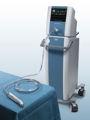
Last week, Dune Medical Devices, Inc., the company that developed the MarginProbeTM System, an intra-operative tissue assessment device to be used as accessory during lumpectomies of early-stage breast cancer, has received Premarket Approval (PMA) by the United States Food and Drug Administration.

MarginProbe system

marginProbe

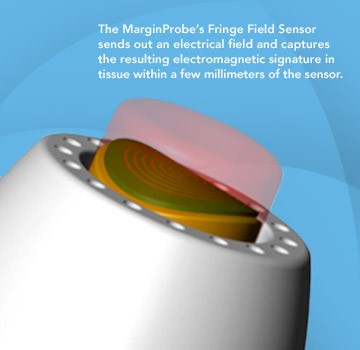
FDA approval of the MarginProbe System was based on a 664 patient prospective, multi-center, randomized, double arm study to evaluate the effectiveness of MarginProbe in identifying cancerous tissue along the margins of removed breast tissue during initial lumpectomy procedures. MarginProbe, which uses electromagnetic “signatures” to identify healthy and cancerous tissue, was found to be over three times more effective in finding cancer on the margin during lumpectomy, compared to traditional intra-operative imaging and palpation assessment. This enabled surgeons to significantly reduce the number of patients with positive margins following initial surgery.

The following publication gives an idea on the clinical performance of MarginProbe:

J Surg Res. 2010 May 15;160(2):277-81. doi: 10.1016/j.jss.2009.02.025. Epub 2009 Mar 31.

Diagnostic performance of a novel device for real-time margin assessment in lumpectomy specimens.

Pappo ISpector RSchindel AMorgenstern SSandbank JLeider LTSchneebaum SLelcuk SKarni T.

Source

Department of General Surgery, Assaf Harofeh Medical Center, Zrifin, Israel. pappo@zahav.net.il

Abstract

BACKGROUND:

Margin status in breast lumpectomy procedures is a prognostic factor for local recurrence and the need to obtain clear margins is often a cause for repeated surgical procedures. A recently developed device for real-time intraoperative margin assessment (MarginProbe; Dune Medical Devices, Caesarea, Israel), was clinically tested. The work presented here looks at the diagnostic performance of the device.

METHODS:

The device was applied to freshly excised lumpectomy and mastectomy specimens at specific tissue measurement sites. These measurement sites were accurately marked, cut out, and sent for histopathologic analysis. Device readings (positive or negative) were compared with histology findings (namely malignant, containing any microscopically detected tumor, or nonmalignant) on a per measurement site basis. The sensitivity and specificity of the device was computed for the full dataset and for additional relevant subgroups.

RESULTS:

A total of 869 tissue measurement sites were obtained from 76 patients, 753 were analyzed, of which 165 were cancerous and 588 were nonmalignant. Device performance on relatively homogeneous sites was: sensitivity 1.00 (95% CI: 0.85-1), specificity 0.87 (95% CI: 0.83-0.90). Performance for the full dataset was: sensitivity 0.70 (95% CI: 0.63-0.77), specificity 0.70 (95% CI: 0.67-0.74). Device sensitivity was estimated to change from 56% to 97% as the cancer feature size increased from 0.7 mm to 6.6 mm. Detection rate of samples containing pure DCIS clusters was not different from rates of samples containing IDC.

CONCLUSIONS:

The device has high sensitivity and specificity in distinguishing between normal and cancer tissue even down to small cancer features.

Copyright (c) 2010 Elsevier Inc. All rights reserved.

PMID: 19628225

Imagine how cost effective breast cancer management can be if it will involve systems such as these in addition to the systems I discussed in some of my previous posts, for example: What could transform an underdog into a winner?

Written by: Dror Nir, PhD.

Read Full Post »

Not applying evidence-based medicine drives up the costs of screening for breast-cancer in the USA

Author: Dror Nir


Costs for breast screening are being driven higher by increased use of new imaging technologies such as digital mammography and MRI, workflows incorporating 2nd and 3rd remote-readings as quality control measure, use of computer-aided detection (CAD) applications and growth in aged population.

According to recent publication in JAMA, 40% of the annual spending is for screening women ages 75 and older. Under existing guidelines routine screening is not recommended for this age group since “There is insufficient evidence to assess the benefits and harms of screening mammography”

The study population comprised women of 66 to 100 years old. “Forty-two percent of the women in the study were younger than age 75, and almost 60% of this group had one or more screening mammograms. Women ages 75 to 85 represented 40% of the total; 42% of this group had mammograms. Women 85 years and older represented 18% of the total; only 15% of this group had mammograms. Women with multiple chronic health conditions were much less likely to have a mammogram (26.6%) than healthy women (47.4%).”

“Abstract

The Cost of Breast Cancer Screening in the Medicare Population.

Cary P. Gross, MD; Jessica B. Long, MPH; Joseph S. Ross, MD, MHS; Maysa M. Abu-Khalaf, MD; Rong Wang, PhD; Brigid K. Killelea, MD, MPH; Heather T. Gold, PhD; Anees B. Chagpar, MD, MA, MPH, MSc; Xiaomei Ma, PhD

JAMA Intern Med. 2013;():1-7. doi:10.1001/jamainternmed.2013.1397. Published online January 7, 2013

Background  Little is known about the cost to Medicare of breast cancer screening or whether regional-level screening expenditures are associated with cancer stage at diagnosis or treatment costs, particularly because newer breast cancer screening technologies, like digital mammography and computer-aided detection (CAD), have diffused into the care of older women.

Methods Using the linked Surveillance, Epidemiology, and End Results–Medicare database, we identified 137 274 women ages 66 to 100 years who had not had breast cancer and assessed the cost to fee-for-service Medicare of breast cancer screening and workup during 2006 to 2007. For women who developed cancer, we calculated initial treatment cost. We then assessed screening-related cost at the Hospital Referral Region (HRR) level and evaluated the association between regional expenditures and workup test utilization, cancer incidence, and treatment costs.

Results In the United States, the annual costs to fee-for-service Medicare for breast cancer screening-related procedures (comprising screening plus workup) and treatment expenditures were $1.08 billion and $1.36 billion, respectively. For women 75 years or older, annual screening-related expenditures exceeded $410 million. Age-standardized screening-related cost per beneficiary varied more than 2-fold across regions (from $42 to $107 per beneficiary); digital screening mammography and CAD accounted for 65% of the difference in screening-related cost between HRRs in the highest and lowest quartiles of cost. Women residing in HRRs with high screening costs were more likely to be diagnosed as having early-stage cancer (incidence rate ratio, 1.78 [95% CI, 1.40-2.26]). There was no significant difference in the cost of initial cancer treatment per beneficiary between the highest and lowest screening cost HRRs ($151 vs $115; P = .20).

Conclusions The cost to Medicare of breast cancer screening exceeds $1 billion annually in the fee-for-service program. Regional variation is substantial and driven by the use of newer and more expensive technologies; it is unclear whether higher screening expenditures are achieving better breast cancer outcomes.”

The study is mainly addressing the difference in costs between different regions of referrals. It would be interesting to explore the situation in the age group of 40 to 66 years old.

Written by:  Dr. Dror Nir, PhD.

Read Full Post »

Whole-body imaging as cancer screening tool; answering an unmet clinical need?

Author: Dror Nir, PhD

Sometimes technologies that were developed to answer clinical needs in a certain area are migrated to perform in a totally inappropriate area. A good example which I discussed several times in my posts is PSA.

Cancer patients’ prognoses, strongly depend on accurate tumor staging. It is also a prerequisite for therapy choice and planning. Whole-body imaging is frequently used in patients with advanced malignant diseases including presence of metastases as these may occur in any anatomic region. It is important to note that classifying a patient as harboring a potentially advanced disease is based on biopsy results of Sentinel Lymph-Nodes and not on imaging. Moreover, referring a patient to a whole-body imaging is a choice of the treating practitioner! Clearly, when the choice of treatment includes administration of drugs, the type of drugs to be used is determined by the characteristics of the primary tumor.

To date, the use of whole-body imaging for post treatment follow-up can be considered as anecdotal.

The most-used technologies for whole-body imaging are computed tomography (CT), positron emission tomography (PET) and MRI. The performance of these systems in detection of cancer metastases of more than 1cm in diameter is very similar and in general quite good, dependent on the primary disease and the body locations of the metastases. Alas, each of these modalities has its strengths and weakness in different cancer and different body locations. Therefore, in the last decade, combined modalities such as PET-CT and recently PET/MRI were introduced. In some cases [1-6] these are reported to show sensitivity of more than 90%.

To demonstrate the level of information produced during whole-body imaging procedure here is an example (taken from Whole-body MRI and PET-CT in the management of cancer patients). This resource includes additional, educating examples:

330_2006_183_Fig1_HTML

Fig. 1

From multimodality to single-step examination. Restaging in a 29-year-old woman treated for breast cancer and with newly elevated tumor markers and bone pain. 1a, 1b Radiograms of the skeleton were normal, but bone scintigraphy showed a pathological tracer uptake in the right pubic bone (arrow). Abdominal ultrasound exhibited a suspicious mass. 1c, 1d CT revealed tumor recurrence in the right breast and confirmed hepatic metastasis. 2a, 2b T1-weighted whole-body MRI depicted a metastasis in the right pubic bone (circle). 2c, 2d HASTE images of the thorax showed the tumor recurrence in the right breast (arrow) and dynamic contrast enhanced studies of the abdomen unmasked the liver metastasis

Before addressing the issue of using whole-body imaging as a screening tool I would like to draw attention to existence of other methods for screening and post treatment follow-up of cancer patients; e.g. detecting levels of cancer-specific bio-markers in the blood or urine or, in case of advanced disease, detecting the level of tumor cells circulating in the blood as presented in: Circulating Tumor Cells versus Imaging—Predicting Overall Survival in Metastatic Breast Cancer by G. Thomas Budd et.al.

Abstract

Purpose: The presence of ≥5 circulating tumor cells (CTC) in 7.5 mL blood from patients with measurable metastatic breast cancer before and/or after initiation of therapy is associated with shorter progression-free and overall survival. In this report, we compared the use of CTCs to radiology for prediction of overall survival.

Experimental Design: One hundred thirty-eight metastatic breast cancer patients had imaging studies done before and a median of 10 weeks after the initiation of therapy. All scans were centrally reviewed by two independent radiologists using WHO criteria to determine radiologic response. CTC counts were determined ∼4 weeks after initiation of therapy. Specimens were analyzed at one of seven laboratories and reviewed by a central laboratory.

Results: Inter-reader variability for radiologic responses and CTC counts were 15.2% and 0.7%, respectively. The median overall survival of 13 (9%) patients with radiologic nonprogression and ≥5 CTCs was significantly shorter than that of the 83 (60%) patients with radiologic nonprogression and <5 CTCs (15.3 versus 26.9 months; P = 0.0389). The median overall survival of the 20 (14%) patients with radiologic progression and <5 CTCs was significantly longer than the 22 (16%) patients with ≥5 CTCs that showed progression by radiology (19.9 versus 6.4 months; P = 0.0039).

Conclusions: Assessment of CTCs is an earlier, more reproducible indication of disease status than current imaging methods. CTCs may be a superior surrogate end point, as they are highly reproducible and correlate better with overall survival than do changes determined by traditional radiology.

I would like first to present the following publication that could explain why people can easily be drawn why whole-body screening is an effective way to detect early cancers:

Enthusiasm for cancer screening in the United States by Schwartz LM, Woloshin S, Fowler FJ Jr, Welch HG SO, JAMA. 2004; 291(1):71.:

“ CONTEXT: Public health officials, physicians, and disease advocacy groups have worked hard to educate individuals living in the United States about the importance of cancer screening.

OBJECTIVE: To determine the public’s enthusiasm for early cancer detection.

DESIGN, SETTING, AND PARTICIPANTS: Survey using a national telephone interview of adults selected by random digit dialing, conducted from December 2001 through July 2002. Five hundred individuals participated (women aged>or =40 years and men aged>or =50 years; without a history of cancer).

MAIN OUTCOME MEASURES: Responses to a survey with 5 modules: a general screening module (eg, value of early detection, total-body computed tomography); and 4 screening test modules: Papanicolaou test; mammography; prostate-specific antigen (PSA) test; and sigmoidoscopy or colonoscopy.

RESULTS: Most adults (87%) believe routine cancer screening is almost always agood idea and that finding cancer early saves lives (74% said most or all the time). Less than one third believe that there will be a time when they will stop undergoing routine screening. A substantial proportion believe that an 80-year-old who chose not to be tested was irresponsible: ranging from 41% with regard to mammography to 32% for colonoscopy. Thirty-eight percent of respondents had experienced at least 1 false-positive screening test; more than 40% of these individuals characterized that experience as “very scary” or the “scariest time of my life.” Yet, looking back, 98% were glad they had had the initial screening test. Most had a strong desire to know about the presence of cancer regardless of its implications: two thirds said they would want to be tested for cancer even if nothing could be done; and 56% said they would want to be tested for what is sometimes termed pseudodisease (cancers growing so slowly that they would never cause problems during the person’s lifetime even if untreated). Seventy-three percent of respondents would prefer to receive a total-body computed tomographic scan instead of receiving 1000 dollars in cash.

CONCLUSIONS: The public is enthusiastic about cancer screening. This commitment is not dampened by false-positive test results or the possibility that testing could lead to unnecessary treatment. This enthusiasm creates an environment ripe for the premature diffusion of technologies such as total-body computed tomographic scanning, placing the public at risk of over testing and overtreatment.”

Whole-body screening is promoted as a one-stop shop for painlessly detecting hidden cancer and preventing cancer-related deaths. It is big business in the United States and in Canada where private clinics have begun offering full-body diagnostic procedures for a fee. The tests and procedures are often marketed to healthy people as a way to scan for hidden abnormalities or cancers, affording people the peace of mind that they are in good health [7 – 9].

When used in this manner, the evidence shows that whole-body cancer screening offers no proven health benefits and that it, in fact, exposes people to a number of unnecessary health risks. The problem I see is that the public is not exposed to “scientific publications” but is exposed to commercial ones!


References

  1. FDG PET and PET/CT: EANM procedure guidelines for tumor PET imaging: version 1.0
  2. Cancer of the Prostate, Testicles and Penis
  3. Gynecologic Cancers
  4. Malignant Melanoma
  5. Molecular Imaging in Cancer
  6. Pre-clinical whole-body fluorescence imaging: Review of instruments, methods and applications
  7. Full body CT scan for screening
  8. Screening for Cancer with PET and PET/CT: Potential and Limitations 
  9. http://www.privatescan.nl/total-bodyscan

Written by: Dror Nir, PhD

Read Full Post »

Breast Cancer: Genomic profiling to predict Survival: Combination of Histopathology and Gene Expression Analysis

Reporter: Aviva Lev-Ari, PhD, RN

Some assays that gauge cancer-related signatures can’t factor in tissue architecture, while other assessments that are good at gauging tissue architecture, provide mostly qualitative tumor data. To reconcile these differences, researchers led by Yinyin Yuan of Cancer Research UK decided to combine histopathological and gene expression analysis to show that quantitative image analysis of the cellular environment inside tumors can bolster the ability of genomic profiling to predict survival in breast cancer patients. This approach, too, though, has its limitations.

For instance, molecular assays that gauge cancer-related signatures are challenged by their inability to factor in tissue architecture and the results are confounded by genomic information from the different types of cells inside the tumor other than cancer cells. Meanwhile, traditional histopathological assessments are good at gauging tissue architecture and differentiating cellular heterogeneity, but mostly provide qualitative tumor data and are too time consuming to be applied in large-scale studies.

Recognizing these weaknesses, researchers led by Yinyin Yuan of Cancer Research UK decided to combine histopathological and gene expression analysis to show that quantitative image analysis of the cellular environment inside tumors can bolster the ability of genomic profiling to predict survival in breast cancer patients. “All technologies have some sort of weakness. That’s why when we combined two types of assays — image and microarray — we get a more reliable readout,” Yuan says.

As they report in Science Translational Medicine, Yuan and her colleagues gathered histopathological information from hematoxylin and eosin-stained images as well as gene expression and copy-number variation data on a discovery set of 323 samples and on a validation set of 241 samples from patients with estrogen receptor-negative breast cancer. Using the discovery sample set, the investigators developed an image-processing method to differentiate the cells inside tumor samples as cancerous, lymphocytic, or stromal. They then tested this technique on the validation sample.

Once Yuan and colleagues had an accurate picture of the types of cells in the tumor samples, they used image analysis to correct copy-number data — as it is influenced by cellular heterogeneity — and developed an algorithm to determine patients’ HER2 status better than copy-number analysis can.

Using the image-processing method, the researchers stratified the discovery and validation sample sets into lymphocytic infiltration-high and lymphocytic infiltration-low groups — as past studies have suggested that high lymphocytic infiltration is linked to better patient outcomes.

When the image analysis was compared to the pathological scores of the samples, the discovery set showed no difference in patient outcomes, but the assessments disagreed with regard to the outcomes of the lymphocytic infiltration-low group in the validation cohort.

Hypothesizing that integrating the gene expression signatures and quantitative image analysis would improve survival prediction, the study investigators combined them. “The gene expression classifier had 67 percent cross-validation accuracy in predicting disease-specific deaths, the image-based classifier had 75 percent, and the integrated classifier reached 86 percent,” the study authors write.

Finally, Yuan and her colleagues applied the image analysis to develop a quantitative score that determines whether specific types of cells are tightly clustered — a high score — or are randomly scattered — a low score. In stromal cells, this approach could discern that breast cancer patients with a high or low score had a “significantly better outcome” than patients whose scores fell in the medium range.

Ultimately, Yuan and her colleagues show that their image processing avoids the biases of manual pathological assessments and accurately quantifies cellular composition and tissue architecture not accounted for by molecular tests. The researchers’ computational approach is also faster than traditional pathological techniques. “These two sets of samples can be done in a day,” Yuan says.
According to the study authors, the limitation of the image processing technique is, of course, that it requires matched molecular and image data.

    Turna Ray is the editor of GenomeWeb’s Pharmacogenomics Reporter. She covers pharmacogenomics, personalized medicine, and companion diagnostics. E-mail her here or follow her GenomeWeb Twitter account at @PGxReporter.

Read Full Post »

Optical Coherent Tomography – emerging technology in cancer patient management

Author: Dror Nir, PhD

Optical Coherence Tomography (‘OCT’), is a technique for obtaining 3D optical-image from 1-2 mm thick tissue samples. The key benefits of using OCT for medical applications is that it is not ionizing, therefore harmless to the patient and the image resolution ~10µm, almost the size of a human cell.

Commercially available OCT systems have been used in ophthalmology and in interventional cardiology for quite a while. Recently, new applications attempting to provide intraoperative histopathology appear as alternative solution to frozen-sections based histopathology.

I found this nice diagram explaining why OCT can be used for identifying presence of cancer intra-operatively in an article [1] I downloaded from the website of the American association for cancer research.

Figure 1. Diagnostic scattering spectroscopy. In this cartoon illustration of the underlying principles of the method described in the work of Laughney and colleagues (1), an excised tissue sample sits on a glass plate, with normal cells depicted schematically on the left and tumor cells on the right. In contrast with the normal cells, the tumor cells are represented as having enlarged nuclei, with increased granularity of the chromatin, and, perhaps, a disrupted cytoskeleton resulting in cellular disorder. Light, from a broadband source, and after manipulation through an optical system, is collimated (parallel rays) and impinges on the tissue at normal incidence. While most light is scattered in the near-forward direction, some of the light is scattered directly backward after one (in most cases) or a few scattering events and is collected to be analyzed by a spectrometer (not depicted). The spectrometer provides a backscattering spectrum that is representative of the tissue properties. The wavelength dependence of the probability for backscattering varies with changes in the sizes and densities of the dominant scattering centers, such that the backscattered light spectrum changes with pathology, as manifested in the mean cellular micromorphology. In this way, the recorded spectrum relates directly to some of the micromorphology features that a pathologist recognizes as indicative of pathology when looking at histology slides, but in a quantitative manner, without the need to generate an actual microscopic image and without requiring subjective interpretation. (Of course, this physicist’s simplification ignores many tissue components that also affect scattering, such as extracellular matrix and vasculature.

In that article, Laughney and colleagues reported on real-time assessment of resection margins during breast- conserving surgery using OCT technique.

At the Thirty-Fifth Annual CTRC-AACR Breast Cancer Symposium held on Dec 4-8, 2012 in San Antonio, TX, BC Wilson, MK Akens, and CJ Niu (University Health Network, Ontario Cancer Institute, Toronto, ON, Canada; Tornado Medical System, Toronto, ON, Canada) discussed the unmet clinical need and the cost saving impact related to the potential use of Optical Coherence Tomography for intraoperative breast tumour margin width estimation in a poster session:

“Total mastectomy and lumpectomy with radiation have been shown to have equivalent patient outcomes, which has likely contributed to the more widespread adoption of breast conserving surgery (BCS) procedures. Assessment of breast lumpectomy margin widths in both an accurate and timely manner is essential to successful breast conservation procedures. Current BCS methodologies have been reported to result in reoperation rates of up to 20–60%, which represents a significant and unmet need for improved margin assessment. High reoperation rates present both increased treatment risk to patients and increased burden on healthcare systems. In the USA alone, over 150,000 lumpectomies are performed per year at an average cost between $11,000 and $19,000 USD per procedure. Assuming a relatively modest average repeat operation rate of 25%, potentially preventable repeat surgeries represent an approximate cost to the US healthcare system of $500M (USD) annually.”

From the same meeting: An abstract presenting the design of an ongoing multi-center, randomized, blinded clinical study aimed at Intraoperative assessment of tumor margins with a new optical imaging technology using a specific implementation of an OCT based device.

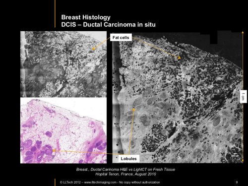
At last RSNA I have visited the French pavilion were one of the companies, LLTech, presented an OCT based system designed facilitate the work of pathologists and save time and money by allowing fresh tissue processing and pathology examination instead of the traditionally frozen sections (intraoperative) and fixated specimens (at lab)

fig2

That could be used by a pathologist to perform in-situ histology in the OR.

fig3

To summarize; OCT based systems seem to be something to look for in the future of cancer patients’ management.

References

1. Laughney AM, Krishnaswamy V, Rizzo EJ, Schwab EM, Barth RJ, et al. Scatter spectroscopic imaging distinguishes between breast pathologies in tissues relevant to surgical margin assessment. Clin Cancer Res. 2012;18:6315–25.

Writen by: Dror Nir, PhD

Read Full Post »

A perspective on where we are on carcinogenesis, cancer variability and predictors of time to recurrence and future behavior

Author: Larry H Bernstein, MD, FCAP

Predicting Tumor Response, Progression, and Time to Recurrence

Word Cloud by Daniel Menzin

I.     Background

In “Tumor Imaging and Targeting: Predicting Tumor Response to Treatment: Where we stand? “ ( Dec 13, 2012) Dr. Ritu Saxena  attempts to integrate three posts and to embed all comments made to all three papers, allowing the reader a critically thought compilation of evidence-based medicine and scientific discourse.

Dr. Dror Nir authored a post on October 16th titled “Knowing the tumor’s size and location, could we target treatment to THE ROI by applying imaging-guided intervention?” The article attracted over 20 comments from readers including researchers and oncologists debating the following issues:

imaging technologies in cancer

  • tumor size, and
  • tumor response to treatment.

The debate lead to several new posts authored by:

Dr. Bernstein’s (What can we expect of tumor therapeutic response),

Dr. Saxena, the Author of this post’s, (Judging ‘tumor response’-there is more food for thought) and

Dr. Lev-Ari’s post on Personalized Medicine: Cancer Cell Biology and Minimally Invasive Surgery (MIS) http://pharmaceuticalintelligence.com/2012/12/01/personalized-medicine-cancer-cell-biology-and-minimally-invasive-surgery-mis/#comment-5269

The post was a compilation of the views of authors representing different specialties including research and medicine. In medicine: Pathology, Oncology Surgery and Medical Imaging, are represented.

Dror Nir added a fresh discussion in “New clinical results supports Imaging-guidance for targeted prostate biopsy” based on a study of “Artemis”, a system that is adjunct to ultrasound and performs 3D Imaging and Navigation for Prostate Biopsy by Eigen (a complementary post to “Imaging-guided biopsies: Is there a preferred strategy to choose?”).

Image fusion is the process of combining multiple images from various sources into a single representative image. Ultrasound is the imaging modality used to guide Artemis in performing the biopsies. In this study MRI is used to overcome the “blindness” regarding tumor location.  This supports the detection reliability issue made in his “ Imaging-guided biopsies: Is there a preferred strategy to choose?” and  “Fundamental challenge in Prostate cancer screening.”

This makes the case that In the future, MRI-ultrasound fusion for lesion targeting is likely to result in fewer and more accurate prostate biopsies than the present use of systematic biopsies with ultrasound guidance alone.   Nevertheless, we haven’t completed the case for prediction of recurrence, even if we may eliminate the unnecessary consequences of radical prostatectomy.

Let’s look a little further. A discussion opens up more questions for discussion. I just read an interesting related article. The door has opened  wider.

II.               Novel technology to detect cancer in early stages

A. nanoparticles

Researchers have developed novel technology to detect the tumors in the body in early stages with the help of nanoparticles .( Nature Biotechnology).

Cancer cells produce many of the proteins that could be used as biomarkers to detect the cancer in the body but the amount of these proteins is not up to the mark or they may get diluted in the body of the patients making it nearly impossible to detect them in early stages.

This new technology has been developed by the researchers from MIT . Nanoparticles (brown) coated with peptides (blue) cleaved by enzymes (green) at the disease site. Peptides than come into the urine to be detected by mass spectrometry. (Credit: Justin H. Lo/MIT)

In this technology, nanoparticles will interact with the tumor proteins helping to make thousands of biomarkers secreted by the cancer cells. We had this ‘aha’ moment: What if you could deliver something that could amplify the signal?”

  • Scientists administered ‘synthetic biomarkers’ having peptides bonded to the nanoparticles and
  • the particles interact with the protease enzymes often found in large quantities in cancer cells

as they help them to cut the proteins normally holding the cells in place and to spread in other parts of the body.

Researchers found that the proteases break down hundreds of peptides from the nanoparticles and release them in the bloodstream. These peptides are then excreted in the urine, where the process of mass spectrometry could help to detect such peptides.

These “Synthetic biomarkers” perform three functions in vivo:

  1. they target sites of disease,
  2. sample dysregulated protease activities and
  3. emit mass-encoded reporters into host urine (for multiplexed detection by MS).

According to Bhatia, this biomarker amplification technology could also be used to manage the advancement of the disease and to check the response of the tumors to the drugs.

Reference:

Kwong, G., von Maltzahn, G., Murugappan, G., Abudayyeh, O., Mo, S., Papayannopoulos, I., Sverdlov, D., Liu, S., Warren, A., Popov, Y., Schuppan, D., & Bhatia, S. (2012). Mass-encoded synthetic biomarkers for multiplexed urinary monitoring of disease Nature Biotechnology  http://dx.doi.org:/10.1038/nbt.2464

IIB    Synthetic Nucleosides

J Gong and SJ Sturla published “A Synthetic Nucleoside Probe that Discerns a DNA Adduct from Unmodified DNA” in JACS Communications on web 4/03/2007).  They state that biologically reactive chemicals alkylate DNA and induce structural modifications in the form of covalent adducts that can persist, escape repair, and serve as templates for polymerase-mediated DNA synthesis. Therefore, correlating chemical structures and quantitative levels of adducts with toxicity is essential for targeting specific agents to carcinogenesis.

  • DNA adducts are formed at exceedingly low levels.
  • Minor lesions may have greater biological impact than more abundant products.
  • New molecular approaches for addressing specific low-abundance adducts are needed

They describe the first example of a synthetic nucleoside that may serve as the chemical basis for a probe of a bulky carcinogen-DNA adduct

IIC.  MicroRNAs caused by DNA methylation

Another molecular approach “ A microRNA DNA methylation signature for human cancer metastasis” was published in PNAS [2008;105(36):13556-13561)] by A Lujambio , Calin GA, Villanueva A et al.

Different sets of miRNAs are usually deregulated in different cancers, and some miRNAs are aberrantly methylated and silenced, causing tumorigenesis. The authors

  • identified aberrantly methylated and silenced miRNAs that are cancer-specific
  • using miRNA microarray techniques.

Functional analyses for the selected genes proved that these miRNAs act on C-MYC, E2F3, CDK6 and TGIF2, resulting in metastasis through aberrant methylation of the miRNAs. The authors suggest that these may be applicable to advance research in the clinical setting.

III.              New methods require advanced mathematical prediction methods

A.  First Case …ProsVue PSA

One of the most elegant papers I have seen in several years  has been published in Clinical Biochemistry (CLB–12-00159), by Mark J. Sarnoa1 and Charles S. Davis2. [1Vision Biotechnology Consulting, 19833 Fortuna Del Este Road, Escondido, CA 92029, USA (mjsarno@att.net), 2CSD Biostatistics, Inc., San Diego, CA, 4860 Barlows Landing Cove, San Diego, CA 92130, USA (chuck@csdbiostat.com)]

Robustness of ProsVue™ linear slope for prediction of prostate cancer recurrence: Simulation studies on effects of analytical imprecision and sampling time variation.
Keywords: ProsVue, slope, prostate cancer, random variates.
Financial support for the investigation was provided by Iris Molecular Diagnostics

Abstract: Objective: The ProsVue assay measures

  • serum total prostate-specific antigen (PSA) over three time points post-radical prostatectomy and
  • calculates rate of change expressed as linear slope. Slopes ≤2.0 pg/ml/month are associated with reduced risk for prostate cancer recurrence.

However, an indicator based on measurement at multiple time points, calculation of slope, and relation of slope to a binary cutpoint may be subject to effects of analytical imprecision and sampling time variation.

They performed simulation studies to determine the presence and magnitude of such effects.

Design and Methods: Using data from a two-site precision study and a multicenter retrospective clinical trial of 304 men, they carried out simulation studies to assess whether analytical imprecision and sampling time variation can drive misclassification of patients with stable disease or classification switching for patients with clinical recurrence.

Results:

  • Analytical imprecision related to expected PSA values in a stable disease population results in ≤1.2% misclassifications.
  • For recurrent populations, an analysis taking into account correlation between sampling time points demonstrated that classification switching across the 2.0 pg/ml/month cutpoint occurs at a rate ≤11%.
  • Lastly, sampling time variation across a wide range of scenarios results in 99.7% retention of proper classification for stable disease patients with linear slopes up to the 75th percentile of the distribution.

Conclusions:

  • These results demonstrate the robustness of the ProsVue assay and the linear slope indicator.
  • Further, these simulation studies provide a potential framework for evaluation of future assays that may rely on the rate of change principle

The ProsVue Assay has been cleared for commercial use by the US Food and Drug Administration (FDA) as “a prognostic marker in conjunction with clinical evaluation as an aid in

  • identifying those patients at reduced risk for recurrence of prostate cancer for the eight year period following prostatectomy.”

The assay measures

  1. serum total prostate specific antigen (PSA) in post-RP samples and
  2. calculates rate of change of PSA over the sampling period,

expressing the outcome as linear slope. The assay is novel in at least a few respects.

  • the assay is optimized to identify patients at reduced risk for recurrence.

In order to demonstrate efficacy for this indication, the assay employs the immuno-polymerase chain reaction (immuno-PCR) to achieve sensitivity

  • an order of magnitude lower than existing “ultrasensitive” PSA assays.

The improved sensitivity allows quantification of PSA at levels exhibited in stable disease (<5 pg/ml), which have been historically below the

measurement range of ultrasensitive assays.

Secondly, the assay is the first to receive clearance based on

  • linear slope of tumor marker concentration versus time post-surgery.
  • Specifically, PSA is measured in three samples taken between 1.5 and 20 months post-RP and
  • the slope calculated using simple least squares regression.
  • The calculated slope is compared to a threshold of 2.0 pg/ml/month with values at or below the threshold associated with reduced risk for PCa recurrence.

Does analytical imprecision present a potential risk for misclassification by driving errors in the calculated slope that result in classification switching?  Since excursions of precision can occur as point sources in single sampling points or in cumulative effect from the three sampling points, the question is worthy of consideration. They carried out studies

  • to address these questions specific to ProsVue and also
  • provide a potential framework for evaluation of future assays.
  • Similarly, does variation in the time at which samples are taken drive errors resulting in classification switching?

Both questions require evaluating the robustness of the ProsVue Assay and are properly presented for clinical chemists and physicians evaluating use of the assay in clinical practice. Furthermore, since future diagnostic assays may employ the rate of change principle, it is important to develop statistical methods to evaluate effects of variation.

The point is that more sophisticated methods are needed to measure scarce analytes associated with risk for eventual clinical events.

  • Accurate measurement at post-RP levels to identify patients with reduced risk of recurrence represents a new development.
  • Furthermore, measurement of PSA at multiple time points and calculation of rate of change using linear regression extends application of the analyte markedly beyond traditional use.

Such use presents certain questions of variation effects.

Their results indicate that analytical imprecision in the range of concentrations exhibited in patients at reduced risk for recurrence (the focus of the assay) presents no significant risk of misclassification.

  • Classification switching in this population occurs at a frequency of ≤1.2%.
  • Slopes for recurrent patients and clinical classification are substantively insensitive to analytical variation even in a subpopulation of recurrent patients with slowly rising PSA values.
  • Sampling time variation negligibly affects clinical classification for stable disease patients with slopes at and below the 75th percentile.

Table 1. Side-effects and effects on recovery ...

Table 1. Side-effects and effects on recovery of treatments for newly diagnosed prostate cancer. The Prostate Brachytherapy Advisory Group: http://www.prostatebrachytherapyinfo.net (Photo credit: Wikipedia)

_____________________________________________________________________________________________________

IIIB. Other interesting developments are going to need further development and validation.

For instance, research has been published online in the journal Cancer Cell, reports a cellular component that is involved in mobility of cancer to other body parts and inhibition of which could increase the tumor formation. These investigators worked on various animal models including chicken, zebrafish and mouse, and patient samples and have found a cellular component; Prrx1 that stops the cancer cells from staying in organs.  Epithelial-mesenchymal transition (EMT) is the process that is required by the cancer cells to spread to other organs. This process helps the cells to become mobile and move with the bloodstream. These cells must lose their mobility before attaching to other body parts.

In the final analysis the cells have to lose the component Prrx1 to lose mobility and to become stationary. Researchers wrote, “Prrx1 loss reverts EMT & induces stemness, both required for metastatic colonization.”  Consequently,  Prrx1 has to be turned off for these cells to group together to form other tumours.” It has been found that the tumors with elevated levels of Prrx1 cannot form new tumors.

IIIC.  PXR and AhR Nuclear Receptor Activation

  • The primary mechanism of cytochrome P450 induction is via increased gene transcription which typically occurs through nuclear receptor activation.
  • The most common nuclear receptors involved in the induction of drug metabolizing enzymes include the pregnane X receptor (PXR), the aryl hydrocarbon receptor (AhR), and the constitutive androstane receptor (CAR) which are known to regulate CYP3A4, CYP1A2 and CYP2B6, respectively.
  • An industry survey of current practices and recommendations (Chu et al., (2009) Drug Metab Dispos 37: 1339-1354) indicates 64% of survey respondents routinely use nuclear receptor transactivation assays to assess the potential of test compounds to cause enzyme induction
  • ‘Because reporter assays are relatively high throughput and cost effective, they can be a valuable tool in drug discovery.’(Chu V, Einolf HJ, Evers R, Kumar G, et.  (2009) Drug Metab Dispos 37; 1339-1354)
  • Luciferase reporter gene assay

No cytotoxicity was observed for any of six compounds at the concentration range tested with the exception of troglitazone for which cytotoxicity was observed at the highest concentration of 50μM.  This data point was excluded in this instance and not used for calculating the Emax or EC50.

In brief, CAR and PXR regulate distinct but overlapping sets of target genes, which include certain phase 1 P450 enzymes (e.g., CYP2B, CYP3A, and CYP2C), phase II conjugation enzymes such as UDP glucuronosyltransferase UGT1A1 and sulfotransferase SULT2A, and phase III transporters such as P-glycoprotein (MDR-1). The AhR receptor has been shown to regulate the expression of CYP1A.

Will this be combined with the other methods for drug selection and prediction of drug free survival?

I have mentioned an improved molecular assay of PSA at the pcg/ml level that is approved for use with an acceptable linear prediction of survival for 8 years post radical prostatectomy.  Then there is a report of a method of measuring nanoparticles in urine, to amplify the signal detected by mass spectrometry. This new technology has been developed by the researchers from MIT and led by Sangeeta Bhatia at MIT. (Novel technology to detect cancer in early stages, Nature Biotechnology, Dec 16, 2012).  There is still another recent report about using gene expression profiles to predict breast cancer, and a number of articles have shown variability in breast cancer types.   I view with reservations until I can see long term predictions of prognosis.

IIID.  Prediction of Breast Cancer Metastasis by Gene Expression Profiles

The report in Cancer Informatics (http://www.la-press.com; open access)  by M Burton, M Thomassen, Q Tan, and TA Kruse is “Prediction of Breast Cancer Metastasis by Gene Expression Profiles: A Comparison of Metagenes and Single Genes.”   The authors state “The diversity of microarray platforms has made the full validation of gene expression  profiles across studies difficult and, the classification accuracies are rarely validated in multiple independent datasets. The individual genes between such lists may not match, but genes with comparable function are included across gene lists. However,  genes can be grouped together as metagenes (MGs) based on common characteristics such as pathways, regulation, or genomic location. Such MGs might be used as features in building a predictive model applicable for classifying independent data.”

Microarray gene expression analysis has in several previous studies been applied to elucidate the relation between clinical outcome and gene expression patterns in breast cancer and has demonstrated improvement of recurrence prediction. In some studies, genes in such profiles might be fully or partially missing in the test data used for validation due to the choice of microarray platform or the presence of missing values associated with a given probe.

To overcome the obstacles, these authors propose that individual genes could be considered part of a larger network such that their expression being controlled by the expression level of other genes or that a group of genes belong to a specific pathway performing a well-defined task. These genes may be controlled by the same transcription factor or located in the same chromosomal region. In fact these groupings have been collected in public databases (the Kyoto Encyclopedia of Genes and Genomes (KEGG), the Molecular Signature Database (MsigDB), the Gene Ontology database (GO)). This could be upregulation or deregulation of pathways associated with metastasis. Metastasis progressionas well as tumor grading (in breast cancer) are associated with accumulated mutations in several genes, leading to amplification or inactivation of genes.

Several studies have defined metagene/gene modules derived from microarray data using various methods such as penalized matrix decomposition which clusters similar genes but without similar expression profiles – hierarchical clustering, correlation, or combining a priori protein-protein interactions with microarray gene expression data defining interaction networks as features. Few studies have attempted to use such predefined gene sets for prediction models.

Their study compared the performance of either metagene- or single gene-based feature sets and classifiers using random forest and two support vector machines for classifier building. The performance within the same dataset, feature set validation performance, and validation performance of entire classifiers in strictly independent datasets were assessed by 10 times repeated 10-fold cross validation, leave-one-out cross validation, and one-fold validation, respectively. To test the significance of the performance difference between MG- and SG-features/classifiers, we used a repeated down-sampled binomial test approach.

They found MG- and SG-feature sets are transferable and perform well for training and testing prediction of metastasis outcome in strictly independent data sets, both between different and within similar microarray platforms.  Further, The study showed that MG- and SG-feature sets perform equally well in classifying independent data. Furthermore, SG-classifiers significantly outperformed MG-classifier when validation is conducted between datasets using similar platforms, while no significant performance difference was found when validation was performed between different platforms.

  • The MG- and SG-classifiers had similar performance when conducting classifier validation in independent data based on a different microarray platform.
  • The latter was also true when only validating sets of MG- and SG-features in independent datasets, both between and within similar and different platforms.

This study appears to be unique in the same way that the PCa prediction study is unique in that genome-based expression patterns are used to classify and predict metastatic potential.

These studies have the potential to materialize into practice changing behavior.

IIIE. Colon Cancer and Treatment Recurrence

Cancer scientists led by Dr. John Dick at the Princess Margaret Cancer Centre have found a way to follow single tumour cells and observe their growth over time. By using special immune-deficient mice to propagate human colorectal cancer, they found that genetic mutations, regarded by many as the chief suspect driving cancer growth, are only one piece of the puzzle. The team discovered that biological factors and cell behaviour — not only genes — drive tumour growth, contributing to therapy failure and relapse. The findings are published December 13 online ahead of print in Science, are “a major conceptual advance in understanding tumour growth and treatment response” according to Dr. Dick.

[1] only some cancer cells are responsible for keeping the cancer growing.

[2] these kept the cancer growing for long time periods (up to 500 days of repeated tumour transplantation)

[3] a class of propagating cancer cells that could lie dormant before being activated.

[4] the mutated cancer genes were identical for all of these different cell behaviours.

[5] given chemotherapy the long-term propagating cells were generally sensitive to treatment, but dormant cells were not killed by drug treatment.

[6] these became activated andpropagated new tumour.

IV. Related References

Diagnostic efficiency of carcinoembryonic antigen and CA125 in the cytological evaluation of effusions.
M M Pinto, L H Bernstein, R A Rudolph, D A Brogan, M Rosman
Arch Pathol Lab Med 1992; 116(6):626-631 ; ICID: 825503

Medically significant concentrations of prostate-specific antigen in serum assessed.
L H Bernstein, R A Rudolph, M M Pinto, N Viner, H Zuckerman
Clin Chem 1990; 36(3):515-518 ; ICID: 825497

Entropy and Information Content of Laboratory Test Results
R T Vollmer
Am J Clin Pathol.  2007;127(1):60-65.

Abstract

This article introduces the use of information theoretic concepts such as entropy, S, for the evaluation of laboratory test results, and it offers a new measure of information, 1 – S,
which tells us just how far toward certainty a laboratory test result can predict a binary outcome. The derived method is applied to the serum markers troponin I and
prostate-specific antigen and to histologic grading of HER-2/neu staining, to cytologic diagnosis of cervical specimens, and to the measurement of tumor thickness in malignant
melanoma. Not only do the graphic results provide insight for these tests, they also validate prior conclusions. Thus, this information theoretic approach shows promise for
evaluating and understanding laboratory test results.

A map of protein-protein interactions involving calmodulin. Protein-protein interactions are both numerous and incredibly complex, and they can be mapped using the Database of
Interacting Proteins (DIP). This image depicts a DIP map for the protein calmodulin. The interactions with the most confidence are drawn with wider connecting lines. This diagram
highlights one level of complexity involved in understanding the downstream effects of gene regulation and expression.

Related article

Read Full Post »

Author: Tilda Barliya PhD

Metastasis, the spread of cancer cells from a primary tumour to seed secondary tumours in distant sites, is one of the greatest challenges in cancer treatment today. For many patients, by the time cancer is detected, metastasis  has already occurred. Over 80% of patients diagnosed  with lung cancer, for example, present with metastatic  disease. Few patients with metastatic cancer are cured by surgical intervention, and other treatment modalities are limited. Across all cancer types, only one in five patients diagnosed with metastatic cancer will survive more than 5 years. (1,2).

Metastatic Cancer 

  • Metastatic cancer is cancer that has spread from the place where it first started to another place in the body.
  • Metastatic cancer has the same name and same type of cancer cells as the original cancer.
  • The most common sites of cancer metastasis are the lungs, bones, and liver.
  • Treatment for metastatic cancer usually depends on the type of cancer and the size, location, and number of metastatic tumors.

How do cancer cells spread (3)

  • Local invasion: Cancer cells invade nearby normal tissue.
  • Intravasation: Cancer cells invade and move through the walls of nearby lymph vessels or blood vessels.
  • Circulation: Cancer cells move through the lymphatic system and the bloodstream to other parts of the body.

The ability of a cancer cell to metastasize successfully depends on its individual properties; the properties of the noncancerous cells, including immune system cells, present at the original location; and the properties of the cells it encounters in the lymphatic system or the bloodstream and at the final destination in another part of the body. Not all cancer cells, by themselves, have the ability to metastasize. In addition, the noncancerous cells at the original location may be able to block cancer cell metastasis. Furthermore, successfully reaching another location in the body does not guarantee that a metastatic tumor will form. Metastatic cancer cells can lie dormant (not grow) at a distant site for many years before they begin to grow again, if at all.

Although cancer therapies are improving, many drugs are not reaching the sites of metastases, and doubt  remains over the efficacy of those that do. Methods  that are effective for treating large, well-vascularized tumours may be inadequate when dealing with small clusters of disseminated malignant cells.

We expect that the expanding capabilities of nanotechnology, especially in targeting, detection and particle trafficking, will enable  novel approaches to treat cancers even after metastatic dissemination.


Lymph nodes, which are linked by lymphatic vessels, are distributed throughout the body and have an integral role in the immune response. Dissemination of cancer cells through the lymph network is thought to be an important route for metastatic spread. Tumor proximal lymph nodes are often the first site of metastases, and the presence of lymph node metastases signifies further metastatic spread and poor patient survival.

As such, lymph nodes have been targeted using cell-based nanotechnologies

Lymph nodes are small, bean-shaped organs that act as filters along the lymph fluid channels. As lymph fluid leaves the organ (such as breast, lung etc) and eventually goes back into the bloodstream, the lymph nodes try to catch and trap cancer cells before they reach other parts of the body. Having cancer cells in the lymph nodes suggests an increased risk of the cancer spreading. It is thus very important to evaluate the involvement of lymph nodes when choosing the best possible treatment for the patient.

Although current mapping methods are available such as CT and MRI scans, PET scan, Endobronchial Ultrasound, Mediastinoscopy and lymph node biopsy, sentinel lymph node (SLN) mapping and nodal treatment in lung cancer remain inadequate for routine clinical use. 

Certain characteristics are associated with preferential (but not exclusive) nanoparticle trafficking to lymph nodes following intravenous administration.

Targeting is often an indirect process, as receptors on the surface of leukocytes bind nanoparticles and transfer them to lymph nodes as part of a normal immune response. Several strategies have been used to enhance nanoparticle uptake by leukocytes in circulation. Coating iron-oxide nanoparticles with carbohydrates, such as dextran, results in the increased accumulation of these nanoparticles in lymph nodes. Conjugating peptides and antibodies, such as immunoglobulin G (IgG), to the particle surface also increases their accumulation in the lymphatic network. In general, negatively charged particles are taken up at faster rates than positively charged or uncharged particles. Conversely, ‘stealth’ polymers, such as polyethylene glycol (PEG), on the surface of nanoparticles, can inhibit uptake by leukocytes, thereby reducing accumulation in the lymph nodes.

Lymph node targeting may be achieved by other routes of administration. Tsuda and co-workers reported that non-cationic particles with a size range of 6–34nm, when introduced to the lungs (intrapulmonary administration), are trafficked rapidly (<1 hour) to local lymph nodes. Administering particles <80 nm in size subcutaneously also results in trafficking to lymph nodes. Interestingly, some studies have indicated that non-pegylated particles exhibit enhanced accumulation in the lymphatics and that pegylated particles tend to appear in the circulation several hours after administration.

Over the last twenty years, sentinel lymph node (SLN) imaging has revolutionized the treatment of several malignancies, such has melanoma and breast cancer, and has the potential to drastically improve treatment in other malignancies, including lung cancer. Several attempts at developing an easy, reliable, and effective method for SLN mapping in lung cancer have been unsuccessful due to unique difficulties inherent to the lung and to operating in the thoracic cavity.

An inexpensive method offering rapid, intraoperative identification of SLNs, with minimal risk to both patient and provider, would allow for improved staging in patients. This, in turn, would permit better selection of patients for adjuvant therapy, thus reducing morbidity in those patients for whom adjuvant treatment is inappropriate, and ensuring that those who need this added therapy actually receive it. (http://www.ncbi.nlm.nih.gov/pmc/articles/PMC3109504/)

Current methods for SLN identification involve the use of radioactivity-guided mapping with technetium-99m sulfur colloid and/or visual mapping using vital blue dyes. Unfortunately these methods can be inadequate for SLN mapping in non-small cell lung cancer (NSCLC) The use of vital blue dyes is limited in vivo by poor visibility, particularly in the presence of anthracotic mediastinal nodes, thereby decreasing the signal-to-background ratio (SBR) that enables nodal detection. Similarly, results with technetium-99m sulfur colloid have been mixed when used in the thoracic cavity, where hilar structures and aberrant patterns of lymphatic drainage make detection more difficult.

Although Nomori et al. have reported an 83% nodal identification rate following a preoperative injection of technetium-99 colloid, there is an associated increased risk of pneumothorax and bleeding with this method. Further, the recently completed CALGB 140203 multicenter Phase 2 trial investigating the use of intraoperative technetium-99m colloid found an identification rate of only 51% with this technique.  Clearly a technology with greater accuracy, improved SBR, and less potential risk to surgeon and patient would be welcome in the field of thoracic oncology.

Near-infrared (NIR) fluorescence imaging has the potential to meet this difficult challenge.

Near-Infrared Light

NIR light is defined as that within the wavelength range of 700 to 1000 nm. Although NIR light is invisible to the naked eye, it can be thought of as “redder” than UV and visible light.

  • Absorption, scatter, and autofluorescence are all significantly reduced at redder wavelengths. For instance, Hemoglobin, water, lipids, and other endogenous chromophores, such as melanin, have their lowest absorption within the NIR spectrum, which permits increased photon depth penetration into tissues
  • In addition, imaging can also be affected by photon scatter, which describes the reflection and/or deflection of light when it interacts with tissue. Scatter, on an absolute scale, is often ten-times higher than absorption. However, the two major types of scatter, Mie and Rayleigh, are both reduced in the NIR, making the use of NIR wavelengths especially important for the reduction of photon attenuation.
  • living tissue has extremely high “autofluorescence” in the UV and visible wavelength ranges due to endogenous fluorophores, such as NADH and the porphyrins. Therefore, UV/visible fluorescence imaging of the intestines, bladder, and gallbladder is essentially precluded. However, in the NIR spectrum, autofluorescence is extremely low, providing the black imaging background necessary for optimal detection of a NIR fluorophore within the surgical field
  • Additionally, optical imaging techniques, such as NIR fluorescence, eliminate the need for ionizing radiation. This, combined with the availability of a NIR fluorophore already FDA-approved for other indications and having extremely low toxicity (discussed below), make this a potentially safe imaging modality.

The main disadvantage is that it’s invisible to the human eye, requiring special imaging-systems to “see” the NIR fluorescence.

Currently there are three intraoperative NIR imaging systems in various stages of development:

  • The SPY system (Novadaq, Canada) – utilizes laser light excitation in order to obtain fluorescent images. The Spy system has been studied for imaging patency of vascular anastamoses following CABG and organ transplantation
  • The Photodynamic Eye(Hamamatsu, Japan) – is presently available only in Japan
  • The Fluorescence-Assisted Resection and Exploration (FLARE) system ()- developed by the authors’ laboratory utilizes NIR light-emitting diode (LED) excitation, eliminating the need for a potentially harmful laser. Additionally, the FLAREsystem has the advantage of being able to provide simultaneous color imaging, NIR fluorescence imaging, and color-NIR merged images, allowing the surgeon to simultaneously visualize invisible NIR fluorescence images within the context of surgical anatomy.

Near-Infrared Fluorescent Nanoparticle Contrast Agents

The ideal contrast agent for SLN mapping would be anionic and within 10–50 nm in size in order to facilitate rapid uptake into lymphatic vessels with optimal retention within the SLN.

Due to the lack of endogenous NIR tissue fluorescence, exogenous contrast agents must be administered for in vivo studies. The most important contrast agents that emit within the NIR spectrum are the heptamethine cyanines fluorophores, of which indocyanine green (ICG) is the most widely used, and fluorescent semiconductor nanocrystals, also known as quantum dots (QDs).

  • ICG is an extremely safe NIR fluorophore, with its only known toxicity being rare anaphylaxis. The dye was FDA approved in 1958 for systemic administration for indicator-dilution studies including measurements of cardiac output and hepatic function. Additionally, it is commonly used in ophthalmic angiography. When given intravenously, ICG is rapidly bound to plasma albumin and cleared from the blood via the biliary system. Peak absorption and emission of ICG occur at 780 nm and 830 nm respectively, within the window where in vivo tissue absorption is at its minimum. ICG has a relatively neutral charge, has a hydrodynamic diameter of only 1.2 nm, and is relatively hydrophobic. Unfortunately, this results in rapid transport out of the SLN and relatively low fluorescence yield, thereby decreasing its efficacy in mapping techniques. However, noncovalent adsorption of ICG to human serum albumin (HSA), as occurs within plasma, results in an anionic nanoparticle with a diameter of 7.3 nm and a three-fold increase in fluorescence yield markedly improving its utility in SLN mapping.
  • QDs consist of an inorganic heavy metal core and shell which emit within the NIR spectrum. This structure is then surrounded by a hydrophilic organic coating which facilitates aqeuous solubility and lymphatic distrubtion. QDs have been extensively studied and are ideal for SLN mapping as their hydrodynamic diameter can be customized to the appropriate size within a narrow distribution (15–20 nm), they can be engineered to have an anionic surface charge, and exhibit an extremely high SBRs with significant photostability. Unfortunately, safety concerns due to the presence of heavy metals within the QDs so far have precluded clinical application

Human Clinical Trials and NIR SLN mapping

Several studies have investigated the clinical use of indocyanine green without adsorption to HSA for NIR fluorescence-guided SLN mapping in breast and gastric cancer with good success (9-13).

Kitai et al. first examined this technique in 2005 in breast cancer patients, and was able to identify a SLN node in 17 of 18 patients using NIR fluorescence rather than the visible green color of ICG (9). Sevick-Muraca et al. reported similar results using significantly lower microdoses of ICG (10 – 100 μg), successfully identifying the SLN in 8 of 9 patients (11). Similar to these subcutaneous studies, 56 patients with gastric cancer underwent endoscopic ICG injection into the submucosa around the tumor 1 to 3 days preoperatively or injection directly into the subserosa intraoperatively with identification of the SLN in 54 patients (13).

Recently, Troyan et al. have completed a pilot phase I clinical trial examining the utility of NIR imaging the ICG:HSA nanoparticle fluorophore for SLN mapping/biopsy in breast cancer using the FLAREsystem. In this study, 6 patients received both 99mTc-sulfur colloid lymphoscintigraphy along with ICG:HSA at micromolar doses. SLNs were identified in all patients using both methods. In 4 of 6 patients the SLNs identified were the same, while in the remaining two, lymphoscintigraphy identified an additional node in one patient and ICG:HSA identified an additional SLN in the other. Irrespective, this study demonstrates that NIR SLN mapping with low dose ICG:HSA is a viable method for intraoperative SLN identification.

Nanotechnology and Drug Delivery in Lung cancer

We previously explored Lung cancer and nanotechnology aspects as polymer nanotechnology has been an area of significant research over the past decade as polymer nanoparticle drug delivery systems offer several advantages over traditional methods of chemotherapy delivery

see: (15) http://pharmaceuticalintelligence.com/2012/11/08/lung-cancer-nsclc-drug-administration-and-nanotechnology/                (16) http://pharmaceuticalintelligence.com/2012/12/01/diagnosing-lung-cancer-in-exhaled-breath-using-gold-nanoparticles/

As the importance of micrometastatic lymphatic spread of tumor becomes clearer, there has been much interest in the use of nanoparticles for lymphatic drug delivery. The considerable focus on developing an effective method for SLN mapping for lung cancer is indicative of the importance of nodal spread on overall survival.

Our lab is investigating the use of image-guided nanoparticles engineered for lymphatic drug delivery. We have previously described the synthesis of novel, pH-responsive methacrylate nanoparticle systems (14). Following a simple subcutaneous injection of NIR fluorophore-labeled nanoparticles 70 nm in size, we have shown that we can deliver paclitaxel loaded within the particles to regional draining lymph nodes in several organ systems of Yorkshire pigs while simultaneously confirming nodal migration using NIR fluorescent light. Future studies will need to investigate the ability of nanoparticles to treat and prevent nodal metastases in animal cancer models. Additionally, the development of tumor specific nanoparticles will potentially allow for targeting of chemotherapy to small groups of metastatic tumor cells further limiting systemic toxicities by narrowing the delivery of cytotoxic drugs.

Ref:

1. http://www.nature.com.rproxy.tau.ac.il/nrc/journal/v12/n1/pdf/nrc3180.pdf

2. http://www.nature.com/nrc/focus/metastasis/index.html

3. http://www.cancer.gov/cancertopics/factsheet/Sites-Types/metastatic

4. http://www.cancerresearchuk.org/cancer-help/about-cancer/what-is-cancer/body/the-lymphatic-system

5. http://www.macmillan.org.uk/Cancerinformation/Cancertypes/Lymphnodessecondary/Secondarycancerlymphnodes.aspx

6. Khullar O, Frangioni JV and Colson YL. Image-Guided Sentinel Lymph Node Mapping and Nanotechnology-Based Nodal Treatment in Lung Cancer using Invisible Near-Infrared Fluorescent Light. Semi Thorac Cardiovasc Surg 2009 :21 (4);  309-315. http://www.ncbi.nlm.nih.gov/pmc/articles/PMC3109504/

7. Stacker SA, Achen MG, Jussila L,  Baldwin ME and Alitalo K. Metastasis: Lymphangiogenesis and cancer metastasis.  Nature Reviews Cancer 2002 2, 573-583. http://www.nature.com/nrc/journal/v2/n8/full/nrc863.html

8. Schroeder A., Heller DA., Winslow MM., Dahlman JE., Pratt GW., Langer R., Jacks T and Anderson DG.. Nature Reviews Cancer 2012; 12(1), 39-50. Treating metastatic cancer with nanotechnology. http://www.nature.com.rproxy.tau.ac.il/nrc/journal/v12/n1/pdf/nrc3180.pdf

http://www.nature.com.rproxy.tau.ac.il/nrc/journal/v12/n1/full/nrc3180.html

9. Kitai T, Inomoto T, Miwa M, et al. Fluorescence navigation with indocyanine green for detecting sentinel lymph nodes in breast cancer. Breast Cancer. 2005;12:211–215.

10. Ogasawara Y, Ikeda H, Takahashi M, et al. Evaluation of breast lymphatic pathways with indocyanine green fluorescence imaging in patients with breast cancer. World journal of surgery.2008;32:1924–1929.

11. Sevick-Muraca EM, Sharma R, Rasmussen JC, et al. Imaging of lymph flow in breast cancer patients after microdose administration of a near-infrared fluorophore: feasibility study. Radiology.2008;246:734–741.

12. Miyashiro I, Miyoshi N, Hiratsuka M, et al. Detection of sentinel node in gastric cancer surgery by indocyanine green fluorescence imaging: comparison with infrared imaging. Ann Surg Oncol.2008;15:1640–1643.

13. Tajima Y, Yamazaki K, Masuda Y, et al. Sentinel node mapping guided by indocyanine green fluorescence imaging in gastric cancer. Ann Surg. 2009;249:58–62.

14. Griset AP, Walpole J, Liu R, et al. Expansile nanoparticles: synthesis, characterization, and in vivo efficacy of an acid-responsive drug delivery system. J Am Chem Soc. 2009;131:2469–2471

15. http://pharmaceuticalintelligence.com/2012/11/08/lung-cancer-nsclc-drug-administration-and-nanotechnology/

16.  http://pharmaceuticalintelligence.com/2012/12/01/diagnosing-lung-cancer-in-exhaled-breath-using-gold-nanoparticles/

Read Full Post »

New clinical results supports Imaging-guidance for targeted prostate biopsy

Author and Curator: Dror Nir, PhD

Last week, I came across an interesting abstract related to work that is carried-out in UCLA for several years now by Prof. Lenny Marks. Lenny participated to the development of “Artemis”. Artemis is a system that is adjunct to ultrasound and performs 3D Imaging and Navigation for Prostate Biopsy by Eigen. I thought that this deserves a complementary post to Imaging-guided biopsies: Is there a preferred strategy to choose? which I posted few weeks ago

Artemis

When men present with risk parameters for harboring prostate cancer, they are advised to undergo a transrectal ultrasound guided prostate biopsy (TRUS biopsy). Over one million biopsies are carried out in the USA ever year.

The indications for a prostate biopsy in the USA are:

·         Raised PSA above 2.5ng/ml

·         Raised age-specific PSA

·         Family history of prostate cancer

·         High PSA density > 0.15ng/ml/cc

·         High PSA velocity> 0.75 ng/ml/year or doubling time <3 years

·         Abnormal digital rectal examination

Overall, men undergoing systematic trans-rectal ultrasound (TRUS) guided biopsy of 12 cores of prostatic tissue have approximately 1 in 4 probability of being diagnosed with prostate cancer. Of these, about half are diagnosed with low risk disease. A known problem with the current practice of TRUS biopsy, is that it is performed blind – the operator does not know where the cancer is. Therefore, many low risk cancers that do not need treating are detected and many high risk cancers are missed or incorrectly classified.

The abstract below is reporting the results of a clinical study, aimed to evaluate the potential added value in using Artemis and ultrasound-MRI image fusion when performing TRUS biopsies, as a method and system to allow urologists to progress from blind biopsies to biopsies, which are mapped, targeted and tracked.

Image fusion is the process of combining multiple images from various sources into a single representative image. Ultrasound is the imaging modality used to guide Artemis in performing the biopsies. In this study MRI is used to overcome the “blindness” regarding tumor location. More on MRI’s cancer detection reliability  can be found in my posts Imaging-guided biopsies: Is there a preferred strategy to choose? and Today’s fundamental challenge in Prostate cancer screening.

Source

Curr Opin Urol. 2013 Jan;23(1):43-50. doi: 10.1097/MOU.0b013e32835ad3ee.

MRI-ultrasound fusion for guidance of targeted prostate biopsy.

Marks LYoung SNatarajan S.  Department of Urology, David Geffen School of Medicine bCenter for Advanced Surgical and Interventional Technology, University of California, Los Angeles, Los Angeles, California, USA.


Abstract

PURPOSE OF REVIEW:

Prostate cancer (CaP) may be detected on MRI. Fusion of MRI with ultrasound allows urologists to progress from blind, systematic biopsies to biopsies, which are mapped, targeted and tracked. We herein review the current status of prostate biopsy via MRI/ultrasound fusion.

RECENT FINDINGS:

Three methods of fusing MRI for targeted biopsy have been recently described: MRI-ultrasound fusion, MRI-MRI fusion (‘in-bore’ biopsy) and cognitive fusion. Supportive data are emerging for the fusion devices, two of which received US Food and Drug Administration approval in the past 5 years: Artemis (Eigen, USA) and Urostation (Koelis, France). Working with the Artemis device in more than 600 individuals, we found that targeted biopsies are two to three times more sensitive for detection of CaP than nontargeted systematic biopsies; nearly 40% of men with Gleason score of at least 7 CaP are diagnosed only by targeted biopsy; nearly 100% of men with highly suspicious MRI lesions are diagnosed with CaP; ability to return to a prior biopsy site is highly accurate (within 1.2 ± 1.1 mm); and targeted and systematic biopsies are twice as accurate as systematic biopsies alone in predicting whole-organ disease.

SUMMARY:

In the future, MRI-ultrasound fusion for lesion targeting is likely to result in fewer and more accurate prostate biopsies than the present use of systematic biopsies with ultrasound guidance alone.

Written by: Dror Nir, PhD.

Read Full Post »

Tumor Imaging and Targeting: Predicting Tumor Response to Treatment: Where we stand?

Author and curator: Ritu Saxena, Ph.D.

Article ID #9: Tumor Imaging and Targeting: Predicting Tumor Response to Treatment: Where we stand?. Published on 12/13/2012

WordCloud Image Produced by Adam Tubman


This post attempts to integrate three posts and to embed all comments made to all three papers, allowing the reader a critically thought compilation of evidence-based medicine and scientific discourse.

Dr. Dror Nir authored a post on October 16th titled “Knowing the tumor’s size and location, could we target treatment to THE ROI by applying imaging-guided intervention?” The article attracted over 20 comments from readers including researchers and oncologists debating the following issues:

  • imaging technologies in cancer
  • tumor size, and
  • tumor response to treatment.

The debate lead to several new posts authored by:

This post is a compilation of the views of authors representing different specialties including research and medicine. In medicine: Pathology, Oncology Surgery and Medical Imaging, are represented.

Dr. Nir’s post talked about an advanced technique developed by the researchers at Sunnybrook Health Sciences Centre, University of Toronto, Canada for cancer lesions’ detection and image-guided cancer treatment in the specific Region of Interest (ROI). The group was successfully able to show the feasibility and safety of magnetic resonance imaging (MRI) – controlled transurethral ultrasound therapy for prostate cancer in eight patients.

The dilemma of defining the Region of Interest for imaging-based therapy

Dr. Bernstein, one of the authors at Pharmaceuticalintelligence.com, a Fellow of the American College of Pathology, reiterated the objective of the study stating that “Their study’s objective was to prove that using real-time MRI guidance of HIFU treatment is possible and it guarantees that the location of ablated tissue indeed corresponds to the locations planned for treatment.” He expressed his opinion about the study by bringing into focus a very important issue i.e., given the fact that the part surrounding the cancer tissue is in the transition state, challenge in defining a ROI that could be approached by imaging-based therapy. Regarding the study discussed, he states – “This is a method demonstration, but not a proof of concept by any means.  It adds to the cacophany of approaches, and in a much larger study would prove to be beneficial in treatment, but not a cure for serious prostate cancer because it is unlikely that it can get beyond the margin, and also because there is overtreatment at the cutoff of PSA at 4.0. I think that the pathologist has to see the tissue, and the standard in pathology now is for any result that is cancer, two pathologists or a group sitting together should see it. It’s not an easy diagnosis.”

“The crux of the matter in terms of capability is that the cancer tissue, adjacent tissue, and the fibrous matrix are all in transition to the cancerous state. It is taught to resect leaving “free margin”, which is better aesthetically, and has had success in breast surgery. The dilemma is that the patient may return, but how soon?” concludes Dr. Larry.

Dr. Nir responded, “The philosophy behind lumpectomy is preserving quality of life. It was Prof. Veronesi (IEO) who introduced this method 30 years ago noticing that in the majority of cases; the patient will die from something else before presenting recurrence of breast cancer. It is well established that when the resection margins are declared by a pathologist (as good as he/she could be) as “free of cancer”, the probability of recurrence is much lower than otherwise. He explains further, “The worst enemy of finding solutions is doing nothing while using the excuse of looking for the “ultimate solution.” Personally, I believe in combining methods and improving clinical assessment based on information fusion. Being able to predict, and then timely track the response to treatment is a major issue that affects survival and costs!

In this discussion my view is expressed, below.

  • The paper that discusses imaging technique had the objective of finding out whether real-time MRI guidance of treatment was even possible and if yes, whether the treatment could be performed in accurate location of the ROI? The data reveals they were pretty successful in accomplishing their objective and of course that gives hope to the imaging-based targeted therapies.
  • Whether the ROI is defined properly and if it accounts for the real tumor cure, is a different question. Role of pathologists and the histological analysis and what they bring to the table cannot be ruled out, and the absence of a defined line between the tumor and the stromal region in the vicinity is well documented. However, that cannot rule out the value and scope of imaging-based detection and targeted therapy. After all, it is seminal in guiding minimally invasive surgery.
  • As another arm of personalized medicine-based cure for cancer, molecular biologists at MD Anderson have suggested molecular and genetic profiling of the tumor to determine genetic aberrations on the basis of which matched-therapy could be recommended to patients.
  • When phase I trial was conducted, the results were encouraging and the survival rate was better in matched-therapy patients compared to unmatched patients. Therefore, every time there is more to consider when treating a cancer patient and who knows a combination of views of oncologists, pathologists, molecular biologists, geneticists, surgeons would device improvised protocols for diagnosis and treatment. It is always going to be complicated and generalizations would never give an answer. Smart interpretations of therapies – imaging-based or others would always be required!

To read additional comments, including those from Dr. Williams, Dr. Lev-Ari, refers to:

Knowing the tumor’s size and location, could we target treatment to THE ROI by applying imaging-guided intervention? Author and Reporter: Dror Nir, Ph.D.

Dr. Lev-Ari in her paper linked three fields that bear weight in the determination of Tumor Response to Therapy:

  • Personalized Medicine
  • Cancer Cell Biology, and
  • Minimally Invasive Surgery (MIS)

Her objectives were to address research methodology, the heterogeneity innate to Cancer Cell Biology and Treatment choice in the Operating Room — all are related to the topic at hand: How to deliver optimal care with least invasive intervention course.

Any attempt aimed at approaching this desirable result, called Personalized Medicine,  involves engagement in three strategies:

  • prediction of Patient’s reaction to Drug induction
  • design of Clinical Trials to validate drug efficacy on small subset of patients predicted to react favorable to drug regimen, increasing validity and reliability
  • Genetical identification of patients at no need to have a drug administered if non sensitivity to the drug has been predicted

These method are to be applied to a list of 56 leading Cancer types.

While the executive task of the clinician remains to assess the differentiation in Tumor Response to Treatment, pursuit of  individualized histopathology, as well as tumor molecular, genetic and functional characteristics has to take into consideration the “total” individual patients’ characteristics: age, co-morbidities, secondary risks and allergies to drugs.

In Dr. Lev-Ari’s paper Minimally Invasive Treatment (MIT) is compared with Minimally Invasive Surgery (MIS) applied for tumor resection.  In many cases MIS is not the right surgical decision, yet, it is applied for a corollary of patient-centered care considerations. At present, facing the unknown of the future behavior of the tumor as its response to therapeutics bearing uncertainty related to therapy outcomes.

Forget me not – says the ‘Stroma’

Dr. Brücher, the author of review on tumor response criteria, expressed his views on the topic. He remembers that 10 years ago, every cancer researcher stated – “look at the tumor cells only – forget the stroma”. However, the times have changed, “now, everyone knows that it is a system we are looking at, and viewing and analyzing only tumor cells is really not enough.”

He went on to state “if we would be honest, we would have to declare that all data, which had been produced 8-13 years ago, dealing with laser capture microdissection, would need a rescrutinization, because the influence of the stroma was ‘forgotten’.”

He added, “the surgeon looks at the ‘free margin’ in a kind of reductionable model, the pathologist is more the control instance. I personally see the pathologist as ‘the control instance’ of surgical quality. Therefore, not the wish of the surgeon is important, the objective way of looking into problems or challenges. Can a pathologist always state if a R0-resection had been performed?”

What is the real RO-resection?

There have been many surrogate marker analysis, says Dr. Brücher, and that a substantially well thought through structured analysis has never been done: mm by mm and afterwards analyzing that by a ROC analysis. For information on genetic markers on cancer, refer to the following post by Dr. Lev-Ari’s: Personalized Medicine: Cancer Cell Biology and Minimally Invasive Surgery (MIS)

He also stated that there is no gold standard to compare the statistical ROC analysis to. Often it is just declared and stated but it is still not clear what the real RO-resection is?

He added, “in some organs it is very difficult and we all (surgeons, pathologists, clinicians) that we always get to the limit, if we try interpreting the R-classification within the 3rd dimension.”

Dr. Brücher explains regarding resectability classification, “If lymph nodes are negative it does not mean, lymph nodes are really negative. For example, up to 38% upper GI cancers have histological negative lymph nodes, but immunohistochemical positive lymph nodes. And, Stojadinovic et al have also shown similar observations at el in colorectal cancer. So the 4th dimension of cancer – the lymph nodes / the lymphatic vessel invasion are much more important than just a TNM classification, which unfortunately does often not reflect real tumor biology.”

The discussion regarding the transition state of the tumor surrounding tissue and the ‘free margin’ led to a bigger issue, the heterogeneity of tumors.

Dr. Bernstein quoted a few lines from the review article titled “Tumor response criteria: are they appropriate?, authored by Dr Björn LDM Brücher et al published in Future Oncology in 2012.

  • Tumor heterogeneity is a ubiquitous phemomenon. In particular, there are important differences among the various types of gastrointestinal (GI) cancers in terms of tumor biology, treatment response and prognosis.
  • This forms the principal basis for targeted therapy directed by tumor-specific testing at either the gene or protein level. Despite rapid advances in our understanding of targeted therapy for GI cancers, the impact on cancer survival has been marginal.
  • Can tumor response to therapy be predicted, thereby improving the selection of patients for cancer treatment?
  • In 2000, the NCI with the European Association for Research and Treatment of Cancer, proposed a replacement of 2D measurement with a decrease in the largest tumor diameter by 30% in one dimension. Tumor response as defined would translate into a 50% decrease for a spherical lesion
  • We must rethink how we may better determine treatment response in a reliable, reproducible way that is aimed at individualizing the therapy of cancer patients.
  • We must change the tools we use to assess tumor response. The new modality should be based on empirical evidence that translates into relevant and meaningful clinical outcome data.
  • This becomes a conundrum of sorts in an era of ‘minimally invasive treatment’.
  • Integrated multidisciplinary panel of international experts – not sure that that will do it.

Dr. Bernstein followed up by authoring a separate post on tumor response. His views on tumor response criteria have been quoted in the following paragraphs:

Can tumor response to therapy be predicted?

The goal is not just complete response. Histopathological response seems to be related post-treatment histopathological assessment but it is not free from the challenge of accurately determining treatment response, as this method cannot delineate whether or not there are residual cancer cells. Functional imaging to assess metabolic response by 18-fluorodeoxyglucose PET also has its limits, as the results are impacted significantly by several variables:

• tumor type
• sizing
• doubling time
• anaplasia?
• extent of tumor necrosis
• type of antitumor therapy and the time when response was determined.

The new modality should be based on individualized histopathology as well as tumor molecular, genetic and functional characteristics, and individual patients’ characteristics, a greater challenge in an era of ‘minimally invasive treatment’.

This listing suggests that for every cancer the following data has to be collected (except doubling time). If there were five variables, the classification based on these alone would calculate to be very sizable based on Eugene Rypka’s feature extraction and classification.

But looking forward, time to remission and disease free survival are additionally important. Treatment for cure is not the endpoint, but the best that can be done is to extend the time of survival to a realistic long term goal and retain a quality of life.

For detailed discussion on the topic of tumor response and comments refer to the following posts:

What can we expect of tumor therapeutic response?

Author: Larry H. Bernstein, MD, FCAP

Judging ‘Tumor response’-there is more food for thought

Reporter: Ritu Saxena, Ph.D.

Additional Sources:

Research articles:

Brücher BLDM  et al. Tumor response criteria: are they appropriate? Future Oncol. August Vol. 8, No. 8, Pages 903-906 (2012).

Brücher BLDM, Piso P, Verwaal V et al. Peritoneal carcinomatosis: overview and basics. Cancer Invest.30(3),209–224 (2012).


Brücher BLDM, Swisher S, Königsrainer A et al. Response to preoperative therapy in upper gastrointestinal cancers. Ann. Surg. Oncol.16(4),878–886 (2009).


Miller AB, Hoogstraten B, Staquet M, Winkler A. Reporting results of cancer treatment. Cancer47(1),207–214 (1981).


Therasse P, Arbuck SG, Eisenhauer EA et al. New guidelines to evaluate the response to treatment in solid tumors. European Organization for Research and Treatment of Cancer, National Cancer Institute of the United States, National Cancer Institute of Canada. J. Natl Cancer Inst.92(3),205–216 (2000).


Brücher BLDM, Becker K, Lordick F et al. The clinical impact of histopathological response assessment by residual tumor cell quantification in esophageal squamous cell carcinomas. Cancer106(10),2119–2127 (2006).

Read Full Post »

A corner in the medical imaging’s ECO system

Author and Curator: Dror Nir, PhD

Availability of imaging devices in medical procedures requires more than science. It depends on meeting a complicated set of conditions that is constantly changing. Recently, a threat on the availability of MRI to patients needing diagnosis and treatment of life-threatening diseases was removed.

Quoted from the Alliance for MRI web-site:

The Alliance for MRI welcomes the Commission’s proposal to exempt Magnetic Resonance Imaging (MRI) from the limit values set in Directive 2004/40/EC to protect workers from electromagnetic fields (EMF Directive).

  • This derogation is necessary to ensure the unimpeded use of MRI so that patients have access to the highest standard of care across Europe.
  • The safe use of this technology is highly regulated. Criteria have been set to eliminate any danger to workers and patients.
  • MRI has been used for over 25 years, imaging up to 500 million patients without evidence of harm to workers due to exposure to electromagnetic fields.
  • The Alliance supports the adoption of guidelines to ensure that working practices are in line with the latest technological developments.

On 14 June 2011 the European Commission adopted a proposal to revise the directive on protecting workers from electromagnetic fields. The European Commission’s proposal includes a derogation for the medical and research use of MRI from the exposure limit values, which will ensure that this vital technology will continue to be available for all patients.

This revision is a result of the concerns raised by the Alliance for MRI and a recognition by the European Commission that the current Directive severely curtailed the use of MRI to the detriment of patients in Europe.

End of quote.

About Alliance for MRI

The ‘Alliance for MRI’ is a coalition of European Parliamentarians, patient groups, leading European scientists and the medical community, who together are seeking to avert the serious threat posed by EU health and safety legislation to the clinical and research use of Magnetic Resonance Imaging (MRI).

Gabriel Krestin, president of the European Society for Radiology said: “Today’s vote is an important step that reverses an earlier detrimental decision. Without this change patients could not have benefited from MRI in the diagnosis and treatment of life-threatening diseases,”

The parliamentary committee’s action corrects problems with the original Directive and endorses an updated proposal by the European Commission on Protecting Workers. By subjecting MRI to overly restrictive limits the original version would have curtailed MRI-guided brain surgery and made MRI difficult to use in situations where close patient contact is required, including imaging of vulnerable patients and children. The exemption is also necessary for research and development and for routine cleaning and maintenance of MRI equipment.

The committee-approved draft will be used as the basis of informal negotiations with Council. If agreement is reached, it will be voted on next year by the full Parliament.

Patient Group Representative Mary Baker (European Brain Council) said: “The derogation for Magnetic Resonance Imaging that was endorsed today will ensure that serious medical conditions such as cancer will be diagnosed and treated to the benefit of patients in Europe. I am calling on all Members of the European Parliament to follow the example of their colleagues and to support the MRI derogation in the plenary vote in early 2013”.

Interested to learn more about the cancer management ECO system? Visit the European Partnership for Action Against Cancer site.

Writen by: Dror Nir, PhD

Read Full Post »

« Newer Posts - Older Posts »