Optical Coherent Tomography – emerging technology in cancer patient management
Author: Dror Nir, PhD

Optical Coherence Tomography (‘OCT’), is a technique for obtaining 3D optical-image from 1-2 mm thick tissue samples. The key benefits of using OCT for medical applications is that it is not ionizing, therefore harmless to the patient and the image resolution ~10µm, almost the size of a human cell.
Commercially available OCT systems have been used in ophthalmology and in interventional cardiology for quite a while. Recently, new applications attempting to provide intraoperative histopathology appear as alternative solution to frozen-sections based histopathology.
I found this nice diagram explaining why OCT can be used for identifying presence of cancer intra-operatively in an article [1] I downloaded from the website of the American association for cancer research.

Figure 1. Diagnostic scattering spectroscopy. In this cartoon illustration of the underlying principles of the method described in the work of Laughney and colleagues (1), an excised tissue sample sits on a glass plate, with normal cells depicted schematically on the left and tumor cells on the right. In contrast with the normal cells, the tumor cells are represented as having enlarged nuclei, with increased granularity of the chromatin, and, perhaps, a disrupted cytoskeleton resulting in cellular disorder. Light, from a broadband source, and after manipulation through an optical system, is collimated (parallel rays) and impinges on the tissue at normal incidence. While most light is scattered in the near-forward direction, some of the light is scattered directly backward after one (in most cases) or a few scattering events and is collected to be analyzed by a spectrometer (not depicted). The spectrometer provides a backscattering spectrum that is representative of the tissue properties. The wavelength dependence of the probability for backscattering varies with changes in the sizes and densities of the dominant scattering centers, such that the backscattered light spectrum changes with pathology, as manifested in the mean cellular micromorphology. In this way, the recorded spectrum relates directly to some of the micromorphology features that a pathologist recognizes as indicative of pathology when looking at histology slides, but in a quantitative manner, without the need to generate an actual microscopic image and without requiring subjective interpretation. (Of course, this physicist’s simplification ignores many tissue components that also affect scattering, such as extracellular matrix and vasculature.
In that article, Laughney and colleagues reported on real-time assessment of resection margins during breast- conserving surgery using OCT technique.
At the Thirty-Fifth Annual CTRC-AACR Breast Cancer Symposium held on Dec 4-8, 2012 in San Antonio, TX, BC Wilson, MK Akens, and CJ Niu (University Health Network, Ontario Cancer Institute, Toronto, ON, Canada; Tornado Medical System, Toronto, ON, Canada) discussed the unmet clinical need and the cost saving impact related to the potential use of Optical Coherence Tomography for intraoperative breast tumour margin width estimation in a poster session:
“Total mastectomy and lumpectomy with radiation have been shown to have equivalent patient outcomes, which has likely contributed to the more widespread adoption of breast conserving surgery (BCS) procedures. Assessment of breast lumpectomy margin widths in both an accurate and timely manner is essential to successful breast conservation procedures. Current BCS methodologies have been reported to result in reoperation rates of up to 20–60%, which represents a significant and unmet need for improved margin assessment. High reoperation rates present both increased treatment risk to patients and increased burden on healthcare systems. In the USA alone, over 150,000 lumpectomies are performed per year at an average cost between $11,000 and $19,000 USD per procedure. Assuming a relatively modest average repeat operation rate of 25%, potentially preventable repeat surgeries represent an approximate cost to the US healthcare system of $500M (USD) annually.”
From the same meeting: An abstract presenting the design of an ongoing multi-center, randomized, blinded clinical study aimed at Intraoperative assessment of tumor margins with a new optical imaging technology using a specific implementation of an OCT based device.
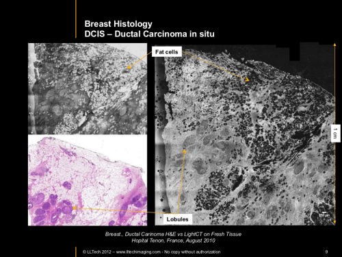
At last RSNA I have visited the French pavilion were one of the companies, LLTech, presented an OCT based system designed facilitate the work of pathologists and save time and money by allowing fresh tissue processing and pathology examination instead of the traditionally frozen sections (intraoperative) and fixated specimens (at lab)

That could be used by a pathologist to perform in-situ histology in the OR.
To summarize; OCT based systems seem to be something to look for in the future of cancer patients’ management.
References
1. Laughney AM, Krishnaswamy V, Rizzo EJ, Schwab EM, Barth RJ, et al. Scatter spectroscopic imaging distinguishes between breast pathologies in tissues relevant to surgical margin assessment. Clin Cancer Res. 2012;18:6315–25.
PUT IT IN CONTEXT OF CANCER CELL MOVEMENT
The contraction of skeletal muscle is triggered by nerve impulses, which stimulate the release of Ca2+ from the sarcoplasmic reticuluma specialized network of internal membranes, similar to the endoplasmic reticulum, that stores high concentrations of Ca2+ ions. The release of Ca2+ from the sarcoplasmic reticulum increases the concentration of Ca2+ in the cytosol from approximately 10-7 to 10-5 M. The increased Ca2+ concentration signals muscle contraction via the action of two accessory proteins bound to the actin filaments: tropomyosin and troponin (Figure 11.25). Tropomyosin is a fibrous protein that binds lengthwise along the groove of actin filaments. In striated muscle, each tropomyosin molecule is bound to troponin, which is a complex of three polypeptides: troponin C (Ca2+-binding), troponin I (inhibitory), and troponin T (tropomyosin-binding). When the concentration of Ca2+ is low, the complex of the troponins with tropomyosin blocks the interaction of actin and myosin, so the muscle does not contract. At high concentrations, Ca2+ binding to troponin C shifts the position of the complex, relieving this inhibition and allowing contraction to proceed.
Figure 11.25
Association of tropomyosin and troponins with actin filaments. (A) Tropomyosin binds lengthwise along actin filaments and, in striated muscle, is associated with a complex of three troponins: troponin I (TnI), troponin C (TnC), and troponin T (TnT). In (more ) Contractile Assemblies of Actin and Myosin in Nonmuscle Cells
Contractile assemblies of actin and myosin, resembling small-scale versions of muscle fibers, are present also in nonmuscle cells. As in muscle, the actin filaments in these contractile assemblies are interdigitated with bipolar filaments of myosin II, consisting of 15 to 20 myosin II molecules, which produce contraction by sliding the actin filaments relative to one another (Figure 11.26). The actin filaments in contractile bundles in nonmuscle cells are also associated with tropomyosin, which facilitates their interaction with myosin II, probably by competing with filamin for binding sites on actin.
Figure 11.26
Contractile assemblies in nonmuscle cells. Bipolar filaments of myosin II produce contraction by sliding actin filaments in opposite directions. Two examples of contractile assemblies in nonmuscle cells, stress fibers and adhesion belts, were discussed earlier with respect to attachment of the actin cytoskeleton to regions of cell-substrate and cell-cell contacts (see Figures 11.13 and 11.14). The contraction of stress fibers produces tension across the cell, allowing the cell to pull on a substrate (e.g., the extracellular matrix) to which it is anchored. The contraction of adhesion belts alters the shape of epithelial cell sheets: a process that is particularly important during embryonic development, when sheets of epithelial cells fold into structures such as tubes.
The most dramatic example of actin-myosin contraction in nonmuscle cells, however, is provided by cytokinesisthe division of a cell into two following mitosis (Figure 11.27). Toward the end of mitosis in animal cells, a contractile ring consisting of actin filaments and myosin II assembles just underneath the plasma membrane. Its contraction pulls the plasma membrane progressively inward, constricting the center of the cell and pinching it in two. Interestingly, the thickness of the contractile ring remains constant as it contracts, implying that actin filaments disassemble as contraction proceeds. The ring then disperses completely following cell division.
Figure 11.27
Cytokinesis. Following completion of mitosis (nuclear division), a contractile ring consisting of actin filaments and myosin II divides the cell in two.
http://www.ncbi.nlm.nih.gov/books/NBK9961/
This is good. I don’t recall seeing it in the original comment. I am very aware of the actin myosin troponin connection in heart and in skeletal muscle, and I did know about the nonmuscle work. I won’t deal with it now, and I have been working with Aviral now online for 2 hours.
I have had a considerable background from way back in atomic orbital theory, physical chemistry, organic chemistry, and the equilibrium necessary for cations and anions. Despite the calcium role in contraction, I would not discount hypomagnesemia in having a disease role because of the intracellular-extracellular connection. The description you pasted reminds me also of a lecture given a few years ago by the Nobel Laureate that year on the mechanism of cell division.