Feeds:
Posts
Comments

Archive for November, 2012

Lung Cancer (NSCLC), drug administration and nanotechnology

Author: Tilda Barliya PhD

Dr. Saxena has greatly introduced us to lung cancer , the associated drug treatments and their market share in the post titled ” NSCLC and where the future lie?”. Since lung cancer is the most leading cause of death in both man and women, and have gained lots of attention I am interested in elaborating on NSCLC and explore the potential use of nanotechnology in this matter.

As previously mentioned, there are 3 common types of lung cancer:

  • Adenocarcinomas are often found in an outer area of the lung. (Most common)
  • Squamous cell carcinomas are usually found in the center of the lung next to an air tube (bronchus).
  • Large cell carcinomas can occur in any part of the lung. They tend to grow and spread faster than the other two types. (Least common).

Figure 1. The Signs and symptoms of lung cancer anatomy.

Image

Since each type develops in different areas/part of the lung, it is hypothesized that they might need different routs of administration. The possible routes of administration are:

  • IV (systemic)————->through the blood
  • Inhaled aerosols (more localized)———–>through the airways

In order to understand what does “different routs of administration” refers to, we need to dig into the anatomy of the lung, i.e, airways and blood circulation as well as understand the lung-blood barriers components that may affect drug absorption.

The Blood Circulation

Two different circulatory systems, the bronchial and the pulmonary, supply the lungs with blood (Staub, 1991). The bronchial circulation is a part of the systemic circulation and is under high pressure. It receives about 1% of the cardiac output and supplies the airways (from the trachea to the terminal bronchioles), pulmonary blood vessels and lymph nodes with oxygenated blood and nutrients and conditions the inspired air (Staub, 1991). In addition, it may be important to the distribution of systemically administered drugs to the airways and to the absorption of inhaled drugs from the airways (Chediak et al., 1990). The pulmonary circulation comprise an extensive low pressure vascular bed, which receives the entire cardiac output. It perfuses the alveolar capillaries to secure efficient gas exchange and supplies nutrients to the alveolar walls. Anastomoses between bronchial and pulmonary arterial circulations have been found in the walls of medium-sized bronchi and bronchioles (Chediak et al., 1990; Kröll et al.,1987)

Image

Advantages:

  • Fast: 15–30 seconds to 1-2 hours
  • suitable for drugs not absorbed by the digestive system
  • IV can deliver continuous medication

Disadvantages:

  • Patients are not typically able to self-administer
  • It is the most dangerous route of administration because it bypasses most of the body’s natural defenses, exposing the user to health problems, known as chemo side affects.
  • Finally dose at the organ site is much lower than the administrated dose

Most of the conventional chemotherapy are mainly administrated IV (Docetaxel, Paxlitaxel, Gemcitiabine, Avastin etc).

The Airways

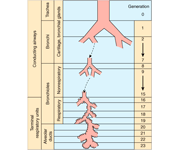
The human respiratory system can be divided in two functional regions: the conducting airways and the respiratory region. The conducting airways, which are composed of the nasal cavity and associated sinuses, the pharynx, larynx, trachea, bronchi, and bronchioles, filter and condition the inspired air. From trachea to the periphery of the airway tree, the airways repeatedly branch dichotomously into two daughter branches with smaller diameters and shorter length than the parent branch (Weibel, 1991). For each new generation of airways, the number of branches is doubled and the crosssectional area is exponentially increased. The conducting region of the airways generally constitutes generation 0 (trachea) to 16 (terminal bronchioles). The respiratory region, where gas exchange takes place, generally constitutes generation 17-23 and is composed of respiratory bronchioles, the alveolar ducts, and the alveolar sacs.

The air-blood barrier of the gas exchange area is composed of the alveolar epithelial cells (surface area 140 m2) on one side and the capillary bed (surface area 130 m2) on the other side of a thin basement membrane (Simionescu, 1991; Stone et al., 1992). The extensive surface area of the air-blood barrier in combination with its extreme thinness (0.1-0.5 μm) permit rapid gas exchange by passive diffusion (Plopper, 1996).

Image

The lung is a very attractive target for drug delivery. It provides direct access to disease in the treatment of respiratory diseases, while providing an enormous surface area and a relatively low enzymatic, controlled environment for systemic absorption of medications. (http://www.ncbi.nlm.nih.gov/pmc/articles/PMC1884307/)

Advantages:

  • Can be self medicated
  • Easy to use
  • Reduced side effects associated with systemic delivery

Disadvantages:

  • Slower route of action
  • Potential problem of deposition to the deeper alveolar (higher generations, like G 8-10)
  • Immuno-defense system
  • Difficulty in measuring the exact dose inside the lung
  • inhaled aerosol is entrapped in the mucus in the conducting airways

Need to be reminded that in addition, a drug’s efficacy may be affected by where in the respiratory tract it is deposited, its delivered dose and the disease it may be trying to treat.

Major components of the lung – barriers to drug absorption
As one of the primary interfaces between the organism and the environment, the respiratory system is constantly exposed to airborne particles, potential pathogens, and toxic gases in the inspired air (Plopper, 1996). As a result a sophisticated respiratory host defense system, present from the nostrils to the alveoli, has evolved to clear offending agents (Twigg, 1998).

The system comprises of:

  • mechanical (i.e. air filtration,cough, sneezing, and mucociliary clearance),
  • chemical (antioxidants, antiproteases and surfactant lipids),
  • immunological defense mechanisms and is tightly regulated to minimize inflammatory reactions that could impair the vital gas-exchange

**Intratracheal inhalation is another  administration option but will be left out of the discussion for now

From a drug delivery perspective, the components of the host defense system comprise barriers that must be overcome to ensure efficient drug deposition and absorption from the respiratory tract.

Generally, lung physiological investigations show that the airway and alveolar epithelia, not the interstitium and the endothelium, constitute the main barrier that restricts the movement of drugs and solutes from the airway lumen into the cells or the blood circulation.

Aerosols are defined as An aerosol is a suspensions of fine solid particles or liquid droplets in a gas.The major aspect affect the efficacy of aerosols as a drug delivery system is Drug Deposition.

Aerosol Drug deposition is affected by:

  • particle properties (e.g. size, shape, density, and charge),
  • respiratory tract morphology,
  • the breathing pattern (e.g. airflow rate and tidal volume)

These parameters determine not only the quantity of particles that are deposited but also in what region of the respiratory tract the particles are deposited.

Particle properties

As the cross-sectional area of the airways increases, the airflow rate rapidly decreases, and consequently the residence time of the particles in the lung increases from the large conducting airways towards the lung periphery. The most important mechanisms of particle deposition in the respiratory tract are (1) inertial impaction, (2) sedimentation, and (3) diffusion.

  • Inertial impaction – Inertial impaction occurs predominantly in the extrathoracic airways and in the tracheobronchial tree, where the airflow velocity is high and rapid changes in airflow direction occurs. Generally, particles with a diameter larger than 10 μm are most likely deposited in the extrathoracic region, whereas 2- to 10-μm particles are deposited in the tracheobronchial tree by inertial impaction. A long residence time of the inspired air favors particle deposition by sedimentation and diffusion.
  • Sedimentation – Sedimentation is of greatest importance in the small airways and alveoli and is most pronounced for particles with a diameter of 0.5-2 μm, Ultrafine particles (<0.5 μm in diameter) are deposited mainly by diffusional transport in the small airways and lung parenchyma where there is a maximal residence time of the inspired air.

Most therapeutic aerosols are almost always heterodisperse, consisting of a wide range of particle sizes and described by the log-normal distribution with the log of the particle diameters plotted against particle number, surface area or volume (mass) on a linear or probability scale and expressed as absolute values or cumulative percentage (http://www.ncbi.nlm.nih.gov/pmc/articles/PMC1884307/)

Optimal drug delivery to the lungs depends on an interaction between;

  • the inhaler device,
  • the drug formulation properties,
  • the inhalation maneuver

The devices currently available for pulmonary drug administration of pharmaceutical aerosols in clinical therapy include nebulizers, pressurized metered dose inhalers (pMDIs), and dry powder inhalers (DPIs).

However, much effort is put into the development of new inhaler devices and formulations to optimize the pulmonary delivery system for local or systemic drug targeting.

One of the major problems in aerosol delivery is

One disadvantage of the aerosol inhalation is, however, that a substantial portion of the aerosolized drug is not delivered to the lungs (i.e. delivered to the nose, mouth, skin, exhaled). only 10–15% of the emitted dose in the lungs.

In general the aerosol exposure techniques have a low dosing effectiveness, which often requires longer exposure times to administer the target dose and renders investigations of rapid kinetic events difficult. In addition, aerosol exposure requires an advanced equipment for exposure and ml-quantities of test formulation to fill up the device.

Airway geometry and humidity

Progressive branching and narrowing of the airways encourage impaction of particles. The larger the particle size, the greater the velocity of incoming air, the greater the bend angle of bifurcations and the smaller the airway radius, the greater the probability of deposition by impaction. The lung has a relative humidity of approximately 99.5%. The addition and removal of water can significantly affect the particle size of a hygroscopic aerosol and thus deposition. Drug particles are known to be hygroscopic and grow or shrink in size in high humidity, such as in the lung. A hygroscopic aerosol that is delivered at relatively low temperature and humidity into one of high humidity and temperature would be expected to increase in size when inhaled into the lung. The rate of growth is a function of the initial diameter of the particle, with the potential for the diameter of fine particles <1 µm to increase five-fold compared with two-to-three-fold for particles >2 µm. he increase in particle size above the initial size should affect the amount of drug deposited and particularly, the distribution of the aerosolized drug within the lung,

Lung Clearance Mechanism

Once deposited in the lungs, inhaled drugs are either cleared from the lungs, absorbed into the systemic circulation or degraded via drug metabolism. Drug particles deposited in the conducting airways are primarily removed through mucociliary clearance and, to a lesser extent, are absorbed through the airway . epithelium into the blood or lymphatic system. a low-viscosity periciliary or sol layer covered by a high-viscosity gel layer. Insoluble particles are trapped in the gel layer and are moved toward the pharynx (and ultimately to the gastrointestinal tract) by the upward movement of mucus generated by the metachronous beating of cilia. In the normal lung, the rate of mucus movement varies with the airway region and is determined by the number of ciliated cells and their beat frequency. Movement is faster in the trachea than in the small airways and is affected by factors influencing ciliary functioning and the quantity and quality of mucus.

Drugs deposited in the alveolar region may be phagocytosed and cleared by alveolar macrophages or absorbed into the pulmonary circulation. Alveolar macrophages are the predominant phagocytic cell for the lung defence against inhaled microorganisms, particles and other toxic agents. There are approximately five to seven alveolar macrophages per alveolus in the lungs of healthy nonsmokers. Macrophages phagocytose insoluble particles that are deposited in the alveolar region and are either cleared by the lymphatic system or moved into the ciliated airways along currents in alveolar fluid and then cleared via the mucociliary escalator.

Very little is known about how the drug-metabolizing activities of the lung affect the concentration and therapeutic efficacy of inhaled drugs. All metabolizing enzymes found in the liver are found to a lesser extent in the lung. Therefore assuming, drug deposition could have been calculated it would be hard to impossible to evaluate it’s metabolism.

In summary:

As the end organ for the treatment of local diseases or as the route of administration for systemic therapies, the lung is a very attractive target for drug delivery. It provides direct access the site of disease for the treatment of respiratory diseases without the inefficiencies and unwanted effects of systemic drug delivery. It provides an enormous surface area and a relatively low enzymatic, controlled environment for systemic absorption of medications. But it is not without barriers. Airway geometry, humidity, clearance mechanisms and presence of lung disease influence the deposition of aerosols and therefore influence the therapeutic effectiveness of inhaled medications. A drug’s efficacy may be affected by the site of deposition in the respiratory tract and the delivered dose to that site. To provide an efficient and effective inhalant therapy, these factors must be considered. Aerosol particle size characteristics can play an important role in avoiding the physiological barriers of the lung, as well as targeting the drug to the appropriate lung region.

Drug formulations and chemo drug delivery will be further discussed in a another post.

Ref:

1. N R Labiris and M B Dolovich. “Pulmonary drug delivery. Part I: Physiological factors affecting therapeutic effectiveness of aerosolized medications”. Br J Clin Pharmacol. 2003 December; 56(6): 588–599. http://www.ncbi.nlm.nih.gov/pmc/articles/PMC1884307/.

2. Tronde A. “Pulmonary drug absorption”. Acta Universities Upsalninesis Uppsala 2002. uu.diva-portal.org/smash/get/diva2:161887/FULLTEXT01

3. Naushad Khan Ghilzai. Pulmonary drug delivery. http://www.drugdel.com/Pulm_review.pdf.

Read Full Post »

Report on the Fall Mid-Atlantic Society of Toxicology Meeting “Reproductive Toxicology of Biologics: Challenges and Considerations.  Author, Reporter: Stephen J. Williams, Ph.D.

The fall 2012 Meeting of the Mid-Atlantic Society of Toxicology (MASOT) focused on the challenges and solutions in developing proper Development and Reproductive Toxicology (DART) studies with regards to the newer classes of bio-therapeutics such as vaccines, antibody-based therapies, and viral-based therapies.  The full meeting and MASOT links can be found at http://www.masot.org.   The overall synopsis of the meeting talks agreed, that although the general aim and design of DART studies for biological are very similar to DART studies for small molecule therapeutics, it is more necessary to take into consideration the pharmacodynamics, pharmacokinetic differences between biologics and small molecules.   In addition it is imperative to use pharmacologically-relevant species, such as non-rodent (guinea pig and non-human primate). The meeting was highlighted by the keynote speaker, Dr. A. Wallace Hayes, renowned board-certified toxicologist, committee and expert panel member for National Academy of Sciences, NIEHS, EPA and Department of Defense, and editor of well-known textbooks including Principles and Methods of Toxicology.  Dr. Hayes discussed a timeline of milestones in the field of toxicology.

The following are the meeting talk abstracts as well as notes for each presenter.

What’s So Different About DART Assessment of Biologics? Christopher Bowman Ph.D., DABT (Pfizer, Inc.)

Abstract:  The aim of developmental and reproductive toxicity (DART) safety assessment of a biologic is no different from that of a small molecule. Both cases consist of evaluating the potential for maternal toxicity, pre- and postnatal development toxicity (including juvenile toxicity) and effects of fertility (reproduction).  The differences lie in the in the product attributes of a specific biologic, the pharmacological response, the potential for undesirable toxicities and how these product attributes influence and are influenced by the biology.  Thus the primary challenge for developing a DART strategy for a biologic are derived from the complexities of these biomolecules and how that dictates a case-by-case strategy for appropriately evaluating the potential for developmental and reproductive toxicity. Most protein biologics have very limited potential for off-target toxicities, but this is not necessarily the case for other modalities such as anti-sense oligonucleotides and antibody-drug-conjugates.  In these cases, off-target toxicities can be a major feature of the DART safety assessment.  The most noticeable difference in DART assessment of biologics is the need to conduct these studies in pharmacologically relevant species and how that can influence the overall nonclinical strategy (including DART).  This has led to increased use of non-human primates as a model system and led to optimizations of this model for this purpose and revisions to international guidelines.

Notes:   Dr. Bowman emphasized the need to understand the type of biological you are testing and to both devise DART studies based on this information, additional endpoint you may want, as well as carefully choosing the correct species most relevant to the biologic.  He highlighted general differences between small molecules versus a biologic with respect to their pharmacology.  These differences are summarized in the Table below:

  Small Molecule Biologic-based therapy
Species specificity Low High
Route of administration Usually oral Parental
ADME (PK, bio-distribution etc.) Wide distribution Low distribution

He noted that clinical trials for biologics rarely include reproductive toxicity so the preclinical DART study is of utmost importance.  He also emphasized that currently, the FDA requires two species for DART testing of small molecule therapies (usually one rodent and one non-rodent).  However this is not possible with many biologics as species is to be taken in consideration when designing a meaningful DART study.  Study designs can be like most DART studies but want to have a steady exposure during fetal organogenesis, use high doses (10 times the clinical dose) to achieve maximal pharmacology, confirm exposure to fetus and to F1 generation, and determine embryolethality.  Some biologics like interferon and insulin-growth factor receptor (IGFR) antagonists are fetal abortifactants. In fact Lucentis (Ranibizumab) and Macugen (Pegaptanib) were approved with no or little DART studies, however these drugs showed reproductive toxicity, resulting in warning concerning pregnancy on the label. Also important is the effect on the immune system and reproductive system of offspring, as well as the pharmacodynamics profile in the offspring.

Species Selection for Reproductive and Developmental Toxicity Testing of Biologics; Elise M. Lewis, Ph.D. (Charles River Preclinical Services)

Abstract:  Regulatory guidelines for developmental and reproductive toxicology studies require selection of “relevant” animal models as determined by kinetic, pharmacological, and preceding toxicological data.  Rats, mice, and rabbits are the preferred animal models for these studies based on historical experience and well-established procedures and study protocols.  However, due to species specificity and immunogenicity issues, developmental and reproductive toxicology testing for biologics is limited to a pharmacologically relevant animal model as described in the ICH s6 guideline.

Notes:  Dr. Lewis notes that DART studies in guinea pigs and hamsters represent a cost effective alternative to large animal models as well as the benefit of shorter duration and ability to assess mating behavior.  She also notes that reproductive toxicology of vaccines should be done in an animal model that can elicit an immune-response to the vaccine, especially to determine any maternal-fetal interaction.  For example, a vaccine may be directed to a maternal protein which when suppressed, may negatively impact the developing fetus.  However it is important to remember that guinea pigs can spontaneously abort so it is good to have proper control arms of a substantial size in order to statistically determine the impact of those spontaneous abortions.


Placental Transfer of an Adnectin Protein During Organogenesis in Guinea Pigs Using a Radiolabeled Methodology; Lakshmi Sivaraman, Ph.D. (Bristol-Myers Squibb)

Abstract:  Knowledge regarding the placental transfer of large molecular weight therapeutics is important to support the enrollment of women of childbearing potential in clinical trials.  There is limited information in the scientific literature that reports the extent to which the conceptus is exposed to these large molecules during organogenesis.  Placental transfer of large therapeutics has been difficult to quantify, due to limited blood volumes that can be obtained from the embryo, as well as insufficient assay sensitivity.  Thus, it is possible that embryos are exposed to pharmacologically active concentrations after maternal drug exposure. We have adopted a radiolabeled approach to quantitate embryo-fetal exposure of a novel protein therapeutic platform (adnectins). Adnectins are fibronectin-based proteins containing domains engineered to bind to targets of therapeutic interests.

Notes: Adnectins molecular weight is typically less than monoclonal antibodies and while IgG is not transferred in great quantity past the placental barrier there have been studies in human indicating maternal-fetal transfer of monoclonal antibodies.  This is particularly important for two reasons:  the monoclonal interacts with a target important in development, or the fetal immune system could be augmented.  Their work will be published in Drug Metabolism and Disposition.  In general Dr. Siveraman engineered a radiolabel on adnectin and used different detection methods to quantify the fetal exposure to a single maternal dose.  Dr. Siverman was able to detect radiolabel in the fetus however it is not clear whether this is a significant amount.

Reproductive Toxicity Testing for Biological Products in Nonhuman Primates: Evolution and Current Perspectives: Gary J. Chellman, Ph.D., DABT (Charles River Preclinical Services)

Notes:  Dr. Chellman gave a review of the current trends being driven by regulatory agencies with regard to nonhuman primate DART studies of biopharmaceuticals.  He noted that an advantage using nonhuman primates were the close physiologic resemblance to humans and because a large animal could monitor pregnancy over time using ultrasound technology.  In general, Dr. Chellman spoke about new study designs which not only reduce the number of animals required but also significantly reduce costs.  For example, a DART study which cost upward of $750,000 now can be done for as little as $350,000.  Dr. Kary Thompson of Bristol Myers Squibb then gave a talk about use of these new enhanced designs to determine reproductive toxicity issues with ipilimumab (Yervoy).

Other research papers on Pharmaceutical Intelligence and Reproductive Biology, Bio Insrumentation, Endocrinology Genetics were published on this Scientific Web site as follows

Non-small Cell Lung Cancer drugs – where does the Future lie?

Reboot evidence-based medicine and reconsider the randomized, placebo-controlled clinical trial

Every sperm is sacred: Sequencing DNA from individual cells vs “humans as a whole.”

Leptin and Puberty

Gene Trap Mutagenesis in Reproductive Research

Genes involved in Male Fertility and Sperm-egg Binding

Hope for Male Contraception: A small molecule that inhibits a protein important for chromatin organization can cause reversible sterility in male mice

Pregnancy with a Leptin-Receptor Mutation

The contribution of comparative genomic hybridization in reproductive medicine

Sperm collide and crawl the walls in chaotic journey to the ovum

Impact of evolutionary selection on functional regions: The imprint of evolutionary selection on ENCODE regulatory elements is manifested between species and within human populations

Biosimilars: CMC Issues and Regulatory Requirements

Biosimilars: Intellectual Property Creation and Protection by Pioneer and by Biosimilar Manufacturers

Assisted Reproductive Technology Cycles and Cumulative Birth Rates

Innovations in Bio instrumentation in Reproductive Clinical and Male Fertility Labs in the US

Increased risks of obesity and cancer, Decreased risk of type 2 diabetes: The role of Tumor-suppressor phosphatase and tensin homologue (PTEN)

Guidelines for the welfare and use of animals in cancer research

Every sperm is sacred: Sequencing DNA from individual cells vs “humans as a whole.”


Read Full Post »

Non-small Cell Lung Cancer drugs – where does the Future lie?

In focus: Tarceva, Avastin and Dacomitinib


UPDATED on July 5, 2013

(from reports published in New England Journal of Medicine on drug, crizotinib)


Curator: Ritu Saxena, Ph.D.


Introduction

Non-small cell lung cancer (NSCLC) is the most common type of lung cancer and usually grows and spreads more slowly than small cell lung cancer.

There are three common forms of NSCLC:

  • Adenocarcinomas are often found in an outer area of the lung.
  • Squamous cell carcinomas are usually found in the center of the lung next to an air tube (bronchus).
  • Large cell carcinomas can occur in any part of the lung. They tend to grow and spread faster than the other two types.

Lung cancer is by far the leading cause of cancer death among both men and women. Each year, more people die of lung cancer than of colon, breast, and prostate cancers combined. The American Cancer Society’s most recent estimates for lung cancer in the United States for 2012 reveal that about 226,160 new cases of lung cancer will be diagnosed (116,470 in men and 109,690 in women), and there will be an estimated 160,340 deaths from lung cancer (87,750 in men and 72,590 among women), accounting for about 28% of all cancer deaths.

Treatment

Different types of treatments are available for non-small cell lung cancer. Treatment depends on the stage of the cancer. For patients in whom the cancer has not spread to nearby lymph nodes are recommended surgery. Surgeon may remove- one of the lobes (lobectomy), only a small portion of the lung (wedge removal), or the entire lung (pneumonectomy). Some patients require chemotherapy that uses drugs to kill cancer cells and stop new cells from growing.

FDA approved drugs for NSCLC

Abitrexate (Methotrexate)
Abraxane (Paclitaxel Albumin-stabilized Nanoparticle Formulation) 
Alimta (Pemetrexed Disodium)
Avastin (Bevacizumab)
Bevacizumab
Carboplatin
Cisplatin
Crizotinib
Erlotinib Hydrochloride
Folex (Methotrexate)
Folex PFS (Methotrexate)
Gefitinib
Gemcitabine Hydrochloride
Gemzar (Gemcitabine Hydrochloride)
Iressa (Gefitinib)
Methotrexate
Methotrexate LPF (Methotrexate)
Mexate (Methotrexate)
Mexate-AQ (Methotrexate)
Paclitaxel
Paclitaxel Albumin-stabilized Nanoparticle Formulation
Paraplat (Carboplatin)
Paraplatin (Carboplatin)
Pemetrexed Disodium
Platinol (Cisplatin)
Platinol-AQ (Cisplatin)
Tarceva (Erlotinib Hydrochloride)
Taxol (Paclitaxel)
Xalkori (Crizotinib)

On the basis of target, the drugs have been classified as follows:

Image

NSCLC Drug Market Analysis

NSCLC drug market expected to grow from $4.2 billion in 2010 to $5.4 billion in 2020

Although, a whole list of agents is available for the treatment of NSCLC, the market for NSCLC drugs is expected to expand from $4.2 billion in 2010 to $5.4 billion in 2020 in the United States, France, Germany, Italy, Spain, the United Kingdom and Japan.   

However, drug sales for metastatic/advanced squamous cell non-small-cell lung cancer, which comprises only a small fraction of the market, will decrease from nearly 17 percent in 2010 to approximately 13 percent in 2020. According to surveyed U.S. oncologists and MCO pharmacy directors, increasing overall survival is one of the greatest unmet needs in first-line advanced squamous non-small-cell lung cancer.

In 2009, antimetabolites dominated the NSCLC market, with Eli Lilly’s Alimta (Pemetrexed) accounting for nearly three-quarters of sales within this drug class. Since then, Alimta has faced tough competition from a number of similar drugs and from emerging therapies. It was speculated that the antimetabolites market share would reduce significantly making it the second-largest drug class in NSCLC, while the epidermal growth factor receptor (EGFR) inhibitor class will garner the top market share by 2019.

Genentech/OSI Pharmaceuticals/Roche/Chugai Pharmaceutical’s Tarceva belongs to the EGFR inhibitor class, and has been prescribed principally along with Eli Lilly’s Alimta, to NSCLC patients.Both these drugs have dominated the NSCLC market till 2010, however, their market hold is expected to weaken from 2015-2020, as claimed by Decision Resources Analyst Karen Pomeranz, Ph.D. Decision Resources is a research and advisory firms for pharmaceutical and healthcare issues.

Tarceva (Erlotinib)

Generic Name: Erlotinib, Brand Name: Tarceva

Other Designation: CP 358774, OSI-774, R1415, RG1415, NSC 718781

Mechanism of Action: Tarceva, a small molecule quinazoline, directly and reversibly inhibits the epidermal growth factor receptor (EGFr) tyrosine kinase. Detailed information on how it works could be found at the Macmillian Cancer support website.

Tarceva has been approved for different cancers and several indications have been filed-

  • non-small cell lung cancer (nsclc), locally advanced or metastatic, second line, after failure of at least one prior chemotherapy regimen (2004)
  • pancreatic cancer, locally advanced or metastatic, in combination with gemcitabine, first line (2005)
  • non-small cell lung cancer (nsclc), advanced, maintenance therapy in responders following first line treatment with platinum-based chemotherapy (2010)
  • non-small cell lung cancer (nsclc) harboring epidermal growth factor (EGFr)-activating mutations, first line treatment in advanced disease

Sales of Tarceva 

May, 2012 sales of Tarceva in the US have been reported to be around $564.2 million.

In a recent article published by Vergnenègre et al in the Clinicoeconomic Outcomes Research journal (2012), cross-market cost-effectiveness of Erlotinib was analyzed. The study aimed at estimating the incremental cost-effectiveness of Erlotinib (150 mg/day) versus best supportive care when used as first-line maintenance therapy for patients with locally advanced or metastatic NSCLC and stable disease.

It was determined that treatment with erlotinib in first-line maintenance resulted in a mean life expectancy of 1.39 years in all countries, compared with a mean 1.11 years with best supportive care, which represents 0.28 life-years (3.4 life-months) gained with erlotinib versus best supportive care.

According to the authors analysis, there was a gain in the costs per-life year as $50,882, $60,025, and $35,669 in France, Germany, and Italy, respectively. Hence, on the basis of the study it was concluded that Erlotinib is a cost-effective treatment option when used as first-line maintenance therapy for locally advanced or metastatic NSCLC.

Avastin (Bevacizumab)

Generic Name: Avastin, Brand Name: Bevacizumab

Other Designation: rhuMAb-VEGF, NSC-704865, R435, RG435

Mechanism of Action

Bevacizumab is a recombinant humanized Mab antagonist of vascular endothelial growth factor A (VEGFA) acting as an angiogenesis inhibitor.

Targets

Vascular endothelial growth factor (VEGF, VEGF-A, VEGFA)

Avastin is the only currently approved VEGF inhibitor that selectively targets VEGF-A.

Three other approved oral drugs, pazopanib (Votrient; GlaxoSmithKline), sunitinib (Sutent; Pfizer) and sorafenib (Nexavar; Onyx Pharmaceuticals) are orally available multi-targeted receptor tyrosine kinase inhibitors that include VEGF receptors among their tar­gets.

Avastin has been approved for different cancers and several indications have been filed:

  • colorectal cancer, advanced, metastatic, first line, in combination with a 5-FU based chemotherapy regimen
  • colorectal cancer, relapsed, metastatic, second line, in combintion with 5-FU-based chemotherapy (2004)
  • non-small cell lung cancer (nsclc), non-squamous, inoperable, locally advanced, recurrent or metastatic, in combination with carboplatin and paclitaxel chemotherapy, first line (2006)
  • breast cancer, chemotherapy naive, first line, locally recurrent or metastatic, in combination with taxane chemotherapy (2008, revoked in 2011)
  • non-small cell lung cancer (nsclc), non-squamous, inoperable, locally advanced, recurrent or metastatic, in combination with platinum-based chemotherapy, first line
  • renal cell carcinoma (RCC), metastatic, in combination with interferon (IFN) alpha, first line (2009)
  • glioblastoma multiforme (GBM), relapsed after first line chemoradiotherapy
  • breast cancer, chemotherapy naive, first line, locally recurrent or metastatic, HEr2 negative, in combination with capecitabine (2009)
  • ovarian cancer, in combination with standard chemotherapy (carboplatin and paclitaxel) as a first line treatment following surgery for women with advanced (Stage IIIb/c or Stage IV) epithelial ovarian, primary peritoneal or fallopian tube cancer
  • ovarian cancer, in combination with carboplatin and gemcitabine as a treatment for women with recurrent, platinum-sensitive ovarian cancer

SOURCE:

New medicine Oncology Knowledge Base

Sales of Avastin 

As of May, 2012, sales of Avastin in the US have been reported to be around $2.66 billion.

It attracted a lot of attention over the past few years after its use as a breast cancer treatment. Avastin was approved by the FDA under its fast-track program. However, the data released by the FDA from follow-up studies led to questioning the use of Avastin as a breast cancer drug. Infact, Genentech pulled the indication from Avastin’s label. Henceforth, the FDA did cancel that approval in late 2011. Doctors, however, can still prescribe it off-label. Potential adverse effects of Avastin that came under scrutiny along with unfavorable cost benefit analyses might pose challenges to its growth potential and continued widespread use. However, the sales of Avastin have continued to increase and it has been reported by Fierce Pharma as one of the 15 best-selling cancer drugs list. (Fierce Pharma)

Dacomitinib: New promising drug for NSCLC

Generic Name: Dacomitinib

Other Designation: PF-299804, PF-00299804, PF-299,804, PF00299804

PF-299804 is an orally available irreversible pan-HEr tyrosine kinase inhibitor.

Dacomitinib is a promising new drug on the market. Phase III trials are ongoing for advanced and refractory NSCLC, locally advanced or metastatic NSCLC and the EGFr mutation containing locally advanced or metastatic NSCLC in several countries including those in Europe, Asia, and America.

SOURCE:

New medicine Oncology Knowledge base

Dacomitinib bests Erlotinib in advanced NSCLC:  Comparison of its Progression-Free Survival (PFS) with the NSCLC marketed drug, Erlotinib.

In September of 2012, a study was published by Ramalingam et al in the Journal of Clinical Oncology, which was a randomized open-label trial comparing dacomitinib with erlotinib in patients with advanced NSCLC. On the basis of the study it was concluded that dacomitinib demonstrated significantly improved progression-free survival (PFS*) as compared to erlotinib, with a certain degree of toxicity.

SOURCE:

Randomized Phase II Study of Dacomitinib Versus Erlotinib in Patients With Advanced Non-Small-Cell Lung Cancer

The results indicated indicated the following:

  • Median PFS was significantly greater with Dacomitinib than Erlotinib, at 2.86 versus 1.91.
  • Mean duration of response was 16.56 months for dacomitinib and 9.23 months for erlotinib.

Patients were divided into groups by tumor type and following results were obtained:

  • Median PFS was 3.71 months with dacomitinib and 1.91 with erlotinib in patients with KRAS wild-type tumors
  • Median PFS was 2.21 months and 1.68 months, in patients with KRAS wild-type/EGFR wild-type tumors.
  • PFS was significantly better in the molecular subgroups harboring a mutant EGFR genotype.

The study also highlighted the side effects which might be more of concern and probably limiting for Dacomitinib.

Although adverse side effects were uncommon in both the groups, certain side effects such as:

  • mouth sores,
  • nailbed infections, and
  • diarrhea

were more common and tended to be more severe with Dacomitinib as compared to Tarceva.

Therefore, for patients for whom side effects of Tarceva seem challenging might face more difficulty with Dacomitinib treatment. Nonetheless, the results of PFS were promising enough and provide a greater efficacy in several clinical and molecular subgroups targeting a larger population than Tarceva. Authors, thus, suggested a larger, randomized phase III trial with the same design.

Current status of Dacomitinib

Based on positive performance of Dacomitinib published in research studies, Pfizer has entered into a collaborative development agreement with the SFJ Pharmaceuticals Group to conduct a phase III clinical trial across multiple sites in Asia and Europe, to evaluate dacomitinib (PF-00299804) as a first line treatment in patients with locally advanced or metastatic non-small cell lung cancer (nsclc) with activating mutations in the epidermal growth factor receptor (EGFr). Under the terms of the agreement, SFJ will provide the funding and clinical development supervision to generate the clinical data necessary to support a registration dossier on Dacomitinib for marketing authorization by regulatory authorities for this indication. If approved for this indication, SFJ will be eligible to receive milestone and earn-out payments.

SOURCE:

New medicine Oncology Knowledge base

*PFS or Progression-free survival is defined as the length of time during and after the treatment of as disease, such as cancer, that a patient lives with the disease but it does not get worse. In a clinical trial, measuring the progression-free survival is one way to see how well a new treatment works.

REFERENCES

Recently, another drug PF-02341066 (crizotinib), was tested on patients with non-small cell lung cancer and the results were published in New England Journal of Medicine (2013). Crizotinib is an orally available aminopyridine-based inhibitor of the) and the c-Met/hepatocyte growth factor receptor (HGFR). Crizotinib, in an ATP-competitive manner, binds to and inhibits ALK kinase and ALK fusion proteins. In addition, crizotinib inhibits c-Met kinase, and disrupts the c-Met signaling pathway. Altogether, this agent inhibits tumor cell growth.

  • Shaw and colleagues (2013) investigated whether crizotinib is superior to standard chemotherapy with respect to efficacy. To answer the question, Pfizer launched a phase III clinical trial (NCT00932893; http://clinicaltrials.gov/show/NCT00932893) comparing the safety and anti-tumor activity of PF-02341066 (crizotinib) versus pemetrexed or docetaxel in patients with advanced non-small cell lung cancer harboring a translocation or inversion event involving the ALK gene. Shaw and colleagues (2013) published the results of the clinical trial in a recent issue of New England Journal of Medicine.  A total of 347 patients with locally advanced or metastatic ALK-positive lung cancer who had received one prior platinum-based regimen were recruited for the trial and patients were randomly assigned to receive oral treatment with crizotinib (250 mg) twice daily or intravenous chemotherapy with either pemetrexed (500 mg per square meter of body-surface area) or docetaxel (75 mg per square meter) every 3 weeks. Patients in the chemotherapy group who had disease progression were permitted to cross over to crizotinib as part of a separate study. The primary end point was progression-free survival. According to the results, the median progression-free survival was 7.7 months in the crizotinib group and 3.0 months in the chemotherapy group. Hazard ratio (HR) for progression or death with crizotinib was 0.49 (95% CI, P<0.001). The response rates were 65% with crizotinib, as compared with 20% with chemotherapy (P<0.001). An interim analysis of overall survival showed no significant improvement with crizotinib as compared with chemotherapy (hazard ratio for death in the crizotinib group, 1.02; 95% CI, P=0.54). Common adverse events associated with crizotinib were visual disorder, gastrointestinal side effects, and elevated liver aminotransferase levels, whereas common adverse events with chemotherapy were fatigue, alopecia, and dyspnea. Patients reported greater reductions in symptoms of lung cancer and greater improvement in global quality of life with crizotinib than with chemotherapy.In conclusion, the results from the trial indicate that crizotinib is superior to standard chemotherapy in patients with previously treated, advanced non–small-cell lung cancer with ALK rearrangement. (Shaw AT, et al, Crizotinib versus Chemotherapy in Advanced ALK-Positive Lung Cancer. N Engl J Med 2013; 20 June, 368:2385-2394; http://www.ncbi.nlm.nih.gov/pubmed/23724913).

However, in the same issue of New England Journal of Medicine, Awad and colleagues (2013) reported from a phase I clinical trial (NCT00585195; http://clinicaltrials.gov/show/NCT00585195), that a patient with metastatic lung adenocarcioma harboring a CD74-ROS1 rearrangement who had initially shown a dramatic response to treatment, showed resistance to crizotinib. Biopsy of the resistant tumor identified an acquired mutation leading to a glycine-to-arginine substitution at codon 2032 in the ROS1 kinase domain. Although this mutation does not lie at the gatekeeper residue, it confers resistance to ROS1 kinase inhibition through steric interference with drug binding. The same resistance mutation was observed at all the metastatic sites that were examined at autopsy, suggesting that this mutation was an early event in the clonal evolution of resistance. The study was funded by Pfizer (Awad MM, et al, Acquired resistance to crizotinib from a mutation in CD74-ROS1. N Engl J Med. 2013 Jun 20;368(25):2395-401; http://www.ncbi.nlm.nih.gov/pubmed/23724914)

Reference: 

Read Full Post »

Reporter: Aviva Lev-Ari, PhD, RN

Genomics: The single life

Sequencing DNA from individual cells is changing the way that researchers think of humans as a whole.

31 October 2012

The tendency of sperm to swim alone makes the cells ideal for single-cell genomics. Adam Auton, a statistical geneticist at Albert Einstein College of Medicine in New York is using sperm to study recombination, the process that shuffles genes during the formation of germ cells and therefore influences which genes are inherited. Recombination is one of the fundamental forces that shapes genetic diversity,” he says. “In recent years we’ve learned that there is considerable variation in the recombination rate between different populations, between the sexes and even between individuals.” But pinning down the rate in people once seemed impossible because it would have required finding individuals with hundreds of children and sequencing their genomes.

The ability to sequence single cells meant that researchers could take another approach. Working with a team at the Chinese sequencing powerhouse BGI, Auton sequenced nearly 200 sperm cells and was able to estimate the recombination rate for the man who had donated them. The work is not yet published, but Auton says that the group found an average of 24.5 recombination events per sperm cell, which is in line with estimates from indirect experiments2. Stephen Quake, a bioengineer at Stanford University in California, has performed similar experiments in 100 sperm cells and identified several places in the genome in which recombination is more likely to occur. The location of these recombination ‘hotspots’ could help population biologists to map the position of genetic variants associated with disease.

Quake also sequenced half a dozen of those 100 sperm in greater depth, and was able to determine the rate at which new mutations arise: about 30 mutations per billion bases per generation3, which is slightly higher than what others have found. “It’s basically the population biology of a sperm sample,” Quake says, and it will allow researchers to study meiosis and recombination in greater detail.

SOURCE:  

VIEW ARTICLE IN NATURE

http://www.nature.com/news/genomics-the-single-life-1.11710#/genome

Read Full Post »

Reporter: Aviva Lev-Ari, PhD, RN


Mark Levin’s business is biotechnology, so it’s no surprise he knew zilch about a tech company called LinkedIn as recently as two years ago. But these days Levin sounds like he can barely do his job without it.

“I’m not the most social media savvy person. I haven’t used a lot of these tools at all,” Levin says, referring to blogs and Twitter. “But I’ll never forget, the first message I got from LinkedIn was an e-mail from what looked like someone called link-a-din. I remember asking myself about Mr. Link-a-din. I was trying to figure out ‘who the hell is this person?’”

Levin, a founding partner of Boston-based Third Rock Ventures and one of the more prominent biotech venture capitalists in the U.S., was a LinkedIn Luddite two years ago. To some extent, he still looks like one: his profile contains no photo, no professional biography, and only tidbits of information posted about his employment history. But appearances can be deceiving. He says he has amassed more than 5,000 connections, and the number keeps growing daily. He says he spends at least a half an hour per day on the site, sifting through more than 100 incoming connection requests a week, and firing off dozens more requests to people he wants to get to know. LinkedIn’s algorithms have gotten to know his tendencies so well, the site is constantly suggesting new people in biotech and pharma companies that he might want to meet. He often does.

Mark Levin of Third Rock Ventures

Levin became so obsessive at one point this year that LinkedIn temporarily shut down his account, until he called the company and assured them he’s a real person using the site for business. Just during a 15-minute phone interview with me on Friday, Levin said he got three new connection requests. One was from an MD that caught his eye immediately.

“About 18 months ago or so, I realized that is an extraordinary way to be in contact with people,” Levin says. “Our biggest challenge is to find great people. We don’t know everybody. And you can find a lot of great people here.”

While many in the tech press mock LinkedIn as an oh-so-boring compiler of mere resumes, it has become the indispensable online hub for networking in life sciences—an industry where relationships make the world go round. LinkedIn has a relatively puny user base of 187 million members around the world, compared to Facebook’s 1 billion, and there’s no question people spend way more time engaging with Mark Zuckerberg’s social network. But it’s also true there’s no question which site matters more to the life sciences. LinkedIn is the singular site for finding people in biotech, whether they are biologists, chemists, toxicologists, admin assistants, business development people, finance pros, or CEOs. There were more than 513,000 people in the LinkedIn database who self-identify as members of the “biotechnology” or “pharmaceutical” industry when I searched on those keywords Friday afternoon.

For journalists like me, this is an everyday reporting tool with almost as much value as Twitter, and possibly more. Even though I only use the basic free version of the site, it’s become an awesome clearinghouse of sources that I call on for help with scoops and analysis. I can slice and dice my network of 2,900 contacts by industry, title, location and more. It’s become a treasure trove of personal e-mails for sources, which I never have to manually update when people leave for new jobs, as they often do. It’s even turned into a place where people read a lot of my stories and the resource where I sometimes find new stories to pursue. In fact, I got the idea for this story by noticing that Levin and I have more than 500 connections in common.

for different reasons, but he raves all the same. Nothing great in biotech can happen without a magical mix of an idea, technology, people, and money.

“Our No. 1 goal in life is to know the best people in the industry who are going to make a difference in our companies,” Levin says. “I don’t remember when it exactly became clear, but it was clear to me that a lot of people were using it to stay in touch. We’ve realized it’s an extraordinary recruiting tool. The more I’ve spent time there, the more aggressive I have gotten.”

Levin isn’t kidding about the emphasis on recruiting at Third Rock, which has a “recruiting partner” in Craig Greaves, a former recruiter at Biogen Idec (NASDAQ: BIIB) and Cubist Pharmaceuticals (NASDAQ: CBST). Levin says all this connecting and re-connecting sometimes leads somewhere fruitful, sometimes not, just like with all other recruiting techniques.

But Levin and his partner at Third Rock aren’t just fiddling around making random contacts, they are being systematic about the connections they form. Once he forms a connection on LinkedIn, he said he sends the new contact a short follow-up note to see what’s new in their lives or careers. He then e-mails his fellow partners to see if any of them know the person. Third Rock uses a premium version of LinkedIn, which has an application that automatically downloads all of Levin’s new contacts into a central database so that all members of the firm can see the person’s profile, Greaves says.

Sometimes an in-person meeting gets scheduled to follow up right away to see if there might be some kind of potential for a match at a Third Rock company. Often Third Rock uses the site for targeted searches, like, say, for an antibody engineer, Greaves says. Other times, it’s just to get acquainted with people who aren’t looking for work now, but might be able to join a startup, consult, or form a valuable partnership with a Third Rock company sometime later, he says.

“We are laying the groundwork and building a network for the long term,” Greaves says.

No doubt, LinkedIn has its potential for misuse just like any other technology, and users need to think about how to use it properly. Back when the site was formed in 2003, people were urged to connect only with people they knew well, because otherwise people might think you were tainted if a shady operator ended up appearing in your network. I think that stigma has largely gone away, because a connection is perceived now as really just like trading business cards, and not an endorsement or recommendation. People have also long worried about whether bosses might be able to use it to spy on their workers, and suspect whether they were getting restless, looking for a new job. I used to leave my entire connections list accessible on the web for anyone who connected with me, until I started connecting with people I don’t really know, and realized some may have ulterior motives that might interfere with my ability to break news.

There are plenty of areas on the site that leave something to be desired. LinkedIn Groups have always struck me as spammy, so I’ve unsubscribed to most of them. The site can be annoying with its constant urges to “update your profile” or “add skills to your profile” or now to “endorse” various people in your network. The whole site appears to be trying really hard to keep people glued to it like Facebook, by constantly updating their status and checking other people’s employment status, which can be annoying and a waste of time.

But the most irritating thing about LinkedIn, to me anyway, is that even though it has achieved critical mass, many C-suite executives and venture capitalists still resist signing up. For example, when I searched on the 40 names of “young and proven” biotech venture capitalists listed in this column two weeks ago, only 24 of the 40 (60 percent) showed up in the LinkedIn database.

I find it baffling that so many senior people in the industry still resist taking advantage of this resource, and have to wonder if they have some better idea on how to network. There’s no getting around the importance of networking. Biotech is a geographically far-flung industry, with hundreds of companies and vendors, who all need to work together in trusting relationships to keep the whole enterprise afloat.

Industry conferences have always been, and still remain, the gold standard way of networking. But those events take time and money, and nobody can do it every day of the week. LinkedIn is becoming the indispensable resource that glues an entire industry together, and helps people make connections between people and ideas and opportunities that would otherwise never be made. While biotech could certainly use a few more groundbreaking advances to make the drug development process more efficient, one of the fastest-growing new tools for the industry is a free resource just a click away on the Web.

Luke Timmerman is the National Biotech Editor of Xconomy. E-mail him at

ltimmerman@xconomy.com 

SOURCE:


Read Full Post »

CABG or PCI: Patients with Diabetes – CABG Rein Supreme

Reporter: Aviva Lev-Ari, PhD, RN


VIEW VIDEO

105

Compelling Evidence for Coronary-Bypass Surgery in Patients with Diabetes

Mark A. Hlatky, M.D.

November 4, 2012DOI: 10.1056/NEJMe1212278

Seventeen years ago, the National Heart, Lung, and Blood Institute issued a clinical alert1 that coronary-artery bypass grafting (CABG) had better rates of survival than percutaneous coronary intervention (PCI) in patients with diabetes. The alert was based on the results of the Bypass Angioplasty Revascularization Investigation (BARI) trial,2 in which patients with multivessel coronary artery disease were randomly assigned to undergo either CABG or PCI.

This recommendation has been controversial ever since, largely because subsequent trials comparing CABG and PCI have enrolled only small numbers of patients with diabetes. A pooled analysis of 10 randomized trials involving 1233 patients with diabetes confirmed that such patients had a particular survival advantage after CABG, as compared with PCI.3 But this evidence was discounted because drug-eluting stents were not used in PCI procedures in the earlier trials, and more recent trials in which drug-eluting stents were used4,5 enrolled relatively few patients with diabetes. Settling this controversy would require a trial with a large number of patients with both diabetes and multivessel coronary artery disease in whom CABG or PCI would be performed with the use of contemporary methods.

Farkouh et al.6 now report in the Journal the results of the definitive Future Revascularization Evaluation in Patients with Diabetes Mellitus: Optimal Management of Multivessel Disease (FREEDOM) trial, in which 1900 patients with diabetes (about as many patients with diabetes as in all previous trials combined) were randomly assigned to undergo either CABG or PCI with drug-eluting stents.

As a cardiologist who does not perform either procedure, I find that the FREEDOM trial provides compelling evidence of the comparative effectiveness of CABG versus PCI in patients with diabetes and multivessel coronary artery disease. After 5 years of follow-up, the 947 patients assigned to undergo CABG had significantly lower mortality (10.9% vs. 16.3%) and fewer myocardial infarctions (6.0% vs. 13.9%) than the 953 patients assigned to undergo PCI. However, patients in the CABG group had significantly more strokes (5.2% vs. 2.4%), mostly because of strokes that occurred within 30 days after revascularization. In the CABG group, the primary composite outcome of death, myocardial infarction, or stroke over 5 years was reduced by 7.9 percentage points, or a relative decrease of 30%, as compared with PCI (18.7% vs. 26.6%, P=0.005). These results are consistent with the findings of multiple previous trials comparing CABG and PCI in patients with diabetes,3 as well as the most recent trials in which drug-eluting stents were used during PCI.4,5

Despite the results of BARI and other trials, over time more and more patients with diabetes have undergone PCI rather than CABG to treat multivessel coronary disease.7,8 The reasons for this trend are uncertain, yet there are two broad potential explanations. First, because PCI technology continues to evolve, many cardiologists simply have dismissed the results of earlier randomized studies as outdated because they used earlier techniques. This is a catch-22, since long-term studies are needed to compare hard outcomes, but evidence from long-term studies may be ignored if therapies are evolving. The results of the FREEDOM trial suggest that the comparative effectiveness of CABG and PCI on hard outcomes remains similar whether PCI is performed without stents, with bare-metal stents, or with drug-eluting stents. Mortality has been consistently reduced by CABG, as compared with PCI, in more than 4000 patients with diabetes who have been evaluated in 13 clinical trials. The controversy should finally be settled.

Another potential reason for the increasing use of PCI in patients with multivessel coronary disease is that the clinical-decision pathway leads patients toward PCI over alternative treatments. Many PCIs today are ad hoc procedures, performed at the time of diagnostic coronary angiography, with the same physician making the diagnosis, recommending the treatment, and performing the procedure. There is little time for informed discussion about alternative treatment options, either medical therapy on the one hand or CABG on the other. Well-informed patients might choose any of those options on the basis of their concerns about the various outcomes of treatment, such as survival, stroke, myocardial infarction, angina, and recovery time. This is a complicated decision, and clinical guidelines in the United States9 and Europe10 now emphasize the importance of more deliberate decision making about coronary revascularization, including discussions with a multidisciplinary heart team.

The results of the FREEDOM trial suggest that patients with diabetes ought to be informed about the potential survival benefit from CABG for the treatment of multivessel disease. These discussions should begin before coronary angiography in order to provide enough time for the patient to digest the information, discuss it with family members and members of the heart team, and come to an informed decision.

Disclosure forms provided by the author are available with the full text of this article at NEJM.org.

This article was published on November 4, 2012, at NEJM.org.

SOURCE INFORMATION

From Stanford University School of Medicine, Stanford, CA.

REFERENCES:

REFERENCES

  1. National Heart, Lung, and Blood Institute (NHLBI). Clinical alert: bypass over angioplasty for patients with diabetes. US National Library of Medicine, National Institutes of Health, September 21, 1995 (http://www.nlm.nih.gov/databases/alerts/bypass_diabetes.html).
  2. The Bypass Angioplasty Revascularization Investigation (BARI) Investigators. Comparison of coronary bypass surgery with angioplasty in patients with multivessel disease. N Engl J Med 1996;335:217-225[Erratum, N Engl J Med 1997;336:147.]Full Text | Web of Science
  3. Hlatky MA, Boothroyd DB, Bravata DM, et al. Coronary artery bypass surgery compared with percutaneous coronary interventions for multivessel disease: a collaborative analysis of individual patient data from ten randomised trials. Lancet 2009;373:1190-1197CrossRef | Web of Science | Medline
  4. Kappetein AP, Feldman TE, Mack MJ, et al. Comparison of coronary bypass surgery with drug-eluting stenting for the treatment of left main and/or three-vessel disease: 3-year follow-up of the SYNTAX trial. Eur Heart J 2011;32:2125-2134CrossRef | Web of Science
  5. Hall R. Coronary Artery Revascularisation in Diabetes trial: five year follow-up data. ESC Clinical Trial and Registry update, Munich, August 27, 2012 (http://www.escardio.org/congresses/esc-2012/congress-reports/Pages/710-5-CARDia.aspx).
  6. Farkouh ME, Domanski M, Sleeper LA, et al. Strategies for multivessel revascularization in patients with diabetes. N Engl J Med 2012. DOI: 10.1056/NEJMoa1211585.
  7. Hassan A, Newman A, Ko DT, et al. Increasing rates of angioplasty versus bypass surgery in Canada, 1994-2005. Am Heart J 2010;160:958-965CrossRef | Web of Science
  8. Frutkin AD, Lindsey JB, Mehta SK, et al. Drug-eluting stents and the use of percutaneous coronary intervention among patients with class I indications for coronary artery bypass surgery undergoing index revascularization: analysis from the NCDR (National Cardiovascular Data Registry). JACC Cardiovasc Interv 2009;2:614-621CrossRef | Web of Science
  9. Hillis LD, Smith PK, Anderson JL, et al. 2011 ACCF/AHA guideline for coronary artery bypass graft surgery: a report of the American College of Cardiology Foundation/American Heart Association Task Force on Practice Guidelines: developed in collaboration with the American Association for Thoracic Surgery, Society of Cardiovascular Anesthesiologists and Society of Thoracic Surgeons. J Am Coll Cardiol 2011;58:e123-e210CrossRef | Web of Science
  10. Wijns W, Kolh P, Danchin N, et al. Guidelines on myocardial revascularization. Eur Heart J2010;31:2501-2555CrossRef | Web of Science | Medline

SOURCE:

http://www.nejm.org/doi/full/10.1056/NEJMe1212278?query=OF

Related Research on this Open SOurce On-Line Scientific Journal include the following:

Outcomes in High Cardiovascular Risk Patients: Prasugrel (Effient) vs. Clopidogrel (Plavix); Aliskiren (Tekturna) added to ACE or added to ARB, Aviva Lev-Ari, PhD, RN

http://pharmaceuticalintelligence.com/2012/08/27/outcomes-in-high-cardiovascular-risk-patients-prasugrel-effient-vs-clopidogrel-plavix-aliskiren-tekturna-added-to-ace-or-added-to-arb/

To Stent or Not? A Critical Decision, Aviva Lev-Ari, PhD, RN

http://pharmaceuticalintelligence.com/2012/10/23/to-stent-or-not-a-critical-decision/

Positioning a Therapeutic Concept for Endogenous Augmentation of cEPCs — Therapeutic Indications for Macrovascular Disease: Coronary, Cerebrovascular and Peripheral, Aviva Lev-Ari, PhD, RN

http://pharmaceuticalintelligence.com/2012/08/29/positioning-a-therapeutic-concept-for-endogenous-augmentation-of-cepcs-therapeutic-indications-for-macrovascular-disease-coronary-cerebrovascular-and-peripheral/

New Definition of MI Unveiled, Fractional Flow Reserve (FFR)CT for Tagging Ischemia

http://pharmaceuticalintelligence.com/2012/08/27/new-definition-of-mi-unveiled-fractional-flow-reserve-ffrct-for-tagging-ischemia/

Foreseen changes in Guideline of Treatment of Cardiogenic Shock with Intra-aortic Balloon counterPulsation (IABP)

http://pharmaceuticalintelligence.com/2012/08/27/foreseen-changes-in-guideline-of-treatment-of-cardiogenic-shock-with-intra-aortic-balloon-counterpulsation-iabp/

New Drug-Eluting Stent Works Well in STEMI

http://pharmaceuticalintelligence.com/2012/08/22/new-drug-eluting-stent-works-well-in-stemi/

Read Full Post »

Barcode Of Me

Barcode Of Me (Photo credit: Purple_Mecha)

Larry H Bernstein, MD, FACP, Reporter

from the DARK Report (—Pamela Scherer McLeod)


Harvard Researchers’ New DNA Barcoding May Give Pathologists Expanded Capabilities in Fluorescence Microscopy 

November 5, 2012

New biomedical imaging technology could enhance pathologists’ ability to examine tissue samples via fluorescence microscopy
Scientists at Harvard University’s Wyss Institute for Biologically Inspired Engineeringhave developed a new DNA, barcoding technique. The fluorescence microscopy approach has significant implications for the imaging community.

Beyond imaging, however, pathologists will be able to use this same technology when evaluating tissue specimens.

The new method could enable simultaneous imaging of many different types of molecules in a single cell, according to Peng Yin, Ph.D., Associate Professor of Systems Biology at Harvard Medical School and Core Faculty Member at Wyss Institute. The developers expect the method to provide researchers with a richer, more accurate view of cell behavior than is possible using current techniques.

Pathologists Could Adopt DNA Barcoding for In Vitro Diagnostics

“We hope this new method will provide much-needed molecular tools for usingfluorescence microscopy to study complex biological problems,” stated Yin, the study’s co-author, in a recent press release.

1903 Siedentopf Fluorescence Microscope

1903 Siedentopf Fluorescence Microscope (Photo credit: Carl Zeiss Microscopy)


Using DNA Origami to Create Fluorescent Linear DNA Barcodes
The newly engineered DNA barcode harnesses the natural ability of DNA to self-assemble. The basis of the new technology is a process called DNA origami. This enables scientists to arrange colored dots, or fluorophores, into geometric patterns, or fluorescent linear DNA barcodes.

These imaging probes translate a cell’s invisible biological information, such asproteins or RNA molecules, into detectable signals, noted a summary of Yin’s research on the Wyss website. These signals help researchers better understand the role of cell behavior in the onset and progression of disease.

New Barcode Could Offer a Virtually Unlimited Number of Styles

Scientists currently use fluorescence microscopy to pair fluorescent elements—the barcodes—with molecules they know will attach to the part of the cells they want to investigate. When they illuminate the sample, it triggers each kind of barcode to fluoresce at a particular wavelength of light, which indicates the location of the molecules of interest.

Click Here for Photo
Researchers at Harvard’s Wyss Institute recently engineered a new DNA barcode. Labeled DNA samples appear as multi-colored barcodes under fluorescent light at certain wavelengths. Pathologists and clinical laboratory professionals will recognize the potential of this technology in the examination of tissue specimens. (Photo credit: Rick Groleau, Harvard University.)

However, the multiplexing ability of fluorescence microscopy is limited by the number of spectrally distinguishable fluorophores, a story in Nature Chemistry explained. The barcodes that scientists currently use have only three or four colors available, such as red, blue, or green. And sometimes those colors blur. This limits the number of objects scientists have been able to study in a cell sample at one time.

Multiplex Capability with 216 Readable Color Combinations

Using the new method, Yin was able to demonstrate 216 color combinations resulting from attaching just three colors to a DNA nanotube, the press release stated. With the new barcode, the combinations are almost limitless. This will significantly advance the ability to fluoresce more cellular structure than previously possible.

DNA origami works by programming a long strand of DNA to self-assemble by folding in on itself, the release stated. Shorter strands, called staples, help it to create predetermined forms. Researchers then attach fluorescent molecules to the desired spots on the now more structurally complex DNA nanostructures. In this way, they use origami technology to generate a large pool of barcodes out of only a few fluorescent molecules.

“We can essentially use DNA joints to assemble the nanostructures into long rods, and we can modify the rods with fluorescence at different locations,” declared Yin. “We then use these tiny rods to arrange the fluorescent spots into colorful barcodes. Basically now using three or four colors, we can have hundreds of different barcodes,” he observed in a story in The Harvard Crimson.

A New Tool in the Cellular Imaging—and In Situ Examination—Toolbox

“[The technique] holds great promise for using the method to study cells in their native environments,” Yin observed. Additionally, the technique is low-cost, easy to do, and more robust compared to current methods, according to Yin.

Pathologists and clinical laboratory managers will recognize the range of potential of this new technology, from developing targeted drug-delivery mechanisms to improving the scope of cellular and molecular activities scientists are able to observe at a disease site.

—Pamela Scherer McLeod


Read Full Post »

Reporter and Curator: Dr. Sudipta Saha, Ph.D.

It is well established that food restriction delays pubertal onset, whereas refeeding abolishes this delay. In addition, murine and human genetic models of leptin deficiency fail to enter puberty, and treatment with leptin can establish a pulsatile secretory pattern of gonadotropins that is characteristic of early puberty. The female transgenic skinny mouse, which is an in vivo model of chronic hyperleptinemia in the absence of adipose tissue, enters puberty precociously. Data regarding the effects of leptin administration on pubertal onset are controversial. It has been shown that intracerebroventricular leptin administration prevents the delay in vaginal opening induced by chronic food restriction in the rat. By contrast, it has been found that artificially raised leptin levels are not sufficient to abolish the delay of pubertal onset caused by food deprivation. Thus, the question arises whether leptin might be a ‘permissive factor’ (tonic mediator), whose concentration above a certain threshold is required for pubertal onset, or a ‘trigger’ (phasic mediator) that determines the pubertal spurt through a rise in serum concentration at an appropriate time of development.

The temporal correlation between increases in leptin concentration and the initiation of LH pulsatility over the peripubertal period has been studied in several species. In men it has been shown that leptin levels rise by 50% before the onset of puberty, and decrease to baseline after the initiation of puberty. Other cross-sectional studies showed that age has a significant effect on serum leptin concentrations through prepuberty into early puberty. It has been reported repeatedly that there are no significant changes in leptin levels over the peripubertal period in male rhesus macaques; however, more recent studies performed in castrated male monkeys showed that nocturnal levels of leptin increase just before the nocturnal prepubertal increase in pulsatile LH release.

A possible explanation for such contrasting reports in monkeys could be the sampling of nocturnal rather than diurnal blood. Indeed, in primates, prepubertal changes in nocturnal LH release occur approximately five months before diurnal variations. Another reason might be the use of different models: agonadal monkeys were treated with intermittent exogenous GnRH to sensitize the pituitary to endogenous GnRH, thus magnifying the LH release independently from gonadal influences. In the same study, the leptin rise was accompanied by a sustained increase in nocturnal GH and IGF-I concentrations before the onset of puberty, which is defined as the increase in nocturnal pulsatile LH secretion. It is not clear whether one of the two metabolic signals has a predominant role or whether both act in concert. Indeed, it has been reported that the maximum increase in GH and leptin occurs simultaneously, about 10–30 days before the onset of puberty. However, these conclusions were based on results from a study that used castrated animals, which in the strictest sense do not undergo puberty. Thus, it remains to be clarified whether the same mechanisms that result in the onset of the pubertal rise in LH secretion in castrated animals are also responsible for the reactivation of the HPG axis in intact animals.

The sexual dimorphism in leptin concentrations becomes evident after puberty. In males, leptin levels rise throughout childhood, reach a peak in the early stages of puberty and then decline, whereas they increase steadily during pubertal development in females. Consequently, leptin levels are three to four times higher in females than in males. The reason for this postpubertal sexual dimorphism in leptin levels is not clear. After puberty, serum testosterone and testicular volume are inversely related to leptin levels in males, whereas in females, when adjusted for adiposity indexes, estradiol is directly correlated with leptin levels. These observations indicate that androgens and estradiol might account, at least in part, for the gender differences in circulating leptin levels. This is also supported by in vitro studies which show that androgens and estrogens inhibit and stimulate leptin expression and release from human adipocytes in culture, respectively.

Thus, puberty represents a turning point in the sexual dimorphic relationships between the HPG axis and leptin by determining the steroid milieu that leads to a different regulation of leptin secretion in the sexes.

Source References:

http://www.sciencedirect.com/science/article/pii/S1043276000003520#

Read Full Post »

Reporter: Aviva Lev-Ari, PhD, RN

Based on the results of a new study, researchers are developing a clinical trial to test imatinib(Gleevec) in patients with anaplastic large cell lymphoma (ALCL), an aggressive type of non-Hodgkin lymphoma that primarily affects children and young adults.

The researchers found that a protein called PDGFRB is important to the development of a common form of ALCL. PDGFRB, a growth factor receptor protein, is a target of imatinib. Imatinib had anticancer effects in both a mouse model of ALCL and a patient with the disease, Dr. Lukas Kenner of the Medical University of Vienna in Austria and his colleagues reported October 14 in Nature Medicine.

http://www.cancer.gov/ncicancerbulletin/103012/page3#b

VIEW VIDEO by  Mayo Clinic Dr. Tim Call describes Chronic Lymphocytic Leukemia (CLL), its diagnosis, and treatment options for patients with CLL.

http://www.youtube.com/watch?v=GCMuizn980c&feature=related

VIEW VIDEO  by Leading CLL researcher Dr. Thomas Kipps discusses the latest in the course of the disease including: symptoms, causes, and risk factors. Dr. Kipps also highlights discoveries made in his lab that may lead to developing new therapies to treat this disease. Series: “Stein Institute for Research on Aging” [9/2010] [Health and Medicine] [Show ID: 19345]

http://www.youtube.com/watch?v=QToR46370OI&feature=related

Understanding the Molecular Basis of Imatinib Mesylate Therapy in Chronic Myelogenous Leukemia and the Related Mechanisms of Resistance1

Commentary re: A. N. Mohamed et al., The Effect of Imatinib Mesylate on Patients with Philadelphia Chromosome-positive Chronic Myeloid Leukemia with Secondary Chromosomal Aberrations. Clin. Cancer Res., 9: 1333–1337, 2003.

  1. Guido Marcucci2,
  2. Danilo Perrotti, and
  3. Michael A. Caligiuri

+Author Affiliations


  1. Division of Hematology and Oncology, Department of Internal Medicine [G. M., M. A. C.], Division of Human Cancer Genetics, Department of Molecular Virology, Immunology and Medical Genetics [G. M., D. P., M. A. C.], The James Cancer Hospital and The Comprehensive Cancer Center at The Ohio State University, Columbus, Ohio 43210

Introduction

CML3 is a myeloproliferative disorder with an incidence of approximately 1–1.5 cases/100,000 population/year, a slight male predominance, and a peak between 50 and 60 years of age (1) . This condition arises from a transformed pluripotent hematopoietic precursor associated with t(9;22)(q34;q11.2) that gives rise to the Ph′ chromosome. At the molecular level, this cytogenetic aberration results in the fusion of the ABL gene at chromosome band 9q34 with the BCR gene at chromosome band 22q11.2. The resultantBCR/ABL fusion gene encodes a chimeric protein necessary and sufficient to confer the leukemic phenotype. In a minority of the cases, despite absence of the Ph′ chromosome, molecular methodologies (i.e., fluorescence in situ hybridization, Southern, or RT-PCR) can still detect BCR/ABL as the product of complex cytogenetic aberrations involving other chromosomes in addition to 9q34 and 22q11.2, or cryptic genomic rearrangements of the BCR and ABL genes. Taken all together, >95% of the CML cases are associated with BCR/ABL expression that, therefore, represents the hallmark of this condition.

CML is a bi- or triphasic disease (2) . Patients usually present in a chronic proliferative phase characterized by splenomegaly and accumulation of neutrophils in their various stages of maturation. Basophils and blasts are normally <20% and 10%, respectively, in CP CML. Evolution to the BP (or blast crisis) is defined by an increase (≥20%) in myeloid or lymphoid blasts in blood, BM, or extramedullary locations. In many cases, the transformation is characterized by a passage through an AP, in which the failure of a patient to respond to therapy is accompanied by an increase in percentage of blasts (10–19%); a ≥20% increase in basophils, uncontrolled thrombocytosis, or thrombocytopenia; and acquisition of new cytogenetic abnormalities such as trisomy 8, isochromosome 17q, or a second Ph chromosome and/or genetic inactivation of the p53 gene.

The ultimate goal of treatment for CML is prevention of blast crisis, because, once this occurs, the prognosis is dismal. Although a rapid reduction of the white cell count can be achieved with chemotherapy (i.e., busulfan or hydroxyurea) in CP CML, these drugs usually fail to prevent disease progression. IFN-α was the first agent proven capable of modifying the biological history of CML by prolonging survival in patients who achieved CHR and MCR (<35% of Ph′+cells; Ref. 3 ). A second breakthrough in the treatment of CML occurred when 60–80% of patients undergoing alloBMT in CP were reported to be disease free at 5 years (456) . However, both IFN-α and alloBMT have considerable treatment-induced toxicity that attenuated the initial enthusiasm for these results, and novel, less toxic therapeutic strategies are being explored. In 2001, Druker et al. (7)reported the first Phase I study with imatinib mesylate (STI571, Gleevec), a specific inhibitor of BCR/ABL oncogenic activity. In this study, the authors demonstrated that high rates of CHR and MCR were achieved with relatively few side effects in patients with CML refractory or intolerant to IFN-α, opening new avenues for molecularly targeted therapies in this disease. Subsequently, encouraging results were also obtained for CML patients in BP (8) .

Is BCR/ABLthe Driving Force for Leukemogenesis in CML?

The transforming activity of BCR/ABL has long been demonstrated using in vitro and in vivo models. In initial studies, transfection of the BCR/ABL gene fusion resulted in malignant transformation of normal fibroblasts, and induced independent survival and proliferation in growth factor-dependent cell lines. Expression of BCR/ABL was also shown to be necessary and sufficient to induce leukemogenesis in animal models (reviewed in Ref. 9 ). Expression of BCR/ABL in mice was achieved by either introduction (“knock-in”) of the fusion gene in the mouse genome or by infecting murine stem cells with BCR/ABL-containing retroviral vectors. In knock-in transgenic mice with conditionalBCR/ABL expression, a low penetrance phenotype of acute B- or T-cell leukemia was reported. In contrast, in mice sublethally radiated and transplanted with syngeneic retrovirally transfected BCR/ABL+ stem cells, a condition mimicking human myeloproliferative disorders with neutrophil increase, BM expansion, hepatosplenomegaly, extramedullary hematopoiesis, and pulmonary leukostasis was observed.

How Does BCR/ABL Transform Cells?

The fusion partner ABL is a member of the nonreceptor tyrosine kinase family (10) . This gene encodes a protein containing a tyrosine kinase activity domain (SH1) in addition to two other regulatory domains (SH2 and SH3) that mediate protein-protein interaction and modulate activation of signal transduction. A nuclear localization domain is also present, supporting a shuttling activity of the ABL protein between cytoplasm and nucleus. Genetic disruption of ABL in mice results in lymphopenia, runting, and perinatal mortality. The other fusion partner, BCR, is a protein with multiple functional domains involved in oligomerization, SH-2 binding, serine/threonine kinase activity, and activation of members of the Rho small GTP-ase family of proteins (10) . Structural analysis of this gene suggests a role as a mediator of signaling transduction. With targeted disruption of BCR, mice have increased susceptibility to septic shock, but normal hematopoiesis.

In CML, each of the two partner genes are disrupted at specific breakpoints and fuse to create the chimeric BCR/ABL gene (11) . The most common rearrangements give rise to fusion transcripts identified as b2a2 or b3a2, which, in turn, are translated into a 210 kDa protein. Alternative BCR breakpoints can be located in minor cluster (m-BCR) or micro (μ) cluster (μ-BCR) regions and result in fusion transcripts that encode smaller (190 kDa) or larger (230 kDa) products, respectively. Although usually associated with Ph′+ acute lymphoblastic PiQO leukemia, the protein can also be detected in ≥90% of the CML patients from alternative splicing of p210. p230 is instead usually associated with CML patients presenting with an unusual predominance of neutrophils, resembling chronic neutrophilic leukemia. In each of these BCR/ABL variants, the ABL tyrosine kinase domain autophosphorylates and becomes constitutionally activated. Such BCR/ABLderegulated tyrosine kinase activity is, in fact, responsible for transformation of the hematopoietic stem cell and maintenance of the leukemic phenotype by recruiting and activating transducing signal pathways (i.e., RAS, RAF, extracellular signal-regulated kinase, c-Jun NH2-terminal kinase, phosphatidylinositol 3′-kinase, cCbl, CRKL, Janus-activated kinase-signal transducers and activators of transcription, PKC and PLCγ) involved in: (a) enhanced gene transcription (i.e., c-myc, c-Jun, reviewed in Ref. 12 ); (b) altered mRNA processing, nuclear export, and translation (i.e., bcl-xL, CAAT/enhancer binding protein α, and p53; reviewed in Ref. 13 , 14 ); and (c) increased or decreased protein stability (i.e., Abi proteins; DNA-PKcs; FUS, and hnRNP A1; reviewed in Ref. 13). This, in turn, leads to enhanced proliferative potential and survival, altered motility and trafficking, and suppression of granulocytic differentiation (1 , 13 , 15 , 16) .

Mechanisms of Action of Imatinib Mesylate

BCR/ABL is an ideal target for molecular targeted therapy, because this fusion protein is present in all of the CML cells, is absent from nonmalignant cells, and is necessary and sufficient to induce leukemia. Imatinib mesylate is a 2-phenylaminopyrimidine tyrosine kinase inhibitor with specific activity for ABL, platelet derived growth factor receptor, c-kit, and Albeson-related gene (17) . The pharmacological basis of this interaction has been elucidated by crystallographic studies. Imatinib mesylate binds to the amino acids of the BCR/ABL tyrosine kinase ATP binding site and stabilizes the inactive, non-ATP-binding form of BCR/ABL, thereby preventing tyrosine autophosphorylation and, in turn, phosphorylation of its substrates. This process ultimately results in “switching-off” the downstream signaling pathways that promote leukemogenesis. Preclinical in vitro and in vivo data indicated an impressive selective activity of imatinib mesylate on cells expressing BCR/ABL, and supported a rapid transition of this compound from the bench to the clinic.

To date, imatinib mesylate has been evaluated in several Phase I and II clinical trials of patients with IFN-α-resistant chronic, accelerated, or BP CML (7 , 8 , 181920) . From the collective analysis of these studies, imatinib mesylate appears to effectively induce high CHR and cytogenetic response rates with relatively few side effects. In patients with CP CML who have failed IFN-α the CHR was 95%, MCR 60%, and complete cytogenetic remission 46%. Notably, in these patients achievement of MCR at the 3-month time point correlated with improved progression-free survival. In AP and in blast crisis, the CHRs were 34% and 8%, MCRs were 24% and 16%, and complete cytogenetic remissions were 17%, and 7%, respectively. Disease progression was 11% at 18 months for CP, 40% at 12 months for AP, and 80% at 18 months for BP. Finally, preliminary data from an interim analysis of a phase III study of untreated CML patients randomized between imatinib mesylate versus IFN-α and ARA-C indicate a significantly better CHR, complete cytogenetic remission, and progression-free survival for the imatinib mesylate group after a median follow-up of 14 months (21) . However, a longer follow-up will be necessary to assess whether this compound can also impact on the natural history of the disease and prevent or delay transformation to blast crisis.

Mechanisms of Resistance to Imatinib Mesylate

During disease progression, CML progenitor cells acquire a number of genetic alterations, most probably because of increased genomic instability, that may explain the aggressive phenotype, chemotherapeutic drug resistance, and poor prognosis of CML in BP. Despite the exciting results obtained with imatinib mesylate noted above, CML patients eventually show resistance at a rate of 80% in BP, 40–50% in AP, and 10% in CP post-IFNα failure, at 2 years (19) . Identification of the molecular basis of resistance is important, because it could provide insight into disease progression and into the design of novel therapeutic strategies to prevent and overcome treatment resistance.

CML patients with imatinib mesylate resistance can be stratified into those with primary refractory disease, most frequently in accelerated or BPs, and those who relapse after initial response, who are most frequently in CP. On the basis of the presence or absence of BCR/ABL tyrosine kinase activity in leukemia cells, it is also possible to discriminate between cases with BCR/ABL-dependent and -independent mechanisms of imatinib mesylate resistance. Notably, because the BCR/ABL enzymatic activity cannot be easily measured in blood or BM patient samples, levels of phosphorylation of the BCR/ABL substrate CRKL have been used as a surrogate end point for the tyrosine kinase activity(22) .

In patients with higher levels of CRKL phosphorylation despite treatment with imatinib mesylate, resistance has been found to result from at least three different BCR/ABL-dependent mechanisms: BCR/ABL gene amplification, BCR/ABL mutations, and high plasma levels of AGP (reviewed in Ref. 23 ).

The association of BCR/ABL gene amplification with resistance to imatinib mesylate is consistent with the reliance of CML blast crisis cells on BCR/ABL expression/activity for their proliferation and survival, and with the reported enhanced expression of BCR/ABL in these cells (24) . Indeed, high levels of BCR/ABL expression, which are detected frequently in CML-blast crisis but not CP cells, appear to be required for suppression of myeloid differentiation and increased resistance to chemotherapy-induced apoptosis ofBCR/ABL-expressing cells. Specifically, high levels of BCR/ABL kinase activity are required for hnRNP E2-dependent inhibition of CAAT/enhancer binding protein α, the major regulator of granulocytic differentiation (25) , and for the La-dependent enhancement of MDM2 expression, which, in turn, results in functional inactivation of p53(14) , also required for myeloid blastic transformation (26 , 27) Although the mechanisms underlying such an increase of BCR/ABL expression are unclear, a double Ph′ chromosome is likely to be responsible for the enhanced BCR-ABL levels in some cases.

Other mechanisms of imatinib mesylate resistance involve mutations in the BCR/ABLgene itself. Several different mutations have been detected in at least 13 amino acids of the ATP-binding site or other regions of the tyrosine kinase domain, and the list is growing (23 , 28 , 29) . These mutations usually prevent imatinib mesylate from binding to BCR/ABL, thereby resulting in lack of inhibition of the tyrosine kinase activity. Among these mutations, substitution of a threonine to isoleucine at position 315 of ABL that prevents imatinib mesylate from binding to the ATP-binding domain, is the first described and one of the most frequent (22) .

A third mechanism of resistance relies on plasma levels of AGP. It has been shown that AGP binds imatinib mesylate at physiological concentrations in vitro and in vivo, and blocks the ability of imatinib mesylate to inhibit BCR/ABL kinase activity in a dose-dependent manner (reviewed in Ref. 23 ). Finally, in patients with primary refractoriness to imatinib mesylate, resistance more often occurs in absence of significant CRKL phosphorylation, suggesting activation of BCR/ABL-independent leukemogenic pathways.

In this issue, Mohamed et al. (30) hypothesized that clonal evolution, defined as acquisition of additional cytogenetic abnormalities other than t(9;22)(q34;q11), is a marker for genomic instability, and thereby, in this setting, additional “hits” can occur to activate BCR/ABL-independent leukemogenic mechanisms. Given this premise, CML patients with a more complex karyotype were expected to be more resistant to imatinib mesylate. To test their hypothesis, these authors analyzed 58 BCR/ABL-positive patients with IFN-α-resistant CP (n = 13), AP (n = 24), or BP (n = 21) CML with additional cytogenetic abnormalities who were each treated with imatinib mesylate. Of the 58 patients, 15 (CP = 46%; AP = 25%; BP = 14%) achieved a cytogenetic response, and 12 had a complete cytogenetic remission. With a follow-up of 17–30 months, 7 (12%) remained in complete remission on imatinib mesylate, supporting the notion that a subset of CML patients with t(9;22)(q34;q11) and additional cytogenetic abnormalities can achieve a sustained response. Because the molecular mechanisms of resistance in these patients were not fully evaluated, the contribution of additional cytogenetic abnormalities as an independent predictor of resistance to imatinib mesylate could not be directly addressed. Similar results were also reported in the imatinib mesylate initial studies (18 , 20) , and more recently by Schoch et al. (31) . These authors reported on 31 patients with additional chromosomal abnormalities present before the start of imatinib mesylate therapy. Additional cytogenetic abnormalities were less frequent in chronic than other phases of the disease (35.5% versus 68.8%; P = 0.0008), and when corrected for the difference in disease stage, they did not influence response to imatinib mesylate. These results have been confirmed recently by a larger study reported by Cortes et al. (32) where 498 CML patients in CP or AP were treated with imatinib mesylate. Of the 498, 121 had additional cytogenetic abnormalities. In a multivariate analysis at the 3-month time point, lack of cytogenetic response, but not presence of additional cytogenetic abnormalities, was found to be a negative prognostic factor for survival

Although a longer follow-up is necessary to draw definitive conclusions, these findings suggest that additional cytogenetic aberrations do not appear to impact on disease response, and, therefore, karyotype analysis should not be used to stratify patients for therapeutic alternatives to imatinib mesylate. Furthermore, despite clonal evolution, the oncogenic potential of BCR/ABL appears to remain the driving force for leukemogenesis, and, therefore, may continue to serve as a therapeutic target. Finally, in patients with additional cytogenetic abnormalities who are resistant to imatinib mesylate, evaluation for levels of CRKL-phosphorylation should be done to sort out the nature of the resistance to this treatment and to understand the interplay between BCR/ABL-dependent and -independent mechanisms in disease progression.

Concluding Remarks

The results obtained with imatinib mesylate to date are truly impressive, but longer follow-up will be necessary to establish whether prevention or delay of blast crisis can be achieved and whether an improved overall survival for the majority of patients with CML can be obtained. It is clear that complete cytogenetic remission is achievable in most patients with CP CML, and that those who do not achieve this important end point between 3 and 6 months are likely to have a poor outcome. However, in patients with complete cytogenetic remission, other challenges remain. In these patients, for instance, molecular remission defined as the absence of BCR/ABL fusion transcript by RT-PCR is usually not achieved. The reasons for persistent low levels of residual disease after imatinib mesylate and the prognostic significance of these findings are unknown. It is possible that small numbers of mutated and resistant BCR/ABL-positive subclones remain essentially unaffected by this treatment, and if a proliferation advantage is subsequently acquired in these cells, they may drive disease recurrence. Therefore, in patients with complete cytogenetic remission, monitoring of the BCR/ABL fusion transcript levels by quantitative RT-PCR has been suggested to predict impending relapse, and if rising levels of BCR/ABL expression are detected, consideration could be given to alternative strategies including alloBMT.

However, emerging data support the notion that hematologic or cytogenenetic remission can be achieved in imatinib-relapsed CP CML patients with higher doses of this agent. Kantarjian et al. reported on 54 patients with CML in CP who were initially treated with 400 mg of imatinib mesylate and, subsequently, with a higher dose (i.e., 800 mg) when hematologic or cytogenetic resistance or relapse developed (33) . Among 20 patients treated for hematologic resistance or relapse, 13 (65%) achieved CHR without complete cytogenetic remission, and among 34 patients treated for cytogenetic resistance or relapse, 19 (56%) achieved a complete cytogenetic remission or MCR. However, the mechanisms of resistance to standard dose were not evaluated in these patients and, therefore, correlation of these mechanisms with clinical response to the higher doses was not possible. Nevertheless, these data are intriguing and pose the question of whether a higher dose of imatinib mesylate could be used at the time of the initial diagnosis or during molecular relapse to prevent overt leukemia recurrence or blast transformation. Future studies will no doubt address these important issues. Regardless, it is undeniable that imatinib mesylate has changed our approach to CML and paved the way for additional molecular targeted strategies in leukemia.

Footnotes

  • 1 Supported in part by P30-CA16058, and K08-CA90469 Grants from the National Cancer Institute, Bethesda, MD, The Elsa U. Pardee Cancer Research Foundation, and The Coleman Leukemia Research Foundation.

  • 2 To whom requests for reprints should be addressed, at The Ohio State University, 458A Starling-Loving Hall, 320 West 10th Avenue, Columbus, OH 43210. Phone: (614) 293-7597; Fax: (614) 293-7527; E-mail: marcucci-1@medctr.osu.edu.

  • 3 The abbreviations used are: CML, chronic myelogenous leukemia; Ph′, Philadelphia; ABL, Abelson; RT-PCR, reverse transcription-PCR; BM, bone marrow; CHR, complete hematologic remission; MCR, major cytogenetic response; alloBMT, allogeneic bone marrow transplantation; AGP, α1 glycoprotein; CP, chronic phase; AP, accelerated phase; BP, blastic phase.

  • Received March 3, 2003.
  • Accepted March 3, 2003.

Imatinib May Help Treat Aggressive Lymphoma

Based on the results of a new study, researchers are developing a clinical trial to test imatinib (Gleevec) in patients with anaplastic large cell lymphoma (ALCL), an aggressive type of non-Hodgkin lymphoma that primarily affects children and young adults.

The researchers found that a protein called PDGFRB is important to the development of a common form of ALCL. PDGFRB, a growth factor receptor protein, is a target of imatinib. Imatinib had anticancer effects in both a mouse model of ALCL and a patient with the disease, Dr. Lukas Kenner of the Medical University of Vienna in Austria and his colleagues reported October 14 in Nature Medicine.

The authors decided to investigate the effect of imatinib after finding a link between PDGFRB and a genetic abnormality that is found in many patients with ALCL. Previous work had shown that this genetic change—a translocation that leads to the production of an abnormal fusion protein called NPM-ALK—stimulates the production of two proteins, transcription factors called JUN and JUNB.

In the new study, experiments in mice revealed that these proteins promote lymphoma development by increasing the levels of PDGFRB.

Because imatinib inhibits PDGFRB, the authors tested the effect of the drug in mice with the NPM-ALK change and found that it improved their survival. They also found that imatinib given together with the ALK inhibitor crizotinib (Xalkori) greatly reduced the growth of NPM-ALK-positive lymphoma cells in mice.

To test the treatment strategy in people, they identified a terminally ill patient with NPM-ALK-positive ALCL who had no other treatment options and agreed to try imatinib. The patient began to improve within 10 days of starting the therapy and has been free of the disease for 22 months, the authors reported.

The observation that inhibiting both ALK and PDGFRB “reduces lymphoma growth and alleviates relapse rates” led the authors to suggest that the findings might be relevant to lymphomas with PDGFRB but without the NPM-ALK protein. “Our findings suggest that imatinib is a potential therapeutic option for patients with crizotinib-resistant lymphomas.”

A planned clinical trial will be based on the expression of PDGFRB in tumors.

SOURCE:

http://www.cancer.gov/ncicancerbulletin/103012/page3#b

References

  1. Sawyers C. L. Chronic myeloid leukemia. N. Engl. J. Med., 340: 1330-1340,1999.
  2. Vardiman J. W., Harris N. L., Brunning R. D. The World Health Organization (WHO) classification of the myeloid neoplasms. Blood, 100: 2292-2302, 2002.
  3. Chronic Myeloid Leukemia Trialists’ Collaborative Group Interferon α versus chemotherapy for chronic myeloid leukemia: a meta-analysis of seven randomized trials. J. Natl. Cancer Inst., 89: 1616-1620, 1997.
  4. Horowitz M. M., Rowlings P. A., Passweg J. R. Allogeneic bone marrow transplantation for CML: a report from the International Bone Marrow Transplant Registry. Bone Marrow Transplant, 17 (Suppl.3): S5-S6, 1996.
  5. Clift R. A., Radich J., Appelbaum F. R., Martin P., Flowers M. E., Deeg H. J., Storb R., Thomas E. D. Long-term follow-up of a randomized study comparing cyclophosphamide and total body irradiation with busulfan and cyclophosphamide for patients receiving allogenic marrow transplants during chronic phase of chronic myeloid leukemia. Blood, 94: 3960-3962, 1999.
  6. van Rhee F., Szydlo R. M., Hermans J., Devergie A., Frassoni F., Arcese W., de Witte T., Kolb H. J., Niederwiser D., Jacobsen N., Gahrton G., Bandini G., Carreras E., Bacigalupo A., Michallet M., Ruutu T., Reiffers J., Goldman J. M., Apperley J., Gratwohl A. Long-term results after allogeneic bone marrow transplantation for chronic myelogenous leukemia in chronic phase: a report from the Chronic Leukemia Working Party of the European Group for Blood and Marrow Transplantation. Bone Marrow Transplant, 20: 553-560, 1997.
  7. Druker B. J., Talpaz M., Resta D. J., Peng B., Buchdunger E., Ford J. M., Lydon N. B., Kantarjian H., Capdeville R., Ohno-Jones S., Sawyers C. L. Efficacy and safety of a specific inhibitor of the BCR-ABL tyrosine kinase in chronic myeloid leukemia. N. Engl. J. Med., 344: 1031-1037, 2001.
  8. Druker B. J., Sawyers C. L., Kantarjian H., Resta D. J., Reese S. F., Ford J. M., Capdeville R., Talpaz M. Activity of a specific inhibitor of the BCR-ABL tyrosine kinase in the blast crisis of chronic myeloid leukemia and acute lymphoblastic leukemia with the Philadelphia chromosome. N. Engl. J. Med., 344: 1038-1042,2001.
  9. Van Etten R. A. Studying the pathogenesis of BCR-ABL+ leukemia in mice.Oncogene, 21: 8643-8651, 2002.
  10. Pane F., Intrieri M., Quintarelli C., Izzo B., Muccioli G. C., Salvatore F. BCR/ABL genes and leukemic phenotype: from molecular mechanisms to clinical correlations. Oncogene, 21: 8652-8667, 2002.
  11. Faderl S., Talpaz M., Estrov Z., O’Brien S., Kurzrock R., Kantarjian H. M. The biology of chronic myeloid leukemia. N. Engl. J. Med., 341: 164-172, 1999.
  12. Zou X., Calame K. Signaling pathways activated by oncogenic forms of Abl tyrosine kinase. J. Biol. Chem., 274: 18141-18144, 1999.
  13. Perrotti D., Calabretta B. Post-transcriptional mechanisms in BCR/ABL leukemogenesis: role of shuttling RNA-binding proteins. Oncogene, 21: 8577-8583,2002.
  14. Trotta R., Vignudelli T., Candini C., Intine R., Pecorari L., Guerzoni C., Santilli G., Byrom B., Goldoni G., Ford L., Caligiuri M., Maraia R., Perrotti D., Calabretta B. BCR/ABL activates mdm2 mRNA translation via the La antigen. Cancer Cell, 3:145-160, 2003.
  15. Osarogiagbon U. R., McGlave P. B. Chronic myelogenous leukemia. Curr. Opin. Hematol., 6: 241-246, 1999.
  16. Gordon M. Y. Biological consequences of the BCR/ABL fusion gene in humans and mice. J. Clin. Pathol., 52: 719-722, 1999.
  17. Druker B. J. Imatinib alone and in combination for chronic myeloid leukemia.Semin. Hematol., 40: 50-58, 2003.
  18. Sawyers C. L., Hochhaus A., Feldman E., Goldman J. M., Miller C. B., Ottmann O. G., Schiffer C. A., Talpaz M., Guilhot F., Deininger M. W., Fischer T., O’Brien S. G., Stone R. M., Gambacorti-Passerini C. B., Russell N. H., Reiffers J. J., Shea T. C., Chapuis B., Coutre S., Tura S., Morra E., Larson R. A., Saven A., Peschel C., Gratwohl A., Mandelli F., Ben-Am M., Gathmann I., Capdeville R., Paquette R. L., Druker B. J. Imatinib induces hematologic and cytogenetic responses in patients with chronic myelogenous leukemia in myeloid blast crisis: results of a phase II study. Blood, 99: 3530-3539, 2002.
  19. Kantarjian H., Sawyers C., Hochhaus A., Guilhot F., Schiffer C., Gambacorti-Passerini C., Niederwieser D., Resta D., Capdeville R., Zoellner U., Talpaz M., Druker B., Goldman J., O’Brien S. G., Russell N., Fischer T., Ottmann O., Cony-Makhoul P., Facon T., Stone R., Miller C., Tallman M., Brown R., Schuster M., Loughran T., Gratwohl A., Mandelli F., Saglio G., Lazzarino M., Russo D., Baccarani M., Morra E. Hematologic and cytogenetic responses to imatinib mesylate in chronic myelogenous leukemia. N. Engl. J. Med., 346: 645-652, 2002.
  20. Talpaz M., Silver R. T., Druker B. J., Goldman J. M., Gambacorti-Passerini C., Guilhot F., Schiffer C. A., Fischer T., Deininger M. W., Lennard A. L., Hochhaus A., Ottmann O. G., Gratwohl A., Baccarani M., Stone R., Tura S., Mahon F. X., Fernandes-Reese S., Gathmann I., Capdeville R., Kantarjian H. M., Sawyers C. L. Imatinib induces durable hematologic and cytogenetic responses in patients with accelerated phase chronic myeloid leukemia: results of a phase 2 study. Blood, 99:1928-1937, 2002.
  21. Druker B. J., O’Brien S. G., Cortes J., Radich J. Chronic myelogenous leukemia. Hematology (Am. Soc. Hematol. Educ. Program), : 111-135, 2002.
  22. Gorre M. E., Ellwood-Yen K., Chiosis G., Rosen N., Sawyers C. L. BCR-ABL point mutants isolated from patients with imatinib mesylate-resistant chronic myeloid leukemia remain sensitive to inhibitors of the BCR-ABL chaperone heat shock protein 90. Blood, 100: 3041-3044, 2002.
  23. Gambacorti-Passerini C. B., Gunby R. H., Piazza R., Galietta A., Rostagno R., Scapozza L. Molecular mechanisms of resistance to imatinib in Philadelphia-chromosome-positive leukaemias. Lancet Oncol., 4: 75-85, 2003.
  24. Elmaagacli A. H., Beelen D. W., Opalka B., Seeber S., Schaefer U. W. The amount of BCR-ABL fusion transcripts detected by the real-time quantitative polymerase chain reaction method in patients with Philadelphia chromosome positive chronic myeloid leukemia correlates with the disease stage. Ann. Hematol., 79: 424-431, 2000.
  25. Perrotti D., Cesi V., Trotta R., Guerzoni C., Santilli G., Campbell K., Iervolino A., Condorelli F., Gambacorti-Passerini C., Caligiuri M. A., Calabretta B. BCR-ABL suppresses C/EBPα expression through inhibitory action of hnRNP E2. Nat. Genet., 30: 48-58, 2002.
  26. Honda H., Ushijima T., Wakazono K., Oda H., Tanaka Y., Aizawa S., Ishikawa T., Yazaki Y., Hirai H. Acquired loss of p53 induces blastic transformation in p210(bcr/abl)-expressing hematopoietic cells: a transgenic study for blast crisis of human CML. Blood, 95: 1144-1150, 2000.
  27. Skorski T., Nieborowska-Skorska M., Wlodarski P., Perrotti D., Martinez R., Wasik M. A., Calabretta B. Blastic transformation of p53-deficient bone marrow cells by p210bcr/abl tyrosine kinase. Proc. Natl. Acad. Sci. USA, 93: 13137-13142,1996.
  28. Hochhaus A., Kreil S., Corbin A. S., La Rosee P., Muller M. C., Lahaye T., Hanfstein B., Schoch C., Cross N. C., Berger U., Gschaidmeier H., Druker B. J., Hehlmann R. Molecular and chromosomal mechanisms of resistance to imatinib (STI571) therapy. Leukemia (Baltimore), 16: 2190-2196, 2002.
  29. O’Dwyer M. E. Chronic myelogenous leukemia. Curr. Opin. Oncol., 15: 10-15,2003.
  30. Mohamed A. N., Pemberton P., Zonder J., Schiffer C. A. The effect of imatinib mesylate on patients with philadelphia chromosome-positive chronic myeloid leukemia with secondary chromosomal aberrations. Clin. Cancer Res., 9: 00-00,2003.
  31. Schoch C., Haferlach T., Kern W., Schnittger S., Berger U., Hehlmann R., Hiddemann W., Hochhaus A. Occurrence of additional chromosome aberrations in chronic myeloid leukemia patients treated with imatinib mesylate. Leukemia (Baltimore), 17: 461-463, 2003.
  32. Cortes J. E., Talpaz M., Giles F., O’Brien S., Rios M. B., Shan J., Garcia-Manero G., Faderl S., Thomas D. A., Wierda, W., Ferrajoli A., Jeha S., Kantarjian H. M. Prognostic significance of cytogenetic clonal evolution in patients with chronic myelogenous leukemia on imatinib mesylate therapy. Blood, : 2003.
  33. Kantarjian H. M., Talpaz M., O’Brien S., Giles F., Garcia-Manero G., Faderl S., Thomas D., Shan J., Rios M. B., Cortes J. Dose escalation of imatinib mesylate can overcome resistance to standard-dose therapy in patients with chronic myelogenous leukemia. Blood, 101: 473-475, 2003.

SOURCE:

Clin. Cancer Res., 9: 1333–1337, 2003.

Related Research on the Open Source On-Line Scientific Journal

Acute Myeloid Leukemia (AML): Role of Chromatin Accessibility during Hematopoiesis, Aviva Lev-Ari, PhD, RN

http://pharmaceuticalintelligence.com/2012/08/31/acute-myeloid-leukemia-aml-role-of-chromatin-accessibility-during-hematopoiesis/

RNA Sequencing led to Targeting FLT3 Gene on Chromosome 13 for Receptor Blockage causing REMISSION in Adult Acute Lymphoblastic Leukemia (ALL), Aviva Lev-Ari, PhD, RN

http://pharmaceuticalintelligence.com/2012/07/10/rna-sequencing-led-to-targeting-flt3-gene-on-chromosome-13-for-receptor-blockage-causing-remission-in-adult-acute-lymphoblastic-leukemia-all/

Read Full Post »

Larry H Bernstein, MD, FACP, Reporter

Clinical Trials
Dr. Eric Topol, Director of the Scripps Translational Science Institute and Editor-in-Chief of Medscape Genomic Medicine and the heart.org.
In our series The Creative Destruction of Medicine, I’m trying to get into critical aspects of how we can Schumpeter or reboot the future of healthcare by leveraging the big innovations that are occurring in the digital world, including digital medicine.

But one of the things that has been missed along the way is that how we do clinical research will be radically affected as well. We have this big thing about evidence-based medicine and, of course, the sanctimonious randomized, placebo-controlled clinical trial. Well, that’s great if one can do that, but often we’re talking about needing thousands, if not tens of thousands, of patients for these types of clinical trials. And things are changing so fast with respect to medicine and, for example, genomically guided interventions that it’s going to become increasingly difficult to justify these very large clinical trials.

For example, there was a drug trial for melanoma and the mutation of BRAF, which is the gene that is found in about 60% of people with malignant melanoma. When that trial was done, there was a placebo control, and there was a big ethical charge asking whether it is justifiable to have a body count. This was a matched drug for the biology underpinning metastatic melanoma, which is essentially a fatal condition within 1 year, and researchers were giving some individuals a placebo.

Would we even do that kind of trial in the future when we now have such elegant matching of the biological defect and the specific drug intervention? A remarkable example of a trial of the future was announced in May.[1] For this trial, the National Institutes of Health is working with [Banner Alzheimer’s Institute] in Arizona, the University of Antioquia in Colombia, and Genentech to have a specific mutation studied in a large extended family living in the country of Colombia in South America. There is a family of 8000 individuals who have the so-called Paisa mutation, a presenilin gene mutation, which results in every member of this family developing dementia in their 40s.

Clinical Trials (journal)

Clinical Trials (journal) (Photo credit: Wikipedia)

Read Full Post »

« Newer Posts - Older Posts »