Feeds:
Posts
Comments

Posts Tagged ‘Lung cancer’

Author: Tilda Barliya PhD

Metastasis, the spread of cancer cells from a primary tumour to seed secondary tumours in distant sites, is one of the greatest challenges in cancer treatment today. For many patients, by the time cancer is detected, metastasis  has already occurred. Over 80% of patients diagnosed  with lung cancer, for example, present with metastatic  disease. Few patients with metastatic cancer are cured by surgical intervention, and other treatment modalities are limited. Across all cancer types, only one in five patients diagnosed with metastatic cancer will survive more than 5 years. (1,2).

Metastatic Cancer 

  • Metastatic cancer is cancer that has spread from the place where it first started to another place in the body.
  • Metastatic cancer has the same name and same type of cancer cells as the original cancer.
  • The most common sites of cancer metastasis are the lungs, bones, and liver.
  • Treatment for metastatic cancer usually depends on the type of cancer and the size, location, and number of metastatic tumors.

How do cancer cells spread (3)

  • Local invasion: Cancer cells invade nearby normal tissue.
  • Intravasation: Cancer cells invade and move through the walls of nearby lymph vessels or blood vessels.
  • Circulation: Cancer cells move through the lymphatic system and the bloodstream to other parts of the body.

The ability of a cancer cell to metastasize successfully depends on its individual properties; the properties of the noncancerous cells, including immune system cells, present at the original location; and the properties of the cells it encounters in the lymphatic system or the bloodstream and at the final destination in another part of the body. Not all cancer cells, by themselves, have the ability to metastasize. In addition, the noncancerous cells at the original location may be able to block cancer cell metastasis. Furthermore, successfully reaching another location in the body does not guarantee that a metastatic tumor will form. Metastatic cancer cells can lie dormant (not grow) at a distant site for many years before they begin to grow again, if at all.

Although cancer therapies are improving, many drugs are not reaching the sites of metastases, and doubt  remains over the efficacy of those that do. Methods  that are effective for treating large, well-vascularized tumours may be inadequate when dealing with small clusters of disseminated malignant cells.

We expect that the expanding capabilities of nanotechnology, especially in targeting, detection and particle trafficking, will enable  novel approaches to treat cancers even after metastatic dissemination.


Lymph nodes, which are linked by lymphatic vessels, are distributed throughout the body and have an integral role in the immune response. Dissemination of cancer cells through the lymph network is thought to be an important route for metastatic spread. Tumor proximal lymph nodes are often the first site of metastases, and the presence of lymph node metastases signifies further metastatic spread and poor patient survival.

As such, lymph nodes have been targeted using cell-based nanotechnologies

Lymph nodes are small, bean-shaped organs that act as filters along the lymph fluid channels. As lymph fluid leaves the organ (such as breast, lung etc) and eventually goes back into the bloodstream, the lymph nodes try to catch and trap cancer cells before they reach other parts of the body. Having cancer cells in the lymph nodes suggests an increased risk of the cancer spreading. It is thus very important to evaluate the involvement of lymph nodes when choosing the best possible treatment for the patient.

Although current mapping methods are available such as CT and MRI scans, PET scan, Endobronchial Ultrasound, Mediastinoscopy and lymph node biopsy, sentinel lymph node (SLN) mapping and nodal treatment in lung cancer remain inadequate for routine clinical use. 

Certain characteristics are associated with preferential (but not exclusive) nanoparticle trafficking to lymph nodes following intravenous administration.

Targeting is often an indirect process, as receptors on the surface of leukocytes bind nanoparticles and transfer them to lymph nodes as part of a normal immune response. Several strategies have been used to enhance nanoparticle uptake by leukocytes in circulation. Coating iron-oxide nanoparticles with carbohydrates, such as dextran, results in the increased accumulation of these nanoparticles in lymph nodes. Conjugating peptides and antibodies, such as immunoglobulin G (IgG), to the particle surface also increases their accumulation in the lymphatic network. In general, negatively charged particles are taken up at faster rates than positively charged or uncharged particles. Conversely, ‘stealth’ polymers, such as polyethylene glycol (PEG), on the surface of nanoparticles, can inhibit uptake by leukocytes, thereby reducing accumulation in the lymph nodes.

Lymph node targeting may be achieved by other routes of administration. Tsuda and co-workers reported that non-cationic particles with a size range of 6–34nm, when introduced to the lungs (intrapulmonary administration), are trafficked rapidly (<1 hour) to local lymph nodes. Administering particles <80 nm in size subcutaneously also results in trafficking to lymph nodes. Interestingly, some studies have indicated that non-pegylated particles exhibit enhanced accumulation in the lymphatics and that pegylated particles tend to appear in the circulation several hours after administration.

Over the last twenty years, sentinel lymph node (SLN) imaging has revolutionized the treatment of several malignancies, such has melanoma and breast cancer, and has the potential to drastically improve treatment in other malignancies, including lung cancer. Several attempts at developing an easy, reliable, and effective method for SLN mapping in lung cancer have been unsuccessful due to unique difficulties inherent to the lung and to operating in the thoracic cavity.

An inexpensive method offering rapid, intraoperative identification of SLNs, with minimal risk to both patient and provider, would allow for improved staging in patients. This, in turn, would permit better selection of patients for adjuvant therapy, thus reducing morbidity in those patients for whom adjuvant treatment is inappropriate, and ensuring that those who need this added therapy actually receive it. (http://www.ncbi.nlm.nih.gov/pmc/articles/PMC3109504/)

Current methods for SLN identification involve the use of radioactivity-guided mapping with technetium-99m sulfur colloid and/or visual mapping using vital blue dyes. Unfortunately these methods can be inadequate for SLN mapping in non-small cell lung cancer (NSCLC) The use of vital blue dyes is limited in vivo by poor visibility, particularly in the presence of anthracotic mediastinal nodes, thereby decreasing the signal-to-background ratio (SBR) that enables nodal detection. Similarly, results with technetium-99m sulfur colloid have been mixed when used in the thoracic cavity, where hilar structures and aberrant patterns of lymphatic drainage make detection more difficult.

Although Nomori et al. have reported an 83% nodal identification rate following a preoperative injection of technetium-99 colloid, there is an associated increased risk of pneumothorax and bleeding with this method. Further, the recently completed CALGB 140203 multicenter Phase 2 trial investigating the use of intraoperative technetium-99m colloid found an identification rate of only 51% with this technique.  Clearly a technology with greater accuracy, improved SBR, and less potential risk to surgeon and patient would be welcome in the field of thoracic oncology.

Near-infrared (NIR) fluorescence imaging has the potential to meet this difficult challenge.

Near-Infrared Light

NIR light is defined as that within the wavelength range of 700 to 1000 nm. Although NIR light is invisible to the naked eye, it can be thought of as “redder” than UV and visible light.

  • Absorption, scatter, and autofluorescence are all significantly reduced at redder wavelengths. For instance, Hemoglobin, water, lipids, and other endogenous chromophores, such as melanin, have their lowest absorption within the NIR spectrum, which permits increased photon depth penetration into tissues
  • In addition, imaging can also be affected by photon scatter, which describes the reflection and/or deflection of light when it interacts with tissue. Scatter, on an absolute scale, is often ten-times higher than absorption. However, the two major types of scatter, Mie and Rayleigh, are both reduced in the NIR, making the use of NIR wavelengths especially important for the reduction of photon attenuation.
  • living tissue has extremely high “autofluorescence” in the UV and visible wavelength ranges due to endogenous fluorophores, such as NADH and the porphyrins. Therefore, UV/visible fluorescence imaging of the intestines, bladder, and gallbladder is essentially precluded. However, in the NIR spectrum, autofluorescence is extremely low, providing the black imaging background necessary for optimal detection of a NIR fluorophore within the surgical field
  • Additionally, optical imaging techniques, such as NIR fluorescence, eliminate the need for ionizing radiation. This, combined with the availability of a NIR fluorophore already FDA-approved for other indications and having extremely low toxicity (discussed below), make this a potentially safe imaging modality.

The main disadvantage is that it’s invisible to the human eye, requiring special imaging-systems to “see” the NIR fluorescence.

Currently there are three intraoperative NIR imaging systems in various stages of development:

  • The SPY system (Novadaq, Canada) – utilizes laser light excitation in order to obtain fluorescent images. The Spy system has been studied for imaging patency of vascular anastamoses following CABG and organ transplantation
  • The Photodynamic Eye(Hamamatsu, Japan) – is presently available only in Japan
  • The Fluorescence-Assisted Resection and Exploration (FLARE) system ()- developed by the authors’ laboratory utilizes NIR light-emitting diode (LED) excitation, eliminating the need for a potentially harmful laser. Additionally, the FLAREsystem has the advantage of being able to provide simultaneous color imaging, NIR fluorescence imaging, and color-NIR merged images, allowing the surgeon to simultaneously visualize invisible NIR fluorescence images within the context of surgical anatomy.

Near-Infrared Fluorescent Nanoparticle Contrast Agents

The ideal contrast agent for SLN mapping would be anionic and within 10–50 nm in size in order to facilitate rapid uptake into lymphatic vessels with optimal retention within the SLN.

Due to the lack of endogenous NIR tissue fluorescence, exogenous contrast agents must be administered for in vivo studies. The most important contrast agents that emit within the NIR spectrum are the heptamethine cyanines fluorophores, of which indocyanine green (ICG) is the most widely used, and fluorescent semiconductor nanocrystals, also known as quantum dots (QDs).

  • ICG is an extremely safe NIR fluorophore, with its only known toxicity being rare anaphylaxis. The dye was FDA approved in 1958 for systemic administration for indicator-dilution studies including measurements of cardiac output and hepatic function. Additionally, it is commonly used in ophthalmic angiography. When given intravenously, ICG is rapidly bound to plasma albumin and cleared from the blood via the biliary system. Peak absorption and emission of ICG occur at 780 nm and 830 nm respectively, within the window where in vivo tissue absorption is at its minimum. ICG has a relatively neutral charge, has a hydrodynamic diameter of only 1.2 nm, and is relatively hydrophobic. Unfortunately, this results in rapid transport out of the SLN and relatively low fluorescence yield, thereby decreasing its efficacy in mapping techniques. However, noncovalent adsorption of ICG to human serum albumin (HSA), as occurs within plasma, results in an anionic nanoparticle with a diameter of 7.3 nm and a three-fold increase in fluorescence yield markedly improving its utility in SLN mapping.
  • QDs consist of an inorganic heavy metal core and shell which emit within the NIR spectrum. This structure is then surrounded by a hydrophilic organic coating which facilitates aqeuous solubility and lymphatic distrubtion. QDs have been extensively studied and are ideal for SLN mapping as their hydrodynamic diameter can be customized to the appropriate size within a narrow distribution (15–20 nm), they can be engineered to have an anionic surface charge, and exhibit an extremely high SBRs with significant photostability. Unfortunately, safety concerns due to the presence of heavy metals within the QDs so far have precluded clinical application

Human Clinical Trials and NIR SLN mapping

Several studies have investigated the clinical use of indocyanine green without adsorption to HSA for NIR fluorescence-guided SLN mapping in breast and gastric cancer with good success (9-13).

Kitai et al. first examined this technique in 2005 in breast cancer patients, and was able to identify a SLN node in 17 of 18 patients using NIR fluorescence rather than the visible green color of ICG (9). Sevick-Muraca et al. reported similar results using significantly lower microdoses of ICG (10 – 100 μg), successfully identifying the SLN in 8 of 9 patients (11). Similar to these subcutaneous studies, 56 patients with gastric cancer underwent endoscopic ICG injection into the submucosa around the tumor 1 to 3 days preoperatively or injection directly into the subserosa intraoperatively with identification of the SLN in 54 patients (13).

Recently, Troyan et al. have completed a pilot phase I clinical trial examining the utility of NIR imaging the ICG:HSA nanoparticle fluorophore for SLN mapping/biopsy in breast cancer using the FLAREsystem. In this study, 6 patients received both 99mTc-sulfur colloid lymphoscintigraphy along with ICG:HSA at micromolar doses. SLNs were identified in all patients using both methods. In 4 of 6 patients the SLNs identified were the same, while in the remaining two, lymphoscintigraphy identified an additional node in one patient and ICG:HSA identified an additional SLN in the other. Irrespective, this study demonstrates that NIR SLN mapping with low dose ICG:HSA is a viable method for intraoperative SLN identification.

Nanotechnology and Drug Delivery in Lung cancer

We previously explored Lung cancer and nanotechnology aspects as polymer nanotechnology has been an area of significant research over the past decade as polymer nanoparticle drug delivery systems offer several advantages over traditional methods of chemotherapy delivery

see: (15) http://pharmaceuticalintelligence.com/2012/11/08/lung-cancer-nsclc-drug-administration-and-nanotechnology/                (16) http://pharmaceuticalintelligence.com/2012/12/01/diagnosing-lung-cancer-in-exhaled-breath-using-gold-nanoparticles/

As the importance of micrometastatic lymphatic spread of tumor becomes clearer, there has been much interest in the use of nanoparticles for lymphatic drug delivery. The considerable focus on developing an effective method for SLN mapping for lung cancer is indicative of the importance of nodal spread on overall survival.

Our lab is investigating the use of image-guided nanoparticles engineered for lymphatic drug delivery. We have previously described the synthesis of novel, pH-responsive methacrylate nanoparticle systems (14). Following a simple subcutaneous injection of NIR fluorophore-labeled nanoparticles 70 nm in size, we have shown that we can deliver paclitaxel loaded within the particles to regional draining lymph nodes in several organ systems of Yorkshire pigs while simultaneously confirming nodal migration using NIR fluorescent light. Future studies will need to investigate the ability of nanoparticles to treat and prevent nodal metastases in animal cancer models. Additionally, the development of tumor specific nanoparticles will potentially allow for targeting of chemotherapy to small groups of metastatic tumor cells further limiting systemic toxicities by narrowing the delivery of cytotoxic drugs.

Ref:

1. http://www.nature.com.rproxy.tau.ac.il/nrc/journal/v12/n1/pdf/nrc3180.pdf

2. http://www.nature.com/nrc/focus/metastasis/index.html

3. http://www.cancer.gov/cancertopics/factsheet/Sites-Types/metastatic

4. http://www.cancerresearchuk.org/cancer-help/about-cancer/what-is-cancer/body/the-lymphatic-system

5. http://www.macmillan.org.uk/Cancerinformation/Cancertypes/Lymphnodessecondary/Secondarycancerlymphnodes.aspx

6. Khullar O, Frangioni JV and Colson YL. Image-Guided Sentinel Lymph Node Mapping and Nanotechnology-Based Nodal Treatment in Lung Cancer using Invisible Near-Infrared Fluorescent Light. Semi Thorac Cardiovasc Surg 2009 :21 (4);  309-315. http://www.ncbi.nlm.nih.gov/pmc/articles/PMC3109504/

7. Stacker SA, Achen MG, Jussila L,  Baldwin ME and Alitalo K. Metastasis: Lymphangiogenesis and cancer metastasis.  Nature Reviews Cancer 2002 2, 573-583. http://www.nature.com/nrc/journal/v2/n8/full/nrc863.html

8. Schroeder A., Heller DA., Winslow MM., Dahlman JE., Pratt GW., Langer R., Jacks T and Anderson DG.. Nature Reviews Cancer 2012; 12(1), 39-50. Treating metastatic cancer with nanotechnology. http://www.nature.com.rproxy.tau.ac.il/nrc/journal/v12/n1/pdf/nrc3180.pdf

http://www.nature.com.rproxy.tau.ac.il/nrc/journal/v12/n1/full/nrc3180.html

9. Kitai T, Inomoto T, Miwa M, et al. Fluorescence navigation with indocyanine green for detecting sentinel lymph nodes in breast cancer. Breast Cancer. 2005;12:211–215.

10. Ogasawara Y, Ikeda H, Takahashi M, et al. Evaluation of breast lymphatic pathways with indocyanine green fluorescence imaging in patients with breast cancer. World journal of surgery.2008;32:1924–1929.

11. Sevick-Muraca EM, Sharma R, Rasmussen JC, et al. Imaging of lymph flow in breast cancer patients after microdose administration of a near-infrared fluorophore: feasibility study. Radiology.2008;246:734–741.

12. Miyashiro I, Miyoshi N, Hiratsuka M, et al. Detection of sentinel node in gastric cancer surgery by indocyanine green fluorescence imaging: comparison with infrared imaging. Ann Surg Oncol.2008;15:1640–1643.

13. Tajima Y, Yamazaki K, Masuda Y, et al. Sentinel node mapping guided by indocyanine green fluorescence imaging in gastric cancer. Ann Surg. 2009;249:58–62.

14. Griset AP, Walpole J, Liu R, et al. Expansile nanoparticles: synthesis, characterization, and in vivo efficacy of an acid-responsive drug delivery system. J Am Chem Soc. 2009;131:2469–2471

15. http://pharmaceuticalintelligence.com/2012/11/08/lung-cancer-nsclc-drug-administration-and-nanotechnology/

16.  http://pharmaceuticalintelligence.com/2012/12/01/diagnosing-lung-cancer-in-exhaled-breath-using-gold-nanoparticles/

Read Full Post »

Diagnosing Lung Cancer in Exhaled Breath using Gold Nanoparticles

Reporter-curator: Tilda Barliya PhD

Authors: Gang Peng, Ulrike Tisch, Orna Adams1, Meggie Hakim, Nisrean Shehada, Yoav Y. Broza, Salem Billan, Roxolyana Abdah-Bortnyak, Abraham Kuten & Hossam Haick. (NATURE NANOTECHNOLOGY | VOL 4 | OCTOBER 2009 |)

Abstract:

Conventional diagnostic methods for lung cancer1,2 are unsuitable for widespread screening, because they are expensive and occasionally miss tumours. Gas chromatography/mass spectrometry studies have shown that several volatile organic compounds, which normally appear at levels of 1–20 ppb in healthy human breath, are elevated to levels between 10 and 100 ppb in lung cancer patients. Here we show that an array of sensors based on gold nanoparticles can rapidly distinguish the breath of lung cancer patients from the breath of healthy individuals in an atmosphere of high humidity. In combination with solidphase microextraction, gas chromatography/mass spectrometry was used to identify 42 volatile organic compounds that represent lung cancer biomarkers. Four of these were used to train and optimize the sensors, demonstrating good agreement between patient and simulated breath samples. Our results show that sensors based on gold nanoparticles could form the basis of an inexpensive and non-invasive diagnostic tool for lung cancer. (http://www.nature.com/nnano/journal/v4/n10/abs/nnano.2009.235.html) (lnbd.technion.ac.il/NanoChemistry/SendFile.asp?DBID=1…1…) Nanosensors Detect Cancer Breath

Introduction:

Lung cancer accounts for 28% of cancer-related deaths. Approximately 1.3 million people die worldwide every year. Breath testing is a fast, non-invasive diagnostic method that links specific volatile organic compounds (VOCs) in exhaled breath to medical conditions. Gas chromatography/mass spectrometry (GC-MS), ion flow tube mass spectrometry10, laser absorption spectrometry,infrared spectroscopy, polymer-coated surface acoustic wave sensors and coated quartz crystal microbalance sensors have been used for this purpose. However, these techniques are expensive, slow, require complex instruments and, furthermore, require pre-concentration of the biomarkers (that is, treating the biomarkers by a process to increase the relative concentration of the biomarkers to a level that can be detected by the specific technique) to improve detection.

Here, we report a simple, inexpensive, portable sensing technology to distinguish the breath of lung cancer patients from healthy subjects without the need to pre-treat the exhaled breath in any way (see also refs 14–16 for the diagnosis of lung cancer by sensing technology that is based on arrays of polymer/carbon black sensors). Our study consisted of four phases and included volunteers aged 28–60 years. Samples were collected from 56 healthy controls and 40 lung cancer patients after clinical diagnosis using conventional methods and before chemotherapy or other treatment.

In the first phase, we collected exhaled alveolar breath of lung cancer patients and healthy subjects using an ‘offline’ method. This method was designed to avoid potential errors arising from the failure to distinguish endogenous compounds from exogenous ones in the breath and to exclude nasal entrainment of the gas. Exogenous VOCs can be either directly absorbed through the lung via the inhaled breath or indirectly through the blood or skin. Endogenous VOCs are generated by cellular biochemical processes in the body and may provide insight into the body’s function

In the second phase, we identified the VOCs that can serve as biomarkers for lung cancer in the breath samples and determined their relative compositions, using GC-MS in combination with solidphase microextraction (SPME). GC-MS analysis identified over 300–400 different VOCs per breath sample, with .87% reproducibility for a specific volunteer examined multiple times over a period of six months. Forward stepwise discriminant analysis identified 33 common VOCs that appear in at least 83% of the patients but in fewer than 83% of the healthy subjects

The compounds that were observed in both healthy breath and lung cancer breath were presented not only at different concentrations but also in distinctively different mixture compositions.

Further forward stepwise discriminant analysis revealed nine uncommon VOCs that appear in at least 83% of the patients but not in the majority (83%) of healthy subjects. This additional class of VOCs has not been recognized in earlier GC-MS studies.

In spite of these advances in the GC-MS analysis, these data certainly do not account for all the VOCs present in the exhaled breath samples, because the pre-concentration technique can be thought of as a solid phase that extracts only part of the analytes present in the examined phase and, subsequently, releases only part of the extracted analytes.

So, it is likely that the actual mixture of VOCs to which, for example, an array of gold nanoparticle sensors would be responding  is different from that obtained by GC-MS.

In the third phase of this study we designed an array of nine crossreactive chemiresistors, in which each sensor was widely responsive to a variety of odorants for the detection of lung cancer by means of breath testing. We used chemiresistors based on assemblies of 5-nm gold nanoparticles  with different organic functionalities (dodecanethiol, decanethiol, 1-butanethiol, 2-ethylhexanethiol, hexanethiol, tert-dodecanethiol, 4-methoxy-toluenethiol, 2-mercaptobenzoxazole and 11-mercapto-1-undecanol).Diagnosing lung cancer in exhaled breath

Chemiresistors based on functionalized gold nanoparticles combine the advantages of organic specificity with the robustness and processability of inorganic materials.

The response of the nine-sensor array to both healthy and lung cancer breath samples was analysed using principal component analysis . It can be seen that there is no overlap of the lung cancer and healthy patterns.

The PCA of the healthy control group revealed that the set of gold nanoparticles sensors was not influenced by characteristics such as gender, age or smoking habits, thus strengthening the ability of the sensors to discriminate between healthy and cancerous breath. Experiments with a wider population of volunteers to thoroughly probe the influence of diet, alcohol consumption,metabolic state and genetics are under way and will be published elsewhere.

Summary:

To summarize, we have demonstrated that an array of chemiresistors based on functionalized gold nanoparticles in combination with pattern recognition methods can distinguish between the breath of lung cancer patients and healthy controls, without the need for dehumidification or pre-concentration of the lung cancer biomarkers. Our results show great promise for fast, easy and cost-effective diagnosis and screening of lung cancer. The developed devices are expected to be relatively inexpensive, portable and amenable to use in widespread screening, making them potentially valuable in saving millions of lives every year. Given the impact of the rising incidence of cancer on health budgets worldwide, the proposed technology will be a significant saving for both private and public health expenditures. The potential exists for using the proposed technology to diagnose other conditions and diseases, which could mean additional cost reductions and enhanced opportunities to save lives.

Ref:

1. Gang Peng, Ulrike Tisch, Orna Adams, Meggie Hakim, Nisrean Shehada, Yoav Y. Broza, Salem Billan, Roxolyana Abdah-Bortnyak, Abraham Kuten& Hossam Haick. Diagnosing lung cancer in exhaled breath using gold nanoparticles. Nature Nanotechnology 4, 669 – 673 (2009) http://www.nature.com/nnano/journal/v4/n10/abs/nnano.2009.235.html

2. http://lungcancer.about.com/od/diagnosisoflungcancer/a/diagnosislungca.htm

3. http://metabolomx.com/2011/12/15/metabolomx-test-detects-lung-cancer-from-breath/

4. http://www.chestnet.org/accp/pccsu/medical-applications-exhaled-breath-analysis-and-testing?page=0,3


Read Full Post »

Lung Cancer (NSCLC), drug administration and nanotechnology

Author: Tilda Barliya PhD

Dr. Saxena has greatly introduced us to lung cancer , the associated drug treatments and their market share in the post titled ” NSCLC and where the future lie?”. Since lung cancer is the most leading cause of death in both man and women, and have gained lots of attention I am interested in elaborating on NSCLC and explore the potential use of nanotechnology in this matter.

As previously mentioned, there are 3 common types of lung cancer:

  • Adenocarcinomas are often found in an outer area of the lung. (Most common)
  • Squamous cell carcinomas are usually found in the center of the lung next to an air tube (bronchus).
  • Large cell carcinomas can occur in any part of the lung. They tend to grow and spread faster than the other two types. (Least common).

Figure 1. The Signs and symptoms of lung cancer anatomy.

Image

Since each type develops in different areas/part of the lung, it is hypothesized that they might need different routs of administration. The possible routes of administration are:

  • IV (systemic)————->through the blood
  • Inhaled aerosols (more localized)———–>through the airways

In order to understand what does “different routs of administration” refers to, we need to dig into the anatomy of the lung, i.e, airways and blood circulation as well as understand the lung-blood barriers components that may affect drug absorption.

The Blood Circulation

Two different circulatory systems, the bronchial and the pulmonary, supply the lungs with blood (Staub, 1991). The bronchial circulation is a part of the systemic circulation and is under high pressure. It receives about 1% of the cardiac output and supplies the airways (from the trachea to the terminal bronchioles), pulmonary blood vessels and lymph nodes with oxygenated blood and nutrients and conditions the inspired air (Staub, 1991). In addition, it may be important to the distribution of systemically administered drugs to the airways and to the absorption of inhaled drugs from the airways (Chediak et al., 1990). The pulmonary circulation comprise an extensive low pressure vascular bed, which receives the entire cardiac output. It perfuses the alveolar capillaries to secure efficient gas exchange and supplies nutrients to the alveolar walls. Anastomoses between bronchial and pulmonary arterial circulations have been found in the walls of medium-sized bronchi and bronchioles (Chediak et al., 1990; Kröll et al.,1987)

Image

Advantages:

  • Fast: 15–30 seconds to 1-2 hours
  • suitable for drugs not absorbed by the digestive system
  • IV can deliver continuous medication

Disadvantages:

  • Patients are not typically able to self-administer
  • It is the most dangerous route of administration because it bypasses most of the body’s natural defenses, exposing the user to health problems, known as chemo side affects.
  • Finally dose at the organ site is much lower than the administrated dose

Most of the conventional chemotherapy are mainly administrated IV (Docetaxel, Paxlitaxel, Gemcitiabine, Avastin etc).

The Airways

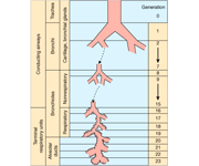
The human respiratory system can be divided in two functional regions: the conducting airways and the respiratory region. The conducting airways, which are composed of the nasal cavity and associated sinuses, the pharynx, larynx, trachea, bronchi, and bronchioles, filter and condition the inspired air. From trachea to the periphery of the airway tree, the airways repeatedly branch dichotomously into two daughter branches with smaller diameters and shorter length than the parent branch (Weibel, 1991). For each new generation of airways, the number of branches is doubled and the crosssectional area is exponentially increased. The conducting region of the airways generally constitutes generation 0 (trachea) to 16 (terminal bronchioles). The respiratory region, where gas exchange takes place, generally constitutes generation 17-23 and is composed of respiratory bronchioles, the alveolar ducts, and the alveolar sacs.

The air-blood barrier of the gas exchange area is composed of the alveolar epithelial cells (surface area 140 m2) on one side and the capillary bed (surface area 130 m2) on the other side of a thin basement membrane (Simionescu, 1991; Stone et al., 1992). The extensive surface area of the air-blood barrier in combination with its extreme thinness (0.1-0.5 μm) permit rapid gas exchange by passive diffusion (Plopper, 1996).

Image

The lung is a very attractive target for drug delivery. It provides direct access to disease in the treatment of respiratory diseases, while providing an enormous surface area and a relatively low enzymatic, controlled environment for systemic absorption of medications. (http://www.ncbi.nlm.nih.gov/pmc/articles/PMC1884307/)

Advantages:

  • Can be self medicated
  • Easy to use
  • Reduced side effects associated with systemic delivery

Disadvantages:

  • Slower route of action
  • Potential problem of deposition to the deeper alveolar (higher generations, like G 8-10)
  • Immuno-defense system
  • Difficulty in measuring the exact dose inside the lung
  • inhaled aerosol is entrapped in the mucus in the conducting airways

Need to be reminded that in addition, a drug’s efficacy may be affected by where in the respiratory tract it is deposited, its delivered dose and the disease it may be trying to treat.

Major components of the lung – barriers to drug absorption
As one of the primary interfaces between the organism and the environment, the respiratory system is constantly exposed to airborne particles, potential pathogens, and toxic gases in the inspired air (Plopper, 1996). As a result a sophisticated respiratory host defense system, present from the nostrils to the alveoli, has evolved to clear offending agents (Twigg, 1998).

The system comprises of:

  • mechanical (i.e. air filtration,cough, sneezing, and mucociliary clearance),
  • chemical (antioxidants, antiproteases and surfactant lipids),
  • immunological defense mechanisms and is tightly regulated to minimize inflammatory reactions that could impair the vital gas-exchange

**Intratracheal inhalation is another  administration option but will be left out of the discussion for now

From a drug delivery perspective, the components of the host defense system comprise barriers that must be overcome to ensure efficient drug deposition and absorption from the respiratory tract.

Generally, lung physiological investigations show that the airway and alveolar epithelia, not the interstitium and the endothelium, constitute the main barrier that restricts the movement of drugs and solutes from the airway lumen into the cells or the blood circulation.

Aerosols are defined as An aerosol is a suspensions of fine solid particles or liquid droplets in a gas.The major aspect affect the efficacy of aerosols as a drug delivery system is Drug Deposition.

Aerosol Drug deposition is affected by:

  • particle properties (e.g. size, shape, density, and charge),
  • respiratory tract morphology,
  • the breathing pattern (e.g. airflow rate and tidal volume)

These parameters determine not only the quantity of particles that are deposited but also in what region of the respiratory tract the particles are deposited.

Particle properties

As the cross-sectional area of the airways increases, the airflow rate rapidly decreases, and consequently the residence time of the particles in the lung increases from the large conducting airways towards the lung periphery. The most important mechanisms of particle deposition in the respiratory tract are (1) inertial impaction, (2) sedimentation, and (3) diffusion.

  • Inertial impaction – Inertial impaction occurs predominantly in the extrathoracic airways and in the tracheobronchial tree, where the airflow velocity is high and rapid changes in airflow direction occurs. Generally, particles with a diameter larger than 10 μm are most likely deposited in the extrathoracic region, whereas 2- to 10-μm particles are deposited in the tracheobronchial tree by inertial impaction. A long residence time of the inspired air favors particle deposition by sedimentation and diffusion.
  • Sedimentation – Sedimentation is of greatest importance in the small airways and alveoli and is most pronounced for particles with a diameter of 0.5-2 μm, Ultrafine particles (<0.5 μm in diameter) are deposited mainly by diffusional transport in the small airways and lung parenchyma where there is a maximal residence time of the inspired air.

Most therapeutic aerosols are almost always heterodisperse, consisting of a wide range of particle sizes and described by the log-normal distribution with the log of the particle diameters plotted against particle number, surface area or volume (mass) on a linear or probability scale and expressed as absolute values or cumulative percentage (http://www.ncbi.nlm.nih.gov/pmc/articles/PMC1884307/)

Optimal drug delivery to the lungs depends on an interaction between;

  • the inhaler device,
  • the drug formulation properties,
  • the inhalation maneuver

The devices currently available for pulmonary drug administration of pharmaceutical aerosols in clinical therapy include nebulizers, pressurized metered dose inhalers (pMDIs), and dry powder inhalers (DPIs).

However, much effort is put into the development of new inhaler devices and formulations to optimize the pulmonary delivery system for local or systemic drug targeting.

One of the major problems in aerosol delivery is

One disadvantage of the aerosol inhalation is, however, that a substantial portion of the aerosolized drug is not delivered to the lungs (i.e. delivered to the nose, mouth, skin, exhaled). only 10–15% of the emitted dose in the lungs.

In general the aerosol exposure techniques have a low dosing effectiveness, which often requires longer exposure times to administer the target dose and renders investigations of rapid kinetic events difficult. In addition, aerosol exposure requires an advanced equipment for exposure and ml-quantities of test formulation to fill up the device.

Airway geometry and humidity

Progressive branching and narrowing of the airways encourage impaction of particles. The larger the particle size, the greater the velocity of incoming air, the greater the bend angle of bifurcations and the smaller the airway radius, the greater the probability of deposition by impaction. The lung has a relative humidity of approximately 99.5%. The addition and removal of water can significantly affect the particle size of a hygroscopic aerosol and thus deposition. Drug particles are known to be hygroscopic and grow or shrink in size in high humidity, such as in the lung. A hygroscopic aerosol that is delivered at relatively low temperature and humidity into one of high humidity and temperature would be expected to increase in size when inhaled into the lung. The rate of growth is a function of the initial diameter of the particle, with the potential for the diameter of fine particles <1 µm to increase five-fold compared with two-to-three-fold for particles >2 µm. he increase in particle size above the initial size should affect the amount of drug deposited and particularly, the distribution of the aerosolized drug within the lung,

Lung Clearance Mechanism

Once deposited in the lungs, inhaled drugs are either cleared from the lungs, absorbed into the systemic circulation or degraded via drug metabolism. Drug particles deposited in the conducting airways are primarily removed through mucociliary clearance and, to a lesser extent, are absorbed through the airway . epithelium into the blood or lymphatic system. a low-viscosity periciliary or sol layer covered by a high-viscosity gel layer. Insoluble particles are trapped in the gel layer and are moved toward the pharynx (and ultimately to the gastrointestinal tract) by the upward movement of mucus generated by the metachronous beating of cilia. In the normal lung, the rate of mucus movement varies with the airway region and is determined by the number of ciliated cells and their beat frequency. Movement is faster in the trachea than in the small airways and is affected by factors influencing ciliary functioning and the quantity and quality of mucus.

Drugs deposited in the alveolar region may be phagocytosed and cleared by alveolar macrophages or absorbed into the pulmonary circulation. Alveolar macrophages are the predominant phagocytic cell for the lung defence against inhaled microorganisms, particles and other toxic agents. There are approximately five to seven alveolar macrophages per alveolus in the lungs of healthy nonsmokers. Macrophages phagocytose insoluble particles that are deposited in the alveolar region and are either cleared by the lymphatic system or moved into the ciliated airways along currents in alveolar fluid and then cleared via the mucociliary escalator.

Very little is known about how the drug-metabolizing activities of the lung affect the concentration and therapeutic efficacy of inhaled drugs. All metabolizing enzymes found in the liver are found to a lesser extent in the lung. Therefore assuming, drug deposition could have been calculated it would be hard to impossible to evaluate it’s metabolism.

In summary:

As the end organ for the treatment of local diseases or as the route of administration for systemic therapies, the lung is a very attractive target for drug delivery. It provides direct access the site of disease for the treatment of respiratory diseases without the inefficiencies and unwanted effects of systemic drug delivery. It provides an enormous surface area and a relatively low enzymatic, controlled environment for systemic absorption of medications. But it is not without barriers. Airway geometry, humidity, clearance mechanisms and presence of lung disease influence the deposition of aerosols and therefore influence the therapeutic effectiveness of inhaled medications. A drug’s efficacy may be affected by the site of deposition in the respiratory tract and the delivered dose to that site. To provide an efficient and effective inhalant therapy, these factors must be considered. Aerosol particle size characteristics can play an important role in avoiding the physiological barriers of the lung, as well as targeting the drug to the appropriate lung region.

Drug formulations and chemo drug delivery will be further discussed in a another post.

Ref:

1. N R Labiris and M B Dolovich. “Pulmonary drug delivery. Part I: Physiological factors affecting therapeutic effectiveness of aerosolized medications”. Br J Clin Pharmacol. 2003 December; 56(6): 588–599. http://www.ncbi.nlm.nih.gov/pmc/articles/PMC1884307/.

2. Tronde A. “Pulmonary drug absorption”. Acta Universities Upsalninesis Uppsala 2002. uu.diva-portal.org/smash/get/diva2:161887/FULLTEXT01

3. Naushad Khan Ghilzai. Pulmonary drug delivery. http://www.drugdel.com/Pulm_review.pdf.

Read Full Post »

Non-small Cell Lung Cancer drugs – where does the Future lie?

In focus: Tarceva, Avastin and Dacomitinib


UPDATED on July 5, 2013

(from reports published in New England Journal of Medicine on drug, crizotinib)


Curator: Ritu Saxena, Ph.D.


Introduction

Non-small cell lung cancer (NSCLC) is the most common type of lung cancer and usually grows and spreads more slowly than small cell lung cancer.

There are three common forms of NSCLC:

  • Adenocarcinomas are often found in an outer area of the lung.
  • Squamous cell carcinomas are usually found in the center of the lung next to an air tube (bronchus).
  • Large cell carcinomas can occur in any part of the lung. They tend to grow and spread faster than the other two types.

Lung cancer is by far the leading cause of cancer death among both men and women. Each year, more people die of lung cancer than of colon, breast, and prostate cancers combined. The American Cancer Society’s most recent estimates for lung cancer in the United States for 2012 reveal that about 226,160 new cases of lung cancer will be diagnosed (116,470 in men and 109,690 in women), and there will be an estimated 160,340 deaths from lung cancer (87,750 in men and 72,590 among women), accounting for about 28% of all cancer deaths.

Treatment

Different types of treatments are available for non-small cell lung cancer. Treatment depends on the stage of the cancer. For patients in whom the cancer has not spread to nearby lymph nodes are recommended surgery. Surgeon may remove- one of the lobes (lobectomy), only a small portion of the lung (wedge removal), or the entire lung (pneumonectomy). Some patients require chemotherapy that uses drugs to kill cancer cells and stop new cells from growing.

FDA approved drugs for NSCLC

Abitrexate (Methotrexate)
Abraxane (Paclitaxel Albumin-stabilized Nanoparticle Formulation) 
Alimta (Pemetrexed Disodium)
Avastin (Bevacizumab)
Bevacizumab
Carboplatin
Cisplatin
Crizotinib
Erlotinib Hydrochloride
Folex (Methotrexate)
Folex PFS (Methotrexate)
Gefitinib
Gemcitabine Hydrochloride
Gemzar (Gemcitabine Hydrochloride)
Iressa (Gefitinib)
Methotrexate
Methotrexate LPF (Methotrexate)
Mexate (Methotrexate)
Mexate-AQ (Methotrexate)
Paclitaxel
Paclitaxel Albumin-stabilized Nanoparticle Formulation
Paraplat (Carboplatin)
Paraplatin (Carboplatin)
Pemetrexed Disodium
Platinol (Cisplatin)
Platinol-AQ (Cisplatin)
Tarceva (Erlotinib Hydrochloride)
Taxol (Paclitaxel)
Xalkori (Crizotinib)

On the basis of target, the drugs have been classified as follows:

Image

NSCLC Drug Market Analysis

NSCLC drug market expected to grow from $4.2 billion in 2010 to $5.4 billion in 2020

Although, a whole list of agents is available for the treatment of NSCLC, the market for NSCLC drugs is expected to expand from $4.2 billion in 2010 to $5.4 billion in 2020 in the United States, France, Germany, Italy, Spain, the United Kingdom and Japan.   

However, drug sales for metastatic/advanced squamous cell non-small-cell lung cancer, which comprises only a small fraction of the market, will decrease from nearly 17 percent in 2010 to approximately 13 percent in 2020. According to surveyed U.S. oncologists and MCO pharmacy directors, increasing overall survival is one of the greatest unmet needs in first-line advanced squamous non-small-cell lung cancer.

In 2009, antimetabolites dominated the NSCLC market, with Eli Lilly’s Alimta (Pemetrexed) accounting for nearly three-quarters of sales within this drug class. Since then, Alimta has faced tough competition from a number of similar drugs and from emerging therapies. It was speculated that the antimetabolites market share would reduce significantly making it the second-largest drug class in NSCLC, while the epidermal growth factor receptor (EGFR) inhibitor class will garner the top market share by 2019.

Genentech/OSI Pharmaceuticals/Roche/Chugai Pharmaceutical’s Tarceva belongs to the EGFR inhibitor class, and has been prescribed principally along with Eli Lilly’s Alimta, to NSCLC patients.Both these drugs have dominated the NSCLC market till 2010, however, their market hold is expected to weaken from 2015-2020, as claimed by Decision Resources Analyst Karen Pomeranz, Ph.D. Decision Resources is a research and advisory firms for pharmaceutical and healthcare issues.

Tarceva (Erlotinib)

Generic Name: Erlotinib, Brand Name: Tarceva

Other Designation: CP 358774, OSI-774, R1415, RG1415, NSC 718781

Mechanism of Action: Tarceva, a small molecule quinazoline, directly and reversibly inhibits the epidermal growth factor receptor (EGFr) tyrosine kinase. Detailed information on how it works could be found at the Macmillian Cancer support website.

Tarceva has been approved for different cancers and several indications have been filed-

  • non-small cell lung cancer (nsclc), locally advanced or metastatic, second line, after failure of at least one prior chemotherapy regimen (2004)
  • pancreatic cancer, locally advanced or metastatic, in combination with gemcitabine, first line (2005)
  • non-small cell lung cancer (nsclc), advanced, maintenance therapy in responders following first line treatment with platinum-based chemotherapy (2010)
  • non-small cell lung cancer (nsclc) harboring epidermal growth factor (EGFr)-activating mutations, first line treatment in advanced disease

Sales of Tarceva 

May, 2012 sales of Tarceva in the US have been reported to be around $564.2 million.

In a recent article published by Vergnenègre et al in the Clinicoeconomic Outcomes Research journal (2012), cross-market cost-effectiveness of Erlotinib was analyzed. The study aimed at estimating the incremental cost-effectiveness of Erlotinib (150 mg/day) versus best supportive care when used as first-line maintenance therapy for patients with locally advanced or metastatic NSCLC and stable disease.

It was determined that treatment with erlotinib in first-line maintenance resulted in a mean life expectancy of 1.39 years in all countries, compared with a mean 1.11 years with best supportive care, which represents 0.28 life-years (3.4 life-months) gained with erlotinib versus best supportive care.

According to the authors analysis, there was a gain in the costs per-life year as $50,882, $60,025, and $35,669 in France, Germany, and Italy, respectively. Hence, on the basis of the study it was concluded that Erlotinib is a cost-effective treatment option when used as first-line maintenance therapy for locally advanced or metastatic NSCLC.

Avastin (Bevacizumab)

Generic Name: Avastin, Brand Name: Bevacizumab

Other Designation: rhuMAb-VEGF, NSC-704865, R435, RG435

Mechanism of Action

Bevacizumab is a recombinant humanized Mab antagonist of vascular endothelial growth factor A (VEGFA) acting as an angiogenesis inhibitor.

Targets

Vascular endothelial growth factor (VEGF, VEGF-A, VEGFA)

Avastin is the only currently approved VEGF inhibitor that selectively targets VEGF-A.

Three other approved oral drugs, pazopanib (Votrient; GlaxoSmithKline), sunitinib (Sutent; Pfizer) and sorafenib (Nexavar; Onyx Pharmaceuticals) are orally available multi-targeted receptor tyrosine kinase inhibitors that include VEGF receptors among their tar­gets.

Avastin has been approved for different cancers and several indications have been filed:

  • colorectal cancer, advanced, metastatic, first line, in combination with a 5-FU based chemotherapy regimen
  • colorectal cancer, relapsed, metastatic, second line, in combintion with 5-FU-based chemotherapy (2004)
  • non-small cell lung cancer (nsclc), non-squamous, inoperable, locally advanced, recurrent or metastatic, in combination with carboplatin and paclitaxel chemotherapy, first line (2006)
  • breast cancer, chemotherapy naive, first line, locally recurrent or metastatic, in combination with taxane chemotherapy (2008, revoked in 2011)
  • non-small cell lung cancer (nsclc), non-squamous, inoperable, locally advanced, recurrent or metastatic, in combination with platinum-based chemotherapy, first line
  • renal cell carcinoma (RCC), metastatic, in combination with interferon (IFN) alpha, first line (2009)
  • glioblastoma multiforme (GBM), relapsed after first line chemoradiotherapy
  • breast cancer, chemotherapy naive, first line, locally recurrent or metastatic, HEr2 negative, in combination with capecitabine (2009)
  • ovarian cancer, in combination with standard chemotherapy (carboplatin and paclitaxel) as a first line treatment following surgery for women with advanced (Stage IIIb/c or Stage IV) epithelial ovarian, primary peritoneal or fallopian tube cancer
  • ovarian cancer, in combination with carboplatin and gemcitabine as a treatment for women with recurrent, platinum-sensitive ovarian cancer

SOURCE:

New medicine Oncology Knowledge Base

Sales of Avastin 

As of May, 2012, sales of Avastin in the US have been reported to be around $2.66 billion.

It attracted a lot of attention over the past few years after its use as a breast cancer treatment. Avastin was approved by the FDA under its fast-track program. However, the data released by the FDA from follow-up studies led to questioning the use of Avastin as a breast cancer drug. Infact, Genentech pulled the indication from Avastin’s label. Henceforth, the FDA did cancel that approval in late 2011. Doctors, however, can still prescribe it off-label. Potential adverse effects of Avastin that came under scrutiny along with unfavorable cost benefit analyses might pose challenges to its growth potential and continued widespread use. However, the sales of Avastin have continued to increase and it has been reported by Fierce Pharma as one of the 15 best-selling cancer drugs list. (Fierce Pharma)

Dacomitinib: New promising drug for NSCLC

Generic Name: Dacomitinib

Other Designation: PF-299804, PF-00299804, PF-299,804, PF00299804

PF-299804 is an orally available irreversible pan-HEr tyrosine kinase inhibitor.

Dacomitinib is a promising new drug on the market. Phase III trials are ongoing for advanced and refractory NSCLC, locally advanced or metastatic NSCLC and the EGFr mutation containing locally advanced or metastatic NSCLC in several countries including those in Europe, Asia, and America.

SOURCE:

New medicine Oncology Knowledge base

Dacomitinib bests Erlotinib in advanced NSCLC:  Comparison of its Progression-Free Survival (PFS) with the NSCLC marketed drug, Erlotinib.

In September of 2012, a study was published by Ramalingam et al in the Journal of Clinical Oncology, which was a randomized open-label trial comparing dacomitinib with erlotinib in patients with advanced NSCLC. On the basis of the study it was concluded that dacomitinib demonstrated significantly improved progression-free survival (PFS*) as compared to erlotinib, with a certain degree of toxicity.

SOURCE:

Randomized Phase II Study of Dacomitinib Versus Erlotinib in Patients With Advanced Non-Small-Cell Lung Cancer

The results indicated indicated the following:

  • Median PFS was significantly greater with Dacomitinib than Erlotinib, at 2.86 versus 1.91.
  • Mean duration of response was 16.56 months for dacomitinib and 9.23 months for erlotinib.

Patients were divided into groups by tumor type and following results were obtained:

  • Median PFS was 3.71 months with dacomitinib and 1.91 with erlotinib in patients with KRAS wild-type tumors
  • Median PFS was 2.21 months and 1.68 months, in patients with KRAS wild-type/EGFR wild-type tumors.
  • PFS was significantly better in the molecular subgroups harboring a mutant EGFR genotype.

The study also highlighted the side effects which might be more of concern and probably limiting for Dacomitinib.

Although adverse side effects were uncommon in both the groups, certain side effects such as:

  • mouth sores,
  • nailbed infections, and
  • diarrhea

were more common and tended to be more severe with Dacomitinib as compared to Tarceva.

Therefore, for patients for whom side effects of Tarceva seem challenging might face more difficulty with Dacomitinib treatment. Nonetheless, the results of PFS were promising enough and provide a greater efficacy in several clinical and molecular subgroups targeting a larger population than Tarceva. Authors, thus, suggested a larger, randomized phase III trial with the same design.

Current status of Dacomitinib

Based on positive performance of Dacomitinib published in research studies, Pfizer has entered into a collaborative development agreement with the SFJ Pharmaceuticals Group to conduct a phase III clinical trial across multiple sites in Asia and Europe, to evaluate dacomitinib (PF-00299804) as a first line treatment in patients with locally advanced or metastatic non-small cell lung cancer (nsclc) with activating mutations in the epidermal growth factor receptor (EGFr). Under the terms of the agreement, SFJ will provide the funding and clinical development supervision to generate the clinical data necessary to support a registration dossier on Dacomitinib for marketing authorization by regulatory authorities for this indication. If approved for this indication, SFJ will be eligible to receive milestone and earn-out payments.

SOURCE:

New medicine Oncology Knowledge base

*PFS or Progression-free survival is defined as the length of time during and after the treatment of as disease, such as cancer, that a patient lives with the disease but it does not get worse. In a clinical trial, measuring the progression-free survival is one way to see how well a new treatment works.

REFERENCES

Recently, another drug PF-02341066 (crizotinib), was tested on patients with non-small cell lung cancer and the results were published in New England Journal of Medicine (2013). Crizotinib is an orally available aminopyridine-based inhibitor of the) and the c-Met/hepatocyte growth factor receptor (HGFR). Crizotinib, in an ATP-competitive manner, binds to and inhibits ALK kinase and ALK fusion proteins. In addition, crizotinib inhibits c-Met kinase, and disrupts the c-Met signaling pathway. Altogether, this agent inhibits tumor cell growth.

  • Shaw and colleagues (2013) investigated whether crizotinib is superior to standard chemotherapy with respect to efficacy. To answer the question, Pfizer launched a phase III clinical trial (NCT00932893; http://clinicaltrials.gov/show/NCT00932893) comparing the safety and anti-tumor activity of PF-02341066 (crizotinib) versus pemetrexed or docetaxel in patients with advanced non-small cell lung cancer harboring a translocation or inversion event involving the ALK gene. Shaw and colleagues (2013) published the results of the clinical trial in a recent issue of New England Journal of Medicine.  A total of 347 patients with locally advanced or metastatic ALK-positive lung cancer who had received one prior platinum-based regimen were recruited for the trial and patients were randomly assigned to receive oral treatment with crizotinib (250 mg) twice daily or intravenous chemotherapy with either pemetrexed (500 mg per square meter of body-surface area) or docetaxel (75 mg per square meter) every 3 weeks. Patients in the chemotherapy group who had disease progression were permitted to cross over to crizotinib as part of a separate study. The primary end point was progression-free survival. According to the results, the median progression-free survival was 7.7 months in the crizotinib group and 3.0 months in the chemotherapy group. Hazard ratio (HR) for progression or death with crizotinib was 0.49 (95% CI, P<0.001). The response rates were 65% with crizotinib, as compared with 20% with chemotherapy (P<0.001). An interim analysis of overall survival showed no significant improvement with crizotinib as compared with chemotherapy (hazard ratio for death in the crizotinib group, 1.02; 95% CI, P=0.54). Common adverse events associated with crizotinib were visual disorder, gastrointestinal side effects, and elevated liver aminotransferase levels, whereas common adverse events with chemotherapy were fatigue, alopecia, and dyspnea. Patients reported greater reductions in symptoms of lung cancer and greater improvement in global quality of life with crizotinib than with chemotherapy.In conclusion, the results from the trial indicate that crizotinib is superior to standard chemotherapy in patients with previously treated, advanced non–small-cell lung cancer with ALK rearrangement. (Shaw AT, et al, Crizotinib versus Chemotherapy in Advanced ALK-Positive Lung Cancer. N Engl J Med 2013; 20 June, 368:2385-2394; http://www.ncbi.nlm.nih.gov/pubmed/23724913).

However, in the same issue of New England Journal of Medicine, Awad and colleagues (2013) reported from a phase I clinical trial (NCT00585195; http://clinicaltrials.gov/show/NCT00585195), that a patient with metastatic lung adenocarcioma harboring a CD74-ROS1 rearrangement who had initially shown a dramatic response to treatment, showed resistance to crizotinib. Biopsy of the resistant tumor identified an acquired mutation leading to a glycine-to-arginine substitution at codon 2032 in the ROS1 kinase domain. Although this mutation does not lie at the gatekeeper residue, it confers resistance to ROS1 kinase inhibition through steric interference with drug binding. The same resistance mutation was observed at all the metastatic sites that were examined at autopsy, suggesting that this mutation was an early event in the clonal evolution of resistance. The study was funded by Pfizer (Awad MM, et al, Acquired resistance to crizotinib from a mutation in CD74-ROS1. N Engl J Med. 2013 Jun 20;368(25):2395-401; http://www.ncbi.nlm.nih.gov/pubmed/23724914)

Reference: 

Read Full Post »

Author and Curator: Ritu Saxena, Ph.D.

Introduction: Mitochondrial fission & fusion

Mitochondria, double membranous and semi-autonomous organelles, are known to convert energy into forms that are usable to the cell. Apart from being sites of cellular respiration, multiple roles of mitochondria have been emphasized in processes such as cell division, growth and cell death. Mitochondria are semi-autonomous in that they are only partially dependent on the cell to replicate and grow. They have their own DNA, ribosomes, and can make their own proteins. Mitochondria have been discussed in several posts published in the Pharmaceutical Intelligence blog.

Mitochondria do not exist as lone organelles, but are part of a dynamic network that continuously undergoes fusion and fission in response to various metabolic and environmental stimuli. Nucleoids, the assemblies of mitochondrial DNA (mtDNA) with its associated proteins, are distributed during fission in such a way that each mitochondrion contains at least one nucleoid. Mitochondrial fusion and fission within a cell is speculated to be involved in several functions including mtDNA DNA protection, alteration of cellular energetics, and regulation of cell division.

Proteins involved in mitochondrial fission & fusion

Multiple mitochondrial membrane GTPases that regulate mitochondrial networking have recently been identified. They are classified as fission and fusion proteins:

Fusion proteins: Members of dynamin family of protein, mitofusin 1 (Mfn-1) and mitofusin 2 (Mfn-2), are involved in fusion between mitochondria by tethering adjacent mitochondria. These proteins have two transmembrane segments that anchor them in the mitochondrial outer membrane. Mutations in Mitofusin proteins gives rise to fragmented mitochondria, but this can be reversed by mutations in mammalian Drp1. Mitochondrial inner membranes are fused by dynamin family members called Opa1.

Fission proteins: Another member of the dynamin family of proteins, dynamin-related protein 1 (Drp-1) mediates fission of mitochondria. Drp-1 is activated by phosphorylation. Drp-1 proteins are largely cytosolic, but cycle on and off of mitochondria as needed for fission. Fission is a complex process and involves a series of well-defined stages and proteins. Cytosolic Drp-1 is activated by calcineurin or other cytosolic signaling proteins after which it translocates to the mitochondrial tubules where it assembles into foci through its interaction with another protein, hFis1. Once Drp-1 rings assemble on the constricted spots, outer membrane of mitochondria undergoes fission through GTP hydrolysis. Drp-1 is now left bound to one of the newly formed mitochondrial ends after which it slowly disassembles before returning to the cytoplasm.

Control of mitochondrial fission & fusion

  • Mitochondrial fission and fusion are controlled by several regulatory mechanisms. Few of which are mentioned as follows:
  • Drp-1 activation by Cdk1/Cyclin B mediated phosphorylation during mitosis – triggers fission
  • Drp-1 inactivation by cAMP-dependent protein kinase (PKA) in quiescent cells- prevents fission
  • Drp-1 activation after reversal of PKA phosphorylation by Calcineurin- triggers fission
  • Ubiquination of fission and fusion proteins by E3 ubiquitin ligase- alters fission
  • Sumoylation of fission proteins – regulates fission

Imparied mitochondrial fission leads to loss of mtDNA

Mitochondrial fission plays an important role in mitochondrial and cellular homeostasis. It was reported by Parone et al (2008) that preventing mitochondrial fission by down-regulating expression of Drp-1 lead to loss of mtDNA and mitochondrial dysfunction. An increase in cellular reactive oxygen species (ROS) was observed. Other cellular implications included depletion of cellular ATP, inhibition of cell proliferation and autophagy. The observations were made in HeLa cells.

MicroRNA regulation of mitochondrial fission

Although several factors have been attributed to the regulation of mitochondrial fission, the mechanism still remains poorly understood. Recently, regulation of mitochondrial fission via miRNAs has become a topic of interest. Following miRNAs have been found to be involved in mitochondrial fission:

  • miR-484:  Wang et al (2012) demonstrated that miR-484 was able to regulate mitochondrial fission by suppressing the translation of a fission protein Fis1, leading to inhibition of Fis1-mediated fission and apoptosis in cardiomyocytes and in the adrenocortical cancer cells. The authors showed that Fis1 is necessary for mitochondrial fission and apoptosis, and is upregulated during anoxia, whereas miR-484 is downregulated. Underlying mechanism involved transactivation of miR-484 by a transcription factor, Foxo3a and miR-484 is able to attenuate Fis1 upregulation and mitochondrial fission, by binding to the amino acid coding sequence of Fis1 and inhibiting its translation.
  • miR-499: miR-499 was reported by Wang et al (2011) to be able to directly target both the α- and β-isoforms of the calcineurin catalytic subunit. Suppression of calcineurin-mediated dephosphorylation of  Drp-1 lead to inhibition of the fission machinery ultimately resulting in the inhibition of cardiomyocyte apoptosis. miR-499 levels, by altering mitochondrial fusion were able affect the severity of myocardial infarction and cardiac dysfunction induced by ischemia-reperfusion. Modulation of miR-499 expression could provide a therapeutic approach for myocardial infarction treatment.
  • miR-30: It was reported by Li et al (2010) that miR-30 family members were able to inhibit mitochondrial fission and also the resulting apoptosis. While exploring the underlying molecular mechanism, the authors identified that miR-30 family members can suppress p53 expression. When cell received apoptotic stimulation, p53 was found to transcriptionally activate the fission protein, Drp-1. Drp-1 was able to induce mitochondrial fission. miR-30 family members were observed to inhibit mitochondrial fission through attenuation of p53 expression and its downstream target Drp-1.

Mitochondrial fission & fusion as a therapeutic target

Since alteration of mitochondrial fission and fusion have been reported to affect various cellular processes including apoptosis, proliferation, ATP consumption, the proteins involved in the process of fission and fusion might be harnessed as therapeutic target.

Mentioned below is a description of research where dynamics of the mitochondrial organelle has been utilized as a therapeutic target:

Inhibition of mitochondrial fission prevents cell cycle progression in lung cancer

A recent article published by Rehman et al (2012) in the FASEB journal drew much attention after interesting observations were made in the mitochondria of lung adenocarcinoma cells. The mitochondrial network of these cells exhibited both impaired fusion and enhanced fission. It was also found that the fragmented phenotype in multiple lung adenocarcinoma cell lines was associated with both a down-regulation of the fusion protein, Mfn-2 and an upregulation of expression of fission protein, Drp-1. The imbalance of Drp-1/Mfn-2 expression in human lung cancer cell lines was reported to promote a state of mitochondrial fission. Similar increase in Drp-1 and decrease in Mfn-2 was observed in the tissue samples from patients compared to adjacent healthy lung. Authors used complementary approaches of Mfn-2 overexpression, Drp-1 inhibition, or Drp-1 knockdown and were able to observe reduction of cancer cell proliferation and an increase spontaneous apoptosis. Thus, the study identified mitochondrial fission and Drp-1 activation as a novel therapeutic target in lung cancer.

Image

Reference:

Research articles-

http://www.ncbi.nlm.nih.gov/pubmed/20556877

http://www.ncbi.nlm.nih.gov/pubmed?term=18806874

http://www.ncbi.nlm.nih.gov/pubmed/22510686

http://www.ncbi.nlm.nih.gov/pubmed/21186368

http://www.ncbi.nlm.nih.gov/pubmed?term=20062521

http://www.ncbi.nlm.nih.gov/pubmed?term=22321727

News brief:

http://www.uchospitals.edu/news/2012/20120221-mitochondria.html

http://news.uchicago.edu/article/2012/02/23/energy-network-within-cells-may-be-new-target-cancer-therapy

http://www.doctortipster.com/7881-mitochondria-could-represent-a-new-target-for-cancer-therapy-according-to-new-study.html

Related reading:

Reviewer: Larry H Bernstein, MD, FACP

http://pharmaceuticalintelligence.com/2012/10/28/mitochondrial-damage-and-repair-under-oxidative-stress/

Author and Curator: Larry H Bernstein, MD, FACP http://pharmaceuticalintelligence.com/2012/09/26/mitochondria-origin-from-oxygen-free-environment-role-in-aerobic-glycolysis-metabolic-adaptation/

Reporter and Editor: Larry H Bernstein, MD, FACP

http://pharmaceuticalintelligence.com/2012/09/16/nitric-oxide-has-a-ubiquitous-role-in-the-regulation-of-glycolysis-with-a-concomitant-influence-on-mitochondrial-function/

Author and Reporter: Ritu Saxena, PhD

http://pharmaceuticalintelligence.com/2012/09/10/%CE%B2-integrin-emerges-as-an-important-player-in-mitochondrial-dysfunction-associated-gastric-cancer/

Author: Ritu Saxena, PhD

http://pharmaceuticalintelligence.com/2012/09/01/mitochondria-and-cancer-an-overview/

Author and Reporter: Ritu Saxena, PhD

http://pharmaceuticalintelligence.com/2012/08/14/mitochondrial-mutation-analysis-might-be-1-step-away/

Reporter: Venkat S. Karra, PhD

http://pharmaceuticalintelligence.com/2012/08/14/detecting-potential-toxicity-in-mitochondria/

Reporter: Aviva Lev-Ari, PhD, RN http://pharmaceuticalintelligence.com/2012/08/01/mitochondrial-mechanisms-of-disease-in-diabetes-mellitus/

Author and Curator: Ritu Saxena, PhD; Consultants: Aviva Lev-Ari, PhD, RN and Pnina G. Abir-Am, PhD

http://pharmaceuticalintelligence.com/2012/07/09/mitochondria-more-than-just-the-powerhouse-of-the-cell/

Read Full Post »

Reporter: Aviva Lev-Ari, PhD, RN

Medical Education Firm Launches Online Tool to Help Docs Guide Personalized Rx Decisions in NSCLC

September 12, 2012
Clinical Care Options, a developer of continuing education and medical decision support resources, has launched a web-based tool to help oncologists figure out which lung cancer patients may benefit from molecularly guided personalized treatments.

The online decision-support tool provides oncologists with expert recommendations on first-line and maintenance treatment options for non-small cell lung cancer patients based on their patients’ medical information and tumor features, including oncogenic markers.

Clinical Care Options developed the online tool based on the treatment choices made by five US experts who were presented 96 cases with specific variables regarding patients’ medical history, such as tumor histology, genomic mutations, age, and smoking history.

In order to use the tool, oncologists select their patients’ medical information and desires and select their treatment of choice. The tool then displays how the five experts would treat this patient. The program then surveys users about how the expert recommendations impacted their treatment decisions.

The firm presented the results of this survey in a poster at the Chicago Multidisciplinary Symposium in Thoracic Oncology this week. The tool has been used by approximately 1,000 physicians around the world, according to Jim Mortimer, senior director of oncology programs and partnership development at Clinical Care Options. Overall, approximately 23 percent of clinicians who used the tool have said it helped change their decisions, while 50 percent indicated the tool helped confirm their initial treatment strategy.

Specifically, with regard to genomically guided personalized NSCLC treatments, all five of the experts selected Pfizer’s Xalkori (crizotinib) whenever a patient case involved the ALK fusion gene. However, out of 80 cases entered by oncologists involving this marker, only around 40 percent selected Xalkori. And although in NSCLC cases with mutated EGFR the experts selected Genentech’s Tarceva (erlotinib), only 60 percent of the 100 such cases entered by clinicians into the tool chose the drug.

The data collected by Clinical Care Options suggest that its decision-support tool may be a useful resource when oncologists want to assess how their peers would prescribe a genomically targeted personalized treatment. These drugs, compared to standard treatments, are relatively new to the market and expensive. Pfizer’s Xalkori was approved by the US Food and Drug Administration last year while Genentech is in the process of getting approval for Tarceva in the US as a first-line treatment for NSCLC patients who have EGFR mutations. Last year, the European Commission approved the use of Tarceva as a first-line treatment for NSCLC in patients with EGFR mutations (PGx Reporter 9/7/2011).

Clinical Care Options said launched the online tool because it noticed that physicians often look for advice beyond broad treatment guidelines when it comes to making decisions for specific patients.

“The tool recommendations align very well with the treatment guidelines but the advantage of the tool is the granularity of the case specifics. Users of the tool can quickly enter in details of a case and see the results for what five experts would recommend,” Mortimer told PGx Reporter. “This contrasts with guidelines that apply to broad groups and provide lists of suitable treatments.”

Mortimer noted that some of the experts’ recommendations included in the tool are outside of the exact indication of a particular drug. However, because the experts’ treatment decisions were evidence based, they “did not indicate any issues with reimbursement.”

Clinical Care Options has developed a continuing medical education-certified program that includes the tool with educational grants from Genentech and Pfizer.

Read Full Post »

Comprehensive Genomic Characterization of Squamous Cell Lung Cancers

Reporter: Aviva Lev-Ari, PhD, RN

Nature (2012) doi:10.1038/nature11404

Received 09 March 2012 
Accepted 09 July 2012 
Published online 09 September 2012
Correspondence to: 

The primary and processed data used to generate the analyses presented here can be downloaded by registered users fromThe Cancer Genome Atlas (https://tcga-data.nci.nih.gov/tcga/tcgaDownload.jsp,https://cghub.ucsc.edu/ and https://tcga-data.nci.nih.gov/docs/publications/lusc_2012/).

Lung squamous cell carcinoma is a common type of lung cancer, causing approximately 400,000 deaths per year worldwide. Genomic alterations in squamous cell lung cancers have not been comprehensively characterized, and no molecularly targeted agents have been specifically developed for its treatment. As part of The Cancer Genome Atlas, here we profile 178 lung squamous cell carcinomas to provide a comprehensive landscape of genomic and epigenomic alterations. We show that the tumour type is characterized by complex genomic alterations, with a mean of 360 exonic mutations, 165 genomic rearrangements, and 323 segments of copy number alteration per tumour. We find statistically recurrent mutations in 11 genes, including mutation of TP53 in nearly all specimens. Previously unreported loss-of-function mutations are seen in the HLA-A class I major histocompatibility gene. Significantly altered pathways included NFE2L2 andKEAP1 in 34%, squamous differentiation genes in 44%, phosphatidylinositol-3-OH kinase pathway genes in 47%, and CDKN2A and RB1 in 72% of tumours. We identified a potential therapeutic target in most tumours, offering new avenues of investigation for the treatment of squamous cell lung cancers.

Read Full Post »

Reporter: Prabodh Kandala, PhD

A team of University of Hawaii Cancer Center scientists led by James Turkson, Ph.D. have created a new type of anti-cancer drug named BP-1-102. The drug, which can be orally administered, targets a key protein that triggers the development of many types of cancer including lung, breast and skin cancers.

The development of BP-1-102 was guided by the research teams computer based molecular analysis of the cancer causing Stat 3 protein that causes cancer by promoting abnormal cell growth in otherwise healthy cells.

“The molecular structure of the hyperactive Stat3 protein basically resembles two cars that are joined together side-by-side,” said Professor Turkson. “We then utilized a computer program that creates molecular models of potential drugs engaging in binding to the Stat3 protein to craft the BP-1-102 drug which literally pulls apart the Stat3 protein rendering it ineffective in causing cancer.”

A unique feature of BP-1-102 is that it remains highly effective against cancer even when administered in oral form. Presently, most anti-cancer drugs require intravenous (IV) administration in a clinic or hospital setting which increases the financial, physical and emotional burdens on cancer patients. In its experimental form, BP-1-102 has shown promise in treating breast and lung cancers.

Currently, breast and lung cancers are two of the most commonly diagnosed cancers accounting for nearly half a million cases per year in the United States with over 200,000 deaths attributed to these diseases. In Hawaii, there is an average of 1500 cases diagnosed and over 600 deaths attributed to breast and lung cancers every year.

Professor Turkson is a recent and welcomed addition to the UH Cancer Center faculty. His innovative and ground breaking research focuses on developing novel anticancer drugs based on targeting signal transduction and apoptosis pathways.

Ref:

http://www.sciencedaily.com/releases/2012/05/120522115252.htm

http://www.pnas.org/content/109/2/600

Read Full Post »

« Newer Posts