Feeds:
Posts
Comments

Posts Tagged ‘Cardiovascular disease’

Clinical Decision Support Systems for Management Decision Making of Cardiovascular Diseases

Author, and Content Consultant to e-SERIES A: Cardiovascular Diseases: Justin Pearlman, MD, PhD, FACC

and

Curator: Aviva Lev-Ari, PhD, RN

This image has an empty alt attribute; its file name is ArticleID-46.png

WordCloud Image Produced by Adam Tubman

Clinical Decision Support Systems (CDSS)

Clinical decision support system (CDSS) is an interactive decision support system (DSS). It generally relies on computer software designed to assist physicians and other health professionals with decision-making tasks, such as when to apply a particular diagnosis, further specific tests or treatments. A functional definition proposed by Robert Hayward of the Centre for Health Evidence defines CDSS as follows:  “Clinical Decision Support systems link health observations with health knowledge to influence health choices by clinicians for improved health care”. CDSS is a major topic in artificial intelligence in medicine.

Vinod Khosla of A Khosla Ventures investment, in a Fortune Magazine article, “Technology will replace 80% of what doctors do”, on December 4, 2012, wrote about CDSS as a harbinger of science in medicine.

Computer-assisted decision support is in its infancy, but we have already begun to see meaningful impact on healthcare. Meaningful use of computer systems is now rewarded under the Affordable Care Act.  Studies have demonstrated the ability of computerized clinical decision support systems to lower diagnostic errors of omission significantly, by directly countering cognitive bias.  Isabel is a differential diagnosis tool and, according to a Stony Book study, matched the diagnoses of experienced clinicians in 74% of complex cases. The system improved to a 95% match after a more rigorous entry of patient data. The IBM supercomputer, Watson, after beating all humans at the intelligence-based task of playing Jeopardy, is now turning its attention to medical diagnosis. It can process natural language questions and is fast at parsing high volumes of medical information, reading and understanding 200 million pages of text in 3 seconds. 

Examples of CDSS

  1. CADUCEUS
  2. DiagnosisPro
  3. Dxplain
  4. MYCIN
  5. RODIA

VIEW VIDEO

“When Should a Physician Deviate from the Diagnostic Decision Support Tool and What Are the Associated Risks?”

Introduction

Justin D. Pearlman, MD, PhD

A Decision Support System consists of one or more tools to help achieve good decisions. For example, decisions that can benefit from DSS include whether or not to undergo surgery, whether or not to undergo a stress test first, whether or not to have an annual mammogram starting at a particular age, or a computed tomography (CT) to screen for lung cancer, whether or not to utilize intensive care support such as a ventilator, chest shocks, chest compressions, forced feeding, strong antibiotics and so on versus care directed to comfort measures only without regard to longevity.

Any DSS can be viewed like a digestive tract, chewing on input, and producing output, and like the digestive tract, the output may only be valuable to a farmer. A well designed DSS is efficient in the input, timely in its processing and useful in the output. Mathematically, a DSS is a model with input parameters and an output variable or set of variables that can be used to determine an action. The input can be categorical (alive, dead), semi-quantitative (cold-warm-hot), or quantitative (temperature, systolic blood pressure, heart rate, oxygen saturation). The output can be binary (yes-no) or it can express probabilities or confidence intervals.

The process of defining specifications for a function and then deriving a useful function is called mathematical modeling. We will derive the function for “average” as an example. By way of specifications, we want to take a list of numbers as input, and come out with a single number that represents the middle of the pack or “central tendency.”   The order of the list should not matter, and if we change scales, the output should scale the same way. For example, if we use centimeters instead of inches, and we apply 2.54 centimeters to an inch, then the output should increase by the multiplier 2.54. If the list of numbers are all the same then the output should be the consistent value. Representing these specifications symbolically:

1. order doesn’t matter: f(a,b) = f(b,a), where “a” and “b” are input values, “f” is the function.

2. multipliers pass through (linearity):  f(ka,kb)=k f(a,b), where k is a scalar e.g. 2.54 cm/inch.

3. identity:  f(a,a,a,…) = a

Properties 1 and 2 lead us to consider linear functions consisting of sums and multipliers: f(a,b,c)=Aa+Bb+Cc …, where the capital letters are multipliers by “constants” – numbers that are independent of the list values a,b,c, and since the order should not matter, we simplify to f(a,b,c)=K (a+b+c+…) because a constant multiplier K makes order not matter. Property 3 forces us to pick K = 1/N where N is the length of the list. These properties lead us to the mathematical solution: average = sum of list of numbers divided by the length of the list.

A coin flip is a simple DSS: heads I do it, tails I don’t. The challenge of a good DSS is to perform better than random choice and also perform better (more accurately, more efficiently, more reliably, more timely and/or under more adverse conditions) than unassisted human decision making.

Therefore, I propose the following guiding principles for DSS design: choose inputs wisely (accessible, timely, efficient, relevant), determine to what you want output to be sensitive AND to what you want output to be insensitive, and be very clear about your measures of success.

For example, consider designing a DSS to determine whether a patient should receive the full range of support capabilities of an intensive care unit (ICU), or not. Politicians have cited the large bump in the cost of the last year of life as an opportunity to reduce costs of healthcare, and now pay primary care doctors to encourage patients to establish advanced directives not to use ICU services. From the DSS standpoint, the reasoning is flawed because the decision not to use ICU services should be sensitive to benefit as well as cost, commonly called cost-benefit analysis. If we measure success of ICU services by the benefit of quality life net gain (QLNG, “quailing”), measured in quality life-years (QuaLYs) and achieve 50% success with that, then the cost per QuaLY measures the cost-benefit of ICU services. In various cost-benefit decisions, the US Congress has decided to proceed if the cost is under $20-$100,000/QuaLY. If ICU services are achieving such a cost-benefit, then it is not logical to summarily block such services in advance. Rather, the ways to reduce those costs include improving the cost efficiency of ICU care, and improving the decision-making of who will benefit.

An example of a DSS is the prediction of plane failure from a thousand measurements of strain and function of various parts of an airplane. The desired output is probability of failure to complete the next flight safely. Cost-Benefit analysis then establishes what threshold or operating point merits grounding the plane for further inspection and preventative maintenance repairs. If a DSS reports probability of failure, then the decision (to ground the plane) needs to establish a threshold at which a certain probability triggers the decision to ground the plane.

The notion of an operating point brings up another important concept in decision support. At first blush, one might think the success of a DSS is determined by its ability to correctly identify a predicted outcome, such as futility of ICU care (when will the end result be no quality life net gain). The flaw in that measure of success is that it depends on prevalence in the study group. As an extreme example, if you study a group of patients with fatal gunshot wounds to the head, none will benefit and the DSS requirement is trivial and any DSS that says no for that group has performed well. At the other extreme, if all patients become healthy, the DSS requirement is also trivial, just say yes. Therefore the proper assessment of a DSS should pay attention to the prevalence and the operating point.

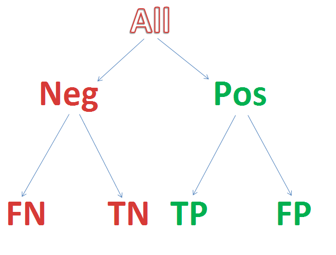
The impact of prevalence and operating point on decision-making is addressed by receiver-operator curves. Consider looking at the blood concentration of Troponin-I (TnI) as the sole determinant to decide who is having a heart attack.  If one plots a graph with horizontal axis troponin level and vertical axis ultimate proof of heart attack, the percentage of hits will generally be higher for higher values of TnI. To create such a graph, we compute a “truth table” which reports whether the test was above or below a decision threshold operating point, and whether or not the disease (heart attack) was in fact present:

TRUTH TABLE

              Disease            Not Disease
Test Positive

TP

FP

Test Negative

FN

TN

Total

TP+FN

FP+TN

The sensitivity to the disease is the true positive rate (TPR), the percentage of all disease cases that are ranked by the decision support as positive: TPR = TP/(TP+FN). 100% sensitivity can be achieved trivially by lowering the threshold for a positive test to zero, at a cost.  While sensitivity is necessary for success it is not sufficient. In addition to wanting sensitivity to disease, we want to avoid labeling non-disease as disease. That is often measured by specificity, the true negative rate (TNR), the percentage of those without disease who are correctly identified as not having disease: TNR = TN/(FP+TN). I propose also we define the complement to specificity, the anti-sensitivity, as the false positive rate (FPR), FPR = FP/(FP+TN) = 1 – TNR. Anti-sensitivity is a penalty cost of lowering the diagnostic threshold to boost sensitivity, as the concomitant rise in anti-sensitivity means a growing number of non-disease subjects are labeled as having disease. We want high sensitivity to true disease without high anti-sensitivity to false disease, and we want to be insensitive to common distractors. In these formulas, note that false negatives (FN) are True for disease, and false positives (FP) are False for disease, so the denominators add FN to TP for total True disease, and add FP to TN for total False for disease.

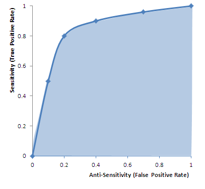
The graph in figure 1 justifies the definition of anti-sensitivity. It is an ROC or “Receiver-Operator Curve” which is a plot of sensitivity versus anti-sensitivity for different diagnostic thresholds of a test (operating points). Note, higher sensitivity comes at the cost of higher anti-sensitivity. Where to operate (what threshold to use for diagnosis) can be selected according to cost-benefit analysis of sensitivity versus anti-sensitivity (and specificity).

 untitled
FIgure 1 ROC (Receiver-Operator Curve): Graph of sensitivity (true positive rate) versus anti-sensitivity (false positive rate) computed by changing the operating point (threshold for declaring a test numeric value positive for disease). High area under the curve (AUC) is favorable because it means less anti-sensitivity for high sensitivity (upper left corner of shaded area more to the left, and higher). The dots on the curve are operating points. An inclusive operating point (high on the curve, high sensitivity) is used for screening tests, whereas an exclusive operating point (low on the curve, low anti-sensitivity) is used for definitive diagnosis.

Cost benefit analysis generally is based on a semi-lattice, or upside-down branching tree, which represents all choices and outcomes. It is important to include all branches down to final outcomes. For example, if the test is a mammogram to screen for breast cancer, the cost is not just the cost of the test, and the benefit “early diagnosis.” The cost-benefit calculation forces us to put a numerical value on the impact, such as a financial cost to an avoidable death, or we can get a numerical result in terms of quality life years expected. The cost, however, is not just the cost of the mammogram, but also of downstream events such as the cost of the needle biopsies for the suspicious “positives” and so on.

semilattice decision treeFigure 2 Semi-lattice Decision Tree: Starting from all patients, create a branch point for your test result, and add further branch points for any subsequent step-wise outcomes until you reach the “bottom line.” Assign a value to each, resulting in a numerical net cost and net benefit. If tests invoke risks (for example, needle biopsy of lung can collapse a lung and require hospitalization for a chest tube) then insert branch points for whether the complication occurs or not, as the treatment of a complication counts as part of the cost. The intermediary nodes can have probability of occurrence as their numeric factor, and the bottom line can apply the net probability of the path leading to a value as a multiplier to the dollar value (a 10% chance of costing $10,000 counts as an expectation cost of 0.1 x 10,000 = $1,000).

A third area of discussion is the statistical power of a DSS – how reliable is it in the application that you care about? Commonly DSS design is contrary to common statistical applications which address significance of a deviation in a small number of variables that have been measured many times in a large population. Instead, DSS often uses many variables to fully describe or characterize the status of a small population. For example, thousands of different measurements may be performed on a few dozen airplanes, aiming to predict when the plane should be grounded for repairs. A similar inversion of numbers – numerous variables, small number of cases – is common in genomics studies.

The success of a DDS is measured by its predictive value compared to outcomes or other measures of success. Thus measures of success include positive predictive value, negative predictive value, and confidence. A major problem with DDS is the inversion of the usually desired ratio of repetitions to measurement variables. When you get a single medical lab test, you have a single measurement value such as potassium level and a large number of normal subjects for comparison. If we knew the  mean μ and standard deviation σ that describes the distribution of normal values in the population at large, then we could compute the confidence in the decision to call our observed value abnormal based on the normal distribution:  , <br /><br /><br /><br /><br /><br /><br /><br /><br />
f(x) = \frac{1}{\sigma\sqrt{2\pi}} e^{ -\frac{(x-\mu)^2}{2\sigma^2} }.<br /><br /><br /><br /><br /><br /><br /><br /><br />

A value may be deemed distinctive based on a 95% confidence interval if it falls outside of the norm, say by more than twice the standard deviation σ, thereby establishing that it is unlikely to be random as the distance from the mean excludes 95% of the normal distribution.

The determination of confidence in an observed set of results stems from maximized likelihood estimates. Earlier in this article we described how to derive the the mean, or center, of a set of measurements. A similar analysis can derive the standard deviation (square root of variance) as a measure of spread around the mean, as well as other descriptive statistics based on sample values. These formulas describe the distribution of sample values about the mean. The calculation is based on a simple inversion. If we knew the mean and variance of a population of values for a measurement, we could calculate the likelihood of each new measurement falling a particular distance from the mean, and we could calculate the combined likelihood for a set of observed values. Maximized Likelihood Estimation (MLE) simply inverts the method of calculation. Instead of treating the mean and variance as known, we can treat the sample observations as the known data, to characterize a distribution for the observed data samples from an estimate of the spread about an unknown mean from a set of N normal samples xi (one can apply calculus to compute the formulas below for the unknown mean and unknown variance, based simply on computing how to maximize the joint likelihood of the observations  xi from the frequency distribution above, in order t0 derive the following formulas): 

\sigma = \sqrt{\frac{1}{N}\left[(x_1-\mu)^2 + (x_2-\mu)^2 + \cdots + (x_N - \mu)^2\right]}, {\rm \ \ where\ \ } \mu = \frac{1}{N} (x_1 + \cdots + x_N),

The frequency distribution (a function of mean and spread) reports the frequency of observing xi  if it is drawn from a population with the specified mean μ and standard deviation σ . We can invert that by treating the observations, xi , as known and the mean μ and standard deviation σ unknown, then calculate the values μ and  σ that maximize the likelihood of our sample set as coming from the dynamically described population.

In DSS there is typically an inversion of the usually requisite large number of samples (small versus large) and number of variables (large versus small. This inversion has major consequences on data confidence. If you measure just 14 independent variables versus one variable, each at 95% confidence, the net confidence drops exponentially to less than 50%: 0.9514=49%. In the airplane grounding screen tests, 1000 independent variables, at 95% confidence each, yields a net confidence of only 5 x 10-23 which is 10 sextillion times less than 50% confidence. This same problem arises in genomics research, in which we have a large array of gene product measurements on a small number of patients. Standard statistical tools are problematic at high variable counts. One can turn to qualitative grouping tools such as exploratory factor analysis, or recover statistical robustness with HykGene, a combined cluster and ranking method devised by the author to improve dramatically the ability to identify distinctions with confidence when the number of variables is high.

Evolution of DSS

Aviva Lev-Ari, PhD, RN

The examples provided above refer to sets of binary models, one family of DSS. Another type of DSS is multivariate in nature, a corollary of multivariate scenarios constitute alternative choice options. Last decade development in the DSS field involved the design of Recommendation Engines given manifested preference functions that involved simultaneous trade-off functions against cost function. Game theoretical context is embedded into Recommendation Engines. The output mentioned above, is in fact an array of options with probabilities of saving reward assigned by the Recommendation Engine.

Underlining Computation Engines

Methodological Basis of Clinical DSS

There are many different methodologies that can be used by a CDSS in order to provide support to the health care professional.[7]

The basic components of a CDSS include a dynamic (medical) knowledge base and an inference mechanism (usually a set of rules derived from the experts and evidence-based medicine) and implemented through medical logic modules based on a language such as Arden syntax. It could be based on Expert systems or artificial neural networks or both (connectionist expert systems).

Bayesian Network

The Bayesian network is a knowledge-based graphical representation that shows a set of variables and their probabilistic relationships between diseases and symptoms. They are based on conditional probabilities, the probability of an event given the occurrence of another event, such as the interpretation of diagnostic tests. Bayes’ rule helps us compute the probability of an event with the help of some more readily available information and it consistently processes options as new evidence is presented. In the context of CDSS, the Bayesian network can be used to compute the probabilities of the presence of the possible diseases given their symptoms.

Some of the advantages of Bayesian Network include the knowledge and conclusions of experts in the form of probabilities, assistance in decision making as new information is available and are based on unbiased probabilities that are applicable to many models.

Some of the disadvantages of Bayesian Network include the difficulty to get the probability knowledge for possible diagnosis and not being practical for large complex systems given multiple symptoms. The Bayesian calculations on multiple simultaneous symptoms could be overwhelming for users.

Example of a Bayesian network in the CDSS context is the Iliad system which makes use of Bayesian reasoning to calculate posterior probabilities of possible diagnoses depending on the symptoms provided. The system now covers about 1500 diagnoses based on thousands of findings.

Another example is the DXplain system that uses a modified form of the Bayesian logic. This CDSS produces a list of ranked diagnoses associated with the symptoms.

A third example is SimulConsult, which began in the area of neurogenetics. By the end of 2010 it covered ~2,600 diseases in neurology and genetics, or roughly 25% of known diagnoses. It addresses the core issue of Bayesian systems, that of a scalable way to input data and calculate probabilities, by focusing specialty by specialty and achieving completeness. Such completeness allows the system to calculate the relative probabilities, rather than the person inputting the data. Using the peer-reviewed medical literature as its source, and applying two levels of peer-review to the data entries, SimulConsult can add a disease with less than a total of four hours of clinician time. It is widely used by pediatric neurologists today in the US and in 85 countries around the world.

Neural Network

Artificial Neural Networks (ANN) is a nonknowledge-based adaptive CDSS that uses a form of artificial intelligence, also known as machine learning, that allows the systems to learn from past experiences / examples and recognizes patterns in clinical information. It consists of nodes called neuron and weighted connections that transmit signals between the neurons in a forward or looped fashion. An ANN consists of 3 main layers: Input (data receiver or findings), Output (communicates results or possible diseases) and Hidden (processes data). The system becomes more efficient with known results for large amounts of data.

The advantages of ANN include the elimination of needing to program the systems and providing input from experts. The ANN CDSS can process incomplete data by making educated guesses about missing data and improves with every use due to its adaptive system learning. Additionally, ANN systems do not require large databases to store outcome data with its associated probabilities. Some of the disadvantages are that the training process may be time consuming leading users to not make use of the systems effectively. The ANN systems derive their own formulas for weighting and combining data based on the statistical recognition patterns over time which may be difficult to interpret and doubt the system’s reliability.

Examples include the diagnosis of appendicitis, back pain, myocardial infarction, psychiatric emergencies and skin disorders. The ANN’s diagnostic predictions of pulmonary embolisms were in some cases even better than physician’s predictions. Additionally, ANN based applications have been useful in the analysis of ECG (A.K.A. EKG) waveforms.

Genetic Algorithms

A Genetic Algorithm (GA) is a nonknowledge-based method developed in the 1940s at the Massachusetts Institute of Technology based on Darwin’s evolutionary theories that dealt with the survival of the fittest. These algorithms rearrange to form different re-combinations that are better than the previous solutions. Similar to neural networks, the genetic algorithms derive their information from patient data.

An advantage of genetic algorithms is these systems go through an iterative process to produce an optimal solution. The fitness function determines the good solutions and the solutions that can be eliminated. A disadvantage is the lack of transparency in the reasoning involved for the decision support systems making it undesirable for physicians. The main challenge in using genetic algorithms is in defining the fitness criteria. In order to use a genetic algorithm, there must be many components such as multiple drugs, symptoms, treatment therapy and so on available in order to solve a problem. Genetic algorithms have proved to be useful in the diagnosis of female urinary incontinence.

Rule-Based System

A rule-based expert system attempts to capture knowledge of domain experts into expressions that can be evaluated known as rules; an example rule might read, “If the patient has high blood pressure, he or she is at risk for a stroke.” Once enough of these rules have been compiled into a rule base, the current working knowledge will be evaluated against the rule base by chaining rules together until a conclusion is reached. Some of the advantages of a rule-based expert system are the fact that it makes it easy to store a large amount of information, and coming up with the rules will help to clarify the logic used in the decision-making process. However, it can be difficult for an expert to transfer their knowledge into distinct rules, and many rules can be required for a system to be effective.

Rule-based systems can aid physicians in many different areas, including diagnosis and treatment. An example of a rule-based expert system in the clinical setting is MYCIN. Developed at Stanford University by Edward Shortliffe in the 1970s, MYCIN was based on around 600 rules and was used to help identify the type of bacteria causing an infection. While useful, MYCIN can help to demonstrate the magnitude of these types of systems by comparing the size of the rule base (600) to the narrow scope of the problem space.

The Stanford AI group subsequently developed ONCOCIN, another rules-based expert system coded in Lisp in the early 1980s.[8] The system was intended to reduce the number of clinical trial protocol violations, and reduce the time required to make decisions about the timing and dosing of chemotherapy in late phase clinical trials. As with MYCIN, the domain of medical knowledge addressed by ONCOCIN was limited in scope and consisted of a series of eligibility criteria, laboratory values, and diagnostic testing and chemotherapy treatment protocols that could be translated into unambiguous rules. Oncocin was put into production in the Stanford Oncology Clinic.

Logical Condition

The methodology behind logical condition is fairly simplistic; given a variable and a bound, check to see if the variable is within or outside of the bounds and take action based on the result. An example statement might be “Is the patient’s heart rate less than 50 BPM?” It is possible to link multiple statements together to form more complex conditions. Technology such as a decision table can be used to provide an easy to analyze representation of these statements.

In the clinical setting, logical conditions are primarily used to provide alerts and reminders to individuals across the care domain. For example, an alert may warn an anesthesiologist that their patient’s heart rate is too low; a reminder could tell a nurse to isolate a patient based on their health condition; finally, another reminder could tell a doctor to make sure he discusses smoking cessation with his patient. Alerts and reminders have been shown to help increase physician compliance with many different guidelines; however, the risk exists that creating too many alerts and reminders could overwhelm doctors, nurses, and other staff and cause them to ignore the alerts altogether.

Causal Probabilistic Network

The primary basis behind the causal network methodology is cause and effect. In a clinical causal probabilistic network, nodes are used to represent items such as symptoms, patient states or disease categories. Connections between nodes indicate a cause and effect relationship. A system based on this logic will attempt to trace a path from symptom nodes all the way to disease classification nodes, using probability to determine which path is the best fit. Some of the advantages of this approach are the fact that it helps to model the progression of a disease over time and the interaction between diseases; however, it is not always the case that medical knowledge knows exactly what causes certain symptoms, and it can be difficult to choose what level of detail to build the model to.

The first clinical decision support system to use a causal probabilistic network was CASNET, used to assist in the diagnosis of glaucoma. CASNET featured a hierarchical representation of knowledge, splitting all of its nodes into one of three separate tiers: symptoms, states and diseases.

  1. ^ a b c d e “Decision support systems .” 26 July 2005. 17 Feb. 2009 <http://www.openclinical.org/dss.html>.
  2. 2^ a b c d e f g Berner, Eta S., ed. Clinical Decision Support Systems. New York, NY: Springer, 2007.
  3. 3^ Khosla, Vinod (December 4, 2012). “Technology will replace 80% of what doctors do”. Retrieved April 25, 2013.
  4. ^ Garg AX, Adhikari NK, McDonald H, Rosas-Arellano MP, Devereaux PJ, Beyene J et al. (2005). “Effects of computerized clinical decision support systems on practitioner performance and patient outcomes: a systematic review.”. JAMA 293 (10): 1223–38. doi:10.1001/jama.293.10.1223. PMID 15755945.
  5. ^ Kensaku Kawamoto, Caitlin A Houlihan, E Andrew Balas, David F Lobach. (2005). “Improving clinical practice using clinical decision support systems: a systematic review of trials to identify features critical to success.”. BMJ 330 (7494): 765. doi:10.1136/bmj.38398.500764.8F. PMC 555881. PMID 15767266.
  6. ^ Gluud C, Nikolova D (2007). “Likely country of origin in publications on randomised controlled trials and controlled clinical trials during the last 60 years.”. Trials 8: 7. doi:10.1186/1745-6215-8-7. PMC 1808475. PMID 17326823.
  7. ^ Wagholikar, K. “Modeling Paradigms for Medical Diagnostic Decision Support: A Survey and Future Directions”. Journal of Medical Systems. Retrieved 2012.
  8. ^ ONCOCIN: An expert system for oncology protocol management E. H. Shortliffe, A. C. Scott, M. B. Bischoff, A. B. Campbell, W. V. Melle, C. D. Jacobs Seventh International Joint Conference on Artificial Intelligence, Vancouver, B.C.. Published in 1981

SOURCE for Computation Engines Section and REFERENCES:

http://en.wikipedia.org/wiki/Clinical_decision_support_system

Cardiovascular Diseases: Decision Support Systems (DSS) for Disease Management Decision Making – DSS analyzes information from hospital cardiovascular patients in real time and compares it with a database of thousands of previous cases to predict the most likely outcome.

Can aviation technology reduce heart surgery complications?

Algorithm for real-time analysis of data holds promise for forecasting
August 13, 2012 | By 

British researchers are working to adapt technology from the aviation industry to help prevent complications among heart patients after surgery. Up to 1,000 sensors aboard aircraft help airlines determine when a plane requires maintenance, reports The Engineer, serving as a model for the British risk-prediction system.

The system analyzes information from hospital cardiovascular patients in real time and compares it with a database of thousands of previous cases to predict the most likely outcome.

“There are vast amounts of clinical data currently collected which is not analyzed in any meaningful way. This tool has the potential to identify subtle early signs of complications from real-time data,” Stuart Grant, a research fellow in surgery at University Hospital of South Manchester, says in a hospital statement. Grant is part of the Academic Surgery Unit working with Lancaster University on the project, which is still its early stages.

The software predicts the patient’s condition over a 24-hour period using four metrics: systolic blood pressure, heart rate, respiration rate and peripheral oxygen saturation, explains EE Times.

As a comparison tool, the researchers obtained a database of 30,000 patient records from the Massachusetts Institute of Technology and combined it with a smaller, more specialized database from Manchester.

In six months of testing, its accuracy is about 75 percent, The Engineer reports. More data and an improved algorithm could boost that rate to 85 percent, the researchers believe. Making the software web-based would allow physicians to access the data anywhere, even on tablets or phones, and could enable remote consultation with specialists.

In their next step, the researchers are applying for more funding and for ethical clearance for a large-scale trial.

U.S. researchers are working on a similar crystal ball, but one covering an array of conditions. Researchers from the University of Washington, MIT and Columbia University are using a statistical model that can predict future ailments based on a patient’s history–and that of thousands of others.

And the U.S. Department of Health & Human Services is using mathematical modeling to analyze effects of specific healthcare interventions.

Predictive modeling also holds promise to make clinical research easier by using algorithms examine multiple scenarios based on different kinds of patient populations, specified health conditions and various treatment regimens

To learn more:
– here’s the Engineer article
– check out the hospital report
– read the EE Times article

Related Articles:
Algorithm looks to past to predict future health conditions
HHS moves to mathematical modeling for research, intervention evaluation
Decision support, predictive modeling may speed clinical research

SOURCE:

Can aviation technology reduce heart surgery complications? – FierceHealthIT http://www.fiercehealthit.com/story/can-aviation-technology-reduce-heart-surgery-complications/2012-08-13#ixzz2SITHc61J

http://www.fiercehealthit.com/story/study-decision-support-systems-must-be-flexible-adaptable-transparent/2012-08-20

Medical Decision Making Tools: Overview of DSS available to date  

http://www.openclinical.org/dss.html

Clinical Decision Support Systems – used for Cardiovascular Medical Decisions

Stud Health Technol Inform. 2010;160(Pt 2):846-50.

AALIM: a cardiac clinical decision support system powered by advanced multi-modal analytics.

Amir A, Beymer D, Grace J, Greenspan H, Gruhl D, Hobbs A, Pohl K, Syeda-Mahmood T, Terdiman J, Wang F.

Source

IBM Almaden Research Center, San Jose, CA, USA.

Abstract

Modern Electronic Medical Record (EMR) systems often integrate large amounts of data from multiple disparate sources. To do so, EMR systems must align the data to create consistency between these sources. The data should also be presented in a manner that allows a clinician to quickly understand the complete condition and history of a patient’s health. We develop the AALIM system to address these issues using advanced multimodal analytics. First, it extracts and computes multiple features and cues from the patient records and medical tests. This additional metadata facilitates more accurate alignment of the various modalities, enables consistency check and empowers a clear, concise presentation of the patient’s complete health information. The system further provides a multimodal search for similar cases within the EMR system, and derives related conditions and drugs information from them. We applied our approach to cardiac data from a major medical care organization and found that it produced results with sufficient quality to assist the clinician making appropriate clinical decisions.

PMID: 20841805 [PubMed – indexed for MEDLINE]

DSS development for Enhancement of Heart Drug Compliance by Cardiac Patients 

A good example of a thorough and effective CDSS development process is an electronic checklist developed by Riggio et al. at Thomas Jefferson University Hospital (TJUH) [12]. TJUH had a computerized physician order-entry system in place. To meet congestive heart failure and acute myocardial infarction quality measures (e.g., use of aspirin, beta blockers, and angiotensin-converting enzyme (ACE) inhibitors), a multidisciplinary team including a focus group of residents developed a checklist, embedded in the computerized discharge instructions, that required resident physicians to prescribe the recommended medications or choose from a drop-down list of contraindications. The checklist was vetted by several committees, including the medical executive committee, and presented at resident conferences for feedback and suggestions. Implementation resulted in a dramatic improvement in compliance.

http://virtualmentor.ama-assn.org/2011/03/medu1-1103.html

Early DSS Development at Stanford Medical Center in the 70s

MYCIN (1976)     MYCIN was a rule-based expert system designed to diagnose and recommend treatment for certain blood infections (antimicrobial selection for patients with bacteremia or meningitis). It was later extended to handle other infectious diseases. Clinical knowledge in MYCIN is represented as a set of IF-THEN rules with certainty factors attached to diagnoses. It was a goal-directed system, using a basic backward chaining reasoning strategy (resulting in exhaustive depth-first search of the rules base for relevant rules though with additional heuristic support to control the search for a proposed solution). MYCIN was developed in the mid-1970s by Ted Shortliffe and colleagues at Stanford University. It is probably the most famous early expert system, described by Mark Musen as being “the first convincing demonstration of the power of the rule-based approach in the development of robust clinical decision-support systems” [Musen, 1999].

The EMYCIN (Essential MYCIN) expert system shell, employing MYCIN’s control structures was developed at Stanford in 1980. This domain-independent framework was used to build diagnostic rule-based expert systems such as PUFF, a system designed to interpret pulmonary function tests for patients with lung disease.

http://www.bmj.com/content/346/bmj.f657

ECG for Detection of MI: DSS use in Cardiovascualr Disease Management

http://faculty.ksu.edu.sa/AlBarrak/Documents/Clinical%20Decision%20Support%20Systems_Ch01.pdf

also showed that neural networks did a better job than two experienced cardiologists in detecting acute myocardial infarction in electrocardiograms with concomitant left bundle branch block.

Olsson SE, Ohlsson M, Ohlin H, Edenbrandt L. Neural networks—a diagnostic tool in acute myocardial infarction with concomitant left bundle branch block. Clin Physiol Funct Imaging 2002;22:295–299.

Sven-Erik Olsson, Hans Öhlin, Mattias Ohlsson and Lars Edenbrandt
Neural networks – a diagnostic tool in acute myocardial infarction with concomitant left bundle branch block
Clinical Physiology and Functional Imaging 22, 295-299 (2002) 

Abstract
The prognosis of acute myocardial infarction (AMI) improves by early revascularization. However the presence of left bundle branch block (LBBB) in the electrocardiogram (ECG) increases the difficulty in recognizing an AMI and different ECG criteria for the diagnosis of AMI have proved to be of limited value. The purpose of this study was to detect AMI in ECGs with LBBB using artificial neural networks and to compare the performance of the networks to that of six sets of conventional ECG criteria and two experienced cardiologists. A total of 518 ECGs, recorded at an emergency department, with a QRS duration > 120 ms and an LBBB configuration, were selected from the clinical ECG database. Of this sample 120 ECGs were recorded on patients with AMI, the remaining 398 ECGs being used as a control group. Artificial neural networks of feed-forward type were trained to classify the ECGs as AMI or not AMI. The neural network showed higher sensitivities than both the cardiologists and the criteria when compared at the same levels of specificity. The sensitivity of the neural network was 12% (P = 0.02) and 19% (P = 0.001) higher than that of the cardiologists. Artificial neural networks can be trained to detect AMI in ECGs with concomitant LBBB more effectively than conventional ECG criteria or experienced cardiologists.

http://home.thep.lu.se/~mattias/publications/papers/lu_tp_00_38_abs.html

Additional SOURCES:

http://www.implementationscience.com/content/6/1/92

http://www.fiercehealthit.com/story/study-decision-support-systems-must-be-flexible-adaptable-transparent/2012-08-20

 Comment of Note

During 1979-1983 Dr. Aviva Lev-Ari was part of Prof. Ronald A. Howard, Stanford University, Study Team, the consulting group to Stanford Medical Center during MYCIN feature enhancement development.

Professor Howard is one of the founders of the decision analysis discipline. His books on probabilistic modeling, decision analysis, dynamic programming, and Markov processes serve as major references for courses and research in these fields.

https://engineering.stanford.edu/profile/rhoward

It was Prof. Howard from EES, Prof. Amos Tversky of Behavior Science  (Advisor of Dr. Lev-Ari’s Masters Thesis at HUJ), and Prof. Kenneth Arrow, Economics, with 15 doctoral students in the early 80s, that formed the Interdisciplinary Decision Analysis Core Group at Stanford. Students of Prof. Howard, chiefly, James E. Matheson, started the Decision Analysis Practice at Stanford Research Institute (SRI, Int’l) in Menlo Park, CA.

http://www.sri.com/

Dr. Lev-Ari  was hired on 3/1985 to head SRI’s effort in algorithm-based DSS development. The models she developed were applied in problem solving for  SRI Clients, among them Pharmaceutical Manufacturers: Ciba Geigy, now NOVARTIS, DuPont, FMC, Rhone-Poulenc, now Sanofi-Aventis.

Read Full Post »

Reporter: Aviva Lev-Ari, PhD, RN

Gene found that regenerates heart tissue

DALLAS – April 17, 2013 – Researchers at UT Southwestern Medical Center have identified a specific gene that regulates the heart’s ability to regenerate after injuries.

Scientists led by Dr. Hesham Sadek have demonstrated that the gene Meis1 regulates the regenerative capability of newborn hearts.

Scientists led by Dr. Hesham Sadek have demonstrated that the gene Meis1 regulates the regenerative capability of newborn hearts.

The function of the gene, called Meis1, in the heart was not known previously. The findings of the UTSW investigation are available online in Nature.

“We found that the activity of the Meis1 gene increases significantly in heart cells soon after birth, right around the time heart muscle cells stop dividing. Based on this observation we asked a simple question: If the Meis1 gene is deleted from the heart, will heart cells continue to divide through adulthood? The answer is ‘yes’,” said Dr. Hesham Sadek, assistant professor of internal medicine in the division of cardiology, and senior author of the study.

In 2011, Dr. Sadek’s laboratory showed that the newborn mammalian heart is capable of a vigorous, regenerative response to injury through division of its own cells. As the newborn develops, the heart rapidly loses the ability to regenerate and to repair injuries such as heart attacks.

The research team demonstrated that deletion of Meis1 extended the proliferation period in the hearts of newborn mice, and also re-activated the regenerative process in the adult mouse heart without harmful effect on cardiac functions. This new finding demonstrates that Meis1 is a key factor in the regeneration process, and the understanding of the gene’s function may lead to new therapeutic options for adult heart regeneration. The findings also provide a possible alternative to current adult heart regeneration research, which focuses on the use of stem cells to replace damaged heart cells.

“Meis1 is a transcription factor, which acts like a software program that has the ability to control the function of other genes. In this case, we found that Meis1 controls several genes that normally act as brakes on cell division,” Dr. Sadek said. “As such, Meis1 could possibly be used as an on/off switch for making adult heart cells divide. If done successfully, this ability could introduce a new era in treatment for heart failure.”

According to the American Heart Association, almost 6 million people in the U.S. have heart failure, which occurs when the heart cannot pump enough blood and oxygen to support other organs. Heart disease is the leading cause of death for both men and women in the country, according to the Centers for Disease Control and Prevention.

The study received support from the American Heart Association, the Gilead Research Scholars Program in Cardiovascular Disease, the Foundation for Heart Failure Research, and the National Institutes of Health.

The co-first authors of the study are Dr. Ahmed I. Mahmoud, who is now a postdoctoral fellow at Harvard University; Dr. Fatih Kocabas, who is now a postdoctoral fellow at North American College; and Dr. Shalini A. Muralidhar, a postdoctoral research fellow II of internal medicine. Other researchers at UT Southwestern involved in the study are Wataru Kimura, a visiting senior researcher of internal medicine; Ahmed Koura, now a medical student at Ain Shams University in Egypt; Dr. Enzo Porrello, research fellow and faculty member at the University of Queensland in Australia; and Suwannee Thet, a research associate of internal medicine.

About UT Southwestern Medical Center
UT Southwestern, one of the premier academic medical centers in the nation, integrates pioneering biomedical research with exceptional clinical care and education. The institution’s faculty includes many distinguished members, including five who have been awarded Nobel Prizes since 1985. Numbering more than 2,700, the faculty is responsible for groundbreaking medical advances and is committed to translating science-driven research quickly to new clinical treatments. UT Southwestern physicians provide medical care in 40 specialties to nearly 100,000 hospitalized patients and oversee more than 2.1 million outpatient visits a year.

Media Contact: Remekca Owens
214-648-9344
remekca.owens@utsouthwestern.edu

 http://www.utsouthwestern.edu/newsroom/news-releases/year-2013/april/heart-sadek.html?goback=.gde_4842427_member_233989854

Genetics: A gene of rare effect

A mutation that gives people rock-bottom cholesterol levels has led geneticists to what could be the next blockbuster heart drug.

09 April 2013
ADAPTED FROM: PETER DAZELEY/GETTY

When Sharlayne Tracy showed up at the clinical suite in the University of Texas (UT) Southwestern Medical Center in Dallas last January, the bandage wrapped around her left wrist was the only sign of anything medically amiss. The bandage covered a minor injury from a cheerleading practice led by Tracy, a 40-year-old African American who is an aerobics instructor, a mother of two and a college student pursuing a degree in business. “I feel like I’m healthy as a horse,” she said.

Indeed, Tracy’s well-being has been inspiring to doctors, geneticists and now pharmaceutical companies precisely because she is so normal. Using every tool in the modern diagnostic arsenal — from brain scans and kidney sonograms to 24-hour blood-pressure monitors and cognitive tests — researchers at the Texas medical centre have diagnostically sliced and diced Tracy to make sure that the two highly unusual genetic mutations she has carried for her entire life have produced nothing more startling than an incredibly low level of cholesterol in her blood. At a time when the target for low-density lipoprotein (LDL) cholesterol, more commonly called ‘bad cholesterol’, in Americans’ blood is less than 100 milligrams per decilitre (a level many people fail to achieve), Tracy’s level is just 14.

A compact woman with wide-eyed energy, Tracy (not her real name) is one of a handful of African Americans whose genetics have enabled scientists to uncover one of the most promising compounds for controlling cholesterol since the first statin drug was approved by the US Food and Drug Administration in 1987. Seven years ago, researchers Helen Hobbs and Jonathan Cohen at UT-Southwestern reported1 that Tracy had inherited two mutations, one from her father and the other from her mother, in a gene called PCSK9, effectively eliminating a protein in the blood that has a fundamental role in controlling the levels of LDL cholesterol. African Americans with similar mutations have a nearly 90% reduced risk of heart disease. “She’s our girl, our main girl,” says Barbara Gilbert, a nurse who has drawn some 8,000 blood samples as part of Cohen and Hobbs’ project to find genes important to cholesterol metabolism.

Of all the intriguing DNA sequences spat out by the Human Genome Project and its ancillary studies, perhaps none is a more promising candidate to have a rapid, large-scale impact on human health than PCSK9. Elias Zerhouni, former director of the US National Institutes of Health (NIH) in Bethesda, Maryland, calls PCSK9 an “iconic example” of translational medicine in the genomics era. Preliminary clinical trials have already shown that drugs that inhibit the PCSK9 protein — used with or without statins — produce dramatic reductions in LDL cholesterol (more than 70% in some patients). Half-a-dozen pharmaceutical companies — all aiming for a share of the global market for cholesterol-reducing drugs that could reach US$25 billion in the next five years according to some estimates — are racing to the market with drugs that mimic the effect of Tracy’s paired mutations.

Free interview

Stephen Hall talks about Sharlayne’s unusual condition and whether similar cases might lead to a new line of drugs.

Zerhouni, now an in-house champion of this class of drug as an executive at drug firm Sanofi, headquartered in Paris, calls the discovery and development of PCSK9 a “beautiful story” in which researchers combined detailed physical information about patients with shrewd genetics to identify a medically important gene that has made “super-fast” progress to the clinic. “Once you have it, boy, everything just lines up,” he says. And although the end of the PCSK9 story has yet to be written — the advanced clinical trials now under way could still be derailed by unexpected side effects — it holds a valuable lesson for genomic research. The key discovery about PCSK9‘s medical potential was made by researchers working not only apart from the prevailing scientific strategy of genome research over the past decade, but with an almost entirely different approach.

As for Tracy, who lives in the southern part of Dallas County, the implications of her special genetic status have become clear. “I really didn’t understand at first,” she admits. “But now I’m watching ads on TV [for cholesterol-lowering drugs], and it’s like, ‘Wow, I don’t have that problem’.”

A heart problem

Cardiovascular disease is — and will be for the foreseeable future, according to the World Health Organization — the leading cause of death in the world, and its development is intimately linked to elevated levels of cholesterol in the blood. Since their introduction, statin drugs have been widely used to lower cholesterol levels. But Jan Breslow, a physician and geneticist at Rockefeller University in New York, points out that up to 20% of patients cannot tolerate statins’ side effects, which include muscle pain and even forgetfulness. And in many others, the drugs simply don’t control cholesterol levels well enough.

The search for better treatments for heart disease gained fresh impetus after scientists published the draft sequence of the human genome in 2001. In an effort to identify the genetic basis of common ailments such as heart disease and diabetes, geneticists settled on a strategy based on the ‘common variant hypothesis’. The idea was that a handful of disease-related versions (or variants) of genes for each disease would be common enough — at a frequency of roughly 5% or so — to be detected by powerful analyses of the whole genome. Massive surveys known as genome-wide association studies compared the genomes of thousands of people with heart disease, for example, with those of healthy controls. By 2009, however, many scientists were lamenting the fact that although the strategy had identified many common variants, each made only a small contribution to the disease. The results for cardiovascular disease have been “pretty disappointing”, says Daniel Steinberg, a lipoprotein expert at the University of California, San Diego.

Single-minded: Helen Hobbs and Jonathan Cohen’s approach to heart-disease genetics yielded a target for drugs that could compete with statins.MISTY KEASLER/REDUX/EYEVINE

More than a decade earlier, in Texas, Hobbs and Cohen had taken the opposite tack. They had backgrounds in Mendelian, or single-gene, disorders, in which an extremely rare variant can have a big — often fatal — effect. They also knew that people with a particular Mendelian disorder didn’t share a single common mutation in the affected gene, but rather had a lot of different, rare mutations. They hypothesized that in complex disorders, many different rare variants were also likely to have a big effect, whereas common variants would have relatively minor effects (otherwise natural selection would have weeded them out). “Jonathan and I did not see any reason why it couldn’t be that rare variants cumulatively contribute to disease,” Hobbs says. To find these rare variants, the pair needed to compile detailed physiological profiles, or phenotypes, of a large general population. Cohen spoke of the need to “Mendelize” people — to compartmentalize them by physiological traits, such as extremely high or low cholesterol levels, and then look in the extreme groups for variations in candidate genes known to be related to the trait.

The pair make a scientific odd couple. Hobbs, who trained as an MD, is gregarious, voluble and driven. Cohen, a soft-spoken geneticist from South Africa, has a laid-back, droll manner and a knack for quantitative thinking. In 1999, they set out to design a population-based study that focused on physical measurements related to heart disease. Organized with Ronald Victor, an expert on high blood pressure also at UT Southwestern, and funded by the Donald W. Reynolds Foundation in Las Vegas, Nevada, the Dallas Heart Study assembled exquisitely detailed physiological profiles on a population of roughly 3,500 Dallas residents2. Crucially, around half of the participants in the study were African Americans, because the researchers wanted to probe racial differences in heart disease and high blood pressure. The team measured blood pressure, body mass index, heart physiology and body-fat distribution, along with a battery of blood factors related to cholesterol metabolism — triglycerides, high-density lipoprotein (HDL) cholesterol and LDL cholesterol. In the samples of blood, of course, they also had DNA from each and every participant.

As soon as the database was completed in 2002, Hobbs and Cohen tested their rare-variant theory by looking at levels of HDL cholesterol. They identified the people with the highest (95th percentile) and lowest (5th percentile) levels, and then sequenced the DNA of three genes known to be key to metabolism of HDL cholesterol. What they found, both in Dallas and in an independent population of Canadians, was that the number of mutations was five times higher in the low HDL group than in the high group3. This made sense, Cohen says, because most human mutations interfere with the function of genes, which would lead to the low HDL numbers. Published in 2004, the results confirmed that rare, medically important mutations could be found in a population subdivided into extreme phenotypes.

Armed with their extensive database of cardiovascular traits, Hobbs and Cohen could now dive back into the Dallas Heart Study whenever they had a new hypothesis about heart disease and, as Cohen put it, “interrogate the DNA”. It wasn’t long before they had an especially intriguing piece of DNA at which to look.

The missing link

In February 2003, Nabil Seidah, a biochemist at the Clinical Research Institute of Montreal in Canada, and his colleagues reported the discovery of an enigmatic protein4. Seidah had been working on a class of enzymes known collectively as proprotein convertases, and the researchers had identified what looked like a new member of the family, called NARC-1: neural apoptosis-regulated convertase 1.

“We didn’t know what it was doing, of course,” Seidah says. But the group established that the gene coding the enzyme showed activity in the liver, kidney and intestines as well as in the developing brain. The team also knew that in humans the gene mapped to a precise genetic neighbourhood on the short arm of chromosome 1.

That last bit of geographical information pointed Seidah to a group led by Catherine Boileau at the Necker Hospital in Paris. Her team had been following families with a genetic form of extremely high levels of LDL cholesterol known as familial hypercholesterolaemia, which leads to severe coronary artery disease and, often, premature death. Group member Marianne Abifadel had spent five fruitless years searching a region on the short arm of chromosome 1 for a gene linked to the condition. When Seidah contacted Boileau and told her that he thought NARC-1 might be the gene she was looking for, she told him, “You’re crazy”, Seidah recalls. Seidah bet her a bottle of champagne that he was correct; within two weeks, Boileau called back, saying: “I owe you three bottles.”

“The PCSK9 story is a terrific example of an up-and-coming pattern of translational research.”

In 2003, the Paris and Montreal groups reported that the French families with hypercholesterolaemia had one of two mutations in this newly discovered gene, and speculated that this might cause increased production of the enzyme5. Despite Seidah’s protests, the journal editors gave both the gene and its protein product a new name that fit with standard nomenclature: proprotein convertase subtilisin/kexin type 9, or PCSK9. At around the same time, Kara Maxwell in Breslow’s group at Rockefeller University6 and Jay Horton, a gastroenterologist at UT-Southwestern7 also independently identified the PCSK9 gene in mice and revealed its role in a previously unknown pathway regulating cholesterol8.

The dramatic phenotype of the French families told Hobbs that “this is an important gene”. She also realized that in genetics, mutations that knock out a function are much more common than ones that amplify function, as seemed to be the case with the French families. “So immediately I’m thinking, a loss-of-function mutation should manifest as a low LDL level,” she says. “Let’s go and see if that’s true.”

Going to extremes

Hobbs and Cohen had no further to look than in the extreme margins of people in the Dallas Heart Study. In quick order, they identified the highest and lowest LDL readings in four groups: black women, black men, white women and white men. They then resequenced the PCSK9 gene in the low-cholesterol groups, looking for mutations that changed the make-up of the protein.

They found seven African Americans with one of two distinct ‘nonsense’ mutations in PCSK9 — mutations that essentially aborted production of the protein. Then they went back and looked for the same mutations in the entire population. Just 2% of all black people in the Dallas study had either of the two PCSK9 mutations — and those mutations were each associated with a 40% reduction of LDL cholesterol in the blood9. (The team later detected a ‘missense mutation’ in 3% of white people, which impaired but did not entirely block production of the protein.) The frequency of the mutations was so low, Hobbs says, that they would never have shown up in a search for common variants.

When Hobbs and Cohen published their findings in 2005, they suggested that PCSK9 played a crucial part in regulating bad cholesterol, but said nothing about whether the mutations had any effect on heart disease. That evidence came later that year, when they teamed up with Eric Boerwinkle, a geneticist at the University of Texas Health Science Center in Houston, to look forPCSK9 mutations in the Atherosclerosis Risk in Communities (ARIC) study, a large prospective study of heart disease that had been running since 1987. To experts such as Steinberg, the results10 — published in early 2006 — were “mind-blowing”. African Americans in ARIC who had mutations in PCSK9 had 28% less LDL cholesterol and an 88% lower risk of developing heart disease than people without the mutations. White people with the less severe mutation in the gene had a 15% reduction in LDL and a 47% reduced risk of heart disease.

How did the gene exert such profound effects on LDL cholesterol levels? As researchers went on to determine11, the PCSK9 protein normally circulates in the bloodstream and binds to the LDL receptor, a protein on the surface of liver cells that captures LDL cholesterol and removes it from the blood. After binding with the receptor, PCSK9 escorts it into the interior of the cell, where it is eventually degraded. When there is a lot of PCSK9 (as in the French families), there are fewer LDL receptors remaining to trap and remove bad cholesterol from the blood. When there is little or no PCSK9 (as in the black people with mutations), there are more free LDL receptors, which in turn remove more LDL cholesterol.

“We didn’t understand why everybody wasn’t doing what we were doing.”

The UT-Southwestern group, meanwhile, went back into the community looking for family members who might carry additional PCSK9 mutations. In September 2004, Gilbert, the nurse known as ‘the cholesterol lady’ in south Dallas because of her frequent visits, knocked on the door of Sharlayne Tracy’s mother, an original member of the Dallas Heart Study. Gilbert tested Tracy, as well as her sister, brother and father. “They tested all of us, and I was the lowest,” Tracy says. Zahid Ahmad, a doctor working with Hobbs at UT-Southwestern, was one of the first to look at Tracy’s lab results. “Dr Zahid was in awe,” Tracy recalled. “He said, ‘You’re not supposed to be so healthy!’.”

It wasn’t just that her LDL cholesterol measured 14. As a person with two dysfunctional copies of the gene — including a new type of mutation — Tracy was effectively a human version of a knockout mouse. The gene had been functionally erased from her genome, and PCSK9 was undetectable in her blood without any obvious untoward effects. The genomics community might have been a little slow to understand the significance, Hobbs says, “but the pharmaceutical companies got it right away”.

The next statin?

This being biology, however, the road to the clinic was not completely smooth. The particular biology of PCSK9 has so far thwarted efforts to find a small molecule that would interrupt its interaction with the LDL receptor and that could be packaged in a pill. But the fact that the molecule operates outside cells means that it is vulnerable to attack by monoclonal antibodies — one of the most successful (albeit most expensive) forms of biological medicine.

The results of early clinical trials have caused a stir. Regeneron Pharmaceuticals of Tarrytown, New York, collaborating with Sanofi, published phase II clinical-trial results12 last October showing that patients with high LDL cholesterol levels who had injections every two weeks of an anti-PCSK9 monoclonal antibody paired with a high-dose statin saw their LDL cholesterol levels fall by 73%; by comparison, patients taking high-dose statins alone had a decrease of just 17%. Last November, Regeneron and Sanofi began to recruit 18,000 patients for phase III trials that will test the ability of their therapy to cut cardiovascular events, including heart attacks and stroke. Amgen of Thousand Oaks, California, has also launched several phase III trials of its own monoclonal antibody after it reported similarly promising results13. Among other companies working on PCSK9-based therapies are Pfizer headquartered in New York, Roche based in Basel, Switzerland, and Alnylam Pharmaceuticals of Cambridge, Massachusetts. (Hobbs previously consulted for Regeneron and Pfizer, and now sits on the corporate board of Pfizer.)

Not everyone is convinced that a huge market awaits this class of cholesterol-lowering drugs. Tony Butler, a financial analyst at Barclays Capital in New York, acknowledges the “beautiful biology” of the PCSK9 story, but wonders if the expense of monoclonal drugs — and a natural reluctance of both patients and doctors to use injectable medicines — will constrain potential sales. “I have no idea what the size of the market may be,” he says.

“Everything hinges on the phase III side effects,” says Steinberg. So far, the main side effects reported have been minor, such as reactions at the injection site, diarrhoea and headaches. But animal experiments have raised potential red flags: the Montreal lab reported in 2006 that knocking out the gene in zebrafish is lethal to embryos14. That is why the case of Tracy was “very, very helpful” to drug companies, says Hobbs. Although her twin mutations have essentially deprived her of PCSK9 throughout her life, doctors have found nothing abnormal about her.

That last point may revive a debate in the cardiology community: should drug therapy to lower cholesterol levels, including statins and the anti-PCSK9 medicines, if they pan out, be started much earlier in patients than their 40s or 50s? That was the message Steinberg took from the people withPCSK9 mutations in the ARIC study — once he got over his shock at the remarkable health effects. “My first reaction was, ‘This must be wrong. How could that be?’And then it hit me — these people had low LDL from the day they were born, and that makes all the difference.” Steinberg argues that cardiologists “should get off our bums” and reach a consensus about beginning people on cholesterol-lowering therapy in their early thirties. But Breslow, a former president of the American Heart Association, cautions against being too aggressive too soon. “Let’s start out with the high-risk individuals and see how they do,” he says.

Not long after Hobbs and Cohen published their paper in 2006, they began to get invited to give keynote talks at major cardiology meetings. Soon after, the genetics community began to acknowledge the strength of their approach. In autumn 2007, then-NIH director Zerhouni organized a discussion at the annual meeting of the institutes’ directors to raise the profile of the rare-variant approach and contrast it with genome-wide studies. “Obviously, the two approaches are opposed to each other, and the question was, what was the relative value of each?” says Zerhouni. “I thought the PCSK9 story was a terrific example of an up-and-coming pattern of translational research” — indeed, he adds, “a harbinger of things to come”.

Hobbs and Cohen might not have found their gene if they had not had a hunch about where to look, but improved sequencing technology and decreasing costs now allow genomicists to incorporate the rare variant approach and to mount large-scale sweeps in search of such variants. “Gene sequencing is getting cheap enough that if there’s another gene like PCSK9 out there, you could probably find it genome-wide,” says Jonathan Pritchard, a population biologist at the University of Chicago, Illinois.

“What was amazing to us,” says Hobbs, “was that the genome project was spending all this time, energy, effort sequencing people, and they weren’t phenotyped, so there was no potential for discovery. We didn’t understand, and couldn’t understand, why everybody wasn’t doing what we were doing. Particularly when we started making discoveries.”

Nature

 496,

152–155

(11 April 2013)

doi:10.1038/496152a

References

  1. Zhao, Z. et al. Am. J. Hum. Genet. 79, 514–523 (2006).

    Show context

  2. Victor, R. G. et al. Am. J. Cardiol. 93, 1473–1480 (2004).

    Show context

  3. Cohen, J. C. et al. Science 305, 869–872 (2004).

    Show context

  4. Seidah, N. G. et al. Proc. Natl Acad. Sci. USA 100, 928–933 (2003).

    Show context

  5. Abifadel, M. et al. Nature Genet. 34, 154–156 (2003).

    Show context

  6. Maxwell, K. N., Soccio, R. E., Duncan, E. M., Sehayek, E. & Breslow, J. L. J. Lipid Res. 44,2109–2119 (2003).

    Show context

  7. Horton, J. D. et al. Proc. Natl Acad. Sci. USA 100, 12027–12032 (2003).

    Show context

  8. Maxwell, K. N. & Breslow, J. L. Proc. Natl Acad. Sci. USA 101, 7100–7105 (2004).

    Show context

  9. Cohen, J. et al. Nature Genet. 37, 161–165 (2005).

    Show context

  10. Cohen, J. C., Boerwinkle, E., Mosley, T. H. Jr & Hobbs, H. H. N. Engl. J. Med. 354, 1264–1272(2006).

    Show context

  11. Horton, J. D., Cohen, J. C. & Hobbs, H. H. J. Lipid Res. 50, S172–S177 (2009).

    Show context

  12. Roth, E. M., McKenney, J. M., Hanotin, C., Asset, G. & Stein, E. A. N. Engl. J. Med. 367,1891–1900 (2012).

    Show context

  13. Koren, M. J. et al. Lancet 380, 1995–2006 (2012).

    Show context

  14. Poirier, S. et al. J. Neurochem. 98, 838–850 (2006).

    Show context

Read Full Post »

Importance of Omega-3 Fatty Acids in Reducing Cardiovascular Disease

Reporter and Curator: Dr. Sudipta Saha, Ph.D.


UPDATED on 7/24/2018

Omega-3 fats Supplements Effect on Cardiovascular Health: EPA and DHA has little or no effect on Mortality or Cardiovascular Health

Reporter: Aviva Lev-Ari, PhD, RN

https://pharmaceuticalintelligence.com/2018/07/24/omega-3-fats-supplements-effect-on-cardiovascular-health-epa-and-dha-has-little-or-no-effect-on-mortality-or-cardiovascular-health/


The available evidence for cardiovascular effects of n-3 polyunsaturated fatty acid (PUFA) consumption has been reviewed here, focusing on long chain (seafood) n-3 PUFA, including their principal dietary sources, effects on physiological risk factors, potential molecular pathways and bioactive metabolites, effects on specific clinical endpoints, and existing dietary guidelines. Major dietary sources include fatty fish and other seafood. n-3 PUFA consumption lowers plasma triglycerides, resting heart rate, and blood pressure and might also improve myocardial filling and efficiency, lower inflammation, and improve vascular function. Experimental studies demonstrate direct anti-arrhythmic effects, which have been challenging to document in humans. n-3 PUFA affect a myriad of molecular pathways, including alteration of physical and chemical properties of cellular membranes, direct interaction with and modulation of membrane channels and proteins, regulation of gene expression via nuclear receptors and transcription factors, changes in eicosanoid profiles, and conversion of n-3 PUFA to bioactive metabolites. In prospective observational studies and adequately powered randomized clinical trials, benefits of n-3 PUFA seem most consistent for coronary heart disease mortality and sudden cardiac death. Potential effects on other cardiovascular outcomes are less-well-established, including conflicting evidence from observational studies and/or randomized trials for effects on nonfatal myocardial infarction, ischemic stroke, atrial fibrillation, recurrent ventricular arrhythmias, and heart failure. Research gaps include the relative importance of different physiological and molecular mechanisms, precise dose-responses of physiological and clinical effects, whether fish oil provides all the benefits of fish consumption, and clinical effects of plant-derived n-3 PUFA. Overall, current data provide strong concordant evidence that n-3 PUFA are bioactive compounds that reduce risk of cardiac death. National and international guidelines have converged on consistent recommendations for the general population to consume at least 250 mg/day of long-chain n-3 PUFA or at least 2 servings / week of oily fish.

Source References:

http://content.onlinejacc.org/article.aspx?articleid=1146941

http://www.ncbi.nlm.nih.gov/pubmed/17047219

http://www.ncbi.nlm.nih.gov/pubmed/18614744

http://www.ncbi.nlm.nih.gov/pubmed/19364995

http://www.ncbi.nlm.nih.gov/pubmed/16172267

Other articles related to this topic were published on this Open Access Online Scientific Journal, including the following:

Reversal of Cardiac mitochondrial dysfunction

Larry H Bernstein, MD, FACP, RN 04/14/2013

http://pharmaceuticalintelligence.com/2013/04/14/reversal-of-cardiac-mitochondrial-dysfunction/

Can resolvins suppress acute lung injury?

Larry H Bernstein, MD, FACB, RN 03/06/2013

http://pharmaceuticalintelligence.com/2013/03/06/can-resolvins-suppress-acute-lung-injury/

Calcium (Ca) supplementation (>1400 mg/day): Higher Death Rates from all Causes and Cardiovascular Disease in Women

Aviva Lev-Ari, PhD., RN 02/19/2013

http://pharmaceuticalintelligence.com/2013/02/19/calcium-ca-supplementation-1400-mgday-higher-death-rates-from-all-causes-and-cardiovascular-disease-in-women/

Endothelial Function and Cardiovascular Disease

Larry H Bernstein, MD, FCAP, Pathologist, Contributor, RN 10/25/2012

http://pharmaceuticalintelligence.com/2012/10/25/endothelial-function-and-cardiovascular-disease/

Mediterranean Diet is BEST for patients with established Heart Disorders

Aviva Lev-Ari, PhD, RN 10/15/2012

http://pharmaceuticalintelligence.com/2012/10/15/mediterranean-diet-is-best-for-patients-with-established-heart-disorders/

Read Full Post »

Two Mutations, in the PCSK9 Gene: Eliminates a Protein involved in Controlling LDL Cholesterol

Reporter: Aviva Lev-Ari, PhD, RN

UPDATED on 11/15/2013

Relax, PCSK9ers: FDA won’t roadblock blockbusters from Sanofi, Amgen

By Damian Garde

On the heels of new guidelines casting doubts on a much-hyped new class of cholesterol drugs, the FDA said it would not demand long and costly outcomes trials before approving PCSK9 treatments from the likes of Amgen ($AMGN), Sanofi ($SNY) and Regeneron ($REGN), clearing the way for treatments expected to rake in up to $3 billion a year.

As Bloomberg reports, the FDA plans to stick to its guns in vetting cardiovascular drugs, looking at reductions in LDL cholesterol and blood pressure as surrogate endpoints for long-term health benefits. That’s a relief for the developers of PCSK9-targeting drugs, who have faced mounting uncertainty about what they’ll need to do to get their would-be blockbusters to market. Partners Sanofi and Regeneron lead the pack with the promising alirocumab, followed by Amgen, Pfizer ($PFE) and numerous others.

Earlier this week, the American College of Cardiology and the American Heart Association put out new guidelines for prescribing cholesterol treatments, recommending tried-and-true statins over more novel therapies because the old drugs’ down-the-line cardiovascular benefits are well-told. That stirred up long-running concerns that the FDA would toughen up its requirements for the coming crop of PCSK9 treatments, asking drug developers to dump millions into long-term studies that demonstrate hard outcomes

But while PCSK9 developers may not have to worry about new regulatory hurdles, what’s good enough for the FDA won’t necessarily sway payers, and the billion-dollar sales estimates tied to PCSK9 drugs are contingent on widespread adoption. With that in mind, Pfizer is plotting a massive, 22,000-patient outcomes trial, looking to demonstrate the PCSK9-targeting RN-316’s ability to improve cardiovascular health in the long run, a move that may spur its competitors to follow suit.

And the FDA’s conventional wisdom on cardiovascular endpoints may not stand pat. Eric Colman, a deputy director at CDER, told Bloomberg the agency is keeping a close eye on a post-market study of Merck’s ($MRK) Vytorin, and if the drug’s LDL-lowering ability doesn’t translate to lower rates of cardiovascular events, it may well rethink its requirements.

Related Articles:

AstraZeneca wins, Merck and AbbVie lose with new statin-use guidelines

Sanofi, Regeneron take the lead in blockbuster PhIII race of PCSK9 drugs

Pfizer bets big on PCSK9 with ‘massive’ Phase III outcomes study

 SOURCE

From: FierceBiotech <editors@fiercebiotech.com>
Reply-To: <editors@fiercebiotech.com>
Date: Fri, 15 Nov 2013 17:56:42 +0000 (GMT)
To: <avivalev-ari@alum.berkeley.edu>
Subject: | 11.15.13 | Sanofi, Amgen dodge PCSK9 hurdles


http://www.nature.com/news/genetics-a-gene-of-rare-effect-1.12773?goback=%2Egde_96118_member_230797138

Genetics: A Gene of Rare Effect

A mutation that gives people rock-bottom cholesterol levels has led geneticists to what could be the next blockbuster heart drug.

Stephen S. Hall

09 April 2013
ADAPTED FROM: PETER DAZELEY/GETTY

Indeed, Tracy’s well-being has been inspiring to doctors, geneticists and now pharmaceutical companies precisely because she is so normal. Using every tool in the modern diagnostic arsenal — from brain scans and kidney sonograms to 24-hour blood-pressure monitors and cognitive tests — researchers at the Texas medical centre have diagnostically sliced and diced Tracy to make sure that the two highly unusual genetic mutations she has carried for her entire life have produced nothing more startling than an incredibly low level of cholesterol in her blood. At a time when the target for low-density lipoprotein (LDL) cholesterol, more commonly called ‘bad cholesterol’, in Americans’ blood is less than 100 milligrams per decilitre (a level many people fail to achieve), Tracy’s level is just 14.

A compact woman with wide-eyed energy, Tracy (not her real name) is one of a handful of African Americans whose genetics have enabled scientists to uncover one of the most promising compounds for controlling cholesterol since the first statin drug was approved by the US Food and Drug Administration in 1987. Seven years ago, researchers Helen Hobbs and Jonathan Cohen at UT-Southwestern reported1 that Tracy had inherited two mutations, one from her father and the other from her mother, in a gene called PCSK9, effectively eliminating a protein in the blood that has a fundamental role in controlling the levels of LDL cholesterol. African Americans with similar mutations have a nearly 90% reduced risk of heart disease. “She’s our girl, our main girl,” says Barbara Gilbert, a nurse who has drawn some 8,000 blood samples as part of Cohen and Hobbs’ project to find genes important to cholesterol metabolism.

Of all the intriguing DNA sequences spat out by the Human Genome Project and its ancillary studies, perhaps none is a more promising candidate to have a rapid, large-scale impact on human health than PCSK9. Elias Zerhouni, former director of the US National Institutes of Health (NIH) in Bethesda, Maryland, calls PCSK9 an “iconic example” of translational medicine in the genomics era. Preliminary clinical trials have already shown that drugs that inhibit the PCSK9 protein — used with or without statins — produce dramatic reductions in LDL cholesterol (more than 70% in some patients). Half-a-dozen pharmaceutical companies — all aiming for a share of the global market for cholesterol-reducing drugs that could reach US$25 billion in the next five years according to some estimates — are racing to the market with drugs that mimic the effect of Tracy’s paired mutations.

Free interview

Stephen Hall talks about Sharlayne’s unusual condition and whether similar cases might lead to a new line of drugs.

Zerhouni, now an in-house champion of this class of drug as an executive at drug firm Sanofi, headquartered in Paris, calls the discovery and development of PCSK9 a “beautiful story” in which researchers combined detailed physical information about patients with shrewd genetics to identify a medically important gene that has made “super-fast” progress to the clinic. “Once you have it, boy, everything just lines up,” he says. And although the end of the PCSK9 story has yet to be written — the advanced clinical trials now under way could still be derailed by unexpected side effects — it holds a valuable lesson for genomic research. The key discovery about PCSK9‘s medical potential was made by researchers working not only apart from the prevailing scientific strategy of genome research over the past decade, but with an almost entirely different approach.

As for Tracy, who lives in the southern part of Dallas County, the implications of her special genetic status have become clear. “I really didn’t understand at first,” she admits. “But now I’m watching ads on TV [for cholesterol-lowering drugs], and it’s like, ‘Wow, I don’t have that problem’.”

A heart problem

Cardiovascular disease is — and will be for the foreseeable future, according to the World Health Organization — the leading cause of death in the world, and its development is intimately linked to elevated levels of cholesterol in the blood. Since their introduction, statin drugs have been widely used to lower cholesterol levels. But Jan Breslow, a physician and geneticist at Rockefeller University in New York, points out that up to 20% of patients cannot tolerate statins’ side effects, which include muscle pain and even forgetfulness. And in many others, the drugs simply don’t control cholesterol levels well enough.

The search for better treatments for heart disease gained fresh impetus after scientists published the draft sequence of the human genome in 2001. In an effort to identify the genetic basis of common ailments such as heart disease and diabetes, geneticists settled on a strategy based on the ‘common variant hypothesis’. The idea was that a handful of disease-related versions (or variants) of genes for each disease would be common enough — at a frequency of roughly 5% or so — to be detected by powerful analyses of the whole genome. Massive surveys known as genome-wide association studies compared the genomes of thousands of people with heart disease, for example, with those of healthy controls. By 2009, however, many scientists were lamenting the fact that although the strategy had identified many common variants, each made only a small contribution to the disease. The results for cardiovascular disease have been “pretty disappointing”, says Daniel Steinberg, a lipoprotein expert at the University of California, San Diego.

Single-minded: Helen Hobbs and Jonathan Cohen’s approach to heart-disease genetics yielded a target for drugs that could compete with statins.MISTY KEASLER/REDUX/EYEVINE

More than a decade earlier, in Texas, Hobbs and Cohen had taken the opposite tack. They had backgrounds in Mendelian, or single-gene, disorders, in which an extremely rare variant can have a big — often fatal — effect. They also knew that people with a particular Mendelian disorder didn’t share a single common mutation in the affected gene, but rather had a lot of different, rare mutations. They hypothesized that in complex disorders, many different rare variants were also likely to have a big effect, whereas common variants would have relatively minor effects (otherwise natural selection would have weeded them out). “Jonathan and I did not see any reason why it couldn’t be that rare variants cumulatively contribute to disease,” Hobbs says. To find these rare variants, the pair needed to compile detailed physiological profiles, or phenotypes, of a large general population. Cohen spoke of the need to “Mendelize” people — to compartmentalize them by physiological traits, such as extremely high or low cholesterol levels, and then look in the extreme groups for variations in candidate genes known to be related to the trait.

The pair make a scientific odd couple. Hobbs, who trained as an MD, is gregarious, voluble and driven. Cohen, a soft-spoken geneticist from South Africa, has a laid-back, droll manner and a knack for quantitative thinking. In 1999, they set out to design a population-based study that focused on physical measurements related to heart disease. Organized with Ronald Victor, an expert on high blood pressure also at UT Southwestern, and funded by the Donald W. Reynolds Foundation in Las Vegas, Nevada, the Dallas Heart Study assembled exquisitely detailed physiological profiles on a population of roughly 3,500 Dallas residents2. Crucially, around half of the participants in the study were African Americans, because the researchers wanted to probe racial differences in heart disease and high blood pressure. The team measured blood pressure, body mass index, heart physiology and body-fat distribution, along with a battery of blood factors related to cholesterol metabolism — triglycerides, high-density lipoprotein (HDL) cholesterol and LDL cholesterol. In the samples of blood, of course, they also had DNA from each and every participant.

As soon as the database was completed in 2002, Hobbs and Cohen tested their rare-variant theory by looking at levels of HDL cholesterol. They identified the people with the highest (95th percentile) and lowest (5th percentile) levels, and then sequenced the DNA of three genes known to be key to metabolism of HDL cholesterol. What they found, both in Dallas and in an independent population of Canadians, was that the number of mutations was five times higher in the low HDL group than in the high group3. This made sense, Cohen says, because most human mutations interfere with the function of genes, which would lead to the low HDL numbers. Published in 2004, the results confirmed that rare, medically important mutations could be found in a population subdivided into extreme phenotypes.

Armed with their extensive database of cardiovascular traits, Hobbs and Cohen could now dive back into the Dallas Heart Study whenever they had a new hypothesis about heart disease and, as Cohen put it, “interrogate the DNA”. It wasn’t long before they had an especially intriguing piece of DNA at which to look.

The missing link

In February 2003, Nabil Seidah, a biochemist at the Clinical Research Institute of Montreal in Canada, and his colleagues reported the discovery of an enigmatic protein4. Seidah had been working on a class of enzymes known collectively as proprotein convertases, and the researchers had identified what looked like a new member of the family, called NARC-1: neural apoptosis-regulated convertase 1.

“We didn’t know what it was doing, of course,” Seidah says. But the group established that the gene coding the enzyme showed activity in the liver, kidney and intestines as well as in the developing brain. The team also knew that in humans the gene mapped to a precise genetic neighbourhood on the short arm of chromosome 1.

That last bit of geographical information pointed Seidah to a group led by Catherine Boileau at the Necker Hospital in Paris. Her team had been following families with a genetic form of extremely high levels of LDL cholesterol known as familial hypercholesterolaemia, which leads to severe coronary artery disease and, often, premature death. Group member Marianne Abifadel had spent five fruitless years searching a region on the short arm of chromosome 1 for a gene linked to the condition. When Seidah contacted Boileau and told her that he thought NARC-1 might be the gene she was looking for, she told him, “You’re crazy”, Seidah recalls. Seidah bet her a bottle of champagne that he was correct; within two weeks, Boileau called back, saying: “I owe you three bottles.”

“The PCSK9 story is a terrific example of an up-and-coming pattern of translational research.”

In 2003, the Paris and Montreal groups reported that the French families with hypercholesterolaemia had one of two mutations in this newly discovered gene, and speculated that this might cause increased production of the enzyme5. Despite Seidah’s protests, the journal editors gave both the gene and its protein product a new name that fit with standard nomenclature: proprotein convertase subtilisin/kexin type 9, or PCSK9. At around the same time, Kara Maxwell in Breslow’s group at Rockefeller University6 and Jay Horton, a gastroenterologist at UT-Southwestern7 also independently identified the PCSK9 gene in mice and revealed its role in a previously unknown pathway regulating cholesterol8.

The dramatic phenotype of the French families told Hobbs that “this is an important gene”. She also realized that in genetics, mutations that knock out a function are much more common than ones that amplify function, as seemed to be the case with the French families. “So immediately I’m thinking, a loss-of-function mutation should manifest as a low LDL level,” she says. “Let’s go and see if that’s true.”

Going to extremes

Hobbs and Cohen had no further to look than in the extreme margins of people in the Dallas Heart Study. In quick order, they identified the highest and lowest LDL readings in four groups: black women, black men, white women and white men. They then resequenced the PCSK9 gene in the low-cholesterol groups, looking for mutations that changed the make-up of the protein.

They found seven African Americans with one of two distinct ‘nonsense’ mutations in PCSK9 — mutations that essentially aborted production of the protein. Then they went back and looked for the same mutations in the entire population. Just 2% of all black people in the Dallas study had either of the two PCSK9 mutations — and those mutations were each associated with a 40% reduction of LDL cholesterol in the blood9. (The team later detected a ‘missense mutation’ in 3% of white people, which impaired but did not entirely block production of the protein.) The frequency of the mutations was so low, Hobbs says, that they would never have shown up in a search for common variants.

When Hobbs and Cohen published their findings in 2005, they suggested that PCSK9 played a crucial part in regulating bad cholesterol, but said nothing about whether the mutations had any effect on heart disease. That evidence came later that year, when they teamed up with Eric Boerwinkle, a geneticist at the University of Texas Health Science Center in Houston, to look forPCSK9 mutations in the Atherosclerosis Risk in Communities (ARIC) study, a large prospective study of heart disease that had been running since 1987. To experts such as Steinberg, the results10 — published in early 2006 — were “mind-blowing”. African Americans in ARIC who had mutations in PCSK9 had 28% less LDL cholesterol and an 88% lower risk of developing heart disease than people without the mutations. White people with the less severe mutation in the gene had a 15% reduction in LDL and a 47% reduced risk of heart disease.

How did the gene exert such profound effects on LDL cholesterol levels? As researchers went on to determine11, the PCSK9 protein normally circulates in the bloodstream and binds to the LDL receptor, a protein on the surface of liver cells that captures LDL cholesterol and removes it from the blood. After binding with the receptor, PCSK9 escorts it into the interior of the cell, where it is eventually degraded. When there is a lot of PCSK9 (as in the French families), there are fewer LDL receptors remaining to trap and remove bad cholesterol from the blood. When there is little or no PCSK9 (as in the black people with mutations), there are more free LDL receptors, which in turn remove more LDL cholesterol.

“We didn’t understand why everybody wasn’t doing what we were doing.”

The UT-Southwestern group, meanwhile, went back into the community looking for family members who might carry additional PCSK9 mutations. In September 2004, Gilbert, the nurse known as ‘the cholesterol lady’ in south Dallas because of her frequent visits, knocked on the door of Sharlayne Tracy’s mother, an original member of the Dallas Heart Study. Gilbert tested Tracy, as well as her sister, brother and father. “They tested all of us, and I was the lowest,” Tracy says. Zahid Ahmad, a doctor working with Hobbs at UT-Southwestern, was one of the first to look at Tracy’s lab results. “Dr Zahid was in awe,” Tracy recalled. “He said, ‘You’re not supposed to be so healthy!’.”

It wasn’t just that her LDL cholesterol measured 14. As a person with two dysfunctional copies of the gene — including a new type of mutation — Tracy was effectively a human version of a knockout mouse. The gene had been functionally erased from her genome, and PCSK9 was undetectable in her blood without any obvious untoward effects. The genomics community might have been a little slow to understand the significance, Hobbs says, “but the pharmaceutical companies got it right away”.

The next statin?

This being biology, however, the road to the clinic was not completely smooth. The particular biology of PCSK9 has so far thwarted efforts to find a small molecule that would interrupt its interaction with the LDL receptor and that could be packaged in a pill. But the fact that the molecule operates outside cells means that it is vulnerable to attack by monoclonal antibodies — one of the most successful (albeit most expensive) forms of biological medicine.

The results of early clinical trials have caused a stir. Regeneron Pharmaceuticals of Tarrytown, New York, collaborating with Sanofi, published phase II clinical-trial results12 last October showing that patients with high LDL cholesterol levels who had injections every two weeks of an anti-PCSK9 monoclonal antibody paired with a high-dose statin saw their LDL cholesterol levels fall by 73%; by comparison, patients taking high-dose statins alone had a decrease of just 17%. Last November, Regeneron and Sanofi began to recruit 18,000 patients for phase III trials that will test the ability of their therapy to cut cardiovascular events, including heart attacks and stroke. Amgen of Thousand Oaks, California, has also launched several phase III trials of its own monoclonal antibody after it reported similarly promising results13. Among other companies working on PCSK9-based therapies are Pfizer headquartered in New York, Roche based in Basel, Switzerland, and Alnylam Pharmaceuticals of Cambridge, Massachusetts. (Hobbs previously consulted for Regeneron and Pfizer, and now sits on the corporate board of Pfizer.)

Not everyone is convinced that a huge market awaits this class of cholesterol-lowering drugs. Tony Butler, a financial analyst at Barclays Capital in New York, acknowledges the “beautiful biology” of the PCSK9 story, but wonders if the expense of monoclonal drugs — and a natural reluctance of both patients and doctors to use injectable medicines — will constrain potential sales. “I have no idea what the size of the market may be,” he says.

“Everything hinges on the phase III side effects,” says Steinberg. So far, the main side effects reported have been minor, such as reactions at the injection site, diarrhoea and headaches. But animal experiments have raised potential red flags: the Montreal lab reported in 2006 that knocking out the gene in zebrafish is lethal to embryos14. That is why the case of Tracy was “very, very helpful” to drug companies, says Hobbs. Although her twin mutations have essentially deprived her of PCSK9 throughout her life, doctors have found nothing abnormal about her.

That last point may revive a debate in the cardiology community: should drug therapy to lower cholesterol levels, including statins and the anti-PCSK9 medicines, if they pan out, be started much earlier in patients than their 40s or 50s? That was the message Steinberg took from the people withPCSK9 mutations in the ARIC study — once he got over his shock at the remarkable health effects. “My first reaction was, ‘This must be wrong. How could that be?’And then it hit me — these people had low LDL from the day they were born, and that makes all the difference.” Steinberg argues that cardiologists “should get off our bums” and reach a consensus about beginning people on cholesterol-lowering therapy in their early thirties. But Breslow, a former president of the American Heart Association, cautions against being too aggressive too soon. “Let’s start out with the high-risk individuals and see how they do,” he says.

Not long after Hobbs and Cohen published their paper in 2006, they began to get invited to give keynote talks at major cardiology meetings. Soon after, the genetics community began to acknowledge the strength of their approach. In autumn 2007, then-NIH director Zerhouni organized a discussion at the annual meeting of the institutes’ directors to raise the profile of the rare-variant approach and contrast it with genome-wide studies. “Obviously, the two approaches are opposed to each other, and the question was, what was the relative value of each?” says Zerhouni. “I thought the PCSK9 story was a terrific example of an up-and-coming pattern of translational research” — indeed, he adds, “a harbinger of things to come”.

Hobbs and Cohen might not have found their gene if they had not had a hunch about where to look, but improved sequencing technology and decreasing costs now allow genomicists to incorporate the rare variant approach and to mount large-scale sweeps in search of such variants. “Gene sequencing is getting cheap enough that if there’s another gene like PCSK9 out there, you could probably find it genome-wide,” says Jonathan Pritchard, a population biologist at the University of Chicago, Illinois.

“What was amazing to us,” says Hobbs, “was that the genome project was spending all this time, energy, effort sequencing people, and they weren’t phenotyped, so there was no potential for discovery. We didn’t understand, and couldn’t understand, why everybody wasn’t doing what we were doing. Particularly when we started making discoveries.”

SOURCE:

Nature 496, 152–155 (11 April 2013) doi:10.1038/496152a

References
  1. Zhao, Z. et al. Am. J. Hum. Genet. 79, 514–523 (2006).

    Show context

  2. Victor, R. G. et al. Am. J. Cardiol. 93, 1473–1480 (2004).

    Show context

  3. Cohen, J. C. et al. Science 305, 869–872 (2004).

    Show context

  4. Seidah, N. G. et al. Proc. Natl Acad. Sci. USA 100, 928–933 (2003).

    Show context

  5. Abifadel, M. et al. Nature Genet. 34, 154–156 (2003).

    Show context

  6. Maxwell, K. N., Soccio, R. E., Duncan, E. M., Sehayek, E. & Breslow, J. L. J. Lipid Res. 44,2109–2119 (2003).

    Show context

  7. Horton, J. D. et al. Proc. Natl Acad. Sci. USA 100, 12027–12032 (2003).

    Show context

  8. Maxwell, K. N. & Breslow, J. L. Proc. Natl Acad. Sci. USA 101, 7100–7105 (2004).

    Show context

  9. Cohen, J. et al. Nature Genet. 37, 161–165 (2005).

    Show context

  10. Cohen, J. C., Boerwinkle, E., Mosley, T. H. Jr & Hobbs, H. H. N. Engl. J. Med. 354, 1264–1272(2006).

    Show context

  11. Horton, J. D., Cohen, J. C. & Hobbs, H. H. J. Lipid Res. 50, S172–S177 (2009).

    Show context

  12. Roth, E. M., McKenney, J. M., Hanotin, C., Asset, G. & Stein, E. A. N. Engl. J. Med. 367,1891–1900 (2012).

    Show context

  13. Koren, M. J. et al. Lancet 380, 1995–2006 (2012).

    Show context

  14. Poirier, S. et al. J. Neurochem. 98, 838–850 (2006).

    Show context

Author information

Affiliations

  1. Stephen S. Hall is a science writer in New York who also teaches public communication to graduate students in science at New York University.

Read Full Post »

We celebrate 1,544 articles, 5,683 tags, 303,847 views, first article 4/30/2012 – Open Access Online Scientific Journal

Reporter: Aviva Lev-Ari, PhD, RN

Updated on 11/13/2023

2023 Update from LPBI Group

https://pharmaceuticalintelligence.com/2022/02/21/update-from-lpbi-group/

Update on 1/1/2023 by Srinivas Sriram and Abhisar Anand

1/1/2023- 2,205,188 views

Content

1/1/2023- 6,162 Posts

754 Categories

10,688 Tags

This image has an empty alt attribute; its file name is ArticleID-41.png

WordCloud Image Produced by Adam Tubman

WordPress.com Annual Report for 2013

The Louvre Museum has 8.5 million visitors per year. This blog was viewed about 220,000 times in 2013. If it were an exhibit at the Louvre Museum, it would take about 9 days for that many people to see it.

In 2013, there were 958 new posts, growing the total archive of this blog to 1,505 posts. There were 982 pictures uploaded, taking up a total of 253 MB. That’s about 3 pictures per day.

The busiest day of the year was September 19th with 2,501 views. The most popular post that day was Is the Warburg Effect the Cause or the Effect of Cancer: A 21st Century View?.

SOURCE

http://pharmaceuticalintelligence.com/2013/annual-report/

UPDATES on 1/4/2014

On 1/4/2014

We celebrate 1,544 articles, 5,683 tags, 303,847 views, first article 4/30/2012 – Open Access Online Scientific Journal  

UPDATED on 11/10/2013

On April 15, 2013

We celebrate 800 articles, 4040 tags, 158,147 views, first article 4/30/2012 – Open Access Online Scientific Journal

On November 10, 2013

We celebrate 1,338 articles, 5,316 tags, 275,104 views, first article 4/30/2012 – Open Access Online Scientific Journal

Encouragement by the Founder: Aviva Lev-Ari, PhD, RN

Updated CURRENT NEEDS on 1/1/2014:

We are SEEKING resources to satisfy our needs at present time:

1. Efforts to find a buyer for our Scientific Journal for 12/2014

http://pharmaceuticalintelligence.com

2. Efforts to find a Publisher for a hardcopy version of a Three Volume Series  onCardiovascular Diseases

3. Find few additional Authors for the Journal

4. Find Editors for Cardiovascular Diseases e-Books

5. Find one Editor for Infectious Diseases

6. Find one Editor for immunology

7. Find few Patent Holders in BioMed, for our Business Partner in Shanghai to be connected to Private Equity investors

8. Find Angel Investors for Venture #5

Business Portfolios

VENTURE #1:

e-Publishing: Medicine, HealthCare, Life Sciences, BioMed, Pharmaceutical

  • Open Access Online Scientific Journal

http://pharmaceuticalintelligence.com

Site statistics http://pharmaceuticalintelligence.com/wp-admin/index.php?page=stats

  • Scoop.it!.com
  1. http://www.scoop.it/t/cardiotoxicity
  2. http://www.scoop.it/t/cardiovascular-and-vascular-imaging
  3. http://www.scoop.it/t/cardiovascular-disease-pharmaco-therapy

VENTURE #2:

1. BioMedical e-Books Series:

http://pharmaceuticalintelligence.com/biomed-e-books/

2. on Amazon’s Kindle e-Books List since 6/2013

3. Plans for Volume 1,2,3 – Hardcover


VENTURE #3:

International Scientific Delegations

http://pharmaceuticalintelligence.com/scientific-delegation/

  • Shanghai, May 2014 
  • Barcelona, Spain, November 2014
  • Amsterdam, May 2015
  • Geneva, November 2015

VENTURE #4:

Joint Ventures

http://pharmaceuticalintelligence.com/joint-ventures/

  • Leaders in Pharmaceutical Business Intelligence AND NEW MEDICINE, INC. [ongoing]
  • Leaders in Pharmaceutical Business Intelligence AND Bio-Tree Systems [pending Bio-Tree finding funding]
  • Leaders in Pharmaceutical Business Intelligence AND Lou Pharma [pending finding Licensees for drugs manufactured in Spain]
  • Leaders in Pharmaceutical Business Intelligence AND AlphaSzenszor Inc.
  • Leaders in Pharmaceutical Business Intelligence AND ValveCure, LLC

VENTURE #5:

Invented HERE!

1.  Development of a NEW Nitric Oxide monitor to Alpha Szenszor Inc. sensor portfolio. A concept for a low cost POC e-nose, capable of real time ppb detection of Cancer
The Cancer Team at Leaders in Pharmaceutical Business Intelligence under the leadership of Dr. Williams

2.  Development of a NEW Nitric Oxide monitor to Alpha Szenszor Inc. sensor portfolio. A concept for Inhaled Nitric Oxide for the Adult HomeCare Market –

IP by Dr. Pearlman and Dr. A. Lev-Ari

a.  iknow iNO is i-kNOw – Inhaled Nitric Oxide for the HomeCare Market

http://pharmaceuticalintelligence.com/2013/10/16/iknow-ino-is-i-know-inhaled-nitric-oxide-for-the-homecare-market/

b. electronic Book on Nitric Oxide by Nitric Oxide Team @ Leaders in Pharmaceutical Business Intelligence (LPBI)

Perspectives on Nitric Oxide in Disease Mechanisms

http://www.amazon.com/dp/B00DINFFYC

c. The rationale and use of inhaled NO in Pulmonary Artery Hypertension and Right Sided Heart Failure

Larry H. Bernstein 8/20/2012

d. Inhaled Nitric Oxide in Adults: Clinical Trials and Meta Analysis Studies – Recent Findings

3.  Cancer Genomics for NEW product development in diagnosis and treatment of Cancer Patients using sensory technology with applications for Radiation Therapy – The Cancer Team at Leaders in Pharmaceutical Business Intelligence under leadership of Dr. Sidney Kadish.

4.  Developing Mitral Valve Disease: MRI Methods and Devices for Percutaneous Mitral Valve Replacement and Mitral Valve Repair
Augmentation of Patented Technology using RF – Dr. Pearlman’s IP Non-Hardware Mitral Annuloplasty – Dr. Justin D. Pearlman

http://pharmaceuticalintelligence.com/joint-ventures/valvecure-llc/non-hardware-mitral-annuloplasty-dr-justin-d-pearlman/

5.  Novel Technology using MRI for Vascular Lesions, Tumors, Hyperactive Glands and non-Surgical Cosmetic Reconstruction – Dr. Pearlman’s IP

http://pharmaceuticalintelligence.com/biomed-e-books/series-a-e-books-on-cardiovascular-diseases/httppharmaceuticalintelligence-combiomed-e-bookscardiovascular-diseases-causes-risks-and-management/cvd-business-affairs/mitral-valve-disease-mri-methods-and-devices/

VENTURE # 6:

PRESS Coverage of Conferences

http://pharmaceuticalintelligence.com/press-coverage/

Top Authors for all days ending 2014-01-05 (Summarized)


Author Views
2012pharmaceutical 96,387
larryhbern 42,452
tildabarliya 17,394
Dr. Sudipta Saha 13,882
Dror Nir 8,909
ritusaxena 8,851
sjwilliamspa 8,456
aviralvatsa 4,681
zraviv06 2,402
anamikasarkar 2,132
Demet Sag, Ph.D., CRA, GCP 2,072
pkandala 1,818
Alan F. Kaul, PharmD., MS, MBA, FCCP 1,329
zs22 1,153
megbaker58 1,005
Aashir Awan, Phd 775
jdpmdphd 344
Ed Kislauskis 244
jukkakarjalainen 168
apreconasia 148


Read Full Post »

Mitochondrial Dysfunction and Cardiac Disorders

Curator: Larry H Bernstein, MD, FACP

This article is the THIRD in a four-article Series covering the topic of the Roles of the Mitochondria in Cardiovascular Diseases. They include the following;

  • Mitochondria and Cardiovascular Disease: A Tribute to Richard Bing, Larry H Bernstein, MD, FACP

http://pharmaceuticalintelligence.com/2013/04/14/chapter-5-mitochondria-and-cardiovascular-disease/

  • Mitochondrial Metabolism and Cardiac Function, Larry H Bernstein, MD, FACP

http://pharmaceuticalintelligence.com/2013/04/14/mitochondrial-metabolism-and-cardiac-function/

  • Mitochondrial Dysfunction and Cardiac Disorders, Larry H Bernstein, MD, FACP

http://pharmaceuticalintelligence.com/2013/04/14/mitochondrial-dysfunction-and-cardiac-disorders/

http://pharmaceuticalintelligence.com/2013/04/14/reversal-of-cardiac-mitochondrial-dysfunction/

Mitochondrial Metabolism in Impaired Cardiac Function

Mitochondrial Dysfunction and the Heart

Chronically elevated plasma free fatty acid levels in heart failure are associated with
  • decreased metabolic efficiency and cellular insulin resistance.
The mitochondrial theory of aging (MTA) and the free-radical theory of aging (FRTA) are closely related.
They were in fact proposed by the same researcher about 20 years apart. MTA adds
  • the mitochondria and its production of free radicals
  • into the concept that free-radicals damage DNA over time.
Tissue hypoxia, resulting from low cardiac output with or independent of endothelial impairment,
This dysfunctional state causes loss of mitochondrial mass. Therapies aimed at protecting mitochondrial function
  • have shown promise in patients and animal models with heart failure that will be the subject of Chapter III.

Myocardial function in hypertension

Genetic variation in vitamin D-dependent signaling
  • is associated with congestive heart failure in human subjects with hypertension.
Functional polymorphisms were selected from five candidate genes:
  1. CYP27B1,
  2. CYP24A1,
  3. VDR,
  4. REN and
  5. ACE.
Using the Marshfield Clinic Personalized Medicine Research Project,
  • 205 subjects with hypertension and congestive heart failure,
  • 206 subjects with hypertension alone and
  • 206 controls (frequency matched by age and gender) were genotyped.
In the context of hypertension, a SNP in CYP27B1 was associated with congestive heart failure
(odds ratio: 2.14 for subjects homozygous for the C allele; 95% CI: 1.05–4.39).
Genetic variation in vitamin D biosynthesis is associated with increased risk of heart failure.
RA Wilke, RU Simpson, BN Mukesh, SV Bhupathi, et al. Genetic variation in CYP27B1 is associated

Heart Failure and Coronary Circulation

There is a decrease in resting and peak stress myocardial function in chronic heart failure patients,
  • with recovery of skeletal muscle phosphocreatine following exercise induced by perhexiline treatment.
This suggested that mitochondrial deficiencies, caused by excessive free fatty acids (FFAs)
  • underlie a common cardiac and skeletal muscle myopathy in heart failure patients.
Tissue hypoxia in chronic heart failure from inadequate circulation in heart failure
  • increases the oxidative stress in lean body mass and in the heart itself.
The heterodimeric transcription factor hypoxia-inducible factor (HIF)-1
  • induces changes in the transcription of genes that encode proteins involved in the adaptation to hypoxia.
HIF-1 activity depends on levels of the HIF-1a subunit, which has a short half-life.
HIF-1a increases in rats with experimentally induced myocardial infarction together with elevated levels of
  • GLUT1 and haemoxygenase-1 in the peri-infarct region of the heart
The cardiac metabolic response to hypoxia is considered to be
  • a return to a pattern of fetal metabolism, in which
  • carbohydrates predominate as substrates for energy metabolism.
The reliance on carbohydrate energy source is thought to be a result of  the downregulation of PPARa with a decreased activity of
The sarcolemmal fatty acid transporter protein (FATP) levels are also decreased with palmitate oxidation,
  • transitioning away from fatty acid metabolism proportional to the degree of cardiac impairment.
The hypoxic changes of heart failure drives a switch toward
  1. glycolysis and glucose oxidation
  2. restriction of myocardial fatty acid uptake.
Nevertheless, late in the progression of heart failure, substrate metabolism is insufficient to support cardiac function, because
  • the hypoxic failing heart is no longer able to oxidize fats and may also be insulin resistant.
The author surmises that mitochondrial dysfunction caused by tissue hypoxia might be mediated by the
  • proapoptotic protein BCL2/adenovirus E1B 19kDa interacting protein (Bnip)3.
It  is strongly upregulated in response to hypoxia. In the isolated, perfused rat heart, Bnip3 expression was
  • induced after 1h of hypoxia, with Bnip3 integrating into the mitochondria of hypoxic ventricular myocytes.
This resulted in mitochondrial defects associated with
  1. opening of the permeability transition pore, leading to
  2. loss of inner membrane integrity and
  3. loss of mitochondrial mass.

Mitochondrial Dysfunction caused by Bnip3 Precedes Cell Death.

Experimentally induced myocardial ischemia had evidence of contractile dysfunction but preserved viability. A progressive
  • decline in circulating levels of endothelial progenitor cells was documented 3 months following instrumentation (P<0.001).
Quantitative polymerase chain reaction analysis revealed that
  • chronic myocardial ischemia produced a biphasic response in both
    • hypoxic-inducible factor 1 and
    • stromal-derived factor 1 mRNA expression.
While initially unregulated, a gradual decline was observed over time (from day 45 to 90), in
  • hypoxic-inducible factor 1 and
  • stromal-derived factor 1 mRNA expression .
On serial assessment, endothelial progenitor cell migration was progressively impaired in response to chemo-attractant gradients of:
  1. vascular endothelial growth factor (10-200 ng/mL)
  2. and stromal cell-derived factor-1 (10-100 ng/mL) .
Decreased circulating levels and migratory dysfunction of bone marrow derived endothelial progenitor cells
  • were documented in a reproducible clinically relevant model of myocardial ischemia.

Nitric Oxide (NO) in Myocardial Ischemia and Infarct

Nitric oxide (NO) is a free radical with an unpaired electron; it is an important physiologic messenger,
  • produced by nitric oxide synthases, which catalyze the reaction l-arginine to citrulline and NO.
The constitutive isoforms exists in neuronal and endothelial cells and is calcium dependent. Calcium binds to calmodulin and
  1. the calcium calmodulin complex activates the constitutive NO synthase that releases NO,
  2. relaxing smooth muscle cells through activation of guanylate cyclase and the production cGMP.
Therefore, the NO produced has a negative inotropic effect on the heart and is instrumental in the autoregulation of the coronary circulation.
The inducible form of NO synthase (iNOS), mostly produced in macrophages, is activated by cytokines and endotoxin. It eliminates intracellular pathogens,
damaging cells by inhibiting
  1. ATP production
  2. oxidative phosphorylation
  3. DNA synthesis.
In infection, lipopolysaccharide released from bacterial walls, stimulates production of iNOS. The large amount of NO produced
  • causes extensive vasodilation and hypotension.
We sought to assess whether oxidation products of
  • nitric oxide (NO), nitrite (NO2−) and nitrate (NO3−), referred to as NOx,
  • are released by the heart of patients after acute myocardial infarction (AMI) and
  • whether NOx can be determined in peripheral blood of these patients.
Previously we reported that in experimental myocardial infarction (rabbits) NOx is released mainly by inflammatory cells
  • (macrophages) in the myocardium 3 days after onset of  ischemia.
NOx is formed in heart muscle from NO; It originates through the activity of the inducible form of nitric oxide synthase (iNOS).
Eight patients with acute anterior MI and an equal number of controls were studied. Coronary venous blood was obtained by
coronary sinus catheterization; NOx concentrations in coronary sinus, in arterial and peripheral venous plasma were measured.
Left ventricular end-diastolic pressure was determined. Measurements were carried out 24, 48 and 72 h after onset of symptoms.
The type and location of coronary arterial lesions were determined by coronary angiography. Plasma NO3− was reduced to NO2−
by nitrate reductase before determination of NO2− concentration by chemiluminescence.
The results provided evidence that in patients with acute anterior MI, the myocardial production of nitrite and nitrate (NOx) was increased,
  • as well as the coronary arterial–venous difference.
Increased NOx production by the infarcted heart accounted for the increase of NOx concentration in arterial and the peripheral venous plasma.
The peak elevation of NOx occurred on days 2 and 3 after onset of the symptoms, suggesting that NOx production was at least in part the result of
  • production of NO by inflammatory cells (macrophages) in the heart.
The appearance of oxidative products of NO (NO2− and NO3−) in peripheral blood of patients with acute MI is
  • the result of their increased release from infarcted heart during the inflammatory phase of myocardial ischemia.
Further studies are needed to define the clinical value of these observations.
K Akiyama,  A Kimura, H Suzuki, Y Takeyama, …. R Bing.  Production of oxidative products of nitric oxide in infarcted human heart.  J Am Coll Cardiol. 1998;32(2):373-379.   http://dx.doi.org/10.1016/S0735-1097(98)00270-8
OPA1 Mutation and Late-Onset Cardiomyopathy
No cardiac disorders have been described in patients with OPA1 or similar mutations
  • involving the fission/fusion genes as seen in inherited maladies like Charcot–Marie–Tooth disease.
Our results indicate that, at least for OPA1, cardiac abnormalities are not completely
  • manifest until the development of blindness.

The OPA1-mutant mice survived more than 1 year and appeared healthy.

In patients with these diseases, reduced cardiac function may go undetected
secondary to reduced physical activity secondary to loss of vision.
It would be expected that patients with such mutations would have impaired cardiac reserve with
  • reduced ability to respond to high-stress disease states such as myocardial infarction and sepsis.
The OPA1-mutant mice have reduced cardiac reserve, as shown by
  • the lack of response to isoproterenol or to ischemia/reperfusion injury,
This suggests that patients with OPA1 and related inherited mitochondrial diseases
  • should be screened for abnormalities of cardiac function.
Le Chen; T Liu; A Tran; Xiyuan Lu; …AA. Knowlton. OPA1 Mutation and Late-Onset Cardiomyopathy:
Mitochondrial Dysfunction and mtDNA Instability.  http://jaha.ahajournals.org/content/1/5/e003012.full

Oxidative Stress and Mitochondria in the Failing Heart

The major problem in tissue hypoxia in the failing heart is oxidative stress. Reactive oxygen species (ROS), including
  • superoxide,
  • hydroxyl radicals and
  • hydrogen peroxide,
are generated by a number of cellular processes, including
  • mitochondrial electron transport,
  • NADPH oxidase and
  • xanthine dehydrogenase/xanthine oxidase.
The low availability of oxygen, the final receptor of mitochondrial electron transport (ET), results in
  • electron accumulation in the ET chain as the complexes become highly reduced.
A number of experimental and clinical studies have suggested that ROS generation is
  • enhanced in heart failure because of electron leak, and complexes I and II
  • are implicated as the primary sites of this loss.
Prolonged oxidative stress in cardiac failure results in damage to mitochondrial DNA.
The continued ROS generation and consequent cellular injury leads to functional decline.
Thus, mitochondria are both the sources and targets of a cycle of ROS-mediated injury in the failing heart.
Mice with a cardiac/skeletal muscle specific deficiency in the scavenger enzyme superoxide dismutase
  • developed progressive congestive heart failure
  • with defects in mitochondrial respiration.
Oxidative stress in these mice also caused specific morphological changes in cardiac mitochondria
  • characterized by decreased ATP levels,
  • impaired contractility,
  • dramatically restricted exercise capacity and
  • decreased survival.
This was in part corrected by treatment with the antioxidant superoxide dismutase mimetic, namely
  • manganese5,10,15,20-tetrakis-(4-benzoic acid)-porphyrin.
EUK-8, a superoxide dismutase and catalase mimetic improved survival and contractile parameters in a mutant mouse model
  • of pressure overload-induced oxidative stress and heart failure and in wild-type mice subjected to pressure overload.
In addition, mitochondria show
  • functional impairment and
  • morphological disorganization
in the left ventricle of Hypertrophic Cardiomyopathy (HCM)  patients without baseline systolic dysfunction.
These mitochondrial changes were associated with impaired myocardial contractile and relaxation reserves.
A strategy to protect the heart against oxidative stress could lie with
  • the modulation of mitochondrial electron transport itself.
Mild mitochondrial uncoupling may offer a potential cardioprotective effect by decreasing ROS production
  • preventing electron accumulation at complex III and
  • the Fe–S centres of complex I, and may therefore

mtDNA, Autophagy, and Heart Failure

Mitochondria are evolutionary endosymbionts derived from bacteria and contain DNA similar to bacterial DNA.
Mitochondria damaged by external haemodynamic stress are degraded by the autophagy/lysosome system in cardiomyocytes.
Mitochondrial DNA (mtDNA) that escapes from autophagy cell-autonomously leads to Toll-like receptor (TLR) 9-mediated
  • inflammatory responses in cardiomyocytes and
  • is capable of inducing myocarditis and dilated cardiomyopathy.
Cardiac-specific deletion of lysosomal deoxyribonuclease (DNase) II showed no cardiac phenotypes under baseline conditions,
but increased mortality and caused severe myocarditis and dilated cardiomyopathy 10 days after treatment with pressure overload.
Early in the pathogenesis, DNase II-deficient hearts showed
  • infiltration of inflammatory cells
  • increased messenger RNA expression of inflammatory cytokines
  • accumulation of mitochondrial DNA deposits in autolysosomes in the myocardium.
Administration of inhibitory oligodeoxynucleotides against TLR9, which is known to be activated by bacterial DNA6, or ablation of Tlr9
  • attenuated the development of cardiomyopathy in DNase II-deficient mice.
Furthermore, Tlr9 ablation
  • improved pressure overload-induced cardiac dysfunction and
  • inflammation even in mice with wild-type Dnase2a alleles.
These data provide new perspectives on the mechanism of genesis of chronic inflammation in failing hearts.
T Oka, S Hikoso, O Yamaguchi, M Taneike, T Takeda, T Tamai, et al.  Mitochondrial DNA that escapes from autophagy causes inflammation and heart failure.

Mitochondrial Dysfunction Increases Expression of Endothelin-1 and Induces Apoptosis

We developed an in vitro model of mitochondrial dysfunction using rotenone, a mitochondrial respiratory chain complex I inhibitor, and studied
  • preproendothelin-1 gene expression and apoptosis.
Rotenone greatly increased the gene expression of preproendothelin-1 in cardiomyocytes.
This result suggests that the gene expression of preproendothelin-1 is induced by the mitochondrial dysfunction.
Furthermore, treatment of cardiomyocytes with rotenone induced an elevation of caspase-3 activity, and caused a marked
  • increase in DNA laddering, an indication of apoptosis.
In conclusion, it is suggested that mitochondrial impairment in primary cultured cardiomyocytes induced by rotenone in vitro,
  • mimics some of the pathophysiological features of heart failure in vivo, and that ET-1 may have a role in myocardial dysfunction
    • with impairment of mitochondria in the failing heart.

Summary

This review focused on the evidence accumulated to the effect that mitochondria are key players in
  • the progression of congestive heart failure (CHF).
Mitochondria are the primary source of energy in the form of adenosine triphosphate that fuels the contractile apparatus,
  • essential for the mechanical activity and the Starling Effect of the heart.
We evaluate changes in mitochondrial morphology and alterations in the main components of mitochondrial energetics, such as
  • substrate utilization and
  • oxidative phosphorylation,
in the context of their contribution to the chronic energy deficit and mechanical dysfunction in HF.
REFERENCES
Zachman AL, Page JM, Prabhakar G, Guelcher SA, and Sung HJ, “Elucidation of adhesion-dependent spontaneous apoptosis in macrophages using phase separated PEG/polyurethane films.”
Acta Biomater. 2012 Nov 2.    http://dx.doi.org/pii: S1742-7061(12)00530-2. 10.1016/j.actbio.2012.10.038.    http://www.ncbi.nlm.nih.gov/pubmed/23128157

Other Related articles published on this Open Access Scientific Journal, include the following:

Perspectives on Nitric Oxide in Disease Mechanisms: The Nitric Oxide Discovery, Function, and Targeted Therapy  Opportunities, 2013, Aviral Vatsa, PhD and Larry H Bernstein, MD, FACP, Editors, Amazon e-Books (forthcoming). http://pharmaceuticalintelligence.com/biomed-e-books/perspectives-on-nitric-oxide-in-disease-mechanisms-v2/

Mitochondria: More than just the “powerhouse of the cell” Ritu Saxena, Ph.D. Consultants: Aviva Lev-Ari, PhD, RN and Pnina G. Abir-Am, PhD 7/9/2012

http://pharmaceuticalintelligence.com/2012/07/09/mitochondria-more-than-just-the-powerhouse-of-the-cell/

Mitochondrial dynamics and cardiovascular diseases, Ritu Saxena, PhD 11/14/2012
http://pharmaceuticalintelligence.com/2012/11/14/mitochondrial-dynamics-and-cardiovascular-diseases/

Mitochondrial Damage and Repair under Oxidative Stress, Larry H Bernstein, MD, FACP 10/28/2012
http://pharmaceuticalintelligence.com/2012/10/28/mitochondrial-damage-and-repair-under-oxidative-stress/

Mitochondria: Origin from oxygen free environment, role in aerobic glycolysis, metabolic adaptation, Larry H Bernstein, MD, FACP 9/26/2012

http://pharmaceuticalintelligence.com/2012/09/26/mitochondria-origin-from-oxygen-free-environment-role-in-aerobic-glycolysis-metabolic-adaptation/

Ca2+ signaling: transcriptional control, Larry H Bernstein, MD, FACP 3/6/2-13
http://pharmaceuticalintelligence.com/2013/03/06/ca2-signaling-transcriptional-control/

MIT Scientists on Proteomics: All the Proteins in the Mitochondrial Matrix identified, Aviva Lev-Ari, PhD, RN 2/3/2013
http://pharmaceuticalintelligence.com/2013/02/03/mit-scientists-on-proteomics-all-the-proteins-in-the-mitochondrial-matrix-identified/

Nitric Oxide has a ubiquitous role in the regulation of glycolysis -with a concomitant influence on mitochondrial function, Larry H Bernstein, MD, FACP 9/16/2012
http://pharmaceuticalintelligence.com/2012/09/16/nitric-oxide-has-a-ubiquitous-role-in-the-regulation-of-glycolysis-with-a-concomitant-influence-on-mitochondrial-function/

Ubiquinin-Proteosome pathway, autophagy, the mitochondrion, proteolysis and cell apoptosis, Larry H Bernstein, MD, FACP 2/14/2013
http://pharmaceuticalintelligence.com/2013/02/14/ubiquinin-proteosome-pathway-autophagy-the-mitochondrion-proteolysis-and-cell-apoptosis-reconsidered/

Low Bioavailability of Nitric Oxide due to Misbalance in Cell Free Hemoglobin in Sickle Cell Disease – A Computational Model   Anamika Sarkar, PhD 11/9/2012
http://pharmaceuticalintelligence.com/2012/11/09/low-bioavailability-of-nitric-oxide-due-to-misbalance-in-cell-free-hemoglobin-in-sickle-cell-disease-a-computational-model/

The rationale and use of inhaled NO in Pulmonary Artery Hypertension and Right Sided Heart Failure, , Larry H Bernstein, MD, FACP 8/20/2012

http://pharmaceuticalintelligence.com/2012/08/20/the-rationale-and-use-of-inhaled-no-in-pulmonary-artery-hypertension-and-right-sided-heart-failure/

Clinical Trials Results for Endothelin System: Pathophysiological role in Chronic Heart Failure, Acute Coronary Syndromes and MI – Marker of Disease Severity or Genetic Determination? Aviva Lev-Ari, PhD, RN 10/19/2012

http://pharmaceuticalintelligence.com/2012/10/19/clinical-trials-results-for-endothelin-system-pathophysiological-role-in-chronic-heart-failure-acute-coronary-syndromes-and-mi-marker-of-disease-severity-or-genetic-determination/

Endothelin Receptors in Cardiovascular Diseases: The Role of eNOS Stimulation, Aviva Lev-Ari, PhD, RN 10/4/2012

http://pharmaceuticalintelligence.com/2012/10/04/endothelin-receptors-in-cardiovascular-diseases-the-role-of-enos-stimulation/

Inhibition of ET-1, ETA and ETA-ETB, Induction of NO production, stimulation of eNOS and Treatment Regime with PPAR-gamma agonists (TZD): cEPCs Endogenous Augmentation for Cardiovascular Risk Reduction – A Bibliography, Aviva Lev-Ari, PhD, RN 10/4/2012

http://pharmaceuticalintelligence.com/2012/10/04/inhibition-of-et-1-eta-and-eta-etb-induction-of-no-production-and-stimulation-of-enos-and-treatment-regime-with-ppar-gamma-agonists-tzd-cepcs-endogenous-augmentation-for-cardiovascular-risk-reduc/

Genomics & Genetics of Cardiovascular Disease Diagnoses: A Literature Survey of AHA’s Circulation Cardiovascular Genetics, 3/2010 – 3/2013, L H Bernstein, MD, FACP and Aviva Lev-Ari,PhD, RN  3/7/2013

http://pharmaceuticalintelligence.com/2013/03/07/genomics-genetics-of-cardiovascular-disease-diagnoses-a-literature-survey-of-ahas-circulation-cardiovascular-genetics-32010-32013/

Cardiovascular Disease (CVD) and the Role of agent alternatives in endothelial Nitric Oxide Synthase (eNOS) Activation and Nitric Oxide Production, Aviva Lev-Ari, PhD, RN 7/19/2012

http://pharmaceuticalintelligence.com/2012/07/19/cardiovascular-disease-cvd-and-the-role-of-agent-alternatives-in-endothelial-nitric-oxide-synthase-enos-activation-and-nitric-oxide-production/

Cardiovascular Risk Inflammatory Marker: Risk Assessment for Coronary Heart Disease and Ischemic Stroke – Atherosclerosis.

Aviva Lev-Ari, PhD, RN 10/30/2012

http://pharmaceuticalintelligence.com/2012/10/30/cardiovascular-risk-inflammatory-marker-risk-assessment-for-coronary-heart-disease-and-ischemic-stroke-atherosclerosis/

Cholesteryl Ester Transfer Protein (CETP) Inhibitor: Potential of Anacetrapib to treat Atherosclerosis and CAD, Aviva Lev-Ari, PhD, RN 4/7/2013

http://pharmaceuticalintelligence.com/2013/04/07/cholesteryl-ester-transfer-protein-cetp-inhibitor-potential-of-anacetrapib-to-treat-atherosclerosis-and-cad/

Hypertriglyceridemia concurrent Hyperlipidemia: Vertical Density Gradient Ultracentrifugation a Better Test to Prevent Undertreatment of High-Risk Cardiac Patients, Aviva Lev-Ari, PhD, RN  4/4/2013

http://pharmaceuticalintelligence.com/2013/04/04/hypertriglyceridemia-concurrent-hyperlipidemia-vertical-density-gradient-ultracentrifugation-a-better-test-to-prevent-undertreatment-of-high-risk-cardiac-patients/

Fight against Atherosclerotic Cardiovascular Disease: A Biologics not a Small Molecule – Recombinant Human lecithin-cholesterol acyltransferase (rhLCAT) attracted AstraZeneca to acquire AlphaCore, Aviva Lev-Ari, PhD, RN 4/3/2013

http://pharmaceuticalintelligence.com/2013/04/03/fight-against-atherosclerotic-cardiovascular-disease-a-biologics-not-a-small-molecule-recombinant-human-lecithin-cholesterol-acyltransferase-rhlcat-attracted-astrazeneca-to-acquire-alphacore/

High-Density Lipoprotein (HDL): An Independent Predictor of Endothelial Function & Atherosclerosis, A Modulator, An Agonist, A Biomarker for Cardiovascular Risk, Aviva Lev-Ari, PhD, RN 3/31/2013

http://pharmaceuticalintelligence.com/2013/03/31/high-density-lipoprotein-hdl-an-independent-predictor-of-endothelial-function-artherosclerosis-a-modulator-an-agonist-a-biomarker-for-cardiovascular-risk/

Peroxisome proliferator-activated receptor (PPAR-gamma) Receptors Activation: PPARγ transrepression for Angiogenesis in Cardiovascular Disease and PPARγ transactivation for Treatment of Diabetes, Aviva Lev-Ari, PhD, RN 11/13/2012

http://pharmaceuticalintelligence.com/2012/11/13/peroxisome-proliferator-activated-receptor-ppar-gamma-receptors-activation-pparγ-transrepression-for-angiogenesis-in-cardiovascular-disease-and-pparγ-transactivation-for-treatment-of-dia/

Sulfur-Deficiciency and Hyperhomocysteinemia, L H Bernstein, MD, FACP

http://pharmaceuticalintelligence.com/2013/04/04/sulfur-deficiency-and-hyperhomocusteinemia/

Mitochondrial metabolism and cardiac function, L H Bernstein, MD, FACP
Cardiotoxicity and Cardiomyopathy Related to Drugs Adverse Effects, L H Bernstein, MD, FACP
Lp(a) Gene Variant Association, L H Bernstein, MD, FACP

Predicting Drug Toxicity for Acute Cardiac Events, L H Bernstein, MD, FACP

Amyloidosis with Cardiomyopathy, L H Bernstein, MD, FACP

Mitochondria and Cardiovascular Disease: A Tribute to Richard Bing, Larry H Bernstein, MD, FACP

http://pharmaceuticalintelligence.com/2013/04/14/chapter-5-mitochondria-and-cardiovascular-disease/

Mitochondrial Metabolism and Cardiac Function, Larry H Bernstein, MD, FACP

http://pharmaceuticalintelligence.com/2013/04/14/mitochondrial-metabolism-and-cardiac-function/

Mitochondrial Dysfunction and Cardiac Disorders, Larry H Bernstein, MD, FACP

http://pharmaceuticalintelligence.com/2013/04/14/mitochondrial-dysfunction-and-cardiac-disorders/

Reversal of Cardiac mitochondrial dysfunction, Larry H Bernstein, MD, FACP

http://pharmaceuticalintelligence.com/2013/04/14/reversal-of-cardiac-mitochondrial-dysfunction/

Related articles

Diagram taken from the paper "Dissection ...

Diagram taken from the paper “Dissection of mitochondrial superhaplogroup H using coding region SNPs” (Photo credit: Asparagirl)

English: Treatment Guidelines for Chronic Hear...

English: Treatment Guidelines for Chronic Heart Failure (Photo credit: Wikipedia)

Nitric Oxide Synthase

Nitric Oxide Synthase (Photo credit: Wikipedia)

Apoptosis Network

Apoptosis Network (Photo credit: sjcockell)

Read Full Post »

Reversal of Cardiac mitochondrial dysfunction

Curator: Larry H Bernstein, MD, FACP

This article is the FOURTH in a four-article Series covering the topic of the Roles of the Mitochondria in Cardiovascular Diseases. They include the following;

  • Mitochondria and Cardiovascular Disease: A Tribute to Richard Bing, Larry H Bernstein, MD, FACP

http://pharmaceuticalintelligence.com/2013/04/14/chapter-5-mitochondria-and-cardiovascular-disease/

  • Mitochondrial Metabolism and Cardiac Function, Larry H Bernstein, MD, FACP

http://pharmaceuticalintelligence.com/2013/04/14/mitochondrial-metabolism-and-cardiac-function/

  • Mitochondrial Dysfunction and Cardiac Disorders, Larry H Bernstein, MD, FACP

http://pharmaceuticalintelligence.com/2013/04/14/mitochondrial-dysfunction-and-cardiac-disorders/

http://pharmaceuticalintelligence.com/2013/04/14/reversal-of-cardiac-mitochondrial-dysfunction/


Mitochondrial metabolism and cardiac function

There is sufficient evidence to suggest that, even with optimal therapy, there is an

  • attenuation or loss of effectiveness of neurohormonal antagonism as heart failure worsens.

The production of oxygen radicals is increased in the failing heart, whereas

  • normal antioxidant enzyme activities are preserved.

Mitochondrial electron transport is an enzymatic source of oxygen radical generation and

  • can be a therapeutic target against oxidant-induced damage in the failing myocardium.

Therefore, future therapeutic targets

  • must address the cellular and molecular mechanisms that contribute to heart failure.

Furthermore, since  fundamental characteristics of the failing heart are 

  • defective mitochondrial energetics and
  • abnormal substrate metabolism

we might expect that substantial benefit may be derived from the development of therapies aimed at

  • preserving cardiac mitochondrial function and
  • optimizing substrate metabolism.

Nutrition and physiological function

Blockade of electron transport in isolated, perfused guinea pig hearts –
before ischaemia with the reversible complex I inhibitor amobarbital
  • decreased superoxide production and
  • preserved oxidative phosphorylation in cardiac mitochondria,
  • decreased myocardial damage.
But when ascorbic acid was administered orally to chronic heart failure patients, there were improvements
  • in endothelial function but
  • no improvement in skeletal muscle energy metabolism.
Angiotensin I-converting enzyme (ACE) inhibitors with trandolapril treatment  in models of heart failure
  • appear to preserve mitochondrial function
  • improving cardiac energy metabolism and
  • function in rats with chronic heart failure.
Similarly perindopril treatment   – in rat skeletal muscle after myocardial infarction -restored :
  • levels of the mitochondrial biogenesis transcription factors PPARg coactivator-1a and
  • nuclear respiratory factor-2a, and
  • prevented mitochondrial dysfunction
Tissue effects of ACE inhibition, such as
might activate intracellular signalling cascades that
  • stimulate mitochondrial biogenesis and
  • improve energy metabolism.
Clearly, the mechanisms of metabolic regulation by
  • existing cardioprotective agents require further investigation.

Substrate metabolism in the failing heart

Increased sympathetic drive in heart failure patients causes adipose tissue lipolysis, thus
  • elevating plasma FFA concentrations.
Myocardial FFA uptake rates are largely determined by circulating FFA concentrations.
In addition to being a major fuel in heart,
  • fatty acids are ligands for the peroxisome proliferator-activated receptors (PPARs),
    •  members of the nuclear hormone receptor (NHR) family.
One PPAR subtype, PPARa, is highly expressed in heart and skeletal muscle. PPARs regulate gene expression by
binding to response elements in the promoter region of target genes that control fatty acid metabolism, including
It has been known for many years that high plasma FFA concentrations are detrimental to the heart,
  • increasing oxygen consumption for any given workload.
Decreased myocardial oxygen efficiency could result, in part,
  • from the inherent stoichiometric inefficiency of fatty acid oxidation,
  • which accounts for the consumption of 12% more oxygen per ATP synthesized than glucose oxidation.

High levels of plasma FFAs have been associated with increased cardiac UCP3 levels in patients undergoing CABG(Fig) and

  • are believed to activate the uncoupling action of UCP3.

http://htmlimg1.scribdassets.com/8o5pfgywg0lyerj/images/4-244729cb6a.jpg

Fig .  Metabolic modulation of the failing heart can be achieved by inhibiting mitochondrial beta-oxidation with trimetazidine, or
  • free fatty acid (FFA) uptake via the carnitine palmitoyltransferase (CPT) system with perhexiline,
    • giving rise to more oxygen-efficient glucose oxidation.
Alternatively, CPT is inhibited by malonyl-coenzyme A (CoA),
  • synthesized from cytosolic acetyl-CoA by acetyl-CoA  carboxylase.
Pharmacological inhibition, or mutation, of
  • malonyl-CoA decarboxylase, which normally converts malonyl-CoA back to acetyl-CoA,
  • elevates malonyl-CoA levels, inhibiting mitochondrial FFA uptake and thus protects the failing heart.

Nutritional Support for the Mitochondria

Human Studies                                       Animal or In Vitro Studies

Alpha lipoic acid                                                    Resveratrol
Co-Enzyme Q10                                                      EgCG
Acetyl-L-Carnitine                                                Curcumin

Lipoic Acid & Acetyl-L-Carnitine

Alpha lipoic acid is known to be a mitochondrial antioxidant that preserves or improves mitochondrial function.

  •  lipoic acid can prevent arterial calcification, and
  • arterial calcification may be related to mitochondrial dysfunction
  • methods are under study to increase lipoic acid synthase production, the enzyme responsible for making lipoic acid in the body.

Co-Enzyme Q10

It is well known that statin drugs taken for high cholesterol severely reduce CoQ10 levels, and causes other negative cardiovascular side effects.
A  study on CAD patients has shown that over 8 weeks of supplementing with 300mg of CoQ10 reversed

  • mitochondrial dysfunction (as measured by a reduced lactate:pyruvate ratio) and
  • improved endothelial function (as measured by increased flow-mediated dilation)

Other Mitochondrial Antioxidants

Other natural compounds that have been shown to have antioxidant effects in the mitochondria include

  • resveratrol, found in wine and grapes,
  • curcumin from turmeric and
  • EGCG, found abundantly in green tea extract.

But no studies have been conducted for these compounds in CVD.

Metabolic syndrome and serum carotenoids: findings of a cross-sectional study
in Queensland, Australia

Metabolic syndrome and serum carotenoids.

T Coyne, TI Ibiebele, PD Baade, CS McClintock and JE Shaw.
Viertel Center for Research in Cancer Control, Cancer Council Queensland, and School of Public Health,
Queensland University of Technology and University of Queensland, Brisbane, Australia
Several components of the metabolic syndrome are known to be oxidative stress-related conditions
  1. diabetes and
  2. cardiovascular disease,
Carotenoids are compounds derived primarily from plants and several have been shown to be potent antioxidant nutrients.
Both diabetes and cardiovascular disease are known to be oxidative stress-related conditions such that
  • antioxidant nutrients may play a protective role in these conditions.
Several cross–sectional surveys have found lower levels of serum carotenoids among those with impaired glucose metabolism or type 2 diabetes.
Carotenoids are compounds derived primarily from plants, several of which are known to be potent antioxidants.
Epidemiological evidence indicates that some serum carotenoids may play a protective role against the development of chronic diseases such as
  1. atherosclerosis,
  2. stroke,
  3. hypertension,
  4. certain cancers,
  5. inflammatory diseases and
  6. diabetic retinopathy.

The primary carotenoids found in human serum are

  1. α-carotene
  2. β-carotene
  3. β-cryptoxanthin
  4. lutein/zeaxanthin
  5. lycopene.
The aim of this study was to examine the associations between metabolic syndrome status and major serum carotenoids in adult Australians.
Data on the presence of the metabolic syndrome, based on International Diabetes Federation 2005 criteria, were collected from 1523 adults
aged 25 years and over in six randomly selected urban centers in Queensland, Australia, using a cross sectional study design.
The following were determined:
  1. Weight
  2. height
  3. BMI
  4. waist circumference
  5. blood pressure
  6. fasting and 2-34 hour blood glucose
  7. lipids
  8. five serum carotenoids.
Criteria used to assess the number of metabolic syndrome components present in a 171 participant using the
2005 International Diabetes Federation definition are as follows:
Components = 0 -none of the metabolic syndrome components (i.e. abdominal obesity, raised triglyceride,
reduced HDL-cholesterol, raised blood pressure, and impaired fasting plasma glucose) are present;
  1. Components = any 1 one of the five metabolic syndrome components is present ;
  2. Components = 2 – any two of the five components are present;
  3. Components = 3 any three of the components are present;
  4. Components = 4 – any four of the components are present;
  5. Components = 5 = all five metabolic syndrome components are present.
This study investigated the relationships between these five primary serum carotenoids and the metabolic syndrome
in a cross-sectional population-based study in Queensland, Australia.  Distributions of serum carotenoids were skewed
and therefore natural logarithmically transformed to better approximate the normal distribution for regression analyses.
Association between log transformed serum carotenoids as dependent variables and metabolic syndrome status were
assessed using multiple linear regression analysis. Results are reported as back transformed geometric means.
Analysis was performed for each serum carotenoid separately, and the sum of the five carotenoids,
adjusting for the following potential confounders:
  1. age
  2. sex
  3. education
  4. BMI
  5. smoking
  6. alcohol intake
  7. physical activity
  8. vitamin use.
Mean serum alpha-carotene, beta-carotene and the sum of the five carotenoid concentrations were significantly lower (p<0.05)
in persons with the metabolic syndrome (after adjusting for age,sex, education, BMI status, alcohol intake, smoking, physical activity
status and vitamin/mineral use) than persons without the syndrome. Alpha, beta and total carotenoids also decreased significantly
(p<0.05) with increased number of components of the metabolic syndrome, after adjusting for these confounders. These differences
were significant among former smokers and non-smokers, but not in current smokers. Low concentrations of serum
  • alpha-carotene,
  • beta carotene and
  • the sum of five carotenoids
appear to be associated with metabolic syndrome status.
The overall prevalence of the syndrome was 24% and was significantly higher among males than females. As would be expected, significant
differences in prevalence of the syndrome were seen with
  • body mass index
  • waist circumference
  • systolic and diastolic blood pressure
  • blood lipids.
Significant differences were also evident by
  • age group, smoking status, educational status and income.
Income was marginally inversely associated. The prevalence increased with age, and was lower in those with post graduate education.
No significant differences were seen by alcohol intake, physical activity levels,  vitamin usage, or fruit intake. There was actually an
  • inverse relationship between vegetable intake (not fruit) and serum carotenoids.
Those who consumed 4 serves or more of vegetable were less likely to have the metabolic syndrome
  • compared to those who consumed 1 serve or less of vegetables.
The mean concentrations of serum alpha-carotene, beta-carotene and the sum of the five carotenoids were significantly lower for participants
  • with the metabolic syndrome present compared with those without the syndrome, after adjusting for potential confounding variables.
Concentrations of alpha-carotene, beta-carotene and the sum of the five carotenoids decreased significantly as
  • the number of components of the metabolic syndrome increased after adjusting for potential confounding variables.
Similarly there was an inverse association between quartiles of
  • individual and total serum carotenoids and metabolic syndrome status and each of its components.
This study was designed to investigate the association between several serum carotenoids and the metabolic syndrome.
The data from the present population study suggest that several serum carotenoids are inversely related to the metabolic syndrome.
The study showed significantly lower concentrations present among those with the metabolic syndrome of
  1. α-carotene,
  2. β-carotene and
  3. the sum of the five carotenoids
 compared to those without.We also found decreasing concentrations of all the carotenoids tested as

  • the number of the metabolic syndrome components increased.
This was significant for
  1. α-carotene,
  2. β-carotene,
  3. β-cryptoxanthin
  4. total carotenoids.
    (not lycopenes)
These findings are consistent with data reported from the third National Health and Nutrition Examination Survey (NHANES III).
In the NHANES III study, significantly lower concentrations of all the carotenoids, except lycopene, were found among persons
with the metabolic syndrome compared with those without, after adjusting for confounding factors similar to those in our study.

Carnitine: A novel health factor-An overview. 

CD Dayanand, N Krishnamurthy, S Ashakiran, KN Shashidhar
Int J Pharm Biomed Res 2011; 2(2): 79-89.  ISSN No: 0976-0350
Carnitine comprises L-carnitine, acetyl –L-carnitine and Propionyl –L-carnitine. Carnitine is
  • obtained in greater amount from animal dietary sources than from plant sources.
The endogenous synthesis of carnitine takes place in animal tissues like
  • liver
  • kidney
  • brain
using precursor amino acids lysine and methionine by a pathway
  • dependent on iron, vitamin C, niacin, pyridoxine .
This is the basis of vegans generally depending on carnitine in larger proportion
  • through in vivo synthesis than omnivorous subjects.
The concentration of tri-methyl lysine residues and the tissue specificity of  butyro-betaine dehydrogenase
  • plays a significant role in regulating the carnitine biosynthesis.
Carnitine transport from the site of synthesis to target tissue occurs via blood.
The measurement of plasma carnitine concentration represents –
  • the balance between the rate of synthesis and rate of excretion
    • through specific transporter proteins.
The cellular functional role of carnitine depends on the uptake into cells through
  1. carnitine transport proteins and
  2. transport into mitochondrial matrix.
The function of carnitine is to traverse Long-chain Fatty Acids across inner mitochondrial membrane
  • for β-oxidation, thereby, generating ATP.
Carnitine deficiency results in muscle disorders.  The deficiency states are primary and secondar.
The primary is of systemic or myopathic, characterized by a defect of high affinity organic cation transporter protein (CTP)
  • present on the plasma membrane of liver and kidney and
  • also due to dysfunction of carnitine reabsorbtion through
    • similar transport proteins in renal tubules.
Secondary carnitine deficiency is associated with
  1. mitochondrial disorders and also
  2. defective β-oxidation such as CPT-II and acyl CoA dehydrogenase.
In recent times, carnitine has been extensively studied in various research activities to explore the therapeutic benefit.
Thus, carnitine justifies as a novel health factor.

Propionyl-L-carnitine Corrects Metabolic and Cardiovascular Alterations in
Diet-Induced Obese Mice and Improves Liver Respiratory Chain Activity

C Mingorance,  L Duluc, M Chalopin, G Simard, et al.
PLC improved the insulin-resistant state and reversed the increased total cholesterol
but not the increase in free fatty acid, triglyceride and HDL/LDL ratio induced by high-fat diet.
Vehicle-HF exhibited a reduced

  • cardiac output/body weight ratio,
  • endothelial dysfunction and
  • tissue decrease of NO production,

all of them being improved by PLC treatment.
The decrease of hepatic mitochondrial activity by high-fat diet was reversed by PLC.

Oral administration of PLC improves the insulin-resistant state developed by obese animals and
decreases the cardiovascular risk associated with the metabolically impaired mitochondrial function.

Omega-3 Fatty Acid and cardioprotection

The Benefits of Flaxseed    

By Elaine Magee, MPH, RD    WebMD Expert Column
Some call it one of the most powerful plant foods on the planet. There’s some evidence it may help reduce your risk of

  • heart disease, cancer, stroke, and diabetes.

That’s quite a tall order for a tiny seed that’s been around for centuries.

Flaxseed was cultivated in Babylon as early as 3000 BC. In the 8th century, King Charlemagne believed so strongly in the
health benefits of flaxseed that he passed laws requiring his subjects to consume it. Now, thirteen centuries later, some
experts say we have preliminary research to back up what Charlemagne suspected.

http://img.webmd.com/dtmcms/live/webmd/consumer_assets/site_images/article_
thumbnails/features/benefits_of_flaxseed_features/375x321_benefits_of_flaxseed_features.jpg

Not only has consumer demand for flaxseed grown, agricultural use has also increased.
Flaxseed is what’s used to feed all those chickens that are laying eggs with higher levels of omega-3 fatty acids.
Although flaxseed contains all sorts of healthy components, it owes its primary healthy reputation to three of them:

  1. Omega-3 essential fatty acids, have been shown to have heart-healthy effects.  1.8 grams of plant omega-3s/tablespoon ground.
  2. Lignans, which have both plant estrogen and antioxidant qualities.  75 to 800 times more lignans than other plant foods.
  3. Fiber. Flaxseed contains both the soluble and insoluble types.

Omega-3 Polyunsaturated Fatty Acids and Cardiovascular Diseases

CJ Lavie, RV Milani, MR Mehra, and HO Ventura.
J. Am. Coll. Cardiol. 2009;54;585-594.   http://dx.doi.org/10.1016/j.jacc.2009.02.084
Fish oil is obtained in the human diet by eating oily fish, such as
  • herring, mackerel, salmon, albacore tuna, and sardines, or
  • by consuming fish oil supplements or cod liver oil.
Fish do not naturally produce these oils, but obtain them through the ocean food chain from the marine microorganisms
  • that are the original source of the omega-3 polyunsaturated fatty acids (ω-3 PUFA) found in fish oils.
Numerous prospective and retrospective trials from many countries, including the U.S., have shown that moderate
  • fish oil consumption decreases the risk of major cardiovascular (CV) events, such as
  1. myocardial infarction (MI),
  2. sudden cardiac death (SCD),
  3. coronary heart disease (CHD),
  4. atrial fibrillation (AF), and most recently,
  5. death in patients with heart failure (HF).
Most of the evidence for benefits of the ω-3 PUFA has been obtained for
  • eicosapentaenoic acid (EPA) and docosahexaenoic acid (DHA), the long-chain fatty acids in this family.
There is support for a benefit from alpha-linolenic acid (ALA),
  • the plant-based precursor of EPA.
The American Heart Association (AHA) has currently endorsed the use of ω-3 PUFA in patients with documented CHD

  • at a dose of approximately 1 g/day of combined DHA and EPA, either in the form of fatty fish or fish oil supplements
The health benefits of these long chain fatty acids are numerous and remain an active area of research.
Omega-3 polyunsaturated fatty acid (ω-3 PUFA) therapy continues to show great promise in primary and,
  • particularly in secondary prevention of cardiovascular (CV) diseases.
This portion of discussion summarizes the current scientific data on the effects of the long chain ω-3 PUFA
  • in the primary and secondary prevention of various CV disorders.
The most compelling evidence for CV benefits of ω-3 PUFA comes from 4 controlled trials
  • of nearly 40,000 participants randomized to receive eicosapentaenoic acid (EPA)
  • with or without docosahexaenoic acid (DHA) in studies of patients
    • in primary prevention,
    • after myocardial infarction, and
    • with heart failure (HF).
The evidence from retrospective epidemiologic studies and from large randomized controlled trials
show the benefits of ω-3 PUFA, specifically EPA and DHA, in primary and secondary CV prevention
and provide insight into potential mechanisms of these observed benefits.

Background Epidemiologic Evidence

During the past 3 decades, numerous epidemiologic and observational studies have been published on the CV benefits of ω-3 PUFA.
As early as 1944, Sinclair described the rarity of CHD in Greenland Eskimos, who consumed a diet high in whale, seal, and fish.
More than 30 years ago, Bang and Dyberg reported that despite a diet low in fruit, vegetables, and complex carbohydrates but
high in saturated fat and cholesterol, serum cholesterol and triglycerides were lower in Greenland Inuit than in age-matched residents
of Denmark, and the risk of MI was markedly lower in the Greenland population compared with the Danes. These initial observations raised
speculation on the potential benefits of ω-3 PUFA (particularly EPA and DHA) as the protective “Eskimo factor”.
Potential EPA and DHA Effects   
  1. Antiarrhythmic effects
  2. Improvements in autonomic function
  3. Decreased platelet aggregation
  4. Vasodilation
  5. Decreased blood pressure
  6. Anti-inflammatory effects
  7. Improvements in endothelial function
  8. Plaque stabilization
  9. Reduced atherosclerosis
  10. Reduced free fatty acids and triglycerides
  11. Up-regulated adiponectin synthesis
  12. Reduced collagen deposition
The target EPA + DHA consumption should be at least 500 mg/day for individuals without underlying overt CV disease
  • and at least 800 to 1,000 mg/day for individuals with known coronary heart disease and HF.
Further studies are needed to determine optimal dosing and the relative ratio of DHA and EPA ω-3 PUFA that
  • provides maximal cardioprotection in those at risk of CV disease
  • as well in the treatment of atherosclerotic, arrhythmic, and primary myocardial disorders.
Lavie et al.  Omega-3 PUFA and CV Diseases  J Am Coll Cardiol 2009; 54(7): 585–94

Assessing Appropriateness of Lipid Management Among Patients With Diabetes Mellitus

Moving From Target to Treatment.   AJ Beard, TP Hofer, JR Downs, et al. and Diabetes Clinical Action Measures Workgroup
Performance measures that emphasize only a treat-to-target approach may motivate ove-rtreatment with high-dose statins,
  • potentially leading to adverse events and unnecessary costs.
We developed a clinical action performance measure for lipid management in patients with diabetes mellitus that is designed
  • to encourage appropriate treatment with moderate-dose statins while minimizing over-treatment.
We examined data from July 2010 to June 2011 for 964 818 active Veterans Affairs primary care patients ≥18 years of age with diabetes mellitus.
We defined 3 conditions as successfully meeting the clinical action measure for patients 50 to 75 years old:
  1.  having a low-density lipoprotein (LDL) <100 mg/dL,
  2. taking a moderate-dose statin regardless of LDL level or measurement, or
  3. receiving appropriate clinical action (starting, switching, or intensifying statin therapy) if LDL is ≥100 mg/dL.
We examined possible over-treatment for patients ≥18 years of age by examining the proportion of patients
  • without ischemic heart disease who were on a high-dose statin.
We then examined variability in measure attainment across 881 facilities using 2-level hierarchical multivariable logistic models.
Of 668 209 patients with diabetes mellitus who were 50 to 75 years of age, 84.6% passed the clinical action measure:
  1. 67.2% with LDL <100 mg/dL,
  2. 13.0% with LDL ≥100 mg/dL and either on a moderate-dose statin (7.5%) or with appropriate clinical action (5.5%), and
  3. 4.4% with no index LDL on at least a moderate-dose statin. Of the entire cohort ≥18 years of age, 13.7% were potentially over-treated.
Use of a performance measure that credits appropriate clinical action indicates that almost 85% of diabetic veterans 50 to 75 years of age
  • are receiving appropriate dyslipidemia management.

Exercise training and mitochondria in heart failure

The beneficial effects of exercise in the rehabilitation of patients with heart failure are well established,
with improvements observed in
  • exercise capacity,
  • quality of life,
  • hospitalization rates and
  • morbidity/mortality.
There is no evidence of training-induced
improvements in cardiac energy metabolism or
  • mitochondrial function, and
  • no modification of myocardial oxidative capacity,
  • oxidative enzymes, or
  • energy transfer enzymes
in exercising rats with experimental heart failure, but there is  evidence of
There are also improvements in
  • skeletal muscle oxidative capacity with
  • increased mitochondrial density
following endurance training in heart failure patients associated with alleviation of symptoms such as
  • exercise intolerance and
  • chronic fatigue.
The mechanism underlying improvements in mitochondrial function may perhaps be a result of
  • more effective peripheral oxygen delivery following training,
  • alleviating tissue hypoxia and oxidative stress.

Treating Type 2 diabetes, and lowering cardiovascular disease risk

Treating Diabetes and Obesity with an FGF21-Mimetic Antibody
Activating the βKlotho/FGFR1c Receptor Complex

IN Foltz, S Hu, C King, Xinle Wu, et al.  Amgen and Texas A&M HSC, Houston, TX.
Sci Transl Med  Nov 2012; 4(162), p. 162ra153
http://dx.doi.org/10.1126/scitranslmed.3004690

Fibroblast growth factor 21 (FGF21) is a distinctive member of the FGF family with potent beneficial effects on

  1. lipid
  2. body weight
  3. glucose metabolism

A monoclonal antibody, mimAb1, binds to βKlotho with high affinity and specifically

  • activates signaling from the βKlotho/FGFR1c (FGF receptor 1c) receptor complex.

Injection of mimAb1 into obese cynomolgus monkeys led to FGF21-like metabolic effects:

  1. decreases in body weight,
  2. plasma insulin,
  3. triglycerides, and
  4. glucose during tolerance testing.

Mice with adipose-selective FGFR1 knockout were refractory to FGF21-induced improvements

  • in glucose metabolism and body weight.

mimAb1 depends on βKlotho to activate FGFR1c, but

  • it is not expected to induce side effects caused by activating FGFR1c alone.

The results in obese monkeys (with mimAb1) and in FGFR1 knockout mice (with FGF21) demonstrated

  • the essential role of FGFR1c in FGF21 function and
  • suggest fat as a critical target tissue for the cytokine and antibody.

This antibody activates FGF21-like signaling through cell surface receptors, and  provided

  • preclinical validation for an innovative therapeutic approach to diabetes and obesity.

Influencing Factors on Cardiac Structure and Function Beyond Glycemic Control
in Patients With Type 2 Diabetes Mellitus (T2DM)

R Ichikawa, M Daimon, T Miyazaki, T Kawata, et al.     Cardiovasc Diabetol. 2013;12(38)

We studied 148 asymptomatic patients with T2DM without overt heart disease.
Early (E) and late (A) diastolic mitral flow velocity and early diastolic mitral annular velocity (e’)

  • were measured for assessing left ventricular (LV) diastolic function.

In addition

  • insulin resistance,
  • non-esterified fatty acid,
  • high-sensitive CRP,
  • estimated glomerular filtration rate,
  • waist/hip ratio,
  • abdominal visceral adipose tissue (VAT),
  • subcutaneous adipose tissue (SAT)

In T2DM (compared to controls),

  • E/A and e’ were significantly lower, and
  • E/e’, left atrial volume and LV mass were significantly greater

VAT  and age were independent determinants of

  • left atrial volume (β =0.203, p=0.011),
  • E/A (β =−0.208, p=0.002), e’ (β =−0.354, p<0.001) and
  • E/e’ (β=0.220, p=0.003).

Independent determinants of LV mass were

  • systolic blood pressure,
  • waist-hip ratio (β=0.173, p=0.024)
  • VAT/SAT ratio (β=0.162, p=0.049)

Excessive visceral fat accompanied by adipocyte dysfunction may play a greater role than

  • glycemic control in the development of diastolic dysfunction and LV hypertrophy in T2DM

Inhibition of oxidative stress and mtDNA damage

Novel pharmacological agents are needed that

  • optimize substrate metabolism and
  • maintain mitochondrial integrity,
  • improve oxidative capacity in heart and skeletal muscle, and
  • alleviate many of the clinical symptoms associated with heart failure.

The evidence for the attenuation or loss of effectiveness of neurohormonal antagonism as heart failure worsens

  • indicates future therapeutic targets must address the cellular and molecular mechanisms that contribute to heart failure.

Pharmacological Targets of oxidative stress and mitochondrial damage

Defective mitochondrial energetics and abnormal substrate metabolism are fundamental characteristics of CHF.

A significant benefit may be derived from developing therapies aimed at

  • preserving cardiac mitochondrial function and
  • optimizing substrate metabolism.
Oxidative stress is enhanced in myocardial remodelling and failure. The increased production of oxygen radicals in the failing heart
  • with preserved antioxidant enzyme activities suggests
  • mitochondrial electron transport as a source of oxygen radical generation
  • can be a therapeutic target against oxidant-induced damage in the failing myocardium.
Chronic increases in oxygen radical production in the mitochondria
  • leads to mitochondrial DNA (mtDNA) damage,
  • functional decline,
  • further oxygen radical generation, and
  • cellular injury.
MtDNA defects may thus play an important role in the
  • development and progression of myocardial remodelling and failure.
Reactive oxygen species induce
  1. myocyte hypertrophy,
  2. apoptosis, and
  3. interstitial fibrosis
  4. by activating matrix metallo-proteinases,
  5. promoting the development and
  6. progression of maladaptive myocardial remodelling and failure.
Oxidative stress has direct effects on cellular structure and function and
  • may activate integral signalling molecules in myocardial remodelling and failure (Figure).
ROS result in a phenotype characterized by
  • hypertrophy and apoptosis in isolated cardiac myocytes.
Therefore, oxidative stress and mtDNA damage are good therapeutic targets.
Overexpression of the genes for
  • peroxiredoxin-3 (Prx-3), a mitochondrial antioxidant, or
  • mitochondrial transcription factor A (TFAM),
    • could ameliorate the decline in mtDNA copy number in failing hearts.
Consistent with alterations in mtDNA, the
  • decrease in mitochondrial function was prevented,
  • proving that the activation of Prx-3 or TFAM gene expression
  • could ameliorate the pathophysiological processes seen
  1. in mitochondrial dysfunction and
  2. myocardial remodelling.
Inhibition of oxidative stress and mtDNA damage
  • could be novel and effective treatment strategies for heart failure.
Proposed mechanisms through which overexpression of the
  • mitochondrial transcription factor A (TFAM) gene prevents
  • mitochondrial DNA (mtDNA) damage,
  • oxidative stress, and
  • myocardial remodelling and failure.
In wild-type mice, mitochondrial transcription factor A
  • directly interacts with mitochondrial DNA to form nucleoids.
Stress such as ischaemia causes mitochondrial DNA damage, which
  1. increases the production of reactive oxygen species (ROS)
  2. leading to a catastrophic cycle of mitochondrial electron transport impairment,
  3. further reactive oxygen species generation, and mitochondrial dysfunction.
TFAM overexpression may protect mitochondrial DNA from damage by
  1. directly binding and stabilizing mitochondrial DNA and
  2. increasing the steady-state levels of mitochondrial DNA
ameliorating mitochondrial dysfunction and thus the development and progression of heart failure.

Conclusion

Heart failure is a multifactorial syndrome that is characterized by
  • abnormal energetics and substrate metabolism in heart and skeletal muscle.
Although existing therapies have been beneficial, there is a clear need for new approaches to treatment.
Pharmacological targeting of the cellular stresses underlying mitochondrial dysfunction, such as
  • elevated fatty acid levels,
  • tissue hypoxia and oxidative stress and
  • metabolic modulation of heart and skeletal muscle mitochondria,
    • appears to offer a promising therapeutic strategy for tackling heart failure.
Murray AJ, Anderson RE, Watson GC, et al. Uncoupling proteins in human heart. Lancet 2004; 364:1786.
Delarue J, Magnan C. Free fatty acids and insulin resistance. Curr Opin ClinNutr Metab Care 2007; 10:142
Lee L, Campbell R, Scheuermann-Freestone M, et al. Metabolic modulation with perhexiline in chronic heart failure: a randomized, controlled trialof short-term use of a novel treatment. Circulation 2005; 112:3280
Tsutsui H, Kinugawa S, Matsushima S. Mitochondrial oxidative stress and dysfunction in myocardial remodelling. Cardiovasc Res. 2009;81(3):449-56. http://dxdoi.org/10.1093/cvr/cvn280.
C Maack, M Böhm. Targeting Mitochondrial Oxidative Stress in Heart Failure. J Am Coll Cardiol. 2011;58(1):83-86. http://dx.doi.org/10.1016/j.jacc.2011.01.032

 References

Mitochondrial dynamics and cardiovascular diseases    Ritu Saxena
http://pharmaceuticalintelligence.com/2012/11/14/mitochondrial-dynamics-and-cardiovascular-diseases/

Mitochondrial Damage and Repair under Oxidative Stress   larryhbern
http://pharmaceuticalintelligence.com/2012/10/28/mitochondrial-damage-and-repair-under-oxidative-stress/

Mitochondria: Origin from oxygen free environment, role in aerobic glycolysis, metabolic adaptation   larryhbern
http://pharmaceuticalintelligence.com/2012/09/26/mitochondria-origin-from-oxygen-free-environment-role-in-aerobic-glycolysis-metabolic-adaptation/

Ca2+ signaling: transcriptional control     larryhbern
http://pharmaceuticalintelligence.com/2013/03/06/ca2-signaling-transcriptional-control/

MIT Scientists on Proteomics: All the Proteins in the Mitochondrial Matrix identified  Aviva Lev-Ari
http://pharmaceuticalintelligence.com/2013/02/03/mit-scientists-on-proteomics-all-the-proteins-in-the-mitochondrial-matrix-identified/

Nitric Oxide has a ubiquitous role in the regulation of glycolysis -with a concomitant influence on mitochondrial function    larryhbern
http://pharmaceuticalintelligence.com/2012/09/16/nitric-oxide-has-a-ubiquitous-role-in-the-regulation-of-glycolysis-with-a-concomitant-influence-on-mitochondrial-function/

Ubiquinin-Proteosome pathway, autophagy, the mitochondrion, proteolysis and cell apoptosis  larryhbern
http://pharmaceuticalintelligence.com/2013/02/14/ubiquinin-proteosome-pathway-autophagy-the-mitochondrion-proteolysis-and-cell-apoptosis-reconsidered/

Low Bioavailability of Nitric Oxide due to Misbalance in Cell Free Hemoglobin in Sickle Cell Disease – A Computational Model   Anamika Sarkar
http://pharmaceuticalintelligence.com/2012/11/09/low-bioavailability-of-nitric-oxide-due-to-misbalance-in-cell-free-hemoglobin-in-sickle-cell-disease-a-computational-model/

The rationale and use of inhaled NO in Pulmonary Artery Hypertension and Right Sided Heart Failure    larryhbern
http://pharmaceuticalintelligence.com/2012/08/20/the-rationale-and-use-of-inhaled-no-in-pulmonary-artery-hypertension-and-right-sided-heart-failure/

Mitochondria and Cardiovascular Disease: A Tribute to Richard Bing, Larry H Bernstein, MD, FACP
http://pharmaceuticalintelligence.com/2013/04/14/chapter-5-mitochondria-and-cardiovascular-disease/

Mitochondrial Metabolism and Cardiac Function, Larry H Bernstein, MD, FACP
http://pharmaceuticalintelligence.com/2013/04/14/mitochondrial-metabolism-and-cardiac-function/

Mitochondrial Dysfunction and Cardiac Disorders, Larry H Bernstein, MD, FACP
http://pharmaceuticalintelligence.com/2013/04/14/mitochondrial-dysfunction-and-cardiac-disorders/

Reversal of Cardiac mitochondrial dysfunction, Larry H Bernstein, MD, FACP
http://pharmaceuticalintelligence.com/2013/04/14/reversal-of-cardiac-mitochondrial-dysfunction/

Clinical Trials Results for Endothelin System: Pathophysiological role in Chronic Heart Failure, Acute Coronary Syndromes and MI – Marker of Disease Severity or Genetic Determination? Aviva Lev-Ari, PhD, RN 10/19/2012
http://pharmaceuticalintelligence.com/2012/10/19/clinical-trials-results-for-endothelin-system-pathophysiological-role-in-chronic-heart-failure-acute-coronary-syndromes-and-mi-marker-of-disease-severity-or-genetic-determination/

Endothelin Receptors in Cardiovascular Diseases: The Role of eNOS Stimulation, Aviva Lev-Ari, PhD, RN 10/4/2012
http://pharmaceuticalintelligence.com/2012/10/04/endothelin-receptors-in-cardiovascular-diseases-the-role-of-enos-stimulation/

Inhibition of ET-1, ETA and ETA-ETB, Induction of NO production, stimulation of eNOS and Treatment Regime with PPAR-gamma agonists (TZD): cEPCs Endogenous Augmentation for Cardiovascular Risk Reduction – A Bibliography, Aviva Lev-Ari, PhD, RN 10/4/2012
http://pharmaceuticalintelligence.com/2012/10/04/inhibition-of-et-1-eta-and-eta-etb-induction-of-no-production-and-stimulation-of-enos-and-treatment-regime-with-ppar-gamma-agonists-tzd-cepcs-endogenous-augmentation-for-cardiovascular-risk-reduc/

Genomics & Genetics of Cardiovascular Disease Diagnoses: A Literature Survey of AHA’s Circulation Cardiovascular Genetics, 3/2010 – 3/2013, L H Bernstein, MD, FACP and Aviva Lev-Ari,PhD, RN  3/7/2013
http://pharmaceuticalintelligence.com/2013/03/07/genomics-genetics-of-cardiovascular-disease-diagnoses-a-literature-survey-of-ahas-circulation-cardiovascular-genetics-32010-32013/

Cardiovascular Disease (CVD) and the Role of agent alternatives in endothelial Nitric Oxide Synthase (eNOS) Activation and Nitric Oxide Production, Aviva Lev-Ari, PhD, RN 7/19/2012 http://pharmaceuticalintelligence.com/2012/07/19/cardiovascular-disease-cvd-and-the-role-of-agent-alternatives-in-endothelial-nitric-oxide-synthase-enos-activation-and-nitric-oxide-production/

Cardiovascular Risk Inflammatory Marker: Risk Assessment for Coronary Heart Disease and Ischemic Stroke – Atherosclerosis. Aviva Lev-Ari, PhD, RN 10/30/2012
http://pharmaceuticalintelligence.com/2012/10/30/cardiovascular-risk-inflammatory-marker-risk-assessment-for-coronary-heart-disease-and-ischemic-stroke-atherosclerosis/

Cholesteryl Ester Transfer Protein (CETP) Inhibitor: Potential of Anacetrapib to treat Atherosclerosis and CAD.     Aviva Lev-Ari, PhD, RN 4/7/2013
http://pharmaceuticalintelligence.com/2013/04/07/cholesteryl-ester-transfer-protein-cetp-inhibitor-potential-of-anacetrapib-to-treat-atherosclerosis-and-cad/

Hypertriglyceridemia concurrent Hyperlipidemia: Vertical Density Gradient Ultracentrifugation a Better Test to Prevent Undertreatment of High-Risk Cardiac Patients, Aviva Lev-Ari, PhD, RN  4/4/2013  http://pharmaceuticalintelligence.com/2013/04/04/hypertriglyceridemia-concurrent-hyperlipidemia-vertical-density-gradient-ultracentrifugation-a-better-test-to-prevent-undertreatment-of-high-risk-cardiac-patients/

Fight against Atherosclerotic Cardiovascular Disease: A Biologics not a Small Molecule – Recombinant Human lecithin-cholesterol acyltransferase (rhLCAT) attracted AstraZeneca to acquire AlphaCore.
Aviva Lev-Ari, PhD, RN 4/3/2013
http://pharmaceuticalintelligence.com/2013/04/03/fight-against-atherosclerotic-cardiovascular-disease-a-biologics-not-a-small-molecule-recombinant-human-lecithin-cholesterol-acyltransferase-rhlcat-attracted-astrazeneca-to-acquire-alphacore/

High-Density Lipoprotein (HDL): An Independent Predictor of Endothelial Function & Atherosclerosis, A Modulator, An Agonist, A Biomarker for Cardiovascular Risk.   Aviva Lev-Ari, PhD, RN 3/31/2013
http://pharmaceuticalintelligence.com/2013/03/31/high-density-lipoprotein-hdl-an-independent-predictor-of-endothelial-function-artherosclerosis-a-modulator-an-agonist-a-biomarker-for-cardiovascular-risk/

Peroxisome proliferator-activated receptor (PPAR-gamma) Receptors Activation: PPARγ transrepression for Angiogenesis in Cardiovascular Disease and PPARγ transactivation for Treatment of Diabetes.
Aviva Lev-Ari, PhD, RN 11/13/2012
http://pharmaceuticalintelligence.com/2012/11/13/peroxisome-proliferator-activated-receptor-ppar-gamma-receptors-activation-pparγ-transrepression-for-angiogenesis-in-cardiovascular-disease-and-pparγ-transactivation-for-treatment-of-dia/

Sulfur-Deficiciency and Hyperhomocysteinemia, L H Bernstein, MD, FACP
http://pharmaceuticalintelligence.com/2013/04/04/sulfur-deficiency-and-hyperhomocusteinemia/

Structure of the human mitochondrial genome.

Structure of the human mitochondrial genome. (Photo credit: Wikipedia)

English: Treatment Guidelines for Chronic Hear...

English: Treatment Guidelines for Chronic Heart Failure (Photo credit: Wikipedia)

English: Oxidative stress process Italiano: Pr...

English: Oxidative stress process Italiano: Processo dello stress ossidativo (Photo credit: Wikipedia)

Diagram taken from the paper "Dissection ...

Diagram taken from the paper “Dissection of mitochondrial superhaplogroup H using coding region SNPs” (Photo credit: Asparagirl)

Read Full Post »

Mitochondrial Metabolism and Cardiac Function

Curator: Larry H Bernstein, MD, FACP

This article is the SECOND in a four-article Series covering the topic of the Roles of the Mitochondria in Cardiovascular Diseases. They include the following;

The mitochondrion serves a critical role as a platform for
  • energy transduction,
  • signaling, and
  • cell death pathways
relevant to common diseases of the myocardium such as heart failure. This review focuses on the molecular regulatory events involved in mitochondrial energy metabolism.
This is followed by the derangements known to occur in the development of heart failure.

 Cardiac Energy Metabolism

All cellular processes are driven by ATP-dependent pathways. The heart has perpetually high energy demands related to
  • the maintenance of specialized cellular processes, including
    • ion transport,
    • sarcomeric function, and
    • intracellular Ca2+ homeostasis.
Myocardial workload (energy demand) and energy substrate availability (supply) are in continual flux. Thus, ATP-generating pathways must

  • respond proportionately to dynamic fluctuations in physiological demands and fuel delivery.
In order to support contractile activity, the human heart requires
  • a daily synthesis of approximately 30kg of ATP, via
    • oxidative phosphorylation at
    • the inner mitochondrial membrane.
These metabolic  processes are regulated, involving
  • allosteric control of enzyme activity,
  • signal transduction events, and
  • the activity of genes encoding
    • rate-limiting enzymes and proteins.
Catabolism of exogenous substrates ,such as
  • fatty acids,
  • glucose,
  • pyruvate,
  • lactate and
  • ketone bodies,
generates most of the reduced compounds,
  • NADH (nicotinamide adenine dinucleotide, reduced) and
  • FADH2 (flavin adenine dinucleotide, reduced),
which are necessary for mitochondrial electron transport (Fig. 1).
Fig 1  Fatty acid beta-oxidation and the Krebs cycle produce
  1. nicotinamide adenine dinucleotide, reduced (NADH) and
  2. flavin adenine dinucleotide, reduced(FADH2),
which are oxidized by complexes I and II, respectively, of
Electrons are transferred through the chain to the final acceptor, namely oxygen(O2).
The free energy from electron transfer
  1. is used to pump hydrogen out of the mitochondria and
  2. generate an electrochemical gradient across the inner mitochondrial membrane.
This gradient is the driving force for ATP synthesis via the ATP synthase. Alternatively,
H can enter the mitochondria by a mechanism not coupled to ATP synthesis, via
  • the uncoupling proteins(UCPs), which results in the dissipation of energy.

[ANT, adenine nucleotide translocase; CoA, coenzymeA; FAT, fatty acid transporter; GLUT, glucose transporter;

NAD, nicotinamide adenine dinucleotide; TCA, tricarboxylic acid].

Cardiac Energy Metabolic Pathways

 Oxidation of free fatty acids (FFAs) and glucose in mitochondria
  • accounts for the vast majority of ATP generation in the healthy adult heart.
FFAs are the preferred substrate in the adult myocardium,
  • supplying 70-90% of total ATP.
FAs derived from circulating triglyceride-rich lipoproteins and albumin bound nonesterified FAs
  • are oxidized in the mitochondrial matrix by the process of beta-oxidation (FAO), whereas
pyruvate derived from glucose and lactate
  • is oxidized by the pyruvate-dehydrogenase (PDH) complex,
    • localized within the inner mitochondrial membrane.
Acetyl-CoA, derived from both pathways,
  • enters the tricarboxylic acid (TCA) cycle.
Reduced flavin adenine dinucleotide (FADH2) and NADH are generated, respectively, via
  • substrate flux through the
The reducing equivalents enter the electron transport (ET) chain,
  • producing an electrochemical gradient across the mitochondrial membrane
  • that drives ATP synthesis in the presence of molecular oxygen (oxidative phosphorylation).
The relative contributions of each of these substrates are determined
  • by their availability
  • cardiac workload and
  • hormonal status
In the healthy, normal heart, the ATP requirement is largely met in the actively metabolic mitochondria by
  • the catabolism of free fatty acids (FFAs) via beta-oxidation,
  • the tricarboxylic acid cycle and
  • oxidative phosphorylation
giving rise to a greater ATP yield per C2 unit than with glucose.
The relative contribution of glucose to the mitochondrial acetyl-coenzyme A (CoA) pool increases
  • during the postprandial period,
    • when the heart is insulin stimulated, and
  • during exercise
  • hypoxia, or
  • ischemia
when glucose is favored as a more oxygen-efficient substrate than
  • FFAs (greater ATP yield per oxygen molecule consumed).
Substrate switching in the heart can also be achieved by
  • acute alterations in transcriptional regulation of key metabolic enzymes
  • in response to alterations in substrate levels and oxygen availability, or
  • indeed by the intracellular circadian clock.
This continual process of fine adjustment in fuel selection
  • allows cardiac mitochondria to function
  • under a range of metabolic conditions to meet the high energy demands of the heart.
Mitochondrial enzymes are encoded by both nuclear and mitochondrial genes.
All of the enzymes of
  1. beta-oxidation and the TCA cycle, and
  2. most of the subunits of Electron Transfer/Oxidative Phosphorylation,
    • are encoded by nuclear genes.
The mitochondrial genome is comprised of
  • 1 circular double-stranded chromosome that encodes
  • 13 ET chain subunits within complexes I, III, and IV.
Since mitochondrial number and function require both nuclear and mitochondrial-encoded genes,
  • coordinated mechanisms exist to regulate the 2 genomes and
  • determine overall cardiac oxidative capacity.
In addition, distinct pathways exist to coordinately regulate
  • nuclear genes encoding component mitochondrial pathways.

Early Postnatal Low-protein Nutrition, Metabolic Programming and
the Autonomic Nervous System in Adult Life.

JC de Oliveira, S Grassiolli, C Gravena, PCF de Mathias  Nutr Metab. 2012;9(80)

The developmental origins of health and disease (DOHaD) hypothesis stipulates that adult metabolic disease

  • may be programmed during the perinatal stage.

A large amount of evidence suggests that the etiology of obesity is not only related to food abundance

  • but also to food restriction during early life.

Protein restriction during lactation has been used as a rat model of metabolic programming

  • to study the impact of perinatal malnutrition on adult metabolism.

In contrast to protein restriction during fetal life, protein restriction during lactation did not appear to cause

  • either obesity or the hallmarks of metabolic syndrome, such as hyperinsulinemia, when individuals reached adulthood.

Protein restriction provokes body underweight and hypoinsulinemia.
Hypoinsulinemia programs adult rats to maintain normoglycemia,

  • pancreatic β-cells are less sensitive to secretion stimuli:
  1.  glucose and
  2. cholinergic agents.

These pancreatic dysfunctions are attributed to an imbalance of ANS activity

  • recorded in adult rats that experienced maternal protein restriction

Several studies have reported that the ANS activity is altered in under- or malnourished organisms. After weaning,

  • rats fed a chronically protein-deficient diet exhibited low activity of the vagus nerve,
  • whereas high sympathetic activity was recorded

These data were in agreement with a low insulin response to glucose.
Pancreatic islets isolated from protein-restricted rats showed

  • weak glucose and cholinergic insulin tropic responses
  • suggesting that pancreatic β-cell dysfunction may be attributed to altered ANS activity

Food abundance or restriction with regard to body weight control involves changes in

  • metabolic homeostasis and ANS balance activity.

Although the secretion of insulin by the pancreatic β-cells is increased in people who were overweight,

  • it is diminished in people who were underweight.

Changes in the ANS activity may constitute the mechanisms underlying the β-cell dysfunction:

  • the high PNS tonus observed in obese individuals constantly potentiates insulin secretion,
  • whereas the low activity reported in underweight individuals is associated with a weak cholinergic insulin tropic effect.

Under Nutrition Early in Life and Epigenetic Modifications, Association With Metabolic Diseases Risk

relevant to this issue is the role of epigenetic changes in the increased risk of developing metabolic diseases,

  • such as type 2 diabetes and obesity, later in life.

Epigenetic mechanisms, such as DNA methylation and/or nucleoprotein acetylation/methylation, are

  • crucial to the normal/physiological development of several tissues in mammals, and
  • they involve several mechanisms to guarantee fluctuations of enzymes and other proteins that regulate the metabolism.

The intrauterine phase of development is particularly important for the genomic processes related to genes associated with metabolic pathways.
This phase of life may be particularly important for nutritional disturbance. In humans who experienced the Dutch famine Winter in 1944–1945 and
in rats that were deprived of food in utero, epigenetic modifications were detected in

  • the insulin-like growth factor 2 (IGF2) and
  • pancreatic and duodenal home box 1 (Pdx1),

the major factors involved in pancreas development and pancreatic β-cell maturation.
The pancreas and the pancreatic β-cells develop during the embryonic phase, but the postnatal life is also crucial for

  • the maintenance processes that control the β-cell mass:
  1. proliferation,
  2. neogenesis
  3. apoptosis.

Nutritional Restriction to the Fetus: A Risk of Obesity Onset

If an abundant diet is offered to people who have been undernourished during the perinatal life,

  • this opportunity induces a metabolic shift toward the storage of energy and high fat tissue accumulation

The concept of Developmental Origins of Health and Disease extends to any type of stressful situations that may

  • predispose babies or pups to develop metabolic disorders when they reach adulthood.

Programmed Metabolism and Insulin Secretion-coupling Process

What are the mechanisms involved in the low glucose insulin tropic response observed in low protein-programmed lean rats?
The pancreatic β-cells secrete insulin when stimulated mostly by glucose. However, several nutrients, such as

  • amino acids,
  • fatty acids,
  • and their metabolites,

stimulate cellular metabolism and increase ATP production.

ATP-sensitive potassium channels (KATP) are inactivated by an increased ATP/ADP ratio. This provokes

  • membrane depolarization and
  • the activation of voltage-dependent calcium channels.

These ionic changes increase the intracellular calcium concentration, which is involved in

  • the export of insulin to the bloodstream.

Glucose may also stimulate insulin secretion by alternative pathways involving KATP channels.

Programmed Metabolism and Insulin Tropic Effects of Neurotransmitters

Insulin release is modulated by non-nutrient secretagogues, such as neurotransmitters, which

  • enhance or inhibit glucose-stimulated insulin secretion.

Pancreatic β-cells contain several receptors for neurotransmitters and Neuropeptide, such as

  • adrenoceptors and cholinergic muscarinic receptors (mAChRs).

These receptors are stimulated by efferent signals from the central nervous system, including the ANS,

  • throughout their neural ends for pancreatic β-cells.

During blood glucose level oscillations, the β-cells receive inputs from

  • the parasympathetic and sympathetic systems to participate in glycemic regulation.

Overall, acetylcholine promotes the potentiation of glucose-induced insulin secretion,

  • whereas noradrenaline and adrenaline inhibit this response.

Functional studies of mAChR subtypes have revealed that M1 and particularly M3 are the receptors that are involved in

  • the insulin tropic effect of acetylcholine.

Interestingly, it was reported that M3mAChR gene knockout mice are

  • underweight,
  • hypophagic and
  • hypoinsulinemic,

as are adult rats that were protein-restricted during lactation.
The pancreatic islets from M3mAChR mice (-/-) showed a reduced secretory response to cholinergic agonists.
In studies using transgenic mice in which the pancreatic β-cell M3mAChRs are chronically stimulated,

  • an improvement of glycemic control has been observed

Adult male rat offspring from whose mothers were protein-restricted during lactation

  • exhibit a low PNS activity.

Evidence suggests that ANS changes may contribute to the impairment of glycemic homeostasis in metabolically programmed rats.

Pathways involved in cardiac energy metabolism.

FA and glucose oxidation are the main ATP-generating pathways in the adult mammalian heart.
Acetyl-CoA derived from FA and glucose oxidation is
  • further oxidized in the TCA cycle to generate NADH and FADH2, which
  • enter the ET/oxidative phosphorylation pathway and drive ATP synthesis.
Genes encoding enzymes involved at multiple steps of these metabolic pathways
  1. uptake,
  2. esterification,
  3. mitochondrial transport,
  4. and oxidation
are transcriptionally regulated by PGC-1a
  • with its nuclear receptor partners, including PPARs and ERRs .
Glucose uptake/oxidation and electron transport/oxidative phosphorylation pathways are also regulated by PGC-1a via
  • other transcription factors, such as MEF-2 and NRF-1.

[Cyt c, cytochrome c]

 Fetal metabolism of carbohydrate utilization

This reviewer poses the question of whether the fetal cardiac metabolism, which is characterized by a (facultative) anaerobic glycolysis,
  • results in lactate production that is not redirected into the TCA cycle.
An unexamined, but related question is whether there is an associated change in the ratio of
  • mitochondrial to cytoplasmic malate dehydrogenase isoenzyme activity (m-MDH:c-MDH).
The fetal heart operates without oxygenation from a functioning lung, bathed in amniotic fluid.
An enzymatic feature might be expressed in a facultative anaerobic cytplasmic glycolytic pathway characterized by
  • a decrease in the h-type lactate dehydrogenase (LD) isoenzyme(s) (LD1, LD2) with a predominance of
  • the m-type LD isoenzymes (LD3, LD4, LD5).
The observation here is that the heart muscle is a syncytium, and it functions at a highly regulated rate,
  • not with the spurts of activity seen in skeletal muscle.
In another article in this series, there are morphological changes that occur in the heart mitochondria, and
  • there are three locations, as if the organelle itself were an organ.
The normal functioning myocardium can utilize lactic acid accumulated in the bloodstream during extreme exercise as fuel.
This is a virtue of mitochondrial function.  There is a significant functional difference between the roles of the h- and m-type LD isoenzymes.
The h-type is a regulatory enzyme that forms a complex as NADH is converted to NAD+ between the
  • LD (H4, H3M; LD1, LD2),
  • oxidized pyridine nucleotide coenzyme, and
  • pyruvate
The complex forms in 200 msec as observed in the Aminco-Morrow stop-flow analyzer.  This is not the case for the m-type isoenzyme.
I presume that it is not a factor in embryonic heart.  It would become a factor after birth with the expansion of the lungs.
This would also bring to the discussion the effect of severe restrictive lung disease on cardiac metabolism.

Related References:

LH Bernstein,  patents: Malate dehydrogenase method,  The lactate dehydrogenase method
LH Bernstein, J Everse. Determination of the isoenzyme levels of lactate dehydrogenase. Methods Enzymol 1975; 41 47-52    ICID: 825516
LH Bernstein, J Everse, N Shioura, PJ Russell. Detection of cardiac damage using a steady state assay for lactate dehydrogenase isoenzymes in serum.   J Mol Cell Cardiol 1974; 6(4):297-315  ICID: 825597
LH Bernstein, MB Grisham, KD Cole, J Everse . Substrate inhibition of the mitochondrial and cytoplasmic malate dehydrogenases. J Biol Chem 1978; 253(24):8697-8701. ICID: 825513
R Belding, L Bernstein, G Reynoso. An evaluation of the immunochemical LD1 method in routine clinical practice. Clin Chem 1981; 27(10):1027-1028.   ICID: 844981
J Adan, L H Bernstein, J Babb. Lactate dehydrogenase isoenzyme-1/total ratio: accurate for determining the existence of myocardial infarction. Clin Chem 1986; 32(4):624-628.  ICID: 825540
MB Grisham, LH Bernstein, J Everse. The cytoplasmic malate dehydrogenase in neoplastic tissues; presence of a novel isoenzyme? Br J Cancer 1983; 47(5):727-731. ICID: 825551
LH Bernstein, P Scinto. Two methods compared for measuring LD-1/total LD activity in serum. Clin Chem 1986; 32(5):792-796.   ICID: 825581

PGC-1a: an inducible integrator of transcriptional circuits

 The PPAR³ coactivator-1 (PGC-1) family of transcriptional coactivators is involved in regulating mitochondrial metabolism and biogenesis.
PGC-1a was the first member discovered through its functional interaction with the nuclear receptor PPAR³ in brown adipose tissue (BAT).
There are two PGC-1a related coactivators,
  1. PGC-1² (also called PERC) and
  2. PGC-1–related coactivator (PRC).
PRC coactivates transcription in mitochondrial biogenesis, with PGC-1a and PGC-1² . Both are expressed in tissues with high oxidative capacity, such as
  1. heart
  2. slow-twitch skeletal muscle, and
  3. BAT
They serve critical roles in the regulation of mitochondrial functional capacity. PGC-1a  also regulates
  • hepatic gluconeogenesis and
  • skeletal muscle glucose uptake.
PGC-1² appears to be important in regulating energy metabolism in the heart, but
  • PGC-1a is distinct from other PGC-1 family members, indeed from most coactivators, in its broad responsiveness to
  1. developmental alterations in energy metabolism and
  2. physiological and pathological cues at the level of expression and transactivation.
In the heart, PGC-1a expression increases at birth coincident with an increase in cardiac oxidative capacity and
  • a perinatal shift from reliance on glucose metabolism to the oxidation of fats for energy.
PGC-1a is induced by physiological stimuli that increase ATP demand and
  • stimulate mitochondrial oxidation, including
  1. cold exposure,
  2. fasting, and
  3. exercise.
Activation of this regulatory cascade increases cardiac mitochondrial oxidative capacity in the heart. In cardiac myocytes in culture, it
  1. increases mitochondrial number,
  2. upregulates expression of mitochondrial enzymes, and
  3. increases rates of FA oxidation and coupled respiration.
Thus, PGC-1a is an inducible coactivator that coordinately regulates
  • cardiac fuel selection and
  • mitochondrial ATP-producing capacity.
 PGC-1a activates expression of nuclear respiratory factor-1 (NRF-1) and NRF-2 and
  • directly coactivates NRF-1 on its target gene promoters.
NRF-1 and NRF-2 regulate expression of mitochondrial transcription factor A (Tfam),
  • a nuclear-encoded transcription factor that binds regulatory sites on mitochondrial DNA and is essential for
  1. replication,
  2. maintenance, and
  3. transcription of the mitochondrial genome.
Furthermore, NRF-1 and NRF-2 regulate the expression of nuclear genes encoding
  • respiratory chain subunits and other proteins required for mitochondrial function.
PGC-1a  also
  • coactivates the PPAR and ERR nuclear receptors, critical regulators of myocardial FFA utilization.
  • regulates genes involved in the cellular uptake and mitochondrial oxidation of FFAs.
  • is an integrator of the transcriptional network regulating mitochondrial biogenesis and function.
Numerous signaling pathways, by increasing either PGC-1a expression or activity, such as –
  • Ca2+-dependent,
  • NO,
  • MAPK, and
  • beta-adrenergic pathways (beta3/cAMP),
    • activate the PGC-1a directly
Additionally, the p38_MAPK pathway
  • selectively activates PPARa, which may bring about synergistic activation in the presence of PGC-1a,
  • whereas ERK-MAPK has the opposite effect.
These signaling pathways transduce physiological stimuli to the PGC-1a pathway:
  1. stress
  2. fasting
  3. exercise
PGC-1a, in turn, coactivates transcriptional partners,which regulate mitochondrial biogenesis and FA-oxidation pathways:
  • NRF-1 and -2,
  • ERRa, and
  • PPARa,
 Insights into the physiological responsiveness of the PGC-1a pathway come from
  • identification of signal transduction pathways that modulate the activity of PGC-1a and its downstream partners.
PGC-1a is upregulated in response to beta-adrenergic signaling, consistent with the involvement of this pathway in thermogenesis.
The stress-activated  p38_MAPK activates PGC-1a by increasing PGC-1a protein stability and promoting dissociation of a repressor.
p38 increases mitochondrial FAO through selective activation of the PGC-1a partner, PPARa. Conversely, the ERK-MAPK pathway
  • inactivates the PPARa/RXRa complex via direct phosphorylation.
Therefore, distinct limbs of the MAPK pathway exert
  • opposing regulatory influences on the PGC-1a cascade.
Recently, NO has emerged as a novel signaling molecule proposed to integrate pathways involved in
  • regulating mitochondrial biogenesis by inducing mitochondrial proliferation.

 A Paradox

Mitochondria are like little cells within our cells. They are the energy producing organelles of the body. The more energy a certain tissue requires
  • such as the brain and the heart
    • the more mitochondria those cells contain.
Conventional transmission electron microscopy of mammalian cardiac tissue reveals mitochondria to be
  1. elliptical individual organelles situated either in clusters beneath the sarcolemma (subsarcolemmal mitochondria, SSM) or
  2. in parallel, longitudinal rows ensconced within the contractile apparatus (interfibrillar mitochondria, IFM).
The two mitochondrial populations differ in their cristae morphology, with
  1. a lamelliform orientation in SSM, whereas
  2. the cristae orientation in IFM is tubular.
The morphology of mitochondria is responsive to changes in cardiomyocytes.
 Mitochondrial oxidative phosphorylation relies
  • not only on the activities of individual complexes, but also on
  • the coordinated action of supramolecular assemblies (respirasomes) of the electron transport chain (ETC) complexes
in both normal and failing heart.
Mitochondria have their own set of DNA and
  • the more energy they generate,
  • the more DNA-damaging free radicals they produce.
Mitochondrial DNA damage is incurred by generation of energy in ATP production, so that
  • the process that sustains life also is the source of toxic damage that causes the dysfunction and mitogeny in the cell.
In human mtDNA mutant cybrids with impaired mitochondrial respiration, the recovery of mitochondrial function
  • correlates with the formation of respirasomes suggesting that
  • respirasomes represent regulatory units of mitochondrial oxidative phosphorylation
    • by facilitating the electron transfer between the catalytic sites of the ETC.
We recently reported a decrease in mitochondrial respirasomes in CHF that fits in the category of a new mitochondrial cytopathy.
 ATP utilized by the heart is synthesized mainly by means of oxidative phosphorylation in the inner mitochondrial membrane,
  • a process that involves the coupling of electron transfer and oxygen consumption with phosphorylation of ADP to ATP.
The catabolism of exogenous substrates (FAs, glucose, pyruvate, lactate, and ketone bodies) provides the reduced intermediates,
  1. NADH (nicotinamide adenine dinucleotide, reduced) and
  2. FADH2 (flavin adenine dinucleotide, reduced),
as donors for mitochondrial electron transport.
The contribution of glucose to the acetyl CoA pool in the heart is
  • increased by insulin during the postprandial period and during exercise.
 All cells and tissues require
  • adenine,
  • pyridine, and
  • flavin nucleotides for energy
by way of Krebs cycle metabolism of fatty acids and carbohydrate substrates.
If DNA holds the blueprint for the proper function of a cell, then any change in the blueprint will change how the cell functions.
If the mitochondria do not function properly, then they cannot fulfill their role in producing energy:
  •  the cell will lose its ability to function adequately.

 Related articles

 References

Mitochondrial dynamics and cardiovascular diseases    Ritu Saxena
http://pharmaceuticalintelligence.com/2012/11/14/mitochondrial-dynamics-and-cardiovascular-diseases/
Mitochondrial Damage and Repair under Oxidative Stress   larryhbern
http://pharmaceuticalintelligence.com/2012/10/28/mitochondrial-damage-and-repair-under-oxidative-stress/
Mitochondria: Origin from oxygen free environment, role in aerobic glycolysis, metabolic adaptation   larryhbern
http://pharmaceuticalintelligence.com/2012/09/26/mitochondria-origin-from-oxygen-free-environment-role-in-aerobic-glycolysis-metabolic-adaptation/ Ca2+ signaling: transcriptional control     larryhbern
http://pharmaceuticalintelligence.com/2013/03/06/ca2-signaling-transcriptional-control/ MIT Scientists on Proteomics: All the Proteins in the Mitochondrial Matrix identified  Aviva Lev-Ari
http://pharmaceuticalintelligence.com/2013/02/03/mit-scientists-on-proteomics-all-the-proteins-in-the-mitochondrial-matrix-identified/
Nitric Oxide has a ubiquitous role in the regulation of glycolysis -with a concomitant influence on mitochondrial function    larryhbern
http://pharmaceuticalintelligence.com/2012/09/16/nitric-oxide-has-a-ubiquitous-role-in-the-regulation-of-glycolysis-with-a-concomitant-influence-on-mitochondrial-function/
Ubiquinin-Proteosome pathway, autophagy, the mitochondrion, proteolysis and cell apoptosis  larryhbern
http://pharmaceuticalintelligence.com/2013/02/14/ubiquinin-proteosome-pathway-autophagy-the-mitochondrion-proteolysis-and-cell-apoptosis-reconsidered/
Low Bioavailability of Nitric Oxide due to Misbalance in Cell Free Hemoglobin in Sickle Cell Disease – A Computational Model   Anamika Sarkar
http://pharmaceuticalintelligence.com/2012/11/09/low-bioavailability-of-nitric-oxide-due-to-misbalance-in-cell-free-hemoglobin-in-sickle-cell-disease-a-computational-model/
The rationale and use of inhaled NO in Pulmonary Artery Hypertension and Right Sided Heart Failure    larryhbern
http://pharmaceuticalintelligence.com/2012/08/20/the-rationale-and-use-of-inhaled-no-in-pulmonary-artery-hypertension-and-right-sided-heart-failure/
Mitochondria and Cardiovascular Disease: A Tribute to Richard Bing, Larry H Bernstein, MD, FACP
http://pharmaceuticalintelligence.com/2013/04/14/chapter-5-mitochondria-and-cardiovascular-disease/
Mitochondrial Metabolism and Cardiac Function, Larry H Bernstein, MD, FACP
http://pharmaceuticalintelligence.com/2013/04/14/mitochondrial-metabolism-and-cardiac-function/
Mitochondrial Dysfunction and Cardiac Disorders, Larry H Bernstein, MD, FACP
http://pharmaceuticalintelligence.com/2013/04/14/mitochondrial-dysfunction-and-cardiac-disorders/
Reversal of Cardiac mitochondrial dysfunction, Larry H Bernstein, MD, FACP
http://pharmaceuticalintelligence.com/2013/04/14/reversal-of-cardiac-mitochondrial-dysfunction/
Clinical Trials Results for Endothelin System: Pathophysiological role in Chronic Heart Failure, Acute Coronary Syndromes and MI – Marker of Disease Severity or Genetic Determination? Aviva Lev-Ari, PhD, RN 10/19/2012
http://pharmaceuticalintelligence.com/2012/10/19/clinical-trials-results-for-endothelin-system-pathophysiological-role-in-chronic-heart-failure-acute-coronary-syndromes-and-mi-marker-of-disease-severity-or-genetic-determination/
Endothelin Receptors in Cardiovascular Diseases: The Role of eNOS Stimulation, Aviva Lev-Ari, PhD, RN 10/4/2012
http://pharmaceuticalintelligence.com/2012/10/04/endothelin-receptors-in-cardiovascular-diseases-the-role-of-enos-stimulation/
Inhibition of ET-1, ETA and ETA-ETB, Induction of NO production, stimulation of eNOS and Treatment Regime with PPAR-gamma agonists (TZD): cEPCs Endogenous Augmentation for Cardiovascular Risk Reduction – A Bibliography, Aviva Lev-Ari, PhD, RN 10/4/2012
http://pharmaceuticalintelligence.com/2012/10/04/inhibition-of-et-1-eta-and-eta-etb-induction-of-no-production-and-stimulation-of-enos-and-treatment-regime-with-ppar-gamma-agonists-tzd-cepcs-endogenous-augmentation-for-cardiovascular-risk-reduc/
Genomics & Genetics of Cardiovascular Disease Diagnoses: A Literature Survey of AHA’s Circulation Cardiovascular Genetics, 3/2010 – 3/2013, L H Bernstein, MD, FACP and Aviva Lev-Ari,PhD, RN  3/7/2013
http://pharmaceuticalintelligence.com/2013/03/07/genomics-genetics-of-cardiovascular-disease-diagnoses-a-literature-survey-of-ahas-circulation-cardiovascular-genetics-32010-32013/
Cardiovascular Disease (CVD) and the Role of agent alternatives in endothelial Nitric Oxide Synthase (eNOS) Activation and Nitric Oxide Production, Aviva Lev-Ari, PhD, RN 7/19/2012
http://pharmaceuticalintelligence.com/2012/07/19/cardiovascular-disease-cvd-and-the-role-of-agent-alternatives-in-endothelial-nitric-oxide-synthase-enos-activation-and-nitric-oxide-production/
Cardiovascular Risk Inflammatory Marker: Risk Assessment for Coronary Heart Disease and Ischemic Stroke – Atherosclerosis. Aviva Lev-Ari, PhD, RN 10/30/2012
http://pharmaceuticalintelligence.com/2012/10/30/cardiovascular-risk-inflammatory-marker-risk-assessment-for-coronary-heart-disease-and-ischemic-stroke-atherosclerosis/
Cholesteryl Ester Transfer Protein (CETP) Inhibitor: Potential of Anacetrapib to treat Atherosclerosis and CAD, Aviva Lev-Ari, PhD, RN 4/7/2013
http://pharmaceuticalintelligence.com/2013/04/07/cholesteryl-ester-transfer-protein-cetp-inhibitor-potential-of-anacetrapib-to-treat-atherosclerosis-and-cad/
Hypertriglyceridemia concurrent Hyperlipidemia: Vertical Density Gradient Ultracentrifugation a Better Test to Prevent Undertreatment of High-Risk Cardiac Patients, Aviva Lev-Ari, PhD, RN  4/4/2013 http://pharmaceuticalintelligence.com/2013/04/04/hypertriglyceridemia-concurrent-hyperlipidemia-vertical-density-gradient-ultracentrifugation-a-better-test-to-prevent-undertreatment-of-high-risk-cardiac-patients/
Fight against Atherosclerotic Cardiovascular Disease: A Biologics not a Small Molecule – Recombinant Human lecithin-cholesterol acyltransferase (rhLCAT) attracted AstraZeneca to acquire AlphaCore, Aviva Lev-Ari, PhD, RN 4/3/2013
http://pharmaceuticalintelligence.com/2013/04/03/fight-against-atherosclerotic-cardiovascular-disease-a-biologics-not-a-small-molecule-recombinant-human-lecithin-cholesterol-acyltransferase-rhlcat-attracted-astrazeneca-to-acquire-alphacore/
High-Density Lipoprotein (HDL): An Independent Predictor of Endothelial Function & Atherosclerosis, A Modulator, An Agonist, A Biomarker for Cardiovascular Risk, Aviva Lev-Ari, PhD, RN 3/31/2013 

http://pharmaceuticalintelligence.com/2013/03/31/high-density-lipoprotein-hdl-an-independent-predictor-of-endothelial-function-artherosclerosis-a-modulator-an-agonist-a-biomarker-for-cardiovascular-risk/
Peroxisome proliferator-activated receptor (PPAR-gamma) Receptors Activation: PPARγ transrepression for Angiogenesis in Cardiovascular Disease and PPARγ transactivation for Treatment of Diabetes, Aviva Lev-Ari, PhD, RN 11/13/2012
http://pharmaceuticalintelligence.com/2012/11/13/peroxisome-proliferator-activated-receptor-ppar-gamma-receptors-activation-pparγ-transrepression-for-angiogenesis-in-cardiovascular-disease-and-pparγ-transactivation-for-treatment-of-dia/
Sulfur-Deficiciency and Hyperhomocysteinemia, L H Bernstein, MD, FACP
http://pharmaceuticalintelligence.com/2013/04/04/sulfur-deficiency-and-hyperhomocusteinemia/


Related articles


Mitochondria structure: 1 : inner membrane 2 :...

Mitochondria structure: 1 : inner membrane 2 : outer membrane 3 : cristae 4 : matrix (Photo credit: Wikipedia)

English: mechanism of fatty acids and L-carnit...

English: mechanism of fatty acids and L-carnitine going through mitochondrial membrane (Photo credit: Wikipedia)

English: Acyl-CoA from the cytosol to the mito...

English: Acyl-CoA from the cytosol to the mitochondrial matrix. Français : Transport de l’Acyl-CoA du Cytosol jusqu’à la matrice mitochondriale. (Photo credit: Wikipedia)

Read Full Post »

Mitochondria and Cardiovascular Disease

Curator: Larry H Bernstein, MD, FACP

This article is the FIRST in a four-article Series covering the topic of the Roles of the Mitochondria in Cardiovascular Diseases. They include the following;

  • Mitochondria and Cardiovascular Disease: A Tribute to Richard Bing, Larry H Bernstein, MD, FACP

http://pharmaceuticalintelligence.com/2013/04/14/chapter-5-mitochondria-and-cardiovascular-disease/

  • Mitochondrial Metabolism and Cardiac Function, Larry H Bernstein, MD, FACP

http://pharmaceuticalintelligence.com/2013/04/14/mitochondrial-metabolism-and-cardiac-function/

  • Mitochondrial Dysfunction and Cardiac Disorders, Larry H Bernstein, MD, FACP

http://pharmaceuticalintelligence.com/2013/04/14/mitochondrial-dysfunction-and-cardiac-disorders/

http://pharmaceuticalintelligence.com/2013/04/14/reversal-of-cardiac-mitochondrial-dysfunction/

Richard Bing was one of the founders of IACS. He was the first president of the International Study Group for Research in Cardiac Metabolism 1969 – 1973.
Then he was elected as Lifetime President of that organization which became the International Society for Heart Research.  In 2001 he was recipient of the first of the Academy’s most prestigious Medals of Merit. On October 12, Richard J. Bing, an APS member since 1942, celebrated his 100th birthday and his scientific colleagues acknowledged the milestone in editorials published in several journals.
A movie documentary was chosen to be aired
Richard Bing published over 500 peer reviewed articles on topics ranging from cardiac metabolism in congestive heart failure to echocardiographic studies of the posterior left ventricular wall in experimental myocardial infarction. He was the first to define the physiology of congenital heart disease (Helen Taussig
and Richard Bing, Blue Baby operation, patent foramen ovale, ductus arteriosus, veno-arterial mixing) by threading a catheter into the heart.
In addition to his scientific contributions, Richard Bing wrote more than 200 compositions of music including
the Missa (Chanted Mass) which was performed in the Cathedral of Saint Stefans in Vienna, Austria on October 30, 1993.
  • Bing also wrote a number of non-medical books including a novel and several short stories.
1. Cheng, T. O. Happy 100th Birthday to Dr. Richard John Bing, Int J Cardiol 137(2): 87-101, 2009.
2. Taegtmeyer, H. Richard Bing at 100: Reflections on a Lion in Winter. J Mol Cell Cardiol 47(5): 562-4, 2009.
3.  Taegtmeyer, H. A Lion at Rest: Richard Bing dies at 101. Circ Res. 2011;108:9-11.
Print ISSN: 0009-7330. Online ISSN: 1524-4571  http://circres.ahajournals.org/content/108/1/9
This discussion is another in a series discussing
  • mitochondrial metabolism,
  • energetics
  • regulatory function, and dysfunction,
  • the process leading to apoptosis
  • a larger effect on disease.
Recall with regard to mitochondrial oxidation-reduction reactions and repair that there are
  • organ specific differences in the rates of organelle mutation errors and in the rates of repair.
Consider also the effect of
  • iron-binding in the function of the cell,
  • Ca2+ binding in the creation of the mechanical work.
The protein content of the cell is determined by
  • the balance between protein synthesis and protein degradation.
At constant intracellular protein concentration, i.e. at steady state,
  • rates of protein synthesis and degradation are equal.
The Ubiquitin-Mediated Proteolytic Pathway is involved in
  • complete destruction of its protein substrates, but
  • in limited proteolysis and posttranslational processing
  • it generates biologically active peptides or fragments.
It is essential for the system that ubiquitin recycles. This function is carried out by
    • ubiquitin C-terminal hydrolases (isopeptidases).
Although turnover of protein results in energy dissipation, regulation
  • at the level of protein degradation effectively controls protein levels.
In the mechanisms controlling macroautophagy,
  • protein phosphorylation plays an important role.
Activation of a signal transduction pathway accompanies inhibition of macroautophagy.
Components of this pathway include
  1. a heterotrimeric Gi3-protein,
  2. phosphatidylinositol 3-kinase
  3. p70S6 kinase.
Autophagy, or mitophagy in mitochondria, is a process
  • central to consider in cardiac dysfunction –
either acute coronary syndrome, or chronic congestive heart failure.

 References

Mitochondrial dynamics and cardiovascular diseases    Ritu Saxena
http://pharmaceuticalintelligence.com/2012/11/14/mitochondrial-dynamics-and-cardiovascular-diseases/

Mitochondrial Damage and Repair under Oxidative Stress   larryhbern
http://pharmaceuticalintelligence.com/2012/10/28/mitochondrial-damage-and-repair-under-oxidative-stress/

Mitochondria: Origin from oxygen free environment, role in aerobic glycolysis, metabolic adaptation   larryhbern
http://pharmaceuticalintelligence.com/2012/09/26/mitochondria-origin-from-oxygen-free-environment-role-in-aerobic-glycolysis-metabolic-adaptation/

Ca2+ signaling: transcriptional control     larryhbern
http://pharmaceuticalintelligence.com/2013/03/06/ca2-signaling-transcriptional-control/

MIT Scientists on Proteomics: All the Proteins in the Mitochondrial Matrix identified  Aviva Lev-Ari
http://pharmaceuticalintelligence.com/2013/02/03/mit-scientists-on-proteomics-all-the-proteins-in-the-mitochondrial-matrix-identified/

Nitric Oxide has a ubiquitous role in the regulation of glycolysis -with a concomitant influence on mitochondrial function    larryhbern
http://pharmaceuticalintelligence.com/2012/09/16/nitric-oxide-has-a-ubiquitous-role-in-the-regulation-of-glycolysis-with-a-concomitant-influence-on-mitochondrial-function/

Ubiquinin-Proteosome pathway, autophagy, the mitochondrion, proteolysis and cell apoptosis  larryhbern
http://pharmaceuticalintelligence.com/2013/02/14/ubiquinin-proteosome-pathway-autophagy-the-mitochondrion-proteolysis-and-cell-apoptosis-reconsidered/

Low Bioavailability of Nitric Oxide due to Misbalance in Cell Free Hemoglobin in Sickle Cell Disease – A Computational Model   Anamika Sarkar
http://pharmaceuticalintelligence.com/2012/11/09/low-bioavailability-of-nitric-oxide-due-to-misbalance-in-cell-free-hemoglobin-in-sickle-cell-disease-a-computational-model/

The rationale and use of inhaled NO in Pulmonary Artery Hypertension and Right Sided Heart Failure    larryhbern
http://pharmaceuticalintelligence.com/2012/08/20/the-rationale-and-use-of-inhaled-no-in-pulmonary-artery-hypertension-and-right-sided-heart-failure/

Mitochondria and Cardiovascular Disease: A Tribute to Richard Bing, Larry H Bernstein, MD, FACP

http://pharmaceuticalintelligence.com/2013/04/14/chapter-5-mitochondria-and-cardiovascular-disease/

Mitochondrial Metabolism and Cardiac Function, Larry H Bernstein, MD, FACP

http://pharmaceuticalintelligence.com/2013/04/14/mitochondrial-metabolism-and-cardiac-function/

Mitochondrial Dysfunction and Cardiac Disorders, Larry H Bernstein, MD, FACP

http://pharmaceuticalintelligence.com/2013/04/14/mitochondrial-dysfunction-and-cardiac-disorders/

Reversal of Cardiac mitochondrial dysfunction, Larry H Bernstein, MD, FACP

http://pharmaceuticalintelligence.com/2013/04/14/reversal-of-cardiac-mitochondrial-dysfunction/


Related articles

Related Pharmaceutical Intelligence articles to follow

Occurrence of the term signal transduction in ...

Occurrence of the term signal transduction in papers since 1977. These figures were extracted through an analysis of the papers contained within the MEDLINE database. (Photo credit: Wikipedia)

Overview of signal transduction pathways invol...

Overview of signal transduction pathways involved in apoptosis, also known as “programmed cell death”. (Photo credit: Wikipedia)

English: I (John Schmidt) made this diagram.

English: I (John Schmidt) made this diagram. (Photo credit: Wikipedia)

Read Full Post »

Genomics & Genetics of Cardiovascular Disease Diagnoses: A Literature Survey of AHA’s Circulation Cardiovascular Genetics, 3/2010 – 3/2013


Curators: Aviva Lev-Ari, PhD, RN and Larry H. Bernstein, MD, FCAP


348 articles that appeared in AHA’s Circulation Cardiovascular Genetics, 3/2010 – 3/2013 were classified by the curators of this article into the following TEN categories. The first 9, represent DIAGNOSES of cardiovascular diseases, the last, deals with Pharmacogenomics.

The Cardiovascular Diagnoses that were covered in the period of 3/2010 – 3/2013, include the following:

  • Preventative Cardiology
  • MicroRNA in Serum as Bimarker for Cardiovascular Pathologies: acute myocardial infarction, viral myocarditis, diastolic dysfunction, and acute heart failure
  • Genetic Determinants of Potassium Sensitivity and Hypertension
  • Heart and Aging Research in Genomic Epidemiology: 1700 MIs and 2300 coronary heart disease events among about 29 000 eligible patients
  • Genetics of CVD and Hyperlipidemia, Hyper Cholesterolemia, Metabolic Syndrome
  • Genomics and Valvular Disease
  • Pharmacogenomics

Introductions

Larry H. Bernstein, MD, FCAP


The curation of this large amount of material in 10 categories begins with a first chapter on preventative cardiology, which has had much public attention for the last decade.  Much of the concern with preventive cardiology has emphasized diet and exercise.  There is much to be said about this in articles not yet written.  However, there are several decades of research on the amino acid composition of foods, and the essential fatty acids, that indicates an essential balance between proinflammatory and antiinflammatory fatty acids in polyunsaturated fatty acids, and of the harmful effects of saturated fats.  There is also much to be said of essential amino acids, and in particular, those essential for methylation processes, and sulfur metabolism.

The next eight chapters are all concerned with genomics in cardiovascular disease.  This is in no small part a follow up on the completion of the genetic code in 2003, a seminal event.  Let us look at these in clusters.

[1]   microRNA in serum is now considered for a biomarker for cardiovascular disease.  It can be measured at very low levels, but we don’t yet know where it fits.   It might be more revealing once we understand the adaptive mechanism in development of congestive heart failure, renal hypertension, and post-genomic events.

[2]  It appears to me that potassium sensitivity and hypertension approached from the genomic side is more complicate.  Why is that?   The kidney excretes a sodium load and in metabolic acidosis, the serum potassium rises with a metabolic acidemia that can’t be compensated by the respiratory loss of CO2 through the carbonic anhydrase mechanism.

[3]  Heart and aging research is a rich area for work on the long term post-genomic changes, and it involves a large population base.

[4][5]  The genomics of cardiac dysrrhytmias and cardiomyopathies will open new doors into our understanding of the mechanisms of these diseases, and perhaps find therapeutic targets.  There has been a large volume of work on lipid synthesis, the role of the liver in generating apolipoproteins, and this has new answers on the way.  The most important feature, not readily accepted is the measurement of particles, which has now been done by a monoclonal antibody.  Metabolic syndrome brings together adipose tissue metabolism, endocrine and changes in CRP and IL-1.

[6]   Vascular pathologies and coagulation, hyperviscosity has had an enormous increase in intensity of research.  The concept of plaque rupture to account for all AMIs is being modified, and the high sensitivity cardio-specific troponins have become the most widely use test.

[7]  The genomics of valvular disease fits with the increased surgical procedures for valvular disease related to atheroschlerosis and advent of minimally invasive surgical procedures for the reapir and replacement of valves, procedure called TAVR vs. Openhealrt surgery for valve replacement.

[8]  Inherited cardiovascular disease is an older family of disorders, going back to Victor McKusik, and also the “Blue Baby” operation, both at Johns Hopkins.

[9] Pharmacogenomics is a vary active field of investigation and has uncovered inter-individual differences in handling Warfarin as a starter.


Preventative Cardiology


Methods in Genetics and Clinical Interpretation Randomized Trial of Personal Genomics for Preventive Cardiology Design and Challenges

Joshua W. Knowles, MD, PhD, Themistocles L. Assimes, MD, PhD, Michaela Kiernan, PhD, Aleksandra Pavlovic, BS, Benjamin A. Goldstein, PhD, Veronica Yank, MD, Michael V. McConnell, MD, Devin Absher, PhD, Carlos Bustamante, PhD, Euan A. Ashley, MD, DPhil and John P.A. Ioannidis, MD, DSc

Author Affiliations

From the Division of Cardiovascular Medicine (J.W.K., T.L.A., A.P., M.V.M., E.A.A.), Stanford Prevention Research Center (M.K., V.Y., J.P.A.I.), Division of General Medical Disciplines (V.Y.), Department of Genetics (C.B.), Department of Health Research and Policy (J.P.A.I.), Stanford University School of Medicine, Stanford, CA; Quantitative Sciences Unit, Stanford University School of Medicine, Palo Alto, CA (B.A.G.); HudsonAlpha Institute for Biotechnology, Huntsville, AL (D.A.); Department of Statistics, Stanford University School of Humanities and Sciences, Stanford, CA (J.P.A.I.).

Correspondence to Joshua W. Knowles, MD, PhD, Stanford University School of Medicine, Division of Cardiovascular Medicine, Falk CVRC, 300 Pasteur Dr, Stanford, CA 94305. E-mail knowlej@stanford.edu

Background

Genome-wide association studies (GWAS) have identified more than 1500 disease-associated single nucleotide polymorphisms (SNPs), including many related to atherosclerotic cardiovascular disease (CVD). Associations have been found for most traditional risk factors (TRFs), including lipids,1,2 blood pressure/hypertension,3,4 weight/body mass index,5,6 smoking behavior,7 and diabetes.8–13 GWAS have also identified susceptibility variants for coronary heart disease (CHD). The first and, so far, strongest of these signals was found in the 9p21.3 locus, where common variants in this region increase the relative risk of CVD by 15% to 30% per risk allele in most race/ethnic groups.13–20 Subsequent large-scale GWAS meta-analyses and replication studies in largely white/European populations have led to the reliable identification of an additional 26 loci conferring susceptibility to CHD,2,20–23 all with substantially lower effects sizes compared with the 9p21 locus. Many of these CVD susceptibility loci appear to be conferring risk independent of TRFs and thus cannot currently be assessed by surrogate clinical measures (Table 1). Among the 27 independent loci identified in the most recent large meta-analyses of CVD, 21 were reported not to be associated with any of the TRFs.20,21

 SOURCE

Circulation: Cardiovascular Genetics 2012; 5: 368-376

doi: 10.1161/ CIRCGENETICS.112.962746


MicroRNA in Serum as Bimarker for Cardiovascular Pathologies: acute myocardial infarction, viral myocarditis,  diastolic dysfunction, and acute heart failure

Increased MicroRNA-1 and MicroRNA-133a Levels in Serum of Patients With Cardiovascular Disease Indicate Myocardial Damage


Yasuhide Kuwabara, MD, Koh Ono, MD, PhD, Takahiro Horie, MD, PhD, Hitoo Nishi, MD, PhD, Kazuya Nagao, MD, PhD, Minako Kinoshita, MD, PhD, Shin Watanabe, MD, PhD, Osamu Baba, MD, Yoji Kojima, MD, PhD, Satoshi Shizuta, MD, Masao Imai, MD, Toshihiro Tamura, MD, Toru Kita, MD, PhD and Takeshi Kimura, MD, PhD

Author Affiliations

From the Department of Cardiovascular Medicine, Graduate School of Medicine, Kyoto University, Kyoto, Japan (Y. Kuwabara, K.O., T.H., H.N., K.N., M.K., S.W., O.B., Y. Kojima, S.S., M.I., T.T., T. Kimura); and Kobe City Medical Center General Hospital, Kobe, Japan (T. Kita).

Correspondence to Koh Ono, MD, PhD, Department of Cardiovascular Medicine, Graduate School of Medicine, Kyoto University, 54 Shogoin-kawahara-cho, Sakyo-ku, Kyoto, Japan 606-8507. E-mail kohono@kuhp.kyoto-u.ac.jp


Abstract

Background—Recently, elevation of circulating muscle-specific microRNA (miRNA) levels has been reported in patients with acute myocardial infarction. However, it is still unclear from which part of the myocardium or under what conditions miRNAs are released into circulating blood. The purpose of this study was to identify the source of elevated levels of circulating miRNAs and their function in cardiovascular diseases.

Conclusions—These results suggest that elevated levels of circulating miR-133a in patients with cardiovascular diseases originate mainly from the injured myocardium. Circulating miR-133a can be used as a marker for cardiomyocyte death, and it may have functions in cardiovascular diseases.

SOURCE:

Circulation: Cardiovascular Genetics. 2011; 4: 446-454

Published online before print June 2, 2011,

doi: 10.1161/ CIRCGENETICS.110.958975

Selective Articles citing this article

Plasma microRNAs serve as biomarkers of therapeutic efficacy and disease progression in hypertension-induced heart failure 
Eur J Heart Fail. 2013;0:hft018v1-hft018,

AbstractFull TextPDF

Intercellular Transport of MicroRNAs 
Arterioscler. Thromb. Vasc. Bio.. 2013;33:186-192,

AbstractFull TextPDF

MicroRNAs Within the Continuum of Postgenomics Biomarker Discovery 
Arterioscler. Thromb. Vasc. Bio.. 2013;33:206-214,

AbstractFull TextPDF

Reply to “Need for Rigor in Design, Reporting, and Interpretation of Transcriptomic Biomarker Studies” 
J. Clin. Microbiol.. 2012;50:4192-4193,

http://www.ncbi.nlm.nih.gov/pmc/articles/PMC3502969/

Full TextPDF

Circulating microRNAs as diagnostic biomarkers for cardiovascular diseases 
Am. J. Physiol. Heart Circ. Physiol.. 2012;303:H1085-H1095,

AbstractFull TextPDF

Sonic Hedgehog-Modified Human CD34+ Cells Preserve Cardiac Function After Acute Myocardial Infarction 
Circ. Res.. 2012;111:312-321,

AbstractFull TextPDF

Metabolomics, Proteomics, Genomics, and Transcriptomics in Circulation and the Circulation Subspecialty Journals 
Circulation. 2012;125:e507-e519,

AbstractFull TextPDF

Circulating MicroRNAs: Novel Biomarkers and Extracellular Communicators in Cardiovascular Disease? 
Circ. Res.. 2012;110:483-495,

AbstractFull TextPDF

Circulating MicroRNAs in Cardiovascular Disease 
Circulation. 2011;124:1908-1910,

Full TextPDF

Circulating MicroRNAs: Biomarkers or Mediators of Cardiovascular Diseases? 
Arterioscler. Thromb. Vasc. Bio.. 2011;31:2383-2390,

AbstractFull TextPDF


Circulating MicroRNA-208b and MicroRNA-499 Reflect Myocardial Damage in Cardiovascular Disease

Maarten F. Corsten, MD, Robert Dennert, MD, Sylvia Jochems, BSc, Tatiana Kuznetsova, MD, PhD, Yvan Devaux, PhD, Leon Hofstra, MD, PhD, Daniel R. Wagner, MD, PhD, Jan A. Staessen, MD, PhD, Stephane Heymans, MD, PhD and Blanche Schroen, PhD

Author Affiliations

From the Center for Heart Failure Research (M.F.C., R.D., S.J., S.H., B.S.), Cardiovascular Research Institute, Maastricht, The Netherlands; the Division of Hypertension and Cardiovascular Rehabilitation (T.K., J.A.S.), Department of Cardiovascular Diseases, University of Leuven, Leuven, Belgium and Department of Epidemiology, Maastricht University Medical Center, Maastricht, The Netherlands; Centre de Recherche Public–Santé, Luxembourg (Y.D., D.R.W.), Luxembourg; Maastricht University Medical Center (L.H.), Maastricht, The Netherlands; and Centre Hospitalier Luxembourg (D.R.W.), Luxembourg.

Correspondence to Blanche Schroen, PhD, Center for Heart Failure Research, Cardiovascular Research Institute Maastricht, Universiteitssingel 50, 6229 ER Maastricht, The Netherlands. E-mail b.schroen@cardio.unimaas.nl

Drs Heymans and Schroen contributed equally to this work.

Abstract

Background— Small RNA molecules, called microRNAs, freely circulate in human plasma and correlate with varying pathologies. In this study, we explored their diagnostic potential in a selection of prevalent cardiovascular disorders.

Methods and Results— MicroRNAs were isolated from plasmas from well-characterized patients with varying degrees of cardiac damage:

(1) acute myocardial infarction,

(2) viral myocarditis,

(3) diastolic dysfunction, and

(4) acute heart failure.

Plasma levels of selected microRNAs, including heart-associated (miR-1, -133a, -208b, and -499), fibrosis-associated (miR-21 and miR-29b), and leukocyte-associated (miR-146, -155, and -223) candidates, were subsequently assessed using real-time polymerase chain reaction. Strikingly, in plasma from acute myocardial infarction patients, cardiac myocyte–associated miR-208b and -499 were highly elevated, 1600-fold (P<0.005) and 100-fold (P<0.0005), respectively, as compared with control subjects. Receiver operating characteristic curve analysis revealed an area under the curve of 0.94 (P<10−10) for miR-208b and 0.92 (P<10−9) for miR-499. Both microRNAs correlated with plasma troponin T, indicating release of microRNAs from injured cardiomyocytes. In viral myocarditis, we observed a milder but significant elevation of these microRNAs, 30-fold and 6-fold, respectively. Plasma levels of leukocyte-expressed microRNAs were not significantly increased in acute myocardial infarction or viral myocarditis patients, despite elevated white blood cell counts. In patients with acute heart failure, only miR-499 was significantly elevated (2-fold), whereas no significant changes in microRNAs studied could be observed in diastolic dysfunction. Remarkably, plasma microRNA levels were not affected by a wide range of clinical confounders, including age, sex, body mass index, kidney function, systolic blood pressure, and white blood cell count.

Conclusions— Cardiac damage initiates the detectable release of cardiomyocyte-specific microRNAs-208b and -499 into the circulation.

SOURCE:

Circulation: Cardiovascular Genetics. 2010; 3: 499-506

Published online before print October 4, 2010,

doi: 10.1161/ CIRCGENETICS.110.957415


Selective Articles citing this article

Plasma microRNAs serve as biomarkers of therapeutic efficacy and disease progression in hypertension-induced heart failure 
Eur J Heart Fail. 2013;0:hft018v1-hft018,

AbstractFull TextPDF

Aspirin treatment hampers the use of plasma microRNA-126 as a biomarker for the progression of vascular disease 
Eur Heart J. 2013;0:eht007v1-eht007,

AbstractFull TextPDF

MicroRNAs in Myocardial Infarction 
Arterioscler. Thromb. Vasc. Bio.. 2013;33:201-205,

AbstractFull TextPDF

MicroRNAs Within the Continuum of Postgenomics Biomarker Discovery 
Arterioscler. Thromb. Vasc. Bio.. 2013;33:206-214,

AbstractFull TextPDF

MicroRNAs Are Involved in End-Organ Damage During Hypertension 
Hypertension. 2012;60:1088-1093,

AbstractFull TextPDF

Circulating microRNAs as diagnostic biomarkers for cardiovascular diseases 
Am. J. Physiol. Heart Circ. Physiol.. 2012;303:H1085-H1095,

AbstractFull TextPDF

Circulation Editors’ Picks: Most Read Articles in Cardiovascular Genetics 
Circulation. 2012;126:e163-e169,

AbstractFull TextPDF

MicroRNAs in Patients on Chronic Hemodialysis (MINOS Study) 
CJASN. 2012;7:619-623,

AbstractFull TextPDF

Novel techniques and targets in cardiovascular microRNA research 
Cardiovasc Res. 2012;93:545-554,

AbstractFull TextPDF

Microparticles: major transport vehicles for distinct microRNAs in circulation 
Cardiovasc Res. 2012;93:633-644,

AbstractFull TextPDF

Profiling of circulating microRNAs: from single biomarkers to re-wired networks 
Cardiovasc Res. 2012;93:555-562,

AbstractFull TextPDF

Small but smart–microRNAs in the centre of inflammatory processes during cardiovascular diseases, the metabolic syndrome, and ageing 
Cardiovasc Res. 2012;93:605-613,

AbstractFull TextPDF

Circulation: Heart Failure Editors’ Picks: Most Important Papers in Pathophysiology and Genetics 
Circ Heart Fail. 2012;5:e32-e49,

AbstractFull TextPDF

Use of Circulating MicroRNAs to Diagnose Acute Myocardial Infarction 
Clin. Chem.. 2012;58:559-567,

AbstractFull TextPDF

Circulating MicroRNAs: Novel Biomarkers and Extracellular Communicators in Cardiovascular Disease? 
Circ. Res.. 2012;110:483-495,

AbstractFull TextPDF

Serum levels of microRNAs in patients with heart failure 
Eur J Heart Fail. 2012;14:147-154,

AbstractFull TextPDF

Circulating microRNAs to identify human heart failure 
Eur J Heart Fail. 2012;14:118-119,

Full TextPDF

Next Steps in Cardiovascular Disease Genomic Research–Sequencing, Epigenetics, and Transcriptomics 
Clin. Chem.. 2012;58:113-126,

AbstractFull TextPDF

Most Read in Cardiovascular Genetics on Biomarkers, Inherited Cardiomyopathies and Arrhythmias, Metabolomics, and Genomics 
Circ Cardiovasc Genet. 2011;4:e24-e30,

AbstractFull TextPDF

MicroRNA-126 modulates endothelial SDF-1 expression and mobilization of Sca-1+/Lin- progenitor cells in ischaemia 
Cardiovasc Res. 2011;92:449-455,

AbstractFull TextPDF

Circulating MicroRNAs: Biomarkers or Mediators of Cardiovascular Diseases? 
Arterioscler. Thromb. Vasc. Bio.. 2011;31:2383-2390,

AbstractFull TextPDF

Transcoronary Concentration Gradients of Circulating MicroRNAs 
Circulation. 2011;124:1936-1944,

AbstractFull TextPDF

MicroRNAs: New Players in Cardiac Injury and Protection 
Mol. Pharmacol.. 2011;80:558-564,

AbstractFull TextPDF

Circulation Editors’ Picks: Most Read Articles in Cardiovascular Genetics, Part I 
Circulation. 2011;124:e345-e357,

AbstractFull TextPDF

MicroRNAs regulating oxidative stress and inflammation in relation to obesity and atherosclerosis 
FASEB J.. 2011;25:2515-2527,

AbstractFull TextPDF

miR-143 and miR-145: Molecular Keys to Switch the Phenotype of Vascular Smooth Muscle Cells 
Circ Cardiovasc Genet. 2011;4:197-205,

Full TextPDF

Response to Letter Regarding Article, “Circulating MicroRNA-208b and MicroRNA-499 Reflect Myocardial Damage in Cardiovascular Disease” 
Circ Cardiovasc Genet. 2011;4:e8,

Full TextPDF


Genetic Determinants of Potassium Sensitivity and Hypertension


Integrated Computational and Experimental Analysis of the Neuroendocrine Transcriptome in Genetic Hypertension Identifies Novel Control Points for the Cardiometabolic Syndrome

Ryan S. Friese, PhD, Chun Ye, PhD, Caroline M. Nievergelt, PhD, Andrew J. Schork, BS, Nitish R. Mahapatra, PhD, Fangwen Rao, MD, Philip S. Napolitan, BS, Jill Waalen, MD, MPH, Georg B. Ehret, MD, Patricia B. Munroe, PhD, Geert W. Schmid-Schönbein, PhD, Eleazar Eskin, PhD and Daniel T. O’Connor, MD

Author Affiliations

From the Departments of Bioengineering (R.S.F., G.W.S.-S.), Medicine (R.S.F., A.J.S., F.R., P.S.N., D.T.O.), Pharmacology (D.T.O.), and Psychiatry (C.M.N.), the Bioinformatics Program (C.Y.), and the Institute for Genomic Medicine (D.T.O.), University of California at San Diego; the VA San Diego Healthcare System, San Diego, CA (D.T.O.); the Departments of Computer Science & Human Genetics, University of California at Los Angeles (E.E.); the Department of Biotechnology, Indian Institute of Technology Madras, Chennai, India (N.R.M.); Clinical Pharmacology and The Genome Centre, William Harvey Research Institute, Barts and The London School of Medicine and Dentistry, Queen Mary University of London, London, United Kingdom (P.B.M.); Center for Complex Disease Genomics, McKusick-Nathans Institute of Genetic Medicine, Johns Hopkins University School of Medicine, Baltimore, MD (G.B.E.); and Scripps Research Institute, La Jolla, CA (J.W.).

Correspondence to Daniel T. O’Connor, MD, Department of Medicine, University of California at San Diego School of Medicine, VASDHS (0838), Skaggs (SSPPS) Room 4256, 9500 Gilman Drive, La Jolla, CA 92093-0838. E-mail doconnor@ucsd.edu

Abstract

Background—Essential hypertension, a common complex disease, displays substantial genetic influence. Contemporary methods to dissect the genetic basis of complex diseases such as the genomewide association study are powerful, yet a large gap exists betweens the fraction of population trait variance explained by such associations and total disease heritability.

Methods and Results—We developed a novel, integrative method (combining animal models, transcriptomics, bioinformatics, molecular biology, and trait-extreme phenotypes) to identify candidate genes for essential hypertension and the metabolic syndrome. We first undertook transcriptome profiling on adrenal glands from blood pressure extreme mouse strains: the hypertensive BPH (blood pressure high) and hypotensive BPL (blood pressure low). Microarray data clustering revealed a striking pattern of global underexpression of intermediary metabolism transcripts in BPH. The MITRA algorithm identified a conserved motif in the transcriptional regulatory regions of the underexpressed metabolic genes, and we then hypothesized that regulation through this motif contributed to the global underexpression. Luciferase reporter assays demonstrated transcriptional activity of the motif through transcription factors HOXA3, SRY, and YY1. We finally hypothesized that genetic variation at HOXA3, SRY, and YY1 might predict blood pressure and other metabolic syndrome traits in humans. Tagging variants for each locus were associated with blood pressure in a human population blood pressure extreme sample with the most extensive associations for YY1 tagging single nucleotide polymorphism rs11625658 on systolic blood pressure, diastolic blood pressure, body mass index, and fasting glucose. Meta-analysis extended the YY1 results into 2 additional large population samples with significant effects preserved on diastolic blood pressure, body mass index, and fasting glucose.

Conclusions—The results outline an innovative, systematic approach to the genetic pathogenesis of complex cardiovascular disease traits and point to transcription factor YY1 as a potential candidate gene involved in essential hypertension and the cardiometabolic syndrome.

 SOURCE:

Circulation: Cardiovascular Genetics.2012; 5: 430-440

Published online before print June 5, 2012,

doi: 10.1161/ CIRCGENETICS.111.962415

Genome-Wide Linkage and Positional Candidate Gene Study of Blood Pressure Response to Dietary Potassium Intervention

The Genetic Epidemiology Network of Salt Sensitivity Study

Tanika N. Kelly, PhD, James E. Hixson, PhD, Dabeeru C. Rao, PhD, Hao Mei, MD, PhD, Treva K. Rice, PhD, Cashell E. Jaquish, PhD, Lawrence C. Shimmin, PhD, Karen Schwander, MS, Chung-Shuian Chen, MS, Depei Liu, PhD, Jichun Chen, MD, Concetta Bormans, PhD, Pramila Shukla, MS, Naveed Farhana, MS, Colin Stuart, BS, Paul K. Whelton, MD, MSc, Jiang He, MD, PhD and Dongfeng Gu, MD, PhD

Author Affiliations

From the Department of Epidemiology (T.N.K., H.M., C.-S.C., J.H.), Tulane University School of Public Health and Tropical Medicine, and Department of Medicine (J.H.), Tulane University School of Medicine, New Orleans, La; Department of Epidemiology (J.E.H., L.C.S., C.B., P.S., N.F., C.S.), University of Texas School of Public Health, Houston, Tex; Division of Biostatistics (D.C.R., T.K.R., K.S.), Washington University School of Medicine, St Louis, Mo; Division of Prevention and Population Sciences (C.E.J.), National Heart, Lung, Blood Institute, Bethesda, Md; National Laboratory of Medical Molecular Biology (D.L.), Institute of Basic Medical Sciences, Chinese Academy of Medical Sciences and Peking Union Medical College, Beijing, China; Cardiovascular Institute and Fuwai Hospital (J.C., D.G.), Chinese Academy of Medical Sciences and Peking Union Medical College and Chinese National Center for Cardiovascular Disease Control and Research, Beijing, China; and Office of the President (P.K.W.), Loyola University Health System and Medical Center, Maywood, Ill.

Correspondence to Dongfeng Gu, MD, PhD, Division of Population Genetics and Prevention, Cardiovascular Institute and Fuwai Hospital, 167 Beilishi Rd, Beijing 100037, China. E-mail gudongfeng@vip.sina.com

Abstract

Background— Genetic determinants of blood pressure (BP) response to potassium, or potassium sensitivity, are largely unknown. We conducted a genome-wide linkage scan and positional candidate gene analysis to identify genetic determinants of potassium sensitivity.

Conclusions— Genetic regions on chromosomes 3 and 11 may harbor important susceptibility loci for potassium sensitivity. Furthermore, the AGTR1 gene was a significant predictor of BP responses to potassium intake.

SOURCE:

Circulation: Cardiovascular Genetics. 2010; 3: 539-547

Published online before print September 22, 2010,

doi: 10.1161/ CIRCGENETICS.110.940635


Genome-Wide Association Study of Cardiac Structure and Systolic Function in African Americans

The Candidate Gene Association Resource (CARe) Study

Ervin R. Fox, MD*, Solomon K. Musani, PhD*, Maja Barbalic, PhD*, Honghuang Lin, PhD, Bing Yu, MS, Kofo O. Ogunyankin, MD, Nicholas L. Smith, PhD, Abdullah Kutlar, MD, Nicole L. Glazer, MD, Wendy S. Post, MD, MS, Dina N. Paltoo, PhD, MPH, Daniel L. Dries, MD, MPH, Deborah N. Farlow, PhD, Christine W. Duarte, PhD, Sharon L. Kardia, PhD, Kristin J. Meyers, PhD, Yan V. Sun, PhD, Donna K. Arnett, PhD, Amit A. Patki, MS, Jin Sha, MS, Xiangqui Cui, PhD, Tandaw E. Samdarshi, MD, MPH, Alan D. Penman, PhD, Kirsten Bibbins-Domingo, MD, PhD, Petra Bůžková, PhD, Emelia J. Benjamin, MD, David A. Bluemke, MD, PhD, Alanna C. Morrison, PhD, Gerardo Heiss, MD, J. Jeffrey Carr, MD, MSc, Russell P. Tracy, PhD, Thomas H. Mosley, PhD, Herman A. Taylor, MD, Bruce M. Psaty, MD, PhD, Susan R. Heckbert, MD, PhD, Thomas P. Cappola, MD, ScM and Ramachandran S. Vasan, MD

Author Affiliations

Guest Editor for this article was Barry London, MD, PhD.

Correspondence to Ervin Fox, MD MPH, FAHA, FACC, Professor of Medicine, Department of Medicine, University of Mississippi Medical Center, 2500 North State St, Jackson, MS 39216. E-mail efox@medicine.umsmed.edu

↵* These authors contributed equally as joint first authors.

Abstract

Background—Using data from 4 community-based cohorts of African Americans, we tested the association between genome-wide markers (single-nucleotide polymorphisms) and cardiac phenotypes in the Candidate-gene Association Resource study.

Methods and Results—Among 6765 African Americans, we related age, sex, height, and weight-adjusted residuals for 9 cardiac phenotypes (assessed by echocardiogram or magnetic resonance imaging) to 2.5 million single-nucleotide polymorphisms genotyped using Genome-wide Affymetrix Human SNP Array 6.0 (Affy6.0) and the remainder imputed. Within the cohort, genome-wide association analysis was conducted, followed by meta-analysis across cohorts using inverse variance weights (genome-wide significance threshold=4.0 ×10−7). Supplementary pathway analysis was performed. We attempted replication in 3 smaller cohorts of African ancestry and tested lookups in 1 consortium of European ancestry (EchoGEN). Across the 9 phenotypes, variants in 4 genetic loci reached genome-wide significance: rs4552931 in UBE2V2 (P=1.43×10⁻⁷) for left ventricular mass, rs7213314 in WIPI1 (P=1.68×10⁻⁷) for left ventricular internal diastolic diameter, rs1571099 in PPAPDC1A (P=2.57×10⁻⁸) for interventricular septal wall thickness, and rs9530176 in KLF5 (P=4.02×10⁻⁷) for ejection fraction. Associated variants were enriched in 3 signaling pathways involved in cardiac remodeling. None of the 4 loci replicated in cohorts of African ancestry was confirmed in lookups in EchoGEN.

Conclusions—In the largest genome-wide association study of cardiac structure and function to date in African Americans, we identified 4 genetic loci related to left ventricular mass, interventricular septal wall thickness, left ventricular internal diastolic diameter, and ejection fraction, which reached genome-wide significance. Replication results suggest that these loci may be unique to individuals of African ancestry. Additional large-scale studies are warranted for these complex phenotypes.

SOURCE:

Circulation: Cardiovascular Genetics. 2013; 6: 37-46

Published online before print December 28, 2012,

doi: 10.1161/ CIRCGENETICS.111.962365


Heart and Aging Research in Genomic Epidemiology: 1700 MIs and 2300 coronary heart disease events among about 29 000 eligible patients


Cohorts for Heart and Aging Research in Genomic Epidemiology (CHARGE) Consortium

Design of Prospective Meta-Analyses of Genome-Wide Association Studies From 5 Cohorts

Bruce M. Psaty, MD, PhD, Christopher J. O’Donnell, MD, MPH, Vilmundur Gudnason, MD, PhD, Kathryn L. Lunetta, PhD, Aaron R. Folsom, MD, Jerome I. Rotter, MD, André G. Uitterlinden, PhD, Tamara B. Harris, MD, Jacqueline C.M. Witteman, PhD, Eric Boerwinkle, PhD and on Behalf of the CHARGE Consortium

Author Affiliations

From the Cardiovascular Health Research Unit, Departments of Medicine, Epidemiology, and Health Services (B.M.P.), University of Wash; Center for Health Studies, Group Health (B.M.P.), Seattle, Wash; the National Heart, Lung and Blood Institute and the Framingham Heart Study (C.J.O.D.), Framingham, Mass; Icelandic Heart Association and the Department of Cardiovascular Genetics (Y.G.), University of Iceland, Reykjavik, Iceland; Department of Biostatistics (K.L.), Boston University School of Public Health, Mass; Division of Epidemiology and Community Health (A.R.F.), University of Minnesota, Minneapolis; Medical Genetics Institute (J.I.R.), Cedars-Sinai Medical Center, Los Angeles, Calif; Departments of Internal Medicine (A.G.U.) and Epidemiology (A.G.U., J.C.M.W.), Erasmus Medical Center, Rotterdam, The Netherlands; Laboratory of Epidemiology, Demography, and Biometry (T.B.H.), Intramural Research Program, National Institute on Aging, Bethesda, Md; and Human Genetics Center and Division of Epidemiology (E.B.), University of Texas, Houston.

Guest editor for this article was Elizabeth R. Hauser, PhD.

Abstract

Background— The primary aim of genome-wide association studies is to identify novel genetic loci associated with interindividual variation in the levels of risk factors, the degree of subclinical disease, or the risk of clinical disease. The requirement for large sample sizes and the importance of replication have served as powerful incentives for scientific collaboration.

Methods— The Cohorts for Heart and Aging Research in Genomic Epidemiology Consortium was formed to facilitate genome-wide association studies meta-analyses and replication opportunities among multiple large population-based cohort studies, which collect data in a standardized fashion and represent the preferred method for estimating disease incidence. The design of the Cohorts for Heart and Aging Research in Genomic Epidemiology Consortium includes 5 prospective cohort studies from the United States and Europe: the Age, Gene/Environment Susceptibility—Reykjavik Study, the Atherosclerosis Risk in Communities Study, the Cardiovascular Health Study, the Framingham Heart Study, and the Rotterdam Study. With genome-wide data on a total of about 38 000 individuals, these cohort studies have a large number of health-related phenotypes measured in similar ways. For each harmonized trait, within-cohort genome-wide association study analyses are combined by meta-analysis. A prospective meta-analysis of data from all 5 cohorts, with a properly selected level of genome-wide statistical significance, is a powerful approach to finding genuine phenotypic associations with novel genetic loci.

Conclusions— The Cohorts for Heart and Aging Research in Genomic Epidemiology Consortium and collaborating non-member studies or consortia provide an excellent framework for the identification of the genetic determinants of risk factors, subclinical-disease measures, and clinical events.

Example of Coronary Heart Disease

The cohort-study methods papers provide detail about many of the phenotypes listed in Table 2. For coronary heart disease, investigators knowledgeable about the phenotype in each study decided to focus on fatal and nonfatal myocardial infarction (MI) as the primary outcome because the MI criteria differed in only trivial ways among the studies. There were some minor differences in the definition of the composite outcome of MI, fatal coronary heart disease, and sudden death, which became the secondary outcome. Only subjects at risk for an incident event were included in the analysis. MI survivors whose DNA was drawn after the event were not eligible. The primary analysis was restricted to Europeans or European Americans. Patients entered the analysis at the time of the DNA blood draw, and were followed until an event, death, loss to follow up, or the last visit. The main recommendations of the Analysis Committee were adopted, and a threshold of 5×10⁻⁸ was selected for genome-wide statistical significance. Analyses in progress include about 1700 MIs and 2300 coronary heart disease events among about 29 000 eligible patients. Each cohort conducted its own analysis, and results were uploaded to a secure share site for the fixed-effects meta-analysis. Even with this number of events (Supplemental Figure 2), power is good for only for relatively high minor allele frequencies (>0.25) and large relative risks (>1.3).

The authors had full access to and take full responsibility for the integrity of the data. All authors have read and agree to the manuscript as written.

Discussion

In thousands of published papers, the 5 CHARGE cohort studies and many of the collaborating studies have already characterized the risk factors for and the incidence and prognosis of a variety of aging-related and cardiovascular conditions. The analysis of the incident MI, for instance, is free from the survival bias typically associated with cross-sectional or case-control studies. The methodologic advantages of the prospective population-based cohort design, the similarity of phenotypes across 5 studies, the availability of genome-wide genotyping data in each cohort, and the need for large sample sizes to provide reliable estimates of genotype-phenotype associations have served as the primary incentives for the formation of the CHARGE consortium, which includes GWAS data on about 38 000 individuals. The consortium effort relies on collaborative methods that are similar to those used by the individual contributing cohorts.

Phenotype experts who know the studies and the data well are responsible for phenotype-standardization across cohorts. The coordinated prospectively planned meta-analyses of CHARGE provide results that are virtually identical to a cohort-adjusted pooled analysis of individual level data. This approach–the within-study analysis followed by a between-study meta-analysis–avoids the human subjects issues associated with individual-level data sharing.

Editors, reviewers, and readers expect replication as the standard in science.6 The finding of a genetic association in one population with evidence for replication in multiple independent populations provides moderate assurance against false-positive reports and helps to establish the validity of the original finding. In a single experiment, the discovery-replication structure is traditionally embodied in a 2-stage design. The CHARGE consortium includes up to 5 independent replicate samples as well as additional collaborating studies for some phenotype working groups, so that it would have been possible to set up analysis plans within CHARGE to mimic the traditional 2-stage design for replication. For instance, the 2 largest cohorts could have served as the discovery set and the others as the replication set. However, attaining the extremely small probability values expected in GWAS requires large sample sizes. For any phenotype, a prospective meta-analysis of all participating cohorts, with a properly selected level of genome-wide statistical significance to minimize the chance of false-positives, is the most powerful approach to finding new genuine associations for genetic loci.25 When findings narrowly miss the prespecified significance threshold, genotyping individuals in other independent populations provides additional evidence about the association. For findings that substantially exceed pre-established significance thresholds, the results of a CHARGE meta-analysis effectively provide evidence of a multistudy replication.

The effort to assemble and manage the CHARGE consortium has provided some interesting and unanticipated challenges. Participating cohorts often had relationships with outside study groups that predated the formation of CHARGE. Timelines for genotyping and imputation have shifted. Purchases of new computer systems for the volume of work were sometimes necessary. Each cohort came to the consortium with their own traditions for methods of analysis, organization, and authorship policies that, while appropriate for their own work, were not always optimal for collaboration with multiple external groups. Within each cohort, the investigators had often formed working groups that divided up the large number of available phenotypes in ways that made sense locally but did not necessarily match the configuration that had been adopted by other cohorts. The Research Steering Committee has attempted to create a set of CHARGE working groups that accommodate the needs and the conventions of the various cohorts. Transparency, disclosure, and professional collaborative behavior by all participating investigators have been essential to the process.

Resource limitations are another challenge. Grant applications that funded the original single-study genome-wide genotyping effort typically imagined a much simpler design. The CHS whole-genome study had as its primary aim, for instance, the analysis of data on 3 endpoints, coronary disease, stroke and heart failure. With a score of active phenotype working groups, the CHARGE collaboration broadened the scope of the short-term work well beyond initial expectations for all the participating cohorts.

One of the premier challenges has been communications among scores of investigators at a dozen sites. CHS and ARIC are themselves multi-site studies. To be successful, the CHARGE collaboration has required effective communications: (1) within each cohort; (2) between cohorts; (3) within the CHARGE working groups; and (4) among the major CHARGE committees. In addition to the traditional methods of conference calls and email, the CHARGE “wiki,” set up by Dr J. Bis (Seattle, Wash), has provided a crucial and highly functional user-driven website for calendars, minutes, guidelines, working group analysis plans, manuscript proposals, and other documents. In the end, there is no substitute for face-to-face meetings, especially at the beginning of the collaboration, and this complex meta-organization has benefited from several CHARGE-wide meetings.

The major emerging opportunity is the collaboration with other studies and consortia. Many working groups have already incorporated nonmember studies into their efforts. Several working groups have coordinated submissions of initial manuscripts with the parallel submission of manuscripts from other studies or consortia. Several working groups have embarked on plans for joint meta-analyses between CHARGE and other consortia. CHARGE has tried to acknowledge and reward the efforts of champions, who assume leadership responsibility for moving these large complex projects forward and who are often hard-working young investigators, the key to the future success of population science.

The CHARGE Consortium represents an innovative model of collaborative research conducted by research teams that know well the strengths, the limitations, and the data from 5 prospective population-based cohort studies. By leveraging the dense genotyping, deep phenotyping and the diverse expertise, prospective meta-analyses are underway to identify and replicate the major common genetic determinants of risk factors, measures of subclinical disease, and clinical events for cardiovascular disease and aging.

SOURCE:

Circulation: Cardiovascular Genetics.2009; 2: 73-80

doi: 10.1161/ CIRCGENETICS.108.829747


Genomics of Ventricular arrhythmias, A-Fib, Right Ventricular Dysplasia, Cardiomyopathy


Comprehensive Desmosome Mutation Analysis in North Americans With Arrhythmogenic Right Ventricular Dysplasia/Cardiomyopathy

A. Dénise den Haan, MD, Boon Yew Tan, MBChB, Michelle N. Zikusoka, MD, Laura Ibañez Lladó, MS, Rahul Jain, MD, Amy Daly, MS, Crystal Tichnell, MGC, Cynthia James, PhD, Nuria Amat-Alarcon, MS, Theodore Abraham, MD, Stuart D. Russell, MD, David A. Bluemke, MD, PhD, Hugh Calkins, MD, Darshan Dalal, MD, PhD and Daniel P. Judge, MD

Author Affiliations

From the Department of Medicine/Cardiology (A.D.d.H., B.Y.T., M.N.Z., L.I.L., R.J., A.D., C.T., C.J., N.A.-A., T.A., S.D.R., H.C., D.D., D.P.J.), Johns Hopkins University School of Medicine, Baltimore, Md; Department of Cardiology, Division of Heart and Lungs (A.D.d.H.), University Medical Center Utrecht, Utrecht, The Netherlands; and National Institutes of Health, Radiology and Imaging Sciences (D.A.B.), Bethesda, Md.

Correspondence to Daniel P. Judge, MD, Johns Hopkins University, Division of Cardiology, Ross 1049; 720 Rutland Avenue, Baltimore, MD 21205. E-mail djudge@jhmi.edu

Abstract

Background— Arrhythmogenic right ventricular dysplasia/cardiomyopathy (ARVD/C) is an inherited disorder typically caused by mutations in components of the cardiac desmosome. The prevalence and significance of desmosome mutations among patients with ARVD/C in North America have not been described previously. We report comprehensive desmosome genetic analysis for 100 North Americans with clinically confirmed or suspected ARVD/C.

Methods and Results— In 82 individuals with ARVD/C and 18 people with suspected ARVD/C, DNA sequence analysis was performed on PKP2, DSG2, DSP, DSC2, and JUP. In those with ARVD/C, 52% harbored a desmosome mutation. A majority of these mutations occurred in PKP2. Notably, 3 of the individuals studied have a mutation in more than 1 gene. Patients with a desmosome mutation were more likely to have experienced ventricular tachycardia (73% versus 44%), and they presented at a younger age (33 versus 41 years) compared with those without a desmosome mutation. Men with ARVD/C were more likely than women to carry a desmosome mutation (63% versus 38%). A mutation was identified in 5 of 18 patients (28%) with suspected ARVD. In this smaller subgroup, there were no significant phenotypic differences identified between individuals with a desmosome mutation compared with those without a mutation.

Conclusions— Our study shows that in 52% of North Americans with ARVD/C a mutation in one of the cardiac desmosome genes can be identified. Compared with those without a desmosome gene mutation, individuals with a desmosome gene mutation had earlier-onset ARVD/C and were more likely to have ventricular tachycardia.

SOURCE:

Circulation: Cardiovascular Genetics.2009; 2: 428-435

Published online before print June 3, 2009,

doi: 10.1161/ CIRCGENETICS.109.858217


Selective Articles citing this article

Genetics of arrhythmogenic right ventricular cardiomyopathy 
J. Med. Genet.. 2013;0:jmedgenet-2013-101523v1-jmedgenet-2013-101523,

AbstractFull Text

Genetic complexity in hypertrophic cardiomyopathy revealed by high-throughput sequencing 
J. Med. Genet.. 2013;0:jmedgenet-2012-101270v1-jmedgenet-2012-101270, 
AbstractFull Text

An Update on Channelopathies: From Mechanisms to Management 
Circulation. 2013;127:126-140,

Full TextPDF

Arrhythmogenic Right Ventricular Cardiomyopathy 
Circ Arrhythm Electrophysiol. 2012;5:1233-1246,

Full TextPDF

Ventricular arrhythmias associated with long-term endurance sports: what is the evidence? 
Br. J. Sports. Med.. 2012;46:i44-i50,

AbstractFull TextPDF

Phospholamban R14del mutation in patients diagnosed with dilated cardiomyopathy or arrhythmogenic right ventricular cardiomyopathy: evidence supporting the concept of arrhythmogenic cardiomyopathy 
Eur J Heart Fail. 2012;14:1199-1207,

AbstractFull TextPDF

Assessing the Significance of Pathogenic Mutations and Autopsy Findings in the Light of 2010 Arrhythmogenic Right Ventricular Cardiomyopathy Diagnostic Criteria: A Clinical Challenge 
Circ Cardiovasc Genet. 2012;5:384-386,

Full TextPDF

Loss of Cadherin-Binding Proteins {beta}-Catenin and Plakoglobin in the Heart Leads to Gap Junction Remodeling and Arrhythmogenesis 
Mol. Cell. Biol.. 2012;32:1056-1067, 
AbstractFull TextPDF

Loss of {alpha}T-catenin alters the hybrid adhering junctions in the heart and leads to dilated cardiomyopathy and ventricular arrhythmia following acute ischemia 
J. Cell Sci.. 2012;125:1058-1067,

AbstractFull TextPDF

General and Disease-Specific Psychosocial Adjustment in Patients With Arrhythmogenic Right Ventricular Dysplasia/Cardiomyopathy With Implantable Cardioverter Defibrillators: A Large Cohort Study 
Circ Cardiovasc Genet. 2012;5:18-24,

AbstractFull TextPDF

Restrictive loss of plakoglobin in cardiomyocytes leads to arrhythmogenic cardiomyopathy 
Hum Mol Genet. 2011;20:4582-4596,

AbstractFull TextPDF

Circulation Editors’ Picks: Most Read Articles in Cardiovascular Genetics, Part I 
Circulation. 2011;124:e345-e357,

AbstractFull TextPDF

Genetic Variation in Titin in Arrhythmogenic Right Ventricular Cardiomyopathy-Overlap Syndromes 
Circulation. 2011;124:876-885,

AbstractFull TextPDF

Circulation: Cardiovascular Genetics Editors’ Picks: A Selection of the Most Important Articles Published 
Circulation. 2011;124:e179-e187,

AbstractFull TextPDF

Sudden Cardiac Arrest Without Overt Heart Disease 
Circulation. 2011;123:2994-3008,

Full TextPDF

Familial Evaluation in Arrhythmogenic Right Ventricular Cardiomyopathy: Impact of Genetics and Revised Task Force Criteria 
Circulation. 2011;123:2701-2709,

AbstractFull TextPDF

Arrhythmogenic Right Ventricular Dysplasia/Cardiomyopathy: Pathogenic Desmosome Mutations in Index-Patients Predict Outcome of Family Screening: Dutch Arrhythmogenic Right Ventricular Dysplasia/Cardiomyopathy Genotype-Phenotype Follow-Up Study 
Circulation. 2011;123:2690-2700,

AbstractFull TextPDF

Arrhythmogenic Right Ventricular Dysplasia/Cardiomyopathy: A Family Affair 
Circulation. 2011;123:2661-2663,

Full TextPDF

Lack of plakoglobin leads to lethal congenital epidermolysis bullosa: a novel clinico-genetic entity 
Hum Mol Genet. 2011;20:1811-1819,

AbstractFull TextPDF

Mechanistic insights into arrhythmogenic right ventricular cardiomyopathy caused by desmocollin-2 mutations 
Cardiovasc Res. 2011;90:77-87,

AbstractFull TextPDF

Cardiac Tissue-Restricted Deletion of Plakoglobin Results in Progressive Cardiomyopathy and Activation of {beta}-Catenin Signaling 
Mol. Cell. Biol.. 2011;31:1134-1144,

AbstractFull TextPDF

Psychological Issues in Genetic Testing for Inherited Cardiovascular Diseases 
Circ Cardiovasc Genet. 2011;4:81-90,

Full TextPDF

De novo desmin-mutation N116S is associated with arrhythmogenic right ventricular cardiomyopathy 
Hum Mol Genet. 2010;19:4595-4607,

AbstractFull TextPDF

Wide spectrum of desmosomal mutations in Danish patients with arrhythmogenic right ventricular cardiomyopathy 
J. Med. Genet.. 2010;47:736-744,

AbstractFull TextPDF

Dilated Cardiomyopathy With Conduction Disease and Arrhythmia 
Circulation. 2010;122:527-534,

Full TextPDF

Lower than expected desmosomal gene mutation prevalence in endurance athletes with complex ventricular arrhythmias of right ventricular origin 
Heart. 2010;96:1268-1274, AbstractFull TextPDF

Desmosomal gene analysis in arrhythmogenic right ventricular dysplasia/cardiomyopathy: spectrum of mutations and clinical impact in practice 
Europace. 2010;12:861-868,

 AbstractFull TextPDF


Large-Scale Candidate Gene Analysis in Whites and African Americans Identifies IL6R Polymorphism in Relation to Atrial Fibrillation

The National Heart, Lung, and Blood Institute’s Candidate Gene Association Resource (CARe) Project

Renate B. Schnabel, MD, MSc*, Kathleen F. Kerr, PhD*, Steven A. Lubitz, MD*, Ermeg L. Alkylbekova, MD*, Gregory M. Marcus, MD, MAS, Moritz F. Sinner, MD, Jared W. Magnani, MD, Philip A. Wolf, MD, Rajat Deo, MD, Donald M. Lloyd-Jones, MD, ScM, Kathryn L. Lunetta, PhD, Reena Mehra, MD, MS, Daniel Levy, MD, Ervin R. Fox, MD, MPH, Dan E. Arking, PhD, Thomas H. Mosley, PhD, Martina Müller-Nurasyid, MSc, PhD, Taylor R. Young, MA, H.-Erich Wichmann, MD, PhD, Sudha Seshadri, MD, Deborah N. Farlow, PhD, Jerome I. Rotter, MD, Elsayed Z. Soliman, MD, MSc, MS, Nicole L. Glazer, PhD, James G. Wilson, MD, Monique M.B. Breteler, MD, Nona Sotoodehnia, MD, MPH, Christopher Newton-Cheh, MD, MPH, Stefan Kääb, MD, PhD, Patrick T. Ellinor, MD, PhD*, Alvaro Alonso, MD*, Emelia J. Benjamin, MD, ScM*, Susan R. Heckbert, MD, PhD* and for the Candidate Gene Association Resource (CARe) Atrial Fibrillation/Electrocardiography Working Group

Correspondence to Susan R. Heckbert, MD, PhD, Cardiovascular Health Research Unit, University of Washington, 1730 Minor Ave, Suite 1360, Seattle, WA 98101. E-mail heckbert@u.washington.edu; Emelia J. Benjamin, MD, ScM, Medicine and Epidemiology, Boston University Schools of Medicine and Public Health, The Framingham Heart Study, 73 Mount Wayte Ave, Framingham, MA 01702–5827. E-mail emelia@bu.edu; Renate B. Schnabel, MD, MSc, Department of Medicine 2, Cardiology, Johannes Gutenberg University, Langenbeckstr 1, 55131 Mainz, Germany. E-mail schnabelr@gmx.de

↵* These authors contributed equally to the manuscript.

Abstract

Background—The genetic background of atrial fibrillation (AF) in whites and African Americans is largely unknown. Genes in cardiovascular pathways have not been systematically investigated.

Methods and Results—We examined a panel of approximately 50 000 common single-nucleotide polymorphisms (SNPs) in 2095 cardiovascular candidate genes and AF in 3 cohorts with participants of European (n=18 524; 2260 cases) or African American descent (n=3662; 263 cases) in the National Heart, Lung, and Blood Institute’s Candidate Gene Association Resource. Results in whites were followed up in the German Competence Network for AF (n=906, 468 cases). The top result was assessed in relation to incident ischemic stroke in the Cohorts for Heart and Aging Research in Genomic Epidemiology Stroke Consortium (n=19 602 whites, 1544 incident strokes). SNP rs4845625 in the IL6R gene was associated with AF (relative risk [RR] C allele, 0.90; 95% confidence interval [CI], 0.85–0.95; P=0.0005) in whites but did not reach statistical significance in African Americans (RR, 0.86; 95% CI, 0.72–1.03; P=0.09). The results were comparable in the German AF Network replication, (RR, 0.71; 95% CI, 0.57–0.89; P=0.003). No association between rs4845625 and stroke was observed in whites. The known chromosome 4 locus near PITX2 in whites also was associated with AF in African Americans (rs4611994; hazard ratio, 1.40; 95% CI, 1.16–1.69; P=0.0005).

Conclusions—In a community-based cohort meta-analysis, we identified genetic association in IL6R with AF in whites. Additionally, we demonstrated that the chromosome 4 locus known from recent genome-wide association studies in whites is associated with AF in African Americans.

 SOURCE:

Circulation: Cardiovascular Genetics.2011; 4: 557-564

Published online before print August 16, 2011,

doi: 10.1161/ CIRCGENETICS.110.959197

PITX2c Is Expressed in the Adult Left Atrium, and Reducing Pitx2c Expression Promotes Atrial Fibrillation Inducibility and Complex Changes in Gene Expression

Paulus Kirchhof, MD*, Peter C. Kahr*, Sven Kaese, Ilaria Piccini, PhD, Ismail Vokshi, BSc, Hans-Heinrich Scheld, MD, Heinrich Rotering, MD, Lisa Fortmueller, MD (vet), Sandra Laakmann, MD (vet), Sander Verheule, PhD, Ulrich Schotten, MD, PhD, Larissa Fabritz, MD and Nigel A. Brown, PhD

Author Affiliations

From the Department of Cardiology and Angiology (P.K., P.C.K., S.K., I.P., L.F., S.L., L.F.) and the Department of Thoracic and Cardiovascular Surgery (H.-H.S., H.R.), University Hospital Muenster, Germany; Division of Biomedical Sciences (P.C.K., I.V., N.A.B.), St. George’s, University of London, United Kingdom; and the Department of Physiology (S.V., U.S.), Maastricht University, The Netherlands.

Correspondence to Nigel A. Brown, PhD, Division of Biomedical Sciences, St George’s, University of London, Cranmer Terrace, London, SW17 0RE, UK. E-mail nbrown@sgul.ac.uk

↵* Drs Kirchhof and Kahr contributed equally to this work.

Abstract

Background— Intergenic variations on chromosome 4q25, close to the PITX2 transcription factor gene, are associated with atrial fibrillation (AF). We therefore tested whether adult hearts express PITX2 and whether variation in expression affects cardiac function.

Methods and Results— mRNA for PITX2 isoform c was expressed in left atria of human and mouse, with levels in right atrium and left and right ventricles being 100-fold lower. In mice heterozygous for Pitx2c (Pitx2c+/−), left atrial Pitx2c expression was 60% of wild-type and cardiac morphology and function were not altered, except for slightly elevated pulmonary flow velocity. Isolated Pitx2c+/− hearts were susceptible to AF during programmed stimulation. At short paced cycle lengths, atrial action potential durations were shorter in Pitx2c+/− than in wild-type. Perfusion with the β-receptor agonist orciprenaline abolished inducibility of AF and reduced the effect on action potential duration. Spontaneous heart rates, atrial conduction velocities, and activation patterns were not affected in Pitx2c+/− hearts, suggesting that action potential duration shortening caused wave length reduction and inducibility of AF. Expression array analyses comparing Pitx2c+/− with wild-type, for left atrial and right atrial tissue separately, identified genes related to calcium ion binding, gap and tight junctions, ion channels, and melanogenesis as being affected by the reduced expression of Pitx2c.

Conclusions— These findings demonstrate a physiological role for PITX2 in the adult heart and support the hypothesis that dysregulation of PITX2 expression can be responsible for susceptibility to AF.

 SOURCE:

Circulation: Cardiovascular Genetics.2011; 4: 123-133

Published online before print January 31, 2011,

doi: 10.1161/ CIRCGENETICS.110.958058


Genetics of CVD and Hyperlipidemia, Hyper Cholesterolemia, Metabolic Syndrome


Genetic Loci Associated With Plasma Concentration of Low-Density Lipoprotein Cholesterol, High-Density Lipoprotein Cholesterol, Triglycerides, Apolipoprotein A1, and Apolipoprotein B Among 6382 White Women in Genome-Wide Analysis With Replication

Daniel I. Chasman, PhD*, Guillaume Paré, MD, MS*, Robert Y.L. Zee, PhD, MPH, Alex N. Parker, PhD, Nancy R. Cook, ScD, Julie E. Buring, ScD, David J. Kwiatkowski, MD, PhD, Lynda M. Rose, MS, Joshua D. Smith, BS, Paul T. Williams, PhD, Mark J. Rieder, PhD, Jerome I. Rotter, MD, Deborah A. Nickerson, PhD, Ronald M. Krauss, MD, Joseph P. Miletich, MD and Paul M Ridker, MD, MPH

Author Affiliations

From the Center for Cardiovascular Disease Prevention (D.I.C., G.P., R.Y.L.Z., N.R.C., J.E.B., L.M.R., P.M.R.) and Donald W. Reynolds Center for Cardiovascular Research (D.I.C., G.P., R.Y.L.Z., N.R.C., D.J.K., P.M.R.), Brigham and Women’s Hospital, Harvard Medical School, Boston, Mass; Amgen, Inc, Cambridge, Mass (A.N.P., J.M.P.); Department of Genome Sciences, University of Washington, Seattle, Wash (J.D.S., M.J.R., D.A.N.); Life Science Division, Lawrence Berkeley National Laboratory, Berkeley, Calif (P.T.W., R.M.K.); Medical Genetics Institute, Cedars-Sinai Medical Center, Los Angeles, Calif (J.I.R.); and Children’s Hospital Oakland Research Institute, Oakland, Calif (R.M.K.).

Correspondence to Daniel I. Chasman, Center for Cardiovascular Disease Prevention, Brigham and Women’s Hospital, 900 Commonwealth Ave E, Boston, MA 02215. E-mail dchasman@rics.bwh.harvard.edu

Abstract

Background— Genome-wide genetic association analysis represents an opportunity for a comprehensive survey of the genes governing lipid metabolism, potentially revealing new insights or even therapeutic strategies for cardiovascular disease and related metabolic disorders.

Methods and Results— We have performed large-scale, genome-wide genetic analysis among 6382 white women with replication in 2 cohorts of 970 additional white men and women for associations between common single-nucleotide polymorphisms and low-density lipoprotein cholesterol, high-density lipoprotein cholesterol, triglycerides, apolipoprotein (Apo) A1, and ApoB. Genome-wide associations (P<5×10⁻⁸) were found at the PCSK9 gene, the APOB gene, the LPL gene, the APOA1-APOA5 locus, the LIPC gene, the CETP gene, the LDLR gene, and the APOE locus. In addition, genome-wide associations with triglycerides at the GCKR gene confirm and extend emerging links between glucose and lipid metabolism. Still other genome-wide associations at the 1p13.3 locus are consistent with emerging biological properties for a region of the genome, possibly related to the SORT1 gene. Below genome-wide significance, our study provides confirmatory evidence for associations at 5 novel loci with low-density lipoprotein cholesterol, high-density lipoprotein cholesterol, or triglycerides reported recently in separate genome-wide association studies. The total proportion of variance explained by common variation at the genome-wide candidate loci ranges from 4.3% for triglycerides to 12.6% for ApoB.

Conclusion— Genome-wide associations at the GCKR gene and near the SORT1 gene, as well as confirmatory associations at 5 additional novel loci, suggest emerging biological pathways for lipid metabolism among white women.

 SOURCE:

Circulation: Cardiovascular Genetics.2008; 1: 21-30

doi: 10.1161/ CIRCGENETICS.108.773168


Integrated Computational and Experimental Analysis of the Neuroendocrine Transcriptome in Genetic Hypertension Identifies Novel Control Points for the Cardiometabolic Syndrome

Ryan S. Friese, PhD, Chun Ye, PhD, Caroline M. Nievergelt, PhD, Andrew J. Schork, BS, Nitish R. Mahapatra, PhD, Fangwen Rao, MD, Philip S. Napolitan, BS, Jill Waalen, MD, MPH, Georg B. Ehret, MD, Patricia B. Munroe, PhD, Geert W. Schmid-Schönbein, PhD, Eleazar Eskin, PhD and Daniel T. O’Connor, MD

Author Affiliations

From the Departments of Bioengineering (R.S.F., G.W.S.-S.), Medicine (R.S.F., A.J.S., F.R., P.S.N., D.T.O.), Pharmacology (D.T.O.), and Psychiatry (C.M.N.), the Bioinformatics Program (C.Y.), and the Institute for Genomic Medicine (D.T.O.), University of California at San Diego; the VA San Diego Healthcare System, San Diego, CA (D.T.O.); the Departments of Computer Science & Human Genetics, University of California at Los Angeles (E.E.); the Department of Biotechnology, Indian Institute of Technology Madras, Chennai, India (N.R.M.); Clinical Pharmacology and The Genome Centre, William Harvey Research Institute, Barts and The London School of Medicine and Dentistry, Queen Mary University of London, London, United Kingdom (P.B.M.); Center for Complex Disease Genomics, McKusick-Nathans Institute of Genetic Medicine, Johns Hopkins University School of Medicine, Baltimore, MD (G.B.E.); and Scripps Research Institute, La Jolla, CA (J.W.).

Correspondence to Daniel T. O’Connor, MD, Department of Medicine, University of California at San Diego School of Medicine, VASDHS (0838), Skaggs (SSPPS) Room 4256, 9500 Gilman Drive, La Jolla, CA 92093-0838. E-mail doconnor@ucsd.edu

Abstract

Background—Essential hypertension, a common complex disease, displays substantial genetic influence. Contemporary methods to dissect the genetic basis of complex diseases such as the genomewide association study are powerful, yet a large gap exists betweens the fraction of population trait variance explained by such associations and total disease heritability.

Methods and Results—We developed a novel, integrative method (combining animal models, transcriptomics, bioinformatics, molecular biology, and trait-extreme phenotypes) to identify candidate genes for essential hypertension and the metabolic syndrome. We first undertook transcriptome profiling on adrenal glands from blood pressure extreme mouse strains: the hypertensive BPH (blood pressure high) and hypotensive BPL (blood pressure low). Microarray data clustering revealed a striking pattern of global underexpression of intermediary metabolism transcripts in BPH. The MITRA algorithm identified a conserved motif in the transcriptional regulatory regions of the underexpressed metabolic genes, and we then hypothesized that regulation through this motif contributed to the global underexpression. Luciferase reporter assays demonstrated transcriptional activity of the motif through transcription factors HOXA3, SRY, and YY1. We finally hypothesized that genetic variation at HOXA3, SRY, and YY1 might predict blood pressure and other metabolic syndrome traits in humans. Tagging variants for each locus were associated with blood pressure in a human population blood pressure extreme sample with the most extensive associations for YY1 tagging single nucleotide polymorphism rs11625658 on systolic blood pressure, diastolic blood pressure, body mass index, and fasting glucose. Meta-analysis extended the YY1 results into 2 additional large population samples with significant effects preserved on diastolic blood pressure, body mass index, and fasting glucose.

Conclusions—The results outline an innovative, systematic approach to the genetic pathogenesis of complex cardiovascular disease traits and point to transcription factor YY1 as a potential candidate gene involved in essential hypertension and the cardiometabolic syndrome.

 SOURCE:

Circulation: Cardiovascular Genetics.2012; 5: 430-440

Published online before print June 5, 2012,

doi: 10.1161/ CIRCGENETICS.111.962415


Associations Between Incident Ischemic Stroke Events and Stroke and Cardiovascular Disease-Related Genome-Wide Association Studies Single Nucleotide Polymorphisms in the Population Architecture Using Genomics and Epidemiology Study

Cara L. Carty, PhD, Petra Bůžková, PhD, Myriam Fornage, PhD, Nora Franceschini, MD, Shelley Cole, PhD, Gerardo Heiss, MD, PhD, Lucia A. Hindorff, PhD, MPH, Barbara V. Howard, PhD, Sue Mann, MPH, Lisa W. Martin, MD, Ying Zhang, PhD, Tara C. Matise, PhD, Ross Prentice, PhD, Alexander P. Reiner, MD, MS and Charles Kooperberg, PhD

Author Affiliations

From the Public Health Sciences, Fred Hutchinson Cancer Research Center (C.L.C., S.M., R.P., C.K.); Department of Biostatistics, University of Washington, Seattle, WA (P.B.); Institute of Molecular Medicine, University of Texas Health Sciences Center at Houston, Houston, TX (M.F.); Division of Epidemiology, School of Public Health, University of Texas Health Sciences Center, Houston, TX (M.F.); Department of Epidemiology, University of North Carolina, Chapel Hill, NC (N.F., G.H.); Department of Genetics, Texas Biomedical Research Institute, San Antonio, TX (S.C.); Office of Population Genomics, National Human Genome Research Institute, Bethesda, MD (L.A.H.); Medstar Health Research Institute, Washington, DC (B.V.H.); George Washington University School of Medicine, Washington, DC (B.V.H., L.W.M.); University of Oklahoma Health Sciences Center, Oklahoma City, OK (Y.Z.); Department of Genetics, Rutgers University, Piscataway, NJ (T.C.M.); Department of Epidemiology, University of Washington, Seattle, WA (A.P.R.).

Correspondence to Dr Cara L. Carty, Fred Hutchinson Cancer Research Center, 1100 Fairview Ave N./M3-A410, Seattle, WA 98109. E-mail ccarty@fhcrc.org

Abstract

Background—Genome-wide association studies (GWAS) have identified loci associated with ischemic stroke (IS) and cardiovascular disease (CVD) in European-descent individuals, but their replication in different populations has been largely unexplored.

Methods and Results—Nine single nucleotide polymorphisms (SNPs) selected from GWAS and meta-analyses of stroke, and 86 SNPs previously associated with myocardial infarction and CVD risk factors, including blood lipids (high density lipoprotein [HDL], low density lipoprotein [LDL], and triglycerides), type 2 diabetes, and body mass index (BMI), were investigated for associations with incident IS in European Americans (EA) N=26 276, African-Americans (AA) N=8970, and American Indians (AI) N=3570 from the Population Architecture using Genomics and Epidemiology Study. Ancestry-specific fixed effects meta-analysis with inverse variance weighting was used to combine study-specific log hazard ratios from Cox proportional hazards models. Two of 9 stroke SNPs (rs783396 and rs1804689) were associated with increased IS hazard in AA; none were significant in this large EA cohort. Of 73 CVD risk factor SNPs tested in EA, 2 (HDL and triglycerides SNPs) were associated with IS. In AA, SNPs associated with LDL, HDL, and BMI were significantly associated with IS (3 of 86 SNPs tested). Out of 58 SNPs tested in AI, 1 LDL SNP was significantly associated with IS.

Conclusions—Our analyses showing lack of replication in spite of reasonable power for many stroke SNPs and differing results by ancestry highlight the need to follow up on GWAS findings and conduct genetic association studies in diverse populations. We found modest IS associations with BMI and lipids SNPs, though these findings require confirmation.

SOURCE:

Circulation: Cardiovascular Genetics.2012; 5: 210-216

Published online before print March 8, 2012,

doi: 10.1161/ CIRCGENETICS.111.962191


Common Variation in Fatty Acid Genes and Resuscitation From Sudden Cardiac Arrest

Catherine O. Johnson, PhD, MPH, Rozenn N. Lemaitre, PhD, MPH, Carol E. Fahrenbruch, MSPH, Stephanie Hesselson, PhD, Nona Sotoodehnia, MD, MPH, Barbara McKnight, PhD, Kenneth M. Rice, PhD, Pui-Yan Kwok, MD, PhD, David S. Siscovick, MD, MPH and Thomas D. Rea, MD, MPH

Author Affiliations

From the Departments of Medicine (C.O.J., R.N.L., N.S., D.S.S., T.D.R.), Biostatistics (B.M., K.M.R.), and Epidemiology (D.S.S), University of Washington, Seattle; King County Emergency Medical Services, Seattle, WA (C.E.F.); and Institute of Human Genetics, University of California San Francisco (S.H., P.-Y.K.).

Correspondence to Catherine O. Johnson, PhD, MPH, Department of Medicine, University of Washington, CHRU 1730 Minor Ave, Suite 1360, Seattle, WA 98101. E-mail johnsoco@uw.edu

Abstract

Background—Fatty acids provide energy and structural substrates for the heart and brain and may influence resuscitation from sudden cardiac arrest (SCA). We investigated whether genetic variation in fatty acid metabolism pathways was associated with SCA survival.

Methods and Results—Subjects (mean age, 67 years; 80% male, white) were out-of-hospital SCA patients found in ventricular fibrillation in King County, WA. We compared subjects who survived to hospital admission (n=664) with those who did not (n=689), and subjects who survived to hospital discharge (n=334) with those who did not (n=1019). Associations between survival and genetic variants were assessed using logistic regression adjusting for age, sex, location, time to arrival of paramedics, whether the event was witnessed, and receipt of bystander cardiopulmonary resuscitation. Within-gene permutation tests were used to correct for multiple comparisons. Variants in 5 genes were significantly associated with SCA survival. After correction for multiple comparisons, single-nucleotide polymorphisms in ACSL1 and ACSL3 were significantly associated with survival to hospital admission. Single-nucleotide polymorphisms in ACSL3, AGPAT3, MLYCD, and SLC27A6 were significantly associated with survival to hospital discharge.

Conclusions—Our findings indicate that variants in genes important in fatty acid metabolism are associated with SCA survival in this population.

SOURCE:

Circulation: Cardiovascular Genetics.2012; 5: 422-429

Published online before print June 1, 2012,

doi: 10.1161/ CIRCGENETICS.111.961912


Genome-Wide Association Study Pinpoints a New Functional Apolipoprotein B Variant Influencing Oxidized Low-Density Lipoprotein Levels But Not Cardiovascular Events

AtheroRemo Consortium

Kari-Matti Mäkelä, BM, BSc, Ilkka Seppälä, MSc, Jussi A. Hernesniemi, MD, PhD, Leo-Pekka Lyytikäinen, MD, Niku Oksala, MD, PhD, DSc, Marcus E. Kleber, PhD, Hubert Scharnagl, PhD, Tanja B. Grammer, MD, Jens Baumert, PhD, Barbara Thorand, PhD, Antti Jula, MD, PhD, Nina Hutri-Kähönen, MD, PhD, Markus Juonala, MD, PhD, Tomi Laitinen, MD, PhD, Reijo Laaksonen, MD, PhD, Pekka J. Karhunen, MD, PhD, Kjell C. Nikus, MD, PhD, Tuomo Nieminen, MD, PhD, MSc, Jari Laurikka, MD, PhD, Pekka Kuukasjärvi, MD, PhD, Matti Tarkka, MD, PhD, Jari Viik, PhD, Norman Klopp, PhD, Thomas Illig, PhD, Johannes Kettunen, PhD, Markku Ahotupa, PhD, Jorma S.A. Viikari, MD, PhD, Mika Kähönen, MD, PhD, Olli T. Raitakari, MD, PhD, Mahir Karakas, MD, Wolfgang Koenig, MD, PhD, Bernhard O. Boehm, MD, Bernhard R. Winkelmann, MD, Winfried März, MD and Terho Lehtimäki, MD, PhD

Correspondence to Kari-Matti Mäkelä, Department of Clinical Chemistry, Finn-Medi 2, PO Box 2000, FI-33521 Tampere, Finland. E-mail kari-matti.makela@uta.fi

Abstract

Background—Oxidized low-density lipoprotein may be a key factor in the development of atherosclerosis. We performed a genome-wide association study on oxidized low-density lipoprotein and tested the impact of associated single-nucleotide polymorphisms (SNPs) on the risk factors of atherosclerosis and cardiovascular events.

Methods and Results—A discovery genome-wide association study was performed on a population of young healthy white individuals (N=2080), and the SNPs associated with a P<5×10⁻⁸ were replicated in 2 independent samples (A: N=2912; B: N=1326). Associations with cardiovascular endpoints were also assessed with 2 additional clinical cohorts (C: N=1118; and D: N=808). We found 328 SNPs associated with oxidized low-density lipoprotein. The genetic variant rs676210 (Pro2739Leu) in apolipoprotein B was the proxy SNP behind all associations (P=4.3×10–136, effect size=13.2 U/L per allele). This association was replicated in the 2 independent samples (A and B, P=2.5×10–47 and 1.1×10⁻¹¹, effect sizes=10.3 U/L and 7.8 U/L, respectively). In the meta-analyses of cohorts A, C, and D (excluding cohort B without angiographic data), the top SNP did not associate significantly with the age of onset of angiographically verified coronary artery disease (hazard ratio=1.00 [0.94–1.06] per allele), 3-vessel coronary artery disease (hazard ratio=1.03 [0.94–1.13]), or myocardial infarction (hazard ratio=1.04 [0.96–1.12]).

Conclusions—This novel genetic marker is an important factor regulating oxidized low-density lipoprotein levels but not a major genetic factor for the studied cardiovascular endpoints.

 SOURCE:

Circulation: Cardiovascular Genetics.2013; 6: 73-81

Published online before print December 17, 2012,

doi: 10.1161/ CIRCGENETICS.112.964965

Genome-Wide Screen for Metabolic Syndrome Susceptibility Loci Reveals Strong Lipid Gene Contribution But No Evidence for Common Genetic Basis for Clustering of Metabolic Syndrome Traits

Kati Kristiansson, PhD, Markus Perola, MD, PhD, Emmi Tikkanen, MSc, Johannes Kettunen, PhD, Ida Surakka, MSc, Aki S. Havulinna, DSc (Tech.), Alena Stančáková, MD, PhD, Chris Barnes, PhD, Elisabeth Widen, MD, PhD, Eero Kajantie, MD, PhD, Johan G. Eriksson, MD, DMSc, Jorma Viikari, MD, PhD, Mika Kähönen, MD, PhD, Terho Lehtimäki, MD, PhD, Olli T. Raitakari, MD, PhD, Anna-Liisa Hartikainen, MD, PhD, Aimo Ruokonen, MD, PhD, Anneli Pouta, MD, PhD, Antti Jula, MD, PhD, Antti J. Kangas, MSc, Pasi Soininen, PhD, Mika Ala-Korpela, PhD, Satu Männistö, PhD, Pekka Jousilahti, MD, PhD, Lori L. Bonnycastle, PhD, Marjo-Riitta Järvelin, MD, PhD, Johanna Kuusisto, MD, PhD, Francis S. Collins, MD, PhD, Markku Laakso, MD, PhD, Matthew E. Hurles, PhD, Aarno Palotie, MD, PhD, Leena Peltonen, MD, PhD*, Samuli Ripatti, PhD and Veikko Salomaa, MD, PhD

Correspondence to Dr Kati Kristiansson, National Institute for Health and Welfare, University of Helsinki, Biomedicum, PL 104, FI-00251 Helsinki, Finland. E-mail kati.kristiansson@thl.fi

Abstract

Background—Genome-wide association (GWA) studies have identified several susceptibility loci for metabolic syndrome (MetS) component traits, but have had variable success in identifying susceptibility loci to the syndrome as an entity. We conducted a GWA study on MetS and its component traits in 4 Finnish cohorts consisting of 2637 MetS cases and 7927 controls, both free of diabetes, and followed the top loci in an independent sample with transcriptome and nuclear magnetic resonance-based metabonomics data. Furthermore, we tested for loci associated with multiple MetS component traits using factor analysis, and built a genetic risk score for MetS.

Methods and Results—A previously known lipid locus, APOA1/C3/A4/A5 gene cluster region (SNP rs964184), was associated with MetS in all 4 study samples (P=7.23×10⁻⁹ in meta-analysis). The association was further supported by serum metabolite analysis, where rs964184 was associated with various very low density lipoprotein, triglyceride, and high-density lipoprotein metabolites (P=0.024–1.88×10⁻⁵). Twenty-two previously identified susceptibility loci for individual MetS component traits were replicated in our GWA and factor analysis. Most of these were associated with lipid phenotypes, and none with 2 or more uncorrelated MetS components. A genetic risk score, calculated as the number of risk alleles in loci associated with individual MetS traits, was strongly associated with MetS status.

Conclusions—Our findings suggest that genes from lipid metabolism pathways have the key role in the genetic background of MetS. We found little evidence for pleiotropy linking dyslipidemia and obesity to the other MetS component traits, such as hypertension and glucose intolerance.

 SOURCE:

Circulation: Cardiovascular Genetics.2012; 5: 242-249

Published online before print March 7, 2012,

doi: 10.1161/ CIRCGENETICS.111.961482


Genetics and Vascular Pathologies and Platelet Aggregation, Cardiac Troponin T in Serum


TGFβRIIb Mutations Trigger Aortic Aneurysm Pathogenesis by Altering Transforming Growth Factor β2 Signal Transduction

Katharine J. Bee, PhD, David C. Wilkes, PhD, Richard B. Devereux, MD, Craig T. Basson, MD, PhD and Cathy J. Hatcher, PhD

Author Affiliations

From the Center for Molecular Cardiology, Greenberg Division of Cardiology, Weill Cornell Medical College, New York, NY.

Correspondence to Cathy J. Hatcher, PhD, Greenberg Division of Cardiology, Weill Cornell Medical College, 525 E. 68th St, New York, NY 10065. E-mail cjhatche@med.cornell.edu

Abstract

Background—Thoracic aortic aneurysm (TAA) is a common progressive disorder involving gradual dilation of the ascending and/or descending thoracic aorta that eventually leads to dissection or rupture. Nonsydromic TAA can occur as a genetically triggered, familial disorder that is usually transmitted in a monogenic autosomal dominant fashion and is known as familial TAA. Genetic analyses of families affected with TAA have identified several chromosomal loci, and further mapping of familial TAA genes has highlighted disease-causing mutations in at least 4 genes: myosin heavy chain 11 (MYH11), α-smooth muscle actin (ACTA2), and transforming growth factor β receptors I and II (TGFβRI and TGFβRII).

Methods and Results—We evaluated 100 probands to determine the mutation frequency in MYH11, ACTA2, TGFβRI, and TGFβRII in an unbiased population of individuals with genetically mediated TAA. In this study, 9% of patients had a mutation in one of the genes analyzed, 3% of patients had mutations in ACTA2, 3% in MYH11, 1% in TGFβRII, and no mutations were found in TGFβRI. Additionally, we identified mutations in a 75 base pair alternatively spliced TGFβRII exon, exon 1a that produces the TGFβRIIb isoform and accounted for 2% of patients with mutations. Our in vitro analyses indicate that the TGFβRIIb activating mutations alter receptor function on TGFβ2 signaling.

Conclusions—We propose that TGFβRIIb expression is a regulatory mechanism for TGFβ2 signal transduction. Dysregulation of the TGFβ2 signaling pathway, as a consequence of TGFβRIIb mutations, results in aortic aneurysm pathogenesis.

SOURCE: 

Circulation: Cardiovascular Genetics.2012; 5: 621-629

Published online before print October 24, 2012,doi: 10.1161/​CIRCGENETICS.112.964064

Matrix Metalloproteinase-9 Genotype as a Potential Genetic Marker for Abdominal Aortic Aneurysm

Tyler Duellman, BS, Christopher L. Warren, PhD, Peggy Peissig, PhD, Martha Wynn, MD and Jay Yang, MD, PhD

Author Affiliations

From the Molecular and Cellular Pharmacology Graduate Program (T.D., J.Y.) and Department of Anesthesiology (M.W., J.Y.), University of Wisconsin School of Medicine and Public Health, Madison; Illumavista Biosciences LLC, Madison, WI (C.L.W.); and Biomedical Informatics Research Center, Marshfield Clinics Research Foundation, Marshfield, WI (P.P.).

Correspondence to Jay Yang, MD, PhD, Department of Anesthesiology, University of Wisconsin SMPH, SMI 301, 1300 University Ave, Madison, WI 53706. E-mail Jyang75@wisc.edu

Abstract

Background—Degradation of extracellular matrix support in the large abdominal arteries contribute to abnormal dilation of aorta, leading to abdominal aortic aneurysms, and matrix metalloproteinase-9 (MMP-9) is the predominant enzyme targeting elastin and collagen present in the walls of the abdominal aorta. Previous studies have suggested a potential association between MMP-9 genotype and abdominal aortic aneurysm, but these studies have been limited only to the p-1562 and (CA) dinucleotide repeat microsatellite polymorphisms in the promoter region of the MMP-9 gene. We determined the functional alterations caused by 15 MMP-9 single-nucleotide polymorphisms (SNPs) reported to be relatively abundant in the human genome through Western blots, gelatinase, and promoter–reporter assays and incorporated this information to perform a logistic-regression analysis of MMP-9 SNPs in 336 human abdominal aortic aneurysm cases and controls.

Methods and Results—Significant functional alterations were observed for 6 exon SNPs and 4 promoter SNPs. Genotype analysis of frequency-matched (age, sex, history of hypertension, hypercholesterolemia, and smoking) cases and controls revealed significant genetic heterogeneity exceeding 20% observed for 6 SNPs in our population of mostly white subjects from Northern Wisconsin. A step-wise logistic-regression analysis with 6 functional SNPs, where weakly contributing confounds were eliminated using Akaike information criteria, gave a final 2 SNP (D165N and p-2502) model with an overall odds ratio of 2.45 (95% confidence interval, 1.06–5.70).

Conclusions—The combined approach of direct experimental confirmation of the functional alterations of MMP-9 SNPs and logistic-regression analysis revealed significant association between MMP-9 genotype and abdominal aortic aneurysm.

SOURCE:

Circulation: Cardiovascular Genetics.2012; 5: 529-537

Published online before print August 31, 2012,

doi: 10.1161/ CIRCGENETICS.112.963082

Common Genetic Variation in the 3′–BCL11B Gene Desert Is Associated With Carotid-Femoral Pulse Wave Velocity and Excess Cardiovascular Disease Risk

The AortaGen Consortium

Gary F. Mitchell, MD*, Germaine C. Verwoert, MSc*, Kirill V. Tarasov, MD, PhD*, Aaron Isaacs, PhD, Albert V. Smith, PhD, Yasmin, BSc, MA, PhD, Ernst R. Rietzschel, MD, PhD, Toshiko Tanaka, PhD, Yongmei Liu, MD, PhD, Afshin Parsa, MD, MPH, Samer S. Najjar, MD, Kevin M. O’Shaughnessy, MA, BM, DPhil, FRCP, Sigurdur Sigurdsson, MSc, Marc L. De Buyzere, MSc, Martin G. Larson, ScD, Mark P.S. Sie, MD, PhD, Jeanette S. Andrews, MS, Wendy S. Post, MD, MS, Francesco U.S. Mattace-Raso, MD, PhD, Carmel M. McEniery, BSc, PhD, Gudny Eiriksdottir, MSc, Patrick Segers, PhD, Ramachandran S. Vasan, MD, Marie Josee E. van Rijn, MD, PhD, Timothy D. Howard, PhD, Patrick F. McArdle, PhD, Abbas Dehghan, MD, PhD, Elizabeth S. Jewell, MS, Stephen J. Newhouse, MSc, PhD, Sofie Bekaert, PhD, Naomi M. Hamburg, MD, Anne B. Newman, MD, MPH, Albert Hofman, MD, PhD, Angelo Scuteri, MD, PhD, Dirk De Bacquer, PhD, Mohammad Arfan Ikram, MD, PhD†, Bruce M. Psaty, MD, PhD†, Christian Fuchsberger, PhD‡, Matthias Olden, PhD‡, Louise V. Wain, PhD§, Paul Elliott, MB, PhD§, Nicholas L. Smith, PhD‖, Janine F. Felix, MD, PhD‖, Jeanette Erdmann, PhD¶, Joseph A. Vita, MD, Kim Sutton-Tyrrell, PhD, Eric J.G. Sijbrands, MD, PhD, Serena Sanna, PhD, Lenore J. Launer, MS, PhD, Tim De Meyer, PhD, Andrew D. Johnson, MD, Anna F.C. Schut, MD, PhD, David M. Herrington, MD, MHS, Fernando Rivadeneira, MD, PhD, Manuela Uda, PhD, Ian B. Wilkinson, MA, BM, FRCP, Thor Aspelund, PhD, Thierry C. Gillebert, MD, PhD, Luc Van Bortel, MD, PhD, Emelia J. Benjamin, MD, MSc, Ben A. Oostra, PhD, Jingzhong Ding, MD, PhD, Quince Gibson, MBA, André G. Uitterlinden, PhD, Gonçalo R. Abecasis, PhD, John R. Cockcroft, BSc, MB, ChB, FRCP, Vilmundur Gudnason, MD, PhD, Guy G. De Backer, MD, PhD, Luigi Ferrucci, MD, Tamara B. Harris, MD, MS, Alan R. Shuldiner, MD, Cornelia M. van Duijn, PhD, Daniel Levy, MD*, Edward G. Lakatta, MD* and Jacqueline C.M. Witteman, PhD*

Correspondence to Gary F. Mitchell, MD, Cardiovascular Engineering, Inc, 1 Edgewater Dr, Suite 201A, Norwood, MA 02062. E-mail GaryFMitchell@mindspring.com

↵* These authors contributed equally.

Abstract

Background—Carotid-femoral pulse wave velocity (CFPWV) is a heritable measure of aortic stiffness that is strongly associated with increased risk for major cardiovascular disease events.

Methods and Results—We conducted a meta-analysis of genome-wide association data in 9 community-based European ancestry cohorts consisting of 20 634 participants. Results were replicated in 2 additional European ancestry cohorts involving 5306 participants. Based on a preliminary analysis of 6 cohorts, we identified a locus on chromosome 14 in the 3′-BCL11B gene desert that is associated with CFPWV (rs7152623, minor allele frequency=0.42, β=−0.075±0.012 SD/allele, P=2.8×10⁻¹⁰; replication β=−0.086±0.020 SD/allele, P=1.4×10⁻⁶). Combined results for rs7152623 from 11 cohorts gave β=−0.076±0.010 SD/allele, P=3.1×10⁻¹⁵. The association persisted when adjusted for mean arterial pressure (β=−0.060±0.009 SD/allele, P=1.0×10⁻¹¹). Results were consistent in younger (<55 years, 6 cohorts, n=13 914, β=−0.081±0.014 SD/allele, P=2.3×10⁻⁹) and older (9 cohorts, n=12 026, β=−0.061±0.014 SD/allele, P=9.4×10⁻⁶) participants. In separate meta-analyses, the locus was associated with increased risk for coronary artery disease (hazard ratio=1.05; confidence interval=1.02–1.08; P=0.0013) and heart failure (hazard ratio=1.10, CI=1.03–1.16, P=0.004).

Conclusions—Common genetic variation in a locus in the BCL11B gene desert that is thought to harbor 1 or more gene enhancers is associated with higher CFPWV and increased risk for cardiovascular disease. Elucidation of the role this novel locus plays in aortic stiffness may facilitate development of therapeutic interventions that limit aortic stiffening and related cardiovascular disease events.

SOURCE:

Circulation: Cardiovascular Genetics.2012; 5: 81-90

Published online before print November 8, 2011,

doi: 10.1161/ CIRCGENETICS.111.959817

Genetic Variation in PEAR1 is Associated with Platelet Aggregation and Cardiovascular Outcomes

Joshua P. Lewis1, Kathleen Ryan1, Jeffrey R. O’Connell1, Richard B. Horenstein1, Coleen M. Damcott1, Quince Gibson1, Toni I. Pollin1, Braxton D. Mitchell1, Amber L. Beitelshees1, Ruth Pakzy1, Keith Tanner1, Afshin Parsa1, Udaya S. Tantry2, Kevin P. Bliden2, Wendy S. Post3, Nauder Faraday3, William Herzog4, Yan Gong5, Carl J. Pepine6, Julie A. Johnson5, Paul A. Gurbel2 and Alan R. Shuldiner7*

Author Affiliations

1University of Maryland School of Medicine, Baltimore, MD

2Sinai Hospital of Baltimore, Baltimore, MD

3Johns Hopkins University School of Medicine, Baltimore, MD

4Sinai Hospital of Baltimore & Johns Hopkins University School of Medicine, Baltimore, MD

5University of Florida College of Pharmacy, Gainesville, FL

6University of Florida College of Medicine, Gainesville, FL

7University of Maryland School of Medicine & Veterans Administration Medical Center, Baltimore, MD

↵* University of Maryland School of Medicine & Veterans Administration Medical Center, Baltimore, MD ashuldin@medicine.umaryland.edu

Abstract

Background-Aspirin or dual antiplatelet therapy (DAPT) with aspirin and clopidogrel is standard therapy for patients at increased risk for cardiovascular events. However, the genetic determinants of variable response to aspirin (alone and in combination with clopidogrel) are not known.

Methods and Results-We measured ex-vivo platelet aggregation before and after DAPT in individuals (n=565) from the Pharmacogenomics of Antiplatelet Intervention (PAPI) Study and conducted a genome-wide association study (GWAS) of drug response. Significant findings were extended by examining genotype and cardiovascular outcomes in two independent aspirin-treated cohorts: 227 percutaneous coronary intervention (PCI) patients, and 1,000 patients of the International VErapamil SR/trandolapril Study (INVEST) GENEtic Substudy (INVEST-GENES). GWAS revealed a strong association between single nucleotide polymorphisms on chromosome 1q23 and post-DAPT platelet aggregation. Further genotyping revealed rs12041331 in the platelet endothelial aggregation receptor-1 (PEAR1) gene to be most strongly associated with DAPT response (P=7.66×10⁻⁹). In Caucasian and African American patients undergoing PCI, A-allele carriers of rs12041331 were more likely to experience a cardiovascular event or death compared to GG homozygotes (hazard ratio = 2.62, 95%CI 0.96-7.10, P=0.059 and hazard ratio = 3.97, 95%CI 1.10-14.31, P=0.035 respectively). In aspirin-treated INVEST-GENES patients, rs12041331 A-allele carriers had significantly increased risk of myocardial infarction compared to GG homozygotes (OR=2.03, 95%CI 1.01-4.09, P=0.048).

Conclusions-Common genetic variation in PEAR1 may be a determinant of platelet response and cardiovascular events in patients on aspirin, alone and in combination with clopidogrel.

Clinical Trial Registration Information-clinicaltrials.gov; Identifiers: NCT00799396 and NCT00370045

SOURCE:

CIRCGENETICS.112.964627

Published online before print February 7, 2013,

doi: 10.1161/ CIRCGENETICS.111.964627

Association of Genome-Wide Variation With Highly Sensitive Cardiac Troponin-T Levels in European Americans and Blacks

A Meta-Analysis From Atherosclerosis Risk in Communities and Cardiovascular Health Studies

Bing Yu, MD, MSc, Maja Barbalic, PhD, Ariel Brautbar, MD, Vijay Nambi, MD, Ron C. Hoogeveen, PhD, Weihong Tang, PhD, Thomas H. Mosley, PhD, Jerome I. Rotter, MD, Christopher R. deFilippi, MD, Christopher J. O’Donnell, MD, Sekar Kathiresan, MD, Ken Rice, PhD, Susan R. Heckbert, MD, PhD, Christie M. Ballantyne, MD, Bruce M. Psaty, MD, PhD and Eric Boerwinkle, PhD on behalf of the CARDIoGRAM Consortium

Author Affiliations

From the Human Genetic Center, University of Texas Health Science Center at Houston, Houston, TX (B.Y., M.B., E.B.); Deptartment of Medicine (A.B., V.N., R.C.H., C.M.B.), and Human Genome Sequencing Center (E.B.), Baylor College of Medicine, Houston, TX; Department of Epidemiology, University of Minnesota, Minneapolis, MN (W.T.); Division of Geriatrics, University of Mississippi Medical Center, Jackson, MS (T.H.M.); Medical Genetics Institute, Cedars-Sinai Medical Center, Los Angeles, CA (J.I.R.); School of Medicine, University of Maryland, Baltimore, MD (C.R.D.); National Heart, Lung, and Blood Institute and Framingham Heart Study, National Institutes of Health, Bethesda, MD (C.J.O.D.); Center for Human Genetic Research & Cardiovascular Research Center, Massachusetts General Hospital and Department of Medicine, Harvard Medical School, Boston, MA (S.K.); Department of Biostatistics (K.R.), and Cardiovascular Health Research Unit & Department of Epidemiology (S.R.H.), University of Washington, Seattle, WA; and Cardiovascular Health Research Unit, Departments of Medicine, Epidemiology, and Health Services, University of Washington & Group Health Research Institute, Group Health Cooperative, Seattle, WA (B.M.P.).

Correspondence to Eric Boerwinkle, PhD, Human Genetic Center, University of Texas School of Public Health, 1200 Herman Pressler E-447, Houston, TX 77030. E-mail Eric.Boerwinkle@uth.tmc.edu

Abstract

Background—High levels of cardiac troponin T, measured by a highly sensitive assay (hs-cTnT), are strongly associated with incident coronary heart disease and heart failure. To date, no large-scale genome-wide association study of hs-cTnT has been reported. We sought to identify novel genetic variants that are associated with hs-cTnT levels.

Methods and Results—We performed a genome-wide association in 9491 European Americans and 2053 blacks free of coronary heart disease and heart failure from 2 prospective cohorts: the Atherosclerosis Risk in Communities Study and the Cardiovascular Health Study. Genome-wide association studies were conducted in each study and race stratum. Fixed-effect meta-analyses combined the results of linear regression from 2 cohorts within each race stratum and then across race strata to produce overall estimates and probability values. The meta-analysis identified a significant association at chromosome 8q13 (rs10091374; P=9.06×10⁻⁹) near the nuclear receptor coactivator 2 (NCOA2) gene. Overexpression of NCOA2 can be detected in myoblasts. An additional analysis using logistic regression and the clinically motivated 99th percentile cut point detected a significant association at 1q32 (rs12564445; P=4.73×10⁻⁸) in the gene TNNT2, which encodes the cardiac troponin T protein itself. The hs-cTnT-associated single-nucleotide polymorphisms were not associated with coronary heart disease in a large case-control study, but rs12564445 was significantly associated with incident heart failure in Atherosclerosis Risk in Communities Study European Americans (hazard ratio=1.16; P=0.004).

Conclusions—We identified 2 loci, near NCOA2 and in the TNNT2 gene, at which variation was significantly associated with hs-cTnT levels. Further use of the new assay should enable replication of these results.

 SOURCE:

Circulation: Cardiovascular Genetics.2013; 6: 82-88

Published online before print December 16, 2012,

doi: 10.1161/ CIRCGENETICS.112.963058


Genomics and Valvular Disease


Supravalvular Aortic Stenosis Elastin Arteriopathy


Giuseppe Merla, PhD, Nicola Brunetti-Pierri, MD, Pasquale Piccolo, PhD, Lucia Micale, PhD and Maria Nicla Loviglio, PhD, MSc

Author Affiliations

From the Medical Genetics Unit, IRCCS Casa Sollievo Della Sofferenza Hospital, San Giovanni Rotondo, Italy (G.M., L.M., M.N.L.); Telethon Institute of Genetics and Medicine, Napoli, Italy (N.B-P., P.P.); Department of Pediatrics, Federico II University of Naples, Naples, Italy (N.B-P.); and CIG Center for Integrative Genomics, University of Lausanne, Lausanne, Switzerland (M.N.L.).

Correspondence to Giuseppe Merla, PhD, Medical Genetics Unit, IRCCS Casa Sollievo della Sofferenza, viale Cappuccini, 71013 San Giovanni Rotondo, Italy. E-mail g.merla@operapadrepio.it

Abstract

Supravalvular aortic stenosis is a systemic elastin (ELN) arteriopathy that disproportionately affects the supravalvular aorta. ELN arteriopathy may be present in a nonsyndromic condition or in syndromic conditions such as Williams–Beuren syndrome. The anatomic findings include congenital narrowing of the lumen of the aorta and other arteries, such as branches of pulmonary or coronary arteries. Given the systemic nature of the disease, accurate evaluation is recommended to establish the degree and extent of vascular involvement and to plan appropriate interventions, which are indicated whenever hemodynamically significant stenoses occur. ELN arteriopathy is genetically heterogeneous and occurs as a consequence of haploinsufficiency of the ELN gene on chromosome 7q11.23, owing to either microdeletion of the entire chromosomal region or ELN point mutations. Interestingly, there is a prevalence of premature termination mutations resulting in null alleles among ELN point mutations. The identification of the genetic defect in patients with supravalvular aortic stenosis is essential for a definitive diagnosis, prognosis, and genetic counseling.

SOURCE:

Circulation: Cardiovascular Genetics.2012; 5: 692-696

doi: 10.1161/ CIRCGENETICS.112.962860

Genetic Loci for Coronary Calcification and Serum Lipids Relate to Aortic and Carotid Calcification

Daniel Bos, MD, M. Arfan Ikram, MD, PhD, Aaron Isaacs, PhD, Benjamin F.J. Verhaaren, MD, Albert Hofman, MD, PhD, Cornelia M. van Duijn, PhD, Jacqueline C.M. Witteman, PhD, Aad van der Lugt, MD, PhD and Meike W. Vernooij, MD, PhD

Author Affiliations

From the Departments of Radiology (D.B., M.A.I., B.F.J.V., A.v.d.L., M.W.V), Epidemiology (D.B., M.A.I., A.I., B.F.J.V., A.H., C.M.v.D., J.C.M.W., M.W.V.), and Genetic Epidemiology Unit (A.I., C.M.v.D.), Erasmus MC, Rotterdam, the Netherlands.

Correspondence to Meike W. Vernooij, MD, PhD, Department of Radiology, Erasmus MC, Gravendijkwal 230, PO Box 2040, 3000CA Rotterdam, the Netherlands. E-mail m.vernooij@erasmusmc.nl

Abstract

Background—Atherosclerosis in different vessel beds shares lifestyle and environmental risk factors. It is unclear whether this holds for genetic risk factors. Hence, for the current study genetic loci for coronary artery calcification and serum lipid levels, one of the strongest risk factors for atherosclerosis, were used to assess their relation with atherosclerosis in different vessel beds.

Methods and Results—From 1987 persons of the population-based Rotterdam Study, 3 single-nucleotide polymorphisms (SNPs) for coronary artery calcification and 132 SNPs for total cholesterol, low-density lipoprotein cholesterol, high-density lipoprotein cholesterol or triglycerides were used. To quantify atherosclerotic calcification as a marker of atherosclerosis, all participants underwent nonenhanced computed tomography of the aortic arch and carotid arteries. Associations between genetic risk scores of the joint effect of the SNPs and of all calcification were investigated. The joint effect of coronary artery calcification–SNPs was associated with larger calcification volumes in all vessel beds (difference in calcification volume per SD increase in genetic risk score: 0.15 [95% confidence interval, 0.11–0.20] in aorta, 0.14 [95% confidence interval, 0.10–0.18] in extracranial carotids, and 0.11 [95% confidence interval, 0.07–0.16] in intracranial carotids). The joint effect of total cholesterol SNPs, low-density lipoprotein SNPs, and of all lipid SNPs together was associated with larger calcification volumes in both the aortic arch and the carotid arteries but attenuated after adjusting for the lipid fraction and lipid-lowering medication.

Conclusions—The genetic basis for aortic arch and carotid artery calcification overlaps with the most important loci of coronary artery calcification. Furthermore, serum lipids share a genetic predisposition with both calcification in the aortic arch and the carotid arteries, providing novel insights into the cause of atherosclerosis.

 SOURCE:

Circulation: Cardiovascular Genetics.2013; 6: 47-53

Published online before print December 16, 2012,

doi: 10.1161/ CIRCGENETICS.112.963934


Joint Associations of 61 Genetic Variants in the Nicotinic Acetylcholine Receptor Genes with Subclinical Atherosclerosis in American Indians

A Gene-Family Analysis

Jingyun Yang, PhD*, Yun Zhu, MS*, Elisa T. Lee, PhD, Ying Zhang, PhD, Shelley A. Cole, PhD, Karin Haack, PhD, Lyle G. Best, BS MD, Richard B. Devereux, MD, Mary J. Roman, MD, Barbara V. Howard, PhD and Jinying Zhao, MD, PhD

Author Affiliations

From the Tulane University School of Public Health and Tropical Medicine, New Orleans, LA (J.Y., Y. Zhu, J.Z.); Center for American Indian Health Research, University of Oklahoma Health Sciences Center, Oklahoma City, OK (E.T.L., Y. Zhang); Texas Biomedical Research Institute, San Antonio, TX (S.A.C., K.H.); Missouri Breaks Industries Research Inc, Timber Lake, SD (L.G.B.); The New York Hospital-Cornell Medical Center, New York, NY (R.B.D., M.J.R.); MedStar Health Research Institute, Hyattsville, MD (B.V.H.); and Georgetown and Howard Universities Centers for Translational Sciences, Washington, DC (B.V.H.).

Correspondence to Jinying Zhao, MD, PhD, Department of Epidemiology, School of Public Health and Tropical Medicine, Tulane University, 1440 Canal St, SL18, New Orleans, LA 70112. E-mail jzhao5@tulane.edu

↵* These authors contributed equally to this work.

Abstract

Background—Atherosclerosis is the underlying cause of cardiovascular disease, the leading cause of morbidity and mortality in all American populations, including American Indians. Genetic factors play an important role in the pathogenesis of atherosclerosis. Although a single-nucleotide polymorphism (SNP) may explain only a small portion of variability in disease, the joint effect of multiple variants in a pathway on disease susceptibility could be large.

Methods and Results—Using a gene-family analysis, we investigated the joint associations of 61 tag SNPs in 7 nicotinic acetylcholine receptor genes with subclinical atherosclerosis, as measured by carotid intima-media thickness and plaque score, in 3665 American Indians from 94 families recruited by the Strong Heart Family Study (SHFS). Although multiple SNPs showed marginal association with intima-media thickness and plaque score individually, only a few survived adjustments for multiple testing. However, simultaneously modeling of the joint effect of all 61 SNPs in 7 nicotinic acetylcholine receptor genes revealed significant association of the nicotinic acetylcholine receptor gene family with both intima-media thickness and plaque score independent of known coronary risk factors.

Conclusions—Genetic variants in the nicotinic acetylcholine receptor gene family jointly contribute to subclinical atherosclerosis in American Indians who participated in the SHFS. These variants may influence the susceptibility of atherosclerosis through pathways other than cigarette smoking per se.

SOURCE:

Circulation: Cardiovascular Genetics.2013; 6: 89-96

Published online before print December 22, 2012,

doi: 10.1161/ CIRCGENETICS.112.963967


Heredity of Cardiovascular Disorders Inheritance


A Clinical Approach to Common Cardiovascular Disorders When There Is a Family History

The Implications of Inheritance for Clinical Management

Srijita Sen-Chowdhry, MBBS, MD, FESC, Daniel Jacoby, MD and William J. McKenna, MD, DSc, FESC

Author Affiliations

From the Institute of Cardiovascular Science, University College London, London, United Kingdom (S.S-C., W.J.M.); Department of Epidemiology, Imperial College, London, London, United Kingdom (S.S-C.); Division of Cardiology, Yale School of Medicine, New Haven, CT (D.J., W.J.M.).

Correspondence to Professor William J. McKenna, MD, DSc, FESC, Institute of Cardiovascular Science, University College London, The Heart Hospital, 16-18 Westmoreland Street, London, E-mail william.mckenna@uclh.nhs.uk

Introduction

Since the advent of genotyping, recognition of heritable disease has been perceived as an opportunity for genetic diagnosis or new gene identification studies to advance understanding of pathogenesis. Until recently, however, clinical application of DNA-based testing was confined largely to Mendelian disorders. Even within this remit, predictive testing of relatives is cost-effective only in diseases in which the majority of families harbor mutations in known causal genes, such as adult polycystic kidney disease and hypertrophic cardiomyopathy, but not dilated cardiomyopathy. Confirmatory genetic testing of index cases with borderline clinical features may be economic in the still smaller subset of diseases with limited locus heterogeneity, such as Marfan syndrome. Furthermore, Mendelian diseases account for ≈5% of total disease burden.1 Genome-wide association studies have made headway in elucidating the genetic contribution to the more common, complex diseases, and high throughput techniques promise to facilitate integration of genetic analysis into clinical practice. Nevertheless, many genes remain to be identified and implementation of genomic profiling as a population screening tool would not be cost-effective at present. The implications of heredity, however, extend beyond serving as a platform for genetic analysis, influencing diagnosis, prognostication, and treatment of both index cases and relatives, and enabling rational targeting of genotyping resources. This review covers acquisition of a family history, evaluation of heritability and inheritance patterns, and the impact of inheritance on subsequent components of the clinical pathway.

SOURCE:

Circulation: Cardiovascular Genetics.2012; 5: 467-476

doi: 10.1161/ CIRCGENETICS.110.959361

Clinical Considerations of Heritable Factors in Common Heart Failure

Thomas P. Cappola, MD, ScM and Gerald W. Dorn II, MD

Author Affiliations

From the Department of Medicine, University of Pennsylvania, Philadelphia, PA (T.P.C.), and Center for Pharmacogenomics, Washington University School of Medicine, St Louis, MO (G.W.D.II.).

Correspondence to Gerald W. Dorn II, MD, Center for Pharmacogenomics, Washington University, 660 S Euclid Ave, Campus Box 8220, St Louis, MO 63110. E-mail gdorn@dom.wustl.edu


Introduction

Heart failure is a common condition responsible for at least 290 000 deaths each year in the United States alone.1 A small minority of heart failure cases are attributed to Mendelian or familial cardiomyopathies. The majority of systolic heart failure cases are not familial but represent the end result of 1 or many conditions that primarily injure the myocardium sufficiently to diminish cardiac output in the absence of compensatory mechanisms. Paradoxically, because they also injure the myocardium, it is the chronic actions of the compensatory mechanisms that in many instances contribute to the progression from simple cardiac injury to dilated cardiomyopathy and overt heart failure. Thus, the epidemiology of common heart failure appears to be just as sporadic as its major antecedent conditions (atherosclerosis, diabetes, hypertension, and viral myocarditis).

Familial trends in preclinical cardiac remodeling2 and risk of developing heart failure3 reveal an important role for genetic modifiers in addition to clinical and environmental factors. Candidate gene studies performed over the past 10 years have identified a few polymorphic gene variants that modify risk or progression of common heart failure.4 Whole-genome sequencing will lead to the discovery of other genetic modifiers that were not candidates.5 The imminent availability of individual whole-genome sequences at a cost competitive with available genetic tests for familial cardiomyopathy will no doubt further expand the list of putative genetic heart failure modifiers. Heart failure risk alleles along with traditional clinical factors will need to be considered by clinical cardiologists in their design of optimal disease surveillance and prevention programs and in individually tailoring heart failure management.

The use of individual genetic make-up is likely to have the earliest and greatest impact on managing patients with heart failure by tailoring available pharmacotherapeutics to optimize patient response and minimize adverse effects (ie, the area of pharmacogenetics). Modern heart failure management has been derived and directed by the results of large, randomized, multicenter clinical trials. When standard therapies are applied according to the selection criteria used in these trials, they prolong average survival across affected populations or decrease the incidence of heart failure in populations at risk.6 For this reason, standardized treatment guidelines prescribe heart failure therapies according to trial designs, aiming for the same target doses and general treatment approaches,7 and largely ignore individual characteristics. In this article, we review established and emerging knowledge of genetic influence on common heart failure and try to anticipate how these genetic factors may be best used to eschew the cookie-cutter approach to heart failure management and move toward implementing a personalized medicine approach for the treatment and prevention of this important and prevalent disease.

The Concept of Genotype-Directed Personal Medical Management in Heart Failure

Variation in clinical heart failure progression and therapeutic response (either benefits or side effects) supports the need for a more individualized approach to disease management. On the basis of clinical stratification (eg, by etiology of heart failure as ischemic versus nonischemic, functional status, comorbid disease), physicians try to match each patient’s specific heart failure syndrome with a therapeutic regime devised to provide the most benefit. Standard heart failure pharmacotherapy currently comprises a minimum of 3 medications (angiotensin-converting enzyme [ACE] inhibitors, β-blockers, and aldosterone antagonists), with consideration of additional medications (hydralazine/isosorbide, angiotensin receptor blockers) and diuretics. The recommended target dosages for these agents, derived from their respective clinical trials, is rarely achieved,8 partly because of untoward clinical side effects such as low blood pressure or renal dysfunction. Accordingly, the published guidelines most often are applied in each individual patient using ad hoc approaches derived from personal experience and the “art of medicine.”

Technological advances in human genomics promise a different approach and are bringing cardiology into an era of clinically applied pharmacogenetics9 (whether we want to or not). As sequencing costs decline, it is not hard to envision that patients will present having had their entire genome already sequenced. The imperative to apply genome information in clinical settings will increase, as demonstrated by recent proof-of-concept studies.10 Our field seems poorly prepared for this type of evolution in care; Roden et al9 identified 3 major barriers: First is the absence of rapidly available genotype information in the clinical workflow. This barrier is being overcome with whole-genome sequencing, which (with proper analysis) promises a permanent and largely immutable genetic roadmap for individual disease risk and drug response at a cost comparable to many other clinical tests.11 Second, we must have the knowledge to properly apply information on genetic variants for the diseases we are managing and the drugs we are using. As we describe, this knowledge is accumulating for heart failure and for other cardiac conditions, and the rate at which we are gaining additional information and developing further expertise appears to be accelerating.

The third and perhaps most formidable barrier is the lack of clinical evidence showing how real-time application of genetic information can best benefit patients. As has been broadly communicated to the medical community and lay public, common functional gene variants in CYP2C19 can impair the transformation of clopidogrel into its active metabolite, leading to increased risk of stent thrombosis after percutaneous coronary intervention.12 The relevant question thus becomes the following: If physicians have this information at the time of clinical care and reacted by adjusting clopidogrel dose or substituting prasugrel, which is unaffected by CYP2C19 genotype,13 would there be any improvement in clinical outcome? It is also important to consider whether any observed benefits justify the additional costs of genetic testing and for the alternate drug. Studies are currently examining these questions, and similar clinical trials will prospectively examine whether a genotype-guided strategy of warfarin dosing will be superior to the standard genotype-blinded approach in reaching target anticoagulation goals. At this time, there are no similar prospective, randomized, blinded trials of genotype-guided care for common heart failure.

Emerging Variants

The variants described here are established, but new ones are emerging. Although findings in heart failure genome-wide association studies have been limited, we can expect additional common heart failure variants to emerge as sample sizes increase.65 The CHARGE (Cohorts for Heart and Aging Research in Genomic Epidemiology) consortium published a genome-wide association study of incident heart failure that tested for associations between >2.4 million HapMap-imputed polymorphisms in >20 000 subjects.7 They identified 2 loci associated with heart failure, rs10519210 (15q22, containing USP3 encoding a ubiquitin-specific protease) in subjects of European ancestry and rs11172782 (12q14, containing LRIG3 encoding a leucine-rich, immunoglobulin-like domain-containing protein of uncertain function) in subjects of African ancestry.66 In a companion study using the same population and genotyping results, mortality analysis of the subgroup of individuals who developed heart failure implicated an intronic SNP in CMTM7 (CKLF-like MARVEL transmembrane domain-containing 7).67 These genetic associations require independent replication and further study to identify the underlying biological mechanisms.

A recently published genome-wide association study by a European consortium on dilated cardiomyopathy identified common variants in BAG3 (BCL2-associated athanogene 3) associated with heart failure57 and identified rare BAG3 missense and truncation mutations that segregate with familial cardiomyopathy. These findings were consistent with an earlier exome-sequencing study that identified BAG3 as a familial dilated cardiomyopathy gene and showed recapitulation of cardiomyopathy with BAG3 morpholino knockdown in zebra fish.68 Together, these studies convincingly support variation in BAG3 as a genetic risk factor of cardiomyopathy and heart failure. It is noteworthy that both common and rare functional variations were identified at this locus. A unifying hypothesis for these findings, which needs to be formally tested, is that common variants in BAG3 serve as proxies for rare functional BAG3 mutations with large effects. In this situation, the underlying genetic lesion is a rare variant with a large functional effect. This has recently been described for common variants in MYH6 that correlated with rare functional MYH6 variants to cause sick sinus syndrome.69 It is premature to speculate on the clinical applications of these newer findings.

Moving Knowledge to Practice

A small number of genomic variants have been identified that modify heart failure by affecting well-understood physiological systems. The principal barrier preventing their adoption in practice may be lack of evidence showing how application of this information can best be used for clinical benefit. Trials testing genotype targeting of antiplatelet therapy and anticoagulation will be completed in the coming years. The findings from these studies will likely determine the level of enthusiasm for conducting genotype-guided trials of β-blockers and RAAS antagonists in heart failure. Given that the lifetime risk of heart failure in the United States is estimated at 1 in 5, even a small favorable effect on heart failure prevention or outcome through use of genome-guided therapy has the potential for a large public health impact. We therefore believe that a near-term goal should be to conduct pharmacogenomic trials in heart failure based on our current understanding of heart failure variants.

Looking ahead, unbiased approaches will continue to reveal a large number heart failure-modifying variants (both common and rare). Based on experience in other complex phenotypes, such has height70 and plasma lipid levels,71 the underlying genetic mechanisms for many new heart failure variants will be completely unknown, and their sheer number will preclude detailed experimentation using murine models to figure them out. Leveraging these variants for clinical application is a challenge that we will be forced to confront.

As our ability to identify rare, disease-causing variants improves through personal genome sequencing, we will be faced with the additional problem of how best to estimate the disease risk conferred by a sequence variant for which there has been no biological validation. In probabilistic terms, because there are 3 billion nucleotides in the human genome and over twice that many humans on the planet, it is likely that a nucleotide substitution for every position is represented in someone. Obviously, it will be impossible to recombinantly express and functionally characterize every DNA variant that is going to be implicated in heart failure. Bioinformatics filters have been used to try and separate functionally significant from insignificant variants based on the likelihood of changing transcript expression or protein function. These tools are limited but will improve if we tailor their results to the known characteristics of each gene product. For example, current approaches to categorize amino acid substitutions as conservative or nonconservative based only on charge or side chains can be improved by molecular modeling that incorporates protein-specific structure-function information. This approach has been used to estimate the pathogenicity of myosin heavy chain (MHC) mutations in an effort to determine which mutations are likely to cause familial cardiomyopathy when linkage analysis is not feasible.72 In concept, this approach can be applied to any protein for which structure-function activities have been finely mapped to distinct domains.

A promising extension of this approach may be to use evolutionary genetics to infer disease causality. Again, using the MHC genes as examples, human genome data show a greater prevalence of nonsynonymous gene variants in MYH6, which encodes the minor cardiac α-MHC isoform, compared with the adjacent MYH7, which encodes the major β-MHC isoform. This disparity suggests a greater tolerance for protein changes in the α-MHC isoform and negative selection against these in β-MHC. We can infer, therefore, that amino acid changes are more likely to have adverse impacts in MYH7-encoded β-MHC. If this paradigm survives prospective testing, then the forthcoming explosion of individual genetic data not only will present a massive problem in interpretation, but also will provide the genetic information by which analyses of rare sequence variants across large unaffected populations can help to differentiate the tolerable variants from those that are more likely to alter disease risk.

Each Reference above is found in:

http://circgenetics.ahajournals.org/content/4/6/701.full

SOURCE: 

Circulation: Cardiovascular Genetics.2011; 4: 701-709

doi: 10.1161/ CIRCGENETICS.110.959379


Pharmacogenomics


Hypertension Susceptibility Loci and Blood Pressure Response to Antihypertensives

Results From the Pharmacogenomic Evaluation of Antihypertensive Responses Study

Yan Gong, PhD, Caitrin W. McDonough, PhD, Zhiying Wang, MS, Wei Hou, PhD, Rhonda M. Cooper-DeHoff, PharmD, MS, Taimour Y. Langaee, PhD, Amber L. Beitelshees, PharmD, MPH, Arlene B. Chapman, MD, John G. Gums, PharmD, Kent R. Bailey, PhD, Eric Boerwinkle, PhD, Stephen T. Turner, MD and Julie A. Johnson, PharmD

Author Affiliations

From the Department of Pharmacotherapy and Translational Research (Y.G., C.W.M., R.M.C.-D., T.Y.L., J.G.G., J.A.J.), Department of Biostatistics, College of Medicine (W.H.), Division of Cardiovascular Medicine, College of Medicine (R.M.C.-D., J.A.J.), and Department of Community Health and Family Medicine (J.G.G.), University of Florida, Gainesville, FL; Division of Epidemiology, University of Texas at Houston, Houston, TX (Z.W., E.B.); Division of Endocrinology, Diabetes and Nutrition, University of Maryland, Baltimore, MD (A.L.B.); Renal Division, Emory University, Atlanta, GA (A.B.C.); and Division of Nephrology and Hypertension, Mayo Clinic, Rochester, MN (S.T.T.).

Correspondence to Yan Gong, PhD, Department of Pharmacotherapy and Translational Research, University of Florida, PO Box 100486, 1600 SW Archer Rd, Gainesville, FL 32610. E-mail gong@cop.ufl.edu.

Abstract

Background—To date, 39 single nucleotide polymorphisms (SNPs) have been associated with blood pressure (BP) or hypertension in genome-wide association studies in whites. Our hypothesis is that the loci/SNPs associated with BP/hypertension are also associated with BP response to antihypertensive drugs.

Methods and Results—We assessed the association of these loci with BP response to atenolol or hydrochlorothiazide monotherapy in 768 hypertensive participants in the Pharmacogenomics Responses of Antihypertensive Responses study. Linear regression analysis was performed on whites for each SNP in an additive model adjusting for baseline BP, age, sex, and principal components for ancestry. Genetic scores were constructed to include SNPs with nominal associations, and empirical P values were determined by permutation test. Genotypes of 37 loci were obtained from Illumina 50K cardiovascular or Omni1M genome-wide association study chips. In whites, no SNPs reached Bonferroni-corrected α of 0.0014, 6 reached nominal significance (P<0.05), and 3 were associated with atenolol BP response at P<0.01. The genetic score of the atenolol BP-lowering alleles was associated with response to atenolol (P=3.3×10⁻⁶ for systolic BP; P=1.6×10⁻⁶ for diastolic BP). The genetic score of the hydrochlorothiazide BP-lowering alleles was associated with response to hydrochlorothiazide (P=0.0006 for systolic BP; P=0.0003 for diastolic BP). Both risk score P values were <0.01 based on the empirical distribution from the permutation test.

Conclusions—These findings suggest that selected signals from hypertension genome-wide association studies may predict BP response to atenolol and hydrochlorothiazide when assessed through risk scoring.

SOURCE:

Circulation: Cardiovascular Genetics.2012; 5: 686-691

Published online before print October 19, 2012,

doi: 10.1161/ CIRCGENETICS.112.964080


Genetic Determinants of Statin-Induced Low-Density Lipoprotein Cholesterol Reduction

The Justification for the Use of Statins in Prevention: An Intervention Trial Evaluating Rosuvastatin (JUPITER) Trial

Daniel I. Chasman, PhD, Franco Giulianini, PhD, Jean MacFadyen, BA, Bryan J. Barratt, PhD, Fredrik Nyberg, MD, PhD, MPH and Paul M Ridker, MD, MPH

Author Affiliations

From the Center for Cardiovascular Disease Prevention (D.I.C., F.G., J.M., P.M.R.), JUPITER Trial Coordinating Center (D.I.C., F.G., J.M., P.M.R.), Brigham and Women’s Hospital and Harvard Medical School (D.I.C., P.M.R.), Boston, MA; Personalised Healthcare and Biomarkers, AstraZeneca Research and Development, Alderley Park, United Kingdom (B.J.B.); AstraZeneca Research and Development, Mölndal, Sweden (F.N.); and Unit of Occupational and Environmental Medicine, Department of Public Health and Community Medicine, Sahlgrenska Academy, University of Gothenburg, Gothenburg, Sweden (F.N.).

Correspondence to Daniel I. Chasman, PhD, Center for Cardiovascular Disease Prevention, Brigham and Women’s Hospital, 900 Commonwealth Ave E, Boston, MA 02215. E-mail dchasman@rics.bwh.harvard.edu

Abstract

Background—In statin trials, each 20 mg/dL reduction in cholesterol results in a 10–15% reduction of annual incidence rates for vascular events. However, interindividual variation in low-density lipoprotein cholesterol (LDL-C) response to statins is wide and may partially be determined on a genetic basis.

Methods and Results—A genome-wide association study of LDL-C response was performed among a total of 6989 men and women of European ancestry who were randomly allocated to either rosuvastatin 20 mg daily or placebo. Single nucleotide polymorphisms (SNPs) for genome-wide association (P<5×10⁻⁸) with LDL-C reduction on rosuvastatin were identified at ABCG2, LPA, and APOE, and a further association at PCSK9 was genome-wide significant for baseline LDL-C and locus-wide significant for LDL-C reduction. Median LDL-C reductions on rosuvastatin were 40, 48, 51, 55, 60, and 64 mg/dL, respectively, among those inheriting increasing numbers of LDL-lowering alleles for SNPs at these 4 loci (P trend=6.2×10⁻²⁰), such that each allele approximately doubled the odds of percent LDL-C reduction greater than the trial median (odds ratio, 1.9; 95% confidence interval, 1.8–2.1; P=5.0×10−41). An intriguing additional association with sub–genome-wide significance (P<1×10⁻⁶) was identified for statin related LDL-C reduction at IDOL, which mediates posttranscriptional regulation of the LDL receptor in response to intracellular cholesterol levels. In candidate analysis, SNPs in SLCO1B1 and LDLR were confirmed as associated with LDL-C lowering, and a significant interaction was observed between SNPs in PCSK9 and LDLR.

Conclusions—Inherited polymorphisms that predominantly relate to statin pharmacokinetics and endocytosis of LDL particles by the LDL receptor are common in the general population and influence individual patient response to statin therapy.

SOURCE:

Circulation: Cardiovascular Genetics.2012; 5: 257-264

Published online before print February 13, 2012,

doi: 10.1161/ CIRCGENETICS.111.961144

Genetic Variation in the β2 Subunit of the Voltage-Gated Calcium Channel and Pharmacogenetic Association With Adverse Cardiovascular Outcomes in the INternational VErapamil SR-Trandolapril STudy GENEtic Substudy (INVEST-GENES)

Yuxin Niu, PhD*, Yan Gong, PhD*, Taimour Y. Langaee, PhD, Heather M. Davis, PharmD, Hazem Elewa, PhD, Amber L. Beitelshees, PharmD, MPH, James I. Moss, PhD, Rhonda M. Cooper-DeHoff, PharmD, Carl J. Pepine, MD and Julie A. Johnson, PharmD

Author Affiliations

From the Department of Pharmacotherapy and Translational Research and Center for Pharmacogenomics (Y.N., Y.G., T.Y.L., H.M.D., H.E., J.I.M., R.M.C.-D., J.A.J.), College of Pharmacy, University of Florida, Gainesville, Fla; Division of Endocrinology, Diabetes and Nutrition (A.L.B.), University of Maryland School of Medicine, Baltimore, Md; and Division of Cardiovascular Medicine (R.M.C.-D., C.J.P., J.A.J.), University of Florida College of Medicine, Gainesville, Fla.

Correspondence to Julie A. Johnson, PharmD, Department of Pharmacotherapy and Translational Research, College of Pharmacy, University of Florida, PO Box 100486, Gainesville, FL 32610. E-mail Johnson@cop.ufl.edu

↵* Drs Niu and Gong contributed equally to this work.

Abstract

Background— Single-nucleotide polymorphisms (SNPs) within the regulatory β2 subunit of the voltage-gated calcium channel (CACNB2) may contribute to variable treatment response to antihypertensive drugs and adverse cardiovascular outcomes.

Methods and Results— SNPs in CACNB2 from 60 ethnically diverse individuals were identified and characterized. Three common SNPs (rs2357928, rs7069292, and rs61839258) and a genome-wide association study-identified intronic SNP (rs11014166) were genotyped for a clinical association study in 5598 hypertensive patients with coronary artery disease randomized to a β-blocker (BB) or a calcium channel blocker (CCB) treatment strategy in the INternational VErapamil SR-Trandolapril STudy GENEtic Substudy (INVEST-GENES). Reporter gene assays were conducted on the promoter SNP, showing association with clinical outcomes. Twenty-one novel SNPs were identified. A promoter A>G SNP (rs2357928) was found to have significant interaction with treatment strategy for adverse cardiovascular outcomes (P for interaction, 0.002). In whites, rs2357928 GG patients randomized to CCB were more likely to experience an adverse outcome than those randomized to BB treatment strategy, with adjusted hazard ratio (HR) (CCB versus BB) of 2.35 (95% CI, 1.19 to 4.66; P=0.014). There was no evidence for such treatment difference in AG (HR, 1.16; 95% CI, 0.75 to 1.79; P=0.69) and AA (HR, 0.63; 95% CI, 0.36 to 1.11; P=0.11) patients. This finding was consistent in Hispanics and blacks. CACNB2 rs11014166 showed similar pharmacogenetic effect in Hispanics, but not in whites or blacks. Reporter assay analysis of rs2357928 showed a significant increase in promoter activity for the G allele compared to the A allele.

Conclusions— These data suggest that genetic variation within CACNB2 may influence treatment-related outcomes in high-risk patients with hypertension.

Clinical Trial Registration— URL: http://www.clinicaltrials.gov. Unique identifier: NCT00133692.

SOURCE:

Circulation: Cardiovascular Genetics.2010; 3: 548-555

doi: 10.1161/ CIRCGENETICS.110.957654


Hepatic Metabolism and Transporter Gene Variants Enhance Response to Rosuvastatin in Patients With Acute Myocardial Infarction

The GEOSTAT-1 Study

Kristian M. Bailey, MBChB, Simon P.R. Romaine, BSc, Beryl M. Jackson, RGN, Amanda J. Farrin, MSc, Maria Efthymiou, MSc, Julian H. Barth, MD, Joanne Copeland, BSc, Terry McCormack, MBBS, Andrew Whitehead, MSc, Marcus D. Flather, MBBS, Nilesh J. Samani, MD, FMedSci, Jane Nixon, PhD, Alistair S. Hall, MD, PhD, Anthony J. Balmforth, PhD and on behalf of the SPACE ROCKET Trial Group

Author Affiliations

From the Division of Cardiovascular and Diabetes Research (K.M.B., S.P.R.R., B.M.J., A.J.B.), and Division of Cardiovascular and Neuronal Remodelling (A.S.H.), Multidisciplinary Cardiovascular Research Centre, Leeds Institute of Genetics, Health and Therapeutics, University of Leeds, Leeds, United Kingdom; Clinical Trials Research Unit (A.J.F., M.E., J.C., J.N.), University of Leeds, Leeds, United Kingdom; Clinical Biochemistry (J.H.B.), Leeds General Infirmary, Leeds, United Kingdom; Whitby Group Practice (T.M.), Spring Vale Medical Centre, Whitby, North Yorkshire, United Kingdom; Pharmacy Department (A.W.), Leeds General Infirmary, Leeds, United Kingdom; Clinical Trials and Evaluation Unit (M.D.F.), Royal Brompton and Harefield NHS Trust and Imperial College, London, United Kingdom; and Department of Cardiovascular Sciences (N.J.S.), University of Leicester, Leicester, United Kingdom.

Correspondence to Alistair S. Hall, Clinical Cardiology, Multidisciplinary Cardiovascular Research Centre (MCRC), G Floor, Jubilee Building, Leeds General Infirmary, Leeds, LS1 3EX, United Kingdom. E-mail A.S.Hall@leeds.ac.uk

* Dr Bailey, Mr Romaine, Dr Hall, and Dr Balmforth contributed equally to this study.

Abstract

Background— Pharmacogenetics aims to maximize benefits and minimize risks of drug treatment. Our objectives were to examine the influence of common variants of hepatic metabolism and transporter genes on the lipid-lowering response to statin therapy.

Methods and Results— The Genetic Effects On STATins (GEOSTAT-1) Study was a genetic substudy of Secondary Prevention of Acute Coronary Events—Reduction of Cholesterol to Key European Targets (SPACE ROCKET) (a randomized, controlled trial comparing 40 mg of simvastatin and 10 mg of rosuvastatin) that recruited 601 patients after myocardial infarction. We genotyped the following functional single nucleotide polymorphisms in the genes coding for the cytochrome P450 (CYP) metabolic enzymes, CYP2C9*2 (430C>T), CYP2C9*3 (1075A>C), CYP2C19*2 (681G>A), CYP3A5*1 (6986A>G), and hepatic influx and efflux transporters SLCO1B1 (521T>C) and breast cancer resistance protein (BCRP; 421C>A). We assessed 3-month LDL cholesterol levels and the proportion of patients reaching the current LDL cholesterol target of <70 mg/dL (<1.81 mmol/L). An enhanced response to rosuvastatin was seen for patients with variant genotypes of either CYP3A5 (P=0.006) or BCRP (P=0.010). Furthermore, multivariate logistic-regression analysis revealed that patients with at least 1 variant CYP3A5 and/or BCRP allele (n=186) were more likely to achieve the LDL cholesterol target (odds ratio: 2.289; 95% CI: 1.157, 4.527; P=0.017; rosuvastatin 54.0% to target vs simvastatin 33.7%). There were no differences for patients with variants of CYP2C9, CYP2C19, or SLCO1B1 in comparison with their respective wild types, nor were differential effects on statin response seen for patients with the most common genotypes for CYP3A5 and BCRP (n=415; odds ratio: 1.207; 95% CI: 0.768, 1.899; P=0.415).

Conclusion— The LDL cholesterol target was achieved more frequently for the 1 in 3 patients with CYP3A5 and/or BCRP variant genotypes when prescribed rosuvastatin 10 mg, compared with simvastatin 40 mg.

Clinical Trial Registration— URL: http://isrctn.org. Unique identifier: ISRCTN 89508434.

SOURCE:

Circulation: Cardiovascular Genetics.2010; 3: 276-285

Published online before print March 5, 2010,

doi: 10.1161/ CIRCGENETICS.109.898502


Comprehensive Whole-Genome and Candidate Gene Analysis for Response to Statin Therapy in the Treating to New Targets (TNT) Cohort

John F. Thompson, PhD, Craig L. Hyde, PhD, Linda S. Wood, MS, Sara A. Paciga, MA, David A. Hinds, PhD, David R. Cox, MD, PhD, G. Kees Hovingh, MD, PhD and John J.P. Kastelein, MD, PhD

Author Affiliations

From the Helicos BioSciences (J.F.T.), Cambridge, Mass; Molecular Medicine (J.F.T., L.S.W., S.A.P.) and Statistical Applications (C.L.H.), Pfizer Global Research and Development, Groton, Conn; Perlegen Sciences (D.A.H., D.R.C.), Mountain View, Calif; and Department of Vascular Medicine (G.K.H., J.J.P.K.), Academic Medical Center, Amsterdam, The Netherlands.

Correspondence to John J.P. Kastelein, MD, PhD, Department of Vascular Medicine, Academic Medical Center, Meibergdreef 9, Room F4-159.2, 1105 AZ Amsterdam, The Netherlands. E-mail j.j.kastelein@amc.uva.nl or j.s.jansen@amc.uva.nl

Abstract

Background— Statins are effective at lowering low-density lipoprotein cholesterol and reducing risk of cardiovascular disease, but variability in response is not well understood. To address this, 5745 individuals from the Treating to New Targets (TNT) trial were genotyped in a combination of a whole-genome and candidate gene approach to identify associations with response to atorvastatin treatment.

Methods and Results— A total of 291 988 single-nucleotide polymorphisms (SNPs) from 1984 individuals were analyzed for association with statin response, followed by genotyping top hits in 3761 additional individuals. None was significant at the whole-genome level in either the initial or follow-up test sets for association with low-density lipoprotein cholesterol, high-density lipoprotein cholesterol, or triglyceride response. In addition to the whole-genome platform, 23 candidate genes previously associated with statin response were analyzed in these 5745 individuals. Three SNPs in apoE were most highly associated with low-density lipoprotein cholesterol response, followed by 1 in PCSK9 with a similar effect size. At the candidate gene level, SNPs in HMGCR were also significant though the effect was less than with those in apoE and PCSK9. rs7412/apoE had the most significant association (P=6×10⁻³⁰), and its high significance in the whole-genome study (P=4×10⁻⁹) confirmed the suitability of this population for detecting effects. Age and gender were found to influence low-density lipoprotein cholesterol response to a similar extent as the most pronounced genetic effects.

Conclusions— Among SNPs tested with an allele frequency of at least 5%, only SNPs in apoE are found to influence statin response significantly. Less frequent variants in PCSK9 and smaller effect sizes in SNPs in HMGCR were also revealed.

SOURCE:

Circulation: Cardiovascular Genetics.2009; 2: 173-181

Published online before print February 12, 2009,

doi: 10.1161/ CIRCGENETICS.108.818062

Summary

Larry H. Bernstein, MD, FCAP

This review has examined a compendium of well regarded documents drawn from 248 articles in Circulation Cardiovascular Genetics from March 2010 to March 2013. The large amount of evidence obtained from large population studies identifying Genome Wide Analysis Studies (GWAS) examines a host of cardiac and vascular diseases in which there is association between specific single nucleotide peptides (SNPs), and gene loci, that may play or have no significant role in developing heart disease. It certainly is evidence of the role that the American Heart Association has is in supporting the leading research today for tomorrow’s patients.   It is too early to sort them out, but it speaks to a large volume of discovery in this area.

It raises another issue that we have been confronted with mostly since the second half of the 20th century.  What is that issue?  The issue, it appears to me, is the vast improvements in analytical technology so that “imprecision” is far less likely to be a confounder in biological measurements and this lends access to far better accuracy?  But from that question arises another! Accuracy only refers to what is measured, but does it give us better ability to explain a complex and dynamic process?  In other words, what is what we are looking at representative of in manageable events?   I think that this is the most important idea that should come out of the recent criticism of the trajectory that molecular genetics been on in the last 5 years.

It was still in an era that “BIG’ science was not the normal.  One could spend an enormous effort at stepwise purification of a protein or enzyme, or other biomolecule starting with a slurry made from 100 lbs of “chicken heart”, for example.  These separations were based on negative charges on the molecules and positive charges on the column, and the molecules of no interest were eluted by gradient elution.  Much was learned about large scale preparation from small scale trials.  But this work was not undertaken without the intent to carry out a number of investigations to understand the “functionality” of a link in a metabolic pathway.  The studies that followed the purification required kinetic investigation with a coenzyme, or with a synthetically modified coenzyme, amino acid sequencing, NMR studies, etc.  You could not put together a “mechanism” without having the minimum amount of necessary information for a reliable account.  It is probably this requirement that led to today’s “BIG” science, that is founded upon multiple methods, now large data bases, and teams of investigators across institutions and continents.  The acquisition of knowledge has been astounding, but the integration of knowledge has not caught up.

However, let’s see if we can sort out the most meaningful signals from what I too am beginning to call the “noisy channel”.  As often happens, important areas of research are opened up that are followed by significant discovery and, in the long run, many other dead end publications that have no lasting significance.  In order to do justice to the work, I’ll pick through documents I find interesting, keeping in mind there is a hidden layer of complexity of which only sufficient information leads to a better understanding.  As much literature calls attention to, much of what ails us has nothing to do with classical Mendelian genetics, and has a postgenomic component.

The most fascinating aspect of this is the withering “dark matter” of the genome. While that component may be silent or expressed, the understanding comes at a higher observed order.  The dark became light! The expression became subtle, like weak bond interactions. The underlying organization is a component of the adaptive ability of an organism or individual in an environment with plants and animals in a changing climate, at particular altitudes, with given water supplies, with disease vectors, and with endogenous sources of essential nutrients.  This brings into focus the regulatory role of the genome as just as important a factor as transmission of the genetic code, especially in somatic cell populations.

The remainder of this discussion deals specifically with my observations on cardiovascular genomics. The following conclusion is appropriate, if incomplete, at this time on circulating miRNAs, particularly miR-133a:

  • elevated levels of circulating miR-133a in patients with cardiovascular diseases originate mainly from the injured myocardium.
  • Circulating miR-133a can be used as a marker for cardiomyocyte death, and

A number of articles that cite this article suggest that it may be useful for following disease progression:
Plasma microRNAs serve as biomarkers of therapeutic efficacy and disease progression in hypertension-induced heart failure  Eur J Heart Fail  2013

MicroRNAs Within the Continuum of Postgenomics Biomarker Discovery Arterio. Thromb. Vasc. Bio. 2013;33:206-214

“Need for Rigor in Design, Reporting, and Interpretation of Transcriptomic Biomarker Studies”  J. Clin. Microbiol.. 2012;50:4192-4193

Circulating microRNAs as diagnostic biomarkers for cardiovascular diseases. Am. J. Physiol. Heart Circ. Physiol.. 2012;303:H1085-H1095,

Circulating MicroRNAs: Novel Biomarkers and Extracellular Communicators in Cardiovascular Disease? Circ. Res.. 2012;110:483-495

Circulating MicroRNAs: Biomarkers or Mediators of Cardiovascular Diseases?  Arterioscler. Thromb. Vasc. Bio. 2011;31:2383-2390,

Circulating MicroRNA-208b and MicroRNA-499 Reflect Myocardial Damage in Cardiovascular Disease MF Corsten, R Dennert, S Jochem, T Kuznetsova,  et al.

The finding refers to an association that is related to the appearance of a miRNA in the circulation of patients with acute cardiac ischemia, and particular released into the circulation of patients from injured myocardium.  This finding has to be distinguished from a finding of another miRNA released with acute injury.  In the case of miR499 (and miR208b), there is a comparison with plasma cTnT, and an ROC curve is produced.

The List of this follows:

Circulation: Cardiovascular Genetics 2010; 3: 499-506

Strikingly, in plasma from

  • acute myocardial infarction patients, cardiac myocyte–associated miR-208b and -499 were highly elevated, 1600-fold (P<0.005) and 100-fold (P<0.0005), respectively, as compared with control subjects. Receiver operating characteristic curve analysis revealed an area under the curve of 0.94 (P<10−10) for miR-208b and 0.92 (P<10−9) for miR-499. Both microRNAs correlated with plasma troponin T, indicating release of microRNAs from injured cardiomyocytes.
  • In patients with acute heart failure, only miR-499 was significantly elevated (2-fold), whereas
  • no significant changes in microRNAs studied could be observed in diastolic dysfunction.

Remarkably, plasma microRNA levels were not affected by a wide range of clinical confounders, including

  • age,
  • sex,
  • body mass index,
  • kidney function,
  • systolic blood pressure, and
  • white blood cell count.

This is miRNA with a different twist.  It appears that there are 3 types found in AMI (133a, 208b, 409).  But type 499 alone is increased with acute heart failure (no mention of chronic cardiomyopathy and no effect of estimated GFR, or of age).

If the problem was just of AMI, then we have to know what this brings to the table.  As it is the hs-troponins have yet to be shown to effectively not only increase the high sensitivity of the tests, but to decrease the confusion generated by the elevation.  The enormous improvement of a test that may be superior to the hs-ctn’s is for the patient with very indeterminiate shortness of breath, a nondefinitive ECG, and in a prodromal phase of AMI.  This happened in the past, and it may happen now, and it may account for many cases of silent MI that were found at autopsy.

Cited by
Plasma microRNAs serve as biomarkers of therapeutic efficacy and disease progression in hypertension-induced heart failure Eur J Heart Fail. 2013;0:hft018v1-hft018,
Circulating microRNAs as diagnostic biomarkers for cardiovascular diseases Am. J. Physiol. Heart Circ. Physiol.. 2012;303:H1085-H1095,

Circulation Editors’ Picks: Most Read Articles in Cardiovascular Genetics Circulation. 2012;126:e163-e169,
MicroRNAs in Patients on Chronic Hemodialysis (MINOS Study) CJASN. 2012;7:619-623,

Novel techniques and targets in cardiovascular microRNA research Cardiovasc Res. 2012;93:545-554,

Microparticles: major transport vehicles for distinct microRNAs in circulation Cardiovasc Res. 2012;93:633-644,

Profiling of circulating microRNAs: from single biomarkers to re-wired networks Cardiovasc Res. 2012;93:555-562,

Small but smart–microRNAs in the centre of inflammatory processes during cardiovascular diseases, the metabolic syndrome, and ageing   Cardiovasc Res. 2012;93:605-613,

Circulation: Heart Failure Editors’ Picks: Most Important Papers in Pathophysiology and Genetics Circ Heart Fail. 2012;5:e32-e49

Use of Circulating MicroRNAs to Diagnose Acute Myocardial Infarction   Clin. Chem. 2012;58:559-567,

Circulating microRNAs to identify human heart failure   Eur J Heart Fail. 2012;14:118-119,

Next Steps in Cardiovascular Disease Genomic Research–Sequencing, Epigenetics, and Transcriptomics  Clin. Chem. 2012;58:113-126,

Most Read in Cardiovascular Genetics on Biomarkers, Inherited Cardiomyopathies and Arrhythmias, Metabolomics, and Genomics Circ Cardiovasc Genet. 2011;4:e24-e30,

MicroRNA-126 modulates endothelial SDF-1 expression and mobilization of Sca-1+/Lin- progenitor cells in ischaemia  Cardiovasc Res. 2011;92:449-455,

The use of genomics for treatment is another matter, and has several factors, e.g., age, residual function after AMI, comorbidities

This is a lot of interesting work that opens as many questions as it answers. The observations are real, and they lead to questions relating to the heart and the circulation.  Maybe it will generate answers to very tough issues concerning hypertension, renal disease and the heart.  It is far too early to tell.  It appears that we are about to hear a cacophony of miR’s in a symphony on cardiac and circulatory diseases not be be pieced together soon. But we have many more tools at our disposal than we did when Karmen discovered and made a distinction between

  • Aspartate and Alanine aminotransferases in the late 1950s, followed in the 1960s by
  • Creatine phosphokinase, the
  • MB-isoenzyme of CK by Sobel, Shell and Kjeckshus,
  • isoenzyme-1 of lactate dehydrogenase, and later the
  • Troponins,

leading to the programs to “reduce the extent of infarct damage”.  Then came the

  • a- and b-type natriuretic peptides,

which are still not fully understood in their role in congestive heart failure and in renal disease.

One item strikes the imagination as a fruitful area of further study.   Genetic Determinants of Potassium Sensitivity and Hypertension.    Integrated Computational and Experimental Analysis of the Neuroendocrine Transcriptome in Genetic Hypertension Identifies Novel Control Points for the Cardiometabolic Syndrome

Essential hypertension, a common complex disease, displays substantial genetic influence. Contemporary methods to dissect the genetic basis of complex diseases such as the genomewide association study are powerful, yet a large gap exists betweens the fraction of population trait variance explained by such associations and total disease heritability.

The researchers

  • developed a novel, integrative method (combining animal models, transcriptomics, bioinformatics, molecular biology, and trait-extreme phenotypes)
  • to identify candidate genes for essential hypertension and the metabolic syndrome.

Method  …  transcriptome profiling on adrenal glands from blood pressure extreme mouse strains:

  1. the hypertensive BPH (blood pressure high) and
  2. hypotensive BPL (blood pressure low).

Results….   Microarray data clustering revealed

  • underexpression of intermediary metabolism transcripts in HIGH BLOOD PRESSURE.
  • The MITRA algorithm identified a conserved motif in the transcriptional regulatory regions of the underexpressed metabolic genes,
  • They decide that regulation through this motif contributed to the global underexpression.
  • Luciferase reporter assays demonstrated transcriptional activity of the motif through transcription factors
    • HOXA3,
    • SRY, and
    •  YY1.

They finally hypothesized that genetic variation at HOXA3, SRY, and YY1 might predict blood pressure and other metabolic syndrome traits in humans. Tagging variants for each locus were associated with

  • blood pressure in a human population blood pressure extreme sample with
  • the most extensive associations for YY1 tagging single nucleotide polymorphism rs11625658 on
  1. systolic blood pressure,
  2. diastolic blood pressure,
  3. body mass index, and
  4. fasting glucose.

Meta-analysis extended the YY1 results into 2 additional large population samples with significant effects preserved on diastolic blood pressure, body mass index, and fasting glucose.

It will take much more of this beautiful integrative work to open up our imagination as to what physiological processes are occurring.

Read Full Post »

« Newer Posts - Older Posts »