Feeds:
Posts
Comments

Posts Tagged ‘nitric oxide’

English: ATP producing pathways of glucose met...

English: ATP producing pathways of glucose metabolism in aerobic respiration (Photo credit: Wikipedia)

Author: Larry H. Bernstein, MD, FCAP,  

Writer, Author, Responder Clinical Pathologist, Biochemist, and Transfusion Physician _____________________________________________________________________________________________________________________________________________

Heterogeneity The heterogeneity is a problem that will take at least another decade to unravel because of the number of signaling pathways and the crosstalk that is specifically at issue. I must refer back to the work of Frank Dixon, Herschel Sidransky, and others, who did much to develop a concept of neoplasia occurring in several stages – minimal deviation and fast growing. These have differences in growth rates, anaplasia, and biochemical. This resembles the multiple “hit” theory that is described in “systemic inflammatory” disease leading to a final stage, as in sepsis and septic shock.

Tumor heterogeneity is problematic because of differences among the metabolic variety among types of gastrointestinal (GI) cancers, confounding treatment response and prognosis. A group of investigators from Sunnybrook Health Sciences Centre, University of Toronto, Ontario, Canada who evaluated the feasibility and safety of magnetic resonance (MR) imaging–controlled transurethral ultrasound therapy for prostate cancer in humans. Their study’s objective was to prove that using real-time MRI guidance of HIFU treatment is possible and it guarantees that the location of ablated tissue indeed corresponds to the locations planned for treatment.  The real-time MRI guidance is an improvement in imaging technology.

The ability to allow resection with removal of the tumor, and adjacent tissue at risk is unproved, and is related to the length of remission.

See comment written for :

Knowing the tumor’s size and location, could we target treatment to THE ROI by applying…..

http://pharmaceuticalintelligence.com/2012/10/16/knowing-the-tumors-size-and-location-could-we-target-treatment-to-the-roi-by-applying-imaging-guided-intervention/


The Response vs. Recurrence Free Interval Conundrum

There is a difference between expected response to esophageal or gastric neoplasms both biologically and in expected response, even given variability within a class. The expected time to recurrence is usually longer in the latter case, but the confounders are –

  1. age at time of discovery,
  2. biological time of detection,
  3. presence of lymph node and/or
  4. distant metastasis, microscopic vascular invasion.

There is a long latent period in abdominal cancers before discovery, unless a lesion is found incidentally in surgery for another reason. The undeniable reality is that it is not difficult to identify the main lesion, but it is difficult to identify adjacent epithelium that is at risk (transitional or pretransitional). Pathologists have a very good idea about precancerous cervical neoplasia.

The heterogeneity rests within each tumor and between the primary and metastatic sites, which is expected to be improved by targeted therapy directed by tumor-specific testing. Despite rapid advances in our understanding of targeted therapy for GI cancers, the impact on cancer survival has been marginal. Brücher BLDM, Bilchik A, Nissan A, Avital I & Stojadinovic A. Can tumor response to therapy be predicted, thereby improving the selection of patients for cancer treatment?  Future Oncology 2012; 8(8): 903-906 , DOI 10.2217/fon.12.78 (doi:10.2217/fon.12.78)   The heterogeneity is a problem that will take at least another decade to unravel because of the number of signaling pathways and the crosstalk that is specifically at issue.

Anaerobic Glycolysis and Respiratory Impairment  In 1920, Otto Warburg received the Nobel Prize for his work on respiration. He postulated that cancer cells become anaerobic compared with their normal counterpart that uses aerobic respiration to meet most energy needs. He attributed this to “mitochondrial dysfunction. In fact, we now think that in response to oxidative stress, the mitochondrion relies on the Lynen Cycle to make more cells and the major source of energy becomes glycolytic, which is at the expense of the lean body mass (muscle), which produces gluconeogenic precursors from muscle proteolysis (cancer cachexia).

There is a loss of about 26 ATP ~Ps in the transition. The mitochondrial gene expression system includes the mitochondrial genome, mitochondrial ribosomes, and the transcription and translation machinery needed to regulate and conduct gene expression as well as mtDNA replication and repair. Machinery involved in energetics includes the enzymes of the Kreb’s citric acid or TCA (tricarboxylic acid) cycle, some of the enzymes involved in fatty acid catabolism (β-oxidation), and the proteins needed to help regulate these systems. The inner membrane is central to mitochondrial physiology and, as such, contains multiple protein systems of interest. These include the protein complexes involved in the electron transport component of oxidative phosphorylation and proteins involved in substrate and ion transport. ________________________________________________________________________________________________________________________________________________________________________________ Mitochondrial Roles in Cellular Homeostasis Mitochondrial roles in, and effects on, cellular homeostasis extend far beyond the production of ATP, but the transformation of energy is central to most mitochondrial functions. Reducing equivalents are also used for anabolic reactions. The energy produced by mitochondria is most commonly thought of to come from the pyruvate that results from glycolysis, but it is important to keep in mind that the chemical energy contained in both fats and amino acids can also be converted into NADH and FADH2 through mitochondrial pathways.

The major mechanism for harvesting energy from fats is β-oxidation; the major mechanism for harvesting energy from amino acids and pyruvate is the TCA cycle. Once the chemical energy has been transformed into NADH and FADH2 (also discovered by Warburg and the basis for a second Nobel nomination in 1934), these compounds are fed into the mitochondrial respiratory chain. The hydroxyl free radical is extremely reactive. It will react with most, if not all, compounds found in the living cell (including DNA, proteins, lipids and a host of small molecules).

The hydroxyl free radical is so aggressive that it will react within 5 (or so) molecular diameters from its site of production. The damage caused by it, therefore, is very site specific. The reactions of the hydroxyl free radical can be classified as hydrogen abstraction, electron transfer, and addition. The formation of the hydroxyl free radical can be disastrous for living organisms. Unlike superoxide and hydrogen peroxide, which are mainly controlled enzymatically, the hydroxyl free radical is far too reactive to be restricted in such a way – it will even attack antioxidant enzymes. Instead, biological defenses have evolved that reduce the chance that the hydroxyl free radical will be produced and, as nothing is perfect, to repair damage. ________________________________________________________________________________________________________________________________________________________________________________ Oxidative Stress and Mitochondrial Impairment Currently, some endogenous markers are being proposed as useful measures of total “oxidative stress” e.g., 8-hydroxy-2’deoxyguanosine in urine. The ideal scavenger must be non-toxic, have limited or no biological activity, readily reach the site of hydroxyl free radical production (i.e., pass through barriers such as the blood-brain barrier), react rapidly with the free radical, be specific for this radical, and neither the scavenger nor its product(s) should undergo further metabolism. Nitric oxide has a single unpaired electron in its π*2p antibonding orbital and is therefore paramagnetic. This unpaired electron also weakens the overall bonding seen in diatomic nitrogen molecules so that the nitrogen and oxygen atoms are joined by only 2.5 bonds. The structure of nitric oxide is a resonance hybrid of two forms. In living organisms nitric oxide is produced enzymatically. Microbes can generate nitric oxide by the reduction of nitrite or oxidation of ammonia.

In mammals nitric oxide is produced by stepwise oxidation of L-arginine catalyzed by nitric oxide synthase (NOS). Nitric oxide is formed from the guanidino nitrogen of the L-arginine in a reaction that consumes five electrons and requires flavin adenine dinucleotide (FAD), flavin mononucleotide (FMN) tetrahydrobiopterin (BH4), and iron protoporphyrin IX as cofactors. The primary product of NOS activity may be the nitroxyl anion that is then converted to nitric oxide by electron acceptors. The thiol-disulfide redox couple is very important to oxidative metabolism. GSH is a reducing cofactor for glutathione peroxidase, an antioxidant enzyme responsible for the destruction of hydrogen peroxide.

Thiols and disulfides can readily undergo exchange reactions, forming mixed disulfides. Thiol-disulfide exchange is biologically very important. For example, GSH can react with protein cystine groups and influence the correct folding of proteins, and it GSH may play a direct role in cellular signaling through thiol-disulfide exchange reactions with membrane bound receptor proteins (e.g., the insulin receptor complex), transcription factors (e.g., nuclear factor κB), and regulatory proteins in cells. Conditions that alter the redox status of the cell can have important consequences on cellular function.  So the complexity of life is not yet unravelled.

_________________________________________________________________________________________________________________________________________________________________________________

Warburgh Effect

Cells seem to be well-adjusted to glycolysis. While Otto Warburg first proposed that cancer cells show increased levels of glucose consumption and lactate fermentation even in the presence of ample oxygen (known as “Warburg Effect”), which requires oxidative phosphorylation to switch to glycolysis promoting the proliferation of cancer cells., many studies have demonstrated glycolysis as the main metabolic pathway in cancer cells. It is now accepted that glycolysis provides cancer cells with the most abundant extracellular nutrient, glucose, to make ample ATP metabolic intermediates, such as ribose sugars, glycerol and citrate, nonessential amino acids, and the oxidative pentose phosphate pathway, which serve as building blocks for cancer cells.

_________________________________________________________________________________________________________________________________________________________________________________

Dampened Mitochondrial Respiration
Since, cancer cells have increased rates of aerobic glycolysis, investigators argue over the function of mitochondria in cancer cells. Mitochondrion, a one of the smaller organelles, produces most of the energy in the form of ATP to supply the body. In Warburg’s theory, the function of cellular mitochondrial respiration is dampened and mitochondria are not fully functional. There are many studies backing this theory. A recent review on hypoxia nicely summarizes some current studies and speculates that the “Warburg Effect” provides a benefit to the tumor not by increasing glycolysis but by decreasing mitochondrial activity.

________________________________________________________________________________________________________________________________________________________________________________

Glycolysis
Glycolysis is enhanced and beneficial to cancer cells. The mammalian target of rapamycin (mTOR) has been well discussed in its role to promote glycolysis; recent literature has revealed some new mechanisms of how glycolysis is promoted during skin cancer development.

On the other hand, Akt is not only involved in the regulation of mitochondrial metabolism in skin cancer but also of glycolysis. Activation of Akt has been found to phosphorylate FoxO3a, a downstream transcription factor of Akt, which promotes glycolysis by inhibiting apoptosis in melanoma. In addition, activated Akt is also associated with stabilized c-Myc and activation of mTOR, which both increase glycolysis for cancer cells.
Nevertheless, ras mutational activation prevails in skin cancer. Oncogenic ras induces glycolysis. In human squamous cell carcinoma, the c-Jun NH(2)-terminal Kinase (JNK) is activated as a mediator of ras signaling, and is essential for ras-induced glycolysis, since pharmacological inhibitors if JNK suppress glycolysis. CD147/basigin, a member of the immunoglobulin superfamily, is high expressed in melanoma and other cancers.
Glyoxalase I (GLO1) is a ubiquitous cellular defense enzyme involved in the detoxification of methylglyoxal, a cytotoxic byproduct of glycolysis. In human melanoma tissue, GLO1 is upregulated at both the mRNA and protein levels.
Knockdown of GLO1 sensitizes A375 and G361 human metastatic melanoma cells to apoptosis.
The transcription factor HIF-1 upregulates a number of genes in low oxygen conditions including glycolytic enzymes, which promotes ATP synthesis in an oxygen independent manner. Studies have demonstrated that hypoxia induces HIF-1 overexpression and its transcriptional activity increases in parallel with the progression of many tumor types. A recent study demonstrated that in malignant melanoma cells, HIF-1 is upregulated, leading to elevated expression of Pyruvate Dehydrogenase Kinase 1 (PDK1), and downregulated mitochondrial oxygen consumption.
The M2 isoform of Pyruvate Kinase (PKM2), which is required for catalyzing the final step of aerobic glycolysis, is highly expressed in cancer cells; whereas the M1 isoform (PKM1) is expressed in normal cells. Studies using the skin cell promotion model (JB6 cells) demonstrated that PKM2 is activated whereas PKM1 is inactivated upon tumor promoter treatment. Acute increases in ROS inhibited PKM2 through oxidation of Cys358 in human lung cancer cells. The levels of ROS and stage of tumor development may be pivotal for the role of PKM2.

_________________________________________________________________________________________________________________________________________________________________________________

Dampening Mitochondrial Both Cause and Effect 

Warburg effect is both, a cause and effect of cancer…Review article mentioned in link below explains how different factors can contribute to metabolic reprogramming and Warburg effect….The Supply-based model and Traditional model clearly explains how the cancer cells will progress during different availability of growth factors and nutrients…And recent studies including my project (under process of getting published) will also suggest that growth factors can drive cancer cells to undergo Warburg effect regardless of the presence of oxygen…

Otto Warburg proposed that “EVEN IN THE PRESENCE OF OXYGEN, cancer cells can reprogram their glucose metabolism, and thus their energy production, by limiting their energy metabolism largely to glycolysis” . http://www.ncbi.nlm.nih.gov/pubmed

Metabolic reprogramming: a cancer hallmark even warburg did not anticipate (Ward & Thompson) Posted by Nirav Patel

_______________________________________________________________________________________________________________________________________________________________________________

The autophagic tumor stroma model of cancer metabolism.
Cancer cells induce oxidative stress in adjacent cancer-associated fibroblasts (CAFs). This activates reactive oxygen species (ROS) production and autophagy. ROS production in CAFs, via the bystander eff ect, serves to induce random mutagenesis in epithelial cancer cells, leading to double-strand DNA breaks and aneuploidy. Cancer cells mount an anti-oxidant defense and upregulate molecules that protect them against ROS and autophagy, preventing them from undergoing apoptosis. So, stromal fibroblasts conveniently feed and mutagenize cancer cells, while protecting them against death. See the text for more details. A+, autophagy positive; A-, autophagy negative; AR, autophagy resistant.

1. Recycled Nutrients
2. Random Mutagenesis
3. Protection Against Apoptosis

____________________________________________________________________________________________________________________________________________________________________________

The reverse Warburg effect.
Via oxidative stress, cancer cells activate two major transcription factors in adjacent stromal fibroblasts (hypoxia-inducible factor (HIF)1α and NFκB).
This leads to the onset of both autophagy and mitophagy, as well as aerobic glycolysis, which then produces recycled nutrients (such as lactate, ketones, and glutamine).
These high-energy chemical building blocks can then be transferred and used as fuel in the tricarboxylic acid cycle (TCA) in adjacent cancer cells.
The outcome is high ATP production in cancer cells, and protection against cell death. ROS, reactive oxygen species.

_______________________________________________________________________________________________________________________________________________________________________________

The choline dependent methylation of PP2A is the brake, the “antidote”, which limits “the poison” resulting from an excess of insulin signaling. Moreover, it seems that choline deficiency is involved in the L to M2 transition of PK isoenzymes. The negative regulation of Ras/MAP kinase signals mediated by PP2A phosphatase seems to be complex.

The serine-threonine phosphatase does more than simply counteracting kinases; it binds to the intermediate Shc protein on the signaling cascade, which is inhibited. The targeting of PP2A towards proteins of the signaling pathway depends of the assembly of the different holoenzymes.

The relative decrease of methylated PP2A in the cytosol, not only cancels the brake over the signaling kinases, but also favors the inactivation of PK and PDH, which remain phosphorylated, contributing to the metabolic anomaly of tumor cells. In order to prevent tumors, one should then favor the methylation route rather than the phosphorylation route for choline metabolism.

_________________________________________________________________________________________________________________________________________________________________________________

Martin Canazales observes….

(http://www.cellsignal.com/reference/pathway/warburg_effect.html), is responsible of overactivation of the PI3K…

the produced peroxide via free radicals over activate the cyclooxigenase and consequently the PI3K pathway, thereby activating  the most important protein-kinase.  This brakes the Warburg effect, and stops the PI3K activation.

(http://www.cellsignal.com/reference/pathway/Akt_PKB.html)

Then all the cancer protein related with the generation of tumor (pAKT,pP70S6K, Cyclin D1, HIF1, VEGF, EGFrc, GSK, Myc, etc, etc, etc)  get down regulated. That is what happens when one knocks down the new protein-kinase in pancreatic cancer cell lines.  These pancreatic cancer cell lines divide very-very-very slowly.

_________________________________________________________________________________________________________________________________________________________________________________

I now transition from what is understood about the metabolic signatures of cancer that tend to behave more alike than the cell of origin, but not initially.  This is perhaps a key to therapeutics.  >>>

Time of intervention>>> and right intervention.

_________________________________________________________________________________________________________________________________________________________________________________

Can tumor response to therapy be predicted, thereby improving the selection of patients for cancer treatment? The goal is not just complete response. Histopathological response seems to be related post-treatment histopathological assessment but it is not free from the challenge of accurately determining treatment response, as this method cannot delineate whether or not there are residual cancer cells. Functional imaging to assess metabolic response by 18-fluorodeoxyglucose PET also has its limits, as the results are impacted significantly by several variables:

  1. tumor type
  2. sizing
  3. doubling time
  4. anaplasia?
  5. extent of tumor necrosis
  6. presence of tumor at the margin of biopsy
  7. lymph node and/or distant metastasis
  8. vascular involvement
  9. type of antitumor therapy and the time when response was determined.

The new modality should be based on individualized histopathology as well as tumor molecular, genetic and functional characteristics, and individual patients’ characteristics, a greater challenge in an era of ‘minimally invasive treatment’. This has been pointed out by Brücher et al. if the International Consortium on Cancer with respect to the shortcoming of MIS as follows: Minimally Invasive Surgery (MIS) vs. conventional surgery dissection applied to cancer tissue with the known pathophysiology of recurrence and remission cycles has its short term advantages.

  1. in many cases MIS is not the right surgical decision
  2. predicting the uncertain future behavior of the tumor with respect to its response to therapeutics bears uncertain outcomes.

An increase in the desirable outcomes of MIS as a modality of treatment, will be assisted in the future, when anticipated progress is made in the field of

  • Cancer Research,
  • Translational Medicine and
  • Personalized Medicine,

when each of the cancer types, above,  will already have a Genetic Marker allowing the Clinical Team to use the marker(s) for:

  • prediction of Patient’s reaction to Drug induction
  • design of Clinical Trials to validate drug efficacy on small subset of patients predicted to react favorable to drug regimen, increasing validity and reliability
  • Genetical identification of patients at no need to have a drug administered if non sensitivity to the drug has been predicted by the genetic marker.

See listing of cancers provided by Dr. Aviva Lev-Ari.

Lev-Ari A. Personalized Medicine: Cancer Cell Biology and Minimally Invasive Surgery (MIS). ________________________________________________________________________________________________________________________________________________________________________________ See comment: 

Judging the ‘Tumor response’-there is more food for thought 


That is an optimistic order to effectively carry out in the face of the statistical/mathematical challenge imposed for any real success.

Brücher BLDM, Bilchik A, Nissan A, Avital I & Stojadinovic A. Can tumor response to therapy be predicted, thereby improving the selection of patients for cancer treatment?  Future Oncology 2012; 8(8): 903-906 , DOI 10.2217/fon.12.78 (doi:10.2217/fon.12.78) _________________________________________________________________________________________________________________________________________________________________________________ A Model Based on Kullback Entropy and Identifying and Classifying Anomalies This listing suggests that for every cancer the following data has to be collected (except doubling time). If there are 8 variables, the classification based on these alone would calculate to be very sizable based on Eugene Rypka’s feature extraction and classification. But looking forward,

Treatment for cure is not the endpoint, but the best that can be done is to extend the time of survival to a realistic long term goal and retain a quality of life. Brücher BLDM, Piso P, Verwaal V et al. Peritoneal carcinomatosis: overview and basics. Cancer Invest.30(3),209–224 (2012). Brücher BLDM, Swisher S, Königsrainer A et al. Response to preoperative therapy in upper gastrointestinal cancers. Ann. Surg. Oncol.16(4),878–886 (2009). Miller AB, Hoogstraten B, Staquet M, Winkler A. Reporting results of cancer treatment. Cancer47(1),207–214 (1981). Therasse P, Arbuck SG, Eisenhauer EA et al. New guidelines to evaluate the response to treatment in solid tumors. European Organization for Research and Treatment of Cancer, National Cancer Institute of the United States, National Cancer Institute of Canada. J. Natl Cancer Inst.92(3),205–216 (2000). Brücher BLDM, Becker K, Lordick F et al. The clinical impact of histopathological response assessment by residual tumor cell quantification in esophageal squamous cell carcinomas. Cancer106(10),2119–2127 (2006). _________________________________________________________________________________________________________________________________________________________________________________

The critical question encountered by the pathologist is that key histological stains have been used for some time, such as Her2, and a number of others to establish tumor cell type, and differences with cell types.  The number will grow as the genomic identifiers are explored and put to use.  It doesn’t appear that the pathologist will be displaced any time soon.  This is separate from older observations of nuclear polymorphism, anaplastic changes related to cell adhesion, etc.  These do not displace the information gained from staging criteria.  Clearly, there is much information that is used for individual decisions about therapeutic approach, which will undergo further refinement even before the end of this decade.

_________________________________________________________________________________________________________________________________________________________________________________ Melanoma Example A marker for increased glycolysis in melanoma is the elevated levels of Lactate Dehydrogenase (LDH) in the blood of patients with melanoma, which has proven to be an accurate predictor of prognosis and response to treatments. LDH converts pyruvate, the final product of glycolysis, to lactate when oxygen is absent. High concentrations of lactate, in turn, negatively regulate LDH. Therefore, targeting acid excretion may provide a feasible and effective therapeutic approach for melanoma. For instance, JugloSne, a main active component in walnut, has been used in traditional medicines.

Studies have shown that Juglone causes cell membrane damage and increased LDH levels in a concentration-dependent manner in cultured melanoma cells. As one of the rate-limiting enzyme of glycolysis, 6-phosphofructo-2-kinase/fructose-2,6-bisphosphatase isozyme 3 (PFKFB3) is activated in neoplastic cells. Studies have confirmed that an inhibitor of PFKFB3, 3-(3-pyridinyl)-1-(4-pyridinyl)-2-propen-1-one (3PO), suppresses glycolysis in neoplastic cells. In melanoma cell lines, the concentrations of Fru-2, 6-BP, lactate, ATP, NAD+, and NADH are diminished by 3PO. Therefore, targeting PFKFB3 using 3PO and other PFKFB3 specific inhibitors could be effective in melanoma chemotherapy.

This is only one example of the encouraging results from targeted therapy. An unexplored idea was provided to me that is interesting and be highly conditional, by loading with high concentrations of ketones to offset the glycolytic pathway redirected bypass of mitochondrial pathways.  There is an inherent problem with muscle proteolysis raising the glucose level from gluconeogenesis. The effect is uncertain with respect to TCA cycle intermediates. It seems plausible that cure is not necessarily attainable due to inability to identify portions of proximate local tumor, modification and drug resistance. The reliable extension of disease free survival and maintaining a patient acceptable quality of life is improvable. __________________________________________________________________________________________________________________________________________________________________________________

Ward PS, Thompson CB. Metabolic Reprogramming: A Cancer Hallmark Even Warburg Did Not Anticipate. Cancer Cell. 2012; 21(3):297-308.

  1. Quiescent versus Proliferating Cells: Both Use Mitochondria, but to Different Ends
  2. Altered Metabolism Is a Direct Response to Growth-Factor Signaling
  3. PI3K/Akt/mTORC1 Activation: Driving Anabolic Metabolism and Tumorigenesis by Reprogramming Mitochondria

Full-size image (51 K) Bhowmick NA. Metastatic Ability: Adapting to a Tissue Site Unseen.  Cancer Cell  2012; 22(5): 563-564. _____________________________________________________________________________________________________________________________________________________________________________ Therapeutic strategies that target glycolysis and biosynthetic pathways in cancer cells are currently the main focus of research in the field of cancer metabolism. In this issue of Cancer Cell, Hitosugi and colleagues show that targeting PGAM1 could be a way of “killing two birds with one stone”. Chaneton B, Gottlieb E. PGAMgnam Style: A Glycolytic Switch Controls Biosynthesis. Cancer Cell 2012; 22(5): 565-566. ______________________________________________________________________________________________________________________________________________________________________________ The Polycomb epigenetic silencing protein EZH2 is affected by gain-of-function somatic mutations in B cell lymphomas. Two recent reports describe the development of highly selective EZH2 inhibitors and reveal mutant EZH2 as playing an essential role in maintaining lymphoma proliferation. EZH2 inhibitors are thus a promising new targeted therapy for lymphoma. Melnick A. Epigenetic Therapy Leaps Ahead with Specific Targeting of EZH2. Cancer Cell 22(5): 569-570. _______________________________________________________________________________________________________________________________________________________________________________ The microenvironment of the primary as well as the metastatic tumor sites can determine the ability for a disseminated tumor to progress. In this issue of Cancer Cell, Calon and colleagues find that systemic TGF-β can facilitate colon cancer metastatic engraftment and expansion. Calon A, Espinet E, Palomo-Ponce S, Tauriello DVF, et al.  Dependency of Colorectal Cancer on a TGF-β-Driven Program in Stromal Cells for Metastasis Initiation.  Cancer Cell 2012;22(5): 571-584. image

_______________________________________________________________________________________________________________________________________________________________________________ An analysis of what is possible, but who knows how far into the accelerating future? Tumor response criteria: are they appropriate? The International Consortium is centered at the Billroth Institute, in Munich. Interesting it is that Billroth was the father of abdominal surgery and performed the first esophagectomy and the firat gastrectomy. He also pioneered in keeping a record of treatments and outcomes in the 19th century, which Halsted studied. I need not repeat what has been stated in the post. The pathologist’s role is still important, as the editorial in Future Oncology gets at.  This also requires necessary and sufficient features to extract differentiating classifiers.  I don’t think we shall see pathologists the likes of many who were masters until the 1990′s. The surgical pathologist today cannot have complete command of the large knowledge base, but the tumor registry and the cancer committee has evolved to a better stage than in the 1960′s. Surgical grand rounds have been used for teaching and evaluating the practice since at least the 1960′s. What is asked is that we go beyond that.

See comment written for:

Knowing the tumor’s size and location, could we target treatment to THE ROI by applying…..

http://pharmaceuticalintelligence.com/2012/10/16/knowing-the-tumors-size-and-location-could-we-target-treatment-to-the-roi-by-applying-imaging-guided-intervention/

________________________________________________________________________________________________________________________________________________________________________________ Evidence-based medicine Evidence-based medicine is substantially flawed because of reliance on meta-analysis to arrive at conclusions from underpowered and inconsistent studies, discarding more than half of the studies examined that don’t meet the inclusion criteria.

  1. – There can be no movement forward without the systematic collection of data into a functionally well designed repository.
  2. – The current construct of the EMR probably has to be “remodeled” if not “remade”.
  3. – The studies will have to use real data, not aggregates of studies with “missing information”.
  4. – Bioinformatics is an emerging field that is only supported in the top two tiers of academic medical centers, which would include the well known cancer centers in Boston, Houston, and New York.

I don’t place much hope in “Watson” coming to the rescue, because you have to collect both a lot of information and “sufficient” information.

  1. -”Sufficient” information has been precluded by years of cost-elimination without paying attention to the real impact of “technologies” on costs, and an inherent competition between labor and “capital” investment.
  2. – Despite the progress in genomics, the heterogeneity of these solid tumors is a natural adaptation that occurs in carcinogenesis.
  3. The heterogeneity traced over a time-span should have information about stage in carcinogenesis.
  4. The pathologist can see and interpret histologic grades in the evolution that may have a better relationship to the evolutionary studies of genomics and signaling pathways than to stage of disease, but by combining the best available evidence, you move to a better classification. Without good classification, I don’t see how you can arrive at “science based” personalized medicine.

there is still a Rubicon to cross in going from genomics to translational medicine, which extends to diet and lifestyle.

Search Results for ‘cancer’ on this web site

Cancer Genomics – Leading the Way by Cancer Genomics Program at UC Santa Cruz Closing the gap towards real-time, imaging-guided treatment of cancer patients. Lipid Profile, Saturated Fats, Raman Spectrosopy, Cancer Cytology

mRNA interference with cancer expression

Pancreatic cancer genomes: Axon guidance pathway genes – aberrations revealed Biomarker tool development for Early Diagnosis of Pancreatic Cancer: Van Andel Institute and Emory University

Is the Warburg Effect the cause or the effect of cancer: A 21st Century View?

Crucial role of Nitric Oxide in Cancer Targeting Glucose Deprived Network Along with Targeted Cancer Therapy Can be a Possible Method of Treatment

Structure of the human mitochondrial genome.

Structure of the human mitochondrial genome. (Photo credit: Wikipedia)

English: ATP production in aerobic respiration

English: ATP production in aerobic respiration (Photo credit: Wikipedia)


Read Full Post »

Subtitle: Nitric Oxide, Peroxinitrite, and NO donors in Renal Function Loss

Curator and Author: Larry H. Bernstein, MD, FCAP

The Nitric Oxide and Renal is presented in FOUR parts:

Part I: The Amazing Structure and Adaptive Functioning of the Kidneys: Nitric Oxide

Part II: Nitric Oxide and iNOS have Key Roles in Kidney Diseases

Part III: The Molecular Biology of Renal Disorders: Nitric Oxide

Part IV: New Insights on Nitric Oxide donors 

Conclusion to this series is presented in

The Essential Role of Nitric Oxide and Therapeutic NO Donor Targets in Renal Pharmacotherapy

Part II.  Oxidative Stress and  Regulating a Balance of Redox Potential is Central to Disordered Kidney Function

We have already described the key role that nitric oxide and the NO synthases play in reduction of oxidative stress. The balance that has to be regulated between pro- and anti-oxidative as well as inflammatory elements necessary for renal function, critically involves the circulation of the kidney. It poses an inherent risk in the kidney, where the existence of a rich circulatory and high energy cortical outer region surrounds a medullary inner portion that is engaged in the  retention of water, the active transport of glucose, urea and uric acid nitrogenous waste, mineral balance and pH.  In this discussion we shall look at kidney function, NO, and the large energy fluxes in the medullary tubules and interstitium.   This is a continuation of of a series of posts on NO and NO related disorders, and the kidney in particular.

Part IIa. Nitric Oxide role in renal tubular epithelial cell function

Tubulointerstitial Nephritides
As part of the exponential growth in our understanding of nitric oxide (NO) in health and disease over the past 2 decades, the kidney has become appreciated as a major site where NO may play a number of important roles. Although earlier work on the kidney focused more on effects of NO at the level of larger blood vessels and glomeruli, there has been a rapidly growing body of work showing critical roles for NO in tubulointerstitial disease. In this review we discuss some of the recent contributions to this important field.
Mattana J, Adamidis A, Singhal PC. Nitric oxide and tubulointerstitial nephritides. Seminars in Nephrology 2004; 24(4):345-353.
Nitric oxide donors and renal tubular (subepithelial) matrix
Nitric oxide (NO) and its metabolite, peroxynitrite (ONOO-), are involved in renal tubular cell injury. If NO/ONOO- has an effect to reduce cell adhesion to the basement membrane, does this effect contribute to tubular obstruction and would it be partially responsible for the harmful effect of NO on the tubular epithelium during acute renal failure (ARF)?
Wangsiripaisan A, et al. examined the effect of the NO donors

  1. (z)-1-[2-(2-aminoethyl)-N-(2-ammonioethyl)amino]diazen-1- ium-1, 2-diolate (DETA/NO),
  2. spermine NONOate (SpNO), and
  3. the ONOO- donor 3-morpholinosydnonimine (SIN-1)

on cell-matrix adhesion to collagen types I and IV, and also fibronectin
using three renal tubular epithelial cell lines:

  1. LLC-PK1,
  2. BSC-1,
  3. OK.

It was only the exposure to SIN-1 that caused a dose-dependent impairment in cell-matrix adhesion. Similar results were obtained in the different cell types and matrix proteins. The effect of SIN-1 (500 microM) on LLC-PK1 cell adhesion was not associated with either cell death or alteration of matrix protein and was attenuated by either

  1. the NO scavenger 2-(4-carboxyphenyl)-4,4,5,5-tetramethylimidazoline-1-oxyl-3-oxide,
  2. the superoxide scavenger superoxide dismutase, or
  3. the ONOO- scavenger uric acid in a dose-dependent manner.

These investigators concluded in this seminal paper that ONOO- generated in the tubular epithelium during ischemia/reperfusion has the potential to impair the adhesion properties of tubular cells, which then may contribute to the tubular obstruction in ARF.

Wangsiripaisan A, Gengaro PE, Nemenoff RA, Ling H, et al. Effect of nitric oxide donors on renal tubular epithelial cell-matrix adhesion. Kidney Int 1999; 55(6):2281-8.

The reaction mechanism of Nitric oxide synthase

The reaction mechanism of Nitric oxide synthase (Photo credit: Wikipedia)

Nitric Oxide Synthase

Nitric Oxide Synthase (Photo credit: Wikipedia)

English: Reactions leading to generation of Ni...

English: Reactions leading to generation of Nitric Oxide and Reactive Nitrogen Species. Novo and Parola Fibrogenesis & Tissue Repair 2008 1:5 doi:10.1186/1755-1536-1-5 (Photo credit: Wikipedia)

Coexpressed Nitric Oxide Synthase and Apical β1 Integrins 

In sepsis-induced acute renal failure, actin cytoskeletal alterations result in shedding of proximal tubule epithelial cells (PTEC) and tubular obstruction. This study examined the hypothesis that inflammatory cytokines, released early in sepsis, cause PTEC cytoskeletal damage and alter integrin-dependent cell-matrix adhesion. The question of whether the intermediate nitric oxide (NO) modulates these cytokine effects was also examined.

After exposure of human PTEC to

  1. tumor necrosis factor-α,
  2. interleukin-1α, and
  3. interferon-γ,

the actin cytoskeleton was disrupted and cells became elongated, with extension of long filopodial processes.

Cytokines induced shedding of

  • viable,
  • apoptotic, and
  • necrotic PTEC,

which was dependent on NO synthesized by inducible NO synthase (iNOS) produced as a result of cytokine actions on PTEC.

Basolateral exposure of polarized PTEC monolayers to cytokines induced maximal NO-dependent cell shedding, mediated in part through NO effects on cGMP. Cell shedding was accompanied by dispersal of

  • basolateral β1 integrins and
  • E-cadherin,

with corresponding upregulation of integrin expression in clusters of cells elevated above the epithelial monolayer.

These cells demonstrated coexpression of iNOS and apically redistributed β1 integrins. These authors point out that the major ligand involved in cell anchorage was laminin, probably through interactions with the integrin α3β1. This interaction was downregulated by cytokines but was not dependent on NO. They posulate a mechanism by which inflammatory cytokines induce PTEC damage in sepsis, in the absence of hypotension and ischemia.

Glynne PA, Picot J and Evans TJ. Coexpressed Nitric Oxide Synthase and Apical β1 Integrins Influence Tubule Cell Adhesion after Cytokine-Induced Injury. JASN 2001; 12(11): 2370-2383.
Potentiation by Nitric Oxide of Apoptosis in Renal Proximal Tubule Cells

Proximal tubular epithelial cells (PTEC) exhibit a high sensitivity to undergo apoptosis in response to proinflammatory stimuli and immunosuppressors and participate in the onset of several renal diseases. This study examined the expression of inducible nitric oxide (NO) synthase after challenge of PTEC with bacterial cell wall molecules and inflammatory cytokines and analyzed the pathways that lead to apoptosis in these cells by measuring changes in the mitochondrial transmembrane potential and caspase activation.

The data show that the apoptotic effects of proinflammatory stimuli mainly were due to the expression of inducible NO synthase. Cyclosporin A and FK506 inhibited partially NO synthesis. However, both NO and immunosuppressors induced apoptosis, probably through a common mechanism that involved the irreversible opening of the mitochondrial permeability transition pore. Activation of caspases 3 and 7 was observed in cells treated with high doses of NO and with moderate concentrations of immunosuppressors. The conclusion is that the cooperation between NO and immunosuppressors that induce apoptosis in PTEC might contribute to the renal toxicity observed in the course of immunosuppressive therapy.

HORTELANO S, CASTILLA M, TORRES AM, TEJEDOR A, and BOSCÁ L. Potentiation by Nitric Oxide of Cyclosporin A and FK506- Induced Apoptosis in Renal Proximal Tubule Cells. J Am Soc Nephrol 2000; 11: 2315–2323.

Part IIb. Related studies with ROS and/or RNS on nonrenal epithelial cells

Reactive nitrogen species block cell cycle re-entry
Endogenous sources of reactive nitrogen species (RNS) act as second messengers in a variety of cell signaling events, whereas environmental sources of RNS like nitrogen dioxide (NO2) inhibit cell survival and growth through covalent modification of cellular macromolecules.

Murine type II alveolar cells arrested in G0 by serum deprivation were exposed to either NO2 or SIN-1, a generator of RNS, during cell cycle re-entry. In serum-stimulated cells, RNS blocked cyclin D1 gene expression, resulting in cell cycle arrest at the boundary between G0 and G1. Dichlorofluorescin diacetate (DCF) fluorescence indicated that RNS induced sustained production of intracellular hydrogen peroxide (H2O2), which normally is produced only transiently in response to serum growth factors.

Loading cells with catalase prevented enhanced DCF fluorescence and rescued cyclin D1 expression and S phase entry.

These studies indicate environmental RNS interfere with cell cycle re-entry through an H2O2-dependent mechanism that influences expression of cyclin D1 and progression from G0 to the G1 phase of the cell cycle.
Yuan Z, Schellekens H, Warner L, Janssen-Heininger Y, Burch P, Heintz NH. Reactive nitrogen species block cell cycle re-entry through sustained production of hydrogen peroxide. Am J Respir Cell Mol Biol. 2003;28(6):705-12. Epub 2003 Jan 10.
Peroxynitrite modulates MnSOD gene expression

Peroxynitrite (ONOO-) is a strong oxidant derived from nitric oxide (‘NO) and superoxide (O2.-), reactive nitrogen (RNS) and oxygen species (ROS) present in inflamed tissue. Other oxidant stresses, e.g., TNF-alpha and hyperoxia, induce mitochondrial, manganese-containing superoxide dismutase (MnSOD) gene expression.

3-morpholinosydnonimine HCI (SIN-1) (10 or 1000 microM)  increased MnSOD mRNA, but did not change hypoxanthine guanine phosphoribosyl transferase (HPRT) mRNA.
Authentic peroxynitrite (ONOO ) (100-500 microM) also increased MnSOD mRNA but did not change constitutive HPRT mRNA expression.  ONOO stimulated luciferase gene expression driven by a 2.5 kb fragment of the rat MnSOD gene 5′ promoter region.

MnSOD gene induction due to ONOO- was inhibited effectively by L-cysteine (10 mM) and partially inhibited by N-acetyl cysteine (50 mM) or pyrrole dithiocarbamate (10 mM).
.NO from 1-propanamine, 3-(2-hydroxy-2-nitroso-1-propylhydrazine) (PAPA NONOate) (100 or 1000 microM) did not change MnSOD or HPRT mRNA, nor did either H202 or NO2-, breakdown products of SIN-1 and ONOO, have any effect on MnSOD mRNA expression; ONOO- and SIN-1 also did not increase detectable MnSOD protein content or increase MnSOD enzymatic activity.
Nevertheless, increased steady state [O2.-] in the presence of .NO yields ONOO , and ONOO has direct, stimulatory effects on MnSOD transcript expression driven at the MnSOD gene 5′ promoter region inhibited completely by L-cysteine and partly by N-acetyl cysteine in lung epithelial cells. This raises a question of whether the same effect is seen in renal tubular epithelium.

Jackson RM, Parish G, Helton ES. Peroxynitrite modulates MnSOD gene expression in lung epithelial cells. Free Radic Biol Med. 1998; 25(4-5):463-72.

Comparative impacts of glutathione peroxidase-1 gene knockout on oxidative stress

Selenium-dependent glutathione peroxidase-1 (GPX1) protects against reactive-oxygen-species (ROS)-induced oxidative stress in vivo, but its role in coping with reactive nitrogen species (RNS) is unclear. Primary hepatocytes were isolated from GPX1-knockout (KO) and wild-type (WT) mice to test protection of GPX1 against cytotoxicity of

  • superoxide generator diquat (DQ),
  • NO donor S-nitroso-N-acetyl-penicillamine (SNAP) and
  • peroxynitrite generator 3-morpholinosydnonimine (SIN-1).

Treating cells with SNAP (0.1 or 0.25 mM) in addition to DQ produced synergistic cytotoxicity that minimized differences in apoptotic cell death and oxidative injuries between the KO and WT cells. Less protein nitrotyrosine was induced by 0.05-0.5 mM DQ+0.25 mM SNAP in the KO than in the WT cells.

Total GPX activity in the WT cells was reduced by 65 and 25% by 0.5 mM DQ+0.1 mM SNAP and 0.5 mM DQ, respectively.

Decreases in Cu,Zn-superoxide dismutase (SOD) activity and increases in Mn-SOD activity in response to DQ or DQ+SNAP were greater in the KO cells than in the WT cells.

The study indicates GPX1 was more effective in protecting hepatocytes against oxidative injuries mediated by ROS alone than by ROS and RNS together, and knockout of GPX1 did not enhance cell susceptibility to RNS-associated cytotoxicity. Instead, it attenuated protein nitration induced by DQ+SNAP.
To better understand the mechanism(s) underlying nitric oxide (. NO)-mediated toxicity, in the presence and absence of concomitant oxidant exposure, postmitotic terminally differentiated NT2N cells, which are incapable of producing . NO, were exposed to PAPA-NONOate (PAPA/NO) and 3-morpholinosydnonimine (SIN-1).
Exposure to SIN-1, which generated peroxynitrite (ONOO) in the range of 25-750 nM/min, produced a concentration- and time-dependent delayed cell death.

In contrast, a critical threshold concentration (>440 nM/min) was required for . NO to produce significant cell injury.
There is a largely necrotic lesion after ONOO exposure and an apoptotic-like morphology after . NO exposure. Cellular levels of reduced thiols correlated with cell death, and pretreatment with N-acetylcysteine (NAC) fully protected from cell death in either PAPA/NO or SIN-1 exposure.

NAC given within the first 3 h posttreatment further delayed cell death and increased the intracellular thiol level in SIN-1 but not . NO-exposed cells.
Cell injury from . NO was independent of cGMP, caspases, and superoxide or peroxynitrite formation.
Overall, exposure of non-. NO-producing cells to . NO or peroxynitrite results in delayed cell death, which, although occurring by different mechanisms,
appears to be mediated by the loss of intracellular redox balance.

Gow AJ, Chen Q, Gole M, Themistocleous M, Lee VM, Ischiropoulos H. Two distinct mechanisms of nitric oxide-mediated neuronal cell death show thiol dependency. Am J Physiol Cell Physiol. 2000; 278(6):C1099-107.

Oxidative stress

Oxidative stress (Photo credit: Wikipedia)

English: Binding of CAPON results in a reducti...

English: Binding of CAPON results in a reduction of NMDA receptor/nitric oxide synthase (NOS) complexes, leading to decreased NMDA receptor–gated calcium influx and a catalytically inactive nitric oxide synthase. Overexpression of either the full-length or the novel shortened CAPON isoform as reported by Brzustowicz and colleagues is, therefore, predicted to lead to impaired NMDA receptor–mediated glutamate neurotransmission. (Photo credit: Wikipedia)

NO2 effect on phosphatidyl choline

Nitrogen dioxide (NO2) inhalation affects the extracellular surfactant as well as the structure and function of type II pneumocytes. The studies had differences in oxidant concentration, duration of exposure, and mode of NO2 application.

This study evaluated the influence of the NO2 application mode on the phospholipid metabolism of type II pneumocytes . Rats were exposed to identical NO2 body doses (720 ppm x h), which were applied continuously (10 ppm for 3 d), intermittently (10 ppm for 8 h per day, for 9 d), and repeatedly (10 ppm for 3 d, 28 d rest, and then 10 ppm for 3 d). Immediately after exposure, type II cells were isolated and evaluated for

  • cell yield,
  • vitality,
  • phosphatidylcholine (PC) synthesis, and
  • secretion.

Type II pneumocyte cell yield was only increased from animals that had been continuously exposed to NO2, but vitality of the isolated type II pneumocytes was not affected by the NO2 exposure modes. Continuous application of 720 ppm x h NO2 resulted in increased activity of the cytidine-5-diphosphate (CDP)-choline pathway.

  • After continuous NO2 application,
  • specific activity of choline kinase,
  • cytidine triphosphate (CTP):cholinephosphate cytidylyltransferase,
  • uptake of choline, and
  • pool sizes of CDP-choline and PC

were significantly increased over those of controls.

Intermittent application of this NO2 body dose provoked less increase in PC synthesis and the synthesis parameters were comparable to those for cells from control animals after repeated exposure. Whereas PC synthesis in type II cells was stimulated by NO2, their secretory activity was reduced. Continuous exposure reduced the secretory activity most, whereas intermittent exposure nonsignificantly reduced this activity as compared with that of controls. The repeated application of NO2 produced no differences.

The authors conclude that type II pneumocytes adapt to NO2 atmospheres depending on the mode of its application, at least for the metabolism of PC and its secretion from isolated type II pneumocytes.   Further studies are necessary to determine whether additional metabolic activities will also adapt to NO2 atmospheres, and if these observations are specific for NO2 or represent effects generally due to oxidants.  The reader, however, asks whether this effect could also be found in renal epithelial cells, for which PC is not considered vital as for type II pneumocytes and possibly related to surfactant activity in the lung.

Müller B, Seifart C, von Wichert P, Barth PJ. Adaptation of rat type II pneumocytes to NO2: effects of NO2 application mode on phosphatidylcholine metabolism. Am J Respir Cell Mol Biol. 1998; 18(5): 712-20.

iNOS involved in immediate response to anaphylaxis

The generation of large quantities of nitric oxide (NO) is implicated in the pathogenesis of anaphylactic shock. The source of NO, however, has not been established and conflicting results have been obtained when investigators have tried to inhibit its production in anaphylaxis.

This study analyzed the expression of inducible nitric oxide synthase (iNOS) and endothelial nitric oxide synthase (eNOS) in a mouse model of anaphylaxis.

BALB/c mice were sensitized and challenged with ovalbumin to induce anaphylaxis. Tissues were removed from the heart and lungs, and blood was drawn at different time points during the first 48 hours after induction of anaphylaxis. The Griess assay was used to measure nitric oxide generation. Nitric oxide synthase expression was examined by reverse transcriptase polymerase chain reaction and immunohistochemistry.

A significant increase in iNOS mRNA expression and nitric oxide production was evident as early as 10 to 30 minutes after allergen challenge in both heart and lungs. In contrast, expression of eNOS mRNA was not altered during the course of the experiment.

The results support involvement of iNOS in the immediate physiological response of anaphylaxis.

Sade K, Schwartz IF, Etkin S, Schwartzenberg S, et al. Expression of Inducible Nitric Oxide
Synthase in a Mouse Model of Anaphylaxis. J Investig Allergol Clin Immunol 2007; 17(6):379-385.

Part IIc. Additional Nonrenal Related NO References

Nitrogen dioxide induces death in lung epithelial cells in a density-dependent manner.
Persinger RL, Blay WM, Heintz NH, Hemenway DR, Janssen-Heininger YM.
Am J Respir Cell Mol Biol. 2001 May;24(5):583-90.
PMID: 11350828 [PubMed – indexed for MEDLINE] Free Article
2.
Molecular mechanisms of nitrogen dioxide induced epithelial injury in the lung.
Persinger RL, Poynter ME, Ckless K, Janssen-Heininger YM.
Mol Cell Biochem. 2002 May-Jun;234-235(1-2):71-80. Review.
PMID: 12162462 [PubMed – indexed for MEDLINE]
3.
Nitric oxide and peroxynitrite-mediated pulmonary cell death.
Gow AJ, Thom SR, Ischiropoulos H.
Am J Physiol. 1998 Jan;274(1 Pt 1):L112-8.
PMID: 9458808 [PubMed – indexed for MEDLINE] Free Article
4.
Mitogen-activated protein kinases mediate peroxynitrite-induced cell death in human bronchial epithelial cells.
Nabeyrat E, Jones GE, Fenwick PS, Barnes PJ, Donnelly LE.
Am J Physiol Lung Cell Mol Physiol. 2003 Jun;284(6):L1112-20. Epub 2003 Feb 21.
PMID: 12598225 [PubMed – indexed for MEDLINE] Free Article
5.
Peroxynitrite inhibits inducible (type 2) nitric oxide synthase in murine lung epithelial cells in vitro.
Robinson VK, Sato E, Nelson DK, Camhi SL, Robbins RA, Hoyt JC.
Free Radic Biol Med. 2001 May 1;30(9):986-91.
PMID: 11316578 [PubMed – indexed for MEDLINE]
6.
Nitric oxide-mediated chondrocyte cell death requires the generation of additional reactive oxygen species.
Del Carlo M Jr, Loeser RF.
Arthritis Rheum. 2002 Feb;46(2):394-403.
PMID: 11840442 [PubMed – indexed for MEDLINE]
7.
Colon epithelial cell death in 2,4,6-trinitrobenzenesulfonic acid-induced colitis is associated with increased inducible nitric-oxide synthase expression and peroxynitrite production.
Yue G, Lai PS, Yin K, Sun FF, Nagele RG, Liu X, Linask KK, Wang C, Lin KT, Wong PY.
J Pharmacol Exp Ther. 2001 Jun;297(3):915-25.
PMID: 11356911 [PubMed – indexed for MEDLINE] Free Article

Summary

In this piece I have covered the conflicting roles of endogenous end inducible nitric oxide (eNOS and iNOS) in the reaction to reactive oxygen and nitrogen stress (ROS, RNS), and many experiments directed at sorting out these effects using continuous and intermittent  delivery of NO2, production of ONOO- from .NO, and  several agents that are used to upregulate and downregulate the underlying mechanism of response.  These investigations are not only carried out in experiments on renal function and apoptosis, but also there are similar examples taken from studies of lung and liver.  This forms a backdrop for the assessment of renal diseases:

  • immune related
  • acute traumatic injury
  • chronic

The continuation of the discussion will be in essays that follow.

A scheme of the shear stress-induced EDRF-NO m...

A scheme of the shear stress-induced EDRF-NO mechanism (Photo credit: Wikipedia)

Read Full Post »

The Molecular Biology of Renal Disorders: Nitric Oxide – Part III

Subtitle: Nitric Oxide, Peroxinitrite, and NO donors in Renal Function Loss 

Curator and Author: Larry H. Bernstein, MD, FCAP

Four Parts present this topic:

Part I: The Amazing Structure and Adaptive Functioning of the Kidneys: Nitric Oxide

Part II: Nitric Oxide and iNOS have Key Roles in Kidney Diseases

Part III: The Molecular Biology of Renal Disorders: Nitric Oxide

Part IV: New Insights on Nitric Oxide donors 

Conclusion to this series is presented in

The Essential Role of Nitric Oxide and Therapeutic NO Donor Targets in Renal Pharmacotherapy

Part III.  The Molecular Biology of Renal Disorders

Renal function affecting urine formation, electrolyte balance, nitrogen excretion, and vascular tone becomes acutely and/or chronically dysfunctional in metabolic, systemic inflammatory and immunological diseases of man. We have already described the key role that nitric oxide and the NO synthases play in reduction of oxidative stress, and we have seen that a balance has to be struck between pro- and anti-oxidative as well as inflammatory elements for avoidance of diseases, specifically involving the circulation. Similar stresses are important in the circulation, in liver disease, in pulmonary function, and in neurodegenerative disease.  In this discussion we continue to look at kidney function, NO and NO donors. This is an extension of a series of posts on NO and NO related disorders.

Part IIIa. Acute renal failure

Acute renal failure (ARF), characterized by sudden loss of the ability of the kidneys to

  1. excrete wastes,
  2. concentrate urine,
  3. conserve electrolytes, and
  4. maintain fluid balance,

is a frequent clinical problem, particularly in the intensive care unit, where it is associated with a mortality of between 50% and 80%.

This clinical entity was described as an acute loss of kidney function that occurred in severely injured crush victims because of histological evidence for patchy necrosis of renal tubules at autopsy. In the clinical setting, the terms ATN and acute renal failure (ARF) are frequently used interchangeably. However, ARF does not include increases in blood urea due to reversible renal vasoconstriction (prerenal azotemia) or urinary tract obstruction (postrenal azotemia). Acute hemodialysis was first used clinically during the Korean War in 1950 to treat military casualties, and this led to a decrease in mortality of the ARF clinical syndrome from about 90% to about 50%.

In the half century that has since passed, much has been learned about the pathogenesis of ischemic and nephrotoxic ARF in experimental models, but there has been very little improvement in mortality. This may be explained by changing demographics: the age of patients with ARF continues to rise, and comorbid diseases are increasingly common in this population. Both factors may obscure any increased survival related to improved critical care.

Examining the incidence of ARF in several military conflicts does, however, provide some optimism. The incidence of ARF in seriously injured casualties decreased between World War II and the Korean War, and again between that war and the Vietnam War, despite the lack of any obvious difference in the severity of the injuries. What was different was the rapidity of the fluid resuscitation of the patients. Fluid resuscitation on the battlefield with the rapid evacuation of the casualties to hospitals by helicopter began during the Korean War and was optimized further during the Vietnam War. For seriously injured casualties the incidence of ischemic ARF was one in 200 in the Korean War and one in 600 in the Vietnam War. This historical sequence of events suggests that early intervention could prevent the occurrence of ARF, at least in military casualties.

In experimental studies it has been shown that progression from an azotemic state associated with

  • renal vasoconstriction and
  • intact tubular function (prerenal azotemia)

to established ARF with tubular dysfunction occurs if the renal ischemia is prolonged. Moreover, early intervention with fluid resuscitation was shown to prevent the progression from prerenal azotemia to established ARF.

Diagnostic evaluation of ARF

One important question, therefore, is how to assure that an early diagnosis of acute renal vasoconstriction can be made prior to the occurrence of tubular dysfunction, thus providing the potential to prevent progression to established ARF. In this regard, past diagnostics relied on observation of the patient response to a fluid challenge:

  • decreasing levels of blood urea nitrogen (BUN) indicated the presence of reversible vasoconstriction, while
  • uncontrolled accumulation of nitrogenous waste products, i.e., BUN and serum creatinine, indicated established ARF.

This approach, however, frequently led to massive fluid overload in the ARF patient with resultant pulmonary congestion, hypoxia, and premature need for mechanical ventilatory support and/or hemodialysis. On this background the focus turned to an evaluation of urine sediment and urine chemistries to differentiate between renal vasoconstriction with intact tubular function and established ARF. It was well established that if tubular function was intact, renal vaso-constriction was associated with enhanced tubular sodium reabsorption. Specifically, the fraction of filtered sodium that is rapidly reabsorbed by normal tubules of the vasoconstricted kidney is greater than 99%. Thus, when nitrogenous wastes, such as creatinine and urea, accumulate in the blood due to a fall in glomerular filtration rate (GFR) secondary to renal vasoconstriction with intact tubular function,

the fractional excretion of filtered sodium (FENa = [(urine sodium × plasma creatinine) / (plasma sodium × urine creatinine)]) is less than 1%.

An exception to this physiological response of the normal kidney to vasoconstriction is when the patient is receiving a diuretic, including mannitol, or has glucosuria, which decreases tubular sodium reabsorption and increases FENa. It has recently been shown in the presence of diuretics that a rate of fractional excretion of urea (FEurea) of less than 35 indicates intact tubular function, thus favoring renal vasoconstriction rather than established ARF as a cause of the azotemia.

Mechanisms of ARF


Based on the foregoing comments, this discussion of mechanisms of ARF will not include nitrogenous-waste accumulation due to renal vasoconstriction with intact tubular function (prerenal azotemia) or urinary tract obstruction (postrenal azotemia). The mechanisms of ARF involve both vascular and tubular factors.

An ischemic insult to the kidney will in general be the cause of the ARF. While a decrease in renal blood flow with diminished oxygen and substrate delivery to the tubule cells is an important ischemic factor, it must be remembered that a relative increase in oxygen demand by the tubule is also a factor in renal ischemia.

Approximately 30–70% of these shed epithelial tubule cells in the urine are viable and can be grown in culture. Recent studies using cellular and molecular techniques have provided information relating to the structural abnormalities of injured renal tubules that occur both in vitro and in vivo.

In vitro studies using chemical anoxia have revealed abnormalities in the proximal tubule cytoskeleton that are associated with translocation of Na+/K+-ATPase from the basolateral to the apical membrane. A comparison of cadaveric transplanted kidneys with delayed versus prompt graft function has also provided important results regarding the role of Na+/K+-ATPase in ischemic renal injury.  This study demonstrated that, compared with kidneys with prompt graft function, those with delayed graft function had a significantly greater cytoplasmic concentration of Na+/K+-ATPase and actin-binding proteins — spectrin (also known as fodrin) and ankyrin — that had translocated from the basolateral membrane to the cytoplasm. Such a translocation of Na+/K+-ATPase from the basolateral membrane to the cytoplasm could explain the decrease in tubular sodium reabsorption that occurs with ARF.

The mechanisms whereby the critical residence of Na+/K+-ATPase in the basolateral membrane, which facilitates vectorial sodium transport, is uncoupled by hypoxia or ischemia have been an important focus of research. The actin-binding proteins, spectrin and ankyrin, serve as substrates for the calcium-activated cysteine protease calpain. In this regard, in vitro studies in proximal tubules have shown a rapid rise in cytosolic calcium concentration during acute hypoxia, which antedates the evidence of tubular injury as assessed by lactic dehydrogenase (LDH) release.  There is further evidence to support the importance of the translocation of Na+/K+-ATPase from the basolateral membrane to the cytoplasm during renal ischemia/reperfusion.

Specifically, calpain-mediated breakdown products of the actin-binding protein spectrin have been shown to occur with renal ischemia. Calpain activity was also demonstrated to be increased during hypoxia in isolated proximal tubules. Measurement of LDH release following calpain inhibition has demonstrated attenuation of hypoxic damage to proximal tubules. There was no evidence in proximal tubules during hypoxia of an increase in cathepsin, another cysteine protease. Further studies demonstrated a calcium-independent pathway for calpain activation during hypoxia. Calpastatin, an endogenous cellular inhibitor of calpain activation, was shown to be diminished during hypoxia in association with the rise in another cysteine protease, caspase. This effect of diminished calpastatin activity could be reversed by caspase inhibition. Proteolytic pathways that may be involved in calpain-mediated proximal tubule cell injury during hypoxia are illustrated. Calcium activation of phospholipase A has also been shown to contribute to renal tubular injury during ischemia.

Location of renal medulla

Location of renal medulla (Photo credit: Wikipedia)

Diagram of renal corpuscle structure

Diagram of renal corpuscle structure (Photo credit: Wikipedia)

English: Reactions leading to generation of Ni...

English: Reactions leading to generation of Nitric Oxide and Reactive Nitrogen Species. Novo and Parola Fibrogenesis & Tissue Repair 2008 1:5 doi:10.1186/1755-1536-1-5 (Photo credit: Wikipedia)

The reaction mechanism of Nitric oxide synthase

The reaction mechanism of Nitric oxide synthase (Photo credit: Wikipedia)

Tubular obstruction in ischemic ARF.


The existence of proteolytic pathways involving cysteine proteases, namely calpain and caspases, may therefore explain the decrease in proximal tubule sodium reabsorption and increased FENa secondary to proteolytic uncoupling of Na+/K+-ATPase from its basolateral membrane anchoring proteins. This tubular perturbation alone, however, does not explain the fall in GFR that leads to nitrogenous-waste retention and thus the rise in BUN and serum creatinine.

Decreased proximal tubule sodium reabsorption may lead to a decreased GFR during ARF. First of all, brush border membranes and cellular debris could provide the substrate for intraluminal obstruction in the highly resistant portion of distal nephrons. In fact, microdissection of individual nephrons of kidneys from patients with ARF demonstrated obstructing casts in distal tubules and collecting ducts. This observation could explain the dilated proximal tubules that are observed upon renal biopsy of ARF kidneys. The intraluminal casts in ARF kidneys stain prominently for Tamm-Horsfall protein (THP), which is produced in the thick ascending limb.

Importantly, THP is secreted into tubular fluid as a monomer but subsequently may become a polymer that forms a gel-like material in the presence of increased luminal Na+ concentration, as occurs in the distal nephron during clinical ARF with the decrease in tubular sodium reabsorption. Thus, the THP polymeric gel in the distal nephron provides an intraluminal environment for distal cast formation involving

  • viable,
  • apoptotic, and
  • necrotic cells.

The loss of the tubular epithelial cell barrier and/or the tight junctions between viable cells during acute renal ischemia could lead to a leak of glomerular filtrate back into the circulation. If this occurs and normally non-reabsorbable substances, such as inulin, leak back into the circulation, then a falsely low GFR will be measured as inulin clearance. It should be noted, however, that the degree of extensive tubular damage observed in experimental studies demonstrating tubular fluid backleak is rarely observed with clinical ARF in humans. Moreover, dextran sieving studies in patients with ARF demonstrated that, at best, only a 10% decrease in GFR could be explained by backleak of filtrate. Cadaveric transplanted kidneys with delayed graft function, however, may have severe tubular necrosis, and thus backleak of glomerular filtration may be more important in this setting.

Inflammation and NO

There is now substantial evidence for the involvement of inflammation in the pathogenesis of the decreased GFR associated with acute renal ischemic injury. In this regard, there is experimental evidence that iNOS may contribute to tubular injury during ARF. Hypoxia in isolated proximal tubules has been shown to increase NO release, and Western blot analysis in ischemic kidney homogenates has demonstrated increased iNOS protein expression. An antisense oligonucleotide was shown to block the upregulation of iNOS and afford functional protection against acute renal ischemia. Moreover, when isolated proximal tubules from iNOS, eNOS, and neuronal NO synthase (nNOS) knockout mice were exposed to hypoxia, only the tubules from the iNOS knockout mice were protected against hypoxia, as assessed by LDH release. The iNOS knockout mice were also shown to have lower mortality during ischemia/reperfusion than wild-type mice. There is also evidence that the scavenging of NO by oxygen radicals produces peroxynitrite that causes tubule damage during ischemia.

  • While iNOS may contribute to ischemic injury of renal tubules, there is evidence that the vascular effect of eNOS in the glomerular afferent arteriole is protective against ischemic injury.
  • In this regard, eNOS knockout mice have been shown to be more sensitive to endotoxin-related injury than normal mice.
  • Moreover, the protective role of vascular eNOS may be more important than the deleterious effect of iNOS at the tubule level during renal ischemia.

The basis for this tentative conclusion is the observation that treatment of mice with the nonspecific NO synthase (NOS) inhibitor L-NAME, which blocks both iNOS and eNOS, worsens renal ischemic injury.

It has also been demonstrated that NO may downregulate eNOS and is a potent inducer of heme oxygenase-1, which has been shown to be cytoprotective against renal injury. The MAPK pathway also appears to be involved in renal oxidant injury. Activation of extracellular signal–regulated kinase (ERK) or inhibition of JNK has been shown to ameliorate oxidant injury–induced necrosis in mouse renal proximal tubule cells in vitro. Upregulation of ERK may also be important in the effect of preconditioning whereby early ischemia affords protection against a subsequent ischemia/reperfusion insult. Alterations in cell cycling have also been shown to be involved in renal ischemic injury. Upregulation of p21, which inhibits cell cycling, appears to allow cellular repair and regeneration, whereas homozygous p21 knockout mice demonstrate enhanced cell necrosis in response to an ischemic insult.

Downregulated Upregulated
eNOS heme-oxygenase-1
ERK JNK
  p21

Prolonged duration of the ARF clinical course and the need for dialysis are major factors projecting a poor prognosis. Patients with ARF who require dialysis have a 50–70% mortality rate. Infection and cardiopulmonary complications are the major causes of death in patients with ARF. Excessive fluid administration in patients with established ARF may lead to

  • pulmonary congestion,
  • hypoxia,
  • the need for ventilatory support,
  • pneumonia, and
  • multiorgan dysfunction syndrome (80–90% mortality).

Until means to reverse the diminished host defense mechanisms in azotemic patients with clinical ARF are available, every effort should be made to avoid invasive procedures such as the placement of bladder catheters, intravenous lines, and mechanical ventilation. Over and above such supportive care, it may be that combination therapy will be necessary to prevent or attenuate the course of ARF. Such combination therapy must involve agents with potential beneficial effects on vascular tone, tubular obstruction, and inflammation.

Schrier RW, Wang W, Poole B, and Mitra A. Acute renal failure: definitions, diagnosis, pathogenesis, and therapy. The Journal of Clinical Investigation 2004; 114(1):5-14.

http://www.jci.org

Part IIIb.  Additional Related References on NO, oxidative stress and Kidney

Shelgikar PJ, Deshpande KH, Sardeshmukh AS, Katkam RV, Suryakarl AN. Role of oxidants and antioxidants in ARF patients undergoing hemodialysis. Indian J Nephrol 2005;15: 73-76.

Lee JW. Renal Dysfunction in Patients with Chronic Liver Disease. Electrolytes Blood Press 7:42-50, 2009ㆍdoi: 10.5049/EBP.2009.7.2.42.

Saadat H, et al.  Endothelial Nitric Oxide Function and Tubular Injury in Premature Infants. Int J App Sci and Technol 2012; 7(6): 77-81. www.ijastnet.com.


Amaresan MS. Cardiovascular disease in chronic kidney disease. Indian J Nephrol 2005;15: 1-7.

Traditional risk factors for CVD in CKD

  • Hypertension
  • Older Age
  • Diabetes Mellitus
  • Male gender
  •  White Race
  • Physical inactivity
  • High LDL
  • Low HDL
  • Smoking
  • Menopause
  • LVH

CKD Related CV Risk Factors


Blood Pressure ADMA (Asymmetric Dimethyl Arginine
Na+ Retention Hypervolemia
Insulin Resistance Anemia
Adiponectin Proteinuria & Hypoalbuminemia
Inflammation 5 Lipoxygenase
Homocysteinemia Genetic factors
ROS Lp (a)   
NO synthesis Iron over load
Ca++ x P++ Vit. C or E


S Vikrant, SC Tiwari. Essential Hypertension – Pathogenesis and Pathophysiology. J Indian Acad Clinical Medicine 2001; 2(3):141-161.

Pathogenesis of salt dependent hypertension. The hypothesis proposes that early hypertension is episodic and is mediated by a hyperactive sympathetic nervous system or activated renin-angiotensin system.

Cell membrane alterations

Hypotheses linking abnormal ionic fluxes to increased peripheral resistance through increase in cell sodium, calcium, or pH.

The hypertension that is more common in obese people may arise in large part from the insulin resistance and resultant hyperinsulinaemia that results from the increased mass of fat.

However, rather unexpectedly, insulin resistance may also be involved in hypertension in non-obese people.

The explanation for insulin resistance found in as many as half of nonobese hypertensive is not obvious and may involve one or more aspects of insulin’s action

Proposed mechanisms by which insulin resistance and/or hyperinsulinemia may lead to increased blood pressure.

  • Enhanced renal sodium and water reabsorption.
  • Increased blood pressure sensitivity to dietary salt intake
  • Augmentation of the pressure and aldosterone responses to AII
  • Changes in transmembrane electrolyte transport
  1. Increased intracellular sodium
  2. Decreased Na+/K+ – ATPase activity
  3. Increased intracellular Ca2+ pump activity
  4. Increased intracellular Ca2+ accumulation
  5. Stimulation of growth factors

Summary:  This portion of the discussion concerns mainly acute renal failure, but also expands upon the development of longer term renal tubular disease.  The last consideration is the link between essential hypertension, obesity and insulin resistance, and impaired renal water retention, sodium retention, decreased Na+/K+ – ATPase activity.  The issue of early intervention with fluid resuscitation is tempered by the risk of pulmonary edema as a significant complication.  A review of the literature indicates that both eNOS and iNOS have counter-effects in the genesis of ARF and CRF.  The protective role of vascular eNOS may be more important than the deleterious effect of iNOS at the tubule level during renal ischemia.


Read Full Post »

Subtitle: Nitric Oxide, Peroxinitrite, and NO donors in Renal Function Loss

Curator and Author: Larry H. Bernstein, MD, FCAP

The Nitric Oxide and Renal is presented in FOUR parts:

Part I: The Amazing Structure and Adaptive Functioning of the Kidneys: Nitric Oxide

Part II: Nitric Oxide and iNOS have Key Roles in Kidney Diseases

Part III: The Molecular Biology of Renal Disorders: Nitric Oxide

Part IV: New Insights on Nitric Oxide donors 

Conclusion to this series is presented in

The Essential Role of Nitric Oxide and Therapeutic NO Donor Targets in Renal Pharmacotherapy

The criticality of renal function is easily overlooked until significant loss of nephron mass is overtly seen.  The kidneys become acutely and/or chronically dysfunctional in metabolic,  systemic inflammatory and immunological diseases of man.  We have described how the key role that nitric oxide and the NO synthases (eNOS and iNOS) play competing roles in reduction or in the genesis of reactive oxygen  species.  There is  a balance to be struck between pro- and anti-oxidative as well as inflammatory elements for avoidance of diseases, specifically involving the circulation, but effectively not limited to any organ system.  In this discussion we shall look at kidney function, NO and NO donors.  This is an extension of a series of posts on NO and NO related disorders.

Part IV. New Insights on NO donors


This study investigated the involvement of nitric oxide (NO) into the irradiation-induced increase of cell attachment. These experiments explored the cellular mechanisms of low-power laser therapy.

HeLa cells were irradiated with a monochromatic visible-tonear infrared radiation (600–860 nm, 52 J/m2) or with a diode laser (820 nm, 8–120 J/m2) and the number of cells attached to a glass matrix was counted after 30 minute incubation at 37oC. The NO donors

  • sodium nitroprusside (SNP),
  • glyceryl trinitrate (GTN), or
  • sodium nitrite (NaNO2)

were added to the cellular suspension before or after irradiation. The action spectra and the concentration and fluence dependencies obtained were compared and analyzed. The well-structured action spectrum for the increase of the adhesion of the cells, with maxima at 619, 657, 675, 740, 760, and 820 nm, points to the existence of a photoacceptor responsible for the enhancement of this property (supposedly cytochrome c oxidase, the terminal respiratory chain enzyme), as well as signaling pathways between the cell mitochondria, plasma membrane, and nucleus. Treating the cellular suspension with SNP before irradiation significantly modifies the action spectrum for the enhancement of the cell attachment property (band maxima at 642, 685, 700, 742, 842, and 856 nm).

The action of SNP, GTN, andNaNO2 added before or after irradiation depends on their concentration and radiation fluence.

The NO donors added to the cellular suspension before irradiation eliminate the radiation induced increase in the number of cells attached to the glass matrix, supposedly by way of binding NO to cytochrome c oxidase. NO added to the suspension after irradiation can also inhibit the light-induced signal downstream. Both effects of NO depend on the concentration of the NO donors added. The results indicate that NO can control the irradiation-activated reactions that increase the attachment of cells.

Karu TI, Pyatibrat LV, and Afanasyeva NI. Cellular Effects of Low Power Laser Therapy Can be Mediated by Nitric Oxide. Lasers Surg. Med 2005; 36:307–314.

Interferon a-2b (IFN-a) effect on barrier function of renal tubular epithelium


IFNa treatment can be accompanied by impaired renal function and capillary leak.  This study shows IFNa produced dose-dependent and time-dependent decrease in transepithelial resistance (TER) ameliorated by tyrphostin, an inhibitor of phosphotyrosine kinase with increased expression of occludin and E-cadherin In conclusion, IFNa can directly affect barrier function in renal epithelial cells via overexpression or missorting of the junctional proteins occludin and E-cadherin.

Lechner J, Krall M, Netzer A, Radmayr C, et al.  Effects of interferon a-2b on barrier function and junctional complexes of renal proximal tubular LLC-pK1 cells. Kidney Int 1999; 55:2178-2191.

Ischemia-reperfusion injury


The pathophysiology of acute renal failure (ARF) is complex and not well understood. Numerous models of ARF suggest that oxygen-derived reactive species are important in renal ischemia-reperfusion (I-R) injury, but the nature of the mediators is still controversial. Treatment with

  • oxygen radical scavengers,
  • antioxidants, and
  • iron chelators such as
  1.                   superoxide dismutase,
  2.                   dimethylthiourea,
  3.                   allopurinol, and
  4.                    deferoxamine

are protective in some models, and suggest a role for the hydroxyl radical formation. However, these compounds are not protective in all models of I-R injury, and direct evidence for the generation of hydroxyl radical is absent. Furthermore, these inhibitors have another property in common. They all directly scavenge or inhibit the formation of peroxynitrite (ONOO−), a highly toxic species derived from nitric oxide (NO) and superoxide. Thus, the protective effects seen with these inhibitors may be due in part to their ability to inhibit ONOO− formation.

Even though reactive oxygen species are thought to participate in ischemia-reperfusion (I-R) injury,  induction of inducible nitric oxide synthase (iNOS) and production of high levels of nitric oxide (NO) also contribute to this injury. NO can combine with superoxide to form the potent oxidant peroxynitrite (ONOO−). NO and ONOO− were investigated in a rat model of renal I-R injury using the selective iNOS inhibitor L-N6-(1-iminoethyl)lysine (L-NIL).

I-R surgery significantly increased plasma creatinine levels to 1.9 ± 0.3 mg/dl (P < .05) and caused renal cortical necrosis. L-NIL administration (3 mg/kg) in animals subjected to I-R significantly decreased plasma creatinine levels to 1.2 ± 0.10 mg/dl (P < .05 compared with I-R) and reduced tubular damage.

ONOO− formation was evaluated by detecting 3-nitrotyrosine-protein adducts, a stable biomarker of ONOO− formation.

  1. The kidneys from I-R animals had increased levels of 3-nitrotyrosine-protein adducts compared with control animals
  2. L-NIL-treated rats (3 mg/kg) subjected to I-R showed decreased levels of 3-nitrotyrosine-protein adducts.

These results support the hypothesis that iNOS-generated NO mediates damage in I-R injury possibly through ONOO− formation.

In summary, 3-nitrotyrosine-protein adducts were detected in renal tubules after I-R injury. Selective inhibition of iNOS by L-NIL

  • decreased injury,
  • improved renal function, and
  • decreased apparent ONOO− formation.

Reactive nitrogen species should be considered potential therapeutic targets in the prevention and treatment of renal I-R injury.

Walker LM, Walker PD, Imam SZ, et al. Evidence for Peroxynitrite Formation in Renal Ischemia-Reperfusion Injury: Studies with the Inducible Nitric Oxide Synthase InhibitorL-N6-(1-Iminoethyl)-lysine1.  2000.

Role of TNFa independent of iNOS

Renal failure is a frequent complication of sepsis, mediated by renal vasoconstrictors and vasodilators. Endotoxin induces several proinflammatory cytokines, among which tumor necrosis factor (TNF) is thought to be of major importance. Tumor necrosis factor (TNF) has been suggested to be a factor in the acute renal failure in sepsis or endotoxemia. Passive immunization by anti-TNFa prevented development of septic shock in animal experiments. The development of ARF involves excessive intrarenal vasoconstriction.

Recent studies also suggest involvement of nitric oxide (NO), generated by inducible NO synthase (iNOS), in the pathogenesis of endotoxin-induced renal failure. TNF-a leads to a decrease in glomerular filtration rate (GFR). The present study tested the hypothesis that the role of TNF-a in endotoxic shock related ARF is mediated by iNOS-derived NO.


An injection of lipopolysaccharide (LPS) constituent of gram-negative bacteria to wild-type mice resulted in a 70% decrease in glomerular filtration rate (GFR) and in a 40% reduction in renal plasma flow (RPF) 16 hours after the  injection.

The results occurred independent of

  • hypotension,
  • morphological changes,
  • apoptosis, and
  • leukocyte accumulation.

In mice pretreated with TNFsRp55, only a 30% decrease in GFR was observed without a significant change in RPF as compared with controls.


Effect of TNFsRp55 (10 mg/kg IP) on renal function in wild-type mice.

Mice were pretreated with TNFsRp55 for one hour before the administration of 5 mg/kg intraperitoneal endotoxin. GFR (A) and RPF (B) were determined 16 hours thereafter. Data are expressed as mean 6, SEM, N 5 6. *P , 0.05 vs. Control; §P , 0.05 vs. LPS, by ANOVA.


The serum NO concentration was significantly lower in endotoxemic wild-type mice pretreated with TNFsRp55, as compared with untreated endotoxemic wild-type mice. In LPS-injected iNOS knockout mice and wild-type mice treated with a selective iNOS inhibitor, 1400W, the development of renal failure was similar to that in wild-type mice. As in wild-type mice,TNFsRp55 significantly attenuated the decrease in GFR (a 33% decline, as compared with 75% without TNFsRp55) without a significant change in RPF in iNOS knockout mice given LPS.


These results demonstrate a role of TNF in the early renal dysfunction (16 h) in a septic mouse model independent of

  • iNOS,
  • hypotension,
  • apoptosis,
  • leukocyte accumulation,and
  • morphological alterations,

thus suggesting renal hypoperfusion secondary to an imbalance between, as yet to be defined renal vasoconstrictors and vasodilators.

Knotek M, Rogachev B, Wang W,….., Edelstein CL, Dinarello CA, and Schrier RW.  Endotoxemic renal failure in mice: Role of tumor necrosis factor independent of inducible nitric oxide synthase. Kidney International 2001; 59:2243–2249

Ischemic acute renal failure

Inflammation plays a major role in the pathophysiology of acute renal failure resulting from ischemia. In this review, we discuss the contribution of endothelial and epithelial cells and leukocytes to this inflammatory response. The roles of cytokines/chemokines in the injury and recovery phase are reviewed. The ability of the mouse kidney to be protected by prior exposure to ischemia or urinary tract obstruction is discussed as a potential model to emulate as we search for pharmacologic agents that will serve to protect the kidney against injury.

the inflammatory mediators produced by tubular epithelial cells and activated leukocytes in renal ischemia/reperfusion (I/R) injury.

Tubular epithelia produce TNF-a, IL-1, IL-6, IL-8, TGF-b, MCP-1, ENA-78, RANTES, and fractalkines, whereas leukocytes produce TNF-a, IL-1, IL-8, MCP-1, ROS, and eicosanoids. The release of these chemokines and cytokines serve as effectors for a positive feedback pathway enhancing inflammation and cell injury

the cycle of tubular epithelial cell injury and repair following renal ischemia/reperfusion.

Tubular epithelia are typically cuboidal in shape and apically-basally polarized; the Na+/K+-ATPase localizes to basolateral plasma membranes, whereas cell adhesion molecules, such as integrins localize basally. In response to ischemia reperfusion, the Na+/K+-ATPase appears apically, and integrins are detected on lateral and basal plasma membranes.

Some of the injured epithelial cells undergo necrosis and/or apoptosis detaching from the underlying basement membrane into the tubular space where they contribute to tubular occlusion. Viable cells that remain attached,

  • dedifferentiate,
  • spread, and
  • migrate to
  • repopulate the denuded basement membrane.

With cell proliferation, cell-cell and cell-matrix contacts are restored, and the epithelium redifferentiates and repolarizes, forming a functional, normal epithelium

Inflammation is a significant component of renal I/R injury, playing a considerable role in its pathophysiology. Although significant progress has been made in defining the major components of this process, the complex cross-talk between endothelial cells, inflammatory cells, and the injured epithelium with each generating and often responding to cytokines and chemokines is not well understood. In addition, we have not yet taken full advantage of the large body of data on inflammation in other organ systems.

Furthermore, preconditioning the kidney to afford protection to subsequent bouts of ischemia may serve as a useful model challenging us to therapeutically mimic endogenous mechanisms of protection. Understanding the inflammatory response prevalent in ischemic kidney injury will facilitate identification of molecular targets for therapeutic intervention.

Bonventre JV and Zuk A. Ischemic acute renal failure: An inflammatory disease? Forefronts in Nephrology  2002;.. :480-485

Gene expression profiles in renal proximal tubules

In kidney disease renal proximal tubular epithelial cells (RPTEC) actively contribute to the progression of tubulointerstitial fibrosis by mediating both an inflammatory response and via epithelial-to-mesenchymal transition. Using laser capture microdissection we specifically isolated RPTEC from cryosections of the healthy parts of kidneys removed owing to renal cell carcinoma and from kidney biopsies from patients with proteinuric nephropathies. RNA was extracted and hybridized to complementary DNA microarrays after linear RNA amplification. Statistical analysis identified 168 unique genes with known gene ontology association, which separated patients from controls.

Besides distinct alterations in signal-transduction pathways (e.g. Wnt signalling), functional annotation revealed a significant upregulation of genes involved in

  • cell proliferation and cell cycle control (like insulin-like growth factor 1 or cell division cycle 34),
  • cell differentiation (e.g. bone morphogenetic protein 7),
  • immune response,
  • intracellular transport and
  • metabolism

in RPTEC from patients.

On the contrary we found differential expression of a number of genes responsible for cell adhesion (like BH-protocadherin) with a marked downregulation of most of these transcripts. In summary, our results obtained from RPTEC revealed a differential regulation of genes, which are likely to be involved in

  • either pro-fibrotic or
  • tubulo-protective mechanisms

in proteinuric patients at an early stage of kidney disease.

Rudnicki M, Eder S, Perco P, Enrich J, et al. Gene expression profiles of human proximal tubular epithelial cells in proteinuric nephropathies. Kidney International 2006; xx:1-11.

Kidney International advance online publication, 20 December 2006; doi:10.1038/sj.ki.5002043. http://www.kidney-international.org


Oxidative stress involved in diabetic nephropathy

Diabetic Nephropathy (DN) poses a major health problem. There is strong evidence for a potential role of the eNOS gene. The aim of this case control study was to investigate the possible role of genetic variants of the endothelial Nitric Oxide Synthase (eNOS) gene and oxidative stress in the pathogenesis of nephropathy in patients with diabetes mellitus.

The study included 124 diabetic patients;

  • 68 of these patients had no diabetic nephropathy (group 1) while
  • 56 patients exhibited symptoms of diabetic nephropathy (group 2).
  • Sixty two healthy non-diabetic individuals were also included as a control group.

Blood samples from subjects and controls were analyzed to investigate the eNOS genotypes and to estimate the lipid profile and markers of oxidative stress such as malondialdehyde (MDA) and nitric oxide (NO). No significant differences were found in the frequency of eNOS genotypes between diabetic patients (either in group 1 or group 2) and controls (p >0.05).

Also, no significant differences were found in the frequency of eNOS genotypes between group 1 and group 2 (p >0.05).

Both group 1 and group 2 had significantly higher levels of nitrite and MDA when compared with controls (all p = 0.0001). Also group 2 patients had significantly higher levels of nitrite and MDA when compared with group 1 (p = 0.02, p = 0.001 respectively).

The higher serum level of the markers of oxidative stress in diabetic patients particularly those with diabetic nephropathy suggest that oxidative stress and not the eNOS gene polymorphism is involved in the pathogenesis of the diabetic nephropathy in this subset of patients

Badawy A, Elbaz R, Abbas AM, Ahmed Elgendy A, et al. Oxidative stress and not endothelial Nitric Oxide Synthase gene polymorphism involved in diabetic nephropathy. Journal of Diabetes and Endocrinology 2011; 2(3): 29-35.

Metformin in renal ischemia reperfusion


Renal ischemia plays an important role in renal impairment and transplantation.

Metformin is a biguanide used in type 2 diabetes, it inhibits hepatic glucose production and increases peripheral insulin sensitivity.  While the mode of action of metformin is incompletely understood, it appears to have anti-inflammatory and antioxidant effects involved in its beneficial effects on insulin resistance.

Control, Sham, ischemia/reperfusion (I/R) and Metformin treated I /R groups

A renal I/R injury was done by a left renal pedicle occlusion to induce ischemia for 45 min followed by 60 min of reperfusion with contralateral nephrectomy. Metformin pretreated I/R rats in a dose of 200 mg/kg/day for three weeks before ischemia induction.

Nitric oxide (NO), tumor necrosis factor alpha (TNF α) , catalase (CAT) and reduced glutathione (GSH) activities were determined in renal tissue, while creatinine clearance (CrCl) , blood urea nitrogen (BUN) were measured and 5 hour urinary volume and electrolytes were estimated .

BUN and CrCl levels in the I/R group were significantly higher than in control rats (p<0.05) table (1).

Table 1: Creatinine clearance (Cr Cl) and blood urea nitrogen( BUN) levels in control and test groups. Mean ± SD.

Groups CrCl (ml/min) BUN mg/dl
Control group 1.30 ±0.11 14.30±0.25
Sham group+metformin 1.27±0.09 15.70±0.19
I/R group P1 1.85±0.25<0.001*** 28.00±0.62<0.001***
I/R+metformin group P2 P3 1.55±0.220.001**0.028* 18.10±1.00<0.001***<0.001***

P1: Statistical significance between control group and saline treated I/R group.

P2 Statistical significance between control group and Metformin treated I/R group.

P3 Statistical significance between saline treated I/R group and Metformin treated I/R group.

When metformin was administered before I/R, BUN and CrCl levels were still significantly higher than control group but their elevation were significantly lower in comparison to I/R group alone (P<0.05).

TNF α and NO levels were significantly higher in the I/R group than those of the control group (Table 2).

Pre-treatment with metformin significantly lowered their levels in comparison to I/R group (P<0.05).


Table 2: Tumour necrosis factior α (TNF α)and inducible nitric oxide (iNO)levels in control and test groups. (Mean ± SD).

Groups TNF α (pmol/mg tissue) iNO nmol/ mg tissue
Control group 17.60 ±5.98 2.54 ± 0.82
Sham group+ metformin 16.70 ±5.50 2.35 ±0.80
I/R group P1 54. 00±6.02<0.001*** 4.50±0.89<0.001**
I/R+ metformin group P2 P3 39 ± 14.01<0.001***0.006** 3.53±0.950.02*0.03*

P1: Statistical significance between control group and saline treated I/R group.

P2 Statistical significance between control group and Metformin treated I/R group.

P3 Statistical significance between saline treated I/R group and Metformin treated I/R group

These results showed significant increase in

  • NO,
  • TNF α,
  • BUN ,
  • CrCl and

significant decrease in

  • urinary volume ,
  • electrolytes,
  • CAT and
  • GSH activities

in the I/R group than those in the control group.

Metformin

  1. decreased significantly NO, TNF α, BUN and CrCl while
  2. increased urinary volume, electrolytes, CAT and GSH activities.

Lipid peroxidation is related to I/R induced tissue injury. Production of inducible NO synthase (NOS) under lipid peroxidation and inflammatory conditions results in the induction of NO

which react with

  • O2
  • liberating peroxynitrite (OONO).

NO itself inactivates the antioxidant enzyme system CAT and GSH.

Alteration in NO synthesis have been observed in other kidney injuries as nephrotoxicity and acute renal failure induced by endotoxins. Treatment with iNOS inhibitors improved renal function and decreased peroxynitrite radical which is believed to be responsible for the shedding of proximal convoluted tubules in I/R.

Metformin produced anti-inflammatory renoprotective effect on CrCl and diuresis in renal I/R injury.


Malek HA. The possible mechanism of action of metformin in renal ischemia reperfusion in rats.  The Pharma Research Journal 2011; 6(1):42-49.

Possible role of NO donors in ARF


The L-arginine-nitric oxide (NO) pathway has been implicated in many physiological functions in the kidney, including

  • regulation of glomerular hemodynamics,
  • mediation of pressure-natriuresis,
  • maintenance of medullary perfusion,
  • blunting of tubuloglomerular feedback (TGF),
  • inhibition of tubular sodium reabsorption and
  • modulation of renal sympathetic nerve activity.

Its net effect in the kidney is to promote natriuresis and diuresis, contributing to adaptation to variations of dietary salt intake and maintenance of normal blood pressure.

RAS Renal hemodynamics Sodium balance
  Medullary perfusion                                           
  Pressure-natriuresis  
     
Salt intake Tubulo-glomerular feedback Blood pressure
  Tubular sodium reabsorption  
     
Blood pressure Renal sympathetic activity Regulation
Extrarenal factors Intrarenal functions Physiological roles


Role of nitric oxide in renal physiology. RAS, renin-angiotensin system

Nitric oxide has been implicated in many physiologic processes that influence both acute and long-term control of kidney function. Its net effect in the kidney is to promote natriuresis and diuresis, contributing to adaptation to variations of dietary salt intake and maintenance of normal blood pressure. A pretreatment with nitric oxide donors or L-arginine may prevent the ischemic acute renal injury. In chronic kidney diseases, the systolic blood pressure is correlated with the plasma level of asymmetric dimethylarginine, an endogenous inhibitor of nitric oxide synthase.

A reduced production and biological action of nitric oxide is associated with an elevation of arterial pressure, and conversely, an exaggerated activity may represent a compensatory mechanism to mitigate the hypertension.

JongUn Lee. Nitric Oxide in the Kidney : Its Physiological Role and Pathophysiological Implications. Electrolyte & Blood Pressure 2008; 6:27-34.

Renal Hypoxia and Dysoxia following Reperfusion


Acute renal failure (ARF) is a common condition which develops in 5% of hospitalized patients. Of the patients who develop ARF, ~10% eventually require renal replacement therapy. Among critical care patients who have acute renal failure and survive, 2%-10% develop terminal renal failure and require long-term dialysis.

The kidneys are particularly susceptible to ischemic injury in many clinical conditions such as renal transplantation, treatment of suprarenal aneurysms, renal artery reconstructions, contrast-agent induced nephropathy, cardiac arrest, and shock. One reason for renal sensitivity to ischemia is that the kidney microvasculature is highly complex and must meet a high energy demand. Under normal, steady state conditions, the oxygen (O2) supply to the renal tissues is well in excess of oxygen demand.

Under pathological conditions, the delicate balance of oxygen supply versus demand is easily disturbed due to the unique arrangement of the renal microvasculature and its increasing numbers of diffusive shunting pathways.

The renal microvasculature is serially organized, with almost all descending vasa recta emerging from the efferent arterioles of the juxtamedullary glomeruli. Adequate tissue oxygenation is thus partially dependent on the maintenance of medullary perfusion by adequate cortical perfusion. This, combined with the low amount of medullary blood flow (~10% of total renal blood flow) in the U-shaped microvasculature of the medulla allows O2 shunting between the descending and ascending vasa recta and contributes to the high sensitivity of the medulla and cortico-medullary junction to decreased O2 supply.

Whereas past investigations have focused mainly on tubular injury as the main cause of ischemia-related acute renal failure, increasing evidence implicates alterations in the intra-renal microcirculation pathway and in the O2 handling. Indeed, although acute tubular necrosis (ATN) has classically been believed to be the leading cause of ARF, data from biopsies in patients with ATN have shown few or no changes consistent with tubular necrosis. The role played by microvascular dysfunction, however, has generated increasing interest. The complex pathophysiology of ischemic ARF includes the inevitable reperfusion phase associated with oxidative stress, cellular dysfunction and altered signal transduction.

During this process, alterations in oxygen transport pathways can result in cellular hypoxia and/or dysoxia. In this context, the distinction between hypoxia and dysoxia is that cellular hypoxia refers to the condition of decreased availability of oxygen due to inadequate convective delivery from the microcirculation. Cellular dysoxia, in contrast, refers to a pathological condition where the ability of mitochondria to perform oxidative phosphorylation is limited, regardless of the amount of available oxygen. The latter condition is associated with mitochondrial failure and/or activation of alternative pathways for oxygen consumption. Thus, we would expect that an optimal balance between oxygen supply and demand is essential to reducing damage from renal ischemia-reperfusion (I/R) injury (Figure 1).

Complex interactions exist between tubular injury, microvascular injury, and inflammation after renal I/R. On the one hand, insults to the tubule cells promotes the liberation of a number of inflammatory mediators, such as TNF-á, IL-6, TGF-â, and chemotactic cytokines (RANTES, monocyte chemotactic protein-1, ENA-78, Gro-á, and IL-8). On the other hand, chemokine production can promote leukocyte-endothelium interactions and leukocyte activation, resulting in renal blood flow impairment and the expansion of tubular damage.

renal hemodynamics and electrolyte reabsorption

Adequate medullary tissue oxygenation, in terms of balanced oxygen supply and demand, is dependent on the maintenance of medullary perfusion by adequate cortical perfusion and also on the high rate of O2 consumption required for active electrolyte transport. Furthermore, renal blood flow is closely associated with renal sodium transport.

In addition to having a limited O2 supply due to the anatomy of the microcirculation anatomy, the sensitivity of the medulla to hypoxic conditions results from this high O2 consumption. Renal sodium transport is the main O2-consuming function of the kidney and is closely linked to renal blood flow for sodium transport, particularly in the thick ascending limbs of the loop of Henle and the S3 segments of the proximal tubules.

Medullary renal blood flow is also highly dependent on cortical perfusion, with almost all descending vasa recta emerging from the efferent arteriole of juxta medullary glomeruli. A profound reduction in cortical perfusion can disrupt medullary blood flow and lead to an imbalance between O2 supply and O2 consumption. On theother hand, inhibition of tubular reabsorption by diuretics increases medullary pO2 by decreasing the activity of Na+/K+-ATPases and local O2 consumption.


Mitochondrial activity and NO-mediated O2 consumption

The medulla has been found to be the main site of production of NO in the kidney. In addition to the actions described above, NO appears to be a key regulator of renal tubule cell metabolism by inhibiting the activity of the Na+-K+-2Cl cotransporter and reducing Na+/H+ exchange. Since superoxide (O2) is required to inhibit solute transport activity, it was assumed that these effects were mediated by peroxynitrite (OONO). Indeed, mitochondrial nNOS upregulation, together with an increase in NO production, has been shown to increase mitochondrial peroxynitrite generation, which in turn, can induce cytochrome c release and promote apoptosis. NO has also been shown to directly compete with O2 at the mitochondrial level. These findings support the idea that NO acts as an endogenous regulator to match O2 supply to O2 consumption, especially in the renal medulla.

NO reversibly binds to the O2 binding site of cytochrome oxidase, and acts as a potent, rapid, and reversible inhibitor of cytochrome oxidase in competition with molecular O2. This inhibition could be dependent on the O2 level, since the IC50 (the concentration of NO that reduces the specified response by half) decreases with reduction in O2 concentration. The inhibition of electron flux at the cytochrome oxidase level switches the electron transport chain to a reduced state, and consequently leads to depolarization of the mitochondrial membrane potential and electron leakage.


To summarize, while the NO/O2 ratio can act as a regulator of cellular O2 consumption by matching decreases in O2 delivery to decreases in cellular O2 cellular, the inhibitory effect of NO on mitochondrial respiration under hypoxic conditions further impairs cellular aerobic metabolism This leads to a state of “cytopathic hypoxia,” as described in the sepsis literature.

Only cell-secreted NO competes with O2 and to regulate mitochondrial respiration. In addition to the 3 isoforms (eNOS, iNOS, cnNOS), an α-isoform of neuronal NOS, the mitochondrial isoform (mNOS) located in the inner mitochondrial membrane, has also been shown to regulate mitochondrial respiration.

These data support a role for NO in the balanced regulation of renal O2 supply and O2 consumption after renal I/R However, the relationships between the determinants of O2 supply, O2 consumption, and renal function, and their relation to renal damage remain largely unknown.

Sustained endothelial activation

Ischemic renal failure leads to persistent endothelial activation, mainly in the form of endothelium-leukocyte interactions and the activation of adhesion molecules.

This persistent activation can

  • compromise renal blood flow,
  • prevent the recovery of adequate tissue oxygenation, and
  • jeopardize tubular cell survival despite the initial recovery of renal tubular function.

A 30-50% reduction in microvascular density was seen 40 weeks after renal ischemic injury in a rat model. Vascular rarefaction has been proposed to induce chronic hypoxia resulting in tubulointerstitial fibrosis via the molecular activation of fibrogenic factors such as

  • transforming growth factor (TGF)-β,
  • collagen, and
  • fibronectin,

all of which may play an important role in the progression of chronic renal disease.

Adaptation to hypoxia


Over the last decade, the role of hypoxia-inducible factors (HIFs) in O2 supply and adaptation to hypoxic conditions has found increasing support. HIFs are O2-sensitive transcription factors involved in O2-dependent gene regulation that mediate cellular adaptation to O2 deprivation and tissue protection under hypoxic conditions in the kidney.

NO generation can promote HIF-1α accumulation in a cGMP-independent manner. However, Hagen et al. (2003) showed that NO may reduce the activation of HIF in hypoxia via the inhibitory effect of NO on cytochrome oxidase. Therefore, it seems that NO has pleiotropic effects on HIF expression, with various responses related to different pathways.

HIF-1α upregulates a number of factors implicated in cytoprotection, including angiogenic growth factors, such as

  • vascular endothelial growth factors (VEGF),
  • endothelial progenitor cell recruitment via the endothelial expression of SDF-1,
  • heme-oxygenase-1 (HO-1), and
  • erythropoietin (EPO), and
  • vasomotor regulation.

HO-1 produces carbon monoxide (a potent vasodilator) while degrading heme, which may preserve tissue blood flow during reperfusion. Thus, it has been suggested that

  • the induction of HO-1 can protect the kidney from ischemic damage by decreasing oxidative damage and NO generation.
  • in addition to its anti-apoptotic properties, EPO may protect the kidney from ischemic damage by restoring the renal microcirculation

(by stimulating the mobilization and differentiation of progenitor cells toward an endothelial phenotype and by inducing NO release from eNOS).


Pharmacological interventions

Use of pharmacological interventions which act at the microcirculatory level may be a successful strategy to overcome ischemia-induced vascular damage and prevent ARF.

Activated protein C (APC), an endogenous vitamin K-dependent serine protease with multiple biological activities, may meet these criteria. Along with antithrombotic and profibrinolytic properties, APC can reduce the chemotaxis and interactions of leukocytes with activated endothelium. However, renal dysfunction was not improved in the largest study published so far. In addition, APC has been discontinued by Lilly for the use intended in severe sepsis.

  •  neither drugs with renal vasodilatory effects (i.e., dopamine, fenoldopam, endothelin receptors blockers, adenosine antagonists) or
  • agents that decrease renal oxygen consumption (i.e., loop diuretics) have been shown to protect the kidney from ischemic damage.

We have to bear in mind that a magic bullet to treat the highly complex condition of which is renal I/R is not in sight.  We can expect that understanding the balance between O2 delivery and O2 consumption, as well as the function of O2-consuming pathways (i.e., mitochondrial function, reactive oxygen species generation) will be central to this treatment strategy.

Take home point


The deleterious effects of NO are thought to be associated with the NO generated by the induction of iNOS and its contribution to oxidative stress both resulting in vascular dysfunction and tissue damage. Ischemic injury also leads to structural damage to the endothelium and leukocyte infiltration. Consequently, renal tissue hypoxia is proposed to promote the initial tubular damage, leading to acute organ dysfunction.

Comment: I express great appreciation for refeering to this work, which does provide enormous new insights into hypoxia-induced acute renal failure, and ties together the anatomy, physiology, and gene regulation through signaling pathways.

Ince C, Legrand M, Mik E , Johannes T, Payen D. Renal Hypoxia and Dysoxia following Reperfusion of the Ischemic Kidney. Molecular Medicine (Proof) 2008; pp36. www.molmed.org

English: Major cellular sources of ROS in livi...

English: Major cellular sources of ROS in living cells. Novo and Parola Fibrogenesis & Tissue Repair 2008 1:5 doi:10.1186/1755-1536-1-5 (Photo credit: Wikipedia)

Figure 1

Figure 1 (Photo credit: Libertas Academica)

The reaction mechanism of Nitric oxide synthase

The reaction mechanism of Nitric oxide synthase (Photo credit: Wikipedia)

Nitric oxide and non-hemodynamic functions of the kidney


One of the major scientific advances in the past decade in understanding of the renal function and disease is the prolific growth of literature incriminating nitric oxide (NO) in renal physiology and pathophysiology. NO was first shown to be identical with endothelial derived relaxing factor (EDRF) in 1987 and this was followed by a rapid flurry of information defining the significance of NO in not only vascular physiology and hemodynamics but also in neurotransmission, inflammation and immune defense systems.

Although most actions of NO are mediated by cyclic guanosine monophosphate (cGMP) signaling, S-nitrosylation of cysteine residues in target  proteins constitutes another well defined non-cGMP dependent mechanism of NO effects.

Recent years have witnessed a phenomenal scientific interest in the vascular biology, particularly the relevance of nitric oxide (NO) in cardiovascular and renal physiology and pathophysiology. Although hemodynamic actions of NO received initial attention, a variety of non-hemodynamic actions are now known to be mediated by NO in the normal kidney,which include

  • tubular transport of electrolyte and water,
  • maintenance of acid-base homeostasis,
  • modulation of glomerular and interstitial functions,
  • renin-angiotensin activation and
  • regulation of immune defense mechanism in the kidney.

Table 1 : Functions of NO in the kidney

1. Renal macrovascular and microvascular dilatation (afferent > efferent)

2. Regulation of mitochondrial respiration.

3. Modulation renal medullary blood flow

4. Stimulation of fluid, sodium and HCO3 – reabsorption in the proximal tubule

5. Stimulation of renal acidification in proximal tubule by stimulation of NHE activity

6. Inhibition of Na+, Cl- and HCO3 – reabsorption in the mTALH

7. Inhibition of Na+ conductance in the CCD

8. Inhibition of H+-ATPase in CCD

One of the renal regulatory mechanisms related to maintenance of arterial blood pressure involves the phenomenon of pressure-natriuresis in response to elevation of arterial pressure. This effect implies inhibition of tubular sodium reabsorption resulting in natriuresis, in an effort to lower arterial pressure. Experimental evidence indicates that intra-renal NO modulates pressure natriuresis.

Furthermore many studies have confirmed the role of intra renal NO in mediating tubulo-glomerular feedback (TGF).  In vivo micropuncture studies have shown that NO derived from nNOS in macula densa specifically inhibits the TGF responses leading to renal afferent arteriolar vasoconstriction in response to sodium reabsorption in the distal tubule. Other recent studies support the inhibitory role of NO from eNOS and iNOS in mTALH segment on TGF effects.

Recent observations in vascular biology have yielded new information that endothelial dysfunction early in the course might contribute to the pathophysiology of acute renal failure. Structural and functional changes in the vascular endothelium are demonstrable in early ischemic renal failure. Altered NO production and /or decreased bioavailability of NO comprise the endothelial dysfunction in acute renal failure.

Several studies have indicated imbalance of NOS activity with enhanced expression and activity of iNOS and decreased eNOS in ischemic kidneys. The imbalance results from enhanced iNOS activity and attenuated eNOS activity in the kidney.

Many experimental studies support a contributory role for NO in glomerulonephritis (GN). Evidence from recent studies pointed out that NO may be involved in peroxynitrite formation, pro-inflammatory chemokines and signaling pathways in addition to direct glomerular effects that promote albumin permeability in GN.

Although originally macrophages and other leukocytes were first considered as the source renal NO production in GN, it is now clear iNOS derived NO from glomerular mesangial cells are the primary source of NO in GN.

In most pathological states, the role of NO is

  • dependent on the stage of the disease,
  • the nitric oxide synthase (NOS) isoform involved and
  • the presence or absence of other modifying intrarenal factors.

Additionally NO may have a dual role in several disease states of the kidney such as

  • acute renal failure,
  • inflammatory nephritides,
  • diabetic nephropathy and
  • transplant rejection.

A rapidly growing body of evidence supports a critical role for NO in tubulointerstitial nephritis (TIN). In the rat model of autoimmune TIN, Gabbai et al. demonstrated increased iNOS expression in the kidney and NO metabolites in urine and plasma. However the effects of iNOS on renal damage in TIN seem to have a biphasic effect- since iNOS specific inhibitors (eg. L-Nil) are renoprotective in the acute phase while they actually accelerated the renal damage in the chronic phase. Thus chronic NOS inhibition is used to induce chronic tubulointerstitial injury and fibrosis along with mild glomerulosclerosis and hypertension.

Major pathways of L-arginine metabolism.

  • L-arginine may be metabolized by the urea cycle enzyme arginase to L-ornithine and urea
  • by arginine decarboxylase to agmatine and CO2 or
  • by NOS to nitric oxide (NO) and L-citrulline.

Adapted from Klahr S: Can L-arginine manipulation reduce renal disease?  Semin Nephrol 1999; 61:304-309.

It is obvious that kidney is not only a major source of arginine and nitric oxide but NO plays an important role in the water and electrolyte balance and acid-base physiology and many other homeostatic functions in the kidney. Unfortunately we are far from a precise understanding of the significance of NO alterations in various disease states primarily due to conflicting data from the existing literature.

Therapeutic potential for manipulation of L-arginine- nitric oxide axis in renal disease states has been discussed. More studies are required to elucidate the abnormalities in NO

metabolism in renal diseases and to confirm the therapeutic potential of L-arginine.

Sharma SP. Nitric oxide and the kidney. Indian J Nephrol 2004;14: 77-84

Inhibition of Constitutive Nitric Oxide Synthase


Excess NO generation plays a major role in the hypotension and systemic vasodilatation characteristic of sepsis. Yet the kidney response to sepsis is characterized by vasoconstriction resulting in renal dysfunction.

We have examined the roles of inducible nitric oxide synthase (iNOS) and endothelial NOS (eNOS) on the renal effects of lipopolysaccharide administration by comparing the effects of specific iNOS inhibition, L-N6-(1-iminoethyl)lysine (L-NIL), and 2,4-diamino-6-hydroxy-pyrimidine vs. nonspecific NOS inhibitors (nitro-L-arginine-methylester). cGMP responses to carbamylcholine (CCh) (stimulated, basal) and sodium nitroprusside in isolated glomeruli were used as indices of eNOS and guanylate cyclase (GC) activity, respectively.

LPS significantly decreased blood pressure and GFR (P =0.05) and inhibited the cGMP response to CCh. GC activity was reciprocally increased. L-NIL and 2,4-diamino-6-hydroxy-pyrimidine administration prevented the decrease in GFR, restored the normal response to CCh, and GC activity was normalized. In vitro application of L-NIL also restored CCh responses in LPS glomeruli.

Neuronal NOS inhibitors verified that CCh responses reflected eNOS activity. L-NAME, a nonspecific inhibitor, worsened GFR, a reduction that was functional and not related to glomerular thrombosis, and eliminated the CCh response. No differences were observed in eNOS mRNA expression among the experimental groups.

Selective iNOS inhibition prevents reductions in GFR, whereas nonselective inhibition of NOS further decreases GFR. These findings suggest that the decrease in GFR after LPS is due to local inhibition of eNOS by iNOS, possibly via NO autoinhibition.

Schwartz D, Mendonca M, Schwartz I, Xia Y, et al. Inhibition of Constitutive Nitric Oxide Synthase (NOS) by Nitric Oxide Generated by Inducible NOS after Lipopolysaccharide Administration Provokes Renal Dysfunction in Rats. J. Clin. Invest. 1997; 100:439–448.

Salt-Sensitivity and Hypertension

Renin-angiotensin system (RAS) plays a key role in

  • the regulation of renal function,
  • volume of extracellular fluid and
  • blood pressure.

The activation of RAS also induces oxidative stress, particularly superoxide anion (O2-) formation. Although the involvement of O2 – production in the pathology of many diseases has been long known, recent studies also strongly suggest its physiological regulatory function of many organs including the kidney.

However, a marked accumulation of O2- in the kidney alters normal regulation of renal function and may contribute to the development of salt-sensitivity and hypertension. In the kidney, O2- acts as vasoconstrictor and enhances tubular sodium reabsoption.

Nitric oxide (NO), another important radical that exhibits opposite effects than O2 -, is also involved in the regulation of kidney function. O2- rapidly interacts with NO and thus, when O2- production increases, it diminishes the bioavailability of NO leading to the impairment of organ function.

As the activation of RAS, particularly the enhanced production of angiotensin II, can induce both O2- and NO generation, it has been suggested that physiological interactions of RAS, NO and O2- provide a coordinated regulation of kidney function.

The imbalance of these interactions is critically linked to the pathophysiology of salt-sensitivity and hypertension.

Kopkan L, Červenka L. Renal Interactions of Renin-Angiotensin System, Nitric Oxide and Superoxide Anion: Implications in the Pathophysiology of Salt-Sensitivity and Hypertension. Physiol. Res. 2009; 58 (Suppl. 2): S55-S67.


Epicrisis


In this review I attempted to evaulate complex and still incomplete and conflicting conclusions from many studies.  I thus broke the report into three major portions:

1 The kidney and its anatomy, physiology, and ontogeny.

2 The pathological disease variation affecting the kidney

a  a tie in to eNOS and iNos, nitric oxide, cGMP and glutaminase – in acute renal failure, hypertension, chronic renal failure, dialysis

the pathology of acute tubular necrosis, glomerular function, efferent arteriolar and kidney medullary circulatory impairment, and cast formation related to Tamm Horsfall protein

b   The role of NO, eNOS and iNOS in disorders of the lund alveolar cell and subendothelial matrix, and of liver disease also affecting the kidney, and the heart.

c   Additional references

3.  a          Acute renal failure, oxidate stress, ischemia-reperfusion injury, tubulointerstitial chronic inflammation

b          Additional references

4.    Nitric oxide donors – opportunities for therapeutic targeting?

As we see this in as full a context as possible, it is hard to distinguish the cart from the horse.  We know that there is an unquestionable role of NO, and a competing balance to be achieved between eNOS, iNOS, an effect on tubular water and ion-cation reabsorptrion, a role of TNFa, and consequently an impofrtant role in essential/malignant hypertension, with the size of the effect related to the stage of disorder, the amount of interstitial fibrosis, the remaining nephron population, the hypertonicity of the medulla, the vasodilation of the medularry circullation, and the renin-angiotensin-aldosterone system.  Substantial data and multiple patientys with many factors per patient would be need to extract the best model using a supercomputer.

Read Full Post »

Subtitle:  Nitric Oxide, Peroxinitrite, and NO donors in Renal Function Loss

Curator and Author: Larry H. Bernstein, MD, FCAP

This is the last of a series of articles on renal function, the nephron, the vasculature of the kidney, the importance of nitric oxide, the multiple and opposing roles of the NOS isoforms in kidney function, and the impairment of renal function in acute and chronic disease.

The Nitric Oxide and Renal is presented in FOUR parts:

Part I: The Amazing Structure and Adaptive Functioning of the Kidneys: Nitric Oxide

Part II: Nitric Oxide and iNOS have Key Roles in Kidney Diseases

Part III: The Molecular Biology of Renal Disorders: Nitric Oxide

Part IV: New Insights on Nitric Oxide donors 

Conclusion to this series is presented in

The Essential Role of Nitric Oxide and Therapeutic NO Donor Targets in Renal Pharmacotherapy

The Essential Role of Nitric Oxide and Therapeutic NO Donor Targets in Renal Pharmacotherapy

Loop of Henle (Grey's Anatomy book)

Loop of Henle (Grey’s Anatomy book) (Photo credit: Wikipedia)

The first section notes the embryonic recapitulation of the evolution of the kidney in the emergence of oxygen dependent species from the deep oceans and swampland to a lung breathing and terrestrial existence.  The excretory kidney that all mammals rely on for elimination of nitrogenous wastes, mainly composed of urea, is a metanephric kidney that develops in the fifth week of human embryonic life.  The kidneys are paired organs capped by the adrenal glands that lie in the lower posterior abdominal wall supplied by the renal arteries that branch from the aorta.

The kidney has grossly and outer cortex that surrounds the medulla.  The cortex is highly vascular containing all of the glomerular structures that receive circulation from an afferent arteriole and return circulation by way of an efferent arteriole.  The efferent arteriole can become thickened by sustained high pressure.   The glomerulus has a globular structure with a double membrane through which no cells pass, but the plasma is filtered, gaining entry into the tubular nephron.  With aging there can be a loss of half of the original nephrons, but the measurement of significant loss is not necessarily measured as decreased glomerular filtration rate (GFR)(120 ml/min) until a loss of 1/3 to 1/2 of kidney mass.  The metanephric kidney requires passage of large amounts of  fluid volume.  The cortex is high-energy and high mitochondrial content because of its concentrated circulation and the pressure need to drive filtration (passive).   The next phase of urine formation begins in the cortex, in the proximal convoluted tubule, with the filtrate passing into the descending limb, in which there is active reabsorption of glucose by a transporter, exchange of Na/K by Na/K ATPase, excretion of urea and secretion of uric acid, and regulation of pH by carbonic anhydrase.  This is not in itself sufficient to manage the formation of urine that is concentrated, contains nitrogenous waste, and returns key analytes to the circulation.

The medulla contains interstitium, the tubular nephron that consists of descending limb, the Loop of Henley, ascending limb, and the collecting ducts that empty into the medullary pyramids.
The interstitium develops a high pressure bacause of the concentration of Na that is pumped in proximally, and a volume of water that is removed from the adjacent distal end and the collecting ducts under the influence of antidiuretic hormone (ADH).    When the circulation and the interstitial pressures are hypoxemic in the medulla, acute medullary necrosis ensues, largely the hallmark of acute tubular necrosis.  When the kidney cortex becomes ischemic, partly as a result microthrombi in the arterioles and/or the glomerular capillaries, acute cortical necrosis ensues.

The second section is a description of the role of nitric oxide (NO) in renal function.  The earliest concern was focused on the NO and eNOS in the cortex, with the rich vascular bed.  The generation of NO by eNOS is essential for vasodilatation.   Then, far more interest lie in the role of NO in tubular and interstitial function.  The tubules are exposed to large changes in fluid flow pressures and to ionic stresses of the adjacent interstitial tissue.    The interest was catalyzed by the wartime crush injuries that led to acute tubular necrosis with an initial 80%  mortality, tht has been improved by fluid resuscitation, with caveats.

Nitric oxide (NO) and its metabolite, peroxynitrite (ONOO-), are involved in renal tubular cell injury.  However, while eNOS generated NO is beneficial, inducible iNOS generated NO is a large player in ATM.   We learn that both forms have an important counter-balancing effect.  NO/ONOO- has an effect of reducing cell adhesion to the basement membrane. It is not the NO, but the converted ONOO- peroxynitrite that has the most damaging effect.  The damage to tubular epithelium does signal repair, but the damaged cells feed into the tubule and are precursor of Tamm Horsfall protein (TFP), which obstructs flow.  The exposure to NO donor SIN-1  caused a dose-dependent impairment in cell-matrix adhesion, that was obtained in different cell types and matrix proteins.  In a seminal paper, the authors concluded that ONOO- generated in the tubular epithelium during ischemia/reperfusion has the potential to impair the adhesion properties of tubular cells, which then may contribute to the tubular obstruction in ARF.

In addition, inflammatory cytokines, released early in sepsis, cause Proximal Tubular Epithelial Cells (PTEC) cytoskeletal damage and alter integrin-dependent cell-matrix adhesion.   After exposure of human PTEC to tumor necrosis factor-α, interleukin-1α, and interferon-γ, the actin cytoskeleton was disrupted , and the cytokines induced shedding of viable, apoptotic, and necrotic PTEC. The shedding was dependent on NO synthesized by inducible NO synthase (iNOS) produced as a result of cytokine actions on PTEC. The major ligand involved in cell anchorage was laminin, probably through interactions with the integrin α3β1, which was downregulated by the cytokines, but this was independent of NO, so hypotension and ischemia is not involved.  Furthermore, the apoptotic effects of proinflammatory stimuli mainly are largely due to the expression of inducible NO synthase.  Other expereiments indicated:

  • Exposure to SIN-1, which generated peroxynitrite (ONOO)  produced a concentration- and time-dependent delayed cell death
  • a critical threshold concentration (>440 nM/min) was required for . NO to produce significant cell injury

N acetyl cysteine (NAC) given within the first 3 h posttreatment further delayed cell death and increased the intracellular thiol level in SIN-1 but not . NO-exposed cells but cell injury from . NO was independent of cGMP, caspases, and superoxide or peroxynitrite formation.  Exposure of non-. NO-producing cells to . NO or peroxynitrite results in delayed cell death, which, although occurring by different mechanisms, is mediated by the loss of intracellular redox balance.

The third section attends to renal diseases.  The best studied type is ARF.  The mechanisms of ARF involve both vascular and tubular factors. An ischemic insult to the kidney will in general be the cause of the ARF. While a decrease in renal blood flow with diminished oxygen and substrate delivery to the tubule cells is an important ischemic factor, it must be remembered that a relative increase in oxygen demand by the tubule is also a factor in renal ischemia.  In vitro studies using chemical anoxia have revealed abnormalities in the proximal tubule cytoskeleton that are associated with translocation of Na+/K+-ATPase from the basolateral to the apical membrane. A comparison of cadaveric transplanted kidneys with delayed versus prompt graft function has also provided important results regarding the role of Na+/K+-ATPase in ischemic renal injury.   A translocation of Na+/K+-ATPase from the basolateral membrane to the cytoplasm may explain the decrease in tubular sodium reabsorption that occurs with ARF.  Calpain-mediated breakdown products of the actin-binding protein spectrin have been shown to occur with renal ischemia. Calpain activity was also demonstrated to be increased during hypoxia in isolated proximal tubules.  The existence of proteolytic pathways involving cysteine proteases, namely calpain and caspases, may explain the decrease in proximal tubule sodium reabsorption and increased FENa secondary to proteolytic uncoupling of Na+/K+-ATPase from its basolateral membrane anchoring proteins, but not the fall in GFR.   An antisense oligonucleotide was shown to block the upregulation of iNOS and afford functional protection against acute renal ischemia. Thus, when isolated proximal tubules from iNOS, eNOS, and neuronal NO synthase (nNOS) knockout mice were exposed to hypoxia, only the tubules from the iNOS knockout mice were protected against hypoxia. This suggests that  the protective role of vascular eNOS may be more important than the deleterious effect of iNOS at the tubule level during renal ischemia

The last section is the NO donors as therapeutic targets.  IFNa produced dose-dependent and time-dependent decrease in transepithelial resistance (TER) ameliorated by tyrphostin, an inhibitor of phosphotyrosine kinase with increased expression of occludin and E-cadherin. Therefore, IFNa can directly affect barrier function in renal epithelial cells via overexpression or missorting of the junctional proteins occludin and E-cadherin.

There is agreement that oxygen-derived reactive species are important in renal ischemia-reperfusion (I-R) injury. Treatment with oxygen radical scavengers, antioxidants, and iron chelators such as superoxide dismutase, dimethylthiourea, allopurinol, and deferoxamine are protective. They all directly scavenge or inhibit the formation of peroxynitrite (ONOO−), a highly toxic species derived from nitric oxide (NO) and superoxide. Thus, the protective effects seen with these inhibitors may be due in part to their ability to inhibit ONOO− formation. However, induction of inducible nitric oxide synthase (iNOS) and production of high levels of nitric oxide (NO) also contribute to this injury. NO can combine with superoxide to form the potent oxidant peroxynitrite (ONOO−).  L-NIL administered  to animals subjected to I-R significantly decreased plasma creatinine levels to 1.2 ± 0.10 mg/dl and reduced tubular damage.  3-nitrotyrosine-protein adducts were detected in renal tubules after I-R injury. Selective inhibition of iNOS by L-NIL decreased injury, improved renal function, and decreased apparent ONOO− formation. Therefore, reactive nitrogen species should be considered potential therapeutic targets in the prevention and treatment of renal I-R injury.

NO functions to promote natriuresis and diuresis, contributing to adaptation to variations of dietary salt intake and maintenance of normal blood pressure. A pretreatment with nitric oxide donors or L-arginine may prevent the ischemic acute renal injury. In chronic kidney diseases, the systolic blood pressure is correlated with the plasma level of asymmetric dimethylarginine, an endogenous inhibitor of nitric oxide synthase. A reduced production and biological action of nitric oxide is associated with an elevation of arterial pressure, and conversely, an exaggerated activity may represent a compensatory mechanism to mitigate the hypertension.

Adequate medullary tissue oxygenation, in terms of balanced oxygen supply and demand, is dependent on the maintenance of medullary perfusion by adequate cortical perfusion and also on the high rate of O2 consumption required for active electrolyte transport.  The sensitivity of the medulla to hypoxic conditions results from high O2 consumption. Renal sodium transport is the main O2-consuming function of the kidney and is closely linked to renal blood flow for sodium transport, particularly in the thick ascending limbs of the loop of Henle and the S3 segments of the proximal tubules. The medulla has been found to be the main site of production of NO in the kidney. In addition to the actions described above, NO appears to be a key regulator of renal tubule cell metabolism by inhibiting the activity of the Na+-K+-2Cl- cotransporter and reducing Na+/H+ exchange.

NO reversibly binds to the O2 binding site of cytochrome oxidase, and acts as a potent, rapid, and reversible inhibitor of cytochrome oxidase in competition with molecular O2.  This inhibition could be dependent on the O2 level, since the IC50 (the concentration of NO that reduces the specified response by half) decreases with reduction in O2 concentration. The inhibition of electron flux at the cytochrome oxidase level switches the electron transport chain to a reduced state, and consequently leads to depolarization of the mitochondrial membrane potential.  While the NO/O2 ratio can act as a regulator of cellular O2 consumption by matching decreases in O2 delivery to decreases in cellular O2 cellular, the inhibitory effect of NO on mitochondrial respiration under hypoxic conditions further impairs cellular aerobic metabolism.

HIFs are O2-sensitive transcription factors involved in O2-dependent gene regulation that mediate cellular adaptation to O2 deprivation and tissue protection under hypoxic conditions in the kidney.  The induction of HO-1 can protect the kidney from ischemic damage by decreasing oxidative damage and NO generation. Finally, in addition to its anti-apoptotic properties, EPO may protect the kidney from ischemic damage by restoring the renal microcirculation by stimulating the mobilization and differentiation of progenitor cells toward an endothelial phenotype and by inducing NO release from eNOS.

The kidney is not only a major source of arginine and nitric oxide but NO plays an important role in the

  • water and electrolyte balance and
  • acid-base physiology and
  • many other homeostatic functions in the kidney.

We know that there is an unquestionable role of NO, and a competing balance to be achieved between eNOS, iNOS, an effect on tubular water and ion-cation reabsorption, a role of TNFa, and consequently an important role in essential/malignant hypertension, with the size of the effect related to the stage of disorder, the amount of interstitial fibrosis, the remaining nephron population, the hypertonicity of the medulla, the vasodilation of the medullary circulation, and the renin-angiotensin-aldosterone system.

Histologie et physiologie glomérulaire, vue ps...

(Photo credit: Wikipedia)

English: Nephron, Diagram of the urine formati...

English: Nephron, Diagram of the urine formation. The number inside tubular urin concentration in mOsm/l – when ADH acts Polski: Nefron, Schemat tworzenia moczu. Cyfry wewnątrz kanalików oznaczają lokalne stężenie w mOsm/l – gdy działa ADH (dochodzi do zagęszczania moczu). (Photo credit: Wikipedia)
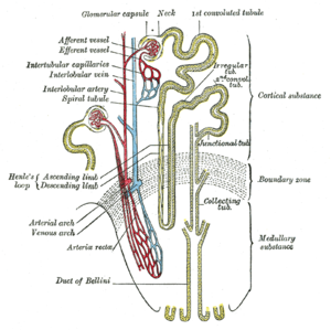
Read Full Post »

Nitric Oxide Function in Coagulation – Part II

Curator and Author: Larry H. Bernstein, MD, FCAP


Subtitle: Nitric oxide in hemostatic and bleeding mechanisms.  Part II.


Summary: This is the second of a coagulation series on PharmaceuticalIntelligence(wordpress.com)  treating the diverse effects of NO on platelets, the coagulation cascade, and protein-membrane interactions with low flow states, local and systemic inflammatory disease, oxidative stress, and hematologic disorders.  It is highly complex as the distinction between intrinsic and extrinsic pathways become blurred as a result of  endothelial shear stress, distinctly different than penetrating or traumatic injury.  In addition, other factors that come into play are also considered.

Please refer to Part I.

Coagulation Pathway

The workhorse tests of the modern coagulation laboratory, the prothrombin time (PT) and the activated partial thromboplastin time (aPTT), are the basis for the published extrinsic and intrinsic coagulation pathways.  This is, however, a much simpler model than one encounters delving into the mechanism and interactions involved in hemostasis and thrombosis, or in hemorrhagic disorders.

We first note that there are three components of the hemostatic system in all vertebrates:

  • Platelets,
  • vascular endothelium, and
  • plasma proteins.

The liver is the largest synthetic organ, which synthesizes

  • albumin,
  • acute phase proteins,
  • hormonal and metal binding proteins,
  • albumin,
  • IGF-1, and
  • prothrombin, mainly responsible for the distinction between plasma and serum (defibrinated plasma).

Role of vascular endothelium.

I have identified the importance of prothrombin, thrombin, and the divalent cation Ca 2+ (1% of the total body pool), mention of heparin action, and of vitamin K (inhibited by warfarin).  Endothelial functions are inherently related to procoagulation and anticoagulation. The subendothelial matrix is a complex of many materials, most important related to coagulation being collagen and von Willebrand factor.

What about extrinsic and intrinsic pathways?  Tissue factor, when bound to factor VIIa, is the major activator of the extrinsic pathway of coagulation. Classically, tissue factor is not present in the plasma but only presented on cell surfaces at a wound site, which is “extrinsic” to the circulation.  Or is it that simple?

Endothelium is the major synthetic and storage site for von Willebrand factor (vWF).  vWF is…

  • secreted from the endothelial cell both into the plasma and also
  • abluminally into the subendothelial matrix, and
  • acts as the intercellular glue binding platelets to one another and also to the subendothelial matrix at an injury site.
  • acts as a carrier protein for factor VIII (antihemophilic factor).
  • It  binds to the platelet glycoprotein Ib/IX/V receptor and
  • mediates platelet adhesion to the vascular wall under shear. [Lefkowitz JB. Coagulation Pathway and Physiology. Chapter I. in Hemostasis Physiology. In ( ???), pp1-12].

Ca++ and phospholipids are necessary for all of the reactions that result in the activation of prothrombin to thrombin. Coagulation is initiated by an extrinsic mechanism that

  • generates small amounts of factor Xa, which in turn
  • activates small amounts of thrombin.

The tissue factor/factorVIIa proteolysis of factor X is quickly inhibited by tissue factor pathway inhibitor (TFPI).The small amounts of thrombin generated from the initial activation feedback

  • to create activated cofactors, factors Va and VIIIa, which in turn help to
  • generate more thrombin.
  • Tissue factor/factor VIIa is also capable of indirectly activating factor X through the activation of factor IX to factor IXa.
  • Finally, as more thrombin is created, it activates factor XI to factor XIa, thereby enhancing the ability to ultimately make more thrombin.

The reconceptualization of hemostasis 

The common theme in activation and regulation of plasma coagulation is the reduction in dimensionality. Most reactions take place in a 2D world that will increase the efficiency of the reactions dramatically. The localization and timing of the coagulation processes are also dependent on the formation of protein complexes on the surface of membranes. The coagulation processes can also be controlled by certain drugs that destroy the membrane binding ability of some coagulation proteins – these proteins will be lost in the 3D world and not able to form procoagulant complexes on surfaces.

Assembly of proteins on membranes – making a 3D world flat

• The timing and efficiency of coagulation processes are handled by reduction in dimensionality

– Make 3 dimensions to 2 dimensions

• Coagulation proteins have membrane binding capacity

• Membranes provide non-coagulant and procoagulant surfaces

– Intact cells/activated cells

• Membrane binding is a target for anticoagulant drugs

– Anti-vitamin K (e.g. warfarin)

Modern View

It can be divided into the phases of initiation, amplification and propagation.

  • In the initiation phase, small amounts of thrombin can be formed after exposure of tissue factor to blood.
  • In the amplification phase, the traces of thrombin will be inactivated or used for amplification of the coagulation process.

At this stage there is not enough thrombin to form insoluble fibrin. In order to proceed further thrombin  activates platelets, which provide a procoagulant surface for the coagulation factors. Thrombin will also activate the vital cofactors V and VIII that will assemble on the surface of activated platelets. Thrombin can also activate factor XI, which is important in a feedback mechanism.

In the final step, the propagation phase, the highly efficient tenase and prothrombinase complexes have been assembled on the membrane surface. This yields large amounts of thrombin at the site of injury that can cleave fibrinogen to insoluble fibrin. Factor XI activation by thrombin then activates factor IX, which leads to the formation of more tenase complexes. This ensures enough thrombin is formed, despite regulation of the initiating TF-FVIIa complex, thus ensuring formation of a stable fibrin clot. Factor XIII stabilizes the fibrin clot through crosslinking when activated by thrombin.

Platelet Aggregation

The activities of adenylate and guanylate cyclase and cyclic nucleotide 3′:5′-phosphodiesterase were determined during the aggregation of human blood platelets with

  • thrombin, ADP,
  • arachidonic acid and
  • epinephrine.

[Aggregation is dependent on an intact release mechanism since inhibition of aggregation occurred with adenosine, colchicine, or EDTA.  (Herman GE, Seegers WH, Henry RL. Autoprothrombin ii-a, thrombin, and epinephrine: interrelated effects on platelet aggregation. Bibl Haematol 1977;44:21-7)].

  1. The platelet guanylate cyclase activity during aggregation depends on the nature and mode of action of the inducing agent.
  2. The membrane adenylate cyclase activity during aggregation is independent of the aggregating agent and is associated with a reduction of activity and
  3. Cyclic nucleotide phosphodiesterase remains unchanged during the process of platelet aggregation and release.

The role of platelets in arterial thrombosis

Formation of a thrombus on a ruptured plaque is the product of a complex interaction between coagulation factors in the plasma and platelets.

  • Tissue factor (TF) released from the subendothelial tissue after endothelial damage induces a cascade of activation of coagulation factors ultimately leading to the formation of thrombin.
  • Thrombin cleaves fibrinogen to fibrin, which assembles into a mesh that supports the platelet aggregates.

The Platelet

The platelets are …

  • anucleated,
  • discoid shaped cell fragments
  • originating from megakaryocytes
  •  fragmented as they are released from the bone marrow

Whether they can in circumstances be developed at extramedullary sites (liver sinusoid) is another matter. They have a lifespan of 7-10 days.  Of special interest is:

  • They have a network of internal membranes forming a dense tubular system and the open canalicular system (OCS).
  • The plasma membrane is an extension of the OCS, thereby greatly increasing the surface area of the platelet.
  • The dense tubular system is comparable to the endoplasmatic reticulum in other cell types and is the main storage place of the majority of the platelet’s Ca2+.

Three types of secretory granules exist in platelets:

  • the dense granules
    •  In the dense granules serotonin
    • adenosine diphosphate (ADP) and
    • Ca2+ are stored.
    • a-granules contain
      • P-selectin,
      • fibrinogen,
      •  thrombospondin,
      • Von Willebrand Factor,
      • platelet factor 4 and
      • platelet derived growth factor
      • lysosomes.

Circulating platelets are kept in a resting state by endothelial cell derived

  • prostacyclin (PGI2) and
  • nitric oxide (NO).

PGI2 increases cyclic adenosine monophosphate (cAMP), the most potent platelet inhibitor.

Contact activation

The major regulator of the activation of the contact system is the plasma protease inhibitor, C1-INH, which inhibits activated fXII, kallikrein and fXIa. In addition, α2-macroglobulin is an important inhibitor of kallikrein and α1-antitrypsin for fXIa. Factor XII also converts the fXI to an active enzyme, fXIa, which, in turn, converts fIX to fIXa, thereby activating the intrinsic pathway of coagulation.

Activation

Several agonists can activate platelets;

  • ADP,
  • collagen,
  • thromboxane A2 (TxA2),
  • epinephrin,
  • serotonine and
  • thrombin,

which lead to activation previously referred to:

  • platelet shape change is
  • followed by aggregation and
  • granule secretion.

Upon activation the discoid shape changes into a spherical form.

Activation of platelets is increased by two positive feedback loops

  1. arachidonic acid is cleaved from phospholipids and transformed by cyclooxygenase

(COX) to prostaglandin G2 and H2,

  • followed by the formation of TxA2, a potent platelet agonist.

2.   the secretion of ADP by the dense granules,

  • resulting in activation of the ADP receptor P2Y12.

This causes inhibition of cyclic AMP and sustained aggregation.

Aggregation

The integrin receptor αIIbβ3 plays a vital role in platelet aggregation. The platelet agonists

  • induce a conformational change of the αIIbβ3 receptor and
  • exposition of binding domains for fibrinogen and von Willebrand Factor.

This allows cross-linking of platelets and the formation of aggregates.

In addition to shape change and aggregation, the membranes of the α- and dense granules fuse with the membranes of the OCS. This causes the release of their contents and the transportation of proteins embedded in their membrane to the plasma membrane.

This complex interaction between

  • endothelial cells
  • clotting factors
  • platelets and
  • other factors and cells

can be studied in both in vitro and in vivo model systems. The disadvantage of in vitro assays is that it studies the role of a certain protein or cell in isolation. Given the large number of participants and the complex interactions of thrombus formation there is need to study thrombosis and hemostasis in intact living animals, with all the components important for thrombus formation – a vessel wall and flowing blood – present.

Endothelial Damage and Role as “Primer”

  • Endothelial injury changes the permeability of the arterial wall.
  • This is followed by an influx of low-density lipoprotein (LDL).
  • This elicits an inflammatory response in the vascular wall.
  • Monocytes and T-cells bind to the endothelial cells promoting increased migration of the cells into the intima layer
  • The monocytes differentiate into macrophages, which take up modified lipoproteins and transform them into foam cells.
  • Concurrent with this process macrophages produce cytokines and proteases.

This is a vicious circle of lipid driven inflammation that leads to narrowing of the vessel’s lumen without early clinical consequences. Clinical manifestations of more advanced atherosclerotic disease are caused by destabilization of an atherosclerotic plaque formed as described.

  • The first recognizable lesion of the stable atherosclerotic plaque is the fatty streak, which consists of the above described foam cells and T-lymphocytes in the intima.
  • Further development of the lesion leads to the intermediate lesion, composed
  • of layers of macrophages and smooth muscle cells.
  • A more advanced stage is called the vulnerable plaque.
    • It has a large lipid core that is covered by a thin fibrous cap.
    • This cap separates the lipid contents of the plaque from the circulating blood.
    • The vulnerable plaque is prone to rupture, resulting in the formation of a thrombus on the site of disruption or the thrombus can be superimposed on plaque erosion without signs of plaque rupture.

The formation of a superimposed thrombus on a disrupted atherosclerotic plaque in the lumen of the artery leads to

  • an acute occlusion of the vessel
  • hypoxia of the downstream tissue.

Depending on the location of the atherosclerotic plaque this will cause a myocardial infarction, stroke or peripheral vascular disease.

Endothelial regulation of coagulation

The endothelium attenuates platelet activity by releasing

  • nitric oxide and
  • prostacyclin.

Several coagulation inhibitors are produced by endothelial cells.

Endothelium-derived TFPI (on its surface) is rapidly released into circulation after heparin administration, reducing the pro-coagulant activities of TF-fVIIa. Endothelial cells also secrete heparin-sulphate, a glycosaminoglycan which catalyzes anti-coagulant activity of AT. Plasma AT binds to heparin-sulphate located on the luminal surface and in the basement membrane of the endothelium. Thrombomodulin is another endothelium-bound protein with anti-coagulant and anti-inflammatory functions. In response to systemic pro-coagulant stimuli, tissue-type plasminogen activator (tPA) is transiently released from the Weibel-Palade bodies of endothelial cells to promote fibrinolysis. Downstream of the vascular injury, the complex of TF-fVIIa/fXa is inhibited by TFPI. Plasma (free) fXa and thrombin are rapidly neutralized by heparan-bound AT. Thrombin is also taken up by endothelial surface-bound thrombomodulin.

The protein C pathway works in hemostasis to control thrombin formation in the area surrounding the clot. Thrombin, generated via the coagulation pathway, is localized to the endothelium by binding to the integral membrane protein, thrombomodulin (TM). TM by occupying exosite I on thrombin, which is required for fibrinogen binding and cleavage, reduces thrombin’s pro-coagulant activities. TM bound thrombin  on the endothelial cell surface is able to cleave PC producing activated protein C (APC), a serine protease.  In the presence of protein S, APC inactivates FVa and FVIIIa. The proteolytic activity of APC is regulated predominantly by a protein C inhibitor.

Fibrinolytic pathway

Fibrinolysis is the physiological breakdown of fibrin to limit and resolve blood clots. Fibrin is degraded primarily by the serine protease, plasmin, which circulates as plasminogen. In an auto-regulatory manner, fibrin serves as both the co-factor for the activation of plasminogen and the substrate for plasmin. In the presence of fibrin, tissue plasminogen activator (tPA) cleaves plasminogen producing plasmin, which proteolyzes the fibrin. This reaction produces the protein fragment D-dimer, which is a useful marker of fibrinolysis, and a marker of thrombin activity because fibrin is cleaved from fibrinogen to fibrin.

Nitric Oxide and Platelet Energy Production

Nitric oxide (NO) has been increasingly recognized as an important intra- and intercellular messenger molecule with a physiological role in

  • vascular relaxation
  • platelet physiology
  • neurotransmission and
  • immune responses.

In vitro NO is a strong inhibitor of platelet adhesion and aggregation. In the blood stream, platelets remain in contact with NO that is permanently released from the endothelial cells and from activated macrophages. It  has been suggested that the activated platelet itself is able to produce NO. It has been proposed that the main intracellular target for NO in platelets is soluble cytosolic guanylate cyclase. NO activates the enzyme. When activated, intracellular cGMP elevation inhibits platelet activation. Further, elevated cGMP may not be the sole factor directly involved in the inhibition of platelet activation.

The reaction mechanism of Nitric oxide synthase
The reaction mechanism of Nitric oxide synthase (Photo credit: Wikipedia)

Platelets are fairly active metabolically and have a total ATP turnover rate of about 3–8 times that of resting mammalian muscle. Platelets contain mitochondria which enable these cells to produce energy both in the oxidative and anaerobic pathways.

  • Under aerobic conditions, ATP is produced by aerobic glycolysis which can account for 30–50% of total ATP production,
  • by oxidative metabolism using glucose and glycogen (6–11%), amino-acids (7%) or free fatty acids (20–40%).

The inhibition of mitochondrial respiration by removing oxygen or by respiratory chain blockers (antimycin A, cyanide, rotenone) results in the stimulation of glycolytic flux. This phenomenon indicates that in platelets glycolysis and mitochondrial respiration are tightly functionally connected. It has been reported that the activation of human platelets by high concentration of thrombin is accompanied by an acceleration of lactate production and an increase in oxygen consumption.

The results (in porcine platelets) indicate that:

  • NO is able to diminish mitochondrial energy production through the inhibition of cytochrome oxidase
  • The inhibitory effect of NO on platelet secretion (but not aggregation) can be attributed to the reduction of mitochondrial energy production.

Porcine blood platelets stimulated by collagen produce more lactate. This indicates that both glycolytic and oxidative ATP production supports platelet responses, and blocking of energy production in platelets may decrease their responses. It is well established that platelet responses have different metabolic energy (ATP) requirements increasing in the order:

  • Aggregation
  • < dense and alfa granule secretion
  • < acid hydrolase secretion.

In addition, exogenously added NO (in the form of NO donors) stimulates glycolysis in intact porcine platelets. Since in platelets glycolysis and mitochondrial respiration are tightly functionally connected, this indicates the stimulatory effect of NO on glycolysis in intact platelets may be produced by non-functional mitochondria.

Can this be the case?

  • NO donors are able to inhibit both mitochondrial respiration and platelet cytochrome oxidase.
  • Interestingly, the concentrations of NO donors inhibiting mitochondrial respiration and cytochrome oxidase were similar to those stimulating glycolysis in intact platelets.

Studies have shown that mitochondrial complex I is inhibited only after a prolonged (6–18 h) exposure to NO and

  • This inhibition appears to result from S-nitrosylation of critical thiols in the enzyme complex.
  • Further studies are needed to establish whether long term exposure of platelets to NO affects Mitochondrial complexes I and II.

Comparison of the concentrations of SNAP and SNP affecting cytochrome oxidase activity and mitochondrial respiration with those reducing the platelet responses indicates that NO does not reduce platelet aggregation through the inhibition of oxidative energy production. The concentrations of the NO donors inhibiting platelet secretion, mitochondrial respiration and cytochrome oxidase were similar. Thus, the platelet release reaction strongly depends on the oxidative energy production, and  in porcine platelets NO inhibits mitochondrial energy production at the step of cytochrome oxidase.

Taking into account that platelets may contain NO synthase and are able to produce significant amounts of NO it seems possible that nitric oxide can function in these cells as a physiological regulator of mitochondrial energy production.

Key words: glycolysis, mitochondrial energy production, nitric oxide, porcine platelets.
Abbreviations: NO, nitric oxide; SNAP, S-nitroso-N-acetylpenicyllamine; SNP, sodium nitroprusside.

[M Tomasiak, H Stelmach, T Rusak and J Wysocka.  Nitric oxide and platelet energy metabolism.  Acta Biochimica Polonica 2004; 51(3):789–803.]

Nitric Oxide and Platelet Adhesion

The adhesion of human platelets to monolayers of bovine endothelial cells in culture was studied to determine the role of endothelium-derived nitric oxide in the regulation of platelet adhesion. The adhesion of unstimulated and thrombin-stimulated platelets, washed and labelled with indium-111, was lower in the presence than in the absence of bradykinin or exogenous nitric oxide. The inhibitory action of both bradykinin and nitric oxide was abolished by hemoglobin, but not by aspirin, and was potentiated by superoxide dismutase to a similar degree. It appears that the effect of bradykinin is mediated by the release of nitric oxide from the endothelial cells, and that nitric oxide release contributes to the non-adhesive properties of vascular endothelium.

(Radomski MW, Palmer RMJ, Moncada S.   Endogenous Nitric Oxide Inhibits Human Platelet Adhesion to Vascular Endothelium. The Lancet  1987 330; 8567(2): 1057–1058.
http://dx.doi.org/10.1016/S0140-6736(87)91481-4)

1 The interactions between endothelium-derived nitric oxide (NO) and prostacyclin as inhibitors of platelet aggregation were examined to determine whether release of NO accounts for the inhibition of platelet aggregation attributed to EDRF.

2 Porcine aortic endothelial cells treated with indomethacin and stimulated with bradykinin (10-100 nM) released NO in quantities sufficient to account for the inhibition of platelet aggregation attributed to endothelium-derived relaxing factor (EDRF).

3 In the absence of indomethacin, stimulation of the cells with bradykinin (1-3 nM) released small amounts of prostacyclin and EDRF which synergistically inhibited platelet aggregation.

4 EDRF and authentic NO also caused disaggregation of platelets aggregated either with collagen or with U46619.

5 A reciprocal potentiation of both the anti- and the disaggregating activity was also observed between low concentrations of prostacyclin and authentic NO or EDRF released from endothelial cells.

6 It is likely that interactions between prostacyclin and NO released by the endothelium play a role in the homeostatic regulation of platelet-vessel wall interactions.

(Radomski MW, Palmer RMJ & Moncada S. The anti-aggregating properties of vascular endothelium: interactions between prostacyclin and nitric oxide. Br J Pharmac 1987; 92: 639-646.


Factor Xa–Nitric Oxide Signaling

Although primarily recognized for maintaining the hemostatic balance, blood proteases of the coagulation and fibrinolytic cascades elicit rapid cellular responses in

  • vascular
  • mesenchymal
  • inflammatory cell types.

Considerable effort has been devoted to elucidate the molecular interface between protease-dependent signaling and pleiotropic cellular responses. This led to the identification of several membrane protease receptors, initiating intracellular signal transduction and effector functions in vascular cells. In this context, thrombin receptor activation

  • generated second messengers in endothelium and smooth muscle cells,
  • released inflammatory cytokines from monocytes, fibroblasts, and endothelium, and
  • increased the expression of leukocyte-endothelial cell adhesion molecules.

Similarly, binding of factor Xa to effector cell protease receptor-1 (EPR-1) participated in

  • in vivo acute inflammatory responses,
  • platelet and brain pericyte prothrombinase activity, and
  • endothelial cell and smooth muscle cell signaling and proliferation.

Factor Xa stimulated a 5- to 10-fold increased release of nitric oxide (NO) in a dose-dependent reaction (0.1–2.5 mgyml) unaffected by the thrombin inhibitor hirudin but abolished by active site inhibitors, tick anticoagulant peptide, or Glu-Gly-Arg-chloromethyl ketone. In contrast, the homologous clotting protease factor IXa or another endothelial cell ligand, fibrinogen, was ineffective.

A factor Xa inter-epidermal growth factor synthetic peptide L83FTRKL88(G) blocking ligand binding to effector cell protease receptor-1 inhibited NO release by factor Xa in a dose-dependent manner, whereas a control scrambled peptide KFTGRLL was ineffective.

Catalytically active factor Xa induced hypotension in rats and vasorelaxation in the isolated rat mesentery, which was blocked by the NO synthase inhibitor L-NG-nitroarginine methyl ester (LNAME) but not by D-NAME. Factor Xa/NO signaling also produced a dose-dependent endothelial cell release of interleukin 6 (range 0.55–3.1 ngyml) in a reaction

  • inhibited by L-NAME and by the
  • inter-epidermal growth factor peptide Leu83–Leu88 but
  • unaffected by hirudin.
We observe that incubation of HUVEC monolayers with factor Xa which resulted in a concentration-dependent release of NO, as determined by cGMP accumulation in these cells, was inhibited by the nitric oxide synthase antagonist L-NAME.

Catalytically inactive DEGR-factor Xa or TAP-treated factor Xa failed to stimulate NO release by HUVEC.

To determine whether factor Xa-induced NO release could also modulate acute phase/inflammatory cytokine gene expression we examined potential changes in IL-6 release following HUVEC stimulation with factor Xa. HUVEC stimulation with factor Xa resulted in a concentration-dependent release of IL-6.

The specificity of factor Xa-induced cytokine release was investigated. Factor Xa-induced IL-6 release from HUVEC was quantitatively indistinguishable from that obtained with tumor necrosis factor-a or thrombin stimulation. This response was abolished by heat denaturation of factor Xa.

Maximal induction of interleukin 6 mRNA required a brief, 30-min stimulation with factor Xa, and was unaffected by subsequent addition of tissue factor pathway inhibitor (TFPI). These data suggest that factor Xa-induced NO release modulates endothelial cell-dependent vasorelaxation and IL-6 cytokine gene expression.

Here, we find that factor Xa induces the release of endothelial cell NO

  • regulating vasorelaxation in vivo and acute response cytokine gene expression in vitro.

This pathway requires a dual step cascade, involving

  • binding of factor Xa to EPR-1 and
  • a secondary as yet unidentified protease activated mechanism.

This pathway requiring factor Xa binding to effector cell protease receptor-1 and a secondary step of ligand-dependent proteolysis may preserve an anti-thrombotic phenotype of endothelium but also trigger acute phase responses during activation of coagulation in vivo.

In summary, these investigators have identified a signaling pathway centered on the ability of factor Xa to rapidly stimulate endothelial cell NO release. This involves a two-step cascade initiated by catalytic active site-independent binding of factor Xa to its receptor, EPR-1, followed by a second step of ligand dependent proteolysis.

(Papapetropoulos A, Piccardoni P, Cirino G, Bucci M, et al. Hypotension and inflammatory cytokine gene expression triggered by factor Xa–nitric oxide signaling. Proc. Natl. Acad. Sci. USA. Pharmacology. 1998; 95:4738–4742.)

Platelets and liver disease

Thrombocytopenia is a marked feature of chronic liver disease and cirrhosis. Traditionally, this thrombocytopenia was attributed to passive platelet sequestration in the spleen. More recent insights suggest an increased platelet breakdown and to a lesser extent decreased platelet production plays a more important role. Besides the reduction in number, other studies suggest functional platelet defects. This platelet dysfunction is probably both intrinsic to the platelets and secondary to soluble plasma factors. It reflects not only a decrease in aggregability, but also an activation of the intrinsic inhibitory pathways. (Witters P, Freson K, Verslype C, Peerlinck K, et al. Review article: blood platelet number and function in chronic liver disease and cirrhosis. Aliment Pharmacol Ther 2008; 27: 1017–1029).

The shortcomings of the old Y-shaped model of normal coagulation are nowhere more apparent than in its clinical application to the complex coagulation disorders of acute and chronic liver disease. In this condition, the clotting cascade is heavily influenced by numerous currents and counter-currents resulting in a mixture of pro- and anticoagulant forces that are themselves further subject to change with altered physiological stress such as super-imposed infection or renal failure.

Multiple mechanisms exist for thrombocytopenia common in patients with cirrhosis besides hypersplenism and expected altered thrombopoietin metabolism. Increased production of two important endothelial derived platelet inhibitors

  • nitric oxide and
  • prostacyclin

may contribute to defective platelet activation in vivo. On the other hand, high plasma levels of vWF in cirrhosis appear to support platelet adhesion.

Reduced levels of coagulation factors V, VII, IX, X, XI, and prothrombin are also commonly observed in liver failure. Vitamin K–dependent clotting factors (II, VII, IX, X) may be defective in function as a result of decreased  y-carboxylation (from vitamin K deficiency or intrinsically impaired carboxylase activity). Fibrinogen levels are decreased with advanced cirrhosis and in patients with acute liver failure.

A hyperfibrinolytic state may develop when plasminogen activation by tPA is accelerated on the fibrin surface. Physiologic stress including infection may be key in tipping this process off through increased release of tPA.  Not uncommonly, laboratory abnormalities in decompensated cirrhosis come to resemble disseminated intravascular coagulation (DIC). Relatively stable platelet levels and characteristically high factor VIII levels distinguish this process from DIC as does the absence of uncompensated thrombin generation. The features of both hyperfibrinolysis and DIC are often evident in the decompensated liver disease patient, and the term “accelerated intravascular coagulation and fibrinolysis” (AICF) has been proposed as a way to encapsulate the process under a single heading. The essence of AICF can be postulated to be the result of formation of a fibrin clot that is more susceptible to plasmin degradation due to elevated levels of tPA coupled with inadequate release of PAI to control tPA and lack of a-2 plasmin inhibitor to quench plasmin activity and the maintenance of high local concentrations of plasminogen on clot surfaces despite lower total plasminogen production. These normally balanced processes become pronounced when disturbed by additional stress such as infection.

Normal hemostasis and coagulation is now viewed as primarily a cell-based process wherein key steps in the classical clotting cascade

  • occur on the phospholipid membrane surface of cells (especially platelets)
  • beginning with activation of tissue factor and factor VII at the site of vascular breach
    •  which produces an initial “priming” amount of thrombin and a
    • subsequent thrombin burst.

Coagulation and hemostasis in the liver failure patient is influenced by multiple, often opposing, and sometimes changing variables. A bleeding diathesis is usually predominant, but the assessment of bleeding risk based on conventional laboratory tests is inherently deficient.

(Caldwell SH, Hoffman M, Lisman T, Gail Macik B, et al. Coagulation Disorders and Hemostasis in Liver Disease: Pathophysiology and Critical Assessment of Current Management. Hepatology 2006;44:1039-1046.)

Bleeding after Coronary Artery bypass Graft

Cardiac surgery with concomitant CPB can profoundly alter haemostasis, predisposing patients to major haemorrhagic complications and possibly early bypass conduit-related thrombotic events as well. Five to seven percent of patients lose more than 2 litres of blood within the first 24 hours after surgery, between 1% and 5% require re-operation for bleeding. Re-operation for bleeding increases hospital mortality 3 to 4 fold, substantially increases post-operative hospital stay and has a sizeable effect on health care costs. Nevertheless, re-exploration is a strong risk factor associated with increased operative mortality and morbidity, including sepsis, renal failure, respiratory failure and arrhythmias.

(Gábor Veres. New Drug Therapies Reduce Bleeding in Cardiac Surgery. Ph.D. Doctoral Dissertation. 2010. Semmelweis University)

Hypercoagulable State in Thalassemia

As the life expectancy of β-thalassemia patients has increased in the last decade, several new complications are being recognized. The presence of a high incidence of thromboembolic events, mainly in thalassemia intermedia patients, has led to the identification of a hypercoagulable state in thalassemia. Patients with thalassemia intermedia (TI) have, in general, a milder clinical phenotype than those with TM and remain largely transfusion independent. The pathophysiology of TI is characterized by extravascular hemolysis, with the release into the peripheral circulation of damaged red blood cells (RBCs) and erythroid precursors because of a high degree of ineffective erythropoiesis. This has also been recently attributed to severe complications such as pulmonary hypertension (PHT) and thromboembolic phenomena.

Many investigators have reported changes in the levels of coagulation factors and inhibitors in thalassemic patients. Prothrombin fragment 1.2 (F1.2), a marker of thrombin generation, is elevated in TI patients. The status of protein C and protein S was investigated in thalassemia in many studies and generally they were found to be decreased; this might be responsible for the occurrence of thromboembolic events in thalassemic patients.

The pathophysiological roles of hemolysis and the dysregulation of nitric oxide homeostasis are correlated with pulmonary hypertension in sickle cell disease and in thalassemia. Nitric oxide binds soluble guanylate cyclase, which converts GTP to cGMP, relaxing vascular smooth muscle and causing vasodilatation. When plasma hemoglobin liberated from intravascularly hemolyzed sickle erythrocytes consumes nitric oxide, the balance is shifted toward vasoconstriction. Pulmonary hypertension is aggravated and in sickle cell disease, it is linked to the intensity of hemolysis. Whether the same mechanism contributes to hypercoagulability in thalassemia is not yet known.

While there are diverse factors contributing to the hypercoagulable state observed in patients with thalassemia. In most cases, a combination of these abnormalities leads to clinical thrombosis. An argument has been made for the a higher incidence of thrombotic events in TI compared to TM patients  attributed to transfusion for TM. The higher rate of thrombosis in transfusion-independent TI compared to polytransused TM patients suggests a potential role for transfusions in decreasing the rate of thromboembolic events (TEE). The reduction of TEE in adequately transfused patients may be the result of decreased numbers of pathological RBCs.

(Cappellini MD, Musallam KM,  Marcon A, and Taher AT. Coagulopathy in Beta-Thalassemia: Current Understanding and Future Prospects. Medit J Hemat Infect Dis 2009; 1(1):22009029.
DOI 10.4084/MJHID.2009.0292.0), www.mjhid.org/article/view/5250.  ISSN 2035-3006.)

Microvascular Endothelial Dysfunction

Severe sepsis, defined as sepsis associated with acute organ dysfunction, results from a generalized inflammatory and procoagulant host response to infection. Coagulopathy in severe sepsis is commonly associated with multiple organ dysfunction, and often results in death. The molecule that is central to these effects is thrombin, although it may also have anticoagulant and antithrombotic effects through the activation of Protein C and induction of prostacyclin. In recent years, it has been recognized that chemicals produced by endothelial cells play a key role in the pathogenesis of sepsis. Thrombomodulin on endothelial cells coverts Protein C to Activated Protein C, which has important antithrombotic, profibrinolytic and anti-inflammatory properties. A number of studies have shown that Protein C levels are reduced in patients with severe infection, or even in inflammatory states without infection. Because coagulopathy is associated with high mortality rates, and animal studies have indicated that therapeutic intervention may result in improved outcomes, it was rational to initiate clinical studies.

Considering the coagulation cascade as a whole, it is the extrinsic pathway (via TF and thrombin activation) rather than the intrinsic pathway that is of primary importance in sepsis. Once coagulation has been triggered by TF activation, leading to thrombin formation, this can have further procoagulant effects, because thrombin itself can activate factors VIII, IX and X. This is normally balanced by the production of anticoagulant factors, such as TF pathway inhibitor, antithrombin and Activated Protein C.

It has been recognized that endothelial cells play a key role in the pathogenesis of sepsis, and that they produce important regulators of both coagulation and inflammation. They can express or release a number of substances, such as TF, endothelin-1 and PAI-1, which promote the coagulation process, as well as other substances, such as antithrombin, thrombomodulin, nitric oxide and prostacyclin, which inhibit it.

Protein C is the source of Activated Protein C. Although Protein C is a biomarker or indicator of sepsis, it has no known specific biological activity. Protein C is converted to Activated Protein C in the presence of normal endothelium. In patients with severe sepsis, the vascular endothelium becomes damaged. The level of thrombomodulin is significantly decreased, and the body’s ability to convert Protein C to Activated Protein C diminishes. Only when activated does Protein C have antithrombotic, profibrinolytic and anti-inflammatory properties.

Blood Coagulation (Thrombin) and Protein C Pat...

Blood Coagulation (Thrombin) and Protein C Pathways (Blood_Coagulation_and_Protein_C_Pathways.jpg) (Photo credit: Wikipedia)

Coagulation abnormalities can occur in all types of infection, including both Gram-positive and Gram-negative bacterial infections, or even in the absence of infection, such as in inflammatory states secondary to trauma or neurosurgery. Interestingly, they can also occur in patients with localized disease, such as those with respiratory infection. In a study by Günther et al., procoagulant activity in bronchial lavage fluid from patients with pneumonia or acute respiratory distress syndrome was found to be increased compared with that from control individuals, with a correlation between the severity of respiratory failure and level of coagulant activity.

Severe sepsis, defined as sepsis associated with acute organ dysfunction, results from a generalized inflammatory and procoagulant host response to infection.  Once the endothelium becomes damaged, levels of endothelial thrombomodulin significantly decrease, and the body’s ability to convert Protein C to Activated Protein C diminishes. The ultimate cause of acute organ dysfunction in sepsis is injury to the vascular endothelium, which can result in microvascular coagulopathy.

(Vincent JL. Microvascular endothelial dysfunction: a renewed appreciation of sepsis pathophysiology.
Critical Care 2001; 5:S1–S5. http://ccforum.com/content/5/S2/S1)

Endothelial Cell Dysfunction in Severe Sepsis

During the past decade a unifying hypothesis has been developed to explain the vascular changes that occur in septic shock on the basis of the effect of inflammatory mediators on the vascular endothelium. The vascular endothelium plays a central role in the control of microvascular flow, and it has been proposed that widespread vascular endothelial activation, dysfunction and eventually injury occurs in septic shock, ultimately resulting in multiorgan failure. This has been characterized in various models of experimental septic shock. Now, direct and indirect evidence for endothelial cell alteration in humans during septic shock is emerging.

The vascular endothelium regulates the flow of nutrient substances, diverse biologically active molecules and the blood cells themselves. This role of endothelium is achieved through the presence of membrane-bound receptors for numerous molecules, including proteins, lipid transporting particles, metabolites and hormones, as well as through specific junction proteins and receptors that govern cell–cell and cell–matrix interactions. Endothelial dysfunction and/or injury with subendothelium exposure facilitates leucocyte and platelet aggregation, and aggravation of coagulopathy. Therefore, endothelial dysfunction and/or injury should favour impaired perfusion, tissue hypoxia and subsequent organ dysfunction.

Anatomical damage to the endothelium during septic shock has been assessed in several studies. A single injection of bacterial lipopolysaccharide (LPS) has long been demonstrated to be a nonmechanical technique for removing endothelium. In endotoxic rabbits, observations tend to demonstrate that EC surface modification occurs easily and rapidly, with ECs being detached from the internal elastic lamina with an indication of subendothelial oedema.  Proinflammatory cytokines increase permeability of the ECs, and this is manifested approximately 6 hours after inflammation is triggered and becomes maximal over 12–24 hours as the combination of cytokines exert potentiating effects. Endothelial physical disruption allows inflammatory fluid and cells to shift from the blood into the interstitial space.

In sepsis

  • ECs become injured, prothrombotic and antifibrinolytic
  • They promote platelet adhesion
  • They promote leucocyte adhesion and inhibit vasodilation

An important point is that EC injury is sustained over time. In an endotoxic rabbit model, we demonstrated that endothelium denudation is present at the level of the abdominal aorta as early as after several hours following injury and persisted for at least 5 days afterward. After 21 days we observed that the endothelial surface had recovered. The de-endothelialized surface accounted for approximately 25% of the total surface.

Thrombomodulin and protein C activation at the microcirculatory level.

The endothelial cell surface thrombin (Th)-binding protein thrombomodulin (TM) is responsible for inhibition of thrombin activity. TM, when bound to Th, forms a potent protein C activator complex. Loss of TM and/or internalization results in Th–thrombin receptor (TR) interaction. Loss of TM and associated protein C activation represents the key event of decreased endothelial coagulation modulation ability and increased inflammation pathways.
( Iba T, Kidokoro A, Yagi Y: The role of the endothelium in changes in procoagulant activity in sepsis. J Am Coll Surg 1998; 187:321-329. Keywords: ATIII, antithrombin III; NF-κ, nuclear factor-κB; PAI,plasminogen activator inhibitor).

In order to test the role of the endothelial-derived relaxing factors NO and PGI2, we investigated, in dogs, the influence of a combination of NG-nitro-L-arginine methyl ester (an inhibitor of NO synthesis) and indomethacin (an inhibitor of PGI2 synthesis). In these dogs treated with indomethacin plus NG-nitro-L-arginine methyl ester, the severity of the oxygen extraction defect was lower than that observed in the deoxycholate-treated dogs, suggesting that other mediators and/or mechanisms may be involved in microcirculatory control during hypoxia. One of these mediators or mechanisms could be related to hyperpolarization. Membrane potential is an important determinant of vascular smooth muscle tone through its influence on calcium influx via voltage-gated calcium channels. Hyperpolarization (as well as depolarization) has been shown to be a means of cell–cell communication in upstream vasodilatation and microcirculatory coordination. It is important to emphasize that intercell coupling exclusively involves ECs.

Interestingly, it was recently shown that sepsis, a situation that is characterized by impaired tissue perfusion and abnormal oxygen extraction, is associated with abnormal inter-EC coupling and reduction in the arteriolar conducted response.  An intra-organ defect in blood flow related to abnormal vascular reactivity, cell adhesion and coagulopathy may account for impaired organ oxygen regulation and function. If specific classes of microvessels must or must not be perfused to achieve efficient oxygen extraction during limitation in oxygen delivery, then impaired vascular reactivity and vessel injury might produce a pathological limitation in supply. In sepsis, the inflammatory response profoundly alters circulatory homeostasis, and this has been referred to as a ‘malignant intravascular inflammation’ that alters vasomotor tone and the distribution of blood flow among and within organs. These mechanisms might coexist with other types of sepsis associated cell dysfunction. For example, data suggest that endotoxin directly impairs oxygen uptake in ECs and indicate the importance of endothelium respiration in maintaining vascular homeostasis under conditions of sepsis.

Consistent with the hypothesis that alteration in endothelium plays a major in the pathophysiology of sepsis, it was observed that chronic ecNOS overexpression in the endothelium of mice resulted in resistance to LPS-induced hypotension, lung injury and death . This observation was confirmed by another group of investigators, who used transgenic mice overexpressing adrenomedullin  – a vasodilating peptide that acts at least in part via an NO-dependent pathway. They demonstrated resistance of these animals to LPS-induced shock, and lesser declines in blood pressure and less severe organ damage than occurred in the control animals. It might therefore be of importance to favour ecNOS expression and function during sepsis. The recent negative results obtained with therapeutic strategies aimed at blocking inducible NOS with the nonselective NOS inhibitor NG-monomethyl-L-arginine in human septic shock further confirm the overall importance of favoring vessel dilatation.

(Vallet B. Bench-to-bedside review: Endothelial cell dysfunction in severe sepsis: a role in organ dysfunction?  Critical Care 2003; 7(2):130-138 (DOI 10.1186/cc1864). (Print ISSN 1364-8535; Online ISSN 1466-609X). http://ccforum.com/content/7/2/130

Thrombosis in Inflammatory Bowel Disease

An association between IBD and thrombosis has been recognized for more than 60 years. Not only are patients with IBD more likely to have thromboembolic complications, but it has also been suggested that thrombosis might be pathogenic in IBD.

Coagulation Described.  See Part I. (Cascade)

Endothelial injury exposes TF, which forms a complex with factor VII.  This complex activates factors X and, to a lesser extent, IX. TFPI prevents this activation progressing  further; for coagulation to progress, factor Xa must be produced via factors IX and VIII. Thrombin, generated by the initial production of factor Xa, activates factor VIII and, through factor XI, factor IX, resulting in further activation of factor X. This positive feedback loop allows coagulation to proceed. Fibrin polymers are stabilized by factor XIIIa. Activated proteins CS (APCS) together inhibit factors VIIIa and Va, whereas antithrombin (AT) inhibits factors VIIa, IXa, Xa, and XIa. Fibrinolysis balances this system through the action of plasmin on fibrin. Plasminogen activator inhibitor controls the plasminogen activator-induced conversion of plasminogen to plasmin.

Inflammation and Thrombotic Processes Linked

Although interest has recently moved away from the proposal that ischemia is a primary cause of IBD, it has become increasingly clear that inflammatory and thrombotic processes are linked.  A vascular component to the pathogenesis of CD was first proposed only a year after Crohn et al. described the condition.  Subsequently, in 1989, a series of changes comprising vascular injury, focal arteritis, fibrin deposition, arterial occlusion, and then microinfarction or neovascularization was proposed as a possible pathogenetic sequence in CD.  In this study, resin casts of the intestinal vasculature showed changes ranging from intravascular fibrin deposition to complete thrombotic occlusion. Furthermore, the early vascular changes appeared to precede mucosal changes, suggesting that they were more likely to cause rather than result from the pathologic features of CD. Subsequent studies showed that intravascular fibrin deposition occurred at the site of granulomatous destruction of mesenteric blood vessels, and positive immunostaining for platelet glycoprotein IIIa occurred in fibrinoid plugs of mucosal capillaries in CD. In addition, intracapillary thrombus has been identified in biopsies from inflamed rectal mucosa from patients with CD. When combined with evidence of ongoing intravascular coagulation in both active and quiescent CD, the above data point toward a thrombotic element contributing to the pathogenesis of CD.

Not only are many different prothrombotic changes described in association with IBD, but they can also have multiple causes. Hyperhomocysteinemia, for example, is known to predispose to thrombosis, and patients with IBD are more likely to have hyperhomocysteinemia than control subjects. Hyperhomocysteinemia in IBD might be due to multiple possible causes, such as deficiencies of vitamin B12 as a result of terminal ileal disease or resection; B6, which is commonly reduced in IBD.  A vegan diet can’t be discarded either because of seriously deficient methyl donors (S-adenosyl methionine).

The realization that platelets are not only prothrombotic but also proinflammatory has stimulated interest in their role in both the pathogenesis and complications of IBD. The association between thrombocytosis and active IBD was first described more than 30 years ago. More recent observations link decreased or normal platelet survival to IBD-related thrombocytosis, possibly due to increased thrombopoiesis. This in turn could be driven by an interleukin-6 –induced increase in thrombopoietin synthesis in the liver. Spontaneous in vitro platelet aggregation occurs in platelets isolated from 30% of patients with IBD but not in platelets from control subjects. Moreover, collagen, arachidonic acid, ristocetin, and ADP-induced platelet activation are more marked in platelets from patients with active IBD than in those from healthy volunteers.

The roles of activated platelets and PLAs in mucosal inflammation. Activated platelets can interact with other cells involved in the inflammatory response either through direct contact or through the release of soluble mediators. Activated platelets interact directly with activated vascular endothelium, causing the latter to express adhesion molecules and release inflammatory and chemotactic cytokines.

Platelet activation might be pathogenic in IBD in several ways. Platelet activation might increase platelet aggregation, hence increasing the likelihood of thrombus formation at sites of vascular injury, for example, within the mesenteric circulation. P-selectin is the major ligand for leukocyte-endothelial interaction and is responsible for the rolling of platelets, leukocytes, and PLAs on vascular endothelium. Moreover, platelets adherent to injured vascular endothelium support leukocyte adhesion via P-selectin, an effect that could contribute to leukocyte emigration from the vasculature into the lamina propria in patients with IBD. In addition, P-selectin is the major platelet ligand for platelet-leukocyte interaction, which in turn causes both leukocyte activation and further platelet activation.

Platelet-Leukocyte Aggregation

Recently, studies showing that platelets and leukocytes that circulate together in aggregates (PLA) are more activated than those that circulate alone have generated interest in the role of PLA in various inflammatory and thrombotic conditions. PLA numbers are increased in patients with ischemic heart disease, systemic lupus erythematosus and rheumatoid arthritis, myeloproliferative disorders, and sepsis and are increased by smoking.

We have recently shown that patients with IBD have more PLAs than both healthy and inflammatory control subjects (patients with inflammatory arthritides).  As with platelet activation, there was no correlation with disease activity, suggesting that increased PLA formation might be an underlying abnormality. PLAs could contribute to the pathogenesis of IBD in a number of ways. As previously mentioned, TF is key to the initiation of thrombus formation. TF has recently been demonstrated on the surface of activated platelets and in platelet-derived microvesicles. Interaction between neutrophils and activated platelets or microvesicles vastly increases the activity of “intravascular” TF.

Conclusion        

It is becoming increasingly apparent that thrombosis and inflammation are intrinsically linked. Hence the involvement of thrombotic processes in the pathogenesis of IBD, although perhaps not as the primary event, seems likely. Indeed, with the recently mounting evidence of the role of activated platelets and of their interaction with leukocytes in the pathogenesis of IBD, it seems even more probable that thrombosis plays some role in the pathogenic process.

(Irving PM, Pasi KJ, and Rampton DS. Thrombosis and Inflammatory Bowel Disease. Clinical Gastroenterology and Hepatology 2005;3:617–628. PII: 10.1053/S1542-3565(05)00154-0.)

Bleeding in Patients with Renal Insufficiency

Approximately 20–40% of critically ill patients will have renal insufficiency at the time of admission or will develop it during their ICU stay, depending on the definition of renal insufficiency and the case mix of the ICU. Such patients are also predisposed to bleeding because of uremic platelet dysfunction, typically multiple comorbidities, coagulopathies and frequent concomitant treatment with antiplatelet or anticoagulant agents.

The impairment in hemostasis in uremic patients is multifactorial and includes physiological defects in platelet hemostasis, an imbalance of mediators of normal endothelial function and frequent comorbidities such as vascular disease, anemia and the frequent need for medical interventions required to treat such comorbidities. Physiologic alterations in uremia include:

  • decreased platelet glycoprotein IIb–IIIa binding to both von Willebrand factor (vWf) and fibrinogen, causing an impairment in platelet aggregation;
  • increased prostacyclin and nitric oxide production, both potent inhibitors of platelet activation and vasoconstriction; and
  • decreased levels of platelet adenosine diphosphate (ADP) and serotonin, causing an impairment in platelet secretion.

In addition to other factors, small peptides containing the RGD (Arg-Gly-Asp) sequence of amino acids have been shown to be inhibitors of platelet aggregation that act by competing with vWf and fibrinogen for binding to the glycoprotein IIb–IIIa receptor.

Conclusion

ICU patients have dynamic risks of thrombosis and bleeding. Invasive procedures may require temporary interruption of anticoagulants. Consequently, approaches to thromboprophylaxis require daily reevaluation.

(Cook DJ, Douketis J, Arnold D, and Crowther MA. Bleeding and venous thromboembolism in the critically ill with emphasis on patients with renal insufficiency. Curr Opin Pulm Med 2009;15:455–462.)

Epicrisis

I have covered a large amount of material on one of the most complex systems in medicine, and still not comprehensive, with a sufficient dash of repetition.  The task is to have some grasp of the cell-mediated imbalances inherent if coagulation and bleeding disorders.  The key points are:

  • inflammation and oxidative stress invariably lurk in the background
  • the Y-shaped model with an extrinsic, intrinsic, and common pathway has no basis in understanding
  • the current model is based on a cell-mediated concept of endothelial damage and platelet-endothelial interaction
  • the model has 3 components: Initiation, Amplification, Propagation
  • NO and prostacyclin have key roles in the process
  • The plasma proteins involved are in the serine-protease class of enzymes
  • The conversion of Protein C to APC has a central role as anti-coagulant

Part II  has explored organ system abnormalities that are all related to impairment of the Nitric Oxide balance and dual platelet-endothelial roles.

Nov 9, 2012 by anamikasarkar
Nitric Oxide (NO) is highly regulated in the blood such that it can be released as vasodilator when needed. The importance and pathway of Nitric Oxide has been nicely reviewed by. “Discovery of NO and its effects of vascular biology”. Other articles which are good readings for the importance of NO are  – a) regulation of glycolysis b) NO in cardiovascular disease c) NO and Immune responses Part I and Part II d) NO signaling pathways. The  effects of NO in diseased states have been reviewed by the articles – “Crucial role of Nitric Oxide in Cancer”, “Nitric Oxide and Sepsis, Hemodynamic Collapse, and the Search for Therapeutic Options”.. (Also, please see Source for more articles on NO and its significance).

Computational models are very efficient tools to understand complex reactions like NO towards physiological conditions. Among them wall shear stress is one of the major factors which is reviewed in the article – “Differential Distribution of Nitric Oxide – A 3-D Mathematical Model”.

Moreover, decrease in availability of NO can lead to many complications like pulmonary hypertension. Some of the causes of decrease in NO have been identified as clinical hypertension,right ventricular overload which can lead to cardiac heart failure,low levels of zinc and high levels of cardiac necrosis.

Sickle Cell disease patients, a hereditary disease, are also known to have decreased levels of NO which can become physiologically challenging. In USA alone, there are 90,000 people who are affected by Sickle cell disease.

Sickle cell disease is breakage of red blood cells (RBC) membrane and resulting release of the hemoglobin (Hb) into blood plasma. This process is also known as Hemolysis. Sickle cell disease is caused by single mutation of Hb which changes RBC from round shape to sickle or crescent shapes (Figure 1).

Image
Figure 1 (A) shows normal red blood cells flowing freely through veins. The inset shows a cross section of a normal red blood cell with normal hemoglobin.
Figure 1 (B) shows abnormal, sickled red blood cells The inset image shows a cross-section of a sickle cell with long polymerized HbS strands stretching and distorting the cell shape.
Image Source: http://en.wikipedia.org/wiki/Sickle-cell_disease

RBCs generally breakdown and release Hbs in blood plasma after they reach their end of life span. Thus, in case of Sickle cell disease, there is more cell free Hb than normal. Furthermore, it is known that NO has a very high affinity towards Hbs, which is one of the ways free NO is regulated in blood. As a result presence of larger amounts of cell free Hb in Sickle cell disease lead to less availability of NO.

As to “a quantitative relationship between cell free Hb and depletion of NO”, Deonikar and Kavdia (J. Appl. Physiol., 2012) addressed this question by developing a model of a single idealized arteriole, with different layers of blood vessels diffusing nutrients to tissue layers (Figure 2:  Deonikar and Kavdia Figure 1).

The model and its parameters are explained in the previously published paper by same authors Deonikar and Kavdia, Annals of Biomed., 2010, who reported that the reaction rate between NO and RBC is 0.2 x 10⁵, M-1 s-1 than 1.4 x 10⁵, M-1 s-1 as previously reported by Butler et.al., Biochim. Biophys. Acta, 1998. Their results show that even small increase in cell free Hb, 0.5uM, can decrease NO concentrations by 3-7 fold.  Their mathematical analysis shows that the increase in diffusion resistance of NO from vascular lumen to cell free zone has no effect on NO distribution and concentration with available levels of cell free Hb.

Deonikar and Kavdia’s  model, a simple representation shows that for SC disease patients, decrease in levels of bioavailable NO is attributed to cell free Hb, which is in abundant for these patients. Their results show that small increase by 0.5 uM in cell free Hb can cause a large decrease in NO concentrations.

Sources:

Deonikar and Kavdia (2012):http://www.ncbi.nlm.nih.gov/pubmed/22223452

Previous model explaining mathematical representation and parameters used in the model :Deonikar and Kavdia,Annals of Biomed., 2010.

Previous paper stating reaction rate of Hb and NO: Butleret.al., Biochim. Biophys. Acta, 1998.

Causes of decrease in NO

Clinical Hypertension :http://www.ncbi.nlm.nih.gov/pubmed/11311074

Right ventricular overload :http://www.ncbi.nlm.nih.gov/pubmed/9559613

Low levels of zinc and high levels of cardiac necrosis :http://www.ncbi.nlm.nih.gov/pubmed/11243421

Sickle Cell Source:

http://en.wikipedia.org/wiki/Sickle-cell_disease

http://www.nhlbi.nih.gov/health/health-topics/topics/sca/

NO Sources:

Differential Distribution of Nitric Oxide – A 3-D Mathematical Model:

Discovery of NO and its effects of vascular biology

Nitric oxide: role in Cardiovascular health and disease

NO signaling pathways

Inhibition of ET-1, ETA and ETA-ETB, Induction of NO production, stimulation of eNOS and Treatment Regime with PPAR-gamma agonists (TZD): cEPCs Endogenous Augmentation for Cardiovascular Risk Reduction – A Bibliography

The rationale and use of inhaled NO in Pulmonary Artery Hypertension and Right Sided Heart Failure

Clinical Trials Results for Endothelin System: Pathophysiological role in Chronic Heart Failure, Acute Coronary Syndromes and MI – Marker of Disease Severity or Genetic Determination?

Endothelial Function and Cardiovascular Disease

Interaction of Nitric Oxide and Prostacyclin in Vascular Endothelium

Endothelial Dysfunction, Diminished Availability of cEPCs,  Increasing  CVD Risk – Macrovascular Disease – Therapeutic Potential of cEPCs

Cardiovascular Disease (CVD) and the Role of agent alternatives in endothelial Nitric Oxide Synthase (eNOS) Activation and Nitric Oxide Production


Read Full Post »

Curator & Author: Larry H. Bernstein, MD, FCAP

Leaders in Pharmaceutical Intelligence

Subtitle: Nitric Oxide, Peroxinitrite, and NO donors in Renal Function Loss

Summary: The criticality of renal function is traced to the emergence of animal forms from the sea to land. It also becomes acutely and/or chronically dysfunctional in metabolic, systemic inflammatory and immunological diseases of man. We have already described the key role that nitric oxide and the NO synthases play in reduction of oxidative stress, and we have seen that a balance has to be struck between pro- and anti-oxidative as well as inflammatory elements for avoidance of diseases, specifically involving the circulation, but effectively not limited to any organ system. In this discussion we shall look at kidney function, NO and NO donors. This is an extension of a series of posts on NO and NO related disorders.

__________________________________________________________________________________________________________________________________________________________

Part I. The evolution of kidney structure and Function Evolution of kidney function

In fish the nerves that activate breathing take a short journey from an ancient part of the brain, the brain stem, to the throat and gills. For the ancient tadpole, the nerve controlling a reflex related to hiccup in man served a useful purpose, allowing the entrance to the lung to remain open when breathing air but closing it off when gulping water – which would then be directed only to the gills.

For humans and other mammals it provides a bit of evidence of our common ancestry. DNA evidence has pinned iguanas and chameleons as the closest relatives to snakes. In utero, we develop three separate kidneys in succession, absorbing the first two before we wind up with the embryonic kidney that will become our adult kidney. The first two of these reprise embryonic kidneys of ancestral forms, and in the proper evolutionary order.

The pronephric kidney does not function in human and other mammalian embryos. It disappears and gives rise to the Mesonephric kidney. This kidney filters wastes from the blood and excretes them to the outside of the body via a pair of tubes called the mesonephric ducts (also “Wolffian ducts”). The mesonephric kidney goes on to develop into the adult kidney of fish and amphibians.

This kidney does function for a few weeks in the human embryo, but then disappears as our final kidney forms, which is the Metanephric kidney. This begins developing about five weeks into gestation, and consists of an organ that filters wastes from the blood and excretes them to the outside through a pair ureters. In the embryo, the wastes are excreted directly into the amniotic fluid. The metanephric kidney is the final adult kidney of reptiles, birds, and mammals.

The first two kidneys resemble, in order, those of primitive aquatic vertebrates (lampreys and hagfish) and aquatic or semiaquatic vertebrates (fish and amphibians): an evolutionary order.

The explanation, then, is that we go through developmental stages that show organs resembling those of our ancestors. Take a step back and we see that fresh water fish have glomerular filtration. Cardiac contraction provides the pressure to force the water, small molecules, and ions into the glomerulus as nephric filtrate. The essential ingredients are then reclaimed by the tubules, returning to the blood in the capillaries surrounding the tubules. The amphibian kidney also functions chiefly as a device for excreting excess water.

But the problem is to conserve water, not eliminate it. The frog adjusts to the varying water content of its surroundings by adjusting the rate of filtration at the glomerulus. When blood flow through the glomerulus is restricted, a renal portal system is present to carry away materials reabsorbed through the tubules. Bird kidneys function like those of reptiles (from which they are descended). Uric acid is also their chief nitrogenous waste. All mammals share our use of urea as their chief nitrogenous waste. Urea requires much more water to be excreted than does uric acid. Mammals produce large amounts of nephric filtrate but are able to reabsorb most of this in the tubules. But even so, humans lose several hundred ml each day in flushing urea out of the body.

In his hypothesis of the evolution of renal function Homer Smith proposed that the formation of glomerular nephron and body armor had been adequate for the appearance of primitive vertebrates in fresh water and that the adaptation of homoiotherms to terrestrial life was accompanied by the appearance of the loop of Henle.

In the current paper, the increase in the arterial blood supply and glomerular filtration rate and the sharp elevation of the proximal reabsorption are viewed as important mechanisms in the evolution of the kidney. The presence of glomeruli in myxines and of nephron loops in lampreys suggests that fresh water animals used the preformed glomerular apparatus of early vertebrates, while mechanisms of urinary concentration was associated with the subdivision of the kidney into the renal cortex and medulla. The principles of evolution of renal functions can be observed at several levels of organizations in the kidney.

Natochin YV. Evolutionary aspects of renal function. Kidney International 1996; 49: 1539–1542; doi:10.1038/ki.1996.220. Smith HW: From Fish to Philosopher. Boston, Little, Brown, 1953.

___________________________________________________________________________________________________________________________________________________________

The Kidney: Anatomy and Physiology

The kidney lies in the lower abdomen capped by the adrenal glands. It has an outer cortex and an inner medulla. The basic unit is the nephron, which filters blood at the glomerulus, and not only filters urine eliminating mainly urea, also uric acid, and other nitrogenous waste, but also reabsorbs Na+ in exchange for H+/(reciprocal K+) through the carbonic anhydrase of the epithelium. In addition, it serves as a endocrine organ and receptor through the renin-angiotensin/aldosterone system, sensitivity to water loss controlled by antidiuretic hormone, and is sensitive to the natriuretic peptides of the heart. The kidney is an elegant structure with a high concentration of glomeruli in the cortex, and in the medulla one finds a U-shaped tube that is critical in a countercurrent multiplier system with a descending limb, Loop of Henley, and ascending limb.

As the filtrate flows through the glomerulus into the descending limb, there is reabsorption of glucose and of H+ by the carbonic anhydrase conversion to water and CO2, except with serious acidemia, in which K+ is reabsorbed with H+ loss to the filtrate, resulting in a hyperkalemia. In the descending limb Na+ is absorbed into the interstitium, and the hypertonic interstitium draws water back for circulation, regulated by the action of ADH on the epithelium of the ascending limb. The result in terms of basic urinary clearance, the volume of urine loss is moderated by the amount needed for circulation (10 units of whole blood) without dehydration, and an amount sufficient for metabolite loss (including drug metabolites). The urine flows into the kidney pelvis and flow down the ureters.

The renal blood flow needs mention. The blood reaches the glomerulus by way of the afferent arteriole and leaves by way of the efferent arteriole. In a book by the Harvard Pathologist Shields Warren on diabetes he made a distinction between hypertension and diabetes in that efferent arteriolar sclerosis is present in both, but diabetes is uniquely identified by afferent arteriolar sclerosis. In diabetes you also have a typical glomerulosclerosis, which might be related to the same hyalinization found in the pancreatic islets – a secondary amyloidosis.

______________________________________________________________________________________________________________________________________________________________

English: Nephron, Diagram of the urine formati...

English: Nephron, Diagram of the urine formation. The number inside tubular urin concentration in mOsm/l – when ADH acts Polski: Nefron, Schemat tworzenia moczu. Cyfry wewnątrz kanalików oznaczają lokalne stężenie w mOsm/l – gdy działa ADH (dochodzi do zagęszczania moczu). (Photo credit: Wikipedia)

Loop of Henle (Grey's Anatomy book)

Loop of Henle (Grey’s Anatomy book) (Photo credit: Wikipedia)

Frontal section through the kidney

Frontal section through the kidney (Photo credit: Wikipedia)

_____________________________________________________________________________________________________________________________________________________________
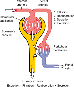
_ Part IIa. Nitric Oxide role in renal tubular epithelial cell function Tubulointerstitial Nephritides

As part of the exponential growth in our understanding of nitric oxide (NO) in health and disease over the past 2 decades, the kidney has become appreciated as a major site where NO may play a number of important roles. Although earlier work on the kidney focused more on effects of NO at the level of larger blood vessels and glomeruli, there has been a rapidly growing body of work showing critical roles for NO in tubulointerstitial disease. In this review we discuss some of the recent contributions to this important field.

Mattana J, Adamidis A, Singhal PC. Nitric oxide and tubulointerstitial nephritides. Seminars in Nephrology 2004; 24(4):345-353.

Nitric oxide donors and renal tubular (subepithelial) matrix

Nitric oxide (NO) and its metabolite, peroxynitrite (ONOO-), are involved in renal tubular cell injury. If NO/ONOO- has an effect to reduce cell adhesion to the basement membrane, does this effect contribute to tubular obstruction and would it be partially responsible for the harmful effect of NO on the tubular epithelium during acute renal failure (ARF)?

Wangsiripaisan A, et al. examined the effect of the NO donors

  • [1] (z)-1-[2-(2-aminoethyl)-N-(2-ammonioethyl)amino]diazen-1- ium-1, 2-diolate (DETA/NO),
  • [2] spermine NONOate (SpNO), and
  • [3] the ONOO- donor 3-morpholinosydnonimine (SIN-1) on

cell-matrix adhesion to collagen types I and IV, and also fibronectin using three renal tubular epithelial cell lines:

  • [1] LLC-PK1,
  • [2] BSC-1, and
  • [3] OK.

It was only the exposure to SIN-1 that caused a dose-dependent impairment in cell-matrix adhesion.

Similar results were obtained in the different cell types and matrix proteins. The effect of SIN-1 (500 microM) on LLC-PK1 cell adhesion was not associated with either cell death or alteration of matrix protein and was attenuated by either

  • [1] the NO scavenger 2-(4-carboxyphenyl)-4,4,5,5-tetramethylimidazoline-1-oxyl-3-oxide,
  • [2] the superoxide scavenger superoxide dismutase, or
  • [3] the ONOO- scavenger uric acid in a dose-dependent manner.

These investigators concluded in this seminal paper that ONOO- generated in the tubular epithelium during ischemia/reperfusion has the potential to impair the adhesion properties of tubular cells, which then may contribute to the tubular obstruction in ARF.

Wangsiripaisan A, Gengaro PE, Nemenoff RA, Ling H, et al. Effect of nitric oxide donors on renal tubular epithelial cell-matrix adhesion. Kidney Int 1999; 55(6):2281-8.

Coexpressed Nitric Oxide Synthase and Apical β1 Integrins

In sepsis-induced acute renal failure, actin cytoskeletal alterations result in shedding of proximal tubule epithelial cells (PTEC) and tubular obstruction.

This study examined the hypothesis that inflammatory cytokines, released early in sepsis, cause PTEC cytoskeletal damage and alter integrin-dependent cell-matrix adhesion. The question of whether the intermediate nitric oxide (NO) modulates these cytokine effects was also examined. After exposure of human PTEC to tumor necrosis factor-α, interleukin-1α, and interferon-γ, the actin cytoskeleton was disrupted and cells became elongated, with extension of long filopodial processes.

Cytokines induced shedding of viable, apoptotic, and necrotic PTEC, which was dependent on NO synthesized by inducible NO synthase (iNOS) produced as a result of cytokine actions on PTEC. Basolateral exposure of polarized PTEC monolayers to cytokines induced maximal NO-dependent cell shedding, mediated in part through NO effects on cGMP. Cell shedding was accompanied by dispersal of basolateral β1 integrins and E-cadherin, with corresponding upregulation of integrin expression in clusters of cells elevated above the epithelial monolayer.

These cells demonstrated coexpression of iNOS and apically redistributed β1 integrins. These authors point out that the major ligand involved in cell anchorage was laminin, probably through interactions with the integrin α3β1.

This interaction was downregulated by cytokines but was not dependent on NO. They posulate a mechanism by which inflammatory cytokines induce PTEC damage in sepsis, in the absence of hypotension and ischemia.

Glynne PA, Picot J and Evans TJ. Coexpressed Nitric Oxide Synthase and Apical β1 Integrins Influence Tubule Cell Adhesion after Cytokine-Induced Injury. JASN 2001; 12(11): 2370-2383.

Potentiation by Nitric Oxide of Apoptosis in Renal Proximal Tubule Cells

Proximal tubular epithelial cells (PTEC) exhibit a high sensitivity to undergo apoptosis in response to proinflammatory stimuli and immunosuppressors and participate in the onset of several renal diseases. This study examined the expression of inducible nitric oxide (NO) synthase after challenge of PTEC with bacterial cell wall molecules and inflammatory cytokines and analyzed the pathways that lead to apoptosis in these cells by measuring changes in the mitochondrial transmembrane potential and caspase activation.

The data show that the apoptotic effects of proinflammatory stimuli mainly were due to the expression of inducible NO synthase. Cyclosporin A and FK506 inhibited partially NO synthesis.

However, both NO and immunosuppressors induced apoptosis, probably through a common mechanism that involved the irreversible opening of the mitochondrial permeability transition pore. Activation of caspases 3 and 7 was observed in cells treated with high doses of NO and with moderate concentrations of immunosuppressors.

The conclusion is that the cooperation between NO and immunosuppressors that induce apoptosis in PTEC might contribute to the renal toxicity observed in the course of immunosuppressive therapy.

Hortelano S, Castilla M, Torres AM, Tejedor A, and Bosca L.  Potentiation by Nitric Oxide of Cyclosporin A and FK506- Induced Apoptosis in Renal Proximal Tubule Cells. J Am Soc Nephrol 2000; 11: 2315–2323.

___________________________________________________________________________________________________________________________________________________________

Part IIb. Related studies with ROS and/or RNS on nonrenal epithelial cells

Reactive nitrogen species block cell cycle re-entry Endogenous sources of reactive nitrogen species (RNS) act as second messengers in a variety of cell signaling events, whereas environmental sources of RNS like nitrogen dioxide (NO2) inhibit cell survival and growth through covalent modification of cellular macromolecules. Murine type II alveolar cells arrested in G0 by serum deprivation were exposed to either NO2 or SIN-1, a generator of RNS, during cell cycle re-entry.

In serum-stimulated cells, RNS blocked cyclin D1 gene expression, resulting in cell cycle arrest at the boundary between G0 and G1. Dichlorofluorescin diacetate (DCF) fluorescence indicated that RNS induced sustained production of intracellular hydrogen peroxide (H2O2), which normally is produced only transiently in response to serum growth factors.

Loading cells with catalase prevented enhanced DCF fluorescence and rescued cyclin D1 expression and S phase entry.

These studies indicate environmental RNS interfere with cell cycle re-entry through an H2O2-dependent mechanism that influences expression of cyclin D1 and progression from G0 to the G1 phase of the cell cycle.

Yuan Z, Schellekens H, Warner L, Janssen-Heininger Y, Burch P, Heintz NH. Reactive nitrogen species block cell cycle re-entry through sustained production of hydrogen peroxide. Am J Respir Cell Mol Biol. 2003;28(6):705-12. Epub 2003 Jan 10.

Peroxynitrite modulates MnSOD gene expression

Peroxynitrite (ONOO-) is a strong oxidant derived from nitric oxide (‘NO) and superoxide (O2.-), reactive nitrogen (RNS) and oxygen species (ROS) present in inflamed tissue. Other oxidant stresses, e.g., TNF-alpha and hyperoxia,   induce mitochondrial, manganese-containing superoxide dismutase (MnSOD) gene expression.   3-morpholinosydnonimine HCI (SIN-1) (10 or 1000 microM) increased MnSOD mRNA, but did not change hypoxanthine guanine phosphoribosyl transferase (HPRT) mRNA.   Authentic peroxynitrite (ONOO ) (100-500 microM) also increased MnSOD mRNA but did not change constitutive HPRT mRNA expression.   ONOO stimulated luciferase gene expression driven by a 2.5 kb fragment of the rat MnSOD gene 5′ promoter region.

MnSOD gene induction due to ONOO- was

  • [1] inhibited effectively by L-cysteine (10 mM) and
  • [2] partially inhibited by N-acetyl cysteine (NAC)(50 mM) or
  • [3] pyrrole dithiocarbamate (10 mM).

.NO from 1-propanamine, 3-(2-hydroxy-2-nitroso-1-propylhydrazine) (PAPA NONOate) (100 or 1000 microM) did not change MnSOD or HPRT mRNA, nor did either H202 or NO2-, breakdown products of SIN-1 and ONOO, have any effect on MnSOD mRNA expression; ONOO- and SIN-1 also did not increase detectable MnSOD protein content or increase MnSOD enzymatic activity.

Nevertheless, increased steady state [O2.-] in the presence of .NO yields ONOO , and ONOO has direct, stimulatory effects on MnSOD transcript expression driven at the MnSOD gene 5′ promoter region inhibited completely by L-cysteine and partly by N-acetyl cysteine in lung epithelial cells. This raises a question of whether the same effect is seen in renal tubular epithelium.

Jackson RM, Parish G, Helton ES. Peroxynitrite modulates MnSOD gene expression in lung epithelial cells. Free Radic Biol Med. 1998; 25(4-5):463-72.

Comparative impacts of glutathione peroxidase-1 gene knockout on oxidative stress

Selenium-dependent glutathione peroxidase-1 (GPX1) protects against reactive-oxygen-species (ROS)-induced oxidative stress in vivo, but its role in coping with reactive nitrogen species (RNS) is unclear. Primary hepatocytes were isolated from GPX1-knockout (KO) and wild-type (WT) mice to test protection of GPX1 against cytotoxicity of

  • [1] superoxide generator diquat (DQ),
  • [2]NO donor S-nitroso-N-acetyl-penicillamine (SNAP) and
  • [3] peroxynitrite generator 3-morpholinosydnonimine (SIN-1).

Treating cells with SNAP in addition to DQ produced synergistic cytotoxicity that minimized differences in apoptotic cell death and oxidative injuries between the KO and WT cells. Less protein nitrotyrosine was induced by 0.05-0.5 mM DQ+0.25 mM SNAP in the KO than in the WT cells.

Total GPX activity in the WT cells was reduced by 65 and 25% by 0.5 mM DQ+0.1 mM SNAP and 0.5 mM DQ, respectively. Decreases in Cu,Zn-superoxide dismutase (SOD) activity and increases in Mn-SOD activity in response to DQ or DQ+SNAP were greater in the KO cells than in the WT cells.

The study indicates GPX1 was more effective in protecting hepatocytes against oxidative injuries mediated by ROS alone than by ROS and RNS together, and knockout of GPX1 did not enhance cell susceptibility to RNS-associated cytotoxicity. Instead, it attenuated protein nitration induced by DQ+SNAP.

To better understand the mechanism(s) underlying nitric oxide (. NO)-mediated toxicity, in the presence and absence of concomitant oxidant exposure, postmitotic terminally differentiated NT2N cells (which are incapable of producing . NO) were exposed to [1]PAPA-NONOate (PAPA/NO) and [2] 3-morpholinosydnonimine (SIN-1).

Exposure to SIN-1, which generated peroxynitrite (ONOO) in the range of 25-750 nM/min, produced a concentration- and time-dependent delayed cell death.   In contrast, a critical threshold concentration (>440 nM/min) was required for . NO to produce significant cell injury.   There is a largely necrotic lesion after ONOO exposure and an apoptotic-like morphology after . NO exposure.

Cellular levels of reduced thiols correlated with cell death, and pretreatment with N-acetylcysteine (NAC) fully protected from cell death in either PAPA/NO or SIN-1 exposure. NAC given within the first 3 h posttreatment further delayed cell death and increased the intracellular thiol level in SIN-1 but not . NO-exposed cells.

Cell injury from . NO was independent of cGMP, caspases, and superoxide or peroxynitrite formation.   Overall, exposure of non-. NO-producing cells to . NO or peroxynitrite results in delayed cell death, which, although occurring by different mechanisms,   appears to be mediated by the loss of intracellular redox balance.

Gow AJ, Chen Q, Gole M, Themistocleous M, Lee VM, Ischiropoulos H. Two distinct mechanisms of nitric oxide-mediated neuronal cell death show thiol dependency. Am J Physiol Cell Physiol. 2000; 278(6):C1099-107.

NO2 effect on phosphatidyl choline   Nitrogen dioxide (NO2) inhalation affects the extracellular surfactant as well as the structure and function of type II pneumocytes.

The studies had differences in oxidant concentration, duration of exposure, and mode of NO2 application. This study evaluated the influence of the NO2 application mode on the phospholipid metabolism of type II pneumocytes. Rats were exposed to identical NO2 body doses (720 ppm x h), which were applied continuously (10 ppm for 3 d), intermittently (10 ppm for 8 h per day, for 9 d), and repeatedly (10 ppm for 3 d, 28 d rest, and then 10 ppm for 3 d). Immediately after exposure, type II cells were isolated and evaluated for cell yield, vitality, phosphatidylcholine (PC) synthesis, and secretion.

Type II pneumocyte cell yield was only increased from animals that had been continuously exposed to NO2, but vitality of the isolated type II pneumocytes was not affected by the NO2 exposure modes. Continuous application of 720 ppm x h NO2 resulted in increased activity of the cytidine-5-diphosphate (CDP)-choline pathway.   After continuous NO2 application,

  • [1] specific activity of choline kinase,
  • [2] cytidine triphosphate (CTP):cholinephosphate cytidylyltransferase,
  • [3] uptake of choline, and
  • [4] pool sizes of CDP-choline and PC   were significantly increased over those of controls.

Intermittent application of this NO2 body dose provoked less increase in PC synthesis and the synthesis parameters were comparable to those for cells from control animals after repeated exposure. Whereas PC synthesis in type II cells was stimulated by NO2, their secretory activity was reduced.   Continuous exposure reduced the secretory activity most, whereas intermittent exposure nonsignificantly reduced this activity as compared with that of controls. The repeated application of NO2 produced no differences.

The authors conclude that…. type II pneumocytes adapt to NO2 atmospheres depending on the mode of its application, at least for the metabolism of PC and its secretion from isolated type II pneumocytes.

The reader asks whether this effect could also be found in renal epithelial cells, for which PC is not considered vital as for type II pneumocytes and possibly related to surfactant activity in the lung.

Müller B, Seifart C, von Wichert P, Barth PJ. Adaptation of rat type II pneumocytes to NO2: effects of NO2 application mode on phosphatidylcholine metabolism. Am J Respir Cell Mol Biol. 1998; 18(5): 712-20.

iNOS involved in immediate response to anaphylaxis

The generation of large quantities of nitric oxide (NO) is implicated in the pathogenesis of anaphylactic shock. The source of NO, however, has not been established and conflicting results have been obtained when investigators have tried to inhibit its production in anaphylaxis.

This study analyzed the expression of inducible nitric oxide synthase (iNOS) and endothelial nitric oxide synthase (eNOS) in a mouse model of anaphylaxis.   BALB/c mice were sensitized and challenged with ovalbumin to induce anaphylaxis. Tissues were removed from the heart and lungs, and blood was drawn at different time points during the first 48 hours after induction of anaphylaxis. The Griess assay was used to measure nitric oxide generation.

Nitric oxide synthase expression was examined by reverse transcriptase polymerase chain reaction and immunohistochemistry. A significant increase in iNOS mRNA expression and nitric oxide production was evident as early as 10 to 30 minutes after allergen challenge in both heart and lungs.

In contrast, expression of eNOS mRNA was not altered during the course of the experiment. The results support involvement of iNOS in the immediate physiological response of anaphylaxis.

Sade K, Schwartz IF, Etkin S, Schwartzenberg S, et al. Expression of Inducible Nitric Oxide Synthase in a Mouse Model of Anaphylaxis. J Investig Allergol Clin Immunol 2007; 17(6):379-385.

________________________________________________________________________________________________________________________________________________________

Part IIc. Additional Nonrenal Related NO References

1. Nitrogen dioxide induces death in lung epithelial cells in a density-dependent manner. Persinger RL, Blay WM, Heintz NH, Hemenway DR, Janssen-Heininger YM. Am J Respir Cell Mol Biol. 2001 May;24(5):583-90. PMID: 11350828 [PubMed – indexed for MEDLINE] Free Article

2. Molecular mechanisms of nitrogen dioxide induced epithelial injury in the lung. Persinger RL, Poynter ME, Ckless K, Janssen-Heininger YM. Mol Cell Biochem. 2002 May-Jun;234-235(1-2):71-80. Review. PMID: 12162462 [PubMed – indexed for MEDLINE]

3. Nitric oxide and peroxynitrite-mediated pulmonary cell death. Gow AJ, Thom SR, Ischiropoulos H. Am J Physiol. 1998 Jan;274(1 Pt 1):L112-8. PMID: 9458808 [PubMed – indexed for MEDLINE] Free Article

4. Mitogen-activated protein kinases mediate peroxynitrite-induced cell death in human bronchial epithelial cells. Nabeyrat E, Jones GE, Fenwick PS, Barnes PJ, Donnelly LE. Am J Physiol Lung Cell Mol Physiol. 2003 Jun;284(6):L1112-20. Epub 2003 Feb 21. PMID: 12598225 [PubMed – indexed for MEDLINE] Free Article

5. Peroxynitrite inhibits inducible (type 2) nitric oxide synthase in murine lung epithelial cells in vitro. Robinson VK, Sato E, Nelson DK, Camhi SL, Robbins RA, Hoyt JC. Free Radic Biol Med. 2001 May 1;30(9):986-91. PMID: 11316578 [PubMed – indexed for MEDLINE]

6. Nitric oxide-mediated chondrocyte cell death requires the generation of additional reactive oxygen species. Del Carlo M Jr, Loeser RF. Arthritis Rheum. 2002 Feb;46(2):394-403. PMID: 11840442 [PubMed – indexed for MEDLINE]

7. Colon epithelial cell death in 2,4,6-trinitrobenzenesulfonic acid-induced colitis is associated with increased inducible nitric-oxide synthase expression and peroxynitrite production.

Yue G, Lai PS, Yin K, Sun FF, Nagele RG, Liu X, Linask KK, Wang C, Lin KT, Wong PY. J Pharmacol Exp Ther. 2001 Jun;297(3):915-25. PMID: 11356911 [PubMed – indexed for MEDLINE] Free Article

__________________________________________________________________________________________________________________________________________________________

Part IIIa. Acute renal failure   Acute renal failure (ARF), characterized by sudden loss of the ability of the kidneys to [1] excrete wastes, [2] concentrate urine, [3] conserve electrolytes, and [4] maintain fluid balance, is a frequent clinical problem, particularly in the intensive care unit, where it is associated with a mortality of between 50% and 80%.

This clinical entity was described as an acute loss of kidney function that occurred in severely injured crush victims because of histological evidence for patchy necrosis of renal tubules at autopsy. In the clinical setting, the terms ATN and acute renal failure (ARF) are frequently used interchangeably. However, ARF does not include increases in blood urea due to [1] reversible renal vasoconstriction (prerenal azotemia) or [2] urinary tract obstruction (postrenal azotemia). Acute hemodialysis was first used clinically during the Korean War in 1950 to treat military casualties, and this led to a decrease in mortality of the ARF clinical syndrome from about 90% to about 50%.   In the half century that has since passed, much has been learned about the pathogenesis of ischemic and nephrotoxic ARF in experimental models, but there has been very little improvement in mortality. This may be explained by changing demographics: [1] the age of patients with ARF continues to rise, and [2] comorbid diseases are increasingly common in this population. Both factors may obscure any increased survival related to improved critical care. Examining the incidence of ARF in several military conflicts does, however, provide some optimism. The incidence of ARF in seriously injured casualties decreased between World War II and the Korean War, and again between that war and the Vietnam War, despite the lack of any obvious difference in the severity of the injuries. What was different was the rapidity of the fluid resuscitation of the patients? Fluid resuscitation on the battlefield with the rapid evacuation of the casualties to hospitals by helicopter began during the Korean War and was optimized further during the Vietnam War. For seriously injured casualties the incidence of ischemic ARF was one in 200 in the Korean War and one in 600 in the Vietnam War. This historical sequence of events suggests that early intervention could prevent the occurrence of ARF, at least in military casualties.   In experimental studies it has been shown that progression from an azotemic state associated with renal vasoconstriction and intact tubular function (prerenal azotemia) to established ARF with tubular dysfunction occurs if the renal ischemia is prolonged. Moreover, early intervention with fluid resuscitation was shown to prevent the progression from prerenal azotemia to established ARF. Diagnostic evaluation of ARF One important question, therefore, is how to assure that an early diagnosis of acute renal vasoconstriction can be made prior to the occurrence of tubular dysfunction, thus providing the potential to prevent progression to established ARF. In this regard, past diagnostics relied on observation of the patient response to a fluid challenge: [1] decreasing levels of blood urea nitrogen (BUN) indicated the presence of reversible vasoconstriction, [2] while uncontrolled accumulation of nitrogenous waste products, i.e., BUN and serum creatinine, indicated established ARF.

This approach, however, frequently led to massive fluid overload in the ARF patient with resultant

  • [1] pulmonary congestion,
  • [2] hypoxia, and
  • [3] premature need for mechanical ventilatory support and/or hemodialysis.

On this background the focus turned to an evaluation of urine sediment and urine chemistries to differentiate between renal vasoconstriction with intact tubular function and established ARF.

It was well established that if tubular function was intact, renal vasoconstriction was associated with enhanced tubular sodium reabsorption. Specifically, the fraction of filtered sodium that is rapidly reabsorbed by normal tubules of the vasoconstricted kidney is greater than 99%.

Thus, when nitrogenous wastes, such as creatinine and urea, accumulate in the blood due to a fall in glomerular filtration rate (GFR) secondary to renal vasoconstriction with intact tubular function, the fractional excretion of filtered sodium (FENa = [(urine sodium × plasma creatinine) / (plasma sodium × urine creatinine)]) is less than 1%. An exception to this physiological response of the normal kidney to vasoconstriction is when the patient is receiving a diuretic, including mannitol, or has glucosuria, which decreases tubular sodium reabsorption and increases FENa.

It has recently been shown in the presence of diuretics that a rate of fractional excretion of urea (FEurea) of less than 35 indicates intact tubular function, thus favoring renal vasoconstriction rather than established ARF as a cause of the azotemia.

___________________________________________________________________________________________________________________________________________________________

English: Physiology of Nephron

English: Physiology of Nephron (Photo credit: Wikipedia)
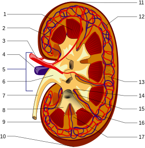
Structures of the kidney: 1.Renal pyramid 2.In...

Structures of the kidney: 1.Renal pyramid 2.Interlobar artery 3.Renal artery 4.Renal vein 5.Renal hilum 6.Renal pelvis 7.Ureter 8.Minor calyx 9.Renal capsule 10.Inferior renal capsule 11.Superior renal capsule 12.Interlobar vein 13.Nephron 14.Minor calyx 15.Major calyx 16.Renal papilla 17.Renal column (no distinction for red/blue (oxygenated or not) blood, arteriole is between capilaries and larger vessels (Photo credit: Wikipedia)

_

______________________________________________________________________________________________________________________________________________________

Mechanisms of ARF

Based on the foregoing comments, this discussion of mechanisms of ARF will not include nitrogenous-waste accumulation due to renal vasoconstriction with intact tubular function (prerenal azotemia) or urinary tract obstruction (postrenal azotemia). The mechanisms of ARF involve both vascular and tubular factors. An ischemic insult to the kidney will in general be the cause of the ARF. While a decrease in renal blood flow with diminished oxygen and substrate delivery to the tubule cells is an important ischemic factor, it must be remembered that a relative increase in oxygen demand by the tubule is also a factor in renal ischemia.

Approximately 30–70% of these shed epithelial tubule cells in the urine are viable and can be grown in culture. Recent studies using cellular and molecular techniques have provided information relating to the structural abnormalities of injured renal tubules that occur both in vitro and in vivo. In vitro studies using chemical anoxia have revealed abnormalities in the proximal tubule cytoskeleton that are associated with translocation of Na+/K+-ATPase from the basolateral to the apical membrane.

A comparison of cadaveric transplanted kidneys with delayed versus prompt graft function has also provided important results regarding the role of Na+/K+-ATPase in ischemic renal injury. This study demonstrated that, compared with kidneys with prompt graft function, those with delayed graft function had a significantly greater cytoplasmic concentration of Na+/K+-ATPase and actin-binding proteins — spectrin (also known as fodrin) and ankyrin — that had translocated from the basolateral membrane to the cytoplasm.

Such a translocation of Na+/K+-ATPase from the basolateral membrane to the cytoplasm could explain the decrease in tubular sodium reabsorption that occurs with ARF. An important focus of research is the mechanisms whereby the critical residence of Na+/K+-ATPase in the basolateral membrane (which facilitates vectorial sodium transport) is uncoupled by hypoxia or ischemia.  The actin-binding proteins,

  • spectrin and
  • ankyrin,

serve as substrates for the calcium-activated cysteine protease calpain.

________________________________________________________________________________________________________________________________________________________

In vitro studies in proximal tubules have shown a rapid rise in cytosolic calcium concentration during acute hypoxia, which antedates the evidence of tubular injury as assessed by lactic dehydrogenase (LDH) release. There is further evidence to support the importance of the translocation of Na+/K+-ATPase from the basolateral membrane to the cytoplasm during renal ischemia/reperfusion.

Specifically, calpain-mediated breakdown products of the actin-binding protein spectrin occur with renal ischemia. Calpain activity was demonstrated to increase during hypoxia in isolated proximal tubules. Measurement of LDH release following calpain inhibition indicated attenuation of hypoxic damage to proximal tubules. There was no evidence of an increase in cathepsin, a (cysteine protease) in proximal tubules during hypoxia , but there is a calcium-independent pathway for calpain activation during hypoxia.

Calpastatin, an endogenous cellular inhibitor of calpain activation, was shown to be diminished during hypoxia in association with the rise in another cysteine protease, caspase.

This effect of diminished calpastatin activity could be reversed by caspase inhibition. Proteolytic pathways appear to be involved in calpain-mediated proximal tubule cell injury during hypoxia. Calcium activation of phospholipase A has also been shown to contribute to renal tubular injury during ischemia.

________________________________________________________________________________________________________________________________________________________

Tubular obstruction during ARF

The existence of proteolytic pathways involving cysteine proteases, namely calpain and caspases, may therefore explain

  • the decrease in proximal tubule sodium reabsorption and
  • increased FENa

secondary to proteolytic uncoupling of Na+/K+-ATPase from its basolateral membrane anchoring proteins.

This tubular perturbation alone, however, does not explain the fall in GFR that leads to nitrogenous-waste retention and thus the rise in BUN and serum creatinine.   Decreased proximal tubule sodium reabsorption may lead to a decreased GFR during ARF. First of all, brush border membranes and cellular debris could provide the substrate for intraluminal obstruction in the highly resistant portion of distal nephrons.

In fact, microdissection of individual nephrons of kidneys from patients with ARF demonstrated obstructing casts in distal tubules and collecting ducts. This observation could explain the dilated proximal tubules that are observed upon renal biopsy of ARF kidneys. The intraluminal casts in ARF kidneys stain prominently for Tamm-Horsfall protein (THP), which is produced in the thick ascending limb. Importantly, THP is secreted into tubular fluid as a monomer but subsequently may become a polymer that forms a gel-like material in the presence of increased luminal Na+ concentration, as occurs in the distal nephron during clinical ARF with the decrease in tubular sodium reabsorption.

Thus, the THP polymeric gel in the distal nephron provides an intraluminal environment for distal cast formation involving viable, apoptotic, and necrotic cells.

The loss of the tubular epithelial cell barrier and/or the tight junctions between viable cells during acute renal ischemia could lead to a leak of glomerular filtrate back into the circulation. (If this occurs and normally non-reabsorbable substances, such as inulin, leak back into the circulation, then a falsely low GFR will be measured as inulin clearance. It should be noted, however, that the degree of extensive tubular damage observed in experimental studies demonstrating tubular fluid backleak is rarely observed with clinical ARF in humans). Moreover, dextran sieving studies in patients with ARF demonstrated that, at best, only a 10% decrease in GFR could be explained by backleak of filtrate. Cadaveric transplanted kidneys with delayed graft function, however, may have severe tubular necrosis, and thus backleak of glomerular filtration may be more important in this setting.

Inflammation and NO

There is now substantial evidence for the involvement of inflammation in the pathogenesis of the decreased GFR associated with acute renal ischemic injury. In this regard, there is experimental evidence that iNOS may contribute to tubular injury during ARF. Hypoxia in isolated proximal tubules has been shown to increase NO release, and there is increased iNOS protein expression in ischemic kidney homogenates. An antisense oligonucleotide was shown to block the upregulation of iNOS and afford functional protection against acute renal ischemia. Moreover, when isolated proximal tubules from iNOS, eNOS, and neuronal NO synthase (nNOS) knockout mice were exposed to hypoxia, only the tubules from the iNOS knockout mice were protected against hypoxia, as assessed by LDH release. The iNOS knockout mice were also shown to have lower mortality during ischemia/reperfusion than wild-type mice.  The scavenging of NO by oxygen radicals produces peroxynitrite causing tubule damage during ischemia. While iNOS may contribute to ischemic injury of renal tubules,  the vascular effect of eNOS in the glomerular afferent arteriole is protective against ischemic injury. In this regard, eNOS knockout mice are more sensitive to endotoxin-related injury than normal mice.

Moreover, the protective role of vascular eNOS may be more important than the deleterious effect of iNOS at the tubule level during renal ischemia.   This is because treatment of mice with the nonspecific NO synthase (NOS) inhibitor L-NAME, which blocks both iNOS and eNOS, worsens renal ischemic injury. NO may downregulate eNOS and is a potent inducer of heme oxygenase-1, which has been shown to be cytoprotective against renal injury. The MAPK pathway also appears to be involved in renal oxidant injury. Activation of extracellular signal–regulated kinase (ERK) or inhibition of JNK ameliorates oxidant injury–induced necrosis in mouse renal proximal tubule cells in vitro. Upregulation of ERK may also be important in the effect of preconditioning whereby early ischemia affords protection against a subsequent ischemia/reperfusion insult. Alterations in cell cycling are also involved in renal ischemic injury. Upregulation of p21, which inhibits cell cycling, appears to allow cellular repair and regeneration, whereas homozygous p21 knockout mice demonstrate enhanced cell necrosis in response to an ischemic insult.

Prolonged duration of the ARF clinical course and the need for dialysis are major factors projecting a poor prognosis. Patients with ARF who require dialysis have a 50–70% mortality rate. Infection and cardiopulmonary complications are the major causes of death in patients with ARF. Excessive fluid administration in patients with established ARF may lead to pulmonary congestion, hypoxia, the need for ventilatory support, pneumonia, and multiorgan dysfunction syndrome, which has an 80–90% mortality rate. Until means to reverse the diminished host defense mechanisms in azotemic patients with clinical ARF are available, every effort should be made to avoid invasive procedures such as the placement of bladder catheters, intravenous lines, and mechanical ventilation. Over and above such supportive care, it may be that combination therapy will be necessary to prevent or attenuate the course of ARF. Such combination therapy must involve agents with potential beneficial effects on vascular tone, tubular obstruction, and inflammation.

Schrier RW, Wang W, Poole B, and Mitra A. Acute renal failure: definitions, diagnosis, pathogenesis, and therapy. The Journal of Clinical Investigation 2004; 114(1):5-14. http://www.jci.org

______________________________________________________________________________________________________________________________________________________

Part IIIb. Additional Related References on NO, oxidative stress and Kidney

Shelgikar PJ, Deshpande KH, Sardeshmukh AS, Katkam RV, Suryakarl AN. Role of oxidants and antioxidants in ARF patients undergoing hemodialysis. Indian J Nephrol 2005;15: 73-76.

Lee JW. Renal Dysfunction in Patients with Chronic Liver Disease. Electrolytes Blood Press 7:42-50, 2009ㆍdoi: 10.5049/EBP.2009.7.2.42.

Saadat H, et al. Endothelial Nitric Oxide Function and Tubular Injury in Premature Infants. Int J App Sci and Technol 2012; 7(6): 77-81. http://www.ijastnet.com.

Amerisan MS. Cardiovascular disease in chronic kidney disease. Indian J Nephrol 2005;15: 1-7.

___________________________________________________________________________________________________________________________________________________________

Traditional risk factors for CVD in CKD

  • Hypertension
  • Older Age
  • Diabetes Mellitus
  • Male gender
  • High LDL
  • White Race
  • Low HDL
  • Physical inactivity
  • Smoking
  • Menopause
  • LVH

CKD Related CV Risk Factors

  • Blood Pressure
  • ? Homocysteinemia
  • Anemia
  • ? Inflammation
  •   Ca++ x P++
  • ? NO synthesis
  • Na+ Retention
  • ? Lp (a)
  • Hypervolemia
  • ? Insulin Resistance
  • Proteinuria & Hypoalbuminemia
  • Iron over load
  • ? Adeponectin
  • ??Vit. C or E
  • ? 5 Lipoxygenase
  • ROS
  • Genetic factors
  • ADMA (Asymmetric Dimethyl Arginine)

S Vikrant, SC Tiwari. Essential Hypertension – Pathogenesis and Pathophysiology. J Indian Acad Clinical Medicine 2001; 2(3):141-161. Scheme for pathogenesis of salt dependent hypertension.

The hypothesis proposes that early hypertension is episodic and is mediated by a hyperactive sympathetic nervous system or activated renin-angiotensin system.

Cell membrane alterations

Hypotheses linking abnormal ionic fluxes to increased peripheral resistance through increase in cell sodium, calcium, or pH.   The hypertension that is more common in obese people may arise in large part from the insulin resistance and resultant hyperinsulinaemia that results from the increased mass of fat. However, rather unexpectedly, insulin resistance may also be involved in hypertension in non-obese people.

Overall scheme for the mechanisms by which obesity, if predominantly upper body or visceral in location, could promote

________________________________________________________________________________________________________________________________________________________

  • diabetes,
  • dyslipidemia and
  • hypertension via hyperinsulinemia.

The explanation for insulin resistance found in as many as half of nonobese hypertensive is not obvious and may involve one or more aspects of insulin’s action

__________________________________________________________________________________________________________________________________________________________

Proposed mechanisms by which insulin resistance and/or hyperinsulinemia may lead to increased blood pressure.

  1. Enhanced renal sodium and water reabsorption.
  2. Increased blood pressure sensitivity to dietary salt intake
  3. Augmentation of the pressure and
  4. aldosterone responses to AII
  5. Changes in transmembrane electrolyte transport
  • a. Increased intracellular sodium
  • b. Decreased Na+/K+ – ATPase activity
  • c. Increased intracellular Ca2+ pump activity
  • d. Increased intracellular Ca2+ accumulation
  • e. Stimulation of growth factors

___________________________________________________________________________________________________________________________________________________________

Part IV. New Insights on NO donors

This study investigated the involvement of nitric oxide (NO) into the irradiation-induced increase of cell attachment. These experiments explored the cellular mechanisms of low-power laser therapy. HeLa cells were irradiated with a monochromatic visible-tonear infrared radiation (600–860 nm, 52 J/m2) or with a diode laser (820 nm, 8–120 J/m2) and the number of cells attached to a glass matrix was counted after 30 minute incubation at 37oC. The NO donors

  1. sodium nitroprusside (SNP),
  2. glyceryl trinitrate (GTN), or
  3. sodium nitrite (NaNO2)

were added to the cellular suspension before or after irradiation. The action spectra and the concentration and fluence dependencies obtained were compared and analyzed.

The well-structured action spectrum for the increase of the adhesion of the cells, with maxima at 619, 657, 675, 740, 760, and 820 nm, points to the existence of a photoacceptor responsible for the enhancement of this property (supposedly cytochrome c oxidase, the terminal respiratory chain enzyme), as well as signaling pathways between the cell mitochondria, plasma membrane, and nucleus.

Treating the cellular suspension with SNP before irradiation significantly modifies the action spectrum for the enhancement of the cell attachment property (band maxima at 642, 685, 700, 742, 842, and 856 nm). The action of SNP, GTN, andNaNO2 added before or after irradiation depends on their concentration and radiation fluence.

The NO donors added to the cellular suspension before irradiation eliminate the radiation induced increase in the number of cells attached to the glass matrix, supposedly by way of binding NO to cytochrome c oxidase. NO added to the suspension after irradiation can also inhibit the light-induced signal downstream. Both effects of NO depend on the concentration of the NO donors added.

The results indicate that NO can control the irradiation-activated reactions that increase the attachment of cells.

Karu TI, Pyatibrat LV, and Afanasyeva NI. Cellular Effects of Low Power Laser Therapy Can be Mediated by Nitric Oxide. Lasers Surg. Med 2005; 36:307–314.

IFNa-2b (IFN-a) effect on barrier function of renal tubular epithelium

IFNa treatment can be accompanied by impaired renal function and capillary leak. This study shows IFNa produced dose-dependent and time-dependent decrease in transepithelial resistance (TER) ameliorated by tyrphostin, an inhibitor of phosphotyrosine kinase with increased expression of occludin and E-cadherin. In conclusion, IFNa can directly affect barrier function in renal epithelial cells via ovewrexpression or missorting of the junctional proteins occludin and E-cadherin.

Lechner J, Krall M, Netzer A, Radmayr C, et al. Effects of interferon a-2b on barrier function and junctional complexes of renal proximal tubulat LLC-pK1 cells. Kidney Int 1999; 55:2178-2191.

Ischemia-reperfusion injury

The pathophysiology of acute renal failure (ARF) is complex and not well understood. Numerous models of ARF suggest that oxygen-derived reactive species are important in renal ischemia-reperfusion (I-R) injury, but the nature of the mediators is still controversial. Treatment with oxygen radical scavengers, antioxidants, and iron chelators such as

  • superoxide dismutase,
  • dimethylthiourea,
  • allopurinol, and
  • deferoxamine

are protective in some models, and suggest a role for the hydroxyl radical formation. However, these compounds are not protective in all models of I-R injury, and direct evidence for the generation of hydroxyl radical is absent. Furthermore, these inhibitors have another property in common.

They all directly scavenge or inhibit the formation of peroxynitrite (ONOO−), a highly toxic species derived from nitric oxide (NO) and superoxide. Thus, the protective effects seen with these inhibitors may be due in part to their ability to inhibit ONOO− formation. Even though reactive oxygen species are thought to participate in ischemia-reperfusion (I-R) injury, induction of and production of high levels of  inducible nitric oxide (NO)  also contribute to this injury.

NO combines with superoxide to form the potent oxidant peroxynitrite (ONOO−). NO and ONOO− were investigated in a rat model of renal I-R injury using the selective iNOS inhibitor L-N6-(1-iminoethyl)lysine (L-NIL).

I-R surgery significantly increased plasma creatinine levels to 1.9 ± 0.3 mg/dl (P < .05) and caused renal cortical necrosis. L-NIL administration (3 mg/kg) in animals subjected to I-R significantly decreased plasma creatinine levels to 1.2 ± 0.10 mg/dl (P < .05 compared with I-R) and reduced tubular damage.

ONOO− formation was evaluated by detecting 3-nitrotyrosine-protein adducts (3NTyPAs), a stable biomarker of ONOO− formation.   The kidneys from I-R animals had increased levels of 3NTyPAs compared with control animals   L-NIL-treated rats (3 mg/kg) subjected to I-R showed decreased levels of 3NTyPAs.

These results suggests that iNOS-generated NO mediates damage in I-R injury possibly through ONOO− formation.

______________________________________________________________________________________________________________________________________________________

In summary,

  1. 3-nitrotyrosine-protein adducts were detected in renal tubules after I-R injury.
  2. Selective inhibition of iNOS by L-NIL decreased injury, improved renal function, and decreased apparent ONOO− formation.
  3. Reactive nitrogen species should be considered potential therapeutic targets in the prevention and treatment of renal I-R injury.

_______________________________________________________________________________________________________________________________________________________

Walker LM, Walker PD, Imam SZ, et al. Evidence for Peroxynitrite Formation in Renal Ischemia-Reperfusion Injury: Studies with the Inducible Nitric Oxide Synthase InhibitorL-N6-(1-Iminoethyl)lysine1. 2000.

Role of TNFa independent of iNOS Renal failure is a frequent complication of sepsis, mediated by renal vasoconstrictors and vasodilators. Endotoxin induces several proinflammatory cytokines, among which tumor necrosis factor (TNF) is thought to be of major importance. Tumor necrosis factor (TNF) has been suggested to be a factor in the acute renal failure in sepsis or endotoxemia. Passive immunization by anti-TNFa prevented development of septic shock in animal experiments.The development of ARF involves excessive intrarenal vasoconstriction. Involvement of nitric oxide (NO), generated by inducible NO synthase (iNOS), is still a factor in the pathogenesis of endotoxin-induced renal failure. TNF-a leads to a decrease in glomerular filtration rate (GFR).

This study tested the hypothesis that the role of TNF-a in endotoxic shock related ARF is mediated by iNOS-derived NO.   An injection of lipopolysaccharide (LPS) constituent of gram-negative bacteria to wild-type mice resulted in a 70% decrease in glomerular filtration rate (GFR) and in a 40% reduction in renal plasma flow (RPF) 16 hours after the injection.   The results occurred independent of hypotension, morphological changes, apoptosis, and leukocyte accumulation. In mice pretreated with TNFsRp55, only a 30% decrease in GFR was observed without a significant change in RPF as compared with controls. Pretreatment with TNKsRp55 on renal function Wild-type mice were pretreated with TNFsRp55(10 mg/kg IP)  for one hour before the administration of 5 mg/kg intraperitoneal endotoxin. GFR and RPF were determined 16 hours thereafter. Data are expressed as mean 6, SEM, N 5 6. *P , 0.05 vs. Control; §P , 0.05 vs. LPS, by ANOVA.

The serum NO concentration was significantly lower in endotoxemic wild-type mice pretreated with TNFsRp55, as compared with untreated endotoxemic wild-type mice. In LPS-injected iNOS knockout mice and wild-type mice treated with a selective iNOS inhibitor, 1400W, the development of renal failure was similar to that in wild-type mice. As in wild-type mice,TNFsRp55 significantly attenuated the decrease in GFR (a 33% decline, as compared with 75% without TNFsRp55) without a significant change in RPF in iNOS knockout mice given LPS. These results demonstrate a role of TNF in the early renal dysfunction (16 h) in a septic mouse model independent of iNOS,

  • hypotension,
  • apoptosis,
  • leukocyte accumulation,and
  • morphological alterations,

thus suggesting renal hypoperfusion secondary to an imbalance between, as yet to be defined renal vasoconstrictors and vasodilators.

Knotek M, Rogachev B, Wang W,….., Edelstein CL, Dinarello CA, and Schrier RW. Endotoxemic renal failure in mice: Role of tumor necrosis factor independent of inducible nitric oxide synthase. Kidney International 2001; 59:2243–2249

Ischemic acute renal failure

Inflammation plays a major role in the pathophysiology of acute renal failure resulting from ischemia. This review discusses the contribution of

  • endothelial
  • epithelial cells and
  • leukocytes

to this inflammatory response. The roles of cytokines/chemokines in the injury and recovery phase are reviewed. The protection of mouse kidney prior to exposure to ischemia or urinary tract obstruction is  a potential model to  search for pharmacologic agents to protect the kidney against injury by inflammatory mediators produced by tubular epithelial cells and activated leukocytes in renal ischemia/reperfusion (I/R) injury. Tubular epithelia produce

  • TNF-a,
  • IL-1,
  • IL-6,
  • IL-8,
  • TGF-b,
  • MCP-1,
  • ENA-78,
  • RANTES, and
  • fractalkines,

whereas leukocytes produce

  • TNF-a,
  • IL-1,
  • IL-8,
  • MCP-1,
  • ROS, and
  • eicosanoids.

The release of these chemokines and cytokines serve as effectors for a positive feedback pathway enhancing inflammation and cell injury, the cycle of tubular epithelial cell injury and repair following renal ischemia/reperfusion.   Tubular epithelia are typically cuboidal in shape and apically-basally polarized; the Na+/K+-ATPase localizes to basolateral plasma membranes, whereas cell adhesion molecules, such as integrins localize basally. In response to ischemia reperfusion,

  • the Na+/K+-ATPase appears apically, and
  • integrins are detected on lateral and basal plasma membranes.

Some of the injured epithelial cells undergo necrosis and/or apoptosis detaching from the underlying basement membrane into the tubular space where they contribute to tubular occlusion. Viable cells that remain attached, dedifferentiate, spread, and migrate to repopulate the denuded basement membrane. With cell proliferation, cell-cell and cell-matrix contacts are restored, and the epithelium redifferentiates and repolarizes, forming a functional, normal epithelium Inflammation is a significant component of renal I/R injury, playing a considerable role in its pathophysiology.

Although significant progress has been made in defining the major components of this process, the complex cross-talk between endothelial cells, inflammatory cells, and the injured epithelium with each generating and often responding to cytokines and chemokines is not well understood. In addition, we have not yet taken full advantage of the large body of data on inflammation in other organ systems.

Furthermore, preconditioning the kidney to afford protection to subsequent bouts of ischemia may serve as a useful model challenging us to therapeutically mimic endogenous mechanisms of protection.

Understanding the inflammatory response prevalent in ischemic kidney injury will facilitate identification of molecular targets for therapeutic intervention.

Bonventre JV and Zuk A. Ischemic acute renal failure: An inflammatory disease? Forefronts in Nephrology 2002;.. :480-485

Gene expression profiles in renal proximal tubules In kidney disease renal proximal tubular epithelial cells (RPTEC) actively contribute to the progression of tubulointerstitial fibrosisby mediating both

  • an inflammatory response and
  • via epithelial-to-mesenchymal transition.

Using laser capture microdissection we specifically isolated RPTEC from cryosections of the healthy parts of kidneys removed owing to renal cell carcinoma and from kidney biopsies from patients with proteinuric nephropathies. RNA was extracted and hybridized to complementary DNA microarrays after linear RNA amplification. Statistical analysis identified 168 unique genes with known gene ontology association, which separated patients from controls. Besides distinct alterations in signal-transduction pathways (e.g. Wnt signalling), functional annotation revealed a significant upregulation of genes involved in

_________________________________________________________________________________________________________________________________________________________

  • cell proliferation and cell cycle control (like insulin-like growth factor 1 or cell division cycle 34),
  • cell differentiation (e.g. bone morphogenetic protein 7),
  • immune response,
  • intracellular transport and
  • metabolism

__________________________________________________________________________________________________________________________________________________________

in RPTEC from patients.

The study also revealed differential expression of a number of genes responsible for cell adhesion (like BH-protocadherin) with a marked downregulation of most of these transcripts. In summary, the results obtained from RPTEC revealed a differential regulation of genes, which are likely to be involved in either pro-fibrotic or tubulo-protective mechanisms in proteinuric patients at an early stage of kidney disease.

Rudnicki M, Eder S, Perco P, Enrich J, et al. Gene expression profiles of human proximal tubular epithelial cells in proteinuric nephropathies. Kidney International 2006; xx:1-11. Kidney International advance online publication, 20 December 2006; doi:10.1038/sj.ki.5002043. http://www.kidney-international.org

Oxidative stress involved with diabetic nephropathy

Diabetic Nephropathy (DN) poses a major health problem. There is strong evidence for a potential role of the eNOS gene. This case control study investigated the possible role of genetic variants of the endothelial Nitric Oxide Synthase (eNOS) gene and oxidative stress in the pathogenesis of nephropathy in patients with diabetes mellitus. The study included 124 diabetic patients;

  1. 68 of these patients had no diabetic nephropathy (group 1) while
  2. 56 patients exhibited symptoms of diabetic nephropathy (group 2).
  3. Sixty two healthy non-diabetic individuals were also included as a control group.

Blood samples from subjects and controls were analyzed to investigate the eNOS genotypes and to estimate

  • the lipid profile and
  • markers of oxidative stress such as malondialdehyde (MDA) and nitric oxide (NO).

No significant differences were found in the frequency of eNOS genotypes between diabetic patients (either in group 1 or group 2) and controls (p >0.05). Also, no significant differences were found in the frequency of eNOS genotypes between group 1 and group 2 (p >0.05). Both group 1 and group 2 had significantly higher levels of nitrite and MDA when compared with controls (all p = 0.0001). Also group 2 patients had significantly higher levels of nitrite and MDA when compared with group 1 (p = 0.02, p = 0.001 respectively).

The higher serum level of the markers of oxidative stress in diabetic patients particularly those with diabetic nephropathy suggest that oxidative stress and not the eNOS gene polymorphism is involved in the pathogenesis of the diabetic nephropathy in this subset of patients

Badawy A, Elbaz R, Abbas AM, Ahmed Elgendy A, et al. Oxidative stress and not endothelial Nitric Oxide Synthase gene polymorphism involved in diabetic nephropathy. Journal of Diabetes and Endocrinology 2011; 2(3): 29-35.

Metformin in renal ischemia reperfusion

Renal ischemia plays an important role in renal impairment and transplantation. Metformin is a biguanide used in type 2 diabetes, it inhibits hepatic glucose production and increases peripheral insulin sensitivity. While the mode of action of metformin is incompletely understood, it appears to have anti-inflammatory and antioxidant effects involved in its beneficial effects on insulin resistance.   Control, Sham, ischemia/reperfusion (I/R) and Metformin treated I /R groups   A renal I/R injury was done by a left renal pedicle occlusion to induce ischemia for 45 min followed by 60 min of reperfusion with contralateral nephrectomy. Metformin pretreated I/R rats in a dose of 200 mg/kg/day for three weeks before ischemia induction.

  • Nitric oxide (NO),
  • tumor necrosis factor alpha (TNF α) ,
  • catalase (CAT) and
  • reduced glutathione (GSH) activities

were determined in renal tissue, while

  • creatinine clearance (CrCl) ,
  • blood urea nitrogen (BUN) were measured and

5 hour urinary volume and electrolytes were estimated . BUN and CrCl levels in the I/R group were significantly higher than in control rats (p<0.05) table (1).

__________________________________________________________________________________________________________________________________

Table 1: Creatinine clearance (Cr Cl) and blood urea nitrogen (BUN) levels in control and test groups.
(Mean ± SD)

Groups CrCl   (ml/min) BUN (mg/dl)
Control group 1.30 ±0.11 14.30±0.25
Sham group+ metformin 1.27±0.09 15.70±0.19
I/R group (P1) 1.85±0.25 (<0.001 ) 28.00±0.62 (<0.001)
I/R+ metformin group (P2,P3) 1.55±0.22 (0.001, 0.028) 18.10±1.00 (<0.001, <0.001)
  • P1: Statistical significance between control
    group and saline treated I/R group.
  • P2 Statistical significance between control
    group and Metformin treated I/R group.
  • P3 Statistical significance between saline treated
    I/R group and Metformin treated I/R group

_______________________________________________________________________________________________________________________________________________________

When metformin was administered before I/R, BUN and CrCl levels were still significantly higher than control group but their elevation were significantly lower in comparison to I/R group alone (P<0.05).   TNF α and NO levels were significantly higher in the I/R group than those of the control group (Table 2). Pre-treatment with metformin significantly lowered their levels in comparison to I/R group (P<0.05).

________________________________________________________________________________________________________________________________________________________

Table 2: Tumor necrosis factor α (TNF α) and inducible nitric oxide (iNO) levels in control and test groups.
(Mean ± SD)

Groups TNF α (pmol/mg tissue) iNO (nmol/ mg tissue)
Control group 1 7.60 ±5.98 2.54 ± 0.82
Sham group+ metformin 16.70 ±5.50 2.35 ±0.80
I/R group (P1) 54. 00±6.02 (<0.001) 4.50±0.89 (<0.001)
I/R+metformin group (P2,P3) 39 ± 14.01 (<0.001, 0.006) 3.53±0.95 (0.02, 0.03)


  • P1: Statistical significance between control group
    and saline treated I/R group.
  • P2 Statistical significance between control group
    and Metformin treated I/R group.
  • P3 Statistical significance between saline treated
    I/R group and Metformin treated I/R group

_________________________________________________________________________________________________________________________________________________________

These results showed significant increase in NO,TNF α, BUN , CrCl and significant decrease in urinary volume , electrolytes, CAT and GSH activities in the I/R group than those in the control group. Metformin decreased significantly NO, TNF α, BUN and CrCl while increased urinary volume, electrolytes, CAT and GSH activities.   Lipid peroxidation is related to I/R induced tissue injury. Production of inducible NO synthase (NOS) under lipid peroxidation and inflammatory conditions results in the induction of NO which react with O2 liberating peroxynitrite (OONO-). NO itself inactivates the antioxidant enzyme system CAT and GSH. Alteration in NO synthesis have been observed in other kidney injuries as nephrotoxicity and acute renal failure induced by endotoxins.

Treatment with iNOS inhibitors improved renal function and decreased peroxynitrite radical which is believed to be responsible for the shedding of proximal convoluted tubules in I/R.   Metformin produced anti-inflammatory renoprotective effect on CrCl and diuresis in renal I/R injury.

Malek HA. The possible mechanism of action of metformin in renal ischemia reperfusion in rats. The Pharma Research Journal 2011; 6(1):42-49.

Possible role of NO donors in ARFThe L-arginine-nitric oxide (NO) pathway has been implicated in many physiological functions in the kidney, including

  • regulation of glomerular hemodynamics,
  • mediation of pressure-natriuresis,
  • maintenance of medullary perfusion,
  • blunting of tubuloglomerular feedback (TGF),
  • inhibition of tubular sodium reabsorption and
  • modulation of renal sympathetic nerve activity

Its net effect in the kidney is to promote natriuresis and diuresis, contributing to adaptation to variations of dietary salt intake and maintenance of normal blood pressure. Nitric oxide has been implicated in many physiologic processes that influence both acute and long-term control of kidney function. Its net effect in the kidney is to promote natriuresis and diuresis, contributing to adaptation to variations of dietary salt intake and maintenance of normal blood pressure. A pretreatment with nitric oxide donors or L-arginine may prevent the ischemic acute renal injury. In chronic kidney diseases, the systolic blood pressure is correlated with the plasma level of asymmetric dimethylarginine, an endogenous inhibitor of nitric oxide synthase. A reduced production and biological action of nitric oxide is associated with an elevation of arterial pressure, and conversely, an exaggerated activity may represent a compensatory mechanism to mitigate the hypertension.

JongUn Lee. Nitric Oxide in the Kidney : Its Physiological Role and Pathophysiological Implications. Electrolyte & Blood Pressure 2008; 6:27-34.

Renal Hypoxia and Dysoxia following Reperfusion

Acute renal failure (ARF) is a common condition which develops in 5% of hospitalized patients. Of the patients who develop ARF, ~10% eventually require renal replacement therapy. Among critical care patients who have acute renal failure and survive, 2%-10% develop terminal renal failure and require long-term dialysis.   The kidneys are particularly susceptible to ischemic injury in many clinical conditions such as renal transplantation, treatment of suprarenal aneurysms, renal artery reconstructions, contrast-agent induced nephropathy, cardiac arrest, and shock. One reason for renal sensitivity to ischemia is that the kidney microvasculature is highly complex and must meet a high energy demand.

Under normal, steady state conditions, the oxygen (O2) supply to the renal tissues is well in excess of oxygen demand.   Under pathological conditions, the delicate balance of oxygen supply versus demand is easily disturbed due to the unique arrangement of the renal microvasculature and its increasing numbers of diffusive shunting pathways.  

The renal microvasculature is serially organized, with almost all descending vasa recta emerging from the efferent arterioles of the juxtamedullary glomeruli. Adequate tissue oxygenation is thus partially dependent on the maintenance of medullary perfusion by adequate cortical perfusion. This, combined with the low amount of medullary blood flow (~10% of total renal blood flow) in the U-shaped microvasculature of the medulla allows O2 shunting between the descending and ascending vasa recta and contributes to the high sensitivity of the medulla and cortico-medullary junction to decreased O2 supply.

Whereas past investigations have focused mainly on tubular injury as the main cause of ischemia-related acute renal failure, increasing evidence implicates alterations in the intra-renal microcirculation pathway and in the O2 handling. Indeed, although acute tubular necrosis (ATN) has classically been believed to be the leading cause of ARF, data from biopsies in patients with ATN have shown few or no changes consistent with tubular necrosis.

The role played by microvascular dysfunction, however, has generated increasing interest. The complex pathophysiology of ischemic ARF includes the inevitable

  • reperfusion phase associated with oxidative stress,
  • cellular dysfunction and
  • altered signal transduction.

During this process, alterations in oxygen transport pathways can result in cellular hypoxia and/or dysoxia. In this context, the distinction between hypoxia and dysoxiais that

  • cellular hypoxia refers to the condition of decreased availability of oxygen due to inadequate convective delivery from the microcirculation.
  • Cellular dysoxia, in contrast, refers to a pathological condition where the ability of mitochondria to perform oxidative phosphorylation is limited, regardless of the amount of available oxygen.

_______________________________________________________________________________________________________________________________________________________

The latter condition is associated with mitochondrial failure and/or activation of alternative pathways for oxygen consumption. Thus, we would expect that an optimal balance between oxygen supply and demand is essential to reducing damage from renal ischemia-reperfusion (I/R) injury. Complex interactions exist between

  • tubular injury,
  • microvascular injury, and
  • inflammation after renal I/R.

On the one hand, insults to the tubule cells promotes the liberation of a number of inflammatory mediators, such as TNF-á, IL-6, TGF-â, and chemotactic cytokines(RANTES, monocyte chemotactic protein-1, ENA-78, Gro-á, and IL-8). On the other hand, chemokine production can promote

  • leukocyte-endothelium interactions and
  • leukocyte activation,

resulting in…..

  • renal blood flow impairment and
  • the expansion of tubular damage
  • impaired renal hemodynamics and
  • electrolyte reabsorption

Adequate medullary tissue oxygenation, in terms of balanced oxygen supply and demand, is dependent on the maintenance of medullary perfusion by adequate cortical perfusion and also on the high rate of O2 consumption required for active electrolyte transport. Furthermore, renal blood flow is closely associated with renal sodium transport, mitochondrial activity and NO-mediated O2 consumption In addition to having a limited O2 supply due to the anatomy of the microcirculation anatomy, the sensitivity of the medulla to hypoxic conditions results from this high O2 consumption.

Renal sodium transport is the main O2-consuming function of the kidney and is closely linked to renal blood flow for sodium transport, particularly in the thick ascending limbs of the loop of Henle and the S3 segments of the proximal tubules. Medullary renal blood flow is also highly dependent on cortical perfusion, with almost all descending vasa recta emerging from the efferent arteriole of juxta medullary glomeruli. A profound reduction in cortical perfusion can disrupt medullary blood flow and lead to an imbalance between O2 supply and O2 consumption. On theother hand, inhibition of tubular reabsorption by diuretics increases medullary pO2 by decreasing the activity of Na+/K+-ATPases and local O2 consumption.

Mitochondrial activity and NO-mediated O2 consumption

The medulla has been found to be the main site of production of NO in the kidney. In addition to the actions described above, NO appears to be a key regulator of renal tubule cell metabolism by inhibiting the activity of the Na+-K+-2Cl- cotransporter and reducing Na+/H+ exchange. Since superoxide (O2-) is required to inhibit solute transport activity, it was assumed that these effects were mediated by peroxynitrite (OONO-). Indeed, mitochondrial nNOS upregulation, together with an increase in NO production, has been shown to increase mitochondrial peroxynitrite generation, which in turn, can induce cytochrome c release and promote apoptosis. NO has also been shown to directly compete with O2 at the mitochondrial level. These findings support the idea that NO acts as an endogenous regulator to match O2 supply to O2 consumption, especially in the renal medulla.   NO reversibly binds to the O2 binding site of cytochrome oxidase, and acts as a potent, rapidMitochondrial activity and NO-mediated O2 consumption, and reversible inhibitor of cytochrome oxidase in competition with molecular O2. This inhibition could be dependent on the O2 level, since the IC50 (the concentration of NO that reduces the specified response by half) decreases with reduction in O2 concentration. The inhibition of electron flux at the cytochrome oxidase level switches the electron transport chain to a reduced state, and consequently leads to depolarization of the mitochondrial membrane potential and electron leakage.

To summarize, while the NO/O2 ratio can act as a regulator of cellular O2 consumption by matching decreases in O2 delivery to decreases in cellular O2 cellular, the inhibitory effect of NO on mitochondrial respiration under hypoxic conditions further impairs cellular aerobic metabolism. This leads to a state of “cytopathic hypoxia,” as described in the sepsis literature.   Only cell-secreted NO competes with O2 and to regulate mitochondrial respiration. In addition to the 3 isoforms (eNOS, iNOS, cnNOS), an α-isoform of neuronal NOS, the mitochondrial isoform (mNOS) located in the inner mitochondrial membrane, has also been shown to regulate mitochondrial respiration. These data support a role for NO in the balanced regulation of renal O2 supply and O2 consumption after renal I/R However, the relationships between the determinants of O2 supply, O2 consumption, and renal function, and their relation to renal damage remain largely unknown.

Sustained endothelial activation Ischemic renal failure leads to persistent endothelial activation, mainly in the form of endothelium-leukocyte interactions and the activation of adhesion molecules. This persistent activation can compromise renal blood flow, prevent the recovery of adequate tissue oxygenation, and jeopardize tubular cell survival despite the initial recovery of renal tubular function. A 30-50% reduction in microvascular density was seen 40 weeks after renal ischemic injury in a rat model. Vascular rarefaction has been proposed to induce chronic hypoxia resulting in tubulointerstitial fibrosis via the molecular activation of fibrogenic factors such as transforming growth factor (TGF)-β, collagen, and fibronectin, all of which may play an important role in the progression of chronic renal disease.

Adaptation to hypoxia Over the last decade, the role of hypoxia-inducible factors (HIFs) in O2 supply and adaptation to hypoxic conditions has found increasing support. HIFs are O2-sensitive transcription factors involved in O2-dependent gene regulation that mediate cellular adaptation to O2 deprivation and tissue protection under hypoxic conditions in the kidney.   NO generation can promote HIF-1α accumulation in a cGMP-independent manner. However, Hagen et al. (2003) showed that NO may reduce the activation of HIF in hypoxia via the inhibitory effect of NO on cytochrome oxidase.

Therefore, it seems that NO has pleiotropic effects on HIF expression, with various responses related to different pathways. HIF-1α upregulates a number of factors implicated in cytoprotection, including angiogenic growth factors, such as vascular endothelial growth factors (VEGF), endothelial progenitor cell recruitment via the endothelial expression of SDF-1, heme-oxygenase-1 (HO-1), and erythropoietin (EPO), and vasomotor regulation.

HO-1 produces carbon monoxide (a potent vasodilator) while degrading heme, which may preserve tissue blood flow during reperfusion. Thus, it has been suggested that the induction of HO-1 can protect the kidney from ischemic damage by decreasing oxidative damage and NO generation.

Finally, in addition to its anti-apoptotic properties, EPO may protect the kidney from ischemic damage by restoring the renal microcirculation by stimulating the mobilization and differentiation of progenitor cells toward an endothelial phenotype and by inducing NO release from eNOS.

Pharmacological interventions

Use of pharmacological interventions which act at the microcirculatory level may be a successful strategy to overcome ischemia-induced vascular damage and prevent ARF. Activated protein C (APC), an endogenous vitamin K-dependent serine protease with multiple biological activities, may meet these criteria. Along with antithrombotic and profibrinolytic properties, APC can reduce the chemotaxis and interactions of leukocytes with activated endothelium.

However, renal dysfunction was not improved in the largest study published so far. In addition, APC has been discontinued by Lilly for the use intended in severe sepsis. Moreover, neither drugs with renal vasodilatory effects (i.e., dopamine, fenoldopam, endothelin receptors blockers, adenosine antagonists) nor agents that decrease renal oxygen consumption (i.e., loop diuretics) have been shown to protect the kidney from ischemic damage. We have to bear in mind that a magic bullet to treat the highly complex condition of which is renal I/R is not in sight.

We can expect that understanding the balance between O2 delivery and O2 consumption, as well as the function of O2-consuming pathways (i.e., mitochondrial function, reactive oxygen species generation) will be central to this treatment strategy.

Take home point

The deleterious effects of NO are thought to be associated with the NO generated by the induction of iNOS and its contribution to oxidative stress both resulting in vascular dysfunction and tissue damage. Ischemic injury also leads to structural damage to the endothelium and leukocyte infiltration. Consequently, renal tissue hypoxia is proposed to promote the initial tubular damage, leading to acute organ dysfunction.   Comment: I express great appreciation for refeering to this work, which does provide enormous new insights into hypoxia-induced acute renal failure, and ties together the anatomy, physiology, and gene regulation through signaling pathways.

Ince C, Legrand M, Mik E , Johannes T, Payen D. Renal Hypoxia and Dysoxia following Reperfusion of the Ischemic Kidney. Molecular Medicine (Proof) 2008; pp36. http://www.molmed.org

Nitric oxide and non-hemodynamic functions of the kidney

One of the major scientific advances in the past decade in understanding of the renal function and disease is the prolific growth of literature incriminating nitric oxide (NO) in renal physiology and pathophysiology. NO was first shown to be identical with endothelial derived relaxing factor (EDRF) in 1987 and this was followed by a rapid flurry of information defining the significance of NO in not only vascular physiology and hemodynamics but also in neurotransmission, inflammation and immune defense systems. Although most actions of NO are mediated by cyclic guanosine monophosphate (cGMP) signaling, S-nitrosylation of cysteine residues in target proteins constitutes another well defined non-cGMP dependent mechanism of NO effects. Recent years have witnessed a phenomenal scientific interest in the vascular biology, particularly the relevance of nitric oxide (NO) in cardiovascular and renal physiology and pathophysiology. Although hemodynamic actions of NO received initial attention, a variety of non-hemodynamic actions are now known to be mediated by NO in the normal kidney, which include

  • tubular transport of electrolyte and water,
  • maintenance of acid-base homeostasis,
  • modulation of glomerular and interstitial functions,
  • renin-angiotensin activation and
  • regulation of immune defense mechanism in the kidney.

____________________________________________________________________________________________________________________________________________________________

Table 1 : Functions of NO in the kidney

  • 1. Renal macrovascular and microvascular dilatation (afferent > efferent)
  • 2. Regulation of mitochondrial respiration.
  • 3. Modulation renal medullary blood flow
  • 4. Stimulation of fluid, sodium and HCO3 – reabsorption in the proximal tubule
  • 5. Stimulation of renal acidification in proximal tubule by stimulation of NHE activity
  • 6. Inhibition of Na+, Cl- and HCO3 – reabsorption in the mTALH
  • 7. Inhibition of Na+ conductance in the CCD
  • 8. Inhibition of H+-ATPase in CCD

_____________________________________________________________________________________________________________________________________________________________

One of the renal regulatory mechanisms related to maintenance of arterial blood pressure involves the phenomenon of pressure-natriuresis in response to elevation of arterial pressure. This effect implies inhibition of tubular sodium reabsorption resulting in natriuresis, in an effort to lower arterial pressure. Experimental evidence from indicates that intra-renal NO modulates pressure natriuresis.

Furthermore many studies have confirmed the role of intra renal NO in mediating tubulo-glomerular feedback (TGF). In vivo micropuncture studies have shown that NO derived from nNOS in macula densa specifically inhibits the TGF responses leading to renal afferent arteriolar vasoconstriction in response to sodium reabsorption in the distal tubule. Other recent studies support the inhibitory role of NO from eNOS and iNOS in mTALH segment on TGF effects.

Recent observations in vascular biology have yielded new information that endothelial dysfunction early in the course might contribute to the pathophysiology of acute renal failure.  Structural and functional changes in the vascular endothelium are demonstrable in early ischemic renal failure. Altered NO production and /or decreased bioavailability of NO comprise the endothelial function in acute renal failure.

Several studies have indicated imbalance of NOS activity with enhanced expression and activity of iNOS and decreased eNOS in ischemic kidneys.

The imbalance results from enhanced iNOS activity and attenuated eNOS activity in the kidney.  

Many experimental studies support a contributory role for NO in glomerulonephritis (GN). Evidence from recent studies pointed out that NO may be involved in peroxynitrite formation, pro-inflammatory chemokines and signaling pathways in addition to direct glomerular effects that promote albumin permeability in GN. Although originally macrophages and other leukocytes were first considered as the source renal NO production in GN, it is now clear iNOS derived NO from glomerular mesangial cells are the primary source of NO in GN.

In most pathological states, the role of NO is dependent by the stage of the disease, the nitric oxide synthase (NOS) isoform involved and the presence or absence of other modifying intrarenal factors. Additionally NO may have a dual role in several disease states of the kidney such as acute renal failure, inflammatory nephritides, diabetic nephropathy and transplant rejection.

A rapidly growing body of evidence supports a critical role for NO in tubulointerstitial nephritis (TIN). In the rat model of autoimmune TIN, Gabbai et al. demonstrated increased iNOS expression in the kidney and NO metabolites in urine and plasma. However the effects of iNOS on renal damage in TIN seem to have a biphasic effect- since iNOS specific inhibitors (eg. L-Nil) are renoprotective in the acute phase while they actually accelerated the renal damage in the chronic phase.

Thus chronic NOS inhibition is used to induce chronic tubulointerstitial injury and fibrosis along with mild glomerulosclerosis and hypertension.

Major pathways of L-arginine metabolism.

L-arginine may be metabolized by the urea cycle enzyme arginase to L-ornithine and urea by arginine decarboxylase to agmatine and CO2 or by NOS to nitric oxide (NO) and L-citrulline.

Adapted from Klahr S: Can L-arginine manipulation reduce renal disease? Semin Nephrol 1999; 61:304-309.

It is obvious that kidney is not only a major source of arginine and nitric oxide but NO plays an important role in the water and electrolyte balance and acid-base physiology and many other homeostatic functions in the kidney. Unfortunately we are far from a precise understanding of the significance of NO alterations in various disease states primarily due to conflicting data from the existing literature.

Therapeutic potential for manipulation of L-arginine- nitric oxide axis in renal disease states has been discussed. More studies are required to elucidate the abnormalities in NO metabolism in renal diseases and to confirm the therapeutic potential of L-arginine.

Sharma SP. Nitric oxide and the kidney. Indian J Nephrol 2004;14: 77-84

Inhibition of Constitutive Nitric Oxide Synthase

Excess NO generation plays a major role in the hypotension and systemic vasodilatation characteristic of sepsis. Yet the kidney response to sepsis is characterized by vasoconstriction resulting in renal dysfunction. We have examined the roles of inducible nitric oxide synthase (iNOS) and endothelial NOS (eNOS) on the renal effects of lipopolysaccharide administration by comparing the effects of specific iNOS inhibition, L-N6-(1-iminoethyl)lysine (L-NIL), and 2,4-diamino-6-hydroxy-pyrimidine vs. nonspecific NOS inhibitors (nitro-L-arginine-methylester). cGMP responses to carbamylcholine (CCh) (stimulated, basal) and sodium nitroprusside in isolated glomeruli were used as indices of eNOS and guanylate cyclase (GC) activity, respectively. LPS significantly decreased blood pressure and GFR (P =0.05) and inhibited the cGMP response to CCh.

GC activity was reciprocally increased. L-NIL and 2,4-diamino-6-hydroxy-pyrimidine administration prevented the decrease in GFR, restored the normal response to CCh, and GC activity was normalized. In vitro application of L-NIL also restored CCh responses in LPS glomeruli. Neuronal NOS inhibitors verified that CCh responses reflected eNOS activity.

L-NAME, a nonspecific inhibitor, worsened GFR, a reduction that was functional and not related to glomerular thrombosis, and eliminated the CCh response. No differences were observed in eNOS mRNA expression among the experimental groups. Selective iNOS inhibition prevents reductions in GFR, whereas nonselective inhibition of NOS further decreases GFR.

These findings suggest that the decrease in GFR after LPS is due to local inhibition of eNOS by iNOS, possibly via NO autoinhibition.

Schwartz D, Mendonca M, Schwartz I, Xia Y, et al. Inhibition of Constitutive Nitric Oxide Synthase (NOS) by Nitric Oxide Generated by Inducible NOS after Lipopolysaccharide Administration Provokes Renal Dysfunction in Rats. J. Clin. Invest. 1997; 100:439–448.

Salt-Sensitivity and Hypertension Renin-angiotensin system (RAS) plays a key role in the regulation of renal function, volume of extracellular fluid and blood pressure. The activation of RAS also induces oxidative stress, particularly superoxide anion (O2-) formation.

Although the involvement of O2 – production in the pathology of many diseases is known for long, recent studies also strongly suggest its physiological regulatory function of many organs including the kidney. However, a marked accumulation of O2- in the kidney alters normal regulation of renal function and may contribute to the development of salt-sensitivity and hypertension.

In the kidney, O2- acts as vasoconstrictor and enhances tubular sodium reabsoption. Nitric oxide (NO), another important radical that exhibits opposite effects than O2 -, is also involved in the regulation of kidney function. O2- rapidly interacts with NO and thus, when O2- production increases, it diminishes the bioavailability of NO leading to the impairment of organ function. As the activation of RAS, particularly the enhanced production of angiotensin II, can induce both O2- and NO generation, it has been suggested that physiological interactions of

  • RAS,
  • NO and
  • O2-

provide a coordinated regulation of kidney function.   The imbalance of these interactions is critically linked to the pathophysiology of salt-sensitivity and hypertension.

Kopkan L, Červenka L. Renal Interactions of Renin-Angiotensin System, Nitric Oxide and Superoxide Anion: Implications in the Pathophysiology of Salt-Sensitivity and Hypertension. Physiol. Res. 2009; 58 (Suppl. 2): S55-S67.

Epicrisis

In this review I attempted to evaluate complex and still incomplete and conflicting conclusions from many studies. I thus broke the report into three major portions:

___________________________________________________________________________________________________________________________________________________________

  • 1 The kidney and its anatomy, physiology, and ontogeny.
  • 2 The pathological disease variation affecting the kidney
  • a: a tie in to eNON and iNos, nitric oxide, cGMP and glutaminase – in acute renal failure, hypertension, chronic renal failure, dialysis the pathology of acute tubular necrosis, glomerular function, efferent arteriolar and kidney medullary circulatory impairment, and cast formation related to Tamm Horsfall protein
  • b :The role of NO, eNOS and iNOS in disorders of the lund alveolar cell and subendothelial matrix, and of liver disease also affecting the kidney, and the heart. c Additional references
  • 3.     a Acute renal failure, oxidate stress, ischemia-reperfusion injury, tubulointerstitial chronic inflammation
  • 3       b Additional references 4. Nitric oxide donors – opportunities for therapeutic targeting? As we see this in as full a context as possible, it is hard to distinguish the cart from the horse.

___________________________________________________________________________________________________________________________________________________________

We know that there is an unquestionable role of NO, and a competing balance to be achieved between eNOS, iNOS, an effect on tubular water and ion-cation reabsorptrion, a role of TNFa, and consequently an important role in essential/malignant hypertension, with the size of the effect related to the stage of disorder, the amount of interstitial fibrosis, the remaining nephron population, the hypertonicity of the medulla, the vasodilation of the medullary circulation, and the renin-angiotensin-aldosterone system. Substantial data and multiple patients with many factors per patient would be need to extract the best model using a supercomputer.

Read Full Post »

Author and Reporter: Anamika Sarkar, Ph.D.

Nitric Oxide (NO) is highly regulated in the blood such that it can be released as vasodilator when needed. The importance and pathway of Nitric Oxide has been nicely reviewed by. “Discovery of NO and its effects of vascular biology”. Other articles which are good readings for the importance of NO are  – a) regulation of glycolysis b) NO in cardiovascular disease c) NO and Immune responses Part I and Part II d) NO signaling pathways. The  effects of NO in diseased states have been reviewed by the articles – “Crucial role of Nitric Oxide in Cancer”, “Nitric Oxide and Sepsis, Hemodynamic Collapse, and the Search for Therapeutic Options”.. (Also, please see Source for more articles on NO and its significance).

Computational models are very efficient tools to understand complex reactions like NO towards physiological conditions. Among them wall shear stress is one of the major factors which is reviewed in the article – “Differential Distribution of Nitric Oxide – A 3-D Mathematical Model”.

Moreover, decrease in availability of NO can lead to many complications like pulmonary hypertension. Some of the causes of decrease in NO have been identified as clinical hypertension, right ventricular overload which can lead to cardiac heart failure, low levels of zinc and high levels of cardiac necrosis.

Sickle Cell disease patients, a hereditary disease, are also known to have decreased levels of NO which can become physiologically challenging. In USA alone, there are 90,000 people who are affected by Sickle cell disease.

Sickle cell disease is breakage of red blood cells (RBC) membrane and resulting release of the hemoglobin (Hb) into blood plasma. This process is also known as Hemolysis. Sickle cell disease is caused by single mutation of Hb which changes RBC from round shape to sickle or crescent shapes (Figure 1).

Image

Figure 1 (A) shows normal red blood cells flowing freely through veins. The inset shows a cross section of a normal red blood cell with normal hemoglobin. Figure 1 (B) shows abnormal, sickled red blood cells The inset image shows a cross-section of a sickle cell with long polymerized HbS strands stretching and distorting the cell shape. Image Source: http://en.wikipedia.org/wiki/Sickle-cell_disease

Sickle Cell RBCs has much shorter life span of 10-20 days when compared with normal RBCs 100-120 days lifespan. Shorter life span of Sickle cell disease RBC’s are compensated by bone marrow generation of new RBCs. However, many times new blood generation cannot cope with the small life span of Sickle cell RBCs and causes pathological condition of Anemia.

RBCs generally breakdown and release Hbs in blood plasma after they reach their end of life span. Thus, in case of Sickle cell disease, there is more cell free Hb than normal. Furthermore, it is known that NO has a very high affinity towards Hbs, which is one of the ways free NO is regulated in blood. As a result presence of larger amounts of cell free Hb in Sickle cell disease lead to less availability of NO.

However, the question remained “what is the quantitative relationship between cell free Hb and depletion of NO. Deonikar and Kavdia (J. Appl. Physiol., 2012) addressed this question by developing a 2 dimensional Mathematical Model of a single idealized arteriole, with different layers of blood vessels diffusing nutrients to tissue layers (Figure 2:  Deonikar and Kavdia Figure 1).

Image

cell free Hb in 2 dimensional representations of blood vessels.

The authors used steady state partial differential equation of circular geometry to represent diffusion of NO in blood and in tissues. They used first and second order biochemical reactions to represent the reactions between NO and RBC and NO autooxidation processes. Some of their reaction model parameters were obtained from literature, rest of them were fitted to experimental results from literature. The model and its parameters are explained in the previously published paper by same authors Deonikar and Kavdia, Annals of Biomed., 2010. The authors found that the reaction rate between NO and RBC is 0.2 x 10⁵, M-1 s-1 than 1.4 x 10⁵, M-1 s-1 as reported before by Butler et.al., Biochim. Biophys. Acta, 1998.

Their results show that even small increase in cell free Hb, 0.5uM, can decrease NO concentrations by 3-7 folds approximately (comparing Fig1(b) and 1(d) of Deonikar and Kavdia, 2012, as shown in Figure 2 of this article). Moreover, their mathematical analysis shows that the increase in diffusion resistance of NO from vascular lumen to cell free zone has no effect on NO distribution and concentration with available levels of cell free Hb.

Deonikar and Kavdia’s mathematical model is a simple representation of actual physiological scenario. However, their model results show that for Sickle cell disease patients, decrease in levels of bioavailable NO is an attribute to cell free Hb, which is in abundant for these patients. Their results show that small increase by 0.5 uM in cell free Hb can cause large decrease in NO concentrations.

These interesting insights from the model can help in further understanding in the context of physiological conditions, by replicating experiments in-vivo and then relating them to other known diseases of Sickle cell disease patients like Anemia, Pulmonary Hypertension. Further, drugs can be targeted towards decreasing free cell Hbs to keep balance in availability of NO, which in turn may help in other related disease like Pulmonary Hypertension of Sickle Cell disease patients.

Sources:

Deonikar and Kavdia (2012) :http://www.ncbi.nlm.nih.gov/pubmed/22223452

Previous model explaining mathematical representation and parameters used in the model :Deonikar and Kavdia, Annals of Biomed., 2010.

Previous paper stating reaction rate of Hb and NO: Butler et.al., Biochim. Biophys. Acta, 1998.

Causes of decrease in NO

Clinical Hypertension : http://www.ncbi.nlm.nih.gov/pubmed/11311074

Right ventricular overload : http://www.ncbi.nlm.nih.gov/pubmed/9559613

Low levels of zinc and high levels of cardiac necrosis : http://www.ncbi.nlm.nih.gov/pubmed/11243421

Sickle Cell Source:

http://en.wikipedia.org/wiki/Sickle-cell_disease

http://www.nhlbi.nih.gov/health/health-topics/topics/sca/

NO Source:

Differential Distribution of Nitric Oxide – A 3-D Mathematical Model:

Discovery of NO and its effects of vascular biology

Nitric Oxide has a ubiquitous role in the regulation of glycolysis -with a concomitant influence on mitochondrial function

Nitric oxide: role in Cardiovascular health and disease

NO signaling pathways

Nitric Oxide and Immune Responses: Part 1

Nitric Oxide and Immune Responses: Part 2

Statins’ Nonlipid Effects on Vascular Endothelium through eNOS Activation

http://pharmaceuticalintelligence.com/2012/10/08/statins-nonlipid-effects-on-vascular-endothelium-through-enos-activation/

Inhibition of ET-1, ETA and ETA-ETB, Induction of NO production, stimulation of eNOS and Treatment Regime with PPAR-gamma agonists (TZD): cEPCs Endogenous Augmentation for Cardiovascular Risk Reduction – A Bibliography

Nitric Oxide, Platelets, Endothelium and Hemostasis

Crucial role of Nitric Oxide in Cancer

The rationale and use of inhaled NO in Pulmonary Artery Hypertension and Right Sided Heart Failure

Nitric Oxide and Sepsis, Hemodynamic Collapse, and the Search for Therapeutic Options

NO Nutritional remedies for hypertension and atherosclerosis. It’s 12 am: do you know where your electrons are?

Clinical Trials Results for Endothelin System: Pathophysiological role in Chronic Heart Failure, Acute Coronary Syndromes and MI – Marker of Disease Severity or Genetic Determination?

Endothelial Function and Cardiovascular Disease

Interaction of Nitric Oxide and Prostacyclin in Vascular Endothelium

Endothelial Dysfunction, Diminished Availability of cEPCs,  Increasing  CVD Risk – Macrovascular Disease – Therapeutic Potential of cEPCs

Cardiovascular Disease (CVD) and the Role of agent alternatives in endothelial Nitric Oxide Synthase (eNOS) Activation and Nitric Oxide Production


Read Full Post »

Nitric Oxide, Platelets, Endothelium and Hemostasis (Coagulation Part II)

Curator: Larry H. Bernstein, MD, FCAP 

Subtitle: Nitric oxide and hemostatic mechanisms.  Part II.

Summary: This is the second of a coagulation series on

http://pharmaceuticalIntelligence.com

Treating the diverse effects of NO on platelets, the coagulation cascade, and protein-membrane interactions with low flow states, local and systemic inflammatory disease, oxidative stress, and hematologic disorders.  It is highly complex as the distinction between intrinsic and extrinsic pathways become blurred as a result of  endothelial shear stress, distinctly different than penetrating or traumatic injury.  In addition, other factors that come into play are also considered.

Please refer to Part I. Coagulation Pathway

http://pharmaceuticalintelligence.com/2012/11/26/biochemistry-of-the-coagulation-cascade-and-platelet-aggregation/

The workhorse tests of the modern coagulation laboratory, the prothrombin time (PT) and the activated partial thromboplastin time (aPTT), are the basis for the published extrinsic and intrinsic coagulation pathways.  This is, however, a much simpler model than one encounters delving into the mechanism and interactions involved in hemostasis and thrombosis, or in hemorrhagic disorders.

We first note that there are three components of the hemostatic system in all vertebrates:

  • Platelets,
  • vascular endothelium, and
  • plasma proteins.

The liver is the largest synthetic organ, which synthesizes

  • albumin,
  • acute phase proteins,
  • hormonal and metal binding proteins,
  • albumin,
  • IGF-1, and
  • prothrombin, mainly responsible for the distinction between plasma and serum (defibrinated plasma).

Role of vascular endothelium.

I have identified the importance of prothrombin, thrombin, and the divalent cation Ca 2+ (1% of the total body pool), mention of heparin action, and of vitamin K (inhibited by warfarin).  Endothelial functions are inherently related to procoagulation and anticoagulation. The subendothelial matrix is a complex of many materials, most important related to coagulation being collagen and von Willebrand factor.

What about extrinsic and intrinsic pathways?  Tissue factor, when bound to factor VIIa, is the major activator of the extrinsic pathway of coagulation. Classically, tissue factor is not present in the plasma but only presented on cell surfaces at a wound site, which is “extrinsic” to the circulation.  Or is it that simple?

Endothelium is the major synthetic and storage site for von Willebrand factor (vWF).  vWF is…

  • secreted from the endothelial cell both into the plasma and also
  • abluminally into the subendothelial matrix, and
  • acts as the intercellular glue binding platelets to one another and also to the subendothelial matrix at an injury site.
  • acts as a carrier protein for factor VIII (antihemophilic factor).
  • It  binds to the platelet glycoprotein Ib/IX/V receptor and
  • mediates platelet adhesion to the vascular wall under shear. [Lefkowitz JB. Coagulation Pathway and Physiology. Chapter I. in Hemostasis Physiology. In ( ???), pp1-12].

Ca++ and phospholipids are necessary for all of the reactions that result in the activation of prothrombin to thrombin. Coagulation is initiated by an extrinsic mechanism that

  • generates small amounts of factor Xa, which in turn
  • activates small amounts of thrombin.

The tissue factor/factorVIIa proteolysis of factor X is quickly inhibited by tissue factor pathway inhibitor (TFPI).The small amounts of thrombin generated from the initial activation feedback

  • to create activated cofactors, factors Va and VIIIa, which in turn help to
  • generate more thrombin.
  • Tissue factor/factor VIIa is also capable of indirectly activating factor X through the activation of factor IX to factor IXa.
  • Finally, as more thrombin is created, it activates factor XI to factor XIa, thereby enhancing the ability to ultimately make more thrombin.

The reconceptualization of hemostasis 

The common theme in activation and regulation of plasma coagulation is the reduction in dimensionality. Most reactions take place in a 2D world that will increase the efficiency of the reactions dramatically. The localization and timing of the coagulation processes are also dependent on the formation of protein complexes on the surface of membranes. The coagulation processes can also be controlled by certain drugs that destroy the membrane binding ability of some coagulation proteins – these proteins will be lost in the 3D world and not able to form procoagulant complexes on surfaces.

Assembly of proteins on membranes – making a 3D world flat

• The timing and efficiency of coagulation processes are handled by reduction in dimensionality

– Make 3 dimensions to 2 dimensions

• Coagulation proteins have membrane binding capacity

• Membranes provide non-coagulant and procoagulant surfaces

– Intact cells/activated cells

• Membrane binding is a target for anticoagulant drugs

– Anti-vitamin K (e.g. warfarin)

Modern View

It can be divided into the phases of initiation, amplification and propagation.

  • In the initiation phase, small amounts of thrombin can be formed after exposure of tissue factor to blood.
  • In the amplification phase, the traces of thrombin will be inactivated or used for amplification of the coagulation process.

At this stage there is not enough thrombin to form insoluble fibrin. In order to proceed further thrombin  activates platelets, which provide a procoagulant surface for the coagulation factors. Thrombin will also activate the vital cofactors V and VIII that will assemble on the surface of activated platelets. Thrombin can also activate factor XI, which is important in a feedback mechanism.

In the final step, the propagation phase, the highly efficient tenase and prothrombinase complexes have been assembled on the membrane surface. This yields large amounts of thrombin at the site of injury that can cleave fibrinogen to insoluble fibrin. Factor XI activation by thrombin then activates factor IX, which leads to the formation of more tenase complexes. This ensures enough thrombin is formed, despite regulation of the initiating TF-FVIIa complex, thus ensuring formation of a stable fibrin clot. Factor XIII stabilizes the fibrin clot through crosslinking when activated by thrombin.

Platelet Aggregation

The activities of adenylate and guanylate cyclase and cyclic nucleotide 3′:5′-phosphodiesterase were determined during the aggregation of human blood platelets with

  • thrombin, ADP,
  • arachidonic acid and
  • epinephrine.

[Aggregation is dependent on an intact release mechanism since inhibition of aggregation occurred with adenosine, colchicine, or EDTA.  (Herman GE, Seegers WH, Henry RL. Autoprothrombin ii-a, thrombin, and epinephrine: interrelated effects on platelet aggregation. Bibl Haematol 1977;44:21-7)].

  1. The platelet guanylate cyclase activity during aggregation depends on the nature and mode of action of the inducing agent.
  2. The membrane adenylate cyclase activity during aggregation is independent of the aggregating agent and is associated with a reduction of activity and
  3. Cyclic nucleotide phosphodiesterase remains unchanged during the process of platelet aggregation and release.

The role of platelets in arterial thrombosis

Formation of a thrombus on a ruptured plaque is the product of a complex interaction between coagulation factors in the plasma and platelets.

  • Tissue factor (TF) released from the subendothelial tissue after endothelial damage induces a cascade of activation of coagulation factors ultimately leading to the formation of thrombin.
  • Thrombin cleaves fibrinogen to fibrin, which assembles into a mesh that supports the platelet aggregates.

The Platelet

The platelets are …

  • anucleated,
  • discoid shaped cell fragments
  • originating from megakaryocytes
  •  fragmented as they are released from the bone marrow

Whether they can in circumstances be developed at extramedullary sites (liver sinusoid) is another matter. They have a lifespan of 7-10 days.  Of special interest is:

  • They have a network of internal membranes forming a dense tubular system and the open canalicular system (OCS).
  • The plasma membrane is an extension of the OCS, thereby greatly increasing the surface area of the platelet.
  • The dense tubular system is comparable to the endoplasmatic reticulum in other cell types and is the main storage place of the majority of the platelet’s Ca2+.

Three types of secretory granules exist in platelets:

  • the dense granules
    •  In the dense granules serotonin
    • adenosine diphosphate (ADP) and
    • Ca2+ are stored.
    • a-granules contain
      • P-selectin,
      • fibrinogen,
      •  thrombospondin,
      • Von Willebrand Factor,
      • platelet factor 4 and
      • platelet derived growth factor
      • lysosomes.

Circulating platelets are kept in a resting state by endothelial cell derived

  • prostacyclin (PGI2) and
  • nitric oxide (NO).

PGI2 increases cyclic adenosine monophosphate (cAMP), the most potent platelet inhibitor.

Contact activation

The major regulator of the activation of the contact system is the plasma protease inhibitor, C1-INH, which inhibits activated fXII, kallikrein and fXIa. In addition, α2-macroglobulin is an important inhibitor of kallikrein and α1-antitrypsin for fXIa. Factor XII also converts the fXI to an active enzyme, fXIa, which, in turn, converts fIX to fIXa, thereby activating the intrinsic pathway of coagulation.

Activation

Several agonists can activate platelets;

  • ADP,
  • collagen,
  • thromboxane A2 (TxA2),
  • epinephrin,
  • serotonine and
  • thrombin,

which lead to activation previously referred to:

  • platelet shape change is
  • followed by aggregation and
  • granule secretion.

Upon activation the discoid shape changes into a spherical form.

Activation of platelets is increased by two positive feedback loops

  1. arachidonic acid is cleaved from phospholipids and transformed by cyclooxygenase

(COX) to prostaglandin G2 and H2,

  • followed by the formation of TxA2, a potent platelet agonist.

2.   the secretion of ADP by the dense granules,

  • resulting in activation of the ADP receptor P2Y12.

This causes inhibition of cyclic AMP and sustained aggregation.

Aggregation

The integrin receptor αIIbβ3 plays a vital role in platelet aggregation. The platelet agonists

  • induce a conformational change of the αIIbβ3 receptor and
  • exposition of binding domains for fibrinogen and von Willebrand Factor.

This allows cross-linking of platelets and the formation of aggregates.

In addition to shape change and aggregation, the membranes of the α- and dense granules fuse with the membranes of the OCS. This causes the release of their contents and the transportation of proteins embedded in their membrane to the plasma membrane.

This complex interaction between

  • endothelial cells
  • clotting factors
  • platelets and
  • other factors and cells

can be studied in both in vitro and in vivo model systems. The disadvantage of in vitro assays is that it studies the role of a certain protein or cell in isolation. Given the large number of participants and the complex interactions of thrombus formation there is need to study thrombosis and hemostasis in intact living animals, with all the components important for thrombus formation – a vessel wall and flowing blood – present.

Endothelial Damage and Role as “Primer”

  • Endothelial injury changes the permeability of the arterial wall.
  • This is followed by an influx of low-density lipoprotein (LDL).
  • This elicits an inflammatory response in the vascular wall.
  • Monocytes and T-cells bind to the endothelial cells promoting increased migration of the cells into the intima layer
  • The monocytes differentiate into macrophages, which take up modified lipoproteins and transform them into foam cells.
  • Concurrent with this process macrophages produce cytokines and proteases.

This is a vicious circle of lipid driven inflammation that leads to narrowing of the vessel’s lumen without early clinical consequences. Clinical manifestations of more advanced atherosclerotic disease are caused by destabilization of an atherosclerotic plaque formed as described.

  • The first recognizable lesion of the stable atherosclerotic plaque is the fatty streak, which consists of the above described foam cells and T-lymphocytes in the intima.
  • Further development of the lesion leads to the intermediate lesion, composed
  • of layers of macrophages and smooth muscle cells.
  • A more advanced stage is called the vulnerable plaque.
    • It has a large lipid core that is covered by a thin fibrous cap.
    • This cap separates the lipid contents of the plaque from the circulating blood.
    • The vulnerable plaque is prone to rupture, resulting in the formation of a thrombus on the site of disruption or the thrombus can be superimposed on plaque erosion without signs of plaque rupture.

The formation of a superimposed thrombus on a disrupted atherosclerotic plaque in the lumen of the artery leads to

  • an acute occlusion of the vessel
  • hypoxia of the downstream tissue.

Depending on the location of the atherosclerotic plaque this will cause a myocardial infarction, stroke or peripheral vascular disease.

Endothelial regulation of coagulation

The endothelium attenuates platelet activity by releasing

  • nitric oxide and
  • prostacyclin.

Several coagulation inhibitors are produced by endothelial cells.

Endothelium-derived TFPI (on its surface) is rapidly released into circulation after heparin administration, reducing the pro-coagulant activities of TF-fVIIa. Endothelial cells also secrete heparin-sulphate, a glycosaminoglycan which catalyzes anti-coagulant activity of AT. Plasma AT binds to heparin-sulphate located on the luminal surface and in the basement membrane of the endothelium. Thrombomodulin is another endothelium-bound protein with anti-coagulant and anti-inflammatory functions. In response to systemic pro-coagulant stimuli, tissue-type plasminogen activator (tPA) is transiently released from the Weibel-Palade bodies of endothelial cells to promote fibrinolysis. Downstream of the vascular injury, the complex of TF-fVIIa/fXa is inhibited by TFPI. Plasma (free) fXa and thrombin are rapidly neutralized by heparan-bound AT. Thrombin is also taken up by endothelial surface-bound thrombomodulin.

The protein C pathway works in hemostasis to control thrombin formation in the area surrounding the clot. Thrombin, generated via the coagulation pathway, is localized to the endothelium by binding to the integral membrane protein, thrombomodulin (TM). TM by occupying exosite I on thrombin, which is required for fibrinogen binding and cleavage, reduces thrombin’s pro-coagulant activities. TM bound thrombin  on the endothelial cell surface is able to cleave PC producing activated protein C (APC), a serine protease.  In the presence of protein S, APC inactivates FVa and FVIIIa. The proteolytic activity of APC is regulated predominantly by a protein C inhibitor.

Fibrinolytic pathway

Fibrinolysis is the physiological breakdown of fibrin to limit and resolve blood clots. Fibrin is degraded primarily by the serine protease, plasmin, which circulates as plasminogen. In an auto-regulatory manner, fibrin serves as both the co-factor for the activation of plasminogen and the substrate for plasmin. In the presence of fibrin, tissue plasminogen activator (tPA) cleaves plasminogen producing plasmin, which proteolyzes the fibrin. This reaction produces the protein fragment D-dimer, which is a useful marker of fibrinolysis, and a marker of thrombin activity because fibrin is cleaved from fibrinogen to fibrin.

Nitric Oxide and Platelet Energy Production

Nitric oxide (NO) has been increasingly recognized as an important intra- and intercellular messenger molecule with a physiological role in

  • vascular relaxation
  • platelet physiology
  • neurotransmission and
  • immune responses.

In vitro NO is a strong inhibitor of platelet adhesion and aggregation. In the blood stream, platelets remain in contact with NO that is permanently released from the endothelial cells and from activated macrophages. It  has been suggested that the activated platelet itself is able to produce NO. It has been proposed that the main intracellular target for NO in platelets is soluble cytosolic guanylate cyclase. NO activates the enzyme. When activated, intracellular cGMP elevation inhibits platelet activation. Further, elevated cGMP may not be the sole factor directly involved in the inhibition of platelet activation.

The reaction mechanism of Nitric oxide synthase

The reaction mechanism of Nitric oxide synthase (Photo credit: Wikipedia)

Platelets are fairly active metabolically and have a total ATP turnover rate of about 3–8 times that of resting mammalian muscle. Platelets contain mitochondria which enable these cells to produce energy both in the oxidative and anaerobic pathways.

  • Under aerobic conditions, ATP is produced by aerobic glycolysis which can account for 30–50% of total ATP production,
  • by oxidative metabolism using glucose and glycogen (6–11%), amino-acids (7%) or free fatty acids (20–40%).

The inhibition of mitochondrial respiration by removing oxygen or by respiratory chain blockers (antimycin A, cyanide, rotenone) results in the stimulation of glycolytic flux. This phenomenon indicates that in platelets glycolysis and mitochondrial respiration are tightly functionally connected. It has been reported that the activation of human platelets by high concentration of thrombin is accompanied by an acceleration of lactate production and an increase in oxygen consumption.

The results (in porcine platelets) indicate that:

  • NO is able to diminish mitochondrial energy production through the inhibition of cytochrome oxidase
  • The inhibitory effect of NO on platelet secretion (but not aggregation) can be attributed to the reduction of mitochondrial energy production.

Porcine blood platelets stimulated by collagen produce more lactate. This indicates that both glycolytic and oxidative ATP production supports platelet responses, and blocking of energy production in platelets may decrease their responses. It is well established that platelet responses have different metabolic energy (ATP) requirements increasing in the order:

  • Aggregation
  • < dense and alfa granule secretion
  • < acid hydrolase secretion.

In addition, exogenously added NO (in the form of NO donors) stimulates glycolysis in intact porcine platelets. Since in platelets glycolysis and mitochondrial respiration are tightly functionally connected, this indicates the stimulatory effect of NO on glycolysis in intact platelets may be produced by non-functional mitochondria.

Can this be the case?

  • NO donors are able to inhibit both mitochondrial respiration and platelet cytochrome oxidase.
  • Interestingly, the concentrations of NO donors inhibiting mitochondrial respiration and cytochrome oxidase were similar to those stimulating glycolysis in intact platelets.

Studies have shown that mitochondrial complex I is inhibited only after a prolonged (6–18 h) exposure to NO and

  • This inhibition appears to result from S-nitrosylation of critical thiols in the enzyme complex.
  • Further studies are needed to establish whether long term exposure of platelets to NO affects Mitochondrial complexes I and II.

Comparison of the concentrations of SNAP and SNP affecting cytochrome oxidase activity and mitochondrial respiration with those reducing the platelet responses indicates that NO does not reduce platelet aggregation through the inhibition of oxidative energy production. The concentrations of the NO donors inhibiting platelet secretion, mitochondrial respiration and cytochrome oxidase were similar. Thus, the platelet release reaction strongly depends on the oxidative energy production, and  in porcine platelets NO inhibits mitochondrial energy production at the step of cytochrome oxidase.

Taking into account that platelets may contain NO synthase and are able to produce significant amounts of NO it seems possible that nitric oxide can function in these cells as a physiological regulator of mitochondrial energy production.

Key words: glycolysis, mitochondrial energy production, nitric oxide, porcine platelets.
Abbreviations: NO, nitric oxide; SNAP, S-nitroso-N-acetylpenicyllamine; SNP, sodium nitroprusside.

[M Tomasiak, H Stelmach, T Rusak and J Wysocka.  Nitric oxide and platelet energy metabolism.  Acta Biochimica Polonica 2004; 51(3):789–803.]

Nitric Oxide and Platelet Adhesion

The adhesion of human platelets to monolayers of bovine endothelial cells in culture was studied to determine the role of endothelium-derived nitric oxide in the regulation of platelet adhesion. The adhesion of unstimulated and thrombin-stimulated platelets, washed and labelled with indium-111, was lower in the presence than in the absence of bradykinin or exogenous nitric oxide. The inhibitory action of both bradykinin and nitric oxide was abolished by hemoglobin, but not by aspirin, and was potentiated by superoxide dismutase to a similar degree. It appears that the effect of bradykinin is mediated by the release of nitric oxide from the endothelial cells, and that nitric oxide release contributes to the non-adhesive properties of vascular endothelium.

(Radomski MW, Palmer RMJ, Moncada S.   Endogenous Nitric Oxide Inhibits Human Platelet Adhesion to Vascular Endothelium. The Lancet  1987 330; 8567(2): 1057–1058.
http://dx.doi.org/10.1016/S0140-6736(87)91481-4)

1 The interactions between endothelium-derived nitric oxide (NO) and prostacyclin as inhibitors of platelet aggregation were examined to determine whether release of NO accounts for the inhibition of platelet aggregation attributed to EDRF.

2 Porcine aortic endothelial cells treated with indomethacin and stimulated with bradykinin (10-100 nM) released NO in quantities sufficient to account for the inhibition of platelet aggregation attributed to endothelium-derived relaxing factor (EDRF).

3 In the absence of indomethacin, stimulation of the cells with bradykinin (1-3 nM) released small amounts of prostacyclin and EDRF which synergistically inhibited platelet aggregation.

4 EDRF and authentic NO also caused disaggregation of platelets aggregated either with collagen or with U46619.

5 A reciprocal potentiation of both the anti- and the disaggregating activity was also observed between low concentrations of prostacyclin and authentic NO or EDRF released from endothelial cells.

6 It is likely that interactions between prostacyclin and NO released by the endothelium play a role in the homeostatic regulation of platelet-vessel wall interactions.

(Radomski MW, Palmer RMJ & Moncada S. The anti-aggregating properties of vascular endothelium: interactions between prostacyclin and nitric oxide. Br J Pharmac 1987; 92: 639-646.


Factor Xa–Nitric Oxide Signaling

Although primarily recognized for maintaining the hemostatic balance, blood proteases of the coagulation and fibrinolytic cascades elicit rapid cellular responses in

  • vascular
  • mesenchymal
  • inflammatory cell types.

Considerable effort has been devoted to elucidate the molecular interface between protease-dependent signaling and pleiotropic cellular responses. This led to the identification of several membrane protease receptors, initiating intracellular signal transduction and effector functions in vascular cells. In this context, thrombin receptor activation

  • generated second messengers in endothelium and smooth muscle cells,
  • released inflammatory cytokines from monocytes, fibroblasts, and endothelium, and
  • increased the expression of leukocyte-endothelial cell adhesion molecules.

Similarly, binding of factor Xa to effector cell protease receptor-1 (EPR-1) participated in

  • in vivo acute inflammatory responses,
  • platelet and brain pericyte prothrombinase activity, and
  • endothelial cell and smooth muscle cell signaling and proliferation.

Factor Xa stimulated a 5- to 10-fold increased release of nitric oxide (NO) in a dose-dependent reaction (0.1–2.5 mgyml) unaffected by the thrombin inhibitor hirudin but abolished by active site inhibitors, tick anticoagulant peptide, or Glu-Gly-Arg-chloromethyl ketone. In contrast, the homologous clotting protease factor IXa or another endothelial cell ligand, fibrinogen, was ineffective.

A factor Xa inter-epidermal growth factor synthetic peptide L83FTRKL88(G) blocking ligand binding to effector cell protease receptor-1 inhibited NO release by factor Xa in a dose-dependent manner, whereas a control scrambled peptide KFTGRLL was ineffective.

Catalytically active factor Xa induced hypotension in rats and vasorelaxation in the isolated rat mesentery, which was blocked by the NO synthase inhibitor L-NG-nitroarginine methyl ester (LNAME) but not by D-NAME. Factor Xa/NO signaling also produced a dose-dependent endothelial cell release of interleukin 6 (range 0.55–3.1 ngyml) in a reaction

  • inhibited by L-NAME and by the
  • inter-epidermal growth factor peptide Leu83–Leu88 but
  • unaffected by hirudin.
We observe that incubation of HUVEC monolayers with factor Xa which resulted in a concentration-dependent release of NO, as determined by cGMP accumulation in these cells, was inhibited by the nitric oxide synthase antagonist L-NAME.

Catalytically inactive DEGR-factor Xa or TAP-treated factor Xa failed to stimulate NO release by HUVEC.

To determine whether factor Xa-induced NO release could also modulate acute phase/inflammatory cytokine gene expression we examined potential changes in IL-6 release following HUVEC stimulation with factor Xa. HUVEC stimulation with factor Xa resulted in a concentration-dependent release of IL-6.

The specificity of factor Xa-induced cytokine release was investigated. Factor Xa-induced IL-6 release from HUVEC was quantitatively indistinguishable from that obtained with tumor necrosis factor-a or thrombin stimulation. This response was abolished by heat denaturation of factor Xa.

Maximal induction of interleukin 6 mRNA required a brief, 30-min stimulation with factor Xa, and was unaffected by subsequent addition of tissue factor pathway inhibitor (TFPI). These data suggest that factor Xa-induced NO release modulates endothelial cell-dependent vasorelaxation and IL-6 cytokine gene expression.

Here, we find that factor Xa induces the release of endothelial cell NO

  • regulating vasorelaxation in vivo and acute response cytokine gene expression in vitro.

This pathway requires a dual step cascade, involving

  • binding of factor Xa to EPR-1 and
  • a secondary as yet unidentified protease activated mechanism.

This pathway requiring factor Xa binding to effector cell protease receptor-1 and a secondary step of ligand-dependent proteolysis may preserve an anti-thrombotic phenotype of endothelium but also trigger acute phase responses during activation of coagulation in vivo.

In summary, these investigators have identified a signaling pathway centered on the ability of factor Xa to rapidly stimulate endothelial cell NO release. This involves a two-step cascade initiated by catalytic active site-independent binding of factor Xa to its receptor, EPR-1, followed by a second step of ligand dependent proteolysis.

(Papapetropoulos A, Piccardoni P, Cirino G, Bucci M, et al. Hypotension and inflammatory cytokine gene expression triggered by factor Xa–nitric oxide signaling. Proc. Natl. Acad. Sci. USA. Pharmacology. 1998; 95:4738–4742.)

Platelets and liver disease

Thrombocytopenia is a marked feature of chronic liver disease and cirrhosis. Traditionally, this thrombocytopenia was attributed to passive platelet sequestration in the spleen. More recent insights suggest an increased platelet breakdown and to a lesser extent decreased platelet production plays a more important role. Besides the reduction in number, other studies suggest functional platelet defects. This platelet dysfunction is probably both intrinsic to the platelets and secondary to soluble plasma factors. It reflects not only a decrease in aggregability, but also an activation of the intrinsic inhibitory pathways. (Witters P, Freson K, Verslype C, Peerlinck K, et al. Review article: blood platelet number and function in chronic liver disease and cirrhosis. Aliment Pharmacol Ther 2008; 27: 1017–1029).

The shortcomings of the old Y-shaped model of normal coagulation are nowhere more apparent than in its clinical application to the complex coagulation disorders of acute and chronic liver disease. In this condition, the clotting cascade is heavily influenced by numerous currents and counter-currents resulting in a mixture of pro- and anticoagulant forces that are themselves further subject to change with altered physiological stress such as super-imposed infection or renal failure.

Multiple mechanisms exist for thrombocytopenia common in patients with cirrhosis besides hypersplenism and expected altered thrombopoietin metabolism. Increased production of two important endothelial derived platelet inhibitors

  • nitric oxide and
  • prostacyclin

may contribute to defective platelet activation in vivo. On the other hand, high plasma levels of vWF in cirrhosis appear to support platelet adhesion.

Reduced levels of coagulation factors V, VII, IX, X, XI, and prothrombin are also commonly observed in liver failure. Vitamin K–dependent clotting factors (II, VII, IX, X) may be defective in function as a result of decreased  y-carboxylation (from vitamin K deficiency or intrinsically impaired carboxylase activity). Fibrinogen levels are decreased with advanced cirrhosis and in patients with acute liver failure.

A hyperfibrinolytic state may develop when plasminogen activation by tPA is accelerated on the fibrin surface. Physiologic stress including infection may be key in tipping this process off through increased release of tPA.  Not uncommonly, laboratory abnormalities in decompensated cirrhosis come to resemble disseminated intravascular coagulation (DIC). Relatively stable platelet levels and characteristically high factor VIII levels distinguish this process from DIC as does the absence of uncompensated thrombin generation. The features of both hyperfibrinolysis and DIC are often evident in the decompensated liver disease patient, and the term “accelerated intravascular coagulation and fibrinolysis” (AICF) has been proposed as a way to encapsulate the process under a single heading. The essence of AICF can be postulated to be the result of formation of a fibrin clot that is more susceptible to plasmin degradation due to elevated levels of tPA coupled with inadequate release of PAI to control tPA and lack of a-2 plasmin inhibitor to quench plasmin activity and the maintenance of high local concentrations of plasminogen on clot surfaces despite lower total plasminogen production. These normally balanced processes become pronounced when disturbed by additional stress such as infection.

Normal hemostasis and coagulation is now viewed as primarily a cell-based process wherein key steps in the classical clotting cascade

  • occur on the phospholipid membrane surface of cells (especially platelets)
  • beginning with activation of tissue factor and factor VII at the site of vascular breach
    •  which produces an initial “priming” amount of thrombin and a
    • subsequent thrombin burst.

Coagulation and hemostasis in the liver failure patient is influenced by multiple, often opposing, and sometimes changing variables. A bleeding diathesis is usually predominant, but the assessment of bleeding risk based on conventional laboratory tests is inherently deficient.

(Caldwell SH, Hoffman M, Lisman T, Gail Macik B, et al. Coagulation Disorders and Hemostasis in Liver Disease: Pathophysiology and Critical Assessment of Current Management. Hepatology 2006;44:1039-1046.)

Bleeding after Coronary Artery bypass Graft

Cardiac surgery with concomitant CPB can profoundly alter haemostasis, predisposing patients to major haemorrhagic complications and possibly early bypass conduit-related thrombotic events as well. Five to seven percent of patients lose more than 2 litres of blood within the first 24 hours after surgery, between 1% and 5% require re-operation for bleeding. Re-operation for bleeding increases hospital mortality 3 to 4 fold, substantially increases post-operative hospital stay and has a sizeable effect on health care costs. Nevertheless, re-exploration is a strong risk factor associated with increased operative mortality and morbidity, including sepsis, renal failure, respiratory failure and arrhythmias.

(Gábor Veres. New Drug Therapies Reduce Bleeding in Cardiac Surgery. Ph.D. Doctoral Dissertation. 2010. Semmelweis University)

Hypercoagulable State in Thalassemia

As the life expectancy of β-thalassemia patients has increased in the last decade, several new complications are being recognized. The presence of a high incidence of thromboembolic events, mainly in thalassemia intermedia patients, has led to the identification of a hypercoagulable state in thalassemia. Patients with thalassemia intermedia (TI) have, in general, a milder clinical phenotype than those with TM and remain largely transfusion independent. The pathophysiology of TI is characterized by extravascular hemolysis, with the release into the peripheral circulation of damaged red blood cells (RBCs) and erythroid precursors because of a high degree of ineffective erythropoiesis. This has also been recently attributed to severe complications such as pulmonary hypertension (PHT) and thromboembolic phenomena.

Many investigators have reported changes in the levels of coagulation factors and inhibitors in thalassemic patients. Prothrombin fragment 1.2 (F1.2), a marker of thrombin generation, is elevated in TI patients. The status of protein C and protein S was investigated in thalassemia in many studies and generally they were found to be decreased; this might be responsible for the occurrence of thromboembolic events in thalassemic patients.

The pathophysiological roles of hemolysis and the dysregulation of nitric oxide homeostasis are correlated with pulmonary hypertension in sickle cell disease and in thalassemia. Nitric oxide binds soluble guanylate cyclase, which converts GTP to cGMP, relaxing vascular smooth muscle and causing vasodilatation. When plasma hemoglobin liberated from intravascularly hemolyzed sickle erythrocytes consumes nitric oxide, the balance is shifted toward vasoconstriction. Pulmonary hypertension is aggravated and in sickle cell disease, it is linked to the intensity of hemolysis. Whether the same mechanism contributes to hypercoagulability in thalassemia is not yet known.

While there are diverse factors contributing to the hypercoagulable state observed in patients with thalassemia. In most cases, a combination of these abnormalities leads to clinical thrombosis. An argument has been made for the a higher incidence of thrombotic events in TI compared to TM patients  attributed to transfusion for TM. The higher rate of thrombosis in transfusion-independent TI compared to polytransused TM patients suggests a potential role for transfusions in decreasing the rate of thromboembolic events (TEE). The reduction of TEE in adequately transfused patients may be the result of decreased numbers of pathological RBCs.

(Cappellini MD, Musallam KM,  Marcon A, and Taher AT. Coagulopathy in Beta-Thalassemia: Current Understanding and Future Prospects. Medit J Hemat Infect Dis 2009; 1(1):22009029.
DOI 10.4084/MJHID.2009.0292.0), www.mjhid.org/article/view/5250.  ISSN 2035-3006.)

Microvascular Endothelial Dysfunction

Severe sepsis, defined as sepsis associated with acute organ dysfunction, results from a generalized inflammatory and procoagulant host response to infection. Coagulopathy in severe sepsis is commonly associated with multiple organ dysfunction, and often results in death. The molecule that is central to these effects is thrombin, although it may also have anticoagulant and antithrombotic effects through the activation of Protein C and induction of prostacyclin. In recent years, it has been recognized that chemicals produced by endothelial cells play a key role in the pathogenesis of sepsis. Thrombomodulin on endothelial cells coverts Protein C to Activated Protein C, which has important antithrombotic, profibrinolytic and anti-inflammatory properties. A number of studies have shown that Protein C levels are reduced in patients with severe infection, or even in inflammatory states without infection. Because coagulopathy is associated with high mortality rates, and animal studies have indicated that therapeutic intervention may result in improved outcomes, it was rational to initiate clinical studies.

Considering the coagulation cascade as a whole, it is the extrinsic pathway (via TF and thrombin activation) rather than the intrinsic pathway that is of primary importance in sepsis. Once coagulation has been triggered by TF activation, leading to thrombin formation, this can have further procoagulant effects, because thrombin itself can activate factors VIII, IX and X. This is normally balanced by the production of anticoagulant factors, such as TF pathway inhibitor, antithrombin and Activated Protein C.

It has been recognized that endothelial cells play a key role in the pathogenesis of sepsis, and that they produce important regulators of both coagulation and inflammation. They can express or release a number of substances, such as TF, endothelin-1 and PAI-1, which promote the coagulation process, as well as other substances, such as antithrombin, thrombomodulin, nitric oxide and prostacyclin, which inhibit it.

Protein C is the source of Activated Protein C. Although Protein C is a biomarker or indicator of sepsis, it has no known specific biological activity. Protein C is converted to Activated Protein C in the presence of normal endothelium. In patients with severe sepsis, the vascular endothelium becomes damaged. The level of thrombomodulin is significantly decreased, and the body’s ability to convert Protein C to Activated Protein C diminishes. Only when activated does Protein C have antithrombotic, profibrinolytic and anti-inflammatory properties.

Blood Coagulation (Thrombin) and Protein C Pat...

Blood Coagulation (Thrombin) and Protein C Pathways (Blood_Coagulation_and_Protein_C_Pathways.jpg) (Photo credit: Wikipedia)

Coagulation abnormalities can occur in all types of infection, including both Gram-positive and Gram-negative bacterial infections, or even in the absence of infection, such as in inflammatory states secondary to trauma or neurosurgery. Interestingly, they can also occur in patients with localized disease, such as those with respiratory infection. In a study by Günther et al., procoagulant activity in bronchial lavage fluid from patients with pneumonia or acute respiratory distress syndrome was found to be increased compared with that from control individuals, with a correlation between the severity of respiratory failure and level of coagulant activity.

Severe sepsis, defined as sepsis associated with acute organ dysfunction, results from a generalized inflammatory and procoagulant host response to infection.  Once the endothelium becomes damaged, levels of endothelial thrombomodulin significantly decrease, and the body’s ability to convert Protein C to Activated Protein C diminishes. The ultimate cause of acute organ dysfunction in sepsis is injury to the vascular endothelium, which can result in microvascular coagulopathy.

(Vincent JL. Microvascular endothelial dysfunction: a renewed appreciation of sepsis pathophysiology.
Critical Care 2001; 5:S1–S5. http://ccforum.com/content/5/S2/S1)

Endothelial Cell Dysfunction in Severe Sepsis

During the past decade a unifying hypothesis has been developed to explain the vascular changes that occur in septic shock on the basis of the effect of inflammatory mediators on the vascular endothelium. The vascular endothelium plays a central role in the control of microvascular flow, and it has been proposed that widespread vascular endothelial activation, dysfunction and eventually injury occurs in septic shock, ultimately resulting in multiorgan failure. This has been characterized in various models of experimental septic shock. Now, direct and indirect evidence for endothelial cell alteration in humans during septic shock is emerging.

The vascular endothelium regulates the flow of nutrient substances, diverse biologically active molecules and the blood cells themselves. This role of endothelium is achieved through the presence of membrane-bound receptors for numerous molecules, including proteins, lipid transporting particles, metabolites and hormones, as well as through specific junction proteins and receptors that govern cell–cell and cell–matrix interactions. Endothelial dysfunction and/or injury with subendothelium exposure facilitates leucocyte and platelet aggregation, and aggravation of coagulopathy. Therefore, endothelial dysfunction and/or injury should favour impaired perfusion, tissue hypoxia and subsequent organ dysfunction.

Anatomical damage to the endothelium during septic shock has been assessed in several studies. A single injection of bacterial lipopolysaccharide (LPS) has long been demonstrated to be a nonmechanical technique for removing endothelium. In endotoxic rabbits, observations tend to demonstrate that EC surface modification occurs easily and rapidly, with ECs being detached from the internal elastic lamina with an indication of subendothelial oedema.  Proinflammatory cytokines increase permeability of the ECs, and this is manifested approximately 6 hours after inflammation is triggered and becomes maximal over 12–24 hours as the combination of cytokines exert potentiating effects. Endothelial physical disruption allows inflammatory fluid and cells to shift from the blood into the interstitial space.

In sepsis

  • ECs become injured, prothrombotic and antifibrinolytic
  • They promote platelet adhesion
  • They promote leucocyte adhesion and inhibit vasodilation

An important point is that EC injury is sustained over time. In an endotoxic rabbit model, we demonstrated that endothelium denudation is present at the level of the abdominal aorta as early as after several hours following injury and persisted for at least 5 days afterward. After 21 days we observed that the endothelial surface had recovered. The de-endothelialized surface accounted for approximately 25% of the total surface.

Thrombomodulin and protein C activation at the microcirculatory level.

The endothelial cell surface thrombin (Th)-binding protein thrombomodulin (TM) is responsible for inhibition of thrombin activity. TM, when bound to Th, forms a potent protein C activator complex. Loss of TM and/or internalization results in Th–thrombin receptor (TR) interaction. Loss of TM and associated protein C activation represents the key event of decreased endothelial coagulation modulation ability and increased inflammation pathways.
( Iba T, Kidokoro A, Yagi Y: The role of the endothelium in changes in procoagulant activity in sepsis. J Am Coll Surg 1998; 187:321-329. Keywords: ATIII, antithrombin III; NF-κ, nuclear factor-κB; PAI,plasminogen activator inhibitor).

In order to test the role of the endothelial-derived relaxing factors NO and PGI2, we investigated, in dogs, the influence of a combination of NG-nitro-L-arginine methyl ester (an inhibitor of NO synthesis) and indomethacin (an inhibitor of PGI2 synthesis). In these dogs treated with indomethacin plus NG-nitro-L-arginine methyl ester, the severity of the oxygen extraction defect was lower than that observed in the deoxycholate-treated dogs, suggesting that other mediators and/or mechanisms may be involved in microcirculatory control during hypoxia. One of these mediators or mechanisms could be related to hyperpolarization. Membrane potential is an important determinant of vascular smooth muscle tone through its influence on calcium influx via voltage-gated calcium channels. Hyperpolarization (as well as depolarization) has been shown to be a means of cell–cell communication in upstream vasodilatation and microcirculatory coordination. It is important to emphasize that intercell coupling exclusively involves ECs.

Interestingly, it was recently shown that sepsis, a situation that is characterized by impaired tissue perfusion and abnormal oxygen extraction, is associated with abnormal inter-EC coupling and reduction in the arteriolar conducted response.  An intra-organ defect in blood flow related to abnormal vascular reactivity, cell adhesion and coagulopathy may account for impaired organ oxygen regulation and function. If specific classes of microvessels must or must not be perfused to achieve efficient oxygen extraction during limitation in oxygen delivery, then impaired vascular reactivity and vessel injury might produce a pathological limitation in supply. In sepsis, the inflammatory response profoundly alters circulatory homeostasis, and this has been referred to as a ‘malignant intravascular inflammation’ that alters vasomotor tone and the distribution of blood flow among and within organs. These mechanisms might coexist with other types of sepsis associated cell dysfunction. For example, data suggest that endotoxin directly impairs oxygen uptake in ECs and indicate the importance of endothelium respiration in maintaining vascular homeostasis under conditions of sepsis.

Consistent with the hypothesis that alteration in endothelium plays a major in the pathophysiology of sepsis, it was observed that chronic ecNOS overexpression in the endothelium of mice resulted in resistance to LPS-induced hypotension, lung injury and death . This observation was confirmed by another group of investigators, who used transgenic mice overexpressing adrenomedullin  – a vasodilating peptide that acts at least in part via an NO-dependent pathway. They demonstrated resistance of these animals to LPS-induced shock, and lesser declines in blood pressure and less severe organ damage than occurred in the control animals. It might therefore be of importance to favour ecNOS expression and function during sepsis. The recent negative results obtained with therapeutic strategies aimed at blocking inducible NOS with the nonselective NOS inhibitor NG-monomethyl-L-arginine in human septic shock further confirm the overall importance of favoring vessel dilatation.

(Vallet B. Bench-to-bedside review: Endothelial cell dysfunction in severe sepsis: a role in organ dysfunction?  Critical Care 2003; 7(2):130-138 (DOI 10.1186/cc1864). (Print ISSN 1364-8535; Online ISSN 1466-609X). http://ccforum.com/content/7/2/130

Thrombosis in Inflammatory Bowel Disease

An association between IBD and thrombosis has been recognized for more than 60 years. Not only are patients with IBD more likely to have thromboembolic complications, but it has also been suggested that thrombosis might be pathogenic in IBD.

Coagulation Described.  See Part I. (Cascade)

Endothelial injury exposes TF, which forms a complex with factor VII.  This complex activates factors X and, to a lesser extent, IX. TFPI prevents this activation progressing  further; for coagulation to progress, factor Xa must be produced via factors IX and VIII. Thrombin, generated by the initial production of factor Xa, activates factor VIII and, through factor XI, factor IX, resulting in further activation of factor X. This positive feedback loop allows coagulation to proceed. Fibrin polymers are stabilized by factor XIIIa. Activated proteins CS (APCS) together inhibit factors VIIIa and Va, whereas antithrombin (AT) inhibits factors VIIa, IXa, Xa, and XIa. Fibrinolysis balances this system through the action of plasmin on fibrin. Plasminogen activator inhibitor controls the plasminogen activator-induced conversion of plasminogen to plasmin.

Inflammation and Thrombotic Processes Linked

Although interest has recently moved away from the proposal that ischemia is a primary cause of IBD, it has become increasingly clear that inflammatory and thrombotic processes are linked.  A vascular component to the pathogenesis of CD was first proposed only a year after Crohn et al. described the condition.  Subsequently, in 1989, a series of changes comprising vascular injury, focal arteritis, fibrin deposition, arterial occlusion, and then microinfarction or neovascularization was proposed as a possible pathogenetic sequence in CD.  In this study, resin casts of the intestinal vasculature showed changes ranging from intravascular fibrin deposition to complete thrombotic occlusion. Furthermore, the early vascular changes appeared to precede mucosal changes, suggesting that they were more likely to cause rather than result from the pathologic features of CD. Subsequent studies showed that intravascular fibrin deposition occurred at the site of granulomatous destruction of mesenteric blood vessels, and positive immunostaining for platelet glycoprotein IIIa occurred in fibrinoid plugs of mucosal capillaries in CD. In addition, intracapillary thrombus has been identified in biopsies from inflamed rectal mucosa from patients with CD. When combined with evidence of ongoing intravascular coagulation in both active and quiescent CD, the above data point toward a thrombotic element contributing to the pathogenesis of CD.

Not only are many different prothrombotic changes described in association with IBD, but they can also have multiple causes. Hyperhomocysteinemia, for example, is known to predispose to thrombosis, and patients with IBD are more likely to have hyperhomocysteinemia than control subjects. Hyperhomocysteinemia in IBD might be due to multiple possible causes, such as deficiencies of vitamin B12 as a result of terminal ileal disease or resection; B6, which is commonly reduced in IBD.  A vegan diet can’t be discarded either because of seriously deficient methyl donors (S-adenosyl methionine).

The realization that platelets are not only prothrombotic but also proinflammatory has stimulated interest in their role in both the pathogenesis and complications of IBD. The association between thrombocytosis and active IBD was first described more than 30 years ago. More recent observations link decreased or normal platelet survival to IBD-related thrombocytosis, possibly due to increased thrombopoiesis. This in turn could be driven by an interleukin-6 –induced increase in thrombopoietin synthesis in the liver. Spontaneous in vitro platelet aggregation occurs in platelets isolated from 30% of patients with IBD but not in platelets from control subjects. Moreover, collagen, arachidonic acid, ristocetin, and ADP-induced platelet activation are more marked in platelets from patients with active IBD than in those from healthy volunteers.

The roles of activated platelets and PLAs in mucosal inflammation. Activated platelets can interact with other cells involved in the inflammatory response either through direct contact or through the release of soluble mediators. Activated platelets interact directly with activated vascular endothelium, causing the latter to express adhesion molecules and release inflammatory and chemotactic cytokines.

Platelet activation might be pathogenic in IBD in several ways. Platelet activation might increase platelet aggregation, hence increasing the likelihood of thrombus formation at sites of vascular injury, for example, within the mesenteric circulation. P-selectin is the major ligand for leukocyte-endothelial interaction and is responsible for the rolling of platelets, leukocytes, and PLAs on vascular endothelium. Moreover, platelets adherent to injured vascular endothelium support leukocyte adhesion via P-selectin, an effect that could contribute to leukocyte emigration from the vasculature into the lamina propria in patients with IBD. In addition, P-selectin is the major platelet ligand for platelet-leukocyte interaction, which in turn causes both leukocyte activation and further platelet activation.

Platelet-Leukocyte Aggregation

Recently, studies showing that platelets and leukocytes that circulate together in aggregates (PLA) are more activated than those that circulate alone have generated interest in the role of PLA in various inflammatory and thrombotic conditions. PLA numbers are increased in patients with ischemic heart disease, systemic lupus erythematosus and rheumatoid arthritis, myeloproliferative disorders, and sepsis and are increased by smoking.

We have recently shown that patients with IBD have more PLAs than both healthy and inflammatory control subjects (patients with inflammatory arthritides).  As with platelet activation, there was no correlation with disease activity, suggesting that increased PLA formation might be an underlying abnormality. PLAs could contribute to the pathogenesis of IBD in a number of ways. As previously mentioned, TF is key to the initiation of thrombus formation. TF has recently been demonstrated on the surface of activated platelets and in platelet-derived microvesicles. Interaction between neutrophils and activated platelets or microvesicles vastly increases the activity of “intravascular” TF.

Conclusion        

It is becoming increasingly apparent that thrombosis and inflammation are intrinsically linked. Hence the involvement of thrombotic processes in the pathogenesis of IBD, although perhaps not as the primary event, seems likely. Indeed, with the recently mounting evidence of the role of activated platelets and of their interaction with leukocytes in the pathogenesis of IBD, it seems even more probable that thrombosis plays some role in the pathogenic process.

(Irving PM, Pasi KJ, and Rampton DS. Thrombosis and Inflammatory Bowel Disease. Clinical Gastroenterology and Hepatology 2005;3:617–628. PII: 10.1053/S1542-3565(05)00154-0.)

Bleeding in Patients with Renal Insufficiency

Approximately 20–40% of critically ill patients will have renal insufficiency at the time of admission or will develop it during their ICU stay, depending on the definition of renal insufficiency and the case mix of the ICU. Such patients are also predisposed to bleeding because of uremic platelet dysfunction, typically multiple comorbidities, coagulopathies and frequent concomitant treatment with antiplatelet or anticoagulant agents.

The impairment in hemostasis in uremic patients is multifactorial and includes physiological defects in platelet hemostasis, an imbalance of mediators of normal endothelial function and frequent comorbidities such as vascular disease, anemia and the frequent need for medical interventions required to treat such comorbidities. Physiologic alterations in uremia include:

  • decreased platelet glycoprotein IIb–IIIa binding to both von Willebrand factor (vWf) and fibrinogen, causing an impairment in platelet aggregation;
  • increased prostacyclin and nitric oxide production, both potent inhibitors of platelet activation and vasoconstriction; and
  • decreased levels of platelet adenosine diphosphate (ADP) and serotonin, causing an impairment in platelet secretion.

In addition to other factors, small peptides containing the RGD (Arg-Gly-Asp) sequence of amino acids have been shown to be inhibitors of platelet aggregation that act by competing with vWf and fibrinogen for binding to the glycoprotein IIb–IIIa receptor.

Conclusion

ICU patients have dynamic risks of thrombosis and bleeding. Invasive procedures may require temporary interruption of anticoagulants. Consequently, approaches to thromboprophylaxis require daily reevaluation.

(Cook DJ, Douketis J, Arnold D, and Crowther MA. Bleeding and venous thromboembolism in the critically ill with emphasis on patients with renal insufficiency. Curr Opin Pulm Med 2009;15:455–462.)

Epicrisis

I have covered a large amount of material on one of the most complex systems in medicine, and still not comprehensive, with a sufficient dash of repetition.  The task is to have some grasp of the cell-mediated imbalances inherent if coagulation and bleeding disorders.  The key points are:

  • inflammation and oxidative stress invariably lurk in the background
  • the Y-shaped model with an extrinsic, intrinsic, and common pathway has no basis in understanding
  • the current model is based on a cell-mediated concept of endothelial damage and platelet-endothelial interaction
  • the model has 3 components: Initiation, Amplification, Propagation
  • NO and prostacyclin have key roles in the process
  • The plasma proteins involved are in the serine-protease class of enzymes
  • The conversion of Protein C to APC has a central role as anti-coagulant

Part II goes into organ aystem abnormalities that are all related to impairment of the Nitric Oxide balance and dual platelet-endothelial roles.

Part III will explore therapeutic targets and opportunities.

Read Full Post »

Author and Reporter: Anamika Sarkar, Ph.D.

Among many important roles of Nitric oxide (NO), one of the key actions is to act as a vasodilator and maintain cardiovascular health. Induction of NO is regulated by signals in tissue as well as endothelium.

Importance of NO has been nicely reviewed in the article  “Discovery of NO and its effects of vascular biology”. Other articles which are good readings for the importance of NO are  – a) regulation of glycolysis b) NO in cardiovascular disease c) NO and Immune responses Part I and Part II d) NO signaling pathways (Also, please see Source for more articles on NO and its significance).

The rate of production of NO has been established to be dependent on Wall Shear Stress (WSS) (Mashour and Broock, Brain Res., 1999) . Many mathematical models have been developed as 2D diffusion models to predict distribution of NO transport in single vessels, eg. arterioles (Please see Sources for references ).

Chen et. al. (Med. Biol. Eng. Comp., 2011) developed a 3-D model consisting of two branched arterioles and nine capillaries surrounding the vessels. Their model not only takes into account of the 3-D volume, but also branching effects on blood flow (Please see Fig 1 and Fig 2 from Chen et. al. 2011 ).

Image

Fig. 1 Blood phase separation with vascular branching. RBC
fractional flow in daughter branch alpha is not necessarily equal
to that in branch beta

Image

The mathematical model considers dynamic characteristics related to blood flow, blood vessel structures and transport mechanism in the wall. The authors have considered effects of branching and ratio of diameters between blood vessels of parent and children to determine the fractional blood flow which gets distributed in the network. These branching effects of the vessels will also affect the blood volume or RBC (Red Blood Cell), hence NO consumption in the blood. Parameters in the model are either obtained or fitted with experimental results from literature. Their model assumes a linear relationship of NO production with wall shear stress which in turn will be regulated by blood flow determined by branching characteristics of blood vessels. Moreover, the mathematical model includes transport of NO through the blood vessels in the tissue (in the defined volume of the model) as diffusion model,. The model was solved using Finite Elements method using the software COMSOL.

Their model results show that wall shear stress changes depending upon the distribution of RBC in the microcirculations of blood vessels, which leads to differential production of NO along the vascular network. Levels of NO at vascular walls can be less in branches which receive more blood flow, due to the balance between higher consumption of NO by RBC and production of NO due to high wall stress.  Their 3-D simulations showed the importance of capillaries such that NO can be concentrated in tissues far away in distance from arterioles facilitating much controlled NO regulation.

Though, the 3-D model developed by Chen et. al., (2011) is an idealized mathematical model of blood flow with production and consumption of NO, depending upon WSS, yet it shows importance of structure of blood vessels in distributions of NO in vessels and tissues. Such a model with proper extension to larger network can give more insights into differential distributions of NO as a function of blood flow and wall shear stress. As nano-medicine become sophisticated in years to come, information of distribution of NO in tissues and blood vessels can help the medicine to be more targeted.

Sources:

Chen et.al. (2011) : http://www.ncbi.nlm.nih.gov/pubmed/21431938

Mashour and Broock, Brain Res., 1999: http://www.ncbi.nlm.nih.gov/pubmed?term=10526117

Mathematical Modes of NO Distribution in 2-D

Other research on Nitric Oxide and Vascular Biology on this Scientific Web Site include the following:

Nitric Oxide and Immune Responses: Part 1

Curator and Reporter: Aviral Vatsa, 10/18/2012

http://pharmaceuticalintelligence.com/2012/10/18/nitric-oxide-and-immune-responses-part-1/

Clinical Trials Results for Endothelin System: Pathophysiological role in Chronic Heart Failure, Acute Coronary Syndromes and MI – Marker of Disease Severity or Genetic Determination?

Curator: Aviva Lev-Ari, 10/19/2012

http://pharmaceuticalintelligence.com/2012/10/19/clinical-trials-results-for-endothelin-system-pathophysiological-role-in-chronic-heart-failure-acute-coronary-syndromes-and-mi-marker-of-disease-severity-or-genetic-determination/

Nitric Oxide and Sepsis, Hemodynamic Collapse, and the Search for Therapeutic Options

Curator and Reporter: Larry Bernstein, MD, 10/20/2012

http://pharmaceuticalintelligence.com/2012/10/20/nitric-oxide-and-sepsis-hemodynamic-collapse-and-the-search-for-therapeutic-options/

Mitochondrial Damage and Repair under Oxidative Stress

Curator: Larry H Bernstein, MD, FCAP, 10/28/2012

http://pharmaceuticalintelligence.com/2012/10/28/mitochondrial-damage-and-repair-under-oxidative-stress/

Nitric Oxide and Immune Responses: Part 2

Curator: Aviral Vatsa, PhD, MBBS, 10/28/2012

http://pharmaceuticalintelligence.com/2012/10/28/nitric-oxide-and-immune-responses-part-2/

Differential Distribution of Nitric Oxide – A 3-D Mathematical Model

Author: Anamika Sarkar, PhD, 10/28/2012

http://pharmaceuticalintelligence.com/2012/10/28/differential-distribution-of-nitric-oxide-a-3-d-mathematical-model/

Statins’ Nonlipid Effects on Vascular Endothelium through eNOS Activation

Curator, EAW: Larry Bernstein, 10/8/2012

http://pharmaceuticalintelligence.com/2012/10/08/statins-nonlipid-effects-on-vascular-endothelium-through-enos-activation/

Nitric Oxide Nutritional remedies for hypertension and atherosclerosis. It’s 12 am: do you know where your electrons are?

Author and Reporter: Meg Baker, 10/7/2012.

http://pharmaceuticalintelligence.com/2012/10/07/no-nutritional-remedies-for-hypertension-and-atherosclerosis-its-12-am-do-you-know-where-your-electrons-are/

Inhibition of ET-1, ETA and ETA-ETB, Induction of NO production, stimulation of eNOS and Treatment Regime with PPAR-gamma agonists (TZD): cEPCs Endogenous Augmentation for Cardiovascular Risk Reduction – A Bibliography

Curator: Aviva Lev-Ari, 10/4/2012.

http://pharmaceuticalintelligence.com/2012/10/04/inhibition-of-et-1-eta-and-eta-etb-induction-of-no-production-and-stimulation-of-enos-and-treatment-regime-with-ppar-gamma-agonists-tzd-cepcs-endogenous-augmentation-for-cardiovascular-risk-reduc/

Coronary Artery Disease – Medical Devices Solutions: From First-In-Man Stent Implantation, via Medical Ethical Dilemmas to Drug Eluting Stents August 13, 2012

Author: Aviva Lev-Ari, PhD, RN, 8/13/2012

http://pharmaceuticalintelligence.com/2012/08/13/coronary-artery-disease-medical-devices-solutions-from-first-in-man-stent-implantation-via-medical-ethical-dilemmas-to-drug-eluting-stents/

Vascular Medicine and Biology: CLASSIFICATION OF FAST ACTING THERAPY FOR PATIENTS AT HIGH RISK FOR MACROVASCULAR EVENTS Macrovascular Disease – Therapeutic Potential of cEPCs

Curator; Aviva Lev-Ari, PhD, RN, 8/24/2012

http://pharmaceuticalintelligence.com/2012/08/24/vascular-medicine-and-biology-classification-of-fast-acting-therapy-for-patients-at-high-risk-for-macrovascular-events-macrovascular-disease-therapeutic-potential-of-cepcs/


Read Full Post »

« Newer Posts - Older Posts »