Healthcare analytics, AI solutions for biological big data, providing an AI platform for the biotech, life sciences, medical and pharmaceutical industries, as well as for related technological approaches, i.e., curation and text analysis with machine learning and other activities related to AI applications to these industries.
QIAGEN – International Leader in NGS and RNA Sequencing, Volume 2 (Volume Two: Latest in Genomics Methodologies for Therapeutics: Gene Editing, NGS and BioInformatics, Simulations and the Genome Ontology), Part 1: Next Generation Sequencing (NGS)
QIAGEN – International Leader in NGS and RNA Sequencing
Reporter: Aviva Lev-Ari, PhD, RN
The reader is encouraged to review all the products of QIAGEN on the company web site.
miRCURY Exosome Kits
For enrichment of exosomes and other extracellular vesicles from serum/plasma or cell/urine/CSF samples
Excellent recovery of exosomes and other extracellular vesicles
Easy and straightforward protocol that takes less than 2 hours
No ultracentrifugation or phenol/chloroform steps required
Fully compatible with the miRCURY LNA miRNA PCR System
Suited for a variety of applications, such as miRNA or RNA profiling
miRCURY Exosome Kits enable high-quality and scalable exosome isolation with an easy protocol that does not require special laboratory equipment. The miRCURY Exosome Serum/Plasma Kit is optimized for serum and plasma samples, while the miRCURY Exosome Cell/Urine/CSF Kit is designed for processing cell-conditioned media, urine and CSF samples. Both kits provide high exosomal recovery and seamless integration with different downstream assays.
Four patents and one patent application on Nanopore Sequencing and methods of trapping a molecule in a nanopore assigned to Genia, is been claimed in a Law Suit by The Regents of the University of California, should be assigned to UCSC, Volume 2 (Volume Two: Latest in Genomics Methodologies for Therapeutics: Gene Editing, NGS and BioInformatics, Simulations and the Genome Ontology), Part 1: Next Generation Sequencing (NGS)
Four patents and one patent application on Nanopore Sequencing and methods of trapping a molecule in a nanopore assigned to Genia, is been claimed in a Law Suit by The Regents of the University of California, should be assigned to UCSC
Reporter: Aviva Lev-Ari, PhD, RN
The university claims that while at UCSC Roger Chen’s research focused on nanopore sequencing, and that he along with others developed technology that became the basis of patent applications filed by the university. However, when Chen left the university in 2008 and cofounded Genia, he was awarded patents for technology developed while he was at UCSC, but those patents were assigned to Genia and not the university, according to the suit.
In the suit, the university notes four patents and one patent application assigned to Genia that it claims should be assigned to UCSC: US Patent Nos., 8,324,914; 8,461,854; 9,041,420; and 9,377,437; and US Patent Application 15/079,322. The patents and patent applications all relate to nanopore sequencing and specifically to methods of trapping a molecule in a nanopore and characterizing it based on the electrical stimulus required to move the molecule through the pore.
Genia was founded in 2009, and in 2014, Roche acquired the startup for $125 million in cash and up to $225 million in milestone payments. Earlier this year, the company published a proof-of-principle study of its technology in the Proceedings of the National Academy of Sciences.
Roche’s head of sequencing solutions, Neil Gunn, said that Roche would announce a commercialization timeline in 2017.
It’s unclear how the lawsuit will impact that commercialization, but Mick Watson, director of ARK-Genomics at the Roslin Institute in the UK, speculated in a blog post that if the suit is decided in favor of UCSC, it could result in a very large settlement and potentially even the end of Genia.
A New Computational Method illuminates the Heterogeneity and Evolutionary Histories of cells within a Tumor, Volume 2 (Volume Two: Latest in Genomics Methodologies for Therapeutics: Gene Editing, NGS and BioInformatics, Simulations and the Genome Ontology), Part 1: Next Generation Sequencing (NGS)
A New Computational Method illuminates the Heterogeneity and Evolutionary Histories of cells within a Tumor
Reporter: Aviva Lev-Ari, PhD, RN
Start Quote
Numerous computational approaches aimed at inferring tumor phylogenies from single or multi-region bulk sequencing data have recently been proposed. Most of these methods utilize the variant allele fraction or cancer cell fraction for somatic single-nucleotide variants restricted to diploid regions to infer a two-state perfect phylogeny, assuming an infinite-site model such that each site can mutate only once and persists. In practice, convergent evolution could result in the acquisition of the same mutation more than once, thereby violating this assumption. Similarly, mutations could be lost due to loss of heterozygosity. Indeed, both single-nucleotide variants and copy number alterations arise during tumor evolution, and both the variant allele fraction and cancer cell fraction depend on the copy number state whose inference reciprocally relies on the relative ordering of these alterations such that joint analysis can help resolve their ancestral relationship (Figure 1). To tackle this outstanding problem, El-Kebir et al. (2016) formulated the multi-state perfect phylogeny mixture deconvolution problem to infer clonal genotypes, clonal fractions, and phylogenies by simultaneously modeling single-nucleotide variants and copy number alterations from multi-region sequencing of individual tumors. Based on this framework, they present SPRUCE (Somatic Phylogeny Reconstruction Using Combinatorial Enumeration), an algorithm designed for this task. This new approach uses the concept of a ‘‘character’’ to represent the status of a variant in the genome.
Commonly, binary characters have been used to represent single-nucleotide variants— that is, the variant is present or absent. In contrast, El-Kebir et al. use multi-state characters to represent copy number alterations, which may be present in zero, one, two, or more copies in the genome.
SPRUCE outperforms existing methods on simulated data, yielding higher recall rates under a variety of scenarios. Moreover, it is more robust to noise in variant allele frequency estimates, which is a significant feature of tumor genome sequencing data. Importantly, El-Kebir and colleagues demonstrate that there is often an ensemble of phylogenetic trees consistent with the underlying data. This uncertainty calls for caution in deriving definitive conclusions about the evolutionary process from a single solution.”
Crowdsourcing Genetic Data Yields Discovery of DNA loci associated with Major Depressive Disorder (MDD) in European Descendants, Volume 2 (Volume Two: Latest in Genomics Methodologies for Therapeutics: Gene Editing, NGS and BioInformatics, Simulations and the Genome Ontology), Part 1: Next Generation Sequencing (NGS)
Crowdsourcing Genetic Data Yields Discovery of DNA loci associated with Major Depressive Disorder (MDD) in European Descendants
Reporter: Kelly Perlman, Life Sciences Student and Research Assistant, McGill University
UPDATED on 11/24/2019
Can AI help diagnose depression? It’s a long shot
At the moment, machine intelligence is just as subjective as human intelligence
Researchers from Pfizer Global Research and Development, 23andMe, and the Massachusetts General Hospital have published a study in Nature Genetics, pinpointing 15 genetic loci associated with the risk of developing major depressive disorder (MDD) in individuals of European ancestry. Evidence from previous research suggests that MDD is heritable, but the details of the specific gene correlates are unclear. The identification of loci where single nucleotide polymorphisms (SNPs) related to MDD exist could provide better insight into the neurobiology of depression, and therefore better treatment options.
23andMe, a private biotechnology company situated in California, offers a DNA sequencing service in which consumers send in a saliva swab for testing, and later receive a report listing the findings of the analysis related to ancestry, physical and behavioral traits, along with risk of inheriting certain diseases. The participants of this study had agreed to provide the results of their genetic testing for scientific research.
The results of 75,607 participants with self-reported diagnoses of depression were compared to the results of 231,747 participants reporting having never experienced depression. This data was combined with the results of previously published MDD genome-wide association studies (GWAS). To test the whether these results could be replicated, another set of results from 23andMe was analyzed, in which there were 45,773 MDD subjects, and 106,354 controls.
After the joint analysis, 17 SNPs were identified at 15 different loci. Tissue and gene enrichment assays showed that the genes that were over-expressed in the CNS were related to functions including neurodevelopment, histone methylation, neurogenesis and synaptic modification.
The team then created a weighted genetic risk score (GRS) in which they compared the 17 SNPs with factors including medication use, comorbid diseases and behavioral phenotypes, all of which were correlated with the GRS. Of note, the GRS was very highly correlated with age of onset of MDD.
The crowdsourcing of genetic data proves to be an efficient and powerful tool for large-scale MDD studies. Pooling large subject databases together is essential in order to account for the heterogeneous nature of the disease. Despite not being able to precisely assess each subject’s disease phenotype, scientists can make more rapid headway by collaborating with biotechnology companies in the quest to better understand the biological mechanisms of depression. Ron Perlis, M.D., M.Sc., of the Massachusetts General Hospital and co-author of this paper explained that “finding genes associated with depression should help make clear that this is a brain disease, which we hope will decrease the stigma still associated with these kinds of illnesses”.
Hyde, C. L., Nagle, M. W., Tian, C., Chen, X., Paciga, S. A., Wendland, J. R., . . . Winslow, A. R. (2016). Identification of 15 genetic loci associated with risk of major depression in individuals of European descent. Nature Genetics Nat Genet. doi:10.1038/ng.3623
Using Online Mendelian Inheritance in Man (OMIM) database and the Human Genome Mutation Database (HGMD) Pro 2015.2 for Quantification of the growth in gene-disease and variant-disease associations, Volume 2 (Volume Two: Latest in Genomics Methodologies for Therapeutics: Gene Editing, NGS and BioInformatics, Simulations and the Genome Ontology), Part 1: Next Generation Sequencing (NGS)
Using Online Mendelian Inheritance in Man (OMIM) database and the Human Genome Mutation Database (HGMD) Pro 2015.2 for Quantification of the growth in gene-disease and variant-disease associations
Reporter: Aviva Lev-Ari, PhD, RN
Reanalysis of Clinical Exome Data Over Time Could Yield New Diagnoses
NEW YORK (GenomeWeb) – Clinical exomes that are re-evaluated in a systematic way could yield new diagnoses and prove useful to clinicians, according to a study published yesterday in Genetics in Medicine.
A team of researchers from Stanford University set out to examine whether nondiagnostic clinical exomes could provide new information for patients if they were re-examined with current bioinformatics software and knowledge of disease-related variants as presented in the literature.
Clinical exome sequencing yields no diagnosis for about 75 percent of patients evaluated for possible Mendelian disorders, wrote senior author Gill Bejerano and his colleagues. But a reanalysis of exome and phenotypic data from 40 such individuals using current methods identified a definitive diagnosis for four of them — 10 percent — the team said.
In these cases, the causative variant was de novo and found in a relevant autosomal-dominant disease gene. At the time these exomes were first sequenced, the researchers wrote, the existing literature on these causative genes was either “weak, nonexistent, or not readily located.” When the exomes were re-examined by his team, Bejerano noted, the supporting literature was more robust.
In addition to re-analyzing exome data, the researchers have been working on establishing causality for novel candidate disease genes through patient matches. For this, the team has been using the GeneMatcher website, which allows them to find other clinicians and researchers around the world who have patients, or animal models, with mutations in the same genes as their own patients. Through an API developed by the Matchmaker Exchange project, GeneMatcher submitters can also query the PhenomeCentral and Decipher databases. As of March, more than 4,000 genes had been submitted to GeneMatcher from more than 1,300 submitters in 48 countries, and 1,900 matches had been made, Sobreira reported.
Her team has so far submitted data from 104 families, involving 280 genes, and has had 314 matches so far, involving 113 genes. Several cases have been successes, meaning the researchers could establish that a candidate gene is indeed disease causing, and several others are pending, both from Hopkins and from other groups. The total number of solved cases tracing their success to GeneMatcher is currently unknown, Sobreira said, but the organizers are planning to survey submitters about their success rate in the near future.
New NGS Guidances for Laboratory Developed Tests (LDT): FDA’s Liz Mansfield on Audio Podcast, Volume 2 (Volume Two: Latest in Genomics Methodologies for Therapeutics: Gene Editing, NGS and BioInformatics, Simulations and the Genome Ontology), Part 1: Next Generation Sequencing (NGS)
New NGS Guidances for Laboratory Developed Tests (LDT): FDA’s Liz Mansfield on Audio Podcast
Reporter: Aviva Lev-Ari, PhD, RN
FDA’s Liz Mansfield on New NGS Guidances
Submitted by Ayanna Monteverdi on Tue, 07/19/2016 – 09:18
Liz Mansfield, Deputy Office Director for Personalized Medicine at the FDA
Chapters:
0:00 A boss who gets it
1:53 The unique challenge of regulating NGS
7:00 How does the new guidance relate to the recent LDT guidance?
12:02 “We’d like to finalize the LDT guidance.”
On July 6th, as part of the President’s Precision Medicine Initiative, the FDA issued two new draft guidances for the oversight of next gen sequencing (NGS) tests. The first guidance is for using NGS testing to diagnose germline diseases. In the second, the FDA lists guidelines for building and using genetic variant databases.
To help us understand just what the guidance is and what led to its release, we’re joined by Liz Mansfield, the Deputy Office Director for Personalized Medicine at the FDA.
It’s unusual for the FDA to issue guidance around a single technology, but Liz says that NGS is “transformative” and is eclipsing so many of the older technologies. The biggest challenge is that NGS is a technology used for discovery and has the power to test for so many things at once.
How does the new NGS guidance relate to the much talked about guidance on LDTs that came out a couple years ago? And does the new guidance represent a more incremental, step by step approach for the FDA in dealing with the explosion of today’s molecular testing field?
“No, it’s not an attempt to break down into smaller bites the issue on LDTs. It’s to address this particular technology, regardless of who the developer is,” says Liz.
The two guidances are for very specific purposes and Liz anticipates further NGS guidances to be issued in the future. For example, guidelines for dealing with somatic mutations rather than germline mutations.
Use of Standards in FDA Regulatory 2 Oversight of Next Generation 3 Sequencing (NGS)-Based In Vitro 4 Diagnostics (IVDs) Used for 5 Diagnosing Germline Diseases
Genomic relationship between autism and bipolar disorder, Volume 2 (Volume Two: Latest in Genomics Methodologies for Therapeutics: Gene Editing, NGS and BioInformatics, Simulations and the Genome Ontology), Part 1: Next Generation Sequencing (NGS)
Genomic relationship between autism and bipolar disorder
Larry H. Bernstein, MD, FCAP, Curator
LPBI
Autism and Bipolar Disorder Share Common Genetic Roots
New study describes genetic commonalities among various psychiatric disorders. [Jonathan Bailey, NHGRI]
Complex neurological disorders, such as autism, schizophrenia, and bipolar disorder (BD) are the likely result of the influence of both common and rare susceptibility genes. While common variation has been widely studied over the past several years, rare variant elucidation has only recently become possible through the use next-generation sequencing techniques.
Now, research from scientists at the University of Iowa (UI) Carver College of Medicine, Johns Hopkins School of Medicine, Cold Spring Harbor Laboratory, and other institutions suggests that there may be genetic susceptibility across major psychiatric disorders—this being the first study to suggest a genetic overlap between bipolar disorder and autism.
Research into BDs is critical due to its high prevalence—affecting between 1% and 3% of the population—and debilitating nature. Although many patients are helped by treatments such as lithium, about one-third of people affected by BD are intractable to current therapies. Although it’s long been known that BD is highly heritable, identifying specific genetic variants that contribute to the illness has proven difficult.
Genomic studies in the past decade have helped uncover several so-called common variations, but none of these variations alone has shown a large effect. However, massively parallel sequencing technology has now provided investigators an opportunity to find rare variations that might individually have a large effect.
“Common variations are thought to each individually have only a tiny impact—for example, increasing a person’s likelihood of getting a disease by 10–20%,” explained senior study author James Potash, M.D., professor and head of the department of psychiatry at UI. “The hope with rare variations is that they individually have a much bigger impact, like doubling or quadrupling risk for disease.”
For this study, the scientists devised a two-tiered strategy, combining a case–control approach with family-based exome sequencing to maximize their chances of identifying rare variants that contribute to BD. Their thinking was that if a genetic variant is found more often in the group of individuals who have the disease compared to a control group of people without the condition, then the gene variation might be associated with increasing susceptibility to the disease.
Moreover, comparing exome sequences of related individuals affected and unaffected by BD can distinguish variants that “travel with” or segregate with the disease. This approach has long been used to identify gene variants or mutations that are passed from parents to children that cause disease.
The findings from this study were published recently in JAMA Psychiatry in an article entitled “Exome Sequencing of Familial Bipolar Disorder.”
The researchers were able to identify, from the family study, 84 rare variants (in 82 genes) that segregated with BD and that were also predicted to be damaging to the proteins encoded by those genes. Subsequently, the research team tested the likelihood that these rare variations might be involved in causing BD by looking for them in three large case–control datasets that included genome sequences from a total of 3541 individuals with BD and 4774 control patients.
Interestingly, despite the large size of the combined datasets, the approach was not powerful enough to identify any of the individual rare variants as definitively associated with BD. However, 19 genes stood out as being overrepresented in BD cases compared to controls.
“The results were not strong enough for us to say ‘we have pinpointed the genetic culprits.’ But it was strong enough for us to remain interested in these genes as potential contributors to bipolar disorder,” noted Dr. Potash.
Yet, when the team considered the 19 genes as a group, they surmised that several were also members of groups of genes that had been implicated in autism and schizophrenia.
“It turned out that the schizophrenia and the autism genes were all more represented among our 82 genes than you would expect by chance,” Dr. Potash remarked. “And when we looked at our whittled down group of 19 genes, the autism genes continued to be unexpectedly prominent among them.
“With studies like this we are finally, after decades of effort, making real progress in nailing down groups of genes and variations in them that play a role in causing bipolar disorder,” Dr. Potash added. “The mechanistic insights we gain from identifying associated genes we hope will point us in the direction of developing new treatments to make a difference for the many people affected by this illness.
Gene Editing with CRISPR gets Crisper, Volume 2 (Volume Two: Latest in Genomics Methodologies for Therapeutics: Gene Editing, NGS and BioInformatics, Simulations and the Genome Ontology), Part 2: CRISPR for Gene Editing and DNA Repair
Gene Editing with CRISPR gets Crisper
Curators: Larry H. Bernstein, MD, FCAP and Aviva Lev-Ari, PhD, RN
CRISPR Moves from Butchery to Surgery
More Genomes Are Going Under the CRISPR Knife, So Surgical Standards Are Rising
The Dharmacon subsidary of GE Healthcare provides the Edit-R Lentiviral Gene Engineering platform. It is based on the natural S. pyrogenes system, but unlike that system, which uses a single guide RNA (sgRNA), the platform uses two component RNAs, a gene-specific CRISPR RNA (crRNA) and a universal trans-activating crRNA (tracrRNA). Once hybridized to the universal tracrRNA (blue), the crRNA (green) directs the Cas9 nuclease to a specific genomic region to induce a double- strand break.
Scientists recently convened at the CRISPR Precision Gene Editing Congress, held in Boston, to discuss the new technology. As with any new technique, scientists have discovered that CRISPR comes with its own set of challenges, and the Congress focused its discussion around improving specificity, efficiency, and delivery.
In the naturally occurring system, CRISPR-Cas9 works like a self-vaccination in the bacterial immune system by targeting and cleaving viral DNA sequences stored from previous encounters with invading phages. The endogenous system uses two RNA elements, CRISPR RNA (crRNA) and trans-activating RNA (tracrRNA), which come together and guide the Cas9 nuclease to the target DNA.
Early publications that demonstrated CRISPR gene editing in mammalian cells combined the crRNA and tracrRNA sequences to form one long transcript called asingle-guide RNA (sgRNA). However, an alternative approach is being explored by scientists at the Dharmacon subsidiary of GE Healthcare. These scientists have a system that mimics the endogenous system through a synthetic two-component approach thatpreserves individual crRNA and tracrRNA. The tracrRNA is universal to any gene target or species; the crRNA contains the information needed to target the gene of interest.
Predesigned Guide RNAs
In contrast to sgRNAs, which are generated through either in vitro transcription of a DNA template or a plasmid-based expression system, synthetic crRNA and tracrRNA eliminate the need for additional cloning and purification steps. The efficacy of guide RNA (gRNA), whether delivered as a sgRNA or individual crRNA and tracrRNA, depends not only on DNA binding, but also on the generation of an indel that will deliver the coup de grâce to gene function.
“Almost all of the gRNAs were able to create a break in genomic DNA,” said Louise Baskin, senior product manager at Dharmacon. “But there was a very wide range in efficiency and in creating functional protein knock-outs.”
To remove the guesswork from gRNA design, Dharmacon developed an algorithm to predict gene knockout efficiency using wet-lab data. They also incorporated specificity as a component of their algorithm, using a much more comprehensive alignment tool to predict potential off-target effects caused by mismatches and bulges often missed by other alignment tools. Customers can enter their target gene to access predesigned gRNAs as either two-component RNAs or lentiviral sgRNA vectors for multiple applications.
“We put time and effort into our algorithm to ensure that our guide RNAs are not only functional but also highly specific,” asserts Baskin. “As a result, customers don’t have to do any design work.”
MilliporeSigma’s CRISPR Epigenetic Activator is based on fusion of a nuclease-deficient Cas9 (dCas9) to the catalytic histone acetyltransferase (HAT) core domain of the human E1A-associated protein p300. This technology allows researchers to target specific DNA regions or gene sequences. Researchers can localize epigenetic changes to their target of interest and see the effects of those changes in gene expression.
Knockout experiments are a powerful tool for analyzing gene function. However, for researchers who want to introduce DNA into the genome, guide design, donor DNA selection, and Cas9 activity are paramount to successful DNA integration.MilliporeSigma offers two formats for donor DNA: double-stranded DNA (dsDNA) plasmids and single-stranded DNA (ssDNA) oligonucleotides. The most appropriate format depends on cell type and length of the donor DNA. “There are some cell types that have immune responses to dsDNA,” said Gregory Davis, Ph.D., R&D manager, MilliporeSigma.
The ssDNA format can save researchers time and money, but it has a limited carrying capacity of approximately 120 base pairs.In addition to selecting an appropriate donor DNA format, controlling where, how, and when the Cas9 enzyme cuts can affect gene-editing efficiency. Scientists are playing tug-of-war, trying to pull cells toward the preferred homology-directed repair (HDR) and away from the less favored nonhomologous end joining (NHEJ) repair mechanism.One method to achieve this modifies the Cas9 enzyme to generate a nickase that cuts only one DNA strand instead of creating a double-strand break. Accordingly, MilliporeSigma has created a Cas9 paired-nickase system that promotes HDR, while also limiting off-target effects and increasing the number of sequences available for site-dependent gene modifications, such as disease-associated single nucleotide polymorphisms (SNPs).“The best thing you can do is to cut as close to the SNP as possible,” advised Dr. Davis. “As you move the double-stranded break away from the site of mutation you get an exponential drop in the frequency of recombination.”
Ribonucleo-protein Complexes
Another strategy to improve gene-editing efficiency, developed by Thermo Fisher, involves combining purified Cas9 protein with gRNA to generate a stable ribonucleoprotein (RNP) complex. In contrast to plasmid- or mRNA-based formats, which require transcription and/or translation, the Cas9 RNP complex cuts DNA immediately after entering the cell. Rapid clearance of the complex from the cell helps to minimize off-target effects, and, unlike a viral vector, the transient complex does not introduce foreign DNA sequences into the genome.
To deliver their Cas9 RNP complex to cells, Thermo Fisher has developed a lipofectamine transfection reagent called CRISPRMAX. “We went back to the drawing board with our delivery, screened a bunch of components, and got a brand-new, fully optimized lipid nanoparticle formulation,” explained Jon Chesnut, Ph.D., the company’s senior director of synthetic biology R&D. “The formulation is specifically designed for delivering the RNP to cells more efficiently.”
Besides the reagent and the formulation, Thermo Fisher has also developed a range of gene-editing tools. For example, it has introduced the Neon® transfection system for delivering DNA, RNA, or protein into cells via electroporation. Dr. Chesnut emphasized the company’s focus on simplifying complex workflows by optimizing protocols and pairing everything with the appropriate up- and downstream reagents.
From Mammalian Cells to Microbes
One of the first sources of CRISPR technology was the Feng Zhang laboratory at the Broad Institute, which counted among its first licensees a company called GenScript. This company offers a gene-editing service called GenCRISPR™ to establish mammalian cell lines with CRISPR-derived gene knockouts.
“There are a lot of challenges with mammalian cells, and each cell line has its own set of issues,” said Laura Geuss, a marketing specialist at GenScript. “We try to offer a variety of packages that can help customers who have difficult-to-work-with cells.” These packages include both viral-based and transient transfection techniques.
However, the most distinctive service offered by GenScript is its microbial genome-editing service for bacteria (Escherichia coli) and yeast (Saccharomyces cerevisiae). The company’s strategy for gene editing in bacteria can enable seamless knockins, knockouts, or gene replacements by combining CRISPR with lambda red recombineering. Traditionally one of the most effective methods for gene editing in microbes, recombineering allows editing without restriction enzymes through in vivo homologous recombination mediated by a phage-based recombination system such as lambda red.
On its own, lambda red technology cannot target multiple genes, but when paired with CRISPR, it allows the editing of multiple genes with greater efficiency than is possible with CRISPR alone, as the lambda red proteins help repair double-strand breaks in E. coli. The ability to knockout different gene combinations makes Genscript’s microbial editing service particularly well suited for the optimization of metabolic pathways.
Pooled and Arrayed Library Strategies
Scientists are using CRISPR technology for applications such as metabolic engineering and drug development. Yet another application area benefitting from CRISPR technology is cancer research. Here, the use of pooled CRISPR libraries is becoming commonplace. Pooled CRISPR libraries can help detect mutations that affect drug resistance, and they can aid in patient stratification and clinical trial design.
Pooled screening uses proliferation or viability as a phenotype to assess how genetic alterations, resulting from the application of a pooled CRISPR library, affect cell growth and death in the presence of a therapeutic compound. The enrichment or depletion of different gRNA populations is quantified using deep sequencing to identify the genomic edits that result in changes to cell viability.
MilliporeSigma provides pooled CRISPR libraries ranging from the whole human genome to smaller custom pools for these gene-function experiments. For pharmaceutical and biotech companies, Horizon Discovery offers a pooled screening service, ResponderSCREEN, which provides a whole-genome pooled screen to identify genes that confer sensitivity or resistance to a compound. This service is comprehensive, taking clients from experimental design all the way through to suggestions for follow-up studies.
Horizon Discovery maintains a Research Biotech business unit that is focused on target discovery and enabling translational medicine in oncology. “Our internal backbone gives us the ability to provide expert advice demonstrated by results,” said Jon Moore, Ph.D., the company’s CSO.
In contrast to a pooled screen, where thousands of gRNA are combined in one tube, an arrayed screen applies one gRNA per well, removing the need for deep sequencing and broadening the options for different endpoint assays. To establish and distribute a whole-genome arrayed lentiviral CRISPR library, MilliporeSigma partnered with the Wellcome Trust Sanger Institute. “This is the first and only arrayed CRISPR library in the world,” declared Shawn Shafer, Ph.D., functional genomics market segment manager, MilliporeSigma. “We were really proud to partner with Sanger on this.”
Pooled and arrayed screens are powerful tools for studying gene function. The appropriate platform for an experiment, however, will be determined by the desired endpoint assay.
The QX200 Droplet Digital PCR System from Bio-Rad Laboratories can provide researchers with an absolute measure of target DNA molecules for EvaGreen or probe-based digital PCR applications. The system, which can provide rapid, low-cost, ultra-sensitive quantification of both NHEJ- and HDR-editing events, consists of two instruments, the QX200 Droplet Generator and the QX200 Droplet Reader, and their associated consumables.
Finally, one last challenge for CRISPR lies in the detection and quantification of changes made to the genome post-editing. Conventional methods for detecting these alterations include gel methods and next-generation sequencing. While gel methods lack sensitivity and scalability, next-generation sequencing is costly and requires intensive bioinformatics.
To address this gap, Bio-Rad Laboratories developed a set of assay strategies to enable sensitive and precise edit detection with its Droplet Digital PCR (ddPCR) technology. The platform is designed to enable absolute quantification of nucleic acids with high sensitivity, high precision, and short turnaround time through massive droplet partitioning of samples.
Using a validated assay, a typical ddPCR experiment takes about five to six hours to complete. The ddPCR platform enables detection of rare mutations, and publications have reported detection of precise edits at a frequency of <0.05%, and of NHEJ-derived indels at a frequency as low as 0.1%. In addition to quantifying precise edits, indels, and computationally predicted off-target mutations, ddPCR can also be used to characterize the consequences of edits at the RNA level.
According to a recently published Science paper, the laboratory of Charles A. Gersbach, Ph.D., at Duke University used ddPCR in a study of muscle function in a mouse model of Duchenne muscular dystrophy. Specifically, ddPCR was used to assess the efficiency of CRISPR-Cas9 in removing the mutated exon 23 from the dystrophin gene. (Exon 23 deletion by CRISPR-Cas9 resulted in expression of the modified dystrophin gene and significant enhancement of muscle force.)
Quantitative ddPCR showed that exon 23 was deleted in ~2% of all alleles from the whole-muscle lysate. Further ddPCR studies found that 59% of mRNA transcripts reflected the deletion.
“There’s an overarching idea that the genome-editing field is moving extremely quickly, and for good reason,” asserted Jennifer Berman, Ph.D., staff scientist, Bio-Rad Laboratories. “There’s a lot of exciting work to be done, but detection and quantification of edits can be a bottleneck for researchers.”
The gene-editing field is moving quickly, and new innovations are finding their way into the laboratory as researchers lay the foundation for precise, well-controlled gene editing with CRISPR.
Researchers utilized a systems biology approach to develop new methods to assess drug sensitivity in cells. [The Institute for Systems Biology]
Understanding how cells respond and proliferate in the presence of anticancer compounds has been the foundation of drug discovery ideology for decades. Now, a new study from scientists at Vanderbilt University casts significant suspicion on the primary method used to test compounds for anticancer activity in cells—instilling doubt on methods employed by the entire scientific enterprise and pharmaceutical industry to discover new cancer drugs.
“More than 90% of candidate cancer drugs fail in late-stage clinical trials, costing hundreds of millions of dollars,” explained co-senior author Vito Quaranta, M.D., director of the Quantitative Systems Biology Center at Vanderbilt. “The flawed in vitro drug discovery metric may not be the only responsible factor, but it may be worth pursuing an estimate of its impact.”
The Vanderbilt investigators have developed what they believe to be a new metric for evaluating a compound’s effect on cell proliferation—called the DIP (drug-induced proliferation) rate—that overcomes the flawed bias in the traditional method.
The findings from this study were published recently in Nature Methods in an article entitled “An Unbiased Metric of Antiproliferative Drug Effect In Vitro.”
For more than three decades, researchers have evaluated the ability of a compound to kill cells by adding the compound in vitro and counting how many cells are alive after 72 hours. Yet, proliferation assays that measure cell number at a single time point don’t take into account the bias introduced by exponential cell proliferation, even in the presence of the drug.
“Cells are not uniform, they all proliferate exponentially, but at different rates,” Dr. Quaranta noted. “At 72 hours, some cells will have doubled three times and others will not have doubled at all.”
Dr. Quaranta added that drugs don’t all behave the same way on every cell line—for example, a drug might have an immediate effect on one cell line and a delayed effect on another.
The research team decided to take a systems biology approach, a mixture of experimentation and mathematical modeling, to demonstrate the time-dependent bias in static proliferation assays and to develop the time-independent DIP rate metric.
“Systems biology is what really makes the difference here,” Dr. Quaranta remarked. “It’s about understanding cells—and life—as dynamic systems.”This new study is of particular importance in light of recent international efforts to generate data sets that include the responses of thousands of cell lines to hundreds of compounds. Using the
Cancer Cell Line Encyclopedia (CCLE) and
Genomics of Drug Sensitivity in Cancer (GDSC) databases
will allow drug discovery scientists to include drug response data along with genomic and proteomic data that detail each cell line’s molecular makeup.
“The idea is to look for statistical correlations—these particular cell lines with this particular makeup are sensitive to these types of compounds—to use these large databases as discovery tools for new therapeutic targets in cancer,” Dr. Quaranta stated. “If the metric by which you’ve evaluated the drug sensitivity of the cells is wrong, your statistical correlations are basically no good.”
The Vanderbilt team evaluated the responses from four different melanoma cell lines to the drug vemurafenib, currently used to treat melanoma, with the standard metric—used for the CCLE and GDSC databases—and with the DIP rate. In one cell line, they found a glaring disagreement between the two metrics.
“The static metric says that the cell line is very sensitive to vemurafenib. However, our analysis shows this is not the case,” said co-lead study author Leonard Harris, Ph.D., a systems biology postdoctoral fellow at Vanderbilt. “A brief period of drug sensitivity, quickly followed by rebound, fools the static metric, but not the DIP rate.”
Dr. Quaranta added that the findings “suggest we should expect melanoma tumors treated with this drug to come back, and that’s what has happened, puzzling investigators. DIP rate analyses may help solve this conundrum, leading to better treatment strategies.”
The researchers noted that using the DIP rate is possible because of advances in automation, robotics, microscopy, and image processing. Moreover, the DIP rate metric offers another advantage—it can reveal which drugs are truly cytotoxic (cell killing), rather than merely cytostatic (cell growth inhibiting). Although cytostatic drugs may initially have promising therapeutic effects, they may leave tumor cells alive that then have the potential to cause the cancer to recur.
The Vanderbilt team is currently in the process of identifying commercial entities that can further refine the software and make it widely available to the research community to inform drug discovery.
An unbiased metric of antiproliferative drug effect in vitro
In vitro cell proliferation assays are widely used in pharmacology, molecular biology, and drug discovery. Using theoretical modeling and experimentation, we show that current metrics of antiproliferative small molecule effect suffer from time-dependent bias, leading to inaccurate assessments of parameters such as drug potency and efficacy. We propose the drug-induced proliferation (DIP) rate, the slope of the line on a plot of cell population doublings versus time, as an alternative, time-independent metric.
Researchers develop a technique to direct chromosome recombination with CRISPR/Cas9, allowing high-resolution genetic mapping of phenotypic traits in yeast.
Researchers used CRISPR/Cas9 to make a targeted double-strand break (DSB) in one arm of a yeast chromosome labeled with a green fluorescent protein (GFP) gene. A within-cell mechanism called homologous repair (HR) mends the broken arm using its homolog, resulting in a recombined region from the site of the break to the chromosome tip. When this cell divides by mitosis, each daughter cell will contain a homozygous section in an outcome known as “loss of heterozygosity” (LOH). One of the daughter cells is detectable because, due to complete loss of the GFP gene, it will no longer be fluorescent.REPRINTED WITH PERMISSION FROM M.J. SADHU ET AL., SCIENCE
When mapping phenotypic traits to specific loci, scientists typically rely on the natural recombination of chromosomes during meiotic cell division in order to infer the positions of responsible genes. But recombination events vary with species and chromosome region, giving researchers little control over which areas of the genome are shuffled. Now, a team at the University of California, Los Angeles (UCLA), has found a way around these problems by using CRISPR/Cas9 to direct targeted recombination events during mitotic cell division in yeast. The team described its technique today (May 5) in Science.
“Current methods rely on events that happen naturally during meiosis,” explained study coauthor Leonid Kruglyak of UCLA. “Whatever rate those events occur at, you’re kind of stuck with. Our idea was that using CRISPR, we can generate those events at will, exactly where we want them, in large numbers, and in a way that’s easy for us to pull out the cells in which they happened.”
Generally, researchers use coinheritance of a trait of interest with specific genetic markers—whose positions are known—to figure out what part of the genome is responsible for a given phenotype. But the procedure often requires impractically large numbers of progeny or generations to observe the few cases in which coinheritance happens to be disrupted informatively. What’s more, the resolution of mapping is limited by the length of the smallest sequence shuffled by recombination—and that sequence could include several genes or gene variants.
“Once you get down to that minimal region, you’re done,” said Kruglyak. “You need to switch to other methods to test every gene and every variant in that region, and that can be anywhere from challenging to impossible.”
But programmable, DNA-cutting champion CRISPR/Cas9 offered an alternative. During mitotic—rather than meiotic—cell division, rare, double-strand breaks in one arm of a chromosome preparing to split are sometimes repaired by a mechanism called homologous recombination. This mechanism uses the other chromosome in the homologous pair to replace the sequence from the break down to the end of the broken arm. Normally, such mitotic recombination happens so rarely as to be impractical for mapping purposes. With CRISPR/Cas9, however, the researchers found that they could direct double-strand breaks to any locus along a chromosome of interest (provided it was heterozygous—to ensure that only one of the chromosomes would be cut), thus controlling the sites of recombination.
Combining this technique with a signal of recombination success, such as a green fluorescent protein (GFP) gene at the tip of one chromosome in the pair, allowed the researchers to pick out cells in which recombination had occurred: if the technique failed, both daughter cells produced by mitotic division would be heterozygous, with one copy of the signal gene each. But if it succeeded, one cell would end up with two copies, and the other cell with none—an outcome called loss of heterozygosity.
“If we get loss of heterozygosity . . . half the cells derived after that loss of heterozygosity event won’t have GFP anymore,” study coauthor Meru Sadhu of UCLA explained. “We search for these cells that don’t have GFP out of the general population of cells.” If these non-fluorescent cells with loss of heterozygosity have the same phenotype as the parent for a trait of interest, then CRISPR/Cas9-targeted recombination missed the responsible gene. If the phenotype is affected, however, then the trait must be linked to a locus in the recombined, now-homozygous region, somewhere between the cut site and the GFP gene.
By systematically making cuts using CRISPR/Cas9 along chromosomes in a hybrid, diploid strain ofSaccharomyces cerevisiae yeast, picking out non-fluorescent cells, and then observing the phenotype, the UCLA team demonstrated that it could rapidly identify the phenotypic contribution of specific gene variants. “We can simply walk along the chromosome and at every [variant] position we can ask, does it matter for the trait we’re studying?” explained Kruglyak.
For example, the team showed that manganese sensitivity—a well-defined phenotypic trait in lab yeast—could be pinpointed using this method to a single nucleotide polymorphism (SNP) in a gene encoding the Pmr1 protein (a manganese transporter).
Jason Moffat, a molecular geneticist at the University of Toronto who was not involved in the work, toldThe Scientist that researchers had “dreamed about” exploiting these sorts of mechanisms for mapping purposes, but without CRISPR, such techniques were previously out of reach. Until now, “it hasn’t been so easy to actually make double-stranded breaks on one copy of a pair of chromosomes, and then follow loss of heterozygosity in mitosis,” he said, adding that he hopes to see the approach translated into human cell lines.
Applying the technique beyond yeast will be important, agreed cell and developmental biologist Ethan Bier of the University of California, San Diego, because chromosomal repair varies among organisms. “In yeast, they absolutely demonstrate the power of [this method],” he said. “We’ll just have to see how the technology develops in other systems that are going to be far less suited to the technology than yeast. . . . I would like to see it implemented in another system to show that they can get the same oomph out of it in, say, mammalian somatic cells.”
Kruglyak told The Scientist that work in higher organisms, though planned, is still in early stages; currently, his team is working to apply the technique to map loci responsible for trait differences between—rather than within—yeast species.
“We have a much poorer understanding of the differences across species,” Sadhu explained. “Except for a few specific examples, we’re pretty much in the dark there.”
Linkage and association studies have mapped thousands of genomic regions that contribute to phenotypic variation, but narrowing these regions to the underlying causal genes and variants has proven much more challenging. Resolution of genetic mapping is limited by the recombination rate. We developed a method that uses CRISPR to build mapping panels with targeted recombination events. We tested the method by generating a panel with recombination events spaced along a yeast chromosome arm, mapping trait variation, and then targeting a high density of recombination events to the region of interest. Using this approach, we fine-mapped manganese sensitivity to a single polymorphism in the transporter Pmr1. Targeting recombination events to regions of interest allows us to rapidly and systematically identify causal variants underlying trait differences.
Thank you, David, for the kind words and comments. We agree that the most immediate applications of the CRISPR-based recombination mapping will be in unicellular organisms and cell culture. We also think the method holds a lot of promise for research in multicellular organisms, although we did not mean to imply that it “will be an efficient mapping method for all multicellular organisms”. Every organism will have its own set of constraints as well as experimental tools that will be relevant when adapting a new technique. To best help experts working on these organisms, here are our thoughts on your questions.
You asked about mutagenesis during recombination. We Sanger sequenced 72 of our LOH lines at the recombination site and did not observe any mutations, as described in the supplementary materials. We expect the absence of mutagenesis is because we targeted heterozygous sites where the untargeted allele did not have a usable PAM site; thus, following LOH, the targeted site is no longer present and cutting stops. In your experiments you targeted sites that were homozygous; thus, following recombination, the CRISPR target site persisted, and continued cutting ultimately led to repair by NHEJ and mutagenesis.
As to the more general question of the optimal mapping strategies in different organisms, they will depend on the ease of generating and screening for editing events, the cost and logistics of maintaining and typing many lines, and generation time, among other factors. It sounds like in Drosophila today, your related approach of generating markers with CRISPR, and then enriching for natural recombination events that separate them, is preferable. In yeast, we’ve found the opposite to be the case. As you note, even in Drosophila, our approach may be preferable for regions with low or highly non-uniform recombination rates.
Finally, mapping in sterile interspecies hybrids should be straightforward for unicellular hybrids (of which there are many examples) and for cells cultured from hybrid animals or plants. For studies in hybrid multicellular organisms, we agree that driving mitotic recombination in the early embryo may be the most promising approach. Chimeric individuals with mitotic clones will be sufficient for many traits. Depending on the system, it may in fact be possible to generate diploid individuals with uniform LOH genotype, but this is certainly beyond the scope of our paper. The calculation of the number of lines assumes that the mapping is done in a single step; as you note in your earlier comment, mapping sequentially can reduce this number dramatically.
This is a lovely method and should find wide applicability in many settings, especially for microorganisms and cell lines. However, it is not clear that this approach will be, as implied by the discussion, an efficient mapping method for all multicellular organisms. I have performed similar experiments in Drosophila, focused on meiotic recombination, on a much smaller scale, and found that CRISPR-Cas9 can indeed generate targeted recombination at gRNA target sites. In every case I tested, I found that the recombination event was associated with a deletion at the gRNA site, which is probably unimportant for most mapping efforts, but may be a concern in some specific cases, for example for clinical applications. It would be interesting to know how often mutations occurred at the targeted gRNA site in this study.
The wider issue, however, is whether CRISPR-mediated recombination will be more efficient than other methods of mapping. After careful consideration of all the costs and the time involved in each of the steps for Drosophila, we have decided that targeted meiotic recombination using flanking visible markers will be, in most cases, considerably more efficient than CRISPR-mediated recombination. This is mainly due to the large expense of injecting embryos and the extensive effort and time required to screen injected animals for appropriate events. It is both cheaper and faster to generate markers (with CRISPR) and then perform a large meiotic recombination mapping experiment than it would be to generate the lines required for CRISPR-mediated recombination mapping. It is possible to dramatically reduce costs by, for example, mapping sequentially at finer resolution. But this approach would require much more time than marker-assisted mapping. If someone develops a rapid and cheap method of reliably introducing DNA into Drosophila embryos, then this calculus might change.
However, it is possible to imagine situations where CRISPR-mediated mapping would be preferable, even for Drosophila. For example, some genomic regions display extremely low or highly non-uniform recombination rates. It is possible that CRISPR-mediated mapping could provide a reasonable approach to fine mapping genes in these regions.
The authors also propose the exciting possibility that CRISPR-mediated loss of heterozygosity could be used to map traits in sterile species hybrids. It is not entirely obvious to me how this experiment would proceed and I hope the authors can illuminate me. If we imagine driving a recombination event in the early embryo (with maternal Cas9 from one parent and gRNA from a second parent), then at best we would end up with chimeric individuals carrying mitotic clones. I don’t think one could generate diploid animals where all cells carried the same loss of heterozygosity event. Even if we could, this experiment would require construction of a substantial number of stable transgenic lines expressing gRNAs. Mapping an ~20Mbp chromosome arm to ~10kb would require on the order of two-thousand transgenic lines. Not an undertaking to be taken lightly. It is already possible to perform similar tests (hemizygosity tests) using D. melanogaster deficiency lines in crosses with D. simulans, so perhaps CRISPR-mediated LOH could complement these deficiency screens for fine mapping efforts. But, at the moment, it is not clear to me how to do the experiment.
Invivoscribe, Thermo Fisher Ink Cancer Dx Development Deal, Volume 2 (Volume Two: Latest in Genomics Methodologies for Therapeutics: Gene Editing, NGS and BioInformatics, Simulations and the Genome Ontology), Part 1: Next Generation Sequencing (NGS)
Invivoscribe, Thermo Fisher Ink Cancer Dx Development Deal
NEW YORK (GenomeWeb) – Invivoscribe Technologies announced today that it has formed a strategic partnership with Thermo Fisher Scientific to develop multiple next-generation sequencing-based in vitro cancer diagnostics.
Under the deal, Invivoscribe will develop and commercialize immune-oncology molecular diagnostics that run on Thermo’s Ion PGM Dx system, as well as associated bioinformatics software for applications in liquid biopsies. The tests will be specifically designed for both the diagnosis and minimal residual disease (MRD) monitoring of various hematologic cancers.
Additional terms of the arrangement were not disclosed.
“We are … very excited to provide our optimized NGS tests with comprehensive bioinformatics software so our customers can perform the entire testing and reporting process, including MRD testing, within their laboratories,” Invivoscribe CEO Jeffrey Miller said in a statement.
Next Generation Sequencing in Clinical Laboratory, Volume 2 (Volume Two: Latest in Genomics Methodologies for Therapeutics: Gene Editing, NGS and BioInformatics, Simulations and the Genome Ontology), Part 1: Next Generation Sequencing (NGS)
Next Generation Sequencing in Clinical Laboratory
Curator: Larry H. Bernstein, MD, FCAP
INSIGHTS on Next-Generation Sequencing
Next-generation (NGS) sequencing brings scalability and sensitivity to diagnostics in ways that traditional DNA analysis could not
Enabling Technology for Diagnosis, Prognosis, and Personalized Medicine
Significantly higher speed, lower cost, smaller sample size, and higher accuracy compared with conventional Sanger sequencing make next-generation sequencing (NGS) an attractive platform for medical diagnostics. By practically eliminating cost and time barriers, NGS allows testing of virtually any gene or genetic mutation associated with diseases.
Scalability and Sensitivity
NGS brings scalability and sensitivity to diagnostics in ways that traditional DNA analysis could not. “NGS analyzes hundreds of gene variants or biomarkers simultaneously. Traditional sequencing is better suited for analysis of single genes or fewer than 100 variants,” notes Joseph Bernardo, president of next-generation sequencing and oncology at Thermo Fisher Scientific (Waltham, MA).
Thermo Fisher’s Oncomine Focus Assay for NGS, for example, analyzes close to 1,000 biomarkers associated with the 52-gene panel. These biomarkers constitute about 1,000 different locations on the 52 genes that correlate with the efficacy of certain drugs. The assay allows single-workflow concurrent analysis of DNA and RNA, enabling sequencing of 35 hot-spot genes, 19 genes associated with copy number gain, and 23 fusion genes.
NGS is also better suited to detect lower levels of variants present in heterogeneous material, such as tumor samples. And while both NGS and Sanger sequencing are versatile, NGS can analyze both DNA and RNA, including RNA fusions, at a much more cost-efficient price point.
“When interrogating a limited number of analytes, Sanger sequencing is the standard for many laboratory- developed tests, offering fast turnaround times and lower cost than NGS,” Bernardo says. “We view the two methods as complementary.”
Diagnostic NGS is moving inexorably toward targeted sequencing, particularly for tumor analysis. The targets are specific regions within a tumor’s DNA or individual genes, or specific locations on single genes.
“Targeted sequencing lends itself to diagnostic testing, particularly in oncology, as the goal is to analyze multiple genes associated with cancer using a platform that offers high sensitivity, reliability, and rapid turnaround time,” Bernardo tells Lab Manager. “It is simply more cost-effective.”
That is why the National Cancer Institute (NCI) chose Thermo Fisher’s Ion Torrent sequencing system and the Oncomine reagents for NCI-MATCH, the most ambitious trial to date of NGS oncology diagnostics.
The NCI test protocol ensures consistency across multiple instruments and sites.
Personalized Treatments
Another great opportunity for NGS-based diagnostics is in personalized or precision medicine for both new and existing drugs. Companion diagnostics—co-approved with the relevant drug—drive this entire business. “The only way personalized medicine can succeed commercially is if pharmaceutical companies incorporate a universal assay philosophy in their development programs instead of developing a unique assay for each new drug,” Bernardo explains. For example, in late 2015, Thermo Fisher partnered with Pfizer and Novartis to develop a universal companion diagnostic with the goal of identifying personalized therapy selection from a menu of drugs targeting non-small-cell lung cancer, which annually causes more deaths than breast, colon, and prostate cancer combined.
While advances in sequencing have been remarkable in recent years, the eventual success of NGS-based diagnostics will not depend on instrumentation alone. “What [ensures] ease of use and commonality of results is the cohesiveness of the entire workflow, from sample prep to rapid sequencing systems and bioinformatics,” Bernardo says. “Those components working together will drive NGS into a realizable solution for the clinical market.”
In addition to confirming a disease condition (diagnosis), NGS also provides valuable information on disease susceptibility, prognosis, and the potential effect of drugs on individual patients. The latter idea, known as precision medicine or personalized medicine, uses an individual’s molecular profile to guide treatment. The idea is to differentiate diseases into subtypes based on molecular (usually genetic) characteristics and tailor therapies accordingly.
Precision medicine is still in its infancy, but dozens of pharmaceutical, diagnostics, and genetics firms have bought into the idea.
“We are just at the beginning of connecting genomic and genetic information to target specific therapies for patients,” says T.J. Johnson, president and CEO of HTG Molecular Diagnostics (Tuscon, AZ). “Precision medicine will have a bright future as we gain better understanding of the root causes of disease.”
In 2013, HTG commercialized its HTG Edge instrument platform and a portfolio of RNA assays, which fully automate the company’s proprietary nuclease protection chemistry. This chemistry measures mRNA and miRNA gene expression levels from very small quantities of difficult-to-handle samples.
HTG entered the NGS market in 2014 with the launch of the first HTG EdgeSeq product, an assay that targets and digitally measures the expression of more than 2,000 microRNAs. The assay utilizes the HTG Edge for sample and library preparation, and it uses a suitable NGS instrument (from either Illumina or Thermo Fisher) for quantitation. The data is imported back into the HTG EdgeSeq instrument for analytics and reporting.
In 2015, the company launched four additional HTG EdgeSeq panels: immuno- oncology and pan-oncology biomarker panels, a lymphoma profiling panel, and a classifier for subtyping diffuse large B-cell lymphomas (DLBCL).
Eliminating Biopsies?
Traditional biopsies for tumor DNA analysis are invasive, risky, and often impossible to obtain, and they may not uncover the heterogeneity often present in tumors. It was recently discovered that dying tumor cells release small pieces of DNA into the bloodstream. This cell-free circulating tumor DNA (ctDNA) is detectable in samples through NGS.
In September 2015, Memorial Sloan Kettering Cancer Center (MSK) and NGS leader Illumina (San Diego, CA) entered a collaboration to study ctDNA for cancer diagnosis and monitoring. The aim is to establish ctDNA as a relevant cancer biomarker.
Heterogeneity as it pertains to cancer traditionally refers to multiple tissues located within a tumor, as determined histologically. A number of recent studies suggest that tumor heterogeneity occurs at the genetic level as well. “In particular, there appears to be a tremendous variety of sequence variants within the same tumor, even resulting in situations where one tumor can have multiple mutated genes that have been demonstrated to drive cancer,” says John Leite, PhD, vice president, oncology—market development and product marketing at Illumina.
Heterogeneity challenges the search for treatments that target a specific gene product or pathway. Once the patient is treated, biopsies tell very little about how that patient is responding. “Our hope is that ctDNA provides clinicians with a real-time measure of the abundance of those mutated genes and that a decrease in the relative abundance is synonymous with a lower tumor burden,” Leite adds.
Clinical trials are needed to demonstrate that patients whose therapy was selected using ctDNA versus traditional tissue biopsy testing had a significantly improved outcome or that the analysis might be informative for prognosis.
What about cancer cells that do not release DNA? “Studies show that tumors from different organs or tissues release more or less ctDNA into the peripheral blood,” Leite explains, “but in general the possibility that some cells might not release ctDNA is an open area of research.”
For the MSK-Illumina collaboration, the cancer center will collect samples, and Illumina will apply its sequencing technology to detect ctDNA in those samples. The big draw here is the potential to reduce the number of invasive, expensive diagnostic and monitoring procedures with a simple blood test. This would not be possible without deep next-generation sequencing—the genomics vernacular for sequencing at great depths of coverage.
“Whereas sequencing to identify germline variants can be performed at a nominal depth of coverage—for example, reading a DNA strand 30 times—sequencing rare variants such as in ctDNA requires a much higher level of sensitivity, which is driven by increasing depth of coverage [as much] as 25,000 times,” Leite tells Lab Manager.
In addition to the Illumina MSK collaboration and the work of Thermo Fisher Scientific described above, many more studies involving research consortia and pharmaceutical companies are under way.
“This is a really exciting time for oncology,” Leite says.
Reducing Sample Size
Similarly, in November 2015, Circulogene Theranostics (Birmingham, AL) launched its cfDNA (cell-free DNA) liquid biopsy products for testing ten tumor types, including breast, lung, and colon cancers. The company claims the ability to enrich circulating cfDNA from a single drop of blood.
“While all liquid biopsy companies are focusing on the downstream novel technologies to selectively enrich or amplify tumor-specific cfDNA from a dominantly normal population, the upstream 40 to 90 percent material loss during cfDNA extraction leads to potential false negative results of cancer mutation detection,” explains Chen Yeh, Circulogene’s chief scientific officer. “This is why 10 to 20 mL of blood [are] generally required for conventional cfDNA liquid biopsies.”
Released cfDNA fragments often complex with proteins and lipids, which shift their densities to values much lower than those of pure DNA or protein while protecting the corresponding cfDNA from attack by circulating nucleases. Circulogene’s cfDNA breakthrough concentrates and enriches these genetic fragments through density fractionation followed by enzyme-based DNA modification and manipulation, eliminating extraction-associated loss. The technology ensures near-full recovery of both small-molecular-weight (apoptotic cell death) and high-molecular-weight (necrotic cell death) cell-free DNA species from droplet volumes of plasma, serum, or other body fluids.
“The 50-gene panel is our first offering,” says Yeh. “We will continue to develop and cover more comprehensive, informative, and actionable genes and tests.”
The current bottleneck in personalized and precision medicine is the severe shortage of anticancer drugs. Yeh provides perspective, saying, “We have about 60 FDA-approved drugs for cancer-targeted therapies on market, while there are approximately 150 cancer driver genes to aim for. If counting all mutations within these 150 genes, the numbers will be overwhelming.”
Circulogene’s cell-free DNA enrichment technology may be followed up with NGS, conventional Sanger sequencing, or any DNA assay based on PCR or mass spectrometry. However, the sensitivity of Sanger sequencing is insufficient for detecting variants with volumes below 15 percent. Moreover, the company’s multiplex NGS liquid biopsy test captures and monitors real-time, longitudinal tumor heterogeneity or tumor clonal dynamic evolution, which goes well beyond testing of a single mutation on a single sample in traditional sequencing.
Gene Editing Casts a Wide Net
With CRISPR, Gene Editing Can Trawl the Murk, Catching Elusive Phenotypes amidst the Epigenetic Ebb and Flow
Genome editing, a much-desired means of accomplishing gene knockout, gene activation, and other tasks, once seemed just beyond the reach of most research scientists and drug developers. But that was before the advent of CRISPR technology, an easy, versatile, and dependable means of implementing genetic modifications. It is in the process of democratizing genome editing.
CRISPR stands for “clustered, regularly interspaced, short palindromic repeats,” segments of DNA that occur naturally in many types of bacteria. These segments function as part of an ancient immune system. Each segment precedes “spacer DNA,” a short base sequence that is derived from a fragment of foreign DNA. Spacers serve as reminders of past encounters with phages or plasmids.
The CRISPR-based immune system encompasses several mechanisms, including one in which CRISPR loci are transcribed into small RNAs that may complex with a nuclease called CRISPR-associated protein (Cas). Then the RNA guides Cas, which cleaves invading DNA on the basis of sequence complementarity.
In the laboratory, CRISPR sequences are combined with a short RNA complementary to a target gene site. The result is a complex in which the RNA guides Cas to a preselected target.
Cas produces precise site-specific DNA breaks, which, with imperfect repair, cause gene mutagenesis. In more recent applications, Cas can serve as an anchor for other proteins, such as transcriptional factors and epigenetic enzymes. This system, it seems, has almost limitless versatility.
Edited Stem Cells
The Sanger Institute Mouse Genetic Program, along with other academic institutions around the world, provides access to thousands of genetically modified mouse strains. “Genetic engineering of mouse embryonic stem (ES) cells by homologous recombination is a powerful technique that has been around since the 1980s,” says William Skarnes, Ph.D., senior group leader at the Wellcome Trust Sanger Institute.
“A significant drawback of the ES technology is the time required to achieve a germline transmission of the modified genetic locus,” he continues. “While we have an exhaustive collection of modified ES cells, only about 5,000 knockout mice, or a quarter of mouse genome, were derived on the basis of this methodology.”
The dominant position of the mouse ES cell engineering is now effectively challenged by the CRISPR technology. Compared with very low rates of homologous recombination in fertilized eggs, CRISPR generates high levels of mutations, and off-target effects may be so few as to be undetectable.
“We used the whole-genome sequencing to thoroughly assess off-target mutations in the offspring of CRISPR-engineered founder animals,” informs Dr. Skarnes. “A mutated Cas9 nuclease was deployed to increase specificity, resulting in nearly perfect targeting.”
Dr. Skarnes explains that the major mouse genome centers are now switching to CRISPR to complete the creation of the world-wide repository of mouse knockouts. His own research is now focused on genetically engineered induced pluripotent stem cells (iPSCs). These cells are adult cells that have been reprogrammed to an embryonic stem cell–like state, and are thus devoid of ethical issues associated with research on human embryonic stem cells. The ultimate goal is to establish a world-wide panel of reference iPSCs created by high-throughput genetic editing of every single human gene.
“We are poised to begin a large-scale phenotypic analysis of human genes,” declares Dr. Skarnes. His lab is releasing the first set of functional data on 100 DNA repair genes. “By knocking out individual proteins involved in DNA repair and sequencing the genomes of mutant cells,” declares Dr. Skarnes, “we hope to better understand the mutational signatures that occur in cancer.”
Pooled CRISPR Libraries
Researchers hope to gain a better understanding of the mutational signatures found in cancers by using CRISPR techniques to knock out individual proteins involved in DNA repair and then sequencing the genomes of mutant cells. [iStock/zmeel]
Connecting a phenotype to the underlying genomics requires an unbiased screening of multiple genes at once. “Pooled CRISPR libraries provide an opportunity to cast a wide net at a reasonably low cost,” says Donato Tedesco, Ph.D., lead research scientist at Cellecta. “Screening one gene at a time on genome scale is a significant investment of time and money that not everyone can afford, especially when looking for common genetic drivers across many cell models.”
Building on years of experience with shRNA libraries, Cellecta is uniquely positioned to prepare pooled CRISPR libraries for genome-wide or targeted screens of gene families. While shRNA interferes with gene translation, CRISPR disrupts a gene and the genomic level due to imperfections in the DNA repair mechanism.
To determine if these different mechanisms for inactivating genes affect the results of genetic screens, the team conducted a side-by-side comparison of Cellecta’s Human Genome-Wide Module 1 shRNA Library, which expresses 50,000 shRNA targeting 6,300 human genes, with the library of 50,000 gRNA targeting the same gene set. The concordance between approaches was very high, suggesting that these technologies may be complementary and used for cross-confirmation of results.
Also, a recently completed Phase I NIH SBIR Grant was used to create and test guiding strand RNA (sgRNA) structures to drastically improve efficiency of gene targeting. For this work, Cellecta used a pool library strategy to simultaneously test multiple sgRNA structures for their efficiency and specificity. An early customized Cellecta pooled gRNA library was successfully utilized for screening for epigenetic genes. This particular screen is highly dependent on a complete loss of function, and could not have been accomplished by shRNA inhibition.
Scientists from Epizyme interrogated 600 genes in a panel of 100 cell lines and, in addition to finding many epigenetic genes required for proliferation in nearly all cell lines, were able to identify validate several essential epigenetic genes required only in subsets of cells with specific genetic lesions. In other words, pooled cell line screening was able to distinguish targets that are likely to produce toxic side effects in certain types of cancer cells from gene targets that are essential in most cells.
“A more complicated application of CRISPR technology is to use it for gene activation,” adds Dr. Tedesco. “Cellecta plans to optimize this application to bring forth highly efficient, inexpensive, high-throughput genetic screens based on their pooled libraries.
Chemically Modified sgRNA
Scientists based at Agilent Research Laboratories and Stanford University worked together to demonstrate that chemically modified single guide RNA can be used to enhance the genome editing of primary hepatopoietic stem cells and T cells. This image, which is from the Stanford laboratory of Matthew Porteus, M.D., Ph.D., shows CD34+ human hematopoietic stem cells that were edited to turn green. Editing involved inserting a construct for green fluorescent protein. About 1,000 cells are pictured here.
Researchers at Agilent Technologies applied their considerable experience in DNA and RNA synthesis to develop a novel chemical synthesis method that can generate long RNAs of 100 nucleotides or more, such as single guide RNAs (sgRNAs) for CRISPR genome editing. “We have used this capability to design and test numerous chemical modifications at different positions of the RNA molecule,” said Laurakay Bruhn, Ph.D., section manager, biological chemistry, Agilent.
Agilent Research Laboratories worked closely with the laboratory of Matthew Porteus, M.D., Ph.D., an associate professor of pediatrics and stem cell transplantation at Stanford University. The Agilent and Stanford researchers collaborated to further explore the benefits of chemically modified sgRNAs in genome editing of primary hematopoetic stem cells and T cells.
Dr. Porteus’ lab chose three key target genes implicated in the development of severe combined immunodeficiency (SCID), sickle cell anemia, and HIV transmission. Editing these genes in the patient-derived cells offers an opportunity for novel precision therapies, as the edited cells can renew, expand, and colonize the donor’s bone marrow.
Dr. Bruhn emphasized the importance of editing specificity, so that no other cellular function is affected by the change. The collaborators focused on three chemical modifications strategically placed at each end of sgRNAs that Agilent had previously tested to show they maintained sgRNA function. A number of other optimization strategies in cell culturing and transfection were explored to ensure high editing yields.
“Primary cells are difficult to manipulate and edit in comparison with cell lines already adapted to cell culture,” maintains Dr. Bruhn. Widely varied cellular properties of primary cells may require experimental adaptation of editing techniques for each primary cell type.
The resulting data showed that chemical modifications can greatly enhance efficiency of gene editing. The next step would translate these findings into animal models. Another advantage of chemical synthesis of RNA is that it can potentially be used to make large enough quantities for therapeutics.
“We are working with Agilent’s Nucleic Acid Solution Division—a business focused on GMP manufacturing of oligonucleotides for therapeutics—to engage with customers interested in this capability and better understand how we might be able to help them accomplish their goals,” says Dr. Bruhn.
Customized Animal Models
“Given their gene-knockout capabilities, zinc-finger-based technologies and CRISPR-based technologies opened the doors for creation of animal models that more closely resemble human disease than mouse models,” says Myung Shin, Ph.D., senior principal scientist, Merck & Co. Dr. Shin’s team supports Merck’s drug discovery and development program by creating animal models mimicking human genetics.
For example, Dr. Shin’s team has worked with the Dahl salt-sensitive strain of rats, a widely studied model of hypertension. “We used zinc-finger nucleases to generate a homozygous knockout of a renal outer medullary potassium channel (ROMK) gene,” elaborates Dr. Shin. “The resulting model represents a major advance in elucidating the role of ROMK gene.”
According to Dr. Shin, the model may also provide a bridge between genetics and physiology, particularly in studies of renal regulation and blood pressure. In one study, the model generated animal data that suggest ROMK plays a key role in kidney development and sodium absorption. Work along these lines may lead to a pharmacological strategy to manage hypertension.
In another study, the team applied zinc-finger nuclease strategy to knockout the coagulation Factor XII, and thoroughly characterize them in thrombosis and hemostasis studies. Results confirmed and extended previous literature findings suggesting Factor XII as a potential target for antithrombotic therapies that carry minimal bleeding risk. The model can be further utilized to study safety profiles and off-target effects of such novel Factor XII inhibitors.
“We use one-cell embryos to conduct genome editing with zinc-fingers and CRISPR,” continues Dr. Shin. “The ease of this genetic manipulation speeds up generation of animal models for testing of various hypotheses.”
A zinc finger–generated knockout of the multidrug resistance protein MDR 1a P-glycoprotein became an invaluable tool for evaluating drug candidates for targets located in the central nervous system. For example, it demonstrated utility in pharmacological analyses.
Dr. Shin’s future research is directed toward preclinical animal models that would contain specific nucleotide changes corresponding to those of humans. “CRISPR technology,” insists Dr. Shin, “brings an unprecedented power to manipulate genome at the level of a single nucleotide, to create gain- or loss-of-function genetic alterations, and to deeply understand the biology of a disease.”
Transcriptionally Active dCas9
“Epigenome editing is important for several reasons,” says Charles Gersbach, Ph.D., an associate professor of biomedical engineering at Duke University. “It is a tool that helps us answer fundamental questions about biology. It advances disease modeling and drug screening. And it may, in the future, serve as mode of genetic therapy.”
“One part of our research focuses on studying the function of epigenetic marks,” Dr. Gersback continues. “While many of these marks are catalogued, and some have been associated with the certain gene-expression states, the exact causal link between these marks and their effect on gene expression is not known. CRISPR technology can potentially allow for targeted direct manipulation of each epigenetic mark, one at a time.”
Dr. Gersback’s team mutated the Cas9 nuclease to create deactivated Cas9 (dCas9), which is devoid of endonuclease activity. Although the dCas9 protein lacks catalytic activity, it may still serve as an anchor for a plethora of other important proteins, such as transcription factors and methyltransferases.
In an elegant study, Dr. Gersbach and colleagues demonstrated that recruitment of a histone acetyltransferase by dCas9 to a genomic site activates nearby gene expression. Moreover, the activation occurred even when the acetyltransferase domain was targeted to a distal enhancer. Similarly, recruitment of KRAB repressor to a distant site silenced the target gene in a very specific manner. These findings support the important role of three-dimensional chromatin structures in gene activation.
“Genome regulation by epigenetic markers is not static,” maintains Dr. Gersbach. “It responds to changes in the environment and other stimuli. It also changes during cell differentiation. We designed an inducible system providing us with an ability to execute dynamic control over the target genes.”
In a light-activated CRISPR-Cas9 effector (LACE) system, blue light may be used to control the recruitment of the transcriptional factor VP64 to target DNA sequences. The system has been used to provide robust activation of four target genes with only minimal background activity. Selective illumination of culture plates created a pattern of gene expression in a population of cells, which could be used to mimic what is observed in natural tissues.
Together with collaborators at Duke University, Dr. Gersbach intends to carry out the high-throughput analysis of all currently identified regulatory elements in the genome. “Our ultimate goal,” he declares, “is to assign function to all of these elements.”


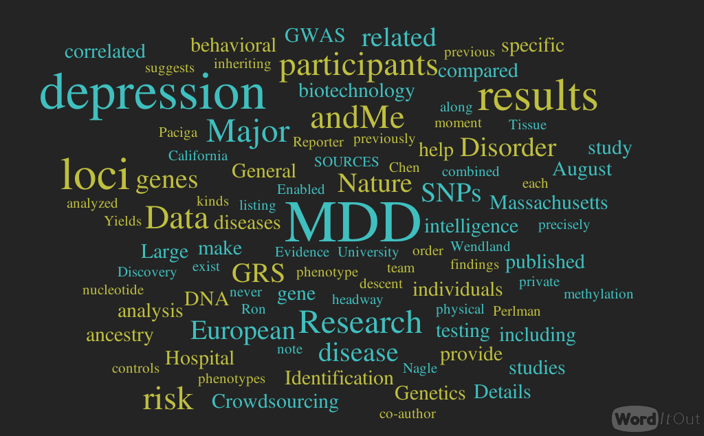
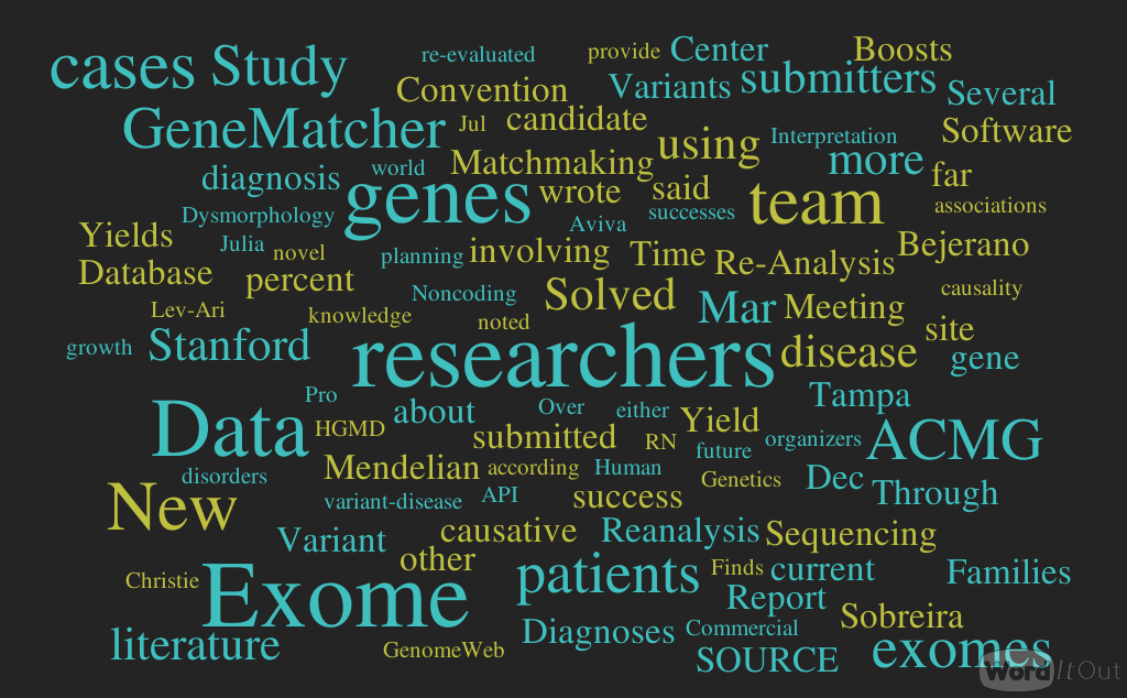

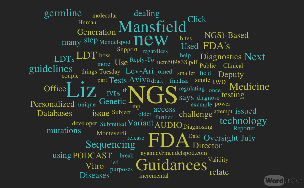


This is a lovely method and should find wide applicability in many settings, especially for microorganisms and cell lines. However, it is not clear that this approach will be, as implied by the discussion, an efficient mapping method for all multicellular organisms. I have performed similar experiments in Drosophila, focused on meiotic recombination, on a much smaller scale, and found that CRISPR-Cas9 can indeed generate targeted recombination at gRNA target sites. In every case I tested, I found that the recombination event was associated with a deletion at the gRNA site, which is probably unimportant for most mapping efforts, but may be a concern in some specific cases, for example for clinical applications. It would be interesting to know how often mutations occurred at the targeted gRNA site in this study.
The wider issue, however, is whether CRISPR-mediated recombination will be more efficient than other methods of mapping. After careful consideration of all the costs and the time involved in each of the steps for Drosophila, we have decided that targeted meiotic recombination using flanking visible markers will be, in most cases, considerably more efficient than CRISPR-mediated recombination. This is mainly due to the large expense of injecting embryos and the extensive effort and time required to screen injected animals for appropriate events. It is both cheaper and faster to generate markers (with CRISPR) and then perform a large meiotic recombination mapping experiment than it would be to generate the lines required for CRISPR-mediated recombination mapping. It is possible to dramatically reduce costs by, for example, mapping sequentially at finer resolution. But this approach would require much more time than marker-assisted mapping. If someone develops a rapid and cheap method of reliably introducing DNA into Drosophila embryos, then this calculus might change.
However, it is possible to imagine situations where CRISPR-mediated mapping would be preferable, even for Drosophila. For example, some genomic regions display extremely low or highly non-uniform recombination rates. It is possible that CRISPR-mediated mapping could provide a reasonable approach to fine mapping genes in these regions.
The authors also propose the exciting possibility that CRISPR-mediated loss of heterozygosity could be used to map traits in sterile species hybrids. It is not entirely obvious to me how this experiment would proceed and I hope the authors can illuminate me. If we imagine driving a recombination event in the early embryo (with maternal Cas9 from one parent and gRNA from a second parent), then at best we would end up with chimeric individuals carrying mitotic clones. I don’t think one could generate diploid animals where all cells carried the same loss of heterozygosity event. Even if we could, this experiment would require construction of a substantial number of stable transgenic lines expressing gRNAs. Mapping an ~20Mbp chromosome arm to ~10kb would require on the order of two-thousand transgenic lines. Not an undertaking to be taken lightly. It is already possible to perform similar tests (hemizygosity tests) using D. melanogaster deficiency lines in crosses with D. simulans, so perhaps CRISPR-mediated LOH could complement these deficiency screens for fine mapping efforts. But, at the moment, it is not clear to me how to do the experiment.