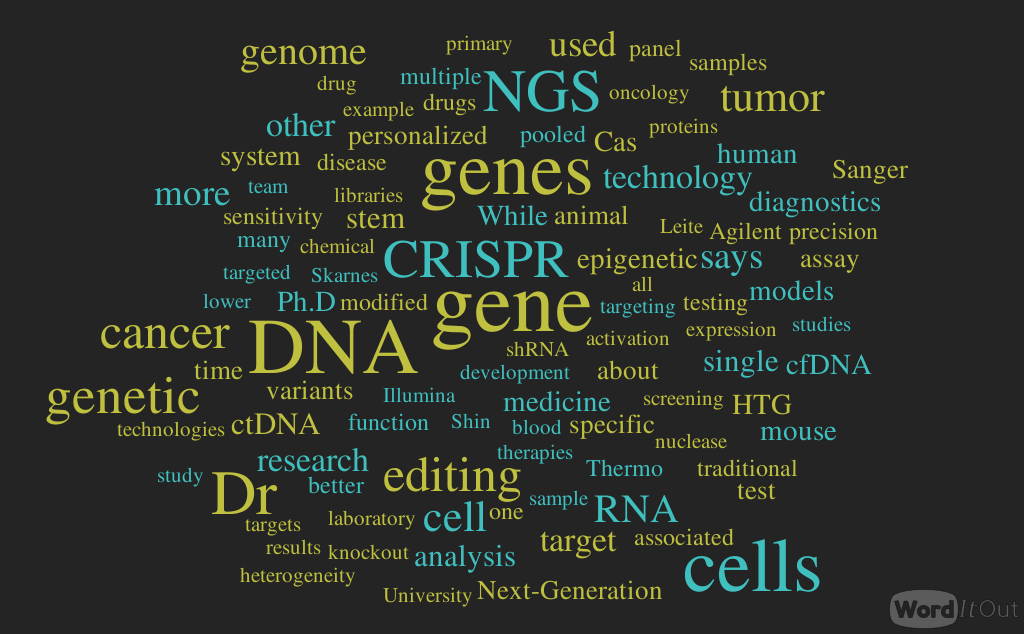
Next Generation Sequencing in Clinical Laboratory
Curator: Larry H. Bernstein, MD, FCAP
INSIGHTS on Next-Generation Sequencing
Next-generation (NGS) sequencing brings scalability and sensitivity to diagnostics in ways that traditional DNA analysis could not
Enabling Technology for Diagnosis, Prognosis, and Personalized Medicine
Significantly higher speed, lower cost, smaller sample size, and higher accuracy compared with conventional Sanger sequencing make next-generation sequencing (NGS) an attractive platform for medical diagnostics. By practically eliminating cost and time barriers, NGS allows testing of virtually any gene or genetic mutation associated with diseases.
Scalability and Sensitivity
NGS brings scalability and sensitivity to diagnostics in ways that traditional DNA analysis could not. “NGS analyzes hundreds of gene variants or biomarkers simultaneously. Traditional sequencing is better suited for analysis of single genes or fewer than 100 variants,” notes Joseph Bernardo, president of next-generation sequencing and oncology at Thermo Fisher Scientific (Waltham, MA).
Related Article: Computational Changes in Next-Generation Sequencing
Thermo Fisher’s Oncomine Focus Assay for NGS, for example, analyzes close to 1,000 biomarkers associated with the 52-gene panel. These biomarkers constitute about 1,000 different locations on the 52 genes that correlate with the efficacy of certain drugs. The assay allows single-workflow concurrent analysis of DNA and RNA, enabling sequencing of 35 hot-spot genes, 19 genes associated with copy number gain, and 23 fusion genes.
NGS is also better suited to detect lower levels of variants present in heterogeneous material, such as tumor samples. And while both NGS and Sanger sequencing are versatile, NGS can analyze both DNA and RNA, including RNA fusions, at a much more cost-efficient price point.
“When interrogating a limited number of analytes, Sanger sequencing is the standard for many laboratory- developed tests, offering fast turnaround times and lower cost than NGS,” Bernardo says. “We view the two methods as complementary.”
Diagnostic NGS is moving inexorably toward targeted sequencing, particularly for tumor analysis. The targets are specific regions within a tumor’s DNA or individual genes, or specific locations on single genes.
“Targeted sequencing lends itself to diagnostic testing, particularly in oncology, as the goal is to analyze multiple genes associated with cancer using a platform that offers high sensitivity, reliability, and rapid turnaround time,” Bernardo tells Lab Manager. “It is simply more cost-effective.”
That is why the National Cancer Institute (NCI) chose Thermo Fisher’s Ion Torrent sequencing system and the Oncomine reagents for NCI-MATCH, the most ambitious trial to date of NGS oncology diagnostics.
NCI-MATCH will use a 143-gene panel to test submitted tumor samples at four centers (NCI, MD Anderson Cancer Center, Massachusetts General Hospital, and Yale University). The centers then provide sequencing data that helps direct appropriate treatments.
The NCI test protocol ensures consistency across multiple instruments and sites.
Personalized Treatments
Another great opportunity for NGS-based diagnostics is in personalized or precision medicine for both new and existing drugs. Companion diagnostics—co-approved with the relevant drug—drive this entire business. “The only way personalized medicine can succeed commercially is if pharmaceutical companies incorporate a universal assay philosophy in their development programs instead of developing a unique assay for each new drug,” Bernardo explains. For example, in late 2015, Thermo Fisher partnered with Pfizer and Novartis to develop a universal companion diagnostic with the goal of identifying personalized therapy selection from a menu of drugs targeting non-small-cell lung cancer, which annually causes more deaths than breast, colon, and prostate cancer combined.
While advances in sequencing have been remarkable in recent years, the eventual success of NGS-based diagnostics will not depend on instrumentation alone. “What [ensures] ease of use and commonality of results is the cohesiveness of the entire workflow, from sample prep to rapid sequencing systems and bioinformatics,” Bernardo says. “Those components working together will drive NGS into a realizable solution for the clinical market.”
In addition to confirming a disease condition (diagnosis), NGS also provides valuable information on disease susceptibility, prognosis, and the potential effect of drugs on individual patients. The latter idea, known as precision medicine or personalized medicine, uses an individual’s molecular profile to guide treatment. The idea is to differentiate diseases into subtypes based on molecular (usually genetic) characteristics and tailor therapies accordingly.
Precision medicine is still in its infancy, but dozens of pharmaceutical, diagnostics, and genetics firms have bought into the idea.
“We are just at the beginning of connecting genomic and genetic information to target specific therapies for patients,” says T.J. Johnson, president and CEO of HTG Molecular Diagnostics (Tuscon, AZ). “Precision medicine will have a bright future as we gain better understanding of the root causes of disease.”
In 2013, HTG commercialized its HTG Edge instrument platform and a portfolio of RNA assays, which fully automate the company’s proprietary nuclease protection chemistry. This chemistry measures mRNA and miRNA gene expression levels from very small quantities of difficult-to-handle samples.
HTG entered the NGS market in 2014 with the launch of the first HTG EdgeSeq product, an assay that targets and digitally measures the expression of more than 2,000 microRNAs. The assay utilizes the HTG Edge for sample and library preparation, and it uses a suitable NGS instrument (from either Illumina or Thermo Fisher) for quantitation. The data is imported back into the HTG EdgeSeq instrument for analytics and reporting.
In 2015, the company launched four additional HTG EdgeSeq panels: immuno- oncology and pan-oncology biomarker panels, a lymphoma profiling panel, and a classifier for subtyping diffuse large B-cell lymphomas (DLBCL).
Eliminating Biopsies?
Traditional biopsies for tumor DNA analysis are invasive, risky, and often impossible to obtain, and they may not uncover the heterogeneity often present in tumors. It was recently discovered that dying tumor cells release small pieces of DNA into the bloodstream. This cell-free circulating tumor DNA (ctDNA) is detectable in samples through NGS.
In September 2015, Memorial Sloan Kettering Cancer Center (MSK) and NGS leader Illumina (San Diego, CA) entered a collaboration to study ctDNA for cancer diagnosis and monitoring. The aim is to establish ctDNA as a relevant cancer biomarker.
Heterogeneity as it pertains to cancer traditionally refers to multiple tissues located within a tumor, as determined histologically. A number of recent studies suggest that tumor heterogeneity occurs at the genetic level as well. “In particular, there appears to be a tremendous variety of sequence variants within the same tumor, even resulting in situations where one tumor can have multiple mutated genes that have been demonstrated to drive cancer,” says John Leite, PhD, vice president, oncology—market development and product marketing at Illumina.
Heterogeneity challenges the search for treatments that target a specific gene product or pathway. Once the patient is treated, biopsies tell very little about how that patient is responding. “Our hope is that ctDNA provides clinicians with a real-time measure of the abundance of those mutated genes and that a decrease in the relative abundance is synonymous with a lower tumor burden,” Leite adds.
Clinical trials are needed to demonstrate that patients whose therapy was selected using ctDNA versus traditional tissue biopsy testing had a significantly improved outcome or that the analysis might be informative for prognosis.
What about cancer cells that do not release DNA? “Studies show that tumors from different organs or tissues release more or less ctDNA into the peripheral blood,” Leite explains, “but in general the possibility that some cells might not release ctDNA is an open area of research.”
For the MSK-Illumina collaboration, the cancer center will collect samples, and Illumina will apply its sequencing technology to detect ctDNA in those samples. The big draw here is the potential to reduce the number of invasive, expensive diagnostic and monitoring procedures with a simple blood test. This would not be possible without deep next-generation sequencing—the genomics vernacular for sequencing at great depths of coverage.
“Whereas sequencing to identify germline variants can be performed at a nominal depth of coverage—for example, reading a DNA strand 30 times—sequencing rare variants such as in ctDNA requires a much higher level of sensitivity, which is driven by increasing depth of coverage [as much] as 25,000 times,” Leite tells Lab Manager.
In addition to the Illumina MSK collaboration and the work of Thermo Fisher Scientific described above, many more studies involving research consortia and pharmaceutical companies are under way.
“This is a really exciting time for oncology,” Leite says.
Reducing Sample Size
Similarly, in November 2015, Circulogene Theranostics (Birmingham, AL) launched its cfDNA (cell-free DNA) liquid biopsy products for testing ten tumor types, including breast, lung, and colon cancers. The company claims the ability to enrich circulating cfDNA from a single drop of blood.
“While all liquid biopsy companies are focusing on the downstream novel technologies to selectively enrich or amplify tumor-specific cfDNA from a dominantly normal population, the upstream 40 to 90 percent material loss during cfDNA extraction leads to potential false negative results of cancer mutation detection,” explains Chen Yeh, Circulogene’s chief scientific officer. “This is why 10 to 20 mL of blood [are] generally required for conventional cfDNA liquid biopsies.”
Released cfDNA fragments often complex with proteins and lipids, which shift their densities to values much lower than those of pure DNA or protein while protecting the corresponding cfDNA from attack by circulating nucleases. Circulogene’s cfDNA breakthrough concentrates and enriches these genetic fragments through density fractionation followed by enzyme-based DNA modification and manipulation, eliminating extraction-associated loss. The technology ensures near-full recovery of both small-molecular-weight (apoptotic cell death) and high-molecular-weight (necrotic cell death) cell-free DNA species from droplet volumes of plasma, serum, or other body fluids.
“The 50-gene panel is our first offering,” says Yeh. “We will continue to develop and cover more comprehensive, informative, and actionable genes and tests.”
The current bottleneck in personalized and precision medicine is the severe shortage of anticancer drugs. Yeh provides perspective, saying, “We have about 60 FDA-approved drugs for cancer-targeted therapies on market, while there are approximately 150 cancer driver genes to aim for. If counting all mutations within these 150 genes, the numbers will be overwhelming.”
Circulogene’s cell-free DNA enrichment technology may be followed up with NGS, conventional Sanger sequencing, or any DNA assay based on PCR or mass spectrometry. However, the sensitivity of Sanger sequencing is insufficient for detecting variants with volumes below 15 percent. Moreover, the company’s multiplex NGS liquid biopsy test captures and monitors real-time, longitudinal tumor heterogeneity or tumor clonal dynamic evolution, which goes well beyond testing of a single mutation on a single sample in traditional sequencing.
Gene Editing Casts a Wide Net
With CRISPR, Gene Editing Can Trawl the Murk, Catching Elusive Phenotypes amidst the Epigenetic Ebb and Flow
http://www.genengnews.com/gen-articles/gene-editing-casts-a-wide-net/5713/
-
Genome editing, a much-desired means of accomplishing gene knockout, gene activation, and other tasks, once seemed just beyond the reach of most research scientists and drug developers. But that was before the advent of CRISPR technology, an easy, versatile, and dependable means of implementing genetic modifications. It is in the process of democratizing genome editing.
CRISPR stands for “clustered, regularly interspaced, short palindromic repeats,” segments of DNA that occur naturally in many types of bacteria. These segments function as part of an ancient immune system. Each segment precedes “spacer DNA,” a short base sequence that is derived from a fragment of foreign DNA. Spacers serve as reminders of past encounters with phages or plasmids.
The CRISPR-based immune system encompasses several mechanisms, including one in which CRISPR loci are transcribed into small RNAs that may complex with a nuclease called CRISPR-associated protein (Cas). Then the RNA guides Cas, which cleaves invading DNA on the basis of sequence complementarity.
In the laboratory, CRISPR sequences are combined with a short RNA complementary to a target gene site. The result is a complex in which the RNA guides Cas to a preselected target.
Cas produces precise site-specific DNA breaks, which, with imperfect repair, cause gene mutagenesis. In more recent applications, Cas can serve as an anchor for other proteins, such as transcriptional factors and epigenetic enzymes. This system, it seems, has almost limitless versatility.
-
Edited Stem Cells
The Sanger Institute Mouse Genetic Program, along with other academic institutions around the world, provides access to thousands of genetically modified mouse strains. “Genetic engineering of mouse embryonic stem (ES) cells by homologous recombination is a powerful technique that has been around since the 1980s,” says William Skarnes, Ph.D., senior group leader at the Wellcome Trust Sanger Institute.
“A significant drawback of the ES technology is the time required to achieve a germline transmission of the modified genetic locus,” he continues. “While we have an exhaustive collection of modified ES cells, only about 5,000 knockout mice, or a quarter of mouse genome, were derived on the basis of this methodology.”
The dominant position of the mouse ES cell engineering is now effectively challenged by the CRISPR technology. Compared with very low rates of homologous recombination in fertilized eggs, CRISPR generates high levels of mutations, and off-target effects may be so few as to be undetectable.
“We used the whole-genome sequencing to thoroughly assess off-target mutations in the offspring of CRISPR-engineered founder animals,” informs Dr. Skarnes. “A mutated Cas9 nuclease was deployed to increase specificity, resulting in nearly perfect targeting.”
Dr. Skarnes explains that the major mouse genome centers are now switching to CRISPR to complete the creation of the world-wide repository of mouse knockouts. His own research is now focused on genetically engineered induced pluripotent stem cells (iPSCs). These cells are adult cells that have been reprogrammed to an embryonic stem cell–like state, and are thus devoid of ethical issues associated with research on human embryonic stem cells. The ultimate goal is to establish a world-wide panel of reference iPSCs created by high-throughput genetic editing of every single human gene.
“We are poised to begin a large-scale phenotypic analysis of human genes,” declares Dr. Skarnes. His lab is releasing the first set of functional data on 100 DNA repair genes. “By knocking out individual proteins involved in DNA repair and sequencing the genomes of mutant cells,” declares Dr. Skarnes, “we hope to better understand the mutational signatures that occur in cancer.”
-
Pooled CRISPR Libraries
 Researchers hope to gain a better understanding of the mutational signatures found in cancers by using CRISPR techniques to knock out individual proteins involved in DNA repair and then sequencing the genomes of mutant cells. [iStock/zmeel]
Connecting a phenotype to the underlying genomics requires an unbiased screening of multiple genes at once. “Pooled CRISPR libraries provide an opportunity to cast a wide net at a reasonably low cost,” says Donato Tedesco, Ph.D., lead research scientist at Cellecta. “Screening one gene at a time on genome scale is a significant investment of time and money that not everyone can afford, especially when looking for common genetic drivers across many cell models.”
Building on years of experience with shRNA libraries, Cellecta is uniquely positioned to prepare pooled CRISPR libraries for genome-wide or targeted screens of gene families. While shRNA interferes with gene translation, CRISPR disrupts a gene and the genomic level due to imperfections in the DNA repair mechanism.
To determine if these different mechanisms for inactivating genes affect the results of genetic screens, the team conducted a side-by-side comparison of Cellecta’s Human Genome-Wide Module 1 shRNA Library, which expresses 50,000 shRNA targeting 6,300 human genes, with the library of 50,000 gRNA targeting the same gene set. The concordance between approaches was very high, suggesting that these technologies may be complementary and used for cross-confirmation of results.
Also, a recently completed Phase I NIH SBIR Grant was used to create and test guiding strand RNA (sgRNA) structures to drastically improve efficiency of gene targeting. For this work, Cellecta used a pool library strategy to simultaneously test multiple sgRNA structures for their efficiency and specificity. An early customized Cellecta pooled gRNA library was successfully utilized for screening for epigenetic genes. This particular screen is highly dependent on a complete loss of function, and could not have been accomplished by shRNA inhibition.
Scientists from Epizyme interrogated 600 genes in a panel of 100 cell lines and, in addition to finding many epigenetic genes required for proliferation in nearly all cell lines, were able to identify validate several essential epigenetic genes required only in subsets of cells with specific genetic lesions. In other words, pooled cell line screening was able to distinguish targets that are likely to produce toxic side effects in certain types of cancer cells from gene targets that are essential in most cells.
“A more complicated application of CRISPR technology is to use it for gene activation,” adds Dr. Tedesco. “Cellecta plans to optimize this application to bring forth highly efficient, inexpensive, high-throughput genetic screens based on their pooled libraries.
-
Chemically Modified sgRNA
 Scientists based at Agilent Research Laboratories and Stanford University worked together to demonstrate that chemically modified single guide RNA can be used to enhance the genome editing of primary hepatopoietic stem cells and T cells. This image, which is from the Stanford laboratory of Matthew Porteus, M.D., Ph.D., shows CD34+ human hematopoietic stem cells that were edited to turn green. Editing involved inserting a construct for green fluorescent protein. About 1,000 cells are pictured here.
Researchers at Agilent Technologies applied their considerable experience in DNA and RNA synthesis to develop a novel chemical synthesis method that can generate long RNAs of 100 nucleotides or more, such as single guide RNAs (sgRNAs) for CRISPR genome editing. “We have used this capability to design and test numerous chemical modifications at different positions of the RNA molecule,” said Laurakay Bruhn, Ph.D., section manager, biological chemistry, Agilent.
Agilent Research Laboratories worked closely with the laboratory of Matthew Porteus, M.D., Ph.D., an associate professor of pediatrics and stem cell transplantation at Stanford University. The Agilent and Stanford researchers collaborated to further explore the benefits of chemically modified sgRNAs in genome editing of primary hematopoetic stem cells and T cells.
Dr. Porteus’ lab chose three key target genes implicated in the development of severe combined immunodeficiency (SCID), sickle cell anemia, and HIV transmission. Editing these genes in the patient-derived cells offers an opportunity for novel precision therapies, as the edited cells can renew, expand, and colonize the donor’s bone marrow.
Dr. Bruhn emphasized the importance of editing specificity, so that no other cellular function is affected by the change. The collaborators focused on three chemical modifications strategically placed at each end of sgRNAs that Agilent had previously tested to show they maintained sgRNA function. A number of other optimization strategies in cell culturing and transfection were explored to ensure high editing yields.
“Primary cells are difficult to manipulate and edit in comparison with cell lines already adapted to cell culture,” maintains Dr. Bruhn. Widely varied cellular properties of primary cells may require experimental adaptation of editing techniques for each primary cell type.
The resulting data showed that chemical modifications can greatly enhance efficiency of gene editing. The next step would translate these findings into animal models. Another advantage of chemical synthesis of RNA is that it can potentially be used to make large enough quantities for therapeutics.
“We are working with Agilent’s Nucleic Acid Solution Division—a business focused on GMP manufacturing of oligonucleotides for therapeutics—to engage with customers interested in this capability and better understand how we might be able to help them accomplish their goals,” says Dr. Bruhn.
-
Customized Animal Models
“Given their gene-knockout capabilities, zinc-finger-based technologies and CRISPR-based technologies opened the doors for creation of animal models that more closely resemble human disease than mouse models,” says Myung Shin, Ph.D., senior principal scientist, Merck & Co. Dr. Shin’s team supports Merck’s drug discovery and development program by creating animal models mimicking human genetics.
For example, Dr. Shin’s team has worked with the Dahl salt-sensitive strain of rats, a widely studied model of hypertension. “We used zinc-finger nucleases to generate a homozygous knockout of a renal outer medullary potassium channel (ROMK) gene,” elaborates Dr. Shin. “The resulting model represents a major advance in elucidating the role of ROMK gene.”
According to Dr. Shin, the model may also provide a bridge between genetics and physiology, particularly in studies of renal regulation and blood pressure. In one study, the model generated animal data that suggest ROMK plays a key role in kidney development and sodium absorption. Work along these lines may lead to a pharmacological strategy to manage hypertension.
In another study, the team applied zinc-finger nuclease strategy to knockout the coagulation Factor XII, and thoroughly characterize them in thrombosis and hemostasis studies. Results confirmed and extended previous literature findings suggesting Factor XII as a potential target for antithrombotic therapies that carry minimal bleeding risk. The model can be further utilized to study safety profiles and off-target effects of such novel Factor XII inhibitors.
“We use one-cell embryos to conduct genome editing with zinc-fingers and CRISPR,” continues Dr. Shin. “The ease of this genetic manipulation speeds up generation of animal models for testing of various hypotheses.”
A zinc finger–generated knockout of the multidrug resistance protein MDR 1a P-glycoprotein became an invaluable tool for evaluating drug candidates for targets located in the central nervous system. For example, it demonstrated utility in pharmacological analyses.
Dr. Shin’s future research is directed toward preclinical animal models that would contain specific nucleotide changes corresponding to those of humans. “CRISPR technology,” insists Dr. Shin, “brings an unprecedented power to manipulate genome at the level of a single nucleotide, to create gain- or loss-of-function genetic alterations, and to deeply understand the biology of a disease.”
-
Transcriptionally Active dCas9
“Epigenome editing is important for several reasons,” says Charles Gersbach, Ph.D., an associate professor of biomedical engineering at Duke University. “It is a tool that helps us answer fundamental questions about biology. It advances disease modeling and drug screening. And it may, in the future, serve as mode of genetic therapy.”
“One part of our research focuses on studying the function of epigenetic marks,” Dr. Gersback continues. “While many of these marks are catalogued, and some have been associated with the certain gene-expression states, the exact causal link between these marks and their effect on gene expression is not known. CRISPR technology can potentially allow for targeted direct manipulation of each epigenetic mark, one at a time.”
Dr. Gersback’s team mutated the Cas9 nuclease to create deactivated Cas9 (dCas9), which is devoid of endonuclease activity. Although the dCas9 protein lacks catalytic activity, it may still serve as an anchor for a plethora of other important proteins, such as transcription factors and methyltransferases.
In an elegant study, Dr. Gersbach and colleagues demonstrated that recruitment of a histone acetyltransferase by dCas9 to a genomic site activates nearby gene expression. Moreover, the activation occurred even when the acetyltransferase domain was targeted to a distal enhancer. Similarly, recruitment of KRAB repressor to a distant site silenced the target gene in a very specific manner. These findings support the important role of three-dimensional chromatin structures in gene activation.
“Genome regulation by epigenetic markers is not static,” maintains Dr. Gersbach. “It responds to changes in the environment and other stimuli. It also changes during cell differentiation. We designed an inducible system providing us with an ability to execute dynamic control over the target genes.”
In a light-activated CRISPR-Cas9 effector (LACE) system, blue light may be used to control the recruitment of the transcriptional factor VP64 to target DNA sequences. The system has been used to provide robust activation of four target genes with only minimal background activity. Selective illumination of culture plates created a pattern of gene expression in a population of cells, which could be used to mimic what is observed in natural tissues.
Together with collaborators at Duke University, Dr. Gersbach intends to carry out the high-throughput analysis of all currently identified regulatory elements in the genome. “Our ultimate goal,” he declares, “is to assign function to all of these elements.”


Leave a Reply