Feeds:
Posts
Comments

Archive for the ‘Valves & Tools’ Category

Heart Metabolism or Metabolic Cardiology: The Role of Ribose (D-ribose) for the Ischemic Heart -The Work of John St. Cyr, M.D., Ph.D.

Reporter: Aviva Lev-Ari, PhD, RN

REVIEW

An interview with John St. Cyr, M.D., Ph.D. on Ribose : A Key to Heart Health and Energy

By Richard A. Passwater, Ph.D.


© Whole Foods Magazine

January 2005

Ribose : A Key to Heart Health and Energy

An interview with John St. Cyr, M.D., Ph.D.

By Richard A. Passwater, Ph.D.

SOURCE

http://www.drpasswater.com/nutrition_library/John_St_Cyr.html


John St. Cyr, M.D., Ph.D. — PATENTS:

Issued:

Suture removal device, USP5250052

Double layer prophylactic incorporating pharmacological fluid and spiral barrier layer, USP5623945

Compositions for increasing energy in vivo, USP6159942

Method for determining viability of a myocardial segment, USP6339716

Method for raising the hypoxic threshold, USP6218366

Use of ribose to prevent cramping and soreness in muscles, USP6159943

Compositions for increasing athletic performance in mammals, USP6429198

Dual lumen adjustable length cannulae for liquid perfusion or lavage, USP6692473

Method for treating acute mountain sickness, USP6511964

Compositions for increasing energy in vivo, USP6534480

Compositions for the storage of platelets, USP6790603

Compositions for enhancing the immune response, USP6663859

Composition methods for improving cardiovascular function, USP7553817

Rejuvenation of stored blood, USP7687468


John St. Cyr, M.D., Ph.D. — Pending applications:

Method for improving ventilatory efficiency, SN20050277598

Storage of blood SN20070111191

Ventilatory benefits of ribose in COPD, smoking, SN

Use of ribose in recovery from anesthesia, SN20070105787

Use of ribose to alleviate rhabdomyolysis and the side effects of statin drugs, SN20060135440

Use of ribose in first response to acute myocardial infarction, SN20100055206

Compositions and methods for improving cardiovascular function, SN20100009924

Use of ribose in lessening the clinical symptoms of aberrant firing of neurons, SN20090286750

Compositions for indoor tanning, SN20090232750

Compositions for improving and repairing skin, SN20090197819

Use of ribose for recovery from anesthesia, SN20090197818

Cosmetic use of D-ribose, SN20080312169

Method for improving ventilator efficiency SN20100099630

Method and compositions for improving pulmonary hypertension, SN20080146514

Storage of blood, SN20070111191

Compositions and methods for feeding poultry, SN201100221446

Use of D-ribose for fatigued subjects, SN20100189785

Fibrin sealants and platelet concentrates applied to effect hemostasis in the interface of an implantable medical device with body tissue, SN20060190017

Compositions for reducing the deleterious effects of stress and aging, SN20120045426


John St. Cyr, M.D., Ph.D. — Provisional patents:

Use of ribose in pre-slaughtering of animals

Rescue therapy for acute decompensated heart failure

Combination of D-ribose plus caffeine

Role of ribose in reducing joint swelling in mammals

Role of D-ribose in cardiac remodeling

Role of D-ribose in cachexia

Use of ribose in stem cells

Use of ribose in cardioplegia

Use of ribose for doping blood for cardioplegia

Surgical adhesive for bleeding situations

Metabolic approach with EECP

Role of ribose in mitral regurgitation

Compositions for the preservation of morphology in stored blood

Methods and nutritional supplements for improving the quality of meat


John St. Cyr, M.D., Ph.D. — Publications 2011 to 2013

This list does not include Publication #1 to #219

220. Shecterle LM, Wagner S, St.Cyr JA.  A sugar for congestive heart failure patients.  Ther Adv Cardiovasc Dis 5(2):95-97, 2011.

221. Perkowski D, Wagner S, Schneider JR, St.Cyr JA.  A targeted metabolic protocol with D-ribose for off pump coronary artery bypass procedures: A retrospective analysis.  Ther Adv Cardiovasc Dis 5(4):185-192, 2011.

222. Foker J, Berry J, Harvey B, Befera N, Tveter K, St.Cyr J, Bianco R.  Heart failure is initiated by and progresses because of normal responses of energy metabolism to stress.  Circ Res   , 2011.

223. Rakow N, Barka N, Gerhart R, Rothstein P, Green M, Schu C, Grassl E, St.Cyr JA, Kopcak MW, Jr.  Chronic aortic root pressure-loading assessment model.  J Invest Surg 25(2):137, 2012.

224. Shecterle LM, St.Cyr JA.  Chapter 11; Myocardial Ischemia: Alterations in myocardial cellular energy and diastolic function, a potential role for D-ribose. In: Novel Strategies in Ischemia Heart Disease. Lakshmanadoss U(Ed). InTech, Croatia.  219-228, 2012.

225. Addis P, Shecterle LM, St.Cyr JA.  Cellular protection during oxidative stress: a potential role for D-ribose and antioxidants.  Journal of Dietary Supplements 9(3):178-182, 2012.

226. Holsworth R, Shecterle L, St.Cyr J, Sloop G.  Letter to the Editor.  Importance of monitoring blood viscosity during cardiopulmonary bypass.  Perfusion 28(1):91-2, 2013.

227. Seifert JG, Frost J, ST.Cyr JA.  Recovery benefits of a heat and moisture exchange mask when performing sprint exercise in cold temperature environments.  Aviation, Space and Environmental Medicine.    , 2013.

228. Seifert JG, McNair M, DeClercq P, St.Cyr JA.  A heat and moisture mask attenuates cardiovascular stress during cold air exposure.  Ther Adv Cardiovasc Dis 7(3):123-129, 2013.

229. Holsworth R, Cho Y, Weldman J, Sloop G, St.Cyr, J.  Cardiovascular benefits of phlebotomy: Relationship to changes in hemorheological variables.  Perfusion,   2013.


Read Full Post »

Myocardial Infarction: The New Definition After Revascularization

Reporter: Aviva Lev-Ari, PhD, RN


UPDATED on 7/31/2014

Myocardial Ischemia Symptoms

Reporter: Aviva Lev-Ari, PhD, RN

http://pharmaceuticalintelligence.com/2014/07/29/myocardial-ischemia-symptoms/


VIEW VIDEO

Gregg Stone, MD

Co-DIrector, Medical Research & Education Division Cardiovascular Research Foundation

http://www.medpagetoday.com/Cardiology/MyocardialInfarction/42256?xid=nl_mpt_DHE_2013-10-15&goback=%2Egmr_4346921%2Egde_4346921_member_5795830612724035588#%21

Primary source: Journal of the American College of Cardiology
Source reference: Moussa I, et al “Consideration of a new definition of clinically relevant myocardial infarction after coronary revascularization: an expert consensus document from the Society for Cardiovascular Angiography and Interventions (SCAI)” J Am Coll Cardiol2013; 62: 1563-1570.

Additional source: Journal of the American College of Cardiology
Source reference:White H “Avatar of the universal definition of periprocedural myocardial infarction” J Am Coll Cardiol 2013; 62: 1571-1574.

Moussa reported that he had no conflicts of interest.

Stone is a consultant for Boston Scientific, Eli Lilly, Daiichi Sankyo, and AstraZeneca. The other authors reported relationships with Guerbet, The Medicines Company, Bristol-Myers Squibb/Sanofi, Merck, Maya Medical, AstraZeneca, Abbott Vascular, Regado Biosciences, Janssen Pharma, Lilly/Daiichi Sankyo, St. Jude Medical, Medtronic, Terumo, Bridgepoint/Boston Scientific, Gilead, Boston Scientific, Eli Lilly, and Daiichi Sankyo.

White is co-chairman for the Task Force for the Universal Definiton of Myocardial Infarction; has received research grants from sanofi-aventis, Eli Lilly, The Medicines Company, the NIH, Pfizer, Roche, Johnson & Johnson, Schering-Plough, Merck Sharpe & Dohme, AstraZeneca, GlaxoSmithKline, Daiichi Sankyo Pharma Development, and Bristol-Myers Squibb; and has served on advisory boards for AstraZeneca, Merck Sharpe & Dohme, Roche, and Regado Biosciences.

WASHINGTON, DC — A “clinically meaningful” definition of MI following PCI or CABG is urgently needed to replace the arbitrarily chosen “universal definition” proposed in recent years that has no relevance to patients and may be muddying clinical-trial results. Those are the conclusions of a new expert consensus document released Monday by the Society of Cardiovascular Angiography and Interventions (SCAI)[1].

The notion of a “universal definition of MI” was first proposed in 2000 and updated in 2007 and 2012. The 2012 document defines a PCI-related MI as an increase in cardiac troponin (cTn) of more than five times the upper limit of normal (ULN) during the first 48 hours postprocedure plus specific clinical or ECG features. Post-CABG, the definition is a cTn increase of >10 times the ULN, plus different clinical or ECG features.

The problem, lead author Dr Issam Moussa (Mayo Clinic, Jacksonville, FL) told heartwire , is that these cutoffs were arbitrarily chosen and not based on any hard evidence that these biomarker levels spelled a poor prognosis. Moreover, “overnight, the rate of MI went from 5% following these procedures to 20% to 30%!” he said.

The SCAI committee, in its new document, focuses on post-PCI procedures and highlights the importance of acquiring baseline cardiac biomarkers and differentiating between patients with elevated baseline CK-MB (or cTn) in whom biomarker levels are stable or falling, as well as those in whom it hasn’t been established whether biomarkers are changing.

SCAI’s Proposed Clinically Meaningful MI Definitions

Group Definition
Normal baseline CK-MB CK-MB rise of >10x ULN or >5x ULN with new pathologic Q-waves in at least 2 contiguous leads or new persistent left bundle branch block
OR
In the absence of baseline CK-MB, a cTn rise of >70x ULN or a rise of>35 ULN plus new pathologic Q-waves in at least 2 contiguous leads or new persistent left bundle branch block
Elevated baseline biomarkers that are stable or falling A CK-MB or cTn rise that is equal (by an absolute increment) to the definitions described for patients with normal CK-MB at baseline.
Elevated baseline biomarkers that have not been shown to be stable or falling A CK-MB or cTn rise that is equal (by an absolute increment) to the definitions described for patients with normal CK-MB at baseline
Plus
New ST-segment elevation or depression
Plus
New-onset or worsening heart failure or sustained hypotension or other signs of a clinically relevant MI.

Moussa is quick to emphasize that these new clinically meaningful definitions have limited evidence to support them—and most of what exists supports CK-MB definitions, not cTn—but that the new document is based on the best scientific evidence available.

“We don’t want to come out with a definitive statement” saying this is the final word on MI definitions,” he stressed. “There is more science that needs to be done and there remains more uncertainty. We framed this to be inclusive and also to open the field for discussion.”

His hope is that this will lead to important changes in how patients are managed and money is spent. Currently, patients with clinically meaningless biomarker elevations may become unnecessarily panicked over news that they’ve had a “heart attack,” while hospital stays may be extended and further tests ordered on the basis of these results.

Moussa et al’s proposal also has important implications for clinical trials, he continued. Currently, for studies that include periprocedural MIs as an individual end point or as part of a composite end point, the very high number of biomarker-defined “MIs” collected in the trial could potentially overwhelm the true impact of any given therapy. “You are really using an end point that is truly not relevant to patients. . . . This could really affect the whole hypothesis.”

He’s expecting some push-back from cardiologists and academics, particularly those who championed the need for the universal definition in the first place, but believes most people will welcome a clinically meaningful definition.

“I think many in the medical community will accept this because they have not really been using the universal definition in their day-to-day practice anyhow.” What’s more, the National Cardiovascular Data Registry (NCDR) does not include the reporting of MI postangiography, in part because of concerns that the universal definition of MI overestimates the true incidence of this problem. “I think many in the community will look at this definition as more reflective of the true incidence of MI after angioplasty, and if it’s accepted, they are more likely to report it to databases like NCDR and use it to reflect quality-of-care processes.”

http://www.medscape.com/viewarticle/812533?nlid=35983_2105&src=wnl_edit_medp_card&uac=93761AJ&spon=2

  • ESC/ACCF/AHA/WHF Expert Consensus Document

Circulation.2012; 126: 2020-2035  Published online before print August 24, 2012,doi: 10.1161/​CIR.0b013e31826e1058

Third Universal Definition of Myocardial Infarction

  1. Kristian Thygesen;
  2. Joseph S. Alpert;
  3. Allan S. Jaffe;
  4. Maarten L. Simoons;
  5. Bernard R. Chaitman;
  6. Harvey D. White
  7. the Writing Group on behalf of the Joint ESC/ACCF/AHA/WHF Task Force for the Universal Definition of Myocardial Infarction
  1. *Corresponding authors/co-chairpersons: Professor Kristian Thygesen, Department of Cardiology, Aarhus University Hospital, Tage-Hansens Gade 2, DK-8000 Aarhus C, Denmark. Tel: +45 7846-7614; fax: +45 7846-7619: E-mail: kristhyg@rm.dk. Professor Joseph S. Alpert, Department of Medicine, Univ. of Arizona College of Medicine, 1501 N. Campbell Ave., P.O. Box 245037, Tucson AZ 85724, USA, Tel: +1 520 626 2763, Fax: +1 520 626 0967, E-mail: jalpert@email.arizona.edu. Professor Harvey D. White, Green Lane Cardiovascular Service, Auckland City Hospital, Private Bag 92024, 1030 Auckland, New Zealand. Tel: +64 9 630 9992, Fax: +64 9 630 9915, E-mail: harveyw@adhb.govt.nz.

Table of Contents

  • Abbreviations and Acronyms. . . . . . . . . . . . . . . . . . . .2021

  • Definition of Myocardial Infarction. . . . . . . . . . . . . . .2022

  • Criteria for Acute Myocardial Infarction. . . . . . . . . . . .2022

  • Criteria for Prior Myocardial Infarction. . . . . . . . . . . .2022

  • Introduction. . . . . . . . . . . . . . . . . . . . . . . . . . . . . . . . . .2022

  • Pathological Characteristics of Myocardial Ischaemia and Infarction. . . . . . . . . . . . . . . . . . . . . . . . . . . . . . . .2023

  • Biomarker Detection of Myocardial Injury With Necrosis. . .2023

  • Clinical Features of Myocardial Ischaemia and Infarction. . .2024

  • Clinical Classification of Myocardial Infarction. . . .2024
    • Spontaneous Myocardial Infarction (MI Type 1). . . .2024

    • Myocardial Infarction Secondary to an Ischaemic Imbalance (MI Type 2). . . . . . . . . . . . . . . . . . . . . . . .2024

    • Cardiac Death Due to Myocardial Infarction (MI Type 3). .2025

    • Myocardial Infarction Associated With Revascularization Procedures (MI Types 4 and 5). . . . . . . . . . . . . . . . . . …

New Definition for MI After Revascularization

Published: Oct 14, 2013 | Updated: Oct 15, 2013

By Todd Neale, Senior Staff Writer, MedPage Today
Reviewed by Zalman S. Agus, MD; Emeritus Professor, Perelman School of Medicine at the University of Pennsylvania and Dorothy Caputo, MA, BSN, RN, Nurse Planner

The Society for Cardiovascular Angiography and Interventions (SCAI) has released a new definition for myocardial infarction (MI) following coronary revascularization aimed at identifying only those events likely to be related to poorer patient outcomes.

In the new criteria — published as an expert consensus document inCatheterization and Cardiovascular Interventions and the Journal of the American College of Cardiology — creatine kinase-myocardial band (CK-MB) is the preferred cardiac biomarker over troponin, and much greater elevations are required to define a clinically relevant MI compared with the universal definition of MI proposed in 2007 and revised in 2012.

Also, the new definition uses the same biomarker elevation thresholds to identify MIs following both percutaneous coronary intervention (PCI) and coronary artery bypass grafting (CABG), whereas the universal definition has different thresholds for events following the two procedures.

“What we’ve really tried to emphasize in this classification scheme is the primary link between biomarker elevations and prognosis,” according to Gregg Stone, MD, of Columbia University Medical Center and the Cardiovascular Research Foundation in New York City, one of the authors of the document.

“In the universal definition of MI, they even acknowledged that their criteria were arbitrary,” Stone said in an interview. “We’ve tried to reduce the arbitrariness of the cutoff values that we selected so that the researcher, academician, clinician, hospital administrator, etc., can be confident that these levels that we’re recommending are the ones that are associated with a worse prognosis for patients suffering periprocedural complications.”

The Change

The existing universal definition for MI defines events following PCI according to an increase in cardiac troponin to greater than five times the 99th percentile upper reference limit (URL) within 48 hours when baseline levels are normal, with confirmation by electrocardiogram (ECG), imaging, or symptoms.

For CABG-related MI, the increase must be more than 10 times the 99th percentile URL within 48 hours when baseline levels are normal, with confirmation by ECG, angiography, or imaging.

But, Stone and colleagues wrote, the relationship between that degree of troponin elevation after a revascularization procedure and prognosis is not as strong as the association between a CK-MB elevation and patient outcomes.

Using a small elevation in troponin to define a post-procedure MI could find myocardial necrosis that is unlikely to be associated with poor clinical outcomes, which could have far-reaching implications, they wrote.

“Widespread adoption of an MI definition not clearly linked to subsequent adverse events such as mortality or heart failure may have serious consequences for the appropriate assessment of devices and therapies, may affect clinical care pathways, and may result in misinterpretation of physician competence,” they wrote.

To address that issue, the expert panel convened by SCAI sought to define clinically relevant MI after PCI or CABG.

A clinically relevant MI is defined in the new document based on an increase of at least 10 times the upper limit of normal in the level of CK-MB within 48 hours after a revascularization procedure when baseline levels are normal.

When the CK-MB level is not available, then an increase in troponin I or T of at least 70 times the upper limit of normal can be used to define a clinically relevant MI, according to the authors.

However, if an ECG shows new pathologic Q-waves in at least two contiguous leads or a new persistent left bundle branch block, then the thresholds can be lowered to at least five times and at least 35 times the upper limit of normal for CK-MB and troponin, respectively.

Further guidance is provided for identifying clinically relevant post-procedure MIs when the cardiac biomarker levels are elevated at baseline.

Dueling Definitions

Co-chairman of the Task Force for the Universal Definition of Myocardial Infarction, Harvey White, DSc, of Auckland City Hospital in Auckland, New Zealand, noted some limitations of the new definition, including the lack of a requirement for ischemic symptoms.

“Ischemic symptoms have always been a basic tenet of the diagnosis of MI, and it should be no different for a [PCI-related] MI,” he wrote in an accompanying editorial.

In addition, with the use of such large elevations in biomarker levels in the new definition, “there will be very few PCI-related events identified, and an opportunity to improve patient outcomes may be lost,” he wrote.

Troponin should remain the preferred biomarker over CK-MB, White argued, pointing to variability in and analytical issues with CK-MB assays, the need for sex-specific cutoffs for CK-MB levels, the need for higher thresholds of CK-MB to determine abnormalities because all individuals have circulating levels of the biomarker, and the reduced sensitivity and specificity of CK-MB.

Also, he said, CK-MB is becoming increasingly unavailable at medical centers.

“With CK-MB becoming obsolete, troponin will become the gold standard, and CK-MB will no longer have a role in defining PCI injury and infarction in clinical practice,” White wrote.

Stone admitted that troponin ultimately might be preferable to CK-MB because of its greater specificity, although the evidence does not yet support it.

“I think there’s a general desirability to move to troponins, although when you look at the data that’s out there it’s much stronger correlating CK-MB elevations to subsequent prognosis,” he said. “I think a lot of the troponin elevations are just noise or troponins are just too sensitive.”

Room for Both?

White noted in his editorial that “the rationale for the SCAI definition has been well articulated by its authors and may be appropriate in an individual trial, but it should not supplant the universal definition of MI,” he wrote.

When asked whether the new definition would replace the universal definition, Stone said there is a place for both sets of criteria.

“We would propose the clinically relevant definition be the one that is used to make most substantial decisions right now, [such as] trade-offs between efficacy and safety for new drugs and devices, in judging hospital systems and physicians, etc.,” he said. “But I do think there’s value in both, and they will both continue to evolve over time as new data becomes evident.”

http://www.medpagetoday.com/Cardiology/MyocardialInfarction/42256?xid=nl_mpt_DHE_2013-10-15&goback=%2Egmr_4346921%2Egde_4346921_member_5795830612724035588#%21 

Articles citing 

Third Universal Definition of Myocardial Infarction

  • Improved long-term clinical outcomes in patients with ST-elevation myocardial infarction undergoing remote ischaemic conditioning as an adjunct to primary percutaneous coronary interventionEur Heart J. 2013;0:eht369v1-eht369

  • The role of myeloperoxidase (MPO) for prognostic evaluation in sensitive cardiac troponin I negative chest pain patients in the emergency departmentEuropean Heart Journal: Acute Cardiovascular Care. 2013;2:203-210,
  • Coronary artery bypass grafting or percutaneous revascularization in acute myocardial infarction?Interact CardioVasc Thorac Surg. 2013;0:ivt381v1-ivt381,
  • Ischemic Conditioning as an Adjunct to Percutaneous Coronary InterventionCirc Cardiovasc Interv. 2013;6:484-492,
  • High sensitivity cardiac troponin in patients with chest painBMJ. 2013;347:f4222,
  • Chest Pain and Palpitations: Taking a Closer LookCirculation. 2013;128:271-277,
  • An Updated Definition of Stroke for the 21st Century: A Statement for Healthcare Professionals From the American Heart Association/American Stroke AssociationStroke. 2013;44:2064-2089,
  • Factors Influencing the 99th Percentile of Cardiac Troponin I Evaluated in Community-Dwelling Individuals at 70 and 75 Years of AgeClin. Chem.. 2013;59:1068-1073,
  • Detection and management of asymptomatic myocardial injury after noncardiac surgeryEuropean Journal of Preventive Cardiology.2013;0:2047487313494294v1-2047487313494294,
  • Postoperative Troponin Screening: A Cardiac Cassandra?Circulation. 2013;127:2253-2256,
  • Remote Ischemic Preconditioning Improves Outcome at 6 Years After Elective Percutaneous Coronary Intervention: The CRISP Stent Trial Long-term Follow-upCirc Cardiovasc Interv. 2013;6:246-251,
  • Outcomes for Clinical Studies Assessing Drug and Revascularization Therapies for Claudication and Critical Limb Ischemia in Peripheral Artery DiseaseCirculation. 2013;127:1241-1250,
  • Prevalence, Incidence, and Implications of Silent Myocardial Infarctions in Patients With Diabetes MellitusCirculation. 2013;127:965-967,
  • 2013 ACCF/AHA Key Data Elements and Definitions for Measuring the Clinical Management and Outcomes of Patients With Acute Coronary Syndromes and Coronary Artery Disease: A Report of the American College of Cardiology Foundation/American Heart Association Task Force on Clinical Data Standards (Writing Committee to Develop Acute Coronary Syndromes and Coronary Artery Disease Clinical Data Standards)Circulation. 2013;127:1052-1089,
  • Clin. Chem.. 2013;59:574-576,
  • Percutaneous Coronary Intervention Versus Optimal Medical Therapy for Prevention of Spontaneous Myocardial Infarction in Subjects With Stable Ischemic Heart DiseaseCirculation. 2013;127:769-781,
  • Frequency of Myocardial Infarction and Its Relationship to Angiographic Collateral Flow in Territories Supplied by Chronically Occluded Coronary ArteriesCirculation. 2013;127:703-709,
  • The Power of More Than OneCirculation. 2013;127:665-667,
  • The curious life of the biomarkerJournal of the American Dental Association. 2013;144:126-128,
  • Persistent Increases in Cardiac Troponin Concentrations As Measured with High-Sensitivity Assays after Acute Myocardial InfarctionClin. Chem.. 2013;59:443-445,
  • 2013 ACCF/AHA Guideline for the Management of ST-Elevation Myocardial Infarction: A Report of the American College of Cardiology Foundation/American Heart Association Task Force on Practice GuidelinesCirculation. 2013;127:e362-e425,

Read Full Post »

Normal and Anomalous Coronary Arteries: Dual Source CT in Cardiothoracic Imaging

Reporters: Justin D Pearlman, MD, PhD, FACC and Aviva Lev-Ari, PhD, RN

Coronary anatomy and anomalies

“Coronary” describes the crown-like position of arteries on the heart that provide its nutrient blood supply. The heart does not live off of the blood in its chambers, but rather receives its nutrient perfusion from branches of the aorta, like all other organs. The most relied on method to exam coronary artery anatomy is angiography – xray image movies obtained while the blood is opacified by injection of iodine (high atomic number to block xrays) to provide a contrast between arterial flow channel (the lumen) and the surrounding tissues. Computed tomography is providing a second-best alternative with 3D reconstructions that can be obtained less invasively (no catheters), but it often fails to see the posterior descending artery (PDA) well, and is lower in resolution (point-discrimination detail) than xray angiography (XRA). Magnetic resonance angiography (MRA) comes in as a distant third place method for examining coronary anatomy (lower quality, lower reliability), but non-invasive with no ionizing radiation. A major goal of defining coronary anatomy in individual patients is to identify coronary artery disease (CAD) and to clarify best options for management – to relieve angina and to avoid adverse consequences, e.g., heart attacks (myocardial infarction), heart failure (CHF) and death. The COURAGE trial showed that for many, aggressive medical management with statins and blood pressure control may obviate need for percutaneous or surgical interventions to control angina and minimize the risk of adverse outcomes. Patients with blockage of the left main coronary artery, or two vessel blockage including proximal left anterior descending (LAD) especially with below normal ejection fraction may be better off in the long run with bypass surgery. Therefore less invasive imaging sufficient to rule out left main disease and proximal LAD disease may suffice for decision making (except that the BARI trial results have not been overturned in favoring bypass surgery for diabetics).

On the left an overview of the coronary arteries in the anterior projection.

Coronary anatomy and anomalies

  • Left Main or left coronary artery (LCA)
    • Left anterior descending (LAD)
      • diagonal branches (D1, D2)
      • septal branches
    • Circumflex (Cx)
      • Marginal branches (M1,M2)
  • Right coronary artery
    • Acute marginal branch (AM)
    • AV node branch
    • Posterior descending artery (PDA)
RCA, LAD and Cx in the anterior projection

On the left an overview of the coronary arteries in the lateral projection.

  • Left Main or left coronary artery (LCA)
    • Left anterior descending (LAD)
      • diagonal branches (D1, D2)
      • septal branches
    • Circumflex (Cx)
      • Marginal branches (M1,M2)
  • Right coronary artery
    • Acute marginal branch (AM)
    • AV node branch
    • Posterior descending artery (PDA)

RCA, LAD and Cx in the right anterior oblique projection
On the left an overview of the coronary arteries in the lateral projection.

  • Left Main or left coronary artery (LCA)
    • Left anterior descending (LAD)
      • diagonal branches (D1, D2)
      • septal branches
    • Circumflex (Cx)
      • Marginal branches (M1,M2)
  • Right coronary artery
    • Acute marginal branch (AM)
    • AV node branch
    • Posterior descending artery (PDA)

RCA, LAD and Cx in the lateral projection

Left Coronary Artery (LCA)

The left coronary artery (LCA) is also known as the left main.
The LCA arises from the left coronary cusp.

The aortic valve has three leaflets, each having a cusp or cup-like configuration.
These are known as the left coronary cusp (L), the right coronary cusp (R) and the posterior non-coronary cusp (N).
Just above the aortic valves there are anatomic dilations of the ascending aorta, also known as the sinus of Valsalva. The left aortic sinus gives rise to the left coronary artery.
The right aortic sinus which lies anteriorly, gives rise to the right coronary artery.
The non-coronary sinus is postioned on the right side.

Left coronary (LC), right coronary (RC) and posterior non-coronary (NC) cusp
The LCA divides almost immediately into the circumflex artery (Cx) and left anterior descending artery (LAD).
On the left an axial CT-image.
The LCA travels between the right ventricle outflow tract anteriorly and the left atrium posteriorly and divides into LAD and Cx.

On the image on the left we see the left main artery dividing into

  • Cx with obtuse marginal branch (OM)
  • LAD with diagonal branches (DB)

On volume rendered images the left atrial appendage needs to be removed to get a good look on the LCA.
In 15% of cases a third branch arises in between the LAD and the Cx, known as the ramus intermedius or intermediate branch.
This intermediate branche behaves as a diagonal branch of the Cx.
Left Anterior Descending (LAD)
The LAD travels in the anterior interventricular groove and continues up to the apex of the heart.
The LAD supplies the anterior part of the septum with septal branches and the anterior wall of the left ventricle with diagonal branches.
The LAD supplies most of the left ventricle and also the AV-bundle.Mnemonic: Diagonal branches arise from the LAD.

CT image of the LAD in RAO projection
The diagonal branches come off the LAD and run laterally to supply the antero-lateral wall of the left ventricle.
The first diagonal branch serves as the boundary between the proximal and mid portion of the LAD (2).
There can be one or more diagonal branches: D1, D2 , etc.
Circumflex (Cx)
The Cx lies in the left AV groove between the left atrium and left ventricle and supplies the vessels of the lateral wall of the left ventricle.
These vessels are known as obtuse marginals (M1, M2…), because they supply the lateral margin of the left ventricle and branch off with an obtuse angle.
In most cases the Cx ends as an obtuse marginal branch, but 10% of patients have a left dominant circulation in which the Cx also supplies the posterior descending artery (PDA).Mnemonic: Marginal branches arise from the Cx and supply the lateral Margin of the left ventricle.

Circumflex and LAD seen in Lateral projection
Right Coronary Artery (RCA)
The right coronary artery arises from the anterior sinus of Valsalva and courses through the right atrioventricular (AV) groove between the right artium and right ventricle to the inferior part of the septum.
In 50-60% the first branch of the RCA is the small conus branch, that supplies the right ventricle outflow tract.
In 20-30% the conus branch arises directly from the aorta.
In 60% a sinus node artery arises as second branch of the RCA, that runs posteriorly to the SA-node (in 40% it originates from the Cx).
The next branches are some diagonals that run anteriorly to supply the anterior wall of the right ventricle.
The large acute marginal branch (AM) comes off with anacute angle and runs along the margin of the right ventricle above the diaphragm.
The RCA continues in the AV groove posteriorly and gives off a branch to the AV node.
In 65% of cases the posterior descending artery (PDA) is a branch of the RCA (right dominant circulation).
The PDA supplies the inferior wall of the left ventricle and inferior part of the septum.
RCA, LAD and LCx in Anterior projection
On the image on the far left we see the most common situation, in which the RCA comes off the right cusp and will provide the conus branch at a lower level (not shown).
On the image next to it, we see a conus branch, that comes off directly from the aorta.
LEFT: RCA comes off the right sinus of Valsalva
RIGHT: Conus artery comes off directly from the aorta
The large acute marginal branch (AM) supplies the lateral wall of the right ventricle.
In this case there is a right dominant circulation, because the posterior descending artery (PDA) comes off the RCA.
Coronary Anomalies

Coronary anomalies are uncommon with a prevalence of 1%.
Early detection and evaluation of coronary artery anomalies is essential because of their potential association with myocardial ischemia and sudden death (3).
With the increased use of cardiac-CT, we will see these anomalies more frequently.

Coronary anomalies can be differentiated into anomalies of the origin, the course and termination (Table).

The illustration in the left upper corner is the most common and clinically significant anomaly.
There is an anomalous origin of the LCA from the right sinus of Valsalva and the LCA courses between the aorta and pulmonary artery.
This interarterial course can lead to compression of the LCA (yellow arrows) resulting in myocardial ischemia.

The other anomalies in the figure on the left are not hemodynamically significant.

Interarterial LCA

On the left images of a patient with an anomalous origin of the LCA from the right sinus of Valsalva and coursing between the aorta and pulmonary artery.
Sudden death is frequently observed in these patients.

ALCAPA

On the left images of a patient with an anomalous origin of the LCA from the pulmonary artery, also known as ALCAPA.
ALCAPA results in the left ventricular myocardium being perfused by relatively desaturated blood under low pressure, leading to myocardial ischemia.
ALCAPA is a rare, congenital cardiac anomaly accounting for approximately 0.25-0.5% of all congenital heart diseases.
Approximately 85% of patients present with clinical symptoms of CHF within the first 1-2 months of life.

Myocardial bridging

Myocardial bridging is most commonly observed of the LAD (figure).
The depth of the vessel under the myocardium is more important that the lenght of the myocardial bridging.
There is debate, whether some of these myocardial bridges are hemodynamically significant.

Fistula

On the image on the left we see a large LAD giving rise to a large septal branch that terminates in the right ventricle (blue arrow).

Left to right shunt: septal branch of LAD teminates in right ventricle
REFERENCES
  1. Introduction to cardiothoracic imaging
    by Carl Jaffe and Patrick J. Lynch
  2. Cardiology Site
    by M. Abdulla
    This site includes instructional movies, 3-D animation, panoramic views, online quiz, interactive video-clips, interactive heart sounds & murmurs and interactive echocardiograms.
  3. Visualization of Anomalous Coronary Arteries on Dual Source Computed Tomography
    by G.J. de Jonge et al
    European Radiology, Volume 18, Number 11 / November, 2008, 2425-2432

SOURCE

Robin Smithuis and Tineke Wilems
Radiology department of the Rijnland Hospital Leiderdorp and the University Medical Centre Groningen, the Netherlands.
http://rad.desk.nl/en/48275120e2ed5 


Read Full Post »

3D Cardiovascular Theater – Hybrid Cath Lab/OR Suite, Hybrid Surgery, Complications Post PCI and Repeat Sternotomy

Curator: Aviva Lev-Ari, PhD, RN

Article ID #70: Cardiovascular Original Research: Cases in Methodology Design for Content Curation and Co-Curation. Published on 7/19/2013

WordCloud Image Produced by Adam Tubman

This article has THREE Parts: 

Part One:  Hybrid Cath Lab/OR Suite for Hybrid Surgery

Part Two: Cardiac Surgery 

Part Three: Invasive Interventions with Complications

1. Repeat Sternotomy Post CABG and/or Aortic Valve Replacement

2. Complications Post PCI – Pump Catheter in Use

The voice of Series A Content Consultant, Justin D Pearlman, MD, PhD, FACC

The leading cause of death and disability from any cause is cardiovascular disease, principally, heart attacks and strokes. Both the heart and brain typically allow only 10 minutes or so of inadequate blood supply before starting a committed course of permanent tissue injury, progressing in severity as time goes by without successful interruption of the disease process. Thus there is great time urgency to get patients to a definitive treatment that can stop the injury and restore adequate nutrient blood supply. Many patients can benefit from a catheterization to identify blockages and insert a small balloon within the blockage to expand the narrow channel, often followed by placement of a stent (wire cage) to maintain the expanded vessel diameter. Chemicals released over time from drug-eluting stents can prevent tissue in growth that may obstruct stents. These emergeny interventions are not always successful. There may be complications from the attempt to access an entry artery, and the blockages may not be amenable to a balloon. When such limitations are encountered, the next chance to help is surgical, with continued time pressure.

The fastest way to make the transition from a diagnostic catheterization to a timely intervention is a hybrid intervention suite that offers non-invasive imaging, catheterization and surgery all in one location. The following articles present the current state of hybrid “do it all” intervention suites. Additional articles address the risks of bad outcomes from such interventions.

Part One 

Hybrid Cath Lab/OR Suite for Hybrid Surgery

In ACC.10 and i2 Summit, 59th Annual Scientific Session, 3/14-3/16, 2010, Alfred A. Bove, M.D., Ph.D., F.A.C.C., ACC President addressed the conference attendees:

Welcome to the all-new Hybrid Cath Lab/OR and 3D CV Theater. Recent developments in cardiac surgery and interventional cardiology have led to the creation of integrated, hybrid cath lab/operating rooms (OR), which provide significant advantages in the diagnosis and treatment of patients requiring cardiac procedures—helping to facilitate a rapid-response approach. These multimodality rooms are designed to support a variety of integrated surgical and endovascular procedures. We are excited to provide you with this opportunity to get a first-hand look—and feel—of the latest technologies. We hope you take the time to explore this interactive, multivendor venue. Learning is at the core of the ACC Annual Scientific Session and we invite you to expand your educational experience in this dynamic learning environment.

In the Hybrid Cath Lab/OR Suite, you’ll discover how integrating cutting edge angiographic and surgical equipment and technologies can facilitate a broad range of procedures within one location. Additionally, you will learn how hybrid suites are providing solutions that enable interventionalists and surgeons to work collaboratively to provide the best treatment options for patients. The adjoining 3D CV Theater features presentations by physicians currently performing intravascular and surgical procedures in hybrid suites. Each live presentation pairs a cardiologist with a surgeon, allowing you to hear perspectives from both sides on a variety of hybrid suite procedures and cases. In addition, the Theater offers video presentations of cases from around the world.

The ACC thanks the supporters of the Hybrid Suite for providing us with the opportunity to share this unique learning destination with you.

http://www.expo.acc.org/acc12/CUSTOM/images/ACC12/ACC.10%20Hybrid%20Suite%20Directory.pdf

Hybrid Cath Lab/OR Suite for Hybrid Surgery

Procedures Performed in a Hybrid Suite

The treatment of cardiovascular diseases has undergone a paradigm shift within the last few years, from

  • open surgery to minimally invasive surgical procedures and from
  • limited percutaneous catheter-based interventions to hybrid interventions for the entire cardiovascular tree.

The Hybrid Suite

are perfect examples of procedures that could, and should, be carried out in a hybrid OR. High-risk patients who require less invasive interventions are the best candidates for treatment in a hybrid suite.

As cardiac surgery becomes less invasive, incisions are becoming smaller and smaller, and even totally endoscopic heart surgery is now possible. Cardiac surgeons have started to perform procedures that include catheter-based skills, such as transapical valve replacement. For these operations, surgeons need more sophisticated imaging techniques, fluoroscopy and contrast injections. The hybrid OR offers all these facilities. Perhaps the most obvious and easiest procedure that can be performed in a hybrid OR is coronary revascularization combining coronary artery bypass grafting with on-table intra-operative completion angiography for quality control. If the surgeon detects a problem during the procedure, he or she can revise the graft immediately and thereby prevent potential perioperative and long-term complications. Currently, cardiologists and cardiovascular surgeons have shown special interest in so-called hybrid coronary interventions, which are combinations of minimally invasive coronary artery bypass grafting and percutaneous coronary interventions. In these procedures, cardiovascular surgeons place a left-internal mammary artery bypass graft to the left-anterior descending artery through small incisions (MIDCAB) or completely endoscopically (TECAB), while any remaining obstructed coronary arteries are treated with stents by an interventional cardiologist. This procedure is an attractive alternative to multivessel open coronary artery bypass grafting. Transcatheter heart-valve replacement and repair are especially suited to a hybrid suite because percutaneous transfemoral and transapical aortic valve repairs include risks that can only be treated successfully by immediate surgical intervention, such as coronary artery obstruction, aortic dissection and aortic perforations.

In addition, endovascular aortic stent grafting for the repair of abdominal aortic aneurysms is a suitable procedure for a hybrid operating room. Endovascular aneurysm repair has become an established alternative to open repair and is increasingly used for thoracic aorta repair as well. Some

  • emergency procedures for traumatic lesions of the thoracic aorta and
  • fulminant pulmonary embolism may also be performed in a hybrid OR. Several
  • pediatric interventions can be carried out in a hybrid suite as well, such as implantation of closure devices for atrial and ventricular septal defects in small children and
  • treatments for hypoplastic left-heart syndrome.

http://www.expo.acc.org/acc12/CUSTOM/images/ACC12/ACC.10%20Hybrid%20Suite%20Directory.pdf

In a recent article we reported on the Change in Requirement for Surgical Support by Cath Labs for performance of Nonemergent PCI without Surgical Backup, that increases the autonomy of Interventional Cardiologists. In the Hybrid OR that change is irrelevant since the presence of a Cardiac Surgeon is a fact of the division of labor between the two types of specialties. Cardiac Surgeons are involved with  percutaneous transfemoral and transapical aortic valve repairs and intervention for endoscopic aorta, AAA and Thoracic AA grafting.

AHA, ACC Change in Requirement for Surgical Support:  Class IIb -> Class III, Level of Evidence A: Supports Nonemergent PCI without Surgical Backup (Change of class IIb, level of Evidence B).

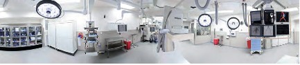
What is a Cardiovascular Hybrid Suite?

Cardiovascular hybrid suite is a state-of-the-art operating room equipped with a fully functional catheterization laboratory, thus allowing surgical procedures and catheter-based interventions to be carried out in the same room. Hybrid suites provide a place where treatments traditionally available only in a cath lab and procedures only available in an operating room can be performed together to provide patients with the best available combination of therapies for cardiovascular disease. These multidisciplinary, integrated cardiovascular procedural suites bring the best of two worlds together by combining all the advantages of a modern cath lab with an up-to-date cardiovascular surgery operating room (OR).

Hybrid suites began to evolve in the mid to late 1990s, when some groups of interventional cardiologists started sharing operating rooms with cardiovascular surgeons. The appeal of the hybrid suite concept has grown as have catheter based devices (stents, coils, balloons and lasers) have been developed that enable interventional cardiologists to advance the invasiveness and effectiveness and applications of percutaneous transcatheter interventions. The interest in these suites has also increased as cardiovascular surgeons have developed a variety of techniques for

  • Minimally invasive procedures, such as minimally invasive direct coronary artery bypass grafting (MIDCAB) or
  • Totally endoscopic coronary artery bypass grafting (TECAB).

With the advent of more tools, interventional cardiologists are becoming more like surgeons, and with less invasive tools, cardiovascular surgeons are becoming more like interventionalists. Rather than separating surgical procedures from interventional procedures performed in traditional operating rooms and cath labs, hybrid suites provide a high-tech environment that allows cardiologists and surgeons to work together to offer patients complex, minimally invasive therapies.

Some experts believe that hybrid suites represent the wave of the future in cardiovascular care and that most heart centers will eventually install hybrid suites to offer patients the latest cardiovascular procedures safely and effectively with minimal surgical trauma. The rooms can be costly to build and equip, but if a medical center is considering building a new operating room or cath lab, setting up a hybrid suite makes sense. Medical centers that have a hybrid suite available can clearly differentiate themselves in a positive way from centers that do not have such capabilities.

The Benefits of a Hybrid Suite for Medical Centers

While building a hybrid suite is more expensive than building a traditional operating room or cath lab, a hybrid suite can potentially be used for all types of cardiovascular procedures, including

  • traditional cardiac and vascular surgery,
  • interventional coronary procedures,
  • endovascular aortic procedures and
  • electrophysiology procedures.

Hybrid suites reinforce the trend in cardiovascular care toward less invasive, comprehensive hybrid procedures. Once a hybrid suite is in place, the demand for its use will likely grow due to increasing indications and referrals for these innovative treatments, many of which are increasingly covered by third-party payers.

http://www.expo.acc.org/acc12/CUSTOM/images/ACC12/ACC.10%20Hybrid%20Suite%20Directory.pdf

What Equipment is Needed?

Interventional cath labs usually have excellent imaging capabilities but lack the sterile facilities and staff needed for a formal OR, while operating rooms frequently lack high-level imaging equipment. Some of the essential equipment for a hybrid suite includes:

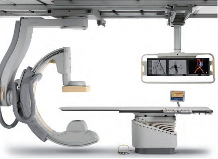
• A state-of-the-art imaging system capable of performing 3D rotational angiography, CT scanning, and ultrasound is advantageous. Floor-mounted and ceiling-mounted systems are available, but many hospitals use ceiling-mounted systems because access to the patient is slightly easier. Some ceiling-mounted systems provide 3D imaging from the surgeon’s position perpendicular to the patient. However, some hospitals prefer floor-mounted systems because having mechanical parts running above the operative field may cause dust to fall, resulting in infections. An important aspect is that the C-arm can be parked away when it is not used. This especially enhances access of the anesthesia team to the patient.

• An operating table that meets the needs of both surgeons and interventionalists by electronically integrating the table with the imaging system is also essential. These tables should have retractable rails for retractors and other surgical tools. To perform 3D imaging on the operating table, the C-arm of the imaging system should allow fast and precise rotation around the patient.

• A variety of other surgical and interventional systems and equipment may also be needed, including a robotic surgical system, a heart-lung machine, an image integration system, an endoscopic imaging system, a radiology display system, an audiovisual system to move images to different monitors and an anesthesia monitoring system, including transesophageal echocardiography. Some equipment like the integrated OR table and the angiography unit need to be fixed parts of the hybrid OR. Some equipment will be mobile in order to maintain some flexibility in workflow.

Hybrid1

Hybrid2

Hybrid3

Hybrid4

Hybrid5

Who are the Equipment Vendors?
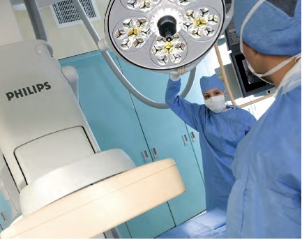
Philips Healthcare

Phone: 800-934-7372

Email: healthcare@philips.com

Web: http://www.philips.com/healthcare

Philips is one of the world’s leading technology companies, with a long history of practical innovation and visionary design. In healthcare, we are committed to understanding the human and technological needs of patients and caregivers. We believe this understanding will help us deliver solutions that not only enable more confident diagnoses and more efficient delivery of care, but also improve the overall experience of care. We offer equipment, software and services for imaging, patient monitoring, resuscitation and much more.  A Hybrid OR can help make life simpler for the interdisciplinary teams who operate in this environment every day. As a world leader in cardiovascular X-ray, Philips has the experience and expertise to deliver the first class technology you need to perform minimally invasive procedures with speed, accuracy and confidence. A long history of innovation has enabled Philips to develop pioneering imaging solutions that really make a difference.

For example, Philips Allura Xper cardiovascular X-ray systems are designed to deliver enhanced imaging with superb performance for all cardiac projections, and our iE33 ultrasound system with Live 3D TEE and QLAB can assist interventional procedures and provide comprehensive quantitative information to support critical decisions. Our cardiology informatics solutions help you manage patient information throughout the cardiovascular care continuum. Philips solutions allow minimally invasive and catheter-based procedures to take place in the same suite as conventional cardiac surgery.

Phillips EchoNavigator – X-Ray and 3-D Ultrasound is described in:

Minimally Invasive Structural CVD Repairs: FDA grants 510(k) Clearance to Philips’ EchoNavigator – X-ray and 3-D Ultrasound Image Fused.

Intuitive Surgical, Inc. 

da Vinci.Surgery by Intuitive Surgical, Inc. 

Phone: 800-876-1310

Email: info@intusurg.com

Web: http://www.intuitivesurgical.com

Intuitive Surgical, Inc. is the global technology leader in robotic-assisted, minimally invasive surgery. The company’s da Vinci® Surgical System offers breakthrough capabilities that enable cardiac surgeons to use a minimally invasive approach and avoid median sternotomy.

Content of FDA Warning Letter, following  FDA Inspection on dates 04/01/2013 – 05/30/2013 – it discussed in

Hybrid Cath Lab/OR Suite’s da Vinci Surgical Robot of Intuitive Surgical gets FDA Warning Letter on Robot Track Record


MAVIG GmbH 

Phone: 631-266-2229,

585-247-1212 ext. 60

Email: info@mavig.com

Web: http://www.mavig.com

MAVIG’s specialty is ceiling/boom suspension systems for lighting (exam, surgical and LED), monitor-suspension systems—single, multibank (one to eight) systems and widescreen, overhead radiation shielding and contrast injector adapters. MAVIG also manufactures radiation protection products such as aprons, gloves, table-attachable lower body shields, adjustable- and fixed-height mobile and modular barriers.

Toshiba America Medical Systems, Inc.

Phone: 714-730-5000

Email: mktgcomm@tams.com

Web: http://www.medical.toshiba.com

Creating a hybrid lab may be complicated, but having an experienced partner that listens makes all the difference. Toshiba’s unique blend of hybrid experience and industry recognized Infinix™-i imaging systems for the Cath Lab.

Hybrid Cath Lab/OR Suite in Leading Hospitals in the US

  • The  Hybrid Cath Lab/OR Suite at New York Presbyterian Hospital/Columbia University Medical Center, New York, NY is presented in

Becoming a Cardiothoracic Surgeon: An Emerging Profile in the Surgery Theater and through Scientific Publications

  • The  Hybrid Cath Lab/OR Suite at Cleveland Clinic, Cleveland, Ohio is presented in

Heart Transplant (HT) Indication for Heart Failure (HF): Procedure Outcomes and Research on HF, HT @ Two Nation’s Leading HF & HT Centers

Speakers at 3D CV Theater, 2010 are working in Hospitals where Hybrid Cath Lab/OR Suite are in operations at the present time. The list include the following Hospitals with a Hybrid Cath Lab/OR Suite:

  • Vanderbilt Medical Center, Nashville, TN
  • University of Maryland Heart Center, Baltimore, MD
  • The Heart Center at Nationwide Children’s Hospital, Columbus, Ohio
  • The Robotic Surgical Center, East Carolina University Department of Surgery, Greenville, N.C.
  • University of Washington Medicine Regional Heart Center, Seattle, WA
  • Brigham and Women’s Hospital, Boston, MA
  • Saint Joseph’s Hospital and Peachtree Cardiovascular and Thoracic Surgery, Atlanta, GA
  • Emory University Hospital, Atlanta, GA
  • Beth Israel Deaconess Medical Center, Boston, MA
  • Boston Medical Center, Boston, MA
  • Mayo Graduate School of Medicine, Mayo Clinic, Rochester, MN
  • Lankenau Hospital, Lancaster, PA
  • Cardiac Non-Invasive Laboratory at Cedars-Sinai Medical Center, Los Angeles, CA
  • Robotic Surgery at St. Joseph’s Hospital, Atlanta, GA

Speakers at 3D CV Theater, 2010, included the following Cardiovascular Interventionists leading the adoption process of Hybrid Surgery in Hybrid Cath Lab/OR Suite into care modalities for cardiovascular disease:

Johannes O. Bonatti, M.D., is professor of surgery and director of coronary surgery and advanced coronary interventions at the University of Maryland Heart Center, Baltimore. He received his training in general surgery and cardiac surgery at the department of surgery at Innsbruck Medical University in Austria. Prior to his arrival at the University of Maryland, he worked at this institution as an attending surgeon and associate professor. Dr. Bonatti’s main interest is the development of minimally invasive, totally endoscopic coronary artery bypass grafting (TECAB) procedures using robotic technology.

As one of the international leaders in this field, he performed the largest series of robotic TECAB on the arrested heart, including single-, double- and triple-vessel TECAB. He has published significantly on procedure development and the implementation process of completely endoscopic coronary surgery using the da Vinci robotic system. Together with colleagues from interventional cardiology, Dr. Bonatti is working on integrated concepts for treatment of coronary artery disease. He was the first to perform a simultaneous hybrid coronary intervention using TECAB and placement of a coronary stent. He is organizing international meetings on hybrid interventions in cardiovascular medicine (http://www.icrworkshop.com). He has trained heart surgeons from around the world in the use of the da Vinci robot for heart surgery and he has introduced TECAB procedures in Austria, the Czech Republic, Greece, Turkey, India and Australia.

John G. Byrne, M.D., is the William S. Stoney Professor of Cardiac Surgery at Vanderbilt University School of Medicine and chair of the department of cardiac surgery at Vanderbilt Medical Center, Nashville, TN.

Before moving to Vanderbilt, he was associate chief and residency program director in the division of cardiac surgery at Brigham and Women’s Hospital, and associate professor of surgery at Harvard Medical School, Cambridge, MA. A graduate of the University of California, Davis, he received his medical degree in 1987 from Boston University. His postdoctoral training was completed at the University of Illinois affiliated hospitals and Brigham and Women’s Hospital in Boston.

Dr. Byrne is the author of more than 100 scientific articles on cardiac surgery and related areas. His patient care emphasis is

  • aortic root surgery,
  • coronary artery disease and
  • valve surgery

He is board-certified in general surgery and thoracic surgery.

John P. Cheatham, M.D., is director of cardiac catheterization and interventional therapy and codirector of The Heart Center at Nationwide Children’s Hospital, Columbus, Ohio. He is also the George H. Dunlap Endowed Chair in Interventional Cardiology and professor of pediatrics and internal medicine at The Ohio State University College of Medicine. Dr. Cheatham’s area of expertise is transcatheter intervention and hybrid therapy of newborns, children and adults with complex congenital heart disease. He has pioneered several new techniques and devices in non-surgical intervention and is a leader in developing hybrid therapies. He has been a principal investigator in numerous FDA-sponsored clinical trials evaluating non-surgical closure devices and stent therapy over the past two decades. Additionally, Dr. Cheatham designed the first hybrid cardiac catheterization suites and advanced imaging equipment at Nationwide Children’s Hospital. He serves as a consultant to various medical companies and proctors new transcatheter techniques and devices to other physicians around the world. Dr. Cheatham has implemented a formal physician exchange program with two of the leading medical institutions in China. In cooperation with China Red Cross, he is also the foreign director of the International Training Center for treatment of congenital heart disease in poor children. Dr. Cheatham has written more than 120 manuscripts, 16 book chapters, 300 national and international presentations and is co-editor of the book, Complications in Percutaneous Interventions for Congenital and Structural Heart Disease. After graduating from the University of Oklahoma College of Medicine, he completed his residency at Boston Children’s Hospital, followed by a fellowship in Pediatric Cardiology at Texas Children’s Hospital in Houston.

W. Randolph Chitwood, Jr., M.D., is senior associate vice chancellor for health sciences and chief of cardiovascular services at East Carolina University Department of Surgery, Greenville, N.C. Dr. Chitwood is a leading international pioneer in minimally invasive and robotic heart surgery. The Robotic Surgical Center at East Carolina University has trained more than 350 surgeons. His research activities relate to myocardial preservation, simulation in surgery and endoscopic/robotic cardiac surgery. He was the principal investigator of the FDA robotic mitral valve trials that led to approval for use in the U.S. He is the son and grandson of “southwestern Virginia mountain doctors” who set the guidelines for his professional life. He graduated from Hampden-Sydney College and received his medical degree from the University of Virginia. After medical school, he completed the surgical residency at Duke University Medical Center under David C. Sabiston, M.D., an influential surgical educator of the era. At Duke he spent 10 years training in general and cardiothoracic surgery, as well as basic science research.

After his chief residency at Duke in 1984, he was selected to begin and head the new cardiac surgery program at the East Carolina University School of Medicine. Because of his prolific publication record as a resident and clinical acumen, his initial appointment was as a full professor of surgery. Except for a two-year hiatus as the chief of cardiothoracic surgery at the University of Kentucky, he has spent his entire career at East Carolina University, where he also served as chairman of the department of surgery. In 2003, he was named to be in charge of the development of the East Carolina Heart Institute, which now includes an integrated department of cardiovascular sciences as well as a $200 million heart hospital, outpatient, research and education center.

Larry S. Dean, M.D., is director of the University of Washington Medicine Regional Heart Center and is professor of medicine and of surgery at the University of Washington School of Medicine, Seattle. In addition to general cardiology, he is an expert in cardiac catheterization and interventional cardiology. He also conducts research on stents to keep blocked heart arteries open and on ways to prevent restenosis after stents are inserted. He is currently involved in the evaluation of percutaneous aortic valve replacement. Dr. Dean earned his M.D. from the University of Alabama School of Medicine, Birmingham, and served his internship and residency at the University of Washington. He then returned to the University of Alabama Hospital for fellowships in cardiovascular disease and in angioplasty. After nearly 15 years as a faculty member at the University of Alabama, he returned to the University of Washington to direct the Regional Heart Center. He is a fellow of the American College of Cardiology and is board-certified in internal medicine, cardiovascular disease and interventional cardiology. He is also a fellow of the American Heart Association and president-elect of the Society of Cardiovascular Angiography and Interventions.

Andrew Craig Eisenhauer, M.D., is director of the interventional cardiovascular medicine service at Brigham and Women’s Hospital and assistant professor of medicine at Harvard Medical School. His specialties are

  • interventional cardiology,
  • vascular medicine and
  • congenital and inherited diseases.

He earned his medical degree at New York University School of Medicine and served a residency at Peter Bent Brigham Hospital and a fellowship at Massachusetts General Hospital. He is certified in internal medicine, cardiovascular disease and interventional cardiology. His clinical interests are

  • endovascular therapy,
  • complex coronary disease,
  • peripheral vascular disease,
  • cerebrovascular disease,
  • congenital heart disease and structural heart disease

Douglas A. Murphy, M.D., is chief of cardiothoracic surgery at Saint Joseph’s Hospital and a cardiothoracic surgeon at Peachtree Cardiovascular and Thoracic Surgery, Atlanta. His areas of interest are robotically assisted heart surgery with an emphasis on repairing the mitral valve rather than replacing it. A graduate of the University of Pennsylvania Medical School, Philadelphia, he served an internship and residency at Massachusetts General Hospital, Boston, and at Emory University, Atlanta.

Khusrow Niazi, M.D., is an assistant professor at Emory University School of Medicine and director of peripheral and carotid intervention at Emory University Hospital Midtown, Atlanta. He earned his medical degree at King Edward Medical College, Lahore, Pakistan, and served an internship at Kettering Medical Center, Dayton, Ohio, and a fellowship at William Beaumont Hospital, Royal Oak, MI. He has published papers on stenting following rotational atherectomy, small vessel stenting for coronary arteries, imaging of lower extremities and treatment of peripheral arterial disease.

Jeffrey J. Popma, M.D., is director of innovations in interventional cardiology, a senior attending physician at Beth Israel Deaconess Medical Center and an associate professor of medicine at Harvard Medical School in Boston. Dr. Popma received his bachelor’s degree in economics from Stanford University, and his M.D. from Indiana University School of Medicine. He completed his internship, residency, chief residency and fellowship at University of Texas Southwestern Medical Center. He also completed an interventional cardiology fellowship at the University of Michigan. Dr. Popma is the past president of the Society for Cardiac Angiography and Intervention and is the co-chair of the ACC Interventional Council. He sits on the editorial boards of several publications, and reviews for several cardiology periodicals. Dr. Popma has more than 300 published peer-reviewed manuscripts.

Dr. Popma also directs the BIDMC Angiographic Core Laboratory and is principal investigator for more than 65 ongoing multicenter device studies within the research laboratory. Over the past 15 years, these trials have included a broad array of new technology, including bare-metal stents, drug-eluting stents, distal-protection devices, total-occlusion devices and carotid and peripheral revascularization procedures. His primary clinical interest currently is the use of percutaneous aortic valve replacement for patients with high-risk aortic stenosis.

Robert S. Poston, M.D., is chief of cardiac surgery at Boston Medical Center and associate professor of cardiothoracic surgery at Boston University School of Medicine. He has a strong background in minimally invasive cardiac bypass surgery and is a pioneer in using robotics, specifically the da Vinci Surgical System, to treat coronary artery disease. A graduate of the Johns Hopkins School of Medicine, Baltimore, Dr. Poston completed a residency in general surgery at the University of California, San Francisco, and continued his training with a research fellowship in cardiothoracic surgery at Stanford University School of Medicine, Palo Alto, CA, and a cardiothoracic residency at the University of Pittsburgh Medical Center.

Charanjit S. Rihal, M.D., is professor of medicine and director of the cardiac catheterization laboratory at Mayo Graduate School of Medicine, Mayo Clinic, Rochester, MN. A graduate of the University of Winnipeg, Dr. Rihal did his residency and fellowship at the Mayo Graduate School of Medicine and also earned an MBA at the Carlson School of Management, University of Minnesota. His medical interests are interventional cardiology, structural heart disease interventions and the management of quality and costs in healthcare.

Timothy A. Shapiro, M.D., is director of the Interventional Cardiology Fellowship Program and campus chief, interventional cardiology, at Lankenau Hospital, Lancaster, PA. A graduate of Yale University School of Medicine, he served his residency and a fellowship at the Hospital of the University of Pennsylvania.

Robert J. Siegel, M.D., is director of the Cardiac Non-Invasive Laboratory at Cedars-Sinai Medical Center, cardiology director of the Cedars-Sinai Marfan Center, and Rexford S. Kennamer, M.D., chair in cardiac ultrasound at Cedars-Sinai Medical Center, Los Angeles. Dr. Siegel is also professor of medicine in residence at the David Geffen School of Medicine at University of California, Los Angeles. He previously served as senior staff fellow in cardiac pathology at the Heart, Lung and Blood Institute of the National Institutes of Health, Bethesda, MD. Internationally recognized as one of the leading experts in the field of cardiovascular ultrasound, Dr. Siegel specializes in cardiovascular ultrasound, including transthoracic, transesophageal and intravascular methodologies. His research interests include

  • valvular heart disease,
  • therapeutic applications of ultrasound energy,
  • transesophageal and intraoperative echocardiography, and the
  • development and use of hand-held portable echocardiographic systems for clinical innovations.

In addition, he is involved with clinical research studies related to the diagnosis, assessment and management of patients with

  • Marfan syndrome,
  • hypertrophic cardiomyopathy and
  • pericardial and valvular heart disease.

Dr. Siegel is a fellow, and has previously served as the president of the California Chapter of the American College of Cardiology and president of the Los Angeles Society of Echocardiography. He has been active in numerous cardiovascular societies, including the American Heart Association, the American College of Cardiology and the American Society of Echocardiography. Dr. Siegel received his medical degree at Baylor College of Medicine, Houston, where he developed an interest in cardiology. He completed his medical residency at Emory University and at Los Angeles County + USC Medical Center. He completed his cardiology fellowship at Harbor-UCLA Medical Center.

Over the last two years Dr. Siegel has worked extensively with live 3D transesophageal echo in the cardiac intervention center and the operating room. He and his echocardiologist colleagues, doctors Shiota, Biner, Tolstrup and Gurudevan, have worked closely at Cedars-Sinai Medical Center in Los Angeles with the interventional cardiologists, doctors Kar and Makkar, as well as with the cardiac surgeons, doctors Trento and Fontana. They use live 3D TEE extensively for the assessment of structural heart disease. In addition, it is used on a regular basis for the guidance of percutaneous procedures for mitral valve e-clip repair, mitral balloon valvuloplasty, aortic and pulmonic valve replacement, left atrial appendage exclusion by the Watchman device as well as for ASD closure.

Sudhir P. Srivastava, M.D., president of the International College of Robotic Surgery at St. Joseph’s Hospital, Atlanta, is a pioneer in performing beating heart totally endoscopic coronary artery bypass surgeries. Previously, he was assistant professor of surgery and director of robotic and minimally invasive cardiac surgery at the University of Chicago Medical Center. Dr. Srivastava specializes in robotically assisted totally endoscopic coronary artery bypass surgery. He has performed approximately 1,000 robotic cardiothoracic surgical procedures, of which 450  have been single- and multivessel beating heart totally endoscopic coronary bypass (BH TECAB) procedures. He has keen interest in hybrid coronary revascularization in TECAB patients to achieve complete revascularization.

Dr. Srivastava has helped launch robotic revascularization programs throughout the world. He has performed numerous live BH TECAB demonstrations both in the U.S. and abroad, and continues to be a presenter and invited speaker at numerous national and international scientific meetings. He earned his medical degree at the Jawahar Lal Nehru Medical College in Ajmer, India and immigrated to the U.S. in 1972. He completed his cardiothoracic surgery residency at the hospitals associated with the University of British Columbia, Vancouver, Canada.

Francis P. Sutter, D.O., F.A.C.S., is clinical professor of surgery at Thomas Jefferson University-Jefferson Medical College, Philadelphia, and chief of cardiothoracic surgery at Lankenau Hospital, Main Line Health System, Wynnewood, PA. A graduate of Philadelphia College of Osteopathic Medicine, his surgical residency and a cardiothoracic fellowship were completed at Thomas Jefferson University Hospital.

Mark R. Vesely, M.D., is an assistant professor of medicine at the University of Maryland School of Medicine. He completed medical school at the George Washington University and postgraduate training—an internal medicine residency and fellowships in cardiovascular disease and interventional cardiology—at the University of Maryland. He is board-certified in internal medicine, cardiovascular disease, nuclear cardiology and interventional cardiology. Dr. Vesely is the associate program director of the Interventional Cardiology fellowship at University of Maryland. His special interests include the partnered approach (interventional cardiologists and cardiac surgeons) for hybrid coronary revascularization and structural heart disease interventions. Additional research interests include investigation of techniques to minimize acute myocardial infarction injury with ventricular-assist devices and adult stem cell therapies.

David X. M. Zhao, M.D., Ph.D., is an associate professor of medicine and cardiac surgery, Harry and Shelley Page Chair in Interventional Cardiology, director of the Cardiac Catheterization Laboratories and interventional cardiology director of the Interventional Cardiology Fellowship Training Program, Vanderbilt University School of Medicine, Nashville, TN. He earned his medical degree at Shanghai Medical University, Shanghai, P.R. China, and his Ph.D. in immunology at Queensland University, Brisbane, Australia. His postdoctoral training was at Zhongshan Hospital, Shanghai Medical University, Shanghai, P.R. China, The Prince Charles Hospital, Brisbane, Australia, and Brigham and Women’s Hospital, Boston.

http://www.expo.acc.org/acc12/CUSTOM/images/ACC12/ACC.10%20Hybrid%20Suite%20Directory.pdf

Part Two

Cardiac Surgery


Cardiac Surgery @ Cleveland Clinic: Traditional OR & Hybrid Cath Lab/OR Suite

Nation #1 for 19 consecutive years – The Heart Center: Miller Family Heart & Vascular Institute @ Cleveland Clinic

The Sydell and Arnold Miller Family Heart & Vascular Institute is one of the largest, most experienced cardiovascular specialty groups in the world. Our physicians are committed to providing the most advanced diagnostic and treatment options, better outcomes and improved quality of life. U.S.News & World Reporthas ranked Cleveland Clinic as the No.1 heart program in America every year since 1995.

Departments & Centers:

Below we present two articles on Cardiac Surgery @ Mayo Clinic 

Cardiac Surgery @ Mayo Clinic: Traditional OR & Hybrid Cath Lab/OR Suite 

Coronary Reperfusion Therapies: CABG vs PCI – Mayo Clinic preprocedure Risk Score (MCRS) for Prediction of in-Hospital Mortality after CABG or PCI

Larry H. Bernstein, MD, FCAP and Aviva Lev-Ari, PhD, RN

Comparison of the 10-year and 15-year survivals after CABG demonstrated benefit from a change in graft sources used at the Mayo Clinic and widely adapted by others: vascular grafts from the left internal mammary artery (LIMA) instead of just leg veins, for multiple grafts (up to 3), LIMA-to-LAD plus grafts using LIMA or radial artery vs LIMA/saphenous vein (SV).

CABG Survival in Multivessel Disease Patients: Comparison of Arterial Bypass Grafts vs Saphenous Venous Grafts

Larry H. Bernstein, MD, FCAP and Aviva Lev-Ari, PhD, RN

Part Three 

Invasive Interventions with Complications

In the following article we covered multiple etiologies for cardiovascular complications related to invasive interventions: cardiovascular and peripheral arterial or peri- and post- cardiac surgery of the open heart type.

Cardiovascular Complications: Death from Reoperative Sternotomy after prior CABG, MVR, AVR, or Radiation; Complications of PCI; Sepsis from Cardiovascular Interventions

Justin D Pearlman, MD, PhD, FACC and Aviva Lev-Ari, PhD, RN

http://pharmaceuticalintelligence.com/2013/07/23/cardiovascular-complications-of-multiple-etiologies-repeat-sternotomy-post-cabg-or-avr-post-pci-pad-endoscopy-andor-resultant-of-systemic-sepsis/

This article covers types of Cardiovascular Complications derived from the following THREE types of assault on the Human body, two related to cardiac invasive interventions, the last due to its systemic nature is taking a fatal Cardiac toll: the Sepsis condition causing cardiac failure.

Three types of Cardiovascular Complications:

I. Risk of Injury During Repeat Sternotomy – following CABG orAortic Valve Replacement, both done in Open Heart Surgery

II. Complications After Percutaneous Coronary intervention (PCI) and endovascular surgery for Peripheral Artery Disease (PAD)

  • (a) Post PCI, and
  • (b) PAD Endovascular Interventions: Carotid Artery Endarterectomy

III. Cardiac Failure During Systemic Sepsis

This article does NOT cover the following two types of Cardiovascular Complications:

1. Trauma Injury causing cardiac arrest, lung collapse or cardiogenic shock

2. Surgical Complication of Non-cardiac surgery type causing cardiac arrest, i.e, Surgery of Joint Replacement causing sepsis causing death or death caused by complications of surgery i.e., blood loss, viral infection, emboli, thrombus, stroke, or cardiogenic shock not related to Cardiovascular and Cardiac invasive interventions

The e-Reader is advised to consider the following expansion on the subject matter carrying the discussion to additional related clinical issues:

Larry H Bernstein, Advanced Topics in Sepsis and the Cardiovascular System at its End Stage

http://pharmaceuticalintelligence.com/2013/08/18/advanced-topics-in-sepsis-and-the-cardiovascular-system-at-its-end-stage/

Bernstein, HL, Pearlman, JD and A. Lev-Ari  Alternative Designs for the Human Artificial Heart: The Patients in Heart Failure – Outcomes of Transplant (donor)/Implantation (artificial) and Monitoring Technologies for the Transplant/Implant Patient in the Community

http://pharmaceuticalintelligence.com/2013/08/05/alternative-designs-for-the-human-artificial-heart-the-patients-in-heart-failure-outcomes-of-transplant-donorimplantation-artificial-and-monitoring-technologies-for-the-transplantimplant-pat/

Pearlman, JD and A. Lev-Ari Cardiac Resynchronization Therapy (CRT) to Arrhythmias: Pacemaker/Implantable Cardioverter Defibrillator (ICD) Insertion

http://pharmaceuticalintelligence.com/2013/07/22/cardiac-resynchronization-therapy-crt-to-arrhythmias-pacemakerimplantable-cardioverter-defibrillator-icd-insertion/

Read Full Post »

Becoming a Cardiothoracic Surgeon: An Emerging Profile in the Surgery Theater and through Scientific Publications 

Author and Curator: Aviva Lev-Ari, PhD, RN

Article ID #65: Becoming a Cardiothoracic Surgeon: An Emerging Profile in the Surgery Theater and through Scientific Publications. Published on 7/8/2013

WordCloud Image Produced by Adam Tubman

Two components of an Emerging Profile of a Young Cardiothoracic Surgeon were researched by the Author for the case of  Dr. Isaac George, Assistant Professor of Surgery, Division of Cardiothoracic Surgery, Department of Surgery, New York Presbyterian Hospital/Columbia University Medical Center , New York, NY.

The two components being:

1. the Cardiothoracic Surgery Theater

2. the Scientific Publications

I noted with interest Dr. George’s second publication, to be about a very well known surgeon in the US and Europe, John Benjamin Murphy. written by Dr. George and two other colleagues,  George I, Hardy MA, Widmann WD. published in Curr Surg. 2004 Sep-Oct;61(5):439-41.

Dr. Murphy, is best remembered for the eponymous clinical sign that is used in evaluating patients with acute cholecystitis. His career spanned general surgery, orthopedicsneurosurgery, and cardiothoracic surgery, which helped him to gain international prominence in the surgical profession. Mayo Clinic co-founder William James Mayo called him “the surgical genius of our generation.”

http://en.wikipedia.org/wiki/John_Benjamin_Murphy 

[Musana, Kenneth and Steven H. Yale (May 2005). “John Benjamin Murphy (1857–1916)”. Clinical Medicine & Research. Retrieved 2008-05-16.]

I assume that Dr. Murphy’s contributions to Thoracic surgery were of interest to Dr. George to inspire him to write on the subject and elect that Specialty in Surgery.

Murphy was first in the U.S. to induce (1898) artificial immobilization and collapse of the lung in treatment of pulmonary tuberculosis. He was a pioneer in the use of bone grafting and made contributions to the understanding and management of ankylosis as well as independently proposing artificial pneumothorax to manage unilateral lung disease in tuberculosis.

      • «It is the purpose of every man’s life to do something worthy of the recognition and appreciation of his fellow men. . . . By their superior intellectual qualifications, their fidelity to purpose and above all their indefatigable labour the few become leaders.»

Journal of the American Medical Association, Chicago, 1911, 57: 1.

SOURCE Whonamedit? A dictionary of medical eponyms, John Benjamin Murphy

I came across Dr. Isaac George’s name while researching clinical indications for Inhaled Nitric Oxide in June 2013, upon the recent publication of Leaders in Pharmaceutical Business Intelligence FIRST e-Book on  Amazon (Biomed e-Books) [Kindle  Edition]

Perspectives on Nitric Oxide in  Disease  Mechanisms
http://www.amazon.com/dp/B00DINFFYC

Dr. George’s article on Outcomes After Inhaled Nitric Oxide Therapy was particularly useful in my own research on the topic,

Clinical Indications for Use of Inhaled Nitric Oxide (iNO) in the Adult Patient Market: Clinical Outcomes after Use of iNO in the Institutional Market,  Therapy Demand and Cost of Care vs. Existing Supply Solutions

Being myself in Analytics and quantitative model design, 1976-2004, I found of particular interest the range of quantitative methodologies used in the following article by Isaac George, assuming that his days at MIT, came very handy in 2006:

George, Isaac, Xydas, Steve, Topkara, Veli K., Ferdinando, Corrina, Barnwell, Eileen C., Gableman, Larissa, Sladen, Robert N., Naka, Yoshifumi, Oz, Mehmet C.
Clinical Indication for Use and Outcomes After Inhaled Nitric Oxide Therapy
Ann Thorac Surg 2006 82: 2161-2169

As a result of studying this article, I became aware that it has impacted  favorably my 6/2013, Editorial decision, for  a forthcoming book on Cardiovascular Disease in 2013. The Editorial decision regarding the selection and representation of  prominent Cardiothoracic Surgery Theater in the US, and my personal decision to select a Young Cardiothoracic Surgeon

Dr. Isaac George, Assistant Professor of Surgery, Division of Cardiothoracic Surgery, Department of Surgery, New York Presbyterian Hospital/Columbia University Medical Center, New York, NY

Education Profile and Medical Training of a Cardiac Surgeon


Isaac George, MD

Positions and Appointments

2012-present Attending Surgeon, Heart Valve Center
NewYork-Presbyterian Hospital/Columbia University Medical Center, New York, NY
2012-present Assistant Professor of Surgery
Division of Cardiothoracic Surgery, Department of Surgery, New York Presbyterian Hospital/Columbia University Medical Center , New York, NY

Clinical Specialties

Adult aortic and mitral valve surgery
Transcatheter aortic and mitral valve implantation
Hybrid coronary artery bypass surgery
Complex aortic surgery
Complex valvular surgery
Heart failure and transplant surgery
Reoperative cardiac surgery
Thoracic aortic endograft implantation

Research Interests

Director, Cardiac Surgery Research Lab

1. Regulation of myostatin signaling in human cardiomyopathy

2. TGFB regulation in non-syndromic aortic aneurysm formation

3. Valve interstitial cell activation mechanisms after surgical and transcatheter valve replacement

4. Clinical outcomes after valve and hybrid surgery

Education and Training

2011-2012 Interventional Cardiology/Hybrid Cardiac Surgery Fellowship
New York Presbyterian Hospital – Columbia University Medical Center, New York, NY
2011 Ventricular Assist Device/Cardiac Transplant Fellowship, Minimally Invasive, Cardiac Surgery
New York Presbyterian Hospital – Columbia University Medical Center, New York, NY
2009-2011 Fellow, Cardiothoracic Surgery
New York Presbyterian Hospital – Columbia University Medical Center, New York, NY
2008-2009 Post-Doctoral Clinical Fellow, Cardiothoracic Surgery
New York Presbyterian Hospital – Columbia University Medical Center, New York, NY
2006-2008 Resident, General Surgery
New York Presbyterian Hospital – Columbia University Medical Center, New York, NY
2004-2006 Research Fellow, Cardiothoracic Surgery
New York Presbyterian Hospital – Columbia University Medical Center, New York, NY
2002-2004 Resident, General Surgery
New York Presbyterian Hospital – Columbia University Medical Center, New York, NY
2001-2002 Internship, General Surgery
New York Presbyterian Hospital – Columbia University Medical Center, New York, NY
1997-2001 MD, Medicine
Duke University School of Medicine, Durham, NC
1993-1997 BS, Mechanical Engineering
Massachusetts Institute of Technology, Cambridge, MA

Board Certifications

American Board of Thoracic Surgery, 2012-
American Board of Surgery, 2008-
Certification, Pediatric Advanced Life Support, 2008-
Certification, Advanced Trauma Life Support, 2006-
MD, State of New York, 2005-
Certification, Advanced Cardiac Life Support/Basic Life Support, 2001-
United States Medical Licensing Examination Step 3, 2004
United States Medical Licensing Examination Step 2, 2001
United States Medical Licensing Examination Step 1, 2000

Professional Honors

2008 Blakemore Prize – Best Resident Research Award, Columbia University College of Physicians and Surgeons

2007 Blakemore Award – Best Resident Research Award, Columbia University College of Physicians and Surgeons

2006 Blakemore Award – Best Resident Research Award, Columbia University College of Physicians and Surgeons

2004 New Era Cardiac Surgery Conference Scholarship

1995 Pi Tau Sigma, Mechanical Engineering Honor Society

1993 Duke University Comprehensive Cancer Center Fellowship

Professional Societies and Committees

2011 Faculty, Transcatheter Cardiovascular Therapeutics (TCT) Annual Symposium

2010- Candidate Member, Society of Thoracic Surgeons

2010- Fellow-in-Training, American College of Cardiology, Surgeons Council

2005-06 American Society of Artificial Internal Organs

2004- Member, American Heart Association

1997-01 Member, American Medical Association

SOURCE http://asp.cpmc.columbia.edu/facdb/profile_list.asp?uni=ig2006&DepAffil=Surgery

The decision to focus on Cardiothoracic Surgery @Presbeterian as described in Dr. Isaac George’s research had yielded one Sub-Chapter (4.1) in Chapter 4

Cardiac Surgery, Cardiothoracic Surgical Procedures and Percutaneous Coronary Intervention (PCI)/Coronary Angioplasty  – Heart and Cardiovascular Medical Devices in Use in Operating Rooms and in Catheterization Labs in the US

in Volume Three in a forthcoming three volume Series of e-Books on Cardiovascular Diseases

Cardiovascular Diseases: Causes, Risks and Management

This very Sub-Chapter represents milestones in Dr. Isaac George – Becoming a Cardiothoracic Surgeon: An Emerging Profile through Scientific Publications, This profile is now in: 


Volume Three

Management of Cardiovascular Diseases

Justin D. Pearlman MD ME PhD MA FACC, Editor

Leaders in Pharmaceutical Business Intelligence, Los Angeles

Aviva Lev-Ari, PhD, RN

Editor-in-Chief BioMed E-Book Series

Leaders in Pharmaceutical Business Intelligence, Boston

avivalev-ari@alum.berkeley.edu

Chapter 5

Invasive Procedures by Surgery versus Catheterization


5.1 Cardiothoracic Surgery 

5.1.1 Becoming a Cardiothoracic Surgeon: An Emerging Profile in the Surgery Theater and through Scientific Publications

Aviva Lev-Ari, PhD, RN

5.2: Catheter Interventions

5.2.2 Survivals Comparison of Coronary Artery Bypass Graft (CABG) and Percutaneous Coronary Intervention (PCI) / Coronary Angioplasty

Larry H Bernstein, MD, FCAP and Aviva Lev-Ari, PhD, RN

5.3: Transcatheter (Percutaneous) Valves

5.3.1 Transcatheter Aortic Valve Replacement (TAVR): Postdilatation to Reduce Paravalvular Regurgitation During TAVR with a Balloon-expandable Valve

Larry H Bernstein, MD, FCAP and Aviva Lev-Ari, PhD, RN

5.3.2 Trans-apical Transcatheter Aortic Valve Replacement in a Patient with Severe and Complex Left Main Coronary Artery Disease (LMCAD)

Larry H Bernstein, MD, FCAP and Aviva Lev-Ari, PhD, RN

Larry H Bernstein, MD, FCAP and Aviva Lev-Ari, PhD, RN

5.4: Transcatheter (Percutaneous) Pumps

5.4.1  Ventricular Assist Device (VAD): Recommended Approach to the Treatment of Intractable Cardiogentic Shock

Larry H Bernstein, MD, FCAP and Aviva Lev-Ari, PhD, RN

5.4.2 Phrenic Nerve Stimulation in Patients with Cheyne-Stokes Respiration and Congestive Heart Failure

Larry H Bernstein, MD, FCAP and Aviva Lev-Ari, PhD, RN

Content Analysis of  Surgeon Isaac George, MD – Publications on PubMed

SOURCE

Original classification by Aviva Lev-Ari, PhD, RN, 7/8/2013

Title

Journal

Year

CABG
Stent

Valves
Bio

material
TAVR MVR

End stage

HF

AMI

shock

Congen
Genet

Animal

Model

Heart &
Heart-Lung
Transpl

Stent exteriorization

CCI

13

X

Left Main Coronary

CCI

13

X

TAVR-MVR

JACC

13

X

Paravalvular

CVI

13

X

Cardiogenic Shock

Heart-Lung

12

X

Cheyne-Stokes

Chest

12

X

Myostatin

PlusOne

11

X

Aortic Root & RV

ATS

11

X

Beta-Adrenergic

CV Research

11

X

Erythropoietin

LV  Systolic

J CV

Pharmacol

10

X

Myostatin & HF

Eur J

Heart Failure

10

X

Stentless in valve conduit

ATS

09

X

BNP peptide-

infusion-post MI

Am J Physiol-

Heart Circ

Physiology

09

X

Marginal donor heart

ATS

09

X

Device-surface & Immunogenic

J Thoracic

CV Surg

08

X

Myocardial

electromagnetic

J Cell Physiol

08

X

Clenbuterol-

muscle-mass

J Heart- Lung Transplant

08

X

Bradycardic LV

J Pharmacol Exper Therap

07

X

Ischemia- post

Heart Transplant

J Thoracic

CV Surgery

07

X

Octogen CABG

ATS

07

X

Ventricular synchrony

Eup J Cardio-thoracic Surg

07

X

Inhaled NO

ATS

06

X

X

Adult heart-donor-

to-pediatric

J Thoracic

CV Surg

06

X

Clenbuterol

on LVAD

J Heart-Lung Transplant

06

X

LV-CA stent

Heart Surg

Forum

06

X

LVAD myocarditis

J Thoracic

CV Surg

06

X

MI-Ischemia

Am J Physiol-

Ht-Circ Physiol

06

X

It is the unique combination of Animal Model Research, Biomaterial, Surgical Procedures and Molecular Cardiology, N=33.

Cardiothoracic Surgeon: An Emerging Profile in the Surgery Theater

Isaac George, MD – Clinical Specialties 

  • Adult aortic and mitral valve surgery
  • Transcatheter aortic and mitral valve implantation
  • Hybrid coronary artery bypass surgery
  • Complex aortic surgery
  • Complex valvular surgery
  • Heart failure and transplant surgery
  • Reoperative cardiac surgery
  • Thoracic aortic endograft implantation


VIDEOS on CardioThoracic Surgery @ Department of Cardiothoracic Surgery at NewYork-Presbyterian Hospital/Weill Cornell Medical Center in New York City

VIEW VIDEO on the new Heart Center @ Presbyterian Hospital

http://videos.nyp.org/videos/introduction-to-the-vivian-and-seymour-milstein-family-heart-center

VIEW VIDEO on the two Hybrid OR with Siemens Artis Zeego Technology

http://videos.nyp.org/videos/tour-a-hybrid-or-with-siemens-artis-zeego-technology

VIEW VIDEO on Mininally Invesive and Conventional Therapy for Aortic Dissection and Aneurysms – Hybrid Surgery Case

http://videos.nyp.org/videos/thoracic-innovations-in-minimally-invasive-and-conventional-therapy-for-aortic-dissection-and-aneurysms

VIEW VIDEO on Mitral Valve Repair and Replacement – Dr. Karl H. Krieger

Dr. Karl H. Krieger, the Vice Chairman of the Department of Cardiothoracic Surgery at NewYork-Presbyterian Hospital/Weill Cornell Medical Center in New York City, discusses treatment for Mitral Valve Disease. Specifically, Dr. Krieger compares the options of Mitral Valve Repair with Mitral Valve Replacement.

This video with Dr. Krieger is from a web cast at the Ronald O. Perelman Heart Institute at NewYork-Presbyterian.

VIEW VIDEO on Left Ventricular Assist Devices (LVADs) – Dr. Jonathan Chen

Dr. Jonathan Chen, the Site Chief for Pediatric Cardiac Surgery at NewYork-Presbyterian Hospital/Weill Cornell Medical Center in New York City, explains how Left Ventricular Assist Devices (LVADs) work and how they can benefit patients with heart failure.

LVADs are implantable devices that help the heart pump blood. They can be used as a temporary therapy, allowing patients’ hearts to rest while they recover from cardiac events such as heart attacks, or while they wait for hearts to become available for transplants. For some patients whose hearts are unlikely to recover and are not candidates for heart transplants, the devices may be used as a permanent therapy. Heart failure, especially in severe forms, can force patients to lead restricted lives because often even very limited physical activity, such as walking from one room to another, will leave them breathless.

Dr. Chen is a pediatric cardiothoracic surgeon, yet the information in the video is applicable to adult patients as well.

VIEW VIDEO on Transcatheter Aortic Valve Implantation @ Presbyterian Hospital

http://videos.nyp.org/videos/chapter-3-transcatheter-aortic-valve-implantation

Heart, Heart-Lung Transplantation @ Presbyterian Hospital

Organ transplantation that prolongs and dramatically improves quality of life is nearly a daily occurrence at Columbia University Medical Center.

The success of solid organ transplantation – with improved surgical techniques, replacement organ procurement, and medical management – is advancing each year. Many of these advances have resulted from scientific and clinical research conducted at Columbia University Medical Center.

A Brief History of Transplantation at Columbia

Transplantation: Where we’ve been, where we’re going

Transplantation: Where we've been, where we're going
Eric A. Rose, MD, former chairman of the department of surgery, left center, performing the first successful pediatric heart transplant in 1984. Transplant pioneer Keith Reemtsma, MD, who is overseeing the operating field (top of photo).

When he transplanted a chimpanzee kidney into a human patient in the late 1960’s, the late Keith Reemtsma, MD, then Department of Surgery Chairman at Tulane University, revolutionized treatment of end-stage organ failure and initiated an era of unprecedented exploration into organ transplantation that would affect the lives of patients around the world.

Transferring to Columbia-Presbyterian Medical Center in 1971, Dr. Reemtsma recruited Mark A. Hardy, MD, who laid another cornerstone of organ transplant medicine by founding the program for dialysis and kidney transplantation. Dr. Hardy based the new program on the principle of collaborative clinical care between surgeons and nephrologists. During a time when renal transplant programs were managed by one or the other discipline but never by both simultaneously, the medical community regarded the concept as folly. Yet the program grew steadily, as did the program’s immune tolerance research initiatives to induce the transplant recipient’s body to accept a donor organ. This multidisciplinary cooperation also led to major contributions in immunogenetics, immunosuppression, and treatment of autoimmune diseases and lymphoma — and it ultimately became the overarching principle for all the NewYork-Presbyterian Hospital transplant services.

Mark A. Hardy, MD

Mark Hardy
Eric Rose
Eric A. Rose, MD
Lloyd Ratner
Lloyd E. Ratner, MD

Colleagues universally give credit to Eric A. Rose, MD, who co-founded the heart transplantation program with Dr. Reemtsma, for his successful transformation of the program into the outstanding center it is today. A parade of achievements marks the history of the heart transplant program, including the first mechanical bridge-totransplantation using intra-aortic balloon pumps in the 1970’s, and the first successful pediatric heart transplant, performed by Dr. Rose in 1984. Under the guidance of Dr. Rose and his successors, the program has pioneered research in immunosuppressant medications, mechanical assist devices, and minimally invasive surgical procedures. It currently performs over 100 heart transplants yearly, with among the highest success rates in the nation.

Also in 2004, Lloyd E. Ratner, MD, succeeded Dr. Hardy as director of the renal and pancreas transplant program. One of the first to perform laparoscopic donor operations, Dr. Ratner has found creative solutions to overcome immune barriers to kidney transplantation. The program now routinely uses extended-criteria donor organs, performs transplants among incompatible donors, and is a leader in coordinating “donor swaps” to maximize availability of compatible donor organs. Since Dr. Ratner’s arrival, Columbia has been designated one of ten regional islet resource centers in the U.S. that isolate and transplant pancreatic cells to treat type 1 diabetes as part of a limited protocol controlled by the FDA. Recent progress in visualization of pancreatic islets using PET technology, under the guidance of Paul Harris, PhD, has been recognized by the scientific community as a milestone in this developing field.

NYPH/Columbia received UNOS approval for pancreatic transplantation in January 2008. Our premier kidney transplant program is facilitating rapid growth of the new pancreatic transplantation program, which overlaps both in its patient population and its surgical and medical expertise. The northeast region of the U.S. has been consistently underserved as far as access to pancreatic transplantation, with relatively few centers serving a disproportionately large metropolitan population. The expanding program at NewYork-Presbyterian now provides much-needed access to patients with end-stage pancreatic disease in New York state, particularly those with the most complex medical and surgical challenges.

Transplantation of cells, rather than organs, is emerging as a therapy with enormous potential. Transplantation of either a patient’s own or a foreign donor’s bone marrow cells, for example, offers hope of regenerating the heart so that patients with heart failure may be able to avoid heart transplantation.

In introducing the transplantation programs, it would be remiss to neglect mention of the yet another dimension in which they excel — education. Physician training is a top priority, and NYPH/Columbia has trained many of the greatest transplant surgeons over the last 20 years, including many of the leaders of transplant programs throughout the U.S.

http://columbiasurgery.org/transplant/history.html

Transplant Initiative

At NewYork-Presbyterian Hospital/Columbia University Medical Center, the Transplant Initiative (TI) has been launched to drive the growth of both clinical and research aspects of transplantation. This multi-year undertaking will involve Departments of Medicine, Pathology, Pediatrics, and Surgery and all of the solid organ transplantation programs, both adult and pediatrics. It is led by its Executive Director, Jean C. Emond, MD.

Although NYP/Columbia is already a national leader in clinical transplantation with respect to volume and patient outcomes, this initiative will further leverage the diverse expertise of its transplant scientists and clinicians.

Heart Transplantation

Approximately 2,200 heart transplants are now performed each year in more than 150 heart transplant centers in the United States. The surgeons and cardiologists of Columbia University Medical Center of NYPH have a long and distinguished history of advancing “standards of care” and the survival rates of our patients by using innovative surgical techniques, by applying our basic scientific research in immunosuppression to the clinical setting, and by inventing and perfecting life-sustaining cardiac assist devices that prolong life while waiting for organ availability.

Lung and Heart-Lung Transplantation

Columbia University Medical Center’s lung and heart-lung transplantation program, which began in 1985, is fast approaching its 200th transplant. Performing more than 30 transplants each year, the lung and heart-lung transplant teams have earned a national reputation for excellence. Our world-renowned transplantation researchers have helped lead the way to improvements in care that, nationwide, have increased the long-term survival rate for lung transplantation by 50% over the past seven years. Among those improvements are new immunosuppressive agents, new antibiotics, refined surgical techniques, and a more comprehensive understanding of follow-up care.

http://columbiasurgery.org/transplant/

It is the combination of basic research at the molecular cardiology level, biomaterial, surgical procedures and PUBLICATION of Cases and research results that found me in Dr. George’s territory as a renewed inspiration.

For Author’s training & experience @ MGH – Cardiac Floor – Ellison 11, BWH – CCU, Tower 3 – 12Fl, BIDMC – Acute Surgery, Farr 9, and Texas Heart Institute, Perfusion, Faulkner Hospital – ICU

http://pharmaceuticalintelligence.com/founder/scientific-and-medical-affairs-chronological-cv/

and in Part II, Section IV in

http://pharmaceuticalintelligence.com/2013/07/14/vascular-surgery-position-statement-in-2013-and-contributions-of-a-vascular-surgeon-at-peak-career-richard-paul-cambria-md-chief-division-of-vascular-and-endovascular-surgery-co-director-thoracic/

Surgeon Isaac George, MD – Training in the OR @ Presbyterian Hospital

2011-2012 Interventional Cardiology/Hybrid Cardiac Surgery Fellowship
New York Presbyterian Hospital – Columbia University Medical Center, New York, NY
2011 Ventricular Assist Device/Cardiac Transplant Fellowship, Minimally Invasive, Cardiac Surgery
New York Presbyterian Hospital – Columbia University Medical Center, New York, NY
2009-2011 Fellow, Cardiothoracic Surgery
New York Presbyterian Hospital – Columbia University Medical Center, New York, NY
2008-2009 Post-Doctoral Clinical Fellow, Cardiothoracic Surgery
New York Presbyterian Hospital – Columbia University Medical Center, New York, NY
2006-2008 Resident, General Surgery
New York Presbyterian Hospital – Columbia University Medical Center, New York, NY
2004-2006 Research Fellow, Cardiothoracic Surgery
New York Presbyterian Hospital – Columbia University Medical Center, New York, NY
2002-2004 Resident, General Surgery
New York Presbyterian Hospital – Columbia University Medical Center, New York, NY
2001-2002 Internship, General Surgery
New York Presbyterian Hospital – Columbia University Medical Center, New York, NY

SOURCE

http://asp.cpmc.columbia.edu/facdb/profile_list.asp?uni=ig2006&DepAffil=Surgery

Surgeon Isaac George, MD – Publications on PubMed

http://www.ncbi.nlm.nih.gov/pubmed

Select item 234757651.

Stent exteriorization facilitates surgical repair for large-bore sheath complications.

George I, Shrikhande G, Williams MR.

Catheter Cardiovasc Interv. 2013 Mar 8. doi: 10.1002/ccd.24918. [Epub ahead of print]

PMID:

23475765

[PubMed – as supplied by publisher]

Related citations

Select item 234131722.

Management of significant left main coronary disease before and after trans-apical transcatheter aortic valve replacement in a patient with severe and complex arterial disease.

Paradis JM, George I, Kodali S.

Catheter Cardiovasc Interv. 2013 Feb 14. doi: 10.1002/ccd.24865. [Epub ahead of print]

PMID:

23413172

[PubMed – as supplied by publisher]

Related citations

Select item 233478683.

Concomitant transcatheter aortic and mitral valve-in-valve replacements using transfemoral devices via the transapical approach: first case in United States.

Paradis JM, Kodali SK, Hahn RT, George I, Daneault B, Koss E, Nazif TM, Leon MB, Williams MR.

JACC Cardiovasc Interv. 2013 Jan;6(1):94-6. doi: 10.1016/j.jcin.2012.07.018. No abstract available.

PMID:

23347868

[PubMed – in process]

Related citations

Select item 233398414.

Efficacy and safety of postdilatation to reduce paravalvular regurgitation during balloon-expandable transcatheter aortic valve replacement.

Daneault B, Koss E, Hahn RT, Kodali S, Williams MR, Généreux P, Paradis JM, George I, Reiss GR, Moses JW, Smith CR, Leon MB.

Circ Cardiovasc Interv. 2013 Feb;6(1):85-91. doi: 10.1161/CIRCINTERVENTIONS.112.971614. Epub 2013 Jan 22.

PMID:

23339841

[PubMed – in process]

Related citations

Select item 226080345.

A stepwise progression in the treatment of cardiogenic shock.

Pollack A, Uriel N, George I, Kodali S, Takayama H, Naka Y, Jorde U.

Heart Lung. 2012 Sep-Oct;41(5):500-4. doi: 10.1016/j.hrtlng.2012.03.007. Epub 2012 May 16.

PMID:

22608034

[PubMed – indexed for MEDLINE]

Related citations

Select item 223022996.

Transvenous phrenic nerve stimulation in patients with Cheyne-Stokes respiration and congestive heart failure: a safety and proof-of-concept study.

Zhang XL, Ding N, Wang H, Augostini R, Yang B, Xu D, Ju W, Hou X, Li X, Ni B, Cao K, George I, Wang J, Zhang SJ.

Chest. 2012 Oct;142(4):927-34.

PMID:

22302299

[PubMed – in process]

Related citations

Select item 219316167.

Myostatin is elevated in congenital heart disease and after mechanical unloading.

Bish LT, George I, Maybaum S, Yang J, Chen JM, Sweeney HL.

PLoS One. 2011;6(9):e23818. doi: 10.1371/journal.pone.0023818. Epub 2011 Sep 13.

PMID:

21931616

[PubMed – indexed for MEDLINE]

Free PMC Article

Related citations

Select item 216199558.

Aortic root and right ventricular outflow tract reconstruction using composite biological valved conduits after failed Ross procedure.

Russo MJ, Easterwood R, Williams MR, George I, Stewart AS.

Ann Thorac Surg. 2011 Jun;91(6):e87-9. doi: 10.1016/j.athoracsur.2011.01.035.

PMID:

21619955

[PubMed – indexed for MEDLINE]

Related citations

Select item 214937019.

β-adrenergic receptor blockade reduces endoplasmic reticulum stress and normalizes calcium handling in a coronary embolization model of heart failure in canines.

George I, Sabbah HN, Xu K, Wang N, Wang J.

Cardiovasc Res. 2011 Aug 1;91(3):447-55. doi: 10.1093/cvr/cvr106. Epub 2011 Apr 14.

PMID:

21493701

[PubMed – indexed for MEDLINE]

Free Article

Related citations

Select item 2088161410.

Erythropoietin derivate improves left ventricular systolic performance and attenuates left ventricular remodeling in rats with myocardial infarct-induced heart failure.

Xu K, George I, Klotz S, Hay I, Xydas S, Zhang G, Cerami A, Wang J.

J Cardiovasc Pharmacol. 2010 Nov;56(5):506-12. doi: 10.1097/FJC.0b013e3181f4f05a.

PMID:

20881614

[PubMed – indexed for MEDLINE]

Related citations

Select item 2034855011.

Myostatin activation in patients with advanced heart failure and after mechanical unloading.

George I, Bish LT, Kamalakkannan G, Petrilli CM, Oz MC, Naka Y, Sweeney HL, Maybaum S.

Eur J Heart Fail. 2010 May;12(5):444-53. doi: 10.1093/eurjhf/hfq039. Epub 2010 Mar 27.

PMID:

20348550

[PubMed – indexed for MEDLINE]

Free PMC Article

Related citations

Select item 1993228712.

Stentless bioprosthesis in a valved conduit: implications for pulmonary reconstruction.

George I, Shah JN, Bacchetta M, Stewart A.

Ann Thorac Surg. 2009 Dec;88(6):2022-4. doi: 10.1016/j.athoracsur.2009.04.145.

PMID:

19932287

[PubMed – indexed for MEDLINE]

Related citations

Select item 1985873513.

Long-term effects of B-type natriuretic peptide infusion after acute myocardial infarction in a rat model.

George I, Xydas S, Klotz S, Hay I, Ng C, Chang J, Xu K, Li Z, Protter AA, Wu EX, Oz MC, Wang J.

J Cardiovasc Pharmacol. 2010 Jan;55(1):14-20. doi: 10.1097/FJC.0b013e3181c5e743.

PMID:

19858735

[PubMed – indexed for MEDLINE]

Free PMC Article

Related citations

Select item 1952537314.

Prolonged effects of B-type natriuretic peptide infusion on cardiac remodeling after sustained myocardial injury.

George I, Morrow B, Xu K, Yi GH, Holmes J, Wu EX, Li Z, Protter AA, Oz MC, Wang J.

Am J Physiol Heart Circ Physiol. 2009 Aug;297(2):H708-17. doi: 10.1152/ajpheart.00661.2008. Epub 2009 Jun 12.

PMID:

19525373

[PubMed – indexed for MEDLINE]

Free PMC Article

Related citations

Select item 1932412915.

Matching high-risk recipients with marginal donor hearts is a clinically effective strategy.

Russo MJ, Davies RR, Hong KN, Chen JM, Argenziano M, Moskowitz A, Ascheim DD, George I, Stewart AS, Williams M, Gelijns A, Naka Y.

Ann Thorac Surg. 2009 Apr;87(4):1066-70; discussion 1071. doi: 10.1016/j.athoracsur.2008.12.020.

PMID:

19324129

[PubMed – indexed for MEDLINE]

Free PMC Article

Related citations

Select item 1854438916.

Association of device surface and biomaterials with immunologic sensitization after mechanical support.

George I, Colley P, Russo MJ, Martens TP, Burke E, Oz MC, Deng MC, Mancini DM, Naka Y.

J Thorac Cardiovasc Surg. 2008 Jun;135(6):1372-9. doi: 10.1016/j.jtcvs.2007.11.049.

PMID:

18544389

[PubMed – indexed for MEDLINE]

Related citations

Select item 1844681617.

Myocardial function improved by electromagnetic field induction of stress protein hsp70.

George I, Geddis MS, Lill Z, Lin H, Gomez T, Blank M, Oz MC, Goodman R.

J Cell Physiol. 2008 Sep;216(3):816-23. doi: 10.1002/jcp.21461.

PMID:

18446816

[PubMed – indexed for MEDLINE]

Free PMC Article

Related citations

Select item 1837488418.

Clenbuterol increases lean muscle mass but not endurance in patients with chronic heart failure.

Kamalakkannan G, Petrilli CM, George I, LaManca J, McLaughlin BT, Shane E, Mancini DM, Maybaum S.

J Heart Lung Transplant. 2008 Apr;27(4):457-61. doi: 10.1016/j.healun.2008.01.013.

PMID:

18374884

[PubMed – indexed for MEDLINE]

Related citations

Select item 1727719619.

Bradycardic therapy improves left ventricular function and remodeling in dogs with coronary embolization-induced chronic heart failure.

Cheng Y, George I, Yi GH, Reiken S, Gu A, Tao YK, Muraskin J, Qin S, He KL, Hay I, Yu K, Oz MC, Burkhoff D, Holmes J, Wang J.

J Pharmacol Exp Ther. 2007 May;321(2):469-76. Epub 2007 Feb 2.

PMID:

17277196

[PubMed – indexed for MEDLINE]

Free Article

Related citations

Select item 1725859920.

The effect of ischemic time on survival after heart transplantation varies by donor age: an analysis of the United Network for Organ Sharing database.

Russo MJ, Chen JM, Sorabella RA, Martens TP, Garrido M, Davies RR, George I, Cheema FH, Mosca RS, Mital S, Ascheim DD, Argenziano M, Stewart AS, Oz MC, Naka Y.

J Thorac Cardiovasc Surg. 2007 Feb;133(2):554-9.

PMID:

17258599

[PubMed – indexed for MEDLINE]

Related citations

Discharge to home rates are significantly lower for octogenarians undergoing coronary artery bypass graft surgery.

Bardakci H, Cheema FH, Topkara VK, Dang NC, Martens TP, Mercando ML, Forster CS, Benson AA, George I, Russo MJ, Oz MC, Esrig BC.

Ann Thorac Surg. 2007 Feb;83(2):483-9.

PMID:

17257973

[PubMed – indexed for MEDLINE]

Related citations

Select item 1712612922.

Clinical indication for use and outcomes after inhaled nitric oxide therapy.

George I, Xydas S, Topkara VK, Ferdinando C, Barnwell EC, Gableman L, Sladen RN, Naka Y, Oz MC.

Ann Thorac Surg. 2006 Dec;82(6):2161-9.

PMID:

17126129

[PubMed – indexed for MEDLINE]

Related citations

Select item 1708176423.

Effect of passive cardiac containment on ventricular synchrony and cardiac function in awake dogs.

George I, Cheng Y, Yi GH, He KL, Li X, Oz MC, Holmes J, Wang J.

Eur J Cardiothorac Surg. 2007 Jan;31(1):55-64. Epub 2006 Nov 1.

PMID:

17081764

[PubMed – indexed for MEDLINE]

Related citations

Select item 1706858824.

Ray optics model for triangular hollow silicon waveguides.

Isaac G, Khalil D.

Appl Opt. 2006 Oct 10;45(29):7567-78.

PMID:

17068588

[PubMed]

Related citations

Select item 1705994525.

Adult-age donors offer acceptable long-term survival to pediatric heart transplant recipients: an analysis of the United Network of Organ Sharing database.

Russo MJ, Davies RR, Sorabella RA, Martens TP, George I, Cheema FH, Mital S, Mosca RS, Chen JM.

J Thorac Cardiovasc Surg. 2006 Nov;132(5):1208-12.

PMID:

17059945

[PubMed – indexed for MEDLINE]

Related citations

Select item 1696247026.

Effect of clenbuterol on cardiac and skeletal muscle function during left ventricular assist device support.

George I, Xydas S, Mancini DM, Lamanca J, DiTullio M, Marboe CC, Shane E, Schulman AR, Colley PM, Petrilli CM, Naka Y, Oz MC, Maybaum S.

J Heart Lung Transplant. 2006 Sep;25(9):1084-90.

PMID:

16962470

[PubMed – indexed for MEDLINE]

Related citations

Select item 2097582827.

Delusional Misidentification Syndromes: Separate Disorders or Unusual Presentations of Existing DSM-IV Categories?

Atta K, Forlenza N, Gujski M, Hashmi S, Isaac G.

Psychiatry (Edgmont). 2006 Sep;3(9):56-61.

PMID:

20975828

[PubMed]

Free PMC Article

Related citations

Select item 1680912728.

Direct left ventricle-to-coronary artery stent restores perfusion to chronic ischemic swine myocardium.

Yi GH, George I, He KL, Lee MJ, Cahalan P, Zhang G, Gu A, Klotz S, Burkhoff D, Wang J.

Heart Surg Forum. 2006;9(5):E744-9.

PMID:

16809127

[PubMed – indexed for MEDLINE]

Related citations

Select item 1667861929.

Ventricular assist device use for the treatment of acute viral myocarditis.

Topkara VK, Dang NC, Barili F, Martens TP, George I, Cheema FH, Bardakci H, Ozcan AV, Naka Y.

J Thorac Cardiovasc Surg. 2006 May;131(5):1190-1. No abstract available.

PMID:

16678619

[PubMed – indexed for MEDLINE]

Related citations

Select item 1661713930.

A polymerized bovine hemoglobin oxygen carrier preserves regional myocardial function and reduces infarct size after acute myocardial ischemia.

George I, Yi GH, Schulman AR, Morrow BT, Cheng Y, Gu A, Zhang G, Oz MC, Burkhoff D, Wang J.

Am J Physiol Heart Circ Physiol. 2006 Sep;291(3):H1126-37. Epub 2006 Apr 14.

PMID:

16617139

[PubMed – indexed for MEDLINE]

Free Article

Related citations

Select item 1656396931.

Predictors and outcomes of continuous veno-venous hemodialysis use after implantation of a left ventricular assist device.

Topkara VK, Dang NC, Barili F, Cheema FH, Martens TP, George I, Bardakci H, Oz MC, Naka Y.

J Heart Lung Transplant. 2006 Apr;25(4):404-8. Epub 2006 Feb 28.

PMID:

16563969

[PubMed – indexed for MEDLINE]

Related citations

Select item 1547509032.

John Benjamin Murphy.

George I, Hardy MA, Widmann WD.

Curr Surg. 2004 Sep-Oct;61(5):439-41. No abstract available.

PMID:

15475090

[PubMed – indexed for MEDLINE]

Related citations

Select item 1239618033.

Multiple-scattering lidar retrieval method: tests on Monte Carlo simulations and comparisons with in situ measurements.

Bissonnette LR, Roy G, Poutier L, Cober SG, Isaac GA.

Appl Opt. 2002 Oct 20;41(30):6307-24.

PMID:

12396180

[PubMed]

Related citations


Read Full Post »

CVD Core

CVD Core

Reporter: Aviva Lev-Ari, PhD, RN

Article ID #62: CVD Core. Published on 6/26/2013

WordCloud Image Produced by Adam Tubman

When this post will be ready it needs be place

under below link 

http://pharmaceuticalintelligence.com/biomed-e-books/cardiovascular-diseases-causes-risks-and-management/introduction-to-the-three-volume-series-core-research-on-cardiovascular-diseases/

See in red my comments, below

Cardiovascular Diseases: Causes, Risks and Management

Justin D. Pearlman MD PhD MA FACC, Editor

Cardiovascular diseases comprise problems of the heart and blood vessels, including rhythm, blood supply, blood pressure, birth defects, or damage from cholesterol, tobacco, street drugs, radiation, viruses, bacteria, or fungi.

Thus the category includes heart failure (inadequate pump function), heart or vessel infection (endocarditis, vasculitis), birth defects (congenital heart disease)

Cardiovascular Diseases: Causes, Risks and Management

Justin D. Pearlman MD ME PhD MA FACC, Editor


Leaders in Pharmaceutical Business Intelligence

Aviva Lev-Ari, PhD, RN

Director and Founder

Editor-in-Chief

Other e-Books  in the  BioMedicine Series

Perspectives on Nitric Oxide in Disease Mechanisms

Human Immune System in Health and in Disease

Metabolic Genomics & Pharmaceutics

Infectious Disease & New Antibiotic Targets

Cancer Biology and Genomics for Disease Diagnosis

Nanotechnology in Drug Delivery

Genomics Orientations for Personalized Medicine 

This book is a comprehensive review of Innovations in Cardiovascular Medicine, including the latest discoveries in

  • Cardiac Medical Imaging,
  • Regenerative Medicine,
  • Pharmacotherapy,
  • Medical Devices for Cardiac Repair,
  • Genomics, and opportunities for Targeted Therapy.

It is written by experts in their respective subspecialties. The e-Book’s articles have been published on the Open Access Online Scientific Journal, since April 2012.  All new articles on this subject will continue to be incorporated with periodical updates.

http://www.pharmaceuticalIntelligence.com

The Journal is a scientific, medical and business, multi-expert authoring environment for information syndication in domains of Life Sciences, Medicine, Pharmaceutical and Healthcare Industries, BioMedicine, Medical Technologies & Devices. Scientific critical interpretations and original articles are written by PhDs, MDs, MD/PhDs, PharmDs, Technical MBAs as Experts, Authors, Writers (EAWs) on an Equity Sharing basis.

The Editor, Justin D. Pearlman MD ME PhD MA FACC, has many different perspectives developed during the years, including:

  • Chief of Cardiology,
  • non-invasive imaging,
  • molecular biology,
  • mathematics,
  • imaging research

contributed a number of firsts:

  • non-endemic Chagas diagnosis,
  • intensity projection angiography,
  • magnetization tagging,
  • myocardial injury mapping by magnetic resonance contrast retention,
  • myocardial viability by MRI,
  • atheroma lipid liquid crystal characterization,
  • outpatient inotropic infusion therapy,
  • angiogenesis imaging,
  • multimodal in vivo stem cell imaging,
  • real-time velocity beam MRI,
  • in vivo microscopic MRI,
  • dobutamine stress echocardiography for low gradient valve disease,
  • alternative stress tests,
  • diagnostic electrocardiography in magnetic environments,
  • statistical methods to solve error propagation of large array genomics,
  • discovery of monocyte role in native coronary collateral development,
  • image tracked stem cell treatment of  heart attacks,
  • singularity editing in differential topology.


Preface to the Three Volume Series

Cardiovascular disease has been a leading cause of death and disability and so it has also been a major focus for intense research, development, and progress. Knowledge of the causes, risks, and best practices for management continually change. That is why a dynamic electronic living textbook presents an exciting opportunity to help you keep current with the ephemeral leading edge. This book is an outgrowth of the commitment of Leaders in Pharmaceutical Business Intelligence to present the most exciting timely and pertinent advances of our day, in a continual medium to stay fresh and up to date. We hope diverse multispecialty perspectives will help you in your quest to understand, adapt and advance the leading edge of cardiovascular disease causes, risks and best practices management.

On the Diagnosis of Cardiovascular Disease: causes, manifestations, consequences and priorities

Doctors aim to spend their time on prevention, diagnosis, and disease management. More and more the time is diverted to expanding demands for documentation and bureaucratic navigation. This article focuses on the art of diagnosis, with examples based on cardiovascular diseases. Diagnosis cannot be achieved without a knowledge of the causes (etiology) of ailments, a necessary but not sufficient component of diagnosis. The causes broadly relate to nature and nurture, how our biological system develops and functions (nature), and its interactions with the outside world driven in part by behavior, diet, exposures, and activities (nurture). The nature of our individuality has been traced to the human genome, a map of code for protein products that build our structures and mediate our body part functions. Numerous blood tests have been devised to check the expression and activity level of such genomic products to identify disease and characterize its stage. The role of diet, behavior, exposures, activities or lack thereof is well established as a complicit factor in disease development and progression.

The art of diagnosis is designed to find out what is wrong. Literally, it is a flow of knowing, based on knowledge of causes of ailments, probabilities (prevalence), consequences, manifestations, priorities (which would be most urgent) and tests: CPCMPT. Review of those elements generates a list of concerns, often expressed as a “differential diagnosis” which is  a prioritized list of plausible explanations for the observations, patient’s report of symptoms and findings from patient examination. The second stage of diagnosis, called the “work-up,” selects and applies tests to stratify the list of possibilities further as well as to characterize the manifestations and stage of disease. Technically, analysis of biological samples, imaging studies and intervention trials each represent tests; however, they are often viewed as distinct tools with just the former labeled as tests (biological samples include blood tests, urine tests, sputum or saliva samples, and biopsies). The primary goal of the work-up is to establish one or more specific diagnoses as the cause of ailment. The secondary goal of the work-up is to characterize the manifestations and stage of disease to define expectations and clarify options for the disease management. The third goal is to develop a management a plan to slow or stop the ailment, decrease risks of complications, slow or stop progression of disease manifestations or otherwise minimize functional impairment.

The manifestations of disease are categorized as signs and symptoms.

  • Signs are observable evidence of consequences,
  • Symptoms are subjective complaints.

A major component of diagnostic skill is the ability to identify and characterize correctly signs and symptoms of all relevant disease conditions. A second major component of diagnostic skill is the ability to select appropriate tests and interpret their significance in context, in keeping with the patient’s presentation.

When someone sees a doctor about chest pain, coronary artery disease is a prominent consideration. The most common causes of chest pain are mechanical (muscle and bone, e.g., muscle spasms, muscle and bone inflammation), but those conditions are not generally life-threatening. The consequences of blocked arteries – arrhythmia, permanent weakness of the heart, blood clots, pulmonary emboli, stroke, cardiogenic shock, death – raise the stakes and push coronary disease high in priority even when the probabilities are low. The prioritization of the differential diagnosis list has multiple considerations: urgency (how quickly it can worsen), severity of consequences, and the probabilities of a macrovascualar event (prevalence, risk factors). A ten percent risk of coronary disease typically takes precedence over a 70% likelihood of muscle spasm in terms of diagnostic testing.

The road map for the construction of our individuality as humans has been fully mapped: the human genome. Genetic variation means we are not fully determined by the mix of genes inherited from our parents. In addition to the genetic material on our 48 chromosomes, and the genetic material in mitochondria inherited from the mother, there are spontaneous changes in the genetic code, and there are modifications that affect gene expression (which codes produce gene products, quantities, rates, and post-production modifications).

The causes of cardiovascular disease are defined by Murphy’s law: what can go wrong will. However, on the nature side, most malfunctions are too severe to reach the light of day, so there is a limited list of disease mechanisms associated with sufficient viability to reach medical attention. Those mechanisms can be summarized by a mnemonic: diseases can develop new metals in-flame, a-fact externs generated (disease mechanisms: congenital, developmental, neoplastic, metabolic, inflammatory, infectious, extrinsic (e.g. stab wound), and degenerative). A taxonomy of cardiovascular diseases can be constructed in various ways: (1) itemize the major cardiovascular functions and subclassify the dysfunctions, (2) itemize by principle anatomic involvement and subclassify by pathology, (3) classify by mechanism of disease, etiology. Compendiums of cardiovascular disease may be found in: (1) French’s Differential Diagnosis, (2) Robbins and Angel Pathology, (3) Guyton’s Textbook of Physiology, as well as cardiovascular disease textbooks such as Hurst, Braunwald, Mayo Clinic, Cleveland Clinic…

Diagnosis takes many forms. The paranoid inclusive approach, manifested as “medical student syndrome”, considers any semblance of a sign or symptom vaguely similar to a disease manifestation as a frightening prospect worthy of detailed pursuit. The minimalist pragmatic approach commonly attributed to general practitioners focuses on reassurance, and pursuit of persisting complaints that match a common ailment. That approach has been summarized by the advice: when you hear hoof beats think of horses, not zebras. Specialists, on the other hand, are taught to consider all possibilities, with due consideration to urgency and treatability, so that zebras are not punished.

The healthcare system promotes the idea of generalists serving as the front line, identifying who can be managed simply, with specialists serving as finishers for more complex cases or cases requiring special skills. A flaw in that model is the need for detailed knowledge of zebras and subtle findings that may represent an urgent issue at the front line for triage. If the generalist does not know that mild symptoms from mitral valve disease or aortic valve disease may require urgent detailed assessment, patients may be referred to a specialist too late to prevent consequences that requires an earlier intervention.

Parsimony in diagnosis refers to identifying the fewest number of diagnoses that explain all the findings. The concept has been attributed to Osler, and it builds on a guiding procedure voiced in the middle ages by Occum, known as Occum’s razor: when deciding between two explanations, favor the one that requires the fewest assumptions. Parsimony is a useful guide for diagnosis of a previously healthy patient who develops a number of findings that are temporally coherent. After age 65 (official geriatrics age), physicians are taught to abandon parsimony and expect more diagnoses than findings.

A study of difficult diagnoses lead to the concept of a pivotal finding as one that has a narrow differential list. The diagnostic process is prone to errors, including cognitive biases, which may benefit from computer assistance. Intuition and analytics can be applied to reduce cognitive bias. The author developed a just-in-time social networking system within a software package called Missive(c) that enables rapid access to such tools, combining efficiency in documentation with improved quality of analysis and reports (faster and better).

Among older Americans, more are hospitalized for heart failure than for any other medical condition (diastolic failure=stiff heart, systolic failure= inadequate pumping).

Genomics – the study of the genetic basis for disease – is rapidly expanding knowledge about etiology (cause of disease), and it helps identify opportunities for accurate diagnosis and treatment. The American Heart Association journal CIRCULATION has published 348 relevant articles related to cardiovascular genomics from 2010-2013.  For example, just on the subtopic of atherosclerosis (hardening of arteries), genomics offers major progress. The genetic factors that affect arterial stiffness are strongly related to a very common underlying health concern, hypertension (high blood pressure). The counterpart to genetics is environment (nature versus nurture), but genetics carries the trump cards because it determines the sensitivities to environment.

anatomy

physiology

laboratory tests

interventional trials

Boundaries of the Domain: Cardiovascular Diseases: Causes, Risks and Management – Volume 1,2,3


The scope of cardiovascular disease scholarly contributions will grow to include: anatomy, surgery, molecular biology, ethics, imaging (echo, nuclear, PET, MRI, OCT, CT), congenital, stress tests, ECG, electrophysiology/rhythm/channelopathies, pacing, resynchronizing, AICD, cardiomyopathies, syncope, valve disease, aorta, renal artery, thrombosis, venous diseases, vasculitis, endothelium, metabolic syndrome, dyslipidemia, risk factors, biomarkers, hypertension, embolism, pulmonary hypertension, cardiac tumors, women’s health, CAD, Angina,  Stem cells, complications of MI, thrombolysis, rehabilitation, reflexes, hormones, diastology, pharmaceuticals, myocarditis, hypertrophy, failure, shock, hemodynamics, interventions, contrast nephropathy, and contrast systemic fibrosis, as well as other relevant topics you may suggest.

An overview of the Core Research on Cardiovascular Diseases is based on the following NINE articles: 

Have only the article title as a live link of the following 9 [originally were on CVD Zero, title and links, now only links]

  1. http://pharmaceuticalintelligence.com/2013/05/15/diagnosis-of-cardiovascular-disease-treatment-and-prevention-current-predicted-cost-of-care-and-the-promise-of-individualized-medicine-using-clinical-decision-support-systems-2/ 
  2. http://pharmaceuticalintelligence.com/2013/05/04/cardiovascular-diseases-decision-support-systems-for-disease-management-decision-making/ 
  3. http://pharmaceuticalintelligence.com/2013/03/07/genomics-genetics-of-cardiovascular-disease-diagnoses-a-literature-survey-of-ahas-circulation-cardiovascular-genetics-32010-32013/
  4. http://pharmaceuticalintelligence.com/2013/05/17/synthetic-biology-on-advanced-genome-interpretation-for-gene-variants-and-pathways-what-is-the-genetic-base-of-atherosclerosis-and-loss-of-arterial-elasticity-with-aging/ 
  5. http://pharmaceuticalintelligence.com/2013/05/11/arterial-elasticity-in-quest-for-a-drug-stabilizer-isolated-systolic-hypertension-caused-by-arterial-stiffening-ineffectively-treated-by-vasodilatation-antihypertensives/ 
  6. http://pharmaceuticalintelligence.com/2013/05/24/imaging-biomarker-for-arterial-stiffness-pathways-in-pharmacotherapy-for-hypertension-and-hypercholesterolemia-management/ 
  7. http://pharmaceuticalintelligence.com/2013/04/28/genetics-of-conduction-disease-atrioventricular-av-conduction-disease-block-gene-mutations-transcription-excitability-and-energy-homeostasis/
  8. http://pharmaceuticalintelligence.com/2013/05/07/on-devices-and-on-algorithms-arrhythmia-after-cardiac-surgery-prediction-and-ecg-prediction-of-paroxysmal-atrial-fibrillation-onset/ 
  9. http://pharmaceuticalintelligence.com/2013/05/22/acute-and-chronic-myocardial-infarction-quantification-of-myocardial-viability-fdg-petmri-vs-mri-or-pet-alone

The main points are

[bring here ONLY the INTRODUCTION and the Summary of each, THEN The EDITOR will provide perspective on the Research and the current STate of Cardiology in the US in 2013/2014]

A. Now you provide ONLY links to 

Volume #

Contributors to Volume #

eTOCS in Volume #

REPEAT A. for each Volume

Volume One: Causes of Cardiovascular Diseases

Table of Contents

Hardening of the arteries is described as atherosclerosis, or porridge-like wall changes with scarring, which leads to heart attacks, high blood pressure, stroke, and organ injury mediated by ischemia (insufficient nutrient blood supply). The causes are both nature (genetic) and nurture (behavior, diet). Specifics of the causes guide diagnosis and management.

Chapter 1.2: Genomics

The completion of the human genome map was a major accomplishment, as gene products make signals, receptors and building blocks that establish health and disease. However, it is just a stepping stone, not explaining why, where, or how the gene products are regulated and  interact.

Chapter 1.3: Cardiovascular Imaging

Imaging applies a principle of physics (light transmission, sound transmission, xray transmission, magnetic resonance, radioactivity) to provide a map of interior structures and/or activities. Image processing (computing) derives further information than simple display of an observed tissue-sensitive parameter. In the case of computed tomography (CT), magnetic resonance (MRI), positron-emission tomography (PET), and single-photon emission tomography (SPECT),  computer reformatting of image data is essential.

Volume Two: Risk Assessment of Cardiovascular Diseases

Contributors

Table of Contents

Cardiovascular disease is the leading cause of death and disability, affecting more than four times as many people as all forms of cancer combined.

Chapter  2.2: Testing for cardiovascular risk

The volunteer population of Framingham Massachusetts provided decades of data clarifying determinants of risk for cardiovascular diseases. That data helped establish the usefulness of cholesterol screening, and lead to the search for additional tests to identify risk and guide management.

Chapter 2.3: Biomarkers

Biomarkers are chemistry levels (concentrations in the blood) that identify injury or risk for injury.

Volume Three: Management of Cardiovascular Diseases

Contributors

Chapter  3.1: Therapeutic Genomics

As the mysteries of the human genome products are unraveled, we get closer to identifying key components. One of them is Thymosin beta 4 (Tβ4) , which plays an essential role in cardiac and blood vessel development and regeneration. It may lead to breakthroughs in angiogenesis and vasculogenesis, or new vessel development, mimicking the behavior of the lucky few who develop new vessels, or collaterals, as a natural bypass system, without requiring a surgeon to provide a blood supply to avoid or limit heart attacks.

Chapter 3.2: Image guidance of Therapy

The US government is helping to sponsor new imaging methods, while they also inhibit it by adding new taxes.

Chapter 3.3: Drug therapy

Emerging new therapies are presented, along with the biological basis.

Chapter 3.4: Cardiovascular Interventions

Technological advances enable minimally invasive solutions to problems previously addressed by surgery or autopsy.

Introduction 


Contributors above, need a LINK to the appropriate contributors in each volume. Table of Contents of each volume above need a LINK to the eTOCS of each volume.  

Please UPDATE all links ABOVE to the appropriate locations in the respective volumes, after implementing the carry over, remove links below EXCEPT CVD1,2,3 and remove this comment of mine in RED, here

REFERENCES for CVD CORE

A.  Diagnosis of Cardiovascular Disease and Cost of Care

Bernstein, HL and A. Lev-Ari 5/15/2013 Diagnosis of Cardiovascular Disease, Treatment and Prevention: Current & Predicted Cost of Care and the Promise of Individualized Medicine Using Clinical Decision Support Systems

http://pharmaceuticalintelligence.com/2013/05/15/diagnosis-of-cardiovascular-disease-treatment-and-prevention-current-predicted-cost-of-care-and-the-promise-of-individualized-medicine-using-clinical-decision-support-systems-2/ 

B. Cardiovascular DiseasesDisease Management Decision Making – use of CDSS

Pearlman, JD and A. Lev-Ari 5/4/2013 Cardiovascular Diseases: Decision Support Systems for Disease Management Decision Making

http://pharmaceuticalintelligence.com/2013/05/04/cardiovascular-diseases-decision-support-systems-for-disease-management-decision-making/ 

C. Genomics & Genetics of Cardiovascular Disease Diagnoses

Lev-Ari, A. and L H Bernstein 3/7/2013 Genomics & Genetics of Cardiovascular Disease Diagnoses: A Literature Survey of AHA’s Circulation Cardiovascular Genetics, 3/2010 – 3/2013

http://pharmaceuticalintelligence.com/2013/03/07/genomics-genetics-of-cardiovascular-disease-diagnoses-a-literature-survey-of-ahas-circulation-cardiovascular-genetics-32010-32013/

D.  Genetic Base of Atherosclerosis and Loss of Arterial Elasticity with Aging

Lev-Ari, A. 5/17/2013 Synthetic Biology: On Advanced Genome Interpretation for Gene Variants and Pathways: What is the Genetic Base of Atherosclerosis and Loss of Arterial Elasticity with Aging

http://pharmaceuticalintelligence.com/2013/05/17/synthetic-biology-on-advanced-genome-interpretation-for-gene-variants-and-pathways-what-is-the-genetic-base-of-atherosclerosis-and-loss-of-arterial-elasticity-with-aging/ 

E.  Hypertension and Vascular Compliance: 2013 Thought Frontier – An Arterial Elasticity Focus

Pearlman, JD and A. Lev-Ari 5/11/2013 Hypertension and Vascular Compliance: 2013 Thought Frontier – An Arterial Elasticity Focus

http://pharmaceuticalintelligence.com/2013/05/11/arterial-elasticity-in-quest-for-a-drug-stabilizer-isolated-systolic-hypertension-caused-by-arterial-stiffening-ineffectively-treated-by-vasodilatation-antihypertensives/ 

F.  Arterial Stiffness: Pharmacotherapy for Hypertension and Hypercholesterolemia Management

Pearlman, JD and A. Lev-Ari 5/24/2013 Imaging Biomarker for Arterial Stiffness: Pathways in Pharmacotherapy for Hypertension and Hypercholesterolemia Management

http://pharmaceuticalintelligence.com/2013/05/24/imaging-biomarker-for-arterial-stiffness-pathways-in-pharmacotherapy-for-hypertension-and-hypercholesterolemia-management/ 

G. Genetics of Conduction Disease

Lev-Ari, A. 4/28/2013 Genetics of Conduction Disease: Atrioventricular (AV) Conduction Disease (block): Gene Mutations – Transcription, Excitability, and Energy Homeostasis

http://pharmaceuticalintelligence.com/2013/04/28/genetics-of-conduction-disease-atrioventricular-av-conduction-disease-block-gene-mutations-transcription-excitability-and-energy-homeostasis/

H.  Arrhythmia after Cardiac Surgery Prediction and ECG Prediction of Paroxysmal Atrial Fibrillation Onset

Pearlman, JD and A. Lev-Ari 5/7/2013 On Devices and On Algorithms: Arrhythmia after Cardiac Surgery Prediction and ECG Prediction of Paroxysmal Atrial Fibrillation Onset

http://pharmaceuticalintelligence.com/2013/05/07/on-devices-and-on-algorithms-arrhythmia-after-cardiac-surgery-prediction-and-ecg-prediction-of-paroxysmal-atrial-fibrillation-onset/ 

I.  Myocardial Infarction: Quantification of Myocardial Perfusion Viability

Pearlman, JD and A. Lev-Ari 5/22/2013 Acute and Chronic Myocardial Infarction: Quantification of Myocardial Perfusion Viability – FDG-PET/MRI vs. MRI or PET alone

http://pharmaceuticalintelligence.com/2013/05/22/acute-and-chronic-myocardial-infarction-quantification-of-myocardial-viability-fdg-petmri-vs-mri-or-pet-alone/

Read Full Post »

Survivals Comparison of Coronary Artery Bypass Graft (CABG) and Percutaneous Coronary Intervention (PCI) / Coronary Angioplasty

Larry H. Bernstein, MD, Writer
And
Aviva Lev-Ari, PhD, RN, Curator


This is a summary of several studies, mostly reviewing one decade of work at Texas Heart Institute, Houston, TX.

Seminal treatments of the evolving methods, leading to a recent review of options for

  • Survival comparison of CABD vs PCI
  • Mitral valve repair or mitral valve replacement for the treatment of ischemic mitral regurgitation. This might further consolidate a series of articles in these chapters.

SOURCES

1. Bypass, Angioplasty Similar in Survival 10 Years After Heart Procedures, Survival Rates Differ Little. K Doheny. WebMD Health News   Oct. 15, 2007
3. Will Stent Revascularization Replace Coronary Artery Bypass Grafting? JM Wilson Tex Heart Inst J. 2012; 39(6): 856–859
4. Coronary Artery Bypass Surgery versus Coronary Stenting. Risk-Adjusted Survival Rates in 5,619 Patients. RP Villlareal,V-V Lee, MA Elayda, JM Wilson.  Tex Heart Inst J. 2002; 29(1): 3–9.
5. Should all ischemic mitral regurgitation be repaired? When should we replace?  DJ LaPar, IL Kron. Curr Opin Cardiol. 2011 March; 26(2): 113–117
6. Hybrid Cath Lab Combines Nonsurgical, Surgical Treatments

Bypass, Angioplasty Similar in Survival 10 Years After Heart Procedures

The survival rates 10 years after coronary artery bypass surgery and angioplasty are similar, according to a new analysis of nearly 10,000 heart patients. Five years after the procedures, 90.7% of the bypass patients and 89.7% of the angioplasty patients were still alive, says  Mark A. Hlatky, MD, senior author of the analysis and a professor of health research and policy and professor of medicine at Stanford University School of Medicine in Palo Alto.

Hlatky and colleagues stress that their analysis only applies to a select group of heart patients: those for whom either procedure would be considered a reasonable choice. For patients who are eligible for either heart intervention, “either is feasible,” Hlatky tells WebMD. The report is released early online and will be published in the Nov. 20 issue of the Annals of Internal Medicine.

CABG vs. Angioplasty

The researchers evaluated the results of 23 clinical trials in which 5,019 patients (average age 61 years; 73% men) were randomly assigned to get angioplasty with or without stents (PCI), and 4,944 were assigned to get coronary artery bypass graft surgery (CABG) In angioplasty, interventional cardiologists push a balloon-like device into the coronary arteries and inflate the balloon to widen the vessel. An expandable wire mesh tube called a stent may be inserted to keep the vessel open. Some stents are coated with drugs meant to help prevent the artery from clogging up. In 2005, about 645,000 angioplasty procedures were done in the U.S. In bypass surgery, cardiac surgeons harvest a segment of a healthy blood vessel from another part of the body and use it to bypass the clogged artery or arteries, rerouting the blood to improve blood flow to the heart. About 261,000 bypass procedures were done in the U.S. in 2005.

Findings

Besides similar survival rates overall, the researchers found no significant survival differences between the two procedures for patients with diabetes, although earlier research had seemed to favor bypass surgery. Similar numbers of patients suffered heart attacks within five years of the procedures. While 11.9 of those who got angioplasty had a heart attack within five years, 10.9% of those who got bypass did. Repeat procedures were more common in angioplasty patients. While 46.1% of angioplasty patients who didn’t get a stent needed repeat procedures, 40.1% of those who got a stent did. But just 9.8% of surgery patients needed another procedure.  The study didn’t include information on drug-coated stents.

Second Opinions

The new analysis is “very complete,” says Kim A. Eagle, MD, director of the Cardiovascular Center and Albion Walter Hewlett Professor of Internal Medicine at the University of Michigan, Ann Arbor. The study shows, he says, that if either procedure is considered appropriate for an individual patient, the decision can rest on patient attitudes and preferences. Patients preferences might be based on lower need to repeat in favor of surgery, or on avoidance of surgery in favor of angioplasty. But it is important to note, acoording to Curtis Hunter at Santa-Monica-UCLA, that the studies cover the least sick with heart disease, so the two procedures are shown to be equal in a very small subset of the patients.

Coronary Artery Bypass Surgery versus Coronary Stenting – Risk-Adjusted Survival Rates in 5,619 Patients  THIJ. 2002

We used the Texas Heart Institute Cardiovascular Research Database to retrospectively identify patients who had undergone their 1st revascularization procedure with coronary artery bypass surgery (CABG; n=2,826) or coronary stenting (n=2,793) between January 1995 and December 1999. Patients were classified into 8 anatomic groups according to the number of diseased vessels and presence or absence of proximal left anterior descending coronary artery disease. Mortality rates were adjusted with proportional hazards methods to correct for baseline differences in severity of disease and comorbidity.
We found that in-hospital mortality was significantly greater in patients undergoing CABG than in those undergoing stenting (3.6% vs 0.75%; adjusted OR 8.4; P <0.0001). At a mean 2.5-year follow-up, risk-adjusted survival was equivalent (CABG 91%, stenting 95%; adjusted OR 1.26; P = 0.06). When subgroups matched for severity of disease were compared, no differences in risk-adjusted survival were seen. A survival advantage of stenting was noted in 3 categories of patients: those >65 years of age (OR 1.33, P = 0.049), those with non-insulin-requiring diabetes (OR 2.06, P = 0.002), and those with any noncoronary vascular disease (OR 1.59, P = 0.009).
In this nonrandomized observational study, CABG had a higher periprocedural mortality rate than did percutaneous stenting. At 2.5 years, however, the survival advantage of stenting was no longer evident. These data suggest that there is no intermediate-term survival advantage of CABG over stenting in patients who have multivessel disease with lesions that can be treated percutaneously. (Tex Heart Inst J 2002;29:3–9)

Fig. 1 Adjusted and unadjusted survival rates in all patients treated with CABG or PCI-stenting
http://www.ncbi.nlm.nih.gov/pmc/articles/PMC101260/table/t3-2/?report=previmg

survival rates  of CABG or PCI-stenting

TABLE III. Multivariate Correlates of Intermediate-Term (2.5-Year) Mortality
http://www.ncbi.nlm.nih.gov/pmc/articles/PMC101260/table/t3-2/?report=previmg

Fig. 2 Adjusted odds ratios comparing the results of CABG and PCI-stenting in the 8 anatomic subgroups.
http://www.ncbi.nlm.nih.gov/pmc/articles/PMC101260/bin/2FF2.jpg

Adjusted odds ratios comparing the results of CABG and PCI-stenting in the 8 anatomic subgroups

TABLE IV. Intermediate-Term (2.5-Year) Survival According to Treatment in Each of the 8 Anatomic Groups
http://www.ncbi.nlm.nih.gov/pmc/articles/PMC101260/bin/2TT4.jpg

Intermediate-Term (2.5-Year) Survival According to Treatment in Each of the 8 Anatomic Groups

Fig. 3 Adjusted odds ratios comparing the results of CABG and PCI-stenting in the various prespecified subsets.
http://www.ncbi.nlm.nih.gov/pmc/articles/PMC101260/bin/2FF3.gif

Adjusted odds ratios comparing the results of CABG and PCI-stenting in the various prespecified subsets.

Will Drug-Eluting Stents Replace Coronary Artery Bypass Surgery?

Abstract

Introduction
The growth of the PCI industry and the consequent decline in the number of patients referred for CABG has produced much speculation about the future role of each type of intervention. Because the new drug-eluting stents allow PCI to be performed with lower rates of early restenosis than do bare-metal stents or percutaneous transluminal coronary angioplasty (PTCA) alone, 2–8 some have predicted that surgical revascularization will soon be obsolete.

CABG vs Pharmaco-Therapy

Randomized clinical trials performed during the 1970s and early 1980s clearly established the advantages of CABG over medical therapy in patients with triple-vessel CAD, left main coronary artery stenosis, double-vessel CAD with proximal left anterior descending (LAD) coronary artery stenosis, or left ventricular dysfunction. Problems arose subsequently because of the limitations built into the trial so that the results were biased in favor of medical therapy.  These were:
  • stringent exclusion criteria that eliminated a large percentage of potential participants
  • left main CAD and an ejection fraction of less than 0.40, eliminated patients for whom CABG would have been beneficial
  • the high rate of crossover from the medical to the surgical groups

The numerous technical and technological advances made since these trials were completed limit the degree to which their results resemble those of the CAD treatments used today. The maximal medical therapy used during the trials did not routinely include lipid-lowering agents, β-blockers, angiotensin-converting enzyme (ACE) inhibitors, clopidogrel, or some of the other drugs currently used for CAD. Nor did the CABG groups benefit from advances that were subsequently made in preoperative imaging, perfusion and myocardial protection, anesthesia, and perioperative and intensive care practices. CABG did not then include the use of left internal mammary artery (LIMA) grafts, much less other arterial conduits. Finally, PCIs, including balloon angioplasty and stenting, were not included in these trials.

CABG vs PTCA

Randomized trials comparing PTCA with CABG revealed dramatically higher re-intervention rates in the PTCA groups and better angina relief in the CABG groups, although there were no significant differences in death or myocardial infarction rates. The Duke database study. 9 showed better survival rates with PTCA than with CABG in patients with single-vessel CAD, whereas CABG produced better survival than did PTCA in patients with severe, triple-vessel CAD.
These results are not necessarily representative of the results obtainable today with PTCA and CABG, for several reasons.
1.  stents were not used in the PTCA patients in these trials
2.  operative mortality rates for the CABG groups were higher than the rates currently found in the Society of Thoracic Surgeons (STS) database
3.  the inclusion/exclusion criteria of these studies eliminated a high percentage of those patients who might have benefited more from CABG than from PTCA

CABG vs Stents

The introduction of coronary artery stenting resulted in better outcomes than those produced by balloon angioplasty or by other adjuncts, including rotational atherectomy, brachytherapy, and laser angioplasty.  Since then, stent designs and delivery techniques have advanced considerably. The use of coronary stents has greatly decreased the necessity of emergent CABG for technical failure of PCI and for dissection or rupture of coronary arteries during PCI. Another major advance in the application of PCI is the use of the antiplatelet agent clopidogrel in addition to aspirin after PCI, as well as the use of glycoprotein (GP) IIb/IIIa receptor inhibitors during the procedure. These adjuncts have significantly reduced the incidence of acute and subacute thrombosis after PTCA with stenting.
Randomized trials comparing PTCA plus stenting with PTCA alone have shown that stenting significantly reduces rates of restenosis and re-intervention, as well as the frequency of emergent CABG.  On the other hand, randomized trials of stenting versus surgery have produced less conclusive results regarding the mid-term survival and freedom from adverse events.  For example, the Stent or Surgery (SOS) trial reported a greater need for repeat revascularization in the stent group (21%) than in the CABG group (6%) and a survival advantage in the CABG group (hazard ratio, 2.91; 95% CI, 1.29–6.53; P = 0.01) during the 3-year follow-up period. Additionally, angina and the use of anti-angina medications were less common in the CABG group at 1-year follow-up.
The ARTS and ERACI trials also reported an increased need for revascularization in the stent groups but did not show a survival advantage in the CABG groups. This was due in part to a higher operative mortality rate in the CABG group than reported in the STS database. Like the PCI versus CABG trials mentioned previously, these randomized trials involved a select group of patients with relatively low expected mortality rates and relatively high expected technical success with PCI.
Observational data in retrospective analyses of large patient databases comparing CABG with PCI plus stenting does indicate that, because of the greater invasiveness of surgical revascularization, CABG produces greater operative mortality than does PCI. However, in patients with multivessel CAD, the risk-adjusted survival rates at 2.5 years of follow-up are no better for PCI than for CABG, and 3 recent risk-adjusted observational studies showed that the CABG patients had a significant survival advantage at 3- to 8-year follow-up.   The CABG patients had significantly more preoperative risk factors than did the PCI patients in each study, so that unadjusted, the CABG groups in each study included significantly more patients with triple-vessel disease and fewer patients with double-vessel disease than did the PCI groups. Again, we have a moving target with recent advances in both surgery and PCI technology.

Disadvantages of Stenting

The Achilles’ heel of PCI is restenosis and the need for repeat revascularization. Stents have decreased the rate of acute and subacute  periprocedural thrombosis. The newer, drug-eluting stents (DESs) have improved in-stent restenosis rates, especially in the carefully selected patient populations studied in the early DES trials. In the RAVEL trial, the early reports of zero in-stent restenosis compared favorably with the 27% in-stent restenosis rates in the bare-metal stent control group at 6-month follow-up. However, the RAVEL trial excluded patients with lesions longer than 18 mm, ostial targets, calcified or thrombosed targets, or target arteries less than 2.5 mm in diameter.
The media frenzy that followed the release of these findings created a public demand for these new “miracle” stents that apparently did not re-occlude. Stories of CAD patients refusing conventional PCI and CABG —instead, adding their names to the list of patients waiting for U.S. Food and Drug Administration (FDA) approval of DESs—appeared to change the practice patterns of cardiologists and cardiac surgeons overnight.  And then there were the calls for class-action lawsuits and recall of various DES models. After the FDA approved the Cordis Cypher™ DES (Cordis Corporation, a Johnson & Johnson company; Miami Lakes, Fla), a few reports of subacute thrombosis and hypersensitivity reactions prompted the FDA to release a public health notification on 29 October 2003.
The SIRIUS trial had slightly less strict exclusion criteria than did the RAVEL trial, admitting patients with target lesions 2.5 to 3.5 mm in diameter and 15 to 30 mm long, as well as patients with diabetes mellitus (who constituted 26% of the total group).  The SIRIUS trial also differed from the RAVEL trial in that the reported end-point was in-segment restenosis, rather than in-stent restenosis. The results showed a significant advantage of DESs over bare-metal stents for preventing in-segment restenosis (9.2% vs 32.3%) and target failures (10.5% vs 19.5%), but major adverse cardiac events were more frequent in the DES group than in the bare-metal stent group (3.7% vs 1.0%). Interestingly, the 6-month restenosis rates of the bare-metal stents in the RAVEL and SIRIUS control groups were much higher than the 19% 12-month restenosis rate associated with bare-metal stents in an earlier study comparing bare-metal stents with PTCA. In fact, the restenosis rates in the RAVEL and SIRIUS control groups more closely resembled the 40% restenosis rate reported for the PTCA control group in the earlier study.
The practical advantages of DESs over bare-metal stents are evident; nonetheless, we still do not have sufficient mid-term or long-term clinical data to argue that PTCA with DESs is preferable to CABG in “real-world” patients who require revascularization. Although DESs will likely provide better outcomes than bare-metal stents for many patients for whom stenting is indicated, a general extrapolation of existing data to justify the use of DESs in patients for whom CABG is currently indicated is unknown, perhaps undeterminable because the lesion and patient characteristics that lead to the failure of PCI are multifactorial, and the size of the population with lesions having unfavorable characteristics , such as,
  • longer
  • total occlusion
  • branch
  • small-diameter
  • calcified
  • multiple
  • left main
  • ostial, and
  • diffuse lesions
are being treated with PCI more often, as well as diabetics, multiple lesions, and patients with multiple comorbidities.

Advantages of CABG

Over the last 4 decades, surgical coronary artery revascularization techniques and technology have advanced significantly. As a result, despite an increasingly older and sicker patient population, CABG outcomes continue to improve. Observed operative mortality rates have decreased because advances in preoperative evaluation, including more precise coronary artery and myocardial imaging and diagnostic techniques, have allowed more appropriate patient selection and surgical planning. In addition, preoperative, intraoperative, and postoperative monitoring and therapeutic interventions have made CABG safer, even for critically ill and high-risk patients. Improvements in cardiopulmonary perfusion and careful myocardial protection, as well as the use of off-pump and on-pump beating- heart techniques in selected patients, have also decreased perioperative morbidity and mortality rates.

LIMA-to-LAD Long-Term Patency

The long-term benefits of CABG with regard to survival and quality of life are dependent on prolonged graft patency. The LIMA-to-LAD bypass, which is now performed in more than 90% of CABG procedures, shows excellent patency in 10- to 20-year angiographic follow-up studies, setting the gold standard with which other revascularization strategies should be compared. Tatoulis et al. reported that LIMA-to-LAD grafts had a 97.1% patency rate in patients who underwent angiography for cardiac symptoms. Those authors also found high patency rates at 5-year (98%), 10-year (95%), and 15-year (88%) follow-up. However, there are not yet long-term data on bare-metal stents or DESs, and by the time 10- or 20-year data are available, DESs probably will have been replaced by a newer, more advanced technology.
Because of the reported success of the LIMA-to-LAD bypass, other types of arterial conduits are also being used much more frequently. Conduit selection has become an area of great interest to cardiac surgeons, and conduit studies are expanding our understanding of the mechanisms of graft failure and ways to improve bypass graft patency. For example, studies have shown that patients who undergo CABG with both LIMA and right internal mammary artery (RIMA) conduits have better results than those who undergo CABG with one IMA and one or more saphenous vein grafts.

Techniques to Improve Conduit Patency

To maximize the odds of long-term graft patency, surgeons carefully harvest the graft as a pedicled or skeletonized conduit using “no touch” techniques. Using careful anastomotic technique to avoid excessive turbulence at the anastomosis site will prolong graft patency, and the quality of the conduit is crucial. Long-term graft patency depends not only on the conduit chosen but also on the target artery and the degree of stenosis proximal to the anastomosis. Maintaining flow patterns in the native artery, including residual flow (that is, competitive flow) and outflow, is important to avoid stasis in the graft, turbulence at the anastomosis, and vasospasm, especially in arterial conduits. Studies have shown an inverse relationship between the degree of proximal stenosis and graft patency. Targeting the LAD produces the highest patency rates. The characteristics of the target artery also determine graft patency, including –
1. the diameter of the target artery,
2. the presence or absence of diffuse disease within the artery,
3. whether or not the artery requires endarterectomy
Surgeons can avoid atheroembolic events by handling the aorta carefully or not at all. They can also improve safety by
1. using aggressive myocardial protection techniques;
2. avoiding the induction of inflammatory mediators; and
3. carefully controlling
  • blood pressure,
  • body temperature, and
  • electrolyte and glucose levels.
Although there have been major innovations that have enabled surgeons to perform cardiac surgery (including CABG) less invasively, minimally invasive surgical procedures are useful only if they are at least as efficacious as conventional surgery. New technology is being developed to enhance the evolving field of minimally invasive coronary bypass surgery.

Hybrid Coronary Revascularization

As PCI technology improves and techniques of LIMA-to-LAD grafting become less invasive, hybrid coronary revascularization is becoming a distinct possibility. For example, a minimally invasive, off-pump, direct LIMA-to-LAD anastomosis can be combined with DES placement in a focal mid-right-coronary-artery lesion in a patient with complex proximal LAD lesions. Hybrid coronary revascularization procedures are currently being performed, with promising early results. A few centers now have hybrid operating rooms with cardiac surgical and coronary angiographic capabilities that make it possible to perform simultaneous hybrid coronary revascularizations.

Although coronary artery bypass grafting (CABG) remains the treatment of choice for certain types of coronary artery disease (CAD), percutaneous coronary intervention (PCI)—particularly coronary angioplasty with stenting—has become the most popular nonmedical treatment approach to CAD. Some have speculated that, with the advent of drug-eluting stents (DESs), PCI will replace CABG entirely. However, the complete disappearance of CABG is both unlikely and unwarranted, for several reasons. Published randomized trials of CABG, PCI, and medical approaches to CAD compared only highly selected subgroups of patients because of strict exclusion criteria that often favored the PCI cohorts. Therefore, their results do not constitute sufficient evidence for the superiority of PCI over CABG in all CAD patients requiring revascularization. As PCI indications broaden to include more complex lesions and more high-risk patients, outcomes will not remain as favorable. In addition, although PCI is less invasive than surgery, CABG offers more complete revascularization and better freedom from repeat revascularization. Furthermore, no long-term patency data on DESs yet exist, whereas excellent 10- and 20-year patency rates have been reported for the left internal mammary artery-to-left anterior descending artery graft used in most CABG procedures. While PCI has been changing, CABG has not been stagnant; recently, advances in many aspects of the CABG procedure have improved short- and long-term outcomes in CABG patients. Both CABG and PCI technologies will continue to advance, not necessarily exclusive of one another, but no data yet exist to suggest that DESs will render CABG obsolete any time soon. 

Will Stent Revascularization Replace Coronary Artery Bypass Grafting?

When we discuss revascularization outcomes, we are talking about 3 major endpoints: death, myocardial infarction, and symptom control. With respect to death, we know that revascularization benefits patients who have severe multivessel disease and left ventricular dysfunction or other physiologic indicators of high risk. 2-vessel disease with proximal left anterior descending coronary artery (LAD) stenosis has been accepted as an indication for revascularization, even though the supporting data come from a small subgroup in a single trial. There has been no success in proving that endovascular treatment has a positive impact on stable CAD, but it is relevant because we leave the native arteries relatively intact. Attempts to improve graft performance beyond the relatively spectacular performance of the pedicled internal mammary artery (IMA) graft to the LAD have been disappointing.

Fig. 1 Graph of graft patency shows deterioration rates over 10 years and the comparative superiority of using the internal mammary artery (IMA) instead of the saphenous vein (SVG).http://www.ncbi.nlm.nih.gov/pmc/articles/PMC3528239/bin/25FF1.gif

graft patency of IMA vs SVG

Percutaneous Transluminal Coronary Angioplasty

When angioplasty was introduced, the hope was for a method of revascularization that would rival coronary artery bypass grafting. However, the results were mixed. Angioplasty worked well in patients with no major risk factors, such as diabetes mellitus, but failed miserably in diabetic patients. In fact, the Bypass Angioplasty Revascularization Investigation (BARI)  taught us this: if revascularization is needed, regardless of physiologic markers of high risk, the use of percutaneous coronary intervention (PCI) is potentially harmful in comparison with an IMA bypass for the LAD.

Stents and Short-Term Outcomes

The use of stents drastically reduced the probability of emergent surgery after attempted; however, the probability of new lesion formation or restenosis after intervention did not decrease.

Fig. 2 Diagrams  show the calculated success (after percutaneous revascularization) of A) percutaneous transluminal coronary angioplasty (PTCA), and B) bare-metal and C) drug-eluting stenting in patients with 3-vessel coronary artery disease (CAD).
http://www.ncbi.nlm.nih.gov/pmc/articles/PMC3528239/bin/25FF2.gif

At the same time, surgeons got better. Myocardial preservation techniques improved, and the use of the pedicled IMA graft changed the game. As a result, successful revascularization, meaning long-term success, became the domain of the surgeon. We at the Texas Heart Institute/St. Luke’s Episcopal Hospital (THI/SLEH) examined our long-term outcomes after stenting or surgery, and we initially reported that stenting was just as beneficial as surgery. This was in accord with the results of several trials: whenever placing a stent was feasible, stent therapy and surgery had the same outcome.

http://www.ncbi.nlm.nih.gov/pmc/articles/PMC3528239/bin/25TT1.jpg

success after PTCA vs bare-metal and drug-eluting stents

Stents and Long-Term Outcomes

Later, when we looked at longer-term follow-up data and the effects of multiple procedures, this picture began to change. Stented patients underwent more procedures. When the risk of one surgical procedure was compared with that of multiple endovascular procedures, the outcomes became more similar, especially in patients with bifurcation lesions or lesions with severe calcification. Drug-eluting stents, with their promise of no restenosis, substantially increased interventional cardiologists’ reach, but not their grasp. In patients with multivessel disease and high-risk lesions, DES placement was almost as risky as surgery and did not yield the same long-term benefit.

Nevertheless, we found locally that the introduction of the DES, with its lower risk of restenosis, was treated as a blessing to proceed with stenting (Table I). This did not follow the data, but cardiologists continued anyway, given the promise of less restenosis. Early risk was discounted, glycoprotein IIb/IIIa inhibitor use declined overnight, and the rate of endovascular procedural complications rose to meet that of surgery without the promise of an IMA graft in our future.

Table I. Independent Predictors of 30-Day Major Adverse Cardiac Events and 3-Year Survival after Drug-Eluting Stent Placement
http://www.ncbi.nlm.nih.gov/pmc/articles/PMC3528239/bin/25TT1.jpg

Comparing Stenting and Surgery

For decades, methods have been sought to quantify lesion complexity in order to compare the early and late risks associated with stenting versus surgery. Although no perfect system has been devised, the SYNTAX was an important step forward. The SYNTAX score is a simple, computer-based tool for evaluating the risk of complications or failure after PCI. And there are other tools for estimating the same complications after surgery. These estimates enable cardiologists to give patients objective advice regarding the revascularization method that has the best short- and long-term probability of success.
In the patient with non-life-threatening disease (that is, not left main or severe multivessel CAD with left ventricular dysfunction or severely impaired function), stent revascularization has become a reasonable, although not ideal, alternative to surgical revascularization. However, this is true only if stenting is confined to patients whose anatomy and physiology are suited to it—considerations that are well quantified in the SYNTAX score. Whenever questions arise as to the most appropriate therapy, the SYNTAX score should be weighed against clinical characteristics that affect surgical risk. This will guide discussions between the cardiologist, cardiovascular surgeon, patient, and treating physician.
I think that our THI risk is more useful than the other available scores. It uses simple clinical data and can be easily calibrated to the geographic location of its use. Other scores require data that might not be available at the time of clinical decision-making or at all—making such predictions hazardous, at best.

Conclusion

With regard to the chosen mode of revascularization, it is perhaps safe to say that the decision goes beyond the individual physician and must become collective. When a patient has multivessel disease, a reasoned approach must be taken, using these predictive tools and considering the patient’s wishes. Treatment decisions should include all interested parties: the patient, cardiologist, cardiovascular surgeon, and anesthesiologist. The time of ad hoc angioplasty for the patient with multivessel CAD has passed.

Should all ischemic mitral regurgitation be repaired? When should we replace?   Curr Opin Cardiol. 2011

Abstract

Purpose of review

Ischemic mitral regurgitation (IMR) is a major source of morbidity and mortality. Although mitral valve repair has become recently popularized for the treatment of IMR, select patients may derive benefits from replacement. The purpose of this review is to describe current surgical options for IMR and to discuss when mitral valve replacement (MVR) may be favored over mitral valve repair.

Recent findings

Current surgical options for the treatment of IMR include surgical revascularization alone, mitral valve repair, or MVR. Although surgical revascularization alone may benefit patients with mild–moderate IMR, most surgeons advocate the performance of revascularization in combination with either mitral valve repair or replacement. In the current era, mitral valve repair has proven to offer improved short-term and long-term survival, decreased valve-related morbidity, and improved left ventricular function compared with MVR. However, MVR should be considered for high-risk patients and those with specific underlying mechanisms of IMR.

Summary

In the absence of level one evidence, mitral valve repair offers an effective and durable surgical approach to the treatment of mitral insufficiency and remains the operation of choice for IMR. MVR, however, is preferred for select patients. Future randomized, prospective clinical trials are needed to directly compare these surgical techniques.

Introduction

Ischemic mitral regurgitation (IMR) describes insufficiency of the mitral valve in the setting of myocardial ischemia, resulting from coronary artery disease. Although IMR may present in the acute setting, usually as a papillary rupture (Carpentier type II), it is usually a consequence of chronic myocardial ischemia that typically presents weeks following a complete infarction. IMR describes mitral insufficiency in the absence of degenerative (structural) mitral valve disease. The underlying pathophysiologic mechanisms of IMR are often complex, resulting from several different structural changes involving left ventricular geometry, the mitral annulus, and the valvular/subvalvular apparatus. Although changes to any one component may result in detectable mitral valve insufficiency, moderate-to-severe IMR requiring surgical correction often involves the complex interplay of several co-existent anatomic changes. These underlying mechanisms result in clinically significant valve incompetence due to the combined effects of decreased ventricular function and restricted motion of the valve itself due to tethering.
IMR is a major source of patient morbidity and mortality. Although the frequency of IMR differs based upon imaging modality, estimates have suggested that nearly 20–30% of patients experience mitral insufficiency following myocardial infarction. Furthermore, its intimate association with heart failure and poor outcomes for suboptimal medical management further complicates the management of clinically significant IMR. Recent evidence suggests that moderate or severe mitral regurgitation may be associated with a three-fold increase in the adjusted risk of heart failure and a 1.6-fold increase in risk-adjusted mortality at 5-year follow-up. In addition, unfavorable patient profiles and co-existing comorbid disease, including renal failure, chronic obstructive pulmonary disease, diabetes, and impaired left ventricular function, further complicate the clinical picture for those with IMR. Consequently, surgical correction of this condition is often required.
The purpose of this review is to analyze published results for the surgical correction of IMR and to provide current opinion regarding the selection of mitral valve procedure in the setting of myocardial ischemia. Herein, we review current surgical options for IMR and discuss when MVR may be favored over mitral valve repair.

Surgical options for ischemic mitral regurgitation: surgical revascularization alone

Surgical revascularization alone with CABG may be beneficial for some patients. Although CABG alone may be performed in cases of mild-to-moderate IMR, for the treatment of severe IMR, evidence supports performance of CABG with a mitral valve. In fact, a lack of evidence exists to support the performance of CABG alone for severe IMR. In one retrospective review of propensity-matched cohorts, Diodato et al. suggested that addition of a mitral valve procedure to patients undergoing CABG for moderately severe to severe IMR did not increase mortality or improve survival over the performance of CABG alone. This study, however, was limited by small sample sizes (51 CABG + mitral valve repair vs. 51 CABG alone) and 3-year follow-up. To the contrary, substantial evidence exists to support the performance of surgical revascularization alone in cases of mild-to-moderate IMR.
A study by Aklog et al. investigated the role of CABG alone in the correction of moderate IMR. In their series of 136 patients with moderate IMR, they demonstrated that performance of revascularization alone conferred improvement of mitral regurgitation in 51% of patients with complete resolution in an additional 9%. Despite these results, 40% of patients remained with 3–4+ mitral regurgitation, leading the authors to conclude that CABG alone may not be the optimal therapy for most patients and suggest that concomitant mitral annuloplasty may improve results. Other series similarly suggest that complete resolution of functional IMR is uncommon following revascularization alone. Despite the presence of residual mitral regurgitation following revascularization, the impact of performance of CABG without a valve procedure on long-term survival remains ill defined. Currently, on-going prospective evaluation may help to define the potential role of revascularization alone for patients with moderate IMR. Until the completion of these trials, however, evidence supports the performance of surgical revascularization combined with a mitral valve procedure for moderate-to-severe mitral regurgitation.

Surgical revascularization with a mitral valve procedure

The majority of patients with moderate-to-severe IMR require surgical revascularization with a concomitant mitral valve procedure (MVR or mitral valve repair). Historically, these procedures have been associated with high morbidity and mortality as well as poor long-term. However, improved surgical techniques and postoperative management have improved contemporary outcomes. Those favoring mitral valve repair promote its beneficial effects on survival, preserved ventricular function, and the avoidance of long-term anticoagulation, whereas those favoring MVR argue that it ensures long-term freedom from recurrent mitral insufficiency.

Mitral valve replacement vs. mitral valve repair

The use of MVR for IMR eliminates the possibility of recurrent IMR. In addition, previous literature suggests improvements in surgical technique for MVR 29–32. For patients with IMR, MVR with preservation of the subvalvular apparatus using a chordal sparing technique has been shown to be beneficial 33. David and Ho 33 demonstrated a significant survival benefit for patients undergoing MVR with preservation of chordae tendineae (89%) compared with complete excision of the mitral valves (59%) in a cohort of 51 patients with IMR. In addition, Cohn et al. suggested disproportionate survival benefits favoring MVR in a cohort of 150 patients with both functional and structural IMR, concluding that survival following performance of mitral valve procedures for IMR was more dependent on underlying pathophysiology rather than surgical technique. More recently, series have suggested equivalent results for the MVR and mitral valve repair. Mantovani et al. report that prosthetic MVR and mitral valve repair offer very similar results for chronic IMR, demonstrating similar operative mortality and 5-year actuarial survival for both techniques. In a similar report, Magne et al.•• compared short-term and long-term outcomes for 370 patients undergoing mitral valve repair (n = 186) and MVR (n = 184) for IMR. Although operative mortality was lower for mitral valve repair compared with MVR (9.7 vs. 17.4%, P = 0.03), 6-year survival was similar for both operations (73 ± 4 vs. 67 ± 4%, P = 0.17). Type of procedure was also not an independent predictor of mortality following risk adjustment. As a result, the authors suggest that mitral valve repair is not superior to MVR for patients with IMR.
In contrast, other series favor the performance of mitral valve repair for functional IMR. Although several repair techniques exist, restrictive annuloplasty remains the most commonly performed operation 37• and has been shown to be beneficial in both functional and chronic IMR 38•. The purported benefits of improved survival, decreased valve-related morbidity, and improved left ventricular function have been previously established, and several series have reported lower hospital mortality with mitral valve repair compared with MVR.
The Cleveland Clinic published a landmark review of 482 patients undergoing mitral valve procedures for IMR to study the influence of mitral valve procedure type on survival 1. In this series, propensity-matched cohorts were compared: mitral valve repair (n = 397) vs. MVR (n = 85). Concomitant CABG was performed in 95% of operations, and annuloplasty for repair occurred in 98% of cases. After matching, patients were risk stratified into five quintiles. Group 1 represented the highest-risk patients with higher degrees of heart failure and emergent operations, and group 5 represented the lowest-risk patients. Subsequent survival analysis revealed that overall 5-year survival was poor for patients with IMR (58% mitral valve repair vs. 36% MVR, P = 0.08). Moreover, within matched quintiles, the highest-risk patients (quintile 1) had the worst survival, but survival was similar (P = 0.4) despite mitral valve procedure type. In contrast, survival favored mitral valve repair over replacement for quintiles III–V (P = 0.003).
In the absence of published randomized trials, two recently published meta-analyses provide more robust comparisons of the influence of surgical mitral valve repair or replacement. Shuhaiber and Anderson  compared outcomes of 29 studies, including over 10 000 patients. Study groups were stratified based upon mitral valve etiology into ischemic, degenerative/myxomatous, rheumatic, and mixed groups. Summary analyses indicated worse overall survival for MVR (early mortality odds ratio = 2.24 and total survival hazard ratio = 1.58) compared with repair. Mitral valve repair was also associated with lower rates of thromboembolism. Moreover, a nonsignificant trend toward lower 30-day mortality favored mitral valve repair for those with IMR. The most recent meta-analysis to date compared short-term and long-term survival of mitral valve repair vs. replacement specifically for IMR ••. In this analysis, nine studies were included based upon stringent exclusion criteria to ensure direct comparisons of survival for mitral valve procedures exclusively performed for IMR. Interestingly, in this series, although patients undergoing MVR were older, those undergoing repair often had higher rates of hypertension and diabetes with lower ejection fractions. Further, the proportion of patients with severe ventricular dysfunction was similar between procedure groups. These findings conflict with a common assumption that an inherent selection bias exists within published studies for the performance of mitral valve repair in healthier patients. Nevertheless, MVR was associated with worse short-term mortality (odds ratio = 2.667) and long-term mortality (hazard ratio = 1.35) compared with mitral valve repair, and the authors advocate that choice in mitral procedure should be based upon individual patient profile.

When not to repair ischemic mitral regurgitation?

Within the context of published literature and current dogma among practicing surgeons, the fundamental question of when not to repair an ischemic mitral valve remains. For several years, accumulated evidence supports the performance of mitral valve repair over replacement for the surgical treatment of functional IMR. The aforementioned benefits of repair include improved long-term survival, durability and efficacy, improved ventricular function, and avoidance of chronic anticoagulation therapy. Nevertheless, MVR still plays a select role in the treatment of IMR.
With respect to the performance of MVR, the use of bioprosthetic valves and the avoidance of mechanical valve replacement are preferred. This choice is largely driven by the avoidance of complications due to long-term anticoagulation use as well as by the belief that it is unlikely that the majority of patients requiring MVR are likely to encounter bioprosthetic deterioration in their lifetime. In addition, MVR with techniques to preserve the subvalvular apparatus should be performed when possible.

Summary

Undoubtedly, the debate regarding when to perform repair or replacement for IMR remains unsettled. In the recent era, mitral valve repair has proven efficacious and remains the preferred surgical strategy for most cases of IMR. MVR should be considered for severe tethering, complex or uncertain mechanisms of mitral insufficiency, regurgitation due to papillary muscle rupture, and perhaps for the sickest and highest-risk patients.
The present review was supported by Award Number 2T32HL007849-11A1 (D.J.L.) from the National Heart, Lung, and Blood Institute. The content is solely the responsibility of the authors.
Hybrid Cath Lab Combines Nonsurgical, Surgical Treatments  2008
A new cardiac treatment facility that couples the benefits of interventional cardiology with cardiothoracic surgery for critically ill newborns, children and adults has opened at Rush University Medical Center, Chicago.  Toshiba’s new biplane hybrid cardiac suite, which is one of only three facilities of its kind in the U.S., is equipped with the latest in continuous, real-time imaging technology and radio frequency identification (RFID) technology which allows “all-in-one-room” care. The suite allows collaboration between the surgeon and interventional cardiologist on complex heart problems. For example, fixing a very large hole in the heart can be done by inserting a catheter through a small incision in the chest rather than relying on major surgery to open the chest to reach the heart. “Now, interventional cardiologists and cardiothoracic surgeons working together in this suite will reduce the amount of time required to correct complex heart problems and reduce the emotional and physical stress placed on a patient and their family – which translates into less pain, less scarring and a faster recovery time,” Ziyad Hijazi, M.D., director of the new Rush Center for Congenital and Structural Heart Disease. The hybrid suite is equipped with the latest technology for minimally invasive interventional cardiology that involves the use of a catheter and an image-guidance system to thread tiny instruments through blood vessels to repair the heart. Through these special catheters, physicians at Rush can implant stents, artificial heart valves and insert patches for holes in the heart. In many complex cardiac cases, patients who would otherwise have no other option but to undergo open-heart bypass surgery can now have minimally invasive procedures that would otherwise not be available to them. “We can now communicate with colleagues and obtain their expertise in real time for very complex situations,” said Dr. Hijazi. “If physicians decide another procedure is needed, even surgery, the suite can be converted into an operating room and the surgical team can be assembled in the new suite ”Patients at Rush will stay in one place in the new hybrid cardiac suite where all the imaging technology and implantable devices that might be needed are stored and located. The additional ability it gives us to provide surgical treatments allows us to provide the most comprehensive care in the most sensitive manner for patients with often extremely fragile conditions.”  The new hybrid cardiac catheterization suite has the most advanced imaging technologies and can still get a precise, optimal image of any region of the heart regardless of the size or complexity of congenital heart disease. The imaging system also features eight-inch cardiac flat panel detectors designed to deliver distortion-free images. The suite also includes intravascular ultrasound machines, which takes real-time images to allow physicians to see the progress of the procedure taking place inside the patient’s body. A high-tech, automated clinical resource management system located in the suite stores and tracks the medication, surgical tools, medical devices, and implantable devices and supplies using the latest RFID enabled technology.

Hybrid Cath Lab/ORs Are the Way of the Future

Recent developments in cardiac surgery and interventional cardiology with new percutaneous alternatives for aneurysm repair, valve replacements, shunt closure devices and aortic arch reconstruction have led to the creation of integrated, hybrid cath lab/operating rooms (OR) that allow both surgical and intravascular procedures. These rooms offer both surgical equipment and high-end angiographic equipment. Creating such rooms requires special planning and design from both surgical and interventional cardiologists working closely together. Cath labs have high-quality fluoroscopy equipment, but generally are smaller rooms and lack the sterile requirements and equipment needed for surgical procedures. ORs tend to use lower quality mobile C-arms, which are not ideal for interventional procedures. The hybrids aim to provide the best of both worlds. The trend toward hybrid labs has been reinforced by digital angiography manufacturers partnering with surgical equipment companies to create easy-to-integrate hybrid room solutions with coordinated installation. Philips partners with both Skytron and Steris. Toshiba partners with MAQUET. GE Healthcare, Siemens and Toshiba also offer hybrid installations. Philips said while some hospitals want to combine interventional procedures with minimally invasive surgeries, they also want a properly equipped room in case emergency surgery is needed.
Philips said hybrids also allow hospitals with lower PCI numbers to get a bigger bang for their buck by allowing the same room to serve the needs of surgeons. Penn Presbyterian Medical Center in Philadelphia, PA, created a hybrid lab with help from Siemens, which opened in November. Wilson Szeto, M.D., cardio-thoracic surgeon, and William Matthai, M.D., interventionalist, both from Penn Presbyterian said hybrid labs are ideally suited for procedures that require both percutaneous and surgical interventions, percutaneous valve replacements, deploying percutaneous septal occluders or installing aortic stent grafts. Interventionalists can also be called in after cardiac surgery to perform a completion angiography.

Key References:

1. Davis KB, Alderman EL, Kosinski AS, Passamani E, Kennedy JW. Early mortality of acute myocardial infarction in patients with and without prior coronary revascularization surgery. A Coronary Artery Surgery Study Registry Study. Circulation 1992;85(6):2100–9. [PubMed]
2. Peduzzi P, Detre K, Murphy ML, Thomsen J, Hultgren H, Takaro T. Ten-year incidence of myocardial infarction and prognosis after infarction. Department of Veterans Affairs Cooperative Study of Coronary Artery Bypass Surgery. Circulation 1991;83(3):747–55. [PubMed]
3. Myocardial infarction and mortality in the coronary artery surgery study (CASS) randomized trial. N Engl J Med 1984; 310(12):750–8. [PubMed]
4. Long-term results of prospective randomised study of coronary artery bypass surgery in stable angina pectoris. European Coronary Surgery Study Group. Lancet 1982;2(8309):1173–80. [PubMed]
5. Frimerman A, Rechavia E, Eigler N, Payton MR, Makkar R, Litvack F. Long-term follow-up of a high risk cohort after stent implantation in saphenous vein grafts. J Am Coll Cardiol 1997;30(5):1277–83. [PubMed]
6. Comparison of coronary bypass surgery with angioplasty in patients with multivessel disease. The Bypass Angioplasty Revascularization Investigation (BARI) Investigators [published erratum appears in N Engl J Med 1997;336(2):147]. N Engl J Med 1996;335(4):217–25. [PubMed]
7. Coronary angioplasty versus coronary artery bypass surgery: the Randomized Intervention Treatment of Angina (RITA) trial. Lancet 1993;341:573–80. [PubMed]
8. Rodriguez A, Boullon F, Perez-Balino N, Paviotti C, Liprandi MI, Palacios IF. Argentine randomized trial of percutaneous transluminal coronary angioplasty versus coronary artery bypass surgery in multivessel disease (ERACI): in-hospital results and 1-year follow-up. ERACI Group. J Am Coll Cardiol 1993;22:1060–7. [PubMed]
9. Hamm CW, Reimers J, Ischinger T, Rupprecht HJ, Berger J, Bleifeld W. A randomized study of coronary angioplasty compared with bypass surgery in patients with symptomatic multivessel coronary disease. German Angioplasty Bypass Surgery Investigation (GABI). N Engl J Med 1994;331: 1037–43. [PubMed]
10. King SB 3rd, Lembo NJ, Weintraub WS, Kosinski AS, Barnhart HX, Kutner MH, et al. A randomized trial comparing coronary angioplasty with coronary bypass surgery. Emory Angioplasty versus Surgery Trial (EAST). N Engl J Med 1994;331:1044–50. [PubMed]
11 First-year results of CABRI (Coronary Angioplasty versus Bypass Revascularisation Investigation). CABRI Trial Participants. Lancet 1995;346:1179–84. [PubMed]
12. Carrie D, Elbaz M, Puel J, Fourcade J, Karouny E, Fournial G, Galinier M. Five-year outcome after coronary angioplasty versus bypass surgery in multivessel coronary artery disease: results from the French Monocentric Study. Circulation 1997; 96(9 Suppl):II-1–6. [PubMed]
13. Altmann DB, Racz M, Battleman DS, Bergman G, Spokojny A, Hannan EL, Sanborn TA. Reduction in angioplasty complications after the introduction of coronary stents: results from a consecutive series of 2242 patients. Am Heart J 1996;132:503–7. [PubMed]
14. Rankin JM, Spinelli JJ, Carere RG, Ricci DR, Penn IM, Hilton JD, et al. Improved clinical outcome after widespread use of coronary-artery stenting in Canada. N Engl J Med 1999;341:1957–65. [PubMed]
15. Jones RH, Kesler K, Phillips HR 3rd, Mark DB, Smith PK, Nelson CL, et al. Long-term survival benefits of coronary artery bypass grafting and percutaneous transluminal angioplasty in patients with coronary artery disease. J Thorac Cardiovasc Surg 1996;111:1013–25. [PubMed]
16. Hannan EL, Racz MJ, McCallister BD, Ryan TJ, Arani DT, Isom OW, Jones RH. A comparison of three-year survival after coronary artery bypass graft surgery and percutaneous transluminal coronary angioplasty. J Am Coll Cardiol 1999; 33:63–72. [PubMed]
17. Topol EJ, Mark DB, Lincoff AM, Cohne E, Burton J, Kleiman N, et al. Outcomes at 1 year and economic implications of platelet glycoprotein IIb/IIIa blockade in patients undergoing coronary stenting: results from a multicentre randomised trial. EPISTENT Investigators. Evaluation of Platelet IIb/IIIa Inhibitor for Stenting [published erratum appears in Lancet 2000;355:1104]. Lancet 1999;354:2019–24. [PubMed]
18. Serruys PW, de Jaegere P, Kiemeneij F, Macaya C, Rutsch W, Heyndrickx G, et al. A comparison of balloon-expandable-stent implantation with balloon angioplasty in patients with coronary artery disease. Benestent Study Group. N Engl J Med 1994;331:489–95. [PubMed]
19. Fischman DL, Leon MB, Baim DS, Schatz RA, Savage MP, Penn I, et al. A randomized comparison of coronary-stent placement and balloon angioplasty in the treatment of coronary artery disease. Stent Restenosis Study Investigators. N Engl J Med 1994;331:496–501. [PubMed]
20. Coronary-artery bypass surgery in stable angina pectoris: survival at two years. European Coronary Surgery Study Group. Lancet 1979;1:889–93. [PubMed]
21.  Coronary artery surgery study (CASS): a randomized trial of coronary artery bypass surgery: quality of life in patients randomly assigned to treatment groups. Circulation 1983; 68:951–60. [PubMed]
22. Takaro T, Hultgren HN, Lipton MJ, Detre KM. The VA cooperative randomized study of surgery for coronary arterial occlusive disease II. Subgroup with significant left main lesions. Circulation 1976;54:III107–17. [PubMed]
23. Hueb WA, Bellotti G, de Oliveira SA, Arie S, de Albuquerque CP, Jatene AD, et al. The Medicine, Angioplasty or Surgery Study (MASS): a prospective, randomized trial of medical therapy, balloon angioplasty or bypass surgery for single proximal left anterior descending artery stenoses. J Am Coll Cardiol 1995;26:1600–5. [PubMed]
24. Nordmann AJ, Hengstler P, Leimenstoll BM, Harr T, Young J, Bucher HC. Clinical outcomes of stents versus balloon angioplasty in non-acute coronary artery disease: a meta-analysis of randomized controlled trials. Eur Heart J 2004;25:69–80. [PubMed]
25. Versaci F, Gaspardone A, Tomai F, Crea F, Chiariello L, Gioffre PA. A comparison of coronary-artery stenting with angioplasty for isolated stenosis of the proximal left anterior descending coronary artery. N Engl J Med 1997;336:817–22. [PubMed]
26. Krumholz HM, Cohen DJ, Williams C, Baim DS, Brinker J, Cabin HS, et al. Health after coronary stenting or balloon angioplasty: results from the Stent Restenosis Study. Am Heart J 1997;134:337–44. [PubMed]
27. Villareal RP, Lee VV, Elayda MA, Wilson JM. Coronary artery bypass surgery versus coronary stenting: risk-adjusted survival rates in 5,619 patients. Tex Heart Inst J 2002;29:3–9. [PMC free article] [PubMed]
28. van Domburg RT, Takkenberg JJ, Noordzij LJ, Saia F, van Herwerden LA, Serruys PW, et al. Late outcome after stenting or coronary artery bypass surgery for the treatment of multivessel disease: a single-center matched-propensity controlled cohort study. Ann Thorac Surg 2005;79:1563–9. [PubMed]
29. Brener SJ, Lytle BW, Casserly IP, Schneider JP, Topol EJ, Lauer MS. Propensity analysis of long-term survival after surgical or percutaneous revascularization in patients with multivessel coronary artery disease and high-risk features. Circulation 2004;109:2290–5. [PubMed]
30. Al-Ruzzeh S, Ambler G, Asimakopoulos G, Omar RZ, Hasan R, Fabri B, et al. Off-pump coronary artery bypass (OPCAB) surgery reduces risk-stratified morbidity and mortality: a United Kingdom multi-center comparative analysis of early clinical outcome. Circulation 2003;108 Suppl 1:II1–8. [PubMed]
31. Puskas JD, Williams WH, Mahoney EM, Huber PR, Block PC, Duke PG, et al. Off-pump vs conventional coronary artery bypass grafting: early and 1-year graft patency, cost, and quality-of-life outcomes: a randomized trial. JAMA 2004;291:1841–9. [PubMed]
32. Goldman S, Zadina K, Moritz T, Ovitt T, Sethi G, Copeland JG, et al. Long-term patency of saphenous vein and left internal mammary artery grafts after coronary artery bypass surgery: results from a Department of Veterans Affairs Cooperative Study. J Am Coll Cardiol 2004;44:2149–56. [PubMed]
33. Shah PJ, Durairaj M, Gordon I, Fuller J, Rosalion A, Seevanayagam S, et al. Factors affecting patency of internal thoracic artery graft: clinical and angiographic study in 1434 symptomatic patients operated between 1982 and 2002. Eur J Cardiothorac Surg 2004;26:118–24. [PubMed]
34. Arima M, Kanoh T, Suzuki T, Kuremoto K, Tanimoto K, Oigawa T, et al. Serial angiographic follow-up beyond 10 years after coronary artery bypass grafting. Circ J 2005;69: 896–902. [PubMed]
35. Tatoulis J, Buxton BF, Fuller JA. Patencies of 2127 arterial to coronary conduits over 15 years. Ann Thorac Surg 2004; 77:93–101. [PubMed]
36. Beauford RB, Saunders CR, Lunceford TA, Niemeier LA, Shah S, Karanam R, et al. Multivessel off-pump revascularization in patients with significant left main coronary artery stenosis: early and midterm outcome analysis. J Card Surg 2005;20:112–8. [PubMed]
37. Banning AP, Westaby S, Morice MC, Kappetein AP, Mohr FW, Berti S, et al. Diabetic and nondiabetic patients with left main and/or 3-vessel coronary artery disease: comparison of outcomes with cardiac surgery and paclitaxel-eluting stents. J Am Coll Cardiol 2010;55(11):1067–75. [PubMed]
38. Laham RJ, Carrozza JP, Berger C, Cohen DJ, Kuntz RE, Baim DS. Long-term (4- to 6-year) outcome of Palmaz-Schatz stenting: paucity of late clinical stent-related problems. J Am Coll Cardiol 1996;28(4):820–6. [PubMed]
39. Rodriguez A, Bernardi V, Navia J, Baldi J, Grinfeld L, Martinez J, et al. Argentine Randomized Study: Coronary Angioplasty with Stenting versus Coronary Bypass Surgery in patients with Multiple-Vessel Disease (ERACI II): 30-day and one-year follow-up results. ERACI II Investigators [published erratum appears in J Am Coll Cardiol 2001;37(3):973–4]. J Am Coll Cardiol 2001;37(1):51–8. [PubMed]
40. Serruys PW, Unger F, Sousa JE, Jatene A, Bonnier HJ, Schonberger JP, et al. Comparison of coronary-artery bypass surgery and stenting for the treatment of multivessel disease. N Engl J Med 2001;344(15):1117–24. [PubMed]
41. Goy JJ, Kaufmann U, Goy-Eggenberger D, Garachemani A, Hurni M, Carrel T, et al. A prospective randomized trial comparing stenting to internal mammary artery grafting for proximal, isolated de novo left anterior coronary artery stenosis: the SIMA trial. Stenting vs Internal Mammary Artery. Mayo Clin Proc 2000;75(11):1116–23. [PubMed]
42. SoS Investigators. Coronary artery bypass surgery versus percutaneous coronary intervention with stent implantation in patients with multivessel coronary artery disease (the Stent or Surgery trial): a randomised controlled trial. Lancet 2002;360 (9338):965–70. [PubMed]
43. Reul RM. Will drug-eluting stents replace coronary artery bypass surgery? Tex Heart Inst J 2005;32(3):323–30. [PMC free article] [PubMed]
44. Sianos G, Morel MA, Kappetein AP, Morice MC, Colombo A, Dawkins K, et al. The SYNTAX Score: an angiographic tool grading the complexity of coronary artery disease. EuroIntervention 2005;1(2):219–27. [PubMed]
45. Madan P, Elayda MA, Lee VV, Wilson JM. Predicting major adverse cardiac events after percutaneous coronary intervention: the Texas Heart Institute risk score. Am Heart J 2008; 155(6):1068–74. [PubMed]
46. Gillinov AM, Wierup PN, Blackstone EH, et al. Is repair preferable to replacement for ischemic mitral regurgitation? J Thorac Cardiovasc Surg. 2001;122:1125–1141. [PubMed]
47. Grigioni F, Enriquez-Sarano M, Zehr KJ, et al. Ischemic mitral regurgitation: long-term outcome and prognostic implications with quantitative Doppler assessment. Circulation. 2001;103:1759–1764. [PubMed]
48. Lamas GA, Mitchell GF, Flaker GC, et al. Clinical significance of mitral regurgitation after acute myocardial infarction. Survival and Ventricular Enlargement Investigators. Circulation. 1997;96:827–833. [PubMed]
49. Bursi F, Enriquez-Sarano M, Nkomo VT, et al. Heart failure and death after myocardial infarction in the community: the emerging role of mitral regurgitation. Circulation. 2005;111:295–301. [PubMed]
50. Adams DH, Filsoufi F, Aklog L. Surgical treatment of the ischemic mitral valve. J Heart Valve Dis. 2002;11 (Suppl 1):S21–S25. [PubMed]
51. Filsoufi F, Salzberg SP, Adams DH. Current management of ischemic mitral regurgitation. Mt Sinai J Med. 2005;72:105–115. [PubMed]
52. Micovic S, Milacic P, Otasevic P, et al. Comparison of valve annuloplasty and replacement for ischemic mitral valve incompetence. Heart Surg Forum. 2008;11:E340–E345. [PubMed]
53. Aklog L, Filsoufi F, Flores KQ, et al. Does coronary artery bypass grafting alone correct moderate ischemic mitral regurgitation? Circulation. 2001;104 (12 Suppl 1):I68–I75. [PubMed]
54. Lam BK, Gillinov AM, Blackstone EH, et al. Importance of moderate ischemic mitral regurgitation. Ann Thorac Surg. 2005;79:462–470. discussion 462–470. [PubMed]
55. Ryden T, Bech-Hanssen O, Brandrup-Wognsen G, et al. The importance of grade 2 ischemic mitral regurgitation in coronary artery bypass grafting. Eur J Cardiothorac Surg. 2001;20:276–281. [PubMed]
56•. Goland S, Czer LS, Siegel RJ, et al. Coronary revascularization alone or with mitral valve repair: outcomes in patients with moderate ischemic mitral regurgitation. Tex Heart Inst J. 2009;36:416–424. This series documents current outcomes for the performance of CABG alone with/without concomitant mitral valve repair for ischemic mitral regurgitation. The authors report similar 5-year survival rates for both techniques; however, revascularization with repair resulted in significantly reduced mitral regurgitation grade, improved left ventricular function, and functional class compared with revascularization alone. This study provides an important comparison of these two techniques in the current surgical era. [PMC free article] [PubMed]
57••. Magne J, Girerd N, Senechal M, et al. Mitral repair versus replacement for ischemic mitral regurgitation: comparison of short-term and long-term survival. Circulation. 2009;120(11 Suppl):S104–S111. In this study, the authors compare postoperative outcomes for mitral valve repair and replacement for ischemic mitral regurgitation. Despite lower operative mortality following mitral valve repair, long-term survival was equivalent between surgical groups. This study adds important long-term comparisons of mitral valve procedures to accumulating data examining surgical treatments for ischemic mitral regurgitation. [PubMed]
58. Silberman S, Klutstein MW, Sabag T, et al. Repair of ischemic mitral regurgitation: comparison between flexible and rigid annuloplasty rings. Ann Thorac Surg. 2009;87:1721–1726. discussion 1726–1727. This study provides a contemporary comparison between the use of flexible and rigid annuloplasty rings for the surgical treatment of IMR. The authors report significantly improved clinical and hemodynamic results for rigid mitral annuloplasty rings compared with flexible rings. [PubMed]
59•. Tekumit H, Cenal AR, Uzun K, et al. Ring annuloplasty in chronic ischemic mitral regurgitation: encouraging early and midterm results. Tex Heart Inst J. 2009;36:287–292. This study reports early and midterm results for the use of flexible annuloplasty rings for the surgical treatment of chronic IMR. The authors demonstrate that use of flexible mitral valve annuloplasty conferred a reduction in left ventricular diameter with improved New York Heart Association functional class. This study reports current, encouraging results and provides a context for future investigations comparing flexible and rigid annuloplasty rings for chronic IMR. [PMC free article] [PubMed]
60. Shuhaiber J, Anderson RJ. Meta-analysis of clinical outcomes following surgical mitral valve repair or replacement. Eur J Cardiothorac Surg. 2007;31:267–275. [PubMed]
61••. Vassileva CM, Boley T, Markwell S, Hazelrigg S. Meta-analysis of short-term and long-term survival following repair versus replacement for ischemic mitral regurgitation. Eur J Cardiothorac Surg. 2010 [Epub ahead of print] This meta-analysis provides a comparison of nine published series specifically addressing the performance of mitral valve repair vs. replacement for IMR. The authors demonstrate worse short-term and long-term mortality for MVR. Their analysis offers an up-to-date and robust comparison of these two surgical techniques. [PubMed]

Other Related articles  published on this Open Access Online Scientific Journal, include the following:

Cardiac Surgery Theatre in China vs. in the US: Cardiac Repair Procedures, Medical Devices in Use, Technology in Hospitals, Surgeons’ Training and Cardiac Disease Severity”    http://pharmaceuticalintelligence.com/2013/01/08/cardiac-surgery-theatre-in-china-vs-in-the-us-cardiac-repair-procedures-medical-devices-in-use-technology-in-hospitals-surgeons-training-and-cardiac-disease-severity/

Heart Remodeling by Design – Implantable Synchronized Cardiac Assist Device: Abiomed’s Symphony                                                                                     http://pharmaceuticalintelligence.com/2012/07/23/heart-remodeling-by-design-implantable-synchronized-cardiac-assist-device-abiomeds-symphony/
Acute Chest Pain/ER Admission: Three Emerging Alternatives to Angiography and PCI    http://pharmaceuticalintelligence.com/2013/03/10/acute-chest-painer-admission-three-emerging-alternatives-to-angiography-and-pci/
Dilated Cardiomyopathy: Decisions on implantable cardioverter-defibrillators (ICDs) using left ventricular ejection fraction (LVEF) and Midwall Fibrosis: Decisions on Replacement using late gadolinium enhancement cardiovascular MR (LGE-CMR)
Clinical Trials on transcatheter aortic valve replacement (TAVR) to be conducted by American College of Cardiology and the Society of Thoracic Surgeons
FDA Pending 510(k) for The Latest Cardiovascular Imaging Technology
PCI Outcomes, Increased Ischemic Risk associated with Elevated Plasma Fibrinogen not Platelet Reactivity
The ACUITY-PCI score: Will it Replace Four Established Risk Scores — TIMI, GRACE, SYNTAX, and Clinical SYNTAX
Coronary artery disease in symptomatic patients referred for coronary angiography: Predicted by Serum Protein Profiles
Ablation Devices Market to 2016 – Global Market Forecast and Trends Analysis by Technology, Devices & Applications
Heart Renewal by pre-existing Cardiomyocytes: Source of New Heart Cell Growth Discovered
Cardiovascular Risk Inflammatory Marker: Risk Assessment for Coronary Heart Disease and Ischemic Stroke – Atherosclerosis.
To Stent or Not? A Critical Decision
Endothelin Receptors in Cardiovascular Diseases: The Role of eNOS Stimulation
Transcatheter Aortic-Valve Replacement for Inoperable Severe Aortic Stenosis
Imbalance of Autonomic Tone: The Promise of Intravascular Stimulation of Autonomics
New Definition of MI Unveiled, Fractional Flow Reserve (FFR)CT for Tagging Ischemia
Ethical Considerations in Studying Drug Safety — The Institute of Medicine Report
New Drug-Eluting Stent Works Well in STEMI
Expected New Trends in Cardiology and Cardiovascular Medical Devices
Minimally Invasive Structural CVD Repairs: FDA grants 510(k) Clearance to Philips’ EchoNavigator – X-ray and 3-D Ultrasound Image Fused.
Drug Eluting Stents: On MIT’s Edelman Lab’s Contributions to Vascular Biology and its Pioneering Research on DES
http://pharmaceuticalintelligence.com/2013/04/25/contributions-to-vascular-biology/

Related articles

Diagram of coronary angioplasty and stent plac...

Diagram of coronary angioplasty and stent placement (Photo credit: Wikipedia)

Denton A. Cooley, MD

Denton A. Cooley, MD (Photo credit: Wikipedia)

English: A thoracic surgeon performs a mitral ...

English: A thoracic surgeon performs a mitral valve replacement at the Fitzsimons Army Medical Center. Slovenščina: Kirurgi med operacijo. (Photo credit: Wikipedia)

Read Full Post »

First case in the US: Valve-in-Valve (Aortic and  Mitral) Replacements with Transapical Transcatheter Implants – The Use of Transfemoral Devices

Writer: Larry H Bernstein, MD, FCAP

and

Curator: Aviva Lev-Ari, PhD, RN


UPDATED on 9/24/2018

TCT: Long-Term Data Reassuring for Valve-in-Valve TAVR

New valve performs well through 3 years

by Nicole Lou, Contributing Writer, MedPage Today

Transcatheter valve-in-valve replacement had lasting benefits in a high-risk patient population requiring valve reintervention, registry data showed.

Starting with 365 patients who got valve-in-valve transcatheter aortic valve replacement (TAVR), death took its toll in 12.1%, 22.2%, and 32.7% by 12, 24, and 36 months, respectively. Stroke and repeat valve replacement had plateaued to 5.1% and 0.6% over 24 months, rising to 6.2% and 1.9% at the 36-month mark.

Valve performance was sustained the whole time, as effective orifice area had a significant boost from baseline to discharge, staying stable thereafter out to 3 years; mean gradient dropped after the procedure and similarly stayed unchanged over time, according to John Webb, MD, of St. Paul’s Hospital in Vancouver, at the Transcatheter Cardiovascular Therapeutics (TCT) conference.

Among survivors, early improvements in functional status were also maintained over the 3-year period, Webb said. Most patients started off in New York Heart Association class 3 and 4 and were reclassified as class 1 and 2 after TAVR. Quality of life also was better, as shown in improved Kansas City Cardiomyopathy Questionnaire overall summary scores: 43.1 at baseline to 70.8 at 30 days (P<0.0001), and staying stable out to 3 years.

The results were not influenced by surgical valve size, failure mode, approach, or residual gradient.

These data are “certainly reassuring out 2-3 years” but the concern lies in anticipation of lower-risk and younger patients who are expected to start getting TAVR in the future, commented Stephan Windecker, MD, of the University of Bern in Switzerland, who was a panelist at the TCT late-breaker trial session.

There are some concerns regarding coronary obstruction, Webb acknowledged. “This is true in surgical valves and it would be every bit as true in transcatheter valves, if not more so.”

Primary Source

Transcatheter Cardiovascular Therapeutics

Source Reference: Webb JG “Late follow-up from the PARTNER aortic valve-in-valve registry” TCT 2018.

SOURCE

https://www.medpagetoday.com/meetingcoverage/tct/75261?xid=nl_mpt_ACC_Reporter_2018-09-23&eun=g5099207d2r


UPDATED on 4/13/2014

Replacement of the Mitral Valve: Using the Edwards’ Sapien Aortic Valve Device

http://pharmaceuticalintelligence.com/2014/04/10/replacement-of-the-mitral-valve-using-the-edwards-sapien-aortic-valve-device/


June 23, 2013

The following is a report of the first case in the US of both aortic and mitral valve transcatheter replacements using transfemoral devices via the transapical approach. 

It is part of a series on the cardiovascular team at the Columbia Univarsity Medical Center/New York Presbyterian Hospital and the Cardiovascular Research Foundation in the Partner trial.

Concomitant Transcatheter Aortic and  Mitral Valve-in-Valve Replacements Using Transfemoral Devices Via the Transapical Approach

Paradis J-M, Kodali SK, Hahn RT, George I, Daneault B, et al.
ColumbiaUniversityMedicalCenter/NewYork-Presbyterian Hospital, New York, NY fJACC:CARDIOVASCULARINTERVENTIONS  2013;6(1):94-96
http://dx.doi.org/10.1016/j.jcin.2012.07.018

Case Presentation

This is an 85 year old man with congestive heart failure (CHF) NYHA class III/IV and hemolytic anemia. He had a previous history of S. viridans bacterial endocarditis that caused severe aortic and mitral regurgitations. Both aortic and mitral valves were replaced in 2002.  A recenttTransesophageal echocardiogram (TEE) showed the left ventricular ejection fraction (LVEF) was 55%.    This was related to severe mitral regurgitation caused by a flail leaflet, and its internal diameters measured 21-23.8 mm.  There was, in addition, severe stenosis of the Carpentir-Edwards valve in the aortic position with an aortic valve area (AVA) of only 0.9 cm, which was 24 mm internal diameter measured by 3-D TEE.

Action Taken

The patient was felt to require reoperative aortic and mitral valve replacements, but he was deemed inoperable by 2 cardiothoracic surgeons.  Therefore they decided to proceed with transapical transcatheter double valve-in-valve implantation using 2 commercially available RetroFlex 3 transfemoral devices (Edwards Lifesciences, Irvine, CA).  A 26-mm Edwards SAPIEN transcatheter heart valve (THV) was placed within the Carpentier-Edwards valve in the aortic position without pre-dilatation under rapid ventricular pacing.  An Edwards SAPIEN 26-mm THV was then deployed within the Hancock modified bioprosthesis in the mitral position with a 2-step inflation technique.  TEE after deplonment of both valves showed excellent function.  The new aortic prosthetic valve had an AVA of 2.08 cm, peak and mean gradients of 12 and 6 mm Hg, respectively, and no aortic insufficiency.  The mitral valve area was 1.65 cm, and there was only trace mitral regurgitation.

Figure 1.TEE Showing the Mitral Bioprosthetic Valve

Transesophageal echocardiogram (TEE) demonstrating (A) color Doppler through the mitral bioprosthetic  valve. Severe intraprosthetic  mitral regurgitation caused by a flail leaflet generates an eccentric regurgitant jet (see Online Video1).The effective regurgitant orifice was calculated  to be 0.42cm. (B) Measurements of the internal dimensions of the mitral bioprosthesis using 3-dimensional reconstruction imaging.

Concomitant Transcatheter Aortic and Mitral Valve-in-Valve Repla

Figure 2.TEE Showing  the Aortic Bioprosthetic Valve.

Transesophageal echocardiogram (TEE) showing (A) planimetry of the orifice and (B) measurement of the internal diameter of the aortic bioprosthesis.

Concomitant Transcatheter Aortic and Mitral Valve-in-Valve Repla

Figure 3.The 4 Prosthetic Heart Valves.

Final fluoroscopic images showing the 4 prosthetic heart valves (Hancock modified,Carpentier-Edwards, and 2 Edwards SAPIEN transcatheter heart valves) in different angulations

Concomitant Transcatheter Aortic and Mitral Valve-in-Valve Repla

Related articles in Pharma Intelligence

Implantable Synchronized Cardiac Assist Device Designed for Heart Remodeling: Abiomed’s Symphony

Aviva Lev-Ari, PhD, RN, 7/11/2012

http://pharmaceuticalintelligence.com/2012/07/11/implantable-synchronized-cardiac-assist-device-designed-for-heart-remodeling-abiomeds-symphony/

Biomaterials Technology: Models of Tissue Engineering for Reperfusion and Implantable Devices for Revascularization

Larry H Bernstein, MD, FCAP and Aviva Lev-Ari, PhD, RN

http://pharmaceuticalintelligence.com/5_04_2013/bernstein_lev-ari/Bioengineering_of_Vascular_and_Tissue_Models

Foreseen changes in Guideline of Treatment of Cardiogenic Shock with Intra-aortic Balloon counterPulsation (IABP)

Evidence for Overturning the Guidelines in Cardiogenic Shock

Clinical Indications for Use of Inhaled Nitric Oxide (iNO) in the Adult Patient Market: Clinical Outcomes after Use, Therapy Demand and Cost of Care

Aviva Lev-Ari, PhD, RN, 6/3/2013

Aviral Vatsa PhD MBBS, 1/4/2013

Clinical Trials Results for Endothelin System: Pathophysiological role in Chronic Heart Failure, Acute Coronary Syndromes and MI – Marker of Disease Severity or Genetic Determination?

Aviva Lev-Ari, PhD, RN, 10/19/2013

http://pharmaceuticalintelligence.com/2012/10/19/clinical-trials-results-for-endothelin-system-pathophysiological-role-in-chronic-heart-failure-acute-coronary-syndromes-and-mi-marker-of-disease-severity-or-genetic-determination/

Diagnosis of Cardiovascular Disease, Treatment and Prevention: Current & Predicted Cost of Care and the Promise of Individualized Medicine Using Clinical Decision Support Systems

Justin Pearlman MD ME PhD MA FACC, Larry H Bernstein, MD, FCAP and Aviva Lev-Ari, PhD, RN

http://pharmaceuticalintelligence.com/2013/05/15/diagnosis-of-cardiovascular-disease-treatment-and-prevention-current-predicted-cost-of-care-and-the-promise-of-individualized-medicine-using-clinical-decision-support-systems-2/

This image was selected as a picture of the we...

This image was selected as a picture of the week on the Farsi Wikipedia for the 46th week, 2010. (Photo credit: Wikipedia)

legend for transesophageal echocardiogram of m...

legend for transesophageal echocardiogram of mitral valve prolapse (Photo credit: Wikipedia)

Diagram of the human heart 1. Superior Vena Ca...

Diagram of the human heart 1. Superior Vena Cava 2. Pulmonary Artery 3. Pulmonary Vein 4. Mitral Valve 5. Aortic Valve 6. Left Ventricle 7. Right Ventricle 8. Left Atrium 9. Right Atrium 10. Aorta 11. Pulmonary Valve 12. Tricuspid Valve 13. Inferior Vena Cava (Photo credit: Wikipedia)

An artificial heart valve may be used to surgi...

An artificial heart valve may be used to surgically replace a patient’s damaged valve. (Photo credit: Wikipedia)

Mitral valve prolapse 2

Mitral valve prolapse 2 (Photo credit: Wikipedia)

Read Full Post »

Trans-apical Transcatheter Aortic Valve Replacement in a Patient with Severe and Complex Left Main Coronary Artery Disease (LMCAD)

Writer: Larry H Bernstein, MD, FCAP

and

Curator: Aviva Lev-Ari, PhD, RN

Significant, defined as a greater than 50 percent narrowing, left main coronary artery disease (LMCAD) is found in 4 to 6 percent of all patients who undergo coronary arteriography [1]. When present, it is associated with multivessel coronary artery disease (MVCAD) about 70 percent of the time [2,3].

Most patients are symptomatic and at high risk of cardiovascular events, since occlusion of this vessel compromises flow to at least 75 percent of the left ventricle, unless it is protected by collateral flow or a patent bypass graft to either the left anterior descending or circumflex artery. Studies performed before revascularization with coronary artery bypass graft surgery (CABG) became the standard of care revealed a poor prognosis for these patients, with three-year survival as low as 37 percent [4]. CABG, when directly compared to medical therapy, is associated with significantly better cardiovascular outcomes, including mortality [5].

Percutaneous coronary intervention (PCI) with stenting has generally been restricted to such patients considered inoperable or at high risk for CABG, or with prior CABG and at least one patent graft to the left anterior descending or circumflex artery (so-called “protected” left main disease). Graft patency is important in this setting in the event of acute or late closure after PCI. However, evidence is increasing to support the use of PCI with stenting in some cases. (See ‘PCI versus CABG’ below.)

Asymptomatic patients with left main lesions felt to not be hemodynamically significant should be managed with preventative therapies. Patients with anginal symptoms attributable to lesions elsewhere should be managed with therapies similar to those used in other patients with coronary artery disease. (See “Overview of the care of patients with stable ischemic heart disease”.)

This topic will discuss most aspects of the management of patients with LMCAD. The approach to patients with multivessel coronary artery disease without LMCAD is discussed elsewhere. (See “Bypass surgery versus percutaneous intervention in the management of stable angina pectoris: Recommendations”.)

http://www.uptodate.com/contents/management-of-left-main-coronary-artery-disease


Management of significant left main coronary disease before and after trans-apical transcatheter aortic valve replacement in a patient with severe and complex arterial disease.

Source

Columbia University Medical Center, New York, New York; Cardiovascular Research Foundation, New York, New York.

Abstract

We report the case of an 81-year-old woman with symptomatic severe aortic stenosis, extremely significant peripheral arterial disease, and obstructive coronary artery disease who underwent percutaneous coronary intervention via a transaxillary conduit immediately before a trans-apical transcatheter aortic valve replacement performed with a transfemoral device. After deployment of the transcatheter heart valve, there was a left main coronary obstruction and the patient required an emergent PCI. This multifaceted case clearly underlines the importance of a well functioning heart team including the interventional cardiologist, the cardiovascular surgeon, and the echocardiographer. © 2013 Wiley Periodicals, Inc.

Copyright © 2013 Wiley Periodicals, Inc.

This is an interesting surgical case presented by the Columbia University Cardiovascular Surgery team, illustrating the importance of combined team skills in the most difficult of cases.  It is part of a series on cardiovascular surgery.

Management of significant left main coronary disease before and after trans-apical transcatheter aortic valve replacement in a patient with severe and complex arterial disease.

Paradis JM, George I, and Kodali S
Catheterization and Cardiovascular Interventions  (2013)

Introduction

Transcatheter aortic valve replacement (TAVR) with the Edwards SAPIEN transcatheter heart valve (THV) (Edwards Lifesciences, Irvin, CA) has been shown to reduce mortality when compared to medical therapy alone for patients with symptomatic severe aortic stenosis deemed unsuitable for surgical aortic valve replacement due to multiple co-morbidities. The Edwards SAPIEN THV, sizes 23 and 26 mm, and the RetroFlex 3 transfemoral delivery system, have been recently approved by the US Food and Drug Administration (FDA) for commercial use outside of the PARTNER clinical trial for patients considered inoperable.  However, an alternative site needs to be selected for patients with peripheral arteries inadequate for transfemoral TAVR.  Although not fully validated, the transapical approach or the transaortic route using a balloon expandable THV,  appears to be appropriate for this specific purpose.  Significant coronary artery disease (CAD) is often found in patients with severe aortic stenosis. in > 50% of patients with aortic stenosis over 70 years of age and in > 65% of patients who are  over 80 years of age. There is no established guideline for managing significant CAD in the context of TAVR, including the appropriate revascularization strategy as well as the timing of interventions.

Case Report

An 81-year-old woman  presented with symptomatic severe aortic stenosis, extremely significant peripheral arterial disease, and obstructive coronary artery disease. She had a six-month history prior to admission of progressive exertional shortness of breath and fatigue, and a long history fo hypertension, hyperlipidemia, obesity, and severe peripheral vascular disease.  In 2003, she underwent a coronary artery bypass graft (CABG) surgery, with grafting of the left internal mammary artery (LIMA) to the left anterior descending (LAD) artery, a saphenous vein graft (SVG) to the first obtuse marginal (OM) branch, and a SVG to the right coronary artery (RCA). Due to associated severe mitral regurgitation, a mitral valve ring annuloplasty was also performed. A transthoracic echocardiogram (TTE) revealed severe aortic stenosis with a peak gradient across the aortic valve of 63 mm Hg, a mean gradient of 39 mm Hg, and an aortic valve area of 0.8 cm2.  The left ventricular ejection fraction (LVEF) was 64% while the pulmonary artery systolic pressure was measured at 28 mm Hg.  Extreme calcification and tortuosity precluded the advancement of any wire, catheter, or sheath, contributing to two attempts at cardiac catheterization prior to transfer with a total occlusion of the distal abdominal aorta, at the level of the aorto-iliac bifurcation, and the left main, proximal LAD, proximal left circumflex, and the proximal RCA all had greater than 70% coronary lesions. In addition, ostial total occlusions were seen in both SVGs.
left main coronary artery
After transfer, a cardiac catheterization through the right radial artery was attempted without success due to calcification and tortuosity in the arterial bed.  An 80% distal left main lesion was clearly identified with a Judkins left 3.5 guiding catheter.  There was non-flow limiting coronary disease in the left circumflex and competitive retrograde flow seen in the LIMA graft, but they still were unable to cannulate the RCA and the SVGs. It was determined that the patient was inoperable, on grounds of her significant frailty, reoperative status and overall comorbid state (Society of Thoracic Surgeons (STS) risk score of 11%). Furthermore, due to the occlusion of the distal aorta, the patient was unsuitable for a TAVR via the transfemoral approach.
They chose to approach her PCI via a conduit on the right axillary artery and perform a concomitant TAVR from a trans-apical approach due to the serious limiting condition of the patient.  She underwent percutaneous coronary intervention via a transaxillary conduit immediately before a trans-apical transcatheter aortic valve replacement performed with a transfemoral device.  Excellent flow from the conduit was noted. A 7 French (Fr) sheath was connected to the end of the conduit, which was kept long to allow better maneuverability (Fig. 1). A Rosen wire was passed with some difficulty to the aortic root, and was switched to a stiff wire in an attempt to straighten the vessel.
PowerPoint Presentation
Fig. 1. Transaxillary conduit used during the procedure. A 7 French sheath was connected to an 8 mm dacron graft, which was previously sewn to the axillary artery.
After deployment of the transcatheter heart valve, there was a left main coronary obstruction and the patient required an emergent PCI.  This multifaceted case clearly underlines the importance of a well functioning heart team including the interventional cardiologist, the cardiovascular surgeon, and the echocardiographer. A Xience
V everolimus eluting stent 3.5 mm  18 mm was implanted starting 2 mm distal to the ostium of the left main, extending in the proximal portion of the left circumflex artery. After one post-dilatation with a non-compliant balloon, the final angiographic result was excellent.
They used a Retroflex 3 transfemoral delivery sheath to perform the trans-apical TAVR. They estimated the size and length of the ventricular cavity, and then placed markers on the delivery sheath (prior to insertion) indicating the appropriate length of sheath to remain outside the heart (Fig. 2).
PowerPoint Presentation
Fig. 2. Marker placed on the RetroFlex 3 transfemoral sheath to safely guide its insertion inside the left ventricular cavity during the trans-apical transcatheter aortic valve replacement.
A 23 mm Edwards SAPIEN valve was selected and deployed under fluoroscopic and transesophageal echocardiographic guidance. Immediately after deployment, turbulent flow was noted within the left main with the color Doppler on TEE, indicating a new obstruction of the left main, which a left coronary angiogram showed to be a severe proximal lesion.  Through the trans-axillary conduit, a  guiding catheter was laboriously brought in the ascending aorta and cannulated the left main artery which permitted a predilation and a stent insertion in the ostial portion of the left main.  She was discharged to a rehabilitation facility 7 days after the procedure.
On follow-up TTE, the LVEF was 55% without any significant wall motion abnormality. There was no aortic regurgitation, and the peak and mean gradients were 14.9 mm Hg and 8.0 mm Hg, respectively. The patient is still doing well more than 6 months after the procedure. She is now in NYHA class 2 and has not had any recurrent hospitalization for congestive heart failure.
Discussion
This report is a case of a complex percutaneous coronary intervention of the left main coronary artery via a right axillary conduit followed immediately by an off label commercial transapical TAVR using the Retro-Flex 3 trans-femoral introducer sheath, complicated finally by a new left main coronary obstruction mandating another PCI. It is the first description of a TAVR procedure preceded and followed by a left main trans-axillary PCI. The role of TEE (color Doppler) in the diagnosis of a very rare TAVR complication is also noteworthy. In a recent meta-analysis of 3,519 patients from 16 studies using the Valve Academic Research Consortium (VARC) definitions, the pooled estimate rate of coronary
obstruction following TAVR was only 0.7%. Obviously, the early recognition and treatment of this hazard is imperative.
The surgical management of this patient also warrants discussion. The hybrid surgical approach of accessing the axillary artery via a conduit provides numerous advantages:
(1) the ascending aorta, coronaries, and aortic valve are easily accessible;
(2) transition to cardiopulmonary bypass or extra-corporeal membrane oxygenation, if needed, is quick; and
(3) long-term morbidity is minimal for the patient when compared to aorto-iliac, aortic, or femoral conduits.
Finally, the heart team approach not only allowed the realization of a difficult coronary
stent implantation through an unusual transaxillary graft followed by a transapical TAVR in a patient with significant peripheral arterial disease, but also permitted the early  recognition and management of a potentially fatal left main obstruction. Considerations such as team-based care, close communication between the different specialties
involved and careful planning for outlining management of potential complications are therefore essential for the success of a TAVR program.

REFERENCES

 1. Leon MB, Smith CR, Mack M, Miller DC, Moses JW, Svensson LG, et al. Transcatheter aortic-valve implantation for aortic stenosis in patients who cannot undergo surgery. N Engl J Med 2010;363:1597–1607.
2. Iung B. Interface between valve disease and ischaemic heart disease. Heart 2000;84:347–352.
3. Wenaweser P, Pilgrim T, Guerios E, Stortecky S, Huber C, Khattab AA, et al. Impact of coronary artery disease and percutaneous coronary intervention on outcomes in patients with severe aortic stenosis undergoing transcatheter aortic valve implantation.
EuroIntervention 2011;7:541–548.
4. Genereux P, Head SJ, Van Mieghem NM, Kodali S, Kirtane AJ, Xu K, et al. Clinical outcomes after transcatheter aortic valve replacement using valve academic research consortium definitions: A weighted meta-analysis of 3,519 patients from 16 studies.
J Am Coll Cardiol 2012;59:2317–2326.
Three coronary artery bypass grafts, a LIMA to...

Three coronary artery bypass grafts, a LIMA to LAD and two saphenous vein grafts – one to the right coronary artery (RCA) system and one to the obtuse marginal (OM) system. (Photo credit: Wikipedia)

heart with coronary arteries

heart with coronary arteries (Photo credit: Wikipedia)

Micrograph of an artery that supplies the hear...

Micrograph of an artery that supplies the heart with significant atherosclerosis and marked luminal narrowing. Tissue has been stained using Masson’s trichrome. (Photo credit: Wikipedia)

Other Related articles on this topic published on this Open Access Online Scientific Journal, include the following:

Investigational Devices: Edwards Sapien Transcatheter Aortic Valve Transapical Deployment

Aviva Lev-Ari, PhD, RN 6/6/2012

http://pharmaceuticalintelligence.com/2012/06/04/investigational-devices-edwards-sapien-transcatheter-heart-valve/

Lev-Ari, A. 2/12/2013 Clinical Trials on transcatheter aortic valve replacement (TAVR) to be conducted by American College of Cardiology and the Society of Thoracic Surgeons

http://pharmaceuticalintelligence.com/2013/02/12/american-college-of-cardiologys-and-the-society-of-thoracic-surgeons-entrance-into-clinical-trials-is-noteworthy-read-more-two-medical-societies-jump-into-clinical-trial-effort-for-tavr-tech-f/

Lev-Ari, A. 8/13/2012 Coronary Artery Disease – Medical Devices Solutions: From First-In-Man Stent Implantation, via Medical Ethical Dilemmas to Drug Eluting Stents http://pharmaceuticalintelligence.com/2012/08/13/coronary-artery-disease-medical-devices-solutions-from-first-in-man-stent-implantation-via-medical-ethical-dilemmas-to-drug-eluting-stents/

Lev-Ari, A. 7/18/2012 Percutaneous Endocardial Ablation of Scar-Related Ventricular Tachycardia

http://pharmaceuticalintelligence.com/2012/07/18/percutaneous-endocardial-ablation-of-scar-related-ventricular-tachycardia/

Lev-Ari, A. 6/22/2012 Competition in the Ecosystem of Medical Devices in Cardiac and Vascular Repair: Heart Valves, Stents, Catheterization Tools and Kits for Open Heart and Minimally Invasive Surgery (MIS)

http://pharmaceuticalintelligence.com/2012/06/22/competition-in-the-ecosystem-of-medical-devices-in-cardiac-and-vascular-repair-heart-valves-stents-catheterization-tools-and-kits-for-open-heart-and-minimally-invasive-surgery-mis/

Lev-Ari, A. 6/19/2012 Executive Compensation and Comparator Group Definition in the Cardiac and Vascular Medical Devices Sector: A Bright Future for Edwards Lifesciences Corporation in the Transcatheter Heart Valve Replacement Market

http://pharmaceuticalintelligence.com/2012/06/19/executive-compensation-and-comparator-group-definition-in-the-cardiac-and-vascular-medical-devices-sector-a-bright-future-for-edwards-lifesciences-corporation-in-the-transcatheter-heart-valve-replace/

Lev-Ari, A. 6/22/2012 Global Supplier Strategy for Market Penetration & Partnership Options (Niche Suppliers vs. National Leaders) in the Massachusetts Cardiology & Vascular Surgery Tools and Devices Market for Cardiac Operating Rooms and Angioplasty Suites

http://pharmaceuticalintelligence.com/2012/06/22/global-supplier-strategy-for-market-penetration-partnership-options-niche-suppliers-vs-national-leaders-in-the-massachusetts-cardiology-vascular-surgery-tools-and-devices-market-for-car/

 We reported on the following Medical Devices News:

Lev-Ari A. 4/6/2012.  Investigational-devices-edwards-sapien-transcatheter-heart-valve. 

http://pharmaceuticalintelligence.com/2012/06/04/investigational-devices-edwards-sapien-transcatheter-heart-valve/

Cardiac Surgery Theatre in China vs. in the US: Cardiac Repair Procedures, Medical Devices in Use, Technology in Hospitals, Surgeons’ Training and Cardiac Disease Severity”    http://pharmaceuticalintelligence.com/2013/01/08/cardiac-surgery-theatre-in-china-vs-in-the-us-cardiac-repair-procedures-medical-devices-in-use-technology-in-hospitals-surgeons-training-and-cardiac-disease-severity/

Acute Chest Pain/ER Admission: Three Emerging Alternatives to Angiography and PCI    http://pharmaceuticalintelligence.com/2013/03/10/acute-chest-painer-admission-three-emerging-alternatives-to-angiography-and-pci/

FDA Pending 510(k) for The Latest Cardiovascular Imaging Technology
http://pharmaceuticalintelligence.com/2013/01/28/fda-pending-510k-for-the-latest-cardiovascular-imaging-technology/

PCI Outcomes, Increased Ischemic Risk associated with Elevated Plasma Fibrinogen not Platelet Reactivity
http://pharmaceuticalintelligence.com/2013/01/10/pci-outcomes-increased-ischemic-risk-associated-with-elevated-plasma-fibrinogen-not-platelet-reactivity/

The ACUITY-PCI score: Will it Replace Four Established Risk Scores — TIMI, GRACE, SYNTAX, and Clinical SYNTAX
http://pharmaceuticalintelligence.com/2013/01/03/the-acuity-pci-score-will-it-replace-four-established-risk-scores-timi-grace-syntax-and-clinical-syntax/

Coronary artery disease in symptomatic patients referred for coronary angiography: Predicted by Serum Protein Profiles
http://pharmaceuticalintelligence.com/2012/12/29/coronary-artery-disease-in-symptomatic-patients-referred-for-coronary-angiography-predicted-by-serum-protein-profiles/

Ablation Devices Market to 2016 – Global Market Forecast and Trends Analysis by Technology, Devices & Applications
http://pharmaceuticalintelligence.com/2012/12/23/ablation-devices-market-to-2016-global-market-forecast-and-trends-analysis-by-technology-devices-applications/

Heart Renewal by pre-existing Cardiomyocytes: Source of New Heart Cell Growth Discovered
http://pharmaceuticalintelligence.com/2012/12/23/heart-renewal-by-pre-existing-cardiomyocytes-source-of-new-heart-cell-growth-discovered/

To Stent or Not? A Critical Decision
http://pharmaceuticalintelligence.com/2012/10/23/to-stent-or-not-a-critical-decision/

Transcatheter Aortic-Valve Replacement for Inoperable Severe Aortic Stenosis

http://pharmaceuticalintelligence.com/2012/09/03/transcatheter-aortic-valve-replacement-for-inoperable-severe-aortic-stenosis/

New Definition of MI Unveiled, Fractional Flow Reserve (FFR)CT for Tagging Ischemia

http://pharmaceuticalintelligence.com/2012/08/27/new-definition-of-mi-unveiled-fractional-flow-reserve-ffrct-for-tagging-ischemia/

New Drug-Eluting Stent Works Well in STEMI
http://pharmaceuticalintelligence.com/2012/08/22/new-drug-eluting-stent-works-well-in-stemi/

Expected New Trends in Cardiology and Cardiovascular Medical Devices
http://pharmaceuticalintelligence.com/2012/08/17/expected-new-trends-in-cardiology-and-cardiovascular-medical-devices/

Read Full Post »

Transcatheter Aortic Valve Replacement (TAVR): Postdilatation to Reduce Paravalvular Regurgitation During TAVR with a Balloon-expandable Valve

Reviewer: Larry H Bernstein, MD, FCAP

and

Curator: Aviva Lev-Ari, PhD, RN

This report is one in a series on advances in cardiovascular surgery.  This report particularly focuses on the safety and efficacy of transcatheter aortic valve replacement (TAVR), a major study carried out at Columbia University Medical Center, involving reduction of paravalvular regurgitation post TAVI.

Circ Cardiovasc Interv. 2013 Feb;6(1):85-91. doi: 10.1161/CIRCINTERVENTIONS.112.971614. Epub 2013 Jan 22.

Efficacy and safety of postdilatation to reduce paravalvular regurgitation during balloon-expandable transcatheter aortic valve replacement.

Daneault BKoss EHahn RTKodali SWilliams MRGénéreux PParadis JMGeorge IReiss GRMoses JWSmith CRLeon MB.

Source

Columbia University Medical Center/New York-Presbyterian Hospital and the Cardiovascular Research Foundation, New York, NY 10032, USA.

Abstract

BACKGROUND:

Paravalvular regurgitation (PVR) is common after transcatheter aortic valve replacement (TAVR) and may be associated with adverse outcomes. Postdilatation (PD) has been proposed to treat PVR without being formally studied. We performed a study to evaluate the safety and efficacy of PD after balloon expandable TAVR.

METHODS AND RESULTS:

Consecutive cases of TAVR were reviewed for clinical outcomes. Procedural transesophageal echocardiography imaging was reviewed for a subgroup of consecutive patients. PVR areas seen on a short-axis view were measured immediately after deployment, after PD, and at the completion of the study. Stent dimensions measured using angiography and the Paieon’s C-THV system pre- and post-PD were compared. Between May 2007 and November 2011, 259 patients underwent TAVR at our institution. PD was performed in 106 patients (41%). These patients had larger annulus, lower cover-index; more often had transfemoral access and implantation of a 26 mm valve. There was a nonsignificant greater rate of cerebrovascular events in PD patients. There was no significant difference in major aortic injury and permanent pacemaker implantation rates between groups. TTE studies were reviewed in 58 patients (35 with PD and 23 without PD). PD patients had larger PVR areas immediately after deployment (40.3±17.1 versus 15.4±14.2 mm(2); P<0.0001). There was significant reduction in PVR area attributable to PD (21.7±9.3 mm(2); P<0.0001). Spontaneous regression of PVR was seen in both groups. PD increased stent dimensions.

CONCLUSIONS:

This study demonstrates the efficacy of PD at reducing PVR in patients with greater than mild PVR after balloon-expandable TAVR.

PMID: 23339841

Efficacy and Safety of Postdilatation to Reduce Paravalvular Regurgitation During Balloon-Expandable Transcatheter Aortic Valve Replacement

Daneault R, Koss E, Hahn RT, Kodali S, Williams MR, et al.
Circ Cardiovasc Interv. 2013;6:85-91. http://dx.doi.org/10.1161/circinterventions.112.971614

Transcatheter aortic valve replacement (TAVR) has emerged as a new alternative treatment for patients with severe aortic stenosis, who are at high risk or deemed inadequate candidates for conventional surgical aortic valve replacement. Paravalvular regurgitation (PVR) is common after transcatheter aortic valve replacement (TAVR) reported in 80% to 96% of TAVR cases Moreover, moderate and severe degrees of regurgitation are associated with worse clinical outcomes While the risk factors are known and include: smaller cover index, annulus eccentricity, and the degree and distribution of leaflet calcifications, postdilatation (PD) of balloon expandable valves after implantation, including transcatheter heart valve (THV) traumatic aorta injury, cerebrovascular embolus, and conduction block may outweigh the potential benefits from reduction in aortic regurgitation. Therefore, these investigators performed a study to evaluate the safety and efficacy of PD after balloon expandable TAVR.

What Is Known

• Significant paravalvular regurgitation after transcatheter aortic valve replacement is associatedwith increased mortality.
• Calcifications, undersized prosthesis, and malposition are causes of paravalvular regurgitation.

Study Design

Procedural and in-hospital outcomes for all consecutive patients treated between May 2007 and November 2011 with Edwards SAPIEN THV (Edwards Lifescience, Irvine, CA) as part of the PARTNER and PARTNER 2 trials were reviewed both prospectively and retrospectively. Information on PD was collected retrospectively from chart and imaging review for the period between 2007 and August 2010, and prospectively after August 2010. PD was performed in cases where PVR was qualitatively more than mild, by transesophageal echocardiography (TEE), immediately after THV implantation. There were 259 patients who underwent TAVR. PD was performed in 106 patients (41%). Procedural transesophageal echocardiography imaging was reviewed for a subgroup of consecutive patients. PVR areas seen on a short-axis view were measured immediately after deployment, after PD, and at the completion of the study. Stent dimensions measured using angiography and the Paieon’s C-THV system pre- and post-PD were compared, and TTE studies were reviewed in 58 patients (35 with PD and 23 without PD).

Endpoints

Neurological events were defined using valve academic research consortium definitions.14 Cover-index is defined as: 100×([THV diameter–TEE annulus diameter]/THV diameter).3 Clinical end points for the current analysis included 30-day mortality, in-hospital stroke or transient ischemic attack, procedural related major aortic injury (aortic dissection, aortic wall hematoma, or annulus/aortic rupture) and need for new permanent pacemaker during the index hospitalization. Echocardiographic end points included spontaneous reduction of PVR [difference between PVR1 and PVR3 in the non-PD group (PD−) and difference between PVR2 and PVR3 in the PD group (PD+)], and reduction of PVR attributable to PD
(PVR1−PVR2) in the PD+. Angiographic end points included additional expansion of IF, OF, and minimal diameters of stents after PD.

Results and Clinical Outcomes

No valve embolization occurred during PD. No patient required implantation of a second THV after PD. Multiple PD was performed in 4 cases. There was no statistically significant
difference between the 2 groups in the incidence of neurological events, although they were more frequent in patients with PD. Permanent pacemaker implantation during the index hospitalization was not significantly different between the 2 groups. Major aortic injuries were rare and occurred at a similar rate between both groups with no aortic annulus rupture in either group.

These (PD) patients had larger annulus, lower cover-index; more often had transfemoral access and implantation of a 26 mm valve. There was a nonsignificant greater rate of cerebrovascular events in PD patients. There was no significant difference in major aortic injury and permanent pacemaker implantation rates between groups.
PD patients had larger PVR areas immediately after deployment (40.3±17.1 versus 15.4±14.2 mm2; P<0.0001). There was significant reduction in PVR area attributable to PD (21.7±9.3 mm2; P<0.0001). Spontaneous regression of PVR was seen in both groups.
PD increased stent dimensions. There was a significant increase in the OF, IF, and minimal diameters after PD of 26 mm valves. The changes were not statistically significant for the 23 mm valves. There was a greater expansion in the IF and OF diameters compared with the minimal diameter.

Discussion

This study is the second that demonstrates the efficacy of PD at reducing postdeployment PVR in patients with greater than mild PVR after balloon-expandable TAVR. Moreover, judicious use of PD for greater than mild PVR is not associated with excess morbidity or mortality, although some concerns regarding cerebral embolism deserve comment. When it occurs, PVR is a significant cause of nonstructural prosthetic valve dysfunction. The anatomic positioning and resultant physiology of THV, however, are different from surgical valves. After surgical aortic valve replacement, most commonly PVR is attributable to infection, suture dehiscence, or fibrosis and calcification of the native annulus, resulting in inadequate contact or gaps between the sewing ring and annulus. Because THVs do not have a sewing ring traditional dehiscence cannot occur. For balloon-expandable THV, significant PVR most commonly results from incomplete prosthesis apposition to the native annulus.

What the Study Adds

• Additional postdilatation can reduce the magnitude of paravalvular regurgitation.
• Spontaneous regression of paravalvular regurgitation occurs within minutes after transcatheter aortic valve replacement.
• Postdilatation may be associated with increased risk of cerebrovascular events.

Other TAVR related articles published on this Open Access Online Scientific Journal include the following:

Lev-Ari, A. 2/12/2013 Clinical Trials on transcatheter aortic valve replacement (TAVR) to be conducted by American College of Cardiology and the Society of Thoracic Surgeons

http://pharmaceuticalintelligence.com/2013/02/12/american-college-of-cardiologys-and-the-society-of-thoracic-surgeons-entrance-into-clinical-trials-is-noteworthy-read-more-two-medical-societies-jump-into-clinical-trial-effort-for-tavr-tech-f/


Lev-Ari, A. 8/13/2012 Coronary Artery Disease – Medical Devices Solutions: From First-In-Man Stent Implantation, via Medical Ethical Dilemmas to Drug Eluting Stents http://pharmaceuticalintelligence.com/2012/08/13/coronary-artery-disease-medical-devices-solutions-from-first-in-man-stent-implantation-via-medical-ethical-dilemmas-to-drug-eluting-stents/


Lev-Ari, A. 7/18/2012 Percutaneous Endocardial Ablation of Scar-Related Ventricular Tachycardia

http://pharmaceuticalintelligence.com/2012/07/18/percutaneous-endocardial-ablation-of-scar-related-ventricular-tachycardia/


Lev-Ari, A. 6/22/2012 Competition in the Ecosystem of Medical Devices in Cardiac and Vascular Repair: Heart Valves, Stents, Catheterization Tools and Kits for Open Heart and Minimally Invasive Surgery (MIS)

http://pharmaceuticalintelligence.com/2012/06/22/competition-in-the-ecosystem-of-medical-devices-in-cardiac-and-vascular-repair-heart-valves-stents-catheterization-tools-and-kits-for-open-heart-and-minimally-invasive-surgery-mis/

Lev-Ari, A. 6/19/2012 Executive Compensation and Comparator Group Definition in the Cardiac and Vascular Medical Devices Sector: A Bright Future for Edwards Lifesciences Corporation in the Transcatheter Heart Valve Replacement Market

http://pharmaceuticalintelligence.com/2012/06/19/executive-compensation-and-comparator-group-definition-in-the-cardiac-and-vascular-medical-devices-sector-a-bright-future-for-edwards-lifesciences-corporation-in-the-transcatheter-heart-valve-replace/


Lev-Ari, A. 6/22/2012 Global Supplier Strategy for Market Penetration & Partnership Options (Niche Suppliers vs. National Leaders) in the Massachusetts Cardiology & Vascular Surgery Tools and Devices Market for Cardiac Operating Rooms and Angioplasty Suites

http://pharmaceuticalintelligence.com/2012/06/22/global-supplier-strategy-for-market-penetration-partnership-options-niche-suppliers-vs-national-leaders-in-the-massachusetts-cardiology-vascular-surgery-tools-and-devices-market-for-car/

 We reported on the following Medical Devices News:

Cardiac Surgery Theatre in China vs. in the US: Cardiac Repair Procedures, Medical Devices in Use, Technology in Hospitals, Surgeons’ Training and Cardiac Disease Severity”    http://pharmaceuticalintelligence.com/2013/01/08/cardiac-surgery-theatre-in-china-vs-in-the-us-cardiac-repair-procedures-medical-devices-in-use-technology-in-hospitals-surgeons-training-and-cardiac-disease-severity/

Acute Chest Pain/ER Admission: Three Emerging Alternatives to Angiography and PCI    http://pharmaceuticalintelligence.com/2013/03/10/acute-chest-painer-admission-three-emerging-alternatives-to-angiography-and-pci/

FDA Pending 510(k) for The Latest Cardiovascular Imaging Technology
http://pharmaceuticalintelligence.com/2013/01/28/fda-pending-510k-for-the-latest-cardiovascular-imaging-technology/

PCI Outcomes, Increased Ischemic Risk associated with Elevated Plasma Fibrinogen not Platelet Reactivity
http://pharmaceuticalintelligence.com/2013/01/10/pci-outcomes-increased-ischemic-risk-associated-with-elevated-plasma-fibrinogen-not-platelet-reactivity/

The ACUITY-PCI score: Will it Replace Four Established Risk Scores — TIMI, GRACE, SYNTAX, and Clinical SYNTAX
http://pharmaceuticalintelligence.com/2013/01/03/the-acuity-pci-score-will-it-replace-four-established-risk-scores-timi-grace-syntax-and-clinical-syntax/

Coronary artery disease in symptomatic patients referred for coronary angiography: Predicted by Serum Protein Profiles
http://pharmaceuticalintelligence.com/2012/12/29/coronary-artery-disease-in-symptomatic-patients-referred-for-coronary-angiography-predicted-by-serum-protein-profiles/

Ablation Devices Market to 2016 – Global Market Forecast and Trends Analysis by Technology, Devices & Applications
http://pharmaceuticalintelligence.com/2012/12/23/ablation-devices-market-to-2016-global-market-forecast-and-trends-analysis-by-technology-devices-applications/

Heart Renewal by pre-existing Cardiomyocytes: Source of New Heart Cell Growth Discovered
http://pharmaceuticalintelligence.com/2012/12/23/heart-renewal-by-pre-existing-cardiomyocytes-source-of-new-heart-cell-growth-discovered/

To Stent or Not? A Critical Decision
http://pharmaceuticalintelligence.com/2012/10/23/to-stent-or-not-a-critical-decision/

Transcatheter Aortic-Valve Replacement for Inoperable Severe Aortic Stenosis

http://pharmaceuticalintelligence.com/2012/09/03/transcatheter-aortic-valve-replacement-for-inoperable-severe-aortic-stenosis/

New Definition of MI Unveiled, Fractional Flow Reserve (FFR)CT for Tagging Ischemia

http://pharmaceuticalintelligence.com/2012/08/27/new-definition-of-mi-unveiled-fractional-flow-reserve-ffrct-for-tagging-ischemia/

New Drug-Eluting Stent Works Well in STEMI
http://pharmaceuticalintelligence.com/2012/08/22/new-drug-eluting-stent-works-well-in-stemi/

Expected New Trends in Cardiology and Cardiovascular Medical Devices
http://pharmaceuticalintelligence.com/2012/08/17/expected-new-trends-in-cardiology-and-cardiovascular-medical-devices/

English: This is a video clip from a living, b...

English: This is a video clip from a living, beating pig heart that was prepared in the laboratory as a working Langendorf preparation. The heart was arrested, connected to the perfusion system and restarted. The working fluid was oxygenated balanced saline solution. (Photo credit: Wikipedia)

English: Phonocardiograms from normal and abno...

English: Phonocardiograms from normal and abnormal heart sounds (Photo credit: Wikipedia)

Read Full Post »

« Newer Posts - Older Posts »