Feeds:
Posts
Comments

Posts Tagged ‘University of Michigan’

The Vibrant Philly Biotech Scene: Proteovant Therapeutics Using Artificial Intelligence and Machine Learning to Develop PROTACs

Reporter: Stephen J. Williams, Ph.D.

It has been a while since I have added to this series but there have been a plethora of exciting biotech startups in the Philadelphia area, and many new startups combining technology, biotech, and machine learning. One such exciting biotech is Proteovant Therapeutics, which is combining the new PROTAC (Proteolysis-Targeting Chimera) technology with their in house ability to utilize machine learning and artificial intelligence to design these types of compounds to multiple intracellular targets.

PROTACs (which actually is under a trademark name of Arvinus Operations, but is also refered to as Protein Degraders. These PROTACs take advantage of the cell protein homeostatic mechanism of ubiquitin-mediated protein degradation, which is a very specific targeted process which regulates protein levels of various transcription factors, protooncogenes, and receptors. In essence this regulated proteolyic process is needed for normal cellular function, and alterations in this process may lead to oncogenesis, or a proteotoxic crisis leading to mitophagy, autophagy and cellular death. The key to this technology is using chemical linkers to associate an E3 ligase with a protein target of interest. E3 ligases are the rate limiting step in marking the proteins bound for degradation by the proteosome with ubiquitin chains.

Model of PROTAC Ternarary Complex

A review of this process as well as PROTACs can be found elsewhere in articles (and future articles) on this Open Access Journal.

Protevant have made two important collaborations:

  1. Oncopia Therapeutics: came out of University of Michigan Innovation Hub and lab of Shaomeng Wang, who developed a library of BET and MDM2 based protein degraders. In 2020 was aquired by Riovant Sciences.
  2. Riovant Sciences: uses computer aided design of protein degraders

Proteovant Company Description:

Proteovant is a newly launched development-stage biotech company focusing on discovery and development of disease-modifying therapies by harnessing natural protein homeostasis processes. We have recently acquired numerous assets at discovery and development stages from Oncopia, a protein degradation company. Our lead program is on track to enter IND in 2021. Proteovant is building a strong drug discovery engine by combining deep drugging expertise with innovative platforms including Roivant’s AI capabilities to accelerate discovery and development of protein degraders to address unmet needs across all therapeutic areas. The company has recently secured $200M funding from SK Holdings in addition to investment from Roivant Sciences. Our current therapeutic focus includes but is not limited to oncology, immunology and neurology. We remain agnostic to therapeutic area and will expand therapeutic focus based on opportunity. Proteovant is expanding its discovery and development teams and has multiple positions in biology, chemistry, biochemistry, DMPK, bioinformatics and CMC at many levels. Our R&D organization is located close to major pharmaceutical companies in Eastern Pennsylvania with a second site close to biotech companies in Boston area.

Protein degradation

Source: Protevant

The ubiquitin proteasome system (UPS) is responsible for maintaining protein homeostasis. Targeted protein degradation by the UPS is a cellular process that involves marking proteins and guiding them to the proteasome for destruction. We leverage this physiological cellular machinery to target and destroy disease-causing proteins.

Unlike traditional small molecule inhibitors, our approach is not limited by the classic “active site” requirements. For example, we can target transcription factors and scaffold proteins that lack a catalytic pocket. These classes of proteins, historically, have been very difficult to drug. Further, we selectively degrade target proteins, rather than isozymes or paralogous proteins with high homology. Because of the catalytic nature of the interactions,  it is possible to achieve efficacy at lower doses with prolonged duration while decreasing dose-limiting toxicities.

Biological targets once deemed “undruggable” are now within reach.

About Riovant Sciences: from PRNewsWire https://www.prnewswire.com/news-releases/roivant-unveils-targeted-protein-degradation-platform-301186928.html

Roivant develops transformative medicines faster by building technologies and developing talent in creative ways, leveraging the Roivant platform to launch “Vants” – nimble and focused biopharmaceutical and health technology companies. These Vants include Proteovant but also Dermovant, ImmunoVant,as well as others.

Roivant’s drug discovery capabilities include the leading computational physics-based platform for in silico drug design and optimization as well as machine learning-based models for protein degradation.

The integration of our computational and experimental engines enables the rapid design of molecules with high precision and fidelity to address challenging targets for diseases with high unmet need.

Our current modalities include small molecules, heterobifunctionals and molecular glues.

Roivant Unveils Targeted Protein Degradation Platform

– First therapeutic candidate on track to enter clinical studies in 2021

– Computationally-designed degraders for six targets currently in preclinical development

– Acquisition of Oncopia Therapeutics and research collaboration with lab of Dr. Shaomeng Wang at the University of Michigan to add diverse pipeline of current and future compounds

Clinical-stage degraders will provide foundation for multiple new Vants in distinct disease areas

– Platform supported by $200 million strategic investment from SK Holdings

Other articles in this Vibrant Philly Biotech Scene on this Online Open Access Journal include:

The Vibrant Philly Biotech Scene: PCCI Meeting Announcement, BioDetego Presents Colon Cancer Diagnostic Tool

The Vibrant Philly Biotech Scene: Focus on KannaLife Sciences and the Discipline and Potential of Pharmacognosy

The Vibrant Philly Biotech Scene: Focus on Vaccines and Philimmune, LLC

The Vibrant Philly Biotech Scene: Focus on Computer-Aided Drug Design and Gfree Bio, LLC

Philly Biotech Scene: Biobots and 3D BioPrinting (Now called Allevi)

Philly Biotech Scene: November 2015 PCCI Meeting Showcasing ViFant (Penn Center For Innovation)

Spark Therapeutics’ $4.8Billion deal Confirmed as Biggest VC-backed Exit in Philadelphia

Read Full Post »

Survivals Comparison of Coronary Artery Bypass Graft (CABG) and Percutaneous Coronary Intervention (PCI) / Coronary Angioplasty

Larry H. Bernstein, MD, Writer
And
Aviva Lev-Ari, PhD, RN, Curator


This is a summary of several studies, mostly reviewing one decade of work at Texas Heart Institute, Houston, TX.

Seminal treatments of the evolving methods, leading to a recent review of options for

  • Survival comparison of CABD vs PCI
  • Mitral valve repair or mitral valve replacement for the treatment of ischemic mitral regurgitation. This might further consolidate a series of articles in these chapters.

SOURCES

1. Bypass, Angioplasty Similar in Survival 10 Years After Heart Procedures, Survival Rates Differ Little. K Doheny. WebMD Health News   Oct. 15, 2007
3. Will Stent Revascularization Replace Coronary Artery Bypass Grafting? JM Wilson Tex Heart Inst J. 2012; 39(6): 856–859
4. Coronary Artery Bypass Surgery versus Coronary Stenting. Risk-Adjusted Survival Rates in 5,619 Patients. RP Villlareal,V-V Lee, MA Elayda, JM Wilson.  Tex Heart Inst J. 2002; 29(1): 3–9.
5. Should all ischemic mitral regurgitation be repaired? When should we replace?  DJ LaPar, IL Kron. Curr Opin Cardiol. 2011 March; 26(2): 113–117
6. Hybrid Cath Lab Combines Nonsurgical, Surgical Treatments

Bypass, Angioplasty Similar in Survival 10 Years After Heart Procedures

The survival rates 10 years after coronary artery bypass surgery and angioplasty are similar, according to a new analysis of nearly 10,000 heart patients. Five years after the procedures, 90.7% of the bypass patients and 89.7% of the angioplasty patients were still alive, says  Mark A. Hlatky, MD, senior author of the analysis and a professor of health research and policy and professor of medicine at Stanford University School of Medicine in Palo Alto.

Hlatky and colleagues stress that their analysis only applies to a select group of heart patients: those for whom either procedure would be considered a reasonable choice. For patients who are eligible for either heart intervention, “either is feasible,” Hlatky tells WebMD. The report is released early online and will be published in the Nov. 20 issue of the Annals of Internal Medicine.

CABG vs. Angioplasty

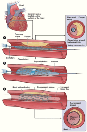
The researchers evaluated the results of 23 clinical trials in which 5,019 patients (average age 61 years; 73% men) were randomly assigned to get angioplasty with or without stents (PCI), and 4,944 were assigned to get coronary artery bypass graft surgery (CABG) In angioplasty, interventional cardiologists push a balloon-like device into the coronary arteries and inflate the balloon to widen the vessel. An expandable wire mesh tube called a stent may be inserted to keep the vessel open. Some stents are coated with drugs meant to help prevent the artery from clogging up. In 2005, about 645,000 angioplasty procedures were done in the U.S. In bypass surgery, cardiac surgeons harvest a segment of a healthy blood vessel from another part of the body and use it to bypass the clogged artery or arteries, rerouting the blood to improve blood flow to the heart. About 261,000 bypass procedures were done in the U.S. in 2005.

Findings

Besides similar survival rates overall, the researchers found no significant survival differences between the two procedures for patients with diabetes, although earlier research had seemed to favor bypass surgery. Similar numbers of patients suffered heart attacks within five years of the procedures. While 11.9 of those who got angioplasty had a heart attack within five years, 10.9% of those who got bypass did. Repeat procedures were more common in angioplasty patients. While 46.1% of angioplasty patients who didn’t get a stent needed repeat procedures, 40.1% of those who got a stent did. But just 9.8% of surgery patients needed another procedure.  The study didn’t include information on drug-coated stents.

Second Opinions

The new analysis is “very complete,” says Kim A. Eagle, MD, director of the Cardiovascular Center and Albion Walter Hewlett Professor of Internal Medicine at the University of Michigan, Ann Arbor. The study shows, he says, that if either procedure is considered appropriate for an individual patient, the decision can rest on patient attitudes and preferences. Patients preferences might be based on lower need to repeat in favor of surgery, or on avoidance of surgery in favor of angioplasty. But it is important to note, acoording to Curtis Hunter at Santa-Monica-UCLA, that the studies cover the least sick with heart disease, so the two procedures are shown to be equal in a very small subset of the patients.

Coronary Artery Bypass Surgery versus Coronary Stenting – Risk-Adjusted Survival Rates in 5,619 Patients  THIJ. 2002

We used the Texas Heart Institute Cardiovascular Research Database to retrospectively identify patients who had undergone their 1st revascularization procedure with coronary artery bypass surgery (CABG; n=2,826) or coronary stenting (n=2,793) between January 1995 and December 1999. Patients were classified into 8 anatomic groups according to the number of diseased vessels and presence or absence of proximal left anterior descending coronary artery disease. Mortality rates were adjusted with proportional hazards methods to correct for baseline differences in severity of disease and comorbidity.
We found that in-hospital mortality was significantly greater in patients undergoing CABG than in those undergoing stenting (3.6% vs 0.75%; adjusted OR 8.4; P <0.0001). At a mean 2.5-year follow-up, risk-adjusted survival was equivalent (CABG 91%, stenting 95%; adjusted OR 1.26; P = 0.06). When subgroups matched for severity of disease were compared, no differences in risk-adjusted survival were seen. A survival advantage of stenting was noted in 3 categories of patients: those >65 years of age (OR 1.33, P = 0.049), those with non-insulin-requiring diabetes (OR 2.06, P = 0.002), and those with any noncoronary vascular disease (OR 1.59, P = 0.009).
In this nonrandomized observational study, CABG had a higher periprocedural mortality rate than did percutaneous stenting. At 2.5 years, however, the survival advantage of stenting was no longer evident. These data suggest that there is no intermediate-term survival advantage of CABG over stenting in patients who have multivessel disease with lesions that can be treated percutaneously. (Tex Heart Inst J 2002;29:3–9)

Fig. 1 Adjusted and unadjusted survival rates in all patients treated with CABG or PCI-stenting
http://www.ncbi.nlm.nih.gov/pmc/articles/PMC101260/table/t3-2/?report=previmg

survival rates  of CABG or PCI-stenting

TABLE III. Multivariate Correlates of Intermediate-Term (2.5-Year) Mortality
http://www.ncbi.nlm.nih.gov/pmc/articles/PMC101260/table/t3-2/?report=previmg

Fig. 2 Adjusted odds ratios comparing the results of CABG and PCI-stenting in the 8 anatomic subgroups.
http://www.ncbi.nlm.nih.gov/pmc/articles/PMC101260/bin/2FF2.jpg

Adjusted odds ratios comparing the results of CABG and PCI-stenting in the 8 anatomic subgroups

TABLE IV. Intermediate-Term (2.5-Year) Survival According to Treatment in Each of the 8 Anatomic Groups
http://www.ncbi.nlm.nih.gov/pmc/articles/PMC101260/bin/2TT4.jpg

Intermediate-Term (2.5-Year) Survival According to Treatment in Each of the 8 Anatomic Groups

Fig. 3 Adjusted odds ratios comparing the results of CABG and PCI-stenting in the various prespecified subsets.
http://www.ncbi.nlm.nih.gov/pmc/articles/PMC101260/bin/2FF3.gif

Adjusted odds ratios comparing the results of CABG and PCI-stenting in the various prespecified subsets.

Will Drug-Eluting Stents Replace Coronary Artery Bypass Surgery?

Abstract

Introduction
The growth of the PCI industry and the consequent decline in the number of patients referred for CABG has produced much speculation about the future role of each type of intervention. Because the new drug-eluting stents allow PCI to be performed with lower rates of early restenosis than do bare-metal stents or percutaneous transluminal coronary angioplasty (PTCA) alone, 2–8 some have predicted that surgical revascularization will soon be obsolete.

CABG vs Pharmaco-Therapy

Randomized clinical trials performed during the 1970s and early 1980s clearly established the advantages of CABG over medical therapy in patients with triple-vessel CAD, left main coronary artery stenosis, double-vessel CAD with proximal left anterior descending (LAD) coronary artery stenosis, or left ventricular dysfunction. Problems arose subsequently because of the limitations built into the trial so that the results were biased in favor of medical therapy.  These were:
  • stringent exclusion criteria that eliminated a large percentage of potential participants
  • left main CAD and an ejection fraction of less than 0.40, eliminated patients for whom CABG would have been beneficial
  • the high rate of crossover from the medical to the surgical groups

The numerous technical and technological advances made since these trials were completed limit the degree to which their results resemble those of the CAD treatments used today. The maximal medical therapy used during the trials did not routinely include lipid-lowering agents, β-blockers, angiotensin-converting enzyme (ACE) inhibitors, clopidogrel, or some of the other drugs currently used for CAD. Nor did the CABG groups benefit from advances that were subsequently made in preoperative imaging, perfusion and myocardial protection, anesthesia, and perioperative and intensive care practices. CABG did not then include the use of left internal mammary artery (LIMA) grafts, much less other arterial conduits. Finally, PCIs, including balloon angioplasty and stenting, were not included in these trials.

CABG vs PTCA

Randomized trials comparing PTCA with CABG revealed dramatically higher re-intervention rates in the PTCA groups and better angina relief in the CABG groups, although there were no significant differences in death or myocardial infarction rates. The Duke database study. 9 showed better survival rates with PTCA than with CABG in patients with single-vessel CAD, whereas CABG produced better survival than did PTCA in patients with severe, triple-vessel CAD.
These results are not necessarily representative of the results obtainable today with PTCA and CABG, for several reasons.
1.  stents were not used in the PTCA patients in these trials
2.  operative mortality rates for the CABG groups were higher than the rates currently found in the Society of Thoracic Surgeons (STS) database
3.  the inclusion/exclusion criteria of these studies eliminated a high percentage of those patients who might have benefited more from CABG than from PTCA

CABG vs Stents

The introduction of coronary artery stenting resulted in better outcomes than those produced by balloon angioplasty or by other adjuncts, including rotational atherectomy, brachytherapy, and laser angioplasty.  Since then, stent designs and delivery techniques have advanced considerably. The use of coronary stents has greatly decreased the necessity of emergent CABG for technical failure of PCI and for dissection or rupture of coronary arteries during PCI. Another major advance in the application of PCI is the use of the antiplatelet agent clopidogrel in addition to aspirin after PCI, as well as the use of glycoprotein (GP) IIb/IIIa receptor inhibitors during the procedure. These adjuncts have significantly reduced the incidence of acute and subacute thrombosis after PTCA with stenting.
Randomized trials comparing PTCA plus stenting with PTCA alone have shown that stenting significantly reduces rates of restenosis and re-intervention, as well as the frequency of emergent CABG.  On the other hand, randomized trials of stenting versus surgery have produced less conclusive results regarding the mid-term survival and freedom from adverse events.  For example, the Stent or Surgery (SOS) trial reported a greater need for repeat revascularization in the stent group (21%) than in the CABG group (6%) and a survival advantage in the CABG group (hazard ratio, 2.91; 95% CI, 1.29–6.53; P = 0.01) during the 3-year follow-up period. Additionally, angina and the use of anti-angina medications were less common in the CABG group at 1-year follow-up.
The ARTS and ERACI trials also reported an increased need for revascularization in the stent groups but did not show a survival advantage in the CABG groups. This was due in part to a higher operative mortality rate in the CABG group than reported in the STS database. Like the PCI versus CABG trials mentioned previously, these randomized trials involved a select group of patients with relatively low expected mortality rates and relatively high expected technical success with PCI.
Observational data in retrospective analyses of large patient databases comparing CABG with PCI plus stenting does indicate that, because of the greater invasiveness of surgical revascularization, CABG produces greater operative mortality than does PCI. However, in patients with multivessel CAD, the risk-adjusted survival rates at 2.5 years of follow-up are no better for PCI than for CABG, and 3 recent risk-adjusted observational studies showed that the CABG patients had a significant survival advantage at 3- to 8-year follow-up.   The CABG patients had significantly more preoperative risk factors than did the PCI patients in each study, so that unadjusted, the CABG groups in each study included significantly more patients with triple-vessel disease and fewer patients with double-vessel disease than did the PCI groups. Again, we have a moving target with recent advances in both surgery and PCI technology.

Disadvantages of Stenting

The Achilles’ heel of PCI is restenosis and the need for repeat revascularization. Stents have decreased the rate of acute and subacute  periprocedural thrombosis. The newer, drug-eluting stents (DESs) have improved in-stent restenosis rates, especially in the carefully selected patient populations studied in the early DES trials. In the RAVEL trial, the early reports of zero in-stent restenosis compared favorably with the 27% in-stent restenosis rates in the bare-metal stent control group at 6-month follow-up. However, the RAVEL trial excluded patients with lesions longer than 18 mm, ostial targets, calcified or thrombosed targets, or target arteries less than 2.5 mm in diameter.
The media frenzy that followed the release of these findings created a public demand for these new “miracle” stents that apparently did not re-occlude. Stories of CAD patients refusing conventional PCI and CABG —instead, adding their names to the list of patients waiting for U.S. Food and Drug Administration (FDA) approval of DESs—appeared to change the practice patterns of cardiologists and cardiac surgeons overnight.  And then there were the calls for class-action lawsuits and recall of various DES models. After the FDA approved the Cordis Cypher™ DES (Cordis Corporation, a Johnson & Johnson company; Miami Lakes, Fla), a few reports of subacute thrombosis and hypersensitivity reactions prompted the FDA to release a public health notification on 29 October 2003.
The SIRIUS trial had slightly less strict exclusion criteria than did the RAVEL trial, admitting patients with target lesions 2.5 to 3.5 mm in diameter and 15 to 30 mm long, as well as patients with diabetes mellitus (who constituted 26% of the total group).  The SIRIUS trial also differed from the RAVEL trial in that the reported end-point was in-segment restenosis, rather than in-stent restenosis. The results showed a significant advantage of DESs over bare-metal stents for preventing in-segment restenosis (9.2% vs 32.3%) and target failures (10.5% vs 19.5%), but major adverse cardiac events were more frequent in the DES group than in the bare-metal stent group (3.7% vs 1.0%). Interestingly, the 6-month restenosis rates of the bare-metal stents in the RAVEL and SIRIUS control groups were much higher than the 19% 12-month restenosis rate associated with bare-metal stents in an earlier study comparing bare-metal stents with PTCA. In fact, the restenosis rates in the RAVEL and SIRIUS control groups more closely resembled the 40% restenosis rate reported for the PTCA control group in the earlier study.
The practical advantages of DESs over bare-metal stents are evident; nonetheless, we still do not have sufficient mid-term or long-term clinical data to argue that PTCA with DESs is preferable to CABG in “real-world” patients who require revascularization. Although DESs will likely provide better outcomes than bare-metal stents for many patients for whom stenting is indicated, a general extrapolation of existing data to justify the use of DESs in patients for whom CABG is currently indicated is unknown, perhaps undeterminable because the lesion and patient characteristics that lead to the failure of PCI are multifactorial, and the size of the population with lesions having unfavorable characteristics , such as,
  • longer
  • total occlusion
  • branch
  • small-diameter
  • calcified
  • multiple
  • left main
  • ostial, and
  • diffuse lesions
are being treated with PCI more often, as well as diabetics, multiple lesions, and patients with multiple comorbidities.

Advantages of CABG

Over the last 4 decades, surgical coronary artery revascularization techniques and technology have advanced significantly. As a result, despite an increasingly older and sicker patient population, CABG outcomes continue to improve. Observed operative mortality rates have decreased because advances in preoperative evaluation, including more precise coronary artery and myocardial imaging and diagnostic techniques, have allowed more appropriate patient selection and surgical planning. In addition, preoperative, intraoperative, and postoperative monitoring and therapeutic interventions have made CABG safer, even for critically ill and high-risk patients. Improvements in cardiopulmonary perfusion and careful myocardial protection, as well as the use of off-pump and on-pump beating- heart techniques in selected patients, have also decreased perioperative morbidity and mortality rates.

LIMA-to-LAD Long-Term Patency

The long-term benefits of CABG with regard to survival and quality of life are dependent on prolonged graft patency. The LIMA-to-LAD bypass, which is now performed in more than 90% of CABG procedures, shows excellent patency in 10- to 20-year angiographic follow-up studies, setting the gold standard with which other revascularization strategies should be compared. Tatoulis et al. reported that LIMA-to-LAD grafts had a 97.1% patency rate in patients who underwent angiography for cardiac symptoms. Those authors also found high patency rates at 5-year (98%), 10-year (95%), and 15-year (88%) follow-up. However, there are not yet long-term data on bare-metal stents or DESs, and by the time 10- or 20-year data are available, DESs probably will have been replaced by a newer, more advanced technology.
Because of the reported success of the LIMA-to-LAD bypass, other types of arterial conduits are also being used much more frequently. Conduit selection has become an area of great interest to cardiac surgeons, and conduit studies are expanding our understanding of the mechanisms of graft failure and ways to improve bypass graft patency. For example, studies have shown that patients who undergo CABG with both LIMA and right internal mammary artery (RIMA) conduits have better results than those who undergo CABG with one IMA and one or more saphenous vein grafts.

Techniques to Improve Conduit Patency

To maximize the odds of long-term graft patency, surgeons carefully harvest the graft as a pedicled or skeletonized conduit using “no touch” techniques. Using careful anastomotic technique to avoid excessive turbulence at the anastomosis site will prolong graft patency, and the quality of the conduit is crucial. Long-term graft patency depends not only on the conduit chosen but also on the target artery and the degree of stenosis proximal to the anastomosis. Maintaining flow patterns in the native artery, including residual flow (that is, competitive flow) and outflow, is important to avoid stasis in the graft, turbulence at the anastomosis, and vasospasm, especially in arterial conduits. Studies have shown an inverse relationship between the degree of proximal stenosis and graft patency. Targeting the LAD produces the highest patency rates. The characteristics of the target artery also determine graft patency, including –
1. the diameter of the target artery,
2. the presence or absence of diffuse disease within the artery,
3. whether or not the artery requires endarterectomy
Surgeons can avoid atheroembolic events by handling the aorta carefully or not at all. They can also improve safety by
1. using aggressive myocardial protection techniques;
2. avoiding the induction of inflammatory mediators; and
3. carefully controlling
  • blood pressure,
  • body temperature, and
  • electrolyte and glucose levels.
Although there have been major innovations that have enabled surgeons to perform cardiac surgery (including CABG) less invasively, minimally invasive surgical procedures are useful only if they are at least as efficacious as conventional surgery. New technology is being developed to enhance the evolving field of minimally invasive coronary bypass surgery.

Hybrid Coronary Revascularization

As PCI technology improves and techniques of LIMA-to-LAD grafting become less invasive, hybrid coronary revascularization is becoming a distinct possibility. For example, a minimally invasive, off-pump, direct LIMA-to-LAD anastomosis can be combined with DES placement in a focal mid-right-coronary-artery lesion in a patient with complex proximal LAD lesions. Hybrid coronary revascularization procedures are currently being performed, with promising early results. A few centers now have hybrid operating rooms with cardiac surgical and coronary angiographic capabilities that make it possible to perform simultaneous hybrid coronary revascularizations.

Although coronary artery bypass grafting (CABG) remains the treatment of choice for certain types of coronary artery disease (CAD), percutaneous coronary intervention (PCI)—particularly coronary angioplasty with stenting—has become the most popular nonmedical treatment approach to CAD. Some have speculated that, with the advent of drug-eluting stents (DESs), PCI will replace CABG entirely. However, the complete disappearance of CABG is both unlikely and unwarranted, for several reasons. Published randomized trials of CABG, PCI, and medical approaches to CAD compared only highly selected subgroups of patients because of strict exclusion criteria that often favored the PCI cohorts. Therefore, their results do not constitute sufficient evidence for the superiority of PCI over CABG in all CAD patients requiring revascularization. As PCI indications broaden to include more complex lesions and more high-risk patients, outcomes will not remain as favorable. In addition, although PCI is less invasive than surgery, CABG offers more complete revascularization and better freedom from repeat revascularization. Furthermore, no long-term patency data on DESs yet exist, whereas excellent 10- and 20-year patency rates have been reported for the left internal mammary artery-to-left anterior descending artery graft used in most CABG procedures. While PCI has been changing, CABG has not been stagnant; recently, advances in many aspects of the CABG procedure have improved short- and long-term outcomes in CABG patients. Both CABG and PCI technologies will continue to advance, not necessarily exclusive of one another, but no data yet exist to suggest that DESs will render CABG obsolete any time soon. 

Will Stent Revascularization Replace Coronary Artery Bypass Grafting?

When we discuss revascularization outcomes, we are talking about 3 major endpoints: death, myocardial infarction, and symptom control. With respect to death, we know that revascularization benefits patients who have severe multivessel disease and left ventricular dysfunction or other physiologic indicators of high risk. 2-vessel disease with proximal left anterior descending coronary artery (LAD) stenosis has been accepted as an indication for revascularization, even though the supporting data come from a small subgroup in a single trial. There has been no success in proving that endovascular treatment has a positive impact on stable CAD, but it is relevant because we leave the native arteries relatively intact. Attempts to improve graft performance beyond the relatively spectacular performance of the pedicled internal mammary artery (IMA) graft to the LAD have been disappointing.

Fig. 1 Graph of graft patency shows deterioration rates over 10 years and the comparative superiority of using the internal mammary artery (IMA) instead of the saphenous vein (SVG).http://www.ncbi.nlm.nih.gov/pmc/articles/PMC3528239/bin/25FF1.gif

graft patency of IMA vs SVG

Percutaneous Transluminal Coronary Angioplasty

When angioplasty was introduced, the hope was for a method of revascularization that would rival coronary artery bypass grafting. However, the results were mixed. Angioplasty worked well in patients with no major risk factors, such as diabetes mellitus, but failed miserably in diabetic patients. In fact, the Bypass Angioplasty Revascularization Investigation (BARI)  taught us this: if revascularization is needed, regardless of physiologic markers of high risk, the use of percutaneous coronary intervention (PCI) is potentially harmful in comparison with an IMA bypass for the LAD.

Stents and Short-Term Outcomes

The use of stents drastically reduced the probability of emergent surgery after attempted; however, the probability of new lesion formation or restenosis after intervention did not decrease.

Fig. 2 Diagrams  show the calculated success (after percutaneous revascularization) of A) percutaneous transluminal coronary angioplasty (PTCA), and B) bare-metal and C) drug-eluting stenting in patients with 3-vessel coronary artery disease (CAD).
http://www.ncbi.nlm.nih.gov/pmc/articles/PMC3528239/bin/25FF2.gif

At the same time, surgeons got better. Myocardial preservation techniques improved, and the use of the pedicled IMA graft changed the game. As a result, successful revascularization, meaning long-term success, became the domain of the surgeon. We at the Texas Heart Institute/St. Luke’s Episcopal Hospital (THI/SLEH) examined our long-term outcomes after stenting or surgery, and we initially reported that stenting was just as beneficial as surgery. This was in accord with the results of several trials: whenever placing a stent was feasible, stent therapy and surgery had the same outcome.

http://www.ncbi.nlm.nih.gov/pmc/articles/PMC3528239/bin/25TT1.jpg

success after PTCA vs bare-metal and drug-eluting stents

Stents and Long-Term Outcomes

Later, when we looked at longer-term follow-up data and the effects of multiple procedures, this picture began to change. Stented patients underwent more procedures. When the risk of one surgical procedure was compared with that of multiple endovascular procedures, the outcomes became more similar, especially in patients with bifurcation lesions or lesions with severe calcification. Drug-eluting stents, with their promise of no restenosis, substantially increased interventional cardiologists’ reach, but not their grasp. In patients with multivessel disease and high-risk lesions, DES placement was almost as risky as surgery and did not yield the same long-term benefit.

Nevertheless, we found locally that the introduction of the DES, with its lower risk of restenosis, was treated as a blessing to proceed with stenting (Table I). This did not follow the data, but cardiologists continued anyway, given the promise of less restenosis. Early risk was discounted, glycoprotein IIb/IIIa inhibitor use declined overnight, and the rate of endovascular procedural complications rose to meet that of surgery without the promise of an IMA graft in our future.

Table I. Independent Predictors of 30-Day Major Adverse Cardiac Events and 3-Year Survival after Drug-Eluting Stent Placement
http://www.ncbi.nlm.nih.gov/pmc/articles/PMC3528239/bin/25TT1.jpg

Comparing Stenting and Surgery

For decades, methods have been sought to quantify lesion complexity in order to compare the early and late risks associated with stenting versus surgery. Although no perfect system has been devised, the SYNTAX was an important step forward. The SYNTAX score is a simple, computer-based tool for evaluating the risk of complications or failure after PCI. And there are other tools for estimating the same complications after surgery. These estimates enable cardiologists to give patients objective advice regarding the revascularization method that has the best short- and long-term probability of success.
In the patient with non-life-threatening disease (that is, not left main or severe multivessel CAD with left ventricular dysfunction or severely impaired function), stent revascularization has become a reasonable, although not ideal, alternative to surgical revascularization. However, this is true only if stenting is confined to patients whose anatomy and physiology are suited to it—considerations that are well quantified in the SYNTAX score. Whenever questions arise as to the most appropriate therapy, the SYNTAX score should be weighed against clinical characteristics that affect surgical risk. This will guide discussions between the cardiologist, cardiovascular surgeon, patient, and treating physician.
I think that our THI risk is more useful than the other available scores. It uses simple clinical data and can be easily calibrated to the geographic location of its use. Other scores require data that might not be available at the time of clinical decision-making or at all—making such predictions hazardous, at best.

Conclusion

With regard to the chosen mode of revascularization, it is perhaps safe to say that the decision goes beyond the individual physician and must become collective. When a patient has multivessel disease, a reasoned approach must be taken, using these predictive tools and considering the patient’s wishes. Treatment decisions should include all interested parties: the patient, cardiologist, cardiovascular surgeon, and anesthesiologist. The time of ad hoc angioplasty for the patient with multivessel CAD has passed.

Should all ischemic mitral regurgitation be repaired? When should we replace?   Curr Opin Cardiol. 2011

Abstract

Purpose of review

Ischemic mitral regurgitation (IMR) is a major source of morbidity and mortality. Although mitral valve repair has become recently popularized for the treatment of IMR, select patients may derive benefits from replacement. The purpose of this review is to describe current surgical options for IMR and to discuss when mitral valve replacement (MVR) may be favored over mitral valve repair.

Recent findings

Current surgical options for the treatment of IMR include surgical revascularization alone, mitral valve repair, or MVR. Although surgical revascularization alone may benefit patients with mild–moderate IMR, most surgeons advocate the performance of revascularization in combination with either mitral valve repair or replacement. In the current era, mitral valve repair has proven to offer improved short-term and long-term survival, decreased valve-related morbidity, and improved left ventricular function compared with MVR. However, MVR should be considered for high-risk patients and those with specific underlying mechanisms of IMR.

Summary

In the absence of level one evidence, mitral valve repair offers an effective and durable surgical approach to the treatment of mitral insufficiency and remains the operation of choice for IMR. MVR, however, is preferred for select patients. Future randomized, prospective clinical trials are needed to directly compare these surgical techniques.

Introduction

Ischemic mitral regurgitation (IMR) describes insufficiency of the mitral valve in the setting of myocardial ischemia, resulting from coronary artery disease. Although IMR may present in the acute setting, usually as a papillary rupture (Carpentier type II), it is usually a consequence of chronic myocardial ischemia that typically presents weeks following a complete infarction. IMR describes mitral insufficiency in the absence of degenerative (structural) mitral valve disease. The underlying pathophysiologic mechanisms of IMR are often complex, resulting from several different structural changes involving left ventricular geometry, the mitral annulus, and the valvular/subvalvular apparatus. Although changes to any one component may result in detectable mitral valve insufficiency, moderate-to-severe IMR requiring surgical correction often involves the complex interplay of several co-existent anatomic changes. These underlying mechanisms result in clinically significant valve incompetence due to the combined effects of decreased ventricular function and restricted motion of the valve itself due to tethering.
IMR is a major source of patient morbidity and mortality. Although the frequency of IMR differs based upon imaging modality, estimates have suggested that nearly 20–30% of patients experience mitral insufficiency following myocardial infarction. Furthermore, its intimate association with heart failure and poor outcomes for suboptimal medical management further complicates the management of clinically significant IMR. Recent evidence suggests that moderate or severe mitral regurgitation may be associated with a three-fold increase in the adjusted risk of heart failure and a 1.6-fold increase in risk-adjusted mortality at 5-year follow-up. In addition, unfavorable patient profiles and co-existing comorbid disease, including renal failure, chronic obstructive pulmonary disease, diabetes, and impaired left ventricular function, further complicate the clinical picture for those with IMR. Consequently, surgical correction of this condition is often required.
The purpose of this review is to analyze published results for the surgical correction of IMR and to provide current opinion regarding the selection of mitral valve procedure in the setting of myocardial ischemia. Herein, we review current surgical options for IMR and discuss when MVR may be favored over mitral valve repair.

Surgical options for ischemic mitral regurgitation: surgical revascularization alone

Surgical revascularization alone with CABG may be beneficial for some patients. Although CABG alone may be performed in cases of mild-to-moderate IMR, for the treatment of severe IMR, evidence supports performance of CABG with a mitral valve. In fact, a lack of evidence exists to support the performance of CABG alone for severe IMR. In one retrospective review of propensity-matched cohorts, Diodato et al. suggested that addition of a mitral valve procedure to patients undergoing CABG for moderately severe to severe IMR did not increase mortality or improve survival over the performance of CABG alone. This study, however, was limited by small sample sizes (51 CABG + mitral valve repair vs. 51 CABG alone) and 3-year follow-up. To the contrary, substantial evidence exists to support the performance of surgical revascularization alone in cases of mild-to-moderate IMR.
A study by Aklog et al. investigated the role of CABG alone in the correction of moderate IMR. In their series of 136 patients with moderate IMR, they demonstrated that performance of revascularization alone conferred improvement of mitral regurgitation in 51% of patients with complete resolution in an additional 9%. Despite these results, 40% of patients remained with 3–4+ mitral regurgitation, leading the authors to conclude that CABG alone may not be the optimal therapy for most patients and suggest that concomitant mitral annuloplasty may improve results. Other series similarly suggest that complete resolution of functional IMR is uncommon following revascularization alone. Despite the presence of residual mitral regurgitation following revascularization, the impact of performance of CABG without a valve procedure on long-term survival remains ill defined. Currently, on-going prospective evaluation may help to define the potential role of revascularization alone for patients with moderate IMR. Until the completion of these trials, however, evidence supports the performance of surgical revascularization combined with a mitral valve procedure for moderate-to-severe mitral regurgitation.

Surgical revascularization with a mitral valve procedure

The majority of patients with moderate-to-severe IMR require surgical revascularization with a concomitant mitral valve procedure (MVR or mitral valve repair). Historically, these procedures have been associated with high morbidity and mortality as well as poor long-term. However, improved surgical techniques and postoperative management have improved contemporary outcomes. Those favoring mitral valve repair promote its beneficial effects on survival, preserved ventricular function, and the avoidance of long-term anticoagulation, whereas those favoring MVR argue that it ensures long-term freedom from recurrent mitral insufficiency.

Mitral valve replacement vs. mitral valve repair

The use of MVR for IMR eliminates the possibility of recurrent IMR. In addition, previous literature suggests improvements in surgical technique for MVR 29–32. For patients with IMR, MVR with preservation of the subvalvular apparatus using a chordal sparing technique has been shown to be beneficial 33. David and Ho 33 demonstrated a significant survival benefit for patients undergoing MVR with preservation of chordae tendineae (89%) compared with complete excision of the mitral valves (59%) in a cohort of 51 patients with IMR. In addition, Cohn et al. suggested disproportionate survival benefits favoring MVR in a cohort of 150 patients with both functional and structural IMR, concluding that survival following performance of mitral valve procedures for IMR was more dependent on underlying pathophysiology rather than surgical technique. More recently, series have suggested equivalent results for the MVR and mitral valve repair. Mantovani et al. report that prosthetic MVR and mitral valve repair offer very similar results for chronic IMR, demonstrating similar operative mortality and 5-year actuarial survival for both techniques. In a similar report, Magne et al.•• compared short-term and long-term outcomes for 370 patients undergoing mitral valve repair (n = 186) and MVR (n = 184) for IMR. Although operative mortality was lower for mitral valve repair compared with MVR (9.7 vs. 17.4%, P = 0.03), 6-year survival was similar for both operations (73 ± 4 vs. 67 ± 4%, P = 0.17). Type of procedure was also not an independent predictor of mortality following risk adjustment. As a result, the authors suggest that mitral valve repair is not superior to MVR for patients with IMR.
In contrast, other series favor the performance of mitral valve repair for functional IMR. Although several repair techniques exist, restrictive annuloplasty remains the most commonly performed operation 37• and has been shown to be beneficial in both functional and chronic IMR 38•. The purported benefits of improved survival, decreased valve-related morbidity, and improved left ventricular function have been previously established, and several series have reported lower hospital mortality with mitral valve repair compared with MVR.
The Cleveland Clinic published a landmark review of 482 patients undergoing mitral valve procedures for IMR to study the influence of mitral valve procedure type on survival 1. In this series, propensity-matched cohorts were compared: mitral valve repair (n = 397) vs. MVR (n = 85). Concomitant CABG was performed in 95% of operations, and annuloplasty for repair occurred in 98% of cases. After matching, patients were risk stratified into five quintiles. Group 1 represented the highest-risk patients with higher degrees of heart failure and emergent operations, and group 5 represented the lowest-risk patients. Subsequent survival analysis revealed that overall 5-year survival was poor for patients with IMR (58% mitral valve repair vs. 36% MVR, P = 0.08). Moreover, within matched quintiles, the highest-risk patients (quintile 1) had the worst survival, but survival was similar (P = 0.4) despite mitral valve procedure type. In contrast, survival favored mitral valve repair over replacement for quintiles III–V (P = 0.003).
In the absence of published randomized trials, two recently published meta-analyses provide more robust comparisons of the influence of surgical mitral valve repair or replacement. Shuhaiber and Anderson  compared outcomes of 29 studies, including over 10 000 patients. Study groups were stratified based upon mitral valve etiology into ischemic, degenerative/myxomatous, rheumatic, and mixed groups. Summary analyses indicated worse overall survival for MVR (early mortality odds ratio = 2.24 and total survival hazard ratio = 1.58) compared with repair. Mitral valve repair was also associated with lower rates of thromboembolism. Moreover, a nonsignificant trend toward lower 30-day mortality favored mitral valve repair for those with IMR. The most recent meta-analysis to date compared short-term and long-term survival of mitral valve repair vs. replacement specifically for IMR ••. In this analysis, nine studies were included based upon stringent exclusion criteria to ensure direct comparisons of survival for mitral valve procedures exclusively performed for IMR. Interestingly, in this series, although patients undergoing MVR were older, those undergoing repair often had higher rates of hypertension and diabetes with lower ejection fractions. Further, the proportion of patients with severe ventricular dysfunction was similar between procedure groups. These findings conflict with a common assumption that an inherent selection bias exists within published studies for the performance of mitral valve repair in healthier patients. Nevertheless, MVR was associated with worse short-term mortality (odds ratio = 2.667) and long-term mortality (hazard ratio = 1.35) compared with mitral valve repair, and the authors advocate that choice in mitral procedure should be based upon individual patient profile.

When not to repair ischemic mitral regurgitation?

Within the context of published literature and current dogma among practicing surgeons, the fundamental question of when not to repair an ischemic mitral valve remains. For several years, accumulated evidence supports the performance of mitral valve repair over replacement for the surgical treatment of functional IMR. The aforementioned benefits of repair include improved long-term survival, durability and efficacy, improved ventricular function, and avoidance of chronic anticoagulation therapy. Nevertheless, MVR still plays a select role in the treatment of IMR.
With respect to the performance of MVR, the use of bioprosthetic valves and the avoidance of mechanical valve replacement are preferred. This choice is largely driven by the avoidance of complications due to long-term anticoagulation use as well as by the belief that it is unlikely that the majority of patients requiring MVR are likely to encounter bioprosthetic deterioration in their lifetime. In addition, MVR with techniques to preserve the subvalvular apparatus should be performed when possible.

Summary

Undoubtedly, the debate regarding when to perform repair or replacement for IMR remains unsettled. In the recent era, mitral valve repair has proven efficacious and remains the preferred surgical strategy for most cases of IMR. MVR should be considered for severe tethering, complex or uncertain mechanisms of mitral insufficiency, regurgitation due to papillary muscle rupture, and perhaps for the sickest and highest-risk patients.
The present review was supported by Award Number 2T32HL007849-11A1 (D.J.L.) from the National Heart, Lung, and Blood Institute. The content is solely the responsibility of the authors.
Hybrid Cath Lab Combines Nonsurgical, Surgical Treatments  2008
A new cardiac treatment facility that couples the benefits of interventional cardiology with cardiothoracic surgery for critically ill newborns, children and adults has opened at Rush University Medical Center, Chicago.  Toshiba’s new biplane hybrid cardiac suite, which is one of only three facilities of its kind in the U.S., is equipped with the latest in continuous, real-time imaging technology and radio frequency identification (RFID) technology which allows “all-in-one-room” care. The suite allows collaboration between the surgeon and interventional cardiologist on complex heart problems. For example, fixing a very large hole in the heart can be done by inserting a catheter through a small incision in the chest rather than relying on major surgery to open the chest to reach the heart. “Now, interventional cardiologists and cardiothoracic surgeons working together in this suite will reduce the amount of time required to correct complex heart problems and reduce the emotional and physical stress placed on a patient and their family – which translates into less pain, less scarring and a faster recovery time,” Ziyad Hijazi, M.D., director of the new Rush Center for Congenital and Structural Heart Disease. The hybrid suite is equipped with the latest technology for minimally invasive interventional cardiology that involves the use of a catheter and an image-guidance system to thread tiny instruments through blood vessels to repair the heart. Through these special catheters, physicians at Rush can implant stents, artificial heart valves and insert patches for holes in the heart. In many complex cardiac cases, patients who would otherwise have no other option but to undergo open-heart bypass surgery can now have minimally invasive procedures that would otherwise not be available to them. “We can now communicate with colleagues and obtain their expertise in real time for very complex situations,” said Dr. Hijazi. “If physicians decide another procedure is needed, even surgery, the suite can be converted into an operating room and the surgical team can be assembled in the new suite ”Patients at Rush will stay in one place in the new hybrid cardiac suite where all the imaging technology and implantable devices that might be needed are stored and located. The additional ability it gives us to provide surgical treatments allows us to provide the most comprehensive care in the most sensitive manner for patients with often extremely fragile conditions.”  The new hybrid cardiac catheterization suite has the most advanced imaging technologies and can still get a precise, optimal image of any region of the heart regardless of the size or complexity of congenital heart disease. The imaging system also features eight-inch cardiac flat panel detectors designed to deliver distortion-free images. The suite also includes intravascular ultrasound machines, which takes real-time images to allow physicians to see the progress of the procedure taking place inside the patient’s body. A high-tech, automated clinical resource management system located in the suite stores and tracks the medication, surgical tools, medical devices, and implantable devices and supplies using the latest RFID enabled technology.

Hybrid Cath Lab/ORs Are the Way of the Future

Recent developments in cardiac surgery and interventional cardiology with new percutaneous alternatives for aneurysm repair, valve replacements, shunt closure devices and aortic arch reconstruction have led to the creation of integrated, hybrid cath lab/operating rooms (OR) that allow both surgical and intravascular procedures. These rooms offer both surgical equipment and high-end angiographic equipment. Creating such rooms requires special planning and design from both surgical and interventional cardiologists working closely together. Cath labs have high-quality fluoroscopy equipment, but generally are smaller rooms and lack the sterile requirements and equipment needed for surgical procedures. ORs tend to use lower quality mobile C-arms, which are not ideal for interventional procedures. The hybrids aim to provide the best of both worlds. The trend toward hybrid labs has been reinforced by digital angiography manufacturers partnering with surgical equipment companies to create easy-to-integrate hybrid room solutions with coordinated installation. Philips partners with both Skytron and Steris. Toshiba partners with MAQUET. GE Healthcare, Siemens and Toshiba also offer hybrid installations. Philips said while some hospitals want to combine interventional procedures with minimally invasive surgeries, they also want a properly equipped room in case emergency surgery is needed.
Philips said hybrids also allow hospitals with lower PCI numbers to get a bigger bang for their buck by allowing the same room to serve the needs of surgeons. Penn Presbyterian Medical Center in Philadelphia, PA, created a hybrid lab with help from Siemens, which opened in November. Wilson Szeto, M.D., cardio-thoracic surgeon, and William Matthai, M.D., interventionalist, both from Penn Presbyterian said hybrid labs are ideally suited for procedures that require both percutaneous and surgical interventions, percutaneous valve replacements, deploying percutaneous septal occluders or installing aortic stent grafts. Interventionalists can also be called in after cardiac surgery to perform a completion angiography.

Key References:

1. Davis KB, Alderman EL, Kosinski AS, Passamani E, Kennedy JW. Early mortality of acute myocardial infarction in patients with and without prior coronary revascularization surgery. A Coronary Artery Surgery Study Registry Study. Circulation 1992;85(6):2100–9. [PubMed]
2. Peduzzi P, Detre K, Murphy ML, Thomsen J, Hultgren H, Takaro T. Ten-year incidence of myocardial infarction and prognosis after infarction. Department of Veterans Affairs Cooperative Study of Coronary Artery Bypass Surgery. Circulation 1991;83(3):747–55. [PubMed]
3. Myocardial infarction and mortality in the coronary artery surgery study (CASS) randomized trial. N Engl J Med 1984; 310(12):750–8. [PubMed]
4. Long-term results of prospective randomised study of coronary artery bypass surgery in stable angina pectoris. European Coronary Surgery Study Group. Lancet 1982;2(8309):1173–80. [PubMed]
5. Frimerman A, Rechavia E, Eigler N, Payton MR, Makkar R, Litvack F. Long-term follow-up of a high risk cohort after stent implantation in saphenous vein grafts. J Am Coll Cardiol 1997;30(5):1277–83. [PubMed]
6. Comparison of coronary bypass surgery with angioplasty in patients with multivessel disease. The Bypass Angioplasty Revascularization Investigation (BARI) Investigators [published erratum appears in N Engl J Med 1997;336(2):147]. N Engl J Med 1996;335(4):217–25. [PubMed]
7. Coronary angioplasty versus coronary artery bypass surgery: the Randomized Intervention Treatment of Angina (RITA) trial. Lancet 1993;341:573–80. [PubMed]
8. Rodriguez A, Boullon F, Perez-Balino N, Paviotti C, Liprandi MI, Palacios IF. Argentine randomized trial of percutaneous transluminal coronary angioplasty versus coronary artery bypass surgery in multivessel disease (ERACI): in-hospital results and 1-year follow-up. ERACI Group. J Am Coll Cardiol 1993;22:1060–7. [PubMed]
9. Hamm CW, Reimers J, Ischinger T, Rupprecht HJ, Berger J, Bleifeld W. A randomized study of coronary angioplasty compared with bypass surgery in patients with symptomatic multivessel coronary disease. German Angioplasty Bypass Surgery Investigation (GABI). N Engl J Med 1994;331: 1037–43. [PubMed]
10. King SB 3rd, Lembo NJ, Weintraub WS, Kosinski AS, Barnhart HX, Kutner MH, et al. A randomized trial comparing coronary angioplasty with coronary bypass surgery. Emory Angioplasty versus Surgery Trial (EAST). N Engl J Med 1994;331:1044–50. [PubMed]
11 First-year results of CABRI (Coronary Angioplasty versus Bypass Revascularisation Investigation). CABRI Trial Participants. Lancet 1995;346:1179–84. [PubMed]
12. Carrie D, Elbaz M, Puel J, Fourcade J, Karouny E, Fournial G, Galinier M. Five-year outcome after coronary angioplasty versus bypass surgery in multivessel coronary artery disease: results from the French Monocentric Study. Circulation 1997; 96(9 Suppl):II-1–6. [PubMed]
13. Altmann DB, Racz M, Battleman DS, Bergman G, Spokojny A, Hannan EL, Sanborn TA. Reduction in angioplasty complications after the introduction of coronary stents: results from a consecutive series of 2242 patients. Am Heart J 1996;132:503–7. [PubMed]
14. Rankin JM, Spinelli JJ, Carere RG, Ricci DR, Penn IM, Hilton JD, et al. Improved clinical outcome after widespread use of coronary-artery stenting in Canada. N Engl J Med 1999;341:1957–65. [PubMed]
15. Jones RH, Kesler K, Phillips HR 3rd, Mark DB, Smith PK, Nelson CL, et al. Long-term survival benefits of coronary artery bypass grafting and percutaneous transluminal angioplasty in patients with coronary artery disease. J Thorac Cardiovasc Surg 1996;111:1013–25. [PubMed]
16. Hannan EL, Racz MJ, McCallister BD, Ryan TJ, Arani DT, Isom OW, Jones RH. A comparison of three-year survival after coronary artery bypass graft surgery and percutaneous transluminal coronary angioplasty. J Am Coll Cardiol 1999; 33:63–72. [PubMed]
17. Topol EJ, Mark DB, Lincoff AM, Cohne E, Burton J, Kleiman N, et al. Outcomes at 1 year and economic implications of platelet glycoprotein IIb/IIIa blockade in patients undergoing coronary stenting: results from a multicentre randomised trial. EPISTENT Investigators. Evaluation of Platelet IIb/IIIa Inhibitor for Stenting [published erratum appears in Lancet 2000;355:1104]. Lancet 1999;354:2019–24. [PubMed]
18. Serruys PW, de Jaegere P, Kiemeneij F, Macaya C, Rutsch W, Heyndrickx G, et al. A comparison of balloon-expandable-stent implantation with balloon angioplasty in patients with coronary artery disease. Benestent Study Group. N Engl J Med 1994;331:489–95. [PubMed]
19. Fischman DL, Leon MB, Baim DS, Schatz RA, Savage MP, Penn I, et al. A randomized comparison of coronary-stent placement and balloon angioplasty in the treatment of coronary artery disease. Stent Restenosis Study Investigators. N Engl J Med 1994;331:496–501. [PubMed]
20. Coronary-artery bypass surgery in stable angina pectoris: survival at two years. European Coronary Surgery Study Group. Lancet 1979;1:889–93. [PubMed]
21.  Coronary artery surgery study (CASS): a randomized trial of coronary artery bypass surgery: quality of life in patients randomly assigned to treatment groups. Circulation 1983; 68:951–60. [PubMed]
22. Takaro T, Hultgren HN, Lipton MJ, Detre KM. The VA cooperative randomized study of surgery for coronary arterial occlusive disease II. Subgroup with significant left main lesions. Circulation 1976;54:III107–17. [PubMed]
23. Hueb WA, Bellotti G, de Oliveira SA, Arie S, de Albuquerque CP, Jatene AD, et al. The Medicine, Angioplasty or Surgery Study (MASS): a prospective, randomized trial of medical therapy, balloon angioplasty or bypass surgery for single proximal left anterior descending artery stenoses. J Am Coll Cardiol 1995;26:1600–5. [PubMed]
24. Nordmann AJ, Hengstler P, Leimenstoll BM, Harr T, Young J, Bucher HC. Clinical outcomes of stents versus balloon angioplasty in non-acute coronary artery disease: a meta-analysis of randomized controlled trials. Eur Heart J 2004;25:69–80. [PubMed]
25. Versaci F, Gaspardone A, Tomai F, Crea F, Chiariello L, Gioffre PA. A comparison of coronary-artery stenting with angioplasty for isolated stenosis of the proximal left anterior descending coronary artery. N Engl J Med 1997;336:817–22. [PubMed]
26. Krumholz HM, Cohen DJ, Williams C, Baim DS, Brinker J, Cabin HS, et al. Health after coronary stenting or balloon angioplasty: results from the Stent Restenosis Study. Am Heart J 1997;134:337–44. [PubMed]
27. Villareal RP, Lee VV, Elayda MA, Wilson JM. Coronary artery bypass surgery versus coronary stenting: risk-adjusted survival rates in 5,619 patients. Tex Heart Inst J 2002;29:3–9. [PMC free article] [PubMed]
28. van Domburg RT, Takkenberg JJ, Noordzij LJ, Saia F, van Herwerden LA, Serruys PW, et al. Late outcome after stenting or coronary artery bypass surgery for the treatment of multivessel disease: a single-center matched-propensity controlled cohort study. Ann Thorac Surg 2005;79:1563–9. [PubMed]
29. Brener SJ, Lytle BW, Casserly IP, Schneider JP, Topol EJ, Lauer MS. Propensity analysis of long-term survival after surgical or percutaneous revascularization in patients with multivessel coronary artery disease and high-risk features. Circulation 2004;109:2290–5. [PubMed]
30. Al-Ruzzeh S, Ambler G, Asimakopoulos G, Omar RZ, Hasan R, Fabri B, et al. Off-pump coronary artery bypass (OPCAB) surgery reduces risk-stratified morbidity and mortality: a United Kingdom multi-center comparative analysis of early clinical outcome. Circulation 2003;108 Suppl 1:II1–8. [PubMed]
31. Puskas JD, Williams WH, Mahoney EM, Huber PR, Block PC, Duke PG, et al. Off-pump vs conventional coronary artery bypass grafting: early and 1-year graft patency, cost, and quality-of-life outcomes: a randomized trial. JAMA 2004;291:1841–9. [PubMed]
32. Goldman S, Zadina K, Moritz T, Ovitt T, Sethi G, Copeland JG, et al. Long-term patency of saphenous vein and left internal mammary artery grafts after coronary artery bypass surgery: results from a Department of Veterans Affairs Cooperative Study. J Am Coll Cardiol 2004;44:2149–56. [PubMed]
33. Shah PJ, Durairaj M, Gordon I, Fuller J, Rosalion A, Seevanayagam S, et al. Factors affecting patency of internal thoracic artery graft: clinical and angiographic study in 1434 symptomatic patients operated between 1982 and 2002. Eur J Cardiothorac Surg 2004;26:118–24. [PubMed]
34. Arima M, Kanoh T, Suzuki T, Kuremoto K, Tanimoto K, Oigawa T, et al. Serial angiographic follow-up beyond 10 years after coronary artery bypass grafting. Circ J 2005;69: 896–902. [PubMed]
35. Tatoulis J, Buxton BF, Fuller JA. Patencies of 2127 arterial to coronary conduits over 15 years. Ann Thorac Surg 2004; 77:93–101. [PubMed]
36. Beauford RB, Saunders CR, Lunceford TA, Niemeier LA, Shah S, Karanam R, et al. Multivessel off-pump revascularization in patients with significant left main coronary artery stenosis: early and midterm outcome analysis. J Card Surg 2005;20:112–8. [PubMed]
37. Banning AP, Westaby S, Morice MC, Kappetein AP, Mohr FW, Berti S, et al. Diabetic and nondiabetic patients with left main and/or 3-vessel coronary artery disease: comparison of outcomes with cardiac surgery and paclitaxel-eluting stents. J Am Coll Cardiol 2010;55(11):1067–75. [PubMed]
38. Laham RJ, Carrozza JP, Berger C, Cohen DJ, Kuntz RE, Baim DS. Long-term (4- to 6-year) outcome of Palmaz-Schatz stenting: paucity of late clinical stent-related problems. J Am Coll Cardiol 1996;28(4):820–6. [PubMed]
39. Rodriguez A, Bernardi V, Navia J, Baldi J, Grinfeld L, Martinez J, et al. Argentine Randomized Study: Coronary Angioplasty with Stenting versus Coronary Bypass Surgery in patients with Multiple-Vessel Disease (ERACI II): 30-day and one-year follow-up results. ERACI II Investigators [published erratum appears in J Am Coll Cardiol 2001;37(3):973–4]. J Am Coll Cardiol 2001;37(1):51–8. [PubMed]
40. Serruys PW, Unger F, Sousa JE, Jatene A, Bonnier HJ, Schonberger JP, et al. Comparison of coronary-artery bypass surgery and stenting for the treatment of multivessel disease. N Engl J Med 2001;344(15):1117–24. [PubMed]
41. Goy JJ, Kaufmann U, Goy-Eggenberger D, Garachemani A, Hurni M, Carrel T, et al. A prospective randomized trial comparing stenting to internal mammary artery grafting for proximal, isolated de novo left anterior coronary artery stenosis: the SIMA trial. Stenting vs Internal Mammary Artery. Mayo Clin Proc 2000;75(11):1116–23. [PubMed]
42. SoS Investigators. Coronary artery bypass surgery versus percutaneous coronary intervention with stent implantation in patients with multivessel coronary artery disease (the Stent or Surgery trial): a randomised controlled trial. Lancet 2002;360 (9338):965–70. [PubMed]
43. Reul RM. Will drug-eluting stents replace coronary artery bypass surgery? Tex Heart Inst J 2005;32(3):323–30. [PMC free article] [PubMed]
44. Sianos G, Morel MA, Kappetein AP, Morice MC, Colombo A, Dawkins K, et al. The SYNTAX Score: an angiographic tool grading the complexity of coronary artery disease. EuroIntervention 2005;1(2):219–27. [PubMed]
45. Madan P, Elayda MA, Lee VV, Wilson JM. Predicting major adverse cardiac events after percutaneous coronary intervention: the Texas Heart Institute risk score. Am Heart J 2008; 155(6):1068–74. [PubMed]
46. Gillinov AM, Wierup PN, Blackstone EH, et al. Is repair preferable to replacement for ischemic mitral regurgitation? J Thorac Cardiovasc Surg. 2001;122:1125–1141. [PubMed]
47. Grigioni F, Enriquez-Sarano M, Zehr KJ, et al. Ischemic mitral regurgitation: long-term outcome and prognostic implications with quantitative Doppler assessment. Circulation. 2001;103:1759–1764. [PubMed]
48. Lamas GA, Mitchell GF, Flaker GC, et al. Clinical significance of mitral regurgitation after acute myocardial infarction. Survival and Ventricular Enlargement Investigators. Circulation. 1997;96:827–833. [PubMed]
49. Bursi F, Enriquez-Sarano M, Nkomo VT, et al. Heart failure and death after myocardial infarction in the community: the emerging role of mitral regurgitation. Circulation. 2005;111:295–301. [PubMed]
50. Adams DH, Filsoufi F, Aklog L. Surgical treatment of the ischemic mitral valve. J Heart Valve Dis. 2002;11 (Suppl 1):S21–S25. [PubMed]
51. Filsoufi F, Salzberg SP, Adams DH. Current management of ischemic mitral regurgitation. Mt Sinai J Med. 2005;72:105–115. [PubMed]
52. Micovic S, Milacic P, Otasevic P, et al. Comparison of valve annuloplasty and replacement for ischemic mitral valve incompetence. Heart Surg Forum. 2008;11:E340–E345. [PubMed]
53. Aklog L, Filsoufi F, Flores KQ, et al. Does coronary artery bypass grafting alone correct moderate ischemic mitral regurgitation? Circulation. 2001;104 (12 Suppl 1):I68–I75. [PubMed]
54. Lam BK, Gillinov AM, Blackstone EH, et al. Importance of moderate ischemic mitral regurgitation. Ann Thorac Surg. 2005;79:462–470. discussion 462–470. [PubMed]
55. Ryden T, Bech-Hanssen O, Brandrup-Wognsen G, et al. The importance of grade 2 ischemic mitral regurgitation in coronary artery bypass grafting. Eur J Cardiothorac Surg. 2001;20:276–281. [PubMed]
56•. Goland S, Czer LS, Siegel RJ, et al. Coronary revascularization alone or with mitral valve repair: outcomes in patients with moderate ischemic mitral regurgitation. Tex Heart Inst J. 2009;36:416–424. This series documents current outcomes for the performance of CABG alone with/without concomitant mitral valve repair for ischemic mitral regurgitation. The authors report similar 5-year survival rates for both techniques; however, revascularization with repair resulted in significantly reduced mitral regurgitation grade, improved left ventricular function, and functional class compared with revascularization alone. This study provides an important comparison of these two techniques in the current surgical era. [PMC free article] [PubMed]
57••. Magne J, Girerd N, Senechal M, et al. Mitral repair versus replacement for ischemic mitral regurgitation: comparison of short-term and long-term survival. Circulation. 2009;120(11 Suppl):S104–S111. In this study, the authors compare postoperative outcomes for mitral valve repair and replacement for ischemic mitral regurgitation. Despite lower operative mortality following mitral valve repair, long-term survival was equivalent between surgical groups. This study adds important long-term comparisons of mitral valve procedures to accumulating data examining surgical treatments for ischemic mitral regurgitation. [PubMed]
58. Silberman S, Klutstein MW, Sabag T, et al. Repair of ischemic mitral regurgitation: comparison between flexible and rigid annuloplasty rings. Ann Thorac Surg. 2009;87:1721–1726. discussion 1726–1727. This study provides a contemporary comparison between the use of flexible and rigid annuloplasty rings for the surgical treatment of IMR. The authors report significantly improved clinical and hemodynamic results for rigid mitral annuloplasty rings compared with flexible rings. [PubMed]
59•. Tekumit H, Cenal AR, Uzun K, et al. Ring annuloplasty in chronic ischemic mitral regurgitation: encouraging early and midterm results. Tex Heart Inst J. 2009;36:287–292. This study reports early and midterm results for the use of flexible annuloplasty rings for the surgical treatment of chronic IMR. The authors demonstrate that use of flexible mitral valve annuloplasty conferred a reduction in left ventricular diameter with improved New York Heart Association functional class. This study reports current, encouraging results and provides a context for future investigations comparing flexible and rigid annuloplasty rings for chronic IMR. [PMC free article] [PubMed]
60. Shuhaiber J, Anderson RJ. Meta-analysis of clinical outcomes following surgical mitral valve repair or replacement. Eur J Cardiothorac Surg. 2007;31:267–275. [PubMed]
61••. Vassileva CM, Boley T, Markwell S, Hazelrigg S. Meta-analysis of short-term and long-term survival following repair versus replacement for ischemic mitral regurgitation. Eur J Cardiothorac Surg. 2010 [Epub ahead of print] This meta-analysis provides a comparison of nine published series specifically addressing the performance of mitral valve repair vs. replacement for IMR. The authors demonstrate worse short-term and long-term mortality for MVR. Their analysis offers an up-to-date and robust comparison of these two surgical techniques. [PubMed]

Other Related articles  published on this Open Access Online Scientific Journal, include the following:

Cardiac Surgery Theatre in China vs. in the US: Cardiac Repair Procedures, Medical Devices in Use, Technology in Hospitals, Surgeons’ Training and Cardiac Disease Severity”    http://pharmaceuticalintelligence.com/2013/01/08/cardiac-surgery-theatre-in-china-vs-in-the-us-cardiac-repair-procedures-medical-devices-in-use-technology-in-hospitals-surgeons-training-and-cardiac-disease-severity/

Heart Remodeling by Design – Implantable Synchronized Cardiac Assist Device: Abiomed’s Symphony                                                                                     http://pharmaceuticalintelligence.com/2012/07/23/heart-remodeling-by-design-implantable-synchronized-cardiac-assist-device-abiomeds-symphony/
Acute Chest Pain/ER Admission: Three Emerging Alternatives to Angiography and PCI    http://pharmaceuticalintelligence.com/2013/03/10/acute-chest-painer-admission-three-emerging-alternatives-to-angiography-and-pci/
Dilated Cardiomyopathy: Decisions on implantable cardioverter-defibrillators (ICDs) using left ventricular ejection fraction (LVEF) and Midwall Fibrosis: Decisions on Replacement using late gadolinium enhancement cardiovascular MR (LGE-CMR)
Clinical Trials on transcatheter aortic valve replacement (TAVR) to be conducted by American College of Cardiology and the Society of Thoracic Surgeons
FDA Pending 510(k) for The Latest Cardiovascular Imaging Technology
PCI Outcomes, Increased Ischemic Risk associated with Elevated Plasma Fibrinogen not Platelet Reactivity
The ACUITY-PCI score: Will it Replace Four Established Risk Scores — TIMI, GRACE, SYNTAX, and Clinical SYNTAX
Coronary artery disease in symptomatic patients referred for coronary angiography: Predicted by Serum Protein Profiles
Ablation Devices Market to 2016 – Global Market Forecast and Trends Analysis by Technology, Devices & Applications
Heart Renewal by pre-existing Cardiomyocytes: Source of New Heart Cell Growth Discovered
Cardiovascular Risk Inflammatory Marker: Risk Assessment for Coronary Heart Disease and Ischemic Stroke – Atherosclerosis.
To Stent or Not? A Critical Decision
Endothelin Receptors in Cardiovascular Diseases: The Role of eNOS Stimulation
Transcatheter Aortic-Valve Replacement for Inoperable Severe Aortic Stenosis
Imbalance of Autonomic Tone: The Promise of Intravascular Stimulation of Autonomics
New Definition of MI Unveiled, Fractional Flow Reserve (FFR)CT for Tagging Ischemia
Ethical Considerations in Studying Drug Safety — The Institute of Medicine Report
New Drug-Eluting Stent Works Well in STEMI
Expected New Trends in Cardiology and Cardiovascular Medical Devices
Minimally Invasive Structural CVD Repairs: FDA grants 510(k) Clearance to Philips’ EchoNavigator – X-ray and 3-D Ultrasound Image Fused.
Drug Eluting Stents: On MIT’s Edelman Lab’s Contributions to Vascular Biology and its Pioneering Research on DES
http://pharmaceuticalintelligence.com/2013/04/25/contributions-to-vascular-biology/

Related articles

Diagram of coronary angioplasty and stent plac...

Diagram of coronary angioplasty and stent placement (Photo credit: Wikipedia)

Denton A. Cooley, MD

Denton A. Cooley, MD (Photo credit: Wikipedia)

English: A thoracic surgeon performs a mitral ...

English: A thoracic surgeon performs a mitral valve replacement at the Fitzsimons Army Medical Center. Slovenščina: Kirurgi med operacijo. (Photo credit: Wikipedia)

Read Full Post »

Reporter: Aviva Lev-Ari, PhD, RN

Engineers work to help biologists cope with big data

Tue, 01/08/2013 – 10:15am

Liang Dong is developing an instrument that will allow plant scientists to simultaneously study thousands of plants grown in precisely controlled condition. Photo: Bob ElbertLiang Dong is developing an instrument that will allow plant scientists to simultaneously study thousands of plants grown in precisely controlled condition. Photo: Bob ElbertLiang Dong held up a clear plastic cube, an inch or so across, just big enough to hold 10 to 20 tiny seeds.

Using sophisticated sensors and software, researchers can precisely control the light, temperature, humidity, and carbon dioxide inside that cube.

Dong—an Iowa State University assistant professor of electrical and computer engineering and of chemical and biological engineering—calls it a “microsystem instrument.” Put hundreds of those cubes together and researchers can simultaneously grow thousands of seeds and seedlings in different conditions and see what happens. How, for example, do the plants react when it is hot and dry? Or carbon dioxide levels change? Or light intensity is adjusted very slightly?

The instrument designed and built by Dong’s research group will keep track of all that by using a robotic arm to run a camera over the cubes and take thousands of images of the growing seeds and seedlings.

Plant scientists will use the images to analyze the plants’ observable characteristics—the leaf color, the root development, the shoot size. All those observations are considered a plant’s phenotype. And while plant scientists understand plant genetics very well, Dong says they don’t have a lot of data about how genetics and environment combine to influence phenotype.

Dong’s instrument will provide researchers with lots of data—too much for scientists to easily sort and analyze. That’s a problem known as big data. And it’s increasingly common in the biological sciences.

“We’re seeing a proliferation of new instruments in the biological sciences,” says Srinivas Aluru, the Ross Martin Mehl and Marylyne Munas Mehl Professor of Computer Engineering at Iowa State. “And the rate of data collection is increasing. So we have to have a solution to analyze all this data.”

Aluru is leading a College of Engineering initiative to build research teams capable of solving big data problems in next-generation DNA sequencing, systems biology, and phenomics. The researchers are developing computing solutions that take advantage of emerging technologies such as cloud computing and high-performance computers. They’re also building partnerships with technology companies such as IBM, Micron, NVIDIA, Illumina Inc., Life Technologies Corp., Monsanto Co., and Roche.

The project is one of the three Dean’s Research Initiatives launched by Jonathan Wickert, former dean of the College of Engineering and currently Iowa State’s senior vice president and provost. The initiatives in high-throughput computational biology, wind energy, and a carbon-negative economy were launched in March 2011 with $500,000 each over three years. That money is to build interdisciplinary, public-private research teams ready to compete for multi-million dollar grants and projects.

Patrick Schnable, Iowa State’s Baker Professor of Agronomy and director of the centers for Plant Genomics and Carbon Capturing Crops, remembers when biologists had no interest in working with computer specialists. That was before they tried to work with billions of data points to, say, accurately predict harvests based on plant genotype, soil type and weather conditions.

“Now we’re getting huge, absolutely huge, data sets,” Schnable says. “There is no way to analyze these data sets without extraordinary computer resources. There’s no way we could do this without the collaboration of engineers.”

To date, the computational biology initiative has attracted $5.5 million for four major research projects. One of the latest grants is a three-year, $2 million award from the BIGDATA program of the National Science Foundation and the National Institutes of Health. The grant will allow Aluru and researchers from Iowa State, Stanford University, Virginia Tech, and the University of Michigan to work together to develop a computing toolbox that helps scientists manage all the data from today’s DNA sequencing instruments.

Aluru says the research initiative helped prepare Iowa State researchers to go after that grant.

“When the BIGDATA call came in, we had the credibility to compete,” he says. “We were already working on leading edge problems and had established relationships with companies.”

The initiative, the grants and the industry partnerships are helping Iowa State faculty and students move to the front of the developing field.

“One computing company wanted to set up a life science research group and it came here for advice,” Aluru says. “Iowa State is known as a big data leader in the biosciences.”

Source: Iowa State University

SOURCE:

http://www.rdmag.com/news/2013/01/engineers-work-help-biologists-cope-big-data?et_cid=3031227&et_rid=461755519&linkid=http%3a%2f%2fwww.rdmag.com%2fnews%2f2013%2f01%2fengineers-work-help-biologists-cope-big-data


Read Full Post »

Reported by: Dr. Venkat S Karra. Ph.D.

A multidisciplinary research team led by Carnegie Mellon University is developing new nanostructural polymer-based treatments to eliminate pathological bone formation in soft tissue, a common occurrence following orthopedic surgeries and amputations.

Heterotopic ossification

“Our tactic is to develop a solution that will control the pathological growth of bone in muscle and tendons (called heterotopic ossification) that frequently occurs following bone trauma and orthopedic surgery,” said Jeffrey O. Hollinger, professor of biomedical engineering and biological sciences, and head of CMU’s Bone Tissue Engineering Center.

“When bone is severely injured and amputation of a limb is necessary, or as a consequence of major orthopedic procedures, unwanted new bone formation occurs in the soft tissues surrounding the operated bone and appears as pieces of gravel-like bone. Consequently, there is pain and discomfort at an amputation stump where a prosthesis is secured. We are developing a therapy that will eliminate heterotopic ossification,” he added.

Data suggests heterotopic ossification occurs in more than 60 percent of military personnel who incur bone injury resulting in limb amputation. Therefore, the CMU labs of J.C. Warner University Professor of Natural Sciences and Chemistry Professor Krzysztof Matyjaszewski are using a three-year, $2.93 million grant from the Department of Defense to work with researchers at the United States Military Academy at West Point, the University of Michigan and the Naval Medical Center in Portsmouth, Va., to produce a therapeutic solution to eliminate heterotopic ossification.

Hollinger, the principal investigator for the grant, said the patient-centric focus of the team’s research includes a nanostructural polymer composite developed by Matyjaszewski to deliver unique RNA identified in the Hollinger lab, into cells at the bone trauma site to prevent heterotopic ossification in the soft tissue.

“The problem of heterotopic ossification is more widespread than the military population,” Hollinger said. More than 90 percent of hip replacement operations in the civilian U.S. population also show signs of heterotopic ossification. Because the problem is so complex, CMU researchers report that it will take a team of clinicians and researchers to develop solutions.

“We see this collaborative research as a win for both military and civilian populations. And we see this particular research project as a great way to help us change our research paradigm at West Point,” said J. Kenneth Wickiser, director of the Center for Molecular Science in the Department of Chemistry & Life Science at the United States Military Academy. “Our cadets are gaining invaluable hands-on research experience as summer interns at CMU’s biomedical engineering labs. And we are becoming more competitive in our abilities at West Point to tackle more innovative research initiatives,” Wickiser said.

Ashley Phillips, a sophomore West Point cadet, praised the CMU internship program for its concise and rigorous approach to problem solving. “I want to be a doctor and this CMU research experience gives me an excellent platform for growth and a medium for sharing my work with other cadets,” said Phillips of Mukwonago, Wis.

CMU researchers report there is a patent pending on the therapy and a clinical trial schedule will be developed once the preventative platform is fully lab tested.

Source:

rdmag

Carnegie Mellon University

Read Full Post »


Reporter: Aviva Lev-Ari, PhD, RN

A Matched Comparison of Perioperative Outcomes of a Single Laparoscopic Surgeon Versus a Multisurgeon Robot-Assisted Cohort for Partial Nephrectomy

The Journal of Urology
Volume 188, Issue 1 , Pages 45-50, July 2012


Department of Urology, University of Michigan, Ann Arbor, Michigan

Received 17 October 2011 published online 14 May 2012.

Purpose

Minimally invasive nephron sparing surgery is gaining popularity for small renal masses. Few groups have evaluated robot-assisted partial nephrectomy compared to other approaches using comparable patient populations. We present a matched pair analysis of a heterogeneous group of surgeons who performed robot-assisted partial nephrectomy and a single experienced laparoscopic surgeon who performed conventional laparoscopic partial nephrectomy. Perioperative outcomes and complications were compared.

Materials and Methods

All 249 conventional laparoscopic and robot-assisted partial nephrectomy cases from January 2007 to June 2010 were reviewed from our prospectively maintained institutional database. Groups were matched 1:1 (108 matched pairs) by R.E.N.A.L. (radius, exophytic/endophytic properties, nearness of tumor to collecting system or sinus, anterior/posterior, location relative to polar lines) nephrometry score, transperitoneal vs retroperitoneal approach, patient age and hilar nature of the tumor. Statistical analysis was done to compare operative outcomes and complications.

Results

Matched analysis revealed that nephrometry score, age, gender, tumor side and American Society of Anesthesia physical status classification were similar. Operative time favored conventional laparoscopic partial nephrectomy. During the study period robot-assisted partial nephrectomy showed significant improvements in estimated blood loss and warm ischemia time compared to those of the experienced conventional laparoscopic group. Postoperative complication rates, and complication distributions by Clavien classification and type were similar for conventional laparoscopic and robot-assisted partial nephrectomy (41.7% and 35.0%, respectively).

Conclusions

Robot-assisted partial nephrectomy has a noticeable but rapid learning curve. After it is overcome the robotic procedure results in perioperative outcomes similar to those achieved with conventional laparoscopic partial nephrectomy done by an experienced surgeon. Robot-assisted partial nephrectomy likely improves surgeon and patient accessibility to minimally invasive nephron sparing surgery.

Key Words:  kidney , kidney neoplasms , nephrectomy , laparoscopy , robotics

Abbreviations and Acronyms:  CLPNconventional laparoscopic partial nephrectomyEBLestimated blood losseGFR,estimated glomerular filtration rateICUintensive care unitLOSlength of stayRAPNrobot-assisted partial nephrectomy,SRMsmall renal massWITwarm ischemia time


Similar outcomes for robot-aided, conventional nephrectomy June 22, 2012 in Other Robot-assisted and conventional laparoscopic partial nephrectomies have similar outcomes and complication rates, according to a study published in the July issue of The Journal of Urology. (HealthDay) — Robot-assisted and conventional laparoscopic partial nephrectomies have similar outcomes and complication rates, according to a study published in the July issue of The Journal of Urology. Ads by Google Prostate Cancer Treatment – Expert Prostate Cancer Treatment & Care – View Video to Learn More! – http://www.TuftsMedicalCenter.tv Prostate Cancer Treatment – Learn about Watchful Waiting. Get a Second Opinion at BIDMC. – http://www.BIDMC.org Jonathan S. Ellison, M.D., from the University of Michigan in Ann Arbor, and colleagues compared perioperative outcomes and complications from conventional laparoscopic and robot-assisted partial nephrectomy cases from January 2007 to June 2010. Robot-assisted partial nephrectomies were performed by a heterogeneous group of surgeons, while a single experienced laparoscopic surgeon performed the conventional procedures. One hundred eight pairs of patients were matched by age, hilar nature of the tumor, approach, and R.E.N.A.L. (radius, exophytic/endophytic properties, nearness of tumor to collecting system or sinus, anterior/posterior, location relative to polar lines) nephrometry score. The researchers found that nephrometry score, age, gender, tumor side, and American Society of Anesthesia physical status classification were similar between the groups. Conventional laparoscopic partial nephrectomy had better operative time. Robot-assisted partial nephrectomy showed significant improvements in estimated blood loss and warm ischemia time compared to the conventional laparoscopic group. The postoperative complication rates and complication distributions by Clavien classification and type were similar for both groups (41.7 percent for the conventional group and 35.0 percent for the robot-assisted group). “Robot-assisted partial nephrectomy has a noticeable but rapid learning curve,” write the authors. “After it is overcome the robotic procedure results in perioperative outcomes similar to those achieved with conventional laparoscopic partial nephrectomy done by an experienced surgeon.” More information: Abstract Full Text (subscription or payment may be required) Journal reference: Journal of Urology

http://medicalxpress.com/news/2012-06-similar-outcomes-robot-aided-conventional-nephrectomy.html


Prostate Cancer

What does your PSA score, level, reading, test mean?

By itself, a PSA reading does not mean very much. There are many possible causes of the rise in the PSA reading. The most common of these reasons is an enlarged, inflamed, or infected prostate. So a high PSA score does not necessarily indicate prostate cancer.

Unfortunately there is no failsafe test or methods at this time that can differentiate between a high PSA level caused by inflammation of the prostate or infection of the prostate or prostate cancer. At best doctors use a statistical model, which seeks to predict your chances of having prostate cancer. But that is purely a statistical construct and does not actually predict your specific and personal situation at all.

Nonetheless, an elevated PSA reading should not be ignored. It is a good indicator, certainly the best we have, and you should take precautionary action.

If you have a high PSA reading you need to return your prostate back to good health. You need to make important changes to your diet. You also need to have regular exercise. A third and equally important part of my recommendation is to take appropriate natural supplements.

I provide a roadmap in my guide “All about the Prostate”. Most men who follow my roadmap will see their PSA levels come down. It will return their prostate to good health.

http://www.bensprostate.com/minib/psa-test-and-levels/?utm_source=bing&utm_medium=cpc&utm_campaign=bing-prostate-us-broad&utm_content=psa-2&utm_term=psa

INDICATION

ZYTIGA® (abiraterone acetate) in combination with prednisone is indicated for the treatment of patients with metastatic castration-resistant prostate cancer (mCRPC) who have received prior chemotherapy containing docetaxel.

IMPORTANT SAFETY INFORMATION

Contraindications – ZYTIGA® (abiraterone acetate) may cause fetal harm (Pregnancy Category X) and is contraindicated in women who are or may become pregnant.

Hypertension, Hypokalemia and Fluid Retention Due to Mineralocorticoid Excess –Use with caution in patients with a history of cardiovascular disease or with medical conditions that might be compromised by increases in hypertension, hypokalemia, and fluid retention. ZYTIGA® may cause hypertension, hypokalemia, and fluid retention as a consequence of increased mineralocorticoid levels resulting from CYP17 inhibition. Safety has not been established in patients with LVEF <50% or New York Heart Association (NYHA) Class III or IV heart failure because these patients were excluded from the randomized clinical trial. Control hypertension and correct hypokalemia before and during treatment. Monitor blood pressure, serum potassium, and symptoms of fluid retention at least monthly.

Adrenocortical Insufficiency (AI) – AI has been reported in clinical trials in patients receiving ZYTIGA® in combination with prednisone, after an interruption of daily steroids and/or with concurrent infection or stress. Use caution and monitor for symptoms and signs of AI if prednisone is stopped or withdrawn, if prednisone dose is reduced, or if the patient experiences unusual stress. Symptoms and signs of AI may be masked by adverse reactions associated with mineralocorticoid excess seen in patients treated with ZYTIGA®. Perform appropriate tests, if indicated, to confirm AI. Increased dosages of corticosteroids may be used before, during, and after stressful situations.

Hepatotoxicity – Increases in liver enzymes have led to drug interruption, dose modification, and/or discontinuation. Monitor liver function and modify, withhold, or discontinue ZYTIGA® dosing as recommended (see Prescribing Information for more information). Measure serum transaminases [alanine aminotransferase (ALT) and aspartate aminotransferase (AST)] and bilirubin levels prior to starting treatment with ZYTIGA®, every two weeks for the first three months of treatment, and monthly thereafter. Promptly measure serum total bilirubin, AST, and ALT if clinical symptoms or signs suggestive of hepatotoxicity develop. Elevations of AST, ALT, or bilirubin from the patient’s baseline should prompt more frequent monitoring. If at any time AST or ALT rise above five times the upper limit of normal (ULN) or the bilirubin rises above three times the ULN, interrupt ZYTIGA® treatment and closely monitor liver function.

Food Effect – ZYTIGA® must be taken on an empty stomach. Exposure of abiraterone increases up to 10-fold when abiraterone acetate is taken with meals. No food should be eaten for at least two hours before the dose of ZYTIGA® is taken and for at least one hour after the dose of ZYTIGA® is taken. Abiraterone Cmax and AUC0-∞ (exposure) were increased up to 17- and 10-fold higher, respectively, when a single dose of abiraterone acetate was administered with a meal compared to a fasted state.

Adverse Reactions – The most common adverse reactions (≥ 5%) are joint swelling or discomfort, hypokalemia, edema, muscle discomfort, hot flush, diarrhea, urinary tract infection, cough, hypertension, arrhythmia, urinary frequency, nocturia, dyspepsia, fractures and upper respiratory tract infection.

Drug Interactions – ZYTIGA® is an inhibitor of the hepatic drug-metabolizing enzyme CYP2D6. Avoid co-administration with CYP2D6 substrates that have a narrow therapeutic index. If an alternative cannot be used, exercise caution and consider a dose reduction of the CYP2D6 substrate. Additionally, abiraterone is a substrate of CYP3A4 in vitro. Strong inhibitors and inducers of CYP3A4 should be avoided or used with caution.

Use in Specific Populations – The safety of ZYTIGA® in patients with baseline severe hepatic impairment has not been studied. These patients should not receive ZYTIGA®.


Read Full Post »