Feeds:
Posts
Comments

Posts Tagged ‘PROTAC’

Use of Systems Biology for Design of inhibitor of Galectins as Cancer Therapeutic – Strategy and Software


Curator: Stephen J. Williams, Ph.D.

Below is a slide representation of the overall mission 4 to produce a PROTAC to inhibit Galectins 1, 3, and 9.


Using A Priori Knowledge of Galectin Receptor Interaction to Create a BioModel of Galectin 3 Binding


Now after collecting literature from PubMed on “galectin-3” AND “binding” to determine literature containing kinetic data we generate a WordCloud on the articles.


This following file contains the articles needed for BioModels generation.

https://pharmaceuticalintelligence.com/wp-content/uploads/2022/12/Curating-Galectin-articles-for-Biomodels.docx


From the WordCloud we can see that these corpus of articles describe galectin binding to the CRD (carbohydrate recognition domain).  Interestingly there are many articles which describe van Der Waals interactions as well as electrostatic interactions.  Certain carbohydrate modifictions like Lac NAc and Gal 1,4 may be important.  Many articles describe the bonding as well as surface  interactions.  Many studies have been performed with galectin inhibitors like TDGs (thio-digalactosides) like TAZ TDG (3-deoxy-3-(4-[m-fluorophenyl]-1H-1,2,3-triazol-1-yl)-thio-digalactoside).  This led to an interesting article

Dual thio-digalactoside-binding modes of human galectins as the structural basis for the design of potent and selective inhibitors

Affiliations 2016 Jul 15;6:29457.
 doi: 10.1038/srep29457. Free PMC article

Abstract

Human galectins are promising targets for cancer immunotherapeutic and fibrotic disease-related drugs. We report herein the binding interactions of three thio-digalactosides (TDGs) including TDG itself, TD139 (3,3′-deoxy-3,3′-bis-(4-[m-fluorophenyl]-1H-1,2,3-triazol-1-yl)-thio-digalactoside, recently approved for the treatment of idiopathic pulmonary fibrosis), and TAZTDG (3-deoxy-3-(4-[m-fluorophenyl]-1H-1,2,3-triazol-1-yl)-thio-digalactoside) with human galectins-1, -3 and -7 as assessed by X-ray crystallography, isothermal titration calorimetry and NMR spectroscopy. Five binding subsites (A-E) make up the carbohydrate-recognition domains of these galectins. We identified novel interactions between an arginine within subsite E of the galectins and an arene group in the ligands. In addition to the interactions contributed by the galactosyl sugar residues bound at subsites C and D, the fluorophenyl group of TAZTDG preferentially bound to subsite B in galectin-3, whereas the same group favored binding at subsite E in galectins-1 and -7. The characterised dual binding modes demonstrate how binding potency, reported as decreased Kd values of the TDG inhibitors from μM to nM, is improved and also offer insights to development of selective inhibitors for individual galectins.

Figures

Figure 1
 
Figure 2
 
Figure 3


Read Full Post »

The drug efflux pump MDR1 promotes intrinsic and acquired resistance to PROTACs in cancer cells

Reporter: Stephen J. Williams, PhD.
Below is one of the first reports  on the potential mechanisms of intrinsic and acquired resistance to PROTAC therapy in cancer cells.
Proteolysis-targeting chimeras (PROTACs) are a promising new class of drugs that selectively degrade cellular proteins of interest. PROTACs that target oncogene products are avidly being explored for cancer therapies, and several are currently in clinical trials. Drug resistance is a substantial challenge in clinical oncology, and resistance to PROTACs has been reported in several cancer cell models. Here, using proteomic analysis, we found intrinsic and acquired resistance mechanisms to PROTACs in cancer cell lines mediated by greater abundance or production of the drug efflux pump MDR1. PROTAC-resistant cells were resensitized to PROTACs by genetic ablation of ABCB1 (which encodes MDR1) or by coadministration of MDR1 inhibitors. In MDR1-overexpressing colorectal cancer cells, degraders targeting either the kinases MEK1/2 or the oncogenic mutant GTPase KRASG12C synergized with the dual epidermal growth factor receptor (EGFR/ErbB)/MDR1 inhibitor lapatinib. Moreover, compared with single-agent therapies, combining MEK1/2 degraders with lapatinib improved growth inhibition of MDR1-overexpressing KRAS-mutant colorectal cancer xenografts in mice. Together, our findings suggest that concurrent blockade of MDR1 will likely be required with PROTACs to achieve durable protein degradation and therapeutic response in cancer.

INTRODUCTION

Proteolysis-targeting chimeras (PROTACs) have emerged as a revolutionary new class of drugs that use cancer cells’ own protein destruction machinery to selectively degrade essential tumor drivers (1). PROTACs are small molecules with two functional ends, wherein one end binds to the protein of interest, whereas the other binds to an E3 ubiquitin ligase (23), bringing the ubiquitin ligase to the target protein, leading to its ubiquitination and subsequent degradation by the proteasome. PROTACs have enabled the development of drugs against previously “undruggable” targets and require neither catalytic activity nor high-affinity target binding to achieve target degradation (4). In addition, low doses of PROTACs can be highly effective at inducing degradation, which can reduce off-target toxicity associated with high dosing of traditional inhibitors (3). PROTACs have been developed for a variety of cancer targets, including oncogenic kinases (5), epigenetic proteins (6), and, recently, KRASG12C proteins (7). PROTACs targeting the androgen receptor or estrogen receptor are avidly being evaluated in clinical trials for prostate cancer (NCT03888612) or breast cancer (NCT04072952), respectively.
However, PROTACs may not escape the overwhelming challenge of drug resistance that befalls so many cancer therapies (8). Resistance to PROTACs in cultured cells has been shown to involve genomic alterations in their E3 ligase targets, such as decreased expression of Cereblon (CRBN), Von Hippel Lindau (VHL), or Cullin2 (CUL2) (911). Up-regulation of the drug efflux pump encoded by ABCB1—MDR1 (multidrug resistance 1), a member of the superfamily of adenosine 5′-triphosphate (ATP)–binding cassette (ABC) transporters—has been shown to convey drug resistance to many anticancer drugs, including chemotherapy agents, kinase inhibitors, and other targeted agents (12). Recently, PROTACs were shown to be substrates for MDR1 (1013), suggesting that drug efflux represents a potential limitation for degrader therapies. Here, using degraders (PROTACs) against bromodomain and extraterminal (BET) bromodomain (BBD) proteins and cyclin-dependent kinase 9 (CDK9) as a proof of concept, we applied proteomics to define acquired resistance mechanisms to PROTAC therapies in cancer cells after chronic exposure. Our study reveals a role for the drug efflux pump MDR1 in both acquired and intrinsic resistance to protein degraders in cancer cells and supports combination therapies involving PROTACs and MDR1 inhibitors to achieve durable protein degradation and therapeutic responses.

Fig. 1. Proteomic characterization of degrader-resistant cancer cell lines.
(A) Workflow for identifying protein targets up-regulated in degrader-resistant cancer cells. Single-run proteome analysis was performed, and changes in protein levels among parent and resistant cells were determined by LFQ. m/z, mass/charge ratio. (B and C) Cell viability assessed by CellTiter-Glo in parental and dBET6- or Thal SNS 032–resistant A1847 cells treated with increasing doses of dBET6 (B) or Thal SNS 032 (C) for 5 days. Data were analyzed as % of DMSO control, presented as means ± SD of three independent assays. Growth inhibitory 50% (GI50) values were determined using Prism software. (D to G) Immunoblotting for degrader targets and downstream signaling in parental A1847 cells and their derivative dBET6-R or Thal-R cells treated with increasing doses of dBET6 or Thal SNS 032 for 4 hours. The dBET6-R and Thal-R cells were continuously cultured in 500 nM PROTAC. Blots are representative, and densitometric analyses are means ± SD from three blots, each normalized to the loading control, GAPDH. DC50 values, quantitating either (E) the dose of dBET6 that reduces BRD2, BRD3, or BRD4 or (G) the dose of Thal SNS 032 that reduces CDK9 protein levels 50% of the DMSO control treatment, were determined with Prism software. Pol II, polymerase II. (H to K) Volcano plot of proteins with increased or reduced abundance in dBET6-R (H) or Thal-R (I) A1847 cells relative to parental cells. Differences in protein log2 LFQ intensities among degrader-resistant and parental cells were determined by paired t test permutation-based adjusted P values at FDR of <0.05 using Perseus software. The top 10 up-regulated proteins in each are shown in (J) and (K), respectively. FC, fold change. (L and M) ABCB1 log2 LFQ values in dBET6-R cells from (H) and Thal-R cells from (I) compared with those in parental A1847 cells. Data are presented as means ± SD from three independent assays. By paired t test permutation-based adjusted P values at FDR of <0.05 using Perseus software, ***P ≤ 0.001. (N) Cell viability assessed by CellTiter-Glo in parental and MZ1-resistant SUM159 cells treated with increasing doses of MZ1 for 5 days. Data were analyzed as % of DMSO control, presented as means of three independent assays. GI50 values were determined using Prism software. (O and P) Immunoblotting for degrader targets and downstream signaling in parental or MZ1-R SUM159 cells treated with increasing doses of MZ1 for 24 hours. The MZ1-R cells were continuously cultured in 500 nM MZ1. Blots are representative, and densitometric analyses are means ± SD from three blots, each normalized to the loading control, GAPDH. DC50 values were determined in Prism software. (Q and R) Top 10 up-regulated proteins (Q) and ABCB1 log2 LFQ values (R) in MZ1-R cells relative to parental SUM159 cells

Fig. 2. Chronic exposure to degraders induces MDR1 expression and drug efflux activity.
(A) ABCB1 mRNA levels in parental and degrader-resistant cell lines as determined by qRT-PCR. Data are means ± SD of three independent experiments. ***P ≤ 0.001 by Student’s t test. (B) Immunoblot analysis of MDR1 protein levels in parental and degrader-resistant cell lines. Blots are representative of three independent experiments. (C to E) Immunofluorescence (“IF”) microscopy of MDR1 protein levels in A1847 dBET6-R (C), SUM159 MZ1-R (D), and Thal-R A1847 cells (E) relative to parental cells. Nuclear staining by DAPI. Images are representative of three independent experiments. Scale bars, 100 μm. (F) Drug efflux activity in A1847 dBET6-R, SUM159 MZ1-R, and Thal-R A1847 cells relative to parental cells (Par.) using rhodamine 123 efflux assays. Bars are means ± SD of three independent experiments. ***P ≤ 0.001 by Student’s t test. (G) Intracellular dBET6 levels in parental or dBET-R A1847 cells transfected with a CRBN sensor and treated with increasing concentrations of dBET6. Intracellular dBET6 levels measured using the CRBN NanoBRET target engagement assay. Data were analyzed as % of DMSO control, presented as means ± SD of three independent assays. *P ≤ 0.05, **P ≤ 0.01, and ***P ≤ 0.001 by Student’s t test. (H and I) FISH analysis of representative drug-sensitive parental and drug-resistant A1847 (H) and SUM159 (I) cells using ABCB1 and control XCE 7 centromere probes. Images of interphase nuclei were captured with a Metasystems Metafer microscope workstation, and the raw images were extracted and processed to depict ABCB1 signals in magenta, centromere 7 signals in cyan, and DAPI-stained nuclei in blue. (J and K) CpG methylation status of the ABCB1 downstream promoter (coordinates: chr7.87,600,166-87,601,336) by bisulfite amplicon sequencing in parent and degrader-resistant A1847 (J) and SUM159 (K) cells. Images depict the averaged percentage of methylation for each region of the promoter, where methylation status is depicted by color as follows: red, methylated; blue, unmethylated. Schematic of the ABCB1 gene with the location of individual CpG sites is shown. Graphs are representative of three independent experiments. (L and M) Immunoblot analysis of MDR1 protein levels after short-term exposure [for hours (h) or days (d) as indicated] to BET protein degraders dBET6 or MZ1 (100 nM) in A1847 (L) and SUM159 (M) cells, respectively. Blots are representative of three independent experiments. (N to P) Immunoblot analysis of MDR1 protein levels in A1847 and SUM159 cells after long-term exposure (7 to 30 days) to BET protein degraders dBET6 (N), Thal SNS 032 (O), or MZ1 (P), each at 500 nM. Blots are representative of three independent experiments. (Q and R) Immunoblot analysis of MDR1 protein levels in degrader-resistant A1847 (Q) and SUM159 (R) cells after PROTAC removal for 2 or 7 days. Blots are representative of three independent experiments.


Fig. 3. Blockade of MDR1 activity resensitizes degrader-resistant cells to PROTACs.
(A and B) Cell viability by CellTiter-Glo assay in parental and degrader-resistant A1847 (A) and SUM159 (B) cells transfected with control siRNA or siRNAs targeting ABCB1 and cultured for 120 hours. Data were analyzed as % of control, presented as means ± SD of three independent assays. ***P ≤ 0.001 by Student’s t test. (C and D) Immunoblot analysis of degrader targets after ABCB1 knockdown in parental and degrader-resistant A1847 (C) and SUM159 (D) cells. Blots are representative, and densitometric analyses using ImageJ are means ± SD of three blots, each normalized to the loading control, GAPDH. (E) Drug efflux activity, using the rhodamine 123 efflux assay, in degrader-resistant cells after MDR1 inhibition by tariquidar (0.1 μM). Data are means ± SD of three independent experiments. ***P ≤ 0.001 by Student’s t test. (F to H) Cell viability by CellTiter-Glo assay in parental and dBET6-R (F) or Thal-R (G) A1847 cells or MZ1-R SUM159 cells (H) treated with increasing concentrations of tariquidar. Data are % of DMSO control, presented as means ± SD of three independent assays. GI50 value determined with Prism software. (I to K) Immunoblot analysis of degrader targets after MDR1 inhibition (tariquidar, 0.1 μM for 24 hours) in parental and degrader-resistant A1847 cells (I and J) and SUM159 cells (K). Blots are representative, and densitometric analyses are means ± SD from three blots, each normalized to the loading control, GAPDH. (L and M) A 14-day colony formation assessed by crystal violet staining of (L) A1847 cells or (M) SUM159 cells treated with degrader (0.1 μM; dBET6 or MZ1, respectively) and MDR1 inhibitor tariquidar (0.1 μM). Images are representative of three biological replicates. (N) Immunoblotting for MDR1 in SUM159 cells stably expressing FLAG-MDR1 after selection with hygromycin. (O) Long-term 14-day colony formation assay of SUM159 cells expressing FLAG-MDR1 that were treated with DMSO, MZ1 (0.1 μM), or MZ1 and tariquidar (0.1 μM) for 14 days, assessed by crystal violet staining. Representative images of three biological replicates are shown. (P and Q) RT-PCR (P) and immunoblot (Q) analysis of ABCB1 mRNA and MDR1 protein levels, respectively, in parental or MZ1-R HCT116, OVCAR3, and MOLT4 cells.


Fig. 4. Overexpression of MDR1 conveys intrinsic resistance to degrader therapies in cancer cells.
(A) Frequency of ABCB1 mRNA overexpression in a panel of cancer cell lines, obtained from cBioPortal for Cancer Genomics using Z-score values of >1.2 for ABCB1 mRNA levels (30). (B) Immunoblot for MDR1 protein levels in a panel of 10 cancer cell lines. Blots are representative of three independent experiments. (C) Cell viability by CellTiter-Glo assay in cancer cell lines expressing high or low MDR1 protein levels and treated with Thal SNS 032 for 5 days. Data were analyzed as % of DMSO control, presented as means ± SD of three independent assays. GI50 values were determined with Prism software. (D to F) Immunoblot analysis of CDK9 in MDR1-low (D) or MDR1-high (E) cell lines after Thal SNS 032 treatment for 4 hours. Blots are representative, and densitometric analyses using ImageJ are means ± SD from three blots, each normalized to the loading control, GAPDH. DC50 value determined with Prism. (G and H) Immunoblotting of control and MDR1-knockdown DLD-1 cells treated for 4 hours with increasing concentrations of Thal SNS 032 [indicated in (H)]. Blots are representative, and densitometric analysis data are means ± SD from three blots, each normalized to the loading control, GAPDH. DC50 value determined with Prism. (I) Drug efflux activity using rhodamine 123 efflux assays in DLD-1 cells treated with DMSO or 0.1 μM tariquidar. Data are means ± SD of three independent experiments. ***P ≤ 0.001 by Student’s t test. (J) Intracellular Thal SNS 032 levels, using the CRBN NanoBRET target engagement assay, in MDR1-overexpressing DLD-1 cells treated with DMSO or 0.1 μM tariquidar and increasing doses of Thal SNS 032. Data are % of DMSO control, presented as means ± SD of three independent assays. **P ≤ 0.01 and ***P ≤ 0.001 by Student’s t test. (K to N) Immunoblotting in DLD-1 cells treated with increasing doses of Thal SNS 032 (K and L) or dBET6 (M and N) alone or with tariquidar (0.1 μM) for 4 hours. Blots are representative, and densitometric analyses are means ± SD from three blots, each normalized to the loading control, GAPDH. DC50 value of Thal SNS 032 for CDK9 reduction (L) or of dBET6 for BRD4 reduction (N) determined with Prism. (O to T) Bliss synergy scores based on cell viability by CellTiter-Glo assay, colony formation, and immunoblotting in DLD-1 cells treated with the indicated doses of Thal SNS 032 (O to Q) or dBET6 (R to T) alone or with tariquidar. Cells were treated for 14 days for colony formation assays and 24 hours for immunoblotting.


Fig. 5. Repurposing dual kinase/MDR1 inhibitors to overcome degrader resistance in cancer cells.
(A and B) Drug efflux activity by rhodamine 123 efflux assays in degrader-resistant [dBET-R (A) or Thal-R (B)] A1847 cells after treatment with tariquidar, RAD001, or lapatinib (each 2 μM). Data are means ± SD of three independent experiments. *P ≤ 0.05 by Student’s t test. (C and D) CellTiter-Glo assay for the cell viability of parental, dBET6-R, or Thal-R A1847 cells treated with increasing concentrations of RAD001 (C) or lapatinib (D). Data were analyzed as % of DMSO control, presented as means ± SD of three independent assays. GI50 values were determined with Prism software. (E to I) Immunoblot analysis of degrader targets in parental (E), dBET6-R (F and G), and Thal-R (H and I) A1847 cells treated with increasing concentrations of RAD001 or lapatinib for 4 hours. Blots are representative, and densitometric analyses are means ± SD from three blots, each normalized to the loading control, GAPDH. DC50 value of dBET6 for BRD4 reduction (G) or of Thal SNS 032 for CDK9 reduction (I) determined with Prism. (J) Immunoblotting for cleaved PARP in dBET6-R or Thal-R A1847 cells treated with RAD001, lapatinib, or tariquidar (each 2 μM) for 24 hours. Blots are representative of three independent blots. (K to N) Immunoblotting for BRD4 in DLD-1 cells treated with increasing doses of dBET6 alone or in combination with either RAD001 or lapatinib [each 2 μM (K and L)] or KU-0063794 or afatinib [each 2 μM (M and N)] for 4 hours. Blots are representative of three independent experiments and, in (L), are means ± SD from three blots, each normalized to the loading control, GAPDH. DC50 value for BRD4 reduction (L) determined in Prism. (O) Colony formation by DLD-1 cells treated with DMSO, dBET6 (0.1 μM), lapatinib (2 μM), afatinib (2 μM), RAD001 (2 μM), KU-0063794 (2 μM), or the combination of inhibitor and dBET6 for 14 days. Images representative of three independent assays. (P and Q) Immunoblotting for CDK9 in DLD-1 cells treated with increasing doses of Thal SNS 032 and/or RAD001 (2 μM) or lapatinib (2 μM) for 4 hours. Blots are representative, and densitometric analyses are means ± SD from three blots, each normalized to the loading control, GAPDH. DC50 value for CDK9 reduction determined with Prism (Q). (R) Colony formation in DLD-1 cells treated with DMSO, Thal SNS 032 (0.5 μM), lapatinib (2 μM), and/or RAD001 (2 μM) as indicated for 14 days.


Fig. 6. Combining MEK1/2 degraders with lapatinib synergistically kills MDR1-overexpressing KRAS-mutant CRC cells and tumors.
(A and B) ABCB1 expression in KRAS-mutant CRC cell lines from cBioPortal (30) (A) and MDR1 abundance in select KRAS-mutant CRC cell lines (B). (C) Cell viability assessed by CellTiter-Glo in CRC cells treated with increasing doses of MS432 for 5 days, analyzed as % of DMSO control. GI50 value determined with Prism software. (D) Colony formation by CRC cells 14 days after treatment with 1 μM MS432. (E) MEK1/2 protein levels assessed by immunoblot in CRC lines SKCO1 (low MDR1) or LS513 (high MDR1) treated with increasing doses of MS432 for 4 hours. (F) Rhodamine 123 efflux in LS513 cells treated with DMSO, 2 μM tariquidar, or 2 μM lapatinib. (G and H) Immunoblotting analysis in LS513 cells treated with increasing doses of MS432 alone or in combination with tariquidar (0.1 μM) or lapatinib (5 μM) for 24 hours. DC50 value for MEK1 levels determined with Prism. (I) Immunoblotting in LS513 cells treated with DMSO, PD0325901 (0.01 μM), lapatinib (5 μM), or the combination for 48 hours. (J and K) Immunoblotting in LS513 cells treated either with DMSO, MS432 (1 μM), tariquidar (0.1 μM) (J), or lapatinib (5 μM) (K), alone or in combination. (L) Bliss synergy scores determined from cell viability assays (CellTiter-Glo) in LS513 cells treated with increasing concentrations of MS432, lapatinib, or the combination. (M and N) Colony formation by LS513 cells (M) and others (N) treated with DMSO, lapatinib (2 μM), MS432 (1 μM), or the combination for 14 days. (O and P) Immunoblotting in LS513 cells treated with increasing doses of MS934 alone (O) or combined with lapatinib (5 μM) (P) for 24 hours. (Q and R) Tumor volume of LS513 xenografts (Q) and the body weights of the tumor-bearing nude mice (R) treated with vehicle, MS934 (50 mg/kg), lapatinib (100 mg/kg), or the combination. n = 5 mice per treatment group. In (A) to (R), blots and images are representative of three independent experiments, and quantified data are means ± SD [SEM in (Q) and (R)] of three independent experiments; ***P ≤ 0.001 by Student’s t test.


Fig. 7. Lapatinib treatment improves KRASG12C degrader therapies in MDR1-overexpressing CRC cell lines.
(A and B) Colony formation by SW1463 (A) or SW837 (B) cells treated with DMSO, LC-2 (1 μM), or MRTX849 (1 μM) for 14 days. Images representative of three independent assays. (C to E) Immunoblotting in SW1463 cells (C and D) and SW837 cells (E) treated with DMSO, LC-2 (1 μM), tariquidar (0.1 μM) (C), or lapatinib (5 μM) (D and E) alone or in combination for 48 hours. Blots are representative of three independent experiments. (F and G) Bliss synergy scores based on CellTiter-Glo assay for the cell viability of SW1463 (F) or SW837 (G) cells treated with increasing concentrations of LC-2, lapatinib, or the combination. Data are means of three experiments ± SD. (H and I) Colony formation of SW1463 (H) or SW837 (I) cells treated as indicated (−, DMSO; LC-2, 1 μM; lapatinib, 2 μM; tariquidar, 0.1 μM) for 14 days. Images representative of three independent assays. (J) Rationale for combining lapatinib with MEK1/2 or KRASG12C degraders in MDR1-overexpressing CRC cell lines. Simultaneous blockade of MDR1 and ErbB receptor signaling overcomes degrader resistance and ErbB receptor kinome reprogramming, resulting in sustained inhibition of KRAS effector signaling.

SOURCE

Other articles in this Open Access Scientific Journal on PROTAC therapy in cancer include

Accelerating PROTAC drug discovery: Establishing a relationship between ubiquitination and target protein degradation

The Vibrant Philly Biotech Scene: Proteovant Therapeutics Using Artificial Intelligence and Machine Learning to Develop PROTACs

The Map of human proteins drawn by artificial intelligence and PROTAC (proteolysis targeting chimeras) Technology for Drug Discovery

Read Full Post »

Accelerating PROTAC drug discovery: Establishing a relationship between ubiquitination and target protein degradation

Curator: Stephen J. Williams, Ph.D.

PROTACs have been explored in multiple disease fields with focus on only few ligases like cereblon (CRBN), Von Hippel-Lindau (VHL), IAP and MDM2. Cancer targets like androgen receptor, estrogen receptor, BTK, BCL2, CDK8 and c-MET [[6], [7], [8], [9], [10], [11]] have been successfully targeted using PROTACs. A variety of BET family (BRD2, BRD3, and BRD4)- PROTACs were designed using multiple ligases; MDM2-based BRD4 PROTAC [12], CRBN based dBET1 [13] and BETd-24-6 [14] for triple-negative breast cancer, enhanced membrane permeable dBET6 [15], and dBET57 PROTAC [16]. PROTACs for Hepatitis c virus (HCV) protease, IRAK4 and Tau [[17], [18], [19]] have been explored for viral, immune and neurodegenerative diseases, respectively. Currently, the PROTAC field expansion to vast undruggable proteome is hindered due to narrow focus on select E3 ligases. Lack of reliable tools to rapidly evaluate PROTACs based on new ligases is hindering the progress. Screening platforms designed must be physiologically relevant and represent true PROTAC cellular function, i.e., PROTAC-mediated target ubiquitination and degradation.

In the current study, we employ TUBEs as affinity capture reagents to monitor PROTAC-induced poly-ubiquitination and degradation as a measure of potency. We established and validated proof-of-concept cell-based assays in a 96-well format using PROTACS for three therapeutic targets BET family proteins, kinases, and KRAS. To our knowledge, the proposed PROTAC assays are first of its kind that can simultaneously 1) detect ubiquitination of endogenous, native protein targets, 2) evaluate the potency of PROTACs, and 3) establish a link between the UPS and protein degradation. Using these TUBE assays, we established rank order potencies between four BET family PROTACs dBET1, dBET6, BETd246 and dBET57 based on peak ubiquitination signals (“UbMax”) of the target protein. TUBE assay was successful in demonstrating promiscuous kinase PROTACs efficiency to degrade Aurora Kinase A at sub-nanomolar concentrations within 1 h. A comparative study to identify changes in the ubiquitination and degradation profile of KRAS G12C PROTACs recruiting two E3 ligases (CRBN and VHL). All of the ubiquitination and degradation profiles obtained from TUBE based assays correlate well with traditional low throughput immunoblotting. Significant correlation between DC50 obtained from protein degradation in western blotting and UbMax values demonstrates our proposed assays can aid in high-throughput screening and drastically eliminate artifacts to overcome bottlenecks in PROTAC drug discovery.

To successfully set up HTS screening with novel PROTACs without pre-existing knowledge, we recommend the following steps. 1. Identify a model PROTAC that can potentially demonstrate activity based on knowledge in PROTAC design or in vitro binding studies. 2. Perform a time course study with 2–3 doses of the model PROTAC based on affinities of the ligands selected. 3. Monitor ubiquitination and degradation profiles using plate-based assay and identify time point that demonstrates UbMax. 4. Perform a dose response at selected time point with a library of PROTACs to establish rank order potency.

INTRODUCTION

Ubiquitination is a major regulatory mechanism to maintain cellular protein homeostasis by marking proteins for proteasomal-mediated degradation [1]. Given ubiquitin’s role in a variety of pathologies, the idea of targeting the Ubiquitin Proteasome System (UPS) is at the forefront of drug discovery [2]. “Event-driven” protein degradation using the cell’s own UPS is a promising technology for addressing the “undruggable” proteome [3]. Targeted protein degradation (TPD) has emerged as a new paradigm and promising therapeutic option to selectively attack previously intractable drug targets using PROteolytic TArgeting Chimeras (PROTACs) [4]. PROTACs are heterobifunctional molecules with a distinct ligand that targets a specific E3 ligase which is tethered to another ligand specific for the target protein using an optimized chemical linker. A functional PROTAC induces a ternary E3-PROTAC-target complex, resulting in poly-ubiquitination and subsequent controlled protein degradation [5]. Ability to function at sub-stoichiometric levels for efficient degradation, a significant advantage over traditional small molecules.

PROTACs have been explored in multiple disease fields with focus on only few ligases like cereblon (CRBN), Von Hippel-Lindau (VHL), IAP and MDM2. Cancer targets like androgen receptorestrogen receptor, BTK, BCL2, CDK8 and c-MET [[6][7][8][9][10][11]] have been successfully targeted using PROTACs. A variety of BET family (BRD2, BRD3, and BRD4)- PROTACs were designed using multiple ligases; MDM2-based BRD4 PROTAC [12], CRBN based dBET1 [13] and BETd-24-6 [14] for triple-negative breast cancer, enhanced membrane permeable dBET6 [15], and dBET57 PROTAC [16]. PROTACs for Hepatitis c virus (HCV) proteaseIRAK4 and Tau [[17][18][19]] have been explored for viral, immune and neurodegenerative diseases, respectively. Currently, the PROTAC field expansion to vast undruggable proteome is hindered due to narrow focus on select E3 ligases. Lack of reliable tools to rapidly evaluate PROTACs based on new ligases is hindering the progress. Screening platforms designed must be physiologically relevant and represent true PROTAC cellular function, i.e., PROTAC-mediated target ubiquitination and degradation.

Cellular PROTAC screening is traditionally performed using cell lines harboring reporter genes and/or Western blotting. While Western blotting is easy to perform, they are low throughput, semi-quantitative and lack sensitivity. While reporter gene assays address some of the issues, they are challenged by reporter tags having internal lysines leading to artifacts. Currently, no approaches are available that can identify true PROTAC effects such as target ubiquitination and proteasome-mediated degradation simultaneously. High affinity ubiquitin capture reagents like TUBEs [20] (tandem ubiquitin binding entities), are engineered ubiquitin binding domains (UBDs) that allow for detection of ultralow levels of polyubiquitinated proteins under native conditions with affinities as low as 1 nM. The versatility and selectivity of TUBEs makes them superior to antibodies, and they also offer chain-selectivity (-K48, -K63, or linear) [21]. High throughput assays that can report the efficacy of multiple PROTACs simultaneously by monitoring PROTAC mediated ubiquitination can help establish rank order potency and guide chemists in developing meaningful structure activity relationships (SAR) rapidly.

In the current study, we employ TUBEs as affinity capture reagents to monitor PROTAC-induced poly-ubiquitination and degradation as a measure of potency. We established and validated proof-of-concept cell-based assays in a 96-well format using PROTACS for three therapeutic targets BET family proteins, kinases, and KRAS. To our knowledge, the proposed PROTAC assays are first of its kind that can simultaneously 1) detect ubiquitination of endogenous, native protein targets, 2) evaluate the potency of PROTACs, and 3) establish a link between the UPS and protein degradation. Using these TUBE assays, we established rank order potencies between four BET family PROTACs dBET1, dBET6, BETd246 and dBET57 based on peak ubiquitination signals (“UbMax”) of the target protein. TUBE assay was successful in demonstrating promiscuous kinase PROTACs efficiency to degrade Aurora Kinase A at sub-nanomolar concentrations within 1 h. A comparative study to identify changes in the ubiquitination and degradation profile of KRAS G12C PROTACs recruiting two E3 ligases (CRBN and VHL). All of the ubiquitination and degradation profiles obtained from TUBE based assays correlate well with traditional low throughput immunoblotting. Significant correlation between DC50 obtained from protein degradation in western blotting and UbMax values demonstrates our proposed assays can aid in high-throughput screening and drastically eliminate artifacts to overcome bottlenecks in PROTAC drug discovery.

Fig. 1. Schematic representation of TUBE assay to monitor PROTAC mediated cellular ubiquitination of target proteins.
Fig. 2. TUBE based assay screening of PROTACs: Jurkat cell lysates were treated with BRD3-specific PROTACs A) dBET1, B) dBET6, C) BETd24-6, and D) dBET57. Polyubiquitination profiles and Ubmax of BRD3 for each PROTAC were represented as relative CL intensity. Relative CL intensities were calculated by dividing raw CL signals from a given PROTAC dose over DMSO treated samples. Error bars represent standard deviations, n = 3.
Fig. 3. PROTAC mediated degradation of bromodomain proteins analyzed by anti-BRD3 western blotting. Dose response of PROTACs dBET1, dBET6, Betd-24-6 and dBET57 at 45 min in Jurkat cells demonstrates degradation of BRD3, Acting as loading control.


Fig. 4. PROTAC mediated ubiquitination and degradation of AURKA in K562 cells. (A) Time course study to evaluate intracellular ubiquitination and degradation. (B) Western blot analysis of time course study: degradation kinetics (C) A dose response study to evaluate DC50 of the promiscuous kinase PROTAC in K562 cells. (D) Western blot analysis of dose response study to monitor degradation, GAPDH as loading control. Error bars represent standard deviation, n = 3.

SOURCE

https://www.sciencedirect.com/science/article/abs/pii/S0006291X22011792

Other articles of PROTACs in this Open Access Journal Include

The Vibrant Philly Biotech Scene: Proteovant Therapeutics Using Artificial Intelligence and Machine Learning to Develop PROTACs

The Map of human proteins drawn by artificial intelligence and PROTAC (proteolysis targeting chimeras) Technology for Drug Discovery

Live Conference Coverage AACR 2020 in Real Time: Monday June 22, 2020 Late Day Sessions

From High-Throughput Assay to Systems Biology: New Tools for Drug Discovery


Read Full Post »

The Vibrant Philly Biotech Scene: Proteovant Therapeutics Using Artificial Intelligence and Machine Learning to Develop PROTACs

Reporter: Stephen J. Williams, Ph.D.

It has been a while since I have added to this series but there have been a plethora of exciting biotech startups in the Philadelphia area, and many new startups combining technology, biotech, and machine learning. One such exciting biotech is Proteovant Therapeutics, which is combining the new PROTAC (Proteolysis-Targeting Chimera) technology with their in house ability to utilize machine learning and artificial intelligence to design these types of compounds to multiple intracellular targets.

PROTACs (which actually is under a trademark name of Arvinus Operations, but is also refered to as Protein Degraders. These PROTACs take advantage of the cell protein homeostatic mechanism of ubiquitin-mediated protein degradation, which is a very specific targeted process which regulates protein levels of various transcription factors, protooncogenes, and receptors. In essence this regulated proteolyic process is needed for normal cellular function, and alterations in this process may lead to oncogenesis, or a proteotoxic crisis leading to mitophagy, autophagy and cellular death. The key to this technology is using chemical linkers to associate an E3 ligase with a protein target of interest. E3 ligases are the rate limiting step in marking the proteins bound for degradation by the proteosome with ubiquitin chains.

Model of PROTAC Ternarary Complex

A review of this process as well as PROTACs can be found elsewhere in articles (and future articles) on this Open Access Journal.

Protevant have made two important collaborations:

  1. Oncopia Therapeutics: came out of University of Michigan Innovation Hub and lab of Shaomeng Wang, who developed a library of BET and MDM2 based protein degraders. In 2020 was aquired by Riovant Sciences.
  2. Riovant Sciences: uses computer aided design of protein degraders

Proteovant Company Description:

Proteovant is a newly launched development-stage biotech company focusing on discovery and development of disease-modifying therapies by harnessing natural protein homeostasis processes. We have recently acquired numerous assets at discovery and development stages from Oncopia, a protein degradation company. Our lead program is on track to enter IND in 2021. Proteovant is building a strong drug discovery engine by combining deep drugging expertise with innovative platforms including Roivant’s AI capabilities to accelerate discovery and development of protein degraders to address unmet needs across all therapeutic areas. The company has recently secured $200M funding from SK Holdings in addition to investment from Roivant Sciences. Our current therapeutic focus includes but is not limited to oncology, immunology and neurology. We remain agnostic to therapeutic area and will expand therapeutic focus based on opportunity. Proteovant is expanding its discovery and development teams and has multiple positions in biology, chemistry, biochemistry, DMPK, bioinformatics and CMC at many levels. Our R&D organization is located close to major pharmaceutical companies in Eastern Pennsylvania with a second site close to biotech companies in Boston area.

Protein degradation

Source: Protevant

The ubiquitin proteasome system (UPS) is responsible for maintaining protein homeostasis. Targeted protein degradation by the UPS is a cellular process that involves marking proteins and guiding them to the proteasome for destruction. We leverage this physiological cellular machinery to target and destroy disease-causing proteins.

Unlike traditional small molecule inhibitors, our approach is not limited by the classic “active site” requirements. For example, we can target transcription factors and scaffold proteins that lack a catalytic pocket. These classes of proteins, historically, have been very difficult to drug. Further, we selectively degrade target proteins, rather than isozymes or paralogous proteins with high homology. Because of the catalytic nature of the interactions,  it is possible to achieve efficacy at lower doses with prolonged duration while decreasing dose-limiting toxicities.

Biological targets once deemed “undruggable” are now within reach.

About Riovant Sciences: from PRNewsWire https://www.prnewswire.com/news-releases/roivant-unveils-targeted-protein-degradation-platform-301186928.html

Roivant develops transformative medicines faster by building technologies and developing talent in creative ways, leveraging the Roivant platform to launch “Vants” – nimble and focused biopharmaceutical and health technology companies. These Vants include Proteovant but also Dermovant, ImmunoVant,as well as others.

Roivant’s drug discovery capabilities include the leading computational physics-based platform for in silico drug design and optimization as well as machine learning-based models for protein degradation.

The integration of our computational and experimental engines enables the rapid design of molecules with high precision and fidelity to address challenging targets for diseases with high unmet need.

Our current modalities include small molecules, heterobifunctionals and molecular glues.

Roivant Unveils Targeted Protein Degradation Platform

– First therapeutic candidate on track to enter clinical studies in 2021

– Computationally-designed degraders for six targets currently in preclinical development

– Acquisition of Oncopia Therapeutics and research collaboration with lab of Dr. Shaomeng Wang at the University of Michigan to add diverse pipeline of current and future compounds

Clinical-stage degraders will provide foundation for multiple new Vants in distinct disease areas

– Platform supported by $200 million strategic investment from SK Holdings

Other articles in this Vibrant Philly Biotech Scene on this Online Open Access Journal include:

The Vibrant Philly Biotech Scene: PCCI Meeting Announcement, BioDetego Presents Colon Cancer Diagnostic Tool

The Vibrant Philly Biotech Scene: Focus on KannaLife Sciences and the Discipline and Potential of Pharmacognosy

The Vibrant Philly Biotech Scene: Focus on Vaccines and Philimmune, LLC

The Vibrant Philly Biotech Scene: Focus on Computer-Aided Drug Design and Gfree Bio, LLC

Philly Biotech Scene: Biobots and 3D BioPrinting (Now called Allevi)

Philly Biotech Scene: November 2015 PCCI Meeting Showcasing ViFant (Penn Center For Innovation)

Spark Therapeutics’ $4.8Billion deal Confirmed as Biggest VC-backed Exit in Philadelphia

Read Full Post »

Live Conference Coverage AACR 2020 in Real Time: Monday June 22, 2020 Late Day Sessions


Reporter: Stephen J. Williams, PhD


Follow Live in Real Time using

#AACR20

@pharma_BI

@AACR


Register for FREE at https://www.aacr.org/


AACR VIRTUAL ANNUAL MEETING II


June 22-24: Free Registration for AACR Members, the Cancer Community, and the Public
This virtual meeting will feature more than 120 sessions and 4,000 e-posters, including sessions on cancer health disparities and the impact of COVID-19 on clinical trials


This Virtual Meeting is Part II of the AACR Annual Meeting.  Part I was held online in April and was centered only on clinical findings.  This Part II of the virtual meeting will contain all the Sessions and Abstracts pertaining to basic and translational cancer research as well as clinical trial findings.


REGISTER NOW


Virtual Educational Session

Prevention Research, Science Policy, Epidemiology, Survivorship

Carcinogens at Home: Science and Pathways to Prevention

Chemicals known to cause cancer are used and released to the environment in large volumes, exposing people where they live, work, play, and go to school. The science establishing an important role for such exposures in the development of cancers continues to strengthen, yet cancer prevention researchers are largely unfamiliar with the data drawn upon in identifying carcinogens and making decisions about their use. Characterizing and reducing harmful exposures and accelerating the devel

Julia Brody, Kathryn Z. Guyton, Polly J. Hoppin, Bill Walsh, Mary H. Ward

DETAILS

Monday, June 22

1:30 PM – 3:30 PM EDT

Virtual Educational Session

Tumor Biology, Molecular and Cellular Biology/Genetics, Clinical Research Excluding Trials

EMT Still Matters: Let’s Explore! – Dedicated to the Memory of Isaiah J. Fidler

During carcinoma progression, initially benign epithelial cells acquire the ability to invade locally and disseminate to distant tissues by activating epithelial-mesenchymal transition (EMT). EMT is a cellular process during which epithelial cells lose their epithelial features and acquire mesenchymal phenotypes and behavior. Growing evidence supports the notion that EMT programs during tumor progression are usually activated to various extents and often partial and reversible, thus pr

Jean-Paul Thiery, Heide L Ford, Jing Yang, Geert Berx

DETAILS

Monday, June 22

1:30 PM – 3:00 PM EDT

Virtual Educational Session

Tumor Biology, Experimental and Molecular Therapeutics, Molecular and Cellular Biology/Genetics

One of These Things Is Not Like the Other: The Many Faces of Senescence in Cancer

Cellular senescence is a stable cell growth arrest that is broadly recognized to act as a barrier against tumorigenesis. Senescent cells acquire a senescence-associated secretory phenotype (SASP), a transcriptional response involving the secretion of inflammatory cytokines, immune modulators, and proteases that can shape the tumor microenvironment. The SASP can initially stimulate tumor immune surveillance and reinforce growth arrest. However, if senescent cells are not removed by the

Clemens A Schmitt, Andrea Alimonti, René Bernards

DETAILS

Monday, June 22

1:30 PM – 3:00 PM EDT

Virtual Educational Session

Clinical Research Excluding Trials, Molecular and Cellular Biology/Genetics

Recent Advances in Applications of Cell-Free DNA

The focus of this educational session will be on recent developments in cell-free DNA (cfDNA) analysis that have the potential to impact the care of cancer patients. Tumors continually shed DNA into the circulation, where it can be detected as circulating tumor DNA (ctDNA). Analysis of ctDNA has become a routine part of care for a subset of patients with advanced malignancies. However, there are a number of exciting potential applications that have promising preliminary data but that h

Michael R Speicher, Maximilian Diehn, Aparna Parikh

DETAILS

Monday, June 22

1:30 PM – 3:30 PM EDT

Virtual Methods Workshop

Clinical Research Excluding Trials, Clinical Trials, Experimental and Molecular Therapeutics, Molecular and Cellular Biology/Genetics

Translating Genetics and Genomics to the Clinic and Population

This session will describe how advances in understanding cancer genomes and in genetic testing technologies are being translated to the clinic. The speakers will illustrate the clinical impact of genomic discoveries for diagnostics and treatment of common tumor types in adults and in children. Cutting-edge technologies for characterization of patient and tumor genomes will be described. New insights into the importance of patient factors for cancer risk and outcome, including predispos

Heather L. Hampel, Gordana Raca, Jaclyn Biegel, Jeffrey M Trent

DETAILS

Monday, June 22

1:30 PM – 3:22 PM EDT

Virtual Educational Session

Regulatory Science and Policy, Drug Development, Epidemiology

Under-representation in Clinical Trials and the Implications for Drug Development

The U.S. Food and Drug Administration relies on data from clinical trials to determine whether medical products are safe and effective. Ideally, patients enrolled in those trials are representative of the population in which the product will be used if approved, including people of different ages, races, ethnic groups, and genders. Unfortunately, with few patients enrolling in clinical trials, many groups are not well-represented in clinical trials. This session will explore challenges

Ajay K. Nooka, Nicole J. Gormley, Kenneth C Anderson, Ruben A. Mesa, Daniel J. George, Yelak Biru, RADM Richardae Araojo, Lola A. Fashoyin-Aje

DETAILS

Monday, June 22

3:45 PM – 5:45 PM EDT

Virtual Educational Session

Cancer Chemistry

Targeted Protein Degradation: Target Validation Tools and Therapeutic Opportunity

This educational session will cover the exciting emerging field of targeted protein degradation. Key learning topics will include: 1. an introduction to the technology and its relevance to oncology; 2. PROTACS, degraders, and CELMoDs; 3. enzymology and protein-protein interactions in targeted protein degraders; 4. examples of differentiated biology due to degradation vs. inhibition; 5. how to address questions of specificity; and 6. how the field is approaching challenges in optimizing therapies

George Burslem, Mary Matyskiela, Lyn H. Jones, Stewart L Fisher, Andrew J Phillips

DETAILS

Monday, June 22

3:45 PM – 5:45 PM EDT

Virtual Educational Session

Bioinformatics and Systems Biology, Experimental and Molecular Therapeutics, Drug Development, Molecular and Cellular Biology/Genetics

Obstacles and opportunities for protein degradation drug discovery

Lyn H. Jones
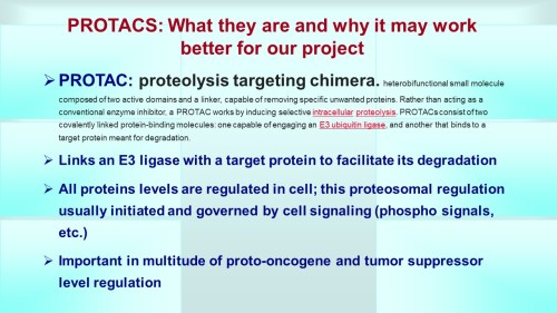
  • PROTACs ubiquitin mediated by E3 ligases;  first discovered by DeShaies and targeted to specific proteins
  • PROTACs used in drug discovery against a host of types of targets including kinases and membrane receptors
  • PROTACs can be modular but lack molecular structural activity relationships
  • can use chemical probes for target validation
  • four requirements: candidate exposure at site of action (for example lipophilicity for candidates needed to cross membranes and accumulate in lysosomes), target engagement (ternary occupancy as measured by FRET), functional pharmacology, relevant phenotype
  • PROTACs hijack the proteosomal degradation system

Proteolysis-targeting chimeras as therapeutics and tools for biological discovery

George Burslem
  • first PROTAC developed to coopt the VHL ubiquitin ligase system which degrades HIF1alpha but now modified for EREalpha
  • in screen for potential PROTACS there were compounds which bound high affinity but no degradation so phenotypic screening very important
  • when look at molecular dynamics can see where PROTAC can add additional protein protein interaction, verifed by site directed mutagenesis
  • able to target bcr-Abl
  • he says this is a rapidly expanding field because of all the new E3 ligase targets being discovered

Expanding the horizons of cereblon modulators

Mary Matyskiela

Translating cellular targeted protein degradation to in vivo models using an enzymology framework

Stewart L Fisher
  • new targeting compounds have an E3 ligase binding domain, a target binding domain and a linker domain
  • in vivo these compounds are very effective; BRD4 degraders good invitro and in vivo with little effect on body weight
  • degraders are essential activators of E3 ligases as these degraders bring targets in close proximity so activates a catalytic cycle of a multistep process (has now high turnover number)
  • in enzymatic pathway the degraders make a productive complex so instead of a kcat think of measuring a kprod or productivity of degraders linked up an E3 ligase
  • the degraders are also affecting the rebound protein synthesis; so Emax never to zero and see a small rebound of protein synthesis


Data-Driven Approaches for Choosing Combinatorial Therapies

Drug combinations remain the gold standard for treating cancer, as they significantly outperform single agents. However, due to the enormous size of drug combination space, it is virtually impossible to interrogate all possible combinations. This session will discuss approaches to identify novel combinations using both experimental and computational approaches. Speakers will discuss i) approaches to drug screening in cell lines, the impact of the microenvironment, and attempts to more

Bence Szalai, James E Korkola, Lisa Tucker-Kellogg, Jeffrey W Tyner

DETAILS

Monday, June 22

3:45 PM – 5:21 PM EDT

Virtual Educational Session

Tumor Biology

Cancer Stem Cells and Therapeutic Resistance

Cancer stem cells are a subpopulation of cells with a high capacity for self-renewal, differentiation and resistance to therapy. In this session, we will define cancer stem cells, discuss cellular plasticity, interactions between cancer stem cells and the tumor microenvironment, and mechanisms that contribute to therapeutic resistance.

Robert S Kerbel, Dolores Hambardzumyan, Jennifer S. Yu

DETAILS

Monday, June 22

3:45 PM – 5:45 PM EDT

Virtual Educational Session

Drug Development, Experimental and Molecular Therapeutics

Molecular Imaging in Cancer Research

This session will cover the fundamentals as well as the major advances made in the field of molecular imaging. Topics covered will include the basics for optical, nuclear, and ultrasound imaging; the pros and cons of each modality; and the recent translational advancements. Learning objectives include the fundamentals of each imaging modality, recent advances in the technology, the processes involved to translate an imaging agent from bench to bedside, and how molecular imaging can gui

Julie Sutcliffe, Summer L Gibbs, Mark D Pagel, Katherine W Ferrara

DETAILS

Monday, June 22

3:45 PM – 5:45 PM EDT

Virtual Educational Session

Tumor Biology, Immunology, Experimental and Molecular Therapeutics, Drug Development

Tumor Endothelium: The Gatekeepers of Tumor Immune Surveillance

Tumor-associated endothelium is a gatekeeper that coordinates the entry and egress of innate and adaptive immune cells within the tumor microenvironment. This is achieved, in part, via the coordinated expression of chemokines and cell adhesion molecules on the endothelial cell surface that attract and retain circulating leukocytes. Crosstalk between adaptive immune cells and the tumor endothelium is therefore essential for tumor immune surveillance and the success of immune-based thera

Dai Fukumura, Maria M Steele, Wen Jiang, Andrew C Dudley

DETAILS

Monday, June 22

3:45 PM – 5:45 PM EDT

Virtual Educational Session

Immunology, Experimental and Molecular Therapeutics

Novel Strategies in Cancer Immunotherapy: The Next Generation of Targets for Anticancer Immunotherapy

T-cell immunotherapy in the form of immune checkpoint blockade or cellular T-cell therapies has been tremendously successful in some types of cancer. This success has opened the door to consider what other modalities or types of immune cells can be harnessed for exert antitumor functions. In this session, experts in their respective fields will discuss topics including novel approaches in immunotherapy, including NK cells, macrophage, and viral oncotherapies.

Evanthia Galanis, Kerry S Campbell, Milan G Chheda, Jennifer L Guerriero

DETAILS

Monday, June 22

3:45 PM – 5:45 PM EDT

Virtual Educational Session

Tumor Biology, Drug Development, Immunology, Clinical Research Excluding Trials

Benign Cells as Drivers of Cancer Progression: Fat and Beyond

Carcinomas develop metastases and resistance to therapy as a result of interaction with tumor microenvironment, composed of various nonmalignant cell types. Understanding the complexity and origins of tumor stromal cells is a prerequisite for development of effective treatments. The link between obesity and cancer progression has revealed the engagement of adipose stromal cells (ASC) and adipocytes from adjacent fat tissue. However, the molecular mechanisms through which they stimulate

Guojun Wu, Matteo Ligorio, Mikhail Kolonin, Maria T Diaz-Meco

DETAILS

Monday, June 22

3:45 PM – 5:45 PM EDT

Virtual Educational Session

Clinical Research Excluding Trials, Experimental and Molecular Therapeutics, Tumor Biology

Dharma Master Jiantai Symposium on Lung Cancer: Know Thy Organ – Lessons Learned from Lung and Pancreatic Cancer Research

The term “cancer” encompasses hundreds of distinct disease entities involving almost every possible site in the human body. Effectively interrogating cancer, either in animals models or human specimens, requires a deep understanding of the involved organ. This includes both the normal cellular constituents of the affected tissue as well as unique aspects of tissue-specific tumorigenesis. It is critical to “Know Thy Organ” when studying cancer. This session will focus on two of the most

Trudy G Oliver, Hossein Borghaei, Laura Delong Wood, Howard C Crawford

DETAILS

Monday, June 22

3:45 PM – 5:45 PM EDT

Virtual Methods Workshop

Clinical Trials

Clinical Trial Design: Part 1: Novel Approaches and Methods in Clinical Trial Design

Good clinical trial design has always had to balance the competing interests of effectively and convincingly answering the question with the limitations imposed by scarce resources, complex logistics, and risks and potential benefits to participants. New targeted therapies, immuno-oncology, and novel combination treatments add new challenges on top of the old ones. This session will introduce these concerns and 1) suggest ways to consider what outcomes are relevant, 2) how we can best

Mary W. Redman, Nolan A. Wages, Susan G Hilsenbeck, Karyn A. Goodman

DETAILS

Monday, June 22

3:45 PM – 5:45 PM EDT

Virtual Methods Workshop

Tumor Biology, Drug Development

High-Throughput Screens for Drivers of Progression and Resistance

The sequencing of human cancers now provides a landscape of the genetic alterations that occur in human cancer, and increasingly knowledge of somatic genetic alterations is becoming part of the evaluation of cancer patients. In some cases, this information leads directly to the selection of particular therapeutic approaches; however, we still lack the ability to decipher the significance of genetic alterations in many cancers. This session will focus on recent developments that permit the identification of molecular targets in specific cancers. This information, coupled with genomic characterization of cancer, will facilitate the development of new therapeutic agents and provide a path to implement precision cancer medicine to all patients.

William C Hahn, Mark A Dawson, Mariella Filbin, Michael Bassik

DETAILS

Monday, June 22

3:45 PM – 5:15 PM EDT

Defining a cancer dependency map

William C Hahn

Introduction

William C Hahn

Genome-scale CRISPR screens in 3D spheroids identify cancer vulnerabilities

Michael Bassik

Utilizing single-cell RNAseq and CRISPR screens to target cancer stem cells in pediatric brain tumors

Mariella Filbin
  • many gliomas are defined by discreet mutational spectra that also discriminates based on age and site as well (for example many cortical tumors have mainly V600E Braf mutations while thalamus will be FGFR1
  • they did single cell RNAseq on needle biopsy from 7 gliomas which gave about 3500 high quality single cells; obtained full length RNA
  • tumors clustered mainly where the patient it came from but had stromal cell contamination probably so did a deconvolution?  Copy number variation showed which were tumor cells and did principle component analysis
  • it seems they used a human glioma model as training set
  • identified a stem cell like glioma cell so concentrated on the genes altered in these for translational studies
  • developed multiple PDX models from patients
  • PDX transcriptome closest to patient transcriptome but organoid grown in serum free very close while organoids grown in serum very distinct transcriptome
  • developed a CRISPR barcoded library to determine genes for survival genes
  • pulled out BMI1  and EZH2 (polycomb complex proteins) as good targets

Virtual Methods Workshop

Prevention Research, Survivorship, Clinical Research Excluding Trials, Epidemiology

Implementation Science Methods for Cancer Prevention and Control in Diverse Populations: Integration of Implementation Science Methods in Care Settings

Through this Education Session we will use examples from ongoing research to provide an overview of implementation science approaches to cancer prevention and control research. We draw on examples to highlight study design approaches, research methods, and real-world solutions when applying implementation science to achieve health equity. Approaches to defining change in the care setting and measuring sustained changes are also emphasized. Using real examples of patient navigation prog

Graham A Colditz, Sanja Percac-Lima, Nathalie Huguet

DETAILS

Monday, June 22

3:45 PM – 5:30 PM EDT

Virtual Educational Session

Regulatory Science and Policy, Epidemiology

COVID-19 and Cancer: Guidance for Clinical Trial Conduct and Considerations for RWE

This session will consider the use of real-world evidence in the context of oncology clinical trials affected by the COVID-19 pandemic. Key aspects of the FDA’s recent “Guidance on Conduct of Clinical Trials of Medical Products of Medical Products during COVID-19 Public Health Emergency” will be discussed, including telemedicine, accounting for missing data, obtaining laboratory tests and images locally, using remote informed consent procedures, and additional considerations for contin

Wendy Rubinstein, Paul G. Kluetz, Amy P. Abernethy, Jonathan Hirsch, C.K. Wang


Read Full Post »

%d bloggers like this: