Healthcare analytics, AI solutions for biological big data, providing an AI platform for the biotech, life sciences, medical and pharmaceutical industries, as well as for related technological approaches, i.e., curation and text analysis with machine learning and other activities related to AI applications to these industries.
Use of Systems Biology for Design of inhibitor of Galectins as Cancer Therapeutic – Strategy and Software
Curator:Stephen J. Williams, Ph.D.
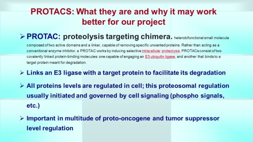
Below is a slide representation of the overall mission 4 to produce a PROTAC to inhibit Galectins 1, 3, and 9.
Using A Priori Knowledge of Galectin Receptor Interaction to Create a BioModel of Galectin 3 Binding
Now after collecting literature from PubMed on “galectin-3” AND “binding” to determine literature containing kinetic data we generate a WordCloud on the articles.
This following file contains the articles needed for BioModels generation.
From the WordCloud we can see that these corpus of articles describe galectin binding to the CRD (carbohydrate recognition domain). Interestingly there are many articles which describe van Der Waals interactions as well as electrostatic interactions. Certain carbohydrate modifictions like Lac NAc and Gal 1,4 may be important. Many articles describe the bonding as well as surface interactions. Many studies have been performed with galectin inhibitors like TDGs (thio-digalactosides) like TAZ TDG (3-deoxy-3-(4-[m-fluorophenyl]-1H-1,2,3-triazol-1-yl)-thio-digalactoside). This led to an interesting article
.
Dual thio-digalactoside-binding modes of human galectins as the structural basis for the design of potent and selective inhibitors
Human galectins are promising targets for cancer immunotherapeutic and fibrotic disease-related drugs. We report herein the binding interactions of three thio-digalactosides (TDGs) including TDG itself, TD139 (3,3′-deoxy-3,3′-bis-(4-[m-fluorophenyl]-1H-1,2,3-triazol-1-yl)-thio-digalactoside, recently approved for the treatment of idiopathic pulmonary fibrosis), and TAZTDG (3-deoxy-3-(4-[m-fluorophenyl]-1H-1,2,3-triazol-1-yl)-thio-digalactoside) with human galectins-1, -3 and -7 as assessed by X-ray crystallography, isothermal titration calorimetry and NMR spectroscopy. Five binding subsites (A-E) make up the carbohydrate-recognition domains of these galectins. We identified novel interactions between an arginine within subsite E of the galectins and an arene group in the ligands. In addition to the interactions contributed by the galactosyl sugar residues bound at subsites C and D, the fluorophenyl group of TAZTDG preferentially bound to subsite B in galectin-3, whereas the same group favored binding at subsite E in galectins-1 and -7. The characterised dual binding modes demonstrate how binding potency, reported as decreased Kd values of the TDG inhibitors from μM to nM, is improved and also offer insights to development of selective inhibitors for individual galectins.
Figures
Figure 1. Chemical structures of L3, TDG…
Figure 2. Structural comparison of the carbohydrate…
The following paper in Cells describes the discovery of protein interactors of endoglin, which is recruited to membranes at the TGF-β receptor complex upon TGF-β signaling. Interesting a carbohydrate binding protein, galectin-3, and an E3-ligase, TRIM21, were found to be unique interactors within this complex.
Gallardo-Vara E, Ruiz-Llorente L, Casado-Vela J, Ruiz-Rodríguez MJ, López-Andrés N, Pattnaik AK, Quintanilla M, Bernabeu C. Endoglin Protein Interactome Profiling Identifies TRIM21 and Galectin-3 as New Binding Partners. Cells. 2019 Sep 13;8(9):1082. doi: 10.3390/cells8091082. PMID: 31540324; PMCID: PMC6769930.
Abstract
Endoglin is a 180-kDa glycoprotein receptor primarily expressed by the vascular endothelium and involved in cardiovascular disease and cancer. Heterozygous mutations in the endoglin gene (ENG) cause hereditary hemorrhagic telangiectasia type 1, a vascular disease that presents with nasal and gastrointestinal bleeding, skin and mucosa telangiectases, and arteriovenous malformations in internal organs. A circulating form of endoglin (alias soluble endoglin, sEng), proteolytically released from the membrane-bound protein, has been observed in several inflammation-related pathological conditions and appears to contribute to endothelial dysfunction and cancer development through unknown mechanisms. Membrane-bound endoglin is an auxiliary component of the TGF-β receptor complex and the extracellular region of endoglin has been shown to interact with types I and II TGF-β receptors, as well as with BMP9 and BMP10 ligands, both members of the TGF-β family. To search for novel protein interactors, we screened a microarray containing over 9000 unique human proteins using recombinant sEng as bait. We find that sEng binds with high affinity, at least, to 22 new proteins. Among these, we validated the interaction of endoglin with galectin-3, a secreted member of the lectin family with capacity to bind membrane glycoproteins, and with tripartite motif-containing protein 21 (TRIM21), an E3 ubiquitin-protein ligase. Using human endothelial cells and Chinese hamster ovary cells, we showed that endoglin co-immunoprecipitates and co-localizes with galectin-3 or TRIM21. These results open new research avenues on endoglin function and regulation.
Endoglin is an auxiliary TGF-β co-receptor predominantly expressed in endothelial cells, which is involved in vascular development, repair, homeostasis, and disease [1,2,3,4]. Heterozygous mutations in the human ENDOGLIN gene (ENG) cause hereditary hemorrhagic telangiectasia (HHT) type 1, a vascular disease associated with nasal and gastrointestinal bleeds, telangiectases on skin and mucosa and arteriovenous malformations in the lung, liver, and brain [4,5,6]. The key role of endoglin in the vasculature is also illustrated by the fact that endoglin-KO mice die in utero due to defects in the vascular system [7]. Endoglin expression is markedly upregulated in proliferating endothelial cells involved in active angiogenesis, including the solid tumor neovasculature [8,9]. For this reason, endoglin has become a promising target for the antiangiogenic treatment of cancer [10,11,12]. Endoglin is also expressed in cancer cells where it can behave as both a tumor suppressor in prostate, breast, esophageal, and skin carcinomas [13,14,15,16] and a promoter of malignancy in melanoma and Ewing’s sarcoma [17]. Ectodomain shedding of membrane-bound endoglin may lead to a circulating form of the protein, also known as soluble endoglin (sEng) [18,19,20]. Increased levels of sEng have been found in several vascular-related pathologies, including preeclampsia, a disease of high prevalence in pregnant women which, if left untreated, can lead to serious and even fatal complications for both mother and baby [2,18,19,21]. Interestingly, several lines of evidence support a pathogenic role of sEng in the vascular system, including endothelial dysfunction, antiangiogenic activity, increased vascular permeability, inflammation-associated leukocyte adhesion and transmigration, and hypertension [18,22,23,24,25,26,27]. Because of its key role in vascular pathology, a large number of studies have addressed the structure and function of endoglin at the molecular level, in order to better understand its mechanism of action.
Galectin-3 Interacts with Endoglin in Cells
Galectin-3 is a secreted member of the lectin family with the capacity to bind membrane glycoproteins like endoglin and is involved in the pathogenesis of many human diseases [52]. We confirmed the protein screen data for galectin-3, as evidenced by two-way co-immunoprecipitation of endoglin and galectin-3 upon co-transfection in CHO-K1 cells. As shown in Figure 1A, galectin-3 and endoglin were efficiently transfected, as demonstrated by Western blot analysis in total cell extracts. No background levels of endoglin were observed in control cells transfected with the empty vector (Ø). By contrast, galectin-3 could be detected in all samples but, as expected, showed an increased signal in cells transfected with the galectin-3 expression vector. Co-immunoprecipitation studies of these cell lysates showed that galectin-3 was present in endoglin immunoprecipitates (Figure 1B). Conversely, endoglin was also detected in galectin-3 immunoprecipitates (Figure 1C).
Figure 1. Protein–protein association between galectin-3 and endoglin. (A–C). Co-immunoprecipitation of galectin-3 and endoglin. CHO-K1 cells were transiently transfected with pcEXV-Ø (Ø), pcEXV–HA–EngFL (Eng) and pcDNA3.1–Gal-3 (Gal3) expression vectors. (A) Total cell lysates (TCL) were analyzed by SDS-PAGE under reducing conditions, followed by Western blot (WB) analysis using specific antibodies to endoglin, galectin-3 and β-actin (loading control). Cell lysates were subjected to immunoprecipitation (IP) with anti-endoglin (B) or anti-galectin-3 (C) antibodies, followed by SDS-PAGE under reducing conditions and WB analysis with anti-endoglin or anti-galectin-3 antibodies, as indicated. Negative controls with an IgG2b (B) and IgG1 (C) were included. (D) Protein-protein interactions between galectin-3 and endoglin using Bio-layer interferometry (BLItz). The Ni–NTA biosensors tips were loaded with 7.3 µM recombinant human galectin-3/6xHis at the C-terminus (LGALS3), and protein binding was measured against 0.1% BSA in PBS (negative control) or 4.1 µM soluble endoglin (sEng). Kinetic sensorgrams were obtained using a single channel ForteBioBLItzTM instrument.
Figure 2.Galectin-3 and endoglin co-localize in human endothelial cells. Human umbilical vein-derived endothelial cell (HUVEC) monolayers were fixed with paraformaldehyde, permeabilized with Triton X-100, incubated with the mouse mAb P4A4 anti-endoglin, washed, and incubated with a rabbit polyclonal anti-galectin-3 antibody (PA5-34819). Galectin-3 and endoglin were detected by immunofluorescence upon incubation with Alexa 647 goat anti-rabbit IgG (red staining) and Alexa 488 goat anti-mouse IgG (green staining) secondary antibodies, respectively. (A) Single staining of galectin-3 (red) and endoglin (green) at the indicated magnifications. (B) Merge images plus DAPI (nuclear staining in blue) show co-localization of galectin-3 and endoglin (yellow color). Representative images of five different experiments are shown.
Endoglin associates with the cullin-type E3 ligase TRIM21
Figure 3.Protein–protein association between TRIM21 and endoglin. (A–E) Co-immunoprecipitation of TRIM21 and endoglin. A,B. HUVEC monolayers were lysed and total cell lysates (TCL) were subjected to SDS-PAGE under reducing (for TRIM21 detection) or nonreducing (for endoglin detection) conditions, followed by Western blot (WB) analysis using antibodies to endoglin, TRIM21 or β-actin (A). HUVECs lysates were subjected to immunoprecipitation (IP) with anti-TRIM21 or negative control antibodies, followed by WB analysis with anti-endoglin (B). C,D. CHO-K1 cells were transiently transfected with pDisplay–HA–Mock (Ø), pDisplay–HA–EngFL (E) or pcDNA3.1–HA–hTRIM21 (T) expression vectors, as indicated. Total cell lysates (TCL) were subjected to SDS-PAGE under nonreducing conditions and WB analysis using specific antibodies to endoglin, TRIM21, and β-actin (C). Cell lysates were subjected to immunoprecipitation (IP) with anti-TRIM21 or anti-endoglin antibodies, followed by SDS-PAGE under reducing (upper panel) or nonreducing (lower panel) conditions and WB analysis with anti-TRIM21 or anti-endoglin antibodies. Negative controls of appropriate IgG were included (D). E. CHO-K1 cells were transiently transfected with pcDNA3.1–HA–hTRIM21 and pDisplay–HA–Mock (Ø), pDisplay–HA–EngFL (FL; full-length), pDisplay–HA–EngEC (EC; cytoplasmic-less) or pDisplay–HA–EngTMEC (TMEC; cytoplasmic-less) expression vectors, as indicated. Cell lysates were subjected to immunoprecipitation with anti-TRIM21, followed by SDS-PAGE under reducing conditions and WB analysis with anti-endoglin antibodies, as indicated. The asterisk indicates the presence of a nonspecific band. Mr, molecular reference; Eng, endoglin; TRIM, TRIM21. (F) Protein–protein interactions between TRIM21 and endoglin using Bio-layer interferometry (BLItz). The Ni–NTA biosensors tips were loaded with 5.4 µM recombinant human TRIM21/6xHis at the N-terminus (R052), and protein binding was measured against 0.1% BSA in PBS (negative control) or 4.1 µM soluble endoglin (sEng). Kinetic sensorgrams were obtained using a single channel ForteBioBLItzTM instrument.
Table 1. Human protein-array analysis of endoglin interactors1.
1 Microarrays containing over 9000 unique human proteins were screened using recombinant sEng as a probe. Protein interactors showing the highest scores (Z-score ≥2.0) are listed. GeneBank (https://www.ncbi.nlm.nih.gov/genbank/) and UniProtKB (https://www.uniprot.org/help/uniprotkb) accession numbers are indicated with a yellow or green background, respectively. The cellular compartment of each protein was obtained from the UniProtKB webpage. Proteins selected for further studies (TRIM21 and galectin-3) are indicated in bold type with blue background.
Note: the following are from NCBI Genbank and Genecards on TRIM21
Official Symbol TRIM21provided by HGNC Official Full Name tripartite motif containing 21provided by HGNC Primary source HGNC:HGNC:11312 See related Ensembl:ENSG00000132109MIM:109092;AllianceGenome:HGNC:11312 Gene type protein coding RefSeq status REVIEWED Organism Homo sapiens Lineage Eukaryota; Metazoa; Chordata; Craniata; Vertebrata; Euteleostomi; Mammalia; Eutheria; Euarchontoglires; Primates; Haplorrhini; Catarrhini; Hominidae; Homo Also known as SSA; RO52; SSA1; RNF81; Ro/SSA Summary This gene encodes a member of the tripartite motif (TRIM) family. The TRIM motif includes three zinc-binding domains, a RING, a B-box type 1 and a B-box type 2, and a coiled-coil region. The encoded protein is part of the RoSSA ribonucleoprotein, which includes a single polypeptide and one of four small RNA molecules. The RoSSA particle localizes to both the cytoplasm and the nucleus. RoSSA interacts with autoantigens in patients with Sjogren syndrome and systemic lupus erythematosus. Alternatively spliced transcript variants for this gene have been described but the full-length nature of only one has been determined. [provided by RefSeq, Jul 2008] Expression Ubiquitous expression in spleen (RPKM 15.5), appendix (RPKM 13.2) and 24 other tissues See more Orthologs mouseall NEW Try the new Gene table Try the new Transcript table
This gene encodes a member of the tripartite motif (TRIM) family. The TRIM motif includes three zinc-binding domains, a RING, a B-box type 1 and a B-box type 2, and a coiled-coil region. The encoded protein is part of the RoSSA ribonucleoprotein, which includes a single polypeptide and one of four small RNA molecules. The RoSSA particle localizes to both the cytoplasm and the nucleus. RoSSA interacts with autoantigens in patients with Sjogren syndrome and systemic lupus erythematosus. Alternatively spliced transcript variants for this gene have been described but the full-length nature of only one has been determined. [provided by RefSeq, Jul 2008]
E3 ubiquitin-protein ligase whose activity is dependent on E2 enzymes, UBE2D1, UBE2D2, UBE2E1 and UBE2E2. Forms a ubiquitin ligase complex in cooperation with the E2 UBE2D2 that is used not only for the ubiquitination of USP4 and IKBKB but also for its self-ubiquitination. Component of cullin-RING-based SCF (SKP1-CUL1-F-box protein) E3 ubiquitin-protein ligase complexes such as SCF(SKP2)-like complexes. A TRIM21-containing SCF(SKP2)-like complex is shown to mediate ubiquitination of CDKN1B (‘Thr-187’ phosphorylated-form), thereby promoting its degradation by the proteasome. Monoubiquitinates IKBKB that will negatively regulates Tax-induced NF-kappa-B signaling. Negatively regulates IFN-beta production post-pathogen recognition by polyubiquitin-mediated degradation of IRF3. Mediates the ubiquitin-mediated proteasomal degradation of IgG1 heavy chain, which is linked to the VCP-mediated ER-associated degradation (ERAD) pathway. Promotes IRF8 ubiquitination, which enhanced the ability of IRF8 to stimulate cytokine genes transcription in macrophages. Plays a role in the regulation of the cell cycle progression. Enhances the decapping activity of DCP2. Exists as a ribonucleoprotein particle present in all mammalian cells studied and composed of a single polypeptide and one of four small RNA molecules. At least two isoforms are present in nucleated and red blood cells, and tissue specific differences in RO/SSA proteins have been identified. The common feature of these proteins is their ability to bind HY RNAs.2. Involved in the regulation of innate immunity and the inflammatory response in response to IFNG/IFN-gamma. Organizes autophagic machinery by serving as a platform for the assembly of ULK1, Beclin 1/BECN1 and ATG8 family members and recognizes specific autophagy targets, thus coordinating target recognition with assembly of the autophagic apparatus and initiation of autophagy. Acts as an autophagy receptor for the degradation of IRF3, hence attenuating type I interferon (IFN)-dependent immune responses (PubMed:26347139, 16297862, 16316627, 16472766, 16880511, 18022694, 18361920, 18641315, 18845142, 19675099). Represses the innate antiviral response by facilitating the formation of the NMI-IFI35 complex through ‘Lys-63’-linked ubiquitination of NMI (PubMed:26342464). ( RO52_HUMAN,P19474 )
Molecular function for TRIM21 Gene according to UniProtKB/Swiss-Prot
Function:
E3 ubiquitin-protein ligase whose activity is dependent on E2 enzymes, UBE2D1, UBE2D2, UBE2E1 and UBE2E2. Forms a ubiquitin ligase complex in cooperation with the E2 UBE2D2 that is used not only for the ubiquitination of USP4 and IKBKB but also for its self-ubiquitination. Component of cullin-RING-based SCF (SKP1-CUL1-F-box protein) E3 ubiquitin-protein ligase complexes such as SCF(SKP2)-like complexes. A TRIM21-containing SCF(SKP2)-like complex is shown to mediate ubiquitination of CDKN1B (‘Thr-187’ phosphorylated-form), thereby promoting its degradation by the proteasome. Monoubiquitinates IKBKB that will negatively regulates Tax-induced NF-kappa-B signaling. Negatively regulates IFN-beta production post-pathogen recognition by polyubiquitin-mediated degradation of IRF3. Mediates the ubiquitin-mediated proteasomal degradation of IgG1 heavy chain, which is linked to the VCP-mediated ER-associated degradation (ERAD) pathway. Promotes IRF8 ubiquitination, which enhanced the ability of IRF8 to stimulate cytokine genes transcription in macrophages. Plays a role in the regulation of the cell cycle progression.
Endoglin Protein Interactome Profiling Identifies TRIM21 and Galectin-3 as New Binding Partners
Gallardo-Vara E, Ruiz-Llorente L, Casado-Vela J, Ruiz-Rodríguez MJ, López-Andrés N, Pattnaik AK, Quintanilla M, Bernabeu C. Endoglin Protein Interactome Profiling Identifies TRIM21 and Galectin-3 as New Binding Partners. Cells. 2019 Sep 13;8(9):1082. doi: 10.3390/cells8091082. PMID: 31540324; PMCID: PMC6769930.
Abstract
Endoglin is a 180-kDa glycoprotein receptor primarily expressed by the vascular endothelium and involved in cardiovascular disease and cancer. Heterozygous mutations in the endoglin gene (ENG) cause hereditary hemorrhagic telangiectasia type 1, a vascular disease that presents with nasal and gastrointestinal bleeding, skin and mucosa telangiectases, and arteriovenous malformations in internal organs. A circulating form of endoglin (alias soluble endoglin, sEng), proteolytically released from the membrane-bound protein, has been observed in several inflammation-related pathological conditions and appears to contribute to endothelial dysfunction and cancer development through unknown mechanisms. Membrane-bound endoglin is an auxiliary component of the TGF-β receptor complex and the extracellular region of endoglin has been shown to interact with types I and II TGF-β receptors, as well as with BMP9 and BMP10 ligands, both members of the TGF-β family. To search for novel protein interactors, we screened a microarray containing over 9000 unique human proteins using recombinant sEng as bait. We find that sEng binds with high affinity, at least, to 22 new proteins. Among these, we validated the interaction of endoglin with galectin-3, a secreted member of the lectin family with capacity to bind membrane glycoproteins, and with tripartite motif-containing protein 21 (TRIM21), an E3 ubiquitin-protein ligase. Using human endothelial cells and Chinese hamster ovary cells, we showed that endoglin co-immunoprecipitates and co-localizes with galectin-3 or TRIM21. These results open new research avenues on endoglin function and regulation.
Endoglin is an auxiliary TGF-β co-receptor predominantly expressed in endothelial cells, which is involved in vascular development, repair, homeostasis, and disease [1,2,3,4]. Heterozygous mutations in the human ENDOGLIN gene (ENG) cause hereditary hemorrhagic telangiectasia (HHT) type 1, a vascular disease associated with nasal and gastrointestinal bleeds, telangiectases on skin and mucosa and arteriovenous malformations in the lung, liver, and brain [4,5,6]. The key role of endoglin in the vasculature is also illustrated by the fact that endoglin-KO mice die in utero due to defects in the vascular system [7]. Endoglin expression is markedly upregulated in proliferating endothelial cells involved in active angiogenesis, including the solid tumor neovasculature [8,9]. For this reason, endoglin has become a promising target for the antiangiogenic treatment of cancer [10,11,12]. Endoglin is also expressed in cancer cells where it can behave as both a tumor suppressor in prostate, breast, esophageal, and skin carcinomas [13,14,15,16] and a promoter of malignancy in melanoma and Ewing’s sarcoma [17]. Ectodomain shedding of membrane-bound endoglin may lead to a circulating form of the protein, also known as soluble endoglin (sEng) [18,19,20]. Increased levels of sEng have been found in several vascular-related pathologies, including preeclampsia, a disease of high prevalence in pregnant women which, if left untreated, can lead to serious and even fatal complications for both mother and baby [2,18,19,21]. Interestingly, several lines of evidence support a pathogenic role of sEng in the vascular system, including endothelial dysfunction, antiangiogenic activity, increased vascular permeability, inflammation-associated leukocyte adhesion and transmigration, and hypertension [18,22,23,24,25,26,27]. Because of its key role in vascular pathology, a large number of studies have addressed the structure and function of endoglin at the molecular level, in order to better understand its mechanism of action.
Galectin-3 Interacts with Endoglin in Cells
Galectin-3 is a secreted member of the lectin family with the capacity to bind membrane glycoproteins like endoglin and is involved in the pathogenesis of many human diseases [52]. We confirmed the protein screen data for galectin-3, as evidenced by two-way co-immunoprecipitation of endoglin and galectin-3 upon co-transfection in CHO-K1 cells. As shown in Figure 1A, galectin-3 and endoglin were efficiently transfected, as demonstrated by Western blot analysis in total cell extracts. No background levels of endoglin were observed in control cells transfected with the empty vector (Ø). By contrast, galectin-3 could be detected in all samples but, as expected, showed an increased signal in cells transfected with the galectin-3 expression vector. Co-immunoprecipitation studies of these cell lysates showed that galectin-3 was present in endoglin immunoprecipitates (Figure 1B). Conversely, endoglin was also detected in galectin-3 immunoprecipitates (Figure 1C).
Figure 1. Protein–protein association between galectin-3 and endoglin. (A–C). Co-immunoprecipitation of galectin-3 and endoglin. CHO-K1 cells were transiently transfected with pcEXV-Ø (Ø), pcEXV–HA–EngFL (Eng) and pcDNA3.1–Gal-3 (Gal3) expression vectors. (A) Total cell lysates (TCL) were analyzed by SDS-PAGE under reducing conditions, followed by Western blot (WB) analysis using specific antibodies to endoglin, galectin-3 and β-actin (loading control). Cell lysates were subjected to immunoprecipitation (IP) with anti-endoglin (B) or anti-galectin-3 (C) antibodies, followed by SDS-PAGE under reducing conditions and WB analysis with anti-endoglin or anti-galectin-3 antibodies, as indicated. Negative controls with an IgG2b (B) and IgG1 (C) were included. (D) Protein-protein interactions between galectin-3 and endoglin using Bio-layer interferometry (BLItz). The Ni–NTA biosensors tips were loaded with 7.3 µM recombinant human galectin-3/6xHis at the C-terminus (LGALS3), and protein binding was measured against 0.1% BSA in PBS (negative control) or 4.1 µM soluble endoglin (sEng). Kinetic sensorgrams were obtained using a single channel ForteBioBLItzTM instrument.
Figure 2.Galectin-3 and endoglin co-localize in human endothelial cells. Human umbilical vein-derived endothelial cell (HUVEC) monolayers were fixed with paraformaldehyde, permeabilized with Triton X-100, incubated with the mouse mAb P4A4 anti-endoglin, washed, and incubated with a rabbit polyclonal anti-galectin-3 antibody (PA5-34819). Galectin-3 and endoglin were detected by immunofluorescence upon incubation with Alexa 647 goat anti-rabbit IgG (red staining) and Alexa 488 goat anti-mouse IgG (green staining) secondary antibodies, respectively. (A) Single staining of galectin-3 (red) and endoglin (green) at the indicated magnifications. (B) Merge images plus DAPI (nuclear staining in blue) show co-localization of galectin-3 and endoglin (yellow color). Representative images of five different experiments are shown.
Endoglin associates with the cullin-type E3 ligase TRIM21
Figure 3.Protein–protein association between TRIM21 and endoglin. (A–E) Co-immunoprecipitation of TRIM21 and endoglin. A,B. HUVEC monolayers were lysed and total cell lysates (TCL) were subjected to SDS-PAGE under reducing (for TRIM21 detection) or nonreducing (for endoglin detection) conditions, followed by Western blot (WB) analysis using antibodies to endoglin, TRIM21 or β-actin (A). HUVECs lysates were subjected to immunoprecipitation (IP) with anti-TRIM21 or negative control antibodies, followed by WB analysis with anti-endoglin (B). C,D. CHO-K1 cells were transiently transfected with pDisplay–HA–Mock (Ø), pDisplay–HA–EngFL (E) or pcDNA3.1–HA–hTRIM21 (T) expression vectors, as indicated. Total cell lysates (TCL) were subjected to SDS-PAGE under nonreducing conditions and WB analysis using specific antibodies to endoglin, TRIM21, and β-actin (C). Cell lysates were subjected to immunoprecipitation (IP) with anti-TRIM21 or anti-endoglin antibodies, followed by SDS-PAGE under reducing (upper panel) or nonreducing (lower panel) conditions and WB analysis with anti-TRIM21 or anti-endoglin antibodies. Negative controls of appropriate IgG were included (D). E. CHO-K1 cells were transiently transfected with pcDNA3.1–HA–hTRIM21 and pDisplay–HA–Mock (Ø), pDisplay–HA–EngFL (FL; full-length), pDisplay–HA–EngEC (EC; cytoplasmic-less) or pDisplay–HA–EngTMEC (TMEC; cytoplasmic-less) expression vectors, as indicated. Cell lysates were subjected to immunoprecipitation with anti-TRIM21, followed by SDS-PAGE under reducing conditions and WB analysis with anti-endoglin antibodies, as indicated. The asterisk indicates the presence of a nonspecific band. Mr, molecular reference; Eng, endoglin; TRIM, TRIM21. (F) Protein–protein interactions between TRIM21 and endoglin using Bio-layer interferometry (BLItz). The Ni–NTA biosensors tips were loaded with 5.4 µM recombinant human TRIM21/6xHis at the N-terminus (R052), and protein binding was measured against 0.1% BSA in PBS (negative control) or 4.1 µM soluble endoglin (sEng). Kinetic sensorgrams were obtained using a single channel ForteBioBLItzTM instrument.
Table 1. Human protein-array analysis of endoglin interactors1.
1 Microarrays containing over 9000 unique human proteins were screened using recombinant sEng as a probe. Protein interactors showing the highest scores (Z-score ≥2.0) are listed. GeneBank (https://www.ncbi.nlm.nih.gov/genbank/) and UniProtKB (https://www.uniprot.org/help/uniprotkb) accession numbers are indicated with a yellow or green background, respectively. The cellular compartment of each protein was obtained from the UniProtKB webpage. Proteins selected for further studies (TRIM21 and galectin-3) are indicated in bold type with blue background.
Note: the following are from NCBI Genbank and Genecards on TRIM21
This gene encodes a member of the tripartite motif (TRIM) family. The TRIM motif includes three zinc-binding domains, a RING, a B-box type 1 and a B-box type 2, and a coiled-coil region. The encoded protein is part of the RoSSA ribonucleoprotein, which includes a single polypeptide and one of four small RNA molecules. The RoSSA particle localizes to both the cytoplasm and the nucleus. RoSSA interacts with autoantigens in patients with Sjogren syndrome and systemic lupus erythematosus. Alternatively spliced transcript variants for this gene have been described but the full-length nature of only one has been determined. [provided by RefSeq, Jul 2008]
Expression
Ubiquitous expression in spleen (RPKM 15.5), appendix (RPKM 13.2) and 24 other tissues See more
This gene encodes a member of the tripartite motif (TRIM) family. The TRIM motif includes three zinc-binding domains, a RING, a B-box type 1 and a B-box type 2, and a coiled-coil region. The encoded protein is part of the RoSSA ribonucleoprotein, which includes a single polypeptide and one of four small RNA molecules. The RoSSA particle localizes to both the cytoplasm and the nucleus. RoSSA interacts with autoantigens in patients with Sjogren syndrome and systemic lupus erythematosus. Alternatively spliced transcript variants for this gene have been described but the full-length nature of only one has been determined. [provided by RefSeq, Jul 2008]
E3 ubiquitin-protein ligase whose activity is dependent on E2 enzymes, UBE2D1, UBE2D2, UBE2E1 and UBE2E2. Forms a ubiquitin ligase complex in cooperation with the E2 UBE2D2 that is used not only for the ubiquitination of USP4 and IKBKB but also for its self-ubiquitination. Component of cullin-RING-based SCF (SKP1-CUL1-F-box protein) E3 ubiquitin-protein ligase complexes such as SCF(SKP2)-like complexes. A TRIM21-containing SCF(SKP2)-like complex is shown to mediate ubiquitination of CDKN1B (‘Thr-187’ phosphorylated-form), thereby promoting its degradation by the proteasome. Monoubiquitinates IKBKB that will negatively regulates Tax-induced NF-kappa-B signaling. Negatively regulates IFN-beta production post-pathogen recognition by polyubiquitin-mediated degradation of IRF3. Mediates the ubiquitin-mediated proteasomal degradation of IgG1 heavy chain, which is linked to the VCP-mediated ER-associated degradation (ERAD) pathway. Promotes IRF8 ubiquitination, which enhanced the ability of IRF8 to stimulate cytokine genes transcription in macrophages. Plays a role in the regulation of the cell cycle progression. Enhances the decapping activity of DCP2. Exists as a ribonucleoprotein particle present in all mammalian cells studied and composed of a single polypeptide and one of four small RNA molecules. At least two isoforms are present in nucleated and red blood cells, and tissue specific differences in RO/SSA proteins have been identified. The common feature of these proteins is their ability to bind HY RNAs.2. Involved in the regulation of innate immunity and the inflammatory response in response to IFNG/IFN-gamma. Organizes autophagic machinery by serving as a platform for the assembly of ULK1, Beclin 1/BECN1 and ATG8 family members and recognizes specific autophagy targets, thus coordinating target recognition with assembly of the autophagic apparatus and initiation of autophagy. Acts as an autophagy receptor for the degradation of IRF3, hence attenuating type I interferon (IFN)-dependent immune responses (PubMed:26347139, 16297862, 16316627, 16472766, 16880511, 18022694, 18361920, 18641315, 18845142, 19675099). Represses the innate antiviral response by facilitating the formation of the NMI-IFI35 complex through ‘Lys-63’-linked ubiquitination of NMI (PubMed:26342464). ( RO52_HUMAN,P19474 )
Molecular function for TRIM21 Gene according to UniProtKB/Swiss-Prot
Function:
E3 ubiquitin-protein ligase whose activity is dependent on E2 enzymes, UBE2D1, UBE2D2, UBE2E1 and UBE2E2. Forms a ubiquitin ligase complex in cooperation with the E2 UBE2D2 that is used not only for the ubiquitination of USP4 and IKBKB but also for its self-ubiquitination. Component of cullin-RING-based SCF (SKP1-CUL1-F-box protein) E3 ubiquitin-protein ligase complexes such as SCF(SKP2)-like complexes. A TRIM21-containing SCF(SKP2)-like complex is shown to mediate ubiquitination of CDKN1B (‘Thr-187’ phosphorylated-form), thereby promoting its degradation by the proteasome. Monoubiquitinates IKBKB that will negatively regulates Tax-induced NF-kappa-B signaling. Negatively regulates IFN-beta production post-pathogen recognition by polyubiquitin-mediated degradation of IRF3. Mediates the ubiquitin-mediated proteasomal degradation of IgG1 heavy chain, which is linked to the VCP-mediated ER-associated degradation (ERAD) pathway. Promotes IRF8 ubiquitination, which enhanced the ability of IRF8 to stimulate cytokine genes transcription in macrophages. Plays a role in the regulation of the cell cycle progression.
Other Articles in this Open Access Scientific Journal on Galectins and Proteosome Include
We studied 592 HFpatients who had been hospitalized for HFand were followed
for 18 months. The primary end-point was a composite of all-cause mortality and
HF hospitalization. A doubling of galectin-3 levels was associated with a hazard
ratio (HR) of 1.97 (1.62–2.42) for the primary outcome (P = 0.001). After
correction for age, gender, BNP, eGFR, and diabetes the HR was 1.38 (1.07–
1.78; P = 0.015). Galectin-3 levels were correlated with higher IL-6 and CRP
levels (P = 0.002). Changes of galectin-3 levels after 6 months did not add
prognostic information to the base-line value (n = 291); however, combining
plasma galectin-3 and BNP levels increased prognostic value over either
biomarker alone (ROC analysis, P = 0.05). The predictive value of galectin-3
was stronger in patients with preserved LVEF (n = 114) compared to
patients with reduced LVEF (P = 0.001).
Galectin-3 in Ambulatory Patients with Heart Failure: Results
from the HF-ACTION Study GM Felker, M Fiuzat, LK. Shaw, R Clare, ,DJ. Whellan, et al.
Circ Heart Fail. 2012 Jan; 5(1): 72–78. http://dx.doi.org:/10.1161/CIRCHEARTFAILURE.111.963637
Galectin-3 is a soluble ß-galactoside-binding lectin released by activated
cardiac macrophages. Elevated levels of galectin-3 have been found to
be associated with adverse outcomes in patients with heart failure. We
evaluated the association between galectin-3 and long-term clinical
outcomes in ambulatory heart failure patients enrolled in the HF-ACTION
study.
Galectin-3 is elevated in ambulatory heart failure patients and is
associated with poor functional capacity and other known measures of
heart failure severity. In univariate analysis, galectin-3 was significantly
predictive of long-term outcomes, but this association did not
persist after adjustment for other predictors, especially NTproBNP.