Feeds:
Posts
Comments

Archive for the ‘Developmental biology’ Category

Colon cancer and organoids

Larry H. Bernstein, MD, FCAP, Curator

LPBI


Guts and Glory

An open mind and collaborative spirit have taken Hans Clevers on a journey from medicine to developmental biology, gastroenterology, cancer, and stem cells.

By Anna Azvolinsky    http://www.the-scientist.com/?articles.view/articleNo/45580/title/Guts-and-Glory

Ihave had to talk a lot about my science recently and it’s made me think about how science works,” says Hans Clevers. “Scientists are trained to think science is driven by hypotheses, but for [my lab], hypothesis-driven research has never worked. Instead, it has been about trying to be as open-minded as possible—which is not natural for our brains,” adds the Utrecht University molecular genetics professor. “The human mind is such that it tries to prove it’s right, so pursuing a hypothesis can result in disaster. My advice to my own team and others is to not preformulate an answer to a scientific question, but just observe and never be afraid of the unknown. What has worked well for us is to keep an open mind and do the experiments. And find a collaborator if it is outside our niche.”

“One thing I have learned is that hypothesis-driven research tends not to be productive when you are in an unknown territory.”

Clevers entered medical school at Utrecht University in The Netherlands in 1978 while simultaneously pursuing a master’s degree in biology. Drawn to working with people in the clinic, Clevers had a training position in pediatrics lined up after medical school, but then mentors persuaded him to spend an additional year converting the master’s degree to a PhD in immunology. “At the end of that year, looking back, I got more satisfaction from the research than from seeing patients.” Clevers also had an aptitude for benchwork, publishing four papers from his PhD year. “They were all projects I had made up myself. The department didn’t do the kind of research I was doing,” he says. “Now that I look back, it’s surprising that an inexperienced PhD student could come up with a project and publish independently.”

Clevers studied T- and B-cell signaling; he set up assays to visualize calcium ion flux and demonstrated that the ions act as messengers to activate human B cells, signaling through antibodies on the cell surface. “As soon as the experiment worked, I got T cells from the lab next door and did the same experiment. That was my strategy: as soon as something worked, I would apply it elsewhere and didn’t stop just because I was a B-cell biologist and not a T-cell biologist. What I learned then, that I have continued to benefit from, is that a lot of scientists tend to adhere to a niche. They cling to these niches and are not that flexible. You think scientists are, but really most are not.”

Here, Clevers talks about promoting a collaborative spirit in research, the art of doing a pilot experiment, and growing miniature organs in a dish.

Clevers Creates

Re-search? Clevers was born in Eindhoven, in the south of The Netherlands. The town was headquarters to Philips Electronics, where his father worked as a businessman, and his mother took care of Clevers and his three brothers. Clevers did well in school but his passion was sports, especially tennis and field hockey, “a big thing in Holland.” Then in 1975, at age 18, he moved to Utrecht University, where he entered an intensive, biology-focused program. “I knew I wanted to be a biology researcher since I was young. In Dutch, the word for research is ‘onderzoek’ and I knew the English word ‘research’ and had wondered why there was the ‘re’ in the word, because I wanted to search but I didn’t want to do re-search—to find what someone else had already found.”

Opportunity to travel. “I was very disappointed in my biology studies, which were old-fashioned and descriptive,” says Clevers. He thought medicine might be more interesting and enrolled in medical school while still pursuing a master’s degree in biology at Utrecht. For the master’s, Clevers had to do three rotations. He spent a year at the International Laboratory for Research on Animal Diseases (ILRAD) in Nairobi, Kenya, and six months in Bethesda, Maryland, at the National Institutes of Health. “Holland is really small, so everyone travels.” Clevers saw those two rotations more as travel explorations. In Nairobi, he went on safaris and explored the country in Land Rovers borrowed from the institute. While in Maryland in 1980, Clevers—with the consent of his advisor, who thought it was a good idea for him to get a feel for the U.S.—flew to Portland, Oregon, and drove back to Boston with a musician friend along the Canadian border. He met the fiancé of political activist and academic Angela Davis in New York City and even stayed in their empty apartment there.

Life and lab lessons. Back in Holland, Clevers joined Rudolf Eugène Ballieux’s lab at Utrecht University to pursue his PhD, for which he studied immune cell signaling. “I didn’t learn much science from him, but I learned that you always have to create trust and to trust people around you. This became a major theme in my own lab. We don’t distrust journals or reviewers or collaborators. We trust everyone and we share. There will be people who take advantage, but there have only been a few of those. So I learned from Ballieux to give everyone maximum trust and then change this strategy only if they fail that trust. We collaborate easily because we give out everything and we also easily get reagents and tools that we may need. It’s been valuable to me in my career. And it is fun!”

Clevers Concentrates

On a mission. “Once I decided to become a scientist, I knew I needed to train seriously. Up to that point, I was totally self-trained.” From an extensive reading of the immunology literature, Clevers became interested in how T cells recognize antigens, and headed off to spend a postdoc studying the problem in Cox Terhorst’s lab at Dana-Farber Cancer Institute in Boston. “Immunology was young, but it was very exciting and there was a lot to discover. I became a professional scientist there and experienced how tough science is.” In 1988, Clevers cloned and characterized the gene for a component of the T-cell receptor (TCR) called CD3-epsilon, which binds antigen and activates intracellular signaling pathways.

On the fast track in Holland. Clevers returned to Utrecht University in 1989 as a professor of immunology. Within one month of setting up his lab, he had two graduate students and a technician, and the lab had cloned the first T cell–specific transcription factor, which they called TCF-1, in human T cells. When his former thesis advisor retired, Clevers was asked, at age 33, to become head of the immunology department. While the appointment was high-risk for him and for the department, Clevers says, he was chosen because he was good at multitasking and because he got along well with everyone.

Problem-solving strategy. “My strategy in research has always been opportunistic. One thing I have learned is that hypothesis-driven research tends not to be productive when you are in an unknown territory. I think there is an art to doing pilot experiments. So we have always just set up systems in which something happens and then you try and try things until a pattern appears and maybe you formulate a small hypothesis. But as soon as it turns out not to be exactly right, you abandon it. It’s a very open-minded type of research where you question whether what you are seeing is a real phenomenon without spending a year on doing all of the proper controls.”

Trial and error. Clevers’s lab found that while TCF-1 bound to DNA, it did not alter gene expression, despite the researchers’ tinkering with promoter and enhancer assays. “For about five years this was a problem. My first PhD students were leaving and they thought the whole TCF project was a failure,” says Clevers. His lab meanwhile cloned TCF homologs from several model organisms and made many reagents including antibodies against these homologs. To try to figure out the function of TCF-1, the lab performed a two-hybrid screen and identified components of the Wnt signaling pathway as binding partners of TCF-1. “We started to read about Wnt and realized that you study Wnt not in T cells but in frogs and flies, so we rapidly transformed into a developmental biology lab. We showed that we held the key for a major issue in developmental biology, the final protein in the Wnt cascade: TCF-1 binds b-catenin when b-catenin becomes available and activates transcription.” In 1996, Clevers published the mechanism of how the TCF-1 homolog in Xenopus embryos, called XTcf-3, is integrated into the Wnt signaling pathway.

Clevers Catapults

COURTESY OF HANS CLEVERS AND JEROEN HUIJBEN, NYMUS

3DCrypt building and colon cancer.

Clevers next collaborated with Bert Vogelstein’s lab at Johns Hopkins, linking TCF to Wnt signaling in colon cancer. In colon cancer cell lines with mutated forms of the tumor suppressor gene APC, the APC protein can’t rein in b-catenin, which accumulates in the cytoplasm, forms a complex with TCF-4 (later renamed TCF7L2) in the nucleus, and caninitiate colon cancer by changing gene expression. Then, the lab showed that Wnt signaling is necessary for self-renewal of adult stem cells, as mice missing TCF-4 do not have intestinal crypts, the site in the gut where stem cells reside. “This was the first time Wnt was shown to play a role in adults, not just during development, and to be crucial for adult stem cell maintenance,” says Clevers. “Then, when I started thinking about studying the gut, I realized it was by far the best way to study stem cells. And I also realized that almost no one in the world was studying the healthy gut. Almost everyone who researched the gut was studying a disease.” The main advantages of the murine model are rapid cell turnover and the presence of millions of stereotypic crypts throughout the entire intestine.

Against the grain. In 2007, Nick Barker, a senior scientist in the Clevers lab, identified the Wnt target gene Lgr5 as a unique marker of adult stem cells in several epithelial organs, including the intestine, hair follicle, and stomach. In the intestine, the gene codes for a plasma membrane protein on crypt stem cells that enable the intestinal epithelium to self-renew, but can also give rise to adenomas of the gut. Upon making mice with adult stem cell populations tagged with a fluorescent Lgr5-binding marker, the lab helped to overturn assumptions that “stem cells are rare, impossible to find, quiescent, and divide asymmetrically.”

On to organoids. Once the lab could identify adult stem cells within the crypts of the gut, postdoc Toshiro Sato discovered that a single stem cell, in the presence of Matrigel and just three growth factors, could generate a miniature crypt structure—what is now called an organoid. “Toshi is very Japanese and doesn’t always talk much,” says Clevers. “One day I had asked him, while he was at the microscope, if the gut stem cells were growing, and he said, ‘Yes.’ Then I looked under the microscope and saw the beautiful structures and said, ‘Why didn’t you tell me?’ and he said, ‘You didn’t ask.’ For three months he had been growing them!” The lab has since also grown mini-pancreases, -livers, -stomachs, and many other mini-organs.

Tumor Organoids. Clevers showed that organoids can be grown from diseased patients’ samples, a technique that could be used in the future to screen drugs. The lab is also building biobanks of organoidsderived from tumor samples and adjacent normal tissue, which could be especially useful for monitoring responses to chemotherapies. “It’s a similar approach to getting a bacterium cultured to identify which antibiotic to take. The most basic goal is not to give a toxic chemotherapy to a patient who will not respond anyway,” says Clevers. “Tumor organoids grow slower than healthy organoids, which seems counterintuitive, but with cancer cells, often they try to divide and often things go wrong because they don’t have normal numbers of chromosomes and [have] lots of mutations. So, I am not yet convinced that this approach will work for every patient. Sometimes, the tumor organoids may just grow too slowly.”

Selective memory. “When I received the Breakthrough Prize in 2013, I invited everyone who has ever worked with me to Amsterdam, about 100 people, and the lab organized a symposium where many of the researchers gave an account of what they had done in the lab,” says Clevers. “In my experience, my lab has been a straight line from cloning TCF-1 to where we are now. But when you hear them talk it was ‘Hans told me to try this and stop this’ and ‘Half of our knockout mice were never published,’ and I realized that the lab is an endless list of failures,” Clevers recalls. “The one thing we did well is that we would start something and, as soon as it didn’t look very good, we would stop it and try something else. And the few times when we seemed to hit gold, I would regroup my entire lab. We just tried a lot of things, and the 10 percent of what worked, those are the things I remember.”

Greatest Hits

  • Cloned the first T cell–specific transcription factor, TCF-1, and identified homologous genes in model organisms including the fruit fly, frog, and worm
  • Found that transcriptional activation by the abundant β-catenin/TCF-4 [TCF7L2] complex drives cancer initiation in colon cells missing the tumor suppressor protein APC
  • First to extend the role of Wnt signaling from developmental biology to adult stem cells by showing that the two Wnt pathway transcription factors, TCF-1 and TCF-4, are necessary for maintaining the stem cell compartments in the thymus and in the crypt structures of the small intestine, respectively
  • Identified Lgr5 as an adult stem cell marker of many epithelial stem cells including those of the colon, small intestine, hair follicle, and stomach, and found that Lgr5-expressing crypt cells in the small intestine divide constantly and symmetrically, disproving the common belief that stem cell division is asymmetrical and uncommon
  • Established a three-dimensional, stable model, the “organoid,” grown from adult stem cells, to study diseased patients’ tissues from the gut, stomach, liver, and prostate
 Regenerative Medicine Comes of Age   
“Anti-Aging Medicine” Sounds Vaguely Disreputable, So Serious Scientists Prefer to Speak of “Regenerative Medicine”
  • Induced pluripotent stem cells (iPSCs) and genome-editing techniques have facilitated manipulation of living organisms in innumerable ways at the cellular and genetic levels, respectively, and will underpin many aspects of regenerative medicine as it continues to evolve.

    An attitudinal change is also occurring. Experts in regenerative medicine have increasingly begun to embrace the view that comprehensively repairing the damage of aging is a practical and feasible goal.

    A notable proponent of this view is Aubrey de Grey, Ph.D., a biomedical gerontologist who has pioneered an regenerative medicine approach called Strategies for Engineered Negligible Senescence (SENS). He works to “develop, promote, and ensure widespread access to regenerative medicine solutions to the disabilities and diseases of aging” as CSO and co-founder of the SENS Research Foundation. He is also the editor-in-chief of Rejuvenation Research, published by Mary Ann Liebert.

    Dr. de Grey points out that stem cell treatments for age-related conditions such as Parkinson’s are already in clinical trials, and immune therapies to remove molecular waste products in the extracellular space, such as amyloid in Alzheimer’s, have succeeded in such trials. Recently, there has been progress in animal models in removing toxic cells that the body is failing to kill. The most encouraging work is in cancer immunotherapy, which is rapidly advancing after decades in the doldrums.

    Many damage-repair strategies are at an  early stage of research. Although these strategies look promising, they are handicapped by a lack of funding. If that does not change soon, the scientific community is at risk of failing to capitalize on the relevant technological advances.

    Regenerative medicine has moved beyond boutique applications. In degenerative disease, cells lose their function or suffer elimination because they harbor genetic defects. iPSC therapies have the potential to be curative, replacing the defective cells and eliminating symptoms in their entirety. One of the biggest hurdles to commercialization of iPSC therapies is manufacturing.

  • Building Stem Cell Factories

    Cellular Dynamics International (CDI) has been developing clinically compatible induced pluripotent stem cells (iPSCs) and iPSC-derived human retinal pigment epithelial (RPE) cells. CDI’s MyCell Retinal Pigment Epithelial Cells are part of a possible therapy for macular degeneration. They can be grown on bioengineered, nanofibrous scaffolds, and then the RPE cell–enriched scaffolds can be transplanted into patients’ eyes. In this pseudo-colored image, RPE cells are shown growing over the nanofibers. Each cell has thousands of “tongue” and “rod” protrusions that could naturally support rod and cone cells in the eye.

    “Now that an infrastructure is being developed to make unlimited cells for the tools business, new opportunities are being created. These cells can be employed in a therapeutic context, and they can be used to understand the efficacy and safety of drugs,” asserts Chris Parker, executive vice president and CBO, Cellular Dynamics International (CDI). “CDI has the capability to make a lot of cells from a single iPSC line that represents one person (a capability termed scale-up) as well as the capability to do it in parallel for multiple individuals (a capability termed scale-out).”

    Minimally manipulated adult stem cells have progressed relatively quickly to the clinic. In this scenario, cells are taken out of the body, expanded unchanged, then reintroduced. More preclinical rigor applies to potential iPSC therapy. In this case, hematopoietic blood cells are used to make stem cells, which are manufactured into the cell type of interest before reintroduction. Preclinical tests must demonstrate that iPSC-derived cells perform as intended, are safe, and possess little or no off-target activity.

    For example, CDI developed a Parkinsonian model in which iPSC-derived dopaminergic neurons were introduced to primates. The model showed engraftment and enervation, and it appeared to be free of proliferative stem cells.

    • “You will see iPSCs first used in clinical trials as a surrogate to understand efficacy and safety,” notes Mr. Parker. “In an ongoing drug-repurposing trial with GlaxoSmithKline and Harvard University, iPSC-derived motor neurons will be produced from patients with amyotrophic lateral sclerosis and tested in parallel with the drug.” CDI has three cell-therapy programs in their commercialization pipeline focusing on macular degeneration, Parkinson’s disease, and postmyocardial infarction.

    • Keeping an Eye on Aging Eyes

      The California Project to Cure Blindness is evaluating a stem cell–based treatment strategy for age-related macular degeneration. The strategy involves growing retinal pigment epithelium (RPE) cells on a biostable, synthetic scaffold, then implanting the RPE cell–enriched scaffold to replace RPE cells that are dying or dysfunctional. One of the project’s directors, Dennis Clegg, Ph.D., a researcher at the University of California, Santa Barbara, provided this image, which shows stem cell–derived RPE cells. Cell borders are green, and nuclei are red.

      The eye has multiple advantages over other organ systems for regenerative medicine. Advanced surgical methods can access the back of the eye, noninvasive imaging methods can follow the transplanted cells, good outcome parameters exist, and relatively few cells are needed.

      These advantages have attracted many groups to tackle ocular disease, in particular age-related macular degeneration, the leading cause of blindness in the elderly in the United States. Most cases of age-related macular degeneration are thought to be due to the death or dysfunction of cells in the retinal pigment epithelium (RPE). RPE cells are crucial support cells for the rods, cones, and photoreceptors. When RPE cells stop working or die, the photoreceptors die and a vision deficit results.

      A regenerated and restored RPE might prevent the irreversible loss of photoreceptors, possibly via the the transplantation of functionally polarized RPE monolayers derived from human embryonic stem cells. This approach is being explored by the California Project to Cure Blindness, a collaborative effort involving the University of Southern California (USC), the University of California, Santa Barbara (UCSB), the California Institute of Technology, City of Hope, and Regenerative Patch Technologies.

      The project, which is funded by the California Institute of Regenerative Medicine (CIRM), started in 2010, and an IND was filed early 2015. Clinical trial recruitment has begun.

      One of the project’s leaders is Dennis Clegg, Ph.D., Wilcox Family Chair in BioMedicine, UCSB. His laboratory developed the protocol to turn undifferentiated H9 embryonic stem cells into a homogenous population of RPE cells.

      “These are not easy experiments,” remarks Dr. Clegg. “Figuring out the biology and how to make the cell of interest is a challenge that everyone in regenerative medicine faces. About 100,000 RPE cells will be grown as a sheet on a 3 × 5 mm biostable, synthetic scaffold, and then implanted in the patients to replace the cells that are dying or dysfunctional. The idea is to preserve the photoreceptors and to halt disease progression.”

      Moving therapies such as this RPE treatment from concept to clinic is a huge team effort and requires various kinds of expertise. Besides benefitting from Dr. Clegg’s contribution, the RPE project incorporates the work of Mark Humayun, M.D., Ph.D., co-director of the USC Eye Institute and director of the USC Institute for Biomedical Therapeutics and recipient of the National Medal of Technology and Innovation, and David Hinton, Ph.D., a researcher at USC who has studied how actvated RPE cells can alter the local retinal microenvironment.

Read Full Post »

Fat Cells Reprogrammed to Make Insulin

Curator: Larry H. Bernstein, MD, FCAP


A New Use for Love Handles, Insulin-Producing Beta Cells

http://www.genengnews.com/gen-news-highlights/a-new-use-for-love-handles-insulin-producing-beta-cells/81252612/

http://www.genengnews.com/Media/images/GENHighlight/112856_web9772135189.jpg


Scientists at the Swiss Federal Institute of Technology (ETH) in Zurich have found an exciting new use for the cells that reside in the undesirable flabby tissue—creating pancreatic beta cells. The ETH researchers extracted stem cells from a 50-year-old test subject’s fatty tissue and reprogrammed them into mature, insulin-producing beta cells.

The findings from this study were published recently in Nature Communications in an article entitled “A Programmable Synthetic Lineage-Control Network That Differentiates Human IPSCs into Glucose-Sensitive Insulin-Secreting Beta-Like Cells.”

The investigators added a highly complex synthetic network of genes to the stem cells to recreate precisely the key growth factors involved in this maturation process. Central to the process were the growth factors Ngn3, Pdx1, and MafA; the researchers found that concentrations of these factors change during the differentiation process.

For instance, MafA is not present at the start of maturation. Only on day 4, in the final maturation step, does it appear, its concentration rising steeply and then remaining at a high level. The changes in the concentrations of Ngn3 and Pdx1, however, are very complex: while the concentration of Ngn3 rises and then falls again, the level of Pdx1 rises at the beginning and toward the end of maturation.

Senior study author Martin Fussenegger, Ph.D., professor of biotechnology and bioengineering at ETH Zurich’s department of biosystems science and engineering stressed that it was essential to reproduce these natural processes as closely as possible to produce functioning beta cells, stating that “the timing and the quantities of these growth factors are extremely important.”

The ETH researchers believe that their work is a real breakthrough, in that a synthetic gene network has been used successfully to achieve genetic reprogramming that delivers beta cells. Until now, scientists have controlled such stem cell differentiation processes by adding various chemicals and proteins exogenously.

“It’s not only really hard to add just the right quantities of these components at just the right time, but it’s also inefficient and impossible to scale up,” Dr. Fussenegger noted.

While the beta cells not only looked very similar to their natural counterparts—containing dark spots known as granules that store insulin—the artificial beta cells also functioned in a very similar manner. However, the researchers admit that more work needs to be done to increase the insulin output.

“At the present time, the quantities of insulin they secrete are not as great as with natural beta cells,” Dr. Fussenegger stated. Yet, the key point is that the researchers have for the first time succeeded in reproducing the entire natural process chain, from stem cell to differentiated beta cell.

In future, the ETH scientists’ novel technique might make it possible to implant new functional beta cells in diabetes sufferers that are made from their adipose tissue. While beta cells have been transplanted in the past, this has always required subsequent suppression of the recipient’s immune system—as with any transplant of donor organs or tissue.

“With our beta cells, there would likely be no need for this action since we can make them using endogenous cell material taken from the patient’s own body,” Dr. Fussenegger said. “This is why our work is of such interest in the treatment of diabetes.”

A programmable synthetic lineage-control network that differentiates human IPSCs into glucose-sensitive insulin-secreting beta-like cells

Pratik Saxena, Boon Chin Heng, Peng Bai, Marc Folcher, Henryk Zulewski & Martin Fussenegger
Nature Communications7,Article number:11247
         doi:10.1038/ncomms11247

Synthetic biology has advanced the design of standardized transcription control devices that programme cellular behaviour. By coupling synthetic signalling cascade- and transcription factor-based gene switches with reverse and differential sensitivity to the licensed food additive vanillic acid, we designed a synthetic lineage-control network combining vanillic acid-triggered mutually exclusive expression switches for the transcription factors Ngn3 (neurogenin 3; OFF-ON-OFF) and Pdx1 (pancreatic and duodenal homeobox 1; ON-OFF-ON) with the concomitant induction of MafA (V-maf musculoaponeurotic fibrosarcoma oncogene homologue A; OFF-ON). This designer network consisting of different network topologies orchestrating the timely control of transgenic and genomic Ngn3, Pdx1 and MafA variants is able to programme human induced pluripotent stem cells (hIPSCs)-derived pancreatic progenitor cells into glucose-sensitive insulin-secreting beta-like cells, whose glucose-stimulated insulin-release dynamics are comparable to human pancreatic islets. Synthetic lineage-control networks may provide the missing link to genetically programme somatic cells into autologous cell phenotypes for regenerative medicine.

Cell-fate decisions during development are regulated by various mechanisms, including morphogen gradients, regulated activation and silencing of key transcription factors, microRNAs, epigenetic modification and lateral inhibition. The latter implies that the decision of one cell to adopt a specific phenotype is associated with the inhibition of neighbouring cells to enter the same developmental path. In mammals, insights into the role of key transcription factors that control development of highly specialized organs like the pancreas were derived from experiments in mice, especially various genetically modified animals1, 2, 3, 4. Normal development of the pancreas requires the activation of pancreatic duodenal homeobox protein (Pdx1) in pre-patterned cells of the endoderm. Inactivating mutations of Pdx1 are associated with pancreas agenesis in mouse and humans5, 6. A similar cell fate decision occurs later with the activation of Ngn3 that is required for the development of all endocrine cells in the pancreas7. Absence of Ngn3 is associated with the loss of pancreatic endocrine cells, whereas the activation of Ngn3 not only allows the differentiation of endocrine cells but also induces lateral inhibition of neighbouring cells—via Delta-Notch pathway—to enter the same pancreatic endocrine cell fate8. This Ngn3-mediated cell-switch occurs at a specific time point and for a short period of time in mice9. Thereafter, it is silenced and becomes almost undetectable in postnatal pancreatic islets. Conversely, Pdx1-positive Ngn3-positive cells reduce Pdx1 expression, as Ngn3-positive cells are Pdx1 negative10. They re-express Pdx1, however, as they go on their path towards glucose-sensitive insulin-secreting cells with parallel induction of MafA that is required for proper differentiation and maturation of pancreatic beta cells11. Data supporting these expression dynamics are derived from mice experiments1, 11, 12. A synthetic gene-switch governing cell fate decision in human induced pluripotent stem cells (hIPSCs) could facilitate the differentiation of glucose-sensitive insulin-secreting cells.

In recent years, synthetic biology has significantly advanced the rational design of synthetic gene networks that can interface with host metabolism, correct physiological disturbances13 and provide treatment strategies for a variety of metabolic disorders, including gouty arthritis14, obesity15 and type-2 diabetes16. Currently, synthetic biology principles may provide the componentry and gene network topologies for the assembly of synthetic lineage-control networks that can programme cell-fate decisions and provide targeted differentiation of stem cells into terminally differentiated somatic cells. Synthetic lineage-control networks may therefore provide the missing link between human pluripotent stem cells17 and their true impact on regenerative medicine18, 19, 20. The use of autologous stem cells in regenerative medicine holds great promise for curing many diseases, including type-1 diabetes mellitus (T1DM), which is characterized by the autoimmune destruction of insulin-producing pancreatic beta cells, thus making patients dependent on exogenous insulin to control their blood glucose21, 22. Although insulin therapy has changed the prospects and survival of T1DM patients, these patients still suffer from diabetic complications arising from the lack of physiological insulin secretion and excessive glucose levels23. The replacement of the pancreatic beta cells either by pancreas transplantation or by transplantation of pancreatic islets has been shown to normalize blood glucose and even improve existing complications of diabetes24. However, insulin independence 5 years after islet transplantation can only be achieved in up to 55% of the patients even when using the latest generation of immune suppression strategies25, 26. Transplantation of human islets or the entire pancreas has allowed T1DM patients to become somewhat insulin independent, which provides a proof-of-concept for beta-cell replacement therapies27, 28. However, because of the shortage of donor pancreases and islets, as well as the significant risk associated with transplantation and life-long immunosuppression, the rational differentiation of stem cells into functional beta-cells remains an attractive alternative29, 30. Nevertheless, a definitive cure for T1DM should address both the beta-cell deficit and the autoimmune response to cells that express insulin. Any beta-cell mimetic should be able to store large amounts of insulin and secrete it on demand, as in response to glucose stimulation29, 31. The most effective protocols for the in vitro generation of bonafide insulin-secreting beta-like cells that are suitable for transplantation have been the result of sophisticated trial-and-error studies elaborating timely addition of complex growth factor and small-molecule compound cocktails to human pancreatic progenitor cells32, 33, 34. The differentiation of pancreatic progenitor cells to beta-like cells is the most challenging part as current protocols provide inconsistent results and limited success in programming pancreatic progenitor cells into glucose-sensitive insulin-secreting beta-like cells35, 36, 37. One of the reasons for these observations could be the heterogeneity in endocrine differentiation and maturation towards a beta cell phenotype. Here we show that a synthetic lineage-control network programming the dynamic expression of the transcription factors Ngn3, Pdx1 and MafA enables the differentiation of hIPSC-derived pancreatic progenitor cells to glucose-sensitive insulin-secreting beta-like cells (Supplementary Fig. 1).


Vanillic acid-programmable positive band-pass filter

The differentiation pathway from pancreatic progenitor cells to glucose-sensitive insulin-secreting pancreatic beta-cells combines the transient mutually exclusive expression switches of Ngn3 (OFF-ON-OFF) and Pdx1 (ON-OFF-ON) with the concomitant induction of MafA (OFF-ON) expression10,11. Since independent control of the pancreatic transcription factors Ngn3, Pdx1 and MafA by different antibiotic transgene control systems responsive to tetracycline, erythromycin and pristinamycin did not result in the desired differential control dynamics (Supplementary Fig. 2), we have designed a vanillic acid-programmable synthetic lineage-control network that programmes hIPSC-derived pancreatic progenitor cells to specifically differentiate into glucose-sensitive insulin-secreting beta-like cells in a seamless and self-sufficient manner. The timely coordination of mutually exclusive Ngn3 and Pdx1 expression with MafA induction requires the trigger-controlled execution of a complex genetic programme that orchestrates two overlapping antagonistic band-pass filter expression profiles (OFF-ON-OFF and ON-OFF-ON), a positive band-pass filter for Ngn3 (OFF-ON-OFF) and a negative band-pass filter, also known as band-stop filter, for Pdx1 (ON-OFF-ON), the ramp-up expression phase of which is linked to a graded induction of MafA (OFF-ON).

The core of the synthetic lineage-control network consists of two transgene control devices that are sensitive to the food component and licensed food additive vanillic acid. These devices are a synthetic vanillic acid-inducible (ON-type) signalling cascade that is gradually induced by increasing the vanillic acid concentration and a vanillic acid-repressible (OFF-type) gene switch that is repressed in a vanillic acid dose-dependent manner (Fig. 1a,b). The designer cascade consists of the vanillic acid-sensitive mammalian olfactory receptor MOR9-1, which sequentially activates the G protein Sα (GSα) and adenylyl cyclase to produce a cyclic AMP (cAMP) second messenger surge38 that is rewired via the cAMP-responsive protein kinase A-mediated phospho-activation of the cAMP-response element-binding protein 1 (CREB1) to the induction of synthetic promoters (PCRE) containing CREB1-specific cAMP response elements (CRE; Fig. 1a). The co-transfection of pCI-MOR9-1 (PhCMV-MOR9-1-pASV40) and pCK53 (PCRE-SEAP-pASV40) into human mesenchymal stem cells (hMSC-TERT) confirmed the vanillic acid-adjustable secreted alkaline phosphatase (SEAP) induction of the designer cascade (>10 nM vanillic acid; Fig. 1a). The vanillic acid-repressible gene switch consists of the vanillic acid-dependent transactivator (VanA1), which binds and activates vanillic acid-responsive promoters (for example, P1VanO2) at low and medium vanillic acid levels (<2 μM). At high vanillic acid concentrations (>2 μM), VanA1 dissociates from P1VanO2, which results in the dose-dependent repression of transgene expression39 (Fig. 1b). The co-transfection of pMG250 (PSV40-VanA1-pASV40) and pMG252 (P1VanO2-SEAP-pASV40) into hMSC-TERT corroborated the fine-tuning of the vanillic acid-repressible SEAP expression (Fig. 1b).

Figure 1: Design of a vanillic acid-responsive positive band-pass filter providing an OFF-ON-OFF expression profile.

Design of a vanillic acid-responsive positive band-pass filter providing an OFF-ON-OFF expression profile.

http://www.nature.com/ncomms/2016/160411/ncomms11247/images_article/ncomms11247-f1.jpg

a) Vanillic acid-inducible transgene expression. The constitutively expressed vanillic acid-sensitive olfactory G protein-coupled receptor MOR9-1 (pCI-MOR9-1; PhCMV-MOR9-1-pA) senses extracellular vanillic acid levels and triggers G protein (Gs)-mediated activation of the membrane-bound adenylyl cyclase (AC) that converts ATP into cyclic AMP (cAMP). The resulting intracellular cAMP surge activates PKA (protein kinase A), whose catalytic subunits translocate into the nucleus to phosphorylate cAMP response element-binding protein 1 (CREB1). Activated CREB1 binds to synthetic promoters (PCRE) containing cAMP-response elements (CRE) and induces PCRE-driven expression of human placental secreted alkaline phosphatase (SEAP; pCK53, PCRE-SEAP-pA). Co-transfection of pCI-MOR9-1 and pCK53 into human mesenchymal stem cells (hMSC-TERT) grown for 48 h in the presence of increasing vanillic acid concentrations results in a dose-inducible SEAP expression profile. (b) Vanillic acid-repressible transgene expression. The constitutively expressed, vanillic acid-dependent transactivator VanA1(pMG250, PSV40-VanA1-pA, VanA1, VanR-VP16) binds and activates the chimeric promoter P1VanO2 (pMG252, P1VanO2-SEAP-pA) in the absence of vanillic acid. In the presence of increasing vanillic acid concentrations, VanA1 is released from P1VanO2, and transgene expression is shut down. Co-transfection of pMG250 and pMG252 into hMSC-TERT grown for 48 h in the presence of increasing vanillic acid concentrations results in a dose-repressible SEAP expression profile. (c) Positive band-pass expression filter. Serial interconnection of the synthetic vanillic acid-inducible signalling cascade (a) with the vanillic acid-repressible transcription factor-based gene switch (b) by PCRE-mediated expression of VanA1 (pSP1, PCRE-VanA1-pA) results in a two-level feed-forward cascade. Owing to the opposing responsiveness and differential sensitivity to vanillic acid, this synthetic gene network programmes SEAP expression with a positive band-pass filter profile (OFF-ON-OFF) as vanillic acid levels are increased. Medium vanillic acid levels activate MOR9-1, which induces PCRE-driven VanA1 expression. VanA1remains active and triggers P1VanO2-mediated SEAP expression in feed-forward manner, which increases to maximum levels. At high vanillic acid concentrations, MOR9-1 maintains PCRE-driven VanA1 expression, but the transactivator dissociates from P1VanO2, which shuts SEAP expression down. Co-transfection of pCI-MOR9-1, pSP1 and pMG252 into hMSC-TERT grown for 48 h in the presence of increasing vanillic acid concentrations programmes SEAP expression with a positive band-pass profile (OFF-ON-OFF). Data are the means±s.d. of triplicate experiments (n=9).

The opposing responsiveness and differential sensitivity of the control devices to vanillic acid are essential to programme band-pass filter expression profiles. Upon daisy-chaining the designer cascade (pCI-MOR9-1; PhCMV-MOR9-1-pASV40; pSP1, PCRE-VanA1-pASV40) and the gene switch (pSP1, PCRE-VanA1-pASV40; pMG252, P1VanO2-SEAP-pASV40) in the same cell, the network executes a band-pass filter SEAP expression profile when exposed to increasing concentrations of vanillic acid (Fig. 1c). Medium vanillic acid levels (10 nM to 2 μM) activate MOR9-1, which induces PCRE-driven VanA1 expression. VanA1 remains active within this concentration range and, in a feed-forward amplifier manner, triggers P1VanO2-mediated SEAP expression, which gradually increases to maximum levels (Fig. 1c). At high vanillic acid concentrations (2 μM to 400 μM), MOR9-1 maintains PCRE-driven VanA1 expression, but the transactivator is inactivated and dissociates from P1VanO2, which results in the gradual shutdown of SEAP expression (Fig. 1c).

Vanillic acid-programmable lineage-control network

For the design of the vanillic acid-programmable synthetic lineage-control network, constitutive MOR9-1 expression and PCRE-driven VanA1 expression were combined with pSP12 (pASV40-Ngn3cm←P3VanO2right arrowmFT-miR30Pdx1g-shRNA-pASV40) for endocrine specification and pSP17(PCREm-Pdx1cm-2A-MafAcm-pASV40) for maturation of developing beta-cells (Fig. 2a,b). ThepSP12-encoded expression unit enables the VanA1-controlled induction of the optimized bidirectional vanillic acid-responsive promoter (P3VanO2) that drives expression of a codon-modified Ngn3cm, the nucleic acid sequence of which is distinct from its genomic counterpart (Ngn3g) to allow for quantitative reverse transcription–PCR (qRT–PCR)-based discrimination. In the opposite direction, P3VanO2 transcribes miR30Pdx1g-shRNA, which exclusively targets genomicPdx1 (Pdx1g) transcripts for RNA interference-based destruction and is linked to the production of a blue-to-red medium fluorescent timer40 (mFT) for precise visualization of the unit’s expression dynamics in situ. pSP17 contains a dicistronic expression unit in which the modified high-tightness and lower-sensitivity PCREm promoter (see below) drives co-cistronic expression of Pdx1cm andMafAcm, which are codon-modified versions producing native transcription factors that specifically differ from their genomic counterparts (Pdx1g, MafAg) in their nucleic acid sequence. After individual validation of the vanillic acid-controlled expression and functionality of all network components (Supplementary Figs 2–9), the lineage-control network was ready to be transfected into hIPSC-derived pancreatic progenitor cells. These cells are characterized by high expression of Pdx1g and Nkx6.1 levels and the absence of Ngn3g and MafAg production32, 33, 34 (day 0:Supplementary Figs 10–16).


Figure 2: Synthetic lineage-control network programming differential expression dynamics of pancreatic transcription factors.

Synthetic lineage-control network programming differential expression dynamics of pancreatic transcription factors.


http://www.nature.com/ncomms/2016/160411/ncomms11247/images/ncomms11247-f2.jpg

(a) Schematic of the synthetic lineage-control network. The constitutively expressed, vanillic acid-sensitive olfactory G protein-coupled receptor MOR9-1 (pCI-MOR9-1; PhCMV-MOR9-1-pA) senses extracellular vanillic acid levels and triggers a synthetic signalling cascade, inducing PCRE-driven expression of the transcription factor VanA1 (pSP1, PCRE-VanA1-pA). At medium vanillic acid concentrations (purple arrows), VanA1 binds and activates the bidirectional vanillic acid-responsive promoter P3VanO2 (pSP12, pA-Ngn3cm←P3VanO2right arrowmFT-miR30Pdx1g-shRNA-pA), which drives the induction of codon-modified Neurogenin 3 (Ngn3cm) as well as the coexpression of both the blue-to-red medium fluorescent timer (mFT) for precise visualization of the unit’s expression dynamics and miR30pdx1g-shRNA (a small hairpin RNA programming the exclusive destruction of genomic pancreatic and duodenal homeobox 1 (Pdx1g) transcripts). Consequently, Ngn3cm levels switch from low to high (OFF-to-ON), and Pdx1g levels toggle from high to low (ON-to-OFF). In addition, Ngn3cm triggers the transcription of Ngn3g from its genomic promoter, which initiates a positive-feedback loop. At high vanillic acid levels (orange arrows), VanA1 is inactivated, and both Ngn3cm and miR30pdx1g-shRNA are shut down. At the same time, the MOR9-1-driven signalling cascade induces the modified high-tightness and lower-sensitivity PCREm promoter that drives the co-cistronic expression of the codon-modified variants of Pdx1 (Pdx1cm) and V-maf musculoaponeurotic fibrosarcoma oncogene homologue A (MafAcm; pSP17, PCREm-Pdx1cm-2A-MafAcm-pA). Consequently, Pdx1cm and MafAcm become fully induced. As Pdx1cm expression ramps up, it initiates a positive-feedback loop by inducing the genomic counterparts Pdx1g and MafAg. Importantly, Pdx1cm levels are not affected by miR30Pdx1g-shRNA because the latter is specific for genomic Pdx1g transcripts and because the positive feedback loop-mediated amplification of Pdx1gexpression becomes active only after the shutdown of miR30Pdx1g-shRNA. Overall, the synthetic lineage-control network provides vanillic acid-programmable, transient, mutually exclusive expression switches for Ngn3 (OFF-ON-OFF) and Pdx1 (ON-OFF-ON) as well as the concomitant induction of MafA (OFF-ON) expression, which can be followed in real time (Supplementary Movies 1 and 2). (b) Schematic illustrating the individual differentiation steps from human IPSCs towards beta-like cells. The colours match the cell phenotypes reached during the individual differentiation stages programmed by the lineage-control network shown in a.

Following the co-transfection of pCI-MOR9-1 (PhCMV-MOR9-1-pASV40), pSP1 (PCRE-VanA1-pASV40), pSP12 (pASV40-Ngn3cm←P3VanO2right arrowmFT-miR30Pdx1g-shRNA-pASV40) and pSP17(PCREm-Pdx1cm-2A-MafAcm-pASV40) into hIPSC-derived pancreatic progenitor cells, the synthetic lineage-control network should override random endogenous differentiation activities and execute the pancreatic beta-cell-specific differentiation programme in a vanillic acid remote-controlled manner. To confirm that the lineage-control network operates as programmed, we cultivated network-containing and pEGFP-N1-transfected (negative-control) cells for 4 days at medium (2 μM) and then 7 days at high (400 μM) vanillic acid concentrations and profiled the differential expression dynamics of all of the network components and their genomic counterparts as well as the interrelated transcription factors and hormones in both whole populations and individual cells at days 0, 4, 11 and 14 (Figs 2 and 3 and Supplementary Figs 11–17).


Figure 3: Dynamics of the lineage-control network.

Dynamics of the lineage-control network.

http://www.nature.com/ncomms/2016/160411/ncomms11247/images/ncomms11247-f3.jpg

(a,b) Quantitative RT–PCR-based expression profiling of the pancreatic transcription factors Ngn3cm/g, Pdx1cm/g and MafAcm/g in hIPSC-derived pancreatic progenitor cells containing the synthetic lineage-control network at days 4 and 11. Data are the means±s.d. of triplicate experiments (n=9). (c–g) Immunocytochemistry of pancreatic transcription factors Ngn3cm/g, Pdx1cm/g and MafAcm/g in hIPSC-derived pancreatic progenitor cells containing the synthetic lineage-control network at days 4 and 11. hIPSC-derived pancreatic progenitor cells were co-transfected with the lineage-control vectors pCI-MOR9-1 (PhCMV-MOR9-1-pA), pSP1 (PCRE-VanA1-pA), pSP12 (pA-Ngn3cm←P3VanO2right arrowmFT-miR30Pdx1g-shRNA-pA) and pSP17 (PCREm-Pdx1cm-2A-MafAcm) and immunocytochemically stained for (c) VanA1 and Pdx1 (day 4), (d) VanA1 and Ngn3 (day 4), (e) VanA1 and Pdx1 (day 11), (f) MafA and Pdx1 (day 11) as well as (g) VanA1 and insulin (C-peptide) (day 11). The cells staining positive for VanA1 are containing the lineage-control network. DAPI, 4′,6-diamidino-2-phenylindole. Scale bar, 100 μm.

…….

Multicellular organisms, including humans, consist of a highly structured assembly of a multitude of specialized cell phenotypes that originate from the same zygote and have traversed a preprogrammed multifactorial developmental plan that orchestrates sequential differentiation steps with high precision in space and time19, 51. Because of the complexity of terminally differentiated cells, the function of damaged tissues can for most medical indications only be restored via the transplantation of donor material, which is in chronically short supply52.

Despite significant progress in regenerative medicine and the availability of stem cells, the design of protocols that replicate natural differentiation programmes and provide fully functional cell mimetics remains challenging29, 53. For example, efforts to generate beta-cells from human embryonic stem cells (hESCs) have led to reliable protocols involving the sequential administration of growth factors (activin A, bone morphogenetic protein 4 (BMP-4), basic fibroblast growth factor (bFGF), FGF-10, Noggin, vascular endothelial growth factor (VEGF) and Wnt3A) and small-molecule compounds (cyclopamine, forskolin, indolactam V, IDE1, IDE2, nicotinamide, retinoic acid, SB−431542 and γ-secretase inhibitor) that modulate differentiation-specific signalling pathways31, 54, 55. In vitro differentiation of hESC-derived pancreatic progenitor cells into beta-like cells is more challenging and has been achieved recently by a complex media formulation with chemicals and growth factors32, 33, 34.

hIPSCs have become a promising alternative to hESCs; however, their use remains restricted in many countries56. Most hIPSCs used for directed differentiation studies were derived from a juvenescent cell source that is expected to show a higher degree of differentiation potential compared with older donors that typically have a higher need for medical interventions37, 57, 58. We previously succeeded in producing mRNA-reprogrammed hIPSCs from adipose tissue-derived mesenchymal stem cells of a 50-year-old donor, demonstrating that the reprogramming of cells from a donor of advanced age is possible in principle59.

Recent studies applying similar hESC-based differentiation protocols to hIPSCs have produced cells that release insulin in response to high glucose32, 33, 34. This observation suggests that functional beta-like cells can eventually be derived from hIPSCs32, 33. In our hands, the growth-factor/chemical-based technique for differentiating human IPSCs resulted in beta-like cells with poor glucose responsiveness. Recent studies have revealed significant variability in the lineage specification propensity of different hIPSC lines35, 60 and substantial differences in the expression profiles of key transcription factors in hIPSC-derived beta-like cells33. Therefore, the growth-factor/chemical-based protocols may require further optimization and need to be customized for specific hIPSC lines35. Synthetic lineage-control networks providing precise dynamic control of transcription factor expression may overcome the challenges associated with the programming of beta-like cells from different hIPSC lines.

Rather than exposing hIPSCs to a refined compound cocktail that triggers the desired differentiation in a fraction of the stem cell population, we chose to design a synthetic lineage-control network to enable single input-programmable differentiation of hIPSC-derived pancreatic progenitor cells into glucose-sensitive insulin-secreting beta-like cells. In contrast with the use of growth-factor/chemical-based cocktails, synthetic lineage-control networks are expected to (i) be more economical because of in situ production of the required transcription factors, (ii) enable simultaneous control of ectopic and chromosomally encoded transcription factor variants, (iii) tap into endogenous pathways and not be limited to cell-surface input, (iv) display improved reversibility that is not dependent on the removal of exogenous growth factors via culture media replacement, (v) provide lateral inhibition, thereby reducing the random differentiation of neighbouring cells and (vi) enable trigger-programmable and (vii) precise differential transcription factor expression switches.

The synthetic lineage-control network that precisely replicates the endogenous relative expression dynamics of the transcription factors Pdx-1, Ngn3 and MafA required the design of a new network topology that interconnects a synthetic signalling cascade and a gene switch with differential and opposing sensitivity to the food additive vanillic acid. This differentiation device provides different band-pass filter, time-delay and feed-forward amplifier topologies that interface with endogenous positive-feedback loops to orchestrate the timely expression and repression of heterologous and chromosomally encoded Ngn3, Pdx1 and MafA variants. The temporary nature of the engineering intervention, which consists of transient transfection of the genetic lineage-control components in the absence of any selection, is expected to avoid stable modification of host chromosomes and alleviate potential safety concerns. In addition, the resulting beta-cell mass could be encapsulated inside vascularized microcontainers28, a proven containment strategy in prototypic cell-based therapies currently being tested in animal models of prominent human diseases14, 15, 16, 61, 62 as well as in human clinical trials28.

The hIPSC-derived beta-like cells resulting from this trigger-induced synthetic lineage-control network exhibited glucose-stimulated insulin-release dynamics and capacity matching the human physiological range and transcriptional profiling, flow cytometric analysis and electron microscopy corroborated the lineage-controlled stem cells reached a mature beta-cell phenotype. In principle, the combination of hIPSCs derived from the adipose tissue of a 50-year-old donor59 with a synthetic lineage-control network programming glucose-sensitive insulin-secreting beta-like cells closes the design cycle of regenerative medicine63. However, hIPSCs that are derived from T1DM patients, differentiated into beta-like cells and transplanted back into the donor would still be targeted by the immune system, as demonstrated in the transplantation of segmental pancreatic grafts from identical twins64. Therefore, any beta-cell-replacement therapy will require complementary modulation of the immune system either via drugs30, 65, engineering or cell-based approaches66, 67 or packaging inside vascularizing, semi-permeable immunoprotective microcontainers28.

Capitalizing on the design principles of synthetic biology, we have successfully constructed and validated a synthetic lineage-control network that replicates the differential expression dynamics of critical transcription factors and mimicks the native differentiation pathway to programme hIPSC-derived pancreatic progenitor cells into glucose-sensitive insulin-secreting beta-like cells that compare with human pancreatic islets at a high level. The design of input-triggered synthetic lineage-control networks that execute a preprogrammed sequential differentiation agenda coordinating the timely induction and repression of multiple genes could provide a new impetus for the advancement of developmental biology and regenerative medicine.

Other related articles published in this Open Access Online Scientific Journal include the following:

Adipocyte Derived Stroma Cells: Their Usage in Regenerative Medicine and Reprogramming into Pancreatic Beta-Like Cells

Curator: Evelina Cohn, Ph.D.

https://pharmaceuticalintelligence.com/2016/03/03/adipocyte-derived-stroma-cells-their-usage-in-regenerative-medicine-and-reprogramming-into-pancreatic-beta-like-cells/

Read Full Post »

Glycobiology advances

Larry H. Bernstein, MD, FCAP, Curator

LPBI


The Evolution of the Glycobiology Space

The Nascent Stage of another Omics Field with Biomarker and Therapeutic Potential

Enal Razvi, Ph.D. , Gary Oosta, Ph.D

http://www.genengnews.com/insight-and-intelligence/the-evolution-of-the-glycobiology-space/77900638/


The Evolution of the Glycobiology Space


Glycobiology is an important field of study with medical applications because it is known that tumor cells alter their glycosylation pattern, which may contribute to their metastatic potential as well as potential immune evasion. [iStock/© vjanez]    http://www.genengnews.com/media/images/AnalysisAndInsight/Apr12_2016_iStock_41612310_PlasmaMembraneOfACell1211657142.jpg

There is growing interest in the field of glycobiology given the fact that epitopes with physiological and pathological relevance have glyco moieties.  We believe that another “omics” revolution is on the horizon—the study of the glyco modifications on the surface of cells and their potential as biomarkers and therapeutic targets in many disease classes. Not much industry tracking of this field has taken place. Thus, we sought to map this landscape by examining the entire ensemble of academic publications in this space and teasing apart the trends operative in this field from a qualitative and quantitative perspective. We believe that this methodology of en masse capture and publication and annotation provides an effective approach to evaluate this early-stage field.

Identifiation and Growth of Glycobiology Publications

http://www.genengnews.com/Media/images/AnalysisAndInsight/thumb_April12_2016_SelectBiosciences_Figure11935315421.jpg

For this article, we identified 7000 publications in the broader glycobiology space and analyzed them in detail.  It is important to frame glycobiology in the context of genomics and proteomics as a means to assess the scale of the field. Figure 1 presents the relative sizes of these fields as assessed by publications in from 1975 to 2015.

Note that the relative scale of genomics versus proteomics and glycobiology/glycomics in this graph strongly suggests that glycobiology is a nascent space, and thus a driver for us to map its landscape today and as it evolves over the coming years.

Figure 2. (A) Segmentation of the glycobiology landscape. (B) Glycobiology versus glycomics publication growth.


http://www.genengnews.com/Media/images/AnalysisAndInsight/thumb_April12_2016_SelectBiosciences_Figure2ab1917013624.jpg

To examine closely the various components of the glycobiology space, we segmented the publications database, presented in Figure 2A. Note the relative sizes and growth rates (slopes) of the various segments.

Clearly, glycoconjugates currently are the majority of this space and account for the bulk of the publications.  Glycobiology and glycomics are small but expanding and therefore can be characterized as “nascent market segments.”  These two spaces are characterized in more detail in Figure 2B, which presents their publication growth rates.

Note the very recent increased attention directed at these spaces and hence our drive to initiate industry coverage of these spaces. Figure 2B presents the overall growth and timeline of expansion of these fields—especially glycobiology—but it provides no information about the qualitative nature of these fields.

Focus of Glycobiology Publications

http://www.genengnews.com/Media/images/AnalysisAndInsight/April12_2016_SelectBiosciences_Figure2c1827601892.jpg

Figure 2C. Word cloud based on titles of publications in the glycobiology and glycomics spaces.

To understand the focus of publications in this field, and indeed the nature of this field, we constructed a word cloud based on titles of the publications that comprise this space presented in Figure 2C.

There is a marked emphasis on terms such as oligosaccharides and an emphasis on cells (this is after all glycosylation on the surface of cells). Overall, a pictorial representation of the types and classes of modifications that comprise this field emerge in this word cloud, demonstrating the expansion of the glycobiology and to a lesser extent the glycomics spaces as well as the character of these nascent but expanding spaces.

Characterization of the Glycobiology Space in Journals

Figure 3A. Breakout of publications in the glycobiology/glycomics fields.   http://www.genengnews.com/Media/images/AnalysisAndInsight/April12_2016_SelectBiosciences_Figure3a_5002432117316.jpg
Having framed the overall growth of the glycobiology field, we wanted to understand its structure and the classes of researchers as well as publications that comprise this field. To do this, we segmented the publications that constitute this field into the various journals in which glycobiology research is published. Figure 3A presents the breakout of publications by journal to illustrate the “scope” of this field.

The distribution of glycobiology publications across the various journals suggests a very concentrated marketplace that is very technically focused. The majority of the publications segregate into specialized journals on this topic, a pattern very indicative of a field in the very early stages of development—a truly nascent marketplace.

http://www.genengnews.com/Media/images/AnalysisAndInsight/thumb_April12_2016_SelectBiosciences_Figure3b1012091061.jpg

Figure 3B. Origin of publications in the glycobiology/glycomics fields.
We also sought to understand the “origin” of these publications—the breakout between academic- versus industry-derived journals. Figure 3B presents this breakout and shows that these publications are overwhelmingly (92.3%) derived from the academic sector. This is again a testimonial to the early nascent nature of this marketplace without significant engagement by the commercial sector and therefore is an important field to characterize and track from the ground up.

Select Biosciences, Inc. further analyzed the growth trajectory of the glycobiology papers in Figure 3C as a means to examine closely the publications trajectory. Although there appears to be some wobble along the way, overall the trajectory is upward, and of late it is expanding significantly.

In Summary

Figure 3C. Trajectory of the glycobiology space.   http://www.genengnews.com/Media/images/AnalysisAndInsight/April12_2016_SelectBiosciences_Figure3c1236921793.jpg
Glycobiology is the study of what coats living cells—glycans, or carbohydrates, and glycoconjugates. This is an important field of study with medical applications because it is known that tumor cells alter their glycosylation pattern, which may contribute to their metastatic potential as well as potential immune evasion.

At this point, glycobiology is largely basic research and thus it pales in comparison with the field of genomics. But in 10 years, we predict the study of glycobiology and glycomics will be ubiquitous and in the mainstream.

We started our analysis of this space because we’ve been focusing on many other classes of analytes, such as microRNAs, long-coding RNAs, oncogenes, tumor suppressor genes, etc., whose potential as biomarkers is becoming established. Glycobiology, on the other hand, represents an entire new space—a whole new category of modifications that could be analyzed for diagnostic potential and perhaps also for therapeutic targeting.

Today, glycobiology and glycomics are where genomics was at the start of the Human Genome Project. They respresent a nascent space and with full headroom for growth. Select Biosciences will continue to track this exciting field for research developments as well as development of biomarkers based on glyco-epitopes.

Enal Razvi, Ph.D., conducted his doctoral work on viral immunology and subsequent to receiving his Ph.D. went on to the Rockefeller University in New York to serve as Aaron Diamond Post-doctoral fellow under Professor Ralph Steinman [Nobel Prize Winner in 2011 for his discovery of dendritic cells in the early-70s with Zanvil Cohn]. Subsequently, Dr. Razvi completed his research fellowship at Harvard Medical School. For the last two decades Dr. Razvi has worked with small and large companies and consulted for more than 100 clients worldwide. He currently serves as Biotechnology Analyst and Managing Director of SelectBio U.S. He can be reached at enal@selectbio.us. Gary M. Oosta holds a Ph.D. in Biophysics from Massachusetts Institute of Technology and a B.A. in Chemistry from E. Mich. Univ. He has 25 years of industrial research experience in various technology areas including medical diagnostics, thin-layer coating, bio-effects of electromagnetic radiation, and blood coagulation. Dr. Oosta has authored 20 technical publications and is an inventor on 77 patents worldwide. In addition, he has managed research groups that were responsible for many other patented innovations. Dr. Oosta has a long-standing interest in using patents and publications as strategic technology indicators for future technology selection and new product development. To enjoy more articles like this from GEN, click here to subscribe now!

RELATED CONTENT
Ezose, Hirosaki University Sign Glycomics Partnership to Identify Urologic Cancer Biomarkers
Getting Testy Over Liquid Biopsies
Enabling High-Throughput Glycomics
Market & Tech Analysis
The Evolution of the Glycobiology Space
Cancer Immunotherapy 2016
The Cancer Biomarkers Marketplace
Microfluidics in the Life Sciences
Liquid Biopsies Landscape

Read Full Post »

Genomics and epigenetics link to DNA structure, Volume 2 (Volume Two: Latest in Genomics Methodologies for Therapeutics: Gene Editing, NGS and BioInformatics, Simulations and the Genome Ontology), Part 1: Next Generation Sequencing (NGS)

Genomics and epigenetics link to DNA structure

Larry H. Bernstein, MD, FCAP, Curator

LPBI


Sequence and Epigenetic Factors Determine Overall DNA Structure

http://www.genengnews.com/gen-news-highlights/sequence-and-epigenetic-factors-determine-overall-dna-structure/81252592/

http://www.genengnews.com/Media/images/GENHighlight/Atomiclevelsimulationsshowingelectrostaticforcesbetweeneachatom1259202113.jpg

Atomic-level simulations show electrostatic forces between each atom. [Alek Aksimentiev, University of Illinois at Urbana-Champaign]


The traditionally held hypothesis about the highly ordered organization of DNA describes the interaction of various proteins with DNA sequences to mediate the dynamic structure of the molecule. However, recent evidence has emerged that stretches of homologous DNA sequences can associate preferentially with one another, even in the absence of proteins.

Researchers at the University of Illinois Center for the Physics of Living Cells, Johns Hopkins University, and Ulsan National Institute of Science and Technology (UNIST) in South Korea found that DNA molecules interact directly with one another in ways that are dependent on the sequence of the DNA and epigenetic factors, such as methylation.

The researchers described evidence they found for sequence-dependent attractive interactions between double-stranded DNA molecules that neither involve intermolecular strand exchange nor are mediated by DNA-binding proteins.

“DNA molecules tend to repel each other in water, but in the presence of special types of cations, they can attract each other just like nuclei pulling each other by sharing electrons in between,” explained lead study author Hajin Kim, Ph.D., assistant professor of biophysics at UNIST. “Our study suggests that the attractive force strongly depends on the nucleic acid sequence and also the epigenetic modifications.”

The investigators used atomic-level supercomputer simulations to measure the forces between a pair of double-stranded DNA helices and proposed that the distribution of methyl groups on the DNA was the key to regulating this sequence-dependent attraction. To verify their findings experimentally, the scientists were able to observe a single pair of DNA molecules within nanoscale bubbles.

“Here we combine molecular dynamics simulations with single-molecule fluorescence resonance energy transfer experiments to examine the interactions between duplex DNA in the presence of spermine, a biological polycation,” the authors wrote. “We find that AT-rich DNA duplexes associate more strongly than GC-rich duplexes, regardless of the sequence homology. Methyl groups of thymine act as a steric block, relocating spermine from major grooves to interhelical regions, thereby increasing DNA–DNA attraction.”

The findings from this study were published recently in Nature Communications in an article entitled “Direct Evidence for Sequence-Dependent Attraction Between Double-Stranded DNA Controlled by Methylation.”

After conducting numerous further simulations, the research team concluded that direct DNA–DNA interactions could play a central role in how chromosomes are organized in the cell and which ones are expanded or folded up compactly, ultimately determining functions of different cell types or regulating the cell cycle.

“Biophysics is a fascinating subject that explores the fundamental principles behind a variety of biological processes and life phenomena,” Dr. Kim noted. “Our study requires cross-disciplinary efforts from physicists, biologists, chemists, and engineering scientists and we pursue the diversity of scientific disciplines within the group.”

Dr. Kim concluded by stating that “in our lab, we try to unravel the mysteries within human cells based on the principles of physics and the mechanisms of biology. In the long run, we are seeking for ways to prevent chronic illnesses and diseases associated with aging.”


Direct evidence for sequence-dependent attraction between double-stranded DNA controlled by methylation

Jejoong Yoo, Hajin Kim, Aleksei Aksimentiev, and Taekjip Ha
Nature Communications 7 11045 (2016)    DOI:10.1038/ncomms11045BibTex

http://bionano.physics.illinois.edu/sites/default/files/styles/large/public/telepathy_figures_0.png?itok=VUJIHX2_

Although proteins mediate highly ordered DNA organization in vivo, theoretical studies suggest that homologous DNA duplexes can preferentially associate with one another even in the absence of proteins. Here we combine molecular dynamics simulations with single-molecule fluorescence resonance energy transfer experiments to examine the interactions between duplex DNA in the presence of spermine, a biological polycation. We find that AT-rich DNA duplexes associate more strongly than GC-rich duplexes, regardless of the sequence homology. Methyl groups of thymine acts as a steric block, relocating spermine from major grooves to interhelical regions, thereby increasing DNA–DNA attraction. Indeed, methylation of cytosines makes attraction between GC-rich DNA as strong as that between AT-rich DNA. Recent genome-wide chromosome organization studies showed that remote contact frequencies are higher for AT-rich and methylated DNA, suggesting that direct DNA–DNA interactions that we report here may play a role in the chromosome organization and gene regulation.

Formation of a DNA double helix occurs through Watson–Crick pairing mediated by the complementary hydrogen bond patterns of the two DNA strands and base stacking. Interactions between double-stranded (ds)DNA molecules in typical experimental conditions containing mono- and divalent cations are repulsive1, but can turn attractive in the presence of high-valence cations2. Theoretical studies have identified the ion–ion correlation effect as a possible microscopic mechanism of the DNA condensation phenomena3, 4, 5. Theoretical investigations have also suggested that sequence-specific attractive forces might exist between two homologous fragments of dsDNA6, and this ‘homology recognition’ hypothesis was supported by in vitro atomic force microscopy7 and in vivo point mutation assays8. However, the systems used in these measurements were too complex to rule out other possible causes such as Watson–Crick strand exchange between partially melted DNA or protein-mediated association of DNA.

Here we present direct evidence for sequence-dependent attractive interactions between dsDNA molecules that neither involve intermolecular strand exchange nor are mediated by proteins. Further, we find that the sequence-dependent attraction is controlled not by homology—contradictory to the ‘homology recognition’ hypothesis6—but by a methylation pattern. Unlike the previous in vitro study that used monovalent (Na+) or divalent (Mg2+) cations7, we presumed that for the sequence-dependent attractive interactions to operate polyamines would have to be present. Polyamine is a biological polycation present at a millimolar concentration in most eukaryotic cells and essential for cell growth and proliferation9, 10. Polyamines are also known to condense DNA in a concentration-dependent manner2, 11. In this study, we use spermine4+(Sm4+) that contains four positively charged amine groups per molecule.

Sequence dependence of DNA–DNA forces

To characterize the molecular mechanisms of DNA–DNA attraction mediated by polyamines, we performed molecular dynamics (MD) simulations where two effectively infinite parallel dsDNA molecules, 20 base pairs (bp) each in a periodic unit cell, were restrained to maintain a prescribed inter-DNA distance; the DNA molecules were free to rotate about their axes. The two DNA molecules were submerged in 100 mM aqueous solution of NaCl that also contained 20 Sm4+molecules; thus, the total charge of Sm4+, 80 e, was equal in magnitude to the total charge of DNA (2 × 2 × 20 e, two unit charges per base pair; Fig. 1a). Repeating such simulations at various inter-DNA distances and applying weighted histogram analysis12 yielded the change in the interaction free energy (ΔG) as a function of the DNA–DNA distance (Fig. 1b,c). In a broad agreement with previous experimental findings13, ΔG had a minimum, ΔGmin, at the inter-DNA distance of 25−30 Å for all sequences examined, indeed showing that two duplex DNA molecules can attract each other. The free energy of inter-duplex attraction was at least an order of magnitude smaller than the Watson–Crick interaction free energy of the same length DNA duplex. A minimum of ΔG was not observed in the absence of polyamines, for example, when divalent or monovalent ions were used instead14, 15.

Figure 1: Polyamine-mediated DNA sequence recognition observed in MD simulations and smFRET experiments.
Polyamine-mediated DNA sequence recognition observed in MD simulations and smFRET experiments.

(a) Set-up of MD simulations. A pair of parallel 20-bp dsDNA duplexes is surrounded by aqueous solution (semi-transparent surface) containing 20 Sm4+ molecules (which compensates exactly the charge of DNA) and 100 mM NaCl. Under periodic boundary conditions, the DNA molecules are effectively infinite. A harmonic potential (not shown) is applied to maintain the prescribed distance between the dsDNA molecules. (b,c) Interaction free energy of the two DNA helices as a function of the DNA–DNA distance for repeat-sequence DNA fragments (b) and DNA homopolymers (c). (d) Schematic of experimental design. A pair of 120-bp dsDNA labelled with a Cy3/Cy5 FRET pair was encapsulated in a ~200-nm diameter lipid vesicle; the vesicles were immobilized on a quartz slide through biotin–neutravidin binding. Sm4+ molecules added after immobilization penetrated into the porous vesicles. The fluorescence signals were measured using a total internal reflection microscope. (e) Typical fluorescence signals indicative of DNA–DNA binding. Brief jumps in the FRET signal indicate binding events. (f) The fraction of traces exhibiting binding events at different Sm4+ concentrations for AT-rich, GC-rich, AT nonhomologous and CpG-methylated DNA pairs. The sequence of the CpG-methylated DNA specifies the methylation sites (CG sequence, orange), restriction sites (BstUI, triangle) and primer region (underlined). The degree of attractive interaction for the AT nonhomologous and CpG-methylated DNA pairs was similar to that of the AT-rich pair. All measurements were done at [NaCl]=50 mM and T=25 °C. (g) Design of the hybrid DNA constructs: 40-bp AT-rich and 40-bp GC-rich regions were flanked by 20-bp common primers. The two labelling configurations permit distinguishing parallel from anti-parallel orientation of the DNA. (h) The fraction of traces exhibiting binding events as a function of NaCl concentration at fixed concentration of Sm4+ (1 mM). The fraction is significantly higher for parallel orientation of the DNA fragments.

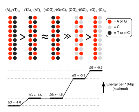
Unexpectedly, we found that DNA sequence has a profound impact on the strength of attractive interaction. The absolute value of ΔG at minimum relative to the value at maximum separation, |ΔGmin|, showed a clearly rank-ordered dependence on the DNA sequence: |ΔGmin| of (A)20>|ΔGmin| of (AT)10>|ΔGmin| of (GC)10>|ΔGmin| of (G)20. Two trends can be noted. First, AT-rich sequences attract each other more strongly than GC-rich sequences16. For example, |ΔGmin| of (AT)10 (1.5 kcal mol−1 per turn) is about twice |ΔGmin| of (GC)10 (0.8 kcal mol−1 per turn) (Fig. 1b). Second, duplexes having identical AT content but different partitioning of the nucleotides between the strands (that is, (A)20 versus (AT)10 or (G)20 versus (GC)10) exhibit statistically significant differences (~0.3 kcal mol−1 per turn) in the value of |ΔGmin|.

To validate the findings of MD simulations, we performed single-molecule fluorescence resonance energy transfer (smFRET)17 experiments of vesicle-encapsulated DNA molecules. Equimolar mixture of donor- and acceptor-labelled 120-bp dsDNA molecules was encapsulated in sub-micron size, porous lipid vesicles18 so that we could observe and quantitate rare binding events between a pair of dsDNA molecules without triggering large-scale DNA condensation2. Our DNA constructs were long enough to ensure dsDNA–dsDNA binding that is stable on the timescale of an smFRET measurement, but shorter than the DNA’s persistence length (~150 bp (ref. 19)) to avoid intramolecular condensation20. The vesicles were immobilized on a polymer-passivated surface, and fluorescence signals from individual vesicles containing one donor and one acceptor were selectively analysed (Fig. 1d). Binding of two dsDNA molecules brings their fluorescent labels in close proximity, increasing the FRET efficiency (Fig. 1e).

FRET signals from individual vesicles were diverse. Sporadic binding events were observed in some vesicles, while others exhibited stable binding; traces indicative of frequent conformational transitions were also observed (Supplementary Fig. 1A). Such diverse behaviours could be expected from non-specific interactions of two large biomolecules having structural degrees of freedom. No binding events were observed in the absence of Sm4+ (Supplementary Fig. 1B) or when no DNA molecules were present. To quantitatively assess the propensity of forming a bound state, we chose to use the fraction of single-molecule traces that showed any binding events within the observation time of 2 min (Methods). This binding fraction for the pair of AT-rich dsDNAs (AT1, 100% AT in the middle 80-bp section of the 120-bp construct) reached a maximum at ~2 mM Sm4+(Fig. 1f), which is consistent with the results of previous experimental studies2, 3. In accordance with the prediction of our MD simulations, GC-rich dsDNAs (GC1, 75% GC in the middle 80 bp) showed much lower binding fraction at all Sm4+ concentrations (Fig. 1b,c). Regardless of the DNA sequence, the binding fraction reduced back to zero at high Sm4+ concentrations, likely due to the resolubilization of now positively charged DNA–Sm4+ complexes2, 3, 13.

Because the donor and acceptor fluorophores were attached to the same sequence of DNA, it remained possible that the sequence homology between the donor-labelled DNA and the acceptor-labelled DNA was necessary for their interaction6. To test this possibility, we designed another AT-rich DNA construct AT2 by scrambling the central 80-bp section of AT1 to remove the sequence homology (Supplementary Table 1). The fraction of binding traces for this nonhomologous pair of donor-labelled AT1 and acceptor-labelled AT2 was comparable to that for the homologous AT-rich pair (donor-labelled AT1 and acceptor-labelled AT1) at all Sm4+ concentrations tested (Fig. 1f). Furthermore, this data set rules out the possibility that the higher binding fraction observed experimentally for the AT-rich constructs was caused by inter-duplex Watson–Crick base pairing of the partially melted constructs.

Next, we designed a DNA construct named ATGC, containing, in its middle section, a 40-bp AT-rich segment followed by a 40-bp GC-rich segment (Fig. 1g). By attaching the acceptor to the end of either the AT-rich or GC-rich segments, we could compare the likelihood of observing the parallel binding mode that brings the two AT-rich segments together and the anti-parallel binding mode. Measurements at 1 mM Sm4+ and 25 or 50 mM NaCl indicated a preference for the parallel binding mode by ~30% (Fig. 1h). Therefore, AT content can modulate DNA–DNA interactions even in a complex sequence context. Note that increasing the concentration of NaCl while keeping the concentration of Sm4+ constant enhances competition between Na+ and Sm4+ counterions, which reduces the concentration of Sm4+ near DNA and hence the frequency of dsDNA–dsDNA binding events (Supplementary Fig. 2).

Methylation determines the strength of DNA–DNA attraction

Analysis of the MD simulations revealed the molecular mechanism of the polyamine-mediated sequence-dependent attraction (Fig. 2). In the case of the AT-rich fragments, the bulky methyl group of thymine base blocks Sm4+ binding to the N7 nitrogen atom of adenine, which is the cation-binding hotspot21, 22. As a result, Sm4+ is not found in the major grooves of the AT-rich duplexes and resides mostly near the DNA backbone (Fig. 2a,d). Such relocated Sm4+ molecules bridge the two DNA duplexes better, accounting for the stronger attraction16, 23, 24, 25. In contrast, significant amount of Sm4+ is adsorbed to the major groove of the GC-rich helices that lacks cation-blocking methyl group (Fig. 2b,e).

Figure 2: Molecular mechanism of polyamine-mediated DNA sequence recognition.
Molecular mechanism of polyamine-mediated DNA sequence recognition.

(a–c) Representative configurations of Sm4+ molecules at the DNA–DNA distance of 28 Å for the (AT)10–(AT)10 (a), (GC)10–(GC)10 (b) and (GmC)10–(GmC)10 (c) DNA pairs. The backbone and bases of DNA are shown as ribbon and molecular bond, respectively; Sm4+ molecules are shown as molecular bonds. Spheres indicate the location of the N7 atoms and the methyl groups. (d–f) The average distributions of cations for the three sequence pairs featured in a–c. Top: density of Sm4+ nitrogen atoms (d=28 Å) averaged over the corresponding MD trajectory and the z axis. White circles (20 Å in diameter) indicate the location of the DNA helices. Bottom: the average density of Sm4+ nitrogen (blue), DNA phosphate (black) and sodium (red) atoms projected onto the DNA–DNA distance axis (x axis). The plot was obtained by averaging the corresponding heat map data over y=[−10, 10] Å. See Supplementary Figs 4 and 5 for the cation distributions at d=30, 32, 34 and 36 Å.

If indeed the extra methyl group in thymine, which is not found in cytosine, is responsible for stronger DNA–DNA interactions, we can predict that cytosine methylation, which occurs naturally in many eukaryotic organisms and is an essential epigenetic regulation mechanism26, would also increase the strength of DNA–DNA attraction. MD simulations showed that the GC-rich helices containing methylated cytosines (mC) lose the adsorbed Sm4+ (Fig. 2c,f) and that |ΔGmin| of (GC)10 increases on methylation of cytosines to become similar to |ΔGmin| of (AT)10 (Fig. 1b).

To experimentally assess the effect of cytosine methylation, we designed another GC-rich construct GC2 that had the same GC content as GC1 but a higher density of CpG sites (Supplementary Table 1). The CpG sites were then fully methylated using M. SssI methyltransferase (Supplementary Fig. 3; Methods). As predicted from the MD simulations, methylation of the GC-rich constructs increased the binding fraction to the level of the AT-rich constructs (Fig. 1f).

The sequence dependence of |ΔGmin| and its relation to the Sm4+ adsorption patterns can be rationalized by examining the number of Sm4+ molecules shared by the dsDNA molecules (Fig. 3a). An Sm4+ cation adsorbed to the major groove of one dsDNA is separated from the other dsDNA by at least 10 Å, contributing much less to the effective DNA–DNA attractive force than a cation positioned between the helices, that is, the ‘bridging’ Sm4+ (ref. 23). An adsorbed Sm4+ also repels other Sm4+ molecules due to like-charge repulsion, lowering the concentration of bridging Sm4+. To demonstrate that the concentration of bridging Sm4+ controls the strength of DNA–DNA attraction, we computed the number of bridging Sm4+ molecules, Nspm (Fig. 3b). Indeed, the number of bridging Sm4+ molecules ranks in the same order as |ΔGmin|: Nspm of (A)20>Nspm of (AT)10≈Nspm of (GmC)10>Nspm of (GC)10>Nspm of (G)20. Thus, the number density of nucleotides carrying a methyl group (T and mC) is the primary determinant of the strength of attractive interaction between two dsDNA molecules. At the same time, the spatial arrangement of the methyl group carrying nucleotides can affect the interaction strength as well (Fig. 3c). The number of methyl groups and their distribution in the (AT)10 and (GmC)10 duplex DNA are identical, and so are their interaction free energies, |ΔGmin| of (AT)10≈|ΔGmin| of (GmC)10. For AT-rich DNA sequences, clustering of the methyl groups repels Sm4+ from the major groove more efficiently than when the same number of methyl groups is distributed along the DNA (Fig. 3b). Hence, |ΔGmin| of (A)20>|ΔGmin| of (AT)10. For GC-rich DNA sequences, clustering of the cation-binding sites (N7 nitrogen) attracts more Sm4+ than when such sites are distributed along the DNA (Fig. 3b), hence |ΔGmin| is larger for (GC)10 than for (G)20.

Figure 3: Methylation modulates the interaction free energy of two dsDNA molecules by altering the number of bridging Sm4+.
Methylation modulates the interaction free energy of two dsDNA molecules by altering the number of bridging Sm4+.

(a) Typical spatial arrangement of Sm4+ molecules around a pair of DNA helices. The phosphates groups of DNA and the amine groups of Sm4+ are shown as red and blue spheres, respectively. ‘Bridging’ Sm4+molecules reside between the DNA helices. Orange rectangles illustrate the volume used for counting the number of bridging Sm4+ molecules. (b) The number of bridging amine groups as a function of the inter-DNA distance. The total number of Sm4+ nitrogen atoms was computed by averaging over the corresponding MD trajectory and the 10 Å (x axis) by 20 Å (y axis) rectangle prism volume (a) centred between the DNA molecules. (c) Schematic representation of the dependence of the interaction free energy of two DNA molecules on their nucleotide sequence. The number and spatial arrangement of nucleotides carrying a methyl group (T or mC) determine the interaction free energy of two dsDNA molecules.

Genome-wide investigations of chromosome conformations using the Hi–C technique revealed that AT-rich loci form tight clusters in human nucleus27, 28. Gene or chromosome inactivation is often accompanied by increased methylation of DNA29 and compaction of facultative heterochromatin regions30. The consistency between those phenomena and our findings suggest the possibility that the polyamine-mediated sequence-dependent DNA–DNA interaction might play a role in chromosome folding and epigenetic regulation of gene expression.

  1. Rau, D. C., Lee, B. & Parsegian, V. A. Measurement of the repulsive force between polyelectrolyte molecules in ionic solution: hydration forces between parallel DNA double helices. Proc. Natl Acad. Sci. USA 81, 2621–2625 (1984).
  2. Raspaud, E., Olvera de la Cruz, M., Sikorav, J. L. & Livolant, F. Precipitation of DNA by polyamines: a polyelectrolyte behavior. Biophys. J. 74, 381–393 (1998).
  3. Besteman, K., Van Eijk, K. & Lemay, S. G. Charge inversion accompanies DNA condensation by multivalent ions. Nat. Phys. 3, 641–644 (2007).
  4. Lipfert, J., Doniach, S., Das, R. & Herschlag, D. Understanding nucleic acid-ion interactions.Annu. Rev. Biochem. 83, 813–841 (2014).
  5. Grosberg, A. Y., Nguyen, T. T. & Shklovskii, B. I. The physics of charge inversion in chemical and biological systems. Rev. Mod. Phys. 74, 329–345 (2002).
  6. Kornyshev, A. A. & Leikin, S. Sequence recognition in the pairing of DNA duplexes. Phys. Rev. Lett. 86, 3666–3669 (2001).
  7. Danilowicz, C. et al. Single molecule detection of direct, homologous, DNA/DNA pairing.Proc. Natl Acad. Sci. USA 106, 19824–19829 (2009).
  8. Gladyshev, E. & Kleckner, N. Direct recognition of homology between double helices of DNA in Neurospora crassa. Nat. Commun. 5, 3509 (2014).
  9. Tabor, C. W. & Tabor, H. Polyamines. Annu. Rev. Biochem. 53, 749–790 (1984).
  10. Thomas, T. & Thomas, T. J. Polyamines in cell growth and cell death: molecular mechanisms and therapeutic applications. Cell. Mol. Life Sci. 58, 244–258 (2001).

Read Full Post »

Y chromosome

Larry H. Bernstein, MD, FCAP, Curator

LPBI


Y Chromosome Is More Than a Sex Switch


Here to stay. The Y chromosome is small compared with the X, but is required to keep levels of some genes high enough for mammals to survive.

http://www.sciencemag.org/sites/default/files/styles/article_main_small/public/images/sn-genes.jpg?itok=7mnkSPKy&timestamp=1398272995

Here to stay. The Y chromosome is small compared with the X, but is required to keep levels of some genes high enough for mammals to survive.

Andrew Syred/Science Source

The small, stumpy Y chromosome—possessed by male mammals but not females, and often shrugged off as doing little more than determining the sex of a developing fetus—may impact human biology in a big way. Two independent studies have concluded that the sex chromosome, which shrank millions of years ago, retains the handful of genes that it does not by chance, but because they are key to our survival. The findings may also explain differences in disease susceptibility between men and women.

“The old textbook description says that once maleness is determined by a few Y chromosome genes and you have gonads, all other sex differences stem from there,” says geneticist Andrew Clark of Cornell University, who was not involved in either study. “These papers open up the door to a much richer and more complex way to think about the Y chromosome.”

The sex chromosomes of mammals have evolved over millions of years, originating from two identical chromosomes. Now, males possess one X and one Y chromosome and females have two Xs. The presence or absence of the Y chromosome is what determines sex—the Y chromosome contains several genes key to testes formation. But while the X chromosome has remained large throughout evolution, with about 2000 genes, the Y chromosome lost most of its genetic material early in its evolution; it now retains less than 100 of those original genes. That’s led some scientists to hypothesize that the chromosome is largely indispensable and could shrink away entirely.

To determine which Y chromosome genes are shared across species, Daniel Winston Bellott, a biologist at the Whitehead Institute for Biomedical Research in Cambridge, Massachusetts, and colleagues compared the Y chromosomes of eight mammals, including humans, chimpanzees, monkeys, mice, rats, bulls, and opossums. The overlap, they found, wasn’t just in those genes known to determine the sex of an embryo. Eighteen diverse genes stood out as being highly similar between the species. The genes had broad functions including controlling the expression of genes in many other areas of the genome. The fact that all the species have retained these genes, despite massive changes to the overall Y chromosome, hints that they’re vital to mammalian survival.

“The thing that really came home to us was that these ancestral Y chromosome genes—these real survivors of millions of years of evolution—are regulators of lots of different processes,” Bellott says.

Bellott and his colleagues looked closer at the properties of the ancestral Y chromosome genes and found that the majority of them were dosage-dependent—that is, they required two copies of the gene to function. (For many genes on the sex chromosomes, only one copy is needed; in females, the copy on the second X chromosome is turned off and in males, the gene is missing altogether.) But with these genes, the female has one on each X chromosome and the male has a copy on both the X and Y chromosomes. Thus, despite the disappearance of nearby genes, these genes have persisted on the Y chromosome, the team reports online today in Nature.

“The Y chromosome doesn’t just say you’re a male; it doesn’t just say you’re a male and you’re fertile. It says that you’re a male, you’re fertile, and you’re going to survive,” Bellott explains. His group next plans to look in more detail at what the ancestral Y chromosome genes do, where they’re expressed in the body, and which are required for an organism’s survival.

In a second Nature paper, also published online today, another group of researchers used a different genetic sequencing approach, and a different set of mammals, to ask similar questions about the evolution of the Y chromosome. Like Bellott’s paper, the second study concluded that one reason that the Y chromosome has remained stable over recent history is the dosage dependence of the remaining genes.

“Knowing now that the Y chromosome can have effects all over the genome, I think it becomes even more important to look at its implications on diseases,” Clark says. “The chromosome is clearly much more than a single trigger that determines maleness.” Because genes on the Y chromosome often vary slightly in sequence—and even function—from the corresponding genes on the X, males could have slightly different patterns of gene expression throughout the body compared with females, due to not only their hormone levels, but also their entire Y chromosome. These gene expression variances could explain the differences in disease risks, or disease symptoms, between males and females, Clark says.

 
(1) http://www.sciencemag.org/news/2014/04/y-chromosome-more-sex-switch
https://docs.google.com/viewer?a=v&pid=sites&srcid=ZGVmYXVsdGRvbWFpbnxuYWxsYXNldGhmc3xneDo0MTI4NzdjNWZiNTBmNDFk
(2) https://docs.google.com/viewer?a=v&pid=sites&srcid=ZGVmYXVsdGRvbWFpbnxuYWxsYXNldGhmc3xneDoxZTJiYjc1OTU5ODFmZmJh
(3) https://docs.google.com/viewer?a=v&pid=sites&srcid=ZGVmYXVsdGRvbWFpbnxuYWxsYXNldGhmc3xneDo0ZTNmYjQwODA2M2JhY2E
(4) https://docs.google.com/viewer?a=v&pid=sites&srcid=ZGVmYXVsdGRvbWFpbnxuYWxsYXNldGhmc3xneDo1MzU2YzczNzgxNTI5YmEz

Read Full Post »

BET Proteins Connect Diabetes and Cancer

Larry H. Bernstein, MD, FCAP, Curator

LPBI


New Proteins Discovered That Link Obesity-Driven Diabetes to Cancer

http://www.dddmag.com/news/2016/03/new-proteins-discovered-link-obesity-driven-diabetes-cancer

 Killer T cells surround a cancer cell. Credit: NIH

Killer T cells surround a cancer cell. Credit: NIH


For the first time, researchers have determined how bromodomain (BRD) proteins work in type 2 diabetes, which may lead to a better understanding of the link between adult-onset diabetes and certain cancers.

The findings, which appear in PLOS ONE, show that reducing levels in pancreatic beta cells of individual BRDs, called BET proteins, previously shown to play a role in cancer, may also help patients who are obese and diabetic.

The research was led by Gerald V. Denis, PhD, associate professor of pharmacology and medicine at Boston University School of Medicine, who was the first to show that BET protein functions are important in cancer development.

Adult-onset diabetes has been known for decades to increase the risk for specific cancers. The three main members of the BET protein family, BRD2, BRD3 and BRD4, are closely related to each other and often cooperate. However at times, they work independently and sometimes against each other.

According to the researchers new small molecule BET inhibitors have been developed that block all three BET proteins in cancer cells, but they interfere with too many functions.

“The BET proteins provide a new pathway to connect adult-onset diabetes with cancer, so properly targeting BET proteins may be helpful for both,” explained Denis, who is the corresponding author of the study.

He believes this discovery shows the need for deeper analysis of individual BET proteins in all human cell types, starting with boosting insulin and improving metabolism in the pancreas of adults who are obese.

“Without better targeted drugs, some ongoing cancer clinical trials for BET inhibitors are premature. These new results offer useful insight into drug treatments that have failed so far to appreciate the complexities in the BET family.”


Epigenetic modulation of type-1 diabetes via a dual effect on pancreatic macrophages and β cells

eLife. 2014; 3: e04631.     doi:  10.7554/eLife.04631

Epigenetic modifiers are an emerging class of anti-tumor drugs, potent in multiple cancer contexts. Their effect on spontaneously developing autoimmune diseases has been little explored. We report that a short treatment with I-BET151, a small-molecule inhibitor of a family of bromodomain-containing transcriptional regulators, irreversibly suppressed development of type-1 diabetes in NOD mice. The inhibitor could prevent or clear insulitis, but had minimal influence on the transcriptomes of infiltrating and circulating T cells. Rather, it induced pancreatic macrophages to adopt an anti-inflammatory phenotype, impacting the NF-κB pathway in particular. I-BET151 also elicited regeneration of islet β-cells, inducing proliferation and expression of genes encoding transcription factors key to β-cell differentiation/function. The effect on β cells did not require T cell infiltration of the islets. Thus, treatment with I-BET151 achieves a ‘combination therapy’ currently advocated by many diabetes investigators, operating by a novel mechanism that coincidentally dampens islet inflammation and enhances β-cell regeneration.

DOI:http://dx.doi.org/10.7554/eLife.04631.001

eLife digest

The DNA inside a cell is often tightly wrapped around proteins to form a compact structure called chromatin. Chemical groups added to the chromatin can encourage nearby genes to either be switched on or off; and several enzymes and other proteins help to read, add, or remove these marks from the chromatin. If these chromatin modifications (or the related enzymes and proteins) are disturbed it can lead to diseases like cancer. It has also been suggested that similar changes may influence autoimmune diseases, in which the immune system attacks the body’s own tissues.

Drugs that target the proteins that read, add, or remove these chromatin modifications are currently being developed to treat cancer. For example, drugs that inhibit one family of these proteins called BET have helped to treat tumors in mice that have cancers of the blood or lymph nodes. However, because these drugs target pathways involved in the immune system they may also be useful for treating autoimmune diseases.

Now Fu et al. have tested whether a BET inhibitor might be a useful treatment for type-1 diabetes. In patients with type-1 diabetes, the cells in the pancreas that produce the insulin hormone are killed off by the immune system. Without adequate levels of insulin, individuals with type-1 diabetes may experience dangerous highs and lows in their blood sugar levels and must take insulin and sometimes other medications.

Using mice that spontaneously develop type-1 diabetes when still relatively young, Fu et al. tested what would happen if the mice received a BET inhibitor for just 2 weeks early on in life. Treated mice were protected from developing type-1 diabetes for the rest of their lives. Specifically, the treatment protected the insulin-producing cells and allowed them to continue producing insulin. The drug reduced inflammation in the pancreas and increased the expression of genes that promote the regeneration of insulin-producing cells.

Diabetes researchers have been searching for drug combinations that protect the insulin-producing cells and boost their regeneration. As such, Fu et al. suggest that these findings justify further studies to see if BET inhibitors may help to treat or prevent type-1 diabetes in humans.

Introduction

Acetylation of lysine residues on histones and non-histone proteins is an important epigenetic modification of chromatin (Kouzarides, 2000). Multiple ‘writers’, ‘erasers’, and ‘readers’ of this modification have been identified: histone acetyltransferases (HATs) that introduce acetyl groups, histone deacetylases (HDACs) that remove them, and bromodomain (BRD)-containing proteins that specifically recognize them. Chromatin acetylation impacts multiple fundamental cellular processes, and its dysregulation has been linked to a variety of disease states, notably various cancers (Dawson and Kouzarides, 2012). Not surprisingly, then, drugs that modulate the activities of HATs or HDACs or, most recently, that block acetyl-lysine:BRD interactions are under active development in the oncology field.

BRDs, conserved from yeast to humans, are domains of approximately 110 amino-acids that recognize acetylation marks on histones (primarily H3 and H4) and certain non-histone proteins (e.g., the transcription factor, NF-κB), and serve as scaffolds for the assembly of multi-protein complexes that regulate transcription (Dawson et al., 2011; Prinjha et al., 2012). The BET subfamily of BRD-containing proteins (BRDs 2, 3, 4 and T) is distinguished as having tandem bromodomains followed by an ‘extra-terminal’ domain. One of its members, Brd4, is critical for both ‘bookmarking’ transcribed loci post-mitotically (Zhao et al., 2011) and surmounting RNA polymerase pausing downstream of transcription initiation (Jang et al., 2005; Hargreaves et al., 2009; Anand et al., 2013; Patel et al., 2013).

Recently, small-molecule inhibitors of BET proteins, for example, JQ1 and I-BET, were found to be effective inhibitors of multiple types of mouse tumors, including a NUT midline carcinoma, leukemias, lymphomas and multiple myeloma (Filippakopoulos et al., 2010; Dawson et al., 2011; Delmore et al., 2011; Zuber et al., 2011). A major, but not the unique, focus of inhibition was the Myc pathway (Delmore et al., 2011; Mertz et al., 2011; Zuber et al., 2011; Lockwood et al., 2012). In addition, BET-protein inhibitors could prevent or reverse endotoxic shock induced by systemic injection of bacterial lipopolysaccharide (LPS) (Nicodeme et al., 2010; Seal et al., 2012; Belkina et al., 2013). The primary cellular focus of action was macrophages, and genes induced by the transcription factor NF-κB were key molecular targets (Nicodeme et al., 2010; Belkina et al., 2013).

Given several recent successes at transposing drugs developed for cancer therapy to the context of autoimmunity, it was logical to explore the effect of BET-protein inhibitors on autoimmune disease. We wondered how they might impact type-1 diabetes (T1D), hallmarked by specific destruction of the insulin-producing β cells of the pancreatic islets (Bluestone et al., 2010). NOD mice, the ‘gold standard’ T1D model (Anderson and Bluestone, 2005), spontaneously and universally develop insulitis at 4–6 weeks of age, while overt diabetes manifests in a subset of individuals beginning from 12–15 weeks, depending on the particular colony. NOD diabetes is primarily a T-cell-mediated disease, but other immune cells—such as B cells, natural killer cells, macrophages (MFs) and dendritic cells (DCs)—also play significant roles. We demonstrate that a punctual, 2-week, treatment of early- or late-stage prediabetic NOD mice with I-BET151 affords long-term protection from diabetes. Mechanistic dissection of this effect revealed important drug influences on both MFs and β cells, in particular on the NF-κB pathway. On the basis of these findings, we argue that epigenetic modifiers are an exciting, emerging option for therapeutic intervention in autoimmune diabetes.

I-BET151 protects NOD mice from development of diabetes

T1D progresses through identifiable phases, which are differentially sensitive to therapeutic intervention (Bluestone et al., 2010). Therefore, we treated NOD mice with the BET-protein inhibitor, I-BET151 (GSK1210151A [Dawson et al., 2011;Seal et al., 2012]) according to three different protocols: from 3–5 weeks of age (incipient insulitis), from 12–14 weeks of age (established insulitis), or for 2 weeks beginning within a day after diagnosis of hyperglycemia (diabetes). Blood-glucose levels of insulitic mice were monitored until 30 weeks of age, after which animals in our colony generally do not progress to diabetes.

I-BET151 prevented diabetes development, no matter whether the treated cohort had incipient (Figure 1A) or established (Figure 1B) insulitis. However, the long-term protection afforded by a 2-week treatment of pre-diabetic mice was only rarely observed with recent-onset diabetic animals. Just after diagnosis, individuals were given a subcutaneous insulin implant, which lowers blood-glucose levels to the normal range within 2 days, where they remain for only about 7 days in the absence of further insulin supplementation (Figure 1C, upper and right panels). Normoglycemia was significantly prolonged in mice treated for 2 weeks with I-BET151; but, upon drug removal, hyperglycemia rapidly ensued in most animals (Figure 1C, lower and right panels). The lack of disease reversal under these conditions suggests that β-cell destruction had proceeded to the point that dampening the autoinflammatory attack was not enough to stem hyperglycemia. However, there was prolonged protection from diabetes in a few cases, suggesting that it might prove worthwhile to explore additional treatment designs in future studies.

I-BET151 inhibits diabetes and insulitis in NOD mice.

…..

BET protein inhibition has a minimal effect on T cells in NOD mice

Given that NOD diabetes is heavily dependent on CD4+ T cells (Anderson and Bluestone, 2005), and that a few recent reports have highlighted an influence of BET-protein inhibitors on the differentiation of T helper (Th) subsets in induced models of autoimmunity (Bandukwala et al., 2012; Mele et al., 2013), we explored the effect of I-BET151 treatment on the transcriptome of CD4+ T cells isolated from relevant sites; that is, the infiltrated pancreas, draining pancreatic lymph nodes (PLNs), and control inguinal lymph nodes (ILNs). Microarray analysis of gene expression revealed surprisingly little impact of the 2-week treatment protocol on any of these populations, similar to what was observed when comparing randomly shuffled datasets (Figure 2A). It is possible that the above protocol missed important effects on T cells because those remaining after prolonged drug treatment were skewed for ‘survivors’. Therefore, we also examined the transcriptomes of pancreas-infiltrating CD4+ T cells at just 12, 24 or 48 hr after a single administration of I-BET151. Again, minimal, background-level, differences were observed in the gene-expression profiles of drug- and vehicle-treated mice (Figure 2B).

Little impact of BET-protein inhibition on CD4+T cells in NOD mice.

I-BET151 induces a regulatory phenotype in the pancreatic macrophage population

I-BET151 treatment promotes an MF-like, anti-inflammatory transcriptional program in pancreatic CD45+ cells.
The NF-κB signaling pathway is a major focus of I-BET151’s influence on NOD leukocytes.

BET-protein inhibition promotes regeneration of NOD β cells

BET-protein inhibition promotes regeneration of islet β cells

The studies presented here showed that treatment of NOD mice with the epigenetic modifier, I-BET151, for a mere 2 weeks prevented the development of NOD diabetes life-long. I-BET151 was able to inhibit impending insulitis as well as clear existing islet infiltration. The drug had a dual mechanism of action: it induced the pancreatic MF population to adopt an anti-inflammatory phenotype, primarily via the NF-κB pathway, and promoted β-cell proliferation (and perhaps differentiation). These findings raise a number of intriguing questions, three of which we address here.

First, why do the mechanisms uncovered in our study appear to be so different from those proposed in the only two previous reports on the effect of BET-protein inhibitors on autoimmune disease? Bandukwala et al. found that I-BET762 (a small-molecule inhibitor similar to I-BET151) altered the differentiation of Th subsets in vitro, perturbing the typical profiles of cytokine production, and reducing the neuropathology provoked by transfer of in-vitro-differentiated Th1, but not Th17, cells reactive to a peptide of myelin oligodendrocyte glycoprotein (Bandukwala et al., 2012). Unfortunately, with such transfer models, it is difficult to know how well the in vitro processes reflect in vivo events, and to distinguish subsidiary effects on cell survival and homing. Mele et al. reported that JQ1 primarily inhibited the differentiation of and cytokine production by Th17 cells, and strongly repressed collagen-induced arthritis and experimental allergic encephalomyelitis (Mele et al., 2013). However, with adjuvant-induced disease models such as these, it is difficult to discriminate influences of the drug on the unfolding of autoimmune pathology vs on whatever the adjuvant is doing. Thus, the very different dual mechanism we propose for I-BET151’s impact on spontaneously developing T1D in NOD mice may reflect several factors, including (but not limited to): pathogenetic differences in induced vs spontaneous autoimmune disease models; our broader analyses of immune target cell populations; and true mechanistic differences between T1D and the other diseases. As concerns the latter, it has been argued that T1D is primarily a Th1-driven disease, with little, or even a negative regulatory, influence by Th17 cells (discussed in [Kriegel et al., 2011]).

Second, how does I-BET151’s effect, focused on MFs and β cells, lead to life-long protection from T1D? MFs seem to play a schizophrenic role in the NOD disease. They were shown long ago to be an early participant in islet infiltration (Jansen et al., 1994), and to play a critical effector role in diabetes pathogenesis, attributed primarily to the production of inflammatory cytokines and other mediators, such as iNOS (Hutchings et al., 1990; Jun et al., 1999a, 1999b; Calderon et al., 2006). More recently, there has been a growing appreciation of their regulatory role in keeping diabetes in check. For example, the frequency of a small subset of pancreatic MFs expressing the complement receptor for immunoglobulin (a.k.a. CRIg) at 6–10 weeks of age determined whether or not NOD diabetes would develop months later (Fu et al., 2012b), and transfer of in-vitro-differentiated M2, but not M1, MFs protected NOD mice from disease development (Parsa et al., 2012).

One normally thinks of immunological tolerance as being the purview of T and B cells, but MFs seem to be playing the driving role in I-BET151’s long-term immunologic impact on T1D. Chronic inflammation (as is the insulitis associated with T1D) typically entails three classes of participant: myeloid cells, in particular, tissue-resident MFs; lymphoid cells, including effector and regulatory T and B cells; and tissue-target cells, that is, islet β cells in the T1D context. The ‘flavor’ and severity of inflammation is determined by three-way interactions amongst these cellular players. One implication of this cross-talk is that a perturbation that targets primarily one of the three compartments has the potential to rebalance the dynamic process of inflammation, resetting homeostasis to a new level either beneficial or detrimental to the individual. BET-protein inhibition skewed the phenotype of pancreatic MFs towards an anti-inflammatory phenotype, whether this be at the population level through differential influx, efflux or death, or at the level of individual cells owing to changes in transcriptional programs. The ‘re-educated’ macrophages appeared to be more potent at inhibiting T cell proliferation. In addition, it is possible that MFs play some role in the I-BET151 influences on β-cell regeneration. The findings on Rag1-deficient mice ruled out the need for adaptive immune cells in the islet infiltrate for I-BET151’s induction of β-cell proliferation, but MFs are not thought to be compromised in this strain. Relatedly, the lack of a consistent I-BET151 effect on cultured mouse and human islets might result from a dearth of MFs under our isolation and incubation conditions (e.g., [Li et al., 2009]). Several recent publications have highlighted a role for MFs, particularly M2 cells, in promoting regeneration of β cells in diverse experimental settings (Brissova et al., 2014; Xiao et al., 2014), a function foretold by the reduced β-cell mass in MF-deficient Csf1op/op mice reported a decade ago (Banaei-Bouchareb et al., 2004).

Whether reflecting a cell-intrinsic or -extrinsic impact of the drug, several pro-regenerative pathways appear to be enhanced in β-cells from I-BET151-treated mice. Increased β-cell proliferation could result from up-regulation of the genes encoding Neurod1 (Kojima et al., 2003), GLP-1R (De Leon et al., 2003), or various of the Reg family members (Unno et al., 2002; Liu et al., 2008), the latter perhaps a consequence of higher IL-22R expression (Hill et al., 2013) (see Figure 6B and Supplementary file 4). Protection of β-cells from apoptosis is likely to be an important outcome of inhibiting the NF-κB pathway (Takahashi et al., 2010), but could also issue from enhanced expression of other known pro-survival factors, such as Cntfr (Rezende et al., 2007) and Tox3 (Dittmer et al., 2011) (see Figures 4 and 6B). Lastly, β-cell differentiation and function should be fostered by up-regulation of genes encoding transcription factors such as Neurod1, Pdx1, Pax6, Nkx6-1 and Nkx2-2. The significant delay in re-onset of diabetes in I-BET151-treated diabetic mice suggests functionally relevant improvement in β-cell function. In brief, the striking effect of I-BET151 on T1D development in NOD mice seems to reflect the fortunate concurrence of a complex, though inter-related, set of diabetes-protective processes.

Lastly, why does a drug that inhibits BET proteins, which include general transcription factors such as Brd4, have such circumscribed effects? A 2-week I-BET151 treatment might be expected to provoke numerous side-effects, but this regimen seemed in general to be well tolerated in our studies. This conundrum has been raised in several contexts of BET-inhibitor treatment, and was recently discussed at length (Shi and Vakoc, 2014). The explanation probably relates to two features of BET-protein, in particular Brd4, biology. First: Brd4 is an important element of so-called ‘super-enhancers’, defined as unusually long transcriptional enhancers that host an exceptionally high density of TFs—both cell-type-specific and general factors, including RNA polymerase-II, Mediator, p300 and Brd4 (Hnisz et al., 2013). They are thought to serve as chromatin depots, collecting TFs and coordinating their delivery to transcriptional start-sites via intra-chromosome looping or inter-chromosome interactions. Super-enhancers are preferentially associated with loci that define and control the biology of particular cell-types, notably developmentally regulated and inducible genes; intriguingly, disease-associated, including T1D-associated, nucleotide polymorphisms are especially enriched in the super-enhancers of disease-relevant cell-types (Hnisz et al., 2013;Parker et al., 2013). Genes associated with super-enhancers show unusually high sensitivity to BET-protein inhibitors (Chapuy et al., 2013; Loven et al., 2013;Whyte et al., 2013). Second: although the bromodomain of Brd4 binds to acetyl-lysine residues on histone-4, and I-BET151 was modeled to inhibit this interaction, it is now known to bind to a few non-histone chromosomal proteins as well, notably NF-κB, a liaison also blocked by BET-protein inhibitors (Huang et al., 2009; Zhang et al., 2012; Zou et al., 2014). Abrogating specific interactions such as these, differing according to the cellular context, might be the dominant impact of BET inhibitors, a scenario that would be consistent with the similar effects we observed with I-BET151 and BAY 11–7082 treatment. Either or both of these explanations could account for the circumscribed effect of I-BET151 on NOD diabetes. Additionally, specificity might be imparted by different BET-family members or isoforms—notably both Brd2 and Brd4 are players in MF inflammatory responses (Belkina et al., 2013). According to either of these explanations, higher doses might unleash a broader array of effects.


Islet inflammation: A unifying target for diabetes treatment?

In the last decade, islet inflammation has emerged as a contributor to the loss of functional β cell mass in both type 1 (T1D) and type 2 diabetes (T2D). Evidence supports that over-nutrition and insulin resistance result in the production of proinflammatory mediators by β cells. In addition to compromising β cell function and survival, cytokines may recruit macrophages into islets, thus augmenting inflammation. Limited, but intriguing, data implies a role of adaptive immune response in islet dysfunction in T2D. Clinical trials validated anti-inflammatory therapies in T2D, while immune therapy for T1D remains challenging. Further research is required to improve our understanding of islet inflammatory pathways, and to identify more effective therapeutic targets for T1D and T2D.
Islet inflammation: an emerging and unifying target for diabetes treatment

The current epidemic of T2D is closely associated with increases in obesity [1]. Excessive energy balance results in insulin resistance that is compensated for by increasing insulin secretion. However, insufficient compensation results in T2D, which is characterized by the reduction in islet mass and function. In recent years, overwhelming evidence defines insulin resistance as a state of chronic inflammation involving both innate and adaptive immune responses [1]. Although the presence of islet inflammation is acknowledged for autoimmune destruction of β cells in T1D, new data implicates overlapping pathogenesis between T1D and T2D. Epidemiologic studies suggest that obesity modifies the risk of T1D development [2, 3]. Importantly, small but seminal human studies have also provided evidences that anti-inflammatory therapy can improve glycemia and β cell function in T2D [4, 5]. Here, we focus on recent discoveries (past five years) to discuss the contribution of inflammatory pathways to islet dysfunction in T2D, and to provide updates on the pathogenesis of T1D.

What triggers inflammation in islets under insulin resistance?

Ample evidence from rodent and human studies indicates that in obesity, adipose tissue (AT) inflammation is a major source of pro-inflammatory mediators, and a primary response to excessive caloric intake. AT contributes to inflammation in obesity by means of increased mass, modified adipocyte phenotype, and increased infiltration of immune cells, which affects islet function through humoral and neuronal pathways [1, 6, 7]. In addition, it is noteworthy that pancreatic islets are under similar stress as adipocytes in T2D. The chronic inflammatory state of T2D is reflected in the elevation of circulatory cytokines that potentially affect islets as well as adipocytes [6, 8]. Both islets and adipocytes are exposed to excess glucose and lipids, especially free fatty acids (FFA). Over-nutrition forces adipose tissue to remodel and accommodate enlarged adipocytes, which results in endoplasmic reticulum (ER) stress, hypoxia, and mechanical stresses [9–11]. Under insulin resistance, insulin production increases to meet the high demand, resulting in the expansion of islet mass [12]. Recent findings revealed that obesity is associated with the activation of inflammatory pathway in the hypothalamus, which may alter functions of AT and islets through neuronal regulation [13]. Considering the multiple stressors potentially shared by AT and islets, it is plausible that islets exist also in a chronic inflammatory state, in T2D.

Adipose tissue dysfunction in obesity: a contributor to β cell inflammation in T2D?

The relationship between the pancreatic islet and AT was thought to be unidirectional, by placing insulin secretion as the major determinant of adipocyte glucose uptake and triglyceride storage. However, several recent studies suggest that insulin resistance in AT significantly contributes to β cell failure, through altered secretion of humoral factors from adipocytes and signals from the adipocyte sensory nerve (Figure. 1) [6, 7]. Of particular interest are adipocytokines that are uniquely produced by adipocytes, such as leptin, adiponectin, omentin, resistin, and visfatin, which may contribute to β cell dysfunction during insulin resistance (Box 1). Circulating cytokines may also connect AT inflammation to β cell dysfunction. Overnight exposure of mouse islets to tumor necrosis factor-alpha (TNFα), Interleukin beta (IL-1β), plus Interferon-gamma (IFNγ), at levels comparable to those seen in human obesity, disrupts the regulation of intracellular calcium [8]. Although glucose stimulated insulin secretion (GSIS) was maintained in this study, circulating cytokines might contribute to islet dysfunction after a prolonged period of exposure and when combined with other stresses [8]. TNFα, a cytokine implicated in insulin resistance, reportedly increased islet amyloid polypeptide (IAPP, amylin) expression in β cells with no concurrent expression of proinsulin. This may lead to amyloid production and β cell death [14]. Recent findings showed that the enzyme dipeptidyl peptidase-4 (DPPIV) is secreted by human adipocytes, and therefore may reduce the half-life of DPPIV substrate glucagon-like peptide-1 (GLP-1) with important implications on the insulinotropic effects of this gut peptide on the β cells [15]. Although it is not clear if obesity is associated with increased levels of DPPIV, inhibition of the latter by sitagliptin in a rodent model of obesity and insulin resistance reduced inflammatory cytokine production both in islets and in AT, and improved glucose-stimulated insulin secretion (GSIS) in islets in vitro [16]. Collectively, dysfunctional AT in obesity produces cytokines and peptides that affect islet health and potentially contribute to islet inflammation in T2D.

Read Full Post »

Conduction, graphene, elements and light

Larry H. Bernstein, MD, FCAP, Curator

LPBI


New 2D material could upstage graphene   Mar 25, 2016

Can function as a conductor or semiconductor, is extremely stable, and uses light, inexpensive earth-abundant elements
http://www.kurzweilai.net/new-2d-material-could-upstage-graphene
The atoms in the new structure are arranged in a hexagonal pattern as in graphene, but that is where the similarity ends. The three elements forming the new material all have different sizes; the bonds connecting the atoms are also different. As a result, the sides of the hexagons formed by these atoms are unequal, unlike in graphene. (credit: Madhu Menon)

A new one-atom-thick flat material made up of silicon, boron, and nitrogen can function as a conductor or semiconductor (unlike graphene) and could upstage graphene and advance digital technology, say scientists at the University of Kentucky, Daimler in Germany, and the Institute for Electronic Structure and Laser (IESL) in Greece.

Reported in Physical Review B, Rapid Communications, the new Si2BN material was discovered in theory (not yet made in the lab). It uses light, inexpensive earth-abundant elements and is extremely stable, a property many other graphene alternatives lack, says University of Kentucky Center for Computational Sciences physicist Madhu Menon, PhD.

Limitations of other 2D semiconducting materials

A search for new 2D semiconducting materials has led researchers to a new class of three-layer materials called transition-metal dichalcogenides (TMDCs). TMDCs are mostly semiconductors and can be made into digital processors with greater efficiency than anything possible with silicon. However, these are much bulkier than graphene and made of materials that are not necessarily earth-abundant and inexpensive.

Other graphene-like materials have been proposed but lack the strengths of the new material. Silicene, for example, does not have a flat surface and eventually forms a 3D surface. Other materials are highly unstable, some only for a few hours at most.

The new Si2BN material is metallic, but by attaching other elements on top of the silicon atoms, its band gap can be changed (from conductor to semiconductor, for example) — a key advantage over graphene for electronics applications and solar-energy conversion.

The presence of silicon also suggests possible seamless integration with current silicon-based technology, allowing the industry to slowly move away from silicon, rather than precipitously, notes Menon.

https://youtu.be/lKc_PbTD5go

Abstract of Prediction of a new graphenelike Si2BN solid

While the possibility to create a single-atom-thick two-dimensional layer from any material remains, only a few such structures have been obtained other than graphene and a monolayer of boron nitride. Here, based upon ab initiotheoretical simulations, we propose a new stable graphenelike single-atomic-layer Si2BN structure that has all of its atoms with sp2 bonding with no out-of-plane buckling. The structure is found to be metallic with a finite density of states at the Fermi level. This structure can be rolled into nanotubes in a manner similar to graphene. Combining first- and second-row elements in the Periodic Table to form a one-atom-thick material that is also flat opens up the possibility for studying new physics beyond graphene. The presence of Si will make the surface more reactive and therefore a promising candidate for hydrogen storage.


Nano-enhanced textiles clean themselves with light

Catalytic uses for industrial-scale chemical processes in agrochemicals, pharmaceuticals, and natural products also seen
http://www.kurzweilai.net/nano-enhanced-textiles-clean-themselves-with-light
Close-up of nanostructures grown on cotton textiles. Image magnified 150,000 times. (credit: RMIT University)

Researchers at at RMIT University in Australia have developed a cheap, efficient way to grow special copper- and silver-based nanostructures on textiles that can degrade organic matter when exposed to light.

Don’t throw out your washing machine yet, but the work paves the way toward nano-enhanced textiles that can spontaneously clean themselves of stains and grime simply by being put under a light or worn out in the sun.

The nanostructures absorb visible light (via localized surface plasmon resonance — collective electron-charge oscillations in metallic nanoparticles that are excited by light), generating high-energy (“hot”) electrons that cause the nanostructures to act as catalysts for chemical reactions that degrade organic matter.

Steps involved in fabricating copper- and silver-based cotton fabrics: 1. Sensitize the fabric with tin. 2. Form palladium seeds that act as nucleation (clustering) sites. 3. Grow metallic copper and silver nanoparticles on the surface of the cotton fabric. (credit: Samuel R. Anderson et al./Advanced Materials Interfaces)

The challenge for researchers has been to bring the concept out of the lab by working out how to build these nanostructures on an industrial scale and permanently attach them to textiles. The RMIT team’s novel approach was to grow the nanostructures directly onto the textiles by dipping them into specific solutions, resulting in development of stable nanostructures within 30 minutes.

When exposed to light, it took less than six minutes for some of the nano-enhanced textiles to spontaneously clean themselves.

The research was described in the journal Advanced Materials Interfaces.

Scaling up to industrial levels

Rajesh Ramanathan, a RMIT postdoctoral fellow and co-senior author, said the process also had a variety of applications for catalysis-based industries such as agrochemicals, pharmaceuticals, and natural products, and could be easily scaled up to industrial levels. “The advantage of textiles is they already have a 3D structure, so they are great at absorbing light, which in turn speeds up the process of degrading organic matter,” he said.

Cotton textile fabric with copper-based nanostructures. The image is magnified 200 times. (credit: RMIT University)

“Our next step will be to test our nano-enhanced textiles with organic compounds that could be more relevant to consumers, to see how quickly they can handle common stains like tomato sauce or wine,” Ramanathan said.

“There’s more work to do to before we can start throwing out our washing machines, but this advance lays a strong foundation for the future development of fully self-cleaning textiles.”


Abstract of Robust Nanostructured Silver and Copper Fabrics with Localized Surface Plasmon Resonance Property for Effective Visible Light Induced Reductive Catalysis

Inspired by high porosity, absorbency, wettability, and hierarchical ordering on the micrometer and nanometer scale of cotton fabrics, a facile strategy is developed to coat visible light active metal nanostructures of copper and silver on cotton fabric substrates. The fabrication of nanostructured Ag and Cu onto interwoven threads of a cotton fabric by electroless deposition creates metal nanostructures that show a localized surface plasmon resonance (LSPR) effect. The micro/nanoscale hierarchical ordering of the cotton fabrics allows access to catalytically active sites to participate in heterogeneous catalysis with high efficiency. The ability of metals to absorb visible light through LSPR further enhances the catalytic reaction rates under photoexcitation conditions. Understanding the modes of electron transfer during visible light illumination in Ag@Cotton and Cu@Cotton through electrochemical measurements provides mechanistic evidence on the influence of light in promoting electron transfer during heterogeneous catalysis for the first time. The outcomes presented in this work will be helpful in designing new multifunctional fabrics with the ability to absorb visible light and thereby enhance light-activated catalytic processes.


New type of molecular tag makes MRI 10,000 times more sensitive

Could detect biochemical processes in opaque tissue without requiring PET radiation or CT x-rays
http://www.kurzweilai.net/new-type-of-molecular-tag-makes-mri-10000-times-more-sensitive

Duke scientists have discovered a new class of inexpensive, long-lived molecular tags that enhance MRI signals by 10,000 times. To activate the tags, the researchers mix them with a newly developed catalyst (center) and a special form of hydrogen (gray), converting them into long-lived magnetic resonance “lightbulbs” that might be used to track disease metabolism in real time. (credit: Thomas Theis, Duke University)

Duke University researchers have discovered a new form of MRI that’s 10,000 times more sensitive and could record actual biochemical reactions, such as those involved in cancer and heart disease, and in real time.

Let’s review how MRI (magnetic resonance imaging) works: MRI takes advantage of a property called spin, which makes the nuclei in hydrogen atoms act like tiny magnets. By generating a strong magnetic field (such as 3 Tesla) and a series of radio-frequency waves, MRI induces these hydrogen magnets in atoms to broadcast their locations. Since most of the hydrogen atoms in the body are bound up in water, the technique is used in clinical settings to create detailed images of soft tissues like organs (such as the brain), blood vessels, and tumors inside the body.


MRI’s ability to track chemical transformations in the body has been limited by the low sensitivity of the technique. That makes it impossible to detect small numbers of molecules (without using unattainably more massive magnetic fields).

So to take MRI a giant step further in sensitivity, the Duke researchers created a new class of molecular “tags” that can track disease metabolism in real time, and can last for more than an hour, using a technique called hyperpolarization.* These tags are biocompatible and inexpensive to produce, allowing for using existing MRI machines.

“This represents a completely new class of molecules that doesn’t look anything at all like what people thought could be made into MRI tags,” said Warren S. Warren, James B. Duke Professor and Chair of Physics at Duke, and senior author on the study. “We envision it could provide a whole new way to use MRI to learn about the biochemistry of disease.”

Sensitive tissue detection without radiation

The new molecular tags open up a new world for medicine and research by making it possible to detect what’s happening in optically opaque tissue instead of requiring expensive positron emission tomography (PET), which uses a radioactive tracer chemical to look at organs in the body and only works for (typically) about 20 minutes, or CT x-rays, according to the researchers.

This research was reported in the March 25 issue of Science Advances. It was supported by the National Science Foundation, the National Institutes of Health, the Department of Defense Congressionally Directed Medical Research Programs Breast Cancer grant, the Pratt School of Engineering Research Innovation Seed Fund, the Burroughs Wellcome Fellowship, and the Donors of the American Chemical Society Petroleum Research Fund.

* For the past decade, researchers have been developing methods to “hyperpolarize” biologically important molecules. “Hyperpolarization gives them 10,000 times more signal than they would normally have if they had just been magnetized in an ordinary magnetic field,” Warren said. But while promising, Warren says these hyperpolarization techniques face two fundamental problems: incredibly expensive equipment — around 3 million dollars for one machine — and most of these molecular “lightbulbs” burn out in a matter of seconds.

“It’s hard to take an image with an agent that is only visible for seconds, and there are a lot of biological processes you could never hope to see,” said Warren. “We wanted to try to figure out what molecules could give extremely long-lived signals so that you could look at slower processes.”

So the researchers synthesized a series of molecules containing diazarines — a chemical structure composed of two nitrogen atoms bound together in a ring. Diazirines were a promising target for screening because their geometry traps hyperpolarization in a “hidden state” where it cannot relax quickly. Using a simple and inexpensive approach to hyperpolarization called SABRE-SHEATH, in which the molecular tags are mixed with a spin-polarized form of hydrogen and a catalyst, the researchers were able to rapidly hyperpolarize one of the diazirine-containing molecules, greatly enhancing its magnetic resonance signals for over an hour.

The scientists believe their SABRE-SHEATH catalyst could be used to hyperpolarize a wide variety of chemical structures at a fraction of the cost of other methods.


Abstract of Direct and cost-efficient hyperpolarization of long-lived nuclear spin states on universal 15N2-diazirine molecular tags

Abstract of Direct and cost-efficient hyperpolarization of long-lived nuclear spin states on universal 15N2-diazirine molecular tags

Conventional magnetic resonance (MR) faces serious sensitivity limitations, which can be overcome by hyperpolarization methods, but the most common method (dynamic nuclear polarization) is complex and expensive, and applications are limited by short spin lifetimes (typically seconds) of biologically relevant molecules. We use a recently developed method, SABRE-SHEATH, to directly hyperpolarize 15N2 magnetization and long-lived 15N2singlet spin order, with signal decay time constants of 5.8 and 23 min, respectively. We find >10,000-fold enhancements generating detectable nuclear MR signals that last for more than an hour. 15N2-diazirines represent a class of particularly promising and versatile molecular tags, and can be incorporated into a wide range of biomolecules without significantly altering molecular function.

references:

[Seems like they have a great idea, now all they need to do is confirm very specific uses or types of cancers/diseases or other processes they can track or target. Will be interesting to see if they can do more than just see things, maybe they can use this to target and destroy bad things in the body also. Keep up the good work….. this sounds like a game changer.]


Scientists time-reverse developed stem cells to make them ‘embryonic’ again

May help avoid ethically controversial use of human embryos for research and support other research goals
http://www.kurzweilai.net/scientists-time-reverse-developed-stem-cells-to-make-them-embryonic-again
Researchers have reversed “primed” (developed) “epiblast” stem cells (top) from early mouse embryos using the drug MM-401, causing the treated cells (bottom) to revert to the original form of the stem cells. (credit: University of Michigan)

University of Michigan Medical School researchers have discovered a way to convert mouse stem cells (taken from an embryo) that have  become “primed” (reached the stage where they can  differentiate, or develop into every specialized cell in the body) to a “naïve” (unspecialized) state by simply adding a drug.

This breakthrough has the potential to one day allow researchers to avoid the ethically controversial use of human embryos left over from infertility treatments. To achieve this breakthrough, the researchers treated the primedembryonic stem cells (“EpiSC”) with a drug called MM-401* (a leukemia drug) for a short period of time.

Embryonic stem cells are able to develop into any type of cell, except those of the placenta (credit: Mike Jones/CC)

…..

* The drug, MM-401, specifically targets epigenetic chemical markers on histones, the protein “spools” that DNA coils around to create structures called chromatin. These epigenetic changes signal the cell’s DNA-reading machinery and tell it where to start uncoiling the chromatin in order to read it.

A gene called Mll1 is responsible for the addition of these epigenetic changes, which are like small chemical tags called methyl groups. Mll1 plays a key role in the uncontrolled explosion of white blood cells in leukemia, which is why researchers developed the drug MM-401 to interfere with this process. But Mll1 also plays a role in cell development and the formation of blood cells and other cells in later-stage embryos.

Stem cells do not turn on the Mll1 gene until they are more developed. The MM-401 drug blocks Mll1’s normal activity in developing cells so the epigenetic chemical markers are missing. These cells are then unable to continue to develop into different types of specialized cells but are still able to revert to healthy naive pluripotent stem cells.


Abstract of MLL1 Inhibition Reprograms Epiblast Stem Cells to Naive Pluripotency

The interconversion between naive and primed pluripotent states is accompanied by drastic epigenetic rearrangements. However, it is unclear whether intrinsic epigenetic events can drive reprogramming to naive pluripotency or if distinct chromatin states are instead simply a reflection of discrete pluripotent states. Here, we show that blocking histone H3K4 methyltransferase MLL1 activity with the small-molecule inhibitor MM-401 reprograms mouse epiblast stem cells (EpiSCs) to naive pluripotency. This reversion is highly efficient and synchronized, with more than 50% of treated EpiSCs exhibiting features of naive embryonic stem cells (ESCs) within 3 days. Reverted ESCs reactivate the silenced X chromosome and contribute to embryos following blastocyst injection, generating germline-competent chimeras. Importantly, blocking MLL1 leads to global redistribution of H3K4me1 at enhancers and represses lineage determinant factors and EpiSC markers, which indirectly regulate ESC transcription circuitry. These findings show that discrete perturbation of H3K4 methylation is sufficient to drive reprogramming to naive pluripotency.


Abstract of Naive Pluripotent Stem Cells Derived Directly from Isolated Cells of the Human Inner Cell Mass

Conventional generation of stem cells from human blastocysts produces a developmentally advanced, or primed, stage of pluripotency. In vitro resetting to a more naive phenotype has been reported. However, whether the reset culture conditions of selective kinase inhibition can enable capture of naive epiblast cells directly from the embryo has not been determined. Here, we show that in these specific conditions individual inner cell mass cells grow into colonies that may then be expanded over multiple passages while retaining a diploid karyotype and naive properties. The cells express hallmark naive pluripotency factors and additionally display features of mitochondrial respiration, global gene expression, and genome-wide hypomethylation distinct from primed cells. They transition through primed pluripotency into somatic lineage differentiation. Collectively these attributes suggest classification as human naive embryonic stem cells. Human counterparts of canonical mouse embryonic stem cells would argue for conservation in the phased progression of pluripotency in mammals.


How to kill bacteria in seconds using gold nanoparticles and light

March 24, 2016


zapping bacteria ft Could treat bacterial infections without using antibiotics, which could help reduce the risk of spreading antibiotics resistance

Researchers at the University of Houston have developed a new technique for killing bacteria in 5 to 25 seconds using highly porous gold nanodisks and light, according to a study published today in Optical Materials Express. The method could one day help hospitals treat some common infections without using antibiotics

Read Full Post »

Selye’s Riddle solved

Larry H. Bernstein, mD, FCAP, Curator

LPBI


Mathematicians Solve 78-year-old Mystery

Mathematicians developed a solution to Selye's riddle which has puzzled scientists for almost 80 years.
Mathematicians developed a solution to Selye’s riddle which has puzzled scientists for almost 80 years.

In previous research, it was suggested that adaptation of an animal to different factors looks like spending of one resource, and that the animal dies when this resource is exhausted. In 1938, Hans Selye introduced “adaptation energy” and found strong experimental arguments in favor of this hypothesis. However, this term has caused much debate because, as it cannot be measured as a physical quantity, adaptation energy is not strictly energy.


Evolution of adaptation mechanisms: Adaptation energy, stress, and oscillating death

Alexander N. Gorbana, , , Tatiana A. Tyukinaa, , Elena V. Smirnovab, , Lyudmila I. Pokidyshevab,

Highlights

•   We formalize Selye׳s ideas about adaptation energy and dynamics of adaptation.
•   A hierarchy of dynamic models of adaptation is developed.
•   Adaptation energy is considered as an internal coordinate on the ‘dominant path’ in the model of adaptation.
•   The optimal distribution of resources for neutralization of harmful factors is studied.
•   The phenomena of ‘oscillating death’ and ‘oscillating remission’ are predicted.       

In previous research, it was suggested that adaptation of an animal to different factors looks like spending of one resource, and that the animal dies when this resource is exhausted.

In 1938, Selye proposed the notion of adaptation energy and published ‘Experimental evidence supporting the conception of adaptation energy.’ Adaptation of an animal to different factors appears as the spending of one resource. Adaptation energy is a hypothetical extensive quantity spent for adaptation. This term causes much debate when one takes it literally, as a physical quantity, i.e. a sort of energy. The controversial points of view impede the systematic use of the notion of adaptation energy despite experimental evidence. Nevertheless, the response to many harmful factors often has general non-specific form and we suggest that the mechanisms of physiological adaptation admit a very general and nonspecific description.

We aim to demonstrate that Selye׳s adaptation energy is the cornerstone of the top-down approach to modelling of non-specific adaptation processes. We analyze Selye׳s axioms of adaptation energy together with Goldstone׳s modifications and propose a series of models for interpretation of these axioms. Adaptation energy is considered as an internal coordinate on the ‘dominant path’ in the model of adaptation. The phenomena of ‘oscillating death’ and ‘oscillating remission’ are predicted on the base of the dynamical models of adaptation. Natural selection plays a key role in the evolution of mechanisms of physiological adaptation. We use the fitness optimization approach to study of the distribution of resources for neutralization of harmful factors, during adaptation to a multifactor environment, and analyze the optimal strategies for different systems of factors.

In this work, an international team of researchers, led by Professor Alexander N. Gorban from the University of Leicester, have developed a solution to Selye’s riddle, which has puzzled scientists for almost 80 years.

Alexander N. Gorban, Professor of Applied Mathematics in the Department of Mathematics at the University of Leicester, said: “Nobody can measure adaptation energy directly, indeed, but it can be understood by its place already in simple models. In this work, we develop a hierarchy of top-down models following Selye’s findings and further developments. We trust Selye’s intuition and experiments and use the notion of adaptation energy as a cornerstone in a system of models. We provide a ‘thermodynamic-like’ theory of organism resilience that, just like classical thermodynamics, allows for economics metaphors, such as cost and bankruptcy and, more importantly, is largely independent of a detailed mechanistic explanation of what is ‘going on underneath’.”

Adaptation energy is considered as an internal coordinate on the “dominant path” in the model of adaptation. The phenomena of “oscillating death” and “oscillating remission,” which have been observed in clinic for a long time, are predicted on the basis of the dynamical models of adaptation. The models, based on Selye’s idea of adaptation energy, demonstrate that the oscillating remission and oscillating death do not need exogenous reasons. The developed theory of adaptation to various factors gives the instrument for the early anticipation of crises.

Professor Alessandro Giuliani from Istituto Superiore di Sanità in Rome commented on the work, saying: “Gorban and his colleagues dare to make science adopting the thermodynamics style: they look for powerful principles endowed with predictive ability in the real world before knowing the microscopic details. This is, in my opinion, the only possible way out from the actual repeatability crisis of mainstream biology, where a fantastic knowledge of the details totally fails to predict anything outside the test tube.1”

Citation: Alexander N. Gorban, Tatiana A. Tyukina, Elena V. Smirnova, Lyudmila I. Pokidysheva. Evolution of adaptation mechanisms: Adaptation energy, stress, and oscillating death. Journal of Theoretical Biology, 2016; DOI:10.1016/j.jtbi.2015.12.017. Voosen P. (2015) Amid a Sea of False Findings NIH tries Reform, The Chronicle of Higher Education.


Read Full Post »

Diabetes Mellitus: new insight into genetic role

Larry H. Bernstein, MD, FCAP, Curator

LPBI


New Study May Lead to Improved Type 2 Diabetes Treatment

http://www.dddmag.com/news/2016/03/new-study-may-lead-improved-type-2-diabetes-treatment


Genetic cause found for loss of beta cells during diabetes development.

Worldwide, 400 million people live with diabetes, with rapid increases projected. Patients with diabetes mostly fall into one of two categories, type 1 diabetics, triggered by autoimmunity at a young age, and type 2 diabetics, caused by metabolic dysfunction of the liver. Despite being labeled a “lifestyle disease”, diabetes has a strong genetic basis. New research under the direction of Adrian Liston (VIB/KU Leuven) has discovered that a common genetic defect in beta cells may underlie both forms of diabetes. This research was published in the international scientific journal Nature Genetics.

Adrian Liston (VIB/University of Leuven): “Our research finds that genetics is critical for the survival of beta cells in the pancreas – the cells that make insulin. Thanks to our genetic make-up, some of us have beta cells that are tough and robust, while others have beta cells that are fragile and can’t handle stress. It is these people who develop diabetes, either type 1 or type 2, while others with tougher beta cells will remain healthy even in if they suffer from autoimmunity or metabolic dysfunction of the liver.”

Different pathways to diabetes development

Diabetes is a hidden killer. One out of every 11 adults is suffering from the disease, yet half of them have not even been diagnosed. Diabetes is caused by the inability of the body to lower blood glucose, a process normally driven by insulin. In patients with type 1 diabetes (T1D), this is caused by the immune system killing off the beta cells that produce insulin. In patients with type 2 diabetes (T2D), a metabolic dysfunction prevents insulin from working on the liver. In both cases, left untreated, the extra glucose in the blood can cause blindness, cardiovascular disease, diabetic nephropathy, diabetic neuropathy and death.

In this study, an international team of researchers investigated how genetic variation controls the development of diabetes. While most previous work has focused on the effect of genetics in altering the immune system (in T1D) and metabolic dysfunction of the liver (in T2D), this research found that genetics also affected the beta cells that produce insulin. Mice with fragile beta cells that were poor at repairing DNA damage would rapidly develop diabetes when those beta cells were challenged by cellular stress. Other mice, with robust beta cells that were good at repairing DNA damage, were able to stay non-diabetic for life, even when those islets were placed under severe cellular stress. The same pathways for beta cell survival and DNA damage repair were also found to be altered in diabetic patient samples, indicating that a genetic predisposition for fragile beta cells may underlie who develops diabetes.

Adrian Liston (VIB/University of Leuven): “While genetics are really the most important factor for developing diabetes, our food environment can also play a deciding role. Even mice with genetically superior beta cells ended up as diabetic when we increased the fat in their diet.”

A new model for testing type 2 diabetes treatments

Current treatments for T2D rely on improving the metabolic response of the liver to insulin. These antidiabetic drugs, in conjunction with lifestyle interventions, can control the early stages of T2D by allowing insulin to function on the liver again. However during the late stages of T2D, the death of beta cells means that there is no longer any insulin being produced in the pancreas. At this stage, antidiabetic drugs and lifestyle interventions have poor efficacy, and medical complications arise.

Dr Lydia Makaroff (International Diabetes Federation, not an author of the current study): “The health cost for diabetes currently exceeds US$600 billion, 12 percent of the global health budget, and will only increase as diabetes becomes more common. Much of this health care burden is caused by late-stage type 2 diabetes, where we do not have effective treatments, so we desperately need new research into novel therapeutic approaches. This discovery dramatically improves our understanding of type 2 diabetes, which will enable the design of better strategies and medications for diabetes in the future”.

Adrian Liston (VIB/University of Leuven): “The big problem in developing drugs for late-stage T2D is that, until now, there has not been an animal model for the beta cell death stage. Previously, animal models were all based on the early stage of metabolic dysfunction in the liver, which has allowed the development of good drugs for treating early-stage T2D. This new mouse model will allow us, for the first time, to test new antidiabetic drugs that focus on preserving beta cells. There are many promising drugs under development at life sciences companies that have just been waiting for a usable animal model. Who knows, there may even be useful compounds hidden away in alternative or traditional medicines that could be found through a good testing program. If a drug is found that stops late-stage diabetes, it would really be a major medical breakthrough!”

New Method Measures Type 2 Diabetes Risk in Blood

http://www.dddmag.com/news/2016/04/new-method-measures-type-2-diabetes-risk-blood

Researchers at Lund University in Sweden have found a new type of biomarker that can predict the risk of type 2 diabetes, by detecting epigenetic changes in specific genes through a simple blood test. The results are published today in Nature Communications.

“This could motivate a person at risk to change their lifestyle”, said Karl Bacos, researcher in epigenetics at Lund University.

Predicting the onset of diabetes is already possible by measuring the blood glucose level average, HbA1C, over time. However, the predictive potential of this method is modest and new methods are needed.

The discoveries made by the research group at Lund University have now made it possible to measure the presence of so-called DNA methylations in four specific genes, and thereby predict who is at risk of developing type 2 diabetes, long before the disease occurs. Methylations are chemical changes that control gene activity, that is, whether they are active or not.

“The hope is that this will be developed into a better way to predict the disease”, said Karl Bacos, first author of the study.

The researchers started by studying insulin-producing beta cells from deceased persons. They found that the DNA methylations in the four genes in question increased, depending on the donor’s age. This in turn affected the activity of the genes.

When these changes were copied in cultured beta cells, they proved to have a positive effect on insulin secretion.

“We could then see the same DNA methylation changes in the blood which was really cool”, said Karl Bacos.

The blood samples from the participants of two separate research projects – one Danish and one Finnish – were then studied and compared with blood samples taken from the same participants ten years later. The Finnish participants, who had exhibited higher levels of DNA methylation in their first sample, had a lower risk of type 2 diabetes ten years later. In the Danish participants, higher DNA methylation in their first sample was associated with higher insulin secretion ten years later. All of the Danish participants were healthy on both occasions, whereas approximately one-third of the Finnish participants had developed type 2 diabetes.

“Increased insulin secretion actually protects against type 2 diabetes. It could be the body’s way of protecting itself when other tissue becomes resistant to insulin, which often happens as we get older”, said professor and research project manager Charlotte Ling.

The studies were based on a relatively small number of participants, and a selection of genes. The researchers therefore now want to continue with finding markers with a stronger predictive potential by implementing so-called epigenetic whole-genome sequencing when analysing a person’s entire genetic make-up and all the DNA methylations that come with it, in a larger population group.

The research group has previously shown that age, diet and exercise affect the so-called epigenetic risk of type 2 diabetes.

“You cannot change your genes and the risks that they entail, but epigenetics means that you can affect the DNA methylations, and thereby gene activity, through lifestyle choices”, said Charlotte Ling.


Read Full Post »

Chromatography and Mass Spectroscopy

Larry H. Bernstein, MD, FCAP, Curator

LPBI


Optimization of Chromatography in the Lab

Sanji Bhal & Karim Kassam; ACD/Labs

While analytical laboratories may still rely to some extent on trial-and-error approaches, there is agreement that this is increasingly less effective as systems become more complex. Regulatory bodies are putting increasing pressure on pharmaceutical companies to incorporate Quality by Design (QbD) approaches throughout the drug development process. QbD is defined in the ICH Q8 guideline as “A systematic approach to development that begins with predefined objectives and emphasizes product and process understanding and process control, based on sound science and quality risk management.”

Developing effective and robust separations methods can be a very time-consuming process. A comprehensive approach to method development would be thorough investigation of the design space for any given mixture or sample including buffer, column, solvent, time, temperature, etc. Given the time constraints and limited resources in any R&D laboratory, however, this type of broad scope investigation is unrealistic.

Modeling for the optimization of chromatographic separations of small molecules has been successfully used for approximately 30 years. A large number of articles have been published on this topic by L. Snyder, P. Janderra, P. Schoenmakers et al. Modeling of chromatographic separations continues to be of interest because as the science of chromatographic separations continues to evolve, modeling techniques must evolve with them to support the needs of the community.

New types of chromatographic techniques (UHPLC, HILIC, ion exchange chromatography, etc.) have demanded the need for new modeling tools. This also led to the need for translation of methods from one technique to newer techniques (HPLC to UHPLC, for example). Furthermore, as pharmaceutical R&D has expanded investigation of new drugs from small molecules to proteins and bio-molecules, many of the old rules no longer apply.

The ability to model the behavior of a sample in silico provides chromatographers with a number of advantages:

Greater efficiency in method development—it is difficult to estimate the number of hours required to identify a suitable method for separation of a mixture. An experienced scientist will rely on their knowledge while an inexperienced colleague may struggle with the same separation. As the number of experienced chromatographers decreases across organizations, and the existing scientists are retiring, software to assist those less experienced becomes more attractive.

With such a large number of variables (temperature, gradient, pH, salt concentration, etc.) it is advantageous to use all available knowledge and tools to ‘get ahead’. Increased efficiency can be realized not only in identifying an optimal method faster but also increasing throughput and decreasing scale-up time.

Risk mitigation through robust methods—this is the ideal result of a method development project. By applying QbD principles and understanding the analytical design space of a sample, the chromatographer can understand, reduce, and control sources of variability; and use this information to create a method that is reliable and robust. Simulation of methods provides scientists the luxury of thoroughly investigating method development space with limited consumption of resources and time, for the best result.

Economic considerations—while there is a cost in man hours and time spent on method development, there is also unrecoverable expenditure on consumables (solvents, columns, etc.). In being able to investigate chromatographic space in silico, this time and expenditure can be greatly reduced.

Green chemistry—the ability to model separations not only reduces the volume of waste, it may also help us reduce environmental impact. Consider the case of acetonitrile shortages in recent years. The ability to use alternative methods, i.e., replacing acetonitrile with methanol, not only lead to reduced cost but also has the side effect of more environmentally safe waste.

Software provided with chromatographic instruments delivers many useful capabilities to execute experimental runs and control instruments. Simulation software, however, is typically purchased separately. Several commercial software packages are available, i.e., DryLab, ACD/LC Simulator (from ACD/Labs), ChromSword, and Osiris, each of which provides different advantages and limitations (an exhaustive list is outside the scope of this article).

Commercially available method optimization software is typically built on one of three models—simulations based on molecular structure, retention based modeling, and statistical modeling. Each has its pros and cons with details in their implementation that appeal to different applications.

Data input—flexibility of data import into a system from the instrument is an important consideration when dealing with multiple experiments under varying conditions. Lack of standardization of chromatographic data formats today, however, means that unless data from separations is transferred into Excel or similar software, scientists are left to transcribe information from one system to another. Direct data import from chromatographic runs into third-party modeling software, in the instrument format, is ideal since it avoids transcription errors and saves time in data input. ACD/Labs provides the only software (ACD/LC Simulator and ACD/GC Simulator) with instrument vendor-agnostic support of analytical data at this time.

Data visualization—the ability to review and interrogate data is of utmost importance in method development and optimization, and software vendors implement various tools to meet chromatographers’ requirements. While 3D modeling, offered by DryLab, has enjoyed popularity in the community, the question of applicability still remains. A significant amount of data input (upwards of 45 injections is not unreasonable for simultaneous optimization of 3 factors) is required for effective 3D modeling, which in itself is counter-intuitive if time and resource efficiency is the ultimate goal.

Automation—ACD/AutoChrom (from ACD/Labs) and ChromSword both provide automation through instrument control. AutoChrom provides automation of the most popular Waters Empower and Agilent ChemStation systems and keeps the scientist in control by allowing user input at key stages of the method development process. This software is best suited for challenging separations such as stability indicating methods and forced degradation studies.

Custom Modeling—while third-party modeling software may cover a broad range of structure and method development space, there is nothing better than the ability for scientists to create their own models. ACD/LC Simulator was the only software known to the authors at the time of publication that offers this capability. Work published by world class chromatographers Patrik Pettersson and Mel Eureby demonstrates the use of ACD/LC Simulator in successfully modelling protein and HILIC separations.

Reverse phase HPLC, temperature/gradient optimization as modeled in ACD/LC Simulator. (Credit:  ACD/Labs )

Reverse phase HPLC, temperature/gradient optimization as modeled in ACD/LC Simulator. (Credit: ACD/Labs )

Physicochemical property predictions such as logD and pKa can also help in method development and optimization. In a general sense, being able to predict behavior with respect to pH can offer insights into method development challenges. ChromSword and ACD/Labs software both provide property predictions, and the latter have been leaders in this field for almost two decades with applications across various areas of research and industries.

As the science of separations evolves and the compounds of interest change, the software to support scientific research and development will need to develop alongside. Software vendors need to satisfy the needs of their customer organizations in releasing the time of valuable scientists for innovation thus releasing them from monotonous and tedious tasks. If your organization has yet to invest in software for modeling separations, it will likely come in the future and many of the topics raised here should be kept in mind to ensure you get the best return on investment.


Tissue Imaging Mass Spec Detects Early Lipid Changes in Acute Kidney Injury

University of Alabama at Birmingham researchers have made a microscopic snapshot of the early renal lipid changes in acute kidney injury, using a laser-scanning method called MALDI tissue imaging to localize the changes.
These disease-model results, recently published in American Journal of Physiology’s Renal Physiology, show an example of the power of MALDI tissue imaging. MALDI tissue imaging is now available at UAB, and it will be able to aid basic and clinical biomedical research across the campus, said corresponding author Janusz Kabarowski, Ph.D., associate professor of microbiology.
“I think the opportunity to integrate this into existing UAB research centers to facilitate grants is immense,” Kabarowski said. “It can be utilized for any tissue damage. For drugs that can be imaged with MALDI imaging mass spectrometry, you can tell where in a slice of tissue the drugs get to, with obvious implications for testing candidate therapeutic agents in cancer research too. We can capture—at the molecular level—a moment in time.”
The imaging has the power to reveal spatial distribution of complex biochemical processes in an organism, showing where changes in proteins or small molecules take place. Unlike chemical stains, immunohistochemical tags or radioactive labels, it does not require a priori knowledge of the target compounds.
Acute kidney injury is a leading cause of hospital illness or death in critically ill patients. In a mouse model of the injury used by Kabarowski and colleagues, kidneys were made ischemic for 30 minutes. Six hours after reperfusion, and before gross kidney damage was seen, the kidneys were removed and cut in half. The lipids were extracted from one of the halves; the other was flash frozen and cut into thin sections that were mounted on specially coated slides.
Extracted lipids were analyzed using SWATH mass spectrometry, and the UAB researchers found that four were significantly changed at six hours (all were increases). Three of the lipids were ether-linked phospholipids, including a plasmalogen, a type of ether phospholipid thought to have protective anti-oxidant properties. They also found that the levels of these ether-linked phospholipids correlated with levels of plasma creatinine, a marker of acute kidney injury. This suggests a causal or a protective role for them in acute kidney injury, and also suggests they may be an effective early biomarker for injury.
The researchers then used MALDI tissue imaging to find where the most abundant of the ether-linked phospholipids was concentrated. In MALDI, a powerful laser scans the thin tissue section after application of a matrix material by vacuum sublimation, knocking the lipid ions off from the surface of the tissue. The MALDI time-of-flight mass spectrometry and ion fragmentation then allowed identification of the proximal tubules of the kidney as the place where the ether-linked phospholipids were concentrated. The proximal tubules are known to be most prone to developing ischemia-related injury.
Besides Kabarowski, authors of “Early lipid changes in acute kidney injury using SWATH lipidomics coupled with MALDI tissue imaging” are co-first authors Sangeetha Rao, M.D., fellow in the UAB Pediatric Critical Care Medicine, and Kelly B. Walters, UAB departments of Chemistry and Microbiology; Landon Wilson and Stephen Barnes, Ph.D., UAB Department of Pharmacology and Toxicology, Targeted Metabolomics and Proteomics Laboratory; Bo Chen, Ph.D., Subhashini Bolisetty, Ph.D., and Anupam Agarwal, M.D., UAB Division of Nephrology and the Nephrology Research and Training Center; and David Graves, UAB Department of Chemistry.
MALDI imaging mass spectrometry stands for “matrix-assisted laser desorption ionization” imaging mass spectrometry. SWATH mass spectrometry stands for “sequential window acquisition of all theoretical spectra” mass spectrometry.

Read Full Post »

« Newer Posts - Older Posts »