Feeds:
Posts
Comments

Archive for the ‘Cell Biology’ Category

Single Nucleotide Repair and Tunable DNA-directed Assembly of Nanomaterials, Volume 2 (Volume Two: Latest in Genomics Methodologies for Therapeutics: Gene Editing, NGS and BioInformatics, Simulations and the Genome Ontology), Part 1: Next Generation Sequencing (NGS)

single nucleotide repair and tunable DNA-directed assembly of nanomaterials

Larry H. Bernstein, MD, FCAP, Curator

LPBI


Expanding DNAzyme functionality through enzyme cascades with applications in single nucleotide repair and tunable DNA-directed assembly of nanomaterials

Yu Xiang,a   Zidong Wang,b   Hang Xinga and   Yi Lu*ab     Show Affiliations
Chem. Sci., 2013, 4, 398-404       DOI:  <a href="http://dx.doi.org:/10.1039/C2SC20763J”>http://dx.doi.org:/10.1039/C2SC20763J

Many biological functions require two or more enzymes working together in cascades. While many examples of protein and RNA enzyme cascades are known, few enzyme cascades containing solely DNAzymes have been reported. Herein we demonstrate the combination of an 8–17 DNAzyme with RNA cleavage activity and an E47 DNAzyme with DNA ligation activity to achieve a new function of single ribonucleotide repair in DNA while maintaining the integrity of the original DNA sequence, which is difficult for a single DNAzyme to achieve. In addition, this method is applied to modify the sequences of DNA strands immobilized on the surface ofnanoparticles to control the DNA-directed assembly selectively and sequentially. Such an approach can be applied to other DNAzymes with different activities to expand the functions of DNAzymes and the scope of their applications.

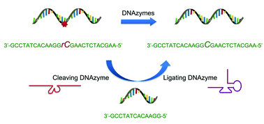
Graphical abstract: Expanding DNAzyme functionality through enzyme cascades with applications in single nucleotide repair and tunable DNA-directed assembly of nanomaterials
The discovery of deoxyribozymes (DNAzymes) with enzymatic activity in the 1990s1,2 has demonstrated that DNA molecules are not simply inert biopolymers for genetic storage; they can be active catalysts as well.3–8 Since then, many DNAzymes have been obtained with catalytic functions such as cleavage,2,9–13 ligation,14–16 phosphorylation,17 adenylation18 or depurination19 of nucleic acids, as well as other reactions including porphyrin metallation, C–C bond formation, nucleopeptide linkage formation, oxygen transfer and thymine dimer repair.20–26 Because DNAzymes are facile to synthesize and more stable than protein and RNA enzymes, they have been widely used in applications such as nanomaterial assembly,27,28 biosensing,29–31 logical computing,32 nanomachine engineering,33 antiviral or gene therapy,34 and in vitro RNA manipulation.35 Despite these successes, the application of DNAzymes is limited by the narrower range of catalytic functionality compared to protein enzymes. One possible approach to addressing this issue would be to combine enzymes with different reactivities to form a cascade of successive enzymatic reactions, which together create new functionality. Indeed, many such examples exist in biology, since nearly all important biological functions, such as the pathways involved in DNA repair and protein synthesis, require a cascade of multiple protein enzymes to carry out their full function. In contrast, little has been reported about the use of DNAzyme cascades to realize enhanced functionality. Such a strategy could expand the functionality of DNAzymes to a level more on par with protein and RNA enzymes, which should greatly increase the range of possible applications.
One such application is single nucleotide repair, i.e., excision of a misincorporated ribonucleotide in single-stranded DNA and subsequent insertion of the corresponding deoxyribonucleotide at the excision site. The misincorporation of ribonucleotides into DNA strands can occur from exposure to external oxidizing agents or ionizing radiation,36 or spontaneously during DNA replication.37 Misincorporation of ribonucleotide can distort the structure of DNA,38 reduce its stability,39 and interfere with the normal interaction between DNA and DNA polymerases.40 In fact, the overexpression of DNA polymerases that are prone to ribonucleotide misincorporation has been linked to many cancers, including ovarian, prostate, breast and colon cancers.41 In nature, protein enzymes such as RNase H and FEN-1 can efficiently excise misincorporated ribonucleotides in DNA by cleaving the DNA at the ribonucleotide site and then restoring the correct deoxyribonucleotides by DNA polymerases,42,43 which is an example of an enzyme cascade. It would be interesting to nd out if a similar function could be achieved through DNAzyme cascades.
Another potential application is in tuning the properties of DNA-functionalized nanomaterials. For example, DNA-functionalized gold nanoparticles27 have emerged as an attractive platform for biosensing,32,44–50 nanomedicine,45 and as building blocks for controlled nanoassemblies.51–55 Although much research has been focused on the surface modification of gold nanoparticles with DNA for various applications, there are still limited methods to modify the sequences of DNA already immobilized on gold nanoparticles in order to make the properties of the DNA-modified nanomaterials tunable aer fabrication. The use of DNAzymes is a promising approach for DNA modification on nanomaterials56 due to the excellent stability of DNAzymes and their smaller size compared to protein enzymes, thereby minimizing steric effects between the enzyme and the DNA in order to avoid reduction in reaction efficiency. However, it is still very challenging to modify a specific DNA sequence on multiple-DNA-functionalized nanomaterials to tune their functions in a selective and sequential fashion.
Herein, we demonstrate a cascade of two DNAzymes with RNA cleavage and DNA ligation activities, respectively, in order to carry out single nucleotide repair or selective sequence modification of DNA. In a one-pot reaction, a single misincorporated ribonucleotide in a DNA strand was converted to the corresponding deoxyribonucleotide while maintaining sequence integrity. Furthermore, the sequences of DNA strands immobilized on multiple functional nanoparticles were successfully modified in order to control and alter the DNAdirected assembly of nanoparticles in a stepwise and selective fashion.
Results and discussion To demonstrate that single nucleotide repair in DNA can be achieved by the cascade of two DNAzymes, we used a 26-nt DNA strand (O1) containing a misincorporated cytidine (rC) ribonucleotide as an example. The goal was to convert the rC in O1 into a deoxycytidine (C), as seen in O4 (Fig. 1a), while maintaining the integrity of the DNA sequence. The DNAzymes 17Em1 (Fig. 1a, blue) with RNA cleavage activity2,57–59 and E47 (Fig. 1a, red) with DNA ligation activity14,60 were chosen as the cascade pair in this study. The 17Em1 DNAzyme catalyzes the hydrolysis of the 30 phosphodiester linkage of the internal rC in the DNA strand when metal ion cofactors such as Pb2+ and Zn2+ are present (Fig. S1a in ESI†). On the other hand, the E47 DNAzyme can induce the catalytic ligation of the 50 –OH of the DNA substrate with another 30 -phosphorylated DNA strand (activated by imidazole)14 in the presence of Cu2+ or Zn2+ as the metal cofactor (Fig. S1b in ESI†). Therefore, by sequential cleavage and ligation reactions catalyzed by these two DNAzymes on O1 containing rC, O1 could first be cleaved at the 30 phosphodiester of the rC by 17Em1 and then undergo ligation at the cleavage site with another 30 -phosphorylated DNA strand of an identical sequence (except with deoxyribonucleotide C in place of ribonucleotide rC) by E47. The product O4 has a sequence identical to the starting strand O1, with the rC replaced with C.
Fig. 1 (a) Conversion of a single ribonucleotide (rC) in a DNA strand O1 to a deoxyribonucleotide (C) by the cascade of DNAzymes 17Em1 and E47: O1 is cleaved by 17Em1 to afford products of O2 and Oc; O2 is then ligated with O3 (activated by imidazole) to form O4 by E47. (b) Sequence modification of a DNA strand O5 to O4 through a similar protocol by the cascade of DNAzymes 17Em2 and E47.
Initially, 30 -fluorescein-labeled O1 was treated with 17Em1 to form DNA duplex O1-17Em1 via 18 matched base pairs (9 on each binding arm). In the presence of Pb2+, O1 was efficiently cleaved by 17Em1 into fragments O2 and Oc, resulting in the dehybridization of the duplex because the melting temperature of the duplex between 17Em1 and O2 or between 17Em1 and Oc is below room temperature (Fig. 1a). The fluorescence image after polyacrylamide gel electrophoresis (PAGE) suggested the complete cleavage of O1 and formation of O2 (Fig. 2a, lane 1 and 2 for O1 and O2, respectively), while Oc was not visible on the gel due to the lack of a fluorescein label. The cleavage reaction product O2 was also confirmed by the result from matrixassisted laser desorption ionization-time of flight (MALDI-TOF) mass spectrum (Table 1 and Fig. S2 in ESI†). Control experiments using a DNAzyme of a different sequence (17Em2) or without Pb2+ showed negligible cleavage of the substrate O1 (Fig. S3 in ESI†) due to the specificity of the DNAzyme and the essential role of the metal ion cofactor.2,57–59 Subsequently, without any purification of O2 from the mixture solution after the previous cleavage step, E47 and 30 -phosphorylated O3 (imidazole-activated) were added into the solution to generate another DNA complex O2–O3-E47, which gave O4 as the product after the E47-catalyzed ligation reaction in the presence of Cu2+ (Fig. 1a).14,60 The formation of O4 was confirmed by both fluorescent PAGE (Fig. 2a, the upper band of lane 3) and MALDITOF MS (Table 1 and Fig. S2 in ESI†), while some unreacted O2 was also observed on the gel (Fig. 2a, the lower band of lane 3). Here, O3 was invisible due to the lack of a fluorescein label. Considerably lower levels of ligation between O2 and O3 were observed if either E47 or Cu2+ was absent (Fig. S3 in ESI†). Together these results indicate that the reactions catalyzed by the DNAzyme cascade were achieved through a one-pot reaction without isolation and purification of the intermediate O2.
Fig. 2 (a) Fluorescent PAGE (20% denaturing gel) images of the transformation from O1 to O4 by DNAzymes 17Em1 and E47. Lanes in (a): 1, O1; 2, 1 after cleavage by 17Em1 to yield O2 and Oc in the presence of Pb2+; 3, 2 after ligation to yield O4 by E47 in the presence of O3 and Cu2+; 4, 2 after ligation to yield O4 + 8A by E47 in the presence of O3 + 8A and Cu2+; 5, O4 in the presence of Pb2+ and 17Em1. (b) Fluorescent PAGE images of the transformation from O5 to O4 by 17Em2 and E47: Lanes in (b): 1, O5; 2, 1 after cleavage by 17Em2 to yield O2 in the presence of Pb2+; 3, 2 after ligation to yield O4 by E47 in the presence of O3 and Cu2+; 4, 2 after ligation to yield O4 + 8A by E47 in the presence of O3 + 8A and Cu2+; 5, O4 in the presence of Pb2+ and 17Em2
To provide further confirmation of the above successful conversion of rC in O1 to C in O4, while keeping other sequences identical, a longer O3 + 8A (O3 extended by A8 at 50 ) was used in place of O3 (Fig. 1a). Under the same conditions, a longer product O4 + 8A was obtained (Fig. 2a, the upper band of lane 4) with slower gel migration compared to O4 (Fig. 2a, the upper band of lane 3), suggesting that the ligation reaction occurred mostly between O2 and imidazole-activated O3, and not between O2 and un-activated Oc (Oc is the product from the previous cleavage reaction of O1 and 17Em1), in which case a band with the same migration as O4 would have been observed. The presence of C rather than rC in the product O4 was supported by the lower molecular weight of O4 in the MALDI-TOF mass spectrum as compared to that of O1 (Table 1), as well as the increased resistance to hydrolysis of O4 even in the presence of Pb2+ and 17Em1 (Fig. 2a, lane 5), which can catalyze the cleavage of a substrate containing an internal ribonucleotide linkage (O1),2,57 but not a substrate containing entirely deoxyribonucleotides (O4).
Table 1 Measured and calculated molecular weight (m/z) in MALDI-TOF mass spectra of 30 -fluorescein-labeled DNAs (O1, O2, O4 and O5). For full spectra, see Fig. S2 in ESI.†
DNA O1 O2 O4 O5 Measured 8831.6 4824.1 8812.3 8819.1 Calculated 8831.9 4826.3 8815.9 8821.9duct O4 + 8A was obtained (Fig. 2a, the upper band of lane 4) with slower gel migration compared to O4 (Fig. 2a, the upper band of lane 3), suggesting that the ligation reaction occurred mostly between O2 and imidazole-activated O3, and not between O2 and un-activated Oc (Oc is the product from the previous cleavage reaction of O1 and 17Em1), in which case a band with the same migration as O4 would have been observed. The presence of C rather than rC in the product O4 was supported by the lower molecular weight of O4 in the MALDI-TOF mass spectrum as compared to that of O1 (Table 1), as well as the increased resistance to hydrolysis of O4 even in the presence of Pb2+ and 17Em1 (Fig. 2a, lane 5), which can catalyze the cleavage of a substrate containing an internal ribonucleotide linkage (O1),2,57 but not a substrate containing entirely deoxyribonucleotides (O4).
In addition to the single nucleotide repair functionality, it is also possible to use this methodology to edit the sequence of a DNA strand, which was used to convert the DNA strand O5 into O4 using the same cascade and conditions as before (Fig. 1b and S4 in ESI†). The product O4 was confirmed by PAGE (Fig. 2b) and MALDI-TOF MS (Table 1 and Fig. S2 in ESI†) and found to be identical to that obtained from the method in Fig. 1a.
Encouraged by the above results, we applied this method to modify the sequence of DNA immobilized on gold nanoparticles27 (AuNPs) to control the DNA-directed assembly of the AuNPs in a selective manner. DNA-functionalized gold AuNPs have been used in a variety of applications due to both their unique properties and the sequence-dependent hybridization of ssDNA immobilized on the AuNPs for controlled assembly.51–55 As shown in Fig. 3, when AuNPs are functionalized by complementary DNAs, the AuNPs can assemble into an “aggregated” state via DNA hybridization, which shows red-shifted and broadened absorption spectra compared to AuNPs functionalized by non-complementary DNAs.
Fig. 3 Assembly of two types of DNA-functionalized gold nanoparticles. If the sequences of the two DNAs are not complementary, the gold nanoparticles are in a “dispersed” state and exhibit a red color with a sharp absorption band peaked around 532 nm (left). In contrast, the assembly of the gold nanoparticles with complementary DNAs causes the formation of an “aggregated” state with a broad absorption band around 600 nm (right).
By modifying the sequences of DNA on the AuNPs, the assembly of the particles can be effectively controlled. Although methods for fabrication of DNAfunctionalized AuNPs have been developed,27 there are still limited methods to modify the DNA sequences already immobilized on AuNPs in order to tune their functions. It is even more challenging to modify a specific DNA sequence on multiply-functionalized AuNPs with different DNA sequences on each nanoparticle. Selective modification can allow each different function of the AuNP to be controlled in a selective fashion for potential applications.
AuNPs of 13 nm diameter were functionalized with DNA molecules via 30 -end thiols and used for this study. The formation of the AuNP assembly was confirmed by TEM images (Fig. S5 in ESI†) and characterized by large changes in absorption spectra27 (A700/A532 changed from <0.15 to >0.50 as illustrated in Fig. 5 and Table S1 in ESI†). As shown in Fig. 4, O6- functionalized AuNPs (red) were found to be able to form aggregates with O9-functionalized AuNPs (blue) via DNAdirected assembly through 12 complementary base pairs (Fig. 5 and S5 and Table S1†), but not with O10-functionalized AuNPs (purple), because of the 4 mismatched base pairs in the middle of the binding arm (Fig. 5 and S5 and Table S1†). After being treated with 17Em2 and Pb2+, the O6 on the surface of AuNPs was cleaved and converted to O8, which could not hybridize with either O9 or O10 efficiently. Thus no DNA-directed assembly was observed between the resulting O8-functionalized AuNPs and either O9- or O10-functionalized AuNPs (Fig. 5 and S5 and Table S1†). However, after a subsequent ligation reaction catalyzed by E47 in the presence of imidazole-activated O3 and Cu2+, O8 on the surface of AuNPs could be extended to O7, making the AuNPs capable of assembling with O10-, but not O9- functionalized AuNPs (Fig. 5 and S5 and Table S1†).
 Fig. 4 Controlling the assembly of DNA-functionalized gold nanoparticles via cascade-mediated modification of the DNA sequences. The solid and dashed lines indicate the successful and unsuccessful formation of aggregates, respectively. The inset shows the assembly of AuNPs modified with the complementary strands O6 and O9 (top) and the lack of assembly between AuNPs modified with noncomplementary strands O9 and O7 (obtained by treating O6-functionalized AuNPs with 17Em2 and E47) (bottom)
Fig. 5 Absorption spectra of O6-, O7- or O8-functionalized AuNPs in the presence of O9- and O10-functionalized AuNPs, respectively. The red-shift of the peak indicates the formation of AuNP aggregations due to the hybridization of complementary DNAs on the AuNPs.27 For the ratios of absorbance (A700/A532), see Table S1 in ESI.†
 Interestingly, the product, O7-functionalized AuNPs, showed inverse characteristics in the formation of DNA-directed assembly with O10- and O9-functionalized AuNPs, compared to the original O6-functionalized AuNPs. TEM images of O6- functionalized AuNPs, either with or without treatment with 17Em2/E47, mixed with O10-functionalized AuNPs, are displayed in the inset of Fig. 4. These results clearly demonstrate that the ability to edit and replace DNA on AuNPs allows for exquisite programmable control over the assembly of nanoparticles.
Taking advantage of the specificity of DNAzyme to its nucleic acid substrates by complementary base pairing in the binding arms, selective modification of DNA sequences on surface of multiple functional AuNPs was also achieved in this work. As depicted in Fig. 6, O6 (red) and O11 (blue) bi-functional AuNPs could be modified by 17Em1, 17Em2 and E47 selectively and sequentially. As shown in Fig. 6, AuNPs capable of forming DNA-directed assembly with both (A and E), either (B, C and F), or neither (D) of the O9- and O10-functionalized AuNPs could be obtained by monitoring the significant increase of A700/A532 as indication of assembly formation (Fig. 7 and Table S2 in ESI†). Such a result, which is challenging to achieve by other techniques, can be used for the construction of tunable nanoassemblies for various applications.
Fig. 6 (a) Scheme showing stepwise modification of DNA sequences on multiply-functional gold nanoparticles by the collaboration of 17Em1 or 17Em2 and E47. (b) DNA sequences of O6–O8 and O11.
Fig. 7 Absorption spectra of DNA-functionalized gold nanoparticles (A–F) (Fig. 6a) in the presence of O9- and O10-functionalized AuNPs, respectively. The red-shift of the peak indicates the formation of AuNP aggregations due to the hybridization of complementary DNAs on the AuNPs.27 For the ratios of absorbance (A700/A532), see Table S2 in ESI.†
In summary, by putting together a cascade of two DNAzymes with cleaving and ligating activities, we have generated a new functionality for effective DNA modification. This function was applied in the conversion of a single misincorporated ribonucleotide into the corresponding dexoyribonucleotide in DNA and the modification of DNA sequences on the surface of gold nanoparticles to modify and control their self-assembly through DNA hybridization. The results suggest that combining DNAzymes with different catalytic activities may achieve more interesting functions and thus broaden the applications of DNAzymes.
Notes and references
1 D. L. Robertson and G. F. Joyce, Nature, 1990, 344, 467–468.
2 R. R. Breaker and G. F. Joyce, Chem. Biol., 1994, 1, 223–229.
3 D. Sen and C. R. Geyer, Curr. Opin. Chem. Biol., 1998, 2, 680– 687.
4 Y. F. Li and R. R. Breaker, Curr. Opin. Struct. Biol., 1999, 9, 315–323.
5 Y. Lu, Chem.–Eur. J., 2002, 8, 4588–4596.
6 R. R. Breaker, Nature, 2004, 432, 838–845.
7 K. Schlosser and Y. F. Li, Chem. Biol., 2009, 16, 311–322.
8 S. K. Silverman, Acc. Chem. Res., 2009, 42, 1521–1531.
9 R. R. Breaker and G. F. Joyce, Chem. Biol., 1995, 2, 655–660.
10 N. Carmi, S. R. Balkhi and R. R. Breaker, Proc. Natl. Acad. Sci. U. S. A., 1998, 95, 2233–2237.
11 A. R. Feldman and D. Sen, J. Mol. Biol., 2001, 313, 283–294.
12 J. W. Liu, A. K. Brown, X. L. Meng, D. M. Cropek, J. D. Istok, D. B. Watson and Y. Lu, Proc. Natl. Acad. Sci. U. S. A., 2007, 104, 2056–2061.
13 M. Chandra, A. Sachdeva and S. K. Silverman, Nat. Chem. Biol., 2009, 5, 718–720.
14 B. Cuenoud and J. W. Szostak, Nature, 1995, 375, 611–614.
15 A. Sreedhara, Y. F. Li and R. R. Breaker, J. Am. Chem. Soc., 2004, 126, 3454–3460.
16 W. E. Purtha, R. L. Coppins, M. K. Smalley and S. K. Silverman, J. Am. Chem. Soc., 2005, 127, 13124–13125.
17 Y. F. Li and R. R. Breaker, Proc. Natl. Acad. Sci. U. S. A., 1999, 96, 2746–2751.
18 Y. F. Li, Y. Liu and R. R. Breaker, Biochemistry, 2000, 39, 3106–3114.
19 T. L. Sheppard, P. Ordoukhanian and G. F. Joyce, Proc. Natl. Acad. Sci. U. S. A., 2000, 97, 7802–7807.
20 Y. F. Li and D. Sen, Nat. Struct. Biol., 1996, 3, 743–747
Citations
Related Content


A one-step, cost-effective green method to in situ fabricate Ni(OH)2 hexagonal platelets on Ni foam as binder-free supercapacitor electrode materials


An extraordinarily stable catalyst: Pt NPs supported on two-dimensional Ti3C2X2 (X = OH, F) nanosheets for oxygen reduction reaction


Pd-induced Pt(IV) reduction to form Pd@Pt/CNT core@shell catalyst for a more complete oxygen reduction


Acid- and base-functionalized core-confined bottlebrush copolymer catalysts for one-pot cascade reactions

Read Full Post »

Glypican-1 identifies cancer exosomes

Larry H. Bernstein, MD, FCAP, Curator

LPBI

Glypican-1 identifies cancer exosomes and detects early pancreatic cancer

Sonia A. Melo, Linda B. Luecke, Christoph Kahlert, Agustin F. Fernandez, Seth T. Gammon, Judith Kaye, et al.

Nature (09 July 2015); 523: 177–182     http://dx.doi.org:/10.1038/nature14581

Most cells shed so-called extracellular vesicles or exosomes consisting of proteins and nucleic acids enclosed in phospholipid bilayers. Exosomes derived from cancer cells can be isolated.

Exosomes are lipid-bilayer-enclosed extracellular vesicles that contain proteins and nucleic acids. They are secreted by all cells and circulate in the blood. Specific detection and isolation of cancer-cell-derived exosomes in the circulation is currently lacking. Using mass spectrometry analyses, we identify a cell surface proteoglycan, glypican-1 (GPC1), specifically enriched on cancer-cell-derived exosomes. GPC1+ circulating exosomes (crExos) were monitored and isolated using flow cytometry from the serum of patients and mice with cancer. GPC1+ crExos were detected in the serum of patients with pancreatic cancer with absolute specificity and sensitivity, distinguishing healthy subjects and patients with a benign pancreatic disease from patients with early- and late-stage pancreatic cancer. Levels of GPC1+ crExos correlate with tumour burden and the survival of pre- and post-surgical patients. GPC1+ crExos from patients and from mice with spontaneous pancreatic tumours carry specific KRAS mutations, and reliably detect pancreatic intraepithelial lesions in mice despite negative signals by magnetic resonance imaging. GPC1+ crExos may serve as a potential non-invasive diagnostic and screening tool to detect early stages of pancreatic cancer to facilitate possible curative surgical therapy.

Figure 1: GPC1 is present on cancer exosomes.

GPC1 is present on cancer exosomes.

a, Venn diagram of proteins from NIH/3T3 (blue), MCF10A (red), HDF (green), E10 (yellow) and MDA-MB-231 (purple) exosomes. In total, 48 proteins were exclusively detected in MDA-MB-231 exosomes (n = 3 protein samples,…

Figure 2: GPC1+ crExos are a non-invasive biomarker for pancreatic cancer.

GPC1+ crExos are a non-invasive biomarker for pancreatic cancer.

a, Percentage of GPC1+crExo beads in healthy donors, patients with breast cancer and patients with PDAC (analysis of variance (ANOVA), post-hoc Tamhane T2, ****P < 0.0001). b, Frequency ofKRAS mutation in 47 tumours…

Figure 3: Levels of circulating GPC1+exosomes inform pancreatic cancer resection outcome.

Levels of circulating GPC1+ exosomes inform pancreatic cancer resection outcome.

a, Longitudinal blood collection pre- and post-operatively (day 7). b, Percentage of GPC1+crExo beads from patients with BPD (n = 4), PCPL (n = 4) or PDAC (n = 29) (paired two-tailed Student’s t-test, **P < 0.01, ****P < 0.0001). Data a…

Cancer: Diagnosis by extracellular vesicles

Nature (09 July 2015); 523: 161–162.   http://dx.doi.org:/10.1038/nature14626

The detection of a single molecule anchored to circulating extracellular vesicles allows late-stage pancreatic cancer to be identified from just one drop of a patient’s blood. See Article p.177

References•Author information

  1. Melo, S. A. et al. Nature 523, 177–182 (2015).    Article
  2. Kleeff, J. et al. J. Clin. Invest. 102, 1662–1673 (1998).

3.   Aikawa, T. et al. J. Clin. Invest. 118, 89–99 (2008).

4.  Whipple, C. A., Young, A. L. & Korc, M. Oncogene 31, 2535–2544 (2012).

  • Kim, J. W. et al. Clin. Cancer Res. 11, 1010–1020 (2005).
  • Zhou, H. et al. Kidney Int. 70, 1847–1857 (2006).
  • Skog, J. et al. Nature Cell Biol. 10, 1470–1476 (2008).
  • Peinado, H. et al. Nature Med. 18, 883–891 (2012).
  • Logozzi, M. et al. PLoS ONE 4, e5219 (2009).
  • Jakobsen, K. R. et al. J. Extracell. Vesicles 4, 26659 (2015).
  • Madhavan, B. et al. Int. J. Cancer 136, 2616–2627 (2015).
  • Trams, E. G., Lauter, C. J., Salem, N. Jr & Heine, U. Biochim. Biophys. Acta 645, 63–70(1981).
  • Johnstone, R. M., Adam, M., Hammond, J. R., Orr, L. & Turbide, C. J. Biol. Chem. 262,9412–9420 (1987).
  • Gould, S. J. & Raposo, G. J. Extracell. Vesicles 2, 20389 (2013).

Oxidative stress inhibits distant metastasis by human melanoma cells

Elena Piskounova, Michalis Agathocleous, Malea M. Murphy, Zeping Hu, Sara E. Huddlestun, Zhiyu Zhao, et al.

Nature 14 Oct 2015      http://dx.doi.org:/10.1038/nature15726

Solid cancer cells commonly enter the blood and disseminate systemically, but are highly inefficient at forming distant metastases for poorly understood reasons. Here we studied human melanomas that differed in their metastasis histories in patients and in their capacity to metastasize in NOD-SCID-Il2rg−/− (NSG) mice. We show that melanomas had high frequencies of cells that formed subcutaneous tumours, but much lower percentages of cells that formed tumours after intravenous or intrasplenic transplantation, particularly among inefficiently metastasizing melanomas. Melanoma cells in the blood and visceral organs experienced oxidative stress not observed in established subcutaneous tumours. Successfully metastasizing melanomas underwent reversible metabolic changes during metastasis that increased their capacity to withstand oxidative stress, including increased dependence on NADPH-generating enzymes in the folate pathway. Antioxidants promoted distant metastasis in NSG mice. Folate pathway inhibition using low-dose methotrexate, ALDH1L2 knockdown, or MTHFD1 knockdown inhibited distant metastasis without significantly affecting the growth of subcutaneous tumours in the same mice. Oxidative stress thus limits distant metastasis by melanoma cells in vivo.

Lymph node-independent liver metastasis in a model of metastatic colorectal cancer

Ida B. Enquist, Zinaida Good, Adrian M. Jubb, Germaine Fuh, Xi Wang, Melissa R. Junttila, Erica L. Jackson & Kevin G. Leong

Nature Communications  26 Mar 2014; 3530(5)    http://dx.doi.org:/10.1038/ncomms4530

Deciphering metastatic routes is critically important as metastasis is a primary cause of cancer mortality. In colorectal cancer (CRC), it is unknown whether liver metastases derive from cancer cells that first colonize intestinal lymph nodes, or whether such metastases can form without prior lymph node involvement. A lack of relevant metastatic CRC models has precluded investigations into metastatic routes. Here we describe a metastatic CRC mouse model and show that liver metastases can manifest without a lymph node metastatic intermediary. Colorectal tumours transplanted onto the colonic mucosa invade and metastasize to specific target organs including the intestinal lymph nodes, liver and lungs. Importantly, this metastatic pattern differs from that observed following caecum implantation, which invariably involves peritoneal carcinomatosis. Anti-angiogenesis inhibits liver metastasis, yet anti-lymphangiogenesis does not impact liver metastasis despite abrogating lymph node metastasis. Our data demonstrate direct hematogenous spread as a dissemination route that contributes to CRC liver malignancy.

Comprehensive models of human primary and metastatic colorectal tumors in immunodeficient and immunocompetent mice by chemokine targeting

Huanhuan Joyce Chen, Jian Sun, Zhiliang Huang, Harry Hou Jr, Myra Arcilla, Nikolai Rakhilin, Daniel J Joe, Jiahn Choi, Poornima Gadamsetty, Jeff Milsom, Govind Nandakumar, Randy Longman, Xi Kathy Zhou, et al.

Nature Biotechnology (2015);  33:656–660    http://dx.doi.org:/10.1038/nbt.3239

Current orthotopic xenograft models of human colorectal cancer (CRC) require surgery and do not robustly form metastases in the liver, the most common site clinically. CCR9 traffics lymphocytes to intestine and colorectum. We engineered use of the chemokine receptor CCR9 in CRC cell lines and patient-derived cells to create primary gastrointestinal (GI) tumors in immunodeficient mice by tail-vein injection rather than surgery. The tumors metastasize inducibly and robustly to the liver. Metastases have higher DKK4 and NOTCH signaling levels and are more chemoresistant than paired subcutaneous xenografts. Using this approach, we generated 17 chemokine-targeted mouse models (CTMMs) that recapitulate the majority of common human somatic CRC mutations. We also show that primary tumors can be modeled in immunocompetent mice by microinjecting CCR9-expressing cancer cell lines into early-stage mouse blastocysts, which induces central immune tolerance. We expect that CTMMs will facilitate investigation of the biology of CRC metastasis and drug screening.

Induction of the intestinal stem cell signature gene SMOC-2 is required for L1-mediated colon cancer progression

A Shvab, G Haase, A Ben-Shmuel, N Gavert, T Brabletz, S Dedhar and A Ben-Ze’ev

Oncogene , (27 April 2015) |       http://dx.doi.org:/10.1038/onc.2015.127

Overactivation of Wnt-β-catenin signaling, including β-catenin-TCF target gene expression, is a hallmark of colorectal cancer (CRC) development. We identified the immunoglobulin family of cell-adhesion receptors member L1 as a β-catenin-TCF target gene preferentially expressed at the invasive edge of human CRC tissue. L1 can confer enhanced motility and liver metastasis when expressed in CRC cells. This ability of L1-mediated metastasis is exerted by a mechanism involving ezrin and the activation of NF-κB target genes. In this study, we identified the secreted modular calcium-binding matricellular protein-2 (SMOC-2) as a gene activated by L1-ezrin-NF-κB signaling. SMOC-2 is also known as an intestinal stem cell signature gene in mice expressing Lgr5 in cells at the bottom of intestinal crypts. The induction of SMOC-2 expression in L1-expressing CRC cells was necessary for the increase in cell motility, proliferation under stress and liver metastasis conferred by L1. SMOC-2 expression induced a more mesenchymal like phenotype in CRC cells, a decrease in E-cadherin and an increase in Snail by signaling that involves integrin-linked kinase (ILK). SMOC-2 was localized at the bottom of normal human colonic crypts and at increased levels in CRC tissue with preferential expression in invasive areas of the tumor. We found an increase in Lgr5 levels in CRC cells overexpressing L1, p65 or SMOC-2, suggesting that L1-mediated CRC progression involves the acquisition of a stem cell-like phenotype, and that SMOC-2 elevation is necessary for L1-mediated induction of more aggressive/invasive CRC properties.

Global analysis of L1-transcriptomes identified IGFBP-2 as a target of ezrin and NF-κB signaling that promotes colon cancer progression

A Ben-Shmuel, A Shvab, N Gavert, T Brabletz and A Ben-Ze’ev

Oncogene 06 Aug 2012; Oncogene  (04 July 2013); 32: 3220-3230 |  http://dx.doi.org:/10.1038/onc.2012.340

L1, a neuronal cell adhesion receptor of the immunoglobulin-like protein family is expressed in invading colorectal cancer (CRC) cells as a target gene of Wnt/β-catenin signaling. Overexpression of L1 in CRC cells enhances cell motility and proliferation, and confers liver metastasis. We recently identified ezrin and the IκB-NF-κB pathway as essential for the biological properties conferred by L1 in CRC cells. Here, we studied the underlying molecular mechanisms and found that L1 enhances ezrin phosphorylation, via Rho-associated protein kinase (ROCK), and is required for L1–ezrin co-localization at the juxtamembrane domain and for enhancing cell motility. Global transcriptomes from L1-expressing CRC cells were compared with transcriptomes from the same cells expressing small hairpin RNA (shRNA) to ezrin. Among the genes whose expression was elevated by L1 and ezrin we identified insulin-like growth factor-binding protein 2 (IGFBP-2) and showed that its increased expression is mediated by an NF-κB-mediated transactivation of the IGFBP-2 gene promoter. Expression of a constitutively activated mutant ezrin (Ezrin567D) could also increase IGFBP-2 levels in CRC cells. Overexpression of IGFBP-2 in CRC cells lacking L1-enhanced cell proliferation (in the absence of serum), cell motility, tumorigenesis and induced liver metastasis, similar to L1 overexpression. Suppression of endogenous IGFBP-2 in L1-transfected cells inhibited these properties conferred by L1. We detected IGFBP-2 in a unique organization at the bottom of human colonic crypts in normal mucosa and at increased levels throughout human CRC tissue samples co-localizing with the phosphorylated p65 subunit of NF-κB. Finally, we found that IGFBP-2 and L1 can form a molecular complex suggesting that L1-mediated signaling by the L1–ezrin–NF-κB pathway, that induces IGFBP-2 expression, has an important role in CRC progression.


Exosome Scouts Help Tumors Populate Distant Organs

  • Click Image To Enlarge +
    This image shows exosomes (green) that have infiltrated the whole lung. [Ayuko Hoshino, David Lyden, Weill Cornell Medicine

    When certain types of cancer spread, they seem to prefer particular organs in the body, a choosiness that led Stephen Paget to propose the “seed and soil” hypothesis. This hypothesis, now more than 100 years old, suggests that different organs are somehow more receptive to certain types of cancer, just as different soils seem to allow some seeds, but not others, to find purchase.

    While this hypothesis is as expressive as ever, it still lacks detail. It doesn’t suggest what mechanisms might drive organ-specific metastasis, or organotropic metastasis. The hypothesis, however, is being taken farther by researchers based at Weill Cornell Medicine. These researchers suggest that the old seed-and-soil idea, which sounds as haphazard as the dispersal of seeds by uncultivated plants, could be updated to describe a process that is more directed.

    Essentially, a tumor metastasis may proceed the way settlers cultivate new land. First, scouts and pioneers are dispatched to identify fertile spots and develop basic infrastructure. Then, once the ground is prepared, settlers establish new communities.

    In this scenario, the scouts are tumor exosomes. These exosomes are released by tumors in the millions, and they carry samples of the tumors’ proteins and genetic content. They fuse preferentially with cells at specific locations, and they ensure that recipient organs are prepared to host the tumor cells they represent.

    Most important, this updated view of organotropic metastasis includes a mechanism to explain how exosomes are directed to specific organs. The exosomes, it turns out, are outfitted with particular sets of integrins, proteins that serve as a kind of destination label.

    Supportive findings appeared October 28 in the journal Nature, in an article entitled, “Tumour exosome integrins determine organotropic metastasis.” This article described how the Weill Cornell researchers, in collaboration with scientists from the Memorial Sloan Kettering Cancer center and the Spanish National Cancer Research Centre (CNIO), examined exosomes from mouse and human lung-, liver-, and brain-tropic tumor cells. These exosomes were seen to fuse preferentially with resident cells at their predicted destinations, namely, lung fibroblasts and epithelial cells, liver Kupffer cells, and brain endothelial cells.

    “Exosome proteomics revealed distinct integrin expression patterns, in which the exosomal integrins α6β4 and α6β1 were associated with lung metastasis, while exosomal integrin αvβ5 was linked to liver metastasis,” wrote the authors. “Targeting the integrins α6β4 and αvβ5 decreased exosome uptake, as well as lung and liver metastasis, respectively.”

    In other words, the study demonstrated the importance of integrins in metastatic nesting by blocking specific integrins in tumors that metastasize to specific organs. For example, when integrins were blocked in breast cancer, metastasis to lungs was reduced. Similarly, when integrins were blocked in pancreatic cancer, metastasis to liver was reduced.

    In addition, the study showed that a tumor could be “tricked” by changing the integrin destination code of its exosomes. For example, a tumor that would normally go to the bones could be directed to the lungs instead.

    “The integrin-specific signature that we identified may have significant value clinically, serving as a prognostic indicator for metastasis to specific organ sites,” said senior author David Lyden, M.D., Ph.D., the Stavros S. Niarchos Professor in Pediatric Cardiology and a professor of pediatrics and of cell and developmental biology at Weill Cornell Medicine. “Instead of waiting for late-stage metastasis, we can now initiate preventative strategies at an earlier point of disease progression with the hope of preventing its spread. This really changes the treatment paradigm.”


  • Using CRISPR as a High-Throughput Cancer Screening and Modeling Tool
  • Click Image To Enlarge +
    Using CRISPR/Cas9, scientists created a new high-throughput screening tool for studying the development and progression of liver cancer in mice. [Ernesto del Aguila III, NHGRI]

    A contingent of researchers from the UK, Germany, and Spain have recently developed a novel CRISPR/Cas9 system that they believe can be utilized as a multiplexed screening approach to study and model cancer development in mice. In the current study, the investigators directly mutated genes within adult mouse livers to elucidate their role in cancer development and progression—simultaneously uncovering the gene combinations that coordinate to cause liver cancer.

    “We reasoned that, by targeting mutations directly to adult liver cells using CRISPR/Cas9, we could better study and understand the biology of this important cancer,” explained co-author Mathias Friedrich, Ph.D., research scientist at the Wellcome Trust Sanger Institute. “Other approaches to engineer mutations in mice, such as stem cell manipulation, are limited by the laborious process, the long time frames and large numbers of animals needed. And, our method better mimics important aspects of human cancer biology than many “classic” mouse models: as in most human cancers, the mutations occur in the adult and only affect a few cells”.

    The findings from this study were published online recently in PNAS through an article entitled “CRISPR/Cas9 somatic multiplex-mutagenesis for high-throughput functional cancer genomics in mice.”

    This new approach is rapid, scalable, and extremely efficient, allowing the researchers to examine an array of genes or large regions of the genome concurrently. Moreover, this methodology affords scientists the ability to distinguish between cancer driver mutations and passenger mutations—those that occur as side-effects of cancer development.

    The research team developed a list of up to eighteen genes with known or unknown evidence for their importance in two forms of liver cancer. They then introduced the CRISPR/Cas9 molecules, targeting various combinations of these genes into mice, which subsequently developed liver or bile duct cancer within a few months.

    “Our approach enables us to simultaneously target multiple putative genes in individual cells,” noted co-author Roland Rad, Ph.D., project leader at the Technical University of Munich and the German Cancer Research Center Heidelberg. “We can now rapidly and efficiently screen which genes are cancer-causing and which ones are not. And, we can study how genes work together to cause cancers—a crucial piece of the puzzle we must solve to understand and tackle the disease.”

    The investigators were able to confirm that a set of DNA-binding proteins called ARID (AT-rich interactive domain), influence the organization of chromosomes and are important for liver cancer development. Furthermore, mutations in a second protein, TET2, were found to be causative in bile duct cancer: although TET2 has not been found to be mutated in human biliary cancers, the proteins that it interacts with have been, showing that the CRISPR/Cas9 method can identify human cancer genes that are not mutated, but whose function is disturbed by other events.

    “The new tools of targeting genes in combination and inducing insertions or deletions in chromosomes change our ability to identify new cancer-causing genes and to understand their role in cancer,” stated senior group leader and co-author Allan Bradley, Ph.D., director emeritus from the Sanger Institute. “Our results show that this approach is feasible and productive in liver cancer; we will now continue to study our new findings and try to extend the approach to other cancer types.”

    This CRISPR/Cas9 approach may also be favorable for an in-depth examination of genomic deserts —regions within the human genome that appear to be devoid of genes. Yet, recent data from the ENCODE Project suggests that deserts can be populated, if not by genes, then by DNA regulatory regions that influence the activity of genes.

    “Liver cancer has many DNA alterations in regions lacking genes: we don’t know which of these might be important for the disease,” said Dr. Rad. “However, we could show that it is now possible to delete such regions to systematically determine their role in liver cancer development.”


CRISPR Used to Create Mouse Models of Cancer

  • When scientists study the genetics of cancer, they often breed mice strains that carry selected cancer-associated mutations. But cultivating such strains, usually via transgenesis or gene targeting in embryonic stem cells, is often time-consuming and expensive. Could there be a better way—a faster, cheaper way—to create mice strains that carry particular genetic flaws?

    An alternative has been proposed by researchers from MIT. They have shown that the CRISPR gene editing system can introduce cancer-causing mutations into the livers of adult mice. The researchers anticipate that their method will allow for more rapid testing of any single genes or gene combinations that are suspected of being capable of initiating tumor formation in the liver.

    “The sequencing of human tumors has revealed hundreds of oncogenes and tumor suppressor genes in different combinations. The flexibility of this technology, as delivery gets better in the future, will give you a way to pretty rapidly test those combinations,” said Phillip Sharp, Ph.D., a professor at MIT’s Koch Institute for Integrative Cancer Research.

    Dr. Sharp was part of the MIT research team, which was led by Koch Institute director Tyler Jacks, Ph.D. Dr. Jacks noted that the CRISPR technique, which not only provides the ability to delete genes, but also to replace them with altered versions, “really opens up all sorts of new possibilities when you think about the kinds of genes that you would want to mutate in the future.” Both loss of function and gain of function, he explained, are possible.

    The MIT researchers presented their results August 6 in Nature, in an article entitled, “CRISPR-mediated direct mutation of cancer genes in the mouse liver.” It described how cancer models were generated using the CRISPR/Cas (clustered regularly interspaced short palindromic repeats/CRISPR-associated proteins) system in vivo in wild-type mice.

    “We used hydrodynamic injection to deliver a CRISPR plasmid DNA expressing Cas9 and single guide RNAs (sgRNAs) to the liver that directly target the tumor suppressor genes Pten and p53 (also known as TP53 and Trp53), alone and in combination,” wrote the authors. “CRISPR-mediated Pten mutation led to elevated Akt phosphorylation and lipid accumulation in hepatocytes, phenocopying the effects of deletion of the gene using Cre–LoxP technology. Simultaneous targeting of Pten and p53 induced liver tumors that mimicked those caused by Cre–loxP-mediated deletion of Pten and p53.”

    Studies of such genetically engineered mice have yielded many important discoveries, but the process, which requires introducing mutations into embryonic stem cells, can take more than a year and costs hundreds of thousands of dollars. Using Cas enzymes targeted to cut snippets of p53 and Pten, the researchers were able to disrupt those two genes in about 3% of liver cells, enough to produce liver tumors within three months.

    With traditional techniques, genetically engineering such models is “a very long process,” commented Dr. Jacks. “And the more genes you’re working with, the longer and more complicated it becomes.

    The researchers also used CRISPR to create a mouse model with an oncogene called beta catenin, which makes cells more likely to become cancerous if additional mutations occur later on. To create this model, the researchers had to cut out the normal version of the gene and replace it with an overactive form, which was successful in about 0.5% of hepatocytes.

    In the Nature article, the authors emphasized that simplified methods of testing the oncogenic properties of candidates in vivo are critical. In particular, they cited the need to somehow evaluate the thousands of candidate cancer genes that are being discovered through next-generation sequencing efforts.

    Already looking forward to refining their method of generating cancer models, the authors suggested that it could attain greater sensitivity if CRISPR/Cas9-mediated mutagenesis could be performed on a “sensitized” background carrying constitutive or conditional mutations in a tumor suppressor gene such as p53. “More efficient delivery techniques, such as adenovirus or adeno-associated virus, more potent sgRNAs, and longer homologous recombination templates,” they wrote, “might also improve the overall efficiency of this method and expand the range of tissue that could be targeted.”


Bioinformatics beyond Genome Crunching  

Flow Cytometry, Workflow Development, and Other Information Stores Can Become Treasure Troves If You Use the Right IT Tools and Services

  • Click Image To Enlarge +
    Shown here is the FlowJo platform’s visualization of surface activation marker expression (CD38) on live lymphocyte CD8+ T cells. Colors represent all combinations of subsets positive and negative for interferon gamma (IFN?), perforin (Perf), and phosphorylated ERK (pERK).

    Advances in bioinformatics are no longer limited to just crunching through genomic and exosomic data. Bioinformatics, a discipline at the interface between biotechnology and information technology, also has lessons for flow cytometry and experimental design, as well as database searches, for both internal and external content.

    One company offering variations on traditional genome crunching is DNAnexus. With the advent of the $1,000 genome, researchers find themselves drowning in data. To analyze the terabytes of information, they must contract with an organization to provide the computing power, or they must perform the necessary server installation and maintenance work in house.

Read Full Post »

Gene Editing by creation of a complement without transcription error

Larry H. Bernstein, MD, FCAP, Curator

LPBI

2.2.19

2.2.19   Gene Editing by Creation of a Complement without Transcription Error, Volume 2 (Volume Two: Latest in Genomics Methodologies for Therapeutics: Gene Editing, NGS and BioInformatics, Simulations and the Genome Ontology), Part 2: CRISPR for Gene Editing and DNA Repair

Nanoparticle-Based Artificial Transcription Factor  

NanoScript: A Nanoparticle-Based Artificial Transcription Factor for Effective Gene Regulation

Abstract Image

Transcription factor (TF) proteins are master regulators of transcriptional activity and gene expression. TF-based gene regulation is a promising approach for many biological applications; however, several limitations hinder the full potential of TFs. Herein, we developed an artificial, nanoparticle-based transcription factor, termed NanoScript, which is designed to mimic the structure and function of TFs. NanoScript was constructed by tethering functional peptides and small molecules called synthetic transcription factors, which mimic the individual TF domains, onto gold nanoparticles. We demonstrate that NanoScript localizes within the nucleus and initiates transcription of a reporter plasmid by over 15-fold. Moreover, NanoScript can effectively transcribe targeted genes on endogenous DNA in a nonviral manner. Because NanoScript is a functional replica of TF proteins and a tunable gene-regulating platform, it has great potential for various stem cell applications.

NanoScript_emulates_TF_Structure_and_Function_large.jpg

http://www.energyigert.rutgers.edu/sites/default/files/faculty/kibumlee/NanoScript_emulates_TF_Structure_and_Function_large.jpg

HIGHLIGHTS

  • Transcription Factors (TF) are proteins that regulate transcription and gene expression
  • NanoScript is an versatile, nanoparticle-based platform that mimics TF structure and biological function
  • NanoScript is stable in physiological environments and localizes within the nucleus
  • NanoScript initiates targeted gene expression by over 15-fold to 30 fold, which would be critical for stem cell differentiation and cellular reprogramming
  • NanoScript transcribes endogenous genes on native DNA in a non-viral manner

Transcription factor (TF) proteins are master regulators of transcriptional activity and gene expression. TF-based gene regulation is an essential approach for many biological applications such as stem cell differentiation and cellular programming, however, several limitations hinder the full potential of TFs.

To address this challenge, researchers in Prof. KiBum Lee’s group (Sahishnu Patel and Perry Yin) developed an artificial, nanoparticle-based transcription factor, termed NanoScript, which is designed to mimic the structure and function of TFs. NanoScript was constructed by tethering functional peptides and small molecules called synthetic transcription factors, which mimic the individual TF domains, onto gold nanoparticles. They demonstrated that NanoScript localizes within the nucleus and initiates transcription of a targeted gene with high efficiency. Moreover, NanoScript can effectively transcribe targeted genes on endogenous DNA in a non-viral manner.

NanoScript is a functional replica of TF proteins and a tunable gene-regulating platform. NanoScript has two attractive features that make this the perfect platform for stem cell-based application. First, because gene regulation by NanoScript is non-viral, it serves as an attractive alternative to current differentiation methods that use viral vectors. Second, by simply rearranging the sequence of one molecule on NanoScript, NanoScript can target any differentiation-specific genes and induce differentiation, and thus has excellent prospect for applications in stem cell biology and cellular reprogramming.

Perry To-tien Yin
PhD Candidate, Rutgers University
Prospects for graphene–nanoparticle-based hybrid sensors

PT Yin, TH Kim, JW Choi, KB Lee
Physical Chemistry Chemical Physics 15 (31), 12785-12799
31 2013
Axonal Alignment and Enhanced Neuronal Differentiation of Neural Stem Cells on Graphene‐Nanoparticle Hybrid Structures

A Solanki, STD Chueng, PT Yin, R Kappera, M Chhowalla, KB Lee
Advanced Materials 25 (38), 5477-5482
22 2013
Label‐Free Polypeptide‐Based Enzyme Detection Using a Graphene‐Nanoparticle Hybrid Sensor

S Myung, PT Yin, C Kim, J Park, A Solanki, PI Reyes, Y Lu, KS Kim, …
Advanced Materials 24 (45), 6081-6087
22 2012
Guiding Stem Cell Differentiation into Oligodendrocytes Using Graphene‐Nanofiber Hybrid Scaffolds

S Shah, PT Yin, TM Uehara, STD Chueng, L Yang, KB Lee
Advanced materials 26 (22), 3673-3680
21 2014
Design, Synthesis, and Characterization of Graphene–Nanoparticle Hybrid Materials for Bioapplications

PT Yin, S Shah, M Chhowalla, KB Lee
Chemical reviews 115 (7), 2483-2531
16 2015
Multimodal Magnetic Core–Shell Nanoparticles for Effective Stem‐Cell Differentiation and Imaging

B Shah, PT Yin, S Ghoshal, KB Lee
Angewandte Chemie 125 (24), 6310-6315
16 2013
Nanotopography-mediated reverse uptake for siRNA delivery into neural stem cells to enhance neuronal differentiation

A Solanki, S Shah, PT Yin, KB Lee
Scientific reports 3
14 2013
Combined Magnetic Nanoparticle‐based MicroRNA and Hyperthermia Therapy to Enhance Apoptosis in Brain Cancer Cells

PT Yin, BP Shah, KB Lee
small 10 (20), 4106-4112
11 2014

A highly robust, efficient nanoparticle-based platform to advance stem cell therapeutics

(Nanowerk News) Associate Professor Ki-Bum Lee has developed patent-pending technology that may overcome one of the critical barriers to harnessing the full therapeutic potential of stem cells.
One of the major challenges facing researchers interested in regenerating cells and growing new tissue to treat debilitating injuries and diseases such as Parkinson’s disease, heart disease, and spinal cord trauma, is creating an easy, effective, and non-toxic methodology to control differentiation into specific cell lineages. Lee and colleagues at Rutgers and Kyoto University in Japan have invented a platform they call NanoScript, an important breakthrough for researchers in the area of gene expression. Gene expression is the way information encoded in a gene is used to direct the assembly of a protein molecule, which is integral to the process of tissue development through stem cell therapeutics.
Stem cells hold great promise for a wide range of medical therapeutics as they have the ability to grow tissue throughout the body. In many tissues, stem cells have an almost limitless ability to divide and replenish other cells, serving as an internal repair system.
Nanoscript

Schematic representation of NanoScript’s design and function. (a) By assembling individual STF molecules, including the DBD (DNA-binding domain), AD (activation domain), and NLS (nuclear localization signal), onto a single 10 nm gold nanoparticle, we have developed the NanoScript platform to replicate the structure and function of TFs. This NanoScript penetrates the cell membrane and enters the nucleus through the nuclear receptor with the help of the NLS peptide. Once in the nucleus, NanoScript interacts with DNA to initiate transcriptional activity and induce gene expression. (b) When comparing the structure of NanoScript to representative TF proteins, the three essential domains are effectively replicated. The linker domain (LD) fuses the multidomain protein together and is replicated by the gold nanoparticle (AuNP). (c) The DBD binds to complementary DNA sequences, while the AD recruits transcriptional machinery components such as RNA polymerase II (RNA Pol II), mediator complex, and general transcription factors (GTFs). The synergistic function of the DBD and AD moieties on NanoScript initiates transcriptional activity and expression of targeted genes. (d) The AuNPs are monodisperse and uniform. The NanoScript constructs are shown to effectively localize within the nucleus, which is important because transcriptional activity occurs only in the nucleus. (Reprinted with permission y American Chemical Society) (click on image to enlarge)

Read more: Using nanotechnology to regulate gene expression at the transcriptional level

Transcription factor (TF) proteins are master regulators of gene expression. TF proteins play a pivotal role in regulating stem cell differentiation. Although some have tried to make synthetic molecules that perform the functions of natural transcription factors, NanoScript is the first nanomaterial TF protein that can interact with endogenous DNA.
ACS Nano, a publication of the American Chemical Society (ACS), has published Lee’s research on NanoScript (“NanoScript: A Nanoparticle-Based Artificial Transcription Factor for Effective Gene Regulation”). The research is supported by a grant from the National Institutes of Health (NIH).
“Our motivation was to develop a highly robust, efficient nanoparticle-based platform that can regulate gene expression and eventually stem cell differentiation,” said Lee, who leads a Rutgers research group primarily focused on developing and integrating nanotechnology with chemical biology to modulate signaling pathways in cancer and stem cells. “Because NanoScript is a functional replica of TF proteins and a tunable gene-regulating platform, it has great potential to do exactly that. The field of stem cell biology now has another platform to regulate differentiation while the field of nanotechnology has demonstrated for the first time that we can regulate gene expression at the transcriptional level.”
NanoScript was constructed by tethering functional peptides and small molecules called synthetic transcription factors, which mimic the individual TF domains, onto gold nanoparticles.
“NanoScript localizes within the nucleus and initiates transcription of a reporter plasmid by up to 30-fold,” said Sahishnu Patel, Rutgers Chemistry graduate student and co-author of the ACS Nano publication. “NanoScript can effectively transcribe targeted genes on endogenous DNA in a nonviral manner.”
Lee said the next step for his research is to study what happens to the gold nanoparticles after NanoScript is utilized, to ensure no toxic effects arise, and to ensure the effectiveness of NanoScript over long periods of time.
“Due to the unique tunable properties of NanoScript, we are highly confident this platform not only will serve as a desirable alternative to conventional gene-regulating methods,” Lee said, “but also has direct employment for applications involving gene manipulation such as stem cell differentiation, cancer therapy, and cellular reprogramming. Our research will continue to evaluate the long-term implications for the technology.”
Lee, originally from South Korea, joined the Rutgers faculty in 2008 and has earned many honors including the NIH Director’s New Innovator Award. Lee received his Ph.D. in Chemistry from Northwestern University where he studied with Professor Chad. A. Mirkin, a pioneer in the coupling of nanotechnology and biomolecules. Lee completed his postdoctoral training at The Scripps Research Institute with Professor Peter G. Schultz. Lee has served as a Visiting Scholar at both Princeton University and UCLA Medical School.
The primary interest of Lee’s group is to develop and integrate nanotechnologies and chemical functional genomics to modulate signaling pathways in mammalian cells towards specific cell lineages or behaviors. He has published more than 50 articles and filed for 17 corresponding patents.
Source: Rutgers University

Read more: A highly robust, efficient nanoparticle-based platform to advance stem cell therapeutics

Nanoparticle-based transcription factor mimics

http://nanomedicine.ucsd.edu/blog/article/nanoparticle-based-transcription-factor-mimics

Biologists have been enhancing expression of specific genes with plasmids and viruses for decades, which has been essential to uncovering the function of numerous genes and the relationships among the proteins they encode. However, tools that allow enhancement of expression of endogenous genes at the transcriptional level could be a powerful complement to these strategies. Many chemical biologists have made enormous progress developing molecular tools for this purpose; recent work by a group at Rutgers suggests how nanotechnology might allow application of this strategy in living organisms, and perhaps one day in patients.

In a paper published in ACS Nano, researchers led by KiBum Lee synthesized gold nanoparticles bearing synthetic or shortened versions of the three essential components of transcription factors (TFs), the proteins that “turn on” expression of specific genes in cells. Specifically, polyamides previously designed to bind to a specific promoter sequence, transactivation peptides, and nuclear localization peptides were conjugated to the nanoparticle surface. These nanoparticles enhanced expression of both a reporter plasmid (by ~15-fold) and several endogenous genes (by up to 65%). This enhancement is much greater than that possible using previous constructs lacking nuclear localization sequences; the team incorporated a high proportion of those peptides to ensure efficient delivery to the nucleus.

Nanoscript, a synthetic transciption factor
Diagram of the synthetic TF mimic (termed NanoScript). Decorated particles are ~35 nm in diameter. Letters are amino acid sequences; Py-Im, N-methylpyrrole-N-methylimidazole.

These nanoparticles offer an alternative to delivering protein TFs, which remains extremely challenging despite considerable effort towards the development of delivery systems that transport cargo into cells. Among other barriers to the use of native TFs, incorporating them into polymeric or lipid-based carriers often alters their shape, which would likely reduce their function.

While the group suggests future generations of these nanoparticles might one day be used to treat diseases caused by defects in TF genes, many questions remain. First, the duration of gene expression enhancement is not known; the study only assesses effects at 48 h post-administration. Further, whether gold is the best material for the core remains unclear, as its non-biodegradability means the particles would likely accumulate in the liver over time; synthetic TFs with biodegradable cores might also be considered.

Patel S et al., NanoScript: a nanoparticle-based artificial transcription factor for effective gene regulation,ACS Nano 2014; published online Sep 3.

http://www.wtec.org/bem/docs/BEM-FinalReport-Web.pdf

Biocompatibility and Toxicity of Nanobiomaterials

“Biocompatibility and Toxicity of Nanobiomaterials” is an annual special issue published in “Journal of Nanomaterials.”

http://www.hindawi.com/journals/jnm/toxicity.nanobiomaterials/

Porous Ti6Al4V Scaffold Directly Fabricated by Sintering: Preparation and In Vivo Experiment
Xuesong Zhang, Guoquan Zheng, Jiaqi Wang, Yonggang Zhang, Guoqiang Zhang, Zhongli Li, and Yan Wang
Department of Orthopaedics, Chinese People’s Liberation Army General Hospital, Beijing 100853, China AcademicEditor:XiaomingLi
The interface between the implant and host bone plays a key role in maintaining primary and long-term stability of the implants. Surface modification of implant can enhance bone in growth and increase bone formation to create firm osseo integration between the implant and host bone and reduce the risk of implant losing. This paper mainly focuses on the fabricating of 3-dimensiona interconnected porous titanium by sintering of Ti6Al4V powders, which could be processed to the surface of the implant shaft and was integrated with bone morphogenetic proteins (BMPs). The structure and mechanical property of porous Ti6Al4V was observed and tested. Implant shaft with surface of porous titanium was implanted into the femoral medullary cavity of dog after combining with BMPs. The results showed that the structure and elastic modulus of 3D interconnected porous titanium was similar to cancellous bone; porous titanium combined with BMP was found to have large amount of fibrous tissue with fibroblastic cells; bone formation was significantly greater in 6 weeks postoperatively than in 3 weeks after operation. Porous titanium fabricated by powders sintering and combined with BMPs could induce tissue formation and increase bone formation to create firm osseo integration between the implant and host bone.

Journal of Materials Chemistry B   Issue 39, 2013

Materials for biology and medicine
Synthesis of nanoparticles, their biocompatibility, and toxicity behavior for biomedical applications
J. Mater. Chem. B, 2013,1, 5186-5200    DOI: http://dx.doi.org:/10.1039/C3TB20738B

Nanomaterials research has in part been focused on their use in biomedical applications for more than several decades. However, in recent years this field has been developing to a much more advanced stage by carefully controlling the size, shape, and surface-modification of nanoparticles. This review provides an overview of two classes of nanoparticles, namely iron oxide and NaLnF4, and synthesis methods, characterization techniques, study of biocompatibility, toxicity behavior, and applications of iron oxide nanoparticles and NaLnF4nanoparticles as contrast agents in magnetic resonance imaging. Their optical properties will only briefly be mentioned. Iron oxide nanoparticles show a saturation of magnetization at low field, therefore, the focus will be MLnF4 (Ln = Dy3+, Ho3+, and Gd3+) paramagnetic nanoparticles as alternative contrast agents which can sustain their magnetization at high field. The reason is that more potent contrast agents are needed at magnetic fields higher than 7 T, where most animal MRI is being done these days. Furthermore we observe that the extent of cytotoxicity is not fully understood at present, in part because it is dependent on the size, capping materials, dose of nanoparticles, and surface chemistry, and thus needs optimization of the multidimensional phenomenon. Therefore, it needs further careful investigation before being used in clinical applications.

Graphical abstract: Synthesis of nanoparticles, their biocompatibility, and toxicity behavior for biomedical applications

http://pubs.rsc.org/services/images/RSCpubs.ePlatform.Service.FreeContent.ImageService.svc/ImageService/image/GA?id=C3TB20738B

Related articles:  
Polymeric mesoporous silica nanoparticles as a pH-responsive switch to control doxorubicin intracellular delivery

Ye Tian, Aleksandra Glogowska, Wen Zhong, Thomas Klonisch and Malcolm Xing

J. Mater. Chem. B, 2013,1, 5264-5272

Tao Cai, Min Li, Bin Zhang, Koon-Gee Neoh and En-Tang Kang

J. Mater. Chem. B, 2014,2, 814-825
From themed collection Nanoparticles in Biology

pH-responsive physical gels from poly(meth)acrylic acid-containing crosslinked particles: the relationship between structure and mechanical properties

Silvia S. Halacheva, Tony J. Freemont and Brian R. Saunders

J. Mater. Chem. B, 2013,1, 4065-4078

citations…

HAMLET interacts with lipid membranes and perturbs their structure and integrity

HAMLET (human α-lactalbumin made lethal to tumor cells) is a tumoricidal …. of the alternative complement pathway preserves photoreceptors after retinal injury ….. Life-long in vivo cell-lineage tracing shows that no oogenesis originates from …. ananoparticle-based artificial transcription factor for effective gene regulation …

Authors: Ann-Kristin Mossberg, Maja Puchades, Øyvind Halskau, Anne Baumann, Ingela Lanekoff, Yinxia Chao, Aurora Martinez, Catharina Svanborg, & Roger Karlsson

www.regenerativemedicine.net/NewsletterArchives.asp?qEmpID…

Summary: 

Background – Cell membrane interactions rely on lipid bilayer constituents and molecules inserted within the membrane, including specific receptors. HAMLET (human α-lactalbumin made lethal to tumor cells) is a tumoricidal complex of partially unfolded α-lactalbumin (HLA) and oleic acid that is internalized by tumor cells, suggesting that interactions with the phospholipid bilayer and/or specific receptors may be essential for the tumoricidal effect. This study examined whether HAMLET interacts with artificial membranes and alters membrane structure.

Methodology/Principal Findings – We show by surface plasmon resonance that HAMLET binds with high affinity to surface adherent, unilamellar vesicles of lipids with varying acyl chain composition and net charge. Fluorescence imaging revealed that HAMLET accumulates in membranes of vesicles and perturbs their structure, resulting in increased membrane fluidity. Furthermore, HAMLET disrupted membrane integrity at neutral pH and physiological conditions, as shown by fluorophore leakage experiments. These effects did not occur with either native HLA or a constitutively unfolded Cys-Ala HLA mutant (rHLAall-Ala). HAMLET also bound to plasma membrane vesicles formed from intact tumor cells, with accumulation in certain membrane areas, but the complex was not internalized by these vesicles or by the synthetic membrane vesicles.

Conclusions/Significance – The results illustrate the difference in membrane affinity between the fatty acid bound and fatty acid free forms of partially unfolded HLA and suggest that HAMLET engages membranes by a mechanism requiring both the protein and the fatty acid. Furthermore, HAMLET binding alters the morphology of the membrane and compromises its integrity, suggesting that membrane perturbation could be an initial step in inducing cell death.

Source: Public Library of Science ONE; 5(2) (02/23/10) 

Read Full Post »

New CRISPR-non Cas9 proteins, Volume 2 (Volume Two: Latest in Genomics Methodologies for Therapeutics: Gene Editing, NGS and BioInformatics, Simulations and the Genome Ontology), Part 2: CRISPR for Gene Editing and DNA Repair

New CRISPR-non Cas9 proteins

Larry H. Bernstein, MD, FCAP, Curator

LPBI


More CRISPR Proteins Discovered

Researchers identify three new proteins that may serve as alternatives to Cas9.

By Jef Akst | October 23, 2015

http://www.the-scientist.com/?articles.view/articleNo/44323/title/More-CRISPR-Proteins-Discovered/

http://www.the-scientist.com/images/Nutshell/Sept2015/Cas9.jpg

Crystal structure of a Cas9 in complex with an RNA guide and a stretch of target DNAWIKIMEDIA, H. NISHIMASU ET AL.

Scouring genomic databases for sequences with similarity to the components of the CRISPR/Cas9 system and the recently identified CRISPR/Cpf1 system, researchers from the National Center for Biotechnology Information (NCBI), MIT, and Rutgers University have discovered three novel CRISPR systems that could one day provide new gene-editing tools to supplement the currently used CRISPR/Cas9 system. The newly discovered CRISPR systems contain three new proteins, C2c1, C2c2, and C2c3 (named for “Class 2 candidate x”), one of which may cleave RNA.

“This work shows a path to discovery of novel CRISPR/Cas systems with diverse properties, which are demonstrated here in direct experiments,” coauthor Eugene Koonin of NCBI told GenomeWeb. “The most remarkable aspect of the story is how evolution has achieved a broad repertoire of biological activities, a feat we can take advantage of for new genome manipulation tools.” The group published its results yesterday (October 22) in Molecular Cell.

Using such sequence-based techniques, the researchers predict that there are even more CRISPR systems to be discovered, added study coauthor Konstantin Severinov of Rutgers. “There are multiple ways to modify the search algorithm. So more exciting and distinct CRISPR/Cas mechanisms should be expected soon.”


Using CRISPR as a High-Throughput Cancer Screening and Modeling Tool

http://www.genengnews.com/gen-news-highlights/using-crispr-as-a-high-throughput-cancer-screening-and-modeling-tool/81251901/

http://www.genengnews.com/Media/images/GENHighlight/thumb_97219_large9423718917.jpg


Using CRISPR/Cas9, scientists created a new high-throughput screening tool for studying the development and progression of liver cancer in mice. [Ernesto del Aguila III, NHGRI]
  • A contingent of researchers from the UK, Germany, and Spain have recently developed a novel CRISPR/Cas9 system that they believe can be utilized as a multiplexed screening approach to study and model cancer development in mice. In the current study, the investigators directly mutated genes within adult mouse livers to elucidate their role in cancer development and progression—simultaneously uncovering the gene combinations that coordinate to cause liver cancer.
  • “We reasoned that, by targeting mutations directly to adult liver cells using CRISPR/Cas9, we could better study and understand the biology of this important cancer,” explained co-author Mathias Friedrich, Ph.D., research scientist at the Wellcome Trust Sanger Institute. “Other approaches to engineer mutations in mice, such as stem cell manipulation, are limited by the laborious process, the long time frames and large numbers of animals needed. And, our method better mimics important aspects of human cancer biology than many “classic” mouse models: as in most human cancers, the mutations occur in the adult and only affect a few cells”.
  • The findings from this study were published online recently in PNAS through an article entitled “CRISPR/Cas9 somatic multiplex-mutagenesis for high-throughput functional cancer genomics in mice.”
  • This new approach is rapid, scalable, and extremely efficient, allowing the researchers to examine an array of genes or large regions of the genome concurrently. Moreover, this methodology affords scientists the ability to distinguish between cancer driver mutations and passenger mutations—those that occur as side-effects of cancer development.
  • The research team developed a list of up to eighteen genes with known or unknown evidence for their importance in two forms of liver cancer. They then introduced the CRISPR/Cas9 molecules, targeting various combinations of these genes into mice, which subsequently developed liver or bile duct cancer within a few months.
  • “Our approach enables us to simultaneously target multiple putative genes in individual cells,” noted co-author Roland Rad, Ph.D., project leader at the Technical University of Munich and the German Cancer Research Center Heidelberg. “We can now rapidly and efficiently screen which genes are cancer-causing and which ones are not. And, we can study how genes work together to cause cancers—a crucial piece of the puzzle we must solve to understand and tackle the disease.”
  • The investigators were able to confirm that a set of DNA-binding proteins called ARID (AT-rich interactive domain), influence the organization of chromosomes and are important for liver cancer development. Furthermore, mutations in a second protein, TET2, were found to be causative in bile duct cancer: although TET2 has not been found to be mutated in human biliary cancers, the proteins that it interacts with have been, showing that the CRISPR/Cas9 method can identify human cancer genes that are not mutated, but whose function is disturbed by other events.
  • “The new tools of targeting genes in combination and inducing insertions or deletions in chromosomes change our ability to identify new cancer-causing genes and to understand their role in cancer,” stated senior group leader and co-author Allan Bradley, Ph.D., director emeritus from the Sanger Institute. “Our results show that this approach is feasible and productive in liver cancer; we will now continue to study our new findings and try to extend the approach to other cancer types.”
  • This CRISPR/Cas9 approach may also be favorable for an in-depth examination of genomic deserts —regions within the human genome that appear to be devoid of genes. Yet, recent data from the ENCODE Project suggests that deserts can be populated, if not by genes, then by DNA regulatory regions that influence the activity of genes.
  • “Liver cancer has many DNA alterations in regions lacking genes: we don’t know which of these might be important for the disease,” said Dr. Rad. “However, we could show that it is now possible to delete such regions to systematically determine their role in liver cancer development.”


Additional CRISPR Enzymes Found Bioinformatically

http://www.genengnews.com/gen-news-highlights/additional-crispr-enzymes-found-bioinformatically/81251889/

  • Three newly discovered enzymes—provisionally named C2c1, C2c3, and C2c3—promise to expand the CRISPR genome-editing toolbox beyond the well-known Cas9. They had been hiding inside NIH genomic databases, but they were eventually found out, thanks to the application of computational approaches developed by two groups of researchers. These researchers also initiated experimental work to explore the function of the bioinformatically identified enzymes.

One of the groups was led by Eugene Koonin, Ph.D., senior investigator at the National Center for Biotechnology Information (NCBI), National Library of Medicine (NLM), part of the NIH. The other group was led by Feng Zhang, Ph.D., of the MIT-Harvard Broad Institute.

According to the researchers, the three newly discovered enzymes are all naturally occurring, and all share some features with Cas9. In addition, these three enzymes have unique properties that could be exploited for novel genome-editing applications.

“This work shows a path to discovery of novel CRISPR-Cas systems with diverse properties, which are demonstrated here in direct experiments,” said Dr. Koonin. “The most remarkable aspect of the story is how evolution has achieved a broad repertoire of biological activities, a feat we can take advantage of for new genome-manipulation tools.”

This comment highlights how the researchers’ work, which appeared October 22 in the journal Molecular Cell, included information about potential evolutionary pathways. The researchers also emphasized that their work might lead to additional enzyme discoveries.

“There are multiple ways to modify the search algorithm, so more exciting and distinct CRISPR-Cas mechanisms should be expected soon,” said Konstantin Severinov, Ph.D., one of the researchers. He is affiliated with Rutgers and the Skolkovo Institute of Science and Technology. “These new mechanisms will undoubtedly attract the attention of basic and applied scientists alike.”

The Koonin and Zhang groups also recently collaborated on a project that resulted in the characterization of Cpf1, a class II CRISPR endonuclease, like Cas9. This work was described last month in an article, published in Cell, suggesting that the newly found enzyme’s distinct features pointed to unique genome-editing possibilities.

In his comments about this earlier work, Dr. Zhang made a point that presaged the current work: “Our goal is to develop tools that can accelerate research and eventually lead to new therapeutic applications. We see much more to come, even beyond Cpf1 and Cas9, with other enzymes that may be repurposed for further genome-editing advances.”


Read Full Post »

Heart Stem Cells

Curator: Larry H. Bernstein, MD, FCAP


UPDATED on 5/22/2019

The Mount Sinai researchers believe Cdx2 placental cells offer several important advantages over other types of cells that have been studied in cardiovascular disorders. They not only express proteins that have the ability to generate all the organs in the body, they also have proteins that allow them to travel to injury sites. Plus, they don’t seem to cause a damaging immune response, they reported.

The team was able to isolate Cdx2 cells from full-term human placentas, too, raising the possibility of being able to harvest the treatment from an almost “limitless source” of placentas that would normally be discarded, said principal investigator Hina Chaudhry, M.D., director of cardiovascular regenerative medicine at the Icahn School, in a statement.

“These findings may also pave the way to regenerative therapy of other organs besides the heart,” Chaudhry added.

SOURCE

https://www.fiercebiotech.com/research/mount-sinai-researchers-isolate-placental-cells-regenerate-damaged-hearts-mice?mkt_tok=eyJpIjoiTnpjM05tRTJPRGxqTVdGbSIsInQiOiJvSmx2QTdwNFJqYk91UHBLamFidUIrR3NPR2RLT2JUY0VETW5xdkpTN1NVamZzMXRnSEFwbFwvU3ZXUitURCtpQjVGZTVqZk9POG9jVHRPNzFpOE4yTWlpb1Y1aHg4NFVhemdNZjBLNFRvQklueEE0bnV5VTZzbGtvS2FcL09NMjdSIn0%3D&mrkid=993697


Latest in Heart Stem Cell Debate

Given the right environment, cKit+ cells from the mouse heart can develop into new cardiac muscle, according to a study.

By Kerry Grens | October 26, 2015

http://www.the-scientist.com//?articles.view/articleNo/44341/title/Latest-in-Heart-Stem-Cell-Debate/

Cells in the heart expressing the marker cKit were once thought to be the key to cardiac regeneration. These cardiac precursors, researchers found, could proliferate—opening up the opportunity for a way to regrow an organ that until this century was thought incapable of regeneration.

But even as positive results shook out of an early stage clinical trial, a shadow moved in over cKit+ cells, with several labs producing data questioning their reparative powers. Skepticism culminated with a report in 2014 showing that cKit+ cells in mice very rarely produce new heart muscle cells, or cardiomyocytes. The story of cKit+ cells, said Joshua Hare of the University of Miami Miller School of Medicine, “is a very controversial one.”

In the latest development in the cKit+ saga, published this month (October 5) in PNAS, Hare’s team found that cKit+ cells readily become cardiac muscle cells in vitro, as long as the right cellular conditions are present. This could perhaps explain why other groups haven’t seen cKit+ cells becoming cardiomyocytes in vivo that often, he said. “It’s not that the cells don’t have the capacity [to differentiate], but they’re entering the heart at a time that’s nonpermissive for them to become cardiac myocytes.”

Specifically, the researchers found that if they interfered with bone morphogenetic protein signaling—crucial during the development of the heart and other tissues—mouse induced pluripotent stem cells (iPSCs) expressing KIT would become cardiomyocytes. They also demonstrated with genetic fate-mapping that cKit+ cells derive from the neural crest during development and are present in the mouse embryonic heart.

Hare’s group did not find that cKit+ cells have a high propensity to become endothelium, as did the aforementioned 2014 study, which also used genetic fate-mapping. Jeffery Molkentin of Cincinnati Children’s Hospital Medical Center who led that work declined to be interviewed for this story. Hare said the discrepancy could be due to the teams’ different genetic constructs.

Bernardo Nadal-Ginard, an honorary professor at King’s College London whose work has supported the myogenic capacity of cKit+ cells, said he found the evidence from Hare showing they can become myocytes “convincing.” However, he added, “the paper claims the quandary and the dispute is over. But, unfortunately, it is not.”

The paper is more qualitative than quantitative, said Nadal-Ginard, meaning researchers still don’t know how often cKit+ cells become myocytes and whether they become other types of cells (and at what frequency).

Michael Kotlikoff of Cornell University pointed out that Hare’s team didn’t demonstrate that cKit+ cells in vivo have the same regenerative capacity as the iPSCs in vitro. “They never show the myogenic potential of those cells and don’t show them giving rise to cardiomyogensis” in vivo, Kotlikoff told The Scientist. “The expression of [cKit], per se, is not sufficient to identify cells as precursors and the further presumption that signaling processes observed in in vitro differentiation experiments limit such cells from undergoing myogenesis in the adult heart, the stage at which clinical regenerative efforts are focussed, is not supported by data,” he added in an email.

Hare is involved in two planned clinical trials that will administer cKit+ cells to patients with heart failure. (He founded a company called Vestion that is developing cardiac cell therapies.) Already, a phase 1 trial called SCIPIO, which Hare was not part of, found positive signs of tissue repair among patients given their own cKit+ cells. But as questions were raised about the regenerative abilities of these cells, some advocated to wait on the clinical trials until the biology was worked out. Hare said his study does not explain SCIPIO’s results; rather, it offers some clues as to how researchers can boost the reparative potential of these cells.

“To say human trials should be stopped because the experiment didn’t work in the mouse is a bit aggressive,” said Brigham and Women’s Hospital’s Piero Anversa, a leading proponent of cKit+ cells who was involved in SCIPIO and who also found Hare’s results convincing. (Anversa’s own work in the field has been a source of controversy, with an expression of concern issued about some SCIPIO results.) “The answer is going to be in the trial. If the trial goes well we win, if the trial doesn’t go well, we lose.”

K.E. Hatzistergos et al., “cKit+ cardiac progenitors of neural crest origin,” PNAS, 112:13051-56, 2015. 


More Doubt Cast Over Cardiac Stem Cells

Contrary to previous reports, cell lineage tracing reveals stem cells in the heart rarely contribute to new muscle.

By Kerry Grens | May 7, 2014

http://www.the-scientist.com/?articles.view/articleNo/39912/title/More-Doubt-Cast-Over-Cardiac-Stem-Cells/
FLICKR, GEORGE SHULKINC-kit cells, which are found in the heart and supposedly act as cardiac stem cells, are the basis of a clinical trial to repair cardiac injury. But a new study published in Nature today (May 7) adds what some researchers are calling “definitive” evidence to the idea that these cells hardly ever produce new heart muscle in vivo. Using genetic lineage tracing in a mouse, a team led by Jeff Molkentin of Cincinnati Children’s Hospital Medical Center found that, while c-kit cells readily produce cardiac endothelium, they very rarely generate cardiomyocytes.

“The conclusion I am led to from this is that the c-kit cell is not a cardiac stem cell, at least in term of its normal, in vivo role,” said Charles Murry, a heart regeneration researcher at the University of Washington who was not involved in this study.

The latest findings add to a string of recent setbacks for advancing the use of these cells as a therapy—including a retraction and an expression of concern regarding two publications and an institutional investigation of one of the leaders in the field, Piero Anversa at Harvard Medical School. “There’s been a tidal wave in the last few weeks of rising skepticism,” said Eduardo Marbán, an author of the new study and a cardiologist at the Cedars-Sinai Heart Institute in Los Angeles. Still, he said, the dispute is not settled, and many stand by the regenerative power of these cells.

“Unequivocal” results

Research led by Anversa has shown that c-kit cells—cardiac progenitor cells expressing the cell surface protein c-kit—can produce new cardiomyocytes. Anversa and others have helped usher the cells into clinical trials to test whether they might help repair damaged cardiac tissue.

Work by other teams, however, has raised doubts about the potential for c-kit cells to actually build new heart muscle. To help resolve the discrepancy, Molkentin and his colleagues developed a mouse in which any cell expressing c-kit—and any of that cell’s progeny—would glow green by a green fluorescent protein tagged to the Kit locus. They found that just 0.027 percent of the myocytes in the mouse heart originated from c-kit cells. “C-kit cells in the heart don’t like to make myocytes,” Molkentin told The Scientist. “We’re not saying anything that’s different” from groups that have not had success with c-kit cells in the past, Molkentin said, “we’re just saying we did it in a way that’s unequivocal.”

Molkentin’s study did not address why there’s a discrepancy between his results and those of Anversa and another leader in the c-kit field, Bernardo Nadal-Ginard, an honorary professor at King’s College London. Last year, Nadal-Ginard and his colleagues showed in Cell that heart regeneration in rodents relies on c-kit positive cells and that depleting these cells abolishes the heart’s ability to repair itself. Nadal-Ginard toldThe Scientist that technical issues with Molkentin’s mouse model could have affected his results, causing too few c-kit cells to be labeled. Additionally, “the work presented by Molkentin used none of our experimental approaches; therefore, it is not possible to compare the results,” Nadal-Ginard said in an e-mail.

In an e-mail to The Scientist, Anversa said his lab is working with the same mouse model Molkentin used, “but our data are too preliminary to make any specific comment. Time will tell.”

Clinical future

Molkentin’s paper only serves to darken the cloud that has moved over Anversa’s work on c-kit cells. Last month, a 2012 paper in Circulation by Anversa’s team was retracted because the data were “sufficiently compromised.” Days later, The Lancet published an expression of concern regarding supplemental data in the published results from the human clinical trial using autologous c-kit cells. Harvard Medical School and Brigham and Women’s Hospital continue to investigate what may have gone wrong.

Meanwhile, Marbán is advancing another type of stem cell, called cardiosphere-derived cells, through human clinical trials to try and treat tissue damage after a heart attack. Marbán said he had been a true believer in c-kit cells, until the data started mounting against them. “The totality of the evidence now says the c-kit cell is no longer a cardiomyocyte progenitor,” he told The Scientist.

If c-kit cells don’t produce new cardiomyocytes, as Molkentin and Marbán assert, where does that leave the clinical trial? Murry said that just because the preclinical, mechanistic basis for the human study is foundering, any promising clinical results are not to be dismissed. “Those results can be considered independent,” he said. Molkentin said it’s possible that c-kit cells work in unknown ways to repair heart tissue. He noted that clinical treatment involves high levels of c-kit cells immersed in culture conditions. “Perhaps these cells act a little different,” Molkentin said.

Nadal-Ginard did not dispute that discrepancies exist between his data and those of others, and agreed that these differences ought to be addressed. He said he’d be willing to work with Molkentin to get to the bottom of it. “The concept under dispute is too important for the field of regenerative medicine—and regenerative cardiology, in particular—to turn into a philosophical/dogmatic argument instead of settling it in a proper scientific manner.”

J.H. van Berlo et al., “c-kit1 cells minimally contribute cardiomyocytes to the heart,” Nature, doi:10.1038/nature13309, 2014.

cKit+ cardiac progenitors of neural crest origin

Konstantinos E. Hatzistergosa, Lauro M. Takeuchia, Dieter Saurb, Barbara Seidlerb, Susan M. Dymeckic, Jia Jia Maic, Ian A. Whitea, Wayne Balkana, Rosemeire M. Kanashiro-Takeuchia,d, Andrew V. Schallye,1, and Joshua M. Harea,1

Author Affiliations

Contributed by Andrew V. Schally, August 29, 2015 (sent for review April 27, 2015; reviewed by Roger Joseph Hajjar)

Abstract Full Text Authors & Info Figures Related Content PDF

PNAS Oct 20, 2015; 112(42): 13051-13056 http://dx.doi.org:/10.1073/pnas.1517201112


Significance

A high-resolution genetic lineage-tracing study in mice reveals that cKit identifies multipotent progenitors of cardiac neural crest (CNC) origin. Normally, the proportion of cardiomyocytes produced from this lineage is limited, not because of poor differentiation capacity as previously thought, but because of stage-specific changes in the activity of the bone morphogenetic protein pathway. Transient bone morphogenetic protein antagonism efficiently directs mouse iPSCs toward the CNC lineage and, consequently, the generation of cKit+ CNCs with full capacity to form cardiomyocytes and other CNC derivatives in vitro. These findings resolve a long-standing controversy regarding the role of cKit in the heart, and are expected to lead to the development of novel stem cell-based therapies for the prevention and treatment of cardiovascular disease.

Abstract

The degree to which cKit-expressing progenitors generate cardiomyocytes in the heart is controversial. Genetic fate-mapping studies suggest minimal contribution; however, whether or not minimal contribution reflects minimal cardiomyogenic capacity is unclear because the embryonic origin and role in cardiogenesis of these progenitors remain elusive. Using high-resolution genetic fate-mapping approaches withcKitCreERT2/+ and Wnt1::Flpe mouse lines, we show that cKit delineates cardiac neural crest progenitors (CNCkit). CNCkit possess full cardiomyogenic capacity and contribute to all CNC derivatives, including cardiac conduction system cells. Furthermore, by modeling cardiogenesis in cKitCreERT2-induced pluripotent stem cells, we show that, paradoxically, the cardiogenic fate of CNCkit is regulated by bone morphogenetic protein antagonism, a signaling pathway activated transiently during establishment of the cardiac crescent, and extinguished from the heart before CNC invasion. Together, these findings elucidate the origin of cKit+ cardiac progenitors and suggest that a nonpermissive cardiac milieu, rather than minimal cardiomyogenic capacity, controls the degree of CNCkit contribution to myocardium.

Read Full Post »

A Curated History of the Science Behind the Ovarian Cancer β-Blocker Trial

Curator: Stephen J. Williams, Ph.D.


This post is a follow-up on the two reports found in this Open Access Journal

http://pharmaceuticalintelligence.com/2015/09/16/ovarian-cancer-survival-increased-5-months-overall-with-beta-blockers-study-the-speaker/

AND

http://pharmaceuticalintelligence.com/2013/04/08/beta-blockers-help-in-better-survival-in-ovarian-cancer/

in order to explain some of the background which went into the development of these reports.

A recent paper by Anil Sood’s group at MD Anderson in Journal of Cancer: Clinical impact of selective and nonselective beta-blockers on survival in patients with ovarian cancer describes a retrospective pathologic evaluation of ovaries from patients taking various beta blockers for currently approved indications.

The history of this finding is quite interesting and, as I remember in a talk given by Dr. Sood in mid-2000’s, a microarray conducted by his lab had showed overexpression of the β2-AR (β2 adrenergic receptor in ovarian cancer cells relative to normal epithelium. At the time it appeared an interesting result however most of the cancer (and ovarian cancer) field were concentrating on the tyrosine kinase signaling pathways as potential therapeutic targets, as much promising translational research in this area was in focus at the time. As a result of this finding and noticing that sustained β-adrenergic stimulation can promote ovarian cancer cell growth (Sood, 2006), Dr. Sood’s group have been studying the effects of β-adrenergic signaling om ovarian cancer. In addition it has been shown that propanalol can block VEGF signaling and norepinephrine increased MMP2 and MMP9 expression, an effect mediated by the β2-AR.

The above re-post of a Scoop-IT describes promising results of a clinical trial for use of selective beta blockers in ovarian cancer.   As to date, there have been many clinical trials initiated in ovarian cancer and most have not met with success for example the following posts:

Good and Bad News Reported for Ovarian Cancer Therapy

a follow-up curation on the problems encountered with the PARP-inhibitor olaparib

enough is enough: Treat ‘Each Patient as an Individual’

which contains an interview with Dr. Maurie Markman (Vice President, Patient Oncology Services, and National Director for Medical Oncology, Cancer Treatment Centers of America) and Dr. Kathy D. Miller, Indiana University School of Medicine) and discusses how each patient’s ovarian cancer is genetically unique and needs to be treated as such

Therefore the mainstay therapy is still carboplatin plus a taxane (Taxotere, Abraxane). The results of this clinical trial show a 5 month improvement in survival, which for a deadly disease like ovarian cancer is a significant improvement.

First below is a SUMMARY of the paper’s methodology and findings.

Methods:

  • Four participating institutions collected retrospective patient data and pathology reports from 1425 patients diagnosed with epithelial ovarian cancer (EOC)
  • Medical records were evaluated for use of both selective and nonselective β-blockers
  • β-blockers were used for various indications however most common indication was treatment for hypertension (71% had used β1 selective blockers while rest of patients taking β blockers were given nonselective blockers for a host of other indications)
  • most patients had stage III/IV disease and in general older (median age 63 years)
  • The authors looked at overall survival (OS) however progression free survival PFS) was not calculated

Results:

  • Hypertension was associated with decreased survival (40.1 monts versus 47.4 months for normotensive patients)
  • Overall Survival for patients on any β blockers was 47.8 months versus 42.0 months for nonusers
  • Patients receiving nonselective β blockers has an OS of 94.9 months versus 38 months for EOC patients receiving β1-selective blockers
  • No effect of diabetes mellitus on survival

Authors Note on Limitations of Study:

  • Retrospective in nature
  • Lack of documentation of dosage, trade-name and duration of β-blocker use
  • Important to stratify patients on selectivity of β-blocker since Eskander et. al. found no difference of Progression Free Survival and non-selective β-blocker
  • Several β adrenergic receptor polymorphisms may exist and no downstream biomarker evaluated to determine effect on signaling; could it be a noncanonical effect?

The goal of this brief, added curation is to paint a historical picture, and highlight the scientific findings which led up to the rationale behind this clinical trial.

How the βeta Adrenergic Receptor (βAR) Became a Target for Ovarian Cancer

.

A. βAR and its signaling over-expressed in ovarian cancer

Role of mitogen-activated protein kinase/extracellular signal-regulated kinase cascade in gonadotropin-releasing hormone-induced growth inhibition of a human ovarian cancer cell line.

Kimura A, Ohmichi M, Kurachi H, Ikegami H, Hayakawa J, Tasaka K, Kanda Y, Nishio Y, Jikihara H, Matsuura N, Murata Y.

Cancer Res. 1999 Oct 15;59(20):5133-42.

Cyclic AMP induces integrin-mediated cell adhesion through Epac and Rap1 upon stimulation of the beta 2-adrenergic receptor.

Rangarajan S, Enserink JM, Kuiperij HB, de Rooij J, Price LS, Schwede F, Bos JL.

J Cell Biol. 2003 Feb 17;160(4):487-93. Epub 2003 Feb 10.

B. Mechanistic Link Between Chronic Stress From Excess Adrenergic Stimulation and Angiogenesis and Metastasis

Stress-related mediators stimulate vascular endothelial growth factor secretion by two ovarian cancer cell lines.

Lutgendorf SK, Cole S, Costanzo E, Bradley S, Coffin J, Jabbari S, Rainwater K, Ritchie JM, Yang M, Sood AK.

Clin Cancer Res. 2003 Oct 1;9(12):4514-21.PMID:

Norepinephrine up-regulates the expression of vascular endothelial growth factor, matrix metalloproteinase (MMP)-2, and MMP-9 in nasopharyngeal carcinoma tumor cells.

Yang EV, Sood AK, Chen M, Li Y, Eubank TD, Marsh CB, Jewell S, Flavahan NA, Morrison C, Yeh PE, Lemeshow S, Glaser R.

Cancer Res. 2006 Nov 1;66(21):10357-64.

VEGF is differentially regulated in multiple myeloma-derived cell lines by norepinephrine.

Yang EV, Donovan EL, Benson DM, Glaser R.

Brain Behav Immun. 2008 Mar;22(3):318-23. Epub 2007 Nov 5.

Chronic stress promotes tumor growth and angiogenesis in a mouse model of ovarian carcinoma.

Thaker PH, Han LY, Kamat AA, Arevalo JM, Takahashi R, Lu C, Jennings NB, Armaiz-Pena G, Bankson JA, Ravoori M, Merritt WM, Lin YG, Mangala LS, Kim TJ, Coleman RL, Landen CN, Li Y, Felix E, Sanguino AM, Newman RA, Lloyd M, Gershenson DM, Kundra V, Lopez-Berestein G, Lutgendorf SK, Cole SW, Sood AK.

Nat Med. 2006 Aug;12(8):939-44. Epub 2006 Jul 23.

Norepinephrine up-regulates the expression of vascular endothelial growth factor, matrix metalloproteinase (MMP)-2, and MMP-9 in nasopharyngeal carcinoma tumor cells.

Yang EV, Sood AK, Chen M, Li Y, Eubank TD, Marsh CB, Jewell S, Flavahan NA, Morrison C, Yeh PE, Lemeshow S, Glaser R.

Cancer Res. 2006 Nov 1;66(21):10357-64.

C. In Vivo Studies Confirm In Vitro Findings That Chronic Stress Via Adrenergic overstimulation Increases Ovarian Cancer Growth

Chronic stress promotes tumor growth and angiogenesis in a mouse model of ovarian carcinoma.

Thaker PH, Han LY, Kamat AA, Arevalo JM, Takahashi R, Lu C, Jennings NB, Armaiz-Pena G, Bankson JA, Ravoori M, Merritt WM, Lin YG, Mangala LS, Kim TJ, Coleman RL, Landen CN, Li Y, Felix E, Sanguino AM, Newman RA, Lloyd M, Gershenson DM, Kundra V, Lopez-Berestein G, Lutgendorf SK, Cole SW, Sood AK.

Nat Med. 2006 Aug;12(8):939-44. Epub 2006 Jul 23.

Stress hormone-mediated invasion of ovarian cancer cells.

Sood AK, Bhatty R, Kamat AA, Landen CN, Han L, Thaker PH, Li Y, Gershenson DM, Lutgendorf S, Cole SW.

Clin Cancer Res. 2006 Jan 15;12(2):369-75.

The neuroendocrine impact of chronic stress on cancer.

Thaker PH, Lutgendorf SK, Sood AK.

Cell Cycle. 2007 Feb 15;6(4):430-3. Epub 2007 Feb 9. Review.

Surgical stress promotes tumor growth in ovarian carcinoma.

Lee JW, Shahzad MM, Lin YG, Armaiz-Pena G, Mangala LS, Han HD, Kim HS, Nam EJ, Jennings NB, Halder J, Nick AM, Stone RL, Lu C, Lutgendorf SK, Cole SW, Lokshin AE, Sood AK.

Clin Cancer Res. 2009 Apr 15;15(8):2695-702. doi: 10.1158/1078-0432.CCR-08-2966. Epub 2009 Apr 7.

Sood group wanted to mimic the surgical stress after laparoscopic surgery to see if surgical stress would promote the growth of micrometasteses remaining after surgical tumor removal. Propranolol completely blocked the effects of surgical stress on tumor growth, indicating a critical role for beta-adrenergic receptor signaling in mediating the effects of surgical stress on tumor growth. In the HeyA8 and SKOV3ip1 models, surgery significantly increased microvessel density (CD31) and vascular endothelial growth factor expression, which were blocked by propranolol treatment. Tumor growth after surgery was decreased in a mouse null for βAR. Levels of cytokines G-CSF, IL-1a, IL-6, and IL-15were increased after surgery

Stress effects on FosB- and interleukin-8 (IL8)-driven ovarian cancer growth and metastasis J Biol Chem. 2010 Nov 12;285(46):35462-70. doi: 10.1074/jbc.M110.109579. Epub 2010 Sep 8.

Shahzad MM1, Arevalo JM, Armaiz-Pena GN, Lu C, Stone RL, Moreno-Smith M, Nishimura M, Lee JW, Jennings NB, Bottsford-Miller J, Vivas-Mejia P, Lutgendorf SK, Lopez-Berestein G, Bar-Eli M, Cole SW, Sood AK.

Free PMC Article

Abstract

A growing number of studies indicate that chronic stress can accelerate tumor growth due to sustained sympathetic nervous system activation. Our recent findings suggest that chronic stress is associated with increased IL8 levels. Here, we examined the molecular and biological significance of IL8 in stress-induced tumor growth. Norepinephrine (NE) treatment of ovarian cancer cells resulted in a 250-300% increase in IL8 protein and 240-320% increase in its mRNA levels. Epinephrine treatment resulted in similar increases. Moreover, NE treatment resulted in a 3.5-4-fold increase in IL8 promoter activity. These effects were blocked by propranolol. Promoter deletion analyses suggested that AP1 transcription factors might mediate catecholamine-stimulated up-regulation of IL8. siRNA inhibition studies identified FosB as the pivotal component responsible for IL8 regulation by NE. In vivo chronic stress resulted in increased tumor growth (by 221 and 235%; p < 0.01) in orthotopic xenograft models involving SKOV3ip1 and HeyA8 ovarian carcinoma cells. This enhanced tumor growth was completely blocked by IL8 or FosB gene silencing using 1,2-dioleoyl-sn-glycero-3-phosphatidylcholine nanoliposomes. IL8 and FosB silencing reduced microvessel density (based on CD31 staining) by 2.5- and 3.5-fold, respectively (p < 0.001). Our findings indicate that neurobehavioral stress leads to FosB-driven increases in IL8, which is associated with increased tumor growth and metastases. These findings may have implications for ovarian cancer management.

Dopamine blocks stress-mediated ovarian carcinoma growth.

Moreno-Smith M, Lu C, Shahzad MM, Pena GN, Allen JK, Stone RL, Mangala LS, Han HD, Kim HS, Farley D, Berestein GL, Cole SW, Lutgendorf SK, Sood AK.

Clin Cancer Res. 2011 Jun 1;17(11):3649-59. doi: 10.1158/1078-0432.CCR-10-2441. Epub 2011 Apr 29.

D. Additional mechanisms iincluding JAK/STAT modulation, prostaglandin synthesis, AKT, and Slug implicated in Stress (norepinephrine) induced increase in Ovarian Tumor Growth

Sustained adrenergic signaling leads to increased metastasis in ovarian cancer via increased PGE2 synthesis.

Nagaraja AS, Dorniak PL, Sadaoui NC, Kang Y, Lin T, Armaiz-Pena G, Wu SY, Rupaimoole R, Allen JK, Gharpure KM, Pradeep S, Zand B, Previs RA, Hansen JM, Ivan C, Rodriguez-Aguayo C, Yang P, Lopez-Berestein G, Lutgendorf SK, Cole SW, Sood AK.

Oncogene. 2015 Aug 10. doi: 10.1038/onc.2015.302. [Epub ahead of print]

The antihypertension drug doxazosin suppresses JAK/STATs phosphorylation and enhances the effects of IFN-α/γ-induced apoptosis.

Park MS, Kim BR, Kang S, Kim DY, Rho SB.

Genes Cancer. 2014 Nov;5(11-12):470-9.

hTERT mediates norepinephrine-induced Slug expression and ovarian cancer aggressiveness.

Choi MJ, Cho KH, Lee S, Bae YJ, Jeong KJ, Rha SY, Choi EJ, Park JH, Kim JM, Lee JS, Mills GB, Lee HY.

Oncogene. 2015 Jun;34(26):3402-12. doi: 10.1038/onc.2014.270. Epub 2014 Aug 25.

The antihypertension drug doxazosin inhibits tumor growth and angiogenesis by decreasing VEGFR-2/Akt/mTOR signaling and VEGF and HIF-1α expression.

Park MS, Kim BR, Dong SM, Lee SH, Kim DY, Rho SB.

Oncotarget. 2014 Jul 15;5(13):4935-44.

Meeting Abstracts on the Subject

From 2007 AACR Meeting

Neuroendocrine Modulation of Signal Transducer and Activator of Transcription-3 in Ovarian Cancer

  1. Requests for reprints:
    Anil K. Sood, Departments of Gynecologic Oncology and Cancer Biology, The University of Texas M. D. Anderson Cancer Center, 1155 Herman Pressler, CPB6.3244, Unit 1362, Houston, TX 77230-1439. Phone: 713-745-5266; Fax: 713-792-7586; E-mail: asood@mdanderson.org.

Abstract

There is growing evidence that chronic stress and other behavioral conditions are associated with cancer pathogenesis and progression, but the mechanisms involved in this association are poorly understood. We examined the effects of two mediators of stress, norepinephrine and epinephrine, on the activation of signal transducer and activator of transcription-3 (STAT3), a transcription factor that contributes to many promalignant pathways. Exposure of ovarian cancer cell lines to increasing concentrations of norepinephrine or epinephrine showed that both independently increased levels of phosphorylated STAT3 in a dose-dependent fashion. Immunolocalization and ELISA of nuclear extracts confirmed increased nuclear STAT3 in response to norepinephrine. Activation of STAT3 was inhibited by blockade of the β1- and β2-adrenergic receptors with propranolol, and by blocking protein kinase A with KT5720, but not with the α receptor blockers prazosin (α1) and/or yohimbine (α2). Catecholamine-mediated STAT3 activation was not inhibited by pretreatment with an anti–interleukin 6 (IL-6) antibody or with small interfering RNA (siRNA)–mediated decrease in IL-6 or gp130. Regarding the effects of STAT3 activation, exposure to norepinephrine resulted in an increase in invasion and matrix metalloproteinase (MMP-2 and MMP-9) production. These effects were completely blocked by STAT3-targeting siRNA. In mice, treatment with liposome-incorporated siRNA directed against STAT3 significantly reduced isoproterenol-stimulated tumor growth. These studies show IL-6–independent activation of STAT3 by norepinephrine and epinephrine, proceeding through the β1/β2-adrenergic receptors and protein kinase A, resulting in increased matrix metalloproteinase production, invasion, and in vivo tumor growth, which can be ameliorated by the down-regulation of STAT3. [Cancer Res 2007;67(21):10389–96]

From 2009 AACR Meeting

Abstract #2506: Functional \#946;2 adrenergic receptors (ADRB2) on human ovarian tumors portend worse clinical outcome

Abstract

Objective: Stress hormones such as catecholamines can augment tumor metastasis and angiogenesis; however, the prevalence and clinical significance of adrenergic receptors in human ovarian cancer is unknown and is the focus of the current study. Methods: After IRB approval, paraffin-embedded samples from 137 patients with invasive epithelial ovarian carcinoma were examined for \#946;1- and \#946;2-adrenergic receptor (ADRB1 and ADRB2, respectively) expression. Correlations with clinical outcomes were determined using parametric and non-parametric tests. Survival analyses were performed using the Kaplan-Meier method. Expression of ADRB1 and -2 was examined by quantitative RT-PCR in 15 freshly extracted human ovarian carcinoma cells. Human ovarian carcinoma cells then underwent time-variable adrenergic stimulation, and tumorigenic and angiogenic cytokine levels were examined by ELISA. Results: Sixty-six percent of the tumors had high expression of ADRB1; 80% of specimens highly expressed ADRB2. Univariate analyses demonstrated that high ADRB1 expression was associated with serous histology (p=0.03) and the presence of ascites (p=0.03), while high expression of ADRB2 was associated with advanced stage (p=0.008). Moreover, high ADRB2 expression was associated with the lower overall survival (2.2 vs. 6.5 years; p<0.001). In multivariate analysis, controlling for FIGO stage, grade, cytoreduction, age, and ADRB expression, only FIGO stage, cytoreduction status, age, and ADRB status retained statistical significance in predicting overall survival. In tumor cells freshly isolated from human ovarian cancers, 75% of samples had high expression of ADRB2 while most lacked ADRB1 compared to normal surface epithelium. Stimulation of the freshly isolated ADRB2-positive human ovarian cancer cells with norepinephrine resulted in increased levels of cAMP and increased angiogenic cytokines IL-6 and VEGF. Conclusions: ADRB2 are frequently found on human ovarian tumors and are strongly associated with poor clinical outcome. These findings support a direct mechanism by which stress hormones modulate ovarian cancer growth and metastasis as well as provide a basis for therapeutic targeting.

And from the 2015 AACR Meeting:

Abstract 3368: Sustained adrenergic signaling activates pro-inflammatory prostaglandin network in ovarian carcinoma

  1. Archana S. Nagaraja1,

Proceedings: AACR 106th Annual Meeting 2015; April 18-22, 2015; Philadelphia, PA

Abstract

Purpose: Catecholamine mediated stress effects are known to induce production of various pro-inflammatory cytokines. However, the mechanism and functional effect of adrenergic signaling in driving inflammation via pro-inflammatory metabolites is currently unknown. Here we address the functional and biological consequences of adrenergic-induced Cox2/PGE2 axis activation in ovarian cancer metastasis.

Methods: We first analyzed global metabolic changes in tumors isolated from patients with known Center for Epidemiologic Studies Depression Scale (CES-D; depressive) scores and tumoral norepinephrine (NE) levels. Beta-adrenergic receptor (ADRB) positive cells (Skov3 and HeyA8) were used to study gene and protein levels of PTGS2 (cyclooxygenase2), PTGES (prostaglandin E synthase) and metabolite PGE2 in vitro and in vivo. To study tumor-specific effects on catecholamine-derived expression of PTGS2, we used a novel DOPC delivery system of PTGS2 siRNA.

Results: Our results revealed that levels of PGs were significantly increased in patients with high depressive scores (>16). PGE2 was upregulated by 2.38 fold when compared to the low CES-D scores. A similar trend was also observed with other pro-inflammatory eicosanoids, such as 6-keto prostaglandin F1 Alpha (2.03), prostaglandin A2 (1.39) and prostaglandin E1 (1.39). Exposure to NE resulted in increased PTGS2 and PTGES (prostaglandin E2 synthase) gene expression and protein levels in Skov3 and HeyA8. PGE2 ELISA confirmed that upon treatment with NE, PGE2 levels were increased in conditioned medium from Skov3 and HeyA8 cells. Treatment with a broad ADRB agonist (isoproterenol) or ADRB2 specific agonist (terbutaline) led to increases in expression of PTGS2 and PTGES as well as PGE2 levels in supernatant. Conversely, treatment with a broad antagonist (propranolol) or an ADRB2 specific antagonist (butoxamine) in the presence of NE abrogated gene expression changes of PTGS2 and PTGES. ChIP analysis showed enrichment of Nf-kB binding to the promoter region of PTGS2 and PTGES by 2.4 and 4.0 fold respectively when Skov3ip1 cells were treated with NE. Silencing PTGS2 resulted in significantly decreased migration (40%) and invasion (25%) of Skov3 cells in the presence of NE. Importantly, in the Skov3-ip1 restraint stress orthotopic model, silencing PTGS2 abrogated stress mediated effects and decreased tumor burden by 70% compared to control siRNA with restraint stress.

Conclusion Increased adrenergic stimulation results in a pro-inflammatory milieu mediated by prostaglandins that drives tumor progression and metastasis in ovarian cancer.

Citation Format: Archana S. Nagaraja, Piotr Dorniak, Nouara Sadaoui, Guillermo Armaiz-Pena, Behrouz Zand, Sherry Y. Wu, Julie K. Allen, Rajesha Rupaimoole, Cristian Rodriguez-Aguayo, Sunila Pradeep, Lin Tan, Rebecca A. Previs, Jean M. Hansen, Peiying Yang, Garbiel Lopez-Berestein, Susan K. Lutgendorf, Steve Cole, Anil K. Sood. Sustained adrenergic signaling activates pro-inflammatory prostaglandin network in ovarian carcinoma. [abstract]. In: Proceedings of the 106th Annual Meeting of the American Association for Cancer Research; 2015 Apr 18-22; Philadelphia, PA. Philadelphia (PA): AACR; Cancer Res 2015;75(15 Suppl):Abstract nr 3368. doi:10.1158/1538-7445.AM2015-3368

Other Article in This Open Access Journal on Ovarian Cancer Include

Beta-Blockers help in better survival in ovarian cancer

Ovarian Cancer Survival Increased 5 Months Overall With Beta Blockers – Study – The Speaker

Model mimicking clinical profile of patients with ovarian cancer @ Yale School of Medicine

Preclinical study identifies ‘master’ proto-oncogene that regulates stress-induced ovarian cancer metastasis | MD Anderson Cancer Center

Beta-Blockers help in better survival in ovarian cancer

Role of Primary Cilia in Ovarian Cancer

Dasatinib in Combination With Other Drugs for Advanced, Recurrent Ovarian Cancer

.

Read Full Post »

Engineered viruses

Larry H. Bernstein, MD, FCAP, Curator

LPBI

Engineered viruses provide quantum-based enhancement of energy transport

October 19, 2015

http://www.kurzweilai.net/engineered-viruses-provide-quantum-based-enhancement-of-energy-transport?utm_source=KurzweilAI+Weekly+Newsletter_147a5a48c1-9a20162408-282099089

Rendering of a virus used in the MIT experiments. The light-collecting centers, called chromophores, are in red, and chromophores that just absorbed a photon of light are glowing white. After the virus is modified to adjust the spacing between the chromophores, energy can jump from one set of chromophores to the next faster and more efficiently. (credit: the researchers and Lauren Alexa Kaye)

http://www.kurzweilai.net/images/Super-Forster.jpg

MIT engineers have achieved a significant efficiency boost in a light-harvesting system, using genetically engineered viruses to achieve higher efficiency in transporting energy from receptors to reaction centers where it can be harnessed, making use of the exotic effects of quantum mechanics. Emulating photosynthesis in nature, it could lead to inexpensive and efficient solar cells or light-driven catalysis,

This achievement in coupling quantum research and genetic manipulation, described this week in the journal Nature Materials, was the work of MIT professors Angela Belcher, an expert on engineering viruses to carry out energy-related tasks, and Seth Lloyd, an expert on quantum theory and its potential applications, and 15 collaborators at MIT and in Italy.

The “Quantum Goldilocks Effect”

In photosynthesis, a photon hits a receptor called a chromophore, which in turn produces an exciton — a quantum particle of energy. This exciton jumps from one chromophore to another until it reaches a reaction center, where that energy is harnessed to build the molecules that support life, or photosynthesis.

But the hopping pathway of excitons is random and inefficient unless it takes advantage of quantum effects that allow it, in effect, to take multiple pathways at once and select the best ones, behaving more like a wave than a particle.

To do that, the chromophores have to be arranged just right, with exactly the right amount of space between them. This, Lloyd explains, is known as the “Quantum Goldilocks Effect.”

Molecular models of the genetically engineered viruses. Left virus has long inter-binding site distances of 16Å and 33Å within two proteins. Right virus has closer inter-binding site distances of approximately 10Å and 13Å, achieving faster excitation-energy transport speed. (credit: Heechul Park et al./Nature Materials)

http://www.kurzweilai.net/images/exiton-hopping.jpg

That’s where the virus comes in. By engineering a virus that Belcher has worked with for years, the team was able to get it to bond with multiple synthetic chromophores — or, in this case, organic dyes. The researchers were then able to produce many varieties of the virus, with slightly different spacings between those synthetic chromophores, and select the ones that performed best.

In the end, they were able to more than double excitons’ speed, increasing the distance they traveled before dissipating — a significant improvement in the efficiency of the process.

The project started from a chance meeting at a conference in Italy. Lloyd and Belcher, a professor of biological engineering, were reporting on different projects they had worked on, and began discussing the possibility of a project encompassing their very different expertise. Lloyd, whose work is mostly theoretical, pointed out that the viruses Belcher works with have the right length scales to potentially support quantum effects.

In 2008, Lloyd had published a paper demonstrating that photosynthetic organisms transmit light energy efficiently because of these quantum effects. When he saw Belcher’s report on her work with engineered viruses, he wondered if that might provide a way to artificially induce a similar effect, in an effort to approach nature’s efficiency.

“I had been talking about potential systems you could use to demonstrate this effect, and Angela said, ‘We’re already making those,’” Lloyd recalls. Eventually, after much analysis, “We came up with design principles to redesign how the virus is capturing light, and get it to this quantum regime.”

Within two weeks, Belcher’s team had created their first test version of the engineered virus. Many months of work then went into perfecting the receptors and the spacings.

Once the team engineered the viruses, they were able to use laser spectroscopy and dynamical modeling to watch the light-harvesting process in action, and to demonstrate that the new viruses were indeed making use of quantum coherence to enhance the transport of excitons.

“It was really fun,” Belcher says. “A group of us who spoke different [scientific] languages worked closely together, to both make this class of organisms, and analyze the data. That’s why I’m so excited by this.”

Inexpensive and efficient solar cells or light-driven catalysis

While this initial result is essentially a proof of concept rather than a practical system, it points the way toward an approach that could lead to inexpensive and efficient solar cells or light-driven catalysis, the team says. So far, the engineered viruses collect and transport energy from incoming light, but do not yet harness it to produce power (as in solar cells) or molecules (as in photosynthesis). But this could be done by adding a reaction center, where such processing takes place, to the end of the virus where the excitons end up.

“This is exciting and high-quality research,” says Alán Aspuru-Guzik, a professor of chemistry and chemical biology atHarvard University who was not involved in this work. The research, he says, “combines the work of a leader in theory (Lloyd) and a leader in experiment (Belcher) in a truly multidisciplinary and exciting combination that spans biology to physics to potentially, future technology.”

“Access to controllable excitonic systems is a goal shared by many researchers in the field,” Aspuru-Guzik adds. “This work provides fundamental understanding that can allow for the development of devices with an increased control of exciton flow.”

The research was supported by the Italian energy company Eni through the MIT Energy Initiative. The team included researchers at the University of Florence, the University of Perugia, and Eni.

https://youtu.be/91vhoxR1Lts

MIT | See how researchers genetically engineer viruses to more efficiently transport energy.

Abstract of Enhanced energy transport in genetically engineered excitonic networks

One of the challenges for achieving efficient exciton transport in solar energy conversion systems is precise structural control of the light-harvesting building blocks. Here, we create a tunable material consisting of a connected chromophore network on an ordered biological virus template. Using genetic engineering, we establish a link between the inter-chromophoric distances and emerging transport properties. The combination of spectroscopy measurements and dynamic modelling enables us to elucidate quantum coherent and classical incoherent energy transport at room temperature. Through genetic modifications, we obtain a significant enhancement of exciton diffusion length of about 68% in an intermediate quantum-classical regime.

Read Full Post »

The biochemistry of S amino acids

Larry H. Bernstein, MD, FCAP, Curator

LPBI

Amino Acid and Sulfur Metabolism

Dr. Rainer Höfgen

http://www.mpimp-golm.mpg.de/5892/2hoefgen

 Sulfur is together with nitrogen, phosphorous and potassium a plant macronutrient and a crucial element affecting plant growth, plant performance and yield. The group of Dr. Rainer Hoefgen focuses on characterising the regulation of cysteine and methionine as a result of sulfate uptake and assimilation in the model plant Arabidopsis thaliana.

Cysteine and methionine are two essential amino acids which contain sulfur. We are also looking at interconnections between sulfur metabolism and other plant nutrients. Further, we are investigating means of improving the nutritional quality of crops, with a current focus on rice (Oryza sativa) with respect to a balanced amino acid composition.

In our studies of plant sulfur metabolism, we use two mutually supporting approaches as the basis of our research portfolio. The first is a targeted, pathway-oriented approach aimed at understanding pathway architecture and coordination, and the regulation of the sulfur-containing metabolites as such. The second is a non-biased approach in which functional genomics is used to work out how sulfur metabolism is embedded and controlled within the whole plant system.

sulfur uptake and assimilation

sulfur uptake and assimilation

Zoom Image

Sulfur is a required macronutrient, sulfur uptake and assimilation are crucial determinants in how quickly plants grow and cope with various stresses, and therefore, in how well crops yield.

Inorganic sulfate is taken up through plant roots and, via cysteine biosynthesis, incorporated as organic sulfur. Our investigations focus on fundamental questions about cysteine (cys) and methionine (met) biosynthesis and on the possibility of engineering crop plants enriched in these sulfur-containing amino acids. Methionine is essential for non-ruminant mammals (including man) and uptake of cysteine reduces the methionine requirement. We have used transgenic strategies to generate many plant lines affected in cysteine and methionine biosynthesis, and subjected them to detailed molecular and biochemical analyses. Recently, we embarked on a course to study sulfur metabolism in a holistic way, rather than focusing on single pathways as such. By applying functional genomic tools like transcript, metabolite, and protein profiling in our analysis of transgenic potato (Solanum tuberosum) and of the model plant Arabidopsis thaliana, we are heading for a better understanding of the sulfur metabolism network in plants.

To learn about the control mechanisms involved in sulfur-containing amino acid biosynthesis, we are isolating and studying the involved genes and their promoters. The model plant systems of our investigations are potato and Arabidopsis, although a limited amount of work is also dedicated to rice (Oryza sativa), cucumber (Cucumis sativus), and tomato (Lycopersicon esculentum). Various transgenic plants exhibiting reduced or increased expression of relevant genes in the pathway have been produced and analysed. Fundamental knowledge of pathway regulation has been obtained as well as an improvement of the nutritional quality of a crop plant: Nutritional quality is largely determined by methionine, which is often the most limited of the essential amino acids.

The main thrust of our research recently shifted to analysing sulfur metabolism networks. In a systems biology approach, we investigate the response of Arabidopsis to different periods or degrees of sulfur starvation by applying non-biased, multiparallel tools including transcript, protein, and metabolite profiling. Our results are integrated to form working models for further detailed investigations with a focus on regulatory aspects of metabolism. This work entails the detailed analysis of Arabidopsis mutants and pulls many of our earlier results together into biological context (eg. the increased thiol levels seen during SAT over-expression, glutathione involvement in stress response mechanisms towards active oxygen species, etc.). Our long-term goal is to imbed sulfur metabolism in a broader context such as carbohydrate and nitrogen metabolic networks, which will occur through close collaborations with external and in house research groups.


metabolite profiling

metabolite profiling

http://www.mpimp-golm.mpg.de/12388/teaser_image_horizontal.jpg


Plants are sessile organisms; if they are to survive and reproduce, they must adapt to the growth conditions in which they find themselves. We use variations in sulfur levels as a stimulus and analyse the complex response using diverse multiparallel techniques, particularly transcript and metabolite profiling, trying to piece together the total system response. The plant of choice here is, obviously, Arabidopsis thaliana, although results obtained in this model system are likely to be transferable to other plant species and crop plants. The goal is to provide a consistent and holistic description of plant sulfur metabolism and its regulation.

H Hesse and R Höfgen (2001) Application of Genomics in Agriculture. In: Molecular analysis of plant adapatation to the environment. Eds: Malcolm J. Hawkesford, Peter Buchner. Kluwer AP, Dordrecht, The Netherlands, 61-79

V Nikiforova, J Freitag, S Kempa, M Adamik, H Hesse, R Hoefgen (2003) Transcriptome analysis of sulfur depletion in Arabidopsis thaliana: Interlacing of biosynthetic pathways provides response specificity. The Plant Journal, 33, 633-650.


Regulation

Plants adapt to available sulfur soil levels by regulating gene expression and protein activity to maintain homeostasis. Sulfur availability in the environment is not static, nor is the plant’s dependence on sulfur at various developmental stages. Thus, one can assume not only that the activities of regulatory proteins are dynamic, but also that changes in the expression of transcription factors involved in triggering downstream gene expression change temporally. Sulfur deprivation triggers a slow adaptive process that resets the level of sulfur homeostasis. Using transcript profiling, we have been able to identify a number of transcription factors involved in this process, which are now the target of further investigations.


Metabolome analysis and bioinformatics

system response

system response

Zoom Image

http://www.mpimp-golm.mpg.de/12342/Figure_2_Sulfur_Metabolism1.jpg

Gene expression, metabolite spectrum and enzyme activities change under sulfur-limiting conditions.

The response of steady state transcription levels to the sulfur stimulus is but the first chapter of the story. To understand the system response, we have to turn the page and look at protein profiles – levels and activities – and before closing the book, at metabolite profiles, which adjust rapidly in response to changes in protein expression. We are now focusing on metabolome analysis: The same samples used for transcriptome analysis are examined using element analysis (ICP-AES) and metabolite analyses (HPLC, CE, GC/MS, GC/TOF, LC/MS), either in house or in collaboration with outside research groups.

Malcolm J. Hawkesford, Rothamsted Research, UK

As these analyses are refined and data accumulates, it will become more and more important to overlay and compare transcript and metabolite profiles in order to try to generate an in silico representation of the plant sulfur regulatory complement. Various approaches are and will be followed here: bioinformatic tools have to be developed and/or adapted to fully mine the data. Otherwise, it will not be possible to fully describe the system: by looking only at the most highly expressed genes in isolation, we would simply be scratching at the surface.


Transcriptome Analysis

gene expression

gene expression

Zoom Image

Scatterplots of gene expression of the ratio -/+ S

http://www.mpimp-golm.mpg.de/12424/Figure_4_Scatterplot1.jpg

Plants and some photoautotrophic bacteria assimilate inorganic sulfur from sulfates into cysteine, the first sulfur-containing organic compound and, effectively, the sole molecular doorway for reduced sulfur in all living beings. This essential process has been as finely tuned through millennia of evolution as photosynthesis. Cysteine is subsequently converted to methionine, and then into a variety of other sulfur-containing organic compounds. Sulfur assimilation is even more spendy in terms of reduction equivalents than nitrogen assimilation. Obviously, such a costly enterprise is highly controlled in juxtaposition with the rest of metabolism.

To elucidate this network of interactions, we stimulate Arabidopsis with sulfur (i.e. sulfate) at its rhizosphere with various concentrations and at different developmental stages to institute periods of starvation and replenishment. The plant tissue samples (roots, shoots) are then subjected to array hybridisation/transcript profiling after RNA extraction using either macro-arrays of 7,200 non-redundant genes on nylon filters and now full genome chips. The expression profiles are processed to select differentially expressed genes. Depending on the duration of treatment, anything between a handful and thousands of genes exhibit altered expression mirroring the gradual response of the system to conditions of altered sulfur availability. Among these responsive genes we expect to find sulfur-regulated genes; genes involved in perception, signalling, and immediate responses; and genes further down the line involved in more pleiotropic mechanisms like general stress responses. Since they arise in response to sulfur stimulation, the latter are still regarded as sulfur-responsive genes.

Sulfur-responsive genes are grouped by functional category or biosynthetic pathway. As expected, genes of the sulfur assimilation pathway are altered in expression. Furthermore, genes involved in the flavonoid, auxin, and jasmonate biosynthesis pathways are up regulated when sulfur is limiting. We focus most of our attention, however, on the regulatory elements, transcription factors.

V Nikiforova, J Freitag, S Kempa, M Adamik, H Hesse, R Hoefgen (2003) Transcriptome analysis of sulfur depletion in Arabidopsis thaliana: Interlacing of biosynthetic pathways provides response specificity. The Plant Journal, 33, 633-650

Further reading

MY Hirai, T Fujiwara, M Awazuhara, T Kimura, M Noji, K Saito (2003) Global expression profiling of sulfur-starved Arabidopsis by DNA macroarray reveals the role of O-acetyl-L-serine as a general regulator of gene expression in response to sulfur nutrition. Plant Journal. 33(4)651-663

A Maruyama-Nakashita, E Inoue, A Watanabe-Takahashi, T Yarnaya, and H Takahashi (2003) Transcriptome profiling of sulfur-responsive genes in Arabidopsis reveals global effects of sulfur nutrition on multiple metabolic pathways. Plant Physiology. 132(2)597-605

Sulfur and Other Plant Nutrients

The plant sulfur assimilation pathway is intricately interconnected with various other pathways and regulatory circuits.

Systems Analysis of Plant Sulfur Metabolism

Every organism is a complex, multi-elemental, multi-functional system living in an ever-changing environment. The viability of the system is provided by, and likewise dependent upon, flexible, effective control circuits of multiple informational fluxes, which interconnect in a dense network of metabolic physiological responses.

[more]


L-cysteine L-Met

L-cysteine L-Met

Methionine is synthesised from cysteine and phosphohomoserine

Methionine is synthesised from cysteine and phosphohomoserine

http://www.mpimp-golm.mpg.de/12530/teaser_image_horizontal.jpg


Pathway Analysis of Sulfur Containing Amino Acids

To learn about the control mechanisms involved in the biosynthesis of sulfur-containing amino acids, we are isolating and studying genes involved and their promoters. Methionine is synthesised from cysteine and phosphohomoserine via the enzymes cystathionine gamma-synthase (CgS), cystathionine beta-lyase (CbL), and methionine synthase (MS); we have cloned and characterised these three genes in potato.

Biosynthesis of Sulfur-Containing Amino Acids

Biosynthesis of Sulfur-Containing Amino Acids

http://www.mpimp-golm.mpg.de/12502/Figure_51.jpg

Genes from Arabidopsis and potato and, when appropriate, E. coli involved in cysteine and methionine biosynthesis have also been cloned, including various isoforms of O-acetylserine (thiol)-lyase, the enzyme that converts O-acetylserine to cys; ATP-sulfurylase, the enzyme activating the inert sulfate through binding to ATP; and serine acetyltransferase (SAT), the enzyme catalysing the activation of serine to O-acetylserine. We manipulated the expression of these genes in an attempt to create conditions in which flux to either cysteine or methionine is increased.

For example, the over-expression of SAT using an E. coli gene targeted to plastids resulted in cysteine and glutathione (a tripeptide containing glutamic acid, cysteine, and glycine) levels almost twice as high as usual. By blocking the competing pathway to threonine using the partial antisense inhibition of threonine synthase in Arabidopsis and potato, we were able to increase leaf and tuber methionine levels significantly. Moreover, analysis of these transformants made it clear that there are species-specific differences in the regulation of methionine biosynthesis.

Our results in Nicotiana plumbaginifolia and potato have established the essential, but not rate-limiting, role of CbL in plant methionine biosynthesis. Furthermore, we found that regulation at the level of CgS differs between the plant species Arabidopsis and potato. Our objective now is to deepen our understanding of the regulation of methionine biosynthesis and to exploit what we learn in order to improve the nutritional quality of crop plants, which is largely determined by methionine content.

Cysteine Biosynthesis

Cysteine biosynthesis represents the essential step in the incorporation of inorganic sulfide to organic sulfur in plants. In order to gain insight into the control mechanisms involved in cysteine biosynthesis, we are isolating and studying the involved genes and their promoters, including genes coding for O-acetylserine(thiol)-lyase (OAS-TL), the enzyme which converts O-acetylserine to cysteine, and serine acetyltransferase (SAT), the enzyme catalysing the activation of serine to O-acetylserine.

Serine acetyltransferase

Serine acetyltransferase

Zoom Image

Serine acetyltransferase

http://www.mpimp-golm.mpg.de/12602/Figure_6_Serine_Acetyltransferase1.jpg

In addition, spatial and developmental aspects of regulation are investigated with respect to gene expression and enzyme activity. We are manipulating the expression of various genes in transgenic potato plants in an attempt to create conditions in which flux to either cysteine or methionine is increased. For example, the heterologous over-expression of an E. coli SAT gene targeted to plastids resulted in a doubling of both cysteine and glutathione (a tripeptide containing glutamic acid, cysteine, and glycine that is involved in stress tolerance) levels. However, these alterations had no effect on outward plant appearance or on the expression and enzymatic activity of OAS-TL. This example demonstrates the importance of SAT in plant cysteine biosynthesis and shows that the accumulation of cysteine and related sulfur-containing compounds is limited by the supply of activated carbon backbones derived from serine. We are currently investigating this and other transgenic plants affected in cysteine and methionine biosynthesis in respect to sulfur assimilation and glutathione-mediated stress tolerance.

Despite the increase of reduced organic sulfur in our potato SAT over-producers, we did not observe an increase in methionine, although other groups reported methionine increases when using a similar approach in maize (Tsakraklides et al., 2002). Again, species specific differences, probably as a result of adaptation to specific environmental or physiological conditions, have to be taken into account, especially when generalising and transferring these data to plant breeding.

V Nikiforova, S Kempa, M Zeh, S Maimann, O Kreft, A P Casazza, K Riedel, E Tauberger, R Hoefgen, H Hesse. (2002) Engineering of cysteine and methionine biosynthesis in potato. Amino Acids 22(259-278).

K Harms, P von Ballmoos, C Brunold, R Höfgen, and H Hesse (2000) Expression of a bacterial serine acetyltransferase in transgenic potato plants leads to increased levels of cysteine and glutathione. Plant J. 22, 335-343

Further reading

MJ Hawkesford (2003) Transporter gene families in plants: the sulphate transporter gene family – redundancy or specialization? Physiologia Plantarum, 117,155-163

G Tsakraklides, M Martin, R Chalam,, MC Tarczynski, A Schmidt, and T Leustek (2002) Sulfate reduction is increased in transgenic Arabidopsis thaliana expressing 5′-adenylylsulfate reductase from Pseudomonas aeruginosa. Plant J. 32, 879

Annu Rev Nutr. 1986;6:179-209.
Metabolism of sulfur-containing amino acids.

Met metabolism occurs primarily by activation of Met to AdoMet and further metabolism of AdoMet by either the transmethylation-transsulfuration pathway or the polyamine biosynthetic pathway. The catabolism of the methyl group and sulfur atom of Met ultimately appears to be dependent upon the transmethylation-transsulfuration pathway because the MTA formed as the co-product of polyamine synthesis is efficiently recycled to Met. On the other hand, the fate of the four-carbon chain of Met appears to depend upon the initial fate of the Met molecule. During transsulfuration, the carbon chain is released as alpha-ketobutyrate, which is further metabolized to CO2. In the polyamine pathway, the carboxyl carbon of Met is lost in the formation of dAdoMet, whereas the other three carbons are ultimately excreted as polyamine derivatives and degradation products. The role of the transamination pathway of Met metabolism is not firmly established. Cys (which may be formed from the sulfur of Met and the carbons of serine via the transsulfuration pathway) appears to be converted to taurine and CO2 primarily by the cysteinesulfinate pathway, and to sulfate and pyruvate primarily by desulfuration pathways in which a reduced form of sulfur with a relatively long biological half-life appears to be an intermediate. With the exception of the nitrogen of Met that is incorporated into polyamines, the nitrogen of Met or Cys is incorporated into urea after it is released as ammonium [in the reactions catalyzed by cystathionase with either cystathionine (from Met) or cystine (from Cys) as substrate] or it is transferred to a keto acid (in Cys or Met transamination). Many areas of sulfur-containing amino acid metabolism need further study. The magnitude of AdoMet flux through the polyamine pathway in the intact animal as well as details about the reactions involved in this pathway remain to be determined. Both the pathways and the possible physiological role of alternate (AdoMet-independent) Met metabolism, including the transamination pathway, must be elucidated. Despite the growing interest in taurine, investigation of Cys metabolism has been a relatively inactive area during the past two decades. Apparent discrepancies in the reported data on Cys metabolism need to be resolved. Future work should consider the role of extrahepatic tissues in amino acid metabolism as well as species differences in the relative roles of various pathways in the metabolism of Met and Cys.

The Sulfur-Containing Amino Acids: An Overview1,2

John T. Brosnan3 and Margaret E. Brosnan

J. Nutr. June 2006; 136(6): 1636S-1640S

http://jn.nutrition.org/content/136/6/1636S.full

Methionine and cysteine may be considered to be the principal sulfur-containing amino acids because they are 2 of the canonical 20 amino acids that are incorporated into proteins. However, homocysteine and taurine also play important physiological roles (Fig. 1). Why does nature employ sulfur in her repertoire of amino acids? The other canonical amino acids are comprised only of carbon, hydrogen, oxygen, and nitrogen atoms. Because both sulfur and oxygen belong to the same group (Group 6) of the Periodic Table and, therefore, are capable of making similar covalent linkages, the question may be restated: why would methionine and cysteine analogs, in which the sulfur atom is replaced by oxygen, not serve the same functions? One of the critical differences between oxygen and sulfur is sulfur’s lower electronegativity. Indeed, oxygen is the second most electronegative element in the periodic table. This accounts for the use of sulfur in methionine; replacement of the sulfur with oxygen would result in a much less hydrophobic amino acid. Cysteine readily forms disulfide linkages because of the ease with which it dissociates to form a thiolate anion. Serine, on the other hand, which differs from cysteine only in the substitution of an oxygen for the sulfur, does not readily make dioxide linkages. The difference results from the fact that thiols are much stronger acids than are alcohols, so that the alcohol group in serine does not dissociate at physiological pH. Substitution of oxygen for sulfur inS-adenosylmethionine would produce so powerful a methylating agent that it would promiscuously methylate cellular nucleophiles without the need for an enzyme.

FIGURE 1 

Structures of the sulfur-containing amino acids.

Methionine and cysteine in proteins.

Although both methionine and cysteine play critical roles in cell metabolism, we suggest that, in general, the 20 canonical amino acids were selected for the roles they play in proteins, not their roles in metabolism. It is important, therefore, to review the role played by these amino acids in proteins. Methionine is among the most hydrophobic of the amino acids. This means that most of the methionine residues in globular proteins are found in the interior hydrophobic core; in membrane-spanning protein domains, methionine is often found to interact with the lipid bilayer. In some proteins a fraction of the methionine residues are somewhat surface exposed. These are susceptible to oxidation to methionine sulfoxide residues. Levine et al. (1) regard these methionine residues as endogenous antioxidants in proteins. In E. coli glutamine synthetase, they tend to be arrayed around the active site and may guard access to this site by reactive oxygen species. Oxidation of these methionine residues has little effect on the catalytic activity of the enzyme. These residues may be reduced to methionine by means of the enzyme methionine sulfoxide reductase (2). Thus, an oxidation–reduction cycle occurs in which exposed methionine residues are oxidized (e.g., by H2O2) to methionine sulfoxide residues, which are subsequently reduced:FormulaFormula

It is considered that the impaired activity of methionine sulfoxide reductase and the subsequent accumulation of methionine sulfoxide residues are associated with age-related diseases, neurodegeneration, and shorter lifespan (2).

Methionine is the initiating amino acid in the synthesis of eukaryotic proteins; N-formyl methionine serves the same function in prokaryotes. Because most of these methionine residues are subsequently removed, it is apparent that their role lies in the initiation of translation, not in protein structure. In eukaryotes, translation initiation involves the association of the initiator tRNA (met-tRNAimet) with eIF-2 and the 40S ribosomal subunit together with a molecule of mRNA. Drabkin and Rajbhandary (3) suggest that the hydrophobic nature of methionine is key to the binding of the initiator tRNA to eIF-2. Using appropriate double mutations (in codon and anticodon), they were able to show that the hydrophobic valine could be used for initiation in mammalian cells but that the polar glutamine was very poor.

Cysteine plays a critical role in protein structure by virtue of its ability to form inter- and intrachain disulfide bonds with other cysteine residues. Most disulfide linkages are found in proteins destined for export or residence on the plasma membrane. These disulfide bonds can be formed nonenzymatically; protein disulfide isomerase, an endoplasmic reticulum protein, can reshuffle any mismatched disulfides to ensure the correct protein folding (4).

S-Adenosylmethionine.

S-Adenosylmethionine (SAM)4 is a key intermediate in methionine metabolism. Discovered in 1953 by Cantoni (5) as the “active methionine” required for the methylation of guanidioacetate to creatine, it is now evident that SAM is a coenzyme of remarkable versatility (Fig. 2). In addition to its role as a methyl donor, SAM serves as a source of methylene groups (for the synthesis of cyclopropyl fatty acids), amino groups (in biotin synthesis), aminoisopropyl groups (in the synthesis of polyamines and, also, in the synthesis of ethylene, used by plants to promote plant ripening), and 5′-deoxyadenosyl radicals. SAM also serves as a source of sulfur atoms in the synthesis of biotin and lipoic acid (6). In mammals, however, the great bulk of SAM is used in methyltransferase reactions. The key to SAM’s utility as a methyl donor lies in the sulfonium ion and in the electrophilic nature of the carbon atoms that are adjacent to the sulfur atom. The essence of these methyltransferase reactions is that the positively charged sulfonium renders the adjoining methyl group electron-poor, which facilitates its attack on electron-rich acceptors (nucleophiles).

Metabolic versatility of S-adenosylmethionine.

Metabolic versatility of S-adenosylmethionine.

FIGURE 2 

Metabolic versatility of S-adenosylmethionine.

SAM can donate its methyl group to a wide variety of acceptors, including amino acid residues in proteins, DNA, RNA, small molecules, and even to a metal, the methylation of arsenite (7,8). At present, about 60 methyltransferases have been identified in mammals. However, the number is almost certainly much larger. A bioinformatic analysis of a number of genomes, including the human genome, by Katz et al. (9) has suggested that Class-1 SAM-dependent methyltransferases account for 0.6–1.6% of open reading frames in these genomes. This would indicate about 300 Class 1 methyltransferases in humans, in addition to a smaller number of Class 2 and 3 enzymes. In humans, it appears that guanidinoacetate N-methyltransferase (responsible for creatine synthesis) and phosphatidylethanolamine N-methyltransferase (synthesis of phosphatidylcholine) are the major users of SAM (10). In addition, there is substantial flux through the glycine N-methyltransferase (GNMT) when methionine intakes are high (11). An important property of all known SAM-dependent methyltransferases is that they are inhibited by their product, S-adenosylhomocysteine (SAH).

Methionine metabolism.

Methionine metabolism begins with its activation to SAM (Fig. 3) by methionine adenosyltransferase (MAT). The reaction is unusual in that all 3 phosphates are removed from ATP, an indication of the “high-energy” nature of this sulfonium ion. SAM then donates its methyl group to an acceptor to produce SAH. SAH is hydrolyzed to homocysteine and adenosine by SAH hydrolase. This sequence of reactions is referred to as transmethylation and is ubiquitously present in cells. Homocysteine may be methylated back to methionine by the ubiquitously distributed methionine synthase (MS) and, also, in the liver as well as the kidney of some species, by betaine:homocysteine methyltransferase (BHMT). MS employs 5-methyl-THF as its methyl donor, whereas BHMT employs betaine, which is produced during choline oxidation as well as being provided by the diet (10). Both MS and BHMT effect remethylation, and the combination of transmethylation andremethylation comprise the methionine cycle, which occurs in most, if not all, cells.

FIGURE 3 

Major pathways of sulfur-containing amino acid metabolism.

Major pathways of sulfur-containing amino acid metabolism.

Major pathways of sulfur-containing amino acid metabolism.

The methionine cycle does not result in the catabolism of methionine. This is brought about by the transsulfuration pathway, which converts homocysteine to cysteine by the combined actions of cystathionine β-synthase (CBS) and cystathionine γ-lyase (CGL). The transsulfuration pathway has a very limited tissue distribution; it is restricted to the liver, kidney, intestine, and pancreas. The conversion of methionine to cysteine is an irreversible process, which accounts for the well-known nutritional principle that cysteine is not a dietary essential amino acid provided that adequate methionine is available, but methionine is a dietary essential amino acid, regardless of cysteine availability. This pathway for methionine catabolism suggests a paradox: is methionine catabolism constrained by the need for methylation reactions? If this were so, the methionine in a methionine-rich diet might exceed the methylation demand so that full catabolism could not occur via this pathway. GNMT methylates glycine to sarcosine, which may, in turn, be metabolized by sarcosine dehydrogenase to regenerate the glycine and oxidize the methyl group to 5,10-methylene-THF.

Application of sophisticated stable isotope tracer methodology to methionine metabolism in humans has yielded estimates of transmethylation, remethylation, and transsulfuration. Such studies reveal important points of regulation. For example, the sparing effect of cysteine on methionine requirements is evident as an increase in the fraction of the homocysteine pool that is remethylated and a decrease in the fraction that undergoes transsulfuration (12). In young adults ingesting a diet containing 1–1.5 g protein·kg−1·d−1, about 43% of the homocysteine pool was remethylated, and 57% was metabolized through the transsulfuration pathway (transmethylation = 9.7, transulfuration = 5.4, remethylation = 4.4 μmol·kg−1·h−1) (13).

Methionine metabolism affords a remarkable example of the role of vitamins in cell chemistry. MS utilizes methylcobalamin as a prosthetic group, 1 of only 2 mammalian enzymes that are known to require Vitamin B-12. The methyl group utilized by MS is provided from the folic acid 1-carbon pool. Methylenetetrahydrofolate reductase (MTHFR), which reduces 5,10-methylene-THF to 5-methyl-THF, contains FAD as a prosthetic group. Both of the enzymes in the transsulfuration pathway (CBS and CGL) contain pyridoxal phosphate. It is hardly surprising, therefore, that deficiencies of each of these vitamins (Vitamin B-12, folic acid, riboflavin, and pyridoxine) are associated with elevated plasma homocysteine levels. The oxidative decarboxylation of the α-ketobutyrate produced by CGL is brought about by pyruvate dehydrogenase so that niacin (NAD), thiamine (thiamine pyrophosphate), and pantothenic acid (coenzyme A) may also be regarded as being required for methionine metabolism.

Not only are vitamins required for methionine metabolism, but methionine metabolism plays a crucial role in the cellular assimilation of folate. MS has 2 principal functions. In addition to its role in methionine conservation, MS converts 5-methyl-THF to THF, thereby making it available to support DNA synthesis and other functions. Because 5-methyl-THF is the dominant circulating form that is taken into cells, MS is essential for cellular folate assimilation. Impaired MS activity (e.g., brought about by cobalamin deficiency) results in the accumulation of the folate coenzymes as 5-methyl-THF, the so-called methyl trap (14). This hypothesis explains the fact that Vitamin B-12 deficiency causes a functional cellular folate deficiency.

The combined transmethylation and transsulfuration pathways are responsible for the catabolism of the great bulk of methionine. However, there is also evidence for the occurrence of a SAM-independent catabolic pathway that begins with a transamination reaction (15). This is a very minor pathway under normal circumstances, but it becomes more significant at very high methionine concentrations. Because it produces powerful toxins such as methane thiol, it has been considered to be responsible for methionine toxicity. The identity of the initiating transaminase is uncertain; the glutamine transaminase can act on methionine, but it is thought to be unlikely to do so under physiological conditions (15). In view of the likelihood that this pathway plays a role in methionine toxicity, more work is warranted on its components, tissue distribution, and physiological function.

Regulation of methionine metabolism.

The major means by which methionine metabolism is regulated are 1) allosteric regulation by SAM and 2) regulation of the expression of key enzymes. In the liver, SAM exerts powerful effects at a variety of loci. The liver-specific MAT has a highKm for methionine and, therefore, is well fitted to remove excess dietary methionine. It exhibits the unusual property of feedback activation; it is activated by its product, SAM (16). This property has been incorporated into a computer model of hepatic methionine metabolism, and it is clear that it renders methionine disposal exquisitely sensitive to the methionine concentration (17). SAM is also an allosteric activator of CBS and an allosteric inhibitor of MTHFR (18). Therefore, elevated SAM promotes transsulfuration (methionine oxidation) and inhibits remethylation (methionine conservation). Many of the enzymes involved in methionine catabolism (MAT 1, GNMT, CBS) are increased in activity on ingestion of a high-protein diet (18).

In addition to its function in methionine catabolism, the transsulfuration pathway also provides cysteine for glutathione synthesis. Cysteine availability is often limiting for glutathione synthesis, and it appears that in a number of cells (e.g., hepatocytes), at least half of the cysteine required is provided by transsulfuration, even in the presence of physiological concentrations of cysteine (19). Transsulfuration is sensitive to the balance of prooxidants and antioxidants; peroxides increase the transsulfuration flux, whereas antioxidants decrease it (20). It is thought that redox regulation of the transsulfuration pathway occurs at the level of CBS, which contains a heme that may serve as a sensor of the oxidative environment (21).

Taurine.

Taurine is remarkable, both for its high concentrations in animal tissues and because of the variety of functions that have been ascribed to it. Taurine is the most abundant free amino acid in animal tissues. Table 1 shows that, although taurine accounts for only 3% of the free amino acid pool in plasma, it accounts for 25%, 50%, 53%, and 19%, respectively, of this pool in liver, kidney, muscle, and brain. The magnitude of the intracellular taurine pool deserves comment. For example, skeletal muscle contains 15.6 μmol of taurine per gram of tissue, which amounts to an intracellular concentration of about 25 mM. In addition to its role in the synthesis of the bile salt taurocholate, taurine has been proposed, inter alia, to act as an antioxidant, an intracellular osmolyte, a membrane stabilizer, and a neurotransmitter. It is an essential nutrient for cats; kittens born to mothers fed taurine-deficient diets exhibit retinal degeneration (24). Taurine is found in mother’s milk, may be conditionally essential for human infants, and is routinely added to most infant formulas. Recent work has begun to reveal taurine’s action in the retina. It appears that taurine, via an effect on a glycine receptor, promotes the generation of rod photoreceptor cells from retinal progenitor cells (25).

View this table:

TABLE 1

Taurine concentrations in rat tissues (22,23)

Perspective.

The sulfur-containing amino acids present a fascinating subject to the protein chemist, the nutritionist, and the metabolic scientist, alike. They play critical roles in protein synthesis, structure, and function. Their metabolism is vital for many critical functions. SAM, a remarkably versatile molecule, is said to be second, only to ATP, in the number of enzymes that require it. Vitamins play a crucial role in the metabolism of these amino acids, which, in turn, play a role in folic acid assimilation. Despite the great advances in our knowledge of the sulfur-containing amino acids, there are important areas where further work is required. These include methionine transamination and the molecular basis for the many functions of taurine.

Disorders of Sulfur Amino Acid Metabolism

  • Generoso Andria,  Brian Fowler,  Gianfranco Sebastio

Chapter  Inborn Metabolic Diseases  pp 224-231

Editors

http://link.springer.com/chapter/10.1007%2F978-3-662-04285-4_18

http://dx.doi.org:/10.1007/978-3-662-04285-4_18

Several defects can exist in the conversion of the sulfur-containing amino acid methionine to cysteine and the ultimate oxidation of cysteine to inorganic sulfate (Fig. 18.1). Cystathionine-β-synthase (CBS) deficiency is the most important. It is associated with severe abnormalities of four organs or organ systems: the eye (dislocation of the lens), the skeleton (dolichostenomelia and arachnodactyly), the vascular system (thromboembolism), and the central nervous system (mental retardation, cerebrovascular accidents). A low-methionine, highcystine diet, pyridoxine, folate, and betaine in various combinations, and antithrombotic treatment may halt the otherwise unfavorable course of the disease. Methionine adenosyltransferase deficiency and γ-cystathionase deficiency usually do not require treatment. Isolated sulfite oxidase deficiency leads (in its severe form) to refractory convulsions, lens dislocation, and early death. No effective treatment exists.

  1. 1.

    Rubba P, Faccenda F, Pauciullo P, Carbone L, Mancini M, Strisciuglio P, Carrozzo R, Sartorio R, Del Giudice E, Andria G (1990) Early signs of vascular disease in homocystinuria: a noninvasive study by ultrasound methods in eight families with cystathionine ß-synthase deficiency. Metabolism 39: 1191–1195 PubMedCrossRef

  2. 2.

    Kang S-S, Wong PWK, Malinow MR (1992) Hyperhomocyst(e)inemia as a risk factor for occlusive vascular disease. Annu Rev Nutr 12: 279–288 PubMedCrossRef

  3. 3.

    Boushey CJ, Beresford SA, Omenn GS, Motulsky AG (1995) A quantitative assessment of plasma homocysteine as a risk factor for vascular disease. Probable benefits of increasing folic acid intakes. JAMA 274: 1049–1057

  4. 4.

    Mudd SH, Skovby F, Levy HL, Pettigrew KD, Wilcken B, Pyeritz RE, Andria G, Boers GHJ, Bromberg IL, Cerone R, Fowler B, Grobe H, Schmidt H, Schweitzer L (1985) The natural history of homocystinuria due to cystathionine (3-synthase deficiency. Am J Hum Genet 37: 1–31 PubMed

  5. 5.

    de Franchis R, Sperandeo MP, Sebastio G, Andria G. The Italian Collaborative Study Group on Homocystinuria (1998) Clinical aspects of cystathionine ß-synthase deficiency: how wide is the spectrum? Eur J Pediatr 157: S67–7o

  6. 6.

    Kraus JP (1994) Molecular basis of phenotype expression in homocystinuria. J Inherited Metab Dis 17: 383–390 PubMedCrossRef

  7. more…

Read Full Post »

The Significant Burden of Childhood Malnutrition and Stunting

Curator: Larry H. Bernstein, MD, FCAP

Micronutrients

Quite a few  trace elements or micronutrients—vitamins and minerals—are important for health. Three very important micronutrient deficiencies in terms of health consequences for poor people in developing countries are:

Iron

  • In developing countries every second pregnant woman and about 40% of preschool children are estimated to be anemic.
  • In many developing countries, iron deficiency anemia is aggravated by worm infections, malaria and other infectious diseases such as HIV and tuberculosis.
  • The major health consequences include poor pregnancy outcome, impaired physical and cognitive development, increased risk of morbidity in children and reduced work productivity in adults. Anemia contributes to 20% of all maternal deaths. (WHO Iron Deficiency Anemia)

Vitamin A

  • Vitamin A deficiency  can cause night blindness and reduces the body’s resistance to disease. In children Vitamin A deficiency can also cause growth retardation.
  • An estimated 250 million preschool children are vitamin A deficient. An estimated 250,000 to 500 000 vitamin A-deficient children become blind every year, half of them dying within 12 months of losing their sight. (WHO Vitamin A Deficiencies)

Iodine

  • Iodine deficiency is one of the main cause of impaired cognitive development in children.
  • Serious iodine deficiency during pregnancy can result in stillbirth, spontaneous abortion, and congenital abnormalities such as cretinism, a grave, irreversible form of mental retardation that affects people living in iodine-deficient areas of Africa and Asia.
  • Iodine deficiency has a simple solution: iodized salt. Thanks to this intervention, the number of countries where iodine deficiency is a public health problem has halved over the past decade.  However 54 countries still have a serious iodine deficiency problem. (WHO Iodine Deficiencies)

Children and hunger

Children are the most visible victims of undernutrition.  Black et al (2013) estimate that undernutrition in the aggregate—including fetal growth restriction, stunting, wasting, and deficiencies of vitamin A and zinc along with suboptimum breastfeeding—is a cause of 3·1 million child deaths annually or 45% of all child deaths in 2011 (Black et al. 2013).  Undernutrition magnifies the effect of every disease, including measles and malaria. The estimated proportions of deaths in which undernutrition is an underlying cause are roughly similar for diarrhea (61%), malaria (57%), pneumonia (52%), and measles (45%) (Black 2003, Bryce 2005). Malnutrition can also be caused by diseases, such as the diseases that cause diarrhea, by reducing the body’s ability to convert food into usable nutrients.

Stunting

  • Globally 161 million under-five year olds were estimated to be stunted in 2013.
  • The global trend in stunting prevalence and numbers affected is decreasing. Between 2000 and 2013 stunting prevalence declined from 33% to 25% and numbers declined from 199 million to 161 million.
  • In 2013, about half of all stunted children lived in Asia and over one third in Africa. (UNICEF et al. 2014b)

Wasting and severe wasting ·

  • Globally, 51 million under-five year olds were wasted and 17 million were severely wasted in 2013.
  • Globally, wasting prevalence in 2013 was estimated at almost 8% and nearly a third of that was for severe wasting, totaling 3%. In 2013, approximately two thirds of all wasted children lived in Asia and almost one third in Africa, with similar proportions for severely wasted children. (UNICEF et al. 2014b)
  •  In 2013, approximately two thirds of all wasted children lived in Asia and almost one third in Africa, with similar proportions for severely wasted children. (UNICEF et al. 2014b)

Under-five Protein Energy Malnutrition Admitted at the University of In Nigeria Teaching Hospital, Enugu: a 10 year retrospective review

Agozie C Ubesie12*, Ngozi S Ibeziako12, Chika I Ndiokwelu3, Chinyeaka M Uzoka3 andChinelo A Nwafor3

Nutrition Journal 2012, 11:43  doi:10.1186/1475-2891-11-43

http://www.nutritionj.com/content/11/1/43

To determine the prevalence, risk factors, co-morbidities and case fatality rates of Protein Energy Malnutrition (PEM) admissions at the paediatric ward of the University of Nigeria Teaching Hospital Enugu, South-east Nigeria over a 10 year period.

Design

A retrospective study using case Notes, admission and mortality registers retrieved from the Hospital’s Medical Records Department.

Subjects

All children aged 0 to 59 months admitted into the hospital on account of PEM between 1996 and 2005.

Results

A total of 212 children with PEM were admitted during the period under review comprising of 127 (59.9%) males and 85(40.1%) females. The most common age groups with PEM were 6 to 12 months (55.7%) and 13 to 24 months (36.8%). Marasmus (34.9%) was the most common form of PEM noted in this review. Diarrhea and malaria were the most common associated co-morbidities. Majority (64.9%) of the patients were from the lower socio-economic class. The overall case fatality rate was 40.1% which was slightly higher among males (50.9%). Mortality in those with marasmic-kwashiokor and in the unclassified group was 53.3% and 54.5% respectively.

Conclusion

Most of the admissions and case fatality were noted in those aged 6 to 24 months which coincides with the weaning period. Marasmic-kwashiokor is associated with higher case fatality rate than other forms of PEM. We suggest strengthening of the infant feeding practices by promoting exclusive breastfeeding for the first six months of life, followed by appropriate weaning with continued breast feeding. Under-five children should be screened for PEM at the community level for early diagnosis and prompt management as a way of reducing the high mortality associated with admitted severe cases.

Globally, PEM continues to be a major health burden in developing countries and the most important risk factor for illnesses and death especially among young children [1]. The World Health Organization estimates that about 60% of all deaths, occurring among children aged less than five years in developing countries, could be attributed to malnutrition [2]. The improvement of nutrition therefore, is the main prerequisite for the reduction of high infant and under five mortality rates, the assurance of physical growth, social and mental development of children as well as academic achievement [3]. Sub-saharan Africa bears the brunt of PEM in the world. On the average, the PEM associated mortality in sub-Saharan Africa is between 25 and 35% [4,5]. In Nigeria, 22 to 40% of under-five mortality has been attributed to PEM [6]. PEM is also associated with a number of co-morbidities such as lower respiratory tract infections including tuberculosis, diarrhea diseases, malaria and anaemia [7,8]. These co-morbidities may prolong the duration of hospital stay and death among affected children.

There is a knowledge gap on the incidence and outcome of PEM seen in the Nigerian tertiary health facilities. In this study, the type of PEM among admitted under-five children, the associated morbidities, and duration of hospitalization and outcome at the University of Nigeria Teaching Hospital Enugu over a 10 year period is reviewed.

Relevant information was extracted from each retrieved case file and/or hospital registers and transferred into the proforma. Diagnosis of PEM was based on the Modified Wellcome Classification because it was the method used for clinical diagnosis by the clinicians. This classified PEM into kwashiorkor, underweight kwashiorkor, underweight, marasmus, marasmic kwashiorkor and there was also provision for unclassified PEM. Marasmus and the various forms of kwashiorkor are part of the recently defined Severe Acute Malnutrition (SAM) by the World Health Organization (WHO). The WHO defined SAM by a very low weight for height (below -3z scores of the median WHO growth standards), visible severe wasting or the presence of nutritional oedema [11,12]. Modified Wellcome classification uses weight for age and the presence or absence of oedema to classify PEM. The weights were measured using infant weighing scales (Waymaster) and stadiometers (Health Scale) depending on the age of the child. A total of 212 proforma were completed covering the entire period of the study.

Diagnosis of HIV was made using Enzyme Linked Immunosorbent Assay [ELISA] and Westerblot. In children aged less than 18 months, positive antibody test was combined with clinical features to make presumptive diagnosis of HIV infection. Diagnosis of malaria was confirmed using blood film and bronchopneumonia using chest X-ray. Diarrhea was defined as passage of watery or loose stools or an increase in frequency above normal for a child. Severe anaemia was defined using a packed cell volume of less than 15%. Sepsis was defined as clinical features of systemic inflammatory response (fever, tachycardia, tachypnea, leukocytosis or leukopenia) associated with infection. Diagnosis of tuberculosis was made in the presence of chronic cough that have lasted for more three weeks supported by varied combination of the following: positive family history of tuberculosis, positive mantoux, suggestive chest X-ray and elevated erythrocyte sedimentation rate. Diagnosis of scabies was clinical based on the typical itching papular rash located at the intertrigous areas. Chronic suppurative otitis media and rickets were suspected clinically and confirmed by culture of ear swab and X-ray of the limbs respectively.

Subjects

A total of 7703 children were admitted into the paediatric wards and 212 of them were cases of PEM during the period under review. This represented about 2.8% of the total paediatric admissions. One hundred and twenty seven (59.9%) were males while 85 (40.1) were females giving a male: female ratio of 1: 0.7. The age group studied was 6 to 59 months (under-5). The mean age of the participants was 15.4 ± 9.3 months.

PEM and demography

PEM was most common among the age groups 6 to 12 and 13 to 24 months, and these accounted for 55.7% and 36.8% of the study population respectively. There was however, no statistically significant difference between the age groups and various forms of PEM as shown in Table 1(χ² = 19.38, df =16, p = 0. 249). The most common form of PEM noted in this review was marasmus (34.9%). Except for marasmic-kwashiokor, more males than females had more of all the various types although this was not statistically significant (χ² = 8.382, df =4, p = 0. 079) as shown in Table2. Admissions for PEM were recorded more in 1996, 1999 and 2004 (15.1, 13.7 and 12.3% respectively), but there were no consistent pattern in the yearly admissions of children with PEM during the period under review (Figure 1).

PEM admissions according to the age groups (months)
PEM type 0-12 m (%) 13-24 m (%) 25-36 m (%) 37-60 m (%) 49-60 m (%)
Kwashiokor 16 (13.6) 19 (24.4) 3 (33.3) 1 (33.3) 1 (25)
Underweight 11 (9.3) 6 (7.7) 0 (0) 0 (0) 0 (0)
Marasmic-kwash 6 (5.1) 8 (10.3) 0 (0) 1 (33.3) 0 (0)
Marasmus 48 (40.7) 24 (30.8) 2 (22.2) 0 (0) 0 (0)
Unclassified 37 (31.4) 21 (26.9) 4 (44.4) 1 (33.3) 3 (75)
Total 118 (100) 78 (100) 9 (100) 3(100) 4 (100)

χ² = 19.38, df =16, P = 0. 249.

Table 3
The associated co-morbidities seen among patients
Co-morbidity Frequency
(%)
Diarrhea 48 (72.2)
Malaria 29 (43.9)
Sepsis 25 (37.9)
Severe anaemia 16 (24.2)
Bronchopneumonia. 11 (16.7)
HIV 9 (13.6)
Tuberculosis 8 (12.1)
other 5 (7.5)

The table shows the associated co-morbidities noted in the patients.

Table 4
Prevalence of PEM by breastfeeding pattern
Breastfeeding pattern Prevalence 95% Confidence Intervals
(%)
Exclusive breast feeding for 0–3 months 18.9 11.2 – 26.6
Predominant breastfeeding 0–3 months 48.6 38.8 – 58.4
Predominant breastfeeding 4–6 months 24.3 15.9 – 32.7
Breast milk substitutes 8.1 2.7 – 13.5

The table shows the prevalence of the various pattern of feeding for the children during their early infancy. The 95% confidence interval is also reported.

Ubesie et al.

Ubesie et al. Nutrition Journal 2012 11:43   doi:10.1186/1475-2891-11-43

Prognostic indicators

The duration of hospitalization was available in only 84 subjects and ranged from 0 to 62 days. The mean duration of hospitalization was 16 ± 15 days. Kwashiokor patients had the highest mean hospitalization days of 19.15 days while marasmic and underweight patients had the least days of 14.52 and 14.55 days respectively. There was no statistically significant difference in the mean hospitalization days for the various types of PEM (F = 0.317, df =4, P = 0. 866). A total of 85 (40.1%) children died while on admission, 124 (58.5%) recovered and were discharged home while 3 (1.4%) were discharged against medical advice. Mortality was higher among the males (50.9%) than females (34.1%) although this was not statistically significant (χ² = 0.723, df =2, P = 0. 697). Most of the deaths were recorded in the age groups 0–12 (55.3%) and 13–24 (36.5%) months although this difference was not statistically significant (χ² = 10.98, df =8, p = 0. 203). The marasmic-kwashiokor and unclassified groups had higher mortality rates (53.3% and 54.5% respectively) than the marasmus (37.8%) or kwashiorkor groups (30%). There was a statistically significant difference in the mortality rates of the various types of PEM as shown in Table 5 (χ² = 17.26, df =4, p = 0. 002) The number of complications ranged from none to four. Kwashiokor has the highest mean number of complications (2.06) while unclassified had the least number of 1.26. There was a statistically significant difference in the number of complications and the various PEM (F = 8.92, df =4, P <0.05)

High PEM associated mortality

The overall mortality in our study was 40.1% which although lower than the WHO estimated 60%[2] is still very high. Studies conducted in various parts of Africa have documented unacceptable high mortality rates among children admitted for PEM. In Oshogbo, South West Nigeria, Ibekwe and Ashworth [6] documented an average mortality rate of 22% over a five year period among 803 children admitted for PEM in a Nutritional Rehabilitation Center. Similarly, in a hospital based study in north-eastern Zambia, involving children below the age of five years, Gernaat et al.[4] documented an overall mortality rate of 25.8% among 288 children admitted for various types of severe/complicated malnutrition . Higher mortality rate for marasmic kwashiorkor than marasmus or kwashiorkor was noted in this review. Gernaat et al.[4] noted similar finding in their review among Zambian children admitted and managed for PEM. This reason for this is unclear. However, Ibekwe and Ashworth [6] did note that PEM associated mortality among oedematous patients was significantly higher compared to those with marasmus. It can be argued therefore, that presence of oedema in a malnourished child connotes poor prognosis. The mean duration of hospitalization was 16 days which is similar to 13.1 and 14.3 days reported by Cartmell et al. [13] but differs from the 35 days reported by Ibekwe and Ashworth [6]. Both this review and the study by Cartmell et al. were hospital based while that of Ibekwe and Ashworth was conducted in a Nutrition Rehabilitation Center. The pressure on bed spaces in a hospital setting could have contributed to earlier discharges in hospital settings.

Associated risk factors for PEM

Our review noted that PEM was more common among children from the lower social class (69.4%) and those predominantly breast fed for three months or less (48.6%) compared to exclusively breast fed children (18.9%). The reason for this may not be unconnected to the fact that poor families have low purchasing power for adequate nutritious foods for their families. Illiteracy on the other hand, may influence feeding practices. The low rate of exclusive breast feeding noted in this review despite the Baby Friendly Initiatives is also very worrisome. Poverty and illiteracy as risk factors for PEM have been documented in the literature. . In a case control study conducted in Dhaka, Bangladesh which involved children aged six to 24 months, Nahar et al.[15] compared 507 children with weight-for-age z-score (WAZ) < −3 matched for age, sex and place of residence with 500 children whose weight-for-age z-score (WAZ) were > −2.5 . They documented that severely-underweight children were more likely to have: undernourished poorly educated teenage mothers, history of shorter duration of predominant breastfeeding, and fathers who were poorly educated and unskilled day-labourers [15].

Diarrhea, malaria, sepsis and severe anaemia were the most prevalent associated co-morbidities from our review in that order. In Maputo, the most prevalent co-morbidities associated with PEM by Cartmell et al. were anaemia, bronchopneumonia, malaria and diarrhea. The prevalence of human immune deficiency virus (HIV) from our review was 13.6% and this compares to a prevalence of 12% in the Maputo study. This finding underscored the high rate of HIV infection among children with severe forms of PEM and the need to routinely screen such children for HIV when they present at a health facility.

Conclusions

Younger children aged less than two years accounted for most of the admissions in this review. Marasmic-kwashiokor was associated with higher case fatality rate than other types of PEM. There is need therefore to strengthen the infant feeding practices by promoting exclusive breastfeeding for the first 6 months of life, followed by appropriate weaning with continued breast feeding till second year of life. PEM was associated with high rate of mortality in this hospital setting and preventive strategies need to be emphasized instead.

Below are 10 interesting facts about poverty and malnutrition.

  1. Malnutrition takes two general forms. Protein-energy malnutrition, which is basically a lack of calories and protein. This form of malnutrition is the most lethal and is the type of malnutrition that is referred to when world hunger is discussed. The second type of malnutrition is micronutrient or vitamin and mineral deficiency.
  2. According to The United Nations Food and Agriculture Organization, it is estimated that nearly 870 million people of the 7.1 billion people in the world – or one in eight – were suffering from chronic undernourishment in 2010-2012.
  3. Poverty and malnutrition have a direct link – poverty is the main and principal cause of malnutrition. The World Bank estimated that in 2008 that there were about 1.35 million poor people in developing countries who live on $1.25 a day or less.
  4. In addition to poverty, the other main causes of malnutrition are harmful economic systems, war and conflict and climate change.
  5. The countries with the highest rates of malnutrition also have the lowest economic indicators.
  6. Children are the most vulnerable victims of malnutrition.  Poor nutrition plays a role in at least half of the 10.9 million child deaths each year.
  7. Mothers who lack access to proper nutrients bear malnourished children. These children face greater challenges in their ability to learn and thrive. They are more susceptible to illness and disease. Their compromised opportunities for healthy development and mental and physical agility usually means the cycle of poverty continues.
  8. In another link between poverty and malnutrition, the WHO reports that one out of three people in developing countries are affected by vitamin and mineral deficiencies.
  9. The world produces enough food to feed everyone. The real problem is that many people in the world do not have sufficient land to grow or income to purchase enough food. Poverty and malnutrition can create a self-sustaining cycle where there is never enough security or stability for recovery of health or economic development.
  10. Some countries address the problem of poverty and malnutrition by administering programs that provide assistance to those who suffer from a lack of nutrients in their diet by offering dietary supplements and fortified foods. This is seen as a cost-effective strategy in combating poverty and malnutrition.

– Nina Verfaillie

http://borgenproject.org/10-facts-poverty-malnutrition/

Chapter 12. Protein-energy malnutrition

http://www.fao.org/docrep/w0073e/w0073e05.htm

Protein-energy malnutrition (PEM) in young children is currently the most important nutritional problem in most countries in Asia, Latin America, the Near East and Africa. Energy deficiency is the major cause. No accurate figures exist on the world prevalence of PEM, but World Health Organization (WHO) estimates suggest that the prevalence of PEM in children under five years of age in developing countries has fallen progressively, from 42.6 percent in 1975 to 34.6 percent in 1995. However, in some regions this fall in percentage has not been as rapid as the rise in population; thus in some regions, such as Africa and South Asia, the number of malnourished children has in fact risen. In fact the number of underweight children worldwide has risen from 195 million in 1975 to an estimated 200 million at the end of 1994, which means that more than one-third of the world’s under-five population is still malnourished.

Failure to grow adequately is the first and most important manifestation of PEM. It often results from consuming too little food, especially energy, and is frequently aggravated by infections. A child who manifests growth failure may be shorter in length or height or lighter in weight than expected for a child of his or her age, or may be thinner than expected for height.

The conceptual framework described in Chapter 1 suggests that there are three necessary conditions to prevent malnutrition or growth failure:

  • adequate food availability and consumption;
  • good health and access to medical care; and
  • adequate care and feeding practices.

If any one of these is absent, PEM is a likely outcome.

The term protein-energy malnutrition entered the medical literature fairly recently, but the condition has been known for many years. In earlier literature it was called by other names, including protein-calorie malnutrition (PCM) and protein-energy deficiency.

The term PEM is used to describe a broad array of clinical conditions ranging from the mild to the serious. At one end of the spectrum, mild PEM manifests itself mainly as poor physical growth in children; at the other end of the spectrum, kwashiorkor (characterized by the presence of oedema) and nutritional marasmus (characterized by severe wasting) have high case fatality rates.

It has been known for centuries that grossly inadequate food intake during famine and food shortages leads to weight loss and wasting and eventually to death from starvation. However, it was not until the 1930s that Cicely Williams, working in Ghana, described in detail the condition she termed “kwashiorkor” (using the local Ga word meaning “the disease of the displaced child”). In the 1950s kwashiorkor began to get a great deal of attention. It was often described as the most important form of malnutrition, and it was believed to be caused mainly by protein deficiency. The solution seemed to be to make more protein-rich foods available to children at risk. This stress on kwashiorkor and on protein led to a relative neglect of nutritional marasmus and adequate food and energy intakes for children.

The current view is that most PEM is the result of inadequate intake or poor utilization of food and energy, not a deficiency of one nutrient and not usually simply a lack of dietary protein. It has also been increasingly realized that infections contribute importantly to PEM. Nutritional marasmus is now recognized to be often more prevalent than kwashiorkor. It is unknown why a given child may develop one syndrome as opposed to the other, and it is now seen that these two serious clinical forms of PEM constitute only the small tip of the iceberg. In most populations studied in poor countries, the point prevalence rate for kwashiorkor and nutritional marasmus combined is 1 to 5 percent, whereas 30 to 70 percent of children up to five years of age manifest what is now termed mild or moderate PEM, diagnosed mainly on the basis of anthropometric measurements.

Causes and epidemiology

PEM, unlike the other important nutritional deficiency diseases, is a macronutrient deficiency, not a micronutrient deficiency. Although termed PEM, it is now generally accepted to stem in most cases from energy deficiency, often caused by insufficient food intake. Energy deficiency is more important and more common than protein deficiency. It is very often associated with infections and with micronutrient deficiencies. Inadequate care, for example infrequent feeding, may play a part.

The cause of PEM (and of some other deficiency diseases prevalent in developing countries) should not, however, be viewed simply in terms of inadequate intake of nutrients. For satisfactory nutrition, foods and the nutrients they contain must be available to the family in adequate quantity; the correct balance of foods and nutrients must be fed at the right intervals; the individual must have an appetite to consume the food; there must be proper digestion and absorption of the nutrients in the food; the metabolism of the person must be reasonably normal; and there should be no conditions that prevent body cells from utilizing the nutrients or that result in abnormal losses of nutrients. Factors that adversely influence any of these requisites can be causes of malnutrition, particularly PEM. The aetiology, therefore, can be complex. Certain factors that contribute to PEM, particularly in the young child, are related to the host, the agent (the diet) and the environment. The underlying causes could also be categorized as those related to the child’s food security, health (including protection from infections and appropriate treatment of illness) and care, including maternal and family practices such as those related to frequency of feeding, breastfeeding and weaning.
Protein-Energy Malnutrition

  • Author: Noah S Scheinfeld, JD, MD, FAAD; Chief Editor: Romesh Khardori, MD, PhD, FACP

http://emedicine.medscape.com/article/1104623-overview

The World Health Organization (WHO)[1] defines malnutrition as “the cellular imbalance between the supply of nutrients and energy and the body’s demand for them to ensure growth, maintenance, and specific functions.” The term protein-energy malnutrition (PEM) applies to a group of related disorders that include marasmus, kwashiorkor (see the images below), and intermediate states of marasmus-kwashiorkor. The term marasmus is derived from the Greek word marasmos, which means withering or wasting. Marasmus involves inadequate intake of protein and calories and is characterized by emaciation. The term kwashiorkor is taken from the Ga language of Ghana and means “the sickness of the weaning.” Williams first used the term in 1933, and it refers to an inadequate protein intake with reasonable caloric (energy) intake. Edema is characteristic of kwashiorkor but is absent in marasmus.

This photograph shows children and a nurse attendant at a Nigerian orphanage in the late 1960s. Notice four of the children with gray-blond hair, a symptom of the protein-deficiency disease kwashiorkor. Image courtesy of Dr. Lyle Conrad and the CDC Public Health Image Library.

This late 1960s photograph shows a seated, listless child who was among many kwashiorkor cases found in Nigerian relief camps during the Nigerian-Biafran war. Kwashiorkor is a disease brought on due to a severe dietary protein deficiency, and this child, whose diet fit such a deficiency profile, presented with symptoms including edema of legs and feet, light-colored, thinning hair, anemia, a pot-belly, and shiny skin. Image courtesy of Dr. Lyle Conrad and the CDC Public Health Image Library.

Studies suggest that marasmus represents an adaptive response to starvation, whereas kwashiorkor represents a maladaptive response to starvation. Children may present with a mixed picture of marasmus and kwashiorkor, and children may present with milder forms of malnutrition. For this reason, Jelliffe suggested the term protein-calorie (energy) malnutrition to include both entities.

Although protein-energy malnutrition affects virtually every organ system, this article primarily focuses on its cutaneous manifestations. Patients with protein-energy malnutrition may also have deficiencies of vitamins, essential fatty acids, and trace elements, all of which may contribute to their dermatosis.

In general, marasmus is an insufficient energy intake to match the body’s requirements. As a result, the body draws on its own stores, resulting in emaciation. In kwashiorkor, adequate carbohydrate consumption and decreased protein intake lead to decreased synthesis of visceral proteins. The resulting hypoalbuminemia contributes to extravascular fluid accumulation. Impaired synthesis of B-lipoprotein produces a fatty liver.

Protein-energy malnutrition also involves an inadequate intake of many essential nutrients. Low serum levels of zinc have been implicated as the cause of skin ulceration in many patients. In a 1979 study of 42 children with marasmus, investigators found that only those children with low serum levels of zinc developed skin ulceration. Serum levels of zinc correlated closely with the presence of edema, stunting of growth, and severe wasting. The classic “mosaic skin” and “flaky paint” dermatosis of kwashiorkor bears considerable resemblance to the skin changes of acrodermatitis enteropathica, the dermatosis of zinc deficiency.

In 2007, Lin et al[2] stated that “a prospective assessment of food and nutrient intake in a population of Malawian children at risk for kwashiorkor” found “no association between the development of kwashiorkor and the consumption of any food or nutrient.”

Marasmus and kwashiorkor can both be associated with impaired glucose clearance that relates to dysfunction of pancreatic beta-cells.[3] In utero, plastic mechanisms appear to operate, adjusting metabolic physiology and adapting postnatal undernutrition and malnutrition to define whether marasmus and kwashiorkor will develop.[4]

United States

Protein-energy malnutrition is the most common form of nutritional deficiency among patients who are hospitalized in the United States. As many as half of all patients admitted to the hospital have malnutrition to some degree. In a recent survey in a large children’s hospital, the prevalence of acute and chronic protein-energy malnutrition was more than one half. This is very much a disease that occurs in 21st century America, and a case in an 8-month-old child in suburban Detroit, Mich, was reported in 2010.[9] Additional cases of kwashiorkor have been noted to occur in the United States. An interesting report of a baby with a clinical picture imitating Stevens-Johnson syndrome but who in fact had kwashiorkor has been noted.[10] Babies solely fed on rice milk can develop kwashiorkor even in the United States.

In a survey focusing on low-income areas of the United States, 22-35% of children aged 2-6 years were below the 15th percentile for weight. Another survey showed that 11% of children in low-income areas had height-for-age measurements below the 5th percentile. Poor growth is seen in 10% of children in rural populations.

In hospitalized elderly persons, up to 55% are undernourished. Up to 85% of institutionalized elderly persons are undernourished. Studies have shown that up to 50% have vitamin and mineral intake that is less than the recommended dietary allowance and up to 30% of elderly persons have below-normal levels of vitamins and minerals.

International

In 2000, the WHO[11] estimated that malnourished children numbered 181.9 million (32%) in developing countries. In addition, an estimated 149.6 million children younger than 5 years are malnourished when measured in terms of weight for age. In south central Asia and eastern Africa, about half the children have growth retardation due to protein-energy malnutrition. This figure is 5 times the prevalence in the western world.

A cross-sectional study of Palestinian adolescents found that 55.66% of boys and 64.81% of girls had inadequate energy intake, with inadequate protein intake in 15.07% of boys and 43.08% of girls. The recommended daily allowance for micronutrients was met by less than 80% of the study subjects.[12]

Mortality/Morbidity

Approximately 50% of the 10 million deaths each year in developing countries occur because of malnutrition in children younger than 5 years. In kwashiorkor, mortality tends to decrease as the age of onset increases.

Race

Dermatologic findings appear more significant and occur more frequently among darker-skinned peoples. This finding is likely explained by the greater prevalence and the increased severity of protein-energy malnutrition in developing countries and not to a difference in racial susceptibility.

The hungry and forgotten

Reprints

chinese child

chinese child

Pilot projects in cooperation with the Ministry of Health have demonstrated the effectiveness of Ying Yang Bao, a simple easy-to-use complementary food supplement, in preventing and controlling childhood malnutrition.UNICEF has been supporting intensive efforts on finding solutions.

Even where children get the calories they need—as most do in rural China—they are not being fed the right things. In one study of 1,800 infants in rural Shaanxi province in China’s north-west, 49% were anaemic and 40% were significantly hampered in developing either cognitive or motor skills. Fewer than one in ten were stunted or wasting, meaning that in most cases the problem was not lack of calories, but lack of nutrients.

China shares this affliction with much of the developing world. But it has the resources to respond. Parents have the means to feed their babies properly. And with a relatively modest investment, the government could do a better job of improving childhood nutrition. The difficulties lie in educating parents—and officials.

“Babies are probably 50% malnourished” in poor rural areas, says Scott Rozelle, co-director of the Rural Education Action Programme (REAP), a research outfit at Stanford University which has done extensive tests on anaemia in rural China. “But almost no mums are malnourished.” Mr Rozelle says that in one of his surveys rural mothers showed a better understanding of how to feed pigs than babies: 71% said pigs need micronutrients, whereas only 20% said babies need them.

Mr Lu’s charity and REAP argue that a nutritional supplement called ying yang bao should be available to rural mothers. A powdery concoction of soyabeans, iron, zinc, calcium and vitamins, it is supposed to be sprinkled on food once a day. Each packet costs less than one yuan (16 cents) to produce and one yuan to distribute, paid by the government.

Trials conducted since 2006 have consistently shown that ying yang bao reduces anaemia and improves growth and development in infants and toddlers. But persuading parents of this (or grandparents, if the parents are off working in cities) has not been easy. About half give up feeding it to their children. “Poor people feel very suspicious”, Mr Lu says. They wonder if free supplements are unsafe, or fake. “Then they worry will we charge later?”

This may be the legacy in rural China of years of seeing government invest little—and often charge a lot—for basic services. Moreover, at the local level the workers who are meant to help mothers may well be family-planning officials responsible for controlling population, a role that hardly inspires trust.

At higher levels of government, too, officials need a lot of persuading that nutrition programmes are not a waste of public money. In 2011 China began instituting a programme similar to America’s federal school-lunch programme for the poor, at a cost of 16 billion yuan ($2.6 billion) a year. But one assessment suggests that perhaps half the schools are providing substandard, uncooked meals, partly because some local governments refuse to foot the bill for kitchens and cooks.

In 2012 the health ministry made a modest investment of 100m yuan to provide supplements to 270,000 babies in 100 counties. This year 400,000 babies in 300 counties are meant to get them. Later this year Mr Lu’s charity will begin a tiny pilot of an early-parenting programme, akin to America’s Head Start, in 50 villages, with 50 more villages being used for controlled comparison. James Heckman, an economist and Nobel laureate who has researched early-childhood development, is helping design the study. Such programmes look promising. But they are tiny.

Part of the problem in getting local or provincial governments to spend money on childhood nutrition is that the payoffs are years in the making. And the returns might not go to the village or province, but to cities miles away, in the form of more skilled workers who move there. Central ministries are keen to invest, Mr Lu says, but they want to spend their cash on things that officials crave more than children do—like buildings in villages for each ministry.

For Mr Lu one kind of building does promise a big payoff—village early-education centres, or preschools. His charity has set them up in 677 villages, often using redundant elementary schools. In Songjia village Tian Lin, 22, and her older sister, Tian Hongjiao, teach 26 children aged three to six, including the younger sister’s own three-year-old son. They cook lunch with whatever the children bring from home. Those with migrant-worker parents, who are a bit better off, may have a chunk of pork; others bring a meagre potato or vegetable. Either way all the children get a ying yang bao with their lunch.

In 2012 a study found the anaemia rate among the three- to five-year-olds in this county was close to 18%, more than twice the average for poor rural areas nationwide, according to Mr Lu’s CDRF. He reckons that, on coming to the centres, the children show only 20% of the memory retention of their urban counterparts and 40-60% of their language abilities and cognition. But nutritional supplements help. A study of nine- and ten-year olds, co-written by Mr Rozelle, found that taking a daily chewable vitamin with iron for six months not only cut anaemia levels. It also improved their maths.

pre-school centre in Songjia

pre-school centre in Songjia

Malnutrition Plagues Children of Rural China
China became an economic superpower in only a matter of decades. Forbes Magazine’s annual rich list reported that China has had 152 billionaires this past year. The once struggling nation has shown promising improvement. According to the World Bank, the number of impoverished people living in China dropped from 683 million in 1990 to 157 million in 2009. This improvement is a result of the rapid urbanization in China in recent years. Greater economic opportunity and government assistance is now available in cities. However, children in rural villages are stuck in a seemingly unbreakable cycle of poverty.

The children of rural China face a variety of challenges that are virtually nonexistent in the cities. Among one of the most glaring is the struggle against malnutrition. UNICEF estimates that there are 12.7 million stunted children in China; this life-long condition that results from severe malnutrition plagues children most during early childhood.

stunted due to malnutrition during his first two years of life.

Lttle Han’s elder brother (right) is 9-years-old and stands barely 1.2 meter tall. It is likely that he is stunted due to malnutrition during his first two years of life.

Back home, noodles without beef and porridge are the staple foods. For an average rural family in Hualong, potato is almost their sole source of vegetable.  Beef and mutton are only consumed during rare festive occasions.

Many families cannot afford to keep any sheep or cattle, therefore both milk and meat can be rarely found on the dining table.

“Babies eat the same food as their mothers after breastfeeding stops – we all know there is not enough nutrition for them, but we didn’t know what to do,” said Dr. Wang Chunhua, from the  township hospital,. She has delivered over 500 babies during her 10 years’ service in Hualong.

In addition to malnutrition, anemia takes a tremendous toll on rural Chinese children. Stanford University conducted a test on 1824 babies in China’s Shaanxi Province. Forty nine percent of the babies tested were anemic and 28 percent were near anemic. Furthermore, of all the babies tested, 40 percent displayed cognitive or motor problems.

Why are rates of anemia so high? Stanford reports that while the parents were generally willing to spend additional money on food for their children, they were uninformed on what type of nutritional value the food should have. Many micronutrients, such as iron, were missing, indicating that fresh fruits and vegetables were consumed infrequently. Additionally, further investigation revealed that mothers stopped breastfeeding after six months. From that point on, the child would typically eat rice porridge or soups.

Misinformed parents are often responsible for their children’s poor health. Parents often do not introduce solid food into children’s diets until they are 12 to 18 months old, though it is recommended that solid food make up half of a one-year-old’s diet. Many parents believe myths that babies cannot digest hard foods or that particular foods, like rice, are better for cognitive development.

Treating anemia and replenishing nutrients is actually quite easy. Stanford researchers state that simply taking iron supplements can counter anemia. To address the rampant malnutrition in China’s poor, rural provinces, UNICEF has begun to distribute a nutrition supplement called Ying Yang Bao. Ying Yang Bao is a small packet of powdered vitamins, minerals and proteins that can be mixed into solid foods like porridge.

Many rural Chinese families cannot afford to buy fresh fruits, vegetables and proteins like beef. Dairy products are also expensive and difficult to access. Often, noodles, porridge, rice and starches like potatoes constitute meals. Fortunately, the micronutrients in Ying Yang Bao are easily dissolved in porridges and soups.

UNICEF reports that, between 2008 and 2011, more than 30,000 rural children received Ying Yang Bao. After consumption, anemia levels were cut in half. A long-term solution to malnutrition is still in the works. While aid from UNICEF and other organizations is improving the health of rural children, education is a key issue to be addressed. Parents are misguided by myths and superstitions, which has led to the silent suffering on many children. A public education program has not been officially instituted, but would be another component of China’s long-term solution for malnutrition.

– Bridget Tobin

Child: Care, Health and Development

Volume 31, Issue 4, pages 417–423, July 2005

  • feeding practices;
  • nutrition;
  • rural China

Abstract

Background  China has the largest population in the world with more than 70% of the people living in rural areas. Over 34% of children under the age of 5 years are responded to show moderate or severe growth stunting, so United Nations International Children’s Emergency Fund and Chinese Ministry of Health conducted this large-scale survey in China. This study aimed to learn the feeding practice, to find the problems in child-feeding practice and to provide evidence for the government to develop an approach to child malnutrition in rural China.

Methods  A structured  questionnaire  was  used  to  survey  21 036  mothers  of  children  with  age  of 0–24 months.

Results  Of the 20 915 children, 98.22% were breastfeeding and 24.36% were exclusively breastfeeding. The proportion of children with weekly protein intake was 78.47%. Among the infants under 4 months, the risk of pneumonia in the group of exclusive breastfeeding was 1.69%, while in the group of non-exclusive breastfeeding was 3.63%, showing a statistically significant difference between the two groups. The risk of diarrhoea in the group of exclusive breastfeeding and in the group of non-exclusive breastfeeding among the infants under 4 months was 24.37% and 40.86%, respectively, also showing a statistically significant difference between the two groups. For children with age 4–6 months, the complementary feeding contributed to a higher prevalence of diarrhoea, but not pneumonia.

Conclusions  The breastfeeding was very common, but the exclusive breastfeeding was quite low and the exclusive breastfeeding for children under the age of 4 months decreased the risks of pneumonia and diarrhoea. For children with age 4–6 months, the exclusive breastfeeding could decrease the risk of diarrhoea, too. Protein intake was insufficient for children in rural China. The rural people lacked health knowledge and were greatly influenced by traditional feeding practices.

Physical growth of children and adolescents in China over the past 35 years

Xin-Nan Zong a & Hui Li a

  1. Department of Growth and Development, Capital Institute of Pediatrics, No. 2 Yabao Road, Chaoyang District, Beijing 100020, China.

Correspondence to Hui Li (email: huiligrowth@163.com).

(Submitted: 18 June 2013 – Revised version received: 10 December 2013 – Accepted: 14 January 2014 – Published online: 05 June 2014.)

Bulletin of the World Health Organization 2014; 92:555-564. doi: http://dx.doi.org/10.2471/BLT.13.126243

Introduction

In 1978, the Government of China introduced economic reforms to convert the country’s planned economy into a free-market system. Since then, sustained economic productivity has greatly increased the food supply, average household income and personal expenditure on food.1,2 With increasing urbanization, the average Chinese diet has become higher in fat and calories, and lower in dietary fibre.3 Also, the level of physical activity during work and leisure time has declined.4In short, dietary changes after these economic reforms have been accompanied by a rise in diseases related to affluence.5,6

Child-growth assessments are useful not only for monitoring a population’s nutritional status, but also for gauging inequalities in human development among different populations.7 Although many growth and nutrition surveys among children and adolescents have been carried out in China,8,9 few have tried to link trends in child growth and nutrition to changes in economic development. One study that evaluated the effects of China’s economic reforms on the growth of children showed an increase in the average height of children in both rural and urban areas. However, the increase in urban areas was five times that of rural areas.10

Since the economic reforms, income inequalities have increased between western rural areas and coastal areas, as well as between and within rural and urban areas.11These inequalities have probably influenced the regional distribution of malnutrition and how this distribution has changed over time.12

The objective of this paper is to give an overall picture of long-term trends in the growth and nutritional status of Chinese children and adolescents by examining the results of seven large surveys conducted over the past 35 years. We focused on regional disparities in child and adolescent growth and nutritional status, as well as on changes in the pattern and rates of malnutrition after the transition to a more high-fat, high-energy-density and low-fibre diet in an attempt to determine if these changes were associated with the country’s economic development.

Methods

Data procurement

Growth and nutrition data

Data on the growth and nutritional status of children and adolescents between 0 and 18 years of age were extracted from published data and raw datasets of seven large surveys undertaken in one or more areas with different economic characteristics in China between 1975 and 2010. The following surveys were included: National Growth Survey of Children under 7 years in the Nine Cities of China; National Growth Survey for Rural Children under 7 years in the Ten Provinces of China; National Epidemiological Survey on Simple Obesity in Childhood; Chinese National Survey on Students’ Constitution and Health; China National Nutrition Survey; Chinese Food and Nutrition Surveillance system and China Health and Nutrition Survey. A summary of these surveys can be found in Table 1.

Classification of economic areas was based on five indices: regional gross domestic product (GDP), total yearly income per capita, average food consumption per capita, natural growth rate of population, and the regional social welfare index.8 The areas were categorized from highest to lowest economic status as large coastal cities, high, medium or low cities, high, medium or low rural areas and poor western rural areas.

Economic data

Development indicators for China were obtained from the World Bank;29 GDP per capita, the Gini index and the percentage of the population living in urban areas between 1970 and 2012.

Mortality data

Mortality rates for infants and for children less than 5 years of age between 1990 and 2013 were obtained from the Global Burden of Disease study.30

Dietary data

Dietary data for children and adolescents – daily intake of calories, fats, and protein – were obtained from the China Health and Nutrition Survey24 and the China National Nutrition Survey.20

Sedentary behaviour and physical activity

To describe trends in the level of physical activity, data on sedentary behaviour (hours per day watching television or videos or using the computer) and on passive commuting to and from school were obtained from replies to the China Health and Nutrition Survey questionnaire.25,26

Data analysis

Since the study designs, location and demographic characteristics of the population vary among the surveys, data from subsequent rounds of the same survey were used to assess trends. We assessed undernutrition using data for underweight and stunting. Underweight was defined as less than minus two standard deviations from the median weight-for-age of the reference population. Stunting was defined as less than minus two standard deviations from median height-for-age of the reference population. We assessed obesity using data for both overweight and obesity as defined by the Working Group on Obesity in China, adjusted for each year of age.31

We examined the statistical associations between physical growth and economic development using ecological comparisons and trends. To explore the relationship between height and GDP and urbanization and infant and child mortality rates, we calculated Pearson’s correlation coefficients (r), adjusting for sex. Trends in the prevalence of underweight, stunting, overweight and obesity were assessed using the χ2 test. SPSS version 13.0 (SPSS Inc., Chicago, United States of America) was used for the statistical analyses.

Results

Secular trends in growth

Between 1975 and 2010, the average height of children and adolescents increased steadily, without any tendency to plateau. The largest increment was noted around puberty, particularly among males, e.g. an increase of 11.9 cm in 13-year-old urban boys. The difference in height between the sexes at 18 years of age increased from 10.3 cm to 12.3 cm during this same period.

Body weight increased in both sexes and all age groups from 1985–2010. After 2005, in all age categories boys were heavier than girls (Fig. 1). To assess whether the increase in adolescents’ average height was associated with economic development – as captured by urbanization, GDP per capita and the Gini index – (Fig. 2), we looked for correlations between two of these indicators and the average height of adolescents 17–18 years of age.

Fig. 1. Changes in physical height and body weight of children and adolescents living in Chinese urban areas, 1975–2010

Fig. 1. Changes in physical height and body weight of children and adolescents living in Chinese urban areas, 1975–2010

Fig. 1. Changes in physical height and body weight of children and adolescents living in Chinese urban areas, 1975–2010

Sample size: n = 140 229 aged 0–18 years in 1975; n = 79 194 for children less than 7 years of age in 1985; n = 79 154 for children less than 7 years of age in 1995; n = 69 760 for children less than 7 years of age in 2005; n = 204 973 aged 7–18 years in 1985; n = 105 409 aged 7–18 years in 1995; n = 117 997 aged 7–18 years in 2005 and n = 107 574 aged 7–18 years in 2010.
Data sources: National Growth Survey of Children under 7 years in the Nine Cities of China13 and Chinese National Survey on Students Constitution and Health.32

Fig. 2. Trends in gross domestic product (GDP) per capita, Gini index, urban population and child mortality rate in China, 1975–2010

Height showed a close correlation with GDP

Height showed a close correlation with GDP

US$, United States dollars.
Data sources: GDP, Gini index and urban population from the World Bank;29 infant mortality and under-5 years mortality rates from the World population prospects: the 2010 revision.30

Height showed a close correlation with GDP per capita (r = 0.90, P < 0.0001) and with urbanization (r = 0.92, P < 0.0001). We also looked for a correlation between the decline in infant and under-5 mortality rates (Fig. 2) and average height and observed that they were both negatively correlated (r = −0.95; P < 0.0001), even after sex adjustment (r = −0.94; P < 0.0001).

Geographical disparities

Differences in height were observed in areas having different economic characteristics. Data from the National Growth Survey of Children under 7 years in Nine Cities of China and the National Growth Survey for Rural Children under 7 years in Ten Provinces of China showed that, on average, children of both sexes in rural areas were 2.1 cm (standard deviation, SD: 1.2) shorter than those in suburban areas and 3.6 cm (SD: 2.0) shorter than those in urban areas.

According to the Chinese National Survey on Students’ Constitution and Health, children and adolescents between 7 and 18 years of age who lived in a coastal city were taller, on average, than those living in other provincial capitals. They were also markedly taller, on average, than those living in medium-sized or small cities. Similar differences were observed among rural areas showing high, moderate and poor economic development (Fig. 3).

Fig. 3. Physical heighta in children and adolescents of different economic status groups, China, 2005

National Growth Survey of Children under 7 years in the Nine Cities of China

National Growth Survey of Children under 7 years in the Nine Cities of China

a Height was measured as length for children less than 3 years of age.
Sample size: n = 69 760 urban children less than 7 years of age; n = 69 015 suburban children less than 7 years of age; n = 95 925 rural children less than 7 years of age;n = 81 438 urban children and adolescents aged 7–18 years; n = 111 584 rural children and adolescents aged 7–18 years.
Data sources: National Growth Survey of Children under 7 years in the Nine Cities of China,13 National Growth Survey for Rural Children under 7 years in the Ten Provinces of China9 and Chinese National Survey on Students Constitution and Health.17,18

Trends in malnutrition

The prevalence of undernutrition in children less than 5 years of age was highest in poor rural areas. Compared with the 1990s, the overall prevalence of undernutrition has declined sharply – by 74% for underweight and 70% for stunting. Significant downward trends in the prevalence of both underweight and stunting were observed for all areas (P < 0.001). However, in poor rural areas in 2010, the prevalence of underweight and stunting was still high, at 8.0% and 20.3%, respectively (Fig. 4).

Fig. 4. Trends in underweighta and stuntingb in children less than 5 years of age, China, 1990–2010

below minus two standard deviations from median weight-for-age of the reference population

below minus two standard deviations from median weight-for-age of the reference population

a Underweight was defined as below minus two standard deviations from median weight-for-age of the reference population.
b Stunting was defined as below minus two standard deviations from median height-for-age of the reference population.
Sample size: n = 3200 rural children and n = 1130 urban children in 1990; n = 2139 rural children and n = 765 urban children in 1995; n = 10 729 rural children and n = 5770 urban children in 2000; n = 10 501 rural children and n = 5535 urban children in 2005; n = 10 596 rural children and n = 4803 urban children in 2010.
Data source: Chinese Food and Nutrition Surveillance System.21–23

In 2010, the combined prevalence of overweight and obesity was found to be highest among urban boys (23.2%), followed by rural boys (13.8%), urban girls (12.7%) and rural girls (8.6%). Significant increases were noted in the combined prevalence of overweight and obesity in all groups (P < 0.001) (Fig. 5). Between 1985 and 2010, the proportion of obese males increased faster than that of obese females. In urban areas, male obesity increased 0.34 percentage points per year, compared with 0.15 for female obesity. In rural areas, the increase was 0.18 percentage points per year for male obesity, compared with 0.10 for female obesity. The increase in obesity in urban areas between 1985 and 2000 was twice that of the increase in rural areas during the same time period. However, between 2005 and 2010, the annual increase in obesity in rural areas has outpaced that of urban areas (0.34 versus 0.30 percentage points in males and 0.17 versus 0.10 percentage points in females).

Fig. 6 (not shown) illustrates the burden of obesity in areas with different economic characteristics. Large coastal cities were the first to exhibit a rise in overweight and obesity and had the largest increase in prevalence – 32.6% (males) and 19.1% (females) in 2010. Similar increases followed in other areas: first in large, prosperous cities, followed by medium-sized cities with a large middle class and, finally, by the more affluent rural areas. Although an increase in obesity was noted between 1985 and 2010 in western rural areas with low economic development, these areas still had the lowest prevalence of obesity in 2010.

Trends in nutrition and physical activity

To assess whether factors associated with increased body weight in children and adolescents were affected by China’s economic reforms, we obtained data on fat and protein intake and level of physical activity. Between 1991 and 2009, people’s diets in China changed considerably. For children and adolescents between 7 and 17 years of age, the average daily fat intake increased from 55 to 66 g and the average daily protein intake decreased from 66 to 58 g. There was also an increase in fats as a proportion of total caloric intake and an increase in the proportion of children and adolescents obtaining more than 30% of their energy from fat. In addition, during this period time spent in front of a television, video or computer also increased, as did the proportion of children and adolescents who commuted to school in a motorized vehicle (Fig. 7)(not shown).

The economic transition

In the wake of the 1978 reforms, China underwent many changes in its social structures, living conditions and diet. This has been accompanied by a positive trend in the physical growth of children.33 An empirical division of China’s economic development into stages based on the time cycle of China growth surveys facilitates the analysis of its association with trends in children’s growth. In Stage I (before 1975) – out of scope of this analysis – a previous subtle upward trend in growth ceased and even reversed owing to the detrimental effects of famine. In Stage II (1975–1985), children’s growth began to improve again with the recovery of the national economy, and positive trends emerged in older age groups of children in the major cities. In Stage III (1985–1995), physical growth continued to improve in parallel with sustained economic growth. The increment in height among children in rural areas exceeded that seen in children living in urban areas because of improved living standards, health care and increased food supply in the rural areas in the mid-1980s.9 In Stage IV (1995–2005), even higher growth increments were documented among both urban and rural residents. According to data from 2005 to 2010 (Stage V), the increment has continued and does not seem to be levelling off.34

The growth of children in China has improved in recent decades and this improvement is more pronounced at puberty than at earlier or later ages, consistent with other population-based studies.35 The increase in height at the age of 18 years is already present in younger ages and the eventual increase in adult height is established during the first 2 years of life.

In the Netherlands, the secular increase in growth has come to a halt after 150 years, with males now 13.1 cm taller on average than females.36 Since sex difference in adult height widens gradually as secular increases in growth continue, the difference of 12.3 cm between the sexes in 2010 suggests that the positive trend in Chinese children may continue.

Before the economic reforms, food had been in short supply,3 but after 1978, when a policy of liberal food production was introduced and annual economic growth improved, people began to eat more meat and grains and less vegetables. Child growth and nutrition improved and overweight and obesity were still rare. In 1985 and 1986, the prevalence of obesity in children and adolescents was below 1% in large cities.15,19

In 1986, China started its first specific survey on obesity and found that the Chinese diet had become richer in fats and calories and lower in fibre, a change that was introducing an increased risk of chronic diseases.37,38 Obesity among infants and preschool children increased by a factor of 2.8 between 1986 and 2006.15 And between 1985 and 2010, overweight among school-aged children and adolescents increased from 1.11% to 9.62% and obesity from 0.13% to 4.95%.16 Additionally, between 1993 and 2009 the prevalence of obesity rose from 6.1% to 13.1% among children between the ages of 6 and 17 years.39 The higher prevalence of overweight males contrasts with the situation in some non-Asian countries.40

In 2012, for the first time in history, China’s urban population outnumbered its rural population.41 This urbanization can be seen as a double-edged sword. Although it has brought increased access to health care and improvements in basic health infrastructure for many, it has also brought about changes in diet and lifestyle, such as an increase in the availability of sweets and fast-food restaurants and in the use of television, personal computers and cars, all of which can pose substantial health risks.42,43

We have shown that in recent decades fat intake and physical inactivity have risen among Chinese children, with a resulting increase in childhood obesity and a documented decline in physical fitness. For instance, the capacity for endurance running among Chinese students declined significantly between 1985 and 2010.32,44

Dual burden of malnutrition

Large discrepancies still exist between rural and urban areas both in health conditions and in health care.45 Decades of observation suggest that despite improved growth in children belonging to all economic groups, a large growth disparity persists between the rural and suburban areas and the urban areas,9 and among different economic subgroups within these areas.17,18

Compared with the late 1980s and early 1990s,46 in 2010, malnutrition in childhood declined dramatically, owing to sustained economic development, sound nutrition policies, improved health services for women and children and broad implementation of child nutritional interventions.23 However, in the same year, nutrition in rural areas was still poor, with a high prevalence of underweight and stunting among children less than5 years of age. Another survey in 2009 reported 15.9% prevalence for stunting, 7.8% for underweight and 3.7% for wasting in poor rural ares.47

We have also observed a paradoxical situation: in 2006, prevalence of overweight children was as high as 16.8%, while that of stunting was 57.6% among the children in the same poor areas of China’s midwestern provinces.48 The coexistence of stunting and overweight in the same child is a result of protein and energy malnutrition, which retards height despite increased body weight,49 and Chinese rural children have a lower daily protein intake than urban children.24

Childhood obesity has become a serious public health problem in China.19,50 The current strategies for preventing and controlling malnutrition need to be re-examined. Research on obesity prevention and control needs to be improved and nutrition policies need to be aligned with appropriate obesity prevention strategies. Cross-sectoral collaboration such as between health and agriculture, needs to be promoted.

Our study has shown that regional inequalities in child growth and nutrition in China accompany regional economic disparities. Therefore, to promote equitable growth for all children in China, strategies for optimal nutrition need to focus more closely on disadvantaged groups in the poor and underdeveloped areas.

References

  1. Chow G. China’s economic transformation. New York (NY): Blackwell Publishing; 2002.
  2. Hu ZL, Khan MS. Economic issues 8: why is China’s growth so fast? Washington (DC): International Monetary Fund; 1997.
  3. Du S, Lu B, Zhai F, Popkin BM. A new stage of the nutrition transition in China. Public Health Nutr. 2002;5(1A) 1a:169–74.http://dx.doi.org/10.1079/PHN2001290 pmid: 12027281
  4. Qin L, Stolk RP, Corpeleijn E. Motorized transportation, social status, and adiposity: the China Health and Nutrition Survey. Am J Prev Med. 2012;43(1):1–10. http://dx.doi.org/10.1016/j.amepre.2012.03.022 pmid: 22704739
  5. Campbell TC, Junshi C, Brun T, Parpia B, Yinsheng Q, Chumming C, et al. China: From diseases of poverty to diseases of affluence: Policy implications of the epidemiological transition. Ecol Food Nutr. 1992;27(2):133–44.http://dx.doi.org/10.1080/03670244.1992.9991235
  6. Van de Poel E, O’Donnell O, Van Doorslaer E. Urbanization and the spread of diseases of affluence in China. Econ Hum Biol. 2009;7(2):200–16.http://dx.doi.org/10.1016/j.ehb.2009.05.004 pmid: 19560989
  7. de Onis M, Frongillo EA, Blössner M. Is malnutrition declining? An analysis of changes in levels of child malnutrition since 1980. Bull World Health Organ. 2000;78(10):1222–33. pmid: 11100617
  8. Ji CY, Chen TJ. Secular changes in stature and body mass index for Chinese youth in sixteen major cities, 1950s-2005. Am J Hum Biol. 2008;20(5):530–7.http://dx.doi.org/10.1002/ajhb.20770 pmid: 18478539
  9. Li H, Zong X, Zhang J, Zhu Z. Physical growth of children in urban, suburban and rural mainland China: a study of 20 years change. Biomed Environ Sci. 2011;24(1):1–11. pmid: 21440834
  10. Shen T, Habicht JP, Chang Y. Effect of economic reforms on child growth in urban and rural areas of China. N Engl J Med. 1996;335(6):400–6.http://dx.doi.org/10.1056/NEJM199608083350606 pmid: 8663882
  11. Cook IG. Pressures of development on China’s cities and regions. In: Cannon T, editor. China’s economic growth: the impact on regions, migration and the environment. London: Macmillan; 2000.
  12. Jones-Smith JC, Gordon-Larsen P, Siddiqi A, Popkin BM. Cross-national comparisons of time trends in overweight inequality by socioeconomic status among women using repeated cross-sectional surveys from 37 developing countries, 1989–2007. Am J Epidemiol. 2011;173(6):667–75.http://dx.doi.org/10.1093/aje/kwq428 pmid: 21300855

listless child who was among many kwashiorkor cases

listless child who was among many kwashiorkor cases

This late 1960s photograph shows a seated, listless child who was among many kwashiorkor cases found in Nigerian relief camps during the Nigerian-Biafran war. Kwashiorkor is a disease brought on due to a severe dietary protein deficiency, and this child, whose diet fit such a deficiency profile, presented with symptoms including edema of legs and feet, light-colored, thinning hair, anemia, a pot-belly, and shiny skin. Image courtesy of Dr. Lyle Conrad and the CDC Public Health Image Library.

anemia

anemia

Even where children get the calories they need—as most do in rural China—they are not being fed the right things. In one study of 1,800 infants in rural Shaanxi province in China’s north-west, 49% were anemic and 40% were significantly hampered in developing either cognitive or motor skills. Fewer than one in ten were stunted or wasting, meaning that in most cases the problem was not lack of calories, but lack of micronutrients.

Part of the problem in getting local or provincial governments to spend money on childhood nutrition is that the payoffs are years in the making. And the returns might not go to the village or province, but to cities miles away, in the form of more skilled workers who move there. Central ministries are keen to invest, Mr Lu says, but they want to spend their cash on things that officials crave more than children do—like buildings in villages for each ministry.

For Mr Lu one kind of building does promise a big payoff—village early-education centres, or preschools. His charity has set them up in 677 villages, often using redundant elementary schools. In Songjia village Tian Lin, 22, and her older sister, Tian Hongjiao, teach 26 children aged three to six, including the younger sister’s own three-year-old son. They cook lunch with whatever the children bring from home. Those with migrant-worker parents, who are a bit better off, may have a chunk of pork; others bring a meagre potato or vegetable. Either way all the children get a ying yang bao with their lunch.

In 2012 a study found the anemia rate among the three- to five-year-olds in this county was close to 18%, more than twice the average for poor rural areas nationwide, according to Mr Lu’s CDRF. He reckons that, on coming to the centres, the children show only 20% of the memory retention of their urban counterparts and 40-60% of their language abilities and cognition. But nutritional supplements help. A study of nine- and ten-year olds, co-written by Mr Rozelle, found that taking a daily chewable vitamin with iron for six months not only cut anaemia levels. It also improved their maths.

children under the age of five, wasting and stunting

children under the age of five, wasting and stunting

Despite progress, malnutrition remains a challenge

http://www.irinnews.org/photo/Download.aspx?Source=Details&Year=2011&ImageID=201108100909210715

AKARTA, 30 August 2012 (IRIN) – While Indonesia in relative terms is cutting the number of malnourished children under the age of five, wasting and stunting – especially in certain pockets of the country – remain a major concern, say health experts.

Children_under_height_for_age_UN_HDR_2007-2008

Children_under_height_for_age_UN_HDR_2007-2008

Vitamin A deficiency

A few salient facts

  • An estimated 250 million preschool children are vitamin A deficient and it is likely that in vitamin A deficient areas a substantial proportion of pregnant women is vitamin A deficient.
  • An estimated 250 000 to 500 000 vitamin A-deficient children become blind every year, half of them dying within 12 months of losing their sight.

A collateral challenge

Vitamin A deficiency (VAD) is the leading cause of preventable blindness in children and increases the risk of disease and death from severe infections. In pregnant women VAD causes night blindness and may increase the risk of maternal mortality.

Vitamin A deficiency is a public health problem in more than half of all countries, especially in Africa and South-East Asia, hitting hardest young children and pregnant women in low-income countries.

Crucial for maternal and child survival, supplying adequate vitamin A in high-risk areas can significantly reduce mortality. Conversely, its absence causes a needlessly high risk of disease and death.

  • For children, lack of vitamin A causes severe visual impairment and blindness, and significantly increases the risk of severe illness, and even death, from such common childhood infections as diarrhoeal disease and measles.
  • For pregnant women in high-risk areas, vitamin A deficiency occurs especially during the last trimester when demand by both the unborn child and the mother is highest. The mother’s deficiency is demonstrated by the high prevalence of night blindness during this period. The impact of VAD on mother-to-child HIV transmission needs further investigation.

http://www.goldenrice.org/Content3-Why/why1_vad.php

The most damaging micronutrient deficiencies in the world are the consequence of low dietary intake of iron, vitamin A, iodine and zinc. Vitamin A deficiency (VAD) is prevalent among the poor whose diets are based mainly on rice or other carbohydrate-rich, micronutrient-poor calory sources. Rice does not contain any β-carotene (provitamin A), which their body could then convert into vitamin A. Dependence on rice as the predominant food source, therefore, necessarily leads to VAD, most severely affecting small children and pregnant women. In 2012 the World Health Organization reported that about 250 million preschool children are affected by VAD, and that providing those children with vitamin A could prevent about a third of all under-five deaths, which amounts to up to 2.7 million children that could be saved from dying unnecessarily.

VAD compromises the immune systems of approximately 40 percent of children under five in the developing world, greatly increasing the severeness of common childhood infections, often leading to deadly outcomes. VAD is most severe in Southeast Asia and Africa. For the 400 million rice-consuming poor, the medical consequences are fatal: impaired vision—, in extreme cases irreversible blindness; impaired epithelial integrity, exposing the affected individuals to infections; reduced immune response; impaired haemopoiesis (and hence reduced capacity to transport oxygen in the blood) and skeletal growth; among other debilitating afflictions.

Rice containing provitamin A could substantially reduce the problems described above. This can only be achieved using genetic engineering because there is no provitamin A in the rice seeds, even though it is present in the leaves. Thousands of rice varieties have been screened for this trait without success. Existing coloured rice varieties contain pigments that belong to a different chemical class.

Small children are most susceptible to micronutrient deficiencies. Initially a VAD affects their eyesight, but at the same time it impairs their immune system, and children fall prey to common infectious diseases. Vitamin A and zinc alone could save more thn a third of the 12 million children who die annually because of malnutrition worldwide.

Golden Rice has the potential to complement existing efforts that seek to reduce blindness and other VAD induced diseases. Those efforts include industrial fortification of basic foodstuffs with vitamin A, distribution of vitamin supplements, and increasing consumption of other foods rich in vitamin A.

Distribution of Vitamin A Deficiency (WHO, 2009)

Distribution of Vitamin A Deficiency (WHO, 2009)

Bibliography

Black, Robert E, Cesar G Victora, Susan P Walker, Zulfiqar A Bhutta, Parul Christian, Mercedes de Onis, Majid Ezzati, Sally Grantham-McGregor, Joanne Katz, Reynaldo Martorell, Ricardo Uauy, the Maternal and Child Nutrition Study Group. 2013. “Maternal and child undernutrition and overweight in low-income and middle-income countries. “Lancet Volume 382, No. 9890, p 427–451.  http://www.thelancet.com/journals/lancet/article/PIIS0140-6736%2813%2960937-X/abstract  (The article is available free of charge, but you will be required to register with Lancet.)

Black RE, Morris SS, Bryce J. “Where and why are 10 million children dying every year?”Lancet. 2003 Jun 28;361(9376):2226-34.

Black, Robert E, Lindsay H Allen, Zulfiqar A Bhutta, Laura E Caulfield, Mercedes de Onis, Majid Ezzati, Colin Mathers, Juan Rivera, for the Maternal and Child Undernutrition Study Group Maternal and child undernutrition: global and regional exposures and health consequences. (Article access is free but will require registration) The Lancet  Vol. 371, Issue 9608, 19 January 2008, 243-260.

Jennifer Bryce, Cynthia Boschi-Pinto, Kenji Shibuya, Robert E. Black, and the WHO Child Health Epidemiology Reference Group. 2005. “WHO estimates of the causes of death in children.” Lancet ; 365: 1147–52.

Center for Research on the Epidemiology of Disasters. 2013. “People affected by conflict: Humanitarian needs in numbers.”  http://cred.be/sites/default/files/PAC2013.pdf

Food and Agriculture Organization. 2012. FAO Statistical Yearbook 2012http://www.fao.org/docrep/015/i2490e/i2490e03a.pdf

Food and Agriculture Organization, International Fund for Agricultural Development, World Food Program. 2014. “The State of Food Insecurity in the World 2014. Strengthening the enabling environment for food security and nutrition.”  Rome: FAOhttp://www.fao.org/publications/sofi/2014/en/

Institute of Development Studies. “Hunger and Nutrition Commitment Index.”http://www.hancindex.org/  Accessed March 2015.

International Food Policy Research Institute. 2014a. 2014 Global Food Policy Report. http://www.ifpri.org/book-9464/gfpr

International Food Policy Research Institute. 2014b. 2014 Global Hunger Indexhttp://www.ifpri.org/book-8018/ourwork/researcharea/global-hunger-index

Oxford University Press. 1971. Oxford English Dictionary. Definition for malnutrition.

Population Reference Bureau. 2014.  “2014 World Population Data Sheet.”http://www.prb.org/pdf14/2014-world-population-data-sheet_eng.pdf

Rosen, Stacey, Birgit Meade, Keith Fuglie, and Nicholas Rada. 2014.  International Food Security Assessment, 2014-24.  Economic Research Service, United States Department of Agriculture.   http://www.ers.usda.gov/media/1499869/gfa25_final-0708.pdf

UNHCR 2014 “Mid-Year Trends” June 2014.http://www.scribd.com/doc/230536635/UNHCR-Global-Trends-Report-2014#scribd

UNICEF, WHO, The World Bank. 2014a. “Levels and Trends in Child Malnutrition.”http://www.who.int/nutgrowthdb/estimates2013/en/

UNICEF-WHO-The World Bank. 2014b “Summary of key facts about the 2013 joint malnutrition estimates.”http://www.who.int/entity/nutgrowthdb/summary_jme_2013.pdf?ua=1

Cesar G Victora, Linda Adair, Caroline Fall, Pedro C Hallal, Reynaldo Martorell, Linda Richter, and Harshpal Singh Sachdev. 2008. “Maternal and child undernutrition: consequences for adult health and human capital.” Lancet. 2008 Jan 26; 371(9609): 340–357. http://www.ncbi.nlm.nih.gov/pmc/articles/PMC2258311/

World Bank. 2015. Poverty website. http://www.worldbank.org/en/topic/poverty and the Overview page  http://www.worldbank.org/en/topic/poverty/overview Accessed March 2015.

World Bank. 2013. “The State of the World’s Poor:  Where are the Poor and where are they the Poorest?”http://www.worldbank.org/content/dam/Worldbank/document/State_of_the_poor_paper_April17.pdf

World Health Organization WHO Global Database on Child Growth and Malnutritionhttp://www.who.int/nutgrowthdb/en/  Accessed March 2015.

World Health Organization Comparative Quantification of Health Risks: Childhood and Maternal Undernutition Accessed March 2015.

World Health Organization.  “Micronutrient Deficiencies.”http://www.who.int/nutrition/topics/vad/en/  Accessed March 2015.

Read Full Post »

Aging Protein Signature

Larry H Bernstein, MD, FCAP, Curator

LPBI


Anti-Aging Protein GDF11: Does it Work?

Read Full Post »

« Newer Posts - Older Posts »