Feeds:
Posts
Comments

Posts Tagged ‘Therapy’

Treatment for Endocrine Tumors and Side Effects

Reporter and Curator: Dr. Sudipta Saha, Ph.D.

Surgery

The purpose of surgery is typically to remove the entire tumor, along with some of the healthy tissue around it, called the margin. If the tumor cannot be removed entirely, “debulking” surgery may be performed. Debulking surgery is a procedure in which the goal is to remove as much of the tumor as possible. Side effects of surgery include weakness, fatigue, and pain for the first few days following the procedure.

Chemotherapy

Chemotherapy is the use of drugs to kill tumor cells, usually by stopping the cells’ ability to grow and divide. Systemic chemotherapy is delivered through the bloodstream to reach tumor cells throughout the body. A chemotherapy regimen (schedule) usually consists of a specific number of cycles given over a set period of time. A patient may receive one drug at a time or combinations of different drugs at the same time. The side effects of chemotherapy depend on the individual and the dose used, but they can include fatigue, risk of infection, nausea and vomiting, loss of appetite, and diarrhea. These side effects usually go away once treatment is finished.

Radiation therapy

Radiation therapy is the use of high-energy x-rays or other particles to kill tumor cells. The most common type of radiation treatment is called external-beam radiation therapy, which is radiation given from a machine outside the body. When radiation treatment is given using implants, it is called internal radiation therapy or brachytherapy. A radiation therapy regimen usually consists of a specific number of treatments given over a set period of time. Side effects from radiation therapy may include fatigue, mild skin reactions, upset stomach, and loose bowel movements. Most side effects go away soon after treatment is finished.

Hormone therapy

The goal of hormone therapy is often to lower the levels of hormones in the body. Hormone therapy may be given to help stop the tumor from growing or to relieve symptoms caused by the tumor. In addition, for thyroid cancer, hormone therapy will be given if the thyroid gland has been removed, to replace the hormone that is needed by the body to function properly.

Immunotherapy

Immunotherapy (also called biologic therapy) is designed to boost the body’s natural defenses to fight the tumor. It uses materials made either by the body or in a laboratory to bolster, target, or restore immune system function. Examples of immunotherapy include cancer vaccines, monoclonal antibodies, and interferons. Alpha interferon is a form of biologic therapy given as an injection under the skin. This is sometimes used to help relieve symptoms caused by the tumor, but it can have severe side effects including fatigue, depression, and flu-like symptoms.

Targeted therapy

Targeted therapy is a treatment that targets the tumor’s specific genes, proteins, or the tissue environment that contributes to cancer growth and survival. This type of treatment blocks the growth and spread of tumor cells while limiting damage to normal cells, usually leading to fewer side effects than other cancer medications.

Recent studies show that not all tumors have the same targets. To find the most effective treatment, the doctor may run tests to identify the genes, proteins, and other factors in the tumor. As a result, doctors can better match each patient with the most effective treatment whenever possible.

Depending on the type of endocrine tumor, targeted therapy may be a possible treatment option. For instance, targeted therapies, such as sunitinib (Sutent) and everolimus (Afinitor), have been approved for treating advanced islet cell tumors. Early results of clinical trials (research studies) with targeted therapy drugs for other types of endocrine tumors are promising, but more research is needed to prove they are effective.

Recurrent endocrine tumor

Once the treatment is complete and there is a remission (absence of symptoms; also called “no evidence of disease” or NED). Many survivors feel worried or anxious that the tumor will come back. If the tumor does return after the original treatment, it is called a recurrent tumor. It may come back in the same place (called a local recurrence), nearby (regional recurrence), or in another place (distant recurrence). When this occurs, a cycle of testing will begin again to learn as much as possible about the recurrence. Often the treatment plan will include the therapies described above (such as surgery, chemotherapy, and radiation therapy) but may be used in a different combination or given at a different pace. People with a recurrent tumor often experience emotions such as disbelief or fear. Patients are encouraged to talk with their health care team about these feelings and ask about support services to help them cope.

Metastatic endocrine tumor

If a cancerous tumor has spread to another location in the body, it is called metastatic cancer. A treatment plan that includes a combination of surgery, chemotherapy, radiation therapy, hormone therapy, immunotherapy, or targeted therapy may be recommended if required.

In addition to treatment to slow, stop, or eliminate the cancer (also called disease-directed treatment), an important part of cancer care is relieving a person’s symptoms and side effects. It includes supporting the patient with his or her physical, emotional, and social needs, an approach called palliative or supportive care. People often receive disease-directed therapy and treatment to ease symptoms at the same time.

Source References:

http://www.cancer.net/cancer-types/endocrine-tumor/treatment


http://www.macmillan.org.uk/Cancerinformation/Cancertypes/Endocrine/Endocrinetumours.aspx


http://cancer.osu.edu/patientsandvisitors/cancerinfo/cancertypes/endocrine/Pages/index.aspx


http://cancer.northwestern.edu/cancertypes/cancer_type.cfm?category=8


http://www.cancervic.org.au/about-cancer/cancer_types/endocrine_cancer


http://www.oncolink.org/types/types1.cfm?c=4

Read Full Post »

Reporter and Curator: Dr. Sudipta Saha, Ph.D.

Abbreviations:


Kinase inhibitors (KIs)

Adenosine triphosphate (ATP)

Mitogen-activated protein kinases (MAPK)

Tyrosine kinase (TK)

Papillary thyroid carcinomas (PTC)

Radioiodine (RAI)

Medullary thyroid carcinoma (MTC)

Mammalian target of rapamycin (mTOR)

Neuroendocrine tumors (NETs)

Adrenocortical carcinoma (ACC)

Kinase inhibitors (KIs) are a group of small organic molecules that interfere with the interaction between the kinase domain and adenosine triphosphate (ATP) or other mechanisms such as allosteric inhibitors, thereby inhibiting phosphorylation of the kinase and activation of downstream signaling pathways. The majority of KIs available in clinical practice are non-selective, being active against several molecular targets. They exhibit anticancer activity by targeting molecules that activate signalling pathways which promote cellular survival, proliferation and growth. Besides, KIs act as anti-angiogenic agents by halting the activation of specific receptors of angiogenic factors, thus inhibiting intracellular pathways that stimulate angiogenesis. In the last 15 years, several KIs have been developed and introduced into anticancer clinical trials. Aggressive forms of endocrine cancer are not uniformly responsive to cytotoxic chemotherapies while radiotherapy has mainly a palliative role. To date, therapeutic approach of endocrine tumors which persist after surgery and are not responsive to cytotoxic chemotherapies is challenging. Treatment with KIs is gaining a growing role in this clinical context. The present review focuses state-of-theart role of KIs for the treatment of advanced endocrine neoplasms. The protein kinases transfer the g-phosphate of ATP to the hydroxyl group of a serine, threonine or tyrosine residue on a target protein. Phosphorylation results in a number of diverse conformational and/or functional modifications in different proteins, thus initiating a downstream cascade of reactions. Thereby, the protein kinases act as effectors of intracellular cascades, such as the mitogen-activated protein kinases (MAPK) and the PI3K/Akt pathways, which regulate crucial cellular processes including proliferation, differentiation and survival. Abnormal activation of these pathways is strikingly involved in the process of human oncogenesis. Moreover, activity of angiogenic factors with a demonstrated role in survival and spread of cancer cells are mediated by receptors with tyrosine kinase (TK) function. To date, treatment with KIs is considered the most promising frontier in the field of oncology, especially in cases where the role of a particular protein kinase has a direct causal relevance to cancer through its inappropriate activation. Activating mutations of protein kinases genes have been associated with several types of endocrine cancer. About 70% of papillary thyroid carcinomas (PTC), the most common type (80-85%) of thyroid cancer, arise as a result of single activating somatic mutations of protein kinases genes. These involve single point mutations of BRAF and RAS and rearrangements of RET named RET/ PTC oncogenes. Furthermore, BRAF mutations are associated with radioiodine (RAI) unresponsiveness and increased rates of disease recurrence and mortality. The prominent role of the protein kinase RET in the pathogenesis of medullary thyroid carcinoma (MTC) has been widely demonstrated. RET proto-oncogene encodes a cell membrane TK receptor which regulates activation of the MAPK and PI3K/Akt pathways. Germline RET point mutations are responsible of hereditary forms of MTC, while approximately 50% of sporadic MTCs harbor activating RET mutations. Furthermore, there is a clear correlation between genotype and tumor behavior. A recent study performed by Moura et al. demonstrated that the majority of sporadic RET-negative MTC harbor mutations of HRAS, thus confirming that abnormal activation of the MAPK and PI3K/Akt pathways is a crucial step for MTC tumorigenesis. The mammalian target of rapamycin (mTOR) is a serine/threonine kinase that plays an important role in cellular growth and homeostasis. mTOR is activated by both phosphorylation through PI3K/Akt pathway and autophosphorylation at specific serine residues. As hyperactivation of the mTOR pathway has been detected in many human cancers, mTOR has become one of the most promising targets in anticancer therapy. Recent studies found that mTOR pathway is involved in the pathogenesis of neuroendocrine tumors (NETs) and adrenocortical carcinoma (ACC), thus stimulating trials with selective mTOR inhibitors for the treatment of these endocrine cancers.

Several retrospective and Phase II studies have been published about efficacy and safety of KIs in endocrine cancer but only few randomized Phase III clinical trials have been completed. To date, the largest experience has been gained in the treatment of advanced forms of MTC and NETs. Vandetanib and cabozantinib have been recently approved by the FDA for the treatment of advanced, progressive MTC. Given the toxicity related to the long-term administration, some authors suggest a selective use of these compounds in MTC patients having a wide disease burden and/or a strong progression of disease. Nevertheless, MTC usually exhibits an indolent behavior with just a slow progression of disease even in metastatic patients. Therefore, further studies are needed to identify therapeutic approaches which could improve the risk/benefit ratio in this kind of patients. Treatment with the mTOR inhibitor everolimus, alone or in combination with somatostatin analogs, should be considered in this field. In 2011, sunitinib and everolimus have been definitively approved for the treatment of advanced pancreatic NET. Given the slowly progressing nature of NET, even in advanced cases, patients are likely to be treated with these agents for many months and possibly years. Therefore, issues of long-term safety and compliance will require special attention in the future. Unfortunately, lack of available comparative studies between these targeted therapies makes it difficult to suggest the optimal sequence of treatments. Currently, treatment with TKIs represents the only feasible approach in patients with advanced RAI-refractory DTC. Nevertheless, none of these agents has been approved yet. Two randomized Phase III clinical trials evaluating activity of sorafenib and lenvatinib have recently completed patient accrual, but results are not available yet. Use of KIs has shown promising but still anecdotal results in the treatment of other types of endocrine cancers such as ATC, PGL/PCC and ACC and Phase II/III trials are needed to assess feasibility and activity of KIs in these fields. Finally, a better understanding of the molecular pathogenesis of other endocrine malignancies such as aggressive pituitary tumors and parathyroid carcinoma would be crucial for providing the rationale to the use of KIs.

Source References:

http://www.ncbi.nlm.nih.gov/pubmed/23675883

http://www.ncbi.nlm.nih.gov/pubmed/23450053

http://www.ncbi.nlm.nih.gov/pubmed/17993229

http://www.ncbi.nlm.nih.gov/pubmed/17253488

http://www.ncbi.nlm.nih.gov/pubmed/20605972

http://www.ncbi.nlm.nih.gov/pubmed/10387987

Read Full Post »

Biological Therapeutics for Asthma

Curator: Larry H Bernstein, MD, FCAP


Update on Biological Therapeutics for Asthma

Marisha L. Cook, MD, and Bruce S. Bochner, MD
Department of Medicine, Division of Allergy and Clinical Immunology
Johns Hopkins University School of Medicine, Baltimore, MD

BASIC AND CLINICAL TRANSLATIONAL SCIENCE IN ALLERGY, ASTHMA AND IMMUNOLOGY
WAO Journal 2010; 3:188–194
Difficulty in managing severe asthma has encouraged research about its pathobiology and treatment options. Novel biologic therapeutics are being developed for the treatment of asthma and are of potential use for severe refractory asthma, especially where the increased cost of such agents is more likely justified. This review summarizes currently approved (omalizumab) and investigational biologic agents for asthma, such as

  • antibodies,
  • soluble receptors,
  •  other protein-based antagonists,

and highlight recent published data on efficacy and safety of these therapies in humans. As these newer agents with highly targeted pharmacology are tested in asthma,

  • we are also poised to learn more about the role of cytokines and other molecules in the pathophysiology of asthma.

Key Words: asthma, biologic therapies, cytokines, monoclonal antibodies

Despite the well-known and fairly consistent efficacy of
drugs such as inhaled corticosteroids, leukotriene modifiers
and 2 agonists for the majority of asthmatics, as many as
10% suffer from severe disease inadequately controlled by
conventional therapy. Severe and sustained symptoms lead to
poor quality of life, disproportionate use of health care

resources, and significant adverse effects. Novel biologic therapeutics are being developed for the treatment of asthma and are of potential use for severe refractory asthma, especially where the increased cost of such agents is more likely justified.
This review will briefly summarize what is meant by “biologic therapies” and then highlight recent published data on efficacy and safety of these therapies for asthma.

WHAT ARE BIOLOGIC THERAPIES?
Biologic therapies have revolutionized the treatment of many diseases including asthma. By definition, the term “biologics” or “biologicals” include a variety of protein based therapeutics, such as antibodies, soluble receptors (eg,etanercept), recombinant protein-based receptor antagonists (eg, pitrakinra) and other related structures. Their main advantages include the duration of action and highly specific and strong binding to the target of interest; their main disadvantages are the cost and need for parenteral administration. Most biologicals in clinical use are antibodies, and their generic names contain standard nomenclature as a suffix to
indicate their origins (Fig. 1). Initially, pure murine antibodies were created with hybridoma technology, generating therapies that were 100% mouse with generic names given the suffix “momab” (eg, ibritumomab); however, immunogenicity of mouse antibodies in human subjects caused reduced efficacy and increased risk of infusion reactions including anaphylaxis and death. To reduce immunogenicity, chimeric antibodies
(“ximabs” like rituximab) were engineered. These antibodies are a marriage of murine variable regions combined with human constant regions, creating antibodies that are 80% human. These were a step forward but still had the potential for being immunogenic. Humanized monoclonal antibodies (“zumabs” such as omalizumab) go one step further, where now only the hypervariable regions of the mouse antibody are retained,
while the remaining 95% of the antibody is molecularly replaced by human sequences.

In the latest approach, fully human antibodies (“umabs” such as adalimumab) can be created by using phage display technology and molecular biology or more directly by immunizing mice that have had their immunoglobulin genes replaced with human versions. Newer artificial antibody structures such as bispecific antibodies, mix 2 separate arms with 2 different binding specificities to target 2 different types of antigens [eg, a single antibody where one arm binds interleukin (IL)-4 and the other arm binds IL-13]. Standard nomenclature for mAbs identifies their source with the last 4 or 5 letters: -omab, murine: –ximab, chimeric: -zumab, humanized: and –umab, human. The middle part of the name reflects the disease indication for which the mAb was initially intended: -lim for immune and inflammatory diseases, -cir for cardiovascular disorders, and -tu for tumors or neoplastic conditions. The first 3 or 4 letters may be chosen by the sponsor. Modified (by adding the structure of a bispecific antibody) . In general, FDA-approved mAbs have emerged between 10 and 12 years after the date that the new technologies on which they were based were reported in the scientific literature. None of these newer antibody structures have been tried in asthma, so the remainder of this review will focus on available data with standard biologicals.
Here is a listing of the key focus on biomolecules for therapeutics:
IL-4    

It induces the IgE isotype switch and up-regulates expression of vascular cell adhesion molecule-1 on endothelium and a variety of TH2 chemokines, thus promoting recruitment of T lymphocytes, monocytes,                 basophils, and eosinophils to sites of allergic inflammation.  A clinical trial studied the soluble recombinant human IL-4 receptor (IL-4R), Nuvance in asthma. Nuvance inhibited a decline in FEV1 during inhaled corticosteroid withdrawal and was overall well tolerated.2,3 However, in subsequent clinical trials in patients taking only beta agonist, soluble IL-4R failed to demonstrate significant clinical efficacy. A phase I randomized double blind placebo controlled study evaluated the effects of pascolizumab, a humanized anti-IL-4 antibody, in 24 patients with mild to moderate asthma. Pascolizumab was well tolerated and no serious adverse events occurred.5 However, a phase IIa clinical trial in steroid-naive, mild to moderate asthmatics, did not demonstrate clinical efficacy. Because the IL-4 targeting studies have failed to demonstrate clinical efficacy, one can justify concluding that either IL-4R is not an effective therapeutic target in asthma.

TNFa

Tumor necrosis factor (TNF) is a multifunctional proinflammatory cytokine produced by inflammatory cells including monocytes, macrophages, mast cells, smooth muscle cells, and epithelial cells. TNF may initiate airway inflammation by up-regulating adhesion molecules, mucin hypersecretion, and airway remodeling, and by synergizing with TH2 cytokines. Berry et al demonstrated that severe refractory asthmatics have evidence of up-regulation of TNF as compared with healthy controls and mild asthmatics.  Entanercept was evaluated in a small, randomized, double-blind placebo-controlled crossover study in 10 patients with severe refractory asthma and elevated TNF levels, 10 patients with mild to moderate asthma, and 10 control patients. Entanercept treatment was associated with improved FEV1, asthma related quality of life, and the concentration of methacholine needed to provoke a 20% decrease in FEV1. No serious adverse reactions were noted. In another double-blind, placebo-controlled, parallel group study, 38 patients with moderate asthma on inhaled corticosteroids were treated with infliximab. Although infliximab treatment did not improve the primary end point of morning peak expiratory flow, it decreased diurnal variation of the peak expiratory flow rate and asthma exacerbations. No serious adverse events were noted. Golimumab was recently evaluated in the largest randomized, double-blind, placebo-controlled study in 309 patients with severe, uncontrolled asthma. No significant differences were observed for the change in FEV1 or exacerbations. However, several serious adverse events occurred. There is no clear role for TNF in perpetuating asthma or asthma exacerbations.

CD4

CD4 T cells are likely to be involved as a source of proinflammatory cytokines in asthma. Keliximab is a monoclonal antibody that causes a transient reduction in the number of CD4 T cells. A double blind, randomized, placebo controlled study with 22 severe oral corticosteroid dependent asthmatics patients was completed. A subset of patients received the highest dose of keliximab (3.0 mg/kg). There was significant improvement of peak expiratory flow rates in the high dose treatment arm. However, CD4 T cells remained transiently reduced 14 days postinfusion, raising safety concerns.

CD23  

CD23 is a low-affinity immunoglobulin E receptor (FcRII) and is important in regulating IgE production. IDEC-152 is a chimeric monoclonal antibody directed against CD23. CD23 is expressed on

  • T and B cells,
  • neutrophils,
  • monocytes, and
  • macrophages.

CD23 is overexpressed in allergic disease and may be involved in IgE overproduction,

    • which can lead to mast cell degranulation.

A phase I dose escalating placebo-controlled study in 30 asthmatics demonstrated that

  • IDEC-152 caused a dose-dependent reduction in serum IgE concentrations.
    • No significant adverse events were reported

CD25

Airway inflammation is associated with activated CD25 T cells, IL-2, and soluble IL-2 receptors. Daclizumab is a humanized monoclonal antibody directed against the alpha subunit of the high affinity IL-2 receptor (CD25). This inhibits IL-2 binding and release of inflammatory cytokines. A randomized, double-blind, placebo-controlled, parallel group study was performed (115 patients, 88 to the treatment arm, 27 to placebo)to evaluate the efficacy of daclizumab in patients with moderate to severe asthma poorly controlled on inhaled corticosteroids. Treatment with daclizumab led to improvements in FEV1, daytime asthma symptoms, and rescue 2 agonist use,but the effects were modest.

IgE

Omalizumab is a humanized monoclonal anti-IgE antibody that binds free circulating IgE and prevents the interaction between IgE and high affinity (FcRI) and low affinity (FcRII) IgE receptors on inflammatory cells. Omalizumab also down-regulates the surface expression of FcRI on basophils, mast cells, and dendritic cells.  Omalizumab decreases free IgE levels and reduces FcRI receptor expression on mast cells and basophils. This results in decreased mast cell activation and sensitivity, leading to a reduction in eosinophil influx and activation. Anti-IgE treatment with omalizumab might result in decreased mast cell survival. Omalizumab also reduces dendritic cell FcRI receptor expression.  The primary end point in a phase III randomized prospective trial was the number of exacerbation episodes during the steroid reduction period and the stable steroid period. During the stable steroid phase, fewer omalizumab subjects than placebo subjects experienced one or more exacerbations (14.6 vs. 23.3%; P  0.009). During the steroid reduction phase, the omalizumab group had fewer subjects with exacerbations (21.3 vs. 32.3%; P  0.04). The median reduction in inhaled corticosteroid dose was significantly greater in the omalizumab group than in the placebo group (75 vs. 50%; P  0.001).  The efficacy of omalizumab was demonstrated in other clinical trials including INNOVATE.  INNOVATE was a double-blind, parallel-group study in which 419 subjects were randomized to receive omalizumab or placebo for 28 weeks. The omalizumab group had a 26% reduction in the rate of clinically significant exacerbations compared with placebo (.68 vs. .91, P  0.042).  A recent omalizumab observational study of 280 subjects demonstrates similar findings. After 6 months, they found a reduction in daily symptoms by 80%, nocturnal symptoms by 86%, asthma exacerbations by 82%, hospitalizations by 76%, unscheduled health care visits by 81%, and improvement in quality of life (Mini Asthma Quality of Life Questionnaire increased from 2.9 to 4.5 after 6 months of treatment).

Examining the effects of biologic agents provides unique and valuable insight into the pathobiology of asthma. Furthermore, it is an ideal opportunity to identify mechanisms inherent to severe refractory asthma. The development of biologic agents has been a slow and arduous process; however, a substantial amount of progress has been achieved. Although omalizumab is an expensive medical treatment, therapy may be cost effective in patients with uncontrolled severe persistent allergic asthma because the majority of the economic burden is in this population. Hopefully ongoing efforts with biologicals will lead to improved management options for our most severe asthma patients.

More information is available from the article:    World Allergy Organ J. 2010;3(6):188–194.    http://dx.doi.org/10.1097/WOX.0b013e3181e5ec5a
PMCID: PMC2922052 NIHMSID: NIHMS221446
http://www.ncbi.nlm.nih.gov/pmc/articles/PMC2922052/figure/F2/  http://www.ncbi.nlm.nih.gov/pmc/articles/PMC2922052/bin/waoj-3-188-g002.gif  http://www.ncbi.nlm.nih.gov/pmc/articles/PMC2922052/figure/F3/  http://www.ncbi.nlm.nih.gov/pmc/articles/PMC2922052/bin/waoj-3-188-g003.gif
http://www.ncbi.nlm.nih.gov/pmc/articles/PMC2922052/

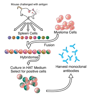
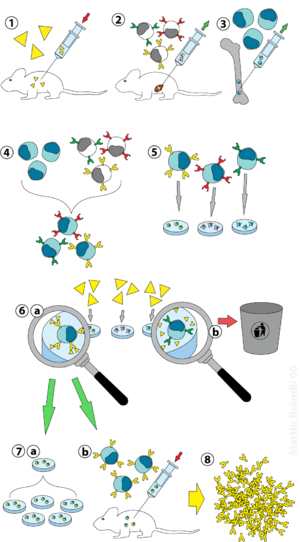
English: Overview of hybridoma technology and ...

English: Overview of hybridoma technology and monoclonal antibody creation (Photo credit: Wikipedia)

Mast cells are involved in allergy. Allergies ...

Mast cells are involved in allergy. Allergies such as pollen allergy are related to the antibody known as IgE. Like other antibodies, each IgE antibody is specific; one acts against oak pollen, another against ragweed. (Photo credit: Wikipedia)

Emil von Behring

Emil von Behring (Photo credit: Wikipedia)

Diagram showing the production of monoclonal a...

Diagram showing the production of monoclonal antibodies via hybridoma technology (Photo credit: Wikipedia)


Read Full Post »

In focus: Melanoma therapeutics


Author and Curator: Ritu Saxena, Ph.D.

In the last post of Melanoma titled “In focus: Melanoma Genetics”, I discussed the clinical characteristics and the genetics involved in Melanoma.  This post would discuss melanoma therapeutics, both current and novel.

According to the American Cancer Society, more than 76,000 new cases and more than 9100 deaths from melanoma were reported in the United States in 2012[1] Melanoma develops from the malignant transformation of melanocytes, the pigment-producing cells that reside in the basal epidermal layer in human skin. Although most melanomas arise in the skin, they may also arise from mucosal surfaces or at other sites to which neural crest cells migrate.

Melanoma therapeutics

Surgical treatment of cutaneous melanoma employs specific surgical margins depending on the depth of invasion of the tumor and there are specific surgical treatment guidelines of primary, nodal, and metastatic melanoma that surgeons adhere to while treatment. Melanoma researchers have been focusing on developing adjuvant therapies for that would increase the survival post-surgery.

Chemotherapy

Among traditional chemotherapeutic agents, only dacarbazine is FDA approved for the treatment of advanced melanoma (Eggermont AM and Kirkwood JM, Eur J Cancer, Aug 2004;40(12):1825-36). Dacarbazine is a triazene derivative and alkylates and cross-links DNA during all phases of the cell cycle, resulting in disruption of DNA function, cell cycle arrest, and apoptosis. Currently, 17 clinical trials are underway to test the efficacy and effectiveness of dacarbazine against melanoma as either a single agent or in combination chemotherapy regimens with other anti-cancer chemotherapeutic agents such as cisplatin, paclitaxel. Temozolomide is a triazene analog of dacarbazine and is approved for the treatment of malignant gliomas. At physiologic pH, it is converted to a short-lived active cytotoxic compound, monomethyl triazeno imidazole carboxamide (MTIC). MTIC methylates DNA at the O6 and N7 positions of guanine, resulting in inhibition of DNA replication. Unlike dacarbazine, which is metabolized to MITC only in the liver, temozolomide is metabolized to MITC at all sites. Temozolomide is administered orally and penetrates well into the central nervous system. Temozolomide is being tested in many combination regimens for patients with melanoma metastatic to the brain (Douglas JG and Margolin K, Semin Oncol, Oct 2002;29(5):518-24).

Immunotherapy

Melanoma and the immune system are closely related. Hence, immunotherapy has been explored in the treatment of the disease. The two most widely investigated immunotherapy drugs for melanoma are Interferon (IFN)-alpha and Interleukin-2 (IL-2).

The role of IFNalpha-2b in the adjuvant therapy of patients with localized melanoma at high risk for relapse was established by the results of three large randomized trials conducted by the US Intergroup; all three trials demonstrated an improvement in relapse-free survival and two in overall survival. One of these trials, a large randomized multicenter trial performed by the Eastern Cooperative Oncology Group (ECOG), in high-risk melanoma patients showed significant improvements in relapse-free and overall survival with adjuvant IFN-α-2b therapy, compared with standard observation (ECOG 1684). The results of the study led to FDA approval of IFN-α-2b for treatment of melanoma. This study was performed on patients with deep primary tumors without lymph node involvement and node-positive melanomas. In other studies, little antitumor activity has been demonstrated in IFN-α-2b–treated metastatic stage IV melanoma.

Recombinant IL-2 showed an overall response rate of 15-20% in metastatic melanoma and was capable of producing complete and durable remissions in about 6% of patients treated. Based upon these data, the US FDA has approved the use of high-dose IL-2 for the therapy of patients with metastatic melanoma. Aldesleukin (Brand name: Proleukin) is a recombinant analog of the endogenous cytokine interleukin-2 (IL-2). It binds to and activates the IL-2 receptor (IL-2R), followed by heterodimerization of the IL-2R beta and gamma(c) cytoplasmic chains; activation of Jak3; and phosphorylation of tyrosine residues on the IL-2R beta chain, resulting in an activated receptor complex (NCI). The activated complex recruits several signaling molecules that act as substrates for regulatory enzymes associated with the complex. It is administered intravenously and stimulates lymphokine-activating killer (LAK) cells, natural killer (NK) cells and the production of cytokines such as gamma interferon (nm|OK). Several clinical trials are currently underway using Aldesleukin to determine the efficacy of combination treatment in melanoma patients.

Another anti-cancer immunotherapeuty-based mechanism involved inhibition of inhibitory signal of cytotoxic T lymphocyte-associated antigen 4 (CTLA-4), a molecule on T-cells that plays a critical role in regulating natural immune responses. Ipilimumab (Brand name: Yervoy) was by FDA for melanoma treatment.  It is a human monoclonal antibody (MAb) T-cell potentiator that specifically blocks CTLA-4. It is approved for inoperable advanced (Stage III) or metastatic (Stage IV) melanoma in newly diagnosed or previously treated patients (nm|OK). The approval, March 25, 2011, was based on a randomized (3:1:1) double-blind double-dummy clinical trial (MDX010-20) in patients with unresectable or metastatic melanoma who had received at least one prior systemic treatment for melanoma. Patients were randomly assigned to receive either ipilimumab, 3 mg/kg intravenously, in combination with the tumor vaccine (n=403); ipilimumab plus vaccine placebo (n=137); or tumor vaccine with placebo (n=136). Patients treated with ipilimumab alone had a median overall survival (OS) of 10 months while those treated with tumor vaccine had a median OS of 6 months. The trial also demonstrated a statistically significant improvement in OS for patients treated with the combination of ipilimumab plus tumor vaccine compared with patients treated with tumor vaccine alone. For more information on the trial, check the clinical trials website, www.clinicaltrials.gov

Signaling pathway inhibitors

Approximately 90% of BRAF gene mutations involve valine (V) to glutamic acid (E) mutation at number 600 residue (V600E). The resulting oncogene product, BRAF (V600E) kinase is highly active and exhibits elevated MAPK pathway. The BRAF(V600E) gene mutation occurs in approximately 60% of melanomas indicating that it could be therapeutically relevant. Vemurafenib (Brand name: Zelboraf) is a novel small-molecule inhibitor of BRAF (V600E) kinase. It selectively binds to the ATP-binding site and inhibits the activity of BRAF (V600E) kinase. Vemurafebib inhibits over active MAPK pathway by inhibiting the mutated BRAF kinase, thereby reducing tumor cell proliferation (NCI). Encouraging results of phase III randomized, open-label, multicenter trial were reported recently at the 2011 ASCO meeting (Chapman PB, et al, ASCO 2011, Abstract LBA4).  The trial compared the novel BRAF inhibitor vemurafenib with dacarbazine in patients with BRAF-mutated melanoma. Previously untreated, unresectable stage IIIC or stage IV melanoma that tested positive for BRAF mutation were randomized (1:1) to vemurafenib or dacarbazine. The response rate (RR) was significantly high (48.4%) in vemurafenib treated patients as compared to 5.5% in dacarbazine among the 65% of patients evaluable for RR to date. In addition, vemurafenib was associated with significantly improved OS and PFS compared to dacarbazine in patients with previously untreated BRAF (V600E) mutation bearing patients with metastatic melanoma.

Biochemotherapy

Biochemothreapy combine traditional chemotherapy with immunotherapies, such as IL-2 and IFN-α-2b. These combination therapies seemed promising in phase II trials, however, seven large studies failed to show statistically significant increased overall survival rates for various biochemotherapy regimens in patients with stage IV metastasis (Margolin KA, et al, Cancer, 1 Aug 2004;101(3):435-8). Owing to inconsistent results of the available studies with regard to benefit including RR, OS and progression time, and consistently high toxicity rates, clinical practice guideline do not recommend biochemotherapy for the treatment of metastatic melanoma (Verma S, et al, Curr Oncol, April 2008; 15(2): 85–89).

Vaccines

The use of therapeutic vaccines is an ongoing area of research, and clinical trials of several types of vaccines (whole cell, carbohydrate, peptide) are being conducted in patients with intermediate and late-stage melanoma. Vaccines are also being tested in patients with metastatic melanoma to determine their immune effects and to define their activity in combination with other immunotherapeutic agents such as IL-2 or IFNalpha (Agarwala S, Am J Clin Dermatol, 2003;4(5):333-46). In fact, recently investigators at the Indiana University Health Goshen Center for Cancer Care (Goshen, IN) conducted a randomized, multicenter phase III trial involving 185 patients with stage IV or locally advanced stage III cutaneous melanoma. The patients were assigned into treatment groups with IL-2 alone or with vaccine (gp100) followed by IL-2. The vaccine-IL-2 group had a significantly improved OR as compared to the IL-2-only group (16% Vs. 6%) and longer progression free survival (2.2 months Vs. 1.6 months). The median overall survival was also longer in the vaccine-interleukin-2 group than in the interleukin-2-only group (17.8 months Vs. 11.1 months). Thus, a combination of vaccine and immunotherapy showed a better response rate and longer progression-free survival than with interleukin-2 alone in patients with advanced melanoma (Schwartzentruber DJ, et al, N Engl J Med, 2 Jun 2011;364(22):2119-27).

Which Treatment When?

Earlier, there were essentially two main options for patients suffering from advanced melanoma, dacarbazine and IL-2. Dacarbazine, a chemotherapeutic agent produces modest improvements in survival or symptomatic benefits in most patients. Interleukin-2 -based drugs, on the other hand, induce long-term remissions in a small group of patients but are highly toxic. Recently, FDA approved ipilimumab and vemurafenib for patients with metastatic melanoma. Apart from these, therapies are also aiming at starving the tumor by inhibiting angiogenesis or depleting nutrients essential for cancer growth. Of the antiangiogenic compounds, VEGFR inhibitors SU5416 and AG-013736 demonstrated broad-spectrum antitumor activity in mice bearing xenografts of human cancer cell lines originating from various tissues, including melanoma. In addition, several trials are currently underway to test the efficacy of the drugs in combination. In the future, personalized medicine-based recommendations of novel and existing drugs for melanoma patients might be the way to go.

Reference:

  1. Eggermont AM and Kirkwood JM, Eur J Cancer, Aug 2004;40(12):1825-36
  2. Douglas JG and Margolin K, Semin Oncol, Oct 2002;29(5):518-24
  3. Chapman PB, et al, ASCO 2011, Abstract LBA4
  4. Margolin KA, et al, Cancer, 1 Aug 2004;101(3):435-8
  5. Verma S, et al, Curr Oncol, April 2008; 15(2): 85–89
  6. Agarwala S, Am J Clin Dermatol, 2003;4(5):333-46
  7. Schwartzentruber DJ, et al, N Engl J Med, 2 Jun 2011;364(22):2119-27
  8. Chudnovsky Y, et al, J Clin Invest, Apr 2005;115(4):813-24.
  9. National Cancer Institute (National Institute of Health)
  10. Clinical Trials reported on the U.S. Institute of Health
  11. New Medicine Oncology KnowledgeBase (nm|OK)

Related articles on Melanoma on this Open Access Online Scientific Journal: 

  1. In focus: Melanoma Genetics Curator- Ritu Saxena, Ph.D.
  2. Thymosin alpha1 and melanoma Author/Editor- Tilda Barliya, Ph.D.
  3. A New Therapy for Melanoma  Reporter- Larry H Bernstein, M.D.
  4. Melanoma: Molecule in Immune System Could Help Treat Dangerous Skin Cancer Reporter: Prabodh Kandala, Ph.D.
  5. Why Braf inhibitors fail to treat melanoma. Reporter: Prabodh Kandala, Ph.D.


Read Full Post »

Reporter: Ritu Saxena, Ph.D.

Diabetes currently affects more than 336 million people worldwide, with healthcare costs by diabetes and its complications of up to $612 million per day in the US alone.  The islets of Langerhans, miniature endocrine organs within the pancreas, are essential regulators of blood glucose homeostasis and play a key role in the pathogenesis of diabetes.  Islets of Langerhans are composed of several types of endocrine cells.  The α- and β-cells are the most abundant and also the most important in that they secrete hormones (glucagon and insulin, respectively) crucial for glucose homeostasis (Bosco D, et al, Diabetes, May 2010;59(5):1202-10).

Diabetes is a ‘bihormonal’ disease, involving both insulin deficiency and excess glucagon.  For decades, insulin deficiency was considered to be the sole reason for diabetes; however, recent studies emphasize excess glucagon as an important part of diabetes etiology.  Thus, insulin-secreting β cells and glucagon-secreting α cells maintain physiological blood glucose levels, and their malfunction drives diabetes development.  Increasing the number of insulin-producing β cells while decreasing the number of glucagon-producing α cells, either in vitro in donor pancreatic islets before transplantation into type 1 diabetics or in vivo in type 2 diabetics, is a promising therapeutic avenue.  A huge leap has been taken in this direction by the researchers at the University of Pennsylvania (Philadelphia, PA) in collaboration with Oregon Health and Science University (Portland, OR), USA by demonstrating that α to β cell reprogramming could be promoted by manipulating the histone methylation signature of human pancreatic islets.  In fact, the treatment of cultured pancreatic islets with a histone methyltransferase inhibitor leads to colocalization of both glucagon and insulin and glucagon and insulin promoter factor 1 (PDX1) in human islets and colocalization of both glucagon and insulin in mouse islets.  The research findings were published in the Journal of Clinical Investigation.

Study design: First step was to study and analyze the epigenetic and transcriptional landscape of human pancreatic human pancreatic α, β, and exocrine cells using ChIP and RNA sequencing.  Study design for determination of the transcriptome and differential histone marks included the dispersion and FACS to of human islets to obtain cell populations highly enriched for α, β, and exocrine (duct and acinar) cells.  Then, chromatin was prepared for ChIP analysis using antibodies for histone modifications, H3K4me3 (represents gene activation) and H3K27me3 (represents gene repression).  RNA-Sequencing analysis was then performed to determine mRNA and lncRNA.  Sample purity was confirmed using qRT-PCR of insulin and glucagon expression levels of the individual α and β cell population revealing high sample purity.

Results:

  • Long noncoding transcripts: Long noncoding RNA molecules have been implicated as important developmental regulators, cell lineage allocators, and contributors to disease development.  The authors discovered 12 cell–specific and 5 α cell–specific noncoding (lnc) transcripts, indicative of the valuable research resource represented from transcriptome data.  Recently discovered lncRNA molecules in islets are regulated during development and dysregulated in type 2 diabetic islets.
  • Monovalent histone modification landscapes shared among three cell types:  Monovalent H3K4me3-enriched regions, indicative of gene activation, were identified and compared in α, β, and exocrine cells.  Strikingly, the vast majority of monovalently H3K4me3-marked genes were shared among the 3 pancreatic cell lineages (83%–95%), reflecting both their related function in protein secretion and common embryonic descent. Similarly, a high degree of overlap was observed in H3K27me3 modification patterns in all the three cell types (73%–83%).
  • Bivalent histone modifications (H3K4me3 and H3K27me3) were high in α cells: Bernstein colleagues observed bivalent marks to be common in undifferentiated cells, such as ES cells and pluripotent progenitor cells, and in most cases, one of the histone modification marks was lost during differentiation, accompanying lineage specification (Bernstein BE, et al, Cell, 21 Apr 2006; 125(2):315-26).  α cells exhibited many more genes bivalently marked, followed by β cells and exocrine cells.  Bivalent state was remarkably similar to that of hESC, suggesting a more plastic epigenomic state for α cells.
  • Monovalent histone modifications were high in β cells: Thousands of the genes that were in bivalent state in α cells were in a monovalent state, carrying only the activating or repressing mark.
  • Inhibition of histone methyltransferases led to partial cell-fate conversion: Adenosine dialdehye (Adox), a drug that interferes with histone methylation and decreases H3K27me3, when administered in human islet tissue, led to decrease of H3K27me3 enrichment at the 3 gene loci that are originally expressed bivalently in α cells and monovalently in β cells:  MAFA, PDX1 and ARX.  Adox resulted in the occasional cooccurrence of glucagon and insulin granules within the same islet cell, which was not observed in untreated islets.  Thus, inhibition of histone methyltransferases leads to partial endocrine cell-fate conversion.

Conclusion:  α cells have been reprogrammed into β cell fate in various mouse models.  The reason, as proposed by the authors, might be the presence of more bivalently marked genes that confers a more plastic epigenomic state of the cells that probably drives them to the β cell fate.  Therefore, using epigenomic information of different cell types in pancreatic islets and harnessing it for subsequent manipulation of their epigenetic signature could be utilized to reprogram cells and hence provide a path for diabetes therapy.

Source: Bramswig NC, et al, Epigenomic plasticity enables human pancreatic α to β cell reprogramming. J Clin Invest, 22 Feb 2013. pii: 66514.

Related reading on Pharmaceutical Intelligence:

Junk DNA codes for valuable miRNAs: non-coding DNA controls Diabetes

Therapeutic Targets for Diabetes and Related Metabolic Disorders

Reprogramming cell fate

CRACKING THE CODE OF HUMAN LIFE: Recent Advances in Genomic Analysis and Disease – Part IIC

2013 Genomics: The Era Beyond the Sequencing of the Human Genome: Francis Collins, Craig Venter, Eric Lander, et al.

Genome-Wide Detection of Single-Nucleotide and Copy-Number Variation of a Single Human Cell

SNAP: Predict Effect of Non-synonymous Polymorphisms: How well Genome Interpretation Tools could Translate to the Clinic

Genomic Endocrinology and its Future

Read Full Post »

Modeling Targeted Therapy

Reporter: Larry H. Bernstein, MD, FCAP
pharmaceuticalintelligence.com/2013/03/02/modeling-targeted-therapy/

Some Perspectives on Network Modeling in Therapeutic Target Prediction
R Albert, B DasGupta and N Mobasheri
Biomedical Engineering and Computational Biology Insights 2013; 5: 17–24    http://dx.doi.org/BECBI/Albert_DasGupta_ Mobasheri
Key steps of a typical therapeutic target identification problem include synthesizing or inferring the complex network of interactions relevant to the disease, connecting this network to the disease-specific behavior, and predicting which components are key mediators of the behavior
http://www.la-press.com/Some_Perspectives_on_Network_Modeling_in_Therapeutic_Target_Prediction/

Journal of Computational Biology

Journal of Computational Biology (Photo credit: Wikipedia)

Read Full Post »

Latest research efforts reported in the San Antonio Breast Cancer Symposium, 2012

Curator: Ritu Saxena, Ph.D.

‘Triple negative breast cancer’ or TNBC, as the name suggests, is a classification of breast cancers lacking the expression of estrogen receptor (ER) and progesterone receptor expression as well as amplification of the human epidermal growth factor receptor 2 (HER2).

Unlike other breast cancer types, treating TNBC is a challenge mainly because of the absence of well-defined molecular targets and because of disease heterogeneity. Currently, neoadjuvant chemotherapies are in use to treat TNBC patients. Some, around 30%, patients respond completely to neoadjuvant chemotherapy and have good outcomes after surgery. However, if there is a residual disease after therapy, outcomes are poor.

Therefore, current focus of the field is to first understand the complexity of the disease, both at genomic and molecular level and look for targets. Also, several combination chemotherapies are currently under trial to determine the efficacy, overall response rate, progression-free survival and other relevant factors for patients suffering with different forms of TNBC.

Recently, in the San Antonio Breast Cancer Symposium (SABCS 2012), several abstarcts related to TNBC research, both clinical and pre-clinical. Here is a compilation of some of the abstracts and their relevance in the field of TNBC research:

Triple Negative Breast Cancer: Subtypes, Molecular Targets, and Therapeutic Approaches, Pietenpol JA, Vanderbilt-Ingram Cancer Center; Vanderbilt University School of Medicine (Nashville, TN), Abstract no. ES2-2.

In order to better understand the complexity of TNBC, an integrative and comprehensive genomic and molecular analysis is required. The analysis would give important cues to developing and administering effective therapeutic agents. The group has compiled an extensive number of TNBC gene expression profiles and initiated molecular subtyping of the disease. Differential GE was used to designate 25 TNBC cell line models representative of the following subtypes:

  •  two basel-like TNBC subtypes with cell cycle and DDR gene expression signatures (BL1 and BL2);
  • two mesenchymal subtypes with high expression of genes involved in differentiation and growth factor pathways (M and MSL);
  • an immunomodulatory (IM) type;
  • a luminal subtype driven by androgen signaling (LAR)

The pharmacological drugs were chosen on the basis of the genetic pathways active in the cell lines with the abovementioned TNBC subtypes. It was observed that BL1 and BL2 subtype cell lines respond to cisplatin. Mesenchymal, basal, and luminal subtype lines with aberrations in PI3K signaling and have the greatest sensitivity to PI3K inhibitors.

The LAR subtype cell lines express AR and are uniquely sensitive to bicalutamide (AR antagonist). The experiment was a proof-of-concept that the best therapy could be based on TNBC subtypes.

The group has also developed a web-based subtyping tool referred to as “TNBCtype,” for candidate TNBC tumor samples using our gene expression metadata and classification methods. The approach would enable alignment of TNBC patients to appropriate targeted therapies.

The Clonal and Mutational Composition of Triple Negative Breast Cancers: Aparicio S, University of British Columbia (Vancouver, BC), Canada. Abstract no. ES2-3.

The abstract is on the same lines, TNBC heterogeneity that is. The concept of clonal heterogeneity in cancers, the spatial and temporal variation in clonal composition, is the focal point of the discussion. The group has developed next generation sequencing approaches and applied them to the understanding of mutational and clonal composition of primary TNBC. They have demonstrated that both mutational composition and clonal structure of primary TNBC is in fact a complete spectrum, a notion that is far from the previous one that stated TNBC to be a distinct disease. The authors add “clonal analysis suggests a means by which the genetic complexity might be reduced by following patient evolution over time and space.” The specific implications of the mutational and transcriptome landscapes of TNBC in relation to possible disease biologies were discussed in the symposium.

Profiling of triple-negative breast cancers after neoadjuvant chemotherapy identifies targetable molecular alterations in the treatment-refractory residual disease:

Balko JM, etal, Vanderbilt University (Nashville, TN); Foundation Medicine, (Cambridge, MA); Instituto Nacional de Enfermedades Neoplásicas, Lima, Peru

In the absence of hormone receptors and hence lack of targets, Neoadjuvant chemotherapy (NAC) is increasingly used in patients with TNBC. NAC can induce a pathologic complete response (pCR) in ∼30% of patients which portends a favorable prognosis. In contrast, patients with residual disease (RD) in the breast at surgical resection exhibit worse outcomes. The authors hypothesize that “profiling residual TNBC after NAC would identify molecularly targetable lesions in the chemotherapy resistant component of the tumor and that the persistent tumor cells would mirror micro-metastases which ultimately recur in such patients.” The researchers utilized targeted next generation sequencing (NGS) for 182 oncogenes and tumor suppressors in a CLIA certified lab (Foundation Medicine, Cambridge, MA) and gene expression profiling (NanoString) of the RD after NAC in 102 patients with TNBC. The RD was stained for Ki67, which has been reported to predict outcome after NAC in unselected breast cancers. Out of the 89 evaluable post-NAC tumors, 57 (64%) were basal-like; 19% HER2-enriched; 6% luminal A; 6% luminal B and 5% normal-like. Of 81 tumors evaluated by NGS, 89% demonstrated mutations in TP53, 27% were MCL1-amplified, and 21% were MYC-amplified.

Several pathways were found to be altered:

  • PI3K/mTOR pathway (AKT1-3, PIK3CA, PIK3R1, RAPTOR, PTEN, and TSC1)
  • Cell cycle genes (amplifications of CDK2, CDK4, and CDK6, CCND1-3, and CCNE1); loss of RB
  • DNA repair pathway (BRCA1/2, ATM)
  • Ras/MAPK pathway (KRAS, RAF1, NF1)
  • Sporadic growth factor receptor (amplifications occurred in EGFR, KIT, PDGFRA, PDGFRB, MET, FGFR1, FGFR2, and IGF1R.

NGS identified 7 patients with ERBB2 gene amplification. NGS could assist in the identification of ERBB2-overexpressing tumors misclassified at the time of diagnosis.

Amplifications of MYC were independently associated with poor recurrence-free survival (RFS) and overall survival (OS). In contrast to the earlier notion, high post-NAC Ki67 score did not predict poor RFS or OS in this predominantly TNBC cohort.

The authors concluded that “the diversity of lesions in residual TNBCs after NAC underscores the need for powerful and broad molecular approaches to identify actionable molecular alterations and, in turn, better inform personalized therapy of this aggressive disease.”

Identification of Novel Synthetic-Lethal Targets for MYC-Driven Triple-Negative Breast Cancer: Goga A, etal, UCSF (San Francisco, CA), Abstract No. S3-8

Reiterating the greatest challenge of the TNBC treatment, no targeted agents currently exist against TNBC. The group at UCSF has discovered that TNBC frequently express high levels of the MYC proto-oncogene. The discovery has led them to identify new “synthetic-lethal” strategies to selectively kill TNBC with MYC overexpression. “Synthetic lethality arises when a combination of mutations in two or more genes leads to cell death, whereas a mutation in only one of these genes has little effect. Using this strategy, we can take advantage of the elevated MYC signaling in TNBC to selectively kill them, while sparing normal tissues in which MYC is expressed at much lower levels”

The researchers performed a shRNA synthetic-lethal screen in the human mammary epithelial cells (HMEC), to identify new molecules, such as cell cycle kinases, which when inhibited can preferentially kill TNBC cells. A high-throughput screen of ∼2000 shRNAs, that target the human kinome (∼ 600 kinases) when performed, led to the identification of 13 kinases whose inhibition by >1 shRNAs gave rise to >50% inhibition of cell growth. ARK5 and GSK3A were the two other kinases that were shown to have a synthetic-lethal interaction with MYC. Since these two kinases have been identified in other studies, it gives validity to the ability to the methods of Goga etal in identifying synthetic-lethal targets. The group is currently characterizing and validating the 11 novel targets identified in this screen, using human cancer cell lines as well as mouse cancer models to determine the impact of inhibiting these targets on triple-negative breast cancer development and proliferation.

Reference:

Dent R, etal.  Triple-negative breast cancer: clinical features and patterns of recurrence (2007) Clin Cancer Res 13, 4429-4434.

Lehmann BD, etal. Identification of human triple-negative breast cancer subtypes and preclinical models for selection of targeted therapies (2011) J Clin Invest. 121: 2750-67.

Chen X, etal. TNBCtype: A Subtyping Tool for Triple- Negative Breast Cancer. (2012) Cancer informatics 11, 147-156.

Abstracts presented in SABCS 2012 can be accessed here.

Read Full Post »

Reporter: Ritu Saxena, Ph.D.

With the number of cancer cases plummeting every year, there is a dire need for finding a cure to wipe the disease out. A number of therapeutic drugs are currently in use, however, due to heterogeneity of the disease targeted therapy is required. An important criteria that needs to be addressed in this context is the –‘tumor response’ and how it could be predicted, thereby improving the selection of patients for cancer treatment. The issue of tumor response has been addressed in a recent editorial titled “Tumor response criteria: are they appropriate?” published recently in Future Oncology.

The article talks about how the early tumor treatment response methods came into practice and how we need to redefine and reassess the tumor response.

Defining ‘tumor response’ has always been a challenge

WHO defines a response to anticancer therapy as 50% or more reduction in the tumor size measured in two perpendicular diameters. It is based on the results of experiments performed by Moertel and Hanley in 1976 and later published by Miller et al in 1981. Twenty years later, in the year 2000, the US National Cancer Institute, with the European Association for Research and Treatment of Cancer, proposed ‘new response criteria’ for solid tumors; a replacement of 2D measurement with measurement of one dimen­sion was made. Tumor response was defined as a decrease in the largest tumor diameter by 30%, which would translate into a 50% decrease for a spherical lesion. However, response criteria have not been updated after that and there a structured standardization of treatment response is still required especially when several studies have revealed that the response of tumors to a therapy via imaging results from conventional approaches such as endoscopy, CT scan, is not reliable. The reason is that evaluating the size of tumor is just one part of the story and to get the complete picture inves­tigating and evaluating the tissue is essential to differentiate between treatment-related scar, fibrosis or micro­scopic residual tumor.

In clinical practice, treatment response is determined on the basis of well-established parameters obtained from diagnostic imaging, both cross-sectional and functional. In general, the response is classified as:

  • Complete remission: If a tumor disappears after a particular therapy,
  • Partial remission: there is residual tumor after therapy.

For a doctor examining the morphology of the tumor, complete remission might seem like good news, however, mission might not be complete yet! For example, in some cases, with regard to prognosis, patients with 0% residual tumor (complete tumor response) had the same prognosis com­pared with those patients with 1–10% residual tumor (subtotal response).

Another example is that in patients demonstrating complete remission of tumor response as observed with clinical, sonographic, functional (PET) and histopathological analysis experience recur­rence within the first 2 years of resection.

Adding complexity to the situation is the fact that the appropriate, clinically relevant timing of assess­ment of tumor response to treatment remains undefined. An example mentioned in the editorial is – for gastrointestinal (GI) malignancies, the assessment timing varies considerably from 3 to 6 weeks after initia­tion of neoadjuvant external beam radiation. Further, time could vary depending upon the type of radiation administered, i.e., if it is external beam, accelerated hyperfractionation, or brachytherapy.

Abovementioned examples remind us of the intricacy and enigma of tumor biol­ogy and subsequent tumor response.

Conclusion

Owing to the extraordinary het­erogeneity of cancers between patients, and pri­mary and metastatic tumors in the same patients, it is important to consider several factors while determining the response of tumors to different therapie in clinical trials. Authors exclaim, “We must change the tools we use to assess tumor response. The new modality should be based on individualized histopathology as well as tumor molecular, genetic and functional characteristics, and individual patients’ charac­teristics.”

Future perspective

Editorial points out that the oncologists, radiotherapists, and immunologists all might have a different opinion and observation as far as tumor response is considered. For example, surgical oncologists might determine a treatment to be effective if the local tumor control is much better after multimodal treatment, and that patients post-therapeutically also reveal an increase of the rate of microscopic and macroscopic R0-resection. Immunologists, on the other hand, might just declare a response if immune-competent cells have been decreased and, possibly, without clinical signs of decrease of tumor size.

What might be the answer to the complexity to reading tumor response is stated in the editorial – “an interdisciplinary initiative with all key stake­holders and disciplines represented is imperative to make predictive and prognostic individualized tumor response assessment a modern-day reality. The integrated multidisciplinary panel of international experts need to define how to leverage existing data, tissue and testing platforms in order to predict individual patient treatment response and prog­nosis.”

Sources:

Editorial : Björn LDM Brücher et al Tumor response criteria: are they appropriate? Future Oncology August 2012, Vol. 8, No. 8, 903-906.

Miller AB, Hoogstraten B, Staquet M, Winkler A. Reporting results of cancer treatment. Cancer 1981, 47(1),207–214.

Related articles to this subject on this Open Access Online Scientific Journal:

See comment written for :

Knowing the tumor’s size and location, could we target treatment to THE ROI by applying

http://pharmaceuticalintelligence.com/2012/10/16/knowing-the-tumors-size-and-location-could-we-target-treatment-to-the-roi-by-applying-imaging-guided-intervention/imaging-guided intervention?

Personalized Medicine: Cancer Cell Biology and Minimally Invasive Surgery (MIS)

http://pharmaceuticalintelligence.com/2012/12/01/personalized-medicine-cancer-cell-biology-and-minimally-invasive-surgery-mis/

Read Full Post »

Curator/Reporter: Aviral Vatsa PhD MBBS

This post is in the second part of the reviews that focuses on the current status of drug delivery to bone and the issues facing this field. The first part can be accessed here

Annual treatment costs for musculoskeletal diseases in the US are roughly 7.7% (~ $849 billion) of total gross domestic product. Such disorders are the main cause of physical disability in US. Almost half of all chronic conditions in people can be attributed to bone and joint disorders. In addition there is increasing ageing population and associated increases in osteoporosis and other diseases, rising incidences of degenerative intervertebral disk diseases and numbers of revision orthopedic arthroplasty surgeries, and increases in spinal fusions. All these factors contribute towards the increasing requirement of bone regeneration and reconstruction methods and products. Delivery of therapeutic grade products to bone has various challenges. Parenteral administration limits the efficient delivery of drugs to the required site of injury and local delivery methods are often expensive and invasive. The theme issue of Advance Drug Delivery reviews focuses on the current status of drug delivery to bone and the issues facing this field. Here is the second part of these reviews and research articles.

1. Targeting polymer therapeutics to bone [1]

Abstract

An aging population in the developing world has led to an increase in musculoskeletal diseases such as osteoporosis and bone metastases. Left untreated many bone diseases cause debilitating pain and in the case of cancer, death. Many potential drugs are effective in treating diseases but result in side effects preventing their efficacy in the clinic. Bone, however, provides a unique environment of inorganic solids, which can be exploited in order to effectively target drugs to diseased tissue. By integration of bone targeting moieties to drug-carrying water-soluble polymers, the payload to diseased area can be increased while side effects decreased. The realization of clinically relevant bone targeted polymer therapeutics depends on (1) understanding bone targeting moiety interactions, (2) development of controlled drug delivery systems, as well as (3) understanding drug interactions. The latter makes it possible to develop bone targeted synergistic drug delivery systems.


2. Development of macromolecular prodrug for rheumatoid arthritis [2]

Abstract

Rheumatoid arthritis (RA) is a chronic autoimmune disease that is considered to be one of the major public health problems worldwide. The development of therapies that target tumor necrosis factor-α (TNF-α), interleukin-6 (IL-6) and co-stimulatory pathways that regulate the immune system have revolutionized the care of patients with RA. Despite these advances, many patients continue to experience symptomatic and functional impairment. To address this issue, more recent therapies that have been developed are designed to target intracellular signaling pathways involved in immunoregulation. Though this approach has been encouraging, there have been major challenges with respect to off-target organ side effects and systemic toxicities related to the widespread distribution of these signaling pathways in multiple cell types and tissues. These limitations have led to an increasing interest in the development of strategies for the macromolecularization of anti-rheumatic drugs, which could target them to the inflamed joints. This approach enhances the efficacy of the therapeutic agent with respect to synovial inflammation, while markedly reducing non-target organ adverse side effects. In this manuscript, we provide a comprehensive overview of the rational design and optimization of macromolecular prodrugs for treatment of RA. The superior and the sustained efficacy of the prodrug may be partially attributed to their Extravasation through Leaky Vasculature and subsequent Inflammatory cell-mediated Sequestration (ELVIS) in the arthritic joints. This biologic process provides a plausible mechanism, by which macromolecular prodrugs preferentially target arthritic joints and illustrates the potential benefits of applying this therapeutic strategy to the treatment of other inflammatory diseases.


3. Peptide-based delivery to bone [3]

Abstract

Peptides are attractive as novel therapeutic reagents, since they are flexible in adopting and mimicking the local structural features of proteins. Versatile capabilities to perform organic synthetic manipulations are another unique feature of peptides compared to protein-based medicines, such as antibodies. On the other hand, a disadvantage of using a peptide for a therapeutic purpose is its low stability and/or high level of aggregation. During the past two decades, numerous peptides were developed for the treatment of bone diseases, and some peptides have already been used for local applications to repair bone defects in the clinic. However, very few peptides have the ability to form bone themselves. We herein summarize the effects of the therapeutic peptides on bone loss and/or local bone defects, including the results from basic studies. We also herein describe some possible methods for overcoming the obstacles associated with using therapeutic peptide candidates.


4. Growth factor delivery: How surface interactions modulate release in vitro and in vivo [4]

Abstract

Biomaterial scaffolds have been extensively used to deliver growth factors to induce new bone formation. The pharmacokinetics of growth factor delivery has been a critical regulator of their clinical success. This review will focus on the surface interactions that control the non-covalent incorporation of growth factors into scaffolds and the mechanisms that control growth factor release from clinically relevant biomaterials. We will focus on the delivery of recombinant human bone morphogenetic protein-2 from materials currently used in the clinical practice, but also suggest how general mechanisms that control growth factor incorporation and release delineated with this growth factor could extend to other systems. A better understanding of the changing mechanisms that control growth factor release during the different stages of preclinical development could instruct the development of future scaffolds for currently untreatable injuries and diseases.


5. Biomaterial delivery of morphogens to mimic the natural healing cascade in bone[5]

Abstract

Complications in treatment of large bone defects using bone grafting still remain. Our understanding of the endogenous bone regeneration cascade has inspired the exploration of a wide variety of growth factors (GFs) in an effort to mimic the natural signaling that controls bone healing. Biomaterial-based delivery of single exogenous GFs has shown therapeutic efficacy, and this likely relates to its ability to recruit and promote replication of cells involved in tissue development and the healing process. However, as the natural bone healing cascade involves the action of multiple factors, each acting in a specific spatiotemporal pattern, strategies aiming to mimic the critical aspects of this process will likely benefit from the usage of multiple therapeutic agents. This article reviews the current status of approaches to deliver single GFs, as well as ongoing efforts to develop sophisticated delivery platforms to deliver multiple lineage-directing morphogens (multiple GFs) during bone healing.

6. Studies of bone morphogenetic protein-based surgical repair[6]

Abstract

Over the past several decades, recombinant human bone morphogenetic proteins (rhBMPs) have been the most extensively studied and widely used osteoinductive agents for clinical bone repair. Since rhBMP-2 and rhBMP-7 were cleared by the U.S. Food and Drug Administration for certain clinical uses, millions of patients worldwide have been treated with rhBMPs for various musculoskeletal disorders. Current clinical applications include treatment of long bone fracture non-unions, spinal surgeries, and oral maxillofacial surgeries. Considering the growing number of recent publications related to clincal research of rhBMPs, there exists enormous promise for these proteins to be used in bone regenerative medicine. The authors take this opportunity to review the rhBMP literature paying specific attention to the current applications of rhBMPs in bone repair and spine surgery. The prospective future of rhBMPs delivered in combination with tissue engineered scaffolds is also reviewed.


7. Strategies for controlled delivery of growth factors and cells for bone regeneration[7]

Abstract

The controlled delivery of growth factors and cells within biomaterial carriers can enhance and accelerate functional bone formation. The carrier system can be designed with pre-programmed release kinetics to deliver bioactive molecules in a localized, spatiotemporal manner most similar to the natural wound healing process. The carrier can also act as an extracellular matrix-mimicking substrate for promoting osteoprogenitor cellular infiltration and proliferation for integrative tissue repair. This review discusses the role of various regenerative factors involved in bone healing and their appropriate combinations with different delivery systems for augmenting bone regeneration. The general requirements of protein, cell and gene therapy are described, with elaboration on how the selection of materials, configurations and processing affects growth factor and cell delivery and regenerative efficacy in both in vitro and in vivo applications for bone tissue engineering.

8. Bone repair cells for craniofacial regeneration[8]

Abstract

Reconstruction of complex craniofacial deformities is a clinical challenge in situations of injury, congenital defects or disease. The use of cell-based therapies represents one of the most advanced methods for enhancing the regenerative response for craniofacial wound healing. Both somatic and stem cells have been adopted in the treatment of complex osseous defects and advances have been made in finding the most adequate scaffold for the delivery of cell therapies in human regenerative medicine. As an example of such approaches for clinical application for craniofacial regeneration, Ixmyelocel-T or bone repair cells are a source of bone marrow derived stem and progenitor cells. They are produced through the use of single pass perfusion bioreactors for CD90+ mesenchymal stem cells and CD14+ monocyte/macrophage progenitor cells. The application of ixmyelocel-T has shown potential in the regeneration of muscular, vascular, nervous and osseous tissue. The purpose of this manuscript is to highlight cell therapies used to repair bony and soft tissue defects in the oral and craniofacial complex. The field at this point remains at an early stage, however this review will provide insights into the progress being made using cell therapies for eventual development into clinical practice.


9. Gene therapy approaches to regenerating bone[9]

Abstract

Bone formation and regeneration therapies continue to require optimization and improvement because many skeletal disorders remain undertreated. Clinical solutions to nonunion fractures and osteoporotic vertebral compression fractures, for example, remain suboptimal and better therapeutic approaches must be created. The widespread use of recombinant human bone morphogenetic proteins (rhBMPs) for spine fusion was recently questioned by a series of reports in a special issue of The Spine Journal, which elucidated the side effects and complications of direct rhBMP treatments. Gene therapy – both direct (in vivo) and cell-mediated (ex vivo) – has long been studied extensively to provide much needed improvements in bone regeneration. In this article, we review recent advances in gene therapy research whose aims are in vivo or ex vivo bone regeneration or formation. We examine appropriate vectors, safety issues, and rates of bone formation. The use of animal models and their relevance for translation of research results to the clinical setting are also discussed in order to provide the reader with a critical view. Finally, we elucidate the main challenges and hurdles faced by gene therapy aimed at bone regeneration as well as expected future trends in this field.

10. Gene delivery to bone[10]

Abstract

Gene delivery to bone is useful both as an experimental tool and as a potential therapeutic strategy. Among its advantages over protein delivery are the potential for directed, sustained and regulated expression of authentically processed, nascent proteins. Although no clinical trials have been initiated, there is a substantial pre-clinical literature documenting the successful transfer of genes to bone, and their intraosseous expression. Recombinant vectors derived from adenovirus, retrovirus and lentivirus, as well as non-viral vectors, have been used for this purpose. Both ex vivo and in vivo strategies, including gene-activated matrices, have been explored. Ex vivo delivery has often employed mesenchymal stem cells (MSCs), partly because of their ability to differentiate into osteoblasts. MSCs also have the potential to home to bone after systemic administration, which could serve as a useful way to deliver transgenes in a disseminated fashion for the treatment of diseases affecting the whole skeleton, such as osteoporosis orosteogenesis imperfecta. Local delivery of osteogenic transgenes, particularly those encoding bone morphogenetic proteins, has shown great promise in a number of applications where it is necessary to regenerate bone. These include healing large segmental defects in long bones and the cranium, as well as spinal fusion and treating avascular necrosis.

11. RNA therapeutics targeting osteoclast-mediated excessive bone resorption[11]

Abstract

RNA interference (RNAi) is a sequence-specific post-transcriptional gene silencing technique developed with dramatically increasing utility for both scientific and therapeutic purposes. Short interfering RNA (siRNA) is currently exploited to regulate protein expression relevant to many therapeutic applications, and commonly used as a tool for elucidating disease-associated genes. Osteoporosis and their associated osteoporotic fragility fractures in both men and women are rapidly becoming a global healthcare crisis as average life expectancy increases worldwide. New therapeutics are needed for this increasing patient population. This review describes the diversity of molecular targets suitable for RNAi-based gene knock down in osteoclasts to control osteoclast-mediated excessive bone resorption. We identify strategies for developing targeted siRNA delivery and efficient gene silencing, and describe opportunities and challenges of introducing siRNA as a therapeutic approach to hard and connective tissue disorders.

Bibliography

[1] S. A. Low and J. Kopeček, “Targeting polymer therapeutics to bone,” Advanced Drug Delivery Reviews, vol. 64, no. 12, pp. 1189–1204, Sep. 2012.

[2] F. Yuan, L. Quan, L. Cui, S. R. Goldring, and D. Wang, “Development of macromolecular prodrug for rheumatoid arthritis,” Advanced Drug Delivery Reviews, vol. 64, no. 12, pp. 1205–1219, Sep. 2012.

[3] K. Aoki, N. Alles, N. Soysa, and K. Ohya, “Peptide-based delivery to bone,” Advanced Drug Delivery Reviews, vol. 64, no. 12, pp. 1220–1238, Sep. 2012.

[4] W. J. King and P. H. Krebsbach, “Growth factor delivery: How surface interactions modulate release in vitro and in vivo,” Advanced Drug Delivery Reviews, vol. 64, no. 12, pp. 1239–1256, Sep. 2012.

[5] M. Mehta, K. Schmidt-Bleek, G. N. Duda, and D. J. Mooney, “Biomaterial delivery of morphogens to mimic the natural healing cascade in bone,” Advanced Drug Delivery Reviews, vol. 64, no. 12, pp. 1257–1276, Sep. 2012.

[6] K. W.-H. Lo, B. D. Ulery, K. M. Ashe, and C. T. Laurencin, “Studies of bone morphogenetic protein-based surgical repair,” Advanced Drug Delivery Reviews, vol. 64, no. 12, pp. 1277–1291, Sep. 2012.

[7] T. N. Vo, F. K. Kasper, and A. G. Mikos, “Strategies for controlled delivery of growth factors and cells for bone regeneration,” Advanced Drug Delivery Reviews, vol. 64, no. 12, pp. 1292–1309, Sep. 2012.

[8] G. Pagni, D. Kaigler, G. Rasperini, G. Avila-Ortiz, R. Bartel, and W. V. Giannobile, “Bone repair cells for craniofacial regeneration,” Advanced Drug Delivery Reviews, vol. 64, no. 12, pp. 1310–1319, Sep. 2012.

[9] N. Kimelman Bleich, I. Kallai, J. R. Lieberman, E. M. Schwarz, G. Pelled, and D. Gazit, “Gene therapy approaches to regenerating bone,” Advanced Drug Delivery Reviews, vol. 64, no. 12, pp. 1320–1330, Sep. 2012.

[10] C. H. Evans, “Gene delivery to bone,” Advanced Drug Delivery Reviews, vol. 64, no. 12, pp. 1331–1340, Sep. 2012.

[11] Y. Wang and D. W. Grainger, “RNA therapeutics targeting osteoclast-mediated excessive bone resorption,” Advanced Drug Delivery Reviews, vol. 64, no. 12, pp. 1341–1357, Sep. 2012.

Read Full Post »

Reporter Aviral Vatsa, PhD MBBS

Annual treatment costs for musculoskeletal diseases in the US are roughly 7.7% (~ $849 billion) of total gross domestic product. Such disorders are the main cause of physical disability in US. Almost half of all chronic conditions in people can be attributed to bone and joint disorders. In addition there is increasing ageing population and associated increases in osteoporosis and other diseases, rising incidences of degenerative intervertebral disk diseases and numbers of revision orthopedic arthroplasty surgeries, and increases in spinal fusions. All these factors contribute towards the increasing requirement of bone regeneration and reconstruction methods and products. Delivery of therapeutic grade products to bone has various challenges. Parenteral administration limits the efficient delivery of drugs to the required site of injury and local delivery methods are often expensive and invasive. The theme issue of Advance Drug Delivery reviews focuses on the current status of drug delivery to bone and the issues facing this field. Here is the first part of these reviews and research articles.

1. Demineralized bone matrix in bone repair: History and use

Abstract

Demineralized bone matrix (DBM) is an osteoconductive and osteoinductive commercial biomaterial and approved medical device used in bone defects with a long track record of clinical use in diverse forms. True to its name and as an acid-extracted organic matrix from human bone sources, DBM retains much of the proteinaceous components native to bone, with small amounts of calcium-based solids, inorganic phosphates and some trace cell debris. Many of DBM’s proteinaceous components (e.g., growth factors) are known to be potent osteogenic agents. Commercially sourced as putty, paste, sheets and flexible pieces, DBM provides a degradable matrix facilitating endogenous release of these compounds to the bone wound sites where it is surgically placed to fill bone defects, inducing new bone formation and accelerating healing. Given DBM’s long clinical track record and commercial accessibility in standard forms and sources, opportunities to further develop and validate DBM as a versatile bone biomaterial in orthopedic repair and regenerative medicine contexts are attractive.

2. Biomimetic hydrogels for controlled biomolecule delivery to augment bone regeneration

Abstract

The regeneration of large bone defects caused by trauma or disease remains a significant clinical problem. Although osteoinductive growth factors such as bone morphogenetic proteins have entered clinics, transplantation of autologous bone remains the gold standard to treat bone defects. The effective treatment of bone defects by protein therapeutics in humans requires quantities that exceed the physiological doses by several orders of magnitude. This not only results in very high treatment costs but also bears considerable risks for adverse side effects. These issues have motivated the development of biomaterials technologies allowing to better control biomolecule delivery from the solid phase. Here we review recent approaches to immobilize biomolecules by affinity binding or by covalent grafting to biomaterial matrices. We focus on biomaterials concepts that are inspired by extracellular matrix (ECM) biology and in particular the dynamic interaction of growth factors with the ECM. We highlight the value of synthetic, ECM-mimicking matrices for future technologies to study bone biology and develop the next generation of ‘smart’ implants.


3. Calcium phosphate cements as drug delivery materials

Abstract

Calcium phosphate cements are used as synthetic bone grafts, with several advantages, such as their osteoconductivity and injectability. Moreover, their low-temperature setting reaction and intrinsic porosity allow for the incorporation of drugs and active principles in the material. It is the aim of the present work to: a) provide an overview of the different approaches taken in the application of calcium phosphate cements for drug delivery in the skeletal system, and b) identify the most significant achievements. The drugs or active principles associated to calcium phosphate cements are classified in three groups, i) low molecular weight drugs; ii) high molecular weight biomolecules; and iii) ions.

4. Silk constructs for delivery of musculoskeletal therapeutics

Abstract

Silk fibroin (SF) is a biopolymer with distinguishing features from many other bio- as well as synthetic polymers. From a biomechanical and drug delivery perspective, SF combines remarkable versatility for scaffolding (solid implants, hydrogels, threads, solutions), with advanced mechanical properties and good stabilization and controlled delivery of entrapped protein and small molecule drugs, respectively. It is this combination of mechanical and pharmaceutical features which renders SF so exciting for biomedical applications. This pattern along with the versatility of this biopolymer has been translated into progress for musculoskeletal applications. We review the use and potential of silk fibroin for systemic and localized delivery of therapeutics in diseases affecting the musculoskeletal system. We also present future directions for this biopolymer as well as the necessary research and development steps for their achievement.

5. Demineralized bone matrix as a vehicle for delivering endogenous and exogenous therapeutics in bone repair

Abstract

As a unique human bone extract approved for implant use, demineralized bone matrix (DBM) retains substantial amounts of endogenous osteoconductive and osteoinductive proteins. Commercial preparations of DBM represent a clinically accessible, familiar, widely used and degradable bone-filling device, available in composite solid, strip/piece, and semi-solid paste forms. Surgically placed and/or injected, DBM releases its constituent compounds to bone sites with some evidence for inducing new bone formation and accelerating healing. Significantly, DBM also has preclinical history as a drug carrier by direct loading and delivery of several important classes of therapeutics. Exogenous bioactive agents, including small molecule drugs, protein and peptide drugs, nucleic acid drugs and transgenes and therapeutic cells have been formulated within DBM and released to bone sites to enhance DBM’s intrinsic biological activity. Local release of these agents from DBM directly to surgical sites in bone provides improved control of dosing and targeting of both endogenous and exogenous bioactivity in the context of bone healing using a clinically familiar product. Given DBM’s long clinical track record and commercial accessibility in standard forms and sources, opportunities to formulate DBM as a versatile matrix to deliver therapeutic agents locally to bone sites in orthopedic repair and regenerative medicine contexts are attractive.

6. Nanofiber-based delivery of bioactive agents and stem cells to bone sites

Abstract

Biodegradable nanofibers are important scaffolding materials for bone regeneration. In this paper, the basic concepts and recent advances of self-assembly, electrospinning and thermally induced phase separation techniques that are widely used to generate nanofibrous scaffolds are reviewed. In addition, surface functionalization and bioactive factor delivery within these nanofibrous scaffolds to enhance bone regeneration are also discussed. Moreover, recent progresses in applying these nanofiber-based scaffolds to deliver stem cells for bone regeneration are presented. Along with the significant advances, challenges and obstacles in the field as well as the future perspective are discussed.

 
7. Intra-operatively customized implant coating strategies for local and controlled drug delivery to bone

Abstract

Bone is one of the few tissues in the human body with high endogenous healing capacity. However, failure of the healing process presents a significant clinical challenge; it is a tremendous burden for the individual and has related health and economic consequences. To overcome such healing deficits, various concepts for a local drug delivery to bone have been developed during the last decades. However, in many cases these concepts do not meet the specific requirements of either surgeons who must use these strategies or individual patients who might benefit from them. We describe currently available methods for local drug delivery and their limitations in therapy. Various solutions for drug delivery to bone focusing on clinical applications and intra-operative constraints are discussed and drug delivery by implant coating is highlighted. Finally, a new set of design and performance requirements for intra-operatively customized implant coatings for controlled drug delivery is proposed. In the future, these requirements may improve approaches for local and intra-operative treatment of patients.


8. Local delivery of small and large biomolecules in craniomaxillofacial bone

Abstract

Current state of the art reconstruction of bony defects in the craniomaxillofacial (CMF) area involves transplantation of autogenous or allogenous bone grafts. However, the inherent drawbacks of this approach strongly urge clinicians and researchers to explore alternative treatment options. Currently, a wide interest exists in local delivery of biomolecules from synthetic biomaterials for CMF bone regeneration, in which small biomolecules are rapidly emerging in recent years as an interesting adjunct for upgrading the clinical treatment of CMF bone regeneration under compromised healing conditions. This review highlights recent advances in the local delivery small and large biomolecules for the clinical treatment of CMF bone defects. Further, it provides a perspective on the efficacy of biomolecule delivery in CMF bone regeneration by reviewing presently available reports of pre-clinical studies using various animal models.

9. Immobilized antibiotics to prevent orthopaedic implant infections

Abstract

Many surgical procedures require the placement of an inert or tissue-derived implant deep within the body cavity. While the majority of these implants do not become colonized by bacteria, a small percentage develops a biofilm layer that harbors invasive microorganisms. In orthopaedic surgery, unresolved periprosthetic infections can lead to implant loosening, arthrodeses, amputations and sometimes death. The focus of this review is to describe development of an implant in which an antibiotic tethered to the metal surface is used to prevent bacterial colonization and biofilm formation. Building on well-established chemical syntheses, studies show that antibiotics can be linked to titanium through a self-assembled monolayer of siloxy amines. The stable metal–antibiotic construct resists bacterial colonization and biofilm formation while remaining amenable to osteoblastic cell adhesion and maturation. In an animal model, the antibiotic modified implant resists challenges by bacteria that are commonly present in periprosthetic infections. While the long-term efficacy and stability is still to be established, ongoing studies support the view that this novel type of bioactive surface has a real potential to mitigate or prevent the devastating consequences of orthopaedic infection.

10. Local delivery of nitric oxide: Targeted delivery of therapeutics to bone and connective tissues

Abstract

Non-invasive treatment of injuries and disorders affecting bone and connective tissue remains a significant challenge facing the medical community. A treatment route that has recently been proposed is nitric oxide (NO) therapy. Nitric oxide plays several important roles in physiology with many conditions lacking adequate levels of NO. As NO is a radical, localized delivery via NO donors is essential to promoting biological activity. Herein, we review current literature related to therapeutic NO delivery in the treatment of bone, skin and tendon repair.

Bibliography

  1. Demineralized bone matrix in bone repair: History and use
  2. Biomimetic hydrogels for controlled biomolecule delivery to augment bone regeneration
  3. Calcium phosphate cements as drug delivery materials
  4. Silk constructs for delivery of musculoskeletal therapeutics
  5. Demineralized bone matrix as a vehicle for delivering endogenous and exogenous therapeutics in bone repair
  6. Nanofiber-based delivery of bioactive agents and stem cells to bone sites
  7. Intra-operatively customized implant coating strategies for local and controlled drug delivery to bone
  8. Immobilized antibiotics to prevent orthopaedic implant infections
  9. Local delivery of nitric oxide: Targeted delivery of therapeutics to bone and connective tissues

Read Full Post »

« Newer Posts - Older Posts »