Feeds:
Posts
Comments

Posts Tagged ‘Hypertension’

Reporter: Aviva Lev-Ari, PhD, RN

UPDATED on 5/29, 2013

Renal Denervation Safe in Real-World Setting

By Todd Neale, Senior Staff Writer, MedPage Today

Published: May 25, 2013

Reviewed by F. Perry Wilson, MD, MSCE; Instructor of Medicine, Perelman School of Medicine at the University of Pennsylvania and Dorothy Caputo, MA, BSN, RN, Nurse Planner

Action Points:

PARIS — May 21-24, 2013

Out in everyday practice, renal denervation with the Symplicity device safely lowers blood pressure in patients with hypertension, preliminary results from the Global SYMPLICITY registry showed.

The Global SYMPLICITY registry is part of the clinical program evaluating the Symplicity device. It has been approved for use in Europe and elsewhere but remains restricted to investigational use in the U.S. Medtronic, which makes the Symplicity device, announced on Thursday that it has completed enrollment in Symplicity HTN-3, the pivotal U.S. trial.

The registry has a targeted enrollment of about 5,000 patients from about 200 centers worldwide; 149 sites spread throughout Canada, Mexico, South America, Europe, Africa, the Middle East, Asia, and Australia have already started collecting data.

Any patient who receives renal denervation can be included in the registry, and thus the study will include patients with hypertension and other conditions associated with increased sympathetic activity, including heart failure, insulin resistance, atrial fibrillation, sleep apnea, and chronic kidney disease.

European Society of Cardiology‘s recently published consensus paper on renal denervation, which recommended treatment in patients with a systolic blood pressure of 160 mm Hg or higher (or at least 150 mm Hg for type 2 diabetics) who were taking at least three antihypertensive medications, including a diuretic.

SOURCE:

Expert consensus document from the European Society of Cardiology on catheter-based renal denervation

http://eurheartj.oxfordjournals.org/content/early/2013/04/25/eurheartj.eht154.extract

Most of the first 617 patients included the registry (60%) were treated in accordance with the European Society of Cardiology’s recently published consensus paper on renal denervation, above.

About one-fifth of the patients (22%) started with a systolic blood pressure of at least 180 mm Hg, which was the average baseline blood pressure in the Symplicity HTN-1 and HTN-2 trials.

The average starting blood pressure overall was 164/89 mm Hg, and patients were taking an average of 4.35 medications. Common comorbidities included diabetes (38.2%), renal disease (30.1%), sleep apnea (16.3%), a history of cardiac disease (49%), heart failure (9.3%), and atrial fibrillation (11.9%).

The registry data showed significant drops in blood pressure measured both in the office and with 24-hour ambulatory monitoring, although the reductions were smaller than those seen in the clinical trials.

That’s not surprising, according to Mahfoud, because out in everyday practice blood pressure is not recorded as appropriately as in a clinical trial setting and poor compliance to medication becomes more of an issue. In fact, he said, a recent study showed that 47% of patients with resistant hypertension were not adherent to their medication regimens.

Also contributing to the smaller reductions in the real-world population is the fact that the average starting blood pressure was lower than in the clinical trials, Mahfoud said, adding that it is known that renal denervation induces greater reductions in blood pressure among those with the highest readings initially.

Mahfoud reported receiving institutional grant/research support from Medtronic, St. Jude, Recor, and serving as a consultant for St. Jude, Medtronic, Boston Scientific, and Cordis. Medtronic makes the Symplicity renal denervation device.

 Primary source: European Association of Percutaneous Cardiovascular Interventions

SOURCE REFERENCE:

Mahfoud F, et al “Early results following renal denervation for treatment of hypertension in a real-world population: the Global SYMPLICITY registry” EuroPCR 2013.

Adverse Events:
Of the first 617 patients included in the registry, only two had vascular complications related to access during the procedure, and none had serious events stemming from delivery of the radiofrequency energy to the renal artery; the rate of vasospasm was 9%, according to Felix Mahfoud, MD, of Saarland University Medical Center in Homburg/Saar, Germany.Through 6 months of follow-up, there were two hospitalizations for hypertensive crisis, two myocardial infarctions, one new case of end-stage renal disease from nephrotoxic overdose, and one death that was not considered to be related to the procedure, he reported at the EuroPCR meeting here.The procedure was not only safe, but also effective at lowering blood pressure, with reductions in office-based readings ranging from 13/6 mm Hg among patients with a baseline systolic blood pressure of 140 mm Hg or higher to 28/18 mm Hg among those with a baseline systolic pressure of 180 mm Hg or higher at 3 months. The findings were similar at 6 months.

“The take-home message will be hopefully … that renal denervation is a safe procedure providing blood pressure lowering in patients with high blood pressure at baseline and that that procedure might have an impact on clinical outcomes,” Mahfoud said in an interview.

Positive Effects of Renal Denervation Ablation for Hypertension in Controlled Randomized SYMPLICITY HTN-2 Trial

Renal Nerve Ablation Effects on BP Lasting

Download Complimentary Source PDF 

By Chris Kaiser, Cardiology Editor, MedPage Today

Published: January 08, 2013
Reviewed by Zalman S. Agus, MD; Emeritus Professor, Perelman School of Medicine at the University of Pennsylvania and Dorothy Caputo, MA, BSN, RN, Nurse Planner

Late-term results from a study of the safety and effectiveness of renal denervation to reduce hypertension mirrored positive results seen earlier in the randomized SYMPLICITY HTN-2 trial, researchers found.

The mean reduction in systolic blood pressure at 1 year post procedure was a significant 28.1 mmHg (P<0.001), similar to the mean 31.7 mmHg drop at 6 months (P=0.16 for the comparison), according to Murray Esler, MD, of the Baker IDI Heart and Diabetes Institute in Melbourne, Australia, and colleagues.

Those in the control group who crossed over to the intervention at 6 months also had a significant fall in systolic blood pressure from a mean 190 to 166 mmHg (P<0.001), researchers reported in the January issue of Circulation: Journal of the American Heart Association.

The increasing prevalence of hypertension is a worldwide phenomenon, with an estimated 1.56 billion predicted to be affected in 2025, the authors noted. Yet, many of these patients cannot control their blood pressure (with control being defined as a pressure <140/90 mmHg) even when taking three or more antihypertensive medications.

Esler and colleagues cited a 2005 study that found a range of 47% to 87% of people in North America and Europe whose blood pressure is not under control (Lancet 2005; 365: 217-223).

Renal denervation has shown promise in these patients who are refractory to medication. The percutaneous procedure uses energy such as radiofrequency waves to scar the renal artery in an attempt to disrupt the sympathetic nerves, thereby affecting blood pressure.

Three-year data from the nonrandomized SYMPLICITY HTN-1 study were in line with 2- and 1-year results, showing a mean drop of 33/19 mmHg associated with the intervention.

In the current study, researchers from the multi-center randomized controlled SYMPLICITY HTN-2 trial enrolled 106 patients with essential hypertension (systolic blood pressure ≥160 mmHg, or ≥150 mmHg for diabetics). Patients were taking at least three antihypertensive medications.

The initial 1-year data from the SYMPLICITY HTN-2 trial were reported at the 2012 American College of Cardiology meeting. The primary endpoint was a change in systolic blood pressure at 6 months. Also at the 6-month mark, patients in the control group were allowed to cross over and receive the treatment; they were then followed for 6 more months.

The 6-month data were based on 101 patients (49 in the treatment group versus 51 controls). The 1-year data were based on 47 patients in the primary treatment group and 35 per-protocol controls who crossed over. The crossover patients also had to have a systolic blood pressure of ≥160 mmHg.

The significant decrease of 28.1 mmHg in systolic blood pressure in the treatment arm at 1 year was matched by significant drops in diastolic blood pressure at 6 and 12 months, as well as in the crossover group at 6 months (P<0.001 for all).

The authors reported that 84% of initial denervation patients had a decrease of at least 10 mmHg at 6 months; at 1 year, the number was 79%. In the crossover group, that rate was 63% at 6 months.

Interestingly, there was no significant difference in the changes in medication — reduced dosage or fewer drugs — between the treatment arm and controls, despite the reduction in blood pressure for the treatment arm.

“These data further substantiate the safety of renal sympathetic denervation via delivery of controlled radiofrequency energy bursts,” Esler and colleagues concluded.

They also noted that renal function remained unchanged at both 6 and 12 months. A pilot study by the Melbourne group looking specifically at patients with chronic kidney disease found renal denervation to be safe in this population.

The limitations to the current study include the lack of 24-hour blood pressure monitoring and the lack of blinding among the staff measuring blood pressure. The investigators noted that the ongoing SYMPLICITY HTN-3 trial addresses these limitations.

This study was funded by Medtronic Ardian.

Esler and three co-authors reported receiving research support from Medtronic Ardian. During the conduct of the trial, senior author Sobotka was chief medical officer of Ardian, and was a medical adviser to Medtronic.

From the American Heart Association:

 SOURCE:

Other articles on this topic on this Open Access Online Scientific Journal:

Lev-Ari, A. (2012aa). Renal Sympathetic Denervation: Updates on the State of Medicine

http://pharmaceuticalintelligence.com/2012/12/31/renal-sympathetic-denervation-updates-on-the-state-of-medicine/


Lev-Ari, A. (2012U). Imbalance of Autonomic Tone: The Promise of Intravascular Stimulation of Autonomics

http://pharmaceuticalintelligence.com/2012/09/02/imbalance-of-autonomic-tone-the-promise-of-intravascular-stimulation-of-autonomics/

Lev-Ari, A. (2012C). Treatment of Refractory Hypertension via Percutaneous Renal Denervation

http://pharmaceuticalintelligence.com/2012/06/13/treatment-of-refractory-hypertension-via-percutaneous-renal-denervation/

Read Full Post »

Author: Aviva Lev-Ari, PhD,RN

UPDATED on 5/8/2013

Cardiosonic Begins Enrollment in the TIVUS I Renal Denervation Trial

April 24, 2013

April 24, 2013 — Cardiosonic Inc. announced the completion of the first phase of patient enrollment in its first-in-man (FIM) TIVUS I clinical study. The study is designed to collect data on the safety and performance of the TIVUS System, a high intensity, non-focused therapeutic ultrasound catheter system for remote tissue ablation for the treatment of hypertension by renal denervation (RDN).

The study enrolled the first five patients at Royal Perth Hospital (RPH), Australia and patient screening is continuing. Sharad Shetty, M.D., principal investigator at RPH, completed the procedures with a 100 percent acute success rate in accessing the vessels and delivering therapy. “The performance of renal denervation with an advanced, ultrasonic catheter has been shown to be quick, easy and seems to be associated with minimal pain. The TIVUS System by Cardiosonic has great potential to become an important technology for management of resistant hypertensive patients,” commented Shetty. Shetty will present interim results from the FIM trial at the Euro PCR conference, Paris, May 21 to 24.

The company completed extensive bench and animal studies and following these initial human results is submitting its next human clinical trial to 20 sites worldwide. Krishna Rocha-Singh, an advisor to the company and a leader in the rapidly growing field of RDN, from the Prairie Heart Institute at the St. John’s Hospital in Springfield, Ill., commented that, “The TIVUS system has great potential to improve the process and outcomes of RDN procedures. In addition the TIVUS system may expand the population of patients eligible for RDN therapy by obviating current anatomic and physiologic restrictions and contra-indications.”

Benny Dilmoney, Cardiosonic CEO, commented that, “We are enthusiastic about completing the first phase of enrollment and progressing towards completion of our FIM patients recruitment and follow-up. Cardiosonic has completed the development of our second generation multi-directional catheter and initiated submission for its study at 20 centers worldwide. We believe that this advanced catheter design will further improve RDN procedures.”

Posted on : 27 November 2012 in 

Renal Sympathetic Denervation: a Rapidly Evolving Field

Written by Dr. Sebastian Mafeld – Radiology Specialist Registrar, Freeman Hospital, Newcastle upon Tyne, UK and Dr. Gerard S Goh – Consultant Interventional Radiologist, St. George’s Healthcare NHS Trust, London, UK.

The 11/27/2012 paper HAS IGNORED THE ALREADY PUBLISHED LITERATURE IN THE FIELD – nothing of the mentioned in it is NEW or innovative — in 2012 that is intolerable !!

The Scientific Honesty is at Stack

PNAS Study: 2/3 of Retractions in Scientific Journals represents Fraud, Duplicate publication, and Plagiarism (Misconduct).

Reporter: Aviva Lev-Ari, PhD, RN

‘We Have a Problem in Science’

October 02, 2012

A recent study in the Proceedings of the National Academy of Sciences found that more than two-thirds of 2,000 retractions in the life science literature were attributable to some form of misconduct, including fraud, duplicate publication, and plagiarism.

The study, led by Arturo Casadevall of Albert Einstein College of Medicine, estimates that the percentage of scientific papers retracted because of fraud has increased more than 10-fold since 1975.

Carl Zimmer notes in The New York Times that previous studies have concluded that most retractions were attributable to “honest errors,” but the new study “challenges that comforting assumption.”

The authors compiled more than 2,000 retraction notices published before May 3, 2012, and then dug into the reasons behind each retraction. Some reasons were cited by the journals, but the authors also found that the retraction notices for some papers did not cite fraud as the reason for the retraction.

The rise in fraudulent papers “is a sign of a winner-take-all culture in which getting a paper published in a major journal can be the difference between heading a lab and facing unemployment,” Zimmer says.

According to Casadevall, the fact that “some fraction of people are starting to cheat” should not be taken lightly, even if the overall number of fraudulent papers is relatively low. “It convinces me more that we have a problem in science,” he says.

 Source:

For the ORIGINAL work on 

Renal Sympathetic Denervation: Updates on the State of Medicine

the Readers is called to go to the ORIGINAL SOURCES listed below:

Intravascular Stimulation of Autonomics: A Letter from Dr. Michael Scherlag

http://pharmaceuticalintelligence.com/2012/09/02/intravascular-stimulation-of-autonomics-a-letter-from-dr-michael-scherlag/

Imbalance of Autonomic Tone: The Promise of Intravascular Stimulation of Autonomics

http://pharmaceuticalintelligence.com/2012/09/02/imbalance-of-autonomic-tone-the-promise-of-intravascular-stimulation-of-autonomics/

Interaction of Nitric Oxide and Prostacyclin in Vascular Endothelium

http://pharmaceuticalintelligence.com/2012/09/14/interaction-of-nitric-oxide-and-prostacyclin-in-vascular-endothelium/

Absorb™ Bioresorbable Vascular Scaffold: An International Launch by Abbott Laboratories

http://pharmaceuticalintelligence.com/2012/09/29/absorb-bioresorbable-vascular-scaffold-an-international-launch-by-abbott-laboratories/

The Molecular Biology of Renal Disorders: Nitric Oxide – Part III

http://pharmaceuticalintelligence.com/2012/11/26/the-molecular-biology-of-renal-disorders/

Treatment of Refractory Hypertension via Percutaneous Renal Denervation

http://pharmaceuticalintelligence.com/2012/06/13/treatment-of-refractory-hypertension-via-percutaneous-renal-denervation/

Renal Denervation Technology of Vessix Vascular, Inc. been acquired by Boston Scientific Corporation (BSX) to pay up to $425 Million

http://pharmaceuticalintelligence.com/2012/11/08/renal-denervation-technology-of-vessix-vascular-inc-been-acquired-by-boston-scientific-corporation-bsx-to-pay-up-to-425-million/

Read Full Post »

Curator & Author: Larry H. Bernstein, MD, FCAP

Leaders in Pharmaceutical Intelligence

Subtitle: Nitric Oxide, Peroxinitrite, and NO donors in Renal Function Loss

Summary: The criticality of renal function is traced to the emergence of animal forms from the sea to land. It also becomes acutely and/or chronically dysfunctional in metabolic, systemic inflammatory and immunological diseases of man. We have already described the key role that nitric oxide and the NO synthases play in reduction of oxidative stress, and we have seen that a balance has to be struck between pro- and anti-oxidative as well as inflammatory elements for avoidance of diseases, specifically involving the circulation, but effectively not limited to any organ system. In this discussion we shall look at kidney function, NO and NO donors. This is an extension of a series of posts on NO and NO related disorders.

__________________________________________________________________________________________________________________________________________________________

Part I. The evolution of kidney structure and Function Evolution of kidney function

In fish the nerves that activate breathing take a short journey from an ancient part of the brain, the brain stem, to the throat and gills. For the ancient tadpole, the nerve controlling a reflex related to hiccup in man served a useful purpose, allowing the entrance to the lung to remain open when breathing air but closing it off when gulping water – which would then be directed only to the gills.

For humans and other mammals it provides a bit of evidence of our common ancestry. DNA evidence has pinned iguanas and chameleons as the closest relatives to snakes. In utero, we develop three separate kidneys in succession, absorbing the first two before we wind up with the embryonic kidney that will become our adult kidney. The first two of these reprise embryonic kidneys of ancestral forms, and in the proper evolutionary order.

The pronephric kidney does not function in human and other mammalian embryos. It disappears and gives rise to the Mesonephric kidney. This kidney filters wastes from the blood and excretes them to the outside of the body via a pair of tubes called the mesonephric ducts (also “Wolffian ducts”). The mesonephric kidney goes on to develop into the adult kidney of fish and amphibians.

This kidney does function for a few weeks in the human embryo, but then disappears as our final kidney forms, which is the Metanephric kidney. This begins developing about five weeks into gestation, and consists of an organ that filters wastes from the blood and excretes them to the outside through a pair ureters. In the embryo, the wastes are excreted directly into the amniotic fluid. The metanephric kidney is the final adult kidney of reptiles, birds, and mammals.

The first two kidneys resemble, in order, those of primitive aquatic vertebrates (lampreys and hagfish) and aquatic or semiaquatic vertebrates (fish and amphibians): an evolutionary order.

The explanation, then, is that we go through developmental stages that show organs resembling those of our ancestors. Take a step back and we see that fresh water fish have glomerular filtration. Cardiac contraction provides the pressure to force the water, small molecules, and ions into the glomerulus as nephric filtrate. The essential ingredients are then reclaimed by the tubules, returning to the blood in the capillaries surrounding the tubules. The amphibian kidney also functions chiefly as a device for excreting excess water.

But the problem is to conserve water, not eliminate it. The frog adjusts to the varying water content of its surroundings by adjusting the rate of filtration at the glomerulus. When blood flow through the glomerulus is restricted, a renal portal system is present to carry away materials reabsorbed through the tubules. Bird kidneys function like those of reptiles (from which they are descended). Uric acid is also their chief nitrogenous waste. All mammals share our use of urea as their chief nitrogenous waste. Urea requires much more water to be excreted than does uric acid. Mammals produce large amounts of nephric filtrate but are able to reabsorb most of this in the tubules. But even so, humans lose several hundred ml each day in flushing urea out of the body.

In his hypothesis of the evolution of renal function Homer Smith proposed that the formation of glomerular nephron and body armor had been adequate for the appearance of primitive vertebrates in fresh water and that the adaptation of homoiotherms to terrestrial life was accompanied by the appearance of the loop of Henle.

In the current paper, the increase in the arterial blood supply and glomerular filtration rate and the sharp elevation of the proximal reabsorption are viewed as important mechanisms in the evolution of the kidney. The presence of glomeruli in myxines and of nephron loops in lampreys suggests that fresh water animals used the preformed glomerular apparatus of early vertebrates, while mechanisms of urinary concentration was associated with the subdivision of the kidney into the renal cortex and medulla. The principles of evolution of renal functions can be observed at several levels of organizations in the kidney.

Natochin YV. Evolutionary aspects of renal function. Kidney International 1996; 49: 1539–1542; doi:10.1038/ki.1996.220. Smith HW: From Fish to Philosopher. Boston, Little, Brown, 1953.

___________________________________________________________________________________________________________________________________________________________

The Kidney: Anatomy and Physiology

The kidney lies in the lower abdomen capped by the adrenal glands. It has an outer cortex and an inner medulla. The basic unit is the nephron, which filters blood at the glomerulus, and not only filters urine eliminating mainly urea, also uric acid, and other nitrogenous waste, but also reabsorbs Na+ in exchange for H+/(reciprocal K+) through the carbonic anhydrase of the epithelium. In addition, it serves as a endocrine organ and receptor through the renin-angiotensin/aldosterone system, sensitivity to water loss controlled by antidiuretic hormone, and is sensitive to the natriuretic peptides of the heart. The kidney is an elegant structure with a high concentration of glomeruli in the cortex, and in the medulla one finds a U-shaped tube that is critical in a countercurrent multiplier system with a descending limb, Loop of Henley, and ascending limb.

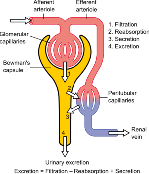
As the filtrate flows through the glomerulus into the descending limb, there is reabsorption of glucose and of H+ by the carbonic anhydrase conversion to water and CO2, except with serious acidemia, in which K+ is reabsorbed with H+ loss to the filtrate, resulting in a hyperkalemia. In the descending limb Na+ is absorbed into the interstitium, and the hypertonic interstitium draws water back for circulation, regulated by the action of ADH on the epithelium of the ascending limb. The result in terms of basic urinary clearance, the volume of urine loss is moderated by the amount needed for circulation (10 units of whole blood) without dehydration, and an amount sufficient for metabolite loss (including drug metabolites). The urine flows into the kidney pelvis and flow down the ureters.

The renal blood flow needs mention. The blood reaches the glomerulus by way of the afferent arteriole and leaves by way of the efferent arteriole. In a book by the Harvard Pathologist Shields Warren on diabetes he made a distinction between hypertension and diabetes in that efferent arteriolar sclerosis is present in both, but diabetes is uniquely identified by afferent arteriolar sclerosis. In diabetes you also have a typical glomerulosclerosis, which might be related to the same hyalinization found in the pancreatic islets – a secondary amyloidosis.

______________________________________________________________________________________________________________________________________________________________

English: Nephron, Diagram of the urine formati...

English: Nephron, Diagram of the urine formation. The number inside tubular urin concentration in mOsm/l – when ADH acts Polski: Nefron, Schemat tworzenia moczu. Cyfry wewnątrz kanalików oznaczają lokalne stężenie w mOsm/l – gdy działa ADH (dochodzi do zagęszczania moczu). (Photo credit: Wikipedia)
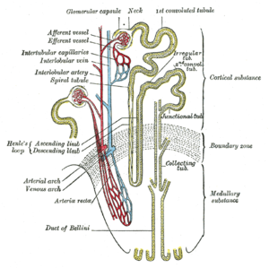
Loop of Henle (Grey's Anatomy book)

Loop of Henle (Grey’s Anatomy book) (Photo credit: Wikipedia)

Frontal section through the kidney

Frontal section through the kidney (Photo credit: Wikipedia)

_____________________________________________________________________________________________________________________________________________________________
_ Part IIa. Nitric Oxide role in renal tubular epithelial cell function Tubulointerstitial Nephritides

As part of the exponential growth in our understanding of nitric oxide (NO) in health and disease over the past 2 decades, the kidney has become appreciated as a major site where NO may play a number of important roles. Although earlier work on the kidney focused more on effects of NO at the level of larger blood vessels and glomeruli, there has been a rapidly growing body of work showing critical roles for NO in tubulointerstitial disease. In this review we discuss some of the recent contributions to this important field.

Mattana J, Adamidis A, Singhal PC. Nitric oxide and tubulointerstitial nephritides. Seminars in Nephrology 2004; 24(4):345-353.

Nitric oxide donors and renal tubular (subepithelial) matrix

Nitric oxide (NO) and its metabolite, peroxynitrite (ONOO-), are involved in renal tubular cell injury. If NO/ONOO- has an effect to reduce cell adhesion to the basement membrane, does this effect contribute to tubular obstruction and would it be partially responsible for the harmful effect of NO on the tubular epithelium during acute renal failure (ARF)?

Wangsiripaisan A, et al. examined the effect of the NO donors

  • [1] (z)-1-[2-(2-aminoethyl)-N-(2-ammonioethyl)amino]diazen-1- ium-1, 2-diolate (DETA/NO),
  • [2] spermine NONOate (SpNO), and
  • [3] the ONOO- donor 3-morpholinosydnonimine (SIN-1) on

cell-matrix adhesion to collagen types I and IV, and also fibronectin using three renal tubular epithelial cell lines:

  • [1] LLC-PK1,
  • [2] BSC-1, and
  • [3] OK.

It was only the exposure to SIN-1 that caused a dose-dependent impairment in cell-matrix adhesion.

Similar results were obtained in the different cell types and matrix proteins. The effect of SIN-1 (500 microM) on LLC-PK1 cell adhesion was not associated with either cell death or alteration of matrix protein and was attenuated by either

  • [1] the NO scavenger 2-(4-carboxyphenyl)-4,4,5,5-tetramethylimidazoline-1-oxyl-3-oxide,
  • [2] the superoxide scavenger superoxide dismutase, or
  • [3] the ONOO- scavenger uric acid in a dose-dependent manner.

These investigators concluded in this seminal paper that ONOO- generated in the tubular epithelium during ischemia/reperfusion has the potential to impair the adhesion properties of tubular cells, which then may contribute to the tubular obstruction in ARF.

Wangsiripaisan A, Gengaro PE, Nemenoff RA, Ling H, et al. Effect of nitric oxide donors on renal tubular epithelial cell-matrix adhesion. Kidney Int 1999; 55(6):2281-8.

Coexpressed Nitric Oxide Synthase and Apical β1 Integrins

In sepsis-induced acute renal failure, actin cytoskeletal alterations result in shedding of proximal tubule epithelial cells (PTEC) and tubular obstruction.

This study examined the hypothesis that inflammatory cytokines, released early in sepsis, cause PTEC cytoskeletal damage and alter integrin-dependent cell-matrix adhesion. The question of whether the intermediate nitric oxide (NO) modulates these cytokine effects was also examined. After exposure of human PTEC to tumor necrosis factor-α, interleukin-1α, and interferon-γ, the actin cytoskeleton was disrupted and cells became elongated, with extension of long filopodial processes.

Cytokines induced shedding of viable, apoptotic, and necrotic PTEC, which was dependent on NO synthesized by inducible NO synthase (iNOS) produced as a result of cytokine actions on PTEC. Basolateral exposure of polarized PTEC monolayers to cytokines induced maximal NO-dependent cell shedding, mediated in part through NO effects on cGMP. Cell shedding was accompanied by dispersal of basolateral β1 integrins and E-cadherin, with corresponding upregulation of integrin expression in clusters of cells elevated above the epithelial monolayer.

These cells demonstrated coexpression of iNOS and apically redistributed β1 integrins. These authors point out that the major ligand involved in cell anchorage was laminin, probably through interactions with the integrin α3β1.

This interaction was downregulated by cytokines but was not dependent on NO. They posulate a mechanism by which inflammatory cytokines induce PTEC damage in sepsis, in the absence of hypotension and ischemia.

Glynne PA, Picot J and Evans TJ. Coexpressed Nitric Oxide Synthase and Apical β1 Integrins Influence Tubule Cell Adhesion after Cytokine-Induced Injury. JASN 2001; 12(11): 2370-2383.

Potentiation by Nitric Oxide of Apoptosis in Renal Proximal Tubule Cells

Proximal tubular epithelial cells (PTEC) exhibit a high sensitivity to undergo apoptosis in response to proinflammatory stimuli and immunosuppressors and participate in the onset of several renal diseases. This study examined the expression of inducible nitric oxide (NO) synthase after challenge of PTEC with bacterial cell wall molecules and inflammatory cytokines and analyzed the pathways that lead to apoptosis in these cells by measuring changes in the mitochondrial transmembrane potential and caspase activation.

The data show that the apoptotic effects of proinflammatory stimuli mainly were due to the expression of inducible NO synthase. Cyclosporin A and FK506 inhibited partially NO synthesis.

However, both NO and immunosuppressors induced apoptosis, probably through a common mechanism that involved the irreversible opening of the mitochondrial permeability transition pore. Activation of caspases 3 and 7 was observed in cells treated with high doses of NO and with moderate concentrations of immunosuppressors.

The conclusion is that the cooperation between NO and immunosuppressors that induce apoptosis in PTEC might contribute to the renal toxicity observed in the course of immunosuppressive therapy.

Hortelano S, Castilla M, Torres AM, Tejedor A, and Bosca L.  Potentiation by Nitric Oxide of Cyclosporin A and FK506- Induced Apoptosis in Renal Proximal Tubule Cells. J Am Soc Nephrol 2000; 11: 2315–2323.

___________________________________________________________________________________________________________________________________________________________

Part IIb. Related studies with ROS and/or RNS on nonrenal epithelial cells

Reactive nitrogen species block cell cycle re-entry Endogenous sources of reactive nitrogen species (RNS) act as second messengers in a variety of cell signaling events, whereas environmental sources of RNS like nitrogen dioxide (NO2) inhibit cell survival and growth through covalent modification of cellular macromolecules. Murine type II alveolar cells arrested in G0 by serum deprivation were exposed to either NO2 or SIN-1, a generator of RNS, during cell cycle re-entry.

In serum-stimulated cells, RNS blocked cyclin D1 gene expression, resulting in cell cycle arrest at the boundary between G0 and G1. Dichlorofluorescin diacetate (DCF) fluorescence indicated that RNS induced sustained production of intracellular hydrogen peroxide (H2O2), which normally is produced only transiently in response to serum growth factors.

Loading cells with catalase prevented enhanced DCF fluorescence and rescued cyclin D1 expression and S phase entry.

These studies indicate environmental RNS interfere with cell cycle re-entry through an H2O2-dependent mechanism that influences expression of cyclin D1 and progression from G0 to the G1 phase of the cell cycle.

Yuan Z, Schellekens H, Warner L, Janssen-Heininger Y, Burch P, Heintz NH. Reactive nitrogen species block cell cycle re-entry through sustained production of hydrogen peroxide. Am J Respir Cell Mol Biol. 2003;28(6):705-12. Epub 2003 Jan 10.

Peroxynitrite modulates MnSOD gene expression

Peroxynitrite (ONOO-) is a strong oxidant derived from nitric oxide (‘NO) and superoxide (O2.-), reactive nitrogen (RNS) and oxygen species (ROS) present in inflamed tissue. Other oxidant stresses, e.g., TNF-alpha and hyperoxia,   induce mitochondrial, manganese-containing superoxide dismutase (MnSOD) gene expression.   3-morpholinosydnonimine HCI (SIN-1) (10 or 1000 microM) increased MnSOD mRNA, but did not change hypoxanthine guanine phosphoribosyl transferase (HPRT) mRNA.   Authentic peroxynitrite (ONOO ) (100-500 microM) also increased MnSOD mRNA but did not change constitutive HPRT mRNA expression.   ONOO stimulated luciferase gene expression driven by a 2.5 kb fragment of the rat MnSOD gene 5′ promoter region.

MnSOD gene induction due to ONOO- was

  • [1] inhibited effectively by L-cysteine (10 mM) and
  • [2] partially inhibited by N-acetyl cysteine (NAC)(50 mM) or
  • [3] pyrrole dithiocarbamate (10 mM).

.NO from 1-propanamine, 3-(2-hydroxy-2-nitroso-1-propylhydrazine) (PAPA NONOate) (100 or 1000 microM) did not change MnSOD or HPRT mRNA, nor did either H202 or NO2-, breakdown products of SIN-1 and ONOO, have any effect on MnSOD mRNA expression; ONOO- and SIN-1 also did not increase detectable MnSOD protein content or increase MnSOD enzymatic activity.

Nevertheless, increased steady state [O2.-] in the presence of .NO yields ONOO , and ONOO has direct, stimulatory effects on MnSOD transcript expression driven at the MnSOD gene 5′ promoter region inhibited completely by L-cysteine and partly by N-acetyl cysteine in lung epithelial cells. This raises a question of whether the same effect is seen in renal tubular epithelium.

Jackson RM, Parish G, Helton ES. Peroxynitrite modulates MnSOD gene expression in lung epithelial cells. Free Radic Biol Med. 1998; 25(4-5):463-72.

Comparative impacts of glutathione peroxidase-1 gene knockout on oxidative stress

Selenium-dependent glutathione peroxidase-1 (GPX1) protects against reactive-oxygen-species (ROS)-induced oxidative stress in vivo, but its role in coping with reactive nitrogen species (RNS) is unclear. Primary hepatocytes were isolated from GPX1-knockout (KO) and wild-type (WT) mice to test protection of GPX1 against cytotoxicity of

  • [1] superoxide generator diquat (DQ),
  • [2]NO donor S-nitroso-N-acetyl-penicillamine (SNAP) and
  • [3] peroxynitrite generator 3-morpholinosydnonimine (SIN-1).

Treating cells with SNAP in addition to DQ produced synergistic cytotoxicity that minimized differences in apoptotic cell death and oxidative injuries between the KO and WT cells. Less protein nitrotyrosine was induced by 0.05-0.5 mM DQ+0.25 mM SNAP in the KO than in the WT cells.

Total GPX activity in the WT cells was reduced by 65 and 25% by 0.5 mM DQ+0.1 mM SNAP and 0.5 mM DQ, respectively. Decreases in Cu,Zn-superoxide dismutase (SOD) activity and increases in Mn-SOD activity in response to DQ or DQ+SNAP were greater in the KO cells than in the WT cells.

The study indicates GPX1 was more effective in protecting hepatocytes against oxidative injuries mediated by ROS alone than by ROS and RNS together, and knockout of GPX1 did not enhance cell susceptibility to RNS-associated cytotoxicity. Instead, it attenuated protein nitration induced by DQ+SNAP.

To better understand the mechanism(s) underlying nitric oxide (. NO)-mediated toxicity, in the presence and absence of concomitant oxidant exposure, postmitotic terminally differentiated NT2N cells (which are incapable of producing . NO) were exposed to [1]PAPA-NONOate (PAPA/NO) and [2] 3-morpholinosydnonimine (SIN-1).

Exposure to SIN-1, which generated peroxynitrite (ONOO) in the range of 25-750 nM/min, produced a concentration- and time-dependent delayed cell death.   In contrast, a critical threshold concentration (>440 nM/min) was required for . NO to produce significant cell injury.   There is a largely necrotic lesion after ONOO exposure and an apoptotic-like morphology after . NO exposure.

Cellular levels of reduced thiols correlated with cell death, and pretreatment with N-acetylcysteine (NAC) fully protected from cell death in either PAPA/NO or SIN-1 exposure. NAC given within the first 3 h posttreatment further delayed cell death and increased the intracellular thiol level in SIN-1 but not . NO-exposed cells.

Cell injury from . NO was independent of cGMP, caspases, and superoxide or peroxynitrite formation.   Overall, exposure of non-. NO-producing cells to . NO or peroxynitrite results in delayed cell death, which, although occurring by different mechanisms,   appears to be mediated by the loss of intracellular redox balance.

Gow AJ, Chen Q, Gole M, Themistocleous M, Lee VM, Ischiropoulos H. Two distinct mechanisms of nitric oxide-mediated neuronal cell death show thiol dependency. Am J Physiol Cell Physiol. 2000; 278(6):C1099-107.

NO2 effect on phosphatidyl choline   Nitrogen dioxide (NO2) inhalation affects the extracellular surfactant as well as the structure and function of type II pneumocytes.

The studies had differences in oxidant concentration, duration of exposure, and mode of NO2 application. This study evaluated the influence of the NO2 application mode on the phospholipid metabolism of type II pneumocytes. Rats were exposed to identical NO2 body doses (720 ppm x h), which were applied continuously (10 ppm for 3 d), intermittently (10 ppm for 8 h per day, for 9 d), and repeatedly (10 ppm for 3 d, 28 d rest, and then 10 ppm for 3 d). Immediately after exposure, type II cells were isolated and evaluated for cell yield, vitality, phosphatidylcholine (PC) synthesis, and secretion.

Type II pneumocyte cell yield was only increased from animals that had been continuously exposed to NO2, but vitality of the isolated type II pneumocytes was not affected by the NO2 exposure modes. Continuous application of 720 ppm x h NO2 resulted in increased activity of the cytidine-5-diphosphate (CDP)-choline pathway.   After continuous NO2 application,

  • [1] specific activity of choline kinase,
  • [2] cytidine triphosphate (CTP):cholinephosphate cytidylyltransferase,
  • [3] uptake of choline, and
  • [4] pool sizes of CDP-choline and PC   were significantly increased over those of controls.

Intermittent application of this NO2 body dose provoked less increase in PC synthesis and the synthesis parameters were comparable to those for cells from control animals after repeated exposure. Whereas PC synthesis in type II cells was stimulated by NO2, their secretory activity was reduced.   Continuous exposure reduced the secretory activity most, whereas intermittent exposure nonsignificantly reduced this activity as compared with that of controls. The repeated application of NO2 produced no differences.

The authors conclude that…. type II pneumocytes adapt to NO2 atmospheres depending on the mode of its application, at least for the metabolism of PC and its secretion from isolated type II pneumocytes.

The reader asks whether this effect could also be found in renal epithelial cells, for which PC is not considered vital as for type II pneumocytes and possibly related to surfactant activity in the lung.

Müller B, Seifart C, von Wichert P, Barth PJ. Adaptation of rat type II pneumocytes to NO2: effects of NO2 application mode on phosphatidylcholine metabolism. Am J Respir Cell Mol Biol. 1998; 18(5): 712-20.

iNOS involved in immediate response to anaphylaxis

The generation of large quantities of nitric oxide (NO) is implicated in the pathogenesis of anaphylactic shock. The source of NO, however, has not been established and conflicting results have been obtained when investigators have tried to inhibit its production in anaphylaxis.

This study analyzed the expression of inducible nitric oxide synthase (iNOS) and endothelial nitric oxide synthase (eNOS) in a mouse model of anaphylaxis.   BALB/c mice were sensitized and challenged with ovalbumin to induce anaphylaxis. Tissues were removed from the heart and lungs, and blood was drawn at different time points during the first 48 hours after induction of anaphylaxis. The Griess assay was used to measure nitric oxide generation.

Nitric oxide synthase expression was examined by reverse transcriptase polymerase chain reaction and immunohistochemistry. A significant increase in iNOS mRNA expression and nitric oxide production was evident as early as 10 to 30 minutes after allergen challenge in both heart and lungs.

In contrast, expression of eNOS mRNA was not altered during the course of the experiment. The results support involvement of iNOS in the immediate physiological response of anaphylaxis.

Sade K, Schwartz IF, Etkin S, Schwartzenberg S, et al. Expression of Inducible Nitric Oxide Synthase in a Mouse Model of Anaphylaxis. J Investig Allergol Clin Immunol 2007; 17(6):379-385.

________________________________________________________________________________________________________________________________________________________

Part IIc. Additional Nonrenal Related NO References

1. Nitrogen dioxide induces death in lung epithelial cells in a density-dependent manner. Persinger RL, Blay WM, Heintz NH, Hemenway DR, Janssen-Heininger YM. Am J Respir Cell Mol Biol. 2001 May;24(5):583-90. PMID: 11350828 [PubMed – indexed for MEDLINE] Free Article

2. Molecular mechanisms of nitrogen dioxide induced epithelial injury in the lung. Persinger RL, Poynter ME, Ckless K, Janssen-Heininger YM. Mol Cell Biochem. 2002 May-Jun;234-235(1-2):71-80. Review. PMID: 12162462 [PubMed – indexed for MEDLINE]

3. Nitric oxide and peroxynitrite-mediated pulmonary cell death. Gow AJ, Thom SR, Ischiropoulos H. Am J Physiol. 1998 Jan;274(1 Pt 1):L112-8. PMID: 9458808 [PubMed – indexed for MEDLINE] Free Article

4. Mitogen-activated protein kinases mediate peroxynitrite-induced cell death in human bronchial epithelial cells. Nabeyrat E, Jones GE, Fenwick PS, Barnes PJ, Donnelly LE. Am J Physiol Lung Cell Mol Physiol. 2003 Jun;284(6):L1112-20. Epub 2003 Feb 21. PMID: 12598225 [PubMed – indexed for MEDLINE] Free Article

5. Peroxynitrite inhibits inducible (type 2) nitric oxide synthase in murine lung epithelial cells in vitro. Robinson VK, Sato E, Nelson DK, Camhi SL, Robbins RA, Hoyt JC. Free Radic Biol Med. 2001 May 1;30(9):986-91. PMID: 11316578 [PubMed – indexed for MEDLINE]

6. Nitric oxide-mediated chondrocyte cell death requires the generation of additional reactive oxygen species. Del Carlo M Jr, Loeser RF. Arthritis Rheum. 2002 Feb;46(2):394-403. PMID: 11840442 [PubMed – indexed for MEDLINE]

7. Colon epithelial cell death in 2,4,6-trinitrobenzenesulfonic acid-induced colitis is associated with increased inducible nitric-oxide synthase expression and peroxynitrite production.

Yue G, Lai PS, Yin K, Sun FF, Nagele RG, Liu X, Linask KK, Wang C, Lin KT, Wong PY. J Pharmacol Exp Ther. 2001 Jun;297(3):915-25. PMID: 11356911 [PubMed – indexed for MEDLINE] Free Article

__________________________________________________________________________________________________________________________________________________________

Part IIIa. Acute renal failure   Acute renal failure (ARF), characterized by sudden loss of the ability of the kidneys to [1] excrete wastes, [2] concentrate urine, [3] conserve electrolytes, and [4] maintain fluid balance, is a frequent clinical problem, particularly in the intensive care unit, where it is associated with a mortality of between 50% and 80%.

This clinical entity was described as an acute loss of kidney function that occurred in severely injured crush victims because of histological evidence for patchy necrosis of renal tubules at autopsy. In the clinical setting, the terms ATN and acute renal failure (ARF) are frequently used interchangeably. However, ARF does not include increases in blood urea due to [1] reversible renal vasoconstriction (prerenal azotemia) or [2] urinary tract obstruction (postrenal azotemia). Acute hemodialysis was first used clinically during the Korean War in 1950 to treat military casualties, and this led to a decrease in mortality of the ARF clinical syndrome from about 90% to about 50%.   In the half century that has since passed, much has been learned about the pathogenesis of ischemic and nephrotoxic ARF in experimental models, but there has been very little improvement in mortality. This may be explained by changing demographics: [1] the age of patients with ARF continues to rise, and [2] comorbid diseases are increasingly common in this population. Both factors may obscure any increased survival related to improved critical care. Examining the incidence of ARF in several military conflicts does, however, provide some optimism. The incidence of ARF in seriously injured casualties decreased between World War II and the Korean War, and again between that war and the Vietnam War, despite the lack of any obvious difference in the severity of the injuries. What was different was the rapidity of the fluid resuscitation of the patients? Fluid resuscitation on the battlefield with the rapid evacuation of the casualties to hospitals by helicopter began during the Korean War and was optimized further during the Vietnam War. For seriously injured casualties the incidence of ischemic ARF was one in 200 in the Korean War and one in 600 in the Vietnam War. This historical sequence of events suggests that early intervention could prevent the occurrence of ARF, at least in military casualties.   In experimental studies it has been shown that progression from an azotemic state associated with renal vasoconstriction and intact tubular function (prerenal azotemia) to established ARF with tubular dysfunction occurs if the renal ischemia is prolonged. Moreover, early intervention with fluid resuscitation was shown to prevent the progression from prerenal azotemia to established ARF. Diagnostic evaluation of ARF One important question, therefore, is how to assure that an early diagnosis of acute renal vasoconstriction can be made prior to the occurrence of tubular dysfunction, thus providing the potential to prevent progression to established ARF. In this regard, past diagnostics relied on observation of the patient response to a fluid challenge: [1] decreasing levels of blood urea nitrogen (BUN) indicated the presence of reversible vasoconstriction, [2] while uncontrolled accumulation of nitrogenous waste products, i.e., BUN and serum creatinine, indicated established ARF.

This approach, however, frequently led to massive fluid overload in the ARF patient with resultant

  • [1] pulmonary congestion,
  • [2] hypoxia, and
  • [3] premature need for mechanical ventilatory support and/or hemodialysis.

On this background the focus turned to an evaluation of urine sediment and urine chemistries to differentiate between renal vasoconstriction with intact tubular function and established ARF.

It was well established that if tubular function was intact, renal vasoconstriction was associated with enhanced tubular sodium reabsorption. Specifically, the fraction of filtered sodium that is rapidly reabsorbed by normal tubules of the vasoconstricted kidney is greater than 99%.

Thus, when nitrogenous wastes, such as creatinine and urea, accumulate in the blood due to a fall in glomerular filtration rate (GFR) secondary to renal vasoconstriction with intact tubular function, the fractional excretion of filtered sodium (FENa = [(urine sodium × plasma creatinine) / (plasma sodium × urine creatinine)]) is less than 1%. An exception to this physiological response of the normal kidney to vasoconstriction is when the patient is receiving a diuretic, including mannitol, or has glucosuria, which decreases tubular sodium reabsorption and increases FENa.

It has recently been shown in the presence of diuretics that a rate of fractional excretion of urea (FEurea) of less than 35 indicates intact tubular function, thus favoring renal vasoconstriction rather than established ARF as a cause of the azotemia.

___________________________________________________________________________________________________________________________________________________________

English: Physiology of Nephron

English: Physiology of Nephron (Photo credit: Wikipedia)
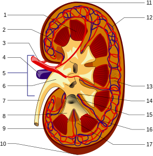
Structures of the kidney: 1.Renal pyramid 2.In...

Structures of the kidney: 1.Renal pyramid 2.Interlobar artery 3.Renal artery 4.Renal vein 5.Renal hilum 6.Renal pelvis 7.Ureter 8.Minor calyx 9.Renal capsule 10.Inferior renal capsule 11.Superior renal capsule 12.Interlobar vein 13.Nephron 14.Minor calyx 15.Major calyx 16.Renal papilla 17.Renal column (no distinction for red/blue (oxygenated or not) blood, arteriole is between capilaries and larger vessels (Photo credit: Wikipedia)

_

______________________________________________________________________________________________________________________________________________________

Mechanisms of ARF

Based on the foregoing comments, this discussion of mechanisms of ARF will not include nitrogenous-waste accumulation due to renal vasoconstriction with intact tubular function (prerenal azotemia) or urinary tract obstruction (postrenal azotemia). The mechanisms of ARF involve both vascular and tubular factors. An ischemic insult to the kidney will in general be the cause of the ARF. While a decrease in renal blood flow with diminished oxygen and substrate delivery to the tubule cells is an important ischemic factor, it must be remembered that a relative increase in oxygen demand by the tubule is also a factor in renal ischemia.

Approximately 30–70% of these shed epithelial tubule cells in the urine are viable and can be grown in culture. Recent studies using cellular and molecular techniques have provided information relating to the structural abnormalities of injured renal tubules that occur both in vitro and in vivo. In vitro studies using chemical anoxia have revealed abnormalities in the proximal tubule cytoskeleton that are associated with translocation of Na+/K+-ATPase from the basolateral to the apical membrane.

A comparison of cadaveric transplanted kidneys with delayed versus prompt graft function has also provided important results regarding the role of Na+/K+-ATPase in ischemic renal injury. This study demonstrated that, compared with kidneys with prompt graft function, those with delayed graft function had a significantly greater cytoplasmic concentration of Na+/K+-ATPase and actin-binding proteins — spectrin (also known as fodrin) and ankyrin — that had translocated from the basolateral membrane to the cytoplasm.

Such a translocation of Na+/K+-ATPase from the basolateral membrane to the cytoplasm could explain the decrease in tubular sodium reabsorption that occurs with ARF. An important focus of research is the mechanisms whereby the critical residence of Na+/K+-ATPase in the basolateral membrane (which facilitates vectorial sodium transport) is uncoupled by hypoxia or ischemia.  The actin-binding proteins,

  • spectrin and
  • ankyrin,

serve as substrates for the calcium-activated cysteine protease calpain.

________________________________________________________________________________________________________________________________________________________

In vitro studies in proximal tubules have shown a rapid rise in cytosolic calcium concentration during acute hypoxia, which antedates the evidence of tubular injury as assessed by lactic dehydrogenase (LDH) release. There is further evidence to support the importance of the translocation of Na+/K+-ATPase from the basolateral membrane to the cytoplasm during renal ischemia/reperfusion.

Specifically, calpain-mediated breakdown products of the actin-binding protein spectrin occur with renal ischemia. Calpain activity was demonstrated to increase during hypoxia in isolated proximal tubules. Measurement of LDH release following calpain inhibition indicated attenuation of hypoxic damage to proximal tubules. There was no evidence of an increase in cathepsin, a (cysteine protease) in proximal tubules during hypoxia , but there is a calcium-independent pathway for calpain activation during hypoxia.

Calpastatin, an endogenous cellular inhibitor of calpain activation, was shown to be diminished during hypoxia in association with the rise in another cysteine protease, caspase.

This effect of diminished calpastatin activity could be reversed by caspase inhibition. Proteolytic pathways appear to be involved in calpain-mediated proximal tubule cell injury during hypoxia. Calcium activation of phospholipase A has also been shown to contribute to renal tubular injury during ischemia.

________________________________________________________________________________________________________________________________________________________

Tubular obstruction during ARF

The existence of proteolytic pathways involving cysteine proteases, namely calpain and caspases, may therefore explain

  • the decrease in proximal tubule sodium reabsorption and
  • increased FENa

secondary to proteolytic uncoupling of Na+/K+-ATPase from its basolateral membrane anchoring proteins.

This tubular perturbation alone, however, does not explain the fall in GFR that leads to nitrogenous-waste retention and thus the rise in BUN and serum creatinine.   Decreased proximal tubule sodium reabsorption may lead to a decreased GFR during ARF. First of all, brush border membranes and cellular debris could provide the substrate for intraluminal obstruction in the highly resistant portion of distal nephrons.

In fact, microdissection of individual nephrons of kidneys from patients with ARF demonstrated obstructing casts in distal tubules and collecting ducts. This observation could explain the dilated proximal tubules that are observed upon renal biopsy of ARF kidneys. The intraluminal casts in ARF kidneys stain prominently for Tamm-Horsfall protein (THP), which is produced in the thick ascending limb. Importantly, THP is secreted into tubular fluid as a monomer but subsequently may become a polymer that forms a gel-like material in the presence of increased luminal Na+ concentration, as occurs in the distal nephron during clinical ARF with the decrease in tubular sodium reabsorption.

Thus, the THP polymeric gel in the distal nephron provides an intraluminal environment for distal cast formation involving viable, apoptotic, and necrotic cells.

The loss of the tubular epithelial cell barrier and/or the tight junctions between viable cells during acute renal ischemia could lead to a leak of glomerular filtrate back into the circulation. (If this occurs and normally non-reabsorbable substances, such as inulin, leak back into the circulation, then a falsely low GFR will be measured as inulin clearance. It should be noted, however, that the degree of extensive tubular damage observed in experimental studies demonstrating tubular fluid backleak is rarely observed with clinical ARF in humans). Moreover, dextran sieving studies in patients with ARF demonstrated that, at best, only a 10% decrease in GFR could be explained by backleak of filtrate. Cadaveric transplanted kidneys with delayed graft function, however, may have severe tubular necrosis, and thus backleak of glomerular filtration may be more important in this setting.

Inflammation and NO

There is now substantial evidence for the involvement of inflammation in the pathogenesis of the decreased GFR associated with acute renal ischemic injury. In this regard, there is experimental evidence that iNOS may contribute to tubular injury during ARF. Hypoxia in isolated proximal tubules has been shown to increase NO release, and there is increased iNOS protein expression in ischemic kidney homogenates. An antisense oligonucleotide was shown to block the upregulation of iNOS and afford functional protection against acute renal ischemia. Moreover, when isolated proximal tubules from iNOS, eNOS, and neuronal NO synthase (nNOS) knockout mice were exposed to hypoxia, only the tubules from the iNOS knockout mice were protected against hypoxia, as assessed by LDH release. The iNOS knockout mice were also shown to have lower mortality during ischemia/reperfusion than wild-type mice.  The scavenging of NO by oxygen radicals produces peroxynitrite causing tubule damage during ischemia. While iNOS may contribute to ischemic injury of renal tubules,  the vascular effect of eNOS in the glomerular afferent arteriole is protective against ischemic injury. In this regard, eNOS knockout mice are more sensitive to endotoxin-related injury than normal mice.

Moreover, the protective role of vascular eNOS may be more important than the deleterious effect of iNOS at the tubule level during renal ischemia.   This is because treatment of mice with the nonspecific NO synthase (NOS) inhibitor L-NAME, which blocks both iNOS and eNOS, worsens renal ischemic injury. NO may downregulate eNOS and is a potent inducer of heme oxygenase-1, which has been shown to be cytoprotective against renal injury. The MAPK pathway also appears to be involved in renal oxidant injury. Activation of extracellular signal–regulated kinase (ERK) or inhibition of JNK ameliorates oxidant injury–induced necrosis in mouse renal proximal tubule cells in vitro. Upregulation of ERK may also be important in the effect of preconditioning whereby early ischemia affords protection against a subsequent ischemia/reperfusion insult. Alterations in cell cycling are also involved in renal ischemic injury. Upregulation of p21, which inhibits cell cycling, appears to allow cellular repair and regeneration, whereas homozygous p21 knockout mice demonstrate enhanced cell necrosis in response to an ischemic insult.

Prolonged duration of the ARF clinical course and the need for dialysis are major factors projecting a poor prognosis. Patients with ARF who require dialysis have a 50–70% mortality rate. Infection and cardiopulmonary complications are the major causes of death in patients with ARF. Excessive fluid administration in patients with established ARF may lead to pulmonary congestion, hypoxia, the need for ventilatory support, pneumonia, and multiorgan dysfunction syndrome, which has an 80–90% mortality rate. Until means to reverse the diminished host defense mechanisms in azotemic patients with clinical ARF are available, every effort should be made to avoid invasive procedures such as the placement of bladder catheters, intravenous lines, and mechanical ventilation. Over and above such supportive care, it may be that combination therapy will be necessary to prevent or attenuate the course of ARF. Such combination therapy must involve agents with potential beneficial effects on vascular tone, tubular obstruction, and inflammation.

Schrier RW, Wang W, Poole B, and Mitra A. Acute renal failure: definitions, diagnosis, pathogenesis, and therapy. The Journal of Clinical Investigation 2004; 114(1):5-14. http://www.jci.org

______________________________________________________________________________________________________________________________________________________

Part IIIb. Additional Related References on NO, oxidative stress and Kidney

Shelgikar PJ, Deshpande KH, Sardeshmukh AS, Katkam RV, Suryakarl AN. Role of oxidants and antioxidants in ARF patients undergoing hemodialysis. Indian J Nephrol 2005;15: 73-76.

Lee JW. Renal Dysfunction in Patients with Chronic Liver Disease. Electrolytes Blood Press 7:42-50, 2009ㆍdoi: 10.5049/EBP.2009.7.2.42.

Saadat H, et al. Endothelial Nitric Oxide Function and Tubular Injury in Premature Infants. Int J App Sci and Technol 2012; 7(6): 77-81. http://www.ijastnet.com.

Amerisan MS. Cardiovascular disease in chronic kidney disease. Indian J Nephrol 2005;15: 1-7.

___________________________________________________________________________________________________________________________________________________________

Traditional risk factors for CVD in CKD

  • Hypertension
  • Older Age
  • Diabetes Mellitus
  • Male gender
  • High LDL
  • White Race
  • Low HDL
  • Physical inactivity
  • Smoking
  • Menopause
  • LVH

CKD Related CV Risk Factors

  • Blood Pressure
  • ? Homocysteinemia
  • Anemia
  • ? Inflammation
  •   Ca++ x P++
  • ? NO synthesis
  • Na+ Retention
  • ? Lp (a)
  • Hypervolemia
  • ? Insulin Resistance
  • Proteinuria & Hypoalbuminemia
  • Iron over load
  • ? Adeponectin
  • ??Vit. C or E
  • ? 5 Lipoxygenase
  • ROS
  • Genetic factors
  • ADMA (Asymmetric Dimethyl Arginine)

S Vikrant, SC Tiwari. Essential Hypertension – Pathogenesis and Pathophysiology. J Indian Acad Clinical Medicine 2001; 2(3):141-161. Scheme for pathogenesis of salt dependent hypertension.

The hypothesis proposes that early hypertension is episodic and is mediated by a hyperactive sympathetic nervous system or activated renin-angiotensin system.

Cell membrane alterations

Hypotheses linking abnormal ionic fluxes to increased peripheral resistance through increase in cell sodium, calcium, or pH.   The hypertension that is more common in obese people may arise in large part from the insulin resistance and resultant hyperinsulinaemia that results from the increased mass of fat. However, rather unexpectedly, insulin resistance may also be involved in hypertension in non-obese people.

Overall scheme for the mechanisms by which obesity, if predominantly upper body or visceral in location, could promote

________________________________________________________________________________________________________________________________________________________

  • diabetes,
  • dyslipidemia and
  • hypertension via hyperinsulinemia.

The explanation for insulin resistance found in as many as half of nonobese hypertensive is not obvious and may involve one or more aspects of insulin’s action

__________________________________________________________________________________________________________________________________________________________

Proposed mechanisms by which insulin resistance and/or hyperinsulinemia may lead to increased blood pressure.

  1. Enhanced renal sodium and water reabsorption.
  2. Increased blood pressure sensitivity to dietary salt intake
  3. Augmentation of the pressure and
  4. aldosterone responses to AII
  5. Changes in transmembrane electrolyte transport
  • a. Increased intracellular sodium
  • b. Decreased Na+/K+ – ATPase activity
  • c. Increased intracellular Ca2+ pump activity
  • d. Increased intracellular Ca2+ accumulation
  • e. Stimulation of growth factors

___________________________________________________________________________________________________________________________________________________________

Part IV. New Insights on NO donors

This study investigated the involvement of nitric oxide (NO) into the irradiation-induced increase of cell attachment. These experiments explored the cellular mechanisms of low-power laser therapy. HeLa cells were irradiated with a monochromatic visible-tonear infrared radiation (600–860 nm, 52 J/m2) or with a diode laser (820 nm, 8–120 J/m2) and the number of cells attached to a glass matrix was counted after 30 minute incubation at 37oC. The NO donors

  1. sodium nitroprusside (SNP),
  2. glyceryl trinitrate (GTN), or
  3. sodium nitrite (NaNO2)

were added to the cellular suspension before or after irradiation. The action spectra and the concentration and fluence dependencies obtained were compared and analyzed.

The well-structured action spectrum for the increase of the adhesion of the cells, with maxima at 619, 657, 675, 740, 760, and 820 nm, points to the existence of a photoacceptor responsible for the enhancement of this property (supposedly cytochrome c oxidase, the terminal respiratory chain enzyme), as well as signaling pathways between the cell mitochondria, plasma membrane, and nucleus.

Treating the cellular suspension with SNP before irradiation significantly modifies the action spectrum for the enhancement of the cell attachment property (band maxima at 642, 685, 700, 742, 842, and 856 nm). The action of SNP, GTN, andNaNO2 added before or after irradiation depends on their concentration and radiation fluence.

The NO donors added to the cellular suspension before irradiation eliminate the radiation induced increase in the number of cells attached to the glass matrix, supposedly by way of binding NO to cytochrome c oxidase. NO added to the suspension after irradiation can also inhibit the light-induced signal downstream. Both effects of NO depend on the concentration of the NO donors added.

The results indicate that NO can control the irradiation-activated reactions that increase the attachment of cells.

Karu TI, Pyatibrat LV, and Afanasyeva NI. Cellular Effects of Low Power Laser Therapy Can be Mediated by Nitric Oxide. Lasers Surg. Med 2005; 36:307–314.

IFNa-2b (IFN-a) effect on barrier function of renal tubular epithelium

IFNa treatment can be accompanied by impaired renal function and capillary leak. This study shows IFNa produced dose-dependent and time-dependent decrease in transepithelial resistance (TER) ameliorated by tyrphostin, an inhibitor of phosphotyrosine kinase with increased expression of occludin and E-cadherin. In conclusion, IFNa can directly affect barrier function in renal epithelial cells via ovewrexpression or missorting of the junctional proteins occludin and E-cadherin.

Lechner J, Krall M, Netzer A, Radmayr C, et al. Effects of interferon a-2b on barrier function and junctional complexes of renal proximal tubulat LLC-pK1 cells. Kidney Int 1999; 55:2178-2191.

Ischemia-reperfusion injury

The pathophysiology of acute renal failure (ARF) is complex and not well understood. Numerous models of ARF suggest that oxygen-derived reactive species are important in renal ischemia-reperfusion (I-R) injury, but the nature of the mediators is still controversial. Treatment with oxygen radical scavengers, antioxidants, and iron chelators such as

  • superoxide dismutase,
  • dimethylthiourea,
  • allopurinol, and
  • deferoxamine

are protective in some models, and suggest a role for the hydroxyl radical formation. However, these compounds are not protective in all models of I-R injury, and direct evidence for the generation of hydroxyl radical is absent. Furthermore, these inhibitors have another property in common.

They all directly scavenge or inhibit the formation of peroxynitrite (ONOO−), a highly toxic species derived from nitric oxide (NO) and superoxide. Thus, the protective effects seen with these inhibitors may be due in part to their ability to inhibit ONOO− formation. Even though reactive oxygen species are thought to participate in ischemia-reperfusion (I-R) injury, induction of and production of high levels of  inducible nitric oxide (NO)  also contribute to this injury.

NO combines with superoxide to form the potent oxidant peroxynitrite (ONOO−). NO and ONOO− were investigated in a rat model of renal I-R injury using the selective iNOS inhibitor L-N6-(1-iminoethyl)lysine (L-NIL).

I-R surgery significantly increased plasma creatinine levels to 1.9 ± 0.3 mg/dl (P < .05) and caused renal cortical necrosis. L-NIL administration (3 mg/kg) in animals subjected to I-R significantly decreased plasma creatinine levels to 1.2 ± 0.10 mg/dl (P < .05 compared with I-R) and reduced tubular damage.

ONOO− formation was evaluated by detecting 3-nitrotyrosine-protein adducts (3NTyPAs), a stable biomarker of ONOO− formation.   The kidneys from I-R animals had increased levels of 3NTyPAs compared with control animals   L-NIL-treated rats (3 mg/kg) subjected to I-R showed decreased levels of 3NTyPAs.

These results suggests that iNOS-generated NO mediates damage in I-R injury possibly through ONOO− formation.

______________________________________________________________________________________________________________________________________________________

In summary,

  1. 3-nitrotyrosine-protein adducts were detected in renal tubules after I-R injury.
  2. Selective inhibition of iNOS by L-NIL decreased injury, improved renal function, and decreased apparent ONOO− formation.
  3. Reactive nitrogen species should be considered potential therapeutic targets in the prevention and treatment of renal I-R injury.

_______________________________________________________________________________________________________________________________________________________

Walker LM, Walker PD, Imam SZ, et al. Evidence for Peroxynitrite Formation in Renal Ischemia-Reperfusion Injury: Studies with the Inducible Nitric Oxide Synthase InhibitorL-N6-(1-Iminoethyl)lysine1. 2000.

Role of TNFa independent of iNOS Renal failure is a frequent complication of sepsis, mediated by renal vasoconstrictors and vasodilators. Endotoxin induces several proinflammatory cytokines, among which tumor necrosis factor (TNF) is thought to be of major importance. Tumor necrosis factor (TNF) has been suggested to be a factor in the acute renal failure in sepsis or endotoxemia. Passive immunization by anti-TNFa prevented development of septic shock in animal experiments.The development of ARF involves excessive intrarenal vasoconstriction. Involvement of nitric oxide (NO), generated by inducible NO synthase (iNOS), is still a factor in the pathogenesis of endotoxin-induced renal failure. TNF-a leads to a decrease in glomerular filtration rate (GFR).

This study tested the hypothesis that the role of TNF-a in endotoxic shock related ARF is mediated by iNOS-derived NO.   An injection of lipopolysaccharide (LPS) constituent of gram-negative bacteria to wild-type mice resulted in a 70% decrease in glomerular filtration rate (GFR) and in a 40% reduction in renal plasma flow (RPF) 16 hours after the injection.   The results occurred independent of hypotension, morphological changes, apoptosis, and leukocyte accumulation. In mice pretreated with TNFsRp55, only a 30% decrease in GFR was observed without a significant change in RPF as compared with controls. Pretreatment with TNKsRp55 on renal function Wild-type mice were pretreated with TNFsRp55(10 mg/kg IP)  for one hour before the administration of 5 mg/kg intraperitoneal endotoxin. GFR and RPF were determined 16 hours thereafter. Data are expressed as mean 6, SEM, N 5 6. *P , 0.05 vs. Control; §P , 0.05 vs. LPS, by ANOVA.

The serum NO concentration was significantly lower in endotoxemic wild-type mice pretreated with TNFsRp55, as compared with untreated endotoxemic wild-type mice. In LPS-injected iNOS knockout mice and wild-type mice treated with a selective iNOS inhibitor, 1400W, the development of renal failure was similar to that in wild-type mice. As in wild-type mice,TNFsRp55 significantly attenuated the decrease in GFR (a 33% decline, as compared with 75% without TNFsRp55) without a significant change in RPF in iNOS knockout mice given LPS. These results demonstrate a role of TNF in the early renal dysfunction (16 h) in a septic mouse model independent of iNOS,

  • hypotension,
  • apoptosis,
  • leukocyte accumulation,and
  • morphological alterations,

thus suggesting renal hypoperfusion secondary to an imbalance between, as yet to be defined renal vasoconstrictors and vasodilators.

Knotek M, Rogachev B, Wang W,….., Edelstein CL, Dinarello CA, and Schrier RW. Endotoxemic renal failure in mice: Role of tumor necrosis factor independent of inducible nitric oxide synthase. Kidney International 2001; 59:2243–2249

Ischemic acute renal failure

Inflammation plays a major role in the pathophysiology of acute renal failure resulting from ischemia. This review discusses the contribution of

  • endothelial
  • epithelial cells and
  • leukocytes

to this inflammatory response. The roles of cytokines/chemokines in the injury and recovery phase are reviewed. The protection of mouse kidney prior to exposure to ischemia or urinary tract obstruction is  a potential model to  search for pharmacologic agents to protect the kidney against injury by inflammatory mediators produced by tubular epithelial cells and activated leukocytes in renal ischemia/reperfusion (I/R) injury. Tubular epithelia produce

  • TNF-a,
  • IL-1,
  • IL-6,
  • IL-8,
  • TGF-b,
  • MCP-1,
  • ENA-78,
  • RANTES, and
  • fractalkines,

whereas leukocytes produce

  • TNF-a,
  • IL-1,
  • IL-8,
  • MCP-1,
  • ROS, and
  • eicosanoids.

The release of these chemokines and cytokines serve as effectors for a positive feedback pathway enhancing inflammation and cell injury, the cycle of tubular epithelial cell injury and repair following renal ischemia/reperfusion.   Tubular epithelia are typically cuboidal in shape and apically-basally polarized; the Na+/K+-ATPase localizes to basolateral plasma membranes, whereas cell adhesion molecules, such as integrins localize basally. In response to ischemia reperfusion,

  • the Na+/K+-ATPase appears apically, and
  • integrins are detected on lateral and basal plasma membranes.

Some of the injured epithelial cells undergo necrosis and/or apoptosis detaching from the underlying basement membrane into the tubular space where they contribute to tubular occlusion. Viable cells that remain attached, dedifferentiate, spread, and migrate to repopulate the denuded basement membrane. With cell proliferation, cell-cell and cell-matrix contacts are restored, and the epithelium redifferentiates and repolarizes, forming a functional, normal epithelium Inflammation is a significant component of renal I/R injury, playing a considerable role in its pathophysiology.

Although significant progress has been made in defining the major components of this process, the complex cross-talk between endothelial cells, inflammatory cells, and the injured epithelium with each generating and often responding to cytokines and chemokines is not well understood. In addition, we have not yet taken full advantage of the large body of data on inflammation in other organ systems.

Furthermore, preconditioning the kidney to afford protection to subsequent bouts of ischemia may serve as a useful model challenging us to therapeutically mimic endogenous mechanisms of protection.

Understanding the inflammatory response prevalent in ischemic kidney injury will facilitate identification of molecular targets for therapeutic intervention.

Bonventre JV and Zuk A. Ischemic acute renal failure: An inflammatory disease? Forefronts in Nephrology 2002;.. :480-485

Gene expression profiles in renal proximal tubules In kidney disease renal proximal tubular epithelial cells (RPTEC) actively contribute to the progression of tubulointerstitial fibrosisby mediating both

  • an inflammatory response and
  • via epithelial-to-mesenchymal transition.

Using laser capture microdissection we specifically isolated RPTEC from cryosections of the healthy parts of kidneys removed owing to renal cell carcinoma and from kidney biopsies from patients with proteinuric nephropathies. RNA was extracted and hybridized to complementary DNA microarrays after linear RNA amplification. Statistical analysis identified 168 unique genes with known gene ontology association, which separated patients from controls. Besides distinct alterations in signal-transduction pathways (e.g. Wnt signalling), functional annotation revealed a significant upregulation of genes involved in

_________________________________________________________________________________________________________________________________________________________

  • cell proliferation and cell cycle control (like insulin-like growth factor 1 or cell division cycle 34),
  • cell differentiation (e.g. bone morphogenetic protein 7),
  • immune response,
  • intracellular transport and
  • metabolism

__________________________________________________________________________________________________________________________________________________________

in RPTEC from patients.

The study also revealed differential expression of a number of genes responsible for cell adhesion (like BH-protocadherin) with a marked downregulation of most of these transcripts. In summary, the results obtained from RPTEC revealed a differential regulation of genes, which are likely to be involved in either pro-fibrotic or tubulo-protective mechanisms in proteinuric patients at an early stage of kidney disease.

Rudnicki M, Eder S, Perco P, Enrich J, et al. Gene expression profiles of human proximal tubular epithelial cells in proteinuric nephropathies. Kidney International 2006; xx:1-11. Kidney International advance online publication, 20 December 2006; doi:10.1038/sj.ki.5002043. http://www.kidney-international.org

Oxidative stress involved with diabetic nephropathy

Diabetic Nephropathy (DN) poses a major health problem. There is strong evidence for a potential role of the eNOS gene. This case control study investigated the possible role of genetic variants of the endothelial Nitric Oxide Synthase (eNOS) gene and oxidative stress in the pathogenesis of nephropathy in patients with diabetes mellitus. The study included 124 diabetic patients;

  1. 68 of these patients had no diabetic nephropathy (group 1) while
  2. 56 patients exhibited symptoms of diabetic nephropathy (group 2).
  3. Sixty two healthy non-diabetic individuals were also included as a control group.

Blood samples from subjects and controls were analyzed to investigate the eNOS genotypes and to estimate

  • the lipid profile and
  • markers of oxidative stress such as malondialdehyde (MDA) and nitric oxide (NO).

No significant differences were found in the frequency of eNOS genotypes between diabetic patients (either in group 1 or group 2) and controls (p >0.05). Also, no significant differences were found in the frequency of eNOS genotypes between group 1 and group 2 (p >0.05). Both group 1 and group 2 had significantly higher levels of nitrite and MDA when compared with controls (all p = 0.0001). Also group 2 patients had significantly higher levels of nitrite and MDA when compared with group 1 (p = 0.02, p = 0.001 respectively).

The higher serum level of the markers of oxidative stress in diabetic patients particularly those with diabetic nephropathy suggest that oxidative stress and not the eNOS gene polymorphism is involved in the pathogenesis of the diabetic nephropathy in this subset of patients

Badawy A, Elbaz R, Abbas AM, Ahmed Elgendy A, et al. Oxidative stress and not endothelial Nitric Oxide Synthase gene polymorphism involved in diabetic nephropathy. Journal of Diabetes and Endocrinology 2011; 2(3): 29-35.

Metformin in renal ischemia reperfusion

Renal ischemia plays an important role in renal impairment and transplantation. Metformin is a biguanide used in type 2 diabetes, it inhibits hepatic glucose production and increases peripheral insulin sensitivity. While the mode of action of metformin is incompletely understood, it appears to have anti-inflammatory and antioxidant effects involved in its beneficial effects on insulin resistance.   Control, Sham, ischemia/reperfusion (I/R) and Metformin treated I /R groups   A renal I/R injury was done by a left renal pedicle occlusion to induce ischemia for 45 min followed by 60 min of reperfusion with contralateral nephrectomy. Metformin pretreated I/R rats in a dose of 200 mg/kg/day for three weeks before ischemia induction.

  • Nitric oxide (NO),
  • tumor necrosis factor alpha (TNF α) ,
  • catalase (CAT) and
  • reduced glutathione (GSH) activities

were determined in renal tissue, while

  • creatinine clearance (CrCl) ,
  • blood urea nitrogen (BUN) were measured and

5 hour urinary volume and electrolytes were estimated . BUN and CrCl levels in the I/R group were significantly higher than in control rats (p<0.05) table (1).

__________________________________________________________________________________________________________________________________

Table 1: Creatinine clearance (Cr Cl) and blood urea nitrogen (BUN) levels in control and test groups.
(Mean ± SD)

Groups CrCl   (ml/min) BUN (mg/dl)
Control group 1.30 ±0.11 14.30±0.25
Sham group+ metformin 1.27±0.09 15.70±0.19
I/R group (P1) 1.85±0.25 (<0.001 ) 28.00±0.62 (<0.001)
I/R+ metformin group (P2,P3) 1.55±0.22 (0.001, 0.028) 18.10±1.00 (<0.001, <0.001)
  • P1: Statistical significance between control
    group and saline treated I/R group.
  • P2 Statistical significance between control
    group and Metformin treated I/R group.
  • P3 Statistical significance between saline treated
    I/R group and Metformin treated I/R group

_______________________________________________________________________________________________________________________________________________________

When metformin was administered before I/R, BUN and CrCl levels were still significantly higher than control group but their elevation were significantly lower in comparison to I/R group alone (P<0.05).   TNF α and NO levels were significantly higher in the I/R group than those of the control group (Table 2). Pre-treatment with metformin significantly lowered their levels in comparison to I/R group (P<0.05).

________________________________________________________________________________________________________________________________________________________

Table 2: Tumor necrosis factor α (TNF α) and inducible nitric oxide (iNO) levels in control and test groups.
(Mean ± SD)

Groups TNF α (pmol/mg tissue) iNO (nmol/ mg tissue)
Control group 1 7.60 ±5.98 2.54 ± 0.82
Sham group+ metformin 16.70 ±5.50 2.35 ±0.80
I/R group (P1) 54. 00±6.02 (<0.001) 4.50±0.89 (<0.001)
I/R+metformin group (P2,P3) 39 ± 14.01 (<0.001, 0.006) 3.53±0.95 (0.02, 0.03)


  • P1: Statistical significance between control group
    and saline treated I/R group.
  • P2 Statistical significance between control group
    and Metformin treated I/R group.
  • P3 Statistical significance between saline treated
    I/R group and Metformin treated I/R group

_________________________________________________________________________________________________________________________________________________________

These results showed significant increase in NO,TNF α, BUN , CrCl and significant decrease in urinary volume , electrolytes, CAT and GSH activities in the I/R group than those in the control group. Metformin decreased significantly NO, TNF α, BUN and CrCl while increased urinary volume, electrolytes, CAT and GSH activities.   Lipid peroxidation is related to I/R induced tissue injury. Production of inducible NO synthase (NOS) under lipid peroxidation and inflammatory conditions results in the induction of NO which react with O2 liberating peroxynitrite (OONO-). NO itself inactivates the antioxidant enzyme system CAT and GSH. Alteration in NO synthesis have been observed in other kidney injuries as nephrotoxicity and acute renal failure induced by endotoxins.

Treatment with iNOS inhibitors improved renal function and decreased peroxynitrite radical which is believed to be responsible for the shedding of proximal convoluted tubules in I/R.   Metformin produced anti-inflammatory renoprotective effect on CrCl and diuresis in renal I/R injury.

Malek HA. The possible mechanism of action of metformin in renal ischemia reperfusion in rats. The Pharma Research Journal 2011; 6(1):42-49.

Possible role of NO donors in ARFThe L-arginine-nitric oxide (NO) pathway has been implicated in many physiological functions in the kidney, including

  • regulation of glomerular hemodynamics,
  • mediation of pressure-natriuresis,
  • maintenance of medullary perfusion,
  • blunting of tubuloglomerular feedback (TGF),
  • inhibition of tubular sodium reabsorption and
  • modulation of renal sympathetic nerve activity

Its net effect in the kidney is to promote natriuresis and diuresis, contributing to adaptation to variations of dietary salt intake and maintenance of normal blood pressure. Nitric oxide has been implicated in many physiologic processes that influence both acute and long-term control of kidney function. Its net effect in the kidney is to promote natriuresis and diuresis, contributing to adaptation to variations of dietary salt intake and maintenance of normal blood pressure. A pretreatment with nitric oxide donors or L-arginine may prevent the ischemic acute renal injury. In chronic kidney diseases, the systolic blood pressure is correlated with the plasma level of asymmetric dimethylarginine, an endogenous inhibitor of nitric oxide synthase. A reduced production and biological action of nitric oxide is associated with an elevation of arterial pressure, and conversely, an exaggerated activity may represent a compensatory mechanism to mitigate the hypertension.

JongUn Lee. Nitric Oxide in the Kidney : Its Physiological Role and Pathophysiological Implications. Electrolyte & Blood Pressure 2008; 6:27-34.

Renal Hypoxia and Dysoxia following Reperfusion

Acute renal failure (ARF) is a common condition which develops in 5% of hospitalized patients. Of the patients who develop ARF, ~10% eventually require renal replacement therapy. Among critical care patients who have acute renal failure and survive, 2%-10% develop terminal renal failure and require long-term dialysis.   The kidneys are particularly susceptible to ischemic injury in many clinical conditions such as renal transplantation, treatment of suprarenal aneurysms, renal artery reconstructions, contrast-agent induced nephropathy, cardiac arrest, and shock. One reason for renal sensitivity to ischemia is that the kidney microvasculature is highly complex and must meet a high energy demand.

Under normal, steady state conditions, the oxygen (O2) supply to the renal tissues is well in excess of oxygen demand.   Under pathological conditions, the delicate balance of oxygen supply versus demand is easily disturbed due to the unique arrangement of the renal microvasculature and its increasing numbers of diffusive shunting pathways.  

The renal microvasculature is serially organized, with almost all descending vasa recta emerging from the efferent arterioles of the juxtamedullary glomeruli. Adequate tissue oxygenation is thus partially dependent on the maintenance of medullary perfusion by adequate cortical perfusion. This, combined with the low amount of medullary blood flow (~10% of total renal blood flow) in the U-shaped microvasculature of the medulla allows O2 shunting between the descending and ascending vasa recta and contributes to the high sensitivity of the medulla and cortico-medullary junction to decreased O2 supply.

Whereas past investigations have focused mainly on tubular injury as the main cause of ischemia-related acute renal failure, increasing evidence implicates alterations in the intra-renal microcirculation pathway and in the O2 handling. Indeed, although acute tubular necrosis (ATN) has classically been believed to be the leading cause of ARF, data from biopsies in patients with ATN have shown few or no changes consistent with tubular necrosis.

The role played by microvascular dysfunction, however, has generated increasing interest. The complex pathophysiology of ischemic ARF includes the inevitable

  • reperfusion phase associated with oxidative stress,
  • cellular dysfunction and
  • altered signal transduction.

During this process, alterations in oxygen transport pathways can result in cellular hypoxia and/or dysoxia. In this context, the distinction between hypoxia and dysoxiais that

  • cellular hypoxia refers to the condition of decreased availability of oxygen due to inadequate convective delivery from the microcirculation.
  • Cellular dysoxia, in contrast, refers to a pathological condition where the ability of mitochondria to perform oxidative phosphorylation is limited, regardless of the amount of available oxygen.

_______________________________________________________________________________________________________________________________________________________

The latter condition is associated with mitochondrial failure and/or activation of alternative pathways for oxygen consumption. Thus, we would expect that an optimal balance between oxygen supply and demand is essential to reducing damage from renal ischemia-reperfusion (I/R) injury. Complex interactions exist between

  • tubular injury,
  • microvascular injury, and
  • inflammation after renal I/R.

On the one hand, insults to the tubule cells promotes the liberation of a number of inflammatory mediators, such as TNF-á, IL-6, TGF-â, and chemotactic cytokines(RANTES, monocyte chemotactic protein-1, ENA-78, Gro-á, and IL-8). On the other hand, chemokine production can promote

  • leukocyte-endothelium interactions and
  • leukocyte activation,

resulting in…..

  • renal blood flow impairment and
  • the expansion of tubular damage
  • impaired renal hemodynamics and
  • electrolyte reabsorption

Adequate medullary tissue oxygenation, in terms of balanced oxygen supply and demand, is dependent on the maintenance of medullary perfusion by adequate cortical perfusion and also on the high rate of O2 consumption required for active electrolyte transport. Furthermore, renal blood flow is closely associated with renal sodium transport, mitochondrial activity and NO-mediated O2 consumption In addition to having a limited O2 supply due to the anatomy of the microcirculation anatomy, the sensitivity of the medulla to hypoxic conditions results from this high O2 consumption.

Renal sodium transport is the main O2-consuming function of the kidney and is closely linked to renal blood flow for sodium transport, particularly in the thick ascending limbs of the loop of Henle and the S3 segments of the proximal tubules. Medullary renal blood flow is also highly dependent on cortical perfusion, with almost all descending vasa recta emerging from the efferent arteriole of juxta medullary glomeruli. A profound reduction in cortical perfusion can disrupt medullary blood flow and lead to an imbalance between O2 supply and O2 consumption. On theother hand, inhibition of tubular reabsorption by diuretics increases medullary pO2 by decreasing the activity of Na+/K+-ATPases and local O2 consumption.

Mitochondrial activity and NO-mediated O2 consumption

The medulla has been found to be the main site of production of NO in the kidney. In addition to the actions described above, NO appears to be a key regulator of renal tubule cell metabolism by inhibiting the activity of the Na+-K+-2Cl- cotransporter and reducing Na+/H+ exchange. Since superoxide (O2-) is required to inhibit solute transport activity, it was assumed that these effects were mediated by peroxynitrite (OONO-). Indeed, mitochondrial nNOS upregulation, together with an increase in NO production, has been shown to increase mitochondrial peroxynitrite generation, which in turn, can induce cytochrome c release and promote apoptosis. NO has also been shown to directly compete with O2 at the mitochondrial level. These findings support the idea that NO acts as an endogenous regulator to match O2 supply to O2 consumption, especially in the renal medulla.   NO reversibly binds to the O2 binding site of cytochrome oxidase, and acts as a potent, rapidMitochondrial activity and NO-mediated O2 consumption, and reversible inhibitor of cytochrome oxidase in competition with molecular O2. This inhibition could be dependent on the O2 level, since the IC50 (the concentration of NO that reduces the specified response by half) decreases with reduction in O2 concentration. The inhibition of electron flux at the cytochrome oxidase level switches the electron transport chain to a reduced state, and consequently leads to depolarization of the mitochondrial membrane potential and electron leakage.

To summarize, while the NO/O2 ratio can act as a regulator of cellular O2 consumption by matching decreases in O2 delivery to decreases in cellular O2 cellular, the inhibitory effect of NO on mitochondrial respiration under hypoxic conditions further impairs cellular aerobic metabolism. This leads to a state of “cytopathic hypoxia,” as described in the sepsis literature.   Only cell-secreted NO competes with O2 and to regulate mitochondrial respiration. In addition to the 3 isoforms (eNOS, iNOS, cnNOS), an α-isoform of neuronal NOS, the mitochondrial isoform (mNOS) located in the inner mitochondrial membrane, has also been shown to regulate mitochondrial respiration. These data support a role for NO in the balanced regulation of renal O2 supply and O2 consumption after renal I/R However, the relationships between the determinants of O2 supply, O2 consumption, and renal function, and their relation to renal damage remain largely unknown.

Sustained endothelial activation Ischemic renal failure leads to persistent endothelial activation, mainly in the form of endothelium-leukocyte interactions and the activation of adhesion molecules. This persistent activation can compromise renal blood flow, prevent the recovery of adequate tissue oxygenation, and jeopardize tubular cell survival despite the initial recovery of renal tubular function. A 30-50% reduction in microvascular density was seen 40 weeks after renal ischemic injury in a rat model. Vascular rarefaction has been proposed to induce chronic hypoxia resulting in tubulointerstitial fibrosis via the molecular activation of fibrogenic factors such as transforming growth factor (TGF)-β, collagen, and fibronectin, all of which may play an important role in the progression of chronic renal disease.

Adaptation to hypoxia Over the last decade, the role of hypoxia-inducible factors (HIFs) in O2 supply and adaptation to hypoxic conditions has found increasing support. HIFs are O2-sensitive transcription factors involved in O2-dependent gene regulation that mediate cellular adaptation to O2 deprivation and tissue protection under hypoxic conditions in the kidney.   NO generation can promote HIF-1α accumulation in a cGMP-independent manner. However, Hagen et al. (2003) showed that NO may reduce the activation of HIF in hypoxia via the inhibitory effect of NO on cytochrome oxidase.

Therefore, it seems that NO has pleiotropic effects on HIF expression, with various responses related to different pathways. HIF-1α upregulates a number of factors implicated in cytoprotection, including angiogenic growth factors, such as vascular endothelial growth factors (VEGF), endothelial progenitor cell recruitment via the endothelial expression of SDF-1, heme-oxygenase-1 (HO-1), and erythropoietin (EPO), and vasomotor regulation.

HO-1 produces carbon monoxide (a potent vasodilator) while degrading heme, which may preserve tissue blood flow during reperfusion. Thus, it has been suggested that the induction of HO-1 can protect the kidney from ischemic damage by decreasing oxidative damage and NO generation.

Finally, in addition to its anti-apoptotic properties, EPO may protect the kidney from ischemic damage by restoring the renal microcirculation by stimulating the mobilization and differentiation of progenitor cells toward an endothelial phenotype and by inducing NO release from eNOS.

Pharmacological interventions

Use of pharmacological interventions which act at the microcirculatory level may be a successful strategy to overcome ischemia-induced vascular damage and prevent ARF. Activated protein C (APC), an endogenous vitamin K-dependent serine protease with multiple biological activities, may meet these criteria. Along with antithrombotic and profibrinolytic properties, APC can reduce the chemotaxis and interactions of leukocytes with activated endothelium.

However, renal dysfunction was not improved in the largest study published so far. In addition, APC has been discontinued by Lilly for the use intended in severe sepsis. Moreover, neither drugs with renal vasodilatory effects (i.e., dopamine, fenoldopam, endothelin receptors blockers, adenosine antagonists) nor agents that decrease renal oxygen consumption (i.e., loop diuretics) have been shown to protect the kidney from ischemic damage. We have to bear in mind that a magic bullet to treat the highly complex condition of which is renal I/R is not in sight.

We can expect that understanding the balance between O2 delivery and O2 consumption, as well as the function of O2-consuming pathways (i.e., mitochondrial function, reactive oxygen species generation) will be central to this treatment strategy.

Take home point

The deleterious effects of NO are thought to be associated with the NO generated by the induction of iNOS and its contribution to oxidative stress both resulting in vascular dysfunction and tissue damage. Ischemic injury also leads to structural damage to the endothelium and leukocyte infiltration. Consequently, renal tissue hypoxia is proposed to promote the initial tubular damage, leading to acute organ dysfunction.   Comment: I express great appreciation for refeering to this work, which does provide enormous new insights into hypoxia-induced acute renal failure, and ties together the anatomy, physiology, and gene regulation through signaling pathways.

Ince C, Legrand M, Mik E , Johannes T, Payen D. Renal Hypoxia and Dysoxia following Reperfusion of the Ischemic Kidney. Molecular Medicine (Proof) 2008; pp36. http://www.molmed.org

Nitric oxide and non-hemodynamic functions of the kidney

One of the major scientific advances in the past decade in understanding of the renal function and disease is the prolific growth of literature incriminating nitric oxide (NO) in renal physiology and pathophysiology. NO was first shown to be identical with endothelial derived relaxing factor (EDRF) in 1987 and this was followed by a rapid flurry of information defining the significance of NO in not only vascular physiology and hemodynamics but also in neurotransmission, inflammation and immune defense systems. Although most actions of NO are mediated by cyclic guanosine monophosphate (cGMP) signaling, S-nitrosylation of cysteine residues in target proteins constitutes another well defined non-cGMP dependent mechanism of NO effects. Recent years have witnessed a phenomenal scientific interest in the vascular biology, particularly the relevance of nitric oxide (NO) in cardiovascular and renal physiology and pathophysiology. Although hemodynamic actions of NO received initial attention, a variety of non-hemodynamic actions are now known to be mediated by NO in the normal kidney, which include

  • tubular transport of electrolyte and water,
  • maintenance of acid-base homeostasis,
  • modulation of glomerular and interstitial functions,
  • renin-angiotensin activation and
  • regulation of immune defense mechanism in the kidney.

____________________________________________________________________________________________________________________________________________________________

Table 1 : Functions of NO in the kidney

  • 1. Renal macrovascular and microvascular dilatation (afferent > efferent)
  • 2. Regulation of mitochondrial respiration.
  • 3. Modulation renal medullary blood flow
  • 4. Stimulation of fluid, sodium and HCO3 – reabsorption in the proximal tubule
  • 5. Stimulation of renal acidification in proximal tubule by stimulation of NHE activity
  • 6. Inhibition of Na+, Cl- and HCO3 – reabsorption in the mTALH
  • 7. Inhibition of Na+ conductance in the CCD
  • 8. Inhibition of H+-ATPase in CCD

_____________________________________________________________________________________________________________________________________________________________

One of the renal regulatory mechanisms related to maintenance of arterial blood pressure involves the phenomenon of pressure-natriuresis in response to elevation of arterial pressure. This effect implies inhibition of tubular sodium reabsorption resulting in natriuresis, in an effort to lower arterial pressure. Experimental evidence from indicates that intra-renal NO modulates pressure natriuresis.

Furthermore many studies have confirmed the role of intra renal NO in mediating tubulo-glomerular feedback (TGF). In vivo micropuncture studies have shown that NO derived from nNOS in macula densa specifically inhibits the TGF responses leading to renal afferent arteriolar vasoconstriction in response to sodium reabsorption in the distal tubule. Other recent studies support the inhibitory role of NO from eNOS and iNOS in mTALH segment on TGF effects.

Recent observations in vascular biology have yielded new information that endothelial dysfunction early in the course might contribute to the pathophysiology of acute renal failure.  Structural and functional changes in the vascular endothelium are demonstrable in early ischemic renal failure. Altered NO production and /or decreased bioavailability of NO comprise the endothelial function in acute renal failure.

Several studies have indicated imbalance of NOS activity with enhanced expression and activity of iNOS and decreased eNOS in ischemic kidneys.

The imbalance results from enhanced iNOS activity and attenuated eNOS activity in the kidney.  

Many experimental studies support a contributory role for NO in glomerulonephritis (GN). Evidence from recent studies pointed out that NO may be involved in peroxynitrite formation, pro-inflammatory chemokines and signaling pathways in addition to direct glomerular effects that promote albumin permeability in GN. Although originally macrophages and other leukocytes were first considered as the source renal NO production in GN, it is now clear iNOS derived NO from glomerular mesangial cells are the primary source of NO in GN.

In most pathological states, the role of NO is dependent by the stage of the disease, the nitric oxide synthase (NOS) isoform involved and the presence or absence of other modifying intrarenal factors. Additionally NO may have a dual role in several disease states of the kidney such as acute renal failure, inflammatory nephritides, diabetic nephropathy and transplant rejection.

A rapidly growing body of evidence supports a critical role for NO in tubulointerstitial nephritis (TIN). In the rat model of autoimmune TIN, Gabbai et al. demonstrated increased iNOS expression in the kidney and NO metabolites in urine and plasma. However the effects of iNOS on renal damage in TIN seem to have a biphasic effect- since iNOS specific inhibitors (eg. L-Nil) are renoprotective in the acute phase while they actually accelerated the renal damage in the chronic phase.

Thus chronic NOS inhibition is used to induce chronic tubulointerstitial injury and fibrosis along with mild glomerulosclerosis and hypertension.

Major pathways of L-arginine metabolism.

L-arginine may be metabolized by the urea cycle enzyme arginase to L-ornithine and urea by arginine decarboxylase to agmatine and CO2 or by NOS to nitric oxide (NO) and L-citrulline.

Adapted from Klahr S: Can L-arginine manipulation reduce renal disease? Semin Nephrol 1999; 61:304-309.

It is obvious that kidney is not only a major source of arginine and nitric oxide but NO plays an important role in the water and electrolyte balance and acid-base physiology and many other homeostatic functions in the kidney. Unfortunately we are far from a precise understanding of the significance of NO alterations in various disease states primarily due to conflicting data from the existing literature.

Therapeutic potential for manipulation of L-arginine- nitric oxide axis in renal disease states has been discussed. More studies are required to elucidate the abnormalities in NO metabolism in renal diseases and to confirm the therapeutic potential of L-arginine.

Sharma SP. Nitric oxide and the kidney. Indian J Nephrol 2004;14: 77-84

Inhibition of Constitutive Nitric Oxide Synthase

Excess NO generation plays a major role in the hypotension and systemic vasodilatation characteristic of sepsis. Yet the kidney response to sepsis is characterized by vasoconstriction resulting in renal dysfunction. We have examined the roles of inducible nitric oxide synthase (iNOS) and endothelial NOS (eNOS) on the renal effects of lipopolysaccharide administration by comparing the effects of specific iNOS inhibition, L-N6-(1-iminoethyl)lysine (L-NIL), and 2,4-diamino-6-hydroxy-pyrimidine vs. nonspecific NOS inhibitors (nitro-L-arginine-methylester). cGMP responses to carbamylcholine (CCh) (stimulated, basal) and sodium nitroprusside in isolated glomeruli were used as indices of eNOS and guanylate cyclase (GC) activity, respectively. LPS significantly decreased blood pressure and GFR (P =0.05) and inhibited the cGMP response to CCh.

GC activity was reciprocally increased. L-NIL and 2,4-diamino-6-hydroxy-pyrimidine administration prevented the decrease in GFR, restored the normal response to CCh, and GC activity was normalized. In vitro application of L-NIL also restored CCh responses in LPS glomeruli. Neuronal NOS inhibitors verified that CCh responses reflected eNOS activity.

L-NAME, a nonspecific inhibitor, worsened GFR, a reduction that was functional and not related to glomerular thrombosis, and eliminated the CCh response. No differences were observed in eNOS mRNA expression among the experimental groups. Selective iNOS inhibition prevents reductions in GFR, whereas nonselective inhibition of NOS further decreases GFR.

These findings suggest that the decrease in GFR after LPS is due to local inhibition of eNOS by iNOS, possibly via NO autoinhibition.

Schwartz D, Mendonca M, Schwartz I, Xia Y, et al. Inhibition of Constitutive Nitric Oxide Synthase (NOS) by Nitric Oxide Generated by Inducible NOS after Lipopolysaccharide Administration Provokes Renal Dysfunction in Rats. J. Clin. Invest. 1997; 100:439–448.

Salt-Sensitivity and Hypertension Renin-angiotensin system (RAS) plays a key role in the regulation of renal function, volume of extracellular fluid and blood pressure. The activation of RAS also induces oxidative stress, particularly superoxide anion (O2-) formation.

Although the involvement of O2 – production in the pathology of many diseases is known for long, recent studies also strongly suggest its physiological regulatory function of many organs including the kidney. However, a marked accumulation of O2- in the kidney alters normal regulation of renal function and may contribute to the development of salt-sensitivity and hypertension.

In the kidney, O2- acts as vasoconstrictor and enhances tubular sodium reabsoption. Nitric oxide (NO), another important radical that exhibits opposite effects than O2 -, is also involved in the regulation of kidney function. O2- rapidly interacts with NO and thus, when O2- production increases, it diminishes the bioavailability of NO leading to the impairment of organ function. As the activation of RAS, particularly the enhanced production of angiotensin II, can induce both O2- and NO generation, it has been suggested that physiological interactions of

  • RAS,
  • NO and
  • O2-

provide a coordinated regulation of kidney function.   The imbalance of these interactions is critically linked to the pathophysiology of salt-sensitivity and hypertension.

Kopkan L, Červenka L. Renal Interactions of Renin-Angiotensin System, Nitric Oxide and Superoxide Anion: Implications in the Pathophysiology of Salt-Sensitivity and Hypertension. Physiol. Res. 2009; 58 (Suppl. 2): S55-S67.

Epicrisis

In this review I attempted to evaluate complex and still incomplete and conflicting conclusions from many studies. I thus broke the report into three major portions:

___________________________________________________________________________________________________________________________________________________________

  • 1 The kidney and its anatomy, physiology, and ontogeny.
  • 2 The pathological disease variation affecting the kidney
  • a: a tie in to eNON and iNos, nitric oxide, cGMP and glutaminase – in acute renal failure, hypertension, chronic renal failure, dialysis the pathology of acute tubular necrosis, glomerular function, efferent arteriolar and kidney medullary circulatory impairment, and cast formation related to Tamm Horsfall protein
  • b :The role of NO, eNOS and iNOS in disorders of the lund alveolar cell and subendothelial matrix, and of liver disease also affecting the kidney, and the heart. c Additional references
  • 3.     a Acute renal failure, oxidate stress, ischemia-reperfusion injury, tubulointerstitial chronic inflammation
  • 3       b Additional references 4. Nitric oxide donors – opportunities for therapeutic targeting? As we see this in as full a context as possible, it is hard to distinguish the cart from the horse.

___________________________________________________________________________________________________________________________________________________________

We know that there is an unquestionable role of NO, and a competing balance to be achieved between eNOS, iNOS, an effect on tubular water and ion-cation reabsorptrion, a role of TNFa, and consequently an important role in essential/malignant hypertension, with the size of the effect related to the stage of disorder, the amount of interstitial fibrosis, the remaining nephron population, the hypertonicity of the medulla, the vasodilation of the medullary circulation, and the renin-angiotensin-aldosterone system. Substantial data and multiple patients with many factors per patient would be need to extract the best model using a supercomputer.

Read Full Post »

Author and Reporter: Anamika Sarkar, Ph.D.

Nitric Oxide (NO) is highly regulated in the blood such that it can be released as vasodilator when needed. The importance and pathway of Nitric Oxide has been nicely reviewed by. “Discovery of NO and its effects of vascular biology”. Other articles which are good readings for the importance of NO are  – a) regulation of glycolysis b) NO in cardiovascular disease c) NO and Immune responses Part I and Part II d) NO signaling pathways. The  effects of NO in diseased states have been reviewed by the articles – “Crucial role of Nitric Oxide in Cancer”, “Nitric Oxide and Sepsis, Hemodynamic Collapse, and the Search for Therapeutic Options”.. (Also, please see Source for more articles on NO and its significance).

Computational models are very efficient tools to understand complex reactions like NO towards physiological conditions. Among them wall shear stress is one of the major factors which is reviewed in the article – “Differential Distribution of Nitric Oxide – A 3-D Mathematical Model”.

Moreover, decrease in availability of NO can lead to many complications like pulmonary hypertension. Some of the causes of decrease in NO have been identified as clinical hypertension, right ventricular overload which can lead to cardiac heart failure, low levels of zinc and high levels of cardiac necrosis.

Sickle Cell disease patients, a hereditary disease, are also known to have decreased levels of NO which can become physiologically challenging. In USA alone, there are 90,000 people who are affected by Sickle cell disease.

Sickle cell disease is breakage of red blood cells (RBC) membrane and resulting release of the hemoglobin (Hb) into blood plasma. This process is also known as Hemolysis. Sickle cell disease is caused by single mutation of Hb which changes RBC from round shape to sickle or crescent shapes (Figure 1).

Image

Figure 1 (A) shows normal red blood cells flowing freely through veins. The inset shows a cross section of a normal red blood cell with normal hemoglobin. Figure 1 (B) shows abnormal, sickled red blood cells The inset image shows a cross-section of a sickle cell with long polymerized HbS strands stretching and distorting the cell shape. Image Source: http://en.wikipedia.org/wiki/Sickle-cell_disease

Sickle Cell RBCs has much shorter life span of 10-20 days when compared with normal RBCs 100-120 days lifespan. Shorter life span of Sickle cell disease RBC’s are compensated by bone marrow generation of new RBCs. However, many times new blood generation cannot cope with the small life span of Sickle cell RBCs and causes pathological condition of Anemia.

RBCs generally breakdown and release Hbs in blood plasma after they reach their end of life span. Thus, in case of Sickle cell disease, there is more cell free Hb than normal. Furthermore, it is known that NO has a very high affinity towards Hbs, which is one of the ways free NO is regulated in blood. As a result presence of larger amounts of cell free Hb in Sickle cell disease lead to less availability of NO.

However, the question remained “what is the quantitative relationship between cell free Hb and depletion of NO. Deonikar and Kavdia (J. Appl. Physiol., 2012) addressed this question by developing a 2 dimensional Mathematical Model of a single idealized arteriole, with different layers of blood vessels diffusing nutrients to tissue layers (Figure 2:  Deonikar and Kavdia Figure 1).

Image

cell free Hb in 2 dimensional representations of blood vessels.

The authors used steady state partial differential equation of circular geometry to represent diffusion of NO in blood and in tissues. They used first and second order biochemical reactions to represent the reactions between NO and RBC and NO autooxidation processes. Some of their reaction model parameters were obtained from literature, rest of them were fitted to experimental results from literature. The model and its parameters are explained in the previously published paper by same authors Deonikar and Kavdia, Annals of Biomed., 2010. The authors found that the reaction rate between NO and RBC is 0.2 x 10⁵, M-1 s-1 than 1.4 x 10⁵, M-1 s-1 as reported before by Butler et.al., Biochim. Biophys. Acta, 1998.

Their results show that even small increase in cell free Hb, 0.5uM, can decrease NO concentrations by 3-7 folds approximately (comparing Fig1(b) and 1(d) of Deonikar and Kavdia, 2012, as shown in Figure 2 of this article). Moreover, their mathematical analysis shows that the increase in diffusion resistance of NO from vascular lumen to cell free zone has no effect on NO distribution and concentration with available levels of cell free Hb.

Deonikar and Kavdia’s mathematical model is a simple representation of actual physiological scenario. However, their model results show that for Sickle cell disease patients, decrease in levels of bioavailable NO is an attribute to cell free Hb, which is in abundant for these patients. Their results show that small increase by 0.5 uM in cell free Hb can cause large decrease in NO concentrations.

These interesting insights from the model can help in further understanding in the context of physiological conditions, by replicating experiments in-vivo and then relating them to other known diseases of Sickle cell disease patients like Anemia, Pulmonary Hypertension. Further, drugs can be targeted towards decreasing free cell Hbs to keep balance in availability of NO, which in turn may help in other related disease like Pulmonary Hypertension of Sickle Cell disease patients.

Sources:

Deonikar and Kavdia (2012) :http://www.ncbi.nlm.nih.gov/pubmed/22223452

Previous model explaining mathematical representation and parameters used in the model :Deonikar and Kavdia, Annals of Biomed., 2010.

Previous paper stating reaction rate of Hb and NO: Butler et.al., Biochim. Biophys. Acta, 1998.

Causes of decrease in NO

Clinical Hypertension : http://www.ncbi.nlm.nih.gov/pubmed/11311074

Right ventricular overload : http://www.ncbi.nlm.nih.gov/pubmed/9559613

Low levels of zinc and high levels of cardiac necrosis : http://www.ncbi.nlm.nih.gov/pubmed/11243421

Sickle Cell Source:

http://en.wikipedia.org/wiki/Sickle-cell_disease

http://www.nhlbi.nih.gov/health/health-topics/topics/sca/

NO Source:

Differential Distribution of Nitric Oxide – A 3-D Mathematical Model:

Discovery of NO and its effects of vascular biology

Nitric Oxide has a ubiquitous role in the regulation of glycolysis -with a concomitant influence on mitochondrial function

Nitric oxide: role in Cardiovascular health and disease

NO signaling pathways

Nitric Oxide and Immune Responses: Part 1

Nitric Oxide and Immune Responses: Part 2

Statins’ Nonlipid Effects on Vascular Endothelium through eNOS Activation

http://pharmaceuticalintelligence.com/2012/10/08/statins-nonlipid-effects-on-vascular-endothelium-through-enos-activation/

Inhibition of ET-1, ETA and ETA-ETB, Induction of NO production, stimulation of eNOS and Treatment Regime with PPAR-gamma agonists (TZD): cEPCs Endogenous Augmentation for Cardiovascular Risk Reduction – A Bibliography

Nitric Oxide, Platelets, Endothelium and Hemostasis

Crucial role of Nitric Oxide in Cancer

The rationale and use of inhaled NO in Pulmonary Artery Hypertension and Right Sided Heart Failure

Nitric Oxide and Sepsis, Hemodynamic Collapse, and the Search for Therapeutic Options

NO Nutritional remedies for hypertension and atherosclerosis. It’s 12 am: do you know where your electrons are?

Clinical Trials Results for Endothelin System: Pathophysiological role in Chronic Heart Failure, Acute Coronary Syndromes and MI – Marker of Disease Severity or Genetic Determination?

Endothelial Function and Cardiovascular Disease

Interaction of Nitric Oxide and Prostacyclin in Vascular Endothelium

Endothelial Dysfunction, Diminished Availability of cEPCs,  Increasing  CVD Risk – Macrovascular Disease – Therapeutic Potential of cEPCs

Cardiovascular Disease (CVD) and the Role of agent alternatives in endothelial Nitric Oxide Synthase (eNOS) Activation and Nitric Oxide Production


Read Full Post »

Renal Denervation Technology of Vessix Vascular, Inc. been acquired by Boston Scientific Corporation (BSX) to pay up to $425 Million

Reporter: Aviva Lev-Ari, PhD, RN

For a detailed study of available technologies and who are the KEY Manufacturers for Renal Denervation Technology

go to 

Treatment of Refractory Hypertension via Percutaneous Renal Denervation

http://pharmaceuticalintelligence.com/2012/06/13/treatment-of-refractory-hypertension-via-percutaneous-renal-denervation/

Boston Scientific Corporation (BSX) Paying Up to $425 Million for Vessix Vascular, Inc.

11/8/2012 7:05:44 AM

NATICK, Mass., Nov. 8, 2012 /PRNewswire/ — Boston Scientific Corporation (NYSE: BSX) is extending its reach into the strategically critical renal denervation market by signing a definitive agreement to acquire Vessix Vascular, Inc., a privately held company based in Laguna Hills, California. Vessix Vascular has developed a catheter-based renal denervation system for the treatment of uncontrolled hypertension. The acquisition is expected to close by the end of November 2012.

“Hypertension is a major global healthcare challenge, affecting more than one billion people worldwide,” said Mike Mahoney, president and chief executive officer at Boston Scientific. “Renal denervation represents a potential breakthrough therapy for the treatment of uncontrolled hypertension and is an important part of the Boston Scientific growth strategy. The acquisition of Vessix Vascular adds a second generation, highly differentiated technology to our hypertension strategy while accelerating our entry into what we expect to be a multi-billion dollar market by 2020.”

Hypertension is the leading attributable cause of death worldwide. Despite the widespread availability of antihypertensive medications, the blood pressure of many patients remains high and uncontrolled. Renal denervation is an emerging, catheter-based therapy for medication-resistant hypertension that uses radiofrequency energy to disrupt the renal sympathetic nerves whose hyperactivity leads to uncontrolled high blood pressure. Renal denervation has been demonstrated in published clinical studies to significantly reduce systolic blood pressure.

The Vessix Vascular V2 Renal Denervation System has received CE Mark in Europe and TGA approval in Australia. Vessix Vascular has initiated the REDUCE-HTN post-market surveillance study and expects to initiate a full launch of the product in CE Mark countries in 2013.

A high-resolution image of the Vessix Vascular V2 Renal Denervation System is available for download at:http://bostonscientific.mediaroom.com/image-gallery?mode=gallery&cat=1762.

“The Vessix System offers the potential for a significant step forward in the treatment of uncontrolled hypertension,” said Prof. Horst Sievert, M.D., Ph.D., Director of the CardioVascular Center Frankfurt, Sankt Katharinen Hospital, in Frankfurt, Germany. “In my experience, the system offers ease of use, faster treatment times with decreased patient discomfort and an intuitive approach to renal denervation that leverages the expertise of the interventionalist with balloon catheter technology.”

“We expect that hypertension therapies will be a key growth driver for Boston Scientific going forward,” said Jeff Mirviss, president of the Peripheral Interventions business for Boston Scientific. “We believe the Vessix Vascular Renal Denervation System will position us for leadership in this important market. We look forward to offering this technology to help patients better control their blood pressure, which also may lead to reduced healthcare costs associated with uncontrolled hypertension.”

Upon completion of the acquisition, Vessix Vascular will become part of the Peripheral Interventions business at Boston Scientific. The portfolio of this business includes products that treat vascular system blockages in areas such as the carotid and renal arteries and the lower extremities.

“Physician response to the V2 Renal Denervation System has been outstanding,” said Raymond W. Cohen, chief executive officer at Vessix Vascular. “We are confident that the combination of the Vessix Vascular renal denervation technology with the Boston Scientific broad global clinical and commercial scale will result in a new standard for the treatment of uncontrolled hypertension.”

The agreement calls for an upfront payment of $125 million, plus additional clinical- and sales-based milestones aggregating a maximum of $300 million over the period between 2013 and 2017. Boston Scientific currently expects the net impact of this transaction on adjusted earnings per share to be immaterial for years 2013 and 2014 and break-even to accretive thereafter, and more dilutive on a GAAP basis as a result of acquisition-related net charges and amortization, which will be determined during the fourth quarter.

The V2 Renal Denervation System is an investigational device and not available for use or sale in the United States.

About Vessix Vascular
Founded in 2003, Vessix is a private company developing novel RF balloon catheter and bipolar RF generator technology. The company has operations in the United States and in Europe, and is backed by world-class European and U.S. venture capital firms including NeoMed Management, Edmond de Rothschild Investment Partners, OrbiMed Advisors LLC and Christopher Weil & Company.

About Boston Scientific
Boston Scientific is a worldwide developer, manufacturer and marketer of medical devices that are used in a broad range of interventional medical specialties. For more information, please visit: http://www.bostonscientific.com/.

Cautionary Statement Regarding Forward-Looking Statements
This press release contains forward-looking statements within the meaning of Section 27A of the Securities Act of 1933 and Section 21E of the Securities Exchange Act of 1934. Forward-looking statements may be identified by words like “anticipate,” “expect,” “project,” “believe,” “plan,” “estimate,” “intend” and similar words. These forward-looking statements are based on our beliefs, assumptions and estimates using information available to us at the time and are not intended to be guarantees of future events or performance. These forward-looking statements include, among other things, statements regarding our business plans, our growth strategy and drivers, markets for our products and our position in those markets, timing of closing the transaction and expected accretion/dilution, product launches, and product performance and importance. If our underlying assumptions turn out to be incorrect, or if certain risks or uncertainties materialize, actual results could vary materially from the expectations and projections expressed or implied by our forward-looking statements. These factors, in some cases, have affected and in the future (together with other factors) could affect our ability to implement our business strategy and may cause actual results to differ materially from those contemplated by the statements expressed in this press release. As a result, readers are cautioned not to place undue reliance on any of our forward-looking statements.

Factors that may cause such differences include, among other things: future economic, competitive, reimbursement and regulatory conditions; new product introductions; demographic trends; intellectual property; litigation; financial market conditions; and future business decisions made by us and our competitors. All of these factors are difficult or impossible to predict accurately and many of them are beyond our control. For a further list and description of these and other important risks and uncertainties that may affect our future operations, see Part I, Item 1A Risk Factors in our most recent Annual Report on Form 10-K filed with the Securities and Exchange Commission, which we may update in Part II, Item 1A Risk Factors in Quarterly Reports on Form 10-Q we have filed or will file hereafter. We disclaim any intention or obligation to publicly update or revise any forward-looking statements to reflect any change in our expectations or in events, conditions or circumstances on which those expectations may be based, or that may affect the likelihood that actual results will differ from those contained in the forward-looking statements. This cautionary statement is applicable to all forward-looking statements contained in this document.

CONTACT:
Steven Campanini
508-652-5740 (office)
Media Relations
Boston Scientific Corporation
steven.campanini@bsci.com

Michael Campbell
508-650-8023 (office)
Investor Relations
Boston Scientific Corporation
investor_relations@bsci.com

SOURCE: Boston Scientific Corporation, 11/8/2012

Additional coverage of the Vascular and Cardiac Repair Medical Devices Market 

go to:

Cardiovascular Medical Devices


Lev-Ari, A. (2012U). Imbalance of Autonomic Tone: The Promise of Intravascular Stimulation of Autonomics

http://pharmaceuticalintelligence.com/2012/09/02/imbalance-of-autonomic-tone-the-promise-of-intravascular-stimulation-of-autonomics/

Lev-Ari, A. (2012R). Coronary Artery Disease – Medical Devices Solutions: From First-In-Man Stent Implantation, via Medical Ethical Dilemmas to Drug Eluting Stents http://pharmaceuticalintelligence.com/2012/08/13/coronary-artery-disease-medical-devices-solutions-from-first-in-man-stent-implantation-via-medical-ethical-dilemmas-to-drug-eluting-stents/


Lev-Ari, A. (2012K). Percutaneous Endocardial Ablation of Scar-Related Ventricular Tachycardia

http://pharmaceuticalintelligence.com/2012/07/18/percutaneous-endocardial-ablation-of-scar-related-ventricular-tachycardia/


Lev-Ari, A. (2012C). Treatment of Refractory Hypertension via Percutaneous Renal Denervation

http://pharmaceuticalintelligence.com/2012/06/13/treatment-of-refractory-hypertension-via-percutaneous-renal-denervation/

Lev-Ari, A. (2012D). Competition in the Ecosystem of Medical Devices in Cardiac and Vascular Repair: Heart Valves, Stents, Catheterization Tools and Kits for Open Heart and Minimally Invasive Surgery (MIS)

http://pharmaceuticalintelligence.com/2012/06/22/competition-in-the-ecosystem-of-medical-devices-in-cardiac-and-vascular-repair-heart-valves-stents-catheterization-tools-and-kits-for-open-heart-and-minimally-invasive-surgery-mis/

Lev-Ari, A. (2012E). Executive Compensation and Comparator Group Definition in the Cardiac and Vascular Medical Devices Sector: A Bright Future for Edwards Lifesciences Corporation in the Transcatheter Heart Valve Replacement Market

http://pharmaceuticalintelligence.com/2012/06/19/executive-compensation-and-comparator-group-definition-in-the-cardiac-and-vascular-medical-devices-sector-a-bright-future-for-edwards-lifesciences-corporation-in-the-transcatheter-heart-valve-replace/


Lev-Ari, A. (2012F). Global Supplier Strategy for Market Penetration & Partnership Options (Niche Suppliers vs. National Leaders) in the Massachusetts Cardiology & Vascular Surgery Tools and Devices Market for Cardiac Operating Rooms and Angioplasty Suites

http://pharmaceuticalintelligence.com/2012/06/22/global-supplier-strategy-for-market-penetration-partnership-options-niche-suppliers-vs-national-leaders-in-the-massachusetts-cardiology-vascular-surgery-tools-and-devices-market-for-car/


Lev-Ari, A. (2012G).  Heart Remodeling by Design: Implantable Synchronized Cardiac Assist Device: Abiomed’s Symphony

http://pharmaceuticalintelligence.com/2012/07/23/heart-remodeling-by-design-implantable-synchronized-cardiac-assist-device-abiomeds-symphony/


Lev-Ari, A. (2006S). First-In-Man Stent Implantation Clinical Trials & Medical Ethical Dilemmas. Bouve College of Health Sciences, Northeastern University, Boston, MA 02115

Read Full Post »

Author and Reporter: Meg Baker, Ph.D., Registered Patent Agent

The 1998 Noble Prize for medicine was for the discovery that nitric oxide (NO) was the chemical messenger responsible for relaxing vascular tissue and thereby increasing blood flow and reducing blood pressure. Alfred Noble himself had been prescribed nitro-glycerin for heart problems over 100 years before, a compound which is metabolized to NO.

NO, a gas at room temperature, has an exceedingly short half-life in the body. Normally, NO is produced from an amino acid, L-arginine (L-Arg), a normal component of the dietary protein, and molecular oxygen (O2) by the one of the several Nitric Oxide Synthases (EC 1.14.13.39): endothelial (eNOS, NOS III), inducible (iNOS, NOS II), and neural (nNOS, NOS I). In human studies, supplementation with l-arginine improved endothelium-dependent vasodilation.

The reaction of iNOS with L-Arg to produce NO leaves another amino acid, citrulline. Excess L-Arg can also be degraded by arginase (enzyme having two isoforms, I and II) which may be coinduced with iNOS in some cell types.

Citrulline formed as a by-product of the NOS reaction can be recycled to arginine by argininosuccinate synthetase (AS) and argininosuccinate lyase (AL).

Mori (2007)  http:// www.ncbi.nlm.nih.gov/ pubmed/ 17513437 found that AS and sometimes AL are coinduced with inducible NOS (iNOS) in various cells. In these cells, NO was synthesized from citrulline (via arginine) as well as from arginine, indicating operation of the citrulline-NO cycle.

Whereas, low concentrations of NO protect cells from apoptosis, excessive NO causes apoptosis. NO causes endoplasmic reticulum (ER) stress, induces a transcription factor, CAAT/enhancer binding protein (C/EBP) homologous protein (CHOP), and leads to apoptosis.

The active site of NOS is formed by a heme-containing substrate-binding cavity, where L-arginine (Arg) and O2 are converted to L-citrulline and NO. The electrons required for reductive O2 activation are transferred from NADPH via the NOS-bound flavins (riboflavin, Vitamin B2) FMN and FAD. All NOS isoforms are only active as homodimers.

Generation of NO occurs in two discrete O2-requiring steps, with intermediate formation of N-hydroxy-L-arginine (NHA or NOHLA). NHA formation consumes one molecule of O2 and two electrons. Conversion of NHA to L-citrulline and NO requires another molecule of O2 and one more electron (http://en.wikipedia.org/wiki/Nitric_oxide_synthase).  The overall stoichiometry, reflecting the three electrons derived from NADPH, that pass through the flavin co-factors and are transferred one by one via the heme iron,  is then:

L-arginine + 3/2 NADPH + H+ + 2 O2 = citrulline + nitric oxide + 3/2 NADP+

Another factor affecting NOS activity is the availability of essential co-factors such as tetrahydrobiopterin (BH4) (Boeger et al. Cardiovasc Res (2003) 59 (4): 824-833 http://cardiovascres.oxfordjournals.org/content/59/4/824.full, Vasquez-Vivar J., et al . Superoxide generation by endothelial nitric oxide synthase: the influence of cofactors. Proc. Natl. Acad. Sci. USA 1998;95:9220-9225 http://www.pnas.org/content/95/16/9220.full). H4-biopterin binds in the immediate vicinity of the heme at the dimer interface, interacting with residues from both subunits. When BH4 availability is limiting, electron transfer from NOS flavins becomes “uncoupled” from l-arginine oxidation and the ferrous-dioxygen complex formed as an intermediate in the reaction sequence, dissociates and superoxide(O2−·) is produced.

See Figure 1 in Werner et al.  2003 Exp Biol  Med 228: 1291-1302.

RADICALS

The conversion of Arg to NHA and of NHA to L-citrulline and NO both depend on the presence of H4-biopterin. In the absence of substrate or pterin, NADPH oxidation by NOS is accompanied by formation of O2 and peroxide (H2O2). Uncoupled eNOS is assumed to produce superoxide (O2−·) in addition to or instead of NO (·NO) which will react with itself, with NO, or with -hydroxyl, -sulfhydryl, or or side groups of proteins, lipids, or glycans. Reaction of ·NO produced by eNOS, with O2−· produced by eNOS or by other enzymes, such as NADPH and xanthine oxidases, decreases the amount of ·NO available to stimulate vascular relaxation. At the very low BH4 concentration of 100 nmol/L, recombinant human eNOS activity is fully developed. However, biopterin is formed from the pterin heterocycle also present in folic acid (Vitamin B9,
pteroyl-L-glutamate)
and which is synthesized from GTP. Human GTP cyclohydrolase I (GTPCH), is the rate-limiting enzyme in BH4 synthesis (Crabtree et al. JBC 2008, http://www.jbc.org/content/284/2/1136.full).

In addition to the NOS reaction, which generates a H3-biopterin radical cation, a neutral H3-biopterin radical is formed when H4-biopterin reacts with various radicals and which can be reduced back to H4-biopterin by ascorbate (Vitamin C). Folate species are also required to synthesize pyrimidines and purines (for DNA synthesis and repair and NADH and NADPH).

Enhancing NO Synthesis

The normal way to increase vascular nitric oxide is through vascular stress, such as exercise. As oxygen demand increases, cardiac output increases and the endothelial lining of the arteries releases nitric oxide into the blood, which, in turn, relaxes and widens the vessel wall, allowing for enhanced blood flow.

Enhancing the presence of L-Arg or the one or more of the NOS enzymes are obviously essential for NO production. However, NOS enzymes are co-valently bound to heme (heme, iron), and flavin co-factors (Vit B2), and require soluble co-factors NADPH (a dinucleotide phosphate, containing niacin, Vitamin B3), and BH4 (from Vit B9).

Foods high in Arginine and Citrulline include melons and cucumber, peanuts, salmon, and soy. Arginine is found in varying degrees (3-15% by weight) in all animal proteins. Blue-fin tuna has 1.8 g of arginine per 100 g so 2 oz. of tuna will provide about 1 g of arginine. Other sources of 1 g of L-Arg: 2.7 oz. of chicken thighs, about 4 oz. of chicken breast, 2 oz. of 75 percent lean hamburger or about 2.5 oz. of pork.

Foods rich in antioxidants and polyphenols will provide protection against free radical assault on proteins and, in particular, act to protect the NOS enzyme and cofactors. Almost all fruit and vegetables such as blueberries, cranberries, carrots, grapefruit, soybeans, apples, and spinach contain high levels of antioxidants. In addition, nuts, tea, seeds, dark chocolate, red wine, and seafood generally contain antioxidants such as resveratrol, ascorbate, and other phytochemicals. Other free radical scavengers, tocopherols (alpha-tocopherol, Vit E) work predominantly in the lipid environment such as in cell membranes, while the sulfur-containing soluble molecule, glutathione (GSH) protects the cytosolic milieu.

Supplements

Both L-Arg or L-citrulline can be purchased over the counter. Dietary L-arginine will be taken up by the intestine and transported directly to the liver by the hepatic artery as are most of the products of digestion. Much of this L-Arg will be used in metabolic steps related to the urea cycle which is co-ordinated with the kidney to rid the body of excess nitrogen and prevent ammonia concentration from building. A small amount will enter the blood stream and be used for NO synthesis.

Proargi-9 Plus® is one product being sold containing mutltigram doses of L-Arg plus L-Citrulline in combination with anti-oxidants and folate. Proargi-9 Plus® is a registered trademark and copyright of Nature’s Sunshine Products, Inc. L-arginine Plus™ is formulation with similar ingredients and stated amounts of L-Arg and L-Citrulline and is not affiliated with the makers of Proargi-9-Plus. Niteworks® is a registered trademark and copyright of Herbalife International, Inc. and is not affiliated with or a sponsor of L-arginine Plus™.

Dr. Joe Prendergast is an endocrinologist using L-Arg therapy who, over 19 years, never had to admit any of his 7200 diabetes patients to the hospital for peripheral artery disease, recommends supplemental L-Arg formulations to his patients. The combination of L-Arg with L-citrulline a longer acting NO forming product. http://www.livingwithoutdisease.com/?route=references/prendergast

Supplements of L-Arg and, in particular, in combination with L-citrulline other B-vitamins and antioxidents may be an effective way to boost vascular NO synthesis for anyone not exercising or eating a balanced diet, having a deficiency in any of the L-Arg recycling enzymes, NOS enzymes, co-factor recycling or synthetic enzymes, or other risk factor. Specific risk factors, such as inherently elevated levels of the natural NOS inhibitor ADMA (asymmetric-dimethyl-L-arginine) are beginning to be uncovered and will be the subject of another post.

 Additional References

Nitric Oxide: Biology and Pathobiology,  LJ Ignarro Editor, Sep 13, 2000 http://books.google.com/books?id=h5FugARr4bgC&dq=pterin+ring&source=gbs_navlinks_s

Mori, M. Regulation of nitric oxide synthesis and apoptosis by arginase and arginine recycling.  J Nutr. 2007 Jun;137(6 Suppl 2):1616S-1620S.   http://www.ncbi.nlm.nih.gov/pubmed/17513437

Werner, et al.  Tetrahydrobiopterin and Nitric Oxide: Mechanistic and Pharmacological Aspects Exp Biol Med December 2003 vol. 228 no. 11 1291-1302  Werner et al. Exp Biol Med 2003

Davel AP, Wenceslau CF, Akamine EH, Xavier FE, Couto GK, Oliveira HT, Rossoni LV. Endothelial dysfunction in cardiovascular and endocrine-metabolic diseases: an update.  Braz J Med Biol Res. 2011 Sep;44(9):920-32. Epub 2011 Aug 19. Davel et al. Braz J Med Biol Res 2011

Rainer H Boeger. Pharmacokinetic and pharmacodynamic properties of oral L-citrulline and L-arginine: impact on nitric oxide metabolism   Schwedhelm E, et al. Br J Clin Pharmacol. 2008_65_51-9

Louise Ignarro, UCLA, Nobel Prize Recipient, Author “NO More Heart Disease”

John Cook, Peripheral artery disease study, Author “Cardiovascular Cure”

Other aspects of Nitric Oxide involvement in biological systems in humans are covered in the following posts on this site:

Nitric Oxide in bone metabolism July 16, 2012

Author: Aviral Vatsa PhD, MBBS

http://pharmaceuticalintelligence.com/2012/07/16/nitric-oxide-in-bone-metabolism/?goback=%2Egde_4346921_member_134751669


Nitric Oxide production in Systemic sclerosis July 25, 2012

Curator: Aviral Vatsa, PhD, MBBS

http://pharmaceuticalintelligence.com/2012/07/25/nitric-oxide-production-in-systemic-sclerosis/?goback=%2Egde_4346921_member_138370383


Nitric Oxide Signalling Pathways August 22, 2012 by

Curator/ Author: Aviral Vatsa, PhD, MBBS

http://pharmaceuticalintelligence.com/2012/08/22/nitric-oxide-signalling-pathways/?goback=%2Egde_4346921_member_151245569


Nitric Oxide: a short historic perspective August 5, 2012

Author/Curator: Aviral Vatsa PhD, MBBS

http://pharmaceuticalintelligence.com/2012/08/05/nitric-oxide-a-short-historic-perspective-7/


Nitric Oxide: Chemistry and function August 10, 2012

Curator/Author: Aviral Vatsa PhD, MBBS

http://pharmaceuticalintelligence.com/2012/08/10/nitric-oxide-chemistry-and-function/?goback=%2Egde_4346921_member_145137865


Nitric Oxide and Platelet Aggregation August 16, 2012 by

Author: Dr. Venkat S. Karra, Ph.D.

http://pharmaceuticalintelligence.com/2012/08/16/no-and-platelet-aggregation/?goback=%2Egde_4346921_member_147475405


The rationale and use of inhaled NO in Pulmonary Artery Hypertension and Right Sided Heart Failure August 20, 2012

Author: Larry Bernstein, MD

http://pharmaceuticalintelligence.com/2012/08/20/the-rationale-and-use-of-inhaled-no-in-pulmonary-artery-hypertension-and-right-sided-heart-failure/

Nitric Oxide: The Nobel Prize in Physiology or Medicine 1998 Robert F. Furchgott, Louis J. Ignarro, Ferid Murad August 16, 2012

Reporter: Aviva Lev-Ari, PhD, RN

http://pharmaceuticalintelligence.com/2012/08/16/nitric-oxide-the-nobel-prize-in-physiology-or-medicine-1998-robert-f-furchgott-louis-j-ignarro-ferid-murad/


Coronary Artery Disease – Medical Devices Solutions: From First-In-Man Stent Implantation, via Medical Ethical Dilemmas to Drug Eluting Stents August 13, 2012

Author: Aviva Lev-Ari, PhD, RN

http://pharmaceuticalintelligence.com/2012/08/13/coronary-artery-disease-medical-devices-solutions-from-first-in-man-stent-implantation-via-medical-ethical-dilemmas-to-drug-eluting-stents/


Nano-particles as Synthetic Platelets to Stop Internal Bleeding Resulting from Trauma

August 22, 2012

Reported by: Dr. V. S. Karra, Ph.D.

http://pharmaceuticalintelligence.com/2012/08/22/nano-particles-as-synthetic-platelets-to-stop-internal-bleeding-resulting-from-trauma/

Cardiovascular Disease (CVD) and the Role of agent alternatives in endothelial Nitric Oxide Synthase (eNOS) Activation and Nitric Oxide Production July 19, 2012

Curator and Research Study Originator: Aviva Lev-Ari, PhD, RN

http://pharmaceuticalintelligence.com/2012/07/19/cardiovascular-disease-cvd-and-the-role-of-agent-alternatives-in-endothelial-nitric-oxide-synthase-enos-activation-and-nitric-oxide-production/

Macrovascular Disease – Therapeutic Potential of cEPCs: Reduction Methods for CV Risk

July 2, 2012

An Investigation of the Potential of circulating Endothelial Progenitor Cells (cEPCs) as a Therapeutic Target for Pharmacological Therapy Design for Cardiovascular Risk Reduction: A New Multimarker Biomarker Discovery

Curator: Aviva Lev-Ari, PhD, RN

http://pharmaceuticalintelligence.com/2012/07/02/macrovascular-disease-therapeutic-potential-of-cepcs-reduction-methods-for-cv-risk/


Bone remodelling in a nutshell June 22, 2012

Author: Aviral Vatsa, Ph.D., MBBS

http://pharmaceuticalintelligence.com/2012/06/22/bone-remodelling-in-a-nutshell/

Targeted delivery of therapeutics to bone and connective tissues: current status and challenges- Part, September  

AuthorL Aviral Vatsa, PhD, September 23, 2012

http://pharmaceuticalintelligence.com/2012/09/23/targeted-delivery-of-therapeutics-to-bone-and-connective-tissues-current-status-and-challenges-part-i/

Calcium dependent NOS induction by sex hormones: Estrogen

Curator: S. Saha, PhD, October 3, 2012

http://pharmaceuticalintelligence.com/2012/10/03/calcium-dependent-nos-induction-by-sex-hormones/


Nitric Oxide and Platelet Aggregation,

Author V. Karra, PhD, August 16, 2012

http://pharmaceuticalintelligence.com/2012/08/16/no-and-platelet-aggregation/

Bystolic’s generic Nebivolol – positive effect on circulating Endothelial Progenitor Cells endogenous augmentation

Curator: Aviva Lev-Ari, PhD, July 16, 2012

http://pharmaceuticalintelligence.com/?s=Nebivolol


Endothelin Receptors in Cardiovascular Diseases: The Role of eNOS Stimulation

Author: Aviva Lev-Ari, PhD, 10/4/2012

http://pharmaceuticalintelligence.com/2012/10/04/endothelin-receptors-in-cardiovascular-diseases-the-role-of-enos-stimulation/


Inhibition of ET-1, ETA and ETA-ETB, Induction of NO production, stimulation of eNOS and Treatment Regime with PPAR-gamma agonists (TZD): cEPCs Endogenous Augmentation for Cardiovascular Risk Reduction – A Bibliography

Curator: Aviva Lev-Ari, 10/4/2012.

http://pharmaceuticalintelligence.com/2012/10/04/inhibition-of-et-1-eta-and-eta-etb-induction-of-no-production-and-stimulation-of-enos-and-treatment-regime-with-ppar-gamma-agonists-tzd-cepcs-endogenous-augmentation-for-cardiovascular-risk-reduc/


Read Full Post »

Interaction of Nitric Oxide and Prostacyclin in Vascular Endothelium

Author: Larry H. Bernstein, MD


This review identifies the complex interactions and underlying regulatory balances/imbalances between the mechanism of vasorelaxation and vasoconstriction of vascular endothelium by way of nitric oxide (NO), prostacyclin, in response to oxidative stress and intimal injury.

A series of related topics have been covered in this Scientific Web Site: http://pharmaceuticalintelligence.com on the Nitric Oxide series:
Nitric oxide: role in Cardiovascular health and disease
The rationale and use of inhaled NO in Pulmonary Artery Hypertension and Right Sided Heart Failure

Imbalance of Autonomic Tone: The Promise of Intravascular Stimulation of Autonomics

Intravascular Stimulation of Autonomics: A Letter from Dr. Michael Scherlag

Will these findings impact the Clopidogrel market?

Positioning a Therapeutic Concept for Endogenous Augmentation of cEPCs — Therapeutic Indications for Macrovascular Disease: Coronary, Cerebrovascular and Peripheral

Cardiovascular Outcomes: Function of circulating Endothelial Progenitor Cells (cEPCs): Exploring Pharmaco-therapy targeted at Endogenous Augmentation of cEPCs

Endothelial Dysfunction, Diminished Availability of cEPCs, Increasing CVD Risk for Macrovascular Disease – Therapeutic Potential of cEPCs

Outcomes in High Cardiovascular Risk Patients: Prasugrel (Effient) vs. Clopidogrel (Plavix); Aliskiren (Tekturna) added to ACE or added to ARB

Vascular Medicine and Biology: CLASSIFICATION OF FAST ACTING THERAPY FOR PATIENTS AT HIGH RISK FOR MACROVASCULAR EVENTS Macrovascular Disease – Therapeutic Potential of cEPCs

Ethical Considerations in Studying Drug Safety — The Institute of Medicine Report

Nitric Oxide Signalling Pathways

Age-Dependent Depression in Circulating Endothelial Progenitor Cells in Coronary Artery Bypass Grafting Patients

Nitric Oxide: The Nobel Prize in Physiology or Medicine 1998 Robert F. Furchgott, Louis J. Ignarro, Ferid Murad

Nitric Oxide and Platelet Aggregation

Obstructive coronary artery disease diagnosed by RNA levels of 23 genes – CardioDx heart disease test wins Medicare coverage

Coronary Artery Disease – Medical Devices Solutions: From First-In-Man Stent Implantation, via Medical Ethical Dilemmas to Drug Eluting Stents

Nitric Oxide: Chemistry and function

Nitric Oxide: a short historic perspective

Human Embryonic-Derived Cardiac Progenitor Cells for Myocardial Repair

Nitric Oxide production in Systemic sclerosis

Cardiovascular Disease (CVD) and the Role of agent alternatives in endothelial Nitric Oxide Synthase (eNOS) Activation and Nitric Oxide Production

Endothelial Differentiation and Morphogenesis of Cardiac Precursors

Bystolic’s generic Nebivolol – positive effect on circulating Endothelial Progenitor Cells endogenous augmentation

Nitric Oxide in bone metabolism

Circulating Endothelial Progenitor Cells Milestones in the research on Circulating Endothelial Progenitor Cells as diagnostic markers of cardiovascular risk have been reported in NEJM

Transcription Factor Lyl-1 Critical in Producing Early T-Cell Progenitors

Macrovascular Disease – Therapeutic Potential of cEPCs: Reduction Methods for CV Risk

Treatment of Refractory Hypertension via Percutaneous Renal Denervation

Triple Antihypertensive Combination Therapy Significantly Lowers Blood Pressure in Hard-to-Treat Patients with Hypertension and Diabetes

Study Finds Low Methylation Regions Prone to Structural Mutation

Atherosclerosis, studied extensively, has common features in the aorta, coronary, carotid and peripheral arteries. There is commonality in morphology of atheromatous plaques, the degree of thrombus formation and types of ensuing ischemic events, therefore, plaque disruption, the degree of thrombus formation and types of ensuing ischemic coronary syndromes of patients have been extensively studied. A concept of unstable atherosclerotic plaques that are amenable to rupture is central to acute myocardial infarction, stroke, aortic aneurysm and sequellae, and peripheral artery disease. These unstable atherosclerotic plaques: plaques with an unstable morphology are essential to the onset of unstable coronary artery disease. Plaque formation results from complex cellular interactions in the intima of arteries, which take place between resident cells of the vessel wall (smooth muscle cells and endothelial cells) and cells of the immune system (leukocytes). Local flow disturbances, extracellular matrix composed of lipids and debris are defining characteristics of the ‘atheroma’.
Inflammation and repair are key events in the natural course of atherosclerosis, and inflammatory cells have profound effects on the integrity of the plaques . Smooth muscle cells produce by far most of the extracellular matrix components of a plaque. Transforming growth factor beta (TGF-β) is one of the most potent stimulators of connective tissue production by smooth muscle cells, and large amounts of this growth factor are detected in restenosis lesions after PTCA.
Atherosclerotic plaque rupture – pathologic basis of plaque stability and instability. AC Newby, P Libby, and AC. van der Wal. Cardiovasc Res 1999; 41 (2): 334-344. doi: 10.1016/S0008-6363(98)00276-4. Vascular endothelium acts as a paracrine/endocrine organ by secreting a wide range of biologically active mediators that play a key role in regulating immune responses, vascular tone and coagulation, and act on adjacent smooth muscle cells, monocytes, macrophages, fibroblasts and organ specific cells.

The key culprits include:

VASODILITATION

  • vasodilators (prostacycline (PGI2),
  • nitric oxide (NO),
  • endothelium-derived relaxing factor (EDRF) ) and
  • cardiac specific natriuretic peptides;

The chemical nature of EDRF was not known until Palmer, Ferrige and Moncada demonstrated that nitric oxide accounted for most if not all of the biological activity of EDRF. Both EDRF and NO act through the stimulation of soluble guanylate cyclase and subsequent formation of cyclic GMP (cGMP). Cyclic GMP activates cGMP-dependent protein kinases and leads to dephosphorylation of myosin light chains and muscle relaxation.

Palmer RM, Ferrige AG, Moncada S. Nitric oxide release accounts for the biological activity of endothelium-derived relaxing factor. Nature 1987; 327:524-6.

Waldman SA, Murad F. Cyclic GMP synthesis and function. Pharma Rev 1987; 39:163-96.

Gryglewski RJ, Botting RM, Vane JR. Mediators produced by the endothelial cell. Hypertension 1988; 12:530-48.

VASOCONSTRICTION

  • vasoconstrictors (endothelin-1 (ET), thromboxane A2, prostaglandin H2 and components of renin-angiotensin system);
  • pro- and anti-thrombotic factors (tissue factor, platelet activating factor (PAF), von Willebrand factor (vWf ) );
  • fibrinolytic activators and inhibitors (tissue plasminogen activator (t-PA), plasminogen activator inhibitor-1 (PAI-1));
  • arachidonate metabolites (prostanoids)( Prostanoids are cyclic lipid mediators which arise from enzymic cyclooxygenation of linear polyunsaturated fatty acids, e.g. arachidonic acid (20:4 n 6, AA). Biologically active prostanoids deriving from AA include stable prostaglandins (PGs), e.g. PGE(2), PGF(2alpha), PGD(2), PGJ(2) as well as labile prostanoids, i.e. PG endoperoxides (PGG(2), PGH(2)), thromboxane A(2) (TXA(2)) and prostacyclin (PGI(2)). Stable PGs regulate smooth muscle tone and also modulate inflammatory and immune reactions in this context. PG endoperoxides are intermediates in biosynthesis of all prostanoids. Gryglewski RJ. Prostacyclin among prostanoids. Pharmacol Rep. 2008 Jan-Feb;60(1):3-11.
  • leukocyte adhesion molecules (intercellular adhesion molecule-1 (ICAM-1), vascular cell adhesion molecule-1 (VCAM-1), E-selectin, P-selectin);
  • multiple cytokines transforming growth factor, proinflammatory and anti-inflammatory mediators, tumour necrosis factor, chemokines and steroids.
  • HU Rehman. Vascular Endothelium as an Endocrine Organ. Proc R Coll Physicians Edinb 2001; 31:149-154.

Prostanoids are cyclic lipid mediators which arise from enzymic cyclooxygenation of linear polyunsaturated fatty acids (n-6 PUFAs), e.g. arachidonic acid (20:4 n 6, AA). Labile Thromboxane A(2) (TXA(2)) ( half life of 30 s at 37 degrees C), generated by platelets has powerful vasoconstrictor, cytotoxic and thrombogenic properties.

Prostacyclin (PGI(2)) with a half life of 4 min at 37 degrees C  is produced by the vascular wall (predominantly by the endothelium) and is a physiological antagonist of TXA(2), and is a powerful cytoprotective agent that exerts its action through activation of adenylate cyclase. PGI(2) acts in concert with the system consisting of NO synthase (eNOS)/nitric oxide free radical (NO)/guanylate cyclase/cyclic-GMP. Both cyclic nucleotides (c-AMP and c-GMP) act in synergy as two energetic fists which defend the cellular machinery from being destroyed by endogenous or exogenous aggressors.

Recently, a new partner has been recognized in this endogenous defensive squadron, i.e. a system involving heme oxygenase (HO-1) and carbon monoxide (CO). The expanding knowledge on the pharmacological steering of this enzymic triad (PGI(2)-S/eNOS/HO-1) is likely to contribute to the rational therapy of many systemic diseases such as

  • atherosclerosis,
  • diabetes mellitus,
  • arterial hypertension or
  • Alzheimer diseases.

The discovery of prostacyclin broadened our pathophysiological horizon, and by itself opened new therapeutic possibilities.Gryglewski RJ. Prostacyclin among prostanoids. Pharmacol Rep. 2008 Jan-Feb;60(1):3-11.

Circulating and tissue endothelin immunoreactivity correlate with the severity of atherosclerosis. In patients presenting with stable and unstable angina, circulating endothelin concentrations are elevated in the setting of acute myocardial infarction. Plasma endothelin concentrations are also elevated in patients with moderate to severe congestive heart failure and correlate with the severity of the symptoms.

The vasculature of the kidney is about ten times more sensitive than that of other organs to the vasoconstrictor effects of ET-1. Endothelin affects several aspects of renal function, including

  • vasoconstriction,
  • regulation of tubule function,
  • cellular proliferation and
  • matrix production.

Endothelin is also involved in the pathogenesis of glomerulosclerosis.
Komuro I, Kurihara H, Sugiyama T et al. Endothelin stimulates c-fos and c-myc expression and proliferation of vascular smooth muscle cells. FEBS Lett 1988; 238:249-52. Miyauchi T, Yanagisawa M, Tomizawa T et al. Increased plasma concentrations of endothelin-1 and big endothelin-1 in acute myocardial infarction. Lancet 1989; 2(8653):53-4.

Physical or chemical damage to the cell membrane results in the release of prostacyclin. Other stimulators of prostacyclin release are

  • bradykinin,
  • thrombin,
  • serotonin,
  • platelet-derived growth factor (PDGF),
  • interleukin-1 and
  • adenine nucleotides.

Prostacyclin acts in a paracrine manner, on the abluminal side causing relaxation of the underlying smooth muscle and in the lumen, preventing platelets clumping onto the endothelium. Vasodilator and anti-platelet actions of prostacyclin are mediated by an increase in the concentrations of cyclic adenosine monophosphate (AMP) in smooth muscle cells and platelets. Aspirin and similar substances prevent prostacyclin formation, but have little effect on normal blood pressure.

This discussion has outlined how the vascular system establishes a tight regulation over the production of these endothelium derived vasoactive factors. Its loss allows local or generalized modifications of the vascular tone. This dysregulation is involved in the pathogenesis of

  • hypertension,
  • atherosclerosis,
  • diabetes mellitus and
  • other vasospastic disorders.

HU Rehman. Vascular Endothelium as an Endocrine Organ. Proc R Coll Physicians Edinb 2001; 31:149-154.

Epicrisis

This discussiion is at the limit of comprehension in one sitting. There is much investigation into the complex issue involving interacting biological regulatory pthaways that intersect among major disease categories, which makes targeting of therapies better, but also limited in the ability to achieve meaningful success. That is a topic for further discussion.

I can say with confidence that the story here is incomplete. The final effective solution will require that we tie together the legs of a three legged stool.

The first leg is genomic, which has not reaped as much benefit as we would like, but the successes generate epectations of better.

The second stool is in the opposing balance of nucleotides involved with vascular tone, such as GMP and the action of prostacyclins.

The third led is off the ledger, but will be developed in detail. This involves the essential role of sulfur, in H2S and in the methylation process, in the interpretation of homocysteine levels in the circulation, in the action of Acetyl CoA, S-adenosyl methionine, and GSH, which requires a knowledge of palnt and protein sources of sulfur amino acids, and variable soil conditions.

There also needs to be an understanding of commonalites between

  • infection,
  • metabolic syndrome,
  • immune-mediated inflammatory disease,
  • HIV wasting, and
  • cancer cachexia as importantly as their separatory features.

There are a few more resources to finish the discussion of NO and prostacyclin. Atherogenesis leads to decreased bioactivity of NO and this, in turn, can precipitate enhanced cell adhesion, proliferation, vasoconstriction and accelerate the generation of atherosclerotic lesions.

It is possible that some of the detrimental effects of atherosclerosis on the NO pathway result from the generation of secondary oxidants such as peroxynitrite, a product of the reaction of NO with superoxide. The pharmacologic strategies including

  • the stimulation of generation of endogenous NO,
  • NO-replacement therapy and
  • decreasing oxidative stress may be useful for ameliorating the clinical course of atherosclerosis.

MW Radomski, E Salas. Nitric oxide— biological mediator, modulator and factor of injury: its role in the pathogenesis of atherosclerosis.Atherosclerosis 1995; 118: S69–S80. (Supplement) http://dx.doi.org/10.1016/0021-9150(95)90075-6,

  • The interactions between endothelium-derived nitric oxide (NO) and prostacyclin as inhibitors of platelet aggregation were examined.
  • Porcine aortic endothelial cells released NO in quantities sufficient to account for the inhibition of platelet aggregation
  • small amounts of prostacyclin and EDRF which synergistically inhibited platelet aggregation.
  • A reciprocal potentiation of both the anti- and the disaggregating activity was also observed between low concentrations of prostacyclin and authentic NO or EDRF released from endothelial cells.
  • It is likely that interactions between prostacyclin and NO released by the endothelium play a role in the homeostatic regulation of platelet-vessel wall interactions.

MW Radomski, RMJ Palmer & S Moncada. The anti-aggregating properties of vascular endothelium: interactions between prostacyclin and nitric oxide.  Br. J. Pharmac (1987), 92, 639-646.

The activity of guanylate cyclase is altered to a much larger degree than adenylate cyclase, while cyclic nucleotide phosphodiesterase activity remains unchanged. During the early phases of thrombin- and ADP-induced platelet aggregation a marked activation of the guanylate cyclase occurs whereas aggregation induced by arachidonic acid or epinephrine results in a rapid diminution of this activity.
AJ Barbera. Cyclic nucleotides and platelet aggregation effect of aggregating agents on the activity of cyclic nucleotide-metabolizing enzymes. Biochimica et Biophysica Acta (BBA) – General Subjects 1976; 444(2):579–595.
http://dx.doi.org/10.1016/0304-4165(76)90402-5,

  • We examined the effect of endothelin-1 (ET-1) on basal and isoprenaline-enhanced L-type Ca2P current (ICaL) in guinea-pig ventricular myocytes under nystatin-perforated patch configuration.
  • ET-1 at concentrations of 5 nm had little effect on basal ICa,L, but Ica,L was enhanced by isoprenaline (500 nM) and was significantly attenuated by 5 nm ET-1 by more than 50 %. This effect was reversed upon washout. ICaL enhanced by forskolin was also decreased by ET-1.
  • The inhibitory effect of ET-1 against isoprenaline was completely blocked by the ETA receptor antagonist BQ-123 (1 /LM).
  • Although ET-1 has been shown to activate specific protein kinase C (PKC) isoforms, a significant inhibitory effect of ET-1 was maintained in the presence of the PKC inhibitor bisindolylmaleimide (20 nM). The nitric oxide (NO) donor SIN-1 (10 AM) attenuated but failed to prevent the ET- 1 effect.
  • In summary, our results demonstrate that ET-1 has no effects on basal ica,L. However, it exerts a potent inhibitory effect against isoprenaline-enhanced Ica,L. This effect is mediated through ETA receptors coupled to PTX-sensitive G-proteins and occurs in the presence of PKC inhibition and NO generation.

GP Thomas, SM Sims, M Karmazyn. Differential effects of endothelin- 1 on basal and isoprenaline-enhanced Ca2+ current in guinea-pig ventricular myocytes.  Journal of Physiology 1997;503(1):55-65

(L-arginine methyl ester) L-NAME-sensitive component of endothelium-dependent relaxation was investigated in the preconstricted femoral arteries during isometric conditions as a difference between acetylcholine-induced relaxation before and after acute NG-nitro-L-arginine methyl ester pre-treatment (L-NAME, 10-5 mol/l). Acetylcholine induced vasorelaxation of SHR was significantly greater than that in control WKY. There was a significant positive correlation between BP and L-NAME-sensitive component of relaxation of the femoral artery. There si absence of endothelial dysfunction in the femoral artery of adult borderline and spontaneously hypertensive rats and gradual elevation of L-NAME-sensitive component of vasorelaxation with increasing blood pressure.

A Puzserovap, Z Csizmadiova, I Bernatova. Effect of Blood Pressure on L-NAME-sensitive Component of Vasorelaxation in Adult Rats.  Physiol. Res. 2007:56 (Suppl. 2): S77-S84.

This review concerns the role of nitric oxide (NO) in the pathogenesis of different models of experimental hypertension (NO-deficient, genetic, salt-dependent), which are characterized by a wide range of etiology. Although the contribution of NO may vary between different models of hypertension, a unifying characteristic of these models is the presence of oxidative stress that participates in the maintenance of elevated arterial pressure and seems to be a common denominator underlying endothelial dysfunction in various forms of experimental hypertension. Besides the imbalance between the endothelial production of vasorelaxing and vasoconstricting compounds as well as the relative insufficiency of vasodilator systems to compensate augmented vasoconstrictor systems, there were found numerous structural and functional abnormalities in blood vessels and heart of hypertensive animals.

J Torok. Participation of Nitric Oxide in Different Models of Experimental Hypertension. Physiol. Res. 2008; 57: 813-825.

Cardiac NO–sGC-cGMP signaling blunts cardiac stress responses, including pressure overload–induced hypertrophy. The latter itself depresses signaling through this pathway by reducing NO generation and enhancing cGMP hydrolysis. Pressure overload depresses NO/heme-dependent sGC activation in the heart, consistent with enhanced oxidation. The data reveal a novel additional mechanism for reduced NO-coupled sGC activity related to dynamic shifts in membrane microdomain localization, with Cav3-microdomains protecting sGC from heme-oxidation and facilitating NO responsiveness. Translocation of sGC out of this domain favors sGC oxidation and contributes to depressed NO-stimulated sGC activity.

EJ Tsai, Y Liu, N Koitabashi, D Bedja, et al. Pressure-Overload–Induced Subcellular
Relocalization/Oxidation of Soluble Guanylyl Cyclase in the Heart Modulates Enzyme Stimulation. Circ Res. 2012;110:295-303.

From the studies in several laboratories, we suggest the following mechanisms for the possible regulation of guanylate cyclase activity:

  • factors that could alter the apparent cooperative nature of the enzyme,
  • interactions of metal ions with the substrate or enzyme,
  • factors that could overcome inhibition by ATP,
  • mechanisms that could regulate the interconversion of latent and active forms of the enzyme,
  • possible translocation of particulate and soluble forms of the enzyme, and
  • induction or repression of the enzyme.

H Kimura, F Murada. Two forms of guanylate cyclase in mammalian tissues and possible mechanisms for their regulation. Metabolism 1975; 24(3): 439–445.


Read Full Post »

Curator: Aviva Lev-Ari, PhD, RN

Intravascular Stimulation/Ablation of Autonomics are procedures, aka Renal Arterial Denervation, that since 1999 has greater promise for treatment of a diseases assumed to be related to an imbalance of autonomic tone.  Dr. Scherlag prefers the name “cardiac sympathetic denervation”.  As he has outlined, the ablation of autonomics in the renal artery has more of an effect on the heart than it does on the kidneys. I do agree that the procedure has  a cardiac effect obtained by autonomic modulation in essence.

http://pharmaceuticalintelligence.com/2012/09/02/intravascular-stimulation-of-autonomics-a-letter-from-dr-michael-scherlag/

Pioneering work in this field, the first to stimulate/ablate autonomic nerves effecting the heart from the intravascular space was accomplished by Dr. Scherlag and others. The greatest accomplishment was “Method and apparatus for  transvascular treatment of tachycardia and fibrillation. US Patent 6,292,695. Filed June 17, 1999.

Dr. Scherlag writes, [T]he possibility that parasympathetic or sympathetic nerves running on blood vessels  could be stimulated or ablated from inside the vasculature was initially demonstrated using basket electrode catheters in a series of experimental studies (1-6) and formally patented in 1999 (7).

Treatment of Hypertension by renal arterial denervation from inside the vasculature using a basket electrode catheters by ultra sound or by RF energy is fully documented in my post Treatment of Refractory Hypertension via Percutaneous Renal Denervation

http://pharmaceuticalintelligence.com/2012/06/13/treatment-of-refractory-hypertension-via-percutaneous-renal-denervation/

Clinical Trials and the Ecosystem of all leading manufacturers of Medical Devices for Renal Arterial Denervation, including videos for demonstration of the procedure are presented in my post, link above.

Benefits and New Indications for Usage of Intravascular Stimulation/Ablation of Autonomics

1. Reduction in Heart Rate and Heart Rate Variability

Dr. Scherlag experiments noted changes in heart rate which have also been reported in SYMPLICITY HTN-1 and SYMPLICITY HTN-2 (8-9).  The SYMPLICITY HTN-2 study demonstrated profound bradycardia in 13% of patients that was treated with atropine.

The intra-procedure effect on heart rate during renal artery denervation documented in the  SYMPLICITY trials is also manifest long term by measuring heart rate variability (10). Indeed, cardiac effects would be expected with autonomic modulation.  Besides the two example above showing that cardiac sympathetic denervation effects heart rate, there are many more that are just beginning to be reported in the literature.

These articles shows the effects of renal denervation on heart rate.

http://www.ncbi.nlm.nih.gov/pubmed/1735574
http://www.ncbi.nlm.nih.gov/pubmed/8777835

A Cleveland Clinic review article states: “Additionally, the resting heart rate was lower and heart rate recovery after exercise improved after the procedure, particularly in patients without diabetes.”
http://www.ccjm.org/content/79/7/501.full

2. Renal Sympathetic Denervation lowers Atrial Fibrillation

This article discusses the effect of renal sympathetic denervation on atrial fibrillation.

http://www.ncbi.nlm.nih.gov/pubmed/22585944

3. Regression of Left Ventricular Hypertrophy, Increase in Ejection Fraction (EF) and improved Diastolic Dysfunction

“Brandt reported regression of left ventricular hypertrophy and significantly improved cardiac functional parameters, including increase in ejection fraction and improved diastolic dysfunction, in a study of 46 patients who underwent renal denervation. This findings suggests a potential beneficial effect on cardiac remodeling.” (Brandt MC, Mahfoud F, Reda S, et al. Renal sympathetic denervation reduces left ventricular hypertrophy and improves cardiac function in patients with resistant hypertension. J Am Coll Cardiol 2012; 59:901–909)

4. Reduction in Ventricular Tachyarrhythmias (VT)

“Ukena reported reduction in ventricular tachyarrhythmias in two patients with congestive heart failure who had therapy-resistant electrical storm.” (Ukena C, Bauer A, Mahfoud F, et al. Renal sympathetic denervation for treatment of electrical storm: first-inman experience. Clin Res Cardiol 2012; 101:63–67)

5. Intravascular Stimulation of Autonomics Effects on Heart Failure

The most recent data from Europe shows the following effects on heart failure:

http://www.eurekalert.org/pub_releases/2012-08/esoc-rdg082712.php
http://www.theheart.org/article/1364267.do

Dr. Scherlag, writes, [N]early ten examples of the effects of “CARDIAC SYMPATHETIC DENERVATION” and what are the effects on the kidney?

No change in GFR.  No change in creatinine.

Medical Debate on the Procedure – The candidates are hypertensive patients receiving blood-pressure-lowering medication that are truly “resistant.”

The Symplicity system (Medtronic) is the far-and-away front runner, having demonstrated average office-based BP drops of 32/12 mm Hg at six months in the SYMPLICITY HTN 2 trial, as reported by heartwire, with 84% of patients having had a >10-mm-Hg drop in systolic blood pressure from baseline.

Upwards of 20 other companies, according to Dr Ron Waksman (Washington Hospital, DC), are busy developing competing systems, some of which were featured in a EuroPCR session devoted to emerging technologies in May 2012 in Paris.

Leading this pack is St Jude’s EnligHTN system, which received CE Mark on the opening day of the meeting. Dr Stephen Worthley (Royal Adelaide Hospital, Australia) presented 30-day results in 47 resistant-hypertension patients treated with the multielectrode, RF-ablation-based system. Mean office BP changes at one month in EnligHTN 1 were -28 systolic and -10 diastolic (p<0.0001 from baseline), with 78% of patients having systolic BP drops of >10 mm Hg.

https://www.massdevice.com/news/europcr-st-judes-enlightn-lowers-blood-pressure-faster-rival-systems

In terms of safety, no serious complications were seen in the renal artery or at the access site in the EnligHTN study; minor procedure-related events included four hematomas, three vasovagal responses to sheath removal, and two postprocedure transient bradycardias.

Other devices featured in the session included a second RF-energy system and two ultrasound systems, see below technology description by supplier.

The risk of cardiovascular death doubles with every 20 point increase in systolic blood pressure, so an average blood pressure reduction of 28 points is quite significant and demonstrates just how effective the technology is. Principal investigator Prof. Stephen Worthley said in prepared remarks. “From other clinical trials studying the impact of renal denervation we have learned that blood pressure continues to be reduced over time, so I would not be surprised to see this trend continue and see an even greater benefit for patients.” St. Jude’s study included 47 patients with high blood pressure that wasn’t managed with drug therapy. Participants had an average of 176/96 mmHg baseline blood pressure, despite taking multiple medications, before the denervation procedure and an average of 148/87 mmHg after. More than 40% had systolic rates below 140 mmHg.

http://investors.sjm.com/phoenix.zhtml?c=73836&p=irol-newsArticle&ID=1695802

Interventionalists who spoke with heartwire were unvaryingly excited about the potential of renal denervation, with some caveats.

“You need enthusiasm to develop new things, and in hypertension we haven’t seen an innovation in decades,” Dr Thomas Lüscher (University Hospital Zürich, Switzerland) told heartwire. “So just the possibility that you would be able to have a persistent treatment effect by a procedure that helps severe hypertension patients and maybe in the future even the option to cure hypertension is very exciting indeed. But I agree it’s a dream at this point. I think we need the SYMPLICITY HTN 3 trial, which hopefully will confirm what the other studies have shown.”

Now enrolling at as many as 90 US centers, SYMPLICITY HTN 3, Lüscher pointed out, has design characteristics addressing two concerns with the earlier trials, namely a sham procedure for the control group and ambulatory blood-pressure monitoring in all patients.

During the same emerging-technologies session, Lüscher explored the albeit-scant data supporting a role for renal denervation in other conditions: everything from metabolic syndrome and obstructive sleep apnea to heart failure, atrial fibrillation, and polycystic-ovary syndrome.

But his counterpoint, Dr Jean Renkin (UCL St Luc University Hospital, Brussels, Belgium), was skeptical, pointing to the myriad unanswered questions with the technology.

“Currently, reasonably solid data are available only for patients with hypertension resistant to pharmacotherapy, which cannot necessarily be extrapolated to other forms of hypertension or conditions referred to [by Dr Lüscher]. However, at this point in time, no clouds have appeared in the sky, so let us dream on.”

Dr Renkin had one staggering number for the audience to consider: of 5000 patients who have undergone renal denervation, only 250 were actually treated as part of clinical studies. While no device has US approval, five denervation systems already hold CE Mark in Europe and are being used with increasing frequency.

Treating the Truly Medication Treatment “Resistant”

For a comprehensive presentation of Triple Antihypertensive Combination Therapy Significantly Lowers Blood Pressure in Hard-to-Treat Patients with Hypertension and Diabetes, refer to

http://pharmaceuticalintelligence.com/2012/05/29/445/

Another talking point is the proportion of patients who are truly “resistant.” The number agreed on by Lüscher, Waksman, and session comoderator Dr Robert Whitbourn (St Vincent’s Hospital, Fitzroy, Australia) was that just 3% of all hypertensive patients receiving blood-pressure-lowering medication are truly “resistant.” Numbers as high as 30% have been suggested in other reports, he noted.

“Interestingly, when we’ve been involved in various trials, every cardiologist says they have hundreds of these patients, but when we actually go to get them, no one actually has any,” Whitbourn quipped. “I think it should be a sobering thought—the numbers are actually quite small.”

Dr William Wijns (Cardiovascular Center Aalst, Belgium), also speaking with heartwire, agreed that the subset was “small” but argued it was “still big numbers, millions of people,” and “a massive unmet need.”

Waksman, insisting he was “excited” by what he called “robust reductions in blood pressure,” nevertheless urged eager interventionalists to work with hypertension experts and resist the urge “to jump on patients before we truly verify that they are resistant to medical treatment.”

In the vast majority of people even for whom renal denervation is appropriate, it “won’t be a cure,” Waksman said. “Most of these patients will have to continue on medical treatment—this is not replacing medical treatment, it is just getting [patients] more in control.”

http://www.theheart.org/article/1402321/print.do

REFERENCES

1. Schauerte P, Scherlag BJ, Scherlag MA, Goli S, Jackman WM, Lazzara R. Transvenous parasympathetic cardiac nerve stimulation: an approach for stable sinus rate control. J Electrophysiol. 1999 Nov;10(11):1517-24.

2. Schauerte P, Scherlag BJ, Scherlag MA, Goli S, Jackman WM, Lazzara R. Ventricular rate control during atrial fibrillation by cardiac parasympathetic nerve stimulation: a transvenous approach. J Am Coll Cardiol. 1999 Dec;34(7):2043-50.

3. Schauerte P, Scherlag BJ, Pitha J, Scherlag MA, Reynolds D, Lazzara R, Jackman WM. Catheter ablation of cardiac autonomic nerves for prevention of vagal atrial fibrillation. Circulation. 2000 Nov 28;102(22):2774-80.

4. Scherlag MA, Scherlag BJ, Yamanashi W, Schauerte P, Goli S, Jackman WM, Reynolds D, Lazzara R. Endovascular neural stimulation via a novel basket electrode catheter: comparison of electrode configurations. J Interv Card Electrophysiol. 2000 Apr;4(1):219-24.

5. Scherlag BJ, Yamanashi WS, Schauerte P, Scherlag M, Sun YX, Hou Y, Jackman WM, Lazzara R. Endovascular stimulation within the left pulmonary artery to induce slowing of heart rate and paroxysmal atrial fibrillation. Cardiovasc Res. 2002 May; 54(2):470-5.

6. Hasdemir C, Scherlag BJ, Yamanashi WS, Lazzara R, Jackman WM. Endovascular stimulation of autonomic neural elements in the superior vena cava using a flexible loop catheter. Jpn Heart J. 2003 May;44(3):417-27.

7. Webster W Jr, Scherlag BJ, Scherlag MA, Schauerte P. Method and apparatus for   transvascular treatment of tachycardia and fibrillation. US Patent 6,292,695. Filed June 17, 1999.

8. Krum H, Schlaich M, Whitbourn R, Sobotka PA, Sadowski J, Bartus K, Kapelak B, Walton A, Sievert H, Thambar S, Abraham WT, Esler M. Catheter-based renal sympathetic denervation for resistant hypertension: a multicentre safety and proof-of-principle cohort study. Lancet. 2009;373(9671):1275-1281.

9. Symplicity HTN-2 Investigators. Renal sympathetic denervation in patients with treatment-resistant hypertension (The Symplicity HTN-2 Trial): a randomised controlled trial. Lancet. 2010;376:1903-1909.

10. Frank Himmel MD, Joachim Weil MD, Michael Reppel MD, Kai Mortensen MD, Klaas Franzen, Leidinger Ansgar MD, Heribert Schunkert MD, Frank Bode MD.  Improved Heart Rate Dynamics in Patients Undergoing Percutaneous Renal Denervation. Letter to the Editor. JCH. 31 MAY 2012.1751-7176.

Read Full Post »


Reporter: Aviva Lev-Ari, PhD, RN

Big 10 Risk Factors for Stroke

It’s clear that strokes are a major cause of disability and death throughout the world. But many of the prime risk factors for stroke are within your power to change — something we have long known. A large international study published in the Lancet underscored how far prevention efforts could go. Collecting data from stroke patients and healthy individuals in 22 countries, it found that 10 largely modifiable risk factors account for 90 percent of the risk of stroke worldwide. That means there is much you can do to rein in your personal risk. Here are the Big 10: 

1. High blood pressure. This is the biggest contributor to strokes worldwide. The Lancet study estimated that blood pressure readings of 160/90 mm Hg or higher accounted for up to 52 percent of the “population-attributable risk” of stroke.

2. Sedentary lifestyle. In general, regular exercise is a good move for your cardiovascular health, as it helps lower blood pressure, regulate your weight, boost “good” high-density lipoprotein (HDL) cholesterol and prevent or manage type 2 diabetes. And there’s evidence that even moderate levels of physical activity can curb your risk of stroke.

3. Being “apple-shaped.” We often talk about excess pounds being a risk to your cardiovascular health, but it’s that middle-aged spread around the waist that may be particularly worrisome.

4. Smoking. If you are still a smoker, you need to work on quitting. In the Lancet study, there was no evidence that former smokers were at greater risk of stroke than people who’d never smoked — suggesting that the excess risk declines quickly after you quit.

5. Diet. Diet may be just as important as smoking habits. In particular, the Lancet study found, features of the traditional Mediterranean diet — namely, a high intake of fish and fruit — appeared protective against stroke.

6. Atrial fibrillation. This is the most common form of heart-rhythm disturbance, in which the upper chambers of the heart (atria) do not contract in a rhythmic pattern but instead quiver chaotically. If you have atrial fibrillation, it is critical that you take any anti-clotting medication or other drugs that your doctor has prescribed.

7. Cholesterol. Studies suggest that the relationship between cholesterol and stroke risk is complex. In theLancet study, total cholesterol levels were not associated with strokes, confirming epidemiological evidence, but higher levels of high-density lipoprotein (HDL, or “good”) cholesterol were linked to a lower risk of ischemic stroke.

8. Alcohol. Moderate drinking of alcohol was linked to a reduced risk of ischemic stroke, while any amount more than that was connected to an increased risk versus teetotaling.

9 & 10. Stress and depression. Both chronic stress (related to home or work life) and depression symptoms were linked to an increased risk of stroke. It’s not completely clear why; it could be because mental-health woes make it more difficult to stick to your healthy diet, exercise and medication regimen. Also unclear is whether depression therapy or stress-management classes can help lower your stroke risk.

Takeaway. The overall message here is that there are many steps you can take to help ward off a stroke. If you are not sure which of these risk factors apply to you or what you should be doing about them, talk with your doctor. It could make a substantial difference in the long run.

Posted in Hypertension and Stroke on August 7, 2012

ESC: Heart Rate No Predictor of Second Stroke

By Chris Kaiser, Cardiology Editor,MedPage Today

Published: August 27, 2012
Reviewed by Robert Jasmer, MD; Associate Clinical Professor of Medicine, University of California, San Francisco

MUNICH — In patients who had a stroke, a high resting heart rate was not associated with recurrent stroke, but was associated with cognitive and functional decline, according to a pooled analysis of the PRoFESS study.

Of the 20,165 patients evaluated, a high baseline heart rate — 77 bpm and greater — was not significantly associated with recurrent stroke or myocardial infarction (MI) compared with lower heart rates, reported Michael Böhm, MD, of the University of Saarland in Saar, Germany, and colleagues.

However, in patients with a recurrent stroke (n=1,627), a high heart rate had a negative impact on patients’ global disability scale according to the modified Rankin score at baseline and 3 months after the recurrent stroke, Böhm reported here at the European Society of Cardiology (ESC) meeting.

In addition, more patients with high heart rates had Mini-Mental State Examination (MMSE) scores indicative of a greater degree of cognitive decline (≤24) at 1 and 3 months (both were significant atP<0.0001), Böhm said.

“What is most striking is that at 3 months, 15% of those with a heart rate of 77 bpm or greater had signs of dementia,” he said during his presentation.

“This study is a landmark analysis and fills a major knowledge gap,” said study discussant Jeffrey Borer, MD, of SUNY Downstate Medical Center in Brooklyn.

“That heart rate doesn’t predict recurrent stroke, but does predict cognitive decline is a new finding,” he said.

He added that many studies have been conducted looking at heart rate, but none of them involved data because of a stroke. “Whether we can affect outcomes by lowering heart rate is not known and should be the next step in this research,” he said.

The initial PRoFESS (Prevention Regimen for Effectively Avoiding Second Stroke) trial found no evidence that aspirin and extended release dipyridamole were superior to clopidogrel (Plavix) or that telmisartan was superior to placebo to prevent recurrent stroke.

In this post hoc analysis, Böhm and colleagues included 20,165 patients enrolled from 35 countries. They were separated by heart rates, with the top three quintiles representing 71-76 bpm, 77-82 bpm and 82 or more bpm, respectively.

The mean age was 66 and less than half (36%) were women. Those with high heart rates tended to be younger, women, and less likely to drink or smoke.

They also tended to have more large cerebral artery involvement and higher baseline modified Rankin scores and NIH Stroke Scale scores, as well as worse baseline scores for self-care. In addition, there were fewer of them who took protective medications such as beta blockers, statins, and diuretics, Böhm said.

Compared with the lowest quintile, those in the top two quintiles had an increased risk of all-cause death (HR 1.42 and 174, respectively). The difference was significant at P<0.0001.

Patients in the top three quintiles were at an increased risk cardiovascular death (HR 1.39 for the third quintile, P<0.0001) and those in the fifth quintile had an increased risk for non-cardiovascular death (HR 1.66, P=0.0016).

“These findings identify a high-risk group of patients starting at a heart rate of 71 bpm that will die primarily from cardiovascular events,” Böhm said.

Surprisingly, heart rate did not affect the risk for recurrent stroke, MI, or new or worsening heart failure.

Even when researchers included blood pressure in the adjusted analysis, they found no change of risk, “indicating that the effects of heart rate on risk are independent of the blood pressure,” they wrote in the European Heart Journal, which published the study to coincide with the ESC meeting.

The study is limited because it relied only on baseline heart rate measurement, and perhaps variations in heart rate during the trial could explain the failure to predict strokes, Borer said.

Böhm also noted that the study is limited because it is a retrospective post hoc analyses of a randomized trial that did not randomize according to heart rate.

The PRoFESS study was funded by Boehringer Ingelheim.

Böhm reported relationships with AstraZeneca, Bayer AG, Boehringer Ingelheim, Novartis, Pfizer, sanofi-aventis, Servier, Adrian-Medtronic, Daiichi-Sankyo, MSD, AWD Dresden, and Berlin-Chemie. One co-author reported relationships with Boehringer Ingelheim, Lundbeck, Mitsubishi, Phagenesi, and ReNeuron. All other authors reported no conflicts of interest.

Borer reported a relationship with Servier.


Read Full Post »

Bystolic’s generic Nebivolol – Positive Effect on circulating Endothelial  Progenitor Cells Endogenous Augmentation

Curator: Aviva Lev-Ari, PhD, RN

UPDATED on 7/30/2022 for 9/12/2014

FDA Advisory votes against approving Actavis nebivolol/valsartan combo – The Pharma Letter

Reporter: Aviva Lev-Ari, PhD, RN

https://pharmaceuticalintelligence.com/2014/09/12/fda-advisory-votes-against-approving-actavis-nebivololvalsartan-combo-the-pharma-letter/


Bystolic’s generic Nebivolol – FDA approved for Treatment of Hypertension since 2008 – Pharmacological agent hypothesized to have positive effect on circulating Endothelial  Progenitor Cells (cEPCs) endogenous augmentation: Low number of cEPCs found to be associated with high Macrovascular Risk Events

Induction of NO Production and Stimulation of eNOS

Mechanism of Action (MOA) for Nitric Oxide (NO) and endothelial Nitric Oxide Syntase (eNOS) are described in George T. and P. Ramwell, (2004). Nitric Oxide, Donors, & Inhibitors. Chapter 19 in Katzung, BG., Basic & Clinical Pharmacology. McGraw-Hill, 9th Edition, pp. 313 – 318

http://books.google.com/books/about/Basic_and_Clinical_Pharmacology.html?id=4O7ghcthkt4C

Nitric oxide (NO) is a relative newcomer to pharmacology, as the paper which initiated the field was published only 25 years ago. In 2006, it is known that Arginine-vasopressin (AVP) is a hormone that is essential for both osmotic and cardiovascular homeostasis and exerts physiological regulation through three receptors, It causes a decrease in BP which occurs through mediated release of NO from the vascular endothelium (Koshimizu et al., 2006).

Dr. S. H. Snyder of Johns Hopkins University has established gases as a new class of neurotransmitters, beginning with his demonstrating the role of nitric oxide in mediating glutamate synaptic transmission and neurotoxicity. His isolation and molecular cloning of nitric oxide synthase led to major insights into the neurotransmitter functions of nitric oxide throughout the body. http://nrc88.nas.edu/pnas_search/memberDetails.aspx?ctID=50282

http://www.pnas.org/content/108/46/E1137.abstract

http://www.ncbi.nlm.nih.gov/pmc/articles/PMC3219156/

http://www.drproctor.com/O2NOpnas.htm

Predominant role of endothelial nitric oxide synthase in vascular endothelial growth factor-induced angiogenesis and vascular permeability

http://www.pnas.org/content/98/5/2604.short

Intracellular processing of endothelial nitric oxide synthase isoforms associated with differences in severity of cardiopulmonary diseases: Cleavage of proteins with aspartate vs. glutamate at position 298

http://www.pnas.org/content/97/6/2832.short

Stroke protection by 3-hydroxy-3-methylglutaryl (HMG)-CoA reductase inhibitors mediated by endothelial nitric oxide synthase

http://www.pnas.org/content/95/15/8880.short

Superoxide generation by endothelial nitric oxide synthase: The influence of cofactors

NO impact is such that to date more than 31,000 papers have been published with NO in the title and more than 65,000 refer to it in some way. The identification of NO with endothelium-derived relaxing factor and the discovery of its synthesis from L-arginine led to the realization that the L-arginine: NO pathway is widespread and plays a variety of physiological roles. These include the maintenance of vascular tone, neurotransmitter function in both the central and peripheral nervous systems, and mediation of cellular defense. In addition, NO interacts with mitochondrial systems to regulate cell respiration and to augment the generation of reactive oxygen species, thus triggering mechanisms of cell survival or death.

Review of the role of NO in the cardiovascular system found, that in addition to maintaining a vasodilator tone, it inhibits platelet aggregation and adhesion and modulates smooth muscle cell proliferation. NO has been implicated in a number of cardiovascular diseases and virtually every risk factor for these appears to be associated with a reduction in endothelial generation of NO. Reduced basal NO synthesis or action leads to vasoconstriction, elevated blood pressure and thrombus formation. By contrast, overproduction of NO leads to vasodilatation, hypotension, vascular leakage, and disruption of cell metabolism. Appropriate pharmacological or molecular biological manipulation of the generation of NO will doubtless prove beneficial in such conditions (Moncada and Higgs, 2006).

http://onlinelibrary.wiley.com/doi/10.1038/sj.bjp.0706458/full

Evidence of HDL Modulation of eNOS in Humans

 Whereas the functional link between HDL and eNOS has been appreciated only recently, the relationship between HDL and endothelium-dependent vasodilation has been known for some time. In studies of coronary vasomotor responses to acetylcholine, it was noted in 1994 that patients with elevated HDL have greater vasodilator and attenuated vasoconstrictor responses (Zeiher et al., 1994).

Circulation, 89:2525–2532.

Studies of flow-mediated vasodilation of the brachial artery have also shown that HDL cholesterol is an independent predictor of endothelial function (Li et al., 2000).

Int. J. Cardiol., 73:231–236

The direct, short-term impact of HDL on endothelial function also has recently been investigated in humans. One particularly elegant study recently evaluated forearm blood flow responses in individuals who are heterozygous for a loss-of-function mutation in the ATP-binding cassette transporter 1 (ABCA1) gene. Compared with controls, ABCA1 heterozygotes (six men and three women) had HDL levels that were decreased by 60%, their blood flow responses to endothelium-dependent vasodilators were blunted, and endothelium-independent responses were unaltered. After a 4-hour infusion of apoAI/phosphatidylcholine disks, their HDL level increased threefold and endothelium-dependent vasomotor responses were fully restored (Bisoendial et al., 2003). It has also been observed that endothelial function is normalized in hypercholesterolemic men with normal HDL levels shortly following the administration of apoAI/phosphatidylcholine particles (Spieker et al., 2002).

Circulation, 105:1399–1402.

Thus, evidence is now accumulating that HDL is a robust positive modulator of endothelial NO production in humans (Shaul & Mineo, 2004).

J Clin Invest., 15; 113(4): 509–513.

HDL is more than an eNOS Agonist

 In addition to the modulation of NO production by signaling events that rapidly dictate the level of enzymatic activity, important control of eNOS involves changes in the abundance of the enzyme. In a clinical trial by the Karas laboratory of niacin therapy in patients with low HDL levels (nine males and two females), flow-mediated dilation of the brachial artery was improved in association with a rise in HDL of 33% over 3 months (Kuvin et al., 2002).

Am. Heart J., 144:165–172.

They also demonstrated that eNOS expression in cultured human endothelial cells is increased by HDL exposure for 24 hours. They further showed that the increase in eNOS is related to an increase in the half-life of the protein, and that this is mediated by PI3K–Akt kinase and MAPK (Ramet et al., 2003).

J. Am. Coll. Cardiol., 41:2288–2297.

Thus, the same mechanisms that underlie the acute activation of eNOS by HDL appear to be operative in upregulating the expression of the enzyme.

The current understanding of the mechanism by which HDL enhances endothelial NO production is summarized in Shaul & Mineo (2004), Figure 1.

J Clin Invest., 15; 113(4): 509–513.

It describes the mechanism of action for HDL enhancement of NO production by eNOS in vascular endothelium.

(a)   HDL causes membrane-initiated signaling, which stimulates eNOS activity. The eNOS protein is localized in cholesterol-enriched (orange circles) plasma membrane caveolae as a result of the myristoylation and palmitoylation of the protein. Binding of HDL to SR-BI via apoAI causes rapid activation of the nonreceptor tyrosine kinase src, leading to PI3K activation and downstream activation of Akt kinase and MAPK. Akt enhances eNOS activity by phosphorylation, and independent MAPK-mediated processes are additionally required (Duarte, et al., 1997). .Eur J Pharmacol, 338:25–33. HDL also causes an increase in intracellular Ca2+ concentration (intracellular Ca2+ store shown in blue; Ca2+ channel shown in pink), which enhances binding of calmodulin (CM) to eNOS. HDL-induced signaling is mediated at least partially by the HDL-associated lysophospholipids SPC, S1P, and LSF acting through the G protein–coupled lysophospholipid receptor S1P3. HDL-associated estradiol (E2) may also activate signaling by binding to plasma membrane–associated estrogen receptors (ERs), which are also G protein coupled. It remains to be determined if signaling events are also directly mediated by SR-BI (Yuhanna et al., 2001), (Nofer et al., 2004), (Gong et al., 2003), (Mineo et al., 2003).

Nat. Med., 7:853–857.

J. Clin. Invest.,113:569–581.

J. Clin. Invest., 111:1579–1587.

J. Biol. Chem., 278:9142–9149.

(b)   HDL regulates eNOS abundance and subcellular distribution. In addition to modulating the acute response, the activation of the PI3K–Akt kinase pathway and MAPK by HDL upregulates eNOS expression (open arrows). HDL also regulates the lipid environment in caveolae (dashed arrows). Oxidized LDL (OxLDL) can serve as a cholesterol acceptor (orange circles), thereby disrupting caveolae and eNOS function. However, in the presence of OxLDL, HDL maintains the total cholesterol content of caveolae by the provision of cholesterol ester (blue circles), resulting in preservation of the eNOS signaling module (Ramet et al., 2003), (Blair et al., 1999), (Uittenbogaard et al., 2000).

J. Am. Coll. Cardiol., 41:2288–2297.

J. Biol. Chem., 274:32512–32519.

J. Biol. Chem., 275:11278–11283.

Source for HDL-eNOS Figure: Shaul & Mineo (2004).


HDL enhances NO production by eNOS in vascular endothelium.

Nebivolol:  DRUG RESEARCH & CLINICAL TRIALS

Agent selection: Nebivolol

Rationale:            Patient’s pharmacological beneficial effects derived from usage of Nebivolol include the following but are not limited to this list

  •       Vasodilatory actions (Mukherjee et al., 2004).
  •      Inhibition of NADPH oxidase activity in inflammatory cells (Mollnau et al., 2003),
  •       Increase in arterial distensibility (McEniery et al., 2004)
  •       Reduction in nitroxidative stress and restores nitric oxide bioavailability in endothelium (Mason et al., 2005)
  •       Stimulation of nitric oxide release from endothelial cells through ATP efflux: a novel mechanism for antihypertensive   action (Kalinowski et al., 2003)
  •       {beta}-Adrenergic Receptor Stimulation and Nitric Oxide Release on Tissue Perfusion and Metabolism (Jordan et al., 2001)
  •       Correction of impaired adrenergic vasorelaxation in hypertension in use in conjunction to gene therapy implantation in the endothelium (Iaccarino, et al., 2002)
  •       Vasorelaxation of Coronary Microvessels (Dessy et al., 2005)
  •       Exploratory treatment for the Brugada syndrome, a disease caused by increased electrical heterogeneity between the right ventricular endo- and epicardium. The degree of electrical heterogeneity may be greater in the free wall in some patients, the outflow tract in others, or even in the inferior wall. The ST-segment elevation may then be recorded at the normal precordial position of V1–V3 in the first situation, at one or two intercostal spaces higher in the second, and in the inferior leads II, III and a VF in the third situation, representing a variant of the Brugada syndrome (Brugada et al., 2001).
  •       Endothelial ß2-Adrenergic Receptor–Mediated Nitric Oxide Production, two actions in one therapeutic agent for populations with prevalent polypharmacy due to multiple co-morbidities (Broeders et al., 2000).

The rationale for Agent selection supports the hypothesis that Nebovolol would have positive effect on cEPCs endogenous augmentation. It was a solution sought for the observations made be Werner in 2003 and in 2005 that Low number of cEPCs found in patient blood is statistically associated with high incidence of Macrovascular Risk Events.

 Nebivolol is a long-acting, cardioselective beta-blocker currently licensed for the treatment of hypertension. It has mild vasodilating properties attributed to its interaction with the L-arginine/nitric oxide pathway, a property not shared by other beta-blockers. To date this has been demonstrated in volunteers and small numbers of patients. If this mechanism is shown to result in improved clinical outcomes, nebivolol could be of value in managing hypertensive patients with endothelial dysfunction e.g., those with diabetes mellitus or hypercholesterolaemia and in patients with ischemic heart disease. It is an effective antihypertensive agent. Short-term (up to 12 weeks), published clinical studies in patients with mild-to-moderate essential hypertension have shown that it lowers sitting systolic and diastolic blood pressure to a similar extent as standard therapies – atenolol, metoprolol, enalapril, lisinopril, nifedipine and hydrochlorothiazide. One open non-comparative study showed that a significant reduction in BP is maintained over 1 year. It is well-tolerated; the frequency and severity of adverse events is similar to that reported for placebo, atenolol or enalapril in published studies. In the largest comparative study the numbers of patients complaining of fatigue was smaller for nebivolol compared with atenolol, although the numbers in both groups were too small for any meaningful comparisons to be made. In addition, in single comparative studies with nifedipine or metoprolol, the overall incidence of adverse events was smaller in the nebivolol groups. Although uncontrolled heart failure is listed as a contra-indication in the SPC, preliminary studies have shown that nebivolol has beneficial effects on left ventricular function in patients with hypertension and heart failure.

Nebivolol is considerably more expensive than atenolol, but costs less than carvedilol or celiprolol

How does it work?

Nebivolol belongs to a group of medicines called beta-blockers, which block beta receptors in the heart, lungs and other organs of the body. Blocking these receptors prevents the action of two chemicals called noradrenaline and adrenaline that occur naturally in the body. These are often referred to as the ‘fight or flight’ chemicals as they are responsible for the body’s reaction to stressful situations.

Blocking the beta receptors in the heart causes the heart to beat more slowly and with less force. This means that the pressure at which blood is pumped out of the heart to the rest of the body is reduced. This medicine also widens the blood vessels. These are two of the ways in which nebivolol helps to reduce blood pressure, however the whole mechanism is not fully understood.

What is it used for?

  •       High blood pressure (hypertension)

In vivo metabolized nebivolol increases vascular NO production. This phenomenon involves endothelial ß2-adrenergic receptor ligation, with a subsequent rise in endothelial free [Ca2+]i and endothelial NO synthase–dependent NO production. This may be an important mechanism underlying the nebivolol-induced, NO-mediated arterial dilation in humans. Nebivolol is a ß1-selective adrenergic receptor antagonist with proposed nitric oxide (NO)–mediated vasodilating properties in humans. In this study, they explored whether nebivolol indeed induces NO production and, if so, by what mechanism. They hypothesized that not nebivolol itself but rather its metabolites augment NO production (Broeders et al., 2000).

Circulation, 102:677.

http://www.dailymedplus.com/monograph/view/setid/673f5ad2-c09b-4a89-9407-efdadd007917

Relation between Beta-adrenoceptor Stimulation and Nitric Oxide Synthesis in Vascular Control.

This commentary reviews recent evidence that implicates nitric oxide (NO) as a mediator of beta(2)-adrenoceptor (beta(2)-AR)-initiated vasodilatation. Emphasis is placed on the following: 1) in vivo studies that demonstrate potential physiological importance, 2) mechanistic studies performed in vitro in human umbilical vein endothelial cells (HUVEC), 3) effects of beta(2) agonists on arterial pulse wave reflection, and 4) therapeutic opportunities offered by the combination of beta(2) agonist action with selective beta(1) antagonism. Vascular beta(2)-AR-initiated mechanisms provide a physiologically important control mechanism during exercise. Activation of beta(2)-AR in HUVEC leads to vasodilatation that is partly NO-mediated via activation of protein kinase A (PKA) and of phosphatidylinositol-3 kinase (PI3K)/Akt pathways, leading to serine phosphorylation of the endothelial NO synthase (eNOS). In vivo, beta(2)-AR activation limits the rise in blood pressure during exercise and reduces arterial pulse wave reflection. Nebivolol is a selective beta(1)-AR antagonist with vasodilator actions operating through these pathways, offering novel therapeutic opportunities.

Ritter JM, Ferro A, Chowienczyk PJ., (2006). Relation between beta-adrenoceptor stimulation and nitric oxide synthesis in vascular control.

Eur J Clin Pharmacol., 62 (Supplement 13):109-113. 

eNOS is not Activated by Nebivolol in Human Failing Myocardium.

Nebivolol is a highly selective beta(1)-adrenoceptor blocker with additional vasodilatory properties, which may be due to an endothelial-dependent beta(3)-adrenergic activation of the endothelial nitric oxide synthase (eNOS). beta(3)-adrenergic eNOS activation has been described in human myocardium and is increased in human heart failure. Therefore, this study investigated whether nebivolol may induce an eNOS activation in cardiac tissue. Immunohistochemical stainings were performed using specific antibodies against eNOS translocation and eNOS serine(1177) phosphorylation in rat isolated cardiomyocytes, human right atrial tissue (coronary bypass-operation), left ventricular non-failing (donor hearts) and failing myocardium after application of the beta-adrenoceptor blockers nebivolol, metoprolol and carvedilol, as well as after application of BRL 37344, a specific beta(3)-adrenoceptor agonist. BRL 37344 (10 muM) significantly increased eNOS activity in all investigated tissues (either via translocation or phosphorylation or both). None of the beta-blockers (each 10 muM), including nebivolol, increased either translocation or phosphorylation in any of the investigated tissues. In human failing myocardium, nebivolol (10 muM) decreased eNOS activity. In conclusion, nebivolol shows a tissue-specific eNOS activation. Nebivolol does not activate the endothelial eNOS in end-stage human heart failure and may thus reduce inhibitory effects of NO on myocardial contractility and on oxidative stress formation. This mode of action may be of advantage when treating heart failure patients.

Brixius K, Song Q, Malick A, Boelck B, Addicks K, Bloch W, Mehlhorn U, Schwinger R, (2006). eNOS is not activated by nebivolol in human failing myocardium.

Life Sci. 2006 Apr 25

A Dose-response Trial of Nebivolol in Essential Hypertension.

Report by International Clinical R&D, Janssen Research Foundation, Beerse, Belgium.

A double-blind placebo-controlled dose-response trial of nebivolol, a cardioselective beta-blocking drug which also induces endothelium-dependent dilatation via nitric oxide, has been performed. Nebivolol reduced blood pressure (BP) in a dose dependent way, and was shown to be effective given once daily, without appreciable differences between peak and trough drug levels. There was no postural component to the BP fall. There was no clear inferiority of efficacy in black patients. A single daily dose of 5 mg was appropriate, with no evident advantage at 10 mg. The drug was well tolerated, even at 10 mg daily. BP control was achieved largely in the absence of typical side effects of beta-blockade. The combination of properties of nebivolol renders it an attractive addition to the antihypertensive repertoire.

Van Nueten L, Dupont AG, Vertommen C, Goyvaerts H, Robertson JI., (1997). A dose-response trial of nebivolol in essential hypertension.

J Hum Hypertens., 11(2):139-44.

Other eNOS Agonists – Exploration of Different Aspects related to eNOS Mechanism of Action

ACEI and NO stimulation

Carboxypeptidase cleavage of the C-terminal Arg of kinins generates specific agonists of the B1 receptor. Activation of B1 receptors produces nitric oxide via eNOS in bovine endothelial cells and iNOS in cytokine-stimulated human endothelial cells. Angiotensin-converting enzyme (ACE) inhibitors are direct agonists of B1 receptors in endothelial cells, although they release NO via a different signaling pathway than peptide ligands in bovine cells. This brief review discusses carboxypeptidase M as a required processing enzyme for generating B1 agonists, how ACE inhibitors and peptide ligands stimulate NO production and the evidence for, as well as some consequences of, the direct activation of B1 receptors by ACE inhibitors (Skidgel et al., 2006).

Biol Chem., 387(2):159-65.

Fenofibrate

 Fenofibrate improves endothelial function by lipid-lowering and anti-inflammatory effects. Additionally, fenofibrate has been demonstrated to upregulate endothelial nitric oxide synthase (eNOS). AMP-activated protein kinase (AMPK) has been reported to phosphorylate eNOS at Ser-1177 and stimulate vascular endothelium-derived nitric oxide (NO) production. We report here that fenofibrate activates AMPK and increases eNOS phosphorylation and NO production in human umbilical vein endothelial cells (HUVEC). Incubation of HUVEC with fenofibrate increased the phosphorylation of AMPK and acetyl-CoA carboxylase. Fenofibrate simultaneously increased eNOS phosphorylation and NO production. Inhibitors of protein kinase A and phosphatidylinositol 3-kinase failed to suppress the fenofibrate-induced eNOS phosphorylation. Neither bezafibrate nor WY-14643 activated AMPK in HUVEC. Furthermore, fenofibrate activated AMPK without requiring any transcriptional activities. These results indicate that fenofibrate stimulates eNOS phosphorylation and NO production through AMPK activation, which is suggested to be a novel characteristic of this agonist and unrelated to its effects on peroxisome proliferator-activated receptor alpha (Murakami et al., 2006). Biochem Biophys Res Commun., 341(4):973-8. Epub 2006 Jan 24.

Function of Ca2+ on NO response

Nitric oxide (NO) produced in the endothelium via the enzyme endothelial nitric-oxide synthase (eNOS) is an important vasoactive compound. Wild-type (WT) eNOS is localized to the plasma membrane and perinuclear/Golgi region by virtue of N-terminal myristoylation and palmitoylation. Acylation-deficient mutants (G2AeNOS) remain cytosolic and release less NO in response to Ca2+-elevating agonists; a disparity that we hypothesized was attributed to the greater distance between G2AeNOS and plasma membrane Ca2+ influx channels. The reduced activity of G2AeNOS versus WT was reversed upon disruption of cellular integrity with detergents or sonication. NO production from both constructs relied almost exclusively on the influx of extracellular Ca2+, and elevating intracellular Ca2+ to saturating levels with 10 microM ionomycin in the presence of 10 mM extracellular Ca2+ equalized NO production. To identify the contribution of calcium to the differences in activity between these enzymes, we created Ca2+/CaM-independent eNOS mutants by deleting the two putative autoinhibitory domains of eNOS. There was no difference in NO production between WT and G2A-targeted Ca2+-independent eNOS, suggesting that Ca2+ was the factor responsible. When eNOS constructs were fused in-frame to the bioluminescent probe aequorin, membrane-bound probes were exposed to higher [Ca2+] in unstimulated cells but upon ionomycin stimulation, the probes experienced equal amounts of Ca2+. The WT and G2A enzymes displayed significant differences in the phosphorylation state of Ser617, Ser635, and Ser1179, and mutating all three sites to alanine or restoring phosphorylation with the phosphatase inhibitor calyculin abolished the differences in activity. We therefore conclude that the disparity in NO production between WTeNOS and G2AeNOS is not caused by different localized [Ca2+] upon stimulation with ionomycin, but rather differences in phosphorylation state between the two constructs (Church & Fulton, 2006).

 J Biol Chem., 2006 Jan 20;281(3):1477-88. Epub 2005 Oct 28.

Muscarinic ACh and Purinergic (ADP) – mediated eNOS activation

Nitric oxide (NO) regulates flow and permeability. Acetylcholine (ACh) and platelet-activating factor (PAF) lead to eNOS phosphorylation and NO release. While ACh causes only vasodilation, PAF induces vasoconstriction and hyperpermeability. The key differential signaling mechanisms for discriminating between vasodilation and hyperpermeability are unknown. We tested the hypothesis that differential translocation may serve as a regulatory mechanism of eNOS to determine specific vascular responses. We used ECV-304 cells permanently transfected with eNOS-green fluorescent protein (ECVeNOS-GFP) and demonstrated that the agonists activate eNOS and reproduce their characteristic endothelial permeability effects in these cells. We evaluated eNOS localization by lipid raft analysis and immunofluorescence microscopy. After PAF and ACh, eNOS moves away from caveolae. eNOS distributes both in the plasma membrane and Golgi in control cells. ACh (10(-5) M, 10(-4) M) translocated eNOS preferentially to the Trans Golgi network (TGN) and PAF (10(-7) M) preferentially to the cytosol. We suggest that PAF-induced eNOS translocation preferentially to cytosol reflects a differential signaling mechanism related to changes in permeability, whereas ACh-induced eNOS translocation to the TGN is related to vasodilation (Sanchez et al., 2006).

Am J Physiol Heart Circ Physiol., May 5; [Epub ahead of print]

Nitric oxide (NO), derived from the endothelial isoform of NO synthase (eNOS), is a vital mediator of cerebral vasodilation. In the present study, we addressed the issue of whether the mechanisms responsible for agonist-induced eNOS activation differ according to the specific receptor being stimulated. Thus we examined whether heat shock protein 90 (HSP90), phosphatidylinositol-3-kinase (PI3K), and tyrosine kinase participate in ACh- versus ADP-induced eNOS activation in cerebral arterioles in vivo. Pial arteriolar diameter changes in anesthetized male rats were measured during sequential applications of ACh and ADP in the absence and presence of the nonselective NOS inhibitor N-nitro-L-arginine methyl ester (L-NAME), the neuronal NOS (nNOS)-selective inhibitor ARR-17477, the HSP90 blocker 17-(allylamino)-17-demethoxygeldanamycin (AAG), the PI3K inhibitor wortmannin (Wort), or the tyrosine kinase blocker tyrphostin 47 (T-47). Only NOS inhibition with L-NAME (not ARR-17477) reduced ACh and ADP responses (by 65-75%), which suggests that all of the NO dependence in the vasodilating actions of those agonists derived from eNOS. Suffusions of AAG, Wort, and T-47 were accompanied by substantial reductions in ACh-induced dilations but no changes in the responses to ADP. These findings suggest that muscarinic (ACh) and purinergic (ADP) receptor-mediated eNOS activation in cerebral arterioles involve distinctly different signal transduction pathways. (Xu et al., 2002).

Am J Physiol Heart Circ Physiol., 282:H237-H243

S-Nitrosylation of eNOS

Endothelial nitric-oxide synthase (eNOS) undergoes a complex pattern of post-translational modifications that regulate its activity. We have recently reported that eNOS is constitutively S-nitrosylated in endothelial cells and that agonists promote eNOS denitrosylation concomitant with enzyme activation (Erwin, P. A., Lin, A. J., Golan, D. E., and Michel, T. (2005),

J. Biol. Chem. 280, 19888–19894).

In the present studies, we use mass spectrometry to confirm that the zinc-tetrathiolate cysteines of eNOS are S-nitrosylated. eNOS targeting to the plasma membrane is necessary for enzyme S-nitrosylation, and we report that translocation between cellular compartments is necessary for dynamic eNOS S-nitrosylation. We transfected cells with cDNA encoding wild-type eNOS, which is membrane-targeted, or with acylation-deficient mutant eNOS (Myr–), which is expressed solely in the cytosol. While wild-type eNOS is robustly S-nitrosylated, we found that S-nitrosylation of the Myr– eNOS mutant is nearly abolished. When we transfected cells with a fusion protein in which Myr– eNOS is ligated to the CD8-transmembrane domain (CD8-Myr–), we found that CD8-Myr– eNOS, which does not undergo dynamic subcellular translocation, is hypernitrosylated relative to wild-type eNOS. Furthermore, we found that when endothelial cells transfected with wild-type or CD8-Myr– eNOS are stimulated with eNOS agonist, only wild-type eNOS is denitrosylated; CD8-Myr– eNOS S-nitrosylation is unchanged. These findings indicate that subcellular targeting is a critical determinant of eNOS S-nitrosylation. Finally, we show that eNOS S-nitrosylation can be detected in intact arterial preparations from mouse and that eNOS S-nitrosylation is a dynamic agonist-modulated process in intact blood vessels. These studies suggest that receptor-regulated eNOS S-nitrosylation may represent an important determinant of NO-dependent signaling in the vascular wall (Erwin et al., 2006).

 J. Biol. Chem., 281:1, 151-157.

Phosphorylation of eNOS

 The endothelial isoform of nitric-oxide synthase (eNOS) undergoes a complex pattern of covalent modifications, including acylation with the fatty acids myristate and palmitate as well as phosphorylation on multiple sites. eNOS acylation is a key determinant for the reversible subcellular targeting of the enzyme to plasmalemmal caveolae. We transfected a series of hemagglutinin epitope-tagged eNOS mutant cDNAs deficient in palmitoylation (palm) and/or myristoylation (myr) into bovine aortic endothelial cells; after treatment with the eNOS agonists sphingosine 1-phosphate or vascular endothelial growth factor, the recombinant eNOS was immunoprecipitated using an antibody directed against the epitope tag, and patterns of eNOS phosphorylation were analyzed in immunoblots probed with phosphorylation state-specific eNOS antibodies. The wild-type eNOS underwent agonist-induced phosphorylation at serine 1179 (a putative site for phosphorylation by kinase Akt), but phosphorylation of the myr eNOS at this residue was nearly abrogated; the palm eNOS exhibited an intermediate phenotype. The addition of the CD8 transmembrane domain to the amino terminus of eNOS acylation-deficient mutants rescued the wild-type phenotype of robust agonist-induced serine 1179 phosphorylation. Thus, membrane targeting, but not necessarily acylation, is the critical determinant for agonist-promoted eNOS phosphorylation at serine 1179. In striking contrast to serine 1179, phosphorylation of eNOS at serine 116 was enhanced in the myr eNOS mutant and was markedly attenuated in the CD8-eNOS membrane-targeted fusion protein. We conclude that eNOS targeting differentially affects eNOS phosphorylation at distinct sites in the protein and suggest that the inter-relationships of eNOS acylation and phosphorylation may modulate eNOS localization and activity and thereby influence NO signaling pathways in the vessel wall (Gonzalez et al., 2002).

J. Biol. Chem., 277;42:39554-39560.

eNOS translocation and Ca2+

In endothelial cells, two ways of endothelial nitric oxide (NO) synthase (eNOS) activation are known: 1) translocation and 2) Akt-dependent phosphorylation of the enzyme at Ser1177 (Ser1177 eNOS). We have recently shown that agonist-induced Ser1177 eNOS phosphorylation also occurs in human myocardium (10). In this study, we investigated the Ca2+ dependency of these two mechanisms in human atrium. Therefore, atrial tissue was obtained from patients who underwent coronary artery bypass operations. In immunohistochemical experiments, the translocated form of eNOS and phosphorylated Ser1177 eNOS were labeled using specific antibodies. eNOS translocation was measured in the absence and presence of the Ca2+ chelator BAPTA before and after application of BRL 37344 (BRL), a 3-adrenoceptor agonist that increases eNOS activity (34). In the absence of BAPTA, BRL time dependently increased the staining intensity of translocated eNOS, whereas in the presence of BAPTA, this effect was blunted. In contrast, BRL clearly increased the staining of phosphorylated Ser1177 eNOS even in the presence of BAPTA. This observation was confirmed using Western blot analysis. Using the NO-sensitive dye diaminofluorescein, we have demonstrated that BRL induced a strong NO release. This effect was completely abolished in the presence of BAPTA but was unaffected by LY-292004, an inhibitor of phosphatidylinositol 3-kinase activity and eNOS phosphorylation. Although Ca2+ dependent, neither the translocation of eNOS nor NO release was changed by the adenylate cyclase activator forskolin. In conclusion, 1) in human atrial myocardium, BRL-induced eNOS translocation but not Ser1177 eNOS phosphorylation is dependent on intracellular Ca2+. 2) In atrial myocardium, eNOS-translocation and not Ser1177 eNOS phosphorylation is responsible for generating the main amount of NO. 3) Although Ca2+ dependent, eNOS translocation and NO release could not be mimicked by adenylate cyclase activation as a mediator of -adrenergic stimulation (Pott et al., 2006).

Am J Physiol Cell Physiol 290: C1437-C1445.

Nebivolol  DRUG INFORMATION

http://www.intekom.com/pharm/adcock/nebilet.html – retrieved on 6/20/2006

 PHARMACOLOGICAL ACTION

 Pharmacodynamics

 Nebivolol is a racemate of two enantiomers, SRRR-nebivolol (or d-nebivolol) and RSSS-nebivolol (or l-nebivolol). It combines two pharmacological activities: –

• It is a competitive & selective B1-receptor antagonist which is attributable to the d-enantiomer

• It has mild vasodilating properties, possible due to an interaction with the L-arginine/nitric oxide pathway Nebivolol reduces heart rate & blood pressure at rest & during exercise. In healthy volunteers it has no significant effect on maximal exercise or endurance.

An in-vitro and in-vivo experiment in animals showed that nebivolol has no intrinsic sympathicomimetic activity and at pharmacological doses has no membrane stabilizing effect. It is also devoid of alpha-adrenergic antagonism at therapeutic doses.

Pharmacokinetics

Nebivolol can be given with or without meals with peak plasma concentrations occurring within 2 – 6 hours after dosing. It is extensively metabolized partly to active hydroxy metabolites. The bioavailability of nebivolol averages 12% in extensive metabolizers (EM’s) & is virtually complete in poor metabolizers (PM’s), but the mean bioavailability of the separate enantiomers and hydroxylated metabolites was fairly similar between EM’s & PM’s and no differences were found in the pharmacodynamic effects.

Steady-state plasma levels for nebivolol are reached within 24 hours in most subjects (EM’s). The elimination half-lives of the hydroxy-metabolites of both enantiomers average 24 hours in EM’s and are twice as long in PM’s. Plasma concentrations are dose proportional and the pharmacokinetics of nebivolol are unaffected by age. Nebivolol is highly protein bound; d-nebivolol being 98.1% and l-nebivolol 97,9% bound to albumin. About 52% of the dose is excreted in urine and about 15% in the faeces in PM’s one week after administration.

INDICATIONS: Treatment of mild to moderate essential hypertension.

 CONTRA-INDICATIONS

  • Hypersensitivity to Nebilet
  • Liver insufficiency or liver function impairment.
  • Pregnancy and lactation
  • Nebilet is contra-indicated in:

– Cardiogenic shock            – Untreated phaeochromocytoma

– Uncontrolled heart failure            – Metabolic acidosis

– Sick sinus syndrome, including            – Bradycardia (heart rate < 50 bpm)

– sino-atrial block            – Bronchial asthma

– 2nd & 3rd degree heart block            – Hypotension

– History of bronchospasm &             – Severe peripheral circulatory disorders

– bronchial asthma             – Verapamil therapy – Children, as safety and efficacy has not been demonstrated

 WARNINGS

Beta-adrenergic antagonists may increase the sensitivity to allergens and the severity of anaphylactic reactions

 SIDE-EFFECTS AND SPECIAL PRECAUTIONS:

 Side-Effects:

The most common side-effects (incidence between – 1-10%) are headache, dizziness, tiredness & paraesthesia. Other side-effects reported in 1% of patients are: diarrhea, constipation, nausea, dyspnea & edema. Typical beta-adrenergic antagonist side-effects reported in less than 1% of patients are: bradycardia, slowed AV conduction/AV-block, hypotension, heart failure, increase of intermittent claudication, impaired vision, impotence, depression, nightmare, dyspepsia, flatulence, vomiting, bronchospasm and rash.

The following side-effects have also been reported with some beta-adrenergic antagonists: hallucinations, psychoses, confusion, cold/cyanotic extremities, Raynaud phenomenon, dry eyes and mucocutaneous toxicity of the practolol-type, sleep disturbances and abdominal cramping.

Congestive heart failure or heart block may be precipitated in patients with underlying cardiac disorders. Pneumonitis, pleurisy, paraesthesia, peripheral neuropathy, overt psychosis, myopathies, skin rash, pruritis, and reversible alopecia have been reported. Ocular symptoms include decreased tear production, blurred vision and soreness.

Hematological reactions include nonthrombocytopenic purpura, thrombocytopenia, and less frequently agranulocytosis. Transient eosinophilia can occur.

Metabolic changes affect glucose control and cholesterol concentrations. Other side effects include a lupus like syndrome, male impotence, hypoglycemia, sclerosing peritonitis and retroperitoneal fibrosis. Severe peripheral vascular disease and even peripheral gangrene may be precipitated.

Special Precautions:

Cardiovascular:

Beta-adrenergic antagonists should not be used in patients with untreated congestive heart failure, unless their condition has been stabilized. One of the pharmacological actions of beta-blockers is to reduce the heart rate.

Abrupt discontinuation of therapy may cause exacerbation of angina pectoris in patients suffering from ischemic heart disease. Discontinuation of therapy should be gradual (over a period of 1-2 weeks) and patients should be advised to limit the extent of their physical activity during the period that their medicine may be discontinued. If the pulse rate drops below 50-55 bpm at rest and/or the patient experiences symptoms suggestive of bradychardia, the dosage should be reduced. Beta-adrenergic antagonists should be used with caution in:

• Peripheral circulatory disorders (Raynaud’s disease or syndrome, intermittent claudication) as the disorders may be aggravated

• 1st degree heart block because of the negative effect of beta-blockers on conduction time

• Prinzmetal’s angina due to unopposed alpha receptor mediated coronary artery vasoconstriction. Beta-blockers may increase the number and duration of anginal attacks

Metabolic/Endocrinological:

Symptoms of hypoglycemia (tachycardia, palpitations) may be masked in diabetic patients. Tachycardic symptoms may be masked in hyperthyroidism. Abrupt withdrawal may intensify symptoms.

Respiratory:

Bronchospasm may occur in patients suffering from asthma, bronchitis and other chronic pulmonary diseases.

Other:

Psoriasis may be aggravated. Patients with phaeochromocytoma should not receive beta-blockers without concomitant alpha-adrenoreceptor blocking therapy.

Beta-blockers may unmask myasthenia gravis.

Adverse reactions are more common in patients with renal decompensation, and in patients who receive beta-blockers intravenously.

INTERACTIONS

Calcium Antagonists:

Caution should be exercised when administering beta-blockers with calcium antagonists of the verapamil or diltiazem type because of their negative effect on contractility and atrio-ventricular conduction. Exaggeration of these effects can occur particularly in patients with impaired ventricular function and/or SA or AV conduction abnormalities. Neither medicine should therefore be administered intravenously within 48 hours of discontinuing the other.

Anti-arrhythmics:

Caution should be exercised when administering beta-blockers with Class I anti-arrhythmic drugs and amiodarone as their effect on atrial conduction time and their negative inotropic effect may be potentiated. Such interactions can have life threatening consequences.

Clonidine:

Beta-blockers increase the risk of rebound hypertension after sudden withdrawal of chronic clonidine treatment.

Digitalis:

Digitalis glycosides associated with beta-blockers may increase atrio-ventricular conduction times. Nebivolol does not influence the kinetics of digoxin & clinical trials have not shown any evidence of an interaction.

Special note: Digitalisation of patients receiving long term beta-blocker therapy may be necessary if congestive cardiac failure is likely to develop. The combination can be considered despite the potentiation of the negative chronotropic effect of the two medicines. Careful control of dosages and of individual patient’s response (notably pulse rate) is essential in this situation.

Insulin & Oral Antidiabetic drugs:

Glucose levels are unaffected, however symptoms of hypoglycemia may be masked.

Anaesthetics:

Concomitant use of beta-blockers & anaesthetics e.g. ether, cyclopropane & trichloroethylene may attenuate reflex tachycardia & increase the risk of hypotension

Other:

Provided Nebilet is taken with a meal & an antacid between meals, the two treatments can be co-prescribed.

Sympathicomimetic agents may counteract the effect of beta-blockers.

Concomitant administration of tricyclic antidepressants, barbiturates & phenothiazines may increase the blood pressure lowering effect.

Concomitant administration of serotonin re-uptake inhibitors or other compounds predominantly metabolized by the CYPZD6 pathway may delay oxidative metabolism of beta-blockers

 KNOWN SYMPTOMS OF OVERDOSAGE AND PARTICULARS OF ITS TREATMENT:

Symptoms:

Bradycardia, hypotension, bronchospasm and acute cardiac insufficiency

Treatment:

Blood glucose levels should be checked and symptomatic and supportive therapy given.

CONCLUSIONS

Nebvolol – one of the most interesting antihypertensive drugs on the market in 2012. Worldwide Sales of Nebivolol 2009-2011 in US $ (millions)

2009 – 179

2010 – 264  %increase 48

2011 – 348  %increase 32

http://www.evaluatepharma.com/Universal/View.aspx?type=Entity&entityType=Product&lType=modData&id=9552&componentID=1003

REFERENCES

Blair A, Shaul PW, Yuhanna IS, Conrad PA, Smart EJ., (1999). Oxidized low density lipoprotein displaces endothelial nitric-oxide synthase (eNOS) from plasmalemmal caveolae and impairs eNOS activation. J. Biol. Chem., 274:32512–32519.

Broeders MAW, Doevendans PA, Bekkers BCAM, Bronsaer R, van Gorsel E, Heemskerk JWM. oude Egbrink MGA, van Breda E, Reneman RS, van der Zee R, (2000). Nebivolol: A Third-Generation ß-Blocker That Augments Vascular Nitric Oxide Release, Endothelial ß2-Adrenergic Receptor–Mediated Nitric Oxide Production.Circulation, 102:677.

Brugada P, Brugada J, Brugada R, (2001). Dealing with biological variation in the Brugada syndrome. Eur. Heart J., 22(24): 2231 – 2232.

Church JE, Fulton D., (2006). Differences in eNOS activity because of subcellular localization are dictated by phosphorylation state rather than the local calcium environment. J Biol Chem., 2006 Jan 20;281(3):1477-88. Epub 2005 Oct 28.

Dessy C, Saliez J, Ghisdal P, Daneau G, Lobysheva II, Frerart F, Belge C, Jnaoui K, Noirhomme P, Feron O, Balligand JL, (2005). Endothelial {beta}3-Adrenoreceptors Mediate Nitric Oxide-Dependent Vasorelaxation of Coronary Microvessels in Response to the Third-Generation {beta}-Blocker Nebivolol. Circulation, 112(8): 1198 – 1205.

Duarte J, Ocete MA, Perez-Vizcaino F, Zarzuelo A, Tamargo J, (1997). Effect of tyrosine kinase and tyrosine phosphatase inhibitors on aortic contraction and induction of nitric oxide synthase. Eur J Pharmacol, 338:25–33.

Erwin, P. A., Lin, A. J., Golan, D. E., and Michel, T. (2005), Receptor-regulated Dynamic S-Nitrosylation of Endothelial Nitric-oxide Synthase in Vascular Endothelial Cells. J. Biol. Chem. 280, 19888–19894).

Erwin PA, Mitchell DA, Sartoretto J, Marletta MA, Michel T., (2006). Subcellular Targeting and Differential S-Nitrosylation of Endothelial Nitric-oxide Synthase. J. Biol. Chem., 281:1, 151-157.

George T. and P. Ramwell, (2004). Nitric Oxide, Donors, & Inhibitors. Chapter 19 in Katzung, BG., Basic & Clinical Pharmacology. McGraw-Hill, 9th Edition, pp. 313 – 318

Gong M, et al., (2003). HDL-associated estradiol stimulates endothelial NO synthase and vasodilation in an SR-BI-dependent manner. J. Clin. Invest., 111:1579–1587.

Gonzalez E, Kou R, Lin AJ, Golan DE, Michel T., (2002). Subcellular Targeting and Agonist-induced Site-specific Phosphorylation of Endothelial Nitric-oxide Synthase. J. Biol. Chem., 277;42:39554-39560.

Iaccarino G, Cipolletta E, Fiorillo A, AnnecchiaricoM, Ciccarelli M, Cimini V, Koch WJ, B. Trimarco B, (2002). {beta}2-Adrenergic Receptor Gene Delivery to the Endothelium Corrects Impaired Adrenergic Vasorelaxation in Hypertension. Circulation, 106(3): 349 – 355. 

Jordan J, Tank J, Stoffels, Franke MG, Christensen NJ, Luft CF, Boschmann M, (2001). Interaction between {beta}-Adrenergic Receptor Stimulation and Nitric Oxide Release on Tissue Perfusion and Metabolism. J. Clin. Endocrinol. Metab., 86(6): 2803 – 2810.

Kalinowski L, Dobrucki LW, Szczepanska-Konkel M, Jankowski M, Martyniec L, Angielski S, Malinski, T, (2003). Third-Generation {beta}-Blockers Stimulate Nitric Oxide Release From Endothelial Cells Through ATP Efflux: A Novel Mechanism for Antihypertensive Action. Circulation, 107(21): 2747 – 2752.

Koshimizu T-A, Nasa Y, Tanoue A, Oikawa R, Kawahara Y, Kiyono Y, Adachi T, Tanaka T, Kuwaki T, Mori T, Takeo S, Okamura H, Tsujimoto G., (2006). V1a vasopressin receptors maintain normal blood pressure by regulating circulating blood volume and baroreflex sensitivity. PNAS, 103;20: 7807-7812.

Li XP, et al., (2000). Protective effect of high density lipoprotein on endothelium-dependent vasodilatation. Int. J. Cardiol., 73:231–236.

Mason RP, Kalinowski L, Jacob RF, Jacoby AM, Malinski BT, (2005). Nebivolol Reduces Nitroxidative Stress and Restores Nitric Oxide Bioavailability in Endothelium of Black Americans. Circulation, 112(24): 3795 – 3801.

McDuffie JE, Motley ED, Limbird LE, Maleque, MA, (2000). 5-Hydroxytryptamine Stimulates Phosphorylation of p44/p42 Mitogen-Activated Protein Kinase Activation in Bovine Aortic Endothelial Cell Cultures. Journal of Cardiovascular Pharmacology, 35(3):398-402.

McEniery CM, Schmitt M, Qasem A, Webb DJ, Avolio AP, Wilkinson IB, Cockcroft JR, (2004). Nebivolol Increases Arterial Distensibility In Vivo. Hypertension, 44(3): 305 – 310.

Mineo C, Yuhanna IS, Quon MJ, Shaul PW., (2003). HDL-induced eNOS activation is mediated by Akt and MAP kinases. J. Biol. Chem., 278:9142–9149.

Mollnau H, Schulz E, Daiber A, Baldus S, Oelze M, August M, Wendt M, Walter U, Geiger C, Agrawal R, Kleschyov AL, Meinertz T. Munzel T, (2003). Nebivolol Prevents Vascular NOS III Uncoupling in Experimental Hyperlipidemia and Inhibits NADPH Oxidase Activity in Inflammatory Cells. Arterioscler. Thromb. Vasc. Biol., 23(4): 615 – 621.

Moncada S, and Higgs EA, (2006). The discovery of nitric oxide and its role in vascular biology. British Journal of Pharmacology, 147, S193–S201

Mukherjee S, Baksi S, Dart RA, Gollub S, Lazar J, Nair C, Schroeder D, Woolf SH, (2003). {beta}-Blockers With Vasodilatory Actions. Chest, 124(4): 1621 – 1621.

Murakami H, Murakami R, Kambe F, Cao X, Takahashi R, Asai T, Hirai T, Numaguchi Y, Okumura K, Seo H, Murohara T., (2006). Fenofibrate activates AMPK and increases eNOS phosphorylation in HUVEC. Biochem Biophys Res Commun., 341(4):973-8. Epub 2006 Jan 24.

Nebivolol is a long-acting, cardioselective beta-blocker currently licensed for the treatment of hypertension.

http://www.saha.org.ar/noticias/nebivolol2.htm  – retrieved on 6/20/2006

Nebivolol

http://www.intekom.com/pharm/adcock/nebilet.html – retrieved on 6/20/2006

Nofer J-R, et al., (2004). HDL induces NO-dependent vasorelaxation via the lysophospholipid receptor S1P3. J. Clin. Invest.,113:569–581.

Pott C, Steinritz D, Bölck B, Mehlhorn U, Brixius K, Schwinger RHG, BlochW., (2006). eNOS translocation but not eNOS phosphorylation is dependent on intracellular Ca2+ in human atrial myocardium. Am J Physiol Cell Physiol 290: C1437-C1445.

Ramet ME, et al., (2003). High-density lipoprotein increases the abundance of eNOS protein in human vascular endothelial cells by increasing its half-life. J. Am. Coll. Cardiol., 41:2288–2297.

Rosenzweig A., (2005). Circulating Endothelial Progenitors – Cells as Biomarkers. NEJM., 353;10: 1055-1057. 

Sanchez FA, Savalia NB, Duran RG, Lal BK, Boric MP, Duran WN., (2006). Functional significance of differential eNOS translocation. Am J Physiol Heart Circ Physiol., May 5; [Epub ahead of print]

Shaul, PW and Mineo, C, (2004). HDL action on the vascular wall: is the answer NO? J Clin Invest., 15; 113(4): 509–513.

Skidgel RA, Stanislavjevic S, Erdos EG., (2006). Kinin- and angiotensin-converting enzyme (ACE) inhibitor-mediated nitric oxide production in endothelial cells. Biol Chem., 387(2):159-65.

Spieker et al., (2002). High-density lipoprotein restores endothelial function in hypercholesterolemic men. Circulation, 105:1399–1402.

Uittenbogaard A, Shaul PW, Yuhanna IS, Blair A, Smart EJ., (2000). High density lipoprotein prevents oxidized low density lipoprotein-induced inhibition of endothelial nitric-oxide synthase localization and activation in caveolae. J. Biol. Chem., 275:11278–11283.

Werner N, Junk S, Laufs L, Link A, Walenta K, Bohm M, Nickenig G., (2003). Intravenous transfusion of endothelial progenitor cells reduces neointima formation after vascular injury. Circ Res., 93:e17– e24.

Werner N, Kosiol S, Tobias Schiegl T, Ahlers P, Walenta K, Link A, Böhm M, Georg N (2005). Circulating Endothelial Progenitor Cells and Cardiovascular Outcomes. N Engl J Med 2005; 353:999-1007

Xu H-L, Feinstein DL, Santizo RA, Koenig HM, Pelligrino DA., (2002).Agonist-specific differences in mechanisms mediating eNOS-dependent pial arteriolar dilation in rats.Am J Physiol Heart Circ Physiol., 282:H237-H243

Yuhanna IS, et al., (2001). High-density lipoprotein binding to scavenger receptor-BI activates endothelial nitric oxide synthase. Nat. Med., 7:853–857.

Zeiher AM, Schachlinger V, Hohnloser SH, Saurbier B, Just H., (1994). Coronary atherosclerotic wall thickening and vascular reactivity in humans. Elevated high-density lipoprotein levels ameliorate abnormal vasoconstriction in early atherosclerosis. Circulation, 89:2525–2532.

Read Full Post »

« Newer Posts - Older Posts »