• Home
  • >>>>>>>>>>>>> 2026 & BEYOND MASTER PLAN >>>>>>>>>>>>>>>>
  • Virtual Data Room (VDR) for Strategic Customized Slide Decks
  • TRAINABLE DATA ASSETS IN CORPORATE LEGACY & ORGANIZATION’s STRATEGIC MEMORY (2012 – 2025)
  • 2.0 LPBI Executive Summary
  • 1.0 LPBI Executive Summary
  • Founder’s Vision
  • Portfolio of IP Assets
  • Knowledge PORTALS System (KPS)
  • 1.0 LPBI – 2012-2020 VISTA
  • LPBI Group’s History
  • 2.0 LPBI – 2021-2025 VISION
  • BioMed e-Series
  • Press Coverage
  • Investor Relations
  • Our TEAM
  • Founder’s Bio
  • Funding, Deals & Partnerships
  • 1.0 LPBI Group News
  • 1.0 LPBI CALENDAR
  • 2.0 LPBI Group News
  • Testimonials about LPBI
  • DrugDiscovery @LPBI Group
  • Medical 3D Printing
  • e-VOICES Podcasting
  • LPBI Newsletters
  • Customer Surveys
  • Health Care INVESTOR’s Corner ($)
  • 2021 Summer Internship Portal
  • 2021-2025 Medical Text Analysis (NLP)
  • Artificial Intelligence: Genomics & Cancer
  • Blockchain Transactions Network Ecosystem
  • 1.0 LPBI Brochure
  • 2.0 LPBI Brochure
  • 2.0 LPBI – Calendar of Zooms
  • Coronavirus, SARS-CoV-2 Portal
  • LPBI India
  • SOP Web STAT
  • Synthetic Biology in Drug Discovery
  • Certificate One Year
  • NFT: Redefined Format of IP Assets
  • Audio English-Spanish: BioMed e-Series
  • Five Bilingual BioMed e-Series
  • Press Releases
  • Intangibles CIM
  • BioMed Audio Podcast Library @LPBI Group
  • Medical School Education: Use of LPBI Group’s Contents
  • ChatGPT + Wolfram PlugIn
  • Medicine with GPT-4 & ChatGPT
  • AGI, generativeAI, Grok, DeepSeek & Expert Models in Healthcare
  • Multimodal Healthcare Foundation Model
  • Cell Therapy in Regenerative Medicine, 2026 and Beyond
  • Contact Us

Leaders in Pharmaceutical Business Intelligence Group, LLC, Doing Business As LPBI Group, Newton, MA

Healthcare analytics, AI solutions for biological big data, providing an AI platform for the biotech, life sciences, medical and pharmaceutical industries, as well as for related technological approaches, i.e., curation and text analysis with machine learning and other activities related to AI applications to these industries.

Feeds:
Posts
Comments

Archive for the ‘Origins of Cardiovascular Disease’ Category

HDL-C: Target of Therapy – Steven E. Nissen, MD, MACC, Cleveland Clinic vs Peter Libby, MD, BWH

Posted in Atherogenic Processes & Pathology, Frontiers in Cardiology and Cardiovascular Disorders, Origins of Cardiovascular Disease, Population Health Management, Genetics & Pharmaceutical, Regulated Clinical Trials: Design, Methods, Components and IRB related issues on November 7, 2014| Leave a Comment »

HDL-C: Target of Therapy – Steven E. Nissen, MD, MACC, Cleveland Clinic vs Peter Libby, MD, BWH

Reporter: Aviva Lev-Ari, PhD, RN


UPDATED on 7/29/2018

HDL-C: Is It Time to Stop Calling It the ‘Good’ Cholesterol? – Medscape – Jul 27, 2018.

What triggers dysfunctional HDL? It has long been known that such conditions as acute coronary syndrome,[15] diabetes, or systemic inflammation can alter HDL from a cardioprotective particle to one that promotes inflammation and LDL oxidation.[16] Budoff and fellow investigators for the MESA study suggest that the transition to menopause should be added to that list. In the main MESA cohort, HDL-C was inversely associated with CAD and carotid intima-media thickness (cIMT).[17] In contrast, in almost 1500 postmenopausal women, HDL-C was positively associated with increased cIMT.[18] NMR analysis suggested that small HDL particles are less susceptible to adverse modification at menopause than larger particles. HDL particles were inversely associated with cIMT for men and women—a relationship that held even after adjustment for atherogenic particles.

What to Measure?

If tests for HDL function are not ready for prime time, could measuring the size of the subfractions be the way to go, because very small cholesterol-depleted HDL particles are the main players in cholesterol efflux?[19] This, too, is overly simplistic for Rosenson, who cautioned that there are many subclasses of HDL and they’re not just differentiated by density. There are more than 60 different proteins associated with HDL, but most particles will only carry a few. Which proteins confer which properties is not fully understood either. The flaw with HDL-C–raising therapies, he noted, is that loading up the particle with cholesterol led to the loss of some surface proteins important in cardioprotection.

Both Budoff and Rosenson like non–HDL-C because it captures all atherogenic particles. But what about patients with very high levels of potentially dysfunctional HDL-C? Budoff explained that when he has a patient of uncertain risk with high LDL-C and very high HDL-C, he will get the HDL particle number; such tests are commercially available. He might also suggest she (he can’t remember seeing a man with HDL-C > 100 mg/dL) get a calcium score. “If the coronaries are clean, the HDL is working,” he said.

SOURCE

https://www.medscape.com/viewarticle/899791?src=WNL_infoc_180729_MSCPEDIT_card&uac=93761AJ&impID=1696486&faf=1#vp_2


HDL-C – A Solid Prognostic Biomarker

Steven E. Nissen, MD, MACC: I am Steve Nissen, chairman of the Department of Cardiovascular Medicine at the Cleveland Clinic. I am here with Dr Peter Libby, chief of cardiology at the Brigham and Women’s Hospital and professor of medicine at Harvard Medical School. We are going to discuss high-density lipoprotein cholesterol (HDL-C), a topic that has been very controversial recently. Peter, HDL-C has been a pretty good biomarker. The question is whether it is a good target.

Peter Libby, MD: Since the early days in Berkley, when they were doing ultracentrifugation, and when it was reinforced and put on the map by the Framingham Study,[1] we have known that HDL-C is an extremely good biomarker of prospective cardiovascular risk with an inverse relationship with all kinds of cardiovascular events. That is as solid a finding as you can get in observational epidemiology. It is a very reliable prospective marker. It’s natural that the pharmaceutical industry and those of us who are interested in risk reduction would focus on HDL-C as a target. That is where the controversies come in.

VIEW VIDEO

http://www.medscape.com/viewarticle/834197?nlid=69312_1984&src=wnl_edit_medn_card&uac=93761AJ&spon=2


Share this:

  • Share on X (Opens in new window) X
  • Share on LinkedIn (Opens in new window) LinkedIn
  • Share on Facebook (Opens in new window) Facebook
  • Print (Opens in new window) Print
  • Email a link to a friend (Opens in new window) Email

Like this:

Like Loading…

Read Full Post »

Summary of Cell Structure, Anatomic Correlates of Metabolic Function

Posted in Amino acids, Best evidence, Biochemical pathways, Bone Disease and Musculoskeletal Disease, Ca2+ triggered activation, Ca2+ triggered activation, Calcium Signaling, Calmodulin Kinase and Contraction, CANCER BIOLOGY & Innovations in Cancer Therapy, Cardiovascular Research, Cell Biology, Signaling & Cell Circuits, Cerebrovascular and Neurodegenerative Diseases, Chemical Biology and its relations to Metabolic Disease, Curation, Experimental validation, Explanatory, Gene Regulation and Evolution, Genome Biology, Historical relevance, Liver & Digestive Diseases Research, Metabolism, Metabolomics, Molecular Genetics & Pharmaceutical, mtDNA, Myocardial Metabolism, Na-K transport, Neurohumoral Transmission, Origins of Cardiovascular Disease, Oxidative phosphorylation, Pharmaceutical Analytics, Proteins, Proteomics, RNA Biology, Signaling, Signaling & Cell Circuits, Synaptic vesicle, Transcriptomics, Translational Science, tagged cell structure, cytoplasm, Cytoskeleton, endoplasmin reticulum, function, Golgi apparatus, lysosomes, mitochondria, nuclear membrane, nuclear pores, nucleus, organelles, ribosomes on November 7, 2014| Leave a Comment »

Summary of Cell Structure, Anatomic Correlates of Metabolic Function

Author and Curator: Larry H. Bernstein, MD, FCAP  


This chapter has been concerned with the subcellular ultrastructure of organelles, and importantly, their function.  There is no waste in the cell structure. The nucleus has the instructions necessary to carry out the cell’s functions.  In the Eukaryotic cell there is significant differentiation so that the cells are regulated for the needs that they uniquely carry out.  When there is disregulation, it leads to remodeling or to cell death.

Here I shall note some highlights of this chapter.

  1. In every aspect of cell function, proteins are involved embedded in the structure, for most efficient functioning.
  2. Metabolic regulation is dependent on pathways that are also linkages of proteins.
  3. Energy utilization is dependent on enzymatic reactions, often involving essential metal ions of high valence numbers, which facilitates covalent and anion binding, and has an essential role in allostericity.

Mitochondria

Mitochondria,_mammalian_lung

Mitochondria,_mammalian_lung

http://en.wikipedia.org/wiki/File:Mitochondria,_mammalian_lung_-_TEM.jpg

Mitochondria range from 0.5 to 1.0 micrometer (μm) in diameter. These structures are sometimes described as “cellular power plants” because they generate most of the cell’s supply of adenosine triphosphate (ATP), used as a source of chemical energy. In addition to supplying cellular energy, mitochondria are involved in other tasks such as signaling, cellular differentiation, cell death, as well as the control of the cell cycle and cell growth. Mitochondria have been implicated in several human diseases, including mitochondrial disorders and cardiac dysfunction.

The number of mitochondria in a cell can vary widely by organism, tissue, and cell type. For instance, red blood cells have no mitochondria, whereas liver cells can have more than 2000. The organelle is composed of compartments that carry out specialized functions. These compartments or regions include the outer membrane, the intermembrane space, the inner membrane, and the cristae and matrix. Mitochondrial proteins vary depending on the tissue and the species. The mitochondrial proteome is thought to be dynamically regulated. Although most of a cell’s DNA is contained in the cell nucleus, the mitochondrion has its own independent genome. Further, its DNA shows substantial similarity to bacterial genomes.

In 1913 particles from extracts of guinea-pig liver were linked to respiration by Otto Heinrich Warburg, which he called “grana”. Warburg and Heinrich Otto Wieland, who had also postulated a similar particle mechanism, disagreed on the chemical nature of the respiration. It was not until 1925 when David Keilin discovered cytochromes that the respiratory chain was described.  In 1939, experiments using minced muscle cells demonstrated that one oxygen atom can form two adenosine triphosphate molecules, and, in 1941, the concept of phosphate bonds being a form of energy in cellular metabolism was developed by Fritz Albert Lipmann. In the following years, the mechanism behind cellular respiration was further elaborated, although its link to the mitochondria was not known. The introduction of tissue fractionation by Albert Claude allowed mitochondria to be isolated from other cell fractions and biochemical analysis to be conducted on them alone. In 1946, he concluded that cytochrome oxidase and other enzymes responsible for the respiratory chain were isolated to the mitchondria.

The first high-resolution micrographs appeared in 1952, replacing the Janus Green stains as the preferred way of visualising the mitochondria. This led to a more detailed analysis of the structure of the mitochondria, including confirmation that they were surrounded by a membrane. It also showed a second membrane inside the mitochondria that folded up in ridges dividing up the inner chamber and that the size and shape of the mitochondria varied from cell to cell.  In 1967, it was discovered that mitochondria contained ribosomes. In 1968, methods were developed for mapping the mitochondrial genes, with the genetic and physical map of yeast mitochondria being completed in 1976.

A mitochondrion contains outer and inner membranes composed of phospholipid bilayers and proteins. The two membranes have different properties. Because of this double-membraned organization, there are five distinct parts to a mitochondrion. They are:

  1. the outer mitochondrial membrane,
  2. the intermembrane space (the space between the outer and inner membranes),
  3. the inner mitochondrial membrane,
  4. the cristae space (formed by infoldings of the inner membrane), and
  5. the matrix (space within the inner membrane).

Mitochondria stripped of their outer membrane are called mitoplasts.

Mitochondrion_structure_drawing

Mitochondrion_structure_drawing

http://upload.wikimedia.org/wikipedia/commons/thumb/9/9e/Mitochondrion_structure_drawing.svg/500px-Mitochondrion_structure_drawing.svg.png

Mitochondrion ultrastructure (interactive diagram) A mitochondrion has a double membrane; the inner one contains its chemiosmotic apparatus and has deep grooves which increase its surface area. While commonly depicted as an “orange sausage with a blob inside of it” (like it is here), mitochondria can take many shapes and their intermembrane space is quite thin.

The intermembrane space is the space between the outer membrane and the inner membrane. It is also known as perimitochondrial space. Because the outer membrane is freely permeable to small molecules, the concentrations of small molecules such as ions and sugars in the intermembrane space is the same as the cytosol. However, large proteins must have a specific signaling sequence to be transported across the outer membrane, so the protein composition of this space is different from the protein composition of the cytosol. One protein that is localized to the intermembrane space in this way is cytochrome c.

The inner mitochondrial membrane contains proteins with five types of functions:

  1. Those that perform the redox reactions of oxidative phosphorylation
  2. ATP synthase, which generates ATP in the matrix
  3. Specific transport proteins that regulate metabolite passage into and out of the matrix
  4. Protein import machinery.
  5. Mitochondria fusion and fission protein.

It contains more than 151 different polypeptides, and has a very high protein-to-phospholipid ratio (more than 3:1 by weight, which is about 1 protein for 15 phospholipids). The inner membrane is home to around 1/5 of the total protein in a mitochondrion. In addition, the inner membrane is rich in an unusual phospholipid, cardiolipin. This phospholipid was originally discovered in cow hearts in 1942, and is usually characteristic of mitochondrial and bacterial plasma membranes. Cardiolipin contains four fatty acids rather than two, and may help to make the inner membrane impermeable. Unlike the outer membrane, the inner membrane doesn’t contain porins, and is highly impermeable to all molecules. Almost all ions and molecules require special membrane transporters to enter or exit the matrix. Proteins are ferried into the matrix via the translocase of the inner membrane (TIM) complex or via Oxa1. In addition, there is a membrane potential across the inner membrane, formed by the action of the enzymes of the electron transport chain.

The inner mitochondrial membrane is compartmentalized into numerous cristae, which expand the surface area of the inner mitochondrial membrane, enhancing its ability to produce ATP. For typical liver mitochondria, the area of the inner membrane is about five times as large as the outer membrane. This ratio is variable and mitochondria from cells that have a greater demand for ATP, such as muscle cells, contain even more cristae. These folds are studded with small round bodies known as F1 particles or oxysomes. These are not simple random folds but rather invaginations of the inner membrane, which can affect overall chemiosmotic function. One recent mathematical modeling study has suggested that the optical properties of the cristae in filamentous mitochondria may affect the generation and propagation of light within the tissue.

Mitochondrion

Mitochondrion

http://upload.wikimedia.org/wikipedia/commons/thumb/d/d8/MitochondrionCAM.jpg/250px-MitochondrionCAM.jpg

The matrix is the space enclosed by the inner membrane. It contains about 2/3 of the total protein in a mitochondrion. The matrix is important in thThe MAM is enriched in enzymes involved in lipid biosynthesis, such as phosphatidylserine synthase on the ER face and phosphatidylserine decarboxylase on the mitochondrial face.[28][29] Because mitochondria are dynamic organelles constantly undergoing fission and fusion events, they require a constant and well-regulated supply of phospholipids for membrane integrity.[30][31] But mitochondria are not only a destination for the phospholipids they finish synthesis of; rather, this organelle also plays a role in inter-organelle trafficking of the intermediates and products of phospholipid biosynthetic pathways, ceramide and cholesterol metabolism, and glycosphingolipid anabolisme production of ATP with the aid of the ATP synthase contained in the inner membrane. The matrix contains a highly concentrated mixture of hundreds of enzymes, special mitochondrial ribosomes, tRNA, and several copies of the mitochondrial DNA genome. Of the enzymes, the major functions include oxidation of pyruvate and fatty acids, and the citric acid cycle.

Purified MAM from subcellular fractionation has shown to be enriched in enzymes involved in phospholipid exchange, in addition to channels associated with Ca2+ signaling. The mitochondria-associated ER membrane (MAM) is another structural element that is increasingly recognized for its critical role in cellular physiology and homeostasis. Once considered a technical snag in cell fractionation techniques, the alleged ER vesicle contaminants that invariably appeared in the mitochondrial fraction have been re-identified as membranous structures derived from the MAM—the interface between mitochondria and the ER. Physical coupling between these two organelles had previously been observed in electron micrographs and has more recently been probed with fluorescence microscopy. Such studies estimate that at the MAM, which may comprise up to 20% of the mitochondrial outer membrane, the ER and mitochondria are separated by a mere 10–25 nm and held together by protein tethering complexes.

Such trafficking capacity depends on the MAM, which has been shown to facilitate transfer of lipid intermediates between organelles. In contrast to the standard vesicular mechanism of lipid transfer, evidence indicates that the physical proximity of the ER and mitochondrial membranes at the MAM allows for lipid flipping between opposed bilayers. Despite this unusual and seemingly energetically unfavorable mechanism, such transport does not require ATP. Instead, in yeast, it has been shown to be dependent on a multiprotein tethering structure termed the ER-mitochondria encounter structure, or ERMES, although it remains unclear whether this structure directly mediates lipid transfer or is required to keep the membranes in sufficiently close proximity to lower the energy barrier for lipid flipping.

A critical role for the ER in calcium signaling was acknowledged before such a role for the mitochondria was widely accepted, in part because the low affinity of Ca2+ channels localized to the outer mitochondrial membrane seemed to fly in the face of this organelle’s purported responsiveness to changes in intracellular Ca2+ flux. But the presence of the MAM resolves this apparent contradiction: the close physical association between the two organelles results in Ca2+ microdomains at contact points that facilitate efficient Ca2+ transmission from the ER to the mitochondria. Transmission occurs in response to so-called “Ca2+ puffs” generated by spontaneous clustering and activation of IP3R, a canonical ER membrane Ca2+ channel.

The properties of the Ca2+ pump SERCA and the channel IP3R present on the ER membrane facilitate feedback regulation coordinated by MAM function. In particular, clearance of Ca2+ by the MAM allows for spatio-temporal patterning of Ca2+ signaling because Ca2+ alters IP3R activity in a biphasic manner. SERCA is likewise affected by mitochondrial feedback: uptake of Ca2+ by the MAM stimulates ATP production, thus providing energy that enables SERCA to reload the ER with Ca2+ for continued Ca2+ efflux at the MAM. Thus, the MAM is not a passive buffer for Ca2+ puffs; rather it helps modulate further Ca2+ signaling through feedback loops that affect ER dynamics.

Regulating ER release of Ca2+ at the MAM is especially critical because only a certain window of Ca2+ uptake sustains the mitochondria, and consequently the cell, at homeostasis. Sufficient intraorganelle Ca2+ signaling is required to stimulate metabolism by activating dehydrogenase enzymes critical to flux through the citric acid cycle. However, once Ca2+ signaling in the mitochondria passes a certain threshold, it stimulates the intrinsic pathway of apoptosis in part by collapsing the mitochondrial membrane potential required for metabolism.  Studies examining the role of pro- and anti-apoptotic factors support this model; for example, the anti-apoptotic factor Bcl-2 has been shown to interact with IP3Rs to reduce Ca2+ filling of the ER, leading to reduced efflux at the MAM and preventing collapse of the mitochondrial membrane potential post-apoptotic stimuli. Given the need for such fine regulation of Ca2+ signaling, it is perhaps unsurprising that dysregulated mitochondrial Ca2+ has been implicated in several neurodegenerative diseases, while the catalogue of tumor suppressors includes a few that are enriched at the MAM.

…more

http://en.wikipedia.org/wiki/Mitochondrion

Lysosome and Apoptosis

Role of autophagy in cancer

R Mathew, V Karantza-Wadsworth & E White

Nature Reviews Cancer 7, 961-967 (Dec 2007) |  http://dx.doi.org:/10.1038/nrc2254

Autophagy is a cellular degradation pathway for the clearance of damaged or superfluous proteins and organelles. The recycling of these intracellular constituents also serves as an alternative energy source during periods of metabolic stress to maintain homeostasis and viability. In tumour cells with defects in apoptosis, autophagy allows prolonged survival. Paradoxically, autophagy defects are associated with increased tumorigenesis, but the mechanism behind this has not been determined. Recent evidence suggests that autophagy provides a protective function to limit tumour necrosis and inflammation, and to mitigate genome damage in tumour cells in response to metabolic stress.

Sustained Activation of mTORC1 in Skeletal Muscle Inhibits Constitutive and Starvation-Induced Autophagy and Causes a Severe, Late-Onset Myopathy

P Castets, S Lin, N Rion, S Di Fulvio, et al.
cell-metabolism 7 May, 2013; 17(5): p731–744   http://dx.doi.org/10.1016/j.cmet.2013.03.015

  • mTORC1 inhibition is required for constitutive and starvation-induced autophagy
  • Sustained activation of mTORC1 causes a severe myopathy due to autophagy impairment
  • TSC1 depletion is sufficient to activate mTORC1 irrespective of other stimuli
  • mTORC1 inactivation is sufficient to trigger LC3 lipidation

Autophagy is a catabolic process that ensures homeostatic cell clearance and is deregulated in a growing number of myopathological conditions. Although FoxO3 was shown to promote the expression of autophagy-related genes in skeletal muscle, the mechanisms triggering autophagy are unclear. We show that TSC1-deficient mice (TSCmKO), characterized by sustained activation of mTORC1, develop a late-onset myopathy related to impaired autophagy. In young TSCmKO mice,

  • constitutive and starvation-induced autophagy is blocked at the induction steps via
  • mTORC1-mediated inhibition of Ulk1, despite FoxO3 activation.

Rapamycin is sufficient to restore autophagy in TSCmKO mice and

  • improves the muscle phenotype of old mutant mice.

Inversely, abrogation of mTORC1 signaling by

  • depletion of raptor induces autophagy regardless of FoxO inhibition.

Thus, mTORC1 is the dominant regulator of autophagy induction in skeletal muscle and

  • ensures a tight coordination of metabolic pathways.

These findings may open interesting avenues for therapeutic strategies directed toward autophagy-related muscle diseases.

Histone deacetylases 1 and 2 regulate autophagy flux and skeletal muscle homeostasis in mice

Viviana Moresi, et al.   PNAS Jan 31, 2012; 109(5): 1649-1654
http://dx.doi.org:/10.1073/pnas.1121159109
http://www.pnas.org/content/109/5/1649/F6.medium.gif

HDAC1 activates FoxO and is both sufficient and required for skeletal muscle atrophy

Beharry, PB. Sandesara, BM. Roberts, et al.
J. Cell Sci. Apr 2014 127 (7) 1441-1453   http://dx.doi.org:/10.1242/​jcs.136390

The Forkhead box O (FoxO) transcription factors are activated, and necessary for the muscle atrophy, in several pathophysiological conditions, including muscle disuse and cancer cachexia. However, the mechanisms that lead to FoxO activation are not well defined. Recent data from our laboratory and others indicate that

  • the activity of FoxO is repressed under basal conditions via reversible lysine acetylation,
  • which becomes compromised during catabolic conditions.

Therefore, we aimed to determine how histone deacetylase (HDAC) proteins contribute to

  • activation of FoxO and induction of the muscle atrophy program.

Through the use of various pharmacological inhibitors to block HDAC activity, we demonstrate that

  • class I HDACs are key regulators of FoxO and the muscle-atrophy program
  • during both nutrient deprivation and skeletal muscle disuse.

Furthermore, we demonstrate, through the use of wild-type and dominant-negative HDAC1 expression plasmids,

  • that HDAC1 is sufficient to activate FoxO and induce muscle fiber atrophy in vivo and
  • is necessary for the atrophy of muscle fibers that is associated with muscle disuse.

The ability of HDAC1 to cause muscle atrophy required its deacetylase activity and

  • was linked to the induction of several atrophy genes by HDAC1,
  • including atrogin-1, which required deacetylation of FoxO3a.

Moreover, pharmacological inhibition of class I HDACs during muscle disuse, using MS-275,

  • significantly attenuated both disuse muscle fiber atrophy and contractile dysfunction.

Together, these data solidify the importance of class I HDACs in the muscle atrophy program and

  • indicate that class I HDAC inhibitors are feasible countermeasures to impede muscle atrophy and weakness.

Autophagy and thyroid carcinogenesis: genetic and epigenetic links
F Morani, R Titone, L Pagano, et al.  Endocr Relat Cancer Feb 1, 2014 21 R13-R29
http://dx.doi.org:/10.1530/ERC-13-0271

Autophagy is a vesicular process for the lysosomal degradation of protein aggregates and

  • of damaged or redundant organelles.

Autophagy plays an important role in cell homeostasis, and there is evidence that

  • this process is dysregulated in cancer cells.

Recent in vitro preclinical studies have indicated that autophagy is

  • involved in the cytotoxic response to chemotherapeutics in thyroid cancer cells.

Indeed, several oncogenes and oncosuppressor genes implicated in thyroid carcinogenesis

  • also play a role in the regulation of autophagy.

In addition, some epigenetic modulators involved in thyroid carcinogenesis also influence autophagy. In this review, we highlight the genetic and epigenetic factors that

  • mechanistically link thyroid carcinogenesis and autophagy, thus substantiating the rationale for
  • an autophagy-targeted therapy of aggressive and radio-chemo-resistant thyroid cancers.

Share this:

  • Share on X (Opens in new window) X
  • Share on LinkedIn (Opens in new window) LinkedIn
  • Share on Facebook (Opens in new window) Facebook
  • Print (Opens in new window) Print
  • Email a link to a friend (Opens in new window) Email

Like this:

Like Loading…

Read Full Post »

AGENDA – ICI Conference – Innovation in Cardiovascular Interventions – December 14-16, at the David InterContinental Hotel, Tel Aviv, Israel

Posted in Academic Publishing, Acute Myocardial Infarction, Aortic Valve: TAVR, TAVI vs Open Heart Surgery, Cardiac and Cardiovascular Surgical Procedures, Cardiovascular Pharmacogenomics, Carotid Artery, CE Mark & Global Regulatory Affairs: process management and strategic planning - GCP, Chronic Thromboembolic Pulmonary Hypertension (CTEPH) and Pulmonary Arterial Hypertension (PAH), Electrophysiology, Frontiers in Cardiology and Cardiovascular Disorders, Medical Devices R&D and Inventions, Mitral Valve: Repair and Replacement, Myocardial metabolism, Myocardial ischemia, myocardial perfusion, Myocardial adenine nucleotide metabolism, Origins of Cardiovascular Disease, PCI, Peripheral Arterial Disease & Peripheral Vascular Surgery, Pharmacotherapy of Cardiovascular Disease, Renal Denervation, Spontaneous Coronary Artery Dissection (SCAD), Stents & Tools, Valves & Tools on November 5, 2014| Leave a Comment »

AGENDA – ICI Conference – Innovation in Cardiovascular Interventions – December 14-16, at the David InterContinental Hotel, Tel Aviv, Israel

Reporter: Aviva Lev-Ari, PhD, RN


1. ICI Scientific Program

ICI2014 speakers are some of the leading figures in the field. The preliminary list can be viewed at the ICI website.

ICI2014 will hold for the second time the “Wall to Wall Session – From the Great Wall of China to the Jerusalem Wall”. Click here for a glance at the 2013 program endorsed by Yanping Gao, the Chinese Ambassador in Israel.

Attendees will:

 Be exposed to promising research and new therapies in various phases of development.

 Learn from live case presentations on the impact of emerging technologies on current and future therapies.

 Gain insights from international experts speaking on important clinical topics—with an emphasis on future perspectives.

2. ICI Exhibition

The heart of the ICI Meeting is the strong International collaboration between Medicine and Industry. With an emphasis on technological developments, novel knowledge-rich technologies, and the diligent pursuit of solutions to yet unsolved problems in heart, brain and cardiovascular medicine, the ICI meeting features a State-of-the-Art Exhibition and Innovative Technology Parade.

Since 1995, the ICI exhibition is rapidly growing with more than 90 international exhibitors and sponsors, including the strongest players in the market alongside cutting edge innovative startups. ICI Exhibition is the perfect opportunity to connect and interact with the people that can affect the future of this field.

3. ICI Technology Parade

Focused on innovation, ICI provides an extensive platform for startup companies presenting their latest technologies. The Technology Parade can be a springboard for new companies with bright and creative new ideas. This is the perfect opportunity to help your business move “from idea to reality”. The Technology Parade Sessions enjoy a tremendous success in every meeting, attracting a wide variety of leading clinicians, scientists and corporate representatives. The wide spectrum of investors who will be in attendance will find the ICI Meeting a valuable forum for exposure to the development and advancement of innovative ideas in cardiology.

The ICI meeting is a tremendous opportunity to review the most innovative startups in the field of medical devices and meet in person at the B2B area. This event can be your chance to look into the latest most prominent investments opportunity. 

SOURCE

http://2014.icimeeting.com/

Conference PROGRAM

http://2014.icimeeting.com/ici-2014-program/

Share this:

  • Share on X (Opens in new window) X
  • Share on LinkedIn (Opens in new window) LinkedIn
  • Share on Facebook (Opens in new window) Facebook
  • Print (Opens in new window) Print
  • Email a link to a friend (Opens in new window) Email

Like this:

Like Loading…

Read Full Post »

Atrial Fibrillation and Silent Cerebral Infarctions: A Meta Analysis Study and Literature Review

Posted in Electrophysiology, Frontiers in Cardiology and Cardiovascular Disorders, Origins of Cardiovascular Disease on November 4, 2014| Leave a Comment »

Atrial Fibrillation and Silent Cerebral Infarctions: A Meta Analysis Study and Literature Review

Reporter: Aviva Lev-Ari, PhD, RN


UPDATED on 5/26/2019

@ClevelandClinic – Cardiac Consult: Catheter Ablation vs Antiarrhythmic Drug Therapy in Atrial Fibrillation: CABANA – What Did We Learn?

Reporter: Aviva Lev-Ari, PhD, RN

https://pharmaceuticalintelligence.com/2019/05/26/clevelandclinic-cardiac-consult-catheter-ablation-vs-antiarrhythmic-drug-therapy-in-atrial-fibrillation-cabana-what-did-we-learn/


Conclusion: Atrial fibrillation is associated with more than a 2-fold increase in the odds for SCI.


VIEW VIDEO

http://www.nlm.nih.gov/medlineplus/videos/news/Atrial_Stroke_110414-1.html

Atrial fibrillation, which affects more than 2.7 million Americans, may double the risk of silent cerebral infarction. Also known as silent stroke, this attack shows no outward signs or symptoms, but may still cause brain injury.

Researchers reviewed 11 studies that looked at the association between AFib and silent stroke. They included more than 5,300 adults with no clinical history of stroke. More than 4,400 of them underwent MRI or CT scans to detect lesions.

After analyzing the data, the researchers concluded that AFib more than doubles a patient’s odds of silent cerebral infarction. They say these findings may help explain why patients with AFib are at a 40% increased risk for developing cognitive impairment independent of their history of symptomatic stroke or other conditions.

SOURCE
http://www.nlm.nih.gov/medlineplus/videos/news/Atrial_Stroke_110414-1.html
 Kalantarian S, et al “Association between atrial fibrillation and silent cerebral infarctions” Ann Intern Med 2014:161:650-658.

Association Between Atrial Fibrillation and Silent Cerebral Infarctions: A Systematic Review and Meta-analysis

Shadi Kalantarian, MD, MPH; Hakan Ay, MD; Randy L. Gollub, MD, PhD; Hang Lee, PhD; Kallirroi Retzepi, MSc; Moussa Mansour, MD; and Jeremy N. Ruskin, MD
[+] Article and Author Information
Ann Intern Med. 2014;161(9):650-658. doi:10.7326/M14-0538
Article
Figures
Tables
References
Supplements
Background: Atrial fibrillation (AF) is a common cause of stroke. Silent cerebral infarctions (SCIs) are known to occur in the presence and absence of AF, but the association between these disorders has not been well-defined.

Purpose: To estimate the association between AF and SCIs and the prevalence of SCIs in stroke-free patients with AF.

Data Sources: Searches of MEDLINE, PsycINFO, Cochrane Library, CINAHL, and EMBASE from inception to 8 May 2014 without language restrictions and manual screening of article references.

Study Selection: Observational studies involving adults with AF and no clinical history of stroke or prosthetic valves who reported SCIs.

Data Extraction: Study characteristics and study quality were assessed in duplicate.

Data Synthesis: Eleven studies including 5317 patients with mean ages from 50.0 to 83.6 years reported on the association between AF and SCIs. Autopsy studies were heterogeneous and low-quality; therefore, they were excluded from the meta-analysis of the risk estimates. When computed tomography (CT) and magnetic resonance imaging (MRI) studies were combined, AF was associated with SCIs in patients with no history of symptomatic stroke (odds ratio, 2.62 [95% CI, 1.81 to 3.80]; I2 = 32.12%; P for heterogeneity = 0.118). This association was independent of AF type (paroxysmal vs. persistent). The results were not altered significantly when the analysis was restricted to studies that met at least 70% of the maximum possible quality score (odds ratio, 3.06 [CI, 2.24 to 4.19]). Seventeen studies reported the prevalence of SCIs. The overall prevalence of SCI lesions on MRI and CT among patients with AF was 40% and 22%, respectively.

Limitation: Most studies were cross-sectional, and autopsy studies were heterogeneous and not sufficiently sensitive to detect small lesions.

Conclusion: Atrial fibrillation is associated with more than a 2-fold increase in the odds for SCI.

Primary Funding Source: Deane Institute for Integrative Research in Atrial Fibrillation and Stroke, Massachusetts General Hospital.

Topics

atrial fibrillation ; silent stroke ; cerebral infarction ;

SOURCE
http://annals.org/article.aspx?articleid=1920505


Share this:

  • Share on X (Opens in new window) X
  • Share on LinkedIn (Opens in new window) LinkedIn
  • Share on Facebook (Opens in new window) Facebook
  • Print (Opens in new window) Print
  • Email a link to a friend (Opens in new window) Email

Like this:

Like Loading…

Read Full Post »

Cholesterol and Regulation of Liver Synthetic Pathways

Posted in Biochemical pathways, Curation, Fatty acids, Gene Regulation, Lipid metabolism, Lipids, Liver & Digestive Diseases Research, Metabolism, Metabolomics, Nutrition, Origins of Cardiovascular Disease, tagged cholesterol metabolism, Fatty acids, liver metabolism on October 25, 2014| Leave a Comment »

Cholesterol and Regulation of Liver Synthetic Pathways

Curator: Larry H. Bernstein, MD, FCAP


SREBPs: activators of the complete program of cholesterol and fatty acid synthesis in the liver

Jay D. Horton1,2, Joseph L. Goldstein1 and Michael S. Brown1

1Department of Molecular Genetics, and
2Department of Internal Medicine, University of Texas Southwestern Medical Center, Dallas, Texas, USA

J Clin Invest. 2002;109(9):1125–1131.
http://dx.doi.org:/10.1172/JCI15593
Lipid homeostasis in vertebrate cells is regulated by a family of membrane-bound transcription factors designated sterol regulatory element–binding proteins (SREBPs). SREBPs directly activate the expression of more than 30 genes dedicated to the synthesis and uptake of cholesterol, fatty acids, triglycerides, and phospholipids, as well as the NADPH cofactor required to synthesize these molecules (1–4). In the liver, three SREBPs regulate the production of lipids for export into the plasma as lipoproteins and into the bile as micelles. The complex, interdigitated roles of these three SREBPs have been dissected through the study of ten different lines of gene-manipulated mice. These studies form the subject of this review.

SREBPs: activation through proteolytic processing

SREBPs belong to the basic helix-loop-helix–leucine zipper (bHLH-Zip) family of transcription factors, but they differ from other bHLH-Zip proteins in that they are synthesized as inactive precursors bound to the endoplasmic reticulum (ER) (1, 5). Each SREBP precursor of about 1150 amino acids is organized into three domains: (a) an NH2-terminal domain of about 480 amino acids that contains the bHLH-Zip region for binding DNA; (b) two hydrophobic transmembrane–spanning segments interrupted by a short loop of about 30 amino acids that projects into the lumen of the ER; and (c) a COOH-terminal domain of about 590 amino acids that performs the essential regulatory function described below.

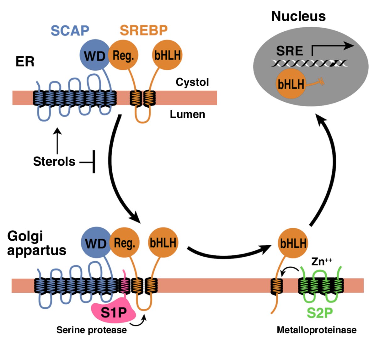
In order to reach the nucleus and act as a transcription factor, the NH2-terminal domain of each SREBP must be released from the membrane proteolytically (Figure 1). Three proteins required for SREBP processing have been delineated in cultured cells, using the tools of somatic cell genetics (see ref. 5for review). One is an escort protein designated SREBP cleavage–activating protein (SCAP). The other two are proteases, designated Site-1 protease (S1P) and Site-2 protease (S2P). Newly synthesized SREBP is inserted into the membranes of the ER, where its COOH-terminal regulatory domain binds to the COOH-terminal domain of SCAP (Figure 1).

Figure 1

Model for the sterol-mediated proteolytic release of SREBPs from membranes JCI0215593.f1

Model for the sterol-mediated proteolytic release of SREBPs from membranes JCI0215593.f1

Model for the sterol-mediated proteolytic release of SREBPs from membranes. SCAP is a sensor of sterols and an escort of SREBPs. When cells are depleted of sterols, SCAP transports SREBPs from the ER to the Golgi apparatus, where two proteases, Site-1 protease (S1P) and Site-2 protease (S2P), act sequentially to release the NH2-terminal bHLH-Zip domain from the membrane. The bHLH-Zip domain enters the nucleus and binds to a sterol response element (SRE) in the enhancer/promoter region of target genes, activating their transcription. When cellular cholesterol rises, the SCAP/SREBP complex is no longer incorporated into ER transport vesicles, SREBPs no longer reach the Golgi apparatus, and the bHLH-Zip domain cannot be released from the membrane. As a result, transcription of all target genes declines. Reprinted from ref. 5 with permission.

http://dm5migu4zj3pb.cloudfront.net/manuscripts/15000/15593/large/JCI0215593.f1.jpg

SCAP is both an escort for SREBPs and a sensor of sterols. When cells become depleted in cholesterol, SCAP escorts the SREBP from the ER to the Golgi apparatus, where the two proteases reside. In the Golgi apparatus, S1P, a membrane-bound serine protease, cleaves the SREBP in the luminal loop between its two membrane-spanning segments, dividing the SREBP molecule in half (Figure 1). The NH2-terminal bHLH-Zip domain is then released from the membrane via a second cleavage mediated by S2P, a membrane-bound zinc metalloproteinase. The NH2-terminal domain, designated nuclear SREBP (nSREBP), translocates to the nucleus, where it activates transcription by binding to nonpalindromic sterol response elements (SREs) in the promoter/enhancer regions of multiple target genes.

When the cholesterol content of cells rises, SCAP senses the excess cholesterol through its membranous sterol-sensing domain, changing its conformation in such a way that the SCAP/SREBP complex is no longer incorporated into ER transport vesicles. The net result is that SREBPs lose their access to S1P and S2P in the Golgi apparatus, so their bHLH-Zip domains cannot be released from the ER membrane, and the transcription of target genes ceases (1, 5). The biophysical mechanism by which SCAP senses sterol levels in the ER membrane and regulates its movement to the Golgi apparatus is not yet understood. Elucidating this mechanism will be fundamental to understanding the molecular basis of cholesterol feedback inhibition of gene expression.

SREBPs: two genes, three proteins

The mammalian genome encodes three SREBP isoforms, designated SREBP-1a, SREBP-1c, and SREBP-2. SREBP-2 is encoded by a gene on human chromosome 22q13. Both SREBP-1a and -1c are derived from a single gene on human chromosome 17p11.2 through the use of alternative transcription start sites that produce alternate forms of exon 1, designated 1a and 1c (1). SREBP-1a is a potent activator of all SREBP-responsive genes, including those that mediate the synthesis of cholesterol, fatty acids, and triglycerides. High-level transcriptional activation is dependent on exon 1a, which encodes a longer acidic transactivation segment than does the first exon of SREBP-1c. The roles of SREBP-1c and SREBP-2 are more restricted than that of SREBP-1a. SREBP-1c preferentially enhances transcription of genes required for fatty acid synthesis but not cholesterol synthesis. Like SREBP-1a, SREBP-2 has a long transcriptional activation domain, but it preferentially activates cholesterol synthesis (1). SREBP-1a and SREBP-2 are the predominant isoforms of SREBP in most cultured cell lines, whereas SREBP-1c and SREBP-2 predominate in the liver and most other intact tissues (6).

When expressed at higher than physiologic levels, each of the three SREBP isoforms can activate all enzymes indicated in Figure 2, which shows the biosynthetic pathways used to generate cholesterol and fatty acids. However, at normal levels of expression, SREBP-1c favors the fatty acid biosynthetic pathway and SREBP-2 favors cholesterologenesis. SREBP-2–responsive genes in the cholesterol biosynthetic pathway include those for the enzymes HMG-CoA synthase, HMG-CoA reductase, farnesyl diphosphate synthase, and squalene synthase. SREBP-1c–responsive genes include those for ATP citrate lyase (which produces acetyl-CoA) and acetyl-CoA carboxylase and fatty acid synthase (which together produce palmitate [C16:0]). Other SREBP-1c target genes encode a rate-limiting enzyme of the fatty acid elongase complex, which converts palmitate to stearate (C18:0) (ref.7); stearoyl-CoA desaturase, which converts stearate to oleate (C18:1); and glycerol-3-phosphate acyltransferase, the first committed enzyme in triglyceride and phospholipid synthesis (3). Finally, SREBP-1c and SREBP-2 activate three genes required to generate NADPH, which is consumed at multiple stages in these lipid biosynthetic pathways (8) (Figure 2).

Figure 2

major metabolic intermediates in the pathways for synthesis of cholesterol, fatty acids, and triglycerides JCI0215593.f2

major metabolic intermediates in the pathways for synthesis of cholesterol, fatty acids, and triglycerides JCI0215593.f2

http://dm5migu4zj3pb.cloudfront.net/manuscripts/15000/15593/large/JCI0215593.f2.jpg

Genes regulated by SREBPs. The diagram shows the major metabolic intermediates in the pathways for synthesis of cholesterol, fatty acids, and triglycerides. In vivo, SREBP-2 preferentially activates genes of cholesterol metabolism, whereas SREBP-1c preferentially activates genes of fatty acid and triglyceride metabolism. DHCR, 7-dehydrocholesterol reductase; FPP, farnesyl diphosphate; GPP, geranylgeranyl pyrophosphate synthase; CYP51, lanosterol 14α-demethylase; G6PD, glucose-6-phosphate dehydrogenase; PGDH, 6-phosphogluconate dehydrogenase; GPAT, glycerol-3-phosphate acyltransferase.

Genes regulated by SREBPs. The diagram shows the major metabolic intermediates in the pathways for synthesis of cholesterol, fatty acids, and triglycerides. In vivo, SREBP-2 preferentially activates genes of cholesterol metabolism, whereas SREBP-1c preferentially activates genes of fatty acid and triglyceride metabolism. DHCR, 7-dehydrocholesterol reductase; FPP, farnesyl diphosphate; GPP, geranylgeranyl pyrophosphate synthase; CYP51, lanosterol 14α-demethylase; G6PD, glucose-6-phosphate dehydrogenase; PGDH, 6-phosphogluconate dehydrogenase; GPAT, glycerol-3-phosphate acyltransferase.

Knockout and transgenic mice

Ten different genetically manipulated mouse models that either lack or overexpress a single component of the SREBP pathway have been generated in the last 6 years (9–16). The key molecular and metabolic alterations observed in these mice are summarized in Table 1.

Table 1
Alterations in hepatic lipid metabolism in gene-manipulated mice overexpressing or lacking SREBPs

http://dm5migu4zj3pb.cloudfront.net/manuscripts/15000/15593/small/JCI0215593.t1.gif

Knockout mice that lack all nSREBPs die early in embryonic development. For instance, a germline deletion of S1p, which prevents the processing of all SREBP isoforms, results in death before day 4 of development (15, 17). Germline deletion of Srebp2 leads to 100% lethality at a later stage of embryonic development than does deletion of S1p (embryonic day 7–8). In contrast, germline deletion of Srebp1, which eliminates both the 1a and the 1c transcripts, leads to partial lethality, in that about 15–45% of Srebp1–/– mice survive (13). The surviving homozygotes manifest elevated levels of SREBP-2 mRNA and protein (Table 1), which presumably compensates for the loss of SREBP-1a and -1c. When the SREBP-1c transcript is selectively eliminated, no embryonic lethality is observed, suggesting that the partial embryonic lethality in the Srebp1–/– mice is due to the loss of the SREBP-1a transcript (16).

To bypass embryonic lethality, we have produced mice in which all SREBP function can be disrupted in adulthood through induction of Cre recombinase. For this purpose, loxP recombination sites were inserted into genomic regions that flank crucial exons in the Scap or S1p genes (so-called floxed alleles) (14, 15). Mice homozygous for the floxed gene and heterozygous for a Cre recombinase transgene, which is under control of an IFN-inducible promoter (MX1-Cre), can be induced to delete Scap or S1p by stimulating IFN expression. Thus, following injection with polyinosinic acid–polycytidylic acid, a double-stranded RNA that provokes antiviral responses, the Cre recombinase is produced in liver and disrupts the floxed gene by recombination between the loxP sites.

Cre-mediated disruption of Scap or S1p dramatically reduces nSREBP-1 and nSREBP-2 levels in liver and diminishes expression of all SREBP target genes in both the cholesterol and the fatty acid synthetic pathways (Table 1). As a result, the rates of synthesis of cholesterol and fatty acids fall by 70–80% in Scap- and S1p-deficient livers.

In cultured cells, the processing of SREBP is inhibited by sterols, and the sensor for this inhibition is SCAP (5). To learn whether SCAP performs the same function in liver, we have produced transgenic mice that express a mutant SCAP with a single amino acid substitution in the sterol-sensing domain (D443N) (12). Studies in tissue culture show that SCAP(D443N) is resistant to inhibition by sterols. Cells that express a single copy of this mutant gene overproduce cholesterol (18). Transgenic mice that express this mutant version of SCAP in the liver exhibit a similar phenotype (12). These livers manifest elevated levels of nSREBP-1 and nSREBP-2, owing to constitutive SREBP processing, which is not suppressed when the animals are fed a cholesterol-rich diet. nSREBP-1 and -2 increase the expression of all SREBP target genes shown in Figure 2, thus stimulating cholesterol and fatty acid synthesis and causing a marked accumulation of hepatic cholesterol and triglycerides (Table 1). This transgenic model provides strong in vivo evidence that SCAP activity is normally under partial inhibition by endogenous sterols, which keeps the synthesis of cholesterol and fatty acids in a partially repressed state in the liver.

http://dm5migu4zj3pb.cloudfront.net/manuscripts/15000/15593/small/JCI0215593.t1.gif

Function of individual SREBP isoforms in vivo

To study the functions of individual SREBPs in the liver, we have produced transgenic mice that overexpress truncated versions of SREBPs (nSREBPs) that terminate prior to the membrane attachment domain. These nSREBPs enter the nucleus directly, bypassing the sterol-regulated cleavage step. By studying each nSREBP isoform separately, we could determine their distinct activating properties, albeit when overexpressed at nonphysiologic levels.

Overexpression of nSREBP-1c in the liver of transgenic mice produces a triglyceride-enriched fatty liver with no increase in cholesterol (10). mRNAs for fatty acid synthetic enzymes and rates of fatty acid synthesis are elevated fourfold in this tissue, whereas the mRNAs for cholesterol synthetic enzymes and the rate of cholesterol synthesis are not increased (8). Conversely, overexpression of nSREBP-2 in the liver increases the mRNAs only fourfold. This increase in cholesterol synthesis is even more remarkable when encoding all cholesterol biosynthetic enzymes; the most dramatic is a 75-fold increase in HMG-CoA reductase mRNA (11). mRNAs for fatty acid synthesis enzymes are increased to a lesser extent, consistent with the in vivo observation that the rate of cholesterol synthesis increases 28-fold in these transgenic nSREBP-2 livers, while fatty acid synthesis increases one considers the extent of cholesterol overload in this tissue, which would ordinarily reduce SREBP processing and essentially abolish cholesterol synthesis (Table 1).

http://dm5migu4zj3pb.cloudfront.net/manuscripts/15000/15593/small/JCI0215593.t1.gif

We have also studied the consequences of overexpressing SREBP-1a, which is expressed only at low levels in the livers of adult mice, rats, hamsters, and humans (6). nSREBP-1a transgenic mice develop a massive fatty liver engorged with both cholesterol and triglycerides (9), with heightened expression of genes controlling cholesterol biosynthesis and, still more dramatically, fatty acid synthesis (Table 1). The preferential activation of fatty acid synthesis (26-fold increase) relative to cholesterol synthesis (fivefold increase) explains the greater accumulation of triglycerides in their livers. The relative representation of the various fatty acids accumulating in this tissue is also unusual. Transgenic nSREBP-1a livers contain about 65% oleate (C18:1), markedly higher levels than the 15–20% found in typical wild-type livers (8) — a result of the induction of fatty acid elongase and stearoyl-CoA desaturase-1 (7). Considered together, the overexpression studies indicate that both SREBP-1 isoforms show a relative preference for activating fatty acid synthesis, whereas SREBP-2 favors cholesterol.

The phenotype of animals lacking the Srebp1 gene, which encodes both the SREBP-1a and -1c transcripts, also supports the notion of distinct hepatic functions for SREBP-1 and SREBP-2 (13). Most homozygous SREBP-1 knockout mice die in utero. The surviving Srebp1–/– mice show reduced synthesis of fatty acids, owing to reduced expression of mRNAs for fatty acid synthetic enzymes (Table 1). Hepatic nSREBP-2 levels increase in these mice, presumably in compensation for the loss of nSREBP-1. As a result, transcription of cholesterol biosynthetic genes increases, producing a threefold increase in hepatic cholesterol synthesis (Table 1).

http://dm5migu4zj3pb.cloudfront.net/manuscripts/15000/15593/small/JCI0215593.t1.gif

The studies in genetically manipulated mice clearly show that, as in cultured cells, SCAP and S1P are required for normal SREBP processing in the liver. SCAP, acting through its sterol-sensing domain, mediates feedback regulation of cholesterol synthesis. The SREBPs play related but distinct roles: SREBP-1c, the predominant SREBP-1 isoform in adult liver, preferentially activates genes required for fatty acid synthesis, while SREBP-2 preferentially activates the LDL receptor gene and various genes required for cholesterol synthesis. SREBP-1a and SREBP-2, but not SREBP-1c, are required for normal embryogenesis.

Transcriptional regulation of SREBP genes

Regulation of SREBPs occurs at two levels — transcriptional and posttranscriptional. The posttranscriptional regulation discussed above involves the sterol-mediated suppression of SREBP cleavage, which results from sterol-mediated suppression of the movement of the SCAP/SREBP complex from the ER to the Golgi apparatus (Figure 1). This form of regulation is manifest not only in cultured cells (1), but also in the livers of rodents fed cholesterol-enriched diets (19).

http://dm5migu4zj3pb.cloudfront.net/manuscripts/15000/15593/small/JCI0215593.f1.gif

The transcriptional regulation of the SREBPs is more complex. SREBP-1c and SREBP-2 are subject to distinct forms of transcriptional regulation, whereas SREBP-1a appears to be constitutively expressed at low levels in liver and most other tissues of adult animals (6). One mechanism of regulation shared by SREBP-1c and SREBP-2 involves a feed-forward regulation mediated by SREs present in the enhancer/promoters of each gene (20, 21). Through this feed-forward loop, nSREBPs activate the transcription of their own genes. In contrast, when nSREBPs decline, as in Scap or S1p knockout mice, there is a secondary decline in the mRNAs encoding SREBP-1c and SREBP-2 (14, 15).

Three factors selectively regulate the transcription of SREBP-1c: liver X-activated receptors (LXRs), insulin, and glucagon. LXRα and LXRβ, nuclear receptors that form heterodimers with retinoid X receptors, are activated by a variety of sterols, including oxysterol intermediates that form during cholesterol biosynthesis (22–24). An LXR-binding site in the SREBP-1c promoter activates SREBP-1c transcription in the presence of LXR agonists (23). The functional significance of LXR-mediated SREBP-1c regulation has been confirmed in two animal models. Mice that lack both LXRα and LXRβ express reduced levels of SREBP-1c and its lipogenic target enzymes in liver and respond relatively weakly to treatment with a synthetic LXR agonist (23). Because a similar blunted response is found in mice that lack SREBP-1c, it appears that LXR increases fatty acid synthesis largely by inducing SREBP-1c (16). LXR-mediated activation of SREBP-1c transcription provides a mechanism for the cell to induce the synthesis of oleate when sterols are in excess (23). Oleate is the preferred fatty acid for the synthesis of cholesteryl esters, which are necessary for both the transport and the storage of cholesterol.

LXR-mediated regulation of SREBP-1c appears also to be one mechanism by which unsaturated fatty acids suppress SREBP-1c transcription and thus fatty acid synthesis. Rodents fed diets enriched in polyunsaturated fatty acids manifest reduced SREBP-1c mRNA expression and low rates of lipogenesis in liver (25). In vitro, unsaturated fatty acids competitively block LXR activation of SREBP-1c expression by antagonizing the activation of LXR by its endogenous ligands (26). In addition to LXR-mediated transcriptional inhibition, polyunsaturated fatty acids lower SREBP-1c levels by accelerating degradation of its mRNA (27). These combined effects may contribute to the long-recognized ability of polyunsaturated fatty acids to lower plasma triglyceride levels.

SREBP-1c and the insulin/glucagon ratio

The liver is the organ responsible for the conversion of excess carbohydrates to fatty acids to be stored as triglycerides or burned in muscle. A classic action of insulin is to stimulate fatty acid synthesis in liver during times of carbohydrate excess. The action of insulin is opposed by glucagon, which acts by raising cAMP. Multiple lines of evidence suggest that insulin’s stimulatory effect on fatty acid synthesis is mediated by an increase in SREBP-1c. In isolated rat hepatocytes, insulin treatment increases the amount of mRNA for SREBP-1c in parallel with the mRNAs of its target genes (28, 29). The induction of the target genes can be blocked if a dominant negative form of SREBP-1c is expressed (30). Conversely, incubating primary hepatocytes with glucagon or dibutyryl cAMP decreases the mRNAs for SREBP-1c and its associated lipogenic target genes (30, 31).

In vivo, the total amount of SREBP-1c in liver and adipose tissue is reduced by fasting, which suppresses insulin and increases glucagon levels, and is elevated by refeeding (32, 33). The levels of mRNA for SREBP-1c target genes parallel the changes in SREBP-1c expression. Similarly, SREBP-1c mRNA levels fall when rats are treated with streptozotocin, which abolishes insulin secretion, and rise after insulin injection (29). Overexpression of nSREBP-1c in livers of transgenic mice prevents the reduction in lipogenic mRNAs that normally follows a fall in plasma insulin levels (32). Conversely, in livers of Scap knockout mice that lack all nSREBPs in the liver (14) or knockout mice lacking either nSREBP-1c (16) or both SREBP-1 isoforms (34), there is a marked decrease in the insulin-induced stimulation of lipogenic gene expression that normally occurs after fasting/refeeding. It should be noted that insulin and glucagon also exert a posttranslational control of fatty acid synthesis though changes in the phosphorylation and activation of acetyl-CoA carboxylase. The posttranslational regulation of fatty acid synthesis persists in transgenic mice that overexpress nSREBP-1c (10). In these mice, the rates of fatty acid synthesis, as measured by [3H]water incorporation, decline after fasting even though the levels of the lipogenic mRNAs remain high (our unpublished observations).

Taken together, the above evidence suggests that SREBP-1c mediates insulin’s lipogenic actions in liver. Recent in vitro and in vivo studies involving adenoviral gene transfer suggest that SREBP-1c may also contribute to the regulation of glucose uptake and glucose synthesis. When overexpressed in hepatocytes, nSREBP-1c induces expression of glucokinase, a key enzyme in glucose utilization. It also suppresses phosphoenolpyruvate carboxykinase, a key gluconeogenic enzyme (35, 36).

SREBPs in disease

Many individuals with obesity and insulin resistance also have fatty livers, one of the most commonly encountered liver abnormalities in the US (37). A subset of individuals with fatty liver go on to develop fibrosis, cirrhosis, and liver failure. Evidence indicates that the fatty liver of insulin resistance is caused by SREBP-1c, which is elevated in response to the high insulin levels. Thus, SREBP-1c levels are elevated in the fatty livers of obese (ob/ob) mice with insulin resistance and hyperinsulinemia caused by leptin deficiency (38, 39). Despite the presence of insulin resistance in peripheral tissues, insulin continues to activate SREBP-1c transcription and cleavage in the livers of these insulin-resistant mice. The elevated nSREBP-1c increases lipogenic gene expression, enhances fatty acid synthesis, and accelerates triglyceride accumulation (31, 39). These metabolic abnormalities are reversed with the administration of leptin, which corrects the insulin resistance and lowers the insulin levels (38).

Metformin, a biguanide drug used to treat insulin-resistant diabetes, reduces hepatic nSREBP-1 levels and dramatically lowers the lipid accumulation in livers of insulin-resistant ob/ob mice (40). Metformin stimulates AMP-activated protein kinase (AMPK), an enzyme that inhibits lipid synthesis through phosphorylation and inactivation of key lipogenic enzymes (41). In rat hepatocytes, metformin-induced activation of AMPK also leads to decreased mRNA expression of SREBP-1c and its lipogenic target genes (41), but the basis of this effect is not understood.

The incidence of coronary artery disease increases with increasing plasma LDL-cholesterol levels, which in turn are inversely proportional to the levels of hepatic LDL receptors. SREBPs stimulate LDL receptor expression, but they also enhance lipid synthesis (1), so their net effect on plasma lipoprotein levels depends on a balance between opposing effects. In mice, the plasma levels of lipoproteins tend to fall when SREBPs are either overexpressed or underexpressed. In transgenic mice that overexpress nSREBPs in liver, plasma cholesterol and triglycerides are generally lower than in control mice (Table 1), even though these mice massively overproduce fatty acids, cholesterol, or both. Hepatocytes of nSREBP-1a transgenic mice overproduce VLDL, but these particles are rapidly removed through the action of LDL receptors, and they do not accumulate in the plasma. Indeed, some nascent VLDL particles are degraded even before secretion by a process that is mediated by LDL receptors (42). The high levels of nSREBP-1a in these animals support continued expression of the LDL receptor, even in cells whose cholesterol concentration is elevated. In LDL receptor–deficient mice carrying the nSREBP-1a transgene, plasma cholesterol and triglyceride levels rise tenfold (43).

Mice that lack all SREBPs in liver as a result of disruption of Scap or S1p also manifest lower plasma cholesterol and triglyceride levels (Table 1).

http://dm5migu4zj3pb.cloudfront.net/manuscripts/15000/15593/small/JCI0215593.t1.gif

In these mice, hepatic cholesterol and triglyceride synthesis is markedly reduced, and this likely causes a decrease in VLDL production and secretion. LDL receptor mRNA and LDL clearance from plasma is also significantly reduced in these mice, but the reduction in LDL clearance is less than the overall reduction in VLDL secretion, the net result being a decrease in plasma lipid levels (15). However, because

humans and mice differ substantially with regard to LDL receptor expression, LDL levels, and other aspects of lipoprotein metabolism,

it is difficult to predict whether human plasma lipids will rise or fall when the SREBP pathway is blocked or activated.

SREBPs in liver: unanswered questions

The studies of SREBPs in liver have exposed a complex regulatory system whose individual parts are coming into focus. Major unanswered questions relate to the ways in which the transcriptional and posttranscriptional controls on SREBP activity are integrated so as to permit independent regulation of cholesterol and fatty acid synthesis in specific nutritional states. A few clues regarding these integration mechanisms are discussed below.

Whereas cholesterol synthesis depends almost entirely on SREBPs, fatty acid synthesis is only partially dependent on these proteins. This has been shown most clearly in cultured nonhepatic cells such as Chinese hamster ovary cells. In the absence of SREBP processing, as when the Site-2 protease is defective, the levels of mRNAs encoding cholesterol biosynthetic enzymes and the rates of cholesterol synthesis decline nearly to undetectable levels, whereas the rate of fatty acid synthesis is reduced by only 30% (44). Under these conditions, transcription of the fatty acid biosynthetic genes must be maintained by factors other than SREBPs. In liver, the gene encoding fatty acid synthase (FASN) can be activated transcriptionally by upstream stimulatory factor, which acts in concert with SREBPs (45). The FASN promoter also contains an LXR element that permits a low-level response to LXR ligands even when SREBPs are suppressed (46). These two transcription factors may help to maintain fatty acid synthesis in liver when nSREBP-1c is low.

Another mechanism of differential regulation is seen in the ability of cholesterol to block the processing of SREBP-2, but not SREBP-1, under certain metabolic conditions. This differential regulation has been studied most thoroughly in cultured cells such as human embryonic kidney (HEK-293) cells. When these cells are incubated in the absence of fatty acids and cholesterol, the addition of sterols blocks processing of SREBP-2, but not SREBP-1, which is largely produced as SREBP-1a in these cells (47). Inhibition of SREBP-1 processing requires an unsaturated fatty acid, such as oleate or arachidonate, in addition to sterols (47). In the absence of fatty acids and in the presence of sterols, SCAP may be able to carry SREBP-1 proteins, but not SREBP-2, to the Golgi apparatus. Further studies are necessary to document this apparent independent regulation of SREBP-1 and SREBP-2 processing and to determine its mechanism.

Acknowledgments

Support for the research cited from the authors’ laboratories was provided by grants from the NIH (HL-20948), the Moss Heart Foundation, the Keck Foundation, and the Perot Family Foundation. J.D. Horton is a Pew Scholar in the Biomedical Sciences and is the recipient of an Established Investigator Grant from the American Heart Association and a Research Scholar Award from the American Digestive Health Industry.

References

  1. Brown, MS, Goldstein, JL. The SREBP pathway: regulation of cholesterol metabolism by proteolysis of a membrane-bound transcription factor. Cell 1997. 89:331-340.

View this article via: PubMed

  1. Horton, JD, Shimomura, I. Sterol regulatory element-binding proteins: activators of cholesterol and fatty acid biosynthesis. Curr Opin Lipidol 1999. 10:143-150.

View this article via: PubMed

  1. Edwards, PA, Tabor, D, Kast, HR, Venkateswaran, A. Regulation of gene expression by SREBP and SCAP. Biochim Biophys Acta 2000. 1529:103-113.

View this article via: PubMed

  1. Sakakura, Y, et al. Sterol regulatory element-binding proteins induce an entire pathway of cholesterol synthesis. Biochem Biophys Res Commun 2001. 286:176-183.

View this article via: PubMed

  1. Goldstein, JL, Rawson, RB, Brown, MS. Mutant mammalian cells as tools to delineate the sterol regulatory element-binding protein pathway for feedback regulation of lipid synthesis. Arch Biochem Biophys 2002. 397:139-148.

View this article via: PubMed

  1. Shimomura, I, Shimano, H, Horton, JD, Goldstein, JL, Brown, MS. Differential expression of exons 1a and 1c in mRNAs for sterol regulatory element binding protein-1 in human and mouse organs and cultured cells. J Clin Invest 1997. 99:838-845.

View this article via: JCI.org PubMed

  1. Moon, Y-A, Shah, NA, Mohapatra, S, Warrington, JA, Horton, JD. Identification of a mammalian long chain fatty acyl elongase regulated by sterol regulatory element-binding proteins. J Biol Chem 2001. 276:45358-45366.

View this article via: PubMed

  1. Shimomura, I, Shimano, H, Korn, BS, Bashmakov, Y, Horton, JD. Nuclear sterol regulatory element binding proteins activate genes responsible for entire program of unsaturated fatty acid biosynthesis in transgenic mouse liver. J Biol Chem 1998. 273:35299-35306.

View this article via: PubMed

  1. Shimano, H, et al. Overproduction of cholesterol and fatty acids causes massive liver enlargement in transgenic mice expressing truncated SREBP-1a. J Clin Invest 1996. 98:1575-1584.

View this article via: JCI.org PubMed

  1. Shimano, H, et al. Isoform 1c of sterol regulatory element binding protein is less active than isoform 1a in livers of transgenic mice and in cultured cells. J Clin Invest 1997. 99:846-854.

View this article via: JCI.org PubMed

  1. Horton, JD, et al. Activation of cholesterol synthesis in preference to fatty acid synthesis in liver and adipose tissue of transgenic mice overproducing sterol regulatory element-binding protein-2. J Clin Invest 1998. 101:2331-2339.

View this article via: JCI.org PubMed

  1. Korn, BS, et al. Blunted feedback suppression of SREBP processing by dietary cholesterol in transgenic mice expressing sterol-resistant SCAP(D443N). J Clin Invest 1998. 102:2050-2060.

View this article via: JCI.org PubMed

  1. Shimano, H, et al. Elevated levels of SREBP-2 and cholesterol synthesis in livers of mice homozygous for a targeted disruption of the SREBP-1 gene. J Clin Invest 1997. 100:2115-2124.

View this article via: JCI.org PubMed

  1. Matsuda, M, et al. SREBP cleavage-activating protein (SCAP) is required for increased lipid synthesis in liver induced by cholesterol deprivation and insulin elevation. Genes Dev 2001. 15:1206-1216.

View this article via: PubMed

  1. Yang, J, et al. Decreased lipid synthesis in livers of mice with disrupted Site-1 protease gene. Proc Natl Acad Sci USA 2001. 98:13607-13612.

View this article via: PubMed

Liang, G, et al. Diminished hepatic response to fasting/refeeding and liver X receptor agonists in mice with selective deficiency of sterol regulatory element-binding protein-1c. J Biol Chem 2002. 277:9520-9528.

http://www.jci.org/articles/view/15593

Share this:

  • Share on X (Opens in new window) X
  • Share on LinkedIn (Opens in new window) LinkedIn
  • Share on Facebook (Opens in new window) Facebook
  • Print (Opens in new window) Print
  • Email a link to a friend (Opens in new window) Email

Like this:

Like Loading…

Read Full Post »

Diabetes Mellitus

Posted in Academic Publishing, Alzheimer's Disease, Cerebrovascular and Neurodegenerative Diseases, Chemical Biology and its relations to Metabolic Disease, Chemical Genetics, Curation, Diabetes Mellitus, Disease Biology, Epigenetics and Cardiovascular Risks, Etiology, Gene Regulation, Innovations, Lipid metabolism, Metabolism, Metabolomics, Nephrology, Neurological Diseases, Nitric Oxide in Health and Disease, Nutrition, Nutrition and Phytochemistry, Nutritional Supplements: Atherogenesis, Origins of Cardiovascular Disease, Plant extract, tagged amylin, Amyloid, Beta cell, Diabetes Mellitus, glomerulosclerosis, Insulin resistance, Insulin Sensitivity, islet cells of pancreas, pharmacotherapy, proinsulin on October 24, 2014| Leave a Comment »

Diabetes Mellitus

Author & Curator: Larry H. Bernstein, MD, FCAP


Diabetes mellitus (DM) is a group of metabolic diseases defined by high blood glucose levels, which, depending on the fasting blood glucose, may be pre-diabetes or overt diabetes (110 mg/dl. 124 mg/dl). This blood glucose level reflects a disorder of control of glucose metabolism, which is mediated through the pituitary growth hormone acting on the liver, which produces insulin growth factor 1 (IGF1).  Diabetes is due to either the pancreas not producing enough insulin, or the cells of the body not responding properly to the insulin produced. That said, there is much to be understood about the long term systemic effects of this disorder, a multisystem disease. The presence of pre-diabetes glucose levels is sufficient to proactively take measures to reduce the circulating glucose.

Globally, as of 2013, an estimated 382 million people have diabetes worldwide, with type 2 diabetes making up about 90% of the cases. This is equal to 8.3% of the adults population, with equal rates in both women and men. Worldwide in 2012 and 2013 diabetes resulted in 1.5 to 5.1 million deaths per year, making it the 8th leading cause of death. Diabetes overall at least doubles the risk of death. The number of people with diabetes is expected to rise to 592 million by 2035. The economic costs of diabetes globally was estimated in 2013 at $548 billion and in the United States in 2012 $245 billion.

The observation of symptoms of frequent urination, increased thirst, and increased hunger is symptomatic of overt DM, and is seen with diabetic ketoacidosis, with very high hyperglycemia and glucosuria, particularly in Type 1 DM. Untreated, diabetes leads to serious complications. Acute complications include diabetic ketoacidosis. Serious long-term complications include heart disease, stroke, kidney failure, foot ulcers and damage to the eyes.

There are three main types of diabetes mellitus:

  • Type 1 DM results from the body’s failure to produce enough insulin. This form was previously referred to as “insulin-dependent diabetes mellitus” (IDDM) or “juvenile diabetes”. The cause is unknown.
  • Type 2 DM begins with insulin resistance, a condition in which cells fail to respond to insulin properly. As the disease progresses a lack of insulin may also develop. This form was previously referred to as “non insulin-dependent diabetes mellitus” (NIDDM) or “adult-onset diabetes”. The primary cause is excessive body weight and not enough exercise.
  • Gestational diabetes, the third, occurs when pregnant women without a previous history of diabetes develop a high blood glucose level.

Type 1 DM, which presents suddenly in children or young adults, is possibly an as yet unidentified post-translational or epigenetic form, unrelated to Type 2, which is becoming more common in children.  It results in the destruction of islet beta cells that then have no capacity to produce insulin.  A family history of the disease would be a signal to raise a child with great care to not stress the pancreas.  Even though I raised the possibility of an epigenetic factor, it is important to keep in mind that the regulation of glucose is responsive to a number of stresses, even in a healthy person.  These are:

  • Corticosteroids
  • Glucagon
  • Growth hormone
  • Catecholamines
  • Proinflammatory cytokines
  • Anxiety disorder
  • Eating disorder

Gestational diabetes is perhaps Type 2 diabetes in a pregnant woman initiated by the condition of pregnancy. Whether these women were not diabetic, with a glucose level between 100-110 prior to pregnancy, is an open question. However, the pregnant state is accompanied by large effects by hormone levels.

Type 2 diabetes has been increasing worldwide, not only in western nations.  However, in non-western countries that have large populations of underserved, there is still a major problem with protein energy malnutrition (PEM). Globally, as of 2013, an estimated 382 million people have diabetes worldwide, with type 2 diabetes making up about 90% of the cases. This is equal to 8.3% of the adults population, with equal rates in both women and men. Worldwide in 2012 and 2013 diabetes resulted in 1.5 to 5.1 million deaths per year, making it the 8th leading cause of death. Diabetes overall at least doubles the risk of death. The number of people with diabetes is expected to rise to 592 million by 2035. The economic costs of diabetes globally was estimated in 2013 at $548 billion and in the United States in 2012 $245 billion.

The major long-term complications relate to damage to blood vessels. Diabetes doubles the risk of cardiovascular disease and about 75% of deaths in diabetics are due to coronary artery disease. Other “macrovascular” diseases are stroke, and peripheral vascular disease. The primary microvascular complications of diabetes include damage to the eyes, kidneys, and nerves. Damage to the eyes, known as diabetic retinopathy, is caused by damage to the blood vessels in the retina of the eye, and can result in gradual vision loss and potentially blindness. Damage to the kidneys, known as diabetic nephropathy, can lead to tissue scarring, urine protein loss, and eventually chronic kidney disease, sometimes requiring dialysis or kidney transplant. Damage to the nerves of the body, known as diabetic neuropathy, is the most common complication of diabetes.

Prevention and treatment involves a healthy diet, physical exercise, not using tobacco and being a normal body weight. Blood pressure control and proper foot care are also important for people with the disease. Type 1 diabetes must be managed with insulin injections. Type 2 diabetes may be treated with medications with or without insulin. Insulin and some oral medications can cause low blood sugar. Weight loss surgery in those with obesity is an effective measure in those with type 2 DM. Gestational diabetes usually resolves after the birth of the baby.

A number of articles in http://pharmaceuticalintelligence,com (this journal) have presented the relationship of DM to heart and vascular disease. The complexity of the disease is not to be underestimated, and there havr been serious controversies with adverse consequences over the use of the class of drugs that includes rosiglitazone and piaglitazone, which has opened serious issues about how clinical trials are conducted, and how the data obtained in studies may be compromised.

Pharmaceutical Insights

Management of Diabetes Mellitus: Could Simultaneous Targeting of Hyperglycemia and Oxidative Stress Be a Better Panacea?

Omotayo O. Erejuwa
Int. J. Mol. Sci. 2012, 13, 2965-2972; http://www.mdpi.com/journal/ijms http://dx.doi.org:/10.3390/ijms13032965

The primary aim of the current management of diabetes mellitus is to achieve and/or maintain a glycated hemoglobin level of ≤6.5%. However, recent evidence indicates that intensive treatment of hyperglycemia is characterized by increased weight gain, severe hypoglycemia and higher mortality. Besides, evidence suggests that it is difficult to achieve and/or maintain optimal glycemic control in many diabetic patients; and that the benefits of intensively-treated hyperglycemia are restricted to microvascular complications only. Evidence also indicates that multiple drugs are required to achieve optimal glycemic target in many diabetic patients. In fact, in many diabetic patients in whom optimal glycemic goal is achieved, glycemic control deteriorates even with optimal drug therapy. It does suggest that with the current hypoglycemic or antidiabetic drugs, it is difficult to achieve and/or maintain tight glycemic control in diabetic patients. In many developing countries, the vast majority of diabetic patients have limited or lack access to quality healthcare providers and good therapeutic monitoring.

While increased weight gain could be due to some component drugs (such as sulphonylureas or insulin) of the intensive therapy regimens, hypoglycemia could be drug-induced or comorbidity-induced. Considering the evidence that associates hypoglycemia with increased mortality, higher incidence of mortality in intensive therapy group could be due to hypoglycemia or too low levels of glycosylated hemoglobin. However, it is difficult to contend that increased mortality was entirely due to hypoglycemia. The possibility of drug-induced or drug-associated toxicities could not be ruled out. For instance, rosiglitazone, which has been prohibited and withdrawn from the market in Europe, was one of the hypoglycemic drugs used to achieve intensive therapy of hyperglycemia in Action to Control Cardiovascular Risk in Diabetes (ACCORD). If these findings are anything to go by, does it not suggest that targeting hyperglycemia as the only therapeutic goal in the management of diabetes mellitus could be detrimental to diabetic patients? In addition, the current hypoglycemic drugs are characterized by limitations and adverse effects. Together with the limitations of intensive glycemic treatment (only beneficial in reducing the risk of microvascular complications, but not macrovascular disease complications), does it not imply that targeting hyperglycemia alone is not only deleterious but also limited and ineffective?

The latest figures predict that the global incidence of diabetes mellitus, which was estimated to be 366 million in 2011, will rise to 522 million by 2030. In view of these frightening statistics on the prevalence of diabetes mellitus and on the lack of adequate healthcare, together with the associated diabetic complications, morbidity and mortality, does it not suggest that there is an urgent need for a better therapeutic management of this disorder? Taken together, with these findings and statistics, it can be contended that it is high time alternative and/or complementary therapies to the currently available hypoglycemic agents (which target primarily hyperglycemia only) were sought.

All these may contribute to the unabated increase in global prevalence of diabetes mellitus and its complications In view of these adverse effects and limitations of intensive treatment of hyperglycemia in preventing diabetic complications, which is linked to oxidative stress,

  • this commentary proposes a hypothesis that “simultaneous targeting of hyperglycemia and oxidative stress” could be more effective than “intensive treatment of hyperglycemia” in the management of diabetes mellitus.

Oxidative stress is defined as

  • an “imbalance between oxidants and antioxidants in favor of the oxidants, potentially leading to damage”.

It is implicated in the pathogenesis and complications of diabetes mellitus. The role of oxidative stress is more definite in the pathogenesis of type 2 diabetes mellitus than in type 1 diabetes mellitus. In regard to diabetic complications, there is compelling evidence in support of the role of oxidative stress in both types of diabetes mellitus. Evidence suggests that elevated reactive oxygen species (ROS), which causes factor of increased ROS production, causes tissue damage or diabetic complications have been identified. These include:

  • hyperglycemia-enhanced polyol pathway;
  • hyperglycemia-enhanced formation of advanced glycation endproducts (AGEs);
  • hyperglycemia-activated protein kinase C (PKC) pathway;
  • hyperglycemia-enhanced hexosamine pathway; and
  • hyperglycemia-activated Poly-ADP ribose polymerase (PARP) pathway.

These pathways are activated or enhanced by hyperglycemia-driven mitochondrial superoxide overproduction.

Even though oxidative stress plays an important role in its pathogenesis and complications,

  • unlike other diseases characterized by oxidative stress, diabetes mellitus is unique.

Its cure (restoration of euglycemia, e.g., via pancreas transplants) does not prevent oxidative stress and diabetic complications. This is very important because hyperglycemia exacerbates oxidative stress which is linked to diabetic complications. Theoretically, restoration of euglycemia should prevent oxidative stress and diabetic complications. However, this is not the case. At present, it remains unclear why restoration of euglycemia does not automatically prevent oxidative stress and diabetic complications. The development of diabetes-related complications (both microvascular and macrovascular) may occur in diabetic patients after normoglycemia has been restored. It is a phenomenon whereby previous hyperglycemic milieu is remembered in many target organs such as heart, eyes, kidneys and nerves. This phenomenon is also documented in diabetic animals. Compelling evidence implicates the role of oxidative stress as an important mechanism by which glycemic memory causes tissue damage and diabetic complications. In view of higher incidence of diabetic complications (of which oxidative stress plays an important role) in conventionally-treated diabetic patients, targeting oxidative stress in these patients might be beneficial. In other words, it is possible that the combination of a conventional therapy of hyperglycemia and antioxidant therapy might be more effective and beneficial than intensive therapy of hyperglycemia alone, which is the gold standard at the moment.

Loss of ACE 2 Exaggerates High-Calorie Diet-Induced Insulin Resistance by Reduction of GLUT4 in Mice

M Takeda, K Yamamoto, Y Takemura, H Takeshita, K Hongyo, et al.  Diabetes 61:1–11, 2012

ACE type 2 (ACE2) functions as

  • a negative regulator of the renin angiotensin system
  • by cleaving angiotensin II (AII) into angiotensin 1–7 (A1–7).

This study assessed the role of

  • endogenous ACE2 in maintaining insulin sensitivity.

Twelve-week-old male ACE2 knockout (ACE2KO) mice had normal insulin sensitivities when fed a standard diet. AII infusion or a high-fat high-sucrose (HFHS) diet impaired glucose tolerance and insulin sensitivity more severely

  • in ACE2KO mice than in their wild-type (WT) littermates.

The strain difference in glucose tolerance

  • was not eliminated by an AII receptor type 1 (AT1) blocker
  • but was eradicated by A1–7 or an AT1 blocker combined with the A1–7 inhibitor (A779).

The expression of GLUT4 and a transcriptional factor, myocyte enhancer factor (MEF) 2A,

  • was dramatically reduced in the skeletal muscles of the standard diet–fed ACE2KO mice.

The expression of GLUT4 and MEF2A was increased

  • by A1–7 in ACE2KO mice and
  • decreased by A779 in WT mice.

A1–7 enhanced upregulation of MEF2A and GLUT4 during differentiation of myoblast cells. In conclusion,

  • ACE2 protects against high calorie diet-induced insulin resistance in mice.

This mechanism may involve the transcriptional regulation of GLUT4 via an A1–7-dependent pathway.
Modulation of the action of insulin by angiotensin-(1–7)
FP. Dominici, V Burghi, MC. Munoz, JF. Giani

Clinical Science (2014) 126, 613–630 http://dx.doi.org:/10.1042/CS20130333

The prevalence of Type 2 diabetes mellitus is predicted to increase dramatically over the coming years and the clinical implications and healthcare costs from this disease are overwhelming. In many cases, this pathological condition is linked to a cluster of metabolic disorders, such as

  1. obesity,
  2. systemic hypertension and
  3. dyslipidaemia,
  • defined as the metabolic syndrome.

Insulin resistance has been proposed as the key mediator of all of these features and contributes to the associated high cardiovascular morbidity and mortality. Although the molecular mechanisms behind insulin resistance are not completely understood, a negative cross-talk between

  • AngII (angiotensin II) and the insulin signalling pathway

has been the focus of great interest in the last decade. Indeed,

substantial evidence has shown that

  • anti-hypertensive drugs that block the RAS (renin–angiotensin system) may also act to prevent diabetes.

Despite its long history, new components within the RAS continue to be discovered.

Among them, Ang-(1–7) [angiotensin-(1–7)] has gained special attention as a counter-regulatory hormone

  • opposing many of the AngII-related deleterious effects.

Specifically, we and others have demonstrated that Ang-(1–7) improves the action of insulin and opposes the negative effect that AngII exerts at this level. In the present review, we provide evidence showing that

  • insulin and Ang-(1–7) share a common intracellular signalling pathway.

We also address the molecular mechanisms behind the beneficial effects of Ang-(1–7) on

  • AngII-mediated insulin resistance.

Finally, we discuss potential therapeutic approaches leading to modulation of the

  • ACE2 (angiotensin-converting enzyme 2)/Ang-(1–7)/Mas receptor axis

as a very attractive strategy in the therapy of the metabolic syndrome and diabetes-associated diseases.

Increased Skeletal Muscle Capillarization After Aerobic Exercise Training and Weight Loss Improves Insulin Sensitivity in Adults With IGT

Prior, JB. Blumenthal, LI. Katzel, AP. Goldberg, AS. Ryan. Diabetes Care 2014;37:1469–1475
http://dx.doi.org:/10.2337/dc13-2358

Transcapillary transport of insulin is one determinant of glucose uptake by skeletal muscle; thus,

  • a reduction in capillary density (CD) may worsen insulin sensitivity.

Skeletal muscle CD is lower in older adults with impaired glucose tolerance (IGT) compared with those with normal glucose tolerance and

  • may be modifiable through aerobic exercise training and weight loss (AEX+WL).

Insulin sensitivity (M) and 120-min postprandial glucose (G120) correlated with CD at baseline (r = 0.58 and r = 20.60, respectively, P < 0.05).

AEX+WL increased maximal oxygen consumption (VO2max) 18%(P = 0.02) and reduced weight and fat mass 8% (P < 0.02).

Regression analyses showed that the AEX+WL-induced increase in CD

  • independently predicted the increase in M (r = 0.74, P < 0.01)
  • as well as the decrease in G120 (r = 20.55, P < 0.05).

AEX+WL increases skeletal muscle CD in older adults with IGT. This represents one mechanism by which AEX+WL improves insulin sensitivity in older adults with IGT.

Glycaemic durability with dipeptidyl peptidase-4 inhibitors in type 2 diabetes: a systematic review and meta-analysis of long-term randomised controlled trials.

K Esposito, P Chiodini, MI Maiorino, G Bellastella, A Capuano, D Giugliano. BMJ Open 2014;4:e005442.
http://dx.doi.org:/10.1136/bmjopen-2014-005442

A systematic review and meta-analysis of longterm randomised trials of DPP-4 inhibitors (sitagliptin, vildagliptin, saxagliptin, linagliptin and alogliptin). on haemoglobin A1c (HbA1c) was conducted. The difference between final and intermediate HbA1c assessment was the primary outcome. All trials were of 76 weeks duration at least. The difference in HbA1c changes between final and intermediate points averaged 0.22% (95% CI 0.15% to 0.29%), with high heterogeneity (I2=91%, p<0.0001). Estimates
of differences were not affected by the analysis of six extension trials (0.24%, 0.02 to 0.46), or five trials in which a DPP-4 inhibitor was added to metformin (0.24%, 0.16 to 0.32).

  • The effect of DPP-4 inhibitors on HbA1c in type 2 diabetes significantly declines during the second year of treatment.

Overcoming Diabetes Mellitus & Borderline Diabetes
By Max Stanley Chartrand, Ph.D. (Behavioral Medicine)

The over-arching biomarker that has more to do with the ability to restore normal metabolic processes is in achieving a cellular pH 7.45 (via the Kreb’s Cycle). To say the least, getting one’s cellular pH to 7.45 and A1C score below 6.0 can be a daunting task!

SIRCLE®: Naturally Achieved Targets

 Cellular pH 7.35-7.45

 Oxygen 99-100% @55-65 bpm

 Resting Blood Pressure: 110-135/ 65-80

mmHg (differs male vs female)

 Fasting blood sugar consistently <70-99

mg/dL or 3.5-5.5 mmol/L

 HgA1C score: .04-5.8

 HDL: 40-60 mg/dL; LDL: 100 -140 mg/dL;

triglycerides: <85 mg/dL

 C-Reactive Protein (CRP) Score <.5

 Galectin-3 Assay <17.8 ng/mL

Antidiabetic Activity of Hydroalcoholic Extracts of Nardostachys jatamansi in Alloxan-induced Diabetic Rats

M.A. Aleem, B.S. Asad, T Mohammed, R.A. Khan, M.F. Ahmed, A. Anjum, M. Ibrahim. Brit J Med & Medical Res 4(28): 4665-4673, 2014. http://www.sciencedomain.org/review-history.php?iid=579&id=12&aid=5024

The antidiabetic study was carried out to estimate the anti hyperglycemic potential of Nardostachys Jatamansi rhizome’s hydroalcoholic extracts in alloxan induced diabetic rats over a period of two weeks. The hydroalcoholic extract HAE1 at a dose (500mg/kg) exhibited significantly greater antihyperglycemic activity than extract HAE2 at a dose (500mg/kg) in diabetic rats. The hydroalcoholic extracts showed improvement in different parameters associated with diabetes, like body weight, lipid
profile and biochemical parameters. Extracts also showed improvement in

  • regeneration of β-cells of pancreas in diabetic rats.

Histopathological studies support the healing of pancreas by hydro alcoholic extracts (HAE1& HAE2) of Nardostachys Jatamansi, as a probable mechanism of their antidiabetic activity.

Antidiabetic and Antihyperlipidemic Effect of Parmelia Perlata. Ach. in Alloxan Induced Diabetic Rats.
Jothi G and Brindha P
Internat J of Pharmacy and Pharmaceut Sciences 2014; 6(suppl 1)

The aqueous extract of the selected plant was administered at dose levels of 200mg and 400mg/kg body weight for 60 days. After the experimental period the blood and tissue samples were collected and subjected to various biochemical and enzymic parameters. There were profound alteration in

  • fasting blood glucose,
  • serum insulin,
  • glycosylated hemoglobin (HbA1C) and
  • liver glycogen levels in alloxanized rats.
  1. Glucose-6-phosphatase,
  2. glucokinase, and
  3. fructose 1-6 bisphosphatase activity
  • were also altered in diabetic rats.

Administration of plant extract significantly (P<0.05)

  • reduced the fasting blood glucose and HbA1C level and increased the level of plasma insulin.

The activities of glucose metabolizing enzymes were also resumed to normal. There was a profound improvement in serum lipid profiles by

  • reducing serum triglyceride, cholesterol, LDL, VLDL, free fatty acids, phospholipids and increasing the HDL level in a dose dependent manner.

The effects of leaf extract were compared with standard drug glibenclamide (600μg/Kg bw). The results indicate that Parmelia perlata. Ach., Linn. could be a good natural source for developing an antidiabetic drug that can effectively maintained the blood glucose levels and lipid profile to near normal values.

Pathophysiological Insights
Diabetic glomerulosclerosis

Reviewers: Nikhil Sangle, M.D.
Revised: 21 February 2014,
Copyright: (c) 2003-2012, PathologyOutlines.com, Inc.

General

==================================================

  • Diffuse capillary basement membrane thickening, diffuse and nodular glomerulosclerosis
  • Causes glomerular disease, arteriolar sclerosis, pyelonephritis, papillary necrosis; similar between type I and II patients
  • Accounts for 30% of long term dialysis patients in US; causes 20% of deaths in patients with diabetes < age 40
  • Changes may be related to nephronectin, which functions in the assembly of extracellular matrix (Nephrol Dial Transplant 2012;27:1889)

Clinical features

==================================================

  • Proteinuria occurs in 50%, usually 12-22 years after onset of diabetes
  • End stage renal disease occurs in 30% of type I patients
  • Early increased GFR and microalbuminemia (30-300 mg/day) are predictive of future diabetic nephropathy
  • Renal disease reduced by tight diabetic control; may recur with renal allografts; ACE inhibitors may reduce progression

Micro description

==================================================

  • Basement membrane thickening and increased mesangial matrix in ALL patients
  • Diffuse glomerulosclerosis: increase in mesangial matrix associated with PAS+ basement membrane thickening, eventually obliterates mesangial cells
  • Nodular glomerulosclerosis: also called intercapillary glomerulosclerosis or Kimmelstiel-Wilson disease; ovoid, spherical, laminated hyaline masses in peripheral of glomerulus, PAS+, eventually obliterates glomerular tuft; specific for diabetes and membranoproliferative glomerulonephritis, light-chain disease and amyloidosis (Hum Pathol 1993;24:77 (pathogenesis of Kimmelstiel-Wilson nodule))
  • Profound hyalinization of afferent arterioles (insudative lesion-intramural): specific for diabetes in afferent arterioles, but non-specific if in periphery of glomerular loop, Bowman’s capsule or mesangium; insudative material composed of proteins, lipids and mucopolysaccharides
  • Organizing fibroepithelial crescents: associated with aggressive clinical course
  • Diffuse thickening of tubular basement membrane, tubular atrophy and interstitial fibrosis
  • Isolated thickened glomerular basement membrane and proteinuria may be an early predictor of diabetic disease (Mod Pathol 2004;17:1506)

Nodular glomerulosclerosis, Kidney

 Glomeruli:

  1.     Acellular, homogeneous, eosinophilic, globular nodules in the mesangial orintercapillary region of a glomerular tuft with capillary displaced to the periphery.
  2.     Diffuse intercapillary glomerulosclerosis: increasing eosinophilic mesangial matrix materials.
  3.     Capsular drop: eosinophilic small nodules on Bowman’s capsule.
  4.     Fibrin cap: eosinophilic, waxy, fatty structure within the lumen of one or more capillary loops of glomerular tufts.
nodular glomeruloschlerosis

nodular glomeruloschlerosis

http://www.kidneypathology.com/Imagenes/Diabetes/Imagen.Hial.jul.w.jpg

Islet amyloid polypeptide, islet amyloid, and diabetes mellitus.

Westermark P1, Andersson A, Westermark GT.
Physiol Rev. 2011 Jul;91(3):795-826.
http://dx.doi.org:/10.1152/physrev.00042.2009.

Islet amyloid polypeptide (IAPP), or amylin, was named for its tendency to

  • aggregate into insoluble amyloid fibrils, features typical of islets of most individuals with type 2 diabetes.

This pathological characteristic is most probably of

  • great importance for the development of the β-cell failure in this disease,
  • but the molecule also has regulatory properties in normal physiology.

In addition, it possibly contributes to the diabetic condition. This review deals with both these facets of IAPP.

Islet amyloid polypeptide (IAPP, or amylin) is one of the major secretory products of β-cells of the pancreatic islets of Langerhans. It is

  • a regulatory peptide with putative function
  • both locally in the islets, where it inhibits insulin and glucagon secretion, and at distant targets.

It has binding sites in the brain, possibly contributing also to satiety regulation and inhibits gastric emptying. Effects on several other organs have also been described.

IAPP was discovered through its ability to

  • aggregate into pancreatic islet amyloid deposits,

which are seen particularly in association with type 2 diabetes in humans and with diabetes in a few other mammalian species, especially monkeys and cats.

Aggregated IAPP has cytotoxic properties and is believed to be

  • of critical importance for the loss of β-cells in type 2 diabetes

and also in pancreatic islets transplanted into individuals with type 1 diabetes. This review deals both with physiological aspects of IAPP and with the

  • pathophysiological role of aggregated forms of IAPP,
  • including mechanisms whereby human IAPP forms toxic aggregates and amyloid fibrils.

Islet amyloid, initially named “islet hyalinization,” was described in 1901 by two researchers independently and for a long time was considered an enigma. It was found to occur in association with diabetes mellitus, particularly in elderly individuals, but its possible pathogenetic importance was often denied. The similarity of the hyaline substance to amyloid was noted at an early date, and some researchers reported staining reactions typical of amyloid. It had been shown in 1959 that

  • amyloid of several types has a characteristic ultrastructure,
  • and islet deposits were found to share this appearance.

When biochemical analyses of amyloid fibrils from systemic primary and secondary amyloidoses showed that

  • these consisted of distinctive proteins,
  • it was suspected that the islet deposits might also be a polymerized protein.

The chemical composition of islet amyloid did not attract much attention even after the characteristics of other amyloid fibrils had been elucidated. The finding that the amyloid in C cell-derived medullary thyroid carcinoma is of polypeptide hormonal origin was an important indication that amyloid in other endocrine tissues also comes from the local secretory products, and it was believed that

  • insulin, or proinsulin, or split products thereof constitute the islet amyloid fibrils.

Immunological trials to characterize the amyloid yielded equivocal results. Only when concentrated formic acid was used on amyloid,

  • extracted from an amyloid-rich insulinoma, was it possible to purify the major fibril protein
  • and characterize it by NH2-terminal amino acid sequence analysis,

which very unexpectedly revealed a novel peptide,

  • not resembling any part of proinsulin
  • but with partial identity to the neuropeptide calcitonin gene-related peptide (CGRP).

Further characterization of the peptide purified from an insulinoma and from islet amyloid of human and feline origin proved it to be a 37-amino acid (aa) residue peptide. The peptide was initially named “insulinoma amyloid peptide” , later diabetes-associated peptide (DAP), and finally islet amyloid polypeptide (IAPP), or “amylin”.

IAPP is a 37-aa residue long peptide, but by the application of molecular biological methods it was quickly shown that IAPP is expressed initially as

  • part of an 89-aa residue preproprotein containing a 22-aa signal peptide and
  • two short flanking peptides, the latter cleaved off at double basic aa residues similar to proinsulin.

IAPP is expressed by one single-copy gene on the short arm of chromosome 12,

  • in contrast to insulin and the other members of the calcitonin family, including
  • CGRP,
  • adrenomedullin, and
  • calcitonin,

all of which are encoded by genes on the evolutionary related chromosome 11.

The preproIAPP gene contains three exons, of which

  • the last two encode the full prepromolecule.

The signal peptide is cleaved

  • off in the endoplasmic reticulum (ER), and
  • conversion of proIAPP to IAPP takes place in the secretory vesicles.

ProIAPP and proinsulin are both processed by the two endoproteases

  • prohormone convertase 2 (PC2) and
  • prohormone convertase 1/3 (PC1/3) and
  • by carboxypeptidase E (CPE) (Figure 1).
amylin

amylin

A: the amino acid sequence of human pro-islet amyloid polypeptide (proIAPP) with the cleavage site for PC2 at the NH2 terminus and the cleavage site for PC1/3 at the COOH terminus, indicated by arrows. The KR residues (blue) that remain at the COOH terminus after PC1/3 processing are removed by carboxypeptidase E. This event exposes the glycine residue that is used for COOH-terminal amidation.
Below is a cartoon of IAPP in blue with the intramolecular S-S bond between residues 2–7 and the amidated COOH terminus.

B: the amino acid sequence of human proinsulin with the basic residues at the B-chain/C-peptide junction and the A-chain/C-peptide/junction indicated in blue and the processing sites indicated by arrows. PC1/3 does almost exclusively process proinsulin at the B-chain/C-peptide junction while PC2 preferentially processes proinsulin at the A-chain/C-peptide junction. The basic residues (RR) (position 31, 32) that remain at the COOH terminus of the B-chain is removed by the carboxypeptidase CPE. Below is a cartoon of insulin A-chain and B-chain in red with intermolecular SS bonds between cystein residues 7 in the A and B chains, between cystein residues at position 19 in the B-chain and 20 in the A-chain and the intermolecular SS bond between cystein residues at position 6 and 11 of the A-chain.

http://physrev.physiology.org/content/physrev/91/3/795/F1.large.jpg

  1. IAPP and insulin genes contain similar promoter elements,
  2. and the transcription factor PDX1 regulates the effects of glucose on both genes.
  3. Glucose stimulated β-cells respond with a parallel expression pattern of IAPP and insulin in the rat.

However, this parallel secretion of IAPP and insulin is altered in experimental diabetes models in rodents. Perfused rat pancreas secreted relatively

  • more IAPP than insulin when exposed to dexamethasone, whereas
  • high doses of streptozotocin or alloxan reduced insulin secretion more than that of IAPP.

Oleat and palmitate increased the expression of IAPP but not of insulin in MIN6 cells. In mice fed a diet high in fat for 6 mo, plasma IAPP increased 4.5 times more than insulin compared with mice fed standard food containing 4% fat.

In human recipients who had become insulin-independent by intrahepatically transplanted islets, there was disproportionately

  • more IAPP than normal secreted during hyperglycemia.

These examples show that the strictly parallel expression of IAPP and insulin may be disturbed under certain conditions.

The crystalline structure of insulin in granules is well characterized.

  • Hexameric insulin, together with zinc, constitutes the core of the mature granules, while
  • IAPP, together with a large number of additional components, including the C peptide, is found in the halo region.

The highly fibrillogenic human IAPP has to be protected in some way from aggregation, which otherwise would take place spontaneously. The fact that very fibril-prone proteins can be kept in solution at high concentrations is known from studies of arthropod silk. The composition of the β-cell granule is extremely complex, and it has many components in addition to insulin and C peptide, in micromolar concentrations.

It is probable that IAPP is protected from aggregation by interaction with other components. Plausible candidates are

  • proinsulin, insulin, or their processing intermediates.

Insulin has been found to be

  • a strong inhibitor of IAPP fibril formation.

This finding has been verified in a number of subsequent studies, which have also shown the potency of the inhibition. The inhibition seems to depend

  • solely on the B-chain,
  • which binds specifically to a short segment of IAPP.

An insulin-to-IAPP ratio of between 1:5 and 1:100 had a strong inhibitory effect. The molar ratio between IAPP and insulin in the granule as a whole is ∼1–2:50.

Type 2 Diabetes, APOE Gene, and the Risk for Dementia and Related Pathologies. The Honolulu-Asia Aging Study

Rita Peila, Beatriz L. Rodriguez and Lenore J. Launer
Diabetes Apr 2002; 51(4): 1256-1262
http://dx.doi.org:/10.2337/diabetes.51.4.1256

Type 2 diabetes may be a risk factor for dementia, but the associated pathological mechanisms remains unclear. We evaluated the association of diabetes

  • alone or combined with the apolipoprotein E (APOE) gene
  • with incident dementia and neuropathological outcomes

in a population-based cohort of 2,574 Japanese-American men enrolled in the Honolulu-Asia Aging Study, including 216 subjects who underwent autopsy. Type 2 diabetes was ascertained by interview and direct glucose testing. Dementia was assessed in 1991 and 1994 by clinical examination and magnetic resonance imaging and was diagnosed according to international guidelines. Logistic regression was used to assess the RR of developing dementia, and log-linear regression was used to estimate the incident rate ratio (IRR) of neuropathological outcomes.

Diabetes was associated with

  1. total dementia (RR 1.5 [95% CI 1.01–2.2]),
  2. Alzheimer’s disease (AD; 1.8 [1.1–2.9]), and
  3. vascular dementia (VsD; 2.3 [1.1–5.0]).

Individuals with both type 2 diabetes and the APOE ε4 allele

  • had an RR of 5.5 (CI 2.2–13.7) for AD compared with those with neither risk factor.

Participants with type 2 diabetes and the ε4 allele had

  • a higher number of hippocampal neuritic plaques (IRR 3.0 [CI 1.2–7.3]) and
  • neurofibrillary tangles in the cortex (IRR 3.5 [1.6–7.5]) and hippocampus (IRR 2.5 [1.5–3.7]), and
  • they had a higher risk of cerebral amyloid angiopathy (RR 6.6, 1.5–29.6).

Type 2 diabetes is a risk factor for AD and VsD. The association between diabetes and AD is particularly strong among carriers of the APOE ε4 allele. The neuropathological data are consistent with the clinical results.

Role of insulin signaling impairment, adiponectin and dyslipidemia in peripheral and central neuropathy in mice

  1. Anderson, MR. King, L Delbruck, CG. Jolivalt
    Dis. Model. Mech. June 2014; 7(6): 625-633
    http://dx.doi.org:/10.1242/dmm.015750

One of the tissues or organs affected by diabetes is the nervous system,

  • predominantly the peripheral system (peripheral polyneuropathy and/or painful peripheral neuropathy)
  • but also the central system with impaired learning, memory and mental flexibility.

The aim of this study was to test the hypothesis that the pre-diabetic or diabetic condition caused by a high-fat diet (HFD) can damage both the peripheral and central nervous systems. Groups of C57BL6 and Swiss Webster mice were fed a diet containing 60% fat for 8 months and compared to control and streptozotocin (STZ)-induced diabetic groups that were fed a standard diet containing 10% fat. Aspects of peripheral nerve function (conduction velocity, thermal sensitivity) and central nervous system function (learning ability, memory) were measured at assorted times during the study. Both strains of mice on HFD developed impaired glucose tolerance, indicative of insulin resistance, but

  • only the C57BL6 mice showed statistically significant hyperglycemia.

STZ-diabetic C57BL6 mice

  • developed learning deficits in the Barnes maze after 8 weeks of diabetes, whereas
  • neither C57BL6 nor Swiss Webster mice fed a HFD showed signs of defects at that time point.

By 6 months on HFD, Swiss Webster mice developed

  • learning and memory deficits in the Barnes maze test,
  • whereas their peripheral nervous system remained normal.

In contrast, C57BL6 mice fed the HFD developed peripheral nerve dysfunction,

  • as indicated by nerve conduction slowing and thermal hyperalgesia,
  • but showed normal learning and memory functions.

Our data indicate that STZ-induced diabetes or a HFD can damage

  • both peripheral and central nervous systems,
  • but learning deficits develop more rapidly in insulin-deficient than in insulin-resistant conditions
  • and only in Swiss Webster mice.

In addition to insulin impairment, dyslipidemia or adiponectinemia might determine the neuropathy phenotype.

Neuroinflammation and neurologic deficits in diabetes linked to brain accumulation of amylin

S Srodulski, S Sharma, AB Bachstetter, JM Brelsfoard, et al.
Molecular Neurodegeneration  2014; 9(30):
http://dx.doi.org:/10.1186/1750-1326-9-30

Background: We recently found that brain tissue from patients with type-2 diabetes (T2D) and cognitive impairment

  • contains deposits of amylin, an amyloidogenic hormone synthesized and co-secreted with insulin by pancreatic β-cells.

Amylin deposition is promoted by

  • chronic hypersecretion of amylin (hyperamylinemia), which is common in humans with obesity or pre-diabetic insulin resistance.

Human amylin oligomerizes quickly when oversecreted, which is toxic,

  • induces inflammation in pancreatic islets and
  • contributes to the development of T2D.

Here, we tested the hypothesis that accumulation of oligomerized amylin affects brain function.

Methods: In contrast to amylin from humans,

  • rodent amylin is neither amyloidogenic nor cytotoxic.

We exploited this fact by comparing

  • rats overexpressing human amylin in the pancreas (HIP rats) with their littermate rats

which express only wild-type (WT) non-amyloidogenic rodent amylin. Cage activity, rotarod and novel object recognition tests were performed on animals nine months of age or older. Amylin deposition in the brain was documented by immunohistochemistry, and western blot. We also measured neuroinflammation by immunohistochemistry, quantitative real-time PCR and cytokine protein levels.

Results: Compared to WT rats, HIP rats show

i) reduced exploratory drive,
ii) impaired recognition memory and
iii) no ability to improve the performance on the rotarod.

The development of neurological deficits is

  • associated with amylin accumulation in the brain.

The level of oligomerized amylin in supernatant fractions and pellets from brain homogenates

  • is almost double in HIP rats compared with WT littermates (P < 0.05).

Large amylin deposits (>50 μm diameter) were also occasionally seen in HIP rat brains. Accumulation of oligomerized amylin

  • alters the brain structure at the molecular level.

Immunohistochemistry analysis with an ED1 antibody indicates possible activated microglia/macrophages which

  • are clustering in areas positive for amylin infiltration.

Multiple inflammatory markers are expressed in HIP rat brains as opposed to WT rats, confirming that

  • amylin deposition in the brain induces a neuroinflammatory response.

Conclusions:

  1. Hyperamylinemia promotes accumulation of oligomerized amylin in the brain
  2. leading to neurological deficits through an oligomerized amylin-mediated inflammatory response.

Additional studies are needed to determine

  • whether brain amylin accumulation may predispose to diabetic brain injury and cognitive decline.

Keywords: Diabetes, Alzheimer’s Disease, Amylin, Pre-diabetes, Insulin Resistance, Inflammation, Behavior

Share this:

  • Share on X (Opens in new window) X
  • Share on LinkedIn (Opens in new window) LinkedIn
  • Share on Facebook (Opens in new window) Facebook
  • Print (Opens in new window) Print
  • Email a link to a friend (Opens in new window) Email

Like this:

Like Loading…

Read Full Post »

Surgical Options for Left Atrial Appendage (LAA) Removal for A-Fib Patients without Indication for Anticoagulant Therapy

Posted in Cardiac & Vascular Repair Tools Subsegment, Cardiac and Cardiovascular Surgical Procedures, CE Mark & Global Regulatory Affairs: process management and strategic planning - GCP, Congenital Heart Disease, Ecosystems & Industrial Concentration in the Medical Device Sector, Electrophysiology, Frontiers in Cardiology and Cardiovascular Disorders, Medical Devices R&D and Inventions, Origins of Cardiovascular Disease on October 15, 2014| Leave a Comment »

Surgical Options for Left Atrial Appendage (LAA) Removal for A-Fib Patients without Indication for Anticoagulant Therapy

Reporter: Aviva Lev-Ari, PhD, RN


Sacha Salzberg of the HeartClinic Zurich discusses surgical options for treating the left atrial appendage (LAA), including concomitant and stand-alone LAA therapies. Dr. Salzberg also offers insights into the use of the Atriclip and Tiger-Paw devices for LAA treatment.

SOURCE

http://www.ctsnet.org/article/surgical-options-left-atrial-appendage
VIEW VIDEO
http://www.ctsnet.org/article/surgical-options-left-atrial-appendage
This presentation was originally given during the SCTS Ionescu University program at the 80th Annual Meeting of the Society for Cardiothoracic Surgery in Great Britain and Ireland. This content is published with the permission of SCTS. Please click here for information on the 2015 ACTA/SCTS Annual Meeting & Forum in Manchester, United Kingdom.


Share this:

  • Share on X (Opens in new window) X
  • Share on LinkedIn (Opens in new window) LinkedIn
  • Share on Facebook (Opens in new window) Facebook
  • Print (Opens in new window) Print
  • Email a link to a friend (Opens in new window) Email

Like this:

Like Loading…

Read Full Post »

Intracranial Vascular Stenosis: Comparison of Clinical Trials: Percutaneous Transluminal Angioplasty and Stenting (PTAS) vs. Clot-inhibiting Drugs: Aspirin and Clopidogrel (dual antiplatelet therapy) – more Strokes if Stenting

Posted in Atherogenic Processes & Pathology, Cardiac and Cardiovascular Surgical Procedures, Cerebrovascular and Neurodegenerative Diseases, Innovations in Neurophysiology & Neuropsychology, PCI on October 15, 2014| Leave a Comment »

Intracranial Vascular Stenosis: Comparison of Clinical Trials: Percutaneous Transluminal Angioplasty and Stenting (PTAS) vs. Clot-inhibiting Drugs: Aspirin and Clopidogrel (dual antiplatelet therapy) – more Strokes if Stenting


Reporter: Aviva Lev-Ari, PhD, RN


UPDATED on 8/5/2018

Clopidogrel plus aspirin bests aspirin alone for CV event reduction: Cochrane

Source: JAMA, Published online July 26, 2018. doi:10.1001/jama.2018.9641
Curated by: Emily Willingham, PhD
August 01, 2018

Takeaway

  • Clopidogrel (Plavix) plus aspirin beats aspirin alone in protecting high-risk patients from cardiovascular (CV) events.
  • The combination does not, however, reduce mortality compared with aspirin alone and increases bleeding risk, according to this evidence synopsis of a Cochrane review.

Why this matters

  • Clinicians must balance bleeding concerns against event risk reduction in high-risk patients.
  • This evidence synopsis is a pocket version of a 2017 Cochrane reviewthat covered 15 randomized clinical trials and 33,970 patients.

Key results

  • Vs aspirin alone, the combination does not reduce (relative risks, 95% CIs):
    • Cardiovascular mortality: 0.98 (0.88-1.10), P=.77; or
    • All-cause mortality: 1.05 (0.87-1.25), P=.62.
  • Vs aspirin alone, the combination is associated with reduced risk for:
    • Myocardial infarction (MI; fatal or not): 0.78 (0.69-0.90), P<.01; and
    • Ischemic stroke (fatal or not): 0.73 (0.59-0.91), P<.01.
  • Vs aspirin alone, the combination is tied to increased risk for major bleeding:
    • 1.44 (1.25-1.64), P<.01.
  • For every 1000 patients treated during 1 year:
    • 13 (95% CI, 6-19) fewer MIs,
    • 23 (95% CI, 7-39) fewer ischemic strokes, but
    • 9 (95% CI, 6-12) more major bleeds.

Study design

  • Clinical Evidence Synopsis.
  • Funding: None disclosed.

Limitations

  • Those of the included studies.

SOURCE

http://univadis.com/player/yiuyhjjdy?m=1_20180802&partner=unl&rgid=5wrwznernxgefmacwqyebgmyb&ts=2018080200&o=tile_01_id


UPDATED on 7/17/2016

Aspirin Fails to Prevent Cardiovascular Events in High-Risk Patients

Significant bleeding risk may outweigh any benefit

https://consultqd.clevelandclinic.org/2015/01/aspirin-fails-to-prevent-cardiovascular-events-in-high-risk-patients/?utm_campaign=qd+tweets&utm_medium=social&utm_source=twitter&utm_content=150129+aspirin+fails+prevent+cardiovascular+events+high+risk+patients&dynid=twitter-_-qd+tweets-_-social-_-social-_-150129+aspirin+fails+prevent+cardiovascular+events+high+risk+patients


PERSONAL NOTE

In August 2007, I shadowed KAI U. FRERICHS, MD, performing interventional procedures at the Stroke and Cerebrovascular Center which is part of the Institute for the Neurosciences at Brigham and Women’s Hospital

KAI U. FRERICHS, MD, an endovascular neurosurgeon, uses leading multimodality approaches for cerebrovascular disease treatment, including coil embolization of cerebral aneurysms, intra-arterial interventions for clot thrombolysis and removal in acute stroke, carotid and intracranial stenting and angioplasty, and cerebral and spinal AVM. Dr. Frerichs’ research focus includes the development of novel neuroprotective strategies for stroke and neurotrauma.

http://www.brighamandwomens.org/Departments_and_Services/neurology/services/stroke/default.aspx


STUDY CONCLUSIONS by the German government report 

Intracranial stents: more strokes than with drug treatment alone. This is probably caused by mechanical manipulation during stent implantation / no advantages of PTAS regarding other outcomes. 


SOURCE

https://www.iqwig.de/en/press/press-releases/press-releases/intracranial-stents-more-strokes-than-with-drug-treatment-alone.6349.html

www.iqwig.de — Institute for Quality and Efficiency in Health Care, Germany

Address of publisher: 

Institute for Quality and Efficiency in Health Care Im Mediapark 8 (KölnTurm) 50670 Cologne Germany

Tel.: +49 (0)221 – 35685-0 Fax: +49 (0)221 – 35685-1 E-Mail: berichte@iqwig.de Internet: http://www.iqwig.de

IQWiG Reports – Commission No. N14-01 

Stents for the treatment of intracranial artery stenosis1 


  1. Press
  2. Press releases
  3. Intracranial stents: more strokes than with d…

2014-10-09

Intracranial stents: more strokes than with drug treatment alone

This is probably caused by mechanical manipulation during stent implantation / no advantages of PTAS regarding other outcomes

The risk of having another stroke is higher if patients, after dilation of their blood vessels in the brain, not only receive clot-inhibiting drugs, but also have small tubes called stents inserted. However, studies have provided no hint of a benefit from stenting, which is also referred to with the abbreviation “PTAS”. This is the conclusion reached in the rapid report of the German Institute for Quality and Efficiency in Health Care (IQWiG), as published on 9 October 2014.

Stents are supposed to prevent restenosis

Blood vessels in the brain that are narrowed or blocked can cause stroke. If patients have already had a stroke or temporary ischaemia (transient ischaemic attack, TIA), there is a high risk that this occurs again. These patients therefore receive drugs that prevent blood clotting. Another option is to additionally widen the narrowed vessels.

Nowadays this is often done using a small balloon in a procedure known as percutaneous transluminal angioplasty (PTA). But even after PTA and simultaneous blood-thinning medication, vessels often remain narrow (stenosis) or new narrowing occurs (restenosis). The treatment has therefore been expanded to include stenting (percutaneous transluminal angioplasty and stenting, PTAS): Small wire mesh tubes are inserted to support the widened blood vessels and prevent restenosis. This stenting has been available for approximately 10 years.

Compare PTAS with treatment alternatives

The Federal Joint Committee (G-BA) had commissioned IQWiG to assess the benefit of PTASfor patients with symptoms of narrowed blood vessels in the brain (symptomatic intracranial stenosis). PTAS was to be compared with drug treatment with blood-thinning medications alone and with balloon dilatation without stenting (PTA). Like PTAS, PTA always involves administration of blood-thinning drugs.

Four studies identified

IQWiG identified a total of four randomized controlled trials (RCTs) that included patients with symptomatic intracranial stenosis. In three studies, PTAS was compared with drug treatment alone. One study tested PTAS versus PTA.

One relevant study on PTAS versus medication alone

The SAMMPRIS study was relevant for the assessment of PTAS in comparison with drug treatment alone. SAMMPRIS included a total of 451 participants and is the largest study currently available. It is also the only study to provide data both on mortality and side effects and on stroke in all areas of the brain.

The two other studies (Miao 2012 and Gao 2013) only reported strokes that occurred in the territory of the treated vessels. However, it is relevant for the patients whether strokes occur at all – irrespective of their localization. Hence only data from the SAMMPRIS study were available for the outcome “stroke” as well.

Lack of data for important outcomes

Besides mortality (all-cause mortality and from stroke), the burden of disease in the form of stroke, TIA, and other physical or psychological impairments from ischaemia, the patient-relevant outcomes of this assessment also include side effects (e.g. bleeding events or myocardial infarction) and dependence on others or requiring care. However, these were not fully reported in any of the studies. None of the four studies provided results on further criteria that are important for patients such as health-related quality of life, hospitalization or physical endurance.

Considerably more strokes in temporal proximity to the intervention

As the SAMMPRIS data showed, a new stroke is considerably more common in patients who have received an intracranial stent than in patients who only received medication: This was the case in 59 participants (26.3%) in the PTAS group, and in only 42 participants (18.5%) in the comparator group.

Differentiated by type of stroke, these differences can be determined in haemorrhagic stroke, i.e. stroke caused by bleeding, but not in ischaemic stroke, which is caused by narrowing. The haemorrhagic strokes were often periprocedural events occurring within 30 days of the intervention. In many cases, these strokes were apparently caused by mechanical manipulation during the placement of the stent.

No differences in morbidity and revascularization

These disadvantages are not accompanied by advantages in other outcomes: There was no relevant difference between the treatment groups in mortality (all-cause mortality and cerebrovascular mortality). The IQWiG report came to the same conclusion with regard to repeated revascularization, i.e. the necessity to widen narrowed vessels again.

Results potentially biased

All the studies included by IQWiG were subject to uncertainty and their results might be biased. The main reasons were unclear issues regarding randomization and the premature unplanned termination of the studies.

This is also true for SAMMPRIS, which started in November 2008, but was ended prematurely in April 2011 because considerably more events (counting deaths and strokes) had occurred in the PTAS group. Even though this decision is comprehensible, the results can be biased because of the premature termination of the study.

Despite the uncertainty described, IQWiG overall considers there to be a hint of harm fromPTAS in comparison with drug treatment alone.

Drugs were not used in compliance with their approval

The interpretation of the SAMMPRIS results was made more difficult because the drugs were not used according to the specifications in the Summary of Product Characteristics valid in Germany: The patients in both groups received a combination of two clot-inhibiting drugs, acetylsalicylic acid (ASA) and clopidogrel (dual antiplatelet therapy). This combination is used in Germany, but is not approved for stroke because it is known to increase the risk of bleeding. It can also not be excluded that there is an interaction between the drugs and stent placement (PTAS), which particularly might influence the occurrence of bleeding.

RCTs indispensable in medical devices as well

PTAS is another example that high-quality studies are needed for risk class III medical devices, which also include stents: In the United States, intracranial stents had at first been approved for treatment of a specific, relatively small group of fewer than 4000 patients affected. This approval was based on case series and on the Humanitarian Device Exemption. However, Medicare, an American health insurance for elderly and disabled people, then demanded anRCT as prerequisite for its reimbursement of stents.

So the SAMMPRIS study, funded by the National Institutes of Health, was initiated. Only thisRCT, i.e. a study with a higher level of evidence, revealed that more strokes occurred under the new, allegedly innovative Treatment.

Product safety is not the same as patient safety

This confirmed the conclusion already drawn in the case of antibody-coated stents inserted in coronary vessels: Not only the medical device is decisive, but also its manner of application. “The entire application including its accompanying factors has to be investigated, and not only the stents themselves, to obtain reliable knowledge about the benefit stents have for patients”, concludes Stefan Sauerland, Head of the IQWiG Department of Non-Drug Interventions.

Process of report production

The Federal Joint Committee (G-BA) commissioned IQWiG on 28 February 2014 to prepare the report in an accelerated process, known as a “rapid report”. Unlike the normal procedure, no preliminary reports are published in this case. Although a draft version of the report is reviewed by external experts, no hearing at which all interested parties can comment takes place. The report (version 1.0) was sent to the commissioning agency on 11 September.

An overview of the background, methods and further results of the report is provided in the following executive summary.

Further information:

  • Executive summary of the rapid reportExecutive summary of the rapid report [PDF, 52 kB, not barrier-free]
  • Rapid report (German version)

Contact:

Phone+49 (0)221 – 35685-0

E-mailpresse@iqwig.de

SOURCE

https://www.iqwig.de/en/press/press-releases/press-releases/intracranial-stents-more-strokes-than-with-drug-treatment-alone.6349.html


Share this:

  • Share on X (Opens in new window) X
  • Share on LinkedIn (Opens in new window) LinkedIn
  • Share on Facebook (Opens in new window) Facebook
  • Print (Opens in new window) Print
  • Email a link to a friend (Opens in new window) Email

Like this:

Like Loading…

Read Full Post »

Video on SCIENCE NEWS: 2014 American Heart Association, Chicago, November 15-19

Posted in Cardiac and Cardiovascular Surgical Procedures, Frontiers in Cardiology and Cardiovascular Disorders, Origins of Cardiovascular Disease on October 14, 2014| Leave a Comment »

2014 Annual Meeting – American Heart Association, Chicago, November 15-19

Reporter: Aviva Lev-Ari, PhD, RN


Exhibits:  November 16—18
Sessions:  November 15—19
ReSS:  November 15—16
CVN:  November 18—19
PVD FIT:  November 15

Chicago, Illinois
scientificsessions.org 

American Heart Logo


VIEW VIDEO

http://bbemaildelivery.com/bbext/?p=land&id=055475BA0FAA9D90E0501E0A7D8012F6&vid=0f43fcab-82d5-aac1-65ed-48ff109a6fc2


2014 Programming Highlights

Programming is designed to improve patient care by communicating the most timely and significant advances in basic, clinical, translational and population health research, spanning the full spectrum of cardiovascular disease from a variety of perspectives, from prevention, through diagnosis and through treatment.

  • 26 Tracks
  • Plenary Sessions and Special Sessions
  • Late-Breaking Clinical Trials
  • Early Career
  • Global Congress on Big Data
  • Case Theaters: Learning at the Movies
  • Maintenance of Certification Learning Sessions
  • Over 4,000 abstracts of original research (oral and poster)
  • Special Lectures
    • Presidential Address, Elliott Antman, Boston, MA
    • Lewis A. Conner Memorial Lecture, Don Ingber, Boston, MA 
    • Paul Dudley White International Lecture, Lars Wallentin, Uppsala, Sweden
    • Nobel Laureate Lecture, Robert Lefkowitz, Durham, NC
    • Distinguished Scientist Lecture, Robert M Califf, Durham, NC
    • The LVAD and Daily Life: The Patient Experience, VP Dick Cheney, Washington, DC

SOURCE

http://my.americanheart.org/professional/Sessions/ScientificSessions/Programming/Programming-Scientific-Sessions_UCM_316970_Article.jsp


Session Track

  • Acute Coronary Syndrome
  • Atherosclerosis and Thrombosis
  • Cardiac Structure and Function
  • Cardiopulmonary Resuscitation
  • Cardiovascular and Stroke Nursing
  • Cardiovascular Stem Cell Biology and Development
  • Congenital Heart Defects, Children and Health Throughout the Life Span
  • Diabetes, Lipids and Cardiometabolic Syndrome
  • Early Career/Professional Development
  • Electrophysiology
  • General Cardiology and Structural Heart Disease
  • Genetics/Genomics
  • Global Congress on Big Data
  • Heart Failure
  • Hypertension and Kidney Disease
  • Imaging
  • Interventional Cardiology
  • Myocyte Signaling and Ion Channels
  • Peripheral Vascular Disease
  • Pharmacology
  • Prevention, Behavior Change and Healthy Lifestyles
  • Pulmonary & Systemic Vascular Biology and Disease
  • Quality of Care Outcomes and Epidemiology
  • Stroke and Cerebrovascular Disease
  • Surgery
  • Systems Biology/Physiology, including Omics

Case Theaters: Learning at the Movies

Updated:Sep 19,2014
Scientific Sessions - Case Theaters Image

Attend 45-Minute Live Presentations of recorded cases by experts in procedures such as structural heart disease, vascular surgery, VAD + transplant, electrophysiology, TAVR, and heart failure management.

Each presentation is accompanied by panel discussion and audience response.

Case Theater Topic Presentation Time

Coronary
Sunday, November 16, 2014, 3:00 pm – 3:45 pm

Structural Heart Disease- Residual Problem After Mitraclip
Sunday, November 16, 2014, 4:00 pm – 4:45 pm

PAD/Vascular Surgery – Retrograde Pedal Access
Monday, November 17, 2014, 2:00 pm – 2:45 pm

VAD+Transplant-  VAD Clot/Dysfunction
Monday, November 17, 2014, 3:00 pm – 3:45 pm

EP – Complex VT Ablation
Tuesday, November 18, 2014, 10:00 am – 10:45 am

TAVR – Paravalvular Leak After TAVR
Tuesday, November 18, 2014, 11:00 am – 11:45 am

Heart Failure Management
Tuesday, November 18, 2014, 1:00 pm – 1:45 pm

SOURCE

http://my.americanheart.org/professional/Sessions/ScientificSessions/Programming/Case-Theaters-Learning-at-the-Movies_UCM_443660_Article.jsp

Programming Navigation

  1. 26 Programming Tracks
  2. Cardiovascular Nursing Clinical Symposium
  3. Case Theaters: Learning at the Movies
  4. Chair’s Welcome Message (Scientific Sessions)
  5. Continuing Education
  6. Final Program (Scientific Sessions)
  7. For Presenters/Moderators (Scientific Sessions)
  8. Global Congress on Big Data
  9. International Highlights
  10. Late-Breaking Clinical Trials
  11. ReSuscitation Science Symposium
  12. Scientific Sessions Grantors
  13. Submit Science


Share this:

  • Share on X (Opens in new window) X
  • Share on LinkedIn (Opens in new window) LinkedIn
  • Share on Facebook (Opens in new window) Facebook
  • Print (Opens in new window) Print
  • Email a link to a friend (Opens in new window) Email

Like this:

Like Loading…

Read Full Post »

Venous Thromboembolism (VTE): Blood Clots in Leg and Lungs – No. 3 Cardiovascular Killer Globally – Is Leading Cause of Premature Death and Disability in Hospitals

Posted in Atherogenic Processes & Pathology, Chronic Thromboembolic Pulmonary Hypertension (CTEPH) and Pulmonary Arterial Hypertension (PAH), Coagulation Therapy and Internal Bleeding, Frontiers in Cardiology and Cardiovascular Disorders, Origins of Cardiovascular Disease, Peripheral Arterial Disease & Peripheral Vascular Surgery, Systemic Inflammatory Response Related Disorders on October 13, 2014| Leave a Comment »

Venous Thromboembolism (VTE): Blood Clots in Leg and Lungs – No. 3 Cardiovascular Killer Globally – Is Leading Cause of Premature Death and Disability in Hospitals


Reporter: Aviva Lev-Ari, PhD, RN


New Scientific Review Confirms Blood Clots in Leg and Lungs (VTE) – No. 3 Cardiovascular Killer Globally – Is Leading Cause of Premature Death and Disability in Hospitals

World Thrombosis Day launches to bring awareness to largely preventable disease as comprehensive science review and public survey reveal large gap in public’s knowledge

CHAPEL HILL, N.C., Oct. 9, 2014 /PRNewswire-USNewswire/ — While the world’s top two cardiovascular killers – heart attack and stroke – are global health priorities, the No. 3 killer, venous thromboembolism (VTE) or blood clots in the leg and lungs, has remained largely unaddressed and under-recognized by the public, according to the most comprehensive scientific review of the global burden of VTE ever undertaken and a global public survey. To address this disconnect, the International Society of Thrombosis and Haemostasis (ISTH) is leading a global effort together with more than 175 health/medical and patient organizations around the world to launch World Thrombosis Day (WTD), focused initially on increasing public and health professionals’ awareness of potentially deadly blood clots in the leg and lungs, the risk factors, symptoms and the importance of prevention.

http://photos.prnewswire.com/prnvar/20141006/150462

“The lack of attention over decades to this largely preventable disease amounts to an unrecognized crisis that we must start addressing immediately,” said Gary Raskob, Ph.D., corresponding author of the scientific review, dean of the College of Public Health, University of Oklahoma Health Center and chairman of the WTD Steering Committee. “It is clear from our review that VTE is a major contributor to global disease burden. We must work together to increase awareness at all levels – personal, health care systems and providers, and ultimately policy makers – and to do a better job of prevention.”

SCIENTIFIC REVIEW HIGHLIGHTS

Published in the Journal of Thrombosis and Haemostasis (JTH)[*] and four other leading thrombosis journals simultaneously worldwide, highlights from the review include:

—  VTE is a major contributor to the global disease burden across high-,
middle- and low-income regions, with overall incidence ranging from 0.75
to 2.7 cases per 1,000 people.
—  VTE should be given higher priority in global disease surveillance and
should be included in the World Health Organization (WHO) and World
Bank’s Global Burden of Disease, Injuries, and Risk Factors Study (GBD
Study). The 2010 GBD Study documents the major impact of arterial
thrombosis (blood clots in the arteries), the underlying cause of heart
attacks and stroke, but does not include VTE as a specific cause of
death and disability.
—  VTE was the leading cause of premature death and disability among the
causes of hospital-associated adverse events evaluated by the WHO
patient safety program.[**]
—  VTE prevention must be a global health priority, specifically the
systematic and consistent use of proven, evidence-based preventive
measures against VTE. This should be a priority for health-care
providers and health systems as VTE is largely preventable.
Simultaneously, the public must be made aware of the disease and how to
prevent it – and they must be encouraged to be proactive about talking
about it with their health care providers.
GLOBAL PUBLIC SURVEY HIGHLIGHTS

A survey conducted this summer among men and women in nine countries, conducted on behalf of ISTH by the global research firm Ipsos, found:

—  Among adults, an average of only about 50 percent were aware of, or had
ever heard of the term pulmonary embolism and 44 percent were aware of
or had heard the term deep vein thrombosis.
—  Only an average 1/4 of respondents were aware that hospital stays,
surgery and cancer were the major risk factors for VTE.
—  Only 28 percent of respondents said they would know what a blood clot in
the leg would feel like. However, 35 percent of those individuals
misidentified the signs of a blood clot. Only 19 percent said that they
know what a blood clot in the lungs – a pulmonary embolism – would feel
like.
—  The majority of respondents were not aware that most blood clots can be
prevented; 55 percent of individuals either were not aware that they can
often be prevented or expressed no opinion about this question.
“Too few people know about blood clots in the leg and lungs and their life-threatening consequences – and too many people are dying a preventable death,” said Raskob. “It is critical for people to: (1) Know the risk factors; (2) Be proactive – talk to your doctor about your risk and ask about preventing blood clots, especially if you are admitted to a hospital or are having surgery; and, (3) Know the symptoms and signs of a deep-vein thrombosis and of a pulmonary embolism and seek medical attention promptly if you experience them.”

NEW PUBLIC EDUCATION RESOURCE

As part of the inaugural WTD launch, the ISTH has launched WorldThrombosisDay.org, a central informational resource to help the public understand the two types of blood clots that comprise VTE – those in the leg, called deep vein thrombosis (DVT), and those in the lungs, called pulmonary embolism (PE) – including an at-a-glance infographic and specific information about risk factors, signs/symptoms and key questions to ask health care providers, to FAQs, key terms to know, two public awareness videos and personal stories from patients and their families. The site also features resources and patient handouts for health professionals to help foster more dialogue between health care providers and the public.

[*] ISTH Steering Committee for World Thrombosis (2014). Thrombosis:  A major contributor to global disease burden. J Thromb Haemost 2014; DOI: 10.1111/jth.12698; 12: 1580-1590.

[**] Jha AK, Larizgoitia I, Audera-Lopez C, Prasopa-Plaizier N, Waters H, Bates DW. The global burden of unsafe medical care: analytic modelling of observational studies.  BMJ Qual Saf 2013; 22: 809-15.

Logo – http://photos.prnewswire.com/prnh/20141006/150462

SOURCE  International Society on Thrombosis and Haemostasis

Photo:http://photos.prnewswire.com/prnh/20141006/150462
http://photoarchive.ap.org/
International Society on Thrombosis and Haemostasis

CONTACT: Ms. Berry Brady, WTD GLOBAL, Media Relations, +1 703 609 6643, berry_brady@yahoo.com, or Ms. Louise Bannon, ISTH, Director of Marketing and Membership, +1 336 430 8309, louise_bannon@isth.org

Web Site: http://www.isth.org


SOURCES

From: “PR Newswire for Journalists” <push_services@prnewswire.com>
Sent: Friday, October 10, 2014 4:58 PM
To: info@newmedinc.com
Subject: New Scientific Review Confirms Blood Clots in Leg and Lungs (VTE) – No. 3 Cardiovascular Killer Globally – Is Leading Cause of Premature Death and Disability in Hospitals

From: info <info@newmedinc.com>

Reply-To: <info@newmedinc.com> Date: Fri, 10 Oct 2014 17:01:40 -0700

To: “Aviva Lev-Ari, PhD, RN” <avivalev-ari@alum.berkeley.edu>

Subject: New Scientific Review Confirms Blood Clots in Leg and Lungs (VTE) – No. 3 Cardiovascular Killer Globally – Is Leading Cause of Premature Death and Disability in Hospitals

Share this:

  • Share on X (Opens in new window) X
  • Share on LinkedIn (Opens in new window) LinkedIn
  • Share on Facebook (Opens in new window) Facebook
  • Print (Opens in new window) Print
  • Email a link to a friend (Opens in new window) Email

Like this:

Like Loading…

Read Full Post »

« Newer Posts - Older Posts »

  • Follow Blog via Email

    Enter your email address to follow this blog and receive notifications of new posts by email.

    Join 2,037 other subscribers
  • Recent Posts

    • Novartis and Bristol Myers Squibb halt CAR-T trial for autoimmune diseases over patient deaths: a potential issue with cell-based manufacturing September 15, 2026
    • FDA Contemplates Changes in Trial Design, Drug Development, and Use of AI in Clinical Trials September 15, 2026
    • FDA Moves Forward With Its Guidance Framework for Rare Disease Trials: 2026 August 3, 2026
    • Artificial Intelligence TRANSFORMS Medicine, Biotech and Healthcare, How? August 3, 2026
    • In Memoriam: Nobel Laureate J. Michael Bishop for discovery of Retroviral Oncogenes July 31, 2026
    • Companies Actively Engaging Artificial Intellegence for Drug Design: 2026 July 31, 2026
    • AI-Native Drug Discovery Landscape 2026 – How LPBI Group Differentiates in the New Era of Foundation Models July 19, 2026
    • In Memoriam: In Remembrance of Cancer Researchers who passed in 2026 July 13, 2026
    • Urgent Request for Action on Proposed Rule Affecting Federal Funding Grant Review July 7, 2026
    • 2026 Tang Prize in Biopharmaceutical Science Awarded to Three Pioneers of Cellular Immunotherapy Revolutionizing Treatments for Blood Cancers and Solid Tumors July 5, 2026
  • Archives

  • Categories

  • Meta

    • Log in
    • Entries feed
    • Comments feed
    • WordPress.org
    • 1 2012pharmaceutical
    • 1 sjwilliamspa

Powered by WordPress.com.

WPThemes.


Loading Comments...
%d