Feeds:
Posts
Comments

Archive for the ‘Cell Biology’ Category

Quantum dots

Writer and Curator: Larry H. Bernstein, MD, FCAP

7.1  Quantum dots

7.1.1 Bioconjugated quantum dots for cancer research: present status, prospects and remaining issues.

7.1.2 Bioconjugated quantum dots for in vivo molecular and cellular imaging

7.1.3 In vivo molecular and cellular imaging with quantum dots

7.1.4 Luminescent quantum dots for multiplexed biological detection and imaging

7.1.5 Multifunctional quantum dots

7.1.6 Potentials and pitfalls of fluorescent quantum dots for biological imaging

7.1.1 Bioconjugated quantum dots for cancer research: present status, prospects and remaining issues.

Biju VMundayoor SOmkumar RVAnas AIshikawa M.
Biotechnol Adv. 2010 Mar-Apr;28(2):199-213
http://dx.doi.org:/10.1016/j.biotechadv.2009.11.007

Semiconductor quantum dots (QDs) are nanoparticles in which charge carriers are three dimensionally confined or quantum confined. The quantum confinement provides size-tunable absorption bands and emission color to QDs. Also, the photoluminescence (PL) of QDs is exceptionally bright and stable, making them potential candidates for biomedical imaging and therapeutic interventions. Although fluorescence imaging and photodynamic therapy (PDT) of cancer have many advantages over imaging using ionizing radiations and chemo and radiation therapies, advancement of PDT is limited due to the poor availability of photostable and NIR fluorophores and photosensitizing (PS) drugs. With the introduction of biocompatible and NIR QDs, fluorescence imaging and PDT of cancer have received new dimensions and drive. In this review, we summarize the prospects of QDs for imaging and PDT of cancer. Specifically, synthesis of visible and NIR QDs, targeting cancer cells with QDs, in vitro and in vivo cancer imaging, multimodality, preparation of QD-PS conjugates and their energy transfer, photosensitized production of reactive oxygen intermediates (ROI), and the prospects and remaining issues in the advancement of QD probes for imaging and PDT of cancer are summarized.

Fluorescence imaging and photodynamic therapy (PDT) are advancing clinical trials for efficient detection and curing of cancers. Fluorescence imaging of cancer is facilitated by targeting tumor milieus using fluorescent dyes conjugated with anticancer antibodies followed by exciting the dyes with visible or NIR light sources.In PDT, cancers are treated by applying a photosensitizing (PS) drug followed by light.The principle underlying PDT is that a photoactivated PS drug transfers energy or electron to oxygen or other molecules, and creates reactive oxygen intermediates (ROI), which immediately react with and damage vital biomolecules in cell organelles resulting in cell death. The main advantage of fluorescence imaging over other biomedical imaging techniques such as X-rays, CT and PET is that visible and NIR excitation in fluorescence imaging is non-ionizing and less hazardous. The main advantage of PDT over chemotherapy and radiation therapy is that site-specific photoactivation of targeted PS drugs using visible or NIR light offers selective therapy, leaving the immune system and normal cells intact. However, fluorescence imaging and PDT of cancer are challenging due to the limited availability of photostable and NIR dyes as PS drugs. The center of fluorescence imaging and PDT of cancer is the selective delivery of fluorescent dyes and PS drugs in tumor milieu.The basic principle underlying PDT is that photoactivation of a PS drug results in the formation of ROI such as singlet oxygen (1O2), hydroxyl radical (UOH), superoxide anion(−∙O2) and hydrogen peroxide (H2O2) through a series of energy and electron transfer reactions initiated between PS and dissolved oxygen (3O2) [(Ochsner, 1997) and (Oleinick and Evans, 1998)].

Fig. 1 shows various photophysical and photochemical processes involved in PDT. Briefly, photoactivation of a PS drug places it at the excited singlet (S1) and triplet (T1) states.The lifetime of the T1 states for most PS drugs ranges from several hundred nanoseconds to milliseconds, much longer than the S1 lifetime. A PS drug in the T1 state either relaxesto the groundstate (S0) by transferring excess energy to molecular oxygen or transfers an electron (also, at S1 state) to oxygen, water or a proximal molecule and enters into a series of photochemical reactions [(Ochsner, 1997) and (Oleinick and Evans, 1998)]. By the energy transfer from a PS to 3O2, an electron in the πx */πy * orbital in 3O2 changes its spin quantum number and forms 1O2, for which the energy required is only 94.3 kJ/ mol. 1O2 is an unstable species and it reacts with water, generating a sequence of ROI such as UOH, −∙O2 and H2O2. On the other hand, electron transfer from a PS drug directly produces ROI. However, electron transfer creates the cation radical of a PS, which irreversibly reactswithothermoleculeandresultsinthechemicaltransformation of PS (Lachheb et al., 2002). On the other hand, photosensitized production of ROI through energy transfer is a renewable process. Thus, energy transfer is preferred over electron transfer for the durability of PS drugs. In both the mechanisms, cell death is initiated by the photochemical reactions of ROI with biomolecules and cell organelles such as amino acids, endoplasmic reticulum, mitochondrion, lysosomes and Golgi apparatus. Examples of standard PS drugs for PDT are porphyrins, phthalocyanines, and chlorine derivatives. In the earlier days, a mixture of porphyrins, called the first generation PS drugs was used for PDT. For example, Dougherty etal. (1975) successfully cured breast cancer in a mouse model by applying hematoporphyrin derivatives as the PS drug. Later, with the introduction of purified PS drugs, also called the second generation PS drugs, such as porphyrins, phthalocyanines and chlorine derivatives, research on PDT has infiltrated into clinical trials. For example, superficial bladder cancer was treated by non-specific administration of photofrin as the PS drug followed by illuminating the bladder with red light (Nseyoetal.,1998). However,this approach suffered from severe side effects due to non-specific drug delivery and photoactivation. Recently,with the advancements such as synthesis of new generation PS drugs, targeted drug delivery, image-guided PDT, and introduction of tunable and fiber-optic laser light sources, imaging and PDT of cancer have become more popular methods for skin cancers, Barrett’s esophagus, bronchial cancers, head and neck cancer, lung cancer, prostate cancer, and bladder cancer. Recently, metal, semiconductor, polymer and ceramic nanoparticles have gained much attraction in the imaging and PDT of cancer (Brigger et al. 2002). Polymer and ceramic nanoparticles have been widely employed as drug carriers, whereas metal and semiconductor nanoparticles act as probes for imaging and therapy. Among various nanoparticles, semiconductor quantum dots (QDs) attracted much attention as probes for bioimaging [(Chan and Nie, 1998), (Bruchez et al.,1998),(Alivisatosetal.,2005),(Gaoetal.,2005),(Paraketal.,2005), (Medintz et al., 2005), (Michalet et al., 2005), (Klostranec and Chan, 2006), (Bijuetal.,2007a), (Hoshinoetal.,2007), (Jamiesonetal.,2007), (Hild et al., 2008), (Biju et al., 2008), (Smith et al., 2008), (Anas et al., 2009), (Delehanty et al., 2009), and (Walling et al., 2009)] and PDT [(Samiaetal.,2003), (Lovricetal.,2005), (Shietal.,2006), (Hsiehetal., 2006), (Tsayetal.,2007), (Bagalkotetal.,2007),(Anasetal.,2008), (Ma etal.,2008), (Juzenasetal.,2008a), (Wallingetal.,2009), and (Yaghini et al., 2009)]. QDs are nanoparticles in which electrons and holes are three dimensionally confined within the exciton Bohr radius of the material,providinguniqueopticalproperties,suchasbroadabsorption and sharp emission bands and size-tunable photoluminescence color [(Brus,1984),(Murrayetal.,1993),(Alivisatos,1996),(Dabbousietal., 1997) and (Biju et al., 2008)]. Also, bright emission, exceptional photostability, large-surface area, large two-photon absorption crosssection, availability in multicolor and with NIR photoluminescence are the most attractive properties of QDs for imaging and PDT of cancer.

Fig. 1. Photophysical and photochemical processes involved in PDT.

http://ars.els-cdn.com/content/image/1-s2.0-S073497500900192X-gr1.sml

Surface functionalization of quantum dots
High quality core-only and core/shell QDs with absorption and photoluminescence in the visible and NIR regions can be prepared by the methods described above. However, surface of such QDs is covered by hydrophobic molecules such as TOPO, TOP and TBP. On the other hand, QDs with hydrophilic surface-molecules and reactive functional groups are necessary for biological applications. Thus, conversion of hydrophobic-capped core and core/shell QDs from organic phase into an aqueous phase was extensively investigated. The conversion was carried out by coating or conjugating hydrophilic and amphiphilic molecules such as mercapto acids, hydrophilic dendrimers, silica-shells, amphiphilic polymers, proteins, and sugars on the surface of core and core/shell QDs. These methods are gracefully summarized by Medintz et al. (2005). For example, Chan and Nie (1998) successfully converted CdSe QDs from an organic to aqueous phase by exchanging hydrophobic molecules on the surface of QDs with mercaptoacetic acid. By a similar approach, Uyeda et al. (2005) tethered bidentate dihydrolipoic acid (DHLA) on the surface of CdSe/ZnS QDs and prepared water-soluble QDs. Now, surface modification of QDs using DHLA has become a popular method. The formation of disulfide bond with ZnS shell is the key in these preparations. Conjugation of biomolecules on the surface of QDs dispersed in water is another important requirement for biological applications. For this purpose, antibodies, nucleic acids, peptides, etc. can be attached either covalently or non-covalently on the surface of QDs. In particular, conjugation of anticancer antibodies, peptides and PS drugs on the surface of QDs is required for imaging and PDT of cancer. QDs bearing surface functional groups such as carboxylic acids, primary amine and thiol can be conjugated with antibodies and peptides by exploiting cross-linking chemistry of carbodiimide, maleimide and succinimide. Also, avidin–biotin cross-linking is one of the most popular methods for conjugating biomolecules on the surface of QDs. These methods are summarized in Fig. 2.

Fig. 2. Schematic presentation of steps involved in the bioconjugation of QDs.

http://ars.els-cdn.com/content/image/1-s2.0-S073497500900192X-gr2.sml
Absorption and photoluminescence properties of quantum dots

Broad absorption bands, sharp and symmetrical photoluminescence bands, large two-photon absorption cross-section, size-tunable absorption and photoluminescence spectra, and exceptional photostability are the optical properties of QDs attractive for biological applications. These properties, in particular, the size-tunable absorption and photoluminescence spectra of QDs originate from the large surface to volume ratios and the quantum confinement effect [(Brus, 1984)]. Due to the broad absorption band and the large two-photon absorption cross-section, QDs can be photoactivated using one- or multi-photon excitation. Also, the sharp and size-tunable photoluminescence of QDs is beneficial for multiplexed bioimaging. The absorption spectra of semiconductor QDs are broad due to a combined effect of a distribution of electronic transitions in the bulk semiconductoranddiscreteelectronictransitionssuchass–s,p–pand d–d transitionsdueto the quantumconfinement effect. However,the sharp photoluminescence bands of QDs are contributed by carrier recombination in the band-edge states. The band-edge states are quantum confined or size-dependent, and are 8-fold degenerate in CdSe QDs due to asymmetric and crystal-filed splitting, and mixing of carrier exchange perturbations with angular momentum of the charge carriers [(Norris and Bawendi, 1995), (Nirmal et al., 1995), (Efros et al.,1996) and (Nirmal and Brus,1999)]. Thus, for example, in the case of CdSe QDs the photoluminescence color shifts from near visible to NIR region with an increase in the size of QDs. In CdSeQDs, the highest occupied states are contributed by the 4p orbitals of selenium and the lowest unoccupied states are contributed by the 5s orbitals of cadmium. Similar to the size-dependent absorption and photoluminescence spectra for a given QD, the absorption and photoluminescence spectra can be tuned from UV to NIR regions by varying the core material.Forexample,2.5 nmdiameterCdS,CdSe,InP,CdTe,PbS,PbSe and PbTe QDs show near visible to NIR band-edge absorption and photoluminescence.Thus,QDs with suitable absorption spectrum and photoluminescence color for bioimaging and PDT can be easily selected based on either the core size or the core material. The merits of the broad absorption and sharp photoluminescence bands of QDs for cancer imaging and PDT are many. For example, QDs can be photoactivated at any wavelength below the band-edge absorption.
Fig. 3. (A) Schematic presentation of an immunoliposome internalized with doxorubicin and conjugated with QDs and anti-Her2 antibody. (B) Fluorescence images of human pancreatic cancer cells incubated with (a) InP QD-anti-Claudin-4 antibody conjugate and (b) InP QD without antibody. Reprinted with permission from (A) Weng et al. (2008) and (B) Yong et al. (2009). Copyright (2008, 2009) American Chemical Society.

http://ars.els-cdn.com/content/image/1-s2.0-S073497500900192X-gr3.sml

Targeted imaging of cancer cells using quantum dot-ligand conjugates

Anticancer antibodies are specific but expensive agents for targeting certain over-expressed receptors in cancer cells. Thus, alternative bioconjugates of QDs for targeted imaging of cancer cells were investigated by many researchers. For example, biomolecules such as arginine–glycine–aspartic acid (RGD peptide), folic acid, epidermal growth factor, transferrin and a few aptamers were investigated for targeting particular cancer cells. Like in the case of antibodies, these biomolecules target specific receptors over-expressed in cancer cells. For example, Cai et al. (2006) targeted MDA-MB-435 human breast cancer cells and U87MG human glioblastoma cells using QD conjugated with RGD peptide. The advantage of QD-RGD peptide conjugate is that the peptide selectively labels over-expressed αvβ3 integrin in the above cell lines. They also found that RGD peptide effectively distinguishes MCF-7 human breast cancer cells, in which αvβ3 integrin is not upregulated, from other cancer cells such as MDA-MB-435 and U87MG cells. Bharali et al. (2005) successfully labeled human nasopharyngeal epidermal carcinoma cells (KB cells) using InPQDs conjugated with folic acid. The advantages of InPQD-folic acid conjugate are twofold: InPQD is less toxic than QDs derived from heavy metals such as Cd, Pb, and Hg, and folic acid selectively recognizes over-expressed folate receptor in KB cells.Onthe other hand, human lung carcinoma cells (A549), in which folate receptor is not up-regulated, were not labeled by QD-folic acid conjugates.Bagalkotetal.(2007)foundthatQDslabeledwithaptamers were selectively delivered in prostate cancer cells. They labeled PSMA positive LNCaP prostate cells using QDs conjugated with an A10 RNA aptamer, but not PSMA-negative PC3 prostate adenocarcinoma cells. The QD-aptamer conjugate was found to be equally efficient as QDPSMA antibody conjugate for selectively labeling and imaging prostate cancer cells. Thus, the aptamer-based targeting is cost effective than antibody-based targeting. Like antibodies, ligands for membrane receptors are ideal candidates for targeting cancer cells. For example, Lidke et al. (2004) and Kawashima et al. (2010) found that CHO and A431 cells were efficiently labeled by QD-epidermal growth factor(EGF) conjugates due to the specific binding of EGF to EGFR. The advantage of QD-EGF conjugate is that it can be utilized for labeling various cancer cells because EGFR is over-expressed in many cancers. Although the QD-conjugates discussed above efficiently label over-expressed receptors in various cancer cells, the receptors are signaling proteins important for the regular growth and functioning of normal cells as well.

In vivo targeted imaging of cancer using quantum dots

In vivo targeted imaging of cancer cells using quantum dot-antibody conjugates

The basic principles underlying in vitro targeting of cancer cells can be applied in vivo. However, the main challenges for in vivo targeting and imaging of cancers using QDs are biodistribution of QD bioconjugates, penetration depths of excitation light and photoluminescence, tissue autofluorescence, toxicity and pharmacokinetics. Bioconjugated QDs were applied in vivo either systemically for deep cancers or subcutaneously for peripheral cancers. However,compared with local administration, systemic administration needs more attention owing to possible interactions of QD-conjugates with blood components and stimulation of immune response. Although it was found that QDs conjugated with various anticancer antibodies were selectively and uniformly distributed in tumor milieu, little evidence supports that QDs have the ability to extravasate to reach tumor cells in vivo. Indeed, biodistribution of QDs and non-specific uptake in the reticulo endothelial system that includes the liver, spleen and lymphatic system is an important issue remaining in the in vivo applications of QDs. InvivoapplicationofQDswas firsttestedbyAkermanetal.(2002). They injected CdSe/ZnS QDs coated with peptides into the tail vein in mouse, and found that the injected QDs preferentially distribute in endothelial cells in the lung blood vessels. Also, based on ex vivo fluorescence microscopic imaging of tissue sections, they found that the QD-peptide conjugates were preferentially bound to tumors. Subsequently, QDs conjugated with various cancer markers such as PSMA antibody (Gao et al., 2004), RGD peptide (Cai et al., 2006), alpha-fetoprotein (Yu et al., 2007) and anti-Her2 antibody (Weng et al.,2008) were tested in vivo in mouse models.Gao etal.(2004) were the first to apply QD-antibody conjugates in vivo and perform whole animal cancer imaging. They systemically administered QD-PSMA antibody conjugates in mouse bearing subcutaneous human prostate cancer. The QD-antibody conjugate was efficiently and uniformly distributed in prostate tumor due to the specific binding between PSMA antigen in prostate cancer cells and PSMA antibody on QDs (Fig. 4A). By using RGD peptide conjugated NIR QDs, Cai et al. (2006) investigated in vivo targeting and imaging of cancers. They targeted glioblastoma with NIR QD-RGD peptide conjugate and investigated the selective targeting by in vivo whole animal imaging and ex vivo tumor imaging. As described in the previous section, the key factor underlying in this targeting is the selective binding of RGD peptide to over-expressed αvβ3 integrin in U87MG glioblastoma cells and MDAMB-435 human breast cancer cells. Fig. 4B shows the signal to background ratio for NIR QD-RGD peptide conjugates in the cancer. More recently, Yu et al. (2007) found that QDs conjugated with an antibody to alpha-fetoprotein (anti-AFP) is an ideal candidate for in vivotargetedimagingofHCCLM6humanhepatacarcinomacells.They subcutaneously implanted HCCLM6 cancer cells in mice, and intravenously injected the QD-anti-AFP antibody conjugates. AFP, a main component in mammalian serum, is an important marker protein for liver cancer. Thus, the systemically administered QD-antiAFP conjugate was effectively accumulated in human hepatocarcinoma cells. Weng et al. (2008) developed multifunctional immunoliposomes for in vivo targeted imaging of cancers, drug delivery, and chemotherapy. As discussed in the previous section, they conjugated NIR QDs and anti-Her2 antibody on the surface of a liposome, and encompassed the liposome with doxorubicin, ananticancerdrug. The immunoliposome was applied to MCF-7/Her2 Xenografts implanted
in nude mouse. This multimodal approach of targeted imaging of cancersand drug deliveryhas great potentialsfor imaging and PDT of cancer.
4.2.2. Non-specific imaging of tumor vasculature and lymph nodes using quantum dots Withtheclassicalworkonmulti-photoninvivo fluorescenceimaging using QDs by Larson et al. (2003), targeted and two-photon imaging of tumor vasculature and lymph node using bioconjugated QDs attracted much attention in cancer research. Larson et al. (2003) systemically administeredwater-solubleCdSe/ZnSQDsinlivingmice,andvisualized capillaries in the adipose tissue and skin using NIR excited two-photon fluorescence.Thelargetwo-photonabsorptioncross-sectionofQDsisthe keyforNIRexcitationofvisibleQDs.Soonafterthisreport,non-specificin vivoimagingoftumorvasculature,lymphnodes,andlymphaticdrainage using bioconjugated QDs emerged into active research topics. For example, Stroh et al. (2005) targeted and imaged tumor vasculature associatedwithMCaIVisogenicmouseadenocarcinomatumorimplants in C3H mice using PEG-phosphatidylethanolamine-labeled core/shell CdS/ZnS and CdSe/ZnCdS QDs and two-photon excitation. Kim et al. (2004) applied QDs for in vivo lymph node mapping. They subcutaneously injected oligo-phosphine coated NIR CdTe/CdSe QDs in a mouse and a pig, and found that the QDs were drained within a few minutes after the injection into the sentinel lymph node (SLN) 1cm below the skin. The NIR photoluminescence of QDs enabled them not only to visualize the drainage of QDs towards SLN, but image-guided resection of samples as well. More recently, Ballou et al. (2007) successfully imaged lymph nodes in mice model using QDs without any specific surface functional group.
Fig. 4. (A) Fluorescence image of human prostate cancer implanted in a mouse. The tumor is targeted with anti-PSMA antigen conjugated CdSe/ZnS QDs. Reprinted by permission from Mcmillan publishers Ltd: [Nature Biotechnology], Ref. Gao et al. (2004).(B)Histogramof fluorescencesignalfromU87MGtumor-bearingmiceinjected with an NIR QD-RGD peptide conjugate. Reprinted with permission from Ref. Cai et al. (2006). Copyright (2006) American Chemical Society.

http://ars.els-cdn.com/content/image/1-s2.0-S073497500900192X-gr4.sml

Quantum dots for multimodal imaging

Magnetic resonance imaging (MRI), radiography, and fluorescence imaging are powerful biomedical imaging modalities. Each imaging modality has its merits and demerits and hence cannot achieve comprehensive imaging. Quality imaging requires high spatial and temporal resolutions, 3D tomography, excellent signal-to-noise ratio, and noninvasiveness. Individual modalities lack one or more of these qualities and therefore, multimodality has been sought as active imaging technology in basic research and biomedical applications. Independent implementation of imaging probes for different modalities cannot be an ideal solution to achieve multimodal imaging because different probes very often differ in their biodistribution and other pharmacodynamic properties. Thus, grouping the properties for different imaging modalities in the same chemical entity has been sought after. Multimodal imaging probes have components that function synergistically, complementing and enhancing the functionality of each other. Notably, QDs are promising multimodal probes as it is possible to combine multiple probe characteristics in QDs. For example, fluorescence imaging using QDs can be combined with MRI and radiography imaging if interfaced with molecules/materials having paramagnetism and radioactivity on the surface of QDs [(Cheon and Lee, 2008) and (Jennings and Long, 2009)]. Examples of bimodal imaging using QD probes are MRI-fluorescence imaging and scintigraphy-fluorescence imaging.The main advantage of QDs for multimodal imaging is the durability of the probe.On the other hand, fluorescence imaging using multimodal probes based on organic dyes such as FITC and rhodamine is less promising due to photobleaching. Typical example for MR-fluorescence bimodal imaging using QDs was investigated by Mulder et al. (2006) using multifunctional CdSe/ ZnSQDprobes.They coated QDs with pegylated phospholipid micelle, a Gd-diethylene triamine pentaacetic acid (DTPA) conjugate as MRI probe, and an RGD peptide for targeting cancer cells. By using this multifunctional probe, they successfully targeted endothelial cells and detected both by fluorescence and MRI imaging. This approach was extended to QD-based bimoda lprobes contained in a silica nanoparticle which is known to improve biocompatibility (Koole et al., 2008). AnotherexampleforQD-basedMR-fluorescencebimodalimagingisthe detection of apoptosis in a culture of Jurkat cells as well as in a murine carotid artery injury model by using QDs conjugated with annexin A5 andaGd-DTPAconjugate(Prinzenetal.,2007).Similarly,bycombining fluorescence and radioactivity in a single nanoprobe, Kobayashi et al. (2007)demonstrated dualmodalinvivolymphatic imaging in mice. In another report, Duconge et al. (2008) successfully demonstrated the utility of CdSe/ZnS QDs encapsulated in Fluorine-18 labeled phospholipids micelle as bimodal imaging probes for combined positron emission tomography (PET) and in vivo fibered confocal fluorescence imaging in mice. In short, as individual imaging technologies are now well-developed, biomedical imaging of cancer should receive a new dimension and momentum with the design and synthesis of suitable multimodal probes based on QDs. This appears achievable in the context of the rapid growth in the field of QDs and the wealth of information on the molecular mechanisms of cancer and other diseases.

Quantum dots for photodynamic therapy of cancer

The quality of a PS drug for PDT depends on its efficiency for energy and or electron transfer to molecular oxygen and the subsequent production of ROI. Compared with electron transfer, energy transfer is desirable for PDT because electron transfer products such as cation and anion radicals undergo irreversible chemical transformations, which prevent subsequent photoactivation of a PS drug and continuous generation of ROI. The concept “QDs for PDT” was proposed and investigated first by Samia et al. (2003). Exceptional photostability of QDs is the most promising property for PDT. Additionally, broad absorption band and large two-photon absorption cross-section of QDs are advantages for photoactivation using various visible and NIR light sources. Despite these advantages, photosensitized production of ROI at high efficiency is the primary requirement for a standard PS drug. Although targeted delivery of QDs in cancer cells and tumor milieu by using anticancer antibodies and other biomolecules have became possible recently, compared with conventional PS drugs such as porphyrins and phthalocyanines, the efficiency of QDs to produce ROI under direct photoactivation is low. Thus, preparation of conjugates between QDs and conventional PS drugs, investigation of energy transfer efficiencies from QDs to PS drugs and ROI production by the conjugates are being widely investigated.

Quantum dots vs conventional PS drugs for PDT

Samia et al. (2003) found that direct photoactivation of QDs produces 1O2 due to energy transfer from the dark exciton state of QDs to 3O2.

Fig. 5. Nude mouse bearing M21 melanoma, dorsal view 3 min after injection into the tumor using 655nmPEG5k-COOH quantum dots. Left, visible light; right, fluorescence at 655nm. Reprinted with permission from Ballouetal. (2007). Copyright (2007) AmericanChemical Society.

http://ars.els-cdn.com/content/image/1-s2.0-S073497500900192X-gr5.sml

Despite the low efficiency for 1O2, QDs offer prolonged photoactivation and persistent production of 1O2 and other ROI owing to the incredible photostability. Thus, in contrast to conventional PS drugs that are less photostable, QDs offer cumulative effects in PDT. For example,Anasetal.(2008)foundthatprolongedphotoactivationofa QD-plasmid DNA conjugate at 512 nm results in the breakage and damage of DNA. The breakage and damage of DNA were due to the photosensitized production of ROI, which was determined using nitroblue tetrazolium (NBT) chloride as the ROI scavenger. Also, the strand breakage of DNA was characterized by atomic force microscopy imaging and nucleobase damage was characterized by gel electrophoresis and base excision repair enzyme assays. ROI such as hydroxyl radical abstract hydrogen atoms from the bases or pyranose ring and create radical centers in DNA. Subsequent rearrangement of free radicals in DNA results in the strand breakage and nucleobase damage in DNA. Fig. 6 shows the photoactivation of a QD, various relaxation processes in a photoactivated QD, ROI production and subsequent breakage and damage of DNA. The photosensitized strand breakage and nucleobase damage of DNA suggest that QDs are promising PS drugs for nucleus targeted PDT if combined with intranuclear delivery of QDs in cancer cells. Also, Liang et al. (2007) reported that UV illumination of a mixture of calf thymus DNA and CdSe QDs results in DNA nicking, which was attributed to the reactions of DNA with ROI. Similarly, Clarke et al. (2006) reported that photoactivation of QD dopamine complex internalized in A9 cells results in DNA damage due to the production of 1O2. However, the production of 1O2 was due to electron transfer from QD to dopamine followed by the oxidation of dopamine. More recently, the potential of QDs as PS drugs for PDT was investigated by Juzenas et al. (2008b). They found that NIR photoactivation of QDs in cancer cells results in the production of ROI and reactive nitrogen species (RNS) such as superoxide and peroxynitrite. They employed dihydrorhodamine 123 as a sensor for the oxidation, and found that RONS generated by QDs results in the breakage of lysosomes. In contrast to the reports by Samia et al. (2003) and Anas et al. (2008), specific tests made by Juzenas et al. (2008a,b) using 9,10-dimethylanthracene, a 1O2 scavenger, and 1O2 sensorgreenindicatedthat 1O2 wasnotproducedbyQDsunderdirect photoactivation. The properties of QDs such as photostability, photosensitized production of ROI and RNS, and damage and breakage of DNA and lysosomes show the potentials of QDs for PDT. However, cytotoxicity of QDs due to photo-oxidation and chemical degradation should be resolved. For example, Derfus et al. (2004) found that CdSe QDs release toxic levels of cadmium ions inside cells and result in cell death. Similarly, Cho et al. (2007) found that human breast cancer cells MCF-7 treated with cysteine or mercaptoacetic acid capped CdTe QDs results in severe mitochondrial impairment and cell death due to both the release of cadmium ions through surface-etching and the production of superoxide through electron transfer.
Fig. 6. Schematic presentation of ROI production by a QD (center part) and reactions of a DNA molecule with hydroxyl radical and subsequent nucleobase damage and strand breakage (peripheral part). Reprinted with permission from Anas et al. (2008). Copyright (2008) American Chemical Society.

http://ars.els-cdn.com/content/image/1-s2.0-S073497500900192X-gr6.sml

Quantum dot-PS hybrids as drugs for PDT

There are several advantages and limitations for both conventional PS drugs and QDs when individually applied for PDT. For example, the properties of QDs such as NIR absorption, large two-photon absorption cross-section,broad absorption band and photostability are promising for PDT. In contrast to these unique optical properties of QDs, narrow absorption band, poor photostability, visible light absorption and small two-photon absorption cross-section of conventional PS drugs are less attractive for PDT. However, the efficiency (N75%) for ROI production by PS drugs is superior to that by QDs (∼5%). In other words, the advantages and limitations of QDs and PS drugs complement each other. Thus, in order to utilize the photostability of QDs and improve the production of 1O2, several conjugates/hybrids of QDs and conventional PS drugs were investigated as new generation drugs for PDT.In such hybrid QD-PS systems, the excited singlet (1PS*) and triplet (3PS*) states of PS drugs are indirectly generated by nonradiative energy transfer, also called Förster resonance energy transfer (FRET) from photoactivated QDs (QD*). Due to the indirect photoactivation, photobleaching of PS drugs was minimized. Also, due to the large surface area and biocompatibility of QDs multiple PS drug molecules, which are hydrophobic, can be conjugated to QDs. The indirectly excited PS drugs form collision-complexes (QD-3PS*-3O2) with oxygen, transfer energy to 3O2, and generate 1O2 and other ROI. Fig.7  shows steps involved in the photoactivation of a QD-PS conjugate and the production of ROI. The concept of FRET-based production of 1O2 by QD-PS hybrid systems was first envisaged and demonstrated by Samia et al. (2003) by preparing a non-covalent mixture composed of CdSe QDs and a silicon phthalocyanine (Pc4). They selected Pc4 due to its high 1O2 efficiency (43%) under direct photoactivation. In the QD-Pc4 hybrid system, QD acts as the energy donor to Pc4, and Pc4 acts as both an energy acceptor from QD and an energy donor to 3O2. Thus, high quantum efficiency for 1O2 and stability for the hybrid system were anticipated. However,according to the principle underlyingFRET,the energy transfer efficiencyinversely varies withthe sixth powerof the distance between a donor and an acceptor [(Lakowicz, 1986), (Medintz and Mattoussi, 2009), (Biju et al., 2006), and (Kanemoto et al., 2008)]. Thus, close conjugation, typically within 10 nm, of PS drugs to QDs is necessary for efficient energy transfer and ROI production. Simply, the construction of energy donor–acceptor QD-PS systems should follow the standards described by Medintz and Mattoussi (2009) and in the reference therein. Following the first investigation of QD-PS system by Samia et al. (2003), many researchers were attracted to the energy transfer properties of covalent and non-covalent QD-PS systems composed of CdSe, CdSe/ CdS/ZnS, CdSe/ZnS, and CdTe QDs as energy donors and various chromophores such as porphyrins, phthalocyanines, inorganic complexes and other organic dyes as energy acceptors. Depending on the energy acceptor, the QD-PS systems can be classified into QDphthalocyanines, QD-porphines, QD-organic dyes, and QD-inorganic dyes.

Quantum dot-phthalocyanine conjugates for FRET and single oxygen production

Phthalocynaine-conjugated QDs (QD-Pc) were widely investigated for energy transfer and 1O2 production due to the high triplet quantum efficiency and long-living triplet state for Pc. Burda and coworkers extended investigations of energy transfer and 1O2 production into a large number of QD-Pc conjugates as functions of donor–acceptor distance, relative numbers of QDs and Pcs, terminal functional group in Pc, bulkiness of spacers between donors and acceptors, the mode of binding between QD and Pc, and the size and surface states of QDs [(Dayal et al., 2006), (Samia et al., 2006), (Dayal and Burda, 2007), and (Dayal et al., 2008)]. For example, they employed fluorescence up-conversion and transient absorption measurements, which are valuable methods for characterizing the energy transfer kinetics from various exciton-states in photoactivated QDs, and investigated energy transfer from CdSe QDs to silicon Pc molecules bearing one or two axial functional groups such as thiol, hydroxyl, tertiary alkyl and tertiary amine [(Dayal et al., 2006), and (Dayal et al., 2008)]. Examples of Pc molecules that were used as energy acceptors in QD-Pc systems are shown in Fig. 8. For these molecules, the energy transfer efficiency decreased with increase in both the length and the bulkiness of spacers between QD and Pc [(Dayal et al. (2006), and (Dayal et al., 2008)]. Also, they found that functional groups such as amine and thiol in Pc played important roles on both QD to Pc bonding and quenching of the excited state of QDs.In particular, the energy transfer efficiency was found higher when Pc molecules were linked to QDs through two axial amine or thiol groups. Dayal et al. (2006) detected up to 70% efficiency for energy transferfrom QDsto aprimary amine-terminatedPc. Also,quenching of QD’s photoluminescence was effective for 1:1 and 1:2 conjugates between QD and Pc, but the energy transfer efficiency has decreased with increase in the number of Pc per QD due to self absorbance (Dayaletal.,2006),indicatingthatalargenumberofPSonthesurface of a QD will be less attractive for PDT. One of the reasons for different energy transfer efficiencies for QD-Pc systems linked through bulky or amine/thiol/alkyl functional groups was different electronic coupling between the donor and acceptor.
Fig. 7. Energy transfer processes in a photoactivated QD-PS system, and the production of ROI.

http://ars.els-cdn.com/content/image/1-s2.0-S073497500900192X-gr7.sml

Another important factor involved in the energy transfer efficiency is the surface states of QDs, which was identified by Dayal et al.(2008) from non-linear relationship between spectral overlap integral and energy transfer efficiency for QD-Pc systems. In short, Burda and coworkers have concluded that 1:1 or 1:2 complexes between QDs and PS molecules bearing two axial amine or thiol functional groups and non-bulky and short spacers would be ideal QD-PS donor–acceptor systems for efficient energy transfer and 1O2 production. Investigations such as preparation of QD-Pc systems, energy transfer from QD to Pc and the generation of 1O2 were further extended into complexes between CdTe QDs and tetrasulfonated aluminum Pc (AlTSPc) systems [(Idowu et al., 2008), (Moeno and Nyokong, 2008), and (Moeno and Nyokong, 2009)]. Here, Nyokong and coworkers prepared CdTe–AlTSPc mixtures by adding solutions of AlTSPc having varying concentrations to solutions of CdTe QDs tethered with mercaptocarboxylic acids such as thioglycolic acid (TDA), 3-mercaptopropionic acid (MPA) and L-lysine (Idowu et al., 2008). In this mixture, the excited state of QDs was quenched and resulted in an increase in the triplet yield for AlTSPc along with fluorescence emission from AlTSPc. Among the CdTe QDs with three different capping ligands stated above, MPA capped CdTe QDs provided long-living triplet state of AlTSPc, which was attributed to the strong binding between AlTSPc and MPA. Later, they found that the CdTe–AlTSPc complex produces 1O2 at 9.5–15% yield that was determined using phosphorescence decay of 1O2 in the presence and absence of sodium azide, a 1O2 scavenger (Moeno and Nyokong, 2008). Recently, they extended energy transfer investigations to various metallophthalocyanines (TSPc) linked to CdTe QDs through sulfonic acid, carboxylic acid, and pyridinium group (Moeno and Nyokong, 2009). By varying the metal ion and the functional groups in Pc, they obtained QD-Pc systems with exceptionally high triplet yields and energy transfer efficiencies (up to 80%). The most important properties of the CdTe-TSPc systems are their water solubility and photosensitized production of 1O2. However, the mode of binding between CdTe QDs and sulfonated Pcs, correlation between the quenching of QD’s excited state and the formation of both the triplet and singlet states of TSPcs, toxicity due to cadmium, and potentials of QD-Pc systems for in vitro and in vivo PDT need further attention.
Quantum dot-porphine conjugates for FRET and singlet oxygen production

Porphines are classical photosensitizers clinically applied for PDT of various cancers due to their high triplet yields and high efficiencies for ROI production. However, as with most phthalocyanines, poor water solubility, inadequate mechanism for selective delivery in tumor milieu and lack of NIR absorption are major drawbacks of porphines for PDT. Recently, Tsay et al. (2007) lifted most of these drawbacks by coating Chlorin e6 on the surface of CdSe/CdS/ZnS QDs either non-covalently using an alkylamine linker or covalently using a lysine-terminated peptide linker (Fig. 9). They found that the photoluminescence lifetime of QDs was decreased as a result of energy transfer from QDs to Chlorin e6. Also, in contrast to the previousreportbyDayaletal.2006),Tsayetal.(2007)foundthatthe energy transfer efficiency from QD to Chlorin e6 has increased with increase in the number of Chlorin e6 molecules attached to a single QD. The QD-Chlorin e6 conjugate provided 1O2 at 31% efficiency. Another example for water-soluble QD-porphine system for 1O2 production is CdTe QDs electrostatically coated by a meso-tetra(4sulfonatophenyl)porphine (TSPP), investigated by Shi et al. (2006). The CdTe-TSPP composite produced 1O2 at 43% efficiency when photoactivated at 355 nm. At this wavelength, both the donor and acceptor were directly photoactivated. Thus, the quantum efficiency for FRET-based 1O2 production was probably overrated. However, based on an assumption that QDs quench the directly-excited triplet stateofanacceptor,Tsayet al.(2007)ruledoutthe productionof 1O2 throughdirectphotoactivationofanacceptorintheproximityofaQD. Also,incontrasttotheproductionof1O2 andotherROIbyCdSeQDsas reportedbySamiaetal.(2003)andAnasetal.(2008), 1O2 production was not detected for CdTe QDs alone, indicating that QD-PS systems are ideal candidates for PDT compared with QDs alone. Despite the above two reports on QD-porphine systems for energy transfer and 1O2 production, systematic investigations of the relations among energy transfer, donor–acceptor distance, size of QDs, dielectric constant of the medium and the efficiency for 1O2 production remain.
Fig. 8. Examples of Pc molecules having different bridging units and terminal functional groups. With kind permission from Springer Science+Business Media; Dayal et al. (2006).

http://ars.els-cdn.com/content/image/1-s2.0-S073497500900192X-gr8.sml

Quantum dot-organic/inorganic dye systems for FRET and singlet oxygen production

Organic and inorganic dyes having high triplet quantum efficiencies are potential energy acceptors for the construction of QD-PS systems for 1O2 and other ROI production and PDT. Typical example for QD-dye conjugates was investigated by Tsay et al. (2007) by conjugating Rose Bengal on the surface of CdSe/CdS/ZnSQDs through alysine-terminated peptide linker (Fig. 9). As a result of the conjugation of Rose Bengal the photoluminescence lifetime of QD was considerably decreased, indicating efficient FRET from QD to Rose Bengal. Furthermore, they investigated the production of 1O2 by recording the steady-state absorption spectrum of anthracene dipropionic acid, a well-known 1O2 scavenger, and the phosphorescence spectrum of 1O2 at 1270nm. The 1O2 quantum efficiency for QD-Rose Bengal conjugate excited at 355nm was 17%. Here,the production of 1O2 through direct excitation of the acceptor was ruled as stated in the previous section. The low quantum efficiency for 1O2 production was attributed to inefficient energy transfer because of poor donor–acceptor spectral overlap integral.Interestingly,by selecting Chlorine6 as the energy acceptor, they achieved 31% quantum efficiency for 1O2 owing to better overlap between the photoluminescence spectrum of QDs and the absorption spectrum of Chlorin e6. Other examples of organic dyes for the preparation of QD-PS systems are Merocyanine 540 (MC540) and Toluidine Blue O (TBO) [(Narayanan et al., 2008), and (Narband et al., 2008)]. From steady-state and timeresolved fluorescence measurements, Narayanan et al. (2008) detected efficient FRET from CdSe/ZnS QDs to MC540, a chemotherapeutic drug. Here,FRET efficiency was determined from the quenching of the steady state and time-resolved photoluminescence of QDs. Narband et al.(2008) utilized the advantages of QD-PS systems for photodynamic killing of bacteria by applying a mixture of NIR QD and TBO. Photoactivation of QDs resulted in FRET from QD to TBO and the production of 1O2.Here,the high molar extinction coefficient of QDs in the short wavelength region and efficient overlap between the photoluminescence spectrum of QDs and the absorption spectrum of TBO were advantageous for the generation of various cytotoxic species including 1O2. Energy transfer, 1O2 production and bactericidal action for TBO:QD mixtures were discussed in terms of ionic interactions between QD andTBO.
Covalent conjugates and physical mixtures between QDs and inorganic dyes are another class of donor–acceptor systems with potentials for PDT. For example, Hsieh et al. (2006) conjugated iridium complexes with CdSe/ZnS QDs and prepared covalent donor– acceptor systems. Photoactivation of a de-oxygenated solution of the conjugate resulted in a weak phosphorescence emission with a 2.1 μs decay component from the Ir complex, which disappeared when the solution was aerated. Here, the excited state of the Ir complex was generated through FRET from QDs. The disappearance of the phosphorescence during aeration was due to the quenching of the excited state of Ir complex by 3O2 and the formation of 1O2. Although high quantum efficiency (97%) for 1O2 production was estimated for the QD-Ir complex system, the roles of non-radiative relaxations of QDs and the Ir complex, spectral overlap integral, anddonor–acceptor distance are yet to be addressed. Another example for QD-inorganic dye systems was investigated by Neuman et al.(2008) by preparing a physical mixture between CdSe/ZnS QDs and trans-Cr(cyclam)Cl2. Here, the excited state of QD was quenched by the Cr complex, evidenced from a non-linear Stern–Volmer quenching kinetics and a decrease in the photoluminescence lifetime of QDs with increase in the concentration of Cr complex. The spectral overlap integral for the QD-Cr complex was ideal for efficient FRET. Preparation of QD-PS systems such as non-covalent and covalent assemblies between QDs and organic chromophores as well as investigation of energy transfer and 1O2 production are emerging research topics with great potentials for environment and health management. The significance of QD-PS systems compared to conventional PS is that the exceptional photostability of QDs offers durability. Despite the reports discussed above, systematic investigationsofthedonor–acceptordistance,donor–acceptorspectraloverlap integral, donor–acceptor orientation, efficiency of 1O2 production, toxicity of the donor–acceptor systems and in vitro andi n vivo PDT are important issues remaining. Inparticular, QD-PS systems composed of QDs with small size and without heavy metals would bring radical changes to PDT of cancer.

Fig. 9. QD-Chlorin e6 (top) and QD-Rose Bengal (bottom) FRET pairs conjugated using peptide linkers. Reproduced with permission from Tsay et al. (2007). Copyright (2007) American Chemical Society

http://ars.els-cdn.com/content/image/1-s2.0-S073497500900192X-gr9.sml

7.1.2 Bioconjugated quantum dots for in vivo molecular and cellular imaging

Smith AMDuan HMohs AMNie S.
Adv Drug Deliv Rev. 2008 Aug 17;60(11):1226-40
http://dx.doi.org:/10.1016/j.addr.2008.03.015

http://www.ncbi.nlm.nih.gov/pmc/articles/instance/2649798/bin/nihms62165f2.gif

http://www.ncbi.nlm.nih.gov/pmc/articles/instance/2649798/bin/nihms62165f3.gif

http://www.ncbi.nlm.nih.gov/pmc/articles/instance/2649798/bin/nihms62165f5.gif

Semiconductor quantum dots (QDs) are tiny light-emitting particles on the nanometer scale, and are emerging as a new class of fluorescent labels for biology and medicine. In comparison with organic dyes and fluorescent proteins, they have unique optical and electronic properties, with size-tunable light emission, superior signal brightness, resistance to photobleaching, and broad absorption spectra for simultaneous excitation of multiple fluorescence colors. QDs also provide a versatile nanoscale scaffold for designing multifunctional nanoparticles with both imaging and therapeutic functions. When linked with targeting ligands such as antibodies, peptides or small molecules, QDs can be used to target tumor biomarkers as well as tumor vasculatures with high affinity and specificity. Here we discuss the synthesis and development of state-of-the-art QD probes and their use for molecular and cellular imaging. We also examine key issues for in vivo imaging and therapy, such as nanoparticle biodistribution, pharmacokinetics, and toxicology.

http://www.ncbi.nlm.nih.gov/pmc/articles/PMC2649798/   FREE PMC Article

The development of biocompatible nanoparticles for molecular imaging and targeted therapy is an area of considerable current interest [19]. The basic rationale is that nanometer-sized particles have functional and structural properties that are not available from either discrete molecules or bulk materials [13]. When conjugated with biomolecular affinity ligands, such as antibodies, peptides or small molecules, these nanoparticles can be used to target malignant tumors with high specificity [1013]. Structurally, nanoparticles also have large surface areas for the attachment of multiple diagnostic (e.g., optical, radioisotopic, or magnetic) and therapeutic (e.g., anticancer) agents. Recent advances have led to the development of biodegradable nanostructures for drug delivery [1418], iron oxide nanocrystals for magnetic resonance imaging (MRI) [19, 20], luminescent quantum dots (QDs) for multiplexed molecular diagnosis and in vivoimaging [2125], as well as nanoscale carriers for siRNA delivery [26, 27].

Due to their novel optical and electronic properties, semiconductor QDs are being intensely studied as a new class of nanoparticle probe for molecular, cellular, and in vivo imaging [1024]. Over the past decade, researchers have generated highly monodispersed QDs encapsulated in stable polymers with versatile surface chemistries. These nanocrystals are brightly fluorescent, enabling their use as imaging probes both in vitroand in vivo. In this article, we discuss recent developments in the synthesis and modification of QD nanocrystals, and their use as imaging probes for living cells and animals. We also discuss the use of QDs as a nanoscale carrier to develop multifunctional nanoparticles for integrated imaging and therapy. In addition, we describe QD biodistribution, pharmacokinetics, toxicology, as well as the challenges and opportunities in developing nanoparticle agents for in vivo imaging and therapy.

QD Chemistry and Probe Development

QDs are nearly spherical semiconductor particles with diameters on the order of 2–10 nanometers, containing roughly 200–10,000 atoms. The semiconducting nature and the size-dependent fluorescence of these nanocrystals have made them very attractive for use in optoelectronic devices, biological detection, and also as fundamental prototypes for the study of colloids and the size-dependent properties of nanomaterials [28]. Bulk semiconductors are characterized by a composition-dependent bandgap energy, which is the minimum energy required to excite an electron to an energy level above its ground state, commonly through the absorption of a photon of energy greater than the bandgap energy. Relaxation of the excited electron back to its ground state may be accompanied by the fluorescent emission of a photon. Small nanocrystals of semiconductors are characterized by a bandgap energy that is dependent on the particle size, allowing the optical characteristics of a QD to be tuned by adjusting its size. Figure 1 shows the optical properties of CdSe QDs at four different sizes (2.2 nm, 2.9 nm, 4.1 nm, and 7.3 nm). In comparison with organic dyes and fluorescent proteins, QDs are about 10–100 times brighter, mainly due to their large absorption cross sections, 100–1000 times more stable against photobleaching, and show narrower and more symmetric emission spectra. In addition, a single light source can be used to excite QDs with different emission wavelengths, which can be tuned from the ultraviolet [29], throughout the visible and near-infrared spectra [3033], and even into the mid-infrared [34]. However QDs are macromolecules that are an order of magnitude larger than organic dyes, which may limit their use in applications in which the size of the fluorescent label must be minimized. Yet, this macromolecular structure allows the QD surface chemistry and biological functionality to be modified independently from its optical properties.

Figure 1

Size-dependent optical properties of cadmium selenide QDs dispersed in chloroform, illustrating quantum confinement and size tunable fluorescence emission. (a) Fluorescence image of four vials of monodisperse QDs with sizes ranging from 2.2 nm to 7.3

2.1. QD Synthesis

QD synthesis was first described in 1982 by Efros and Ekimov [35, 36], who grew nanocrystals and microcrystals of semiconductors in glass matrices. Since this work, a wide variety of synthetic methods have been devised for the preparation of QDs in different media, including aqueous solution, high-temperature organic solvents, and solid substrates [28, 37, 38]. Colloidal suspensions of QDs are commonly synthesized through the introduction of semiconductor precursors under conditions that thermodynamically favor crystal growth, in the presence of semiconductor-binding agents, which function to kinetically control crystal growth and maintain their size within the quantum-confinement size regime.

The size-dependent optical properties of QDs can only be harnessed if the nanoparticles are prepared with narrow size distributions. Major progress toward this goal was made in 1993 by Bawendi and coworkers [39], with the introduction of a synthetic method for monodisperse QDs made from cadmium sulfide (CdS), cadmium selenide (CdSe), or cadmium telluride (CdTe). Following this report, the synthetic chemistry of CdSe QDs quickly advanced, generating brightly fluorescent QDs that can span the visible spectrum. As a result, CdSe has become the most common chemical composition for QD synthesis, especially for biological applications. Many techniques have been implemented to post-synthetically modify QDs for various purposes, such as coating with a protective inorganic shell [40, 41], surface modification to render colloidal stability [42, 43], and direct linkage to biologically active molecules [44, 45]. QD production has now become an elaborate molecular engineering process, best exemplified in the synthesis of polymer-encapsulated (CdSe)ZnS (core)shell QDs. In this method, CdSe cores are prepared in a nonpolar solvent, and a shell of zinc sulfide (ZnS) is grown on their surfaces. The QDs are then transferred to aqueous solution through encapsulation with an amphiphilic polymer, which can then be cross-linked to biomolecules to yield targeted molecular imaging agents.

In the design of a QD imaging probe, the selection of a QD core composition is determined by the desired wavelength of emission. For example, CdSe QDs may be size-tuned to emit in the 450–650 nm range, whereas CdTe can emit in the 500–750 nm range. QDs of this composition are then grown to the appropriate wavelength-dependent size. In a typical synthesis of CdSe, a room-temperature selenium precursor (commonly trioctylphosphine-selenide or tributylphosphine-selenide) is swiftly injected into a hot (~300°C) solution containing both a cadmium precursor (dimethylcadmium or cadmium oleate) and a coordinating ligand (trioctylphosphine oxide or hexadecylamine) under inert conditions (nitrogen or argon atmosphere). The cadmium and selenium precursors react quickly at this high temperature, forming CdSe nanocrystal nuclei. The coordinating ligands bind to metal atoms on the surfaces of the growing nanocrystals, stabilizing them colloidally in solution, and controlling their rate of growth. This injection of a cool solution quickly reduces the temperature of the reaction mixture, causing nucleation to cease. The remaining cadmium and selenium precursors then can grow on the existing nuclei at a slower rate at lower temperature (240–270°C). Once the QDs have reached the desired size and emission wavelength, the reaction mixture may be cooled to room temperature to arrest growth. The resulting QDs are coated in aliphatic coordinating ligands and are highly hydrophobic, allowing them to be purified through liquid-liquid extractions or via precipitation from a polar solvent.

Because QDs have high surface area to volume ratios, a large fraction of the constituent atoms are exposed to the surface, and therefore have atomic or molecular orbitals that are not completely bonded. These “dangling” orbitals serve as defect sites that quench QD fluorescence. For this reason, it is advantageous to grow a shell of another semiconductor with a wider bandgap on the core surface after synthesis to provide electronic insulation. The growth of a shell of ZnS on the surface of CdSe cores has been found to dramatically enhance photoluminescence efficiency [40, 41]. ZnS is also less prone to oxidation than CdSe, increasing the chemical stability of the QDs, and greatly decreasing their rate of oxidative photobleaching [46]. As well, the Zn2+ atoms on the surface of the QD bind more strongly than Cd2+ to most basic ligands, such as alkyl phosphines and alkylamines, increasing the colloidal stability of the nanoparticles [47]. In a typical shell growth of ZnS on CdSe, the purified cores are again mixed with coordinating ligands, and heated to an elevated temperature (140–240°C). Molecular precursors of the shell, usually diethylzinc and hexamethyldisilathiane dissolved in TOP, are then slowly added [40]. The (CdSe)ZnS nanocrystals may then be purified just like the cores.

More recently, it has become possible to widely engineer the fluorescence of QDs by changing the material composition while maintaining the same size. The technological advances that made this possible were the development of alloyed QDs [29, 30] and type-II heterostructures [32]. For example, homogeneously alloying the semiconductors CdTe and CdSe in different ratios allows one to prepare QDs of 5 nm diameter with emission wavelengths of 620 nm for CdSe, 700 nm for CdTe, and 800 nm for the CdSe0.34Te0.67 alloy [30]. Alternatively, type-II QDs allow one to physically separate the charge carriers (the electron and its cationic counterpart, known as the hole) into different regions of a QD by growing an appropriately chosen material on the QD as a shell [32]. For example, both the valence and conduction band energy levels of CdSe are lower in energy than those of CdTe. This means that in a heterostructure composed of CdTe and CdSe domains, electrons will segregate to the CdSe region to the lowest energy of the conduction band, whereas the hole will segregate to the CdTe region, where the valence band is highest in energy. This will effectively decrease the bandgap due to the smaller energy separating the two charge carriers, and emission will occur at a longer wavelength. By using different sizes of the core and different shell thicknesses, one can engineer QDs with the same size but different wavelengths of emission.

Surface Modification

QDs produced in nonpolar solutions using aliphatic coordinating ligands are only soluble in nonpolar organic solvents, making phase transfer an essential and nontrivial step for the QDs to be useful as biological reporters. Alternatively, QD syntheses have been performed directly in aqueous solution, generating QDs ready to use in biological environments [48], but these protocols rarely achieve the level of monodispersity, crystallinity, stability, and fluorescent efficiency as the QDs produced in high-temperature coordinating solvents. Two general strategies have been developed to render hydrophobic QDs soluble in aqueous solution: ligand exchange, and encapsulation by an amphiphilic polymer. For ligand exchange, a suspension of TOPO-coated QDs are mixed with a solution containing an excess of a heterobifunctional ligand, which has one functional group that binds to the QD surface, and another functional group that is hydrophilic. Thereby, hydrophobic TOPO ligands are displaced from the QD through mass action, as the new bifunctional ligand adsorbs to render water solubility. Using this method, (CdSe)ZnS QDs have been coated with mercaptoacetic acid and (3-mercaptopropyl) trimethoxysilane, both of which contain basic thiol groups to bind to the QD surface atoms, yielding QDs displaying carboxylic acids or silane monomers, respectively [44, 45]. These methods generate QDs that are useful for biological assays, but ligand exchange is commonly associated with decreased fluorescence efficiency and a propensity to aggregate and precipitate in biological buffers. More recently it has been shown that these problems can be alleviated by retaining the native coordinating ligands on the surface, and covering the hydrophobic QDs with amphiphilic polymers [10, 23,49]. This encapsulation method yields QDs that can be dispersed in aqueous solution and remain stable for long periods of time due to a protective hydrophobic bilayer surrounding each QD through hydrophobic interactions. No matter what method is used to suspend the QDs in aqueous buffers, they should be purified from residual ligands and excess amphiphiles before use in biological assays, using ultracentrifugation, dialysis, or filtration. Also, when choosing a water solubilization method, it should be noted that many biological and physical properties of the QDs may be affected by the surface coating, and the overall physical dimensions of the QDs are dependent on the coating thickness. Typically the QDs are much larger when coated with amphiphiles, compared to those coated with a monolayer of ligand.

Bioconjugation

Water-soluble QDs may be cross-linked to biomolecules such antibodies, oligonucleotides, or small molecule ligands to render them specific to biological targets. This may be accomplished using standard bioconjugation protocols, such as the coupling of maleimide-activated QDs to the thiols of reduced antibodies [22]. The reactivities of many types of biomolecules have been found to remain after conjugation to nanoparticles surfaces, although possibly at a decreased binding strength. The optimization of surface immobilization of biomolecules is currently an active area of research [50, 51]. The surfaces of QDs may also be modified with bio-inert, hydrophilic molecules such as polyethylene glycol, to eliminate possible nonspecific binding, or to decrease the rate of clearance from the bloodstream following intravenous injection. QDs have also emerged as a new class of sensor, mediated by energy transfer to organic dyes (fluorescence resonance energy transfer, FRET) [5254]. It has also recently been reported that QDs can emit fluorescence without an external source of excitation when conjugated to enzymes that catalyze bioluminescent reactions, due to bioluminescence resonance energy transfer (BRET) [55].

Figure 2 depicts the most commonly used and technologically advanced QD probes. Biologically nonfunctional QDs may be prepared by using a variety of methods. As shown from left to right (top), QDs coated with a monolayer of hydrophilic thiols (e.g. mercaptoacetic acid) are generally stabilized ionically in solution [45]; QDs coated with a cross-linked silica shell can be readily modified with a variety of organic functionalities using well developed silane chemistry [44]; QDs encapsulated in amphiphilic polymers form highly stable, micelle-like structures [23, 49]; and any of these QDs may be modified to contain polyethylene glycol (PEG) to decrease surface charge and increase colloidal stability [56]. Also, water-soluble QDs may be covalently or electrostatically bound to a wide range of biologically active molecules to render specificity to a biological target. As shown in Figure 2 (middle), QDs conjugated to streptavidin may be readily bound to many biotinylated molecules of interest with high affinity [23]; QDs conjugated to antibodies can yield specificity for a variety of antigens, and are often prepared through the reaction between reduced antibody fragments with maleimide-PEG-activated QDs [22, 57]; QDs cross-linked to small molecule ligands, inhibitors, peptides, or aptamers can bind with high specificity to many different cellular receptors and targets [58, 59]; and QDs conjugated to cationic peptides, such as the HIV Tat peptide, can quickly associate with cells and become internalized via endocytosis [60]. Further, QDs have been used to detect the presence of biomolecules using intricate probe designs incorporating energy donors or acceptors. For example, QDs can be adapted to sense the presence of the sugar maltose by conjugating the maltose binding protein to the nanocrystal surface (Figure 2, bottom) [53]. By initially incubating the QDs with an energy-accepting dye that is conjugated to a sugar recognized by the receptor, excitation of the QD (blue) yields little fluorescence, as the energy is nonradiatively transferred (grey) to the dye. Upon addition of maltose, the quencher-sugar conjugate is displaced, restoring fluorescence (green) in a concentration-dependent manner. QDs can also be sensors for specific DNA sequences [52]. By mixing the ssDNA to be detected with (a) an acceptor fluorophores conjugated to a DNA fragment complementary to one end of the target DNA and (b) a biotinylated DNA fragment complementary to the opposite end of the target DNA, these nucleotides hybridize to yield a biotin-DNA-fluorophore conjugate. Upon mixing this conjugate with QDs, QD fluorescence (green) is quenched via nonradiative energy transfer (grey) to the fluorophore conjugate. This dye acceptor then becomes fluorescent (red), specifically and quantitatively indicating the presence of the target DNA. Finally, QDs conjugated to the luciferase enzyme can nonradiatively accept energy from the enzymatic bioluminescent oxidation of luciferins on the QD surface, exciting the QDs without the need for external illumination [55].

nonfunctionalized and bioconjugated QD probes  nihms62165f2

nonfunctionalized and bioconjugated QD probes nihms62165f2

Schematic diagrams of nonfunctionalized and bioconjugated QD probes for imaging and sensing applications. See text for detailed discussion.
Live-Cell Imaging

Researchers have achieved considerable success in using QDs for in vitro bioassays [61, 62], labeling fixed cells [23] and tissue specimens [63, 64], and for imaging membrane proteins on living cells [58, 65]. However, only limited progress has been made in developing QD probes for imaging inside living cells. A major problem is the lack of efficient methods for delivering monodispersed (that is, single) QDs into the cytoplasms of living cells. A common observation is that QDs tend to aggregate inside cells, and are often trapped in endocytotic vesicles such as endosomes and lysosomes.

Imaging and Tracking of Membrane Receptors

QD bioconjugates have been found to be powerful imaging agents for specific recognition and tracking of plasma membrane antigens on living cells. In 2002 Lidke et al. coupled red-light emitting (CdSe)ZnS QDs to epidermal growth factor, a small protein with a specific affinity for the erbB/HER membrane receptor [58]. After addition of these conjugates to cultured human cancer cells, receptor-bound QDs could be identified at the single-molecule level (single QDs may be distinguished from aggregates because the fluorescent intensity from discrete dots is intermittent, or “blinking”). The bright, stable fluorescence emitted from these QDs allowed the continuous observation of protein diffusion on the cellular membrane, and could even be visualized after the proteins were internalized. Dahan et al. similarly reported that QDs conjugated to an antibody fragment specific for glycine receptors on the membranes of living neurons allowed tracking of single receptors [65]. These conjugates showed superior photostability, lateral resolution, and sensitivity relative to organic dyes. These applications have inspired the use QDs for monitoring other plasma membrane proteins such as integrins [50, 66], tyrosine kinases [67, 68], G-protein coupled receptors [69], and membrane lipids associated with apoptosis [70, 71]. As well, detailed procedures for receptor labeling and visualization of receptor dynamics with QDs have recently been published [72, 73], and new techniques to label plasma membrane proteins using versatile molecular biology methods have been developed [74, 75].

Intracellular Delivery of QDs

A variety of techniques have been explored to label cells internally with QDs, using passive uptake, receptor-mediated internalization, chemical transfection, and mechanical delivery. QDs have been loaded passively into cells by exploiting the innate capacity of many cell types to uptake their extracellular space through endocytosis [7678]. It has been found that the efficiency of this process may be dramatically enhanced by coupling the QDs to membrane receptors. This is likely due to the avidity-induced increase in local concentration of QDs at the surface of the cell, as well as an active enhancement caused by receptor-induced internalization [58, 77, 79]. However, these methods lead to sequestration of aggregated QDs in vesicles, showing strong colocalization with membrane dyes. Although these QDs cannot diffuse to specific intracellular targets, this is a simple way to label cells with QDs, and an easy method to fluorescently image the process of endocytosis. Nonspecific endocytosis was also utilized by Parak et al. to fluorescently monitor the motility of cells on a QD-coated substrate [78]. The path traversed by each cell became dark, and the cells increased in fluorescence as they took up more QDs. Chemically-mediated delivery enhances plasma membrane translocation with the use of cationic lipids or peptides, and was originally developed for the intracellular delivery of a wide variety of drugs and biomolecules [60, 8083]. The efficacy of these carriers for the intracellular deliver of QDs is discussed below (Section 3.3 and Section 3.4). Mechanical delivery methods include microinjection of QDs into individual cells, and electroporation of cells in the presence of QDs. Microinjection has been reported to deliver QDs homogeneously into the cytoplasms of cells [49, 83], however this method is of low statistical value, as careful manipulation of single cells prevents the use of large sample sizes. Electroporation makes use of the increased permeability of cellular membranes under pulsed electric fields to deliver QDs, but this method was reported to result in aggregation of QDs in the cytoplasm [83], and generally results in widespread cell death.

Despite the current technical challenges, QDs are garnering interest as intracellular probes due to their intense, stable fluorescence, and recent reports have demonstrated that intracellular targeting is not far off. In 2004, Derfus et al. demonstrated that QDs conjugated to organelle-targeting peptides could specifically stain either cellular mitochondria or nuclei, following microinjection into fibroblast cytoplasms [83]. Similarly, Chen et al. targeted peptide-QD conjugates to cellular nuclei, using electroporation to overcome the plasma membrane barrier [60]. These schemes have resulted in organelle-level resolution of intracellular targets for living cells, yielding fluorescent contrast of vesicles, mitochondria, and nuclei, but not the ability to visualize single molecules. Recently Courty et al. demonstrated the capacity to image individual kinesin motors in HeLa cells using QDs delivered into the cytoplasm via osmotic lysis of pinocytotic vesicles [84]. By incubating the cells in a hypertonic solution containing QDs, water efflux resulted in membrane invagination and pinocytosis, trapping extracellular QDs in endosomal vesicles. Then a brief incubation in hypotonic medium induced intracellular water influx, rupturing the newly formed vesicles, and releasing single QDs into the cytosol. All of the QDs were observed to undergo random Brownian motion in the cytoplasm. However if these QDs were first conjugated to kinesin motor proteins, a significant population of the QDs exhibited directional motion. The velocity of the directed motion and its processivity (average time before cessation of directed motion) were remarkably close to those observed for the motion of these conjugates on purified microtubules in vitro. Although this work managed to overcome the plasma membrane diffusion barrier, it highlighted a different problem fundamental to intracellular imaging of living cells, which is the impossibility of removing probes that have not found their target. In this report, the behavior of the QDs was sufficient to distinguish bound QDs from those that were not bound, but this will not be the case for the majority of other protein targets. Without the ability to wash away unbound probes, which is a crucial step for intracellular labeling of fixed, permeabilized cells, the need for activateable probes that are ‘off’ until they reach their intended target is apparent. However QDs have already found a niche for quantitative monitoring of motor protein transport and for tracking the fate of internalized receptors, allowing the study of downstream signaling pathways in real time with high signal-to-noise and high temporal and spatial resolution [58, 67, 68, 85, 86].

Tat-QD Conjugates

Cell-penetrating peptides are a class of chemical transfectants that have garnered widespread interest due to the high transfection efficiency of their conjugated cargo, versatility of conjugation, and low toxicity. For this reason, cell-penetrating peptides such as polyarginine and Tat have been investigated for their capacity to deliver QDs into living cells [81, 85, 87], but the delivery mechanism and the behavior of intracellular QDs are still a matter of debate. Considerable effort has been devoted to understanding the delivery mechanism of these cationic carrier, especially the HIV-1-derived Tat peptide, which has emerged as a widely used cellular delivery vector [8893]. The delivery process was initially thought to be independent of endocytosis because of its apparent temperature-independence [8993]. However, later research showed that the earlier work failed to exclude the Tat peptide conjugated cargos bound to plasma membranes, and was largely an artifact caused by cellular fixation. More recent studies based on improved experimental methods indicate that Tat peptide-mediated delivery occurs via macropinocytosis [94], a fluid-phase endocytosis process that is initiated by the binding of Tat-QD to the cell surface [90]. These new results, however, did not shed any light on the downstream events or the intracellular behavior of the internalized cargo. This kind of detailed and mechanistic investigation would be possible with QDs, which are sufficiently bright and photostable for extended imaging and tracking of intracellular events. In addition, most previous studies on Tat peptide-mediated delivery are based on the use of small dye molecules and proteins as cargo [8993], so it is not clear whether larger nanoparticles would undergo the same processes of cellular uptake and transport. This understanding is needed for the design and development of imaging and therapeutic nanoparticles for biology and medicine.

Ruan et al. have recently used Tat peptide-conjugated QDs (Tat-QDs) as a model system to examine the cellular uptake and intracellular transport of nanoparticles in live cells [95]. The authors used a spinning-disk confocal microscope for dynamic fluorescence imaging of quantum dots in living cells at 10 frames per second. The results indicate that the peptide-conjugated QDs are internalized by macropinocytosis, in agreement with the recent work of Dowdy and coworkers [90]. It is interesting, however, that the internalized Tat-QDs are tethered to the inner surface of vesicles, and are trapped in intracellular organelles. An important finding is that the QD-loaded vesicles are actively transported by molecular machines (such as dyneins) along microtubule tracks to an asymmetric perinuclear region called the microtubule organizing center (MTOC) [96]. Furthermore, it was found that Tat-QDs strongly bind to cellular membrane structures such as filopodia, and that large QD-containing vesicles are able to pinch off from the tips of filopodia. These results not only provide new insight into the mechanisms of Tat peptide-mediated delivery, but also are important for the development of nanoparticle probes for intracellular targeting and imaging.

QDs with Endosome-Disrupting Coatings

Duan and Nie [97] developed a new class of cell-penetrating quantum dots (QDs) based on the use of multivalent and endosome-disrupting (endosomolytic) surface coatings (Figure 3). Hyperbranched copolymer ligands such as PEG-grafted polyethylenimine (PEI-g-PEG) were found to encapsulate and stabilize luminescent quantum dots in aqueous solution through direct ligand binding to the QD surface. Due to the cationic charges and a “proton sponge effect” [98100] associated with multivalent amine groups, these QDs could penetrate cell membranes and disrupt endosomal organelles in living cells. This mechanism arises from the presence of a large number of weak bases (with buffering capabilities at pH 5–6), which lead to proton absorption in acidic organelles, and an osmotic pressure buildup across the organelle membrane [100]. This osmotic pressure causes swelling and/or rupture of the acidic endosomes and a release of the trapped materials into the cytoplasm. PEI and other polycations are known to be cytotoxic, however the grafted PEG segment was found to significantly reduce the toxicity and improve the overall nanoparticle stability and biocompatibility. In comparison with previous QDs encapsulated with amphiphilic polymers, the cell-penetrating QDs were smaller in size and exceedingly stable in acidic environments [56]. Cellular uptake and imaging studies revealed that these dots were rapidly internalized by endocytosis, and the pathways of the QDs inside the cells showed dependence on the number of PEG grafts of the polymer ligands. While higher PEG content led to QD sequestration in vesicles, the QDs coated by PEI-g-PEG with fewer PEG grafts are able to escape from endosomes and release into the cytoplasm.

Encapsulation and solubilization of core-shell CdSe-CdS-ZnS quantum dots  nihms62165f3

Encapsulation and solubilization of core-shell CdSe-CdS-ZnS quantum dots nihms62165f3

Encapsulation and solubilization of core-shell CdSe/CdS/ZnS quantum dots by using multivalent and hyperbranched copolymer ligands. (a) and (b) Chemical structures of PEI and 19 PEI-g-PEG copolymers consisting of two or four PEG chains per PEI polymer

Lovric et al. [101] recently reported that very small QDs (2.2 nm) coated with small molecule ligands (cysteamine) spontaneously translocated to the nuclei of murine microglial cells following cellular uptake through passive endocytosis. In contrast, larger QDs (5.5 nm) and small QDs bound to albumin remained in the cytosol only. This is fascinating because these QDs could not only escape from endocytotic vesicles, but were also subjected to an unknown type of active machinery that attracted the QDs to the nucleus. Nabiev et al. [102] studied a similar trend of size-dependent QD segregation in human macrophages, and found that small QDs may target nuclear histones and nucleoli after active transport across the nuclear membrane. They found that the size cut-off for this effect was around 3.0 nm. Larger QDs eventually ended up in vesicles in the MTOC region, although some QDs were found to be free in the cytoplasm. This group proposed that the proton sponge effect was also responsible for endosomal escape, as small carboxyl-coated QDs could buffer in the pH 5–7 range. These insights are important for the design and development of nanoparticle agents for intracellular imaging and therapeutic applications.

In Vivo Animal Imaging

Compared to the study of living cells in culture, different challenges arise with the increase in complexity to a multicellular organism, and with the accompanying increase in size. Unlike monolayers of cultured cells and thin tissue sections, tissue thickness becomes a major concern because biological tissue attenuates most signals used for imaging. Optical imaging, especially fluorescence imaging, has been used in living animal models, but it is still limited by the poor transmission of visible light through biological tissue. It has been suggested that there is a near-infrared optical window in most biological tissue that is the key to deep-tissue optical imaging [103]. The rationale is that Rayleigh scattering decreases with increasing wavelength, and that the major chromophores in mammals, hemoglobin and water, have local minima in absorption in this window. Few organic dyes are available that emit brightly in this spectral region, and they suffer from the same photobleaching problems as their visible counterparts, although this has not prevented their successful use as contrast agents for living organisms [104]. One of the greatest advantages of QDs for imaging in living tissue is that their emission wavelengths can be tuned throughout the near-infrared spectrum by adjusting their composition and size, resulting in photostable fluorophores that are stable in biological buffers [24].

Biodistribution of QDs

For most in vivo imaging applications using QDs and other nanoparticle contrast agents, systemic intravenous delivery into the bloodstream will be the main mode of administration. For this reason, the interaction of the nanoparticles with the components of plasma, the specific and nonspecific adsorption to blood cells and the vascular endothelium, and the eventual biodistribution in various organs are of great interest. Immediately upon exposure to blood, QDs may be quickly adsorbed by opsonins, in turn flagging them for phagocytosis. In addition, platelet coagulation may occur, the complement system may be activated, or the immune system can be stimulated or repressed (Figure 4). Although it is important for each of these potential biological effects to be addressed in detail, so far there are no studies that directly examine blood or immune system biocompatibility of QDs in vivo or ex vivo. However, a recent review article by Dobrovolskaia and McNeil addresses the immunological properties of polymeric, liposomal, carbon-based, and magnetic nanoparticles [105]. Considering the many factors that may affect systemically administered QDs, such as size, shape, charge, targeting ligands, etc., the two most important parameters that affect biodistribution are likely size and the propensity for serum protein adsorption.

QD interactions with blood immune cells and plasma proteins  nihms62165f4

QD interactions with blood immune cells and plasma proteins nihms62165f4

Schematic diagram showing QD interactions with blood immune cells and plasma proteins. The probable modes of interactions include (a) QD opsonization and phagocytosis by leucocytes (e.g., monocytes), (b) non-specific QD-cell membrane interactions (electrostatic

The number of papers published on quantum dot pharmacokinetics and biodistribution is limited, but several common trends can be identified. It has been consistently reported that QDs are taken up nonspecifically by the reticuloendothelial system (RES), including the liver and spleen, and the lymphatic system [106108]. These findings are not necessarily intrinsic to QDs, but are strictly predicated upon the size of the QDs and their surface coatings. Ballou and coworkers reported that (CdSe)ZnS QDs were rapidly removed from the bloodstream into organs of the RES, and remained there for at least 4 months with detectable fluorescence [107]. TEM of these tissues revealed that these QDs retained their morphology, suggesting that given the proper coating, QDs are stable in vivo for very long periods of time without degradation into their potentially toxic elemental components. A complimentary work by Fischer, et al. showed that nearly 100% of albumin-coated QDs were removed from circulation and sequestered in the liver within hours after a tail vein injection, much faster than QDs that were not bound to albumin [108]. Within the liver, QDs conjugated to albumin were primarily associated with Kupffer cells (resident macrophages). From a clinical perspective, it may be possible to completely inhibit the accumulation of QDs and avoid potential toxic effects if they are within the size range of renal excretion. Recent publications have focused on this insight. Frangioni and coworkers demonstrated that the renal clearance of quantum dots is closely related to the hydrodynamic diameter of the nanoparticle and the renal filtration threshold (~5–6 nm) [109]. Of equal importance to the QD size, is that the surface does not promote protein adsorption, which could significantly increase QD size above that of the renal threshold, and promote phagocytosis. However, it is unlikely that even small QDs could be entirely eliminated from the kidneys, as it has also been found that small QDs (~9 nm) may directly extravasate out of blood vessels, into interstitial fluid [110].

For targeted imaging, specific modulation of the biodistribution of QD contrast agents is the main goal. One way to increase the probability of bioaffinity ligand-specific distribution is to increase the circulation time of the contrast agent in the bloodstream. QD structure and surface properties have been found to strongly impact the plasma half-life. It was demonstrated by Ballou et al. [107] that the lifetime of anionic, carboxylated QDs in the bloodstream of mice (4.6 minutes half-life) is significantly increased if the QDs are coated with PEG polymer chains (71 minutes half-life). This effect has also been documented for other types of nanoparticles and small molecules, in part due to decreased nonspecific adsorption of the nanoparticles, an increase in size, and decreased antigenicity [111]. In a more recent study using perfused porcine skin in vitro, Lee, et al.demonstrated that carboxylated QDs were extracted more rapidly from circulation, and had greater tissue deposition than PEG coated QDs [112]. It is important to note that a bioaffinity molecule may also be prone to RES uptake, despite a strong affinity for its intended target. For example, Jayagopal et al. reported that QD-antibody conjugates have a significantly longer circulation time if the Fc antibody regions (non-antigen binding domains) are immunologically shielded to reduce nonspecific interactions [113].

In Vivo Vascular Imaging

One of the most immediately successful applications of QDs in vivo has been their use as contrast agents for the two major circulatory systems of mammals, the cardiovascular system and the lymphatic system. In 2003, Larson et. al demonstrated that green-light emitting QDs remained fluorescent and detectable in capillaries of adipose tissue and skin of a living mouse following intravenous injection [114]. This work was aided by the use of near-infrared two-photon excitation for deeper penetration of excitation light, and by the extremely large two-photon cross-sections of QDs, 100–20,000 times that of organic dyes [115]. In other work, Lim et al. used near-infrared QDs to image the coronary vasculature of a rat heart [116], and Smith et al. imaged the blood vessels of chicken embryos with a variety of near-infrared and visible QDs [117]. The later report showed that QDs could be detected with higher sensitivity than traditionally used fluorescein-dextran conjugates, and resulted in a higher uniformity in image contrast across vessel lumena. Jayagopal et al. [113] recently demonstrated the potential for QDs to serve as molecular imaging agents for vascular imaging. Spectrally distinct QDs were conjugated to three different cell adhesion molecules (CAMs), and intravenously injected in a diabetic rat model. Fluorescence angiography of the retinal vasculature revealed CAM-specific increases in fluorescence, and allowed imaging of the inflammation-specific behavior of individual leukocytes, as they freely floated in the vessels, rolled along the endothelium, and underwent leukostasis. The unique spectral properties of QDs allowed the authors to simultaneously image up to four spectrally distinct QD tags.

For imaging of the lymphatic system, the overall size of the probe is an important parameter for determining biodistribution and clearance. For example, Kim et al. [24] intradermally injected ~16–19 nm near-infrared QDs in mice and pigs. QDs translocated to sentinel lymph nodes, likely due to a combination of passive flow in lymphatic vessels, and active migration of dendritic cells that engulfed the nanoparticles. Fluorescence contrast of these nodes could be observed up to 1 cm beneath the skin surface. It was found that if these QDs were formulated to have a smaller overall hydrodynamic size (~9 nm), they could migrate further into the lymphatic system, with up to 5 nodes showing fluorescence [110]. This technique could have great clinical impact due to the quick speed of lymphatic drainage and the ease of identification of lymph nodes, enabling surgeons to fluorescently identify and excise nodes draining from primary metastatic tumors for the staging of cancer. This technique has been used to identify lymph nodes downstream from the lungs [106, 118], esophagus [119], and from subcutaneous tumors [120]. Recently the multiplexing capabilities of QDs have been exploited for mapping lymphatic drainage networks. By injection of QDs of different color at different intradermal locations, these QDs could be fluorescently observed to drain to common nodes [121], or up to 5 different nodes in real time [122]. A current problem is that a major fraction of the QDs remain at the site of injection for an unknown length of time [123].

In Vivo Tracking of QD-Loaded Cells

Cells can also be loaded with QDs in vitro, and then administered to an organism, providing a means to identify the original cells and their progeny within the organism. This was first demonstrated on a small organism scale by microinjecting QDs into the cytoplasms of single frog embryos [49]. As the embryos grew, the cells divided, and each cell that descended from the original labeled cell retained a portion of the fluorescent cytoplasm, which could be fluorescently imaged in real time under continuous illumination. In reports by Hoshino et al. [124] and Voura et al. [82], cells loaded with QDs were injected intravenously into mice, and their distributions in the animals were later determined through tissue dissection, followed by fluorescence imaging. Also Gao et al. loaded human cancer cells with QDs, and injected these cells subcutaneously in an immune-compromised mouse [10]. The cancer cells divided to form a solid tumor, which could be visualized fluorescently through the skin of the mouse. Rosen et al. recently reported that human mesenchymal stem cells loaded with QDs could be implanted into an extracellular matrix patch for use as a regenerative implant for canine hearts with a surgically-induced defect [125]. Eight weeks following implantation, it was found that the QDs remained fluorescent within the cells, and could be used to track the locations and fates of these cells. This group also directly injected QD-labeled stem cells into the canine myocardium, and used the fluorescence signals in cardiac tissue sections to elaborately reconstruct the locations of these cells in the heart. With reports that cells may be labeled with QDs at a high degree of specificity [80, 81], it is foreseeable that multiple types of cells may be simultaneously monitored in living organisms, and also identified using their distinct optical codes.

In Vivo Tumor Imaging

Imaging of tumors presents a unique challenge not only because of the urgent need for sensitive and specific imaging agents of cancer, but also because of the unique biological attributes inherent to cancerous tissue. Blood vessels are abnormally formed during tumorinduced angiogenesis, having erratic architectures and wide endothelial pores. These pores are large enough to allow the extravasation of large macromolecules up to ~400 nm in size, which accumulate in the tumor microenvironment due to a lack of effective lymphatic drainage [126129]. This “enhanced permeability and retention” effect (EPR effect) has inspired the development of a variety of nanotherapeutics and nanoparticulates for the treatment and imaging of cancer (Figure 5). Because cancerous cells are effectively exposed to the constituents of the bloodstream, their surface receptors may also be used as active targets of bioaffinity molecules. In the case of imaging probes, active targeting of cancer antigens (molecular imaging) has become an area of tremendous interest to the field of medicine because of the potential to detect early stage cancers and their metastases. QDs hold great promise for these applications mainly due to their intense fluorescent signals and multiplexing capabilities, which could allow a high degree of sensitivity and selectivity in cancer imaging with multiple antigens.

QDs involved in both active and passive tumor targeting  nihms62165f5

Schematic diagram showing QDs involved in both active and passive tumor targeting. In the passive mode, nanometer-sized particles such as quantum dots accumulate at tumor sites through an enhanced permeability and retention (EPR) effect [126

The first steps toward this goal were undertaken in 2002 by Akerman et al., who conjugated QDs to peptides with affinity for various tumor cells and their vasculatures [130]. After intravenous injection of these probes into tumor-bearing mice, microscopic fluorescence imaging of tissue sections demonstrated that the QDs specifically homed to the tumor vasculature. In 2004 Gao et al. demonstrated that tumor targeting with QDs could generate tumor contrast on the scale of whole-animal imaging [10]. QDs were conjugated to an antibody against the prostate-specific membrane antigen (PSMA), and intravenously injected into mice bearing subcutaneous human prostate cancers. Tumor fluorescence was significantly greater for the actively targeted conjugates compared to nonconjugated QDs, which also accumulated passively though the EPR effect. Using similar methods, Yu et al. were able to actively target and image mouse models of human liver cancer with QDs conjugated to an antibody against alpha-fetoprotein [131], and Cai et al. showed that labeling QDs with RGD peptide significantly increased their uptake in human glioblastoma tumors [132].

The development of clinically relevant QD contrast agents for in vivo imaging is certain to encounter many roadblocks in the near future (see Section 5), however QDs can currently be used as powerful imaging agents for the study of the complex anatomy and pathophysiology of cancer in animal models. Stroh et al.[133] demonstrated that QDs greatly enhance current intravital microscopy techniques for the imaging of tumor microenvironment. The authors used QDs as fluorescent contrast agents for blood vessels using two-photon excitation, and simultaneously captured images of extracellular matrix from autofluorescent collagen, and perivascular cell contrast from fluorescent protein expression. The use of QDs allowed stark contrast between the tumor constituents due to their intense brightness, tunable wavelengths, and reduced propensity to extravasate into the tumor, compared to organic dye conjugates. In this work, the authors also used QD-tagged beads with variable sizes to model the size-dependent distribution of various nanotherapeutics in tumors. As well, the authors demonstrated that bone marrow lineage-negative cells, which are thought to be progenitors for neovascular endothelium, were labeled ex vivo with QDs and imaged in vivo as they flowed and adhered to tumor blood vessels following intravenous administration. More recently, Tada et al. used QDs to study the biological processes involved in active targeting of nanoparticles. The authors used QDs labeled with an antibody against human epidermal growth factor receptor 2 (HER2) to target human breast cancer in a mouse model [134]. Through intravital fluorescence microscopy of the tumor following systemic QD administration, the authors could distinctly observe individual QDs as they circulated in the bloodstream, extravasated into the tumor, diffused in extracellular matrix, bound to their receptors on tumor cells, and then translocated into the perinuclear region of the cells. The combination of sensitive QD probes with powerful techniques like intravital microscopy and in vivo animal imaging could soon lead to major breakthroughs in the current understanding of tumor biology, improve early detection schemes, and guide new therapeutic designs.

Nanoparticle Toxicity

Great concern has been raised over the use of quantum dots in living cells and animals due to their chemical composition of toxic heavy metal atoms (e.g. Cd, Hg, Pb, As, Pb). Presently the most commonly used QDs contain divalent cadmium, a nephrotoxin in its ionic form. Although this element is incorporated into a nanocrystalline core, surrounded by biologically inert zinc sulfide, and encapsulated within a stable polymer, it is still unclear if these toxic ions will impact the use of QDs as clinical contrast agents. It may be of greater concern that QDs, and many other types of nanoparticles, have been found to aggregate, bind nonspecifically to cellular membranes and intracellular proteins, and induce the formation of reactive oxygen species. As previously stated, QDs larger than the renal filtration threshold quickly accumulate in the reticuloendothelial system following intravenous administration. The eventual fate of these nanoparticles is of vital importance, but so far has yet to be elucidated.

Cadmium Toxicity

In the only long-term, quantitative study on QD biodistribution to date, Yang, et al. showed that after intravenous administration of cadmium-based QDs, the concentration of cadmium in the liver and kidneys gradually increased over the course of 28 days, as determined via ICP-MS [135]. The cadmium levels in the kidneys eventually reached nearly 10% of the injected dose, compared to 40% in the liver. Although it was not apparent if the cadmium was in the form of a free ion, or remained in the nanocrystalline form, fluorescence microscopy revealed the presence of intact QDs in both the liver and kidneys. However the redistribution of the cadmium over time may signify the degradation of QDs in vivo, since the natural accumulation sites of Cd2+ ions are the liver and kidneys [79, 136, 137]. In acute exposures, free cadmium also may be redistributed to the kidneys via hepatic production of metallothionein [138]. Whether or not this is the specific mechanism observed in this report should be the focus of detailed in vivo validation studies. Nevertheless, these findings stress that (a) QD size and nonspecific protein interaction should be minimized to allow renal filtration, or else QDs will inevitably accumulate in organs and tissues of the RES, lung, and kidney, and (b) the potential release of the elements of the QD and their distribution in specific organs, tissues, cell types, and subcellular locations must be well understood.

In general, most in vitro studies on the exposure of cells to QDs have attempted to relate cytotoxic events to the release of potentially toxic elements and/or to the size, shape, surface, and cellular uptake of QDs. Because the toxicity of Cd2+ ions is well documented, a significant body of work has focused on the intracellular release of free cadmium from the QDs. Cd2+ ions can be released through oxidative degradation of the QD, and may then bind to sulfhydryl groups on a variety of intracellular proteins, causing decreased functionality in many subcellular organelles [139]. Several groups have investigated methods to quantify the amount of free Cd2+ ions released from QDs, either intracellularly or into culture media, by ICP-MS or fluorometric assays, leading to the conclusion that Cd2+ release correlates with cytotoxic manifestations [79,140, 141]. Derfus, et al. facilitated oxidative release of cadmium ions from the surface of CdSe QDs by exposure to air or ultraviolet irradiation [79]. Under these conditions, CdSe QD cores coated with small thiolate ligands were toxic. Capping these QDs with ZnS shells or coating with BSA rendered the QD cores less susceptible to oxidative degradation and less toxic to primary rat hepatocytes, implicating the potential role of cadmium in QDs cytotoxicity. The decrease in QD cytotoxicity of CdSe QDs with the overgrowth of a ZnS shell has since been verified in several reports [139, 142]. If it is revealed in the future that Cd2+release is a major hindrance for the use of QDs in cells and in animals, several new types of QDs that have no heavy metals atoms may be useful for advancing this field [143, 144].

Toxicity Induced by Colloidal Instability

Presently it is nearly impossible to drawing firm conclusions about the toxicity of QDs in cultured cells due to (a) the immense variety of QDs and variations of surface coatings used by different labs and (b) a technical disparity in experimental conditions, such as the duration of the nanoparticle exposure, use of relevant cell lines, media choice (e.g. with or without serum), and even the units of concentration (e.g. mg/ml versus nM). Nonetheless, the cytotoxicity of QDs reported in the literature has strongly correlated with the stability and surface coatings of these nanoparticles, which can be separated into three categories. (1) Core CdTe QDs that are synthesized in aqueous solution and stabilized by small thiolate ligands (e.g. mercaptopropionic acid or mercaptoacetic acid). These QDs have been widely used due to their ease of synthesis, low cost, and immediate utility in biological buffers. However, because these QDs are protected only by a weakly bound ligand, they are highly prone to degradation and aggregation, and their cytotoxicity toward cells in culture has been widely reported [140, 145]. (2) Core/shell CdSe/ZnS QDs synthesized in nonpolar solvents and transferred to water using thiolate ligands. CdSe is less prone to oxidation than CdTe, and ZnS is even more inert, and therefore these QDs are much more chemically stable. With direct comparison to CdTe QDs, these nanocrystals are significantly less cytotoxic, although high concentrations have been found to illicit toxic responses from cells [140]. Because these QDs are coated with a ZnS shell, the origin of this cytotoxicity is still unclear, whether it is from degradation of the shell, leading to cadmium release, or if it is caused by other effects. When coated with small ligands, these QDs have similar surface chemistries compared to aqueous CdTe QDs, burdened by significant dissociation of ligands from the QDs, rendering the nanoparticles colloidally unstable [146]. This propensity for aggregation may contribute to their cytotoxicity, even if free cadmium is not released. Importantly for the comparison between CdSe/ZnS QDs and their cadmium-only counterparts (CdSe or CdTe core QDs), thiolate ligands bind more strongly to zinc than to cadmium, which may contribute colloidal stability. (3) Core/shell CdSe/ZnS QDs synthesized in nonpolar solvents and transferred to water via encapsulation in amphiphilic polymers or cross-linked silica. These QDs have been found to be significantly more stable colloidally, chemically, and optically when compared to their counterparts coated in small ligands [56]. For this reason, they have been found to be nearly biologically inert in both living cells and living animals [10, 24, 49, 60, 79, 107, 114, 147]. Only when exposed to extreme conditions or when directly injected into cells at immensely high concentrations have these QDs been found to elicit toxic or inflammatory responses [49, 142].

It is feasible that a significant amount of toxicological data obtained for QDs thus far has been considerably influenced by the colloidal nature of these nanoparticles. The tendency for nanoparticles to aggregate, precipitate on cells in culture, nonspecifically adsorb to biomolecules, and catalyze the formation of reactive oxygen species (ROS) may be just as important as heavy metal toxicity contributions to toxicity. For example, Kircher et al. found that CdSe/ZnS QDs coated with an amphiphilic polymer shell induced the detachment of human breast cancer cells from their cell culture substrate [139]. This effect was found to also occur for biologically inert gold nanoparticles coated with the same polymer, thus ruling out the possibility of heavy metal atom poisoning. Microscopic examination of the cells revealed that the nanoparticles precipitated on the cells, causing physical harm. Indeed, carbon nanotubes, which are entirely composed of harmless carbon, have been found to be capable of impaling cells and causing major problems in the lungs of mammals [148]. Nonspecific adsorption to intracellular proteins may also impair cellular function, especially for very small QDs (3 nm and below), which can invade the cellular nucleus [101], binding to histones and nucleosomes [102], and damage DNA in vitro [149, 150]. QDs are also known to catalyze the formation of ROS [145, 151], especially when exposed to ultraviolet radiation. In fact, Cho et al. exposed cells to CdTe QDs in cell culture and determined that their cytotoxicity could only be accounted for with the effects of ROS generation, as there was no dose-dependent relationship with intracellular Cd2+ release, as determined with a cadmium-reactive dye [140]. However, protection of the surface of QDs with a thick ZnS shell may greatly reduce ROS production [152, 153]. Despite a significant surge of interest in the cytotoxicity of nanoparticles, there is still much to learn about the cytological and physiological mediators of nanoparticle toxicology. If it is determined that heavy metal composition plays a negligible role in QD toxicity, QDs will have as good of a chance as any other nanoparticle at being used as clinical contrast agents.

Dual-Modality QDs for Imaging and Therapy

In comparison with small organic fluorophores, QDs have large surfaces that can be modified through versatile chemistry. This makes QDs convenient scaffolds to accommodate multiple imaging (e.g., radionuclide-based or paramagnetic probes) and therapeutic agents (e.g. anticancer drugs), through chemical linkage or by simple physical immobilization. This may enable the development of a nearly limitless library of multifunctional nanostructures for multimodality imaging, as well as for integrated imaging and therapy.

Dual-Modality Imaging

The applications of QDs described above for in vivo imaging are limited by tissue penetration depth, quantification problems, and a lack of anatomic resolution and spatial information. To address these limitations, several research groups have led efforts to couple QD-based optical imaging with other imaging modalities that are not limited by penetration depth, such as MRI, positron emission tomography (PET) and single photon emission computed tomography (SPECT) [154158]. For example, Mulder et al. [154] developed a dual-modality imaging probe for both optical imaging and MRI by chemically incorporating paramagnetic gadolinium complexes in the lipid coating layer of QDs [154, 155]. In vitro experiments showed that labeling of cultured cells with these QDs led to significant T1 contrast enhancement with a brightening effect in MRI, as well as an easily detectable fluorescence signal from QDs. However, the in vivoimaging potential of this specific dual-modality contrast agent is uncertain due to the unstable nature of the lipid coating that was used. More recently, Chen and coworkers used a similar approach to attach the PET-detectable radionuclide 64Cu to the polymeric coating of QDs through a covalently bound chelation compound [158]. The use of this probe for targeted in vivo imaging of a subcutaneous mouse tumor model was achieved by also attaching αvβ3 integrin-binding RGD peptides on the QD surface. The quantification ability and ultrahigh sensitivity of PET imaging enabled the quantitative analysis of the biodistribution and targeting efficacy of this dual-modality imaging probe. However, the full potential of in vivo dual-modality imaging was not realized in this study, as fluorescence was only used as an ex vivo imaging tool to validate the in vivo results of PET imaging, primarily due to the lower sensitivity of optical imaging in comparison with PET. This imbalance in sensitivity is fundamental to the differences in the physics of these imaging modalities, and points to an inherent difficulty in designing useful multimodal imaging probes. The majority of these probes are still at an early stage of development. The clinical relevance of these nanoplatforms still needs further improvement in sensitivity and better integration of different imaging modalities, as well as validation of their biocompatibility and safety.

It is also noteworthy that recent advances in the synthesis of QDs containing paramagnetic dopants, such as manganese, have led to a new class of QDs that are intrinsically fluorescent and magnetic [159, 160]. However the utility of these new probes for bioimaging application is unclear because they are currently limited to the ultraviolet and visible emission windows, and their stability (e.g., photochemical and colloidal) and biocompatibility have yet to be systematically investigated [144]. As well, inorganic heterodimers of QDs and magnetic nanoparticles have generated dual-functional nanoparticles [161, 162]. Although these new materials are of great interest, they are still in development and have only recently shown applicability in cell culture, but not yet in living animals [160, 163].

Integration of Imaging and Therapy

Drug-containing nanoparticles have shown great promise for treating tumors in animal models and even in clinical trials [157]. Both passive and active targeting of nanotherapeutics have been used to increase the local concentration of chemotherapeutics in the tumor. Due to the size and structural similarities between imaging and therapeutic nanoparticles, it is possible that their functions can be integrated to directly monitor therapeutic biodistribution, to improve treatment specificity, and to reduce side effects. This synergy has become the principle foundation for the development of multi-functional nanoparticles for integrated imaging and cancer treatment. Most studies are still at a proof-of-concept stage using cultured cancer cells, and are not immediately relevant to in vivo imaging and treatment of solid tumors. However, these studies will guide the future design and optimization of multifunctional nanoparticle agents for in vivo imaging and therapy [164167].

In one example, Farokhzad et al. reported a ternary system composed of a QD, an aptamer, and the small molecular anticancer drug doxorubicin (Dox) for in vitro targeted imaging, therapy and sensing of drug release [165]. As illustrated in Figure 6, aptamers were conjugated to QDs to serve as targeting units, and Dox was attached to the stem region of the aptamers, taking advantage of the nucleic acid binding ability of doxorubicin. Two donor-quencher pairs of fluorescence resonance energy transfer occurred in this construct, as the QD fluorescence were quenched by Dox, and Dox was quenched by the double-stranded RNA aptamers. As a result, gradual release of Dox from the conjugate was found to “turn on” the fluorescence of both QDs and Dox, providing a means to sense the release of the drug. However it is clear that the current design of this conjugate will not be sufficient for in vivo use unless the drug loading capacity can be greatly increased (currently 7–8 Dox molecules per QD).

QD-Aptamer-Dox FRET system and its targeted delivery  nihms62165f6

QD-Aptamer-Dox FRET system and its targeted delivery nihms62165f6

Schematic illustration of QD-Aptamer-Dox FRET system and its targeted delivery through receptor-mediated endocytosis. (a) QDs-aptamer conjugates (QD-Apt) are fluorescent until they are mixed with the fluorescent drug doxorubicin (Dox), which intercalates
http://www.ncbi.nlm.nih.gov/pmc/articles/PMC2649798/bin/nihms62165f6.jpg
QDs for siRNA Delivery and Imaging

QDs also provide a versatile nanoscale scaffold to develop multifunctional nanoparticles for siRNA delivery and imaging. RNA interference (RNAi) is a powerful technology for sequence-specific suppression of genes, and has broad applications ranging from functional gene analysis to targeted therapy [168172]. However, these applications are limited by the same delivery problems that hinder intracellular imaging with QDs (Section 3.2), namely intracellular delivery and endosomal escape, in addition to dissociation from the delivery vehicle (i.e. unpacking), and coupling with cellular machines (such as the RNA-induced silencing complex or RISC). For cellular and in vivo siRNA delivery, a number of approaches have been developed (see ref. [168] for a review), but these methods have various shortcomings and do not allow a balanced optimization of gene silencing efficacy and toxicity. For example, previous work has used QDs and iron oxide nanoparticles for siRNA delivery and imaging [27, 166, 167, 173], but the QD probes were either mixed with conventional siRNA delivery agents [166] or an exogenous compound, such as the antimalaria drug chloroquine, was needed for endosomal rupture and gene silencing activity [173].

Gao et al. have recently fine-tuned the colloidal and chemical properties of QDs for use as delivery vehicles for siRNA, resulting in highly effective and safe RNA interference, as well as fluorescence contrast [174]. The authors balanced the proton-absorbing capacity of the QD surface in order to induce endosomal release of the siRNA through the proton sponge effect (see Section 3.4). A major finding is that this effect can be precisely controlled by partially converting the carboxylic acid groups on a QD into tertiary amines. When both are linked to the surface of nanometer-sized particles, these two functional groups provide steric and electrostatic interactions that are highly responsive to the acidic organelles, and are also well suited for siRNA binding and cellular entry. As a result, these conjugates can improve gene silencing activity by 10–20 fold, and reduce cellular toxicity by 5–6 fold, compared with current siRNA delivery agents (lipofectamine, JetPEI, and TransIT). In addition, QDs are inherently dual-modality optical and electron microscopy probes, allowing real-time tracking and ultrastructural localization of QDs during transfection.

Concluding Remarks

Quantum dots have been received as technological marvels with characteristics that could greatly improve biological imaging and detection. In the near future, there are a number of areas of research that are particularly promising but will require concerted effort for success:

(1) Design and development of nanoparticles with multiple functions

For cancer and other medical applications, important functions include imaging (single or dual-modality), therapy (single drug or combination of two or more drugs), and targeting (one or more ligands). With each added function, nanoparticles could be designed to have novel properties and applications. For example, binary nanoparticles with two functions could be developed for molecular imaging, targeted therapy, or for simultaneous imaging and therapy. Ternary nanoparticles with three functions could be designed for simultaneous imaging and therapy with targeting, targeted dual-modality imaging, or for targeted dual-drug therapy. Quaternary nanoparticles with four functions can be conceptualized in the future to have the capabilities of tumor targeting, dual-drug therapy and imaging.

(2) Use of multiplexed QD bioconjugates for analyzing a panel of biomarkers and for correlation with disease behavior, clinical outcome, and treatment response

This application should begin with retrospective studies of archived specimens in which the patient outcome is already known. A key hypothesis to be tested is that the analysis of a panel of tumor markers will allow more accurate correlations than single tumor markers. As well, the analysis of the relationship between gene expression from cancer cells and the host stroma may help to define important cancer subclasses, identify aggressive phenotypes of cancer, and determine the response of early stage disease to treatment (chemotherapy, radiation, or surgery).

(3) Design and development of biocompatible nanoparticles to overcome nonspecific organ uptake and RES scavenging

There is an urgent need to develop nanoparticles that are capable of escaping RES uptake, and able to target tumors by active binding mechanisms. This in vivo biodistribution barrier might be mitigated or overcome by systematically optimizing the size, shape, and surface chemistry of imaging and therapeutic nanoparticles.

(4) Penetration of imaging and therapeutic nanoparticles into solid tumors beyond the vascular endothelium

This task will likely require active pumping mechanisms such as caveolin transcytosis and receptor-mediated endocytosis, or cell-based strategies such as nanoparticle-loaded macrophages.

(5) Release of drug payloads inside targeted cells or organs

This task will likely require the development of biodegradable nanoparticle carriers that are responsive to pH, temperature, or enzymatic reactions.

(6) Nanotoxicology studies including nanoparticle distribution, excretion, metabolism, pharmacokinetics, and pharmacodynamics in animal models in vivo

These investigations will be vital for the development of nanoparticles beyond their current use as research tools, toward clinical applications in cancer imaging and therapy.

7.1.3 In vivo molecular and cellular imaging with quantum dots

Gao XYang LPetros JAMarshall FFSimons JWNie S.
Curr Opin Biotechnol. 2005 Feb;16(1):63-72.
http://dx.doi.org:/10.1016/j.copbio.2004.11.003

http://ars.els-cdn.com/content/image/1-s2.0-S0958166904001648-gr1.sml

The structure of a multifunctional QD probe. Schematic illustration showing the capping ligand TOPO, an encapsulating copolymer layer, tumor-targeting ligands (such as peptides, antibodies or small-molecule inhibitors), and polyethylene glycol (PEG).

http://ars.els-cdn.com/content/image/1-s2.0-S0958166904001648-gr2.sml

Methods for conjugating QDs to biomolecules. (a) Traditional covalent cross-linking chemistry using EDAC (ethyl-3-dimethyl amino propyl carbodiimide) as a catalyst. (b) Conjugation of antibody fragments to QDs via reduced sulfhydryl-amine coupling. SMCC, succinimidyl-4-Nmaleimidomethyl-cyclohexane carboxylate. (c) Conjugation of antibodies to QDs via an adaptor protein. (d) Conjugation of histidine-tagged peptides and proteins to Ni-NTA-modified QDs, with potential control of the attachment site and QD:ligand molar ratios.

http://ars.els-cdn.com/content/image/1-s2.0-S0958166904001648-gr4.sml

Quantum dots (QDs), tiny light-emitting particles on the nanometer scale, are emerging as a new class of fluorescent probe for in vivo biomolecular and cellular imaging. In comparison with organic dyes and fluorescent proteins, QDs have unique optical and electronic properties: size-tunable light emission, improved signal brightness, resistance against photobleaching, and simultaneous excitation of multiple fluorescence colors. Recent advances have led to the development of multifunctional nanoparticle probes that are very bright and stable under complex in vivo conditions. A new structural design involves encapsulating luminescent QDs with amphiphilic block copolymers and linking the polymer coating to tumor-targeting ligands and drug delivery functionalities. Polymer-encapsulated QDs are essentially nontoxic to cells and animals, but their long-term in vivo toxicity and degradation need more careful study. Bioconjugated QDs have raised new possibilities for ultrasensitive and multiplexed imaging of molecular targets in living cells, animal models and possibly in humans.

7.1.4 Luminescent quantum dots for multiplexed biological detection and imaging

Chan WC1Maxwell DJGao XBailey REHan MNie S.
Curr Opin Biotechnol. 2002 Feb;13(1):40-6.


http://dx.doi.org/10.1016/S0958-1669(02)00282-3

http://ars.els-cdn.com/content/image/1-s2.0-S0958166902002823-gr2.sml

http://ars.els-cdn.com/content/image/1-s2.0-S0958166902002823-gr3.sml

http://ars.els-cdn.com/content/image/1-s2.0-S0958166902002823-gr5.sml

Recent advances in nanomaterials have produced a new class of fluorescent labels by conjugating semiconductor quantum dots with biorecognition molecules. These nanometer-sized conjugates are water-soluble and biocompatible, and provide important advantages over organic dyes and lanthanide probes. In particular, the emission wavelength of quantum-dot nanocrystals can be continuously tuned by changing the particle size, and a single light source can be used for simultaneous excitation of all different-sized dots. High-quality dots are also highly stable against photobleaching and have narrow, symmetric emission spectra. These novel optical properties render quantum dots ideal fluorophores for ultrasensitive, multicolor, and multiplexing applications in molecular biotechnology and bioengineering.

7.1.5 Multifunctional quantum dots for Personalized Medicine

Pavel Zrazhevskiy and Xiaohu Gao
Nano Today. 2009 Oct 5; 4(5): 414–428.
http://dx.doi.org:/10.1016/j.nantod.2009.07.004

Successes in biomedical research and state-of-the-art medicine have undoubtedly improved the quality of life. However, a number of diseases, such as cancer, immunodeficiencies, and neurological disorders, still evade conventional diagnostic and therapeutic approaches. A transformation towards personalized medicine may help to combat these diseases. For this, identification of disease molecular fingerprints and their association with prognosis and targeted therapy must become available. Quantum dots (QDs), semiconductor nanocrystals with unique photo-physical properties, represent a novel class of fluorescence probes to address many of the needs of personalized medicine. This review outlines the properties of QDs that make them a suitable platform for advancing personalized medicine, examines several proof-of-concept studies showing utility of QDs for clinically relevant applications, and discusses current challenges in introducing QDs into clinical practice.

State-of-the-art medicine is an indispensable part of the human society. Wealth of medical knowledge accumulated over centuries of observation and experimentation, advanced diagnostic techniques made possible by the technological revolution, and innovative biomedical research done on the cellular and molecular levels provide a formidable weapon against nearly any threat to human health. However, the most devastating diseases, such as cancer, immunodeficiencies and neurological disorders to name a few, are notorious for their ability to evade current diagnostic methods and resist therapy. It is not easy to pinpoint the main reasons for poor success in combating these diseases, as they might range from a lack of understanding of patho-physiology to the absence of appropriate diagnostic techniques capable of addressing the complexity of these diseases. One potential issue is that utilization of generalized diagnostic and treatment approaches based on identifying and targeting disease symptoms (often with limited information about the underlying cause) is inefficient in addressing the great genetic and phenotypic variability of cancer and immune system disorders. Significant heterogeneity on molecular level, complex interlinking of subcellular mechanisms along with integrated pathophysiological effects on organs and systems of the human body, and an often unclear origin and cause of the disease represent major challenges for current biomedical research and clinical practice.

Personalized medicine, a practice of addressing individual diseases in a pathology-specific and patient-specific manner spanning all levels from whole-body symptoms down to molecular signatures of the disease, is an emerging field of medicine promising to provide efficient tools against cancer and other challenging diseases. A personalized approach offers unique opportunities to accurate diagnosis (i.e. pinpoint exact changes that occurred within healthy cells and tissues), prognosis (i.e. predict progression of a disease based on these changes), and treatment (i.e. specifically reverse the changes or, if not possible, target and kill the diseased cells without affecting healthy ones). Such an approach relies on advances in basic research as well as integration of novel diagnostic and therapeutic techniques into clinical practice.

Currently, attempts of introducing personalized approach in medicine rely on screening for genetic alterations in diseased cells; yet diagnostic and predictive power of genetic screening alone is questionable due to insufficient knowledge of how certain alterations on the DNA level propagate along the DNA-RNA-protein chain [1, 2] and the requirement of performing analysis on a homogenized mixture of different cell types, including a variety of healthy cells [3]. Therefore, complementary analysis of phenotypic changes (i.e. changes in protein expression) as well as assessment of the effect of diseased cells on the healthy tissues (e.g. activation of angiogenesis in tumors) is necessary for comprehensive analysis of a pathological process. Compilation of a database of genetic and phenotypic signatures of individual diseases will provide an access to a more accurate prognosis and personalized treatment targeted directly against the biomarkers expressed. Realizing this, significant research effort is being focused at understanding the physiology of normal cellular processes as well as patho-physiology of diseases in order to determine specific disease-causing changes in individual cells, organs, and systems.

A key challenge is presented by the complexity of inter- and intracellular networks with multiple inputs, controllers, and feedback loops, which is hard to assess using conventional biomedical techniques (such as immunohistochemistry, Western blot, ELISA, etc) that suffer from a limitation in the number of biomarkers that can be analyzed simultaneously, lack real-time monitoring capacity for intracellular processes, provide limited single-cell information resulting from the need to analyze signals averaged over many cells, and utilize qualitative rather than quantitative analytical techniques [46]. Consequently, diagnosis and prognosis are limited by the lack of knowledge about the predictive biomarkers that would unambiguously discriminate between disease and normal function as well as distinguish different disease types and provide information about possible progression of the pathological process.

Advances in nanotechnology have enabled the design of nanoparticle-based tools for improved diagnosis and personalized treatment of many complex diseases. In particular, semiconductor QDs have emerged as a new platform for high-throughput quantitative characterization of multiple biomarkers in cells and clinical tissue specimens ex vivo, detection of diseased cells in vivo, and potentially targeted and traceable drug delivery [3,7, 8].

Properties of quantum dots for addressing the needs of personalized medicine

QDs are semiconductor nanoparticles with size ranging between 2 and 10 nm in diameter (hydrodynamic size often larger). Restricting the mobility of charge carriers (electrons and holes) within the nanoscale dimensions generates the quantum confinement effect responsible for unique size-dependent photo-physical properties of QDs [911]. Additionally, nanometer-scale size of QDs comparable with the size of large proteins enables integration of nanoparticles and biomolecules yielding biologically functional nanomaterials suitable for probing physiological processes on a molecular level [1214]. While a relatively large size (compared to small drug molecules or organic fluorescent dyes) might be associated with slower diffusion, limited permeability, complex bio-distribution, and possible interference with intracellular processes [15], QDs possess a wide range of features essential for addressing the most urgent needs of personalized medicine. Among such features are size-tunable and spectrally narrow light emission, simultaneous excitation of multiple colors, improved brightness, resistance to photobleaching, and an extremely large Stokes shift.

The cornerstone of personalized medicine is the ability to uniquely identify the disease by its “molecular fingerprint” (i.e. pattern of biomarker expression), associate the fingerprint with possible progression of the disease, and assign a treatment which targets diseased cells with the identified fingerprint. Achieving this goal is not a trivial task – many diseased cells look very much like the healthy ones (especially in case of cancer), and screening for a large panel of biomarkers is required. It is quite possible that certain diseases have one or few biomarkers specific enough for unique identification, yet finding these biomarkers de novo using low-throughput conventional approaches is like looking for a needle in a haystack. QDs open access to a multi-parameter biomarker screening on intact specimens via multiplexed detection [16]. This feature is based on two properties of QDs: spectrally narrow size-tunable light emission [1719] and effective light absorption throughout a wide spectrum [12] (Fig. 1). Excitation of multiple QD probes with a single light source (e.g. laser) significantly reduces the complexity and cost of imaging instrumentation and simplifies data analysis. Utilization of hyperspectral imaging, a technique that allows deconvolution of an image into spectral components, further improves the multiplexing capabilities of QD technology (Fig. 2) [20]. It is worth mentioning that highly multiplexed molecular analysis would be limited if hyperspectral imaging or QDs are used separately. Combination of these two complementary technologies enhances each other’s capability.

Figure 1

Quantum dots possess unique photo-physical properties suitable for addressing the needs of personalized medicine. The ability to utilize multicolor QD probes (A) and tune the emission color by the particle size allows multiplexed biomarker detection.

Figure 2

Hyperspectral imaging represents a powerful technique for analysis of multiple QD-labeled biomarkers within a single specimen. While standard RGB camera cannot distinguish spectrally overlapping probes and is limited to analysis of few biomarkers, hyperspectral

An indispensable part of disease molecular profiling is the ability to quantify biomarker expression in an accurate and consistent manner. So far, this requirement has been only partially fulfilled. The problem lies in the fact that colorimetric assays usually rely on amplification mechanisms, which are difficult to control, thus providing inconsistent and mostly qualitative information about the biomarker expression. Quantitative analysis with fluorescence imaging using organic fluorophores is often compromised by the quick photobleaching of the dyes and unstable signal intensity. Destructive techniques, while allowing protein quantification (e.g. Western blot, RT-PCR, protein chips), do not preserve tissue morphology and cannot properly address the heterogeneity of specimens. QD probes, on the other hand, are well-suited for addressing these issues. First of all, QDs are highly resistant to photobleaching and photodegradation: in one example QDs retained constant signal intensity for over 30 minutes of illumination, while organic dyes faded by more than 90% in less than one minute under identical experimental conditions [21]. Second, QDs do not rely on chemical amplification (in contrast to assays such as horse radish peroxidase mediated color development and Au catalyzed Ag-enhancement) and have a promise of providing imaging probes with a 1:1 stoichiometry. It is necessary to note, though, that the intensity of different color QDs under identical illumination conditions differ significantly, showing enhancement of red QD signal over green/blue QDs. Such discordance has been observed by Ghazani and coworkers in a three-color staining of lung carcinoma xenografts for epidermal growth factor receptor, E-cadherin, and cytokeratin using QDs emitting at 655, 605, and 565 nm [22]. While quantitative analysis of individual QD signals was readily achievable, comparison between different QD signals was not possible through this study. The discordance in fluorescence intensity of individual probes directly relates to light absorption properties and the quantum yield of QDs (i.e. red particles having larger cross-section absorb light more efficiently) and can be accounted for in signal analysis algorithm. For example, Yezhelyev et al used bulk fluorescence measurement of equal concentrations of QDs and determined that QD655 were 8 times as bright and QD605 4 times as bright as QD565 [23]. However, other effects associated with high QD concentration, such as steric hindrance between the probes, self-quenching, and fluorescence resonance energy transfer (FRET) from smaller to larger particles [22], might be possible in cases of high biomarker density and deserves further investigation for achieving accurate quantitative analysis.

Studying patho-physiology with QD probes

A variety of nanomaterials have already shown utility in addressing tough questions posed by unmet clinical needs. In particular, QDs have proven to be well suited for sensitive quantitative molecular profiling of cells and tissues, holding tremendous promise for unraveling the complex gene expression profiles of diseases, accurate clinical diagnosis and personalized treatment of patients [3, 24]. Possessing advantageous photo-physical properties and being compatible with conventional biomedical assays, QDs have found use in most techniques where fluorescence or colorimetric imaging of target biomarker is utilized (e.g. cell and tissue staining, Western blot, ELISA, etc.) and have launched many novel applications (e.g. targeted in vivoimaging, single-molecule tracking, traceable drug delivery, etc.). The number of biomedical applications of QDs continues growing, ranging from ultrasensitive detection in vitro to targeted drug delivery and imagingin vivo.

Identification of molecular fingerprints of diseases

Molecular fingerprinting of diseases implies characterization of biomarker expression schemes in diseased cells in comparison to healthy ones. QD-based probes are uniquely suited for this task when employed by both multi-parameter flow-cytometry analysis of cell populations and quantitative multiplexed analysis of biomarker expression in intact tissue specimens. For example, Chattopadhyay et al, by utilizing a 17-parameter flow-cytometry (based on 8 QD probes and 9 organic fluorophores), revealed significant phenotypic differences between T-cells specific to distinct epitopes of the same pathogen (Fig. 3) [25]. Access to molecular profiles of individual cell populations not only improves our understanding of immune response, but also enables analysis of changes occurring during immune system disorders, sensitive detection of metastasizing cancer cells in a bloodstream, and accurate phenotyping of heterogeneous cell populations.

Figure 3

Seventeen-parameter flow-cytometry analysis of antigen-specific T-cell populations was achieved using 8 QD probes and 9 organic fluorophores. Significant heterogeneity in biomarker expression within a CD8+ T-cell population (shown in gray) emphasizes

Moving towards introducing QD technology into clinical diagnostics, five-parameter characterization of breast cancer tissue specimens obtained from biopsies has been demonstrated [23]. Comparison of the three specimens revealed distinct molecular profiles, where one tumor over-expressed such biomarkers as ER and PR, another tumor primarily expressed EGFR, and third tumor showed abundance of ER and HER2 (Fig. 4). Besides diagnostic and prognostic value of such analysis, potential targets for anti-cancer treatment can also be identified, thus enabling a “personalized” approach in therapy.

Figure 4

Five-parameter quantitative analysis of the three tissue specimens obtained from tumor biopsies clearly identified the differences in biomarker expression profiles between different types of breast cancers. Molecular fingerprinting might not only provide

Accuracy of molecular fingerprinting based on protein expression can be further improved by analysis of gene expression via quantification of mRNA using fluorescent in situ hybridization (FISH). Relying on binding of oligonucleotide probes to complimentary mRNA molecules in 1:1 probe-to-target ratio, this technique offers high level of specificity, yields direct quantitative correlation between gene amplification (i.e. number of mRNA molecules present) and signal intensity, and provides accurate information about mRNA localization within the cell. Similar to protein-based staining, quantitative potential and sensitivity of FISH might be significantly improved by utilization of QD probes [14]. In early proof-of-concept studies Xiao and Barker have used highly stable QD-Streptavidin bioconjugates for monochromatic visualization of biotinylated oligonucleotide probes in FISH analysis of amplification of clinically important erbB2 gene [26]. Using a slightly modified procedure, Tholouli et al have achieved multiplexed staining of 3 mRNA targets within one specimen [27]. In order to reduce the size of imaging probe and improve binding stoichiometry, Chan et al have developed a monovalent FISH probe by blocking extra streptavidin sites with biocytin (water-soluble biotin derivative) [28]. High-resolution multiplexed FISH has been demonstrated in simultaneous detection of four mRNA targets using two different QD probes and two different organic fluorophore probes within a single mouse midbrain neuron (Fig. 5). Notably, reduced size of FISH probes enabled staining in milder, protein-compatible specimen permeabilization conditions, which is essential for combined QD-based FISH and QD-based immunohistochemistry (IHC), thus offering the possibility of correlating gene expression at the mRNA level with the number of corresponding protein copies in diseased cells or tissue specimens [14].

Figure 5

Multi-parameter FISH using QD probes and organic fluorophores enables high-resolution imaging of different mRNA molecular within single cells, thus providing information about relative gene expression levels, localization of mRNA within cellular compartments,
Probing intracellular pathways

While molecular fingerprinting of diseases holds tremendous diagnostic and therapeutic value, uncovering intracellular pathways leading to disorder is essential for understanding the patho-physiology of a disease, identification of an underlying cause of the pathologic changes, and design of therapies targeting dysfunctional pathways on a molecular level. Study of patho-physiology on sub-cellular level involves the characterization of intracellular distribution and relative expression of biomarkers (proteins, mRNA, etc.), analysis of phenotypic changes in cells upon certain stimulation, and real-time monitoring of changes in intracellular processes (e.g. phagocytosis, intracellular trafficking, and cell motility) in live cells.

One interesting study of intracellular morphology was demonstrated by Matsuno et al who combined QD-based FISH and IHC along with confocal laser scanning microscopy for three-dimensional imaging of the intracellular localization of growth hormone (GH), prolactin (PRL), and of their mRNAs within tissue specimens [29]. With further improvements in design of QD probes suitable for multiplexed FISH and IHC, this technology will allow three-dimensional mapping of the relative position of biomarkers and corresponding mRNAs inside cells and tissues with high resolution and sensitivity, thus providing access to studies of intricate signaling pathways and mechanisms of pathogenesis.

Further improvement in imaging resolution can be achieved by utilization of transmission electron microscopy (TEM). For example, relatively high electron density of QDs was successfully employed by Giepmans et al for high-resolution study of intracellular biomarker distribution [30]. In this study initial optimization of staining conditions was achieved using fluorescence imaging, while further examination with TEM revealed intracellular localization of QD probes (and corresponding biomarkers) with respect to sub-cellular structures. Due to direct correlation between fluorescence emission color and QD size, detection of three QD-labeled biomarkers distinguishable at both fluorescence (by color) and TEM (by size) levels was achieved [30]. Enhancement in multiplexing functionality of this technique can be obtained from discrimination of QDs based on their elemental composition. Nisman et al have proposed the use of electron spectroscopic imaging (ESI, a technique for generating elemental maps of materials with high resolution and detection sensitivity) for mapping the distribution of QDs in cells and tissues based on QD internal chemistry in addition to discriminating probes by size [31].

Monitoring of intracellular processes in live cells, although more difficult and less flexible in terms of multiplexing, provides information about dynamics of cellular functioning and real-time cellular response to applied stimuli. Design of biocompatible coatings and unprecedented photostability render QDs well-suited for this task, as long exposure to excitation source and constant signal intensity are often not achievable with conventional techniques. The relatively large size of QD probes creates a barrier for intracellular targeting, yet biomarkers expressed on the cell membrane are readily accessible. As a result the majority of reports on real-time tracking describe dynamics of membrane proteins rather than intracellular targets. For example, Lidke et al used QDs conjugated to epidermal growth factor (EGF) to study erbB/HER receptor-mediated cellular response to EGF in living human epidermoid carcinoma A431 cells, assigning the mechanism of EGF-induced signaling to heterodimerization of erbB1 and erbB2 monomers and uncovering retrograde transport of endocytosed QD probes (Fig. 6) [32]. Murcia et al utilized QD-lipid bioconjugates for high-speed tracking of single-probe movement on cell surface and accurate measurement of diffusion coefficient [33], while Roullier et al labeled two subunits of type I interferon receptor with QD probes and monitored diffusion and interaction of these subunits in real-time [34].

Figure 6

Outstanding photostability and high brightness of QD probes enable long-term real-time monitoring of erbB receptor activation by QD-EGF and study the retrograde transport of these probes along the filopodia towards the cell body. Scale bars 5 um [32].

One highly informative method of intracellular tracking involves endocytosis of QD probes with consequent monitoring of endosome dynamics. Cui et al utilized pseudo-TIRF (total internal reflection fluorescence) microscopy for long-term real-time tracking of intracellular transport of QD-labeled nerve growth factor (NGF) along axons of rat dorsal root ganglion neurons and described the dynamics of axonal internalization and neuronal retrograde transport of QD-NGF [35]. In another example, Zhang et al induced single QD uptake into synaptic vesicles and monitored fluorescence of each QD probe to discriminate between complete vesicle fusion (full-collapse fusion) and transient fusion (so-called kiss-and-run behavior), thus characterizing dynamics of neuronal transmission with respect to time and frequency of impulse firing [36].

The challenge that is yet to be overcome is labeling of intracellular components in live cells. Integrity of cellular membrane and crowded intracellular environment have proven to be an obstacle for QD entry into live cells. While endosomal uptake of bare QDs is readily achievable, escape from endosomal compartments and labeling of specific components is challenging. Further, elimination of unbound probes from intracellular compartments to avoid false positive detection is often hampered, because, unlike fixed cells, unbound nanoparticles cannot be washed away. Recently a few reports on delivery of nanoparticles within live cells have been published. In mechano-chemical approach, Yum et al utilized gold-coated boron nitride nanotubes (with a diameter of 50 nm) to deliver QDs within the cytoplasm or nucleus of live HeLa cells with consequent 30-minute monitoring of QD diffusion within those compartments (Fig. 7A) [37]. Park et alengineered arrays of vertically aligned carbon nanosyringes for intracellular delivery of QDs and therapeutic agents (Fig. 7B) [38]. While efficiently delivering nanoparticles within cells, both techniques are quite labor-intensive and low-throughput. Design of nanoparticles capable of escaping endosomes or entering cells without inducing endocytosis remains the most promising approach for intracellular delivery [3942]. For example, Kim et al encapsulated multiple QDs within the biodegradable polymer poly(D,L-lactide-co-glycolide) (PLGA) that induced cellular uptake, endosomal escape, and release of QD load within the cytoplasm (Fig. 7C), providing efficient high-throughput method for intracellular delivery of multicolor QDs and enabling multiplexed staining within live cells [40]. In a single-particle approach, Qi and Gao coated individual QDs with a pH-responsive amphyphilic polymer [42]. Besides achieving efficient cellular uptake and endosomal escape facilitated by a proton sponge effect, polymer-coated QDs allowed delivery of intact siRNA inside the cells and monitoring of siRNA release within the cytoplasm.

delivery-of-qd-probes-inside-cells-represents-a-challenge-for-labeling-intracellular-target-nihms137729f7

delivery-of-qd-probes-inside-cells-represents-a-challenge-for-labeling-intracellular-target-nihms137729f7

Delivery of QD probes inside cells represents a challenge for labeling intracellular targets. Different modes of delivery are being developed to overcome this issue. A) Mechano-chemical modes of QD delivery involve utilization of mechanically strong materials
In vivo molecular imaging and profiling using quantum dots

In vivo imaging of diseased cells and tissues provides many benefits for personalized medicine, including high-throughput screening and potential for diagnosis at early stages of disease, obtaining patient-specific information about the localization and size of the disease core, assessment of adverse effects on healthy tissues, and monitoring of disease progression and response to therapy. Therefore, non-invasive in vivoimaging represents one of the major goals of current biomedical research. Conventional medical imaging techniques, such as ultrasound imaging, magnetic resonance imaging (MRI), and positron emission tomography (PET), in most cases do not offer sensitivity and resolution simultaneously for early-stage diagnosis (e.g. MRI provides high resolution, yet poor sensitivity; while PET offers high sensitivity with low resolution) as well as specificity for conveying disease molecular information.

Fluorescence microscopy remains the most potent technique for molecular profiling of diseased cells. However, presence of tissue barriers between disease sites and imaging equipment complicates the utilization of fluorescence microscopy for in vivo imaging, as biological tissues efficiently absorb and scatter visible light along with producing intense autofluorescence over a broad spectrum. Unlike organic fluorophores, QDs possess high brightness and multiplexing capabilities along with large Stokes shifts, thus representing a promising tool for in vivo molecular imaging and profiling [16, 22, 2931, 4345]. In particular, the spectral gap between excitation and emission for QDs is significantly larger than that of organic fluorophores and can be as large as 300–400 nm, depending on the wavelength of the excitation light [46, 47], thus moving QD signal into a region with reduced tissue autofluorescence. For example, in early studies Akerman et aldemonstrated targeted imaging of tumor vasculature using QD-peptide bioconjugates [48]. However, utilization of green and red QDs limited deep-tissue imaging in live animals, and post-mortem histological examination of tissue specimens was used to evaluate QD biodistribution. Taking advantage of large stokes shift, Gao et al have demonstrated the utility of PEG-coated red QDs (emission peak around 640 nm) conjugated to antibodies against prostate-specific membrane antigen (PSMA) for in vivo tumor imaging in mice [49]. Further signal processing with spectral unmixing algorithm allowed clear separation of QD signal from the background fluorescence (Fig. 8).

Figure 8

Utilization of large Stokes shift produced by red and NIR QD probes enables targeted in vivo imaging of subcutaneous tumors. Further image processing with spectral demixing allows efficient removal of tissue autofluorescence [49].

Utilization of near-infrared imaging probes might further reduce interference from tissue autofluorescence and enable in vivo imaging with deeper penetration and better resolution. Modeling studies performed by Limet al have identified two spectral windows in far-red (700–900 nm) and infrared (1200–1600 nm) regions suitable for nearly background-free deep-tissue imaging [50]. Kim et al utilized model predictions on practice in mapping sentinel lymph nodes (SLN) with NIR QDs, providing accurate identification and image-guided resection of SLN – an indispensable tool in surgical treatment of metastatic cancers [51]. Targeted in vivoimaging of human glioblastoma vasculature in mouse model was demonstrated by Cai et al, who used NIR CdTe/ZnS QDs conjugated to targeting peptide against integrin αvβ3, which is significantly up-regulated in tumors [52]. Recently, Diagaradjane et al reported on in vivo imaging and quantitative analysis of EGFR with NIR QDs (emission peak at 800 nm), showing QD capability to distinguish EGFR over-expression in tumor site compared to normal expression levels in surrounding tissues [53]. Meanwhile Shi et al used NIR QDs to achieve a deep-tissue high-sensitivity detection of prostate cancer xenografts grown in mouse tibia [54].

An alternative NIR QD probe was developed by So et al, who conjugated luciferase to QD surface to yield self-illuminating fluorescent probes via bioluminescence resonance energy transfer process (Fig. 9) [55]. By making external excitation unnecessary, bioluminescent QDs completely eliminated the tissue autofluorescence and provided higher sensitivity of detection. Increased size of luciferase-QD bioconjugates and requirement for supplying the substrate coelenterazine put certain limitations on in vivo imaging applications. Therefore, development of compact self-illuminating QDs utilizing naturally occurring biomolecules as a substrate might further advance this technology and provide high-sensitivity in vivoimaging probes.

shallow-depth-of-in-vivo-imaging-with-qd-probes-imposes-significant-limits-nihms137729f9

shallow-depth-of-in-vivo-imaging-with-qd-probes-imposes-significant-limits-nihms137729f9

Shallow depth of in vivo imaging with QD probes imposes significant limits on utilization of this technology for deep-tissue imaging. One way to improve the depth and sensitivity of imaging is to use self-illuminating QDs. QD probes conjugated with luciferase

Two-photon microscopy represents another promising alternative to standard in vivo fluorescence imaging. Despite some technical limitations, two-photon microscopy represents a powerful tool for multiplexed and highly sensitive in vivo imaging. This technique uses low-energy photons (in red and infrared regions) for excitation of QDs emitting in visible range, achieving dramatically reduced attenuation of excitation light by tissues along with reducing the autofluorescense, while allowing utilization of QDs emitting over a full visible spectrum. Moreover, the high two-photon cross-section of QDs enables deeper-tissue imaging with higher sensitivity. The first study of QD-based multiphoton fluorescence in vivo imaging was reported by Larson et al, when green CdSe/ZnS QDs were used for imaging of capillaries under the dermis layer of skin [56]. Levene et al have combined needle-like gradient index lens imaging setup together with multiphoton microscopy to obtain high-resolution microangiographs of deep brain blood vessels labeled with QDs [57]. In a recent in vivo study of tumor morphology Stroh et al utilized two-photon microscopy for simultaneous imaging of tumor vessels (stained with blue QDs) and perivascular cells (expressing GFP) [58]. Further incorporation of second harmonic generation signal emanating from collagen provided information about the distribution and morphology of extracellular matrix (Fig. 10).

Figure 10

Multi-photon microscopy represents a powerful tool for multiplexed in vivo imaging. By utilizing low-energy photons (minimally absorbed by tissues) for excitation of multicolor QD probes, this technique provides deeper tissue penetration and higher sensitivity

Overall, NIR QDs have already proven to be a great tool for characterization of disease models in small animals and post-mortem evaluation of tissue specimens. Moving towards in vivo imaging in human subjects, mapping of lymph nodes and image-guided resection of tumors represent most promising clinical applications of QD probes. Additionally, recent reports on decorating QD probes with TAT peptide [59] and wheat germ agglutinin [60] for improving QD transport over a blood-brain barrier and targeting cells of the central nervous system might enable highly accurate and conservative image-guided surgeries on brain tissue.

Targeted and traceable drug delivery

Accurate identification of key molecular targets distinguishing diseased cells from healthy ones enables targeted drug delivery with minimal side-effects. Nanoparticle-based drug carriers show great potential for efficient targeted delivery applications, as they can provide sufficiently long blood circulation, protect the cargo from degradation, possess large drug loading capacity and controlled drug release profile, and integrate multiple targeting ligands on their surface. Additionally, QD probes provide unique functionality of traceable drug delivery, as biodistribution of carriers and intracellular uptake can be monitored via fluorescence.

Several drug delivery applications employing tracing functionality of QDs have been developed recently. For example, Chen et al co-transfected QDs and siRNA using Lipofectamine 2000 and monitored transfection efficiency via QD fluorescence [61]. Mixing QDs with transfection reagent in 1:1 mass ratio provided correlation between the QD signal intensity and the degree of gene silencing. Such an approach enabled the collection of uniformly silenced cell population by fluorescence-activated cell sorting, while introducing minimal modifications into standard siRNA transfection protocol and requiring no chemical modifications of siRNA. Interestingly, additional co-transfection of different siRNA molecules with different QD colors might allow multiplexed monitoring of gene silencing. Yet, indirect link between siRNA and QD transfection imposes certain limitations on this technology, as there is a possibility of interference between QD probes and siRNA transfection resulting in inaccurate correlation of fluorescence signal with the degree of gene silencing. More reliable quantitative information about the number of siRNA molecules delivered into cells can be achieved by using QD-doped chitosan nanobeads developed by Tan et al [62]. In such an approach siRNA molecules are deposited on the surface of nanobeads, and intracellular delivery can be directly monitored by the nanobead fluorescence. Further improvement can be gained from a direct labeling approach demonstrated by Jia et al, who used carbon nanotubes for intracellular delivery of antisense oligonucleotides tagged with QDs [63]. This technology might not only enable a reliable method of quantification of intracellular oligonucleotide concentration, but also provide spatial information about the localization of oligonucleotides within the cell. For example, direct labeling of plasmid DNA with QDs followed by Lipofectamine-mediated transfection enabled long-term study of intracellular and intranuclear localization and transport of plasmid DNA, while preserving the ability of expressing reporter protein encoded by the plasmid [64].

Initial success of highly efficient and traceable intracellular drug delivery utilizing supplementary transfection reagents inspired the design of compact single-QD based carriers with integrated functionalities. Utilization of single-QD drug delivery vehicles for in vivo applications is desirable, as intermediate size of such carriers (~10–20 nm in diameter) reduces the renal clearance as well as uptake by reticulo-endothelial system (RES), thus increasing the blood circulation time and improving the delivery efficiency. Further, QD core can serve as a structural scaffold for loading of various types of drug molecules. For example, small-molecule hydrophobic drugs can be embedded between the inorganic core and the amphiphilic polymer coating layer [65], while hydrophilic therapeutic agents (such as siRNA and antisense oligonucleotides) can be deposited onto the hydrophilic exterior of the polymeric shell (Fig. 11) [41]. Flexibility of the shell design enables engineering of drug carriers with different physical properties (e.g. size, charge, biodegradability, etc), thus providing a large platform for a variety of specific applications.

Figure 11

QD-based drug carriers integrate drug delivery tracing, loading of various types of drugs (e.g. hydrophobic small-molecule drugs between the QD core and polymer coating or hydrophilic drugs on the exterior surface of the polymeric shell), and targeting

Integration of functionality for enhanced cellular uptake and endosomal escape within single-QD probes was demonstrated by Qi and Gao [42]. Encapsulation of QDs with zwitterionic amphipols enabled non-covalent deposition of up to 10 siRNA molecules on the surface of each particle via electrostatic interaction. Efficient endosomal uptake of such particles followed by decrease in pH and shift in particle surface charge resulted in endosomal escape and release of intact siRNA within the cells. While outperforming transfection efficiency of common reagents (such as PEI and Lipofectamine), QD carriers showed substantially lower toxicity in cell cultures. Further, real-time monitoring of particle uptake (via QD fluorescence) and release of siRNA (via labeling of individual siRNA molecules with FITC) was achieved. Targeted siRNA transfection to tumor cells was demonstrated by Defrus et al, who used PEG-coated QDs as a platform for deposition of siRNA and tumor-homing peptide [66]. Attachment of siRNA molecules via cleavable chemical bonds ensured efficient intracellular release of active siRNAs. However, deposition of targeting ligands and cargo molecules in a “parallel” manner introduced competition between the loading capacity and targeting capabilities of the delivery vehicles. In light of successful RNAi experiments with aptamer-siRNA chimeras performed by McNamara et al [67] it is reasonable to expect higher efficiency from vehicles with “serial” attachment of therapeutic molecule and targeting ligand. For small-molecule drug delivery, Bagalkot et al functionalized the QDs with targeting RNA aptamers and loaded anti-cancer drug Doxorubicin via intercalation within the aptamer [68]. Notably, bi-FRET from QD core to Doxorubicin and then to aptamer enabled monitoring of the vehicle disintegration and drug release within the cells via restoration of QD fluorescence.

In vivo drug delivery with QD carriers was demonstrated by Manabe et al [69]. Conjugation of an antihypertensive drug captopril to the QD surface provided the therapeutic effect similar to that of the free drug, while also enabling the monitoring of QD-drug biodistribution over a 96-hour period. With advancements in design of biocompatible QD surface coatings and identification of suitable molecular targets for therapy, QD-based drug delivery vehicles promise to provide an indispensable tool for modeling of pharmacokinetics and pharmacodynamics of nanoparticle-drug carriers.

Challenges of integrating QD technology into clinical practice

Nanotechnology represents a highly dynamic field of research developing novel platforms for a variety of applications. Unique properties of nanomaterials inspire enthusiasm for overcoming limitations of current technology and hold promise of advancing the field of personalized medicine. An increasing number of proof-of-concept studies along with more applied and clinically relevant QD-based tools appearing in a variety of fields ranging from ex vivo molecular fingerprinting of individual cells to in vivo diagnostics and image-guided therapy will undoubtedly make their way into clinical practice. However, there are still a number of challenges on the way of integrating QD technology into biomedical applications.

Unique behavior of nanomaterials compared with small molecules and lack of clinical experience of utilizing nanoparticle-based assays often raise concerns of result reproducibility, reliability, and comparability between each other and conventional techniques. However, increasing numbers of proof-of-concept studies are actively exploring a wide range of possible areas of QD applications. A forthcoming leap towards technologies working in clinical settings along with wide-scale “test-drives” of QD tools and training of technical personnel should encourage interest in QD-based tools, increase familiarity and hands-on experience working with QD probes, and establish confidence in this technology within scientific and medical communities. Among first steps towards this goal, standardization of QD-based assays will be beneficial for making data from different research centers comparable and enabling large-scale clinical studies.

Increasing efforts are focused on the study of the effect of QDs on human health and environment. Partially due to the novelty of nanotechnology, there is not much information about these effects available yet. Short-term and long-term toxicity and immunogenicity of nanoparticles as well as disposal of nanoparticle-containing waste remain a highly debatable area of research and deserve thorough investigation to ensure safety of QD technology in clinical practice [7072]. While early studies of QD toxicity by Derfus et alindicated significant cytotoxicity of unprotected CdSe QDs due to nanoparticle photo-oxidation upon exposure to UV light and release of toxic Cd2+ ions [73, 74], capping of CdSe core with ZnS layer and deposition of a stable coating seemed to dramatically reduce QD toxicity in cell cultures. In a more adequate model based on 3D cell culture (liver tissue spheroids) Lee et al observed substantially decreased nanoparticle-induced toxicity compared to 2D cell cultures, emphasizing the impact of tissue morphology on toxicity [75]. Sometimes conflicting toxicity data might also result from significant over- or under-estimation of cell toxicity determined with standard cell viability assays [76]. In addition to in vitro assays, greater complexity of live organisms with plethora of mechanisms for QD accumulation, degradation, and excretion might require more thorough in vivo toxicity studies. For example, Mancini et al suggested that hypochlorous acid together with hydrogen peroxide produced by phagocytes can diffuse through an otherwise stable secondary coating, causing solubilization of the QD core and release of toxic ions [77]. Additionally, the QD surface coating and particle size play important role in the particle biodistribution and toxicity [71, 78, 79]. Pharmacokinetics studies performed on rat models by Fischer et al have shown that QDs coated with bovine serum albumin (BSA) are efficiently eliminated from the bloodstream by liver uptake, while QDs lacking BSA on their surface are cleared at slower rate [80]. As each QD probe appears to be unique, development of comprehensive assays for QD toxicity assessment should improve our understanding of potential risks of this technology, provide guidance for design of QD probes with minimized adverse effects, and increase the public confidence in QD-based diagnostics and therapeutics.

As promising benefits of QD technology might be hampered by potential adverse effects, design of biocompatible and non-toxic QD probes has become an active area of research. One way of resolving an issue of heavy metal toxicity involves utilization of QD probes made of benign materials. For example, Yonget al recently prepared Cd-free InP/ZnS QDs and utilized these probes for targeting of pancreatic cancer cell lines [81]; however, low quantum yield (~30%) and large size (~30 nm in diameter) might limit utility of such probes for in vivo imaging. Higher-quality probes with quantum yield of up to ~60% and hydrodynamic diameter of 17 nm were developed by Li et al on the basis of CuInS2/ZnS QDs [82]. Further, engineering of low-cost, non-toxic, and potentially biodegradable in vivo imaging probes might become available through utilization of recently developed technology for preparation of water-soluble QDs made of silicon [83, 84] – intert, biocompatible, and abundant material.

While being an attractive approach, Cd-free QDs still suffer from poor stability and inferior photo-physical properties compared to high-quality QDs made of toxic materials (such as CdSe). Therefore, improving biocompatibility of potentially toxic QD probes remains a sound and highly promising alternative, and elimination or reduction of cadmium interaction with live cells seem to be the cornerstone of such approach. There are several feasible strategies to achieving this goal. The toxicity associated with cadmium poisoning comes from a quick release of large amounts of this metal into a bloodstream, its preferential accumulation in kidneys, and consequent nephrotoxicity. However, up to 30 ug/day of dietary Cd (coming from fish, vegetables, and other sources) can be consumed by a healthy adult without adverse effects on kidney function [85]. Therefore, slow degradation of QD probes within a human body followed by urinary excretion might offer a way of safe and efficient elimination of QDs. Adapting technology developed for controlled drug release and coating nanoparticles with biodegradable polymers might provide one strategy for gaining control over QD degradation and Cd release in vivo.

Complete and quick elimination of intact QD probes from the body via renal excretion represents another approach to overcoming possible toxicity. This approach seems especially favorable in light of sparse information on in vivo QD degradation mechanisms and long-term effect of QD accumulation in organs. Systematic investigation of the renal clearance of QDs on rat and mice models done by Choi et al has defined the renal clearance threshold of 5.5 nm and emphasized the role of zwitterionic surface coatings in preventing protein absorption and retaining the original nanoparticles size [79]. Working along this direction, Law et alprepared ultrasmall (3–5 nm in diameter) cysteine-coated CdTe/ZnTe QDs and tested biodistribution of these probes in mice, finding no QDs in liver and spleen 2 weeks post-injection [86]. However, bio-functionalization of QDs, required for targeted imaging and therapy, increases the QD size, thus making renal clearance of functional QD probes difficult. Further, quick renal clearance is often undesirable, as prolonged QD circulation is required for specific targeting, high-sensitivity imaging, and therapeutic efficiency. Therefore, high molecular weight coatings are routinely applied to QD probes to increase their circulation time and improve bioavailability. Ballou et al emphasized the importance of coating with high molecular weight PEG to reduce accumulation of QDs in liver and bone marrow [87], and Prencipe et alachieved remarkably long blood circulation of nanomaterials encapsulated with branched PEG [88]. Utilization of biodegradable ligands that would detach from QD probes after prolonged circulation in blood or due to degradation in target cells, thus releasing single nanoparticles with original size below 5.5 nm, might render renal excretion of functional QD probes feasible.

In some cases complete elimination of QD probes from the body via renal excretion or other means might prove challenging or undesirable. Engineering of ultra-stable QDs encapsulated with inert biocompatible materials might provide an alternative strategy for addressing Cd toxicity issue. If QD integrity within a human body can be retained for many years, biological systems might never be exposed to heavy metal components of the QD core. For example, Ballou et al indicating that intact PEG-coated QDs remained in bone marrow and lymph nodes of mice for several months after injection [87]. While organic coatings, such as polymers and lipids, might still degrade due to exposure to biological environment, utilization of more stable inorganic materials should protect the cores of QD probes for extended periods of time.

Summary

Advancement of personalized medicine is essential for making progress towards combating such complex diseases as cancer and immune system disorders, and incorporation of novel QD-based tools will undoubtedly play a major role in this process. Design of compact, stable, and biocompatible coatings functionalized with targeting agents have already converted QDs into multifunctional nanodevices suitable for in vitro as well as in vivo applications. While certain challenges and concerns regarding QD incorporation into clinical practice remain, and cautiously enthusiastic attitude towards QD-based tools prevails in scientific community, the benefits of this technology will ensure the increasing interest in QDs as more practical and clinically relevant applications are demonstrated and comprehensive toxicity data is made available. With further advances in design and engineering of biocompatible QD probes such applications as image-guided surgery, molecular fingerprinting of diseases, and personalized diagnosis and therapy will become widely available.

7.1.6 Potentials and pitfalls of fluorescent quantum dots for biological imaging

Jaiswal JKSimon SM.
Trends Cell Biol. 2004 Sep; 14(9):497-504.
http://dx.doi.org/10.1016/j.tcb.2004.07.012

Fluorescent semiconductor nanocrystals, known as quantum dots (QDs), have several unique optical and chemical features. These features make them desirable fluorescent tags for cell and developmental biological applications that require long-term, multi-target and highly sensitive imaging. The improved synthesis of water-stable QDs, the development of approaches to label cells efficiently with QDs, and improvements in conjugating QDs to specific biomolecules have triggered the recent explosion in their use in biological imaging. Although there have been many successes in using QDs for biological applications, limitations remain that must be overcome before these powerful tools can be used routinely by biologists.

Glossary
Fluorescence blinking: a property of a single fluorophore to transit between a fluorescent (on) and non-fluorescent (off) phase, which is caused by its transition between a singlet (fluorescent) and a triplet (non fluorescent) state. Blinking occurs in quantum dots because a specific process causes them to switch between their ionized and neutralized states.

Multiphoton microscopy: a process in which more than one photon, each with a fraction of the energy needed to excite fluorescent molecules, is simultaneously absorbed by the fluorophore, resulting in fluoresce emission. This process facilitates the use of infrared light (which, owing to its longer wavelength, penetrates deeper into the tissue) for animal imaging.

Quantum yield: the ratio of photons absorbed to photons emitted by a fluorescent molecule. The quantum yield quantifies the probability that a molecule in an excited state will relax by emitting fluorescence rather than by decaying non-radiatively.

Semiconductor: a material that is an insulator at very low temperature but has considerable electrical conductivity at room temperature. Stoke’s shift: the separation in energy (and thus wavelength) between the excitation and emission spectra.

Box 1. History of biocompatible quantum dots

Ekimov and Onuschenko [46] carried out the first controlled synthesis of semiconductor crystals of nanometer size by heating glass containers with supersaturated solutions of copper and chlorine compounds at high temperatures to cause the controlled precipitation of copper chloride (CuCl). They used additional heating to create, systematically, collections of small crystalline CuCl particles ranging from tens to hundreds of A ˚ ngstroms, which were initially called quantum droplets and later given other names including nanoparticles, nanocrystals, nanocrystallites and Q-dots. This approach provided particles that remained trapped in the glass and thus could not be easily manipulated after synthesis. In 1993, Bawendi’s group [47] developed an approach for quantum dot (QD) synthesis that facilitated the production of high-quality (see Ref. [2]) monodisperse nanoparticle QDs. Their approach allowed the synthesis of QDs that could be dispersed in various solvents and whose surface could be derivatized. These QDs still had poor fluorescence quantum yields (w10%). A subsequent approach led to the large-scale synthesis of more uniform and monodisperse QDs with higher quantum yields (O20%) [48]. It was, however, the approach of coating the QDs with a few layers of zinc sulfide (ZnS) that provided the greatest enhancement of quantum yield (Figure 1a) [3,49]. Because ZnS-coated QDs are hydrophobic, several methods have been used to stabilize them in aqueous solution and to facilitate their conjugation to biomolecules to make them useful for biological imaging. These include (i) embedding them in a silica or siloxane shell with a thickness of 1–5 nm and with amine, thiol or carboxyl functional groups on its surface [17,50]; (ii) derivatizing their surface with mercaptoacetic acid [18]; (iii) encapsulating them in phospholipid micelles [16]; (iv) derivatizing their surface with dihydroxylipoic acid [2]; and (v) coating them with an amine-modified polyacrylic acid [13].

http://ars.els-cdn.com/content/image/1-s2.0-S0962892404001916-gr1.sml

Figure 1. Properties of bioconjugatable quantum dots (QDs). (a) QDs are inorganic fluorophores and consist of a cadmium selenide (CdSe) core with several layers of a thick zinc sulfide (ZnS) shell to improve quantum yield and photostability. (b) The excitation spectrum (broken lines) of a QD (green) is very broad, whereas that of an organic dye (rhodamine, orange) is narrow. The emission spectrum (unbroken lines) is narrower for a QD (green) than for organic dyes (rhodamine, orange). Values indicate the full spectral width at half-maximum intensity (FWHM value). (c) The emission of the QDs can be tuned by controlling the size of the CdSe core: an increase in the size of the core shifts the emission to the red end of the spectrum. The combined size of the core and the shell of QDs emitting in the visible region of spectra are in the size range of commonly used fluorescent proteins such as green fluorescent protein (GFP) and DsRed. (d,e) To provide specificity of binding, QDs are conjugated with antibody molecules (blue) by using avidin (purple) or protein A (green) as linkers. Between 10 and 15 linker molecules can be attached covalently or electrostatically to a single QD, which facilitates the binding of many or a few (note the presence offree linker molecules) antibody molecules on each QD. Note that, although the QDs and molecules are drawn to size, their binding sites and relative topologies are shown hypothetically

http://ars.els-cdn.com/content/image/1-s2.0-S0962892404001916-gr2.sml

Figure 2. Specific labeling of live cells with quantum dots (QDs). (a) Positively charged avidin and maltose-binding protein containing a positively charged tail (MBPzb) selfassemble on the negatively charged surface of QDs capped with dihydrolipoic acid (DHLA) and can bind to biotinylated molecules such as antibodies specific for Pgp. (b) Transient transfection of HeLa cells with Pgp–GFP (green fluorescent protein) results in its expression in a subset of cells (not marked with arrows). The subsequent incubation of all cells with biotin-conjugated antibodies specific for Pgp, followed by avidin-conjugated QDs, leads to labeling of the cell membrane with the QD bioconjugates: only cells that express detectable levels of Pgp–GFP, and not those that do not express Pgp–GFP (marked with arrows), are labeled [48]. Yellow coloring in the fluorescence image indicates an overlap of green (Pgp–GFP) and red (QD bioconjugate) fluorescence emission. (See Ref. [2] for further details).

Box 2. Specific labeling of biomolecules in vitro

Quantum dots (QDs) have been used to tag molecules of interest both selectively and stably. One approach involves capping the surface of QDs with dihydroxylipoic acid (DHLA), which makes the QD surface negatively charged [2]; this enables QDs to bind to linker molecules, such as protein G engineered to carry a positively charged tail (PGzb) or avidin, which is innately positively charged. These linker molecules provide the specificity to bind the molecule of interest through interactions between either PGzb and antibody or avidin andbiotin (Figure2a). Such QDbioconjugateshave beenused to detect simultaneously as little as 10K9 g of single or multiple toxins and small molecules in vitro [6,20]. Specific biomolecules can be detected despite an excess of other nonspecific biomolecules; the specificity is limited only by the specificity of the antibody used [6]. Collectively, theseresults haveprovedthatQDscanbe conjugatedto biomolecules without compromising their biological activity. Because QDs are brighter than most conventional fluorophores, their use should increase the sensitivity of all fluorescence-based assays. In addition, QDs have been shown to be inert when conjugated via other approaches and when used to detect other molecules such as protein ligands [11,51]. For example, QDs have found a major application in the area of nucleic acid detection [52–54], where QD-tagged probes are being used for the simultaneous detection of multiple nucleic acids [52,53]. The ability to identify simultaneously (not sequentially) and specifically different molecules in a single solution significantly expedites high-throughput chemical screening and holds the potential to revolutionize microarray-based approaches for large-scale studies of the gene expression profiles of organisms.

Box 3. How to get quantum dots into cells

Owing to their size and chemical nature, quantum dots (QDs) cannot diffuse through the cell membrane. To use QDs for labeling and imaging cytoplasmic proteins, the QDs must be delivered by invasive approaches such as microinjection [16], cationic lipidbased reagents [7] or conjugation to membrane-permeable peptides [30]. However, these approaches can cause the intracellular QDs to aggregate in punctae or to end up in endosomes [26,55], instead of being dispersed in the cytosol. Crucial challenges to using QDs for intracellular imaging are (i) the development of non-invasive approaches for the efficient intracellular deliveryanddispersalofQDs;(ii)thedevelopmentofmethodstolabel intracellularproteinsthatarelocatedinanenvironmentvastlydifferent from the extracellular space; and (iii) the development of QDs that eitherareinerttothecytoplasmicenvironmentorrespondinadefined manner to selective changes of the cytoplasmic environment.

Read Full Post »

Targeting the Wnt Pathway

Writer and Curator: Larry H Bernstein, MD, FCAP 

Trageting the Wnt Pathway [7.11]

Word Cloud created by Noam Steiner Tomer 8/10/2020

7.11 Targeting the Wnt Pathway

7.11.1 Targeting the Wnt pathway in human cancers. Therapeutic targeting with a focus on OMP-54F28

7.11.2 Wnt signaling and hepatocarcinogenesis – Molecular targets

7.11.4 SALL4 is directly activated by TCF.LEF in the canonical Wnt signaling pathway

7.11.5 SALL4. An emerging cancer biomarker and target

7.11.6 Sal-like 4 (SALL4) suppresses CDH1 expression and maintains cell dispersion in basal-like breast cancer

7.11.7 The transcription factor SALL4 regulates stemness of EpCAM-positive hepatocellular carcinoma

7.11.8 Overexpression of the novel oncogene SALL4 and activation of the Wnt.β-catenin pathway in myelodysplastic syndromes

7.11.1 Targeting the Wnt pathway in human cancers. Therapeutic targeting with a focus on OMP-54F28

Le PN, McDermott JD, Jimeno A.
Pharmacol Ther. 2015 Feb; 146:1-11
http://dx.doi.org/10.1016/j.pharmthera.2014.08.005

The Wnt signaling pathways are a group of signal transduction pathways that play an important role in cell fate specification, cell proliferation and cell migration. Aberrant signaling in these pathways has been implicated in the development and progression of multiple cancers by allowing increased proliferation, angiogenesis, survival and metastasis. Activation of the Wnt pathway also contributes to the tumorigenicity of cancer stem cells (CSCs). Therefore, inhibiting this pathway has been a recent focus of cancer research with multiple targetable candidates in development. OMP-54F28 is a fusion protein that combines the cysteine-rich domain of frizzled family receptor 8 (Fzd8) with the immunoglobulin Fc domain that competes with the native Fzd8 receptor for its ligands and antagonizes Wnt signaling. Preclinical models with OMP-54F28 have shown reduced tumor growth and decreased CSC frequency as a single agent and in combination with other chemotherapeutic agents. Due to these findings, a phase 1a study is nearing completion with OMP-54F28 in advanced solid tumors and 3 phase 1b studies have been opened with OMP-54F28 in combination with standard-of-care chemotherapy backbones in ovarian, pancreatic and hepatocellular cancers. This article will review the Wnt signaling pathway, preclinical data on OMP-54F28 and other Wnt pathway inhibitors and ongoing clinical trials.

OMP-54F28

OMP-54F28

OMP-54F28
http://ars.els-cdn.com/content/image/1-s2.0-S0163725814001624-gr1.sml

Wnt signaling pathway

Three Wnt signaling pathways have been defined, including the canonical, non-canonical planar cell polarity pathway and the noncanonical Wnt/Ca2+ pathway.Of the three,the canonicalWnt pathway is the best described. Here, a cysteine-rich Wnt ligand binds the extracellular cysteine-rich domain (CRD) at the amino terminus of a seven pass transmembrane receptor termed Frizzled (FZ/Fzd [Vinson et al., 1989; Bhanot et al., 1996]) and low-density lipoprotein (LDL) receptor-related protein 5/6 (LRP5/6) that acts as a co-receptor (Pinson et al., 2000; Tamai et al., 2000; Wehrli et al., 2000) to start the activation of the canonical Wnt signaling pathway. Nineteen Wnt ligands have been identified along with 10 Fzd receptors (Huang & Klein,2004).Various Wnt ligands have been shown to bind to particular Fzd receptors, but this interaction is promiscuous wherein oneWnt can bind multiple Fzd receptors (Bhanot et al., 1996). Wnt glycoproteins are relatively hydrophobic and insoluble possibly due to cysteine palmitoylation by Porcupine(PORC [Willertetal., 2003; Zhai et al., 2004]). However, PORC is required for Wnt signaling, suggesting that palmitoylation is essential in Wnt ligand secretion and pathway activation. Wnt ligands can activate signaling by both autocrine and paracrine signaling (Bafico et al., 2004). Wnt signaling can be inhibited through the binding of soluble Dickkopf (DKK) to LRP5/6 (Glinkaetal.,1998) or secreted Frizzled-related protein (SFRP) binding to Wnt ligands due to their sequence homology to the CRD domain of Fzd (Hoang et al., 1996). Wnt inhibitor factor (WIF) proteins, due to their similarity to the extracellular domain of derailed/RYK Wnt transmembrane receptors, can also regulate Wnt signaling by interacting with Wnt ligands (Hsieh et al., 1999a). When there is no Wnt ligand present, β-catenin levels are limited by the destruction complex that includes Adenomatous Polyposis Coli (APC) and AXIN. With Wnt signaling “off,” AXIN facilitates the phosphorylation of β-catenin by casein kinase 1 (CK1) and glycogen synthase kinase 3 (GSK-3 [Peifer et al., 1994; Yost et al., 1996; Sakanaka etal.,1999;Liuetal.,2002]). These phosphorylated ser/thr sites are recognized by an E3ubiquitin ligase complex, and β-catenin is subsequently targeted for proteasomal degradation (Aberleetal.,1997). Therefore, β-catenin is maintained at low cytoplasmic and nuclear levels. In the “on” state, Wnt ligand binds the extracellular CRD of the amino terminus of Fzd and the LRP5/6 co-receptor (Dannet al., 2001; Pinson et al., 2000;Tamaietal.,2000). Dishevelled (Dsh/Dvl) is activated and recruited along with the destruction complex to the plasma membrane (Lee et al., 1999;Rothbacher et al., 2000).AXIN alsointeracts with the plasma membrane, possibly by binding the cytoplasmic tail of LRP5/6 (Mao et al., 2001). This binding is promoted by phosphorylation of LRP5/6 by GSK-3 and CK1 (Davidson et al., 2005; Zeng et al., 2005). AXIN is degraded, and GSK-3 is thus prevented from phosphorylating β-catenin. This leads to the accumulation of β-catenin in the nucleus and its interaction with T-cell factor (TCF) and lymphoid enhancerbinding protein (LEF) transcription factors to activated downstream targets (Behrenset al., 1996;Huber et al., 1996).

Wnt pathway and cancer

Aberrant Wnt signaling was first implicated in cancer in mouse studies, where mouse mammary tumor virus (MMTV) was found to be virally inserted into the promoter region of Int-1, promoting mammary tumors (Nusse & Varmus, 1982; Tsukamoto et al., 1988). It was later found that Int-1 was a homologue to Wg, and thus renamed Wnt (Nusse et al., 1991; Rijsewijk et al., 1987). Since this time, the Wnt pathway has been shown to be aberrantly regulated in many cancers. Abnormal β-catenin activation has been well characterized in colon cancer, where mutations in APC, or less frequently in β-catenin, results in constitutively active β-catenin and consequently active downstream effectors (Morin et al., 1997). While APC and β-catenin mutations are rare in lung cancer, overexpression of Dvl, Wnt-1 and Wnt-2 have all been correlated with non-small cell lung cancer (NSCLC) (He et al., 2004; Pongracz & Stockley, 2006; Ueda et al., 2001; Uematsu et al., 2003; You et al., 2004c). Moreover, increased tumor relapse was associated with a TCF4 Wnt gene signature in lung adenocarcinomas (Nguyen et al., 2009b). Together, these data provide strong evidence for the role of Wnt signaling in lung cancers. Wnt-5a has also been shown to be increased in breast cancer (Lejeune et al., 1995). Several of Fzds that have been shown to be overexpressed in cancers and/or cancer cell migration include Fzd4, Fzd7, Fzd8 and Fzd10 (Fukukawa et al., 2009; Jin et al., 2011; Ueno et al., 2009; Wang et al., 2012b; Yang et al., 2011). These have been shown to activate the canonical and/or non-canonical Wnt pathway. However, these are just a few of the studies linking Fzd overexpression with cancer, and an extensive list was previously covered by Ueno et al. (2013). Wnt expression has also beenassociated with metastasis and tumor microenvironment. Inhibition of Wnt signaling byRNAi targeting LEF1 and HOXB9 reduced brain and bone metastasis using a mouse model of lung adenocarcinoma (Nguyen et al., 2009b). The mechanism of LEF1 and HOXB9 metastasis promotion was not elucidated in this study although Wnt signaling, specifically Wnt-1 and Wnt-5a, has been shown to increase proliferation and survival of endothelial cells (Masckauchan et al., 2005,2006). β-catenin was also shown to correlate with VEGF expression in colon cancer (Easwaran et al., 2003; Zhang et al., 2001), suggesting ar ole for Wnt signaling in angiogenesis.Moreover, Wnt-5a expression has recently been shown to be increased in NSCLC; its expression in patient tissue was correlated with expression of angiogenesis related proteins such as vascular endothelial cadherin and matrix metalloprotease 2, microvessel density and vasculogenic mimicry, all of which suggest a role for Wnt-5a in promoting angiogenesis (Yao et al., 2014). Decreased expression of Wnt pathway inhibitors (WIF-1,DKKs, and SFRPs) “allows” for the activation of Wnt signaling and has also been observed in various cancers.For example, the down-regulation of associated Wnt antagonist, WIF-1, has been implicated in the breast, prostate, lung and bladder cancer (Wissmann et al., 2003). Furthermore, WIF-1 has been shown to be epigenetically silenced in lung and bladder cancer (Mazieres et al., 2004; Urakami et al., 2006). Epigenetic silencing of DKK-1 has been shown in colorectal cancer (Aguilera et al., 2006) and SFRP in NSCLC, hepatocellular carcinoma and colorectal cancer (Fukui etal., 2005; Shihetal., 2006; Suzukietal., 2004). Recent studies suggest that Wnt inhibitors may also play a pro-apoptotic role, where reduced apoptosis and p53 expression were observed in mammary glands isolated from SFRP-/- mice following induction of DNA damage by -irradiation (Gauger & Schneider, 2014). In addition, another study suggests that WIF- 1 may inhibit angiogenesis. DKK-1 and WIF-1 directly interact and together may act as co-regulators in promoting apoptosis in the human umbilical vein endothelial cell (HUVEC) system(Koetal.,2014). Although Wnt signaling is not as well correlated with head and neck squamous cell carcinoma (HNSCC) as with other cancers, such as colon cancer, recent studies provide evidence that Wnt signaling is an attractive target in HNSCC. Wnt pathway activation has been shown in HPV positive HNSCC, possibly driven by E6 and E7 (Rampias et al., 2010). β-catenin nuclear accumulation was also observed in the majority of patient HNSCC tumor samples (Wend et al., 2013). Up-regulation of several Fzd receptors was observed in HNSCC, including Fzd1, Fzd7a, Fzd10b, Fzd2 and Fzd13 (Rhee et al., 2002). Furthermore, Wnt expression may affect radio sensitivity in HNSCC cell lines, where β-catenin nuclear accumulation was correlated with radiation-resistance (Chang et al.,2008). Similarly, radiation-resistant mouse mammary progenitor cells were associated with active Wnt signaling (Chen et al., 2007; Woodward et al., 2007). Wnt expression has been correlated with therapy resistance in prostate cancer, where Wnt16B increased following therapy and lessened DNA damage following treatment with a topoisomerase inhibitor (Sun et al., 2012). In this study, Wnt16B increased growth and proliferation. Taken together, these studies suggest that Wnt expression not only promotes cancer cell proliferation, but may also affect treatment efficacy. Furthermore, the up-regulation of Wnt16B originating specifically in the stroma compartment, and through tumor-stroma interactions promoting therapy resistance in the tumor compartment, suggests that the stroma is a favorable target for therapy. Consistent with this,human ovarian fibroblasts released Wnt16B in to the stroma compartment following DNA damage by radiation or chemotherapy (Shen et al., 2014). Interestingly stromal Wnt16B activated the Wnt signaling pathway in dendritic cells (DCs), causing the release of interleukin-10 (IL-10) and tumor growth factor-β (TGF-β) and regulatory T-cell differentiation. Thus, Wnt16B may not only confer therapy resistance, but also alter the tumor microenvironment and as the authors suggest, possibly promote immune evasion.

Wnt signaling and cancer stem cells (CSCs)

Wnt signaling is important in stem cell homeostasis.In the intestinal villi Wnt signaling is particularly important in stem cell maintenance as well as in determining stem cell fate (Batlle et al., 2002; Korinek et al., 1998). Wnt signaling has also been shown to be essential in stem cell proliferation and hair follicle development and may function to activate stem cells in the bulge to more proliferative progenitor cells, as well as determining cell fate (Andl et al., 2002; Choi et al., 2013; Lien et al., 2014; Lowry et al., 2005).Similarly,Wnt overexpression in hematopoietic stem cells leads to the expansion of progenitor cells, suggesting that Wnt signaling is also important in hematopoiesis (Austin et al., 1997). Aberrant Wnt signaling in the stem cell compartment has been shown to contribute to tumorigenesis. Here, it is important to note thatwhile some authors appropriately choose conservative terminology in the definition of CSCs, for the purpose of coherency in this review, we loosely combine tumor-initiating, tumor propagating and CSCs into one term, as CSCs. Loss of APC, consequently leading to the accumulationof β-catenin, in colorectal cells resulted in cells maintaining a phenotype similar to progenitor cells of the crypt (Sansom et al., 2004). In another approach, high levels of Wnt expression were observed in CSCs from colon cancer grown as spheroids (Vermeulen et al., 2010). Similarly, Wnt-1, -3 and -5a all promoted murine mammosphere growth, a method that enriches for stem cells, and results suggested both canonical and non-canonical Wnt signaling could promote growth (Many & Brown, 2014). Furthermore, hair follicle tumors were observed to have stable expression of β-catenin in mice (Gat et al.,1998). A recent study found hair follicle stem cells (HFSC) treated with dimethylbenzanthracene (DMBA) and 12-O-Tetradecanoylphorbol-13-acetate (TPA) induced sebaceous neoplasms in C57BL/6 mice, as well as increased Wnt10b expression in basal cells via immunostaining (Qiu et al., 2014). Here the authors propose a model wherein increased Wnt10b results in proliferation and differentiation of HFSCs and thus promoting sebaceous neoplasms. High levels of Wnt expression were also observed in granulocyte-macrophage progenitors isolated from chronic myeloid leukemia (CML) patients and correlated with increased self-renewal (Jamieson et al., 2004). Fzd4 was suggested to regulate “stemness” of cancer cells and promote invasiveness in glioma cells (Jin et al., 2011). In HNSCC cell lines, side populations sorted by Hoechst efflux, a functional assay for enriching stem cells, were more invasive and tumorigenic in nude mice, and importantly these populations exhibited higher Wnt signaling (Song et al., 2010). Together, the data suggest that the same Wnt signaling mechanisms that regulate stem cells, when abnormal, may contribute to the tumorigenic potential of CSCs.

Targeting the Wnt pathway
Wnt pathway components are often difficult to target due to their redundancy in other functions. β-catenin, for example, also interacts with E-cadherin, an interaction that is essential for cell adhesion, as well as interacting with APC and TCF competitively within the same armadillo repeat domain (Behrens et al., 1996; Hulsken et al., 1994; Ozawa et al., 1989). In order to circumvent this, specific inhibitors that disrupt the β-catenin and TCF interaction have been widely explored, as well as RNAi approaches. However, even with utmost specificity, due to the essential role of the Wnt pathway in stem cell maintenance, tissue homeostasis and cell fate determination, targeting this signaling pathway has potential pitfalls. A potential concern is that toxicity, specifically to the GI tract, as well as anemia and immune suppression, might be too great for obtaining an adequate therapeutic index. In spite of these potential hurdles, research toward identifying potent Wnt pathway antagonists for cancer treatment has been promising.

Natural compounds

Non-steroidal anti-inflammatory drugs (NSAIDS), vitamins A and D, and polyphenols, such as curcumin and resveratrol, have all been shown to inhibit the Wnt pathway, and these have been elegantly reviewed (Table 1 and Fig. 1 [Barker & Clevers, 2006; Takahashi-Yanaga & Sasaguri, 2007; Takahashi-Yanaga & Kahn, 2010]). These compounds, although promising, have shown insufficient efficacy and thus may prove ineffectual as single-agent treatments. For example, the use of NSAIDS, specifically sulindac, in patients diagnosed with Familial Adenomatous Polyposis (FAP) reduced the number of polyps by only ~44% (Giardiello et al., 1993). Quercetin, a polyphenol and dietary flavonoid, has also been shown to decrease β-catenin and TCF protein levels (Fig.1) and inhibit colon cancer cell growth in vitro via decreased cyclin D1 and survivin levels (Park et al., 2005; Shan et al., 2009). Quercetin was also shown to inhibit murine mammary cancer cell growth and target theWnt pathway through DKK1,2,3 and 4 up-regulation (Kimetal., 2013). Salinomycin, an antibacterial potassium ionophore, was first identified by high throughput screening and was shown to inhibit breast CSCs (Gupta et al., 2009). Its mechanism was later elucidated and was shown to inhibit LRP5/6 phosphorylation, causing its degradation (Fig. 1 [Lu et al., 2011a]). Salinomycin has recently been shown to inhibit breastand prostate cancer cell proliferation and induce apoptosis, targeting Wnt signaling by decreased LRP5/6 expression, but also by targeting mTORC (Lu & Li, 2014), suggesting it may function in targeting multiple pathways. Salinomycin has also been shown to have antitumorigenic effects in hepatocellular carcinoma, osteosarcoma, gastric cancer, NSCLC and nasopharygeal carcinoma; studies suggest that it specifically targets CSCs by inhibiting cell proliferation, inducing apoptosis and limiting cell migration (Arafat et al., 2013; Mao et al., 2014; Tang et al., 2011; Wang et al., 2012a; Wu et al., 2014). COX-2 inhibitors may target the Wnt pathway by inhibiting prostaglandin E2 (PGE2), the product of COX-2, which acts to phosphorylate GSK-3 (Fig. 1 [Fujino et al., 2002]). Celecoxib, a NSAID and a COX-2 inhibitor, has been shown to decrease CD133 expression, a surface marker of prostate CSCs, by targeting the Wnt pathway, and this effect was observed to be independent of its COX-2 inhibiting activity (Deng et al., 2013). In order to circumvent the toxicities associated with long term COX-2 inhibition, one group suggests using synthetic derivatives of sulindac, another NSAID that was previously mentioned, that do not target COX-2 and were successful in limiting colon cancer cell growth and promoting apoptosis in vitro(Li et al., 2013;Whitt e tal., 2012). Resveratrol has recently been shown to inhibit the growth of breast CSCs both in invitroandwhenimplantedinNOD/SCIDmicebytargetingthecanonicalWntpathwayandinducingautophagy(Fuetal.,2014).Resveratrol also limited growth of cervical cancer cells by causing cell cycle arrest and inducing apoptosis (Zhang et al., 2014b). This study found resveratrol not only disrupted Wnt signaling, but also abrogated STAT3 signaling.

Fig.1.Mechanisms of inhibitors within the Wnt pathway.Wnt inhibitors act at various points within the active Wnt pathway.Common targets include Wnt ligands, including sequestration by OMP-54F28, and the β-catenin/TCF interaction. LGK974 is unique in that it inhibits pathway activation by preventingWnt ligand secretion by inhibiting palmitoylation by PORC. COX inhibition by NSAIDS prevents PGE2 from blocking the function of GSK-3 and Axin. Other targets are theWnt receptor, Fzd, and co-receptor LRP5/6. Several inhibitors act to stabilize the destruction complex, thus preventing the accumulation of β-catenin and transcription of downstream effectors. Alternatively, others prevent transcription by inhibiting transcriptional co-factors.

Small molecule inhibitors

There are many inhibitors that specifically disrupt the interaction of β-catenin with other key components of the Wnt pathway. PNU74654 was discovered by high-throughput screening, and was shown to inhibit the interaction of β-catenin and TCF (Fig. 1 [Trosset et al., 2006]). Treatment with certain 2,4-diamino-quinazoline derivatives, another compound that disrupts the β-catenin/TCF interaction, resulted in 20– 35% tumor growth inhibition when colorectal cells were implanted in nude mice(Chen et al., 2009c). Another approach is to disrupt a different β-catenin/activator interaction. Emami et al. identified a small molecule inhibitor using a cellbased screen that specifically bound to CREB binding protein (CBP), a TCF co-activator, termed ICG-001 (Fig. 1 [Emami et al., 2004]). This inhibitor has been experimentally explored in other diseases with aberrant Wnt signaling including kidney disease and pulmonary fibrosis with promising success (Hao et al., 2011; Henderson et al., 2010; Sasakiet al., 2013). ICG-001, at higher doses,was found to induce apoptosis in colon cancer cells with minimal effects on normal colon cells in vitro (Emami et al., 2004). Using ICG-001 in combination with a Met inhibitor and a CXCR4 inhibitor delayed tumor onset in a breast cancer mouse model (Holland et al., 2013). Tumors that arose from CSCs (CD24+ CD29+) isolated from salivary gland tumors grown in NOD/SCID mice and passed into NOD/SCID mice had decreased tumor volume when treatedwith ICG-001(Wend et al., 2013).These salivary gland tumors were originally grown in mice that were double-mutants, wherein mice had a gain of function mutation in β-catenin and a loss of function mutation in BMPR1A, a receptor in bone morphogenetic proteins (BMP) signaling which has been shown to inhibit CSCs proliferation in glioblastomas (Piccirillo et al., 2006). These were subsequently implanted into NOD/SCID mice, suggesting that Wnt inhibition with ICG-001 is effective in inhibiting tumor growth where Wnt activation is one of the key drivers. ICG-001 has also been shown to inhibit cell proliferation in pancreatic ductal adenocarcinoma(PDAC) by causing a G1 cell cycle arrest; however this effect appeared to be independent of Wnt signaling inhibition, suggesting ICG-001 may also target other pathways (Arensman et al., 2014). Several small molecule inhibitors, including XAV939, JW55 and IWR-1 promote β-catenin degradation by inhibiting PARsylation by Tankyrase 1 and Tankyrase 2 and thereby stabilizing axin (Fig. 1 [Chen etal.,2009a;Huangetal.,2009;Waaleretal.,2012]). XAV939 promoted Axin apoptosis in neuroblastoma cells and inhibited proliferation under serum-deprivation in breast and colorectal cancer cells (Bao et al., 2012; Tian et al., 2013). JW55 inhibited in vivo tumor growth in APC mutant mice using colorectal carcinoma cells (Waaler et al., 2012). Similarly, IWR-1 inhibited colon and prostate cancer cell growth (Chen et al., 2009a). Other small molecule inhibitors target Dvl or PORC (Fig. 1).LGK974 inhibits PORC, an O-acyltransferase that is required for the palmitoylation of Wnt ligands and ligand secretion (Zhai et al., 2004) and induced tumor regression in vivo using a mouse model for Wntdriven breast cancer and HNSCC (Liu et al., 2013). Interestingly exome sequencing a panel of 40 HNSCC cell lines showed a strong correlation between LGK974 sensitivity and Notch1 mutations although the significance of this has yet to be elucidated. Several small molecule inhibitors target the Wnt pathway by interacting with Dvl and thereby the destruction complex, ultimately leading to decreased β-catenin. NSC668036, FJ9 and 3289–8625 were shown to inhibit Wnt signaling by directly binding the PDZ domain of Dvl (Fujii et al., 2007; Grandy et al., 2009; Shan et al., 2005). 3289–8625 was shown to inhibit PC3 prostate cancer cell growth (Grandy et al., 2009), and FJ9 was shown to induce apoptosis in both melanoma and NSCLC cell lines (Fujii et al., 2007). FJ9 also inhibited tumor growth using implanted NSCLC cells in a mouse xenograft model. Two FDA-approved anthelmintics effectively inhibit the pathway by targeting several factors. Using a high-throughput small molecule screen, Pyrivinium was identified as a Wnt antagonist (Thorne et al., 2010). Pyrivinium, classically used in the treatment for pinworm infection (Royer & Berdnikoff, 1962), inhibits Wnt signaling at multiple points in the pathway (Fig. 1). It binds and induces a conformational change in CK1, promoting its kinase activity, and thus stabilizing axin and retaining β-catenin in the cytoplasm. Furthermore, it promoted the degradation of pygopus, a nuclear factor that is required by β-catenin for transcription of downstream Wnt targets (Thorne et al., 2010). Pyrivinium has recently been shown to target Wnt signaling in colon cancer cells, resulting in increased cell death, inhibition of cell migration and delaying liver metastasis growth in vivo (Wiegering et al., 2014). Another FDA-approved drug termed niclosamide, routinely used in the treatment of tapeworm, inhibited Wnt signaling by causing Fzd1 receptor internalization and decreased Dvl2 protein levels in human osteosarcoma cells (Fig. 1 [Chen et al., 2009b]). In contrast, another study suggests that niclosamide acts through targeting LRP6, both decreasing its phosphorylation and overall protein expression. In this study, Dvl2 was unperturbed, and decreased cell proliferation and apoptosis induction were observed in prostate and breast cancer cells (Lu et al., 2011b). These findings suggest the mechanisms are dependent on cell type, warranting more studies on this compound. Niclosamide was found to decrease spheroid growth, increase apoptosis and inhibit tumor growth in NOD/SCID mice when using the side population sorted from breast cancer cells (Wang et al., 2013). Both spheroid growth and side population highly enrich for CSCs, indicating that niclosamide may function in target CSCs. Ye et al. used breast cancer cells and observed decreased proliferation, migration and invasion, as well as increased apoptosis and decreased tumor growth in an in vivo mouse model (Ye et al., 2014). Niclosamide has also been used to target basal-like breast,liver,brain and ovarian cancer (Arend et al., 2014; Londono-Joshi et al., 2014; Tomizawa et al., 2013; Wieland et al., 2013; Yo et al., 2012). It is important to note that these in vivo studies, as well as the ones stated earlier, have shown little to limited levels of toxicity, providing hopeful optimism for Wnt inhibition in human cancer therapy.

Viral-based inhibitors

Numerous studies have used viral-based targeting with recombinant adenoviruses (Barker & Clevers, 2006). This is accomplished by integrating TCF binding sites into robust promoters and thus achieving
cell killing specific to cells with active Wnt signaling. Cancer cell killing was attained through manipulation of adenoviruses with E1 and E2 promoters, and these effectively targeted cancers with aberrant Wnt signaling (Brunori et al.,2001; Fuerer & Iggo, 2002). Surprisingly, Brunori et al. observed cell killing in lung cancer cells, and little effect in colon cancer cells. However, the reason for this was largely unknown. Variations of this have been done, and in one study, the addition of ADP cytosolic protein boosted the ability of the virus to spread from cell to cell(Toth et al., 2004). In addition, viral expression and effectiveness in tumor growth inhibition using mouse xenograft models were specific to colon cancer cells and non-effective in lung cancer cells. This suggests specificity to those cancers with greater levels of Wnt activity. In another approach, using TCF-driven E1 and E4 promoters, the Na/I symporter (hNIS) gene was included in the recombinant adenovirus (Peerlinck et al., 2009). This resulted in the enhancement of 131I− radiotherapy and allowed for imaging and tracking the spread of the adenovirus using computed tomography(CT)imagingwhen injected with 99mTcO4 −. Similarly, other studies combine cytotoxic gene expression with specificity to the Wnt pathway by the integration of promoters that are under the control of TCF. Using this technique and various promoters, apoptosis promoting Fas-associated via death domain (fadd), diphtheria toxin A (DTA) and herpes simplex virus thymidine kinase (HSV TK) genes have all been expressed and shown to be effective in targeting cancer cells with active Wnt signaling (Chen & McCormick, 2001; Kwong et al., 2002; Lipinski et al., 2004). Alternatively, others have added a gene that enhances cytotoxicity of prodrugs in order to increase therapeutic efficacy (Fuerer  & Iggo, 2004; Lukashev et al.,2005).

Antibody-based inhibitors

As stated earlier, because of the overexpression of Wnt ligands and/or receptors in many cancers, antibody-based inhibitors have been developed to bind and sequester either free ligand or Fzd receptors (Fig. 1). Several antibodies toward Wnt ligands have been produced. A monoclonal Wnt-1 antibody was shown to induce apoptosis in NSCLC and breast cancer cells by Wnt inhibition and activating cytochrome c and caspase 3, as well as decreasing survivin expression (He et al., 2004). Furthermore, the Wnt-1 antibody inhibited tumor growth in nude mice with NSCLC cells implanted subcutaneously, independent of whether the antibody was administered at implantation or once tumors were established, suggesting the timing of antibody administration was irrelevant for tumor control. Similar results were observed in colon cancer and sarcoma using theWnt-1antibody(Heetal.,2005;Mikamietal.,2005). The Wnt-1 antibody also induced apoptosis in mesothelioma cells that weredeficientin β-catenin, suggestingnon-canonical Wnt signaling inhibition was possible as well (You et al., 2004a). By the same token, a monoclonalWnt-2 antibody induced apoptosis in NSCLC and melanoma, as well as inhibited tumor growth in a melanoma xenograft model (You et al., 2004b, 2004c). In Wnt-activated HNSCC cells, both Wnt-1 and Wnt-10b antibodies effectively blocked Wnt signaling, induced apoptosis and inhibited cell proliferation (Rhee et al., 2002). OMP-18R5 is a monoclonal antibodythat was initially identified for its ability to bind Fzd7. Since then, OMP-18R5 has been found to bind Fzd1, Fzd2, Fzd5, Fzd7 and Fzd8 and block β-catenin signaling in responseto Wnt3a ligand (Gurney et al., 2012). In the same study, using humantumorxenografts,OMP-18R5inhibitedtumorgrowthinseveral tumor types, including colon, breast, pancreatic and lung cancers. Tumor recurrence was also delayed. Furthermore, the addition of OMP-18R5 to standard-of-care chemotherapies, such as paclitaxel, increased efficacy in tumor growth inhibition in a synergistic manner. Although off-target effects were suggested in that several Wnt genes were inhibited in the mouse liver, at effective doses there was little toxicity observedin the GI tract. In another approach, peptides with complementary sequences interacting with either the Wnt ligand or Fzd receptor are fused with the immunoglobulin Fc domain. Using this approach, for example, WIF1-Fc and SFRP-Fc were expressed in cancer cells using recombinant adenoviruses (Hu et al.,2009).Wnt signaling inhibition by these antagonists inhibited tumor growth and prolonged survival in hepatocellular xenografts.

OMP-54F28:preclinical data

Effective Wnt targeting has been accomplished using an immunoglobin Fc fused to Fzd8, Fzd8(1–173)hFc (Fig. 1 [Hsieh et al., 1999b; Reya et al., 2003]). Others improved upon this, and constructed a minimal Fzd8 protein (residues1–155), wherein possible protease cleavage sites were removed (DeAlmeida et al., 2007). The fusion of the CRD domain of Fzd8 with Fc (F8CRDhFc) exhibited an extended half-life in vivo in comparison to Fzd8(1–173)hFc and successfully inhibited growth in human teratoma tumor xenografts with very limited toxicity to regenerating tissues. OMP-54F28 is a truncated Fzd8 receptor fused to the IgG1Fc region. This inhibitor has been shown to block Wnt signaling and block tumor growth using a MMTV-Wnt1 induced tumor model (Hoey, 2013). Furthermore,OMP-54F28 was shown to synergize with chemotherapeutic agents.When a patient-derived pancreatic cancer xenograft model was treated with gemcitabine and OMP-54F28, OMP-54F28 alone reduced tumor growth to a greater extent than gemcitabine alone and a combination of the two gave a slight advantage over single-agen tOMP-54F28. OMP-54F28 also reduced the frequency of CSCs as quantitated by the number of tumors that regrew when serially passaged (30, 90 or 270 cells) into NOD/SCID for 82 days. Similar to tumor growth inhibition, the greatest reduction in CSC frequency occurred in combination, and this was slightly greater than OMP-54F28 alone. However, with gemcitabine alone the frequency of CSCs increased when compared to control. The percent of cells expressing CD44+, a marker for CSCs, decreased from ~12.7% to 1.9%with OMP-54F28 treatment alone as compared to an increase from ~12.7% to 13.9% when treated with gemcitabine alone and from ~12.7% to 1.7% when a combination of OMP-54F28 and gemcitabine was used. Using luciferase-labeled pancreatic tumor cells implanted orthotopically, tumors were grown for 30 days, treated with OMP-54F28 and imaged in vivo for metastases. A decrease in both liver and lung metastases was observed (Hoey, 2013). Althoughcancers,including pancreatic cancers, are initially sensitive to gemcitabine, they can become resistant to treatment. A gemcitabine resistant pancreatic tumor model was created by continuously passing cells inincreasing concentration of gemcitabine. Using this gemcitabine resistant model, tumor growth was inhibited with a combination of 5FU and irinotecanor OMP-54F28 alone in comparison to control. However when all three are combined, there is a greater effect in tumor growth inhibition. Epithelial specific antigen (ESA)+CD201+, a marker of pancreatic CSCs, was assessed and a substantial decrease was observed with a combination of the three compounds, while treatment with OMP-54F28 alone showed the greatest decrease in ESA + CD201+. Treatment of gemcitabine resistant xenografts with OMP-54F28, gemcitabine and Abraxane resulted in tumor growth inhibition, and this growth inhibition was greater than that with the combination of gemcitabine and Abraxane (Hoey, 2013). Together the data suggest that OMP54F28 inhibits tumor growth, limits CSC frequency and tumor recurrence and is active in gemcitabine resistant tumors. It is effective as a single agent, but also in combination with chemotherapeutic agents.

OMP-54F28: first-in-human clinical data

With the efficacy seen in preclinical solid tumor models, OMP-54F28 has been recently investigated in a first-in-human phase1a study with advanced solid tumors (Jimeno, 2014). The primary objective of this study was to determine the safety and toxicity profile of the drug in patients with advanced solid tumors. Secondary objectives included pharmacokinetics, immunogenicity, and preliminary efficacy of OMP-54F28. The study was designed as a 3+3 dose escalation trial with dose levels between 0.5 and 20 mg/kg given intravenously every 3 weeks. Dose limiting toxicities (DLTs) were assessed every 28 days, and tumor assessment was done every eight weeks. At the time of submission of this review, only preliminary data from the phase1 study has been reported. The main adverse effects seen with OMP-54F28 included dysgeusia, fatigue, muscle spasms, decreased appetite, nausea and vomiting. Modulation of the WNT pathway has been shown to have effects in the bone including bone remodeling (Goldring & Goldring, 2007). In the present study, β-C-terminal telopeptide (β-CTX), a marker of increased bone turnover, was closely assessed, and it was recommended that zoledronic acid should be given to patients with doubling of their β-CTX levels.

Ongoing studies with OMP-54F28

There are 3 ongoing phase 1b studies combining OMP-54F28 with other drugs in solid tumors based on preclinical data and the safety and tolerability found in the phase 1a trial (Table 2 [OncoMed Pharmaceuticals Inc., 2014]). The first trial is combining OMP-54F28 with sorafenibin patients with hepatocellular cancer. Patients included must have locally advanced or metastatic hepatocellular cancer with no prior systemic therapies. Patients will receive sorafenib 400 mg orally twice daily with OMP-54F28 intravenously on day 1 of a 21-day cycle. Initiallydosesof5 mg/kgor10 mg/kgwillbeused,andbasedonsafety data higher or lower doses may be evaluated. The primary objectives are to evaluate the safety and tolerability of OMP-54F28 in combination with sorafenib, to identify dose limiting toxicities (DLTs) and maximum tolerated dose (MTD) and to determine the recommended phase 2 dose. Secondary objectives include characterization of the pharmacokinetics of OMP-54F28 in combination with sorafenib, characterization of the immunogenicity of OMP-54F28 and preliminary assessment of efficacyof the two drugs combined. Another phase1b study will be evaluating the combination of OMP54F28, nab-paclitaxel,and gemcitabine in patients with pancreatic cancer. Patients enrolled must have previously untreated stage IV ductal adenocarcinoma of the pancreas. They will receive nab-paclitaxel 125 mg/m2 and gemcitabine 1000 mg/m2 intravenously on days 1, 8 and 15 of a 28-daycycle. OMP-54F28 will be given at either 3.5 mg/kg or 7.0 mg/kg intravenously on days 1 and 15. Depending on emerging safety data, higher doses may also be evaluated. The primary objectives of this study will include evaluation of the safety and tolerability of the drug combinations, identification of the DLTs and MTD and identification of the recommended phase 2 dose for OMP-54F28 in combination with nab-paclitaxel and gemcitabine. Secondary objectives will be characterization of the pharmacokinetics and immunogenicity of the drug combinations and preliminary assessment of the efficacy of these drugs for metastatic pancreatic cancer. The third phase1b trial open is studying OMP-54F28 in combination with paclitaxel and carboplatin in ovarian cancer. The patients included should have recurrent, platinum-sensitive ovarian cancer, defined as disease progression greater than 6 months after completing a minimum of 4 cycles of a platinum-containing chemotherapy regimen. Patients who have received prior treatment with paclitaxel and carboplatin for recurrent disease will be excluded. Paclitaxel 175 mg/m2 and carboplatin AUC 5 will be given intravenously on day 1 of a 21-day cycle. OMP-54F28 will be given at 5 mg/kg or 10 mg/kg intravenously on day 1 with potential further dose escalation based on safety data. The paclitaxel and carboplatin will be given for a maximum total of 6 cycles with OMP-54F28 continuing until disease progression.The primary objectives are safety and tolerability of OMP-54F28 in combination with paclitaxel and carboplatin, determination of any DLTs and the MTD and planned phase2 dose of OMP-54F28. Secondary objectives will include pharmacokinetics, pharmacodynamics and efficacy of the drug combination.

7.11.2 Wnt signaling and hepatocarcinogenesis – Molecular targets

Pez F1Lopez AKim MWands JRCaron de Fromentel CMerle P.
J Hepatol. 2013 Nov; 59(5):1107-17.
http://dx.doi.org/10.1016/j.jhep.2013.07.001

Hepatocellular carcinoma (HCC) is one of the most common causes of cancer death worldwide. HCC can be cured by radical therapies if early diagnosis is done while the tumor has remained of small size. Unfortunately, diagnosis is commonly late when the tumor has grown and spread. Thus, palliative approaches are usually applied such as transarterial intrahepatic chemoembolization and sorafenib, an anti-angiogenic agent and MAP kinase inhibitor. This latter is the only targeted therapy that has shown significant, although moderate, efficiency in some individuals with advanced HCC. This highlights the need to develop other targeted therapies, and to this goal, to identify more and more pathways as potential targets. The Wnt pathway is a key component of a physiological process involved in embryonic development and tissue homeostasis. Activation of this pathway occurs when a Wnt ligand binds to a Frizzled (FZD) receptor at the cell membrane. Two different Wnt signaling cascades have been identified, called non-canonical and canonical pathways, the latter involving the β-catenin protein. Deregulation of the Wnt pathway is an early event in hepatocarcinogenesis and has been associated with an aggressive HCC phenotype, since it is implicated both in cell survival, proliferation, migration and invasion. Thus, component proteins identified in this pathway are potential candidates of pharmacological intervention. This review focuses on the characteristics and functions of the molecular targets of the Wnt signaling cascade and how they may be manipulated to achieve anti-tumor effects.

HCC represents a major public health problem with a high impact on society. HCC is the sixth most common tumor worldwide in terms of incidence (about one million per year). Projections are that this incidence will substantially increase during the next decades due to persistent infection with the hepatitis C virus as well as the emergence of non-alcoholic steatohepatitis as a major health problem. HCC portends a poor prognosis since ranking third in terms of “cause of death” by cancer, and often presents as a major complication of cirrhosis related to chronic hepatitis B and C infections, or non-virus related [[1], [2], [3]]. The dismal prognosis is generally related to a late diagnosis after HCC cells have infiltrated the liver parenchyma, have spread through the portal venous system and/or have formed distant metastases. However, if HCC is diagnosed early (<20% of patients), these smaller tumors may be cured by surgical resection, liver transplantation or radiofrequency ablation. In more advanced tumors (>80% of patients at diagnosis), only palliative approaches can be applied. In this regard, transarterial intrahepatic chemoembolization has been shown to be somewhat effective in increasing overall survival of individuals with tumors that have spread only into the liver parenchyma without extrahepatic metastasis (median overall survival is increased from 15 to 20 months compared to the best supportive care). In HCC with extrahepatic spread, only sorafenib, an anti-angiogenic and MAP kinase inhibitor, has been shown to increase overall survival of patients (from 8 to 11 months) [4]. All other systemic approaches such as cytotoxic chemotherapy have not been shown to be effective; thus, to date, no targeted therapy except sorafenib has been proven to prolong life in patients with HCC. However, there are ongoing or ended clinical trials with agents that target FGF, VEGF, PDGF, EGF, IGF, mTOR, and TGFβ signaling pathways but none has been shown yet to have a significant impact on patient survival [5].

Recently, cancer stem cells (CSC) have been hypothesized to play a key role in tumor maintenance as well as relapse after surgical resection. There is accumulating information that supports a role for CSC in hepatocarcinogenesis to maintain the tumor size and to initiate tumor recurrence following therapy [6]. The pool of CSC is maintained by self-renewal capabilities that are largely driven by reactivation of embryonic signaling programs mediated by Wnt, Notch, Bmi, and Hedgehog pathways, similar to what has been previously demonstrated during breast carcinogenesis [7]. Preclinical studies further underline the potential value of inhibiting activation of these signaling programs in some tumor types [[8],[9], [10], [11]].

In this review, we describe the features of a therapeutic target, i.e., the Wnt pathway, for potential therapy of HCC. We will discuss experimental and preclinical studies regarding the use of Wnt inhibitors as a therapeutic approach for HCC.

The Wnt-mediated signaling

The first member of the Wnt family of ligands was identified from the int-1 gene found in a mammary adenocarcinoma, located at the integration site of the mouse mammary tumor virus (MMTV); subsequently, it was demonstrated to have oncogenic properties [12]. More important, int-1 homolog genes have been found in human tumors as well [13]. In addition, a highly conserved int-1 homolog was also discovered in Drosophila and designated Wingless “Wg” [14]. The combination of int-1 and Wingless led to the common Wnt1 terminology and recently has been used to designate the Wnt family of ligands [15].

Wnt proteins are secreted extracellular auto-paracrine glycoproteins that interact with Frizzled receptors (FZD), a seven transmembrane domain protein, resembling the G-protein-coupled receptor (GPCR) family. Vinson and colleagues revealed that FZD contains an extracellular cysteine-rich domain (CRD) which is the putative binding site for the Wnt ligands. These investigators demonstrated the functional role of the frizzled locus to coordinate development of the cytoskeleton in Drosophila epidermal cells [16]. Subsequently, Wnt/FZD-mediated signaling has been extensively studied, and although it has been widely implicated in cellular homeostasis, these ligand/receptor interactions have now been appreciated as key factors during the oncogenesis process and therefore, could serve as new therapeutic targets.

Thus, Wnt proteins represent members of a highly conserved family that is involved in several processes including embryonic development, cell fate determination, proliferation, polarity, migration, and stem cell maintenance. In addition, Wnt/beta-catenin signaling has been found to play key roles in metabolic zonation of adult liver, regeneration [17]. In adult organisms, deregulation of Wnt signaling may lead to tumor development [[18], [19]]. The Wnt-mediated pathway is activated through the binding of one Wnt ligand to a FZD receptor. Ten different FZD receptors and 19 Wnt ligands have been identified in humans. The binding of Wnt to an FZD receptor can trigger activation of at least three different pathways. The first is the Wnt/β-catenin cascade, also called the Wnt-canonical pathway; the remaining two are the planar cell polarity (PCP) and the Wnt/calcium pathways, respectively. The two latter are β-catenin independent and represent examples of the non-canonical cascades. In this regard, a multitude of combinations between the 19 Wnt ligands and the 10 FZD receptors, such as co-receptors and other molecules, are theoretically possible. Classically, Wnt1/2/3/3a/8a/8b/10a/10b and FZD1/5/7/9 are classified as the canonical elements, whereas Wnt4/5a/5b/6/7a/7b/11 and FZD2/3/4/6 are designated as non-canonical components. The remaining Wnt2b/9a/9b/16 and FZD8/10 proteins remain unclassified [[19], [20]]. However, it remains elusive how selectivity between Wnt/FZD as well as specificity of downstream signaling is achieved. Some Wnt/FZD elements can share dual canonical and non-canonical functions. For instance, it has been shown that in absence of Ror2 co-receptors, Wnt5a can activate β-catenin signaling with FZD4 and Lrp5 [21]. FZD3 has been described to act likely through canonical pathways in mice neurogenesis [21]. Zhang et al. demonstrated that in Xenopus foregut, FZD7 can activate low level of β-catenin and non-canonical JNK signaling in which both pathways contributed to foregut fate and proliferation while JNK pathway regulated cell morphology [22]. It is of interest that canonical and non-canonical pathways can not only be driven by specific Wnt/FZD combinations, but also by cell type, differentiation status, localization and composition of the microenvironment [23].

The canonical Wnt/FZD pathway

The β-catenin protein, encoded by the CTNNB1 gene, is a key component of Wnt-canonical pathway signaling. β-catenin has a central region which presents armadillo domain repeats important for the binding of partners, such as Axin1 and adenomatous polyposis coli protein (APC) as well as transcription factors [24]. The C- and N-terminal regions are important. C-terminus of β-catenin serves as a binding factor for a multitude of complexes promoting β-catenin-mediated transcription, whereas phosphorylation of the N-terminus promotes degradation of β-catenin. Indeed, β-catenin may be present in several cellular compartments, such as the inner plasma membrane having a role in cell-cell junctions, the cytoplasm and the nucleus where it forms an active complex containing TCF/LEF transcription factors (T-cell factor/lymphoid enhancer factor) [25]. In the absence of nuclear β-catenin, TCF/LEF interact with the transcriptional co-repressor transducin like enhancer-1 (TLE-1) (Drosophila homolog Groucho), thus preventing β-catenin target gene expression [26]. Following translocation into the nucleus, β-catenin binds to TCF/LEF and replaces the TLE-1 repressor to form a transcriptional complex that activates the expression of its target genes (Fig. 1).

Canonical Wnt-FZD signaling pathway gr1_lrg

Canonical Wnt-FZD signaling pathway gr1_lrg

Canonical Wnt/FZD signaling pathway

http://www.journal-of-hepatology.eu/cms/attachment/2009077506/2031094357/gr1.sml

Fig. 1 Canonical Wnt/FZD signaling pathway. (A) In the absence of Wnt signaling, soluble β-catenin is phosphorylated by a degradation complex consisting of the kinases GSK3β and CK1α and the scaffolding proteins APC and Axin1. Phosphorylated β-catenin is targeted for proteasomal degradation after ubiquitination by the SCF protein complex. In the nucleus and in the absence of β-catenin, TCF/LEF transcription factor activity is repressed by TLE-1; (B) activation of the canonical Wnt/FZD signaling leads to phosphorylation of Dvl/Dsh, which in turn recruits Axin1 and GSK3β adjacent to the plasma membrane, thus preventing the formation of the degradation complex. As a result, β-catenin accumulates in the cytoplasm and translocates into the nucleus, where it promotes the expression of target genes via interaction with TCF/LEF transcription factors and other proteins such as CBP, Bcl9, and Pygo.

In absence of the canonical Wnt signaling, cytosolic β-catenin is targeted for degradation by a complex composed of a scaffold of proteins named axin1, APC, and two serine/threonine kinases: the glycogen synthase kinase 3β (GSK3β) and the casein kinase 1 (CK1) [27] (Fig. 1A). Axin1 and APC act together as scaffolding proteins through binding of β-catenin, and enhance its N-terminal phosphorylation by GSK3β and CK1. The first phosphorylation event is generated by CK1 at Ser45 which allows the GSK3β-mediated sequential phosphorylation of Thr41, Ser37, and Ser33 [[28], [29]]. Ser37 and Ser33 phosphorylations provide a binding site for the E3 ubiquitin ligase β-TRCP (β-transducin repeat containing protein), leading to β-catenin ubiquitination in a β-TRCP/Skp1/cullin F-box complex (SCF) dependent manner followed by proteasomal degradation [[30], [31]].

Activation of the canonical Wnt signaling cascade leads to disruption of the β-catenin degradation complex, resulting in β-catenin accumulation in the cytoplasm followed by translocation into the nucleus where it serves as a transcription factor to activate downstream target genes (Fig. 1B). In brief, this process is as follows: Wnt ligand binds to the extracellular domain of an FZD receptor and Lrp5/6 co-receptors. This ternary complex (Wnt/FZD/Lrp) recruits the scaffolding phosphoprotein dishevelled (Dvl/Dsh) at the plasma membrane which in turn traps the axin-bound-GSK3β complex, thus preventing proteasomal degradation of cytosolic β-catenin. When stabilized, β-catenin is able to translocate into the nucleus, where it binds to TCF/LEF transcription factors and then forms a transcriptionally active complex with pygopus (Pygo), CBP (CREB-Binding Protein) and Bcl9 proteins [32]. In mammals, four TCF genes have been described, which adds further complexity to the mechanism(s) of activation of the Wnt canonical cascade [33]. Of notice is the β-catenin pool localized at the plasma membrane that plays a key role in cell-cell junctions. To this aim, a complex including either p120 catenin/γ-catenin(plakoglobin)/α-catenin or p120 catenin/β-catenin/α-catenin [25] binds to the cytoplasmic carboxyl terminus domain of E-cadherin adhesion molecule, in order to join cadherins to the actin cytoskeleton. More precisely, p120 catenin binds to the juxtamembrane and then β-catenin or γ-catenin binds to the cytoplasmic domain of E-cadherin. The remaining α-catenin serves as a link between actin and β/γ-catenin which leads to the stabilization of cell adhesion [34]. The possible consequences of inhibiting β-catenin at adherent junctions have to be discuss in respect of their role in epithelio-mesenchymal transition (EMT). Disruption of E-cadherin-mediated adherent junctions is a major event in EMT [35] and because of the interplay between cadherin-mediated cell adhesion and canonical/β-catenin signaling [36], targeting β-catenin could also promote the disruption of these junctions leading to enhance EMT. However, Wickline et al.have shown that in hepatocyte-specific β-catenin-conditional null mice, γ-catenin is upregulated and associated with E-cadherin and actin to maintain adherent junctions. In addition, no nuclear γ-catenin was detected in liver of KO mice, leading to the conclusion that despite armadillo domains on γ-catenin, there is no compensation at nuclear level. Nevertheless, authors warn us about preventing concurrent γ-catenin suppression that may increase tumor cell invasion [37]. More recent study confirmed these results in in vitro experiments with HCC cell lines and identified the mechanism of γ-catenin stabilization as serine/threonine phosphorylation induced by protein kinase A [38]. With regard to this recent data, targeting β-catenin in HCC therapies may not disturb cell junctions since the design of Wnt inhibitors for therapeutic intervention, specifically designates soluble active β-catenin as preferential target.

The non-canonical Wnt/FZD pathways

In contrast to the canonical Wnt pathway, non-canonical signaling does not depend on β-catenin and requires Ror2/Ryk co-receptors instead of Lrp5/6 (Fig. 2). In the Wnt/PCP pathway, Wnt/FZD interaction promotes the recruitment of Dvl/Dsh, which in turn binds to the small GTPase protein called Rac, leading to both the induction of ROCK (Rho-associated protein kinase) pathway and the activation of the MAP kinase cascade and subsequently to the activation of AP1-mediated target gene expression [[39], [40]]. In the Wnt/calcium pathway, the complex formation between FZD, Dvl/Dsh and G proteins results in PLC (Phospho Lipase C) activation which cleaves PIP2 (Phosphatidyl Inositol 4,5 biphosphate) into DAG (DiAcylGlycerol) and IP3 (Inositol 1,4,5-triphosphate). This process results in the activation of PKC (Protein Kinase C) through DAG while IP3 promotes calcium release from the endoplasmic reticulum. Increased intracellular concentration of calcium enhances phosphorylation and activation of PKCs. This also triggers the activation of Ca2+-calmodulin-dependent calcineurin and CAMKII (Ca2+-calmodulin dependent kinase II), leading to NFAT (Nuclear Factor of Activated T-cell) and NLK (Nemo Like Kinase) translocation, respectively. NLK acts as a β-catenin pathway inhibitor through phosphorylation and degradation of TCF/LEF transcription factors [41].

Non-canonical Wnt-FZD signaling pathway gr2_lrg

Non-canonical Wnt-FZD signaling pathway gr2_lrg

Non-canonical Wnt/FZD signaling pathway

http://www.journal-of-hepatology.eu/cms/attachment/2009077506/2031094361/gr2.sml

Fig. 2 Non-canonical Wnt/FZD signaling pathways. Interaction of Wnt, FZD, and ROR2/RYK co-receptors leads to either (1) JNK activation, (2) PKCs activation, (3) NFAT transactivation, or (4) inhibition of β-catenin activity through binding of NLK to TCF/LEF.

Antagonists and agonists of Wnt/FZD-mediated signaling

Several secreted proteins are known to negatively or positively regulate the Wnt/FZD complex. Four classes of antagonistic molecules have been described. Wnt inhibitory protein-1 (Wif1) and secreted FZD-related proteins (sFRP1, 2, 3, 4, 5) bind to and sequester the soluble Wnt ligands, thus inhibiting their interaction and binding to FZD receptors [[42],[43], [44], [45]]. The Dickkopf family is composed of four members (Dkk1, 2, 3, 4) that can interact with both Lrp5/6 and Krm1,2 (Kremen1,2) co-receptors [46]. The ternary complex Lrp-Dkk-Krm prevents β-catenin stabilization by promoting Lrp5/6 endocytosis [47]. Wise and Sost proteins form the other class of secreted antagonists. They bind to Lrp5/6 and thus disrupt the Wnt-induced FZD-Lrp5/6 interaction [[48], [49]].

Three agonistic molecules have recently been identified; the R-spondins (Rspo1, 2, 3, 4), norrin and glypican-3 (Gpc3). The Gpc3 is a heparan sulfate proteoglycan bound to the cell membrane through a glycosyl-phosphatidylinositol anchor. Gpc3 increases autocrine/paracrine canonical Wnt signaling by binding to Wnt ligands, thus facilitating the interaction between Wnt ligands and FZD receptors [50]. Mechanisms by which Rspo and Norrin activate the canonical Wnt pathway have not been clarified. Rspo1 is able to bind to both Lrps and FZDs but it has also been proposed that Rspo prevents Lrp6 internalization through binding to Krm instead of Dkk [[51], [52], [53]].

Wnt signaling deregulation in human hepatocarcinogenesis

Similar to other tumor tissue types, the canonical Wnt/FZD signaling is a critical contributor to HCC pathogenesis. Indeed, 40–70% of HCCs harbor nuclear accumulation of the β-catenin protein, one of the hallmarks of the Wnt/β-catenin pathway activation [[54], [55], [56]]. Activating mutations of the β-catenin gene (CTNNB1) occur in 8–30% of tumors, while loss-of-function/mutations in APC and Axin genes occur in 1–3% and 8–15%, respectively and are mutually exclusive to CTNNB1mutations [[54], [57], [58], [59], [60], [61], [62]]. Some observations suggest that the CTNNB1 mutation could be a late event during hepatocarcinogenesis. However, accumulation of β-catenin was detected in the early stage of HCC development, suggesting that other mechanisms could contribute to β-catenin stabilization (Table 1) [[60], [63]]. Strikingly, extrinsic activation of Wnt/β-catenin pathway and CTNNB1 mutation do not lead to the same molecular expression pattern, supporting different roles for wild type and mutated β-catenin. The Wnt/β-catenin activated HCC subclass with a CTNNB1mutation is characterized by upregulation of liver-specific Wnt-targets, low grade and well-differentiated tumors, with chromosome stability and a favorable prognosis. The Wnt/β-catenin activated HCC subclass without CTNNB1 mutation is characterized by dysregulation of classical Wnt targets, high chromosomal instability, aggressive phenotype, and is preferentially associated with chronic HBV infection [[54], [63], [64]].

Table 1Most prevalent potential mechanisms involved in activation of beta-catenin found so far in HCCs.

Modulation of Wnt ligands or FZD receptor expression could account for Wnt/β-catenin pathway activation without any other mutations in CTNNB1APC, or Axin genes. Indeed, upregulation of activators, such as ligands (Wnt1/3/4/5a/10b) or receptors/co-receptors (FZD3/6/7, Lrp6), and downregulation of inhibitors (sFRP1/4/5, Wif1, Dkk3, Dkk4) have been reported both in HCC tumors and surrounding precancerous liver tissues, which emphasizes that their over and/or underexpression may be early molecular events during hepatocarcinogenesis [[65], [66], [67], [68], [69], [70], [71],
[72],[73]].

Modulation of Wnt ligands or FZD receptor expression could account for Wnt/β-catenin pathway activation without any other mutations in CTNNB1APC, or Axin genes. Indeed, upregulation of activators, such as ligands (Wnt1/3/4/5a/10b) or receptors/co-receptors (FZD3/6/7, Lrp6), and downregulation of inhibitors (sFRP1/4/5, Wif1, Dkk3, Dkk4) have been reported both in HCC tumors and surrounding precancerous liver tissues, which emphasizes that their over and/or underexpression may be early molecular events during hepatocarcinogenesis [[65], [66], [67], [68], [69], [70], [71], [72],[73]].

Although β-catenin activation is crucial for liver development and regeneration, it is not sufficient per se for initiation of hepatocarcinogenesis. Indeed, animal models overexpressing an active β-catenin protein do not spontaneously form HCC [[74], [75], [76]]. However, β-catenin activation may cooperate with other oncogenic pathways such as insulin/IGF-1/IRS-1/MAPK, H-RAS, MET, AKT and chemicals to induce HCC formation in mice [[75], [77], [78], [79]]. It is described that beta-catenin mutation is a late event in hepatocarcinogenesis since present in some HCC tumors whereas absent in preneoplastic lesions, thus prompting us to speculate that only non-mutated beta-catenin could play a role in very early steps of hepatocarcinogenesis such as initiation and promotion. However, mutated forms of beta-catenin are used in experimental models to assess the role of activated beta-catenin in hepatocarcinogenesis. In these experimental mouse models, it is well shown that mutated beta-catenin is insufficient alone and per se for initiation of HCC but only enhance tumor promotion either in a context of chromosomal instability and increase of susceptibility to DEN-induced HCC formation [[78], [80]], or in a context of Lkb1+/− mice that spontaneously develop multiple hepatic nodular foci (NdFc) followed by HCC [81], or in a context of H-Ras transgenic mice where mutated beta-catenin appears as a strong carcinogenic co-factor collaborating with the mutated Ras oncogene [82]. In contrast and apparently paradoxically, invalidation of beta-catenin in hepatic beta-catenin conditional knockout mice has been found as enhancing DEN-induced tumorigenesis [83]. Of interest is another model of HCC developing in mice under exposure to phenobarbital (PB, potent tumor promoter in mouse liver) and DEN as tumor initiator. A tumor initiation–promotion study was conducted in mice with conditional hepatocyte-specific knockout (KO) of Ctnnb1 and in Ctnnb1 wild type controls. As expected, DEN + PB strongly enhanced liver tumor formation in Ctnnb1 wild type mice. Amazingly, the prevalence of tumors in Ctnnb1 KO mice was 7-fold higher than in wild type mice, suggesting an enhancing effect of the gene KO on liver tumor development [84]. Thus there is a paradox where the absence of wild type beta-catenin or presence of the mutated form, both lead to enhanced DEN-induced hepatocarcinogenesis. The issue is that the discussion is speculative since the mechanism of increased HCC in conditional beta-catenin KO is unknown. In the design of Wnt inhibitors for therapeutic intervention, these agents do target the Wnt pathway through the soluble beta-catenin cascade, but do not impact on invalidation of the beta-catenin pool involved in the membrane catenin/cadherin complexes involved in cell homeostasis. The beta-catenin therapeutic targeting may need to be personalized, based on the unexpected findings of enhanced tumorigenesis after chemical exposure in hepatocyte-specific beta-catenin conditional knockout mice.

Although the role of Wnt/β-catenin pathway is debated with respect to the initiation of hepatocarcinogenesis, it is definitively implicated in determining HCC aggressiveness, due to its promotion of increased cell proliferation, migration and invasion. This finding has been further substantiated by ectopic expression of Wnt3 and FZD7, Lrp6 or downregulation of sFRP1, Dkk1 and Dkk4 in HCC cell lines [[66], [69], [73], [85], [86]]. Moreover, recent studies have revealed that the Wnt/β-catenin pathway is also involved in the self-renewal and expansion of HCC initiating cells (i.e., the so-called liver CSC) which also influences tumor aggressiveness and resistance to chemo- radio-therapeutic agents [[87],[88]]. Furthermore, Wnt/FZD-mediated signaling could influence tumor microenvironment that supports tumor survival, growth, and size. Recent investigations emphasize the role of sFRP1 in the induction of senescence of tumor-associated fibroblasts after chemotherapeutic treatment [[89], [90], [91]].

It is noteworthy that the canonical and non-canonical Wnt/FZD pathways may have complementary roles in the pathogenesis of HCC. Indeed, β-catenin activation appears to be involved in the tumor initiation phase of hepatic oncogenesis, whereas subsequent activation of non-canonical pathways associated with inactivation of β-catenin may enhance tumor promotion and progression [88]. However, non-canonical pathways can also exhibit opposite effects on tumor behavior, since specific Wnt/FZD combinations are able to function as tumor suppressors [92]. Although little is known about the role of Wnt/PKC pathway in HCC, it has been demonstrated that inhibition of PKCβ activity reduces motility and invasion properties of HCC cells [93]. Finally, activation of the Wnt/JNK pathway during HCC progression would presumably support tumor growth, since enhanced JNK activity appears to be involved in HCC cell proliferation both in vitro and in vivo [94].

Identification of molecular targets for therapeutic interventions

There is some evidence to link the Wnt pathway activation to tumor cell properties characteristic of the malignant phenotype, such as enhanced cell proliferation, migration and invasion, which raises the possibility to target members of this signaling cascade as an attractive therapeutic approach for treatment of HCC [[95], [96]] (Fig. 3).

Potential Wnt-component targets  gr3_lrg

Potential Wnt-component targets gr3_lrg

Potential Wnt-component targets

http://www.journal-of-hepatology.eu/cms/attachment/2009077506/2031094360/gr3.sml

Fig. 3 Potential Wnt-component targets for therapeutic intervention on tumor development and growth. Inactivation of Wnt signaling pathway could be achieved by: (1) targeting extracellular signaling molecules with monoclonal antibodies, soluble factors or small molecules; (2) preventing the FZD/Dvl interaction; (3) stabilizing the destruction complex or (4) increasing β-catenin proteasomal degradation and (5) preventing the interaction between β-catenin and its co-factors for transactivity in the nucleus. The relationship between therapeutic molecules and their protein targets is indicated by a color code. Molecules in bold have been tested in HCC model, those in italics in other models of tumor growth.

Targeting extracellular molecules of the Wnt pathway

Antibody-based therapies directed against the overexpressed Wnt ligands and FZD proteins could provide a therapeutic approach. For instance, preclinical experiments have shown that an anti-Wnt1 monoclonal antibody inhibits the Wnt signaling pathway resulting in enhanced apoptosis and inhibiting cell proliferation, both in vitro and in vivo in a xenograft model of HCC [67]. These findings have been experimentally validated for several other types of tumors, such as sarcomas, colon, breast, non-small-cell lung cancer, and head-neck squamous cell carcinomas [[97], [98], [99], [100]]. Interestingly, as demonstrated with a colon cancer cell line, this anti-Wnt antibody was able to induce apoptosis even in the presence of downstream mutations in APC or CTNNB1 genes and appeared to be synergistic with docetaxel chemotherapy with respect to therapeutic response [97]. Although not tested in HCC tumors thus far, anti-Wnt2 antibodies may be useful to inhibit the Wnt/β-catenin cascade. Such antibodies induce apoptosis and inhibit tumor growth in vivo in several tumor types, including melanoma, mesothelioma, and non-small-cell lung cancer [[101], [102], [103]]. Since non-canonical pathways seem to be implied in tumor progression, the inhibition of Wnt-related ligand could be considered for therapy. For instance, WNT5A, which seems to be involved in the non-canonical pathway in HCC [88], could be antagonized by the use of anti-WNT5a antibodies. Indeed, in gastric cancer cells where WNT5A activates the non-canonical pathway, its inhibition reduces migration and invasion activities in vitro and in vivo [104]. Nevertheless, since the non-canonical pathway could antagonize the canonical one, it might be deleterious to inhibit the former. Anti-FZD7 antibodies that induce apoptosis and decrease cell proliferation both in vitro and in vivo of FZD7 positive Wilms’ tumor cells are also available [105]. More recently, a multispecific antibody that targets both FZD 1, 2, 5, 7, and 8 and mainly affects the canonical signaling pathway has been developed. It triggers a therapeutic reduction of breast, colon, lung and pancreas tumor growth and synergizes with other chemotherapeutic agents as well [106]. Strikingly, this antibody remains effective even in tumor cells with APC or CTNNB1 gene mutations. In addition, FZD co-receptors could also be attractive targets for monoclonal antibody therapy since, in a retinal pigment epithelial cell line, anti-Lrp6 antibody has been shown to inhibit Wnt signaling [107].

Another therapeutic strategy would be to trap the endogenous Wnt ligands with the exogenous soluble form of FZD receptors. This approach was reported for FZD7 by Tanaka and colleagues in esophagus carcinoma cells and confirmed later in HCC cells [[86], [108]]. More recently, Wei and co-workers have developed the same approach using an FZD7 extracellular domain peptide (sFZD7) that can bind to and sequester the soluble Wnt3 ligand. This peptide decreased the viability of HCC cell lines with high specificity, since normal hepatocytes were not sensitive to sFZD7. Moreover, sFZD7 cooperates with doxorubicin to reduce HCC cell proliferation in vitro and in a xenograft murine model as well. Interestingly, it has been shown to be highly efficient and independent of the β-catenin mutational status [109]. Inhibition of Wnt secretion by the small molecules, IWP2 and Wnt-C59 may also prevent autocrine Wnt signaling activation, as observed in colon cancer cell lines. These small molecules are also able to inhibit the progression of mammary tumors in Wnt1 transgenic mice [[110], [111]]. Addition of Wnt antagonist, such as sFRP1 or Wif1, has shown encouraging therapeutic results in HCC cell lines by blocking the Wnt/β-catenin signaling. These soluble molecules induce apoptosis, reduce angiogenesis and cell proliferation both in vitro and in vivo and are not influenced by the CTNNB1 mutation status [112]. Other Wnt antagonists such as sFRP2 and sFRP5 should also be considered, since they show similar treatment effects in colon cancer as sFRP1 exhibits in HCC [113]. Interestingly, Dkk1 and sFRP1 addition cooperates with anti-FZD7 antibodies to increase apoptosis in Wilm’s tumor demonstrating the importance of combinatorial therapies [105]. Therapeutic small molecules, such as niclosamide and silibinin, display anti-tumor activity in vitro and in vivo by suppressing Lrp6 expression, leading to inhibition of Wnt/β-catenin signaling in human prostate and breast tumor cells, as well as by promoting induction of apoptosis [[114], [115]].

Targeting the Wnt-mediated pathway in the cytosol

The straight-in approach to inhibit Wnt/β-catenin pathway is to directly target β-catenin by small interfering-RNA or antisense based therapy, which can reduce cell proliferation and survival of HCC cell line, providing a proof of principle for this approach [[116], [117], [118]]. However, its potential use as a therapeutic tool remains unlikely since β-catenin protein is essential for cell junction. Thus, targeting the soluble active pool of β-catenin seems more appropriate.

The interaction between the cytosolic tail of FZD and its adaptor Dvl protein is of importance in mediating Wnt signaling. A proof-of-principle has clearly been established in HCC cells, by using small interfering peptides capable of entering the tumor cells and disrupting the interaction between a specific motif on the FZD7 cytosolic tail and the PDZ domain of Dvl [11]. Similar results have been obtained in melanoma and non-small-cell lung cancer cells with small molecules using this same strategy [119].

Targeting the β-catenin destruction complex (APC, Axin, CK1, and GSK3β) as a therapeutic target has not been assessed in HCC so far. However, using other tumor model systems, such a strategy has demonstrated some potential. Since Axin1 overexpression induces apoptosis in HCC harboring APCAxin1 or CTNNB1 mutations, stabilization of Axin1 would be an attractive approach to trigger β-catenin degradation [120]. This may be achieved by using inhibitors of the Axin1 or/and 2 degradation, such as the smalls peptides IWR2, JW55 or XAV939 that inhibit the Wnt/β-catenin pathway, leading to a decreased proliferation of colon and breast cancer cell lines. Nevertheless, recent findings support the idea that this decrease may be restricted to low nutriment conditions, and emphasizes that stabilization of Axin needs to be combined with other therapeutic approaches [[110], [121], [122], [123]]. Preventing β-catenin stabilization through GSK3β activation would also be possible due to the discovery of differentiation-inducing factors (DIFs), which are natural metabolites expressed by Dictyostelium discoideum. Although the mechanisms of action of DIFs activity remain poorly understood, it is well known that DIFs induce β-catenin degradation and subsequently reduce cyclin D1 expression and function [124]. CK1α, another component of the destruction complex, may be stabilized by pyrvinium that inhibits both Wnt signaling and cell proliferation, even in the presence of APC or CTNNB1 mutations, as observed in colon cancer cell lines [125]. Another therapeutic approach would be to enhance β-catenin proteasomal degradation. In HCC, colon and prostate cancer cell lines, the small molecule antagonist CGK062 has been shown to exert such an effect, via the induction of β-catenin phosphorylation in the N-terminal domain which promotes its degradation [126]. Two chemicals agents, hexachlorophene and isoreserpine, upregulate Siah-1, an ubiquitin ligase that induces β-catenin degradation, independent of its phosphorylation status, thereby inhibiting Wnt signaling and subsequently has been shown to reduce colon cancer cell proliferation [127].

Targeting the Wnt pathway in the nucleus

Finally, an alternative way to block Wnt-mediated signaling is to target the nuclear β-catenin per se and/or the co-factors responsible for transcription of downstream Wnt-responsive genes. To accomplish this aim, several small molecules have been identified. The FH535 agent prevents both Wnt- and PPAR- (Peroxisome Proliferator-Activated Receptors) mediated signaling by suppressing the recruitment of β-catenin co-activators to target gene promoters and has been shown to be active in HCC, colon, and lung tumor cell lines [128]. PKF115-584, PKF118-310, and CGP049090 are inhibitors of TCF/β-catenin binding to DNA target sequences. They induce apoptosis in vitro and in vivo, as well as cell cycle arrest at the G1/S phase and suppress tumor growth in vivo independently of the mutated status of CTNNB1 [129]. Furthermore, inhibition of β-catenin/CBP interaction by ICG-001 both selectively induces apoptosis in transformed, but not in normal colonic cells and reduces growth of colon carcinoma cells in vitro as well as in vivo [130]. A second generation ICG-001 (PRI-724) is also available and in phase-I clinical trial (http://clinicaltrials.gov/show/NCT01302405). Other β-catenin binding proteins such as TBP, Bcl9, and Pygo also represent attractive approaches for inactivating Wnt signaling. Finally, interferon can inhibit β-catenin signaling through upregulation of RanBP3 that is a nuclear export factor, serving as extruding β-catenin outside the nucleus [131].

Conclusions and perspectives

Developmental regulated signaling pathways, such as Notch, Hedgehog and Wnt, have become important targets for new cancer drug development. While Notch and Hedgehog inhibitors are already in clinical trials, the Wnt inhibitors are still under preclinical assessment and only a few compounds have started to reach the phase-I clinical trials, since only recently has this pathway been recognized as playing a key role in tumor development. However, many studies have established proof-of-principle that specific targeting of molecules in this pathway can partially or fully switch off canonical as well as non-canonical Wnt signaling and lead to substantial anti-tumor activity. Thus, biotechnology and pharmaceutical organizations are currently developing Wnt signaling inhibitors. These inhibitors can target upstream or downstream proteins in this pathway. Targeting the Wnt cascade upstream of APC is controversial because downstream activating mutations in APC would, in theory, still drive tumor development. To cover the broadest number of activating mutations that occur in tumors, it seems that the ideal antagonist would be one that exerts its anti-tumor effect in the nucleus. Nevertheless, several experiments show that upstream targeting can also be very effective. Of importance is the potential toxicity of Wnt inhibitors on normal cells. Indeed, the Wnt pathway is critical for tissue and liver regeneration and for the ability of stem cells to self-renewal. Wnt pathway inhibitors could therefore have substantial and long-term side effects including anemia, immune suppression, as well as damage of the gastrointestinal tract. It is unknown what may occur in an adult mammal when this pathway is shut down or reduced in normal activity. Despite these known and unknown pitfalls, drug development is moving steadily forward to generate and characterize Wnt pathway inhibitors bothin vitro and in vivo. Indeed, agents that inhibit Wnt/β-catenin signaling as a means to produce anti-tumor effects are currently being assessed in clinical trials.

7.11.4 SALL4 is directly activated by TCF.LEF in the canonical Wnt signaling pathway

Böhm J1Sustmann CWilhelm CKohlhase J.
Biochem Biophys Res Commun. 2006 Sep 29; 348(3):898-907
http://dx.doi.org/10.1016/j.bbrc.2006.07.124

The SALL4 promoter has not yet been characterized. Animal studies showed that SALL4 is downstream of and interacts with TBX5 during limb and heart development, but a direct regulation of SALL4 by TBX5 has not been demonstrated. For other SAL genes, regulation within the Shh, Wnt, and Fgf pathways has been reported. Chicken csal1 expression can be activated by a combination of Fgf4 and Wnt3a or Wnt7a. Murine Sall1 enhances, but Xenopus Xsal2 represses, the canonical Wnt signaling. Here we describe the cloning and functional analysis of the SALL4 promoter. Within a minimal promoter region of 31 bp, we identified a consensus TCF/LEF-binding site.The SALL4 promoter was strongly activated not only by LEF1 but also by TCF4E. Mutation of the TCF/LEF-binding site resulted in decreased promoter activation. Our results demonstrate for the first time the direct regulation of a SALL gene by the canonical Wnt signaling pathway.

http://ars.els-cdn.com/content/image/1-s2.0-S0006291X06016615-gr1.sml

http://ars.els-cdn.com/content/image/1-s2.0-S0006291X06016615-gr2.sml

SALL4 is one out of five (four functional (SALL1-4) and one pseudogene (SALL1P)) human genes related to spalt (sal) of Drosophila melanogaster [1–5]. Mutations of SALL4 cause Okihiro/Duane-Radial Ray syndrome (DRRS, OMIM 607323), a rare autosomal dominant condition characterized by radial ray defects and Duane anomaly (a form of strabismus). Other features of Okihiro/ DRRS patients are anal, renal, cardiac, ear, and foot malformations, hearing loss, postnatal growth retardation, and facial asymmetry. The SALL4 gene product is a zinc finger protein thought to act as a transcription factor. It contains three highly conserved C2H2 double zinc finger domains, which are evenly distributed. A single C2H2 motif is attached to the second domain, and at the amino terminus SALL4 contains a C2HC motif. All but two reported SALL4 mutations lead to preterminal stop codons and are thought to cause the phenotype via haploinsufficiency.

The detection of larger deletions involving the whole SALL4 gene or single exons in patients with Okihiro or acro-renal-ocular syndrome provided proof for haploinsufficiency as the cause of the malformations. SALL4 is the only gene currently known to be mutated in patients with Okihiro/DRR syndrome. Mutations of SALL4 have been found in >90% of patients with classical Okihiro syndrome [6]. Sall4 is regulated by Tbx5 in mouse and zebrafish [7,8]. In the mouse, it controls Fgf10 expression in a synergistic manner together with Tbx5 in the forelimbs or with Tbx4 in the hindlimbs via direct effects on the Fgf10 promoter, whereas the effect of Sall4 and Tbx1 coexpression on the Fgf10 promoter is only additive [8]. The cooperative action of Sall4 with Tbx5 or Tbx4 can be counteracted by Tbx2 and Tbx3. In the heart, mouse Sall4 interacts with Tbx5 and activates Gja5 but interferes with Tbx5-dependent activation of Nppa. In zebrafish, fgf10 expression also depends on both tbx5 and sall4 [7]. Here, fgf10 expression is activated by fgf24 via fgfr2. While tbx5 controls expression of fgf24, sall1a (SALL1 orthologue), and sall4, fgf10 expression is indirectly activated by sall1a and sall4 via activation of fgfr2 expression. Although these data provide information on one pathway for SALL4 regulation, the SALL4 promoter and its regulation has neither been described in the mouse nor in human or zebrafish. Here we report on the cloning and functional analysis of the human SALL4 promoter.

Identification of SALL4 expressing cell lines

SALL4 expression had previously been detected in human teratocarcinoma cell lines H12.1 and 2102 EP by Northern blotting [4]. Since faint SALL4 expression had also been observed in human ovary tissue by RT-PCR, we analyzed SALL4 mRNA expression in human OVCAR-3 and OV-MZ-9 epithelial ovarian cancer cell lines by quantitative real-time RT-PCR. Only the 2102 EP cells as positive control and the epithelial ovarian cancer cells OVCAR-3 expressed SALL4 mRNA but not OVMZ-9 cells (Fig. 1). Interestingly, 2102 EP cells express SALL4 at an 18.7-fold higher level than OVCAR-3 cells and at levels similar to GAPDH.

Fig. 1. Real-time RT-PCR of human SALL4 mRNA from 2102 EP, OVCAR-3, and OV-MZ-9 cells. 2102 EP embryonal carcinoma cells show a 18.7-fold higher SALL4 expression level than epithelial ovarian cancer cells OVCAR-3, whereas human ovarian cancer cells OV-MZ-9 appeared to be consistently negative. Values are shown in comparison to endogenous GAPDH expression levels in the top left-hand corner of the diagram. Negative controls lacking template were always below threshold. The dissociation curve confirmed the amplification of only a single amplicon and verified the absence of secondary PCR products. Such single peak dissociation curves were found for all positive mRNAs in our samples.

Cloning of the 50 end of the SALL4 cDNA and identification of additional SALL4 transcripts

In order to identify the transcriptional startpoint of SALL4,5 0 RACE was used. Two amplification products of approximately 500 bp and 750 bp were obtained and sequenced. The 500 bp fragment contained parts of exon 2 (initial 130 base pairs) as well as exon 1 including the ATG and additional 28 base pairs in comparison to the SALL4 mRNA database sequence (NM020436), placing a transcriptional start site at 95 bp 50 of the ATG. This finding is supported by several ESTs (DA666635, DB066881, andCN308408) carrying similar 50 ends. No sequence identifying an additional upstream exon preceding exon 1 could be identified. The 750 bp fragment contained sequences of two putative alternative exons, positioned within intron 1 and following the AG-GT rule. One putative exon starts at position IVS1406 and ends at position IVS1574, covering 169 base pairs. The other putative exon starts at position IVS1+4577 and ends at position IVS1+4853, spanning 277 base pairs. These putative exons do not contain ATG start codons followed by an open reading frame, indicating the presence of an alternative transcript of SALL4 starting with a downstream ATG start codon within exon 2 (Fig. 2).

Fig. 2. Schematic representation of the SALL4 gene and the two detected transcript variants including alternative first exons. (above) The most common variant represented by database sequence NM020436 contains exons 1, 2, 3, and 4. Translation starts with the ATG in exon 1. (below) Alternative transcript variant detected by 50 RACE. Transcription starts with exon 1a and includes exon 1b, both located within the large 50 intron. Zinc finger domains are represented by rhombuses within the boxes, grey color indicates untranslated regions. Since neither contains an in-frame ATG, translation is likely to start at the first ATG 30 of the region coding for the first (C2HC) zinc finger domain. Similar variants were detected in the SALL3 gene [2].

Cloning and transcriptional characterization of the SALL4 promoter

In order to clone the SALL4 promoter, 5 initial constructs overlapping at their 30 ends were generated by PCR amplification of up to 1971 bp upstream of the SALL4 ATG start codon and cloning into the promoterless pGL3-Basic luciferase reporter plasmid. These constructs were transiently transfected into 2102 EP and OVCAR-3 cells, and co-transfected with pRL-SV40 vector in order to normalize luciferase activity in reference to the Renilla reniformis luciferase activity. Reporter constructs starting at 1971, 1446, 1003, 514, and 358 bp 50 of the ATG start codon demonstrated on average a 28-fold expression of luciferase (with no significant difference among the different constructs) in comparison to the empty pGL3-Basic vector in 2102 EP cells (Fig. 3A). Reporter gene activation was significantly lower (on average 10-fold as compared to pGL3-Basic) in OVCAR3 cells than in 2102 EP cells (Fig. 3B). Murine and human putative promoter regions were aligned and showed 88% homology over 375 bp upstream of ATG (Fig. 4). More 50 sequences did not reveal any significant homology. Further constructs of 278, 249, 218, 188, and 160 bp were generated and transfected to map the sequences responsible for transcriptional activation. The smallest construct showing full promoter activity included 249 bp 50 of ATG, while the constructs containing 218, 188, or 160 bp 50 of ATG did not show any significant higher luciferase activity than the promoterless pGL3-Basic vector, indicating that important DNA sequences for driving SALL4 expression through binding of activating proteins reside within only 31 base pairs. Analysis of those 31 base pairs with the ‘‘genomatix Matinspector’’ (http://www.genomatix.de) revealed two highly conserved recognition sequences for HepG2-specific P450 2C factor-1 (ZNF83) and TCF/LEF, involved in the Wnt
signal transduction pathway. Additionally, a classical CAAT-box is positioned at 165 and two putative GATA-binding sites are positioned in direct proximity at 144 and 123 from ATG.

Fig. 3. Analysis of luciferase reporter constructs for minimal DNA sequence required for human SALL4 promoter activity. (A) Constructs were transiently transfected into 2102 EP cells and normalized to Renilla reniformis luciferase activity. Constructs containing at least 249 bp 50 upstream of ATG demonstrated functional activity, whereas a construct containing 218 bp 50 of ATG did not show any significant luciferase activity. Reporter constructs comprising 1971, 1446, 1003, 514, and 358 bp did not show any significant difference in activating luciferase expression. In average, a 28-fold increase in luciferase activity was observed compared to the empty pGL3-Basic vector. Each transfection was performed at least in triplicate using two different DNA preparations of each construct. (B) Luciferase assay of transiently transfected constructs in OVCAR3 cells. As in 2102 EP cells, the minimal sequence stretch required for complete promoter activity contains 249 bp upstream of ATG. Smaller constructs do not promote any significant reporter gene expression. Compared to the empty pGL3-Basic vector, a 7.9-fold uprating luciferase activity was ascertained in average.

The SALL4 promoter region is highly conserved in mammals

Comparison of 367 bp upstream of the ATG of human SALL4 with corresponding sequences of Pan trogloydes, Mus musculus, Canis familialis or Bos taurus (http://www.ebi.ac.uk/clustalw/index.html) revealed homologies between 89% (Mus musculus) and 99% (Pan trogloydes)(Fig. 4). For 2000 bp 50 of the translational start point (not shown), the homology between human and chimpanzee is 98%, but this ratio decreases to 75% in Canis familialis and Bos taurus, and mouse and human sequences correspond only weakly (58%), with highly conserved regions residing adjacent to exon 1. The TCF/LEF-binding motiv TACAAAG is fully conserved with the exception of the putative mouse Sall4 promoter. Here, the last adenine is altered to a guanine, but this does not change the binding specificity for LEF1 [9]. The CAAT-box (165 from ATG) is identical between the analyzed species (Fig. 4).

Fig. 4. Cross-species sequence comparison between Homo sapiens, Pan trogloydes, Mus musculus, Canis familialis, and Bos taurus of the 50 upstream noncoding region of SALL4 and its orthologues. (A) Sequence identity to SALL4 varies between 89% (Mus musculus) and 99% (Pan trogloydes) within 367 base pairs upstream of ATG. (B) Sequence motifs putatively moderating SALL4 expression, accentuated by rectangles, as the CAAT-Box (165 from ATG) or GATA binding sites (144 and 123 from ATG) are highly conserved throughout the analyzed species. The TCF/LEF-binding motiv TACAAAG is invariable except in case of the putative mouse Sall4 promoter, where the third adenine is altered to a guanine. Asterisks indicate identical nucleotides at selected position across species. The position 1 denominates the first nucleotide 50 of ATG.

Mutation of the TCF/LEF-binding site

Since the closely related Sall1 protein was found to enhance the canonical Wnt signaling pathway, we sought to test if LEF1 binding was crucial for SALL4 expression. The core-binding sequence of LEF1 was altered from TACAAAG to GCACCCT by site-directed mutagenesis in the 1997Luc construct. The luciferase activity of the mutated construct was strongly reduced to approximately 36% as compared with the activity of the wild type (Fig. 5). In comparison to the empty pGL3-Basic vector, the mutated construct showed a 11.2-fold luciferase expression, while the wild-type construct had a 31.0-fold increase of promoter activity.

Fig. 5. Transcriptional activity of the SALL4 promoter with an altered TCF/LEF-binding motif compared to wild type. Constructs were transiently transfected at least in triplicate into 2102 EP cells using standard procedures and incubated for 24 h. Values were calculated by normalizing against Renilla reniformis luciferase activity and compared to the empty pGL3-Basic vector. Luciferase activitiy of the mutated construct appeared to be reduced to 36% of the wild-type construct, showing a 11.2- and 31-fold increase of promoter activity, respectively.

pGL3 empty vector                           + – – – – – –

SALL4 promoter-Luc                       – + + + – – –

SALL4 promoter mut-Luc               – – – – + + +

β-catenin                                            + – + + + + +

LEF1                                                    + – + – – + –

TCF4E                                                 – – – + – – +

Empty vector                                     – + – – + – –
Rel. luciferase activity
Co-transfection of SALL4-luciferase constructs and TCF/ LEF

Since human embryonal kidney cells HEK293 are known to exhibit a low level of endogenous Wnt signaling, we selected those cells for transfections and subsequent reporter gene assays. SALL4-Luc constructs, either wild type or bearing a mutated binding motif for TCF/LEF, were co-transfected with expression constructs for b-catenin and LEF1 or TCF4E. As the expression vectors for LEF1 and TCF4E were not identical, expression levels of both proteins were analyzed by immunoblot using HA-antibody (BD Clontech, Heidelberg, Germany) in HEK293  of the SALL4 promoter and members of the TCF/LEF family in HEK293 cells. Induction is depicted relative to Renilla reniformis luciferase activity. Cotransfections were performed using equal amounts of DNA and adjusted concentrations of expression vectors previously analyzed by immunoblot. Promoter activity of SALL4 is enhanced by overexpressing LEF1 or TCF4E in concert with b-catenin. Transfection of the mutated construct resulted in a comparatively 68% lower induction of promoter activity. Overexpression of LEF/TCF4E promotes rescue of the mutated construct resulting in luciferase values comparable to single transfection of the wild-type SALL4 promoter in HEK293 cells transfected with these expression plasmids (data not shown). Band intensities were compared and adjusted amounts of expression plasmids were subsequently used for reporter gene assays (Fig. 6). In comparison to the promoterless pGL3-Basic vector co-transfected with b-catenin and LEF1 expression vectors, transfection of SALL4-Luc plasmid resulted in a 4.8-fold increase in reporter gene activity. 13.2-fold luciferase activity was achieved by co-transfection of SALL4-Luc with bcatenin and LEF1 expression vectors. Co-transfection of SALL4-Luc with b-catenin and TCF4E expression plasmids revealed 10.6-fold expression of luciferase. Co-transfection of SALL4 mut-Luc and the LEF1 or TCF4E and b-catenin expression vectors in a 1:30/1:6 ratio resulted in a weaker induction of promoter activity as compared to the wild-type construct (4.2-fold as compared to 10.6-fold normalized luciferase activity). No statistically significant difference was seen between LEF1 and TCF4E. However, overexpression of these constructs in concert with b-catenin promotes rescue of the mutated SALL4 promoter leading to luciferase values comparable to transfection of the wild-type SALL4 promoter without LEF1/ TCF4E, indicating that altering the binding motif from TACAAAG to GCACCCT is not sufficient to abrogate affinity under the experimental conditions of cell culture mediated luciferase assays.

Fig. 6. Co-transfection of the SALL4 promoter and members of the TCF/LEF family in HEK293 cells. Induction is depicted relative to Renilla reniformis luciferase activity. Cotransfections were performed using equal amounts of DNA and adjusted concentrations of expression vectors previously analyzed by immunoblot. Promoter activity of SALL4 is enhanced by overexpressing LEF1 or TCF4E in concert with b-catenin. Transfection of the mutated construct resulted in a comparatively 68% lower induction of promoter activity. Overexpression of LEF/TCF4E promotes rescue of the mutated construct resulting in luciferase values comparable to single transfection of the wild-type SALL4 promoter in HEK293 cells.

LEF1 binds to the SALL4 promoter in vitro

LEF 1 (Lef1) is expressed in high levels in pre-B and Tcells in adult mice, and in the neural crest, mesencephalon, tooth germs, whisker follicles, and other sites during mouse embryonic development [10]. By Western blot analysis, we confirmed that LEF1 is also expressed in human 2102 EP cells (data not shown). To determine whether LEF1 protein interacts with the TCF/LEF-binding motif within the SALL4 promoter in vitro, we performed gel retardation assays. Nuclear extracts from 2102 EP cells as well as recombinant LEF1 and TCF4E proteins were incubated with a [c-32P]dATP-labeled synthetic double-strand DNA probe of the SALL4 promoter with the normal and a mutated TCF/LEF-binding site in the middle. A single complex was observed when the SALL4 probe was incubated with rising amounts of recombinant LEF1 protein (Fig. 7).

Fig. 7. Electrophoretic mobility shift assay confirms in vitro binding of LEF1 to a LEF/TCF consensus site within the SALL4 promoter region. (A) Recombinant LEF1 (100 and 300 ng) binds to a LEF/TCF consensus site in the SALL4 wild-type (WT) probe (lanes 2 and 3) and can be supershifted by the addition of a-LEF1 antibody (lane 4). It does not bind to a mutated (MUT) LEF/TCF site (lanes 6 and 7). As negative controls, no labeled probe was added in lanes 1 and 5. (B) LEF1 in nuclear extract derived from 2102 EP (2 and 6 lg) binds to SALL4 wild-type probe (lanes 2 and 3) and TCRa wt positive control probe (lanes 8 and 9) but not to a TCRa mutant probe (lanes 5 and 6). In lanes 1, 4, and 7 no radioactively labeled probe was added.

The application of a specific polyclonal rabbit a-LEF1 antibody resulted in a supershift of the complex. No retardation could be detected in case of the mutant probe. Adding recombinant TCF4E instead of LEF1 in a further assay also resulted in retardation of the wild-type probe (data not shown). However, members of the TCF family share highly conserved sequence homology and demonstrate similar affinity to canonical-binding sites. Nuclear extract proteins from 2102 EP cells bound to labeled LEF1 probe but not to the mutated probe in presence of unspecific competitor. Mutant and wild-type probes derived from the promoter of nuclear T-cell receptor (TCR)a [11] served as a control since it is known to bind LEF1 protein.

Discussion

In this report, we describe the first characterization of the SALL4 promoter and the activation of SALL4 by members of the TCF/LEF family in the canonical WNT signaling pathway. In order to identify the most suitable cell line for reporter gene studies, SALL4 mRNA expression was analyzed by quantitative real-time RT-PCR. SALL4 was found to be expressed in OVCAR-3 cells, but not as strong as in 2102 EP cells. In OV-MZ-9 cells, no expression was detected. By 50 RACE, the transcriptional startpoint was mapped to 95 bp 50 of the ATG start codon, and two additional, yet undescribed, exons 50 to exon 2 were identified. Reporter constructs were generated containing up to 1977 bp 50 of the ATG. Transfection of those constructs into 2102 EP cells identified a region responsible for reporter gene activation under the conditions tested within 249 bp upstream of the ATG. Transfection of other constructs containing 218, 188, and 160 bp 50 of the ATG revealed no reporter gene activation, rendering a region of 31 bp being responsible for activation of reporter gene expression. The region contains a TCF/LEF-binding site, and mutation of this site resulted in decreased but not abolished activation of the SALL4 promoter. Co-transfection of SALL4-reporter gene constructs and LEF1/b-catenin or TCF4E/ b-catenin confirmed SALL4 activation by LEF1 but also by TCF4E. EMSA studies provided evidence of direct binding of both members of the TCF/ LEF family to the presumed binding site, which was further substantiated by a supershift observed upon incubation with a LEF1 antibody.

Identification of alternative transcripts

By 50 RACE, we could identify a previously unknown SALL4 transcript containing exons 1a and 1b instead of exon 1. No transcript was detected containing exon 1 in addition to exons 1a or 1b, suggesting that the two detected variants are the only existing mRNAs with different 50 ends. These exons are conserved in the genomic SALL4 sequence of Pan trogloydes but not in mouse Sall4 (data not shown). Interestingly, translation of the exon 1a–1b transcript would likely result in a SALL4 protein not containing the conserved amino terminal region including the C2HC zinc finger domain and the region which contains the amino terminal repressor domain in SALL1 [12,13]. A similar transcript had previously been detected for the gene SALL3, which appeared to be the most closely related SALL gene [2,3]. The resulting protein would also miss a conserved stretch of 12 amino acids encoded by exon 1 important for recruitment of the nucleosome remodeling and deacetylase corepressor complex (NuRD) involved in global transcriptional repression and regulation of specific developmental processes [14]. Although SALL4 and SALL3 are not (yet) known to be transcriptional repressors as shown for SALL1, the existence of a transcript lacking essential repressor domains might indicate that the alternative transcripts in SALL4 and SALL3 reflect different functional properties.

Structure of the SALL4 promoter

By 50 RACE, the transcriptional start-point was mapped to 95 from ATG. No consensus TATA box was found within 2 kb upstream of this site, suggesting that SALL4 has a TATA-less promoter as shown for SALL1 and 2 [15,16]. Only one putative, but non-canonical, TATA box in proximity to the transcriptional start site was detected, TAATAATAATTA, 41 nt from the predicted initiator region (INR). The consensus for the INR in mammalian cells is Py-Py(C)-A+1-N-T/A-Py-Py, fitting quite well with the predicted INR element for SALL4 (CCCAACTCC, Fig. 4). INR regions are important for binding the basal transcription factor TFIID in a sequence specific manner and cooperatively with a DPE motif. The DPE is found most commonly in TATA-less promoters and is located at +28 to +32 relative to the A+1 position in the INR with a consensus sequence A/G+28-G-A/T-C/T-G/A/C (for review, see Ref. [33–35]. Such a sequence cannot be found within the SALL4 promoter, indicating that this promoter may be TATA-box dependent despite the lack of a classical TATA box. On the other hand, TATA-less promoters are often associated with multiple transcription start sites.

The results of the 50 RACE in comparison with the sequences of several ESTs suggest multiple transcription start sites for SALL4, since the transcription start site obviously varies in a few nucleotides. By reporter gene analysis, we could demonstrate that a minimal region of 31 bp is important for SALL4 expression in the 2102 EP cell line. The region contained a TCF/LEF-binding site highly conserved throughout mammals, which we could show to be functional by reporter studies and mobility shift assays. Earlier reports described activation of Sall4 in mouse and sall4 in zebrafish by Tbx5 [7], but no Tbx5-binding site was detected within 2 kb upstream of the ATG in exon 1 of SALL4. This does not exclude a direct interaction of SALL4 by TBX5 but indicates that TBX5 might rather bind to a limb-specific enhancer. Within the 31 bp region crucial for SALL4 expression, only one other binding site for ZNF83 was predicted, which however overlaps with the TCF/LEF-binding site. Mutation of the TCF/LEF-binding site in the 1997 bp construct resulted in a decreased but not abolished activation of the SALL4 promoter, which might be explained by other activating factors binding upstream of the TCF/LEF site.

SALL4 is a target of the canonical WNT signaling

Our results provide a new and important aspect on the relation of SALL genes and WNT signaling. Studies in chicken have shown that ectopic expression of the SALL1 homologue csal1 is depending upon interaction of wnt3a or wnt7a with Fgf4 [17]. Studies in mouse and Xenopus laevis have shown that SALL genes can also actively act on the Wnt signaling pathway. While Sall1 was found to interact with β-catenin and to enhance the canonical Wnt signaling by binding to heterochromatin [18], the Xsal2 protein acts by repressing canonical Wnt signaling [19].

Lymphoid enhancer-binding factor-1 (LEF-1) belongs to the high mobility group (HMG) regulating, e.g., genes involved in T-cell development through binding the core consensus sequence 50-CTTTGA/TA/T-3 in the minor groove of the DNA [20, 21]. Binding induces formation of a sharp bend in the double helix, thus facilitating the binding of transcription factors to the adjacent DNA sequences required for driving transcription [9]. TCF/LEF apparently operates as a repressor when not linked to b-catenin by interacting with Groucho, in turn recruiting histone deacetylases. These HDAC remove an acetyl group from histones, which allows histones to bind DNA and inhibit gene transcription [22–24]. Binding of β-catenin possibly replaces Groucho by the histone acetylase CBP/p300 (cyclic AMP response element-binding protein), inducing chromatin remodeling resulting in transcriptional activation of the target gene [25,26].

As in the cytoplasm and the extracellular compartment, negative regulators of the Wnt induced pathway also exist in the nucleus. Chibby and ICAT can bind β-catenin and thereby prevent its interaction with LEF1 [26,27]. Phosphorylation by MAPK related protein kinase also serves as a mechanism for regulating LEF1 activity, leading to a reduced capability of the LEF1/b-catenin complex to form a ternary complex with DNA [28, 29]. A key protein within the signaling pathway initiated by WNTs is β-catenin. The primary structure of the β-catenin protein comprises an N-terminal domain that is subject to phosphorylation events leading to its proteolytic degradation, a 42 amino acid arm repeat interacting with TCF/ LEF, and a C-terminal domain important for transcriptional activation.

Phosphorylation, and hence targeting of β-catenin for ubiquitination and degradation, through glycogen synthase kinase-3 β (GSK3) is faciliated by Axin and Adenomatous Polyposis Coli (APC), constituting a kind of scaffold. Cytoplamic β-catenin levels are consequently kept low. Binding of the signal molecule Wnt to its transmembrane receptor Frizzled induces diverse processes leading to an inhibition of the GSK3 activity and thus to a cytoplasmic accumulation of β-catenin. Free cytoplasmic β-catenin enters the nucleus and induces gene expression by complexing TCF/LEF and forming a ternary complex with DNA (for review, see Ref. [36]).

In addition to enhancement or repression of the canonical WNT pathway by SALL genes [18,19], we report here a further interaction of a SALL gene with the canonical WNT pathway by demonstrating that the canonical WNT signaling can directly regulate expression of SALL4, and that this regulation is achieved by direct interaction of LEF1 and the SALL4 promoter. A large number of Wnt targets have been identified, including also members of the Wnt signaling pathway resulting in feedback regulation and underlying the complex machinery working in concert for fine-tuning signal transduction. One of the key roles of Wnt signaling concerns cell proliferation, and lack of Wnt5a inhibits outgrowth of the limbs [30], a major phenotype seen in patients with Okihiro syndrome. Mutation of human WNT3 results in tetraamelia [31], and mutation of WNT7A in Fuhrmann and Al-Awadi/Raas-Rothschild/Schinzel phocomelia syndromes [32], supporting an important role of WNT signaling in the pathogenesis of human limb malformations. The finding of a direct regulation of SALL4 by TCF/LEF supports now the assumption that SALL4 may be activated by WNT3 and/ or WNT7A by means of TCF/ LEF during limb development.

References

[1] R. Al-Baradie, K. Yamada, C. St Hilaire, W.M. Chan, C. Andrews, N. McIntosh, M. Nakano, E.J. Martonyi, W.R. Raymond, S. Okumura, M.M. Okihiro, E.C. Engle, Duane Radial Ray Syndrome (Okihiro syndrome) maps to 20q13 and results from mutations in SALL4, a new member of the SAL family, Am. J. Hum. Genet. 71 (2002) 1195–1199. [2] J. Kohlhase, S. Hausmann, G. Stojmenovic, C. Dixkens, K. Bink, W. Schulz-Schaeffer, M. Altmann, W. Engel, SALL3, a new member of the human spalt-like gene family, maps to 18q23, Genomics 62 (1999) 216–222. [3] J. Kohlhase, M. Heinrich, L. Schubert, M. Liebers, A. Kispert, F. Laccone, P. Turnpenny, R.M. Winter, W. Reardon, Okihiro syndrome is caused by SALL4 mutations, Hum. Mol. Genet. 11 (2002) 2979–2987. [4] J. Kohlhase, A. Ko ¨hler, H. Ja ¨ckle, W. Engel, R. Stick, Molecular cloning of a SALL1-related pseudogene and mapping to chromosome Xp11.2, Cytogenet. Cell Genet. 84 (1999) 31–34. [5] J. Kohlhase, R. Schuh, G. Dowe, R.P. Ku ¨hnlein, H. Ja ¨ckle, B. Schroeder, W. Schulz-Schaeffer, H.A. Kretzschmar, A. Ko ¨hler, U. Mu ¨ller, M. Raab-Vetter, E. Burkhardt, W. Engel, R. Stick, Isolation, characterization, and organ-specific expression of two novel human zinc finger genes related to the Drosophila gene spalt, Genomics 38 (1996) 291–298. [6] J. Kohlhase, D. Chitayat, D. Kotzot, S. Ceylaner, U.G. Froster, S. Fuchs, T. Montgomery, B. Rosler, SALL4 mutations in Okihiro syndrome (Duane-radial ray syndrome), acro-renal-ocular syndrome, and related disorders, Hum. Mutat. 26 (2005) 176–183. [7] S.A. Harvey, M.P. Logan, sall4 acts downstream of tbx5 and is required for pectoral fin outgrowth, Development 133 (2006) 1165–1173. [8] K. Koshiba-Takeuchi, J.K. Takeuchi, E.P. Arruda, I.S. Kathiriya, R. Mo, C.C. Hui, D. Srivastava, B.G. Bruneau, Cooperative and antagonistic interactions between Sall4 and Tbx5 pattern the mouse limb and heart, Nat. Genet. 38 (2006) 175–183. [9] J.J. Love, X. Li, D.A. Case, K. Giese, R. Grosschedl, P.E. Wright, Structural basis for DNA bending by the architectural transcription factor LEF-1, Nature 376 (1995) 791–795. [10] M. Oosterwegel, M. van de Wetering, J. Timmerman, A. Kruisbeek, O. Destree, F. Meijlink, H. Clevers, Differential expression of the HMG box factors TCF-1 and LEF-1 during murine embryogenesis, Development 118 (1993) 439–448. [11] J. Galceran, S.C. Hsu, R. Grosschedl, Rescue of a Wnt mutation by an activated form of LEF-1: regulation of maintenance but not initiation of Brachyury expression, Proc. Natl. Acad. Sci. USA 98 (2001) 8668–8673. [12] S. McLeskey Kiefer, K.K. Ohlemiller, J. Yang, B.W. McDill, J. Kohlhase, M. Rauchman, Expression of a truncated Sall1 transcriptional repressor is responsible for Townes-Brocks syndrome birth defects, Hum. Mol. Genet. 12 (2003) 2221–2227. [13] C. Netzer, S. Bohlander, M. Hinzke, Y. Chen, J. Kohlhase, Defining the heterochromatin localization and repression domains of SALL1, Biochim. Biophys. Acta 1762 (2006) 386–391. [14] S.M. Lauberth, M. Rauchman, A conserved twelve amino acid motif in sall1 recruits nuRD, J. Biol. Chem. (2006). [15] L. Chai, J. Yang, C. Di, W. Cui, R. Lai, Y. Ma, Transcriptional activation of the sall1 by the human six1 homeodomain during kidney development, J. Biol. Chem. 281 (2006) 18918–18926. [16] Y. Ma, D. Li, L. Chai, A. Luciani, D. Ford, J. Morgan, A. Maizel, Cloning and characterization of two promoters for the human HSAL2 gene and their transcriptional repression by the Wilms tumor suppressor gene product, J. Biol. Chem. 276 (2001) 48223–48230. [17] E.R. Farrell, A.E. Munsterberg, csal1 is controlled by a combination of FGF and Wnt signals in developing limb buds, Dev. Biol. 225 (2000) 447–458. [18] A. Sato, S. Kishida, T. Tanaka, A. Kikuchi, T. Kodama, M. Asashima, R. Nishinakamura, Sall1, a causative gene for Townes-Brocks syndrome, enhances the canonical Wnt signaling by localizing to heterochromatin, Biochem. Biophys. Res. Commun. 319 (2004) 103–113. [19] T. Onai, N. Sasai, M. Matsui, Y. Sasai, Xenopus XsalF: anterior neuroectodermal specification by attenuating cellular responsiveness to Wnt signaling, Dev. Cell 7 (2004) 95–106.

7.11.5 SALL4. An emerging cancer biomarker and target

Zhang X1Yuan X2Zhu W2Qian H2Xu W3.
Cancer Lett. 2015 Feb 1; 357(1):55-62
http://dx.doi.org:/10.1016/j.canlet.2014.11.037

SALL4 is a transcription factor that plays essential roles in maintaining self-renewal and pluripotency of embryonic stem cells (ESCs). In fully differentiated cells, SALL4 expression is down-regulated or silenced. Accumulating evidence suggest that SALL4 expression is reactivated in cancer. Constitutive expression of SALL4 transgene readily induces acute myeloid leukemia (AML) development in mice. Gain- and loss-of-function studies reveal that SALL4 regulates proliferation, apoptosis, invasive migration, chemoresistance, and the maintenance of cancer stem cells (CSCs). SALL4 controls the expression of its downstream genes through both genetic and epigenetic mechanisms. High level of SALL4 expression is detected in cancer patients, which predicts adverse progression and poor outcome. Moreover, targeted inhibition of SALL4 has shown efficient therapeutic effects on cancer. We have summarized the recent advances in the biology of SALL4 with a focus on its role in cancer. Further study of the oncogenic functions of SALL4 and the underlying molecular mechanisms will shed light on cancer biology and provide new implications for cancer diagnostics and therapy.

SALL4 (sal-like4) is a member of the mammal homologs of Drosophila homeoticgene spalt (sal). In humans, SALL4 gene mutations are known to cause Okihiro syndrome (Duane radial ray syndrome), an autosomal dominant disease involving multiple organ defects [1–4]. In mice, SALL4 homozygous knockout is embryonic lethal and SALL4 heterozygous knockout causes dysplasia of multiple organs [5,6]. SALL4 is an essential factor for the maintenance of self-renewal and pluripotency of embryonic stem cells(ESCs)[7,8]. SALL4 expression gradually decreases with the maturation of tissues and organs. However, SALL4 expression is found to be restored in numerous human malignancies. High levels of SALL4 has been observed in both hematological diseases and solid tumors, including acute myeloid leukemia, chronic myeloid leukemia, breast cancer, lung cancer, gastric cancer, colorectal cancer, liver cancer, endometrial cancer and glioma. SALL4 acts as an oncogene that plays multifaceted roles in the processes of cancer initiation, development, and progression. Exploring the underlying mechanisms responsible for the oncogenic functions of SALL4 will allow the development of a novel target for cancer therapy. In this review, we focus on recent progress in understanding the roles of SALL4 in cancer and the molecular mechanisms, with an emphasis on the potential of SALL4 in cancer diagnostics and treatment.

Roles of SALL4 in stem cells

SALL4 has two isoforms,S ALL4A and SALL4B, which resulted from different internal splicing patterns in exon 2 (Fig.1).

Fig. 1. SALL4 gene and protein structure. Human SALL4 gene localizes on chromosome 20q13.13-q13.2 and consists of four exons. The human SALL4 protein has multiple zinc finger (ZF) domains of the SAL type, which is composed of one N terminal C2HC type zinc finger (NT ZF) and seven C2H2 type zinc fingers. A Q rich motif is responsible for interactions between members of SALL1-4. The SALL4 protein has two isoforms, SALL4A and SALL4B, which resulted from different internal splicing patterns in exon 2. SALL4B lacks the 386 to 822 amino acids of full-length SALL4 protein. A conserved “KRLR” motif at amino acid positions of 64–68 is identified in both SALL4A and SALL4B as nuclear localization signal (NLS).

SALL4A and SALL4B are able to form homodimers or heterodimers with distinct DNA binding sites and exhibit different roles during early embryogenesis [9]. In murine ESCs, depletion of both isoforms of SALL4 by shRNA leads to multilineage differentiation. SALL4A and SALL4B have overlapping, but not identical binding sites of epigenetic marks in target loci or their interactions with other pluripotent factors. In addition, SALL4B, but not SALL4A, alone can maintain the pluripotent state of mouse ESCs. SALL4 expression could be detected in embryos as early as at the 4-cell stage and is gradually restricted to inner cell mass from which ESCs are normally derived. Disruption of both SALL4 alleles cause embryonic lethality during peri-implantation and depletion of SALL4 results in early embryonic development defects, suggesting that SALL4 is critical for early embryonic development. SALL4 plays a vital role in stem cell self-renewal and pluripotency through multiple layers of mechanisms.

First, SALL4 regulates the activation of several important signaling pathways in stem cells. Activation of Wnt/β-catenin signaling maintains the pluripotency of human and mouse embryonic stem cells [10]. SALL4 binds to β-catenin and up-regulates the expression of the target genes of the Wnt/β-catenin pathway [11], suggesting that SALL4 may promote stem cell self-renewal and inhibit stem cell differentiation through the interaction with β-catenin. STAT3 activation mediates the self-renewal and pluripotency of embryonic stem cells [12]. SALL4 may interact with STAT3 to regulate the self-renewal and differentiation of stem cells. The Hedgehog signaling pathway plays a pivotal role in organogenesis and differentiation during development [13]. Genome-wide analysis reveals that SALL4 regulates Sonic Hedgehog (SHH) pathway [7]. SALL4 may modulate SHH signaling to prevent the differentiation of embryonic stem cells. SALL4 inhibits the expression of PTEN and induces the activation of the Akt pathway [14], which may enhance stem cell self-renewal and expansion and maintain stem cells at undifferentiated state.

Second, SALL4 modulates the transcription of key stemness factors including Oct4, Nanog, Sox2, and c-Myc [7,15–17]. Compared to wild type ESCs, the expression of these 4 genes is remarkably down-regulated in SALL4+/− ESCs [7], suggesting that SALL4 could be a key regulator for stem cell factors. SALL4 can activate the expression of Oct4, Nanog, Sox2, and c-Myc and form a transcriptional core network with these factors to maintain cell stemness. The down-regulation of core stemness factors may be responsible for SALL4 knockdown-induced spontaneous cell differentiation [18]. Due to its critical role in maintaining pluripotency, SALL4 has been used as an enhancer for induced pluripotent stem cell (iPS) generation from somatic cells [19].

Third, SALL4 may regulate the expression of key genes that are associated with stem cell self-renewal and differentiation through epigenetic modulation. SALL4 induces the activation of Bmi-1, an important factor for regulating stem cell self-renewal, by mediating H3K4 trimethylation and H3K79 dimethylation at thepromoter region [20]. In addition, SALL4 recruits MLL (mixed lineage leukemia), a histone methyltransferase, to prompt H3K4 and H3K79 methylation, resulting in HOXA9 up-regulation [21].

In summary, these findings indicate that SALL4 is involved in regulating self-renewal and pluripotency of stem cells through a variety of signaling pathways, transcription factors, and epigenetic modulators. SALL4 expression has also been found in adult stem/progenitor cells. In human bone marrow, SALL4 expression is strictly limited to the CD34+ hematopoietic stem/progenitor cells (HSCs/HPCs) and decreased following differentiation [22,23]. SALL4 is found to be a robust stimulator for both human and mouse HSC/HPC self-renewal [24,25]. Human HSCs transduced with SALL4 are able to expand rapidly and efficiently in vitro. On the contrary, depletion of endogenous SALL4 leads to reduced HSC proliferation and accelerated cell differentiation. SALL4 regulates the expression of genes that are critical in maintaining short-term and long-term HSC proliferation, including Bmi-1, HOXA9, and c-Myc [26]. SALL4 works with these factors to form a concerted network for normal hematopoiesis [27]. In addition to HSCs/HPCs, SALL4 is expressed in fetal hepatoblasts but silenced in adult hepatocytes [28]. The expression levels of SALL4 gradually fall during liver development. SALL4 overexpression enhances while SALL4 knockdown impairs induced differentiation of hepatoblasts to cholangiocytes and the formation of bile duct, suggesting that SALL4 regulates cell fate decision in fetal hepatic stem/progenitor cells.

Roles of SALL4 in cancer

SALL4 is overexpressed in cancer and affects multiple cellular processes which are involved in tumorigenesis, tumor growth and tumor progression. SALL4 regulates a variety of targets in distinct types of cancer cells. In this section, we review the targets of SALL4 and their functions in cancer (Fig.2).

Fig. 2. Proposed model for SALL4 regulatory network in cancer. SALL4 regulates cell proliferation, apoptosis, migration/invasion, drug resistance, and stemness by targeting a variety of genes. SALL4 regulates cell proliferation through the β-catenin/cyclin D1, Bmi-1, and PTEN pathways. SALL4 regulates cell migration/invasion through the ZEB1/ E-cadherin and the c-Myc pathways. SALL4 inhibits apoptosis through the Bmi-1, HOXA9, and FADD pathways. SALL4 regulates the resistance of cancer cells to chemotherapy by targeting the ABCA3, ABCG2, and c-Myc pathways. SALL4 regulates the self-renewal of cancer stem cells through the Oct4, Nanog, Sox-2, and Bmi-1 pathways. STAT3, CDX1, and TCF/LEF are upstream positive regulators of SALL4. SALL4 is a downstream target of microRNA-107. Natural compounds matrine and apigenin could inhibit the expression of SALL4.

SALL4 and cell transformation

During normal hematopoiesis, SALL4 is expressed in the CD34+ HSC/HPC population. SALL4 expression is down-regulated or silenced in mature blood cells. In contrast, SALL4 is constitutively expressed in human primary acute myeloid leukemia (AML) and myeloid leukemia cell lines. To test whether constitutive expression of SALL4 is sufficient to induce AML, Ma and colleagues generated a SALL4B transgenic mouse model with SALL4B expression in most organs. They demonstrated that all the monitored SALL4B transgenic mice exhibit dysregulated hematopoiesis and develop myelodysplastic syndrome (MDS)-like features at ages 6–8 months and half of the monitored mice eventually progressed to AML [11]. Mice injected with serially transplanted SALL4B-induced AML cells also develop aggressive AML, suggesting that SALL4B-induced AML is transplantable.

The potential signaling pathway that SALL4 may affect in leukemogenesis has been postulated, which includes SALL4 binding to β-catenin and activating the Wnt/β-catenin signaling pathway. The expression of Wnt/β-catenin downstream target genes, such as c-Myc and Cyclin D1, is upregulated in SALL4B transgenic mice. Thus, constitutive expression of SALL4 in AML may enable leukemic blasts to gain stem cell properties, such as self-renewal and/or lack of differentiation, and thus become leukemic stem cells (LSCs). In addition, Bmi-1 is identified as a target gene for SALL4 in both hematopoietic and leukemic cells [20].

Bmi-1 is a putative oncogene that modulates stem cell pluripotency and plays a role in leukemogenesis. SALL4 binds to Bmi-1 promoter and directly affects the levels of endogenous Bmi-1 expression. In vitro knockdown of SALL4 by siRNA in leukemic cells or in vivo deletion of one copy of SALL4 in mouse bone marrow significantly reduces Bmi-1 expression. Bmi-1 expression is up-regulated in transgenic mice that constitutively overexpress SALL4B, and the levels of Bmi-1 in these mice increase as they progress from normal to preleukemic (myelodysplastic syndrome) and leukemic (acute myeloid leukemia) stages. Furthermore, there is a strong positive correlation between SALL4 and Bmi-1 expression in human AML samples. SALL4 induces high levels of H3K4 trimethylation and H3K79 dimethylation in the binding region of the Bmi-1 promoter, suggesting that SALL4 provides epigenetic modifications at the Bmi-1 gene promoter. These findings indicate a link between SALL4 and leukemogenesis by regulating self-renewal of leukemic stem cells.

SALL4 and cell proliferation and apoptosis

SALL4 acts as a key regulator of cell proliferation and apoptosis in cancer cells. SALL4 knockdown induces massive apoptosis and significant growth arrest in human leukemic cells [29]. In addition, reduction of SALL4 markedly diminishes tumorigenicity of human leukemia cells in immunodeficient mice. ChIP-chip assay for the global gene target of SALL4 in human leukemic cells reveals that SALL4 binds to the promoter of genes that are critically involved in apoptosis. The expression levels of these genes change significantly after SALL4 knockdown, indicating that SALL4 is a key regulator of apoptosis-associated genes. In addition, SALL4 has an important role in the proliferation and survival of chronic myeloid leukemia (CML) cells, and its expression is associated with an advanced stage of CML disease. Downregulation of SALL4 leads to cell cycle arrest and apoptosis in CML cells [30].

In AML and CML cells, SALL4 knockdown-induced apoptosis and cell cycle arrest are rescued by forced expression of Bmi-1, suggesting that SALL4 regulation of Bmi-1 may at least be partially responsible for its effects on cell proliferation and apoptosis. Moreover, HOXA9 is identified as another downstream target of SALL4 [21]. SALL4 interacts with mixed lineage leukemia (MLL) and co-occupies the HOXA9 promoter region in AML leukemic cells. Compared with wild-type controls, HOXA9 is up-regulated in SALL4B transgenic mice. In primary human AML cells, downregulation of SALL4 also reduces HOXA9 expression and induces cell apoptosis. Furthermore, SALL4 knockdown leads to growth inhibition of lung cancer cells as a result of cell cycle arrest [31]. Similarly, reduction of SALL4 expression by siRNA completely also inhibits the proliferation of breast cancer cells as a result of cell cycle arrest [32]. Conversely, SALL4-overexpressing liver cancer cells exhibit enhanced cell proliferation with the characteristics of reduced cell population in the G1 phase through the up-regulation of cyclin D1 and D2 [33]. In contrast, down-regulation of SALL4 inhibits liver cancer cell proliferation in vitro as well as in tumor xenograft models. We have recently reported that SALL4 overexpression enhances while knockdown of SALL4 inhibits the proliferation of gastric cancer cells [34]. In consistent with this observation, SALL4 knockdown also inhibits endometrial cancer cell growth in vitro and tumorigenicity in vivo due to the inhibition of cell proliferation and increased apoptosis [35].

SALL4 and invasive migration

The research from our group has indicated that the SALL4 level is highly correlated with lymph node metastasis of gastric cancer [34]. Enforced expression of SALL4 enhances the migration of human gastric cancer cells, whereas knockdown of SALL4 by siRNA leads to the opposite effects. SALL4 overexpression up-regulates the expression of Twist1, N-cadherin while down-regulating E-cadherin expression, thus inducing epithelial–mesenchymal transition (EMT) in gastric cancer cells. In endometrial cancer, down-regulation of SALL4 significantly impedes the migration and invasion properties of cancer cells in vitro and their metastatic potential in vivo[35]. SALL4 specifically binds to the c-Myc promoter region in endometrial cancer cells. Reduction of SALL4 leads to a decreased expression of c-Myc and ectopic SALL4 overexpression causes increased c-Myc expression, indicating that c-Myc is one of the SALL4 downstream targets in endometrial cancer. In addition, SALL4 suppresses E-cadherin expression and maintains cell dispersion in basal-like breast cancer [36]. SALL4 inhibits intercellular adhesion and maintains cell motility after cell–cell interaction and cell division, which results in the dispersed phenotype. SALL4 knockdown leads to EMT in basal-like breast cancer cells. Further study showed that SALL4 positively regulates the EMT factor ZEB1, therefore suppressing E-cadherin transcription and leading to cell dispersion and mesenchymal gene expression.

SALL4 and cancer stem cells

SALL4 is essential for maintaining the properties of cancer stem cells (CSCs). The expression of SALL4 is significantly higher in side population (SP) cells than that in non-SP cells in leukemia cell lines, suggesting that SALL4 is more abundant in CSCs [37]. Knockdown of SALL4 leads to reduced frequency of SP cells, indicating that SALL4 is required for the self-renewal of cancer stem cells. Similarly, SALL4 is also enriched in SP of breast cancer cells and increased SALL4 expression leads to an expansion of SP subset in breast cancer cells.

We have recently demonstrated that SALL4 overexpression induces the acquirement of stemness in gastric cancer cells through increasing the levels of Sox2, Bmi-1, CD133, and Lin28B [34]. SALL4 overexpressing gastric cancer cells could be efficiently induced to differentiate into osteoblasts and adipocytes under the appropriate conditions, suggesting that SALL4 overexpression endows gastric cancer cells with stemness and pluripotency. SALL4 is suggested as a stem cell marker in liver cancer that regulates the stemness of liver cancer cells [33]. SALL4 overexpression down-regulates differentiation markers ALB, TTR, and UGT2B7, suggesting that SALL4 inhibits hepatocytic differentiation in human liver cancer cells. SALL4 is identified as one of the transcription factors that are potentially activated in hepatic stem cell-like HCC (HpSC-HCC)[38]. SALL4-positive HCCs are associated with expression of the hepatic stem cell markers including EpCAM.

EpCAM+ cells have higher expression of SALL4 and possess a stronger spheroid formation capacity than EpCAM− cells, indicating that SALL4 is activated in EpCAM+liver CSCs. Ectopic expression of SALL4 leads to up-regulation of the hepatic stem cell markers and down-regulation of the mature hepatocyte marker. Moreover, SALL4 overexpression results in the significant activation of spheroid formation while knockdown of SALL4 results in a compromised spheroid formation capacity with decreased expression of EpCAM, suggesting that SALL4 regulates stemness of HpSC-HCC.

SALL4 and chemoresistance

SALL4 expression is associated with therapy response in cancer. In acute myeloid leukemia (AML), SALL4 expression is higher in drug resistant patients than those from drug-responsive cases. AML cells with decreased SALL4 expression are more sensitive to drug treatments than their parental cells. SALL4 modulates drug sensitivity through the maintenance of SP cells in AML [37]. SALL4 directly binds to the promoter region of ABCA3, a resistance-mediating transporter, affecting the formation of SP cells in AML. SALL4 expression is positively correlated to that of ABCA3 in primary leukemic patient samples. In addition to AML patients, constitutive expression of SALL4 has also been observed in CML cells in the blast crisis or accelerated phase. Exposure to tyrosine kinase inhibitors (TKI) leads to increased expression of SALL4 in CML cells, which consequently upregulates ABCA3 [39]. High ABCA3 levels facilitate detoxification of TKI, protecting CML cells against TKI effects. The positive regulation of ABCA3 through SALL4 constitutes an auto-protective loop to protect CML cells from the lytic activity of TKI. Suppression of SALL4 by siRNA partially abrogates a TKI-associated increase in ABCA3 expression and increases susceptibility of CML cells to cytotoxicity of tyrosine kinase inhibition. Treatment with indomethacin interrupts the inducible SALL4/ABCA3 pathway in CML cells to restore TKI susceptibility.

Constitutive expression of SALL4 affects the sensitivity of endometrial cancer cells to carboplatin treatment [35]. Overexpression of SALL4 in carboplatin sensitive endometrial cancer cells could further promote carboplatin resistance in a dose-dependent manner. SALL4-transfected endometrial cancer cells show increased proliferationaftercarboplatintreatmentcomparedwithcontrolcellswhile the overexpression also protects endometrial cancer cells from carboplatin-inducedapoptosis.Incontrast,incarboplatin-resistant endometrial cancer cells, SALL4 knockdown significantly sensitizes the cells to carboplatin treatment. SALL4 directly regulates c-Myctranscriptionalactivityinendometrialcancercells,whichmay be partially responsible for the chemotherapy resistance induced by SALL4 upregulation. In addition, SALL4 expression is correlated with chemosensitivity in liver cancer cells. SALL4-overexpression induces survival and proliferation of liver cancer cells in response to 5-FU treatment, suggesting that SALL4 expression results in selection of chemoresistant cells [33].

SALL4 and epigenetic modulation

In addition to transcriptional control, SALL4 also regulates gene expression through epigenetic mechanisms. DNA methylation, histone modification, chromatin remodeling, and non-coding RNAs are the four major molecular mechanisms responsible for epigenetic modification. Yang et al. suggest that SALL4 protein directly interacts with DNA methyltransferases (DNMTs), indicating that SALL4 is able to repress transcription through recruitment of DNA methyltransferases [40]. In addition, SALL4 has been shown to co-occupy target genes with polycomb repressive complex (PRC) [7].

SALL4 may repress gene transcription though the induction of PRC components (such as Bmi-1) or interaction with PRC complex members. Moreover, SALL4 interacts with histone lysine-specific demethylase1 (LSD1) to repress gene transcription in stem cells [41]. In addition to gene repression, SALL4 is capable of binding to the histone methyltransferase MLL to activate HOXA9 gene expression [21], suggesting that SALL4-mediated methylation and demethylation in DNA and histone may distinctly regulate gene expression in stem cells and cancer.

Second, SALL4 is associated with Mi-2/nucleosome remodeling and deacetylase (NuRD) complex and theSALL4-interacting protein complex exhibits histone deacetylase (HDAC)activity [42]. For instance, SALL4 co-occupies the same promoter regions of PTEN as HDAC2 and represses its expression in vitro, indicating that SALL4-repressed gene transcription could be mediated by histone deacetylation and nucleosome remodeling. However, there is a lack of studies about the regulatory effects of SALL4 on non-coding RNA in epigenetic modulation. Therefore, the existing data suggest that SALL4 may recruit multiple epigenetic modifiers to synergistically remodel local chromatin structure and coordinately regulate gene transcription (Fig.3).

Fig. 3. SALL4 and epigenetic machinery. SALL4 represses or activates gene transcription through the interaction with distinct epigenetic modifiers. SALL4 suppresses gene transcription through recruitment of DNA methyltransferases (DNMT), Mi-2/nucleosome remodeling and deacetylase (NuRD) complex, polycomb repressive complex (PRC), and histone demethylase (LSD1). SALL4 activates gene expression through recruitment of histone methyltransferase such as MLL (mixed-lineage leukemia).

SALL4 regulation in cancer

SALL4 is regulated at multiple levels in cancer. Aberrant hypomethylation of the promoter region of SALL4 gene is observed in MDS patients and SALL4 mRNA level is highly associated with the status of SALL4 hypomethylation, indicating that SALL4 gene expression is dysregulated in MDS patients by epigenetic mechanism [43]. The frequency of SALL4 hypomethylation is significantly increased in higher risk MDS patients, suggesting that hypomethylation of SALL4 gene is involved in the progression of MDS. In addition, SALL4 gene is aberrantly hypomethylated in acute myeloid leukemia patients and the status of SALL4 gene methylation is associated with intermediate and poor karyotypes of AML [44]. SALL4 is also regulated by a variety of transcription factors that are closely linked with tumor development and progression.

Multiple STAT3-binding sites have been identified in the SALL4 gene promoter region. Down-regulation of STAT3 activity remarkably decreased the expression of SALL4 [45]. STAT3-mediated SALL4 regulation is critical for the survival of breast cancer cells. SALL4 expression is also regulated by the canonical Wnt signaling pathway. SALL4 promoter contains a conserved TCF/LEF-binding site. Co-transfection of β-catenin with LEF1 (or TCF4E) greatly increases SALL4 luciferase activity while mutation of the TCF/LEF-binding site attenuates SALL4 luciferase activity, suggesting that SALL4 is a direct transcriptional target of canonical Wnt signaling [46]. Furthermore, SALL4 interacts with β-catenin to cooperatively activate its target genes. Therefore, the regulation of SALL4 by Wnt signaling may form a feedback loop to fine-tune the Wnt signaling pathway.

SALL4 is identified as a direct target of caudal-related homeobox 1 (CDX1) transcription factor [47]. SALL4 is aberrantly expressed in the CDX1-positive intestinal metaplasia of the stomach in both humans and mice. CDX1-induced SALL4 converts gastric epithelial cells into tissue stem-like progenitor cells, which then transdifferentiate into intestinal epithelial cells, suggesting that SALL4 is a critical component of CDX1-directed transcriptional circuitry that promotes intestinal metaplasia. Furthermore, SALL4 is found to be regulated by microRNA in glioma cells [48]. MiR-107 mimics reduce while miR-107 inhibitors increase the SALL4 mRNA level. MiR-107 overexpression inhibits cell proliferation and induces apoptosis in glioma cells, which are reversed by SALL4 reintroduction. An obvious inverse correlation between miR-107 expression and SALL4 level is observed in clinical glioma samples, indicating that upregulation of miR-107 inhibits glioma cell growth through direct targeting of SALL4.

SALL4B can be modified by both ubiquitination and sumoylation at the post-translational level. However, SALL4B sumoylation is independent of ubiquitination while lysine residues 156, 316, 374, and 401 are essential for sumoylation. It is known so far that only SALL4 sumoylation is functionally important [49]. A constitutive sumoylation of SALL4B is readily detectable in teratocarcinoma cells. SUMO-deficiency compromises the transactivational or trans-repressional activities of SALL4B, suggesting that sumoylation is an important post-translational modification for SALL4 activity.

SALL4 as cancer biomarker and target

SALL4 seems to be a sensitive and specific cancer biomarker. SALL4 expression is reported in numerous malignancies, such as precursor B-cell lymphoblastic lymphoma [50,51], myelodysplastic syndromes [52], acute myeloid leukemia [11], chronic myeloid leukemia [30], breast cancer [32], lung cancer [31,53], endometrial cancer [35], liver cancer [33,38], gastrointestinal carcinoma [34,54–56], glioma [48,57], germ cell tumors (GCTs), and yolk sac tumors [58–60]. SALL4 expression correlates with disease progression in human AML and its expression in AML patients is correlated with treatment status. Therefore, SALL4 may be used as a marker for diagnosis and prognosis for AML. SALL4 is expressed at a high level in the early clinical stages of breast cancer, indicating that SALL4 may be helpful for breast cancer screening.

SALL4 expression is reactivated in human HCC patients. HCC patients with high SALL4 expression are significantly associated with shorter survival. SALL4 is an independent prognostic factor for overall survival and early recurrence of HCC. In endometrial cancer patients, the level of SALL4 expression is positively correlated with worse patient survival and aggressive features such as metastasis. In colorectal carcinoma (CRC), significant increase in SALL4 expression is detected in 87 tissues and SALL4 expression is highly correlated with tumor metastasis. In esophageal carcinoma (ESCC), SALL4 expression has a significant correlation with invasion and metastasis of the disease [61]. We have shown that SALL4 expression is up-regulated in gastric cancer patients and high level of SALL4 predicts poor prognosis in these patients. Furthermore, a high level of SALL4 protein is detected in the serum of HCC patients [62]. HCC patients with high SALL4 serum levels have poor prognosis evidenced by both tumor recurrence and overall survival rate, suggesting that the high serum level of SALL4 is a novel prognosis biomarker for HCC patients.

In addition to being a biomarker for cancer diagnostics, SALL4 may also constitute a possible therapeutic target. The inhibition of SALL4 expression by siRNA causes reduced cell survival and impaired migration and invasion in distinct cancer cells in vitro.Stable knockdown of SALL4 by shRNA efficiently retards tumor growth and restrains tumor metastasis in animal models. Moreover, SALL4 silencing by miRNA inhibits glioma cell proliferation and induces apoptosis in vitro and in vivo. These findings suggest that depletion of SALL4 has potential anti-tumor effects. In addition, natural compounds such as matrine and apigenin have been shown to suppress SALL4 expression and inactivate the β-catenin signaling pathway in leukemic cells [63]. However, the specificity of these natural compounds to SALL4 still needs to be further investigated.

SALL4 also serves as an ideal target for combined therapy. SALL4 knockdown in combination with Bcl2 inhibitor treatment increases the apoptotic AML cells to 2–3 fold compared to cells treated alone [64], suggesting that the combination of Bcl2 inhibitor and down-regulation of SALL4 could be a novel therapeutic strategy in treating AML patients. In human hepatocellular carcinoma, HDAC inhibitor SBHA reduces SALL4 expression and inhibits the proliferation of SALL4-positive HCC cells, suggesting the therapeutic potential of these inhibitors in the treatment of SALL4-positive HpSC HCC through targeting SALL4 [38]. Furthermore, interfering with the interaction of SALL4 and its epigenetic partner complex has therapeutic effects in cancer.

Gao and colleagues have developed a peptide inhibitor that can compete with SALL4 in interacting with the HDAC complex [65]. They demonstrate that treating SALL4 expressing leukemic cells with this peptide leads to cell death through the reactivation of PTEN. The antileukemic effect of this peptide can be confirmed on primary human leukemia cells in culture and in vivo, and is identical to that of down-regulation of SALL4 in these cells by using siRNA. The biological activity of this peptide is further confirmed in hepatocellular carcinoma.TheSALL4 peptide inhibitor inhibits the viability of SALL4-overexpressing hepatocellular carcinoma cells in a PTEN-dependent manner, with minimaltoxiceffectsonSALL4-negativecells,as compared with the HDAC inhibitor trichostatin (TSA). To test the therapeutic effect of this peptide for in vivo treatment, the peptide is conjugated with the transactivator of transcription (TAT) protein transduction domain and intraperitoneally injected into NOD/SCID mice bearing subcutaneous xenograft tumor [66]. TAT fusion peptide greatly reduces the tumorigenicity of SALL4-overexpressing hepatocellular carcinoma cells, suggesting that the SALL4 peptide inhibitor is a potent anti-cancer agent for SALL4-overexpressing hepatocellular carcinoma.

Conclusions and future directions

Over the past decade, accumulating evidence indicates that SALL4 plays a key role in cancer biology in addition to its seminal function in stem cells and development. In distinct types of cancer,SALL4 has been shown to be crucial for cell survival and proliferation, invasive migration, and chemoresistance. Evidence from mouse models suggests that SALL4 is critically involved in leukemogenesis. SALL4 protein may either function as a transcription activator or suppressor to regulate the expression of downstream target genes. However, the pathological roles of SALL4 in cancer seem to be dependent on cell type and context. Therefore, it is necessary to establish mouse models in which SALL4 isoforms are conditionally activated or knocked down in certain cell types. In addition, the oncogenic functions of SALL4 have not been completely characterized. Much less is known about the roles of SALL4 in the other hallmarks of cancer such as sustained angiogenesis, immune evasion, and deregulated energy metabolism. The underlying molecular mechanisms responsible for the different functions of SALL4 in tumor development and progression have not been fully elucidated. Up to now only a few mediators have been identified.

It is conceivable that many other downstream targets of the SALL4 signaling pathway remain to be discovered. Thus, transcriptomic and proteomic analyses to reveal the global downstream target genes (including both coding and noncoding genes) and interacting proteins for SALL4 will help establish the integrated signaling network in cancer. Moreover, the mechanisms driving the re-activation of SALL4 in cancer remain largely unknown. The activation of SALL4 upstream regulators is frequently seen in human cancers. For instance, STAT3 is associated with constitutive SALL4 expression in breast cancer and inhibition of STAT3 activity disrupts SALL4 expression [58]. Thus, we need to understand if any other genetic or epigenetic modifications of SALL4 gene exist that contribute to tumor development and progression. In addition, a specific peptide inhibitor for SALL4 has shown promising anti-cancer activity and further efforts to develop small molecule peptide mimetics or combine with conventional anticancer drugs may help rediscover its therapeutic value. Targeted delivery of siRNA and other inhibitors to disrupt the expression and function of SALL4 in cancer cells by using advanced biotechnologies will provide a new strategy for cancer therapy. Finally, SALL4 is known to maintain the self-renewal and pluripotency of stem cells.

It is interesting to test whether targeting SALL4 will be able to eradicate CSCs given that CSCs are thought to cause metastasis, chemoresistance, and subsequently tumor recurrence. Answers to these important questions will shed light on the role of SALL4 in cancer biology and provide full potential for SALL4 as a valid cancer biomarker and target.

7.11.6 Sal-like 4 (SALL4) suppresses CDH1 expression and maintains cell dispersion in basal-like breast cancer

Itou J1Matsumoto YYoshikawa KToi M.
FEBS Lett. 2013 Sep 17; 587(18):3115-21
http://dx.doi.org/10.1016/j.febslet.2013.07.049

Highlights

  • SALL4 suppresses CDH1 transcription.
    • SALL4 positively regulates expression of ZEB1, a CDH1 suppressor.
    • SALL4 prevents intercellular adhesion in basal-like breast cancer.
    • SALL4 maintains cell motility in basal-like breast cancer.

In cell cultures, the dispersed phenotype is indicative of the migratory ability. Here we characterized Sal-like 4 (SALL4) as a dispersion factor in basal-like breast cancer. Our shRNA-mediated SALL4 knockdown system and SALL4 overexpression system revealed that SALL4 suppresses the expression of adhesion gene CDH1, and positively regulates the CDH1 suppressor ZEB1. Cell behavior analyses showed that SALL4 suppresses intercellular adhesion and maintains cell motility after cell–cell interaction and cell division, which results in the dispersed phenotype. Our findings indicate that SALL4 functions to suppress CDH1 expression and to maintain cell dispersion in basal-like breast cancer.

Cell migration is recognized in various fields, including cancer. A hallmark of migratory cells is the dispersed phenotype in in vitro condition, in which a cell located at the edge of a cluster loses intercellular adhesion, possesses membrane spikes and front–rear polarity, and moves away independently from the cluster. In contrast, plated non-migratory cells form compacted clusters, where adhesiveness is augmented, and single dispersed cell is not seen. One of the phenomena to induce the migratory ability is epithelial mesenchymal transition (EMT), by which epithelial properties, e.g., the compacted morphology and epithelial marker expression, are replaced by the dispersed phenotype and mesenchymal gene expression [1]. An advantageous model to study cell dispersion and EMT is basal-like breast cancer. Some of basal-like breast cancer cell lines, such as SUM159 and MDA-MB-231, have the dispersed phenotype and mesenchymal gene expression. These characteristics are convertible to epithelial properties by genetic manipulation, which allows us to digest what factor(s) functions to control cell dispersion and EMT. For instance, the zinc finger- and homeobox containing transcription factor ZEB1 (also known as deltaEF1 and TCF8) acts as an EMT activator. ZEB1 suppresses the transcription of the adhesion gene CDH1, and ZEB1 knockdown enhances cell–cell adhesion [2]. The miR200 family of microRNAs is known as a suppressor of the ZEB family [3]. Introduction of miR200-mediated ZEB1 silencing diminishes the dispersed phenotype and motility in MDA-MB-231 [4]CDH1 encodes the cell–cell adhesion protein E-cadherin. MDA-MB-231 having ectopic E-cadherin expression exhibits the compacted epithelial morphology and loss of the migratory ability [5]. These revealed that CDH1 suppression by ZEB1 plays a key role in the maintenance of cell dispersion in basal-like breast cancer.

Sal-like 4 (SALL4) is one of the mammalian homologs of the Drosophila region specific homeotic genespalt (sal), which encodes a multiple zinc finger transcription factor. SALL4 consists of four exons, and the second of which has an internal splicing donor site. SALL4A, one of two SALL4 variants, is translated from the mRNA having the entire exon2, whereas the mRNA for SALL4B has the short form of exon2 [6]. SALL4 has been identified as a causative factor in acute myeloid leukemia [6]. An increase in SALL4 expression has also been reported in breast- [7] and [8], lung- [9], colorectal- [10] and liver cancers [11] as well as germ cell tumors [12] and [13]. In addition to in cancerous tissues and cancer cell lines, SALL4 expression has been detected in circulating breast cancer cells [14]. In breast cancer cell lines, SALL4 transcription is positively regulated by STAT3 [7], and SALL4 suppression provides proliferative inhibition [7] and [8].

In this study, we identified SALL4 as a cell dispersion factor. We demonstrated that basal-like breast cancer cell lines undergo transition to a compacted epithelial state by SALL4 knockdown. In reciprocal experiments, the overexpression of SALL4 provided the dispersed phenotype and a reduction in CDH1expression to epithelial cells. The time-course observation revealed that SALL4 prevents cell–cell adhesion, and maintains cell motility in basal-like breast cancer.

Epithelial transition is induced by SALL4 knockdown in basal-like breast cancer

SALL4 involves in cell proliferation in breast cancer cell lines [7] and [8]. The functions of SALL4, however, remain elusive. To analyze the functions of SALL4 in breast cancer, we established a DOX inducible shRNA expression system with shGFP and shSALL4 constructs in the basal-like breast cancer cell line SUM159. To evaluate effects of our system, we analyzed the cell proliferative ability, a known function of SALL4. In our system, reduced cell number was observed in cells having shSALL4#3 and #5 expression, but not in shGFP expression (Fig. S1A–C). Target sites of shSALL4#3 and #5 were designed at the regions common to the mRNAs of SALL4 variants. Because the shSALL4#5 is more effective than the #3, we mainly used the #5 in this study. Quantitative RT-PCR and immunoblotting showed significant reductions in SALL4 mRNA and protein levels in DOX-induced cells (Fig. S1D and E). The ratio between the numbers of dead cells and total cells in shSALL4-expressing cells was identical to that in the no-DOX control (Fig. S1F), indicating that the reduced cell number observed by SALL4 knockdown is not due to decreased cell survival. To analyze changes in expression of the proliferation genes, we quantified the mRNA levels.BMI1, a polycomb group gene, is positively regulated by SALL4 [15]. BMI1 suppresses expression of the cyclin-dependent kinase inhibitors, such as p16p18 and p21 [16]. In our system, shSALL4 reduced theBMI1 level and increased the p16 and p18 levels ( Fig. S2A–D). We analyzed other proliferation markers,MYCCCNE1 and CCND1. It has been reported that SALL4 positively regulates CCND1 in transcription level [11]. We observed a reduction in expression of CCND1 in shSALL4-expressing cells ( Fig. S2E–G). In protein analysis, Cyclin D1, the product of CCND1, level was reduced ( Fig. S2H). These results indicate that SALL4 regulates cell proliferation in breast cancer, and our inducible shRNA expression system is useful to explore SALL4 functions.

In in vitro conditions, some of basal-like breast cancer cell lines, including SUM159, tend to be dispersed. Surprisingly, almost cells having shSALL4 expression lost membrane spikes, and formed compacted clusters (Fig. 1A–F). In order to examine this difference, we measured the lengths of perimeters and contacting areas of cells located at the edges of the clusters (Fig. S3). Polarized and spine-rich cells typically have a longer perimeter than spineless cells. We compared the lengths of perimeters of shSALL4-expressing cells to that of no-DOX and shGFP controls. Small values observed in shSALL4-expressing cells indicate that the cells became spineless (Fig. 1G). The ratio of the length of contacting area to that of perimeter reflects the degree of compaction. Cells having shSALL4 expression were more compacted than the controls (Fig. 1H).

Fig. 1. The compacted phenotype and epithelial gene expression observed by SALL4 knockdown in SUM159. (A–F) Cell compaction was observed in cells having shSALL4 expression.
http://www.sciencedirect.com/cache/MiamiImageURL/1-s2.0-S0014579313006145-gr1_lrg.jpg

Since mammary cells possess a potential to shift between compacted epithelial and dispersed mesenchymal states [2] and [17], the compaction observed in shSALL4-expressing cells was suggestive of a transition to the epithelial state. Thus, we analyzed mRNA levels of the epithelial marker CDH1 and the mesenchymal markers VIM and CDH2 ( Fig. 1I–K). In shSALL4-expressing cells, the CDH1 level was increased and the VIM level was reduced. The CDH2 level was not significantly changed. We detected immunoreaction of E-cadherin, the product of CDH1, in shSALL4-expressing cells ( Fig. 1L–O). Our observations, the compacted phenotype ( Fig. 1D, F and H) and the up-regulation of epithelial markerCDH1 ( Fig. 1I and O), indicate that SALL4 knockdown induces the epithelial transition. The previous study has demonstrated that ectopic E-cadherin expression induces the compacted phenotype and reduction in the vimentin, the product of VIM, level in basal-like breast cancer MDA-MB-231 [5]. SALL4 might regulate cell dispersion and mesenchymal gene expression by suppressing CDH1 transcription.

SALL4 regulates the EMT factor ZEB1

We suspected that SALL4 regulates transcription factors involving in EMT, because SALL4 knockdown induces epithelial transition. In order to identify the factor(s), we used quantitative RT-PCR to screen the transcription factors, SNAI1SNAI2TWIST1TWIST2FOXC1FOXC2TGFB1TCF3GSCGRHL2,ZEB1 and ZEB2 [18][19] and [20]. In the result, we found reduction in the ZEB1 mRNA level in shSALL4-expressing cells ( Fig. 2A), while the others were not significantly changed ( Fig. 2B and Fig. S4). No detectable amplification was observed in the experiments for GSC and GRHL2. In addition to change in theZEB1 mRNA level, ZEB1 protein level was reduced in shSALL4-expressing cells ( Fig. S5).

Fig. 2.  The SALL4-ZEB1 network in SUM159.

http://www.sciencedirect.com/cache/MiamiImageURL/1-s2.0-S0014579313006145-gr2_lrg.jpg

ZEB1 mRNA is one of known targets of the miR200 family-mediated gene silencing [4] and [21]. Thus we assessed the activities of miR200s by using a miR200 reporter ( Fig. S6), which has the ZEB1 3′ untranslated region, a target of miR200 family. To evaluate the miR200 reporter, we introduced expressions of two miR200 regions, miR200b-a-429 and miR200c-141. The expression of miR200 family decreased the luciferase activity ( Fig. 2C), indicating that using the miR200 reporter enables us to examine the activities of miR200s-mediated gene silencing. Comparing to the shGFP control, shSALL4-expressing cells showed no alteration of the luciferase activity ( Fig. 2D). This result indicates that the activities of miR200s are not changed by SALL4 knockdown. It is known that expressions of the ZEB family and the miR200 family are mutually exclusive [3]. ZEB1 and ZEB2 bind to the promoter regions of miR200 family, and suppress their transcription. The miR200s act as the silencer for ZEB1 and ZEB2 mRNAs by binding to their 3′ untranslated regions. A previous study has demonstrated that ZEB1 knockdown increases miR200s activities [22]. We showed reduction in ZEB1 level ( Fig. 2A). However the activities of miR200s were not increased ( Fig. 2D). No alteration of miR200s activity observed in shSALL4-expressing cells is likely due to the function of another miR200s suppressor ZEB2, the mRNA level of which was not changed by SALL4 knockdown ( Fig. 2B). A similar observation has been reported in a study in ovarian cancer, which showed that the miR200c-141 level was not altered by ZEB1 knockdown in cells expressing ZEB2 [23]. To analyze whether ZEB1 promoter activity was affected by SALL4 knockdown, we connected the 5 kbp upstream region of the ZEB1 initiation codon to the luciferase2 gene ( Fig. S6). Cells having thisZEB1 promoter construct showed a reduction in the luciferase activity when shSALL4 was expressed ( Fig. 2E), suggesting that SALL4 positively regulates ZEB1 transcription.

Our results demonstrated that SALL4 regulates two transcriptional regulators, BMI1 ( Fig. S2A) and ZEB1 (Fig. 2A). To analyze whether BMI1 regulates ZEB1 transcription, we performed shRNA-mediated BMI1knockdown assays. Due to severe proliferative inhibition of BMI1 knockdown [16], we could not obtain enough number of shBMI1 infectants to analyze the gene expression. We therefore established the DOX inducible shBMI1 expression system to obtain a sufficient number of cells with avoiding the proliferative inhibition. The ZEB1 mRNA level was not changed by shBMI1 induction ( Fig. 2F and G). In head and neck squamous cell carcinoma, CDH1 transcription is suppressed by BMI1, and BMI1 knockdown increases the E-cadherin level [24]. In basal-like breast cancer, however, the CDH1 mRNA level was not affected by shBMI1 expression ( Fig. 2H). This suggests that the mechanism of CDH1 regulation is different among cell types.

Besides analyses in shBMI1 expressing cells, we performed ZEB1 knockdown experiments. The BMI1mRNA level was not affected by ZEB1 knockdown ( Fig. 2I and J). The results of ZEB1 and BMI1 knockdown experiments suggest that these transcriptional regulators are independently regulated by SALL4. Since ZEB1 acts as the suppressor for CDH1 transcription [2], the CDH1 mRNA level was up-regulated by shZEB1 expression ( Fig. 2K). This suggests that the SALL4-ZEB1 network regulates CDH1transcription.

If CDH1 transcription is suppressed by the SALL4-ZEB1 network, a change in CDH1 level should be observed after a reduction in ZEB1 expression when shSALL4 is induced. To analyze the timing of changes in SALL4ZEB1 and CDH1 expressions, we performed quantitative RT-PCR in the DOX inducible shSALL4 expression system at time points 0.5, 1, 2 and 4 days post DOX administration. A reduction in theSALL4 mRNA level was observed from 0.5 day ( Fig. 3A). The ZEB1 level was significantly changed from 1 day ( Fig. 3B). An increase in the CDH1 level was observed from 2 days, suggesting that up-regulation ofCDH1 transcription occurs between 1 and 2 days ( Fig. 3C). These results support the suggestion thatCDH1 is suppressed by the SALL4-ZEB1 network in basal-like breast cancer.

Sequential gene expression changes after SALL4 knockdown in SUM159.

http://www.sciencedirect.com/cache/MiamiImageURL/1-s2.0-S0014579313006145-gr3_lrg.jpg

Fig. 3. Sequential gene expression changes after SALL4 knockdown in SUM159. (A–C) Quantification of the mRNA levels for SALL4 (A,= 3), ZEB1 (B, = 3) and CDH1 (C, = 3) were performed at 0.5, 1, 2 and 4 days. The same values of no-DOX controls as for the analyses shown in Fig. S1 (SALL4), Fig. 2 (ZEB1) and Fig. 1 (CDH1) were used to calculate relative values. Error bars indicate standard deviations. Asterisks indicate statistical significance. Data between the no-DOX control and each time point were analyzed by the Student’s t-test.

SALL4 maintains cell dispersion and regulates gene expression in MDA-MB-231 as well as in SUM159

The previous study has reported that SALL4 knockdown impairs the proliferative ability in another basal-like breast cancer cell line MDA-MB-231 [7]. To assess the generality of our observation in SUM159, we analyzed the changes in the phenotype and gene expression in MDA-MB-231. Cells having shSALL4 expression lost spikes and exhibited an oval-shape (Fig. 4A and B). However, unlike SUM159, the cells were enlarged. The mean perimeter length of enlarged oval-shaped cells was comparable to that of spine-rich controls (Fig. 4C). SALL4 knockdown increased the degree of compaction in MDA-MB-231 (Fig. 4D). In quantitative RT-PCR analyses for the epithelial and mesenchymal genes, CDH1 expression was augmented, and VIM was reduced ( Fig. 4E and F). The CDH2 level was not significantly changed ( Fig. 4G). The levels of transcriptional regulators BMI1 and ZEB1 were reduced by SALL4 knockdown ( Fig. 4H and Fig. S7). These results, except for the effect on cell size, were similar to the observations in SUM159, suggesting that SALL4 maintains cell dispersion and regulates the expressions of epithelial and mesenchymal genes in basal-like breast cancer cell lines.

http://ars.els-cdn.com/content/image/1-s2.0-S0014579313006145-gr4.jpg

Mammary epithelial cells exhibit the dispersed phenotype and express the mesenchymal genes by SALL4 overexpression

HMLE is utilized as an epithelial cell model in breast cancer studies [17]. We overexpressed SALL4 variants, SALL4A and SALL4B, in HMLE to analyze whether SALL4 induces cell dispersion in compacted epithelial cells. The EGFP control showed compacted clusters (Fig. 5A). The clusters of SALL4A and SALL4B overexpressing cells had more spaces than that of EGFP control (Fig. 5B and C arrowheads). These spaces were likely to be caused by a loss of adhesiveness. In SALL4 overexpressions, cells exhibited membrane spikes, and the mean lengths of perimeters were increased (Fig. 5D). The degrees of compaction were reduced (Fig. 5E). These results imply that SALL4 forces cell dispersion in HMLE. However, SALL4 itself is insufficient to induce complete cell dispersion as in basal-like breast cancers, suggesting that other supportive factor(s) is required to induce it.

http://www.sciencedirect.com/cache/MiamiImageURL/1-s2.0-S0014579313006145-gr5_lrg.jpg

Fig. 5. The dispersed phenotype and mesenchymal gene expression induced by SALL4 overexpression in epithelial cells.

In basal-like breast cancers, SALL4 knockdown increases the expression of adhesion gene CDH1 and reduces the levels of mesenchymal genes VIM and ZEB1 ( Fig. 1Fig. 2 and Fig. 4). We analyzed the mRNA levels of CDH1VIM and ZEB1 in HMLE having SALL4 overexpression. SALL4A and SALL4B reduced the CDH1 level ( Fig. 5F), which might involve in a loss of cell–cell adhesion. Conversely, the VIMand ZEB1 expressions were up-regulated ( Fig. 5G and H). Our results suggest that in addition to the maintenance of dispersed phenotype in basal-like breast cancers, SALL4 is capable of inducing cell dispersion with a reduction in CDH1 expression and an increase in the transcription of mesenchymal genes in epithelial cells. Given that SALL4 up-regulates the ZEB1 transcription ( Figs. 2A and E, 4H, 5H), and that ZEB1 suppresses the CDH1 transcription ( Fig. 2K) [2], the down-regulation of CDH1 was likely to be caused by an ectopic activation of SALL4-ZEB1 network. Since loss of CDH1 function diminishes intercellular adhesiveness in HMLE [25]CDH1 suppression by the SALL4-ZEB1 network might result in a loss of adhesiveness. We observed identical effects between the SALL4A and SALL4B overexpressions in HMLE, indicating that the regulation of cell dispersion is a fundamental function of SALL4.

SALL4 suppresses cell–cell adhesion to maintain cell dispersion in basal-like breast cancer

Cells having shSALL4 expression exhibited compacted clusters (Fig. 1D and F), suggesting that SALL4 knockdown changes cell behavior. Time-lapse microscopy is utilized to explore cell movements. We performed the time-lapse analyses from 1 to 2 days after starting incubation with DOX in which compaction is initiated in our SALL4 knockdown system. The up-regulation of CDH1 transcription is also initiated between 1 and 2 days post DOX administration ( Fig. 3C). The no-DOX controls repeated contact and dispersion ( Movie S1). For instance, as shown in Fig. 6A top, one cell interacted with another cell at time point 20 min, and the contact was preserved until 120 min. At 140 min, the cells were uncoupled. Subsequently, one uncoupled cell collided with the other cell at 160 min, and dispersed immediately. In comparison to the control, the contacting period of the cell having shSALL4 expression was extended ( Fig. 6A bottom, Movie S2). Cells were interacted at 20 min, and adhered. This contact was persisted for longer than 200 min.

http://www.sciencedirect.com/cache/MiamiImageURL/1-s2.0-S0014579313006145-gr6_lrg.jpg

Fig. 6. Attenuation of the dispersion ability in shSALL4-expressing SUM159

For further understanding of the behavior, we compared the frequencies of cells immediately dispersed, dispersed in 1, 3 and 5 h, and adhered longer than 5 h after cell–cell interaction (Fig. 6B). Most of the control cells were dispersed within 5 h after interaction (78.11%). In shSALL4-expressing cells, the frequencies of dispersion within 5 h were decreased (48.12%), and the rate of formation of intercellular adhesion was increased (21.89–51.88%). In addition, we analyzed the frequencies after cell division (Fig. 6C). Similarly to the results of after cell–cell interaction, the frequencies of dispersion within 5 h were reduced (24.57–5.31%). These suggest that SALL4 knockdown impairs the dispersion ability by enhancing intercellular adhesiveness.

We asked whether shSALL4-expressing cells do not disperse from highly compacted clusters, and whether the compacted clusters move around. We performed the wound healing assay in 80–90% confluent cultures with the proliferation inhibitor mitomycin C (Fig. 7). Because the proliferative ability is different between the control and shSALL4-expressing cells (Fig. S1), inhibition of proliferation was demanded to count the exact number of cells moved into the scratched areas, and to analyze the dispersion ability in the wound healing assay. To determine the concentration of mitomycin C, we performed a growth assay, and found that 0.5 μg/ml of it sufficiently inhibited cell proliferation (Fig. S8). In controls, cells were dispersed and filled the scratched areas (Fig. 7E–G). However the number of cells in the scratched areas was significantly reduced in shSALL4-expressing cells (Fig. 7H and I), indicating that shSALL4-expressing cells do not disperse from their cluster, and that the compacted cluster is immobile. We, moreover, analyzed the speeds of movement in the time-lapse movies used for the analyses shown inFig. 6. The mean and maximum speeds were not changed between single cells with and without shSALL4 expression (Fig. 8A and B, single). We also analyzed the motility of cells in contact with other cell(s). Although the no-DOX control had an identical moving speed to single cells, shSALL4-expressing cells showed reduced moving speeds (Fig. 8A and B contacting). The attenuation of the motility observed in contacting shSALL4-expressing cells is consistent with the results of the wound healing assay, and suggests that the trigger to lose cell motility is intercellular adhesion.

http://www.sciencedirect.com/cache/MiamiImageURL/1-s2.0-S0014579313006145-gr7_lrg.jpg

Fig. 7. Loss of migratory ability in compacted shSALL4-expressing SUM159.

http://www.sciencedirect.com/cache/MiamiImageURL/1-s2.0-S0014579313006145-gr8_lrg.jpg

Fig. 8. Attenuation of the motility in contacting shSALL4-expressing SUM159.

We showed that the ability of cell–cell adhesion after interaction and cell division was enhanced in shSALL4-expressing cells (Fig. 6A–C). In epithelial cells, the adhesion protein E-cadherin localizes in the contacting area to form cell–cell adhesion after interaction [26]. Our immunostaining for E-cadherin showed strong signals in the contacting areas (Fig. 1O), supporting the notion that cell–cell adhesion is enhanced by SALL4 knockdown. As exemplified by the wound healing assay and the analysis of moving speeds (Fig. 7 and Fig. 8), intercellular adhesion observed in shSALL4-expressing cells persists for more than 24 h, and adhered cells loses their motility. Accumulation of the low-motile adhered cells could develop to compacted clusters. Taken together, SALL4 functions to suppress the formation of cell–cell adhesion to preserve cell motility when cells interact, which contributes to the dispersed phenotype.

In this study, we identified SALL4 as the cell dispersion factor. SALL4 suppresses the adhesion geneCDH1, and positively regulates the CDH1 suppressor ZEB1. Consistent with the previous study [5], basal-like breast cancer having shSALL4-induced CDH1 expression lost the dispersed phenotype. The STAT3 inhibitor impairs the dispersion ability in glioma cells [27]. Given that STAT3 is a positive regulator forSALL4 transcription in breast cancer [7], our findings are in agreement with the report in glioma cells. Dispersion from an adhesive cluster is one of the characteristics of metastatic cancer [28]. Some of compacted cancer cells acquire the motility and migrate from a cluster to a distant site. Similar events are known in other research fields, such as migratory neural crest cells in development [29] and cardiomyocyte migration in regeneration [30]. Therefore, this study might not only contribute to therapies for cancer metastasis, but also facilitate understanding of the nature of cell migration.

7.11.7 The transcription factor SALL4 regulates stemness of EpCAM-positive hepatocellular carcinoma

Zeng SS1Yamashita T2Kondo M1Nio K1Hayashi T1Hara Y1, et al.
J Hepatol. 2014 Jan; 60(1):127-34
http://dx.doi.org:/10.1016/j.jhep.2013.08.024

Background & Aims: Recent evidence suggests that hepatocellular carcinoma can be classified into certain molecular subtypes with distinct prognoses based on the stem/maturational status of the tumor. We investigated the transcription program deregulated in hepatocellular carcinomas with stem cell features. Methods: Gene and protein expression profiles were obtained from 238 (analyzed by microarray), 144 (analyzed by immunohistochemistry), and 61 (analyzed by qRT-PCR) hepatocellular carcinoma cases. Activation/suppression of an identified transcription factor was used to evaluate its role in cell lines. The relationship of the transcription factor and prognosis was statistically examined. Results: The transcription factor SALL4, known to regulate stemness in embryonic and hematopoietic stem cells, was found to be activated in a hepatocellular carcinoma subtype with stem cell features. SALL4-positive hepatocellular carcinoma patients were associated with high values of serum alpha fetoprotein, high frequency of hepatitis B virus infection, and poor prognosis after surgery compared with SALL4-negative patients. Activation of SALL4 enhanced spheroid formation and invasion capacities, key characteristics of cancer stem cells, and up-regulated the hepatic stem cell markers KRT19, EPCAM, and CD44 in cell lines. Knockdown of SALL4 resulted in the down-regulation of these stem cell markers, together with attenuation of the invasion capacity. The SALL4 expression status was associated with histone deacetylase activity in cell lines, and the histone deacetylase inhibitor successfully suppressed proliferation of SALL4-positive hepatocellular carcinoma cells.  Conclusions: SALL4 is a valuable biomarker and therapeutic target for the diagnosis and treatment of hepatocellular carcinoma with stem cell features.

7.11.8 Overexpression of the novel oncogene SALL4 and activation of the Wnt.β-catenin pathway in myelodysplastic syndromes

Shuai X1Zhou DShen TWu YZhang JWang XLi Q.
Cancer Genet Cytogenet. 2009 Oct 15; 194(2):119-24
http://dx.doi.org:/10.1016/j.cancergencyto.2009.06.006

Myelodysplastic syndromes (MDS) are a group of heterogeneous clonal stem cell diseases with a tendency to progress to leukemic transformation. The cytogenetic and molecular pathogenesis of MDS has not been well understood. SALL4, a newly identified oncogene, modulates stem cell pluripotency and self-renewal capability in embryonic development and also plays a role in leukemogenesis. Overexpression of SALL4 induces MDS-like features and subsequent leukemic progression in transgenic mice. Here, we examined SALL4 expression levels in bone marrow mononuclear cells from MDS patients, acute myeloid leukemia (AML) patients, and normal control subjects using a semiquantitative reverse transcription polymerase chain reaction. Higher levels of SALL4 expression were seen in MDS and AML samples than in control samples. The expression level of SALL4 positively correlated with those of MYC and CCND1, both of which are downstream target genes in the Wnt/beta-catenin pathway. We therefore propose that SALL4 plays a critical role in the pathogenesis of MDS by causing the aberrant activation of the Wnt/beta-catenin pathway.

Comments:

  1. sjwilliamspa

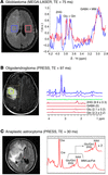
    Would be good to talk about how different Wnt isoforms activate either the noncanonical or canonical pathways in different cancer types. Different Wnts have different specificities for different tissues and activate different pathways. For reference see work by Rugang Zhang in ovarian.

  2. In addition it would be good to find literature on why, after nearly a decade, drug development strategies against the Wnt pathway, have never come to fruition, much like the early days of the farnesylation inhibitors. I believe the early inhibitors were too toxic. In addition, in relation to the OMP trial, Wnt inhibitors target the cancer stem cell and results showing a few months benefit in survival.

    good review on current status of Wnt inhibitor development
    http://link.springer.com/chapter/10.1007/978-1-4419-8023-6_9

    also this is a reference which should be linked to great article by Emma Hill
    http://www.onclive.com/publications/oncology-live/2012/december-2012/wnt-signaling-inhibition-will-decades-of-effort-be-fruitful-at-last/2

    shows all the trials and tribulations of a decade worth of effort.

Date: 28 Dec 2010
Inhibiting the Wnt Signaling Pathway with Small Molecule
Wnt signaling plays important roles in embryonic development and in maintenance of adult tissues. Mutation, loss, or overexpression of key Wnt pathway components has been linked to various types of cancer. Therefore, inhibition of Wnt signaling is of interest for the development of novel anticancer agents. The results of recent structure-based screening, high-throughput screening (HTS), and chemical genomics studies demonstrate that small molecules, including synthetic and natural compounds, can inhibit Wnt signaling in various cancers by blocking specific protein–protein interactions or the activity of specific enzymes. In biological studies, these compounds appear promising as potential anticancer agents; however, their efficacy and toxicity have yet to be investigated. Small molecule inhibitors of Wnt signaling also have wide-ranging potential as tools for elucidating disease and basic biology. Indubitably, in the near future, these compounds will yield agents that are clinically useful against malignant diseases.

Wnt Signaling Inhibition: Will Decades of Effort Be Fruitful at Last?

Emma Hitt, PhD

Oncology Live  Published Online: Monday, January 7, 2013

The Wnt signaling pathway was first characterized in the 1970s in Drosophila melanogaster development. It was later recognized in mammalian systems for its importance in cancer. Specifically, core components of this pathway were shown to be dysregulated in colorectal disease. It has since been shown to play a role in other forms of cancer, where it promotes proliferation and survival. It also plays an important role in the maintenance of the pool of tumor-initiating cells, which promote the regrowth of tumors after an insult like surgery or chemotherapy. Tumor-initiating cells are thought to be the source of metastasis. For these reasons, the Wnt pathway is viewed as a strong candidate for therapeutic intervention.

Wnt Signaling in Cancer

The Wnt pathway takes on many forms that fall under the broad classifications of canonical and noncanonical. The canonical pathway deals with the regulation of β-catenin protein levels. Under normal conditions, a cytosolic scaffold known as the destruction complex binds and phosphorylates β-catenin, resulting in its ubiquitylation and degradation. The destruction complex includes the adenomatous polyposis coli (APC) protein, axin, and glycogen synthase kinase-3β. When Wnt ligand binds to the Frizzled receptor, its coreceptor LRP5/6 is recruited and phosphorylated in the intracellular domain, promoting the binding of Dishevelled protein and the sequestration of axin. This disintegrates the destruction complex, resulting in accumulation of β-catenin in the cytosol and upregulated trafficking into the nucleus. β-catenin promotes transcription of genes related to proliferation and survival by acting as a coactivator for the Tcf/Lef family of transcription factors in the nucleus.

Aside from canonical Wnt signaling, two major noncanonical pathways have been studied. In the first of these two pathways, Wnt ligand binding to the Frizzled receptor induces recruitment of Dishevelled protein and the Dishevelled-associated activator of morphogenesis 1 (Daam1). This complex initiates a cascade that activates the Rac and Rho GTPases to mediate cell polarity. The other most widely studied noncanonical Wnt signaling pathway is related to calcium signaling. Wnt ligand binding to the Frizzled receptor promotes the recruitment of Dishevelled in complex with a G protein. This complex promotes intracellular calcium levels to mediate other signaling pathways.

Biological systems tightly regulate the Wnt signaling pathway to prevent aberrant cell growth. It has been known for decades that dysregulation of Wnt signaling leads to cancer, where it was first recognized in familial colorectal disease with mutations in the APC gene. Since then, Wnt signaling has been found to act prominently in breast, liver, skin, and prostate cancers.

Aberrations in canonical Wnt signaling can manifest in many ways. For example, proteins involved in the destruction complex can become nonfunctional through mutations or truncations, inhibiting β-catenin downregulation. The β-catenin protein itself can be mutated to inhibit its recognition by the destruction complex. In addition, the production of Wnt ligand or receptors can be upregulated, resulting in excessive signaling. These different routes of activation complicate the use of a single therapeutic against the Wnt pathway.

Wnt Signaling Pathways

This illustration depicts the best-elucidated cancer-promoting routes of Wnt cell signaling, which draws its name from the wingless mutation in the fruitfly Drosophila melanogaster.

Wnt-Signaling cancer-promoting routes

Wnt-Signaling cancer-promoting routes

Therapeutic Targeting of Wnt Pathway

Wnt signaling is of great interest to cancer researchers because it is linked to many different forms of the disease. Preclinical models throughout the last decade have established this pathway as an attractive drug target. However, to date, therapies meant to attenuate the Wnt pathway have remained largely theoretical and preclinical. Thankfully, compounds are now starting to enter clinical trials.

Vitamin D has been postulated as a suitable anti-Wnt therapy. The vitamin D receptor binds and sequesters β-catenin at the plasma membrane, inhibiting its nuclear translocation. Mice bearing an APC mutation that promotes spontaneous colon cancer developed more disease when the vitamin D receptor was knocked out. Additionally, the association between sunlight exposure and decreased risk of colon cancer implies that the inhibition of Wnt signaling by vitamin D may be conserved in humans. This phenomenon has yet to be tested in a systematic trial, however.

High-throughput screens have been used extensively to identify small-molecule targeted inhibitors of different Wnt pathway constituents. The binding of β-catenin to its nuclear targets T-cell factor (Tcf) or Creb-binding protein (CBP) is a prototypical example of study in this realm. Screens were performed to identify specific inhibitors of this interaction. This work has identified useful hits in cell-based assays, but translation into the clinic has remained difficult due to the potential off-target effects. First, the drugs identified so far fail to discriminate between the binding of β-catenin to Tcf or APC, so the drug may prevent degradation of β-catenin in addition to its intended effect on transcription. Disruption of the destruction complex binding to β-catenin could lead to severe side effects in normal tissue. Another deleterious effect identified with blockers of β-catenin binding is the inhibition of complex formation with E-cadherin at cell-cell junctions. The net effect of this phenomenon could be to impair cell adhesion on a grand scale. As a result of these challenges, only one compound that directly disrupts β-catenin function has moved into clinical trials.

Other drug candidates inhibit Wnt aberrations upstream of β-catenin function. Several of these compounds have recently begun clinical trials. They block either Wnt ligand secretion or recognition, and preclinical evidence has been encouraging so far. Targeting at this level can also lead to side effects, however. By blocking Wnt signaling at the level of the membrane, it is possible to inhibit noncanonical in addition to canonical signaling. The effect this will have on the body is unknown. In addition, blocking Wnt ligand-receptor interactions may not be sufficient to inhibit Wnt signaling since the activating event may be mutation within the destruction complex or β-catenin itself. In these settings, Wnt signaling can be rendered constitutive and independent of ligand, and the therapy would likely fail.

Moving Into the Clinic

No approved compounds exist for the treatment of Wnt signaling. Phase I trials for these inhibitors should be illustrative in the coming years. While many cancers are addicted to this signaling for long-term growth and renewal, high-turnover tissues like the gastric epithelium and hair follicles are similarly reliant. Therefore, the field is cautious when utilizing any blockade of Wnt signaling, as significant toxicity may result.

If Wnt pathway inhibitors can be proven safe, it may represent a milestone in cancer research given the strong preclinical evidence for cancer cell cytotoxicity. Since this pathway is also crucial in the maintenance of tumor-initiating cells, inhibition could represent a powerful tool in our arsenal to target cells that are resistant to traditional chemotherapy and promote metastasis.

Wnt-Targeting Compounds in Development

Phase I/II Trials
OMP-18R5
(OncoMed Pharmaceuticals/ Bayer)
This monoclonal antibody targets the Frizzled receptors to block association with Wnt ligands. It was recently shown to potently block the capabilities of pancreatic tumor-initiating cells in a serial dilution assay. In xenograft models of breast, lung, pancreatic, and colon cancer, OMP-18R5 demonstrated significant inhibition of tumor growth, and it synergized with standard-of-care treatment in these models (paclitaxel in breast cancer, for example). (NCT01345201)
OMP-54F28
(OncoMed Pharmaceuticals/ Bayer)
This agent is a fusion protein of the Frizzled8 ligand-binding domain with the Fc region of a human immunoglobulin. It binds and sequesters soluble Wnt ligand, impairing its recognition by receptors on tissues. (NCT01608867)
PRI-724
(Prism Pharma Co, Ltd/Eisai)
This is a small-molecule inhibitor of the interaction between β-catenin and CBP. Disrupting the interaction prevents activated transcription by aberrant Wnt signaling at many levels. It is being studied in both solid tumors and myeloid malignancies. (NCT01606579, NCT01302405)
LGK974
(Novartis Pharmaceuticals)
This small molecule inhibits acyltransferase porcupine. Preclinical work demonstrated this enzyme’s action is crucial in the secretion of Wnt ligands out of the cell; therefore, inhibiting porcupine can be a small-molecule–based method for inhibiting Wnt ligand-mediated activation. (NCT01351103)
Preclinical Studies
XAV939
(Novartis Pharmaceuticals)
This small-molecule poly(ADP ribose) polymerase (PARP) inhibitor has demonstrated efficacy in cellular models of cancer survival. In Wnt signaling, PARPs like the tankyrases promote the ribosylation and subsequent degradation of axin, a key scaffolding protein of the destruction complex. By inhibiting tankyrase, the axin protein is stabilized and can promote the degradation of β-catenin.
JW55
(Tocris Bioscience)
This selective tankyrase1/2 inhibitor has been shown to inhibit the growth of colon cancer cells in both cell and animal models.

Sources: ClinicalTrials.gov website, company websites.

Key Research

  • Chen B, Dodge ME, Tang W, et al. Small molecule-mediated disruption of Wnt-dependent signaling in tissue regeneration and cancer [published online ahead of print January 4, 2009]. Nat Chem Biol. 2009;5(2):100–107. doi: 10.1038/nchembio.137.
  • Clevers H, Nusse R. Wnt/β-catenin signaling and disease. Cell. 2012;149(6):1192-1205.
  • Eckhardt SG. Targeting the WNT Pathway for Cancer Therapy. Presented at: 10th International Congress on Targeted Therapies in Cancer; August 17-18, 2012; Washington, DC.
  • He B, Reguart N, You L, et al. Blockade of Wnt-1 signaling induces apoptosis in human colorectal cancer cells containing downstream mutations. Oncogene. 2005;24(18):3054-3058.
  • Ichii S, Horii A, Nakatsuru S, et al. Inactivation of both APC alleles in an early stage of colon adenomas in a patient with familial adenomatous polyposis (FAP). Hum Mol Genet. 1992;1(6):387-390.
  • Korinek V, Barker N, Morin PJ, et al. Constitutive transcriptional activation by a beta-catenin-Tcf complex in APC-/- colon carcinoma. Science. 1997;275(5307):1784-1787.
  • Rubinfeld B, Souza B, Albert I, et al. Association of the APC gene product with beta-catenin. Science. 1993;262(5140):1731-1734.

Read Full Post »

Wnt/β-catenin Signaling

Writer and Curator: Larry H. Bernstein, MD, FCAP 


7.10 Wnt/β-catenin signaling

7.10.1 Wnt signaling and hepatocarcinogenesis. The hepatoblastoma model

7.10.2 The Wnt.β-catenin pathway in ovarian cancer : a review.

7.10.3 Wnt Signaling in the Niche Enforces Hematopoietic Stem Cell Quiescence and Is Necessary to Preserve Self-Renewal In Vivo

7.10.4 Wnt.β-Catenin Signaling in Development and Disease

7.10.5 Wnt.β-Catenin Signaling. Components, Mechanisms, and Diseases

7.10.6 Wnt.β-Catenin Signaling. Turning the Switch

7.10.7 Wnt–β-catenin signaling

7.10.8 Extracellular modulators of Wnt signaling

7.10.9 FOXO3a modulates WNT.β-catenin signaling and suppresses epithelial-to-mesenchymal transition in prostate cancer cells

7.10.1 Wnt signalinbg pathway in liver cancer

7.10.1.1 Wnt signaling and hepatocarcinogenesis. The hepatoblastoma model

Armengol C1Cairo SFabre MBuendia MA.
Int J Biochem Cell Biol. 2011 Feb; 43(2):265-70.
http://dx.doi.org:/10.1016/j.biocel.2009.07.012

The Wnt/β-catenin pathway plays a key role in liver development, regeneration and tumorigenesis. Among human cancers tightly linked to abnormal Wnt/β-catenin signaling, hepatoblastoma (HB) presents with the highest rate (50-90%) of β-catenin mutations. HB is the most common malignant tumor of the liver in childhood. This embryonic tumor differs from hepatocellular carcinoma by the absence of viral etiology and underlying liver disease, and by distinctive morphological patterns evoking hepatoblasts, the bipotent precursors of hepatocytes and cholangiocytes. Recent studies of the molecular pathogenesis of hepatoblastoma have led to identify two major tumor subclasses resembling early and late phases of prenatal liver development and presenting distinctive chromosomal alterations. It has been shown that the molecular signature of Wnt/β-catenin signaling in hepatoblastoma is mainly imposed by liver context, but differs according to developmental stage. Finally, the differentiation stage of tumor cells strongly influences their invasive and metastatic properties, therefore affecting clinical behavior.

7.10.1.2 Targeting the Wnt/β-Catenin Signaling Pathway in Liver Cancer Stem Cells and Hepatocellular Carcinoma Cell Lines with FH535

Roberto Gedaly ,Roberto Galuppo, Michael F. Daily, Malay Shah, Erin Maynard, et al.
PLoS ONE 2014; 9(6): e99272.     http://dx.doi.org:/10.1371/journal.pone.0099272

Activation of the Wnt/β-catenin pathway has been observed in at least 1/3 of hepatocellular carcinomas (HCC), and a significant number of these have mutations in the β-catenin gene. Therefore, effective inhibition of this pathway could provide a novel method to treat HCC. The purposed of this study was to determine whether FH535, which was previously shown to block the β-catenin pathway, could inhibit β-catenin activation of target genes and inhibit proliferation of Liver Cancer Stem Cells (LCSC) and HCC cell lines. Using β-catenin responsive reporter genes, our data indicates that FH535 can inhibit target gene activation by endogenous and exogenously expressed β-catenin, including the constitutively active form of β-catenin that contains a Serine37Alanine mutation. Our data also indicate that proliferation of LCSC and HCC lines is inhibited by FH535 in a dose-dependent manner, and that this correlates with a decrease in the percentage of cells in S phase. Finally, we also show that expression of two well-characterized targets of β-catenin, Cyclin D1 and Survivin, is reduced by FH535. Taken together, this data indicates that FH535 has potential therapeutic value in treatment of liver cancer. Importantly, these results suggest that this therapy may be effective at several levels by targeting both HCC and LCSC.

Hepatocellular carcinoma (HCC), the most common liver cancer, is the fifth most common cancer and the third highest cause of cancer-related mortality worldwide [1][2]. The alarming rise in HCC incidence in Europe and North America in recent years is related mainly to hepatitis C virus infection, although other factors such as excessive alcohol consumption and obesity also contribute to this increase [3]. The etiology of HCC is complex and involves numerous genetic and epigenetic alterations and the disruption of various signaling pathways including the Wnt/β-catenin, Ras/Raf/MAPK, PI3K/AKT/mTOR, HGF/c-MET, IGF, VEGF and PDGF pathways. Among these, the Wnt/β-catenin pathway is considered among the most difficult to inhibit [4]. Currently, few chemical agents targeting the Wnt/β-catenin pathway are available or under investigation [5].

Activation of the canonical Wnt/β-catenin pathway involves the binding of Wnt proteins to cell surface Frizzled receptors and LRP5/6 co-receptors. In the absence of Wnt proteins, much of the cellular β-catenin is bound to E-cadherin on the cell membrane. Cytosolic β-catenin is constitutively phosphorylated at specific serine residues by an enzymatic complex that includes adenomatous polyposis coli (APC), Axin, and the kinases glycogen synthase kinase-3β (GSK-3β) and casein kinase I, marking it for ubiquitin-mediated proteolysis. Under these conditions, the TCF/LEF transcription factors are bound to their cognate DNA recognition elements along with members of the Groucho family of co-repressors, insuring the transcriptional silencing of β-catenin target genes. Engagement of Wnt proteins with the Frizzled receptor activates the Dishevelled protein, resulting in the dissociation of the cytosolic destructive complex and inhibition of GSK-3β. This leads to the stabilization and accumulation of cytoplasmic β-catenin, which then enters the nucleus, binds TCF/LEF proteins and leads to the subsequent dissociation of groucho co-repressors, recruitment of the coactivator p300 and activation of β-catenin target genes [6][9]. Many of the β-catenin targets, including Cyclin D1, c-myc and Survivin, promote cell cycle progression and inhibit apoptosis [10][12]. Consistent with this data, activation of the Wnt/β-catenin pathway is seen in a variety of cancers, including HCC. Aberrant activation of the Wnt/β-catenin pathway has been observed in at least 1/3 of HCC, and roughly 20% of HCCs have mutations in the β-catenin gene. More than 50% of HCC tumors display nuclear accumulation of β-catenin indicating that other factors may be involved such as aberrant methylation of the tumor suppressors APC and E-cadherin, inactivation of casein kinase and GSK-3β, or increased secretion of Wnt ligants [4][5].

There has been increasing interest in the role of liver cancer stem cells (LCSC) in tumorigenesis, tumor progression, invasion and metastases. The cancer stem cell theory suggests that a tumor is comprised of a heterogeneous population of cells that form a distinct cellular hierarchy. Recent studies have provided convincing evidence that these cells do exist in solid tumors of many types including, brain, breast, colorectal, liver, pancreas and prostate cancers. In 2006, two different groups isolated a CD133+ subpopulation from HCC cell lines and described higher proliferative and tumorigenic potential, consistent with stem cell properties. CD44 was also found as an important marker used in combination with other stem cell markers to better define the surface phenotype of LCSC. It has been demonstrated that CD133+ and CD90+ cells co-expressing CD44+ are more aggressive than those expressing CD133 or CD90 alone [13][14].

The chemical agents used to target Wnt-/β-catenin pathway are at the membrane, cytosol and transcription factor levels [5]. The small molecular agent FH535 is a dual inhibitor of peroxisome proliferator-activated receptor (PPAR) and β-catenin/TCF/LEF. FH535 has been shown to inhibit proliferation of HCC and hepatoblastoma cell lines and its specificity on inhibition of β-catenin/TCF/LEF activity was illustrated in hepatoblastoma cell line HepG2 [15].

The aim of this study was to determine if FH535 can inhibit the activation of β-catenin-regulated genes by endogenous and ectopically expressed β-catenin in the HCC cell lines Huh7, Hep3B and PLC and liver cancer stem cells (LCSC). The specificity of FH535 on inhibition of β-catenin via TCF/LEF activation was assayed in dual luciferase reporter transfected in LCSC and in HCC cells. Proliferation, cell cycle, and other targeted genes and proteins were assayed.

FH535 inhibits transcriptional activation mediated by wild-type and constitutively active β-catenin

FH535 has been shown to block signaling through endogenous β-catenin in several cell lines, including the hepatoblastoma cell line HepG2 [15]. To further explore this regulation and to test whether FH535 could block ectopic β-catenin, co-transfections with β-catenin expression vectors and the TCF4-dependent luciferase reporter vector TOPFlash were performed in the human HCC cell lines Huh7 and Hep3B (Fig. 1). In both cell lines, co-transfected wild-type β-catenin expression vector increased luciferase activity from TOPFlash nearly 15-fold compared to cells co-transfected with the empty vector (E.V.) control. This β-catenin-dependent increase was inhibited by FH535 in a dose-dependent manner. β-catenin is often mutated in various cancers, including HCC. One natural mutation changes the serine at position 37; this altered form of β-catenin is resistant to degradation by the APC complex and thus has higher stability. To test whether this form of activated form of β-catenin could also be blocked by FH535, an expression vector for βCatS37A, in which the serine at position 37 has been changed to an alanine, was co-transfected with TOPFlash. As expected, βCatS37A-mediated transactivation of TOPFlash was significantly higher than transactivation by wild-type β-catenin. However, in both cell lines, βCatS37A-mediated transactivation was significantly inhibited by FH535. As controls, cells were also co-transfected with FOPFlash, which is identical to TOPFlash except that the TCF4 sites have been mutated and therefore no longer responsive to β-catenin; FOPFlash was not activated by wild-type β-catenin or βCatS37A as shown in Figure 1.

thumbnail

Figure 1. FH535 inhibits β-catenin dependent transcriptional activation in HCC cell lines.

Huh7 (Panel A) and Hep3B (Panel B) HCC cells were transfected with the luciferase reporter genes TOPFlash (left panels), which contains three TCF binding sites, or E3-pGL3 (right panels), which contains the AFP enhancer element E3 that has a highly conserved TCF site. Cells were additionally co-transfected with an expression vector that contained no insert (empty vector control, E.V.), wild-type β-catenin (β-catenin), or a constitutively active form of β-catenin (βcatS37A). Renilla luciferase was used to control for variations in transfection efficiency. Six hours after the addition of DNA, cells were treated with DMSO alone (no treatment) or increasing amounts of FH535. After 48 hours, luciferase levels were determined; firefly luciferase was normalized to renilla. In both cell lines, FH535 inhibited β-catenin-dependent activation of target genes. *P<0.05. The experiment was done twice with similar results.

doi:10.1371/journal.pone.0099272.g001

TOPFlash contains three consensus TCF4 binding motifs that confer responsiveness to β-catenin. To test whether FH535 could also block β-catenin-mediated transactivation of a TCF4 motif in the context of a natural regulatory region, co-transfections were performed with E3-pGL3. E3 is a ~340 bp fragment that contains alpha-fetoprotein (AFP) enhancer element E3, one of three enhancers that control hepatic expression of the mouse AFP gene. E3 contains binding sites for multiple factors, including Foxa/HNF6, C/EBP, orphan nuclear receptors, and TCF4 [26][27]. We recently showed that this enhancer is regulated by β-catenin in cells and transgenic mice [21]. E3-pGL3 was transactivated by β-catenin and to a greater extent by βCatS37A (Fig. 1). However, this transactivation by both wild-type and S37A forms of β-catenin was blocked by FH535 in a dose-dependent manner.

3.2 FH535 inhibits β-catenin-mediated transcriptional activation in LCSC

Previous studies have shown that β-catenin signaling is elevated in EpCAM positive cells with LCSC properties [28]. We previously described that CD133+, CD44+, CD24+ LCSC aggressively form tumors when small numbers of these cells are injected into nude mice [29]. To test the ability of FH535 to inhibit β-catenin in these LCSCs, transient transfections were performed with TOPFlash. As controls, TOPFlash was also transfected into the HCC cell lines Huh7 and PLC (Fig. 2). In all three populations, untreated cells exhibited low luciferase levels. When treated with the GSK-3β inhibitor LiCl, which leads to endogenous β-catenin activation[30], TOPFlash activity increased dramatically. FH535 effectively blocked LiCl-mediated activation of TOPFlash in a dose-dependent manner. Interestingly, this inhibition was more robust in LCSC than in either HCC cell line. As a control, transfections were also performed with FOPFlash, which is no longer responsive to β-catenin. As expected, luciferase activity in FOPFlash-transfected cells was neither increased by LiCl nor inhibited by FH535.

thumbnail

Figure 2. FH535 inhibits TOPFlash activation in LCSC and HCC cell lines.

LCSC (left panel), Huh7 (middle panel) and HPLC (right panel) cells were co-transfected with TOPFlash or FOPFlash luciferase reporter genes along with renilla luciferase. After 6 hours, cells were left untreated (no treatment) or treated with LiCl alone or LiCl with increasing amounts of FH535. LiCl is a known activator of β-catenin. After an additional 36 hours, cells were harvested and luciferase levels were determined; firefly luciferase was normalized to renilla. TOPFlash activity was highly induced in all three cell populations; this activation was inhibited by FH535. The negative control FOPFlash showed minimal response to LiCl or FH535. TOPFlash inhibition by FH535 was more robust in LCSC than in either HCC cell line. * P<0.003, # P<0.001. The experiment was done twice with similar results.

doi:10.1371/journal.pone.0099272.g002

3.3 FH535 inhibits proliferation of LCSC and HCC cell lines

Numerous studies have demonstrated that β-catenin plays an important role in proliferation during normal development and in cellular transformation in many tissues, including the liver. Liver development is impaired in the absence of β-catenin, and mutations that activate the β-catenin pathway are found in about 1/3 of HCC [4][5]. Furthermore, the growth of adult liver progenitor stem cells (oval cells) can be inhibited by blocking the β-catenin pathway. Since our data indicated that FH535 can block β-catenin-mediated transcriptional activation, we also tested whether proliferation of LCSC and HCC cell lines was affected by this compound. LCSC were cultured in the presence of 10% or 1% serum and with between 5 µM and 30 µM FH535 for 72 hours, and cell proliferation was monitored by 3H-thymidine incorporation (Figs. 3A and 3B, respectively). Proliferation decreased with increasing amounts of FH535, with a more dramatic reduction observed in cells grown in the presence of lower serum; the concentration of FH535 to cause a 50% inhibition of cell grown (IC50) was 13.8 µM for cells grown in 10% serum and 5.1 µM for cells grown in 1% serum. This inhibition was more potent than that seen with XAV939 (IC50 = 55 µM), which inhibits tankyrase, thus stabilizing axin and promoting β-catenin degradation (Fig. 3C) [31]. FH535 also blocked proliferation of HCC cells at concentrations that were similar to that seen with LCSC (IC50 of 10.9 µM, 9.25 µM and 6.6 µM for Huh7, PLC and Hep3B, respectively; Fig.3D). To confirm that FH535 indeed inhibited cell proliferation and did not lead to increased cell death, FH535 and 3H-thymidine were added simultaneously to Huh7 cells, which were then cultured for 18 h. In this scenario, we observed a significant inhibition of proliferation at 2.5, 5, 10 and 15 µM of FH535 treatment as compared to control (p<0.05, n = 6), with FH535 at 15 µM causing a 41% inhibition (Figure S3). This data indicates that FH535 is inhibiting cell proliferation rather than increasing cell death.

thumbnail

Figure 3. FH535 inhibits proliferation of LCSC and HCC cell lines.

Cells were seeded in 96-well plates in 0.2 ml of media as described below for 72 hours, followed by the addition of 3H-thymidine at 1 µCi/well for 4 hours. Incorporation of 3H-thymidine was determined by scintillation counting. In panels A, B and D, the final concentration of DMSO in each well was 0.05%; in panel C, the final DMSO concentration in each well was 0.1%. (A) LCSCs were plated at 1000 cells/well in DMEM with 10% FBS along with DMSO alone or with increasing amounts of FH535. (B). LCSCs were plated at 5000 cells/well in DMEM with 1% FBS with DMSO alone or with increasing concentrations of FH535. (C). LCSCs were plated in DMEM with 10% FBS at 1000 cells/well with DMSO alone or increasing concentrations of XAV939. (D). Huh7, Hep3B and PLC cells were plated in DMEM with 10% FBS at 1000, 2500, and 5000 cells/well, respectively, with DMSO alone or increasing concentrations of FH535. Pvalues are for all the three cell lines treated with FH535 are compared to controls. The experiment was done twice with similar results.

doi:10.1371/journal.pone.0099272.g003

3.4 FH535 induces cell cycle arrest in the HCC cell line Huh7 and in LCSC

The ability of FH535 to inhibit cell proliferation prompted us to investigate the cell cycle distribution following treatment. Huh7 cells were synchronized by growth in 0.1% FBS for 24 hours and then cultured in the presence of 10% FBS and with no FH535 or FH535 at 7.5 µM and 15 µM. After 24 hours, cells were harvested and DNA content was analyzed by propidium iodide staining. In the presence of FH535, there was a statistically significant increase in the number of cells in G0/G1 and a corresponding decreased in the percentage of cells in S phase compared to cells grown in the absence of FH535 (Fig. 4A). The number of cells in G2 was not significantly altered by FH535. In addition, there was no sub-G1 peak detected by flow cytometry, indicating that FH535 was not promoting apoptosis at the concentrations being use (see Figure S4). We also did cell cycle analysis in LCSC after FH535 treatment and found FH535 at 15 µM significantly caused G1 phase arrest in LCSC (P = 0.012). FH535 also significantly reduced G2/M phase in the LCSC after 24 h of 7.5 µM and 15 µM FH535 treatment (P = 0.038 and P<0.001 respectively), no significant S phase inhibition was observed in LCSC (p = 0.446) (Fig. 4B.). Our data are similar to previously published results and reflects β-catenin regulation of cell cycle is different in different cell types [32][33]. Cell cycle regulators (cyclins, CDKs and regulators) can vary in different cell types, which could lead to different responses after FH535 treatment. This may worth exploring in our future study.

thumbnail

Figure 4. FH535 alters cell cycle progression in Huh7 and LCSC cells.

A. Huh7 cells were cultured in DMEM +10%FBS for 24 h. The cells were washed with serum free DMEM 3 times, then cultured in DMEM +0.1% FBS for 24 h for cell synchronization. Cells were then cultured in DMEM+10% FBS along with different concentrations of FH535 for 24 h. The cells were harvested and stained with propidium iodide (PI) and analyzed by flow cytometry according to the GenScript protocol (Piscataway, NJ, USA). Treatment with FH535 increased the percentage of cells in G1 and decreased the percentage of cells in S phase. The experiment was done twice with similar results. B. LCSC cells were cultured in CelProgen complete LCSC culture medium for 24 h. Cells were then washed with serum free CelProgen medium 3 times and cultured in CelProgen Medium +0.1% FBS for 24 h for synchronization of the cells. The cells were then returned to CelProgen Complete Medium +10% FBS with different concentrations of FH535 for 24 h. Cell cycle was assayed as per Huh7 described above.

doi:10.1371/journal.pone.0099272.g004

3.5 Expression of β-catenin target genes cyclin D1 and Survivin is inhibited by FH535

β-catenin controls cell proliferation and survival by regulating the expression of numerous targets genes. Two well-established targets are the genes encoding Survivin (Birc5) and Cyclin D1 (CcnD1). Survivin is an anti-apoptotic protein that also regulates progression through mitosis [34]; Cyclin D1 controls proliferation by activating the G1 kinases cdk4 and cdk6 [35]. Survivin and Cyclin D1 transcription are regulated through TCF elements in their promoter regions [36]. To test whether FH535 inhibits expression of these two β-catenin target genes, real-time RT-PCR was performed with LCSC and HCC cells that were treated with increasing amounts of FH535. Cyclin D1 and Survivin mRNA levels were reduced by FH535 in all three cell populations in a dose-dependent manner (Fig. 5). To confirm that this reduction in mRNA levels also led to lower protein levels, western analysis was performed using whole cell extracts from Huh7 cells. Both Cyclin D1 and Survivin protein levels were reduced in a dose-dependent manner, with the greatest reduction seen in the presence of 10 µM FH535 (Fig. 6.). Densitometric analysis indicated that FH535 at 5 and 10 µM inhibited Cyclin D1 28% and 64% respectively; FH535 at 5 and 10 µM inhibited surviving 24% and 48% respectively (Fig. 6).

thumbnail

Figure 5. FH535 reduces cyclin D1 and survivin mRNA levels in LCSC and in HCC cell lines.

LCSCs, Huh7 and Hep3B cells were treated with DMSO alone or increasing concentrations of FH535 for 38-time PCR for expression of Cyclin D1 (Panel A) or Survivin (Panel B). In both cases, mRNA levels were plotted relative to β2-microglobulin. The experiment was done twice with similar results.

doi:10.1371/journal.pone.0099272.g005

thumbnail

Figure 6. FH535 reduces cyclin D1 and Survivin protein levels in Huh7 cells.

Huh7 cells were treated with DMSO alone or increasing amounts of FH535 for 38-PAGE, and transferred for Western analysis with antibodies against Cyclin D1, Survivin, and β-actin. The top of shows the western blot image; the bottom graph shows densitometric analysis of the western data. This densitometric analysis indicated that FH535 at 5 and 10 µM inhibited Cyclin D1 protein levels 28% and 64% respectively; FH535 at 5 and 10 µM inhibited Survivin protein levels 24% and 48% respectively. The experiment was done twice with similar results.

doi:10.1371/journal.pone.0099272.g006

Discussion

In recent years, numerous signaling pathways have been implicated in hepatic carcinogenesis. The β-catenin pathway is essential in stem cells for self-renewal and maintenance of stem cell properties. Disruption of this balance results in both genetic and epigenetic changes, found in many cancers, including colon cancer and HCC [4]. In this study, we used FH535 as an inhibitor of the β-catenin pathway. This compound has been used previously to inhibit β-catenin expression in cells from colon and lung as well as in cells from hepatoblastoma and HCC [15]. In this report, the authors concluded that FH535 was toxic to a number of cell lines, including Huh7. However, their assays could not distinguish between toxicity and reduced cell proliferation. Our data indicates that FH535 does indeed inhibit cell proliferation; we did not directly measure toxicity.

FH535 inhibition of LCSC proliferation is of interest due to its potential therapeutic effect in chemo-resistant HCC. Our group and others have focused on strategies to inhibit the proliferation of LCSC and differences in resistance patterns with non-liver cancer stem cell lines in vitro and in vivo.

Despite numerous efforts, the etiology of HCC tumorigenesis, whether transformed cells originate from mature hepatocytes or stem/progenitor cells remains unclear. Stem cells are defined by their potential for self-renewal and by their ability to proliferate and differentiate into diverse cell types [37]. In recent years, studies have provided convincing evidence that these cells do exist in solid tumors of many types including, brain, breast, colorectal, liver, pancreas and prostate cancers [27]. In this study we have used LCSC that are 64.4%, 83.2%, 96.4% and 96.9% positive, respectively, for CD133, CD44, CD24 and Aldehyde A1 as determined by flow cytometry. These cells have been previously profiled not only by checking the LCSC markers but also by evaluating their tumorigenic potential using low cell numbers (using 2000 LCSCs instead of 100,000 HCC cells to generate tumors) and studying resistance to several drugs. We previously found that these LCSC have intermediate to high resistance to drugs compare to non- liver cancer stem cell lines using different inhibitors.

In this study, we found that FH535, LCSC inhibition of proliferation was affected by FBS concentration in the culture medium, suggesting that the PPAR pathway may be involved in LCSC proliferation as found in the human cancer cell line HCT116 [15]. This could be explained by a variety of fatty acids and their derivatives present in the FBS that are natural agonists to PPAR. It is possible the PPAR agonists suppress the inhibitory effects of FH535 in cell culture. Indeed, in HCT116 cells, FH535 inhibition of β-catein/TCF-dependent luciferase reporter genes was five times stronger in serum-free medium than in media containing 10% FBS. The ability of FH535 to inhibit tumor growth was dramatically increased when 10% FBS was replaced with 10% BSA [15]. Lysophosphatidic acid was found to be an effective PPAR agonist that could reverse FH535 induced inhibition of HCT116 growth [15]. However, the potential function of PPAR in LCSC is beyond the scope of this study and needs further investigation. Recently, FH535 was found to be the most potent drug among several other Wnt/β-catenin inhibitors on human biliary tract cancer cells cultured in serum-free medium [38]. Our study found that FH535 is much more potent than XAV939 in 10%FBS DMEM. This may be related to the PPAR inhibition potential of FH535. Our study found that FH535 inhibited HCC cell lines Huh7, Hep3B and PLC proliferation, indicating that Wnt/β-catenin signaling plays an important role not only in LCSC but also in HCC.

FH535 inhibition of LCSC and HCC proliferation was illustrated by its ability to inhibit β-catenin/TCF/LEF-dependent luciferase reporter activity. To our knowledge, this is the first report on the ability of FH535 to inhibit β-catenin/TCF/LEF activity in LCSC and in HCC cell lines. Previously, Handeli and Simon reported that FH535 inhibits β-catenin/TCF/LEF activity in the HepG2 cell line, which was mistakenly labeled as HCC by these authors [15]. For over thirty years this cell line was considered HCC by numerous investigators. Lopez et al., who initially isolated these cells, recently concluded that HepG2 cells should in fact be considered a hepatoblastoma cell line [39]. Further studies will be needed to investigate how FH535 inhibition of β-catenin influences LCSCs and HCCs. As shown here, cyclin D1 and Survivin expression are inhibited by FH535. Survivin is an anti-apoptotic protein that also regulates progression through mitosis [26], whereas Cyclin D1 controls proliferation by activating the G1 kinases [35]. Real-time RT-PCR and Western analysis confirmed that the expression of these target genes was evident at the mRNA and protein level. Our preliminary data indicate that FH535 treatment does not alter CD133, CD13 and EPCAM expression in LCSC and HCC cell lines (data not shown). Further analysis of these and other stem cell markers are warranted.

In conclusion, our data show that FH535 is a potent inhibitor of the Wnt/β-catenin pathway in LCSCs and HCC cell lines. Whether its ability to inhibit PPAR also affects the growth of LCSCs and HCC cells will require further investigation. Further studies will also be needed to investigate the in vivo efficacy and toxicity of FH535 on HCC xenografts in an animal model. The role of combination therapy using FH535 with other anti-HCC drugs and the possibility of finding cross-talk of Wnt/β-catenin pathway with other signaling pathways should be investigated.

7.10.1.3 Wnt signaling in hepatocellular carcinoma: analysis of mutation and expression of beta-catenin, T-cell factor-4 and glycogen synthase kinase 3-beta genes.

Hepatocellular carcinoma (HCC) is a common killer cancer in the world. Recently, abnormal activation of the Wnt pathway has been found to be involved in the carcinogenesis of several human cancers including HCC. The goal of the present study was to investigate the mechanism of inappropriate activation of the Wnt pathway in hepatocarcinogenesis. We analyzed the alterations of three key components of the Wnt pathway: beta-catenin, glycogen synthase kinase (GSK)-3beta and T-cell factor (Tcf)-4 in 34 HCC and paracancerous normal liver by immunohistochemistry, polymerase chain reaction (PCR)-single-strand conformation polymorphism (SSCP), direct sequencing, and quantitative real-time reverse transcription (RT)-PCR. We found that 61.8% (21/34) of all HCC examined showed an abnormal beta-catenin protein accumulation in the cytoplasm or nuclei. The RT-PCR-SSCP and direct sequencing showed that beta-catenin exon 3 mutations existed in 44.1% (15/34) of the HCC. No mutations of GSK-3beta or Tcf-4 were detected in HCC. Moreover, messenger RNA of beta-catenin and Tcf-4, but not GSK-3beta, was found to be overexpressed in HCC. On analyzing the relationship between alterations of beta-catenin or Tcf-4 and C-myc or Cyclin D1 expression, we found that mutations of beta-catenin, as well as overexpression of beta-catenin or the Tcf-4 gene were independently correlated with C-myc gene overexpression in HCC. Our present findings strongly suggest that mutations of beta-catenin, as well as overexpression of beta-catenin and the Tcf-4 gene, independently activate the Wnt pathway in HCC, with the target gene most likely to be C-myc.
7.10.1.4 Wnt signaling and cancer
Genes & Dev. 2000. 14:1837-1851
http://dx.doi.org:/10.1101/gad.14.15.1837

The regulation of cell growth and survival can be subverted by a variety of genetic defects that alter transcriptional programs normally responsible for controlling cell number. High throughput analysis of these gene expression patterns should ultimately lead to the identification of minimal expression profiles that will serve as common denominators in assigning a cancer to a given category. In the course of defining the common denominators, though, we should not be too surprised to find that cancers within a single category may nevertheless exhibit seemingly disparate genetic defects. The wnt pathway has already provided an outstanding example of this. We now know of three regulatory genes in this pathway that are mutated in primary human cancers and several others that promote experimental cancers in rodents (Fig. 1). In all of these cases the common denominator is the activation of gene transcription by β-catenin. The resulting gene expression profile should provide us with a signature common to those cancers carrying defects in the wnt pathway. In this review, the wnt pathway will be covered from the perspective of cancer, with emphasis placed on molecular defects known to promote neoplastic transformation in humans and in animal models.

Figure 1.

Oncogenes and tumor suppressors in the wnt signaling pathway. Lines ending with arrows or bars indicate activating or inhibitory effects, respectively. Green and red indicate proto-oncogenic and tumor suppressive activity, respectively, in human cancer or transgenic animals. Definition of the genes and the basis for their activities are described in the text.

The wnt signaling mechanism

The model illustrated in Figure 2 is a proposed mechanism for wnt signaling and is based on the following literature. Signaling is initiated by the secreted wnt proteins, which bind to a class of seven-pass transmembrane receptors encoded by the frizzled genes (Bhanot et al. 1996; Yang-Snyder et al. 1996; He et al. 1997). Activation of the receptor leads to the phosphorylation of the dishevelled protein which, through its association with axin, prevents glycogen synthase kinase 3β (GSK3β) from phosphorylating critical substrates (Itoh et al. 1998; Kishida et al. 1999; Lee et al. 1999; Peters et al. 1999; Smalley et al. 1999). In vertebrates, the inactivation of GSK3β might result from its interaction with Frat-1 (Thomas et al. 1999; Yost et al. 1998; Li et al. 1999a; Salic et al. 2000). The GSK3β substrates include the negative regulators axin and APC, as well as β-catenin itself (Rubinfeld et al. 1996; Yost et al. 1996; Yamamoto et al. 1999). Unphosphorylated β-catenin escapes recognition by β-TRCP, a component of an E3 ubiquitin ligase, and translocates to the nucleus where it engages transcription factors such as TCF and LEF (Behrens et al. 1996; Molenaar et al. 1996;Hart et al. 1999). Additional components in the pathway include casein kinases I and II, both of which have been proposed to phosphorylate dishevelled (Sakanaka et al. 1999; Willert et al. 1997; Peters et al. 1999). The serine/threonine phosphatase PP2A associates with axin and APC, although its functional role in the pathway remains obscure (Hsu et al. 1999; Seeling et al. 1999). Also obscure is the manner by which the wnt receptors communicate with dishevelled.

Figure 2.

Proposed mechanism for the transmission of wnt signals. In the absence of wnt –wnt) GSK3β phosphorylates APC and axin, increasing their binding affinities for β-catenin, which too is phosphorylated by GSK3β, marking it for destruction. In the presence of wnt (+wnt) FRAT prevents GSK3β from phosphorylating its substrates, and β-catenin is stabilized. Casein kinase1ε (CK1ε) binds to and phosphorylates dishevelled (dvl) modulating the FRAT1/GSK3β interaction. RGS, PDZ, and DIX are protein interaction domains.

 Receptors, ligands, and related proteins

The proto-oncogenic effects of wnt were discovered over 18 years ago inciting intense investigation into the role of wnt genes in human cancer (Nusse and Varmus 1982). The subsequent discovery of wingless, the fly homolog of wnt-1, paved the way for assembling a signaling pathway subsequently found to contain cancer causing genes (Cabrera et al. 1987; Rijsewijk et al. 1987). Although wnt was the prototypical oncogene in this pathway, no formal proof for its involvement in human cancer has ever been documented. There have been numerous reports on the overexpression, and sometimes underexpression, of wnt genes in human cancers, but mRNA expression levels are merely correlative. More compelling evidence, such as amplification, rearrangement, or mutation of genes encoding wnt ligands or receptors has not been forthcoming. In lieu of these sorts of findings, we are left to speculate on the consequences of epigenetic events implicating these genes in human cancer. In doing so we can use animal and cell culture models to guide our interpretation.

The wnt ligands, of which there are at least 16 members in vertebrates, are secreted glycoproteins that can be loosely categorized according to their ability to promote neoplastic transformation (for review, seeWodarz and Nusse 1998). For example, the activation of wnt-1, wnt-3, or wnt-10b by retroviral insertion in the mammary gland will promote tumor formation in mice (Lee et al. 1995; Nusse and Varmus 1982; Roelink et al. 1990). Oncogenic potential can also be assessed in cultured mammalian cells, such as C57MG and CH310T1/2, where expression of the proto-oncogenic wnts results in morphological transformation (Bradbury et al. 1994; Wong et al. 1994). These cells are transformed by wnt-1, wnt-2, wnt3a but not by wnt-4, wnt-5a, and wnt-6. The transforming wnt genes also promote the accumulation of β-catenin in some cultured mammalian cells (Shimizu et al. 1997). Some aspects of the wnt cancer pathway are also recapitulated inXenopusdevelopment, where injection of transforming wnts into early embryos results in duplication of the dorsal axis (Wodarz and Nusse 1998). A caveat here is that the lack of specific receptors for certain wnts might also explain their inactivity in some of these assays (He et al. 1997). Nevertheless, identifying those wnts capable of neoplastic transformation will aid the interpretation of epigenetic evidence implicating wnts in cancer. For example, expression of thewnt-16 gene is activated by the E2A–Pbx1 fusion product in acute lymphoblastoid leukemia (McWhirter et al. 1999), but the oncogenic potential of wnt-16 is unknown.

As might be expected from the plethora of wnt genes, there are also numerous wnt receptors. At least 11 vertebrate frizzled genes have been identified, but how they differ in function and ligand specificity is far from clear. The analysis of mere binding specificity may not be sufficient to sort out the appropriate combinations of functional receptor-ligand interactions. Wnt-3a and wnt-5a both bind to Human frizzled 1 (Hfz1), yet only wnt-3a mediates TCF-dependent transcription (Gazit et al. 1999). This suggests that the activation of TCF/LEF-dependent transcription is a good correlate to neoplastic transformation. Implementation of this assay, along with a second assay involving the translocation of PKC to the cell membrane, resulted in the categorization of murine wnt receptors into two exclusive groups (Sheldahl et al. 1999). Human FzE3 fell into the TCF/LEF activation group, consistent with previous work showing that its overexpression resulted in nuclear localization of β-catenin (Tanaka et al. 1998). This receptor was also expressed in numerous human esophageal cancers, but not in matched normal tissue (Tanaka et al. 1998).

In addition to the frizzled receptors, there exists a family of secreted proteins bearing homology to the extracellular cysteine-rich domain of frizzled. The so-called secreted frizzled-related proteins (sFRP) bind to the wnt ligands, thereby exerting antagonistic activity when overexpressed in wnt signaling assays (Leyns et al. 1997; Wang et al. 1997). The vertebrate sFRPs, like the frizzled proteins, exhibit functional specificity with respect to the various wnts. InXenopus assays, the prototypical frizzled related protein frzb, now known as sFRP-3, inhibited wnt-1 and wnt-8, but not wnt-5a (Leyns et al. 1997; Lin et al. 1997; Wang et al. 1997). Assays in mammalian cells showed that FrzA, now termed sFRP-1, inhibited wnt-1-induced accumulation of β-catenin (Dennis et al. 1999;Melkonyan et al. 1997). Again, binding specificity may not relate to functional specificity, as wnt-5a associated with sFRP-3 but was unable to inhibit its activity (Lin et al. 1997). Even the significance of specific functional interactions might be suspect based on recent titration experiments with purified soluble sFRP-1. At low concentrations sFRP-1 enhanced signaling activity by soluble wingless protein, whereas at higher concentrations it was inhibitory (Uren et al. 2000). The authors proposed high and low states of binding affinity that involved the carboxy-terminal heparin binding domain and the amino-terminal cysteine-rich domain of sFRP-1, respectively. Binding to the cysteine-rich domain might confer inhibition while binding to the carboxy-terminal region could facilitate presentation of active ligand to receptor. The potential for some sFRPs to activate wnt signaling is consistent with a previous study in which sFRP-2, then known as SARP-1, increased the intracellular concentration of β-catenin and conferred anti-apoptotic properties to cultured MCF-7 cells (Melkonyan et al. 1997). Functional studies are further complicated by the binding of a sFRP to the putative human receptor frizzled-6, underscoring additional possible modes of regulation (Bafico et al. 1999). The sFRPs have not been directly linked to cancer, but one could speculate that the anti-apoptotic activity observed with the SARP-1 could contribute to tumor progression. Alternatively, the identification of sFRP-2 as a target of the hedgehog signaling pathway might be relevant to human basal cell cancers (Lee et al. 2000). Additional structurally distinct secreted inhibitors of wnt signaling include the recently discovered dickopft-1 and wif-1 proteins (Fedi et al. 1999; Glinka et al. 1998;Hsieh et al. 1999).

GSK3β

The serine/threonine kinase GSK3β binds to and phosphorylates several proteins in the wnt pathway and is instrumental to the down regulation of β-catenin (Dominguez et al. 1995; He et al. 1995; Hedgepeth et al. 1999b; Ikeda et al. 1998;Itoh et al. 1998;Li et al. 1999a; Nakamura et al. 1998b; Rubinfeld et al. 1996;Yamamoto et al. 1999; Yost et al. 1996). As a negative regulator of wnt signaling, GSK3β would qualify as a potential tumor suppressor. However, mutations or deletions in the gene coding for GSK3β were not been detect ed in a survey of colorectal tumors (Sparks et al. 1998). Perhaps GSK3β can compensate for the loss of GSK3β and the biallelic inactivation of both these genes is unlikely in tumor progression. Alternatively, the utilization of GSK3β by pathways independent of wnt could make its overall ablation incompatible with cell viability. Nevertheless, inactivation of GSK3β can still be achieved by a means other than genetic ablation and can occur in a manner that uniquely affects wnt signaling. This mode of inactivation involves the association of GSK3β with Frat-1. Frat-1 was identified by insertional mutagenesis in a screen for genes that enhanced the progression of transplanted T-cell lymphomas in mice (Jonkers et al. 1997). Subsequent transgenic expression of Frat-1 alone did not induce spontaneous lymphomas, but greatly enhanced lymphomagenesis initiated either by leukemia virus M-MuLV or expression of the Pim1 oncogene (Jonkers et al. 1999). A connection to GSK3β was realized by the discovery of the Frat-1 Xenopushomolog GBP, a GSK3β binding protein inhibitory to wnt signaling when expressed in Xenopus embryos (Yost et al. 1998). Frat-1 is also antagonistic to wnt signaling in mammalian cells, presumably because it competes with axin for binding to GSK3β (Li et al. 1999a; Thomas et al. 1999). GBP also inhibited the phosphorylation and degradation of β-catenin in vitro when added to Xenopusextracts (Salic et al. 2000). Although Frat-1 contributes to cancer progression in a transgenic mouse model, its contribution to human cancer has not been documented.

Dishevelled

The genetic analysis of dishevelled in developmental systems has defined it as a positive mediator of wnt signaling positioned downstream of the receptor and upstream of β-catenin (Noordermeer et al. 1994). Overexpression or constitutive activation of dishevelled would be expected to promote neoplastic transformation, but its involvement in human cancers has not been reported. This might reflect the dual function of dishevelled, one that transduces wnt signals for the stabilization of β-catenin and a second that relays signals for the activation of jun kinases (Li et al. 1999b; Moriguchi et al. 1999). Although these two functions are housed in physically separable regions of the protein, dysregulation of one function, without impacting the other, could place severe constraints on selection for potential oncogenic mutations. A possible connection of dishevelled to cancer is through casein kinase II, which binds to and phosphorylates dishevelled and also promotes the formation of lymphomas when expressed in transgenic mice (Seldin and Leder 1995; Song et al. 2000; Willert et al. 1997).

β-catenin

Mutations in the β-catenin gene (CTNNb1) affecting the amino-terminal region of the protein make it refractory to regulation by APC (Morin et al. 1997; Rubinfeld et al. 1997). These mutations affect specific serine and threonine residues, and amino acids adjacent to them, that are essential for the targeted degradation of β-catenin (for review, see Polakis 1999). The mutations abrogate the phosphorylation dependent interaction of β-catenin with β-TRCP, a component of an E3 ubiquitin ligase that makes direct contact with amino terminal sequence in β-catenin (Hart et al. 1999). This regulatory sequence in β-catenin is mutated in a wide variety of human cancers as well as in chemically and genetically induced animal tumors. Importantly, β-catenin mutations in tumors are exclusive to those that inactivate APC. This is particularly apparent in colorectal cancer where the vast majority of these tumors contain APC mutations and the overall frequency of β-catenin mutations is quite low (Samowitz et al. 1999; Sparks et al. 1998;Kitaeva et al. 1997) (Table 1). When colorectal tumors lacking APC mutations were analyzed separately, the likelihood of finding a CTNNb1 mutation was greatly increased (Iwao et al. 1998; Sparks et al. 1998). The exclusivity of CTNNb1 and APC mutations in colorectal cancer was also evident from the analysis of replication error-positive tumors identified by microsatellite instability. Both the hereditary and sporadic forms of replication error-positive colorectal cancers had a relatively high frequency of β-catenin mutations, whereas APC mutations were relatively rare (Mirabelli-Primdahl et al. 1999; Miyaki et al. 1999) (Table 1). Interestingly, this correlation between microsatellite instability andCTNNb1 mutations was not apparent in endometrial cancers (Mirabelli-Primdahl et al. 1999).

Table 1. 
Beta-catenin mutations in human cancers
Aggressive fibromatosis, otherwise known as desmoid tumor, is a locally invasive fibrocytic growth that occurs with increased incidence in patients with familial adenomatous polyposis coli (FAP). FAP individuals carry APC mutations in their germline and present with multiple intestinal adenomas at an early age. Desmoids also occur sporadically and, with the exception of colorectal cancer, represent a rare example of biallelic inactivation of APC in individuals without a pre-existing germline mutation in APC (Alman et al. 1997). Not surprisingly, mutations inCTNNb1 have also been detected in sporadic desmoid tumors (Shitoh et al. 1999;Tejpar et al. 1999). The β-catenin mutations were found in over half of the 42 desmoids analyzed, while inactivating mutations in APC were detected in nine and, again, there was no overlap between APC and β-catenin mutations (Tejpar et al. 1999). The β-catenin mutations were all of the missense variety and were confined to codons 41 and 45. Some of the desmoids lacked mutations in either β-catenin or APC, but all displayed increased expression of β-catenin, implying that yet unidentified defects in β-catenin regulation exist in some of these tumors.

There appears to be a low probability of accruing biallelic inactivating mutations in APC in most sporadic cancers, despite increased cancer incidence at numerous extracolonic sites in FAP patients. This suggests that the stabilization of β-catenin can promote cancer in many tissue types, but the biallelic inactivation of APC is an unlikely means to this end. Components in the wnt pathway other than APC, such as β-catenin, might make easier targets for oncogenic mutations. Indeed, several mutations in CTNNb1 were recently identified in gastric cancers, which occur with increased incidence in FAP patients (Park et al. 1999). In this study, 27% of intestinal type gastric cancers harbored mutations in β-catenin. Hepatoblastoma also occurs with increased incidence in FAP individuals (Hughes and Michels 1992;Giardiello et al. 1996; Cetta et al. 1997), but biallelic inactivation of APC is uncommon in the sporadic forms of these tumors. In three separate studies, mutations in β-catenin were identified at high frequency in hepatoblastoma, while no APC mutations were found (Koch et al. 1999; Jeng et al. 2000; Wei et al. 2000). Hepatoblastoma is also associated with Beckwidth–Wiedemann syndrome (BWS), however, a direct link between wnt signaling and the genetic defects underlying BWS are unlikely as a tumor from one of these patients also contained a somatic mutation in β-catenin (Wei et al. 2000). By contrast, a subset of patients with Turcot’s syndrome harbor germline mutations in APC and are at increased risk of medulloblastoma (Hamilton et al. 1995; Lasser et al. 1994). Although inactivating mutations in APC have not been detected in the sporadic forms of medulloblastoma, CTNNb1mutations were found in a small percentage (Zurawel et al. 1998).

Hepatocellular carcinoma (HCC) has become one of the most common tumors harboring mutations in the wnt pathway. Based on five separate studies, the frequency of CTNNb1 mutations in hepatocellular carcinoma (HCC) was ∼20% overall and perhaps higher still for HCCs associated with hepatitis C virus (de La Coste et al. 1998; Miyoshi et al. 1998;Huang et al. 1999; Legoix et al. 1999; Van Nhieu et al. 1999) (Table1). Preliminary data indicated a poorer prognosis associated with nuclear accumulation of β-catenin in HCC and histological data indicated enhanced nuclear staining in the invasive and intravascular compartments of the tumors (Huang et al. 1999; Van Nhieu et al. 1999). In one of these studies an inverse correlation between β-catenin mutations and loss of heterozygosity in the genome was noted (Legoix et al. 1999). This suggests that chromosomal instability and mutations inCTNNb1 represent alternative modes of tumor progression in HCC.

It is noteworthy that c-myc and cyclin D genes are amplified in a subset of HCCs and both these genes are downstream targets of β-catenin (He et al. 1998; Nishida et al. 1994; Peng et al. 1993;Shtutman et al. 1999; Tetsu and McCormick 1999). It would be of interest to determine whether any overlap exists between their amplification and CTNNb1mutations in HCC. Animal models of HCC have provided some clues toward understanding the relationship between these genes in cancer. HCCs induced by transgenic expression of SV40 T antigen in murine liver did not contain mutations in CTNNb1 (Umeda 2000). As T antigen activates cyclin D kinase by sequestration of Rb, the activation of the cyclin D gene by mutant β-catenin may no longer be required. By contrast, activating mutations inCTNNb1 were identified in half of the HCCs generated by transgenic expression of c-myc in murine liver (de La Coste et al. 1998). This animal model suggests that β-catenin mutations occur as a second “hit” in HCC tumor progression in cooperation with a distinct cancer pathway initiated by c-myc. That CTNNb1mutations can occur subsequent to other oncogenic defects is also evident from their occurrence in Wilm’s tumor. Mutations in β-catenin were detected in 15% of these pediatric kidney cancers and in two of these cases they were concomitant with mutations in the Wilm’s tumor gene WT1 (Koesters et al. 1999). One of these cases was associated with Denys-Drash syndrome, a familial disorder attributable to germline mutations in WT1.

It makes sense that extracolonic tumors associated with FAP, such as desmoids, medulloblastoma, and HCC, would contain CTNNb1mutations in their sporadic forms. Thyroid cancers also occur with increased incidence in FAP and, not surprisingly, a high frequency ofCTNNb1 mutations was recently reported for anaplastic thyroid cancers (Cetta et al. 2000; Garcia-Rostan et al. 1999). Although many of these mutations affected amino acids known to influence the regulation of β-catenin, many of them affected residues for which the consequence of their mutation is unknown (Garcia-Rostan et al. 1999). In particular, the substitution K49R was detected nine times. This mutation was frequently detected in the context of independentCTNNb1 mutations in the same thyroid tumor, and up to four independent CTNNb1 mutations were found in some tumors. The occurrence of multiple independent CTNNb1 mutations was also noted in some HCCs and might reflect the multifocal origin of some cancers (Huang et al. 1999; Legoix et al. 1999; Van Nhieu et al. 1999). In one HCC study, examination of different tumor areas from the same patient revealed distinct CTTNb1 mutations in two independent cases (Huang et al. 1999).

Some cancers, such as endometrial ovarian tumors, do not occur with increased incidence in patients with FAP, yet they contain activating mutations in CTNNb1(Palacios and Gamallo 1998; Gamallo et al. 1999; Wright et al. 1999). Perhaps inactivation of the remaining wild-type APC allele in FAP individuals is unlikely in this tissue, or the expression of an alternative APC gene compensates for its loss. The CTNNb1 mutations associated with ovarian cancer appeared to be confined to the endometrioid subtype. In this tissue, cancers with activated β-catenin signaling were reported to be less aggressive than their nonactivated counterparts. In one report, a more favorable prognosis was associated with cancers exhibiting enhanced nuclear staining of β-catenin and another indicated higher frequency ofCTNNb1 mutations in lower grade tumors (Palacios and Gamallo 1998; Wright et al. 1999). A similar inverse correlation between tumor grade and occurrence ofCTNNb1 mutations was also reported for uterine endometrial cancers (Fukuchi et al. 1998). The overlap between mutations in CTNNb1 and other gene defects in ovarian cancers has not been explored in detail, although one study noted coexisting mutations in the PTEN tumor suppressor andCTNNb1 in endometrioid tumors (Wright et al. 1999).

Additional types of cancers with CTNNb1 mutations, albeit at low frequency, include melanoma and prostate. Although only one of sixty-five melanomas contained detectable mutations, nuclear localization of the protein was seen in one-third (Rimm et al. 1999). Thus, additional mechanisms for β-catenin activation likely occur in these tumors. Possibly the highest percentage ofCTNNb1mutations occurs in a common skin tumor known as pilomatricomas (Chan et al. 1999). That these tumors might contain CTNNb1 mutations was surmised from the genesis of similar tumors in transgenic mice expressing mutant β-catenin in the skin (Gat et al. 1998). The tumors appeared to originate from the hair follicle, which is consistent with the lack of hair in mice homozygous for mutations in LEF, a transcription factor responsive to β-catenin (van Genderen et al. 1994).

Axin

Axin was originally identified as an inhibitor of wnt signaling inXenopus embryos and was subsequently shown to bind directly to APC, β-catenin, GSK3β and dishevelled (for review, see Peifer and Polakis 2000). A plethora of in vitro and in vivo studies inXenopus, Drosophila, and cultured mammalian cells has demonstrated that axin is central to the down regulation of β-catenin (Zeng et al. 1997; Behrens et al. 1998; Hart et al. 1998;Ikeda et al. 1998; Nakamura et al. 1998a; Sakanaka et al. 1998; Fagotto et al. 1999; Hedgepeth et al. 1999a; Li et al. 1999a; Willert et al. 1999a; Farr et al. 2000). It is not entirely clear how axin functions, but it has been proposed to facilitate the phosphorylation of β-catenin and APC by GSK3β (Hart et al. 1998; Ikeda et al. 1998). Thus axin would be viewed as a tumor suppressor based on its ability to downregulate signaling, and this has now been verified by documentation of its biallelic inactivation in human hepatocellular cancers and cell lines (Satoh et al. 2000). Importantly, these mutations were identified in those HCCs that lacked activating mutations inCTNNb1. All of the mutations were predicted to truncate the axin protein in a manner that eliminated the β-catenin binding sites. Axin, which should now be regarded as a tumor suppressor, constitutes the third genetic defect in the wnt pathway that contributes to human cancer. There also exists a close homolog of axin termed conductin, which exhibits of all the binding and regulatory functions of axin (Behrens et al. 1998). That this apparent redundancy did not suppress axin mutations in HCC suggests conductin is either not functionally equivalent to axin or not expressed at levels sufficient to compensate for its loss in HCCs.

PP2A

The dependence upon serine/threonine kinases for the regulation of β-catenin implies that phosphatases are also involved. Indeed, the rapid dephosphorylation of the axin protein is a consequence of wnt signaling and has been proposed to both destabilize axin and reduce its affinity for β-catenin (Willert et al. 1999b;Yamamoto et al. 1999). Although axin binds directly to the PP2A catalytic subunit, the phosphatase affecting axin in response to wnt signaling has not been identified (Hsu et al. 1999). If PP2A is this phosphatase, it would be viewed as proto-oncogenic because it downregulates the tumor suppressor axin. On the contrary, expression of the PP2A regulatory subunit B56 in human colon cancer cells results in the downregulation of β-catenin, consistent with a tumor suppressive function in the wnt pathway (Seeling et al. 1999). Moreover, the beta isoform of the PP2A A subunit is deleted in some human colon tumors, again implying tumor suppression (Wang et al. 1998). Also, disruption of twins, aDrosophila gene coding for a PP2A subunit, complemented the overexpression and underexpression of the β-catenin homolog armadillo, in a manner consistent with negative regulation of wnt signaling (Greaves et al. 1999). By all accounts, PP2A plays a role in wnt signaling, but its potential role as proto-oncogene or tumor suppressor might be dependent upon the precise nature of the defect.

APC

Genetic analysis of FAP families led to the identification of theAPC gene, and subsequent studies demonstrating an interaction with β-catenin placed it tentatively in the wnt pathway (Groden et al. 1991; Kinzler et al. 1991; Munemitsu et al. 1995; Rubinfeld et al. 1993; Su et al. 1993). Experiments in Drosophilaultimately revealed that genetic ablation of APC indeed resulted in upregulation of β-catenin signaling (Ahmed et al. 1998). In some systems, such as Xenopus andCaenorhabditis elegans, a positive role for APC in the wnt pathway has been proposed, but the former study suffers from potential overexpression artifacts and the latter from a lack of relatedness to the vertebrate APC protein (Rocheleau et al. 1997; Vleminckx et al. 1997). In any case, APC is a tumor suppressor in human cancers and its mutation relates strongly to the regulation of β-catenin. The spectrum of APC mutations, which typically truncate the protein, suggest selection against β-catenin regulatory domains, albeit not necessarily against β-catenin binding (for review, see Polakis 1999). The selective pressure might be directed against the presence of Axin binding sites, of which there are three, dispersed across the central region of the APC protein (Behrens et al. 1998). The presence of axin binding sites are critical to APC in the regulation of β-catenin levels and signaling in cultured cells (Kawahara et al. 2000). Moreover, mice lacking wild-type APC but expressing a truncated mutant APC retaining a single axin binding site are viable and do not develop intestinal neoplasia (Smits et al. 1999). This has not been the case for mice with more extensive truncations in APC (Oshima et al. 1995a; Su et al. 1992). Also, milder forms of colorectal polyposis, as well as familial infiltrative fibromatosis (desmoid tumors), have been associated with germline mutations in the 3′ region of the APC open reading frame. These mutations predict truncated proteins that retain only one or two of the three axin binding sites in APC (Walon et al. 1997; Kartheuser et al. 1999; Scott et al. 1996;van der Luijt et al. 1996). A recent study has also demonstrated that the expression of just the central region of APC, which contains all of the axin and β-catenin binding sites, was sufficient to elicit cellular growth suppression (Shih et al. 2000). This effect is consistent with previous work showing that a like fragment of APC was sufficient to downregulate β-catenin levels in cancer cells (Munemitsu et al. 1995).

Although both copies of the APC gene are typically inactivated in colorectal cancers, it remains possible that a mutant truncated APC could contribute to cancer progression. This was tested by transgenic expression of two different APC mutants in a wild-type intestinal background (Oshima et al. 1995b). This did not result in cancer-prone mice, despite high levels of expression of mutant proteins and, therefore, argues against a dominant negative effect by these particular mutants. However, it does not rule out an additive contribution to tumor progression by mutant APC protein in a background lacking wildtype APC. In fact, genetic evidence argues in favor of selection for a somewhat specific mutant APC protein. The mutation cluster region (MCR) in APC, roughly defined by codons 1250–1500, is not only consistent with selection against specific sequence, but also retention of an APC molecule that extends into the MCR (Fig.3.). A correlation between the presence of a germline mutation in the MCR and the severity of polyposis has been noted (Ficari et al. 2000; Nagase et al. 1992; Wu et al. 1998). The enhanced severity of polyposis suggests there should also be selective pressure for somatic mutations in the MCR, which indeed appears to be the case (Miyoshi et al. 1992). Selective pressure for an MCR mutant has also been proposed based on the occurrence of somatic mutations in the MCR relative to the position of the germline mutation in FAP (Lamlum et al. 1999). Tumors from FAP patients with a germline MCR mutation exhibited frequent inactivation of the remaining APC allele by LOH, while those without a germline MCR mutation had frequent somatic mutations in the MCR (Fig. 3). Therefore, a germline mutation in the MCR could relieve the constraint for a subsequent somatic MCR mutation, thereby increasing the likelihood of polyposis. This implies that a truncated MCR APC mutant has an interfering or gain of function property that enhances tumor progression beyond simple loss of APC function. An interfering function would probably not involve interaction with wild-type APC, as recently suggested, because the MCR mutant is still selected for in the absence of a wild-type APC gene copy (Dihlmann et al. 1999). Finally, some of the germline mutations in APC do not disrupt the open reading frame yet correlate with increased risk of colorectal cancer (Frayling et al. 1998; Gryfe et al. 1999; Laken et al. 1997). These mutations have been proposed to increase the occurrence of subsequent truncating mutations by enhancing the mutational susceptibility of the affected nucleotide tract.

Figure 3.

Mutations in APC. A compilation of germline and somatic mutations in APC illustrates selection for mutations in the mutation cluster region (MCR). MCR mutations result in truncated proteins retaining β-catenin binding but not regulatory activity. Somatic MCR mutations are more frequently selected for in FAP patients with germline mutations outside of the MCR.

Transcription factors

Prior to discussing the potential role for LEF/TCF transcription factors in cancer, it is important to outline the mechanism by which they have been proposed to operate. Although LEF/TCFs bind directly to DNA through their HMG domains, they are incapable of independently activating gene transcription (Eastman and Grosschedl 1999; Roose and Clevers 1999). This has best been illustrated for LEF, which through its binding to the cofactor ALY, makes indirect contacts with a second transcription factor AML (Bruhn et al. 1997). The TCFs do not contain the ALY binding site, but like LEF they cannot activate test genes comprised of multimerized TCF/LEF binding sites and a minimal promotor sequence. However, these reporter genes are activated on coexpression of TCF with β-catenin, suggesting that β-catenin supplies additional cofactors required for transcriptional activation (Molenaar et al. 1996). This activity was localized to the carboxy-terminal region of the Drosophila β-catenin armadillo, which when fused directly to TCF resulted in β-catenin independent transcriptional activation (van de Wetering et al. 1997).

The simple interpretation is that the TCF/LEF-β-catenin complex comprises a bipartite positive acting transcription factor in the wnt pathway. This interpretation agrees well with developmental studies in which the manipulation of LEF/TCF function results in phenotypes consistent with the genetic manipulation of wnt/β-catenin signaling (Behrens et al. 1996; Brunner et al. 1997; Huber et al. 1996; van de Wetering et al. 1997). For example, a zygotic homozygous null mutation inDrosophila LEF results in a loss of naked cuticle in the larval epidermis, a phenotype typical of loss of function wingless mutations (Brunner et al. 1997). Moreover, the formation of excess naked cuticle by ectopic expression of armadillo in wild-type embryos does not occur in the LEF null mutants. Exactly how β-catenin contributes to transcriptional activation is unclear, but might involve additional proteins that bridge the TCF/β-catenin complex to the basal transcriptional machinery. The bridging function might be fulfilled by Pontin 52, a TATA-binding protein that was reported to bind to β-catenin (Bauer et al. 1998). Also, a mutant form of β-catenin incapable of binding LEF squelched LEF-dependent reporter gene activation, presumably by titration of an essential cofactor (Prieve and Waterman 1999). Finally, the carboxy-terminal region of armadillo binds to the Zinc finger protein teashirt, a homeotic gene essential for a subset of wingless signaling outputs in Drosophila (Gallet et al. 1999).

The simple model of positive transcriptional activation by the TCF-β-catenin complex is not in accord with all experiments. Mutation of the TCF/LEF binding sites in the promotors of the wingless responsive gene ultrabithorax and the Wnt-responsive Xenopus gene Siamois enhanced their activities under conditions where the wingless/β-catenin signal input was weak (Brannon et al. 1999; Riese et al. 1997). The mammalian cyclin D gene was recently identified as a wnt target and, again, mutation of the corresponding TCF binding sites enhanced its basal activity (Tetsu and McCormick 1999). These results suggest TCF represses transcription of its target genes in unstimulated cells and the binding of β-catenin promotes derepression. Derepression cannot fully account for signaling activity, however, as mutations in the TCF binding sites compromise target gene activation under conditions of active wnt signaling (Brannon et al. 1999; Riese et al. 1997). Repression of gene expression by TCF is consistent with its direct physical interaction with at least three different gene products, the Groucho/TLE and CtBP corepressors, and the CREB binding protein CBP (Brannon et al. 1999;Cavallo et al. 1998; Levanon et al. 1998; Roose et al. 1998; Waltzer and Bienz 1998).

The groucho/TLE proteins bind to the central region of TCF/LEF at a site distinct from that of β-catenin binding and inhibit gene activation of TCF target genes (Levanon et al. 1998; Roose et al. 1998). By contrast, CtBP binds to two independent sites in the carboxy-terminal region of Xtcf-3, which when mutated abrogated the repressor function of this region of Xtcf-3 (Brannon et al. 1999). The binding sites for CtBP are not present in LEF, which might explain the ability of LEF, but not Xtcf-3, to induce axis duplication in Xenopus embryos. Finally, the Drosophila CREB binding protein CBP was reported to bind to the HMG domain of dTCF (Waltzer and Bienz 1998). Loss-of-function CBP mutants displayed some features of wingless over expression and also suppressed phenotypes resulting from loss of the β-catenin homolog armadillo. The genetics imply suppression of wingless by CBP, which is somewhat paradoxical when considering the role of CBP acetyltransferase activity in chromatin remodeling and gene activation. However, it was shown that CBP acetylates a lysine proximal to the armadillo binding site in TCF, thereby reducing its affinity for armadillo. Repression of β-catenin/TCF signaling by CBP does not occur in all settings, though, as two recent studies demonstrated activation ofXenopus TCF target genes by CBP (Hecht et al. 2000;Takemaru and Moon 2000). CBP directly associated with carboxy-terminal sequence in β-catenin and overexpression of E1A, which also directly binds CBP, reduced β-catenin dependent transactivation.

Does the activation of TCF/LEF target genes by β-catenin cause cancer? Good evidence to this effect was provided by the expression of a chimeric protein consisting of the LEF DNA binding sequence fused to the transcriptional activation domain of either VP16 or the estrogen receptor (Aoki et al. 1999). Expression of these constructs in chicken embryo fibroblasts resulted in their neoplastic transformation. The proliferative potential of TCF was also apparent from the phenotype resulting from homozygous disruption of TCF-4 in the germline of mice. These animals were incapable of maintaining a proliferative stem cell compartment in the small intestine and died shortly after birth (Korinek et al. 1998). Whether the TCF/LEF genes are directly activated by mutations in cancer is unclear, but mutations in TCF-4 have been detected in a subset of colorectal tumors (Duval et al. 1999). The mutations all occur as single base deletions in an (A)9 nucleotide repeat within the 3′ coding region of the gene. These deletions generate frame shifts predicted to effect the proportion of the long and short forms of TCF that normally result from alternative mRNA splicing. The mutations were also found in cancer cell lines, all of which possessed mutations in either APC or β-catenin. This indicates that the TCF mutations do not substitute for APC/β-catenin mutations but could act in an additive manner.

An additional mechanism by which TCFs could contribute to cancer was gleaned from the phenotype of mice homozygous for mutations in TCF-1 (Roose et al. 1999). Fifteen percent of these animals developed adenomatous intestinal polyps by one year of age, implicating TCF-1 as a tumor suppressor. The major isoforms of TCF-1 do not contain a β-catenin binding site and could therefore act in a dominant negative manner in wnt signaling. Crossing TCF-1 null mice with cancer-prone ApcMin/+ mice resulted in offspring with ten times the number of intestinal polyps relative to ApcMin/+ littermates. This experimental model suggests that the genetic ablation of TCF-1 could modify, or even independently contribute to cancer progression in humans. Additional potential mechanisms for cancer would include the inactivation of corepressors such as CtBP and TLE/groucho.

Cross talk

Defects leading to activation of the wnt pathway could also occur in signaling systems that are seemingly unrelated to wnt signaling. One potential mode of cross talk includes the kinase TAK1, which can substitute for MAPK kinase kinase in the yeast pheromone pathway. TAK1 (TGF-β activatedkinase) is activated by TGF-β in mammalian cells and has also been implicated in interleukin-1 activation of NFκB (Ninomiya-Tsuji et al. 1999; Yamaguchi et al. 1995). In c. elegans, the TAK1 homolog MOM-4 negatively regulates the TCF homolog POP-1 by activating another kinase LIT-1, which then phosphorylates POP-1 (Meneghini et al. 1999;Shin et al. 1999). LIT-1 is thought to gain access to POP-1 through its direct binding to the β-catenin homolog WRM-1 (Shin et al. 1999). Parallel interactions have been demonstrated for the mammalian counterparts of these proteins where the phosphorylation of TCF, by the LIT-1 homolog NLK, reduces its DNA binding affinity (Ishitani et al. 1999). Thus a MAPK-like signaling system might downregulate the wnt-1 pathway. A second opportunity for cross talk with wnt signaling was realized by a physical interaction between the β-catenin-TCF complex and SMAD4, a mediator of TGF-β signaling (Nishita et al. 2000). This interaction was proposed to be synergistic with respect to the activation of theXenopus wnt target gene twin. How this relates to cancer is somewhat puzzling when considering that TGF-β signaling is typically compromised by genetic and epigenetic defects during tumor progression.

An additional mode of cross regulation was recently revealed by the discovery that retinoids inhibit β-catenin dependent gene transcription (Easwaran et al. 1999). β-catenin associated with a retinoic acid receptor (RAR) and cooperated with retinoids to enhance activation of a retinoic acid responsive promotor. Moreover, the identification of RAR-γ as a target of wnt signaling inXenopus also points to an interaction between these signaling systems (McGrew et al. 1999). Signaling by β-catenin was also reported to be repressed by expression of sox3 and sox17 transcription factors, which associated directly with β-catenin (Zorn et al. 1999). Although inhibition of β-catenin signaling was clearly demonstrated, it is also possible that β-catenin drives gene activation independent of LEF/TCF, through its association with the sox proteins. Finally, the activation of the WISP genes by β-catenin is highly dependent upon the presence of a CREB binding site in the WISP promotor (Xu et al. 2000). This implies that cAMP-dependent protein kinase A feeds into wnt signaling and might cooperate with the activation of some wnt target genes. The binding of CBP to β-catenin is particularly relevant with respect to this proposal (Hecht et al. 2000; Takemaru and Moon 2000).

Conclusion

It is apparent that wnt signaling causes cancer and that tumor promotion by this pathway can proceed through a number of different genetic defects. Additional mechanisms by which defects in the regulation of wnt signaling contribute to tumor progression probably remain undiscovered. The manifestation of cancer by aberrant wnt signaling most likely results from inappropriate gene activation mediated by stabilized β-catenin. Target genes need not contain TCF/LEF binding sites in their promotors, though, as new potential modes of gene activation by β-catenin are becoming apparent. Several target genes of β-catenin signaling have now been identified and some of their functions are consistent with control of cellular growth, differentiation, and survival. For an excellent summary of wnt target genes, and a wealth of information on wnt signaling in general, I refer the reader to the Wnt Home Page posted by the Nusse lab (http://www.stanford.edu/rnusse/wntwindow.html).

7.10.2 The Wnt.β-catenin pathway in ovarian cancer : a review.

Arend RC1Londoño-Joshi AIStraughn JM JrBuchsbaum DJ.
Gynecol Oncol. 2013 Dec; 131(3):772-9.
http://dx.doi.org:/10.1016/j.ygyno.2013.09.034.

Objective: Ovarian cancer is the deadliest gynecologic malignancy and the fifth leading cause of death from cancer in women in the U.S. Since overall survival remains poor, there is a need for new therapeutic paradigms. This paper will review the Wnt/β-catenin pathway as it relates to epithelial ovarian cancer, specifically its role in chemoresistance and its potential role as a target for chemosensitization. Methods: A PubMed search was performed for articles published pertaining to Wnt/β-catenin pathway specific to ovarian cancer. Wnt/β-catenin signaling pathways play an active role in cancer stem cells (CSCs) and carcinogenesis of all ovarian cancer subtypes. Studies also have shown that ovarian CSCs are involved in chemoresistance, metastasis, and tumor recurrence. Results: Wnt/β-catenin target genes regulate cell proliferation and apoptosis, thereby mediating cancer initiation and progression. The Wnt/β-catenin pathway is one of the major signaling pathways thought to be involved in epithelial-to-mesenchymal transition (EMT). Alterations affecting Wnt pathway proteins on the cell membrane, in the cytoplasm, and in the nucleus have been shown to play important roles in the tumorigenesis of ovarian cancer. Conclusions: Wnt signaling is activated in epithelial ovarian cancer. Given the role of the Wnt/β-catenin pathway in carcinogenesis, more pre-clinical studies are warranted to further investigate other Wnt inhibitors in ovarian cancer. The Wnt pathway should also be investigated as a potential target in the development of new drugs for ovarian cancer as a single agent and in combination with chemotherapy or other targeted agents.

Introduction
Ovarian cancer is the deadliest gynecologic malignancy and the fifth leading cause of death from cancer in women in the U.S. In 2013, there will be an estimated 22,240 newly diagnosed cases of ovarian cancer and an estimated 14,030 deaths in the United States [1].A major contributor to the high mortality rate is the fact that 70% of women with ovarian cancer initially present with metastases throughout the peritoneal cavity. Over the last two decades, advances in chemotherapy have improved the overall survival in patients with advanced stage ovarian cancer [2]. These advances include the introduction of taxane/platinum-based chemotherapy, intraperitoneal delivery of chemotherapy,dose-dense chemotherapy, and the availability of novel agents such as bevacizumab [3,4].Since overall survival remains poor, there is a need for new therapeutic paradigms. Further research is needed to understand how molecular pathways contribute to the development of metastasis, recurrence, and resistance of ovarian cancer to chemotherapeutic agents. Studies have shown that ovarian cancer stem cells (CSCs) are also involved in chemoresistance, metastasis, and tumor recurrence [5]. CSCs area subpopulation of cancer cells that possess characteristics associated with normal stem cells and are able to generate tumors through the stem cell processes of self-renewal and differentiation.These cells are proposed to persist in tumors as a distinct population that cause recurrence and metastasis by giving rise to new tumors. Recently, chemoresistance has been reported to be associated with acquiring epithelial to mesenchymal transition (EMT) in ovarian cancer cells [6].CancercellsundergoingEMT are unique in that they have stem-like properties that enable cancer cell dissemination and metastasis formation [7]. Major signaling pathways involved in EMT include TGF-β, Wnt/ β-catenin, Notch, Hedgehog, and others [8]. Endometrioid ovarian carcinomas often harbor mutations in the β-catenin gene, but mutations in the Wnt/β-catenin pathway are rare in serous, clear cell, and mucinous ovarian carcinomas [9]. There is emerging data that suggests that despite not having mutations, the Wnt/β-catenin pathway plays a role in carcinogenesis of all ovarian cancer subtypes [10–12]. It has been suggested that the Wnt/β-catenin target genes can be divided into two groups: a “stemness/proliferation group” that is active early in tumor progression and an “EMT/ dissemination group” that is expressed in late stage tumors. The Wnt/ β-catenin pathway has been shown to be a therapeutic molecular target for CSCs[13].Wnt/β-catenin target genes regulate cell proliferation and apoptosis,thereby mediating cancer initiation and progression [14,15]. Given the role of the Wnt/β-catenin pathway in carcinogenesis, we will review the Wnt/β-catenin pathway as it relates to epithelial ovarian cancer, specifically its role in chemoresistance and its potential role as a target for chemosensitization.

Historical perspective of Wnt signaling in the ovary

In the late 1990s, the importance of the Wnt pathways in the embryonic development of the ovary was established. Wnt-4, a Wnt ligand, demonstrated a critical role in embryonic ovarian development [16]. Wnt-7a was shown to affect sex-specific differentiation of the reproductive tract [17]. In 2002, Ricken et al. reported that components of the Wnt signaling pathways are expressed in the immature rat ovary, and that their expression is localized to specific ovarian compartments [18]. This study reported the expression of three different Wnt transcripts (Wnt-2b, Wnt-5a, Wnt-11) that were common to five ovarian cancer cell lines derived from histologically varied human ovarian carcinomas.These results raised the possibility that aberrant Wnt expression may be involved in ovarian tumorigenesis in humans. Prior to this study, alterations in Wnt expression had been described in a variety of female human tumors, including breast and endometrial cancer, but this was the first study to suggest its involvement in ovarian cancer. When β-catenin gene mutations were initially discovered in ovarian cancer, they were thought to be limited to the endometrioid subtype [19]. A study by Wu et al. carried out a comprehensive molecular analysis of 45 tumor specimens of primary ovarian endometrioid adenocarcinomas and two ovarian endometrioid adenocarcinomaderived cell lines. They found Wnt/β-catenin pathway defects in both the cell lines and in nearly half of the primary ovarian endometrioid adenocarcinomas analyzed. β-catenin deregulation was most often attributable to a mutation of the β-catenin gene (CTNNB1) itself, although less frequently β-catenin deregulation may have resulted from inactive mutations in the APC, AXIN1, orAXIN2 genes [20]. Depending on the study, a wide range (3–59%) of serous ovarian cancers have also been reported to stain positive for cytoplasmic and nuclear β-catenin by immunohistochemistry even in the absence of mutations in APC, Axin or β-catenin, which are more common in the endometrioid subtype [21–23]. More recent data have shown that although gene mutations in the Wnt/β-catenin pathway are relatively uncommon in ovarian cancer in general, especially in serous ovarian cancer,components of the pathway are still important in the molecular events that lead to ovarian cancer development [12]. There are three main Wnt signaling pathways: the canonical Wnt/β-catenin pathway, the non-canonical planar cell polarity pathway, and the non-canonical Wnt–Ca2+ pathway. These pathways belong to one of two categories: canonical or non-canonical. The difference between these two categories is the presence or absence of β-catenin. The canonical Wnt/β-catenin pathway involves this protein and the non-canonical pathway operates independently of it.

Components of the Wnt signaling pathway

Non-canonical Wnt signaling pathways

Wnt proteins, which serve as ligands for the Wnt pathway, consist of 19 cysteine-rich glycoproteins. They bind to the Frizzled (Fzd) transmembrane receptor, one of the main receptors of the Wnt pathways [24]. When Wnt binds to Fzd, it can activate one of the three distinct intracellular signaling pathways. While the canonical Wnt/β-catenin signaling pathway leads to the accumulation and stabilization of cytosolic, unphosphorylated (“free”) β-catenin, the non-canonical pathways promote an increase in intracellular calcium or mediate cell polarity. In all three pathways, a Wnt ligand binds to Fzd receptor and promotes recruitment of Dishevelled (Dsh) protein (Figs. 1 and 2). In the planar cell polarity non-canonical pathway, this complex binds to the Dsh-associated activator of morphogenesis (Daam1). This cascade of events leads to the activation of Rac and RhoA GTPases which mediate cell polarity (Fig. 1). In the Wnt-Ca2+ noncanonical pathway, the Wnt/Fzd/Dsh complex binds with a G protein (Ror 1/2) as shown in Fig. 1, which leads to activation of calmodulindependent kinase II, protein kinase C and the phosphatase calcineurin. This binding promotes the increase in intracellular calcium levels which then mediates other signaling pathways. The Wnt pathways are critical to embryonic development of a variety of organs including the ovaries. Activation of Wnt signaling occurs via the canonical Wnt/β-catenin pathway and the non-canonical cell polarity pathway and the Wnt/ Ca2+ pathway; however, as it relates to oncology research the Wnt/β-catenin canonical pathwayis the mostrelevant [25].

Canonical (Wnt/β-catenin) signaling pathway

In the canonicalWnt/β-catenin pathway, the pathway is “off” when either there is no Wnt ligand, no receptor, or the receptor is being blocked (Fig. 2A). Dikkopf family (DKK1–4) binds directly to one of the transmembrane receptors (Fzd, LRP5/6) and blocks Wnt from binding. Wnt-inhibitory factor (WIF-1) and the family of secreted Fzd receptor proteins (SFRP1-5) bind to Wnt itself and prevent them from binding to the receptors. If the Wnt ligand does not bind to the receptors, β-catenin is degraded by a destruction complex that is comprised of Axin, adenomatous polyposis coli (APC), and glycogen synthase kinase 3β (GSK3β). β-Catenin is phosphorylated by the kinases casein kinase 1 (CK1) and GSK3β, followed by ubiquitination and proteasomal degradation by the 26S proteasome. Low cytoplasmic levels of β-catenin allow for the recruitment of the corepressor Groucho to lymphoid enhanced factor–T-cell factor (TCF/LEF) transcription factors,which blocks the target genes from being activated and ensures transcriptional repression (Fig. 2A). Activation of the canonical Wnt pathway involves the stabilization of β-catenin through the binding of Wn tligands to cell surface receptors including Fzd family receptors and low-density lipoprotein receptor (LDLR)-related proteins: LRP5 and LRP6. When the Wnt pathway is “on”, cytosolic β-catenin is stabilized (Fig. 2B). LRP6/LRP5 is phosphorylated by the kinases CK1 and GSK3β. Dsh molecules are recruited to the plasma membrane to interact with Fzd. Interaction of Axin with phosphorylated LRP6/LRP5 and Dsh leads to inactivation of the destruction complex and degradation of β-catenin is inhibited. βCatenin accumulates in the cytoplasm and enters the nucleus and activates Wnt target genes by binding to the transcriptional factors of the TCF/LEF family by displacing Groucho and interacting with coactivators such as B-cell lymphoma 9/Legles (BCL9/LGS) and Pygopus (Pygo) to promote transcription of target genes [26]. TCF/LEF, BCL9/ LGS, and Pygo all bind with β-catenin in the nucleus to form a transcriptional activation complex (Fig. 2B). β-Catenin promotes transcription of genes related to proliferation and survival. Some of the key downstream proteins and genes that are activated with the binding of β-catenin to the transcriptional factors of the canonical pathway include c-MYC (MYC), Cyclin D1 (CCND1), Survivin (BIRC5), Axin2 (AXIN2), and matrix metalloproteinases (MMPs). There have been over 100 target genes identified as regulated by the Wnt pathway and 23 of them have been shown to be overexpressed in ovarian cancer [27].

Regulation of the Wnt pathway

The remainder of the review will focus on the canonical Wnt/ β-catenin pathway, because the Wnt/β-catenin pathway has been the most well described in the literature as it relates to cancer research and specifically ovarian cancer. It is regulated at multiple levels: gene mutations, extracellular inhibitors, and intranuclear transcription cofactors. These all contribute to the diverse mechanisms that are involved in the Wnt pathway.When there is no Wnt ligand, a destruction complex regulates β-catenin levels. Specifically, CK1 and unphosphorylated GSK3β phosphorylate β-catenin and target the protein for ubiquitination and proteasomal degradation. Phosphorylation of GSK3β by protein kinases (A, B, and C), Akt/PI3K, and MAPK inhibits its ability to phosphorylate and target β-catenin for degradation. The majority of ovarian cancers have an activation of PI3K (phosphoinositide 3-kinase) by gene amplification, which can potentially phosphorylate GSK3β, impeding the phosphorylation of β-catenin and resulting in cellular differentiation, division, and survival [28,29].

Alterations of the Wnt pathway in ovarian cancer

Membranous factors

The first event in the activation of the Wnt pathway is the binding of a Wnt ligand to Fzd and LRP6/LRP5. Two subtypes of the Fzd receptor are increased in epithelial ovarian cancer, Fzd1 and Fzd5. A higher number of malignant ovarian specimens stained positive for both receptors than normal ovary and the Fzd5-positive tumors had a worse 6-year survival than those that were Fzd5-negative [30]. During metastatic spread of epithelial ovarian cancer, there is adhesion of cancer cells to submesothelial interstitial collagens. When β1 integrin mediated anchoring to the mesothelium and submesothelial matrix occurs, it facilitates the formation of metastatic tumor sites on other peritoneal organs. The engagement of collagen-binding β1 integrins have been shown to upregulate LRP6, WNT5A, MMP9, PTGS2 (COX2), PLAUR (uPAR), VIM (vimentin), SNAII (Snail) at the mRNA level [31]. This suggests tha tmetastatic spread of ovarian cancer is likely facilitated by the upregulation of LRP6 and targeting LRP6 may be an effective strategy for treating ovarian cancer.

There are several proteins that act as antagonists to the Wnt pathway. These proteins include: the Dikkopf family (DKK1–4), Wnt inhibitory factor (WIF-1) and the family of secreted Fzd receptor proteins (SFRP1-5)(Fig.2A). SFRPs bind directly to the Wnt ligand or Fzd receptor and inhibit Wnt from binding to Fzd and activating the pathway. Loss of SFRP4 expression correlates with a more aggressive ovarian cancer phenotype and the level of SFRP4 is directly related to prognosis [32]. Investigators have studied the re-expression of SFRP4 in epithelial ovarian cancer cell lines, and found that re-expression inhibited the Wnt/ β-catenin signaling pathway, thereby inhibiting cell migration and EMT. These proteins provide important potential therapeutic targets by either re-expression, if their expression is lost,or potentially upregulated.

Cytoplasmic and nuclear factors

Endometrioid ovarian carcinomas often have mutations in the βcatenin gene. Table 1 summarizes the studies that show β-catenin mutations in human ovarian cancer, from 16% to 54% in endometrioid cancers and 14% in mucinous cancers. Despite no reported mutations in the CTNNB1 gene in serous and clear cell cancers, nuclear β-catenin has been observed in serous and clear cell ovarian cancer [21]. Lee et al. showed a statistically significant correlation between nuclear β-catenin expression and high-grade serous ovarian cancer [23]. The protooncogene, frequently rearranged in advanced T-cell lymphomas-1 (FRAT1), which inhibits phosphorylation of β-catenin, was found to be overexpressed in serous ovarian cancer and was strongly correlated with the accumulation of cytoplasmic β-catenin, leading to an increase in nuclear β-catenin [21]. Pygo, oneof the co-activators that binds to β-catenin is a necessary component of tumor cell growth and is widely expressed in ovarian cancer, both in cell lines and in primary tumor tissue [33]. RNA expression of BCL9/LGS, also a co-activator,is common in both epithelial ovarian cancer and normal ovaries. Upregulation of these co-activators is further evidence that the Wnt pathway plays a pivotal role in the tumorigenesis of ovarian cancer.

Intercellular interactions

Cells undergoing EMT are known to lose E-cadherin and gain vimentin expression, resulting in tumor cell invasion and metastasis [34]. Epithelial ovarian cancer cells also undergo a mesenchymal to epithelial transition (MET) because the normal ovarian surface epithelium is mesenchymally derived. This dynamic process has been termed EMP (epithelial to mesenchymal plasticity). It is thought that both transitions are equally important for metastasis formation and that the “metastable” state is actually when the cells transition between the two states [34]. Metastatic epithelial ovarian cancer cells adhere to the interstitial collagen of the peritoneal cavity via integrins. Cell–matrix and cell– cell adhesions are paramount to this process and are mediated by integrins and E-cadherins. Integrin engagement has been linked to increased internalization of E-cadherin [31]. In epithelial cancer, the MET component dominates, unlike other epithelial cell-derived cancers where the EMT component dominates; therefore, E-cadherin expression is increased with malignant transformation in ovarian cancer [31]. E-cadherin-based adherens junctions are stabilized by β-catenin, and the loss of stability in the junctions may cause an increase in cytoplasmic and/or nuclear β-catenin. Integrins have also been suggested to inhibit GSK3β, elevate levels of nuclear β-catenin, and increase β-catenin-regulated promoter activation. Burkhalter et al.
showed that an inhibitor of β-catenin and TCF-4, a member of the TCF/LEF transcription factor family, reduced cellular invasion [31]. Most of the regulation of the Wnt pathway ultimately leads to an accumulation or depletion of β-catenin in the nucleus, or affects the binding of nuclear β-catenin to TCF/LEF, which determines whether apoptosis can occur. It is important to note that the transcriptional regulatory activity of β-catenin is also controlled by factors other than Wnt signaling. One example of Wnt-independent regulation of β-catenin is through E-cadherin expression, which selectively depletes the transcriptionally active pool of β-catenin [35]. This is especially significant as epithelial ovarian cancer cells are known to undergo MET which causes an increase in E-cadherin.

Extracellular factors

Not only have membranous and intercellular components of theWnt pathway been found to be upregulated in epithelial ovarian cancer, but extracellular activators also are upregulated. These factors specifically include Wnt-1,Wnt-2b,Wnt-5a, and Wnt-11 [30]. Ricken et al. reported the possibility that Wnt-5a could be involved in ovarian carcinogenesis [18]. This study used RT-PCR on RNA from five ovarian cancer cell lines and confirmed the expression of transcripts for Wnt-2b, Wnt-5a and Wnt-11. Filho et al. showed that upregulation of Wnt-1 and Wnt-5a, detected by immunohistochemistry in patient samples, portended a significantly lower survival than ovarian cancer patient samples that did not have an upregulation of Wnt-1 and Wnt-5a [30].

Gene expression

Kumar et al. analyzed 1500 miRNAs to identify which ones were potentially different between A2780 (parental ovarian cancer cell line) and A2780.cp70 (cisplatin resistant cell line) and found changes in 11 miRNAs [36]. The microRNA data was validated by quantitative realtime PCR for these 11 miRNAs. Ingenuity Pathway Analysis (IPA) and Kyoto Encyclopedia of Genes and Genomes (KEGG) analysis were performed for the 11 miRNAs and their targets to identify the pathways involved in cisplatin resistance. Not only was Wnt signaling one of the pathways identified, but so were MAPK and mTOR signaling pathways which both cross-talk with the Wnt pathway by causing the phosphorylation of GSK3β, blocking its ability to phosphorylate βcatenin to allow it to be ubiquitinated. Four gene expression datasets: Moffitt Cancer Center (MCC), Total Cancer Care (TCC), the Cancer Genome Atlas(TCGA),andMDAnderson (MDA) were analyzed, and only four pathways were noted to be differentially expressed between normal ovarian surface epithelium and ovarian cancer. One of these pathways is the “Cytoskeleton remodeling/TGF–Wnt pathway” [37]. The“Cytoskeleton remodeling/TGF/WNT” pathway was previously described as a common pathway created by the crosstalk between the TGF-β pathway and the Wnt pathway that is involved in cytoskeleton remodeling: cell–cell adhesion and cell–matrix adhesion [38]. This pathway has been associated with metastasis in various cancer types and is critical for cancer cell migration and invasion. The same group at H. Lee Mof fitt Cancer Center found that six common molecular signaling pathways were associated with chemoresistance and survival in ovarian cancer that included the TGF– Wnt pathway and specifically Wnt pathway activated by Wnt-2, one of the 19 Wnt ligands [39]. In addition, this group also used the same novel computer analysis technique to identify genes and molecular signaling pathways associated with cancer cell proliferation. Genes and pathways associated with cancer cell proliferation and survival were analyzed against the NCI 60 cell line-drug screening database to identify agents predicted to have pathway- and gene-specific activity. They identified 81 existing agents that could potentially be repurposed to target the TGF-Wnt pathway that are currently the focus of in vitro functional analyses [40].

Non-canonical pathways

Fig. 1. Non-canonical Wnt signaling pathways. In the planar cell polarity pathway Wnt–Frizzled complex binds to the Dsh-associated activator of morphogenesis (Daam1). This cascade of events leads to the activation of Rac and RhoA GTPases which mediate cell polarity. In the Wnt–Ca2+ pathway, the Wnt/Fzd/Dsh complex binds with a G protein, which leads to activation of calmodulin-dependent kinase II (CaMKII), protein kinase C (PKC), and the phosphatase calcineurin. This binding promotes the increase in intracellular calcium levels which stimulates other signaling pathways.

Fig.2.The canonical Wnt signaling pathway. (A)In the absence of Wnt ligand, β-catenin is degraded through interactions with Axin, APC and GSK3β “destruction complex”. β-Cateninis phosphorylated by the kinases CK1 (casein kinase 1) and GSK3β (glycogen synthase kinase 3β), followed by ubiquitylation and proteasomal degradation. Low cytoplasmic levels of βcatenin allow for the recruitment of the corepressor Groucho to LEF (lymphoid enhanced factor)–TCF (T-cell factor) transcription factors which ensures transcriptional repression. Dikkopf (DKK) family proteins, the Wnt-inhibitory factor (WIF), and the family of secreted Frizzled receptor proteins (SFRPs) all act as antagonists to the Wnt pathway. SFRP binds directly to the Wnt ligand or th eFrizzled receptor to inhibit Wnt binding to Frizzled. (B) In the presence of Wnt ligands, Wnt proteins bind to Frizzled/LRP6/LRP5 receptor complex at the cell surface. LRP6/LRP5 is phosphorylated by the kinases casein kinase 1 (CK1) and glycogen synthase kinase 3β (GSK3β). Dishevelled (Dsh) molecules are recruited to the plasma membrane to interact with Frizzled. Interaction of Axin with phosphorylated LRP6/LRP5 and Dsh leads to inactivation of the destruction complex. Degradation of β-catenin is inhibited. β-Catenin accumulates inthe cytoplasm and nucleus. β-Catenin forms a transcriptionally active complex with TCF/LEF by displacing Groucho and interacting with co-activators suchasBCL9/LGS (B-cell lymphoma 9/Legless) and Pygo (Pygopus) to promote transcription of target genes (Axin, CyclinD1, Survivin). β-Catenin is also a coactivator of CREB binding protein (CBP) which is the binding protein of the cAMP response element-binding protein (CREB). β-Catenin/CBP binds to Wnt-responsive element (WRE) and activates transcription. This leads to cell proliferation, survival, and self-renewal.

Potential therapeutic targets of the Wnt pathway in ovarian cancer

Identification of the specific membranous, intracellular, and extracellular components of the Wnt pathway gives insight to potential targets for therapy. There currently are several small molecules that have recently entered into phase I clinical trials that target the Wnt pathway (Table 2). In order for the Wnt protein to be secreted by the cell to act as a ligand it must first undergo fatty acyl modification. Once it undergoes palmiteolyation it is shepherded through the secretory pathway by Wntless chaperone protein. PORCN is the founding member of a 16-gene family with acyltransferase activity and Porcupine (Porcn) is the acyltransferase enzyme that adds the fatty acid to Wnt which is a crucial step in the secretion of the Wnt ligand. Without Porcn to catalyze this modification, the Wnt protein remains trapped inside the cell. Currently being studied in a phase 1 trial is the small molecule, LGK974 (Novartis Pharmaceuticals) that inhibits Porcn(NCT01352203) [41]. Drugs that specifically target the Wnt signaling pathway in the nucleus include the small molecule inhibitor, PRI-724, which specifically blocks the recruitment of β-catenin with its coactivator CBP which is the binding protein of the cAMP response element-binding protein CREB. βCatenin/CBP binds to Wnt-responsive element (WRE) and activates transcription; therefore, PRI-724 prevents activated transcription by aberrant Wnt signaling. This drug is being studied in solid tumors and myeloid malignancies (NCT01606579) [41]. Other pathways may cross-talk with the Wnt pathway. In Wnt signaling, Axin is a key scaffolding protein of the destruction complex of β-catenin, and Poly (ADP ribose) polymerases (PARPs) promote the ribosylation of Axin, thereby causing it to become degraded and no longer facilitate β-catenin destruction. If PARP is inhibited, Axin is stabilized, which allows it to degrade β-catenin [42]. There are several PARP inhibitors that are currently being used in clinical trials for ovarian cancer. In addition, preclinical studies have been carried out with XAV939, which is a small-molecule PARP inhibitor that targets tankyrases, a specific type of PARP. Huang et al. used a chemical genetic screen to identify the small molecule, XAV939, which selectively inhibits β-catenin mediated transcription. XAV939 was shown to stimulate β-catenin degradation by stabilizing Axin. They used a quantitative chemical proteomic approach to show that XAV939 stabilizes Axin by inhibiting tankyrase1 and tankyrase2.They showed that both tankyrase isoforms 1 and 2 stimulate Axin degradation through the ubiquitin–proteasome pathway [43]. JW55 (Tocris Bioscience) is a selective tankyrase 1 and 2 inhibitor which has been shown to inhibit the growth of cancer. JW55 inhibits the canonical Wnt signaling pathway in colon carcinoma cells that contained mutations either in the APC locus or in anallele of β-catenin [44]. Frizzled, oneof themembrane receptors that activates thepathway upon Wnt ligand binding, has been reported to be overexpressed in ovarian cancer. There are two drugs that specifically target the Fzd receptorthatarebeingevaluatedinclinicaltrials.OMP-18R5(OncoMed Pharmaceuticals/Bayer) is one of the Wnt-targeted compounds that is in clinical development (NCT01345201) [41]. It is a monoclonal antibody that targets Fzd receptors and blocks their association with Wnt ligands. This drug is being used in combination with the standard chemotherapy for breast, lung, pancreas, and colon cancer. Another drug, OMP-54F28, binds to and sequesters the Wnt ligand and is a fusion protein of the Fzd8 ligand-binding domain with the Fc region of a human immunoglobulin (OncoMed Pharmaceuticals/Bayer) (NCT01352203) [41]. There has been a growing trend in oncology to evaluate“repurposed” drugs which are drugs that have been used in the past for other purposes and are now being screened for their function as anticancer drugs. Several drugs have been shown to work through the Wnt pathway including the FDA-approved anti-helminth compound, niclosamide, non-steroidalanti-inflammatory drugs(NSAIDS), and two antipsychotic drugs: lithium and valproic acid. NSAIDS have been shown to cause degradation of TCF and inhibit Wnt target genes such as COX2. Although they do not target the Wnt pathway directly, they could be a potential anti-Wnt agent. Niclosamide inhibitsWnt/β-catenin pathway activation. In colorectal cancer, it was shown to downregulate Dvl2, a member of the Dsh protein family, which in turn decreased downstreamβ-catenin signaling [45]. Recently, niclosamide has been reported to target not only Wnt/β-catenin but also other signaling pathways involved in CSC maintenance such as NF-κB, Notch, ROS, mTORC1, and Stat3 [46,47]. Niclosamide has also been reported to inhibit Wnt/β-catenin signaling by inducing degradation of the Wnt surface receptor, LRP6 [48]. Our laboratory has seen an increase expression of LRP6 in ovarian cancer patients. Yo et al. identified a subset of chemoresistant ovarian tumor cells that fulfilled the definition of CSCs and subjected these cells to high-throughput drug screening using more than 1200 clinically approved drugs. Sixty-one potential compounds were identified on preliminary screening and after more stringent screening, niclosamide was found to be the best drug to selectively target ovarian CSCs both in vitro and in vivo [49].

Wnt/β-catenin pathway and CSC

TheWnt/β-catenin pathway is an important pathway in cell survival and has been implicated in the mechanism of chemoresistance of ovarian CSCs. CSCs are a subpopulation of tumor cells that possess characteristics associated with normal stem cells and have the ability to self-renew and differentiate. Wnt/β-catenin signaling plays an important role in the transcription of multidrug resistance genes such as ABCB1/MDR-1 [50]. Chemoresistance, which can be a result of the inhibition of apoptosis, has been reported to be associated with acquiring EMT in ovarian cancer cells [51,52]. Ovarian cancer cells undergoing EMT have stem-like properties that enable cancer cell dissemination and metastasis formation. A recent study done at Georgia Institute of Technology confirmed that metastasizing ovarian cancer cells taken from patients have a different molecular structure from primary tumor cells and display genetic signatures consistent with EMT [53]. The Wnt/ β-catenin pathway is one of the major signaling pathways thought to be involved in EMT and thus has been shown to play an integral role in metastasis.

Conclusions
Alterations affectingWnt pathway proteins on the cel lmembrane, in the cytoplasm, and in the nucleus have been shown to play important roles in the tumorigenesis of ovarian cancer. Pre-clinical studies have shown an upregulation of 5 of the 19 known Wnt ligands in ovarian cancer, which leads to increased activity of the Wnt pathway. Fzd is one of the membrane receptors that activates the pathway upon Wnt ligand binding. It has been reported to be overexpressed in ovarian cancer. Our laboratory has also seen an upregulation of LRP6 detected by immunohistochemistry (unpublished data). In ovarian cancer, an increase in nuclear β-catenin has been shown to be the result of an upregulation in the β-catenin gene itself and also mutations in the proteins necessary to degrade cytoplasmic β-catenin such as Axin2 and APC. The β-catenin destruction complex consists of Axin2, APC, and GSK3β, which must not be phosphorylated in order to cause βcatenin degradation. GSK3β is frequently phosphorylated in ovarian cancer through other pathways, such as PI3K, inhibiting its ability to degrade β-catenin. Upregulation of co-activators of β-catenin also contributes to the increase in transcription of the target genes. As many as 23 different target genes that lead to cell proliferation and survival, which is a result of nuclear β-catenin build-up, have been shown to be overexpressed in ovariancancer. Wntsignalingis activated in epithelial ovarian cancer, both directly through ligand activated upregulation of the pathway and through a ligand independent increase in nuclear β-catenin through cross-talk with other pathways. Recently, Yo et al. reported that niclosamide, which has been shown to have anti-Wnt activity inhibits growth in ovariantumor-initiatingcells[49].Morepre-clinicalstudies,specifically animal studies and mechanistic studies, are warranted to further investigate other Wnt inhibitors in ovarian cancer. The Wnt pathway is very complex, and further studies with targeted agents need to be done to see if inhibition of a single component of the pathway will be clinically useful. This paper supports the fact that the Wnt pathway shows promise as an effective target for anti-cancer therapy in ovarian cancer. As more efficacy data is collected from the phase 1 studies with Wnt inhibitors LGK974, OMP-54F28, OMP-18R5, and PRI724: NCT01352203, NCT01608867, NCT01345201, and NCT01606579 (www.clinicaltrials.gov), they should be considered as potential agents in the treatment of ovarian cancer. Given the fact that the Wnt pathway is involved in so many biological pathways, results from these studies will be important to determine if effective Wnt pathway inhibition will be excessively toxic to patients. Future directions for investigating the Wnt pathway in ovarian cancer should include genetic sequencing of ovarian cancer patients with the aim of targeting those patients who specifically have upregulation of Wnt pathway target genes. More quantitative data is needed to specifically look at the mechanisms of these drugs in patients by performing qPCR on tissue obtained before and after treatment. The Wnt pathway should be investigated as a potential target in the development of new drugs for ovarian cancer as a single agent and in combination with chemotherapy or other targeted agents.

References
[1] Morton BA, Beatty BG, Mison AP, Wanek PM, Beatty JD. Role of bone marrow transplantation in 90Y antibody therapy of colon cancer xenografts in nude mice. Cancer Res Suppl 1990;50:1008s–10s. [2] OzolsRF,BundyBN,GreerBE,FowlerJM,Clarke-PearsonD,BurgerRA,etal.PhaseIII trialofcarboplatinand paclitaxelcompared with cisplatinand paclitaxel inpatients with optimally resected stage III ovarian cancer: a Gynecologic Oncology Group study. J Clin Oncol 2003;21:3194–200. [3] Garcia AA, Hirte H,Fleming G,YangD, Tsao-WeiDD, RomanL,etal. Phase IIclinical trialofbevacizumabandlow-dosemetronomicoralcyclophosphamideinrecurrent ovarian cancer: a trial of the California, Chicago, and Princess Margaret Hospital phase II consortia. J Clin Oncol 2008;26:76–82. [4] Penson RT, Dizon DS, Cannistra SA, Roche MR, Krasner CN, Berlin ST, et al. Phase II study of carboplatin,paclitaxel, andbevacizumab with maintenance bevacizumab as first-line chemotherapy for advanced mulleriantumors.J ClinOncol2010;28:154–9. [5] Steg AD, Bevis KS, Katre AA, Ziebarth A, Dobbin ZC, Alvarez RD, et al. Stem cell pathways contribute to clinical chemoresistance in ovarian cancer. Clin Cancer Res 2012;18:869–81. [6] RicciF,BernasconiS,PeregoP,GanzinelliM,RussoG,BonoF,etal.Ovariancarcinoma tumor-initiatingcellshaveamesenchymalphenotype.CellCycle2012;11:1966–76. [7] Raimondi C, Gianni W, Cortesi E, Gazzaniga P. Cancer stem cells and epithelial– mesenchymal transition: revisiting minimal residual disease. Curr Cancer Drug Targets 2010;10:496–508.
[8] Talbot LJ, Bhattacharya SD, Kuo PC. Epithelial–mesenchymal transition, the tumor microenvironment,andmetastaticbehaviorofepithelialmalignancies.IntJBiochem Mol Biol 2012;3:117–36. [9] DubeauL.Thecelloforiginofovarianepithelialtumours.LancetOncol2008;9:1191–7. [10] Boyer A, Goff AK, Boerboom D. WNT signaling in ovarian follicle biology and tumorigenesis. Trends Endocrinol Metab 2010;21:25–32. [11] Rask K, Nilsson A, Brannstrom M, Carlsson P, Hellberg P, Janson PO, et al. Wntsignalling pathway in ovarian epithelial tumours: increased expression of betacatenin and GSK3beta. Br J Cancer 2003;89:1298–304. [12] Gatcliffe TA, Monk BJ, Planutis K, Holcombe RF. Wnt signaling in ovarian tumorigenesis. Int J Gynecol Cancer 2008;18:954–62. [13] Takebe N, Harris PJ, Warren RQ, Ivy SP. Targeting cancer stem cells by inhibiting Wnt, Notch, and Hedgehog pathways. Nat Rev Clin Oncol 2011;8:97–106. [14] Verras M, Sun Z. Roles and regulation ofWnt signaling and beta-catenin inprostate cancer. Cancer Lett 2006;237:22–32. [15] Wend P, Holland JD, Ziebold U, Birchmeier W. Wnt signaling in stem and cancer stem cells. Semin Cell Dev Biol 2010;21:855–63. [16] Vainio S, Heikkila M, Kispert A, Chin N, McMahon AP. Female development in mammals is regulated by Wnt-4 signalling. Nature 1999;397:405–9. [17] Parr BA, McMahon AP. Sexually dimorphic development of the mammalian reproductive tract requires Wnt-7a. Nature 1998;395:707–10. [18] Ricken A, Lochhead P, Kontogiannea M, Farookhi R. Wnt signaling in the ovary: identification and compartmentalized expression of wnt-2, wnt-2b, and frizzled-4 mRNAs. Endocrinology 2002;143:2741–9. [19] Saegusa M, Okayasu I. Frequent nuclear beta-catenin accumulation and associated mutations in endometrioid-type endometrial and ovarian carcinomas with squamous differentiation. J Pathol 2001;194:59–67. [20] Wu R, Zhai Y, Fearon ER, Cho KR. Diverse mechanisms of beta-catenin deregulation in ovarian endometrioid adenocarcinomas. Cancer Res 2001;61:8247–55. [21] WangY,HewittSM,LiuS,ZhouX,ZhuH,ZhouC,etal.Tissuemicroarrayanalysisof human FRAT1 expression and its correlation with the subcellular localisation of beta-catenin in ovarian tumours. Br J Cancer 2006;94:686–91. [22] Karbova E, Davidson B, Metodiev K, Trope CG, Nesland JM. Adenomatous polyposis coli (APC) protein expression in primary and metastatic serous ovarian carcinoma. Int J Surg Pathol 2002;10:175–80. [23] LeeCM,ShvartsmanH,DeaversMT,WangSC,XiaW,SchmandtR,etal.Beta-catenin nuclear localization is associated with grade in ovarian serous carcinoma. Gynecol Oncol 2003;88:363–8. [24] Gonzalez-SanchoJM,BrennanKR,Castelo-SoccioLA,BrownAM.Wntproteinsinduce dishevelled phosphorylation via an LRP5/6-independent mechanism, irrespective of theirabilityto stabilize beta-catenin. MolCellBiol 2004;24:4757–68. [25] HarrisPJ,SperanzaG,DanskyUllmannC.Targetingembryonicsignalingpathwaysin cancer therapy. Expert Opin Ther Targets 2012;16:131–45. [26] Kikuchi A, Yamamoto H, Kishida S. Multiplicity of the interactions of Wnt proteins and their receptors. Cell Signal 2007;19:659–71. [27] Barbolina MV, Burkhalter RJ, Stack MS. Diverse mechanisms for activation of Wnt signalling in the ovarian tumour microenvironment. Biochem J 2011;437:1–12. [28] Bader AG, Kang S, Zhao L, Vogt PK. Oncogenic PI3K deregulates transcription and translation. Nat Rev Cancer 2005;5:921–9. [29] Bast Jr RC, Hennessy B, Mills GB. The biology of ovarian cancer: new opportunities for translation. Nat Rev Cancer 2009;9:415–28. [30] Badiglian Filho L, Oshima CT, De Oliveira Lima F, De Oliveira Costa H, De Sousa DamiaoR,GomesTS,etal.CanonicalandnoncanonicalWntpathway:acomparison among normal ovary, benign ovarian tumor and ovarian cancer. Oncol Rep 2009;21:313–20. [31] Burkhalter RJ, Symowicz J, Hudson LG, Gottardi CJ, Stack MS. Integrin regulation of beta-catenin signaling in ovarian carcinoma. J Biol Chem 2011;286:23467–75. [32] Jacob F, Ukegjini K, Nixdorf S, Ford CE, Olivier J, Caduff R, et al. Loss of secreted frizzled-related protein 4 correlates with an aggressive phenotype and predicts poor outcome in ovarian cancer patients. PLoS One 2012;7:e31885. [33] Popadiuk CM, Xiong J, Wells MG, Andrews PG, Dankwa K, Hirasawa K, et al. Antisense suppression of pygopus2 results in growth arrest of epithelial ovarian cancer. Clin Cancer Res 2006;12:2216–23. [34] Christiansen JJ, Rajasekaran AK. Reassessing epithelial to mesenchymal transition as a prerequisite for carcinoma invasion and metastasis. Cancer Res 2006;66:8319–26. [35] Gottardi CJ, Wong E, Gumbiner BM. E-cadherin suppresses cellular transformation by inhibiting beta-catenin signaling in an adhesion-independent manner. J Cell Biol 2001;153:1049–60. [36] Kumar S, Kumar A, Shah PP, Rai SN, Panguluri SK, Kakar SS. MicroRNA signature of cis-platin resistant vs. cis-platin sensitive ovarian cancer cell lines. J Ovarian Res 2011;4:17. [37] Al Sawah DM E, Xiong Y, Ramirez-Diaz I, Abbasi F, Bou Zgheib N, Stickles X, et al. A novel strategy to identify ovarian cancer molecular signaling pathways and drug repurposing candidates. Abstract supplement from the Society of Gynecologic Oncology 44th Annual Meeting on Women’s Cancer; 2013. p. S7. [38] FloresRJ,LiY,YuA,ShenJ,RaoPH,LauSS,etal.Asystemsbiologyapproachreveals common metastatic pathways in osteosarcoma. BMC Syst Biol 2012;6:50. [39] Marchion IR-D D, Xiong Y, Al Sawah E, Abbasi F, Bou Zgheib N, Stickles X, et al. An innovativeinsilicomethodtoidentifyagentsthattargetpathwaysofhumancancer chemoresistance. Abstract supplement from the Society of Gynecologic Oncology 44th Annual Meeting on Women’s Cancer; 2013. p. S151. [40] RamirezDMI,XiongY,AlSawahE,BouZgheibN,BoacB,SticklesX,etal.Identifying drug repurposing opportunities to target genes and molecular pathways associated with cancer cell proliferation. Abstract supplement from the Society of Gynecologic Oncology 44th Annual Meeting on Women’s Cancer; 2013. p. S152.

7.10.3 Wnt Signaling in the Niche Enforces Hematopoietic Stem Cell Quiescence and Is Necessary to Preserve Self-Renewal In Vivo

Fleming HE1Janzen VLo Celso CGuo JLeahy KMKronenberg HMScadden DT.
Cell Stem Cell. 2008 Mar 6; 2(3):274-83
http://dx.doi.org/10.1016%2Fj.stem.2008.01.003

Wingless (Wnt) is a potent morphogen demonstrated in multiple cell lineages to promote the expansion and maintenance of stem and progenitor cell populations. Pharmacologic modification of Wnt signaling has been shown to increase hematopoietic stem cells (HSC). We explored the impact of Wnt signaling in vivo, specifically within the context of the HSC niche. Using an osteoblast-specific promoter to drive the expression of a pan-inhibitor of canonical Wnt signaling, Dickkopf1 (Dkk1), we noted changes in trabecular bone and in HSC. Wnt signaling was inhibited in HSC and the cells exhibited reduced p21Cip1 expression, increased cell cycling and a progressive decline in regenerative function after transplantation. This effect was microenvironment-determined, but irreversible if the cells were transferred to a normal host. Wnt pathway activation in the niche is required to preserve the reconstituting function of endogenous hematopoietic stem cells.

The regulation of hematopoietic stem cell function is a complex and balanced process that requires coordinated input from inherent HSC programs and moderating signals provided by the surrounding microenvironment. Together, these signals permit the maintenance of the stem cell pool for the life of the organism, while also allowing for sufficient steady-state and injury-responsive blood cell production. These somewhat dichotomous aspects of HSC function require mechanisms that both preserve a quiescent population of stem cells and also promote their activation, expansion, differentiation and circulation under appropriate conditions (Akala and Clarke, 2006Scadden, 2006). The morphogen family of signaling molecules has been identified as a prominent player in the function of numerous stem cell types, including the hematopoietic lineage. The wingless (Wnt) pathway has been studied extensively in the context of hematopoiesis, and the combined impact of multiple family members binding to a range of receptors leads to activation of canonical and non-canonical signaling pathways (Nemeth and Bodine, 2007). Canonical signals are mediated by TCF/LEF transcription factor activity (Daniels and Weis, 2005), and are considered to be largely dependent on the accumulation of nuclear β- (and/or γ-) catenin (Nemeth and Bodine, 2007).Wnt signals have been implicated in mammalian hematopoiesis by studies not intended to assess normal physiology in which Wnt activation had a strong expansive effect on reconstituting HSCs and multipotent progenitors (Baba et al, 2006Murdoch et al, 2003Reya et al, 2003Trowbridge et al, 2006). With enforced, persistent Wnt activation, however, engineered mice developed hematopoietic failure with impaired differentiation of HSC (Kirstetter et al, 2006Scheller et al, 2006). In contrast, deletion of members of the Wnt / β-catenin cascade under homeostatic conditions had little to no effect on blood cell production by HSCs (Cobas et al, 2004Jeannet et al, 2007Koch et al, 2007), raising the question of what physiological role, if any, Wnt signaling has on this cell type. Some of the variation observed may reflect differing influences exerted by canonical versus non-canonical Wnt signals, particularly given a recent report indicating that Wnt5a can modulate canonical signals mediated by Wnt3a (Nemeth et al, 2007). Wnt signals are also regulated by a host of soluble inhibitors that may interact directly with Wnt ligands, such as the frizzled-related proteins (sFRP) or by preventing Wnt binding to its receptors (Kawano and Kypta, 2003). The Dickkopf (Dkk) family of Wnt inhibitors falls into this latter category, by binding the Wnt coreceptor LRP5/6 in combination with a Kremen receptor, and leading to internalization of the complex (Mao et al, 2001Mao et al, 2002). In order to specifically examine the impact of Wnt activation in an in vivomicroenvironment that has been shown to regulate HSC number and function, we utilized mice engineered to overexpress the Wnt inhibitor, Dkk1, under control of the osteoblast specific 2.3kb fraction of the collagen1α promoter. This promoter has been previously shown to direct transgene expression to osteoblastic cells, resulting in changes in the number and function of HSCs (Calvi et al, 2001Calvi et al, 2003)

We noted very little overt phenotype in the hematopoietic compartment of the Dkk1 tg mice at steady-state, and confirmed that transgene expression did not extend to the primitive hematopoietic fraction itself. Clear alterations of bone morphology were observed, however, including a 20% decrease in trabecular bone (manuscript in preparation). Despite the absence of a steady-state hematopoetic phenotype, TCF/LEF activity was specifically reduced within the HSC-containing fraction of Dkk1 transgenic mice, and stem cell function was altered under specific conditions. For example, a highly significant defect in the maintenance of reconstitution potential of HSC was observed, either in settings of serial transplant, or following secondary transplantation of wildtype donor cells previously used to reconstitute Dkk1 tg hosts. In agreement with the functional data, HSC populations had a marked reduction of cells within the G0 fraction of the cell cycle, and displayed enhanced sensitivity to 5-fluorouracil treatment. Wnt signals therefore appear to participate in mediating HSC quiescence in vivo, a result that was largely unpredicted from previous studies, although recent analysis of Hmgb3 mutant mice also supports this conclusion (Nemeth et al., 2006). Our results highlight the importance of studying the impact of a signaling pathway over long-term experiments, and in a physiologic context when seeking to resolve the effects of manipulations on HSC function. In that context, Wnt signaling plays an unanticipated role in maintaining HSC quiescence, which may underlie its requirement in preserving the self-renewing capability of HSC.

Osteoblast expression of Dkk1 does not affect blood or marrow primitive hematopoietic cell populations at steady state

The Wnt inhibitor, Dkk1, has been shown to play an important role in bone formation during development (Niehrs, 2006), and is normally expressed by osteoblasts (Grotewold et al., 1999MacDonald et al., 2004), hence may have regulatory roles as part of the endosteal HSC niche. To examine the impact of Wnt inhibition on hematopoietic stem cells localized to the periendosteal region, Dkk1 was overexpressed within osteoblastic lineage cells under the control of the truncated 2.3kb collagen 1α promoter (manuscript in preparation). Resulting Col1α2.3-Dkk1 transgenic (Dkk1 tg) mice were backcrossed for at least 5 generations to the C57Bl/6 background and examined for bone and blood phenotypic alterations. No significant differences in peripheral white or red blood cell counts were observed (figure S1a). Bone marrow (BM) and spleen cellularity were also unchanged when Dkk1 tg mice and their littermates were compared, although a slight but not significant trend towards reduced body weight and BM cellularity was apparent in transgenic mice (figure S1b and data not shown). In contrast, significant alterations in bone morphology were observed, as is reported elsewhere (manuscript in preparation, and (Li et al, 2006)) Of note, trabecular bone volume was reduced by approximately 20%, whereas cortical bone was unaffected in Dkk1 tg mice (data not shown). Trabecular bone has been shown by us and others to affect HSC number and function (Adams et al, 2007Calvi et al, 2003Jung et al, 2007Zhang et al, 2003). A panel of antibodies using 7 different flurochromes was used for multiparametric analysis of primitive precursors within the BM of Dkk1 tg mice and their littermates, including populations of LT-HSC, ST-HSC, CMP, GMP, MEP and CLP (figure 1a,c). Subpopulations containing primitive HSCs were not significantly altered at steady-state (figure 1b). However, additional cell surface markers revealed a slight but significant increase in the population containing phenotypically-defined common lymphoid progenitors (figure 1d). The calculated absolute cell numbers based on these frequencies indicated a similar pattern of results (figure S2). Despite the elevation of early lymphoid progenitors in the BM of Dkk1 tg mice, no significant changes were observed in the relative proportion of early B lineage progenitor subsets in the BM (data not shown).

Seven color FACS analysis of primitive populations in wt and Dkk1-tg BM nihms-240191-f0001

Seven color FACS analysis of primitive populations in wt and Dkk1-tg BM nihms-240191-f0001

Seven color FACS analysis of primitive populations in wt and Dkk1-tg BM

http://www.ncbi.nlm.nih.gov/pmc/articles/PMC2991120/bin/nihms-240191-f0001.jpg

Figure 1 Seven color FACS analysis of primitive populations in wt and Dkk1-tg BM

BM from Dkk1 tg and littermates was assayed by multiparameter FACS for relative proportion of primitive HSC populations. BM was stained with antibodies against Lineage markers, cKit, Sca-1, CD34, Flk2, CD16/32 and CD127 and gated as shown in panels (A) and (C). At least 10 mice per genotype were compared, over at least three separate experiments. The proportion of BM corresponding to the HSC-containing LK+S+ fraction (A, blue gate) is shown in (B, left axis), and is sub-sectioned according to CD34 and Flk2 expression to yield phenotypic assessments of LT-HSC and ST-HSC fractions (B, right axis). More differentiated progenitors gated in the LK+S− population (A, left, green gate) were sub-sectioned based on CD16/32 and CD34 expression to compare CMP, GMP and MEP progenitors as shown in (C, left panel). Frequencies of each population, from the same samples quantified for HSC frequency in (B) are shown in (D, left axis). The CLP fraction, gated on LKloSlo in (A, red gate), and gated further on CD127+ cells in (C, right panel) are quantified in (D, right axis). Significance was determined by a Student’s 2-tailed T-test. Error bars indicate the SE of the mean.

Dkk1-tg HSCs exhibit impaired Wnt signaling in a non-cell autonomous manner

To confirm that the transgenic expression of Dkk1 leads to the inhibition of Wnt/βcatenin signaling in the Dkk1 tg mice, HSC-containing populations were isolated from Dkk1 transgenic mice that had been intercrossed with the Topgal reporter strain. In these Topgal mice (DasGupta and Fuchs, 1999), multiple TCF/LEF binding sites have been inserted to control the expression of the reporter gene, β-galactosidase. Reporter activity using this construct has been shown to correlate with canonical Wnt signaling. Of note, TCF/LEF transcription has recently been shown to proceed even with the combined loss of β-catenin and γ-catenin, suggesting that canonical Wnt signals can be transduced by alternate intermediates (Jeannet et al, 2007). Reporter activity was examined within the LK+S+ (Lineage-cKit+Sca1+), HSC-containing population, and the LK+S− population which is devoid of LT-HSC potential. When the Wnt reporter activity detected in each of these populations was compared, a dramatic reduction (>100 fold reduction) in β-catenin activation was observed in the HSC-containing LK+S+ population isolated from Dkk1 tg mice (figure 2a). A more modest reduction (<5 fold reduction) was observed in the less-actively signaling LK+S− fraction. This finding indicates that despite the unchanged frequency of phenotypically-defined HSC-containing populations in unmanipulated Dkk1 tg animals, there is evidence that these cells are molecularly altered by osteoblast expression of the Wnt inhibitor. These data provide evidence for direct inhibition of Wnt signaling in the HSC population in addition to any effects that might be mediated by decreased trabecular bone mass. Wnt signaling is regulated, in part, via a negative feedback loop by TCF/LEF-dependent transcription of endogenous Dkk1 (Niida et al, 2004). Consistent with the decrease in Topgal reporter activity, expression of endogenous Dkk1 was also inhibited in the LK+S+ population of Dkk1 tg mice (figure 2b). Using primers specific for the Dkk1 tg, and in comparison its expression in wt and Dkk1 tg tibea, sorted LK+S+ cells do not express the Dkk1 transgene (figure 2c). Together, these results confirm that Dkk1 tg mice inhibit Wnt signaling specifically within the HSC compartment in a non-cell autonomous manner.

Assessment of canonical Wnt signal activity in HSC-containing populations of Dkk1-tg mice nihms-240191-f0002

Assessment of canonical Wnt signal activity in HSC-containing populations of Dkk1-tg mice nihms-240191-f0002

Assessment of canonical Wnt signal activity in HSC-containing populations of Dkk1-tg mice

http://www.ncbi.nlm.nih.gov/pmc/articles/PMC2991120/bin/nihms-240191-f0002.gif

Figure 2 Assessment of canonical Wnt signal activity in HSC-containing populations of Dkk1-tg mice

Functional impact of the Dkk1 transgene on BM reconstitution

Analysis of stem/progenitor activity cannot rely exclusively on the quantitation of precursors according to phenotypically-defined parameters. Using functional measures, we detected a consistent defect in multilineage and myeloid colony formation on a per cell basis in BM isolated from Dkk1 transgenic mice (figure 3a). This result was despite the absence of significant alteration of myeloid and more primitive progenitors by immunophenotype, possibly reflecting the elevated lymphoid fraction, whose progeny are not read out under these culture conditions. In vitro methods such as the CFU assay offer an entry-level analysis of hematopoietic activity, however functional reconstitution in vivo more accurately examines true HSC function (Purton and Scadden, 2007). Therefore, in order to better assess the functional capacity of HSCs isolated from the Dkk1 transgenic environment, BM was transplanted from wt or Dkk1 tg littermates with an equivalent dose of competing marrow from congenic donor mice into lethally irradiated recipients. Donor marrow was isolated from a single wt or transgenic mouse to assess any individual-to-individual variation. Following six months of engraftment, no significant changes in reconstitution were observed across the groups of recipients receiving BM isolated from individual wt or Dkk1 tg environments, although a range of reconstitution capacity was apparent in both groups (figure S3a). Using a limiting dilution assay to determine the frequency of repopulating cells present in BM isolated from individual Dkk1-expressing animals revealed a two-fold elevation in the number of functional reconstituting HSCs (Figure 3b). These transplant results indicate that cells isolated from the Dkk1-epressing niche are capable of reconstituting irradiated recipients, and appear to be present at a higher frequency when Wnt has been inhibited in this location. An important additional parameter to test when investigating HSC function is their longevity, or ability to respond to repeated rounds of expansion stress. To assay the longevity of HSCs isolated from Dkk1 tg mice, noncompetitive serial transplants were performed. As expected from the previous transplant experiments, Dkk1 tg BM was able to completely reconstitute wt irradiated recipients (data not shown).

Functional assessment of HSCs isolated from Dkk1-tg mice

http://www.ncbi.nlm.nih.gov/pmc/articles/PMC2991120/bin/nihms-240191-f0003.jpg

Figure 3 Functional assessment of HSCs isolated from Dkk1-tg mice

(A) BM from 8 pairs of wt and Dkk1-tg mice was plated in methylcellulose with growth factors (SCF, IL-3, IL-6, Epo) and scored for CFU-C (combined scoring for BFU-E, CFU-GM and CFU-GEMM colonies) after 12 days. All live colonies of more than 30 cells were counted for each of three wells plated per sample. Data are shown as mean colonies per well for each of 8 mice studied over three individual experiments. Significance was determined using a two-tailed Student’s T test. (B) Limiting dilution experiments were performed using three doses of test marrow (CD45.1) transplanted with 5×10⁵ competing cells (CD45.2) into groups of at least 9 recipients (CD45.2) per dose. Test marrow was isolated from two wt and two Dkk1-tg mice, and the Dkk1-tg donors shown here were transplanted into separate groups of irradiated recipients. Data points are plotted as the percent of recipients per group that did not exhibit at least 1% multi-lineage PB engraftment at 6 months (percent unreconstituted). LT-HSC frequency and significance were determined using Poisson statistics: wt, 1 in 63,00 (circles) vs tg, 1 in 31,500 or 1: 37,000 (squares); p<0.02. Similar results were obtained in an independent assessment of two Dkk1-tg donors. (C) Non-competitive serial transplants were initiated by transplanting 1×10⁶ whole BM pooled from three wt or Dkk1-tg donors (CD45.1) into each of 10 irradiated recipients (CD45.2). Secondary and tertiary transplants were performed after 14 weeks of engraftment by pooling BM from 3-4 reconstituted recipients to transplant 1×10⁶ whole BM into new groups of 10 irradiated CD45.2 recipients. The Kaplan-Meier survival graph depicts the survival of tertiary recipients, mice receiving BM from Dkk1-tg mice (solid line) or wt controls (dashed line). Similar results were obtained in an independent assessment of 2 wt and 2 Dkk1-tg mice. (D) Prior to transplant into tertiary recipients, BM from 5 secondary recipients of both genotypes was assayed by FACS for the frequency of LT-HSCs (LK+S+CD34loFlk2−). Error bars indicated SD of the mean, and significance was determined by a two-tailed T test

Effect of temporary exposure to endosteal Dkk1 on HSC function

http://www.ncbi.nlm.nih.gov/pmc/articles/PMC2991120/bin/nihms-240191-f0004.jpg

Figure 4 Transplant analysis of HSC function following residence in a Dkk1-tg environment

Wnt-inhibited HSC-containing populations are less quiescent

http://www.ncbi.nlm.nih.gov/pmc/articles/PMC2991120/bin/nihms-240191-f0005.gif

Figure 5 Examination of cell cycle status of primitive BM in wt and Dkk1-tg mice

http://www.ncbi.nlm.nih.gov/pmc/articles/PMC2991120/bin/nihms-240191-f0006.gif

Figure 6 Gene expression by quantitative PCR of sorted primitive populations

Understanding the role of specific signals in the varied regulatory functions of HSC activities is crucial for designing and developing therapeutic interventions involving these cells. The impact of the Wnt family on the expansion and regulation of hematopoietic cells has been examined in a variety of studies. However, the physiologic effects of this pathway remain somewhat ill-defined with often contradicting results. Some have demonstrated that Wnt cascade activation promotes the proliferation of HSCs and their progeny while maintaining at least short-term functional activity (Baba et al, 2006Murdoch et al, 2003Reya et al, 2003;Trowbridge et al, 2006). Others, employing persistent genetic activation of the pathway, have also demonstrated an increase in proliferation of cells with an HSC immunophenotype, but with marked impairment of HSC differentiation resulting in animal death (Kirstetter et al, 2006Scheller et al, 2006). However, induced deletion of β-catenin, the primary downstream mediator of the Wnt cascade resulted in no apparent impact on HSC activity, even in a reconstitution assay that required expansion of β-catenin null transplanted HSCs (Cobas et al, 2004). Furthermore, recent combined deletions of both β-catenin and its homologue, γ-catenin, also maintain HSC function under steady-state and primary reconstitution conditions (Jeannet et al, 2007Koch et al, 2007).All of these studies have either assayed Wnt activity in broad over- or under-stimulation settings, and the manipulations have been performed on the HSCs themselves, or broadly applied to recipient animals. The context in which morphogens are present is highly relevant to their effect and not previously studied for Wnt effects on hematopoiesis (Trowbridge et al., 2006). Indeed, Wnt ligands can modulate signaling initiated by other Wnt family members, underscoring the concept that context, and different signaling intermediates may have a strong impact on functional outcome (Nemeth et al, 2007).

In the present study, we have established a system that permits the analysis of localized Wnt inhibition, offering the opportunity to assay the impact of chronic or temporary exposure to this inhibited environment. In particular, we have directed expression of the Wnt inhibitor, Dkk1, to a cell population that has been previously demonstrated to exert a regulatory function over HSC activity, and which normally express Dkk-1, albeit at lower levels (Grotewold et al,1999MacDonald et al, 2004). It should be noted that while an increasing number of reports suggest that phenotypically-identified HSCs inhabit additional physical locations within the bone marrow environment (Hooper et al, 2007Scadden, 2006), the promoter used in our study has proven to functionally impact the number and activity of HSCs when used to direct modifying signal expression to a population of osteoblastic cells. Given that expression of Dkk1 also results in alterations to bone morphology itself, there is likely to be a dual effect of Dkk1: one altering the niche architecture and the other affecting Wnt signaling in stem/progenitor cells. Our studies demonstrated an effect of Dkk1 overexpression by non-HSCs on Wnt signaling in hematopoietic stem/progenitors, suggesting that this is at least a contributing factor to the phenotype observed. This observation that TCF/LEF reporter activity is reduced, as is expression of endogenous Dkk1, itself a Wnt signaling target (Niida et al, 2004) in BM cells of the transgenic mice indicates altered canonical Wnt signaling. It does not rule out that Dkk1 may exert additional Wnt-independent functions. The results presented here also indicate that the reduced longevity of HSCs does not require constant exposure to exogenous Dkk1, given that we were unable to detect Dkk1 tg expression within populations of primitive hematopoietic cells, and therefore the functional impact on transplanted cells is observed in a Dkk tg-free environment. It is important to note that transplantation of whole BM populations is generally not effective at engrafting non-hematopoietic cells (Koc et al, 1999).

Wnt mediates HSC quiescence and maintains reconstitution function in vivo

The results presented here establish a role for Wnt, in the maintenance of a quiescent fraction of functional HSCs in BM. This was associated with evidence of increased stem cells on limit dilution transplant analysis. However the ability of the same cells to function after serial rounds of transplantation was drastically reduced. The ability of stem cells to persist under the stress conditions of transplantation requires self-renewal capability that is compromised after Dkk1 exposure

The studies of inducible deletion of β- and γ-catenin noted that they were dispensable for HSC function, however did not include sequential transplants out to the extent where we observed our most dramatic phenotype (Cobas et al, 2004Jeannet et al, 2007Koch et al, 2007) Alternatively, it is possible that Dkk1 interferes with HSC function through a process that does not depend on β- or γ-catenin signaling (Jeannet et al, 2007Niehrs, 2006).

Our results emphasize the importance of studying pathways within the context of other signals present in the natural microenvironment, and underscore the potential for unanticipated functional roles. It is clear that different combinations of signals may have a range of effects depending on the context in which they are received. Indeed, we observed an impact of Wnt-inhibition on the activation of the Notch target, Hes-1, raising the possibility that Notch and Wnt coordinate in vivo to maintain quiescence of HSCs, rather than participating in expansive and/or self-renewal functions (Duncan et al, 2005). Notably, elevated Hes-1 and p21 expression have recently been shown to correlate with the maintenance of quiescence and repopulating function of primitive HSCs (Yu et al, 2006). We noted a highly specific impact of the Dkk1 tg on the stem cell enriched LK+S+ fraction in Wnt-dependent pathway activation and inhibition and the Notch target, Hes-1, or the cell cycle regulator, p21 expression.

The effects of Dkk1 on cell cycling were unanticipated given previous reports of constitutively active β-catenin inducing increased stem/progenitor cell proliferation (Kirstetter et al, 2006Scheller et al, 2006). However, others found that with deletion of the chromatin binding protein, Hmgb3, Wnt signaling was increased, yet stem cells more readily returned to quiescence after 5-FU challenge than controls. (Nemeth et al, 2006) Both increased and decreased activation of the pathway may therefore alter HSC cycling kinetics. This may again be due to the context differences observed with a microenvironmentally-provided signal in the current study contrasted with cell autonomous activation of the pathway in the prior reports. Alternatively, it may be an example of the complex effects of morphogens, which have dose-dependent actions (Delaney et al, 2005Kielman et al, 2002MacDonald et al, 2004). It may be that there is a bi-phasic response of cell cycling to the Wnt pathway and that proper control of stem cell quiescence requires a fine-tuned modulation of intermediate Wnt signaling intensity. This has implications for the potential use of Wnts as mediators of stem cell expansion ex vivo and for interruption of this pathway as an anti-leukemic intervention.

In sum, niche related expression of Dkk1 reveals a role for Wnt signaling in the physiologic regulation of the hematopoietic compartment, altering stem cell cycling and longevity following repeated expansion, or self-renewal. The phenotype observed was sufficiently distinct from what cell-autonomous modifications of the pathway would have predicted to argue for niche specific modeling of exogenous factors’ effects on stem cells. This may be particularly true for members of the locally acting morphogen group of cell modifiers.

7.10.4 Wnt.β-Catenin Signaling in Development and Disease

Clevers H1.
Cell. 2006 Nov 3; 127(3):469-80.
http://dx.doi.org/10.1016/j.cell.2006.10.018

A remarkable interdisciplinary effort has unraveled the WNT (Wingless and INT-1) signal transduction cascade over the last two decades. Wnt genes encode small secreted proteins that are found in all animal genomes. Wnt signaling is involved in virtually every aspect of embryonic development and also controls homeostatic self-renewal in a number of adult tissues. Germline mutations in the Wnt pathway cause several hereditary diseases, and somatic mutations are associated with cancer of the intestine and a variety of other tissues.

The mouse wnt1 gene, originally named Int-1, was identified in 1982 by Nusse and Varmus as a preferential integration site for the Mouse Mammary Tumor Virus in virally induced breast tumors ( Nusse and Varmus, 1982). When sequenced, the Wnt1 proto-oncogene was seen to encode a secreted protein that is cysteine rich. Subsequently, Drosophila wingless (wg), which controls segment polarity during larval development ( Nüsslein-Volhard and Wieschaus, 1980), was shown to be a fly homolog of Wnt1 ( Rijsewijk et al., 1987). Segmentation of the epidermis of wg mutant fly embryos is severely impaired as evidenced by abnormalities in the overlying ventral cuticle. In contrast to the wild-type cuticle, which exhibits alternating denticle and naked belts, the wg cuticle is completely covered with denticles. Fly embryos carrying mutations in the porcupinedishevelled, and armadillo genes display similar cuticle abnormalities to wgmutant embryos, whereas mutations in shaggy/zeste-white 3 cause the opposite phenotype, a naked cuticle. Epistatic analysis of cuticle structure in double mutants indicated that these genes constituted the core of a new signal transduction cascade ( Siegfried et al., 1992Noordermeer et al., 1994 and Peifer et al., 1994).

In 1989, McMahon and Moon (McMahon and Moon, 1989) observed a duplication of the body axis inXenopus following injection of mouse Wnt1 mRNA into ventral blastomeres of embryos at the 4-cell stage. This observation supported the notion that Wnt signaling was shared between vertebrates and invertebrates and, moreover, provided a rapid and convenient assay to study components of the Wnt pathway in vertebrates. Axis duplication was also induced by Dishevelled (Dsh), β-catenin (the vertebrate homolog of armadillo), and a dominant-negative version of glycogen synthase kinase 3 (GSK3), the vertebrate homolog of shaggy/zeste-white 3 ( Dominguez et al., 1995Guger and Gumbiner, 1995 and He et al., 1995). Although long elusive, the specific Wnt signal that triggers axis induction in Xenopus was identified as Wnt11 by Heasman and colleagues last year ( Tao et al., 2005).

The combined observations made in Drosophila and Xenopus delineated a highly conserved signaling pathway, activated by secreted Wnt proteins. Independent of these studies, the adenomatous polyposis coli (APC) gene was discovered in a hereditary cancer syndrome termed familial adenomatous polyposis (FAP) ( Kinzler et al., 1991 and Nishisho et al., 1991). Soon after, the large cytoplasmic APC protein was found to interact with β-catenin ( Rubinfeld et al., 1993 and Su et al., 1993). This observation provided the first connection between the Wnt pathway and human cancer.

Genome sequencing has since revealed that mammalian species have roughly 20 secreted Wnt proteins, which can be divided into 12 conserved Wnt subfamilies. Of these, only 6 subfamilies have counterparts in ecdysozoan animals such as Drosophila and Caenorhabditis. In contrast, at least 11 of the Wnt subfamilies occur in the genome of a cnidarian (the sea anemone Nematostella vectensis). This finding suggests that some Wnt subfamilies were lost during the evolution of the ecdysozoan lineage but more importantly reveals that a complex inventory of Wnt factors was present in multicellular animals well before the Cambrian explosion (550 million years ago). Thus, comparative genomic analysis underscores the crucial role that Wnt genes play in organismal patterning throughout the animal kingdom ( Kusserow et al., 2005).

Currently, three different pathways are believed to be activated upon Wnt receptor activation: the canonical Wnt/β-catenin cascade, the noncanonical planar cell polarity (PCP) pathway, and the Wnt/Ca2+ pathway. Of these three, the canonical pathway is best understood and is the primary subject of this review. For recent comprehensive overviews on the other Wnt signaling pathways, the reader is referred to Katoh (2005) and Kohn and Moon (2005). This review discusses how Wnt proteins are produced and secreted and how they activate the canonical Wnt signaling pathway in recipient cells. Further, the review examines the roles of the canonical Wnt pathway in development, tissue self-renewal, and cancer.

Wnt Protein Secretion

Wnt proteins are characterized by a high number of conserved cysteine residues. Although Wnt proteins carry an N-terminal signal peptide and are secreted, they are relatively insoluble. This insolubility has been attributed to a particular protein modification, cysteine palmitoylation, which is essential for Wnt function (Willert et al., 2003). Hofmann (2000) reported that a Drosophila gene required in the Wnt-secreting cell, termed porcupine, displays homology to acyl-transferases, enzymes that acylate a variety of substrates in the endoplasmic reticulum. Thus, porcupine and its worm homolog mom-1 are believed to encode the enzyme that is responsible for Wnt palmitoylation ( Zhai et al., 2004).

Recently, Banziger et al. (2006) and Bartscherer et al. (2006) uncovered in Drosophila another conserved gene that is essential for Wnt secretion, named wntless (wls) and evenness interrupted (evi), respectively. The gene encodes a seven-pass transmembrane protein that is conserved from worms (mom-3) to man (hWLS). In the absence of Wls/evi, Wnts are retained inside the cell that produces them. The Wntless protein resides primarily in the Golgi apparatus, where it colocalizes and physically interacts with Wnts. A genetic screen in C. elegans revealed that the retromer, a multiprotein complex involved in intracellular trafficking and conserved from yeast to man, is also essential for Wnt secretion and for the generation of a Wnt gradient ( Coudreuse et al., 2006). An attractive hypothesis is that the retromer complex is involved in recycling a Wnt cargo receptor (such as Wntless) between the default secretory pathway and a compartment dedicated to Wnt secretion (see Figure 1).

wnt-secretion

wnt-secretion

http://ars.els-cdn.com/content/image/1-s2.0-S0092867406013444-gr1.jpg

Figure 1. Wnt Secretion

To be secreted, Wnt proteins in the endoplasmic reticulum (ER) need to be palmitoylated by the action of Porcupine. Wnt proteins also require Wntless (Wls/Evi) in order to be routed to the outside of the cell. Loading onto lipoprotein particles may occur in a dedicated endo/exocytic compartment. The retromer complex may shuttle Wls between the Golgi and the endo/exocytic compartment.

Wnt is thought to act as a morphogen (that is, a long-range signal whose activity is concentration dependent) (reviewed in Logan and Nusse, 2004). However, it is unclear how these long-range gradients are generated. It is conceivable that the palmitoyl moiety constrains movement away from membranes or lipid particles. Thus, Wnts may be tethered to intercellular transport vesicles or lipoprotein particles (Panakova et al., 2005). Alternatively, Wnts may be transported by cytonemes, which are long, thin filopodial processes. Additionally, studies in Drosophila suggest a role for extracellular heparan sulfate proteoglycans (HSPG) in the transport or stabilization of Wnt proteins. For instance, flies carrying mutations in Dally, a GPI-anchored HSPG, or in genes encoding enzymes that modify HSPGs resemblewingless mutants (reviewed in Lin, 2004).

Receptors, Agonists, and Antagonists for Wnt

Wnts bind Frizzled (Fz) proteins, which are seven-pass transmembrane receptors with an extracellular N-terminal cysteine-rich domain (CRD) (Bhanot et al., 1996). The Wnt-Fz interaction appears promiscuous, in that a single Wnt can bind multiple Frizzled proteins (e.g., Bhanot et al., 1996) and vice versa. In binding Wnt, Fzs cooperate with a single-pass transmembrane molecule of the LRP family known as Arrow inDrosophila ( Wehrli et al., 2000) and LRP5 and -6 in vertebrates ( Pinson et al., 2000 and Tamai et al., 2000). The transport of Arrow/LRP5/6 to the cell surface is dependent on a chaperone called Boca inDrosophila and Mesd in mice ( Culi and Mann, 2003 and Hsieh et al., 2003). And consistent with a role of the Boca/Mesd chaperone in the transport of Arrow/LRP5/6 transport, mutations in Boca and Mesdresemble loss of Arrow/LRP5/6. Although it has not been formally demonstrated that Wnt molecules form trimeric complexes with LRP5/6 and Frizzled, surface expression of both receptors is required to initiate the Wnt signal.

Derailed, a transmembrane tyrosine kinase receptor from the RYK subfamily, is an unusual Wnt receptor.Drosophila Wnt5 controls axon guidance in the central nervous system. Embryos lacking Dwnt-5 resemble those lacking Derailed, that is, they generate aberrant neuronal projections across the midline ( Yoshikawa et al., 2003). Derailed binds DWnt-5 through its extracellular WIF (Wnt inhibitory factor) domain. Signaling events downstream of this alternative Wnt receptor remain unclear. Somewhat unexpectedly, the Derailed kinase domain may be dispensable for signaling. Lu et al. (2004) propose that, unlike the Drosophila Ryk homolog Derailed, mammalian Ryk functions as a coreceptor along with Fz. Mammalian Ryk binds Dishevelled to activate the canonical Wnt/β-catenin signaling pathway. Another tyrosine kinase receptor, Ror2, harbors a Wnt binding CRD motif. Wnt5a can engage Ror2 to inhibit the canonical Wnt signaling pathway, although paradoxically Wnt5a can also activate the canonical pathway by directly engaging Fz4 (Mikels and Nusse, 2006) and Fz5 ( He et al., 1997).

At least two types of proteins that are unrelated to Wnt factors activate the Frizzled/LRP receptors. One of these factors is the cysteine-knot protein Norrin, which is mutated in Norrie disease, a developmental disorder characterized by vascular abnormalities in the eye and blindness. Norrin binds with high affinity to Frizzled-4 and activates the canonical signaling pathway in an LRP5/6-dependent fashion (Xu et al., 2004). Other factors that activate the canonical Wnt signaling pathway are R-spondins, which are thrombospondin domain-containing proteins. In Xenopus, R-spondin-2 is a Wnt agonist that synergizes with Wnts to activate β-catenin ( Kazanskaya et al., 2004). Human R-spondin-1 has been found to strongly promote the proliferation of intestinal crypt cells, a process which involves the stabilization of β-catenin (Kim et al., 2005). Indeed, studies in cultured cells demonstrate that R-spondins can physically interact with the extracellular domains of LRP6 and Fzd8 and activate Wnt reporter genes ( Nam et al., 2006).

The secreted Dickkopf (Dkk) proteins inhibit Wnt signaling by direct binding to LRP5/6 (Glinka et al., 1998). Through this interaction, Dkk1 crosslinks LRP6 to another class of transmembrane molecules, the Kremens (Mao et al., 2002), thus promoting the internalization and inactivation of LRP6. An unrelated secreted Wnt inhibitor, Wise, also acts by binding to LRP (Itasaki et al., 2003), as does the WISE family member SOST (Li et al., 2005 and Semenov et al., 2005).

Soluble Frizzled-Related Proteins (SFRPs) resemble the ligand-binding CRD domain of the Frizzled family of Wnt receptors (Hoang et al., 1996). WIF proteins are secreted molecules with similarity to the extracellular portion of the Derailed/RYK class of transmembrane Wnt receptors (Hsieh et al., 1999). SFRPs and WIFs are believed to function as extracellular Wnt inhibitors (reviewed in Logan and Nusse, 2004) but, depending on context, may also promote signaling by Wnt stabilization or by facilitating Wnt secretion or transport.

Canonical Wnt Signaling

Once bound by their cognate ligands, the Fz/LRP coreceptor complex activates the canonical signaling pathway (Figure 2). Fz can physically interact with Dsh, a cytoplasmic protein that functions upstream of β-catenin and the kinase GSK-3. Wnt signaling controls phosphorylation of Dsh (reviewed in Wallingford and Habas, 2005). However, it remains unclear whether the binding of Wnt to Fz regulates a direct Fz-Dsh interaction, nor is it known how Dsh phosphorylation is controlled or how phosphorylated Dsh functions in Wnt signal transduction.

canonical-wnt-signaling

canonical-wnt-signaling

Figure 2. Canonical Wnt Signaling

(Left panel) When Wnt receptor complexes are not bound by ligand, the serine/threonine kinases, CK1 and GSK3α/β, phosphorylate β-catenin. Phosphorylated β-catenin is recognized by the F box/WD repeat protein β-TrCP, a component of a dedicated E3 ubiquitin ligase complex. Following ubiquitination, β-catenin is targeted for rapid destruction by the proteasome. In the nucleus, the binding of Groucho to TCF (T cell factor) inhibits the transcription of Wnt target genes. (Right panel) Once bound by Wnt, the Frizzled(Fz)/LRP coreceptor complex activates the canonical signaling pathway. Fz interacts with Dsh, a cytoplasmic protein that functions upstream of β-catenin and the kinase GSK3β. Wnt signaling controls phosphorylation of Dishevelled (Dsh). Wnts are thought to induce the phosphorylation of LRP by GSK3β and casein kinase I-γ (CK1γ), thus regulating the docking of Axin. The recruitment of Axin away from the destruction complex leads to the stabilization of β-catenin. In the nucleus, β-catenin displaces Groucho from Tcf/Lef to promote the transcription of Wnt target genes.

Recent studies have indicated that the coreceptor LRP5/6 interacts with Axin through five phosphorylated PPP(S/T)P repeats in the cytoplasmic tail of LRP (Davidson et al., 2005 and Zeng et al., 2005). Wnts are thought to induce the phosphorylation of the cytoplasmic tail of LRP, thus regulating the docking of Axin. GSK3 phosphorylates the PPP(S/T)P motif, whereas caseine kinase I-γ (CK1γ) phosphorylates multiple motifs close to the GSK3 sites. CK1γ is unique within the CK1 family in that it is anchored in the membrane through C-terminal palmitoylation. Both kinases are essential for signal initiation. It remains presently debated whether Wnt controls GSK3-mediated phosphorylation of LRP5/6 (Zeng et al., 2005) or whether CK1γ is the kinase regulated by Wnt (Davidson et al., 2005). When bound to their respective membrane receptors, Dsh and Axin may cooperatively mediate downstream activation events by heterodimerization through their respective DIX (Dishevelled-Axin) domains.

The Cytoplasmic Destruction Complex

The central player in the canonical Wnt cascade is β-catenin, a cytoplasmic protein whose stability is regulated by the destruction complex. The tumor suppressor protein Axin acts as the scaffold of this complex as it directly interacts with all other components—β-catenin, the tumor suppressor protein APC, and the two kinase families (CK1α, -δ, -ɛ and GSK3α and -β [reviewed in Price, 2006]). When WNT receptor complexes are not engaged, CK1 and GSK3α/β sequentially phosphorylate β-catenin at a series of highly conserved Ser/Thr residues near its N terminus (Figure 2). Phosphorylated β-catenin is then recognized by the F box/WD repeat protein β-TrCP, a component of a dedicated E3 ubiquitin ligase complex. As a consequence, β-catenin is ubiquitinated and targeted for rapid destruction by the proteasome (Aberle et al., 1997). Note that the CK1 and GSK3 kinases perform paradoxical roles in the Wnt pathway. At the level of the LRP coreceptor they act as agonists, whereas in the destruction complex they act as antagonists

Although genetic observations imply an essential role for APC in the destruction complex, there is no consensus on its specific molecular activity. APC has a series of 15 and 20 amino acid repeats with which it interacts with β-catenin. Three Axin-binding motifs are interspersed between these β-catenin-binding motifs. Increasing the expression of Axin in cancer cells that lack APC restores the activity of the destruction complex, implying that APC is only essential when Axin levels are limiting. Quantitatively, Axin indeed appears to be the limiting factor (Lee et al., 2003) and may be the key scaffolding molecule that promotes the rapid assembly and disassembly of the destruction complex.

Given that CK1, Dsh, β-TrCP, and GSK3 participate in other signaling pathways, low levels of Axin may insulate the Wnt pathway from changes in the abundance or activity of these signaling components. It has been proposed that APC is required for efficient shuttling and loading/unloading of β-catenin onto the cytoplasmic destruction complex. Both APC and Axin can themselves be phosphorylated by their associated kinases, which changes their affinity for other components of the destruction complex. Our understanding of the relevance of these phosphorylation events in the regulation of Wnt signaling remains incomplete. For a comprehensive discussion of the kinases in the Wnt pathway, the reader is referred to a recent review (Price, 2006)

β-catenin plays a second role in simple epithelia, that is, as a component of adherens junctions. It is an essential binding partner for the cytoplasmic tail of various cadherins, such as E-cadherin (Peifer et al., 1992). Unlike the signaling pool of β-catenin, the pool that is bound to the adherens junction is highly stable. It is currently unclear whether the adhesive and signaling properties of β-catenin are interconnected. In a likely scenario, newly synthesized β-catenin first saturates the pool that is part of the adhesion junction, which never becomes available for signaling. “Excess,” free cytoplasmic β-catenin protein is then efficiently degraded by the APC complex. It is only this second, highly unstable pool that is subject to regulation by Wnt signals. In support of this model, these two functions of β-catenin are separately performed by two different β-catenin homologs in C. elegans ( Korswagen et al., 2000).

Upon receptor activation by WNT ligands, the intrinsic kinase activity of the APC complex for β-catenin is inhibited. It is unclear how this occurs, but it likely involves the Wnt-induced recruitment of Axin to the phosphorylated tail of LRP and/or to Fz-bound Dsh. As a consequence, stable, nonphosphorylated β-catenin accumulates and translocates into the nucleus, where it binds to the N terminus of LEF/TCF (lymphoid enhancer factor/T cell factor) transcription factors (Behrens et al., 1996Molenaar et al., 1996 and van de Wetering et al., 1997).

It has been suggested that protein phosphatases may regulate β-catenin stability as antagonists of the serine kinases (reviewed in Price, 2006). For example, heterotrimeric PP2A is required for the elevation of β-catenin levels that is dependent on Wnt. Moroever, PP2A can bind Axin and APC, suggesting that it might function to dephosphorylate GSK3 substrates. If and how PP2A activity is regulated by Wnt signals remains to be resolved.

Crystallographic studies are starting to provide insights into the structure of the destruction complex. The central region of β-catenin (to which most partners bind) was the first component of the pathway to be crystallized. It consists of 12 armadillo repeats, which adopt a superhelical shape with a basic groove running along its length. Subsequently, structural interactions of Axin, APC, E-cadherin, and TCF with β-catenin have been visualized (Choi et al., 2006, and references therein). APC, E-cadherin, and TCF bind the central part of the basic groove in a mutually exclusive fashion. Despite very limited conservation of primary sequence in the respective interaction domains, the modes of binding are structurally very similar. Axin utilizes a helix that occupies the groove formed by the third and fourth armadillo repeats of β-catenin. Axin binding precludes the simultaneous interaction with other β-catenin partners in this region. Based on this observation, it is suggested that a key function of APC is to remove phosphorylated β-catenin from the active site of the complex (Xing et al., 2003). In a further study, the structure of Axin bound to APC (Spink et al., 2000) was solved. These studies form stepping stones to a better understanding of the dynamics of the destruction complex. Unfortunately, biochemical studies of the destruction complex in its different activation states are sorely lacking.

Nuclear Events

Upon stabilization by Wnt signals, β-catenin enters the nucleus to reprogram the responding cell (Figure 3). There is no consensus on the mechanism by which β-catenin travels between the cytoplasm and the nucleus. In many cases, cells that undergo Wnt signaling may actually display an overall rise in β-catenin protein without a clear nuclear preference. β-catenin’s nuclear import is independent of the Nuclear Localization Signal/importin machinery. β-catenin itself is a close relative of importin/karyopherins and directly interacts with nuclear pore components. Two proteins, Tcf and Pygopus are proposed to anchor β-catenin in the nucleus, although β-catenin can still localize to the nucleus in the absence of either of the two (reviewed in Staedeli et al., 2006). β-catenin can also be actively transported back to the cytoplasm, by either an intrinsic export signal or as cargo of Axin (Cong and Varmus, 2004) or APC (Rosin-Arbesfeld et al., 2000) that shuttle between cytoplasm and nucleus.

transactivation-of-wnt-target-genes

transactivation-of-wnt-target-genes

Transactivation of Wnt Target Genes

http://ars.els-cdn.com/content/image/1-s2.0-S0092867406013444-gr3.jpg

Figure 3. Transactivation of Wnt Target Genes

The β-catenin/Tcf complex interacts with a variety of chromatin-remodeling complexes to activate transcription of Wnt target genes. The recruitment of β-catenin to Tcf target genes affects local chromatin in several ways. Bcl9 acts as a bridge between Pygopus and the N terminus of β-catenin. Evidence suggests that this trimeric complex is involved in nuclear import/retention of β-catenin (Townsley et al., 2004), but it may also be involved in the ability of β-catenin to activate transcription (Hoffmans et al., 2005). The C terminus of β-catenin also binds to coactivators such as the histone acetylase CBP, Hyrax, and Brg-1 (a component of the SWI/SNF chromatin-remodeling complex).

Whereas the fly and worm genomes both encode a single Tcf protein, the vertebrate genome harbors fourTcf/Lef genes. Tcf factors bind their cognate motif in an unusual fashion, i.e., in the minor groove of the DNA helix, while inducing a dramatic bend of over 90°. Tcf target sites are highly conserved between the four vertebrate Tcf/Lef proteins and Drosophila Tcf. These sites resemble AGATCAAAGG ( van de Wetering et al., 1997). Wnt/TCF reporter plasmids such as pTOPflash ( Korinek et al., 1997), widely used to measure Wnt pathway activation, consist of concatamers of 3–10 of these binding motifs cloned upstream of a minimal promoter. The four vertebrate TCF/LEF differ dramatically in their embryonic and adult expression domains, yet they are highly similar biochemically, explaining the extensive redundancy unveiled in double knockout experiments (as in Galceran et al., 1999).

In the absence of Wnt signals, Tcf acts as a transcriptional repressor by forming a complex with Groucho/Grg/TLE proteins (Cavallo et al., 1998 and Roose et al., 1998). The interaction of β-catenin with the N terminus of Tcf (Behrens et al., 1996Molenaar et al., 1996 and van de Wetering et al., 1997) transiently converts it into an activator, translating the Wnt signal into the transient transcription of Tcf target genes. To accomplish this, β-catenin physically displaces Groucho from Tcf/Lef (Daniels and Weis, 2005). The recruitment of β-catenin to Tcf target genes affects local chromatin in several ways. Its C terminus is a potent transcriptional activator in transient reporter gene assays (van de Wetering et al., 1997). It binds coactivators such as the histone acetylase CBP and Brg-1, a component of the SWI/SNF chromatin remodeling complex (reviewed in Staedeli et al., 2006). A recent study implies that the human and fly homologs of yeast Cdc37 (Parafibromin and Hyrax, respectively) also interact with the C-terminal transactivation domain of β-catenin to activate target gene transcription (Mosimann et al., 2006). Cdc37 is a component of the PAF complex. In yeast the PAF complex directly interacts with RNA polymerase II to regulate transcription initiation and elongation.

Two dedicated, nuclear partners of the TCF/β-catenin complex, Legless/Bcl9 and Pygopus, were recently found in genetic screens in Drosophila ( Kramps et al., 2002Parker et al., 2002 and Thompson et al., 2002). Mutations in these genes result in phenotypes similar to wingless, and overexpression of both genes promotes TCF/β-catenin activity in mammalian cells ( Thompson et al., 2002). Bcl9 bridges Pygopus to the N terminus of β-catenin. The formation of this trimeric complex has been implicated in nuclear import/retention of β-catenin ( Townsley et al., 2004) but may also directly contribute to the ability of β-catenin to transactivate transcription ( Hoffmans et al., 2005). Although most if not all Wnt signaling events in Drosophila appear to be dependent on Bcl9 and Pygopus, it is currently unclear if this holds true in vertebrate development.

Tcf itself can be regulated by phosphorylation. The MAP kinase-related protein kinase NLK/Nemo (Ishitani et al., 1999) phosphorylates Tcf, thereby decreasing the DNA-binding affinity of the β-catenin/Tcf complex and inhibiting transcriptional regulation of Wnt target genes. In C. elegans, LIT-1/NLK-dependent phosphorylation results in PAR-5/14-3-3- and CRM-1-dependent nuclear export of POP-1/Tcf ( Meneghini et al., 1999 and Lo et al., 2004). And lastly, a recent study utilizing chromatin immunoprecipitations suggests that APC, independent of its role in the cytoplasmic destruction complex, acts on chromatin to facilitate CtBP-mediated repression of Wnt target genes in normal, but not in colorectal cancer cells ( Sierra et al., 2006).

Wnt Target Genes

Loss of components of the Wnt pathway can produce dramatic phenotypes that affect a wide variety of organs and tissues. A popular view equates Wnt signaling with maintenance or activation of stem cells (Reya and Clevers, 2005). It should be realized, however, that Wnt signals ultimately activate transcriptional programs and that there is no intrinsic restriction in the type of biological event that may be controlled by these programs. Thus, Wnt signals may promote cell proliferation and tissue expansion but also control fate determination or terminal differentiation of postmitotic cells. Sometimes, these disparate events, proliferation and terminal differentiation, can be activated by Wnt in different cell types within the same structure, such as the hair follicle or the intestinal crypt (Reya and Clevers, 2005).

Numerous Tcf target genes have been identified in diverse biological systems. These studies tend to focus on target genes involved in cancer, as exemplified by the wide interest in the Wnt target genes cMyc and Cyclin D1. For a comprehensive, updated overview of Tcf target genes, the reader is referred to the Wnt homepage (http://www.stanford.edu/∼rnusse/wntwindow.html). The Wnt pathway has distinct transcriptional outputs, which are determined by the developmental identity of the responding cell, rather than by the nature of the signal. In other words, the majority of Wnt target genes appear to be cell type specific. It is not clear whether “universal” Wnt/Tcf target genes exist. The best current candidates in vertebrates are Axin2/conductin (Jho et al., 2002) and SP5 (Weidinger et al., 2005). As noted (Logan and Nusse, 2004), Wnt signaling is autoregulated at many levels. The expression of a variety of positive and negative regulators of the pathway, such as Frizzleds, LRP and HSPG, Axin2, and TCF/Lef are all controlled by the β-catenin/TCF complex.

Wnt Signaling in Self-Renewing Tissues in Adult Mammals

Wnt signaling not only features in many developmental processes; in some self-renewing tissues in mammals it remains essential throughout life. It is this aspect of Wnt signaling that is intricately connected to the development of disease. The examples discussed below illustrate how the Wnt pathway is involved in adult tissue self-renewal. Mutations in the Wnt pathway tip the homoeostatic balance in these tissues to cause pathological conditions such as disturbances in skeletal bone mass or cancer.

Gut

Figure 4. Self-Renewing Tissues in the Adult Mammal

Current evidence indicates that the Wnt cascade is the dominant force in controlling cell fate along the crypt-villus axis. In neonatal mice lacking Tcf4, the differentiated villus epithelium appears unaffected, but the crypt progenitor compartment is entirely absent (Korinek et al., 1998). This implies that physiological Wnt signaling is required for the establishment of this progenitor compartment.

Hair Follicle

Multipotent epidermal stem cells reside in the bulge region of the hair follicle (Figure 4). Bulge stem cells can generate all hair lineages but also the sebocytes and even the stem cells of the interfollicular epidermis (Alonso and Fuchs, 2003). To form a hair, cells migrate downward from the bulge through the outer root sheath. At the base of the hair, the cells enter a transit-amplifying compartment termed the germinative matrix where they undergo terminal differentiation in the precortex compartment of the hair.

Hematopoietic System

Hematopoietic stem cells (HSCs) are the best studied stem cells in mammals. A number of studies have implicated the Wnt signaling pathway as an important regulator of hematopoietic stem and progenitor cells. HSCs themselves as well as the bone marrow microenvironment can produce Wnt proteins. Indeed, Tcf reporters are active in HSCs in their native microenvironment.

Bone

In postnatal and adult life, osteoblasts produce bone matrix, whereas osteoclasts resorb the matrix. Bone density is determined by the relative activities of these two cell types. Gain-of-function mutations in the human LRP5 gene occur in bone diseases, indicating that canonical Wnt signaling may regulate bone mass. This observation has motivated genetic studies in mouse models, which generally confirm the importance of this signaling pathway in bone homeostasis, primarily as a positive regulator of the osteoblast lineage. Similar to humans carrying the gain-of-function LRP5G171V mutation, transgenic mice expressing this allele in osteoblasts display increased bone density and elevated numbers of active osteoblasts (reviewed in Hartmann, 2006).

Wnt Signaling in Cancer

Colon Cancer

The APC gene was among the first tumor suppressors to be cloned. A germline APC mutation is the genetic cause of a hereditary cancer syndrome termed Familiar Adenomatous Polyposis (FAP) (Kinzler et al., 1991 and Nishisho et al., 1991). FAP patients inherit one defective APC allele and as a consequence develop large numbers of colon adenomas, or polyps, in early adulthood. Polyps are benign, clonal outgrowths of epithelial cells in which the second APC allele is inactivated. Inevitably, some of these polyps progress into malignant adenocarcinoma. Loss of both APC alleles occurs in the large majority of sporadic colorectal cancers (Kinzler and Vogelstein, 1996). Mutational inactivation of APC leads to the inappropriate stabilization of β-catenin (Rubinfeld et al., 1996Figure 4). Indeed, Tcf reporter constructs, normally transcribed only upon Wnt signaling, are inappropriately transcribed in APC mutant cancer cells through the action of constitutive complexes between β-catenin and the intestinal TCF family member Tcf4 (Korinek et al., 1997). In rare cases of colorectal cancer where APC is not mutated, Axin2 is mutant (Liu et al., 2000), or activating (oncogenic) point mutations in β-catenin remove its N-terminal Ser/Thr destruction motif (Morin et al., 1997). Of note, patients with hereditary Axin2 mutations display a predisposition to colon cancer (Lammi et al., 2004).

In intestinal epithelial cells in which APC is mutated, the constitutive β-catenin/Tcf4 complex activates a genetic program in crypt stem/progenitor cells (van de Wetering et al., 2002). In the crypt, the Wnt signaling gradient drives expression of this genetic program to maintain progenitor cell proliferation. The Wnt gradient also controls expression of the EphB/EphrinB sorting receptors and ligands (Battle et al., 2002). The resulting EphB/EphrinB countergradients establish crypt-villus boundaries as well as position the Paneth cells at the bottom of the crypt. Several EphB genes are initially upregulated as Wnt/Tcf4 target genes in early adenomas, but their expression is lost upon cancer progression (Batlle et al., 2005) apparently as the result of a selection process. Activating Wnt pathway mutations are not restricted to cancer of the intestine. Loss-of-function mutations in Axin have also been found in hepatocellular carcinomas, whereas oncogenic β-catenin mutations occur in a wide variety of solid tumors (reviewed inReya and Clevers, 2005).

Several animal models exist for FAP. Dove and colleagues first described the multiple intestinal neoplasia(min) mouse, which carries a stop codon in APC (Apcmin). Unlike FAP patients, Apcmin mice develop adenomas predominantly in the small intestine ( Su et al., 1992). Several additional Apc knockout models have been generated in mice. Invariably, these mice develop neoplastic lesions but they may differ in tumor incidence and tissue type in which tumors first appear. In a recent elegant study, the Wnt cascade was mutationally activated in adult mice by conditional deletion of Apc ( Sansom et al., 2004). Within days, villi were entirely populated by crypt-like cells, demonstrating the direct link between active Wnt signaling and the proliferation of crypt progenitors, which when unrestrained results in cancer. Zebrafish that are mutant in Apc resemble the mouse models in that heterozygous mutants develop adenomas in organs of endodermal origin including the intestine. These fish may prove useful for genetic screens for genes that modify cancer risk ( Haramis et al., 2006).

Hair Follicle Tumors

Leukemia

Drawing from the parallels between self-renewal and cancer in the gut and hair follicle, the effects of Wnt pathway components on hematopoietic progenitors predict that Wnt deregulation may contribute to hematological malignancies. Indeed, a recent report suggests that leukemic growth of both myeloid and lymphoid lineages is dependent on Wnt signaling. Granulocyte-macrophage progenitors from Chronic Myelogenous Leukemia patients and blast crisis cells from patients resistant to therapy display active Wnt signaling as demonstrated by Tcf reporter activity and the accumulation of nuclear β-catenin (Jamieson et al., 2004).

Over the last 20 years, a detailed outline of the canonical Wnt pathway has emerged. Although it is likely that most core components of the pathway have now been identified, much remains to be learned about the biochemical events that connect these components. Many of the gaps in our knowledge are due to the notorious difficulties in the production of purified Wnt proteins. Few good Wnt antibodies exist and, 25 years after the cloning of Wnt1, its structure remains unknown. The routing and the coincident posttranslational modifications of Wnt proteins in the secreting cell are incompletely understood. And the rules that dictate the movement of Wnt proteins between cells remain uncertain. However, a procedure to produce soluble Wnt has recently been developed (Willert et al., 2003), which creates avenues to address many of these issues.

The components of the destruction complex have been long known, yet the biochemistry of its activity has remained elusive. APC is an essential component of the destruction complex, but what is its biochemical activity? How relevant is Dsh for the coupling of Wnt receptors to the destruction complex? And what mechanism inhibits the phosphorylation of β-catenin by the destruction complex when a Wnt signal is being transduced?

In addition, a multitude of proposed pathway components, not discussed here, may activate, modify, or inhibit Wnt signaling or may be involved in crosstalk to other pathways. An updated, comprehensive list of these putative components and interactions appears on http://www.stanford.edu/∼rnusse/wntwindow.html. Often based on single studies, these candidate components remain to be independently confirmed.

Wnt signaling ultimately controls developmental fates through the transcription of cell type-specific programs of Tcf target genes. Recent developments in array-based technology allow detailed analysis of the nuclear transcriptional response to Wnt signals. With these technologies, it is expected that the dissection of the gene programs in various developmental or pathological events will provide a wealth of insight into the biology of these processes.

7.10.5 Wnt.β-Catenin Signaling. Components, Mechanisms, and Diseases

MacDonald BT1Tamai KHe X.
Dev Cell. 2009 Jul; 17(1):9-26
http://dx.doi.org/10.1016%2Fj.devcel.2009.06.016

Signaling by the Wnt family of secreted glycolipoproteins via the transcription co-activator β-catenin controls embryonic development and adult homeostasis. Here we review recent progresses in this so-called canonical Wnt signaling pathway. We discuss Wnt ligands, agonists and antagonists and their interactions with Wnt receptors. We also dissect critical events that regulate β-catenin stability from Wnt receptors to the cytoplasmic β-catenin destruction complex, and nuclear machinery that mediates β-catenin-dependent transcription. Finally we highlight some key aspects of Wnt/β-catenin signaling in human diseases including congenital malformations, cancer and osteoporosis and potential therapeutic implications.

Signaling by the Wnt family of secreted glycolipoproteins is one of the fundamental mechanisms that direct cell proliferation, cell polarity and cell fate determination during embryonic development and tissue homeostasis (Logan and Nusse, 2004). As a result, mutations in the Wnt pathway are often linked to human birth defects, cancer and other diseases (Clevers, 2006). A critical and most studied Wnt pathway is canonical Wnt signaling, which functions by regulating the amount of the transcriptional co-activator β-catenin that controls key developmental gene expression programs. This review focuses on our current understanding of Wnt/β-catenin signaling, drawing mainly from genetic, developmental and biochemical analyses in Drosophila, Xenopus, mice and humans. For more comprehensive and historic perspective we refer readers to earlier reviews (Clevers, 2006Logan and Nusse, 2004) and the Wnt homepage (www.stanford.edu/~rnusse/wntwindow.html). The nematode Caenorhabditis elegans exhibits similar but also divergent Wnt/β-catenin pathways, which are covered elsewhere (Mizumoto and Sawa, 2007) and in the accompanying review (Kimble 2009). Wnt also activates a number of non-canonical signaling pathways that are independent of β-catenin and have been recently reviewed (Seifert and Mlodzik, 2007Wang and Nathans, 2007).

The central logic of Wnt/β-catenin signaling has emerged from two decades of studies (Figure 1). In the absence of Wnt, cytoplasmic β-catenin protein is constantly degraded by the action of the Axin complex, which is composed of the scaffolding protein Axin, the tumor suppressor adenomatous polyposis coli gene product (APC), casein kinase 1 (CK1), and glycogen synthase kinase 3 (GSK3). CK1 and GSK3 sequentially phosphorylate the amino terminal region of β-catenin, resulting in β-catenin recognition by β-Trcp, an E3 ubiquitin ligase subunit, and subsequent β-catenin ubiquitination and proteasomal degradation (He et al., 2004). This continual elimination of β-catenin prevents β-catenin from reaching the nucleus, and Wnt target genes are thereby repressed by the DNA-bound T cell factor/lymphoid enhancer factor (TCF/LEF) family of proteins (Figure 1a). The Wnt/β-catenin pathway is activated when a Wnt ligand binds to a seven-pass transmembrane Frizzled (Fz) receptor and its co-receptor, low-density lipoprotein receptor related protein 6 (LRP6) or its close relative LRP5. The formation of a likely Wnt-Fz-LRP6 complex together with the recruitment of the scaffolding protein Dishevelled (Dvl) results in LRP6 phosphorylation and activation and the recruitment of the Axin complex to the receptors. These events lead to inhibition of Axin-mediated β-catenin phosphorylation and thereby to the stabilization of β-catenin, which accumulates and travels to the nucleus to form complexes with TCF/LEF and activates Wnt target gene expression (Figure 1b).

Overview of Wnt.β-catenin signaling nihms196288f1

Overview of Wnt.β-catenin signaling nihms196288f1

Overview of Wnt/β-catenin signaling
http://www.ncbi.nlm.nih.gov/pmc/articles/PMC2861485/bin/nihms196288f1.gif

Figure 1 Overview of Wnt/β-catenin signaling

Wnt ligands and biogenesis

Wnts are conserved in all metazoan animals. In mammals, complexity and specificity in Wnt signaling are in part achieved through 19 Wnt ligands, which are cysteine rich proteins of approxiamately 350-400 amino acids that contain an N-terminal signal peptide for secretion. Murine Wnt3a represents the first purified and biochemically characterized Wnt protein (Willert et al., 2003) owing to its relatively efficient secretion (in contrast to most other Wnt proteins). In addition to N-linked glycosylation, which is required for Wnt3a secretion (Komekado et al., 2007), Wnt3a undergoes two types of lipid modifications that likely account for the hydrophobicity and poor solubility of Wnt proteins (Hausmann et al., 2007). The first reported lipididation was the addition of palmitate to cysteine 77 (Willert et al., 2003). Its mutation had minimal effect on Wnt3a secretion but diminished the ability of Wnt3a to activate β-catenin signaling (Galli et al., 2007;Komekado et al., 2007Willert et al., 2003). The second identified lipididation was a palmitoleoyl attached to serine 209, and its mutation resulted in Wnt3a accumulation in the endoplasmic reticulum (ER) and failure in secretion (Takada et al., 2006).

Drosophila Wingless (Wg) is the Wnt molecule most investigated in vivo (Hausmann et al., 2007). These studies plus work in nematodes have identified genes that regulate Wnt biogenesis and secretion. Porcupine (Porc) encodes a multipass transmembrane ER protein that contains an O-acyl transferase domain suggesting a role in Wg lipid modification (Hausmann et al., 2007). Porc deficiency results in Wg and Wnt3a accumulation in the ER and diminished Wnt3a palmitoleoylation at serine 209 (Takada et al., 2006), suggesting that Porc is responsible for this particular lipidation. Whether Porc or a distinct acyltransferase is involved in Wnt3a palmitoylation at cysteine 77 remains unknown.

Two additional proteins/protein complexes were identified for Wg/Wnt secretion: Wntless (Wls), also known as Evenness interrupted (Evi) or Sprinter (Srt), in Drosophila and the retromer complex in nematodes (Hausmann et al., 2007). Wls is a multipass transmembrane protein that localizes to the Golgi, endocytic compartments and the plasma membrane, and is essential for Wg secretion. The retromer complex, which is composed of five subunits, was defined first in yeast. It mediates membrane protein trafficking between endosomes and the Golgi apparatus (Hausmann et al., 2007). Several groups recently reported that the retromer complex is required for retrieval/recycling of Wls from the endosome to the Golgi (Belenkaya et al., 2008Franch-Marro et al., 2008bPan et al., 2008aPort et al., 2008Yang et al., 2008), likely mediated by direct interaction between Wls and the retromer Vps35 subunit. Loss of retromer function causes Wls to be degraded in the lysosomes and results in reduction of Wls and thus Wnt secretion. These studies led to an emerging picture of Wnt biogenesis (Figure 2). Wnt is glycosylated and lipid modified by Porc in the ER, and is escorted by Wls from the Golgi to the plasma membrane for secretion. Wls is recycled by endocytosis and trafficked back to Golgi by the retromer. Note that porcwls and retromer mutants largely phenocopywg/wnt mutants in flies and worms, attesting their dedicated roles in Wnt biogenesis.

Wnt biogenesis and secretion nihms196288f2

Wnt biogenesis and secretion nihms196288f2

Wnt biogenesis and secretion

http://www.ncbi.nlm.nih.gov/pmc/articles/PMC2861485/bin/nihms196288f2.gif

Figure 2  Wnt biogenesis and secretion

Wnt extracellular distribution and movement

Wnt proteins can function as morphogens that are capable of both short and long range signaling, as best demonstrated for Wg. Wg lipidation raises the issue of its diffusion and distribution through the aqueous extracellular space. Indeed purified Wnt3a exhibits increased activity via artificial liposomal packaging (Morrell et al., 2008). Two distinct Wg secretory pathways for short and long range signaling have been speculated but not fully substantiated. Wg may form multimers to bury lipid modifications inside (Katanaev et al., 2008), or bind to lipoprotein particles, which may be involved in Wg long range signaling (Panakova et al., 2005) (Figure 2). The membrane microdomain protein reggie-1/flotillin-2 specifically promotes Wg long-range secretion (Katanaev et al., 2008). The Wg receptors (see below) and heparan sulfate proteoglycans (HSPGs) such as Dally and Dally-like protein have important roles in the Wg morphogen concentration via regulating Wg degradation, diffusion, endocytosis/transcytosis, and may function in Wg signaling as potential low-affinity co-receptors (Lin, 2004). Note that reggie-1/flotillin-2, lipoprotein particles, Dally and Dally-like protein are important analogously for secreted Hedgehog morphogen, which is also lipid modified (Katanaev et al., 2008Lin, 2004Panakova et al., 2005).

Wnt receptors: Frizzled and LRP5/6

Two distinct receptor families are critical for Wnt/β-catenin signaling (Figure 3): the Frizzled (Fz or Fzd) seven-pass transmembrane receptors (Logan and Nusse, 2004) and the LDL receptor-related proteins 5 and 6 (LRP5 and LRP6) (He et al., 2004). The Wnt-receptor relationship is best illustrated for Wg, which binds toDrosophila Fz2 (Dfz2) and Dfz1 with high affinity (1-10 nM) and requires either Fz in a redundant manner (Logan and Nusse, 2004). Wg reception also absolutely depends on Arrow, the LRP5/6 homolog (He et al., 2004). The mammalian genome harbors 10 Fz genes, most of which have variable capacities to activate β-catenin signaling when co-overexpressed with Wnt and LRP5/6 (e.g., Binnerts et al., 2007) and functional redundancy among Fz members is likely prevalent (Logan and Nusse, 2004). Between the two LRPs, LRP6 plays a more dominant role and is essential for embryogenesis whereas LRP5 is dispensable for embryogenesis but critical for adult bone homeostasis. Nonetheless LRP5 and LRP6 are partially redundant as their functions together are required for mouse gastrulation (He et al., 2004). Most data, including Wnt binding to LRP5/6 and Wnt1-Fz8-LRP6 complex formation in vitro and observations that engineered Fz-LRP5/6 proximity is sufficient to activate β-catenin signaling (Cong et al., 2004Holmen et al., 2005;Tolwinski et al., 2003), support the model that Wnt induces the formation of Fz-LRP5/6 complex (He et al., 2004) (Figure 1). But unambiguous demonstration of this receptor complex in vivo is lacking. It is noteworthy that Wnt3a palmitoylation (at cysteine 77) is important for binding to both Fz and LRP6 (Cong et al., 2004Komekado et al., 2007), explaining in part the importance of this lipid modification

Secreted Wnt antagonists and agonists nihms196288f3

Secreted Wnt antagonists and agonists nihms196288f3

Secreted Wnt antagonists and agonists
http://www.ncbi.nlm.nih.gov/pmc/articles/PMC2861485/bin/nihms196288f3.gif

Figure 3 Secreted Wnt antagonists and agonists

A particular Wnt may activate β-catenin and/or non-canonical pathways depending on the receptor complement (van Amerongen et al., 2008). Fz function is involved in β-catenin and non-canonical pathways. The Fz-LRP5/6 co-receptor model stipulates that a Wnt-Fz pair capable of recruiting LRP5/6 activates the β-catenin pathway, consistent with the specific requirement of LRP5/6 in Wnt/β-catenin signaling (He et al., 2004). However some evidence suggests that LRP6 antagonizes non-canonical Wnt signaling in vivo, possibly via competing for Wnt ligands (Bryja et al., 2009) or an unknown mechanism (Tahinci et al., 2007). Other Wnt receptors exist such as Ryk and ROR2, which are not required for, but in some cases may antagonize, Wnt/β-catenin signaling (van Amerongen et al., 2008).

Wnt antagonists and agonists

Several secreted protein families antagonize or modulate Wnt/β-catenin signaling (Figure 3). sFRPs (secreted Frizzled related proteins), and WIF (Wnt inhibitory protein) bind to Wnt, and in the case of sFRPs, also to Fz (Figure 3), and thereby function as Wnt antagonists for both β-catenin and non-canonical signaling (Bovolenta et al., 2008). Loss-of-function studies in mice have revealed significant redundancy for the sFRP genes (Satoh et al., 2008). The Wnt-binding property suggests that sFRPs and WIF may also regulate Wnt stability and diffusion/distribution extracellularly beyond just Wnt inhibitors. Some sFRPs have been shown to have Wnt-independent activity such as regulators of extracellular proteinases (Bovolenta et al., 2008).

Two distinct classes of Wnt inhibitors are the Dickkopf (Dkk) family and the Wise/SOST family (Figure 3). Dkk proteins, exemplified by Dkk1, are LRP5/6 ligands/antagonists and are considered specific inhibitors for Wnt/β-catenin signaling. Although two different models for Dkk1 action have been proposed (Mao et al., 2002Semenov et al., 2001), recent biochemical and genetic studies (Ellwanger et al., 2008Semenov et al., 2008Wang et al., 2008) have argued against the model that Dkk1 inhibits Wnt signaling via inducing LRP6 internalization/degradation through transmembrane Kremen (Krm) proteins (Mao et al., 2002). Dkk1 disruption of Wnt-induced Fz-LRP6 complex remains a more likely mechanism (Semenov et al., 2001), with Krm playing a minor modulatory role in specific tissues (Ellwanger et al., 2008). Wise and SOST constitute another family of LRP5/6 ligands/antagonists (Itasaki et al., 2003Li et al., 2005Semenov et al., 2005). Like Dkk1, SOST is able to disrupt Wnt-induced Fz-LRP6 complex in vitro (Semenov et al., 2005). Both Dkk1 and SOST are strongly implicated in human diseases (see below).

Shisa proteins represent a distinct family of Wnt antagonists (Figure 3), which trap Fz proteins in the ER and prevent Fz from reaching the cell surface, thereby inhibiting Wnt signaling cell-autonomously (Yamamoto et al., 2005). Shisa proteins also antagonize FGF (fibroblast growth factor) signaling by trapping FGF receptors in the ER. Other Wnt antagonists with multivalent activities exist. Xenopus Cerberus binds to and inhibits Wnt as well as Nodal and BMP (bone morphogenetic protein) (Piccolo et al., 1999), and IGFBP-4 (Insulin-like growth-factor-binding protein-4) antagonizes Wnt signaling via binding to both Fz and LRP6, in addition to modulating IGF signaling (Zhu et al., 2008).

Norrin and R-spondin (Rspo) proteins are two families of agonists for Wnt/β-catenin signaling (Figure 3). Norrin is a specific ligand for Fz4 and acts through Fz4 and LRP5/6 during retinal vascularization (Xu et al., 2004). Rspo proteins exhibit synergy with Wnt, Fz and LRP6 (Kazanskaya et al., 2004Kim et al., 2005;Nam et al., 2006Wei et al., 2007), and show genetic interaction with LRP6 during embryogenesis (Bell et al., 2008), but their mechanism of action is controversial. Results that Rspo binds to both Fz and LRP6 (Nam et al., 2006), to LRP6 primarily (Wei et al., 2007), or to neither (Kazanskaya et al., 2004) have been reported. Another model suggests that Rspo is a ligand for Krm and antagonizes Dkk/Krm-mediated LRP6 internalization (Binnerts et al., 2007), but this seems unlikely given that Krm1 and Krm2 double knockout mice are viable and do not exhibit Rspo mutant phenotypes, and Rspo activates β-catenin signaling in cells lacking both Krm genes (Bell et al., 2008Ellwanger et al., 2008). Rspo genes are often co-expressed with and depend on Wnt for expression (Kazanskaya et al., 2004), and may represent a means of positive feedback that reinforces Wnt signaling. Mutations in Norrin and Rspo genes cause distinct hereditary diseases (see below).

Wnt signaling

Wnt-off state: β-catenin phosphorylation/degradation by the Axin complex

Cytosolic β-catenin phosphorylation/degradation and its regulation by Wnt are the essence of Wnt signaling (Figure 1). The scaffolding protein Axin uses separate domains to interact with GSK3, CK1α, and β-catenin and coordinates sequential phosphorylation of β-catenin at serine 45 by CK1α and then at threonine 41, serine 37 and serine 33 by GSK3 (Kimelman and Xu, 2006). β-catenin phosphorylation at serine 33 and 37 creates a binding site for the E3 ubiquitin ligase β-Trcp, leading to β-catenin ubiquitination and degradation (Figure 4). Mutations of β-catenin at and surrounding these serine and threonine residues are frequently found in cancers, generating mutant β-catenin that escapes phosphorylation and degradation (Table 1). Axin also contains an RGS (regulator of G protein signaling) domain that interacts with APC, a large multifunctional scaffolding protein that itself binds β-catenin. These core Axin complex components (Kimelman and Xu, 2006) share a common goal of ensuring β-catenin phosphorylation and degradation. Indeed both APC and Axin are tumor suppressor genes, and APC mutations are particularly prevalent in colorectal cancer (Table 1).

Regulation of Axin complex assembly for β-catenin degradation
http://www.ncbi.nlm.nih.gov/pmc/articles/PMC2861485/bin/nihms196288f4.gif

Figure 4 Regulation of Axin complex assembly for β-catenin degradation

Table 1 Human diseases associated with mutations of the Wnt signaling components

Several aspects of the Axin complex deserve further discussion. (i) In addition to β-catenin, GSK3 and CK1 also phosphorylate Axin and APC, leading to increased association of Axin and APC with β-catenin and thus enhanced β-catenin phosphorylation/degradation (Huang and He, 2008Kimelman and Xu, 2006) (Figure 4). (ii) Two abundant serine/threonine phosphatases, PP1 and PP2A, both of which associate with Axin and/or APC, counteract the action of GSK3 and/or CK1 in the Axin complex. Thus PP1 dephosphorylates Axin and promotes the disassembly of the Axin complex (Luo et al., 2007), whereas PP2A dephosphorylates β-catenin (Su et al., 2008), each resulting in reduced β-catenin degradation (Figure 4). One should note that PP2A may have multiple and opposing roles in the Wnt pathway depending on the particular associated regulatory subunits and substrates (Kimelman and Xu, 2006). (iii) The assembly of the Axin complex appears to be multivalent and robust. In fly embryos that are null for Axin, expression, at physiological levels, of Axin mutants lacking either the APC-, GSK3-, or β-catenin-binding domain restores a significant degree of normal patterning, implying a quasi-functional Axin complex assembly via multivalent interactions; furthermore, some of these Axin deletion mutants can complement each other and restore fly viability, possibly via Axin dimerization or multimerization (Peterson-Nedry et al., 2008). Indeed Axin has multiple potential dimerization domains (Luo et al., 2005) and the Axin DIX domain may form multimeric polymers (Schwarz-Romond et al., 2007a). (iv) Axin concentration is exceedingly low compared to other components in Xenopus oocytes, indicating that Axin is rate limiting for the complex assembly. This feature may ensure that changes in the Axin protein level will not fluctuate the availability of GSK3 (or other components) for non-Wnt functions, thereby further insulating Wnt and other signaling events (Lee et al., 2003). It is unknown, however, whether the drastic difference between the concentration of Axin versus the other components applies universally, and whether different cells employ quantitative differences in the ratio of Axin and other components to shape their unique Wnt response kinetics (such as the speed and level of β-catenin accumulation). Indeed in Drosophila photoreceptors, APC appears to be present at minimal levels such that a 50% reduction alters the graded Wg response (Benchabane et al., 2008).

Other proteins such as WTX (Wilms tumor gene on the X chromosome) may have roles in β-catenin degradation. Loss of WTX and activating β-catenin mutations seem to have non-overlapping occurrence in Wilms tumor (a pediatric kidney cancer) (Rivera et al., 2007). WTX binds to β-catenin, Axin, APC and β-Trcp to promote β-catenin ubiquitination, although its biochemical role remains unknown (Major et al., 2007). Another Axin-binding protein Diversin can facilitate β-catenin degradation via recruiting CK1ε to phosphorylate β-catenin (Schwarz-Romond et al., 2002).

APC function and APC-Axin cross regulation

The biochemical nature of APC has been enigmatic. A recent study suggested that APC protectsβ-catenin from dephosphorylation by PP2A thereby enhancing β-catenin phosphorylation/degradation (Su et al., 2008) (Figure 4), consistent with the observation that Axin overexpression causes β-catenin degradation even in cells lacking APC function (Behrens et al., 1998). Surprisingly APC (upon phosphorylation by CK1/GSK3) and Axin bind to and compete for the same β-catenin interaction interface, leading to a proposal that APC acts as a “ratchet” to remove phosphorylated β-catenin from Axin for ubiquitination and for making Axin available for a further round of β-catenin phosphorylation (Kimelman and Xu, 2006Xing et al., 2003). A different model was proposed based on differential β-catenin binding affinity by unphosphorylated versus phosphorylated APC (Ha et al., 2004). APC has also been shown to promote β-catenin nuclear export and to act as a chromatin-associated suppressor for β-catenin target genes, thus functioning in the nucleus (see below).

Another paradoxical observation is that APC has a positive function in physiological and ectopic Wg/Wnt signaling through the promotion of Axin degradation (Lee et al., 2003Takacs et al., 2008) (Figure 4). One model suggests that this represents a fail-safe mechanism to buffer dramatic β-catenin fluctuations when APC levels vary (Lee et al., 2003). Thus a decrease in the APC level results in higher Axin amounts, compensating for β-catenin degradation. APC-mediated Axin degradation depends on the APC amino terminal domain that is not involved inβ-catenin degradation (Takacs et al., 2008). It is intriguing that colon cancer cells are rarely null for APC but rather retain the amino terminal half, and may have hijacked a part of this fail-safe regulation for tumorigenesis. Conversely Axin can also facilitate APC degradation upon overexpression (Choi et al., 2004), constituting perhaps the other side of the Axin-APC regulation circuit (Figure 4). Mechanisms for Axin and APC degradation, which are proteosome-dependent, have not been characterized.

Wnt-on state

Activation of Wnt receptors

Wnt signaling requires both Fz and LRP6 (or LRP5), likely through a Wnt-induced Fz-LRP6 complex (Figure 1). Wnt-induced LRP6 phosphorylation is a key event in receptor activation (Tamai et al., 2004). LRP6, LRP5 and Arrow each have five reiterated PPPSPxS motifs (P, proline; S, serine or threonine, x, a variable residue), which are essential for LRP6 function and are each transferrable to a heterologous receptor to result in constitutive β-catenin signaling (MacDonald et al., 2008Tamai et al., 2004Zeng et al., 2005). These dually phosphorylated PPPSPxS motifs are docking sites for the Axin complex (Davidson et al., 2005;Tamai et al., 2004Zeng et al., 2005), thereby recruiting Axin to LRP6 upon Wnt stimulation (Mao et al., 2001) (Figure 5).

Models of Wnt receptor activation nihms196288f5

Models of Wnt receptor activation nihms196288f5

Models of Wnt receptor activation

http://www.ncbi.nlm.nih.gov/pmc/articles/PMC2861485/bin/nihms196288f5.gif

Figure 5 Models of Wnt receptor activation

The kinases responsible for PPPSPxS phosphorylation have been identified unexpectedly as GSK3 and CK1 (Davidson et al., 2005Zeng et al., 2005). Although one study argued that only CK1 phosphorylation is Wnt-induced (Davidson et al., 2005), most available data support that Wnt induces PPPSP phosphorylation (Binnerts et al., 2007Khan et al., 2007Pan et al., 2008bWei et al., 2007), which is carried out by GSK3 and primes xS phosphorylation by CK1, thereby leading to dually induced phosphorylation (Zeng et al., 2005) (Figure 5). Although potential involvement of additional kinases cannot be ruled out, experiments in GSK3α/β null cells indicate that GSK3 accounts for most, if not all, PPPSP phosphorylation (Zeng et al., 2008Zeng et al., 2005). As in β-catenin phosphorylation, Axin-bound GSK3 appears to mediate LRP6 phosphorylation (Zeng et al., 2008). Thus PPPSPxS phosphorylation exhibits a mirror image of β-catenin phosphorylation in sequential order, in priming requirement, and importantly in functionality, but apparently by the same Axin-GSK3 complex (Huang and He, 2008) (Figure 5). This unusual mechanism, using the same kinase complex for both positive and negative regulation, is reminiscent of another morphogenetic pathway, Hedgehog signaling in Drosophila (Price, 2006), and implies a simple view that Wnt signaling regulates the two opposing activities of the Axin-GSK3 complex. One caveat is that GSK3 is genetically defined as a negative regulator of β-catenin signaling. The positive requirement of GSK3 in LRP6 activation is demonstrated when a membrane-tethered GSK3 inhibitory peptide blocks Wnt signaling (Zeng et al., 2008).

Fz function is required for Wnt-induced LRP6 phosphorylation, and forced Fz-LRP6 association is sufficient to trigger LRP6 phosphorylation (Zeng et al., 2008). Fz function is usually linked to Dsh/Dvl (Wallingford and Habas, 2005), a cytoplasmic scaffolding protein that may directly interact with Fz (Wong et al., 2003). Indeed Fz-Dvl interaction and Dvl function are critical for Wnt-induced LRP6 phosphorylation (Bilic et al., 2007Zeng et al., 2008). As Dvl interacts with Axin (Wallingford and Habas, 2005), and is required for Axin recruitment to the plasma membrane during Wg signaling (Cliffe et al., 2003) or in Fz overexpression (Zeng et al., 2008), one model stipulates that Fz-Dvl recruitment of the Axin-GSK3 complex initiates LRP6 phosphorylation by GSK3 (Zeng et al., 2008) (Figure 5).

Several features of Wnt receptor activation deserve further discussion. (i) The observation that Axin is required for LRP6 phosphorylation, and phosphorylated LRP6 in turn recruits Axin suggests a positive feed-forward loop, potentially amplifying and ensuring the phosphorylation of all five PPPSPxS motifs (Figure 5). Indeed the phosphorylation of these motifs relies on the presence of one another, and LRP6 activity is particularly sensitive to the PPPSPxS copy number (MacDonald et al., 2008Wolf et al., 2008). This may explain the distinct roles of Fz and LRP6/Arrow in the “initiation” (which requires both Fz and Arrow) and “amplification” (which requires Arrow only) during Wg signaling (Baig-Lewis et al., 2007) (Figure 5a). (ii) Wnt-induced clustering of Fz-LRP6 receptor has been reported that critically depend on Dvl, Axin and GSK3 for formation (see below) (Bilic et al., 2007Schwarz-Romond et al., 2007a). Although unambiguous evidence for such aggregation under physiological conditions without overexpression remains to be shown, this “signalsome” model (Figure 5b) and the “initiation-amplification” model (Figure 5a) together provide a spatial and temporal framework for understanding Wnt receptor activation. (iii) Wnt also induces LRP6 phosphorylation by CK1γ outside the PPPSPxS motifs, in particular in a conserved S/T cluster amino-terminal to the first PPPSPxS motif (Davidson et al., 2005). This region upon phosphorylation binds to GSK3 (Piao et al., 2008), potentially accounting for observed LRP6-GSK3 interaction (Mi et al., 2006Zeng et al., 2005). The significance of this S/T cluster to LRP6 function has not been investigated in the intact receptor, but these results imply multiple interaction interfaces among LRP6, Axin and GSK3. (iv) Wnt may also “activate” Fz, which is structurally related to G-protein coupled receptors (GPCRs). Some genetic and pharmacological evidence suggests that trimeric G proteins, specifically the Gαo and Gαq, are required downstream of Fz and probably upstream of Dvl in Wnt/β-catenin signaling (Katanaev et al., 2005Liu et al., 2001Liu et al., 2005). Whether G proteins are involved in Wnt/Fz/Dvl-regulated LRP6 phosphorylation is unknown.

Dvl is involved in Wnt/β-catenin and other Wnt/Fz-dependent pathways and has numerous putative binding partners (Wallingford and Habas, 2005). For example CK1ε (or CK1δ) binds to Dvl and is a potent activator of β-catenin signaling, possibly via phosphorylating Dvl, LRP6 and/or the Axin complex (Price, 2006) (Figure 5). PP2A also associates with Dvl but has a positive or negative influence on Wnt signaling depending on the associated regulatory subunit (Kimelman and Xu, 2006). In addition Dvl is subjected to proteasomal degradation via distinct ubiquitination pathways (Angers et al., 2006Simons et al., 2005). Some of these Dvl regulation events have been suggested to switch Dvl between β-catenin and non-canonical pathways. Despite these progresses, the mechanism by which Dvl acts in Wnt/β-catenin signaling remains enigmatic. Two recent findings suggest potential new insights. (i) Polymerization/aggregation of Dvl (and Axin). Fz-Dvl and Dvl-Axin interactions are relatively weak (Schwarz-Romond et al., 2007bWong et al., 2003). However Dvl and Axin each harbor a homologous DIX domain that exhibit dynamic polymerization (Schwarz-Romond et al., 2007a). This unusual property is proposed to allow Dvl and Axin to form large aggregates that facilitate weak but dynamic protein interactions (Figure 5b). Indeed Wnt-induced receptor clustering requires an intact Dvl DIX domain (Bilic et al., 2007Schwarz-Romond et al., 2007a). It is unclear whether Wnt regulates DIX-dependent polymerization, and perhaps in a related manner, Fz-Dvl or Dvl-Axin interaction. (ii) Dvl stimulation of phosphatidylinositol 4,5-bisphosphate [PtdIns (4,5)P2 or PIP2] production by sequential actions of phosphatidylinositol 4-kinase type II (PI4KIIα) and phosphatidylinositol-4-phosphate 5-kinase type I (PIP5KI) (Pan et al., 2008b). Wnt induces Dvl, via the DIX domain, to bind to and activate PIP5K, and the resulting PIP2 production is suggested to promote LRP6 clustering and phosphorylation, although the underlying mechanism remains unclear (Figure 5c). Given that PIP2 has pleiotropic functions in cells including receptor endocytosis (see below), other potential mechanisms for PIP2 in LRP6 phosphorylation remain to be explored. Nonetheless Dvl DIX polymerization and stimulation of PIP2 may act in concert to ensure LRP6 clustering/phosphorylation/activation.

Other regulatory events at or proximal to Wnt receptors

A cytoplasmic protein in vertebrates, referred to as Caprin-2, binds to LRP6 and facilitates LRP6 phosphorylation by GSK3 (Ding et al., 2008). Caprin-2 has an oligomerization domain that may enhance LRP6 aggregation, and Caprin-2 additionally may also associate with both GSK3 and Axin and promote LRP6-Axin-GSK3 complex formation (Ding et al., 2008). Besides the requirement of Dvl, recruitment of Axin to the receptor complex may involve a giant protein (600 kD), Macf1 (microtubule actin cross-linking factor 1) (Chen et al., 2006). Macf1 is a member of the spectraplakin family of proteins that link the cytoskeleton to junctional proteins. Defective gastrulation in Macf1−/− mouse embryos phenotypically resembles Lrp5/6−/− double knockout mutants. On Wnt stimulation Macf1 associates with the Axin complex (including APC) in the cytosol and with LRP6 and the Axin complex (but not APC) in the membrane fraction (Chen et al., 2006), and may shuttle Axin to LRP6 (Figure 5). This Macf1 function may be vertebrate-specific as Drosophila Macf1 (shortstop) mutants do not exhibit wg-related phenotypes. …

Inhibition of β-catenin phosphorylation

How receptor activation leads to inhibition of β-catenin phosphorylation remains uncertain, and available data suggest possible parallel mechanisms. In the LRP6-centric view, as constitutively activated forms of LRP6 fully activate β-catenin signaling in an apparently Fz and Dvl-independent manner (He et al., 2004), LRP6 represents the key output whereas Fz and Dvl act upstream to control LRP6 activation. On the other hand, Dsh overexpression in Drosophila or recombinant Dvl in Xenopus egg extracts can activate β-catenin signaling presumably in the absence of Arrow/LRP6 (Salic et al., 2000Wehrli et al., 2000), and so does a GPCR-Fz chimeric protein in response to the GPCR ligand (Liu et al., 2001). These results argue that Fz/Dvl may activate β-catenin signaling independent of LRP6. The fact that nematodes have a related Wnt/β-catenin pathway (Kimble 2009) but have no LRP6 homolog may be consistent with this notion. Perhaps inDrosophila and vertebrates Wnt signaling components exist under sub-optimal levels and the two parallel branches need to operate together to counteract efficient β-catenin phosphorylation/degradation, whereas over-activation of either branch is sufficient to stabilize β-catenin. …

β-catenin nuclear function

β-catenin nuclear/cytoplasmic shuttling and retention

β-catenin stabilization results in its higher nuclear levels, but how β-catenin is shuttled to and retained in the nucleus is not well understood (Henderson and Fagotto, 2002Stadeli et al., 2006). Earlier studies suggested that β-catenin enters the nucleus in an NLS (nuclear localization signal)- and importin-independent fashion by interacting directly with nuclear pore proteins (Henderson and Fagotto, 2002). β-catenin also exits the nucleus via export involving APC (Henderson and Fagotto, 2002), Axin (Cong and Varmus, 2004), and RanBP3 (Ran binding protein 3), which binds to β-catenin in a Ran-GTP dependent manner (Hendriksen et al., 2005). Live cell imaging suggests that while Axin and APC can enrich β-catenin in the cytoplasm and TCF and β-catenin co-activators (BCL9 and Pygopus, see below) increase nuclear β-catenin, they do not accelerate the export or import rate of β-catenin, thereby arguing for their roles in β-catenin retention rather than shuttling (Krieghoff et al., 2006). Thus β-catenin nuclear and cytoplasmic partitioning is likely the dynamic sum of both shuttling and retention between the two compartments via multiple mechanisms. ….

TCF/LEF

The TCF/LEF family of DNA-bound transcription factors is the main partner for β-catenin in gene regulation (Arce et al., 2006Hoppler and Kavanagh, 2007). TCF represses gene expression by interacting with the repressor Groucho (TLE1 in human), which promotes histone deacetylation and chromatin compaction; Wnt-induced β-catenin stabilization and nuclear accumulation leads TCF to complex with β-catenin, which appears to displace Groucho (Daniels and Weis, 2005) and recruits other co-activators for gene activation (Figure 1). While a single TCF gene is found in Drosophila and worm, four TCF genes, TCF1, LEF1, TCF3 and TCF4, exist in mammals. Alternative splicing and promoter usage produce a large number of TCF variants with distinct properties (Arce et al., 2006Hoppler and Kavanagh, 2007). TCF proteins are HMG (high mobility group) DNA-binding factors, and upon binding to a DNA consensus sequence referred to as the Wnt responsive element (WRE), CCTTTGWW (W represents either T or A), they cause significant DNA bending that may alter local chromatin structure. A genome-wide analysis in colon cancer cells suggests that TCF4/β-catenin target genes are frequently “decorated” with multiple WREs, most of which are located at large distances from transcription start sites (Hatzis et al., 2008). Some TCF1 and TCF4 splicing variants harbor a second DNA-binding domain called C-clamp, which recognizes an additional GC element downstream of the typical WRE, allowing regulation of different sets of target genes (Atcha et al., 2007). These similarities and differences, combined with overlapping and unique expression patterns, underlie in part distinct and sometimes redundant functions of vertebrate/mammalian TCF genes. ….

Three major strategies exist to regulate TCF/β-catenin transcription. (i) Alternative promoter usage in TCF-1 and LEF-1 genes produces dnTCF-1/dnLEF-1, which lack the amino-terminal β-catenin-binding domain and thus act as the endogenous dominant negative TCF/LEF (Arce et al., 2006Hoppler and Kavanagh, 2007). Indeed the TCF-1 locus acts as an intestinal tumor suppressor primarily due to the production of dnTCF-1, which antagonizes TCF-4 in stem cell renewal. (ii) Nuclear antagonists Chibby and ICAT bind to β-catenin and disrupt β-catenin/TCF and β-catenin/co-activator interactions and promote β-catenin nuclear export (Li et al., 2008Tago et al., 2000). Besides these devoted inhibitors, many DNA-binding transcription factors interact with β-catenin or TCF and antagonize TCF/β-catenin-dependent transcription (Supplemental Table 1). For example, KLF4 inhibition of β-catenin transcriptional activation is important for intestinal homeostasis and tumor suppression (Zhang et al., 2006). (iii) Post-translational modifications of TCF/LEF exist including phosphorylation, acetylation, sumoylation, and ubiquitination/degradation (Arce et al., 2006Hoppler and Kavanagh, 2007). For instance, TCF-3 phosphorylation by CK1ε and LEF-1 phosphorylation by CK2 enhances their binding to β-catenin and diminishes LEF-1 binding to Groucho/TLE, whereas LEF-1 and TCF-4 phosphorylation by NLK (Nemo-like kinase) leads to less LEF/TCF/β-catenin complex binding to DNA and to LEF-1/TCF-4 degradation. LEF-1 and TCF-4 sumoylation (by the SUMO ligase PIASy) represses LEF-1 activity by targeting it to nuclear bodies but enhances TCF-4/β-catenin transcription, while CBP-mediated acetylation of TCF results in decreased TCF/β-catenin-binding in Drosophila and increased TCF nuclear retention in nematodes, both leading to transcriptional repression. These diverse modifications are often specific to individual TCF/LEF proteins, conferring differential regulation.

β-catenin associated co-activators

A plethora of β-catenin associated co-activators have been identified. These multi-protein complexes include BCL9 and Pygopus (Pygo), Mediator (for transcription initiation), p300/CBP and TRRAP/TIP60 histone acetyltransferases (HATs), MLL1/2 histone methyltransferases (HMTs), the SWI/SNF family of ATPases for chromatin remodeling, and the PAF1 complex for transcription elongation and histone modifications (Mosimann et al., 2009Willert and Jones, 2006) (Figure 6). While the central Arm-repeats of β-catenin associate with TCF, and the amino-terminal Arm-repeat binds to BCL9, most of the co-activator complexes interact with the β-catenin carboxyl terminal portion (Figure 6), creating a dazzling interplay between β-catenin and the transcriptional apparatus and the chromatin. Indeed TCF/β-catenin binding to WREs leads to histone acetylation in a CBP-dependent manner over a significant genomic distance (30 kb), suggesting that local TCF/β-catenin recruitment results in widespread chromatin modifications (Parker et al., 2008). …

Nuclear TCF.β-catenin co-activator complexes  nihms196288f6

Nuclear TCF.β-catenin co-activator complexes nihms196288f6

Nuclear TCF/β-catenin co-activator complexes

http://www.ncbi.nlm.nih.gov/pmc/articles/PMC2861485/bin/nihms196288f6.gif

Figure 6  Nuclear TCF/β-catenin co-activator complexes

…..

Unlike most co-activators that have general roles in transcription, BCL9 and Pygo in Drosophila are specifically required for β-catenin-dependent transcription and their biochemical functions proposed provide a glimpse of the complexity of TCF/β-catenin-coactivator interactions (Mosimann et al., 2009). (i) BCL9 and Pygo function as a “chain of activators” (Hoffmans et al., 2005). β-catenin binding to BCL9 recruits Pygo, which also interacts with Mediator (Carrera et al., 2008) (Figure 6); (ii) Pygo is constitutively nuclear and may have a role in recruiting/retaining BCL9/β-catenin in the nucleus upon Wg/Wnt signaling (Brembeck et al., 2004Townsley et al., 2004); (iii) Pygo also co-occupies chromatin loci with and via TCF in the absence of Wg signaling (despite a lack of direct TCF-Pygo interaction), and may help capture BCL9/β-catenin for TCF at the onset of Wg signaling (de la Roche and Bienz, 2007); (iv) Pygo has a PHD (plant homology domain) that binds preferentially to dimethylated H3K4 upon interaction with BCL9 (Fiedler et al., 2008). This “histone code” recognition leads to the speculation that Pygo/BCL9 act during the transition from gene silencing to Wnt-induced transcription by participating in histone methylation changes. Alternatively Pygo/BCL9-binding to dimethylated H3K4 may provide a separate β-catenin anchor on chromatin, thereby freeing TCF for interaction with Groucho to pause/terminate transcription (Mosimann et al., 2009); (v) Pygo function is not required when Groucho activity is absent, suggesting that Pygo acts as an anti-repressor (Mieszczanek et al., 2008). Therefore either a single biochemical mechanism of Pygo underlies these diverse observations, or multiple functional properties of Pygo participate in β-catenin signaling. …

Nuclear functions of “cytoplasmic” Wnt signaling components

APC also acts directly on chromatin/WREs to antagonize β-catenin-mediated gene activation via promoting the exchange of co-activators with co-repressors in a stepwise and oscillating manner, as such exchange does not occur in APC mutant cancer cells (Sierra et al., 2006). How APC is recruited to chromatin is a mystery but is unlikely due to β-catenin/TCF, because APC and TCF bind to β-catenin in a mutually exclusive manner. GSK3 and β-Trcp also appear to be associated with the WRE in a cyclic fashion that synchronizes with APC but is opposite to that of β-catenin/co-activators, suggesting that they may have negative roles in TCF/β-catenin-mediated transcription (Sierra et al., 2006). Some studies have also suggested that Dvl is observed in the nucleus (Itoh et al., 2005Torres and Nelson, 2000) and that nuclear Dvl is a component of the TCF/β-catenin complex and facilitates TCF/β-catenin interaction in conjunction with the c-Jun transcription factor (Gan et al., 2008). …

β-catenin-mediated repression and other transcriptional events

Wnt signaling, via the TCF/β-catenin complex, also represses transcription. Note that this is distinct from TCF-mediated repression in the absence of β-catenin. One mechanism is competitive repression, through which TCF/β-catenin displaces or inhibits other DNA-binding transcription activators (Kahler and Westendorf, 2003Piepenburg et al., 2000). Another mechanism is direct repression via TCF/β-catenin binding to the canonical WREs by recruiting co-repressors (Jamora et al., 2003Theisen et al., 2007). A third mechanism is revealed by a novel TCF binding element, AGAWAW, which specifically mediates TCF/β-catenin repression in Drosophila (Blauwkamp et al., 2008). There is evidence that β-catenin is capable of recruiting co-repressors including Groucho/TLE and histone deacetylases (Olson et al., 2006), but the mechanism by which β-catenin recruits co-activators versus co-repressors is unknown. The involvement of co-factors (Theisen et al., 2007) or distinct TCF/β-catenin configurations offers potential explanations. A less understood aspect of β-catenin signaling is that many DNA-binding transcription factors, in addition to TCF/LEF, interact with β-catenin to either activate or repress transcription (Supplemental Table 1b). These β-catenin partners in principle expand significantly the gene expression programs that are regulated by Wnt/β-catenin signaling, but further substantiation of their roles in mediating Wnt signaling is required.

Wnt/β-catenin target genes and Wnt pathway self-regulation

As Wnt/β-catenin signaling regulates proliferation, fate specification and differentiation in numerous developmental stages and adult tissue homeostasis, Wnt target genes are diverse (Vlad et al., 2008) and cell- and context-specific (Logan and Nusse, 2004). An emerging feature is that Wnt signaling components including Fz, LRP6, Axin2, TCF/LEF, Naked (a Dvl antagonist), Dkk1, and Rspo, are often regulated positively or negatively by TCF/β-catenin (Chamorro et al., 2005Kazanskaya et al., 2004Khan et al., 2007Logan and Nusse, 2004). Wnt induction of Axin2, Dkk1 and Naked and suppression of Fz and LRP6 constitute negative feedback loops that dampen Wnt signaling, and the suppression of Fz and LRP6 also enhances Wg/Wnt gradient formation over longer distances (Logan and Nusse, 2004). On the contrary, Wnt induction of Rspo and TCF/LEF genes constitute positive feed-forward circuits that reinforce Wnt signaling, a feature that has been exploited during colon carcinogenesis (Arce et al., 2006Hoppler and Kavanagh, 2007). These various Wnt pathway self-regulatory loops are mostly utilized in a cell-specific manner, affording additional complexity in the control of amplitude and duration of Wnt responses. …

Wnt/β-catenin signaling in diseases and potential therapeutics

Give the critical roles of Wnt/β-catenin signaling in development and homeostasis it is no surprise that mutations of the Wnt pathway components are associated with many hereditary disorders, cancer and other diseases (Table 1). …

LRP5 activity correlates with bone mass likely via regulation of osteoblast (bone forming cell) proliferation, whereas SOST and DKK1, which are specifically expressed in osteocytes, negatively regulates bone mass by antagonizing LRP5. …

Association of deregulated Wnt/β-catenin signaling with cancer has been well documented, particularly with colorectal cancer (Polakis, 2007) (Table 1). Constitutively activated β-catenin signaling, due to APC deficiency or β-catenin mutations that prevent its degradation, leads to excessive stem cell renewal/proliferation that predisposes cells to tumorigenesis. Indeed APC deletion or β-catenin activation in stem cells is essential for intestinal neoplasia (Fuchs, 2009). Blocking β-catenin signaling for cancer treatment has thus generated significant interests. Indeed the beneficial effect of non-steroidal anti-inflammatory drugs (NSAIDS) in colorectal cancer prevention and therapy has been attributed partially to the perturbation of TCF/β-catenin signaling through the ability of NSAIDS to inhibit Prostaglandin E2 production, which enhances TCF/β-catenin-dependent transcription (Castellone et al., 2005Shao et al., 2005). Small molecules that disrupt TCF/β-catenin (Lepourcelet et al., 2004) or β-catenin/co-activator (CBP) interaction (Emami et al., 2004) and thereby block TCF/β-catenin signaling have been described. The task of disrupting TCF/β-catenin interaction specifically, however, is a difficult one since β-catenin interacts with TCF and other binding partners such as APC, Axin and E-cadherin via the same or overlapping interface (Barker and Clevers, 2006). Another potential therapeutic target is the kinase CDK8, which, as a Mediator subunit, is often amplified in and is required for β-catenin-dependent transcription and proliferation of colon cancer cells (Firestein et al., 2008Morris et al., 2008). A new class of small molecules that inhibits β-catenin signaling has recently be identified (Chen et al., 2009), which via an unknown mechanism stabilizes the Axin protein, thereby promoting β-catenin degradation even in cancer cells that lack APC function. As discussed above, since Axin protein levels are the rate-limiting step for β-catenin degradation, manipulation of Axin stabilization represents a promising therapeutic strategy.

Many cancers that do not harbor mutations in the Wnt pathway nonetheless rely on autocrine Wnt signaling for proliferation or survival (Barker and Clevers, 2006). In fact APC mutant colon cancer cells maintain their dependence on Wnt and epigenetically silence the expression of secreted Wnt antagonists (He et al., 2005;Suzuki et al., 2004). Therefore targeting Wnt signaling upstream of TCF/β-catenin is also an important therapeutic option. Reagents against Wnt proteins such as antibodies (He et al., 2005) or a secreted Fz extracellular domain (DeAlmeida et al., 2007), which act outside the cancer cells to block Wnt-receptor interaction, show promise in certain experimental settings, as do small molecule and peptide inhibitors that antagonize Fz-Dvl interaction (Shan et al., 2005Zhang et al., 2009). Small molecules have also been identified that inhibit Porcupine and thus prevent Wnt lipidation and secretion (Chen et al., 2009). We will likely see additional molecular and chemical agents that can interfere with different steps of Wnt/β-catenin signaling, whose complexity presents many potential therapeutic targets. The challenge will be ensuring that these agents target cancer cells without damaging normal tissue homeostasis.

Since the discovery of the Wnt-1 gene 27 years ago (Nusse and Varmus, 1982), Wnt/β-catenin signaling has cemented its role as a key regulatory system in biology. Studies of different animal models and human diseases have established a complex Wnt signaling network far beyond a linear pathway, with many components having multiple distinct roles and acting in different cellular compartments, and many modulators feeding into and cross-regulating within this network. The patterns of dynamic and kinetic protein phosphorylation/modification and complex assembly/disassembly are beginning to emerge. Challenges and excitement both lie ahead. (i) Novel regulators will likely continue to be identified using classical genetic, molecular, modern genomic and proteomic approaches. (ii) New analytical and imaging technologies should enable us to dissect and visualize the dynamic signaling events in vivo and to shed light on the cell biological aspects of Wnt signaling, including where, when and how signaling occurs inside the cell. (iii) Although we have obtained significant structural information on individual domains and protein interaction interfaces, atomic structures of protein complexes such as the Axin complex and ligand-receptor complexes remain daunting challenges. (iv) Additional specific small molecular inhibitors or activators with defined targets and mechanisms would provide not only leads for therapeutics but also research tools to manipulate the Wnt pathway in precise temporal and spatial manners. (v) Integration of vast amounts of information into quantitative models will allow us to predict the behavior and to study the robustness and evolvability of Wnt signaling in various biological contexts. (vi) The Wnt responsive transcriptome remains a gold mine for digging into Wnt-regulated biology. Unfolding examples include Wnt regulation of intestinal and hair follicle development/homeostasis, which has provided significant insights into stem cell biology and cancer pathogenesis (Clevers, 2006Fuchs, 2009). As β-catenin is a co-activator for other transcription factors in addition to TCF/LEF, comparative analyses of Wnt responsive transcription programs that depend on TCF/LEF versus others will likely uncover further complexity of Wnt-regulated gene expression. (vii) β-catenin and APC are also key components in the E-cadherin cell adhesion complex and the microtubule network, but how Wnt/β-catenin signaling interacts with these cellular structures remains poorly understood. In addition, the involvement of the primary cilium, a centrosome- and microtubule-based protrusive organelle in vertebrate cells, in Wnt/β-catenin versus non-canonical Wnt signaling remains an intriguing but debated topic (Gerdes et al., 2009).

Since the discovery of the Wnt-1 gene 27 years ago (Nusse and Varmus, 1982), Wnt/β-catenin signaling has cemented its role as a key regulatory system in biology. Studies of different animal models and human diseases have established a complex Wnt signaling network far beyond a linear pathway, with many components having multiple distinct roles and acting in different cellular compartments, and many modulators feeding into and cross-regulating within this network. The patterns of dynamic and kinetic protein phosphorylation/modification and complex assembly/disassembly are beginning to emerge. Challenges and excitement both lie ahead. (i) Novel regulators will likely continue to be identified using classical genetic, molecular, modern genomic and proteomic approaches. (ii) New analytical and imaging technologies should enable us to dissect and visualize the dynamic signaling events in vivo and to shed light on the cell biological aspects of Wnt signaling, including where, when and how signaling occurs inside the cell. (iii) Although we have obtained significant structural information on individual domains and protein interaction interfaces, atomic structures of protein complexes such as the Axin complex and ligand-receptor complexes remain daunting challenges. (iv) Additional specific small molecular inhibitors or activators with defined targets and mechanisms would provide not only leads for therapeutics but also research tools to manipulate the Wnt pathway in precise temporal and spatial manners. (v) Integration of vast amounts of information into quantitative models will allow us to predict the behavior and to study the robustness and evolvability of Wnt signaling in various biological contexts. (vi) The Wnt responsive transcriptome remains a gold mine for digging into Wnt-regulated biology. Unfolding examples include Wnt regulation of intestinal and hair follicle development/homeostasis, which has provided significant insights into stem cell biology and cancer pathogenesis (Clevers, 2006Fuchs, 2009). As β-catenin is a co-activator for other transcription factors in addition to TCF/LEF, comparative analyses of Wnt responsive transcription programs that depend on TCF/LEF versus others will likely uncover further complexity of Wnt-regulated gene expression. (vii) β-catenin and APC are also key components in the E-cadherin cell adhesion complex and the microtubule network, but how Wnt/β-catenin signaling interacts with these cellular structures remains poorly understood. In addition, the involvement of the primary cilium, a centrosome- and microtubule-based protrusive organelle in vertebrate cells, in Wnt/β-catenin versus non-canonical Wnt signaling remains an intriguing but debated topic (Gerdes et al., 2009).

Finally the study of Wnt signaling in human diseases, and in stem cell biology and regeneration holds promises for translational medicine. In addition to cancer and osteoporosis, both of which will likely see Wnt signaling-based therapeutics moving into clinical trials or even clinics in the near future, potential links between neurological diseases (De Ferrari and Moon, 2006) and a Schizophrenia susceptibility gene product (Mao et al., 2009) to Wnt/β-catenin signaling offer new hopes for the treatment of neurological and psychiatric disorders. Manipulation of Wnt signaling for stem cell regulation also offers exciting opportunities for regenerative medicine (Clevers, 2006Fuchs, 2009Goessling et al., 2009Willert et al., 2003). A better understanding of Wnt/β-catenin signaling will have broad impact on biology and medicine.

7.10.6 Wnt.β-Catenin Signaling. Turning the Switch

Cadigan KM1.
Dev Cell. 2008 Mar; 14(3):322-3
http://dx.doi.org/10.1016/j.devcel.2008.02.006

The regulation of many targets of the Wnt/β-catenin signaling pathway is thought to occur through a transcriptional switch that is achieved by β-catenin binding to TCF transcription factors. Recent work indicates that β-catenin’s intrinsic affinity for TCF is not sufficient for the switch to occur.

The Wnt/β-catenin signaling pathway plays many crucial roles in specifying cell fates during animal development and in regenerating adult tissues. In addition, this pathway is linked to several pathological states, most notably colorectal cancer. Many of the transcriptional responses to Wnt/β-catenin signaling are mediated by the TCF/LEF-1 (TCF) family of transcription factors. Several TCFs are known to repress Wnt targets in the absence of signaling, but upon pathway activation, β-catenin enters the nucleus and binds to TCF on the target chromatin, creating a transcriptional activation complex. Is β-catenin’s intrinsic affinity for TCF sufficient to switch TCF from the repression to the activation state? Two recent papers shed some light on this question. One reports that two previously characterized co-repressor subunits bind to β-catenin and are required to stabilize the β-catenin-TCF interaction. The other suggests that this interaction may be regulated by ubiquitination of APC, a well-known negative regulator of the Wnt/β-catenin pathway.

The first report from Li and Wang (2008) concerns Transducin β-like protein 1 (TBL1) and TBL1-related protein (TBLR1). These proteins are components of the SMRT-nuclear receptor corepressor (N-CoR) complex, where they have been shown to recruit E3 ubiquitin ligases to facilitate the replacement of corepressors with coactivators (Perissi et al., 2004). Similarly, the Drosophila homolog of TBL1, known as Ebi, facilitates proteosomal degradation of the fly N-CoR homolog SMRTER ( Tsuda et al., 2002). In addition, TBL1 binds to the E3 ubiquitin ligase components Siah-1 and Skp1 to promote β-catenin degradation ( Matsuzawa and Reed, 2001). Despite the extensive connections between TBL1, TBLR1, and proteosomal degradation, Li and Wang (2008) found no evidence for these proteins influencing β-catenin turnover in their system. In addition, the proteosome does not appear to contribute to the function of TBL1 and TBLR1 in promoting Wnt/β-catenin signaling.

Using siRNA, Li and Wang found that TBL1 and TBLR1 are required for activation of several targets by Wnt signaling in cell culture. Both proteins interact with β-catenin in coimmunoprecipitation assays. When TBL1 or TBLR1 are depleted, the pathway still promotes nuclear accumulation of β-catenin, but its recruitment to Wnt response element (WRE) chromatin is dramatically reduced. Conversely, TBL1 and TBLR1 are recruited to several WREs in a Wnt- and β-catenin-dependent manner. Thus, binding of β-catenin, TBL1, and TBLR1 to WREs is mutally dependent. Interestingly, TBL1 (but not TBLR1) can be immunoprecipitated by TCF4, and TBL1 is present at some WREs even in the absence of Wnt stimulation. This suggests a model where interactions between TBL1, TCFs, and β-catenin reinforce the complex on WREs, which is required for subsequent recruitment of transcriptional coactivators necessary to activate target gene expression (see Figure 1).

 role-of-tbl1-tblr1-and-trabid-in-tcf-ceb2-catenin-gene-regulation


role-of-tbl1-tblr1-and-trabid-in-tcf-ceb2-catenin-gene-regulation

Role of TBL1-TBLR1 and Trabid in TCF-β-Catenin Gene Regulation

http://ars.els-cdn.com/content/image/1-s2.0-S1534580708000762-gr1.jpg

Figure 1. Speculative Model on the Role of TBL1-TBLR1 and Trabid in TCF-β-Catenin Gene Regulation

In the absence of signaling (top panel), a TCF-corepressor complex silences target gene expression. When Wnt signaling causes nuclear accumulation of β-catenin (bottom panel), TBL1 and TBLR1 help recruit β-catenin to TCF at target loci, which nucleates a complex of transcriptional coactivators. APC can inhibit the TCF-β-catenin complex, and Trabid’s positive role in the pathway can be explained by its ability to regulate the ubiquitination state of APC.

This report extends the importance of TBL1 and TBLR1 in Wnt/β-catenin gene regulation in two important ways. First, the key findings were reproduced in Drosophila cell culture. Second, the authors demonstrate that depletion of TBL1 or TBLR1 greatly reduced activation of Wnt targets in a well-characterized colorectal cell line lacking functional APC. This decrease in target gene activation had a striking effect on the ability of these cells to grow on soft agar. In addition, the invasive nature of head and squamous cell carcinoma cells transfected with β-catenin was greatly curtailed by TBL1 or TBLR1 knockdown, as was the growth of these cells into tumors in nude mice. These results clearly demonstrate both the evolutionary conservation of these factors in the pathway and suggest that strategies to interfere with their function might have great therapeutic value.

While most reports (and reviews) focus on the TCF transcriptional switch from the “OFF” to the “ON” state, it is also interesting to consider how the switch works in reverse. For example, a colorectal cell line lacking functional APC can be stably transfected with an inducible full-length APC gene. Without induction, β-catenin is bound to WREs and Wnt target expression is high. Upon induction of APC, Jones and coworkers found that β-catenin and coactivators are rapidly replaced by corepressors at the WRE (Sierra et al., 2006). Interestingly, APC transiently occupies the WRE during this switch. A recent report from Bienz and coworkers (Tran et al., 2008) suggests that ubiquitination of APC may influence its ability to regulate the TCF-β-catenin complex.

This group identified an APC-interacting protein they call Trabid, which contains three tandem Npl4 zinc (NZF) fingers and an ovarian tumor (OTU) domain. They demonstrated that the OTU domain contains a deubiquitylating (DUB) activity that shows marked preference for K63-linked ubiquitin chains. When Trabid is depleted from cells by siRNA, activation of several Wnt targets is reduced, and rescue experiments indicate that both the NZF and OTU domains are required for Trabid’s positive role in Wnt signaling. Epitasis analysis indicates that Trabid is required downstream of β-catenin stabilization but is dispensible for TCF fusion proteins containing transactivation domains. This suggests that Trabid may influence the formation or dynamics of TCF-β-catenin complexes.

7.10.7 Wnt–β-catenin signaling

Tetsu Akiyama
Cyokine & GF Rev Dec 1, 2000; 11(4):273–282
http://dx.doi.org/10.1016/S1359-6101(00)00011-3

The Wnt/Wingless signaling transduction pathway plays an important role in both embryonic development and tumorigenesis. β-Catenin, a key component of the Wnt signaling pathway, interacts with the TCF/LEF family of transcription factors and activates transcription of Wnt target genes. Recent studies have revealed that a number of proteins such as, the tumor suppressor APC and Axin are involved in the regulation of the Wnt signaling pathway. Furthermore, mutations in APC or β-catenin have been found to be responsible for the genesis of human cancers.

7.10.8 Extracellular modulators of Wnt signaling

Boudin E1Fijalkowski IPiters EVan Hul W.
Semin Arthritis Rheum. 2013 Oct; 43(2):220-40
http://dx.doi.org:/10.1016/j.semarthrit.2013.01.004

Objectives: The Wnt signaling pathway is a key pathway in various processes, including bone metabolism. In this review, current knowledge of all extracellular modulators of the canonical Wnt signaling in bone metabolism is summarized and discussed. Methods: The PubMed database was searched using the following keywords: canonical Wnt signaling, β-catenin bone metabolism, BMD, osteoblast, osteoporosis, Wnt, LRPs, Frizzleds, sFRPs, sclerostin or SOST, dickkopfs, Wif1, R-spondins, glypicans, SOST-dc1 and kremen, all separately as well as in different combinations.
Results: Canonical Wnt signaling is considered to be one of the major pathways regulating bone formation. Consequently, a large number of studies were performed to elucidate the role of numerous proteins in canonical Wnt signaling and bone metabolism. These studies led to the identification of novel modulators of the pathway like the R-spondin and glypican protein families. Furthermore novel insights are gained in the regulatory role of the different Wnt proteins. Finally, due to its function in bone formation, the pathway is an interesting target for the development of therapeutics for osteoporosis and other bone diseases. In this review, we discuss the promising results of the Wnt modulators sclerostin, Dkk1 and sFRP1 as targets for osteoporosis treatment.
Conclusion: The increasing number of studies into the exact function of all proteins in the canonical Wnt pathway in general and in bone metabolism already led to novel insights in the regulation of the canonical Wnt pathway. In this review we covered the current knowledge of all extracellular modulators of canonical Wnt signaling.

Fig 1. Activators and inhibitors of the Wnt/b-catenin signaling pathway.
(a) Lipid-modified Wnt protein (green; palmitoleoyl group is shown in red) binds to Frizzled CRD, LRP6 b-propellers 1–2 and/or 3–4, and triggers downstream signaling. CRD of Wnt receptor Frizzled8
is shown in light blue, four b propellers of  co-receptor LRP6 are shown in dark blue. Hinge region between b-propellers 1–2 and 3–4 is shown as a blue dot. Dimeric signaling activator Norrin (monomers
shown in magenta and grey) binds specifically to Frizzled4 (grey) and LRP6 b-propellers 1–2. Dotted lines represent interactions between molecules where crystal structures of the complexes are absent.
(b) Extracellular inhibitors bind to Wnt co-receptor LRP6 or Wnt and prevent them from triggering signalling. Both Sclerostin and Dickkopf  (Dkk) contain an Asn-X-Ile motif (peptide shown as
connected yellow spheres) recognized by LRP6 b-propeller 1.  The C-terminal domain of Dkk1 (red) binds to LRP6 b-propeller 3.  WIF1 (pink; WIF1-bound DPPC, light blue) and secreted Frizzled
related protein 3 (sFRP3 CRD, teal) prevent signaling by binding to Wnts. WIF1 binds to HS chains of HSPGs (grey). Sclerostin (as well as other activators and inhibitors) bind to HS-mimic, heparin.
Signaling inhibitor 5T4/Wnt-activated inhibitory factor 1 (WAIF1, purple) acts via unknown binding partners.

Fig 2. Atomic details of Wnt recognition and signaling inhibition.
(a) Zoom-in view of the palmitoleoyl binding site in the CRD of Frizzled8. Molecules are colored as in Figure 1. The Ser187-linked palmitoleoyl group is shown as connected red spheres. Frizzled8 CRD
residues forming the hydrophobic groove are shown as sticks (carbon, blue; oxygen, red) and numbered. Boundaries of the lipid-binding groove are marked with grey lines.
(b) WIF domain of WIF-1 forms a hydrophobic pocket which accommodates DPPC (carbon, grey; oxygen, red; phosphorus, orange; nitrogen, blue). The head group of DPPC is exposed to the solvent
and located proximal to the putative Wnt3a binding site.
(c) The first b propeller of LRP6 recognizes an evolutionarily conserved tripeptide motif Asn-X-Ile (X, variable amino acid) present in two inhibitors of  Wnt signaling, Dickkopf1 and Sclerostin. A peptide
derived from human Sclerostin (residues Leu115–Arg121) is shown as sticks (carbon, yellow; oxygen, red; nitrogen, blue).

Regulation of Wnt signaling by R-spondin and its receptors.

(a) Transmembrane ubiquitin (Ub, shown in grey) E3 ligases ZNRF3 (brown) and RNF43 (red) ubiquitinylate Frizzled thus promoting its endocytosis and inhibition of
Wnt signalling. Cytoplasmic regions of both ligases contain RING domains required for ubiquitinylation. The extracellular domains of ZNRF3 form weak dimers in
solution (protomers are shown in brown and grey, respectively; [36]).
(b) R-spondin 1 (RSPO1, green) forms a ternary complex with RNF43 and LGR5 (blue) [35]. Endocytosis of RNF43 and ZNRF3 in complex with RSPOs and LGRs
4–6 prevents ubiquitinylation of Frizzled and promotes Wnt signaling. Dotted lines represent interactions between molecules where crystal structures of complexes
are not determined.

Conclusions and future perspectives

Tremendous progress has been made in structural studies of  Wnt signaling receptors and modulators during the past five years. A series of structures of the Wnt co-receptor LRP6, agonists and
antagonists, and, remarkably, the first crystal structure of a Wnt family member, Wnt8, in complex with the Frizzled8 CRD, provide invaluable insights into the basic mechanisms of Wnt
signaling activation and regulation. In 2012, a novel mechanism of Wnt signaling regulation was discovered which centers on the interactions of the ZNRF3/RNF43 E3 ubiquitin ligases,
the R-spondins and LGR4/5/6.

7.10.9 FOXO3a modulates WNT.β-catenin signaling and suppresses epithelial-to-mesenchymal transition in prostate cancer cells

Liu H1Yin J1Wang H2Jiang G3Deng M1Zhang G2Bu X2Cai S4Du J5He Z6.
Cell Signal. 2015 Mar; 27(3):510-8
http://dx.doi.org:/10.1016/j.cellsig.2015.01.001

Highlights

  • FOXO3a inhibits β-catenin expression through transactivating miR-34b/c.
  • FOXO3a direct binds to β-catenin.
  • FOXO3a inhibits β-catenin/TCF transcriptional activity.
  • FOXO3a inhibit EMT in prostate cancer cells.
  • β-catenin as a regulator of FOXO3a-mediated suppression of EMT.

Emerging evidence has revealed a negative correlation between Forkhead box-O (FOXO) expression and prostate cancer grade and spread, indicating its role as a suppressor of prostate cancer metastasis. However, there is still incomplete understanding about the role of FOXO transcription factors in prostate cancer progression. In this investigation, we demonstrate that FOXO3a significantly inhibits the expression β-catenin in prostate cancer cells. The mechanism of inhibiting β-catenin expression involves the FOXO3a-mediated transactivated microRNA-34b/c, which consequently suppressed β-catenin mRNA expression by targeting the untranslated regions (UTRs) of β-catenin. Additionally, FOXO3a can directly bind to β-catenin, and competes with TCF for interaction with β-catenin, thereby inhibiting β-catenin/TCF transcriptional activity and reducing the expression of β-catenin target genes. Furthermore, prostate cancer cells expressing FOXO3a shRNAs display mesenchymal characteristics, including enhanced cell migration and differential regulation of the EMT markers, whereas knockdown of β-catenin results in reversal of shFOXO3a-mediated EMT phenotypic changes. Collectively, these observations demonstrated that FOXO3a inhibits malignant phenotypes that are dependent on β-catenin-dependent modulation of EMT-related genes, and provided fresh insight into the mechanisms by which a FOXO3a-miR-34b/c axis restrains canonical β-catenin signaling cascades in prostate cancer cell.

Fig.1. FOXO3a activation correlates with downregulation of β-catenin expression in prostate cancer cells. (A) PC3 and DU145 cells were treated with LY294002 for 48h, and Western blot was performed to assess p-FOXO3a, total FOXO3a, and β-catenin expression compared with that of the control cells.(B,C) The PC3 and DU145 cells were transfected with FOXO3a overexpressing and si-FOXO3a knockdown vectors; the mRNA expression (B) and protein expression (C) of β-catenin were assessed by real-time RT-PCR and Western blot, respectively. (D) PC3 cells were transfected with FOXO3a overexpressing vector, immunofluorescence images from PC3 cells stained for FOXO3aand β-catenin. DNA is stained with 4,6-diamidino2-phenylindole (DAPI, blue).Data were presentedasmeans± SDof three independent experiments. *Significant difference from control values with P b 0.05.

Fig.2.FOXO3a inhibits β-catenin expression by modulating miR-34 expression. (A) The miR-34b/c promoter contains consensus FOXO binding sites. miR-34b and miR-34c are encoded by one primary transcript (BC021736). Putative FOXO binding sites were identified at positions−1518,−1512,−1223,and−185 relative to the transcription start site.(B)FOXO3abinds to the BC021736 promoter in vivo. PC3 cells were infected with pCMV-FOXO3a. DNA-bound proteins were crosslinked to chromatin, and FOXO3a was immunoprecipitated with an antibody directed against FOXO3a. Rabbit IgG immune serum was used as IP control. Immunoprecipitated DNA-fragments were amplified by PCR with primers specific for theputative FOXO3 a consensus binding sites(−1518/12,−1223,−185) or a control region.Data are plotted aspercentage ofinput DNA ± SD. (C, D)The PC3 cells were transfectedwith FOXO3a overexpressing(C)andsi-FOXO3aknockdownvectors(D),themRNAexpressionofmiR-34wereassessedbyreal-timeRT-PCR.(E)RealtimeRT-PCRanalysesofβ-cateninmRNAexpression levelswereperformedinPC3 cells 48h after transfectionwith control,miR-34b, ormiR-34cmimics. (F)ThePC3 cells were transfected with pCMV-FOXO3a, anti-miR-34c, pCMVFOXO3a and anti-miR-34c, respectively; or shFOXO3a, miR-34c mimics, shFOXO3a and miR-34c mimics, respectively, the protein expression of FOXO3a and β-catenin were analyzed by Western blot. Data were presented as means± SD of three independent experiments. *Significant difference from control valueswith P < 0.05.

Fig.3. FOXO3a binds to β-catenin, reduces binding of β-Catenin to TCF, and inhibits β-Catenin/TCF-dependent transcription. (A) Total protein extracts of PC3 and DU145 cells were subjected to IP using FOXO3a antibody or control IgG, followed by IB with β-cateninantibody (upper panels). Reciprocal IP was done using β-catenin antibody or control IgG, followed by IB with the FOXO3a antibody (lower panels). (B) Lysates of PC3 cells that stably express FOXO3a or a control vector were subjected to IPusing FOXO3a antibodies, followed by IB with β-catenin antibody.(C) Lysates of PC3 cells that stably express FOXO3a or a control vector were subjected to IP using TCF4 antibodies, followed by IB with β-catenin antibody. Reciprocal IP was done using β-catenin antibody or control IgG, followed by IB with the TCF4 antibody. (D) TOP flash and FOP flash firefly luciferase expression vectors were co-transfected with control, pCMV-FOXO3a, and pCMV-β-catenin plasmid in PC3 cells, and TOP flash activity was measured. (E) PC3 cells were transfected with pCMV-FOXO3a plasmid, or FOXO3 as hRNA, the differential expression of potential β-catenin target genes are shown in the heat map.Data were presented as means±SD of three independent experiments.**Significant difference from control values with P<b 0.01

http://ars.els-cdn.com/content/image/1-s2.0-S0898656815000030-fx1.sml

Aurelian Udristioiu

Aurelian

Aurelian Udristioiu

Lab Director at Emergency County Hospital Targu Jiu

Some studies showed that patients with cancer make
antibodies against p53 proteins, but the frequency and
magnitude of this response is still under debate (Vojtesek
et al., 1995). However, a large number of patients with
cancer did produce p53-reactive T cells (Van der Burg et
al., 2001).
The results from these studies served as a good
reason to attempt the vaccination of patients using p53-
derived peptides, and a several clinical trials are currently
in progress. The most advanced work used a long
synthetic peptide mixture derived from p53 (p53-SLP; ISA
Pharmaceuticals, Bilthoven, the Netherlands) (Speetjens
et al., 2009; Shangary et al., 2008; Van der Burg et al.,
* The vaccine is delivered in the adjuvant setting
and induces T helper type cells.

UPDATE 10/10/2021

WNT/β-catenin pathway activation correlates with immune exclusion across human cancers

Source: Luke JJ, Bao R, Sweis RF, Spranger S, Gajewski TF. WNT/β-catenin Pathway Activation Correlates with Immune Exclusion across Human Cancers. Clin Cancer Res. 2019;25(10):3074-3083. doi:10.1158/1078-0432.CCR-18-1942

Abstract

Background:

The T cell-inflamed phenotype correlates with efficacy of immune-checkpoint blockade while non-T cell-inflamed tumors infrequently benefit. Tumor-intrinsic WNT/β-catenin signaling mediates immune exclusion in melanoma, but association with the non-T cell-inflamed tumor microenvironment in other tumor types is not well understood.

Methods:

Using The Cancer Genome Atlas (TCGA), a T cell-inflamed gene expression signature segregated samples within tumor types. Activation of WNT/β-catenin signaling was inferred using three approaches: somatic mutations or somatic copy number alterations (SCNAs) in β-catenin signaling elements including CTNNB1, APC, APC2, AXIN1, AXIN2; pathway prediction from RNAseq gene expression; and inverse correlation of β-catenin protein levels with the T cell-inflamed gene expression signature.

Results:

Across TCGA, 3137/9244 (33.9%) tumors were non-T cell-inflamed while 3161/9244 (34.2%) were T cell-inflamed. Non-T cell-inflamed tumors demonstrated significantly lower expression of T cell inflammation genes relative to matched normal tissue, arguing for loss of a natural immune phenotype. Mutations of β-catenin signaling molecules in non-T cell-inflamed tumors were enriched three-fold relative to T cell-inflamed tumors. Across 31 tumors, 28 (90%) demonstrated activated β-catenin signaling in the non-T cell-inflamed subset by at least one method. This included target molecule expression from somatic mutations and/or SCNAs of β-catenin signaling elements (19 tumors, 61%), pathway analysis (14 tumors, 45%), and increased β-catenin protein levels (20 tumors, 65%).

Conclusions:

Activation of tumor-intrinsic WNT/β-catenin signaling is enriched in non-T cell-inflamed tumors. These data provide a strong rationale for development of pharmacologic inhibitors of this pathway with the aim of restoring immune cell infiltration and augmenting immunotherapy.

Introduction

Immunotherapies targeting immune checkpoints have contributed to a marked improvement in treatment outcomes in patients with advanced cancer. In melanoma, anti-cytotoxic T lymphocyte antigen 4 (CTLA-4) and anti-programmed death 1 (PD-1) antibodies have demonstrated robust response rates with years of durability in some patients(,) and improvement in overall survival(,). Significant clinical activity of PD-1-targeting agents has led to FDA approval in multiple additional cancer entities. Despite this broad activity, only a subset of patients benefits from treatment within each cancer subtype, and molecular mechanisms to explain primary resistance in these patients remain incompletely understood.

High expression of specific immune cell genes in the tumor microenvironment, described as the T cell-inflamed phenotype, has been observed to correlate with response to multiple immunotherapies including therapeutic vaccines and checkpoint blocking antibodies(,). Conversely, the non-T cell inflamed tumor microenvironment appears to closely associate with lack of clinical benefit to immunotherapy, particularly with anti-PD-1 antibodies(,). Categorization of tumors using transcriptional profiles marking the T cell-inflamed gene expression signature is advantageous as it can define biologically relevant patient sub-populations and set a framework in which to investigate hypothetical mechanisms for primary immunotherapy resistance.

Although multiple molecular mechanisms could theoretically disfavor a T cell-inflamed microenvironment, several lines of investigation have indicated that specific oncogenic molecular aberrations can be sufficient to drive this immune exclusion phenotype in some cases. Tumor cell-intrinsic WNT/β-catenin signaling in melanoma was the first somatic alteration associated with the non-T cell-inflamed tumor microenvironment in patients, and was demonstrated to be causal using a genetically-engineered mouse model(). The mechanism of this effect appears to be through transcriptional repression of key chemokine genes that leads to lack of Batf3-lineage dendritic cell recruitment and subsequent failure to prime and recruit CD8+ T cells(,). This effect is dominant in the tumor microenvironment and leads to loss of therapeutic efficacy of checkpoint blockade, tumor antigen vaccination, and adoptive T cell transfer immunotherapy approaches preclinically(,). While the above studies of tumor-intrinsic WNT/β-catenin signaling have been evaluated in the context of melanoma, the impact of this pathway in driving the non-T cell-inflamed tumor microenvironment in other tumor types are increasingly being recognized. In syngeneic murine models of B16F10 melanoma, 4T1 mammary carcinoma, Neuro2A neuroblastoma, and Renca renal adenocarcinoma, blocking β-catenin pathway signaling via RNA interference resulted in influx of CD8+ T cells and increase in interferon-γ-associated gene targets(). Subsequent combination with immunotherapy yielded complete regressions in the majority of treated animals. More broadly, roles for WNT/β-catenin signaling impacting on the immune system via development and function, active immune exclusion by tumor cells and cancer immunosurveillance are being recognized and accepted across cancer types().

To investigate the influence of WNT/β-catenin signaling across cancers, we performed an integrative analysis of The Cancer Genome Atlas (TCGA) separating individual tumors by T cell-inflamed status and identifying β-catenin pathway activation on three levels. We find that most tumor types within TCGA are enriched for activation of WNT/β-catenin signaling in non-T cell-inflamed tumors. These observations suggest pharmacologic targeting of this pathway could have broad implications for improving immunotherapy efficacy.

Editors note:  Although the majority of mutations in the WNT signaling pathway in cancer have been in the APC gene, this study, although bioinformatic in nature, shows good correlate between other pathway mutations and immune infiltrate. It is interesting to also note that tumor utational burden is the approved biomarker for immune checkpoint inhibitor efficacy.

Read Full Post »

Hypoxia Inducible Factor 1 (HIF-1)

Writer and Curator: Larry H Bernstein, MD, FCAP

7.9  Hypoxia Inducible Factor 1 (HIF-1)

7.9.1 Hypoxia and mitochondrial oxidative metabolism

7.9.2 Hypoxia promotes isocitrate dehydrogenase-dependent carboxylation of α-ketoglutarate to citrate to support cell growth and viability

7.9.3 Hypoxia-Inducible Factors in Physiology and Medicine

7.9.4 Hypoxia-inducible factor 1. Regulator of mitochondrial metabolism and mediator of ischemic preconditioning

7.9.5 Regulation of cancer cell metabolism by hypoxia-inducible factor 1

7.9.6 Coming up for air. HIF-1 and mitochondrial oxygen consumption

7.9.7 HIF-1 mediates adaptation to hypoxia by actively downregulating mitochondrial oxygen consumption

7.9.8 HIF-1. upstream and downstream of cancer metabolism

7.9.9 In Vivo HIF-Mediated Reductive Carboxylation

7.9.10 Evaluation of HIF-1 inhibitors as anticancer agents


7.9.1 Hypoxia and mitochondrial oxidative metabolism

Solaini G1Baracca ALenaz GSgarbi G.
Biochim Biophys Acta. 2010 Jun-Jul; 1797(6-7):1171-7
http://dx.doi.org/10.1016/j.bbabio.2010.02.011

It is now clear that mitochondrial defects are associated with a large variety of clinical phenotypes. This is the result of the mitochondria’s central role in energy production, reactive oxygen species homeostasis, and cell death. These processes are interdependent and may occur under various stressing conditions, among which low oxygen levels (hypoxia) are certainly prominent. Cells exposed to hypoxia respond acutely with endogenous metabolites and proteins promptly regulating metabolic pathways, but if low oxygen levels are prolonged, cells activate adapting mechanisms, the master switch being the hypoxia-inducible factor 1 (HIF-1). Activation of this factor is strictly bound to the mitochondrial function, which in turn is related with the oxygen level. Therefore in hypoxia, mitochondria act as [O2] sensors, convey signals to HIF-1directly or indirectly, and contribute to the cell redox potential, ion homeostasis, and energy production. Although over the last two decades cellular responses to low oxygen tension have been studied extensively, mechanisms underlying these functions are still indefinite. Here we review current knowledge of the mitochondrial role in hypoxia, focusing mainly on their role in cellular energy and reactive oxygen species homeostasis in relation with HIF-1 stabilization. In addition, we address the involvement of HIF-1 and the inhibitor protein of F1F0 ATPase in the hypoxia-induced mitochondrial autophagy.

Over the last two decades a defective mitochondrial function associated with hypoxia has been invoked in many diverse complex disorders, such as type 2 diabetes [1] and [2], Alzheimer’s disease [3] and [4], cardiac ischemia/reperfusion injury [5] and [6], tissue inflammation [7], and cancer [8][9][10],[11] and [12].

The [O2] in air-saturated aqueous buffer at 37 °C is approx. 200 μM [13]; however, mitochondria in vivo are exposed to a considerably lower [O2] that varies with tissue and physiological state. Under physiological conditions, most human resting cells experience some 5% oxygen tension, however the [O2] gradient occurring between the extracellular environment and mitochondria, where oxygen is consumed by cytochrome c oxidase, results in a significantly lower [O2] exposition of mitochondria. Below this oxygen level, most mammalian tissues are exposed to hypoxic conditions  [14]. These may arise in normal development, or as a consequence of pathophysiological conditions where there is a reduced oxygen supply due to a respiratory insufficiency or to a defective vasculature. Such conditions include inflammatory diseases, diabetes, ischemic disorders (cerebral or cardiovascular), and solid tumors. Mitochondria consume the greatest amount (some 85–90%) of oxygen in cells to allow oxidative phosphorylation (OXPHOS), which is the primary metabolic pathway for ATP production. Therefore hypoxia will hamper this metabolic pathway, and if the oxygen level is very low, insufficient ATP availability might result in cell death [15].

When cells are exposed to an atmosphere with reduced oxygen concentration, cells readily “respond” by inducing adaptive reactions for their survival through the AMP-activated protein kinase (AMPK) pathway (see for a recent review [16]) which inter alia increases glycolysis driven by enhanced catalytic efficiency of some enzymes, including phosphofructokinase-1 and pyruvate kinase (of note, this oxidative flux is thermodynamically allowed due to both reduced phosphorylation potential [ATP]/([ADP][Pi]) and the physiological redox state of the cell). However, this is particularly efficient only in the short term, therefore cells respond to prolonged hypoxia also by stimulation of hypoxia-inducible factors (HIFs: HIF-1 being the mostly studied), which are heterodimeric transcription factors composed of α and β subunits, first described by Semenza and Wang [17]. These HIFs in the presence of hypoxic oxygen levels are activated through a complex mechanism in which the oxygen tension is critical (see below). Afterwards HIFs bind to hypoxia-responsive elements, activating the transcription of more than two hundred genes that allow cells to adapt to the hypoxic environment [18] and [19].

Several excellent reviews appeared in the last few years describing the array of changes induced by oxygen deficiency in both isolated cells and animal tissues. In in vivo models, a coordinated regulation of tissue perfusion through vasoactive molecules such as nitric oxide and the action of carotid bodies rapidly respond to changes in oxygen demand [20][21][22][23] and [24]. Within isolated cells, hypoxia induces significant metabolic changes due to both variation of metabolites level and activation/inhibition of enzymes and transporters; the most important intracellular effects induced by different pathways are expertly described elsewhere (for recent reviews, see [25][26] and [27]). It is reasonable to suppose that the type of cells and both the severity and duration of hypoxia may determine which pathways are activated/depressed and their timing of onset [3][6][10][12][23] and [28]. These pathways will eventually lead to preferential translation of key proteins required for adaptation and survival to hypoxic stress. Although in the past two decades, the discovery of HIF-1 by Gregg Semenza et al. provided a molecular platform to investigate the mechanism underlying responses to oxygen deprivation, the molecular and cellular biology of hypoxia has still to be completely elucidated. This review summarizes recent experimental data concerned with mitochondrial structure and function adaptation to hypoxia and evaluates it in light of the main structural and functional parameters defining the mitochondrial bioenergetics. Since mitochondria contain an inhibitor protein, IF1, whose action on the F1F0 ATPase has been considered for decades of critical importance in hypoxia/ischemia, particular notice will be dedicated to analyze molecular aspects of IF1 regulation of the enzyme and its possible role in the metabolic changes induced by low oxygen levels in cells.

Mechanism(s) of HIF-1 activation

HIF-1 consists of an oxygen-sensitive HIF-1α subunit that heterodimerizes with the HIF-1β subunit to bind DNA. In high O2 tension, HIF-1α is oxidized (hydroxylated) by prolyl hydroxylases (PHDs) using α-ketoglutarate derived from the tricarboxylic acid (TCA) cycle. The hydroxylated HIF-1α subunit interacts with the von Hippel–Lindau protein, a critical member of an E3 ubiquitin ligase complex that polyubiquitylates HIF. This is then catabolized by proteasomes, such that HIF-1α is continuously synthesized and degraded under normoxic conditions [18]. Under hypoxia, HIF-1α hydroxylation does not occur, thereby stabilizing HIF-1 (Fig. 1). The active HIF-1 complex in turn binds to a core hypoxia response element in a wide array of genes involved in a diversity of biological processes, and directly transactivates glycolytic enzyme genes [29]. Notably, O2 concentration, multiple mitochondrial products, including the TCA cycle intermediates and reactive oxygen species, can coordinate PHD activity, HIF stabilization, hence the cellular responses to O2 depletion [30] and [31]. Incidentally, impaired TCA cycle flux, particularly if it is caused by succinate dehydrogenase dysfunction, results in decreased or loss of energy production from both the electron-transport chain and the Krebs cycle, and also in overproduction of free radicals [32]. This leads to severe early-onset neurodegeneration or, as it occurs in individuals carrying mutations in the non-catalytic subunits of the same enzyme, to tumors such as phaeochromocytoma and paraganglioma. However, impairment of the TCA cycle may be relevant also for the metabolic changes occurring in mitochondria exposed to hypoxia, since accumulation of succinate has been reported to inhibit PHDs [33]. It has to be noticed that some authors believe reactive oxygen species (ROS) to be essential to activate HIF-1 [34], but others challenge this idea [35], therefore the role of mitochondrial ROS in the regulation of HIF-1 under hypoxia is still controversial [36]. Moreover, the contribution of functional mitochondria to HIF-1 regulation has also been questioned by others [37][38] and [39].

http://ars.els-cdn.com/content/image/1-s2.0-S0005272810000575-gr1.jpg

Major mitochondrial changes in hypoxia

Major mitochondrial changes in hypoxia

Fig. 1. Major mitochondrial changes in hypoxia. Hypoxia could decrease electron-transport rate determining Δψm reduction, increased ROS generation, and enhanced NO synthase. One (or more) of these factors likely contributes to HIF stabilization, that in turn induces metabolic adaptation of both hypoxic cells and mitophagy. The decreased Δψm could also induce an active binding of IF1, which might change mitochondrial morphology and/or dynamics, and inhibit mitophagy. Solid lines indicate well established hypoxic changes in cells, whilst dotted lines indicate changes not yet stated. Inset, relationships between extracellular O2concentration and oxygen tension.

Oxygen is a major determinant of cell metabolism and gene expression, and as cellular O2 levels decrease, either during isolated hypoxia or ischemia-associated hypoxia, metabolism and gene expression profiles in the cells are significantly altered. Low oxygen reduces OXPHOS and Krebs cycle rates, and participates in the generation of nitric oxide (NO), which also contributes to decrease respiration rate [23] and [40]. However, oxygen is also central in the generation of reactive oxygen species, which can participate in cell signaling processes or can induce irreversible cellular damage and death [41].

As specified above, cells adapt to oxygen reduction by inducing active HIF, whose major effect on cells energy homeostasis is the inactivation of anabolism, activation of anaerobic glycolysis, and inhibition of the mitochondrial aerobic metabolism: the TCA cycle, and OXPHOS. Since OXPHOS supplies the majority of ATP required for cellular processes, low oxygen tension will severely reduce cell energy availability. This occurs through several mechanisms: first, reduced oxygen tension decreases the respiration rate, due first to nonsaturating substrate for cytochrome c oxidase (COX), secondarily, to allosteric modulation of COX[42]. As a consequence, the phosphorylation potential decreases, with enhancement of the glycolysis rate primarily due to allosteric increase of phosphofructokinase activity; glycolysis however is poorly efficient and produces lactate in proportion of 0.5 mol/mol ATP, which eventually drops cellular pH if cells are not well perfused, as it occurs under defective vasculature or ischemic conditions  [6]. Besides this “spontaneous” (thermodynamically-driven) shift from aerobic to anaerobic metabolism which is mediated by the kinetic changes of most enzymes, the HIF-1 factor activates transcription of genes encoding glucose transporters and glycolytic enzymes to further increase flux of reducing equivalents from glucose to lactate[43] and [44]. Second, HIF-1 coordinates two different actions on the mitochondrial phase of glucose oxidation: it activates transcription of the PDK1 gene encoding a kinase that phosphorylates and inactivates pyruvate dehydrogenase, thereby shunting away pyruvate from the mitochondria by preventing its oxidative decarboxylation to acetyl-CoA [45] and [46]. Moreover, HIF-1 induces a switch in the composition of cytochrome c oxidase from COX4-1 to COX4-2 isoform, which enhances the specific activity of the enzyme. As a result, both respiration rate and ATP level of hypoxic cells carrying the COX4-2 isoform of cytochrome c oxidase were found significantly increased with respect to the same cells carrying the COX4-1 isoform [47]. Incidentally, HIF-1 can also increase the expression of carbonic anhydrase 9, which catalyses the reversible hydration of CO2 to HCO3 and H+, therefore contributing to pH regulation.

Effects of hypoxia on mitochondrial structure and dynamics

Mitochondria form a highly dynamic tubular network, the morphology of which is regulated by frequent fission and fusion events. The fusion/fission machineries are modulated in response to changes in the metabolic conditions of the cell, therefore one should expect that hypoxia affect mitochondrial dynamics. Oxygen availability to cells decreases glucose oxidation, whereas oxygen shortage consumes glucose faster in an attempt to produce ATP via the less efficient anaerobic glycolysis to lactate (Pasteur effect). Under these conditions, mitochondria are not fueled with substrates (acetyl-CoA and O2), inducing major changes of structure, function, and dynamics (for a recent review see [48]). Concerning structure and dynamics, one of the first correlates that emerge is that impairment of mitochondrial fusion leads to mitochondrial depolarization, loss of mtDNA that may be accompanied by altered respiration rate, and impaired distribution of the mitochondria within cells [49][50] and [51]. Indeed, exposure of cortical neurons to moderate hypoxic conditions for several hours, significantly altered mitochondrial morphology, decreased mitochondrial size and reduced mitochondrial mean velocity. Since these effects were either prevented by exposing the neurons to inhibitors of nitric oxide synthase or mimicked by NO donors in normoxia, the involvement of an NO-mediated pathway was suggested [52]. Mitochondrial motility was also found inhibited and controlled locally by the [ADP]/[ATP] ratio [53]. Interestingly, the author used an original approach in which mitochondria were visualized using tetramethylrhodamineethylester and their movements were followed by applying single-particle tracking.

Of notice in this chapter is that enzymes controlling mitochondrial morphology regulators provide a platform through which cellular signals are transduced within the cell in order to affect mitochondrial function [54]. Accordingly, one might expect that besides other mitochondrial factors [30] and [55] playing roles in HIF stabilization, also mitochondrial morphology might reasonably be associated with HIF stabilization. In order to better define the mechanisms involved in the morphology changes of mitochondria and in their dynamics when cells experience hypoxic conditions, these pioneering studies should be corroborated by and extended to observations on other types of cells focusing also on single proteins involved in both mitochondrial fusion/fission and motion.

Effects of hypoxia on the respiratory chain complexes

O2 is the terminal acceptor of electrons from cytochrome c oxidase (Complex IV), which has a very high affinity for it, being the oxygen concentration for half-maximal respiratory rate at pH 7.4 approximately 0.7 µM [56]. Measurements of mitochondrial oxidative phosphorylation indicated that it is not dependent on oxygen concentration up to at least 20 µM at pH 7.0 and the oxygen dependence becomes markedly greater as the pH is more alkaline [56]. Similarly, Moncada et al. [57] found that the rate of O2 consumption remained constant until [O2] fell below 15 µM. Accordingly, most reports in the literature consider hypoxic conditions occurring in cells at 5–0.5% O2, a range corresponding to 46–4.6 µM O2 in the cells culture medium (see Fig. 1 inset). Since between the extracellular environment and mitochondria an oxygen pressure gradient is established [58], the O2 concentration experienced by Complex IV falls in the range affecting its kinetics, as reported above.

Under these conditions, a number of changes on the OXPHOS machinery components, mostly mediated by HIF-1 have been found. Thus, Semenza et al. [59] and others thereafter [46] reported that activation of HIF-1α induces pyruvate dehydrogenase kinase, which inhibits pyruvate dehydrogenase, suggesting that respiration is decreased by substrate limitation. Besides, other HIF-1 dependent mechanisms capable to affect respiration rate have been reported. First, the subunit composition of COX is altered in hypoxic cells by increased degradation of the COX4-1 subunit, which optimizes COX activity under aerobic conditions, and increased expression of the COX4-2 subunit, which optimizes COX activity under hypoxic conditions [29]. On the other hand, direct assay of respiration rate in cells exposed to hypoxia resulted in a significant reduction of respiration [60]. According with the evidence of Zhang et al., the respiration rate decrease has to be ascribed to mitochondrial autophagy, due to HIF-1-mediated expression of BNIP3. This interpretation is in line with preliminary results obtained in our laboratory where the assay of the citrate synthase activity of cells exposed to different oxygen tensions was performed. Fig. 2 shows the citrate synthase activity, which is taken as an index of the mitochondrial mass [11], with respect to oxygen tension: [O2] and mitochondrial mass are directly linked.

Citrate synthase activity

Citrate synthase activity

http://ars.els-cdn.com/content/image/1-s2.0-S0005272810000575-gr2.jpg

Fig. 2. Citrate synthase activity. Human primary fibroblasts, obtained from skin biopsies of 5 healthy donors, were seeded at a density of 8,000 cells/cm2 in high glucose Dulbecco’s Modified Eagle Medium, DMEM (25 mM glucose, 110 mg/l pyruvate, and 4 mM glutamine) supplemented with 15% Foetal Bovine Serum (FBS). 18 h later, cell culture dishes were washed once with Hank’s Balanced Salt Solution (HBSS) and the medium was replaced with DMEM containing 5 mM glucose, 110 mg/l pyruvate, and 4 mM glutamine supplemented with 15% FBS. Cell culture dishes were then placed into an INVIVO2 humidified hypoxia workstation (Ruskinn Technologies, Bridgend, UK) for 72 h changing the medium at 48 h, and oxygen partial pressure (tension) conditions were: 20%, 4%, 2%, 1% and 0.5%. Cells were subsequently collected within the workstation with trypsin-EDTA (0.25%), washed with PBS and resuspended in a buffer containing 10 mM Tris/HCl, 0.1 M KCl, 5 mM KH2PO4, 1 mM EGTA, 3 mM EDTA, and 2 mM MgCl2 pH 7.4 (all the solutions were preconditioned to the appropriate oxygen tension condition). The citrate synthase activity was assayed essentially by incubating 40 µg of cells with 0.02% Triton X-100, and monitoring the reaction by measuring spectrophotometrically the rate of free coenzyme A released, as described in [90]. Enzymatic activity was expressed as nmol/min/mg of protein. Three independent experiments were carried out and assays were performed in either duplicate or triplicate.

However, the observations of Semenza et al. must be seen in relation with data reported by Moncada et al.[57] and confirmed by others [61] in which it is clearly shown that when cells (various cell lines) experience hypoxic conditions, nitric oxide synthases (NOSs) are activated, therefore NO is released. As already mentioned above, NO is a strong competitor of O2 for cytochrome c oxidase, whose apparent Km results increased, hence reduction of mitochondrial cytochromes and all the other redox centres of the respiratory chain occurs. In addition, very recent data indicate a potential de-activation of Complex I when oxygen is lacking, as it occurs in prolonged hypoxia [62]. According to Hagen et al. [63] the NO-dependent inhibition of cytochrome c oxidase should allow “saved” O2 to redistribute within the cell to be used by other enzymes, including PHDs which inactivate HIF. Therefore, unless NO inhibition of cytochrome c oxidase occurs only when [O2] is very low, inhibition of mitochondrial oxygen consumption creates the paradox of a situation in which the cell may fail to register hypoxia. It has been tempted to solve this paradox, but to date only hypotheses have been proposed [23] and [26]. Interestingly, recent observations on yeast cells exposed to hypoxia revealed abnormal protein carbonylation and protein tyrosine nitration that were ascribed to increased mitochondrially generated superoxide radicals and NO, two species typically produced at low oxygen levels, that combine to form ONOO [64]. Based on these studies a possible explanation has been proposed for the above paradox.

Finally, it has to be noticed that the mitochondrial respiratory deficiency observed in cardiomyocytes of dogs in which experimental heart failure had been induced lies in the supermolecular assembly rather than in the individual components of the electron-transport chain [65]. This observation is particularly intriguing since loss of respirasomes is thought to facilitate ROS generation in mitochondria [66], therefore supercomplexes disassembly might explain the paradox of reduced [O2] and the enhanced ROS found in hypoxic cells. Specifically, hypoxia could reduce mitochondrial fusion by impairing mitochondrial membrane potential, which in turn could induce supercomplexes disassembly, increasing ROS production[11].

Complex III and ROS production

It has been estimated that, under normoxic physiological conditions, 1–2% of electron flow through the mitochondrial respiratory chain gives rise to ROS [67] and [68]. It is now recognized that the major sites of ROS production are within Complexes I and III, being prevalent the contribution of Complex I [69] (Fig. 3). It might be expected that hypoxia would decrease ROS production, due to the low level of O2 and to the diminished mitochondrial respiration [6] and [46], but ROS level is paradoxically increased. Indeed, about a decade ago, Chandel et al. [70] provided good evidence that mitochondrial reactive oxygen species trigger hypoxia-induced transcription, and a few years later the same group [71] showed that ROS generated at Complex III of the mitochondrial respiratory chain stabilize HIF-1α during hypoxia (Fig. 1 and Fig. 3). Although others have proposed mechanisms indicating a key role of mitochondria in HIF-1α regulation during hypoxia (for reviews see [64] and [72]), the contribution of mitochondria to HIF-1 regulation has been questioned by others [35][36] and [37]. Results of Gong and Agani [35] for instance show that inhibition of electron-transport Complexes I, III, and IV, as well as inhibition of mitochondrial F0F1 ATPase, prevents HIF-1α expression and that mitochondrial reactive oxygen species are not involved in HIF-1α regulation during hypoxia. Concurrently, Tuttle et al. [73], by means of a non invasive, spectroscopic approach, could find no evidence to suggest that ROS, produced by mitochondria, are needed to stabilize HIF-1α under moderate hypoxia. The same authors found the levels of HIF-1α comparable in both normal and ρ0 cells (i.e. cells lacking mitochondrial DNA). On the contrary, experiments carried out on genetic models consisting of either cells lacking cytochrome c or ρ0 cells both could evidence the essential role of mitochondrial respiration to stabilize HIF-1α [74]. Thus, cytochrome c null cells, being incapable to respire, exposed to moderate hypoxia (1.5% O2) prevented oxidation of ubiquinol and generation of the ubisemiquinone radical, thus eliminating superoxide formation at Complex III [71]. Concurrently, ρ0 cells lacking electron transport, exposed 4 h to moderate hypoxia failed to stabilize HIF-1α, suggesting the essential role of the respiratory chain for the cellular sensing of low O2 levels. In addition, recent evidence obtained on genetic manipulated cells (i.e. cytochrome b deficient cybrids) showed increased ROS levels and stabilized HIF-1α protein during hypoxia [75]. Moreover, RNA interference of the Complex III subunit Rieske iron sulfur protein in the cytochrome b deficient cells, abolished ROS generation at the Qo site of Complex III, preventing HIF-1α stabilization. These observations, substantiated by experiments with MitoQ, an efficient mitochondria-targeted antioxidant, strongly support the involvement of mitochondrial ROS in regulating HIF-1α. Nonetheless, collectively, the available data do not allow to definitely state the precise role of mitochondrial ROS in regulating HIF-1α, but the pathway stabilizing HIF-1α appears undoubtedly mitochondria-dependent [30].

Overview of mitochondrial electron and proton flux in hypoxia

Overview of mitochondrial electron and proton flux in hypoxia

Overview of mitochondrial electron and proton flux in hypoxia

http://ars.els-cdn.com/content/image/1-s2.0-S0005272810000575-gr3.jpg

Fig. 3. Overview of mitochondrial electron and proton flux in hypoxia. Electrons released from reduced cofactors (NADH and FADH2) under normoxia flow through the redox centres of the respiratory chain (r.c.) to molecular oxygen (blue dotted line), to which a proton flux from the mitochondrial matrix to the intermembrane space is coupled (blue arrows). Protons then flow back to the matrix through the F0 sector of the ATP synthase complex, driving ATP synthesis. ATP is carried to the cell cytosol by the adenine nucleotide translocator (blue arrows). Under moderate to severe hypoxia, electrons escape the r.c. redox centres and reduce molecular oxygen to the superoxide anion radical before reaching the cytochrome c (red arrow). Under these conditions, to maintain an appropriate Δψm, ATP produced by cytosolic glycolysis enters the mitochondria where it is hydrolyzed by the F1F0ATPase with extrusion of protons from the mitochondrial matrix (red arrows).

Hypoxia and ATP synthase

The F1F0 ATPase (ATP synthase) is the enzyme responsible of catalysing ADP phosphorylation as the last step of OXPHOS. It is a rotary motor using the proton motive force across the mitochondrial inner membrane to drive the synthesis of ATP [76]. It is a reversible enzyme with ATP synthesis or hydrolysis taking place in the F1 sector at the matrix side of the membrane, chemical catalysis being coupled to H+transport through the transmembrane F0 sector.

Under normoxia the enzyme synthesizes ATP, but when mitochondria experience hypoxic conditions the mitochondrial membrane potential (Δψm) decreases below its endogenous steady-state level (some 140 mV, negative inside the matrix [77]) and the F1F0 ATPase may work in the reversal mode: it hydrolyses ATP (produced by anaerobic glycolysis) and uses the energy released to pump protons from the mitochondrial matrix to the intermembrane space, concurring with the adenine nucleotide translocator (i.e. in hypoxia it exchanges cytosolic ATP4− for matrix ADP3−) to maintain the physiological Δψm ( Fig. 3). Since under conditions of limited oxygen availability the decline in cytoplasmic high energy phosphates is mainly due to hydrolysis by the ATP synthase working in reverse [6] and [78], the enzyme must be strictly regulated in order to avoid ATP dissipation. This is achieved by a natural protein, the H+ψm-dependent IF1, that binds to the catalytic F1 sector at low pH and low Δψm (such as it occurs in hypoxia/ischemia) [79]. IF1 binding to the ATP synthase results in a rapid and reversible inhibition of the enzyme [80], which could reach about 50% of maximal activity (for recent reviews see [6] and [81]).

Besides this widely studied effect, IF1 appears to be associated with ROS production and mitochondrial autophagy (mitophagy). This is a mechanism involving the catabolic degradation of macromolecules and organelles via the lysosomal pathway that contributes to housekeeping and regenerate metabolites. Autophagic degradation is involved in the regulation of the ageing process and in several human diseases, such as myocardial ischemia/reperfusion [82], Alzheimer’s Disease, Huntington diseases, and inflammatory diseases (for recent reviews see [83] and [84], and, as mentioned above, it promotes cell survival by reducing ROS and mtDNA damage under hypoxic conditions.

Campanella et al. [81] reported that, in HeLa cells under normoxic conditions, basal autophagic activity varies in relation to the expression levels of IF1. Accordingly, cells overexpressing IF1 result in ROS production similar to controls, conversely cells in which IF1 expression is suppressed show an enhanced ROS production. In parallel, the latter cells show activation of the mitophagy pathway (Fig. 1), therefore suggesting that variations in IF1 expression level may play a significant role in defining two particularly important parameters in the context of the current review: rates of ROS generation and mitophagy. Thus, the hypoxia-induced enhanced expression level of IF1[81] should be associated with a decrease of both ROS production and autophagy, which is in apparent conflict with the hypoxia-induced ROS increase and with the HIF-1-dependent mitochondrial autophagy shown by Zhang et al. [60] as an adaptive metabolic response to hypoxia. However, in the experiments of Zhang et al. the cells were exposed to hypoxia for 48 h, whereas the F1F0-ATPase inhibitor exerts a prompt action on the enzyme and to our knowledge, it has never been reported whether its action persists during prolonged hypoxic expositions. Pertinent with this problem is the very recent observation that IEX-1 (immediate early response gene X-1), a stress-inducible gene that suppresses production of ROS and protects cells from apoptosis [85], targets the mitochondrial F1F0-ATPase inhibitor for degradation, reducing ROS by decreasing Δψm. It has to be noticed that the experiments described were carried out under normal oxygen availability, but it does not seem reasonable to rule out IEX-1 from playing a role under stress conditions as those induced by hypoxia in cells, therefore this issue might deserve an investigation also at low oxygen levels.

In conclusion, data are still emerging regarding the regulation of mitochondrial function by the F1F0 ATPase within hypoxic responses in different cellular and physiological contexts. Given the broad pathophysiological role of hypoxic cellular modulation, an understanding of the subtle tuning among different effectors of the ATP synthase is desirable to eventually target future therapeutics most effectively. Our laboratory is actually involved in carrying out investigations to clarify this context.

Conclusions and perspectives

The mitochondria are important cellular platforms that both propagate and initiate intracellular signals that lead to overall cellular and metabolic responses. During the last decades, a significant amount of relevant data has been obtained on the identification of mechanisms of cellular adaptation to hypoxia. In hypoxic cells there is an enhanced transcription and synthesis of several glycolytic pathway enzymes/transporters and reduction of synthesis of proteins involved in mitochondrial catabolism. Although well defined kinetic parameters of reactions in hypoxia are lacking, it is usually assumed that these transcriptional changes lead to metabolic flux modification. The required biochemical experimentation has been scarcely addressed until now and only in few of the molecular and cellular biology studies the transporter and enzyme kinetic parameters and flux rate have been determined, leaving some uncertainties.

Central to mitochondrial function and ROS generation is an electrochemical proton gradient across the mitochondrial inner membrane that is established by the proton pumping activity of the respiratory chain, and that is strictly linked to the F1F0-ATPase function. Evaluation of the mitochondrial membrane potential in hypoxia has only been studied using semiquantitative methods based on measurements of the fluorescence intensity of probes taken up by cells experiencing normal or hypoxic conditions. However, this approach is intrinsically incorrect due to the different capability that molecular oxygen has to quench fluorescence [86] and [87] and to the uncertain concentration the probe attains within mitochondria, whose mass may be reduced by a half in hypoxia [60]. In addition, the uncertainty about measurement of mitochondrial superoxide radical and H2O2 formation in vivo [88] hampers studies on the role of mitochondrial ROS in hypoxic oxidative damage, redox signaling, and HIF-1 stabilization.

The duration and severity of hypoxic stress differentially activate the responses discussed throughout and lead to substantial phenotypic variations amongst tissues and cell models, which are not consistently and definitely known. Certainly, understanding whether a hierarchy among hypoxia response mechanisms exists and which are the precise timing and conditions of each mechanism to activate, will improve our knowledge of the biochemical mechanisms underlying hypoxia in cells, which eventually may contribute to define therapeutic targets in hypoxia-associated diseases. To this aim it might be worth investigating the hypoxia-induced structural organization of both the respiratory chain enzymes in supramolecular complexes and the assembly of the ATP synthase to form oligomers affecting ROS production [65] and inner mitochondrial membrane structure [89], respectively.

7.9.2 Hypoxia promotes isocitrate dehydrogenase-dependent carboxylation of α-ketoglutarate to citrate to support cell growth and viability

DR WisePS WardJES ShayJR CrossJJ Gruber, UM Sachdeva, et al.
Proc Nat Acad Sci Oct 27, 2011; 108(49):19611–19616
http://dx.doi.org:/10.1073/pnas.1117773108

Citrate is a critical metabolite required to support both mitochondrial bioenergetics and cytosolic macromolecular synthesis. When cells proliferate under normoxic conditions, glucose provides the acetyl-CoA that condenses with oxaloacetate to support citrate production. Tricarboxylic acid (TCA) cycle anaplerosis is maintained primarily by glutamine. Here we report that some hypoxic cells are able to maintain cell proliferation despite a profound reduction in glucose-dependent citrate production. In these hypoxic cells, glutamine becomes a major source of citrate. Glutamine-derived α-ketoglutarate is reductively carboxylated by the NADPH-linked mitochondrial isocitrate dehydrogenase (IDH2) to form isocitrate, which can then be isomerized to citrate. The increased IDH2-dependent carboxylation of glutamine-derived α-ketoglutarate in hypoxia is associated with a concomitant increased synthesis of 2-hydroxyglutarate (2HG) in cells with wild-type IDH1 and IDH2. When either starved of glutamine or rendered IDH2-deficient by RNAi, hypoxic cells are unable to proliferate. The reductive carboxylation of glutamine is part of the metabolic reprogramming associated with hypoxia-inducible factor 1 (HIF1), as constitutive activation of HIF1 recapitulates the preferential reductive metabolism of glutamine-derived α-ketoglutarate even in normoxic conditions. These data support a role for glutamine carboxylation in maintaining citrate synthesis and cell growth under hypoxic conditions.

Citrate plays a critical role at the center of cancer cell metabolism. It provides the cell with a source of carbon for fatty acid and cholesterol synthesis (1). The breakdown of citrate by ATP-citrate lyase is a primary source of acetyl-CoA for protein acetylation (2). Metabolism of cytosolic citrate by aconitase and IDH1 can also provide the cell with a source of NADPH for redox regulation and anabolic synthesis. Mammalian cells depend on the catabolism of glucose and glutamine to fuel proliferation (3). In cancer cells cultured at atmospheric oxygen tension (21% O2), glucose and glutamine have both been shown to contribute to the cellular citrate pool, with glutamine providing the major source of the four-carbon molecule oxaloacetate and glucose providing the major source of the two-carbon molecule acetyl-CoA (45). The condensation of oxaloacetate and acetyl-CoA via citrate synthase generates the 6 carbon citrate molecule. However, both the conversion of glucose-derived pyruvate to acetyl-CoA by pyruvate dehydrogenase (PDH) and the conversion of glutamine to oxaloacetate through the TCA cycle depend on NAD+, which can be compromised under hypoxic conditions. This raises the question of how cells that can proliferate in hypoxia continue to synthesize the citrate required for macromolecular synthesis.

This question is particularly important given that many cancers and stem/progenitor cells can continue proliferating in the setting of limited oxygen availability (67). Louis Pasteur first highlighted the impact of hypoxia on nutrient metabolism based on his observation that hypoxic yeast cells preferred to convert glucose into lactic acid rather than burning it in an oxidative fashion. The molecular basis for this shift in mammalian cells has been linked to the activity of the transcription factor HIF1 (810). Stabilization of the labile HIF1α subunit occurs in hypoxia. It can also occur in normoxia through several mechanisms including loss of the von Hippel-Lindau tumor suppressor (VHL), a common occurrence in renal carcinoma (11). Although hypoxia and/or HIF1α stabilization is a common feature of multiple cancers, to date the source of citrate in the setting of hypoxia or HIF activation has not been determined.

Here, we study the sources of hypoxic citrate synthesis in a glioblastoma cell line that proliferates in profound hypoxia (0.5% O2). Glucose uptake and conversion to lactic acid increased in hypoxia. However, glucose conversion into citrate dramatically declined. Glutamine consumption remained constant in hypoxia, and hypoxic cells were addicted to the use of glutamine in hypoxia as a source of α-ketoglutarate. Glutamine provided the major carbon source for citrate synthesis during hypoxia. However, the TCA cycle-dependent conversion of glutamine into citric acid was significantly suppressed. In contrast, there was a relative increase in glutamine-dependent citrate production in hypoxia that resulted from carboxylation of α-ketoglutarate. This reductive synthesis required the presence of mitochondrial isocitrate dehydrogenase 2 (IDH2). In confirmation of the reverse flux through IDH2, the increased reductive metabolism of glutamine-derived α-ketoglutarate in hypoxia was associated with increased synthesis of 2HG. Finally, constitutive HIF1α-expressing cells also demonstrated significant reductive-carboxylation-dependent synthesis of citrate in normoxia and a relative defect in the oxidative conversion of glutamine into citrate. Collectively, the data demonstrate that mitochondrial glutamine metabolism can be rerouted through IDH2-dependent citrate synthesis in support of hypoxic cell growth.

Some Cancer Cells Can Proliferate at 0.5% O2 Despite a Sharp Decline in Glucose-Dependent Citrate Synthesis.

At 21% O2, cancer cells have been shown to synthesize citrate by condensing glucose-derived acetyl-CoA with glutamine-derived oxaloacetate through the activity of the canonical TCA cycle enzyme citrate synthase (4). In contrast, less is known regarding the synthesis of citrate by cells that can continue proliferating in hypoxia. The glioblastoma cell line SF188 is able to proliferate at 0.5% O2 (Fig. 1A), a level of hypoxia that is sufficient to stabilize HIF1α (Fig. 1B) and predicted to limit respiration (1213). Consistent with previous observations in hypoxic cells, we found that SF188 cells demonstrated increased lactate production when incubated in hypoxia (Fig. 1C), and the ratio of lactate produced to glucose consumed increased demonstrating an increase in the rate of anaerobic glycolysis. When glucose-derived carbon in the form of pyruvate is converted to lactate, it is diverted away from subsequent metabolism that can contribute to citrate production. However, we observed that SF188 cells incubated in hypoxia maintain their intracellular citrate to ∼75% of the level maintained under normoxia (Fig. 1D). This prompted an investigation of how proliferating cells maintain citrate production under hypoxia.

SF188 glioblastoma cells proliferate at 0.5% O2 despite a profound reduction in glucose-dependent citrate synthesis.

SF188 glioblastoma cells proliferate at 0.5% O2 despite a profound reduction in glucose-dependent citrate synthesis.

http://www.pnas.org/content/108/49/19611/F1.medium.gif

Fig. 1. SF188 glioblastoma cells proliferate at 0.5% O2 despite a profound reduction in glucose-dependent citrate synthesis. (A) SF188 cells were plated in complete medium equilibrated with 21% O2 (Normoxia) or 0.5% O2 (Hypoxia), total viable cells were counted 24 h and 48 h later (Day 1 and Day 2), and population doublings were calculated. Data are the mean ± SEM of four independent experiments. (B) Western blot demonstrates stabilized HIF1α protein in cells cultured in hypoxia compared with normoxia. (C) Cells were grown in normoxia or hypoxia for 24 h, after which culture medium was collected. Medium glucose and lactate levels were measured and compared with the levels in fresh medium. (D) Cells were cultured for 24 h as in C. Intracellular metabolism was then quenched with 80% MeOH prechilled to −80 °C that was spiked with a 13C-labeled citrate as an internal standard. Metabolites were then extracted, and intracellular citrate levels were analyzed with GC-MS and normalized to cell number. Data for C and D are the mean ± SEM of three independent experiments. (E) Model depicting the pathway for cit+2 production from [U-13C]glucose. Glucose uniformly 13C-labeled will generate pyruvate+3. Pyruvate+3 can be oxidatively decarboxylated by PDH to produce acetyl-CoA+2, which can condense with unlabeled oxaloacetate to produce cit+2. (F) Cells were cultured for 24 h as in C and D, followed by an additional 4 h of culture in glucose-deficient medium supplemented with 10 mM [U-13C]glucose. Intracellular metabolites were then extracted, and 13C-enrichment in cellular citrate was analyzed by GC-MS and normalized to the total citrate pool size. Data are the mean ± SD of three independent cultures from a representative of two independent experiments. *P < 0.05, ***P < 0.001.

Increased glucose uptake and glycolytic metabolism are critical elements of the metabolic response to hypoxia. To evaluate the contributions made by glucose to the citrate pool under normoxia or hypoxia, SF188 cells incubated in normoxia or hypoxia were cultured in medium containing 10 mM [U-13C]glucose. Following a 4-h labeling period, cellular metabolites were extracted and analyzed for isotopic enrichment by gas chromatography-mass spectrometry (GC-MS). In normoxia, the major 13C-enriched citrate species found was citrate enriched with two 13C atoms (cit+2), which can arise from the NAD+-dependent decarboxylation of pyruvate+3 to acetyl-CoA+2 by PDH, followed by the condensation of acetyl-CoA+2 with unenriched oxaloacetate (Fig. 1 E and F). Compared with the accumulation of cit+2, we observed minimal accumulation of cit+3 and cit+5 under normoxia. Cit+3 arises from pyruvate carboxylase (PC)-dependent conversion of pyruvate+3 to oxaloacetate+3, followed by the condensation of oxaloacetate+3 with unenriched acetyl-CoA. Cit+5 arises when PC-generated oxaloacetate+3 condenses with PDH-generated acetyl-CoA+2. The lack of cit+3 and cit+5 accumulation is consistent with PC activity not playing a major role in citrate production in normoxic SF188 cells, as reported (4).

In hypoxic cells, the major citrate species observed was unenriched. Cit+2, cit+3, and cit+5 all constituted minor fractions of the total citrate pool, consistent with glucose carbon not being incorporated into citrate through either PDH or PC-mediated metabolism under hypoxic conditions (Fig. 1F). These data demonstrate that in contrast to normoxic cells, where a large percentage of citrate production depends on glucose-derived carbon, hypoxic cells significantly reduce their rate of citrate production from glucose.

Glutamine Carbon Metabolism Is Required for Viability in Hypoxia.

In addition to glucose, we have previously reported that glutamine can contribute to citrate production during cell growth under normoxic conditions (4). Surprisingly, under hypoxic conditions, we observed that SF188 cells retained their high rate of glutamine consumption (Fig. 2A). Moreover, hypoxic cells cultured in glutamine-deficient medium displayed a significant loss of viability (Fig. 2B). In normoxia, the requirement for glutamine to maintain viability of SF188 cells can be satisfied by α-ketoglutarate, the downstream metabolite of glutamine that is devoid of nitrogenous groups (14). α-ketoglutarate cannot fulfill glutamine’s roles as a nitrogen source for nonessential amino acid synthesis or as an amide donor for nucleotide or hexosamine synthesis, but can be metabolized through the oxidative TCA cycle to regenerate oxaloacetate, and subsequently condense with glucose-derived acetyl-CoA to produce citrate. To test whether the restoration of carbon from glutamine metabolism in the form of α-ketoglutarate could rescue the viability defect of glutamine-starved SF188 cells even under hypoxia, SF188 cells incubated in hypoxia were cultured in glutamine-deficient medium supplemented with a cell-penetrant form of α-ketoglutarate (dimethyl α-ketoglutarate). The addition of dimethyl α-ketoglutarate rescued the defect in cell viability observed upon glutamine withdrawal (Fig. 2B). These data demonstrate that, even under hypoxic conditions, when the ability of glutamine to replenish oxaloacetate through oxidative TCA cycle metabolism is diminished, SF188 cells retain their requirement for glutamine as the carbon backbone for α-ketoglutarate. This result raised the possibility that glutamine could be the carbon source for citrate production through an alternative, nonoxidative, pathway in hypoxia.

Glutamine carbon is required for hypoxic cell viability

Glutamine carbon is required for hypoxic cell viability

Glutamine carbon is required for hypoxic cell viability

http://www.pnas.org/content/108/49/19611/F2.medium.gif

Fig. 2. Glutamine carbon is required for hypoxic cell viability and contributes to increased citrate production through reductive carboxylation relative to oxidative metabolism in hypoxia. (A) SF188 cells were cultured for 24 h in complete medium equilibrated with either 21% O2 (Normoxia) or 0.5% O2(Hypoxia). Culture medium was then removed from cells and analyzed for glutamine levels which were compared with the glutamine levels in fresh medium. Data are the mean ± SEM of three independent experiments. (B) The requirement for glutamine to maintain hypoxic cell viability can be satisfied by α-ketoglutarate. Cells were cultured in complete medium equilibrated with 0.5% O2 for 24 h, followed by an additional 48 h at 0.5% O2 in either complete medium (+Gln), glutamine-deficient medium (−Gln), or glutamine-deficient medium supplemented with 7 mM dimethyl α-ketoglutarate (−Gln +αKG). All medium was preconditioned in 0.5% O2. Cell viability was determined by trypan blue dye exclusion. Data are the mean and range from two independent experiments. (C) Model depicting the pathways for cit+4 and cit+5 production from [U-13C]glutamine (glutamine+5). Glutamine+5 is catabolized to α-ketoglutarate+5, which can then contribute to citrate production by two divergent pathways. Oxidative metabolism produces oxaloacetate+4, which can condense with unlabeled acetyl-CoA to produce cit+4. Alternatively, reductive carboxylation produces isocitrate+5, which can isomerize to cit+5. (D) Glutamine contributes to citrate production through increased reductive carboxylation relative to oxidative metabolism in hypoxic proliferating cancer cells. Cells were cultured for 24 h as in A, followed by 4 h of culture in glutamine-deficient medium supplemented with 4 mM [U-13C]glutamine. 13C enrichment in cellular citrate was quantitated with GC-MS. Data are the mean ± SD of three independent cultures from a representative of three independent experiments. **P < 0.01.

Cells Proliferating in Hypoxia Maintain Levels of Additional Metabolites Through Reductive Carboxylation.

Previous work has documented that, in normoxic conditions, SF188 cells use glutamine as the primary anaplerotic substrate, maintaining the pool sizes of TCA cycle intermediates through oxidative metabolism (4). Surprisingly, we found that, when incubated in hypoxia, SF188 cells largely maintained their levels of aspartate (in equilibrium with oxaloacetate), malate, and fumarate (Fig. 3A). To distinguish how glutamine carbon contributes to these metabolites in normoxia and hypoxia, SF188 cells incubated in normoxia or hypoxia were cultured in medium containing 4 mM [U-13C]glutamine. After a 4-h labeling period, metabolites were extracted and the intracellular pools of aspartate, malate, and fumarate were analyzed by GC-MS.

In normoxia, the majority of the enriched intracellular asparatate, malate, and fumarate were the +4 species, which arise through oxidative metabolism of glutamine-derived α-ketoglutarate (Fig. 3 B and C). The +3 species, which can be derived from the citrate generated by the reductive carboxylation of glutamine-derived α-ketoglutarate, constituted a significantly lower percentage of the total aspartate, malate, and fumarate pools. By contrast, in hypoxia, the +3 species constituted a larger percentage of the total aspartate, malate, and fumarate pools than they did in normoxia. These data demonstrate that, in addition to citrate, hypoxic cells preferentially synthesize oxaloacetate, malate, and fumarate through the pathway of reductive carboxylation rather than the oxidative TCA cycle.

IDH2 Is Critical in Hypoxia for Reductive Metabolism of Glutamine and for Cell Proliferation.

We hypothesized that the relative increase in reductive carboxylation we observed in hypoxia could arise from the suppression of α-ketoglutarate oxidation through the TCA cycle. Consistent with this, we found that α-ketoglutarate levels increased in SF188 cells following 24 h in hypoxia (Fig. 4A). Surprisingly, we also found that levels of the closely related metabolite 2-hydroxyglutarate (2HG) increased in hypoxia, concomitant with the increase in α-ketoglutarate under these conditions. 2HG can arise from the noncarboxylating reduction of α-ketoglutarate (Fig. 4B). Recent work has found that specific cancer-associated mutations in the active sites of either IDH1 or IDH2 lead to a 10- to 100-fold enhancement in this activity facilitating 2HG production (1517), but SF188 cells lack IDH1/2 mutations. However, 2HG levels are also substantially elevated in the inborn error of metabolism 2HG aciduria, and the majority of patients with this disease lack IDH1/2 mutations. As 2HG has been demonstrated to arise in these patients from mitochondrial α-ketoglutarate (18), we hypothesized that both the increased reductive carboxylation of glutamine-derived α-ketoglutarate to citrate and the increased 2HG accumulation we observed in hypoxia could arise from increased reductive metabolism by wild-type IDH2 in the mitochondria.

Reductive carboxylation of glutamine-derived α-ketoglutarate to citrate in hypoxic cancer cells is dependent on mitochondrial IDH2

Reductive carboxylation of glutamine-derived α-ketoglutarate to citrate in hypoxic cancer cells is dependent on mitochondrial IDH2

Reductive carboxylation of glutamine-derived α-ketoglutarate to citrate in hypoxic cancer cells is dependent on mitochondrial IDH2

http://www.pnas.org/content/108/49/19611/F4.medium.gif

Fig. 4. Reductive carboxylation of glutamine-derived α-ketoglutarate to citrate in hypoxic cancer cells is dependent on mitochondrial IDH2. (A) α-ketoglutarate and 2HG increase in hypoxia. SF188 cells were cultured in complete medium equilibrated with either 21% O2 (Normoxia) or 0.5% O2 (Hypoxia) for 24 h. Intracellular metabolites were then extracted, cell extracts spiked with a 13C-labeled citrate as an internal standard, and intracellular α-ketoglutarate and 2HG levels were analyzed with GC-MS. Data shown are the mean ± SEM of three independent experiments. (B) Model for reductive metabolism from glutamine-derived α-ketoglutarate. Glutamine+5 is catabolized to α-ketoglutarate+5. Carboxylation of α-ketoglutarate+5 followed by reduction of the carboxylated intermediate (reductive carboxylation) will produce isocitrate+5, which can then isomerize to cit+5. In contrast, reductive activity on α-ketoglutarate+5 that is uncoupled from carboxylation will produce 2HG+5. (C) IDH2 is required for reductive metabolism of glutamine-derived α-ketoglutarate in hypoxia. SF188 cells transfected with a siRNA against IDH2 (siIDH2) or nontargeting negative control (siCTRL) were cultured for 2 d in complete medium equilibrated with 0.5% O2. (Upper) Cells were then cultured at 0.5% O2 for an additional 4 h in glutamine-deficient medium supplemented with 4 mM [U-13C]glutamine. 13C enrichment in intracellular citrate and 2HG was determined and normalized to the relevant metabolite total pool size. (Lower) Cells transfected and cultured in parallel at 0.5% O2 were counted by hemacytometer (excluding nonviable cells with trypan blue staining) or harvested for protein to assess IDH2 expression by Western blot. Data shown for GC-MS and cell counts are the mean ± SD of three independent cultures from a representative experiment. **P < 0.01, ***P < 0.001.

In an experiment to test this hypothesis, SF188 cells were transfected with either siRNA directed against mitochondrial IDH2 (siIDH2) or nontargeting control, incubated in hypoxia for 2 d, and then cultured for another 4 h in hypoxia in media containing 4 mM [U-13C]glutamine. After the labeling period, metabolites were extracted and analyzed by GC-MS (Fig. 4C). Hypoxic SF188 cells transfected with siIDH2 displayed a decreased contribution of cit+5 to the total citrate pool, supporting an important role for IDH2 in the reductive carboxylation of glutamine-derived α-ketoglutarate in hypoxic conditions. The contribution of cit+4 to the total citrate pool did not decrease with siIDH2 treatment, consistent with IDH2 knockdown specifically affecting the pathway of reductive carboxylation and not other fundamental TCA cycle-regulating processes. In confirmation of reverse flux occurring through IDH2, the contribution of 2HG+5 to the total 2HG pool decreased in siIDH2-treated cells. Supporting the importance of citrate production by IDH2-mediated reductive carboxylation for hypoxic cell proliferation, siIDH2-transfected SF188 cells displayed a defect in cellular accumulation in hypoxia. Decreased expression of IDH2 protein following siIDH2 transfection was confirmed by Western blot. Collectively, these data point to the importance of mitochondrial IDH2 for the increase in reductive carboxylation flux of glutamine-derived α-ketoglutarate to maintain citrate levels in hypoxia, and to the importance of this reductive pathway for hypoxic cell proliferation.

Reprogramming of Metabolism by HIF1 in the Absence of Hypoxia Is Sufficient to Induce Increased Citrate Synthesis by Reductive Carboxylation Relative to Oxidative Metabolism.

The relative increase in the reductive metabolism of glutamine-derived α-ketoglutarate at 0.5% O2 may be explained by the decreased ability to carry out oxidative NAD+-dependent reactions as respiration is inhibited (1213). However, a shift to preferential reductive glutamine metabolism could also result from the active reprogramming of cellular metabolism by HIF1 (810), which inhibits the generation of mitochondrial acetyl-CoA necessary for the synthesis of citrate by oxidative glucose and glutamine metabolism (Fig. 5A). To better understand the role of HIF1 in reductive glutamine metabolism, we used VHL-deficient RCC4 cells, which display constitutive expression of HIF1α under normoxia (Fig. 5B). RCC4 cells expressing either a nontargeting control shRNA (shCTRL) or an shRNA directed at HIF1α (shHIF1α) were incubated in normoxia and cultured in medium with 4 mM [U-13C]glutamine. Following a 4-h labeling period, metabolites were extracted and the cellular citrate pool was analyzed by GC-MS. In shCTRL cells, which have constitutive HIF1α expression despite incubation in normoxia, the majority of the total citrate pool was constituted by the cit+5 species, with low levels of all other species including cit+4 (Fig. 5C). By contrast, in HIF1α-deficient cells the contribution of cit+5 to the total citrate pool was greatly decreased, whereas the contribution of cit+4 to the total citrate pool increased and was the most abundant citrate species. These data demonstrate that the relative enhancement of the reductive carboxylation pathway for citrate synthesis can be recapitulated by constitutive HIF1 activation in normoxia.

Reprogramming of metabolism by HIF1 in the absence of hypoxia

Reprogramming of metabolism by HIF1 in the absence of hypoxia

http://www.pnas.org/content/108/49/19611/F5.medium.gif

Reprogramming of metabolism by HIF1 in the absence of hypoxia is sufficient to induce reductive carboxylation of glutamine-derived α-ketoglutarate.

Fig. 5. Reprogramming of metabolism by HIF1 in the absence of hypoxia is sufficient to induce reductive carboxylation of glutamine-derived α-ketoglutarate. (A) Model depicting how HIF1 signaling’s inhibition of pyruvate dehydrogenase (PDH) activity and promotion of lactate dehydrogenase-A (LDH-A) activity can block the generation of mitochondrial acetyl-CoA from glucose-derived pyruvate, thereby favoring citrate synthesis from reductive carboxylation of glutamine-derived α-ketoglutarate. (B) Western blot demonstrating HIF1α protein in RCC4 VHL−/− cells in normoxia with a nontargeting shRNA (shCTRL), and the decrease in HIF1α protein in RCC4 VHL−/− cells stably expressing HIF1α shRNA (shHIF1α). (C) HIF1-induced reprogramming of glutamine metabolism. Cells from B at 21% O2 were cultured for 4 h in glutamine-deficient medium supplemented with 4 mM [U-13C]glutamine. Intracellular metabolites were then extracted, and 13C enrichment in cellular citrate was determined by GC-MS. Data shown are the mean ± SD of three independent cultures from a representative of three independent experiments. ***P < 0.001.

Compared with glucose metabolism, much less is known regarding how glutamine metabolism is altered under hypoxia. It has also remained unclear how hypoxic cells can maintain the citrate production necessary for macromolecular biosynthesis. In this report, we demonstrate that in contrast to cells at 21% O2, where citrate is predominantly synthesized through oxidative metabolism of both glucose and glutamine, reductive carboxylation of glutamine carbon becomes the major pathway of citrate synthesis in cells that can effectively proliferate at 0.5% O2. Moreover, we show that in these hypoxic cells, reductive carboxylation of glutamine-derived α-ketoglutarate is dependent on mitochondrial IDH2. Although others have previously suggested the existence of reductive carboxylation in cancer cells (1920), these studies failed to demonstrate the intracellular localization or specific IDH isoform responsible for the reductive carboxylation flux. Recently, we identified IDH2 as an isoform that contributes to reductive carboxylation in cancer cells incubated at 21% O2 (16), but remaining unclear were the physiological importance and regulation of this pathway relative to oxidative metabolism, as well as the conditions where this reductive pathway might be advantageous for proliferating cells.

Here we report that IDH2-mediated reductive carboxylation of glutamine-derived α-ketoglutarate to citrate is an important feature of cells proliferating in hypoxia. Moreover, the reliance on reductive glutamine metabolism can be recapitulated in normoxia by constitutive HIF1 activation in cells with loss of VHL. The mitochondrial NADPH/NADP+ ratio required to fuel the reductive reaction through IDH2 can arise from the increased NADH/NAD+ ratio existing in the mitochondria under hypoxic conditions (2122), with the transfer of electrons from NADH to NADP+ to generate NADPH occurring through the activity of the mitochondrial transhydrogenase (23). Our data do not exclude a complementary role for cytosolic IDH1 in impacting reductive glutamine metabolism, potentially through its oxidative function in an IDH2/IDH1 shuttle that transfers high energy electrons in the form of NADPH from mitochondria to cytosol (1624).

In further support of the increased mitochondrial reductive glutamine metabolism that we observe in hypoxia, we report here that incubation in hypoxia can lead to elevated 2HG levels in cells lacking IDH1/2 mutations. 2HG production from glutamine-derived α-ketoglutarate significantly decreased with knockdown of IDH2, supporting the conclusion that 2HG is produced in hypoxia by enhanced reverse flux of α-ketoglutarate through IDH2 in a truncated, noncarboxylating reductive reaction. However, other mechanisms may also contribute to 2HG elevation in hypoxia. These include diminished oxidative activity and/or enhanced reductive activity of the 2HG dehydrogenase, a mitochondrial enzyme that normally functions to oxidize 2HG back to α-ketoglutarate (25). The level of 2HG elevation we observe in hypoxic cells is associated with a concomitant increase in α-ketoglutarate, and is modest relative to that observed in cancers with IDH1/2 gain-of-function mutations. Nonetheless, 2HG elevation resulting from hypoxia in cells with wild-type IDH1/2 may hold promise as a cellular or serum biomarker for tissues undergoing chronic hypoxia and/or excessive glutamine metabolism.

The IDH2-dependent reductive carboxylation pathway that we propose in this report allows for continued citrate production from glutamine carbon when hypoxia and/or HIF1 activation prevents glucose carbon from contributing to citrate synthesis. Moreover, as opposed to continued oxidative TCA cycle functioning in hypoxia which can increase reactive oxygen species (ROS), reductive carboxylation of α-ketoglutarate in the mitochondria may serve as an electron sink that decreases the generation of ROS. HIF1 activity is not limited to the setting of hypoxia, as a common feature of several cancers is the normoxic stabilization of HIF1α through loss of the VHL tumor suppressor or other mechanisms. We demonstrate here that altered glutamine metabolism through a mitochondrial reductive pathway is a central aspect of hypoxic proliferating cell metabolism and HIF1-induced metabolic reprogramming. These findings are relevant for the understanding of numerous constitutive HIF1-expressing malignancies, as well as for populations, such as stem progenitor cells, which frequently proliferate in hypoxic conditions.

7.9.3 Hypoxia-Inducible Factors in Physiology and Medicine

Gregg L. Semenza
Cell. 2012 Feb 3; 148(3): 399–408.
http://dx.doi.org/10.1016%2Fj.cell.2012.01.021

Oxygen homeostasis represents an organizing principle for understanding metazoan evolution, development, physiology, and pathobiology. The hypoxia-inducible factors (HIFs) are transcriptional activators that function as master regulators of oxygen homeostasis in all metazoan species. Rapid progress is being made in elucidating homeostatic roles of HIFs in many physiological systems, determining pathological consequences of HIF dysregulation in chronic diseases, and investigating potential targeting of HIFs for therapeutic purposes. Oxygen homeostasis represents an organizing principle for understanding metazoan evolution, development, physiology, and pathobiology. The hypoxia-inducible factors (HIFs) are transcriptional activators that function as master regulators of oxygen homeostasis in all metazoan species. Rapid progress is being made in elucidating homeostatic roles of HIFs in many physiological systems, determining pathological consequences of HIF dysregulation in chronic diseases, and investigating potential targeting of HIFs for therapeutic purposes.


Oxygen is central to biology because of its utilization in the process of respiration. O2 serves as the final electron acceptor in oxidative phosphorylation, which carries with it the risk of generating reactive oxygen species (ROS) that react with cellular macromolecules and alter their biochemical or physical properties, resulting in cell dysfunction or death. As a consequence, metazoan organisms have evolved elaborate cellular metabolic and systemic physiological systems that are designed to maintain oxygen homeostasis. This review will focus on the role of hypoxia-inducible factors (HIFs) as master regulators of oxygen homeostasis and, in particular, on recent advances in understanding their roles in physiology and medicine. Due to space limitations and the remarkably pleiotropic effects of HIFs, the description of such roles will be illustrative rather than comprehensive.

O2 and Evolution, Part 1

Accumulation of O2 in Earth’s atmosphere starting ~2.5 billion years ago led to evolution of the extraordinarily efficient system of oxidative phosphorylation that transfers chemical energy stored in carbon bonds of organic molecules to the high-energy phosphate bond in ATP, which is used to power physicochemical reactions in living cells. Energy produced by mitochondrial respiration is sufficient to power the development and maintenance of multicellular organisms, which could not be sustained by energy produced by glycolysis alone (Lane and Martin, 2010). The modest dimensions of primitive metazoan species were such that O2 could diffuse from the atmosphere to all of the organism’s thousand cells, as is the case for the worm Caenorhabditis elegans. To escape the constraints placed on organismal growth by diffusion, systems designed to conduct air to cells deep within the body evolved and were sufficient for O2delivery to organisms with hundreds of thousands of cells, such as the fly Drosophila melanogaster. The final leap in body scale occurred in vertebrates and was associated with the evolution of complex respiratory, circulatory, and nervous systems designed to efficiently capture and distribute O2 to hundreds of millions of millions of cells in the case of the adult Homo sapiens.

Hypoxia-Inducible Factors

Hypoxia-inducible factor 1 (HIF-1) is expressed by all extant metazoan species analyzed (Loenarz et al., 2011). HIF-1 consists of HIF-1α and HIF-1β subunits, which each contain basic helix-loop-helix-PAS (bHLH-PAS) domains (Wang et al., 1995) that mediate heterodimerization and DNA binding (Jiang et al., 1996a). HIF-1β heterodimerizes with other bHLH-PAS proteins and is present in excess, such that HIF-1α protein levels determine HIF-1 transcriptional activity (Semenza et al., 1996).

Under well-oxygenated conditions, HIF-1α is bound by the von Hippel-Lindau (VHL) protein, which recruits an ubiquitin ligase that targets HIF-1α for proteasomal degradation (Kaelin and Ratcliffe, 2008). VHL binding is dependent upon hydroxylation of a specific proline residue in HIF-1α by the prolyl hydroxylase PHD2, which uses O2 as a substrate such that its activity is inhibited under hypoxic conditions (Epstein et al., 2001). In the reaction, one oxygen atom is inserted into the prolyl residue and the other atom is inserted into the co-substrate α-ketoglutarate, splitting it into CO2 and succinate (Kaelin and Ratcliffe, 2008). Factor inhibiting HIF-1 (FIH-1) represses HIF-1α transactivation function (Mahon et al., 2001) by hydroxylating an asparaginyl residue, using O2 and α-ketoglutarate as substrates, thereby blocking the association of HIF-1α with the p300 coactivator protein (Lando et al., 2002). Dimethyloxalylglycine (DMOG), a competitive antagonist of α-ketoglutarate, inhibits the hydroxylases and induces HIF-1-dependent transcription (Epstein et al., 2001). HIF-1 activity is also induced by iron chelators (such as desferrioxamine) and cobalt chloride, which inhibit hydroxylases by displacing Fe(II) from the catalytic center (Epstein et al., 2001).

Studies in cultured cells (Jiang et al., 1996b) and isolated, perfused, and ventilated lung preparations (Yu et al., 1998) revealed an exponential increase in HIF-1α levels at O2 concentrations less than 6% (~40 mm Hg), which is not explained by known biochemical properties of the hydroxylases. In most adult tissues, O2concentrations are in the range of 3-5% and any decrease occurs along the steep portion of the dose-response curve, allowing a graded response to hypoxia. Analyses of cultured human cells have revealed that expression of hundreds of genes was increased in response to hypoxia in a HIF-1-dependent manner (as determined by RNA interference) with direct binding of HIF-1 to the gene (as determined by chromatin immunoprecipitation [ChIP] assays); in addition, the expression of hundreds of genes was decreased in response to hypoxia in a HIF-1-dependent manner but binding of HIF-1 to these genes was not detected (Mole et al., 2009), indicating that HIF-dependent repression occurs via indirect mechanisms, which include HIF-1-dependent expression of transcriptional repressors (Yun et al., 2002) and microRNAs (Kulshreshtha et al., 2007). ChIP-seq studies have revealed that only 40% of HIF-1 binding sites are located within 2.5 kb of the transcription start site (Schödel et al., 2011).

In vertebrates, HIF-2α is a HIF-1α paralog that is also regulated by prolyl and asparaginyl hydroxylation and dimerizes with HIF-1β, but is expressed in a cell-restricted manner and plays important roles in erythropoiesis, vascularization, and pulmonary development, as described below. In D. melanogaster, the gene encoding the HIF-1α ortholog is designated similar and its paralog is designated trachealess because inactivating mutations result in defective development of the tracheal tubes (Wilk et al., 1996). In contrast, C. elegans has only a single HIF-1α homolog (Epstein et al., 2001). Thus, in both invertebrates and vertebrates, evolution of specialized systems for O2 delivery was associated with the appearance of a HIF-1α paralog.

O2 and Metabolism

The regulation of metabolism is a principal and primordial function of HIF-1. Under hypoxic conditions, HIF-1 mediates a transition from oxidative to glycolytic metabolism through its regulation of: PDK1, encoding pyruvate dehydrogenase (PDH) kinase 1, which phosphorylates and inactivates PDH, thereby inhibiting the conversion of pyruvate to acetyl coenzyme A for entry into the tricarboxylic acid cycle (Kim et al., 2006Papandreou et al., 2006); LDHA, encoding lactate dehydrogenase A, which converts pyruvate to lactate (Semenza et al. 1996); and BNIP3 (Zhang et al. 2008) and BNIP3L (Bellot et al., 2009), which mediate selective mitochondrial autophagy (Figure 1). HIF-1 also mediates a subunit switch in cytochrome coxidase that improves the efficiency of electron transfer under hypoxic conditions (Fukuda et al., 2007). An analogous subunit switch is also observed in Saccharomyces cerevisiae, although it is mediated by a completely different mechanism (yeast lack HIF-1), suggesting that it may represent a fundamental response of eukaryotic cells to hypoxia.

Regulation of Glucose Metabolism nihms-350382-f0001

Regulation of Glucose Metabolism nihms-350382-f0001

Regulation of Glucose Metabolism

http://www.ncbi.nlm.nih.gov/pmc/articles/PMC3437543/bin/nihms-350382-f0001.gif
Figure 1
Regulation of Glucose Metabolism

It is conventional wisdom that cells switch to glycolysis when O2 becomes limiting for mitochondrial ATP production. Yet, HIF-1α-null mouse embryo fibroblasts, which do not down-regulate respiration under hypoxic conditions, have higher ATP levels at 1% O2 than wild-type cells at 20% O2, demonstrating that under these conditions O2 is not limiting for ATP production (Zhang et al., 2008). However, the HIF-1α-null cells die under prolonged hypoxic conditions due to ROS toxicity (Kim et al. 2006Zhang et al., 2008). These studies have led to a paradigm shift with regard to our understanding of the regulation of cellular metabolism (Semenza, 2011): the purpose of this switch is to prevent excess mitochondrial generation of ROS that would otherwise occur due to the reduced efficiency of electron transfer under hypoxic conditions (Chandel et al., 1998). This may be particularly important in stem cells, in which avoidance of DNA damage is critical (Suda et al., 2011).

Role of HIFs in Development

Much of mammalian embryogenesis occurs at O2 concentrations of 1-5% and O2 functions as a morphogen (through HIFs) in many developmental systems (Dunwoodie, 2009). Mice that are homozygous for a null allele at the locus encoding HIF-1α die by embryonic day 10.5 with cardiac malformations, vascular defects, and impaired erythropoiesis, indicating that all three components of the circulatory system are dependent upon HIF-1 for normal development (Iyer et al., 1998Yoon et al., 2011). Depending on the genetic background, mice lacking HIF-2α: die by embryonic day 12.5 with vascular defects (Peng et al., 2000) or bradycardia due to deficient catecholamine production (Tian et al., 1998); die as neonates due to impaired lung maturation (Compernolle et al., 2002); or die several months after birth due to ROS-mediated multi-organ failure (Scortegagna et al., 2003). Thus, while vertebrate evolution was associated with concomitant appearance of the circulatory system and HIF-2α, both HIF-1 and HIF-2 have important roles in circulatory system development. Conditional knockout of HIF-1α in specific cell types has demonstrated important roles in chondrogenesis (Schipani et al., 2001), adipogenesis (Yun et al., 2002), B-lymphocyte development (Kojima et al., 2002), osteogenesis (Wang et al., 2007), hematopoiesis (Takubo et al., 2010), T-lymphocyte differentiation (Dang et al., 2011), and innate immunity (Zinkernagel et al., 2007). While knockout mouse experiments point to the adverse effects of HIF-1 loss-of-function on development, it is also possible that increased HIF-1 activity, induced by hypoxia in embryonic tissues as a result of abnormalities in placental blood flow, may also dysregulate development and result in congenital malformations. For example, HIF-1α has been shown to interact with, and stimulate the transcriptional activity of, Notch, which plays a key role in many developmental pathways (Gustafsson et al., 2005).

Translational Prospects

Drug discovery programs have been initiated at many pharmaceutical and biotech companies to develop prolyl hydroxylase inhibitors (PHIs) that, as described above for DMOG, induce HIF activity for treatment of disorders in which HIF mediates protective physiological responses. Local and/or short term induction of HIF activity by PHIs, gene therapy, or other means are likely to be useful novel therapies for many of the diseases described above. In the case of ischemic cardiovascular disease, local therapy is needed to provide homing signals for the recruitment of BMDACs. Chronic systemic use of PHIs must be approached with great caution: individuals with genetic mutations that constitutively activate the HIF pathway (described below) have increased incidence of cardiovascular disease and mortality (Yoon et al., 2011). On the other hand, the profound inhibition of HIF activity and vascular responses to ischemia that are associated with aging suggest that systemic replacement therapy might be contemplated as a preventive measure for subjects in whom impaired HIF responses to hypoxia can be documented. In C. elegans, VHL loss-of-function increases lifespan in a HIF-1-dependent manner (Mehta et al., 2009), providing further evidence for a mutually antagonistic relationship between HIF-1 and aging.

Cancer

Cancers contain hypoxic regions as a result of high rates of cell proliferation coupled with the formation of vasculature that is structurally and functionally abnormal. Increased HIF-1α and/or HIF-2α levels in diagnostic tumor biopsies are associated with increased risk of mortality in cancers of the bladder, brain, breast, colon, cervix, endometrium, head/neck, lung, ovary, pancreas, prostate, rectum, and stomach; these results are complemented by experimental studies, which demonstrate that genetic manipulations that increase HIF-1α expression result in increased tumor growth, whereas loss of HIF activity results in decreased tumor growth (Semenza, 2010). HIFs are also activated by genetic alterations, most notably, VHL loss of function in clear cell renal carcinoma (Majmunder et al., 2010). HIFs activate transcription of genes that play key roles in critical aspects of cancer biology, including stem cell maintenance (Wang et al., 2011), cell immortalization, epithelial-mesenchymal transition (Mak et al., 2010), genetic instability (Huang et al., 2007), vascularization (Liao and Johnson, 2007), glucose metabolism (Luo et al., 2011), pH regulation (Swietach et al., 2007), immune evasion (Lukashev et al., 2007), invasion and metastasis (Chan and Giaccia, 2007), and radiation resistance (Moeller et al., 2007). Given the extensive validation of HIF-1 as a potential therapeutic target, drugs that inhibit HIF-1 have been identified and shown to have anti-cancer effects in xenograft models (Table 1Semenza, 2010).

Table 1  Drugs that Inhibit HIF-1

Process Inhibited Drug Class Prototype
HIF-1 α synthesis Cardiac glycosidemTOR inhibitorMicrotubule targeting agent

Topoisomerase I inhibitor

DigoxinRapamycin2-Methoxyestradiol

Topotecan

HIF-1 α protein stability HDAC inhibitorHSP90 inhibitorCalcineurin inhibitor

Guanylate cyclase activator

LAQ82417-AAGCyclosporine

YC-1

Heterodimerization Antimicrobial agent Acriflavine
DNA binding AnthracyclineQuinoxaline antibiotic DoxorubicinEchinomycin
Transactivation Proteasome inhibitorAntifungal agent BortezomibAmphotericin B
Signal transduction BCR-ABL inhibitorCyclooxygenase inhibitorEGFR inhibitor

HER2 inhibitor

ImatinibIbuprofenErlotinib, Gefitinib

Trastuzumab

Over 100 women die every day of breast cancer in the U.S. The mean PO2 is 10 mm Hg in breast cancer as compared to > 60 mm Hg in normal breast tissue and cancers with PO2 < 10 mm Hg are associated with increased risk of metastasis and patient mortality (Vaupel et al., 2004). Increased HIF-1α protein levels, as identified by immunohistochemical analysis of tumor biopsies, are associated with increased risk of metastasis and/or patient mortality in unselected breast cancer patients and in lymph node-positive, lymph node-negative, HER2+, or estrogen receptor+ subpopulations (Semenza, 2011). Metastasis is responsible for > 90% of breast cancer mortality. The requirement for HIF-1 in breast cancer metastasis has been demonstrated for both autochthonous tumors in transgenic mice (Liao et al., 2007) and orthotopic transplants in immunodeficient mice (Zhang et al., 2011Wong et al., 2011). Primary tumors direct the recruitment of bone marrow-derived cells to the lungs and other sites of metastasis (Kaplan et al., 2005). In breast cancer, hypoxia induces the expression of lysyl oxidase (LOX), a secreted protein that remodels collagen at sites of metastatic niche formation (Erler et al., 2009). In addition to LOX, breast cancers also express LOX-like proteins 2 and 4. LOX, LOXL2, and LOXL4 are all HIF-1-regulated genes and HIF-1 inhibition blocks metastatic niche formation regardless of which LOX/LOXL protein is expressed, whereas available LOX inhibitors are not effective against all LOXL proteins (Wong et al., 2011), again illustrating the role of HIF-1 as a master regulator that controls the expression of multiple genes involved in a single (patho)physiological process.

Translational Prospects

Small molecule inhibitors of HIF activity that have anti-cancer effects in mouse models have been identified (Table 1). Inhibition of HIF impairs both vascular and metabolic adaptations to hypoxia, which may decrease O2 delivery and increase O2 utilization. These drugs are likely to be useful (as components of multidrug regimens) in the treatment of a subset of cancer patients in whom high HIF activity is driving progression. As with all novel cancer therapeutics, successful translation will require the development of methods for identifying the appropriate patient cohort. Effects of combination drug therapy also need to be considered. VEGF receptor tyrosine kinase inhibitors, which induce tumor hypoxia by blocking vascularization, have been reported to increase metastasis in mouse models (Ebos et al., 2009), which may be mediated by HIF-1; if so, combined use of HIF-1 inhibitors with these drugs may prevent unintended counter-therapeutic effects.

HIF inhibitors may also be useful in the treatment of other diseases in which dysregulated HIF activity is pathogenic. Proof of principle has been established in mouse models of ocular neovascularization, a major cause of blindness in the developed world, in which systemic or intraocular injection of the HIF-1 inhibitor digoxin is therapeutic (Yoshida et al., 2010). Systemic administration of HIF inhibitors for cancer therapy would be contraindicated in patients who also have ischemic cardiovascular disease, in which HIF activity is protective. The analysis of SNPs at the HIF1A locus described above suggests that the population may include HIF hypo-responders, who are at increased risk of severe ischemic cardiovascular disease. It is also possible that HIF hyper-responders, such as individuals with hereditary erythrocytosis, are at increased risk of particularly aggressive cancer.

O2 and Evolution, Part 2

When lowlanders sojourn to high altitude, hypobaric hypoxia induces erythropoiesis, which is a relatively ineffective response because the problem is not insufficient red cells, but rather insufficient ambient O2. Chronic erythrocytosis increases the risk of heart attack, stroke, and fetal loss during pregnancy. Many high-altitude Tibetans maintain the same hemoglobin concentration as lowlanders and yet, despite severe hypoxemia, they also maintain aerobic metabolism. The basis for this remarkable evolutionary adaptation appears to have involved the selection of genetic variants at multiple loci encoding components of the oxygen sensing system, particularly HIF-2α (Beall et al., 2010Simonson et al., 2010Yi et al., 2010). Given that hereditary erythrocytosis is associated with modest HIF-2α gain-of-function, the Tibetan genotype associated with absence of an erythrocytotic response to hypoxia may encode reduced HIF-2α activity along with other alterations that increase metabolic efficiency. Delineating the molecular mechanisms underlying these metabolic adaptations may lead to novel therapies for ischemic disorders, illustrating the importance of oxygen homeostasis as a nexus where evolution, biology, and medicine converge.

7.9.4 Hypoxia-inducible factor 1. Regulator of mitochondrial metabolism and mediator of ischemic preconditioning

Semenza GL1.
Biochim Biophys Acta. 2011 Jul; 1813(7):1263-8.
http://dx.doi.org/10.1016%2Fj.bbamcr.2010.08.006

Hypoxia-inducible factor 1 (HIF-1) mediates adaptive responses to reduced oxygen availability by regulating gene expression. A critical cell-autonomous adaptive response to chronic hypoxia controlled by HIF-1 is reduced mitochondrial mass and/or metabolism. Exposure of HIF-1-deficient fibroblasts to chronic hypoxia results in cell death due to excessive levels of reactive oxygen species (ROS). HIF-1 reduces ROS production under hypoxic conditions by multiple mechanisms including: a subunit switch in cytochrome c oxidase from the COX4-1 to COX4-2 regulatory subunit that increases the efficiency of complex IV; induction of pyruvate dehydrogenase kinase 1, which shunts pyruvate away from the mitochondria; induction of BNIP3, which triggers mitochondrial selective autophagy; and induction of microRNA-210, which blocks assembly of Fe/S clusters that are required for oxidative phosphorylation. HIF-1 is also required for ischemic preconditioning and this effect may be due in part to its induction of CD73, the enzyme that produces adenosine. HIF-1-dependent regulation of mitochondrial metabolism may also contribute to the protective effects of ischemic preconditioning.

The story of life on Earth is a tale of oxygen production and utilization. Approximately 3 billion years ago, primitive single-celled organisms evolved the capacity for photosynthesis, a biochemical process in which photons of solar energy are captured by chlorophyll and used to power the reaction of CO2 and H2O to form glucose and O2. The subsequent rise in the atmospheric O2 concentration over the next billion years set the stage for the ascendance of organisms with the capacity for respiration, a process that consumes glucose and O2 and generates CO2, H2O, and energy in the form of ATP. Some of these single-celled organisms eventually took up residence within the cytoplasm of other cells and devoted all of their effort to energy production as mitochondria. Compared to the conversion of glucose to lactate by glycolysis, the complete oxidation of glucose by respiration provided such a large increase in energy production that it made possible the evolution of multicellular organisms. Among metazoan organisms, the progressive increase in body size during evolution was accompanied by progressively more complex anatomic structures that function to ensure the adequate delivery of O2 to all cells, ultimately resulting in the sophisticated circulatory and respiratory systems of vertebrates.

All metazoan cells can sense and respond to reduced O2 availability (hypoxia). Adaptive responses to hypoxia can be cell autonomous, such as the alterations in mitochondrial metabolism that are described below, or non-cell-autonomous, such as changes in tissue vascularization (reviewed in ref. 1). Primary responses to hypoxia need to be distinguished from secondary responses to sequelae of hypoxia, such as the adaptive responses to ATP depletion that are mediated by AMP kinase (reviewed in ref 2). In contrast, recent data suggest that O2 and redox homeostasis are inextricably linked and that changes in oxygenation are inevitably associated with changes in the levels of reactive oxygen species (ROS), as will be discussed below.

HIF-1 Regulates Oxygen Homeostasis in All Metazoan Species

A key regulator of the developmental and physiological networks required for the maintenance of O2homeostasis is hypoxia-inducible factor 1 (HIF-1). HIF-1 is a heterodimeric transcription factor that is composed of an O2-regulated HIF-1α subunit and a constitutively expressed HIF-1β subunit [3,4]. HIF-1 regulates the expression of hundreds of genes through several major mechanisms. First, HIF-1 binds directly to hypoxia response elements, which are cis-acting DNA sequences located within target genes [5]. The binding of HIF-1 results in the recruitment of co-activator proteins that activate gene transcription (Fig. 1A). Only rarely does HIF-1 binding result in transcriptional repression [6]. Instead, HIF-1 represses gene expression by indirect mechanisms, which are described below. Second, among the genes activated by HIF-1 are many that encode transcription factors [7], which when synthesized can bind to and regulate (either positively or negatively) secondary batteries of target genes (Fig. 1B). Third, another group of HIF-1 target genes encode members of the Jumonji domain family of histone demethylases [8,9], which regulate gene expression by modifying chromatin structure (Fig. 1C). Fourth, HIF-1 can activate the transcription of genes encoding microRNAs [10], which bind to specific mRNA molecules and either block their translation or mediate their degradation (Fig. 1D). Fifth, the isolated HIF-1α subunit can bind to other transcription factors [11,12] and inhibit (Fig. 1E) or potentiate (Fig. 1F) their activity.

Mechanisms by which HIF-1 regulates gene expression. nihms232046f1

Mechanisms by which HIF-1 regulates gene expression. nihms232046f1

Mechanisms by which HIF-1 regulates gene expression.

http://www.ncbi.nlm.nih.gov/pmc/articles/PMC3010308/bin/nihms232046f1.gif

Fig. 1 Mechanisms by which HIF-1 regulates gene expression. (A) Top: HIF-1 binds directly to target genes at a cis-acting hypoxia response element (HRE) and recruits coactivator proteins such as p300 to increase gene transcription.

HIF-1α and HIF-1β are present in all metazoan species, including the simple roundworm Caenorhabitis elegans [13], which consists of ~103 cells and has no specialized systems for O2 delivery. The fruit flyDrosophila melanogaster evolved tracheal tubes, which conduct air into the interior of the body from which it diffuses to surrounding cells. In vertebrates, the development of the circulatory and respiratory systems was accompanied by the appearance of HIF-2α, which is also O2-regulated and heterodimerizes with HIF-1β [14] but is only expressed in a restricted number of cell types [15], whereas HIF-1α and HIF-1β are expressed in all human and mouse tissues [16]. In Drosophila, the ubiquitiously expressed HIF-1α ortholog is designatedSimilar [17] and the paralogous gene that is expressed specifically in tracheal tubes is designated Trachealess[18].

HIF-1 Activity is Regulated by Oxygen

In the presence of O2, HIF-1α and HIF-2α are subjected to hydroxylation by prolyl-4-hydroxylase domain proteins (PHDs) that use O2 and α-ketoglutarate as substrates and generate CO2 and succinate as by-products [19]. Prolyl hydroxylation is required for binding of the von Hipple-Lindau protein, which recruits a ubiquitin-protein ligase that targets HIF-1α and HIF-2α for proteasomal degradation (Fig. 2). Under hypoxic conditions, the rate of hydroxylation declines and the non-hydroxylated proteins accumulate. HIF-1α transactivation domain function is also O2-regulated [20,21]. Factor inhibiting HIF-1 (FIH-1) represses transactivation domain function [22] by hydroxylating asparagine residue 803 in HIF-1α, thereby blocking the binding of the co-activators p300 and CBP [23].

Negative regulation of HIF-1 activity by oxygen nihms232046f2

Negative regulation of HIF-1 activity by oxygen nihms232046f2

Negative regulation of HIF-1 activity by oxygen

http://www.ncbi.nlm.nih.gov/pmc/articles/PMC3010308/bin/nihms232046f2.gif

Fig. 2 Negative regulation of HIF-1 activity by oxygen. Top: In the presence of O2: prolyl hydroxylation of HIF-1a leads to binding of the von Hippel-Lindau protein (VHL), which recruits a ubiquitin protein-ligase that targets HIF-1a for proteasomal degradation;

When cells are acutely exposed to hypoxic conditions, the generation of ROS at complex III of the mitochondrial electron transport chain (ETC) increases and is required for the induction of HIF-1α protein levels [24]. More than a decade after these observations were first made, the precise mechanism by which hypoxia increases ROS generation and by which ROS induces HIF-1α accumulation remain unknown. However, the prolyl and asparaginyl hydroxylases contain Fe2+ in their active site and oxidation to Fe3+would block their catalytic activity. Since O2 is a substrate for the hydroxylation reaction, anoxia also results in a loss of enzyme activity. However, the concentration at which O2 becomes limiting for prolyl or asparaginyl hydroxylase activity in vivo is not known.

HIF-1 Regulates the Balance Between Oxidative and Glycolytic Metabolism

All metazoan organisms depend on mitochondrial respiration as the primary mechanism for generating sufficient amounts of ATP to maintain cellular and systemic homeostasis. Respiration, in turn, is dependent on an adequate supply of O2 to serve as the final electron acceptor in the ETC. In this process, electrons are transferred from complex I (or complex II) to complex III, then to complex IV, and finally to O2, which is reduced to water. This orderly transfer of electrons generates a proton gradient across the inner mitochondrial membrane that is used to drive the synthesis of ATP. At each step of this process, some electrons combine with O2 prematurely, resulting in the production of superoxide anion, which is reduced to hydrogen peroxide through the activity of mitochondrial superoxide dismutase. The efficiency of electron transport appears to be optimized to the physiological range of O2 concentrations, such that ATP is produced without the production of excess superoxide, hydrogen peroxide, and other ROS at levels that would result in the increased oxidation of cellular macromolecules and subsequent cellular dysfunction or death. In contrast, when O2levels are acutely increased or decreased, an imbalance between O2 and electron flow occurs, which results in increased ROS production.

MEFs require HIF-1 activity to make two critical metabolic adaptations to chronic hypoxia. First, HIF-1 activates the gene encoding pyruvate dehydrogenase (PDH) kinase 1 (PDK1), which phosphorylates and inactivates the catalytic subunit of PDH, the enzyme that converts pyruvate to acetyl coenzyme A (AcCoA) for entry into the mitochondrial tricarboxylic acid (TCA) cycle [25]. Second, HIF-1 activates the gene encoding BNIP3, a member of the Bcl-2 family of mitochondrial proteins, which triggers selective mitochondrial autophagy [26]. Interference with the induction of either of these proteins in hypoxic cells results in increased ROS production and increased cell death. Overexpression of either PDK1 or BNIP3 rescues HIF-1α-null MEFs. By shunting pyruvate away from the mitochondria, PDK1 decreases flux through the ETC and thereby counteracts the reduced efficiency of electron transport under hypoxic conditions, which would otherwise increase ROS production. PDK1 functions cooperatively with the product of another HIF-1 target gene, LDHA [27], which converts pyruvate to lactate, thereby further reducing available substrate for the PDH reaction.

PDK1 effectively reduces flux through the TCA cycle and thereby reduces flux through the ETC in cells that primarily utilize glucose as a substrate for oxidative phosphorylation. However, PDK1 is predicted to have little effect on ROS generation in cells that utilize fatty acid oxidation as their source of AcCoA. Hence another strategy to reduce ROS generation under hypoxic conditions is selective mitochondrial autophagy [26]. MEFs reduce their mitochondrial mass and O2 consumption by >50% after only two days at 1% O2. BNIP3 competes with Beclin-1 for binding to Bcl-2, thereby freeing Beclin-1 to activate autophagy. Using short hairpin RNAs to knockdown expression of BNIP3, Beclin-1, or Atg5 (another component of the autophagy machinery) phenocopied HIF-1α-null cells by preventing hypoxia-induced reductions in mitochondrial mass and O2 consumption as a result of failure to induce autophagy [26]. HIF-1-regulated expression of BNIP3L also contributes to hypoxia-induced autophagy [28]. Remarkably, mice heterozygous for the HIF-1α KO allele have a significantly increased ratio of mitochondrial:nuclear DNA in their lungs (even though this is the organ that is exposed to the highest O2 concentrations), indicating that HIF-1 regulates mitochondrial mass under physiological conditions in vivo [26]. In contrast to the selective mitochondrial autophagy that is induced in response to hypoxia as described above, autophagy (of unspecified cellular components) induced by anoxia does not require HIF-1, BNIP3, or BNIP3L, but is instead regulated by AMP kinase [29].

The multiplicity of HIF-1-mediated mechanisms identified so far by which cells regulate mitochondrial metabolism in response to changes in cellular O2 concentration (Fig. 3) suggests that this is a critical adaptive response to hypoxia. The fundamental nature of this physiological response is underscored by the fact that yeast also switch COX4 subunits in an O2-dependent manner but do so by an entirely different molecular mechanism [33], since yeast do not have a HIF-1α homologue. Thus, it appears that by convergent evolution both unicellular and multicellular eukaryotes possess mechanisms by which they modulate mitochondrial metabolism to maintain redox homeostasis despite changes in O2 availability. Indeed, it is the balance between energy, oxygen, and redox homeostasis that represents the key to life with oxygen.

Regulation of mitochondrial metabolism by HIF-1  nihms232046f3

Regulation of mitochondrial metabolism by HIF-1 nihms232046f3

Regulation of mitochondrial metabolism by HIF-1α

http://www.ncbi.nlm.nih.gov/pmc/articles/PMC3010308/bin/nihms232046f3.gif

Fig. 3 Regulation of mitochondrial metabolism by HIF-1α. Acute hypoxia leads to increased mitochondrial generation of reactive oxygen species (ROS). Decreased O2 and increased ROS levels lead to decreased HIF-1α hydroxylation (see Fig. 2) and increased HIF-1-dependent 


7.9.5 Regulation of cancer cell metabolism by hypoxia-inducible factor 1

Semenza GL1.
Semin Cancer Biol. 2009 Feb; 19(1):12-6.

The Warburg Effect: The Re-discovery of the Importance of Aerobic Glycolysis in Tumor Cells
http://dx.doi.org:/10.1016/j.semcancer.2008.11.009

The induction of hypoxia-inducible factor 1 (HIF-1) activity, either as a result of intratumoral hypoxia or loss-of-function mutations in the VHL gene, leads to a dramatic reprogramming of cancer cell metabolism involving increased glucose transport into the cell, increased conversion of glucose to pyruvate, and a concomitant decrease in mitochondrial metabolism and mitochondrial mass. Blocking these adaptive metabolic responses to hypoxia leads to cell death due to toxic levels of reactive oxygen species. Targeting HIF-1 or metabolic enzymes encoded by HIF-1 target genes may represent a novel therapeutic approach to cancer.

http://ars.els-cdn.com/content/image/1-s2.0-S1044579X08001065-gr1.sml

http://ars.els-cdn.com/content/image/1-s2.0-S1044579X08001065-gr2.sml

7.9.6 Coming up for air. HIF-1 and mitochondrial oxygen consumption

Simon MC1.
Cell Metab. 2006 Mar;3(3):150-1.
http://dx.doi.org/10.1016/j.cmet.2006.02.007

Hypoxic cells induce glycolytic enzymes; this HIF-1-mediated metabolic adaptation increases glucose flux to pyruvate and produces glycolytic ATP. Two papers in this issue of Cell Metabolism (Kim et al., 2006; Papandreou et al., 2006) demonstrate that HIF-1 also influences mitochondrial function, suppressing both the TCA cycle and respiration by inducing pyruvate dehydrogenase kinase 1 (PDK1). PDK1 regulation in hypoxic cells promotes cell survival.

Comment on

Oxygen deprivation (hypoxia) occurs in tissues when O2 supply via the cardiovascular system fails to meet the demand of O2-consuming cells. Hypoxia occurs naturally in physiological settings (e.g., embryonic development and exercising muscle), as well as in pathophysiological conditions (e.g., myocardial infarction, inflammation, and solid tumor formation). For over a century, it has been appreciated that O2-deprived cells exhibit increased conversion of glucose to lactate (the “Pasteur effect”). Activation of the Pasteur effect during hypoxia in mammalian cells is facilitated by HIF-1, which mediates the upregulation of glycolytic enzymes that support an increase in glycolytic ATP production as mitochondria become starved for O2, the substrate for oxidative phosphorylation (Seagroves et al., 2001). Thus, mitochondrial respiration passively decreases due to O2 depletion in hypoxic tissues. However, reports by Kim et al. (2006) and Papandreou et al. (2006) in this issue of Cell Metabolism demonstrate that this critical metabolic adaptation is more complex and includes an active suppression of mitochondrial pyruvate catabolism and O2consumption by HIF-1.

Mitochondrial oxidative phosphorylation is regulated by multiple mechanisms, including substrate availability. Major substrates include O2 (the terminal electron acceptor) and pyruvate (the primary carbon source). Pyruvate, as the end product of glycolysis, is converted to acetyl-CoA by the pyruvate dehydrogenase enzymatic complex and enters the tricarboxylic acid (TCA) cycle. Pyruvate conversion into acetyl-CoA is irreversible; this therefore represents an important regulatory point in cellular energy metabolism. Pyruvate dehydrogenase kinase (PDK) inhibits pyruvate dehydrogenase activity by phosphorylating its E1 subunit (Sugden and Holness, 2003). In the manuscripts by Kim et al. (2006) and Papandreou et al. (2006), the authors find that PDK1 is a HIF-1 target gene that actively regulates mitochondrial respiration by limiting pyruvate entry into the TCA cycle. By excluding pyruvate from mitochondrial metabolism, hypoxic cells accumulate pyruvate, which is then converted into lactate via lactate dehydrogenase (LDH), another HIF-1-regulated enzyme. Lactate in turn is released into the extracellular space, regenerating NAD+ for continued glycolysis by O2-starved cells (see Figure 1). This HIF-1-dependent block to mitochondrial O2 consumption promotes cell survival, especially when O2 deprivation is severe and prolonged.

multiple-hypoxia-induced-cellular-metabolic-changes-are-regulated-by-hif-1

multiple-hypoxia-induced-cellular-metabolic-changes-are-regulated-by-hif-1

http://ars.els-cdn.com/content/image/1-s2.0-S1550413106000672-gr1.jpg

Figure 1. Multiple hypoxia-induced cellular metabolic changes are regulated by HIF-1

By stimulating the expression of glucose transporters and glycolytic enzymes, HIF-1 promotes glycolysis to generate increased levels of pyruvate. In addition, HIF-1 promotes pyruvate reduction to lactate by activating lactate dehydrogenase (LDH). Pyruvate reduction to lactate regenerates NAD+, which permits continued glycolysis and ATP production by hypoxic cells. Furthermore, HIF-1 induces pyruvate dehydrogenase kinase 1 (PDK1), which inhibits pyruvate dehydrogenase and blocks conversion of pyruvate to acetyl CoA, resulting in decreased flux through the tricarboxylic acid (TCA) cycle. Decreased TCA cycle activity results in attenuation of oxidative phosphorylation and excessive mitochondrial reactive oxygen species (ROS) production. Because hypoxic cells already exhibit increased ROS, which have been shown to promote HIF-1 accumulation, the induction of PDK1 prevents the persistence of potentially harmful ROS levels.

Papandreou et al. demonstrate that hypoxic regulation of PDK has important implications for antitumor therapies. Recent interest has focused on cytotoxins that target hypoxic cells in tumor microenvironments, such as the drug tirapazamine (TPZ). Because intracellular O2 concentrations are decreased by mitochondrial O2 consumption, HIF-1 could protect tumor cells from TPZ-mediated cell death by maintaining intracellular O2 levels. Indeed, Papandreou et al. show that HIF-1-deficient cells grown at 2% O2 exhibit increased sensitivity to TPZ relative to wild-type cells, presumably due to higher rates of mitochondrial O2 consumption. HIF-1 inhibition in hypoxic tumor cells should have multiple therapeutic benefits, but the use of HIF-1 inhibitors in conjunction with other treatments has to be carefully evaluated for the most effective combination and sequence of drug delivery. One result of HIF-1 inhibition would be a relative decrease in intracellular O2 levels, making hypoxic cytotoxins such as TPZ more potent antitumor agents. Because PDK expression has been detected in multiple human tumor samples and appears to be induced by hypoxia (Koukourakis et al., 2005), small molecule inhibitors of HIF-1 combined with TPZ represent an attractive therapeutic approach for future clinical studies.

Hypoxic regulation of PDK1 has other important implications for cell survival during O2 depletion. Because the TCA cycle is coupled to electron transport, Kim et al. suggest that induction of the pyruvate dehydrogenase complex by PDK1 attenuates not only mitochondrial respiration but also the production of mitochondrial reactive oxygen species (ROS) in hypoxic cells. ROS are a byproduct of electron transfer to O2, and cells cultured at 1 to 5% O2 generate increased mitochondrial ROS relative to those cultured at 21% O2 (Chandel et al., 1998 and Guzy et al., 2005). In fact, hypoxia-induced mitochondrial ROS have also been shown to be necessary for the stabilization of HIF-1 in hypoxic cells (Brunelle et al., 2005Guzy et al., 2005 and Mansfield et al., 2005). However, the persistence of ROS could ultimately be lethal to tissues during chronic O2 deprivation, and PDK1 induction by HIF-1 should promote cell viability during long-term hypoxia. Kim et al. present evidence that HIF-1-deficient cells exhibit increased apoptosis after 72 hr of culture at 0.5% O2 compared to wild-type cells and that cell survival is rescued by enforced expression of exogenous PDK1. Furthermore, PDK1 reduces ROS production by the HIF-1 null cells. These findings support a novel prosurvival dimension of cellular hypoxic adaptation where PDK1 inhibits the TCA cycle, mitochondrial respiration, and chronic ROS production.

The HIF-1-mediated block to mitochondrial O2 consumption via PDK1 regulation also has implications for O2-sensing pathways by hypoxic cells. One school of thought suggests that perturbing mitochondrial O2consumption increases intracellular O2 concentrations and suppresses HIF-1 induction by promoting the activity of HIF prolyl hydroxylases, the O2-dependent enzymes that regulate HIF-1 stability (Hagen et al., 2003 and Doege et al., 2005). This model suggests that mitochondria function as “O2 sinks.” Although Papandreou et al. demonstrate that increased mitochondrial respiration due to PDK1 depletion results in decreased intracellular O2 levels (based on pimonidazole staining), these changes failed to reduce HIF-1 levels in hypoxic cells. Another model for hypoxic activation of HIF-1 describes a critical role for mitochondrial ROS in prolyl hydroxylase inhibition and HIF-1 stabilization in O2-starved cells (Brunelle et al., 2005Guzy et al., 2005 and Mansfield et al., 2005) (see Figure 1). The mitochondrial “O2 sink” hypothesis can account for some observations in the literature but fails to explain the inhibition of HIF-1 stabilization by ROS scavengers (Chandel et al., 1998Brunelle et al., 2005Guzy et al., 2005 and Sanjuán-Pla et al., 2005). While the relationship between HIF-1 stability, mitochondrial metabolism, ROS, and intracellular O2 redistribution will continue to be debated for some time, these most recent findings shed new light on findings by Louis Pasteur over a century ago.

Selected reading

Brunelle et al., 2005

J.K. Brunelle, E.L. Bell, N.M. Quesada, K. Vercauteren, V. Tiranti, M. Zeviani, R.C. Scarpulla, N.S. Chandel

Cell Metab., 1 (2005), pp. 409–414

Article  PDF (324 K) View Record in Scopus Citing articles (357)

Chandel et al., 1998

N.S. Chandel, E. Maltepe, E. Goldwasser, C.E. Mathieu, M.C. Simon, P.T. Schumacker

Proc. Natl. Acad. Sci. USA, 95 (1998), pp. 11715–11720

View Record in Scopus Full Text via CrossRef Citing articles (973)

Doege et al., 2005Doege, S. Heine, I. Jensen, W. Jelkmann, E. Metzen

Blood, 106 (2005), pp. 2311–2317

View Record in Scopus Full Text via CrossRef Citing articles (84)

Guzy et al., 2005

R.D. Guzy, B. Hoyos, E. Robin, H. Chen, L. Liu, K.D. Mansfield, M.C. Simon, U. Hammerling, P.T. Schumacker

Cell Metab., 1 (2005), pp. 401–408

Article  PDF (510 K) View Record in Scopus Citing articles (593)

Hagen et al., 2003

Hagen, C.T. Taylor, F. Lam, S. Moncada

Science, 302 (2003), pp. 1975–1978

View Record in Scopus Full Text via CrossRef Citing articles (450)

7.9.7 HIF-1 mediates adaptation to hypoxia by actively downregulating mitochondrial oxygen consumption

Papandreou I1Cairns RAFontana LLim ALDenko NC.
Cell Metab. 2006 Mar; 3(3):187-97.
http://dx.doi.org/10.1016/j.cmet.2006.01.012

The HIF-1 transcription factor drives hypoxic gene expression changes that are thought to be adaptive for cells exposed to a reduced-oxygen environment. For example, HIF-1 induces the expression of glycolytic genes. It is presumed that increased glycolysis is necessary to produce energy when low oxygen will not support oxidative phosphorylation at the mitochondria. However, we find that while HIF-1 stimulates glycolysis, it also actively represses mitochondrial function and oxygen consumption by inducing pyruvate dehydrogenase kinase 1 (PDK1). PDK1 phosphorylates and inhibits pyruvate dehydrogenase from using pyruvate to fuel the mitochondrial TCA cycle. This causes a drop in mitochondrial oxygen consumption and results in a relative increase in intracellular oxygen tension. We show by genetic means that HIF-1-dependent block to oxygen utilization results in increased oxygen availability, decreased cell death when total oxygen is limiting, and reduced cell death in response to the hypoxic cytotoxin tirapazamine.

Comment in

Tissue hypoxia results when supply of oxygen from the bloodstream does not meet demand from the cells in the tissue. Such a supply-demand mismatch can occur in physiologic conditions such as the exercising muscle, in the pathologic condition such as the ischemic heart, or in the tumor microenvironment (Hockel and Vaupel, 2001 and Semenza, 2004). In either the physiologic circumstance or pathologic conditions, there is a molecular response from the cell in which a program of gene expression changes is initiated by the hypoxia-inducible factor-1 (HIF-1) transcription factor. This program of gene expression changes is thought to help the cells adapt to the stressful environment. For example, HIF-1-dependent expression of erythropoietin and angiogenic compounds results in increased blood vessel formation for delivery of a richer supply of oxygenated blood to the hypoxic tissue. Additionally, HIF-1 induction of glycolytic enzymes allows for production of energy when the mitochondria are starved of oxygen as a substrate for oxidative phosphorylation. We now find that this metabolic adaptation is more complex, with HIF-1 not only regulating the supply of oxygen from the bloodstream, but also actively regulating the oxygen demand of the tissue by reducing the activity of the major cellular consumer of oxygen, the mitochondria.

Perhaps the best-studied example of chronic hypoxia is the hypoxia associated with the tumor microenvironment (Brown and Giaccia, 1998). The tumor suffers from poor oxygen supply through a chaotic jumble of blood vessels that are unable to adequately perfuse the tumor cells. The oxygen tension within the tumor is also a function of the demand within the tissue, with oxygen consumption influencing the extent of tumor hypoxia (Gulledge and Dewhirst, 1996 and Papandreou et al., 2005b). The net result is that a large fraction of the tumor cells are hypoxic. Oxygen tensions within the tumor range from near normal at the capillary wall, to near zero in the perinecrotic regions. This perfusion-limited hypoxia is a potent microenvironmental stress during tumor evolution (Graeber et al., 1996 and Hockel and Vaupel, 2001) and an important variable capable of predicting for poor patient outcome. (Brizel et al., 1996Cairns and Hill, 2004Hockel et al., 1996 and Nordsmark and Overgaard, 2004).

The HIF-1 transcription factor was first identified based on its ability to activate the erythropoetin gene in response to hypoxia (Wang and Semenza, 1993). Since then, it is has been shown to be activated by hypoxia in many cells and tissues, where it can induce hypoxia-responsive target genes such as VEGF and Glut1 (Airley et al., 2001 and Kimura et al., 2004). The connection between HIF-regulation and human cancer was directly linked when it was discovered that the VHL tumor suppressor gene was part of the molecular complex responsible for the oxic degradation of HIF-1α (Maxwell et al., 1999). In normoxia, a family of prolyl hydroxylase enzymes uses molecular oxygen as a substrate and modifies HIF-1α and HIF2α by hydroxylation of prolines 564 and 402 (Bruick and McKnight, 2001 and Epstein et al., 2001). VHL then recognizes the modified HIF-α proteins, acts as an E3-type of ubiquitin ligase, and along with elongins B and C is responsible for the polyubiquitination of HIF-αs and their proteosomal degradation (Bruick and McKnight, 2001Chan et al., 2002Ivan et al., 2001 and Jaakkola et al., 2001). Mutations in VHL lead to constitutive HIF-1 gene expression, and predispose humans to cancer. The ability to recognize modified HIF-αs is at least partly responsible for VHL activity as a tumor suppressor, as introduction of nondegradable HIF-2α is capable of overcoming the growth–inhibitory activity of wild-type (wt) VHL in renal cancer cells (Kondo et al., 2003).

Mitochondrial function can be regulated by PDK1 expression. Mitochondrial oxidative phosphorylation (OXPHOS) is regulated by several mechanisms, including substrate availability (Brown, 1992). The major substrates for OXPHOS are oxygen, which is the terminal electron acceptor, and pyruvate, which is the primary carbon source. Pyruvate is the end product of glycolysis and is converted to acetyl-CoA through the activity of the pyruvate dehydrogenase complex of enzymes. The acetyl-CoA then directly enters the TCA cycle at citrate synthase where it is combined with oxaloacetate to generate citrate. In metazoans, the conversion of pyruvate to acetyl-CoA is irreversible and therefore represents a critical regulatory point in cellular energy metabolism. Pyruvate dehydrogenase is regulated by three known mechanisms: it is inhibited by acetyl-CoA and NADH, it is stimulated by reduced energy in the cell, and it is inhibited by regulatory phosphorylation of its E1 subunit by pyruvate dehydrogenase kinase (PDK) (Holness and Sugden, 2003 and Sugden and Holness, 2003). There are four members of the PDK family in vertebrates, each with specific tissue distributions (Roche et al., 2001). PDK expression has been observed in human tumor biopsies (Koukourakis et al., 2005), and we have reported that PDK3 is hypoxia-inducible in some cell types (Denko et al., 2003). In this manuscript, we find that PDK1 is also a hypoxia-responsive protein that actively regulates the function of the mitochondria under hypoxic conditions by reducing pyruvate entry into the TCA cycle. By excluding pyruvate from mitochondrial consumption, PDK1 induction may increase the conversion of pyruvate to lactate, which is in turn shunted to the extracellular space, regenerating NAD for continued glycolysis.

Identification of HIF-dependent mitochondrial proteins through genomic and bioinformatics approaches

In order to help elucidate the role of HIF-1α in regulating metabolism, we undertook a genomic search for genes that were regulated by HIF-1 in tumor cells exposed to hypoxia in vitro. We used genetically matched human RCC4 cells that had lost VHL during tumorigenesis and displayed constitutive HIF-1 activity, and a cell line engineered to re-express VHL to establish hypoxia-dependent HIF activation. These cells were treated with 18 hr of stringent hypoxia (<0.01% oxygen), and microarray analysis performed. Using a strict 2.5-fold elevation as our cutoff, we identified 173 genes that were regulated by hypoxia and/or VHL status (Table S1 in the Supplemental Data available with this article online). We used the pattern of expression in these experiments to identify putative HIF-regulated genes—ones that were constitutively elevated in the parent RCC4s independent of hypoxia, downregulated in the RCC4VHL cells under normoxia, and elevated in response to hypoxia. Of the 173 hypoxia and VHL-regulated genes, 74 fit the putative HIF-1 target pattern. The open reading frames of these genes were run through a pair of bioinformatics engines in order to predict subcellular localization, and 10 proteins scored as mitochondrial on at least one engine. The genes, fold induction, and mitochondrial scores are listed in Table 1.

HIF-1 downregulates mitochondrial oxygen consumption

Having identified several putative HIF-1 responsive gene products that had the potential to regulate mitochondrial function, we then directly measured mitochondrial oxygen consumption in cells exposed to long-term hypoxia. While other groups have studied mitochondrial function under acute hypoxia (Chandel et al., 1997), this is one of the first descriptions of mitochondrial function after long-term hypoxia where there have been extensive hypoxia-induced gene expression changes. Figure 1A is an example of the primary oxygen trace from a Clark electrode showing a drop in oxygen concentration in cell suspensions of primary fibroblasts taken from normoxic and hypoxic cultures. The slope of the curve is a direct measure of the total cellular oxygen consumption rate. Exposure of either primary human or immortalized mouse fibroblasts to 24 hr of hypoxia resulted in a reduction of this rate by approximately 50% (Figures 1A and 1B). In these experiments, the oxygen consumption can be stimulated with the mitochondrial uncoupling agent CCCP (carbonyl cyanide 3-chloro phenylhydrazone) and was completely inhibited by 2 mM potassium cyanide. We determined that the change in total cellular oxygen consumption was due to changes in mitochondrial activity by the use of the cell-permeable poison of mitochondrial complex 3, Antimycin A. Figure 1C shows that the difference in the normoxic and hypoxic oxygen consumption in murine fibroblasts is entirely due to the Antimycin-sensitive mitochondrial consumption. The kinetics with which mitochondrial function slows in hypoxic tumor cells also suggests that it is due to gene expression changes because it takes over 6 hr to achieve maximal reduction, and the reversal of this repression requires at least another 6 hr of reoxygenation (Figure 1D). These effects are not likely due to proliferation or toxicity of the treatments as these conditions are not growth inhibitory or toxic to the cells (Papandreou et al., 2005a).

Since we had predicted from the gene expression data that the mitochondrial oxygen consumption changes were due to HIF-1-mediated expression changes, we tested several genetically matched systems to determine what role HIF-1 played in the process (Figure 2). We first tested the cell lines that had been used for microarray analysis and found that the parental RCC4 cells had reduced mitochondrial oxygen consumption when compared to the VHL-reintroduced cells. Oxygen consumption in the parental cells was insensitive to hypoxia, while it was reduced by hypoxia in the wild-type VHL-transfected cell lines. Interestingly, stable introduction of a tumor-derived mutant VHL (Y98H) that cannot degrade HIF was also unable to restore oxygen consumption. These results indicate that increased expression of HIF-1 is sufficient to reduce oxygen consumption (Figure 2A). We also investigated whether HIF-1 induction was required for the observed reduction in oxygen consumption in hypoxia using two genetically matched systems. We measured normoxic and hypoxic oxygen consumption in murine fibroblasts derived from wild-type or HIF-1α null embryos (Figure 2B) and from human RKO tumor cells and RKO cells constitutively expressing ShRNAs directed against the HIF-1α gene (Figures 2C and 4C). Neither of the HIF-deficient cell systems was able to reduce oxygen consumption in response to hypoxia. These data from the HIF-overexpressing RCC cells and the HIF-deficient cells indicate that HIF-1 is both necessary and sufficient for reducing mitochondrial oxygen consumption in hypoxia.

HIF-dependent mitochondrial changes are functional, not structural

Because addition of CCCP could increase oxygen consumption even in the hypoxia-treated cells, we hypothesized that the hypoxic inhibition was a regulated activity, not a structural change in the mitochondria in response to hypoxic stress. We confirmed this interpretation by examining several additional mitochondrial characteristics in hypoxic cells such as mitochondrial morphology, quantity, and membrane potential. We examined morphology by visual inspection of both the transiently transfected mitochondrially localized DsRed protein and the endogenous mitochondrial protein cytochrome C. Both markers were indistinguishable in the parental RCC4 and the RCC4VHL cells (Figure 3A). Likewise, we measured the mitochondrial membrane potential with the functional dye rhodamine 123 and found that it was identical in the matched RCC4 cells and the matched HIF wt and knockout (KO) cells when cultured in normoxia or hypoxia (Figure 3B). Finally, we determined that the quantity of mitochondria per cell was not altered in response to HIF or hypoxia by showing that the amount of the mitochondrial marker protein HSP60 was identical in the RCC4 and HIF cell lines (Figure 3C)

PDK1 is a HIF-1 inducible target protein

After examination of the list of putative HIF-regulated mitochondrial target genes, we hypothesized that PDK1 could mediate the functional changes that we observed in hypoxia. We therefore investigated PDK1 protein expression in response to HIF and hypoxia in the genetically matched cell systems. Figure 4A shows that in the RCC4 cells PDK1 and the HIF-target gene BNip3 (Greijer et al., 2005 and Papandreou et al., 2005a) were both induced by hypoxia in a VHL-dependent manner, with the expression of PDK1 inversely matching the oxygen consumption measured in Figure 1 above. Likewise, the HIF wt MEFs show oxygen-dependent induction of PDK1 and BNip3, while the HIF KO MEFs did not show any expression of either of these proteins under any oxygen conditions (Figure 4B). Finally, the parental RKO cells were able to induce PDK1 and the HIF target gene BNip3L in response to hypoxia, while the HIF-depleted ShRNA RKO cells could not induce either protein (Figure 4C). Therefore, in all three cell types, the HIF-1-dependent regulation of oxygen consumption seen in Figure 2, corresponds to the HIF-1-dependent induction of PDK1 seen in Figure 4.

In order to determine if PDK1 was a direct HIF-1 target gene, we analyzed the genomic sequence flanking the 5′ end of the gene for possible HIF-1 binding sites based on the consensus core HRE element (A/G)CGTG (Caro, 2001). Several such sites exist within the first 400 bases upstream, so we generated reporter constructs by fusing the genomic sequence from −400 to +30 of the start site of transcription to the firefly luciferase gene. In transfection experiments, the chimeric construct showed significant induction by either cotransfection with a constitutively active HIF proline mutant (P402A/P564G) (Chan et al., 2002) or exposure of the transfected cells to 0.5% oxygen (Figure 4D). Most noteworthy, when the reporter gene was transfected into the HIF-1α null cells, it did not show induction when the cells were cultured in hypoxia, but it did show induction when cotransfected with expression HIF-1α plasmid. We then generated deletions down to the first 36 bases upstream of transcription and found that even this short sequence was responsive to HIF-1 (Figure 4D). Analysis of this small fragment showed only one consensus HRE site located in an inverted orientation in the 5′ untranslated region. We synthesized and cloned a mutant promoter fragment in which the core element ACGTG was replaced with AAAAG, and this construct lost over 90% of its hypoxic induction. These experiments suggest that it is this HRE within the proximal 5′ UTR that HIF-1 uses to transactivate the endogenous PDK1 gene in response to hypoxia.

PDK1 is responsible for the HIF-dependent mitochondrial oxygen consumption changes

In order to directly test if PDK1 was the HIF-1 target gene responsible for the hypoxic reduction in mitochondrial oxygen consumption, we generated RKO cell lines with either knockdown or overexpression of PDK1 and measured the oxygen consumption in these derivatives. The PDK1 ShRNA stable knockdown line was generated as a pool of clones cotransfected with pSUPER ShPDK1 and pTK-hygro resistance gene. After selection for growth in hygromycin, the cells were tested by Western blot for the level of PDK1 protein expression. We found that normoxic PDK1 is reduced by 75%, however, there was measurable expression of PDK1 in these cells in response to hypoxia (Figure 5A). When we measured the corresponding oxygen consumption in these cells, we found a change commensurate with the level of PDK1. The knockdown cells show elevated baseline oxygen consumption, and partial reduction in this activity in response to hypoxia. Therefore, reduction of PDK1 expression by genetic means increased mitochondrial oxygen consumption in both normoxic and hypoxic conditions. Interestingly, these cells still induced HIF-1α (Figure 5A) and HIF-1 target genes such as BNip3L in response to hypoxia (data not shown), suggesting that altered PDK1 levels do not alter HIF-1α function.

pdk1-expression-directly-regulates-cellular-oxygen-consumption-rate

pdk1-expression-directly-regulates-cellular-oxygen-consumption-rate

PDK1 expression directly regulates cellular oxygen consumption rate

http://ars.els-cdn.com/content/image/1-s2.0-S155041310600060X-gr5.jpg

Figure 5. PDK1 expression directly regulates cellular oxygen consumption rate

  1. A)Western blot of RKO cell and ShRNAPDK1RKO cell lysates after exposure to 24 hr of normoxia or 0.5% O2. Blots were probed for HIF 1α, PDK1, and tubulin as a loading control.
  2. B)Oxygen consumption rate in RKO and ShRNAPDK1RKO cells after exposure to 24 hr of normoxia or 0.5% O2.
  3. C)Western blot of RKOiresGUS cell and RKOiresPDK1 cell lysates after exposure to 24 hr of normoxia or 0.5% O2. Blots were probed for HIF 1α, PDK1, and tubulin as a loading control.
  4. D)Oxygen consumption rate in RKOiresGUS and RKOiresPDK1 cells after exposure to 24 hr of normoxia or 0.5% O2.
  5. E)Model describing the interconnected effects of HIF-1 target gene activation on hypoxic cell metabolism. Reduced oxygen conditions causes HIF-1 to coordinately induce the enzymes shown in boxes. HIF-1 activation results in increased glucose transporter expression to increase intracellular glucose flux, induction of glycolytic enzymes increases the conversion of glucose to pyruvate generating energy and NADH, induction of PDK1 decreases mitochondrial utilization of pyruvate and oxygen, and induction of LDH increases the removal of excess pyruvate as lactate and also regenerates NAD+ for increased glycolysis.

For all graphs, the error bars represent the standard error of the mean.

We also determined if overexpression of PDK1 could lead to reduced mitochondrial oxygen consumption. A separate culture of RKO cells was transfected with a PDK1-IRES-puro expression plasmid and selected for resistance to puromycin. The pool of puromycin resistant cells was tested for PDK1 expression by Western blot. These cells showed a modest increase in PDK1 expression under control conditions when compared to the cells transfected with GUS-IRES-puro, with an additional increase in PDK1 protein in response to hypoxia (Figure 5C). The corresponding oxygen consumption measurements showed that the mitochondria is very sensitive to changes in the levels of PDK1, as even this slight increase was able to significantly reduce oxygen consumption in the normoxic PDK1-puro cultures. Further increase in PDK1 levels with hypoxia further reduced oxygen consumption in both cultures (Figure 5D). The model describing the relationship between hypoxia, HIF-1, PDK1, and intermediate metabolism is described inFigure 5E.

Altering oxygen consumption alters intracellular oxygen tension and sensitivity to hypoxia-dependent cell killing

The intracellular concentration of oxygen is a net result of the rate at which oxygen diffuses into the cell and the rate at which it is consumed. We hypothesized that the rate at which oxygen was consumed within the cell would significantly affect its steady-state intracellular concentrations. We tested this hypothesis in vitro using the hypoxic marker drug pimonidazole (Bennewith and Durand, 2004). We plated high density cultures of HIF wild-type and HIF knockout cells and placed these cultures in normoxic, 2% oxygen, and anoxic incubators for overnight treatment. The overnight treatment gives the cells time to adapt to the hypoxic conditions and establish altered oxygen consumption profiles. Pimonidozole was then added for the last 4 hr of the growth of the culture. Pimonidazole binding was detected after fixation of the cells using an FITC labeled anti-pimonidazole antibody and it was quantitated by flow cytometry. The quantity of the bound drug is a direct indication of the oxygen concentration within the cell (Bennewith and Durand, 2004). The histograms in Figure 6A show that the HIF-1 knockout and wild-type cells show similar staining in the cells grown in 0% oxygen. However, the cells treated with 2% oxygen show the consequence of the genetic removal of HIF-1. The HIF-proficient cells showed relatively less pimonidazole binding at 2% when compared to the 0% culture, while the HIF-deficient cells showed identical binding between the cells at 2% and those at 0%. We interpret these results to mean that the HIF-deficient cells have greater oxygen consumption, and this has lowered the intracellular oxygenation from the ambient 2% to close to zero intracellularly. The HIF-proficient cells reduced their oxygen consumption rate so that the rate of diffusion into the cell is greater than the rate of consumption.

Figure 6. HIF-dependent decrease in oxygen consumption raises intracellular oxygen concentration, protects when oxygen is limiting, and decreases sensitivity to tirapazamine in vitro

  1. A)Pimonidazole was used to determine the intracellular oxygen concentration of cells in culture. HIF wt and HIF KO MEFs were grown at high density and exposed to 2% O2or anoxia for 24 hr in glass dishes. For the last 4 hr of treatment, cells were exposed to 60 μg/ml pimonidazole. Pimonidazole binding was quantitated by flow cytometry after binding of an FITC conjugated anti-pimo mAb. Results are representative of two independent experiments.
  2. B)HIF1α reduces oxygen consumption and protects cells when total oxygen is limited. HIF wt and HIF KO cells were plated at high density and sealed in aluminum jigs at <0.02% oxygen. At the indicated times, cells were harvested, and dead cells were quantitated by trypan blue exclusion. Note both cell lines are equally sensitive to anoxia-induced apoptosis, so the death of the HIF null cells indicates that the increased oxygen consumption removed any residual oxygen in the jig and resulted in anoxia-induced death.
  3. C)PDK1 is responsible for HIF-1’s adaptive response when oxygen is limiting. A similar jig experiment was performed to measure survival in the parental RKO, the RKO ShRNAHIF1α, and the RKOShPDK1 cells. Cell death by trypan blue uptake was measured 48 hr after the jigs were sealed.
  4. D)HIF status alters sensitivity to TPZ in vitro. HIF wt and HIF KO MEFs were grown at high density in glass dishes and exposed to 21%, 2%, and <0.01% O2conditions for 18 hr in the presence of varying concentrations of Tirapazamine. After exposure, cells were harvested and replated under normoxia to determine clonogenic viability. Survival is calculated relative to the plating efficiency of cells exposed to 0 μM TPZ for each oxygen concentration.
  5. E)Cell density alters sensitivity to TPZ. HIF wt and HIF KO MEFs were grown at varying cell densities in glass dishes and exposed to 2% O2in the presence of 10 μM TPZ for 18 hr. After the exposure, survival was determined as described in (C).

For all graphs, the error bars represent the standard error of the mean.

HIF-induced PDK1 can reduce the total amount of oxygen consumed per cell. The reduction in the amount of oxygen consumed could be significant if there is a finite amount of oxygen available, as would be the case in the hours following a blood vessel occlusion. The tissue that is fed by the vessel would benefit from being economical with the oxygen that is present. We experimentally modeled such an event using aluminum jigs that could be sealed with defined amounts of cells and oxygen present (Siim et al., 1996). We placed 10 × 106 wild-type or HIF null cells in the sealed jig at 0.02% oxygen, waited for the cells to consume the remaining oxygen, and measured cell viability. We have previously shown that these two cell types are resistant to mild hypoxia and equally sensitive to anoxia-induced apoptosis (Papandreou et al., 2005a). Therefore, any death in this experiment would be the result of the cells consuming the small amount of remaining oxygen and dying in response to anoxia. We found that in sealed jigs, the wild-type cells are more able to adapt to the limited oxygen supply by reducing consumption. The HIF null cells continued to consume oxygen, reached anoxic levels, and started to lose viability within 36 hr (Figure 6B). This is a secondary adaptive effect of HIF1. We confirmed that PDK1 was responsible for this difference by performing a similar experiment using the parental RKO cells, the RKOShRNAHIF1α and the RKOShRNAPDK1 cells. We found similar results in which both the cells with HIF1α knockdown and PDK1 knockdown were sensitive to the long-term effects of being sealed in a jig with a defined amount of oxygen (Figure 6c). Note that the RKOShPDK1 cells are even more sensitive than the RKOShHIF1α cells, presumably because they have higher basal oxygen consumption rates (Figure 5B).

Because HIF-1 can help cells adapt to hypoxia and maintain some intracellular oxygen level, it may also protect tumor cells from killing by the hypoxic cytotoxin tirapazamine (TPZ). TPZ toxicity is very oxygen dependent, especially at oxygen levels between 1%–4% (Koch, 1993). We therefore tested the relative sensitivity of the HIF wt and HIF KO cells to TPZ killing in high density cultures (Figure 6D). We exposed the cells to the indicated concentrations of drug and oxygen concentrations overnight. The cells were then harvested and replated to determine reproductive viability by colony formation. Both cell types were equally resistant to TPZ at 21% oxygen, while both cell types are equally sensitive to TPZ in anoxic conditions where intracellular oxygen levels are equivalent (Figure 6A). The identical sensitivity of both cell types in anoxia indicates that both cell types are equally competent in repairing the TPZ-induced DNA damage that is presumed to be responsible for its toxicity. However, in 2% oxygen cultures, the HIF null cells displayed a significantly greater sensitivity to the drug than the wild-type cells. This suggests that the increased oxygen consumption rate in the HIF-deficient cells is sufficient to lower the intracellular oxygen concentration relative to that in the HIF-proficient cells. The lower oxygen level is significant enough to dramatically sensitize these cells to killing by TPZ.

If the increased sensitivity to TPZ in the HIF ko cells is determined by intracellular oxygen consumption differences, then this effect should also be cell-density dependent. We showed that this is indeed the case in Figure 6E where oxygen and TPZ concentrations were held constant, and increased cell density lead to increased TPZ toxicity. The effect was much more pronounced in the HIF KO cells, although the HIF wt cells showed some increased toxicity in the highest density cultures, consistent with the fact they were still consuming some oxygen, even with HIF present (Figure 1). The in vitro TPZ survival data is therefore consistent with our hypothesis that control of oxygen consumption can regulate intracellular oxygen concentration, and suggests that increased oxygen consumption could sensitize cells to hypoxia-dependent therapy.

Discussion

The findings presented here show that HIF-1 is actively responsible for regulating energy production in hypoxic cells by an additional, previously unrecognized mechanism. It has been shown that HIF-1 induces the enzymes responsible for glycolysis when it was presumed that low oxygen did not support efficient oxidative phosphorylation (Iyer et al., 1998 and Seagroves et al., 2001). The use of glucose to generate ATP is capable of satisfying the energy requirements of a cell if glucose is in excess (Papandreou et al., 2005a). We now find that at the same time that glycolysis is increasing, mitochondrial respiration is decreasing. However, the decreased respiration is not because there is not enough oxygen present to act as a substrate for oxidative phosphorylation, but because the flow of pyruvate into the TCA cycle has been reduced by the activity of pyruvate dehydrogenase kinase. Other reports have suggested that oxygen utilization is shifted in cells exposed to hypoxia, but these reports have focused on other regulators such as nitric oxide synthase (Hagen et al., 2003). NO can reduce oxygen consumption through direct inhibition of cytochrome oxidase, but this effect seems to be more significant at physiologic oxygen concentrations, not at severe levels seen in the tumor (Palacios-Callender et al., 2004).

7.9.8 HIF-1. upstream and downstream of cancer metabolism

Semenza GL1.
Curr Opin Genet Dev. 2010 Feb; 20(1):51-6
http://dx.doi.org/10.1016%2Fj.gde.2009.10.009

Hypoxia-inducible factor 1 (HIF-1) plays a key role in the reprogramming of cancer metabolism by activating transcription of genes encoding glucose transporters and glycolytic enzymes, which take up glucose and convert it to lactate; pyruvate dehydrogenase kinase 1, which shunts pyruvate away from the mitochondria; and BNIP3, which triggers selective mitochondrial autophagy. The shift from oxidative to glycolytic metabolism allows maintenance of redox homeostasis and cell survival under conditions of prolonged hypoxia. Many metabolic abnormalities in cancer cells increase HIF-1 activity. As a result, a feed-forward mechanism can be activated that drives HIF-1 activation and may promote tumor progression. Hypoxia-inducible factor 1 (HIF-1) plays a key role in the reprogramming of cancer metabolism by activating transcription of genes encoding glucose transporters and glycolytic enzymes, which take up glucose and convert it to lactate; pyruvate dehydrogenase kinase 1, which shunts pyruvate away from the mitochondria; and BNIP3, which triggers selective mitochondrial autophagy. The shift from oxidative to glycolytic metabolism allows maintenance of redox homeostasis and cell survival under conditions of prolonged hypoxia. Many metabolic abnormalities in cancer cells increase HIF-1 activity. As a result, a feed-forward mechanism can be activated that drives HIF-1 activation and may promote tumor progression.

Metastatic cancer is characterized by reprogramming of cellular metabolism leading to increased uptake of glucose for use as both an anabolic and catabolic substrate. Increased glucose uptake is such a reliable feature that it is utilized clinically to detect metastases by positron emission tomography using 18F-fluorodeoxyglucose (FDG-PET) with a sensitivity of ~90% [1]. As with all aspects of cancer biology, the details of metabolic reprogramming differ widely among individual tumors. However, the role of specific signaling pathways and transcription factors in this process is now understood in considerable detail. This review will focus on the involvement of hypoxia-inducible factor 1 (HIF-1) in both mediating metabolic reprogramming and responding to metabolic alterations. The placement of HIF-1 both upstream and downstream of cancer metabolism results in a feed-forward mechanism that may play a major role in the development of the invasive, metastatic, and lethal cancer phenotype.

O2 concentrations are significantly reduced in many human cancers compared to the surrounding normal tissue. The median PO2 in breast cancers is ~10 mm Hg, as compared to ~65 mm Hg in normal breast tissue [2]. Reduced O2 availability induces HIF-1, which regulates the transcription of hundreds of genes [3*,4*] that encode proteins involved in every aspect of cancer biology, including: cell immortalization and stem cell maintenance; genetic instability; glucose and energy metabolism; vascularization; autocrine growth factor signaling; invasion and metastasis; immune evasion; and resistance to chemotherapy and radiation therapy [5].

HIF-1 is a transcription factor that consists of an O2-regulated HIF-1α and a constitutively expressed HIF-1β subunit [6]. In well-oxygenated cells, HIF-1α is hydroxylated on proline residue 402 (Pro-402) and/or Pro-564 by prolyl hydroxylase domain protein 2 (PHD2), which uses O2 and α-ketoglutarate as substrates in a reaction that generates CO2 and succinate as byproducts [7]. Prolyl-hydroxylated HIF-1α is bound by the von Hippel-Lindau tumor suppressor protein (VHL), which recruits an E3-ubiquitin ligase that targets HIF-1α for proteasomal degradation (Figure 1A). Asparagine 803 in the transactivation domain is hydroxylated in well-oxygenated cells by factor inhibiting HIF-1 (FIH-1), which blocks the binding of the coactivators p300 and CBP [7]. Under hypoxic conditions, the prolyl and asparaginyl hydroxylation reactions are inhibited by substrate (O2) deprivation and/or the mitochondrial generation of reactive oxygen species (ROS), which may oxidize Fe(II) present in the catalytic center of the hydroxylases [8].

HIF-1 and metabolism  nihms156580f1

HIF-1 and metabolism nihms156580f1

HIF-1 and metabolism

http://www.ncbi.nlm.nih.gov/pmc/articles/PMC2822127/bin/nihms156580f1.gif

Figure 1 HIF-1 and metabolism. (A) Regulation of HIF-1α protein synthesis and stability and HIF-1-dependent metabolic reprogramming. The rate of translation of HIF-1α mRNA into protein in cancer cells is dependent upon the activity of the mammalian 

The finding that acute changes in PO2 increase mitochondrial ROS production suggests that cellular respiration is optimized at physiological PO2 to limit ROS generation and that any deviation in PO2 — up or down — results in increased ROS generation. If hypoxia persists, induction of HIF-1 leads to adaptive mechanisms to reduce ROS and re-establish homeostasis, as described below. Prolyl and asparaginyl hydroxylation provide a molecular mechanism by which changes in cellular oxygenation can be transduced to the nucleus as changes in HIF-1 activity. This review will focus on recent advances in our understanding of the role of HIF-1 in controlling glucose and energy metabolism, but it should be appreciated that any increase in HIF-1 activity that leads to changes in cell metabolism will also affect many other critical aspects of cancer biology [5] that will not be addressed here.

HIF-1 target genes involved in glucose and energy metabolism

HIF-1 activates the transcription of SLC2A1 and SLC2A3, which encode the glucose transporters GLUT1 and GLUT3, respectively, as well as HK1 and HK2, which encode hexokinase, the first enzyme of the Embden-Meyerhoff (glycolytic) pathway [9]. Once taken up by GLUT and phosphorylated by HK, FDG cannot be metabolized further; thus, FDG-PET signal is determined by FDG delivery to tissue (i.e. perfusion) and GLUT/HK expression/activity. Unlike FDG, glucose is further metabolized to pyruvate by the action of the glycolytic enzymes, which are all encoded by HIF-1 target genes (Figure 1A). Glycolytic intermediates are also utilized for nucleotide and lipid synthesis [10]. Lactate dehydrogenase A (LDHA), which converts pyruvate to lactate, and monocarboxylate transporter 4 (MCT4), which transports lactate out of the cell (Figure 1B), are also regulated by HIF-1 [9,11]. Remarkably, lactate produced by hypoxic cancer cells can be taken up by non-hypoxic cells and used as a respiratory substrate [12**].

Pyruvate represents a critical metabolic control point, as it can be converted to acetyl coenzyme A (AcCoA) by pyruvate dehydrogenase (PDH) for entry into the tricarboxylic acid (TCA) cycle or it can be converted to lactate by LDHA (Figure 1B). Pyruvate dehydrogenase kinase (PDK), which phosphorylates and inactivates the catalytic domain of PDH, is encoded by four genes and PDK1 is activated by HIF-1 [13,14]. (Further studies are required to determine whether PDK2PDK3, or PDK4 is regulated by HIF-1.) As a result of PDK1 activation, pyruvate is actively shunted away from the mitochondria, which reduces flux through the TCA cycle, thereby reducing delivery of NADH and FADH2 to the electron transport chain. This is a critical adaptive response to hypoxia, because in HIF-1α–null mouse embryo fibroblasts (MEFs), PDK1 expression is not induced by hypoxia and the cells die due to excess ROS production, which can be ameliorated by forced expression of PDK1 [13]. MYC, which is activated in ~40% of human cancers, cooperates with HIF-1 to activate transcription of PDK1, thereby amplifying the hypoxic response [15]. Pharmacological inhibition of HIF-1 or PDK1 activity increases O2 consumption by cancer cells and increases the efficacy of a hypoxia-specific cytotoxin [16].

Hypoxia also induces mitochondrial autophagy in many human cancer cell lines through HIF-1-dependent expression of BNIP3 and a related BH3 domain protein, BNIP3L [19**]. Autocrine signaling through the platelet-derived growth factor receptor in cancer cells increases HIF-1 activity and thereby increases autophagy and cell survival under hypoxic conditions [21]. Autophagy may also occur in a HIF-1-independent manner in response to other physiological stimuli that are associated with hypoxic conditions, such as a decrease in the cellular ATP:AMP ratio, which activates AMP kinase signaling [22].

In clear cell renal carcinoma, VHL loss of function (LoF) results in constitutive HIF-1 activation, which is associated with impaired mitochondrial biogenesis that results from HIF-1-dependent expression of MXI1, which blocks MYC-dependent expression of PGC-1β, a coactivator that is required for mitochondrial biogenesis [23]. Inhibition of wild type MYC activity in renal cell carcinoma contrasts with the synergistic effect of HIF-1 and oncogenic MYC in activating PDK1 transcription [24].

Genetic and metabolic activators of HIF-1

Hypoxia plays a critical role in cancer progression [2,5] but not all cancer cells are hypoxic and a growing number of O2-independent mechanisms have been identified by which HIF-1 is induced [5]. Several mechanisms that are particularly relevant to cancer metabolism are described below.

Activation of mTOR

Alterations in mitochondrial metabolism

NAD+ levels

It is of interest that the NAD+-dependent deacetylase sirtuin 1 (SIRT1) was found to bind to, deacetylate, and increase transcriptional activation by HIF-2α but not HIF-1α [42**]. Another NAD+-dependent enzyme is poly(ADP-ribose) polymerase 1 (PARP1), which was recently shown to bind to HIF-1α and promote transactivation through a mechanism that required the enzymatic activity of PARP1 [43]. Thus, transactivation mediated by both HIF-1α and HIF-2α can be modulated according to NAD+ levels.

Nitric oxide

Increased expression of nitric oxide (NO) synthase isoforms and increased levels of NO have been shown to increase HIF-1α protein stability in human oral squamous cell carcinoma [44]. In prostate cancer, nuclear co-localization of endothelial NO synthase, estrogen receptor β, HIF-1α, and HIF-2α was associated with aggressive disease and the proteins were found to form chromatin complexes on the promoter of TERT gene encoding telomerase [45**]. The NOS2 gene encoding inducible NO synthase is HIF-1 regulated [5], suggesting another possible feed-forward mechanism.

7.9.9 In Vivo HIF-Mediated Reductive Carboxylation

Gameiro PA1Yang JMetelo AMPérez-Carro R, et al.
Cell Metab. 2013 Mar 5; 17(3):372-85.
http://dx.doi.org/10.1016%2Fj.cmet.2013.02.002

Hypoxic and VHL-deficient cells use glutamine to generate citrate and lipids through reductive carboxylation (RC) of α-ketoglutarate. To gain insights into the role of HIF and the molecular mechanisms underlying RC, we took advantage of a panel of disease-associated VHL mutants and showed that HIF expression is necessary and sufficient for the induction of RC in human renal cell carcinoma (RCC) cells. HIF expression drastically reduced intracellular citrate levels. Feeding VHL-deficient RCC cells with acetate or citrate or knocking down PDK-1 and ACLY restored citrate levels and suppressed RC. These data suggest that HIF-induced low intracellular citrate levels promote the reductive flux by mass action to maintain lipogenesis. Using [1–13C] glutamine, we demonstrated in vivo RC activity in VHL-deficient tumors growing as xenografts in mice. Lastly, HIF rendered VHL-deficient cells sensitive to glutamine deprivation in vitro, and systemic administration of glutaminase inhibitors suppressed the growth of RCC cells as mice xenografts.

Cancer cells undergo fundamental changes in their metabolism to support rapid growth, adapt to limited nutrient resources, and compete for these supplies with surrounding normal cells. One of the metabolic hallmarks of cancer is the activation of glycolysis and lactate production even in the presence of adequate oxygen. This is termed the Warburg effect, and efforts in cancer biology have revealed some of the molecular mechanisms responsible for this phenotype (Cairns et al., 2011). More recently, 13C isotopic studies have elucidated the complementary switch of glutamine metabolism that supports efficient carbon utilization for anabolism and growth (DeBerardinis and Cheng, 2010). Acetyl-CoA is a central biosynthetic precursor for lipid synthesis, being generated from glucose-derived citrate in well-oxygenated cells (Hatzivassiliou et al., 2005). Warburg-like cells, and those exposed to hypoxia, divert glucose to lactate, raising the question of how the tricarboxylic acid (TCA) cycle is supplied with acetyl-CoA to support lipogenesis. We and others demonstrated, using 13C isotopic tracers, that cells under hypoxic conditions or defective mitochondria primarily utilize glutamine to generate citrate and lipids through reductive carboxylation (RC) of α-ketoglutarate by isocitrate dehydrogenase 1 (IDH1) or 2 (IDH2) (Filipp et al., 2012Metallo et al., 2012;Mullen et al., 2012Wise et al., 2011).

The transcription factors hypoxia inducible factors 1α and 2α (HIF-1α, HIF-2α) have been established as master regulators of the hypoxic program and tumor phenotype (Gordan and Simon, 2007Semenza, 2010). In addition to tumor-associated hypoxia, HIF can be directly activated by cancer-associated mutations. The von Hippel-Lindau (VHL) tumor suppressor is inactivated in the majority of sporadic clear-cell renal carcinomas (RCC), with VHL-deficient RCC cells exhibiting constitutive HIF-1α and/or HIF-2α activity irrespective of oxygen availability (Kim and Kaelin, 2003). Previously, we showed that VHL-deficient cells also relied on RC for lipid synthesis even under normoxia. Moreover, metabolic profiling of two isogenic clones that differ in pVHL expression (WT8 and PRC3) suggested that reintroduction of wild-type VHL can restore glucose utilization for lipogenesis (Metallo et al., 2012). The VHL tumor suppressor protein (pVHL) has been reported to have several functions other than the well-studied targeting of HIF. Specifically, it has been reported that pVHL regulates the large subunit of RNA polymerase (Pol) II (Mikhaylova et al., 2008), p53 (Roe et al., 2006), and the Wnt signaling regulator Jade-1. VHL has also been implicated in regulation of NF-κB signaling, tubulin polymerization, cilia biogenesis, and proper assembly of extracellular fibronectin (Chitalia et al., 2008Kim and Kaelin, 2003Ohh et al., 1998Thoma et al., 2007Yang et al., 2007). Hypoxia inactivates the α-ketoglutarate-dependent HIF prolyl hydroxylases, leading to stabilization of HIF. In addition to this well-established function, oxygen tension regulates a larger family of α-ketoglutarate-dependent cellular oxygenases, leading to posttranslational modification of several substrates, among which are chromatin modifiers (Melvin and Rocha, 2012). It is therefore conceivable that the effect of hypoxia on RC that was reported previously may be mediated by signaling mechanisms independent of the disruption of the pVHL-HIF interaction. Here we (1) demonstrate that HIF is necessary and sufficient for RC, (2) provide insights into the molecular mechanisms that link HIF to RC, (3) detected RC activity in vivo in human VHL-deficient RCC cells growing as tumors in nude mice, (4) provide evidence that the reductive phenotype ofVHL-deficient cells renders them sensitive to glutamine restriction in vitro, and (5) show that inhibition of glutaminase suppresses growth of VHL-deficient cells in nude mice. These observations lay the ground for metabolism-based therapeutic strategies for targeting HIF-driven tumors (such as RCC) and possibly the hypoxic compartment of solid tumors in general.

Functional Interaction between pVHL and HIF Is Necessary to Inhibit RC

Figure 1  HIF Inactivation Is Necessary for Downregulation of Reductive Carboxylation by pVHL

We observed a concurrent regulation in glucose metabolism in the different VHL mutants. Reintroduction of wild-type or type 2C pVHL mutant, which can meditate HIF-α destruction, stimulated glucose oxidation via pyruvate dehydrogenase (PDH), as determined by the degree of 13C-labeled TCA cycle metabolites (M2 enrichment) (Figures 1D and 1E). In contrast, reintroduction of an HIF nonbinding Type 2B pVHL mutant failed to stimulate glucose oxidation, resembling the phenotype observed in VHL-deficient cells (Figures 1D and 1E). Additional evidence for the overall glucose utilization was obtained from the enrichment of M3 isotopomers using [U13-C6]glucose (Figure S1A), which shows a lower contribution of glucose-derived carbons to the TCA cycle in VHL-deficient RCC cells (via pyruvate carboxylase and/or continued TCA cycling).

To test the effect of HIF activation on the overall glutamine incorporation in the TCA cycle, we labeled an isogenic pair of VHL-deficient and VHL-reconstituted UMRC2 cells with [U-13C5]glutamine, which generates M4 fumarate, M4 malate, M4 aspartate, and M4 citrate isotopomers through glutamine oxidation. As seen in Figure S1BVHL-deficient/VHL-positive UMRC2 cells exhibit similar enrichment of M4 fumarate, M4 malate, and M4 asparate (but not citrate) showing that VHL-deficient cells upregulate reductive carboxylation without compromising oxidative metabolism from glutamine. …  Labeled carbon derived from [5-13C1]glutamine can be incorporated into fatty acids exclusively through RC, and the labeled carbon cannot be transferred to palmitate through the oxidative TCA cycle (Figure 1B, red carbons). Tracer incorporation from [5-13C1]glutamine occurs in the one carbon (C1) of acetyl-CoA, which results in labeling of palmitate at M1, M2, M3, M4, M5, M6, M7, and M8 mass isotopomers. In contrast, lipogenic acetyl-CoA molecules originating from [U-13C6]glucose are fully labeled, and the labeled palmitate is represented by M2, M4, M6, M8, M10, M12, M14, and M16 mass isotopomers.

Figure 2 HIF Inactivation Is Necessary for Downregulation of Reductive Lipogenesis by pVHL

To determine the specific contribution from glucose oxidation or glutamine reduction to lipogenic acetyl-CoA, we performed isotopomer spectral analysis (ISA) of palmitate labeling patterns. ISA indicates that wild-type pVHL or pVHL L188V mutant-reconstituted UMRC2 cells relied mainly on glucose oxidation to produce lipogenic acetyl-CoA, while UMRC2 cells reconstituted with a pVHL mutant defective in HIF inactivation (Y112N or Y98N) primarily employed RC. Upon disruption of the pVHL-HIF interaction, glutamine becomes the preferred substrate for lipogenesis, supplying 70%–80% of the lipogenic acetyl-CoA (Figure 2C). This is not a cell-line-specific phenomenon, but it applies to VHL-deficient human RCC cells in general; the same changes are observed in 786-O cells reconstituted with wild-type pVHL or mutant pVHL or infected with vector only as control (Figure S2).

HIF Is Sufficient to Induce RC (reductive carboxylation) from Glutamine in RCC Cells

As shown in Figure 3C, reintroduction of wild-type VHLinto 786-O cells suppressed RC, whereas the expression of the constitutively active HIF-2α mutant was sufficient to stimulate this reaction, restoring the M1 enrichment of TCA cycle metabolites observed in VHL-deficient 786-O cells. Expression of HIF-2α P-A also led to a concomitant decrease in glucose oxidation, corroborating the metabolic alterations observed in glutamine metabolism (Figures 3D and 3E).

Figure 3 Expression of HIF-2α Is Sufficient to Induce Reductive Carboxylation and Lipogenesis from Glutamine in RCC Cells

Expression of HIF-2α P-A in 786-O cells phenocopied the loss-of-VHL with regards to glutamine reduction for lipogenesis (Figure 3G), suggesting that HIF-2α can induce the glutamine-to-lipid pathway in RCC cells per se. Although reintroduction of wild-type VHL restored glucose oxidation in UMRC2 and UMRC3 cells (Figures S3B–S3I), HIF-2α P-A expression did not measurably affect the contribution of each substrate to the TCA cycle or lipid synthesis in these RCC cells (data not shown). UMRC2 and UMRC3 cells endogenously express both HIF-1α and HIF-2α, whereas 786-O cells exclusively express HIF-2α. There is compelling evidence suggesting, at least in RCC cells, that HIF-α isoforms have overlapping—but also distinct—functions and their roles in regulating bioenergetic processes remain an area of active investigation. Overall, HIF-1α has an antiproliferative effect, and its expression in vitro leads to rapid death of RCC cells while HIF-2α promotes tumor growth (Keith et al., 2011Raval et al., 2005).

Metabolic Flux Analysis Shows Net Reversion of the IDH Flux upon HIF Activation

To determine absolute fluxes in RCC cells, we employed 13C metabolic flux analysis (MFA) as previously described (Metallo et al., 2012). Herein, we performed MFA using a combined model of [U-13C6]glucose and [1-13C1]glutamine tracer data sets from the 786-O derived isogenic clones PRC3 (VHL−/ −)/WT8 (VHL+) cells, which show a robust metabolic regulation by reintroduction of pVHL. To this end, we first determined specific glucose/glutamine consumption and lactate/glutamate secretion rates. As expected, PRC3 exhibited increased glucose consumption and lactate production when compared to WT8 counterparts (Figure 4A). While PRC3 exhibited both higher glutamine consumption and glutamate production rates than WT8 (Figure 4A), the net carbon influx was higher in PRC3 cells (Figure 4B). Importantly, the fitted data show that the flux of citrate to α-ketoglutarate was negative in PRC3 cells (Figure 4C). This indicates that the net (forward plus reverse) flux of isocitrate dehydrogenase and aconitase (IDH + ACO) is toward citrate production. The exchange flux was also higher in PRC3 than WT8 cells, whereas the PDH flux was lower in PRC3 cells. In agreement with the tracer data, these MFA results strongly suggest that the reverse IDH + ACO fluxes surpass the forward flux in VHL-deficient cells. The estimated ATP citrate lyase (ACLY) flux was also lower in PRC3 than in WT8 cells. Furthermore, the malate dehydrogenase (MDH) flux was negative, reflecting a net conversion of oxaloacetate into malate in VHL-deficient cells (Figure 4C). This indicates an increased flux through the reductive pathway downstream of IDH, ACO, and ACLY. Additionally, some TCA cycle flux estimates downstream of α-ketoglutarate were not significantly different between PRC and WT8 (Table S1). This shows that VHL-deficient cells maintain glutamine oxidation while upregulating reductive carboxylation (Figure S1B). This finding is in agreement with the higher glutamine uptake observed in VHL-deficient cells. Table S1 shows the metabolic network and complete MFA results. …

Addition of citrate in the medium, in contrast to acetate, led to an increase in the citrate-to-α-ketoglutarate ratio (Figure 5L) and absolute citrate levels (Figure S4H) not only in VHL-deficient but alsoVHL-reconstituted cells. The ability of exogenous citrate, but not acetate, to also affect RC in VHL-reconstituted cells may be explained by compartmentalization differences or by allosteric inhibition of citrate synthase (Lehninger, 2005); that is, the ability of acetate to raise the intracellular levels of citrate may be limited in (VHL-reconstituted) cells that exhibit high endogenous levels of citrate. Whatever the mechanism, the results imply that increasing the pools of intracellular citrate has a direct biochemical effect in cells with regards to their reliance on RC. Finally, we assayed the transcript and protein levels of enzymes involved in the reductive utilization of glutamine and did not observe significant differences between VHL-deficient andVHL-reconstituted UMRC2 cells (Figures S4I and S4J), suggesting that HIF does not promote RC by direct transactivation of these enzymes. The IDH1/IDH2 equilibrium is defined as follows:

[α−ketoglutrate][NADPH][CO2]/[Isocitrate][NADP+]=K(IDH)

Figure 5 Regulation of HIF-Mediated Reductive Carboxylation by Citrate Levels

We sought to investigate whether HIF could affect the driving force of the IDH reaction by also enhancing NADPH production. We did not observe a significant alteration of the NADP+/NADPH ratio between VHL-deficient and VHL-positive cells in the cell lysate (Figure S4I). Yet, we determined the ratio of the free dinucleotides using the measured ratios of suitable oxidized (α-ketoglutarate) and reduced (isocitrate/citrate) metabolites that are linked to the NADP-dependent IDH enzymes. The determined ratios (Figure S4J) are in close agreement with the values initially reported by the Krebs lab (Veech et al., 1969) and showed that HIF-expressing UMRC2 cells exhibit a higher NADP+/NADPH ratio. Collectively, these data strongly suggest that HIF-regulated citrate levels modulate the reductive flux to maintain adequate lipogenesis.

Reductive Carboxylation from Glutamine Is Detectable In Vivo

Figure 6 Evidence for Reductive Carboxylation Activity In Vivo

Loss of VHL Renders RCC Cells Sensitive to Glutamine Deprivation

We hypothesized that VHL deficiency results in cell addiction to glutamine for proliferation. We treated the isogenic clones PRC3 (VHL-deficient cells) and WT8 (VHL-reconstituted cells) with the glutaminase inhibitor 968 (Wang et al., 2010a). VHL-deficient PRC3 cells were more sensitive to treatment with 968, compared to the VHL-reconstituted WT8 cells (Figure 7A). To confirm that this is not only a cell-line-specific phenomenon, we also cultured UMRC2 cells in the presence of 968 or diluent control and showed selective sensitivity of VHL-deficient cells (Figure 7B).

Figure 7 VHL-Deficient Cells and Tumors Are Sensitive to Glutamine Deprivation

(A–E) Cell proliferation is normalized to the corresponding cell type grown in 1 mM glutamine-containing medium. Effect of treatment with glutaminase (GLS) inhibitor 968 in PRC3/WT8 (A) and UMRC2 cells (B). Rescue of GLS inhibition with dimethyl alpha-ketoglutarate (DM-Akg; 4 mM) or acetate (4 mM) in PRC3/WT8 clonal cells (C) and polyclonal 786-O cells (D). Effect of GLS inhibitor BPTES in UMRC2 cells (E). Student’s t test compares VHL-reconstituted cells to control cells in (A), (B), and (E) and DM-Akg or acetate-rescued cells to correspondent control cells treated with 968 only in (C) and (D) (asterisk in parenthesis indicates comparison between VHL-reconstituted to control cells). Error bars represent SEM.

(F) GLS inhibitor BPTES suppresses growth of human UMRC3 RCC cells as xenografts in nu/nu mice. When the tumors reached 100mm3, injections with BPTES or vehicle control were carried out daily for 14 days (n = 12). BPTES treatment decreases tumor size and mass (see insert). Student’s t test compares control to BPTES-treated mice (F). Error bars represent SEM.

(G) Diagram showing the regulation of reductive carboxylation by HIF.

http://www.ncbi.nlm.nih.gov/pmc/articles/PMC4003458/bin/nihms449661f7.jpg

In summary, our findings show that HIF is necessary and sufficient to promote RC from glutamine. By inhibiting glucose oxidation in the TCA cycle and reducing citrate levels, HIF shifts the IDH reaction toward RC to support citrate production and lipogenesis (Figure 7G). The reductive flux is active in vivo, fuels tumor growth, and can potentially be targeted pharmacologically. Understanding the significance of reductive glutamine metabolism in tumors may lead to metabolism-based therapeutic strategies.

Along with others, we reported that hypoxia and loss of VHL engage cells in reductive carboxylation (RC) from glutamine to support citrate and lipid synthesis (Filipp et al., 2012Metallo et al., 2012Wise et al., 2011). Wise et al. (2011) suggested that inactivation of HIF in VHL-deficient cells leads to reduction of RC. These observations raise the hypothesis that HIF, which is induced by hypoxia and is constitutively active inVHL-deficient cells, mediates RC. In our current work, we provide mechanistic insights that link HIF to RC. First, we demonstrate that polyclonal reconstitution of VHL in several human VHL-deficient RCC cell lines inhibits RC and restores glucose oxidation. Second, the VHL mutational analysis demonstrates that the ability of pVHL to mitigate reductive lipogenesis is mediated by HIF and is not the outcome of previously reported, HIF-independent pVHL function(s). Third, to prove our hypothesis we showed that constitutive expression of a VHL-independent HIF mutant is sufficient to phenocopy the reductive phenotype observed in VHL-deficient cells. In addition, we showed that RC is not a mere in vitro phenomenon, but it can be detected in vivo in human tumors growing as mouse xenografts. Lastly, treatment of VHL-deficient human xenografts with glutaminase inhibitors led to suppression of their growth as tumors.

7.9.10 Evaluation of HIF-1 inhibitors as anticancer agents

Semenza GL1.
Drug Discov Today. 2007 Oct; 12(19-20):853-9
http://dx.doi.org/10.1016/j.drudis.2007.08.006

Hypoxia-inducible factor 1 (HIF-1) regulates the transcription of many genes involved in key aspects of cancer biology, including immortalization, maintenance of stem cell pools, cellular dedifferentiation, genetic instability, vascularization, metabolic reprogramming, autocrine growth factor signaling, invasion/metastasis, and treatment failure. In animal models, HIF-1 overexpression is associated with increased tumor growth, vascularization, and metastasis, whereas HIF-1 loss-of-function has the opposite effect, thus validating HIF-1 as a target. In further support of this conclusion, immunohistochemical detection of HIF-1α overexpression in biopsy sections is a prognostic factor in many cancers. A growing number of novel anticancer agents have been shown to inhibit HIF-1 through a variety of molecular mechanisms. Determining which combination of drugs to administer to any given patient remains a major obstacle to improving cancer treatment outcomes.

Aurelian Udristioiu

Aurelian

Aurelian Udristioiu

Lab Director at Emergency County Hospital Targu Jiu

Mechanisms that control T cell metabolic reprogramming are now coming to light, and many of the same oncogenes importance in cancer metabolism are also crucial to drive T cell metabolic transformations, most notably Myc, hypoxia inducible factor (HIF)1a, estrogen-related receptor (ERR) a, and the mTOR pathway.
The proto-oncogenic transcription factor, Myc, is known to promote transcription of genes for the cell cycle, as well as aerobic glycolysis and glutamine metabolism. Recently, Myc has been shown to play an essential role in inducing the expression of glycolytic and glutamine metabolism genes in the initial hours of T cell activation. In a similar fashion, the transcription factor (HIF)1a can up-regulate glycolytic genes to allow cancer cells to survive under hypoxic conditions

Read Full Post »

Sirtuins

Writer and Curator: Larry H. Bernstein, MD, FCAP 

7.8  Sirtuins

7.8.1 Function and regulation of the mitochondrial Sirtuin isoform Sirt5 in Mammalia

7.8.2 Substrates and Regulation Mechanisms for the Human Mitochondrial Sirtuins- Sirt3 and Sirt5

7.8.3 The mTORC1 Pathway Stimulates Glutamine Metabolism and Cell Proliferation by Repressing SIRT4

7.8.4  Rab1A and small GTPases Activate mTORC1

7.8.5 PI3K.Akt signaling in osteosarcoma

7.8.6 The mTORC1-S6K1 Pathway Regulates Glutamine Metabolism through the eIF4B-Dependent Control of c-Myc Translation

7.8.7 Localization of mouse mitochondrial SIRT proteins

7.8.8 SIRT4 Has Tumor-Suppressive Activity and Regulates the Cellular Metabolic Response to DNA Damage by Inhibiting Mitochondrial Glutamine Metabolism

7.8.9 Mitochondrial sirtuins and metabolic homeostasis

7.8.10 Mitochondrial sirtuins

7.8.11 Sirtuin regulation of mitochondria: energy production, apoptosis, and signaling


7.8.1 Function and regulation of the mitochondrial Sirtuin isoform Sirt5 in Mammalia

Gertz M1Steegborn C.
Biochim Biophys Acta. 2010 Aug; 1804(8):1658-65
http://dx.doi.org:/10.1016/j.bbapap.2009.09.011

Sirtuins are a family of protein deacetylases that catalyze the nicotinamide adenine dinucleotide (NAD(+))-dependent removal of acetyl groups from modified lysine side chains in various proteins. Sirtuins act as metabolic sensors and influence metabolic adaptation but also many other processes such as stress response mechanisms, gene expression, and organismal aging. Mammals have seven Sirtuin isoforms, three of them – Sirt3, Sirt4, and Sirt5 – located to mitochondria, our centers of energy metabolism and apoptosis initiation. In this review, we shortly introduce the mammalian Sirtuin family, with a focus on the mitochondrial isoforms. We then discuss in detail the current knowledge on the mitochondrial isoform Sirt5. Its physiological role in metabolic regulation has recently been confirmed, whereas an additional function in apoptosis regulation remains speculative. We will discuss the biochemical properties of Sirt5 and how they might contribute to its physiological function. Furthermore, we discuss the potential use of Sirt5 as a drug target, structural features of Sirt5 and of an Sirt5/inhibitor complex as well as their differences to other Sirtuins and the current status of modulating Sirt5 activity with pharmacological compounds.

removal of acetyl groups from modified lysine side chain

removal of acetyl groups from modified lysine side chain

http://ars.els-cdn.com/content/image/1-s2.0-S1570963909002593-gr1.sml
removal of acetyl groups from modified lysine side chain

sirtuin structure

sirtuin structure

http://ars.els-cdn.com/content/image/1-s2.0-S1570963909002593-gr2.sml
sirtuin structure

7.8.2 Substrates and Regulation Mechanisms for the Human Mitochondrial Sirtuins- Sirt3 and Sirt5

Schlicker C1Gertz MPapatheodorou PKachholz BBecker CFSteegborn C
J Mol Biol. 2008 Oct 10; 382(3):790-801
http://dx.doi.org/10.1016/j.jmb.2008.07.048

The enzymes of the Sirtuin family of nicotinamide-adenine-dinucleotide-dependent protein deacetylases are emerging key players in nuclear and cytosolic signaling, but also in mitochondrial regulation and aging. Mammalian mitochondria contain three Sirtuins, Sirt3, Sirt4, and Sirt5. Only one substrate is known for Sirt3 as well as for Sirt4, and up to now, no target for Sirt5 has been reported. Here, we describe the identification of novel substrates for the human mitochondrial Sirtuin isoforms Sirt3 and Sirt5. We show that Sirt3 can deacetylate and thereby activate a central metabolic regulator in the mitochondrial matrix, glutamate dehydrogenase. Furthermore, Sirt3 deacetylates and activates isocitrate dehydrogenase 2, an enzyme that promotes regeneration of antioxidants and catalyzes a key regulation point of the citric acid cycle. Sirt3 thus can regulate flux and anapleurosis of this central metabolic cycle. We further find that the N- and C-terminal regions of Sirt3 regulate its activity against glutamate dehydrogenase and a peptide substrate, indicating roles for these regions in substrate recognition and Sirtuin regulation. Sirt5, in contrast to Sirt3, deacetylates none of the mitochondrial matrix proteins tested. Instead, it can deacetylate cytochrome c, a protein of the mitochondrial intermembrane space with a central function in oxidative metabolism, as well as apoptosis initiation. Using a mitochondrial import assay, we find that Sirt5 can indeed be translocated into the mitochondrial intermembrane space, but also into the matrix, indicating that localization might contribute to Sirt5 regulation and substrate selection.

Mitochondria are central organelles in cellular energy metabolism, but also in processes such as apoptosis, cellular senescence, and lifespan regulation.1 and 2 Failures in mitochondrial function and regulation contribute to aging-related diseases, such as atherosclerosis3 and Parkinson’s disease,4 likely by increasing cellular levels of reactive oxygen species and the damage they cause.1 Emerging players in metabolic regulation and cellular signaling are members of the Sirtuin family of homologs of “silent information regulator 2” (Sir2), a yeast protein deacetylase.5 and 6 Sir2 was found to be involved in aging processes and lifespan determination in yeast,7 and 8 and its homologs were subsequently identified as lifespan regulators in various higher organisms.89 and 10 Sirtuins form class III of the protein deacetylase superfamily and hydrolyze one nicotinamide adenine dinucleotide (NAD +) as cosubstrate for each lysine residue they deacetylate.11 and 12 The coupling of deacetylation to NAD + was proposed to link changes in cellular energy levels to deacetylation activity,13 and 14 which would indicate Sirtuins as metabolic sensors. Other known regulation mechanisms for Sirtuin activity are the modulation of the expression levels of their genes6 and the autoinhibitory effect of an N-terminal region on the yeast Sirtuin “homologous to SIR2 protein 2” (Hst2).15

The seven mammalian Sirtuin proteins (Sirt1–Sirt7) have various substrate proteins that mediate functions in genetic, cellular, and mitochondrial regulation.5 and 6 The best-studied mammalian Sir2 homolog, Sirt1, was shown to regulate, among others, transcription factor p53, nuclear factor-kappa B, and peroxisome proliferator-activated receptor gamma coactivator-1-alpha.6 Three human Sirtuin proteins are known to be located in the mitochondria, Sirt3, Sirt4, and Sirt5,161718 and 19 although Sirt3 was reported to change its localization to nuclear when coexpressed with Sirt5.20 The recent identification of the first substrates for mitochondrial Sirtuins—acetyl coenzyme A synthetase 221 and 22 and glutamate dehydrogenase (GDH)16—as targets of Sirtuins 3 and 4, respectively, revealed that these Sirtuins control a regulatory network that has implications for energy metabolism and the mechanisms of caloric restriction (CR) and lifespan determination.23 Sirt3 regulates adaptive thermogenesis and decreases mitochondrial membrane potential and reactive oxygen species production, while increasing cellular respiration.24 Furthermore, Sirt3 is down-regulated in several genetically obese mice,24 and variability in the human SIRT3 gene has been linked to survivorship in the elderly. 25 In contrast to the deacetylases Sirt3 and Sirt5, Sirt4 appears to be an ADP ribosyltransferase. 16 Through this activity, Sirt4 inhibits GDH and thereby down-regulates insulin secretion in response to amino acids. 16 For Sirt5, however, there is no report yet on its physiological function or any physiological substrate. It is dominantly expressed in lymphoblasts and heart muscle cells,17 and 26 and its gene contains multiple repetitive elements that might make it a hotspot for chromosomal breaks. 26 Interestingly, the Sirt5 gene has been located to a chromosomal region known for abnormalities associated with malignant diseases. 26

A proteomics study found 277 acetylation sites in 133 mitochondrial proteins;27 many of them should be substrates for the mitochondrial Sirtuins mediating their various functions, but up to now, only one physiological substrate could be identified for Sirt3,21 and 22 and none could be identified for Sirt5. Our understanding of substrate selection by Sirtuins is incomplete, and knowledge of specific Sirtuin targets would be essential for a better understanding of Sirtuin-mediated processes and Sirtuin-targeted therapy. A first study on several Sirtuins showed varying preferences among acetylated peptides.28 Structural and thermodynamic analysis of peptides bound to the Sirtuin Sir2Tm from Thermatoga maritima indicated that positions − 1 and + 2 relative to the acetylation site play a significant role in substrate binding. 29 However, these studies were conducted with nonphysiological Sirtuin/substrate pairs, and other studies indicated little sequence specificity; instead, the yeast Sirtuin Hst2 was described to display contextual and conformational specificity: Hst2 deacetylated acetyl lysine only in the context of a protein, and it preferentially deacetylated within flexible protein regions. 30 Finally, statistical analysis of a proteomics study on acetylated proteins identified preferences at various positions such as + 1, − 2, and − 3, and deacetylation sites appeared to occur preferentially in helical regions. 27 Thus, our present knowledge of Sirtuin substrates and of factors determining Sirtuin specificity is incomplete and insufficient for sequence-based identification of physiological substrates.

Here, we describe the identification of novel targets for the mitochondrial deacetylases Sirt3 and Sirt5. We show that Sirt3 can deacetylate and thereby activate the enzymes GDH and isocitrate dehydrogenase (ICDH) 2—two key metabolic regulators in the mitochondrial matrix. We find that the N- and C-terminal regions of Sirt3 influence its activity against GDH and a peptide substrate, indicating roles in regulation and substrate recognition for these regions. Furthermore, we find that Sirt5 can deacetylate cytochrome c, a protein of the mitochondrial intermembrane space (IMS) with a central function in oxidative metabolism and apoptosis.

The upstream sequence contributes to the target specificity of Sirt3 and Sirt5

Sirtuins have been reported to have little sequence specificity,30 but other studies indicated a sequence preference dominated by positions − 1 and + 2.29 We tested the importance of the amino acid pattern preceding the acetylation site for recognition by the mitochondrial Sirtuins Sirt3 and Sirt5 through a fluorescence assay. First, the fluorogenic and commercially available modified p53-derived tetrapeptide QPK-acetylK, originally developed for Sirt2 assays but also efficiently used by Sirt3, was tested. Even 60 μg of Sirt5 did not lead to any deacetylation signal, whereas 0.35 μg of Sirt3 efficiently deacetylated the peptide (Fig. 1a). We then tested Sirt3 and Sirt5 on a second modified p53-derived tetrapeptide, RHK-acetylK. Sirt3 (0.5 μg) showed a slightly increased activity against this substrate as compared to QPK-acetylK (Fig. 1b); more importantly, 0.5 μg of Sirt5 showed significant activity against this peptide. These results show that the mitochondrial Sirtuins Sirt3 and, especially, Sirt5 indeed recognize the local target sequence, and target positions further upstream of − 1 seem to be involved in substrate recognition. For identification of novel substrates for the mitochondrial Sirtuins and further characterization of their target recognition mechanisms, we then turned to testing full-length proteins, as the downstream sequence and the larger protein context of the deacetylation site might also contribute to substrate selection.

Sirtuin substrate specificity

Sirtuin substrate specificity

Fig. 1. Testing the substrate specificity of Sirt3 and Sirt5 with peptides. (a) Sirt3, but not Sirt5, deacetylates the fluorogenic peptide QPK-acetylK. (b) Sirt3 efficiently deacetylates the fluorogenic peptide RHK-acetylK, and Sirt5 also significantly deacetylates this substrate.
http://ars.els-cdn.com/content/image/1-s2.0-S0022283608009029-gr1.jpg

Sirt3 deacetylates and activates GDH

In order to identify novel physiological substrates of the mitochondrial Sirtuins, we used proteins isolated in their partly acetylated form from natural sources (i.e., from mammalian mitochondria). These proteins, carrying physiological acetylations, were tested as Sirt3 and Sirt5 substrates in vitro in an ELISA system using an antibody specific for acetylated lysine. In a recent proteomics study, 27 GDH, a central regulator of mitochondrial metabolism, was identified to be acetylated in a feeding-dependent manner. With our ELISA, we found that Sirt3 and Sirt5 can both deacetylate pure GDH isolated from mitochondria, but with very different efficiencies ( Fig. 2a). Sirt3 significantly deacetylated GDH, but even large amounts of Sirt5 decreased the acetylation level of this substrate only slightly. We next tested the effect of GDH deacetylation on its activity. Deacetylation of GDH through incubation with Sirt3 and NAD + before its examination in a GDH activity assay increased its activity by 10%, and a stronger stimulation of GDH activity was seen when larger amounts of Sirt3 were used for deacetylation ( Fig. 2b). GDH is colocalized with Sirt3 in the mitochondrial matrix 1618 and 19 and, thus, likely could be a physiological substrate of this Sirtuin. Indeed, GDH from a Sirt3 knockout mouse was recently shown to be hyperacetylated compared to protein from wild-type mice. 31 Thus, Sirt3 deacetylates GDH in vivo, and our results show that this direct deacetylation of GDH by Sirt3 leads to GDH activation.

sirtuin structure

sirtuin structure

Fig. 2. Sirt3 can deacetylate and thereby activate GDH. (a) Deacetylation of GDH tested in ELISA. Sirt3 efficiently deacetylates GDH, whereas Sirt5 has only a small effect on the acetylation state. (b) GDH activity is increased after deacetylation of the enzyme by Sirt3. The increase in GDH activity depends on the amount of Sirt3 activity used for deacetylation.
http://ars.els-cdn.com/content/image/1-s2.0-S0022283608009029-gr2.jpg

Sirt3 can deacetylate and thereby activate ICDH2

In the proteomics study by Kim et al., the mitochondrial citric acid cycle enzymes fumarase and ICDH2 (a key regulator of this metabolic cycle) were found to be acetylated in a feeding-dependent manner. 27 In our ELISA system, we found that Sirt3 efficiently deacetylated the ICDH2 substrate isolated from mitochondria ( Fig. 3a). Western blot analysis (data not shown) and mass spectrometry confirmed that, indeed, the ICDH2 fraction of the partially purified protein was deacetylated by Sirt3. In contrast, even large amounts of Sirt5 did not significantly decrease the acetylation level of this substrate ( Fig. 3a). As expected, deacetylation of ICDH2 by Sirt3 was dependent on NAD +. Fumarase, in contrast, could not be deacetylated as efficiently as ICDH2 through treatment with either Sirt3 or Sirt5 ( Fig. 3b). The low absolute values over background for the ELISA with fumarase, however, might indicate low acetylation levels of the natively purified protein, and a stronger effect might be attainable when testing fumarase with a higher acetylation level.

Fig. 3. Sirt3 deacetylates ICDH2, but not fumarase. (a) Deacetylation of ICDH2 by Sirt3 and Sirt5 tested in ELISA. Sirt3, but not Sirt5, deacetylates ICDH2 in a NAD +-dependent manner. (b) Fumarase acetylation determined through ELISA cannot be significantly decreased by incubation with recombinant Sirt3 or Sirt5. (c) ICDH2 activity measured in a spectrophotometric assay based on the formation of NADPH. ICDH2 activity (continuous line) is increased after deacetylation of the enzyme by Sirt3 (dashed line). (d) The stimulatory effect of deacetylation on ICDH2 activity depends on the amount of deacetylase activity added during pretreatment. (e) ICDH2 with and without Sirt3 treatment analyzed by mass spectrometry after proteolytic digest. The decrease in the signal at 962.3 Da and the increase in signal at 903.5 Da indicate deacetylation at either K211 or K212.

In order to analyze the potential physiological function of ICDH2 deacetylation, we tested the effect of Sirt3-mediated ICDH2 deacetylation on its activity. Incubation of ICDH2 with Sirt3 and NAD + prior to its analysis in an ICDH activity assay increased its activity (Fig. 3c). The stimulation of ICDH2 activity was further increased when larger amounts of Sirt3 were used for deacetylation (Fig. 3d), and no significant increase in ICDH2 activity was observed when the Sirtuin inhibitor dihydrocoumarin was present during incubation with Sirt3 (data not shown). Sirt3 and ICDH2 are colocalized in the mitochondrial matrix,1619 and 32 and we therefore assume that ICDH2 is likely a physiological substrate for Sirt3, which activates ICDH2 by deacetylation.
http://ars.els-cdn.com/content/image/1-s2.0-S0022283608009029-gr3.jpg

Sirt3 can deacetylate KK motifs in substrate proteins

In order to identify the site of ICDH2 deacetylation upon treatment with Sirt3, we analyzed ICDH2 by mass spectrometry. For analyzing pure ICDH2, we excised its band from an SDS gel before mass spectrometry analysis. In the proteomics study by Kim et al., two acetylation sites were reported for ICDH2: K75 and K241 (numbering of the partial sequence of the unprocessed precursor; SwissProt entry P33198). 27 After digest of ICDH2, we could not detect peptides comprising K75 and, therefore, could not determine its acetylation status, and we only observed the deacetylated form of K241. We identified an additional acetylation site, however, by detecting signals at m/z = 903.5 and m/z = 962.3 for the peptide QYAIQKK (residues 206–212) carrying one and two acetyl groups, respectively ( Fig. 3e; calculated m/z = 903.5 and 962.5). Sirt3 treatment decreased the signal for the double-acetylated form and increased the signal for the single-acetylated form as compared to internal peptides [e.g., m/z = 890.5 (calculated m/z = 890.5) andm/z = 1041.4 (calculated m/z = 1041.5)]. These data indicate that Sirt3 deacetylates either position K211 or K212 of this KK motif located at a surface-exposed end of a helix that flanks the active site of ICDH2. 33Deacetylation of a KK motif by Sirt3 is consistent with the efficient use of the tested peptide substrates (see above) that both carry KK motifs.

Fig. 4. Increased activity of N- and C-terminally truncated Sirt3. (a) Specific activity against a peptide substrate of the longest Sirt3 form after proteolytic processing that covers residues 102–399. N-terminal truncation increases the specific activity dramatically, and an additional C-terminal truncation activates the catalytic core further. (b) Homology model of Sirt3 based on the crystal structure of Sirt2. The part comprising the catalytic core is shown in red. The NAD + and peptide ligands were manually placed into their binding sides based on the crystal structure of their complex with a bacterial Sir2 homolog from T. maritima. Parts removed in N- and C-terminal truncation constructs are shown in cyan and blue, respectively. (c) Level of acetylation of GDH tested in ELISA. The shortest Sirt3 form Sirt3(114–380) deacetylates more efficiently than Sirt3(114–399) and Sirt3(102–399), which show activities comparable to each other.

Sirt5 can deacetylate cytochrome c

Sirt5 can deacetylate cytochrome c

http://ars.els-cdn.com/content/image/1-s2.0-S0022283608009029-gr4.jpg

Sirt5 can deacetylate cytochrome c

The Sirt5 protein that we used for our study comprises residues 34–302, corresponding to the fully active catalytic core determined for Sirt3 (see above). This protein is indeed active against a peptide substrate, but it showed no significant activity against the acetylated mitochondrial matrix proteins tested so far: GDH, ICDH2, and fumarase. We thus picked cytochrome c, a central protein in energy metabolism and apoptosis localized in the mitochondrial IMS, from the list of acetylated mitochondrial proteins 27 for testing as deacetylation substrate. Sirt5 showed deacetylation activity against pure cytochrome c in our ELISA system, whereas Sirt3 had almost no activity against this substrate ( Fig. 5a). Even the more active shortened form of Sirt3(114–380) showed no considerable activity against this substrate.

Fig. 5.  Sirt5 can deacetylate cytochrome c. (a) Deacetylation of cytochrome c tested in ELISA. Sirt5 uses cytochrome c as substrate for deacetylation, whereas Sirt3 treatment leaves the acetylation level of cytochrome c unchanged. (b) Model of the action of the mammalian Sirtuins Sirt3, Sirt4, and Sirt5 in mitochondria. CAC: citric acid cycle. (c) Digest of Sirt5 synthesized in vitro with PK. The protein is fully degraded at proteinase concentrations of 25 μg/ml and above. (d) Import of Sirt5 into isolated yeast mitochondria. Sirt5 reaches an inner mitochondrial compartment in the presence and in the absence of the mitochondrial membrane potential (ΔΨ), whereas Sirt3, as a control for a matrix-targeted protein, is not imported into uncoupled mitochondria. (e) Intramitochondrial localization of Sirt5. Part of the imported Sirt5 is sensitive to PK after swelling (SW) and thus localized in the IMS, but another part of the protein remains protease-resistant and therefore appears to be localized to the matrix. Atp3, a protein localized at the matrix site of the mitochondrial inner membrane, and an IMS-located domain of translocase of inner membrane 23 detected by Western blot analysis served as controls for matrix transport and swelling, respectively. aTim23: anti-Tim23. (f) Scheme of the domain organizations of Sirt3 and Sirt5. Numbers in brackets are residue numbers for boundaries of protein parts. NLS: nuclear localization sequence; MLS: mitochondrial localization sequence; R1, regulatory region 1; R2: regulatory region 2.
http://ars.els-cdn.com/content/image/1-s2.0-S0022283608009029-gr5.jpg

Cytochrome c might be a physiological substrate of Sirt5 if this Sirtuin is localized to the mitochondrial IMS (Fig. 5b). A recent study on overexpressed tagged mouse Sirt5 in COS7 cells 20 indeed indicated that Sirt5, at least from mouse, is localized in the IMS. In order to test whether human Sirt5 can be localized to the IMS, we performed import experiments with human Sirt3 and Sirt5 using isolated yeast mitochondria as a model system. 3 Sirt3 and Sirt5 proteins were incubated with mitochondria, followed by PK treatment for degradation of nonimported protein ( Fig. 5d). In a parallel reaction, mitochondria were uncoupled prior to the import reaction by addition of valinomycin (− ΔΨ). Sirt3, a protein known to be located in the mitochondrial matrix, 19 was only efficiently imported in the presence of a membrane potential. Dependence on the mitochondrial potential is a hallmark of matrix import, 38 and the results thus show that Sirt3 is imported into the correct compartment in our experimental system. Sirt5, in contrast, reaches an inner-mitochondrial compartment both in the presence and in the absence of the membrane potential, suggesting that Sirt5 may accumulate in the IMS.

In order to further test the localization of Sirt5, we removed the outer mitochondrial membrane after the import reaction by osmotic swelling, followed by PK digest of then accessible proteins (Fig. 5e). Rupture of the outer membrane was confirmed by monitoring the accessibility of an IMS-exposed domain of endogenous translocase of inner membrane 23 (detected by Western blot analysis). Part of the imported Sirt5 was degraded by PK, indicating its localization in the IMS.

Sirtuins are involved in central physiological regulation mechanisms, many of them with relevance to metabolic regulation and aging processes.5 and 6 Therefore, the seven mammalian Sirtuin isoforms are emerging targets for the treatment of metabolic disorders and aging-related diseases.39 For most Sirtuin effects, however, the specific signaling mechanisms and molecular targets are not yet known. We have identified novel potential targets for Sirtuins in mitochondria, the major metabolic centers in cells. We found that Sirt3 can deacetylate and thereby activate ICDH2, a key regulation point for flux throughout the citric acid cycle. Interestingly, the ICDH isoform regulated by Sirt3 forms NADPH instead of the NADH used for ATP synthesis. This activity is assumed to be important for the NADPH-dependent regeneration of antioxidants,40 and its stimulation by Sirt3 should thus help to slow oxidative damage and cellular aging processes. Furthermore, Sirt3 deacetylates GDH in vitro (this study) and in vivo31 and we find that this modification also stimulates GDH activity that promotes glucose and ATP synthesis by enabling amino acids to be used as fuels for citric acid cycle and gluconeogenesis. 41 Consistently, Sirt3 was reported to increase respiration, 24 which is needed for ATP synthesis but also for conversion of amino acids into glucose and urea. 41 The enzyme previously identified to be activated by Sirt3, acetyl coenzyme A synthetase 2, 21 and 22 also fuels the citric acid cycle independently of glycolysis by activating free acetate (Fig. 5b). Interestingly, a shift away from liver glycolysis is one of the metabolic changes observed under CR, a feeding regimen with 20–40% fewer calories than consumed ad libitum that is found to extend the lifespan of a variety of organisms. 6 CR was previously reported to increase GDH activity in the liver, 42where Sirt3 is highly expressed, 17 and Sirt3 activity is known to be increased by CR. 6 and 24 It thus appears that Sirt3 mediates some of the effects of CR and lifespan regulation, consistent with its implication in survivorship in the elderly 25 and 43 and the prominent role of Sirtuins in CR found for various organisms,6 and 44 and it also appears that GDH activation likely contributes to the Sirt3-dependent effects.

Little is known about additional factors regulating the activity and specificity of Sirtuin enzymes. Their requirement for NAD + indicates that the NAD +/NADH ratio should regulate Sirtuins,13 and 14 but even changes to ratios observed under extreme conditions such as CR appear to influence Sirtuin activity only slightly.35 Furthermore, NAD + levels would influence all Sirtuins similarly, but a more specific tuning of individual Sirtuin activities appears necessary in order to orchestrate the many effects mediated by Sirtuins (see, e.g., discussion above).6 and 45 A deeper insight into the regulation of Sirtuin enzymes would also be required for the development of more specific Sirtuin inhibitors—a prerequisite for Sirtuin-targeted therapy.39 The regulatory parts flanking the catalytic cores might be interesting target sites (Fig. 5f). N-terminal extensions between ∼ 30 and 120 residues are present in all human Sirtuins but show little conservation, indicating that they might respond to various regulators. Our results indicate that the corresponding N-terminal region in Sirt3 also blocks productive binding for small peptides (Fig. 4a), but enables access for entire protein substrates (Fig. 4c). The C-terminal truncated part in our experiments (Sirt3 residues 380–399) is formed by α14 (secondary structure numbering for Sirt236) whose end corresponds to the N-terminus of Hst2 α13 that partly occupies the NAD +binding site.15 In Sirt3, however, the C-terminal truncation alone lowers activity only slightly, and we assume that it has no regulatory function on its own but might instead assist the N-terminal autoinhibitory region. This module of the N-terminus and the C-terminus (Figs. 4b and 5f) appears to contribute to the substrate specificity of the enzyme, and ligands binding to it might enable or block rearrangements opening up the active site and thereby regulate the enzyme’s activity. Alternatively, the flanking parts might be removed by proteolytic processing or alternative splicing, thereby changing Sirtuin activity and specificity.

7.8.3 The mTORC1 Pathway Stimulates Glutamine Metabolism and Cell Proliferation by Repressing SIRT4

Csibi A1Fendt SMLi CPoulogiannis GChoo AYChapski DJ, et al.
Cell. 2013 May 9; 153(4):840-54.
http://dx.doi.org:/10.1016/j.cell.2013.04.023

Proliferating mammalian cells use glutamine as a source of nitrogen and as a key anaplerotic source to provide metabolites to the tricarboxylic acid cycle (TCA) for biosynthesis. Recently, mTORC1 activation has been correlated with increased nutrient uptake and metabolism, but no molecular connection to glutaminolysis has been reported. Here, we show that mTORC1 promotes glutamine anaplerosis by activating glutamate dehydrogenase (GDH). This regulation requires transcriptional repression of SIRT4, the mitochondrial-localized sirtuin that inhibits GDH. Mechanistically, mTORC1 represses SIRT4 by promoting the proteasome-mediated destabilization of cAMP response element binding-2 (CREB2). Thus, a relationship between mTORC1, SIRT4 and cancer is suggested by our findings. Indeed, SIRT4 expression is reduced in human cancer, and its overexpression reduces cell proliferation, transformation and tumor development. Finally, our data indicate that targeting nutrient metabolism in energy-addicted cancers with high mTORC1 signaling may be an effective therapeutic approach.

Proliferating mammalian cells use glutamine as a source of nitrogen and as a key anaplerotic source to provide metabolites to the tricarboxylic acid cycle (TCA) for biosynthesis. Recently, mTORC1 activation has been correlated with increased nutrient uptake and metabolism, but no molecular connection to glutaminolysis has been reported. Here, we show that mTORC1 promotes glutamine anaplerosis by activating glutamate dehydrogenase (GDH). This regulation requires transcriptional repression of SIRT4, the mitochondrial-localized sirtuin that inhibits GDH. Mechanistically, mTORC1 represses SIRT4 by promoting the proteasome-mediated destabilization of cAMP response element binding-2 (CREB2). Thus, a relationship between mTORC1, SIRT4 and cancer is suggested by our findings. Indeed, SIRT4 expression is reduced in human cancer, and its overexpression reduces cell proliferation, transformation and tumor development. Finally, our data indicate that targeting nutrient metabolism in energy-addicted cancers with high mTORC1 signaling may be an effective therapeutic approach.

Nutrient availability plays a pivotal role in the decision of a cell to commit to cell proliferation. In conditions of sufficient nutrient sources and growth factors (GFs), the cell generates enough energy and acquires or synthesizes essential building blocks at a sufficient rate to meet the demands of proliferation. Conversely, when nutrients are scarce, the cell responds by halting the biosynthetic machinery and by stimulating catabolic processes such as fatty acid oxidation and autophagy to provide energy maintenance (Vander Heiden et al., 2009). Essential to the decision process between anabolism and catabolism is the highly conserved, atypical Serine/Threonine kinase mammalian Target of Rapamycin Complex 1 (mTORC1), whose activity is deregulated in many cancers (Menon and Manning, 2008). This complex, which consists of mTOR, Raptor, and mLST8, is activated by amino acids (aa), GFs (insulin/IGF-1) and cellular energy to drive nutrient uptake and subsequently proliferation (Yecies and Manning, 2011). The molecular details of these nutrient-sensing processes are not yet fully elucidated, but it has been shown that aa activate the Rag GTPases to regulate mTORC1 localization to the lysosomes (Kim et al., 2008Sancak et al., 2008); and GFs signal through the PI3K-Akt or the extracellular signal-regulated kinase (ERK)-ribosomal protein S6 kinase (RSK) pathways to activate mTORC1 by releasing the Ras homolog enriched in brain (RHEB) GTPase from repression by the tumor suppressors, tuberous sclerosis 1 (TSC1)– TSC2 (Inoki et al., 2002Manning et al., 2002Roux et al., 2004). Finally, low energy conditions inhibit mTORC1 by activating AMPK and by repressing the assembly of the TTT-RUVBL1/2 complex. (Inoki et al., 2003Gwinn et al., 2008Kim et al., 2013).

Glutamine, the most abundant amino acid in the body plays an important role in cellular proliferation. It is catabolized to α-ketoglutarate (αKG), an intermediate of the tricarboxylic acid (TCA) cycle through two deamination reactions in a process termed glutamine anaplerosis (DeBerardinis et al., 2007). The first reaction requires glutaminase (GLS) to generate glutamate, and the second occurs by the action of either glutamate dehydrogenase (GDH) or transaminases. Incorporation of αKG into the TCA cycle is the major anaplerotic step critical for the production of biomass building blocks including nucleotides, lipids and aa (Wise and Thompson, 2010). Recent studies have demonstrated that glutamine is also an important signaling molecule. Accordingly, it positively regulates the mTORC1 pathway by facilitating the uptake of leucine (Nicklin et al., 2009) and by promoting mTORC1 assembly and lysosomal localization (Duran et al., 2012;Kim et al., 2013).

Commonly occurring oncogenic signals directly stimulate nutrient metabolism, resulting in nutrient addiction. Oncogenic levels of Myc have been linked to increased glutamine uptake and metabolism through a coordinated transcriptional program (Wise et al., 2008Gao et al., 2009). Hence, it is not surprising that cancer cells are addicted to glutamine (Wise and Thompson, 2010). Thus, considering the prevalence of mTORC1 activation in cancer and the requirement of nutrients for cell proliferation, understanding how mTORC1 activation regulates nutrient levels and metabolism is critical. Activation of the mTORC1 pathway promotes the utilization of glucose, another nutrient absolutely required for cell growth. However, no study has yet investigated if and how the mTORC1 pathway regulates glutamine uptake and metabolism. Here, we discover a novel role of the mTORC1 pathway in the stimulation of glutamine anaplerosis by promoting the activity of GDH. Mechanistically, mTORC1 represses the transcription of SIRT4, an inhibitor of GDH. SIRT4 is a mitochondrial-localized member of the sirtuin family of NAD-dependent enzymes known to play key roles in metabolism, stress response and longevity (Haigis and Guarente, 2006). We demonstrate that the mTORC1 pathway negatively controls SIRT4 by promoting the proteasome-mediated degradation of cAMP-responsive element-binding (CREB) 2. We reveal that SIRT4 levels are decreased in a variety of cancers, and when expressed, SIRT4 delays tumor development in a Tsc2−/− mouse embryonic fibroblasts (MEFs) xenograft model. Thus, our findings provide new insights into how mTORC1 regulates glutamine anaplerosis, contributing therefore to the metabolic reprogramming of cancer cells, an essential hallmark to support their excessive needs for proliferation.

The mTORC1 pathway regulates glutamine metabolism via GDH

The activation of the mTORC1 pathway has recently been linked to glutamine addiction of cancer cells (Choo et al., 2010), yet it remains to be resolved if mTORC1 serves as a regulator of glutamine anaplerosis. To investigate this possibility, we first determined the effect of mTORC1 activity on glutamine uptake. We measured glutamine uptake rates in Tsc2 wild-type (WT) and Tsc2−/− MEFs. We found that Tsc2−/− MEFs consumed significantly more glutamine (Figure 1A), showing that mTORC1 activation stimulates the uptake of this nutrient. In addition, re-expression of Tsc2 in Tsc2−/− cells reduced glutamine uptake (Figure S1A). Similarly, mTORC1 inhibition with rapamycin resulted in decreased glutamine uptake in MEFs (Figure 1A). The decreased on glutamine uptake was significantly reduced after 6h of rapamycin treatment when compared to control (data not shown). To further confirm the role of mTORC1 on glutamine uptake, we used human embryonic kidney (HEK) 293T cells stably expressing either WT-RHEB or a constitutively active mutant (S16H) of RHEB. Increased mTORC1 signaling, as evidenced by sustained phosphorylation of S6K1 and its target rpS6, was observed in RHEB-expressing cells (Figure S1B). The activation of the mTORC1 pathway nicely correlated with an increase in glutamine consumption, therefore confirming that changes in mTORC1 signaling are reflected in cellular glutamine uptake (Figure S1B). To determine whether the modulation of glutamine uptake by the mTORC1 pathway occurs in cancer cells, we examined glutamine uptake rates in conditions of mTORC1 inhibition in human epithelial tumor cell lines, including the colon carcinoma DLD1, and the prostate cancer DU145. Rapamycin treatment resulted in decreased proliferation (data not shown) and yielded a decreased glutamine uptake in both cell lines (Figure 1B & data not shown). Glutamine is the major nitrogen donor for the majority of ammonia production in cells (Figure 1C) (Shanware et al., 2011). Consistent with decreased glutamine uptake, we found that ammonia levels were also diminished after rapamycin treatment (Figure S1C).

Figure 1  The mTORC1 pathway regulates glutamine metabolism via glutamate dehydrogenase

We next examined the fate of glutamine in conditions of mTORC1 inhibition, using gas chromatography/mass spectrometry (GC/MS) analysis to monitor the incorporation of uniformly labeled [U-13C5]-Glutamine into TCA cycle intermediates. Direct glutamine contribution to I̧KG (m+5), succinate (m+4), malate (m+4) and citrate (m+4) was decreased in rapamycin treated cells (Figure S1D) indicating that rapamycin impaired glutamine oxidation and subsequent carbon contribution into the TCA cycle.

To test whether glutamine uptake or glutamine conversion is limiting, we measured the intracellular levels of glutamine and glutamate in DLD1 cells. Increased levels of glutamine and/or glutamate will show that the catalyzing enzyme activity is limiting and not glutamine transport itself (Fendt et al., 2010). Rapamycin treatment resulted in increased intracellular levels of both glutamine and glutamate, showing that glutamate to αKG conversion is the critical limiting reaction (Figures 1D & 1E). To further confirm the implication of the glutamate catalyzing reaction we also measured αKG levels. If glutamate conversion is indeed critical we expect no alteration in αKG levels. This is expected because αKG is downstream of the potentially limiting glutamate conversion step, and it has been shown that product metabolite concentrations of limiting metabolic enzymes stay unaltered, while the substrate metabolite concentrations change to keep metabolic homeostasis (Fendt et al., 2010). We found that αKG levels were unaltered after rapamycin treatment, corroborating that the limiting enzymatic step is glutamate conversion (Figure 1F). To further confirm the limitation in glutamate-to-αKG conversion, we measured flux through this reaction. Strikingly, this flux was significantly reduced during rapamycin treatment (Figure 1G). Additionally, the inhibition of mTORC1 resulted in increased glutamate secretion (Figure 1H), thus confirming that the glutamate-to-αKG conversion step is a major bottleneck in the glutamine pathway during rapamycin treatment.

Glutamate conversion can be conducted by GDH (Figure 1C), suggesting that the mTORC1 pathway potentially regulates this enzyme. In agreement, rapamycin treatment resulted in decreased GDH activity in DLD1 cells (Figure 1I). To exclude that transaminases play a role in the mTORC1-induced regulation of glutamine metabolism, we used amin ooxyacetate (AOA) at a concentration shown to effectively inhibit the two predominant transaminases, alanine aminotransferase (ALT) and aspartate aminotransferase (AST) (Figure 1C) (Wise et al., 2008), or rapamycin in the presence of α-15N-labeled glutamine. Subsequently, we measured 15N-labeling patterns and metabolite levels of alanine, an amino acid that is predominately produced by a transaminase-catalyzed reaction (Possemato et al., 2011). We found that AOA dramatically decreased 15N contribution and metabolite levels of alanine, while rapamycin only mildly affected the 15N contribution to this amino acid and showed no effect on alanine levels compared to the control condition (Figures 1J & S1E). In conclusion, these data demonstrate that GDH, not transaminases, plays a major role in the regulation of glutamine metabolism downstream of mTORC1.

mTORC1 controls GDH activity by repressing SIRT4

As our results show that mTORC1 regulates glutamate dehydrogenase, we sought to identify the molecular mechanism. SIRT4 is a negative regulator of GDH activity through ADP-ribosylation (Haigis et al., 2006), thus suggesting that mTORC1 potentially controls this step of glutamine metabolism via SIRT4. To test this possibility, we first assessed the ADP-ribosylation status of GDH by introducing biotin-labeled NAD followed by immunoprecipitation using avidin-coated beads. Rapamycin treatment led to an increase in the mono-ADP-ribosylation status of GDH, similar to that observed in cells stably expressing SIRT4 (Figure 2A). Importantly, we found that the knockdown of SIRT4 abrogated the rapamycin-induced decrease in the activity of GDH (Figures 2B & S2A). Strikingly, SIRT4 protein levels were increased upon mTORC1 inhibition in MEFs (Figures 2C). This regulation was confirmed in both DLD1 and DU145 cells (Figures 2D). Remarkably, rapamycin potently increased SIRT4 levels after 6h of treatment (Figure S2B), correlating with reduced glutamine consumption at the same time point (data not shown). In contrast, SIRT4 levels were not influenced by the treatment of MEFs with U0216, an inhibitor of MEK1/2 in the MAPK pathway (Figure S2C). All other mTOR catalytic inhibitors tested in Tsc2−/− MEFs also resulted in increased SIRT4 protein levels (Figure S2D). To evaluate a potential regulation of SIRT4 by mTORC2, we performed RNA interference (RNAi) experiments of either raptor or the mTORC2 component, rictor, in Tsc2−/− MEFs. The knockdown of raptor, but not rictor, was sufficient to increase SIRT4 protein levels, confirming the role of the mTORC1 pathway in the regulation of SIRT4 (Figure 2E). To investigate whether mTORC1 regulation of SIRT4 occurs in tumor samples, a TSC-xenograft model was used. We injected a TSC2−/− rat leiomyoma cell line; ELT3 cells, expressing either an empty vector (V3) or TSC2 (T3), in the flank of nude mice. SIRT4 levels were dramatically increased in TSC2-expressing tumors compared to empty vector samples (Figure S2E). In addition, we assessed the levels of SIRT4 in both ELT3 xenograft tumors and in mouse Tsc2+/− liver tumors after rapamycin treatment. As expected, these tumor samples exhibited robust elevation of SIRT4 after rapamycin treatment (Figures 2F & S2F). Thus, these data demonstrate that the mTORC1 pathway represses SIRT4 in several tumor systems.

Figure 2  mTORC1 controls glutamate dehydrogenase activity by repressing SIRT4

CREB2 regulates the transcription of SIRT4 in an mTORC1-dependent fashion

We next asked whether the mTORC1-dependent regulation of SIRT4 occurred at the mRNA level. Quantitative RT-PCR results show that rapamycin treatment significantly increased the expression of SIRT4mRNA in Tsc2−/− MEFs (Figure 3A). SIRT4 mRNA levels were dramatically reduced in Tsc2−/− MEFs compared to their WT counterpart (Figure 3B). Similar results were obtained from transcriptional profiling analysis of the SIRT4 gene from a previously published dataset (GSE21755) (Figure 3C) (Duvel et al. 2010). Altogether, our data demonstrate that mTORC1 negatively regulates the transcription of SIRT4. To determine whether CREB2 is involved in the mTORC1-dependent regulation of SIRT4, we performed RNAi experiments. The silencing of CREB2 abolished the rapamycin-induced expression of SIRT4 (Figures 3E & S3A). The knockdown of CREB1 did not affect the upregulation of SIRT4 upon mTORC1 inhibition, thus demonstrating the specificity of CREB2 to induce SIRT4 (Figure S3B), and the knockdown of CREB2 significantly abrogated the rapamycin-induced increase in the activity of the SIRT4 promoter.

Figure 3  SIRT4 is regulated at the mRNA level in an mTORC1-dependent fashion

mTORC1 regulates the stability of CREB2

We next investigated whether the mTORC1 pathway regulates CREB2. Although we did not observe major changes in Creb2 mRNA in normal growth conditions (Figure S4A), mTORC1 inhibition resulted in accumulation of CREB2 protein levels by 2h of rapamycin treatment (Figure 4A). U0126 failed to cause the accumulation of CREB2 (Figure S4B). In contrast, CREB1 protein levels were not affected after 24h rapamycin treatment (Figure S4C). As observed for SIRT4, mTOR catalytic inhibitors, and the specific knockdown of mTOR, resulted in upregulation of CREB2 protein levels (Figures S4D & S4E). CREB2 is upregulated in diverse cell types as a response to a variety of stresses, including hypoxia, DNA damage, and withdrawal of GFs, glucose, and aa (Cherasse et al., 2007Rouschop et al., 2010Yamaguchi et al., 2008;Whitney et al., 2009). Interestingly, mTORC1 is negatively regulated by all of these environmental inputs (Zoncu et al., 2011). Since mTORC1 signaling in Tsc2−/− MEFs is insensitive to serum deprivation, we assessed the role of aa withdrawal and re-stimulation on CREB2 levels. As shown in Fig. 4B, CREB2 accumulated upon aa deprivation, and was decreased following aa re-addition. This phenomenon required the action of the proteasome as MG132 efficiently blocked CREB2 degradation following aa re-addition. Importantly, we found that mTORC1 inhibition abrogated the aa-induced decrease of CREB2 (Figure 4B).

Figure 4  mTORC1 regulates the stability of CREB2

mTORC1 activation promotes the binding of CREB2 to βTrCP and modulates CREB2 ubiquitination

Next, we attempted to identify the E3 ubiquitin ligase that might be responsible for CREB2 turnover. Consistent with a recent study, we found CREB2 to bind the E3 ligase, βTrCP (Frank et al., 2010). However, other related E3 ligases including Fbxw2, Fbxw7a, and Fbxw9 did not bind to CREB2 (data not shown). The interaction of CREB2 with Flag-βTrCP1 was enhanced in the presence of insulin, and was abolished by rapamycin pretreatment (Figure 4D). Importantly, insulin treatment promoted the ubiquitination of CREB2 in an mTORC1-dependent fashion (Figure 4E). Altogether, our results support the notion that the mTORC1 pathway regulates the targeting of CREB2 for proteasome-mediated degradation. βTrCP binds substrates via phosphorylated residues in conserved degradation motifs (degrons), typically including the consensus sequence DpSGX(n)pS or similar variants. We found an evolutionary conserved putative βTrCP binding site (DSGXXXS) in CREB2 (Figure 4F). Interestingly, we noted a downward mobility shift in CREB2 protein with mTORC1 inhibition, consistent with a possible decrease in the phosphorylation of CREB2. (Figure 4A). Frank et al. (2010) showed that phosphorylation of the first serine in the degron motif corresponding to Ser218 is required for the CREB2/βTrCP interaction, and this modification acts as a priming site for a gradient of phosphorylation events on five proline-directed residues codons (T212, S223, S230, S234, and S247) that is required for CREB2 degradation during the cell cycle progression (Frank et al., 2010). Consistent with these observations, we found that the mutation of the five residues to alanine (5A mutant) resulted in strong stabilization of CREB2, comparable to the serine-to-alanine mutation on the priming Ser218 phosphorylation site (Figure S4G).

SIRT4 represses bioenergetics and cell proliferation

We observed that glutamine utilization is repressed by rapamycin treatment (Figure 1) and SIRT4 is induced by mTORC1 inhibition (Figure 2). Thus, we tested whether SIRT4 itself directly regulates cellular glutamine uptake. The stable expression of SIRT4 resulted in the repression of glutamine uptake in Tsc2−/− MEFs and DLD1 cells (Figures 5A & 5B). Glucose uptake was not affected by SIRT4 expression (data not shown). Because glutamine can be an important nutrient for energy production, we examined ATP levels in SIRT4 expressing cells. Consistent with reduced glutamine consumption, the expression of SIRT4 in Tsc2−/− cells resulted in decreased ATP/ADP ratio compared to control cells (Figure 5C). Cells produce ATP via glycolysis and oxidative phosphorylation (OXPHOS). To test the contribution of mitochondrial metabolism versus glycolysis to ATP, we measured the ATP/ADP ratio after the treatment with oligomycin, an inhibitor of ATP synthesis from OXPHOS. Importantly, the difference of the ATP/ADP ratio between control and SIRT4 expressing cells was abrogated by oligomycin (Figure 5C), further demonstrating that SIRT4 may repress the ability of cells to generate energy from mitochondrial glutamine catabolism. Mitochondrial glutamine catabolism is essential for energy production and viability in the absence of glucose (Yang et al., 2009Choo et al., 2010). Thus, we examined the effect of SIRT4 on the survival of Tsc2−/− MEFs during glucose deprivation. Control cells remained viable following 48h of glucose deprivation. Conversely, SIRT4 expressing cells showed a dramatic increase in cell death under glucose-free conditions, which was rescued by the addition of the cell permeable dimethyl-I̧KG (DM-I̧KG) (Figure 5D). Conversely, the expression of SIRT4 did not affect the viability of glucose-deprived Tsc2 WT MEFs (Figure S5A). Glucose deprivation also induced death of the human DU145 cancer cell line stably expressing SIRT4 (data not shown).

Figure 5  SIRT4 represses bioenergetics and proliferation

Glutamine is an essential metabolite for proliferating cells, and many cancer cells exhibit a high rate of glutamine consumption (DeBerardinis et al., 2007). Thus, decreased glutamine uptake in DLD1 and DU145 cancer cells expressing SIRT4 might result in decreased proliferation. Indeed, these cells grew significantly slower than did control cells. Remarkably, DM-I̧KG completely abrogated the decreased proliferation of SIRT4 expressing cells (Figure 5E & 5F), suggesting that repressed glutamine metabolism drove the reduced proliferation of cells expressing SIRT4. The expression of SIRT4 also slowed the proliferation of Tsc2−/− MEFs but did not affect Tsc2 WT MEFs (Figures S5B & S5C). Finally, to rule out that the effect on proliferation was due to aberrant localization and to off-target effects of the overexpressed protein, we examined the localization of HA-SIRT4. We found that SIRT4 is co-localized with the MitoTracker, a mitochondrial-selective marker (Figure S5D). Taken together, these data demonstrate that SIRT4 is a critical negative regulator of mitochondrial glutamine metabolism and cell proliferation.

SIRT4 represses TSC-tumor development

Recent studies have demonstrated a major role of glutamine metabolism in driving oncogenic transformation of many cell lines (Gao et al., 2009Wang et al., 2011). Since SIRT4 expression represses glutamine uptake and cell proliferation (Figure 5), we hypothesized that it could affect tumorigenesis. To test this idea, we assessed the role of SIRT4 in cell transformation by using an anchorage-independent growth assay. SIRT4 expression reduced the ability of Tsc2−/−p53−/− MEFs to grow in soft agar. However, the expression of SIRT4 in Tsc2+/+p53−/− did not impair their colony formation properties (Figure 6A). Tumor incidence in mice injected with Tsc2+/+p53−/− MEFs was not affected by SIRT4 (data not shown). Conversely, in the Tsc2−/−p53−/− cohort, SIRT4 reduced tumor incidence by 20 days at median (Figure 6B). SIRT4 expression inTsc2−/−p53−/− MEFs resulted in reduction of Ki-67 positivity by 60% (Figure 6E), consistent with the finding that SIRT4 inhibits the proliferation of these cells in vitro (Figure S5B). Finally, we performed a comprehensive meta-analysis of SIRT4 expression in human tumors and found significantly lower expression levels of SIRT4, relative to normal tissue, in bladder, breast, colon, gastric, ovarian and thyroid carcinomas (Figure 6F). Interestingly, loss of SIRT4 expression showed a strong association with shorter time to metastasis in patients with breast cancer (Figures 6G & 6H). Altogether, these data strongly suggest that SIRT4 delays tumorigenesis regulated by the mTORC1 pathway.

Figure 6
SIRT4 suppresses TSC-tumor development

The pharmacologic inhibition of glutamine anaplerosis synergizes with glycolytic inhibition to induce the specific death of mTORC1 hyperactive cells

The activation of mTORC1 leads to glucose and glutamine addiction as a result of increased uptake and metabolism of these nutrients (Choo et al., 2010Duvel et al., 2010 & Figure 1). These observations suggest that targeting this addiction offers an interesting therapeutic approach for mTORC1-driven tumors. The alkylating agent, mechlorethamine (Mechlo), incites cell toxicity in part by the inhibition of the GAPDH step of glycolysis via poly-ADP ribose polymerase (PARP)-dependent cellular consumption of cytoplasmic NAD+. The ultimate consequence is glycolytic inhibition, thus mimicking glucose deprivation (Zong et al., 2004). Treatment of Tsc2−/− MEFs with Mechlo decreased both NAD levels and lactate production (Figure 7A and data not shown). The decrease in NAD+ levels was rescued by addition of DPQ (Figure 7A), a PARP inhibitor (Zong et al., 2004). We next tested the ability of glutamine inhibition to determine the sensitivity of Tsc2−/− MEFs to Mechlo. As shown in Figure 7B, the treatment with EGCG, a GDH inhibitor (Figure 1G), potently synergized with Mechlo to kill Tsc2−/− MEFs with the greatest effect observed at 30μM (Figure 7B). As a result, this combination dramatically increased the cleavage of PARP, an apoptotic marker (Figure 7E). Similarly, glutamine deprivation sensitized Tsc2−/− MEFs to Mechlo (data not shown). The RNAi-mediated knockdown of GDH also synergized with Mechlo to induce death of Tsc2−/− MEFs (Figure 7D). Importantly, at these concentrations the combination did not induce death of a Tsc2-rescued cell line (Figure 7C).

Figure 7 The combination of glutamine metabolism inhibitors with glycolytic inhibition is an effective therapy to kill Tsc2−/− and PTEN−/− cells

http://www.ncbi.nlm.nih.gov/pmc/articles/PMC3684628/bin/nihms-474527-f0007.gif

Because the metabolic properties of cells with activated mTORC1 by Tsc2– deficiency can be efficiently targeted, we also examined other cell types in which mTORC1 is hyperactive by the loss of PTEN. We found that the combination of Mechlo and EGCG was also effective to induce specific toxicity of PTEN−/− MEFs, while PTEN+/+ MEFs were not affected (Figures S7A & S7B). In addition, the PTEN-deficient human prostate adenocarcinoma cell line, LNCaP, was also sensitive to treatment with Mechlo and EGCG (Figure 7F). This effect was specifically due to lack of TCA cycle replenishment as pyruvate supplementation completely reversed the synergistic effect (Figure 7F). The combination of Mechlo with the GLS1 inhibitor, BPTES (Figure 1G), also resulted in decreased viability of Tsc2−/− cells but not of Tsc2-reexpressing cells (Figures S7C & S7D). Again, death in Tsc2−/− cells was rescued with pyruvate or OAA (Figure S7E). To further investigate if the potent cell death in Tsc2−/− was restricted to Mechlo, we used 2-DG, a glycolytic inhibitor. The combination of 2-DG with either EGCG or BPTES resulted in enhanced cell death of Tsc2−/− MEFs compared to single agent treatments (Figure S7F). This effect was also specific to Tsc2−/− cells, since this combination was less toxic in Tsc2-reexpressing MEFs (Figure S7G). Taken together, our results demonstrate that the combination treatments aimed at inhibiting glycolysis and glutaminolysis potently synergize to kill cells with hyperactive mTORC1 signaling.

Here, we define a novel mTORC1-regulated pathway that controls glutamine-dependent anaplerosis and energy metabolism (Figure 7G). We discovered that the mTORC1 pathway regulates glutamine metabolism by promoting the activity of GDH (Figures 1​-3).3). We show that this regulation occurs by repressing the expression of SIRT4, an inhibitor of GDH (Figures 2 & 3). Molecularly, this is the result of mTORC1-dependent proteasome-mediated degradation of the SIRT4 transcriptional regulator, CREB2 (Figure 4). Interestingly, the modulation of CREB2 levels correlates with increased sensitivity to glutamine deprivation (Ye et al., 2010Qing et al., 2012), fitting with our model of glutamine addiction as a result of mTORC1 activation (Choo et al., 2010). Our data suggest that mTORC1 promotes the binding of the E3 ligase, βTrCP, to CREB2 (Figure 4D), promoting CREB2 degradation by the proteasome (Figure 4E). A previous study has demonstrated that five residues in CREB2 located next to the βTrCP degron are required for its stability (Frank et al., 2010). Accordingly, the mutation of these residues to alanine resulted in stabilization of CREB2 and SIRT4 following insulin and aa-dependent mTORC1 activation (Figure 4G). Future work is aimed at determining if mTORC1 and/or downstream kinases are directly responsible for the multisite phosphorylation of CREB2.

The identification of CREB2 as an mTORC1-regulated transcription factor increases the repertoire of transcriptional regulators modulated by this pathway including HIF1α (glycolysis), Myc (glycolysis) and SREBP1 (lipid biosynthesis) (Duvel et al., 2010Yecies and Manning, 2011). The oncogene Myc has also been linked to the regulation of glutamine metabolism by increasing the expression of the surface transporters ASCT2 and SN2, and the enzyme GLS. Thus, enhanced activity of Myc correlates with increased glutamine uptake and glutamate production (Wise et al., 2008Gao et al., 2009). Our findings describe a new level of control to this metabolic node as shown by the modulation of the glutamate-to-αKG flux (Figure 2). This regulation is particularly relevant as some cancer cells produce more than 50% of their ATP by oxidizing glutamine-derived αKG in the mitochondria (Reitzer et al JBC, 1979). Therefore, these studies support the notion that Myc and CREB2/SIRT4 cooperate to regulate the metabolism of glutamine to αKG.

7.8.4  Rab1A and small GTPases Activate mTORC1

7.8.4.1 Rab1A Is an mTORC1 Activator and a Colorectal Oncogene

Thomas JD1Zhang YJ2Wei YH3Cho JH3Morris LE3Wang HY4Zheng XF5.
Cancer Cell. 2014 Nov 10; 26(5):754-69.
http://dx.doi.org:/10.1016/j.ccell.2014.09.008.

Highlights

  • Rab1A mediates amino acid signaling to activate mTORC1 independently of Rag
  • Rab1A regulates mTORC1-Rheb interaction on the Golgi apparatus
  • Rab1A is an oncogene that is frequently overexpressed in human cancer
  • Hyperactive amino acid signaling is a common driver for cancer

Amino acid (AA) is a potent mitogen that controls growth and metabolism. Here we describe the identification of Rab1 as a conserved regulator of AA signaling to mTORC1. AA stimulates Rab1A GTP binding and interaction with mTORC1 and Rheb-mTORC1 interaction in the Golgi. Rab1A overexpression promotes mTORC1 signaling and oncogenic growth in an AA- and mTORC1-dependent manner. Conversely, Rab1A knockdown selectively attenuates oncogenic growth of Rab1-overexpressing cancer cells. Moreover, Rab1A is overexpressed in colorectal cancer (CRC), which is correlated with elevated mTORC1 signaling, tumor invasion, progression, and poor prognosis. Our results demonstrate that Rab1 is an mTORC1 activator and an oncogene and that hyperactive AA signaling through Rab1A overexpression drives oncogenesis and renders cancer cells prone to mTORC1-targeted therapy.

7.8.4.2 Regulation of TOR by small GTPases

Raúl V Durán1 and Michael N Halla,1
EMBO Rep. 2012 Feb; 13(2): 121–128.
http://dx.doi.org/10.1038%2Fembor.2011.257

TOR is a conserved serine/threonine kinase that responds to nutrients, growth factors, the bioenergetic status of the cell and cellular stress to control growth, metabolism and ageing. A diverse group of small GTPases including Rheb, Rag, Rac1, RalA and Ryh1 play a variety of roles in the regulation of TOR. For example, while Rheb binds to and activates TOR directly, Rag and Rac1 regulate its localization and RalA activates it indirectly through the production of phosphatidic acid. Here, we review recent findings on the regulation of TOR by small GTPases.

The growth-controlling TOR signalling pathway is structurally and functionally conserved from unicellular eukaryotes to humans. TOR, an atypical serine/threonine kinase, was originally discovered inSaccharomyces cerevisiae as the target of rapamycin (Heitman et al, 1991). It was later described in many other organisms including the protozoan Trypanosoma brucei, the yeast Schizosaccharomyces pombe, photosynthetic organisms such as Arabidopsis thaliana and Chlamydomonas reinhardtii, and in metazoans such as Caenorhabditis elegansDrosophila melanogaster and mammals. TOR integrates various stimuli to control growth, metabolism and ageing (Avruch et al, 2009Kim & Guan, 2011Soulard et al, 2009;Wullschleger et al, 2006Zoncu et al, 2011a). In mammals, mTOR is activated by nutrients, growth factors and cellular energy, and is inhibited by stress. Thus, the molecular regulation of TOR is complex and diverse. Among the increasing number of TOR regulators, small GTPases are currently garnering much attention. Small GTPases (20–25 kDa) are either in an inactive GDP-bound form or an active GTP-bound form (Bos et al, 2007). GDP–GTP exchange is regulated by GEFs, which mediate the replacement of GDP by GTP, and by GAPs, which stimulate the intrinsic GTPase activity of a cognate GTPase to convert GTP into GDP (Fig 1). Upon activation, small GTPases interact with effector proteins, thereby stimulating downstream signalling pathways. Small GTPases constitute a superfamily that comprises several subfamilies, such as the Rho, Ras, Rab, Ran and Arf families. Rheb, Rag, RalA, Rac1 and Ryh1, all members of the small GTPase superfamily, play a role in the concerted regulation of TOR by different stimuli. This review summarizes recent advances in the understanding of TOR regulation by these small GTPases.

Regulation of small GTPases by GEFs and GAPs

Regulation of small GTPases by GEFs and GAPs

http://www.ncbi.nlm.nih.gov/pmc/articles/PMC3271343/bin/embor2011257f1.gif

Figure 1 Regulation of small GTPases by GEFs and GAPs. A guanine nucleotide exchange factor (GEF) replaces GDP with GTP to activate the signalling function of the GTPase. Conversely, a GTPase-activating protein (GAP) stimulates hydrolysis of GTP into GDP

The TOR complexes

TOR is found in two functionally and structurally distinct multiprotein complexes, named TORC1 and TORC2 (Avruch et al, 2009Kim & Guan, 2011Soulard et al, 2009Wullschleger et al, 2006Zoncu et al, 2011a). TORC1 regulates several cellular processes including protein synthesis, ribosome biogenesis, nutrient uptake and autophagy. TORC2, in turn, regulates actin cytoskeleton organization, cell survival, lipid synthesis and probably other processes. TORC1 and TORC2 are rapamycin-sensitive and rapamycin-insensitive, respectively, although in some organisms, for example A. thaliana and T. brucei, this rule does not apply (Barquilla et al, 2008Mahfouz et al, 2006). Nevertheless, long-term treatment with rapamycin can also indirectly inhibit TORC2 in mammalian cell lines (Sarbassov et al, 2006). Furthermore, there is accumulating evidence that not all TORC1 readouts are rapamycin-sensitive (Choo & Blenis, 2009Dowling et al, 2010Peterson et al, 2011).

Upstream of TOR

Four main inputs regulate mTORC1: nutrients, growth factors, the bioenergetic status of the cell and oxygen availability. It is well established that growth factors activate mTORC1 through the PI3K–AKT pathway. Once activated, AKT phosphorylates and inhibits the heterodimeric complex TSC1–TSC2, a GAP for Rheb and thus an inhibitor of mTORC1 (Avruch et al, 2009). The TSC1–TSC2 heterodimer is a ‘reception centre’ for various stimuli that are then transduced to mTORC1, including growth factor signals transduced through the AKT and ERK pathways, hypoxia through HIF1 and REDD1, and energy status through AMPK (Wullschleger et al, 2006). In addition to the small GTPases Rheb and Rag (see below), PA also binds to and activates mTORC1 (Fang et al, 2001). Pharmacological or genetic inhibition of PA production, through the inhibition of PLD, impairs activation of mTORC1 by nutrients and growth factors (Fang et al, 2001). Moreover, elevated PLD activity leads to rapamycin resistance in human breast cancer cells (Chen et al, 2003), further supporting a role for PA as an mTORC1 regulator. As discussed below, the small GTPase RalA participates in the mechanism by which PA activates mTORC1 (Maehama et al, 2008Xu et al, 2011).

In the case of nutrients, amino acids in particular, several elements mediate the activation of TORC1. As discussed below, the Rag GTPases are necessary to activate TORC1 in response to amino acids (Binda et al, 2009Kim et al, 2008Sancak et al, 2008). In mammals, it has also been proposed that amino acids stimulate an increase in intracellular calcium concentration, which in turn activates mTORC1 through the class III PI3K Vps34 (Gulati et al, 2008).

Downstream of TOR

TORC1 regulates growth-related processes such as transcription, ribosome biogenesis, protein synthesis, nutrient transport and autophagy (Wullschleger et al, 2006). In mammals, the best-characterized substrates of mTORC1 are S6K and 4E-BP1, through which mTORC1 stimulates protein synthesis. mTORC1 activates S6K, which is a positive regulator of protein synthesis, and inhibits 4E-BP1, which is a negative regulator of protein synthesis. Upon phosphorylation by mTORC1, 4E-BP1 releases eIF4E. Once released from 4E-BP1, eIF4E interacts with the eIF4G subunit of the eIF4F complex, allowing initiation of translation. In mammals, 4E-BP1 participates mainly in the regulation of cell proliferation and metabolism (Dowling et al, 2010). In S. cerevisiae, the main substrate of TORC1 is the S6K orthologue Sch9 (Urban et al, 2007). Sch9 is required for the activation of ribosome biogenesis and translation initiation stimulated by TORC1. Furthermore, it participates in TORC1-dependent inhibition of G0 phase entry.

Regulation of TOR by Rheb

The small GTPase Rheb was first identified in 1994 in a screen for genes induced in neurons in response to synaptic activity (Yamagata et al, 1994), and was first described to interact with the Raf1 kinase (Yee & Worley, 1997). A later report showed that loss of Rhb1, the Rheb orthologue in S. pombe, causes a starvation-like growth arrest (Mach et al, 2000). In 2003, several independent groups working with mammalian cells in vitro and Drosophila in vivo demonstrated that Rheb is the target of the TSC1–TSC2 GAP and a TORC1 activator (Avruch et al, 2009).

Interestingly, the Rheb–mTOR interaction both in vivo and in vitro does not depend on GTP loading of Rheb. This is unusual for GTPases as GTP loading usually regulates effector binding. However, GTP loading of Rheb is crucial for the activation of mTOR kinase activity (Sancak et al, 2007). Conversely, mTOR becomes inactive after association with a nucleotide-deficient Rheb (Long et al, 2005a; Fig 2). Similar results were obtained in S. pombe, making use of mutations that hyperactivate Rheb by increasing its overall GTP : GDP binding ratio (Urano et al, 2005). In contrast to the situation in mammals, interaction of Rheb with SpTOR2 in fission yeast is detected only with a hyperactive Rheb mutant. This suggests that, in S. pombe, Rheb binds to SpTOR2 in a GTP-dependent manner.

Rheb activates TORC1

Rheb activates TORC1

http://www.ncbi.nlm.nih.gov/pmc/articles/PMC3271343/bin/embor2011257f2.gif

Figure 2 Rheb activates TORC1 both directly and indirectly. GTP-bound Rheb interacts directly with TORC1 to activate TORC1 kinase. GTP-bound Rheb also activates RalA, which activates PLD to increase production of PA. PA in turn interacts with TORC1

In addition to the direct interaction between mTOR and Rheb, activation of PA production by Rheb is an additional mechanism by which Rheb might regulate mTORC1. Rheb binds to and activates PLD in a GTP-dependent manner (Sun et al, 2008). PLD produces PA, which binds directly to and upregulates mTORC1. This finding reveals cross-talk between the TSC–Rheb and the PA pathways in the regulation of mTORC1 signalling. A recent study by Yoon and colleagues further demonstrated the role of PLD in mTORC1 regulation (Yoon et al, 2011). They showed that amino acids activate PLD through translocation of PLD to the lysosomal compartment. This translocation is positively regulated by human Vps34 and is necessary for the activation of mTORC1 by amino acids. These authors propose the existence of a Vps34–PLD1 pathway that activates mTORC1 in parallel to the Rag pathway (Yoon et al, 2011).

Although Rheb is required for the activation of mTORC1 by amino acids, Rheb itself does not participate in amino acid sensing, and GTP-loading of Rheb is not affected by amino acid depletion (Long et al, 2005b). Furthermore, amino acid depletion inhibits mTORC1 even in TSC2−/− fibroblasts (Roccio et al, 2006). Nevertheless, interaction of mTORC1 with Rheb depends on amino acid availability (Long et al, 2005b). As discussed below, the current model proposes that amino acids mediate translocation of mTORC1 to the lysosomal surface where mTORC1 interacts with and is activated by GTP-loaded Rheb (Sancak et al, 2008).

Regulation of TOR by Rag

Rag GTPases have unique features among the Ras GTPase subfamily members: they form heterodimers and lack a membrane-targeting sequence (Nakashima et al, 1999Sekiguchi et al, 2001). Gtr1 in S. cerevisiaewas the first member of this GTPase subfamily to be identified (Bun-Ya et al, 1992). The mammalian RagA and RagB GTPases were later described as Gtr1 orthologues (Hirose et al, 1998). Gtr2 in yeast (Nakashima et al, 1999) and its mammalian orthologues RagC and RagD (Sekiguchi et al, 2001) were subsequently discovered due to their ability to form heterodimers with Gtr1 in yeast and RagA and RagB in mammals, respectively. The crystal structure of the Gtr1–Gtr2 complex has been determined recently (Gong et al, 2011). Gtr1 and Gtr2 have similar structures, organized in two domains: an amino-terminal GTPase domain (designated as the G domain) and a carboxy-terminal domain. The Gtr1–Gtr2 heterodimer presents a pseudo-twofold symmetry resembling a horseshoe. The crystal structure reveals that Gtr1–Gtr2 dimerization results from extensive contacts between the C-terminal domains of both proteins, while the G domains do not contact each other (Gong et al, 2011).

Rag proteins mediate the activation of TORC1 in response to amino acids.

Rag proteins mediate the activation of TORC1 in response to amino acids.

http://www.ncbi.nlm.nih.gov/pmc/articles/PMC3271343/bin/embor2011257f3.gif

Figure 3 Rag proteins mediate the activation of TORC1 in response to amino acids. The RagA/B–RagC/D heterodimer is anchored to the MP1–p14–p18 complex on the surface of the lysosome.

Overexpressed Rheb is mislocalized throughout the cell, and therefore interaction of mTORC1 with Rheb does not require amino-acid-induced translocation of mTORC1 to the lysosome. The model is further supported by observations in Drosophila showing that expression of a constitutively active mutant of RagA significantly increases the size of individual cells, whereas expression of a dominant negative mutant of RagA reduces cell size (Kim et al, 2008). Moreover, Rag plays a role in TORC1-mediated inhibition of autophagy both in Drosophila (Kim et al, 2008) and in human cells (Narita et al, 2011).

mTOR and small GTPases are therapeutic targets in the treatment of cancer (Berndt et al, 2011Dazert & Hall, 2011). Aberrant activation of GTPases, including Ras, Rho, Rab or Ran GTPases, promotes cell transformation and cancer (Agola et al, 2011Ly et al, 2010Pylayeva-Gupta et al, 2011), in some cases by acting in the mTOR pathway. Targeting GTPases by using farnesyltransferase inhibitors or geranylgeranyltransferase inhibitors affects signal transduction pathways, cell cycle progression, proliferation and cell survival. Both types of inhibitor are currently under investigation for cancer therapy, although only a small subset of patients responds to these inhibitors (Berndt et al, 2011). A better understanding of the relationship between GTPases and mTOR is essential for the design of combined therapies.

From a mechanistic point of view, research on TOR in different systems is continually adding new insight on the role of TOR in cell biology. However, what is lacking is an integration of the various proposed regulators of TOR, in particular small GTPases (see Sidebar A).

Sidebar A | In need of answers

  1. How are amino acids sensed by the cell?
  2. What is the mechanism by which amino acids regulate the GTP-loading of Rag proteins? What are the GEF and GAP for the Rag proteins?
  3. Is there a GEF that regulates the GTP-loading of Rheb?
  4. What is the molecular mechanism by which Rheb activates TORC1?
  5. How is the dual effect of Rac1 being both upstream and downstream from TOR regulated?
  6. How are the diverse GTPases that impinge on TOR integrated?

7.8.5 PI3K.Akt signaling in osteosarcoma

Zhang J1Yu XH2Yan YG1Wang C1Wang WJ3.
Clin Chim Acta. 2015 Apr 15; 444:182-192.
http://dx.doi.org:/10.1016/j.cca.2014.12.041

Highlights

  • Activation of the PI3K/Akt signaling regulates various cellular functions.
  • The PI3K/Akt signaling may play a key role in the progression of osteosarcoma.
  • Targeting the PI3K/Akt signaling has therapeutic potential for osteosarcoma.

Osteosarcoma (OS) is the most common nonhematologic bone malignancy in children and adolescents. Despite the advances of adjuvant chemotherapy and significant improvement of survival, the prognosis remains generally poor. As such, the search for more effective anti-OS agents is urgent. The phosphatidylinositol 3-kinase (PI3K)/Akt pathway is thought to be one of the most important oncogenic pathways in human cancer. An increasing body of evidence has shown that this pathway is frequently hyperactivated in OS and contributes to disease initiation and development, including tumorigenesis, proliferation, invasion, cell cycle progression, inhibition of apoptosis, angiogenesis, metastasis and chemoresistance. Inhibition of this pathway through small molecule compounds represents an attractive potential therapeutic approach for OS. The aim of this review is to summarize the roles of the PI3K/Akt pathway in the development and progression of OS, and to highlight the therapeutic potential of targeting this signaling pathway. Knowledge obtained from the application of these compounds will help in further understanding the pathogenesis of OS and designing subsequent treatment strategies.

PK.Akt signaling

PK.Akt signaling

http://ars.els-cdn.com/content/image/1-s2.0-S0009898115001059-gr1.sml

PI3K/Akt signaling

PI3K.Akt signaling pathway

PI3K.Akt signaling pathway

http://ars.els-cdn.com/content/image/1-s2.0-S0009898115001059-gr2.sml

PI3K/Akt signaling pathway

PK.Akt therapeutic target

PK.Akt therapeutic target

http://ars.els-cdn.com/content/image/1-s2.0-S0009898115001059-gr3.sml

PK/Akt therapeutic target

7.8.6 The mTORC1-S6K1 Pathway Regulates Glutamine Metabolism through the eIF4B-Dependent Control of c-Myc Translation

Csibi A1Lee G1Yoon SO1Tong H2,…, Fendt SM4Roberts TM2Blenis J5.
Curr Biol. 2014 Oct 6; 24(19):2274-80.
http://dx.doi.org:/10.1016/j.cub.2014.08.007

Growth-promoting signaling molecules, including the mammalian target of rapamycin complex 1 (mTORC1), drive the metabolic reprogramming of cancer cells required to support their biosynthetic needs for rapid growth and proliferation. Glutamine is catabolyzed to α-ketoglutarate (αKG), a tricarboxylic acid (TCA) cycle intermediate, through two deamination reactions, the first requiring glutaminase (GLS) to generate glutamate and the second occurring via glutamate dehydrogenase (GDH) or transaminases. Activation of the mTORC1 pathway has been shown previously to promote the anaplerotic entry of glutamine to the TCA cycle via GDH. Moreover, mTORC1 activation also stimulates the uptake of glutamine, but the mechanism is unknown. It is generally thought that rates of glutamine utilization are limited by mitochondrial uptake via GLS, suggesting that, in addition to GDH, mTORC1 could regulate GLS. Here we demonstrate that mTORC1 positively regulates GLS and glutamine flux through this enzyme. We show that mTORC1 controls GLS levels through the S6K1-dependent regulation of c-Myc (Myc). Molecularly, S6K1 enhances Myc translation efficiency by modulating the phosphorylation of eukaryotic initiation factor eIF4B, which is critical to unwind its structured 5′ untranslated region (5’UTR). Finally, our data show that the pharmacological inhibition of GLS is a promising target in pancreatic cancers expressing low levels of PTEN.

Highlights

  • The mTORC1 pathway positively regulates GLS and glutamine flux
  • mTORC1 controls the translation efficiency of Myc mRNA
  • S6K1 regulates Myc translation through eIF4B phosphorylation
  • Inhibition of GLS decreases the growth of pancreatic cancer cells

Figure 1. The mTORC1 Pathway Regulates GLS1 (A–C and E) GLS protein levels in whole cell lysates from Tsc2 WT and Tsc22/2 MEFs treated with rapamycin (Rapa) for 8 hr (A); HEK293T cells stably expressing Rheb WT, the mutant S16H Rheb, or EV and treated with rapamycin for 24 hr (B); Tsc22/2 MEFs treated with rapamycin at the indicated time points (C); and Tsc2 WT and Tsc22/2 MEFs treated with the indicated compounds for 8 hr (E). The concentrations of the compounds were as follows: rapamycin, 20 ng/ml; LY294002 (LY), 20 mM; and BEZ235, 10 mM. (D) Time course of glutamine consumption in Tsc22/2 MEFs incubated with or without 20ng/ml rapamycin for 24 hr. Each time data point is an average of triplicate experiments. (F) Intracellular glutamine levels in Tsc22/2 MEFs treated with rapamycin for 24 hr. (G) Glutamineflux inTsc22/2 MEFs expressing an EV or re-expressingTSC2 treated with theindicated compounds for 24hr.The concentrations of the compounds were as follows: rapamycin 20 ng/ml; LY294002, 20 mM; BEZ235, 10 mM; BPTES, 10 mM; and 6-diazo-5-oxo-l-norleucine, 1mM. The mean is shown. Error bars represent the SEM from at least three biological replicates. Numbers below the immunoblot image represent quantification normalized to the loading control. See also Figure S1.

Figure2. The mTORC1 Pathway Regulates GLS1 via Myc GLS and Myc protein levels in whole cell lysates from BxPC3 cells transfected with a nontargeting control (NTC) siRNA or four independent siRNAs against Myc for 72 hr (A), Tsc2 WT and Tsc22/2 MEFs treated with rapamycin (20 ng/ml) for 8 hr (B), and Tsc22/2 MEFs stably expressing Myc or EV and treated with rapamycin (20 ng/ml) for 24 hr (C).

Figure 3. The mTORC1 Substrate S6K1 Controls GLS through Myc mRNA Translation (A) Normalized luciferase light units of Tsc22/2 MEFs stably expressing a Myc-responsive firefly luciferase construct (Myc-Luc) or vector control (pCignal Lenti-TRE Reporter). Myc transcriptional activity was measured after treatment with rapamycin (20 ng/ml) or PF4708671 (10 mM) for 8 hr. (B) GLS and Myc protein levels in whole cell lysates from HEK293T cells expressing HA-S6K1-CA (F5A-R3A-T389E) or EV treated with rapamycin (20 ng/ml) for 24 hr. HA, hemagglutinin. (CandD) Intracellular glutamine levels of Tsc22/2 MEFs stably expressing S6K-CA(F5A/R5A/T389E, mutating either the three arginines or all residues within the RSPRR motif to alanines shows the same effect; [10]) or empty vector and treated with rapamycin (20 ng/ml) or DMSO for 48 hr (C) or transfected with NTC siRNA or siRNA against both S6K1/2 (D). 24 hr posttransfection, cells transfected with NTC siRNA were treated with PF4708671 (10 mM) or DMSO for 48 hr. (E) Glutamine consumption of Tsc22/2 MEFs transfected with NTC siRNA or siRNA against both S6K1/2. 72 hr posttransfection, media were collected, and levels of glutamine in the media were determined. (F) Normalized luciferase light units of Tsc2WTMEFs transfected with thepDL-N reporter construct containing the 50 UTR of Myc under the control of Renilla luciferase. Firefly luciferase was used as an internal control. 48hr posttransfection, cells were treated with rapamycin (20ng/ml) or PF4708671 (10mM) for 8h. (G) Relative levels of Myc, Gls, and Actin mRNA in each polysomal gradient fraction. mRNA levels were measured by quantitative PCR and normalized to the 5S rRNA level. HEK293T cells were treated with rapamycin (20 ng/ml) for 24 hr, and polysomes were fractionated on sucrose density gradients. The values are averaged from two independent experiments performed in duplicate, and the error bars denote SEM (n = 4). (Hand I) GLS and Myc protein levels in whole cell lysates from Tsc22/2 MEFs transfected with NTC siRNA or two independent siRNAs against eIF4B for 72hr (H) and Tsc22/2 MEFs stably expressing eIF4B WT, mutant S422D, or EV) and treated with rapamycin for 24 hr (I). The mean is shown. Error bars represent the SEM from at least three biological replicates. The asterisk denotes a nonspecific band. The numbers below the immunoblot image represent quantification normalized to the loading control. See also Figures S2 and S3.

Figure 4. Inhibition of GLS Reduces the Growth of Pancreatic Cancer Cells (A) GLS and Myc protein levels in whole cell lysates from BxPC3, MIAPaCa-2, or AsPC-1 cells treated with rapamycin (20 ng/ml) or BEZ235 (1 mM) for 24 hr. (B) Glutamine consumption of BxPC3 or AsPC-1 cells 48 hr after plating. (Cand D) Soft agar assays with BxPC3 or AsPC-1 cells treated with BPTES (10 mM), the combination of BPTES (10 mM) + OAA (2 mM) (C) and BxPC3 or AsPC-1 cells treated with BPTES, and the combination of BPTES (10 mM) + NAC (10 mM) (D). NS, not significant. The mean is shown. Error bars represent the SEM from at least three biological replicates.

7.8.7 Localization of mouse mitochondrial SIRT proteins

Nakamura Y1Ogura MTanaka DInagaki N.
Biochem Biophys Res Commun. 2008 Feb 1; 366(1):174-9
http://www.ncbi.nlm.nih.gov/pubmed/18054327#

Yeast silent information regulator 2 (SIR2) is involved in extension of yeast longevity by calorie restriction, and SIRT3, SIRT4, and SIRT5 are mammalian homologs of SIR2 localized in mitochondria. We have investigated the localization of these three SIRT proteins of mouse. SIRT3, SIRT4, and SIRT5 proteins were localized in different compartments of the mitochondria. When SIRT3 and SIRT5 were co-expressed in the cell, localization of SIRT3 protein changed from mitochondria to nucleus. These results suggest that the SIRT3, SIRT4, and SIRT5 proteins exert distinct functions in mitochondria. In addition, the SIRT3 protein might function in nucleus

Fig. 1. Localization of SIRT3, SIRT4, and SIRT5 in mitochondria. (A) Confocal microscopy. SIRT3-myc (upper panels), SIRT4-myc (middle panels), and SIRT5-FLAG (lower panels) were expressed in COS7 cells and immunostained with anti-myc antibody or anti-FLAG antibody. Mitochondria and nuclei were stained by MitoTracker Red and DAPI, respectively, and fluorescent images were obtained using a confocal microscope. (B) Fractionation of post-nuclear supernatant. SIRT3-myc, SIRT4-myc, and SIRT5-FLAG proteins each was expressed in COS7 cells, and the obtained PNS was fractionated into mitochondria-enriched precipitate (P1), microsome-enriched precipitate (P2), and supernatant (S) fractions. The three fractions were separated by SDS–PAGE and then analyzed by Western blotting using anti-myc antibody for SIRT3-myc and SIRT4-myc or anti-FLAG antibody for SIRT5-FLAG. Hsp60, calnexin, and GAPDH were used as endogenous markers for mitochondria, microsome, and cytosol, respectively. (C) Alkaline treatment of mitochondria. Mitochondria prepared from the COS7 cells expressing each of the SIRT3-myc, SIRT4-myc, and SIRT5-FLAG proteins were treated with Na2CO3. The reaction mixture was centrifuged to separate the precipitate and supernatant fractions, containing membrane-integrated proteins and soluble proteins, respectively. The two fractions were analyzed by Western blotting. Cytochrome c (cytc) and hsp60 were used as endogenous protein markers for mitochondrial soluble protein. (D) Submitochondrial fractionation. The mitochondria from COS7 cells expressing one of three SIRT proteins were treated with either H2O (hypotonic) or TX-100, and then treated with trypsin. The reaction mixtures were analyzed by Western blotting. Cytochrome c and hsp60 were used as endogenous markers for mitochondrial intermembrane space protein and matrix protein, respectively.

Fig. 2. Localization of SIRT3 when co-expressed with SIRT5. (A) Confocal microscopic analysis of COS7 cells expressing two of the three mitochondrial SIRT proteins. SIRT3-myc and SIRT5-FLAG (upper panels), SIRT3-myc and SIRT4-FLAG (middle panels), and SIRT4-myc and SIRT5-FLAG (lower panels) were co-expressed in COS7 cells, and immunostained using antibodies against myc tag and FLAG tag. Nuclei were stained by DAPI. (B) Subcellular fractionation of PNS. PNS of COS7 cells co-expressing SIRT3-myc and SIRT5-FLAG was fractionated into mitochondria-enriched precipitate (P1), microsome-enriched precipitate (P2), and supernatant (S) fractions, and these fractions along with whole cell lysate were analyzed by Western blotting. (C) Subcellular fractionation using digitonin. COS7 cells expressing either SIRT3-myc (left) or SIRT5-FLAG (middle) or both (right) were solubilized by digitonin, and the obtained lysate was centrifuged and fractionated into nuclear-enriched insoluble (INS), and soluble (SOL) fractions. Hsp60 and laminA/C were used as endogenous markers for mitochondria protein and nucleus protein, respectively.

Because the segment containing amino acid residues 66– 88 potentially forms a basic amphiphilic a-helical structure, it could serve as a MTS. To examine the role of this segment, SIRT3 mutant SIRT3mt, in which the four amino acid residues 72–75 were replaced by four alanine residues, was constructed (Fig. 3A). When SIRT3mt alone was expressed in COS7 cells, SIRT3mt protein was not detected in mitochondria but was widely distributed in the cell in confocal microscopic analysis (Fig. 3B, upper panels). In addition, when SIRT3mt and SIRT5 were co-expressed, the distribution of SIRT3mt protein was not changed compared to that expressed alone (Fig. 3B, lower panels). In fractionation of PNS, SIRT3mt protein was fractionated into S fraction both when SIRT3mt was expressed alone and when SIRT3mt and SIRT5 were co-expressed. SIRT5 protein was localized in mitochondria when SIRT3mt and SIRT5 were co-expressed (Fig. 3C). These results indicate that the MTS is necessary not only for targeting SIRT3 to mitochondria in the absence of SIRT5 but also for targeting SIRT3 to nucleus in the presence of SIRT5.

Fig. 3. Effect of disruption of putative mitochondrial targeting signal of SIRT3. (A) Alanine replacement of putative MTS of SIRT3. Four residues of the putative MTS of SIRT3 (amino acid residues 72–75) were replaced with four alanine residues. In the SIRT3mt sequence, amino acid residues identical with wild-type SIRT3 protein are indicated with dots. (B) Confocal microscopy. Immunofluorescent images of COS7 cells expressing SIRT3mt-myc alone (upper panels) or both SIRT3mt-myc and SIRT5-FLAG (lower panels) are shown. Mitochondria and nuclei were stained by MitoTracker Red and DAPI, respectively. (C) Subcellular fractionation of PNS. PNSs of COS7 cells expressing SIRT3mt-myc alone (an upper panel) or co-expressing SIRT3mt-myc and SIRT5-FLAG (middle and lower panels) were centrifuged and fractionated into mitochondria-enriched precipitate (P1), microsome-enriched precipitate (P2), and supernatant (S) fractions. The fractions were analyzed by Western blotting.

Fig. 4. Effect of disruption of putative nuclear localization signal of SIRT3. (A) Comparison of the amino acid sequences of putative NLS of SIRT3, SIRT3nu, and SV40 large T antigen. Three basic amino acid residues of the putative NLS of SIRT3 (amino acid residues 214–216) were replaced with three alanine residues. In the SIRT3nu sequence, amino acid residues identical with wild-type SIRT3 protein are indicated with dots. The classical NLS of SV40 large T antigen also is shown (SV40). (B) Confocal microscopy. Immunofluorescent images of COS7 cells expressing SIRT3nu-myc alone (upper panels) or both SIRT3nu-myc and SIRT5-FLAG (lower panels) are shown. Mitochondria and nuclei were stained by MitoTracker Red and DAPI, respectively. (C) Subcellular fractionation of PNS. PNSs of the COS7 cells expressing SIRT3nu-myc alone (an upper panel) or co-expressing SIRT3numyc and SIRT5-FLAG (middle and lower panels) were fractionated into mitochondria-enriched precipitate (P1), microsome-enriched precipitate (P2), and supernatant (S) fractions. The fractions were analyzed by Western blotting.

The sequence containing amino acid sequence 213-219 of the SIRT3 closely resembles the putative protein classical NLS of the SV40 T antigen (Fig. 4A). To examine whether this sequence functions as a NLS, the mutant SIRT3 protein SIRT3nu, in which the three basic amino acid residues (214–216) in the putative NLS of SIRT3 were replaced by three alanine residues (Fig. 4A), was constructed. When SIRT3nu alone was expressed in COS7 cells, it was localized in mitochondria (Fig. 4B, upper panels). In the cells co-expressing SIRT3nu and SIRT5, a shift of SIRT3nu protein to the nucleus was not observed, and SIRT3nu protein and a part of SIRT5 protein were scattered widely in the cell in confocal microscopic analysis (Fig. 4B, lower panels). In fractionation of PNS, all of the SIRT3nu protein and nearly half of the SIRT5 protein were shifted from P1 fraction to S fraction by co-expression (Figs. 1B and 4C). These results suggest that the segment containing amino acid residues 213–219 of SIRT3 plays an important role in the localization shift of SIRT3 protein to nucleus when co-expressed with SIRT5. Furthermore, SIRT5 may well hamper SIRT3nu localization in mitochondria through interaction with SIRT3nu. However, further study is required to elucidate the mechanism of the localization shift of SIRT3 protein. Interestingly, recent study has reported that human prohibitin 2 (PHB2), known as a repressor of estrogen receptor (ER) activity, is localized in the mitochondrial inner membrane, and translocates to the nucleus in the presence of ER and estradiol [18]. Although the mechanism of regulation of the expression level of SIRT5 remains unknown, SIRT3 might play a role in communication between nucleus and mitochondria in a SIRT5-dependent manner. The function of mitochondrial SIRT proteins is still not well known. In the present study, we determined the exact localization of mouse SIRT3, SIRT4, and SIRT5 proteins in mitochondria. In addition, we demonstrated that SIRT3 can be present in nucleus in the presence of SIRT5. It has been reported that SIRT3 deacetylates proteins that are not localized in mitochondria in vitro such as histone-4 peptide and tubulin [14]. Thus, if SIRT3 is present in nucleus in vivo, SIRT3 protein might well deacetylate nuclear proteins. These results provide useful information for the investigation of the function of these proteins.

References

[1] J.C. Tanny, G.J. Dowd, J. Huang, H. Hilz, D. Moazed, An enzymatic activity in the yeast Sir2 protein that is essential for gene silencing, Cell 99 (1999) 735–745.
[2] S. Imai, C.M. Armstrong, M. Kaeberlein, L. Guarente, Transcriptional silencing and longevity protein Sir2 is an NAD-dependent histone deacetylase, Nature 403 (2000) 795–800.
[3] M. Gotta, S. Strahl-Bolsinger, H. Renauld, T. Laroche, B.K. Kennedy, M. Grunstein, S.M. Gasser, Localization of Sir2p: the nucleolus as a compartment for silent information regulators, EMBO J. 16 (1997) 3243–3255.
[4] I. Muller, M. Zimmermann, D. Becker, M. Flomer, Calendar life span versus budding life span of Saccharomyces cerevisiae, Mech. Aging Dev. 12 (1980) 47–52.
[5] S.J. Lin, M. Kaeberlein, A.A. Andalis, L.A. Sturtz, P.A. Defossez, V.C. Culotta, G.R. Fink, L. Guarente, Calorie restriction extends Saccharomyces cerevisiae lifespan by increasing respiration, Nature 418 (2002) 344–348.
[6] S.J. Lin, P.A. Defossez, L. Guarente, Requirement of NAD and SIR2 for life-span extension by calorie restriction in Saccharomyces cerevisiae, Science 289 (2000) 2126–2128.

7.8.8 SIRT4 Has Tumor-Suppressive Activity and Regulates the Cellular Metabolic Response to DNA Damage by Inhibiting Mitochondrial Glutamine Metabolism

Jeong SM1Xiao CFinley LWLahusen TSouza ALPierce KLi YH, et al.
Cancer Cell. 2013 Apr 15; 23(4):450-63.
http://www.ncbi.nlm.nih.gov/pubmed/23562301#
http://dx.doi.org:/10.1016/j.ccr.2013.02.024

DNA damage elicits a cellular signaling response that initiates cell cycle arrest and DNA repair. Here we find that DNA damage triggers a critical block in glutamine metabolism, which is required for proper DNA damage responses. This block requires the mitochondrial SIRT4, which is induced by numerous genotoxic agents and represses the metabolism of glutamine into TCA cycle. SIRT4 loss leads to both increased glutamine-dependent proliferation and stress-induced genomic instability, resulting in tumorigenic phenotypes. Moreover, SIRT4 knockout mice spontaneously develop lung tumors. Our data uncover SIRT4 as an important component of the DNA damage response pathway that orchestrates a metabolic block in glutamine metabolism, cell cycle arrest and tumor suppression.

DNA damage initiates a tightly coordinated signaling response to maintain genomic integrity by promoting cell cycle arrest and DNA repair. Upon DNA damage, ataxia telangiectasia mutated (ATM) and ataxia telangiectasia and RAD3-related protein (ATR) are activated and induce phosphorylation of Chk1, Chk2 and γ-H2AX to trigger cell cycle arrest and to initiate assembly of DNA damage repair machinery (Abraham, 2001Ciccia and Elledge, 2010Su, 2006). Cell cycle arrest is a critical outcome of the DNA damage response (DDR) and defects in the DDR often lead to increased incorporation of mutations into newly synthesized DNA, the accumulation of chromosomal instability and tumor development (Abbas and Dutta, 2009Deng, 2006Negrini et al., 2010).

The cellular metabolic response to DNA damage is not well elucidated. Recently, it has been shown that DNA damage causes cells to upregulate the pentose phosphate pathway (PPP) to generate nucleotide precursors needed for DNA repair (Cosentino et al., 2011). Intriguingly, a related metabolic switch to increase anabolic glucose metabolism has been observed for tumor cells and is an important component of rapid generation of biomass for cell growth and proliferation (Jones and Thompson, 2009Koppenol et al., 2011). Hence, cells exposed to genotoxic stress face a metabolic challenge; they must be able to upregulate nucleotide biosynthesis to facilitate DNA repair, while at the same time limiting proliferation and inducing cell cycle arrest to limit the accumulation of damaged DNA. The molecular events that regulate this specific metabolic program in response to DNA damage are still unclear.

Sirtuins are a highly conserved family of NAD+-dependent deacetylases, deacylases, and ADP-ribosyltransferases that play various roles in metabolism, stress response and longevity (Finkel et al., 2009;Haigis and Guarente, 2006). In this study, we studied the role of SIRT4, a mitochondria-localized sirtuin, in cellular metabolic response to DNA damage and tumorigenesis.

DNA damage represses glutamine metabolism

To investigate how cells might balance needs for continued nucleotide synthesis, while also preparing for cell cycle arrest, we assessed the metabolic response to DNA damage by monitoring changes in the cellular consumption of two important fuels, glucose and glutamine, after DNA-damage. Strikingly, treatment of primary mouse embryonic fibroblasts (MEFs) with camptothecin (CPT), a topoisomerase 1 inhibitor that causes double-stranded DNA breaks (DSBs), resulted in a pronounced reduction in glutamine consumption (Figure 1A). Glutamine metabolism in mammalian cells is complex and contributes to a number of metabolic pathways. Glutamine is the primary nitrogen donor for protein and nucleotide synthesis, which are essential for cell proliferation (Wise and Thompson, 2010). Additionally, glutamine provides mitochondrial anaplerosis. Glutamine can be metabolized via glutaminase (GLS) to glutamate and NH4+, and further converted to the tricarboxylic acid (TCA) cycle intermediate α-ketoglutarate via glutamate dehydrogenase (GDH) or aminotransferases. This metabolism of glutamine provides an important entry point of carbon to fuel the TCA cycle (Jones and Thompson, 2009), and accounts for the majority of ammonia production in cells (Yang et al., 2009). CPT-induced reduction of glutamine consumption was accompanied by a reduction in ammonia secretion from cells (Figure 1B). Notably, under these conditions, we observed no obvious decrease in glucose uptake and lactate production (Figures 1C and 1D), consistent with previous studies showing that intact glucose utilization through the PPP is important for a normal DNA damage response (Cosentino et al., 2011). Preservation of glucose uptake also suggests that repression of glutamine consumption may be a specific metabolic response to genotoxic stress and not reflective of a non-specific metabolic crisis.

Figure 1 Glutamine metabolism is repressed by genotoxic stress

To examine the metabolic response to other forms of genotoxic stress, we monitored the metabolic response to ultra-violet (UV) exposure in primary MEFs. Similar to CPT treatment, UV exposure reduced glutamine uptake, without significant changes in glucose consumption (Figures 1E and 1F). Similarly two human cell lines, HepG2 and HEK293T, also demonstrated marked reductions in glutamine uptake in response to DNA damaging agents without comparable changes in glucose uptake (Figures 1G and 1HFigures S1A and S1B). Taken together, these results suggest that a variety of primary and tumor cell lines (from mouse or human) respond to genotoxic stress by down-regulating glutamine metabolism.

To examine in more detail the changes in cellular glutamine metabolism after genotoxic stress, we performed a global metabolomic analysis with transformed MEFs before and after DNA damage. As previously reported, we observed that PPP intermediates were increased in response to DNA damage (Figures 1I and 1J). Remarkably, we observed a decrease in measured TCA cycle intermediates after UV exposure (Figures 1I and 1K). Moreover, we found that HepG2 cells showed a similar metabolomic shift in response to DNA damage (Figure S1D). We did not observe a clear, coordinated repression of nucleotides or glutamine-derived amino acids after exposure to DNA damage (Figure S1C).

To determine whether reduction in TCA cycle metabolites was the consequence of reduced glutamine metabolism, we performed a time-course tracer study to monitor the incorporation of [U-13C5]glutamine into TCA cycle intermediates at 0, 2 and 4 hr after UV treatment. We observed that after UV exposure, cells reduced contribution of glutamine to TCA cycle intermediates in a time-dependent manner (Figure 1L). Moreover, the vast majority of the labeled fumarate and malate contained four carbon atoms derived from [U-13 C5]glutamine (Figure S1F, M+3 versus M+4), indicating that most glutamine was used in the non-reductive direction towards succinate, fumarate and malate production. We were able to observe little contribution of glutamine flux into nucleotides or glutathione in control or UV-treated cells at these time points (data not shown), suggesting that the mitochondrial metabolism of glutamine accounts for the majority of glutamine consumption in these cells. Taken together, the metabolic flux analysis demonstrates that DNA damage results in a reduction of mitochondrial glutamine anaplerosis, thus limiting the critical refueling of carbons into the TCA cycle.

To assess the functional relevance of decreased glutamine metabolism after DNA damage, we deprived cells of glucose, thereby shifting cellular dependence to glutamine to maintain viability (Choo et al., 2011Dang, 2010). If DNA damage represses glutamine usage, we reasoned that cells would be more sensitive to glucose deprivation. Indeed, following 72 hr of glucose deprivation, cell death in primary MEFs was significantly elevated at 10 hr after UV exposure (Figure S1E). However, cells cultured with glucose remained viable in these conditions. Thus, these data demonstrate that genotoxic stress limits glutamine entry into the central mitochondrial metabolism of the TCA cycle.

SIRT4 is induced in response to genotoxic stress

Because sirtuins regulate both cellular metabolism and stress responses (Finkel et al., 2009Schwer and Verdin, 2008), we examined whether sirtuins were involved in the metabolic adaptation to DNA damage. We first examined the expression of sirtuins in the response to DNA damage. Specifically, we probed SIRT1, which is involved in stress responses (Haigis and Guarente, 2006), as well as mitochondrial sirtuins (SIRT3–5), which have been shown to regulate amino acid metabolism (Haigis et al., 2006Hallows et al., 2011Nakagawa et al., 2009). Remarkably, SIRT4 mRNA levels were induced by nearly 15-fold at 15 hr after CPT treatment and 5-fold after etoposide (ETS), a topoisomerase 2 inhibitor, in HEK293T cells (Figure 2A). Interestingly, the induction of SIRT4 was significantly higher than the induction of SIRT1 and mitochondrial SIRT3 (~2-fold), sirtuins known to be induced by DNA damage and regulate cellular responses to DNA damage (Sundaresan et al., 2008Vaziri et al., 2001Wang et al., 2006). Moreover, overall mitochondrial mass was increased by only 10% in comparison with control cells (Figure S2A), indicating that the induction of SIRT4 is not an indirect consequence of mitochondrial biogenesis. These data hint that SIRT4 may have an important, previously undetermined role in the DDR.

Figure 2 SIRT4 is induced by DNA damage stimuli

To test the induction of SIRT4 in the general genotoxic stress response, we treated cells with other types of DNA damage, including UV and gamma-irradiation (IR). SIRT4 mRNA levels were also increased by these genotoxic agents (Figures S2B and S2C) and low doses of CPT and UV treatment also induced SIRT4expression (Figures S2D and S2E). We observed similar results with MEFs (Figures 2B and 2DFigure S2F) and HepG2 cells (Figure S2G). DNA damaging agents elevated SIRT4 in p53-inactive HEK293T cells (Figures 2A and 2C) and in p53-null PC3 human prostate cancer cells (Figure S2H), suggesting that SIRT4can be induced in a p53-independent manner.

To examine whether the induction of SIRT4 occurred as a result of cell cycle arrest, we measured SIRT4levels after the treatment of nocodazole, which inhibits microtubule polymerization to block mitosis. While treatment with nocodazole completely inhibited cell proliferation (data not shown), SIRT4 expression was not elevated (Figure S2I). In addition, we analyzed SIRT4 expression in distinct stages of the cell cycle in HepG2 cells synchronized with thymidine block (Figure S2J, Left). SIRT4 mRNA levels were measured at different times after release and were not elevated during G1 or G2/M phases (Figure S2J, Right), suggesting thatSIRT4 is not induced as a general consequence of cell cycle arrest. Next, we re-examined the localization of SIRT4 after DNA damage. SIRT4 localizes to the mitochondria of human and mouse cells under basal, unstressed conditions (Ahuja et al., 2007Haigis et al., 2006). Following CPT treatment, SIRT4 colocalized with MitoTracker, a mitochondrial-selective marker, indicating that SIRT4 retains its mitochondrial localization after exposure to DNA damage (Figure S2K). Taken together, our findings demonstrate that SIRT4 is induced by multiple forms of DNA damage in numerous cell types, perhaps to coordinate the mitochondrial response to genotoxic stress.

SIRT4 represses glutamine anaplerosis

We observed that glutamine anaplerosis is repressed by genotoxic stress (Figure 1) and SIRT4 is induced by DNA damage (Figure 2). Additionally, previous studies reported that SIRT4 represses glutamine anaplerosis (Haigis et al., 2006). We next tested whether SIRT4 directly regulates cellular glutamine metabolism and contribution of glutamine to the TCA cycle. Like DNA damage, SIRT4 overexpression (SIRT4-OE) in HepG2, HeLa or HEK293T cells resulted in the repression of glutamine consumption (Figure 3AFigures S3A–C). Conversely, SIRT4 knockout (KO) MEFs consumed more glutamine than did wild-type (WT) cells (Figure 3B).

Figure 3 SIRT4 represses mitochondrial glutamine metabolism in response to DNA damage

Mitochondrial glutamine catabolism refuels the TCA cycle and is essential for viability in the absence of glucose (Choo et al., 2011Yang et al., 2009). Thus, we examined the effect of SIRT4 on cell survival during glucose deprivation. Overexpression of SIRT4 in HEK293T or HeLa cells increased cell death in glucose-free media compared to control cells (Figure 3CFigure S3D). Importantly, this cell death was completely rescued by the addition of pyruvate or cell permeable dimethyl α-ketoglutarate (DM-KG), demonstrating that SIRT4 overexpression reduced the ability of cells to utilize glutamine for mitochondrial energy production. Moreover, cell death was equally maximized in the absence of glucose and presence of the mitochondrial ATPase inhibitor oligomycin (Figure 3C). These findings are in line with the model that SIRT4 induction with DNA damage limits glutamine metabolism and utilization by the TCA cycle

We next utilized a metabolomic approach to interrogate glutamine usage in the absence of SIRT4. SIRT4 KO MEFs demonstrated elevated levels of TCA cycle intermediates (Figure 3J, WT versus KO), whereas intermediates of glycolysis were comparable with WT cells (data not shown). Nucleotides and other metabolites downstream of glutamine metabolism were not coordinately regulated by SIRT4 loss (Figure S3E and data not shown). Next, we analyzed glutamine flux in WT and SIRT4 KO MEFs in medium containing [U-13C5]glutamine for 2 or 4 hours and measured isotopic enrichment of TCA cycle intermediates. Loss of SIRT4 promoted a higher rate of incorporation of 13C-labeled metabolites derived from [U-13C5]glutamine in all TCA cycle intermediates measured (Figure 3D). These data provide direct evidence that SIRT4 loss drives increased entry of glutamine-derived carbon into the TCA cycle.

Next, we examined the mechanisms involved in this repression of glutamine anaplerosis. GLS is the first required enzyme for mitochondrial glutamine metabolism (Curthoys and Watford, 1995) and its inhibition limits glutamine flux into the TCA cycle (Wang et al., 2010; Le et al., 2012; Yuneva et al., 2012). Treatment with bis-2-(5-phenylacetoamido-1,2,4-thiadiazol-2-yl)ethyl sulfide (BPTES) (Robinson et al., 2007), an inhibitor of GLS1, repressed glutamine uptake and completely rescued the increased glutamine consumption of SIRT4 KO cells (Figure 3E). Moreover, SIRT4 overexpression no longer inhibited glutamine uptake when GLS1 was reduced by using short hairpin RNAs (shRNAs) (Figures 3F and 3G), demonstrating that SIRT4 regulates mitochondrial glutamine metabolism. SIRT4 is a negative regulator of GDH activity (Haigis et al., 2006) and SIRT4 KO MEFs exhibited increased GDH activity in comparison with WT MEFs (Figure S3F). To test whether SIRT4 regulates mitochondrial glutamine metabolism via inhibiting GDH activity, we measured glutamine uptake in WT and SIRT4 KO cells in the presence of EGCG, a GDH inhibitor (Choo et al., 2011Li et al., 2006). The treatment of EGCG partially rescued the increased glutamine uptake of KO cells (Figure S3G), suggesting that GDH contributes to the role of SIRT4 in glutamine metabolism.

SIRT4 represses mitochondrial glutamine metabolism after DNA damage

SIRT4 regulates cell cycle progression and genomic fidelity in response to DNA damage

Figure 4 SIRT4 is involved in cellular DNA damage responses

SIRT4 represses tumor proliferation

Figure 5 SIRT4 has tumor suppressive function

(A and B) Growth curves of WT and SIRT4 KO MEFs (n = 3) cultured in standard media (A) or media supplemented with BPTES (10 μM) (B). Data are means ±SD.

(C and D) Growth curves of Vector and SIRT4-OE HeLa cells (n = 3) cultured in standard media (C) or media supplemented with BPTES (10 μM) (D). Data are means ±SD.

(E) Focus formation assays with transformed WT and SIRT4 KO MEFs (left). Cells were cultured with normal medium or medium without glucose or glutamine for 10 days and stained with crystal violet. The number of colonies was counted (right) (n =3 samples of each condition). n.d., not determined.

(F) Focus formation assays with transformed KO MEFs reconstituted with SIRT4 or a catalytic mutant of SIRT4 (n = 3). Cells were cultured for 8 days and stained with crystal violet.

(G) Contact inhibited cell growth of transformed WT and SIRT4 KO MEFs cultured in the presence of DMSO or BPTES (10 μM) for 14 days (left). The number of colonies was counted (right). Data are means ±SEM. n.s., not significant. *p < 0.05, **p < 0.005. See also Figure S5.

http://www.ncbi.nlm.nih.gov/pmc/articles/PMC3650305/bin/nihms451579f5.jpg

SIRT4 represses tumor formation in vivo

To investigate SIRT4 function in human cancers, we examined changes in SIRT4 expression. SIRT4 mRNA level was reduced in several human cancers, such as small cell lung carcinoma (Garber et al., 2001), gastric cancer (Wang et al., 2012), bladder carcinoma (Blaveri et al., 2005), breast cancer (TCGA) and leukemia (Choi et al., 2007) (Figure 6A). Of note, lower SIRT4 expression associated with shorter time to death in lung tumor patients (Shedden et al., 2008) (Figure 6B). Overall the expression data is consistent with the model that SIRT4 may play a tumor suppressive role in human cancers.

Figure 6 SIRT4 is a mitochondrial tumor suppressor

SIRT4 regulates glutamine metabolism in lung tissue

To test further the biological relevance of this pathway in lung, we examined whether SIRT4 is induced in vivo after exposure to DNA damaging IR treatment. Remarkably, Sirt4 was significantly induced in lung tissue after IR exposure (Figure 7A). We next examined whether IR repressed glutamine metabolism in vivo, as observed in cell culture by examining GDH activity in lung tissue from WT and SIRT4 KO mice with or without IR exposure. GDH activity was elevated in lung tissue extracts from SIRT4 KO mice compared with WT lung tissue (Figure 7B). Importantly, GDH activity was significantly decreased in lung tissue from WT mice after IR exposure, whereas not in lung tissue from KO mice (Figure 7C). Thus, these findings recapitulate our cellular studies and are in line with the model that SIRT4 induction with DNA damage limits mitochondrial glutamine metabolism and utilization.

SIRT4 inhibits mitochondria glutamine metabolism in vivo

SIRT4 inhibits mitochondria glutamine metabolism in vivo

http://www.ncbi.nlm.nih.gov/pmc/articles/PMC3650305/bin/nihms451579f7.gif

Figure 7 SIRT4 inhibits mitochondria glutamine metabolism in vivo

To assess whether the functions of SIRT4 can be reproduced in these lung tumors, cells derived from SIRT4 KO lung tumors were reconstituted with wild type SIRT4 (Figure S7A). As expected, SIRT4 reconstitution reduced glutamine uptake, but not glucose uptake (Figures 7D and 7E) and repressed proliferation (Figure S7B) of lung tumor cells.

Here, we report that SIRT4 has an important role in cellular metabolic response to DNA damage by regulating mitochondrial glutamine metabolism with important implication for the DDR and tumorigenesis. First, we discovered that DNA damage represses cellular glutamine metabolism (Figure 1). Next, we found that SIRT4 is induced by genotoxic stress (Figure 2) and is required for the repression of mitochondrial glutamine metabolism (Figure 3). This metabolic response contributes to the control of cell cycle progression and the maintenance of genomic integrity in response to DNA damage (Figure 4). Loss of SIRT4 increased glutamine-dependent tumor cell proliferation and tumorigenesis (Figure 5). In mice, SIRT4 loss resulted in spontaneous tumor development (Figure 6). We demonstrate that SIRT4 is induced in normal lung tissue in response to DNA damage where it represses GDH activity. Finally, the glutamine metabolism-genomic fidelity axis is recapitulated in lung tumor cells derived from SIRT4 KO mice via SIRT4 reconstitution (Figure 7). Our studies therefore uncover SIRT4 as a important regulator of cellular metabolic response to DNA damage that coordinates repression of glutamine metabolism, genomic stability and tumor suppression.

The DDR is a highly orchestrated and well-studied signaling response that detects and repairs DNA damage. Upon sensing DNA damage, the ATM/ATR protein kinases are activated to phosphorylate target proteins, leading to cell cycle arrest, DNA repair, transcriptional regulation and initiation of apoptosis (Ciccia and Elledge, 2010Su, 2006). Dysregulation of this pathway is frequently observed in many tumors. Emerging evidence has suggested that cell metabolism also plays key roles downstream of the DDR-induced pathways.


7.8.9 Mitochondrial sirtuins and metabolic homeostasis

Pirinen E1Lo Sasso GAuwerx J.
Best Pract Res Clin Endocrinol Metab. 2012 Dec; 26(6):759-70. http://dx.doi.org:/10.1016/j.beem.2012.05.001

The maintenance of metabolic homeostasis requires the well-orchestrated network of several pathways of glucose, lipid and amino acid metabolism. Mitochondria integrate these pathways and serve not only as the prime site of cellular energy harvesting but also as the producer of many key metabolic intermediates. The sirtuins are a family of NAD+-dependent enzymes, which have a crucial role in the cellular adaptation to metabolic stress. The mitochondrial sirtuins SIRT3, SIRT4 and SIRT5 together with the nuclear SIRT1 regulate several aspects of mitochondrial physiology by controlling posttranslational modifications of mitochondrial protein and transcription of mitochondrial genes. Here we discuss current knowledge how mitochondrial sirtuins and SIRT1 govern mitochondrial processes involved in different metabolic pathways.

Mitochondria are organelles composed of a matrix enclosed by a double (inner and outer) membrane (1). Major cellular functions, such as nutrient oxidation, nitrogen metabolism, and especially ATP production, take place in the mitochondria. ATP production occurs in a process referred to as oxidative phosphorylation (OXPHOS), which involves electron transport through a chain of protein complexes (I-IV), located in the inner mitochondrial membrane. These complexes carry electrons from electron donors (e.g. NADH) to electron acceptors (e.g. oxygen), generating a chemiosmotic gradient between the mitochondrial intermembrane space and matrix. The energy stored in this gradient is then used by ATP synthase to produce ATP (1). One well-known side effect of the OXPHOS process is the production of reactive oxygen species (ROS) that can generate oxidative damage in biological macromolecules (1). However, to neutralize the harmful effects of ROS, cells have several antioxidant enzymes, including superoxide dismutase, catalase, and peroxidases (1). The sirtuin silent information regulator 2 (Sir2), the founding member of the sirtuin protein family, was identified in 1984 (2). Sir2 was subsequently characterized as important in yeast replicative aging (3) and shown to posses NAD+-dependent histone deacetylase activity (4), suggesting it could play a role as an energy sensor. A family of conserved Sir2-related proteins was subsequently identified. Given their involvement in basic cellular processes and their potential contribution to the pathogenesis of several diseases (5), the sirtuins became a widely studied protein family.

In mammals the sirtuin family consists of seven proteins (SIRT1-SIRT7), which show different functions, structure, and localization. SIRT1 is mostly localized in the nucleus but, under specific physiological conditions, it shuttles to the cytosol (6). Similar to SIRT1, also SIRT6 (7) and SIRT7 (8) are localized in the nucleus. On the contrary, SIRT2 is mainly present in the cytosol and shuttles into the nucleus during G2/M cell cycle transition (9). Finally, SIRT3, SIRT4, and SIRT5, are mitochondrial proteins (10).

The main enzymatic activity catalyzed by the sirtuins is NAD+-dependent deacetylation, as known for the progenitor Sir2 (4,11). Along with histones also many transcription factors and enzymes were identified as targets for deacetylation by the sirtuins. Remarkably, mammalian sirtuins show additional interesting enzymatic activities. SIRT4 has an important ADP-ribosyltransferase activity (12), while SIRT6 can both deacetylate and ADP-ribosylate proteins (13,14). Moreover, SIRT5 was recently shown to demalonylate and desuccinylate proteins (15,16), in particular the urea cycle enzyme carbamoyl phosphate synthetase 1 (CPS1) (16). The (patho-)physiological context in which the seven mammalian sirtuins exert their functions, as well as their biochemical characteristics, are extensively discussed in the literature (17,18) and will not be addressed in this review; here we will focus on the emerging roles of the mitochondrial sirtuins, and their involvement in metabolism. Moreover, SIRT1 will be discussed as an important enzyme that indirectly affects mitochondrial physiology.

Sirtuins are regulated at different levels. Their subcellular localization, but also transcriptional regulation, post-translational modifications, and substrate availability, all impact on sirtuin activity. Moreover, nutrients and other molecules could affect directly or indirectly sirtuin activity. As sirtuins are NAD+-dependent enzymes, the availability of NAD+ is perhaps one of the most important mechanisms to regulate their activity. Changes in NAD+ levels occur as the result of modification in both its synthesis or consumption (19). Increase in NAD+ amounts during metabolic stress, as prolonged fasting or caloric restriction (CR) (2022), is well documented and tightly connected with sirtuin activation (4,19). Furthermore, the depletion and or inhibition of poly-ADP-ribose polymerase (PARP) 1 (23) or cADP-ribose synthase 38 (24), two NAD+consuming enzymes, increase SIRT1 action.

Analysis of the SIRT1 promoter region identified several transcription factors involved in up- or down-regulation of SIRT1 expression. FOXO1 (25), peroxisome proliferator-activated receptors (PPAR) α/β (26,27), and cAMP response element-binding (28) induce SIRT1 transcription, while PPARγ (29), hypermethylated in cancer 1 (30), PARP2 (31), and carbohydrate response element-binding protein (28) repress SIRT1 transcription. Of note, SIRT1 is also under the negative control of miRNAs, like miR34a (32) and miR199a (33). Furthermore, the SIRT1 protein contains several phosphorylation sites that are targeted by several kinases (34,35), which may tag the SIRT1 protein so that it only exerts activity towards specific targets (36,37). The beneficial effects driven by the SIRT1 activation – discussed below- led the development of small molecules modulators of SIRT1. Of note, resveratrol, a natural plant polyphenol, was shown to increase SIRT1 activity (38), most likely indirectly (22,39,40), inducing lifespan in a range of species ranging from yeast (38) to high-fat diet fed mice (41). The beneficial effect of SIRT1 activation by resveratrol on lifespan, may involve enhanced mitochondrial function and metabolic control documented both in mice (42) and humans (43). Subsequently, several powerful synthetic SIRT1 agonists have been identified (e.g. SRT1720 (44)), which, analogously to resveratrol, improve mitochondrial function and metabolic diseases (45). The precise mechanism of action of these compounds is still under debate; in fact, it may well be that part of their action is mediated by AMP-activated protein kinase (AMPK) activation (21,22,46), as resveratrol was shown to inhibit ATP synthesis by directly inhibiting ATP synthase in the mitochondrial respiratory chain (47), leading to an energy stress with subsequent activation of AMPK. However, at least in β-cells, resveratrol-mediated SIRT1 activation and AMPK activation seem to regulate glucose response in the opposite direction, pointing to the existence of alternative molecular targets (48).

Another hypothesis to explain the pleitropic effects of resveratrol suggests it inhibits cAMP-degrading phosphodiesterase 4 (PDE4), resulting in the cAMP-dependent activation of exchange proteins activated by cyclic AMP (Epac1) (40). The consequent Epac1-mediated increase of intracellular Ca2+ levels may then activate of CamKKβ-AMPK pathway (40), which ultimately will result in an increase in NAD+ levels and SIRT1 activation (21). Interestingly, also PDE4 inhibitors reproduce some of the metabolic benefits of resveratrol representing yet another putative way to activate SIRT1.

The regulation of the activity of the mitochondrial sirtuins is at present poorly understood. SIRT3 expression is induced in white adipose (WAT) and brown adipose tissues upon CR (49), while it is down-regulated in the liver of high-fat fed mice (50). SIRT3 activity changes also in the muscle after fasting (51) and chronic contraction (52). All these processes are associated with increase (20,53) or decrease (50) in NAD+ levels. From a transcriptional point of view, SIRT3 gene expression in brown adipocytes seems under the control of peroxisome proliferator-activated receptor gamma coactivator-1α (PGC-1α) -estrogen-related receptor α (ERRα) axis, and this effect is crucial for full brown adipocyte differentiation (54,55). SIRT4 expression is reported to be reduced during CR (12), while the impact of resveratrol on SIRT4 is still under debate (56). Finally, upon ethanol exposure, SIRT5 gene expression was shown to be decreased together with the NAD+levels (57), probably explaining the protein hyperacetylation caused by alcohol exposure (58).

Metabolic homeostasis

The maintenance of metabolic homeostasis is critical for the survival of all species to sustain body structure and function. Metabolic homeostasis is achieved through complicated interactions between metabolic pathways that govern glucose, lipid and amino acid metabolism. Mitochondria are organelles, which integrate these metabolic pathways by serving a physical site for the production and recycling of metabolic intermediates.

Glucose metabolism

Overview

Glucose homeostasis is regulated through various complex processes including hepatic glucose output, glucose uptake, glucose utilization and storage. The main hormones regulating glucose homeostasis are insulin and glucagon, and the balance between these hormones determines glucose homeostasis. Insulin promotes glucose uptake in peripheral tissues (muscle and WAT), glycolysis and storage of glucose as glycogen in the fed state, while glucagon stimulates hepatic glucose production during fasting. Sirtuins influence many aspects of glucose homeostasis in several tissues such as muscle, WAT, liver and pancreas.

Gluconeogenesis

The body’s ability to synthesise glucose is vital in order to provide an uninterrupted supply of glucose to the brain and survive during starvation. Gluconeogenesis is a cytosolic process, in which glucose is formed from non-carbohydrate sources, such as amino acids, lactate, the glycerol portion of fats and tricarboxylic acid (59) cycle intermediates, during energy demand. This process, which occurs mainly in liver and kidney, shares some enzymes with glycolysis but it employs phosphoenolpyruvate carboxykinase, fructose-1,6-bisphosphatase and glucose-6-phosphatase to control the flow of metabolites towards glucose production. These three enzymes are stimulated by glucagon, epinephrine and glucocorticoids, whereas their activity is suppressed by insulin.

The role of mitochondrial sirtuins in the control of gluconeogenesis is not well established. SIRT3 is suggested to induce fasting-dependent hepatic glucose production from amino acids by deacetylating and activating the mitochondrial conversion of glutamate into the TCA cycle intermediate α-ketoglutarate, via the enzyme glutamate dehydrogenase (GDH) (Fig. 1A) (60,61). As SIRT3−/− mice do not display changes in GDH activity (62), the mechanism requires further clarification. In contrast to SIRT3, SIRT4 inhibits GDH via ADP-ribosylation under basal dietary conditions (Fig. 1A-B) (12). Conversely, SIRT4 activity is suppressed during CR resulting in activation of GDH, which fuels the TCA cycle and possibly also gluconeogenesis (12). Therefore, mitochondrial sirtuins may function to support gluconeogenesis during energy limitation, but further research is required to understand the exact roles of mitochondrial sirtuins in gluconeogenesis.

Summary of mitochondrial sirtuins’ role in mitochondrial pathways

Summary of mitochondrial sirtuins’ role in mitochondrial pathways

Figure 1 Summary of mitochondrial sirtuins’ role in mitochondrial pathways

http://www.ncbi.nlm.nih.gov/pmc/articles/PMC3621019/bin/emss-52490-f0001.gif

Glucose utilization

 Lipid metabolism

Urea metabolism

The recent discoveries in the biology of mitochondria have shed light on the metabolic regulatory roles of the sirtuin family. To maintain proper metabolic homeostasis, sirtuins sense cellular NAD+ levels, which reflect the nutritional status of the cells, and translate this information to adapt the activity of mitochondrial processes via posttranslational modifications and transcriptional regulation. SIRT1 and SIRT3 function to stimulate proper energy production via FAO and SIRT3 also protects from oxidative stress and ammonia accumulation during nutrient deprivation. SIRT4 seems to play role in the regulation of gluconeogenesis, insulin secretion and fatty acid utilization during times of energy limitation, while SIRT5 detoxifies excess ammonia that can accumulate during fasting. However, we are only at the beginning of our understanding of the roles of the mitochondrial sirtuins, SIRT3, SIRT4 and SIRT5 in complex metabolic processes. In the coming years, further research should identify and verify novel sirtuin targets in vivo and in vitro. We need also to elucidate the regulation and tissue-specific functions of these mitochondrial sirtuins, as well as to understand the potential crosstalk and synchrony between the different sirtuins in different subcellular compartments. Ultimately, the understanding of mitochondrial sirtuin functions may open new possibilities, not only for treatment of cancer and metabolic diseases characterized by mitochondrial dysfunction, but also for disease prevention and health maintenance.

7.8.10 Mitochondrial sirtuins

Huang JY1Hirschey MDShimazu THo LVerdin E.
Biochim Biophys Acta. 2010 Aug; 1804(8):1645-51. http://dx.doi.org:/10.1016/j.bbapap.2009.12.021

Sirtuins have emerged as important proteins in aging, stress resistance and metabolic regulation. Three sirtuins, SIRT3, 4 and 5, are located within the mitochondrial matrix. SIRT3 and SIRT5 are NAD(+)-dependent deacetylases that remove acetyl groups from acetyllysine-modified proteins and yield 2′-O-acetyl-ADP-ribose and nicotinamide. SIRT4 can transfer the ADP-ribose group from NAD(+) onto acceptor proteins. Recent findings reveal that a large fraction of mitochondrial proteins are acetylated and that mitochondrial protein acetylation is modulated by nutritional status. This and the identification of targets for SIRT3, 4 and 5 support the model that mitochondrial sirtuins are metabolic sensors that modulate the activity of metabolic enzymes via protein deacetylation or mono-ADP-ribosylation. Here, we review and discuss recent progress in the study of mitochondrial sirtuins and their targets.

mitochondrial sirtuins

mitochondrial sirtuins

http://www.sciencedirect.com/science/article/pii/S1570963909003902

mitochondrial sirtuins
Fig.1 .NAD+ -dependent deacetylation of sirtuins. The two step catalytic reaction mechanism. In this diagram ADPR = acetyl-ADP-ribose, NAM = nicotinamide, 1-O-AADPR = 1-O-acetyl ADP-ribose and βNAD = beta nicotinamide adenine dinucleotide.

Table 1 Shows subcellular localization, substrates and functions of different types of sirtuins.

Fig.2. Sirt3 regulated pathways in mitochondrial metabolism. Schematic diagram demonstrating the different roles of Sirt3 in the regulation of the main metabolic pathways of mitochondria.In this diagram LCAD = long-chain acyl-CoA dehydrogenase, ACeS2 = acetyl coenzyme synthetase 2, Mn SOD = manganese superoxide dismutase, CypD = cyclophilin D, ICDH2 = isocitrate dehydrogenase 2, OTC = ornithine transcarbomylase,TCA = tricaboxylic acid, ROS = reactive oxygen species, mPTP = membrane permeability transition pore, I–V = respiratory chain complex I–V

Fig. 3.(A) Schematic diagram showing different roles of Sirt4 in the regulation of various metabolic pathways. The diagram shows the Sirt4 regulated decrease in insulin level and the increase in availability of ATP inside mitochondria via upregulation of insulin degrading enzyme (IDE) and adenine translocator (ANT). The diagram also shows the Sirt4 regulated decrease in the efficiency of fatty acid oxidation and tricarboxylic acid cycle (TCA) via inhibition of glutamate dehydrogenase (GDH) and malonyl CoA decarboxylase (MCoAD). (B) Schematic diagram indicating the different roles of Sirt5 in regulation of various metabolic pathways. Sirt5 regulates urea production, fatty acid oxidation, tricarboxylic acid cycle (TCA), glycolysis, reactive oxygen species (ROS) metabolism, purine metabolism via regulating carbamoyl phosphate synthetase (CPS), hydroxyl-coenzyme A dehydrogenase (HADH), pyruvate dehydrogenase (PDH), pyruvate kinase (PK), succinate dehydrogenase(SDH) andurate oxidase (UO) respectively

Conclusion and future perspectives

Sirtuins are highly conserved NAD+-dependent protein deacetylases or ADP ribosyl transferases involved in many cellular processes including genome stability, cell survival, oxidative stress responses, metabolism, and aging. Mitochondrial sirtuins, Sirt3, Sirt4 and Sirt5 are important energy sensors and thus can be regarded as master regulators of mitochondrial metabolism. But it is still not known whether specific sirtuins can only function within particular metabolic pathways or two or more sirtuins could affect the same pathways. One of the mitochondrial sirtuins, Sirt3 is a major mitochondrial deacetylase that plays a pivotal role in the acetylation based regulation of numerous mitochondrial proteins. However, the question how mitochondrial proteins become acetylated is still unsolved and the identity of mitochondrial acetyltransferases is mysterious. Although the predominant function of the sirtuins is NAD+ dependent lysine deacetylation, but along with this major function another less characterized activity of these sirtuins includes ADP ribosylation which is mainly done by Sirt4. Moreover, in the case when the mitochondrial sirtuins exhibit both deacetylase and ADP ribosyl transferase activity, the conditions that determine the relative contribution of both of these activities in same or different metabolic pathways require further investigation. Sirt5 another mitochondrial sirtuin, was a puzzle until the recent finding as it possesses unique demalonylase and desuccinylase activities. However, most of the malonylated or succinylated proteins are important metabolic enzymes but as the significance of lysine malonylation and succinylation is still unknown thus it would be interesting to know how lysine malonylation and succinylation alter the functions of various metabolic enzymes. The mitochondrial sirtuins Sirt3, Sirt4 and Sirt5 serve as critical junctions and are required to exert many of the beneficial effect in mitochondrial metabolism. The emerging multidimensional role of mitochondrial sirtuins in regulation of mitochondrial metabolism and bioenergetics may have far-reaching consequences for many diseases associated with mitochondrial dysfunctions. However it is very important to fully elucidate the functions of mitochondrial sirtuins in different tissues to achieve the goal of therapeutic intervention in different metabolic diseases. Although several proteomic studies have provided detailed information that how mitochondrial sirtuin driven modification takes place on various targets in response to different environmental conditions, still the role of sirtuins in mitochondrial physiology and human diseases requires further exploration. Hopefully the progress in the field of sirtuin biology will soon provide insight into the therapeutic applications for targeting mitochondrial sirtuins by bioactive compounds to treat various human age-related diseases.

References

Ahn B.H.,et al.,2008. A role for the mitochondrial deacetylase Sirt3 in regulating energy homeostasis. Proc. Natl. Acad. Sci. U. S. A. 105 (38), 14447–14452. http://dx.doi.org/10.1073/pnas.0803790105.

Ahuja N.,et al., 2007. Regulation of insulin secretion by SIRT4, a mitochondrial ADP ribosyltransferase. J. Biol. Chem. 282 (46), 33583–33592. http://dx.doi.org/10.1074/jbc.M705488200.

Allison, S.J., Milner, J., 2007. SIRT3 is pro-apoptotic and participates in distinct basal apoptotic pathways. Cell Cycle 6, 2669–2677. http://dx.doi.org/10.4161/cc.6.21.4866.

Ashraf, N., et al., 2006. Altered sirtuin expression is associated with node-positive breast cancer. Br. J. Cancer 95, 1056–1061. http://dx.doi.org/10.1038/sj.bjc.6603384.

Bao, J.,et al.,2010. SIRT3 is regulated by nutrient excess and modulates hepatic susceptibility to lipotoxicity. Free Radic. Biol. Med. 49, 1230–1237.

Beal, M.F., 2005. Less stress, longer life. Nat. Med. 11 (6), 598–599. http://dx.doi.org/10.1038/nm0605-598.

Bell, E.L., Guarente,L., 2011. The SirT3 divining rod points to oxidative stress. Mol.Cell 42 (5), 561–568. http://dx.doi.org/10.1016/j.molcel.2011.05.008
(Review).

Bell,E.L., Emerling,B.M., Ricoult,S.J.H., Guarente,L., 2011. SirT3 suppresses hypoxia inducible factor 1α and tumor growth by inhibiting mitochondrial ROS production. Oncogene 30, 2986–2996. http://dx.doi.org/10.1038/onc.2011.37.

Bellizzi,D.,Rose,G.,Cavalcante,P.,Covello,G.,et al., 2005. A novel VNTR enhancer within the SIRT3 gene, a human homologue of SIR2, is associated with survival at oldest ages. Genomics 85, 258–263.
http://dx.doi.org/10.1016/j.ygeno.2004.11.003.

7.8.11 Sirtuin regulation of mitochondria: energy production, apoptosis, and signaling

Verdin E1Hirschey MDFinley LWHaigis MC.
Trends Biochem Sci. 2010 Dec; 35(12):669-75.
http://dx.doi.org:/10.1016/j.tibs.2010.07.003

Sirtuins are a highly conserved family of proteins whose activity can prolong the lifespan of model organisms such as yeast, worms and flies. Mammals contain seven sirtuins (SIRT1-7) that modulate distinct metabolic and stress response pathways. Three sirtuins, SIRT3, SIRT4 and SIRT5, are located in the mitochondria, dynamic organelles that function as the primary site of oxidative metabolism and play crucial roles in apoptosis and intracellular signaling. Recent findings have shed light on how the mitochondrial sirtuins function in the control of basic mitochondrial biology, including energy production, metabolism, apoptosis and intracellular signaling.

Mitochondria play critical roles in energy production, metabolism, apoptosis, and intracellular signaling [13]. These highly dynamic organelles have the ability to change their function, morphology and number in response to physiological conditions and stressors such as diet, exercise, temperature, and hormones [4]. Proper mitochondrial function is crucial for maintenance of metabolic homeostasis and activation of appropriate stress responses. Not surprisingly, changes in mitochondrial number and activity are implicated in aging and age-related diseases, including diabetes, neurodegenerative diseases, and cancer [1]. Despite the important link between mitochondrial dysfunction and human diseases, in most cases, the molecular causes for dysfunction have not been identified and remain poorly understood.

One of the principal bioenergetic functions of mitochondria is to generate ATP through the process of oxidative phosphorylation (OXPHOS), which occurs in the inner-mitochondrial membrane. Mitochondria are unique bi-membrane organelles that contain their own circular genome (mtDNA) encoding 13 protein subunits involved in electron transport. The remainder of the estimated 1000-1500 mitochondrial proteins are encoded by the nuclear genome and imported into mitochondria from the cytoplasm [56]. These imported proteins can be found either in the matrix, associated with inner or outer mitochondrial membranes or in the inner membrane space (Figure 1). Dozens of nuclear-encoded protein subunits form complexes with the mtDNA-encoded subunits to form electron transport complexes I-IV and ATP synthase, again highlighting the need for precise coordination between these two genomes. The transcriptional coactivator PGC-1α, a master regulator of mitochondrial biogenesis and function, is responsive to a variety of metabolic stresses, ensuring that the number and capacity of mitochondria keeps pace with the energetic demands of tissues [7].

Network of mitochondrial sirtuins

Network of mitochondrial sirtuins

http://www.ncbi.nlm.nih.gov/pmc/articles/instance/2992946/bin/nihms239607f1.gif

Network of mitochondrial sirtuins. Mitochondria can metabolize fuels, such as fatty acids, amino acids, and pyruvate, derived from glucose. Electrons pass through electron transport complexes (I-IV; red) generating a proton gradient, which is used to drive ATP synthase (AS; red) to generate ATP. SIRT3 (gold) binds complexes I and II, regulating cellular energy levels in the cell [4355]. Moreover, SIRT3 binds and deacetylates acetyl-CoA synthetase 2 (AceCS2) [3940] and glutamate dehydrogenase (GDH) [3347], thereby activating their enzymatic activities. SIRT3 also binds and activates long-chain acyl-CoA dehydrogenase (LCAD) [46]. SIRT4 (light purple) binds and represses GDH activity via ADP-ribosylation [21]. In the rate-limiting step of the urea cycle, SIRT5 (light blue) deacetylates and activates carbamoyl phosphate synthetase 1 (CPS1) [4849].

As high-energy electrons derived from glucose, amino acids or fatty acids fuels are passed through a series of protein complexes (I-IV), their energy is used to pump protons from the mitochondrial matrix through the inner membrane into the inner-membrane space, generating a proton gradient known as the mitochondrial membrane potential (Dψm) (Figure 1). Ultimately, the electrons reduce oxygen to form water, and the protons flow down their gradient through ATP synthase, driving the formation of ATP from ADP. Protons can also flow through uncoupling proteins (UCPs), dissipating their potential energy as heat. Reactive oxygen species (ROS) are a normal side-product of the respiration process [18]. In addition, an increase in Dψm, whether caused by impaired OXPHOS or by an overabundance of nutrients relative to ADP, will result in aberrant electron migration in the electron transport chain and elevated ROS production [1]. ROS react with lipids, protein and DNA, generating oxidative damage. Consequently, cells have evolved robust mechanisms to guard against an increase in oxidative stress accompanying ROS production [9].

Mitochondria are the primary site of ROS production within the cell, and increased oxidative stress is proposed to be one of the causes of mammalian aging [1210]. Major mitochondrial age-related changes are observed in multiple tissues and include decreased Dψm, increased ROS production and an increase in oxidative damage to mtDNA, proteins, and lipids [1114]. As a result, mitochondrial bioenergetic changes that occur with aging have been extensively reviewed [1517].

Silent information regulator (SIR) 2 protein and its orthologs in other species, termed sirtuins, promote an increased lifespan in model organisms such as yeast, worms and flies. Mammals contain seven sirtuins (SIRT1–7) that are characterized by an evolutionary conserved sirtuin core domain [1819]. This domain contains the catalytic activity and invariant amino acid residues involved in binding NAD+, a metabolic co-substrate. All sirtuins exhibit two major enzymatic activities in vitro: NAD+-dependent protein deacetylase activity and ADP-ribosyltransferase activity. Except for SIRT4, well-defined acetylated substrates have been identified for the other sirtuins. So far, only ADP-ribosyltransferase activity has been described for SIRT4 [2021]. Thus, these enzymes couple their biochemical and biological functions to an organism’s energetic state via their dependency on NAD+. A decade of research, largely focused on SIRT1, has revealed that mammalian sirtuins regulate metabolism and cellular survival. In brief, SIRT1–7 target distinct acetylated protein substrates and are localized in distinct subcellular compartments. SIRT1, SIRT6 and SIRT7 are found in nucleus, SIRT2 is primarily cytosolic and SIRT3, 4 and 5 are found in the mitochondria. The mitochondrial-only localization of SIRT3 is controversial and other groups have reported non-mitochondrial localization of this sirtuin [2223]. The biology and biochemistry of the seven mammalian sirtuins have been extensively discussed in the literature [2426] and is not the topic of this review. Instead, we focus on the mitochondrial sirtuins, their substrates, and their impact on mitochondrial biology.

The mitochondrial sirtuins, SIRT3–5 [212729], participate in the regulation of ATP production, metabolism, apoptosis and cell signaling. Unlike SIRT1, a 100 kDa protein, the mitochondrial sirtuins are small, ranging from 30–40 kDa. Thus, their amino acid sequence consists mostly of an N-terminal mitochondrial targeting sequence and the sirtuin core domain, with small flanking regions. Whereas, SIRT3 and SIRT5 function as NAD+-dependent deacetylases on well defined substrates, SIRT4 has no identified acetylated substrate and only shows ADP-ribosyltransferase activity. It is likely, however, that SIRT4 possesses substrate-specific NAD+-dependent deacetylase activity, as has been demonstrated for SIRT6 [30,31]. The three-dimensional structures for the core domains of human SIRT3 and human SIRT5 have been solved and reveal remarkable structural conservation with other sirtuins, such as the ancestral yeast protein and human SIRT2 (Figure 2) [3234]. Given its sequence conservation with the other sirtuins [18], it is likely that SIRT4 adopts a similar three-dimensional conformation.

Figure 2 Structure and alignment of sirtuins

Role of mitochondrial sirtuins in metabolism and energy production

The NAD+ dependence of sirtuins provided the first clue that these enzymes function as metabolic sensors. For instance, sirtuin activity can increase when NAD+ levels are abundant, such as times of nutrient deprivation. In line with this model, mass spectrometry studies have revealed that metabolic proteins, such as tricarboxylic acid (TCA) cycle enzymes, fatty acid oxidation enzymes and subunits of oxidative phosphorylation complexes are acetylated in response to metabolic stress [3537].

Fatty acid oxidation

Consistent with the hypothesis that nutrient stress alters sirtuin activity, a recent report identified significant metabolic abnormalities in Sirt3-/- mice during fasting [38]. In this study, hepatic SIRT3 protein expression increased during fasting, suggesting that both its levels and enzymatic activity are elevated during nutrient deprivation. SIRT3 activates hepatic lipid catabolism via deacetylation of long-chain acyl-CoA dehydrogenase (LCAD), a central enzyme in the fatty acid oxidation pathway. Sirt3-/- mice have diminished fatty acid oxidation, develop fatty liver, have low ATP production, and show a defect in thermogenesis and hypoglycemia during a cold test [38].

Surprisingly, many of the phenotypes observed in Sirt3-/- mice were also observed in mice lacking acetyl-CoA synthetase 2 (AceCS2), a previously identified substrate of SIRT3 [3940]. For example, fasting ATP levels were reduced by 50% in skeletal muscle of AceCS2-/- mice, in comparison to wild type (WT) mice. As a result, fasted AceCS2-/- mice were hypothermic and had reduced capacity for exercise. By converting acetate into acetyl CoA, AceCS2 provides an alternate energy source during times of metabolic challenges, such as thermogenesis or fasting. Interestingly, Acadl-deficient mice (Acadl encodes LCAD) also show cold intolerance, reduced ATP, and hypoglycemia under fasting conditions [41]. These overlapping phenotypes between Sirt3-/-AceCS2-/- and Acadl-/- mice indicate that the regulation of LCAD and AceCS2 acetylation by SIRT3 represents an important adaptive signal during the fasting response (Figure 2).

Electron transport chain

Of all mitochondrial proteins, oxidative phosphorylation complexes are among the most heavily acetylated. One study reported that 511 lysine residues in complexes I-IV and ATP synthase are modified by acetylation [37], hinting that a mitochondrial sirtuin might deacetylate these residues. Indeed, SIRT3 interacts with and deacetylates complex I subunits (including NDUFA9) [42], succinate dehydrogenase (complex II) [43]. SIRT3 has also been shown to bind ATP synthase in a proteomic analysis [44]. SIRT3 also regulates mitochondrial translation, a process which can impact electron transport [45]. Mice lacking SIRT3 demonstrate reduced ATP levels in many tissues [42 46]; however, additional work is required to determine if reduced ATP levels in Sirt3-/- mice is a direct result of OX PHOS hyperacetylation or an indirect effect, via decreased fatty acid oxidation, or a combination of both effects.

Less is known about the roles of SIRT4 and SIRT5 in electron transport. SIRT4 binds adenine nucleotide translocator (ANT), which transports ATP into the cytosol and ADP into the mitochondrial matrix, thereby providing a substrate for ATP synthase [20]. SIRT5 physically interacts with cytochrome C. The biological significance of these interactions, however, remains unknown [21].

TCA cycle

Enzymes for the TCA cycle (also called the Kreb’s cycle) are located in the mitochondrial matrix; this compartmentalization provides a way for cells to utilize metabolites from carbohydrates, fats and proteins. Numerous TCA cycle enzymes are modified by acetylation, although the functional consequences of acetylation have been examined for only a few of these proteins. SIRT3 interacts with several TCA cycle enzymes, including succinate dehydrogenase (SDH, see above [43]) and isocitrate dehydrogenase 2 (ICDH2) [33]. ICDH2 catalyzes the irreversible oxidative decarboxylation of isocitrate to form alpha-ketoglutarate and CO2, while converting NAD+ to NADH. Although the biological significance of these interactions is not yet known, it seems possible that SIRT3 might regulate flux through the TCA cycle.

Role of mitochondrial sirtuins in signaling

During cellular stress or damage, mitochondria release a variety of signals to the cytosol and the nucleus to alert the cell of changes in mitochondrial function. In response, the nucleus generates transcriptional changes to activate a stress response or repair the damage. For example, mitochondrial biogenesis requires a sophisticated transcriptional program capable of responding to the energetic demands of the cell by coordinating expression of both nuclear and mitochondrial encoded genes [4]. Unlike anterograde transcriptional control of mitochondria from nuclear transcription regulators such as PGC-1α, the retrograde signaling pathway, from the mitochondria to the nucleus is poorly understood in mammals. Although there is no evidence directly linking sirtuins to a mammalian retrograde signaling pathway, changes in mitochondrial sirtuin activity could influence signals transmitted from the mitochondria. Interestingly, the nuclear sirtuin SIRT1 deacetylates and activates PGC-1α, a key factor in the transcriptional regulation of genes involved in fatty acid oxidation and oxidative phosphorylation (Figure 3) [5051]. Thus, mitochondrial and nuclear sirtuins might exist in a signaling communication loop to control metabolism.

mitochondria-at-nexus-of-cellular-signaling-nihms239607f3

mitochondria-at-nexus-of-cellular-signaling-nihms239607f3

http://www.ncbi.nlm.nih.gov/pmc/articles/instance/2992946/bin/nihms239607f3.gif

Mitochondria at nexus of cellular signaling. Mitochondria and mitochondrial sirtuins play a central role in intra- and extra-cellular signaling. Circulating fatty acids and acetate provide whole body energy homeostasis. The mitochondrial metabolites NAD+, NADH, ATP, Ca2+, ROS, ketone bodies, and acetyl-CoA participate in intracellular signaling.

Numerous signaling pathways are activated by changes in mitochondrial release of metabolites and molecules, such as Ca2+, ATP, NAD+, NADH, nitric oxide (NO), and ROS (Figure 3). Of these, Ca2+ is the best studied as a mitochondrial messenger. Mitochondria are important regulators of Ca2+ storage and homeostasis, and mitochondrial Ca2+ uptake is directly tied to the membrane potential of the organelle. Membrane potential serves as a gauge of mitochondrial function: disruption of OXPHOS, interruption in the supply or catabolism of nutrients or loss of structural integrity generally result in a fall in membrane potential, and, in turn, decreased mitochondrial Ca2+ uptake. Subsequent increases in cytosolic free Ca2+ will activate calcineurin and several Ca2+-dependent kinases [52] and affect a wide variety of transcription factors to produce appropriate cell-specific transcriptional responses [53]. Through regulation of nutrient oxidation and electron transport or yet to be identified target(s), mitochondrial sirtuins could influence mAlthough the effect of sirtuins on intracellular calcium signaling has not been studied directly, sirtuin effects on ATP production have been shown. ANT facilitates the exchange of mitochondrial ATP with cytosolic ADP. As a result the cytosolic ATP:ADP ratio reflects changes in mitochondrial energy production. A fall in ATP production activates AMP-activated protein kinase (AMPK), which directly stimulates mitochondrial energy production, inhibits protein synthesis through regulation of mammalian target of rapamycin (mTOR), and influences mitochondrial transcriptional programs [54]. SIRT3 regulates ATP levels in a variety of tissues, suggesting that its activity could have an important role in ATP-mediated retrograde signaling [46,55]. Indeed, recent studies have shown that SIRT3 regulates AMPK activation [5658]. Furthermore, SIRT4 interacts with ANT [20], raising the possibility that SIRT4 activity also influences the ATP:ADP ratio or membrane potential and modulates important mitochondrial signals.

NAD+ and NADH levels are intimately connected with mitochondrial energy production and regulate mitochondrial sirtuin activity. Unlike NAD+, however, NADH is not a sirtuin co-substrate. Indeed, changes in the NAD+:NADH ratio can change the redox state of the cell and alter the activity of enzymes such as poly-ADP-ribose polymerases and sirtuins, with subsequent effects on signaling cascades and gene expression [5961]. Changes in mitochondrial sirtuin activity could change the balance of these metabolites within the mitochondria. For example, fatty acid oxidation reduces NAD+ to NADH, which is oxidized back to NAD+ by OXPHOS. However, it is unclear whether changes in NAD+/NADH can be transmitted outside the organelle. The inner mitochondrial membrane is impermeable to NAD+ and NADH; however, the mitochondrial malate-aspartate shuttle could transfer reducing equivalents across the mitochondrial membranes.

Mitochondrial sirtuin control of apoptosis

Apoptosis is a cellular process of programmed cell death. Mitochondria play an important role in apoptosis by the activation of mitochondrial outer membrane permeabilization, which represents the irrevocable point of no return in committing a cell to death. Outer membrane permeabilization leads to the release of caspase-activating molecules, caspase-independent death effectors, and disruption of ATP production. Despite the central role for mitochondria in the control of apoptosis, surprisingly little is known about how mitochondrial sirtuins participate in apoptotic programs. SIRT3 plays a pro-apoptotic role in both BCL2-53- and JNK-regulated apoptosis [63]. Additionally, cells lacking SIRT3 show decreased stress-induced apoptosis, lending further support for a pro-apoptotic role for SIRT3 [62]. Furthermore, recent work points to a tumor suppressive role for SIRT3: SIRT3 levels are decreased in human breast cancers and Sirt3 null mice develop mammary tumors after 12 months [62]. The mechanism for the tumor suppressive function of SIRT3 is incompletely understood, but involves repression of ROS and protection against DNA damage [62]. In conflicting studies, SIRT3 has been shown to be anti-apoptotic. For example, in the cellular response to DNA damage when mitochondrial NAD+ levels fall below critical levels, SIRT3 and SIRT4 display anti-apoptotic activity, protecting cells from death [64]. SIRT3 has also been shown to be cardioprotective, in part by activation of ROS clearance genes [65]. In future studies, it will be important to elucidate the balance achieved by SIRT3 between stress resistance (anti-apoptosis) and tumor suppression (pro-apoptosis). Additionally, the role of SIRT4 and SIRT5 in regulating metabolism suggests that these mitochondrial sirtuins could also contribute to apoptosis in tumor suppressive or stress resistant manners.

Concluding remarks

An elegant coordination of metabolism by mitochondrial sirtuins is emerging where SIRT3, SIRT4 and SIRT5 serve at critical junctions in mitochondrial metabolism by acting as switches to facilitate energy production during nutrient adaptation and stress. Rather than satisfy, these studies lead to more questions. How important are changes in global mitochondrial acetylation to mitochondrial biology and is acetylation status a readout for sirtuin activity? What are other substrates for SIRT4 and SIRT5? What molecular factors dictate substrate specificity for mitochondrial sirtuins? Moreover, further studies will provide insight into the therapeutic applications for targeting mitochondrial sirtuins to treat human diseases. It is clear that many discoveries have yet to be made in this exciting area of biology.

Body of review in energetic metabolic pathways in malignant T cells

Antigen stimulation of T cell receptor (TCR) signaling to nuclear factor (NF)-B is required for T cell proliferation and differentiation of effector cells.
The TCR-to-NF-B pathway is generally viewed as a linear sequence of events in which TCR engagement triggers a cytoplasmic cascade of protein-protein interactions and post-translational modifications, ultimately culminating in the nuclear translocation of NF-B.
Activation of effect or T cells leads to increased glucose uptake, glycolysis, and lipid synthesis to support growth and proliferation.
Activated T cells were identified with CD7, CD5, CD3, CD2, CD4, CD8 and CD45RO. Simultaneously, the expression of CD95 and its ligand causes apoptotic cells death by paracrine or autocrine mechanism, and during inflammation, IL1-β and interferon-1α. The receptor glucose, Glut 1, is expressed at a low level in naive T cells, and rapidly induced by Myc following T cell receptor (TCR) activation. Glut1 trafficking is also highly regulated, with Glut1 protein remaining in intracellular vesicles until T cell activation.

Dr. Aurel,
Targu Jiu

  1. sjwilliamspa

    Wouldn’t then the preferred target be mTORC instead of Sirtuins if mTORC represses Sirtuin activity?

  2. The answer may not be so simple, perhaps a conundrum.

    In conflicting studies, SIRT3 has been shown to be anti-apoptotic. For example, in the cellular response to DNA damage when mitochondrial NAD+ levels fall below critical levels, SIRT3 and SIRT4 display anti-apoptotic activity, protecting cells from death [64].

    For anti-cancer activity, apoptosis is a desired effect. This reminds me of the problem 15 years ago with the drug that would be effective against sepsis, the best paper of the year in NEJM. It failed.

    We tend to not appeciate the intricacies of biological interactions and fail to see bypass reactions. Pleotropy comes up again and again. The seminal work from Britton Chances lab on the NAD+/NADH ratio have been overlooked.

Read Full Post »

Pathway Specific Targeting in Anticancer Therapies

Writer and Curator: Larry H. Bernstein, MD, FCAP 


7.7 Pathway specific targeting in anticancer therapies

7.7.1 Structural basis for the allosteric inhibitory mechanism of human kidney-type glutaminase (KGA) and its regulation by Raf-Mek-Erk signaling in cancer cell metabolism

7.7.2 Sonic hedgehog (Shh) signaling promotes tumorigenicity and stemness via activation of epithelial-to-mesenchymal transition (EMT) in bladder cancer.

7.7.3 Differential activation of NF-κB signaling is associated with platinum and taxane resistance in MyD88 deficient epithelial ovarian cancer cells

7.7.4 Activation of apoptosis by caspase-3-dependent specific RelB cleavage in anticancer agent-treated cancer cells

7.7.5 Identification of Liver Cancer Progenitors Whose Malignant Progression Depends on Autocrine IL-6 Signaling

7.7.6 Acetylation Stabilizes ATP-Citrate Lyase to Promote Lipid Biosynthesis and Tumor Growth

7.7.7 Monoacylglycerol Lipase Regulates a Fatty Acid Network that Promotes Cancer Pathogenesis

7.7.8 Pirin regulates epithelial to mesenchymal transition and down-regulates EAF/U19 signaling in prostate cancer cells

7.7.9 O-GlcNAcylation at promoters, nutrient sensors, and transcriptional regulation


7.7.1 Structural basis for the allosteric inhibitory mechanism of human kidney-type glutaminase (KGA) and its regulation by Raf-Mek-Erk signaling in cancer cell metabolism

Thangavelua, CQ Pana, …, BC Lowa, and J. Sivaramana
Proc Nat Acad Sci 2012; 109(20):7705–7710
http://dx.doi.org:/10.1073/pnas.1116573109

Besides thriving on altered glucose metabolism, cancer cells undergo glutaminolysis to meet their energy demands. As the first enzyme in catalyzing glutaminolysis, human kidney-type glutaminase isoform (KGA) is becoming an attractive target for small molecules such as BPTES [bis-2-(5 phenylacetamido-1, 2, 4-thiadiazol-2-yl) ethyl sulfide], although the regulatory mechanism of KGA remains unknown. On the basis of crystal structures, we reveal that BPTES binds to an allosteric pocket at the dimer interface of KGA, triggering a dramatic conformational change of the key loop (Glu312-Pro329) near the catalytic site and rendering it inactive. The binding mode of BPTES on the hydrophobic pocket explains its specificity to KGA. Interestingly, KGA activity in cells is stimulated by EGF, and KGA associates with all three kinase components of the Raf-1/Mek2/Erk signaling module. However, the enhanced activity is abrogated by kinase-dead, dominant negative mutants of Raf-1 (Raf-1-K375M) and Mek2 (Mek2-K101A), protein phosphatase PP2A, and Mek-inhibitor U0126, indicative of phosphorylation-dependent regulation. Furthermore, treating cells that coexpressed Mek2-K101A and KGA with suboptimal level of BPTES leads to synergistic inhibition on cell proliferation. Consequently, mutating the crucial hydrophobic residues at this key loop abrogates KGA activity and cell proliferation, despite the binding of constitutive active Mek2-S222/226D. These studies therefore offer insights into (i) allosteric inhibition of KGA by BPTES, revealing the dynamic nature of KGA’s active and inhibitory sites, and (ii) cross-talk and regulation of KGA activities by EGF-mediated Raf-Mek-Erk signaling. These findings will help in the design of better inhibitors and strategies for the treatment of cancers addicted with glutamine metabolism.

The Warburg effect in cancer biology describes the tendency of cancer cells to take up more glucose than most normal cells, despite the availability of oxygen (12). In addition to altered glucose metabolism, glutaminolysis (catabolism of glutamine to ATP and lactate) is another hallmark of cancer cells (23). In glutaminolysis, mitochondrial glutaminase catalyzes the conversion of glutamine to glutamate (4), which is further catabolized in the Krebs cycle for the production of ATP, nucleotides, certain amino acids, lipids, and glutathione (25).

Humans express two glutaminase isoforms: KGA (kidney-type) and LGA (liver-type) from two closely related genes (6). Although KGA is important for promoting growth, nothing is known about the precise mechanism of its activation or inhibition and how its functions are regulated under physiological or pathophysiological conditions. Inhibition of rat KGA activity by antisense mRNA results in decreased growth and tumorigenicity of Ehrlich ascites tumor cells (7), reduced level of glutathione, and induced apoptosis (8), whereas Myc, an oncogenic transcription factor, stimulates KGA expression and glutamine metabolism (5). Interestingly, direct suppression of miR23a and miR23b (9) or activation of TGF-β (10) enhances KGA expression. Similarly, Rho GTPase that controls cytoskeleton and cell division also up-regulates KGA expression in an NF-κB–dependent manner (11). In addition, KGA is a substrate for the ubiquitin ligase anaphase-promoting complex/cyclosome (APC/C)-Cdh1, linking glutaminolysis to cell cycle progression (12). In comparison, function and regulation of LGA is not well studied, although it was recently shown to be linked to p53 pathway (1314). Although intense efforts are being made to develop a specific KGA inhibitor such as BPTES [bis-2-(5-phenylacetamido-1, 2, 4-thiadiazol-2-yl) ethyl sulfide] (15), its mechanism of inhibition and selectivity is not yet understood. Equally important is to understand how KGA function is regulated in normal and cancer cells so that a better treatment strategy can be considered.

The previous crystal structures of microbial (Mglu) and Escherichia coli glutaminases show a conserved catalytic domain of KGA (1617). However, detailed structural information and regulation are not available for human glutaminases especially the KGA, and this has hindered our strategies to develop inhibitors. Here we report the crystal structure of the catalytic domain of human apo KGA and its complexes with substrate (L-glutamine), product (L-glutamate), BPTES, and its derived inhibitors. Further, Raf-Mek-Erk module is identified as the regulator of KGA activity. Although BPTES is not recognized in the active site, its binding confers a drastic conformational change of a key loop (Glu312-Pro329), which is essential in stabilizing the catalytic pocket. Significantly, EGF activates KGA activity, which can be abolished by the kinase-dead, dominant negative mutants of Mek2 (Mek2-K101A) or its upstream activator Raf-1 (Raf-1-K375M), which are the kinase components of the growth-promoting Raf-Mek2-Erk signaling node. Furthermore, coexpression of phosphatase PP2A and treatment with Mek-specific inhibitor or alkaline phosphatase all abolished enhanced KGA activity inside the cells and in vitro, indicating that stimulation of KGA is phosphorylation dependent. Our results therefore provide mechanistic insights into KGA inhibition by BPTES and its regulation by EGF-mediated Raf-Mek-Erk module in cell growth and possibly cancer manifestation.

Structures of cKGA and Its Complexes with L-Glutamine and L-Glutamate.
The human KGA consists of 669 amino acids. We refer to Ile221-Leu533 as the catalytic domain of KGA (cKGA) (Fig. 1A). The crystal structures of the apo cKGA and in complex with L-glutamine or L-glutamate were determined (Table S1). The structure of cKGA has two domains with the active site located at the interface. Domain I comprises (Ile221-Pro281 and Cys424 -Leu533) of a five-stranded anti-parallel β-sheet (β2↓β1↑β5↓β4↑β3↓) surrounded by six α-helices and several loops. The domain II (Phe282-Thr423) mainly consists of seven α-helices. L-Glutamine/L-glutamate is bound in the active site cleft (Fig. 1B and Fig. S1B). Overall the active site is highly basic, and the bound ligand makes several hydrogen-bonding contacts to Gln285, Ser286, Asn335, Glu381, Asn388, Tyr414, Tyr466, and Val484 (Fig. 1C and Fig. S1C), and these residues are highly conserved among KGA homologs (Fig. S1D). Notably, the putative serine-lysine catalytic dyad (286-SCVK-289), corresponding to the SXXK motif of class D β-lactamase (17), is located in close proximity to the bound ligand. In the apo structure, two water molecules were located in the active site, one of them being displaced by glutamine in the substrate complex. The substrate side chain is within hydrogen-bonding distance (2.9 Å) to the active site Ser286. Other key residues involved in catalysis, such as Lys289, Tyr414, and Tyr466, are in the vicinity of the active site. Lys289 is within hydrogen-bonding distance to Ser286 (3.1 Å) and acts as a general base for the nucleophilic attack by accepting the proton from Ser286. Tyr466, which is close to Ser286 and in hydrogen-bonding contact (3.2 Å) with glutamine, is involved in proton transfer during catalysis. Moreover, the carbonyl oxygen of the glutamine is hydrogen-bonded with the main chain amino groups of Ser286 and Val484, forming the oxyanion hole. Thus, we propose that in addition to the putative catalytic dyad (Ser286 XX Lys289), Tyr466 could play an important role in the catalysis (Fig. 1Cand Fig. S2).

structure of the cKGA-L-glutamine complex

structure of the cKGA-L-glutamine complex

http://www.pnas.org/content/109/20/7705/F1.medium.gif

Fig. 1.  Schematic view and structure of the cKGA-L-glutamine complex. (A) Human KGA domains and signature motifs (refer to Fig. S1A for details). (B) Structure of the of cKGA and bound substrate (L-glutamine) is shown as a cyan stick. (C) Fourier 2Fo-Fc electron density map (contoured at 1 σ) for L-glutamine, that makes hydrogen bonds with active site residues are shown.

Allosteric Binding Pocket for BPTES. The chemical structure of BPTES has an internal symmetry, with two exactly equivalent parts including a thiadiazole, amide, and a phenyl group (Fig. S3A), and it equally interacts with each monomer. The thiadiazole group and the aliphatic linker are well buried in a hydrophobic cluster that consists of Leu321, Phe322, Leu323, and Tyr394 from both monomers, which forms the allosteric pocket (Fig. 2 B–E). The side chain of Phe322 is found at the bottom of the allosteric pocket. The phenyl-acetamido moiety of BPTES is partially exposed on the loop (Asn324-Glu325), where it interacts with Phe318, Asn324, and the aliphatic part of the Glu325 side chain. On the basis of our observations we synthesized a series of BPTES-derived inhibitors (compounds2–5) (Fig. S3 AF and SI Results) and solved their cocrystal structure of compounds 2–4. Similar to BPTES, compounds 24 all resides within the hydrophobic cluster of the allosteric pocket (Fig. S3 CF).

Fig. 2. Structure of cKGA: BPTES complex and the allosteric binding mode of BPTES.

Allosteric Binding of BPTES Triggers Major Conformational Change in the Key Loop Near the Active Site.  The overall structure of these inhibitor complexes superimposes well with apo cKGA. However, a major conformational change at the Glu312 to Pro329 loop was observed in the BPTES complex (Fig. 2F). The most conformational changes of the backbone atoms that moved away from the active site region are found at the center of the loop (Leu316-Lys320). The backbone of the residues Phe318 and Asn319 is moved ≈9 Å and ≈7 Å, respectively, compared with the apo structure, whereas the side chain of these residues moved ≈14 Å and ≈12 Å, respectively. This loop rearrangement in turn brings Phe318 closer to the phenyl group of the inhibitor and forms the inhibitor binding pocket, whereas in the apo structure the same loop region (Leu316-Lys320) was found to be adjacent to the active site and forms a closed conformation of the active site.

Binding of BPTES Stabilizes the Inactive Tetramers of cKGA.  To understand the role of oligomerization in KGA function, dimers and tetramers of cKGA were generated using the symmetry-related monomers (Fig. 2 A–E and Fig. S4 D and E). The dimer interface in the cKGA: BPTES complex is formed by residues from the helix Asp386-Lys398 of both monomers and involves hydrogen bonding, salt bridges, and hydrophobic interactions (Phe389, Ala390, Tyr393, and Tyr394), besides two sulfate ions located in the interface (Fig. 2E). The dimers are further stabilized by binding of BPTES, where it binds to loop residues (Glu312-Pro329) and Tyr394 from both monomers (Fig. 2 D and E). Similarly, residues from Lys311-Asn319 loop and Arg454, His461, Gln471, and Asn529-Leu533 are involved in the interface with neighboring monomers to form the tetramer in the BPTES complex.

BPTES Induces Allosteric Conformational Changes That Destabilize Catalytic Function of KGA

Fig. 3A shows that 293T cells overexpressing KGA produced higher level of glutamate compared with the vector control cells. Most significantly, all of these mutants, except Phe322Ala, greatly diminished the KGA activity.

Fig. 3. Mutations at allosteric loop and BPTES binding pocket abrogate KGA activity and BPTES sensitivity.

Raf-Mek-Erk Signaling Module Regulates KGA Activity. Because KGA supports cell growth and proliferation, we first validated that treatment of cells with BPTES indeed inhibits KGA activity and cell proliferation (Fig. S5 A–D and SI Results). Next, as cells respond to various physiological stimuli to regulate their metabolism, with many of the metabolic enzymes being the primary targets of modulation (18), we examined whether KGA activity can be regulated by physiological stimuli, in particular EGF, which is important for cell growth and proliferation. Cells overexpressing KGA were made quiescent and then stimulated with EGF for various time points. Fig. 4A shows that the basal KGA activity remained unchanged 30 min after EGF stimulation, but the activity was substantially enhanced after 1 h and then gradually returned to the basal level after 4 h. Because EGF activates the Raf-Mek-Erk signaling module (19), treatment of cells with Mek-specific inhibitor U0126 could block the enhanced KGA activity with parallel inhibition of Erk phosphorylation (Fig. 4A). Interestingly, such Mek-induced KGA activity is specific to EGF and lysophosphatidic acid (LPA) but not with other growth factors, such as PDGF, TGF-β, and basic FGF (bFGF), despite activation of Mek-Erk by bFGF (Fig. S6A).

The results show that KGA could interact equally well with the wild-type or mutant forms of Raf-1 and Mek2 (Fig. 4C). Importantly, endogenous Raf-1 or Erk1/2, including the phosphorylated Erk1/2 (Fig. 4 C and D), could be detected in the KGA complex. Taken together, these results indicate that the activity of KGA is directly regulated by Raf-Mek-Erk downstream of EGF receptor. To further show that Mek2-enhanced KGA activity requires both the kinase activity of Mek2 and the core residues for KGA catalysis, wild-type or triple mutant (Leu321Ala/Phe322Ala/Leu323Ala) of KGA was coexpressed with dominant negative Mek2-KA or the constitutive active Mek2-SD and their KGA activities measured. The result shows that the presence of Mek2-KA blocks KGA activity, whereas the triple mutant still remains inert even in the presence of the constitutively active Mek2 (Fig. 4E), and despite Mek2 binding to the KGA triple mutant (Fig. S7B). Consequently, expressing triple mutant did not support cell proliferation as well as the wild-type control (Fig. S7C).

Fig. 4. EGFR-Raf-Mek-Erk signaling stimulates KGA activity.

When cells expressing both KGA and Mek2-K101A were treated with subthreshold levels of BPTES, there was a synergistic reduction in cell proliferation (Fig. S6C and SI Results). Lastly, to determine whether regulation of KGA by Raf-Mek-Erk depends on its phosphorylation status, cells were transfected with KGA with or without the protein phosphatase PP2A and assayed for the KGA activity. PP2A is a ubiquitous and conserved serine/threonine phosphatase with broad substrate specificity. The results indicate that KGA activity was reduced down to the basal level in the presence of PP2A (Fig. 5A). Coimmunoprecipitation study also revealed that KGA interacts with PP2A (Fig. 5B), suggesting a negative feedback regulation by this protein phosphatase. Furthermore, treatment of immunoprecipitated and purified KGA with calf-intestine alkaline phosphatase (CIAP) almost completely abolished the KGA activity in vitro (Fig. S6D). Taken together, these results indicate that KGA activity is regulated by Raf-Mek2, and KGA activation by EGF could be part of the EGF-stimulated Raf-Mek-Erk signaling program in controlling cell growth and proliferation (Fig. 5C).

KGA activity is regulated by phosphorylation

KGA activity is regulated by phosphorylation

http://www.pnas.org/content/109/20/7705/F5.medium.gif

Fig. 5. KGA activity is regulated by phosphorylation. (C) Schematic model depicting the synergistic cross-talk between KGA-mediated glutaminolysis and EGF-activated Raf-Mek-Erk signaling. Exogenous glutamine can be transported across the membrane and converted to glutamate by glutaminase (KGA), thus feeding the metabolite to the ATP-producing tricarboxylic acid (TCA) cycle. This process can be stimulated by EGF receptor-mediated Raf-Mek-Erk signaling via their phosphorylation-dependent pathway, as evidenced by the inhibition of KGA activity by the kinase-dead and dominant negative mutants of Raf-1 (Raf-1-K375M) and Mek2 (Mek2-K101A), protein phosphatase PP2A, and Mek-specific inhibitor U0126. Consequently, inhibiting KGA with BPTES and blocking Raf-Mek pathway with Mek2-K101A provide a synergistic inhibition on cell proliferation.

Small-molecule inhibitors that target glutaminase activity in cancer cells are under development. Earlier efforts targeting glutaminase using glutamine analogs have been unsuccessful owing to their toxicities (2). BPTES has attracted much attention as a selective, nontoxic inhibitor of KGA (15), and preclinical testing of BPTES toward human cancers has just begun (20). BPTES selectively suppresses the growth of glioma cells (21) and inhibits the growth of lymphoma tumor growth in animal model studies (22). Wang et al. (11) reported a small molecule that targets glutaminase activity and oncogenic transformation. Despite extensive studies, nothing is known about the structural and molecular basis for KGA inhibitory mechanisms and how their function is regulated during normal and cancer cell metabolism. Such limited information impedes our effort in producing better generations of inhibitors for better treatment regimens.

Comparison of the complex structures with apo cKGA structure, which has well-defined electron density for the key loop, we provide the atomic view of an allosteric binding pocket for BPTES and elucidate the inhibitory mechanism of KGA by BPTES. The key residues of the loop (Glu312-Pro329) undergo major conformational changes upon binding of BPTES. In addition, structure-based mutagenesis studies suggest that this loop is essential for stabilizing the active site. Therefore, by binding in an allosteric pocket, BPTES inhibits the enzymatic activity of KGA through (i) triggering a major conformational change on the key residues that would normally be involved in stabilizing the active sites and regulating its enzymatic activity; and (ii) forming a stable inactive tetrameric KGA form. Our findings are further supported by two very recent reports on KGA isoform (GAC) (2324), although these studies lack full details owing to limitation of their electron density maps. BPTES is specific to KGA but not to LGA (15). Sequence comparison of KGA with LGA (Fig. S8A) reveals two unique residues on KGA, Phe318 and Phe322, which upon mutation to LGA counterparts, become resistant to BPTES. Thus, our study provides the molecular basis of BPTES specificity.

7.7.2 Sonic hedgehog (Shh) signaling promotes tumorigenicity and stemness via activation of epithelial-to-mesenchymal transition (EMT) in bladder cancer.

Islam SS, Mokhtari RB, Noman AS, …, van der Kwast T, Yeger H, Farhat WA.
Molec Carcinogenesis mar 2015; 54(5). http://dx.doi.org:/10.1002/mc.22300

shh sonic hedgehog signaling pathway nri2151-f1

shh sonic hedgehog signaling pathway nri2151-f1

Activation of the sonic hedgehog (Shh) signaling pathway controls tumorigenesis in a variety of cancers. Here, we show a role for Shh signaling in the promotion of epithelial-to-mesenchymal transition (EMT), tumorigenicity, and stemness in the bladder cancer. EMT induction was assessed by the decreased expression of E-cadherin and ZO-1 and increased expression of N-cadherin. The induced EMT was associated with increased cell motility, invasiveness, and clonogenicity. These progression relevant behaviors were attenuated by treatment with Hh inhibitors cyclopamine and GDC-0449, and after knockdown by Shh-siRNA, and led to reversal of the EMT phenotype. The results with HTB-9 were confirmed using a second bladder cancer cell line, BFTC905 (DM). In a xenograft mouse model TGF-β1 treated HTB-9 cells exhibited enhanced tumor growth. Although normal bladder epithelial cells could also undergo EMT and upregulate Shh with TGF-β1 they did not exhibit tumorigenicity. The TGF-β1 treated HTB-9 xenografts showed strong evidence for a switch to a more stem cell like phenotype, with functional activation of CD133, Sox2, Nanog, and Oct4. The bladder cancer specific stem cell markers CK5 and CK14 were upregulated in the TGF-β1 treated xenograft tumor samples, while CD44 remained unchanged in both treated and untreated tumors. Immunohistochemical analysis of 22 primary human bladder tumors indicated that Shh expression was positively correlated with tumor grade and stage. Elevated expression of Ki-67, Shh, Gli2, and N-cadherin were observed in the high grade and stage human bladder tumor samples, and conversely, the downregulation of these genes were observed in the low grade and stage tumor samples. Collectively, this study indicates that TGF-β1-induced Shh may regulate EMT and tumorigenicity in bladder cancer. Our studies reveal that the TGF-β1 induction of EMT and Shh is cell type context dependent. Thus, targeting the Shh pathway could be clinically beneficial in the ability to reverse the EMT phenotype of tumor cells and potentially inhibit bladder cancer progression and metastasis

Sonic_hedgehog_pathway

Sonic_hedgehog_pathway

7.7.3 Differential activation of NF-κB signaling is associated with platinum and taxane resistance in MyD88 deficient epithelial ovarian cancer cells

Gaikwad SM, Thakur B, Sakpal A, Singh RK, Ray P.
Int J Biochem Cell Biol. 2015 Apr; 61:90-102
http://dx.doi.org:/10.1016/j.biocel.2015.02.001

Development of chemoresistance is a major impediment to successful treatment of patients suffering from epithelial ovarian carcinoma (EOC). Among various molecular factors, presence of MyD88, a component of TLR-4/MyD88 mediated NF-κB signaling in EOC tumors is reported to cause intrinsic paclitaxel resistance and poor survival. However, 50-60% of EOC patients do not express MyD88 and one-third of these patients finally relapses and dies due to disease burden. The status and role of NF-κB signaling in this chemoresistant MyD88(negative) population has not been investigated so far. Using isogenic cellular matrices of cisplatin, paclitaxel and platinum-taxol resistant MyD88(negative) A2780 ovarian cancer cells expressing a NF-κB reporter sensor, we showed that enhanced NF-κB activity was required for cisplatin but not for paclitaxel resistance. Immunofluorescence and gel mobility shift assay demonstrated enhanced nuclear localization of NF-κB and subsequent binding to NF-κB response element in cisplatin resistant cells. The enhanced NF-κB activity was measurable from in vivo tumor xenografts by dual bioluminescence imaging. In contrast, paclitaxel and the platinum-taxol resistant cells showed down regulation in NF-κB activity. Intriguingly, silencing of MyD88 in cisplatin resistant and MyD88(positive) TOV21G and SKOV3 cells showed enhanced NF-κB activity after cisplatin but not after paclitaxel or platinum-taxol treatments. Our data thus suggest that NF-κB signaling is important for maintenance of cisplatin resistance but not for taxol or platinum-taxol resistance in absence of an active TLR-4/MyD88 receptor mediated cell survival pathway in epithelial ovarian carcinoma.

7.7.4 Activation of apoptosis by caspase-3-dependent specific RelB cleavage in anticancer agent-treated cancer cells

Kuboki MIto ASimizu SUmezawa K.
Biochem Biophys Res Commun. 2015 Jan 16; 456(3):810-4
http://dx.doi.org:/10.1016/j.bbrc.2014.12.024

Activation of caspase 3 and caspase-dependent apoptosis  nrmicro2071-f1

Activation of caspase 3 and caspase-dependent apoptosis nrmicro2071-f1

Highlights

  • We have prepared RelB mutants that are resistant to caspase 3-induced scission.
  • Vinblastine induced caspase 3-dependent site-specific RelB cleavage in cancer cells.
  • Cancer cells expressing cleavage-resistant RelB showed less sensitivity to vinblastine.
  • Caspase 3-induced RelB cleavage may provide positive feedback mechanism in apoptosis.

DTCM-glutarimide (DTCM-G) is a newly found anti-inflammatory agent. In the course of experiments with lymphoma cells, we found that DTCM-G induced specific RelB cleavage. Anticancer agent vinblastine also induced the specific RelB cleavage in human fibrosarcoma HT1080 cells. The site-directed mutagenesis analysis revealed that the Asp205 site in RelB was specifically cleaved possibly by caspase-3 in vinblastine-treated HT1080 cells. Moreover, the cells stably overexpressing RelB Asp205Ala were resistant to vinblastine-induced apoptosis. Thus, the specific Asp205 cleavage of RelB by caspase-3 would be involved in the apoptosis induction by anticancer agents, which would provide the positive feedback mechanism.

apoptotic-caspases-control-microglia-activation-cdd2011107f3

apoptotic-caspases-control-microglia-activation-cdd2011107f3


7.7.5 Identification of Liver Cancer Progenitors Whose Malignant Progression Depends on Autocrine IL-6 Signaling

He GDhar DNakagawa HFont-Burgada JOgata HJiang Y, et al.
Cell. 2013 Oct 10; 155(2):384-96
http://dx.doi.org/10.1016%2Fj.cell.2013.09.031

Il-6 signaling in cancer cells

Il-6 signaling in cancer cells

Hepatocellular carcinoma (HCC) is a slowly developing malignancy postulated to evolve from pre-malignant lesions in chronically damaged livers. However, it was never established that premalignant lesions actually contain tumor progenitors that give rise to cancer. Here, we describe isolation and characterization of HCC progenitor cells (HcPCs) from different mouse HCC models. Unlike fully malignant HCC, HcPCs give rise to cancer only when introduced into a liver undergoing chronic damage and compensatory proliferation. Although HcPCs exhibit a similar transcriptomic profile to bipotential hepatobiliary progenitors, the latter do not give rise to tumors. Cells resembling HcPCs reside within dysplastic lesions that appear several months before HCC nodules. Unlike early hepatocarcinogenesis, which depends on paracrine IL-6 production by inflammatory cells, due to upregulation of LIN28 expression, HcPCs had acquired autocrine IL-6 signaling that stimulates their in vivo growth and malignant progression. This may be a general mechanism that drives other IL-6-producing malignancies.

Clonal evolution and selective pressure may cause some descendants of the initial progenitor to cross the bridge of no return and form a premalignant lesion. Cancer genome sequencing indicates that most cancers require at least five genetic changes to evolve (Wood et al., 2007). It has been difficult to isolate and propagate cancer progenitors prior to detection of tumor masses. Further, it is not clear whether cancer progenitors are the precursors for the  cancer stem cells (CSCs)isolated from cancers. An answer to these critical questions depends on identification and isolation of cancer progenitors, which may also enable definition of molecular markers and signaling pathways suitable for early detection and treatment.

Hepatocellular carcinoma (HCC), the end product of chronic liver diseases, requires several decades to evolve (El-Serag, 2011). It is the third most deadly and fifth most common cancer worldwide, and in the United States its incidence has doubled in the past two decades. Furthermore, 8% of the world’s population are chronically infected with hepatitis B or C viruses (HBV and HCV) and are at a high risk of new HCC development (El-Serag, 2011). Up to 5% of HCV patients will develop HCC in their lifetime, and the yearly HCC incidence in patients with cirrhosis is 3%–5%. These tumors may arise from premalignant lesions, ranging from dysplastic foci to dysplastic hepatocyte nodules that are often seen in damaged and cirrhotic livers and are more proliferative than the surrounding parenchyma (Hytiroglou et al., 2007). There is no effective treatment for HCC and, upon diagnosis, most patients with advanced disease have a remaining lifespan of 4–6 months. Premalignant lesions, called foci of altered hepatocytes (FAH), were described in chemically induced HCC models (Pitot, 1990), but it was questioned whether these lesions harbor tumor progenitors or result from compensatory proliferation (Sell and Leffert, 2008). The aim of this study was to determine whether HCC progenitor cells (HcPCs) exist and if so, to isolate these cells and identify some of the signaling networks that are involved in their maintenance and progression.

We now describe HcPC isolation from mice treated with the procarcinogen diethyl nitrosamine (DEN), which induces poorly differentiated HCC nodules within 8 to 9 months (Verna et al., 1996). The use of a chemical carcinogen is justified because the finding of up to 121 mutations per HCC genome suggests that carcinogens may be responsible for human HCC induction (Guichard et al., 2012). Furthermore, 20%–30% of HCC, especially in HBV-infected individuals, evolve in noncirrhotic livers (El-Serag, 2011). Nonetheless, we also isolated HcPCs fromTak1Δhep mice, which develop spontaneous HCC as a result of progressive liver damage, inflammation, and fibrosis caused by ablation of TAK1 (Inokuchi et al., 2010). Although the etiology of each model is distinct, both contain HcPCs that express marker genes and signaling pathways previously identified in human HCC stem cells (Marquardt and Thorgeirsson, 2010) long before visible tumors are detected. Furthermore, DEN-induced premalignant lesions and HcPCs exhibit autocrine IL-6 production that is critical for tumorigenic progression. Circulating IL-6 is a risk indicator in several human pathologies and is strongly correlated with adverse prognosis in HCC and cholangiocarcinoma (Porta et al., 2008Soresi et al., 2006). IL-6 produced by in-vitro-induced CSCs was suggested to be important for their maintenance (Iliopoulos et al., 2009). Little is known about the source of IL-6 in HCC.

DEN-Induced Collagenase-Resistant Aggregates of HCC Progenitors

A single intraperitoneal (i.p.) injection of DEN into 15-day-old BL/6 mice induces HCC nodules first detected 8 to 9 months later. However, hepatocytes prepared from macroscopically normal livers 3 months after DEN administration already contain cells that progress to HCC when transplanted into the permissive liver environment of MUP-uPA mice (He et al., 2010), which express urokinase plasminogen activator (uPA) from a mouse liver-specific major urinary protein (MUP) promoter and undergo chronic liver damage and compensatory proliferation (Rhim et al., 1994). HCC markers such as α fetoprotein (AFP), glypican 3 (Gpc3), and Ly6D, whose expression in mouse liver cancer was reported (Meyer et al., 2003), were upregulated in aggregates from DEN-treated livers, but not in nonaggregated hepatocytes or aggregates from control livers (Figure S1A). Using 70 μm and 40 μm sieves, we separated aggregated from nonaggregated hepatocytes (Figure 1A) and tested their tumorigenic potential by transplantation into MUP-uPA mice (Figure 1B). To facilitate transplantation, the aggregates were mechanically dispersed and suspended in Dulbecco’s modified Eagle’s medium (DMEM). Five months after intrasplenic (i.s.) injection of 104 viable cells, mice receiving cells from aggregates developed about 18 liver tumors per mouse, whereas mice receiving nonaggregated hepatocytes developed less than 1 tumor each (Figure 1B). The tumors exhibited typical trabecular HCC morphology and contained cells that abundantly express AFP (Figure S1B).

Only liver tumors were formed by the transplanted cells. Other organs, including the spleen into which the cells were injected, remained tumor free (Figure 1B), suggesting that HcPCs progress to cancer only in the proper microenvironment. Indeed, no tumors appeared after HcPC transplantation into normal BL/6 mice. But, if BL/6 mice were first treated with retrorsine (a chemical that permanently inhibits hepatocyte proliferation [Laconi et al., 1998]), intrasplenically transplanted with HcPC-containing aggregates, and challenged with CCl4 to induce liver injury and compensatory proliferation (Guo et al., 2002), HCCs readily appeared (Figure 1C). CCl4 omission prevented tumor development. Notably, MUP-uPA or CCl4-treated livers are fragile, rendering direct intrahepatic transplantation difficult. CCl4-induced liver damage, especially within a male liver, generates a microenvironment that drives HcPC proliferation and malignant progression. To examine this point, we transplanted GFP-labeled HcPC-containing aggregates into retrorsine-treated BL/6 mice and examined their ability to proliferate with or without subsequent CCl4 treatment. Indeed, the GFP+ cells formed clusters that grew in size only in CCl4-treated host livers (Figure S1E). Omission of CC14 prevented their expansion.

Because CD44 is expressed by HCC stem cells (Yang et al., 2008Zhu et al., 2010), we dispersed the aggregates and separated CD44+ from CD44 cells and transplanted both into MUP-uPA mice. Whereas as few as 103 CD44+ cells gave rise to HCCs in 100% of recipients, no tumors were detected after transplantation of CD44 cells (Figure 1E). Remarkably, 50% of recipients developed at least one HCC after receiving as few as 102 CD44+ cells.

HcPC-Containing Aggregates in Tak1Δhep Mice

We applied the same HcPC isolation protocol to Tak1Δhep mice, which develop HCC of different etiology from DEN-induced HCC. Importantly, Tak1Δhep mice develop HCC as a consequence of chronic liver injury and fibrosis without carcinogen or toxicant exposure (Inokuchi et al., 2010). Indeed, whole-tumor exome sequencing revealed that DEN-induced HCC contained about 24 mutations per 106 bases (Mb) sequenced, with B-RafV637E being the most recurrent, whereas 1.4 mutations per Mb were detected inTak1Δhep HCC’s exome (Table S1). By contrast, Tak1Δhep HCC exhibited gene copy number changes. HCC developed in 75% of MUP-uPA mice that received dispersed Tak1Δhep aggregates, but no tumors appeared in mice receiving nonaggregated Tak1Δhep or totalTak1f/f hepatocytes (Figure 2B). bile duct ligation (BDL) or feeding with 3,5-dicarbethoxy-1,4-dihydrocollidine (DDC), treatments that cause cholestatic liver injuries and oval cell expansion (Dorrell et al., 2011), did increase the number of small hepatocytic cell aggregates (Figure S2A). Nonetheless, no tumors were observed 5 months after injection of such aggregates into MUP-uPA mice (Figure S2B). Thus, not all hepatocytic aggregates contain HcPCs, and HcPCs only appear under tumorigenic conditions.

The HcPC Transcriptome Is Similar to that of HCC and Oval Cells

To determine the relationship between DEN-induced HcPCs, normal hepatocytes, and fully transformed HCC cells, we analyzed the transcriptomes of aggregated and nonaggregated hepatocytes from male littermates 5 months after DEN administration, HCC epithelial cells from DEN-induced tumors, and normal hepatocytes from age- and gender-matched littermate controls. Clustering analysis distinguished the HCC samples from other samples and revealed that the aggregated hepatocyte samples did not cluster with each other but rather with nonaggregated hepatocytes derived from the same mouse (Figure S3A). 57% (583/1,020) of genes differentially expressed in aggregated relative to nonaggregated hepatocytes are also differentially expressed in HCC relative to normal hepatocytes (Figure 3B, top), a value that is highly significant (p < 7.13 × 10−243). More specifically, 85% (494/583) of these genes are overexpressed in both HCC and HcPC-containing aggregates (Figure 3B, bottom table). Thus, hepatocyte aggregates isolated 5 months after DEN injection contain cells that are related in their gene expression profile to HCC cells isolated from fully developed tumor nodules.

Figure 3 Aggregated Hepatocytes Exhibit an Altered Transcriptome Similar to that of HCC Cells

We examined which biological processes or cellular compartments were significantly overrepresented in the induced or repressed genes in both pairwise comparisons (Gene Ontology Analysis). As expected, processes and compartments that were enriched in aggregated hepatocytes relative to nonaggregated hepatocytes were almost identical to those that were enriched in HCC relative to normal hepatocytes (Figure 3C). Several human HCC markers, including AFP, Gpc3 and H19, were upregulated in aggregated hepatocytes (Figures 3D and 3E). Aggregated hepatocytes also expressed more Tetraspanin 8 (Tspan8), a cell-surface glycoprotein that complexes with integrins and is overexpressed in human carcinomas (Zöller, 2009). Another cell-surface molecule highly expressed in aggregated cells is Ly6D (Figures 3D and 3E). Immunofluorescence (IF) analysis revealed that Ly6D was undetectable in normal liver but was elevated in FAH and ubiquitously expressed in most HCC cells (Figure S3C). A fluorescent-labeled Ly6D antibody injected into HCC-bearing mice specifically stained tumor nodules (Figure S3D). Other cell-surface molecules that were upregulated in aggregated cells included syndecan 3 (Sdc3), integrin α 9 (Itga9), claudin 5 (Cldn5), and cadherin 5 (Cdh5) (Figure 3D). Aggregated hepatocytes also exhibited elevated expression of extracellular matrix proteins (TIF3 and Reln1) and a serine protease inhibitor (Spink3). Elevated expression of such proteins may explain aggregate formation. Aggregated hepatocytes also expressed progenitor cell markers, including the epithelial cell adhesion molecule (EpCAM) (Figure 3E) and Dlk1 (Figure 3D). We compared the HcPC and HCC (Figure 3A) to the transcriptome of DDC-induced oval cells (Shin et al., 2011). This analysis revealed a striking similarity between the HCC, HcPC, and the oval cell transcriptomes (Figure S3B). Despite these similarities, some genes that were upregulated in HcPC-containing aggregates and HCC were not upregulated in oval cells. Such genes may account for the tumorigenic properties of HcPC and HCC.

Figure 4  DEN-Induced HcPC Aggregates Express Pathways and Markers Characteristic of HCC and Hepatobiliary Stem Cells

We examined the aggregates for signaling pathways and transcription factors involved in hepatocarcinogenesis. Many aggregated cells were positive for phosphorylated c-Jun and STAT3 (Figure 4A), transcription factors involved in DEN-induced hepatocarcinogenesis (Eferl et al., 2003He et al., 2010). Sox9, a transcription factor that marks hepatobiliary progenitors (Dorrell et al., 2011), was also expressed by many of the aggregated cells, which were also positive for phosphorylated c-Met (Figure 4A), a receptor tyrosine kinase that is activated by hepatocyte growth factor (HGF) and is essential for liver development (Bladt et al., 1995) and hepatocarcinogenesis (Wang et al., 2001). Few of the nonaggregated hepatocytes exhibited activation of these signaling pathways. Despite different etiology, HcPC-containing aggregates from Tak1Δhep mice exhibit upregulation of many of the same markers and pathways that are upregulated in DEN-induced HcPC-containing aggregates. Flow cytometry confirmed enrichment of CD44+ cells as well as CD44+/CD90+ and CD44+/EpCAM+ double-positive cells in the HcPC-containing aggregates from either DEN-treated or Tak1Δhep livers (Figure S4B).

HcPC-Containing Aggregates Originate from Premalignant Dysplastic Lesions

FAH are dysplastic lesions occurring in rodent livers exposed to hepatic carcinogens (Su et al., 1990). Similar lesions are present in premalignant human livers (Su et al., 1997). Yet, it is still debated whether FAH correspond to premalignant lesions or are a reaction to liver injury that does not lead to cancer (Sell and Leffert, 2008). In DEN-treated males, FAH were detected as early as 3 months after DEN administration (Figure 5A), concomitant with the time at which HcPC-containing aggregates were detected. In females, FAH development was delayed. FAH contained cells positive for the same progenitor cell markers and activated signaling pathways present in HcPC-containing aggregates, including AFP, CD44, and EpCAM (Figure 5C). FAH also contained cells positive for activated STAT3, c-Jun, and PCNA (Figure 5C).

HcPCs Exhibit Autocrine IL-6 Expression Necessary for HCC Progression

In situ hybridization (ISH) and immunohistochemistry (IHC) revealed that DEN-induced FAH contained IL-6-expressing cells (Figures 6A, 6B, and S5), and freshly isolated DEN-induced aggregates contained more IL-6 messenger RNA (mRNA) than nonaggregated hepatocytes (Figure 6C). We examined several factors that control IL-6 expression and found that LIN28A and B were significantly upregulated in HcPCs and HCC (Figures 6D and 6E). LIN28-expressing cells were also detected within FAH (Figure 6F). As reported (Iliopoulos et al., 2009), knockdown of LIN28B in cultured HcPC or HCC cell lines decreased IL-6 expression (Figure 6G). LIN28 exerts its effects through downregulation of the microRNA (miRNA) Let-7 (Iliopoulos et al., 2009).

Figure 6  Liver Premalignant Lesions and HcPCs Exhibit Elevated IL-6 and LIN28 Expression

Figure 7  HCC Growth Depends on Autocrine IL-6 Production

The isolation and characterization of cells that can give rise to HCC only after transplantation into an appropriate host liver undergoing chronic injury demonstrates that cancer arises from progenitor cells that are yet to become fully malignant. Importantly, unlike fully malignant HCC cells, the HcPCs we isolated cannot form s.c. tumors or even liver tumors when introduced into a nondamaged liver. Liver damage induced by uPA expression or CCl4 treatment provides HcPCs with the proper cytokine and growth factor milieu needed for their proliferation. Although HcPCs produce IL-6, they may also depend on other cytokines such as TNF, which is produced by macrophages that are recruited to the damaged liver. In addition, uPA expression and CCl4 treatment may enhance HcPC growth and progression through their fibrogenic effect on hepatic stellate cells. Although HCC and other cancers have been suspected to arise from premalignant/dysplastic lesions (Hruban et al., 2007Hytiroglou et al., 2007), a direct demonstration that such lesions progress into malignant tumors has been lacking. Based on expression of common markers—EpCAM, CD44, AFP, activated STAT3, and IL-6—that are not expressed in normal hepatocytes, we postulate that HcPCs originate from FAH or dysplastic foci, which are first observed in male mice within 3 months of DEN exposure.

7.7.6 Acetylation Stabilizes ATP-Citrate Lyase to Promote Lipid Biosynthesis and Tumor Growth

Lin R1Tao RGao XLi TZhou XGuan KLXiong YLei QY.
Mol Cell. 2013 Aug 22; 51(4):506-18
http://dx.doi.org:/10.1016/j.molcel.2013.07.002

Increased fatty acid synthesis is required to meet the demand for membrane expansion of rapidly growing cells. ATP-citrate lyase (ACLY) is upregulated or activated in several types of cancer, and inhibition of ACLY arrests proliferation of cancer cells. Here we show that ACLY is acetylated at lysine residues 540, 546, and 554 (3K). Acetylation at these three lysine residues is stimulated by P300/calcium-binding protein (CBP)-associated factor (PCAF) acetyltransferase under high glucose and increases ACLY stability by blocking its ubiquitylation and degradation. Conversely, the protein deacetylase sirtuin 2 (SIRT2) deacetylates and destabilizes ACLY. Substitution of 3K abolishes ACLY ubiquitylation and promotes de novo lipid synthesis, cell proliferation, and tumor growth. Importantly, 3K acetylation of ACLY is increased in human lung cancers. Our study reveals a crosstalk between acetylation and ubiquitylation by competing for the same lysine residues in the regulation of fatty acid synthesis and cell growth in response to glucose.

Fatty acid synthesis occurs at low rates in most nondividing cells of normal tissues that primarily uptake lipids from circulation. In contrast, increased lipogenesis, especially de novo lipid synthesis, is a key characteristic of cancer cells. Many studies have demonstrated that in cancer cells, fatty acids are preferred to be derived from de novo synthesis instead of extracellular lipid supply (Medes et al., 1953Menendez and Lupu, 2007;Ookhtens et al., 1984Sabine et al., 1967). Fatty acids are key building blocks for membrane biogenesis, and glucose serves as a major carbon source for de novo fatty acid synthesis (Kuhajda, 2000McAndrew, 1986;Swinnen et al., 2006). In rapidly proliferating cells, citrate generated by the tricarboxylic acid (TCA) cycle, either from glucose by glycolysis or glutamine by anaplerosis, is preferentially exported from mitochondria to cytosol and then cleaved by ATP citrate lyase (ACLY) (Icard et al., 2012) to produce cytosolic acetyl coenzyme A (acetyl-CoA), which is the building block for de novo lipid synthesis. As such, ACLY couples energy metabolism with fatty acids synthesis and plays a critical role in supporting cell growth. The function of ACLY in cell growth is supported by the observation that inhibition of ACLY by chemical inhibitors or RNAi dramatically suppresses tumor cell proliferation and induces differentiation in vitro and in vivo (Bauer et al., 2005Hatzivassiliou et al., 2005). In addition, ACLY activity may link metabolic status to histone acetylation by providing acetyl-CoA and, therefore, gene expression (Wellen et al., 2009).

While ACLY is transcriptionally regulated by sterol regulatory element-binding protein 1 (SREBP-1) (Kim et al., 2010), ACLY activity is regulated by the phosphatidylinositol 3-kinase (PI3K)/Akt pathway (Berwick et al., 2002Migita et al., 2008Pierce et al., 1982). Akt can directly phosphorylate and activate ACLY (Bauer et al., 2005Berwick et al., 2002Migita et al., 2008Potapova et al., 2000). Covalent lysine acetylation has recently been found to play a broad and critical role in the regulation of multiple metabolic enzymes (Choudhary et al., 2009Zhao et al., 2010). In this study, we demonstrate that ACLY protein is acetylated on multiple lysine residues in response to high glucose. Acetylation of ACLY blocks its ubiquitinylation and degradation, thus leading to ACLY accumulation and increased fatty acid synthesis. Our observations reveal a crosstalk between protein acetylation and ubiquitylation in the regulation of fatty acid synthesis and cell growth.

Acetylation of ACLY at Lysines 540, 546, and 554

Recent mass spectrometry-based proteomic analyses have potentially identified a large number of acetylated proteins, including ACLY (Figure S1A available online; Choudhary et al., 2009Zhao et al., 2010). We detected the acetylation level of ectopically expressed ACLY followed by western blot using pan-specific anti-acetylated lysine antibody. ACLY was indeed acetylated, and its acetylation was increased by nearly 3-fold after treatment with nicotinamide (NAM), an inhibitor of the SIRT family deacetylases, and trichostatin A (TSA), an inhibitor of histone deacetylase (HDAC) class I and class II (Figure 1A). Experiments with endogenous ACLY also showed that TSA and NAM treatment enhanced ACLY acetylation (Figure 1B).

Figure 1  ACLY Is Acetylated at Lysines 540, 546, and 554

Ten putative acetylation sites were identified by mass spectrometry analyses (Table S1). We singly mutated each lysine to either a glutamine (Q) or an arginine (R) and found that no single mutation resulted in a significant reduction of ACLY acetylation (data not shown), indicating that ACLY may be acetylated at multiple lysine residues. Three lysine residues, K540, K546, and K554, received high scores in the acetylation proteomic screen and are evolutionarily conserved from C. elegans to mammals (Figure S1A). We generated triple Q and R mutants of K540, K546, and K554 (3KQ and 3KR) and found that both 3KQ and 3KR mutations resulted in a significant (~60%) decrease in ACLY acetylation (Figure 1C), indicating that 3K are the major acetylation sites of ACLY.  Further, we found that the acetylation of endogenous ACLY is clearly increased after treatment of cells with NAM and TSA (Figure 1D). These results demonstrate that ACLY is acetylated at K540, K546, and K554.

Glucose Promotes ACLY Acetylation to Stabilize ACLY

In mammalian cells, glucose is the main carbon source for de novo lipid synthesis. We found that ACLY levels increased with increasing glucose concentration, which also correlated with increased ACLY 3K acetylation (Figure 1E). Furthermore, to confirm whether the glucose level affects ACLY protein stability in vivo, we intraperitoneally injected glucose in BALB/c mice and found that high glucose resulted in a significant increase of ACLY protein levels (Figure 1F).

To determine whether ACLY acetylation affects its protein levels, we treated HeLa and Chang liver cells with NAM and TSA and found an increase in ACLY protein levels (Figure S1G, upper panel). ACLY mRNA levels were not significantly changed by the treatment of NAM and TSA (Figure S1G, lower panel), indicating that this upregulation of ACLY is mostly achieved at the posttranscriptional level. Indeed, ACLY protein was also accumulated in cells treated with the proteasome inhibitor MG132, indicating that ACLY stability could be regulated by the ubiquitin-proteasome pathway (Figure 1G). Blocking deacetylase activity stabilized ACLY (Figure S1H). The stabilization of ACLY induced by high glucose was associated with an increase of ACLY acetylation at K540, K546, and K554. Together, these data support a notion that high glucose induces both ACLY acetylation and protein stabilization and prompted us to ask whether acetylation directly regulates ACLY stability. We then generated ACLYWT, ACLY3KQ, and ACLY3KRstable cells after knocking down the endogenous ACLY. We found that the ACLY3KR or ACLY3KQmutant was more stable than the ACLYWT (Figures 1I and S1I). Collectively, our results suggest that glucose induces acetylation at K540, 546, and 554 to stabilize ACLY.

Acetylation Stabilizes ACLY by Inhibiting Ubiquitylation

To determine the mechanism underlying the acetylation and ACLY protein stability, we first examined ACLY ubiquitylation and found that it was actively ubiquitylated (Figure 2A). Previous proteomic analyses have identified K546 in ACLY as a ubiquitylation site (Wagner et al., 2011). In order to identify the ubiquitylation sites, we tested the ubiquitylation levels of double mutants 540R–546R and 546–554R (Figure S2A). We found that the ubiquitylation of the 540R-546R and 546R-554R mutants is partially decreased, while mutation of K540, K546, and K554 (3KR), which changes all three putative acetylation lysine residues of ACLY to arginine residues, dramatically reduced the ACLY ubiquitylation level (Figures 2B and S2A), indicating that 3K lysines might also be the ubiquitylation target residues. Moreover, inhibition of deacetylases by NAM and TSA decreased ubiquitylation of WT but not 3KQ or 3KR mutant ACLY (Figure 2C). These results implicate an antagonizing role of the acetylation towards the ubiquitylation of ACLY at these three lysine residues.

Figure 2  Acetylation Protects ACLY from Proteasome Degradation by Inhibiting Ubiquitylation

We found that ACLY acetylation was only detected in the nonubiquitylated, but not the ubiquitylated (high-molecular-weight), ACLY species. This result indicates that ACLY acetylation and ubiquitylation are mutually exclusive and is consistent with the model that K540, K546, and K554 are the sites of both ubiquitylation and acetylation. Therefore, acetylation of these lysines would block ubiquitylation.

We also found that glucose upregulates ACLY acetylation at 3K and decreases its ubiquitylation (Figure S2B). High glucose (25 mM) effectively decreased ACLY ubiquitylation, while inhibition of deacetylases clearly diminished its ubiquitylation (Figure 2E). We conclude that acetylation and ubiquitylation occur mutually exclusively at K540, K546, and K554 and that high-glucose-induced acetylation at these three sites blocks ACLY ubiquitylation and degradation.

UBR4 Targets ACLY for Degradation

UBR4 was identified as a putative ACLY-interacting protein by affinity purification coupled with mass spectrometry analysis (data not shown). To address if UBR4 is a potential ACLY E3 ligase, we determined the interaction between ACLY and UBR4 and found that ACLY interacted with the E3 ligase domain of UBR4; this interaction was enhanced by MG132 treatment (Figure 3A). UBR4 knockdown in A549 cells resulted in an increase of endogenous ACLY protein level (Figure 3C). Moreover, UBR4 knockdown significantly stabilized ACLY (Figure 3D) and decreased ACLY ubiquitylation (Figure 3E). Taken together, these results indicate that UBR4 is an ACLY E3 ligase that responds to glucose regulation.

Figure 3  UBR4 Is the E3 Ligase of ACLY

PCAF Acetylates ACLY

PCAF knockdown significantly reduced acetylation of 3K, indicating that PCAF is a potential 3K acetyltransferase in vivo (Figure 4C, upper panel). Furthermore, PCAF knockdown decreased the steady-state level of endogenous ACLY, but not ACLY mRNA (Figure 4C, middle and lower panels). Moreover, we found that PCAF knockdown destabilized ACLY (Figure 4D). In addition, overexpression of PCAF decreases ACLY ubiquitylation (Figure 4E), while PCAF inhibition increases the interaction between UBR4 E3 ligase domain and wild-type ACLY, but not 3KR (Figure 4F). Together, our results indicate that PCAF increases ACLY protein level, possibly via acetylating ACLY at 3K.

Figure 4  PCAF Is the Acetylase of ACLY

SIRT2 Deacetylates ACLY

Figure 5  SIRT2 Decreases ACLY Acetylation and Increases Its Protein Levels In Vivo

Acetylation of ACLY Promotes Cell Proliferation and De Novo Lipid Synthesis

The protein levels of ACLY 3KQ and 3KR were accumulated to a level higher than the wild-type cells upon extended culture in low-glucose medium (Figure S6A, right panel), indicating a growth advantage conferred by ACLY stabilization resulting from the disruption of both acetylation and ubiquitylation at K540, K546, and K554. Cellular acetyl-CoA assay showed that cells expressing 3KQ or 3KR mutant ACLY produce more acetyl-CoA than cells expressing the wild-type ACLY under low glucose (Figures 6B and S6B), further supporting the conclusion that 3KQ or 3KR mutation stabilizes ACLY.

Figure 6  Acetylation of ACLY at 3K Promotes Lipogenesis and Tumor Cell Proliferation

ACLY is a key enzyme in de novo lipid synthesis. Silencing ACLY inhibited the proliferation of multiple cancer cell lines, and this inhibition can be partially rescued by adding extra fatty acids or cholesterol into the culture media (Zaidi et al., 2012). This prompted us to measure extracellular lipid incorporation in A549 cells after knockdown and ectopic expression of ACLY. We found that when cultured in low glucose (2.5 mM), cells expressing wild-type ACLY uptake significantly more phospholipids compared to cells expressing 3KQ or 3KR mutant ACLY (Figures 6C, 6D, and S6D). When cultured in the presence of high glucose (25 mM), however, cells expressing either the wild-type, 3KQ, or 3KR mutant ACLY all have reduced, but similar, uptake of extracellular phospholipids (Figures 6C, 6D, and S6D). The above results are consistent with a model that acetylation of ACLY induced by high glucose increases its stability and stimulates de novo lipid synthesis.

3K Acetylation of ACLY Is Increased in Lung Cancer

ACLY is reported to be upregulated in human lung cancer (Migita et al., 2008). Many small chemicals targeting ACLY have been designed for cancer treatment (Zu et al., 2012). The finding that 3KQ or 3KR mutant increased the ability of ACLY to support A549 lung cancer cell proliferation prompted us to examine 3K acetylation in human lung cancers. We collected a total of 54 pairs of primary human lung cancer samples with adjacent normal lung tissues and performed immunoblotting for ACLY protein levels. This analysis revealed that, when compared to the matched normal lung tissues, 29 pairs showed a significant increase of total ACLY protein using b-actin as a loading control (Figures 7A and S7A). The tumor sample analyses demonstrate that ACLY protein levels are elevated in lung cancers, and 3K acetylation positively correlates with the elevated ACLY protein. These data also indicate that ACLY with 3K acetylation may be potential biomarker for lung cancer diagnosis.

Figure 7
  Acetylation of ACLY at 3K Is Upregulated in Human Lung Carcinoma

Dysregulation of cellular metabolism is a hallmark of cancer (Hanahan and Weinberg, 2011Vander Heiden et al., 2009). Besides elevated glycolysis, increased lipogenesis, especially de novo lipid synthesis, also plays an important role in tumor growth. Because most carbon sources for fatty acid synthesis are from glucose in mammalian cells (Wellen et al., 2009), the channeling of carbon into de novo lipid synthesis as building blocks for tumor cell growth is primarily linked to acetyl-CoA production by ACLY. Moreover, the ACLY-catalyzed reaction consumes ATP. Therefore, as the key cellular energy and carbon source, one may expect a role for glucose in ACLY regulation. In the present study, we have uncovered a mechanism of ACLY regulation by glucose that increases ACLY protein level to meet the enhanced demand of lipogenesis in growing cells, such as tumor cells (Figure 7C). Glucose increases ACLY protein levels by stimulating its acetylation.

Upregulation of ACLY is common in many cancers (Kuhajda, 2000Milgraum et al., 1997Swinnen et al., 2004Yahagi et al., 2005). This is in part due to the transcriptional activation by SREBP-1 resulting from the activation of the PI3K/AKT pathway in cancers (Kim et al., 2010Nadler et al., 2001Wang and Dey, 2006). In this study, we report a mechanism of ACLY regulation at the posttranscriptional level. We propose that acetylation modulated by glucose status plays a crucial role in coordinating the intracellular level of ACLY, hence fatty acid synthesis, and glucose availability. When glucose is sufficient, lipogenesis is enhanced. This can be achieved, at least in part, by the glucose-induced stabilization of ACLY. High glucose increases ACLY acetylation, which inhibits its ubiquitylation and degradation, leading to the accumulation of ACLY and enhanced lipogenesis. In contrast, when glucose is limited, ACLY is not acetylated and thus can be ubiquitylated, leading to ACLY degradation and reduced lipogenesis. Moreover, our data indicate that acetylation and ubiquitylation in ACLY may compete with each other by targeting the same lysine residues at K540, K546, and K554. Consistently, previous proteomic analyses have identified K546 in ACLY as a ubiquitylation site (Wagner et al., 2011). Similar models of different modifications on the same lysine residues have been reported in the regulation of other proteins (Grönroos et al., 2002Li et al., 20022012). We propose that acetylation and ubiquitylation have opposing effects in the regulation of ACLY by competitively modifying the same lysine residues. The acetylation-mimetic 3KQ and the acetylation-deficient 3KR mutants behaved indistinguishably in most biochemical and functional assays, mainly due to the fact that these mutations disrupt lysine ubiquitylation that primarily occurs on these three residues.

ACLY is increased in lung cancer tissues compared to adjacent tissues. Consistently, ACLY acetylation at 3K is also significantly increased in lung cancer tissues. These observations not only confirm ACLY acetylation in vivo, but also suggest that ACLY 3K acetylation may play a role in lung cancer development. Our study reveals a mechanism of ACLY regulation in response to glucose signals.


7.7.7 Monoacylglycerol Lipase Regulates a Fatty Acid Network that Promotes Cancer Pathogenesis

Nomura DK1Long JZNiessen SHoover HSNg SWCravatt BF.
Cell. 2010 Jan 8; 140(1):49-61
http://dx.doi.org/10.1016.2Fj.cell.2009.11.027

Highlights

  • Monoacylglycerol lipase (MAGL) is elevated in aggressive human cancer cells
  • Loss of MAGL lowers fatty acid levels in cancer cells and impairs pathogenicity
  • MAGL controls a signaling network enriched in protumorigenic lipids
  • A high-fat diet can restore the growth of tumors lacking MAGL in vivo
monoacylglycerol-lipase-magl-is-highly-expressed-in-aggressive-human-cancer-cells-and-primary-tumors

monoacylglycerol-lipase-magl-is-highly-expressed-in-aggressive-human-cancer-cells-and-primary-tumors

http://www.cell.com/cms/attachment/1082768/7977146/fx1.jpg

Tumor cells display progressive changes in metabolism that correlate with malignancy, including development of a lipogenic phenotype. How stored fats are liberated and remodeled to support cancer pathogenesis, however, remains unknown. Here, we show that the enzyme monoacylglycerol lipase (MAGL) is highly expressed in aggressive human cancer cells and primary tumors, where it regulates a fatty acid network enriched in oncogenic signaling lipids that promotes migration, invasion, survival, and in vivo tumor growth. Overexpression of MAGL in nonaggressive cancer cells recapitulates this fatty acid network and increases their pathogenicity—phenotypes that are reversed by an MAGL inhibitor. Impairments in MAGL-dependent tumor growth are rescued by a high-fat diet, indicating that exogenous sources of fatty acids can contribute to malignancy in cancers lacking MAGL activity. Together, these findings reveal how cancer cells can co-opt a lipolytic enzyme to translate their lipogenic state into an array of protumorigenic signals.

We show that the enzyme monoacylglycerol lipase (MAGL) is highly expressed in aggressive human cancer cells and primary tumors, where it regulates a fatty acid network enriched in oncogenic signaling lipids that promotes migration, invasion, survival, and in vivo tumor growth. Overexpression of MAGL in non-aggressive cancer cells recapitulates this fatty acid network and increases their pathogenicity — phenotypes that are reversed by an MAGL inhibitor. Interestingly, impairments in MAGL-dependent tumor growth are rescued by a high-fat diet, indicating that exogenous sources of fatty acids can contribute to malignancy in cancers lacking MAGL activity. Together, these findings reveal how cancer cells can co-opt a lipolytic enzyme to translate their lipogenic state into an array of pro-tumorigenic signals.

The conversion of cells from a normal to cancerous state is accompanied by reprogramming of metabolic pathways (Deberardinis et al., 2008Jones and Thompson, 2009Kroemer and Pouyssegur, 2008), including those that regulate glycolysis (Christofk et al., 2008Gatenby and Gillies, 2004), glutamine-dependent anaplerosis (DeBerardinis et al., 2008DeBerardinis et al., 2007Wise et al., 2008), and the production of lipids (DeBerardinis et al., 2008Menendez and Lupu, 2007). Despite a growing appreciation that dysregulated metabolism is a defining feature of cancer, it remains unclear, in many instances, how such biochemical changes occur and whether they play crucial roles in disease progression and malignancy.

Among dysregulated metabolic pathways, heightened de novo lipid biosynthesis, or the development a “lipogenic” phenotype (Menendez and Lupu, 2007), has been posited to play a major role in cancer. For instance, elevated levels of fatty acid synthase (FAS), the enzyme responsible for fatty acid biosynthesis from acetate and malonyl CoA, are correlated with poor prognosis in breast cancer patients, and inhibition of FAS results in decreased cell proliferation, loss of cell viability, and decreased tumor growth in vivo (Kuhajda et al., 2000Menendez and Lupu, 2007Zhou et al., 2007). FAS may support cancer growth, at least in part, by providing metabolic substrates for energy production (via fatty acid oxidation) (Buzzai et al., 2005Buzzai et al., 2007Liu, 2006). Many other features of lipid biochemistry, however, are also critical for supporting the malignancy of cancer cells, including:

Prominent examples of lipid messengers that contribute to cancer include:

Here, we use functional proteomic methods to discover a lipolytic enzyme, monoacylglycerol lipase (MAGL), that is highly elevated in aggressive cancer cells from multiple tissues of origin. We show that MAGL, through hydrolysis of monoacylglycerols (MAGs), controls free fatty acid (FFA) levels in cancer cells. The resulting MAGL-FFA pathway feeds into a diverse lipid network enriched in pro-tumorigenic signaling molecules and promotes migration, survival, and in vivo tumor growth. Aggressive cancer cells thus pair lipogenesis with high lipolytic activity to generate an array of pro-tumorigenic signals that support their malignant behavior.

Activity-Based Proteomic Analysis of Hydrolytic Enzymes in Human Cancer Cells

To identify enzyme activities that contribute to cancer pathogenesis, we conducted a functional proteomic analysis of a panel of aggressive and non-aggressive human cancer cell lines from multiple tumors of origin, including melanoma [aggressive (C8161, MUM2B), non-aggressive (MUM2C)], ovarian [aggressive (SKOV3), non-aggressive (OVCAR3)], and breast [aggressive (231MFP), non-aggressive (MCF7)] cancer. Aggressive cancer lines were confirmed to display much greater in vitro migration and in vivo tumor-growth rates compared to their non-aggressive counterparts (Figure S1), as previously shown (Jessani et al., 2004;Jessani et al., 2002Seftor et al., 2002Welch et al., 1991). Proteomes from these cancer lines were screened by activity-based protein profiling (ABPP) using serine hydrolase-directed fluorophosphonate (FP) activity-based probes (Jessani et al., 2002Patricelli et al., 2001). Serine hydrolases are one of the largest and most diverse enzyme classes in the human proteome (representing ~ 1–1.5% of all human proteins) and play important roles in many biochemical processes of potential relevance to cancer, such as proteolysis (McMahon and Kwaan, 2008Puustinen et al., 2009), signal transduction (Puustinen et al., 2009), and lipid metabolism (Menendez and Lupu, 2007Zechner et al., 2005). The goal of this study was to identify hydrolytic enzyme activities that were consistently altered in aggressive versus non-aggressive cancer lines, working under the hypothesis that these conserved enzymatic changes would have a high probability of contributing to the pathogenic state of cancer cells.

Among the more than 50 serine hydrolases detected in this analysis (Tables S13), two enzymes, KIAA1363 and MAGL, were found to be consistently elevated in aggressive cancer cells relative to their non-aggressive counterparts, as judged by spectral counting (Jessani et al., 2005Liu et al., 2004). We confirmed elevations in KIAA1363 and MAGL in aggressive cancer cells by gel-based ABPP, where proteomes are treated with a rhodamine-tagged FP probe and resolved by 1D-SDS-PAGE and in-gel fluorescence scanning (Figure 1A). In both cases, two forms of each enzyme were detected (Figure 1A), due to differential glycoslyation for KIAA1363 (Jessani et al., 2002), and possibly alternative splicing for MAGL (Karlsson et al., 2001). We have previously shown that KIAA1363 plays a role in regulating ether lipid signaling pathways in aggressive cancer cells (Chiang et al., 2006). On the other hand, very little was known about the function of MAGL in cancer.

Figure 1  MAGL is elevated in aggressive cancer cells, where the enzyme regulates monoacylgycerol (MAG) and free fatty acid (FFA) levels

The heightened activity of MAGL in aggressive cancer cells was confirmed using the substrate C20:4 MAG (Figure 1B). Since several enzymes have been shown to display MAG hydrolytic activity (Blankman et al., 2007), we confirmed the contribution that MAGL makes to this process in cancer cells using the potent and selective MAGL inhibitor JZL184 (Long et al., 2009a).

MAGL Regulates Free Fatty Acid Levels in Aggressive Cancer Cells

MAGL is perhaps best recognized for its role in degrading the endogenous cannabinoid 2-arachidonoylglycerol (2-AG, C20:4 MAG), as well as other MAGs, in brain and peripheral tissues (Dinh et al., 2002Long et al., 2009aLong et al., 2009bNomura et al., 2008). Consistent with this established function, blockade of MAGL by JZL184 (1 μM, 4 hr) produced significant elevations in the levels of several MAGs, including 2-AG, in each of the aggressive cancer cell lines (Figure 1C and Figure S2). Interestingly, however, MAGL inhibition also caused significant reductions in the levels of FFAs in aggressive cancer cells (Figure 1D and Figure S2). This surprising finding contrasts with the function of MAGL in normal tissues, where the enzyme does not, in general, control the levels of FFAs (Long et al., 2009aLong et al., 2009b;Nomura et al., 2008).

Metabolic labeling studies using the non-natural C17:0-MAG confirmed that MAGs are converted to LPC and LPE by aggressive cancer cells, and that this metabolic transformation is significantly enhanced by treatment with JZL184 (Figure S1). Finally, JZL184 treatment did not affect the levels of MAGs and FFAs in non-aggressive cancer lines (Figure 1C, D), consistent with the negligible expression of MAGL in these cells (Figure 1A, B).

We next stably knocked down MAGL expression by RNA interference technology using two independent shRNA probes (shMAGL1, shMAGL2), both of which reduced MAGL activity by 70–80% in aggressive cancer lines (Figure 2A, D and Figure S2). Other serine hydrolase activities were unaffected by shMAGL probes (Figure 2A, D and Figures S2), confirming the specificity of these reagents. Both shMAGL probes caused significant elevations in MAGs and corresponding reductions in FFAs in aggressive melanoma (Figure 2B, C), ovarian (Figure 2E, F), and breast cancer cells (Figure S2).

Figure 2  Stable shRNA-mediated knockdown of MAGL lowers FFA levels in aggressive cancer cells.

Together, these data demonstrate that both acute (pharmacological) and stable (shRNA) blockade of MAGL cause elevations in MAGs and reductions in FFAs in aggressive cancer cells. These intriguing findings indicate that MAGL is the principal regulator of FFA levels in aggressive cancer cells. Finally, we confirmed that MAGL activity (Figure 3A, B) and FFA levels (Figure 3C) are also elevated in high-grade primary human ovarian tumors compared to benign or low-grade tumors. Thus, heightened expression of the MAGL-FFA pathway is a prominent feature of both aggressive human cancer cell lines and primary tumors.

Figure 3  High-grade primary human ovarian tumors possess elevated MAGL activity and FFAs compared to benign tumors.

Disruption of MAGL Expression and Activity Impairs Cancer Pathogenicity

shMAGL cancer lines were next examined for alterations in pathogenicity using a set of in vitro and in vivo assays. shMAGL-melanoma (C8161), ovarian (SKOV3), and breast (231MFP) cancer cells exhibited significantly reduced in vitro migration (Figure 4A, F and Figure S2), invasion (Figure 4B, G and Figure S2), and cell survival under serum-starvation conditions (Figure 4C, H and Figure S2). Acute pharmacological blockade of MAGL by JZL184 also decreased cancer cell migration (Figure S2), but not survival, possibly indicating that maximal impairments in cancer aggressiveness require sustained inhibition of MAGL.

Figure 4  shRNA-mediated knockdown and pharmacological inhibition of MAGL impair cancer aggressiveness.

MAGL Overexpression Increases FFAs and the Aggressiveness of Cancer Cells

Stable MAGL-overexpressing (MAGL-OE) and control [expressing an empty vector or a catalytically inactive version of MAGL, where the serine nucleophile was mutated to alanine (S122A)] variants of MUM2C and OVCAR3 cells were generated by retroviral infection and evaluated for their respective MAGL activities by ABPP and C20:4 MAG substrate assays. Both assays confirmed that MAGL-OE cells possess greater than 10-fold elevations in MAGL activity compared to control cells (Figure 5A and Figure S4). MAGL-OE cells also showed significant reductions in MAGs (Figure 5B andFigure S4) and elevated FFAs (Figure 5C and Figure S4). This altered metabolic profile was accompanied by increased migration (Figure 5D and Figure S4), invasion (Figure 5E and Figure S4), and survival (Figure S4) in MAGL-OE cells. None of these effects were observed in cancer cells expressing the S122A MAGL mutant, indicating that they require MAGL activity.  MAGL-OE MUM2C cells also showed enhanced tumor growth in vivo compared to control cells (Figure 5F). Notably, the increased tumor growth rate of MAGL-OE MUM2C cells nearly matched that of aggressive C8161 cells (Figure S4). These data indicate that the ectopic expression of MAGL in non-aggressive cancer cells is sufficient to elevate their FFA levels and promote pathogenicity both in vitro and in vivo.

Figure 5 Ectopic expression of MAGL elevates FFA levels and enhances the in vitro and in vivo pathogenicity of MUM2C melanoma cells.

Metabolic Rescue of Impaired Pathogenicity in MAGL-Disrupted Cancer Cells

MAGL could support the aggressiveness of cancer cells by either reducing the levels of its MAG substrates, elevating the levels of its FFA products, or both. Among MAGs, the principal signaling molecule is the endocannabinoid 2-AG, which activates the CB1 and CB2 receptors (Ahn et al., 2008Mackie and Stella, 2006). The endocannabinoid system has been implicated previously in cancer progression and, depending on the specific study, shown to promote (Sarnataro et al., 2006Zhao et al., 2005) or suppress (Endsley et al., 2007Wang et al., 2008) cancer pathogenesis. Neither a CB1 or CB2 antagonist rescued the migratory defects of shMAGL cancer cells (Figure S5). CB1 and CB2 antagonists also did not affect the levels of MAGs or FFAs in cancer cells (Figure S5).

We then determined whether increased FFA delivery could rectify the tumor growth defect observed for shMAGL cells in vivo. Immune-deficient mice were fed either a normal chow or high-fat diet throughout the duration of a xenograft tumor growth experiment. Notably, the impaired tumor growth rate of shMAGL-C8161 cells was completely rescued in mice fed a high-fat diet. In contrast, shControl-C8161 cells showed equivalent tumor growth rates on a normal versus high-fat diet. The recovery in tumor growth for shMAGL-C8161 cells in the high-fat diet group correlated with significantly increases levels of FFAs in excised tumors (Figure 6D). Collectively, these results indicate that MAGL supports the pathogenic properties of cancer cells by maintaining tonically elevated levels of FFAs.

Figure 6  Recovery of the pathogenic properties of shMAGL cancer cells by treatment with exogenous fatty acids.

MAGL Regulates a Fatty Acid Network Enriched in Pro-Tumorigenic Signals

Studies revealed that neither

  • the MAGL-FFA pathway might serve as a means to regenerate NAD+ (via continual fatty acyl glyceride/FFA recycling) to fuel glycolysis, or
  • increased lipolysis could be to generate FFA substrates for β-oxidation, which may serve as an important energy source for cancer cells (Buzzai et al., 2005), or
  • CPT1 blockade (reduced expression of CPT1 in aggressive cancer cells (data not shown) has been reported previously (Deberardinis et al., 2006))

providing evidence against a role for β-oxidation as a downstream mediator of the pathogenic effects of the MAGL-fatty acid pathway.

Considering that FFAs are fundamental building blocks for the production and remodeling of membrane structures and signaling molecules, perturbations in MAGL might be expected to affect several lipid-dependent biochemical networks important for malignancy. To test this hypothesis, we performed lipidomic analyses of cancer cell models with altered MAGL activity, including comparisons of:

  1. MAGL-OE versus control cancer cells (OVCAR3, MUM2C), and
  2. shMAGL versus shControl cancer cells (SKOV3, C8161).

Complementing these global profiles, we also conducted targeted measurements of specific bioactive lipids (e.g., prostaglandins) that are too low in abundance for detection by standard lipidomic methods. The resulting data sets were then mined to identify a common signature of lipid metabolites regulated by MAGL, which we defined as metabolites that were significantly increased or reduced in MAGL–OE cells and showed the opposite change in shMAGL cells relative to their respective control groups (Figure 7A, B and Table S4).

Figure 7  MAGL regulates a lipid network enriched in pro-tumorigenic signaling molecules.

Most of the lipids in the MAGL-fatty acid network, including several lysophospholipids (LPC, LPA, LPE), ether lipids (MAGE, alkyl LPE), phosphatidic acid (PA), and prostaglandin E2 (PGE2), displayed similar profiles to FFAs, being consistently elevated and reduced in MAGL-OE and shMAGL cells, respectively. Only MAGs were found to show the opposite profile (elevated and reduced in shMAGL and MAGL-OE cells, respectively). Interestingly, virtually this entire lipidomic signature was also observed in aggressive cancer cells when compared to their non-aggressive counterparts (e.g., C8161 versus MUM2C and SKOV3 versus OVCAR3, respectively; Table S4). These findings demonstrate that MAGL regulates a lipid network in aggressive cancer cells that consists of not only FFAs and MAGs, but also a host of secondary lipid metabolites. Increases (rather than decreases) in LPCs and LPEs were observed in JZL184-treated cells (Figure S1 and Table S4). These data indicate that acute and chronic blockade of MAGL generate distinct metabolomic effects in cancer cells, likely reflecting the differential outcomes of short- versus long-term depletion of FFAs.

Within the MAGL-fatty acid network are several pro-tumorigenic lipid messengers, including LPA and PGE2, that have been reported to promote the aggressiveness of cancer cells (Gupta et al., 2007Mills and Moolenaar, 2003). Metabolic labeling studies confirmed that aggressive cancer cells can convert both MAGs and FFAs (Figure S1) to LPA and PGE2 and, for MAGs, this conversion was blocked by JZL184 (Figure S1). Interestingly, treatment with either LPA or PGE2 (100 nM, 4 hr) rescued the impaired migration of shMAGL cancer cells at concentrations that did not affect the migration of shControl cells (Figure 7E).

Heightened lipogenesis is an established early hallmark of dysregulated metabolism and pathogenicity in cancer (Menendez and Lupu, 2007). Cancer lipogenesis appears to be driven principally by FAS, which is elevated in most transformed cells and important for survival and proliferation (De Schrijver et al., 2003;Kuhajda et al., 2000Vazquez-Martin et al., 2008). It is not yet clear how FAS supports cancer growth, but most of the proposed mechanisms invoke pro-tumorigenic functions for the enzyme s fatty acid products and their lipid derivatives (Menendez and Lupu, 2007). This creates a conundrum, since the fatty acid molecules produced by FAS are thought to be rapidly incorporated into neutral- and phospho-lipids, pointing to the need for complementary lipolytic pathways in cancer cells to release stored fatty acids for metabolic and signaling purposes (Prentki and Madiraju, 2008Przybytkowski et al., 2007). Consistent with this hypothesis, we found that acute treatment with the FAS inhibitor C75 (40 μM, 4 h) did not reduce FFA levels in cancer cells (data not shown). Furthermore, aggressive and non-aggressive cancer cells exhibited similar levels of FAS (data not shown), indicating that lipogenesis in the absence of paired lipolysis may be insufficient to confer high levels of malignancy.

Here we show that aggressive cancer cells do indeed acquire the ability to liberate FFAs from neutral lipid stores as a consequence of heightened expression of MAGL. MAGL and its FFA products were found to be elevated in aggressive human cancer cell lines from multiple tissues of origin, as well as in high-grade primary human ovarian tumors. These data suggest that the MAGL-FFA pathway may be a conserved feature of advanced forms of many types of cancer. Further evidence in support of this premise originates from gene expression profiling studies, which have identified increased levels of MAGL in primary human ductal breast tumors compared to less malignant medullary breast tumors (Gjerstorff et al., 2006). The key role that MAGL plays in regulating FFA levels in aggressive cancer cells contrasts with the function of this enzyme in normal tissues, where it mainly controls the levels of MAGs, but not FFAs (Long et al., 2009b). These data thus provide a striking example of the co-opting of an enzyme by cancer cells to serve a distinct metabolic purpose that supports their pathogenic behavior.

Taken together, our results indicate that MAGL serves as key metabolic hub in aggressive cancer cells, where the enzyme regulates a fatty acid network that feeds into a number of pro-tumorigenic signaling pathways.


7.7.8 Pirin regulates epithelial to mesenchymal transition and down-regulates EAF/U19 signaling in prostate cancer cells

7.7.8.1  Pirin regulates epithelial to mesenchymal transition independently of Bcl3-Slug signaling

Komai K1Niwa Y1Sasazawa Y1Simizu S2.
FEBS Lett. 2015 Mar 12; 589(6):738-43
http://dx.doi.org:/10.1016/j.febslet.2015.01.040

Highlights

  • Pirin decreases E-cadherin expression and induces EMT.
  • The induction of EMT by Pirin is achieved through a Bcl3 independent pathway.
  • Pirin may be a novel target for cancer therapy.

Epithelial to mesenchymal transition (EMT) is an important mechanism for the initial step of metastasis. Proteomic analysis indicates that Pirin is involved in metastasis. However, there are no reports demonstrating its direct contribution. Here we investigated the involvement of Pirin in EMT. In HeLa cells, Pirin suppressed E-cadherin expression and regulated the expression of other EMT markers. Furthermore, cells expressing Pirin exhibited a spindle-like morphology, which is reminiscent of EMT. A Pirin mutant defective for Bcl3 binding decreased E-cadherin expression similar to wild-type, suggesting that Pirin regulates E-cadherin independently of Bcl3-Slug signaling. These data provide direct evidence that Pirin contributes to cancer metastasis.

Pirin regulates the expression of E-cadherin and EMT markers

In melanoma, Pirin enhances NF-jB activity and increases Slug expression by binding Bcl3 [31], and it may also be involved in adenoid cystic tumor metastasis [23]. Since Slug suppresses E-cadherin transcription and is recognized as a major EMT inducer, we hypothesized that Pirin may regulate EMT through inducing Slug expression. To investigate whether Pirin regulates EMT, we measured E-cadherin expression following Pirin knockdown. As shown in Fig. 1A and B, E-cadherin expression was significantly increased following Pirin knockdown indicating that it may promote EMT. To confirm this, we established Pirin-expressing HeLa cells (Fig. 1C), which inhibited the expression of E-cadherin (Fig. 1D). Additionally, the expression of Occludin, an epithelial marker, was decreased, and several mesenchymal markers, including Fibronectin, N-cadherin, and Vimentin, were increased by Pirin expression (Fig. 1D). These data suggest that Pirin promotes EMT.

Pirin induces EMT-associated cell morphological changes

As mentioned above, cells undergo morphological changes during EMT. Therefore, we next analyzed whether Pirin expression affects cell morphology. Quantitative analysis of morphological changes was based on cell circularity, {4p(area)/(perimeter)2}100, which decreases during EMT-associated morphological changes [34–36]. Indeed, TGF-b or TNF-a exposure induced EMTassociated cell morphological changes in HeLa cells (data not shown). Employing this parameter of circularity, we compared the morphology of our established HeLa/Pirin-GFP cells with control HeLa/GFP cells. Although the control HeLa/GFP cells displayed a cobblestone-like morphology, HeLa/Pirin-GFP cells were elongated in shape (Fig. 2A). Indeed, compared with control cells, the circularity of HeLa/Pirin-GFP cells was significantly decreased (Fig. 2B). To confirm that these observations were dependent on Pirin expression, HeLa/Pirin-GFP cells were treated with an siRNA targeting Pirin. HeLa/Pirin-GFP cells recovered a cobblestone-like morphology (Fig. 2C) and circularity (Fig. 2D) when treated with Pirin siRNA indicating that Pirin expression induces EMT.

Pirin induces cell migration

During EMT cells acquire migratory capabilities. Therefore, we analyzed whether Pirin affects cell migration. HeLa cells were treated with an siRNA targeting Pirin and migration was assessed using a wound healing assay. Although Pirin knockdown had no effect on cell proliferation (data not shown), wound repair was inhibited in Pirin-depleted HeLa cells (Fig. 3A and B) suggesting that Pirin promoted cell migration. Furthermore, camptothecin treatment of HeLa/GFP cells caused decreased cell viability in a dose-dependent manner, whereas HeLa/Pirin-GFP cells were more resistantto drugtreatment (datanot shown).These results suggest that Pirin induces EMT-like phenotypes, such as cell migration and anticancer drug resistance.
Pirin regulates EMT independently of Bcl3-Slug signaling

To investigate whether Pirin controls E-cadherin expression at the transcriptional level, we measured E-cadherin promoter activity with a reporter assay. Indeed, the luciferase reporter analysis indicated that Pirin inhibited E-cadherin promoter activity (Fig. 4A and B). To determine if Bcl3 is involved in Pirin-induced EMT, we tested whether a Pirin mutant defective in Bcl3 binding could inhibit E-cadherin expression. We generated a mutation in the metal-binding cavity of Pirin(E103A) and confirmed that it disrupted Bcl3 binding. In vitro GST pull-down analysis using recombinant Pirin and Bcl3/ARD demonstrated that the Pirin mutant was defective for Bcl3 binding compared to wild-type (Fig. 5A). Interestingly, expression of both wild-type Pirin and the mutant defective in Bcl3 binding reduced E-cadherin gene and protein expression (Fig. 5B and C). Taken together these results indicate that Pirin decreases E-cadherin expression without binding Bcl3, and suggest that Pirin regulates EMT independently of Bcl3-Slug signaling.

Discussion

A characteristic feature of EMT is the disruption of epithelial cell–cell contact, which is achieved by reduced E-cadherin expression. Therefore, revealing the regulatory pathways controlling E-cadherin expression may elucidate the mechanisms of EMT. Several transcription factors regulate E-cadherin transcription. For instance,Snail,Slug,Twist,and Zebact as mastertranscriptional regulators that bind the consensus E-box sequence in the E-cadherin gene promoter and decrease the transcriptional activity [38]. Since Pirin regulates the transcription of Slug [31], we hypothesized that Pirin may also regulate EMT. In this study we demonstrated that Pirin decreases E-cadherin expression, and induces EMT and cancer malignant phenotypes. Since EMT is an initial step of metastasis, Pirin may contribute to cancer progression. We next examined whether the regulation of EMT by Pirin is attributed to Bcl3 binding and the induction of Slug. To this end, we generated a Pirin mutant (E103A) defective for Bcl3 binding (Fig. 5A). Single Fe2+ ion chelating is coordinated by His56, His58, His101, and Glu103 of Pirin, and the N-terminal domain containing these residues is highly conserved between mammals, plants, fungi, and prokaryotic organisms [15,27]. Therefore, it has been predicted that this N-terminal domain containing the metal-binding cavity is important for Pirin function [20,26,31]. Indeed, TPh A inserts into the metal-binding cavity and inhibits binding to Bcl3 suggesting that the interaction occurs with the metal-binding cavity of Pirin [31]. In contrast, Hai Pang suggests that a Pirin–Bcl3– (p50)2 complex forms between acidic regions of the N-terminal Pirin domain at residues 77–82, 97–103 and 124–128 with a basic patch of Bcl3 [27]. In this study, we mutated Glutamic acid 103, a residue common between Hai Pang’s model and Pirin’s metalbinding cavity. Pull-down analysis indicated that an E103A mutant is defectiveinfor Bcl3binding(Fig.5A). Thisis the firstexperimental demonstration showing that Glu103 of Pirin is important Bcl3 binding. However, expression of the E103A mutant suppressed Ecadherin gene expression similarly to wild-type Pirin (Fig. 5B and C). Although the Bcl3–(p50)2 complex participates in oncogene addiction in cervical cells [39,40], expression of Pirin in HeLa cells did not increase Slug expression (data not shown). Therefore, we concludethatPirindecreasesE-cadherinexpressionindependently of Bcl3-Slug signaling. To understand how Pirin suppresses E-cadherin gene expression, we analyzed E-cadherin promoter activity (Fig. 4). Since Pirin decreased the activity of the E-cadherin promoter (995+1), we constructed a series of promoter deletion mutants (795+1, 565+1, 365+1, 175+1) to identify a region important for Pirin-mediated regulation. Expression of Pirin decreased the transcriptional activity of all constructs (Supplementary Fig. S1A), suggesting that Pirin may suppress E-cadherin expression through element(s) in region 175+1. Yan-Nan Liu and colleagues proposed that this region contains four Sp1-binding sites and two E-boxes that regulate E-cadherin expression.

Fig. 1. Pirin regulates E-cadherin gene expression. (A, B) HeLa cells were transfected with siRNA targeting Pirin (siPirin#1 or #2) or control siRNA (siCTRL). Forty-eight hours after transfection, cDNA was used for PCR using primer sets specific against Pirin, E-cadherin and GAPDH (A). Forty-eight hours after transfection, HeLa cells were lysed and the lysates were analyzed by Western blot with the indicated antibodies (B). (C) Lysates from HeLa/Pirin-GFP and HeLa/GFP cells were analyzed by Western blot with the indicated antibodies. (D) cDNA from HeLa/GFP or HeLa/Pirin-GFP cells was used for PCR to determine the effect of Pirin on the expression of EMT marker genes.

Fig. 2. Pirin induces cell morphological changes associated with EMT. (A) Phase contrast and fluorescence microscopic images were taken of HeLa/GFP and HeLa/Pirin-GFP cells. (B) Cell circularity was defined as form factor, {4p(area)/(perimeter)2}100 [%], and calculated using Image J software. A random selection of 100 cells from each condition was measured. (C, D) Phase contrast and fluorescence microscopic images were taken of siRNA-treated HeLa/GFP and HeLa/Pirin-GFP cells. Each cell line was transfected with siPirin#2 or siCTRL. Cells were observed by microscopy 48 h after transfection (C) and circularity was measured (D). Data shown are means ± s.d. ⁄P <0.05, bars 100lm.

Fig. 3. Pirin knockdown suppresses cell migration. (A, B) HeLa cells were transfected with siPirin#2 or siCTRL. An artificial wound was created with a tip 24h after transfection and cells were cultured for an additional 12 h. For quantification, the cells were photographed after 12h of incubation (A) and the area covered by cells was measured using Image J and normalized to control cells (B).

Fig. 4. Pirin regulates E-cadherin promoter activity.(A). HeLacells were transfected with siPirin#2 or siGFP (control) and cultured for 24 h. The E-cadherin promoter construct (995+1) and phRL-TK vectorwere transfected and cellswere cultured for an additional 24 h. Luciferase activities were measured and normalized to Renilla luciferase activity. (B) HeLa cells were transfected with the promoter construct (995+1), phRL-TK vector, and a Pirin expression vector. After 24 h, luciferase activities were measured and normalized to Renilla luciferase activity. Data are the mean ± s.d. ⁄P < 0.05.

Fig. 5. Pirin decreases E-cadherin expression in a Bcl3-independent manner. (A) Purified His6-Pirin and His6-Pirin(E103A) were incubated with Glutathione-Sepharose beads conjugated to GST or GST-Bcl3/ARD. The samples were analyzed by Western blot. (B, C) HeLa cells were transfected with vectors encoding GFP, Pirin-GFP, or Pirin(E103A)GFP. Cells were lysed 48 h after transfection and lysates were analyzed by Western blot (B). RNA collected at 48h was used for RT-PCR with the specified primer sets for each gene (C).

7.7.8.2 1324 PIRIN DOWN-REGULATES THE EAF2/U19 SIGNALING AND RETARDS THE GROWTH INHIBITION INDUCED BY EAF2/U19 IN PROSTATE CANCER CELLS

Zhongjie Qiao, Dan Wang, Zhou Wang
The Journal of Urology Apr 2013; 189(4), Supplement: e541
http://dx.doi.org/10.1016/j.juro.2013.02.2678
EAF2/U19, as the tumor suppressor, has been reported to induce apoptosis of LNCaP cells and suppress AT6.1 xenograft prostate tumor growth in vivo, and its expression level is down-regulated in advanced human prostate cancer. EAF2/U19 is also a putative transcription factor with a transactivation domain and capability of sequence-specific DNA binding. Identification and characterization of the binding partners and regulators of EAF2/U19 is essential to understand its function in regulating apoptosis/survival of prostate cancer cells.

7.7.8.3 Pirin Inhibits Cellular Senescence in Melanocytic Cells

Cellular senescence has been widely recognized as a tumor suppressing mechanism that acts as a barrier to cancer development after oncogenic stimuli. A prominent in vivo model of the senescence barrier is represented by nevi, which are composed of melanocytes that, after an initial phase of proliferation induced by activated oncogenes (most commonly BRAF), are blocked in a state of cellular senescence. Transformation to melanoma occurs when genes involved in controlling senescence are mutated or silenced and cells reacquire the capacity to proliferate. Pirin (PIR) is a highly conserved nuclear protein that likely functions as a transcriptional regulator whose expression levels are altered in different types of tumors. We analyzed the expression pattern of PIR in adult human tissues and found that it is expressed in melanocytes and has a complex pattern of regulation in nevi and melanoma: it is rarely detected in mature nevi, but is expressed at high levels in a subset of melanomas. Loss of function and overexpression experiments in normal and transformed melanocytic cells revealed that PIR is involved in the negative control of cellular senescence and that its expression is necessary to overcome the senescence barrier. Our results suggest that PIR may have a relevant role in melanoma progression

Cellular senescence is a physiological process through which normal somatic cells lose their ability to divide and enter an irreversible state of cell cycle arrest, although they remain viable and metabolically active.1,2The specific molecular circuitry underlying the onset of cellular senescence is dependent on the type of stimulus and on the cellular context. A central role is held by the activation of the tumor suppressor proteins p53 and retinoblastoma susceptibility protein (pRB),3–5 which act by interfering with the transcriptional program of the cell and ultimately arresting cell cycle progression.

In the last decade, senescence has been recognized as a major barrier against the development of tumors in mammals.6–8 One of the most prominent in vivo examples is represented by nevi, in which cells proliferate after oncogene activation and then become senescent. Melanoma is a highly aggressive form of neoplasm often observed to derive from nevi, and the transition implies suppression of the mechanisms that sustain the onset and maintenance of senescence.9 In fact, many of the melanoma-associated tumor suppressor genes identified to date are themselves involved in control of senescence, including BRAF (encoding serine/threonine-protein kinase B-raf), CKD4 (cyclin-dependent kinase 4), and CDKN2A (encoding cyclin-dependent kinase inhibitor 2A isoforms p16INK4a and p19ARF).3,10

Nevi frequently harbor oncogenic mutations of the tyrosine kinase BRAF gene, particularly V600E,11 andBRAFV600E is also found in approximately 70% of cutaneous melanomas.12 Expression of BRAFV600E in human melanocytes leads to oncogene-induced senescence,8 which can be considered as a mechanism that protects from malignant progression. In time, some cells may eventually escape senescence, probably through the acquisition of additional genetic abnormalities, thus favoring transformation to melanoma.13

Pirin (PIR) is a highly conserved nuclear protein belonging to the Cupin superfamily14 whose function is, to date, poorly characterized. It has been described as a putative transcriptional regulator on the basis of its physical association with the nuclear I/CCAAT box transcription factor NFI/CTF115 and with the B-cell lymphoma protein, BCL-3, a regulator of NF-κB/Rel activity. A recent report shows that PIR controls melanoma cell migration through the transcriptional regulation of snail homolog 2, SNAI2 (previously SLUG).16 Other reports described quercetinase enzymatic activity,17 and regulation of apoptosis18,19 and stress response, unveiling a high degree of cell-type and species specificity in PIR function.

There is evidence of variations in PIR expression levels in different types of malignancies, but a systematic analysis of PIR expression in human tumors has been lacking. We analyzed PIR expression pattern in a collection of normal and neoplastic human tissues and found that it is expressed in scattered melanocytes, virtually absent in more mature regions of nevi, and present at high levels in a subset of melanomas. Functional studies performed in normal and transformed melanocytic cells revealed that PIR ablation results in cellular senescence, and that PIR levels decrease in response to senescence stimuli. Our results suggest that PIR may be a relevant player in the negative control of cellular senescence in PIR-expressing melanomas.

PIR overexpression in melanoma

Figure 3  PIR overexpression in PIR melanoma cells has no effect on proliferation.
PIR Expression Is Down-Regulated by BRAF Activation and Camptothecin Treatment

BRAF mutations are frequent in nevi, and are directly linked to the induction of oncogene-induced senescence. Variations in PIR expression levels were therefore investigated in an experimental model of senescence induced by oncogenic BRAF. Human diploid fibroblasts (TIG3–hTERT) expressing a conditional form of constitutively activated BRAF fused to the ligand-binding domain of the estrogen receptor (ER) rapidly undergo oncogene-induced senescence on treatment with 4-hydroxytamoxifen (OHT).28,29 PIR protein and mRNA levels were measured in TIG3-BRAF-ER cells at different time points of treatment with 800 nmol/L OHT. PIR expression was significantly repressed both at the mRNA and at the protein level after BRAF activation (Figure 6A), and remained at low levels after 120 hours, suggesting that a significant reduction of PIR expression is associated with the establishment of oncogene-induced senescence in different cell types.

7.7.9 O-GlcNAcylation at promoters, nutrient sensors, and transcriptional regulation

Brian A. Lewis
Biochim et Biophys Acta (BBA) – Gene Regulatory Mechanisms Nov 2013; 1829(11): 1202–1206
http://dx.doi.org/10.1016/j.bbagrm.2013.09.003

Highlights

  • This review article discusses recent advances in the links between O-GlcNAc and transcriptional regulation.
  • Discusses several systems to illustrate O-GlcNAc dynamics: Tet proteins, MLL complexes, circadian clock proteins and RNA pol II.
  • Suggests that promoters are nutrient sensors.

Post-translational modifications play important roles in transcriptional regulation. Among the less understood PTMs is O-GlcNAcylation. Nevertheless, O-GlcNAcylation in the nucleus is found on hundreds of transcription factors and coactivators and is often found in a mutually exclusive ying–yang relationship with phosphorylation. O-GlcNAcylation also links cellular metabolism directly to the proteome, serving as a conduit of metabolic information to the nucleus. This review serves as a brief introduction to O-GlcNAcylation, emphasizing its important thematic roles in transcriptional regulation, and highlights several recent and important additions to the literature that illustrate the connections between O-GlcNAc and transcription.

links between O-GlcNAc and transcriptional regulation.

links between O-GlcNAc and transcriptional regulation.

http://ars.els-cdn.com/content/image/1-s2.0-S1874939913001351-gr1.sml
links between O-GlcNAc and transcriptional regulation.

systems to illustrate O-GlcNAc dynamics

systems to illustrate O-GlcNAc dynamics

http://ars.els-cdn.com/content/image/1-s2.0-S1874939913001351-gr2.sml
systems to illustrate O-GlcNAc dynamics

7.7.10 O-GlcNAcylation in cellular functions and human diseases

Yang YR1Suh PG2.
Adv Biol Regul. 2014 Jan; 54:68-73
http://dx.doi.org:/10.1016/j.jbior.2013.09.007

O-GlcNAcylation is dynamic and a ubiquitous post-translational modification. O-GlcNAcylated proteins influence fundamental functions of proteins such as protein-protein interactions, altering protein stability, and changing protein activity. Thus, aberrant regulation of O-GlcNAcylation contributes to the etiology of chronic diseases of aging, including cancer, cardiovascular disease, metabolic disorders, and Alzheimer’s disease. Diverse cellular signaling systems are involved in pathogenesis of these diseases. O-GlcNAcylated proteins occur in many different tissues and cellular compartments and affect specific cell signaling. This review focuses on the O-GlcNAcylation in basic cellular functions and human diseases.

O-GlcNAcylated proteins influence protein phosphorylation and protein-protein interactions

O-GlcNAcylated proteins influence protein phosphorylation and protein-protein interactions

http://ars.els-cdn.com/content/image/1-s2.0-S2212492613000717-gr2.sml
O-GlcNAcylated proteins influence protein phosphorylation and protein-protein interactions

aberrant regulation of O-GlcNAcylation in disease

aberrant regulation of O-GlcNAcylation in disease

http://ars.els-cdn.com/content/image/1-s2.0-S2212492613000717-gr3.sml
aberrant regulation of O-GlcNAcylation in disease

 Comment:

Body of review in energetic metabolic pathways in malignant T cells

Antigen stimulation of T cell receptor (TCR) signaling to nuclear factor (NF)-B is required for T cell proliferation and differentiation of effector cells.
The TCR-to-NF-B pathway is generally viewed as a linear sequence of events in which TCR engagement triggers a cytoplasmic cascade of protein-protein interactions and post-translational modifications, ultimately culminating in the nuclear translocation of NF-B.
Activation of effect or T cells leads to increased glucose uptake, glycolysis, and lipid synthesis to support growth and proliferation.
Activated T cells were identified with CD7, CD5, CD3, CD2, CD4, CD8 and CD45RO. Simultaneously, the expression of CD95 and its ligand causes apoptotic cells death by paracrine or autocrine mechanism, and during inflammation, IL1-β and interferon-1α. The receptor glucose, Glut 1, is expressed at a low level in naive T cells, and rapidly induced by Myc following T cell receptor (TCR) activation. Glut1 trafficking is also highly regulated, with Glut1 protein remaining in intracellular vesicles until T cell activation.

Dr. Aurel,
Targu Jiu

Read Full Post »

Complex Relationship between Ligand Binding and Dimerization in the Epidermal Growth Factor Receptor

Larry H Bernstein, MD, FCAP, Writer and Curator

http://pharmaceuticalintelligence.com/2015/04/07/larryhbern/Complex_Relationship_between_Ligand_Binding_and_Dimerization_in_the_Epidermal_Growth_Factor_Receptor

7.3.6 Complex Relationship between Ligand Binding and Dimerization in the Epidermal Growth Factor Receptor

Complex Relationship between Ligand Binding and Dimerization in the Epidermal Growth Factor Receptor

Bessman NJ1Bagchi A2Ferguson KM2Lemmon MA3.
Cell Rep. 2014 Nov 20; 9(4):1306-17.
http://dx.doi.org/10.1016/j.celrep.2014.10.010

Highlights

  • Preformed extracellular dimers of human EGFR are structurally heterogeneous • EGFR dimerization does not stabilize ligand binding
    • Extracellular mutations found in glioblastoma do not stabilize EGFR dimerization • Glioblastoma mutations in EGFR increase ligand-binding affinity

The epidermal growth factor receptor (EGFR) plays pivotal roles in development and is mutated or overexpressed in several cancers. Despite recent advances, the complex allosteric regulation of EGFR remains incompletely understood. Through efforts to understand why the negative cooperativity observed for intact EGFR is lost in studies of its isolated extracellular region (ECR), we uncovered unexpected relationships between ligand binding and receptor dimerization. The two processes appear to compete. Surprisingly, dimerization does not enhance ligand binding (although ligand binding promotes dimerization). We further show that simply forcing EGFR ECRs into preformed dimers without ligand yields ill-defined, heterogeneous structures. Finally, we demonstrate that extracellular EGFR-activating mutations in glioblastoma enhance ligand-binding affinity without directly promoting EGFR dimerization, suggesting that these oncogenic mutations alter the allosteric linkage between dimerization and ligand binding. Our findings have important implications for understanding how EGFR and its relatives are activated by specific ligands and pathological mutations.

http://www.cell.com/cms/attachment/2020816777/2040986303/fx1.jpg

X-ray crystal structures from 2002 and 2003 (Burgess et al., 2003) yielded the scheme for ligand-induced epidermal growth factor receptor (EGFR) dimerization shown in Figure 1. Binding of a single ligand to domains I and III within the same extracellular region (ECR) stabilizes an “extended” conformation and exposes a dimerization interface in domain II, promoting self-association with a KD in the micromolar range (Burgess et al., 2003, Dawson et al., 2005, Dawson et al., 2007). Although this model satisfyingly explains ligand-induced EGFR dimerization, it fails to capture the complex ligand-binding characteristics seen for cell-surface EGFR, with concave-up Scatchard plots indicating either negative cooperativity (De Meyts, 2008, Macdonald and Pike, 2008) or distinct affinity classes of EGF-binding site with high-affinity sites responsible for EGFR signaling (Defize et al., 1989). This cooperativity or heterogeneity is lost when the ECR from EGFR is studied in isolation, as also described for the insulin receptor (De Meyts, 2008).

Figure 1

Structural View of Ligand-Induced Dimerization of the hEGFR ECR

(A) Surface representation of tethered, unliganded, sEGFR from Protein Data Bank entry 1NQL (Ferguson et al., 2003). Ligand-binding domains I and III are green and cysteine-rich domains II and IV are cyan. The intramolecular domain II/IV tether is circled in red.

(B) Hypothetical model for an extended EGF-bound sEGFR monomer based on SAXS studies of an EGF-bound dimerization-defective sEGFR variant (Dawson et al., 2007) from PDB entry 3NJP (Lu et al., 2012). EGF is blue, and the red boundary represents the primary dimerization interface.

(C) 2:2 (EGF/sEGFR) dimer, from PDB entry 3NJP (Lu et al., 2012), colored as in (B). Dimerization arm contacts are circled in red.

http://www.cell.com/cms/attachment/2020816777/2040986313/gr1.sml

Here, we describe studies of an artificially dimerized ECR from hEGFR that yield useful insight into the heterogeneous nature of preformed ECR dimers and into the origins of negative cooperativity. Our data also argue that extracellular structures induced by ligand binding are not “optimized” for dimerization and conversely that dimerization does not optimize the ligand-binding sites. We also analyzed the effects of oncogenic mutations found in glioblastoma patients (Lee et al., 2006), revealing that they affect allosteric linkage between ligand binding and dimerization rather than simply promoting EGFR dimerization. These studies have important implications for understanding extracellular activating mutations found in EGFR/ErbB family receptors in glioblastoma and other cancers and also for understanding specificity of ligand-induced ErbB receptor heterodimerization

Predimerizing the EGFR ECR Has Modest Effects on EGF Binding

To access preformed dimers of the hEGFR ECR (sEGFR) experimentally, we C-terminally fused (to residue 621 of the mature protein) either a dimerizing Fc domain (creating sEGFR-Fc) or the dimeric leucine zipper from S. cerevisiae GCN4 (creating sEGFR-Zip). Size exclusion chromatography (SEC) and/or sedimentation equilibrium analytical ultracentrifugation (AUC) confirmed that the resulting purified sEGFR fusion proteins are dimeric (Figure S1). To measure KD values for ligand binding to sEGFR-Fc and sEGFR-Zip, we labeled EGF with Alexa-488 and monitored binding in fluorescence anisotropy (FA) assays. As shown in Figure 2A, EGF binds approximately 10-fold more tightly to the dimeric sEGFR-Fc or sEGFR-Zip proteins than to monomeric sEGFR (Table 1). The curves obtained for EGF binding to sEGFR-Fc and sEGFR-Zip showed no signs of negative cooperativity, with sEGFR-Zip actually requiring a Hill coefficient (nH) greater than 1 for a good fit (nH = 1 for both sEGFRWT and sEGFR-Fc). Thus, our initial studies argued that simply dimerizing human sEGFR fails to restore the negatively cooperative ligand binding seen for the intact receptor in cells.

One surprise from these data was that forced sEGFR dimerization has only a modest (≤10-fold) effect on EGF-binding affinity. Under the conditions of the FA experiments, isolated sEGFR (without zipper or Fc fusion) remains monomeric; the FA assay contains just 60 nM EGF, so the maximum concentration of EGF-bound sEGFR is also limited to 60 nM, which is over 20-fold lower than the KD for dimerization of the EGF/sEGFR complex (Dawson et al., 2005, Lemmon et al., 1997). This ≤10-fold difference in affinity for dimeric and monomeric sEGFR seems small in light of the strict dependence of sEGFR dimerization on ligand binding (Dawson et al., 2005,Lax et al., 1991, Lemmon et al., 1997). Unliganded sEGFR does not dimerize detectably even at millimolar concentrations, whereas liganded sEGFR dimerizes with KD ∼1 μM, suggesting that ligand enhances dimerization by at least 104– to 106-fold. Straightforward linkage of dimerization and binding equilibria should stabilize EGF binding to dimeric sEGFR similarly (by 5.5–8.0 kcal/mol). The modest difference in EGF-binding affinity for dimeric and monomeric sEGFR is also significantly smaller than the 40- to 100-fold difference typically reported between high-affinity and low-affinity EGF binding on the cell surface when data are fit to two affinity classes of binding site (Burgess et al., 2003, Magun et al., 1980).

Mutations that Prevent sEGFR Dimerization Do Not Significantly Reduce Ligand-Binding Affinity

The fact that predimerizing sEGFR only modestly increased ligand-binding affinity led us to question the extent to which domain II-mediated sEGFR dimerization is linked to ligand binding. It is typically assumed that the domain II conformation stabilized upon forming the sEGFR dimer in Figure 1C optimizes the domain I and III positions for EGF binding. To test this hypothesis, we introduced a well-characterized pair of domain II mutations into sEGFRs that block dimerization: one at the tip of the dimerization arm (Y251A) and one at its “docking site” on the adjacent molecule in a dimer (R285S). The resulting (Y251A/R285S) mutation abolishes sEGFR dimerization and EGFR signaling (Dawson et al., 2005, Ogiso et al., 2002). Importantly, we chose isothermal titration calorimetry (ITC) for these studies, where all interacting components are free in solution. Previous surface plasmon resonance (SPR) studies have indicated that dimerization-defective sEGFR variants bind immobilized EGF with reduced affinity (Dawson et al., 2005), and we were concerned that this reflects avidity artifacts, where dimeric sEGFR binds more avidly than monomeric sEGFR to sensor chip-immobilized EGF.

Surprisingly, our ITC studies showed that the Y251A/R285S mutation has no significant effect on ligand-binding affinity for sEGFR in solution (Table 1). These experiments employed sEGFR (with no Fc fusion) at 10 μM—ten times higher than KD for dimerization of ligand-saturated WT sEGFR (sEGFRWT) (KD ∼1 μM). Dimerization of sEGFRWT should therefore be complete under these conditions, whereas the Y251A/R285S-mutated variant (sEGFRY251A/R285S) does not dimerize at all (Dawson et al., 2005). The KD value for EGF binding to dimeric sEGFRWT was essentially the same (within 2-fold) as that for sEGFRY251A/R285S (Figures 2B and 2C; Table 1), arguing that the favorable Gibbs free energy (ΔG) of liganded sEGFR dimerization (−5.5 to −8 kcal/mol) does not contribute significantly (<0.4 kcal/mol) to enhanced ligand binding. …

Thermodynamics of EGF Binding to sEGFR-Fc

If there is no discernible positive linkage between sEGFR dimerization and EGF binding, why do sEGFR-Fc and sEGFR-Zip bind EGF ∼10-fold more strongly than wild-type sEGFR? To investigate this, we used ITC to compare EGF binding to sEGFR-Fc and sEGFR-Zip (Figures 3A and 3B ) with binding to isolated (nonfusion) sEGFRWT. As shown in Table 1, the positive (unfavorable) ΔH for EGF binding is further elevated in predimerized sEGFR compared with sEGFRWT, suggesting that enforced dimerization may actually impair ligand/receptor interactions such as hydrogen bonds and salt bridges. The increased ΔH is more than compensated for, however, by a favorable increase in TΔS. This favorable entropic effect may reflect an “ordering” imposed on unliganded sEGFR when it is predimerized, such that it exhibits fewer degrees of freedom compared with monomeric sEGFR. In particular, since EGF binding does induce sEGFR dimerization, it is clear that predimerization will reduce the entropic cost of bringing two sEGFR molecules into a dimer upon ligand binding, possibly underlying this effect.

Possible Heterogeneity of Binding Sites in sEGFR-Fc

Close inspection of EGF/sEGFR-Fc titrations such as that in Figure 3A suggested some heterogeneity of sites, as evidenced by the slope in the early part of the experiment. To investigate this possibility further, we repeated titrations over a range of temperatures. We reasoned that if there are two different types of EGF-binding sites in an sEGFR-Fc dimer, they might have different values for heat capacity change (ΔCp), with differences that might become more evident at higher (or lower) temperatures. Indeed, ΔCp values correlate with the nonpolar surface area buried upon binding (Livingstone et al., 1991), and we know that this differs for the two Spitz-binding sites in the asymmetric Drosophila EGFR dimer (Alvarado et al., 2010). As shown in Figure 3C, the heterogeneity was indeed clearer at higher temperatures for sEGFR-Fc—especially at 25°C and 30°C—suggesting the possible presence of distinct classes of binding sites in the sEGFR-Fc dimer. We were not able to fit the two KD values (or ΔH values) uniquely with any precision because the experiment has insufficient information for unique fitting to a model with four variables. Whereas binding to sEGFRWT could be fit confidently with a single-site binding model throughout the temperature range, enforced sEGFR dimerization (by Fc fusion) creates apparent heterogeneity in binding sites, which may reflect negative cooperativity of the sort seen with dEGFR. …

Ligand Binding Is Required for Well-Defined Dimerization of the EGFR ECR

To investigate the structural nature of the preformed sEGFR-Fc dimer, we used negative stain electron microscopy (EM). We hypothesized that enforced dimerization might cause the unliganded ECR to form the same type of loose domain II-mediated dimer seen in crystals of unliganded Drosophila sEGFR (Alvarado et al., 2009). When bound to ligand (Figure 4A), the Fc-fused ECR clearly formed the characteristic heart-shape dimer seen by crystallography and EM (Lu et al., 2010, Mi et al., 2011). Figure 4B presents a structural model of an Fc-fused liganded sEGFR dimer, and Figure 4C shows a calculated 12 Å resolution projection of this model. The class averages for sEGFR-Fc plus EGF (Figure 4A) closely resemble this model, yielding clear densities for all four receptor domains, arranged as expected for the EGF-induced domain II-mediated back-to-back extracellular dimer shown in Figure 1 (Garrett et al., 2002, Lu et al., 2010). In a subset of classes, the Fc domain also appeared well resolved, indicating that these particular arrangements of the Fc domain relative to the ECR represent highly populated states, with the Fc domains occupying similar positions to those of the kinase domain in detergent-solubilized intact receptors (Mi et al., 2011). …

Our results and those of Lu et al. (2012)) argue that preformed extracellular dimers of hEGFR do not contain a well-defined domain II-mediated interface. Rather, the ECRs in these dimers likely sample a broad range of positions (and possibly conformations). This conclusion argues against recent suggestions that stable unliganded extracellular dimers “disfavor activation in preformed dimers by assuming conformations inconsistent with” productive dimerization of the rest of the receptor (Arkhipov et al., 2013). The ligand-free inactive dimeric ECR species modeled by Arkhipov et al. (2013) in their computational studies of the intact receptor do not appear to be stable. The isolated ECR from EGFR has a very low propensity for self-association without ligand, with KD in the millimolar range (or higher). Moreover, sEGFR does not form a defined structure even when forced to dimerize by Fc fusion. It is therefore difficult to envision how it might assume any particular autoinhibitory dimeric conformation in preformed dimers. …

Extracellular Oncogenic Mutations Observed in Glioblastoma May Alter Linkage between Ligand Binding and sEGFR Dimerization

Missense mutations in the hEGFR ECR were discovered in several human glioblastoma multiforme samples or cell lines and occur in 10%–15% of glioblastoma cases (Brennan et al., 2013, Lee et al., 2006). Several elevate basal receptor phosphorylation and cause EGFR to transform NIH 3T3 cells in the absence of EGF (Lee et al., 2006). Thus, these are constitutively activating oncogenic mutations, although the mutated receptors can be activated further by ligand (Lee et al., 2006, Vivanco et al., 2012). Two of the most commonly mutated sites in glioblastoma, R84 and A265 (R108 and A289 in pro-EGFR), are in domains I and II of the ECR, respectively, and contribute directly in inactive sEGFR to intramolecular interactions between these domains that are thought to be autoinhibitory (Figure 5). Domains I and II become separated from one another in this region upon ligand binding to EGFR (Alvarado et al., 2009), as illustrated in the lower part of Figure 5. Interestingly, analogous mutations in the EGFR relative ErbB3 were also found in colon and gastric cancers (Jaiswal et al., 2013).

We hypothesized that domain I/II interface mutations might activate EGFR by disrupting autoinhibitory interactions between these two domains, possibly promoting a domain II conformation that drives dimerization even in the absence of ligand. In contrast, however, sedimentation equilibrium AUC showed that sEGFR variants harboring R84K, A265D, or A265V mutations all remained completely monomeric in the absence of ligand (Figure 6A) at a concentration of 10 μM, which is similar to that experienced at the cell surface (Lemmon et al., 1997). As with WT sEGFR, however, addition of ligand promoted dimerization of each mutated sEGFR variant, with KD values that were indistinguishable from those of WT. Thus, extracellular EGFR mutations seen in glioblastoma do not simply promote ligand-independent ECR dimerization, consistent with our finding that even dimerized sEGFR-Fc requires ligand binding in order to form the characteristic heart-shaped dimer. …

We suggest that domain I is normally restrained by domain I/II interactions so that its orientation with respect to the ligand is compromised. When the domain I/II interface is weakened with mutations, this effect is mitigated. If this results simply in increased ligand-binding affinity of the monomeric receptor, the biological consequence might be to sensitize cells to lower concentrations of EGF or TGF-α (or other agonists). However, cellular studies of EGFR with glioblastoma-derived mutations (Lee et al., 2006, Vivanco et al., 2012) clearly show ligand-independent activation, arguing that this is not the key mechanism. The domain I/II interface mutations may also reduce restraints on domain II so as to permit dimerization of a small proportion of intact receptor, driven by the documented interactions that promote self-association of the transmembrane, juxtamembrane, and intracellular regions of EGFR (Endres et al., 2013, Lemmon et al., 2014, Red Brewer et al., 2009).

Setting out to test the hypothesis that simply dimerizing the EGFR ECR is sufficient to recover the negative cooperativity lost when it is removed from the intact receptor, we were led to revisit several central assumptions about this receptor. Our findings suggest three main conclusions. First, we find that enforcing dimerization of the hEGFR ECR does not drive formation of a well-defined domain II-mediated dimer that resembles ligand-bound ECRs or the unliganded ECR from Drosophila EGFR. Our EM and SAXS data show that ligand binding is necessary for formation of well-defined heart-shaped domain II-mediated dimers. This result argues that the unliganded extracellular dimers modeled by Arkhipov et al. (2013)) are not stable and that it is improbable that stable conformations of preformed extracellular dimers disfavor receptor activation by assuming conformations that counter activating dimerization of the rest of the receptor. Recent work from the Springer laboratory employing kinase inhibitors to drive dimerization of hEGFR (Lu et al., 2012) also showed that EGF binding is required to form heart-shaped ECR dimers. These findings leave open the question of the nature of the ECR in preformed EGFR dimers but certainly argue that it is unlikely to resemble the crystallographic dimer seen for unligandedDrosophila EGFR (Alvarado et al., 2009) or that suggested by computational studies (Arkhipov et al., 2013).

This result argues that ligand binding is required to permit dimerization but that domain II-mediated dimerization may compromise, rather than enhance, ligand binding. Assuming flexibility in domain II, we suggest that this domain serves to link dimerization and ligand binding allosterically. Optimal ligand binding may stabilize one conformation of domain II in the scheme shown in Figure 1 that is then distorted upon dimerization of the ECR, in turn reducing the strength of interactions with the ligand. Such a mechanism would give the appearance of a lack of positive linkage between ligand binding and ECR dimerization, and a good test of this model would be to determine the high-resolution structure of a liganded sEGFR monomer (which we expect to differ from a half dimer). This model also suggests a mechanism for selective heterodimerization over homodimerization of certain ErbB receptors. If a ligand-bound EGFR monomer has a domain II conformation that heterodimerizes with ErbB2 in preference to forming EGFR homodimers, this could explain several important observations. It could explain reports that ErbB2 is a preferred heterodimerization partner of EGFR (Graus-Porta et al., 1997) and might also explain why EGF binds more tightly to EGFR in cells where it can form heterodimers with ErbB2 than in cells lacking ErbB2, where only EGFR homodimers can form (Li et al., 2012).

Read Full Post »

Putting together structures of epidermal growth factor receptors

Larry H Bernstein, MD, FCAP, Writer and Curator

http://pharmaceuticalintelligence.com/2015/04/07/larryhbern/Puttin _together_structures_of_epidermal_growth_factor_receptors


7.3.5 Putting together structures of epidermal growth factor receptors

Bessman NJ, Freed DM, Lemmon MA
Curr Opin Struct Biol. 2014 Dec; 29:95-101
http://dx.doi.org:/10.1016/j.sbi.2014.10.002

Highlights

  • Several studies suggest flexible linkage between extracellular and intracellular regions. • Others imply more rigid connections, required for allosteric regulation of dimers. • Interactions with membrane lipids play important roles in EGFR regulation. • Cellular studies suggest half-of-the-sites negative cooperativity for human EGFR.

Numerous crystal structures have been reported for the isolated extracellular region and tyrosine kinase domain of the epidermal growth factor receptor (EGFR) and its relatives, in different states of activation and bound to a variety of inhibitors used in cancer therapy. The next challenge is to put these structures together accurately in functional models of the intact receptor in its membrane environment. The intact EGFR has been studied using electron microscopy, chemical biology methods, biochemically, and computationally. The distinct approaches yield different impressions about the structural modes of communication between extracellular and intracellular regions. They highlight possible differences between ligands, and also underline the need to understand how the receptor interacts with the membrane itself.

http://ars.els-cdn.com/content/image/1-s2.0-S0959440X14001304-gr1.sml

http://ars.els-cdn.com/content/image/1-s2.0-S0959440X14001304-gr2.sml


Read Full Post »

Autophagy

Writer and Curator: Larry H Bernstein, MD, FCAP

2.1.6      Autophagy

2.1.6.1 Cepharanthine induces apoptosis through reactive oxygen species

Hua P, Sun M, Zhang G, Zhang Y, Tian X, Li X, Cui R, Zhang X
Biochem Biophys Res Commun. 2015 Mar 5. pii: S0006-291X(15)00378-2
http://dx.doi.org/10.1016/j.bbrc.2015.02.131

Cepharanthine is a medicinal plant-derived natural compound which possesses potent anti-cancer properties. However, there is little report about its effects on lung cancer cells. In this study, we investigated the effects of cepharanthine on the cell viability and apoptosis in human non-small-cell lung cancer H1299 and A549 cells. It was found that cepharanthine inhibited the growth of H1299 and A549 cells in a dose-dependent manner which was associated with the generation of reactive oxygen species (ROS) and the dissipation of mitochondrial membrane potential (Δψm). These effects were markedly abrogated when cells were pretreated with N-acetylcysteine (NAC), a specific ROS inhibitor, indicating that the apoptosis-inducing effect of cepharanthine in lung cancer cells was mediated by ROS. In addition, cepharanthine triggered apoptosis in non-small lung cancer cells via the upregulation of Bax, downregulation of Bcl-2 and significant activation of caspase-3 and PARP. These results provide the rationale for further research and preclinical investigation of cepharanthine’s anti-tumor effect against human non-small-cell lung cancer.

2.1.6.2 Mitochondrial Shape Governs BAX-Induced membrane permeabilization and apoptosis

Renault TT, Floros KV, Elkholi R, Corrigan KA, Kushnareva Y, Wieder SY, et al.
Mol Cell. 2015 Jan 8;57(1):69-82
http://dx.doi.org/10.1016/j.molcel.2014.10.028.

Highlights

  • A proapoptotic BCL-2 repertoire containing BIM, PUMA, and BAX initiates MOMP
    • BAX-dependent membrane permeabilization exhibits mitochondrial size requirements
    • Mitochondrial membrane shape directly regulates BAX alpha helix 9 to induce MOMP
    • Mitochondrial hyperfission can be pharmacologically reversed to promote apoptosis

Proapoptotic BCL-2 proteins converge upon the outer mitochondrial membrane (OMM) to promote mitochondrial outer membrane permeabilization (MOMP) and apoptosis. Here we investigated the mechanistic relationship between mitochondrial shape and MOMP and provide evidence that BAX requires a distinct mitochondrial size to induce MOMP. We utilized the terminal unfolded protein response pathway to systematically define proapoptotic BCL-2 protein composition after stress and then directly interrogated their requirement for a productive mitochondrial size. Complementary biochemical, cellular, in vivo, and ex vivo studies reveal that Mfn1, a GTPase involved in mitochondrial fusion, establishes a mitochondrial size that is permissive for proapoptotic BCL-2 family function. Cells with hyperfragmented mitochondria, along with size-restricted OMM model systems, fail to support BAX-dependent membrane association and permeabilization due to an inability to stabilize BAXα9·membrane interactions. This work identifies a mechanistic contribution of mitochondrial size in dictating BAX activation, MOMP, and apoptosis.

http://www.cell.com/cms/attachment/2023304675/2043672113/fx1.jpg

Figure 1. Terminal UPR Requires BAX

(A–D) WT and Bak/Bax/ MEFs were treated with b-ME (A), DTT (B), Tg (C), or Tun (D) for 18 hr. (E) WT, Bak/, and Bax/ MEFs were treated with b-ME (15 mM), DTT (5 mM), Tg (1.5 mM), or Tun (2.5 mg/ml) for 18 hr. (F) Lysates from ER stress-treated WT, Bak/, and Bax/ MEFs in (E) were analyzed by western blot. (G) CHAPS lysates from ER stress-treated WT MEFs (highest doses at 18hr) were subjected to 6A7 IP and western blot. Total cell lysates (5%) were analyzed as a loading control. (H) HM fractions isolated from ER stress-treated WT MEFs (highest doses at 18hr) were subjected to trypsinization and analyzed by western blot.Total cell lysates (5%) were analyzed as a loading control for BAX; VDAC is a pretrypsinization mitochondrial loading control. (I) WT and Bid/Bim/ MEFs were treated with DTT for 18 hr. (J) HM fractions isolated from ER stress-treated WT MEFs (highest doses at 18 hr) were analyzed by western blot. (K) HM fractions from ER stress-treated WT MEFs (highest doses at 18 hr) were incubated with ABT-737 (1 mM) for 30 min at 37 C, centrifuged, and the supernatants were analyzed by western blot. CHAPS (0.25%) lysed mitochondria indicates total cyto c within each lane. *Nonspecific band. (L) Same as in (J), but probed for PUMA and SMAC. (M) WT and Puma/ MEFs were treated with b-ME for 18 hr. (N) Puma/ MEFs were pretreated either with ABT-737 (1 mM) for 1 hr and then b-ME for 18 hr or with b-ME for 18 hr and then ABT-737 for an additional 6 hr. (O)A summary schematic of BCL-2 family interactions required for apoptosis to proceed.All data are representative of at least triplicate experiments and reported as ±SD, as required. See also Figures S1–S3.

Figure 3. Mitochondrial Network Shape Regulates tUPR (A) WT and Mfn1/ MEFs were treated with DTT for 18 hr. (B) WT, Mfn2/, and Mfn1/ MEFs were loaded with MitoTracker Green (50 nM) and Hoechst 33342 (20 mM) before imaging (4003). (C) Mfn1/ MEFs were treated with mDIVI-1 (25 mM) for 2 hr before imaging (4003). Further magnified regions (2.53) are shown in white boxes. The average length of 200 mitochondria is shown. (D) Mfn1/ MEFs were pretreated with mDIVI-1 (25 mM) for 2 or 8 hr and then DTT for 18 hr. (E and F) Mfn1/ MEFs were pretreated with mDIVI-1 (25 mM) for 8 hr, then Tg (0.25 mM) (E) or Tun (0.5 mg/ml) (F) for 18 hr. (G) Mfn1/ MEFs were pretreated with mDIVI-1 (25 mM) for 8 hr, then TNFa and CHX (10 mg/ml) for 18 hr. (H) HM fractions from ER stress-treated Mfn1/ MEFs were analyzed by western blot. (I) Mfn1/MEFs were pretreated withmDIVI-1 (25mM) for 2hr and ER stress agents for 18hr,and mitochondria were isolated and analyzed by western blot. High molecular weight complexes of BAX are indicated (*). VDAC is a loading control. (J) Mfn1/ MEFs were treated with mDIVI-1 (25 mM) for 8 hr, and lysates were analyzed by western blot. (K) WT MEFs were pretreated with mDIVI-1 (25 mM) for 8 hr and ER stress agents for 18 hr. (L) Mfn1/ MEFs were pretreated with mDIVI-1 (25 mM) for 8 hr and Paclitaxel or cisplatin for 18 hr. (M) Same as in (L), but A375. (N) Same as in (C), but A375.

Figure 4. Mitochondrial Size Dictates Sensitivity to BAX-Dependent MOMP (A) Schematic representation of measuring D(DcM) to detect MOMP. (BandC) Digitonin-permeabilized, JC-1-loaded Mfn1/MEFs (pretreated with 25mM mDIVI-1, or DMSO, for 8hr) were incubated with BAX (0.25mM) or OG-BAX (0.25 mM), and mitochondrial depolarization (DcM) was determined. Kinetic and endpoint measurements are shown in (B) and (C), respectively. (D and E) Same as in (B), but with BIM-S (25 nM). Kinetic and endpoint measurements are shown in (D) and (E), respectively. (F) JC-1-loaded WT liver mitochondria were fractionated by size, and the relationships between 0.5 and 0.05 mm LUVs are indicated on the same graph. (G and H) Larger (>0.5 mm; fractions 6–8) and smaller (<0.5 mm; fractions 11–15) WT mitochondria were treated with BAX (100 nM) or OG-BAX (100 nM) for 1 hr at 37oC. Kinetic and endpoint measurements are shown in (G) and (H), respectively. (I) JC-1-loaded Bak/ liver mitochondria were fractionated by size. (J and K) Larger (>0.5 mm; fractions 8–10) and smaller (<0.5 mm; fractions 12–16) Bak/ mitochondria were treated with BAX (20 nM) ± BIM-S (20 nM) for 1 hr. Kinetic and endpoint measurements are shown in (J) and (K), respectively. All data are representative of at least triplicate experiments and reported as ±SD, as required. See also Figure S5.

Figure 5. BAX Preferentially Permeabilizes OMVs with Diameters Similar to Those of WT Mitochondria (A) Schematic representation of OMVs. (B) Unextruded OMVs were combined with BAX (40 nM) and N/C-BID (20 nM) or BIM BH3 peptide (2.5 mM) for 30 min at 37 C. (C) Kinetic traces of unextruded OMV permeabilization with BAX (40 nM) and BID (25 nM) or BIM BH3 (2.5 mM) for 30 min at 37 C. Triton X-100 solubilizes OMVs and establishes 100% release. An anti-FITC antibody is used to quench the FITC-dextran released during permeabilization. (D–F) DLS analyses of extruded (1, 0.2, and 0.05 mm) OMVs. The major peak was calculated as the area under the curve and is reported as a percentage. (G) OMVs were combined with BAX (0.25 mM) for 10 or 30 min. (H) OMVs were combined with BAX (40 nM) and BIM BH3 (2.5 mM) for 10 or 30 min. (I) Same as in (H), but with N/C-BID (20 nM). (J) OMVs were combined with BAX (40 nM), BIM BH3 (2.5 mM), BCL-xLDC (300 nM), and PUMA BH3 (5 mM) for 30 min. All data are representative of at least triplicate experiments and reported as ±SD, as required. See also Figure S6.

Figure 6. BAX Preferentially Permeabilizes LUVs with Diameters Similar to Those of WT Mitochondria (A) Schematic representation of LUVs. (B) Standard LUVs (1 mm) were combined with BAX (100 nM) and N/C-BID (20 nM) or BIM BH3 (2.5 mM) for 1 hr at 37oC. (C–E) DLS analyses of LUVs extruded 1 (C), 0.2 (D), and 0.05 (E) mm. (F) LUVs were combined with BAX (0.25 and 0.75 mM) for 1 hr. (G) LUVs were combined with BAX (75 and 100 nM) and BIM BH3 (2.5 mM) for 1 hr. (H) Same as in (G), but with N/C-BID (20 nM). (I) LUVs were combined with BAX (100 nM) and N/C-BID (20 nM) or BIM BH3 (2.5 mM) for 30minat 37oC prior to centrifugation, solubilization, and western blot for associated BAX. (J) LUVs were combined with BAX (100nM), BIMBH3 (2.5mM), BCL-xLDC (300nM), and PUMABH3 (5mM) for 1 hr. All data are representative of at least triplicate experiments and reported as ±SD, as required. See also Figure S7.

Figure 7. BAX a9 Displays Requirements for Membrane Shape (A) LUVs were combined with BAX or BAXOG (0.25 mM) for 15 min at 37oC. (B) BAX (100 ng) was incubated in the presence of BIM BH3 (2.5 mM) and LUVs for 30 min prior to 6A7 IP and western blot. (C) LUVs were combined with BAXWT or BAXDC (0.25 mM) for 1 hr at 37oC. The required incubation time is longer for BAXDC compared to BAXWT, which increases BAXWT activity. (D) LUVs were combined with BAXWT or BAXS184A for 30 min at 37oC. (E) Same as in (D), but with BIM BH3 (2.5 mM). (F) LUVs were combined with BAXWT or BAXS184A (100 nM) for 30 min at 37oC prior to centrifugation, solubilization, and western blot for associated BAX. (G) NBD-BAXWT or NBD-BAXS184A was incubated with 1 mm LUVs for 5 min ± BIMBH3 (2.5 mM). An increase in NBD fluorescence indicates BAX$LUV interactions and is reported as fold increase compared to NBD-BAXWT + LUVs. (H) LUVs (1 mm) were combined with BAXWT or BAXS184A (50, 75, 100 nM) with BIM BH3 (2.5 mM) for 30 min at 37oC. (I) LUVs were combined with BAXWT or BAXS184A (50 nM) with BIM BH3 (2.5 mM) for 30 min at 37oC. (J) OMVs were combined with BAXWT or BAXS184A (50 nM) and BIM BH3 (2.5 mM) for 30 min at 37oC. (K) NBD-BAXWT or NBD-BAXS184A ± BIM BH3 (2.5 mM) was incubated with OMVs for 30 min at 37oC. The interaction between NBD-BAXWT + BIM BH3 with 1 mm OMVs is reported as 100%. (L) Digitonin-permeabilized, JC-1-loaded Mfn1/ MEFs were incubated with BIM BH3 (0.1 mM), BAXWT (50 nM), and BAXS184A (50 nM), and DDcM was determined. (M) Mfn1/ MEFs expressing shBax were reconstituted with human BAXWT or BAXS184A and treated with DTT (1.5 mM), and the kinetics of tUPR were evaluated by IncuCyte. (N) A schematic summarizing the relationship between BAX, mitochondrial shape, and apoptosis. All data are representative of at least triplicate experiments and reported as ±SD, as required. See also Figure S7.

2.1.6.3 Stress-Independent Activation of XBP1s and/or ATF6 Reveals Three Functionally Diverse ER Proteostasis Environments

MD Shoulders, LM Ryno, JC Genereux, JJ Moresco, PG Tu, et al.
Cell Reports 25 Apr 2013; 3(4):1279–1292
http://dx.doi.org:/10.1016/j.celrep.2013.03.024

Highlights

► Orthogonal, ligand-dependent control of XBP1s and/or ATF6 in a single cell ► Proteomic and transcriptomic characterization of XBP1s and/or ATF6 activation ► XBP1s and/or ATF6 influences pathogenic protein fates, but not the endogenous proteome ► Arm-selective UPR activation reduces secretion of destabilized transthyretin variants

The unfolded protein response (UPR) maintains endoplasmic reticulum (ER) proteostasis through the activation of transcription factors such as XBP1s and ATF6. The functional consequences of these transcription factors for ER proteostasis remain poorly defined. Here, we describe methodology that enables orthogonal, small-molecule-mediated activation of the UPR-associated transcription factors XBP1s and/or ATF6 in the same cell independent of stress. We employ transcriptomics and quantitative proteomics to evaluate ER proteostasis network remodeling owing to the XBP1s and/or ATF6 transcriptional programs. Furthermore, we demonstrate that the three ER proteostasis environments accessible by activating XBP1s and/or ATF6 differentially influence the folding, trafficking, and degradation of destabilized ER client proteins without globally affecting the endogenous proteome. Our data reveal how the ER proteostasis network is remodeled by the XBP1s and/or ATF6 transcriptional programs at the molecular level and demonstrate the potential for selective restoration of aberrant ER proteostasis of pathologic, destabilized proteins through arm-selective UPR activation.

3 proteostasis envronments

3 proteostasis envronments

http://ars.els-cdn.com/content/image/1-s2.0-S2211124713001319-fx1.jpg

One-third of the human proteome is directed to the endoplasmic reticulum (ER) for partitioning between folding and trafficking versus ER-associated degradation (ERAD), a decision primarily dictated by the exact composition of the ER protein homeostasis (or proteostasis) network (Balch et al., 2008Braakman and Bulleid, 2011Hartl et al., 2011McClellan et al., 2005). This partitioning protects the integrity of downstream proteomes by ensuring that only folded, functional proteins are trafficked from the ER (Brodsky and Skach, 2011Smith et al., 2011bWiseman et al., 2007).

The folding, trafficking, and degradation capacity of the ER is dynamically adjusted to meet demand by the unfolded protein response (UPR)—a stress-responsive signaling pathway comprising three integrated signaling cascades emanating from the ER transmembrane proteins IRE1, ATF6, and PERK (Schröder and Kaufman, 2005Walter and Ron, 2011). UPR signaling is activated by the accumulation of misfolded or aggregated proteins within the ER lumen. UPR activation causes transient, PERK-mediated translational attenuation and activation of the basic leucine zipper transcription factors ATF4, XBP1s, and the cleaved N-terminal fragment of ATF6 downstream of the ER stress sensors PERK, IRE1, and full-length ATF6, respectively. These transcription factors increase expression of distinct but overlapping sets of genes comprising both ER-specific and general cellular proteostasis pathways (Adachi et al., 2008Lee et al., 2003Okada et al., 2002Yamamoto et al., 20042007). The three mechanistically distinct arms of the metazoan UPR presumably evolved to provide cells with flexibility to adapt to tissue-specific environmental and metabolic demands, creating a mechanism to restore ER proteostasis in response to a wide array of cellular insults (Gass et al., 2008Harding et al., 2001Kaser et al., 2008Wu et al., 2007).

Pharmacologic activation of the UPR offers the potential to adapt ER proteostasis and rescue misfolded, aberrantly degraded, or aggregation-prone ER client proteins without significantly affecting the healthy, wild-type proteome (Balch et al., 2008Walter and Ron, 2011). For example, activation of a UPR signaling pathway that increases ER protein folding capacity could decrease the aberrant ERAD and increase the ER folding and export of destabilized, mutant proteins, thereby ameliorating loss-of-function diseases such as cystic fibrosis or lysosomal storage diseases (Chiang et al., 2012Mu et al., 2008Wang et al., 2006). Alternatively, increasing ERAD activity could attenuate the secretion of destabilized, aggregation-prone proteins that undergo concentration-dependent extracellular aggregation into amorphous aggregates and amyloid fibrils (Braakman and Bulleid, 2011Brodsky and Skach, 2011Luheshi and Dobson, 2009Sitia and Braakman, 2003), providing a potential strategy to ameliorate amyloid disease pathology.

Concomitant pharmacologic activation of the PERK, IRE1, and ATF6 UPR arms can be achieved by the application of toxic small molecules such as tunicamycin (Tm; inhibits protein N-glycosylation) or thapsigargin (Tg; disrupts ER calcium homeostasis) that induce ER protein misfolding and aggregation, ultimately causing apoptosis (Schröder and Kaufman, 2005Walter and Ron, 2011). These global UPR activators have proven useful for delineating the molecular underpinnings of UPR signaling pathways. Unfortunately, the pleiotropic effects and acute toxicity of global UPR activation complicate studies focused on understanding how UPR activation (either global or arm selective) remodels the ER proteostasis network in the absence of an acute ER stress or how the partitioning between folding and trafficking versus degradation of ER client proteins can be influenced by arm-selective UPR activation. Thus, despite the considerable effort focused on understanding the signaling mechanisms of IRE1, ATF6, and PERK activation, the functional implications of activating these pathways on ER proteostasis pathway composition and function remain poorly defined.

Herein, we introduce small molecule-regulated, genetically encoded transcription factors that enable orthogonal activation of UPR transcriptional programs in the same cell. Using our methodology, we characterize the three distinct ER proteostasis environments accessible by activating XBP1s and/or ATF6 to physiologically relevant levels in the absence of stress. We also evaluate the functional consequences of activating XBP1s and/or ATF6 on the folding and trafficking versus degradation of destabilized ER client proteins, including transthyretin (TTR). Ultimately, we demonstrate that arm-selective UPR activation selectively reduces secretion of a destabilized, aggregation-prone TTR variant without affecting the analogous wild-type protein and without globally altering the endogenous intracellular or secreted proteomes. Our results demonstrate, in molecular detail, how the XBP1s and/or ATF6 transcriptional programs integrate to adapt ER proteostasis pathways and highlight the capacity of functionally distinct ER proteostasis environments accessed by arm-selective UPR activation to restore the aberrant ER proteostasis of destabilized protein variants.

To characterize the ER proteostasis environments accessible by the selective or combined activity of the UPR-associated transcription factors XBP1s and ATF6, we required methodology for the small molecule-mediated, orthogonal regulation of two transcription factors in the same cell. Tetracycline (tet)-repressor technology can be applied to allow doxycycline (dox)-dependent control of XBP1s levels in the physiologic range (Lee et al., 2003). However, we have found that tet-repressor regulation of ATF6 activity within the physiologically relevant range is difficult. Even after careful optimization and single-colony stable cell selection of HEK293T-REx cells expressing constitutively active ATF6(1–373) (henceforth termed ATF6) under the tet repressor, we observed nonphysiologic levels of ATF6 target gene expression and significant off-target effects including strong upregulation of established XBP1s target genes, following ATF6 induction at all permissive dox doses (Figures S1A and S1B). We required, therefore, an alternative strategy to regulate the ATF6 transcription factor that would be dosable and orthogonal to tet-repressor technology.

Figure S1. Development and Characterization of HEK293DAX and HEK293DYG Cell Lines, Related to Figure 1

(A) qPCR analysis of clonal HEK293T-REx cells stably expressing dox-inducible ATF6 treated for 12 hr with vehicle, or 1 μg/mL dox. The effects of activating the global unfolded protein response with tunicamycin (Tm; 10 μg/mL for 6 h) or thapsigargin (Tg; 10 μM for 2 h) in HEK293T-REx cells expressing DHFR.YFP are shown for comparison. The dox-inducible ATF6 cell line was carefully selected for the lowest levels of ATF6 expression across multiple isolated single colonies. Note the non-physiologic levels of Hyou1 and HerpUD induction and the upregulation of the established XBP1s-selective target Erdj4 following dox-dependent ATF6 activation. qPCR data are reported relative to appropriate clonal HEK293T-REx cell lines stably expressing dox-inducible eGFP or DHFR.YFP. qPCR data are reported as the mean ± 95% confidence interval.

(B) qPCR analysis of HerpUD mRNA levels in clonal HEK293T-REx cells expressing dox-inducible ATF6 treated for 12 hr with increasing concentrations of dox. Note the lack of dox dose-dependence of HerpUD upregulation in these cells. qPCR data are reported as the mean ± 95% confidence interval.

(C) Time course for induction of HerpUD in HEK293T-REx cells expressing DHFR.ATF6 or tet-inducible ATF6 and treated with 10 μM TMP or 1 μg/mL dox, respectively. Data are presented as percentage of maximal induction and calculated relative to vehicle-treated DHFR.YFP- or eGFP-expressing cells. qPCR data are reported as the mean ± 95% confidence interval.

(D) Immunoblot of nuclear (top) and post-nuclear (bottom) fractions from HEK293DAX and HEK293DYG cells treated 12 hr with dox (1 μg/mL), TMP (10 μM) or both. The immunoblot of matrin-3 shows the efficiency of the nuclear extraction.

(E) qPCR analysis of ATF6 and XBP1s target genes in HEK293DAX cells following 12 hr activation of XBP1s (dox; 1 μg/mL), DHFR.ATF6 (TMP; 10 μM), or both. qPCR data are reported relative to vehicle-treated HEK293DYG cells. qPCR data are reported as the mean ± 95% confidence interval.

(F) Representative autoradiogram of cell lysates prepared from HEK293DAX cells pretreated for 12 hr with dox (1 μg/mL), TMP (10 μM), or both. Following activation, cells were labeled with [35S]-methionine/cysteine for 30 min then chased in non-radioactive media for 0 or 4 hr, as indicated. These lysates are prepared from the same experiments described in Figure 2J.

(G) Quantification of cell lysate autoradiograms prepared from [35S]-labeled HEK293DAX cells following a 12 hr activation of XBP1s and/or ATF6 as described in Figure S1F. The quantified results reflect the amount of [35S] incorporated into the cellular proteome directly following a 30 min labeling period. Error bars indicate the standard error from biological replicates (n = 3).

(H) Immunoblot of lysates prepared from HEK293DAX cells treated with dox (1 μg/mL), TMP (10 μM), or both for the indicated time. Tg (1 μg/mL) was added for 2 hr as a control.

(I) HEK293DAX cells were plated at 5,000 cells/well in a translucent, flat-bottomed 96 well plate, and treated for 15 hr with vehicle, 10 μM TMP or 1 μg/mL dox. 2 hr before cell metabolic activity was assessed, Tg (10 μM) was added to untreated cells. Cell metabolic activity was measured using the 7-hydroxy-3H-phenoxazin-3-one 10-oxide (resazurin) assay, which reports on mitochondrial redox potential. Cells were incubated with a final concentration of 50 μM resazurin for 2 hr at 37°C, The fluorescence signal, which is proportional to cell metabolism and viability, was then measured (excitation wavelength 530 nm, emission wavelength 590 nm). Error bars indicate standard error from biological replicates (n = 3). ∗∗ indicates p-value < 0.01.

We envisioned that destabilized domain (DD) technology (Figure 1A) (Banaszynski et al., 2006Iwamoto et al., 2010) could be adapted to prepare a dose-dependent, ligand-regulated ATF6 transcription factor whose activity would be inducible to levels more consistent with those observed in human physiology. We fused a destabilized variant of E. coli dihydrofolate reductase (DHFR) to the N terminus of ATF6 via a short Gly-Ser linker. The poorly folded DHFR domain directs the entire constitutively expressed DHFR.ATF6 fusion protein to rapid proteasomal degradation. Administration of the DHFR-specific pharmacologic chaperone, trimethoprim (TMP), stabilizes the folded DHFR conformation, increasing the initially poorly populated folded DHFR population, attenuating proteasomal degradation, and inducing the ATF6 transcriptional program ( Figure 1A).

Figure 1. Orthogonal, Ligand-Dependent Control of XBP1s and ATF6 Transcriptional Activity

(A) Model illustrating the TMP-mediated, posttranslational regulation of DHFR.ATF6.

(B) Immunoblot of nuclear (top) and postnuclear (bottom) fractions from HEK293T-REx cells expressing DHFR.YFP or DHFR.ATF6 treated 12 hr with TMP (10 μM). The immunoblot of matrin-3 shows the efficiency of the nuclear extraction.

(C) qPCR analysis of Hyou1HerpUD, and Erdj4 in HEK293T-REx cells expressing DHFR.YFP or DHFR.ATF6 following a 12 hr treatment with TMP (10 μM) or a 6 hr treatment with Tm (10 μg/ml). qPCR data are reported relative to vehicle-treated cells expressing DHFR.YFP. qPCR data are reported as the mean ± 95% confidence interval.

(D) TMP dose dependence of HerpUD upregulation in HEK293T-REx cells expressing DHFR.ATF6 (12 hr treatments with TMP). qPCR data are reported as the mean ± 95% confidence interval.

(E) qPCR analysis of the ATF6 target gene BiP in HepG2, Huh7, or primary fibroblast cells transiently transduced with DHFR.YFP- or DHFR.ATF6-expressing adenoviruses and treated for 12 hr with 100 μM TMP or vehicle. qPCR data are reported relative to the corresponding vehicle-treated cells. qPCR data are reported as the mean ± 95% confidence interval.

(F) qPCR analysis of BiP and Erdj4 in HEK293DAX cells following a 12 hr activation of XBP1s (dox; 1 μg/ml), DHFR.ATF6 (TMP; 10 μM), or both. qPCR data are reported relative to vehicle-treated HEK293DYG cells. qPCR data are reported as the mean ± 95% confidence interval.

See also Figure S1 and Table S4.

The addition of TMP stabilizes DHFR.ATF6 in nuclear fractions isolated from HEK293T-REx cells expressing DHFR.ATF6 (Figure 1B). DHFR.ATF6 is not detected in the absence of TMP. Furthermore, TMP induces expression of the ATF6 target genes HerpUD and Hyou1 ( Adachi et al., 2008) in cells expressing DHFR.ATF6 to levels consistent with those observed following global UPR-dependent activation induced by Tm ( Figure 1C). We observe no increased expression of these genes in untreated cells expressing DHFR.ATF6 or TMP-treated cells expressing DHFR.YFP. The TMP-dependent activation of DHFR.ATF6 is rapid, causing significant upregulation of HerpUD in <2 hr ( Figure S1C). Importantly, TMP treatment does not induce expression of the XBP1s-selective target gene Erdj4 ( Lee et al., 2003) (Figure 1C). Increasing concentrations of TMP reveal a linear dose-dependent upregulation of ATF6 target genes, demonstrating a significant dynamic range for activation of DHFR.ATF6 by TMP ( Figure 1D). Because DHFR.ATF6 is a single gene product, it similarly enables the straightforward, ligand-dependent activation of the ATF6 transcriptional program at physiologic levels in a wide variety of other cellular model systems ( Figure 1E).

In order to activate both XBP1s and ATF6 in the same cell, we incorporated DHFR.ATF6 and tet-inducible XBP1s into a HEK293T-REx cell line stably expressing the tet repressor. Selection of a single colony resulted in the HEK293DAX cell line in which XBP1s is induced by dox, and DHFR.ATF6 is activated by TMP (TMP-dependent DHFR.ATF6 activation in HEK293DAX cells will henceforth be referred to as ATF6 activation for simplicity). We confirmed ligand-dependent regulation of XBP1s and ATF6 by immunoblotting (Figure S1D). qPCR analysis of HEK293DAX cells demonstrates the orthogonal, ligand-dependent activation of the XBP1s and/or ATF6 transcriptional programs (Figures 1F and S1E) (Lee et al., 2003). An analogous HEK293DYG control cell line expressing tet-inducible EGFP and DHFR.YFP was also prepared as a control (Figure S1D).

The addition of activating ligands to HEK293DAX cells neither alters the incorporation of [35S]-labeled methionine into the cellular proteome (Figures S1F and S1G) nor increases eIF2α phosphorylation (Figure S1H), demonstrating that selective XBP1s and/or ATF6 activation within the physiologically relevant regime does not cause PERK-mediated translational attenuation through stress-induced global UPR activation. Independent activation of XBP1s or ATF6 also does not significantly reduce cellular viability (unlike global UPR activators such as Tm or Tg; Figure S1I). Thus, HEK293DAX cells enable orthogonal control of the transcriptional programs regulated by XBP1s and ATF6 in the same cell independent of stress.

The activation of XBP1s or ATF6 results in the upregulation of overlapping but divergent gene sets (Figure 2A), reflecting two distinct ER proteostasis environments accessible by activating these transcription factors independently. The transcriptional targets induced by XBP1s or ATF6 largely overlap with those previously identified by Adachi et al. (2008)Lee et al. (2003)Okada et al. (2002), andYamamoto et al., 2004 and Yamamoto et al., 2007. Interestingly, activating both XBP1s and ATF6 affords a third, previously inaccessible, ER proteostasis environment that is not simply the sum of the transcriptional consequences of activating XBP1s or ATF6 independently. This third ER proteostasis environment includes genes upregulated to similar levels by activating either XBP1s or ATF6 in comparison to the combination (Figure 2B, red and blue, respectively). In addition, 31 genes display cooperative upregulation owing to combined XBP1s and ATF6 activation (Figure 2B, green). We have validated the cooperative induction of several of these genes by qPCR (Figure 2C). This cooperative induction likely reflects the binding of both XBP1s and ATF6 to promoter regions or the preferential binding of XBP1s/ATF6 heterodimers to select promoters (Yamamoto et al., 2007) and represents a unique transcriptional profile only accessible by our ability to activate both XBP1s and ATF6 in the same cell independent of stress.

Integration of Transcriptomics and Proteomics Reveals Three Distinct ER Proteostasis Environments

Integration of Transcriptomics and Proteomics Reveals Three Distinct ER Proteostasis Environments

http://ars.els-cdn.com/content/image/1-s2.0-S2211124713001319-gr2.jpg

Integration of Transcriptomics and Proteomics Reveals Three Distinct ER Proteostasis Environments Accessible upon Activation of XBP1s and/or ATF6

Our transcriptional and proteomic profiling of HEK293DAX cells reveals how the composition of the ER proteostasis network is differentially remodeled by activation of the XBP1s and/or ATF6 transcriptional programs (Figure 3). Consistent with the IRE1-XBP1s signaling cascade being the only UPR pathway conserved from yeast to humans, XBP1s activation has a broader impact on the composition of ER proteostasis pathways than does ATF6. XBP1s activation upregulates entire ER proteostasis pathways, including those involved in ER protein import, N-linked glycosylation, and anterograde/retrograde vesicular trafficking (Figure 3, red). The induction of these pathways is similarly observed by enrichment analysis (Table S2). In contrast, although ATF6 is responsible for upregulating only a select subset of ER proteostasis network proteins, these ATF6-selective targets represent critical hub proteins in the ER proteostasis network, including BiP, Sel1L, and calreticulin (Figure 3, blue).

impact-of-activating-xbp1s-atf6-or-both-xbp1s-and-atf6-on-the-composition-of-er-proteostasis-pathways

impact-of-activating-xbp1s-atf6-or-both-xbp1s-and-atf6-on-the-composition-of-er-proteostasis-pathways

http://ars.els-cdn.com/content/image/1-s2.0-S2211124713001319-gr3.jpg

Figure 3. Predictive Pathway Analysis for Stress-Independent XBP1s- and/or ATF6-Mediated Remodeling of the ER Proteostasis Network

Cartoon depicting the impact of activating XBP1s, ATF6, or both XBP1s and ATF6 on the composition of ER proteostasis pathways obtained by integrating transcriptional, proteomic, and biochemical results. XBP1s (red) and ATF6 (blue)-selective genes are genes where activating either XBP1s (but not ATF6) or ATF6 (but not XBP1s) independently results in >75% of the induction observed when both XBP1s and ATF6 are activated (“max induction”). Genes induced >75% of the max induction by activating XBP1s in isolation and induced >75% of the max induction by activating ATF6 in isolation (i.e., lacking selectivity) are colored purple. Genes cooperatively induced >1.33-fold upon activation of both XBP1s and ATF6 relative to the activation of either transcription factor alone are colored green. Plain type indicates results from array data. Italicized type indicates results from proteomics data. Underlined type indicates results confirmed at both the transcript and the protein levels. Thresholds for transcriptional analyses were set at a FDR of <0.05. Thresholds for proteomic analyses were set at a FDR of 0.1.

Some proteins are upregulated to similar levels by activating XBP1s in isolation, ATF6 in isolation, or both XBP1s and ATF6 (Figure 3, purple). Alternatively, a number of proteins primarily involved in ER quality control and degradation are cooperatively upregulated when both XBP1s and ATF6 are activated (Figure 3, green). These results are consistent with the biological pathways predicted to be transcriptionally enhanced by XBP1s:ATF6 heterodimers (Yamamoto et al., 2007) and clearly demonstrate that the impact of the combined activation of XBP1s and ATF6 on the composition of the ER proteostasis network is greater than the sum of activating XBP1s or ATF6 individually.

xbp1s-and-or-atf6-activation-differentially-influences-the-degradation-of-nhk-a1at-and-nhk-a1atqqq

http://ars.els-cdn.com/content/image/1-s2.0-S2211124713001319-gr4.jpg

Figure 4. XBP1s and/or ATF6 Activation Differentially Influences the Degradation of NHK-A1AT and NHK-A1ATQQQ

(A) Representative autoradiogram of [35S]-labeled NHK-A1AT immunopurified from transfected HEK293DAX cells following a 15 hr preactivation of XBP1s (dox; 1 μg/ml), DHFR.ATF6 (TMP; 10 μM), or both. The metabolic-labeling protocol employed is shown.

(B) Quantification of autoradiograms in (A) monitoring the degradation of [35S]-labeled NHK-A1AT. The fraction of NHK-A1AT remaining was calculated by normalizing the recovered [35S] signal to the total amount of labeling observed at 0 hr. Error bars represent SE from biological replicates (n = 18).

(C) Bar graph depicting the normalized fraction of NHK-A1AT remaining at 3 hr calculated as in (B).

(D) Representative autoradiogram of [35S]-labeled NHK-A1ATQQQ immunopurified from transfected HEK293DAX cells following a 15 hr preactivation of XBP1s (dox; 1 μg/ml), DHFR.ATF6 (TMP; 10 μM), or both. The metabolic-labeling protocol employed is shown.

(E) Quantification of autoradiograms in (D) monitoring the degradation of [35S]-labeled NHK-A1ATQQQ. The fraction of NHK-A1ATQQQ remaining was calculated as in (B). Error bars represent SE from biological replicates (n = 6).

(F) Bar graph depicting the normalized fraction of NHK-A1ATQQQ remaining at 4.5 hr calculated as in (B).

p < 0.05, ∗∗p < 0.01, and ∗∗∗p < 0.001. See also Figure S3.

Influence of Dox and-or TMP Treatment on NHK-A1AT and NHK-A1ATQQQ Degradation in HEK293DAX and HEK293DYG Cells, Related to Figure 4

Influence of Dox and-or TMP Treatment on NHK-A1AT and NHK-A1ATQQQ Degradation in HEK293DAX and HEK293DYG Cells, Related to Figure 4

http://ars.els-cdn.com/content/image/1-s2.0-S2211124713001319-figs3.jpg

Figure S3. Influence of Dox and/or TMP Treatment on NHK-A1AT and NHK-A1ATQQQ Degradation in HEK293DAX and HEK293DYG Cells, Related to Figure 4

(A) Quantification of immunoblots of lysates prepared from HEK293DAX cells transfected with NHK-A1AT and treated for 15 hr with vehicle or TMP (10 μM; activates DHFR.ATF6). Cycloheximide (CHX, 50 μg/mL) was applied for the indicated time prior to harvest. Total NHK-A1AT at each CHX time point was normalized to the amount of NHK-A1AT observed in the absence of CHX. Error bars indicate the standard error from biological replicates (n = 4).

(B) Quantification of autoradiograms monitoring the degradation of [35S]-labeled, NHK-A1AT in transfected HEK293DYG cells following a 15 hr induction of GFP (dox; 1 μg/mL), DHFR.YFP (TMP; 10 μM), or both. The metabolic labeling protocol employed is identical to that used in Figure 4A. Fraction remaining was calculated as in Figure 4B. Error bars indicate the standard error from biological replicates (n = 3).

(C) Quantification of autoradiograms monitoring the degradation of [35S]-labeled NHK-A1ATQQQ in transfected HEK293DYG cells following a 15 hr induction of GFP (dox; 1 μg/mL), DHFR.YFP (TMP; 10 μM) or both. The metabolic labeling protocol employed is identical to that used in Figure 4D. Fraction remaining was calculated as in Figure 4E. Error bars indicate the standard error from biological replicates (n = 3).

atf6-activation-selectively-attenuates-the-secretion-of-amyloidogenic-ttr1

atf6-activation-selectively-attenuates-the-secretion-of-amyloidogenic-ttr1

http://ars.els-cdn.com/content/image/1-s2.0-S2211124713001319-gr5.jpg

Figure 5. ATF6 Activation Selectively Attenuates the Secretion of Amyloidogenic TTR

(A) Autoradiogram of [35S]-labeled FTTTRA25T immunopurified from media and lysates collected from transfected HEK293DAX cells following a 15 hr preactivation of XBP1s (dox; 1 μg/ml), DHFR.ATF6 (TMP; 10 μM), or both. The metabolic-labeling protocol employed is shown.

(B) Quantification of autoradiograms as shown in (A). Fraction secreted was calculated as previously described by Sekijima et al. (2005). Error bars represent SE from biological replicates (n = 4).

(C) Graph depicting the normalized fraction secreted of [35S]-labeled FTTTRWT (white bars) or FTTTRA25T (orange bars) at 4 hr following a 15 hr preactivation of DHFR.ATF6 (TMP; 10 μM) in HEK293DAX cells. Error bars represent SE from biological replicates (n = 8 for FTTTRA25T, and n = 9 for FTTTRWT).

(D) Graph depicting the total [35S]-labeled FTTTRA25T remaining in HEK293DAX cells (combined media and lysate protein levels as in A). The fraction remaining was calculated as reported previously by Sekijima et al. (2005). Error bars represent SE from biological replicates (n = 8).

(E) Graph depicting the normalized fraction secreted of [35S]-labeled FTTTRA25T (orange bars) at 4 hr following preactivation of DHFR.ATF6 (TMP; 10 μM; 15 hr) in the presence or absence of tafamidis (10 μM; 15 hr) in HEK293DAX cells. Error bars represent SE from biological replicates (n = 4).

(F) Bar graph depicting the normalized fraction secreted of FTTTRA25T and endogenous TTRWT at 4 hr following a 13 hr pretreatment with TMP (100 μM) in HepG2 cells stably expressing DHFR.ATF6. Error bars represent SE from biological replicates (n = 4).

(G) Bar graph depicting the normalized fraction secreted of [35S]-labeled FTTTRD18G at 4 hr following a 15 hr pretreatment with TMP (10 μM) from HEK293DAX cells transfected with both FTTTRD18G and TTRWT. Error bars represent SE from biological replicates (n = 4).

(H) Immunoblot of α-FLAG M1 FTTTRA25T immunoisolations from DSP-crosslinked lysates prepared from HEK293DAX cells expressing FTTTRA25T following 15 hr activation of XBP1s (dox; 1 μg/ml), DHFR.ATF6 (TMP; 10 μM), or both. HEK293DAX cells expressing GFP are shown as a negative control (Mock). The KDEL immunoblot shows BiP.

p < 0.05, ∗∗p < 0.01, ∗∗∗p < 0.005. See also Figure S4.

atf6-activation-selectively-attenuates-the-secretion-of-amyloidogenic-transthyretin-related-to-figure-5

atf6-activation-selectively-attenuates-the-secretion-of-amyloidogenic-transthyretin-related-to-figure-5

http://ars.els-cdn.com/content/image/1-s2.0-S2211124713001319-figs4.jpg

Figure S4.  ATF6 Activation Selectively Attenuates the Secretion of Amyloidogenic Transthyretin, Related to Figure 5

(A) Quantification of immunoblots measuring FTTTRA25T and FTTTRWT secreted into the media from transfected HEK293DAX cells during a 15 hr activation of XBP1s (dox; 1 μg/mL), DHFR.ATF6 (TMP; 10 μM), or both. Error bars indicate standard error from biologic replicates (n = 10 for FTTTRA25T and n = 7 for FTTTRWT).

(B) Quantification of autoradiograms monitoring [35S]-labeled FTTTRA25T secreted from transfected HEK293DYG cells following a 15 hr induction of GFP (dox; 1 μg/mL), DHFR.YFP (TMP; 10 μM) or both. The metabolic labeling protocol employed is identical to that used in Figure 5A. Fraction secreted was calculated as in Figure 5B. Error bars indicate the standard error from biological replicates (n = 3).

(C) Bar graph depicting the normalized fraction secreted of [35S]-labeled FTTTRA25T and FTTTRWT at t = 4 hr following a 15 hr preactivation of DHFR.ATF6 (TMP; 10 μM) or global UPR activation by treatment with Tg (500 nM). Normalized fraction secreted was calculated as in Figure 5C and a representative autoradiogram is shown. The error bars represent standard error from biological replicates (n = 2).

(D) Concentration-dependent kinetics of recombinant TTRA25T aggregation at pH 6.0 as monitored by turbidity at 400 nm. TTR was purified by gel filtration immediately prior to use. 150 μl of TTR in 10 mM phosphate buffer pH 7.0, 100 mM KCl, 1 mM EDTA, was added to 750 μl of 0.1 M citrate-phosphate buffer pH 6.0 in a plastic cuvette to yield final concentrations as indicated. The samples were incubated at 37°C without stirring, and agitated prior to transmittance measurement. Higher concentrations yield a higher extent and rate of TTR aggregation. Error bars indicate standard error from biologic replicates (n = 3).

(E) Representative immunoblot depicting detergent-soluble and insoluble FTTTRA25T isolated from HEK293DAX cells treated for 15 hr with vehicle or TMP (10 μM) and separated by SDS-PAGE. Detergent-insoluble protein was recovered by incubating the washed pellet from RIPA-lysed cells in 8 M urea in 50 mM Tris pH 8.0 at 4°C overnight, followed by shearing, dilution in RIPA, and centrifugation at 16000 × g for 15 min. The error bars represent standard error from biological replicates (n = 3).

(F) Representative autoradiogram and bar graph depicting the total [35S]-labeled FTTTRD18G remaining in HEK293DAX cells following 15 hr TMP (10 μM) pretreatment. The fraction remaining of FTTTRD18G following a 90 min chase was calculated as inFigure 5D. The error bars represent standard error from biological replicates (n = 4). indicates p-value < 0.05.

(G) Representative immunoblot and quantification of FTTTRD18G protein levels in HEK293DAX cells following a 15 hr incubation with TMP (10 μM) and/or MG-132 (10 μM). The error bars represent standard error from biological replicates (n = 9). ∗∗indicates a p-value < 0.05

(H) Representative immunoblot and quantification of media collected from equal numbers of HEK293DAX cells transiently transfected with FTTTRA25T and pretreated with TMP (10 μM) and/or tafamidis (10 μM) for 15 hr, as indicated. The error bars represent standard error from biological replicates (n = 4). ∗∗∗ indicates p-value < 0.01.

(I) Representative autoradiogram of [35S]-labeled FTTTRA25T in the media and lysates of HEK293DAX cells treated with TMP (10 μM) and/or tafamidis (10 μM) for 15 hr, as indicated, and used to prepare Figure 5E. Cells were metabolically labeled using an identical protocol to that shown in Figure 5A. The bar graph shows the quantification of normalized total [35S]-labeled FTTTRA25Tremaining from autoradiograms as shown above. Error bars represent standard error from biological replicates (n = 4).

(J) qPCR analysis of clonal HepG2T-REx cells stably expressing DHFR.ATF6 treated overnight with vehicle or 100 μM TMP. TMP treatment leads to substantial expression of the ATF6 target BiP, but not the XBP1s target ERdj4, demonstrating the selective activation of the ATF6 transcriptional program in these cells. qPCR data are reported as the mean ± 95% confidence interval.

(K) Representative autoradiogram of [35S]-labeled FTTTRA25T and endogenous TTRWT isolated from HepG2T-REx cells stably-expressing DHFR.ATF6 and pretreated with TMP (100 μM; 15 h). The cells were metabolically labeled using an identical approach to that shown in Figure 5A. FTTTRA25T was immunopurified using the anti-Flag M1 antibody. Endogenous TTRWT was immunopurified using an anti-TTR rabbit polyclonal antibody (Sekijima et al., 2005).

(L) Representative autoradiogram of [35S]-labeled FTTTRD18G isolated from HEK293DAX cells transiently transfected withFTTTRD18G and TTRWT. Cells were treated with TMP (10 μM; 14 h), as indicated. TTR was immunopurified from these cells using either the anti-Flag M1 antibody that selectively recognizes FTTTRD18G (n = 2) or the anti-TTR rabbit polyclonal antibody, which immunopurifies both FTTTRD18G and TTRWT (n = 2). The arrows indicate FTTTRD18G and TTRWT, which are separated by SDS-PAGE. It is important to note that anti-Flag M1 immunoisolation of FTTTRD18G co-purifies TTRWT, reflecting efficient formation of heterotetrameric TTR containing both FTTTRD18G and TTRWT subunits.

Herein, we establish methodology that allows for the orthogonal, small molecule-mediated regulation of the UPR-associated transcription factors XBP1s and/or ATF6 in the same cell independent of stress. We employ our methodology to reveal the molecular composition of the three distinct ER proteostasis environments accessible by activating the XBP1s and/or ATF6 transcriptional programs. Furthermore, we show that selectively activating XBP1s and/or ATF6 differentially influences the ER partitioning of destabilized protein variants between folding and trafficking versus degradation. Our results provide molecular insights into how the XBP1s and/or ATF6 transcriptional programs remodel the ER proteostasis environment and demonstrate the potential to influence the ER proteostasis of destabilized protein variants via physiologic levels of arm-selective UPR activation.

Our quantitative transcriptional and proteomic profiling of HEK293DAX cells provides an experimentally validated, conceptual framework to identify specific ER proteins and/or pathways that can be adapted to alter the fate of disease-associated ER client proteins (Figure 3). Critical pathways directly responsible for the partitioning of ER client proteins between folding and trafficking versus degradation are differentially impacted by XBP1s and/or ATF6 activation. For example, the levels of BiP and BiP cochaperones, which are known to modulate folding versus degradation decisions of client proteins in the ER lumen, are differentially influenced by XBP1s and/or ATF6 activation (Figure 3) (Kampinga and Craig, 2010). Considering the importance of BiP cochaperones in defining BiP function, these findings suggest that the fates of BiP clients are distinctly influenced by XBP1s and ATF6 activation. Consistent with this prediction, we show that BiP and HYOU1 have increased association with TTRA25T only when ATF6 is activated, even though HYOU1 is also upregulated by XBP1s (Table 1).

Analogously, XBP1s- or ATF6-dependent remodeling of ER client protein folding pathways can be deconvoluted from our bioinformatic characterization of HEK293DAX cells. For example, XBP1s-selective transcriptional upregulation of the ERAD-associated proteins ERMan1, ERdj5, and EDEM-3 may explain the enhanced degradation of NHK-A1AT upon XBP1s activation because overexpression of these three proteins enhances NHK-A1AT ERAD (Hosokawa et al., 20032006Ushioda et al., 2008). Alternatively, ATF6 selectively enhances the expression of the ERAD-associated protein Sel1L, which when overexpressed, accelerates degradation of the nonglycosylated protein NHK-A1ATQQQ (Iida et al., 2011). Thus, our transcriptional and proteomic profiles of cells remodeled by XBP1s and/or ATF6 activation enable hypothesis generation to dissect the contributions of ER proteostasis proteins and/or pathways involved in altering the folding, trafficking, or degradation of ER client proteins.

We used HEK293DAX cells to explore the potential for ER proteostasis environments accessed through arm-selective UPR activation to reduce the secretion of a destabilized, amyloidogenic TTR variant. We found that ATF6 activation selectively reduces secretion of the destabilized, aggregation-prone TTRA25T, but not the secretion of TTRWT or the global endogenous secreted proteome. Previously, we and others have demonstrated that the efficient secretion of destabilized TTR variants through the hepatic secretory pathway is a contributing factor to the extracellular aggregation and distal deposition of TTR as amyloid in the pathology of numerous TTR amyloid diseases (Hammarström et al., 20022003aHolmgren et al., 1993Sekijima et al., 20032005Suhr et al., 2000Susuki et al., 2009Tan et al., 1995). Thus, our discovery that ATF6-dependent remodeling of the ER proteostasis environment selectively reduces secretion of destabilized TTRA25T reveals a potential mechanism to attenuate the secretion and subsequent pathologic extracellular aggregation of the >100 destabilized TTR variants involved in TTR amyloid diseases (Sekijima et al., 2008). Furthermore, the establishment and characterization of the DHFR.ATF6 construct (which we demonstrate can be rapidly incorporated into any cellular model) and the HEK293DAX cell line provide invaluable resources to evaluate the functional impact of arm-selective UPR activation to physiologic levels on the aberrant ER proteostasis of destabilized mutant proteins involved in the pathology of many other protein misfolding-related diseases. Consistent with the potential to correct pathologic imbalances in destabilized protein ER proteostasis, recent studies that employ global UPR activation using toxic small molecules or the unregulated overexpression of XBP1s or ATF6 have suggested that remodeling ER proteostasis pathways through arm-selective UPR activation could correct the aberrant ER proteostasis of pathologic destabilized protein mutants involved in protein misfolding diseases (Chiang et al., 2012Mu et al., 2008Smith et al., 2011a).

Finally, we note that despite clear functional roles for XBP1s and ATF6 in adapting the composition of ER proteostasis pathways highlighted herein, organisms have distinct dependencies on these transcription factors. XBP1s is critical for biological processes including plasma cell differentiation and development (XBP1s knockout mice are not viable; Reimold et al., 2000). Alternatively, mice lacking ATF6α, the primary ATF6 homolog involved in UPR-dependent remodeling of the ER proteostasis environment, develop normally, although deletion of both mammalian ATF6 homologs, ATF6α and ATF6β, is embryonic lethal (Adachi et al., 2008Wu et al., 2007Yamamoto et al., 2007). Thus, whereas XBP1s is required for organismal development, our results suggest that functional roles for ATF6 in remodeling the ER proteostasis environment are adaptive—adjusting ER proteostasis capacity to match demand under conditions of cellular or organismal stress. Therefore, modulation of ATF6 may provide a unique opportunity to sensitively “tune” the ER proteostasis environment without globally influencing the folding, trafficking, or degradation of the secreted proteome.

In summary, we show that the application of DD methodology to control ATF6 transcriptional activity provides an experimental strategy to characterize the impact of stress-independent activation of XBP1s and/or ATF6 on ER proteostasis pathway composition and ER function. Adapting the underlying biology of the proteostasis network through the activation of specific UPR transcriptional programs reveals emergent functions of the proteostasis network, including a window to alter the ER proteostasis of destabilized mutant proteins without significantly affecting the proteostasis of the vast majority of the endogenous, wild-type proteome. Our transcriptional, proteomic, and functional characterization of the ER proteostasis environments accessible by activating XBP1s and/or ATF6 in a single cell validates targeting specific pathways within the proteostasis network as a potential therapeutic approach for adapting the aberrant ER proteostasis associated with numerous protein misfolding diseases, strongly motivating the development of arm-selective small molecule activators of the UPR.

2.1.6.4 Modeling general proteostasis – proteome balance in health and disease

Roth, D. M., Balch, William E.
Current Opinion in Cell Biology 2011; 23(2): 126-134
http://vivo.scripps.edu/individual/endnote128101
http://dx.doi.org:/10.1016/j.ceb.2010.11.001

Protein function is generated and maintained by the proteostasis network (PN) (Balch et al. (2008) Science, 319:916). The PN is a modular, yet integrated system unique to each cell type that is sensitive to signaling pathways that direct development and aging, and respond to folding stress. Mismanagement of protein folding and function triggered by genetic, epigenetic and environmental causes poses a major challenge to human health and lifespan. Herein, we address the impact of proteostasis defined by the FoldFx model on our understanding of protein folding and function in biology. FoldFx describes how general proteostasis control (GPC) enables the polypeptide chain sequence to achieve functional balance in the context of the cellular proteome. By linking together the chemical and energetic properties of the protein fold with the composition of the PN we discuss the principle of the proteostasis boundary (PB) as a key component of GPC. The curved surface of the PB observed in 3-dimensional space suggests that the polypeptide chain sequence and the PN operate as an evolutionarily conserved functional unit to generate and sustain protein dynamics required for biology. Modeling general proteostasis provides a rational basis for tackling some of the most challenging diseases facing mankind in the 21st century.

Newly synthesized proteins must fold into a unique three-dimensional (3D) structure to become functionally active. We now appreciate that all proteins likely require the assistance of the “proteostasis network” (PN) to generate and maintain function. The PN comprises not less than a 1000 factors that regulate protein synthesis, folding, function, and degradation [13] (FIG. 1). These form the Yin and Yang environment that promotes what we have referred to recently as proteome balance in health [4]. Importantly, the composition of the PN is dynamically regulated by a variety of signaling pathways [5,6], and in response to developmental cues, genetic changes, epigenetic marks, environmental stress and aging; challenges that all cells encounter during their lifespan to maintain normal organismal physiology [3,7,8]. Of importance, is that a very large number of inherited diseases are caused by mutations in the sequence of a polypeptide chain, leading to loss of protein stability, misfolding and disease. While genetic changes often severely challenge the dynamics of proteostasis to retain proteome balance, the response of the PN to mutation can significantly contribute to organismal evolution [9]. Given the multiplicity of cellular PN stress responses, it is not surprising that the PN has evolved to be highly versatile in its capacity to maintain proteome balance. Herein, we discuss the role of protein energetics and kinetics in generating and maintaining proteome balance through the activity of the PN. We explore how modeling of proteostasis opens new avenues to the management of human health and disease.

The PN

The PN

http://www.ncbi.nlm.nih.gov/pmc/articles/PMC3077458/bin/nihms258064f1.jpg

The PN

Shown are the interactions that comprise the PN, the composition of which is responsible for generating and maintaining the biological protein fold. Components comprising the PN outlined in the inner-most layer (in blue font) involve the synthesis module, the folding/unfolding module, and the degradation module (the GPC triad). A second layer shows the signaling transcriptional pathways (in green font) that influence the level and activity of the triad found in the innermost layer. The third layer (in red font) includes modifiers that influence and/or integrate the activities defined by the second and first layers. Modifiers and signaling pathways from both cell autonomous and cell non-autonomous origin. Modified figure reproduced with permission from Elsevier Press [2].

While small, single domain proteins can fold efficiently in the test tube, we now appreciate that these and multi-domain proteins generated in the crowded environment of the cell often fail to do so. This is because there are energy barriers in the landscape model (Fig. 2a, peaks and troughs) [10,11], that dictate the kinetics and thermodynamics of folding intermediates in the path(s) required to achieve the native folded state (Fig. 2a, red circles and ‘N’ in figure). The native state here is defined as the state with the lowest energy- which may or may not be the biologically important state [4]. To avoid off-pathway misfolding, degradation and/or aggregation that can occur during progress through intermediate folding steps in biology (Fig. 2a, red circles and white arrows), PN components are thought to interact with the polypeptide chain to generate and protect biological function (Fig. 2a, black and gray arrows) [1,2].

Coupling of the folding energy landscape with the PN

Coupling of the folding energy landscape with the PN

Figure 2 Coupling of the folding energy landscape with the PN

(a) Illustrated is a bumpy energy landscape funnel (http://www.dillgroup.ucsf.edu/) in which an unfolded protein proceeds along various intermediate steps (red circles) that can pose energetic barriers to achieve the native state (N) at the base of the funnel. The white arrows indicate potential pathways through various folding intermediates that the nascent protein may take to reach the native state. The solid black and blue (synthesis and folding modules) and the dashed gray (degradative module) arrows illustrate how different components of the PN may influence pathway choice. The dashed oval indicates that all intermediates are potential steps in which a protein can be targeted for degradation. Hsp90 is thought to principally facilitate late folding events (solid blue lines). (b) The energy landscape (a slice through the funnel illustrated in panel a) illustrates the central role of the ATPase cycle in synthesis, folding and degradative modules in managing the biology of the protein structure in the cell to achieve function. By coupling the energy of ATP hydrolysis (X-axis) by PN ATPase machines with the energetics imparted in the chemistry of the amino acid sequence of the polypeptide chain (Y-axis), PN ATPases maintain the protein fold in a dynamic state- essential for biology. Right side of panel illustrates energy barriers necessary to achieve a functional fold (black arrow) relative to the energetics associated with misfolding (red and orange arrows). The additional energetic demands challenging GPC through misfolding (red line) or aggregation (short orange line) are illustrated. Purple arrows indicate potential steps for GPC to alter folding kinetics and energetics to promote proteome balance and cell health. Abbreviations: sHsp (small heat shock proteins); TRiC (TCP1-ring complex)

The PN is an integrated system consisting of chaperones, folding enzymes, degradation components, and regulatory pathways that control the composition and concentration of the general proteostasis system [5,12] (Fig. 1). PN components include the molecular chaperones/co-chaperones belonging to the Hsc/Hsp (Hsc/p) 70 and 90 families [13], the GroEL/TCP1-ring complex (TRiC)/chaperonin family of folding machines [14], tetratricopeptide repeat (TRP)-domain containing proteins, proteins that modulate oxidative folding (e.g., protein disulfide isomerases [15]), and degradation components comprising both the cytosolic ubiquitin-proteasome and membrane-linked autophagy-lysosome systems [6,16] (Fig. 1). Some PN components are highly abundant (e.g., ribosome, Hsc/p70-Hsp90, proteasome/lysosome) and provide a cellular ‘buffer’ for synthesis, folding and/or degradation [12]. Most others function as specialists, either alone or together with the Hsc/p70 and Hsp90 systems, to synthesize and/or maintain specific folds for the highly evolved dynamic functions dictating extant organismal physiology. Regulation of the composition of the PN occurs through a number of signaling pathways including the unfolded protein response (UPR) [17], the heat shock response (HSR) [7,18,19], oxidative stress pathways [20], and growth factor and diet sensitive pathways [13], among others. A simplistic view of the PN is that components directing synthesis, (un)folding, and degradation could be considered as a triad of modules (Fig. 1, dark black lines). Triad modules recognize the chemical properties of polypeptide folding intermediates (Fig. 2), yet are integrated by the overall composition of the PN to maintain normal biological function.

It is important to recognize that the PN is unique for each type of cell and the numerous subcellular compartments within a eukaryotic cell. These environments change differentially during development, aging and in response to physiological stress [2,21]. Moreover, the PN is constantly challenged by changes in the composition of the amino acid pool, metabolites/co-factors, ion balance, genetic-epigenetic-environmental triggers, and viral/bacterial pathogens. These factors not only affect the inherited capacity of the proteostasis program, but are readily sensed by the above regulatory signaling pathways that attempt to rebalance the proteome to preserve healthspan [1,3,4]. Thus, the PN is dynamically tuned to cellular function as prescribed by cell autonomous processes and cell non-autonomous signals that optimize folding for function in complex organismal environments [7,19].

Role of ATP in maintaining proteome balance

The capacity of the PN to maintain proteome balance in the cytosol and exocytic/endocytic trafficking compartments, that is, the Yin-Yang relationship between generating and maintaining a functional fold or targeting a protein for degradation [4], is referred here as general proteostasis control (GPC). GPC emphasizes that function of a polypeptide chain is tightly linked to the local composition of the PN triad- the environment being the ultimate arbitrator of biological folding for function. What is a wild-type protein fold in one PN environment becomes a ‘mutant’ in another and can be removed and/or challenge the health status of the cell. The former is evident in the cyclical stability of proteins during cell cycle, or the transient stability observed in developmental programs. The latter is observed in, for example, numerous sporadic aggregation diseases, type II diabetes and cancer [2].

Both subtle and global challenges to protein folding energetics directly challenge the dynamics of the kinetics of protein folding and its thermodynamic stability. Thus, there is a close link between PN folding for function (Fig. 2a) and ATP-based proteostasis machines that manipulate the energy landscape dictated by the unique chemistries of amino acid sequence of each polypeptide chain (Fig. 2b). For example, during protein biogenesis, newly synthesized polypeptides are generated by the ribosome at a very high energy cost and in response to protein specific translational control programs. They emerge from the ribosome with exposed hydrophobic residues that are recognized by the folding module of the PN to prevent protein aggregation. This first step of GPC faced by nascent proteins is often regulated by members of the Hsc/p70 family [12] and/or the TRiC/chaperonin ATPase machines [22]. While TRiC ATPases appear to be dedicated to folding, the Hsc/p70 ATPases function to either promote folding/assembly of newly synthesized proteins or direct ‘non-native’ polypeptides to degradation [23], serving as a key linker between the various PN modules (Fig. 1). Thus, the Hsc/p70 system plays a critical role in proteome balance in response to energetics of the polypeptide chain [4].

Hsc/p70 family of chaperones, utilize ATP-dependent cycles of client binding and release in response to a plethora of accessory proteins, called co-chaperones. In the case of Hsc/p70 these include nucleotide exchange factors (NEFs) composed by the Bag (BCL2 associated athanogene) family of proteins, which facilitate ADP release and ATP binding to promote Hsc/p70 client substrate release, and a large Hsp40/DnaJ family of co-chaperones that stimulate the ATPase activity of Hsc/p70 and stabilize protein client-chaperone interactions [24]. Thus, Hsc/p70 co-chaperones will not only fine-tune Hsc/p70 client substrate specificity but dictate the cellular fate of the protein client [13,25,26].

Proteins that interact with the Hsc/p70 arm of GPC are in many cases subject to a second level of maintenance by the Hsp90 system [27]. The Hsp90 ATPase machinery appears to recognize dynamic facets of more folded substrates to modulate their activity(s) (Fig. 2a, blue lines), [27,28] (www.picard.ch/downloads/Hsp90interactors.pdf). As is seen for the Hsc/p70 system, a unique collection of co-chaperones also regulate Hsp90 ATPase activity. Hsp90 co-chaperones include the ATPase activator Aha1 and the ATPase inhibitor p23, as well as Cdc37, HOP, protein-disulfide isomerases (PPIases), and a large family of TRP-domain containing proteins. Depending on the local activity/composition of the co-chaperone environment, Hsp90 can promote either protein stability or degradation of folding intermediates- dynamically altering the proteome balance [4] (Fig. 2b).

Like the folding GPC module (Fig. 1), the degradation module involving the proteasome and the autophagy-lysosome pathways are intensely ATP-dependent [29]. While many of the regulatory factors that control function of these degradative machines remain to be determined, client targeting to multiple degradative pathways is generally regulated by the ubiquitin/sumoylation system. Targeting for degradation utilizes a highly diverse (>300) set of client specific ligases that utilize the energy of ATP to prime polypeptide targets for destruction [23]. Thus, ATP-dependent cycling of synthesis, folding, and degradation modules provides an energetic link between the functional and degradation prone states of a target protein in biology.

While GPC has a universal high level of energy demand, it is important to realize that folding/function is highly compartmentalized. For example, the cytosol is a reducing environment maintaining folded proteins in the absence of disulfide bonding. In contrast, the endoplasmic reticulum (ER), the first step in the secretory pathway, is an oxidative environment where protein folding is driven by an evolutionarily related, but distinct set of luminal PN folding components. The folding module in the ER is tightly coupled by membrane translocation pathways to the reducing cytosolic proteasomal degradation module [23,30,31]. Recent studies [32] have shown that cytosolic GPC components important for generation of newly synthesized transmembrane polypeptides in the ER also modulate protein stability at the cell surface. Likewise, the lysosome not only handles internalized cargoes from the cell surface, but is a critical partner of the autophagosome pathway that engulfs a wide range of misfolded cytosolic proteins and dysfunctional organelles [29,33]. These results suggest the importance of as yet unknown cellular proteostasis sensors that unify and balance folding throughout the cell.

In summary, it is now clear that GPC may define energetic standards for each cell type that is linked in as yet to be determined ways by the activity of PN-linked ATPase machines. By coupling the chemistry of the polypeptide chain sequence and its associated folding energetics with the ATPase activity of PN modules (Fig. 2b), a biologically dynamic GPC standard generates the proper balance between the triad of synthesis, (un)folding, and degradation modules (Fig. 1). This standard defines the proteome balance in a healthy cell and its response to stress, disease, injury and aging programs.

Modeling proteostasis

An understanding of the rules guiding GPC to achieve protein function involves integrating the chemistry of the polypeptide sequence with the activity of PN components (Fig. 2). For this purpose, we applied Michaelis-Menten formalism in the FoldEx model to describe how the inherent chemistry and energetics of the polypeptide chain can be read and manipulated by the PN for proteins trafficking through the exocytic pathway [34]. The concepts stemming from the FoldEx model were extended to describe a more encompassing model of how folding energetics and the PN work together. We refer to this new model asFoldFx [2]. FoldFx is applicable to folding of proteins in all compartments of the cell and the extracellular space in response to the composition of the local PN (Fig. 1). In FoldFx, the operational goal of the triad of protein synthesis, (un)folding, and degradation modules through GPC is to achieve ‘function’.

A key feature of the FoldFx model which rigorously defines the activity of GPC is the concept of the ‘proteostasis boundary’ or PB [2] (Fig. 3a). The PB can be used to define the minimal energetic properties that a protein must have to achieve normal function in response to the local PN. The PB is best illustrated in a 3-dimentsional (3D) space diagram as a curved surface. The position of a protein in 3D is determined by its inherent folding kinetics, misfolding kinetics, and thermodynamics (Fig. 3a). The curved shape of the PB is dictated by the variable concentration of proteostasis components. These are, in turn, defined by the genetic, epigenetic, and intrinsic and extrinsic factors that regulate PN pathways and thereby tune the PN for specific client functionality. Beneath the boundary is a normal biological network, defined by nodes (the proteins) and edges (their links to other proteins within the network) (Fig. 3a). Each node is positioned according to its folding energetics (its unique folding and misfolding rate and stability). In a healthy cell, each node and its link (the edges in Fig. 3a) are embraced by the curved space of the PB, indicating that the PN is sufficient to maintain normal function (Fig. 3a). In disease, a node falls outside the embrace of the PB, resulting in misfolding, aggregation and/or degradation (Fig. 3b).

proteostasis boundary (PB)

proteostasis boundary (PB)

Fig 3. The proteostasis boundary (PB)

The position of each node (protein) relative to the PB (curved surface) responsible for biological function is defined by a protein’s folding properties: folding kinetics (Z-axis), misfolding kinetics (Y-axis) and thermodynamic stability (X-axis). Each line defines a physical or functional interaction between two proteins in the system. The location of the PB in 3D space is established by the composition of the PN and modulated by the GPC triad. (a) All of the nodes are within the PB boundary in a healthy cell. (b) Mutations or aberrant post-translational modifications can alter folding kinetics and energetics, making their corresponding nodes and edges fall outside (above the curved surface) of the PB. This space in the 3D plot does not support function of the energetically destabilized variant and can lead to either degradation (red node) or protein aggregation (black node). The loss of connectivity to proteins within the embrace of the PB can challenge the entire PN leading to cell, tissue, and organismal disease. Reproduced with permission from Elsevier Press [2].

Yin-Yang of proteome balance

Yin-Yang of proteome balance

Figure 4 Modeling the Yin-Yang of proteome balance in health, disease, and in response to proteostasis therapeutics

(Panel a) Proteome balance [4] in a healthy cell is determined by the composition of the synthesis and folding modules (FM) (the Yang on the left side of diagram) and degradative module (DM) (the Yin on the right side of diagram). GPC1 determines the position of the PB (the S-shaped curve) and healthspan. The dashed lines illustrate that misfolding and aging can challenge the position of the PB. (Panel b) Aging and unfolding move the PB to the left resulting in compromised proteostasis function (GPC2) and an unhealthy cell by triggering increased degradation and/or accumulation of protein aggregates. (Panel c) Biological signaling pathways including the HSR (HSF1 and IGF1-R/FOXO pathways), UPR, oxidative stress response (OSR), diet, IGF1-R and/or proteostasis targeted therapeutics can move the Yin-Yang balance defined by the PB to the right generating GPC3. GPC3 provides an environment that protects the cell from physiological stress, misfolding and aging, allowing the cell to return to GPC1.

Therapeutics in modeling of the GPC triad

Multiple lines of evidence suggest that protection to misfolding disease and aging can be boasted through multiple pathways that regulate the expression of PN components (e.g., HSR, IGF1-R signaling, diet restriction and pathways that protect against oxidative stress mentioned above) [1,3] (Fig. 1Fig. 4c -GPC3). Modulation of components of the PN biologically by targeting individual PN components with siRNA implicated in these pathways can dramatically affect the outcome of disease. For example, depletion of Aha1 (an Hsp90 ATPase regulator) or E3ligase RMA1/CHIP, partially restores functionality in cystic fibrosis (CF) models [40,41]. These represent changes in distinct arms of the Yin-Yang balancing act, involving both cytosolic and exocytic/endocytic membrane trafficking pathways managed by the GPC triad (Fig. 4c). Moreover, overexpression of Hsc/p70 and its co-chaperones has been shown to reduce aggregation and toxicity in models of neurodegenerative/misfolding diseases, such as AD [42], prion disease [43], and HD [24]. Overexpression of Hsp40 reduces polyQ inclusion formation and toxicity [24] while the co-chaperone CHIP suppresses the toxicity of α-synuclein and polyQ proteins [44], and reduces accumulation of tau and Aβ [45], possibly through removal of aggregated misfolded proteins via the proteasome. The cofactor Bag1 also has been shown to reduce toxicity caused by polyQ Huntington aggregates [46]. Indeed, the FoldFx model predicts that bolstering the operation of the PN along specific axis’s is likely to not only improve healthspan, but also simultaneously improve longevity- the ultimate test of a therapeutic approach [1,2,7,8,47].

An increasing number of small molecules are now recognized to impact these pathways and provide protective function to human disease [4,48,49] (Fig. 4c). For example, the inhibition of the proteasome arrests myeloma disease [50], kinetic stabilizers arrest onset of TTR [51], histone deacetylases function to correct CF [52], Friedreich Ataxia [53], HD [54] and poly-glutamine (polyQ) disease [26] and have a strong like to GPC [18].

It is now clear that the ultimate goal for FoldFx modeling will be to utilize it as framework for further understanding of human biology and for development of small molecule therapeutics that manipulate GPC triad to maintain and restore human health.

2.1.6.5 To Be or Not to Be. How Selective Autophagy and Cell Death Govern Cell Fate

Douglas R. GreenBeth Levine
Cell    Mar 2014; 157(1):65–75
http://dx.doi.org:/10.1016/j.cell.2014.02.049

The health of metazoan organisms requires an effective response to organellar and cellular damage either by repair of such damage and/or by elimination of the damaged parts of the cells or the damaged cell in its entirety. Here, we consider the progress that has been made in the last few decades in determining the fates of damaged organelles and damaged cells through discrete, but genetically overlapping, pathways involving the selective autophagy and cell death machinery. We further discuss the ways in which the autophagy machinery may impact the clearance and consequences of dying cells for host physiology. Failure in the proper removal of damaged organelles and/or damaged cells by selective autophagy and cell death processes is likely to contribute to developmental abnormalities, cancer, aging, inflammation, and other diseases.

As in all living things, each of our cells suffers the slings and arrows of outrageous fortune, facing damage from without and within. And, like the Prince of Denmark, each decides whether to be or not to be. To be, the cell must monitor and repair the damage. If not, it will “melt, thaw, and resolve itself into a dew,” dying and cleared from the body by other cells (with apologies to Shakespeare for scrambling his immortal words).

Here, we consider how the molecular pathways of autophagy and cell death and, ultimately the clearance of dying cells, function in this crucial decision. Although autophagy and cell death occur in response to a wide variety of metabolic and other cues, our focus is restricted here to those aspects of each that are directly concerned with the quality control of cells—the “garbage” (cellular or organellar) that must be managed for organismal function. And although there are many important functions of quality control mechanisms (e.g., DNA and membrane repair, cell growth and cell-cycle control, unfolded protein and endoplasmic reticulum (ER) stress responses, innate and adaptive immunity, and tumor suppression), our discussion is limited to the selective disposal of damaged or otherwise unwanted organelles and, when necessary, damaged or excess cells and how the autophagic and cell death mechanisms function in these processes. Overall, we focus on the overriding theme of waste management, but as we will see, many of the links between these elements remain largely unexplored. Further, although a great deal of what we know was delineated in yeast and invertebrate model systems, we largely restrict our consideration to what is known in mammals.

Engaging Autophagy

The process of macroautophagy (herein, autophagy) is best understood in the context of nutrient starvation (Kroemer et al., 2010 and Mizushima and Komatsu, 2011). When energy in the form of ATP is limiting, AMP kinase (AMPK) becomes active, and this can drive autophagy. Similarly, deprivation from growth factors and/or amino acids leads to the inhibition of TORC1, which, when active, represses conventional autophagy. As a result of AMPK induction and/or TORC1 inhibition, autophagy is engaged, although other signals may bypass AMPK and TORC1 to engage autophagy (Figure 1).

general-autophagy-pathway

general-autophagy-pathway

http://ars.els-cdn.com/content/image/1-s2.0-S0092867414002943-gr1.jpg

Figure 1. Overview of the General Autophagy Pathway

Cellular events and selected aspects of the molecular regulation involved in the lysosomal degradation pathway of autophagy in mammalian cells are shown. Several membrane sources may serve as the origin of the autophagosome and/or contribute to its expansion. A “preinitiation” complex (also called the ULK complex) is negatively and positively regulated by upstream kinases that sense cellular nutrient and energy status, resulting in inhibitory and stimulatory phosphorylations on ULK1/2 proteins. In addition to nutrient-sensing kinases shown here, other signals involved in autophagy induction may also regulate the activity of the ULK complex. The preinitiation complex activates the “initiation complex” (also called the Class III PI3K complex) through ULK-dependent phosphorylation of key components and, likely, other mechanisms. Activation of the Class III PI3K complex requires the disruption of binding of Bcl-2 antiapoptotic proteins to Beclin 1 and is also regulated by AMPK and a variety of other proteins not shown in the figure. The Class III PI3K complex generates PI3P at the site of nucleation of the isolation membrane (also known as the phagophore), which leads to the binding of PI3P-binding proteins (such as WIPI/II) and the subsequent recruitment of proteins involved in the “elongation reaction” (also called the ubiquitin-like protein conjugation systems) to the isolation membrane. These proteins contribute to membrane expansion, resulting in the formation of a closed double-membrane structure, the autophagosome, which surrounds cargo destined for degradation. The phosphatidylethanolamine-conjugated form of the LC3 (LC3-PE), generated by the ATG4-dependent proteolytic cleavage of LC3, and the action of the E1 ligase, ATG7, the E2 ligase, ATG3, and the E3 ligase complex, ATG12/ATG5/ATG16L, is the only autophagy protein that stably associates with the mature autophagosome. The autophagosome fuses with a lysosome to form an autolysosome; inside the autolysosome, the sequestered contents are degraded and released into the cytoplasm for recycling. Late endosomes or multivesicular bodies can also fuse with autophagosomes, generating intermediate structures known as amphisomes, and they also contribute to the formation of mature lysosomes. Additional proteins (not depicted in diagram) function in the fusion of autophagosomes and lysosomes. The general autophagy pathway has numerous functions in cellular homeostasis (examples listed in box labeled “physiological functions”), which contribute to the role of autophagy in development and protection against different diseases.

The “goal” of the autophagy machinery is to deliver cytosolic materials to the interior of the lysosomes for degradation, thereby recovering sources of metabolic energy and requisite metabolites in times of starvation (general autophagy). Autophagy can similarly function to target damaged or otherwise unwanted organelles to lysosomes for removal (selective autophagy). Although we focus here primarily on selective autophagy, it is useful to also consider general autophagy to highlight similarities and distinctions between the two processes.

In both cases a double-membrane structure, the autophagosome, fuses with lysosomes to deliver the contents for degradation, and this involves a proteolipid molecule, LC3-II, a component of the autophagosome composed of a protein, LC3, and a lipid, phosphatidylethanolamine. LC3-II is generated by a process resembling ubiquitination, involving E1, E2, and E3 ligases (Figure 1). The parent molecule, LC3-I, is generated by the action of a protease, ATG4, which cleaves LC3 to produce LC3-I. This is bound by the E1, ATG7, and transferred to the E2, ATG3. The E3 ligase is a complex composed of ATG16L and ATG12-5; the latter is produced by another reaction in which ATG12 is bound by the E1, ATG7, transferred to a different E2, ATG10, and from there to ATG5. The process by which ATG12-5 is formed—and, subsequently, LC3-II (also known as LC3-PE) is generated—is referred to as the elongation reaction and is required for the formation of the autophagosome.

As discussed in the Introduction, autophagy can function to remove damaged or otherwise unwanted organelles in a cell. By “unwanted,” we mean organelles that are removed during differentiation (e.g., in maturing erythrocytes) or when environmental factors (e.g., hypoxia) disfavor some organelles in the cell. We refer to this process as selective autophagy. When considering selective autophagy, we are faced with two problems. First, how does the process “know” which structures or organelles to target for removal? And second, how does this occur even when the conventional autophagy machinery is suppressed (at least partially), such as in nutrient-rich conditions? With regard to the latter, the problem is confounded by the simple fact that lysosomal digestion of organelles will itself provide amino acids and other metabolites, presumably activating TORC1 and suppressing AMPK. As we have seen, such conditions inhibit the function of the preinitiation complex. Nevertheless, animals lacking Ulk1 display a defect in at least one selective autophagic process, that of efficient removal of mitochondria during erythrocyte development (Kundu et al., 2008). Presumably, there are ways to bypass conventional inhibitory mechanisms to engage Ulk1 activity and promote selective autophagy in some settings. Alternatively, the preinitiation complex may be bypassed in some situations. We will not fully resolve this paradox here but perhaps provide clues as we consider the first problem—how specific cargoes are marked for clearance.

Before considering this issue, it may be useful to note that, even in nutrient-starved conditions, autophagy may be selective. Ribosomes represent a major portion of the biomass of many cells, and upon starvation, these are more rapidly removed than other structures in the cell (Cebollero et al., 2012). Similarly, there appears to be selective removal of peroxisomes during starvation (Hara-Kuge and Fujiki, 2008). The same may be the case for ER (reticulophagy), although it remains possible that, in this case, this is a consequence of developing the requisite autophagosomes for nutritional supplementation using the ER membrane (see above). Another possible selection during starvation is the preservation of functional mitochondria; because these are necessary for the catabolism of free fatty acids or amino acids and for the optimal generation of energy from glucose (all generated by lysosomal digestion), it simply does not make sense that inadvertent removal of mitochondria during starvation would be permitted. Such possible “antiselection,” however, has not been fully documented or adequately explored.

Targeting in selective organellar autophagy is perhaps best analyzed in the clearance of mitochondria (mitophagy) and peroxisomes (pexophagy). Tissues or cells lacking requisite components of the autophagy elongation machinery (e.g., ATG5 and ATG7) often display greatly increased numbers of apparently damaged mitochondria (Mizushima and Levine, 2010) and peroxisomes (Till et al., 2012). That said, there is evidence that, even in the absence of ATG5 and ATG7, some selective mitophagy continues via unknown mechanisms, perhaps via vesicular trafficking between mitochondria and lysosomes (Soubannier et al., 2012). Nevertheless, accumulated observations indicate that the autophagy elongation machinery and autophagosome formation are important for selective autophagy of damaged or otherwise unwanted organelles.

One way in which damaged mitochondria are removed by autophagy involves the action of two proteins, PINK1 and Parkin (Figure 2). PINK1 is a kinase that is constitutively imported into functional mitochondria and degraded by the rhomboid protease, PARL. As with most mitochondrial import, this requires the transmembrane potential of the inner mitochondrial membrane, ΔΨm. Loss of this potential, which can occur when the electron transport chain is damaged or if protons are allowed to pass freely across the inner membrane (i.e., due to the expression of uncoupler protein [UCP], presence of environmental protonophores, or as a consequence of the mitochondrial permeability transition) causes active PINK1 to accumulate on the cytosolic face of the outer mitochondrial membrane. This then recruits and activates Parkin, which is a ubiquitin E3-ligase, which then ubiquitinates proteins on the mitochondria.

roles-of-autophagy-proteins-in-the-removal-of-unwanted-organelles-and-in-the-removal-of-cell

roles-of-autophagy-proteins-in-the-removal-of-unwanted-organelles-and-in-the-removal-of-cell

http://ars.els-cdn.com/content/image/1-s2.0-S0092867414002943-gr2.jpg

Figure 2. Roles of Autophagy Proteins in the Removal of Unwanted Organelles and in the Removal of Cells

The left panel shows Parkin-dependent and Parkin-independent mechanisms involved in the selective degradation of mitochondria by autophagy (mitophagy). In Parkin-dependent mitophagy, mitochondrial damage and loss of mitochondrial membrane potential (ΔΨm) lead to localization of the kinase, PINK1, on the cytoplasmic surface of the mitochondria, resulting in recruitment of the E3 ubiquitin ligase, Parkin, to the mitochondria, followed by the ubiquitination of mitochondrial proteins and the formation of an isolation membrane that surrounds the damaged mitochondria. In Parkin-independent mitophagy, proteins such as Nix (shown in figure), BNIP3, and FUNDC1 (not shown in the figure) bind to LC3. Other autophagy proteins may be involved in Parkin-dependent and Parkin-independent mitophagy (discussed in the text). The precise details of how an isolation membrane is formed around specific mitochondria earmarked for degradation are unclear. Other damaged/unwanted organelles such as ER, peroxisomes, and lipid droplets can also be degraded by selective autophagy; the molecular mechanisms of these forms of selective autophagy are not well understood in mammalian cells. The right panel depicts roles of LAP of apoptotic corpses and of live cells (entosis). In LAP, components of the autophagy initiation complex (Beclin 1 and VPS34) are recruited to the phagosome, which leads to recruitment of LC3-PE and facilitation of phagolyosomal fusion. This process requires other components of the elongation machinery, but—in contrast to general autophagy or selective autophagy—proceeds independently of the ULK preinitiation complex.

It is self-evident that the selective removal of damaged or excess organelles is a critical homeostatic process, but beyond this, our information on what happens when this goes wrong is somewhat limited. There is an accumulation of damaged organelles (including mitochondria, perixosomes, and ER) and organ degeneration in mice with tissue-specific knockout of core autophagy genes such as Atg5 and Atg7in liver, neurons, heart, pancreatic acinar cells, muscle, podocytes, adipocytes, and hematopoietic stem cells ( Mizushima and Levine, 2010). Although it may not be possible to dissociate the effects of general autophagy from those of selective autophagy, it is reasonable to postulate that these phenotypes are partly related to defects in selective organellar autophagy, and at a minimum, such studies unequivocally establish a role for autophagy genes in the removal of damaged organelles in vivo.

The proper removal of excess or unwanted mitochondria is likely necessary for certain key aspects in development. As discussed above, the mitophagy factor Nix is required for mitochondrial clearance during erythroid maturation in vivo (Sandoval et al., 2008), and mouse erythrocytes lacking general autophagy factors such as Ulk1 and Atg7 fail to clear mitochondria ( Kundu et al., 2008 and Mortensen et al., 2010). Reduction in mitochondrial number may also contribute to the role of core autophagy genes, such as Atg7, in white adipocyte differentiation ( Zhang et al., 2009). An intriguing question is whether selective mitophagy—of paternal mitochondria—during embryonic development underlies mammalian maternal mitochondrial DNA (mtDNA) inheritance ( Levine and Elazar, 2011). In C. elegans, several studies showed that paternal mitochondria and mtDNA are eliminated from the fertilized oocyte by autophagy (with surrounding membranous organelles, but not the mitochondria themselves, marked by ubiquitin) ( Al Rawi et al., 2011Sato and Sato, 2011 and Zhou et al., 2011). In one of these studies ( Al Rawi et al., 2011), p62 and LC3 were also found to colocalize with sperm mitochondria after fertilization in mice. However, a more recent study confirmed that sperm mitochondria colocalized with p62 and LC3 in mouse embryos but concluded that this was not involved in their degradation ( Luo et al., 2013). Thus, the question of whether selective mitophagy explains why our mitochondrial DNA comes mainly from our mothers remains to be resolved.

An emerging far-reaching biomedical paradigm is that defects in mitophagy—presumably through resulting abnormal mitochondrial function, abnormal mitochondrial biogenesis, and/or increased mitochondrial generation of reactive oxygen species (leading to genomic instability and enhanced proinflammatory signaling) —contribute to cancer, neurodegenerative diseases, myopathies, aging, and inflammatory disorders (reviewed in Ding and Yin, 2012Green et al., 2011Lu et al., 2013 and Narendra and Youle, 2011). This paradigm intuitively makes sense and is consistent with a large body of literature in autophagy-deficient mice. Yet, it is difficult to establish a direct causal relationship between mitophagy defects and disease in mice lacking general autophagy factors. Presumably, phenotypes observed in mice lacking selective mitophagy factors may be more informative. For example, Parkin-deficient mice have cancer-prone phenotypes, including accelerated intestinal adenoma development (in the background ofApc mutation) ( Poulogiannis et al., 2010) and the development of hepatocellular carcinoma ( Fujiwara et al., 2008). However, these studies also do not provide direct evidence that Parkin-mediated mitophagy, rather than other potential effects of Parkin, contribute to its role in tumor suppression. Moreover, mice lacking Ulk1 ( Kundu et al., 2008) or Nix ( Sandoval et al., 2008) have progressive anemia with mature erythrocytes containing mitochondria but no other obvious cancer-prone defects. In addition, Parkin-null mice clear defective mitochondria normally in dopaminergic neurons in the substantia nigra ( Sterky et al., 2011), even though PARKIN and PINK1 mutations in humans lead to overt degeneration of these neurons and Parkinson’s disease. It is not unlikely that there are several overlapping mechanisms for selective autophagy that compensate for such deficiencies. Another possible explanation for the lack of more striking phenotypes in mice lacking selective autophagy factors is that other processes help to mediate the damage that should accrue when damaged organelles are not effectively cleared from cells, including perhaps the removal of the cells themselves.

cell-death-pathways-engaged-by-cellular-damage

cell-death-pathways-engaged-by-cellular-damage

Figure 3. Cell Death Pathways Engaged by Cellular Damage

Cellular damage induces cell death by inducing expression and/or modification of proapoptotic BH3-only proteins of the Bcl-2 family (inset), which engage the mitochondrial pathway of apoptosis, in which MOMP releases proteins of the mitochondrial intermembrane space. Among these is cytochrome c, which activates APAF1 to form a caspase-activation platform (the apoptosome) that binds and activates caspase-9. This then cleaves and thereby activates executioner caspases to promote apoptosis. Cellular damage can also induce the expression of death ligands of the TNF family, which bind their receptors to promote the activation of caspase-8 by FADD. The latter is antagonized by expression of c-FLIPL, and the caspase-8-FLIP heterodimer does not promote apoptosis but instead blocks another cell death pathway engaged by death receptors, necroptosis. Necroptosis involves the activation of RIPK1 and RIPK3, resulting in phosphorylation and activation of the pseudokinase, MLKL, which promotes an active necrotic cell death.

Noncanonical Autophagy Pathway and Clearance of Dying Cells

When dying cells are engulfed by a macrophage or other cell, the corpse-containing phagosome is rapidly decorated with the autophagic protein, LC3, which facilitates fusion with lysosomes and destruction of the cargo (Sanjuan et al., 2009). This LC3-associated phagocytosis (LAP) is dependent on the Beclin 1-VPS34 complex and the elongation machinery, but rather than generating a double-membrane autophagosome, LC3-II is generated on the single-membrane phagosome itself (Figure 2). In contrast to general or selective organellar autophagy, however, LAP appears to proceed independently of the ULK1 preinitiation complex in mammalian cells (Henault et al., 2012). If LAP is defective (due, for example, to lack of the requisite autophagy machinery), the corpse is not digested, and macrophages produce high levels of proinflammatory cytokines (Martinez et al., 2011). This may have implications for disease. For example, systemic lupus erythematosis is often characterized by circulation of “LE cells,” which have been identified as macrophages containing an undigested corpse. It is possible that proinflammatory signals emitted by such macrophages contribute to the disease.

The Interface of Autophagy and Cell Death in Tissue Homeostasis

LAP (see above) may also represent a link between autophagy components and a cell death process (and not only the clearance of dying cells), as engulfment of cells may restrict oncogenesis naturally. Immortalized mammary epithelial cells, upon loss of anchorage to basement membranes, engulf each other in a process called “entosis” (Florey et al., 2010). The engulfed cell dies by apoptosis due to nutrient deprivation. If the cell expresses an antiapoptotic signal, such as Bcl-2, it is nevertheless killed as LAP in the engulfing cell promotes fusion with lysosomes (Figure 2). However, if cells resist apoptosis and fail to engage LAP (e.g., due to ablation of the autophagic machinery), such immortalized cells escape entosis and grow in an anchorage-independent manner. The implications of such an interplay between apoptosis and autophagy at the cellular level has obvious consequences for understanding oncogenesis.

In thinking about general autophagy (in response to metabolic stress), selective autophagy (in response to damaged organelles), and cell death (as a consequence of excessive damage), it is obvious that the pathways crosstalk at a superficial level. That is, a cell that is defective for autophagy will necessarily be more prone to die if faced with nutrient deprivation. Cells lacking selective autophagy will accumulate damaged organelles such as mitochondria, which can generate signals (e.g., ROS) that promote further damage and ultimately cell death. If cell death does not occur, the ensuing dysfunction may promote severe effects in the form of oncogenesis. And, of course, a cell that engages death pathways will circumvent any benefit that might arise from autophagy. In a recent study, the extent of autophagy of individual cells in a population inversely correlated with the likelihood that a cell would die in response to engagement of the death receptor pathway of apoptosis (Gump et al., 2014).

There are more fundamental molecular interactions between these pathways, but it is difficult to parse how specific interactions contribute to cross-regulation in the face of the overarching effect of apoptotic defects on cellular health. For example, Beclin 1 is bound and inhibited by the antiapoptotic Bcl-2 proteins (Pattingre et al., 2005), and proapoptotic BH3-only proteins appear to be capable of disrupting this interaction to promote autophagy (Maiuri et al., 2007). Other autophagy components also interact with apoptotic players, but again, it is unclear whether these interactions, per se, influence cell fate.

We began our discussion with what may be the most fundamental dichotomy in biology, to be or not to be. At the cellular level, the question might be less elegantly posed along these lines: should we (the cell) be functional or, if not, die? Or, if we are dysfunctional and survive, do we risk compromising the life of the organism? Do we unite the fundamental pathways of garbage disposal, selective autophagy, and active cell death through complex (and largely unexplored) molecular interactions, or do we let the thresholds of damage dictate which pathway holds sway against the thousand natural shocks that flesh is heir to (again, with apologies to the Bard)? Those are the questions.


2.1.6.6 Signaling cell death from the endoplasmic reticulum stress response

Shore GC, Papa FR, Oakes SA.
Curr Opin Cell Biol. 2011 Apr; 23(2):143-9
http://dx.doi.org:/10.1016/j.ceb.2010.11.003

Inability to meet protein folding demands within the endoplasmic reticulum (ER) activates the unfolded protein response (UPR), a signaling pathway with both adaptive and apoptotic outputs. While some secretory cell types have a remarkable ability to increase protein folding capacity, their upper limits can be reached when pathological conditions overwhelm the fidelity and/or output of the secretory pathway. Irremediable ‘ER stress’ induces apoptosis and contributes to cell loss in several common human diseases, including type 2 diabetes and neurodegeneration. Researchers have begun to elucidate the molecular switches that determine when ER stress is too great to repair and the signals that are then sent from the UPR to execute the cell.

Connections from the UPR to the Mitochondrial Apoptotic Pathway

Connections from the UPR to the Mitochondrial Apoptotic Pathway

Connections from the UPR to the Mitochondrial Apoptotic Pathway

Under excessive ER stress, the ER transmembrane sensors IRE1α and PERK send signals through the BCL-2 family of proteins to activate the mitochondrial apoptotic pathway. In response to unfolded proteins, IRE1α oligomerizes and induces endonucleolytic decay of hundreds of ER-localized mRNAs, depleting ER protein folding components and leading to worsening ER stress. Phosphorylated IRE1α also recruits TNF receptor-associated factor 2 (TRAF2) and activates apoptosis signaling kinase 1 (ASK1) and its downstream target c-Jun NH2-terminal kinase (JNK). JNK then activates pro-apoptotic BIM and inhibits anti-apoptotic BCL-2. These conditions result in dimerization of PERK and activation of its kinase domain to phosphorylate eukaryotic translation initiation factor 2α (eIF2α), which causes selective translation of activating transcription factor-4 (ATF4). ATF4 upregulates expression of the CHOP/GADD153 transcription factor, which inhibits the gene encoding anti-apoptotic BCL-2 while inducing expression of pro-apoptotic BIM. ER stress also promotes p53-dependent transcriptional upregulation of Noxa and Puma, two additional pro-apoptotic BH3-only proteins. Furthermore, high levels of UPR signaling induce initiator caspase-2 to proteolytically cleave and activate pro-apoptotic BID upstream of the mitochondrion. In addition to antagonizing pro-survival BCL-2 members, cleaved BID, BIM and PUMA activate Bax and/or Bak. Hence, in response to excessive UPR signaling, the balance of BCL-2 family proteins shifts in the direction of apoptosis and leads to the oligomerization of BAX and BAK, two multi-domain pro-apoptotic BCL-2 family proteins that then drive the permeabilization of the outer mitochondrial membrane, apoptosome formation and activation of executioner caspases such as Caspase-3. Figure adapted with permission from the Journal of Cell Science [58].

The lumen of the ER is a unique cellular environment optimized to carry out the three primary tasks of this organelle: calcium storage and release, protein folding and secretion, and lipid biogenesis [1]. A range of cellular disturbances lead to accumulation of misfolded proteins in the ER, including point mutations in secreted proteins that disrupt their proper folding, sustained secretory demands on endocrine cells, viral infection with ER overload of virus-encoding protein, and loss of calcium homeostasis with detrimental effects on ER-resident calcium-dependent chaperones [24]. The tripartite UPR consists of three ER transmembrane proteins (IRE1α, PERK, ATF6) that alert the cell to the presence of misfolded proteins in the ER and attempt to restore homeostasis in this organelle through increasing ER biogenesis, decreasing the influx of new proteins into the ER, promoting the transport of damaged proteins from the ER to the cytosol for degradation, and upregulating protein folding chaperones [5]. The adaptive responses of the UPR can markedly expand the protein folding capacity of the cell and restore ER homeostasis [6]. However, if these adaptive outputs fail to compensate because ER stress is excessive or prolonged, the UPR induces cell death. The cell death pathways collectively triggered by the UPR include both caspase-dependent apoptosis and caspase-independent necrosis. While many details remain unknown, we are beginning to understand how cells determine when ER stress is beyond repair and communicate this information to the cell death machinery. For the purposes of this review, we focus on the apoptotic outputs trigged by the UPR under irremediable ER stress. While the ER contains numerous additional signaling platforms and targets that respond to diverse apoptotic stimuli (eg., those associated with the Bap31 complex [7,8]), their formal link to UPR-driven apoptosis remains to be determined.

The proximal unfolded protein response sensors

UPR signaling is initiated by three ER transmembrane proteins: IRE1α, PERK, and ATF6. The most ancient ER stress sensor, IRE1α, contains an ER lumenal domain, a cytosolic kinase domain and a cytosolic RNase domain [9,10]. In the presence of unfolded proteins, IRE1α’s ER lumenal domains homo-oligomerize, leading first to kinase trans-autophosphorylation and subsequent RNase activation. Dissociation of the ER chaperone BiP from IRE1α’s lumenal domain in order to engage unfolded proteins may facilitate IRE1α oligomerization [11]; alternatively, the lumenal domain may bind unfolded proteins directly [12]. PERK’s ER lumenal domain is thought to be activated similarly [13,14]. The ATF6 activation mechanism is less clear. Under ER stress, ATF6 translocates to the Golgi and is cleaved by Site-1 and Site-2 proteases to generate the ATF6(N) transcription factor [15].

All three UPR sensors have outputs that attempt to tilt protein folding demand and capacity back into homeostasis. PERK contains a cytosolic kinase that phosphorylates eukaryotic translation initiation factor 2α (eIF2α), which impedes translation initiation to reduce the protein load on the ER [16]. IRE1α splices XBP1mRNA, to produce the homeostatic transcription factor XBP1s [17,18]. Together with ATF6(N), XBP1s increases transcription of genes that augment ER size and function[19]. When eIF2α is phosphorylated, the translation of the activating transcription factor-4 (ATF4) is actively promoted and leads to the transcription of many pro-survival genes [20]. Together, these transcriptional events act as homeostatic feedback loops to reduce ER stress. If successful in reducing the amount of unfolded proteins, the UPR attenuates.

However, when these adaptive responses prove insufficient, the UPR switches into an alternate mode that promotes apoptosis. Under irremediable ER stress, PERK signaling can induce ATF-4-dependent upregulation of the CHOP/GADD153 transcription factor, which inhibits expression of the gene encoding anti-apoptotic BCL-2 while upregulating the expression of oxidase ERO1α to induce damaging ER oxidation [21,22]. Sustained IRE1α oligomerization leads to activation of apoptosis signal-regulating kinase 1 (ASK1) and its downstream target c-Jun NH2-terminal kinase (JNK) [23,24]. Phosphorylation by JNK has been reported to both activate pro-apoptotic BIM and inhibit anti-apoptotic BCL-2 (see below). Small molecule modulators of ASK1 have been shown to protect cultured cells against ER stress-induced apoptosis, emphasizing the importance of the IRE1α-ASK1-JNK output as a death signal in this pathway [25]. In response to sustained oligomerization, the IRE1α RNase also causes endonucleolytic decay of hundreds of ER-localized mRNAs [26]. By depleting ER cargo and protein folding components, IRE1α-mediated mRNA decay may worsen ER stress, and could be a key aspect of IRE1α’s pro-apoptotic program [27]. Recently, inhibitors of IRE1α’s kinase pocket have been shown to conformationally activate its adjacent RNase domain in a manner that enforces homeostatic XBP1s without causing destructive mRNA decay [27], a potentially exciting strategy for preventing ER stress-induced cell loss.

When deciding whether to switch into apoptotic mode, cells might use one or more “timers” to indicate if UPR signaling remains continuously active under high or chronic ER stress. For example, sustained PERK activity could result in protracted translation attenuation, which should be incompatible with survival, as well as high levels of pro-apoptotic CHOP. Similarly, high-level mRNA degradation mediated by IRE1α may deplete ER protein folding capacity further, and along with JNK signaling push the cell towards apoptosis. Thus, the continuous activation of the proximal sensors IRE1α and PERK may constitute a “timer” that triggers the switch to apoptosis under irremediable ER stress. Moreover, the ultimate response may depend on cell context. For example, the ability of IRE1α to complex with regulators such as BAX/BAK and Bax Inhibitor 1 (BI-1) at the ER may influence its ability to remediate ER stress and/or potentially signal apoptosis [28,29].

The BCL-2 family and the Mitochondrial Apoptotic Pathway

A wealth of genetic and biochemical data argues that the intrinsic (mitochondrial) apoptotic pathway is the major cell death pathway induced by the UPR, at least in most cell types. This apoptotic pathway is set in motion when several toxic proteins (e.g., cytochrome c, Smac/Diablo) are released from mitochondria into the cytosol where they lead to activation of downstream effector caspases (e.g., Caspase-3) [30]. The BCL-2 family, a large class of both pro- and anti- survival proteins, tightly regulates the intrinsic apoptotic pathway by controlling the integrity of the outer mitochondrial membrane [31]. This pathway is set in motion when cell injury leads to the transcriptional and/or post-translational activation of one or more BH3-only proteins, a structurally diverse class of pro-apoptotic BCL-2 proteins that share sequence similarity only in a short alpha helix (~9–12 a.a.) known as the Bcl-2 homology 3 (BH3) domain [32]. Once activated, BH3-only proteins lead to loss of mitochondrial integrity by disabling mitochondrial protecting proteins (e.g., BCL-2, BCL-XL, MCL-1) and (for a subset) directly triggering the oligomerization of the multidomain pro-apoptotic BAX and BAK proteins that drive the permeabilization of the outer mitochondrial membrane.

ER stress has been reported to activate at least four distinct BH3-only proteins (BID, BIM, NOXA, PUMA) that then signal the mitochondrial apoptotic machinery (i.e., BAX/BAK) [3335]. Each of these BH3-only proteins is activated by ER stress in a unique way. For example, BID is proteolytically cleaved at a caspase recognition site into a potent apoptotic signal [33]. The Bim gene is transcriptionally upregulated and its protein product stabilized through dephosphorylation in response to ER stress [34]. Cells individually deficient in any of these BH3-only proteins are modestly protected against ER stress-inducing agents, but not nearly as resistant as cells null for their common downstream targets BAX and BAK [36]—the essential gatekeepers to the mitochondrial apoptotic pathway. Moreover, cells genetically deficient in both Bim andPuma are more protected against ER stress-induced apoptosis than Bim or Puma single knockout cells [37], arguing that several BH3-only proteins are necessary for efficient activation of BAX/BAK-dependent apoptosis under conditions of irremediable ER stress.

The ER stress sensor that signals these BH3-only proteins is known in a few cases (i.e., BIM is downstream of PERK); however, we do not yet understand how the UPR communicates with most of the BH3-only proteins. Moreover, it is not known if all of the above BH3-only proteins are simultaneously set in motion by all forms of ER stress or if a subset is activated under specific pathological stimuli that injure this organelle. Understanding the molecular details of how ER damage is communicated to the mitochondrial apoptotic machinery is critical if we want to target disease specific apoptotic signals sent from the ER.

Initiator and Executor Caspases

Caspases, or cysteine-dependent aspartate-directed proteases, play essential roles in both initiating apoptotic signaling (initiator caspases- 2, 4, 8, 12) and executing the final stages of cell demise (executioner caspases- 3, 7, 9) [38]. The executioner caspases are proteolytically activated through either mitochondrial-dependent apoptosome formation or death receptor activation of upstream initiator caspases (i.e., caspase- 8, 10). Given the promiment role of the mitochondrial apoptotic pathway in ER stress-induced death, it is not surprising that the executioner caspases (casp-3,7,9) are critical for cell death resulting from damage to this organelle. On the other hand, there has been much controversy regarding the role of initiator caspases in ER stress-induced apoptosis. Caspase 12 was the first caspase reported to localize to the ER and become activated by UPR signaling in murine cells [39]. However, Caspase 12 was subsequently shown to be downstream of BAX/BAK-dependent mitochondrial permeabilization and executioner caspase activation in this pathway [40], arguing that its role is probably limited to amplifying rather than initiating ER stress-induced apoptosis. Moreover, most humans fail to express a functional CASP12 due to a polymorphism that creates a nonsense mutation in the coding region [41], which rules out an essential role for this protease in human ER stress signaling. More recently, caspase-2 was found to be the premitochondrial protease that proteolytically cleaves and activates the BH3-only protein BID in response to ER stress [33]. Genetic knockdown or pharmacological inhibition of caspase-2 confers resistance to ER stress-induced apoptosis [42]. How the UPR activates caspase-2 and whether other initiator caspases, such as caspase 4, are also involved remains to be determined.

Calcium and Cell Death

Although an extreme depletion of ER luminal Ca2+ concentrations is a well-documented initiator of the UPR and ER stress-induced apoptosis or necrosis, it represents a relatively non-physiological stimulus. Given that Ca2+ signaling from the ER is likely coupled to most pathways leading to apoptosis, however, it is not surprising that this also extends to UPR overload. For example, recent evidence in macrophages indicates that UPR-induced activation of ERO1-α via CHOP results in stimulation of inositol 1,4,5-triphosphate receptor (IP3R) [43], the major release channel for luminal Ca2+ from the ER. Although pathways may exist for ER Ca2+ release independently of IP3 receptors, many seemingly disparate pathways appear to converge on the IP3R platform. Consistent with this, all three sub-groups of the Bcl-2 family at the ER regulate IP3R activity. Mechanistically, this might ultimately result from titrations of pro-survival Bcl-XL, Bcl-2, and Mcl-1 that physically associate with IP3R [44]. Release of ER Ca2+ via IP3R into the cytoplasm could of course influence multiple pathways upstream of the core apoptosis machinery. However, a significant fraction of IP3R is a constituent of highly specialized tethers that physically attach ER cisternae to mitochondria (mitochondrial-associated membrane) and regulate local Ca2+ dynamics at the ER-mitochondrion interface [4546]. This results in propagation of privileged IP3R-mediated Ca2+ oscillations into mitochondria, which can influence cell survival in multiple ways. In an extreme scenario, massive transmission of Ca2+ into mitochondria results in Ca2+ overload and cell death by caspase-dependent and –independent means [46], particularly via the pathway involving the permeability transition pore/cyclophilin D complex [47]. More refined transmission regulated by the Bcl-2 axis at the ER can influence cristae junctions and the availability of cytochrome c for its release across the outer mitochondrial membrane [48]. Finally, such regulated Ca2+transmission to mitochondria is a key determinant of mitochondrial bioenergetics, which is linked not only to potential apoptotic responses, but importantly to survival/death mechanisms dependent on macroautophagy [49].

ER Stress-Induced Cell Loss and Disease

Mounting evidence suggests that ER stress-induced apoptosis contributes to a range of human diseases of cell loss, including diabetes, neurodegeneration, stroke, and heart disease, to name a few (reviewed in REF [50]). The cause of ER stress in these distinct diseases varies depending on the cell type affected and the intracellular and/or extracellular conditions that disrupt proteostasis. For example, some cases of inherited amyotrophic lateral sclerosis (ALS) are caused by toxic, gain-of-function point mutations in superoxide dismutase-1 (SOD1). Other neurodegenerative diseases, such as Huntington, result from mutant proteins (e.g., huntingtin) containing expanded glutamate repeat sequences. Both mutant SOD1 and mutant huntingtin proteins aggregate, exhaust proteasome activity, and result in secondary accumulations of misfolded proteins in the ER [5152]. In the early stages of type 2 diabetes, peripheral insulin resistance challenges pancreatic beta cells to secrete greater amounts of insulin in order to maintain euglycemia. This increased secretory demand can lead to ER stress, beta cell loss, and hyperglycemia [53]. Mutations in PERK result in massive pancreatic beta cell death and infant-onset diabetes in patients with Wolcott-Rallison syndrome [54], an autosomal recessive inherited disorder that illustrates the importance of a properly functioning UPR for beta cell health. An association between ER stress and heart disease has been implicated on a number of levels. Oxidative stress, high levels of cholesterol, and fatty acids can all cause ER stress-induced apoptosis of macrophages and endothelial cells associated with atherosclerotic plaques, leading to progression of atherosclerosis [55]. Myocardial infarction activates the UPR in cardiac myocytes; and Ask1−/− mice show preservation of left ventricular function compared to wild-type controls after coronary artery ligation [56]. Stroke (ischemia-reperfusion injury) has also been shown to induce ER stress-induced apoptosis, and Chop−/− mice are partly protected from neuronal loss after stroke injury [57].

While by no means exhaustive, these examples illustrate the therapeutic potential for novel drugs that block ER stress-induced apoptosis. While chronic UPR-targeted therapies may be problematic for the many tissues that require this pathway to maintain proteastasis, acute modulation of the UPR during stroke or myocardial infarction could be an effective strategy to prevent cell loss. In the case of IRE1α, it may be possible to use kinase inhibitors to activate its cytoprotective signaling and shut down its apoptotic outputs [27]. Whether similar strategies will work for PERK and/or ATF6 remains to be seen. Alternatively, blocking the specific apoptotic signals that emerge from the UPR is perhaps a more straightforward strategy to prevent ER stress-induced cell loss. To this end, small molecular inhibitors of ASK and JNK are currently being tested in a variety preclinical models of ER stress [5253,5657]. This is just the beginning, and much work needs to be done to validate the best drugs targets in the ER stress pathway.

Conclusions

The UPR is a highly complex signaling pathway activated by ER stress that sends out both adaptive and apoptotic signals. All three transmembrane ER stress sensors (IRE1α, PERK, AFT6) have outputs that initially decrease the load and increase capacity of the ER secretory pathway in an effort to restore ER homeostasis. However, under extreme ER stress, continuous engagement of IRE1α and PERK results in events that simultaneously exacerbate protein misfolding and signal death, the latter involving caspase-dependent apoptosis and caspase-independent necrosis. Advances in our molecular understanding of how these stress sensors switch from life to death signaling will hopefully lead to new strategies to prevent diseases caused by ER stress-induced cell loss.

Read Full Post »

Mitochondrial Isocitrate Dehydrogenase and Variants

Writer and Curator: Larry H. Bernstein, MD, FCAP 

2.1.4      Mitochondrial Isocitrate Dehydrogenase (IDH) and variants

2.1.4.1 Accumulation of 2-hydroxyglutarate is not a biomarker for malignant progression of IDH-mutated low grade gliomas

Juratli TA, Peitzsch M, Geiger K, Schackert G, Eisenhofer G, Krex D.
Neuro Oncol. 2013 Jun;15(6):682-90
http://dx.doi.org:/10.1093/neuonc/not006

Low-grade gliomas (LGG) occur in the cerebral hemispheres and represent 10%–15% of all astrocytic brain tumors.1 Despite long-term survival in many patients, 50%–75% of patients with LGG eventually die of either progression of a low-grade tumor or transformation to a malignant glioma.2 The time to progression can vary from a few months to several years,35 and the median survival among patients with LGG ranges from 5 to 10 years.6,7 Among several risk factors, only age, histology, tumor location, and Karnofsky performance index have generally been accepted as prognostic factors for patients with LGG.8,9 As a prognostic molecular marker, only 1p19q codeletion was identified as such in pure oligodendrogliomas. However, this association was not seen in either astrocytomas or oligoastrocytomas.10

Somatic mutations in human cytosolic isocitrate dehydrogenases 1 (IDH1) were first described in 2008 in ∼12% of glioblastomas11 and later in acute myeloid leukemia, in which the reported mutations were missense and specific for a single R132 residue.11,12 Some gliomas lacking cytosolic IDH1 mutations were later observed to have mutations in IDH2, the mitochondrial homolog of IDH1.12 IDH mutations are the most commonly mutated genes in many types of gliomas, with incidences of up to 75% in grade II and grade III gliomas.13,14 Further frequent mutations in patients with LGG were recently identified, including inactivating alterations in alpha thalassemia/mental retardation syndrome X-linked (ATRX), inactivating mutations in 2 suppressor genes, homolog of Drosophila capicua (CIC) and far-upstream binding protein 1 (FUBP1), in about 70% of grade II gliomas and 57% of sGBM.1517 The association between ATRX mutations with IDHmutations and the association between CIC/FUBP1 mutations and IDH mutations and 1p/19q loss are especially common among the grade II-III gliomas and remarkably homogeneous in terms of genetic alterations and clinical characteristics.16

It was thought that IDH mutations might be a prognostic factor in LGG, predicting a prolonged survival from the beginning of the disease.1823 However, this assumption, as shown in our and other earlier studies, had to be corrected because survival among patients who have LGG with IDH mutations is only improved after transformation to secondary high-grade gliomas.18,19,24 Furthermore, it had already been demonstrated that an IDH mutation is not a biomarker for further malignant transformation in LGG.18 IDH1 and IDH2 catalyze the oxidative decarboxylation of isocitrate to α-ketoglutarate (α-KG) and reduce NADP to NADPH.25 The mutations inactivate the standard enzymatic activity of IDH112 and confer novel activity on IDH1 for conversion of α-KG and NADPH to 2-hydroxyglutarate (2HG) and NADP+, supporting the evidence thatIDH1 and 2 are proto-oncogenes. This gain of function causes an accumulation of 2HG in glioma and acute myeloid leukemia samples.26,27 The 2HG levels in cancers with IDH mutations are found to be consistently elevated by 10–100-fold, compared with levels in samples lacking mutations of IDH1 or IDH2.26,28Nevertheless, how exactly the production or accumulation of 2HG by mutant IDH might drive cancer development is not well understood.

In the present study, we postulate that intratumoral 2HG could be a useful biomarker that predicts the malignant transformation of WHO grade II LGG. We therefore screened for IDH mutations in patients with LGG and measured the accumulation of 2HG in 2 populations of patients, patients with LGG with and without malignant transformation, with use of liquid chromatography–tandem mass spectrometry (LC-MS/MS). Furthermore, we compared the concentrations of 2HG in LGG and their consecutive secondary glioblastomas (sGBM) to evaluate changes in metabolite levels during the malignant progression.

Objectives: To determine whether accumulation of 2-hydroxyglutarate in IDH-mutated low-grade gliomas (LGG; WHO grade II) correlates with their malignant transformation and to evaluate changes in metabolite levels during malignant progression. Methods: Samples from 54 patients were screened for IDH mutations: 17 patients with LGG without malignant transformation, 18 patients with both LGG and their consecutive secondary glioblastomas (sGBM; n = 36), 2 additional patients with sGBM, 10 patients with primary glioblastomas (pGBM), and 7 patients without gliomas. The cellular tricarboxylic acid cycle metabolites, citrate, isocitrate, 2-hydroxyglutarate, α-ketoglutarate, fumarate, and succinate were profiled by liquid chromatography-tandem mass spectrometry. Ratios of 2-hydroxyglutarate/isocitrate were used to evaluate differences in 2-hydroxyglutarate accumulation in tumors from LGG and sGBM groups, compared with pGBM and nonglioma groups. Results: IDH1 mutations were detected in 27 (77.1%) of 37 patients with LGG. In addition, in patients with LGG with malignant progression (n = 18), 17 patients were IDH1 mutated with a stable mutation status during their malignant progression. None of the patients with pGBM or nonglioma tumors had an IDH mutation. Increased 2-hydroxyglutarate/isocitrate ratios were seen in patients with IDH1-mutated LGG and sGBM, in comparison with those with IDH1-nonmutated LGG, pGBM, and nonglioma groups. However, no differences in intratumoral 2-hydroxyglutarate/isocitrate ratios were found between patients with LGG with and without malignant transformation. Furthermore, in patients with paired samples of LGG and their consecutive sGBM, the 2-hydroxyglutarate/isocitrate ratios did not differ between both tumor stages. Conclusion: Although intratumoral 2-hydroxyglutarate accumulation provides a marker for the presence of IDH mutations, the metabolite is not a useful biomarker for identifying malignant transformation or evaluating malignant progression.

LC-MS/MS Analysis of Tricarboxylic Acid Cycle (TCA) Metabolites

Instrumentation included an AB Sciex QTRAP 5500 triple quadruple mass spectrometer coupled to a high-performance liquid chromatography (HPLC) system from Shimadzu containing a binary pump system, an autosampler, and a column oven. Targeted analyses of citrate, isocitrate, α-ketoglutarate (α-KG), succinate, fumarate (Sigma-Aldrich), and 2-hydroxyglutarate (2HG; SiChem GmbH) were performed in multiple reaction monitoring (MRM) scan mode with use of negative electrospray ionization (-ESI). Expected mass/charge ratios (m/z), assumed as [M-H+], were m/z 190.9, m/z 191.0, m/z 145.0, m/z 116.9, m/z 114.8, and m/z 147.0 for citrate, isocitrate, α-KG, succinate, fumarate, and 2HG, respectively. For quantification, ratios of analytes and respective stable isotope-labeled internal standards (IS) (Table 2) were used. For quantification of isocitrate and 2HG, stable isotope-labeled succinate was used as IS because of unavailability of labeled analogs. MRM transitions are summarized in Table 2.

IDH1 Mutation and Outcome

An IDH1 mutation was detected in 27 of 35 patients with LGG (77.1%), in 10 of 17 patients in LGG1 (59%), and in 17 of 18 patients in LGG2 (95%). In all cases, IDH1 mutations were found on R132. IDH2mutations were not detected in any of the patients. The IDH1 mutation status was stable during progression from LGG to sGBM in all patients in LGG2. None of the patients with pGBM or nonglioma had an IDH mutation. Patients with LGG with an IDH1 mutation had a median PFS of 3.3 years, which was comparable to that among patients with wild-type LGG (2.8 years; P > .05). Furthermore, the OS among patients with LGG with an IDH1 mutation was not statistically different at 13.0 years compared with that among patients with LGG without an IDH1 mutation, who had an OS of 9.3 years (P = .66).

LC-MS/MS Profiling of TCA Metabolites

TCA metabolites, citrate, isocitrate, α-ketoglutarate, succinate, fumarate, and 2-hydroxyglutarate were measured in glioma samples with and without an IDH1 mutation, in samples identified as primary GBM, and in nonglioma brain tumor specimens (Fig. 1). No differences in citrate, isocitrate, α-KG, succinate, and fumarate concentrations were found when comparing all of the latter groups. Concentrations of 2HG, a side product in IDH1-mutated gliomas, were 20–34-fold higher in IDH1-mutated gliomas (0.64–0.81 µmol/g), compared with non–IDH1-mutated LGG1 (P ≤ .001). No differences were observed between IDH1-mutated gliomas and IDH1-nonmutated LGG2 and sGBM, caused by strongly elevated 2HG levels in either 1 or 2 samples in these groups, respectively. Furthermore, in IDH1-mutated gliomas, 2HG concentrations were a mean of 20 times higher than in pGBM and nongliomas (P ≤ .001) (Fig. 1). No differences were observed between the single groups of IDH1-mutated gliomas LGG1, LGG2, and sGBM in relation to 2HG concentration.

Fig. 1.  Dot-box and whisker plots show concentration ranges for TCA metabolites measured in IDH1-nonmutated (IDH1wt) and IDH1-mutated (IDH1mut) LGG and sGBM and in pGBM and nonglioma tumor specimens

http://www.ncbi.nlm.nih.gov/pmc/articles/PMC3661092/bin/not00601.gif

To detect possible differences among the IDH1-mutated LGG1, LGG2, and sGBM, the α-KG/isocitrate and 2HG/isocitrate ratios were used in additional tests. Therefore, the direct precursor-product relation would correct for all differences possibly expected during pre-analytical processing. To prove this, analyte ratios ofIDH1-mutated and nonmutated gliomas were compared. IDH1-mutated gliomas showed a 2HG/isocitrate ratio that was 13 times higher (P ≤ .001) (Fig. 2A), which corresponds to a lower accumulation of 2HG inIDH1-nonmutated gliomas. α-KG/isocitrate ratios were determined to be approximately 10-fold higher inIDH1-mutated gliomas than in IDH1-nonmutated gliomas (P = .005) (Fig. 2B), which also implies lower accumulation of α-KG in IDH1-nonmutated gliomas.

2-hydroxyglutarate-to-isocitrate-ratios

2-hydroxyglutarate-to-isocitrate-ratios

http://www.ncbi.nlm.nih.gov/pmc/articles/PMC3661092/bin/not00602.jpg

Fig. 2.  2-Hydroxyglutarate to isocitrate ratios (A) and α-ketoglutarate to isocitrate ratios (B) for IDH1-nonmutated (IDH1wt) and IDH1-mutated (IDH1mut) gliomas (LGG and sGBM); boxes span the 25th and 75th percentiles with median, and whiskers represent the 10th and 90th percentiles with points as outliers. Abbreviations: LGG, low-grade gliomas; sGBM, secondary glioblastomas.

2HG/isocitrate and α-KG/isocitrate ratios, respectively, were calculated in all 8 specimen groups (Fig. 3). In addition to the differences in 2HG/isocitrate ratios of IDH1-mutated and nonmutated gliomas (Fig. 2A), the ratios in IDH1-mutated gliomas were 4–9 times higher, compared with those in pGBM (P ≤ .001), and 3–6 times higher, compared with those in non-glioma tumor specimens, which was not statistically significant (Fig. 3A). In detail, ratios of 2HG and isocitrate were established to be 13, 9.4, and 22 times higher in IDH1-mutated LGG1, LGG2, and their consecutive sGBM, respectively, than in IDH1-nonmutated LGG1 (Fig. 3A). No significant differences were observed between IDH1-mutated gliomas and IDH1-nonmutated LGG2 and sGBM. The comparison of 2HG/isocitrate ratios between IDH1-nonmutated gliomas and IDH1-mutated LGG2 and sGBM showed no statistically significant differences. However, a trend toward higher ratios inIDH1-mutated LGG1/2 was seen. Furthermore, no differences could be determined by comparing 2HG/isocitrate ratios measured in the groups of IDH1-mutated LGG1 and LGG2. Although 2HG/isocitrate ratios in IDH1-mutated secondary glioblastomas are 1.7 and 2.3 times higher than in the LGG1 and LGG2 groups, respectively, no statistically significant differences were observed.   Fig. 3.

http://www.ncbi.nlm.nih.gov/pmc/articles/PMC3661092/bin/not00603.gif

The absence of a straight trend to higher 2HG/isocitrate ratios during malignant progression is shown by paired analysis of IDH1-mutated LGG2 and their consecutive sGBM (Fig. 3C). Similar findings were observed using the α-KG/isocitrate ratios. Although significant differences were found, with ratios approximately 10 times higher in IDH1-mutated glioblastomas than in IDH1-nonmutated glioblastomas (Fig. 2B), it was not possible to differentiate among the 3 IDH1-mutated glioblastoma groups LGG1, LGG2, and their consecutive sGBM with use of this analyte ratio (Fig. 3B and D).

On the basis of a comprehensive analysis of cellular TCA metabolites from several cohorts of patients with glioma and nonglioma, our study provides evidence that the level of 2HG accumulation is not suitable as an early biomarker for distinguishing patients with LGG in relation to their course of malignancy. To our knowledge, this is the first report of a paired analysis of 2HG levels in LGG and their consecutive sGBM showing stable 2HG accumulation during malignant progression. This fact assumes that malignant transformation of IDH-mutated LGG appears to be independent of their intracellular 2HG accumulation. Considering these results, we could not stratify patients with LGG into subgroups with distinct survival.

2.1.4.2 An Inhibitor of Mutant IDH1 Delays Growth and Promotes Differentiation of Glioma Cells

Rohle D1, Popovici-Muller J, Palaskas N, Turcan S, Grommes C, et al.
Science. 2013 May 3; 340(6132):626-30
http://dx.doi.org:/10.1126/science.1236062

The recent discovery of mutations in metabolic enzymes has rekindled interest in harnessing the altered metabolism of cancer cells for cancer therapy. One potential drug target is isocitrate dehydrogenase 1 (IDH1), which is mutated in multiple human cancers. Here, we examine the role of mutant IDH1 in fully transformed cells with endogenous IDH1 mutations. A selective R132H-IDH1 inhibitor (AGI-5198) identified through a high-throughput screen blocked, in a dose-dependent manner, the ability of the mutant enzyme (mIDH1) to produce R-2-hydroxyglutarate (R-2HG). Under conditions of near-complete R-2HG inhibition, the mIDH1 inhibitor induced demethylation of histone H3K9me3 and expression of genes associated with gliogenic differentiation. Blockade of mIDH1 impaired the growth of IDH1-mutant–but not IDH1-wild-type–glioma cells without appreciable changes in genome-wide DNA methylation. These data suggest that mIDH1 may promote glioma growth through mechanisms beyond its well-characterized epigenetic effects.

Somatic mutations in the metabolic enzyme isocitrate dehydrogenase (IDH) have recently been identified in multiple human cancers, including glioma (12), sarcoma (34), acute myeloid leukemia (56), and others. All mutations map to arginine residues in the catalytic pockets of IDH1 (R132) or IDH2 (R140 and R172) and confer on the enzymes a new activity: catalysis of alpha-ketoglutarate (2-OG) to the (R)-enantiomer of 2-hydroxyglutarate (R-2HG) (78). R-2HG is structurally similar to 2-OG and, due to its accumulation to millimolar concentrations in IDH1-mutant tumors, competitively inhibits 2-OG–dependent dioxygenases (9).

The mechanism by which mutant IDH1 contributes to the pathogenesis of human glioma remains incompletely understood. Mutations in IDH1 are found in 50 to 80% of human low-grade (WHO grade II) glioma, a disease that progresses to fatal WHO grade III (anaplastic glioma) and WHO grade IV (glioblastoma) tumors over the course of 3 to 15 years. IDH1 mutations appear to precede the occurrence of other mutations (10) and are associated with a distinctive gene-expression profile (“proneural” signature), DNA hypermethylation [CpG island methylator phenotype (CIMP)], and certain clinicopathological features (1113). When ectopically expressed in immortalized human astrocytes, R132H-IDH1 promotes the growth of these cells in soft agar (14) and induces epigenetic alterations found in IDH1-mutant human gliomas (15,16). However, no tumor formation was observed when R132H-IDH1 was expressed from the endogenousIDH1 locus in several cell types of the murine central nervous system (17).

To explore the role of mutant IDH1 in tumor maintenance, we used a compound that was identified in a high-throughput screen for compounds that inhibit the IDH1-R132H mutant homodimer (fig. S1 and supplementary materials) (18). This compound, subsequently referred to as AGI-5198 (Fig. 1A), potently inhibited mutant IDH1 [R132H-IDH1; half-maximal inhibitory concentration (IC50), 0.07 µM) but not wild-type IDH1 (IC50 > 100 µM) or any of the examined IDH2 isoforms (IC50 > 100 µM) (Fig. 1B). We observed no induction of nonspecific cell death at the highest examined concentration of AGI-5198 (20 µM).

Fig. 1 An R132H-IDH1 inhibitor blocks R-2HG production and soft-agar growth of IDH1-mutant glioma cells

http://www.ncbi.nlm.nih.gov/pmc/articles/PMC3985613/bin/nihms504357f1.jpg

an-r132h-idh1-inhibitor-blocks-r-2hg-production-and-soft-agar-growth-of-idh1-mutant-glioma-cells

an-r132h-idh1-inhibitor-blocks-r-2hg-production-and-soft-agar-growth-of-idh1-mutant-glioma-cells

(A) Chemical structure of AGI-5198. (B) IC50 of AGI-5198 against different isoforms of IDH1 and IDH2, measured in vitro. (C) Sanger sequencing chromatogram (top) and comparative genomic hybridization profile array (bottom) of TS603 glioma cells. (D) AGI-5198 inhibits R-2HG production in R132H-IDH1 mutant TS603 glioma cells. Cells were treated for 2 days with AGI-5198, and R-2HG was measured in cell pellets. R-2HG concentrations are indicated above each bar (in mM). Error bars, mean ± SEM of triplicates. (E and F) AGI-5198 impairs soft-agar colony formation of (E) IDH1-mutant TS603 glioma cells [*P < 0.05, one-way analysis of variance (ANOVA)] but not (F) IDH1–wild-type glioma cell lines (TS676 and TS516). Error bars, mean ± SEM of triplicates.

We next explored the activity of AGI-5198 in TS603 glioma cells with an endogenous heterozygous R132H-IDH1 mutation, the most common IDH mutation in glioma (2). TS603 cells were derived from a patient with anaplastic oligodendroglioma (WHO grade III) and harbor another pathognomomic lesion for this glioma subtype, namely co-deletion of the short arm of chromosome 1 (1p) and the long arm of chromosome 19 (19q) (19) (Fig. 1C). Measurements of R-2HG concentrations in pellets of TS603 glioma cells demonstrated dose-dependent inhibition of the mutant IDH1 enzyme by AGI-5198 (Fig. 1D). When added to TS603 glioma cells growing in soft agar, AGI-5198 inhibited colony formation by 40 to 60% (Fig. 1E). AGI-5198 did not impair colony formation of two patient-derived glioma lines that express only the wild-type IDH1allele (TS676 and TS516) (Fig. 1F), further supporting the selectivity of AGI-5198.

After exploratory pharmacokinetic studies in mice (fig. S2), we examined the effects of orally administered AGI-5198 on the growth of human glioma xenografts. When given daily to mice with established R132H-IDH1 glioma xenografts, AGI-5198 [450 mg per kg of weight (mg/kg) per os] caused 50 to 60% growth inhibition (Fig. 2A). Treatment was tolerated well with no signs of toxicity during 3 weeks of daily treatment (fig. S3). Tumors from AGI-5198– treated mice showed reduced staining with an antibody against the Ki-67 protein, a marker used for quantification of tumor cell proliferation in human brain tumors. In contrast, staining with an antibody against cleaved caspase-3 showed no differences between tumors from vehicle and AGI-5198–treated mice (fig. S4), suggesting that the growth-inhibitory effects of AGI-5198 were primarily due to impaired tumor cell proliferation rather than induction of apoptotic cell death. AGI-5198 did not affect the growth of IDH1 wild-type glioma xenografts (Fig. 2B).

Fig. 2 AGI-5198 impairs growth of IDH1-mutant glioma xenografts in mice

http://www.ncbi.nlm.nih.gov/corecgi/tileshop/tileshop.fcgi?p=PMC3&id=735048&s=43&r=3&c=2

AGI-5198 impairs growth of IDH1-mutant glioma xenografts in mice

AGI-5198 impairs growth of IDH1-mutant glioma xenografts in mice

Given the likely prominent role of R-2HG in the pathogenesis of IDH-mutant human cancers, we investigated whether intratumoral depletion of this metabolite would have similar growth inhibitory effects onR132H-IDH1-mutant glioma cells as AGI-5198. We engineered TS603 sublines in which IDH1–short hairpin RNA (shRNA) targeting sequences were expressed from a doxycycline-inducible cassette. Doxycycline had no effect on IDH1 protein levels in cells expressing the vector control but depleted IDH1 protein levels by 60 to 80% in cells infected with IDH1-shRNA targeting sequences (Fig. 2C). We next injected these cells into the flanks of mice with severe combined immunodeficiency and, after establishment of subcutaneous tumors, randomized the mice to receive either regular chow or doxycycline-containing chow. As predicted from our experiments with AGI-5198, doxycycline impaired the growth of TS603 glioma cells expressing inducible IDH1-shRNAs in soft agar (fig. S5) and in vivo (Fig. 2D) but had no effect on the growth of tumors expressing the vector control (fig. S6). Immunohistochemistry (IHC) with a mutant-specific R132H-IDH1 antibody confirmed depletion of the mutant IDH1 protein in IDH1-shRNA tumors treated with doxycycline. This was associated with an 80 to 90% reduction in intratumoral R-2HG levels, similar to the levels observed in TS603 glioma xenografts treated with AGI-5198 (fig. S7). Knockdown of the IDH1 protein in R132C-IDH1-mutant HT1080 sarcoma cells similarly impaired the growth of these cells in vitro and in vivo (fig. S8).

Fig. 3 AGI-5198 promotes astroglial differentiation in R132H-IDH1  mutant cells
http://www.ncbi.nlm.nih.gov/pmc/articles/PMC3985613/bin/nihms504357f3.jpg

The gene-expression data suggested that treatment of IDH1-mutant glioma xenografts with AGI-5198 promotes a gene-expression program akin to gliogenic (i.e., astrocytic and oligodendrocytic) differentiation. To examine this question further, we treated TS603 glioma cells ex vivo with AGI-5198 and performed immunofluorescence for glial fibrillary acidic protein (GFAP) and nestin (NES) as markers for astrocytes and undifferentiated neuroprogenitor cells, respectively. .. We investigated whether blockade of mutant IDH1 could restore this ability, and this was indeed the case (Fig. 3D). These results indicate that mIDH1 plays an active role in restricting cellular differentiation potential, and this defect is acutely reversible by blockade of the mutant enzyme.

In the developing central nervous system, gliogenic differentiation is regulated through changes in DNA and histone methylation (24). Mutant IDH1 can affect both epigenetic processes through R-2HG mediated suppression of TET (ten-eleven translocation) methyl cytosine hydroxylases and Jumonji-C domain histone demethylases (JHDMs). We therefore sought to define the epigenetic changes that were associated with the acute growth-inhibitory effects of AGI-5198 in vivo. .. Treatment of mice with AGI-5198 resulted in dose-dependent reduction of intratumoral R-2HG with partial R-2HG reduction at the 150 mg/kg dose (0.85 ± 0.22 mM) and near-complete reduction at the 450 mg/kg dose (0.13 ± 0.03 mM) (Fig. 4A).

Fig. 4 Dose-dependent inhibition of histone methylation in IDH1-mutant gliomas after short term treatment with AGI-5198

http://www.ncbi.nlm.nih.gov/pmc/articles/PMC3985613/bin/nihms504357f4.gif

We next examined whether acute pharmacological blockade of the mutant IDH1 enzyme reversed the CIMP, which is strongly associated with IDH1-mutant human gliomas (12). ..  On a genome-wide scale, we observed no statistically significant change in the distribution of β values between AGI-5198– and vehicle-treated tumors (Fig. 4B) (supplementary materials).
We next examined the kinetics of histone demethylation after inhibition of the mutant IDH1 enzyme. The histone demethylases JMJD2A and JMJD2C, which remove bi- and trimethyl marks from H3K9, are significantly more sensitive to inhibition by the R-2HG oncometabolite than other 2-OG–dependent oxygenases (891425). Restoring their enzymatic activity in IDH1-mutant cancer cells would thus be expected to require near-complete inhibition of R-2HG production. Consistent with this prediction, tumors from the 450 mg/kg AGI-5198 cohort showed a marked decrease in H3K9me3 staining, but there was no decrease in H3K9me3 staining in tumors from the 150 mg/kg AGI-5198 cohort (Fig. 4C) (fig. S11). Of note, AGI-5198 did not decrease H3K9 trimethylation in IDH1–wild-type glioma xenografts (fig. S12A) or in normal astrocytes (fig. S12B), demonstrating that the effect of AGI-5198 on histone methylation was not only dose-dependent but also IDH1-mutant selective.

Because the inability to erase repressive H3K9 methylation can be sufficient to impair cellular differentiation of nontransformed cells (16), we examined the TS603 xenograft tumors for changes in the RNA expression of astrocytic (GFAP, AQP4, and ATP1A2) and oligodendrocytic (CNP and NG2) differentiation markers by real-time polymerase chain reaction (RT-PCR). Compared with vehicletreated tumors, we observed an increase in the expression of astroglial differentiation genes only in tumors treated with 450 mg/kg AGI-5198 (Fig. 4D).

In summary, we describe a tool compound (AGI-5198) that impairs the growth of R132H-IDH1-mutant, but not IDH1 wild-type, glioma cells. This data demonstrates an important role of mutant IDH1 in tumor maintenance, in addition to its ability to promote transformation in certain cellular contexts (1426). Effector pathways of mutant IDH remain incompletely understood and may differ between tumor types, reflecting clinical differences between these disorders. Although much attention has been directed toward TET-family methyl cytosine hydroxylases and Jumonji-C domain histone demethylases, the family of 2-OG–dependent dioxygenases includes more than 50 members with diverse functions in collagen maturation, hypoxic sensing, lipid biosynthesis/metabolism, and regulation of gene expression (27).

2.1.4.3 Detection of oncogenic IDH1 mutations using MRS

OC Andronesi, O Rapalino, E Gerstner, A Chi, TT Batchelor, et al.
J Clin Invest. 2013;123(9):3659–3663
http://dx.doi.org:/10.1172/JCI67229

The investigation of metabolic pathways disturbed in isocitrate dehydrogenase (IDH) mutant tumors revealed that the hallmark metabolic alteration is the production of D-2-hydroxyglutarate (D-2HG). The biological impact of D-2HG strongly suggests that high levels of this metabolite may play a central role in propagating downstream the effects of mutant IDH, leading to malignant transformation of cells. Hence, D-2HG may be an ideal biomarker for both diagnosing and monitoring treatment response targeting IDH mutations. Magnetic resonance spectroscopy (MRS) is well suited to the task of noninvasive D-2HG detection, and there has been much interest in developing such methods. Here, we review recent efforts to translate methodology using MRS to reliably measure in vivo D-2HG into clinical research.

Recurrent heterozygous somatic mutations of the isocitrate dehydrogenase 1 and 2 (IDH1 and IDH2) genes were recently found by genome-wide sequencing to be highly frequent (50%–80%) in human grade II–IV gliomas (12). IDH mutations are also often observed in several other cancers, including acute myeloid leukemia (3), central/periosteal chondrosarcoma and enchondroma (4), and intrahepatic cholangiocarcinoma (5). The identification of frequent IDH mutations in multiple cancers suggests that this pathway is involved in oncogenesis. Indeed, increasing evidence demonstrates that IDH mutations alter downstream epigenetic and genetic cellular signal transduction pathways in tumors (67). In gliomas, IDH1 mutations appear to define a distinct clinical subset of tumors, as these patients have a 2- to 4-fold longer median survival compared with patients with wild-type IDH1 gliomas (8). IDH1 mutations are especially common in secondary glioblastoma (GBM) arising from lower-grade gliomas, arguing that these mutations are early driver events in this disease (9). Despite aggressive therapy with surgery, radiation, and cytotoxic chemotherapy, average survival of patients with GBM is less than 2 years, and less than 10% of patients survive 5 years or more (10).

The discovery of cancer-related IDH1 mutations has raised hopes that this pathway can be targeted for therapeutic benefit (1112). Methods that can rapidly and noninvasively identify patients for clinical trials and determine the pharmacodynamic effect of candidate agents in patients enrolled in trials are particularly important to guide and accelerate the translation of these treatments from bench to bedside. Magnetic resonance spectroscopy (MRS) can play an important role in clinical and translational research because IDH mutated tumor cells have such a distinct molecular phenotype (13,14).

The family of IDH enzymes includes three isoforms: IDH1, which localizes in peroxisomes and cytoplasm, and IDH2 and IDH3, which localize in mitochondria as part of the tricarboxylic acid cycle (11). All three wild-type enzymes catalyze the oxidative decarboxylation of isocitrate to α-ketoglutarate (αKG), using the cofactor NADP+ (IDH1 and IDH2) or NAD+(IDH3) as the electron acceptor. To date, only mutations of IDH1 and IDH2 have been identified in human cancers (11), and only one allele is mutated. In gliomas, about 90% of IDH mutations involve a substitution in IDH1 in which arginine 132 (R132) from the catalytic site is replaced by a histidine (IDH1 R132H), known as the canonical IDH1 mutation (8). A number of noncanonical mutations such as IDH1 R132C, IDH1 R132S, IDH1 R132L, and IDH1 R132G are less frequently present. Arginine R172 in IDH2 is the corresponding residue to R132 in IDH1, and the most common mutation is IDH2 R172K. In addition to IDH2 R172K, IDH2 R140Q has also been observed in acute myeloid leukemia. Although most IDH1 mutations occur at R132, a small number of mutations producing D-2-hydroxyglutarate (D-2HG) occur at R100, G97, and Y139 (15). However, only a single residue is mutated in either IDH1 or IDH2 in a given tumor.

IDH mutations result in a very high accumulation of the oncometabolite D-2HG in the range of 5- to 35-mM levels, which is 2–3 orders of magnitude higher than D-2HG levels in tumors with wild-type IDH or in healthy tissue (13). All IDH1 G97, R100, R132, and Y139 and IDH2 R140 and R172 mutations confer a neomorphic activity to the IDH1/2 enzymes, switching their activity toward the reduction of αKG to D-2HG, using NADPH as a cofactor (15). The gain of function conferred by these mutations is possible because in each tumor cell a copy of the wild-type allele exists to supply the αKG substrate and NADPH cofactor for the mutated allele.

A cause and effect relationship between IDH mutation and tumorigenesis is probable, and D-2HG appears to play a pivotal role as the relay agent. Evidence is mounting that high levels of D-2HG alter the biology of tumor cells toward malignancy by influencing the activity of enzymes critical for regulating the metabolic (14) and epigenetic state of cells (671618). D-2HG may act as an oncometabolite via competitive inhibition of αKG-dependent dioxygenases (16). This includes inhibition of histone demethylases and 5-methlycytosine hydroxylases (e.g., TET2), leading to genome-wide alterations in histone and DNA hypermethylation as well as inhibition of hydroxylases, resulting in upregulation of HIF-1 (19). The effects of D-2HG have been shown to be reversible in leukemic transformation (18), which gives further evidence that treatments that lower D-2HG could be a valid therapeutic approach for IDH-mutant tumors. In addition to increased D-2HG, widespread metabolic disturbances of the cellular metabolome have been measured in cells with IDH mutations, including changes in amino acid concentration (increased levels of glycine, serine, threonine, among others, and decreased levels of aspartate and glutamate), N-acetylated amino acids (N-acetylaspartate, N-acetylserine, N-acetylthreonine), glutathione derivatives, choline metabolites, and TCA cycle intermediates (fumarate, malate) (14). These metabolic changes might be exploited for therapy. For example, IDH mutations cause a depletion of NADPH, which lowers the reductive capabilities of tumor cells (20) and perhaps makes them more susceptible to treatments that create free radicals (e.g., radiation) (21).

In vivo MRS of D-2HG in IDH mutant tumors

D-2HG may be an optimal biomarker for tumors with IDH mutations, as it ideally fulfills several important requirements: (a) there is virtually no normal D-2HG background — in cells without IDH mutations, D-2HG is produced as an error product of normal metabolism and is only present at trace levels; (b) 99% of tumors with IDH mutations have increased levels of D-2HG by several orders of magnitude; (c) the only other known cause of elevated 2HG is hydroxyglutaric aciduria (in this case, high L-2HG caused by a mutation in 2HG dehydrogenase), which is a rare inborn error of metabolism that presents with a different clinical phenotype and marked developmental anomalies in early childhood. Hence, tumors displaying increased levels of D-2HG are unlikely to represent false-positive cases for IDH mutations. Furthermore, this raises the possibility that D-2HG levels could also be used to quantify and predict the efficacy of drugs targeting mutant IDH1 for antitumor therapy (1115). In fact, it is hard to find a similar example of another tumor biomarker metabolite that is so well supported by the underlying biology.

The high levels of D-2HG observed in IDH1-mutant gliomas are amenable to detection by in vivo MRS. Given that the detection threshold of in vivo MRS is around 1 mM (1 μmol/g, wet tissue), D-2HG should be measurable only in situations in which it accumulates due to IDH1 mutations. Conversely, D-2HG is not expected to be detectable in tumors in which IDH1 is not mutated or in healthy tissues. In addition, ex vivo MRS measurements of intact biopsies (22) or extracts reach higher sensitivity 0.1–0.01 mM (0.1–0.01 μmol/g) and can be used as a cheaper and faster alternative to mass spectrometry.

Recently, reliable detection of D-2HG using in vivo 1H MRS was demonstrated in glioma patients (2930). Andronesi et al. reported the unambiguous detection of D-2HG in mutant IDH1 glioma in vivo using 2D correlation spectroscopy (COSY) and J-difference spectroscopy (29). In 2D COSY the overlapping signals are resolved along a second orthogonal chemical shift dimension (3132), and in the case of D-2HG, the cross-peaks resulting from the scalar coupling of Hα-Hβ protons show up in a region that is free of the contribution of other metabolites in both healthy and wild-type tumors. While 2D COSY retains all the metabolites in the spectrum, J-difference spectroscopy (2533) takes the opposite approach instead by focusing on the metabolite of interest, such as D-2HG, and selectively applying a narrow-band radiofrequency pulse to selectively refocus the Hα-Hβ scalar coupling evolution, then removing the contribution of overlapping metabolites. In this case a 1D difference spectrum with the Hα signal of D-2HG is detected at 4.02 ppm. Both methods have strengths and weaknesses: 2D COSY has the highest resolving power to disentangle overlapping metabolites, but has less sensitivity and quantification is more complex; J-difference spectroscopy has increased sensitivity, and quantification is straightforward, but it is susceptible to subtraction errors.

In Table 1, a comparison is made among the published methods for D-2HG detection. Results selected from the literature are shown in Figure 1. Besides the approaches discussed thus far, other methods are available in the in vivo MRS armamentarium that could be perhaps explored for reliable detection of 2D-HG, such as multiple-quantum filtering sequences (3435) and a variety of 2D spectroscopic methods (3639).

Table 1 Summary of in vivo 1H MRS methods used in the literature for detection of D-2HG in patients with mutant IDH glioma

http://dm5migu4zj3pb.cloudfront.net/manuscripts/67000/67229/small/JCI67229.t1.gif

Figure 1 In vivo D-2HG measurements: (A) J-difference spectroscopy with MEGA-LASER sequence in a patient with GBM with mutant IDH1. Adapted with permission from Science Translational Medicine (29). (B) Spectral editing with PRESS sequence of TE 97 ms (TE1: 32 ms, TE2: 65 ms) in a patient with mutant IDH1 oligodendroglioma. Adapted with permission from Nature Medicine (30). (C) Spectra acquired with PRESS sequence of TE 30 ms in a patient with mutant IDH1 anaplastic astrocytoma. Adapted with permission from Journal of Neuro-Oncology (24). Cho, choline; Cre, creatine; Gln, glutamine; Glu, glutamate; Lac, lactate; MM, macromolecules; NAA, N-acetyl- aspartate.

http://dm5migu4zj3pb.cloudfront.net/manuscripts/67000/67229/small/JCI67229.f1.gif

Ex vivo MRS of D-2HG in tumors with IDH mutations

The panoply of methods and ability of ex vivo MRS (50) to detect D-2HG in patient samples is far superior to in vivo MRS because the above list of limitations and artifacts is not of concern.

Metabolic profiling of intact tumor biopsies as small as 1 mg can be performed with high-resolution magic angle spinning (HRMAS) (5153). HRMAS preserves the integrity of the samples that can be further analyzed with immunohistochemistry, genomics, or other metabolic profiling tools such as mass spectrometry. Detection of D-2HG in mutant IDH1 glioma was confirmed by ex vivo HRMAS experiments (295455). In addition to D-2HG, ex vivo HRMAS studies can detect quantitative and qualitative changes for a large number of metabolites in IDH mutated tumors (5455).

The example of IDH1 mutations is a perfect illustration of the rapid pace of progress brought to the medical sciences by the power and advances of modern technology: genome-wide sequencing, metabolomics, and imaging.

In vivo MRS has the unique ability to noninvasively probe IDH mutations by measuring the endogenously produced oncometabolite D-2HG. As an imaging-based technique, it has the benefit of posing minimal risk to the patients, can be performed repeatedly as many times as necessary, and can probe tumor heterogeneity without disturbing the internal milieu. To date, in vivo MRS is the only imaging method that is specific to IDH mutations — existing PET or SPECT radiotracers are not specific (5657), IDH-targeted agents for in vivo molecular imaging do not yet exist, and the prohibitive cost of radiotracers will likely limit their clinical development.
2.1.4.4 Hypoxia promotes IDH-dependent carboxylation of α-KG to citrate to support cell growth and viability

DR Wise, PS Ward, JES Shay, JR Cross, Joshua J Grube, et al.
PNAS | Dec 6, 2011; 108(49):19611–19616
http://www.pnas.org/cgi/doi/10.1073/pnas.1117773108

Citrate is a critical metabolite required to support both mitochondrial bioenergetics and cytosolic macromolecular synthesis. When cells proliferate under normoxic conditions, glucose provides the acetyl-CoA that condenses with oxaloacetate to support citrate production. Tricarboxylic acid (TCA) cycle anaplerosis is maintained primarily by glutamine. Here we report that some hypoxic cells are able to maintain cell proliferation despite a profound reduction in glucose-dependent citrate production. In these hypoxic cells, glutamine becomes a major source of citrate. Glutamine-derived α-ketoglutarate is reductively carboxylated by the NADPH-linked mitochondrial isocitrate dehydrogenase (IDH2) to form isocitrate, which can then be isomerized to citrate. The increased IDH2-dependent carboxylation of glutamine-derived α-ketoglutarate in hypoxia is associated with a concomitantincreased synthesisof2-hydroxyglutarate (2HG) in cells with wild-type IDH1 and IDH2. When either starved of glutamine or rendered IDH2-deficient by RNAi, hypoxic cells areunable toproliferate.The reductive carboxylation ofglutamine is part of the metabolic reprogramming associated with hypoxia-inducible factor 1 (HIF1), as constitutive activation of HIF1 recapitulates the preferential reductive metabolism of glutamine derived α-ketoglutarate even in normoxic conditions. These data support a role for glutamine carboxylation in maintaining citrate synthesis and cell growth under hypoxic conditions.

Citrate plays a critical role at the center of cancer cell metabolism. It provides the cell with a source of carbon for fatty acid and cholesterol synthesis (1). The breakdown of citrate by ATP-citrate lyase is a primary source of acetyl-CoA for protein acetylation (2). Metabolism of cytosolic citrate by aconitase and IDH1 can also provide the cell with a source of NADPH for redox regulation and anabolic synthesis. Mammalian cells depend on the catabolism of glucose and glutamine to fuel proliferation (3). In cancer cells cultured at atmospheric oxygen tension (21% O2), glucose and glutamine have both been shown to contribute to the cellular citrate pool, with glutamine providing the major source of the four-carbon molecule oxaloacetate and glucose providing the major source of the two-carbon molecule acetyl-CoA (4, 5). The condensation of oxaloacetate and acetyl-CoA via citrate synthase generates the 6 carbon citrate molecule. However, both the conversion of glucose-derived pyruvate to acetyl-CoA by pyruvate dehydrogenase (PDH) and the conversion of glutamine to oxaloacetate through the TCA cycle depend on NAD+, which can be compromised under hypoxic conditions. This raises the question of how cells that can proliferate in hypoxia continue to synthesize the citrate required for macromolecular synthesis.

This question is particularly important given that many cancers and stem/progenitor cells can continue proliferating in the setting of limited oxygen availability (6, 7). Louis Pasteur first highlighted the impact of hypoxia on nutrient metabolism based on his observation that hypoxic yeast cells preferred to convert glucose into lactic acid rather than burning it in an oxidative fashion. The molecular basis forthis shift in mammalian cells has been linked to the activity of the transcription factor HIF1 (8–10). Stabilization of the labile HIF1α subunit occurs in hypoxia. It can also occur in normoxia through several mechanisms including loss of the von Hippel-Lindau tumor suppressor (VHL), a common occurrence in renal carcinoma(11). Although hypoxia and/or HIF1α stabilization is a common feature of multiple cancers, to date the source of citrate in the setting of hypoxia or HIF activation has not been determined. Here, we study the sources of hypoxic citrate synthesis in a glioblastoma cell line that proliferates in profound hypoxia (0.5% O2). Glucose uptake and conversion to lactic acid increased in hypoxia. However, glucose conversion into citrate dramatically declined. Glutamine consumption remained constant in hypoxia, and hypoxic cells were addicted to the use of glutamine in hypoxia as a source of α-ketoglutarate. Glutamine provided the major carbon source for citrate synthesis during hypoxia. However, the TCA cycle-dependent conversion of glutamine into citric acid was significantly suppressed. In contrast, there was a relative increase in glutamine-dependent citrate production in hypoxia that resulted from carboxylation of α-ketoglutarate. This reductive synthesis required the presence of mitochondrial isocitrate dehydrogenase 2 (IDH2). In confirmation of the reverse flux through IDH2, the increased reductive metabolism of glutamine-derived α-ketoglutarate in hypoxia was associated with increased synthesis of 2HG. Finally, constitutive HIF1α-expressing cells also demonstrated significant reductive carboxylation-dependent synthesis of citrate in normoxia and a relative defect in the oxidative conversion of glutamine into citrate. Collectively, the data demonstrate that mitochondrial glutaminemetabolismcanbereroutedthroughIDH2-dependent citrate synthesis in support of hypoxic cell growth.

Some Cancer Cells Can Proliferate at 0.5% O2 Despite a Sharp Decline in Glucose-Dependent Citrate Synthesis. At 21% O2, cancer cells have been shown to synthesize citrate by condensing glucose-derived acetyl-CoA with glutamine-derived oxaloacetate through the activity of the canonical TCA cycle enzyme citrate synthase (4). In contrast, less is known regarding the synthesis of citrate by cells that can continue proliferating in hypoxia. The glioblastoma cellline SF188 is able to proliferate at 0.5% O2 (Fig.1A),a level of hypoxia that is sufficient to stabilize HIF1α (Fig. 1B) and predicted to limit respiration (12, 13). Consistent with previous observations in hypoxic cells, we found that SF188 cells demonstrated increased lactate production when incubated in hypoxia
(Fig. 1C), and the ratio of lactate produced to glucose consumed increased demonstrating an increase in the rate of anaerobic glycolysis. When glucose-derived carbon in the form of pyruvate is converted to lactate, it is diverted away from subsequent metabolism that can contribute to citrate production. However, we observed that SF188 cells incubated in hypoxia maintain their intracellular citrate to ∼75% of the level maintained under normoxia (Fig. 1D). This prompted an investigation of how proliferating cells maintain citrate production under hypoxia. Increased glucose uptake and glycolytic metabolism are critical elements of the metabolic response to hypoxia. To evaluate the contributions made by glucose to the citrate pool under normoxia or hypoxia, SF188 cells incubated in normoxia or hypoxia were cultured in medium containing 10 mM [U-13C] glucose. Following a 4-h labeling period, cellular metabolites were extracted and analyzed for isotopic enrichment.

Fig. 1. SF188 glioblastoma cells proliferate at 0.5% O2 despite a profound reduction in glucose-dependent citrate synthesis. (A) SF188 cells were plated in complete medium equilibrated with 21% O2 (Normoxia) or 0.5% O2 (Hypoxia), total viable cells were counted 24 h and 48 h later (Day 1 and Day 2), and population doublings were calculated. Data are the mean ± SEM of four independent experiments. (B) Western blot demonstrates stabilized HIF1α protein in cells cultured in hypoxia compared with normoxia. (C) Cells were grown in normoxia or hypoxia for 24 h, after which culture medium was collected. Medium glucose and lactate levels were measured and compared with the levels in fresh medium. (D) Cells were cultured for 24 h as in C. Intracellular metabolism was then quenched with 80% MeOH prechilled to −80 °C that was spiked with a 13C-labeled citrate as an internal standard. Metabolites were then extracted, and intracellular citrate levels were analyzed with GC-MS and normalized to cell number. Data for C and D are the mean ± SEM of three independent experiments. (E) Model depicting the pathway for cit+2 production from [U-13C] glucose. Glucose uniformly 13Clabeled will generate pyruvate+3. Pyruvate+3 can be oxidatively decarboxylated by PDH to produce acetyl-CoA+2, which can condense with unlabeled oxaloacetate to produce cit+2. (F) Cells were cultured for 24 h as in C and D, followed by an additional 4 h of culture in glucose-deficient medium supplemented with 10 mM [U-13C]glucose. Intracellular metabolites were then extracted, and 13C-enrichment in cellular citrate was analyzed by GCMS and normalized to the total citrate pool size. Data are the mean ± SD of three independent cultures from a representative of two independent experiments. *P < 0.05, ***P < 0.001

Fig. 2. Glutamine carbon is required for hypoxic cell viability and contributes to increased citrate production through reductive carboxylation relative to oxidative metabolism in hypoxia. (A) SF188 cells were cultured for 24 h in complete medium equilibrated with either 21% O2 (Normoxia) or 0.5% O2 (Hypoxia). Culture medium was then removed from cells and analyzed for glutamine levels which were compared with the glutamine levels in fresh medium. Data are the mean ± SEM of three independent experiments. (B) The requirement for glutamine to maintain hypoxic cell viability can be satisfied by α-ketoglutarate. Cells were cultured in complete medium equilibrated with 0.5% O2 for 24 h, followed by an additional 48 h at 0.5% O2 in either complete medium (+Gln), glutamine-deficient medium (−Gln), or glutamine-deficient medium supplemented with 7 mM dimethyl α-ketoglutarate (−Gln +αKG). All medium was preconditioned in 0.5% O2. Cell viability was determined by trypan blue dye exclusion. Data are the mean and range from two independent experiments. (C) Model depicting the pathways for cit+4 and cit+5 production from [U-13C]glutamine (glutamine+5). Glutamine+5 is catabolized to α-ketoglutarate+5, which can then contribute to citrate production by two divergent pathways. Oxidative metabolism produces oxaloacetate+4, which can condense with unlabeled acetyl-CoA to produce cit+4. Alternatively, reductive carboxylation produces isocitrate+5, which can isomerize to cit+5. (D) Glutamine contributes to citrate production through increased reductive carboxylation relative to oxidative metabolism in hypoxic proliferating cancer cells. Cells were cultured for 24 h as in A, followed by 4 h of culture in glutamine-deficient medium supplemented with 4 mM [U-13C]glutamine. 13C enrichment in cellular citrate was quantitated with GC-MS. Data are the mean ± SD of three independent cultures from a representative of three independent experiments. **P < 0.01.

Fig. 3. Cancer cells maintain production of other metabolites in addition to citrate through reductive carboxylation in hypoxia. (A) SF188 cells were cultured in complete medium equilibrated with either 21% O2 (Normoxia) or 0.5% O2 (Hypoxia) for 24 h. Intracellular metabolism was then quenched with 80% MeOH prechilled to −80 °C that was spiked with a 13C-labeled citrate as an internal standard. Metabolites were extracted, and intracellular aspartate (asp), malate (mal), and fumarate (fum) levels were analyzed with GC-MS. Data are the mean± SEM of three independent experiments. (B) Model for the generation of aspartate, malate, and fumarate isotopomers from [U-13C] glutamine (glutamine+5). Glutamine+5 is catabolized to α-ketoglutarate+5. Oxidative metabolism of α-ketoglutarate+5 produces fumarate+4, malate+4, and oxaloacetate (OAA)+4 (OAA+ 4 is in equilibrium with aspartate+4 via transamination). Alternatively, α-ketoglutarate+5 can be reductively carboxylated to generate isocitrate+5 and citrate+5. Cleavage of citrate+5 in the cytosol by ATP-citrate lyase (ACL) will produce oxaloacetate+3 (in equilibrium with aspartate+3). Oxaloacetate+3 can be metabolized to malate+3 and fumarate+3. (C) SF188 cells were cultured for 24 h as in A, and then cultured for an additional 4 h in glutamine-deficient medium supplemented with 4 mM [U-13C] glutamine. 13C enrichment in cellular aspartate, malate, and fumarate was determined by GC-MS and normalized to the relevant metabolite total pool size. Data shown are the mean ± SD of three independent cultures from a representative of three independent experiments. **P < 0.01, ***P < 0.001.

Glutamine Carbon Metabolism Is Required for Viability in Hypoxia. In addition to glucose, we have previously reported that glutamine can contribute to citrate production during cell growth under normoxic conditions (4). Surprisingly, under hypoxic conditions, we observed that SF188 cells retained their high rate of glutamine consumption (Fig. 2A). Moreover, hypoxic cells cultured in glutamine-deficient medium displayed a significant loss of viability (Fig. 2B). In normoxia, the requirement for glutamine to maintain viability of SF188 cells can be satisfied by α-ketoglutarate, the downstream metabolite of glutamine that is devoid of nitrogenous groups (14). α-ketoglutarate cannot fulfill glutamine’s roles as a nitrogen source for nonessential amino acid synthesis or as an amide donor for nucleotide or hexosamine synthesis, but can be metabolized through the oxidative TCA cycle to regenerate oxaloacetate, and subsequently condense with glucose-derived acetyl-CoA to produce citrate. To test whether the restoration of carbon from glutamine metabolism in the form of α-ketoglutarate could rescue the viability defect of glutamine-starved SF188 cells even under hypoxia, SF188 cells incubated in hypoxia were cultured in glutamine-deficient medium supplemented with a cell-penetrant form of α-ketoglutarate (dimethyl α-ketoglutarate). The addition of dimethyl α-ketoglutarate rescued the defect in cell viability observed upon glutamine withdrawal (Fig. 2B). These data demonstrate that, even under hypoxic conditions, when the ability of glutamine to replenish oxaloacetate through oxidative TCA cycle metabolism is diminished, SF188 cells retain their requirement for glutamine as the carbon backbone for α-ketoglutarate. This result raised the possibility that glutamine could be the carbon source for citrate production through an alternative, nonoxidative, pathway in hypoxia.

Cells Proliferating in Hypoxia Preferentially Produce Citrate Through Reductive Carboxylation Rather than Oxidative Metabolism. To distinguish the pathways by which glutamine carbon contributes to citrate production in normoxia and hypoxia, SF188 cells were incubated in normoxia or hypoxia and cultured in medium containing 4 mM [U-13C] glutamine. After 4 h of labeling, intracellular metabolites were extracted and analyzed by GC-MS. In normoxia,the cit+4 pool constituted the majority of the enriched citrate in the cell. Cit+4 arises from the oxidative metabolism of glutamine-derived α-ketoglutarate+5 to oxaloacetate+4 and its subsequent condensation with unenriched, glucose-derived acetyl-CoA (Fig.2C and D). Cit+5 constituted a significantly smaller pool than cit+4 in normoxia. Conversely, in hypoxia, cit+5 constituted the majority of the enriched citrate in the cell. Cit+5 arises from the reductive carboxylation of glutamine-derived α-ketoglutarate+5 to isocitrate+5, followed by the isomerization of isocitrate+5 to cit+5 by aconitase. The contribution of cit+4 to the total citrate pool was significantly lower in hypoxia than normoxia, and the accumulation of other enriched citrate species in hypoxia remained low. These data support the role of glutamine as a carbon source for citrate production in normoxia and hypoxia.

Cells Proliferating in Hypoxia Maintain Levels of Additional Metabolites Through Reductive Carboxylation. Previous work has documented that, in normoxic conditions, SF188 cells use glutamine as the primary anaplerotic substrate, maintaining the pool sizes of TCA cycle intermediates through oxidative metabolism (4). Surprisingly, we found that, when incubated in hypoxia, SF188 cells largely maintained their levels of aspartate (in equilibrium with oxaloacetate), malate, and fumarate (Fig. 3A). To distinguish how glutamine carbon contributes to these metabolites in normoxia and hypoxia, SF188 cells incubated in normoxia or hypoxia were cultured in medium containing 4 mM [U-13C] glutamine. After a 4-h labeling period, metabolites were extracted and the intracellular pools of aspartate, malate, and fumarate were analyzed by GC-MS. In normoxia, the majority of the enriched intracellular asparatate, malate, and fumarate were the +4 species, which arise through oxidative metabolism of glutamine-derived α-ketoglutarate (Fig. 3 B and C). The +3 species, which can be derived from the citrate generated by the reductive carboxylation of glutamine derived α-ketoglutarate, constituted a significantly lower percentage of the total aspartate, malate, and fumarate pools. By contrast, in hypoxia, the +3 species constituted a larger percentage of the total aspartate, malate, and fumarate pools than they did in normoxia. These data demonstrate that, in addition to citrate, hypoxic cells preferentially synthesize oxaloacetate, malate, and fumarate through the pathway of reductive carboxylation rather than the oxidative TCA cycle.

IDH2 Is Critical in Hypoxia for Reductive Metabolism of Glutamine and for Cell Proliferation.We hypothesized that the relative increase in reductive carboxylation we observed in hypoxia could arise from the suppression of α-ketoglutarate oxidation through the TCA cycle. Consistent with this, we found that α-ketoglutarate levels increased in SF188 cells following 24 h in hypoxia (Fig. 4A). Surprisingly, we also found that levels of the closely related metabolite 2-hydroxyglutarate (2HG) increased in hypoxia, concomitant with the increase in α-ketoglutarate under these conditions. 2HG can arise from the noncarboxylating reduction of α-ketoglutarate (Fig. 4B). Recent work has found that specific cancer-associated mutations in the active sites of either IDH1 or IDH2 lead to a 10- to 100-fold enhancement in this activity facilitating 2HG production (15–17), but SF188 cells lack IDH1/2 mutations. However, 2HG levels are also substantially elevated in the inborn error of metabolism 2HG aciduria, and the majority of patients with this disease lack IDH1/2 mutations. As 2HG has been demonstrated to arise in these patients from mitochondrial α-ketoglutarate (18), we hypothesized that both the increased reductive carboxylation of glutamine-derived α-ketoglutarate to citrate and the increased 2HG accumulation we observed in hypoxia could arise from increased reductive metabolism by wild-type IDH2 in the mitochondria.

Fig. 4. Reductive carboxylation of glutamine-derived α-ketoglutarate to citrate in hypoxic cancer cells is dependent on mitochondrial IDH2. (A) α-ketoglutarate and 2HG increase in hypoxia. SF188 cells were cultured in complete medium equilibrated with either 21% O2 (Normoxia) or 0.5% O2 (Hypoxia) for 24 h. Intracellular metabolites were then extracted, cell extracts spiked with a 13C-labeled citrate as an internal standard, and intracellular α-ketoglutarate and 2HG levels were analyzed with GC-MS. Data shown are the mean ± SEM of three independent experiments. (B) Model for reductive metabolism from glutamine-derived α-ketoglutarate. Glutamine+5 is catabolized to α-ketoglutarate+5. Carboxylation of α-ketoglutarate+5 followed by reduction of the carboxylated intermediate (reductive carboxylation) will produce isocitrate+5, which can then isomerize to cit+5. In contrast, reductive activity on α-ketoglutarate+5 that is uncoupled from carboxylation will produce 2HG+5. (C) IDH2 is required for reductive metabolism of glutamine-derived α-ketoglutarate in hypoxia. SF188 cells transfected with a siRNA against IDH2 (siIDH2) or nontargeting negative control (siCTRL) were cultured for 2 d in complete medium equilibrated with 0.5% O2.(Upper) Cells were then cultured at 0.5% O2 for an additional 4 h in glutamine-deficient medium supplemented with 4 mM [U-13C]glutamine. 13C enrichment in intracellular citrate and 2HG was determined and normalized to the relevant metabolite total pool size. (Lower) Cells transfected and cultured in parallel at 0.5% O2 were counted by hemocytometer (excluding nonviable cells with trypan blue staining) or harvested for protein to assess IDH2 expression by Western blot. Data shown for GC-MS and cell counts are the mean ± SD of three independent cultures from a representative experiment. **P < 0.01, ***P < 0.001.

Reprogramming of Metabolism by HIF1 in the Absence of Hypoxia Is Sufficient to Induce Increased Citrate Synthesis by Reductive Carboxylation Relative to Oxidative Metabolism. The relative increase in the reductive metabolism of glutamine-derived α-ketoglutarate at 0.5% O2 may be explained by the decreased ability to carry out oxidative NAD+-dependent reactions as respiration is inhibited (12, 13). However, a shift to preferential reductive glutamine metabolism could also result from the active reprogramming of cellular metabolism by HIF1 (8–10), which inhibits the generation of mitochondrial acetyl-CoA necessary for the synthesis of citrate by oxidative glucose and glutamine metabolism (Fig. 5A). To better understand the role of HIF1 in reductive glutamine metabolism, we used VHL-deficient RCC4 cells, which display constitutive expression of HIF1α under normoxia (Fig. 5B).

Fig. 5. Reprogramming of metabolism by HIF1 in the absence of hypoxia is sufficient to induce reductive carboxylation of glutamine-derived α-ketoglutarate. (A) Model depicting how HIF1 signaling’s inhibition of pyruvate dehydrogenase (PDH) activity and promotion of lactate dehydrogenase-A (LDH-A) activity can block the generation of mitochondrial acetyl-CoA from glucose-derived pyruvate, thereby favoring citrate synthesis from reductive carboxylation of glutamine-derived α-ketoglutarate. (B) Western blot demonstrating HIF1α protein in RCC4 VHL−/− cells in normoxia with a nontargeting shRNA (shCTRL), and the decrease in HIF1α protein in RCC4 VHL−/− cells stably expressing HIF1α shRNA (shHIF1α). (C) HIF1-induced reprogramming of glutamine metabolism. Cells from B at 21% O2 were cultured for 4 h in glutamine-deficient medium supplemented with 4 mM [U-13C]glutamine. Intracellular metabolites were then extracted, and 13C enrichment in cellular citrate was determined by GC-MS. Data shown are the mean ± SD of three independent cultures from a representative of three independent experiments. ***P < 0.001.

Compared with glucose metabolism, much less is known regarding how glutamine metabolism is altered under hypoxia. It has also remained unclear how hypoxic cells can maintain the citrate production necessary for macromolecular biosynthesis. In this report, we demonstrate that in contrast to cells at 21% O2, where citrate is predominantly synthesized through oxidative metabolism of both glucose and glutamine, reductive carboxylation of glutamine carbon becomes the major pathway of citrate synthesis in cells that can effectively proliferate at 0.5% O2. Moreover, we show that in these hypoxic cells, reductive carboxylation of glutamine-derived α-ketoglutarate is dependent on mitochondrial IDH2. Although others have previously suggested the existence of reductive carboxylation in cancer cells (19, 20), these studies failed to demonstrate the intracellular localization or specific IDH isoform responsible for the reductive carboxylation flux. Recently, we identified IDH2 as an isoform that contributes to reductive carboxylation in cancer cells incubated at 21% O2 (16), but remaining unclear were the physiological importance and regulation of this pathway relative to oxidative metabolism, as well as the conditions where this reductive pathway might be advantageous for proliferating cells. Here we report that IDH2-mediated reductive carboxylation of glutamine-derived α-ketoglutarate to citrate is an important feature of cells proliferating in hypoxia. Moreover, the reliance on reductive glutamine metabolism can be recapitulated in normoxia by constitutive HIF1 activation in cells with loss of VHL. The mitochondrial NADPH/NADP+ ratio required to fuel the reductive reaction through IDH2 can arise from the increased NADH/NAD+ ratio existing in the mitochondria under hypoxic conditions (21, 22), with the transfer of electrons from NADH to NADP+ to generate NADPH occurring through the activity of the mitochondrial transhydrogenase (23).

In further support of the increased mitochondrial reductive glutamine metabolism that we observe in hypoxia, we report here that incubation in hypoxia can lead to elevated 2HG levels in cells lacking IDH1/2 mutations. 2HG production from glutamine-derived α-ketoglutarate significantly decreased with knockdown of IDH2, supporting the conclusion that 2HG is produced in hypoxia by enhanced reverse flux of α-ketoglutarate through IDH2in a truncated, noncarboxylating reductive reaction. However,other mechanisms may also contribute to 2HG elevation in hypoxia. These include diminished oxidative activity and/or enhanced reductive activity of the 2HG dehydrogenase, a mitochondrial enzyme that normally functions to oxidize 2HG back to α-ketoglutarate (25). The level of 2HG elevation we observe in hypoxic cells is associated with a concomitant increase in α-ketoglutarate, and is modest relative to that observed in cancers with IDH1/2 gain-of-function mutations. Nonetheless, 2HG elevation resulting from hypoxia in cells with wild-type IDH1/2 may hold promise as a cellular or serum biomarker for tissues undergoing chronic hypoxia and/or excessive glutamine metabolism.

2.1.4.5 IDH mutation impairs histone demethylation and results in a block to cell differentiation.

C Lu, PS Ward, GS Kapoor, D Rohle, S Turcan, et al.
Nature 483, 474–478 (22 Mar 2012)
http://dx.doi.org:/10.1038/nature10860

Recurrent mutations in isocitrate dehydrogenase 1 (IDH1) and IDH2 have been identified in gliomas, acute myeloid leukaemias (AML) and chondrosarcomas, and share a novel enzymatic property of producing 2-hydroxyglutarate (2HG) from α-ketoglutarate1, 2, 3, 4, 5, 6. Here we report that 2HG-producing IDH mutants can prevent the histone demethylation that is required for lineage-specific progenitor cells to differentiate into terminally differentiated cells. In tumour samples from glioma patients, IDH mutations were associated with a distinct gene expression profile enriched for genes expressed in neural progenitor cells, and this was associated with increased histone methylation. To test whether the ability of IDH mutants to promote histone methylation contributes to a block in cell differentiation in non-transformed cells, we tested the effect of neomorphic IDH mutants on adipocyte differentiation in vitro. Introduction of either mutant IDH or cell-permeable 2HG was associated with repression of the inducible expression of lineage-specific differentiation genes and a block to differentiation. This correlated with a significant increase in repressive histone methylation marks without observable changes in promoter DNA methylation. Gliomas were found to have elevated levels of similar histone repressive marks. Stable transfection of a 2HG-producing mutant IDH into immortalized astrocytes resulted in progressive accumulation of histone methylation. Of the marks examined, increased H3K9 methylation reproducibly preceded a rise in DNA methylation as cells were passaged in culture. Furthermore, we found that the 2HG-inhibitable H3K9 demethylase KDM4C was induced during adipocyte differentiation, and that RNA-interference suppression of KDM4C was sufficient to block differentiation. Together these data demonstrate that 2HG can inhibit histone demethylation and that inhibition of histone demethylation can be sufficient to block the differentiation of non-transformed cells.

Figure 1: IDH mutations are associated with dysregulation of glial differentiation and global histone methylation.

http://www.nature.com/nature/journal/v483/n7390/carousel/nature10860-f1.2.jpg

Figure 2: Differentiation arrest induced by mutant IDH or 2HG is associated with increased global and promoter-specific H3K9 and H3K27 methylation.

http://www.nature.com/nature/journal/v483/n7390/carousel/nature10860-f2.2.jpg

Figure 3: IDH mutation induces histone methylation increase in CNS-derived cells and can alter cell lineage gene expression.

http://www.nature.com/nature/journal/v483/n7390/carousel/nature10860-f3.2.jpg
2.1.4.6 Isocitrate dehydrogenase mutations in leukemia

McKenney AS, Levine RL.
J Clin Invest. 2013 Sep; 123(9):3672-7
http://dx.doi.org:/1172/JCI67266

Recent genome-wide discovery studies have identified a spectrum of mutations in different malignancies and have led to the elucidation of novel pathways that contribute to oncogenic transformation. The discovery of mutations in the genes encoding isocitrate dehydrogenase (IDH) has uncovered a critical role for altered metabolism in oncogenesis, and the neomorphic, oncogenic function of IDH mutations affects several epigenetic and gene regulatory pathways. Here we discuss the relevance of IDH mutations to leukemia pathogenesis, therapy, and outcome and how mutations in IDH1 and IDH2 affect the leukemia epigenome, hematopoietic differentiation, and clinical outcome.

Mutations in isocitrate dehydrogenase (IDH) have been identified in a spectrum of human malignancies. Mutations in IDH1 were first identified in an exome resequencing analysis of patients with colorectal cancer (1). Shortly thereafter, recurrent IDH1 and IDH2 mutations were found in patients with glioma, most commonly in patients who present with lower-grade gliomas (2). IDH1 mutations were subsequently discovered in patients with acute myeloid leukemia (AML) through whole genome sequencing (3), which was followed by the identification of somatic IDH2 mutations in patients with AML (46). Further studies revealed that IDH mutations induce a neomorphic function to produce the oncometabolite 2-hydroxyglutarate (2HG) (78), which can inhibit many cellular processes (910). In particular, the ability of 2HG to alter the epigenetic landscape makes IDH a prototypical target for prognostic studies and drug targeting in leukemias.

IDH proteins catalyze the oxidative decarboxylation of isocitrate to α-ketoglutarate (αKG, also known as 2-oxoglutarate). IDH3 primarily functions as the allosterically regulated, rate-limiting enzymatic step in the TCA cycle, while the other two isoforms, which are mutated in cancer, utilize this catalytic process in additional contexts including metabolism and glucose sensing (IDH1) and regulation of oxidative respiration (IDH2) (1112). Loss-of-function mutations in other TCA cycle components have previously been identified in other types of cancer, specifically in mutations in fumarate hydratase (FH) and succinate dehydrogenase (SDH). As such, many hypothesized that IDH1/2 mutations would result in loss of metabolic activity, and indeed, enzymatic studies confirmed that the mutant protein’s ability to perform its native function is markedly attenuated, as measured by reduced production of αKG or NADPH (1314).

However, the genetic data relating to these mutations were more consistent with gain-of-function mutation: all of the observed alterations are somatic, heterozygous mutations that occur at highly conserved positions, which appear to be functionally equivalent between different isoforms. This discrepancy was resolved when metabolic profiling showed that the IDH1 mutant protein catalyzes a neomorphic reaction that converts αKG to 2HG. 2HG can be detected at high levels in gliomas harboring these mutations (4), and the accumulation of 2HG was further found to be common to oncogenic IDH mutations (8). This finding indicated that 2HG may serve as a potential functional biomarker of IDH mutation, and later, metabolomics analysis of 2HG content in patient samples led to the identification of IDH2 mutations in leukemias (6). IDH mutant proteins have been proposed to form a heterodimer with the remaining wild-type IDH isoform (7814), which is consistent with genetic data showing retention of the wild-type allele in IDH-mutant cancers.

The discovery of the neomorphic function of IDH opened the doors for true investigation into the implications of these mutations and the resultant intracellular accumulation of 2HG. 2HG is thought to competitively inhibit the activity of a broad spectrum of αKG-dependent enzymes with known and postulated roles in oncogenic transformation. Some targets, such as the prolyl 4-hydroxylases, have unclear implications in leukemia pathogenesis. However, the recent demonstration that alterations in epigenetic factors occur in the majority of acute leukemias led to investigations of the effects of 2HG on the jumonji C domain histone-modifying enzymes and the newly characterized tet methylcytosine dioxygenase (TET) family of methylcytosine hydroxylases. Importantly, expression of IDH or exposure to chemically modified, cell-permeable 2HG affects hematopoietic differentiation, likely due to changes in epigenetic regulation that induce reversible alterations in differentiation states (15).

TET1 was initially discovered as a binding partner of mixed-lineage leukemia (MLL) in patients with MLL-translocated AML (1617). However, the function of the TET gene family and its role in leukemogenesis remained unknown until TET1 was shown to catalyze αKG-dependent addition of a hydroxyl group to methylated cytosines (18), which precedes DNA demethylation and results in altered epigenetic control (10,1824). TET enzymes have further been shown to catalyze conversion of 5-methylcytosine (5mC) to 5-formylcytosine (5fC) or 5-carboxylcytosine (5cC) (2526). These data suggest that loss of TET2 enzymatic function can lead to aberrant cytosine methylation and epigenetic silencing in malignant settings. TET2mutations were initially found in array-comparative genomic hybridization and genome-wide SNP arrays, which identified microdeletions containing this gene in a patient with myeloproliferative neoplasm (MPN) and myelodysplastic syndrome (MDS) (27). This discovery was followed by the identification of somatic missense, nonsense, and frameshift TET2 mutations in patients with MDS, MPN, AML, and other myeloid malignancies (2730). Most TET2 alleles result in nonsense/frameshift mutations, which result in loss of TET2 catalytic function (31), consistent with a tumor suppressor function in myeloid malignancies.

When 2HG was hypothesized to affect specific enzymatic processes in oncogenesis, AML patients were observed to harbor IDH1/2 and TET mutations in a mutually exclusive manner (9). Of note, exploration into the functional relationship between these mutant IDH proteins and the function of TET2 ultimately suggested a role for 2HG in inhibiting TET enzymatic function. IDH- or TET2-mutant patient samples are characterized by increased global hypermethylation of DNA and transcriptional silencing of genes with hypermethylated promoters. Expression of these IDH-mutant alleles in experimental models was further observed to result in increased methylation, reduced hydroxymethylation, and impaired TET2 function (9). Finally, in biochemical assays, 2HG was shown to directly inhibit TET2 as well as other αKG-dependent enzymes (10). These data demonstrate that a key feature of IDH1/2 mutations in hematopoietic cells is to impair TET2 function and disrupt DNA methylation (​Figure1).

Figure 1 Normal IDH functions to convert isocitrate to αKG in the Krebs cycle.

http://www.ncbi.nlm.nih.gov/pmc/articles/PMC3754251/bin/JCI67266.f1.gif

mutations have been observed with IDH1_2 mutations leukemias

mutations have been observed with IDH1_2 mutations leukemias

Many mutations have been observed in conjunction with IDH1/2 mutations in different types of leukemia.

In de novo adult AML, these mutations should be observed in the context of other prognostic indicators such as CEBPA, NPM1, and DNMT3A mutation. In AML that progresses from MPN, IDH1/2 mutations can be examined separately from the mutations responsible for MPN (such as JAK2 or MPL mutations) using paired pre- and post-transformation samples. Evidence supports a role for IDH1/2 hotspot mutations in leukemic transformation.

http://www.ncbi.nlm.nih.gov/pmc/articles/instance/3754251/bin/JCI67266.f2.gif

Conditional loss of Tet2 expression in mice results in a chronic myelomonocytic leukemia (CMML) phenotype and in increased hematopoietic self-renewal in vivo (32). Of note, in vitro systems have shown that TET2 silencing and expression of IDH1/2 mutant alleles leads to impaired hematopoietic differentiation and expansion of stem/progenitor cells (9). More recently, IDH1 (R132H) conditional knockin mice with hematopoietic-specific recombination were analyzed and found to have myeloid expansion, although they did not develop overt AML. This suggests that IDH mutations by themselves cannot promote overt transformation, and that additional genetic, epigenetic, and/or microenvironmental factors are needed to cooperate with mutant IDH alleles to promote hematologic malignancies. The hematopoietic defects included increased numbers of hematopoietic stem cells and myeloid progenitor cells, and a DNA methylation signature that was similar to observed patterns in primary AML patients with IDH1 mutations (33). While many models of IDH-mutant leukemia have shown potential, future models that incorporate the complexity seen in human patients are needed, as discussed below. More recently, the effects of IDH1/2 mutations on hematopoietic cell lines were replicated using exogenously applied 2HG, which was rendered permeable to the cell membrane by esterification. The Kaelin group used this system to dissect the role of 2HG in the αKG-dependent pathways that may be affected in IDH mutation, and to show that the effects are reversible (34). Tools such as these will help advance our understanding of the biology of IDH mutations and, by extension, the potential therapies that may affect mutant IDH and the downstream pathways. Indeed, given the recent description of mutant-selective IDH1/2 inhibitors (3437), the development of genetically accurate models of IDH mutant–mediated leukemogenesis will be critical to evaluate the effects of targeted therapies in mice with AML and subsequently in the clinical context.

2.1.4.7 The Common Feature of Leukemia-Associated IDH1 and IDH2 Mutations – a Neomorphic Enzyme Activity Converting α-Ketoglutarate to 2-Hydroxyglutarate

PS Ward, J Patel, DR Wise, O Abdel-Wahab, BD Bennett, HA Coller, et al.
Cancer Cell 2010 Mar 16; 17(3):225–234
http://dx.doi.org/10.1016/j.ccr.2010.01.020

Highlights

  • All IDH mutations reported in cancer share a common neomorphic enzymatic activity
  • Both wild-type IDH1 and IDH2 are required for cell proliferation
  • IDH2 R140Q mutations occur in 9% of AML cases
  • Overall, IDH2 mutations appear more common than IDH1 mutations in AML


Summary

The somatic mutations in cytosolic isocitrate dehydrogenase 1 (IDH1) observed in gliomas can lead to the production of 2-hydroxyglutarate (2HG). Here, we report that tumor 2HG is elevated in a high percentage of patients with cytogenetically normal acute myeloid leukemia (AML). Surprisingly, less than half of cases with elevated 2HG possessed IDH1 mutations. The remaining cases with elevated 2HG had mutations in IDH2, the mitochondrial homolog of IDH1. These data demonstrate that a shared feature of all cancer-associated IDH mutations is production of the oncometabolite 2HG. Furthermore, AML patients with IDH mutations display a significantly reduced number of other well characterized AML-associated mutations and/or associated chromosomal abnormalities, potentially implicating IDH mutation in a distinct mechanism of AML pathogenesis.

Significance

Most cancer-associated enzyme mutations result in either catalytic inactivation or constitutive activation. Here we report that the common feature of IDH1 and IDH2 mutations observed in AML and glioma is the acquisition of an enzymatic activity not shared by either wild-type enzyme. The product of this neomorphic enzyme activity can be readily detected in tumor samples, and we show that tumor metabolite analysis can identify patients with tumor-associated IDH mutations. Using this method, we discovered a 2HG-producing IDH2 mutation, IDH2 R140Q, that was present in 9% of serial AML samples. Overall, IDH1 and IDH2 mutations were observed in over 23% of AML patients.

Mutations in human cytosolic isocitrate dehydrogenase I (IDH1) occur somatically in > 70% of grade II-III gliomas and secondary glioblastomas, and in 8.5% of acute myeloid leukemias (AML) (Mardis et al., 2009 and Yan et al., 2009). Mutations have also been reported in cancers of the colon and prostate (Kang et al., 2009 and Sjoblom et al., 2006). To date, all reported IDH1 mutations result in an amino acid substitution at a single arginine residue in the enzyme’s active site, R132. A subset of intermediate grade gliomas lacking mutations in IDH1 has been found to harbor mutations in IDH2, the mitochondrial homolog of IDH1. The IDH2 mutations that have been identified in gliomas occur at the analogous residue to IDH1 R132, IDH2 R172. Both IDH1 R132 and IDH2 R172 mutants lack the wild-type enzyme’s ability to convert isocitrate to α-ketoglutarate (Yan et al., 2009). To date, all reported IDH1 or IDH2 mutations are heterozygous, with the cancer cells retaining one wild-type copy of the relevant IDH1 or IDH2 allele. No patient has been reported with both an IDH1 and IDH2 mutation. These data argue against the IDH mutations resulting in a simple loss of function.

Normally both cytosolic IDH1 and mitochondrial IDH2 exist as homodimers within their respective cellular compartments, and the mutant proteins retain the ability to bind to their respective wild-type partner. Therefore, it has been proposed that mutant IDH1 can act as a dominant negative against wild-type IDH1 function, resulting in a decrease in cytosolic α-ketoglutarate levels and leading to an indirect activation of the HIF-1α pathway (Zhao et al., 2009). However, recent work has provided an alternative explanation. The R132H IDH1 mutation observed in gliomas was found to display a gain of function for the NADPH-dependent reduction of α-ketoglutarate to R(–)-2-hydroxyglutarate (2HG) ( Dang et al., 2009). This in vitro activity was confirmed when 2HG was found to be elevated in IDH1-mutated gliomas. Whether this neomorphic activity is a common feature shared by IDH2 mutations was not determined.

IDH1 R132 mutations identical to those reported to produce 2HG in gliomas were recently reported in AML (Mardis et al., 2009). These IDH1 R132 mutations were observed in 8.5% of AML patients studied, and a significantly higher percentage of mutation was observed in the subset of patients whose tumors lacked cytogenetic abnormalities. IDH2 R172 mutations were not observed in this study. However, during efforts to confirm and extend these findings, we found an IDH2 R172K mutation in an AML sample obtained from a 77-year-old woman. This finding confirmed that both IDH1 and IDH2 mutations can occur in AML and prompted us to more comprehensively investigate the role of IDH2 in AML.

The present study was undertaken to see if IDH2 mutations might share the same neomorphic activity as recently reported for glioma-associated IDH1 R132 mutations. We also determined whether tumor-associated 2HG elevation could prospectively identify AML patients with mutations in IDH. To investigate the lack of reduction to homozygosity for either IDH1 or IDH2 mutations in tumor samples, the ability of wild-type IDH1 and/or IDH2 to contribute to cell proliferation was examined.

IDH2 Is Mutated in AML

A recent study employing a whole-genome sequencing strategy in an AML patient resulted in the identification of somatic IDH1 mutations in AML (Mardis et al., 2009). Based on the report that IDH2 mutations were also observed in the other major tumor type in which IDH1 mutations were implicated (Yan et al., 2009), we sequenced the IDH2 gene in a set of de-identified AML DNA samples. Several cases with IDH2 R172 mutations were identified. In the initial case, the IDH2 mutation found, R172K, was the same mutation reported in glioma samples. It has been recently reported that cancer-associated IDH1 R132 mutants display a loss-of-function for the use of isocitrate as substrate, with a concomitant gain-of-function for the reduction of α-ketoglutarate to 2HG (Dang et al., 2009). This prompted us to determine if the recurrent R172K mutation in IDH2 observed in both gliomas and leukemias might also display the same neomorphic activity. In IDH1, the role of R132 in determining IDH1 enzymatic activity is consistent with the stabilizing charge interaction of its guanidinium moiety with the β-carboxyl group of isocitrate (Figure 1A). This β-carboxyl is critical for IDH’s ability to catalyze the interconversion of isocitrate and α-ketoglutarate, with the overall reaction occurring in two steps through a β-carboxyl-containing intermediate (Ehrlich and Colman, 1976). Proceeding in the oxidative direction, this β-carboxyl remains on the substrate throughout the IDH reaction until the final decarboxylating step which produces α-ketoglutarate.

IDH1 R132 and IDH2 R172 Are Analogous Residues

IDH1 R132 and IDH2 R172 Are Analogous Residues

http://ars.els-cdn.com/content/image/1-s2.0-S153561081000036X-gr1.jpg

Figure 1. IDH1 R132 and IDH2 R172 Are Analogous Residues that Both Interact with the β-Carboxyl of Isocitrate

(A) Active site of crystallized human IDH1 with isocitrate.

(B) Active site of human IDH2 with isocitrate, modeled based on the highly homologous and crystallized pig IDH2 structure. For (A) and (B), carbon 6 of isocitrate containing the β-carboxyl is highlighted in cyan, with remaining isocitrate carbons shown in yellow. Carbon atoms of amino acids (green), amines (blue), and oxygens (red) are also shown. Hydrogen atoms are omitted from the figure for clarity. Dashed lines depict interactions < 3.1 Å, corresponding to hydrogen and ionic bonds. Residues coming from the other monomer of the IDH dimer are denoted with a prime (′) symbol.

To understand how R172 mutations in IDH2 might relate to the R132 mutations in IDH1 characterized for gliomas, we modeled human IDH2 based on the pig IDH2 structure containing bound isocitrate (Ceccarelli et al., 2002). Human and pig IDH2 protein share over 97% identity and all active site residues are identical. The active site of human IDH2 was structurally aligned with human IDH1 (Figure 1). Similar to IDH1, in the active site of IDH2 the isocitrate substrate is stabilized by multiple charge interactions throughout the binding pocket. Moreover, like R132 in IDH1, the analogous R172 in IDH2 is predicted to interact strongly with the β-carboxyl of isocitrate. This raised the possibility that cancer-associated IDH2 mutations at R172 might affect enzymatic interconversion of isocitrate and α-ketoglutarate similarly to IDH1 mutations at R132.

Mutation of IDH2 R172K Enhances α-Ketoglutarate-Dependent NADPH Consumption

To test whether cancer-associated IDH2 R172K mutations shared the gain of function in α-ketoglutarate reduction observed for IDH1 R132 mutations (Dang et al., 2009), we overexpressed wild-type or R172K mutant IDH2 in cells with endogenous wild-type IDH2 expression, and then assessed isocitrate-dependent NADPH production and α-ketoglutarate-dependent NADPH consumption in cell lysates. As reported previously (Yan et al., 2009), extracts from cells expressing the R172K mutant IDH2 did not display isocitrate-dependent NADPH production above the levels observed in extracts from vector-transfected cells. In contrast, extracts from cells expressing a comparable amount of wild-type IDH2 markedly increased isocitrate-dependent NADPH production (Figure 2A). However, when these same extracts were tested for NADPH consumption in the presence of α-ketoglutarate, R172K mutant IDH2 expression was found to correlate with a significant enhancement to α-ketoglutarate-dependent NADPH consumption. Vector-transfected cell lysates did not demonstrate this activity (Figure 2B). Although not nearly to the same degree as with the mutant enzyme, wild-type IDH2 overexpression also reproducibly enhanced α-ketoglutarate-dependent NADPH consumption under these conditions.

Expression of R172K Mutant IDH2 Results in Enhanced α-Ketoglutarate-Dependent Consumption of NADPH

Expression of R172K Mutant IDH2 Results in Enhanced α-Ketoglutarate-Dependent Consumption of NADPH

http://ars.els-cdn.com/content/image/1-s2.0-S153561081000036X-gr2.jpg

Figure 2. Expression of R172K Mutant IDH2 Results in Enhanced α-Ketoglutarate-Dependent Consumption of NADPH

(A) 293T cells transfected with wild-type or R172K mutant IDH2, or empty vector, were lysed and subsequently assayed for their ability to generate NADPH from NADP+ in the presence of 0.1 mM isocitrate.

(B) The same cell lysates described in (A) were assayed for their consumption of NADPH in the presence of 0.5 mM α-ketoglutarate. Data for (A) and (B) are each representative of three independent experiments. Data are presented as the mean and standard error of the mean (SEM) from three independent measurements at the indicated time points.

(C) Expression of wild-type and R172K mutant IDH2 was confirmed by western blotting of the lysates assayed in (A) and (B). Reprobing of the same blot with IDH1 antibody as a control is also shown.

Mutation of IDH2 R172K Results in Elevated 2HG Levels

R172K mutant IDH2 lacks the guanidinium moiety in residue 172 that normally stabilizes β-carboxyl addition in the interconversion of α-ketoglutarate and isocitrate. Yet R172K mutant IDH2 exhibited enhanced α-ketoglutarate-dependent NADPH consumption in cell lysates (Figure 2B). A similar enhancement of α-ketoglutarate-dependent NADPH consumption has been reported for R132 mutations in IDH1, resulting in conversion of α-ketoglutarate to 2HG (Dang et al., 2009). To determine whether cells expressing IDH2 R172K shared this property, we expressed IDH2 wild-type or IDH2 R172K in cells. The accumulation of organic acids, including 2HG, both within cells and in culture medium of the transfectants was then assessed by gas-chromatography mass spectrometry (GC-MS) after MTBSTFA derivatization of the organic acid pool. We observed a metabolite peak eluting at 32.5 min on GC-MS that was of minimal intensity in the culture medium of IDH2-wild-type-expressing cells, but that in the medium of IDH2-R172K-expressing cells had a markedly higher intensity approximating that of the glutamate signal (Figures 3A and 3B). Mass spectra of this metabolite peak fit that predicted for MTBSTFA-derivatized 2HG, and the peak’s identity as 2HG was additionally confirmed by matching its mass spectra with that obtained by derivatization of commercial 2HG standards (Figure 3C). Similar results were obtained when the intracellular organic acid pool was analyzed. IDH2 R172K expressing cells were found to have an approximately 100-fold increase in the intracellular levels of 2HG compared with the levels detected in vector-transfected and IDH2-wild-type-overexpressing cells (Figure 3D). Consistent with previous work, IDH1-R132H-expressing cells analyzed in the same experiment had comparable accumulation of 2HG in both cells and in culture medium. 2HG accumulation was not observed in cells overexpressing IDH1 wild-type (data not shown).

Figure 3. Expression of R172K Mutant IDH2 Elevates 2HG Levels within Cells and in Culture Medium

(A and B) 293T cells transfected with IDH2 wild-type (A) or IDH2 R172K (B) were provided fresh culture medium the day after transfection. Twenty-four hours later, the medium was collected, from which organic acids were extracted, purified, and derivatized with MTBSTFA. Shown are representative gas chromatographs for the derivatized organic acids eluting between 30 to 34 min, including aspartate (Asp) and glutamate (Glu). The arrows indicate the expected elution time of 32.5 min for MTBSTFA-derivatized 2HG, based on similar derivatization of a commercial R(-)-2HG standard. Metabolite abundance refers to GC-MS signal intensity.

(C) Mass spectrum of the metabolite peak eluting at 32.5 min in (B), confirming its identity as MTBSTFA-derivatized 2HG. The structure of this derivative is shown in the inset, with the tert-butyl dimethylsilyl groups added during derivatization highlighted in green. m/e indicates the mass (in atomic mass units) to charge ratio for fragments generated by electron impact ionization.

(D) Cells were transfected as in (A) and (B), and after 48 hr intracellular metabolites were extracted, purified, MTBSTFA-derivatized, and analyzed by GC-MS. Shown is the quantitation of 2HG signal intensity relative to glutamate for a representative experiment. See also Figure S1.

http://ars.els-cdn.com/content/image/1-s2.0-S153561081000036X-gr3.jpg

Mutant IDH2 Produces the (R) Enantiomer of 2HG

Cancer-associated mutants of IDH1 produce the (R) enantiomer of 2HG ( Dang et al., 2009). To determine the chirality of the 2HG produced by mutant IDH2 and to compare it with that produced by R132H mutant IDH1, we used a two-step derivatization method to distinguish the stereoisomers of 2HG by GC-MS: an esterification step with R-(−)-2-butanolic HCl, followed by acetylation of the 2-hydroxyl with acetic anhydride ( Kamerling et al., 1981). Test of this method on commercial S(+)-2HG and R(−)-2HG standards demonstrated clear separation of the (S) and (R) enantiomers, and mass spectra of the metabolite peaks confirmed their identity as the O-acetylated di-(−)-2-butyl esters of 2HG (see Figures S1A and S1B available online). By this method, we confirmed the chirality of the 2HG found in cells expressing either R132H mutant IDH1 or R172K mutant IDH2 corresponded exclusively to the (R) enantiomer ( Figures S1C and S1D).

Leukemic Cells Bearing Heterozygous R172K IDH2 Mutations Accumulate 2HG

IDH2 Is Critical for Proliferating Cells and Contributes to the Conversion of α-Ketoglutarate into Citrate in the Mitochondria

A peculiar feature of the IDH-mutated cancers described to date is their lack of reduction to homozygosity. All tumors with IDH mutations retain one IDH wild-type allele. To address this issue we examined whether wild-type IDH1 and/or IDH2 might play a role in either cell survival or proliferation. Consistent with this possibility, we found that siRNA knockdown of either IDH1 or IDH2 can significantly reduce the proliferative capacity of a cancer cell line expressing both wild-type IDH1 and IDH2 ( Figure 4A).

Both IDH1 and IDH2 Are Critical for Cell Proliferation

Both IDH1 and IDH2 Are Critical for Cell Proliferation

http://ars.els-cdn.com/content/image/1-s2.0-S153561081000036X-gr4.jpg

Figure 4. Both IDH1 and IDH2 Are Critical for Cell Proliferation

(A) SF188 cells were treated with either of two unique siRNA oligonucleotides against IDH1 (siIDH1-A and siIDH1-B), either of two unique siRNA oligonucleotides against IDH2 (siIDH2-A and siIDH2-B), or control siRNA (siCTRL), and total viable cells were counted 5 days later. Data are the mean ± SEM of four independent experiments. In each case, both pairs of siIDH nucleotides gave comparable results. A representative western blot from one of the experiments, probed with antibody specific for either IDH1 or IDH2 as indicated, is shown on the right-hand side.

(B) Model depicting the pathways for citrate +4 (blue) and citrate +5 (red) formation in proliferating cells from [13C-U]-L-glutamine (glutamine +5).

(C) Cells were treated with two unique siRNA oligonucleotides against IDH2 or control siRNA, labeled with [13C-U]-L-glutamine, and then assessed for isotopic enrichment in citrate by LC-MS. Citrate +5 and Citrate +4 refer to citrate with five or four 13C-enriched atoms, respectively. Reduced expression of IDH2 from the two unique oligonucleotides was confirmed by western blot. Blotting with actin antibody is shown as a loading control.

(D) Cells were treated with two unique siRNA oligonucleotides against IDH3 (siIDH3-A and siIDH3-B) or control siRNA, and then labeled and assessed for isotopic citrate enrichment by GC-MS. Shown are representative data from three independent experiments. Reduced expression of IDH3 from the two unique oligonucleotides was confirmed by western blot. In (C) and (D), data are presented as mean and standard deviation of three replicates per experimental group.

The genetic analysis of these tumor samples revealed two neomorphic IDH mutations that produce 2HG. Among the IDH1 mutations, tumors with IDH1 R132C or IDH1 R132G accumulated 2HG. This result is not unexpected, as a number of mutations of R132 to other residues have also been shown to accumulate 2HG in glioma samples (Dang et al., 2009).

The other neomorphic allele was unexpected. All five of the IDH2 mutations producing 2HG in this sample set contained the same mutation, R140Q. As shown in Figure 1, both R140 in IDH2 and R100 in IDH1 are predicted to interact with the β-carboxyl of isocitrate. Additional modeling revealed that despite the reduced ability to bind isocitrate, the R140Q mutant IDH2 is predicted to maintain its ability to bind and orient α-ketoglutarate in the active site (Figure 6). This potentially explains the ability of cells with this neomorph to accumulate 2HG in vivo. As shown in Figure 5, samples containing IDH2 R140Q mutations were found to have accumulated 2HG to levels 10-fold to 100-fold greater than the highest levels detected in IDH wild-type samples.

Figure 5. Primary Human AML Samples with IDH1 or IDH2 Mutations Display Marked Elevations of 2HG

http://ars.els-cdn.com/content/image/1-s2.0-S153561081000036X-gr5.jpg

Structural Modeling of R140Q Mutant IDH2

Structural Modeling of R140Q Mutant IDH2

Figure 6.  Structural Modeling of R140Q Mutant IDH2

(A) Active site of human wild-type IDH2 with isocitrate replaced by α-ketoglutarate (α-KG). R140 is well positioned to interact with the β-carboxyl group that is added as a branch off carbon 3 when α-ketoglutarate is reductively carboxylated to isocitrate.

(B) Active site of R140Q mutant IDH2 complexed with α-ketoglutarate, demonstrating the loss of proximity to the substrate in the R140Q mutant. This eliminates the charge interaction from residue 140 that stabilizes the addition of the β-carboxyl required to convert α-ketoglutarate to isocitrate.

IDH2 Mutations Are More Common Than IDH1 Mutations in AML

  • Neomorphic Enzymatic Activity to Produce 2HG Is the Shared Feature of IDH1 and IDH2 Mutations
  • 2HG as a Screening and Diagnostic Marker
  • Maintaining At Least One IDH1 and IDH2 Wild-Type Allele May Be Essential for Transformed Cells
  • 2HG as an Oncometabolite

Read Full Post »

« Newer Posts - Older Posts »