Feeds:
Posts
Comments

Archive for the ‘BioSimilars’ Category

Combining Nanotube Technology and Genetically Engineered Antibodies to Detect Prostate Cancer Biomarkers

Writer, Curator: Stephen J. Williams, Ph.D.

Article ID #61: Combining Nanotube Technology and Genetically Engineered Antibodies to Detect Prostate Cancer Biomarkers. Published on 6/13/2013

WordCloud Image Produced by Adam Tubman

acs nanoFigure of  Carbon Nanotube Transistor design with functionalized antibodies for biomarker detection.  From paper of A.T. Johnson; used with permission from A.T. Johnson)

In a literature review of the current status of the breast cancer biomarker field[2], author Dr. Michael Duffy, from University College Dublin, pondered the clinical utility of breast cancer serum markers and suggested that due to lack of sensitivity and specificity none of available markers is of value for detection of early breast cancer however these biomarkers have been shown useful in monitoring patients with advanced disease. For instance high preoperative CA15-3 is indicative of adverse patient outcome.  According to American Society of Clinical Oncology Expert Panel, however CA 15-3 may lack the sensitivity and disease specificity for breast cancer as a prognostic marker.  For panel suggestions please click on the link below:

http://www.asco.org/sites/www.asco.org/files/breast_tm_2007_changes-final.pdf

The same panel also concurred on the lack of prognostic value of other markers (for example CEA for colon cancer) but did agree that 66-73% of patients with advanced disease, who responded to therapy, showed reduction in these serum markers.  Indeed, CA125, long associated as a biomarker for ovarian cancer, does not have the sensitivity and especially the disease specificity to be a stand-alone prognostic marker[3].  Therefore, although “omics” strategies have suggested multiple possible biomarkers  for various cancers, a major issue in translating a putative biomarker to either:

1)      a clinically validated (panel) of disease-relevant biomarkers or

2)      biomarkers useful for therapeutic monitoring

is obtaining the specificity and sensitivity for detection in bio-specimens.   As discussed below, this is being achieved with the merger of nanotechnology-based sensors and bioengineering of biomolecule.

For ASCO panel suggestions of biomarkers useful in Prostate cancer please see the link below:

http://jco.ascopubs.org/site/misc/specialarticles.xhtml#GENITOURINARY_CANCER

As a side note, since 2010, ASCO has focused on reviewing and producing new guidelines for cancer biomarkers including genome sequencing:

http://www.medscape.com/viewarticle/723349

Osteopontin (OPN) and prostate cancer

Osteopontin is a phosphorylated glycoprotein secreted by activated macrophages, leukocytes, activated T lymphocytes and is present at sites of inflammation (for a review of OPN see [4]).  Osteopontin interacts with several integrins and CD44 (a putative cancer stem cell marker).  Binding of OPN to cell integrins mediates cell-matrix and cell-cell communication, stimulating adhesion, migration (through interaction with urokinase plasminogen activator {uPA}) and cell signaling pathways such as the HGF-Met pathway.  Overexpression is found on a variety of cancers including breast, lung, colorectal, ovarian and melanoma[5].  And although OPN is detected in normal tissue, it is known that OPN over-expression can alter the malignant potential of tumor cells.

Roles of osteopontin in cancer include:

  • Binding to CD44
  • Increase in growth factor signaling (HGF/Met pathway)
  • Increase uPA activity- increase invasiveness
  • Angiogenesis thru binding with αvβ3 integrin and increased VEGF expression
  • Protection against apoptosis: OPN activates nuclear factor Κβ

Some researchers have suggested it could be a prognostic marker for breast and lung cancer while there have been conflicting reports as to whether OPN expression is correlated to malignant potential in prostate cancer[6].  Osteopontin is found on tumor infiltrating macrophages, which may contribute to OPN as a prognostic marker. Breast cancer patients (disseminated carcinomas) have 4-10 times higher serum levels of OPN than found in healthy patients, although there is no difference in pre- or post-menopausal women[7].

Piezoelectric sensors have been used by the same group at Fox Chase Cancer Center to detect serum levels of the HER2 protein in breast cancer patients, for the purpose of therapeutic monitoring after anti-HER2 antibody trastuzumab (Herceptin™) therapy.  Lina Loo, in the laboratory of Dr. Gregory Adams showed the utility of using (scFv) to trastuzumab (anti-HER2) with pizo-electric nanotubes to accurately and reproducibly determine levels of serum HER2[8].  This method improved the sensitivity of serum HER2 detection over other methods such as:

  • ELISA {enzyme-linked immunoassay}
  • Luminex platforms

Please watch the following video interview concerning genetically engineered scFV antibody fragments and their use in cancer detection and treatment (with Dr. Matt Robinson and Dr. Greg Adams, from Fox Chase Cancer Center)

PLEASE WATCH VIDEO

However the advent of nanotechnology-based detection system combined with engineered affinity-based biomolecules has increased both the sensitivity and specificity of biomarker detection from complex fluids such as plasma and urine.  The advent of multiple types of biosensors, including

has given the ability to measure, with enhanced sensitivity and specificity,  putative biomarkers of disease in minute volumes of precious bio-samples.

The basic design of a biosensor is made of three components:

  1. A recognition element (I.e. antibodies, nucleic acids, enzymes)
  2. A signal transducer (electrochemical, optical, piezoelectric)
  3. Signal processor (relays and displays)

In the journal ACS Nano Mitchel Lerner from Dr. Charlie Johnson’s laboratory at University of Pennsylvania in collaboration with Fox Chase Cancer researchers in the laboratory of Dr. Matthew Robinson, describe a piezoelectric detection system for quantifying levels of osteopontin (OPN), a putative biomarker for prostate cancer[1].  In this paper Dr. Robinson’s group at Fox Chase, genetically engineered a single chain variable fragment (scFv) protein {the binding portion of the antibody} which had high affinity for OPN.  This scFv was attached to a carbon nanotube field-effect transistor (NT-FET), designed by Dr. Johnson’s group, using a chemical process called chemical functionalization {a process using diazonium salts to covalently attach scFV to NT-FET.

functionalization

Figure. Functionalization scheme for OPN attachment to carbon nanotubes. As figure 1 legend in paper states: “First, sp8 hybridized-sites are created o the nanotube sidewall by incubation in a diazonium salt solution.  The carboxylic acid group is then activated by EDC and stabilized with NHS.ScFv antibody displaces the NHS and forms an amide bond.  OPN epitope is shown in yellow and the C and N-terminuses are in orange and green respectively.” (used by permision for A.T. Charlie Johnson)

This system was then used to determine the selectivity and sensitivity of OPN from complex solutions.

Methods: 

Nanotube (NT) design

  • Grown by catalytic vapor deposition
  • Electrical contacts patterned using photo-lithography
  • Atomic Force microscopy was used to verify structure of nanotube

Chemical Linking of scFv to nanotube

  • Diazonium treatment resulted in activation and subsequent stabilization of amino (NHS) side chain
  • Amine group on lysine of scFV displaced NHS group => covalent attachment of scFV to NT
  • Atomic Force Spectroscopy used to verify linkage of scFv to nanotube

Results showed there was

  • minimal non-specific binding of OPN to the scFv
  • system allowed for detection limit of 1 pg/ml OPN (pictogram/milliliter) or 30 fM (fentomolar) in a phosphate buffered saline solution.
  •  Only a minute volume (10 µl) of sample is needed
  • Sensor able to measure million-fold  range of OPN concentrations ( from 10-3 to 103 ng/mL OPN)

Two experiments were conducted to determine the specificity of OPN to the antibody-detection system.

1st experiment

–          scFv functionalized  sensor was incubated in a solution of high concentration of BSA (450 mg/ml) to approximate nonspecific proteins in patient samples

–           minimal signal was detected

        2nd experiment

–          Functionalized NT-FET devices with a scFv based on the HER2 therapeutic antibody trastuzumab

–          There was no binding of OPN to anti-HER2 devices

–          Therefore anti OPN (23C3) scFv-functionalized carbon nanotube sensors exhibit high levels of specificity to OPN

The authors conclude “the functionalization procedure described here is expected to be generalizable to any antibody containing an accessible amine group, and to result in biosensors appropriate for detection of corresponding complementary proteins at fM concentrations”.

I had the opportunity to speak with co-author Dr. Matthew Robinson, Assistant Professor in the Developmental Therapeutics Program at Fox Chase Cancer Center about the next steps for this work.  Dr. Robinson mentioned that “at this point we have not looked in patient samples yet but our plan is to move in that direction. We need to establish sensitivity/specificity in increasingly complex samples (e.g. spiked normal serum and retrospectively in patient serum with known levels of biomarkers).” 

Cancer patients often present a complex metabolic profile.  The paper notes that OPN has a pI (isoelectric point) of 4.2, which would result in a negative charge at physiologically normal pH of 7.6. I asked Dr. Robinson about if changes in metabolic profile could hinder OPN binding to the NT-FET system would require some preprocessing of blood samples.  Dr. Robinson  agreed “that confounding variables such as additional diseases but even things like diet (i.e. is fasting necessary) need to be addressed before this platform is ready for use in clinical setting.
It is likely that sample prep will be needed to remove albumin, lower salt concentrations, etc. This could end up being problematic for biomarkers that are unstable and would degrade over the time necessary for sample prep. It is also possible that sample prep to remove albumin and other background factors could result in loss of biomarkers. This will need to be determined on a case-by-case basis with validated testing methods.”
One useful advantage of this system is the possibility of measuring multiple biomarkers, clinically important as studies has suggested that

multiple markers result in the higher sensitivity/specificity for many infrequent cancers, such as ovarian. Dr. Robinson agrees “that panels of biomarkers are likely to be better at early detection and diagnosis. In principle the platform that we describe can be set up to allow for detection of  multiple biomarkers at a time. From the biology end of things we have built antibodies against 3 different prostate cancer biomarkers for that purpose.”

Dr. Johnson  commented on the ability of the platform allowed for the simultaneous detection of multiple biomarkers, noting that ”the platform is compatible with the measurement of multiple biomarkers through the use of multiple devices, each functionalized with their own antibody.”

ASCO guidelines Expert Panel on Tumor Biomarkers 2007 Update for Breast Cancer:

http://www.asco.org/sites/www.asco.org/files/breast_tm_2007_changes-final.pdf 

ASCO Guidelines for Genitourinary Cancer:

Screening for Prostate Cancer With Prostate-Specific Antigen Testing: American Society of Clinical Oncology Provisional Clinical Opinion

Published in JCO, Vol. 30, Issue 24 (August 20), 2012: 3020-3025

American Society of Clinical Oncology Clinical Practice Guideline on Uses of Serum Tumor Markers in Adult Males With Germ Cell Tumors

Published in JCO, Vol 28, Issue 20 (July 10), 2010: 3388-3404

American Society of Clinical Oncology Endorsement of the Cancer Care Ontario Practice Guideline on Nonhormonal Therapy for Men With Metastatic Hormone-Refractory (castration-resistant) Prostate Cancer

Published in JCO, Vol 25, Issue 33 (November 20), 2007: 5313-5318

Initial Hormonal Management of Androgen-Sensitive Metastatic, Recurrent, or Progressive Prostate Cancer: 2006 Update of an American Society of Clinical Oncology Practice Guideline

Published in JCO, Vol. 25, Issue 12 (April 20), 2007: 1596-1605

References:

1.            Lerner MB, D’Souza J, Pazina T, Dailey J, Goldsmith BR, Robinson MK, Johnson AT: Hybrids of a genetically engineered antibody and a carbon nanotube transistor for detection of prostate cancer biomarkers. ACS nano 2012, 6(6):5143-5149.

2.            Duffy MJ: Serum tumor markers in breast cancer: are they of clinical value? Clinical chemistry 2006, 52(3):345-351.

3.            Meyer T, Rustin GJ: Role of tumour markers in monitoring epithelial ovarian cancer. British journal of cancer 2000, 82(9):1535-1538.

4.            Rodrigues LR, Teixeira JA, Schmitt FL, Paulsson M, Lindmark-Mansson H: The role of osteopontin in tumor progression and metastasis in breast cancer. Cancer epidemiology, biomarkers & prevention : a publication of the American Association for Cancer Research, cosponsored by the American Society of Preventive Oncology 2007, 16(6):1087-1097.

5.            Brown LF, Berse B, Van de Water L, Papadopoulos-Sergiou A, Perruzzi CA, Manseau EJ, Dvorak HF, Senger DR: Expression and distribution of osteopontin in human tissues: widespread association with luminal epithelial surfaces. Molecular biology of the cell 1992, 3(10):1169-1180.

6.            Thoms JW, Dal Pra A, Anborgh PH, Christensen E, Fleshner N, Menard C, Chadwick K, Milosevic M, Catton C, Pintilie M et al: Plasma osteopontin as a biomarker of prostate cancer aggression: relationship to risk category and treatment response. British journal of cancer 2012, 107(5):840-846.

7.            Brown LF, Papadopoulos-Sergiou A, Berse B, Manseau EJ, Tognazzi K, Perruzzi CA, Dvorak HF, Senger DR: Osteopontin expression and distribution in human carcinomas. The American journal of pathology 1994, 145(3):610-623.

8.            Loo L, Capobianco JA, Wu W, Gao X, Shih WY, Shih WH, Pourrezaei K, Robinson MK, Adams GP: Highly sensitive detection of HER2 extracellular domain in the serum of breast cancer patients by piezoelectric microcantilevers. Analytical chemistry 2011, 83(9):3392-3397.

Other posts from this site on Biomarkers, Cancer, and Nanotechnology include:

Stanniocalcin: A Cancer Biomarker.

Mesothelin: An early detection biomarker for cancer (By Jack Andraka)

Squeezing Ovarian Cancer Cells to Predict Metastatic Potential: Cell Stiffness as Possible Biomarker

PIK3CA mutation in Colorectal Cancer may serve as a Predictive Molecular Biomarker for adjuvant Aspirin therapy

Biomarker tool development for Early Diagnosis of Pancreatic Cancer: Van Andel Institute and Emory University

Early Biomarker for Pancreatic Cancer Identified

In Search of Clarity on Prostate Cancer Screening, Post-Surgical Followup, and Prediction of Long Term Remission

Prostate Cancer Molecular Diagnostic Market – the Players are: SRI Int’l, Genomic Health w/Cleveland Clinic, Myriad Genetics w/UCSF, GenomeDx and BioTheranostics

Early Detection of Prostate Cancer: American Urological Association (AUA) Guideline

A Blood Test to Identify Aggressive Prostate Cancer: a Discovery @ SRI International, Menlo Park, CA

Prostate Cancer Cells: Histone Deacetylase Inhibitors Induce Epithelial-to-Mesenchymal Transition

Prostate Cancer and Nanotecnology


Read Full Post »

“Artificial Blood” : Part I

Author: Tilda Barliya PhD


UPDATED on 10/14/2020

Recent article about a lab-made blood substitute that could one day make blood shortages a thing of the past.  https://www.freethink.com/articles/artificial-blood.


“Artificial blood” has been the main focus of research in the past few years (1) and refers to a substance used to mimic and fulfill some functions of biological function.

A number of driving forces have led to the development of artificial blood substitutes (1):

  1.  The military, which requires a large volume of blood products that can be easily stored and readily shipped to the site of casualties.
  2.  HIV; with the advent of this virus, the medical community and the public suddenly became aware of the significance of transfusion-transmitted diseases and became concerned about the safety of the national blood supply.
  3. The growing shortage of blood donors. Approximately 60% of the population is eligible to donate blood, but fewer than 5% are regular blood donors.
  4. Short shelf-life of the blood products.
  5. High hospital needs: cancer patients, transplantation etc

Artificial blood products offer many important benefits:

  • Readily available
  • Have a long shelf life
  • Can undergo filtration and pasteurization processes
  • Do not require blood typing (i.e A,B AB, O)
  • Do not appear to cause immunosuppression in the recipient.

Researchers have focused their efforts on creating artificial substitutes for 2 important functions of blood: A) oxygen transport by red blood cells and B) hemostasis by platelets (1).

A) Red Cell Substitutes:

  • Hemoglobin based
  • Perfluorocarbon (PFC) based

A1) Hemoglobin-based

The hemoglobin-based substitutes use hemoglobin from several different sources (1):

  • Human – Human hemoglobin is obtained from donated blood that has reached its expiration date and from the small amount of red cells collected as a by-product during plasma donation.
  • Animal – Animal hemoglobin is obtained from cows. This source creates some apprehension regarding the possible transmission of animal pathogens, specifically bovine spongiform encephalopathy.
  • Recombinant – Recombinant hemoglobin is obtained by inserting the gene for human hemoglobin into bacteria and then isolating the hemoglobin from the culture.

Understanding hemoglobin, its transition from a monomer to a tetramer and the way it needs to be linked to the surface of the artificial blood cells is of major issue and will be discussed in more depth in part II.

A2) Perfluorocarbon (PFC) based

PFCs are synthetic hydrocarbons with halide substitutions and are about 1/100th the size of a red blood cell. These solutions have the capacity to dissolve up to 50 times more oxygen than plasma. Because PFC solutions are modified hydrocarbons, however, they do not mix well with blood and must be emulsified with lipids or oils. The PFCs are inert products. After infusion, the molecules vaporize and are then exhaled over several days (1).

B) Platelet Substitutes:

Platelets are also at very high need due to their extremely short shelf-life (5 days) and very limited supply. Several methods have been utilized to create platelet substitutes including:

  • Infusible platelet membranes
  • Thrombospheres
  • Lyophilized human platelet product

Use and need for HLA antigen or platelet antigens, fibrinogen proteins and aggregation factors will be further discussed in part II.

In Summary:

The growing need for blood supply due to short shelf-life, limited supply and increase in disease/injured population have urged researchers to look for blood substitutes.   Although the many years of research and profound progress that have been made, there’s plenty of disadvantages having complications and  limited clinical benefits. The topic of blood substitutes will be further discussed in part II, highlighting the different substitutes that were developed, those which entered clinical trails, and the potential use of nanotechnology in this field of research.

Reference:

1. Lesley Kresie. Artificial blood: an update on current red cell and platelet substitutes. Proc (Bayl Univ Med Cent). 2001 April; 14(2): 158–161 http://www.ncbi.nlm.nih.gov/pmc/articles/PMC1291332/

2. By: Tony Rairden. Synthetic Red Blood Cells Developed. http://www.nanotech-now.com/news.cgi?story_id=35993

3. By: Abdu I. Alayash. BLOOD SUBSTITUTES: Working to Fulfill a Dream. FDA voice. http://blogs.fda.gov/fdavoice/index.php/2012/06/blood-substitutes-working-to-fulfill-a-dream/

4. Jiin-Yu Chen, Michelle Scerbo, and George Kramer. A Review of Blood Substitutes: Examining The History, Clinical Trial Results, and Ethics of Hemoglobin-Based Oxygen Carriers. Clinics (San Paulo) 2009 August; 64(8): 803-813. http://www.ncbi.nlm.nih.gov/pmc/articles/PMC2728196/

Read Full Post »

Author: Tilda Barliya PhD

Neuroblastoma: A Review

WordCloud created by Noam Steiner Tomer 8/10/2020

Neuroblastoma is the most common extracranial solid tumor of infancy. It is an embryonal tumor of the autonomic/sympathetic  nervous system arising from neuroblasts (pluripotent sympathetic cells).In the developing embryo, these cells invaginate, migrate along the neuraxis, and populate the sympathetic ganglia, adrenal medulla, and other sites. The patterns of distribution of these cells correlates with the sites of primary neuroblastoma presentation.

Age, stage, and biological features encountered in tumor cells are important prognostic factors and are used for risk stratification and treatment assignment. The differences in outcome for patients with neuroblastoma are striking.

Epidemiology

The incidence of neuroblastoma per year is 10.5 per million children less than 15 years of age (1). Neuroblastoma accounts for 8% to 10% of all childhood cancers and for approximately 15% of cancer deaths in children.

  • No significant geographical variation in the incidence between North America and Europe
  • No differences between races.
  • slightly more frequently in boys than girls (ratio 1.2:1)
  • The incidence peaks at age 0 to 4 years
  • Cases of familial neuroblastoma have been reported (but rare).
  • Environmental factors are implicated in the development of neuroblastoma (eg, paternal exposure to electromagnetic fields or prenatal exposure to alcohol, pesticides, or phenobarbital). Yet, none of these environmental factors has been confirmed in independent studies
  • Asymptomatic tumors could be detected in infants by measurement of urinary catecholamine metabolites (2).

Note: The Quebec Neuroblastoma Screening Project and the German Neuroblastoma Screening studies demonstrate that screening for neuroblastoma at or under the age of 1 year identifies tumors with a good prognosis and molecular pathology, doubles the incidence, and fails to detect the poor-prognosis disease that presents clinically at an older age.

Pathology

The peripheral neuroblastic tumors (pNTs), including neuroblastoma, belong to the ‘‘small blue round cell’’ neoplasms of childhood (3). “They are derived from progenitor cells of the sympathetic nervous system: the sympathogonia of the sympathoadrenal lineage. After migrating from the neural crest, these pluripotent sympathogonia form the sympathetic ganglia, the chromaffin cells of the adrenal medulla, and the paraganglia, reflecting the typical localization of neuroblastic tumors”.

Defects in embryonic genes controlling neural crest development are likely to underlie the proliferation and differentiation of neurobalstoma, yet the precise mechanism is unknown.Developmental programs controlling self-renewal in neuronal stem cells, including the Notch, Sonic hedgehog, and Wnt/b-catenin pathways, have been implicated in embryonal tumorigenesis (1,4,5).

Zhi F et al investigated the role of Wnt/β-catenin in modulation of cellular plasticity of the N2A cells-derived neurons and its possible functions in origination of neuroblastoma.  In human neuroblastoma specimens, the authors found that the amount of activated β-catenin in nucleus was up-regulated significantly in pace with clinical neuroblastoma risk (8).

Wickstorm M et al as well as others have investigated the role of Hedgehog (HH) signaling pathway and its role in the development of several types of cancer (9,10). Specific inhibitors revealed that inhibition of HH signaling at the level of GLI was most effective in reducing neuroblastoma growth. GANT61 sensitivity positively correlated to GLI1 and negatively to MYCN expression in the neuroblastoma cell lines tested. Wickstrom M and colleagues suggest that suggests that inhibition of HH signaling is a highly relevant therapeutic target for high-risk neuroblastoma lacking MYCN amplification and should be considered for clinical testing.

Although Sonic hedgehog, and Wnt/b-catenin pathways were found to be relevant in neuroblastoma progression, there were yet to be implied in the clinical practice.

According to the International Neuroblastoma Pathology Classification (INPC) the pNTs are assigned to one of the following
four basic morphological categories:

  • (1) Neuroblastoma (Schwannian-stroma poor)
  • (2) Ganglioneuroblastoma, intermixed (Schwannian stroma-rich)
  • (3) Ganglioneuroblastoma, nodular (composite Schwannian stroma-rich/stroma dominant and stroma-poor).
  • (4) Ganglioneuroma (Schwannian-stroma-dominant).

Shimada et al developed a histopathologic classification in patients with neuroblastoma (6) which was adapted by the INPC.

Important features of this classification include:

  • (1) the degree of neuroblast differentiation,
  • (2) the presence or absence of Schwannian stromal development (stroma-rich, stroma-poor),
  • (3) the index of cellular proliferation (known as mitosis-karyorrhexis index [MKI]),
  • (4) nodular pattern,
  • (5) age.

In a short summary, these pathological classification differentiate these patients into 2 major categories that prognosis:

  • Patients with low-risk and intermediate-risk neuroblastoma have excellent prognosis and outcome.
  • Patients with high-risk disease continue to have very poor outcomes despite intensive therapy.

Unfortunately, approximately 70-80% of patients older than 18 months present with metastatic disease, usually in the lymph nodes, liver, bone, and bone marrow, with particular predilection for metaphyseal, skull, and orbital bone sites. ” A classic presentation of periorbital swelling and ecchymoses (‘‘raccoon eyes’’) is seen in children who have disease spread to periorbital region”.

In contrast to the frequent lack of symptoms with locoregional disease, patients who have widespread disease are often ill appearing with fever, pain, and irritability.

Gene mutations and biomarkers:

Many chromosomal and molecular abnormalities have been identified in patients with neuroblastoma, some of these have been incorporated into the strategies used for risk assignment (7).

  • MYCN  amplification – is considered the most important biomarker in patients with neuroblastoma. “MYCN is an oncogene that is overexpressed in approximately one quarter of cases of neuroblastoma via the amplification of the distal arm of chromosome 2. This gene is amplified in approximately 25% of de novo cases and is more common in patients with advanced-stage disease. Patients whose tumors have MYCN amplification tend to have rapid tumor progression and poor prognosis, even in the setting of other favorable factors such as low-stage disease or 4S disease” (7).
  • H-ras expression – An oncogene correlates with lower stages of the disease
  • Deletion of Chromosome 1 – Deletion of the short arm of chromosome 1 is the most common chromosomal abnormality present in neuroblastoma and confers a poor prognosis. The 1p chromosome region likely harbors tumor suppressor genes or genes that control neuroblast differentiation. Deletion of 1p is associated with more advanced stage of the disease.
  • DNA index – a useful test that correlates with response to therapy in infants. DNA index >1 (=hyperdiploidy) have good therapeutic response while DNA index <1 are less responsive and require a more aggressive treatment. Note – DNA index does not have any prognostic significance in older children and this index occurs in the context of other chromosomal and molecular abnormalities that confer a poor prognosis.
  • Neurotrophin receptors (TrkA, TrkB and TrkC) – TrkA gene expression is inversely correlated with the amplification of the MYCN gene. In most patients younger than 1 year, a high expression of TrkA correlates with a good prognosis, especially in patients with stages 1, 2, and 4S. TrkC gene is correlated with TrkA expression. In contrast, TrkB is more commonly expressed in tumors with MYCN amplification. This association may represent an autocrine survival pathway.
  • Disruption of normal apoptotic pathways – Drugs that target DNA methylation, such as decitabine, are being explored in preliminary studies.
  • Others – other gene and protein expression were found such as glycoprotein  CD44 and multidrug resistance protein (MRP). Yet their role in the development of neuroblastoma is controversial.

Therapy

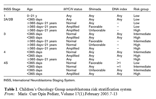
The table below outlines criteria for risk assignment based on the International Neuroblastoma Staging System (INSS), age, and biologic risk factors.

These criteria are based on the analysis of several thousands of patients treated in cooperative group protocols in Australia, Canada, Europe, Japan, and the United States.

Treatment regimes is carefully designed upon risk assessment and staging (1):

Low-risk neuroblastoma  Survival rates for patients who have INSS stage 1 disease, regardless of biologic factors, are excellent with surgery alone. Chemotherapy may be needed as an effective salvage therapy for patients who have INSS stage 1 disease who relapse after surgery only.

For patients who have INSS stage
1, 2A, or 2B disease, chemotherapy should be reserved for those who have localized neuroblastoma and experience life- or organ-threatening symptoms at diagnosis or for the minority of patients who experience recurrent or progressive disease.Patients with stage 2A/2B disease with amplified MYCN are considered high risk regardless of age and histology

Stage 4S neuroblastoma withoutMYCN amplification undergoes spontaneous regression in the majority of cases.  Chemotherapy or low-dose radiotherapy is used in patients who have large tumors or massive hepatomegaly.

Intermediate-risk neuroblastoma

Surgical resection and moderate–dose, multiagent chemotherapy (cyclophosphamide, doxorubicin, carboplatin, etoposide) are the standard of care. Chemo rounds are of either 4 cycles, 6 cycles, or 8 cycles, depending on histology and DNA index and response to treatment.  If residual disease is present after chemotherapy and surgery, radiation therapy could be considered. However, the use of radiation is controversial.

High-risk neuroblastoma

Patients with high-risk neuroblastoma require treatment with multiagent chemotherapy, surgery, and radiotherapy. Current therapeutic protocols involve 4 phases of therapy, including induction, local control, consolidation and treatment of minimal residual disease. Induction therapy currently involves multiagent chemotherapy with non–cross-resistant profiles, including: alkylating agents, platinum, and anthracyclines and topoisomerase II inhibitors. Topoisomerase I inhibitor are also being considered. Local control involves surgical resection of primary tumor site as well as radiation to primary tumor site.

Myeloablative consolidation therapy – myeloablative consolidation therapy with etoposide, carboplatin, and melphalan have improved the outcome of patients. most centers now recommend the use of peripheral blood stem cell support over bone marrow for consolidation therapy in patients with high-risk neuroblastoma.

Other consideration – Use of 13-cis -retinoic acid in a maintenance phase of therapy. Recent data have showed improved survival in patients receiving 13-cis -RA in combination with immunomodulatory therapy with interleukin (IL)-2, granulocyte macrophage colony-stimulating factor (GM-CSF), and the chimeric anti-GD2 (gangliosidase) antibody when compared with 13-cis -RA alone.

Summary:

“Neuroblastoma is a heterogenous tumor for which biology dictates clinical behavior”.  The main the goal is to have patient-tailored prognosis. Additional research in search for new therapeutics for high-risk patients is needed. Some therapies under investigation include aurora kinase inhibitors, antiangiogenic agents, histone deacetylase inhibitors, and therapeutic metaiodobenzylguanidine (MIBG).  According to Park et al: “we must minimize the lasting effects of therapy,For the remaining patients who have low- and intermediate-risk disease,specifically avoiding organ damage or organ loss from surgery and organ dysfunction or risk for secondary malignancy after chemotherapy”.

Other future aspect of therapeutics may include specific inhibitor of this pathway, viz Cyclopamine and other kinase inhibitors like LY294002 for PI3K inhibition or  GSK-3β inhibitors in order to inhibit the Hedgehog and the β-catenin pathways, respectively.

Reference:

1. Park JR., Eggert A and Caron H.Neuroblastoma: Biology, Prognosis and Treatment. Pediatric Clinics of North America 2008; 55(1): 97-120. http://www.sciencedirect.com/science/article/pii/S0031395507001575

2. Yamamoto K, Hayashi Y, Hanada R, et al. Mass screening and age-specific incidence of neuroblastoma in Saitama Prefecture, Japan. J Clin Oncol 1995;13(8):2033–2038. http://www.ncbi.nlm.nih.gov/pubmed/?term=Mass+screening+and+age-specific+incidence+of+neuroblastoma+in+Saitama+Prefecture%2C+Japan

3. Triche TJ. Neuroblastoma: biology confronts nosology. Arch Pathol Lab Med 1986;110(11):994–996. no available abstract.

4. Singh SK, Hawkins C, Clarke ID, et al. Identification of human brain tumour initiating cells. Nature 2004;432(7015):396–401. http://www.ncbi.nlm.nih.gov/pubmed/15549107

5. Tirode F, Laud-Duval K, Prieur A, et al. Mesenchymal stem cell features of Ewing tumors.Cancer Cell 2007;11(5):421–429. http://www.ncbi.nlm.nih.gov/pubmed/17482132

6. Shimada H, Chatten J, Newton WA Jr, et al. Histopathologic prognostic factors in neuroblastic tumors: definition of subtypes of ganglioneuroblastoma and an age-linked classification of neuroblastomas.J Natl Cancer Inst. Aug 1984;73(2):405-416. http://www.ncbi.nlm.nih.gov/pubmed/6589432

7. Norman J Lacayo and Max J Coppes. Pediatric Neuroblastoma. MedScape Reference June 2012. http://emedicine.medscape.com/article/988284-overview#a0104

8. Zhi F., Gong G., Xu Y., Zhu Y., Hu D., Yang Y and Hu Y.Activated β-catenin Forces N2A Cell-derived Neurons Back to Tumor-like Neuroblasts and Positively Correlates with a Risk for Human Neuroblastoma. Int J Biol Sci. 2012; 8(2): 289–297. http://www.ncbi.nlm.nih.gov/pmc/articles/PMC3269611/

9. Shahi MH., Sinha S., Afzal M and Castresana JS. Role of Sonic hedgehog signaling pathway in neuroblastoma development. Biology and Medicine 2009, 1 (4): Rev2, 1-6. http://biolmedonline.com/Articles/vol1_4_Rev2.pdf

10. Wickstrom M., Dyberg C, Shimokawa T., Milosevic J., Baryawno N., Fuskevag OM., Larsson R., Kogner P, Zaphiropoulos PG and Johnsen JI. Targeting the hedgehog signal transduction pathway at the level of GLI inhibits neuroblastoma cell growth in vitro and in vivo.  Int J. Cancer 2013 Apr 1;132(7):1516-1524. http://www.ncbi.nlm.nih.gov/pubmed/22949014

Other related articles on Open Access Leaders in Pharmaceutical Intelligence:

1.By: Larry H Bernstein. AKT signaling variable effects. http://pharmaceuticalintelligence.com/2013/03/04/akt-signaling-variable-effects/

2. By: Aviva Lev-Ari PhD RN. Human Variome Project: encyclopedic catalog of sequence variants indexed to the human genome sequence. http://pharmaceuticalintelligence.com/2012/11/24/human-variome-project-encyclopedic-catalog-of-sequence-variants-indexed-to-the-human-genome-sequence/

3. By: Aviva Lev-Ari PhD RN. Neuroprotective Therapies: Pharmacogenomics vs Psychotropic drugs and Cholinesterase Inhibitors. http://pharmaceuticalintelligence.com/2012/11/23/neuroprotective-therapies-pharmacogenomics-vs-psychotropic-drugs-and-cholinesterase-inhibitors/

4. By:  Aviva Lev-Ari PhD RN.  arrayMap: Genomic Feature Mining of Cancer Entities of Copy Number Abnormalities (CNAs) Data. http://pharmaceuticalintelligence.com/2012/11/01/arraymap-genomic-feature-mining-of-cancer-entities-of-copy-number-abnormalities-cnas-data/

5. By: Venkat S Karra. $20 million Novartis deal with ‘University of Pennsylvania’ to develop Ultra-Personalized Cancer Immunotherapy. http://pharmaceuticalintelligence.com/2012/08/08/20-million-novartis-deal-with-university-of-pennsylvania-to-develop-ultra-personalized-cancer-immunotherapy/

7.  By: Aviva Lev-Ari PhD RN. Acoustic Neuroma, Neurinoma or Vestibular Schwannoma: Treatment Options. http://pharmaceuticalintelligence.com/2012/10/30/acoustic-neuroma-neurinoma-or-vestibular-schwannoma-treatment-options/

8.  By: Aviva Lev-Ari PhD RN. Clinical Trials on Schwannoma & Benign Intracranial Tumors Radiosurgery Treatment. http://pharmaceuticalintelligence.com/2012/10/30/clinical-trials-on-schwannoma-benign-intracranial-tumors-radiosurgery-treatment/

9. By: Aviva Lev-Ari PhD RN. Facial Nerve, Intracanalicular Meningiomas, Vestibular Schwannomas: Surgical Planning. http://pharmaceuticalintelligence.com/2012/10/15/facial-nerve-intracanalicular-meningiomas-vestibular-schwannomas-surgical-planning/

Read Full Post »

Personalized Medicine and Colon Cancer

Author: Tilda Barliya, PhD

According to Dr. Neil Risch a leading expert in statistical genetics and the director of the UCSF Institute for Human Genetics,  “Personalized medicine, in which a suite of molecules measured in a patient’s lab tests can inform decisions about preventing or treating diseases, is becoming a reality” (7).

Colorectal cancer (CRC) is the third most common cancer and the fourth-leading cause of cancer death worldwide despite advances in screening, diagnosis, and treatment. Staging is the only prognostic classification used in clinical practice to select patients for adjuvant chemotherapy. However, pathological staging fails to predict recurrence accurately in many patients undergoing curative surgery for localized CRC (1,2). Most of the patients who are not eligible for surgery need adjuvant chemotherapy in order to avoid relapse or to increase survival. Unfortunately, only a small portion of them shows an objective response to chemotherapy, becoming problematic to correctly predict patients’ clinical outcome (3).

CRC patients are normally being tested for several known biomarkers which falls into 4 main categories (5):

  1. Chromosomal Instability (CIN)
  2. Microsatellite Instability (MSI)
  3. CpG Island methylator phynotype (CIMP)
  4. Global DNA hypomethylation

In the past few years many studies have exploited microarray technology to investigate gene expression profiles (GEPs) in CRC, but no established signature has been found that is useful for clinical practice, especially for predicting prognosis.  Only a subset of CRC patients with MSI tumors have been shown to have better prognosis and probably respond differently to adjuvant chemotherapy compared to microsatellite stable (MSS) cancer patients (6).

Pritchard & Grady have summarized the selected biomarkers that have been evaluated in colon cancer patients (10).

Table 1

Selected Biomarkers That Have Been Evaluated in Colorectal Cancer

Biomarker Molecular Lesion Frequency
in CRC
Prediction Prognosis Diagnosis
KRAS Codon 12/13 activating
mutations; rarely codon
61, 117,146
40% Yes Possible –
BRAF V600E activating
mutation
10% Probable Probable Lynch
Syndrome
PIK3CA Helical and kinase
domain mutations
20% Possible Possible –
PTEN Loss of protein by IHC 30% Possible – –
Microsatellite Instability (MSI) Defined as >30%
unstable loci in the NCI
consensus panel or
>40% unstable loci in a
panel of mononucleotide
microsatellite repeats9
15% Probable Yes Lynch
Syndrome
Chromosome Instability (CIN) Aneuploidy 70% Probable Yes –
18qLOH Deletion of the long arm
of chromosome 18
50% Probable Probable –
CpG Island Methylator
Phenotype (CIMP)
Methylation of at least
three loci from a selected
panel of five markers
15% +/− +/− –
Vimentin (VIM) Methylation 75% – – Early
Detection
TGFBR2 Inactivating Mutations 30% – – –
TP53 Mutations Inactivating Mutations 50% – – –
APC Mutations Inactivating Mutations 70% – – FAP
CTNNB1 (β-Catenin) Activating Mutations 2% – – –
Mismatch Repair Genes Loss of protein by IHC;
methylation; inactivating
mutations
1–15% – – Lynch
Syndrome

CRC- colorectal cancer; IHC- immunohistochemistry; FAP- Familial Adenomatous Polyposis

Examples for the great need of personalized medicine tailored according to the patients’ genetics is clearly seen with two specific drugs for CRC:  Cetuximab and panitumumab are two antibodies that were developed to treat colon cancer. However, at first it seemed as if they were a failure because they did not work in many patients. Then, it was discovered that if a cancer cell has a specific genetic mutation, known as K-ras, these drugs do not work.  This is an excellent example of using individual tumor genetics to predict whether or not treatment will work (8).

According to Marisa L et al, however, the molecular classification of CC currently used, which is based on a few common DNA markers as mentioned above (MSI, CpG island methylator phenotype [CIMP], chromosomal instability [CIN], and BRAF and KRAS mutations), needs to be refined.

Genetic Expression Profiles (GEP)

CRC is composed of distinct molecular entities that may develop through multiple pathways on the basis of different molecular features, as a consequence, there may be several prognostic signatures for CRC, each corresponding to a different entity. GEP studies have recently identified at least three distinct molecular subtypes of CC (4). Dr. Marisa Laetitia and her colleagues from the Boige’s lab however, have conducted a very thorough study and identifies 6 distinct clusters for CC patients. Herein, we’ll describe the majority of this study and their results.

Study  Design:

Marisa L et al (1) performed a consensus unsupervised analysis (using an Affymertix chip) of the GEP on tumor tissue sample from 750 patients with stage I to IV CC. Patients were staged according to the American Joint Committee on Cancer tumor node metastasis (TNM) staging system. Of the 750 tumor samples of the CIT cohort, 566 fulfilled RNA quality requirements for GEP analysis. The 566 samples were split into a discovery set (n = 443) and a validation set (n = 123).

Several known mutations were used as internal controls, including:

  • The seven most frequent mutations in codons 12 and 13 of KRAS .
  • The BRAF c.1799T>A (p.V600E)
  • TP53mutations (exons 4–9)
  • MSI was analyzed using a panel of five different microsatellite loci from the Bethesda reference panel
  • CIMP status was determined using a panel of five markers (CACNA1G, IGF2, NEUROG1, RUNX3, and SOCS1)

Results:

The results revealed six clusters of samples based on the most variant probe sets. The consensus matrix showed that C2, C3, C4, and C6 appeared as well-individualized clusters, whereas there was more classification overlap between C1 and C5. In other words:

  • Tumors classified as C1, C5, and C6 were more frequently CIN+, CIMP−, TP53– mutant, and distal (p<0.001), without any other molecular or clinicopathological features able to discriminate these three clusters clearly.
  • Tumors classified as C2, C4, and C3 were more frequently CIMP+ (59%, 34%, and 18%, respectively, versus <5% in other clusters) and proximal.
  • C2 was enriched for dMMR (68%) and BRAF- mutant tumors (40%).
  • C3 was enriched for KRAS- mutant tumors (87%).

Note: No association between clusters and TNM stage (histopathology) was found, except enrichment for metastatic (31%) tumors in C4.

Figure: These signaling pathways associated with the molecular subtype (by cluster)

Figure 2 Signaling pathways associated with each molecular subtype.

Marisa L et al. Signaling pathways associated with each molecular subtype

These clusters fall into several signaling pathways:

  • up-regulated immune system and cell growth pathways were found in C2, the subtype enriched for dMMR tumors
  • C4 and C6 both showed down-regulation of cell growth and death pathways and up-regulation of the epithelial–mesenchymal transition/motility pathways. displaying “stem cell phenotype–like” GEPs (91%)
  • Most signaling pathways were down-regulated in C1 and C3.
  • In C1, cell communication and immune pathways were down-regulated.
  • In C5, cell communication, Wnt, and metabolism pathways were up-regulated.

These results are further summarized in table 2:

Figure 3 Summary of the main characteristics of the six subtypes.

Marisa L et al. Gene Expression Classification of Colon Cancer into Molecular Subtypes

The authors have identified six robust molecular subtypes of CC individualized by distinct clinicobiological characteristics (as summarized in table 2).

This classification successfully identified the dMMR tumor subtype, and also individualized five other distinct subtypes among pMMR tumors, including three CIN+ CIMP− subtypes representing slightly more than half of the tumors. As expected, mutation of BRAF was associated with the dMMR subtype, but was also frequent in the C4 CIMP+ poor prognosis subtype. TP53– andKRAS-mutant tumors were found in all the subtypes; nevertheless, the C3 subtype, highly enriched in KRAS-mutant CC, was individualized and validated, suggesting a specific role of this mutation in this particular subgroup of CC.

Current Treatments for colon cancer- Table 3 (11) .

Constant S et al. Colon Cancer: Current Treatments and Preclinical Models for the Discovery and Development of New Therapies

Exploratory analysis of each subtype GEP with previously published supervised signatures and relevant deregulated signaling pathways improved the biological relevance of the classification.

The biological relevance of our subtypes was highlighted by significant differences in prognosis. In our unsupervised hierarchical clustering, patients whose tumors were classified as C4 or C6 had poorer RFS than the other patients.

Prognostic analyses based solely on common DNA alterations can distinguish between risk groups, but are still inadequate, as most CCs are pMMR CIMP− BRAFwt.

The markers BRAF-mutant, CIMP+, and dMMR may be useful for classifying a small proportion of cases, but are uninformative for a large number of patients.

Unfortunately, 5 of the 9 anti-CRC drugs approved by the FDA today are basic cytotoxic chemotherapeutics that attack cancer cells at a very fundamental level (i.e. the cell division machinery) without specific targets, resulting in poor effectiveness and strong side-effects (Table 3) (11).

An example for side effects induction mechanisms have also been reported in CRC for the BRAF(V600E) inhibitor Vemurafenib that triggers paradoxical EGFR activation (12).

Summary:

The authors of this study “report a new classification of CC into six robust molecular subtypes that arise through distinct biological pathways and represent novel prognostic subgroups. Our study clearly demonstrates that these gene signatures reflect the molecular heterogeneity of CC. This classification therefore provides a basis for the rational design of robust prognostic signatures for stage II–III CC and for identifying specific, potentially targetable markers for the different subtypes”.

These results further underline the urgent need to expand the standard therapy options by turning to more focused therapeutic strategies: a targeted therapy-for specific subtype profile.. Accordingly, the expansion and the development of new path of therapy, like drugs specifically targeting the self-renewal of intestinal cancer stem cells – a tumor cell population from which CRC is supposed to relapse, remains relevant.

Therefore, the complexity of these results supports the arrival of a personalized medicine, where a careful profiling of tumors will be useful to stratify patient population in order to test drugs sensitivity and combination with the ultimate goal to make treatments safer and more effective.

References:

1. Marisa L,  de Reyniès A, Alex Duval A,  Selves J, Pierre Gaub M, Vescovo L, Etienne-Grimaldi MC, Schiappa R, Guenot D, Ayadi M, Kirzin S, Chazal M, Fléjou JF…Boige V. Gene Expression Classification of Colon Cancer into Molecular Subtypes: Characterization, Validation, and Prognostic Value. PLoS Med May 2013 10(5): e1001453. doi:10.1371. http://www.plosmedicine.org/article/info%3Adoi/10.1371/journal.pmed.1001453

2. Villamil BP, Lopez AR, Prieto SH, Campos GL, Calles A, Lopez- Asenjo JA, Sanz Ortega J, Perez CF, Sastre J, Alfonso R, Caldes T, Sanchez FM and Rubio ED. Colon cancer molecular subtypes identified by expression profiling and associated to stroma, mucinous type and different clinical behavior. BMC Cancer 2012, 12:260.  http://www.biomedcentral.com/1471-2407/12/260/

3. Diaz-Rubio E, Tabernero J, Gomez-Espana A, Massuti B, Sastre J, Chaves M, Abad A, Carrato A, Queralt B, Reina JJ, et al.: Phase III study of capecitabine plus oxaliplatin compared with continuous-infusion fluorouracil plus oxaliplatin as first-line therapy in metastatic colorectal cancer: final report of the Spanish Cooperative Group for the Treatment of Digestive Tumors Trial. J Clin Oncol 2007, 25(27):4224-4230. http://jco.ascopubs.org/content/25/27/4224.long

4. Salazar R, Roepman P, Capella G, Moreno V, Simon I, et al. (2011) Gene expression signature to improve prognosis prediction of stage II and III colorectal cancer. J Clin Oncol 29: 17–24. http://www.ncbi.nlm.nih.gov/pubmed?cmd=Search&doptcmdl=Citation&defaultField=Title%20Word&term=Salazar%5Bauthor%5D%20AND%20Gene%20expression%20signature%20to%20improve%20prognosis%20prediction%20of%20stage%20II%20and%20III%20colorectal%20cancer

5.  By: Global Genome Knowledge. Colorectal Cancer- Personalized Medicine, Now a Clinical Reality.  http://www.srlworld.com/innersense/Voice-135-Colorectal-Cancer-Sept-2012-IS.pdf

6. Popat S, Hubner R and Houlston RS. Systematic review of microsatellite instability and colorectal cancer prognosis. J Clin Oncol. 2005 Jan 20;23(3):609-618. http://www.ncbi.nlm.nih.gov/pubmed/15659508

7. By: Jeffrey Norris. Value of Genomics and Personalized Medicine Is Wrongly Downplayed.http://www.ucsf.edu/news/2012/04/11864/value-genomics-and-personalized-medicine-wrongly-downplayed

8. By: James C Salwitz. The Future is now: Personalized Medicine. http://www.cancer.org/cancer/news/expertvoices/post/2012/04/18/the-future-is-now-personalized-medicine.aspx

9. Jeffrey A. Meyerhardt., and Robert J. Mayer. Systemic Therapy for Colorectal Cancer. N Engl J Med 2005;352:476-487. http://www.med.upenn.edu/gastro/documents/NEJMchemotherapycolorectalcancer.pdf

10. Pritchard CC and Grady WM. Colorectal Cancer Molecular Biology Moves Into Clinical Practice. Gut. Jan 2011 60(1): 116-129.  Gut. 2011 January; 60(1): 116–129. http://www.ncbi.nlm.nih.gov/pmc/articles/PMC3006043/

11. Constant S, Huang S, Wiszniewski L andMas C. Colon Cancer: Current Treatments and Preclinical Models for the Discovery and Development of New Therapies.  Pharmacology, Toxicology and Pharmaceutical Science » “Drug Discovery”, book edited by Hany A. El-Shemy, ISBN 978-953-51-0906-8.  http://www.intechopen.com/books/drug-discovery/colon-cancer-current-treatments-and-preclinical-models-for-the-discovery-and-development-of-new-ther

12. Prahallad, C. Sun, S. Huang, F. Di Nicolantonio, R. Salazar, D. Zecchin, R. L. Beijersbergen, A. Bardelli, R. Bernards, 2012 Unresponsiveness of colon cancer to BRAF(V600E) inhibition through feedback activation of EGFR. Nature Jan 2012 483 (7387): 100-103. http://www.nature.com/nature/journal/v483/n7387/full/nature10868.html

Other related articles on this Open Access Online Scientific Journal include the following:

*. By Tilda Barliya PhD. Colon Cancer. http://pharmaceuticalintelligence.com/2013/04/30/colon-cancer/

**. By: Tilda Barliya PhD. CD47: Target Therapy for Cancer. http://pharmaceuticalintelligence.com/2013/05/07/cd47-target-therapy-for-cancer/

I. By: Aviva Lev-Ari, PhD, RN. Cancer Genomic Precision Therapy: Digitized Tumor’s Genome (WGSA) Compared with Genome-native Germ Line: Flash-frozen specimen and Formalin-fixed paraffin-embedded Specimen Needed. http://pharmaceuticalintelligence.com/2013/04/21/cancer-genomic-precision-therapy-digitized-tumors-genome-wgsa-compared-with-genome-native-germ-line-flash-frozen-specimen-and-formalin-fixed-paraffin-embedded-specimen-needed/

II. By: Aviva Lev-Ari, PhD, RN. Critical Gene in Calcium Reabsorption: Variants in the KCNJ and SLC12A1 genes – Calcium Intake and Cancer Protection. http://pharmaceuticalintelligence.com/2013/04/12/critical-gene-in-calcium-reabsorption-variants-in-the-kcnj-and-slc12a1-genes-calcium-intake-and-cancer-protection/

III.  By: Stephen J. Williams, Ph.D. Issues in Personalized Medicine in Cancer: Intratumor Heterogeneity and Branched Evolution Revealed by Multiregion Sequencing. http://pharmaceuticalintelligence.com/2013/04/10/issues-in-personalized-medicine-in-cancer-intratumor-heterogeneity-and-branched-evolution-revealed-by-multiregion-sequencing/

IV. By: Ritu Saxena, Ph.D. In Focus: Targeting of Cancer Stem Cells. http://pharmaceuticalintelligence.com/2013/03/27/in-focus-targeting-of-cancer-stem-cells/

V.  By: Ziv Raviv PhD. Cancer Screening at Sourasky Medical Center Cancer Prevention Center in Tel-Aviv. http://pharmaceuticalintelligence.com/2013/03/25/tel-aviv-sourasky-medical-center-cancer-prevention-center-excellent-example-for-adopting-prevention-of-cancer-as-a-mean-of-fighting-it/

VI. By: Ritu Saxena, PhD. In Focus: Identity of Cancer Stem Cells. http://pharmaceuticalintelligence.com/2013/03/22/in-focus-identity-of-cancer-stem-cells/

VII. By: Dror Nir, PhD. State of the art in oncologic imaging of Colorectal cancers. http://pharmaceuticalintelligence.com/2013/02/02/state-of-the-art-in-oncologic-imaging-of-colorectal-cancers/

Read Full Post »

DNA Nanotechnology

Author: Tilda Barliya PhD

The field of DNA and RNA nanotechnologies  are considered one of the most dynamic research areas in the field of drug delivery in molecular medicine. Both DNA and RNA have a wide aspect of medical application including: drug deliveries, for genetic immunization, for metabolite and nucleic acid detection, gene regulation, siRNA delivery for cancer treatment (I), and even analytical and therapeutic applications.

Seeman (6,7) pioneered the concept 30 years ago of using DNA as a material for creating nanostructures; this has led to an explosion of knowledge in the now well-established field of DNA nanotechnology. The unique properties in terms of free energy, folding, noncanonical base-pairing, base-stacking, in vivo transcription and processing that distinguish RNA from DNA provides sufficient rationale to regard RNA nanotechnology as its own technological discipline. Herein, we will discuss the advantages of DNA nanotechnology and it’s use in medicine.

So What is the rational of using DNA nanotechnology(3)?

  • Genetic studies – its application in various biological fields like biomedicine, cancer research, medical devices  and genetic engineering.
  • Its unique properties of structural stability, programmability of sequences, and predictable self-assembly.
DNA origami

Structures made from DNA using the DNA-origami method (Rothemund, 2006)

Structural DNA nanotechnology rests on three pillars: [1] Hybridization; [2] Stably branched DNA; and [3] Convenient synthesis of designed sequences.

Hybridization

Hybridization. The self-association (self=assembly) of complementary nucleic acid molecules or parts of molecules, is implicit in all aspects of structural DNA nanotechnology. Individual motifs are formed by the hybridization of strands designed to produce particular topological species. A key aspect of hybridization is the use of sticky ended cohesion to combine pieces of linear duplex DNA; this has been a fundamental component of genetic engineering for over 35 years (7). Not only is hybridization critical to the formation of structure, but it is deeply involved in almost all the sequence-dependent nanomechanical devices that have been constructed, and it is central to many attempts to build structural motifs in a sequential fashion (7,8 ).

Stably Branched DNA

branched DNA molecules are central to DNA nanotechnology. It is the combination of in vitro hybridization and synthetic branched DNA that leads to the ability to use DNA as a construction material. Such branched DNA is thought to be intermediates in genetic recombination (such as Holliday junctions).

Convenient Synthesis of Designed Sequences

Biologically derived branched DNA molecules, such as Holliday junctions, are inherently unstable, because they exhibit sequence symmetry; i.e., the four strands actually consist of two pairs of strands with the same sequence. This symmetry enables an isomerization known as branch migration that allows the branch point to relocate.  DNA nanotechnology entailed sequence design that attempted to minimize sequence symmetry in every way possible.

One of the most remarkable innovations in structural DNA-nanotechnology in recent years is DNA origami, which was invented in 2006 by Paul Rothemund (1) (see Fig above). DNA origami utilizes the genome from a virus together with a large number of shorter DNA strands to enable the creation of numerous DNA-based structures (Figure 1). The shorter DNA strands forces the long viral DNA to fold into a pattern that is defined by the interaction between the long and the short DNA strands (1,2).

Rothemund believes that an  application of patterned DNA origami would be the creation of a ‘nanobreadboard’, to which diverse components could be added. The attachment of proteins23, for example, might allow novel biological experiments aimed at modelling complex protein assemblies and examining the effects of spatial organization, whereas molecular electronic or plasmonic circuits might be created by attaching nanowires, carbon nanotubes or gold nanoparticles (1).

DNA nanotechnology and Biological Application

The physical and chemical properties of nanomaterials such as polymers, semiconductors, and metals present diverse advantages for various in vivo applications (3,9 ). For example:

  • Therapeutics – In cancer for example, nanosystems that are designed from biological materials such as DNA and RNA are ‘programmed’ to be able to evade most, if not all, drug-resistance mechanisms. Based on these properties, most nanosystems are able to deliver high concentrations of drugs to cancer cells while curtailing damage to surrounding healthy cells (2b, 3, 9, 11, 15).
  • Biosensors – capable of picking up very specific biological signals and converting them into electrical outputs that can be analyzed for identification. Biosensors are efficient as they have a high ratio of surface area to volume as well as adjustable electronic, magnetic, optical, and biological properties (3, 12, 13, 14).
  • **Amin and colleagues have developed a biotinylated DNA thin film-coated fiber optic reflectance biosensor for the detection of streptavidin aerosols. DNA thin films were prepared by dropping DNA samples into a polymer optical fiber which responded quickly to the specific biomolecules in the atmosphere. This approach of coating optical fibers with DNA nanostructures could be very useful in the future for detecting atmospheric bio-aerosols with high sensitivity and specificity (3, 14)
  • Computing – Another aspect uses the programmability of DNA to create devices that are capable of computing. Here, the structure of the assembled DNA is not of primary interest. Instead, control of the DNA sequence is used in the creation of computational algorithms, like e.g. artificial neural networks. Qian et al for example, built on the richness of DNA computing and strand displacement circuitry, they showed how molecular systems can exhibit autonomous brain-like behaviours. Using a simple DNA gate architecture that allows experimental scale-up of multilayer digital circuits, they systematically transform arbitrary linear threshold circuits (an artificial neural network model) into DNA strand displacement cascades that function as small neural networks (3, 10).
  • Additional features: 3rd generation DNA sequencers (II), Biomimetic systems, Energy transfer and photonics etc

Summary:

DNA nanotechnology is an evolving field that affects medicine, computation, material sciences, and physics. DNA nanostructures offer unprecedented control over shape, size, mechanical flexibility and anisotropic surface  modification. Clearly, proper control over these aspects can increase  circulation times by orders of magnitude, as can be seen for longcirculating particles such as erythrocytes and various pathogenic particles evolved to overcome this issue.  The use of DNA in DNA/protein-based matrices makes these structures inherently amenable to structural tunability. More research in this direction  will certainly be developed, making DNA a promising biomaterial  in tissue engineering. future development of novel ways in which DNA would be utilized to have a much more comprehensive role in biological computation and data storage is envisaged.

REFERENCES

1. Paul W. K. Rothemund. Folding DNA to create nanoscale shapes and patterns. NATURE 2006 (March 16)|Vol 440: 297-302. http://www.nature.com/nature/journal/v440/n7082/full/nature04586.html

http://www.dna.caltech.edu/Papers/DNAorigami-nature.pdf

2. Andre V. Pinheiro, Dongran Han, William M. Shih and Hao Yan. Challenges and opportunities for structural DNA nanotechnology. Nature Nanotechnology 2011 Dec | VOL 6: 763-772.  http://www.nature.com/nnano/journal/v6/n12/pdf/nnano.2011.187.pdf

2b. Thi Huyen La, Thi Thu Thuy Nguyen, Van Phuc Pham, Thi Minh Huyen Nguyen and Quang Huan Le.  Using DNA nanotechnology to produce a drug delivery system. Adv. Nat. Sci.: Nanosci. Nanotechnol. 4 (2013) 015002 (7pp). http://iopscience.iop.org/2043-6262/4/1/015002. http://iopscience.iop.org/2043-6262/4/1/015002/pdf/2043-6262_4_1_015002.pdf

3. Muniza Zahid, Byeonghoon Kim, Rafaqat Hussain, Rashid Amin and Sung H Park. DNA nanotechnology: a future perspective. Nanoscale Research Letters 2013, 8:119. http://www.nanoscalereslett.com/content/8/1/119

4.By: Cientifica Ltd 2007. The Nanotech Revolution in Drug Delivery.  http://www.cientifica.com/WhitePapers/054_Drug%20Delivery%20White%20Paper.pdf

5. Gemma Campbell. Nanotechnology and its implications for the health of the E.U citizen: Diagnostics, drug discovery and drug delivery. Institute of Nanotechnology and Nanoforum. http://www.nano.org.uk/nanomednet/images/stories/Reports/diagnostics,%20drug%20discovery%20and%20drug%20delivery.pdf

6.Peixuan Guo., Haque F., Brent Hallahan, Randall Reif and Hui Li. Uniqueness, Advantages, Challenges, Solutions, and Perspectives in Therapeutics Applying RNA Nanotechnology. Nucleic Acid Ther. 2012 August; 22(4): 226–245. http://www.ncbi.nlm.nih.gov/pmc/articles/PMC3426230/

7. SEEMAN N.C. Nanomaterials based on DNA. Annu. Rev. Biochem. 2010;79:65–87. http://www.ncbi.nlm.nih.gov/pmc/articles/PMC3454582/

8. Yin P, Choi HMT, Calvert CR, Pierce NA. Programming biomolecular self-assembly pathways. Nature.2008;451:318–323.  http://www.ncbi.nlm.nih.gov/pubmed/18202654

9. Yan Lee P, Wong KY: Nanomedicine: a new frontier in cancer therapeutics. Curr Drug Deliv 2011, 8(3):245-253. OpenURLhttp://www.eurekaselect.com/73728/article

10. Qian, L.L., Winfree, E., and Bruck, J. Neural Network Computation with DNA Strand Displacement Cascades. Nature 2011 475, 368-372.  http://www.nature.com/nature/journal/v475/n7356/full/nature10262.html

11. Acharya S, Dilnawaz F, Sahoo SK: Targeted epidermal growth factor receptor nanoparticle bioconjugates for breast cancer therapy. Biomaterials 2009, 30(29):5737-5750. http://www.sciencedirect.com/science/article/pii/S0142961209006929

12. Bohunicky B, Mousa SA: Biosensors: the new wave in cancer diagnosis. Nanotechnology, Science and Applications 2011, 4:1-10. http://www.dovepress.com/biosensors-the-new-wave-in-cancer-diagnosis-peer-reviewed-article-NSA-recommendation1

13. Sanvicens N, Mannelli I, Salvador J, Valera E, Marco M: Biosensors for pharmaceuticals based on novel technology. Trends Anal Chem 2011, 30:541-553. http://www.sciencedirect.com/science/article/pii/S016599361100015X

14. Amin R, Kulkarni A, Kim T, Park SH: DNA thin film coated optical fiber biosensor. Curr Appl Phys 2011, 12(3):841-845. http://www.sciencedirect.com/science/article/pii/S1567173911005888

15. Choi, Y.; Baker, J. R. Targeting Cancer Cells with DNA Assembled Dendrimers: A Mix and Match Strategy for Cancer. Cell Cycle 2005, 4, 669–671. http://www.ncbi.nlm.nih.gov/pubmed/15846063  http://www.landesbioscience.com/journals/cc/article/1684/

Other related articles on this Open Access Online Scientific Journal include the following

I. By: Ziv Raviv PhD. The Development of siRNA-Based Therapies for Cancer. http://pharmaceuticalintelligence.com/2013/05/09/the-development-of-sirna-based-therapies-for-cancer/

II. By: Tilda Barliya PhD. Nanotechnology, personalized medicine and DNA sequencing. http://pharmaceuticalintelligence.com/2013/01/09/nanotechnology-personalized-medicine-and-dna-sequencing/

III. By: Larry Bernstein MD FACP. DNA Sequencing Technology. http://pharmaceuticalintelligence.com/2013/03/03/dna-sequencing-technology/

IV. By: Venkat S Karra PhD. Measuring glucose without needle pricks: nano-sized biosensors made the test easy. http://pharmaceuticalintelligence.com/2012/09/04/measuring-glucose-without-needle-pricks-nano-sized-biosensors-made-the-test-easy/

Read Full Post »

CD47: Target Therapy for Cancer

Author/Curator: Tilda Barliya

“A research team from Stanford University’s School of Medicine is now one step closer to uncovering a cancer treatment that could be applicable across the board in killing every kind of cancer tumor” (1). It appeared that their antibody-drug against the CD47 protein, enabled the shrinking of all tumor cells. After completing their animal studies the researchers now move into a human phase clinical trials. CD47 has been previously studied and evaluated for its role in multiple cells, some of this data however, is somewhat controversy. So where do we stand?

CD47

CD47 (originally named integrin-associated protein (IAP)) is a cell surface protein of the immunoglobulin (Ig) superfamily, which is heavily glycosylated and expressed by virtually all cells in the body and overexpressed in many types of cancer  including breast, ovarian, colon, prostate and others (3). CD47 was first recognized as a 50 kDa protein associated and copurified with the  Alpha-v-Beta-3 integrin in placenta and neutrophil granulocytes and later shown to have the capacity to regulate integrin function and the responsiveness of leukocytes to RGD-containing extracellular matrix proteins. CD47 has also been shown to be identical to the OA-3/OVTL3 antigen highly expressed on most ovarian carcinomas (4,5).

CD47 consists of an extracellular IgV domain, a five times transmembrane-spanning domain, and a short alternatively spliced cytoplasmic tail. In both humans and mice, the cytoplasmic tail can be found as four different splice isoforms ranging from 4 to 36 amino acids, showing different tissue expression patterns (3).

CD47 interactions (3, 6):

  • Thrombospondin-1 (TSP-1) – a secreted glycoprotein that plays a role in vascular development and angiogenesis. Binding of TSP-1 to CD47 influences several fundamental cellular functions including cell migration and adhesion, cell proliferation or apoptosis, and plays a role in the regulation of angiogenesis and inflammation.
  • Signal-regulatory protein-alpha (SIRPα) – an inhibitory transmembrane receptor present on myeloid cells. The CD47/SIRPα interaction leads to bidirectional signaling, resulting in different cell-to-cell responses including inhibition of phagocytosis, stimulation of cell-cell fusion, and T-cell activation.
  • Integrins – several membrane integrins, most commonly integrin avb3. These interactions result in CD47/integrin complexes that effect a range of cell functions including adhesion, spreading and migration

These interactions with multiple proteins and cells types create several important functions, which include:

  • Cell proliferation – cell proliferation is heavily dependent on cell type as both activation and loss of CD47 can result in enhanced proliferation. For example, activation of CD47 with TSP-1 in wild-type cells inhibits proliferation and reduces expression of stem cell transcription factors. In cancer cells however, activation of CD47 with TSP-1 increases proliferation of human U87 and U373 astrocytoma. it is likely that CD47 promotes proliferation via the PI3K/Akt pathway in cancerous cells but not normal cells (7).  Loss of CD47 allows sustained proliferation of primary murine endothelial cells and enables these cells to spontaneously reprogram to form multipotent embryoid body-like clusters (8).
  • Apoptosis – Ligation of CD47 by anti-CD47 mAbs was found to induce apoptosis in a number of different cell types (3). For example: Of the two SIRP-family members known to bind the CD47 IgV domain (SIRPα and SIRPγ), SIRPα as a soluble Fc-fusion protein does not induce CD47-dependent apoptosis, hile SIRPα or SIRPγ bound onto the surface of beads induces apoptosis through CD47 in Jurkat T cells and the myelomonocytic cell line U937.
  • Migration – CD47  role on cell migration was first demonstrated in neutrophils, these effects were shown to be dependent on avb3 integrins, which interact with and are activated by CD47 at the plasma membrane. In cancer, Blocking CD47 function has been shown to inhibit migration and metastasis in a variety of tumor models. Blockade of CD47 by neutralizing antibodies reduced migration and chemotaxis in response to collagen IV in melanoma, prostate cancer and ovarian cancer-derived cells (9).
  • Angiogenesis – The mechanism of the anti-angiogenic activity of CD47 is not fully understood, but introduction of CD47 antibodies and TSP-1 have been shown to inhibit nitric oxide (NO)-stimulated responses in both endothelial and vascular smooth muscle cells (10). More so, CD47 signaling influences the SDF-1 chemokine pathway, which plays a role in angiogenesis (11). (12)
  • Inflammatory response – Interactions between endothelial cell CD47 and leukocyte SIRPγ regulate T cell transendothelial migration (TEM) at sites of inflammation. CD47 also functions as a marker of self on murine red blood cells which allows RBC to avoid phagocytosis. Tumor cells can also evade macrophage phagocytosis through the expression of CD47 (2, 13).

It appears that CD47 ligation induce different responses, depending on cell type and partner for ligation.

Therapeutic and clinical aspect of CD47 in human cancer:

CD47 is overexpressed in many types of human cancers  and its known function as a “don’t eat me” signal, suggests the potential for targeting the CD47-SIRPα pathway as a common therapy for human malignancies (2,13). Upregulation of CD47 expression in human cancers also appears to influence tumor growth and dissemination. First, increased expression of CD47 in several hematologic malignancies was found to be associated with a worse clinical prognosis, and in ALL to predict refractoriness to standard chemotherapies (13, 14-16). Second, CD47 was demonstrated to regulate tumor metastasis and dissemination in both MM and NHL (13, 17).

Efforts have been made to develop therapies inhibiting the CD47-SIRPα pathway, principally through blocking monoclonal antibodies directed against CD47, but also possibly with a recombinant SIRPα protein that can also bind and block CD47.

Figure 2

Chao MP et al. 2012 Combination strategies targeting CD47 in cancer

While monotherapies targeting CD47 were efficacious in several pre-clinical tumor models, combination strategies involving inhibition of the CD47-SIRPα pathway offer even greater therapeutic potential. Specifically, antibodies targeting CD47-SIRPα can be included in combination therapies with other therapeutic antibodies, macrophage-enhancing agents, chemo-radiation therapy, or as an adjuvant therapy to inhibit metastasis (13).

For example, anti-SIRPα antibody was found to potentiate  antibody-dependent cellular cytotoxicity (ADCC) mediated by the anti-Her2/Neu antibody trastuzumab against breast cancer cells (18).  CD47–SIRPα interactions and SIRPα signaling negatively regulate trastuzumab-mediated ADCC in vitro and antibody-dependent elimination of tumor cells in vivo

More so, chemo-radiation therapy-mediated upregulation of cell surface calreticulin may potentially augment the activity of anti-CD47 antibody. However, this approach may also lead to increased toxicity as cell surface calreticulin is expressed on non-cancerous cells undergoing apoptosis, a principle effect of chemo-radiation therapy (19).

Highlights:

  • Phagocytic cells, macrophages, regulate tumor growth through phagocytic clearance
  • CD47 binds SIRPα on phagocytes which delivers an inhibitory signal for phagocytosis
  • A blocking anti-CD47 antibody enabled phagocytic clearance of many human cancers
  • Phagocytosis depends on a balance of anti-(CD47) and pro-(calreticulin) signals
  • Anti-CD47 antibody synergized with an FcR-engaging antibody, such as rituximab

Summary

Evasion of immune recognition is a major mechanism by which cancers establish and propagate disease. Recent data has demonstrated that the innate immune system plays a key role in modulating tumor phagocytosis through the CD47-SIRPα pathway. Careful development of reagents that can block the CD47/SIRPα interaction may indeed be useful to treat many forms of cancer without having too much of a negative side effect in terms of inducing clearance of host cells. Therapeutic approaches inhibiting this pathway have demonstrated significant efficacy, leading to the reduction and elimination of multiple tumor types.

Dr. Weissman says: “We are now hopeful that the first human clinical trials of anti-CD47 antibody will take place at Stanford in mid-2014, if all goes well. Clinical trials may also be done in the United Kingdom”. These clinical trials must be designed so that the data they generate will produce a valid scientific result!!!

REFERENCES

1. By Sara Gates:  Cancer Drug That Shrinks All Tumors Set To Begin Human Clinical Trials. http://www.huffingtonpost.com/2013/03/28/cancer-drug-shrinks-tumors_n_2972708.html

2. Willingham SB, Volkmer JP, Gentles AJ, Sahoo D, Dalerba P, Mitra SS, Wang J, Contreras-Trujillo H, Martin R, Cohen JD, Lovelace P, Scheeren FA, Chao MP, Weiskopf K, Tang C, Volkmer AK, Naik TJ, Storm TA, Mosley AR, Edris B, Schmid SM, Sun CK, Chua MS, Murillo O, Rajendran P, Cha AC, Chin RK, Kim D, Adorno M, Raveh T, Tseng D, Jaiswal S, Enger PØ, Steinberg GK, Li G, So SK, Majeti R, Harsh GR, van de Rijn M, Teng NN, Sunwoo JB, Alizadeh AA, Clarke MF, Weissman IL. The CD47-signal regulatory protein alpha (SIRPa) interaction is a therapeutic target for human solid tumors. Proc Natl Acad Sci U S A. 2012 Apr 24;109(17):6662-6667. http://www.pnas.org/content/early/2012/03/20/1121623109

3. Oldenborg PL. CD47: A Cell Surface Glycoprotein Which Regulates Multiple Functions of Hematopoietic Cells in Health and Disease. ISRN Hematology Volume 2013 (2013), Article ID 614619, 19 pages.  http://www.hindawi.com/isrn/hematology/2013/614619/

4. G. Campbell, P. S. Freemont, W. Foulkes, and J. Trowsdale, “An ovarian tumor marker with homology to vaccinia virus contains an IgV- like region and multiple transmembrane domains,”Cancer Research, vol. 52, no. 19, pp. 5416–5420, 1992. http://cancerres.aacrjournals.org/content/52/19/5416.long

5. L. G. Poels, D. Peters, Y. van Megen et al., “Monoclonal antibody against human ovarian tumor-associated antigens,” Journal of the National Cancer Institute, vol. 76, no. 5, pp. 781–791, 1986. http://www.ncbi.nlm.nih.gov/pubmed/3517452

6. CD47. Wikipedia. http://en.wikipedia.org/wiki/CD47

7. Sick E, Boukhari A, Deramaudt T, Rondé P, Bucher B, André P, Gies JP, Takeda K (February 2011). “Activation of CD47 receptors causes proliferation of human astrocytoma but not normal astrocytes via an Akt-dependent pathway”. Glia 59 (2): 308–319. http://www.ncbi.nlm.nih.gov/pubmed/21125662

8. Kaur S, Soto-Pantoja DR, Stein EV, Liu C, Elkahloun AG, Pendrak ML, Nicolae A, Singh SP, Nie Z, Levens D, Isenberg JS, Roberts DD.  “Thrombospondin-1 Signaling through CD47 Inhibits Self-renewal by Regulating c-Myc and Other Stem Cell Transcription Factors”. Sci Rep 2013: 3: 1673. http://www.ncbi.nlm.nih.gov/pmc/articles/PMC3628113/

9. Shahan TA, Fawzi A, Bellon G, Monboisse JC, Kefalides NA. “Regulation of tumor cell chemotaxis by type IV collagen is mediated by a Ca(2+)-dependent mechanism requiring CD47 and the integrin alpha(V)beta(3)”. J. Biol. Chem 2000. 275 (7): 4796–4802. http://www.jbc.org/content/275/7/4796

10. Isenberg JS, Ridnour LA, Dimitry J, Frazier WA, Wink DA, Roberts DD. “CD47 is necessary for inhibition of nitric oxide-stimulated vascular cell responses by thrombospondin-1”. J. Biol. Chem  2006. 281 (36): 26069–26080.  http://www.jbc.org/content/281/36/26069

11. Smadja DM, d’Audigier C, Bièche I, Evrard S, Mauge L, Dias JV, Labreuche J, Laurendeau I, Marsac B, Dizier B, Wagner-Ballon O, Boisson-Vidal C, Morandi V, Duong-Van-Huyen JP, Bruneval P, Dignat-George F, Emmerich J, Gaussem P. “Thrombospondin-1 is a plasmatic marker of peripheral arterial disease that modulates endothelial progenitor cell angiogenic properties”. Arterioscler. Thromb. Vasc. Biol  2011. 31 (3): 551–559. http://atvb.ahajournals.org/content/31/3/551

12. G. D. Grossfeld, D. A. Ginsberg, J. P. Stein et al., “Thrombospondin-1 expression in bladder cancer: association with p53 alterations, tumor angiogenesis, and tumor progression,” Journal of the National Cancer Institute 1997 vol. 89, no. 3, pp. 219–227. http://www.scopus.com/record/display.url?eid=2-s2.0-18744423089&origin=inward&txGid=9C86356DDB0B6816ACCBF90F9CA44E92.WlW7NKKC52nnQNxjqAQrlA%3a2

13. Chao MP, Weissman IL, Majeti R. “The CD47-SIRPα pathway in cancer immune evasion and potential therapeutic implications”. Curr. Opin. Immunol 2012. 24 (2): 225–32. http://www.sciencedirect.com/science/article/pii/S095279151200012X. http://www.ncbi.nlm.nih.gov/pmc/articles/PMC3319521/

14. Majeti R, Chao MP, Alizadeh AA, Pang WW, Jaiswal S, Gibbs KD, Jr, van Rooijen N, Weissman IL. Cd47 is an adverse prognostic factor and therapeutic antibody target on human acute myeloid leukemia stem cells. Cell. 2009;138(2):286–299. http://www.ncbi.nlm.nih.gov/pmc/articles/PMC2726837/

15. Chao MP, Alizadeh AA, Tang C, Jan M, Weissman-Tsukamoto R, Zhao F, Park CY, Weissman IL, Majeti R. Therapeutic antibody targeting of cd47 eliminates human acute lymphoblastic leukemia.Cancer Res. 2011;71 (4):1374–1384. http://www.ncbi.nlm.nih.gov/pmc/articles/PMC3041855/

16. Chao MP, Alizadeh AA, Tang C, Myklebust JH, Varghese B, Gill S, Jan M, Cha AC, Chan CK, Tan BT, Park CY, et al. Anti-cd47 antibody synergizes with rituximab to promote phagocytosis and eradicate non-hodgkin lymphoma. Cell. 2010;142(5):699–713. http://www.ncbi.nlm.nih.gov/pmc/articles/PMC2943345/

17. Chao MP, Tang C, Pachynski RK, Chin R, Majeti R, Weissman IL. Extranodal dissemination of non-hodgkin lymphoma requires cd47 and is inhibited by anti-cd47 antibody therapy. Blood.2011;118(18):4890–4901. http://www.ncbi.nlm.nih.gov/pmc/articles/PMC3208297/

18. Zhao XW, van Beek EM, Schornagel K, Van der Maaden H, Van Houdt M, Otten MA, Finetti P, Van Egmond M, Matozaki T, Kraal G, Birnbaum D, et al. Cd47-signal regulatory protein-alpha (sirpalpha) interactions form a barrier for antibody-mediated tumor cell destruction. Proc Natl Acad Sci U S A.2011;108(45):18342–18347. http://www.ncbi.nlm.nih.gov/pmc/articles/PMC3215076/

19. Obeid M, Tesniere A, Ghiringhelli F, Fimia GM, Apetoh L, Perfettini JL, Castedo M, Mignot G, Panaretakis T, Casares N, Metivier D, et al. Calreticulin exposure dictates the immunogenicity of cancer cell death. Nat Med. 2007;13(1):54–61. http://www.ncbi.nlm.nih.gov/pubmed/17187072

Other related articles on this Open Access Online Scientific Journal include the following:

I. By: Larry Bernstein MD. Treatment for Metastatic HER2 Breast Cancer http://pharmaceuticalintelligence.com/2013/03/03/treatment-for-metastatic-her2-breast-cancer/

II. By: Tilda Barliya PhD. Colon Cancer.  http://pharmaceuticalintelligence.com/2013/04/30/colon-cancer/

III. By: Ritu Saxena PhD. In focus: Triple Negative Breast Cancer. http://pharmaceuticalintelligence.com/2013/01/29/in-focus-triple-negative-breast-cancer/

Read Full Post »

Colon Cancer

Author/Editor: Tilda Barliya PhD

Screen Shot 2021-07-19 at 7.29.03 PM

Word Cloud By Danielle Smolyar

Colorectal cancer is the third most common type of cancer diagnosed in the United States and is the third most common cause of cancer-related death. The majority of cases are sporadic, with hereditary colon cancer contributing up to 15% of all colon cancer diagnoses. Treatment consists of surgery for early-stage disease and the combination of surgery and adjuvant chemotherapy for advanced-stage disease. Management of metastatic disease has evolved from primary chemotherapeutic treatment to include resection of single liver and lung metastases in addition to resection of the primary disease and chemotherapy (1-4).

Courtesy WebMD site

In the United States, colorectal cancer (CRC) is the third most common type of cancer diagnosed and the third most common cause of cancer-related death in men and women. In 2010, an estimated 102,900 new cases of colon cancer were diagnosed (49,470 male, 53,430 female) and 51,370 patients (26,580 male, 24,790 female) died from CRC. The death rate from colon cancer decreased over the preceding decade, from 30.77 per 100,000 people to 20.5 per 100,000 people. The lifetime risk of developing colon cancer in industrialized nations is 5% and is stable or decreasing. In contrast, the incidence in developing countries continues to rise, hypothesized to be due to increased exposure to risk factors. It has been estimated that 1.5 million people in the United States will be living with CRC by 2020.The financial burden of caring for this population is significant: $4.5 to $9.6 billion per year.

Colon Cancer is divided into 5 types:

  1. Sporadic: 60-85%
  2. Familial: 10-30%
  3. Hereditary non-Polyposis Colon Cancer (HNPCC): 5%
  4. Familial Adenomatous Polyposis (FAP): 1%
  5. Autosomal Dominant Inheritance

The molecular defects are of two types:

  • alterations that lead to novel or increased function of oncogenes
  • alterations that lead to loss of function of tumor-suppressor genes (TSGs)

Multiple genes are associated with the initiation and progression of the different syndromes of colon cancer and are summarized by Fearon ER in Table 1 (6):

Table 1  Genetics of inherited colorectal tumor syndromesa
Syndrome Common features Gene defect(s)
FAP Multiple adenomatous polyps (>100) and carcinomas of the colon and rectum; duodenal polyps and carcinomas; fundic gland polyps in the stomach; congenital hypertrophy of retinal pigment epithelium APC (>90%)
Gardner syndrome Same as FAP; also, desmoid tumors and mandibular osteomas APC
Turcot’s syndrome Polyposis and colorectal cancer with brain tumors (medulloblastomas); colorectal cancer and brain tumors (glioblastoma) APC
MLH1, PMS2
Attenuated adenomatous polyposis coli Fewer than 100 polyps, although marked variation in polyp number (from 5 to >1,000 polyps) observed in mutation carriers within a single family APC(predominantly 5′ mutations)
Hereditary nonpolyposis colorectal cancer Colorectal cancer without extensive polyposis; other cancers include endometrial, ovarian and stomach cancer, and occasionally urothelial, hepatobiliary, and brain tumors MSH2
MLH1
PMS2
GTBP, MSH6
Peutz-Jeghers syndrome Hamartomatous polyps throughout the GI tract; mucocutaneous pigmentation; increased risk of GI and non-GI cancers LKB1, STK11(30–70%)
Cowden disease Multiple hamartomas involving breast, thyroid, skin, central nervous system, and GI tract; increased risk of breast, uterus, and thyroid cancers; risk of GI cancer unclear PTEN (85%)
Juvenile polyposis syndrome Multiple hamartomatous/juvenile polyps with predominance in colon and stomach; variable increase in colorectal and stomach cancer risk; facial changes DPC4 (15%)
BMPR1a(25%)
PTEN (5%)
MYH-associated polyposis Multiple adenomatous GI polyps, autosomal recessive basis; colon polyps often have somatic KRAS mutations MYH

aAbbreviations: FAP, familial adenomatous polyposis; GI, gastrointestinal.

Essentially all of the genes discussed above are conclusively implicated in subsets of CRC due to specific somatic defects that either activate or inactivate gene and protein function. It is hypothesized that essentially any gene with dysregulated expression in CRC—either increased or decreased expression—may have a functionally significant role as an oncogene or a TSG, respectively. The aggregate data on the mutations and function of any given gene must be carefully evaluated to establish whether the gene truly contributes to CRC pathogenesis and whether it should be designated as an oncogene or a TSG (5,6).

The first proposed genetic model of CRC assumed that most CRCs arise from preexisting adenomatous lesions and that the accumulation of multiple gene defects is required for CRCs.

Benign GI tumors are a varied group, but localized lesions that project above the surrounding mucosa are commonly termed polyps. In humans, most colorectal polyps, particularly small polyps less than 5 mm in size, are hyperplastic (6). Most data indicate that hyperplastic polyps are not a major precursor to CRC; rather, the adenomatous polyp, or adenoma, is probably the important precursor lesion (7).

” Adenomas arise from glandular epithelium and are characterized by dysplastic morphology and altered differentiation of the epithelial cells in the lesion. The prevalence of adenomas in the United States is approximately 25% by age 50 and approximately 50% by age 70 (8)”. Only a fraction of adenomas progress to cancer, and progression probably occurs over years to decades. Individuals affected by syndromes that strongly predispose to adenomas, such as FAP, invariably develop CRCs by the third to fifth decade of life if their colons are not removed”.

A more recent and modified version of the genetic model postulate that each gene defect described in the model occurs at high frequency only at particular stages of tumor development. This observation is the basis for assigning a relative order to the defects in a multistep pathway.

Colon Cancer and clinical Trails:

Mutations in the KRAS proto-oncogene are found in 40-45% of patients with CRC and occur mainly in exon 2 (codon 12 and 13) and to a lesser extent in exon 3 (codon 61) and exon 4 (codon 146). A number of studies have evaluated a potential prognostic role of KRAS  in clinical practice for the treatment of colorectal cancer. However, clinical study design, reproducibility, interpretation and reporting of the clinical data remain important challenges.

Laurent-Puig’s group was the first to show the negative predictive value of KRAS mutations for response to the EGFR monoclonal antibody (mAb) cetuximab (11, 12, 13). Ever since then, a number of large phase II-III randomized studies have confirmed the negative predictive value of KRAS mutations for response to cetuximab and panitumumab treatment.

The role of KRAS mutations in predicting response to other therapies remains unclear. A subset analysis of patients treated in the phase III study of bevacizumab plus IFL (irinotecan, bolus 5-FU, and folinic acid) versus IFL showed that the clinical benefit of bevacizumab is independent of KRAS mutational status (11, 14).

“The KRAS biomarker story is unique in several ways. It represents the first biomarker integrated into clinical practice in CRC“.

The high prevalence of KRAS mutations in CRC and its strong negative predictive value for EGFR mAb therapies, has led to its rapid acceptance as a valuable biomarker. The EMEA, FDA and ASCO47 now recommend that all patients with metastatic CRC who are candidates for anti-EGFR mAb therapy should be tested for KRAS mutations and, if a KRAS mutation in codon 12 or 13 is detected, then patients should not receive anti-EGFR antibody therapy.

More so, Data from the PETACC-3 trial, presented at ASCO 2010, have shown that KRAS and BRAF mutant CRC tumors induce different gene-expression profiles, further reiterating that these tumors have a distinct underlying biology. Despite intensive progress in the field of genomic research, none of these genomic markers are used routinely in clinical trials.  Only, nowadays, trials are starting to use specific gene-pathway” target in CRC clinical trials.

Samuel Constant et al. Colon Cancer: Current Treatments and Preclinical Models for the Discovery and Development of New Therapies

Summary:

Early studies are underway to understand the role of DNA methylation, chromatin modification, changes in the patterns of mRNA and noncoding RNA expression, and changes in protein expression and posttranslational modification. However,  we do not yet have an indepth and comprehensive understanding of the pathogenesis of the biologically and clinically distinct subsets of CRC. Careful design of clinical trials end points and validation of the genes as potential prognostic markers will allow a better outcome for these patients.

Ref:

1. Sarah Popek, MD, and Vassiliki Liana Tsikitis, MD. Colorectal Cancer: A Review. OncLive  November 10, 2011. http://www.onclive.com/publications/contemporary-oncology/2011/fall-2011/Colorectal-Cancer-A-Review

x. Martin Hefti.,  H.Maximilian Mehdorn., Ina Albert and Lutz Dörner. Fluorescence-Guided Surgery for Malignant Glioma: A Review on Aminolevulinic Acid Induced Protoporphyrin IX Photodynamic Diagnostic in Brain Tumors.  Current Medical Imaging Reviews, 2010, 6, 1-5. http://www.hirslanden.ch/content/global/en/startseite/gesundheit_medizin/mediathek_bibliothek/fachartikel/verschiedenes/fluorescence_guidedsurgeryformalignantglioma/_jcr_content/download/file.res/FluorescenceGuidedSurgeryforMalignantGlioma.pdf

2. Oguz Akin, Sandra B. Brennan., D. David Dershaw., Michelle S. Ginsberg., Marc J. Gollub., Heiko Sch€oder., David M. Panicek, and Hedvig Hricak. Advances in Oncologic Imaging: Update on 5 Common Cancers. CA CANCER J CLIN 2012;62:364–393. http://onlinelibrary.wiley.com/doi/10.3322/caac.21156/pdf

3. O’Donnell, Kevin et al. Nanoparticulate systems for oral drug delivery to the colon. International Journal of Nanotechnology, 2010, 8, 1/2, 4-20. “Colonic Navigation: Nanotechnology Helps Deliver Drugs to Intestinal Target”. http://www.sciencedaily.com/releases/2010/11/101104154553.htm

4. Perumal V. Molecular Therapy and Nanocarrier Based Drug Delivery to Colon Cancer: Targeted Molecular Therapy (AEE788 and Celecoxib) and Drug Delivery (Celecoxib) To Colon Cancer. http://www.amazon.com/Molecular-Therapy-Nanocarrier-Delivery-Cancer/dp/3659162558

5. Xiaoyun Liao, Paul Lochhead, Reiko Nishihara, Teppei Morikawa, Aya Kuchiba, Mai Yamauchi, Yu Imamura, Zhi Rong Qian, Yoshifumi Baba, Kaori Shima, Ruifang Sun, Katsuhiko Nosho, Jeffrey A. Meyerhardt, Edward Giovannucci, Charles S. Fuchs, Andrew T. Chan, Shuji Ogino. Aspirin Use, TumorPIK3CAMutation, and Colorectal-Cancer Survival. New England Journal of Medicine, 2012; 367 (17): 1596 DOI:10.1056/NEJMoa1207756. http://www.ncbi.nlm.nih.gov/pmc/articles/PMC3532946/

Gene Mutation Identifies Colorectal Cancer Patients Who Live Longer With Aspirin Therapy. http://www.sciencedaily.com/releases/2012/10/121024175357.htm

6. Fearon ER. Molecular Genetics of Colorectal Cancer. Annual Review of Pathology: Mechanisms of Disease 2011; 6: 479-507.http://www.annualreviews.org/doi/pdf/10.1146/annurev-pathol-011110-130235

7.  Jass JR. 2007. Classification of colorectal cancer based on correlation of clinical, morphological and molecular features. Hisopathology 50:113–130. http://www.amedeoprize.com/ap/pdf/histopathology.pdf

8.  Rex DK, Lehman GA, Ulbright TM, Smith JJ, Pound DC, et al.  Colonic neoplasia in asymptomatic persons with negative fecal occult blood tests: influence of age, gender, and family history. Am. J. Gastroenterol 1993. 88:825–831.http://www.ncbi.nlm.nih.gov/pubmed/8503374

9. Kerber RA, Neklason DW, Samowitz WS, Burt RW. Frequency of familial colon cancer and hereditary nonpolyposis colorectal cancer (Lynch syndrome) in a large population database. Fam. Cancer 2005; 4:239–44. http://www.ncbi.nlm.nih.gov/pubmed/16136384

10. Kinzler KW, Vogelstein B. Lessons from hereditary colorectal cancer. Cell 1996: 87:159–170. http://users.ugent.be/~fspelema/les%204-5%20HMG/kinzler%20clon.pdf

11. Sandra Van Schaeybroeck, Wendy L. Allen, Richard C. Turkington & Patrick G. Johnston. Implementing prognostic and predictive biomarkers in CRC clinical trials.(colorectal cancer)(Clinical report). Nature Reviews Clinical Oncology 2011: 8; 222-232. http://www.nature.com/nrclinonc/journal/v8/n4/abs/nrclinonc.2011.15.html

12. Lievre, A. et al. KRAS mutation status is predictive of response to cetuximab therapy in colorectal cancer. Cancer Res. 66 2006: 3992-3995. http://hwmaint.cancerres.aacrjournals.org/cgi/content/full/66/8/3992

13. Lievre, A. et al. KRAS mutations as an independent prognostic factor in patients with advanced colorectal cancer treated with cetuximab. J. Clin. Oncol. 2008: 26, 374-379. http://jco.ascopubs.org/content/26/3/374.full.pdf

14. Hurwitz, H. I., Yi, J., Ince, W., Novotny, W. F. & Rosen, O. The clinical benefit of bevacizumab in metastatic colorectal cancer is independent of K-ras mutation status: analysis of a phase III study of bevacizumab with chemotherapy in previously untreated metastatic colorectal cancer. Oncologist  2009: 14, 22-28. http://theoncologist.alphamedpress.org/content/14/1/22.full

Other related articles on this Open Access Online Scientific Journal include the following:

I. By: Aviva Lev-Ari, PhD, RN. Cancer Genomic Precision Therapy: Digitized Tumor’s Genome (WGSA) Compared with Genome-native Germ Line: Flash-frozen specimen and Formalin-fixed paraffin-embedded Specimen Needed. http://pharmaceuticalintelligence.com/2013/04/21/cancer-genomic-precision-therapy-digitized-tumors-genome-wgsa-compared-with-genome-native-germ-line-flash-frozen-specimen-and-formalin-fixed-paraffin-embedded-specimen-needed/

II. By: Aviva Lev-Ari, PhD, RN. Critical Gene in Calcium Reabsorption: Variants in the KCNJ and SLC12A1 genes – Calcium Intake and Cancer Protection. http://pharmaceuticalintelligence.com/2013/04/12/critical-gene-in-calcium-reabsorption-variants-in-the-kcnj-and-slc12a1-genes-calcium-intake-and-cancer-protection/

III.  By: Stephen J. Williams, Ph.D. Issues in Personalized Medicine in Cancer: Intratumor Heterogeneity and Branched Evolution Revealed by Multiregion Sequencing. http://pharmaceuticalintelligence.com/2013/04/10/issues-in-personalized-medicine-in-cancer-intratumor-heterogeneity-and-branched-evolution-revealed-by-multiregion-sequencing/

IV. By: Ritu Saxena, Ph.D. In Focus: Targeting of Cancer Stem Cells. http://pharmaceuticalintelligence.com/2013/03/27/in-focus-targeting-of-cancer-stem-cells/

V.  By: Ziv Raviv PhD. Cancer Screening at Sourasky Medical Center Cancer Prevention Center in Tel-Aviv. http://pharmaceuticalintelligence.com/2013/03/25/tel-aviv-sourasky-medical-center-cancer-prevention-center-excellent-example-for-adopting-prevention-of-cancer-as-a-mean-of-fighting-it/

VI. By: Ritu Saxena, PhD. In Focus: Identity of Cancer Stem Cells. http://pharmaceuticalintelligence.com/2013/03/22/in-focus-identity-of-cancer-stem-cells/

VII. By: Dror Nir, PhD. State of the art in oncologic imaging of Colorectal cancers. http://pharmaceuticalintelligence.com/2013/02/02/state-of-the-art-in-oncologic-imaging-of-colorectal-cancers/

Other posts by the group: Please see http://pharmaceuticalintelligence.com/?s=colon+cancer

Read Full Post »

Mesothelin: An early detection biomarker for cancer (By Jack Andraka)

Author/ Curator:  Tilda Barliya PhD

I was recently amazed to read about a young teen who scooped the headlines with his story: Jack Andraka created an early detection test for pancreatic cancer (PC) (1). While we extensively discussed pancreatic cancer in previous posts (1b), this one deserve it’s on attention.

Andraka tells the audience about his journey from learning about a the  family member  diagnosed with PC, to a flash insight while learning about carbon nanotubes during a biology class, through the screening and finding one protein out of thousands and all the way up his final discovery. His journey wasn’t easy to say the least, he story though deserve all the applause.

Starting with his journey, Andraka began by “looking for a protein in the bloodstream that would be a biomarker for pancreatic cancer, one that would be found in all cases, even in the earliest stages”. He finally narrowed it down to the one that could work – Mesothelin.

So what is mesothelin?

Model for peritoneal metastasis of ovarian tumors. A model showing the importance of MUC16-mesothelin interaction in the peritoneal metastasis of ovarian tumors is shown.

Gubbels JA, et al. Mol. Cancer (2006). Model for peritoneal metastasis of ovarian tumors.

Mesothelin is a 4o kDa secreted protein expressed in normal mesothelial cells and over-expressed in several human tumors including mesothelioma, ovarian and pancreatic adenocarcinoma (2,3). Although the full mechanism by which mesothelin work is still unsolved, it is postulated thought, that mesothelin growth and apoptosis of pancreatic cancer cells by a p53 -dependent and independent pathways (7).

Andraka’s method:

human mesothelin-specific antibodies  were mixed with single walled carbon nanotubes and used to coat strips of ordinary filter paper. This made the paper conductive. The optimal layering was determined using a scanning electron microscope.  Cell media spiked with varying amounts of mesothelin was then tested against the paper biosensor and any change in the electrical potential of the sensor strip (due to the changing conductivity of the nanotubes) was measured, before and after each application.

The antibodies would bind to the mesothelin and enlarge. These beefed-up molecules would spread the nanotubes farther apart, changing the electrical properties of the network: The more mesothelin present, the more antibodies would bind and grow big, and the weaker the electrical signal would become.

A dose-response curve was constructed with an R2 value of .9992. Tests on human blood serum obtained from both healthy people and patients with chronic pancreatities, pancreatic intraepithelial neoplasia (a precursor to pancreatic carcinoma), or pancreatic cancer showed a similar response. The sensor’s limit of detection sensitivity was found to be 0.156 ng/mL; 10 ng/mL is considered the level of overexpression of mesothelin consistent with pancreatic cancer. Andraka’s sensor costs $0.03 (to compare to a $800 cost of a standard test) and 10 tests can be performed per strip, taking 5 minutes each. The method is 168 times faster, 26,667 times less expensive, and 400 times more sensitive than ELISA, and 25% to 50% more accurate than the CA19-9 test (5).

More so, Wang K and colleagues showed that inhibition of mesothelin may be used as novel strategy for targeting cancer cells (6). The authors showed that silencing the MSLN gene, encoding for mesothelin, inhibits cell proliferation and invasion. While this work is very impressive, the authors haven’t evaluated the potential use these siRNA in animal studies.

In summary:

It is very exiting to know that we may now have a simple and cheap blood test that has the huge potential to save many lives. All we need to do now is to conduct a multinational large scale screening for potential patients.

Andraka on his part is very hopeful, he believes  “it could potentially be used to test for ovarian and lung cancer too. And by switching out the protein the test reacts to, it could — down the road — be used for diseases as varied as heart disease and HIV/AIDS”.

Ref:

1. By: Kate Torgovnich . An early detection test for pancreatic cancer: Jack Andraka at TED2013.http://blog.ted.com/2013/02/27/an-early-detection-test-for-pancreatic-cancer-jack-andraka-at-ted2013/

1b. By; Tilda Barliya PhD. Pancreatic Cancer: Genetics, Genomics and Immunotherapy. http://pharmaceuticalintelligence.com/2013/04/11/update-on-pancreatic-cancer/

2. Mesothelin. http://en.wikipedia.org/wiki/Mesothelin

3. Nathalie Scholler. Mesothelin. http://www.med.upenn.edu/schollerlab/user_documents/Scholler%20Encyclopedia%20of%20Cancer%202008.pdf

4. Argani P, Iacobuzio-Donahue C, Ryu B, Rosty C, Goggins M, Wilentz RE, Murugesan SR, Leach SD, Jaffee E, Yeo CJ, Cameron JL, Kern SE and Hruban RH. Mesothelin is overexpressed in the vast majority of ductal adenocarcinomas of the pancreas: identification of a new pancreatic cancer marker by serial analysis of gene expression (SAGE). Clin Cancer Res. 2001 Dec;7(12):3862-3868. http://clincancerres.aacrjournals.org/content/7/12/3862.long

5. Jack Andraka and Glen Burnie, MD. A Novel Paper Sensor for the Detection of Pancreatic Cancer. http://apps.societyforscience.org/intelisef2012/project.cfm?PID=ME028&CFID=28485&CFTOKEN=10931553

6. Wang K, Bodempudi V, Liu Z, Borrego-Diaz E, Yamoutpoor F, et al. (2012) Inhibition of Mesothelin as a Novel Strategy for Targeting Cancer Cells. PLoS ONE 7(4): e33214. doi:10.1371/journal.pone.0033214. http://www.plosone.org/article/info:doi/10.1371/journal.pone.0033214

7.  Zheng C, Jia W, Tang Y, Zhao HL, Jiang Y and Sun S.  Mesothelin regulates growth and apoptosis in pancreatic cancer cells through p53-dependent and -independent signal pathway. Journal of Experimental & Clinical Cancer Research 2012, 31:84.  http://www.jeccr.com/content/pdf/1756-9966-31-84.pdf

Other related articles on this open Access Online Scientific Journal, include the following:

I. Pancreatic cancer genomes: Axon guidance pathway genes – aberrations revealed.

Aviva Lev-Ari, PhD, RN, 10/24/2012

http://pharmaceuticalintelligence.com/2012/10/24/pancreatic-cancer-genomes-axon-guidance-pathway-genes-aberrations-revealed/

II. Biomarker tool development for Early Diagnosis of Pancreatic Cancer: Van Andel Institute and Emory University.

Aviva Lev-Ari PhD,RN, 10/24/2012

http://pharmaceuticalintelligence.com/2012/10/24/biomarker-tool-development-for-early-diagnosis-of-pancreatic-cancer-van-andel-institute-and-emory-university/

III. Personalized Pancreatic Cancer Treatment Option.

Aviva Lev-Ari PhD, RN, 10/16/2012

http://pharmaceuticalintelligence.com/2012/10/16/personalized-pancreatic-cancer-treatment-option/

IV. Battle of Steve Jobs and Ralph Steinman with Pancreatic cancer: How we lost.

Ritu Saxena PhD, 5/21/2012

http://pharmaceuticalintelligence.com/2012/05/21/battle-of-steve-jobs-and-ralph-steinman-with-pancreatic-cancer-how-we-lost/

V.  Early Biomarker for Pancreatic Cancer Identified.

Prabodh Kandala, PhD, 5/17/2012

http://pharmaceuticalintelligence.com/2012/05/17/early-biomarker-for-pancreatic-cancer-identified/

VI. Usp9x: Promising therapeutic target for pancreatic cancer.

Ritu Saxen PhD, 5/14/2012

http://pharmaceuticalintelligence.com/2012/05/14/promising-therapeutic-target-discovered-for-pancreatic-cancer/

VII. Issues in Personalized Medicine in Cancer: Intratumor Heterogeneity and Branched Evolution Revealed by Multiregion Sequencing.

Stephen J. Williams, PhD, 10/4/2013

http://pharmaceuticalintelligence.com/2013/04/10/issues-in-personalized-medicine-in-cancer-intratumor-heterogeneity-and-branched-evolution-revealed-by-multiregion-sequencing/

VIII. In Focus: Targeting of Cancer Stem Cells.

Ritu Saxena, PhD, 3/27/2013

http://pharmaceuticalintelligence.com/2013/03/27/in-focus-targeting-of-cancer-stem-cells/

IIX. New Ecosystem of Cancer Research: Cross Institutional Team Science.

Aviva Lev-Ari. PhD, RN, 3/24/2013

http://pharmaceuticalintelligence.com/2013/03/24/new-ecosystem-of-cancer-research-cross-institutional-team-science/

IX. In Focus: Identity of Cancer Stem Cells.

Ritu Saxena, PhD, 3/22/2013

http://pharmaceuticalintelligence.com/2013/03/22/in-focus-identity-of-cancer-stem-cells/


Read Full Post »

Author: Tilda Barliya PhD

Screen Shot 2021-07-19 at 7.33.05 PM

Word Cloud By Danielle Smolyar

Pancreatic cancer has been previously addressed here in our blog (I-IX) but a recent diagnosis of a colleague urged me to go back to the basics and search for more answers and updates hoping it would offer some peace.

Pancreatic cancer is the 4th leading cause of death in the united states with only 3% rate for 5-year survival rate (1). Due to lack of symptoms and limitation in diagnostic methods, patients are mostly diagnosed at mush advanced stages. When reach these stages, patients start to show symptoms of weight loss, abdominal pain, jaundice, by than, the cancer has already spread.

Several treatment options are available in which surgical resection (for the 15%-20% that are eligible for it) increase the 5-year survival rate by up to 20% , and that’s mainly because the cancer comes back about 85 percent of the time (1,2). These statistics are very hard to comprehend, especially with the progress been made in other types of cancer.

So Why pancreatic cancer is so deadly?

Pancreatic cancer biology and genetics

Pancreatic cancer biology and genetics. Nabeel Bardeesy & Ronald A. DePinho. Nature Reviews Cancer 2002: 2, 897-909

The pancreas is a highly vascularized 6 inch dual-function gland that plays a major role in the body. It secretes digestive enzymes and hormones (i.e; insulin, glucagon, somatostatin and pancreatic polypeptide) which assist in the digestion of fats and the absorption of nutrients. These enzymes help further digest carbohydrates, proteins and lipids in the chyme.

It is postulated that a tumor starts to overcome  the functionally of the pancreas;  causing reduction of important hormones (insulin) and enzymes (digestive enzymes) production thus impacting the overall ability of the body to absorb nutrients and get energy coins thus affecting  the overall performance of the body. Several studies were conducted to evaluate the connection between dietary factors and induction of pancreatic cancer, however no direct correlation was observed (11, 12)

More so, the pancreas is located at the junction of several organs; liver, gall bladder and intestines,  thus enabling metastatic cells to harbor multiple vital organ. Most patients die for liver failure due to liver metastases.

These factors; late- diagnosis, reduction in overall body function and failure of vital organs (such as the liver due to metastasis), cause the aggressive and fast death of these panvreatic patients.

A growing number of studies have identified common mutational profiles in simultaneous lesions, providing supportive evidence of the relationship between pancreatic intraepithelial neoplasia (PanINs) and the pathogenesis of pancreatic adenocarcinoma. Nabeel Bardeesy and Ronald A. DePinho summarized this data in Figure and table inserted herein. Intriguingly, there seems to be an ordered series of mutational events in association with specific neoplastic stages (1,4).

Pancreatic cancer biology and genetics. Nabeel Bardeesy & Ronald A. DePinho. Nature Reviews Cancer 2002, 2: 897-909.

The combination of these multiple mutations render pancreatic cancer cells resistant to current chemo and radiotherapy. More so, known pancreatic cancer antigens have generated relatively weak immune responses due to these combined mutagenesis (5, 16). These crucial somatic genetic mutations can generate pancreatic cancer proteins that are essentially altered self proteins

Therefore, in order to design a good  immunotherapeutic approach one must incorporate at least one agent against a pancreatic cancer target as well as one or more agents that will modify both local and systemic mechanisms of pancreatic-cancer-induced.

Another important element that needs to be taken into consideration are the immunological checkpoints. These checkpoints serve two  purposes:

  1. To help generate and maintain self-tolerance, by eliminating T cells that are specific for self-antigens.
  2. To restrain the amplitude of normal T-cell responses so that they do not ‘overshoot’ in their natural response to foreign pathogens

The prototypical immunological checkpoint is mediated by the cytotoxic-T-lymphocyte-associated protein 4 (CTLA4) counter regulatory receptor that is expressed by T cells when they become activated (6).  CTLA4 binds two B7 FAMILY members on the surface APCs — B7.1 (also known as CD80) and B7.2 (also known as CD86): with roughly 20-fold higher affinity than the T-cell surface protein CD28 binds these molecules. CD28 is a co-stimulatory receptor that is constitutively expressed on naive T cells. Because of its higher affinity, CTLA4 out-competes CD28 for B7.1/B7.2 binding, resulting in the downmodulation of T-cell responses (7). Monoclonal antibodies that downregulate B7-H1 and B7-H4 are currently in clinical development. This is just one example of the potential use of targeted therapy for use in clinical trials.

Dan Laheru* and Elizabeth M. Jaffee have summarized the immunotherapy clinical trials  back in 2005:

Immunotherapy for pancreatic cancer |[mdash]| science driving clinical progress

Herein you can read about the latest summary of the NCI portfolio on Pancreatic cancer and research highlights : http://www.cancer.gov/researchandfunding/reports/pancreatic-research-progress.pdf

Here’s their recommendation for future plans for clinical trials:

  • Perform well-designed Phase II studies to help define strategies likely to succeed in a Phase III setting.
  • Adopt consistent entry and evaluation criteria for Phase II trials.
  • Conduct high-priority Phase III trials as intergroup trials and include scientifically appropriate biorepositories.
  • Conduct trials on rational combinations of targeted agents and develop predictive biomarkers to assist in patient selection.
  • Explore use of immune therapies, particularly among those with earlier stage disease.
  • Share trial outcomes, including those of trials with negative results.

According to the NCI clinical trial results from two phase III clinical trials, the targeted therapies sunitinib (Sutent®) and everolimus (Afinitor®) increased the length of time patients with pancreatic neuroendocrine tumors (panNET) survived without the disease progressing. And, in the sunitinib trial, patients who received the drug also had better overall survival. The findings were published February 9, 2011, in the New England Journal of Medicine (NEJM). Although neuroadenoma is rare and presents only 2% of all pancreatic cancer, no effective treatment was available, now these results may offer some hope (9).

More so, a four-drug chemotherapy regimen has produced the longest improvement in survival ever seen in a phase III clinical trial of patients with metastatic pancreatic cancer, one of the deadliest types of cancer (10). Patients who received the regimen, called FOLFIRINOX, lived approximately 4 months longer than patients treated with the current standard of care, gemcitabine (11.1 months compared with 6.8 months).

In summary:

Remarkable progress has been made in understanding the  genetics and development biology pancreatic cancer have offered new potential targets for therapy. ” The availability of powerful new technologies and continued contributions of investigators in many related disciplines provides a measure of optimism towards future progress in treating this disease (1)”. Latest results of clinical trials may also shade some hope for patients suffering from this horrible disease.

On a personal note, I hope these new opportunities and clinical trials will offer another avenue to my colleague……

REFERENCES

1. Nabeel Bardeesy and Ronald A.DePinho. Pancreatic cancer biology and genetics. Nature Cancer reviews 2002, 2: 897-909. http://www.nature.com/nrc/journal/v2/n12/full/nrc949.html

2. Melinda Wenner. What makes pancreatic cancer so deadly. Scientific American 2008. http://www.scientificamerican.com/article.cfm?id=experts-pancreatic-cancer-gene-upshaw

3. Pancreas. Wikipedia. http://en.wikipedia.org/wiki/Pancreas

4. Jaffee, E. M., Hruban, R. H., Canto, M. & Kern, S.E. Focus on pancreas cancer. Cancer Cell 2, 25–28 (2002). http://www.sciencedirect.com/science/article/pii/S1535610802000934

5.  Dan Laheru* and Elizabeth M. Jaffee. Immunotherapy for pancreatic cancer – science driving clinical progress.  Nature Reviews: Cancer. 2005. 5: 459-467. http://www.nature.com/nrc/journal/v5/n6/full/nrc1630.html

6. Coyle, A. J. & Gutierrez-Ramos, J. C. The expanding B7 superfamily: increasing complexity in co-stimulatory signals regulating T cell function. Nature Immunol 2001. 2, 203–209. http://www.nature.com/ni/journal/v2/n3/full/ni0301_203.html

7.  Walunas, T. L., Bakker, C. Y. & Bluestone, J. A. CTLA-4 ligation blocks CD28-dependent T cell activation. J. Exp. Med 1996. 183, 2541–2550. http://www.ncbi.nlm.nih.gov/pmc/articles/PMC2192609/

http://www.ncbi.nlm.nih.gov/pmc/articles/PMC2192609/pdf/je18362541.pdf

8. Pancreatic Cancer: A summary of NCI’s portfolio and highlights of recent research progress 2010. http://www.cancer.gov/researchandfunding/reports/pancreatic-research-progress.pdf

9. NCI bulletin: Targeted Therapies May Be Effective Against Rare Pancreatic Cancer. http://www.cancer.gov/clinicaltrials/results/summary/2011/panNET-Therapy0411

10. NCI bulletin: Chemotherapy Regimen Extends Survival in Advanced Pancreatic Cancer Patients http://www.cancer.gov/clinicaltrials/results/summary/2011/pancreatic-chemo0611

11. Nilsen TI, Vatten LJ. A prospective study of lifestyle factors and the risk of pancreatic cancer in NordTrondelag, Norway. Cancer Causes Control 2000;11:645-52. http://www.ncbi.nlm.nih.gov/pubmed/10977109

12. Marshall JR, Freudenheim J. Alcohol. In: Schottenfeld D, Fraumeni JF Jr., eds. Cancer Epidemiology and  Prevention, 3rd ed. New York: Oxford University Press, 2006. P. 243-58. http://www.oxfordscholarship.com/view/10.1093/acprof:oso/9780195149616.001.0001/acprof-9780195149616

13. Alison P. Klein. Identifying people at a high risk of developing pancreatic cancer. Nature Reviews Cancer 2012, 13: 66-74. http://www.nature.com/nrc/journal/v13/n1/full/nrc3420.html

14. John P. Morris, Sam C. Wang & Matthias Hebrok. KRAS, Hedgehog, Wnt and the twisted developmental biology of pancreatic ductal adenocarcinoma.Nature Reviews Cancer 2012. 10:683-695.  http://www.nature.com/nrc/journal/v10/n10/full/nrc2899.html

15. Patrick Goymer. Imaging: Early detection for pancreatic cancer. Nature Reviews Cancer 2008, 8: 408-409. http://www.nature.com/nrc/journal/v8/n6/full/nrc2407.html

16. Koido S, Homma S, Takahara A, Namiki Y, Tsukinaga S, Mitobe J, Odahara S, Yukawa T, Matsudaira H, Nagatsuma K, Uchiyama K, Satoh K, Ito M, Komita H, Arakawa H, Ohkusa T, Gong J, Tajiri H. Current Immunotherapeutic Approaches in Pancreatic Cancer, Clin Dev Immunol. 2011;2011:267539. http://www.hindawi.com/journals/cdi/2011/267539/

Other related articles on this open Access Online Scientific Journal, include the following:

I. Pancreatic cancer genomes: Axon guidance pathway genes – aberrations revealed.

Aviva Lev-Ari, PhD, RN, 10/24/2012

http://pharmaceuticalintelligence.com/2012/10/24/pancreatic-cancer-genomes-axon-guidance-pathway-genes-aberrations-revealed/

II. Biomarker tool development for Early Diagnosis of Pancreatic Cancer: Van Andel Institute and Emory University.

Aviva Lev-Ari PhD,RN, 10/24/2012

http://pharmaceuticalintelligence.com/2012/10/24/biomarker-tool-development-for-early-diagnosis-of-pancreatic-cancer-van-andel-institute-and-emory-university/

III. Personalized Pancreatic Cancer Treatment Option.

Aviva Lev-Ari PhD, RN, 10/16/2012

http://pharmaceuticalintelligence.com/2012/10/16/personalized-pancreatic-cancer-treatment-option/

IV. Battle of Steve Jobs and Ralph Steinman with Pancreatic cancer: How we lost.

Ritu Saxena PhD, 5/21/2012

http://pharmaceuticalintelligence.com/2012/05/21/battle-of-steve-jobs-and-ralph-steinman-with-pancreatic-cancer-how-we-lost/

V.  Early Biomarker for Pancreatic Cancer Identified.

Prabodh Kandala, PhD, 5/17/2012

http://pharmaceuticalintelligence.com/2012/05/17/early-biomarker-for-pancreatic-cancer-identified/

VI. Usp9x: Promising therapeutic target for pancreatic cancer.

Ritu Saxen PhD, 5/14/2012

http://pharmaceuticalintelligence.com/2012/05/14/promising-therapeutic-target-discovered-for-pancreatic-cancer/

VII. Issues in Personalized Medicine in Cancer: Intratumor Heterogeneity and Branched Evolution Revealed by Multiregion Sequencing.

Stephen J. Williams, PhD, 10/4/2013

http://pharmaceuticalintelligence.com/2013/04/10/issues-in-personalized-medicine-in-cancer-intratumor-heterogeneity-and-branched-evolution-revealed-by-multiregion-sequencing/

VIII. In Focus: Targeting of Cancer Stem Cells.

Ritu Saxena, PhD, 3/27/2013

http://pharmaceuticalintelligence.com/2013/03/27/in-focus-targeting-of-cancer-stem-cells/

IIX. New Ecosystem of Cancer Research: Cross Institutional Team Science.

Aviva Lev-Ari. PhD, RN, 3/24/2013

http://pharmaceuticalintelligence.com/2013/03/24/new-ecosystem-of-cancer-research-cross-institutional-team-science/

IX. In Focus: Identity of Cancer Stem Cells.

Ritu Saxena, PhD, 3/22/2013

http://pharmaceuticalintelligence.com/2013/03/22/in-focus-identity-of-cancer-stem-cells/


Read Full Post »

Nobel Laureate Jack Szostak Previews his Plenary Keynote for Drug Discovery Chemistry

Reporter: Aviva Lev-Ari, PhD, RN

This image has an empty alt attribute; its file name is ArticleID-36.png

WordCloud Image Produced by Adam Tubman

Nobel Laureate Jack Szostak Previews his Plenary Keynote for Drug Discovery Chemistry

Word Cloud by Daniel Menzin

Drug Discovery Chemistry

Optimizing Small Molecules for Tomorrow’s Therapeutics

April 16-18, 2013 | San Diego, CA

Conference Brochure

http://www.drugdiscoverychemistry.com/uploadedFiles/Drug_Discovery_Chemistry/13/2013-Drug-Discovery-Chemistry-Brochure.pdf

Nobel Laureate Jack Szostak to Present Plenary Keynote at Eighth Annual Drug Discovery Chemistry Conference on April 16 – Record Attendance from More than 225 Organizations and 25 Countries is Expected

Drug Discovery Chemistry is one of the few conferences for medicinal chemists working in pharma and biotech, and is focused on discovery and optimization challenges of small molecule drug candidates. New this year are Constrained Peptides and Macrocyclics and GPCR-Based Drug Design which represent areas of chemistry where evolving technologies are leading to renewed interest. They complement the most popular meetings from the past few years: Anti-Inflammatories, Fragment-Based Drug Discovery, Kinase Inhibitor Chemistry and Protein-Protein Interactions:

 – Anti-Inflammatories  [View Agenda] 4/16-17

– Fragment-Based Drug Discovery  [View Agenda] 4/16-17

– Constrained Peptides and Macrocyclics Drug Discovery  [View Agenda] 4/16-17

– Kinase Inhibitor Chemistry  [View Agenda] 4/17-18

– Protein-Protein Interactions  [View Agenda] 4/17-18

– GPCR-Based Drug Design  [View Agenda] 4/17-18

In a recent interview with Bio-IT World’s Kevin Davies, Dr. Szostak shared his thoughts on evolutionary chemistry, cyclic peptides, discovery of new small molecules for therapeutics, and his upcoming plenary keynote address: mRNA Display: From Basic Principles to Macrocycle Drug Discovery.

MEDIA GALLERY: VIDEOS

Jack Szostak Previews his Plenary Keynote for Drug Discovery Chemistry on

mRNA Display: From Basic Principles to Macrocycle Drug Discovery.

VIEW VIDEO

Jack Szostak Previews his Plenary Keynote for Drug Discovery Chemistry

In a recent interview, nobel laureate Dr. Jack Szostak shared his thoughts with Bio-IT World’s Kevin Davies on evolutionary chemistry, cyclic peptides, discovery of new small molecules for therapeutics, his upcoming plenary keynote address, and much more. On April 16, at the Eighth Annual Drug Discovery Chemistry conference, Dr. Szostak will present mRNA Display: From Basic Principles to Macrocycle Drug Discovery.


For VIDEO of Doug Treco Discusses Constrained Peptides and Macrocyclics

SCROLL DOWN THE SAME PAGE AS THE PREVIOUS VIDEO

VIEW VIDEO

Dr. Doug Treco, president & CEO of Ra Pharmaceuticals, shares recent developments in constrained peptides, macrocyclics, and how to develop peptides into a more useful class of drug. Dr. Treco will present Direct Selection of Cyclomimetics™ from mRNA Display Libraries at the Eighth Annual Drug Discovery Chemistry conference in San Diego on April 17.

List of Attendees:

Record Attendance Expected this Year! Should your Organization be on this List?
(Partial List as of 4/5)

A Star – Head – Organic Chemistry
Abbvie – Scientist – Research
Abbvie Bioresearch Ctr – Sr Scientist III – Chemistry
Abbvie Bioresearch Ctr – Sr Scientist III
AbbVie Inc – Principal Research Scientist
AbbVie Inc – Sr Scientist III – Molecular Modeling
Actelion Pharmaceuticals Ltd – Lab Head – Medicinal Chemistry
Actelion Pharmaceuticals Ltd – Sr Lab Head
Activ Motif – PostDoctoral Assoc
Addex Therapeutics – Assoc Res Dir – Structural Science
Adnexus a Bristol Myers Squibb R&D Co – Staff Scientist – Discovery
Affymax Inc – Sr Scientist II
Aileron Therapeutics Inc – Sr VP & CSO
Ajinomoto Co Ltd – Sr. Researcher
Allergan Inc – Sr Scientist – Medicinal Chemistry
Amakem NV – CoFounder & Dir External R&D – External R&D
Ambrx Inc – CTO
Amgen Inc – Principal Scientist – Pharmacokinetics & Drug Metabolism
Amgen Inc – Sr Scientist – Protein Technologies
AMRI – Dir Discovery R&D – Chemistry
AnalytiCon Discovery LLC – Exec VP Bus Dev – Natural Products
Anaspec Inc – Sr Chemist
Ardea Biosciences Inc – VP Research – Research Operations
Arena Pharmaceuticals – Research Fellow
Argenta Discovery 2009 Ltd – Sr Dir Chemistry
ARIAD Pharmaceuticals Inc – Assoc Dir Chemistry
Array BioPharma Inc – Sr Dir Cellular & Translational Biology
Asahi Kasei Pharma Co Ltd – Researcher – Lab for Medicinal Chemistry
Astellas – Scientist – Drug Discovery Research
Astellas Research Institute of American LLC – Principal Scientist – Med Chem
AstraZeneca – Assoc Principal Scientist – R&I iMed Medicinal Chemistry
AstraZeneca R&D Moelndal – Project Leader & Principal Scientist – Medicinal Chemistry & CVGI iMed
Axikin Pharmaceuticals Inc – Sr Dir Drug Dev Technologies – Drug Dev Technologies
Bayer AG – Principal Scientist
Bayer HealthCare AG – Computational Chemistry
Beijing Hanmi Pharmaceutical Co Ltd – Grp Leader Medicinal Chemistry & Analytical Chem
Ben Gurion Univ – Assoc Prof – Microbiology & Immunology & Health Sciences
Bicycle Therapeutics Ltd – CSO
Bio Rad Labs – Sales Mgr
Biogen Idec Inc – Principal Scientist – Physical Biochemistry
Biogen Idec Inc – Principal Scientist
Biogen Idec Inc – Scientist II – Medicinal Chemistry & Drug Discovery
Biogen Idec Inc – Sr Scientist & Medicinal Chemist
Biotage LLC – Specialist – Peptide Applications
Boehringer Ingelheim Pharma – Principal Scientist – Structural Research
Boehringer Ingelheim Pharma – Sr Principal Scientist – Medicinal Chemistry
Boehringer Ingelheim Pharma – Sr Scientist – Medicinal Chemistry
Boehringer Ingelheim Pharma GmbH & CO KG – Scientist – Chemical Research
BPS Bioscience Inc – Sr Research Scientist I
Brigham & Womens Hospital – Asst Prof Neurology
Bristol Myers Squibb – Grp Leader – Medicinal Chemistry
C&C Research Labs – Sr Research Scientist – Medicinal Chemistry
C&C Research Labs – Sr Researcher – CADD
Cancer Research UK Beatson Labs – Head – Chemistry
Catholic Univ of Korea – Prof – Natl Lab for Molecular Virology
Celgene – Assoc Scientist – Chemistry
Celgene – Sr Scientist – Chemistry
Celgene Avilomics Research – Sr Dir Chemistry
Celgene Corp – Sr Principal Investigator – Translational Dev
Cell Assay Innovations LLC – Founder & President
Charnwood Molecular Ltd – Head – Medicinal Chemistry
Charnwood Molecular Ltd – Mgr – Bus Dev
ChemAxon – Account Mgr
ChemAxon Ltd – Dir – Sales
ChemAxon Ltd – Principal Application Scientist
ChemBridge Corp – Exec Dir Sales & Marketing
Chemical Computing Group Inc – Principal Scientist – Scientific Support
Chinese Academy of Science – Guangzhou Institute of Biomedicine & Health
Chugai Pharmaceutical Co Ltd – Medicinal Chemist – Research
Chugai Pharmaceutical Co Ltd – Researcher
City of Hope Beckman Research Institute – Prof – Immunology
City of Hope Natl Medical Ctr – Staff Scientist – Molecular Medicine
CMD Bioscience LLC – Dir – Bus Dev
CMD Bioscience LLC – Dir Computational Chemistry
CNRS – Principal Investigator – Cancer Research
CNRS IPBS – Chemistry
Computype Inc – Market Mgr – Life Sciences
Computype Inc – Regional Sales Mgr
Cubist Pharmaceuticals Inc – Sr Scientist – Discovey Chemistry
Daiichi Sankyo Co Ltd – Assoc Sr Researcher – Discovery Research Lab
Daiichi Sankyo Co Ltd – Assoc Sr Researcher – Lead Discovery & Optimization Research Labs I
Daiichi Sankyo Co Ltd – Sr Dir – Lead Discovery & Optimization Research Labs I
Dart NeuroScience LLC – Assoc Dir – Chemistry
Dart NeuroScience LLC – Assoc Dir Chemistry
Dart NeuroScience LLC – Scientist II
Dart NeuroScience LLC – Scientist III – Leade Discovery HTS
Dart NeuroScience LLC – Scientist III
Dart NeuroScience LLC – Senior Research Assoc – Chemistry
Dart NeuroScience LLC – Sr Research Assoc – Chemistry
DEL BioPharma – Owner
DiscoveRx Corp – Dir Marketing – LeadHunter
DiscoveRx Corp – Sr Product Mgr
Dong A Pharmaceutical Co Ltd – Research Scientist
Dotmatics Ltd
E Merge Tech Global Svcs – CEO
E Merge Tech Global Svcs – Mgr – R&D
Eisai Co Ltd – Researcher – Product Creation
Eli Lilly & Co – Computational Chemist & Crystallographer
Eli Lilly & Co – Endocrine
Eli Lilly & Co – Sr Advisor – DCR&T
Eli Lilly & Co – Sr Research Advisor – Discovery Chemistry
Eli Lilly & Co – Sr Research Advisor – Discovery Chemistry Research
Eli Lilly & Co – Sr Research Scientist – Discovery Chemistry
EMD Serono Research & Development Institute Inc – Chemistry
EMD Serono Research & Development Institute Inc – Head – Drug Target Innovation & External Innovations
EMD Serono Research & Development Institute Inc – Sr Scientist – Chemistry
EMD Serono Research & Development Institute Inc – Sr Scientist – Medicinal Chemistry
Emory Univ – Post Doc Fellow
Ensemble Therapeutics – CSO
Entelos Inc – Dir Marketing
ESBS – Research Dir Receptord & Membrane Proteins – CNRS Biotechnology
ETH Zurich – Sr Scientist – Pharmaceutical Sciences
Evotec Inc – Project Leader – Discovery
Ewha Womans University – Prof
F Hoffmann La Roche Inc – Consultant – Medical Affairs
F Hoffmann La Roche Inc – Sr Research Leader – PR&D Discovery Chemistry
F Hoffmann La Roche Inc – VP & Global Head of Discovery Technologies – pRED & Discovery Technologies
Ferring Research Institute – Sr Scientist – Medicinal Chemistry
FLAMMA – Dir – Bus Dev
Full Spectrum Genetics Inc – Head – Bioinformatics
GE Healthcare – Product Specialist
Genentech Inc – Assoc Dir Structural Biology
Genentech Inc – Postdoc Research Fellow – Structural Biology
Genentech Inc – Scientist – Medicinal Chemistry
Genentech Inc – Scientist – Protein Engineering
Genentech Inc – Scientist & Team Leader – Medicinal Chemistry
Genentech Inc – Sr Mgr – Bus Dev
Genomics Institute of the Novartis Research Foundation – Principal Investigator – Structural Biology
Gilead Sciences Inc – Dir Biology
Gilead Sciences Inc – Dir Medicinal Chemistry
Gilead Sciences Inc – Research Scientist II
GL Chemtech Intl Ltd – Dir Bus Dev
Gwangju Institute of Science & Technology – Prof – Life Sciences
Harvard Medical School – Hematology Oncology
Hauptman Woodward Institute – Sr Research Scientist – Structural Biology
Helmholtz Zentrum Muenchen GmbH – Head – Assy Dev & Sxcreening Platform
Helsinn Therapeutics Inc – Sr Assoc – Tech Affairs
Heptares Therapeutics Ltd – Head – Biomolecular Structure
Hewlett Packard Oregon
Imgenex Corp – Principal Consultant – Corp Dev
Imgenex Corp – Product Mgr – Marketing
In Silico Biosciences – CSO – Computational Neuropharmacology
Indiana University – Research Asst Prof – Biochemistry & Molecular Biology
Industry Canada – Sr Patent Examiner – Organic 02
INSERM – Principal Investigator – CRCM CNRS
INSERM – Prof
Integral BioSciences – Research Scientist – Med Chem
Iowa State University – Principal Investigator – Biomedical Sciences
Ironwood Pharmaceuticals Inc – MedChem
Janssen Pharmaceuticals Inc – Sr Scientist – Scale Up Synthesis
Japan Tobacco Inc – Research Scientist – Central Pharmaceutical Research Lab
Japan Tobacco Inc – Researcher
Japan Tobacco Inc – Sr Dir Project & Portfolio Mgmt – Project & Portfolio Mgmt
Johns Hopkins University – Prof – Biology
Johnson & Johnson Pharmaceutical R&D – Assoc Scientist – Chemistry
Johnson & Johnson Pharmaceutical R&D – Chemist – Med Chem
Johnson & Johnson Pharmaceutical R&D – Physiological Systems
Johnson & Johnson Pharmaceutical R&D – Principal Scientist – Immunology Chemistry
Johnson & Johnson Pharmaceutical R&D – Sr Scientist – Chemistry
JT Central Pharmaceutical Research Institute – Research Scientist – Chemistry
Jubilant Discovery
Kalexsyn Inc
King Saud University – Pharmaceutical Chemistry
Korea Research Institute of Chemical Technology – Principal Researcher Drug Discovery Research
Lexicon Pharmaceuticals – Assoc Dir – Chem Tech
Life Chemicals Inc – Mgr – Bus Dev & Sales
Life Chemicals Inc – VP Marketing & Sales
LipoScience – CSO
Los Alamos Natl Lab – PostDoc Research Assoc – Bioscience
Lundbeck Research USA – Principal Scientist – Discovery Chemistry & DMPK
Massachusetts General Hospital – Prof – Genetics
Mayo Clinic – Asst Prof – Immunology
Medivation Inc – Dir Medicinal Chemistry
Merck – Principal Scientist
Merck Serono Research – Sr Dir Lead Discovery Technologies – MS DTC Strategic Operations Global Tech
Mitsubishi Tanabe Pharma Corp – Research Scientist – Medicinal Chemistry
MorphoSys AG – Sr Scientist
Natl Institute of Biological Science – Sr Investigator
Natl Taiwan University – Research Assoc – Chemisty
Natl Univ of Singapore – Assoc Prof – Chemistry
NIH CIT – CIO & Dir
NIH NCATS – Research Scientist – Probe Dev Ctr
NIH NINDS – Investigator – Basic Neuroscience
Novartis Institute for Tropical Diseases Pte Ltd – Investigator III – Chemistry
Novartis Institutes for BioMedical Research Inc – Dir Bus Dev
Novartis Institutes for BioMedical Research Inc – Presidential Postdoctoral Fellow
Novartis Pharma AG – Investigator III – Global Discovery Chemistry
Novartis Pharma AG – Proteomic Chemistry
Novartis Pharma AG – Research Investigator – NIBR
Nuevolution AS – Research Scientist
Nuevolution AS – Sr Scientist – Molecular Design
Oncodesign SA – CSO
Onyx Scientific Ltd – Dir Bus Dev
OpenEye Scientific Software Inc – Dir Emerging Markets – Emerging Markets
OpenEye Scientific Software Inc – Sr Applications Scientist
Ora Inc – Dir Pre Clinical Svcs
Original Biomedical Corp – Bus Dev
Peptron Inc – CEO
Pfizer Animal Health – Sr Principal Scientist
Pfizer Global R&D Groton Labs – Sr Principal Scientist – Structure Biology & Biophysics
Pfizer Inc – Assoc Dir – Prizer Animal Health
Pfizer Inc – Assoc Research Scientist – Chemical Sciences
Pfizer Inc – VP Chemistry
Pfizer Research Labs – Principal Scientist – Worldwide Medicinal Chemistry
PharmaCore Inc – Dir Bus Dev
PharmaCore Inc – President
Pharmacyclics Inc – Research Scientist III – Medicinal Chemistry
Polish Academy Of Sciences – Institute of Organic Chemistry
Polyphor Ltd – CSO & CoFounder
POSTECH – Assoc Prof
Prestwick Chemical – Head – Medicinal Chemistry
Prestwick Chemical – VP US Operations
Principia BioPharma Inc – Assoc Dir
Protagonist Therapeutics Inc – President & CEO
Purdue University – Assoc Prof – Medicinal Chemistry & Molecular Pharmacology
Quantum Tessera Consulting LLC – President and CSO
RA Pharmaceuticals Inc – President & CEO
RA Pharmaceuticals Inc – Scientist II
Receptos Inc – Assoc Dir – Biology
Receptos Inc – Dir Structural Biology
Receptos Inc – Scientist I
Roche Diagnostics GmbH – Dir Bio Analysis – Antibody Dev
Roche NimbleGen Inc – Sr Scientist
Rockefeller University – Richard M & Isabel P Furlaud Prof – Molecular Biology & Biochemistry
Rutgers University – Asst Research Prof – CABM
Sanofi Aventis – Head – In Silico Drug Discovery
sanofi aventis Grp – Project Leader
Santen Pharmaceutical – Researcher – Synthetic Chemistry Group
Schrodinger Inc – Applications Scientist
Science for Solutions LLC – President
Scripps Research Institute – Asst Prof – Molecular Biology
Scripps Research Institute – PostDoc Assoc – Molecular Therapeutics
Scripps Research Institute – Prof – Chemical Physiology
Scripps Research Institute – Research Assoc – Chemistry
Scripps Research Institute – Visiting Scientist – Chemical Physiology
Selcia Ltd – Grp Leader – Biology
Senomyx Inc – Principal Scientist – Chemistry
SENSIQ – COO
SENSIQ – CSO – Bioinstrumentation
SENSIQ – VP Marketing – Sales & Marketing
Shionogi & Co Ltd
SIGA Technologies Inc – CSO
Simulations Plus Inc – Research Fellow – Life Sciences
Simulations Plus Inc – Team Leader – Cheminformatics Study
Sookmyung Womens University – Student – College of Pharmacy
SRI Intl – Program Dir Medicinal Chemistry
St Jude Childrens Research Hospital – Post Doc Fellow – Chemical Biology & Therapeutics
St Jude Childrens Research Hospital – Postdoc – Chem Bio & Therapeutics
St Jude Childrens Research Hospital – Research Asst – Chemical Biology & Therapeutics
St Petersburg State Institute of Technology – Lab of Molecular Pharmacology
Stanford University – Assoc Prof – Bioengineering
Stanford University – Graduate Student – Kobilka Lab
Stanford University – Research Assoc – Chemical & Systems Biology
Structural Genomics Consortium – Postdoc Research Assoc – Epigenetics Probes Team
Sygene Intl Ltd – Sr Lead Investigator – Medchem
Sygnature Discovery Ltd – CEO
Sygnature Discovery Ltd – Dir New Bus Dev
SYNthesis Shanghai – Managing Dir
Synthonix – Bus Dev Mgr
Synthonix – CoFounder & President & CEO
Taiho Pharmaceutical Co Ltd – Sr Scientist – Medicinal Chemistry
Takeda California – Assoc Scientist
Takeda California – Scientist – Discovery Biology
Takeda California – Sr Scientist – Chemistry
Takeda California – Sr Scientist – SB & Ab Core Science & Technology
Takeda Pharmaceutical Co Ltd – Principal Scientist – Structural Biology
Takeda Pharmaceutical Co Ltd – Researcher – Medicinal Chemistry Lab
Takeda Pharmaceutical Co Ltd – Researcher – Pharmaceutical Research
Takeda San Diego – Dir Immunology Chemistry
Takeda San Diego – Scientist II
TC Scientific Inc – CEO
Theravance Inc – Research Assoc – Medicinal Chemistry
Theravance Inc – Scientist – Med Chem
Theravance Inc – Sr Research Advisor – Medicinal Chemistry
Theravance Inc – VP Molecular & Cellular Biology
Thermo Fisher Scientific Inc – Bus Mgr
Topharman USA
Tranzyme Pharma Inc – Sr VP Research & Preclinical Dev
Tranzyme Pharma Inc – VP IP & Operations
Tsinghua Univ – Medicinal Chemistry
UCB Pharma – Principal Scientist – CADD
UCB Pharma – Computational Medicinal Chemist
University of California Los Angeles – Prof – Molecular Imaging
University of California San Diego – Asst Prof – Chemistry & Biochemistry
University of California San Diego – Postdoc – Chemistry & Biochemistry
University of California San Diego – Prof – Clinical Medicine & Rheumatology
University of California San Diego – Prof – Molecular Biology
University of California San Francisco – Assoc Adjunct Prof – Lab Medicine
University of Central Florida – GRA – Chemistry
University of Cincinnati – Assoc Prof – Environmental Health
University of Essex – Prof – Computational Chemistry
University of Illinois Chicago – Prof – Biopharmaceutical Sciences
University of Maryland Baltimore – Grollman Glick Prof – Pharmaceutical Sciences
University of Miami – Dir Drug Discovery – Diabetes Research Institute
University of Navarra – Dir – Small Molecule Discovery
University of New South Wales – Postgraduate Student – Chemistry
University of Paris Diderot – Sr Research Assoc – Inserm UMR S973
University of Pittsburgh – Prof & Chair – Microbiology & Molecular Genetics
University of Pittsburgh – Research Asst Prof – Computational & Systems Biology
University of Southern California – Assoc Prof – Pharmacology & Pharmaceutical Sciences
University of Southern California – Scientist – Molecular & Computational Biology
University of Southern California – Molecular Microbiology & Immunology
University of Texas Dallas – Assoc Prof – Psychiatry & Neurology & Neurotherapeutics
University of Texas Houston – Asst Prof – Stem Cell Research
University of Toronto – Principal Investigator – Medical Biophysics
University of Utah – Research Assoc
University of Warsaw – Institute of Genetics and Biotechnology
University of York – Prof – Chemistry
University of Zurich – Scientist – Biochemistry
University Pompeu Fabra – Research Assoc – Medicinal Chemistry
Vanderbilt University – Research Fellow – Ctr for Neuroscience Drug Discovery
Vertex Pharmaceuticals Inc – Med Chem
Vertex Pharmaceuticals Inc – Research Fellow II – Biology
Vertex Pharmaceuticals Inc – Research Scientist – Chemistry
Vertex Pharmaceuticals Inc – Research Scientist I – Biology
Vidya Bharati College – Asst Prof – Chemistry
Vrije University Brussels – Exec Dir & Prof – Mol & Cellular Interactions
Vrije University Brussels – Prof Research Grp of Organic Chemistry – Bioengineering Sciences & Chemistry
Wayne State University – Research Scientist – Pathology & Oncology
Wichita State University – WSU Foundation Distinguished Prof – Chemistry
Wistar Institute – Staff Scientist
WuXi AppTec – Dir Bus Dev – Chemistry Svcs
X Chem Pharmaceuticals Inc – Dir Chemistry
X Chem Pharmaceuticals Inc – Sr Dir Lead Discovery
Yale University – Assoc Prof – Lab Medicine & Pharmacology
Yonsei University – Prof – Biochemistry
Yonsei University – Prof – Biotechnology

Read Full Post »

« Newer Posts - Older Posts »