Author: Tilda Barliya PhD

WordCloud created by Noam Steiner Tomer 8/10/2020
Neuroblastoma is the most common extracranial solid tumor of infancy. It is an embryonal tumor of the autonomic/sympathetic nervous system arising from neuroblasts (pluripotent sympathetic cells).In the developing embryo, these cells invaginate, migrate along the neuraxis, and populate the sympathetic ganglia, adrenal medulla, and other sites. The patterns of distribution of these cells correlates with the sites of primary neuroblastoma presentation.
Age, stage, and biological features encountered in tumor cells are important prognostic factors and are used for risk stratification and treatment assignment. The differences in outcome for patients with neuroblastoma are striking.
Epidemiology
The incidence of neuroblastoma per year is 10.5 per million children less than 15 years of age (1). Neuroblastoma accounts for 8% to 10% of all childhood cancers and for approximately 15% of cancer deaths in children.
- No significant geographical variation in the incidence between North America and Europe
- No differences between races.
- slightly more frequently in boys than girls (ratio 1.2:1)
- The incidence peaks at age 0 to 4 years
- Cases of familial neuroblastoma have been reported (but rare).
- Environmental factors are implicated in the development of neuroblastoma (eg, paternal exposure to electromagnetic fields or prenatal exposure to alcohol, pesticides, or phenobarbital). Yet, none of these environmental factors has been confirmed in independent studies
- Asymptomatic tumors could be detected in infants by measurement of urinary catecholamine metabolites (2).
Note: The Quebec Neuroblastoma Screening Project and the German Neuroblastoma Screening studies demonstrate that screening for neuroblastoma at or under the age of 1 year identifies tumors with a good prognosis and molecular pathology, doubles the incidence, and fails to detect the poor-prognosis disease that presents clinically at an older age.
Pathology
The peripheral neuroblastic tumors (pNTs), including neuroblastoma, belong to the ‘‘small blue round cell’’ neoplasms of childhood (3). “They are derived from progenitor cells of the sympathetic nervous system: the sympathogonia of the sympathoadrenal lineage. After migrating from the neural crest, these pluripotent sympathogonia form the sympathetic ganglia, the chromaffin cells of the adrenal medulla, and the paraganglia, reflecting the typical localization of neuroblastic tumors”.
Defects in embryonic genes controlling neural crest development are likely to underlie the proliferation and differentiation of neurobalstoma, yet the precise mechanism is unknown.Developmental programs controlling self-renewal in neuronal stem cells, including the Notch, Sonic hedgehog, and Wnt/b-catenin pathways, have been implicated in embryonal tumorigenesis (1,4,5).
Zhi F et al investigated the role of Wnt/β-catenin in modulation of cellular plasticity of the N2A cells-derived neurons and its possible functions in origination of neuroblastoma. In human neuroblastoma specimens, the authors found that the amount of activated β-catenin in nucleus was up-regulated significantly in pace with clinical neuroblastoma risk (8).
Wickstorm M et al as well as others have investigated the role of Hedgehog (HH) signaling pathway and its role in the development of several types of cancer (9,10). Specific inhibitors revealed that inhibition of HH signaling at the level of GLI was most effective in reducing neuroblastoma growth. GANT61 sensitivity positively correlated to GLI1 and negatively to MYCN expression in the neuroblastoma cell lines tested. Wickstrom M and colleagues suggest that suggests that inhibition of HH signaling is a highly relevant therapeutic target for high-risk neuroblastoma lacking MYCN amplification and should be considered for clinical testing.
Although Sonic hedgehog, and Wnt/b-catenin pathways were found to be relevant in neuroblastoma progression, there were yet to be implied in the clinical practice.

According to the International Neuroblastoma Pathology Classification (INPC) the pNTs are assigned to one of the following
four basic morphological categories:
- (1) Neuroblastoma (Schwannian-stroma poor)
- (2) Ganglioneuroblastoma, intermixed (Schwannian stroma-rich)
- (3) Ganglioneuroblastoma, nodular (composite Schwannian stroma-rich/stroma dominant and stroma-poor).
- (4) Ganglioneuroma (Schwannian-stroma-dominant).
Shimada et al developed a histopathologic classification in patients with neuroblastoma (6) which was adapted by the INPC.
Important features of this classification include:
- (1) the degree of neuroblast differentiation,
- (2) the presence or absence of Schwannian stromal development (stroma-rich, stroma-poor),
- (3) the index of cellular proliferation (known as mitosis-karyorrhexis index [MKI]),
- (4) nodular pattern,
- (5) age.
In a short summary, these pathological classification differentiate these patients into 2 major categories that prognosis:
- Patients with low-risk and intermediate-risk neuroblastoma have excellent prognosis and outcome.
- Patients with high-risk disease continue to have very poor outcomes despite intensive therapy.
Unfortunately, approximately 70-80% of patients older than 18 months present with metastatic disease, usually in the lymph nodes, liver, bone, and bone marrow, with particular predilection for metaphyseal, skull, and orbital bone sites. ” A classic presentation of periorbital swelling and ecchymoses (‘‘raccoon eyes’’) is seen in children who have disease spread to periorbital region”.
In contrast to the frequent lack of symptoms with locoregional disease, patients who have widespread disease are often ill appearing with fever, pain, and irritability.
Gene mutations and biomarkers:
Many chromosomal and molecular abnormalities have been identified in patients with neuroblastoma, some of these have been incorporated into the strategies used for risk assignment (7).
- MYCN amplification – is considered the most important biomarker in patients with neuroblastoma. “MYCN is an oncogene that is overexpressed in approximately one quarter of cases of neuroblastoma via the amplification of the distal arm of chromosome 2. This gene is amplified in approximately 25% of de novo cases and is more common in patients with advanced-stage disease. Patients whose tumors have MYCN amplification tend to have rapid tumor progression and poor prognosis, even in the setting of other favorable factors such as low-stage disease or 4S disease” (7).
- H-ras expression – An oncogene correlates with lower stages of the disease
- Deletion of Chromosome 1 – Deletion of the short arm of chromosome 1 is the most common chromosomal abnormality present in neuroblastoma and confers a poor prognosis. The 1p chromosome region likely harbors tumor suppressor genes or genes that control neuroblast differentiation. Deletion of 1p is associated with more advanced stage of the disease.
- DNA index – a useful test that correlates with response to therapy in infants. DNA index >1 (=hyperdiploidy) have good therapeutic response while DNA index <1 are less responsive and require a more aggressive treatment. Note – DNA index does not have any prognostic significance in older children and this index occurs in the context of other chromosomal and molecular abnormalities that confer a poor prognosis.
- Neurotrophin receptors (TrkA, TrkB and TrkC) – TrkA gene expression is inversely correlated with the amplification of the MYCN gene. In most patients younger than 1 year, a high expression of TrkA correlates with a good prognosis, especially in patients with stages 1, 2, and 4S. TrkC gene is correlated with TrkA expression. In contrast, TrkB is more commonly expressed in tumors with MYCN amplification. This association may represent an autocrine survival pathway.
- Disruption of normal apoptotic pathways – Drugs that target DNA methylation, such as decitabine, are being explored in preliminary studies.
- Others – other gene and protein expression were found such as glycoprotein CD44 and multidrug resistance protein (MRP). Yet their role in the development of neuroblastoma is controversial.
Therapy
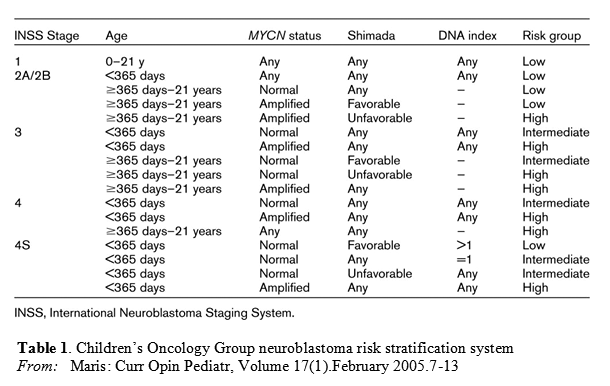
The table below outlines criteria for risk assignment based on the International Neuroblastoma Staging System (INSS), age, and biologic risk factors.
These criteria are based on the analysis of several thousands of patients treated in cooperative group protocols in Australia, Canada, Europe, Japan, and the United States.
Treatment regimes is carefully designed upon risk assessment and staging (1):
Low-risk neuroblastoma Survival rates for patients who have INSS stage 1 disease, regardless of biologic factors, are excellent with surgery alone. Chemotherapy may be needed as an effective salvage therapy for patients who have INSS stage 1 disease who relapse after surgery only.
For patients who have INSS stage
1, 2A, or 2B disease, chemotherapy should be reserved for those who have localized neuroblastoma and experience life- or organ-threatening symptoms at diagnosis or for the minority of patients who experience recurrent or progressive disease.Patients with stage 2A/2B disease with amplified MYCN are considered high risk regardless of age and histology
Stage 4S neuroblastoma withoutMYCN amplification undergoes spontaneous regression in the majority of cases. Chemotherapy or low-dose radiotherapy is used in patients who have large tumors or massive hepatomegaly.
Intermediate-risk neuroblastoma
Surgical resection and moderate–dose, multiagent chemotherapy (cyclophosphamide, doxorubicin, carboplatin, etoposide) are the standard of care. Chemo rounds are of either 4 cycles, 6 cycles, or 8 cycles, depending on histology and DNA index and response to treatment. If residual disease is present after chemotherapy and surgery, radiation therapy could be considered. However, the use of radiation is controversial.
High-risk neuroblastoma
Patients with high-risk neuroblastoma require treatment with multiagent chemotherapy, surgery, and radiotherapy. Current therapeutic protocols involve 4 phases of therapy, including induction, local control, consolidation and treatment of minimal residual disease. Induction therapy currently involves multiagent chemotherapy with non–cross-resistant profiles, including: alkylating agents, platinum, and anthracyclines and topoisomerase II inhibitors. Topoisomerase I inhibitor are also being considered. Local control involves surgical resection of primary tumor site as well as radiation to primary tumor site.
Myeloablative consolidation therapy – myeloablative consolidation therapy with etoposide, carboplatin, and melphalan have improved the outcome of patients. most centers now recommend the use of peripheral blood stem cell support over bone marrow for consolidation therapy in patients with high-risk neuroblastoma.
Other consideration – Use of 13-cis -retinoic acid in a maintenance phase of therapy. Recent data have showed improved survival in patients receiving 13-cis -RA in combination with immunomodulatory therapy with interleukin (IL)-2, granulocyte macrophage colony-stimulating factor (GM-CSF), and the chimeric anti-GD2 (gangliosidase) antibody when compared with 13-cis -RA alone.
Summary:
“Neuroblastoma is a heterogenous tumor for which biology dictates clinical behavior”. The main the goal is to have patient-tailored prognosis. Additional research in search for new therapeutics for high-risk patients is needed. Some therapies under investigation include aurora kinase inhibitors, antiangiogenic agents, histone deacetylase inhibitors, and therapeutic metaiodobenzylguanidine (MIBG). According to Park et al: “we must minimize the lasting effects of therapy,For the remaining patients who have low- and intermediate-risk disease,specifically avoiding organ damage or organ loss from surgery and organ dysfunction or risk for secondary malignancy after chemotherapy”.
Other future aspect of therapeutics may include specific inhibitor of this pathway, viz Cyclopamine and other kinase inhibitors like LY294002 for PI3K inhibition or GSK-3β inhibitors in order to inhibit the Hedgehog and the β-catenin pathways, respectively.
Reference:
1. Park JR., Eggert A and Caron H.Neuroblastoma: Biology, Prognosis and Treatment. Pediatric Clinics of North America 2008; 55(1): 97-120. http://www.sciencedirect.com/science/article/pii/S0031395507001575
2. Yamamoto K, Hayashi Y, Hanada R, et al. Mass screening and age-specific incidence of neuroblastoma in Saitama Prefecture, Japan. J Clin Oncol 1995;13(8):2033–2038. http://www.ncbi.nlm.nih.gov/pubmed/?term=Mass+screening+and+age-specific+incidence+of+neuroblastoma+in+Saitama+Prefecture%2C+Japan
3. Triche TJ. Neuroblastoma: biology confronts nosology. Arch Pathol Lab Med 1986;110(11):994–996. no available abstract.
4. Singh SK, Hawkins C, Clarke ID, et al. Identification of human brain tumour initiating cells. Nature 2004;432(7015):396–401. http://www.ncbi.nlm.nih.gov/pubmed/15549107
5. Tirode F, Laud-Duval K, Prieur A, et al. Mesenchymal stem cell features of Ewing tumors.Cancer Cell 2007;11(5):421–429. http://www.ncbi.nlm.nih.gov/pubmed/17482132
6. Shimada H, Chatten J, Newton WA Jr, et al. Histopathologic prognostic factors in neuroblastic tumors: definition of subtypes of ganglioneuroblastoma and an age-linked classification of neuroblastomas.J Natl Cancer Inst. Aug 1984;73(2):405-416. http://www.ncbi.nlm.nih.gov/pubmed/6589432
7. Norman J Lacayo and Max J Coppes. Pediatric Neuroblastoma. MedScape Reference June 2012. http://emedicine.medscape.com/article/988284-overview#a0104
8. Zhi F., Gong G., Xu Y., Zhu Y., Hu D., Yang Y and Hu Y.Activated β-catenin Forces N2A Cell-derived Neurons Back to Tumor-like Neuroblasts and Positively Correlates with a Risk for Human Neuroblastoma. Int J Biol Sci. 2012; 8(2): 289–297. http://www.ncbi.nlm.nih.gov/pmc/articles/PMC3269611/
9. Shahi MH., Sinha S., Afzal M and Castresana JS. Role of Sonic hedgehog signaling pathway in neuroblastoma development. Biology and Medicine 2009, 1 (4): Rev2, 1-6. http://biolmedonline.com/Articles/vol1_4_Rev2.pdf
10. Wickstrom M., Dyberg C, Shimokawa T., Milosevic J., Baryawno N., Fuskevag OM., Larsson R., Kogner P, Zaphiropoulos PG and Johnsen JI. Targeting the hedgehog signal transduction pathway at the level of GLI inhibits neuroblastoma cell growth in vitro and in vivo. Int J. Cancer 2013 Apr 1;132(7):1516-1524. http://www.ncbi.nlm.nih.gov/pubmed/22949014
Other related articles on Open Access Leaders in Pharmaceutical Intelligence:
1.By: Larry H Bernstein. AKT signaling variable effects. http://pharmaceuticalintelligence.com/2013/03/04/akt-signaling-variable-effects/
2. By: Aviva Lev-Ari PhD RN. Human Variome Project: encyclopedic catalog of sequence variants indexed to the human genome sequence. http://pharmaceuticalintelligence.com/2012/11/24/human-variome-project-encyclopedic-catalog-of-sequence-variants-indexed-to-the-human-genome-sequence/
3. By: Aviva Lev-Ari PhD RN. Neuroprotective Therapies: Pharmacogenomics vs Psychotropic drugs and Cholinesterase Inhibitors. http://pharmaceuticalintelligence.com/2012/11/23/neuroprotective-therapies-pharmacogenomics-vs-psychotropic-drugs-and-cholinesterase-inhibitors/
4. By: Aviva Lev-Ari PhD RN. arrayMap: Genomic Feature Mining of Cancer Entities of Copy Number Abnormalities (CNAs) Data. http://pharmaceuticalintelligence.com/2012/11/01/arraymap-genomic-feature-mining-of-cancer-entities-of-copy-number-abnormalities-cnas-data/
5. By: Venkat S Karra. $20 million Novartis deal with ‘University of Pennsylvania’ to develop Ultra-Personalized Cancer Immunotherapy. http://pharmaceuticalintelligence.com/2012/08/08/20-million-novartis-deal-with-university-of-pennsylvania-to-develop-ultra-personalized-cancer-immunotherapy/
7. By: Aviva Lev-Ari PhD RN. Acoustic Neuroma, Neurinoma or Vestibular Schwannoma: Treatment Options. http://pharmaceuticalintelligence.com/2012/10/30/acoustic-neuroma-neurinoma-or-vestibular-schwannoma-treatment-options/
8. By: Aviva Lev-Ari PhD RN. Clinical Trials on Schwannoma & Benign Intracranial Tumors Radiosurgery Treatment. http://pharmaceuticalintelligence.com/2012/10/30/clinical-trials-on-schwannoma-benign-intracranial-tumors-radiosurgery-treatment/
9. By: Aviva Lev-Ari PhD RN. Facial Nerve, Intracanalicular Meningiomas, Vestibular Schwannomas: Surgical Planning. http://pharmaceuticalintelligence.com/2012/10/15/facial-nerve-intracanalicular-meningiomas-vestibular-schwannomas-surgical-planning/