Feeds:
Posts
Comments

Posts Tagged ‘Cardiology’

Affordable Care Act became law in 2010, Cardiologists’ Practice Management Decisions Unclear

Reporter: Aviva Lev-Ari, PhD, RN

Washington-Watch

ACA Delays Decisions in Cardiology

Published: Jun 28, 2013

By Chris Kaiser, Cardiology Editor, MedPage Today
Since the Affordable Care Act became law in 2010, cardiologists have been mired in a fog of uncertainty, leading to delays in making important practice management decisions.

“When I get together with colleagues at national meetings, I get the sense that nobody really understands the future,” said Cam Patterson, MD, MBA, chief of the division of cardiology at the University of North Carolina at Chapel Hill.

That uncertainty “throws a wrench into the planning process,” including recruitment and benchmark setting, he told MedPage Today.

“It’s a major sea change,” added Thomas Tu, MD, director of the cardiac catheterization lab for the Louisville Cardiology Group in Louisville, Ky., who notes that physicians are “struggling” to find ways they can be influential in the new environment.

Patterson noted the plight of young cardiologists seeking jobs in a healthcare market unsure of how or when to make its next move.

“It’s challenging to hire new recruits when budgetary and human resources decisions are essentially on hold until there is a better understanding of what the ACA will bring,” he commented.

Regarding setting benchmarks, Patterson said the days of merely imagining your quality is as good as the next practice or hospital are gone.

Cold, hard data are the new norm, but which data and how best to collect and analyze them, as well as apply the results in a robust and meaningful way, are being worked out slowly.

“As with everyone else, we are scrambling to get a grip on what our quality measures are,” Patterson said.

Education and Prevention Will Be Key

Hospitalists, as well as advanced nurse practitioners and physician assistants, can help ease the workload due to the shortage of primary care providers, a shortage that is particularly acute in California, according to C. Noel Bairey Merz, MD, director of the Barbra Streisand Women’s Heart Center at Cedars-Sinai Medical Center in Los Angeles.

“If the reform happens the way it is intended, we should have an integrated healthcare system where primary prevention — management of hypertension, dyslipidemia, diabetes, smoking cessation counseling, and therapeutic lifestyle changes — is handled at the primary care level,” she said.

The truth of the matter, however, is that it takes twice as long to train the average general physician as it does an average nurse practitioner, and four times as long as the average physician assistant.

“It’s a lot to expect of physician extenders to practice primary care medicine,” Merz toldMedPage Today.

“A better system is the medical home model, with physician team leaders and physician extenders who work on protocols. The physician extenders would be licensed and would be able to work autonomously within a protocol,” she said.

Five years ago, the cardiology department at Geisinger Health Center in Danville, Pa., employed four nurse practitioners or clinical nurse specialists. Today, there are 12 and the department is seeking three more, according to James Blankenship, MD, vice president of the Society for Cardiovascular Angiography and Interventions, as well as an interventional cardiologist at Geisinger.

Blankenship also said that acknowledging the need for more primary care providers is to miss half of the equation. “We will need more specialists, as well.”

Given the newly insured patients coming into the system, as well as the aging Medicare population, cardiologists will be stretched pretty thin. But the field of cardiology has been instrumental in advocating teamwork among the different specialties for years, he said. “That’s a train that’s already on the tracks.”

Merz noted an expectation to see more cardiovascular care teams in response to the ACA. Such teams typically consist of a physician leader, nurse practitioners, pharmacists, behavioral experts, rehab professionals, and others.

These teams are vital for the care of high-risk patients such as survivors of angioplasty, bypass surgery, and heart failure, she said, especially since there aren’t enough cardiologists to do it all.

Echoing Blankenship, Merz said that cardiologists will probably be busier than ever as heart disease remains the leading killer among men and women as the population ages. She noted a decline in the most severe type of heart attack — ST-segment elevation MI, or STEMI — in the Medicare population, a decline that is likely multifactorial, but two reasons stand out as attributable to the decline — the use of low-dose aspirin and statin therapy for primary and secondary prevention, she said.

“At whatever level these medications are prescribed and managed — primary care physician, nurse practitioner, cardiologist — one thing is clear: they work and they should continue to be utilized at the front line of heart disease management,” Merz said.

Patients with chronic diseases already consume a great deal of healthcare resources. The other side of that coin is prevention, noted Kathy Berra, MSN, ANP, president of the Preventive Cardiovascular Nurses Association and a nurse at Stanford Prevention Research Center in Stanford, Calif.

“Prevention is a family affair. It’s been shown that when women take care of themselves, the health of the family improves.”

Emerging as one of the more important gatekeepers for women’s health — including cardiovascular health — are ob/gyns, Berra said.

Gynecologists have increased their efforts to quiz women about heart disease risk factors such as hypertension, high cholesterol, and diabetes. If red flags are apparent, patients can be referred to primary care providers, internal medicine physicians, or cardiologists.

“Ob/gyns are on the front line of women’s health. Perhaps under the ACA model, these specialists will have a closer relationship with cardiologists,” Berra told MedPage Today.

Regarding nurses and other care providers in hospitals, they need to be able to educate patients about how to take care of themselves post-discharge, how to understand the importance of their medications, and how to best re-connect with their nonhospital environment.

Readmission is at epidemic proportions and it can be reined in by patient education at the hospital level. Even pharmacists are getting more involved in patient education.

Scott & White Hospital in Temple, Texas, has a program that encourages adherence by waiving drug copays following an education session, according to James Rohack, MD, director of the Center for Healthcare Policy at Scott & White.

Patients on Seniorcare who are on five medications or more are asked if they want to participate in the program. If they agree, they meet with a pharmacist once a month for 15 to 30 minutes. The pharmacist goes over everything about the patient’s medication, listens to any concerns, and sends him or her home with new medications, waiving the copay.

“Having no copay is a great benefit for patients on fixed incomes, but it goes beyond that. A little bit of education goes a long way and if patients can be reminded once a month about the importance of taking their medications, we will have fewer hospitalizations,” Rohack said.

Accountable Care Organizations

The development of ACOs is probably one of the biggest challenges under the ACA, said Geisinger’s Blankenship.

The promise of ACOs is to have better integrated care, less fragmented care among various providers. Part of this integrated care involves incentives to minimize procedures that are either unnecessary or could be replaced with a less costly treatment.

“Having been under a fee-for-service model for a long time, some in cardiology might find the new paradigm challenging,” Blankenship suggested.

ACOs are supposed to help take the sting out of moving away from the fee-for-service model by providing the opportunity for better coordinated care — which should translate into a higher quality of care.

However, ACOs can be difficult to set up, especially from scratch, as they have a large startup cost, he said.

One of the most important aspects of an ACO is to have a solid network of primary care doctors. Patterson, at UNC Chapel Hill, said the uncertainty of whether his state will expand Medicaid has led to the “very aggressive acquisition of primary care practices.”

“The goal is to have enough physicians and patients so that we will have a low-cost ACO when we are ready to implement that model. We are going to need about 1 million patients to have an efficient ACO,” he said.

But there are also fears that the ACA will deluge cardiologists with paperwork.

“In the clinic, I spend as much time with paperwork as I do with patients — particularly with Medicare and Medicaid patients,” noted John Day, MD, director of Heart Rhythm Services at Intermountain Medical Center in Salt Lake City, Utah. “Many of us are worried we haven’t even seen the beginning of the deluge.”

The intrusion of paperwork and other government regulations tends to erode the time physicians get to spend with patients — “one of the primary reasons I wanted to be a doctor,” Day said.

In addition, Day said that he and many of his colleagues are disappointed that the ACA did not address malpractice concerns. “Perhaps it’s not so much what’s in the bill as what is not in the bill,” he said.

“Malpractice concerns are real; they scare me every day; it affects how you practice medicine. I don’t see how you can rein in costs without addressing the malpractice quagmire,” Day told MedPage Today.

Shifting Sands

“For those of us working in the trenches, we have a vague concept of the changes coming down the road,” said James A. de Lemos, MD, director of the coronary care unit at Parkland Memorial Hospital in Dallas.

“We seem to be too busy to think about the changes, which leads to one of my biggest worries — that I won’t have prepared my troops well enough,” he said.

From a clinical perspective, it’s business as usual, with de Lemos and colleagues focused on growth and the development of referrals and procedure services.

“We are concerned, however, that the entire paradigm is going to shift and what we’re building today might not be financially sound in the ACO model,” de Lemos told MedPage Today.

De Lemos, who is active in cardiovascular biomarker research, suggested that biomarkers will become more important in the ACA era of healthcare.

“It will no longer be prudent to send everyone with a complaint to a cardiologist,” he commented. “Biomarker screening may play a role as a triage method to separate out those who merit a trip to the cardiologist from those who can be treated by primary care doctors.”

Rohack made these suggestions for getting ready for the changes associated with the ACA:

  • Make sure you are actively aware of your quality measures, your individual quality measures.
  • When caring for uninsured adults, make sure you are aware of the potential benefits with health insurance exchanges, because they may qualify.
  • Make sure you are aware of impending deadlines regarding the implementation of certain aspects associated with electronic medical records because penalties can be assessed for missing deadlines.


Who Takes the Lead?

There are a lot of moving pieces that will contribute to finding success in the new era of healthcare and leaders must emerge to help forge pathways that others can follow. Hospitalists will be among those leaders, says Jeffrey H. Barsuk, MD, MS, a hospitalist and director of Simulation and Patient Safety for Graduate Medical Education at Northwestern University Feinberg School of Medicine in Chicago.

“At our hospital, we are probably the largest group of physicians involved in healthcare safety, quality, and reform,” he said.

The ACA, he told MedPage Today, is starting to have more of an impact on how he and his colleagues position themselves for the future.

In particular, the new bundled payment and fee-per-encounter models are ideal scenarios where hospitalists can make a difference by bridging gaps in the continuity of care and helping to shorten the length of stay without compromising quality.

Hospitalists can, for example, provide smoking cessation counseling for heart patients, discuss the importance of medication adherence, and check to ensure there are no contraindications to the medications or no potential for drug-drug adverse interactions.

Ultimately, though, clinicians at all levels, primary care practitioners and specialists, will need to work closely together because, as interventionalist Tu noted, government intervention that is not well thought out can backfire. The ACA might save money in the short run, Tu said, but in the long term, there is a great potential “to damage the care of patients and harm the profession of medicine. Already many good people don’t want to be in the field anymore.”

http://www.medpagetoday.com/Washington-Watch/Reform/40164?xid=nl_mpt_DHE_2013-06-29&utm_content=&utm_medium=email&utm_campaign=DailyHeadlines&utm_source=WC&eun=g406134d0r&userid=406134&email=serbangg@gmail.com&mu_id=5783576 

Read Full Post »

3D Cardiovascular Theater – Hybrid Cath Lab/OR Suite, Hybrid Surgery, Complications Post PCI and Repeat Sternotomy

Curator: Aviva Lev-Ari, PhD, RN

Article ID #70: Cardiovascular Original Research: Cases in Methodology Design for Content Curation and Co-Curation. Published on 7/19/2013

WordCloud Image Produced by Adam Tubman

This article has THREE Parts: 

Part One:  Hybrid Cath Lab/OR Suite for Hybrid Surgery

Part Two: Cardiac Surgery 

Part Three: Invasive Interventions with Complications

1. Repeat Sternotomy Post CABG and/or Aortic Valve Replacement

2. Complications Post PCI – Pump Catheter in Use

The voice of Series A Content Consultant, Justin D Pearlman, MD, PhD, FACC

The leading cause of death and disability from any cause is cardiovascular disease, principally, heart attacks and strokes. Both the heart and brain typically allow only 10 minutes or so of inadequate blood supply before starting a committed course of permanent tissue injury, progressing in severity as time goes by without successful interruption of the disease process. Thus there is great time urgency to get patients to a definitive treatment that can stop the injury and restore adequate nutrient blood supply. Many patients can benefit from a catheterization to identify blockages and insert a small balloon within the blockage to expand the narrow channel, often followed by placement of a stent (wire cage) to maintain the expanded vessel diameter. Chemicals released over time from drug-eluting stents can prevent tissue in growth that may obstruct stents. These emergeny interventions are not always successful. There may be complications from the attempt to access an entry artery, and the blockages may not be amenable to a balloon. When such limitations are encountered, the next chance to help is surgical, with continued time pressure.

The fastest way to make the transition from a diagnostic catheterization to a timely intervention is a hybrid intervention suite that offers non-invasive imaging, catheterization and surgery all in one location. The following articles present the current state of hybrid “do it all” intervention suites. Additional articles address the risks of bad outcomes from such interventions.

Part One 

Hybrid Cath Lab/OR Suite for Hybrid Surgery

In ACC.10 and i2 Summit, 59th Annual Scientific Session, 3/14-3/16, 2010, Alfred A. Bove, M.D., Ph.D., F.A.C.C., ACC President addressed the conference attendees:

Welcome to the all-new Hybrid Cath Lab/OR and 3D CV Theater. Recent developments in cardiac surgery and interventional cardiology have led to the creation of integrated, hybrid cath lab/operating rooms (OR), which provide significant advantages in the diagnosis and treatment of patients requiring cardiac procedures—helping to facilitate a rapid-response approach. These multimodality rooms are designed to support a variety of integrated surgical and endovascular procedures. We are excited to provide you with this opportunity to get a first-hand look—and feel—of the latest technologies. We hope you take the time to explore this interactive, multivendor venue. Learning is at the core of the ACC Annual Scientific Session and we invite you to expand your educational experience in this dynamic learning environment.

In the Hybrid Cath Lab/OR Suite, you’ll discover how integrating cutting edge angiographic and surgical equipment and technologies can facilitate a broad range of procedures within one location. Additionally, you will learn how hybrid suites are providing solutions that enable interventionalists and surgeons to work collaboratively to provide the best treatment options for patients. The adjoining 3D CV Theater features presentations by physicians currently performing intravascular and surgical procedures in hybrid suites. Each live presentation pairs a cardiologist with a surgeon, allowing you to hear perspectives from both sides on a variety of hybrid suite procedures and cases. In addition, the Theater offers video presentations of cases from around the world.

The ACC thanks the supporters of the Hybrid Suite for providing us with the opportunity to share this unique learning destination with you.

http://www.expo.acc.org/acc12/CUSTOM/images/ACC12/ACC.10%20Hybrid%20Suite%20Directory.pdf

Hybrid Cath Lab/OR Suite for Hybrid Surgery

Procedures Performed in a Hybrid Suite

The treatment of cardiovascular diseases has undergone a paradigm shift within the last few years, from

  • open surgery to minimally invasive surgical procedures and from
  • limited percutaneous catheter-based interventions to hybrid interventions for the entire cardiovascular tree.

The Hybrid Suite

are perfect examples of procedures that could, and should, be carried out in a hybrid OR. High-risk patients who require less invasive interventions are the best candidates for treatment in a hybrid suite.

As cardiac surgery becomes less invasive, incisions are becoming smaller and smaller, and even totally endoscopic heart surgery is now possible. Cardiac surgeons have started to perform procedures that include catheter-based skills, such as transapical valve replacement. For these operations, surgeons need more sophisticated imaging techniques, fluoroscopy and contrast injections. The hybrid OR offers all these facilities. Perhaps the most obvious and easiest procedure that can be performed in a hybrid OR is coronary revascularization combining coronary artery bypass grafting with on-table intra-operative completion angiography for quality control. If the surgeon detects a problem during the procedure, he or she can revise the graft immediately and thereby prevent potential perioperative and long-term complications. Currently, cardiologists and cardiovascular surgeons have shown special interest in so-called hybrid coronary interventions, which are combinations of minimally invasive coronary artery bypass grafting and percutaneous coronary interventions. In these procedures, cardiovascular surgeons place a left-internal mammary artery bypass graft to the left-anterior descending artery through small incisions (MIDCAB) or completely endoscopically (TECAB), while any remaining obstructed coronary arteries are treated with stents by an interventional cardiologist. This procedure is an attractive alternative to multivessel open coronary artery bypass grafting. Transcatheter heart-valve replacement and repair are especially suited to a hybrid suite because percutaneous transfemoral and transapical aortic valve repairs include risks that can only be treated successfully by immediate surgical intervention, such as coronary artery obstruction, aortic dissection and aortic perforations.

In addition, endovascular aortic stent grafting for the repair of abdominal aortic aneurysms is a suitable procedure for a hybrid operating room. Endovascular aneurysm repair has become an established alternative to open repair and is increasingly used for thoracic aorta repair as well. Some

  • emergency procedures for traumatic lesions of the thoracic aorta and
  • fulminant pulmonary embolism may also be performed in a hybrid OR. Several
  • pediatric interventions can be carried out in a hybrid suite as well, such as implantation of closure devices for atrial and ventricular septal defects in small children and
  • treatments for hypoplastic left-heart syndrome.

http://www.expo.acc.org/acc12/CUSTOM/images/ACC12/ACC.10%20Hybrid%20Suite%20Directory.pdf

In a recent article we reported on the Change in Requirement for Surgical Support by Cath Labs for performance of Nonemergent PCI without Surgical Backup, that increases the autonomy of Interventional Cardiologists. In the Hybrid OR that change is irrelevant since the presence of a Cardiac Surgeon is a fact of the division of labor between the two types of specialties. Cardiac Surgeons are involved with  percutaneous transfemoral and transapical aortic valve repairs and intervention for endoscopic aorta, AAA and Thoracic AA grafting.

AHA, ACC Change in Requirement for Surgical Support:  Class IIb -> Class III, Level of Evidence A: Supports Nonemergent PCI without Surgical Backup (Change of class IIb, level of Evidence B).

What is a Cardiovascular Hybrid Suite?

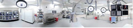
Cardiovascular hybrid suite is a state-of-the-art operating room equipped with a fully functional catheterization laboratory, thus allowing surgical procedures and catheter-based interventions to be carried out in the same room. Hybrid suites provide a place where treatments traditionally available only in a cath lab and procedures only available in an operating room can be performed together to provide patients with the best available combination of therapies for cardiovascular disease. These multidisciplinary, integrated cardiovascular procedural suites bring the best of two worlds together by combining all the advantages of a modern cath lab with an up-to-date cardiovascular surgery operating room (OR).

Hybrid suites began to evolve in the mid to late 1990s, when some groups of interventional cardiologists started sharing operating rooms with cardiovascular surgeons. The appeal of the hybrid suite concept has grown as have catheter based devices (stents, coils, balloons and lasers) have been developed that enable interventional cardiologists to advance the invasiveness and effectiveness and applications of percutaneous transcatheter interventions. The interest in these suites has also increased as cardiovascular surgeons have developed a variety of techniques for

  • Minimally invasive procedures, such as minimally invasive direct coronary artery bypass grafting (MIDCAB) or
  • Totally endoscopic coronary artery bypass grafting (TECAB).

With the advent of more tools, interventional cardiologists are becoming more like surgeons, and with less invasive tools, cardiovascular surgeons are becoming more like interventionalists. Rather than separating surgical procedures from interventional procedures performed in traditional operating rooms and cath labs, hybrid suites provide a high-tech environment that allows cardiologists and surgeons to work together to offer patients complex, minimally invasive therapies.

Some experts believe that hybrid suites represent the wave of the future in cardiovascular care and that most heart centers will eventually install hybrid suites to offer patients the latest cardiovascular procedures safely and effectively with minimal surgical trauma. The rooms can be costly to build and equip, but if a medical center is considering building a new operating room or cath lab, setting up a hybrid suite makes sense. Medical centers that have a hybrid suite available can clearly differentiate themselves in a positive way from centers that do not have such capabilities.

The Benefits of a Hybrid Suite for Medical Centers

While building a hybrid suite is more expensive than building a traditional operating room or cath lab, a hybrid suite can potentially be used for all types of cardiovascular procedures, including

  • traditional cardiac and vascular surgery,
  • interventional coronary procedures,
  • endovascular aortic procedures and
  • electrophysiology procedures.

Hybrid suites reinforce the trend in cardiovascular care toward less invasive, comprehensive hybrid procedures. Once a hybrid suite is in place, the demand for its use will likely grow due to increasing indications and referrals for these innovative treatments, many of which are increasingly covered by third-party payers.

http://www.expo.acc.org/acc12/CUSTOM/images/ACC12/ACC.10%20Hybrid%20Suite%20Directory.pdf

What Equipment is Needed?

Interventional cath labs usually have excellent imaging capabilities but lack the sterile facilities and staff needed for a formal OR, while operating rooms frequently lack high-level imaging equipment. Some of the essential equipment for a hybrid suite includes:

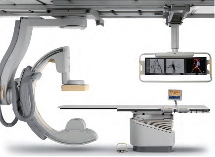
• A state-of-the-art imaging system capable of performing 3D rotational angiography, CT scanning, and ultrasound is advantageous. Floor-mounted and ceiling-mounted systems are available, but many hospitals use ceiling-mounted systems because access to the patient is slightly easier. Some ceiling-mounted systems provide 3D imaging from the surgeon’s position perpendicular to the patient. However, some hospitals prefer floor-mounted systems because having mechanical parts running above the operative field may cause dust to fall, resulting in infections. An important aspect is that the C-arm can be parked away when it is not used. This especially enhances access of the anesthesia team to the patient.

• An operating table that meets the needs of both surgeons and interventionalists by electronically integrating the table with the imaging system is also essential. These tables should have retractable rails for retractors and other surgical tools. To perform 3D imaging on the operating table, the C-arm of the imaging system should allow fast and precise rotation around the patient.

• A variety of other surgical and interventional systems and equipment may also be needed, including a robotic surgical system, a heart-lung machine, an image integration system, an endoscopic imaging system, a radiology display system, an audiovisual system to move images to different monitors and an anesthesia monitoring system, including transesophageal echocardiography. Some equipment like the integrated OR table and the angiography unit need to be fixed parts of the hybrid OR. Some equipment will be mobile in order to maintain some flexibility in workflow.

Hybrid1

Hybrid2

Hybrid3

Hybrid4

Hybrid5

Who are the Equipment Vendors?

Philips Healthcare

Phone: 800-934-7372

Email: healthcare@philips.com

Web: http://www.philips.com/healthcare

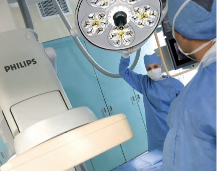
Philips is one of the world’s leading technology companies, with a long history of practical innovation and visionary design. In healthcare, we are committed to understanding the human and technological needs of patients and caregivers. We believe this understanding will help us deliver solutions that not only enable more confident diagnoses and more efficient delivery of care, but also improve the overall experience of care. We offer equipment, software and services for imaging, patient monitoring, resuscitation and much more.  A Hybrid OR can help make life simpler for the interdisciplinary teams who operate in this environment every day. As a world leader in cardiovascular X-ray, Philips has the experience and expertise to deliver the first class technology you need to perform minimally invasive procedures with speed, accuracy and confidence. A long history of innovation has enabled Philips to develop pioneering imaging solutions that really make a difference.

For example, Philips Allura Xper cardiovascular X-ray systems are designed to deliver enhanced imaging with superb performance for all cardiac projections, and our iE33 ultrasound system with Live 3D TEE and QLAB can assist interventional procedures and provide comprehensive quantitative information to support critical decisions. Our cardiology informatics solutions help you manage patient information throughout the cardiovascular care continuum. Philips solutions allow minimally invasive and catheter-based procedures to take place in the same suite as conventional cardiac surgery.

Phillips EchoNavigator – X-Ray and 3-D Ultrasound is described in:

Minimally Invasive Structural CVD Repairs: FDA grants 510(k) Clearance to Philips’ EchoNavigator – X-ray and 3-D Ultrasound Image Fused.

Intuitive Surgical, Inc. 

da Vinci.Surgery by Intuitive Surgical, Inc. 

Phone: 800-876-1310

Email: info@intusurg.com

Web: http://www.intuitivesurgical.com

Intuitive Surgical, Inc. is the global technology leader in robotic-assisted, minimally invasive surgery. The company’s da Vinci® Surgical System offers breakthrough capabilities that enable cardiac surgeons to use a minimally invasive approach and avoid median sternotomy.

Content of FDA Warning Letter, following  FDA Inspection on dates 04/01/2013 – 05/30/2013 – it discussed in

Hybrid Cath Lab/OR Suite’s da Vinci Surgical Robot of Intuitive Surgical gets FDA Warning Letter on Robot Track Record


MAVIG GmbH 

Phone: 631-266-2229,

585-247-1212 ext. 60

Email: info@mavig.com

Web: http://www.mavig.com

MAVIG’s specialty is ceiling/boom suspension systems for lighting (exam, surgical and LED), monitor-suspension systems—single, multibank (one to eight) systems and widescreen, overhead radiation shielding and contrast injector adapters. MAVIG also manufactures radiation protection products such as aprons, gloves, table-attachable lower body shields, adjustable- and fixed-height mobile and modular barriers.

Toshiba America Medical Systems, Inc.

Phone: 714-730-5000

Email: mktgcomm@tams.com

Web: http://www.medical.toshiba.com

Creating a hybrid lab may be complicated, but having an experienced partner that listens makes all the difference. Toshiba’s unique blend of hybrid experience and industry recognized Infinix™-i imaging systems for the Cath Lab.

Hybrid Cath Lab/OR Suite in Leading Hospitals in the US

  • The  Hybrid Cath Lab/OR Suite at New York Presbyterian Hospital/Columbia University Medical Center, New York, NY is presented in

Becoming a Cardiothoracic Surgeon: An Emerging Profile in the Surgery Theater and through Scientific Publications

  • The  Hybrid Cath Lab/OR Suite at Cleveland Clinic, Cleveland, Ohio is presented in

Heart Transplant (HT) Indication for Heart Failure (HF): Procedure Outcomes and Research on HF, HT @ Two Nation’s Leading HF & HT Centers

Speakers at 3D CV Theater, 2010 are working in Hospitals where Hybrid Cath Lab/OR Suite are in operations at the present time. The list include the following Hospitals with a Hybrid Cath Lab/OR Suite:

  • Vanderbilt Medical Center, Nashville, TN
  • University of Maryland Heart Center, Baltimore, MD
  • The Heart Center at Nationwide Children’s Hospital, Columbus, Ohio
  • The Robotic Surgical Center, East Carolina University Department of Surgery, Greenville, N.C.
  • University of Washington Medicine Regional Heart Center, Seattle, WA
  • Brigham and Women’s Hospital, Boston, MA
  • Saint Joseph’s Hospital and Peachtree Cardiovascular and Thoracic Surgery, Atlanta, GA
  • Emory University Hospital, Atlanta, GA
  • Beth Israel Deaconess Medical Center, Boston, MA
  • Boston Medical Center, Boston, MA
  • Mayo Graduate School of Medicine, Mayo Clinic, Rochester, MN
  • Lankenau Hospital, Lancaster, PA
  • Cardiac Non-Invasive Laboratory at Cedars-Sinai Medical Center, Los Angeles, CA
  • Robotic Surgery at St. Joseph’s Hospital, Atlanta, GA

Speakers at 3D CV Theater, 2010, included the following Cardiovascular Interventionists leading the adoption process of Hybrid Surgery in Hybrid Cath Lab/OR Suite into care modalities for cardiovascular disease:

Johannes O. Bonatti, M.D., is professor of surgery and director of coronary surgery and advanced coronary interventions at the University of Maryland Heart Center, Baltimore. He received his training in general surgery and cardiac surgery at the department of surgery at Innsbruck Medical University in Austria. Prior to his arrival at the University of Maryland, he worked at this institution as an attending surgeon and associate professor. Dr. Bonatti’s main interest is the development of minimally invasive, totally endoscopic coronary artery bypass grafting (TECAB) procedures using robotic technology.

As one of the international leaders in this field, he performed the largest series of robotic TECAB on the arrested heart, including single-, double- and triple-vessel TECAB. He has published significantly on procedure development and the implementation process of completely endoscopic coronary surgery using the da Vinci robotic system. Together with colleagues from interventional cardiology, Dr. Bonatti is working on integrated concepts for treatment of coronary artery disease. He was the first to perform a simultaneous hybrid coronary intervention using TECAB and placement of a coronary stent. He is organizing international meetings on hybrid interventions in cardiovascular medicine (http://www.icrworkshop.com). He has trained heart surgeons from around the world in the use of the da Vinci robot for heart surgery and he has introduced TECAB procedures in Austria, the Czech Republic, Greece, Turkey, India and Australia.

John G. Byrne, M.D., is the William S. Stoney Professor of Cardiac Surgery at Vanderbilt University School of Medicine and chair of the department of cardiac surgery at Vanderbilt Medical Center, Nashville, TN.

Before moving to Vanderbilt, he was associate chief and residency program director in the division of cardiac surgery at Brigham and Women’s Hospital, and associate professor of surgery at Harvard Medical School, Cambridge, MA. A graduate of the University of California, Davis, he received his medical degree in 1987 from Boston University. His postdoctoral training was completed at the University of Illinois affiliated hospitals and Brigham and Women’s Hospital in Boston.

Dr. Byrne is the author of more than 100 scientific articles on cardiac surgery and related areas. His patient care emphasis is

  • aortic root surgery,
  • coronary artery disease and
  • valve surgery

He is board-certified in general surgery and thoracic surgery.

John P. Cheatham, M.D., is director of cardiac catheterization and interventional therapy and codirector of The Heart Center at Nationwide Children’s Hospital, Columbus, Ohio. He is also the George H. Dunlap Endowed Chair in Interventional Cardiology and professor of pediatrics and internal medicine at The Ohio State University College of Medicine. Dr. Cheatham’s area of expertise is transcatheter intervention and hybrid therapy of newborns, children and adults with complex congenital heart disease. He has pioneered several new techniques and devices in non-surgical intervention and is a leader in developing hybrid therapies. He has been a principal investigator in numerous FDA-sponsored clinical trials evaluating non-surgical closure devices and stent therapy over the past two decades. Additionally, Dr. Cheatham designed the first hybrid cardiac catheterization suites and advanced imaging equipment at Nationwide Children’s Hospital. He serves as a consultant to various medical companies and proctors new transcatheter techniques and devices to other physicians around the world. Dr. Cheatham has implemented a formal physician exchange program with two of the leading medical institutions in China. In cooperation with China Red Cross, he is also the foreign director of the International Training Center for treatment of congenital heart disease in poor children. Dr. Cheatham has written more than 120 manuscripts, 16 book chapters, 300 national and international presentations and is co-editor of the book, Complications in Percutaneous Interventions for Congenital and Structural Heart Disease. After graduating from the University of Oklahoma College of Medicine, he completed his residency at Boston Children’s Hospital, followed by a fellowship in Pediatric Cardiology at Texas Children’s Hospital in Houston.

W. Randolph Chitwood, Jr., M.D., is senior associate vice chancellor for health sciences and chief of cardiovascular services at East Carolina University Department of Surgery, Greenville, N.C. Dr. Chitwood is a leading international pioneer in minimally invasive and robotic heart surgery. The Robotic Surgical Center at East Carolina University has trained more than 350 surgeons. His research activities relate to myocardial preservation, simulation in surgery and endoscopic/robotic cardiac surgery. He was the principal investigator of the FDA robotic mitral valve trials that led to approval for use in the U.S. He is the son and grandson of “southwestern Virginia mountain doctors” who set the guidelines for his professional life. He graduated from Hampden-Sydney College and received his medical degree from the University of Virginia. After medical school, he completed the surgical residency at Duke University Medical Center under David C. Sabiston, M.D., an influential surgical educator of the era. At Duke he spent 10 years training in general and cardiothoracic surgery, as well as basic science research.

After his chief residency at Duke in 1984, he was selected to begin and head the new cardiac surgery program at the East Carolina University School of Medicine. Because of his prolific publication record as a resident and clinical acumen, his initial appointment was as a full professor of surgery. Except for a two-year hiatus as the chief of cardiothoracic surgery at the University of Kentucky, he has spent his entire career at East Carolina University, where he also served as chairman of the department of surgery. In 2003, he was named to be in charge of the development of the East Carolina Heart Institute, which now includes an integrated department of cardiovascular sciences as well as a $200 million heart hospital, outpatient, research and education center.

Larry S. Dean, M.D., is director of the University of Washington Medicine Regional Heart Center and is professor of medicine and of surgery at the University of Washington School of Medicine, Seattle. In addition to general cardiology, he is an expert in cardiac catheterization and interventional cardiology. He also conducts research on stents to keep blocked heart arteries open and on ways to prevent restenosis after stents are inserted. He is currently involved in the evaluation of percutaneous aortic valve replacement. Dr. Dean earned his M.D. from the University of Alabama School of Medicine, Birmingham, and served his internship and residency at the University of Washington. He then returned to the University of Alabama Hospital for fellowships in cardiovascular disease and in angioplasty. After nearly 15 years as a faculty member at the University of Alabama, he returned to the University of Washington to direct the Regional Heart Center. He is a fellow of the American College of Cardiology and is board-certified in internal medicine, cardiovascular disease and interventional cardiology. He is also a fellow of the American Heart Association and president-elect of the Society of Cardiovascular Angiography and Interventions.

Andrew Craig Eisenhauer, M.D., is director of the interventional cardiovascular medicine service at Brigham and Women’s Hospital and assistant professor of medicine at Harvard Medical School. His specialties are

  • interventional cardiology,
  • vascular medicine and
  • congenital and inherited diseases.

He earned his medical degree at New York University School of Medicine and served a residency at Peter Bent Brigham Hospital and a fellowship at Massachusetts General Hospital. He is certified in internal medicine, cardiovascular disease and interventional cardiology. His clinical interests are

  • endovascular therapy,
  • complex coronary disease,
  • peripheral vascular disease,
  • cerebrovascular disease,
  • congenital heart disease and structural heart disease

Douglas A. Murphy, M.D., is chief of cardiothoracic surgery at Saint Joseph’s Hospital and a cardiothoracic surgeon at Peachtree Cardiovascular and Thoracic Surgery, Atlanta. His areas of interest are robotically assisted heart surgery with an emphasis on repairing the mitral valve rather than replacing it. A graduate of the University of Pennsylvania Medical School, Philadelphia, he served an internship and residency at Massachusetts General Hospital, Boston, and at Emory University, Atlanta.

Khusrow Niazi, M.D., is an assistant professor at Emory University School of Medicine and director of peripheral and carotid intervention at Emory University Hospital Midtown, Atlanta. He earned his medical degree at King Edward Medical College, Lahore, Pakistan, and served an internship at Kettering Medical Center, Dayton, Ohio, and a fellowship at William Beaumont Hospital, Royal Oak, MI. He has published papers on stenting following rotational atherectomy, small vessel stenting for coronary arteries, imaging of lower extremities and treatment of peripheral arterial disease.

Jeffrey J. Popma, M.D., is director of innovations in interventional cardiology, a senior attending physician at Beth Israel Deaconess Medical Center and an associate professor of medicine at Harvard Medical School in Boston. Dr. Popma received his bachelor’s degree in economics from Stanford University, and his M.D. from Indiana University School of Medicine. He completed his internship, residency, chief residency and fellowship at University of Texas Southwestern Medical Center. He also completed an interventional cardiology fellowship at the University of Michigan. Dr. Popma is the past president of the Society for Cardiac Angiography and Intervention and is the co-chair of the ACC Interventional Council. He sits on the editorial boards of several publications, and reviews for several cardiology periodicals. Dr. Popma has more than 300 published peer-reviewed manuscripts.

Dr. Popma also directs the BIDMC Angiographic Core Laboratory and is principal investigator for more than 65 ongoing multicenter device studies within the research laboratory. Over the past 15 years, these trials have included a broad array of new technology, including bare-metal stents, drug-eluting stents, distal-protection devices, total-occlusion devices and carotid and peripheral revascularization procedures. His primary clinical interest currently is the use of percutaneous aortic valve replacement for patients with high-risk aortic stenosis.

Robert S. Poston, M.D., is chief of cardiac surgery at Boston Medical Center and associate professor of cardiothoracic surgery at Boston University School of Medicine. He has a strong background in minimally invasive cardiac bypass surgery and is a pioneer in using robotics, specifically the da Vinci Surgical System, to treat coronary artery disease. A graduate of the Johns Hopkins School of Medicine, Baltimore, Dr. Poston completed a residency in general surgery at the University of California, San Francisco, and continued his training with a research fellowship in cardiothoracic surgery at Stanford University School of Medicine, Palo Alto, CA, and a cardiothoracic residency at the University of Pittsburgh Medical Center.

Charanjit S. Rihal, M.D., is professor of medicine and director of the cardiac catheterization laboratory at Mayo Graduate School of Medicine, Mayo Clinic, Rochester, MN. A graduate of the University of Winnipeg, Dr. Rihal did his residency and fellowship at the Mayo Graduate School of Medicine and also earned an MBA at the Carlson School of Management, University of Minnesota. His medical interests are interventional cardiology, structural heart disease interventions and the management of quality and costs in healthcare.

Timothy A. Shapiro, M.D., is director of the Interventional Cardiology Fellowship Program and campus chief, interventional cardiology, at Lankenau Hospital, Lancaster, PA. A graduate of Yale University School of Medicine, he served his residency and a fellowship at the Hospital of the University of Pennsylvania.

Robert J. Siegel, M.D., is director of the Cardiac Non-Invasive Laboratory at Cedars-Sinai Medical Center, cardiology director of the Cedars-Sinai Marfan Center, and Rexford S. Kennamer, M.D., chair in cardiac ultrasound at Cedars-Sinai Medical Center, Los Angeles. Dr. Siegel is also professor of medicine in residence at the David Geffen School of Medicine at University of California, Los Angeles. He previously served as senior staff fellow in cardiac pathology at the Heart, Lung and Blood Institute of the National Institutes of Health, Bethesda, MD. Internationally recognized as one of the leading experts in the field of cardiovascular ultrasound, Dr. Siegel specializes in cardiovascular ultrasound, including transthoracic, transesophageal and intravascular methodologies. His research interests include

  • valvular heart disease,
  • therapeutic applications of ultrasound energy,
  • transesophageal and intraoperative echocardiography, and the
  • development and use of hand-held portable echocardiographic systems for clinical innovations.

In addition, he is involved with clinical research studies related to the diagnosis, assessment and management of patients with

  • Marfan syndrome,
  • hypertrophic cardiomyopathy and
  • pericardial and valvular heart disease.

Dr. Siegel is a fellow, and has previously served as the president of the California Chapter of the American College of Cardiology and president of the Los Angeles Society of Echocardiography. He has been active in numerous cardiovascular societies, including the American Heart Association, the American College of Cardiology and the American Society of Echocardiography. Dr. Siegel received his medical degree at Baylor College of Medicine, Houston, where he developed an interest in cardiology. He completed his medical residency at Emory University and at Los Angeles County + USC Medical Center. He completed his cardiology fellowship at Harbor-UCLA Medical Center.

Over the last two years Dr. Siegel has worked extensively with live 3D transesophageal echo in the cardiac intervention center and the operating room. He and his echocardiologist colleagues, doctors Shiota, Biner, Tolstrup and Gurudevan, have worked closely at Cedars-Sinai Medical Center in Los Angeles with the interventional cardiologists, doctors Kar and Makkar, as well as with the cardiac surgeons, doctors Trento and Fontana. They use live 3D TEE extensively for the assessment of structural heart disease. In addition, it is used on a regular basis for the guidance of percutaneous procedures for mitral valve e-clip repair, mitral balloon valvuloplasty, aortic and pulmonic valve replacement, left atrial appendage exclusion by the Watchman device as well as for ASD closure.

Sudhir P. Srivastava, M.D., president of the International College of Robotic Surgery at St. Joseph’s Hospital, Atlanta, is a pioneer in performing beating heart totally endoscopic coronary artery bypass surgeries. Previously, he was assistant professor of surgery and director of robotic and minimally invasive cardiac surgery at the University of Chicago Medical Center. Dr. Srivastava specializes in robotically assisted totally endoscopic coronary artery bypass surgery. He has performed approximately 1,000 robotic cardiothoracic surgical procedures, of which 450  have been single- and multivessel beating heart totally endoscopic coronary bypass (BH TECAB) procedures. He has keen interest in hybrid coronary revascularization in TECAB patients to achieve complete revascularization.

Dr. Srivastava has helped launch robotic revascularization programs throughout the world. He has performed numerous live BH TECAB demonstrations both in the U.S. and abroad, and continues to be a presenter and invited speaker at numerous national and international scientific meetings. He earned his medical degree at the Jawahar Lal Nehru Medical College in Ajmer, India and immigrated to the U.S. in 1972. He completed his cardiothoracic surgery residency at the hospitals associated with the University of British Columbia, Vancouver, Canada.

Francis P. Sutter, D.O., F.A.C.S., is clinical professor of surgery at Thomas Jefferson University-Jefferson Medical College, Philadelphia, and chief of cardiothoracic surgery at Lankenau Hospital, Main Line Health System, Wynnewood, PA. A graduate of Philadelphia College of Osteopathic Medicine, his surgical residency and a cardiothoracic fellowship were completed at Thomas Jefferson University Hospital.

Mark R. Vesely, M.D., is an assistant professor of medicine at the University of Maryland School of Medicine. He completed medical school at the George Washington University and postgraduate training—an internal medicine residency and fellowships in cardiovascular disease and interventional cardiology—at the University of Maryland. He is board-certified in internal medicine, cardiovascular disease, nuclear cardiology and interventional cardiology. Dr. Vesely is the associate program director of the Interventional Cardiology fellowship at University of Maryland. His special interests include the partnered approach (interventional cardiologists and cardiac surgeons) for hybrid coronary revascularization and structural heart disease interventions. Additional research interests include investigation of techniques to minimize acute myocardial infarction injury with ventricular-assist devices and adult stem cell therapies.

David X. M. Zhao, M.D., Ph.D., is an associate professor of medicine and cardiac surgery, Harry and Shelley Page Chair in Interventional Cardiology, director of the Cardiac Catheterization Laboratories and interventional cardiology director of the Interventional Cardiology Fellowship Training Program, Vanderbilt University School of Medicine, Nashville, TN. He earned his medical degree at Shanghai Medical University, Shanghai, P.R. China, and his Ph.D. in immunology at Queensland University, Brisbane, Australia. His postdoctoral training was at Zhongshan Hospital, Shanghai Medical University, Shanghai, P.R. China, The Prince Charles Hospital, Brisbane, Australia, and Brigham and Women’s Hospital, Boston.

http://www.expo.acc.org/acc12/CUSTOM/images/ACC12/ACC.10%20Hybrid%20Suite%20Directory.pdf

Part Two

Cardiac Surgery


Cardiac Surgery @ Cleveland Clinic: Traditional OR & Hybrid Cath Lab/OR Suite

Nation #1 for 19 consecutive years – The Heart Center: Miller Family Heart & Vascular Institute @ Cleveland Clinic

The Sydell and Arnold Miller Family Heart & Vascular Institute is one of the largest, most experienced cardiovascular specialty groups in the world. Our physicians are committed to providing the most advanced diagnostic and treatment options, better outcomes and improved quality of life. U.S.News & World Reporthas ranked Cleveland Clinic as the No.1 heart program in America every year since 1995.

Departments & Centers:

Below we present two articles on Cardiac Surgery @ Mayo Clinic 

Cardiac Surgery @ Mayo Clinic: Traditional OR & Hybrid Cath Lab/OR Suite 

Coronary Reperfusion Therapies: CABG vs PCI – Mayo Clinic preprocedure Risk Score (MCRS) for Prediction of in-Hospital Mortality after CABG or PCI

Larry H. Bernstein, MD, FCAP and Aviva Lev-Ari, PhD, RN

Comparison of the 10-year and 15-year survivals after CABG demonstrated benefit from a change in graft sources used at the Mayo Clinic and widely adapted by others: vascular grafts from the left internal mammary artery (LIMA) instead of just leg veins, for multiple grafts (up to 3), LIMA-to-LAD plus grafts using LIMA or radial artery vs LIMA/saphenous vein (SV).

CABG Survival in Multivessel Disease Patients: Comparison of Arterial Bypass Grafts vs Saphenous Venous Grafts

Larry H. Bernstein, MD, FCAP and Aviva Lev-Ari, PhD, RN

Part Three 

Invasive Interventions with Complications

In the following article we covered multiple etiologies for cardiovascular complications related to invasive interventions: cardiovascular and peripheral arterial or peri- and post- cardiac surgery of the open heart type.

Cardiovascular Complications: Death from Reoperative Sternotomy after prior CABG, MVR, AVR, or Radiation; Complications of PCI; Sepsis from Cardiovascular Interventions

Justin D Pearlman, MD, PhD, FACC and Aviva Lev-Ari, PhD, RN

http://pharmaceuticalintelligence.com/2013/07/23/cardiovascular-complications-of-multiple-etiologies-repeat-sternotomy-post-cabg-or-avr-post-pci-pad-endoscopy-andor-resultant-of-systemic-sepsis/

This article covers types of Cardiovascular Complications derived from the following THREE types of assault on the Human body, two related to cardiac invasive interventions, the last due to its systemic nature is taking a fatal Cardiac toll: the Sepsis condition causing cardiac failure.

Three types of Cardiovascular Complications:

I. Risk of Injury During Repeat Sternotomy – following CABG orAortic Valve Replacement, both done in Open Heart Surgery

II. Complications After Percutaneous Coronary intervention (PCI) and endovascular surgery for Peripheral Artery Disease (PAD)

  • (a) Post PCI, and
  • (b) PAD Endovascular Interventions: Carotid Artery Endarterectomy

III. Cardiac Failure During Systemic Sepsis

This article does NOT cover the following two types of Cardiovascular Complications:

1. Trauma Injury causing cardiac arrest, lung collapse or cardiogenic shock

2. Surgical Complication of Non-cardiac surgery type causing cardiac arrest, i.e, Surgery of Joint Replacement causing sepsis causing death or death caused by complications of surgery i.e., blood loss, viral infection, emboli, thrombus, stroke, or cardiogenic shock not related to Cardiovascular and Cardiac invasive interventions

The e-Reader is advised to consider the following expansion on the subject matter carrying the discussion to additional related clinical issues:

Larry H Bernstein, Advanced Topics in Sepsis and the Cardiovascular System at its End Stage

http://pharmaceuticalintelligence.com/2013/08/18/advanced-topics-in-sepsis-and-the-cardiovascular-system-at-its-end-stage/

Bernstein, HL, Pearlman, JD and A. Lev-Ari  Alternative Designs for the Human Artificial Heart: The Patients in Heart Failure – Outcomes of Transplant (donor)/Implantation (artificial) and Monitoring Technologies for the Transplant/Implant Patient in the Community

http://pharmaceuticalintelligence.com/2013/08/05/alternative-designs-for-the-human-artificial-heart-the-patients-in-heart-failure-outcomes-of-transplant-donorimplantation-artificial-and-monitoring-technologies-for-the-transplantimplant-pat/

Pearlman, JD and A. Lev-Ari Cardiac Resynchronization Therapy (CRT) to Arrhythmias: Pacemaker/Implantable Cardioverter Defibrillator (ICD) Insertion

http://pharmaceuticalintelligence.com/2013/07/22/cardiac-resynchronization-therapy-crt-to-arrhythmias-pacemakerimplantable-cardioverter-defibrillator-icd-insertion/

Read Full Post »

Reporter: Aviva Lev-Ari, PhD, RN

Heart Failure and Dietary Sodium: Do we know as much as we think?

Samar Sheth,1 Alan B. Weder2 and Scott L. Hummel2,3; 1. Department of Internal Medicine, University of Michigan; 2. Division of Cardiovascular Medicine, Department of Medicine, University of Michigan Medical School; 3. Staff Cardiologist, Department of Veterans Affairs Medical Center, Ann Arbor, Michigan

Treatment Strategies Cardiology, Volume 3 Issue 1, 44-48

Depolarisation Reserve: A New Identification Concept of Responders to Biventricular Stimulation.

Philippe Chevalier and Alina Scridon; Centre de Référence des Troubles du Rythme Cardiaque Héréditaires, Hôpital Cardiologique Louis Pradel, Bron Cedex

Treatment Strategies Cardiology, Volume 3 Issue 2, 65-70

Selective Site Pacing During Cardiac Resynchronization Therapy

Leonidas Tzogias and Attila Roka, Hospital of St. Raphael, New Haven, Connecticut

Treatment Strategies Cardiology, Volume 4 Issue 1, 37-40

The Clinical Utility of BNP with Heart Failure in the European Physician Oce Setting

Treatment Strategies Cardiology, Volume 4 Issue 2, 49-52

The Treatment of Virus-associated Inammatory Cardiomyopathy

Uwe Kühl and Heinz-Peter Schultheiss, Department of Cardiology and Pulmonology, Charité – Universitätsmedizin Berlin, Campus Benjamin Franklin, Berlin

Treatment Strategies Cardiology, Volume 4 Issue 2, 63-68

TREATMENT STRATEGIES – CARDIOLOGY

Treatment Strategies – Cardiology is a print (on request) and online eBook publication that provides its readership with a collection of comprehensive and thought-provoking articles from the most respected key opinion leaders, leading doctors and authorities in the cardiology field. The series informs and educates clinicians on the latest therapeutic and technological advances. Published in line with the foremost cardiology congresses, the editorial content includes an unbiased, independent inbound supplement reviewing either the ESC or ACC congress. The review is dedicated to bringing readers the latest cardiology breaking news, exhibition highlights, awards and prizes and research developments from the key-note presentations at the congress.

Treatment Strategies – Cardiology (European and US edition) is available online as a free-to-view eBook providing its readers with an exciting interactive experience. Easily accessible, user friendly and free-to-print, the eBook can provide you with a wide range of dynamic features, including links to external websites, newsletters and email addresses to direct readers to your specialist products and services forming strong links with media partnerships. Importantly, an eBook can be sent out to clients worldwide in an organised and professional layout that allows a far-reaching distribution of your products. The eBook also offers you the opportunity to include, on any page, videos and podcasts of current events such as symposium proceedings that you may wish to highlight for the readership. The latest eBook also permits the advertiser to track statistical data for each page and publication; including the number of unique visits, click though pages, geographical location of the visitor and average time spent viewing.

Advisory Panel

 Treatment Strategies – Cardiology is shaped by an advisory panel of world-renowned specialists from the leading associations and societies, including experts from:

American College of Cardiology (ACC)
American Society of Echocardiography (ASE)
European Association of Cardiothoracic Anaesthesiologists (EACTA)
European Association of Echocardiography (EAE)

European Society of Cardiology (ESC)

Nicholas Antony Boon, Consultant Cardiologist, Royal Infirmary of Edinburgh, Honorary Reader, University of Edinburgh and former President of the British Cardiovascular Society

 (BCS)

Carl J. Pepine, Professor of Medicine, Division of Cardiovascular Medicine, University of Florida; Past-President, American College of Cardiology (ACC)

Bertram Pitt, University of Michigan Medical Center, William Beaumont Hospital, Ann Arbor, Michigan; President of the Michigan Chapter of American College of Cardiology, Chairman, Young Investigator’s Award Committee of the American College of Cardiology, Chairman of the Reveal Committee of the ACC

For our full Editorial Advisory Panel list please click here.

US Edition

Treatment Strategies – Cardiology Volume 5 Issue 1 will include an unbiased, independent inbound supplement reviewing the ACC Congress, taking place in San Francisco in March. The inbound supplement will present the readers with the latest news, exhibition highlights, awards and prizes and research developments from the key-note presentations from the ACC. The publication will be published in April 2013.

European Edition

Treatment Strategies – Cardiology Volume 5 Issue 2 will include an independently written review of the ESC congress taking place in Amsterdam in August. The review is dedicated to bringing readers the latest cardiology news, exhibition highlights, awards and prizes and research developments from the key-note presentations at the congress. Please click on the image (below) to view the media pack as an eBook. Volume 5 Issue 2 will be published in September 2013.

SOURCE

http://www.cambridgeresearchcentre.co.uk/all-publications/cardiology/

Read Full Post »

Fractional Flow Reserve (FFR) & Instantaneous wave-free ratio (iFR): An Evaluation of Catheterization Lab Tools (Software Validation) for Ischemic Assessment (Diagnostics) – Change in Paradigm: The RIGHT vessel not ALL vessels

Reporters: Justin D Pearlman, MD, PhD, FACC and Aviva Lev-Ari, PhD, RN

The evaluation of coronary artery disease (blocked arterial blood supply to heart muscle) by stress tests is functional: is there enough blockage to starve a region of muscle when demand is high? By starvation we mean ischemia – insufficient supply to meet metabolic demands as expressed by consequent functional impairment, e.g., metabolic, electric, mechanical. In the catheterization laboratory, the evaluation is primarily anatomic – is there a bite missing in the silhouette of a coronary artery consistent with a significant impediment to blood delivery beyond the lesion? Half of all heart attacks are due to such lesions. The other half derive from non-obstructive but unstable lesions that may crack, bleed into the vessel wall, and suddenly clot,  blocking the blood flow. Coronary lesions that restrict blood delivery sufficient to cause demand ischemia (insufficient blood supply to meet high demands) cause angina pectoris.

The focus of flow reserve is to add an assessment of functional significance to anatomic lesions observed at catheterization. The widespread practice of deciding on intervention based on percent diameter reduction imposed by a lesion is obviously flawed. The flow limitation imposed by a lesion depends on its length and shape (entrance and exit effects on flow pattern), not merely the diameter reduction expressed as a percent, that is currently deemed the decision-making “degree of stenosis.” In the midst of a medical emergency heart attack, the target of intervention is the culprit lesion, the one that best explains why a region of muscle is dying. In that case, timely intervention is potentially life saving and does not depend on or wait for measurements. In the non-emergent setting, it is much harder to establish benefit from intervention. If a lesion is flow limiting and explanatory for angina pectoris, then intervention to relieve obstruction offers pain relief and may improve exertion tolerance. In relatively rare circumstances (3-vessel obstruction, left main obstruction) intervention may avoid heart attack and extend life expectancy, but with as good or better outcomes from bypass surgery. Most elective catheter interventions (balloon angioplasty, stent placement) have failed  to establish improved life expectancy or even superiority over medication. Furthermore, stent placement obligates use of strong anti-platelet medications (e.g., aspirin plus clopidogrel) that elevate risk of serious bleeding and stroke. Therefore it is reasonable to require further evidence that a coronary lesion is obstructive and consequential that just percent stenosis (narrowing) as indication for intervention. Fractional flow reserve offers such confirmation of lesion significance.

Fractional flow reserve may prove useful also in the definition of heart attack (myocardial infarction): New Definition of MI Unveiled, Fractional Flow Reserve (FFR) CT for Tagging Ischemia

June 21, 2013

Recorded: May 23, 2013

VIEW VIDEO

http://www.theheart.org/editorial-program/1550783.do?utm_medium=email&utm_source=20130704_heartwire&utm_campaign=newsletter

The use of FFR is relatively widespread in Europe, while its usage is beginning to catch up in the US. But for what reasons? Drs Roxana Mehran,Justin Davies, and Ron Waksman gathered recently to share their thoughts on the role of functional assessment in the cath lab, to evaluate FFR and its alternatives, and discuss what testing the future might hold for the optimal detection of culprit lesions.

Host

Roxana Mehran MD
Professor of Medicine, Divisions of Cardiology and Health Evidence and Policy
Director, Interventional Cardiovascular Research and Clinical Trials
The Zena and Michael A Wiener Cardiovascular Institute
Icahn School of Medicine at Mount Sinai
New York, NY

Dr Mehran has served as an advisor or consultant for AstraZeneca Pharmaceuticals, Regado Biosciences, Abbott Cardiovascular Systems, Janssen (Johnson & Johnson), Merck, and Maya Medical. She has received grants for clinical research from Bristol-Myers Squibb, Sanofi, the Medicines Company, and Lilly/DSI.

Guests

Justin Davies MBBS PhD
Consultant Interventional Cardiologist
Hammersmith Hospital
Imperial College
London, United Kingdom

Dr Davies has served as an advisor or consultant for Volcano and Medtronic. He has served as a speaker or a member of a speakers’ bureau for Medtronic and has received grants for clinical research from Volcano, Medtronic, and Abbott.

Ron Waksman MD
Director, Clinical Research and Advanced Education
MedStar Cardiovascular Research Network/Cleveland Clinic Heart and Vascular Institute
Clinical Professor of Medicine (Cardiology)
Georgetown University
Washington, DC

Dr Waksman has served as a speaker or a member of a speakers’ bureau for AstraZeneca Pharmaceuticals, Boston Scientific, and Medtronic.

Roxana Mehran, MD: Hello. My name is Roxana Mehran from Mount Sinai

School of Medicine in New York. It’s my pleasure to welcome you to this editorial

program in which we will look into how FFR usage differs in Europe vs the United

States.

I’m joined by my colleagues Justin Davies from Imperial College London and, of

course, Ron Waksman, an old friend, from the Washington Hospital Center in

DC. Welcome.

Ron Waksman, MD: Thank you.

Dr Mehran: We thought today we’d have a conversation about fractional flow

reserve and the use of functional studies in guiding our interventional

procedures. This has become a very interesting and important modality that has

been incorporated, now, more and more into the cath lab.

Let’s begin, Justin. Maybe you could tell us: what is FFR? How do we use it?

What do you think about it? How important is it to have? Does every cath lab

need to have FFR?

Justin E Davies, MD: I think that it provides cath labs with an objective measure

of stenosis assessment that is quick and relatively easy to use. Often you find

people don’t come to cath labs with preprocedural ischemic assessment, so it

enables a physician in the lab to see if there is ischemia and to effectively

document that. It has clearly got a good, strong evidence base, which has been

developed over a number of years with the founding studies of DEFER and

FAME—and FAME-2, now, which has led to the technique getting into guidelines

and which has propelled its use to widespread practice.

Dr Mehran: There’s no question that functional assessment is quite important,

but what do you think about patients coming into the cath lab, especially in the

United States, without an ischemic assessment? How often are you seeing that

and would you even use [FFR] in a patient who has got a 90% stenosis or 80%

stenosis, Ron?

Dr Waksman: I think we are in a period of transformational culture, now, in terms

of appropriate-use criteria and justifying every lesion that we are doing. I think

that interventional cardiologists are on the defensive—because we have to justify

almost any angioplasty that we are doing, especially with an intermediate lesion.

I think physicians are more flexible in using FFR and looking it as guidance to

support their decision-making.

I would say, still, if it is a 90% lesion even without a functional test I don’t think it

is required, but the 90% is on the eyes of the beholder, so you know that if you

take it to the core lab it is not going to be probably 90%, it is usually going to be

less than that.

And as you know, we have been scrutinized by looking at films and, again, [we

need to] justify. It is true that in the past the algorithm was having a functional

test and going to the cath lab. But this [step] has delayed things, and now people

presenting with some chest discomfort, [who] have risk factors, they sometimes

would be sent to the cath lab as the first method of assessment. I think we have

to take this more carefully and incorporate a functional ischemic assessment in

the cath lab—especially when those lesions are not necessarily unambiguous,

we don’t clearly know that they would derive ischemia.

Dr Mehran: It seems like that is really the way to go. You talked to us about

FAME, FAME-2, [that FFR is] the only modality in the cath lab that actually

improves hard end points like death and MI. I think that was how it got into the

guidelines, obviously. Very important studies. But at what cost? Can we afford to

do this in every single patient who presents with, let’s say, multivessel disease,

as they did in FAME?

Dr Davies: I think, actually, that this is a tremendous opportunity because I think

for us as cardiologists, as Ron said, it is very easy to get yourself into a little bit of

a hole stenting lesions that are not as significant as you may think, and I think

this obviously provides a justification and a safety net for people to deploy stents.

But also I think increasingly going forward when you are taking on potentially

more challenging techniques and you have got three-vessel disease, [FFR]

enables us to assess multivessel disease and perhaps convert a three-vessel

PCI into a two- or [even] a single-vessel PCI, which may move them from getting

a CABG to angioplasty.

Dr Mehran: So actually decreasing the number of stents. Reducing your

devices—hopefully even radiation exposure and contrast media. If you actually

just do an FFR and say, okay, I am done. But you know that we have all

[discussed]: if you want to treat the lesion you use an IVUS; if you don’t want to

treat the lesion you use an FFR. What do you think about that, Ron? Is that

something that is going on in your lab?

Dr Waksman: No. We try to stay away [from that]. I know that is said. But I

would still think that FFR is oversold. For the controversy, I would [argue] that

those studies were investigator-sponsored studies and I don’t think the data are

so relevant today. In DEFER there was a balloon angioplasty. Even in FAME-1, it

was with first-generation stents, and it was not really practice to go after every

lesion.

I think we have to take these [data] with a grain of salt. To my view, not

everything is definitive. Nevertheless, we do see uptake of the FFR usage in the

lab and for something that used to be under 3% in the US—only a couple of

years ago—it is now reaching up to 20%. More and more people are using [FFR].

There are other modalities that you can use. I think that we try to do a conversion

between the anatomical [minimum lumen area] (MLA) to the IVUS / FFR. It is

controversial, but it is another option. I think we need to learn to use the tool

when we really need it, not to be obsessed with it.

Often, we would have a scenario [where] a patient presents with chest pain. It is

classical angina. It is relieved by nitroglycerin. You have what you think is about

70% lesion in the proximal LAD and then you stick [in] the FFR wire and you get

0.81. Then you have a problem. You can repeat the study. You shoot another

adenosine and now it is 0.79. Then you shoot another one and it is 0.80. It is very

hard for me when you have a binary number to make a decision [based] on that

number. I don’t think we should lose our clinical judgment. It is a nice tool, but

don’t abuse it. Use it when it is really helpful.

Dr Davies: I will share my view. I have to say I agree wholeheartedly with Ron.

To me, if you ask them what are the most important numbers in FFR, people will

say: 0.80 or 0.75. Actually, I think the two most important numbers are 0 when it

is completely occluded and 1. As you get nearer to 0.80 you know that you are

approaching a place where it is going to be likely ischemia and a high probability

of events. Ron is absolutely right. If you have a type A, 90% lesion and you have

an FFR of 0.81—I know [that] in the US you are in difficulties, at the moment,

with these kinds of lesions. I think with the commonsense kind of medical

entirety/holistic approach that would say you should probably stent these people.

Dr Mehran: But isn’t that just so important? Those are really important points

because it is not about the dichotomous number of 0.80 or 0.75. It is about the

clinician and what they feel the scenario is and how it all fits together.

Dr Waksman: I would say even though it is getting very hard to support by

studies but I may not have an 80% or 70% lesion. I would rather have an FFR of

0.92 or 0.96 than 0.81.

Dr Davies: Absolutely.

Dr Mehran: Of course.

Dr Waksman: If I have the choice. You also have to realize that the stents of

today are not the stents of yesterday. I think we see [many fewer] events. I think

that the price of stenting and the likelihood that we would have events is much

lower than in the past. So even if we deviate a little bit, it I don’t think we do an

injustice to the patients [or] put them at high risk. I would challenge that if you

would [do] the same study today as DEFER, with the new second-generation

stents, I am not sure that the results would be the same—as robust—as they

were in the past. As a matter of fact, if you are looking even [at] FAME-2, at the

two groups—[those who] were medically treated and those who were

[interventionally] treated, the curves were actually very similar. I would challenge

that you [would not be replicating these results] with second-generation [stents].

You have to be taking [FFR] when you really need. I don’t think [that]

systematically you go [to] every lesion and if it meets the criteria of 0.80, you

don’t treat. If it is less, you treat.

Dr Davies: And there are some people who see this as a weakness, but we

don’t do that in any other form of medicine we practice, and I see it as a strength

that you get a continuous range of values. I think the one thing, which we have

also done, is using these techniques purely as an outcome base. But really if you

look back they were designed to describe ischemia and chest pain, so really it is

a very good tool for seeing if chest pain is genuine and if it is likely to benefit from

a stent.

Dr Mehran: That’s right.

Dr. Davies: And that hasn’t been thoroughly explored since the original studies.

Dr Mehran: Those are really excellent points. Now, we have alternatives to FFR.

We talked a little bit about IVUS, but we also know now that, Justin, you have

done a lot of the work on [instantaneous wave-free ratio] iFR. Maybe you can just

tell us: what is iFR? How is it different from FFR and where are we in that? Do

you believe it will replace FFR?

Dr Davies: iFR is a technique which is very similar to perform as FFR. You use

the same pressure wire. It is a software change in the console that essentially

allows us to make a measurement of stenosis severity over a particular phase of

the cardiac cycle without the need for a drug. It typically takes a few seconds to

measure and is very quick.

There have been, to date, about 3000 patients studied, in five clinical trials,

which—with the exception of one study—have all shown, essentially, the same

findings.

And we know at the EuroPCR meeting this was, again, replicated this week. At

the moment, we are in a situation where we are advocating the use of a hybrid

approach, similar to the big RESOLVE study, which essentially says that if you

are above an iFR threshold of 0.93 you are safe to defer and below 0.86, to treat.

That gives you about a 90% to 95% agreement with FFR and overall

classification, and the ADVISE-II study shows it saves about 70% of adenosine.

There are potentially quite marked savings in the cath lab.

I think this is out there in clinical practice—in a limited release, in terms of certain

labs around the world on three continents. The general experience has been

very, very good from people in terms of just facilitating the use of physiology.

What I mean by that: I take centers that were relatively small users of FFR and

they found they have done the same number of cases in three months as they

would have done over the whole year. If you ask them why, it’s because it lowers

the burden of doing [the cases]. I think if we then move on to doing triple-vesseldisease

assessment I think it takes five seconds of each.

Dr Mehran: I think there is no question that taking away the adenosine is music

to a lot of people’s ears. We all know that adenosine is not being given perfectly

right in certain laboratories. It really should be an intravenous injection. There is

time needed for nurses to put it together, to put in the IV, the intra-arterial

[injection] has been refuted, etc.

But when I look at iFR I start to think that we are pushing ourselves toward what

Ron was just talking about. I think the validations need to take place. It would be

great to have technology that is well validated, studied, that actually correlates

with events without adenosine. I think that part of it is brilliant. But are we there

today?

Dr Davies: We have had a very good response taking the stuff from the research

lab into the cath lab, so this is what we are using this as a tool, certainly, within

the framework of studies. I think now we are in a position to do large studies. I

will give you an example: we asked all of the investigators who have got these

machines if they are willing to contribute to analysis at the time of PCI. [In the

space of] for four weeks—most of them only had the device that length of time,

they managed to get together 400 cases. [This shows that] doing very large

studies of 1500 or 2000 patients is extremely feasible and very easy to do.

Dr Mehran: I hope you are designing them and actually performing them.

Ron, what do you think about iFR? I love to hear your scrutiny.

Dr Waksman: I think it hasn’t been validated, obviously. I [would] like to get rid of

the adenosine. But I like to see reproducibility of any test. Again, I would say, we

don’t have to lose our brains just because we have numbers. We have a patient

in front of us. He has symptoms and we have lesions that we have to treat.

Obviously if you have a proximal lesion, it’s going to behave differently than a mid

or distal vessel. We know that, for example, if you look at most of the studies, at

just a circumflex of FFR. Most of them will be above 8.0. But you take most

proximal LADs, they probably would fit more into the predictability of ischemia vs

nonischemia. We have to, again, use our brains when we use the numbers and

understand what they mean.

I think that there will be other technologies that [will] try to be alternatives to

FFR—not that FFR is necessarily bad, but there are other ways that you can do

it. There is the heart flow option with a CT. I still think that IVUS is an option.

Not all of them are ideal, but it gives you a variety of options. The message is: we

are trying to treat only the vessels that need to be treated. I think that can also

change the paradigm of treatment. For example, we may turn “three vessel” to

“one vessel” and change the whole syntax score and move patients from CABG

to PCI—which is very attractive for interventional cardiologists.

One other thing that is interesting: recently I heard that SJ Park was presenting a

systematic use on all patients with FFR—which is amazing! It is over 70%. It was

not a randomized study but what he did show by systematically using FFR in his

practice [is that] he reduced, by a lot, the number of PCIs, the number of stents,

and the outcome of those patients was good. You have to compare it in a

randomized fashion. What would be the alternative? And that is the challenge.

You really have to show [efficacy] in a randomized clinical trial. I recognize there

were studies in the past, but they have limitations. I think we [are] moving to

another phase that this has to be tested.

Dr Mehran: Quickly touching on what you just said about noninvasive functional

assessments. More and more we are getting patients who come in with a

multislice CT. Can we use that technology to actually do some of the functional

assessment right then and there? The DEFACTO trial, in my mind, is a negative

study. Where are we with that technology?

Dr Davies: You are absolutely right to say a lot of these patients have CTs and it

is a question of whether we can use information from that CT. As Ron said, there

is HeartFlow technology, which enables you to effectively get a noninvasive

preprocedural virtual FFR measurement. Certainly from a theoretical perspective,

it should be possible to do these calculations. I think the problem that the

HeartFlow team has is translating the computational flow dynamic theory in a

perfect research environment into the clinical practice of getting good-quality

CTs. I think there is probably more work in progress to see that really translate.

Dr Mehran: That’s right.

Dr Waksman: But what we are seeing in the US right now is [that] there is a

decline in the nuclear test and there is increased uptake in FFR. There is a

change in paradigm because of many reasons. Some of them have nothing to do

with medicine. It is more the reimbursement. Because reimbursement went down

on nuclear tests, we see less nuclear tests being performed. Now we are getting

the patients actually to be assessed in the lab and we get [to have] more

confidence with FFR or other technologies. I think we are shifting the traditional

assessment of ischemia, which was in the old days was nuclear or dobutamine

echo, more into those [tests performed] in the lab. And I do believe that the fact

that studies were negative is not the end of the story. We still have to fine-tune.

This is all about software validation and finding the sweet spot. What is the

window that allows you to get good matching? That you can feel comfortable

[with]?

Dr Mehran: So great technology to look forward to in the future. We are looking

for that kind of noninvasive assessment of functional studies. Let’s now turn to

why we are really here, which is about the regional differences of FFR. In the UK,

in Europe, in the United States, are there regional differences? Let’s better

understand that. And, if so, why? Justin, maybe you could tell us about the UK

and Europe?

Dr Davies: I think [FFR penetration] is somewhere between 15% and 20% of

cases in the UK, which is very high on a worldwide basis. I think some of that has

to do with reimbursement and some of it is to do with the way that doctors are

reimbursed, as well. In terms of the UK, if we put a stent in or not, it has no net

effect on the income to ourselves. So it is very easy for us to follow guidelines

and, in fact, if we don’t, [we] get rapped around the knuckles and told off for not

doing so. I think that is a strong incentive to do it.

I think there are obviously differences from us in other parts of the world with

regard to the reimbursement—the cost of the bits of kit and the availability of the

kit. In some labs around the world, and some territories, getting adenosine is

simply not possible or it is [so] outrageously expensive that people would just say

I am not going to make this measurement and they defer to angiography or, as

Ron said, to IVUS.

Dr Mehran: It seems like the penetration is a little bit the same between the UK

and US? What do you think about the United States?

Dr Waksman: Not yet. I would actually take from what Justin just said. I think

that the main motivation in Europe for the penetration of FFR was monetary. It

was actually to save money to the operator, to the cath lab. This never was the

case in the US. I think the in the US of the uptick is more related to

the appropriateness[-criteria guidelines] and to be on the defensive. The

interventionalist now has to defend himself for every procedure [he is] doing and

to have a backup [as to] why they did this procedure. That was not the case in

the European continent, [where] the main drive was to reduce overall costs on

the capitation system. I always had a problem with that because this is the way

that it was presented and I think that we should give the best to our patients.

We also have to realize that the reason for the uptick could be because of

appropriateness. We actually learned to turn this into a helpful tool for us [and] to

use it not just for those ancillary decisions—that probably should not be related to

the patient (whether it is a cost or whether it is appropriateness), but [also for]

what [it is] really good [for]: to see how we can utilize [it] to do the right procedure

to the right vessel. So it’s another tool.

But as I mentioned before, I think we are seeing an uptick. I don’t think we are

crossing the 20% and we are not as broad as in Europe. When are we going to

get there? It is a question of how much push we are going to see, but one thing

you see [now is] more companies providing FFR systems. That means that there

will be more reps in the labs and more opportunities, and that is usually what will

populate the usage of the device. I have no doubt that we are going to continue

to see an increase.

Dr Davies: It is interesting. I know from the US, and some of the data there,

there is a big difference between diagnostic use of FFR and actually the PCI use.

It is almost used in the US to justify PCI, and I think the angiography use is

somewhere around 3%. If you take that study that SJ Park has just done and you

compare that 70% percent that he was doing with the 3%, there is obviously a

huge potential.

Dr Mehran: Isn’t it interesting that maybe the driving force of doing a functional

assessment in the lab is different in the UK vs the US or Europe vs the United

States? I believe that at the end of the day they both will come to the same

conclusion of doing the right procedure to the right patient, making the correct

diagnosis, treating the right lesion for the right patient, but at the end of the day

actually decreasing costs. While maybe we are seeing in the United States that

appropriate-use criteria is why we are doing this, it has, perhaps, to do with

capitation, as well, for us in the United States, and enhancing the cost in the

system, hopefully, with this kind of functional assessment?

Dr Waksman: I think there is one more important collateral benefit from using

the FFR. That I would say is that we are changing the paradigm. In the old days

we thought we have to treat all the three vessels; we have to have complete

revascularization. I think FFR taught us that actually we may not need to treat all

the three vessels. That is a big advantage of technology. As we are using it we

are starting to see maybe we just have to treat the culprit lesion and move on

and then leave the others either on medical therapy or not treat them at all. That

is a huge change in paradigm.

Dr Mehran: This has been a fantastic conversation among the three of us, and I

just want to close and I want you each to close for me. What do you think is the

future of FFR? What should we be looking forward to as alternatives and what

incorporation of functional assessment in the cath lab as we move to the next

decade of interventional cardiology. Ron?

Dr Waksman: I think that the FFR will continue to grow. I think that there is a

good future for iFR without the adenosine, the wireless, and better wires that you

can use. I think you [could] incorporate an IVUS probe in them—FFR on an IVUS

probe, so you can do both. I think that the combination of anatomical and

physiological [testing] is important. We learned that with IVUS you can optimize

the outcome of the PCI, not only just determine whether you treat or not. So the

future is there. We are coming to do more sophisticated PCIs, and these data will

help us to get better outcomes and also to triage the patients to what should be

the treatment of choice. In the long run—even though the short run shows

reduction of the PCIs—if we use [FFR] carefully it will open us or enable us to do

more complex patients and meet the outcome that is expected.

Dr Mehran: That’s great. Justin?

Dr Davies: I would agree with Ron’s thoughts and also extend them to say I think

we will be doing more of these measurements, but I also think we should be

doing more smartly. As we discussed earlier, if you get these very borderline

lesions in patients who clearly have angina, then this is an indication for treating

your patient and looking at the patient as a whole.

I think we are really going to embrace technology. Medicine is always a little bit

behind the kind of technological leaps compared with smartphones, for instance.

I think techniques such as the HeartFlow technique, techniques such as the ones

we have been working on with iFR, I think will continue to move forward. I think

whereas we only today have discussed things from the purely diagnostic single

ischemic perspective, I think within one or two years you are going to have

techniques freely available in the cath lab that enable us to coregister the

FFR/iFR images onto angiogram in real time, enable you to plan PCI by selecting

which lesions may or may not benefit from therapy, even before you deploy a

stent. I think this, in the SYNTAX era, where we know the potential benefits of

minimizing angioplasty, like Ron said, will really facilitate our practice, and I

suppose the most important thing is lead to the better results for our patients.

Dr Mehran: I think that you both did a beautiful job telling us about the current

and the future technology and even if there are regional differences, at the end of

the day what we are trying to do is use the functional assessment to enhance

outcomes for our patients with cardiovascular disease, to make the right

diagnostic and therapeutic choices in these patients. And the combination of

these technologies that currently exists and hopefully will exist in the future will

absolutely get us there.

Thank you so much for your time this morning and I hope our audience enjoys

this conversation as I did. Thank you.

SOURCES

http://media.theheart.org/pdf/FFR-state-of-the-nation.pdf

http://www.theheart.org/editorial-program/1550783.do?utm_medium=email&utm_source=20130704_heartwire&utm_campaign=newsletter

On this Open Access Online Scientific Journal the following articles published cases and results on Tools for Ischemic Assessment

 Advanced CT Reconstruction: Plaque Estimation Algorithm for Fewer Errors and Semiautomation

http://pharmaceuticalintelligence.com/2013/04/18/advanced-ct-reconstruction-plaque-estimation-algorithm-for-fewer-errors-and-semiautomation/

Detection and quantification of myocardial perfusion … – MDLinx

http://pharmaceuticalintelligence.com/2013/05/16/detection-and-quantification-of-myocardial-perfusion-mdlinx/

CT Angiography (CCTA) Reduced Medical Resource Utilization compared to Standard Care reported in JACC

http://pharmaceuticalintelligence.com/2013/05/16/ct-angiography-ccta-reduced-medical-resource-utilization-compared-to-standard-care-reported-in-jacc/

Acute Chest Pain/ER Admission: Three Emerging Alternatives to Angiography and PCI – Corus CAD, hs cTn, CCTA

http://pharmaceuticalintelligence.com/2013/03/10/acute-chest-painer-admission-three-emerging-alternatives-to-angiography-and-pci/

Coronary CT Angiography versus Standard Evaluation in Acute Chest Pain

http://pharmaceuticalintelligence.com/2012/08/09/coronary-ct-angiography-versus-standard-evaluation-in-acute-chest-pain/

Stress CMR for CAD Matches Prognostic Utility of More Established Techniques – TCTMD

http://pharmaceuticalintelligence.com/2013/06/26/stress-cmr-for-cad-matches-prognostic-utility-of-more-established-techniques-tctmd/

Accurate Identification and Treatment of Emergent Cardiac Events

http://pharmaceuticalintelligence.com/2013/03/15/accurate-identification-and-treatment-of-emergent-cardiac-events/

Drug Eluting Stents: On MIT’s Edelman Lab’s Contributions to Vascular Biology and its Pioneering Research on DES

http://pharmaceuticalintelligence.com/2013/04/25/contributions-to-vascular-biology/

Revascularization: PCI, Prior History of PCI vs CABG

http://pharmaceuticalintelligence.com/2013/04/25/revascularization-pci-prior-history-of-pci-vs-cabg/

Accurate Identification and Treatment of Emergent Cardiac Events

http://pharmaceuticalintelligence.com/2013/03/15/accurate-identification-and-treatment-of-emergent-cardiac-events/

Read Full Post »

Orthotopic Heart Transplant (OHT): Effects of Autonomic Innervation / Denervation on Atrial Fibrillation (AF) Genesis and Maintenance

Author and Curator: Larry H. Bernstein, MD, FCAP

and

Curator: Aviva Lev-Ari, PhD, RN

Sympathetic stimulation increases heart rate (positive chronotropy), inotropy and conduction velocity (positive dromotropy), whereas parasympathetic stimulation of the heart has opposite effects.

Noheria A, Patel SM, Mirzoyev S, Madhavan M, Friedman PA, Packer DL, Daly RC, Kushwaha SS, Edwards BS, Asirvatham SJ.

Division of Cardiology, Cedars-Sinai Medical Center, Los Angeles, California.
Pacing Clin Electrophysiol. 2013 Jun;36(6):741-7. http://dx.doi.org/10.1111/pace.12102. Epub 2013 Feb 25.

http://www.cvphysiology.com/Blood%20Pressure/ANS-medulla.gif
ANS- autonomic innervation of heart

The medulla, located in the brainstem above the spinal cord, is the primary site in the brain for regulating sympathetic and parasympathetic (vagal) outflow to the heart and blood vessels. The nucleus tractus solitarius (NTS) of the medulla receives sensory input from different systemic and central receptors (e.g., baroreceptors and chemoreceptors).
The heart is innervated by vagal and sympathetic fibers. The right vagus nerve primarily innervates the SA node, whereas the left vagus innervates the AV node; however, there can be significant overlap in the anatomical distribution. Atrial muscle is also innervated by vagal efferents, whereas the ventricular myocardium is only sparsely innervated by vagal efferents. Sympathetic efferent nerves are present throughout the atria (especially in the SA node) and ventricles, including the conduction system of the heart.
Cardiac function is altered by neural activation. Sympathetic stimulation increases heart rate (positive chronotropy), inotropy and conduction velocity (positive dromotropy), whereas parasympathetic stimulation of the heart has opposite effects.  Sympathetic and parasympathetic effects on heart function are mediated by beta-adrenoceptors and muscarinic receptors, respectively.
The overall effect of sympathetic activation is to increase cardiac output, systemic vascular resistance (both arteries and veins), and arterial blood pressure. Enhanced sympathetic activity is particularly important during exercise, emotional stress, and during hemorrhagic shock.
The actions of autonomic nerves are mediated by the release of neurotransmitters that bind to specific cardiac receptors and vascular receptors. These receptors are coupled to signal transduction pathways that evoke changes in cellular function.

                                         Sympathetic                      Parasympathetic

Heart

Chronotropy (rate)

+ + +                                     − − −

Inotropy (contractility)

+ + +                                      − 1

 Lusitropy (relaxation)                              
                                             + + +                                     –  1 
Dromotropy (conduction velocity)

                                              + +                                       − − −

Vessels

Arterial constriction    + + +                                    0

Venous constriction      + + +                                    0

Relative magnitude of responses indicated by number of + or – signs.
1 More pronounced in atria than ventricles.

CV Physiology: Autonomic Innervation of the Heart and Vasculature
http://www.cvphysiology.com/Blood%20Pressure/BP008.htm

Ablation Therapy for Cardiac Arrhythmias

By Richard N. Fogoros, M.D., About.com Guide Updated November 18, 2011
The most common form of ablation is done during a specialized form of cardiac catheterization, performed by a type of doctor known as a cardiac electrophysiologist (heart rhythm specialist). These procedures are sometimes called “trans-catheter ablations.”
During trans-catheter ablation procedures, specialized electrode catheters are positioned inside the heart, and the cardiac electrical system is mapped, showing the abnormal electrical pathways that are often responsible for producing the rapid heart rate. If these abnormal pathways are identified, the tip of the catheter (a tube) is placed on the abnormal pathway and the pathway is ablated (eliminated). The ablation itself is accomplished by transmitting some form of energy through the catheter (heat energy, freezing energy, or microwave energy), in order to damage the tissue at the tip of the catheter.

Decreased postoperative atrial fibrillation following cardiac transplantation: the significance of autonomic denervation.

BACKGROUND:  Endocardial ablation approaches have been proposed to targeting the retroatrial cardiac ganglia to treat atrial fibrillation (AF) . The potential value using this approach is unknown. Disruption of the autonomic inputs with orthotropic heart transplant (OHT) provides a unique opportunity to study the effects of autonomic innervation on AF genesis and maintenance.
The investigators hypothesized that due to denervation, the risk of postoperative AF would be lower following OHT compared to surgical maze even though both groups get isolation of the pulmonary veins.
METHODS:  We reviewed 155 OHTs (mean age 52 ± 11 years, 72% males) and used 1:1 age-, sex-, and date-of-surgery-matched two control groups from patients undergoing surgical maze or only coronary artery bypass grafting (CABG). Using conditional logistic regression we compared the odds of AF within 2 weeks following OHT versus controls.
RESULTS: Postoperative AF occurred in 10/155 (6.5%) OHT patients.
  1. The conditional odds of postoperative AF were lower for OHT as compared to controls (vs maze: odds ratio [OR] 0.27 [95% confidence interval (CI) 0.13-0.57], vs CABG: OR 0.38 [0.17-0.81], P = 0.003; and
  2. on additional adjustment for left atrial enlargement, vs maze: OR 0.28 [0.13-0.60], vs CABG: OR 0.14 [0.04-0.47], P = 0.0009).
CONCLUSIONS:
Risk of postoperative AF is significantly lower with OHT as in comparison to surgical maze. As both surgeries entail isolation of the pulmonary veins but
  • only OHT causes disruption of autonomic innervation,
this observation supports a mechanistic role of autonomic nervous system in AF. The benefit of targeting the cardiac autonomic system to treat AF needs further investigation.

Other related articles published on this Open Access Online Scientific Journal, including the following:

Imbalance of Autonomic Tone: The Promise of Intravascular Stimulation of Autonomics

Aviva Lev-Ari, PhD, RN

http://pharmaceuticalintelligence.com/2012/09/02/imbalance-of-autonomic-tone-the-promise-of-intravascular-stimulation-of-autonomics/

Renal Sympathetic Denervation: Updates on the State of Medicine

Aviva Lev-Ari, PhD, RN

http://pharmaceuticalintelligence.com/2012/12/31/renal-sympathetic-denervation-updates-on-the-state-of-medicine/

On Devices and On Algorithms: Prediction of Arrhythmia after Cardiac Surgery and ECG Prediction of an Onset of Paroxysmal Atrial Fibrillation

Justin D Pearlman, MD, PhD, FACC and Aviva Lev-Ari, PhD, RN
http://pharmaceuticalintelligence.com/2013/05/07/on-devices-and-on-algorithms-arrhythmia-after-cardiac-surgery-prediction-and-ecg-prediction-of-paroxysmal-atrial-fibrillation-onset/

Xarelto (Rivaroxaban): Anticoagulant Therapy gains FDA New Indications and Risk Reduction for: (DVT) and (PE), while in use for Atrial fibrillation increase in Gastrointestinal (GI) Bleeding Reported

Aviva Lev-Ari, PhD, RN
http://pharmaceuticalintelligence.com/2012/11/04/xarelto-rivaroxaban-anticoagulant-therapy-gains-fda-new-indications-and-risk-reduction-for-dvt-and-pe-while-in-use-for-atrial-fibrillation-increase-in-gastrointestinal-gi-bleeding-reported/
Sustained Cardiac Atrial Fibrillation: Management Strategies by Director of the Arrhythmia Service and Electrophysiology Lab at The Johns Hopkins Hospital

Aviva Lev-Ari, PhD, RN
http://pharmaceuticalintelligence.com/2012/10/16/sustained-cardiac-atrial-fibrillation-management-strategies-by-director-of-the-arrhythmia-service-and-electrophysiology-lab-at-the-johns-hopkins-hospital/
Stroke and Bleeding in Atrial Fibrillation with Chronic Kidney Disease

Aviva Lev-Ari, PhD, RN
http://pharmaceuticalintelligence.com/2012/08/16/stroke-and-bleeding-in-atrial-fibrillation-with-chronic-kidney-disease/
Atrial Fibrillation: The Latest Management Strategies

Aviva Lev-Ari, PhD, RN
http://pharmaceuticalintelligence.com/2012/07/16/atrial-fibrillation-the-latest-management-strategies/
Genetics of Conduction Disease: Atrioventricular (AV) Conduction Disease (block): Gene Mutations – Transcription, Excitability, and Energy Homeostasis

Aviva Lev-Ari, PhD, RN
http://pharmaceuticalintelligence.com/2013/04/28/genetics-of-conduction-disease-atrioventricular-av-conduction-disease-block-gene-mutations-transcription-excitability-and-energy-homeostasis/

Minimally Invasive Structural CVD Repairs: FDA grants 510(k) Clearance to Philips’ EchoNavigator – X-ray and 3-D Ultrasound Image Fused

Aviva Lev-Ari, PhD, RN
http://pharmaceuticalintelligence.com/2013/03/21/minimally-invasive-structural-cvd-repairs-fda-grants-510k-to-philips-echonavigator-x-ray-and-3-d-ultrasound-image-fused/

Accurate Identification and Treatment of Emergent Cardiac Events

Larry Bernstein, MD, FCAP
http://pharmaceuticalintelligence.com/2013/03/15/accurate-identification-and-treatment-of-emergent-cardiac-events/

Percutaneous Endocardial Ablation of Scar-Related Ventricular Tachycardia

Aviva Lev-Ari, PhD, RN

http://pharmaceuticalintelligence.com/2012/07/18/percutaneous-endocardial-ablation-of-scar-related-ventricular-tachycardia/

Sympathetic (red) and parasympathetic (blue) n...

Sympathetic (red) and parasympathetic (blue) nervous system Русский: Аанатомия иннервации вегетативной нервной системы. Системы: симпатическая (красным) и парасимпатическая (синим) Українська: Аанатомія іннервації вегетативної нервової системи. Симпатична (червоним) та парасимпатична (синім) гілки Polski: Układ autonomiczny: czerwony – sympatyczny, niebieski – parasympatyczny. (Photo credit: Wikipedia)

Scheme of atrial fibrillation (top) and sinus ...

Scheme of atrial fibrillation (top) and sinus rhythm (bottom). The purple arrow indicates a P wave, which is lost in atrial fibrillation. (Photo credit: Wikipedia)

English: A graphical representation of the Ele...

English: A graphical representation of the Electrical conduction system of the heart showing the Sinoatrial node, Atrioventricular node, Bundle of His, Purkinje fibers, and Bachmann’s bundle (Photo credit: Wikipedia)

Read Full Post »

iElastance: Calculates Ventricular Elastance, Arterial Elastance and Ventricular-Arterial Coupling using Echocardiographic derived values in a single beat determination

Reporter: Aviva Lev-Ari, PhD, RN


unnamed

unnamed-1unnamed-2

First iElastance release for Android!

iElastance is an application designed for calculate Ventricular Elastance, Arterial Elastance and Ventricular-Arterial Coupling using Echocardiographic derived values in a single beat determination.
This application is extremely useful to a variety of health care givers such as Cardiologists, Intensivists, Anesthesiologist and more who want to calculate ventricular arterial coupling even in the Critical Care setting and, above all, bedside.

The variables needed for the calculator to work are:

Systolic Blood Pressure (mmHg)
Diastolic Blood Pressure (mmHg)
Stroke Volume (ml)
Ejection Fraction (0-1)
Total Ejection Time (msec)
Pre Ejection Time (msec)

Formulas are validated and extracted from the article by Chen CH et Al J Am Coll Cardiol. 2001 Dec;38(7):2028-34.

DISCLAIMER: The calculator provided is not meant to be a substitute for professional advice and is not to be used for medical diagnosis. Extensive effort has been exerted to make this software as accurate as possible; however the accuracy of information provided by this software cannot be guaranteed. Health care professionals should use clinical judjement and individualize therapy to each patient care situation.

All rights reserved – 2013 Pietro Bertini – Department of Cardiothoracic Anesthesia and Intensive Care Medicine – University Hospital of Pisa – Dr. Fabio Guarracino, Head of Department

FULL NETWORK ACCESS
Allows the app to create network sockets and use custom network protocols. The browser and other applications provide means to send data to the internet, so this permission is not required to send data to the internet.

https://play.google.com/store/apps/details?id=air.iElastance


Read Full Post »

Acute Chest Pain/ER Admission: Three Emerging Alternatives to Angiography and PCI – Corus CAD, hs cTn, CCTA

Curator: Aviva Lev-Ari, PhD, RN

We examine the emergence of Alternatives to Angiography and PCI as most common strategy for ER admission with listed cause of Acute Chest Pain. The Goal is to use methods that will improve the process to identify for an Interventional procedure only the patients that a PCI is a must to have.

Alternative #1: Corus®  CAD

Alternative #2: High-Sensitivity Cardiac Troponins in Acute Cardiac Care

Alternative #3: Coronary CT Angiography for Acute Chest Pain


After presenting the Three alternatives, the Editorial by R.F. Redberg, Division of Cardiology, UCSF, will be analyzed.
  • Alternative #1:  First-Line Test to Help Clinicians Exclude Obstructive CAD as a Cause of the Patient’s Symptoms

Corus®  CAD, a blood-based  gene expression test, demonstrated high accuracy with both a high negative predictive value (96 percent) and high sensitivity (89 percent) for assessing  obstructive coronary artery disease  (CAD) in a population of patients referred for stress testing with myocardial perfusion imaging (MPI).

COMPASS enrolled stable patients with symptoms suggestive of CAD who had been referred for MPI at 19 U.S. sites.  A blood sample was obtained in all 431 patients prior to MPI and Corus CAD gene expression testing was performed with study investigators blinded to Corus CAD test results.Following MPI, patients underwent either invasive coronary angiography orcoronary CT angiography, gold-standard anatomical tests for the diagnosis of coronary artery disease.

A Blood Based Gene Expression Test for Obstructive Coronary Artery Disease Tested in Symptomatic Non-Diabetic Patients Referred for Myocardial Perfusion Imaging: The COMPASS Study

http://pharmaceuticalintelligence.com/2012/08/14/obstructive-coronary-artery-disease-diagnosed-by-rna-levels-of-23-genes-cardiodx-heart-disease-test-wins-medicare-coverage/

  • Alternative #2: High-Sensitivity Cardiac Troponins in Acute Cardiac Care

Recommendations for the use of cardiac troponin (cTn) measurement in acute cardiac care have recently been published.[1] Subsequently, a high-sensitivity (hs) cTn T assay was introduced into routine clinical practice.[2] This assay, as others, called highly sensitive, permits measurement of cTn concentrations in significant numbers of apparently illness-free individuals. These assays can measure cTn in the single digit range of nanograms per litre (=picograms per millilitre) and some research assays even allow detection of concentrations <1 ng/L.[2–4] Thus, they provide a more precise calculation of the 99th percentile of cTn concentration in reference subjects (the recommended upper reference limit [URL]). These assays measure the URL with a coefficient of variation (CV) <10%.[2–4]The high precision of hs-cTn assays increases their ability to determine small differences in cTn over time. Many assays currently in use have a CV >10% at the 99th percentile URL limiting that ability.[5–7] However, the less precise cTn assays do not cause clinically relevant false-positive diagnosis of acute myocardial infarction (AMI) and a CV <20% at the 99th percentile URL is still considered acceptable.[8]

We believe that hs-cTn assays, if used appropriately, will improve clinical care. We propose criteria for the clinical interpretation of test results based on the limited evidence available at this time.

References

1. Thygesen K, Mair J, Katus H, Plebani M, Venge P, Collinson P, Lindahl B,

Giannitsis E, Hasin Y, Galvani M, Tubaro M, Alpert JS, Biasucci LM, Koenig W,

Mueller C, Huber K, Hamm C, Jaffe AS; Study Group on Biomarkers in Cardiology

of the ESC Working Group on Acute Cardiac Care. Recommendations

for the use of cardiac troponin measurement in acute cardiac care. Eur Heart J

2010;31:2197–2204.

2. Saenger AK, Beyrau R, Braun S, Cooray R, Dolci A, Freidank H, Giannitsis E,

Gustafson S, Handy B, Katus H, Melanson SE, Panteghini M, Venge P, Zorn M,

Jarolim P, Bruton D, Jarausch J, Jaffe AS. Multicenter analytical evaluation of a highsensitivity

troponin T assay. Clin Chim Acta 2011;412:748–754.

3. Zaninotto M, Mion MM, Novello E, Moretti M, Delprete E, Rocchi MB, Sisti D,

Plebani M. Precision performance at low levels and 99th percentile concentration

of the Access AccuTnI assay on two different platforms. Clin Chem Lab Med 2009;

47:367–371.

4. Todd J, Freese B, Lu A, Held D, Morey J, Livingston R, Goix P. Ultrasensitive flowbased

immunoassays using single-molecule counting. Clin Chem 2007;53:

1990–1995.

5. van de Kerkhof D, Peters B, Scharnhorst V. Performance of Advia Centaur

second-generation troponin assay TnI-Ultra compared with the first-generation

cTnI assay. Ann Clin Biochem 2008;45:316–317.

6. Lam Q, Black M, Youdell O, Spilsbury H, Schneider HG. Performance evaluation

and subsequent clinical experience with the Abbott automated Architect STAT

Troponin-I assay. Clin Chem 2006;52:298–300.

7. Tate JR, Ferguson W, Bais R, Kostner K, Marwick T, Carter A. The determination

of the 99th percentile level for troponin assays in an Australian reference population.

Ann Clin Biochem 2008;45:275–288.

8. Jaffe AS, Apple FS, Morrow DA, Lindahl B, Katus HA. Being rational about (im)-

precision: a statement from the Biochemistry Subcommittee of the Joint European

Society of Cardiology/American College of Cardiology Foundation/

American Heart Association/World Heart Federation Task Force for the definition of myocardial infarction. Clin Chem 2010;56:921–943.

To the Editor:

Hoffmann et al. (July 26 issue)1 conclude that, among patients with low-to-intermediate-risk acute coronary syndromes, the incorporation of coronary computed tomographic angiography (CCTA) improves the standard evaluation strategy.2 However, it may be difficult to generalize their results, owing to different situations on the two sides of the Atlantic and the availability of high-sensitivity troponin T assays in Europe. In the United States, the Food and Drug Administration has still not approved a high-sensitivity troponin test, and patients in the Rule Out Myocardial Infarction/Ischemia Using Computer Assisted Tomography (ROMICAT-II) trial only underwent testing with the conventional troponin T test. As we found in the biomarker substudy in the ROMICAT-I trial, a single high-sensitivity troponin T test at the time of CCTA accurately ruled out acute myocardial infarction (negative predictive value, 100%) (Table 1TABLE 1Results of High-Sensitivity Troponin T Testing for the Diagnosis of Acute Coronary Syndromes in ROMICAT-I.).3 In addition, patients with acute myocardial infarction can be reliably identified, with up to 100% sensitivity, with the use of two high-sensitivity measurements of troponin T within 3 hours after admission.4,5

It seems plausible to assume that the incorporation of high-sensitivity troponin T assays in this trial would have outperformed CCTA. Therefore, it is important to assess the performance of such testing and compare it with routine CCTA testing in terms of length of stay in the hospital and secondary end points, especially cumulative costs and major adverse coronary events at 28 days.

Mahir Karakas, M.D.
Wolfgang Koenig, M.D.
University of Ulm Medical Center, Ulm, Germany
wolfgang.koenig@uniklinik-ulm.de

References

  1. Hoffmann U, Truong QA, Schoenfeld DA, et al. Coronary CT angiography versus standard evaluation in acute chest pain. N Engl J Med 2012;367:299-308

  2. Redberg RF. Coronary CT angiography for acute chest pain. N Engl J Med 2012;367:375-376

  3. Januzzi JL Jr, Bamberg F, Lee H, et al. High-sensitivity troponin T concentrations in acute chest pain patients evaluated with cardiac computed tomography. Circulation2010;121:1227-1234

  4. Keller T, Zeller T, Ojeda F, et al. Serial changes in highly sensitive troponin I assay and early diagnosis of myocardial infarction. JAMA 2011;306:2684-2693

  5. Thygesen K, Mair J, Giannitsis E, et al. How to use high-sensitivity cardiac troponins in acute cardiac care. Eur Heart J 2012;33:2252-2257

Author/Editor Response

In response to Karakas and Koenig: we agree that high-sensitivity troponin T assays may permit more efficient care of low-risk patients presenting to the emergency department with acute chest pain1 and may also have the potential to identify patients with unstable angina because cardiac troponin T levels are associated with the degree and severity of coronary artery disease.2 Hence, high-sensitivity troponin T assays performed early may constitute an efficient and safe gatekeeper for imaging. CCTA, however, may be useful for ruling out coronary artery disease in patients who have cardiac troponin T levels above the 99th percentile but below levels that are diagnostic for myocardial infarction. The hypothesis that high-sensitivity troponin T testing followed by CCTA, as compared with other strategies, may enable safe and more efficient treatment of patients in the emergency department who are at low-to-moderate risk warrants further assessment. The generalizability of our data to clinical settings outside the United States may also be limited because of differences in the risk profile of emergency-department populations and the use of nuclear stress imaging.3

Udo Hoffmann, M.D., M.P.H.
Massachusetts General Hospital, Boston, MA
uhoffmann@partners.org

W. Frank Peacock, M.D.
Baylor College of Medicine, Houston, TX

James E. Udelson, M.D.
Tufts Medical Center, Boston, MA

Since publication of their article, the authors report no further potential conflict of interest.

References

  1. Than M, Cullen L, Reid CM, et al. A 2-h diagnostic protocol to assess patients with chest pain symptoms in the Asia-Pacific region (ASPECT): a prospective observational validation study. Lancet 2011;377:1077-1084

  2. Januzzi JL Jr, Bamberg F, Lee H, et al. High-sensitivity troponin T concentrations in acute chest pain patients evaluated with cardiac computed tomography. Circulation2010;121:1227-1234

  3. Peacock WF. The value of nothing: the consequence of a negative troponin test. J Am Coll Cardiol 2011;58:1340-1342

  • Alternative #3: Coronary CT Angiography for Acute Chest Pain

The Study concluded:

There was increased diagnostic testing and higher radiation exposure in the CCTA group, with no overall reduction in the cost of care. 

Coronary CT Angiography versus Standard Evaluation in Acute Chest Pain

Udo Hoffmann, M.D., M.P.H., Quynh A. Truong, M.D., M.P.H., David A. Schoenfeld, Ph.D., Eric T. Chou, M.D., Pamela K. Woodard, M.D., John T. Nagurney, M.D., M.P.H., J. Hector Pope, M.D., Thomas H. Hauser, M.D., M.P.H., Charles S. White, M.D., Scott G. Weiner, M.D., M.P.H., Shant Kalanjian, M.D., Michael E. Mullins, M.D., Issam Mikati, M.D., W. Frank Peacock, M.D., Pearl Zakroysky, B.A., Douglas Hayden, Ph.D., Alexander Goehler, M.D., Ph.D., Hang Lee, Ph.D., G. Scott Gazelle, M.D., M.P.H., Ph.D., Stephen D. Wiviott, M.D., Jerome L. Fleg, M.D., and James E. Udelson, M.D. for the ROMICAT-II Investigators

N Engl J Med 2012; 367:299-308 July 26, 2012DOI: 10.1056/NEJMoa1201161

BACKGROUND

It is unclear whether an evaluation incorporating coronary computed tomographic angiography (CCTA) is more effective than standard evaluation in the emergency department in patients with symptoms suggestive of acute coronary syndromes.

METHODS

In this multicenter trial, we randomly assigned patients 40 to 74 years of age with symptoms suggestive of acute coronary syndromes but without ischemic electrocardiographic changes or an initial positive troponin test to early CCTA or to standard evaluation in the emergency department on weekdays during daylight hours between April 2010 and January 2012. The primary end point was length of stay in the hospital. Secondary end points included rates of discharge from the emergency department, major adverse cardiovascular events at 28 days, and cumulative costs. Safety end points were undetected acute coronary syndromes.

RESULTS

The rate of acute coronary syndromes among 1000 patients with a mean (±SD) age of 54±8 years (47% women) was 8%. After early CCTA, as compared with standard evaluation, the mean length of stay in the hospital was reduced by 7.6 hours (P<0.001) and more patients were discharged directly from the emergency department (47% vs. 12%, P<0.001). There were no undetected acute coronary syndromes and no significant differences in major adverse cardiovascular events at 28 days. After CCTA, there was more downstream testing and higher radiation exposure. The cumulative mean cost of care was similar in the CCTA group and the standard-evaluation group ($4,289 and $4,060, respectively; P=0.65).

CONCLUSIONS

In patients in the emergency department with symptoms suggestive of acute coronary syndromes, incorporating CCTA into a triage strategy improved the efficiency of clinical decision making, as compared with a standard evaluation in the emergency department, but it resulted in an increase in downstream testing and radiation exposure with no decrease in the overall costs of care. (Funded by the National Heart, Lung, and Blood Institute; ROMICAT-II ClinicalTrials.gov number, NCT01084239.)

http://www.nejm.org/doi/full/10.1056/NEJMoa1201161#t=abstract

REFERENCES

  1. Roe MT, Harrington RA, Prosper DM, et al. Clinical and therapeutic profile of patients presenting with acute coronary syndromes who do not have significant coronary artery disease. Circulation 2000;102:1101-1106

  2. Miller JM, Rochitte CE, Dewey M, et al. Diagnostic performance of coronary angiography by 64-row CT. N Engl J Med 2008;359:2324-2336

  3. Budoff MJ, Dowe D, Jollis JG, et al. Diagnostic performance of 64-multidetector row coronary computed tomographic angiography for evaluation of coronary artery stenosis in individuals without known coronary artery disease: results from the prospective multicenter ACCURACY (Assessment by Coronary Computed Tomographic Angiography of Individuals Undergoing Invasive Coronary Angiography) trial. J Am Coll Cardiol 2008;52:1724-1732

  4. Marano R, De Cobelli F, Floriani I, et al. Italian multicenter, prospective study to evaluate the negative predictive value of 16- and 64-slice MDCT imaging in patients scheduled for coronary angiography (NIMISCAD-Non Invasive Multicenter Italian Study for Coronary Artery Disease). Eur Radiol 2009;19:1114-1123
  5. Meijboom WB, Meijs MF, Schuijf JD, et al. Diagnostic accuracy of 64-slice computed tomography coronary angiography: a prospective, multicenter, multivendor study. J Am Coll Cardiol 2008;52:2135-2144
  6. Hoffmann U, Bamberg F, Chae CU, et al. Coronary computed tomography angiography for early triage of patients with acute chest pain: the ROMICAT (Rule Out Myocardial Infarction using Computer Assisted Tomography) trial. J Am Coll Cardiol 2009;53:1642-1650

  7. Hollander JE, Chang AM, Shofer FS, et al. One-year outcomes following coronary computerized tomographic angiography for evaluation of emergency department patients with potential acute coronary syndrome. Acad Emerg Med 2009;16:693-698

  8. Rubinshtein R, Halon DA, Gaspar T, et al. Usefulness of 64-slice cardiac computed tomographic angiography for diagnosing acute coronary syndromes and predicting clinical outcome in emergency department patients with chest pain of uncertain origin. Circulation2007;115:1762-1768

  9. Schlett CL, Banerji D, Siegel E, et al. Prognostic value of CT angiography for major adverse cardiac events in patients with acute chest pain from the emergency department: 2-year outcomes of the ROMICAT trial. JACC Cardiovasc Imaging 2011;4:481-491

  10. Goldstein JA, Chinnaiyan KM, Abidov A, et al. The CT-STAT (Coronary Computed Tomographic Angiography for Systematic Triage of Acute Chest Pain Patients to Treatment) trial. J Am Coll Cardiol 2011;58:1414-1422

  11. Litt HI, Gatsonis C, Snyder B, et al. CT angiography for safe discharge of patients with possible acute coronary syndromes. N Engl J Med 2012;366:1393-1403

  12. Shreibati JB, Baker LC, Hlatky MA. Association of coronary CT angiography or stress testing with subsequent utilization and spending among Medicare beneficiaries. JAMA2011;306:2128-2136

  13. Hoffmann U, Truong QA, Fleg JL, et al. Design of the Rule Out Myocardial Ischemia/Infarction Using Computer Assisted Tomography: a multicenter randomized comparative effectiveness trial of cardiac computed tomography versus alternative triage strategies in patients with acute chest pain in the emergency department. Am Heart J2012;163:330-338

  14. Abbara S, Arbab-Zadeh A, Callister TQ, et al. SCCT guidelines for performance of coronary computed tomographic angiography: a report of the Society of Cardiovascular Computed Tomography Guidelines Committee. J Cardiovasc Comput Tomogr 2009;3:190-204

  15. Gerber TC, Carr JJ, Arai AE, et al. Ionizing radiation in cardiac imaging: a science advisory from the American Heart Association Committee on Cardiac Imaging of the Council on Clinical Cardiology and Committee on Cardiovascular Imaging and Intervention of the Council on Cardiovascular Radiology and Intervention. Circulation 2009;119:1056-1065

  16. von Ballmoos MW, Haring B, Juillerat P, Alkadhi H. Meta-analysis: diagnostic performance of low-radiation-dose coronary computed tomography angiography. Ann Intern Med2011;154:413-420[Erratum, Ann Intern Med 2011;154:848.]

  17. Achenbach S, Marwan M, Ropers D, et al. Coronary computed tomography angiography with a consistent dose below 1 mSv using prospectively electrocardiogram-triggered high-pitch spiral acquisition. Eur Heart J 2010;31:340-346

  18. Than M, Cullen L, Reid CM, et al. A 2-h diagnostic protocol to assess patients with chest pain symptoms in the Asia-Pacific region (ASPECT): a prospective observational validation study. Lancet 2011;377:1077-1084

In the EDITORIAL by Redberg RF. Dr. Redberg, Cardiology Division, UCSF made the following points in:

Coronary CT angiography for acute chest pain. N Engl J Med 2012;367:375-376

  • Six million people present to ER annually with Acute Chest Pain, most have other diseases that Heart.
  • Current diagnostic methods lead to admission to the hospital, unnecessary stays and over-treatment – improvement of outcomes is needed.
  • Rule Out Myocardial Infarction Using Computer Assisted Tomography II (ROMICAT-II) 100 patients were randomly assigned to CCTA group or Standard Diagnosis Procedures Group in the ER which involved Stress Test in 74%.

CRITIQUE and Study FLAWS in MGH Study:

  • ROMICAT-II enrolled patients only during “weekday daytime hours, no weekend or nights when the costs are higher.
  • Assumption that a diagnostic test must be done before discharge for low-to-intermediate-risk patients is unproven and probably unwarranted.. No evidence that the tests performed let to improved outcomes.
  • Events rate for patient underwent CCTA, Stress test or no testing at al were less that 1% to have an MI, no one died. Thus, it is impossible to assign a benefit to the CCTA Group. So very low rates were observed in other studies
  • CCTA patients were exposed to substantial dose of Radiation, , contrast die,
  • Patients underwent ECG and Negative Troponin, no evidence that additional testing further reduced the risk.
  • Average age of patients: 54, 47% women.Demographic Characteristics with low incidence of CAD, NEJM, 1979; 300:1350-8
  • Risk of Cancer from radiation in younger population is higher, same in women.
  • Hoffmann’s Study: Radiation burden was clinically significant: Standard Evaluation Group: (4.7+-8.4 mSv), CCTA: (13.9+-10.4 mSv), exposure of 10 mSv have been projected to lead to 1 death from Cancer per 2000 persons, Arch Intern Med 2009; 169:2071-7
  • Middle Age women, increased risk of Breast Cancer from radiation, Arch Intern Med 2012 June 11 (ePub ahead of Print)
  • ROMICAT-II study: discharge diagnosis Acute Coronary Syndrome – less than 10%
  • CCTA Group: more tests, more radiation, more interventions tht the standard-evaluation group.
  • Choose Wisely Campaign – order test only when the benefit will exceed the risks

Dr. Redberd advocates ECG and Troponin, if NORMAL, no further testing.

Read Full Post »

Reporter: Aviva Lev-Ari, PhD, RN

Combinatorial Pharmacogenetic Interactions of Bucindolol and β1, α2C Adrenergic Receptor Polymorphisms


UPDATED ON 9/4/2019

Beta-Blockers Increase Survival for HF Patients With Renal Impairment

Call for use of the agents in those with heart failure with reduced ejection fraction


Christopher M. O’Connor1*, Mona Fiuzat1, Peter E. Carson2, Inder S. Anand3, Jonathan F. Plehn4, Stephen S. Gottlieb5, Marc A. Silver6, JoAnn Lindenfeld7, Alan B. Miller8, Michel White9, Ryan Walsh7, Penny Nelson7, Allen Medway7, Gordon Davis10, Alastair D. Robertson7, J. David Port7,10, James Carr10, Guinevere A. Murphy10,Laura C. Lazzeroni11, William T. Abraham12, Stephen B. Liggett13, Michael R. Bristow7,10

1 Division of Cardiology, Duke University Medical Center/Duke Clinical Research Institute, Durham, North Carolina, United States of America,2 Division of Cardiology, Department of Veterans Affairs, Washington, District of Columbia, United States of America, 3 Division of Cardiology, Department of Veterans Affairs, Minneapolis, Minnesota, United States of America, 4 National Heart, Lung, and Blood Institute, National Institutes of Health, Washington, District of Columbia, United States of America, 5 Department of Medicine, University of Maryland, Baltimore, Maryland, United States of America, 6 Heart and Vascular Institute, Advocate Christ Medical Center, Oak Lawn, Illinois, United States of America, 7 Division of Cardiology/Cardiovascular Institute, University of Colorado School of Medicine, Aurora, Colorado, United States of America, 8 Division of Cardiology, University of Florida Health Sciences Center, Jacksonville, Florida, United States of America, 9 Research Center, Montreal Heart Institute, Montreal, Quebec, Canada, 10 ARCA biopharma, Broomfield, Colorado, United States of America, 11 Department of Psychiatry and Behavioral United States of America, Sciences and of Pediatrics, Stanford University, Stanford, California, United States of America, 12 Ohio State University, Columbus, Ohio, United States of America, 13 Center for Personalized Medicine and Genomics, University of South Florida, Morsani College of Medicine, Tampa, Florida, United States of America

Competing interests: Drs Bristow, Carr, Murphy, and Port and Mr Davis are employees of and own stock or stock options in ARCA biopharma, Inc., which owns the rights to bucindolol. Drs Fiuzat, Liggett, Lindenfeld, and Robertson are consultants of ARCA biopharma. Also, Drs Fiuzat and Liggett own stock or stock options in ARCA biopharma. Drs O’Connor, Carson, Anand, Plehn, Gottlieb, Silver, Miller, White, Lazzeroni, and Abraham and Mr Walsh, Ms Nelson, and Mr Medway have no conflicts to report. This does not alter the authors’ adherence to all the PLOS ONE policies on sharing data and materials.* E-mail: christophe.oconnor@duke.edu

Background

Pharmacogenetics involves complex interactions of gene products affecting pharmacodynamics and pharmacokinetics, but there is little information on the interaction of multiple genetic modifiers of drug response. Bucindolol is a β-blocker/sympatholytic agent whose efficacy is modulated by polymorphisms in the primary target (β1 adrenergic receptor [AR] Arg389 Gly on cardiac myocytes) and a secondary target modifier (α2C AR Ins [wild-type (Wt)] 322–325 deletion [Del] on cardiac adrenergic neurons). The major allele homozygotes and minor allele carriers of each polymorphism are respectively associated with efficacy enhancement and loss, creating the possibility for genotype combination interactions that can be measured by clinical trial methodology.

Methodology

In a 1,040 patient substudy of a bucindolol vs. placebo heart failure clinical trial, we tested the hypothesis that combinations of β1389 and α2C322–325 polymorphisms are additive for both efficacy enhancement and loss. Additionally, norepinephrine (NE) affinity for β1389 AR variants was measured in human explanted left ventricles.

Principal Findings

The combination of β1389 Arg+α2C322–325 Wt major allele homozygotes (47% of the trial population) was non-additive for efficacy enhancement across six clinical endpoints, with an average efficacy increase of 1.70-fold vs. 2.32-fold in β1389 Arg homozygotes+α2C322–325 Del minor allele carriers. In contrast, the minor allele carrier combination (13% subset) exhibited additive efficacy loss. These disparate effects are likely due to the higher proportion (42% vs. 8.7%, P = 0.009) of high-affinity NE binding sites in β1389 Arg vs. Gly ARs, which converts α2CDel minor allele-associated NE lowering from a therapeutic liability to a benefit.

Conclusions

On combination, the two sets of AR polymorphisms

1) influenced bucindolol efficacy seemingly unpredictably but consistent with their pharmacologic interactions, and

2) identified subpopulations with enhanced (β1389 Arg homozygotes), intermediate (β1389 Gly carriers+α2C322–325 Wt homozygotes), and no (β1389 Gly carriers+α2C322–325 Del carriers) efficacy.

thumbnail

Figure 1:

http://www.plosone.org/article/info%3Adoi%2F10.1371%2Fjournal.pone.0044324

Limitations

There are some limitations to this study. First, although the substudy was prospectively designed and hypothesis-driven, the pharmacogenetic data were generated and analyzed after the trial’s main results were analyzed and published [5]. However, the investigators generating the pharmacogenetic data remained blinded to the treatment code and to clinical outcomes throughout. Second, approximately two-thirds of the patients were enrolled into the DNA substudy after being randomized into the parent trial. This “late entry” phenomenon has been extensively analyzed, by both L-truncation [12] and, most recently, propensity score statistical methods (unpublished observations). The effect of late entry into the DNA substudy is only to lower event rates for all clinical endpoints, without affecting genotype-specific treatment effects.

Conclusions

The combinatorial interaction of two sets of AR polymorphisms that influence bucindolol’s drug action resulted in unanticipated effects on HF clinical responses, non-additivity in efficacy enhancement for the major allele homozygotes, and additive effects for minor allele carrier-associated efficacy loss. An explanation for these disparate results was provided by the effects of the α2C322–325 minor (Del) allele on facilitating bucindolol’s NE-lowering properties, where excessive NE lowering abolished efficacy when the β1389 Gly minor allele and low NE affinity AR were present but did not alter or even enhance efficacy in the presence of the major allele homozygous β1389 Arg genotype, which encodes ARs with a NE affinity of ~100-fold more than 389 Gly ARs.

Combinatorial genotyping led to improvement in pharmacogenetic differentiation of drug response compared with monotype genotyping. The use of β1389 Arg/Gly and α2C322–325 Wt/Del genotype combinations accomplishes the goal of pharmacogenetics to identify response outliers from both ends of the therapeutic spectrum. Compared with the use of β1389 Arg/Gly or α2C322–325 Wt/Del monotypes, the differential efficacy gained by the use of genotype combinations was increased by respective amounts of 54% and 94%. The new identification of a completely unresponsive genotype, supported by biologic plausibility and bolstered by data consistency across multiple clinical endpoints, is especially important inasmuch as a major goal of pharmacogenetics is to identify patients with no likelihood of benefit who can then be spared drug side effects [21]. Other β-blockers that have been used to treat HF do not have these pharmacogenetic interactions [22][23], but rather exhibit response heterogeneity through other, unknown mechanisms[8]. Thus, the ability to predict drug response through pre-treatment pharmacogenetic testing should improve therapeutic response to this drug class but will need to be confirmed by prospective studies.

Finally, the unexpected results of this study, (i.e., the additive loss of efficacy by minor allele combinations in the absence of additive gain of efficacy by major allele homozygotes) emphasizes that combinations of response-altering polymorphisms may behave in unpredictable ways and in-silico predictions of combinatorial genetic effects will need to be supported by empirical data.

References

  1. [No authors listed] (1999) The Cardiac Insufficiency Bisoprolol Study II (CIBIS-II): a randomised trial. Lancet 353: 9–13. FIND THIS ARTICLE ONLINE
  2. Flather MD, Shibata MC, Coats AJ, Van Veldhuisen DJ, Parkhomenko A, et al. (2005) Randomized trial to determine the effect of nebivolol on mortality and cardiovascular hospital admission in elderly patients with heart failure (SENIORS). Eur Heart J 26: 215–225. FIND THIS ARTICLE ONLINE
  3. [No authors listed] (1999) Effect of metoprolol CR/XL in chronic heart failure: Metoprolol CR/XL Randomised Intervention Trial in Congestive Heart Failure (MERIT-HF). Lancet 353: 2001–2007.FIND THIS ARTICLE ONLINE
  4. Packer M, Coats AJ, Fowler MB, Katus HA, Krum H, et al. (2001) Effect of carvedilol on survival in severe chronic heart failure. N Engl J Med 344: 1651–1658. FIND THIS ARTICLE ONLINE
  5. The Beta Blocker Evaluation of Survival Trial Investigators (2001) A trial of the beta-blocker bucindolol in patients with advanced chronic heart failure. N Engl J Med 344: 1659–1667. FIND THIS ARTICLE ONLINE
  6. Domanski MJ, Krause-Steinrauf H, Massie BM, Deedwania P, Follmann D, et al. (2003) A comparative analysis of the results from 4 trials of beta-blocker therapy for heart failure: BEST, CIBIS-II, MERIT-HF, and COPERNICUS. J Card Fail 9: 354–363. FIND THIS ARTICLE ONLINE
  7. O’Connor CM, Fiuzat M, Caron MF, Deedwania P, Follmann D, et al. (2011) Influence of global region on outcomes in large heart failure β-blocker trials. J Am Coll Cardiol 58: 915–922. FIND THIS ARTICLE ONLINE
  8. Metra M, Bristow MR (2010) Beta-blocker therapy in chronic heart failure. In: Mann DL, ed. Heart Failure: A companion to Braunwald’s Heart Disease.
  9. Small KM, Wagoner LE, Levin AM, Kardia SL, Liggett SB (2002) Synergistic polymorphisms of beta1- and alpha2C-adrenergic receptors and the risk of congestive heart failure. N Engl J Med 347: 1135–1142. FIND THIS ARTICLE ONLINE
  10. Mialet Perez J, Rathz DA, Petrashevskaya NN, Hahn HS, Wagoner LE, et al. (2003) Beta 1-adrenergic receptor polymorphisms confer differential function and predisposition to heart failure. Nat Med 9: 1300–1305. FIND THIS ARTICLE ONLINE
  11. Liggett SB, Mialet-Perez J, Thaneemit-Chen S, Weber SA, Greene SM, et al. (2006) A polymorphism within a conserved beta(1)-adrenergic receptor motif alters cardiac function and beta-blocker response in human heart failure. Proc Natl Acad Sci U S A 103: 11288–11293. FIND THIS ARTICLE ONLINE
  12. Bristow MR, Murphy GA, Krause-Steinrauf H, Anderson JL, Carlquist JF, et al. (2010) An α2C-adrenergic receptor polymorphism alters the norepinephrine lowering effects and therapeutic response of the beta blocker bucindolol in chronic heart failure. Circ Heart Fail 3: 21–28. FIND THIS ARTICLE ONLINE
  13. Mason DA, Moore JD, Green SA, Liggett SB (1999) A gain-of-function polymorphism in a G-protein coupling domain of the human beta1-adrenergic receptor. J Biol Chem 274: 12670–12674. FIND THIS ARTICLE ONLINE
  14. Sandilands AJ, O’Shaughnessy KM, Brown MJ (2003) Greater inotropic and cyclic AMP responses evoked by noradrenaline through Arg389 β1-adrenoceptors versus Gly389 β1-adrenoceptors in isolated human atrial myocardium. Br J Pharmacol 138: 386–392. FIND THIS ARTICLE ONLINE
  15. Hein L, Altman JD, Kobilka BK (1999) Two functionally distinct alpha2-adrenergic receptors regulate sympathetic neurotransmission. Nature 402: 181–184. FIND THIS ARTICLE ONLINE
  16. Small KM, Forbes SL, Rahman FF, Bridges KM, Liggett SB (2000) A four amino acid deletion polymorphism in the third intracellular loop of the human alpha 2C-adrenergic receptor confers impaired coupling to multiple effectors. J Biol Chem 275: 23059–23064. FIND THIS ARTICLE ONLINE
  17. Evans WE, Relling MV (2004) Moving towards individualized medicine with pharmacogenomics. Nature 429: 464–468. FIND THIS ARTICLE ONLINE
  18. Sadee W, Dai Z (2005) Pharmacogenetics/genomics and personalized medicine. Hum Mol Genet 14 (Spec No.2) R207–R214. FIND THIS ARTICLE ONLINE
  19. Hershberger RE, Wynn JR, Sundberg L, Bristow MR (1990) Mechanism of action of bucindolol in human ventricular myocardium. J Cardiovasc Pharm 15: 959–967. doi: 10.1097/00005344-199006000-00014FIND THIS ARTICLE ONLINE
  20. Bristow MR, Krause-Steinrauf H, Nuzzo R, Liang CS, Lindenfeld J, et al. (2004) Effect of baseline or changes in adrenergic activity on clinical outcomes in the beta-blocker evaluation of survival trial (BEST). Circulation 110: 1437–1442. FIND THIS ARTICLE ONLINE
  21. Woodcock J, Lesko LJ (2009) Pharmacogenetics–tailoring treatment for the outliers. N Engl J Med 360: 811–813. FIND THIS ARTICLE ONLINE
  22. White HL, de Boer RA, Maqbool A, Greenwood D, van Veldhuisen DJ, et al. (2003) An evaluation of the beta-1 adrenergic receptor Arg389Gly polymorphism in individuals with heart failure: a MERIT-HF sub-study. Eur J Heart Fail 5: 463–468. FIND THIS ARTICLE ONLINE
  23. Sehnert AJ, Daniels SE, Elashoff M, Wingrove JA, Burrow CR, et al. (2008) Lack of association between beta adrenergic receptor genotype and survival in heart failure patients treated with carvedilol or metoprolol. J Am Coll Cardiol 52: 644–651. FIND THIS ARTICLE ONLINE
Source:

BROOMFIELD, Colo. (TheStreet) — Wacky, inexplicable things sometimes happen to biotech stocks. Like Friday, when ARCA Biopharma (ABIO) shares more than tripled after the small drug company was granted a new U.S. patent for its experimental heart failure drug.

Arca shares rose an astonishing $5.57, or 210%, to close Friday at $8.22. Calculated another way, one U.S. patent for Arca added $40 million in market value.

Not bad, especially considering Friday’s announcement wasn’t particularly new. Arca issued a press release in January announcing the U.S. Patent and Trademark Office had informed the company that the patent was coming. Friday’s press release simply confirmed that the patent had been issued.

In case you’re wondering, Arca shares rose just 17 cents as a result of the January press release.

So, what’s made Arca rocket Friday when it barely budged in January on the same patent news?

Like I said, some things in biotech defy logic. Fundamentals had nothing to do it, clearly. Instead, Friday’s move was more likely a function of momentum traders finding an easy plaything in Arca, which sports a tiny float of just 4.4 million shares.

More than 49 million Arca shares traded hands Friday, or seven times the number of shares outstanding.

It was little noticed Friday, but Arca actually disclosed some bad news regarding the development of its heart failure drug bucindolol. Arca and the U.S. Food and Drug Administration have still not come to agreement on a Special Protocol Assessment for a proposed phase III study of bucindolol. Arca said Friday it had to submit revisions to the design of the study, which will now enroll 3,200 heart failure patients, up from 3,000 patients previously.

Arca needs FDA sign off on the bucindolol trial design, after which the company needs to raise money to conduct the trial. Arca says it can likely start the pivotal bucindolol study one year after both those things happen. The company expects the study to take two years to complete once fully enrolled.

As of December 31, Arca had $7.8 million in its coffers.

http://www.thestreet.com/story/10712682/1/arca-biopharma-patent-deja-vu-biobuzz.html

In a study sponsored by ARCA Biopharma ($ABIO) and carried out by a number of U.S. universities, a pharmacogenetic test predicted which patients would respond to the company’s beta blocker and vasodilator bucindolol (Gencaro), in development for the treatment of chronic heart failure. The level of clinical activity of this oral drug depends on two changes in two genes.

The researchers screened more than a thousand of the patients with congestive heart failure who took part in the Beta-Blocker Evaluation of Survival Trial (BEST) and were given either bucindolol or dummy pills. Based on the patients’ clinical results and genetic profile, the team created a “genetic scorecard.” The results were published in PLoS ONE.

A biomarker for bucindolol will not only speed it through development but could also be used to point out those patients who will (and won’t) respond to which drug, sparing those patients who won’t respond the risk of potential side effects.

According to Stephen B. Liggett of the University of South Florida and founder of ARCA Biopharma, the researchers were able to use the two-gene test to “identify individuals with heart failure who will not respond to bucindolol and those who have an especially favorable treatment response. We also identified those who will have an intermediate level of response. The results showed that the choice of the best drug for a given patient, made the first time without a trial-and-error period, can be accomplished using this two-gene test.”

Bucindolol has been designated as a fast track development program for the reduction of cardiovascular mortality and cardiovascular hospitalizations in a genotype-defined heart failure population.

http://www.fiercebiomarkers.com/press-releases/two-gene-test-predicts-which-patients-heart-failure-respond-best-beta-block?utm_medium=nl&utm_source=internal

October 17, 2012

Two-gene test predicts which patients with heart failure respond best to beta-blocker drug, study finds
Personalized medicine research at University of South Florida strikes early for heart genes

Tampa, FL – A landmark paper identifying genetic signatures that predict which patients will respond to a life-saving drug for treating congestive heart failure has been published by a research team co-led by Stephen B. Liggett, MD, of the University of South Florida.

The study, drawing upon a randomized placebo-controlled trial for the beta blocker bucindolol, appears this month in the  international online journal PLoS ONE.  In addition to Dr. Liggett, whose laboratory discovered and characterized the two genetic variations, Christopher O’Connor, MD, of Duke University Medical Center, and Michael Bristow, MD, PhD, of ARCA biopharma and the University of Colorado Anschutz Medical Campus, were leading members of the research team.
The analysis led to a “genetic scorecard” for patients with congestive heart failure, a serious condition in which the heart can’t pump enough blood to meet the body’s needs, said Dr. Liggett, the study’s co-principal investigator and the new vice dean for research and vice dean for personalized medicine and genomics at the USF Morsani College of Medicine.
“We have been studying the molecular basis of heart failure in the laboratory with a goal of finding genetic variations in a patient’s DNA that alter how drugs work,” Dr. Liggett said.  “We took this knowledge from the lab to patients and found that we can indeed, using a two-gene test, identify individuals with heart failure who will not respond to bucindolol and those who have an especially favorable treatment response. We also identified those who will have an intermediate level of response.” The research has implications for clinical practice, because the genetic test could theoretically be used to target the beta blocker to patients the drug is likely to help. Equally important, its use could be avoided in patients with no likelihood of benefit, who could then be spared potential drug side effects.  Prospective studies are needed to confirm that bucindolol would be a better treatment than other classes of beta blockers for a subset of patients with health failure.

Dr. Liggett collaborated with medical centers across the United States, including the NASDAq-listed biotech company ARCA biopharma, which he co-founded in Denver, CO.   This genetic sub-study involved 1,040 patients who participated in the Beta-Blocker Evaluation of Survival Trial (BEST).  The researchers analyzed mortality, hospital admissions for heart failure exacerbations and other clinical outcome indicators of drug performance.

“The results showed that the choice of the best drug for a given patient, made the first time without a trial-and-error period, can be accomplished using this two-gene test,” Dr. Liggett said.

The genetic test discovered by the Liggett team requires less than 1/100th of a teaspoon of blood drawn from a patient, from which DNA is isolated.  DNA is highly stable when frozen, so a single blood draw will suffice for many decades, Dr. Liggett said. And since a patient’s DNA does not change over their lifetime, as new discoveries are made and other tests need to be run, it would not be necessary to give another blood sample, he added.

This is part of the strategy for the USF Center for Personalized Medicine and Genomics. The discovery of genetic variations in diseases can be targeted to predict three new types of information: who will get a disease, how the disease will progress, and the best drug to use for treatment.

“In the not too distant future, such tests will become routine, and patient outcomes, and the efficiency and cost of medical care will be impacted in positive ways.  We also will move toward an era where we embrace the fact that one drug does not fit all,” Dr. Liggett said.  “If we can identify by straightforward tests which drug is best for which patient, drugs that work with certain smaller populations can be brought to the market, filling a somewhat empty pipeline of new drugs.”

This approach is applicable to most diseases, Dr. Liggett said, but the USF Center has initially concentrated on heart disease, because it is a leading cause of deaths, hospitalizations and lost productivity in the Tampa Bay region and Florida.  Dr. Liggett is a recent recruit to the USF Health Morsani College of Medicine, coming from the University of Maryland School of Medicine.  His work at USF has been supported by several National Institutes of Health grants and $2 million in funding from Hillsborough County.

Heart failure is characterized by an inability of the heart muscle to pump blood, resulting in dysfunction of multiple organs caused by poor blood and oxygen flow throughout the body.  An estimated 6 million Americans are living with heart failure, and more than half a million new cases are diagnosed each year.  About 50 percent of patients diagnosed with heart failure die within five years.  The economic burden of heart failure in the United States is estimated at $40 billion a year.

Article citation:
Christopher M. O’Connor, Mona Fiuzat, Peter E. Carson, Inder S. Anand, Jonathan F. Plehn, Stephen S. Gottlieb, Marc A. Silver, JoAnn Lindenfeld, Alan B. Miller, Michel White, Ryan Walsh, Penny Nelson, Allen Medway, Gordon Davis, Alastair D. Robertson, J. David Port, James Carr, Guinevere A. Murphy, Laura C. Lazzeroni, William T. Abraham, Stephen B. Liggett and Michael Bristow, “Combinatorial Pharmacogenetic Interactions of Bucindolol and β1, α2C Adrenergic Receptor Polymorphisms,” PLoS ONE   7(10): e44324. doi:10.1371/journal.pone.0044324

-USF Health-

USF Health’s mission is to envision and implement the future of health. It is the partnership of the USF Health Morsani College of Medicine, the College of Nursing, the College of Public Health, the College of Pharmacy, the School of Biomedical Sciences and the School of Physical Therapy and Rehabilitation Sciences; and the USF Physician’s Group. The University of South Florida is a global research university ranked 50th in the nation by the National Science Foundation for both federal and total research expenditures among all U.S. universities.

Media contact:
Anne DeLotto Baier, USF Health Communications
(813) 974-3303 or abaier@health.usf.edu

Read more: Two-gene test predicts which patients with heart failure respond best to beta-blocker drug, study finds – FierceBiomarkers http://www.fiercebiomarkers.com/press-releases/two-gene-test-predicts-which-patients-heart-failure-respond-best-beta-block#ixzz29ZLX92k6
http://www.fiercebiomarkers.com/signup?sourceform=Viral-Tynt-FierceBiomarkers-FierceBiomarkers

Read Full Post »

Expected New Trends in Cardiology and Cardiovascular Medical Devices

Reported: Aviva Lev-Ari, PhD, RN


UPDATED on 11/22/2018 – Published Activity on http://pharmaceuticalintelligence.com

  • World Medical Innovation Forum – CARDIOVASCULAR • MAY 1-3, 2017, BOSTON, MA

(a) Real Time Highlights and Tweets: Day 1,2,3: World Medical Innovation Forum – CARDIOVASCULAR • MAY 1-3, 2017, BOSTON, MA

(b) e-Proceedings for Day 1,2,3: World Medical Innovation Forum – CARDIOVASCULAR • MAY 1-3, 2017, BOSTON, MA

(c)  Tweets by @pharma_BI and @AVIVA1950 at World Medical Innovation Forum – CARDIOVASCULAR • MAY 1-3, 2017, BOSTON, MA


UPDATED on 11/22/2018 – External Sources

  • Latest Medical Devices, Equipment, & Drug Information in Cardiology

Search the latest Cardiology developments in medical devices, equipment and approved drugs. Find new FDA approved procedures, drugs and devices in the MDLinx Product Center.

https://www.mdlinx.com/cardiology/product-center/

  • 5 New Implantable Cardiovascular Technologies to Watch

Recent device technology advances may offer new therapies for atrial fibrillation and heart failure

https://www.dicardiology.com/article/5-new-implantable-cardiovascular-technologies-watch

  • Advances and Trends in Vascular Closure Devices

Faster hemostasis saves nursing time, speeds patient ambulation

https://www.dicardiology.com/article/advances-and-trends-vascular-closure-devices

  • Drivers and Trends in Cardiovascular Device Development: Insights from Key Opinion Leaders

Medical Device Clinical TrialsMedical Device DesignMedical Device Manufacturing & Supply ChainMedical Device Safety and Regulation

https://xtalks.com/webinars/cardiovascular-device-development-insights-from-key-opinion-leaders/

Cardiology Industry 2018

View Trends, Analysis and Statistics.

Reportlinker.com offers immediate download access to top market reports on the Cardiology Industry.

View >

US Cardiology Industry 2018-2022

  • 157 Reports

  • Demographic, Demand, Company Financials…
  • 844 Statistics

  • Demographic, Environment, Market Size…
View >

Global Cardiology Industry 2018-2022

  • 11 Reports

  • Demographic, Supply, Demand…
  • 168 Statistics

  • Market Size, Demand, Company…
View >

European Cardiology Industry 2018-2022

  • 6 Reports

  • Market Description, Statistics, Demographic…
  • 76 Statistics

  • Market Size, Demand, Demographic…
View >

Chinese Cardiology Industry 2018-2022

  • 6 Reports

  • Demographic, Demand, Social Conditions…
  • 29 Statistics

  • Market Size, Demographic, Demand…
View >

UK Cardiology Industry 2018-2022

  • 25 Reports

  • Demographic, Demand, Social Conditions…
  • 394 Statistics

  • Demographic, Environment, Social Conditions…
View >

Australian Cardiology Industry 2018-2022

  • 10 Reports

  • Demographic, Demand, Supply…
  • 123 Statistics

  • Demographic, Environment, Social Conditions…
View >

Canadian Cardiology Industry 2018-2022

  • 6 Reports

  • Demographic, Demand, Forecast…
  • 36 Statistics

  • Demographic, Environment, Social Conditions…
View >

Asian Cardiology Industry 2018-2022

  • 4 Reports

  • Demand, Market Size, Forecast…
  • 19 Statistics

  • Demographic, Environment, Demand…

SOURCE

https://www.reportlinker.com/market-report/Health-Services/115531/Cardiology?utm_source=bingads_usa&utm_medium=cpc&utm_campaign=Healthcare&utm_adgroup=Cardiology_Trends&msclkid=8801b5a8be3910db0eaaee5e55981f3e&utm_term=%2Bcardiology%20%2Btrends&utm_content=Cardiology%20Trends


2012

Dr. Joseph Loscalzo of BWH, the moderator of  the  Cardiology Panel — NEJM Dialogue in Medicine, June 22, 2012 has asked the Panel members: What are the new innovations to be foreseen by each member of the Panel which included Panelists:

Emelia Benjamin, BMC

Eugene Braunwald, BWH,

Desmond Jordan, Columbia University Medical Center

Thomas Luscher, Professor and Chairman of Cardiology at the University Hospital Zurich and Director of CardioVascular Research at the Institute of Physiology of the University Zurich, Switzerland.

Craig Smith, Columbia University Medical Center

Dr. Craig Smith of Columbia University Medical Center bet the future of cardiovascular medical devices on the Implantable Synchronized Cardiac Assist Device which has three version, Left ventricle, Right Ventricle or both

Dr. Eugene Braunwald, Founding Chairman, TIMI Study Group (TIMI stands for ‘Thrombolysis In Myocardial Infarction’ and is the name of an Academic Research Organization (ARO) that, since it was founded by Dr. Eugene Braunwald in 1984, has conducted numerous practice-changing clinical trials in patients with cardiovascular disease or risk factors for cardiovascular disease, he has expressed anticipation of the next big and much needed to be available — the Multiple Pill, one drug that is a combination drugs of ASA, TZD, BB, ACE, Statin.

On this Scientific Web Site — Frontiers in Cardiology is one of the Research Categories on Cardiovascular disease, research directed by Dr. Aviva Lev-Ari, noted to be of great relevance to the innovations foreseen by Dr. Craig Smith and by Dr. Brounwald, as expressed by them on June 22, 2012 in Boston, MA

To read on Implantable Artificial Heart — go to 

Lev-Ari, A. (2012G).  Heart Remodeling by Design: Implantable Synchronized Cardiac Assist Device: Abiomed’s Symphony

http://pharmaceuticalintelligence.com/2012/07/23/heart-remodeling-by-design-implantable-synchronized-cardiac-assist-device-abiomeds-symphony/

To read on the only currently available Multiple Pill — go to 

Lev-Ari, A. (2012b). Triple Antihypertensive Combination Therapy Significantly Lowers Blood Pressure in Hard-to-Treat Patients with Hypertension and Diabetes

http://pharmaceuticalintelligence.com/2012/05/29/445/

To watch the video of the Cardiology Panel — go to 

http://nejm200.nejm.org/news-and-events/dialogues-in-medicine/?panel=4

To review our post on the Cardiology Panel — go to 

http://pharmaceuticalintelligence.com/2012/08/16/cardiology-panel-nejm-dialogue-in-medicine-june-22-2012/

To review TIMI research results — go to 

http://www.timi.org/ 

Read Full Post »
Reporter: Aviva Lev-Ari, PhD, RN

Extreme Performance Athletes May Be at Risk for Cardiac Arrhythmias

http://bidmc.org/CentersandDepartments/Departments/CardiovascularInstitute/CVINewsletter/TooMuchGoodThing.aspx

World-class rowing champion Frederick SchochWorld-class rowing champion Frederick Schoch

You probably know that regular, moderate exercise can result in many health benefits. But you may be surprised to learn that prolonged, intensive endurance exercise on an Olympian scale can actually damage your heart.

Scientists and doctors have demonstrated that long-term training and competition in extreme endurance sports such as marathons, iron-man triathlons, competitive rowing and long-distance bicycle races may cause structural changes to the heart and large arteries.

Athlete’s heart syndrome” was first described more than 100 years ago as an apparently benign condition in which the elite athlete’s heart enlarges and thickens. For many years, doctors noted that abnormal electrocardiograms were common for extreme athletes, but no evidence pointed to an association with serious arrhythmias or sudden cardiac death. However, there is accumulating evidence that chronic extreme athletic activity may lead to potentially harmful changes in the heart in some individuals.

For example, a review published recently in the Mayo Clinic Proceedingsdemonstrated that approximately 12 percent of apparently healthy marathon runners showed microscopic areas of fibrosis, or scarring, in the heart chambers. The significance of these changes is not clear, but they could predispose the heart to abnormal rhythms in some cases.

A two-year follow-up revealed that the rate of coronary heart disease was significantly higher in extreme marathon runners than in moderate runners.

A 2011 Swedish study showed that elite cross-country skiers with long years of endurance training had a 29 percent higher risk of developing a variety of abnormal heart rhythms, some benign as well as some more serious.

Extreme Athletics and Atrial Fibrillation

High-profile professional athletes who are believed to have died of fatal arrhythmias include NFL star Reggie White, who died at 43 in 2004, and legendary ultra-marathoner Micah True, who died at 58 earlier this year.

Researchers believe high-endurance sports may promote the occurrence of atrial fibrillation in susceptible persons. This arrhythmia, which involves uncomfortable episodes of irregular, rapid heartbeat caused by faulty electrical signals in the heart, is usually not life-threatening. However, in about 5 percent of those with the condition, it can lead to heart failure and stroke.

“Physicians are becoming increasingly aware that extreme training regimens and endurance-style competitions can, in rare instances, lead to potentially dangerous abnormal cardiac rhythms,” says Alfred E. Buxton, MD, Director of the Clinical Electrophysiology Laboratory at Beth Israel Deaconess Medical Center’sCardioVascular Institute.

Physicians need to take into account the current research, follow new developments and be prepared to advise certain patients to make lifestyle changes based on the new data, he says.

One Athlete’s Story

Mark E. Josephson, MDMark E. Josephson, MD

Mark E. Josephson, MD, Chief of Cardiovascular Medicine at the CardioVascular Institute — and an internationally recognized expert in electrophysiology and catheter ablation — has treated numerous rowers and other high-performance athletes who have suffered from atrial fibrillation. One example is Frederick Schoch, a world-class rowing champion and executive director of Boston’s celebrated annual Head of the Charles Regatta.

Schoch was diagnosed with paroxysmal atrial fibrillation in 2009 after he experienced heavy breathing and lightheadness following his crew’s fifth consecutive first-place finish in the Regatta’s 50 and older category. Schoch learned that atrial fibrillation can cause blood clots in the left atrium (upper chamber) and can lead to heart failure and stroke. Treatments may include medications (such as beta-blockers or anti-arrhythmic drugs), interventions (such as catheter ablation) and/or surgery.

To regulate his heart rate, Schoch’s primary care physician prescribed the drug diltiazem, but his episodes persisted and he feared he would never be able to compete again.

“Rowing is in my DNA,” says Schoch, whose father was an Olympic rower. “But with afib, I couldn’t even walk up the stairs.”

In 2010, a fellow rower referred Schoch to Dr. Josephson, who has treated more than 1,000 cardiac arrhythmia patients.

Back in the Boat

After hearing Schoch’s story, Dr. Josephson performed a catheter ablation, an interventional procedure that reduces the frequency of paroxysmal atrial fibrillation symptoms about 70 percent of the time. The procedure typically lasts two hours and involves inserting a catheter through the groin and puncturing the membrane between the heart’s right and left atria. Catheters are placed at the pulmonary veins (which are the source of atrial fibrillation triggers in 90 percent of paroxysmal afib cases), while the cardiologist delivers a high-frequency, low-voltage current to the site. This burns the tissue to isolate the pulmonary vein from the atrius so that triggers can’t initiate atrial fibrillation.

Schoch spent one night at BIDMC and returned to work two days later. He resumed his training, and in 2011, just one year after his procedure, he led his team to their sixth first-place finish in the Regatta’s senior division. Currently, he is serving as a television analyst, commenting on rowing at the 2012 Summer Olympics. He is also training for the next Head of the Charles Regatta in October.

“I am eternally grateful to Dr. Josephson,” says Schoch. “I hope that my experience can be helpful to others.”

Do Not Stop Exercising!

While the findings cited here are unsettling, they shouldn’t discourage anyone from being physically active. For most adults, the American Heart Associationrecommends 150 minutes of moderate exercise a week (30 minutes a day on five days), or 75 minutes per week of vigorous exercise. Exercise is almost as effective when divided into several shorter periods during the day for convenience.

The result will be increased physical capacity and mental well-being, and a significant reduction in cardiovascular disease risk factors such as high blood pressure, excess weight and unhealthy cholesterol levels. Regular exercise helps fend off not only heart disease and stroke, but also many cancers, osteoporosis, diabetes, arthritis and depression.

“Moderate exercise is certainly good for your heart and your overall health,” says Dr. Buxton. “However, as is usually the case in life, moderation should be the guideline. Beyond 30 to 60 minutes per day, you may reach a point of diminishing returns. So don’t overdo it, but take comfort in knowing that with the right diagnosis and treatment, atrial fibrillation can be managed successfully.”

Above content provided by the CardioVascular Institute at Beth Israel Deaconess Medical Center. For advice about your medical care, consult your doctor.

Posted August 2012

Read Full Post »

« Newer Posts - Older Posts »