Feeds:
Posts
Comments

See on Scoop.itCardiovascular and vascular imaging

The gastric mucosa ischemic tissular damage plays an important role in critical care patients’ outcome, because it is the first damaged tissue by compensatory mechanism during shock. The aim of the study is to relate bioimpedance changes …

See on www.hindawi.com

See on Scoop.itCardiovascular and vascular imaging

ALIAS: No Benefit of Albumin in Acute Stroke Medscape “It appears to retard processes that cause brain tissue to be derived of blood flow, showing consistently reduced infarct size and improved behavior by increasing perfusion, reducing brain…

See on login.medscape.com

See on Scoop.itCardiovascular and vascular imaging

Cardiogenic shock occurs when cardiac output is insufficient to meet the metabolic demands of the body, resulting in inadequate tissue perfusion. There are four stages of cardiogenic shock: initial, compensatory, progressive, and …

See on www.diseasesanddisorders.org

See on Scoop.itCardiovascular and vascular imaging

Bradenton Herald
Sudden cardiac deaths like one that fell ‘Sopranos’ star Gandolfini can be …
Bradenton Herald
“I had not a sign, not a pain, not a symptom and that is called silent ischemia. You don’t want that.

See on www.bradenton.com

See on Scoop.itCardiovascular and vascular imaging

Plavix warning may not be necessary
CNN (blog)
In 2001 I had a massive MI (heart attack). My right artery was one hundred percent (100%) blocked and my left … On July 05, 2011 I had a Heart Stress Test with Mycardial Perfusion Imaging performed.

See on thechart.blogs.cnn.com

CVD Core

CVD Core

Reporter: Aviva Lev-Ari, PhD, RN

Article ID #62: CVD Core. Published on 6/26/2013

WordCloud Image Produced by Adam Tubman

When this post will be ready it needs be place

under below link 

http://pharmaceuticalintelligence.com/biomed-e-books/cardiovascular-diseases-causes-risks-and-management/introduction-to-the-three-volume-series-core-research-on-cardiovascular-diseases/

See in red my comments, below

Cardiovascular Diseases: Causes, Risks and Management

Justin D. Pearlman MD PhD MA FACC, Editor

Cardiovascular diseases comprise problems of the heart and blood vessels, including rhythm, blood supply, blood pressure, birth defects, or damage from cholesterol, tobacco, street drugs, radiation, viruses, bacteria, or fungi.

Thus the category includes heart failure (inadequate pump function), heart or vessel infection (endocarditis, vasculitis), birth defects (congenital heart disease)

Cardiovascular Diseases: Causes, Risks and Management

Justin D. Pearlman MD ME PhD MA FACC, Editor


Leaders in Pharmaceutical Business Intelligence

Aviva Lev-Ari, PhD, RN

Director and Founder

Editor-in-Chief

Other e-Books  in the  BioMedicine Series

Perspectives on Nitric Oxide in Disease Mechanisms

Human Immune System in Health and in Disease

Metabolic Genomics & Pharmaceutics

Infectious Disease & New Antibiotic Targets

Cancer Biology and Genomics for Disease Diagnosis

Nanotechnology in Drug Delivery

Genomics Orientations for Personalized Medicine 

This book is a comprehensive review of Innovations in Cardiovascular Medicine, including the latest discoveries in

  • Cardiac Medical Imaging,
  • Regenerative Medicine,
  • Pharmacotherapy,
  • Medical Devices for Cardiac Repair,
  • Genomics, and opportunities for Targeted Therapy.

It is written by experts in their respective subspecialties. The e-Book’s articles have been published on the Open Access Online Scientific Journal, since April 2012.  All new articles on this subject will continue to be incorporated with periodical updates.

http://www.pharmaceuticalIntelligence.com

The Journal is a scientific, medical and business, multi-expert authoring environment for information syndication in domains of Life Sciences, Medicine, Pharmaceutical and Healthcare Industries, BioMedicine, Medical Technologies & Devices. Scientific critical interpretations and original articles are written by PhDs, MDs, MD/PhDs, PharmDs, Technical MBAs as Experts, Authors, Writers (EAWs) on an Equity Sharing basis.

The Editor, Justin D. Pearlman MD ME PhD MA FACC, has many different perspectives developed during the years, including:

  • Chief of Cardiology,
  • non-invasive imaging,
  • molecular biology,
  • mathematics,
  • imaging research

contributed a number of firsts:

  • non-endemic Chagas diagnosis,
  • intensity projection angiography,
  • magnetization tagging,
  • myocardial injury mapping by magnetic resonance contrast retention,
  • myocardial viability by MRI,
  • atheroma lipid liquid crystal characterization,
  • outpatient inotropic infusion therapy,
  • angiogenesis imaging,
  • multimodal in vivo stem cell imaging,
  • real-time velocity beam MRI,
  • in vivo microscopic MRI,
  • dobutamine stress echocardiography for low gradient valve disease,
  • alternative stress tests,
  • diagnostic electrocardiography in magnetic environments,
  • statistical methods to solve error propagation of large array genomics,
  • discovery of monocyte role in native coronary collateral development,
  • image tracked stem cell treatment of  heart attacks,
  • singularity editing in differential topology.


Preface to the Three Volume Series

Cardiovascular disease has been a leading cause of death and disability and so it has also been a major focus for intense research, development, and progress. Knowledge of the causes, risks, and best practices for management continually change. That is why a dynamic electronic living textbook presents an exciting opportunity to help you keep current with the ephemeral leading edge. This book is an outgrowth of the commitment of Leaders in Pharmaceutical Business Intelligence to present the most exciting timely and pertinent advances of our day, in a continual medium to stay fresh and up to date. We hope diverse multispecialty perspectives will help you in your quest to understand, adapt and advance the leading edge of cardiovascular disease causes, risks and best practices management.

On the Diagnosis of Cardiovascular Disease: causes, manifestations, consequences and priorities

Doctors aim to spend their time on prevention, diagnosis, and disease management. More and more the time is diverted to expanding demands for documentation and bureaucratic navigation. This article focuses on the art of diagnosis, with examples based on cardiovascular diseases. Diagnosis cannot be achieved without a knowledge of the causes (etiology) of ailments, a necessary but not sufficient component of diagnosis. The causes broadly relate to nature and nurture, how our biological system develops and functions (nature), and its interactions with the outside world driven in part by behavior, diet, exposures, and activities (nurture). The nature of our individuality has been traced to the human genome, a map of code for protein products that build our structures and mediate our body part functions. Numerous blood tests have been devised to check the expression and activity level of such genomic products to identify disease and characterize its stage. The role of diet, behavior, exposures, activities or lack thereof is well established as a complicit factor in disease development and progression.

The art of diagnosis is designed to find out what is wrong. Literally, it is a flow of knowing, based on knowledge of causes of ailments, probabilities (prevalence), consequences, manifestations, priorities (which would be most urgent) and tests: CPCMPT. Review of those elements generates a list of concerns, often expressed as a “differential diagnosis” which is  a prioritized list of plausible explanations for the observations, patient’s report of symptoms and findings from patient examination. The second stage of diagnosis, called the “work-up,” selects and applies tests to stratify the list of possibilities further as well as to characterize the manifestations and stage of disease. Technically, analysis of biological samples, imaging studies and intervention trials each represent tests; however, they are often viewed as distinct tools with just the former labeled as tests (biological samples include blood tests, urine tests, sputum or saliva samples, and biopsies). The primary goal of the work-up is to establish one or more specific diagnoses as the cause of ailment. The secondary goal of the work-up is to characterize the manifestations and stage of disease to define expectations and clarify options for the disease management. The third goal is to develop a management a plan to slow or stop the ailment, decrease risks of complications, slow or stop progression of disease manifestations or otherwise minimize functional impairment.

The manifestations of disease are categorized as signs and symptoms.

  • Signs are observable evidence of consequences,
  • Symptoms are subjective complaints.

A major component of diagnostic skill is the ability to identify and characterize correctly signs and symptoms of all relevant disease conditions. A second major component of diagnostic skill is the ability to select appropriate tests and interpret their significance in context, in keeping with the patient’s presentation.

When someone sees a doctor about chest pain, coronary artery disease is a prominent consideration. The most common causes of chest pain are mechanical (muscle and bone, e.g., muscle spasms, muscle and bone inflammation), but those conditions are not generally life-threatening. The consequences of blocked arteries – arrhythmia, permanent weakness of the heart, blood clots, pulmonary emboli, stroke, cardiogenic shock, death – raise the stakes and push coronary disease high in priority even when the probabilities are low. The prioritization of the differential diagnosis list has multiple considerations: urgency (how quickly it can worsen), severity of consequences, and the probabilities of a macrovascualar event (prevalence, risk factors). A ten percent risk of coronary disease typically takes precedence over a 70% likelihood of muscle spasm in terms of diagnostic testing.

The road map for the construction of our individuality as humans has been fully mapped: the human genome. Genetic variation means we are not fully determined by the mix of genes inherited from our parents. In addition to the genetic material on our 48 chromosomes, and the genetic material in mitochondria inherited from the mother, there are spontaneous changes in the genetic code, and there are modifications that affect gene expression (which codes produce gene products, quantities, rates, and post-production modifications).

The causes of cardiovascular disease are defined by Murphy’s law: what can go wrong will. However, on the nature side, most malfunctions are too severe to reach the light of day, so there is a limited list of disease mechanisms associated with sufficient viability to reach medical attention. Those mechanisms can be summarized by a mnemonic: diseases can develop new metals in-flame, a-fact externs generated (disease mechanisms: congenital, developmental, neoplastic, metabolic, inflammatory, infectious, extrinsic (e.g. stab wound), and degenerative). A taxonomy of cardiovascular diseases can be constructed in various ways: (1) itemize the major cardiovascular functions and subclassify the dysfunctions, (2) itemize by principle anatomic involvement and subclassify by pathology, (3) classify by mechanism of disease, etiology. Compendiums of cardiovascular disease may be found in: (1) French’s Differential Diagnosis, (2) Robbins and Angel Pathology, (3) Guyton’s Textbook of Physiology, as well as cardiovascular disease textbooks such as Hurst, Braunwald, Mayo Clinic, Cleveland Clinic…

Diagnosis takes many forms. The paranoid inclusive approach, manifested as “medical student syndrome”, considers any semblance of a sign or symptom vaguely similar to a disease manifestation as a frightening prospect worthy of detailed pursuit. The minimalist pragmatic approach commonly attributed to general practitioners focuses on reassurance, and pursuit of persisting complaints that match a common ailment. That approach has been summarized by the advice: when you hear hoof beats think of horses, not zebras. Specialists, on the other hand, are taught to consider all possibilities, with due consideration to urgency and treatability, so that zebras are not punished.

The healthcare system promotes the idea of generalists serving as the front line, identifying who can be managed simply, with specialists serving as finishers for more complex cases or cases requiring special skills. A flaw in that model is the need for detailed knowledge of zebras and subtle findings that may represent an urgent issue at the front line for triage. If the generalist does not know that mild symptoms from mitral valve disease or aortic valve disease may require urgent detailed assessment, patients may be referred to a specialist too late to prevent consequences that requires an earlier intervention.

Parsimony in diagnosis refers to identifying the fewest number of diagnoses that explain all the findings. The concept has been attributed to Osler, and it builds on a guiding procedure voiced in the middle ages by Occum, known as Occum’s razor: when deciding between two explanations, favor the one that requires the fewest assumptions. Parsimony is a useful guide for diagnosis of a previously healthy patient who develops a number of findings that are temporally coherent. After age 65 (official geriatrics age), physicians are taught to abandon parsimony and expect more diagnoses than findings.

A study of difficult diagnoses lead to the concept of a pivotal finding as one that has a narrow differential list. The diagnostic process is prone to errors, including cognitive biases, which may benefit from computer assistance. Intuition and analytics can be applied to reduce cognitive bias. The author developed a just-in-time social networking system within a software package called Missive(c) that enables rapid access to such tools, combining efficiency in documentation with improved quality of analysis and reports (faster and better).

Among older Americans, more are hospitalized for heart failure than for any other medical condition (diastolic failure=stiff heart, systolic failure= inadequate pumping).

Genomics – the study of the genetic basis for disease – is rapidly expanding knowledge about etiology (cause of disease), and it helps identify opportunities for accurate diagnosis and treatment. The American Heart Association journal CIRCULATION has published 348 relevant articles related to cardiovascular genomics from 2010-2013.  For example, just on the subtopic of atherosclerosis (hardening of arteries), genomics offers major progress. The genetic factors that affect arterial stiffness are strongly related to a very common underlying health concern, hypertension (high blood pressure). The counterpart to genetics is environment (nature versus nurture), but genetics carries the trump cards because it determines the sensitivities to environment.

anatomy

physiology

laboratory tests

interventional trials

Boundaries of the Domain: Cardiovascular Diseases: Causes, Risks and Management – Volume 1,2,3


The scope of cardiovascular disease scholarly contributions will grow to include: anatomy, surgery, molecular biology, ethics, imaging (echo, nuclear, PET, MRI, OCT, CT), congenital, stress tests, ECG, electrophysiology/rhythm/channelopathies, pacing, resynchronizing, AICD, cardiomyopathies, syncope, valve disease, aorta, renal artery, thrombosis, venous diseases, vasculitis, endothelium, metabolic syndrome, dyslipidemia, risk factors, biomarkers, hypertension, embolism, pulmonary hypertension, cardiac tumors, women’s health, CAD, Angina,  Stem cells, complications of MI, thrombolysis, rehabilitation, reflexes, hormones, diastology, pharmaceuticals, myocarditis, hypertrophy, failure, shock, hemodynamics, interventions, contrast nephropathy, and contrast systemic fibrosis, as well as other relevant topics you may suggest.

An overview of the Core Research on Cardiovascular Diseases is based on the following NINE articles: 

Have only the article title as a live link of the following 9 [originally were on CVD Zero, title and links, now only links]

  1. http://pharmaceuticalintelligence.com/2013/05/15/diagnosis-of-cardiovascular-disease-treatment-and-prevention-current-predicted-cost-of-care-and-the-promise-of-individualized-medicine-using-clinical-decision-support-systems-2/ 
  2. http://pharmaceuticalintelligence.com/2013/05/04/cardiovascular-diseases-decision-support-systems-for-disease-management-decision-making/ 
  3. http://pharmaceuticalintelligence.com/2013/03/07/genomics-genetics-of-cardiovascular-disease-diagnoses-a-literature-survey-of-ahas-circulation-cardiovascular-genetics-32010-32013/
  4. http://pharmaceuticalintelligence.com/2013/05/17/synthetic-biology-on-advanced-genome-interpretation-for-gene-variants-and-pathways-what-is-the-genetic-base-of-atherosclerosis-and-loss-of-arterial-elasticity-with-aging/ 
  5. http://pharmaceuticalintelligence.com/2013/05/11/arterial-elasticity-in-quest-for-a-drug-stabilizer-isolated-systolic-hypertension-caused-by-arterial-stiffening-ineffectively-treated-by-vasodilatation-antihypertensives/ 
  6. http://pharmaceuticalintelligence.com/2013/05/24/imaging-biomarker-for-arterial-stiffness-pathways-in-pharmacotherapy-for-hypertension-and-hypercholesterolemia-management/ 
  7. http://pharmaceuticalintelligence.com/2013/04/28/genetics-of-conduction-disease-atrioventricular-av-conduction-disease-block-gene-mutations-transcription-excitability-and-energy-homeostasis/
  8. http://pharmaceuticalintelligence.com/2013/05/07/on-devices-and-on-algorithms-arrhythmia-after-cardiac-surgery-prediction-and-ecg-prediction-of-paroxysmal-atrial-fibrillation-onset/ 
  9. http://pharmaceuticalintelligence.com/2013/05/22/acute-and-chronic-myocardial-infarction-quantification-of-myocardial-viability-fdg-petmri-vs-mri-or-pet-alone

The main points are

[bring here ONLY the INTRODUCTION and the Summary of each, THEN The EDITOR will provide perspective on the Research and the current STate of Cardiology in the US in 2013/2014]

A. Now you provide ONLY links to 

Volume #

Contributors to Volume #

eTOCS in Volume #

REPEAT A. for each Volume

Volume One: Causes of Cardiovascular Diseases

Table of Contents

Hardening of the arteries is described as atherosclerosis, or porridge-like wall changes with scarring, which leads to heart attacks, high blood pressure, stroke, and organ injury mediated by ischemia (insufficient nutrient blood supply). The causes are both nature (genetic) and nurture (behavior, diet). Specifics of the causes guide diagnosis and management.

Chapter 1.2: Genomics

The completion of the human genome map was a major accomplishment, as gene products make signals, receptors and building blocks that establish health and disease. However, it is just a stepping stone, not explaining why, where, or how the gene products are regulated and  interact.

Chapter 1.3: Cardiovascular Imaging

Imaging applies a principle of physics (light transmission, sound transmission, xray transmission, magnetic resonance, radioactivity) to provide a map of interior structures and/or activities. Image processing (computing) derives further information than simple display of an observed tissue-sensitive parameter. In the case of computed tomography (CT), magnetic resonance (MRI), positron-emission tomography (PET), and single-photon emission tomography (SPECT),  computer reformatting of image data is essential.

Volume Two: Risk Assessment of Cardiovascular Diseases

Contributors

Table of Contents

Cardiovascular disease is the leading cause of death and disability, affecting more than four times as many people as all forms of cancer combined.

Chapter  2.2: Testing for cardiovascular risk

The volunteer population of Framingham Massachusetts provided decades of data clarifying determinants of risk for cardiovascular diseases. That data helped establish the usefulness of cholesterol screening, and lead to the search for additional tests to identify risk and guide management.

Chapter 2.3: Biomarkers

Biomarkers are chemistry levels (concentrations in the blood) that identify injury or risk for injury.

Volume Three: Management of Cardiovascular Diseases

Contributors

Chapter  3.1: Therapeutic Genomics

As the mysteries of the human genome products are unraveled, we get closer to identifying key components. One of them is Thymosin beta 4 (Tβ4) , which plays an essential role in cardiac and blood vessel development and regeneration. It may lead to breakthroughs in angiogenesis and vasculogenesis, or new vessel development, mimicking the behavior of the lucky few who develop new vessels, or collaterals, as a natural bypass system, without requiring a surgeon to provide a blood supply to avoid or limit heart attacks.

Chapter 3.2: Image guidance of Therapy

The US government is helping to sponsor new imaging methods, while they also inhibit it by adding new taxes.

Chapter 3.3: Drug therapy

Emerging new therapies are presented, along with the biological basis.

Chapter 3.4: Cardiovascular Interventions

Technological advances enable minimally invasive solutions to problems previously addressed by surgery or autopsy.

Introduction 


Contributors above, need a LINK to the appropriate contributors in each volume. Table of Contents of each volume above need a LINK to the eTOCS of each volume.  

Please UPDATE all links ABOVE to the appropriate locations in the respective volumes, after implementing the carry over, remove links below EXCEPT CVD1,2,3 and remove this comment of mine in RED, here

REFERENCES for CVD CORE

A.  Diagnosis of Cardiovascular Disease and Cost of Care

Bernstein, HL and A. Lev-Ari 5/15/2013 Diagnosis of Cardiovascular Disease, Treatment and Prevention: Current & Predicted Cost of Care and the Promise of Individualized Medicine Using Clinical Decision Support Systems

http://pharmaceuticalintelligence.com/2013/05/15/diagnosis-of-cardiovascular-disease-treatment-and-prevention-current-predicted-cost-of-care-and-the-promise-of-individualized-medicine-using-clinical-decision-support-systems-2/ 

B. Cardiovascular DiseasesDisease Management Decision Making – use of CDSS

Pearlman, JD and A. Lev-Ari 5/4/2013 Cardiovascular Diseases: Decision Support Systems for Disease Management Decision Making

http://pharmaceuticalintelligence.com/2013/05/04/cardiovascular-diseases-decision-support-systems-for-disease-management-decision-making/ 

C. Genomics & Genetics of Cardiovascular Disease Diagnoses

Lev-Ari, A. and L H Bernstein 3/7/2013 Genomics & Genetics of Cardiovascular Disease Diagnoses: A Literature Survey of AHA’s Circulation Cardiovascular Genetics, 3/2010 – 3/2013

http://pharmaceuticalintelligence.com/2013/03/07/genomics-genetics-of-cardiovascular-disease-diagnoses-a-literature-survey-of-ahas-circulation-cardiovascular-genetics-32010-32013/

D.  Genetic Base of Atherosclerosis and Loss of Arterial Elasticity with Aging

Lev-Ari, A. 5/17/2013 Synthetic Biology: On Advanced Genome Interpretation for Gene Variants and Pathways: What is the Genetic Base of Atherosclerosis and Loss of Arterial Elasticity with Aging

http://pharmaceuticalintelligence.com/2013/05/17/synthetic-biology-on-advanced-genome-interpretation-for-gene-variants-and-pathways-what-is-the-genetic-base-of-atherosclerosis-and-loss-of-arterial-elasticity-with-aging/ 

E.  Hypertension and Vascular Compliance: 2013 Thought Frontier – An Arterial Elasticity Focus

Pearlman, JD and A. Lev-Ari 5/11/2013 Hypertension and Vascular Compliance: 2013 Thought Frontier – An Arterial Elasticity Focus

http://pharmaceuticalintelligence.com/2013/05/11/arterial-elasticity-in-quest-for-a-drug-stabilizer-isolated-systolic-hypertension-caused-by-arterial-stiffening-ineffectively-treated-by-vasodilatation-antihypertensives/ 

F.  Arterial Stiffness: Pharmacotherapy for Hypertension and Hypercholesterolemia Management

Pearlman, JD and A. Lev-Ari 5/24/2013 Imaging Biomarker for Arterial Stiffness: Pathways in Pharmacotherapy for Hypertension and Hypercholesterolemia Management

http://pharmaceuticalintelligence.com/2013/05/24/imaging-biomarker-for-arterial-stiffness-pathways-in-pharmacotherapy-for-hypertension-and-hypercholesterolemia-management/ 

G. Genetics of Conduction Disease

Lev-Ari, A. 4/28/2013 Genetics of Conduction Disease: Atrioventricular (AV) Conduction Disease (block): Gene Mutations – Transcription, Excitability, and Energy Homeostasis

http://pharmaceuticalintelligence.com/2013/04/28/genetics-of-conduction-disease-atrioventricular-av-conduction-disease-block-gene-mutations-transcription-excitability-and-energy-homeostasis/

H.  Arrhythmia after Cardiac Surgery Prediction and ECG Prediction of Paroxysmal Atrial Fibrillation Onset

Pearlman, JD and A. Lev-Ari 5/7/2013 On Devices and On Algorithms: Arrhythmia after Cardiac Surgery Prediction and ECG Prediction of Paroxysmal Atrial Fibrillation Onset

http://pharmaceuticalintelligence.com/2013/05/07/on-devices-and-on-algorithms-arrhythmia-after-cardiac-surgery-prediction-and-ecg-prediction-of-paroxysmal-atrial-fibrillation-onset/ 

I.  Myocardial Infarction: Quantification of Myocardial Perfusion Viability

Pearlman, JD and A. Lev-Ari 5/22/2013 Acute and Chronic Myocardial Infarction: Quantification of Myocardial Perfusion Viability – FDG-PET/MRI vs. MRI or PET alone

http://pharmaceuticalintelligence.com/2013/05/22/acute-and-chronic-myocardial-infarction-quantification-of-myocardial-viability-fdg-petmri-vs-mri-or-pet-alone/

EHR Issues with Avoidable Complications

Reporter: Larry H. Bernstein, MA, FCAP

Digital Health Records’ Risks Emerge as Deaths Blamed on Systems


http://www.bloomberg.com/news/2013-06-25/digital-health-records-risks-emerge-as-deaths-blamed-on-systems.html


Thanks de-identified S.T.

(It’s real initials are not S.T.)

Role of Progesterone in Breast Cancer Progression

Author: Tilda Barliya PhD

Breast Cancer has been long discussed herein focusing on different aspects of the diseases: from diagnosis and all the way up to treatment modalities (I). The literature has put a lot of emphasis on the role of Estrogen receptor in the development of breast cancer, yet not much focus was placed on the counterpart partner–Progesterone Receptor.

Progesterone:

Progesterone is secreted by the empty egg follicle after ovulation has occurred. It is highest during the last phases of the menstrual cycle, after ovulation. Progesterone causes the endometrium to secrete special proteins to prepare it for the implantation of a fertilized egg (2). If conception has occurred, progesterone becomes the major hormone supporting pregnancy, with many important functions:

  • Responsible for the growth and maintenance of the endometrium
  • Suppresses further maturation of eggs by preventing release of LH and FSH (Follicle Stimulating Hormone).
  • By relaxing the major muscle of the uterus, progesterone prevents early contractions and birth.
  • It thicken the muscle, helping the body prepare for the hard work of labor.
  • Suppresses prolactin (the primary hormone of milk production), preventing lactation until birth

A recent review by Prof. Cathrin Brisken from ISREC- Swiss Institute for Experimental Cancer Research, summarizes and highlights the important role of progesterone in breast cancer progression (1). So where do we stand?

“The ovarian steroid hormones, 17β‑oestradiol and progesterone, are pivotal in the control of breast development and physiology, and both experimental and  epidemiological studies indicate that the two hormones are intimately linked to mammary carcinogenesis”.

“Ever since the 1960s,  pharmacological antagonists of both estrogen and progesterone were developed. PR antagonists failed in the clinic because of severe side effects, such as liver toxicity. By contrast, drugs that interfere with estrogen signalling, such as tamoxifen and aromatase inhibitors have become mainstays of breast cancer therapy; they substantially prolong survival and have saved many lives”.

Agonists for both receptors have been developed and are used for both contraception and hormone replacement therapy (HRT), but there are growing concerns that they may increase breast cancer risk. Women receiving HRT have little or no increase in breast cancer risk when taking estrogens only, in fact there may even be a protective effect (1,3).

“By contrast, a substantial increase in breast cancer risk was noticed in women taking combinations of an estrogen and various synthetic progesterone agonists (progestins). This could be related to the increase in cell proliferation in the breast epithelium that has been reported with combination therapy”. These results however differ between women who took natural progesterone and those who received the synthetic form- progestin, which may be due to the fact that progestin may bound other nuclear receptors (i.e androgen and glucocorticoid receptors). Other factors aside from progesterone may advances this higher risk for HRT-related breast cancer and include for example breast density (fatty pad density).

Cellular Mechanism:

“Across species, ERα and PR are absent from the myoepithelial cells and basal cells and are expressed by 30–50% of the luminal cells. Most cells co-express ERα and PR, which is consistent with PR being an ERα target. A small subset of cells expresses either only ERα or only PR”.  It was found that cells that are either or both hormone receptor(s) positive may affect neighboring cells in a paracrine fashion by secreting signalling and proliferating factors . Some of the attractive target genes of this hormones include but excluded to WNT, fibroblast growth factors (FGF), epidermal growth factor (EGF) as well as direct intercellular signalling mediated by Notch, ephrins or gap junctions.

Hormone Receptor (HR)+ cells seem to act as ‘sensor’ cells that translate the signals encoded by systemic hormones into local paracrine signals. To relay these signals they secrete paracrine factors that bind to receptors on HR–, luminal and basal cells, which act as the ‘secondary responder cells”.

This organizing principle ensures that the signal is amplified and prolonged in time and provides a means of coordinating different biological functions of distinct cell types.

Several experiments in MCF-7 cells showed that if a cell had recently been stimulated by estrogens it would be hormone receptor (HR)–. More so, later experiments showed that the HR expression, rather positive or negative, is a hallmark of a distinct cell type in the mammary epithelium.

There are many alternations in global gene expressions and protein factors during each menstrual cycle and more over in the life time of a woman. The entire sum of changes in the different cell population determine the proliferation and development of breast cancer.

There are two types of proliferation, cell-intrinsic and paracrine proliferation. For example, it was found in mice model, that the cell-intrinsic action of progesterone on HR+ cell proliferation requires cyclin D1. Whereas the proliferation of HR– cells does not (1).

Proliferation of HR– cells on progesterone stimulation requires RANKL, which is a tumour necrosis factor‑α (TNFα) family member. It was further noted that that RANKL is a crucial mediator of PR signalling function.

It is believed that recurrent activation of PR during repeated menstrual cycles and its downstream effectors, cyclin D1, WNT4 and RANKL promotes breast carcinogenesis (Fig.1). It was found for instance, that use of PR agonists or ectopic expression of RANKL induce mammary tumors in mice models.

Therefore, of clinical relevance  for example, soluble RANKL administered intravenously can elicit proliferation in the mammary epithelium, and systemic administration of its decoy receptor osteoprotegerin (OPG) can inhibit proliferation (1). There are obviously other genes associated with these phenotypes and the RANKL was given as an example.

Cathrin Brisken 2011

Novel preventive strategies are envisioned to PR itself and its downstream mediators. The new generation of selective progesterone receptor modulators (SPRMs)  used for gynaecological disorders, have fewer side effects than earlier ones, and are thought to be introduced as potential breast cancer therapy.

Reproductive hormones impinge on breast carcinogenesis at all stages and can determine whether the disease will progress (Fig 1). In particular, PR signalling has a pivotal role in controlling tumour promotion from the in situ stage onwards.

Clinical Aspect

Breast Cancers are generally divided into molecular subtypes which include:

  • Basal-like: ER-, PR- and HER2-; also called triple negative breast cancer (TNBC). Most BRCA1 breast cancers are basal-like TNBC.
  • Luminal A: ER+ and low grade
  • Luminal B: ER+ but often high grade
  • Luminal ER-/AR+: (overlapping with apocrine and so called molecular apocrine) – recently identified androgen responsive subtype which may respond to antihormonal treatment with bicalutamide.  
  • ERBB2/HER2+: has amplified HER2/neu.
  • Normal breast-like
  • Claudin-low: a more recently described class; often triple-negative, but distinct in that there is low expression of cell-cell junction protein including E-cadherin and frequently there is infiltration with lymphocytes.

NCCN 2007

Onitilo et al suggested this subgroups in their 7-year retrospective study(6):

  • ER/PR+, Her2+ = ER+/PR+, Her2+; ER−/PR+, Her2+; ER+/PR−, Her2+

  • ER/PR+, Her2− = ER+/PR+, Her2−; ER−/PR+, Her2−; ER+/PR−, Her2−

  • ER/PR−, Her2+ = ER−/PR−, Her2+

  • ER/PR−, Her2− = ER−/PR−, Her2−

The independent prognostic and predictive role of PR expression irrespective of ER has been a subject of great controversy.

In their study, Onitilo & colleagues have evaluated numerous patients for different factors such as five-year overall and disease-free survival, recurrent site and age, depending on their subgroups (6).

Their study supports other studies which have shown both the triple negative and Her2+/ER− subtypes to have poorer clinical, pathologic and molecular prognoses. The triple negative group has the worst overall and disease-free survival. More so the prognosis according to ER/PR status was found to be:

ER-positive/PR-positive tumors >> ER-positive/PR-negative tumors >>> ER-negative/PR-negative tumors.

But what happens with the ER-negative/PR positive group? How many patients fall into this category and how important that is? Could it be an artifact?

Maleki et al believes that in their study tumor that were initially reported as ER-negative/PR-positive are actually grade I (low grade) ER positive tumors such as infiltrating lobular carcinoma and colloidal carcinoma (7).

Summary:

Reproductive hormones impinge on breast carcinogenesis at all stages and can determine whether the disease will progress. In particular, PR signalling has a pivotal role in controlling tumour promotion from the in situ stage onwards. It will therefore be a good opportunity to design new treatment strategies that include selective progesterone receptor inhibitors. Interfering with the breast-specific effects of increased serum progesterone levels may be an effective way to reduce their risk of dying of breast cancer without blocking all reproductive function.More so, the majority of the physicians and researchers would agree that more studies are necessary to refine IHC classification for better classification and clinical use.

Reference:

1. Cathrin Brisken. Progesterone signalling in breast  cancer: a neglected hormone coming  into the limelight. Nature Reviews Cancer June 2013, (13): 385-396. http://www.nature.com/nrc/journal/v13/n6/full/nrc3518.html

2. Nicole Galan RN. What is Progesterone? http://pcos.about.com/od/normalmenstrualcycle/f/Progesterone.htm

3. Anderson, G. L. et al. Conjugated equine oestrogen and breast cancer incidence and mortality in postmenopausal women with hysterectomy: extended follow-up of the Women’s Health Initiative randomised placebo-controlled trial. Lancet Oncol 2012. 13, 476–486.

4. MJ, Möller MF, DG, Niggemann B, Zänker KS and Entschladen F. Luminal and basal-like breast cancer cells show increased migration induced by hypoxia, mediated by an autocrine mechanism. BMC Cancer 2011, 11:158. http://www.biomedcentral.com/1471-2407/11/158

5. MCU Cheang, J Parker, K DeSchryver, J Snider, T Walsh, S Davies, A Prat, T Vickery, J Reed, B Zehnbauer, S Leung, D Voduc, T Nielsen, E Mardis, P Bernard, C Perou, and M Ellis. Luminal A vs. Basal-like Breast Cancer: time dependent changes in the risk of relapse in the absence of treatment. Cancer Research: December 15, 2012; Volume 72, Issue 24, Supplement 3. http://cancerres.aacrjournals.org/cgi/content/meeting_abstract/72/24_MeetingAbstracts/P6-07-10

6. Onitilo AA., Engel JM., Greenlee RT and Mukesh BN. Breast Cancer Subtypes Based on ER/PR and Her2 Expression: Comparison of Clinicopathologic Features and Survival. Clinical Medicine & Research  2009 June 1 7 (1-2); 4-13. http://www.clinmedres.org/content/7/1-2/4.long

7. Maleki Z., Shariat S., Mokri M and Atri M.  ER-negative /PR-positive Breast Carcinomas or Technical Artifacts in Immunohistochemistry? Arch Iran  Med. 2012; 15(6): 366 – 369. http://www.ams.ac.ir/AIM/NEWPUB/12/15/6/0010.pdf

Other articles from our Open Access Jounal:

I By: Larry Bernstein MD. “recurrence risk for breast cancer”. http://pharmaceuticalintelligence.com/2013/03/02/recurrence-risk-for-breast-cancer/

II. By: Ritu Saxena PhD. “In focus: Triple Negative Breast Cancer”. http://pharmaceuticalintelligence.com/2013/01/29/in-focus-triple-negative-breast-cancer/

III. By: Tilda Barliya PhD. The Molecular pathology of Breast Cancer Progression. http://pharmaceuticalintelligence.com/2013/01/10/the-molecular-pathology-of-breast-cancer-progression/

IV. By: Sudipta Saha PhD. The FEMALE reproductive system and the hypothalamic-pituitary-thyroid axis. http://pharmaceuticalintelligence.com/2012/12/11/the-female-reproductive-system-and-the-hypothalamic-pituitary-thyroid-axis/

V. By: Tilda Barliya PhD. Nanotech Therapy for Breast Cancer. http://pharmaceuticalintelligence.com/2012/12/09/naotech-therapy-for-breast-cancer/


Author/Curator: Ritu Saxena, PhD

Screen Shot 2021-07-19 at 6.29.00 PM

Word Cloud By Danielle Smolyar

For several decades, research efforts have focused on targeting progression of cancer cells in primary tumors. Primary tumor cell targeting strategies include standard chemotherapy and immunotherapy and modulation of host microenvironment including tumor vasculature. However, cancer progression is comprised of both primary tumor growth and secondary metastasis (Langley RR and Fidler IJ. Tumor cell-organ microenvironment interactions in the pathogenesis of cancer metastasis. Endocr Rev. 2007 May;28(3):297-321; http://www.ncbi.nlm.nih.gov/pubmed/17409287). Owing to the property of unilimited cell division, cells in primary tumor increase rapidly in number and density and are able to favorably influence their microenvironment. Metastasis, on the other hand, depends on the ability of cancer cells to disseminate, circulate, adapt to the harsh environment and seed in different organs to establish secondary tumors. Although tumor cells are shed into the circulation in large numbers since early stages of tumor formation, few tumor cells can survive and proceed to overt metastasis. (Husemann Y et al. Systemic spread is an early step in breast cancer. Cancer Cell. 2008 Jan;13(1):58-68; http://www.ncbi.nlm.nih.gov/pubmed/18167340). Tight vascular wall barriers, unfavorable conditions for survival in distant organs, and a rate-limiting acquisition of organ colonization functions are just some of the impediments to the formation of distant metastasis (Chiang AC and Massagué J. Molecular basis of metastasis. N Engl J Med. 2008 Dec 25;359(26):2814-23; http://www.ncbi.nlm.nih.gov/pubmed/19109576).

It has been hypothesized that metastasis is initiated by a subpopulation of circulating tumor cells (CTC) found in the blood of patients. Therefore, understanding the function of CTC and targeting the CTC is gaining attention as a possible therapeutic avenue in carcinoma treatment.

CTCs

Figure: Circulating tumor cells in the metastatic cascade

(Image source: Chaffer CL and Weinberg RA. Science 2011,331, pp. 1559-1564; http://www.ncbi.nlm.nih.gov/pubmed/21436443)

Isolation of CTC

Initial methods relied on the difference in physical properties of cells. When spun in a centrifuge, different cells in the blood sample settle in separate layers based on their byoyancy, and CTC are found in the white blood cell fraction. Because CTC are generally larger than white blood cells, a size-based filter could be used to separate the cell types (Vona G, et al, Isolation by size of epithelial tumor cells : a new method for the immunomorphological and molecular characterization of circulating tumor cells. Am J Pathol, 2000 Jan;156(1):57-63; http://www.ncbi.nlm.nih.gov/pubmed/10623654).

Herbert A Fritsche, PhD, Professor and Chief, Clinical Chemistry, Department of Laboratory Medicine, The University of Texas, MD Anderson Cancer Center, demonstrated that the CTC can be captured using antibody labeled magnetic beads, either in positive or negative selection schema. After the circulating tumor cells are isolated, they may be characterized by immunohistochemistry and counted.  Alternatively, these cells may be characterized by gene expression analysis using RT-PCR. One of the CTC detection methods, Veridex Inc, Cell Search Assay, has been cleared by the US FDA for use as a prognostic test in patients with metastatic cancers of the breast, prostate and colon. This technology relies on the expression of epithelial cellular adhesion molecular (EpCAM) by epithelial cells and the isolation of these cells by immunomagnetic capture using anti-EpCAM antibodies.  Enriched CTC are identified by immunofluorescence. Martin Fleisher, PhD, Chair, Department of Clinical Laboratories, Memorial Sloan-Kettering Cancer Center discussed in a webinar at the biomarker symposia, Cambridge Healthtech Institute, that every new technology has shortcomings, and the reliance on cancer cells to express sufficient EpCAM to enable capture may affect the role of this technology in future clinical use. Heterogeneous downregulation of epithelial surface antigen in invasive tumor cells has been reported. Thus, alternative methods to detect CTC are being developed. These new methods include-

  1. Flow cytometry that sorts cells by size and surface antigen expression.
  2. CTC microchips that are designed to capture CTC as whole blood flows past EpCAM-coated mirco-posts.
  3. Enrichment by filtration using filters with a pore size of 7-8 µm, that permits smaller red blood cell, leukocytes, and platelets to pass, but captures CTC that have diameters of about 12-15 µm.

Better identification of CTC

Baccelli et al (2013) developed a xenograft assay and demonstrated that the primary human luminal breast cancer CTC contain metastasis-initiated cells (MICs) that give rise to bone, lung and liver metastases in mice. These MIC-containing CTC populations expressed EPCAM, CD44, CD47 and MET. It was observed that in a small cohort of patients with metastases, the number of CTC expressing markers EPCAM,CD44, CD47 and MET, but not of bulk EPCAM+ CTC, correlated with lower overall survival and increased number of metastasic sites. These data describe functional circulating MICs and associated markers, which may aid the design of better tools to diagnose and treat metastatic breast cancer. The findings were published in the Nature Biotechnology journal recently (Baccelli I, et al. Identification of a population of blood circulating tumor cells from breast cancer patients that initiates metastasis in a xenograft assay. Nature Biotechnology 2013 31, 539–544; http://www.ncbi.nlm.nih.gov/pubmed/23609047).

CTC as prognostic and predictive factor for cancer progression

Martin Fleisher, PhD states “detecting CTC in peripheral blood of patients with cancer has become a clinically relevant and important prognostic biomarker and has been shown to be a predictive biomarker post-therapy. But, key to the use of CTC as a biomarker is the technology designed to enrich cancer cells from peripheral blood.”

Since CTC isolation methods started being established, correlation studies between the cells and a patient’s disease emerged. In 2004, investigators at the Department of Breast Medical Oncology, University of Texas MD Anderson Cancer Center (Houston, TX) discovered that the CTC were associated with disease progression and survival in metastatic breast cancer. The clinical trial recruited 177 patients with measurable metastatic breast cancer for levels of CTC both before the patients were to start a new line of treatment and at the first follow-up visit. The progression of the disease or the response to treatment was determined with the use of standard imaging studies at the participating centers. Patients in a training set with levels of CTC equal to or higher than 5 per 7.5 ml of whole blood, as compared with the group with fewer than 5 CTC per 7.5 ml, had a shorter median progression-free survival (2.7 months vs. 7.0 months, P<0.001) and shorter overall survival (10.1 months vs. >18 months, P<0.001). At the first follow-up visit after the initiation of therapy, this difference between the groups persisted (progression-free survival, 2.1 months vs. 7.0 months; P<0.001; overall survival, 8.2 months vs. >18 months; P<0.001), and the reduced proportion of patients (from 49 percent to 30 percent) in the group with an unfavorable prognosis suggested that there was a benefit from therapy.  Thus, the number of CTC was found to be an independent predictor of progression-free survival and overall survival in patients with metastatic breast cancer (Cristofanilli M, et al, Circulating tumor cells, disease progression, and survival in metastatic breast cancer. N Engl J Med. 2004 Aug 19;351(8):781-91; http://www.ncbi.nlm.nih.gov/pubmed/15317891).

Similar results have been observed in other cancer types, including prostate and colorectal cancer. The Cell Search System developed by Veridex LLC (Huntingdon Valley, PA) enumerated CTC from 7.5 mL of venous blood and was used to compare the outcomes from three prospective multicenter studies investigating the use of CTC to monitor patients undergoing treatment for metastatic breast, colorectal, or prostate cancer. Evaluation of CTC at anytime during the course of disease allowed assessment of patient prognosis and is predictive of overall survival (Miller MC, et al. Significance of Circulating Tumor Cells Detected by the CellSearch System in Patients with Metastatic Breast Colorectal and Prostate Cancer. J Oncol. 2010; http://www.ncbi.nlm.nih.gov/pubmed/20016752). In addition, the CTC test may permit the oncologist to make an early decision to discontinue first line therapy for metastatic breast cancer and pursue more aggressive alternative treatments.

Genetic analysis of CTC

Additional studies have analyzed the genetic mutations that the cells carry, comparing the mutations to those in a primary tumor or correlating the findings to a patient’s disease severity or spread. In one study, lung cancer patients whose CTC carried a mutation known to cause drug resistance had faster disease progression than those whose CTC lacked the mutation. The investigators analyzed the evolutionary aspect of cancer progression and studied the precursor cells of metastases directly for the identification of prognostic and therapeutic markers. Single disseminated cancer cells isolated from lymph nodes and bone marrow of 107 consecutive esophageal cancer patients were analyzed by whole-genome screening which revealed that primary tumors and lymphatically and hematogenously disseminated cancer cells diverged for most genetic aberrations. Chromosome 17q12-21, the region comprising HER2, was identified as the most frequent gain in disseminated tumor cells that were isolated from both ectopic sites. Furthermore, survival analysis demonstrated that HER2 gain in a single disseminated tumor cell but not in primary tumors conferred high risk for early death (Stoecklein NH, et al. Direct genetic analysis of single disseminated cancer cells for prediction of outcome and therapy selection in esophageal cancer. Cancer Cell. 2008 May;13(5):441-53; http://www.ncbi.nlm.nih.gov/pubmed/18455127).

The abovementioned studies indicate that CTC blood tests have been successfully used to track the severity of a cancer or efficacy of a treatment. In conclusion, the evolution of the CTC technology will be critical in the emerging area of targeted therapy.  With the development and use of new technologies, the links between the genomic information and CTC could be explored and established for targeted therapy.

Challenges in CTC research

  1. Potential clinical significance of CTC has been demonstrated as early detection, diagnostic, prognostic, predictive, surrogate, stratification, and pharmacodynamic biomarkers. Hong B and Zu Y (2013) discuss that “the role of CTC as a disease marker may be unique in different clinical conditions and should be carefully interpreted. A good example is the comparison between the prognostic and predictive biomarkers. Both biomarkers employ progression-free survival and overall survival for data interpretation; however, the prognostic biomarker is independent of specific drug treatment or therapy, and used for the determination of outcomes before treatment, while the predictive biomarker is related to a particular treatment to predict the response. Furthermore, inconsistent results are increasingly reported among the various CTC assay methods, specifically pertaining to results for the CTC detection rate, patient positivity rate, and the correlation between the presence of CTC and survival rate (Hong B and Zu Y. Detecting circulating tumor cells: current challenges and new trends. Source. Theranostics. 2013 Apr 23;3(6):377-94; http://www.ncbi.nlm.nih.gov/pubmed/23781285).
  2. Heterogeneity in CTC along with several other technical factors contribute to discordance, including the changes in methodology, lack of reference standard, spectrum and selection bias, operator variability and bias, sample size, blurred clinical impact with known clinical/pathologic data, use of diverse capture antibodies from different sources, lack of awareness of the pre-analytical phase, oversimplification of the cytopathology process, use of dichotomous decision criteria, etc (Sturgeon C. Limitations of assay techniques for tumor markers. In: (ed.) Diamandis EP, Fritsche HA, Lilja H, Chan DW, Schwartz MK. Tumor markers: physiology, pathobiology, technology, and clinical applications. Washington, DC: AACC Press. 2002:65-82; Gion M and Daidone MG. Circulating biomarkers from tumour bulk to tumour machinery: promises and pitfalls. Eur J Cancer. 2004;40(17):2613-2622; http://www.ncbi.nlm.nih.gov/pubmed/15541962). Therefore, employing a standard protocol is essential in order to minimize a lot of inconsistencies and technical errors.
  3. CTC in a small amount of blood sample might not represent the actual CTC count in the whole blood. In fact, it has been reported that the Cell Search system might undercount the number of CTC. Nagrath et al (2007) have demonstrated that the average CTC number per mL of whole blood is approximately 79-155 in various cancers (Nagrath S, et al. Isolation of rare circulating tumous cells in cancer patients by microchip technology. Nature. 2007;450(7173):1235-1239; http://www.ncbi.nlm.nih.gov/pubmed/18097410). In addition, an investigative CellSearch Profile approach (for research use only) detected an approximately 30-fold higher number of the median CTC in the same paired blood samples (Flores LM, et al. Improving the yield of circulating tumour cells facilitates molecular characterisation and recognition of discordant HER2 amplification in breast cancer. Br J Cancer. 2010;102(10):1495-502; http://www.ncbi.nlm.nih.gov/pubmed/20461092). Such measurement discrepancies indicate that the actual CTC numbers in the blood of patients could be at least 30-100 fold higher than that currently reported by the only FDA-cleared CellSearch system.

Thus, although promising, the CTC technology faces several challenges both in detection and interpretation, which has resulted in its limited clinical acceptance and use. In order to prepare the CTC technology for future widespread clinical acceptance, a comprehensive guideline for all phases of CTC technology development was published by the Foundation for the National Institutes of Health (FNIH) Biomarkers Consortium. The guidelines describe methods for interactive comparisons of proprietary new technologies, clinical trial designs, a clinical validation qualification strategy, and an approach for effectively carrying out this work through a public-private partnership that includes test developers, drug developers, clinical trialists, the FDA and the National Cancer Institute (NCI) (Parkinson DR, et al. Considerations in the development of circulating tumor cell technology for clinical use. J Transl Med. 2012;10(1):138; http://www.ncbi.nlm.nih.gov/pubmed/22747748).

Reference:

  1. Langley RR and Fidler IJ. Tumor cell-organ microenvironment interactions in the pathogenesis of cancer metastasis. Endocr Rev. 2007 May;28(3):297-321; http://www.ncbi.nlm.nih.gov/pubmed/17409287
  2. Husemann Y et al. Systemic spread is an early step in breast cancer. Cancer Cell. 2008 Jan;13(1):58-68; http://www.ncbi.nlm.nih.gov/pubmed/18167340
  3. Chiang AC and Massagué J. Molecular basis of metastasis. N Engl J Med. 2008 Dec 25;359(26):2814-23; http://www.ncbi.nlm.nih.gov/pubmed/19109576
  4. Vona G, et al, Isolation by size of epithelial tumor cells : a new method for the immunomorphological and molecular characterization of circulating tumor cells. Am J Pathol, 2000 Jan;156(1):57-63; http://www.ncbi.nlm.nih.gov/pubmed/10623654
  5. Baccelli I, et al. Identification of a population of blood circulating tumor cells from breast cancer patients that initiates metastasis in a xenograft assay. Nature Biotechnology 2013 31, 539–544; http://www.ncbi.nlm.nih.gov/pubmed/23609047
  6. Cristofanilli M, et al, Circulating tumor cells, disease progression, and survival in metastatic breast cancer. N Engl J Med. 2004 Aug 19;351(8):781-91; http://www.ncbi.nlm.nih.gov/pubmed/15317891
  7. Miller MC, et al. Significance of Circulating Tumor Cells Detected by the CellSearch System in Patients with Metastatic Breast Colorectal and Prostate Cancer. J Oncol. 2010; http://www.ncbi.nlm.nih.gov/pubmed/20016752
  8. Stoecklein NH, et al. Direct genetic analysis of single disseminated cancer cells for prediction of outcome and therapy selection in esophageal cancer. Cancer Cell. 2008 May;13(5):441-53; http://www.ncbi.nlm.nih.gov/pubmed/18455127
  9. Hong B and Zu Y. Detecting circulating tumor cells: current challenges and new trends. Source. Theranostics. 2013 Apr 23;3(6):377-94; http://www.ncbi.nlm.nih.gov/pubmed/23781285
  10. 10. Sturgeon C. Limitations of assay techniques for tumor markers. In: (ed.) Diamandis EP, Fritsche HA, Lilja H, Chan DW, Schwartz MK. Tumor markers: physiology, pathobiology, technology, and clinical applications. Washington, DC: AACC Press. 2002:65-82
  11. Gion M and Daidone MG. Circulating biomarkers from tumour bulk to tumour machinery: promises and pitfalls. Eur J Cancer. 2004;40(17):2613-2622; http://www.ncbi.nlm.nih.gov/pubmed/15541962
  12. Nagrath S, et al. Isolation of rare circulating tumous cells in cancer patients by microchip technology. Nature. 2007;450(7173):1235-1239; http://www.ncbi.nlm.nih.gov/pubmed/18097410
  13. Flores LM, et al. Improving the yield of circulating tumour cells facilitates molecular characterisation and recognition of discordant HER2 amplification in breast cancer. Br J Cancer. 2010;102(10):1495-502; http://www.ncbi.nlm.nih.gov/pubmed/20461092
  14. Chaffer CL and Weinberg RA. Science 2011,331, pp. 1559-1564; http://www.ncbi.nlm.nih.gov/pubmed/21436443

Other related articles on circulation cells as biomarkers published on this Open Access Scientific Journal, include the following:

Blood-vessels-generating stem cells discovered

Ritu Saxena, PhD

http://pharmaceuticalintelligence.com/2012/10/22/blood-vessel-generating-stem-cells-discovered/

Cardiovascular and circulating endothelial cells as BIOMARKERS for prediction of Disease progression risks

Statins’ Nonlipid Effects on Vascular Endothelium through eNOS Activation Curator, Author,Writer, Reporter: Larry Bernstein, MD, FCAP

Cardiovascular Outcomes: Function of circulating Endothelial Progenitor Cells (cEPCs): Exploring Pharmaco-therapy targeted at Endogenous Augmentation of cEPCs Author and Curator: Aviva Lev-Ari, PhD, RN

Vascular Medicine and Biology: Macrovascular Disease – Therapeutic Potential of cEPCs Curator and Author: Aviva Lev-Ari, PhD, RN

Repair damaged blood vessels in heart disease, stroke, diabetes and trauma: Cellular Reprogramming amniotic fluid-derived cells into Endothelial Cells

Reporter: Aviva Lev-Ari, PhD, RN

Stem cells in therapy

A possible light by Stem cell therapy in painful dark of Osteoarthritis” – Kartogenin, a small molecule, differentiates stem cells to chondrocyte, healthy cartilage cells Author and Reporter: Anamika Sarkar, Ph.D and Ritu Saxena, Ph.D.

Human embryonic pluripotent stem cells and healing post-myocardial infarctionAuthor: Larry H. Bernstein, MD

Stem cells create new heart cells in baby mice, but not in adults, study showsReporter: Aviva Lev-Ari, PhD, RN

Stem cells for the rescue of mitochondrial dysfunction in Parkinson’s diseaseReporter: Ritu Saxena, Ph.D.

Stem Cell Research — The Frontier is at the Technion in Israel Reporter: Aviva Lev-Ari, PhD, RN

Research articles by MA Gaballa, PhD

Harris DT, Badowski M, Nafees A, Gaballa MAThe potential of Cord Blood Stem Cells for Use in Regenerative Medicine. Expert Opinion in Biological Therapy 2007. Sept 7(9): 1131-22.

Furfaro E, Gaballa MADo adult stem cells ameliorate the damaged myocardium?. Human cord blood as a potential source of stem cells. Current Vascular Pharmacology 2007, 5; 27-44.

Controlling focused-treatment of Prostate cancer with MRI

Writer and reporter: Dror Nir, PhD.

In recent years there is a growing trend of treating prostate cancer in a way that will preserve, at least partially, the functionality of this organ. When patients are presenting at biopsy a low-grade localized disease, they might be offered focused treatment of the cancer lesion. One of the option is treatment by high-intensity focused ultrasound (HIFU).

The offering of such treatments created the need of controlling their outcome while the prostate is still inside the patient’s body. The most commonly used protocol is following up the patient’s PSA levels and performing “control” biopsies. The biopsies part is at best case; extremely unpleasant. It also bears some risk for complications.

Therefore, urologists are constantly seeking an imaging based protocol that will enable them to assess the treatment outcome without the need for biopsy. The publication I bring below presents the possibility of using MRI for this task. Although it is not recent, it contains many images that makes the story very clear for the reader.  The main weakness of the study is the small number of patients – only 15.

MR Imaging of Prostate after Treatment with High-Intensity Focused Ultrasound

Alexander P. S. Kirkham, FRCR, Mark Emberton, FRCS, Ivan M. Hoh, MRCS, Rowland O. Illing, MRCS, A. Alex Freeman, FRCP and Clare Allen, FRCR

From the Department of Imaging, University College London Hospitals NHS Foundation Trust, England (A.P.S.K., C.A.); Institute of Urology (M.E., I.M.H., R.O.I.) and Department of Histopathology (A.A.F.), University College London, England.

Address correspondence to A.P.S.K., Imaging Department, University College Hospital, 235 Euston Road, London, England NW1 2BU (e-mail: alexkirkham@yahoo.com).

Radiology March 2008; 246 (3) – 833-844.

Abstract

Purpose: To prospectively evaluate magnetic resonance (MR) imaging findings after high-intensity focused ultrasound (HIFU) treatment of the prostate and to correlate them with clinical and histologic findings.

Materials and Methods: Local ethics committee approval and informed consent were obtained. Fifteen consecutive men aged 46–70 years with organ-confined prostate cancer underwent ultrasonographically guided ablation of the whole prostate. Postoperative MR images were obtained within 1 month (12 patients), at 1–3 months (five patients), and in all patients at 6 months. Prostate volume was measured on T2-weighted images, and enhancing tissue was measured on dynamic images after intravenous administration of gadopentetate dimeglumine. Prostate-specific antigen (PSA) level was measured at regular intervals, and transrectal biopsy was performed in each patient at 6 months after treatment.

Results: Initial post-HIFU images showed a central nonenhancing area, surrounded by an enhancing rim. At 6 months, the prostate was small (median volume reduction, 61%) and was of predominantly low signal intensity on T2-weighted images. The volume of prostate enhancing on the initial posttreatment image correlated well with serum PSA level nadir (Spearman r = 0.90, P < .001) and with volume at 6 months (Pearsonr = 0.80, P = .001). The three patients with the highest volume of enhancing prostate at the initial posttreatment acquisition had persistent cancer at 6-month biopsy.

Conclusion: MR imaging results of the prostate show a consistent sequence of changes after treatment with HIFU and can provide information to the operator about completeness of treatment.

There is currently little to offer men with localized prostate cancer between the two extremes of watchful waiting and radical treatment—most commonly prostatectomy or radiation therapy (1). Ablation of the gland has been proposed as an alternative that has the potential to completely treat the tumor while minimizing the sexual and urinary morbidity that still accompany established radical therapies (2). Several techniques have been used in the prostate—including microwave (3) and radiofrequency (4) ablation, cryotherapy (5), photodynamic therapy (6), and high-intensity focused ultrasound (HIFU) treatment (7).

HIFU is, in several respects, ideally suited to the prostate. In contrast to extracorporeal devices for the liver and kidney (8), with the transrectal approach, there is little movement of the target because of respiration or reflection by overlying bone. A focal distance of 3 or 4 cm allows the generation of coagulative necrosis in treatment voxels less than 0.2 mL and allows a treatment volume that conforms to the shape of the prostate (9)—a degree of precision that may be beyond that of other techniques. Even so, complete ablation is likely to affect periprostatic tissues, including the neurovascular bundles containing the cavernosal nerves (10) and the external urethral sphincter. Preservation of these structures—and the patient’s erectile and urinary function—must be balanced against full treatment of the gland.

Although impotence rates after HIFU treatment approach 50% (11), it is likely that in its current clinical implementation, the prostate is not being fully ablated: In published series, the recurrence rates for cancer range between 25% and 38% (7,11,12). To our knowledge, no groups have reported mean reductions in prostate volume of more than 50% (12,13), and several groups have found it difficult to treat the anterior gland (14).

If we are to improve outcomes, a fundamental requirement for HIFU treatment (and ablative technologies in general) is a method that provides anatomic information to the operator about areas that have been over- or undertreated. This might lead to modifications in future technique, and if obtained soon after treatment, might indicate the need for further ablation. Such a method might also help predict outcome earlier than established measures, such as prostate-specific antigen (PSA) measurement and biopsy.

Magnetic resonance (MR) imaging has great potential in this setting, and Rouviere et al (14) have described the appearance of the prostate on contrast material–enhanced MR images obtained up to 5 months after HIFU treatment. Rouviere et al found a good correlation between the theoretical treatment volume and the volume of nonenhancing prostate on a subsequent acquisition. The aim of our study was to prospectively evaluate MR imaging findings after HIFU treatment of the prostate and to correlate them with clinical and histologic findings.


MATERIALS AND METHODS

Misonix (the European distributors of the Sonablate device) funded the phase-II European study and provided equipment and reimbursed the hospital for costs. The company has funded two authors (I.M.H. and R.O.I.) through educational awards. One author (M.E.) has acted as a paid consultant to Misonix and also received honoraria for training and teaching. Authors other than I.M.H., R.O.I., and M.E. had control of the information and data submitted for publication. Misonix was not involved in the analysis of data or the writing of this article.

Patients

We included the first 15 men at University College Hospital (age range, 46–70 years; mean age, 59 years) who were taking part in a registered phase-II multicenter European study of HIFU therapy for organ-confined prostate cancer (Table 1). The study was approved by the local ethics committee, and full written consent was obtained from each patient. The patients understood that HIFU is an experimental treatment whose long-term outcome is unknown and were offered full conventional treatment as an alternative. The study was limited to men with a serum PSA level 15 μg/L or less, Gleason score less than 8, prostate volume less than 40 mL, life expectancy more than 5 years, and age less than 80 years. There was no limit to the number of biopsy cores that had a positive finding or the amount of cancer in each core removed. Patients with a history of previous prostate surgery were excluded, as were men who had undergone androgen deprivation therapy in the 6 months prior to recruitment or had intragland prostatic calcification more than 1 cm in diameter.

Table 1.  Patients and Demographics

 table 1

 * Ratio of cores with a positive finding to cores obtained.

 † Image not available for analysis; volume was calculated by using US measurements.

The Sonablate 500 (Focus Surgery, Indianapolis, Ind) consists of a power generator, water cooling system (the Sonachill), a treatment probe, and a positioning system. The probe contains two curved rectangular piezoceramic transducers with a driving frequency of 4 MHz and focal lengths of 30 and 40 mm. During treatment, these may be driven at low energy to provide real-time diagnostic imaging or at high energy for therapeutic ablation (in situ intensity, 1300–2200 W/cm2). The probe is covered with a condom, under which cold (17°–18°C) degassed water is circulated to help protect the rectum from thermal injury.

Patients were prepared before the procedure with two phosphate enemas to empty the rectum. Oral bowel preparation was used in some patients. Treatment was performed with general anesthesia in the lithotomy position and was performed or closely supervised in every case by an author (M.E., 2 years of experience in HIFU treatment). After gentle dilation of the anal sphincter, the treatment probe was introduced with a covering of ultrasonographic (US) gel to couple it to the rectal mucosa and was held in position with an articulated arm attached to the operating table. A 16-F Foley urethral catheter was inserted using sterile technique, and a 10-mL balloon was inflated to allow the bladder neck and median sagittal plane to be seen accurately. It was removed before treatment began.

Treatment was planned by using US-acquired volumes consisting of stacks of both sagittal and transverse sections (voxel size, 2 × 3 × 30 mm) and was applied in rows that extended in the craniocaudal axis, interleaved to avoid interference from adjacent, recently treated areas. After each 3-second period of ablation, diagnostic transverse and sagittal images in the plane of treatment were obtained to permit tailoring of the energy delivery in the next voxel according to visible changes on the gray-scale image. This is an important difference from the device used by Rouviere’s group (14), in which power is planned before the treatment begins. We aimed to set the power for each voxel at a level that produced hyperechoic change due to cavitation (as described by Illing et al [15]), and we invariably treated the whole anterior prostate. Neurovascular bundles were not identified at treatment (the Sonablate device does not yet have color Doppler capability); rather, we aimed to avoid treating outside the capsule where they lie posterolaterally (10). The time between the first ablation and the point at which treatment was considered complete was 3.0–4.4 hours (mean, 3.6 hours). A 16-F urethral catheter was placed immediately after the treatment and was left in place for 2 weeks.

MR Imaging

For most preoperative examinations and for all post-HIFU imaging, we used an MR machine (Symphony or Avanto; Siemens, Erlangen, Germany) with 1.5-T magnet and a pelvic-phased array coil. Except where stated, a full protocol of T1- and T2-weighted turbo spin-echo (Siemens) images and a dynamic fat-saturated postcontrast volume acquisition were used for both preoperative diagnostic and planning imaging and for postoperative assessment of HIFU treatment (Table 2). The contrast material used was 20 mL of gadopentetate dimeglumine (Magnevist; Schering, Berlin, Germany) given intravenously at 3 mL/sec.

Table 2. MR Sequences Used at Prostate Imaging

table 2

We aimed to image patients less than 1 month after treatment and did so in 12 patients. The remaining three patients were imaged between 1 and 3 months after treatment. Two patients were imaged in both time periods. Every patient underwent a 6-month MR examination.

Image Analysis

All volume measurements (except where stated) were acquired by using planimetry of contiguous 3-mm sections (16). T2-weighted images were used for measurement of prostatic volume both before and after treatment. The amount of intermediate- or high-signal-intensity material (ie, higher than muscle) remaining within the prostate was also measured on the 6-month posttreatment T2-weighted image.

The volume of nonenhancing prostate tissue at the post-HIFU acquisition was measured by using the final dynamic postcontrast image. On the initial posttreatment image, we also measured the volume of extraprostatic tissue that was both of low signal intensity on the T1-weighted image and nonenhancing. The distance between this tissue and the rectal mucosa was measured at its narrowest point. The mean thickness of the enhancing rim surrounding the treatment volume was measured on transverse postcontrast T1-weighted spin-echo images and was calculated by dividing the area of the rim by its circumference.

The volume of persistently enhancing prostate tissue on the initial image was calculated by subtracting the nonenhancing volume from the total volume of prostate on the T2-weighted image. This could be calculated in 13 patients; one patient did not receive contrast material at the post-HIFU MR acquisition, and the other was imaged more than 2 months after treatment.

All measurements were performed by a first-year radiology fellow (A.P.S.K.) without knowledge of PSA and histologic results. Two other observers independently measured the three key parameters that were used for correlation calculations for each patient: (a) the volume of nonenhancing prostate on the initial image, (b) the total volume of the prostate on the initial image, and (c) the final prostate volume at 6 months. One was a consultant uroradiologist with more than 10 years of experience in the interpretation of prostate MR images (C.A.); the other was a third-year urology research fellow with an interest in prostate imaging (R.O.I.). For each parameter, the mean of the three observers’ measurements was calculated and used for further analysis.

PSA Measurement and Prostate Biopsy

Serum PSA level was measured before and at 1.5, 3, and 6 months after HIFU treatment. The nadir was defined as the lowest of the three values.

Biopsies were performed by an author (A.P.S.K., with 4 years of experience in prostate biopsy) by using a transrectal approach with US guidance and an 18-gauge needle with a 2-cm throw soon after the 6-month MR examination. The number of cores obtained depended on the amount of residual prostate and varied between two and 10 (median, eight cores).

Erectile Function and Continence

The International Index of Erectile Function was used to assess erectile function both before and 3 months after HIFU treatment in each patient (17). The most important question was, “How often were your erections hard enough for penetration [with or without phosphodiesterase type 5 inhibitors]?” A score of 2 (a few times in 4 weeks) to 5 (always) was, for the purposes of this article, considered evidence of intact erectile function.

Men were asked to complete the International Continence Society–validated continence function questionnaire at baseline and at 3 and 6 months after therapy. The question deemed to be most informative was how often the patient required the use of pads or adult diapers. Responses could include “never,” “not more than one per day,” “1–2 per day,” or “more than 3 per day.”

Statistical Analysis

To assess the variance of results between observers, we used the intraclass correlation coefficient (18) applied to measurements obtained by three observers of the calculated volume of enhancing prostate on the initial post-HIFU image and the 6-month prostate volume.

The Spearman rank test was used to assess the correlation between enhancing prostate volume and serum PSA level nadir, and the Pearson test was used to examine the correlation between initial enhancing prostate volume and final prostate volume. Only the patients who were imaged less than 1 month after treatment were included in the analysis. These tests were performed by using software (GraphPad Prism for Mac, version 3; http://www.graphpad.com).

Because some of the covariance of volumes measured after treatment was likely to be due to their correlation with pretreatment prostate volume, we also applied a correction: The values were expressed as a proportion of the pretreatment volume, and a further correlation measurement was performed by using the Pearson test. In each case, a P value of less than .02 was considered to indicate a significant difference.


RESULTS

Up to 1 Month After Treatment

T2-weighted images.—Compared with that on the preoperative image, the prostate volume increased in every case (Table 1 and Table E1, Fig 1). The signal intensity from the prostate on T2-weighted images within the first month was always heterogeneous and variable. It was impossible to predict from the findings on T2-weighted images which areas of the prostate would enhance after intravenous contrast material administration. The periprostatic fat was also heterogeneous in signal intensity, which was consistent with edema (Fig 2).

Figure 1: Graph of change in prostate volume after HIFU treatment. Volume rises initially (less than 1 month after treatment) and is reduced in all cases at 6 months. Numbers = patient numbers.

 Picture1

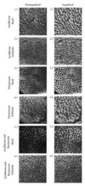
Figure 2: MR images in patient 1 (a–d) and (e–h) patient 8 show low volume of enhancing prostate at initial imaging and small residual prostate at 6 months. Posttreatment serum PSA level was less than 0.05 μg/L in both cases.

Figure 2a:

Picture2a

Figure 2b:

Picture2b

Figure 2c:

Picture2c

Figure 2d:

Picture2d

Figure 2e:

Picture2e

Figure 2f:

Picture2f

Figure 2g:

Picture2g

Figure 2h:

Picture2h

T1-weighted images.—The prostate was of predominantly low signal intensity, although patchy areas of intermediate or high signal intensity, likely to represent hemorrhage, were a constant finding within the gland and in all but one of 28 seminal vesicles.

Postcontrast images.—In each patient, the postcontrast images showed a central area of nonenhancing tissue. This conformed to the treatment volume and was surrounded by an enhancing rim of mean thickness of 2–8 mm (median, 4 mm) that was continuous around the prostate in most patients (Fig 2; Table E1,).

The enhancing prostate varied in size and position. Part of the enhancing rim usually lay within the prostatic capsule and continued to the prostatic apex where there was almost always some enhancing tissue between the nonenhancing prostate and the external urethral sphincter. In many patients, more central areas of enhancement were seen: at the apex or base, either posteriorly or anteriorly (Table E1), and were almost always in continuity with the rim.

In every patient, the nonenhancing, low signal intensity within the prostate extended outside the gland and involved the periprostatic fat and the levator ani muscle, particularly anterolaterally (Table E1, Figs 23). This varied considerably and tended to be most prominent in those who had no residual gland enhancement and had an undetectable serum PSA level after HIFU treatment (Table E1). In several patients, the nonenhancing area extended to involve the Denonvilliers fascia. (The distance between its margin and the rectal muscle is listed in Table E1.) In one patient, a proportion of the rectal wall enhanced avidly, but in no patient was there loss of rectal wall enhancement to suggest necrosis.

Figure 3: MR images obtained near the prostate apex show incomplete treatment and persisting high signal intensity in prostate. Serum PSA level nadir = 0.61 μg/L.

Figure 3a: Patient 4:

 Picture3a

 Figure 3b: Patient 4:

Picture3b

Figure 3c: Patient 4:

Picture3c

Figure 3d: Patient 4:

Picture3d

At 1–3 Months

In three patients, there was a “double rim” (Fig 4) on postcontrast images obtained at 36 and 56 days after HIFU treatment. The inner component lay within the prostate and the outer at the prostatic capsule; the intervening part was of low signal intensity on both T1- and T2-weighted images.

 Figure 4: MR images of “double rim” at 56 days after HIFU treatment.

Figure 4a: Patient 3:

 Picture4a

Figure 4b: Patient 3:

Picture4b

Figure 4c: Patient 3:

Picture4c

Six-month Appearance

T2-weighted images.—In every patient, the volume of the prostate was reduced by more than 45% (median, 61% reduction) (Table E1). On T2-weighted images, the majority of the persisting prostate was of low signal intensity, with poor definition to the capsule and with persisting heterogeneous signal intensity to the surrounding fat. However, in 12 of 15 patients, there was persisting high or intermediate signal intensity of the prostate—up to 5.34 mL in volume and most often seen posteriorly and at the apex (Table E1, Figs 3 and 5). In many patients (for example, those in Fig 2), low-signal-intensity prostate of reduced volume surrounded a capacious prostatic cavity continuous with the urethra, which is similar to the cavity seen after transurethral resection (19).

Figure 5: MR images of incomplete treatment of tumor and positive biopsy findings in three of 10 cores at 6 months (in right lateral midzone, right lateral base, and right parasagittal base samples). Serum PSA level nadir = 1.19 μg/L.

Figure 5a: Patient 13:

Picture5a

Figure 5b: Patient 13:

Picture5b

Figure 5c: Patient 13:

Picture5c

Figure 5d: Patient 13:

Picture5d

Figure 5e: Patient 13:

Picture5e

Postcontrast images.—Some small areas of nonenhancing tissue persisted in eight of 14 patients, but this was less than 1 mL in all but one (patient 13, in whom 4 mL of the gland volume of 18.7 mL was nonenhancing). The levator muscle showed a normal signal intensity.

Correlation Between Initial Imaging and Later Findings

In the 12 patients who underwent the initial acquisition within 1 month of HIFU treatment, the volume of enhancing tissue on the initial posttreatment image was positively correlated with the serum PSA level nadir (Fig 6) (Spearman r = 0.90, P < .001) and with the amount of residual tissue at 6 months (including all low-signal-intensity material that was likely to represent fibrosis or necrosis) (Fig 7) (Pearson r = 0.80, P = .001).

 Figure 6: Graph of relationship between the proportion of the prostate still enhancing on initial image and serum PSA level nadir. There is a significant positive correlation (Spearman r = 0.90, P < .001). * = patient 13, who was included in graph but not in analysis (imaged 56 days after HIFU treatment). Patients 14 and 15 are not included because they did not undergo contrast-enhanced acquisition within 2 months of HIFU treatment. μgl−1 = μg/L.

 Picture6

Figure 7: Graph of relationship between the proportion of the prostate still enhancing on initial image and final volume of prostate. There is a significant positive correlation between the variables (Pearson r = 0.80, P = .001). * = patient 13, who was included in graph but not in analysis (imaged 56 days after HIFU treatment). Patients 14 and 15 are not included because they did not undergo contrast-enhanced acquisition within 2 months of HIFU treatment.

Picture7

When posttreatment volumes are expressed as a proportion of pretreatment prostate volume, the correlation between enhancing tissue volume on the initial posttreatment image and the 6-month prostate volume persists (Pearson r = 0.70, P = .001).

Interobserver Correlation

The interobserver variation was excellent for the calculated volume of prostate enhancing on the initial post-HIFU image, with an intraclass correlation coefficient of 0.92, and was good for final prostate volume (intraclass correlation coefficient = 0.73).

Clinical Findings

In five patients (patients 1, 3, 8, 11, and 13), there was imaging evidence (at MR imaging or retrograde urethrography) of a stricture in the mid- or distal prostatic urethra, which was confirmed by using flow rate studies and treated by using self-catheterization or with graded urethral dilators. None have required formal urethrotomy. Patient 14 developed a bladder neck stricture, which was treated successfully by incision.

Before treatment, no men required pads or adult diapers for incontinence. At 6 months after the treatment, four men still required not more than one pad per day. In two cases, this was for reassurance rather than actual leakage.

In the 14 patients in whom there was intact erectile function (score 2–5 for the question, “How often were your erections hard enough for penetration?”) before HIFU treatment, it was intact in nine patients after the procedure. One patient had stopped trying to achieve erections, and four could not achieve penetration.

Histologic Findings

In the three patients in whom there was no high-signal-intensity peripheral zone at 6 months and with serum PSA level less than 0.05 μg/L, there was either no prostatic tissue or only a small group of acini in one core. The remaining patients had a variable amount of residual prostate at core biopsy.

Five patients had residual tumor. In three patients, it was seen in at least two cores (Table E1), and these three patients also had the largest volume of enhancing prostate on the initial post-HIFU MR image (Figs 6 and 7) and more than 2 mL of intermediate- or high-signal-intensity gland on T2-weighted images at 6 months.

In four of five patients with residual cancer, it could not be identified on either contrast-enhanced or T2-weighted images. In one patient (Fig 4), the early dynamic images showed prominent enhancement in the anterior gland, which was consistent with residual cancer found at the distal (ie, nonrectal) end of three right-sided biopsy cores. Such enhancement was not seen in patients with no cancer found at core biopsy.


DISCUSSION

We found a consistent sequence of changes at MR imaging after HIFU treatment of the whole prostate. The proportion of enhancing tissue on the initial posttreatment MR image was predictive of gland volume at 6 months and serum PSA level nadir. A strong statistical relationship between the latter and outcome has recently been demonstrated (20).

Most patients with residual cancer had evidence of incomplete ablation early (a large volume of enhancing prostate on the initial image) and late (a large volume of high-signal-intensity residual prostate on T2-weighted images at 6 months).

In some patients it was possible to achieve an undetectable serum PSA level at 6 months and entirely low signal intensity on T2-weighted images in the region of the prostate. These patients had either no or a small amount of viable prostate in one core at biopsy.

Conversely, in spite of reductions in prostate volume of more than 45% at 6 months, the majority of patients had histologic evidence of persisting viable prostate, and in a group of patients with organ-confined disease but no limit to the volume of cancer pretreatment, one-third had evidence of residual tumor.

Persisting enhancing prostatic tissue usually occurred at the periphery (or extended toward the center of the gland from it) and was particularly common at the apex and near the rectum.

Results of one previously published study (14) of post-HIFU appearances with MR imaging show a similar sequence of acute changes, although there was no attempt to quantify prostate volume at 6 months. There is also a large body of work on the MR imaging appearances with thermotherapy (whether laser [21,22] or radiofrequency [23]) and cryotherapy (24) within the prostate and other organs. The hyperenhancing rim of tissue is a constant finding in several tissues, including the liver (25), the kidney (26), and the brain (27). In the liver and the kidney, it is thin (1 mm or less) and, in most cases, has disappeared by 2 months after ablation (28). Within the prostate, the hyperenhancing rim has been shown to occur after laser ablation of benign prostatic hyperplasia (21,22) and after HIFU treatment (14).

Histologic evidence in animal models—including rabbit and porcine liver (29)—suggests that the enhancing rim corresponds to an area of inflammation and then fibrosis, with a variable amount of residual, viable tissue. How much of the rim will be viable after ablation of the prostate in humans remains uncertain. On the one hand, after HIFU treatment, core biopsy results show “partial or complete necrosis” in the rim (14). On the other, after laser ablation of benign prostatic hyperplasia, the volume of coagulative necrosis at histologic examination correlates very well with the central nonenhancing region at MR imaging, not including the rim (22). The answer is likely to be that a variable amount of the rim contains viable tissue (depending on the organ being imaged [30], the nature of the treatment, and the interval before the acquisition), and the implication is that the only reliably necrotic area at MR imaging is that which does not enhance. We have avoided the term necrosis for the nonenhancing areas of prostate seen in our current study, but from these data it is likely that the areas of prostate without enhancement are truly necrotic.

The distribution of enhancing prostate on posttreatment MR images fits with histologic evidence that “ventral, lateral and dorsal sides of the prostate” have residual viable prostatic tissue at histologic examination after HIFU treatment (31). What all of these areas have in common is proximity to the more richly vascular prostatic capsule. Is it possible that increased vascularity here results in reduced efficacy? This is another area that has been addressed by Rouviere’s group (32), who did not find a correlation between successful ablation and prostate vascularity by using power Doppler US; they conclude, as others have (33,34), that short (3-second) high-intensity bursts of focused ultrasound are unlikely to be markedly affected by blood flow. An alternate explanation is a geometric one: Centrally lying voxels are easier to treat because they may be rendered necrotic either by direct treatment or by damage to supplying vessels in the periphery.

An implication of these results is that the best strategies for minimizing complications while ensuring destruction of the cancer are likely to involve a degree of targeting: If the tumor can be imaged with MR imaging, the patient might be treated with higher power and wider margins (including periprostatic fat, muscle, or even neurovascular bundles) at the site of the cancer and with a standard intensity to the rest of the gland. An analogous approach is the wide excision, including a unilateral neurovascular bundle, of bulky tumors at radical prostatectomy (35). Such an approach may well have benefited our patients 7 and 13.

One methodologic issue that is currently unresolved relates to the timing of MR imaging. A detailed within-patient study of MR imaging changes after HIFU treatment is needed to properly describe the longitudinal changes in the appearance of the prostate. Rouviere et al (14) found that the area of nonenhancing tissue decreases by 50% at 1 month compared with that at an immediate (<1 week) post-HIFU acquisition, which suggests that for an accurate assessment of necrosis volume, the prostate should be imaged as soon as possible after treatment. Of course, perfusion would ideally be assessed during HIFU treatment so that undertreated areas could be further ablated. There is some evidence that Doppler or contrast-enhanced US (36) could play this role, but, to our knowledge, there are no studies on the correlation of immediate findings with later clinical data, such as serum PSA level or histologic examination.

We used fast low-angle shot sequences to assess enhancement because we found that the subjective assessment (together with objective measurements of signal intensity) of the dynamic series helped us identify truly nonenhancing tissue. However, the T1-weighted spin-echo postcontrast sequence would have been adequate, and we consider, as others do (22), dynamic contrast-enhanced sequences not to be an essential part of the protocol for postablation assessment. What is certain is that unenhanced T2-weighted sequences are inadequate for assessing necrosis (14,22).

Our results differ from those of other published series of HIFU treatment in the marked reduction in gland volume and absence of zonal anatomy in many patients observed at 6 months. In contrast to the study of post-HIFU MR imaging by Rouviere et al (14) who used a different device, we did not find that “HIFU-induced abnormalities seem to disappear within 3–5 months.” Rather, in several patients, it was difficult to discern any residual prostate at all at both MR and US studies. The difference probably lies in the power used for treatment and the completeness of gland coverage. The stricture rate of six of 15 is high when compared with that in published series (7,37,38) and may be related to the power used, the degree of fibrosis occurring in the prostate, and the strategy for catheterization. The latter is considered likely to be important, and we have recently changed to using a suprapubic catheter (rather than urethral) after treatment. The rate of impotence after treatment is similar to that in published series (11), as is grade I incontinence.

Our work has implications for the conduct of HIFU. The finding that the volume of enhancing prostate on the initial posttreatment image correlates well with intermediate measures, such as serum PSA level nadir and biopsy evidence of residual cancer, suggests that MR imaging can provide the operator with feedback on the effectiveness of the intervention. This information might enable modification of the technique to treat areas that have been incompletely ablated in previous patients—in our series, those areas encompassed the apex and posterior gland and rarely anterior tissue (in contrast to other study results [14]). Conversely, we might have reduced power or treatment volume at the anterolateral aspect of the gland adjacent to the levator muscle. Such feedback has been cited as a desirable attribute for ablation technology (39) and up to now has been missing.

Our study had several limitations. Although it is likely that nonenhancing areas at MR imaging represent necrosis, we do not have direct histologic evidence. Sampling error and misregistration limit the utility of core biopsies in this context. We have shown that the MR imaging appearances soon after HIFU treatment correlate with findings at 6 months, but this is not the same as outcome. A considerably longer follow-up and a larger number of patients will be necessary to determine both the ultimate efficacy of HIFU treatment and the ability of MR imaging to help predict outcome. Last, while our findings suggest that MR imaging soon after treatment may be useful to assess areas of under- and overtreatment, this is not real-time feedback and does not allow modification of the treatment as it progresses.

In summary, MR imaging results in the first 6 months after HIFU treatment show a consistent sequence of changes, and appearances in the 1st month correlate with serum PSA level nadir and imaging findings at 6 months. Such imaging results hold promise for providing feedback to the operator about the effectiveness of treatment.


ADVANCES IN KNOWLEDGE

  • Treatment of prostate cancer by using ablation with high-intensity focused ultrasound (HIFU) results in a consistent series of changes within the gland during 6 months seen at contrast-enhanced MR imaging.
  • Within 1 month after treatment, a central nonenhancing area is surrounded by an enhancing rim of tissue lying variably within and outside the prostate.
  • At 6 months, the gland is markedly smaller and of partly or completely low signal intensity on T2-weighted images.
  • The amount of enhancing prostate on the initial image correlates with several findings at 6 months, including serum prostate-specific antigen level nadir and prostate volume.


IMPLICATION FOR PATIENT CARE

  • MR imaging after HIFU treatment may provide information about completeness of tumor ablation and the need for early retreatment or close monitoring in cases of incomplete coverage.


Footnotes

  • Trial registration: This trial started recruiting before the trial registration requirements of the International Committee of Medical Journal Editors were formalized.

See Materials and Methods for pertinent disclosures.

Author contributions: Guarantors of integrity of entire study, A.P.S.K., I.M.H., C.A.; study concepts/study design or data acquisition or data analysis/interpretation, all authors; manuscript drafting or manuscript revision for important intellectual content, all authors; manuscript final version approval, all authors; literature research, A.P.S.K., M.E., I.M.H., R.O.I., C.A.; clinical studies, A.P.S.K., R.O.I., C.A.; statistical analysis, A.P.S.K.; and manuscript editing, all authors

Abbreviations:HIFU = high-intensity focused ultrasoundPSA = prostate-specific antigen


References

Parker C. Active surveillance: towards a new paradigm in the management of early prostate cancer. Lancet Oncol 2004; 5: 101–106.

CrossRefMedline

Steineck G, Helgesen F, Adolfsson J, et al. Quality of life after radical prostatectomy or watchful waiting. N Engl J Med 2002;347:790–796.

CrossRefMedline

Huidobro C, Bolmsjo M, Larson T, et al. Evaluation of microwave thermotherapy with histopathology, magnetic resonance imaging and temperature mapping. J Urol 2004;171:672–678.

CrossRefMedline

Shariat SF, Raptidis G, Masatoschi M, Bergamaschi F, Slawin KM. Pilot study of radiofrequency interstitial tumor ablation (RITA) for the treatment of radio-recurrent prostate cancer. Prostate 2005;65:260–267.

CrossRefMedline

Onik G. The male lumpectomy: rationale for a cancer targeted approach for prostate cryoablation—a review. Technol Cancer Res Treat2004;3:365–370.

Medline

Moore CM, Hoh IM, Bown SG, Emberton M. Does photodynamic therapy have the necessary attributes to become a future treatment for organ-confined prostate cancer? BJU Int 2005;96:754–758.

CrossRefMedline

Uchida T, Ohkusa H, Nagata Y, Hyodo T, Satoh T, Irie A. Treatment of localized prostate cancer using high-intensity focused ultrasound. BJU Int2006;97:56–61.

CrossRefMedline

Kennedy JE. High-intensity focused ultrasound in the treatment of solid tumours. Nat Rev Cancer 2005;5:321–327.

CrossRefMedline

Uchida T, Sanghvi NT, Gardner TA, et al. Transrectal high-intensity focused ultrasound for treatment of patients with stage T1b-2n0m0 localized prostate cancer: a preliminary report. Urology 2002;59:394–398.

CrossRefMedline

Walsh PC, Donker PJ. Impotence following radical prostatectomy: insight into etiology and prevention. J Urol 1982;128:492–497.

Medline

Blana A, Walter B, Rogenhofer S, Wieland WF. High-intensity focused ultrasound for the treatment of localized prostate cancer: 5-year experience.Urology 2004;63:297–300.

CrossRefMedline

Gelet A, Chapelon JY, Bouvier R, et al. Transrectal high-intensity focused ultrasound: minimally invasive therapy of localized prostate cancer. J Endourol 2000;14:519–528.

Medline

Chaussy C, Thuroff S. The status of high-intensity focused ultrasound in the treatment of localized prostate cancer and the impact of a combined resection. Curr Urol Rep 2003;4:248–252.

CrossRefMedline

Rouviere O, Lyonnet D, Raudrant A, et al. MRI appearance of prostate following transrectal HIFU ablation of localized cancer. Eur Urol2001;40:265–274.

CrossRefMedline

Illing RO, Leslie TA, Kennedy JE, Calleary JG, Ogden CW, Emberton M. Visually directed high-intensity focused ultrasound for organ-confined prostate cancer: a proposed standard for the conduct of therapy. BJU Int 2006;98:1187–1192.

CrossRefMedline

Bakker J, Olree M, Kaatee R, de Lange EE, Beek FJ. In vitro measurement of kidney size: comparison of ultrasonography and MRI. Ultrasound Med Biol 1998;24:683–688.

CrossRefMedline

Rosen RC, Cappelleri JC, Smith MD, Lipsky J, Pena BM. Development and evaluation of an abridged, 5-item version of the International Index of Erectile Function (IIEF-5) as a diagnostic tool for erectile dysfunction. Int J Impot Res 1999;11:319–326.

CrossRefMedline

Fleiss JL. Reliability of measurement. In: The design and analysis of clinical experiments. New York, NY: Wiley, 1986; 1–32.

Sheu MH, Chiang H, Wang JH, Chang YH, Chang CY. Transurethral resection of the prostate-related changes in the prostate gland: correlation of MRI and histopathology. J Comput Assist Tomogr 2000;24:596–599.

CrossRefMedline

Uchida T, Illing RO, Cathcart PJ, Emberton M. To what extent does the prostate-specific antigen nadir predict subsequent treatment failure after transrectal high-intensity focused ultrasound therapy for presumed localized adenocarcinoma of the prostate? BJU Int 2006;98:537–539.

CrossRefMedline

Mueller-Lisse UG, Heuck AF, Schneede P, et al. Postoperative MRI in patients undergoing interstitial laser coagulation thermotherapy of benign prostatic hyperplasia. J Comput Assist Tomogr 1996;20:273–278.

CrossRefMedline

Boni RA, Sulser T, Jochum W, Romanowski B, Debatin JF, Krestin GP. Laser ablation-induced changes in the prostate: findings at endorectal MR imaging with histologic correlation. Radiology 1997;202:232–236.

Abstract/FREE Full Text

Zlotta AR, Djavan B, Matos C, et al. Percutaneous transperineal radiofrequency ablation of prostate tumour: safety, feasibility and pathological effects on human prostate cancer. Br J Urol 1998;81:265–275.

Medline

Vellet AD, Saliken J, Donnelly B, et al. Prostatic cryosurgery: use of MR imaging in evaluation of success and technical modifications. Radiology1997;203:653–659.

Abstract/FREE Full Text

Guan YS, Sun L, Zhou XP, Li X, Zheng XH. Hepatocellular carcinoma treated with interventional procedures: CT and MRI follow-up. World J Gastroenterol 2004;10:3543–3548.

Medline

Merkle EM, Nour SG, Lewin JS. MR imaging follow-up after percutaneous radiofrequency ablation of renal cell carcinoma: findings in 18 patients during first 6 months. Radiology 2005;235:1065–1071.

Abstract/FREE Full Text

Kahn T, Bettag M, Ulrich F, et al. MRI-guided laser-induced interstitial thermotherapy of cerebral neoplasms. J Comput Assist Tomogr1994;18:519–532.

Medline

Dromain C, de Baere T, Elias D, et al. Hepatic tumors treated with percutaneous radiofrequency ablation: CT and MR imaging follow-up. Radiology2002;223:255–262.

Abstract/FREE Full Text

Raman SS, Lu DS, Vodopich DJ, Sayre J, Lassman C. Creation of radiofrequency lesions in a porcine model: correlation with sonography, CT, and histopathology. AJR Am J Roentgenol 2000;175:1253–1258.

Abstract/FREE Full Text

Illing RO, Kennedy JE, Wu F, et al. The safety and feasibility of extracorporeal high-intensity focused ultrasound (HIFU) for the treatment of liver and kidney tumours in a Western population. Br J Cancer 2005;93:890–895.

CrossRefMedline

Van Leenders GJ, Beerlage HP, Ruijter ET, de la Rosette JJ, van de Kaa CA. Histopathological changes associated with high intensity focused ultrasound (HIFU) treatment for localised adenocarcinoma of the prostate. J Clin Pathol 2000;53:391–394.

Abstract/FREE Full Text

Rouviere O, Curiel L, Chapelon JY, et al. Can color doppler predict the uniformity of HIFU-induced prostate tissue destruction? Prostate2004;60:289–297.

Billard BE, Hynynen K, Roemer RB. Effects of physical parameters on high temperature ultrasound hyperthermia. Ultrasound Med Biol1990;16:409–420.

CrossRefMedline

Chen L, ter Haar G, Hill CR, et al. Effect of blood perfusion on the ablation of liver parenchyma with high-intensity focused ultrasound. Phys Med Biol 1993;38:1661–1673.

CrossRefMedline

Park EL, Dalkin B, Escobar C, Nagle RB. Site-specific positive margins at radical prostatectomy: assessing cancer-control benefits of wide excision of the neurovascular bundle on a side with cancer on biopsy. BJU Int 2003;91:219–222.

CrossRefMedline

Sedelaar JP, Aarnink RG, van Leenders GJ, et al. The application of three-dimensional contrast-enhanced ultrasound to measure volume of affected tissue after HIFU treatment for localized prostate cancer. Eur Urol 2000;37:559–568.

CrossRefMedline

Thuroff S, Chaussy C, Vallancien G, et al. High-intensity focused ultrasound and localized prostate cancer: efficacy results from the European multicentric study. J Endourol 2003;17:673–677.

CrossRefMedline

Gelet A, Chapelon JY, Bouvier R, Rouviere O, Lyonnet D, Dubernard JM. Transrectal high intensity focused ultrasound for the treatment of localized prostate cancer: factors influencing the outcome. Eur Urol 2001;40:124–129.

CrossRefMedline

Gillett MD, Gettman MT, Zincke H, Blute ML. Tissue ablation technologies for localized prostate cancer. Mayo Clin Proc 2004;79:1547–1555.

CrossRefMedline