Feeds:
Posts
Comments

Archive for the ‘Materials Science & Engineering’ Category

Reporter: Danut Dragoi, PhD

Scientists at MIT and Massachusetts General Hospital have discovered how cancer cells latch onto blood vessels and invade tissues to form new tumors — a finding that could help them develop drugs that inhibit this process and prevent cancers from metastasizing.

Cancer cells circulating in the bloodstream can stick to blood vessel walls and construct tiny “bridges” through which they inject genetic material that transforms the endothelial cells lining the blood vessels, making them much more hospitable to additional cancer cells, according to the new study.

The researchers also found that they could greatly reduce metastasis in mice by inhibiting the formation of these nanobridges. Endothelial cells line every blood vessel and are the first cells in contact with any blood-borne element. They serve as the gateway into and out of tumors and have been the focus of intense research in vascular and cancer biology.

Building bridges

Metastasis is a multistep process that allows cancer to spread from its original site and form new tumors elsewhere in the body. Certain cancers tend to metastasize to specific locations; for example, lung tumors tend to spread to the brain, and breast tumors to the liver and bone.

To metastasize, tumor cells must first become mobile so they can detach from the initial tumor. Then they break into nearby blood vessels so they can flow through the body, where they become circulating tumor cells (CTCs). These CTCs must then find a spot where they can latch onto the blood vessel walls and penetrate into adjacent tissue to form a new tumor.

Blood vessels are lined with endothelial cells, which are typically resistant to intruders.

The researchers first spotted tiny bridges between cancer cells and endothelial cells while using electron microscopy to study the interactions between those cell types. They speculated that the cancer cells might be sending some kind of signal to the endothelial cells.

Once we saw that these structures allowed for a ubiquitous transfer of a lot of different materials, microRNAs were an obvious interesting molecule because they’re able to very broadly control the genome of a cell in ways that we don’t really understand,” Connor says. “That became our focus.”

MicroRNA, discovered in the early 1990s, helps a cell to fine-tune its gene expression. These strands of RNA, about 22 base pairs long, can interfere with messenger RNA, preventing it from being translated into proteins.

In this case, the researchers found, the injected microRNA makes the endothelial cells “sticky.” That is, the cells begin to express proteins on their surfaces that attract other cells to adhere to them. This allows additional CTCs to bind to the same site and penetrate through the vessels into the adjacent tissue, forming a new tumor.

Non-metastatic cancer cells did not produce these invasive nanobridges when grown on endothelial cells.

Shutting down metastasis

The nanobridges are made from the proteins actin and tubulin (NB-the protein actin is abundant in all eukaryotic cells. It was first discovered in skeletal muscle, where actin filaments slide along filaments of another protein called myosin to make the cells contract. In non-muscle cells, actin filaments are less organized and myosin is much less prominent, and a tubulin is a protein that is the main constituent of the micro-tubules of living cells, which also form the cytoskeleton that gives cells their structure). The researchers found that they could inhibit the formation of these nanobridges, which are about 300 microns long, by giving low doses of drugs that interfere with actin.

When the researchers gave these drugs to mice with tumors that normally metastasize, the tumors did not spread.

Sengupta’s lab is now trying to figure out the mechanism of nanobridge formation in more detail, with an eye toward developing drugs that act more specifically to inhibit the process.

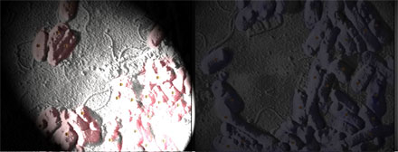
 The SEM picture below,

Cancer_cell_Vein

is a rounded cancer cell (top left) that sends out nanotubes connecting with endothelial cells. Genetic material can be injected via these nanotubes, transforming the endothelial cells and making them more hospitable to additional cancer cells. Image credit: Sengupta Lab. The second picture below,

Cancer_Cell_Vein_Penetration

is showing a cancer cell (bottom center) that creates a gap and enters the endothelial tube. Another cancer cell (middle right) sends out nanotubes to connect with endothelial cells. Both image are credited to Sengupta Lab

An interesting comment on why plants do not develop cancer is given here,  The article states that in plants, as in animals, most cells that constitute the organism limit their reproductive potential in order to provide collective support for the immortal germ line. And, as in animals, the mechanisms that restrict the proliferation of somatic cells in plants can fail, leading to tumors. There are intriguing similarities in tumorigenesis between plants and animals, including the involvement of the retinoblastoma pathway as well as overlap with mechanisms that are used for stem cell maintenance. However, plant tumors are less frequent and are not as lethal as those in animals. The authors of the article argue that fundamental differences between plant and animal development make it much more difficult for individual plant cells to escape communal controls.

The structure of the endothelium, the thin layer of cells that line our arteries and veins, is visible here. The endothelium is like a gatekeeper, controlling the movement of materials into and out of the bloodstream. Endothelial cells are held tightly together by specialized proteins that function like strong ropes (red) and others that act like cement (blue). In the picture here, the cell is preparing to divide. Two copies of each chromosome (blue) are lined
up next to each other in the center of the cell. Next, protein strands (red) will pull apart these paired
chromosomes and drag them to opposite sides of the cell. The cell will then split to form two daughter cells, each with a single, complete set of chromosomes. It is interesting that protein strands (red) in this picture are implicated on pulling apart the two copies of chromosomes in the same way the proteins actin and tubules do in the cancer cell interaction with the veins and arteries. It looks like the proteins shaped as strings, the nanobridges, due their shape to the tension development between cancer cells and the veins.

Source
1.  http://news.mit.edu/2015/cancer-cells-escape-blood-vessels-1216
2.  http://www.nature.com/nrc/journal/v10/n11/full/nrc2942.html

Read Full Post »

Quantum dots target infections

Larry H. Bernstein, MD, FCAP, Curator

LPBI


Photoactivated QDs Kill Antibiotic-Resistant ‘Superbugs’

BOULDER, Colo., Jan. 20, 2016 — A technique for treating bacterial infections has successfully used light-activated quantum dots (QDs) to kill multiple multidrug-resistant strains.

http://www.photonics.com/Article.aspx?AID=58218

e coli

http://www.photonics.com/images/Web/Articles/2016/1/20/PIC_QD2.jpg

Modified atomic force micrograph of multidrug-resistant E. coli. Courtesy of the Nagpal Group/University of Colorado Boulder.


The approach is adaptive to constantly evolving drug-resistant bacteria and avoids damage to surrounding cells, an issue encountered in earlier attempts that deployed metal nanoparticles such as gold and silver to combat bacteria.

“By shrinking these [QD] semiconductors down to the nanoscale, we’re able to create highly specific interactions within the cellular environment that only target the infection,” said professor Prashant Nagpal of the University of Colorado Boulder.

The QDs — which are inactive in darkness — were tailored to target particular infections thanks to their light-activated properties. The researchers said that by modifying the wavelength of light applied, they could activate the QDs to alter and kill infected cells with specificity.

Napgal and his team tested the QD therapy on mammalian tissue containing bacterial cells in mono- and cocultures. The bacteria under investigation were ethicillin-resistant Staphylococcus aureus, carbapenem-resistant E. coli, and extended-spectrum ß-lactamase-producing Klebsiella pneumoniae and Salmonella typhimurium.

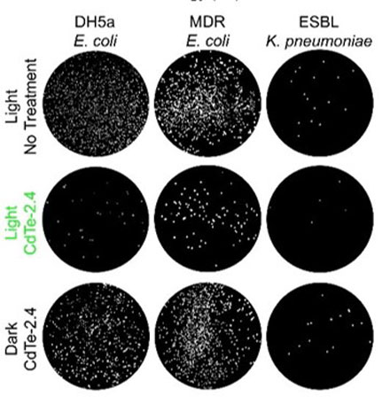
They reported 92 percent of bacterial cells were killed, while leaving mammalian cells intact. The QDs could also be tuned to increase bacterial proliferation.

qd

http://www.photonics.com/images/Web/Articles/2016/1/20/PIC_QD1.jpg

Plated antibiotic resistant ‘superbugs’ before and after treatment with nanoparticles. Courtesy of the Nagpal Group/University of Colorado Boulder.

The team said the killing effect was independent of the QD material used; rather, it was controlled by the redox potentials of the photogenerated charge carriers, which selectively altered cellular redox states. Photoexcited QDs could be used in the study of the effect of redox states on living systems, and lead to clinical phototherapy for the treatment of infections, the researchers said.

The specificity of the treatment could help reduce or eliminate the potential side effects of other treatment methods, as well as provide a path forward for future development and clinical trials.

“Antibiotics are not just a baseline treatment for bacterial infections, but HIV and cancer as well,” said professor Anushree Chatterjee. “Failure to develop effective treatments for drug-resistant strains is not an option, and that’s what this technology moves closer to solving.”

Nagpal and Chatterjee are the cofounders of Praan Biosciences Inc., a startup that can sequence genetic profiles using a single molecule, and have filed a patent on the QD therapy technology.

The research was published in Nature Materials (doi: 10.1038/nmat4542).


Photoexcited quantum dots for killing multidrug-resistant bacteria

Colleen M. CourtneySamuel M. GoodmanJessica A. McDanielNancy E. MadingerAnushree ChatterjeePrashant Nagpal

Nature Materials(2016)      http://dx.doi.org:/10.1038/nmat4542

Multidrug-resistant bacterial infections are an ever-growing threat because of the shrinking arsenal of efficacious antibiotics1, 2, 3, 4. Metal nanoparticles can induce cell death, yet the toxicity effect is typically nonspecific5, 6, 7, 8. Here, we show that photoexcited quantum dots (QDs) can kill a wide range of multidrug-resistant bacterial clinical isolates, including methicillin-resistant Staphylococcus aureus, carbapenem-resistant Escherichia coli, and extended-spectrum β-lactamase-producingKlebsiella pneumoniae and Salmonella typhimurium. The killing effect is independent of material and controlled by the redox potentials of the photogenerated charge carriers, which selectively alter the cellular redox state. We also show that the QDs can be tailored to kill 92% of bacterial cells in a monoculture, and in a co-culture of E. coli and HEK 293T cells, while leaving the mammalian cells intact, or to increase bacterial proliferation. Photoexcited QDs could be used in the study of the effect of redox states on living systems, and lead to clinical phototherapy for the treatment of infections.

Figure 2: The effect of CdTe-2.4 is specific to the reduction and oxidation potentials.close

The effect of CdTe-2.4 is specific to the reduction and oxidation potentials.

a, Absorbance spectra for CdTe and CdSe of several sizes. Insets show transmission electron microscopy (TEM) images with colour-coded scale bars (50nm except for CdTe-2.4, which is 25nm). b, Scanning tunnelling spectroscopy (STS) meas…

Read Full Post »

Graphene Interaction with Neurons

Larry H. Bernstein, MD, FCAP, Curator

LPBI


Graphene Shown to Safely Interact with Neurons in the Brain

University of Cambridge

(Source: University of Cambridge)

http://www.biosciencetechnology.com/sites/biosciencetechnology.com/files/bt1601_cambridge_graphene.png


Researchers have successfully demonstrated how it is possible to interface graphene – a two-dimensional form of carbon – with neurons, or nerve cells, while maintaining the integrity of these vital cells. The work may be used to build graphene-based electrodes that can safely be implanted in the brain, offering promise for the restoration of sensory functions for amputee or paralyzed patients, or for individuals with motor disorders such as epilepsy or Parkinson’s disease.

The research, published in the journal ACS Nano, was an interdisciplinary collaboration coordinated by the University of Trieste in Italy and the Cambridge Graphene Centre.

Previously, other groups had shown that it is possible to use treated graphene to interact with neurons. However the signal to noise ratio from this interface was very low. By developing methods of working with untreated graphene, the researchers retained the material’s electrical conductivity, making it a significantly better electrode.

“For the first time we interfaced graphene to neurons directly,” said Professor Laura Ballerini of the University of Trieste in Italy. “We then tested the ability of neurons to generate electrical signals known to represent brain activities, and found that the neurons retained their neuronal signaling properties unaltered. This is the first functional study of neuronal synaptic activity using uncoated graphene based materials.”

Our understanding of the brain has increased to such a degree that by interfacing directly between the brain and the outside world we can now harness and control some of its functions. For instance, by measuring the brain’s electrical impulses, sensory functions can be recovered. This can be used to control robotic arms for amputee patients or any number of basic processes for paralyzed patients – from speech to movement of objects in the world around them. Alternatively, by interfering with these electrical impulses, motor disorders (such as epilepsy or Parkinson’s) can start to be controlled.

Scientists have made this possible by developing electrodes that can be placed deep within the brain. These electrodes connect directly to neurons and transmit their electrical signals away from the body, allowing their meaning to be decoded.

However, the interface between neurons and electrodes has often been problematic: not only do the electrodes need to be highly sensitive to electrical impulses, but they need to be stable in the body without altering the tissue they measure.

Too often the modern electrodes used for this interface (based on tungsten or silicon) suffer from partial or complete loss of signal over time. This is often caused by the formation of scar tissue from the electrode insertion, which prevents the electrode from moving with the natural movements of the brain due to its rigid nature.

Graphene has been shown to be a promising material to solve these problems, because of its excellent conductivity, flexibility, biocompatibility and stability within the body.

Based on experiments conducted in rat brain cell cultures, the researchers found that untreated graphene electrodes interfaced well with neurons. By studying the neurons with electron microscopy and immunofluorescence the researchers found that they remained healthy, transmitting normal electric impulses and, importantly, none of the adverse reactions which lead to the damaging scar tissue were seen.

According to the researchers, this is the first step towards using pristine graphene-based materials as an electrode for a neuro-interface. In future, the researchers will investigate how different forms of graphene, from multiple layers to monolayers, are able to affect neurons, and whether tuning the material properties of graphene might alter the synapses and neuronal excitability in new and unique ways. “Hopefully this will pave the way for better deep brain implants to both harness and control the brain, with higher sensitivity and fewer unwanted side effects,” said Ballerini.

“We are currently involved in frontline research in graphene technology towards biomedical applications,” said Professor Maurizio Prato from the University of Trieste. “In this scenario, the development and translation in neurology of graphene-based high-performance biodevices requires the exploration of the interactions between graphene nano- and micro-sheets with the sophisticated signalling machinery of nerve cells. Our work is only a first step in that direction.”

“These initial results show how we are just at the tip of the iceberg when it comes to the potential of graphene and related materials in bio-applications and medicine,” said Professor Andrea Ferrari, Director of the Cambridge Graphene Centre. “The expertise developed at the Cambridge Graphene Centre allows us to produce large quantities of pristine material in solution, and this study proves the compatibility of our process with neuro-interfaces.”

The research was funded by the Graphene Flagship, a European initiative which promotes a collaborative approach to research with an aim of helping to translate graphene out of the academic laboratory, through local industry and into society.

Source: University of Cambridge


Remembering to Remember Supported by Two Distinct Brain Processes

http://www.biosciencetechnology.com/news/2013/08/remembering-remember-supported-two-distinct-brain-processes

To investigate how prospective memory is processed in the brain, psychological scientist Mark McDaniel of Washington University in St. Louis and colleagues had participants lie in an fMRI scanner and asked them to press one of two buttons to indicate whether a word that popped up on a screen was a member of a designated category.  In addition to this ongoing activity, participants were asked to try to remember to press a third button whenever a special target popped up. The task was designed to tap into participants’ prospective memory, or their ability to remember to take certain actions in response to specific future events.

When McDaniel and colleagues analyzed the fMRI data, they observed that two distinct brain activation patterns emerged when participants made the correct button press for a special target.

When the special target was not relevant to the ongoing activity—such as a syllable like “tor”—participants seemed to rely on top-down brain processes supported by the prefrontal cortex. In order to answer correctly when the special syllable flashed up on the screen, the participants had to sustain their attention and monitor for the special syllable throughout the entire task. In the grocery bag scenario, this would be like remembering to bring the grocery bags by constantly reminding yourself that you can’t forget them.

When the special target was integral to the ongoing activity—such as a whole word, like “table”—participants recruited a different set of brain regions, and they didn’t show sustained activation in these regions. The findings suggest that remembering what to do when the special target was a whole word didn’t require the same type of top-down monitoring. Instead, the target word seemed to act as an environmental cue that prompted participants to make the appropriate response—like reminding yourself to bring the grocery bags by leaving them near the front door.

“These findings suggest that people could make use of several different strategies to accomplish prospective memory tasks,” says McDaniel.

McDaniel and colleagues are continuing their research on prospective memory, examining how this phenomenon might change with age.

Co-authors on this research include Pamela LaMontagne, Michael Scullin, Todd Braver of Washington University in St. Louis; and Stefanie Beck of Technische Universität Dresden.

This research was funded by the National Institute on Aging, the Washington University Institute of Clinical and Translation Sciences, the National Center for Advancing Translational Sciences, and the German Science Foundation.

Read Full Post »

Nano metal inks

Reporter: Danut Dragoi, PhD

 
Not for a long time ago, PEDOT:PSS ink see link in here , appeared as a serious alternative to Poly-Si wafer that is used directly to manufacture Photo Voltaic (PV) cells. The most important properties of PEDOT:PSS ink is its electroconductivity. A very important issue in the fabrication of polymer solar cells, either for small-scale or R2R high throughput production, is finding suitable inks that match the processing method. Ideally, the solvents should be environmental friendly and nontoxic, with water being the cheapest and best case. Not all of the materials in polymer solar cells can currently fulfill this demand. PEDOT:PSS is being the only component generally processed from aqueous solution. Nevertheless, fully aqueous processed polymers solar cells were successfully produced by introducing thermo-cleavable polymers to make the active layer insoluble.[1]
Multi-layer devices such as polymer solar cells that are produced in an all-solution process generally require orthogonal solvents (polar and non-polar) for the deposition of the subsequent layers. It must be ensured that the deposition of one ink does not dissolve the functional layer underneath. Ink formulation can be divided into three categories:

  • dissolved material (each molecule is fully solvated),
  • emulsions (material is kept in solution by additives which allow for micelle formation), and
  • particulate solutions or pastes (solid particles are suspended in solution).

Careful exploitation of ink formulations and processes might be the answer to how to replace the use of orthogonal solvents.[2]
An important trend that will very likely start to see some very serious commercial applications in 2016: 3D printed electronics, both for prototyping of PCBs and end use wearable and IoT products. One company that is literally mapping the path for this to happen is PV Nano Cell, a major player in the production of nano-particle-metal inks for printed electronics, see link in here.

Electronics printing today takes place primarily in 2D, using silver nanoparticle inks. Founded by Fernando de la Vega, the current CEO, PV Nano Cell began in the field of printable photovoltaics and has recently moved on to assume a leadership position in the production of conductive inks, mainly through the commercialisation of its Sicrys line of products.
The Sicrys inks are high performance, state-of-the-art single crystal nano-metric conductive inks, based on PVN’s exclusive dispersion technology. They are made to offer a competitive edge to clients using inkjet printing for printed electronics and more. The Sicrys line of products is available in silver or copper-based form, ensuring maximum conductivity at a lowest cost.
How is this going to affect 3D printing? Through the development and modification of current inkjet 3D printing technologies – such as Stratasys Polyjet technology (and 3D System’s MultiJet Modeling) – It will become possible to directly 3D print electronic-capable products, such as photovoltaics, printed circuit boards, RFID, sensors, smart cards, touchscreens and advanced packaging.
“The idea here is to replace all the analogue production by using digital conductive printing, as part of an an additive process,” de la Vega explains. “We are primarily an ink company and we are developing inks that differ from anything else currently on the market. First of all, we have both silver and copper inks. Copper is safer and does not oxidize as easily. Our inks are made of nanometer particles; they are very special single crystal particles with enhanced properties that enable better electrical conductivity and stability. Our inks enable high throughput 24/7 printing for a highly industrialised process.”
Another aspect that, according to de la Vega, will help bring 3D printed electronics into the mainstream is that PVN’s inks are extremely low cost, with end prices that – even depending on quantity – are about one third of other nanometal particle conductive inks on the market. “We use a water-based process that is extremely cost effective,” he explains, “so much so that we can even compete with the prices of standard PolyJet resins in terms of pricing.”
To make end products, the inks can either be applied by ink jetting on pre-manufactured plastic parts, such as, for example, polycarbonate for mobile phone antennas, or they will be able to be 3D printed directly into the photopolymers during 3D printing. For this, the strategy by PVN is to focus exclusively on the inks and actively collaborate with hardware manufacturers to find the best compatibility with their own materials and processes. Recently, PVN signed a collaboration deal with a large PCB manufacturer in Israel and a consumer electronics company based in the USA and China.
As recently as last November, PVN won the IDTechEx Award for Best Development in Materials for 3D Printing for its Sicrys materials and their potential in accelerating the adoption of solar photovoltaics (PV) and printed electronics (PE).
PVN has a production capacity of hundreds of kilograms and is specifically targeting the market for mass end-use products and parts.

Source
[1] Søndergaard et. al., Advanced Energy Materials 2011 10.1002/aenm.201000007
[2] Søndergaard et. al., Materials Today 2012
http://3dprintingindustry.com/2016/01/05/pv-nanocells-newest-nanometal-inks-write-up-2016-as-the-year-of-3d-printed-electronics/

Read Full Post »

3D “Squeeze” Helps Adult Cells Become Stem Cells

Reporter: Irina Robu, PhD

Scientists based at Ecole Polytechnique Fédérale de Lausanne led by Matthias Lutolf have been engineering 3D extracellular matrices—gels. These scientists report that they have developed a gel that boosts the ability of normal cells to revert into stem cells by simply “squeezing” them.

The detail of the scientists’ work appeared in Nature Materials, January 11, 2015 in an article entitled, “Defined three-dimensional microenvironments boost induction of pluripotency.” According to the authors they find that the physical cell confinement imposed by the 3D microenvironment boosts reprogramming through an accelerated mesenchymal-to-epithelial transition and increased epigenetic remodeling. They concluded that 3D microenvironmental signals act synergistically with reprogramming transcription factors to increase somatic plasticity.

The researchers discovered that they could reprogram the cells faster and more efficiently  by simply adjusting the composition, hence the stiffness and density of the surrounding gel. As a result, the gel exerts different forces on the cells, “squeezing” them.

The scientists propose that the 3D environment is key to this process, generating mechanical signals that work together with genetic factors to make the cell easier to transform into a stem cell. The technique can be applied to a large number of cells to produce stem cells on an industrial scale.

Source

http://www.genengnews.com/gen-news-highlights/3d-squeeze-helps-adult-cells-become-stem-cells/81252223/


Read Full Post »

Platform Technologies for Directly Reconstructing 3D Living Biomaterials

Reporter: Irina Robu, PhD

The techniques of electrospraying and electrospinning have existed for at least a century. These techniques employs a high voltage applied to a needle accommodating the flow of media, placed above a counter electrode which could either be grounded or have an opposite charge to the needle—thus introducing the charged media to an electric field.

These endeavors have demonstrated the wider applicability of these technologies and hence in the last 20 years or so have been used for the direct handling of a wide range of materials, including bio-inspired materials. These investigations have generated interest in areas such as the development of fine monolayered surfaces, fabrication of scaffolds which could be used for many laboratory-based fundamental biological studies.

In 2005, Jayasinghe et al. began investigations into both electrospraying and electrospinning of immortalized cell lines. Even though the high voltages involved, these cells were  found to be viable post-electrospraying/electrospinning. Additional work has extended these studies to different cell types, both murine and human, immortalized or primary, stem cells, and even whole fertilized embryos from model organisms. Established protocols (such as flow cytometry, genetic/genomic interrogation, and microarray analysis) proved that cells processed using either electrospraying or electrospinning were indistinguishable from controls. Hence bio-electrospraying (BES) and cell electrospinning (CE) have become platform technologies for the biological and life science and are the leading technologies for the direct handling of cells—both for distribution of cells with pinpoint precision as cell-bearing droplets, and for the formation of truly 3D living scaffolds.

Previous studies have been carried out with processed cells suspended in matrices generated from animal/tumor-derived materials which contain largely uncharacterized growth factors and bioactive signals. This makes them very undesirable for clinical assays. While not applicable to humans, they can be used  with advanced biopolymers, which could be directly translated to humans, and have the potential for creating artificial constructs which could be used for a variety of applications in the regenerative medicine field. The present study describes the in vivo application of such biopolymers, using murine macrophages to interrogate biocompatibility and cellular behavior post-transfer.

Source

http://onlinelibrary.wiley.com/doi/10.1002/adma.201503001/full

Read Full Post »

Occupational Therapy

Larry H. Bernstein, MD, FCAP, Curator

LPBI


Definition of Occupational Therapy

Occupational therapy is a client-centred health profession concerned with promoting health and well being through occupation. The primary goal of occupational therapy is to enable people to participate in the activities of everyday life. Occupational therapists achieve this outcome by working with people and communities to enhance their ability to engage in the occupations they want to, need to, or are expected to do, or by modifying the occupation or the environment to better support their occupational engagement.(WFOT 2012)

Read the Statement on Occupational Therapy

In occupational therapy, occupations refer to the everyday activities that people do as individuals, in families and with communities to occupy time and bring meaning and purpose to life. Occupations include things people need to, want to and are expected to do.

Definition of Occupational Therapy Practice for the AOTA Model Practice Act

http://www.aota.org/-/media/Corporate/Files/Advocacy/State/Resources/PracticeAct/Model%20Definition%20of%20OT%20Practice%20%20Adopted%2041411.ashx

The practice of occupational therapy means the therapeutic use of occupations, including everyday life activities with individuals, groups, populations, or organizations to support participation, performance, and function in roles and situations in home, school, workplace, community, and other settings. Occupational therapy services are provided for habilitation, rehabilitation, and the promotion of health and wellness to those who have or are at risk for developing an illness, injury, disease, disorder, condition, impairment, disability, activity limitation, or participation restriction. Occupational therapy addresses the physical, cognitive, psychosocial, sensory-perceptual, and other aspects of performance in a variety of contexts and environments to support engagement in occupations that affect physical and mental health, well-being, and quality of life. The practice of occupational therapy includes:

A. Evaluation of factors affecting activities of daily living (ADL), instrumental activities of daily living (IADL), rest and sleep, education, work, play, leisure, and social participation, including:

1. Client factors, including body functions (such as neuromusculoskeletal, sensory-perceptual, visual, mental, cognitive, and pain factors) and body structures (such as cardiovascular, digestive, nervous, integumentary, genitourinary systems, and structures related to movement), values, beliefs, and spirituality.

2. Habits, routines, roles, rituals, and behavior patterns.

3. Physical and social environments, cultural, personal, temporal, and virtual contexts and activity demands that affect performance.

4. Performance skills, including motor and praxis, sensory-perceptual, emotional regulation, cognitive, communication and social skills.

B. Methods or approaches selected to direct the process of interventions such as:

1. Establishment, remediation, or restoration of a skill or ability that has not yet developed, is impaired, or is in decline.

2. Compensation, modification, or adaptation of activity or environment to enhance performance, or to prevent injuries, disorders, or other conditions.

3. Retention and enhancement of skills or abilities without which performance in everyday life activities would decline.

4. Promotion of health and wellness, including the use of self-management strategies, to enable or enhance performance in everyday life activities.

5. Prevention of barriers to performance and participation, including injury and disability prevention.

C. Interventions and procedures to promote or enhance safety and performance in activities of daily living (ADL), instrumental activities of daily living (IADL), rest and sleep, education, work, play, leisure, and social participation, including:

1. Therapeutic use of occupations, exercises, and activities.

2. Training in self-care, self-management, health management and maintenance, home management, community/work reintegration, and school activities and work performance.

3. Development, remediation, or compensation of neuromusculoskeletal, sensory-perceptual, visual, mental, and cognitive functions, pain tolerance and management, and behavioral skills.

4. Therapeutic use of self, including one’s personality, insights, perceptions, and judgments, as part of the therapeutic process.

5. Education and training of individuals, including family members, caregivers, groups, populations, and others.

6. Care coordination, case management, and transition services.

7. Consultative services to groups, programs, organizations, or communities.

8. Modification of environments (home, work, school, or community) and adaptation of processes, including the application of ergonomic principles.

9. Assessment, design, fabrication, application, fitting, and training in seating and positioning, assistive technology, adaptive devices, and orthotic devices, and training in the use of prosthetic devices.

10. Assessment, recommendation, and training in techniques to enhance functional mobility, including management of wheelchairs and other mobility devices.

11. Low vision rehabilitation.

12. Driver rehabilitation and community mobility.

13. Management of feeding, eating, and swallowing to enable eating and feeding performance.

14. Application of physical agent modalities, and use of a range of specific therapeutic procedures (such as wound care management; interventions to enhance sensory-perceptual, and cognitive processing; and manual therapy) to enhance performance skills.

15. Facilitating the occupational performance of groups, populations, or organizations through the modification of environments and the adaptation of processes.

Adopted by the Representative Assembly 4/14/11 (Agenda A13, Charge 18)

Read Full Post »

Asthma sourced carbon nanotubes

Larry H. Bernstein, MD, FCAP, Curator

LPBI

Carbon nanotubes found in cells from airways of asthmatic children in Paris

Carbon nanotubes, possibly from cars, are ubiquitous, found even in ice cores — we may all have them in our lungs, say Rice scientists
October 19, 2015

http://www.kurzweilai.net/carbon-nanotubes-found-in-cells-from-airways-of-asthmatic-children-in-paris?utm_source=KurzweilAI+Weekly+Newsletter_147a5a48c1-9a20162408-282099089

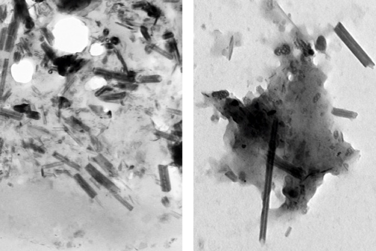
Carbon nanotubes (rods) and nanoparticles (black clumps) found inside a lung cell vacuole (left) are similar to those found in vehicle exhaust in tailpipes of cars in Paris (right) (credit: Fathi Moussa/Paris-Saclay University)

http://www.kurzweilai.net/images/CNTs-in-lungs-cars.jpg

Carbon nanotubes (CNTs) have been found in cells extracted from the airways of Parisian children under routine treatment for asthma, according to a report in the journal EBioMedicine (open access) by scientists in France and atRice University.

The cells were taken from 69 randomly selected asthma patients aged 2 to 17 who underwent routine fiber-optic bronchoscopies as part of their treatment. The researchers analyzed particulate matter found in the alveolar macrophage cells (also known as dust cells), which help stop foreign materials like particles and bacteria from entering the lungs.

The study partially answers the question of what makes up the black material inside alveolar macrophages, the original focus of the study. The researchers found single-walled and multiwalled carbon nanotubes and amorphous carbon among the cells.

The nanotube aggregates in the cells ranged in size from 10 to 60 nanometers in diameter and up to several hundred nanometers in length, small enough that optical microscopes would not have been able to identify them in samples from former patients. The new study used more sophisticated tools, including high-resolution transmission electron microscopy, X-ray spectroscopy, Raman spectroscopy, and near-infrared fluorescence microscopy to definitively identify them in the cells and in the environmental samples.

“The concentrations of nanotubes are so low in these samples that it’s hard to believe they would cause asthma, but you never know,” said Rice chemist Lon Wilson, a corresponding author of the paper. “What surprised me the most was that carbon nanotubes were the major component of the carbonaceous pollution we found in the samples.”

The study notes but does not make definitive conclusions about the controversial proposition that carbon nanotube fibers may act like asbestos, a proven carcinogen. But the authors did note that “long carbon nanotubes and large aggregates of short ones can induce a granulomatous (inflammation) reaction.”

The researchers also suggested previous studies that link the carbon content of airway macrophages and the decline of lung function should be reconsidered in light of the new findings. The researchers also suggested that the large surface areas of nanotubes and their ability to adhere to substances may make them effective carriers for other pollutants.

Carbon nanotubes from forest fires and cars?

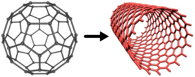
Fullerenes (left) can be converted to carbon nanotubes (right) with a catalytic process, according to Rice chemists (credits: Soroush83/CC and Matías Soto/Rice University)

http://www.kurzweilai.net/images/fullerene-to-nanotube.jpg

However, similar nanotubes have been found in samples from the exhaust pipes of Paris vehicles, in dust gathered from various places around the city, in spider webs in India, and even in ice cores, the paper notes.

“We know that carbon nanoparticles are found in nature,” Wilson said, noting that round fullerene (C60) molecules are commonly produced by volcanoes, forest fires, and other combustion of carbon materials. “All you need is a little catalysis to make carbon nanotubes instead of fullerenes.”

A car’s catalytic converter, which turns toxic carbon monoxide into safer emissions, bears at least a passing resemblance to the Rice-invented high-pressure carbon monoxide, or HiPco, process to make carbon nanotubes, he said. “So it is not a big surprise, when you think about it,” Wilson said.

“Based on our discovery of CNTs in tailpipes, we propose that the catalytic converters of the automobiles are manufacturing carbon nanotubes, Wilson told KurzweilAI. “However, we have not actually proven that.”

We are all carbon-nanotube bearers now

For ethical reasons, no cells from healthy patients were analyzed, but because nanotubes were found in all of the samples, the study led the researchers to conclude that carbon nanotubes are likely to be found in everybody.

“It’s kind of ironic. In our laboratory, working with carbon nanotubes, we wear facemasks to prevent exactly what we’re seeing in these samples, yet everyone walking around out there in the world probably has at least a small concentration of carbon nanotubes in their lungs,” he said.

The study followed one released by Rice and Baylor College of Medicine earlier this month with the similar goal ofanalyzing the black substance found in the lungs of smokers who died of emphysema. That study found carbon black nanoparticles that were the product of the incomplete combustion of such organic material as tobacco.

Co-authors are from Paris-Saclay University, the Paediatric Pulmonology and Allergy Center and the Department of Anatomo-Pathology of the Groupe hospitalier La Roche-Guyon, and Paris Diderot University. The Welch Foundation partially supported the research.


Abstract of Anthropogenic Carbon Nanotubes Found in the Airways of Parisian Children

Compelling evidence shows that fine particulate matters (PM) from air pollution penetrate lower airways and are associated with adverse health effects even within concentrations below those recommended by the WHO. A paper reported a dose-dependent link between carbon content in alveolar macrophages (assessed only by optical microscopy) and the decline in lung function. However, to the best of our knowledge, PM had never been accurately characterized inside human lung cells and the most responsible components of the particulate mix are still unknown. On another hand carbon nanotubes (CNTs) from natural and anthropogenic sources might be an important component of PM in both indoor and outdoor air.

We used high-resolution transmission electron microscopy and energy dispersive X-ray spectroscopy to characterize PM present in broncho-alveolar lavage-fluids (n = 64) and inside lung cells (n = 5 patients) of asthmatic children. We show that inhaled PM mostly consist of CNTs. These CNTs are present in all examined samples and they are similar to those we found in dusts and vehicle exhausts collected in Paris, as well as to those previously characterized in ambient air in the USA, in spider webs in India, and in ice core. These results strongly suggest that humans are routinely exposed to CNTs.

Read Full Post »

Nanotechnology therapy for non-cancerous diseases

Larry H. Bernstein, MD, FCAP, Curator

LPBI

Nanotechnology in respiratory medicine

Albert Joachim Omlor1, Juliane Nguyen2, Robert Bals3 and Quoc Thai Dinh13

Respiratory Research 2015, 16:64  http://dx.doi.org:/10.1186/s12931-015-0223-5

http://respiratory-research.com/content/16/1/64

Like two sides of the same coin, nanotechnology can be both boon and bane for respiratory medicine. Nanomaterials open new ways in diagnostics and treatment of lung diseases. Nanoparticle based drug delivery systems can help against diseases such as lung cancer, tuberculosis, and pulmonary fibrosis. Moreover, nanoparticles can be loaded with DNA and act as vectors for gene therapy in diseases like cystic fibrosis. Even lung diagnostics with computer tomography (CT) or magnetic resonance imaging (MRI) profits from new nanoparticle based contrast agents. However, the risks of nanotechnology also have to be taken into consideration as engineered nanomaterials resemble natural fine dusts and fibers, which are known to be harmful for the respiratory system in many cases. Recent studies have shown that nanoparticles in the respiratory tract can influence the immune system, can create oxidative stress and even cause genotoxicity. Another important aspect to assess the safety of nanotechnology based products is the absorption of nanoparticles. It was demonstrated that the amount of pulmonary nanoparticle uptake not only depends on physical and chemical nanoparticle characteristics but also on the health status of the organism. The huge diversity in nanotechnology could revolutionize medicine but makes safety assessment a challenging task.

Keywords: Nanoparticles; Lung; Airways; Nanotoxicology; Biodistribution; Nanomedicine

Over the past years nanomaterials have found their way into more and more areas of life. Examples are new coatings and pigments, electronic devices as well as cosmetic products like sunscreens and toothpastes. On top of that, much effort is done to adopt nanotechnology for the treatment of human diseases. The term “Nano” refers to structures in the range of 1 to 100 nm. In contrast to nanoparticles, which have to measure between 1 and 100 nm in all dimensions, nanomaterials may consist of elements bigger than 100 nm but need to be structured in the nanoscale and exhibit characteristic features associated with their nanostructure [1]. In this context, the International Organization for Standardization defined the term nano-object as a material with one, two or three external dimensions in the nanoscale [2] (Fig. 1). Nanomaterials have an extremely high surface area to volume ratio. Therefore, some of them are very reactive or catalytically active. Moreover, in the nanoworld quantum effects become visible and lead to some of the unique properties of nanoparticles. Like viruses and cellular structures, some nanoparticles are able to self-assemble to more complex structures [3]. This makes them interesting candidates for novel drugs. On the other hand it is necessary to redefine toxicology because of nanotechnology. Unlike classical toxicology, where dose and composition matter, in nanotoxicology the focus has to be set on properties like morphology, size, size distribution, surface charge, and agglomeration state as well. Nanotechnology is important for respiratory medicine for several reasons. Firstly, it offers new approaches to treat diseases of the respiratory tract. However, as nanotechnology usage in consumer products, cosmetics, and medicine is continuously increasing, it is also pivotal to understand potentially adverse effects of nanomaterials on the respiratory system. Additionally, studying respiratory effects of manufactured nanomaterials helps to understand the impact of combustion exhaust and ultra-fine dusts on human health. On top of that, the lung is probably the most important gateway of nanoparticles to the human organism. For the assessment of safety in nanotechnology it is therefore also important to elucidate which nanoparticle properties determine pulmonary resorption and biodistribution (Fig. 2).

Fig. 1. Nano-Objects can be divided into nanoparticles, nanofibres and nanoplates depending on the number of external dimensions in the nanoscale

Nano-Objects can be divided into nanoparticles, nanofibres and nanoplates

Nano-Objects can be divided into nanoparticles, nanofibres and nanoplates

http://www.respiratory-research.com/content/figures/s12931-015-0223-5-1.gif

Fig. 2. The increasing use of nanotechnology affects respiratory medicine in three main areas. Firstly, nanotechnology enables more sophisticated options in therapy and diagnostics. Secondly, the use of nanomaterials can cause toxic effects in the respiratory system. Health risks associated with the use of nanomaterials are not fully understood and merit further investigation. Moreover, it will be essential to understand the effects of inhaled nanoparticles on extrapulmonary organs

nanotechnology affects respiratory medicine in three main areas

nanotechnology affects respiratory medicine in three main areas

http://www.respiratory-research.com/content/figures/s12931-015-0223-5-2.gif

Applications of nanotechnology in therapeutics and diagnostics

Although clinical application of nanotechnology in therapeutics and diagnostics is still rare, there are multiple promising candidates for future use in the field of respiratory medicine.

Drug delivery

Nanoparticles can act as vessels for drugs because they are small enough to reach almost any region of the human organism. Drugs can be bound chemically to the nanoparticles by a multitude of different linker molecules or by encapsulation. This allows better control of toxicokinetics. However, the main advantage is the capability of targeted drug delivery. The targeting can be active or passive. In case of tumor diseases, the leaky and immature vasculature of fast growing tumors can be taken advantage of in order to achieve passive targeting of chemotherapeutic loaded nanoparticles. This is called the enhanced permeability and retention (EPR) effect [4]. The first generation nano drug delivery systems rely entirely on the EPR effect. One example is Genoxol-PM, a polymeric paclitaxel loaded poly(lactic acid)-block-poly(ethylene glycol) micelle-formulation [5]. This nanocarrier has recently been tested in a phase II trial in patients with advanced non-small cell lung cancer (NSCLC). 43 patients were treated with four 3-week cycles of Genexol-PM at 230 mg/m2 on day 1 combined with gemcitabine 1000 mg/m2 on day 1 and day 8. With a response rate of 46.5 %, the therapy showed favorable antitumor activity. Moreover, emetogenicity was low. However, frequent grade 3/4 adverse events like neutropenia and pneumonia were observed [6]. The second generation nanoparticle drug delivery systems possess targeting ligands. These can be antibodies, aptamers, small molecules and proteins (Fig. 3). The attached ligands actively guide the nanoparticles and therefore the drugs to the tumor cells. Tumor specific monoclonal antibodies are already widely used in cancer therapy. Those antibodies can be attached to nanoparticles for active targeting. In a recent study polyglycolic acid nanoparticles, that were conjugated with cetuximab antibodies for targeting and loaded with the drug paclitaxel palmitate, were administered intravenously to mice with A549-luc-C8 lung tumors. The survival rate of these mice increased significantly compared to the control group [7]. Another approach involves aptamers as targeting agents. Aptamers are synthetic oligonucleotides that are capable of binding specific target structures. Their small size, their simple synthesis, and their lack of immunogenicity make them promising ligands for nanoparticles. Moreover, small molecules such as folate can be used for targeting tumor cells that express a high density of folate receptors. In addition, tumors often overexpress receptors for several proteins. Proteins like transferrin therefore are common targeting ligands [8]. These second generation nanocarriers are already used clinically against lung cancer with substances like Aurimmune Cyt-6091 and Bind-014. Aurimmune Cyt-6091 is a drug delivery system based on gold nanoparticles functionalized with polyethylene glycol (PEG) and tumor necrosis factor alpha (TNF-α). It has been used against adenocarcinoma of the lung in a phase I clinical trial. The TNF-α serves both as targeting and therapeutic agent in this case [9]. A phase II clinical trial for non-small cell lung cancer patients has been planned [10]. The nano drug delivery system Bind-014 is currently tested in a phase II clinical trial as second-line therapy for patients with non-small cell lung cancer [11]. Bind-014 nanoparticles consist of a polylactic acid (PLA) core, in which the anti-tumor drug docetaxel is physically entrapped. The particles are surface-decorated with PEG to reduce elimination from the immune system and contain ligands against prostate-specific membrane antigen (PSMA) for targeting. PSMA is expressed in prostate cancer cells and in the neovasculature of nonprostate solid tumors, such as NSCLC [12]. Preliminary data demonstrates, that Bind-014 is clinically active and well tolerated. It also showed promising effects on patients with KRAS mutations, where ordinary anti-tumor agents usually fail. Additionally, adverse effects like anemia, neutropenia and neuropathy were significantly reduced compared to solvent based docetaxel [13].

Four different strategies for active targeting of nanoparticle based drug delivery systems

Four different strategies for active targeting of nanoparticle based drug delivery systems

Fig. 3. Four different strategies for active targeting of nanoparticle based drug delivery systems are shown. The nanoparticles can be conjugated with tumor specific antibodies or aptamers. Additionally, small molecules, such as folate, as well as proteins, such as transferrin, can be used for targeting receptors that are overexpressed on tumors

http://www.respiratory-research.com/content/figures/s12931-015-0223-5-3.jpg

Nanoparticle based drug delivery also offers potential in other fields of respiratory medicine. In experiments with tuberculosis infected guinea pigs, it was demonstrated that inhaled alginate nanoparticles encapsulating isoniazid, rifampicin, and pyrazinamide showed better bioavailability and higher efficiency than oral drug medication [14]. Similar results were presented by Pandey et al. with the three antitubercular drugs encapsulated in poly (DL-lactide-co-glycolide) nanoparticles[15]. Moreover, another study demonstrated that pirfenidone loaded nanoparticles have higher anti-fibrotic efficacy in the treatment of mice with bleomycin-induced pulmonary fibrosis than dissolved pirfenidone [16].

Hyperthermia

Nanoparticle induced hyperthermia can be used to locally destroy tumor cells. Heat generation is usually achieved by two approaches, magnetic and photothermal hyperthermia. In magnetic hyperthermia, an extracorporeal coil creates an alternating magnetic field that heats magnetic nanoparticles inside a tumor. This increases the temperature in the tumor without affecting healthy tissue. A recent study assessed the effect of inhalable superparamagnetic iron oxide nanoparticles in a mouse model of NSCLC. Compared to the non-targeted nanoparticles, the epidermal growth factor receptor (EGFR) targeted nanoparticles showed significantly more effective tumor shrinkage after magnetic hyperthermia treatment [17]. The other approach, photothermal therapy uses laser radiation in the visible or near infrared spectrum and photosensitizing nanoparticles such as gold or graphene. A commercial product called auroshell is available for tumor therapy. Auroshell nanoparticles consist of a silica core surrounded by a thin layer of gold. The gold nanoshells are administered intravenously and accumulate in the tumor due to the EPR effect. Upon exposure of the tumor to a near infrared laser, the laser energy is efficiently converted to heat by the gold nanoshells [18]. This therapy, which is called AuroLase, is currently undergoing clinical trial in patients with primary and/or metastatic lung tumors [19] (Fig. 4).

Two different approaches of nanoparticle based hyperthermia therapy

Two different approaches of nanoparticle based hyperthermia therapy

Fig. 4. Two different approaches of nanoparticle based hyperthermia therapy are shown. a In magnetic hyperthermia, magnetic nanoparticles (MNP) are applied intravenously and accumulate inside the tumor. When an oscillating magnetic field is created by an extracorporeal coil the magnetic nanoparticles produce heat inside the tumor. b In photothermal hyperthermia, gold nanoshells (GNS) or similar photosensitizing nanoparticles are applied intravenously and accumulate inside the tumor. Upon exposure of the tumor to near infrared (NIR) laser radiation, the gold nanoshells convert the laser light into heat

http://www.respiratory-research.com/content/figures/s12931-015-0223-5-4.gif

Gene therapy

Like viruses, nanoparticles can be used as vectors for genes. But in contrast to viruses, they are less immunogenic and have higher DNA transport capacity. In a study, DNA loaded polyethylenimine nanoparticles were used in order to treat lipopolysaccharide induced acute lung injury in mice. After intravenous injection of the nanoparticles, the beta2-Adrenic Receptor genes in the nanoparticles led to a short lived transgene expression in alveolar epithelia cells. As a result the 5-day survival rate improved from 28 % to 64 %. The severity of the symptoms measured by alveolar fluid clearance, lung water content, histopathology, bronchioalveolar lavage cellularity, protein concentration, and inflammatory cytokines was also significantly attenuated [20]. DNA loaded nanoparticles are also promising candidates in the treatment of cystic fibrosis. It was shown in a clinical trial that nasal application of DNA nanoparticles is safe and evidently leads to vector gene transfer [21]. One major problem in this context is to overcome the mucus barrier. In a recent study, it was demonstrated that densely PEG-coated DNA nanoparticles can rapidly penetrate extracorporeal human cystic fibrosis and extracorporeal mouse airway mucus. In addition, those particles exhibited better gene transfer after intranasal administration to mice than conventional carriers [22].

Diagnostics

Nanoparticles have the potential to improve pulmonary x-ray diagnostics. Folic acid-modified dendrimer-entrapped gold nanoparticles were utilized as imaging probes for targeted CT imaging. In in-vitro and in-vivo tests, the nanoparticles were trapped in the lysosomes of folic acid receptor expressing lung adenocarcinoma cells (SPC-A1). It was possible to detect the tumor cells by micro-CT imaging after nanoparticle uptake. In addition, it was also shown that the particles possess good biocompatibility, with no impact on cell morphology, viability, cell cycle, and apoptosis [23]. Nanoparticles can also be used to enhance MR diagnostics of lung tissue. In experiments with intratracheal administration of Gadolinium-DOTA nanoparticles in mice, signal enhancements in several organs including the lung were measured with ultrashort-echo-time-proton-MRI. The signal change over time in the different organs demonstrated the passage of the nanoparticles from the lung to the blood, then to the kidneys, and finally to the bladder [24].

Toxicological aspects of nanomaterials

Toxic effects of nanoparticles are a major concern in pulmonary medicine. Especially ultrafine particles of low soluble, low toxic materials like titanium dioxide, carbon black, and polystyrene are overall more toxic and inflammatory than fine particles of the same material. This applies to both synthesized nanoparticles and natural dusts [25]. For nano related toxicity multiple mechanisms seem to be important. In the following, the interaction with the immune system, the creation of oxidative stress, and toxic effects on the genome are taken a closer look at. In order to correlate toxic effects with nanoparticle properties, it is necessary to thoroughly characterize the selected nanoparticles prior to administration.

Nanoparticle characterization

The most commonly used methods to characterize nanoparticles for toxicology studies are transmission electron microscopy (TEM) for size, morphology, and agglomeration, dynamic light scattering (DLS) for the size distribution of the particles, zeta potential measurement for nanoparticle surface charge, and x-ray diffraction (XRD) for the particles’ crystal structure. In some cases such as gold and silver nanoparticles, UV-vis spectroscopy can be used to determine size and size distribution due to a special size dependent optical activity [26]. Ideally, nanoparticle characterization is repeated after administration as changes of the nanoparticles during the application process are possible. In in-vitro experiments, nanoparticles are usually applied by mixing with cell culture medium. The dissolved components of the medium, especially the ions, lead to agglomeration and precipitation of many nanoparticles, causing significant changes in their physicochemical properties. Similar effects are to be expected when nanoparticles come into contact with surfactant or other biological fluids. It was shown, that some nanoparticles tend to form protein coronae in biological systems [27].

Effects on immune system and inflammation

Many nanoparticles possess properties that give them the potential to influence the immune system. In this context, nanoparticles’ ability to penetrate cellular boundaries, to escape phagocytation by macrophages, to act as haptens, and even to disturb the Th1/Th2 balance might be essential [28]. For carbon black nanoparticles, a recent study investigated the effects of inhalative exposure on mice with bleomycin-induced pulmonary fibrosis. The analysis of histology as well as cytokine expression suggested that the nanoparticles triggered an inhalation exacerbated lung inflammation. The author concluded that especially for people with pulmonary preconditions inhalation of nanoparticles can lead to serious health problems [29]. In this context, another study found out that PEGylated cationic shell-cross-linked knedel-like (cSCK) nanoparticles produced significantly less airway inflammation than non-PEGylated ones. This was explained by a change in endocytosis. In contrast to the clathrin-dependent endocytosis of non-PEGylated particles, the PEGylated cSCK nanoparticles showed a clathrin-independent route [30]. On the other hand, some nanomaterials exhibit impressive immune modulating activity. As an example, [Gd@C82(OH)22]n, a fullerene derivate with a gadolinium atom inside showed anticancer activity without being cytotoxic (Fig. 5). In vitro studies demonstrated that [Gd@C82(OH)22]n activated dendritic cells (DCs) and even induced phenotypic maturation of those cells. Moreover, the [Gd@C82(OH)22]n treated DCs also stimulated allogenic T cells in a Th1 characteristic. The effect of [Gd@C82(OH)22]n was comparable, probably even stronger than the effect of lipopolysaccharide (LPS) on DCs. The study also verified that the nanoparticles were free of LPS contamination. In-vivo experiments on ovalbumin (OVA) immunized mice showed enhanced immune responses comparable to the adjuvant effect of Alum on OVA mice. However, whereas Alum lead to a Th2 response pattern with IL-4, IL-5 and IL-10 upregulation, [Gd@C82(OH)22]n caused a Th1 pattern with upregulation of IFNγ [31]. Similar results were demonstrated in another study using a murine asthma model. OVA sensitized mice that were additionally treated with the nanomaterial graphene oxide during allergen sensitization had stronger airway remodeling and hyperresponsiveness than mice that have only been treated with OVA. The graphene oxide lead to a downregulation of Th2 dependent markers such as IL-4, IL-5, IL-13 IgE and IgG1 but increased Th1-associated IgG2a. Moreover, the graphene oxide increased the macrophage production of mammalian chinitases, chitinase-3-like protein 1 (CHI3L1), and AMCase, which could be the reason for the overall augmentation in airway remodeling and hyperresponsiveness [32]. However, this kind of immune modulation can also be utilized for therapeutic purposes. In a recent study a nanoparticle-based vaccine has been used to treat dust mite allergies in mice. The immune-modulating carriers were generated by loading dust mite allergen Der p2 and the potent Th1 adjuvant unmethylated cytosine-phosphate-guanine (CpG) into biodegradable poly(lactic-co-glycolic acid) (PLGA) polymer particles. Mice treated with those nanoparticles showed significantly lower airway hyperresponsiveness as well as lower IgE antibody levels after a 10 day intranasal Der p2 instillation compared to the control group. The authors conclude that this biodegradable nanoparticle-based vaccination strategy has significant potential for treating HDM allergies [33].

Gd@C82(OH)22 consists of a gadolinium atom (green) inside a 2 nm cage of carbon atoms (grey)

Gd@C82(OH)22 consists of a gadolinium atom (green) inside a 2 nm cage of carbon atoms (grey)

Fig. 5. Gd@C82(OH)22 consists of a gadolinium atom (green) inside a 2 nm cage of carbon atoms (grey). The hydroxyl groups (red) outside the cage are responsible for water solubility. In water the molecule forms aggregates [Gd@C82(OH)22]n with average size of 25 nm

http://www.respiratory-research.com/content/figures/s12931-015-0223-5-5.jpg

Oxidative stress and catalysis

Oxidative stress is often brought in context with nanotoxicology. It can be measured directly with dichlorofluorecein or indirectly by the upregulation of reactive oxygen species (ROS) eliminating enzymes like superoxide dismutase [34]. Another approach involves tests whether the nanoparticle dependent toxicity can be reduced by the application of an antioxidant. Widely used semiconductor materials such as lead sulfide nanoparticles may have the potential to generate oxidative stress in the lung. A recent study tested the toxicity of intratracheally applied 30 nm and 60 nm lead sulfide nanoparticles on rats. Oxidative damage was evaluated based on superoxide dismutase, total antioxidant capacity, and concentration of malondialdehyde. In addition to inflammatory responses, both 30 nm and 60 nm groups showed increased oxidative damage compared to control groups. The effect was significantly stronger for the 30 nm lead sulfide compared to the 60 nm nanoparticles [35]. Another nanomaterial which is associated with oxidative stress is nanosized titanium dioxide. Li et al. induced pulmonary injury in mice by daily intranasal instillation of suspended 294 nm TiO2 nanoparticles for 90 days, demonstrating that the rate of reactive oxygen species (ROS) generation increased with increasing TiO2 doses. Moreover lipid, protein and DNA peroxidation products were identified in elevated doses, which suggests that ROS dependent lung damage was significant in the nanoparticle treated animals [36]. Furthermore, in vitro tests on BEAS-2B and A549 lung cell lines demonstrated that the commonly used nanoparticles ZnO and Fe2O3 are very different in terms of creating oxidative stress. The Fe2O3nanoparticles with an average diameter of 39 nm were distributed in the cytoplasm, whereas the 63 nm ZnO nanoparticles were trapped in organelles such as the endosome. In contrast to the Fe2O3 nanoparticles the ZnO nanoparticles caused reactive oxygen species production as well as cell cycle arrest, cell apoptosis, mitochondrial dysfunction and glucose metabolism perturbation[37] (Table 1).

Table 1. Oxidative stress induction in respiratory tissue by different nanoparticles

Genotoxicity

Another important type of toxicity caused by nanoparticles is genotoxicity. A common method to quantify genotoxicity is the comet assay, which uses electrophoresis to detect DNA strand breaks. This assay was used in a recent study to check whether intratracheal instilled fullerene C60nanoparticles induced DNA damage in male rats. However, despite inflammatory responses and hemorrhages in the alveoli of the C60 treated rats, there was no significant increase in fractured DNA in their lung cells. Therefore, it was concluded that even at inflammation inducing doses, fullerene C60 nanoparticles have no potential for DNA damage in the lung cells of rats [38]. Similarly, another study demonstrated that intratracheal instillation of anatase TiO2 nanoparticles on rats did not result in genotoxicity. None of the TiO2 groups showed an increase in fractured DNA while the positive control with ethyl methanesulfonate exhibited significant increases [39]. In contrast to those results, Kyjovska ZO et al. found that even in low doses, where no inflammation occurs, Printex 90 carbon black nanoparticles induce genotoxicity in mice. There was no inflammation, cell damage and acute phase response, which means that the increased DNA strand breaks are related to direct DNA damage caused by the nanoparticles [40]. On the other hand, a recent study suggests that CeO2 nanoparticles may be even used as antioxidant and anti-genotoxic agents in the lung. After treatment with the oxidative stress-inducing agent KBrO3, BEAS-2B cells pretreated with the CeO2 nanoparticles showed significantly less intracellular ROS as well as a reduction in DNA damage compared to non-pretreated cells [41].

Biodistribution

Nanoparticle detection

Research on the biodistribution of nanoparticles requires tracking of the applied nanoparticles in the test animal. Conventional light microscopy is not able to detect nanoparticles because of Abbe’s law. Therefore, electron microscopic imaging is often required. However, light microscopy can be used to describe the nanoparticle induced changes in the cell morphology without being able to see the nanoparticles themselves. Additionally, nanoparticles can be indirectly made detectable in light microscopy by a method called autometallography. This is a silver staining that can be used to increase the size of several types of nanoparticles like gold, silver, and some metal sulfides and selenides in the histological section [42]. This technique was used to detect silver nanoparticles in the olfactory bulb and lateral brain ventricles of mice that had been intranasally treated with 25 nm silver nanoparticles [43].

Particle deposition and resorption in the respiratory tract

Most research about biodistribution of nanoparticles in organisms focuses on intravenous injection. However, nanoparticles were shown to be able to pass the blood air barrier of the lung. Whether or not nanoparticles can travel through the lung into the body seems to be size dependent. This was evaluated by injecting neutron activated radioactive gold nanoparticles of 1.4 nm and 18 nm intratracheally to rats. The bigger nanoparticles almost completely retained in the lung while significant amounts of the smaller 1.4 nm particles were found in blood, liver, skin and carcass 24 h after instillation [44]. Choi H. S. et al. applied nanoparticles of different size and charge to mice. The nanoparticles were tracked in different organs through fluorescence labeling. It was demonstrated that nanoparticles rapidly translocated to the mediastinal lymph nodes if they possess a hydrodynamic diameter of 34 nm or less and a neutral or anionic surface. Bigger and positively charged nanoparticles exhibited no significant uptake [45] (Fig. 6). In addition to physical parameters of the applied nanoparticles the health status of the exposed organism also seems to play an important role. A recent study showed that the distribution of oropharyngeal instilled 40 nm gold nanoparticles is influenced by additional LPS treatment. The gold content of organs was measured with inductively coupled plasma mass spectroscopy. BALB/C mice that had been oropharyngeal treated with LPS 24 h prior to the nanoparticle administration exhibited less gold content in their lungs than untreated mice. In both groups gold was detected in different organs. High concentrations were found in heart and thymus in the non LPS group, while the LPS treated mice accumulated most of the gold in the spleen. The author concluded that nanoparticle uptake may depend on medical preconditions [46].

Fig. 6. Pulmonary uptake of nanoparticles depends on size and surface charge. Positively charged nanoparticles and nanoparticles that are bigger than 34 nm cannot pass the epithelial barrier of the lung. Only small and not positively charged nanoparticles can translocate from the lung over blood and lymph system to the organism

Pulmonary uptake of nanoparticles depends on size and surface charge

Pulmonary uptake of nanoparticles depends on size and surface charge

http://www.respiratory-research.com/content/figures/s12931-015-0223-5-6.jpg

Conclusions

Over the last decade, major breakthroughs in nanotechnology have been achieved. It is only a matter of time before new nano based drugs reach respiratory medicine. Especially the fields of targeted drug delivery, gene therapy, and hyperthermia offer great potential for modern drugs. On the other hand the increased use of nanomaterials in all fields of life also bears the risk of exposure through inhalation. It is therefore essential to understand pulmonary toxicology of nanomaterials in all its facets. However, it is still very unclear why the toxic effects of nanoparticles in the respiratory tract are so inhomogeneous and not well predictable. In this context, not only local reactions of lung and airways but also nanoparticle uptake and distribution in the organism are important factors and therefore fields of current research. As only few nanoparticle compositions have been tested, it is questionable whether those results can be easily adapted to other nanoparticles. Because of the continuously increasing diversity of engineered nanoparticles, toxicology can hardly keep pace with the safety assessment of future products. Therefore, more attention should be set on this wide field of research.

Abbreviations

CHI3L1: Chitinase-3-like protein 1

cSCK: Cationic shell-cross-linked knedel-like

CT: Computer tompgraphy

DC: Dendritic cell

DLS: Dynamic light scattering

EGFR: Epidermal growth factor receptor

EPR: Enhanced permeability and retention

LPS: Lipopolysaccharide

MRI: Magnetic resonance imaging

NSCLC: Non-small-cell lung carcinoma

OVA: Ovalbumin

PEG: Polyethylene glycol

PLA: Polylactic acid

PLGA: Poly(lactic-co-glycolic acid)

PSMA: Prostate-specific membrane antigen

ROS: Reactive oxygen species

TEM: Transmission electron microscopy

TNF-α: Tumor necrosis factor alpha

XRD: X-ray diffraction

  1. Oberdorster G, Maynard A, Donaldson K, Castranova V, Fitzpatrick J, Ausman K et al..Principles for characterizing the potential human health effects from exposure to nanomaterials: elements of a screening strategy. Part Fibre Toxicol. 2005; 2:8.PubMed Abstract | BioMed Central Full TextOpenURL
  2. Nanotechnologies — Terminology and definitions for nano-objects — Nanoparticle, nanofibre and nanoplate, ISO/TS 27687:2008. [Accessed March 30, 2015]. Iso.org . 26-1-2012. Ref Type: Electronic Citation
  3. Grzelczak M, Vermant J, Furst EM, Liz-Marzan LM. Directed self-assembly of nanoparticles. ACS Nano. 2010; 4:3591-3605. PubMed Abstract | Publisher Full TextOpenURL
  4. Hobbs SK, Monsky WL, Yuan F, Roberts WG, Griffith L, Torchilin VP et al.. Regulation of transport pathways in tumor vessels: role of tumor type and microenvironment.Proc Natl Acad Sci U S A. 1998; 95:4607-4612. PubMed Abstract | Publisher Full TextOpenURL
  5. Sanna V, Pala N, Sechi M. Targeted therapy using nanotechnology: focus on cancer. Int J Nanomedicine. 2014; 9:467-483. PubMed Abstract | Publisher Full TextOpenURL
  6. Ahn HK, Jung M, Sym SJ, Shin DB, Kang SM, Kyung SY et al.. A phase II trial of Cremorphor EL-free paclitaxel (Genexol-PM) and gemcitabine in patients with advanced non-small cell lung cancer. Cancer Chemother Pharmacol. 2014; 74:277-282.PubMed Abstract | Publisher Full TextOpenURL
  7. Karra N, Nassar T, Ripin AN, Schwob O, Borlak J, Benita S. Antibody conjugated PLGA nanoparticles for targeted delivery of paclitaxel palmitate: efficacy and biofate in a lung cancer mouse model. Small. 2013; 9:4221-4236. PubMed Abstract |Publisher Full TextOpenURL
  8. Yu B, Tai HC, Xue W, Lee LJ, Lee RJ. Receptor-targeted nanocarriers for therapeutic delivery to cancer. Mol Membr Biol. 2010; 27:286-298. PubMed Abstract |Publisher Full TextOpenURL
  9. Libutti SK, Paciotti GF, Byrnes AA, Alexander HR, Gannon WE, Walker M et al.. Phase I and pharmacokinetic studies of CYT-6091, a novel PEGylated colloidal gold-rhTNF nanomedicine. Clin Cancer Res. 2010; 16:6139-6149. PubMed Abstract |Publisher Full TextOpenURL
  10. Aurimmune CYT-6091 phase II clinical trial planned [Accessed March 30, 2015]. 17-9-2014. Ref Type: Online Source
  11. A Phase 2 Study to Determine the Safety and Efficacy of BIND-014 (Docetaxel Nanoparticles for Injectable Suspension) as Second-line Therapy to Patients With Non-Small Cell Lung Cancer. [Accessed March 30, 2015]. ClinicalTrials.gov . 23-1-2015. Ref Type: Electronic Citation.
  12. Hrkach J, Von HD, Mukkaram AM, Andrianova E, Auer J, Campbell T et al.. Preclinical development and clinical translation of a PSMA-targeted docetaxel nanoparticle with a differentiated pharmacological profile. Sci Transl Med. 2012; 4:128ra39.PubMed Abstract | Publisher Full TextOpenURL
  13. Natale R, Socinski M, Hart L, Lipatov O, Spigel D, Gershenhorn B et al.. 41 Clinical activity of BIND-014 (docetaxel nanoparticles for injectable suspension) as second-line therapy in patients (pts) with Stage III/IV non-small cell lung cancer. Eur J Cancer. 2014; 50, Supplement 6:19. Publisher Full TextOpenURL
  14. Ahmad Z, Sharma S, Khuller GK. Inhalable alginate nanoparticles as antitubercular drug carriers against experimental tuberculosis. Int J Antimicrob Agents. 2005; 26:298-303. PubMed Abstract | Publisher Full TextOpenURL
  15. Pandey R, Sharma A, Zahoor A, Sharma S, Khuller GK, Prasad B. Poly (DL-lactide-co-glycolide) nanoparticle-based inhalable sustained drug delivery system for experimental tuberculosis. J Antimicrob Chemother. 2003; 52:981-986. PubMed Abstract| Publisher Full TextOpenURL

Pulmonary applications and toxicity of engineered nanoparticles.

Because of their unique physicochemical properties, engineered nanoparticles have the potential to significantly impact respiratory research and medicine by means of improving imaging capability and drug delivery, among other applications. These same properties, however, present potential safety concerns, and there is accumulating evidence to suggest that nanoparticles may exert adverse effects on pulmonary structure and function. The respiratory system is susceptible to injury resulting from inhalation of gases, aerosols, and particles, and also from systemic delivery of drugs, chemicals, and other compounds to the lungs via direct cardiac output to the pulmonary arteries. As such, it is a prime target for the possible toxic effects of engineered nanoparticles. The purpose of this article is to provide an overview of the potential usefulness of nanoparticles and nanotechnology in respiratory research and medicine and to highlight important issues and recent data pertaining to nanoparticle-related pulmonary toxicity.

PMID:
18641236
[PubMed – indexed for MEDLINE]
PMCID:
PMC2536798

Free PMC Article

The possibility of nanotechnology dramatically improving the health and quality of life of people throughout the world holds great promise. Predictions of beneficial effects of nanotechnology in numerous industrial, consumer, and medical applications have been promising. By no means an exhaustive list, these applications include those that may lead to more efficient water purification, stronger and lighter building materials, increased computing power and speed, improved generation and conservation of energy, and new tools for the diagnosis and treatment of disease. The optimistic outlook for a future improved by nanotechnology must be tempered, however, by the realization that relatively little is known about the potential adverse effects of nanomaterials on human health and the environment.

The definition of a nanoparticle is generally considered to be a particle with at least one dimension of 100 nm or less. As a result of their small size and unique physicochemical properties, the toxicological profiles of nanoparticles may differ considerably from those of larger particles composed of the same materials (15, 98). Furthermore, nanoparticles of different materials (e.g., gold, silica, titanium, carbon nanotubes, quantum dots) are not expected to interact with and affect biological systems in a similar fashion. As a result, it seems unlikely that the toxic potential and/or mechanisms of nanoparticles can be predicted or explained by any single unifying concept.

The respiratory system represents a unique target for the potential toxicity of nanoparticles due to the fact that in addition to being the portal of entry for inhaled particles, it also receives the entire cardiac output. As such, there is potential for exposure of the lungs to nanoparticles that are introduced to the body via the act of breathing and by any other exposure route that may result in systemic distribution, including dermal and gastrointestinal absorption and direct injection. Interest in the respiratory system as a target for the potential effects, both beneficial and adverse, of nanoparticles is reflected by the steady increase in the number of scientific publications on these subjects during the past decade (Fig. 1).

publications related to the pulmonary toxicity and applications of engineered nanoparticles

publications related to the pulmonary toxicity and applications of engineered nanoparticles

Scientific publications related to the pulmonary toxicity and applications of engineered nanoparticles. The number of articles published in each of the past 10 years was identified by searching the PubMed database

he purpose of this article is to complement and expand on previous reviews of the pulmonary effects of nanoparticles (11, 14, 34, 35) by providing an overview of potential applications of nanotechnology in pulmonary research and in diagnosis and treatment of disease. In addition, recent advances regarding the potential pulmonary toxicity of nanoparticles as assessed in human, experimental animal, and in vitro studies are discussed. For the purposes of this article, only intentionally engineered nanoparticles are considered; unintentionally generated (e.g., via combustion engines, grilling, welding) and naturally occurring nanoparticles (e.g., via forest fires or volcanic eruptions) are not included in this discussion.

NANOPARTICLES AND THE LUNG

There are myriad nanoparticles to which the respiratory system may be exposed.

There is the potential for the respiratory system to be exposed to a seemingly countless number of unique nanoparticles, essentially none of which has been sufficiently examined for potential toxicity at this time. A substantial number of nanoparticles are already present in the marketplace in consumer products such as sunscreens, cosmetics, and car wax, and many more are sure to follow (a comprehensive list is maintained and updated by the Project on Emerging Nanotechnologies at the Woodrow Wilson International Center for Scholars: http://www.wilsoncenter.org/nano). Although the toxicity of the majority of nanoparticles may prove to be minimal, the fact that there is any potential for adverse effects to result from exposure suggests that prudence is warranted.

Various types of nanoparticles exist including those that are carbon-based (e.g., nanotubes, nanowires, fullerenes) and metal-based (e.g., gold, silver, quantum dots, metal oxides such as titanium dioxide and zinc oxide) and those that are arguably more biological in nature (e.g., liposomes and viruses designed for gene or drug delivery). To demonstrate the complexity of the situation, it is worthwhile to consider the case of carbon nanotubes as an example. Carbon nanotubes can be: 1) produced and/or cleaned using one of several different methods; 2) produced using one of several different metal catalysts; 3) single- or multi-walled; 4) of various lengths; and 5) subjected to numerous surface modifications. The result of these permutations is that a vast number of unique carbon nanotubes can be derived, all of which fall under one broad category, namely the carbon nanotube. Dividing these into single-walled and multi-walled forms reduces the ambiguity only so much, and we are still left with potentially thousands of each type. Furthermore, as has been demonstrated in recent in vitro experiments (37), the potential for nanotube agglomeration or for adhesion of nanotubes to biological molecules and the resultant alteration of their reactivity must be considered. Needless to say, the variations in nanoparticle form and functionality, not only for carbon nanotubes but also for nanoparticles in general, present significant challenges in the assessment of their potential usefulness and toxicity.

Nanoparticle accumulation within the lung.

Nanoparticles may reach the lung via inhalation or systemic delivery and do so by incidental/accidental or intentional means. Intentional pulmonary administration is being examined as a means of nanoparticle delivery for imaging and therapeutic purposes and is discussed separately below. Incidental or accidental inhalation exposure to nanoparticles can be envisioned most likely to occur as a result of exposure to occupational aerosols during the production or packaging of nanoparticles or nanostructured materials (89). In addition to pulmonary effects resulting from such exposures, translocation and subsequent systemic exposure and accumulation are also possible and are being investigated. It should be noted that nanoparticles naturally tend to agglomerate into larger particles that can be microns in size, thereby reducing the likelihood of free nanoparticles being respired. However, surface modifications designed to limit particle-particle interactions and protein binding may reduce the tendency for nanoparticle agglomeration and increase the potential for inhalation and deposition within the lungs (131).

Incidental pulmonary exposure as a result of systemic delivery is likely inherent for any nanoparticle that is injected or that might be absorbed following dermal application or ingestion. Although no published human data pertaining to pulmonary accumulation of nanoparticles following systemic exposure were identified, several animal studies have demonstrated pulmonary accumulation of nanoparticles (or of drug-conjugated nanoparticles) by means of determining their quantity in total lung homogenate preparations following their ingestion or intravenous or subcutaneous injection (43, 71, 109, 138, 142, 155). None of these studies investigated whether systemically administered nanoparticles traversed the blood-air barrier to gain access to the interstitium or lung epithelium; however, this is not necessarily a requirement for beneficial (or detrimental) effects to ensue. Although the levels and duration of accumulation appear to vary for the different nanoparticles examined, these data highlight the potential for exposure of the lungs to nanoparticles via the systemic route.

PULMONARY APPLICATIONS OF NANOPARTICLES

Imaging and diagnostic applications.

Many improvements in imaging capabilities that will benefit basic and clinical pulmonary research and disease diagnosis can be envisioned through the application of nanotechnology. Advances that include the delivery of nanoparticle imaging agents to specific cells or tissues of interest, the development of nanoprobes for molecular imaging of disease pathways, and the development of better contrast agents are forthcoming (21, 22, 115). Quantum dots are one type of nanoparticle that is proving to be particularly useful for imaging and diagnostic purposes. These semiconductor nanocrystals have broad absorption spectra and narrow emission spectra, and as their fluorescence is dependent on their chemical composition and size, multiple quantum dots (each with a unique color emission) can be detected simultaneously. Moreover, their relatively large surface area provides the opportunity for attachment of peptides or antibodies that precisely target cell types or tissues for imaging, thereby increasing specificity and decreasing background. In this regard, Akerman et al. (2) demonstrated that quantum dots coated with a peptide that binds to membrane dipeptidase on pulmonary endothelial cells were detected in the lung but not in brain or kidney 5 min after intravenous administration in BALB/c mice. Furthermore, in a study using quantum dots conjugated to monoclonal antibodies, rapid and specific detection of respiratory syncytial virus infection was demonstrated in vitro and in the lungs of BALB/c mice in vivo (137). Quantum dots have also been used to study tumor cell extravasation into lung tissue in C57BL/6 mice (140), highlighting the utility of these nanoparticles in the study of tumor metastasis.

Other nanoprobes for pulmonary imaging and diagnostics are also being examined experimentally. A recent study by le Masne de Chermont et al. (78) demonstrated that inorganic luminescent nanoparticles can be optically excited before injection into mice to provide long-lasting imaging of the lung. This was particularly evident for the positively charged nanoparticles that were studied, as noninvasive external detection revealed significant pulmonary accumulation of these nanoparticles up to 1 h following intravenous injection (78).

Therapeutic applications.

The potential therapeutic applications of nanoparticles in respiratory and systemic diseases are numerous (20, 21, 112,115, 133). A considerable thrust of recent research has been focused on determining the suitability of nanoparticles of various types to serve as vectors for the pulmonary delivery of drugs or genes via inhalation or systemic administration, whereas other efforts have been directed toward developing and delivering nano-sized drug particles to the lung (Table 1). The majority of the studies reported to date have focused on the utility of these strategies for the treatment of pulmonary infection. As an example, gene transfer using intranasal administration of chitosan-DNA nanospheres was shown to prophylactically inhibit respiratory syncytial virus infection and to reduce allergic airway inflammation in mice when given prophylactically or therapeutically (74, 75). Moreover, nanoparticle-mediated intranasal delivery of short interfering RNA (siRNA) targeted against a specific viral gene, NS1, has also been shown to inhibit respiratory syncytial virus infection in mice and rats (72, 161).

Table 1.

logo-ajplung

The usefulness of nano-sized drug particles as treatment modalities in models of pulmonary infection has also been investigated. Inhalation of aerosolized nano-sized itraconazole resulted in significantly higher lung concentrations in mice than did oral administration (138) and was found to prophylactically inhibit invasive pulmonary aspergillosis and reduce infection-related deaths in mice, whereas oral drug administration did not (4, 59). In addition, Pandey et al. (110) demonstrated that a single inhalation of aerosolized poly (DL-lactide-co-glycolide) nanoparticles loaded with antitubercular drugs (isoniazid, rifampicin, or pyrazinamide) resulted in therapeutic plasma drug levels for up to 6 days in guinea pigs and found that repeated inhalations were as effective as more frequent oral administrations of free drug in treating experimental tuberculosis. A subsequent study revealed that a single subcutaneous injection of these antitubercular drug-containing nanoparticles in mice resulted in therapeutic plasma drug levels for up to 32 days and was more effective at reducing bacterial counts in the lungs and spleen than was daily oral administration of free drug (109). Finally, Zahoor et al. (158) reported that the same antitubercular drugs were more effective than free oral drugs when they were encapsulated in alginate nanoparticles and administered via inhalation to guinea pigs.

Other studies relevant to the potential utility of nano-sized drugs in disease treatment have examined siRNA-mediated suppression of target mRNA levels following intranasal administration of chitosan-based nanoparticles in mice (61) and the pharmacokinetics of lipid-coated nanoparticles of 5-fluorouracil in hamsters (58). Moreover, allergic airway inflammation in mice has been shown to be reduced by intravenous administration of polymer nanoparticles coated with a P-selectin inhibitor (67) and by intranasal administration of chitosan nanoparticles carrying theophylline (79). Importantly, Dames et al. (30) recently reported on the ability to externally direct inhaled magnetically charged iron oxide nanoparticles to specific areas of the lungs of mice without adversely affecting respiratory mechanics, demonstrating for the first time that targeted aerosol delivery to the lungs is achievable. Such an approach could prove to be beneficial in the treatment of localized lung infections or tumors.

Although the majority of the toxicity studies that are discussed below focused on nonbiodegradable nanoparticles such as metals and carbon nanotubes, nanoparticles designed for clinical pulmonary drug delivery will likely be biodegradable (133). In this regard, Dailey et al. (29) reported that intratracheal administration of biodegradable polymeric nanoparticles to BALB/c mice did not induce pulmonary inflammation (measured as bronchoalveolar lavage fluid neutrophil influx, protein content, and lactate dehydrogenase activity), whereas nonbiodegradable polystyrene nanoparticles did. In addition to the treatment of lung diseases, the inhalation route is being explored for the systemic delivery of drugs to treat a variety of nonpulmonary ailments. This is due in part to the large surface area of the lungs and the relatively high bioavailability of many small molecules when administered by this route (113). As discussed below, human studies have not demonstrated systemic translocation of nanoparticles following inhalation, although some animal studies suggest that it is possible. Indeed, experimental animal data demonstrating achievement of therapeutic plasma drug levels following inhalation of nanoparticle-encapsulated antitubercular drugs (109, 110, 158) indicate that this approach may be feasible. Efforts to develop safe and effective nanoparticles for aerosol delivery are ongoing (33, 41, 52, 53, 124, 130) and will undoubtedly lead to significant advances in the treatment of respiratory and systemic diseases.

A simplified depiction of potential factors that may influence the effects of engineered nanoparticles on the respiratory system.

A simplified depiction of potential factors that may influence the effects of engineered nanoparticles on the respiratory system.

Fig. 2. From: Pulmonary applications and toxicity of engineered nanoparticles.

A simplified depiction of potential factors that may influence the effects of engineered nanoparticles on the respiratory system.
Jeffrey W. Card, et al. Am J Physiol Lung Cell Mol Physiol. 2008 September;295(3):L400-L411.
…. more….

Studies in humans.

As summarized elsewhere (7, 107), inhaled particles of different sizes exhibit different fractional depositions within the human respiratory tract. Although inhaled ultrafine particles (<100 nm) deposit in all regions, tracheobronchial deposition is highest for particles <10 nm in size, whereas alveolar deposition is highest for particles approximately 10–20 nm in size (7, 107). Particles <20 nm in size also efficiently deposit in the nasopharyngeal-laryngeal region. Human studies of potential adverse pulmonary effects resulting from exposure to engineered nanoparticles appear to be limited, although a number of investigations into pulmonary deposition patterns of inhaled nanoparticles in the healthy and diseased lung have been conducted (5, 24, 28, 93). Computational models predict increased deposition of inhaled nanoparticles in diseased or constricted airways (44), and, consistent with this prediction, obstructive lung disease and asthma have both been demonstrated to increase their pulmonary retention (5, 24). Nonetheless, Pietropaoli et al. (114) did not observe differences between healthy and asthmatic subjects in respiratory parameters assessed up to 45 h after a 2-h inhalation of ultrafine carbon particles (up to 25 μg/m2), nor was airway inflammation observed in either group (measured as exhaled nitric oxide). Moreover, the same study reported that exposure of healthy subjects to a higher concentration of ultrafine carbon particles (50 μg/m2 for 2 h) resulted in decreased midexpiratory flow rate and carbon monoxide diffusing capacity 21 h after exposure, albeit still in the absence of airway inflammation (114). Thus nanoparticles may influence respiratory function and gas exchange without a concomitant induction of inflammation.

Several studies have also examined the potential for inhaled manufactured ultrafine particles (i.e., 99mtechnetium-labeled carbon nanoparticles) to translocate from the lungs to the systemic circulation in humans. This is an important issue to consider as inhaled engineered nanoparticles may exert adverse cardiovascular effects, similar to the proposed mechanism for the nanoparticulate fraction of urban air pollution (15, 40). All but one of the studies reported to date indicate that inhaled 99mtechnetium-labeled carbon nanoparticles are not detected outside of the lungs in appreciable quantities after inhalation (17, 91, 93, 100, 150, 151). However, as alluded to by Mills et al. (91), these findings do not indicate that other nanoparticles will behave in the same manner, nor do they rule out the possibility that nanoparticles may interact with and influence the vasculature. Moreover, the studies conducted to date have used a single inhalation exposure protocol, and it is possible that repeated exposures may result in greater pulmonary accumulation and translocation of significant quantities of nanoparticles to the circulation.

Studies in experimental animals.

Pulmonary effects resulting from airway administration of nanoparticles have been examined in a number of experimental animal studies, a summary of which is presented in Table 2. Although the primary outcomes of interest in the majority of these studies have been pulmonary inflammation and fibrosis, several have investigated distribution patterns within the lung and the potential translocation and systemic distribution of nanoparticles following pulmonary administration; these are summarized in Table 3. In addition to the endpoints listed in Tables 2 and and3,3, carcinogenic effects of inhaled nanoparticles (ultrafine particles) have, in some instances, been found to be more severe than those of larger size analogs. This is thought to result primarily from lung particle overload due to the inability of alveolar macrophages to recognize and/or clear particles of this size, leading to particle build up, chronic inflammation, fibrosis, and tumorigenesis. These effects are discussed in detail elsewhere (14, 101) and will not be covered here.

…..more…

Improvements in the diagnosis and treatment of respiratory diseases as a result of the application of nanotechnology are anticipated, and experimental evidence indicates that engineered nanoparticles have unique properties that may render them beneficial in visualizing disease processes earlier and in delivering therapeutics to the lung, possibly even to specific areas within the lung. Using the lungs as a portal of entry for nanoparticles in the treatment of systemic diseases is also being explored and holds tremendous promise. However, nanotechnology is not without its limitations, and of foremost concern is the current lack of knowledge regarding the potential toxicity of engineered nanoparticles. As has been summarized here, a considerable amount of data from in vitro and in vivo studies indicates that nanoparticles have the capacity to exert adverse pulmonary effects, although not all nanoparticles are equivalent in this regard. In addition, in vitro toxicities are not always predictive of in vivo effects or potencies and vice versa, underscoring the need for the continued development and refinement of a suitable testing strategy for assessing the pulmonary effects of nanoparticles. It is anticipated that continued investigation into the mechanisms underlying the adverse in vitro and in vivo effects summarized in this review and their relevance to human lung physiology and disease will lead to a better understanding of the potential hazards associated with nanoparticle exposure and to the development of safe and effective respiratory medical applications and therapeutics based on nanotechnology.

Read Full Post »

Stem Cells and Cancer

Larry H. Bernstein, MD, FCAP, Curator

Leaders in Pharmaceutical Intelligence

Series E. 2; 8.09

Cancer cells programmed back to normal by US scientists

By Sarah Knapton, Science Editor

Scientists have turned cancerous cells back to normal by switching back on the process which stops normal cells from replicating too quickly. Cancer cells could be stopped from replicating after scientists found how to switch on the brakes.

http://www.telegraph.co.uk/news/science/science-news/11821334/Cancer-cells-programmed-back-to-normal-by-US-scientists.html

Cancer cells have been programmed back to normal by scientists in a breakthrough which could lead to new treatments and even reverse tumour growth.

For the first time aggressive breast, lung and bladder cancer cells have been turned back into harmless benign cells by restoring the function which prevents them from multiplying excessively and forming dangerous growths.

Scientists at the Mayo Clinic in Florida, US, said it was like applying the brakes to a speeding car.

So far it has only been tested on human cells in the lab, but the researchers are hopeful that the technique could one day be used to target tumours so that cancer could be ‘switched off’ without the need for harsh chemotherapy or surgery.

“We should be able to re-establish the brakes and restore normal cell function,” said Profesor Panos Anastasiadis, of the Department for Cancer Biology.

“Initial experiments in some aggressive types of cancer are indeed very promising.

“It represents an unexpected new biology that provides the code, the software for turning off cancer.”

Cells need to divide constantly to replace themselves. But in cancer the cells do not stop dividing leading to huge cell reproduction and tumour growth.

The scientists discovered that the glue which holds cells together is regulated by biological microprocessors called microRNAs. When everything is working normally the microRNAs instruct the cells to stop dividing when they have replicated sufficiently. They do this by triggering production of a protein called PLEKHA7 which breaks the cell bonds. But in cancer that process does not work.

Scientists discovered they could switch on cancer in cells by removing the microRNAs from cells and preventing them from producing the protein.

And, crucially they found that they could reverse the process switching the brakes back on and stopping cancer. MicroRNAs are small molecules which can be delivered directly to cells or tumours so an injection to increase levels could switch off disease.

“We have now done this in very aggressive human cell lines from breast and bladder cancer,” added Dr Anastasiadis.

“These cells are already missing PLEKHA7. Restoring either PLEKHA7 levels, or the levels of microRNAs in these cells turns them back to a benign state. We are now working on better delivery options.”

Cancer experts in Britain said the research solved a riddle that biologists had puzzled over for decades, why cells did not naturally prevent the proliferation of cancer.

“This is an unexpected finding,” said Dr Chris Bakal, a specialist in how cells change shape to become cancerous, at the Institute for Cancer Research in London.

“We have been trying to work out how normal cells might be suppressing cancer, and stopping dividing when they form contacts with each other, which has been a big mystery.

“Normal cells touch each other and form junctions then they shut down proliferation. If there is a way to turn that back on then that would be a way to stop tumours from growing.

“I think in reality it is unlikely that you could reverse tumours by reversing just one mechanism, but it’s a very interesting finding.”

Henry Scowcroft, Cancer Research UK’s senior science information manager, said: “This important study solves a long-standing biological mystery, but we mustn’t get ahead of ourselves.

“There’s a long way to go before we know whether these findings, in cells grown in a laboratory, will help treat people with cancer. But it’s a significant step forward in understanding how certain cells in our body know when to grow, and when to stop. Understanding these key concepts is crucial to help continue the encouraging progress against cancer we’ve seen in recent years.”

The research was published in the journal Nature Cell Biology.

Biomaterial Sponge-Like Impant Traps Spreading Cancer Cells

September 9, 2015 by mburatov http://wp.me/ptV19-1vG

Prof Lonnie Shea, from the Department of Biomedical Engineering at the University of Michigan and his team have designed a small sponge-like implant that has the ability to mop up cancer cells as they move through the body. This device has been tested in mice, but there is hope that the device could act as an early warning system in patients, alerting doctors to cancer spread. The sponge-like implant also seemed to stop rogue cancer cells from reaching other areas where they could establish the growth of new tumors. Shea and others published their findings in the journal Nature Communications.

According to Cancer Research UK, nine in 10 cancer deaths are caused by the disease-spreading to other areas of the body. Stopping the spread of cancer cells, or metastasis, is one of the ways to prevent cancers from becoming worse. Complicating this venture is the fact that cancer cells that circulate in the bloodstream are rare and difficult to detect.

Shea’s device is about 5mm or 0.2 inches in diameter and made of a “biomaterial” already approved for use in medical devices. So far, this implant has so far been tested in mice with breast cancer. Implantation experiments showed that it can be placed either in the abdominal fat or under the skin and that it tended to suck up cancer cells that had started to circulate in the body.

The implant mimicked a process known as chemoattraction in which cells that have broken free from a tumor are attracted to other areas in the body by immune cells. Shea and others found that these immune cells are drawn to the implant where they “set up shop.” This is actually a natural reaction to any foreign body, and the presence of the immune cells also attracts the cancer cells to the implant.

Initially, Shea and others labeled cancer cells with fluorescent proteins that caused them to glow under certain lights, which allowed them to be easily spotted. However, they eventually went on to use a special imaging technique that can distinguish between cancerous and normal cells. They discovered that they could definitively detect cancer cells that had been caught within the implant.

Unexpectedly, when they measured cancer cells that had spread in mice with and without the implant, they showed that the implant not only captured circulating cancer cells, but it also reduced the numbers of cancer cells present at other sites in the body.

Shea, said that he and his team were planning the first clinical trials in humans fairly soon: “We need to see if metastatic cells will show up in the implant in humans like they did in the mice, and if it’s a safe procedure and that we can use the same imaging to detect cancer cells.”

Shea and his coworkers are continuing their work in animals to determine what the outcomes if the spread of the cancer spread was detected at a very early stage, which is something that is presently not yet fully understood.

Lucy Holmes, Cancer Research UK’s science information manager, said: “We urgently need new ways to stop cancer in its tracks. So far this implant approach has only been tested in mice, but it’s encouraging to see these results, which could one day play a role in stopping cancer spread in patients.”


U of Penn Group Releases Hopeful Results of CAR T-Cells Trial

Sept 8, 2015 by mburatov

https://beyondthedish.wordpress.com/2015/09/08/u-of-penn-group-releases-hopeful-results-of-car-t-cells-trial/

Chimeric Antigen Receptor T-Cells (CART-cells) are a type of genetically engineered type of immune cell that represents one of the most promising avenues of cancer therapy. Such treatments can induce sustained remissions in patients with stubborn disease.

Studies with CART-cells have been tested in patients with relapsed and stubborn chronic lymphocytic leukemia (CLL). Now a new publication by Porter and others reports the results of a clinical trial that examined CART-cells as a treatment for blood-based cancers. This study reports that infused CART-cells were functional up to 4 years after treatment. Patients also achieved completely remission, and no patient who achieved complete remission relapsed, and no minimal residual disease was detected, suggesting that in a subset of patients, CAR T cells may drive disease eradication.

Patients enrolled in this study suffered from CLL and had a poor prognosis. The CART-cells employed in this study targeted the molecule CD19. Porter and others report the mature results of the treatment of 14 patients with relapsed and refractory CLL.

The patient’s own T-Cells were extracted from circulating blood, and genetically engineered to express a CD19-directed receptor. Patients received doses of 0.14 × 10[8] to 11 × 10[8] CTL019 cells. Patients were monitored for toxicity, response, expansion, and persistence of circulating CTL019 T cells.

The overall response rate in these heavily pretreated CLL patients was 8 of 14 (57%), and there were 4 complete remissions (CR) and 4 partial remissions (PR). The expansion of the CAR T-cells in culture correlated with clinical responses; the better the engineered T-cells grew in culture the better they performed in the Patient’s bodies. Furthermore, the CAR T-cells persisted and remained functional beyond 4 years in the first two patients achieving Complete Remission. None of the patients who experienced Complete Remission have relapsed.

All the patients who responded to the treatment developed “B cell aplastic” (abnormally low B-cell levels) and experienced cytokine release syndrome, which was part and partial of T cell proliferation.

Minimal residual disease was not detectable in patients who achieved Complete Remission, suggesting that disease eradication may be possible in some patients with advanced CLL.


New Method to Regulate Stem Cell Differentiation

GEN News Highlights Sep 2, 2015
http://www.genengnews.com/gen-news-highlights/new-method-developed-to-regulate-stem-cell-differentiation/81251707/

Researchers have developed a method that enables the regulation of a single gene’s behavior without changing the genome itself. [Professor Otonkoski Lab, University of Helsinki]

http://www.genengnews.com/Media/images/GENHighlight/thumb_Sep0915_UnivHelsinki_StemCellDifferentiationGraph3620321462.jpg

Scientists at the University of Helsinki in Finland say they have developed a new method that enables the activation of genes in a cell without changing the genome. Applications of the method include directing the differentiation of stem cells.

The method was developed by researchers Diego Balboa and Jere Weltner, who are working on their doctoral dissertations in the lab of  Timo Otonkoski, Ph.D., at the Meilahti medical campus of the University of Helsinki. The research study (“Conditionally Stabilized dCas9 Activator for Controlling Gene Expression in Human Cell Reprogramming and Differentiation”) was published in Stem Cell Reports.

The hottest topics in stem cell research at the moment are methods that can regulate the differentiation of cells. The differentiation process is based on how genes in a cell are activated and deactivated, so researchers are looking for ways to control the activation of the genes. The researchers dream of being able to activate and deactivate genes precisely at specific moments.

“We can produce undifferentiated stem cells from specialized cells, also known as iPS, or induced pluripotent stem cells, and we can regulate the differentiation of these cells by providing them with the right kinds of growth environments. However, we cannot control the differentiation process sufficiently. The process may go smoothly, but then at the very end, a single gene won’t activate at the necessary time, and the cell remains immature,” Dr. Otonkoski explains.

Researchers in Dr. Otonkoski’s laboratory have now developed a method that enables the regulation of a single gene’s behavior without changing the genome itself. The method employs CRISPR technology, but the regulation itself is controlled by the addition of chemicals. The desired gene is made receptive to the drug by introducing bits of RNA into the cell that will bind to the activator protein and the gene’s regulatory area. The gene will then activate in the desired way when the chemicals that regulates the activator protein are provided to the cell.

“In our research, we used two common antibiotics, doxycycline and trimethoprim, and these chemicals enabled us to regulate the expression of many genes precisely and effectively. The method worked on all cells we tested, including stem cells. We used human cells in our development,” continued Dr. Otonkoski, who emphasized that the method is currently being used in experimental models. It is far too early to discuss therapeutic applications.

“The basic idea has now been developed, and the method has been demonstrated to be viable, and I believe that it can become a very important research tool. In my laboratory we use the method to regulate the differentiation of stem cells, but it has many potential applications in other research fields, for example, in cancer biology.”


Single Cell Analysis (SCA): Expanding in Importance in Life Science Research — circa 2015

Technologies Impacting SCA and Driving Translation Towards Single Cell-based Diagnostics

GEN Sep 2, 2015  http://www.genengnews.com/insight-and-intelligence/single-cell-analysis-sca-expanding-in-importance-in-life-science-research-circa-2015/77900516/

The focus of this GEN Market & Tech Analysis report is Single Cell Analysis (SCA) Trends.

  • Select Biosciences performed a study of the en bloc Single Cell Analysis (SCA) space in August 2015 to reveal trends in this evolving field—the results from these analyses are presented in this GENReport
  • The field is evolving as it is permeating into life sciences research as well as diagnostics development — this represents the translation of SCA and is evidenced for instance by the increasing penetrance of circulating tumor cell (CTC) research in the SCA space
  • The field of SCA is intersecting with nucleic acid and protein characterizing approaches/technologies which suggests that the “cargo” of single cells is a current area of study
  • The utilization of microfluidics approaches in SCA is a key and growing theme and suggests that the use of microfluidics for single cell capture and interrogation is gaining momentum

Shedding Light On Century-Old Biochemical Mystery

Aug 20, 2015  http://www.technologynetworks.com/Metabolomics/news.aspx?ID=182141

Yale scientists have used magnetic resonance measurements to show how glucose is metabolized in yeast to answer the puzzle of the “Warburg Effect.”

Given plenty of glucose and oxygen, yeast and cancer cells do not burn it all to produce energy but convert much of it to the byproducts ethanol and lactate, respectively.

In the 1920s Nobel laureate Otto Heinrich Warburg asked why these cells were so wasteful of energy. He suggested that this seemingly inefficient cellular use of resources was a root cause of cancer, a hypothesis that has been the subject of research ever since.

Almost a century later, two Yale scientists have used magnetic resonance measurements showing how glucose is metabolized in yeast to answer the puzzle of the “Warburg Effect.” The production of these byproducts is a result of the cell’s need to keep its internal state constant during glucose consumption, they report.

This biochemical response is an example of homeostasis, a fundamental need of all life forms.

“It’s the cell’s way of saying it has enough to eat,” said Robert Shulman, professor emeritus of molecular biophysics and biochemistry.

In the 1980s, Shulman conducted pioneering studies of metabolism in yeast using magnetic resonance spectroscopy, a method then confined to the study of cells but now used routinely in patients.

More recently, Shulman and co-author Douglas Rothman, professor of diagnostic radiology and of biomedical engineering, reviewed the data applying new methods of analyzing metabolic control. They found key intermediate molecular steps involved in the conversion of glucose to ethanol as well as to ATP, the chief energy source of cells. When these molecular switches that maintained homeostasis were disabled by mutations, the cells died from accumulated excesses of both byproducts and ATP.

This chemical balancing act explains why yeast and likely cancer cells do not convert all available fuel to energy that they could use to divide and flourish.

“Cancer cells have to survive first,” Rothman said.

Shulman and Rothman point out that their results open a new direction for cancer researchers — identifying metabolic homeostasis mechanisms and targeting them for treatment.

“By taking another look at the in vivo data available from magnetic resonance experiments, I think we can revitalize research efforts in a host of areas,” Shulman said.

Orchestrating Organoids

A guide to crafting tissues in a dish that reprise in vivo organs

By Kelly Rae Chi | Sep 1, 2015 http://www.the-scientist.com//?articles.view/articleNo/43842/title/Orchestrating-Organoids/

In 2009, at the Hubrecht Institute in Utrecht, Netherlands, Hans Clevers and postdoc Toshiro Sato took adult stem cells from the mouse intestine and created the first mini-guts they called organoids—three-dimensional organized clusters of cells that would allow the researchers to glean new insights into the biology of gut health and disease, including colorectal cancer.

This method inspired many other scientists, working with both mouse and human tissues, to create a rapidly expanding palette of organoids that now includes kidney, brain, liver, prostate, and pancreas. These cultured clumps are tiny enough to be sustained without a blood supply, but large and diverse enough in their cell compositions to tell us something about tissue development and whole-organ physiology.

A typical organoid protocol starts with isolated embryonic or pluripotent stem cells. Scientists culture the cells in a proteinaceous matrix (such as Matrigel) that supports three-dimensional growth. After a set period of time the organoids grow mature enough for study, or for engrafting into a mouse to allow them to further develop. Researchers then harvest the organoids and slice them for immunohistochemistry, funnel them through a flow cytometer to study their cell surface markers, or blend them for PCR.

Of course, the devil’s in the details. Although the field of organoid research is maturing rapidly (see “2013’s Big Advances in Science,” The Scientist, December 24, 2013), with some organoids already moving into clinical studies to test drug efficacy, culture methods are still in their infancy, says Michael Shen, professor of medicine and of genetics and development at Columbia University in New York City. “Certainly there are different ways to pursue organoid culture, and some of these are just beginning to be explored. I don’t think we’re at the point yet where this is all entirely cookbook.”

The Scientist talked with researchers about how they’re producing organoids, and what beginners should know. Here’s what we learned.

BRAIN BEADS
Researcher: Madeline Lancaster, group leader, MRC Laboratory of Molecular Biology, Cambridge, U.K.

Project: Understanding early brain development and disease using organoids cultured from human stem cells

Background: In 2013, as a postdoctoral researcher in the lab of Jürgen Knoblich at the Institute of Molecular Biotechnology in Vienna, Austria, Lancaster developed organoids from neural stem cells that she had been studying in 2-D culture conditions. She used the method to coax human induced pluripotent stem cells into brain organoids in order to understand the biology of microcephaly, a disorder that is difficult to re-create in animal models (Nature, 501:373-79, 2013).

Researchers have adopted Lancaster’s methods to create models of embryonic brain development, analogous to what happens in the first trimester of pregnancy, and to probe the molecular mechanisms of brain disorders, including autism, schizophrenia, and neurodegenerative diseases such as Parkinson’s and Alzheimer’s.

Getting started: The group’s protocol addresses some of the common questions asked by new users and provides photos showing the appearance of healthy organoids (Nat Protoc, 9:2329-40, 2014).

For those well versed in cell and tissue culture, the time and financial investment required to delve into organoids is minimal, Lancaster says. You need two main things: Matrigel (the supportive structure that allows the organoids to develop into more complex tissue) and equipment that will allow you to agitate the organoids to enhance nutrient and oxygen exchange in the media, making bigger organoids possible. If you don’t have a spinning bioreactor, you can use an orbital shaker set inside a standard tissue culture incubator.

Considerations: You should closely characterize the first few batches using RT-PCR or immunofluorescence to check for the expression of certain genes that indicate the organoids are indeed brain cells, Lancaster says.

Researchers studying neurodegeneration might consider examining their organoids starting at about four months. Although the organoids survive for up to 15 months, by that time they don’t look healthy. They start to decline at around six or seven months, as the neurons begin to disappear and are replaced by glia.

Tip: It takes some time and practice to develop an eye for healthy organoids. A good way to learn is to take pictures of your organoids as they develop. “You can always look back and say, ‘Oh, at that point I think it started going bad,’” Lancaster says.

Cost: Roughly $150 per organoid (not including equipment), according to Lancaster’s calculations

Looking ahead: Lancaster has already tweaked the method to improve the reproducibility, using a combination of timing and media formulations, and some new additives. She expects to publish a revised protocol by the end of the year.

GUTSY GLOBS
INTIMATING INTESTINE: Mini-gut methods are the most established of organoid protocols. Proliferating epithelial cells in small intestinal aggregations from mouse (green, left) and human (pink, right) will pave the way for patient-specific organoids.COURTESY OF HELMRATH LABResearcher: Maxime Mahé, postdoctoral research fellow inMichael Helmrath’s lab at Cincinnati Children’s Hospital Medical Center, Ohio

Project: Understanding gastrointestinal development and homeostasis and generating patient-specific organoids for study

Background: The intestinal epithelial layer is made up of tiny, slender projections, called villi, resembling the strands of a shag carpet. The nooks formed at the bases of the villi, known as crypts, are home to intestinal stem cells responsible for constant renewal of the intestinal lining. Building on Sato’s protocol, Mahé added two new twists: he used manual dissection to extract the crypts, rather than shaking the tissue to dissociate the cells; and he added a small-molecule activator of the Wnt3A pathway to boost expansion of the cells (Curr Protoc Mouse Biol, 3:217-40, 2013).

Helmrath’s group grew such “enteroids” from intestinal stem cells isolated from the crypts of surgically removed human intestine. In principle, such organoids could be developed from the tissue of specific patients for diagnostic and clinical uses. A video protocol is available in the Journal of  Visualized Experiments (doi: 10.3791/52483, 2015).

Getting started: It takes five or six attempts to get comfortable with the procedure, especially mastering the hardest part: the initial dissection. “The tissue is not always the same; it’s not something you can standardize,” Mahé says. “Sometimes you get a high number of crypts, sometimes you have a few.”

Tip: Many questions about cell proliferation, migration, and differentiation can be answered using in vitro organoids, Mahé says. “You save time, you save money, you save animals as well.” After that, you might consider moving into an animal model, depending on your goals: for example, to see muscle development, you should work in vivo, Mahé adds.

Looking ahead: The group is still working to be able to efficiently engraft human adult intestinal stem cell–derived organoids into mice. Although their first attempts were unsuccessful, they have since generated organoids for research from human embryonic stem cells (ESCs) and human induced pluripotent stem cells (iPSCs) derived by reprogramming fibroblasts. When organoids created from the either type of pluripotent stem cells are engrafted into immunodeficient mice to allow the cells to mature further, they develop into a human intestine (Nat Med, 20:1310-14, 2014), which may eventually lead to bioengineering a custom human intestine.

Cost: The Helmrath group spends roughly $150/sample in reagents to culture their organoids for a month. The medical center’s Pluripotent Stem Cell Facility provides training for a fee, and sells human intestinal organoids for roughly $400/plate (which contains 20–30 organoids).

B-CELL BALLS
PROSTRATE PROGRESS: Researchers have grown prostate organoids that consist of basal cells (green/blue) and luminal cells (red/blue).MAHO SHIBATAResearcher: Ankur Singh, assistant professor of mechanical and aerospace engineering, Cornell University

Project: In vitro modeling of immune reactions in mice

Background: When naive B cells in the body are exposed to antigens, they form clumps of cells called germinal centers in a lymph node or the spleen, where they proliferate, mutate to generate high-affinity antibodies, and undergo clonal expansion. Until now, this process has been difficult to recapitulate in vitro. Adding the necessary (stromal) support cells to primary naive B cells and culturing them in 2-D does not enable them to differentiate into cells resembling those from germinal centers, Singh says. Unlike stem cells, naive B cells do not tend to grow in clusters, so they need a little extra help.

Rather than using the conventional Matrigel for 3-D culture, Singh and his collaborators developed a gelatin and silicate-nanoparticle mix that mimics the softness of the body’s lymphoid organs. Within four to six days, the B cells in these organoids mature—100 times faster than B cells in 2-D culture—and produce two classes of antibodies important for fighting infections. The scientists use collagenase to dissolve the gel and harvest the organoid’s cells for analysis using flow cytometry. These new germinal center organoids were described this year in Biomaterials (63:24-34).

Getting started: Making the gelatin-nanoparticle mix is as easy as making Jell-O at home, Singh says, and the ingredients are commercially available. You’ll need experience with animal dissection (the necessary starting point is isolation of naive B cells from the spleen) and with cell culture. Once these techniques have been mastered, it takes roughly one week to get your first batch of organoids with mature antibody-producing cells.

Considerations: Singh’s group has already determined an optimal gelatin-nanoparticle ratio (2% gelatin/1.5% nanoparticle), but if you you’re using genetically mutated B cells, you may need to tweak the ratios. “It can be easily tuned,” Singh says.

Tip: After four days of incubating the cells with gel, you will see dark spots—a sign that the cells are proliferating and that you’re on the right track.

Cost: Not including the cost of generating immortalized stromal cell lines, it costs roughly $1 to produce one germinal center.

Looking ahead: Eventually, Singh’s group hopes to adapt the technique for use with patient-specific stem cells, though it has proven challenging to produce immune cells from stem cells. “It’s a very complicated process,” says Singh, “[but] it will happen one day in the context of this system.”

PROSTATE PELLETS
Researcher: Michael Shen, professor of medicine and of genetics and development, Columbia University Medical Center, New York

Project: Understanding basic prostate regeneration and prostate cancer

Background: In 2009, Shen’s group discovered a rare population of stem cells from which prostate cancer can originate (Nature, 461:495-500, 2009). Calling them CARNS, for castration-resistant Nkx3.1-expressing cells, the group knew they would face challenges culturing the cells because they are a type of luminal epithelial cell, which had historically proven difficult to expand using 2-D methods. “We thought if any type of approach would succeed it would be 3-D,” Shen recalls.

Through a trial-and-error approach, postdoctoral researcher Chee Wai Chua eventually converted mouse CARNS into organoids (Nat Cell Biol, 16:951-61, 2014). The resulting cell types and tissue architecture resembled those characteristic of normal prostate epithelium. The researchers then engrafted the organoids into mice to generate prostatic tissues.

Getting started: Shen’s group has made their method available via the Nature Protocol Exchange. The most difficult part for beginners is the initial tissue-dissociation step, which is typical of any organoid protocol. “To work out the details of how to do this is not straightforward,” Shen says. “In our case, we’re still working on this. We’re continually seeking to improve dissociation conditions.”

Considerations: When applied to the prostate, Clevers’s conditions seem to favor the growth of a different type of prostate cell known as a basal cell, though his group also grew luminal cells. Shen’s conditions are less defined than those of Clevers, using serum instead of specific growth factors. Shen’s group doesn’t know exactly which growth factors in the serum drive organoid growth and development.

Tip: If you are making the organoids from normal prostate for the first time, you might consider assessing their response to androgen deprivation. They should lose expression of Nkx3.1 in response to this condition.

Cost: It costs $1 or less for one mouse prostate organoid (not counting animal, equipment or labor costs).

Looking ahead: The group has been able to create organoids derived from human prostate cells, but determining the ideal conditions for these cells is still a work in progress, Shen says.

Tags

techniquesorganoidsdisease/medicine and 3-D cell culture

Aurelian Udristioiu commented on your update

“The human body emits low levels light, heat, and acoustical energy, these wavelengths of radiations having the electrical and magnetic properties and may also to be transformed in kinds of energy that cannot be easily defined by classical physical sciences and chemistry. In last time most researches has focused on electromagnetic aspects of the bio-magnetic field Bio-energetic fluids can be used in technology of preparation of drugs, from homeopath medicine and in laboratory medicine by the changes of pH in liquid medium with cultivated stem cells for to prolong the span life of cells, in view of cell-stem transplantation in chronic diseases. ”

Umbilical Cord Blood Contains c-kit+ Cells that Can Differentiate into Heart-like Cells

https://beyondthedish.wordpress.com/2015/09/10/umbilical-cord-blood-contains-c-kit-cells-that-can-differentiate-into-heart-like-cells/

Directed Neural Differentiation of Induced Pluripotent Stem Cells in the Marmoset

Peter J. Hornsby Ph.D. | 10th-Sep-2015

http://medical.wesrch.com/paper-details/pdf-ME1XXFT06ILUR-directed-neural-differentiation-of-induced-pluripotent-stem-cells-in-the-marmoset#page1

Description: Personalized cell therapy: The marmoset as a model- Before personalized cell therapy is used in humans, need to move beyond rodent models, Beyond rodents, nonhuman primates play key roles, Within nonhuman primates, the marmoset is a suitable size and life span for stem cell studies, Has been used in drug studies and in disease models, e.g. Parkinson’s disease, The marmoset was the first nonhuman primate to have transgenics with germline transmission, The second nonhuman primate (after the rhesus macaque) for which induced pluripotent stem cells were derived (our work, 2010).

DMSO treatment/differentiation: Conclusions- Despite some differences in growth characteristics of 3 marmoset iPS cell lines, all can be directed to a uniform pattern of neural differentiation by prior exposure to 24 h DMSO, The optimal DMSO concentration should be determined for each cell line, Therefore we should be able to differentiate any given (newly created) iPS cell population “on demand” by a protocol similar to the one used here.

Progress so far; next step- Marmoset iPS cells generated by a reproducible reprogramming method, Many marmoset iPS cell lines continuously grown for >1 year – immortal; maintain pluripotency, Rapid differentiation into the neural lineage using combinations of drugs with iterative testing, Rapid reprogramming of samples from living individuals, Rapid differentiation of living individual iPS cells. .

Read Full Post »

« Newer Posts - Older Posts »