Feeds:
Posts
Comments

Archive for the ‘Medical Devices R&D and Inventions’ Category

Inferior Vena Cava Filters: Device for Prevention of Pulmonary Embolism and Thrombosis

Reporter: Aviva Lev-Ari, PhD, RN


UPDATED on 7/18/2018


Original Investigation
Cardiology
July 13, 2018

Association of Inferior Vena Cava Filter Placement for Venous Thromboembolic Disease and a Contraindication to Anticoagulation With 30-Day Mortality

JAMA Network Open. 2018;1(3):e180452. doi:10.1001/jamanetworkopen.2018.0452
Key Points

Question  What is the association of inferior vena cava filter placement with 30-day mortality in patients with venous thromboembolic disease and a contraindication to anticoagulation?

Findings  In this cohort study, using 2 different statistical methods with adjustment for immortal time bias, inferior vena cava filter placement in patients with venous thromboembolic disease and a contraindication to anticoagulation was associated with an increased risk of 30-day mortality.

Meaning  Randomized clinical trials are needed to define the role of inferior vena cava filter placement in patients with venous thromboembolic disease and a contraindication to anticoagulation.


Abstract

Importance  Despite the absence of data from randomized clinical trials, professional societies recommend inferior vena cava (IVC) filters for patients with venous thromboembolic disease (VTE) and a contraindication to anticoagulation therapy. Prior observational studies of IVC filters have suggested a mortality benefit associated with IVC filter insertion but have often failed to adjust for immortal time bias, which is the time before IVC filter insertion, during which death can only occur in the control group.

Objective  To determine the association of IVC filter placement with 30-day mortality after adjustment for immortal time bias.

Design, Setting, and Participants  This comparative effectiveness, retrospective cohort study used a population-based sample of hospitalized patients with VTE and a contraindication to anticoagulation using the State Inpatient Database and the State Emergency Department Database, part of the Healthcare Cost and Utilization Project of the Agency for Healthcare Research and Quality, from hospitals in California (January 1, 2005, to December 31, 2011), Florida (January 1, 2005, to December 31, 2013), and New York (January 1, 2005, to December 31, 2012). Data analysis was conducted from September 15, 2015, to March 14, 2018.

Exposure  Inferior vena cava filter placement.

Main Outcomes and Measures  Multivariable Cox proportional hazard models were constructed with IVC filters as a time-dependent variable that adjusts for immortal time bias. The Cox model was further adjusted using the propensity score as an adjustment variable.

Results  Of 126 030 patients with VTE, 61 281 (48.6%) were male and the mean (SD) age was 66.9 (16.6) years. In this cohort, 45 771 (36.3%) were treated with an IVC filter, whereas 80 259 (63.7%) did not receive a filter. In the Cox model with IVC filter status analyzed as a time-dependent variable to account for immortal time bias, IVC filter placement was associated with a significantly increased hazard ratio of 30-day mortality (1.18; 95% CI, 1.13-1.22; P < .001). When the propensity score was included in the Cox model, IVC filter placement remained associated with an increased hazard ratio of 30-day mortality (1.18; 95% CI, 1.13-1.22; P < .001).

Conclusions and Relevance  After adjustment for immortal time bias, IVC filter placement was associated with increased 30-day mortality in patients with VTE and a contraindication to anticoagulation. Randomized clinical trials are needed to determine the efficacy of IVC filter placement in patients with VTE and a contraindication to anticoagulation.


Requiem for Liberalizing Indications for Vena Caval Filters?

Samuel Z. Goldhaber

Guidelines

However, it is premature to hammer nails into the coffin and to gather as a medical community for a requiem that celebrates no indication for liberalizing indications for placing an IVC filter. Instead, we need to shift the focus of the questions that we investigate and pour resources into further randomized and observational trials of IVC filter insertion in special highrisk populations.

There remain important groups of patients who may benefit from IVC filters with reduction in PE and PE-associated mortality (Table 2). In some cases, tantalizing data suggest that these populations warrant filters. In other cases, we lack data to guide us. Patients with massive PE—accompanied by cardiogenic shock requiring vasopressors to support blood pressure—are desperately ill. They are clinically unstable. An additional PE under these circumstances can be the fatal blow. Data from the National Inpatient Sample and the International Cooperative PE Registry suggest that filters in these patients may be lifesaving.

Patients with severe PE who undergo acute surgical pulmonary embolectomy are vulnerable to recurrent PE, especially during the early postoperative period where full anticoagulation cannot be immediately implemented. I have had personal experience managing this type of patient where the embolectomy is successful but the patient dies of recurrent PE.19

Table 1. Generally Accepted Consensus Recommendations for IVC Filter Insertion in Patients With VTE

  • Major bleeding on full-dose anticoagulation
  • Major contraindication to full-dose anticoagulation
  • New-onset acute PE (especially recurrent PE) despite well-documented fulldose anticoagulation for an existing VTE

IVC indicates inferior vena caval; PE, pulmonary embolism; and VTE, venous thromboembolism.


Table 2. Special Populations Where Benefits of IVC Filter Insertion May Outweigh Risks

  • Massive PE or high-risk submassive PE
  • Surgical pulmonary embolectomy
  • Cancer patients with VTE or at high risk of VTE with concomitant high risk of bleeding if anticoagulated
  • Surgical patients (especially during preoperative evaluation) at high risk of VTE with concomitant high risk of bleeding if anticoagulated

IVC indicates inferior vena caval; PE, pulmonary embolism; and VTE, venous thromboembolism.

http://dx.doi.org/10.1161/CIRCULATIONAHA.116.022730

References

1. Stein PD, Matta F, Hull RD. Increasing use of vena cava filters for prevention of pulmonary embolism. Am J Med. 2011;124:655–661. doi:10.1016/j.amjmed.2011.02.021.

2. Jia Z, Wu A, Tam M, Spain J, McKinney JM, Wang W. Caval penetration by inferior vena cava filters: a systematic literature review of clinical significance and management. Circulation. 2015;132:944–952. doi: 10.1161/ CIRCULATIONAHA.115.016468

3. Owens CA, Bui JT, Knuttinen MG, Gaba RC, Carrillo TC, Hoefling N, Layden-Almer JE. Intracardiac migration of inferior vena cava filters: review of published data. Chest. 2009;136:877–887. doi: 10.1378/ chest.09-0153.

4. Nicholson W, Nicholson WJ, Tolerico P, Taylor B, Solomon S, Schryver T, McCullum K, Goldberg H, Mills J, Schuler B, Shears L, Siddoway L, Agarwal N, Tuohy C. Prevalence of fracture and fragment embolization of Bard retrievable vena cava filters and clinical implications including cardiac perforation and tamponade. Arch Intern Med. 2010;170:1827–1831. doi: 10.1001/archinternmed.2010.316.

5. Angel LF, Tapson V, Galgon RE, Restrepo MI, Kaufman J. Systematic review of the use of retrievable inferior vena cava filters. J Vasc Interv Radiol. 2011;22:1522–1530.e3. doi: 10.1016/j.jvir.2011.08.024.

19. Aklog L, Williams CS, Byrne JG, Goldhaber SZ. Acute pulmonary embolectomy: a contemporary approach. Circulation. 2002;105:1416–1419.

Other related articles published in this Open Access Online Scientific Journal include the follwoing:


Xarelto (Rivaroxaban): Anticoagulant Therapy gains FDA New Indications and Risk Reduction for: (DVT) and (PE), while in use for Atrial fibrillation increase in Gastrointestinal (GI) Bleeding Reported

https://pharmaceuticalintelligence.com/2012/11/04/xarelto-rivaroxaban-anticoagulant-therapy-gains-fda-new-indications-and-risk-reduction-for-dvt-and-pe-while-in-use-for-atrial-fibrillation-increase-in-gastrointestinal-gi-bleeding-reported/

Venous Thromboembolism (VTE): Blood Clots in Leg and Lungs – No. 3 Cardiovascular Killer Globally – Is Leading Cause of Premature Death and Disability in Hospitals

https://pharmaceuticalintelligence.com/2014/10/13/venous-thromboembolism-vte-blood-clots-in-leg-and-lungs-no-3-cardiovascular-killer-globally-is-leading-cause-of-premature-death-and-disability-in-hospitals/

The Relation between Coagulation and Cancer affects Supportive Treatments

https://pharmaceuticalintelligence.com/2015/10/19/the-relation-between-coagulation-and-cancer-affects-supportive-treatments/

Read Full Post »

The presence of any Valvular Heart Disease (VHD) did not influence the comparison of Dabigatran [Pradaxa, Boehringer Ingelheim] with Warfarin

Reporter: Aviva Lev-Ari, PhD, RN


UPDATED on 10/22/2018

Dabigatran (Pradaxa) was no better than aspirin for prevention of recurrent stroke among patients with an embolic stroke of undetermined source in the RE-SPECT ESUS trial reported at the World Stroke Congress.


Pradaxa® (dabigatran etexilate)
Clinical experience of Pradaxa® equates to over 9 million patient-years in all licensed indications worldwide. Pradaxa® has been in the market for more than ten years and is approved in over 100 countries.15
Currently approved indications for Pradaxa® are:16,17
  • Prevention of stroke and systemic embolism in patients with non-valvular atrial fibrillation and a risk factor for stroke
  • Primary prevention of venous thromboembolic events in patients undergoing elective total hip replacement surgery or total knee replacement surgery
  • Treatment of deep vein thrombosis (DVT) and pulmonary embolism (PE) and the prevention of recurrent DVT and recurrent PE in adults
Dabigatran, a direct thrombin inhibitor (DTI), was the first widely approved drug in a new generation of direct oral anticoagulants, available to target a high unmet medical need in the prevention and treatment of acute and chronic thromboembolic diseases.18,19,20
REFERENCES

SOURCE

https://www.boehringer-ingelheim.com/press-release/Results-from-two-Pradaxa-trials-to-be-presented-at-WSC


Event Rate and Outcome Risk, With vs Without Valvular Heart Disease

Outcome Valvular heart disease, event rate/y, % No valvular heart disease, event rate/y, % HR (95% CI)* P
Stroke, systemic embolic event 1.61 1.41 1.09 (0.88–1.33) 0.43
Major bleeding 4.36 2.84 1.32 (1.16–1.33) <0.001
Intracranial hemorrhage 0.51 0.41 1.20 (0.83–1.74) 0.32
All-cause mortality 4.45 3.67 1.09 (0.96–1.23) 0.18
*Adjusted using propensity scores

ORIGINAL RESEARCH ARTICLE

Comparison of Dabigatran versus Warfarin in Patients with Atrial Fibrillation and Valvular Heart Disease: The RE-LY Trial

Michael D. Ezekowitz, Rangadham Nagarakanti, Herbert Noack, Martina Brueckmann, Claire Litherland, Mark Jacobs, Andreas Clemens,Paul A. Reilly, Stuart J. Connolly, Salim Yusuf and Lars Wallentin

 http://dx.doi.org/10.1161/CIRCULATIONAHA.115.020950


Results—There were 3950 patients with any VHD:

  • 3101 had mitral regurgitation,
  • 1179 tricuspid regurgitation,
  • 817 aortic regurgitations,
  • 471 aortic stenosis and
  • 193 mild mitral stenosis.

At baseline patients with any VHD had more

  • heart failure,
  • coronary disease,
  • renal impairment and
  • persistent atrial fibrillation.

Patients with any VHD had higher rates of

  • major bleeds (HR 1.32; 95% CI 1.16-1.5)

but similar

  • stroke or systemic embolism (SEE) rates (HR 1.09; 95% CI 0.88-1.33).

For D110 patients, major bleed rates were lower than warfarin (HR 0.73; 95% CI 0.56-0.95 with and HR 0.84; 95% CI 0.71-0.99 without VHD) and

For D150 similar to warfarin in patients with (HR 0.82; 95% CI 0.64-1.06) or without VHD (HR 0.98; 95% CI 0.83-1.15).

For D150 patients stroke/SEE rates were lower versus warfarin with (HR 0.59; 95% CI 0.37-0.93) and without VHD (HR 0.67; 95% CI 0.52-0.86) and similar to warfarin for D110 irrespective of presence of VHD (HR 0.97 CI 0.65-1.45 and 0.85 CI 0.70-1.10).

For intracranial bleeds and death rates for D150 and D110 were lower vs warfarin independent of presence of VHD.

Conclusions—The presence of any VHD did not influence the comparison of dabigatran with warfarin.

Clinical Trial Registration—URL: http://clinicaltrials.gov. Unique Identifier: NCT00262600.

SOURCES

http://circ.ahajournals.org/content/early/2016/08/05/CIRCULATIONAHA.115.020950

http://www.medscape.com/viewarticle/867482?nlid=108872_3866&src=WNL_mdplsfeat_160816_mscpedit_card&uac=93761AJ&spon=2&impID=1179558&faf=1


Read Full Post »

Thriving Three Groups on LinkedIn

Reporter: Aviva Lev-Ari, PhD, RN

Article ID #206: Thriving Three Groups on LinkedIn. Published on 7/20/2016

WordCloud Image Produced by Adam Tubman

Groups Launcher and Group Manager: Aviva Lev-Ari, PhD, RN

Cardiovascular Biotech & Pharma UK & US Networking Group

954 members

https://www.linkedin.com/groups/4357927

Leaders in Pharmaceutical Business Intelligence

350 members

https://www.linkedin.com/groups/4346921

Innovation in Israel

205 members

https://www.linkedin.com/groups/2987122

Read Full Post »

SAPIEN 3 Transcatheter Aortic Valve Replacement in High-Risk and Inoperable Patients with Severe Aortic Stenosis: One-Year Clinical Outcomes

Reporter: Aviva Lev-Ari, PhD, RN

Circulation


The registry study — the preliminary results of which were presented at the Transcatheter Cardiovascular Therapeutics meeting in 2015 — included 583 high-risk (65%) or inoperable patients (35%) who got TAVR with the Sapien 3 device between 2013 and 2014. Procedures were performed via the transfemoral (84%) or transapical/transaortic routes (16%).

On multivariable adjustment, the predictors of 1-year mortality were

  • alternative access (hazard ratio [HR] 2.06, 95% CI 1.26-3.36),
  • moderate paravalvular leak (HR 3.75, 95% CI 1.57-8.96), and
  • disabling stroke (HR 10.33, 95% CI 4.62-233.09).

“Even with the low rate of moderate paravalvular leak rate, they found an association with 1 year mortality,” commented Kappetein. “Moderate paravalvular leak is bad for a patient!”

Within 30 days, overall stroke and disabling stroke occurred at frequencies of 1.4% and 0.9%, respectively. Between 1 month and 1 year, these rates were 4.3% and 2.4%.

Généreux appeared to be at ease with the 1-year stroke rate, adding that “this reflects it being a high-risk population.”

In order to bring mortality and stroke rates even lower, he suggested that clinicians “focus on other strategies such as anticoagulation and other medical or device therapies like left atrial appendage closure.”

Nearly all patients improved enough to move out of New York Heart Association class three and four after the procedure (90.1% at baseline versus 7.7% at 1 year, P<0.0001). Self-reported quality of life similarly improved on the Kansas City Cardiomyopathy Questionnaire, with average scores rising from 46.9 to 72.4 at 1 year (P<0.0001).

“A high-risk population is difficult because patients may have mortality related to a comorbidity such as infection and heart failure. That being said, there is still room for improvement in the care of patients beyond the device choice,” Généreux said, suggesting that appropriate anticoagulation is key.

For now, “the combination of new design features of Sapien 3, procedural improvements, operator experience and improved patient selection have all contributed to a low rate of important adverse events (including stroke) and a high rate of 1-year survival in high-risk and inoperable patients with severe aortic stenosis,” Herrmann and colleagues concluded.

SOURCE

One-Year Clinical Outcomes With SAPIEN 3 Transcatheter Aortic Valve Replacement in High-Risk and Inoperable Patients With Severe Aortic Stenosis

Howard C. Herrmann, Vinod H. Thourani, Susheel K. Kodali, Raj R. Makkar, Wilson Y. Szeto, Saif Anwaruddin, Nimesh Desai, Scott Lim, S. Chris Malaisrie, Dean J. Kereiakes, Steven Ramee, Kevin L. Greason, Samir Kapadia, Vasilis Babaliaros, Rebecca T. Hahn, Philippe Pibarot,Neil J. Weissman, Jonathon Leipsic, Brian K. Whisenant, John G. Webb, Michael J. Mack and Martin B. Leon and For the PARTNER Investigators

Abstract

Background: In the initial PARTNER trial (Placement of Aortic Transcatheter Valves) of transcatheter aortic valve replacement for high-risk (HR) and inoperable patients, mortality at 1 year was 24% in HR and 31% in inoperable patients. A recent report of the 30-day outcomes with the low-profile SAPIEN 3 transcatheter aortic valve replacement system demonstrated very low rates of adverse events, but little is known about the longer-term outcomes with this device.

Methods: Between October 2013 and September 2014, 583 HR (65%) or inoperable (35%) patients were treated via the transfemoral (84%) or transapical/transaortic (16%) access route at 29 US sites. Major clinical events at 1 year were adjudicated by an independent clinical events committee, and echocardiographic results were analyzed by a core laboratory.

Results: Baseline characteristics included age of 83 years, 42% female, and median Society of Thoracic Surgeons score of 8.4%. At the 1-year follow-up, survival (all-cause) was 85.6% for all patients, 87.3% in the HR subgroup, and 82.3% in the inoperable subgroup. Survival free of all-cause and cardiovascular mortality in the transfemoral patients from the HR cohort was 87.7% and 93.3%, respectively. There was no severe paravalvular leak. Moderate paravalvular leak (2.7%) was associated with an increase in mortality at 1 year, whereas mild paravalvular leak had no significant association with mortality. Symptomatic improvement as assessed by the percentage of patients in New York Heart Association class III and IV (90.1% to 7.7% at 1 year; P<0.0001) and by Kansas City Cardiomyopathy Questionnaire overall summary score (improved from 46.9 to 72.4;P<0.0001) was marked. Multivariable predictors of 1-year mortality included alternative access, Society of Thoracic Surgeons score, and disabling stroke.

Conclusions: In this large, adjudicated registry of SAPIEN 3 HR and inoperable patients, the very low rates of important complications resulted in a strikingly low mortality rate at 1 year. Between 30 and 365 days, the incidence of moderate paravalvular aortic regurgitation did not increase, and no association between mild paravalvular leak and 1-year mortality was observed, although a small increase in disabling stroke occurred. These results, which likely reflect device iteration and procedural evolution, support the use of transcatheter aortic valve replacement as the preferred therapy in HR and inoperable patients with aortic stenosis.

Clinical Trial Registration : URL: http://www.clinicaltrials.gov. Unique identifier:NCT01314313.

SOURCE

http://circ.ahajournals.org/content/134/2/130.full

Read Full Post »

Entire Family of Impella Abiomed Impella® Therapy Left Side Heart Pumps: FDA Approved To Enable Heart Recovery

Reporter: Aviva Lev-Ari, PhD, RN

UPDATED on 11/1/2022

Johnson & Johnson to acquire Abiomed in $16.6B deal

https://cardiovascularbusiness.com/topics/healthcare-management/healthcare-economics/johnson-johnson-acquire-abiomed-166b-deal?utm_source=newsletter&utm_medium=cvb_breaking

Abiomed Impella® Therapy Receives FDA Approval for Cardiogenic Shock After Heart Attack or Heart Surgery

Entire Family of Impella Left Side Heart Pumps FDA Approved To Enable Heart Recovery

DANVERS, Mass., April 07, 2016 (GLOBE NEWSWIRE) — Abiomed, Inc. (NASDAQ:ABMD), a leading provider of breakthrough heart support technologies, today announced that it has received U.S. Food and Drug Administration (FDA) Pre-Market Approval (PMA) for its Impella 2.5™, Impella CP®, Impella 5.0™ and Impella LD™ heart pumps to provide treatment of ongoing cardiogenic shock. In this setting, the Impella heart pumps stabilize the patient’s hemodynamics, unload the left ventricle, perfuse the end organs and allow for recovery of the native heart.  This latest approval adds to the prior FDA indication of Impella 2.5 for high risk percutaneous coronary intervention (PCI), or Protected PCI™, received in March 2015.

With this approval, these are the first and only percutaneous temporary ventricular support devices that are FDA-approved as safe and effective for the cardiogenic shock indication, as stated below:

The Impella 2.5, Impella CP, Impella 5.0 and Impella LD catheters, in conjunction with the Automated Impella Controller console, are intended for short-term use (<4 days for the Impella 2.5 and Impella CP and <6 days for the Impella 5.0 and Impella LD) and indicated for the treatment of ongoing cardiogenic shock that occurs immediately (<48 hours) following acute myocardial infarction (AMI) or open heart surgery as a result of isolated left ventricular failure that is not responsive to optimal medical management and conventional treatment measures with or without an intra-aortic balloon pump.  The intent of the Impella system therapy is to reduce ventricular work and to provide the circulatory support necessary to allow heart recovery and early assessment of residual myocardial function.

The product labeling also allows for the clinical decision to leave Impella 2.5, Impella CP, Impella 5.0 and Impella LD in place beyond the intended duration of four to six days due to unforeseen circumstances.

The Impella products offer the unique ability to both stabilize the patient’s hemodynamics before or during a PCI procedure and unload the heart, which allows the muscle to rest and potentially recover its native function. Heart recovery is the ideal option for a patient’s quality of life and as documented in several clinical papers, has the ability to save costs for the healthcare system1,2,3.

Cardiogenic shock is a life-threatening condition in which the heart is suddenly unable to pump enough blood and oxygen to support the body’s vital organs. For this approval, it typically occurs during or after a heart attack or acute myocardial infarction (AMI) or cardiopulmonary bypass surgery as a result of a weakened or damaged heart muscle. Despite advancements in medical technology, critical care guidelines and interventional techniques, AMI cardiogenic shock and post-cardiotomy cardiogenic shock (PCCS) carry a high mortality risk and has shown an incremental but consistent increase in occurrence in recent years in the United States.

“This approval sets a new standard for the entire cardiovascular community as clinicians continue to seek education and new approaches to effectively treat severely ill cardiac patients with limited options and high mortality risk,” said William O’Neill, M.D., medical director of the Center for Structural Heart Disease at Henry Ford Hospital. “The Impella heart pumps offer the ability to provide percutaneous hemodynamic stability to high-risk patients in need of rapid and effective treatment by unloading the heart, perfusing the end organs and ultimately, allowing for the opportunity to recover native heart function.”

“Abiomed would like to recognize our customers, physicians, nurses, scientists, regulators and employees for their last fifteen years of circulatory support research and clinical applications. This FDA approval marks a significant milestone in the treatment of heart disease. The new medical field of heart muscle recovery has begun,” said Michael R. Minogue, President, Chairman and Chief Executive Officer of Abiomed. “Today, Abiomed only treats around 5% of this AMI cardiogenic shock patient population, which suffers one of the highest mortality risks of any patient in the heart hospital. Tomorrow, Abiomed will be able to educate and directly partner with our customers and establish appropriate protocols to improve the patient outcomes focused on native heart recovery.”

Abiomed Data Supporting FDA Approval

The data submitted to the FDA in support of the PMA included an analysis of 415 patients from the RECOVER 1 study and the U.S. Impella registry (cVAD Registry™), as well as an Impella literature review including 692 patients treated with Impella from 17 clinical studies. A safety analysis reviewed over 24,000 Impella treated patients using the FDA medical device reporting (“MDR”) database, which draws from seven years of U.S. experience with Impella.

In addition, the Company also provided a benchmark analysis of Impella patients in the real-world Impella cVAD registry vs. these same patient groups in the Abiomed AB5000/BVS 5000 Registry. The Abiomed BVS 5000 product was the first ventricular assist device (VAD) ever approved by the FDA in 1991 based on 83 patient PMA study. In 2003, the AB5000 Ventricle received FDA approval and this also included a PMA study with 60 patients.

For this approval, the data source for this benchmark analysis was a registry (“AB/BVS Registry”) that contained 2,152 patients that received the AB5000 and BVS 5000 devices, which were originally approved for heart recovery. The analysis examined by the FDA used 204 patients that received the AB5000 device for the same indications. This analysis demonstrated significantly better outcomes with Impella in these patients.

The Company believes this is the most comprehensive review ever submitted to the FDA for circulatory support in the cardiogenic shock population.

  1. Maini B, Gregory D, Scotti DJ, Buyantseva L. Percutaneous cardiac assist devices compared with surgical hemodynamic support alternatives: Cost-Effectiveness in the Emergent Setting.Catheter Cardiovasc Interv. 2014 May 1;83(6):E183-92.
  2. Cheung A, Danter M, Gregory D. TCT-385 Comparative Economic Outcomes in Cardiogenic Shock Patients Managed with the Minimally Invasive Impella or Extracorporeal Life Support. J Am Coll Cardiol. 2012;60(17_S):. doi:10.1016/j.jacc.2012.08.413.
  3. Gregory D, Scotti DJ, de Lissovoy G, Palacios I, Dixon, Maini B, O’Neill W. A value-based analysis of hemodynamic support strategies for high-risk heart failure patients undergoing a percutaneous coronary intervention. Am Health Drug Benefits. 2013 Mar;6(2):88-99


ABOUT IMPELLA

Impella 2.5 received FDA PMA approval for high risk PCI in March 2015, is supported by clinical guidelines, and is reimbursed by the Centers for Medicare & Medicaid Services (CMS) under ICD-9-CM code 37.68 for multiple indications. The Impella RP® device received Humanitarian Device Exemption (HDE) approval in January 2015. The Impella product portfolio, which is comprised of Impella 2.5, Impella CP, Impella 5.0, Impella LD, and Impella RP, has supported over 35,000 patients in the United States.

The ABIOMED logo, ABIOMED, Impella, Impella CP, and Impella RP are registered trademarks of Abiomed, Inc. in the U.S.A. and certain foreign countries.  Impella 2.5, Impella 5.0, Impella LD, and Protected PCI are trademarks of Abiomed, Inc.

ABOUT ABIOMED
Based in Danvers, Massachusetts, Abiomed, Inc. is a leading provider of medical devices that provide circulatory support.  Our products are designed to enable the heart to rest by improving blood flow and/or performing the pumping of the heart.  For additional information, please visit: www.abiomed.com

FORWARD-LOOKING STATEMENTS
This release includes forward-looking statements.  These forward-looking statements generally can be identified by the use of words such as “anticipate,” “expect,” “plan,” “could,” “may,” “will,” “believe,” “estimate,” “forecast,” “goal,” “project,” and other words of similar meaning.  These forward-looking statements address various matters including, the Company’s guidance for fiscal 2016 revenue. Each forward-looking statement contained in this press release is subject to risks and uncertainties that could cause actual results to differ materially from those expressed or implied by such statement.  Applicable risks and uncertainties include, among others, uncertainties associated with development, testing and related regulatory approvals, including the potential for future losses, complex manufacturing, high quality requirements, dependence on limited sources of supply, competition, technological change, government regulation, litigation matters, future capital needs and uncertainty of additional financing, and the risks identified under the heading “Risk Factors” in the Company’s Annual Report on Form 10-K for the year ended March 31, 2015 and the Company’s Quarterly Report on Form 10-Q for the quarter ended September 30, 2015, each filed with the Securities and Exchange Commission, as well as other information the Company files with the SEC.  We caution investors not to place considerable reliance on the forward-looking statements contained in this press release.  You are encouraged to read our filings with the SEC, available at www.sec.gov, for a discussion of these and other risks and uncertainties.  The forward-looking statements in this press release speak only as of the date of this release and the Company undertakes no obligation to update or revise any of these statements.  Our business is subject to substantial risks and uncertainties, including those referenced above.  Investors, potential investors, and others should give careful consideration to these risks and uncertainties.

For more information, please contact: Aimee Genzler Director, Corporate Communications 978-646-1553 agenzler@abiomed.com Ingrid Goldberg Director, Investor Relations igoldberg@abiomed.com

SOURCE
http://investors.abiomed.com/releasedetail.cfm?ReleaseID=964113

Read Full Post »

FDA approved Absorb GT1 Bioresorbable Vascular Scaffold System (BVS), Everolimus releasing and Absorbed by the body in 3 years

Reporter: Aviva Lev-Ari, PhD, RN

FDA approves first absorbable stent for coronary artery disease

For Immediate Release

July 5, 2016

Release

The U.S. Food and Drug Administration today approved the first fully absorbable stent to treat coronary artery disease. The Absorb GT1 Bioresorbable Vascular Scaffold System (BVS), which releases the drug everolimus to limit the growth of scar tissue, is gradually absorbed by the body in approximately three years.

“The FDA’s approval of the Absorb GT1 BVS offers a new treatment option for individuals who are candidates for angioplasty, but would prefer an absorbable device rather than a permanent metallic coronary stent,” said Bram Zuckerman, M.D., director of the division of cardiovascular devices at the FDA’s Center for Devices and Radiological Health.

Coronary heart disease is responsible for about 370,000 deaths each year in the U.S., according to the National Heart, Lung, and Blood Institute. The condition develops when cholesterol-containing deposits build up and narrow the coronary arteries, decreasing blood flow to the heart. This can cause chest pain (angina), shortness of breath, fatigue or other heart disease symptoms. Doctors often treat coronary artery disease with a procedure called angioplasty to widen the artery using a metal stent. Scar tissue can form within the stent causing the artery to narrow again (restenosis). Drug-eluting stents temporarily release a drug, typically for a few months after stent placement, to combat the formation of scar tissue.

The Absorb GT1 BVS is manufactured from a biodegradable polymer called poly(L-lactide), which is similar to materials used in other types of absorbable medical devices, such as sutures. The device’s absorption by the body gradually eliminates the presence of foreign material in the artery once the stent is no longer needed. After absorption, there are only four very small platinum markers embedded in the walls of the artery, which help cardiologists identify where the Absorb GT1 BVS was originally placed.

In approving the Absorb GT1 BVS, the FDA evaluated data from a randomized trial of 2,008 patients, which compared the rate of major adverse cardiac events between the Absorb GT1 BVS and a drug-eluting metallic stent. After one year, the Absorb GT1 BVS group showed a major cardiac adverse event rate of 7.8 percent, which was clinically comparable to the rate of 6.1 percent observed in the control group. In addition, after one year, the rate of blood clots forming within the devices was 1.54 percent for the Absorb GT1 BVS and 0.74 percent rate for the control.

Possible adverse events that may be associated with the procedure to insert the Absorb GT1 BVS or with the Absorb GT1 BVS itself include allergic reactions to materials in the device or medications used during the procedure, allergic reaction to the drug everolimus, infection or irritation at the catheter insertion site, internal bleeding, the development of abnormal connections between arteries and veins, embolism, or other coronary artery complications that may require medical intervention and that could lead to death.

The Absorb GT1 BVS is contraindicated for patients who have a known hypersensitivity or allergy to everolimus or the materials used in the device, such as poly(L-lactide), poly(D,L-lactide), or platinum. It is also contraindicated for those who are not candidates for angioplasty, have sensitivity to contrast, or who cannot take long-term aspirin therapy along with other blood-thinning medications (antiplatelet agents).

The Absorb GT1 BVS is manufactured by Abbott Vascular in Santa Clara, California.

The FDA, an agency within the U.S. Department of Health and Human Services, protects the public health by assuring the safety, effectiveness, and security of human and veterinary drugs, vaccines and other biological products for human use, and medical devices. The agency also is responsible for the safety and security of our nation’s food supply, cosmetics, dietary supplements, products that give off electronic radiation, and for regulating tobacco products.

###

Page Last Updated: 07/05/2016

Read Full Post »

3D revolution and tissue repair

Curator: Larry H. Bernstein, MD, FCAP


Berkeley Lab captures first high-res 3D images of DNA segments

DNA segments are targeted to be building blocks for molecular computer memory and electronic devices, nanoscale drug-delivery systems, and as markers for biological research and imaging disease-relevant proteins

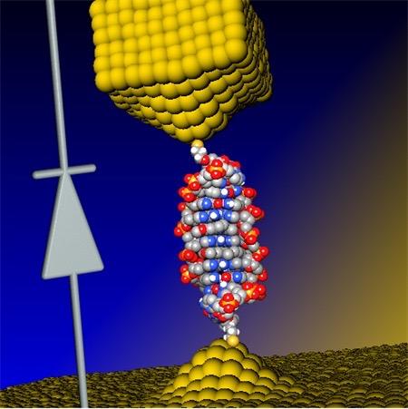
In a Berkeley Lab-led study, flexible double-helix DNA segments (purple, with green DNA models) connected to gold nanoparticles (yellow) are revealed from the 3D density maps reconstructed from individual samples using a Berkeley Lab-developed technique called individual-particle electron tomography (IPET). Projections of the structures are shown in the green background grid. (credit: Berkeley Lab)

An international research team working at the Lawrence Berkeley National Laboratory (Berkeley Lab) has captured the first high-resolution 3D images of double-helix DNA segments attached at either end to gold nanoparticles — which could act as building blocks for molecular computer memory and electronic devices (see World’s smallest electronic diode made from single DNA molecule), nanoscale drug-delivery systems, and as markers for biological research and for imaging disease-relevant proteins.

The researchers connected coiled DNA strands between polygon-shaped gold nanoparticles and then reconstructed 3D images, using a cutting-edge electron microscope technique coupled with a protein-staining process and sophisticated software that provided structural details at the scale of about 2 nanometers.

“We had no idea about what the double-strand DNA would look like between the gold nanoparticles,” said Gang “Gary” Ren, a Berkeley Lab scientist who led the research. “This is the first time for directly visualizing an individual double-strand DNA segment in 3D,” he said.

The results were published in an open-access paper in the March 30 edition of Nature Communications.

The method developed by this team, called individual-particle electron tomography (IPET), had earlier captured the 3-D structure of a single protein that plays a key role in human cholesterol metabolism. By grabbing 2D images of an object from different angles, the technique allows researchers to assemble a 3D image of that object.

The team has also used the technique to uncover the fluctuation of another well-known flexible protein, human immunoglobulin 1, which plays a role in the human immune system.

https://youtu.be/lQrbmg9ry90
Berkeley Lab | 3-D Reconstructions of Double strand DNA and Gold Nanoparticle Structures

For this new study of DNA nanostructures, Ren used an electron-beam study technique called cryo-electron microscopy (cryo-EM) to examine frozen DNA-nanogold samples, and used IPET to reconstruct 3-D images from samples stained with heavy metal salts. The team also used molecular simulation tools to test the natural shape variations (“conformations”) in the samples, and compared these simulated shapes with observations.

First visualization of DNA strand dynamics without distorting x-ray crystallography

Ren explained that the naturally flexible dynamics of samples, like a man waving his arms, cannot be fully detailed by any method that uses an average of many observations.

A popular way to view the nanoscale structural details of delicate biological samples is to form them into crystals and zap them with X-rays, but that destroys their natural shape, especially fir the DNA-nanogold samples in this study, which the scientists say are incredibly challenging to crystallize. Other common research techniques may require a collection of thousands of near-identical objects, viewed with an electron microscope, to compile a single, averaged 3-D structure. But an averaged 3D image may not adequately show the natural shape fluctuations of a given object.

The samples in the latest experiment were formed from individual polygon gold nanostructures, measuring about 5 nanometers across, connected to single DNA-segment strands with 84 base pairs. Base pairs are basic chemical building blocks that give DNA its structure. Each individual DNA segment and gold nanoparticle naturally zipped together with a partner to form the double-stranded DNA segment with a gold particle at either end.

https://youtu.be/RDOpgj62PLU
Berkeley Lab | These views compare the various shape fluctuations obtained from different samples of the same type of double-helix DNA segment (DNA renderings in green, 3D reconstructions in purple) connected to gold nanoparticles (yellow).

The samples were flash-frozen to preserve their structure for study with cryo-EM imaging. The distance between the two gold nanoparticles in individual samples varied from 20 to 30 nanometers, based on different shapes observed in the DNA segments.

Researchers used a cryo-electron microscope at Berkeley Lab’s Molecular Foundry for this study. They collected a series of tilted images of the stained objects, and reconstructed 14 electron-density maps that detailed the structure of individual samples using the IPET technique.

Sub-nanometer images next

Ren said that the next step will be to work to improve the resolution to the sub-nanometer scale.

“Even in this current state we begin to see 3-D structures at 1- to 2-nanometer resolution,” he said. “Through better instrumentation and improved computational algorithms, it would be promising to push the resolution to that visualizing a single DNA helix within an individual protein.”

In future studies, researchers could attempt to improve the imaging resolution for complex structures that incorporate more DNA segments as a sort of “DNA origami,” Ren said. Researchers hope to build and better characterize nanoscale molecular devices using DNA segments that can, for example, store and deliver drugs to targeted areas in the body.

“DNA is easy to program, synthesize and replicate, so it can be used as a special material to quickly self-assemble into nanostructures and to guide the operation of molecular-scale devices,” he said. “Our current study is just a proof of concept for imaging these kinds of molecular devices’ structures.”

The team included researchers at UC Berkeley, the Kavli Energy NanoSciences Institute at Berkeley Lab and UC Berkeley, and Xi’an Jiaotong University in China. This work was supported by the National Science Foundation, DOE Office of Basic Energy Sciences, National Institutes of Health, the National Natural Science Foundation of China, Xi’an Jiaotong University in China, and the Ministry of Science and Technology in China. View more about Gary Ren’s research group here.


Abstract of Three-dimensional structural dynamics and fluctuations of DNA-nanogold conjugates by individual-particle electron tomography

DNA base pairing has been used for many years to direct the arrangement of inorganic nanocrystals into small groupings and arrays with tailored optical and electrical properties. The control of DNA-mediated assembly depends crucially on a better understanding of three-dimensional structure of DNA-nanocrystal-hybridized building blocks. Existing techniques do not allow for structural determination of these flexible and heterogeneous samples. Here we report cryo-electron microscopy and negative-staining electron tomography approaches to image, and three-dimensionally reconstruct a single DNA-nanogold conjugate, an 84-bp double-stranded DNA with two 5-nm nanogold particles for potential substrates in plasmon-coupling experiments. By individual-particle electron tomography reconstruction, we obtain 14 density maps at ~2-nm resolution. Using these maps as constraints, we derive 14 conformations of dsDNA by molecular dynamics simulations. The conformational variation is consistent with that from liquid solution, suggesting that individual-particle electron tomography could be an expected approach to study DNA-assembling and flexible protein structure and dynamics.


World’s smallest electronic diode made from single DNA molecule

Electronic components 1,000 times smaller than with silicon may be possible
http://www.kurzweilai.net/worlds-smallest-electronic-diode-made-from-single-dna-molecule
By inserting a small “coralyne” molecule into DNA, scientists were able to create a single-molecule diode (connected here by two gold electrodes), which can be used as an active element in future nanoscale circuits. The diode circuit symbol is shown on the left. (credit: University of Georgia and Ben-Gurion University)

Nanoscale electronic components can be made from single DNA molecules, as researchers at the University of Georgia and at Ben-Gurion University in Israel have demonstrated, using a single molecule of DNA to create the world’s smallest diode.

DNA double helix with base pairs (credit: National Human Genome Research Institute)

A diode is a component vital to electronic devices that allows current to flow in one direction but prevents its flow in the other direction. The development could help stimulate development of DNA components for molecular electronics.

As noted in an open-access Nature Chemistry paper published this week, the researchers designed a 11-base-pair (bp) DNA molecule and inserted a small molecule named coralyne into the DNA.*

They found, surprisingly, that this caused the current flowing through the DNA to be 15 times stronger for negative voltages than for positive voltages, a necessary feature of a diode.

Electronic elements 1,00o times smaller than current components

“Our discovery can lead to progress in the design and construction of nanoscale electronic elements that are at least 1,000 times smaller than current components,” says the study’s lead author, Bingqian Xu an associate professor in the UGA College of Engineering and an adjunct professor in chemistry and physics.

The research team plans to enhance the performance of the molecular diode and construct additional molecular devices, which may include a transistor (similar to a two-layer diode, but with one additional layer).

A theoretical model developed by Yanantan Dubi of Ben-Gurion University indicated the diode-like behavior of DNA originates from the bias voltage-induced breaking of spatial symmetry inside the DNA molecule after the coralyne is inserted.

The research is supported by the National Science Foundation.

*“We prepared the DNA–coralyne complex by specifically intercalating two coralyne molecules into a custom-designed 11-base-pair (bp) DNA molecule (5′-CGCGAAACGCG-3′) containing three mismatched A–A base pairs at the centre,” according to the authors.

UPDATE April 6, 2016 to clarify the coralyne intercalation (insertion) into the DNA molecule.


Abstract of Molecular rectifier composed of DNA with high rectification ratio enabled by intercalation

The predictability, diversity and programmability of DNA make it a leading candidate for the design of functional electronic devices that use single molecules, yet its electron transport properties have not been fully elucidated. This is primarily because of a poor understanding of how the structure of DNA determines its electron transport. Here, we demonstrate a DNA-based molecular rectifier constructed by site-specific intercalation of small molecules (coralyne) into a custom-designed 11-base-pair DNA duplex. Measured current–voltage curves of the DNA–coralyne molecular junction show unexpectedly large rectification with a rectification ratio of about 15 at 1.1 V, a counter-intuitive finding considering the seemingly symmetrical molecular structure of the junction. A non-equilibrium Green’s function-based model—parameterized by density functional theory calculations—revealed that the coralyne-induced spatial asymmetry in the electron state distribution caused the observed rectification. This inherent asymmetry leads to changes in the coupling of the molecular HOMO−1 level to the electrodes when an external voltage is applied, resulting in an asymmetric change in transmission.


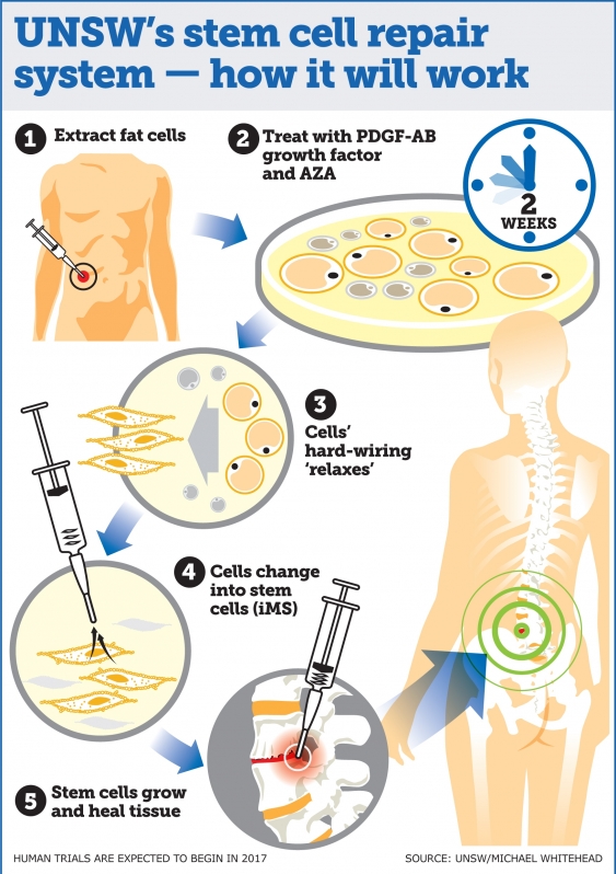
A stem-cell repair system that can regenerate any kind of human tissue …including disease and aging; human trials next year
http://www.kurzweilai.net/a-stem-cell-repair-system-that-can-regenerate-any-kind-of-human-tissue

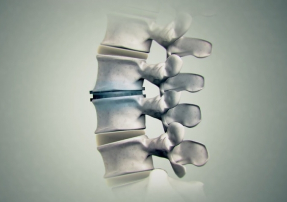
http://www.kurzweilai.net/images/spinal_disc_regeneration.jpg

UNSW researchers say the therapy has enormous potential for treating spinal disc injury and joint and muscle degeneration and could also speed up recovery following complex surgeries where bones and joints need to integrate with the body (credit: UNSW TV)

A stem cell therapy system capable of regenerating any human tissue damaged by injury, disease, or aging could be available within a few years, say University of New South Wales (UNSW Australia) researchers.

Their new repair system*, similar to the method used by salamanders to regenerate limbs, could be used to repair everything from spinal discs to bone fractures, and could transform current treatment approaches to regenerative medicine.

The UNSW-led research was published this week in the Proceedings of the National Academy of Sciences journal.

Reprogramming bone and fat cells

The system reprograms bone and fat cells into induced multipotent stem cells (iMS), which can regenerate multiple tissue types and has been successfully demonstrated in mice, according to study lead author, haematologist, and UNSW Associate Professor John Pimanda.

“This technique is a significant advance on many of the current unproven stem cell therapies, which have shown little or no objective evidence they contribute directly to new tissue formation,” Pimanda said. “We have taken bone and fat cells, switched off their memory and converted them into stem cells so they can repair different cell types once they are put back inside the body.”

“We are currently assessing whether adult human fat cells reprogrammed into iMS cells can safely repair damaged tissue in mice, with human trials expected to begin in late 2017.”

http://www.kurzweilai.net/images/UNSW-stem-cell-repair.jpg

Advantages over stem-cell types

There are different types of stem cells including embryonic stem (ES) cells, which during embryonic development generate every type of cell in the human body, and adult stem cells, which are tissue-specific, but don’t regenerate multiple tissue types. Embryonic stem cells cannot be used to treat damaged tissues because of their tumor forming capacity. The other problem when generating stem cells is the requirement to use viruses to transform cells into stem cells, which is clinically unacceptable, the researchers note.

Research shows that up to 20% of spinal implants either don’t heal or there is delayed healing. The rates are higher for smokers, older people and patients with diseases such diabetes or kidney disease.

Human trials are planned next year once the safety and effectiveness of the technique using human cells in mice has been demonstrated.

* The technique involves extracting adult human fat cells and treating them with the compound 5-Azacytidine (AZA), along with platelet-derived growth factor-AB (PDGF-AB) for about two days. The cells are then treated with the growth factor alone for a further two-three weeks.

AZA is known to induce cell plasticity, which is crucial for reprogramming cells. The AZA compound relaxes the hard-wiring of the cell, which is expanded by the growth factor, transforming the bone and fat cells into iMS cells. When the stem cells are inserted into the damaged tissue site, they multiply, promoting growth and healing.

The new technique is similar to salamander limb regeneration, which is also dependent on the plasticity of differentiated cells, which can repair multiple tissue types, depending on which body part needs replacing.

Along with confirming that human adult fat cells reprogrammed into iMS stem cells can safely repair damaged tissue in mice, the researchers said further work is required to establish whether iMS cells remain dormant at the sites of transplantation and retain their capacity to proliferate on demand.

https://youtu.be/zAMCBNujzzw

Abstract of PDGF-AB and 5-Azacytidine induce conversion of somatic cells into tissue-regenerative multipotent stem cells

Current approaches in tissue engineering are geared toward generating tissue-specific stem cells. Given the complexity and heterogeneity of tissues, this approach has its limitations. An alternate approach is to induce terminally differentiated cells to dedifferentiate into multipotent proliferative cells with the capacity to regenerate all components of a damaged tissue, a phenomenon used by salamanders to regenerate limbs. 5-Azacytidine (AZA) is a nucleoside analog that is used to treat preleukemic and leukemic blood disorders. AZA is also known to induce cell plasticity. We hypothesized that AZA-induced cell plasticity occurs via a transient multipotent cell state and that concomitant exposure to a receptive growth factor might result in the expansion of a plastic and proliferative population of cells. To this end, we treated lineage-committed cells with AZA and screened a number of different growth factors with known activity in mesenchyme-derived tissues. Here, we report that transient treatment with AZA in combination with platelet-derived growth factor–AB converts primary somatic cells into tissue-regenerative multipotent stem (iMS) cells. iMS cells possess a distinct transcriptome, are immunosuppressive, and demonstrate long-term self-renewal, serial clonogenicity, and multigerm layer differentiation potential. Importantly, unlike mesenchymal stem cells, iMS cells contribute directly to in vivo tissue regeneration in a context-dependent manner and, unlike embryonic or pluripotent stem cells, do not form teratomas. Taken together, this vector-free method of generating iMS cells from primary terminally differentiated cells has significant scope for application in tissue regeneration.


First transistors made entirely of nanocrystal ‘inks’ in simplified process

Transistors and other electronic components to be built into flexible or wearable applications; 3D printing planned
http://www.kurzweilai.net/first-transistors-made-entirely-of-nanocrystal-inks
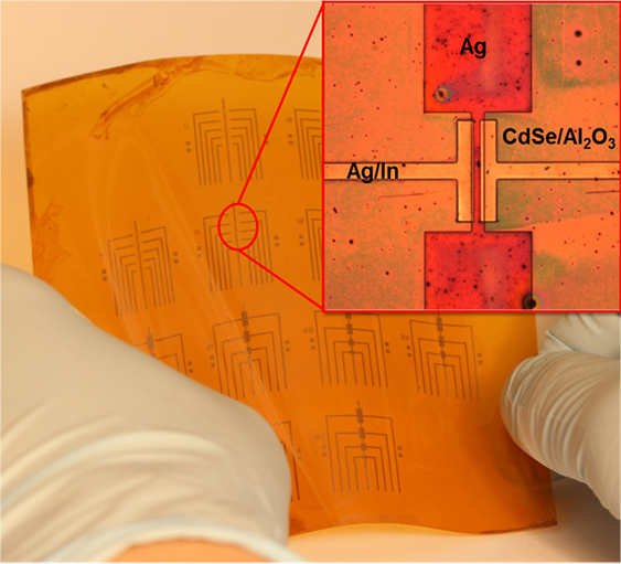
Because this process works at relatively low temperatures, many transistors can be made on a flexible backing at once. (credit: University of Pennsylvania)

University of Pennsylvania engineers have developed a simplified new approach for making transistors by sequentially depositing their components in the form of liquid nanocrystal “inks.” The new process open the door for transistors and other electronic components to be built into flexible or wearable applications. It also avoids the highly complex current process for creating transistors, which requires high-temperature, high-vacuum equipment. Also, the new lower-temperature process is compatible with a wide array of materials and can be applied to larger areas.

Transistors patterned on plastic backing

The researchers’ nanocrystal-based field effect transistors were patterned onto flexible plastic backings using spin coating, but could eventually be constructed by additive manufacturing systems, like 3D printers.

Published in the journal Science,  the study was lead by Cherie Kagan, the Stephen J. Angello Professor in the School of Engineering and Applied Science, and Ji-Hyuk Choi, then a member of her lab, now a senior researcher at the Korea Institute of Geoscience and Mineral Resources. Researchers at Korea University Korea’s Yonsei University were also involved.

[+]

Kagan’s group developed four nanocrystal inks that comprise the transistor, then deposited them on a flexible backing. (credit: University of Pennsylvania)

The researchers began by dispersing a specific type of nanocrystals in a liquid, creating nanocrystal inks. They developed a library of four of these inks: a conductor (silver), an insulator (aluminum oxide), a semiconductor (cadmium selenide), and a conductor combined with a dopant (a mixture of silver and indium). (“Doping” the semiconductor layer of a transistor with impurities controls whether the device creates a positive or negative charge.)

“These materials are colloids just like the ink in your inkjet printer,” Kagan said, “but you can get all the characteristics that you want and expect from the analogous bulk materials, such as whether they’re conductors, semiconductors or insulators.” Although the electrical properties of several of these nanocrystal inks had been independently verified, they had never been combined into full devices. “Our question was whether you could lay them down on a surface in such a way that they work together to form functional transistors.”

Laying down patterns in layers

Such a process entails layering or mixing them in precise patterns.

First, the conductive silver nanocrystal ink was deposited from liquid on a flexible plastic surface that was treated with a photolithographic mask, then rapidly spun to draw it out in an even layer. The mask was then removed to leave the silver ink in the shape of the transistor’s gate electrode.

The researchers followed that layer by spin-coating a layer of the aluminum oxide nanocrystal-based insulator, then a layer of the cadmium selenide nanocrystal-based semiconductor and finally another masked layer for the indium/silver mixture, which forms the transistor’s source and drain electrodes. Upon heating at relatively low temperatures, the indium dopant diffused from those electrodes into the semiconductor component.

“The trick with working with solution-based materials is making sure that, when you add the second layer, it doesn’t wash off the first, and so on,” Kagan said. “We had to treat the surfaces of the nanocrystals, both when they’re first in solution and after they’re deposited, to make sure they have the right electrical properties and that they stick together in the configuration we want.”

Because this entirely ink-based fabrication process works at lower temperatures than existing vacuum-based methods, the researchers were able to make several transistors on the same flexible plastic backing at the same time.

[+]

The inks’ specialized surface chemistry allowed them to stay in configuration without losing their electrical properties. (credit: University of Pennsylvania)

“Making transistors over larger areas and at lower temperatures have been goals for an emerging class of technologies, when people think of the Internet of things, large area flexible electronics and wearable devices,” Kagan said. “We haven’t developed all of the necessary aspects so they could be printed yet, but because these materials are all solution-based, it demonstrates the promise of this materials class and sets the stage for additive manufacturing.”

Because this entirely ink-based fabrication process works at lower temperatures than existing vacuum-based methods, the researchers were able to make several transistors on the same flexible plastic backing at the same time.

3D-printing transistors for wearables

“This is the first work,” Choi said, “showing that all the components, the metallic, insulating, and semiconducting layers of the transistors, and even the doping of the semiconductor, could be made from nanocrystals.”

“Making transistors over larger areas and at lower temperatures have been goals for an emerging class of technologies, when people think of the Internet of things, large area flexible electronics and wearable devices,” Kagan said. “We haven’t developed all of the necessary aspects so they could be printed yet, but because these materials are all solution-based, it demonstrates the promise of this materials class and sets the stage for additive manufacturing.”

The research was supported by the National Science Foundation, the U.S. Department of Energy, the Office of Naval Research, and the Korea Institute of Geoscience and Mineral Resources funded by the Ministry of Science, ICT, and Future Planning of Korea.


Abstract of Exploiting the colloidal nanocrystal library to construct electronic devices

Synthetic methods produce libraries of colloidal nanocrystals with tunable physical properties by tailoring the nanocrystal size, shape, and composition. Here, we exploit colloidal nanocrystal diversity and design the materials, interfaces, and processes to construct all-nanocrystal electronic devices using solution-based processes. Metallic silver and semiconducting cadmium selenide nanocrystals are deposited to form high-conductivity and high-mobility thin-film electrodes and channel layers of field-effect transistors. Insulating aluminum oxide nanocrystals are assembled layer by layer with polyelectrolytes to form high–dielectric constant gate insulator layers for low-voltage device operation. Metallic indium nanocrystals are codispersed with silver nanocrystals to integrate an indium supply in the deposited electrodes that serves to passivate and dope the cadmium selenide nanocrystal channel layer. We fabricate all-nanocrystal field-effect transistors on flexible plastics with electron mobilities of 21.7 square centimeters per volt-second.

Best textile manufacturing methods for creating human tissues with stem cells
Bioengineers determine three best processes for engineering tissues needed for organ and tissue repair
http://www.kurzweilai.net/best-textile-manufacturing-methods-for-creating-human-tissues-with-stem-cells
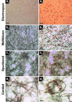
All four textile manufacturing processes and corresponding scaffold (structure) types studied exhibited the presence of lipid vacuoles (small red spheres, right column, indicating stem cells undergoing random differentiation), compared to control (left). Electrospun scaffolds (row a) exhibited only a monolayer of lipid vacuoles in a single focal plane, while meltblown, spunbond, and carded scaffolds (rows b, c, d) exhibited vacuoles in multiple planes throughout the fabric thickness. Scale bars: 100 μm (credit: S. A. Tuin et al./Biomedical Materials)

Elizabeth Loboa, dean of the Missouri University College of Engineering, and her team have tested new tissue- engineering methods (based on textile manufacturing) to find ones that are most cost-effective and can be produced in larger quantities.

Tissue engineering is a process that uses novel biomaterials seeded with stem cells to grow and replace missing tissues. When certain types of materials are used, the “scaffolds” that are created to hold stem cells eventually degrade, leaving natural tissue in its place. The new tissues could help patients suffering from wounds caused by diabetes and circulation disorders, patients in need of cartilage or bone repair, and women who have had mastectomies by replacing their breast tissue. The challenge is creating enough of the material on a scale that clinicians need to treat patients.

Comparing textile manufacturing techniques

http://www.kurzweilai.net/images/electrospinning.png

Electrospinning experiment: nanofibers are collected into an ethanol bath and removed at predefined time intervals (credit: J. M. Coburn et al./The Johns Hopkins University/PNAS)

In typical tissue engineering approaches that use fibers as scaffolds, non-woven materials are often bonded together using an electrostatic field. This process, called electrospinning (see Nanoscale scaffolds and stem cells show promise in cartilage repair and Improved artificial blood vessels), creates the scaffolds needed to attach to stem cells.

However, large-scale production with electrospinning is not cost-effective. “Electrospinning produces weak fibers, scaffolds that are not consistent, and pores that are too small,” Loboa said. “The goal of ‘scaling up’ is to produce hundreds of meters of material that look the same, have the same properties, and can be used in clinical settings. So we investigated the processes that create textiles, such as clothing and window furnishings like drapery, to scale up the manufacturing process.”

The group published two papers using three industry-standard, high-throughput manufacturing techniques — meltblowing, spunbonding, and carding — to determine if they would create the materials needed to mimic native tissue.

Meltblowing is a technique during which nonwoven materials are created using a molten polymer to create continuous fibers. Spunbond materials are made much the same way but the fibers are drawn into a web while in a solid state instead of a molten one. Carding involves the separation of fibers through the use of rollers, forming the web needed to hold stem cells in place.

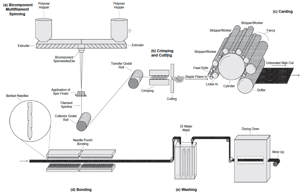
http://www.kurzweilai.net/images/carded-scaffold-fabrication.jpg

Schematic of gilled fiber multifilament spinning and carded scaffold fabrication (credit: Stephen A. Tuin et al./Acta Biomaterialia)

Cost-effective methods

Loboa and her colleagues tested these techniques to create polylactic acid (PLA) scaffolds (a Food and Drug Administration-approved material used as collagen fillers), seeded with human stem cells. They then spent three weeks studying whether the stem cells remained healthy and if they began to differentiate into fat and bone pathways, which is the goal of using stem cells in a clinical setting when new bone and/or new fat tissue is needed at a defect site. Results showed that the three textile manufacturing methods proved as viable if not more so than electrospinning.

“These alternative methods are more cost-effective than electrospinning,” Loboa said. “A small sample of electrospun material could cost between $2 to $5. The cost for the three manufacturing methods is between $.30 to $3.00; these methods proved to be effective and efficient. Next steps include testing how the different scaffolds created in the three methods perform once implanted in animals.”

Researchers at North Carolina State University and the University of North Carolina at Chapel Hill were also involved in the two studies, which were published in Biomedical Materials (open access) and Acta Biomaterialia. The National Science Foundation, the National Institutes of Health, and the Nonwovens Institute provided funding for the studies.


Abstract of Creating tissues from textiles: scalable nonwoven manufacturing techniques for fabrication of tissue engineering scaffolds

Electrospun nonwovens have been used extensively for tissue engineering applications due to their inherent similarities with respect to fibre size and morphology to that of native extracellular matrix (ECM). However, fabrication of large scaffold constructs is time consuming, may require harsh organic solvents, and often results in mechanical properties inferior to the tissue being treated. In order to translate nonwoven based tissue engineering scaffold strategies to clinical use, a high throughput, repeatable, scalable, and economic manufacturing process is needed. We suggest that nonwoven industry standard high throughput manufacturing techniques (meltblowing, spunbond, and carding) can meet this need. In this study, meltblown, spunbond and carded poly(lactic acid) (PLA) nonwovens were evaluated as tissue engineering scaffolds using human adipose derived stem cells (hASC) and compared to electrospun nonwovens. Scaffolds were seeded with hASC and viability, proliferation, and differentiation were evaluated over the course of 3 weeks. We found that nonwovens manufactured via these industry standard, commercially relevant manufacturing techniques were capable of supporting hASC attachment, proliferation, and both adipogenic and osteogenic differentiation of hASC, making them promising candidates for commercialization and translation of nonwoven scaffold based tissue engineering strategies.


Abstract of Fabrication of novel high surface area mushroom gilled fibers and their effects on human adipose derived stem cells under pulsatile fluid flow for tissue engineering applications

The fabrication and characterization of novel high surface area hollow gilled fiber tissue engineering scaffolds via industrially relevant, scalable, repeatable, high speed, and economical nonwoven carding technology is described. Scaffolds were validated as tissue engineering scaffolds using human adipose derived stem cells (hASC) exposed to pulsatile fluid flow (PFF). The effects of fiber morphology on the proliferation and viability of hASC, as well as effects of varied magnitudes of shear stress applied via PFF on the expression of the early osteogenic gene marker runt related transcription factor 2 (RUNX2) were evaluated. Gilled fiber scaffolds led to a significant increase in proliferation of hASC after seven days in static culture, and exhibited fewer dead cells compared to pure PLA round fiber controls. Further, hASC-seeded scaffolds exposed to 3 and 6 dyn/cm2 resulted in significantly increased mRNA expression of RUNX2 after one hour of PFF in the absence of soluble osteogenic induction factors. This is the first study to describe a method for the fabrication of high surface area gilled fibers and scaffolds. The scalable manufacturing process and potential fabrication across multiple nonwoven and woven platforms makes them promising candidates for a variety of applications that require high surface area fibrous materials.

Statement of Significance

We report here for the first time the successful fabrication of novel high surface area gilled fiber scaffolds for tissue engineering applications. Gilled fibers led to a significant increase in proliferation of human adipose derived stem cells after one week in culture, and a greater number of viable cells compared to round fiber controls. Further, in the absence of osteogenic induction factors, gilled fibers led to significantly increased mRNA expression of an early marker for osteogenesis after exposure to pulsatile fluid flow. This is the first study to describe gilled fiber fabrication and their potential for tissue engineering applications. The repeatable, industrially scalable, and versatile fabrication process makes them promising candidates for a variety of scaffold-based tissue engineering applications.

Read Full Post »

Boston Scientific implant designed to occlude the heart’s left atrial appendage implicated with embolization – Device Sales in Europe halts

Reporter: Aviva Lev-Ari, PhD, RN


Boston Scientific halts EU sales of next-gen Watchman FLX anti-stroke device

Boston Scientific WatchmanBoston Scientific (NYSE:BSX) reportedly halted European sales of its the next generation of its anti-stroke device, the Watchman FLX, after receiving reports of device embolization.

Spokeswoman Trish Backes told TCTMD that there were 6 device embolizations in 207 (2.9%) European implantations of the Watchman FLX, an implant that designed to occlude the heart’s left atrial appendage. One of those patients died from complications related to an infection suffered after the device was retrieved.

The 1st-generation Watchman device showed a 30-day embolization rate of 0 to 0.7% in trials, and a post-approval registry called Ewolution showed a rate of 0.2%. The Watchman FLX device won CE Mark approval in the European Unionlast November; the original iteration won FDA approval in March 2015.

Watchman FLX will be taken off the shelves until Boston Scientific can determine what’s causing the unexpectedly high embolism rate, Backes told the website.

“With [the original] Watchman, we’re really confident. We’ve seen really low embolization rates,” she said. “With the robust clinical training program that we have in place for physicians before they start implanting the device, we feel really good about that. This doesn’t impact what we’re doing in the U.S. or what we’re doing with the current Watchman device. It’s not raising any concerns for us for the current device.”

Medical officers with the Marlborough, Mass.-based company, speaking at the annual conference of the American College of Cardiology, said they’ll look at whether physician training or implant technique are factors. The company said the sales halt for Watchman FLX will not affect its structural heart sales forecast of $175 million to $200 million this year.

Boston Scientific said earlier this week at ACC 2016 that a review of the 1st 1,000 Watchman patients found similar results as in pre-market trials.

Material from Reuters was used in this report.

SOURCE

http://www.massdevice.com/boston-scientific-halts-eu-sales-of-next-gen-watchman-flx-anti-stroke-device/?utm_source=newsletter-160405&utm_medium=email&utm_campaign=newsletter-160405&spMailingID=8750804&spUserID=MTI2MTQxNTczMjM5S0&spJobID=900546483&spReportId=OTAwNTQ2NDgzS0

Read Full Post »

Ngai-Yin Chan-The Practice of Catheter Cryoablation for Cardiac Arrhythmias[PDF] 20 MB PDF… https://t.co/8EYkq36tJA

Sourced through Scoop.it from: www.medbooksvn.info

See on Scoop.itCardiovascular Disease: PHARMACO-THERAPY

Read Full Post »

Dissolvable sensor for determining temperature and pressure

Curator: Danut Dragoi, PhD

The Concept

The concept of dissolvable sensor in human body fluid and its experimentation was a successful task of electrical engineers at the University of Illinois at Urbana-Champaign. The device is intended to be implanted inside the head of human body in order to measure important parameters such as temperature and pressure.

Based on actual silicon technology, the device is built on a very thin silicon crystal, which is dissolvable in human body fluids after a given period and after  the measurements are done. The need for such device is required by a medical intervention, a surgery, or a special medication.

For measuring the temperature,the device uses the principle of variation of current / voltage of a silicon diode with temperature see link in here . To illustrate how the diode works as a thermometer, see the link in here  in which the curve voltage output versus temperature, variable T, is a decreasing linear function as a function of temperature.The other variable pressure P can be obtained from the base material, the thin silicon substrate, even if silicon is not a traditional piezoelectric material. Knowing that silicon can be a piezorezistive material, link in here,  a signal output can be obtained from an engineered part of the silicon chip that has the resistance as a function of pressure P.

Two Variable Sensor: Temperature and Pressure

The picture bellow, IMAGE CREDIT::JOHN A. ROGERS, UNIVERSITY OF ILLINOIS AT URBANA-CHAMPAIGN AND MDTMAG.COM,

T and P on Brain

is the actual device made by electrical engineers at the University of Illinois at Urbana-Champaign. The device shown in the picture, SOA in the field,  is based on silicon and is bioresorbable. The coil in the center is for transmission data purposes. The link in here  describes in more details the device.

According with Prof Rogers of University of Illinois at Urbana-Champaign, a new class of small, thin electronic sensors can monitor temperature and pressure within the skull – which are crucial health parameters after a brain injury or surgery – then melt away when they are no longer needed, eliminating the need for additional surgery to remove the monitors and reducing the risk of infection and hemorrhage. Similar sensors can be adapted for postoperative monitoring in other body systems as well.  John A. Rogers and Wilson Ray, a professor of neurological surgery at the Washington University School of Medicine in St. Louis,  published their work in the journal Nature.

Applications of the device

After a traumatic brain injury or brain surgery, it is crucial to monitor the patient for swelling and pressure on the brain. Current monitoring technology is bulky and invasive,and the wires restrict the patient’s movement and hamper physical therapy as they recover.

Because they require continuous, hard-wired access into the head, such implants also carry the risk of allergic reactions, infection and hemorrhage, and even could exacerbate the inflammation they are meant to monitor. Professor Rogers mentioned that the demonstration was done on animal models, with a measurement precision that’s just as good as that of conventional devices.

The sensors, smaller than a grain of rice, are built on extremely thin sheets of silicon – which are naturally biodegradable – that are configured to function normally for a few weeks, then dissolve away, completely and harmlessly, in the body’s own fluids.

Rogers’ group teamed with Illinois materials science and engineering professor Paul V. Braun to make the silicon platforms sensitive to clinically relevant pressure levels in the intracranial fluid surrounding the brain. They also added a tiny temperature sensor and connected it to a wireless transmitter roughly the size of a postage stamp, implanted under the skin but on top of the skull.

The Illinois group worked with clinical experts in traumatic brain injury at Washington University to implant the sensors in rats, testing for performance and bio-compatibility. They found that the temperature and pressure readings from the dissolvable sensors matched conventional monitoring devices for accuracy.

The researchers are moving toward human trials for this technology, as well as extending its functionality for other bio-medical applications.

Source

Nature(2016), Published online 18 January 2016, Bioresorbable silicon electronic sensors for the brain, Seung-Kyun Kang, Rory K. J. Murphy, Suk-Won Hwang, Seung Min Lee, Daniel V. Harburg, Neil A. Krueger, Jiho Shin, Paul Gamble, Huanyu Cheng, Sooyoun Yu, Zhuangjian Liu, Jordan G. McCall, Manu Stephen, Hanze Ying, Jeonghyun Kim, Gayoung Park, R. Chad Webb, Chi Hwan Lee, Sangjin Chung, Dae Seung Wie, Amit D. Gujar, Bharat Vemulapalli, Albert H. Kim, Kyung-Mi Lee, Jianjun Cheng, Younggang Huang, Sang Hoon Lee, Paul V. Braun, Wilson Z. Ray & John A. Rogers,

http://www.nature.com/nature/journal/v530/n7588/fig_tab/nature16492_SF1.html

http://www.pveducation.org/pvcdrom/pn-junction/diode-equation

Click to access ME189_Chapter%207.pdf

https://news.illinois.edu/blog/view/6367/312684


Read Full Post »

« Newer Posts - Older Posts »