Feeds:
Posts
Comments

Posts Tagged ‘Cancer – General’

Blood test uses DNA strands of dying cells

Curators:  Larry H. Bernstein, MD, FCAP and Aviva Lev-Ari, PhD, RN

LPBI


Hadassah-Developed Blood Test Detects Multiple Sclerosis, Cancer & Brain Damage

http://www.hadassah.org/news-stories/blood-test-detects-neurodegenerative-disease.html

A new blood test that uses the DNA strands of dying cells to detect diabetes, cancer, traumatic brain injury, and neurodegenerative disease has been developed by researchers at Hadassah Medical Organization (HMO) and The Hebrew University.

In a study involving 320 patients, the researchers were able to infer cell death in specific tissues by looking at the unique chemical modifications (called methylation patterns) of circulating DNA that these dying cells release. Previously, it had not been possible to measure cell death in specific human tissues non-invasively.

The findings are reported in the March 14, 2016 online edition of Proceedings of National Academy of Sciences USA, in an article entitled “Identification of tissue specific cell death using methylation patterns of circulating DNA.”  Prof. Benjamin Glaser, head of Endocrinology at Hadassah, and Dr. Ruth Shemer and Prof. Yuval Dor from The Hebrew University of Jerusalem led an international team in performing the groundbreaking research.

Cell death is a central feature in health and disease. It can signify the early stages of pathology (e.g. a developing tumor or the beginning of an autoimmune or neurodegenerative disease); it can illuminate whether a disease has progressed and whether a particular treatment, such as chemotherapy, is working; and it can alert physicians to unintended toxic effects of treatment or the early rejection of a transplant.

As the researchers relate: “The approach can be adapted to identify cfDNA (cell-free circulating DNA) derived from any cell type in the body, offering a minimally invasive window for diagnosing and monitoring a broad spectrum of human pathologies as well as providing a better understanding of normal tissue dynamics.”

“In the long run,” notes Prof. Glaser, “we envision a new type of blood test aimed at the sensitive detection of tissue damage, even without a-priori suspicion of disease in a specific organ. We believe that such a tool will have broad utility in diagnostic medicine and in the study of human biology.”

The research was performed by Hebrew University students Roni Lehmann-Werman, Daniel Neiman, Hai Zemmour, Joshua Moss and Judith Magenheim, aided by clinicians and scientists from Hadassah Medical Center, Sheba Medical Center, and from institutions in Germany, Sweden, the USA and Canada, who provided precious blood samples from patients.

Scientists have known for decades that dying cells release fragmented DNA into the blood; however, since the DNA sequence of all cells in the body is identical, it had not been possible to determine the tissue of origin of the circulating DNA.  Knowing that the DNA of each cell type carries a unique methylation and that methylation patterns of DNA account for the identity of cells, the researchers were able to use patterns of methylated DNA sequences as biomarkers to detect the origin of the DNA and to identify a specific pathology. For example, they were able to detect evidence of pancreatic beta-cell death in the blood of patients with new-onset type 1 diabetes, oligodendrocyte cell death in patients with relapsing multiple sclerosis, brain cell death in patients after traumatic or ischemic brain damage, and exocrine pancreatic tissue cell death in patients with pancreatic cancer or pancreatitis.

Support for the research came from the Juvenile Diabetes Research Foundation, the Human Islet Research Network of the National Institutes of Health, the Sir Zalman Cowen Universities Fund, the DFG (a Trilateral German-Israel-Palestine program), and the Soyka pancreatic cancer fund.

 Identification of tissue-specific cell death using methylation patterns of circulating DNA.
Minimally invasive detection of cell death could prove an invaluable resource in many physiologic and pathologic situations. Cell-free circulating DNA (cfDNA) released from dying cells is emerging as a diagnostic tool for monitoring cancer dynamics and graft failure. However, existing methods rely on differences in DNA sequences in source tissues, so that cell death cannot be identified in tissues with a normal genome. We developed a method of detecting tissue-specific cell death in humans based on tissue-specific methylation patterns in cfDNA. We interrogated tissue-specific methylome databases to identify cell type-specific DNA methylation signatures and developed a method to detect these signatures in mixed DNA samples. We isolated cfDNA from plasma or serum of donors, treated the cfDNA with bisulfite, PCR-amplified the cfDNA, and sequenced it to quantify cfDNA carrying the methylation markers of the cell type of interest. Pancreatic β-cell DNA was identified in the circulation of patients with recently diagnosed type-1 diabetes and islet-graft recipients; oligodendrocyte DNA was identified in patients with relapsing multiple sclerosis; neuronal/glial DNA was identified in patients after traumatic brain injury or cardiac arrest; and exocrine pancreas DNA was identified in patients with pancreatic cancer or pancreatitis. This proof-of-concept study demonstrates that the tissue origins of cfDNA and thus the rate of death of specific cell types can be determined in humans. The approach can be adapted to identify cfDNA derived from any cell type in the body, offering a minimally invasive window for diagnosing and monitoring a broad spectrum of human pathologies as well as providing a better understanding of normal tissue dynamics.

While impressively organ specific, they did not specifically prove that the DNA was from an actual dying cell. For example, you would need to see if Troponin levels were elevated when assuming the DNA is from injured myocardium. Also, for brain, though impractical , you’d want to see a brain biopsy or imaging for the brain related cases. The experiment of spiking with DNA was clever though. Also, what is the turnaround time for this test in practical use?

Larry HB

Very good comment. I was reluctant to put this up, but it was of interest and published in PNAS.  Perhaps I can find more information.  Troponin levels would be good for 48 hours, longer than CK and comparable to LD.  What about Nat peptides?

Glutamine and cancer: cell biology, physiology, and clinical opportunities

Christopher T. Hensley,1 Ajla T. Wasti,1,2 

J Clin Invest 2013   https://www.jci.org/articles/view/69600

Glutamine is an abundant and versatile nutrient that participates in energy formation, redox homeostasis, macromolecular synthesis, and signaling in cancer cells. These characteristics make glutamine metabolism an appealing target for new clinical strategies to detect, monitor, and treat cancer. Here we review the metabolic functions of glutamine as a super nutrient and the surprising roles of glutamine in supporting the biological hallmarks of malignancy. We also review recent efforts in imaging and therapeutics to exploit tumor cell glutamine dependence, discuss some of the challenges in this arena, and suggest a disease-focused paradigm to deploy these emerging approaches.

It has been nearly a century since the discovery that tumors display metabolic activities that distinguish them from differentiated, non-proliferating tissues and presumably contribute to their supraphysiological survival and growth (1). Interest in cancer metabolism was boosted by discoveries that oncogenes and tumor suppressors could regulate nutrient metabolism, and that mutations in some metabolic enzymes participate in the development of malignancy (2, 3). The persistent appeal of cancer metabolism as a line of investigation lies both in its ability to uncover fundamental aspects of malignancy and in the translational potential of exploiting cancer metabolism to improve the way we diagnose, monitor, and treat cancer. Furthermore, an improved understanding of how altered metabolism contributes to cancer has a high potential for synergy with translational efforts. For example, the demonstration that asparagine is a conditionally essential nutrient in rapidly growing cancer cells paved the way for L-asparaginase therapy in leukemia. Additionally, the avidity of some tumors for glucose uptake led to the development of 18fluoro-2-deoxyglucose imaging by PET; this in turn stimulated hundreds of studies on the biological underpinnings of tumor glucose metabolism.

There continue to be large gaps in understanding which metabolic pathways are altered in cancer, whether these alterations benefit the tumor in a substantive way, and how this information could be used in clinical oncology. In this Review, we consider glutamine, a highly versatile nutrient whose metabolism has implications for tumor cell biology, metabolic imaging, and perhaps novel therapeutics.

Glutamine in intermediary metabolism

Glutamine metabolism has been reviewed extensively and is briefly outlined here (4, 5). The importance of glutamine as a nutrient in cancer derives from its abilities to donate its nitrogen and carbon into an array of growth-promoting pathways (Figure 1). At concentrations of 0.6–0.9 mmol/l, glutamine is the most abundant amino acid in plasma (6). Although most tissues can synthesize glutamine, during periods of rapid growth or other stresses, demand outpaces supply, and glutamine becomes conditionally essential (7). This requirement for glutamine is particularly true in cancer cells, many of which display oncogene-dependent addictions to glutamine in culture (8). Glutamine catabolism begins with its conversion to glutamate in reactions that either donate the amide nitrogen to biosynthetic pathways or release it as ammonia. The latter reactions are catalyzed by the glutaminases (GLSs), of which several isozymes are encoded by human genes GLS and GLS2 (9). Classical studies revealed that GLS isozymes, particularly those encoded by GLS, are expressed in experimental tumors in rats and mice, where their enzyme activity correlates with growth rate and malignancy. Silencing GLS expression or inhibiting GLS activity is sufficient to delay tumor growth in a number of models (1013). The role of GLS2 in cancer appears to be context specific and regulated by factors that are still incompletely characterized. In some tissues, GLS2 is a p53 target gene and seems to function in tumor suppression (14). On the other hand, GLS2 expression is enhanced in some neuroblastomas, where it contributes to cell survival (15). These observations, coupled with the demonstration that c-Myc stimulates GLS expression (12, 16), position at least some of the GLS isozymes as pro-oncogenic.

Glutamine metabolism as a target for diagnostic imaging and therapy in cancFigure 1Glutamine metabolism as a target for diagnostic imaging and therapy in cancer. Glutamine is imported via SLC1A5 and other transporters, then enters a complex metabolic network by which its carbon and nitrogen are supplied to pathways that promote cell survival and growth. Enzymes discussed in the text are shown in green, and inhibitors that target various aspects of glutamine metabolism are shown in red. Green arrows denote reductive carboxylation. 18F-labeled analogs of glutamine are also under development as PET probes for localization of tumor tissue. AcCoA, acetyl-CoA; DON, 6-diazo-5-oxo-L-norleucine; GSH, glutathione; NEAA, nonessential amino acids; ME, malic enzyme; OAA, oxaloacetate; TA, transaminase; 968, compound 968; α-KG, α-ketoglutarate.

Glutamate, the product of the GLS reaction, is a precursor of glutathione, the major cellular antioxidant. It is also the source of amino groups for nonessential amino acids like alanine, aspartate, serine, and glycine, all of which are required for macromolecular synthesis. In glutamine-consuming cells, glutamate is also the major source of α-ketoglutarate, a TCA cycle intermediate and substrate for dioxygenases that modify proteins and DNA. These dioxygenases include prolyl hydroxylases, histone demethylases, and 5-methylcytosine hydroxylases. Their requirement for α-ketoglutarate, although likely accounting for only a small fraction of total α-ketoglutarate utilization, makes this metabolite an essential component of cell signaling and epigenetic networks.

Conversion of glutamate to α-ketoglutarate occurs either through oxidative deamination by glutamate dehydrogenase (GDH) in the mitochondrion or by transamination to produce nonessential amino acids in either the cytosol or the mitochondrion. During avid glucose metabolism, the transamination pathway predominates (17). When glucose is scarce, GDH becomes a major pathway to supply glutamine carbon to the TCA cycle, and is required for cell survival (17, 18). Metabolism of glutamine-derived α-ketoglutarate in the TCA cycle serves several purposes: it generates reducing equivalents for the electron transport chain (ETC) and oxidative phosphorylation, becoming a major source of energy (19); and it is an important anaplerotic nutrient, feeding net production of oxaloacetate to offset export of intermediates from the cycle to supply anabolism (20). Glutamine oxidation also supports redox homeostasis by supplying carbon to malic enzyme, some isoforms of which produce NADPH (Figure 1). In KRAS-driven pancreatic adenocarcinoma cells, a pathway involving glutamine-dependent NADPH production is essential for redox balance and growth (21). In these cells, glutamine is used to produce aspartate in the mitochondria. This aspartate is then trafficked to the cytosol, where it is deaminated to produce oxaloacetate and then malate, the substrate for malic enzyme.

Recent work has uncovered an unexpected role for glutamine in cells with reduced mitochondrial function. Despite glutamine’s conventional role as a respiratory substrate, several studies demonstrated a persistence of glutamine dependence in cells with permanent mitochondrial dysfunction from mutations in the ETC or TCA cycle, or transient impairment secondary to hypoxia (2225). Under these conditions, glutamine-derived α-ketoglutarate is reductively carboxylated by NADPH-dependent isoforms of isocitrate dehydrogenase to produce isocitrate, citrate, and other TCA cycle intermediates (Figure 1). These conditions broaden glutamine’s utility as a carbon source because it becomes not only a major source of oxaloacetate, but also generates acetyl-CoA in what amounts to a striking rewiring of TCA cycle metabolism.

Glutamine promotes hallmarks of malignancy

Deregulated energetics. One hallmark of cancer cells is aberrant bioenergetics (26). Glutamine’s involvement in the pathways outlined above contributes to a phenotype conducive to energy formation, survival, and growth. In addition to its role in mitochondrial metabolism, glutamine also suppresses expression of thioredoxin-interacting protein, a negative regulator of glucose uptake (27). Thus, glutamine contributes to both of the energy-forming pathways in cancer cells: oxidative phosphorylation and glycolysis. Glutamine also modulates hallmarks not traditionally thought to be metabolic, as outlined below. These interactions highlight the complex interplay between glutamine metabolism and many aspects of cell biology.

Sustaining proliferative signaling. Pathological cancer cell growth relies on maintenance of proliferative signaling pathways with increased autonomy relative to non-malignant cells. Several lines of evidence argue that glutamine reinforces activity of these pathways. In some cancer cells, excess glutamine is exported in exchange for leucine and other essential amino acids. This exchange facilitates activation of the serine/threonine kinase mTOR, a major positive regulator of cell growth (28). In addition, glutamine-derived nitrogen is a component of amino sugars, known as hexosamines, that are used to glycosylate growth factor receptors and promote their localization to the cell surface. Disruption of hexosamine synthesis reduces the ability to initiate signaling pathways downstream of growth factors (29).

Enabling replicative immortality. Some aspects of glutamine metabolism oppose senescence and promote replicative immortality in cultured cells. In IMR90 lung fibroblasts, silencing either of two NADPH-generating isoforms of malic enzyme (ME1, ME2) rapidly induced senescence, while malic enzyme overexpression suppressed senescence (30). Both malic enzyme isoforms are repressed at the transcriptional level by p53 and contribute to enhanced levels of glutamine consumption and NADPH production in p53-deficient cells. The ability of p53-replete cells to resist senescence required the expression of ME1 and ME2, and silencing either enzyme reduced the growth of TP53+/+ and, to a lesser degree, TP53–/– tumors (30). These observations position malic enzymes as potential therapeutic targets.

Resisting cell death. Although many cancer cells require glutamine for survival, cells with enhanced expression of Myc oncoproteins are particularly sensitive to glutamine deprivation (8, 12, 16). In these cells, glutamine deprivation induces depletion of TCA cycle intermediates, depression of ATP levels, delayed growth, diminished glutathione pools, and apoptosis. Myc drives glutamine uptake and catabolism by activating the expression of genes involved in glutamine metabolism, including GLSand SLC1A5, which encodes the Na+-dependent amino acid transporter ASCT2 (12, 16). SilencingGLS mimicked some of the effects of glutamine deprivation, including growth suppression in Myc-expressing cells and tumors (10, 12). MYCN amplification occurs in 20%–25% of neuroblastomas and is correlated with poor outcome (31). In cells with high N-Myc levels, glutamine deprivation triggered an ATF4-dependent induction of apoptosis that could be prevented by restoring downstream metabolites oxaloacetate and α-ketoglutarate (15). In this model, pharmacological activation of ATF4, inhibition of glutamine metabolic enzymes, or combinations of these treatments mimicked the effects of glutamine deprivation in cells and suppressed growth of MYCN-amplified subcutaneous and transgenic tumors in mice.

The PKC isoform PKC-ζ also regulates glutamine metabolism. Loss of PKC-ζ enhances glutamine utilization and enables cells to survive glucose deprivation (32). This effect requires flux of carbon and nitrogen from glutamine into serine. PKC-ζ reduces the expression of phosphoglycerate dehydrogenase, an enzyme required for glutamine-dependent serine biosynthesis, and also phosphorylates and inactivates this enzyme. Thus, PKC-ζ loss, which promotes intestinal tumorigenesis in mice, enables cells to alter glutamine metabolism in response to nutrient stress.

Invasion and metastasis. Loss of the epithelial cell-cell adhesion molecule E-cadherin is a component of the epithelial-mesenchymal transition, and is sufficient to induce migration, invasion, and tumor progression (33, 34). Addiction to glutamine may oppose this process because glutamine favors stabilization of tight junctions in some cells (35). Furthermore, the selection of breast cancer cells with the ability to grow without glutamine yielded highly adaptable subpopulations with enhanced mesenchymal marker expression and improved capacity for anchorage-independent growth, therapeutic resistance, and metastasis in vivo (36). It is unknown whether this result reflects a primary role for glutamine in suppressing these markers of aggressiveness in breast cancer, or whether prolonged glutamine deprivation selects for cells with enhanced fitness across a number of phenotypes.

Organ-specific glutamine metabolism in health and disease

As a major player in carbon and nitrogen transport, glutamine metabolism displays complex inter-organ dynamics, with some organs functioning as net producers and others as consumers (Figure 2). Organ-specific glutamine metabolism has frequently been studied in humans and animal models by measuring the arteriovenous difference in plasma glutamine abundance. In healthy subjects, the plasma glutamine pool is largely the result of release from skeletal muscle (3739). In rats, the lungs are comparable to muscle in terms of glutamine production (40, 41), and human lungs also have the capacity for marked glutamine release, although such release is most prominent in times of stress (42, 43). Stress-induced release from the lung is regulated by an induction of glutamine synthase expression as a consequence of glucocorticoid signaling and other mechanisms (44, 45). Although this results in a small arteriovenous difference, the overall release of glutamine is significant because of the large pulmonary perfusion. In rats and humans, adipose tissue is a minor but potentially important source of glutamine (46, 47). The liver has the capacity to synthesize or catabolize glutamine, with these activities subject both to regional heterogeneity among hepatocytes and regulatory effects of systemic acidosis and hyperammonemia. However, the liver does not appear to be a major contributor to the plasma glutamine pool in healthy rats and humans (39, 48, 49).

Model for inter-organ glutamine metabolism in health and cancer.Figure 2Model for inter-organ glutamine metabolism in health and cancer. Organs that release glutamine into the bloodstream are shown in green, and those that consume glutamine are in red; the shade denotes magnitude of consumption/release. For some organs (liver, kidneys), evidence from model systems and/or human studies suggests that there is a change in net glutamine flux during tumorigenesis.

Glutamine consumption occurs largely in the gut and kidney. The organs of the gastrointestinal tract drained by the portal vein, particularly the small intestine, are major consumers of plasma glutamine in both rats and humans (37, 38, 49, 50). Enterocytes oxidize more than half of glutamine carbon to CO2, accounting for a third of the respiration of these cells in fasting animals (51). The kidney consumes net quantities of glutamine to maintain acid-base balance (37, 38, 52, 53). During acidosis, the kidneys substantially increase their uptake of glutamine, cleaving it by GLS to produce ammonia, which is excreted along with organic acids to maintain physiologic pH (52, 54). Glutamine is also a major metabolic substrate in lymphocytes and macrophages, at least during mitogenic stimulation of primary cells in culture (5557).

Importantly, cancer seems to cause major changes in inter-organ glutamine trafficking (Figure 2). Currently, much work in this area is derived from studies in methylcholanthrene-induced fibrosarcoma in the rat, a model of an aggressively growing, glutamine-consuming tumor. In this model, fibrosarcoma induces skeletal muscle expression of glutamine synthetase and greatly increases the release of glutamine into the circulation. As the tumor increases in size, intramuscular glutamine pools are depleted in association with loss of lean muscle mass, mimicking the cachectic phenotype of humans in advanced stages of cancer (52). Simultaneously, both the liver and the kidneys become net glutamine exporters, although the hepatic effect may be diminished as the tumor size becomes very large (48, 49, 52). Glutamine utilization by organs supplied by the portal vein is diminished in cancer (48). In addition to its function as a nutrient for the tumor itself, and possibly for cancer-associated immune cells, glutamine provides additional, indirect metabolic benefits to both the tumor and the host. For example, glutamine was used as a gluconeogenic substrate in cachectic mice with large orthotopic gliomas, providing a significant source of carbon in the plasma glucose pool (58). This glucose was taken up and metabolized by the tumor to produce lactate and to supply the TCA cycle.

It will be valuable to extend work in human inter-organ glutamine trafficking, both in healthy subjects and in cancer patients. Such studies will likely produce a better understanding of the pathophysiology of cancer cachexia, a major source of morbidity and mortality. Research in this area should also aid in the anticipation of organ-specific toxicities of drugs designed to interfere with glutamine metabolism. Alterations of glutamine handling in cancer may induce a different spectrum of toxicities compared with healthy subjects.

Tumors differ according to their need for glutamine

One important consideration is that not all cancer cells need an exogenous supply of glutamine. A panel of lung cancer cell lines displayed significant variability in their response to glutamine deprivation, with some cells possessing almost complete independence (59). Breast cancer cells also demonstrate systematic differences in glutamine dependence, with basal-type cells tending to be glutamine dependent and luminal-type cells tending to be glutamine independent (60). Resistance to glutamine deprivation is associated with the ability to synthesize glutamine de novo and/or to engage alternative pathways of anaplerosis (10, 60).

Tumors also display variable levels of glutamine metabolism in vivo. A study of orthotopic gliomas revealed that genetically diverse, human-derived tumors took up glutamine in the mouse brain but did not catabolize it (58). Rather, the tumors synthesized glutamine de novo and used pyruvate carboxylation for anaplerosis. Cells derived from these tumors did not require glutamine to survive or proliferate when cultured ex vivo. Glutamine synthesis from glucose was also a prominent feature of primary gliomas in human subjects infused with 13C-glucose at the time of surgical resection (61). Furthermore, an analysis of glutamine metabolism in lung and liver tumors revealed that both the tissue of origin and the oncogene influence whether the tumor produces or consumes glutamine (62). MET-induced hepatic tumors produced glutamine, whereas Myc-induced liver tumors catabolized it. In the lung, however, Myc expression was associated with glutamine accumulation.

This variability makes it imperative to develop ways to predict which tumors have the highest likelihood of responding to inhibitors of glutamine metabolism. Methods to image or otherwise quantify glutamine metabolism in vivo would be useful in this regard (63). Infusions of pre-surgical subjects with isotopically labeled glutamine, followed by extraction of metabolites from the tumor and analysis of 13C enrichment, can be used to detect both glutamine uptake and catabolism (58, 62). However, this approach requires a specimen of the tumor to be obtained. Approaches for glutamine-based imaging, which avoid this problem, include a number of glutamine analogs compatible with PET. Although glutamine could in principle be imaged using the radioisotopes 11C, 13N, or 18F, the relatively long half-life of the latter increases its appeal. In mice, 18F-(2S, 4R)4-fluoroglutamine is avidly taken up by tumors derived from highly glutaminolytic cells, and by glutamine-consuming organs including the intestine, kidney, liver, and pancreas (64). Labeled analogs of glutamate are also taken up by some tumors (65, 66). One of these, (4S)-4-(3-[18F] fluoropropyl)-L-glutamate (18F-FSPG, also called BAY 94-9392), was evaluated in small clinical trials involving patients with several types of cancer (65, 67). This analog enters the cell through the cystine/glutamate exchange transporter (xCtransport system), which is linked to glutathione biosynthesis (68). The analog was well tolerated, with high tumor detection rates and good tumor-to-background ratios in hepatocellular carcinoma and lung cancer.

PET approaches detect analog uptake and retention but cannot provide information about downstream metabolism. Analysis of hyperpolarized nuclei can provide a real-time view of enzyme-catalyzed reactions. This technique involves redistribution of the populations of energy levels of a nucleus (e.g., 13C, 15N), resulting in a gain in magnetic resonance signal that can temporarily exceed 10,000-fold (69). This gain in signal enables rapid detection of both the labeled molecule and its downstream metabolites. Glutamine has been hyperpolarized on 15N and 13C (70, 71). In the latter case, the conversion of hyperpolarized glutamine to glutamate could be detected in intact hepatoma cells (70). If these analogs are translated to clinical studies, they might provide a dynamic view of the proximal reactions of glutaminolysis in vivo.

Pharmacological strategies to inhibit glutamine metabolism in cancer

Efforts to inhibit glutamine metabolism using amino acid analogs have an extensive history, including evaluation in clinical trials. Acivicin, 6-diazo-5-oxo-L-norleucine, and azaserine, three of the most widely studied analogs (Figure 1), all demonstrated variable degrees of gastrointestinal toxicity, myelosuppression, and neurotoxicity (72). Because these agents non-selectively target glutamine-consuming processes, recent interest has focused on developing methods directed at specific nodes of glutamine metabolism. First, ASCT2, the Na+-dependent neutral amino acid transporter encoded by SLC1A5, is broadly expressed in lung cancer cell lines and accounts for a majority of glutamine transport in those cells (Figure 1). It has been shown that γ-L-glutamyl-p-nitroanilide (GPNA) inhibits this transporter and limits lung cancer cell growth (73). Additional interest in GPNA lies in its ability to enhance the uptake of drugs imported via the monocarboxylate transporter MCT1. Suppressing glutamine uptake with GPNA enhances MCT1 stability and stimulates uptake of the glycolytic inhibitor 3-bromopyruvate (3-BrPyr) (74, 75). Because enforced MCT1 overexpression is sufficient to sensitize tumor xenografts to 3-BrPyr (76), GPNA may have a place in 3-BrPyr–based therapeutic regimens.

Two inhibitors of GLS isoforms have been characterized in recent years (Figure 1). Compound 968, an inhibitor of the GLS-encoded splice isoform GAC, inhibits the transformation of fibroblasts by oncogenic RhoGTPases and delays the growth of GLS-expressing lymphoma xenografts (13). Bis-2-(5-phenylacetamido-1,2,4-thiadiazol-2-yl)ethyl sulfide (BPTES) also potently inhibits GLS isoforms encoded by GLS (77). BPTES impairs ATP levels and growth rates of P493 lymphoma cells under both normoxic and hypoxic conditions and suppresses the growth of P493-derived xenografts (78).

Evidence also supports a role for targeting the flux from glutamate to α-ketoglutarate, although no potent, specific inhibitors yet exist to inhibit these enzymes in intact cells. Aminooxyacetate (AOA) inhibits aminotransferases non-specifically, but milliomolar doses are typically used to achieve this effect in cultured cells (Figure 1). Nevertheless, AOA has demonstrated efficacy in both breast adenocarcinoma xenografts and autochthonous neuroblastomas in mice (15, 79). Epigallocatechin gallate (EGCG), a green tea polyphenol, has numerous pharmacological effects, one of which is to inhibit GDH (80). The effects of EGCG on GDH have been used to kill glutamine-addicted cancer cells during glucose deprivation or glycolytic inhibition (17, 18) and to suppress growth of neuroblastoma xenografts (15).

A paradigm to exploit glutamine metabolism in cancer

Recent advances in glutamine-based imaging, coupled with the successful application of glutamine metabolic inhibitors in mouse models of cancer, make it possible to conceive of treatment plans that feature consideration of tumor glutamine utilization. A key challenge will be predicting which tumors are most likely to respond to inhibitors of glutamine metabolism. Neuroblastoma is used here as an example of a tumor in which evidence supports the utility of strategies that would involve both glutamine-based imaging and therapy (Figure 3). Neuroblastoma is the second most common extracranial solid malignancy of childhood. High-risk neuroblastoma is defined by age, stage, and biological features of the tumor, including MYCN amplification, which occurs in some 20%–25% of cases (31). Because MYCN-amplified tumor cells require glutamine catabolism for survival and growth (15), glutamine-based PET at the time of standard diagnostic imaging could help predict which tumors would be likely to respond to inhibitors of glutamine metabolism. Infusion of 13C-glutamine coordinated with the diagnostic biopsy could then enable inspection of 13C enrichment in glutamine-derived metabolites from the tumor, confirming the activity of glutamine catabolic pathways. Following on evidence from mouse models of neuroblastoma, treatment could then include agents directed against glutamine catabolism (15). Of note, some tumors were sensitive to the ATF4 agonist fenretinide (FRT), alone or in combination with EGCG. Importantly, FRT has already been the focus of a Phase I clinical trial in children with solid tumors, including neuroblastoma, and was fairly well tolerated (81).

A strategy to integrate glutamine metabolism into the diagnosis, classificaFigure 3A strategy to integrate glutamine metabolism into the diagnosis, classification, treatment, and monitoring of neuroblastoma. Neuroblastoma commonly presents in children as an abdominal mass. A standard evaluation of a child with suspected neuroblastoma includes measurement of urine catecholamines, a bone scan, and full-body imaging with meta-iodobenzylguanidine (MIBG), all of which contribute to diagnosis and disease staging. In animal models, a subset of these tumors requires glutamine metabolism. This finding implies that approaches to image, quantify, or block glutamine metabolism (highlighted in red) in human neuroblastoma could be incorporated into the diagnosis and management of this disease. In particular, glutamine metabolic studies may help predict which tumors would respond to therapies targeting glutamine metabolism. HVA, homovanillic acid; VMA, vanillylmandelic acid.

Conclusions

Glutamine is a versatile nutrient required for the survival and growth of a potentially large subset of tumors. Work over the next several years should produce a more accurate picture of the molecular determinants of glutamine addiction and the identification of death pathways that execute cells when glutamine catabolism is impaired. Advancement of glutamine-based imaging into clinical practice should soon make it possible to differentiate tumors that take up glutamine from those that do not. Finally, the development of safe, high-potency inhibitors of key metabolic nodes should facilitate therapeutic regimens featuring inhibition of glutamine metabolism.

Therapeutic strategies impacting cancer cell glutamine metabolism

The metabolic adaptations that support oncogenic growth can also render cancer cells dependent on certain nutrients. Along with the Warburg effect, increased utilization of glutamine is one of the metabolic hallmarks of the transformed state. Glutamine catabolism is positively regulated by multiple oncogenic signals, including those transmitted by the Rho family of GTPases and by c-Myc. The recent identification of mechanistically distinct inhibitors of glutaminase, which can selectively block cellular transformation, has revived interest in the possibility of targeting glutamine metabolism in cancer therapy. Here, we outline the regulation and roles of glutamine metabolism within cancer cells and discuss possible strategies for, and the consequences of, impacting these processes therapeutically.

Cancer cell metabolism & glutamine addiction

Interest in the metabolic changes characteristic of malignant transformation has undergone a renaissance of sorts in the cancer biology and pharmaceutical communities. However, the recognition that an important connection exists between cellular metabolism and cancer began nearly a century ago with the work of Otto Warburg [13]. Warburg found that rapidly proliferating tumor cells exhibit elevated glucose uptake and glycolytic flux, and furthermore that much of the pyruvate generated by glycolysis is reduced to lactate rather than undergoing mitochondrial oxidation via the tricarboxylic acid (TCA) cycle (Figure 1). This phenomenon persists even under aerobic conditions (‘aerobic glycolysis’), and is known as the Warburg effect [4]. Warburg proposed that aerobic glycolysis was caused by defective mitochondria in cancer cells, but it is now known that mitochondrial dysfunction is relatively rare and that most tumors have an unimpaired capacity for oxidative phosphorylation [5]. In fact, the most important selective advantages provided by the Warburg effect are still debated. Although aerobic glycolysis is an inefficient way to produce ATP (2 ATP/glucose vs ~36 ATP/glucose by complete oxidation), a high glycolytic flux can generate ATP rapidly and furthermore can provide a biosynthetic advantage by supplying precursors and reducing equivalents for the synthesis of macromolecules [4]. The mechanisms underlying the Warburg effect are also not yet fully resolved, although it is increasingly clear that a number of oncogenes and tumor suppressors contribute to the phenomenon. The PI3K/Akt/mTORC1 signaling axis, for example, is a key regulator of aerobic glycolysis and biosynthesis, driving the surface expression of nutrient transporters and the upregulation of glycolytic enzymes [6]. The HIF transcription factor also upregulates expression of glucose transporters and glycolytic enzymes in response to hypoxia and growth factors (or loss of the von Hippel–Landau [VHL] tumor suppressor), and the oncogenic transcription factor c-Myc similarly induces expression of proteins important for glycolysis [6].

An external file that holds a picture, illustration, etc. Object name is nihms610340f1.jpg

http://www.ncbi.nlm.nih.gov/pmc/articles/PMC4154374/bin/nihms610340f1.jpg

Cell proliferation requires metabolic reprogramming

A second major change in the metabolic program of many cancer cells, and the primary focus of this review, is the alteration of glutamine metabolism. Glutamine is the major carrier of nitrogen between organs, and the most abundant amino acid in plasma [7]. It is also a key nutrient for numerous intracellular processes including oxidative metabolism and ATP generation, biosynthesis of proteins, lipids and nucleic acids, and also redox homeostasis and the regulation of signal transduction pathways [810]. Although most mammalian cells are capable of synthesizing glutamine, the demand for this amino acid can become so great during rapid proliferation that an additional extracellular supply is required; hence glutamine is considered conditionally essential [11]. Indeed, many cancer cells are ‘glutamine addicted’, and cannot survive in the absence of an exogenous glutamine supply [12,13].

An important step in the elevation of glutamine catabolism is the activation of the mitochondrial enzyme glutaminase, which catalyzes the hydrolysis of glutamine to generate glutamate and ammonium. The subsequent deamination of glutamate releases a second ammonium to yield the TCA cycle intermediate α-ketoglutarate (α-KG), a reaction catalyzed by glutamate dehydrogenase (GLUD1). This series of reactions is particularly important in rapidly proliferating cells, in which a considerable proportion of the TCA cycle metabolite citrate is exported from mitochondria in order to generate cytosolic acetyl-CoA for lipid biosynthesis [14]. Replenishment of TCA cycle intermediates (anaplerosis) is therefore required, and glutamine often serves as the key anaplerotic substrate through its conversion via glutamate to α-KG (Figure 1).

Mammals express two genes for glutaminase enzymes [1517]. The GLS gene encodes a protein initially characterized in kidney and thus called kidney-type glutaminase (KGA), although this enzyme and its shorter splice variant glutaminase C (GAC), collectively referred to as GLS, are now known to be widely distributed [1820]. The KGA and GAC isoforms share identical N-terminal and catalytic domains, encoded by exons 1–14 of the GLS gene, but have distinct C-termini derived from exon 15 in the case of GAC and exons 16–19 in the case of KGA [21]. Upregulation of GLS, in particular the GAC iso-form, is common in cancer cells and the degree of GLS overexpression correlates with both the degree of malignancy and the tumor grade in human breast cancer samples [22,23]. The GLS2 gene encodes a protein originally discovered and characterized in liver, which has thus been referred to as liver-type glutaminase and, more recently, as glutaminase 2 (GLS2) [15].

Both KGA and GAC can be activated by inorganic phosphate (Pi), and this activation correlates closely with a dimer-to-tetramer transition for each enzyme [7, 22]. As the concentration of Pi is raised the apparent catalytic constant, kcatapp, increases and simultaneously the apparent Michaelis constant, Kmapp, decreases; consequently the catalytic efficiency rises dramatically, especially in the case of GAC [22]. x-ray crystal structures of GAC and KGA in different states indicate that the positioning of a key loop within each monomer (Glu312 to Pro329), located between the active site and the dimer–dimer interface, is critical for mediating tetramerization-induced activation [22,24]. Given the ability of Pi to promote tetramerization and activation of GAC and KGA, it has been proposed that the elevated mitochondrial Pi levels found under hypoxic conditions, which are commonly encountered in the tumor microenvironment, could be one trigger for GLS activation [22].

Oncogenic alterations affecting glutamine metabolism

At least two classes of cellular signals regulate glutamine metabolism, influencing both the expression level and the enzymatic activity of GLS. The transcription factor c-Myc can suppress the expression of microRNAs miR-23a and miR-23b and, in doing so, upregulates GLS (specifically GAC) expression [13,25]. Independent of changes in GAC expression, oncogenic diffuse B-cell lymphoma protein (Dbl), a GEF for Rho GTPases and oncogenic variants of downstream Rho GTPases are able to signal to activate GAC in a manner that is dependent on NF-κB [23]. Mitochondria isolated from Dbl- or Rho GTPase-transformed NIH-3T3 fibroblasts demonstrate significantly higher basal glutaminase activity than mitochondria isolated from non-transformed cells [23]. Furthermore, the enzymatic activity of GAC immunoprecipitated from Dbl-transformed cells is elevated relative to GAC from non-transformed cells, indicating the presence of activating post-translational modification(s) [23]. Indeed, when GAC isolated from Dbl-transformed cells is treated with alkaline phosphatase, basal enzymatic activity is dramatically reduced [23]. Collectively, these findings point to phosphorylation events underlying the activation of GAC in transformed cells. Similarly, phosphorylation-dependent regulation of KGA activity downstream of the Raf-Mek-Erk signaling axis occurs in response to EGF stimulation [24].

It is becoming clear that, in addition to c-Myc and Dbl, many other oncogenic signals and environmental conditions can impact cellular glutamine metabolism. Loss of the retinoblastoma tumor suppressor, for example, leads to a marked increase in glutamine uptake and catabolism, and renders mouse embryonic fibroblasts dependent on exogenous glutamine [26]. Cells transformed by KRAS also illustrate increased expression of genes associated with glutamine metabolism and a corresponding increased utilization of glutamine for anabolic synthesis [27]. In fact, KRAS signaling appears to induce glutamine dependence, since the deleterious effects of glutamine withdrawal in KRAS-driven cells can be rescued by expression of a dominant-negative GEF for Ras [28]. Downstream of Ras, the Raf-MEK-ERK signaling pathway has been implicated in the upregulation of glutamine uptake and metabolism [24,29]. A recent study using human pancreatic ductal adenocarcinoma cells identified a novel KRAS-regulated metabolic pathway, through which glutamine supports cell growth [30]. Proliferation of KRAS-mutant pancreatic ductal adenocarcinoma cells depends on GLS-catalyzed production of glutamate, but not on downstream deamination of glutamate to α-KG; instead, transaminase-mediated glutamate metabolism is essential for growth. Glutamine-derived aspartate is subsequently transported into the cytoplasm where it is converted by aspartate transaminase into oxaloacetate, which can be used to generate malate and pyruvate. The series of reactions maintains NADPH levels and thus the cellular redox state [30].

Other recent studies have revealed that another pathway for glutamine metabolism can be essential under hypoxic conditions, and also in cancer cells with mitochondrial defects or loss of the VHL tumor suppressor [3135]. In these situations, glutamine-derived α-KG undergoes reductive carboxylation by IDH1 or IDH2 to generate citrate, which can be exported from mitochondria to support lipogenesis (Figure 1). Activation of HIF is both necessary and sufficient for driving the reductive carboxylation phenotype in renal cell carcinoma, and suppression of HIF activity can induce a switch from glutamine-mediated lipogenesis back to glucose-mediated lipogenesis [32,35]. Furthermore, loss of VHL and consequent downstream activation of HIF renders renal cell carcinoma cells sensitive to inhibitors of GLS [35]. Evidently, the metabolic routes through which glutamine supports cancer cell proliferation vary with genetic background and with microenvironmental conditions. Nevertheless, it is increasingly clear that diverse oncogenic signals promote glutamine utilization and furthermore that hypoxia, a common condition within poorly vascularized tumors, increases glutamine dependence.

…….

Consistent with the critical role of TCA cycle anaplerosis in cancer cell proliferation, a range of glutamine-dependent cancer cell lines are sensitive to silencing or inhibition of GLS [23,93]. Although loss of GLS suppresses proliferation, in some cases the induction of a compensatory anaplerotic mechanism mediated by pyruvate carboxylase (PC) allows the use of glucose- rather than glutamine-derived carbon for anaplerosis [93]. Low glutamine conditions render glioblastoma cells completely dependent on PC for proliferation; reciprocally, glucose deprivation causes them to become dependent on GLUD1, presumably as a mediator of glutamine-dependent anaplerosis [94]. These studies provide insight into the possibility of inhibiting glutamine-dependent TCA cycle anaplerosis (e.g., with 968 or BPTES) and indicate that high expression of PC could represent a means of resistance to GLS inhibitors.

In c-Myc-induced human Burkitt lymphoma P493 cells, entry of glucose-derived carbon into the TCA cycle is attenuated under hypoxia, whereas glutamine oxidation via the TCA cycle persists [95]. Upon complete withdrawal of glucose, the TCA cycle continues to function and is driven by glutamine. The proportions of viable and proliferating cell populations are almost identical in glucose-replete and -deplete conditions so long as glutamine is present. Inhibition of GLS by BPTES causes a decrease in ATP and glutathione levels, with a simultaneous increase in reactive oxygen species production. Strikingly, whereas BPTES treatment under aerobic conditions suppresses proliferation, under hypoxic conditions it results in cell death, an effect ascribed to glutamine’s critical roles in alleviating oxidative stress in addition to supporting bioenergetics.

In addition to deamidation, glutamine-derived carbon can also reach the TCA cycle through transamination [96], and recent studies indicate that inhibition of this process could be a promising strategy for cancer treatment [30,97,98]. The transaminase inhibitor amino-oxyacetate selectively suppresses proliferation of the aggressive breast cancer cell line MDA-MB-231 relative to normal human mammary epithelial cells, and similar effects were observed with siRNA knockdown of aspartate transaminase [97]. Treatment with amino-oxyacetate killed glutamine-dependent glioblastoma cells, in a manner that could be rescued by α-KG and was dependent on c-Myc expression [13]. Transaminase inhibitors have also been found to suppress both anchorage-dependent and anchorage-independent growth of lung carcinoma cells [98].

Reductive carboxylation

The central metabolic precursor for fatty acid biosynthesis is acetyl-CoA, which can be generated from pyruvate in the mitochondria by pyruvate dehydrogenase. Since acetyl-CoA cannot cross the inner mitochondrial membrane, it is exported to the cytosol via the citrate shuttle following its condensation with oxaloacetate in the TCA cycle (Figure 3). In the cytosol, citrate is converted back to acetyl-CoA and oxaloacetate in a reaction catalyzed by ATP citrate lyase. In addition to its synthesis from glycolytic pyruvate, citrate can also be generated by reductive carboxylation of α-KG [99]. Across a range of cancer cell lines, 10–25% of lipogenic acetyl-CoA is generated from glutamine via this reductive pathway; indeed, reductive metabolism is the primary route for incorporation of glutamine, glutamate and α-KG carbon into lipids [32]. Some of the reductive carboxylation of α-KG is catalyzed by cytosolic IDH1, as well as by mitochondrial IDH2 and/or IDH3.

In A549 lung carcinoma cells, glutamine dependence and reductive carboxylation flux increases under hypoxic conditions [32,34], such that glutamine-derived α-KG accounts for approximately 80% of the carbon used for de novo lipogenesis. Similarly, in melanoma cells, the major source of carbon for acetyl-CoA, citrate and fatty acids switches from glucose under normoxia to glutamine (via reductive carboxylation) under hypoxia [31]. The hypoxic switch to reductive glutamine metabolism is dependent on HIF, and constitutive activation of HIF is sufficient to induce the preferential reductive metabolism of α-KG even under normoxic conditions [32]. Tumor cells with mitochondrial defects, such as electron-transport chain mutations/inhibition, also use glutamine-dependent reductive carboxylation as the major pathway for citrate generation, and loss of electron-transport chain activity is sufficient to induce a switch from glucose to glutamine as the primary source of lipogenic carbon [33].

Together these studies indicate that mitochondrial defects/inhibition, and/or hypoxia, might sensitize cancer cells to inhibition of GLS. The fact that P493 cells are more sensitive to BPTES under hypoxic conditions could in part be explained by an increased reliance on glutamine-dependent reductive carboxylation for lipogenesis [95]. Intriguingly, cancer cells harboring neoenzymatic mutations in IDH1, which results in production of the oncometabolite 2-hydroxyglutarate, are also sensitized to GLS inhibition [100]. 2-hydroxyglutarate is generated primarily from glutamine-derived α-KG [100,101], and therefore tumors expressing mutant IDH might be especially susceptible to alterations in α-KG levels.

……

As with all therapies, the potential side effects of strategies impacting glutamine metabolism must be seriously considered. The widespread use of l-asparaginase to lower plasma asparagine and glutamine concentrations in ALL patients demonstrates the potential for glutamine metabolism to be safely targeted, and also sheds light on potential toxicological consequences. For example, glutamine is known to be essential for the proliferation of lymphocytes, macrophages and neutrophils, and immunosuppression is a known side effect of l-asparaginase treatment, requiring close monitoring [11,105]. Evidence from early trials using glutamine-mimetic anti-metabolites, such as l-DON, indicates that these unselective molecules can cause excessive gastrointestinal toxicity and neurotoxicity. Within the brain, GLS converts glutamine into the neurotransmitter glutamate in neurons; astrocytes then take up synaptically released glutamate and convert it back to glutamine, which is subsequently transported back to neurons [106,107].

……

It has become clear during the past decade that altered metabolism plays a critical, in some cases even causal, role in the development and maintenance of cancers. It is now accepted that virtually all oncogenes and tumor suppressors impact metabolic pathways [5]. Furthermore, mutations in certain metabolic enzymes (e.g., isocitrate dehydrogenase, succinate dehydrogenase and fumarate hydratase) are associated with both familial and sporadic human cancers [113]. With this realization has come a renewed interest in the possibility of selectively targeting the metabolism of cancer cells as a therapeutic strategy. The use of l-asparaginase to treat ALL by depleting plasma asparagine and glutamine levels and the promising outcome of the first use of dichloroacetate (which acts, at least in part, through its inhibition of the metabolic enzyme pyruvate dehydrogenase kinase) in glioblastoma patients [114,115], support the notion that cancer metabolism can be safely and effectively targeted in the clinic. The metabolic adaptations of cancer cells must balance the requirements for modestly increased ATP synthesis, dramatically upregulated macromolecular biosynthesis and maintenance of redox balance. By serving as a carbon source for energy generation, a carbon and nitrogen source for biosynthesis and a precursor of the cellular antioxidant glutathione, glutamine is able to contribute to each of these requirements.

The countless combinations of genetic alterations that are found in human neo-plasias mean that there is not a single rigid metabolic program that is characteristic of all transformed cells. This perhaps explains why some current anti-metabolite chemotherapies (e.g., those targeting nucleotide synthesis) are effective only for certain malignancies. A deeper understanding of the metabolic alterations within specific genetic contexts will allow for better-targeted therapeutic interventions. Furthermore, it seems highly likely that combination therapies based on drug synergisms will be especially important for exploiting therapeutic windows within which cancer cells, but not normal cells, are impacted [37]. Glucose and glutamine metabolic pathways, for example, might be able to compensate for one another under some circumstances. When glucose metabolism is impaired in glioblastoma cells, glutamine catabolism becomes essential for survival [94]; reciprocally, suppression of GLS expression causes cells to become fully dependent on glucose-driven TCA cycle anaplerosis via PC [93]. The implication is that PC inhibition could synergize with GLS inhibition.

A topic warranting further investigation is the role that GLS2 plays in cellular metabolism. GLS, in particular the GAC isoform, is upregulated downstream of oncogenes and downregulated by tumor suppressors, and is essential for growth of many cancer cells. In contrast, GLS2 is activated by the ‘universal’ tumor suppressor p53, and furthermore is significantly downregulated in liver tumors and can block transformed characteristics of some cancer cells when overexpressed [116118]. Emphasizing the importance of genetic context, it was recently reported that GLS2 is significantly upregulated in neuroblastomas overexpressing N-Myc [119]. There are various possible explanations for the apparently different roles of two enzymes that catalyze the same reaction. Because the regulation of GLS and GLS2 is distinct, they will be called up under different conditions. The two enzymes have different kinetic characteristics, and therefore might influence energy metabolism and antioxidant defense in different manners [20]. There is also evidence that GLS2 may act, directly or indirectly, as a transcription factor [118]. Finally, it is possible that the different interactions of GLS and GLS2 with other proteins are responsible for their apparently different roles.


Mitochondria as biosynthetic factories for cancer proliferation

Christopher S Ahn and Christian M Metallo

Cancer & Metabolism (2015) 3:1      http://dx.doi.org:/10.1186/s40170-015-0128-2

Unchecked growth and proliferation is a hallmark of cancer, and numerous oncogenic mutations reprogram cellular metabolism to fuel these processes. As a central metabolic organelle, mitochondria execute critical biochemical functions for the synthesis of fundamental cellular components, including fatty acids, amino acids, and nucleotides. Despite the extensive interest in the glycolytic phenotype of many cancer cells, tumors contain fully functional mitochondria that support proliferation and survival. Furthermore, tumor cells commonly increase flux through one or more mitochondrial pathways, and pharmacological inhibition of mitochondrial metabolism is emerging as a potential therapeutic strategy in some cancers. Here, we review the biosynthetic roles of mitochondrial metabolism in tumors and highlight specific cancers where these processes are activated.

………………

Recent characterizations of metabolic enzymes as tumor suppressors and oncogene-driven metabolic reprogramming have reinvigorated interest in cancer metabolism. Although therapies targeting metabolic processes have long been a staple in cancer treatment (e.g. inhibition of folate metabolism via methotrexate), the focused therapeutic potential surrounding these findings have generated a renewed appreciation for Otto Warburg’s work almost a century ago. Warburg observed that tumor cells ferment much of the glucose taken up during growth to lactate, thus using glycolysis as a major means of adenosine triphosphate (ATP) regeneration [1]. However, the observation of decreased respiration in cancer cells and idea that “the respiration of all cancer cells is damaged” belies the critical role of mitochondria in biosynthesis and cell survival [1]. On the contrary, functional mitochondria are present in all proliferative cells within our body (including all tumors), as they are responsible for converting the diverse nutrients available to cells into the fundamental building blocks required for cell growth. These organelles execute numerous functions in cancer cells to promote tumor growth and survival in response to stress. Here, we outline the critical biosynthetic functions served by mitochondria within tumors (Figure 1). Although many of these functions are similarly important in normal, proliferating cells, we have attempted to highlight potential points where mitochondrial metabolism may be therapeutically targeted to slow cancer growth. This review is organized by specific metabolic pathways or processes (i.e., glucose metabolism and lipogenesis, amino acid metabolism, and nucleotide biosynthesis). Tumors or cancer cell types where enzymes in each pathway have been specifically observed to by dysregulated are described within the text and summarized in Table 1.

https://static-content.springer.com/image/art%3A10.1186%2Fs40170-015-0128-2/MediaObjects/40170_2015_128_Fig1_HTML.gif

Figure 1

Biosynthetic nodes within mitochondria. Metabolic pathways within mitochondria that contribute to biosynthesis in cancer and other proliferating cells. TCA metabolism and FOCM enable cells to convert carbohydrates and amino acids to lipids, non-essential amino acids, nucleotides (including purines used for cofactor synthesis), glutathione, heme, and other cellular components. Critical biosynthetic routes are indicated by yellow arrows. Enzymatic reactions that are dependent on redox-sensitive cofactors are depicted in red.  https://static-content.springer.com/image/art%3A10.1186%2Fs40170-015-0128-2/MediaObjects/40170_2015_128_Fig1_HTML.gif

Table 1

Overview of mitochondrial biosynthetic enzymes important in cancer

TCA cycle, anaplerosis, and AcCoA metabolism

Cancers in which three or more mitochondrial enzymes have been studied and found to be differentially regulated (or mutated, as indicated) in cancers vs. control groups are included. Dysregulation of each enzyme was demonstrated in clinical tumors samples, animal models, or cell lines at the levels of genes, mRNA, protein, metabolites, and/or flux.

https://static-content.springer.com/image/art%3A10.1186%2Fs40170-015-0128-2/MediaObjects/40170_2015_128_Fig2_HTML.gif

Figure 2

Coordination of carbon and nitrogen metabolism across amino acids. Glutamate and aKG are key substrates in numerous transamination reactions and can also serve as precursors for glutamine, proline, and the TCA cycle. Mitochondrial enzymes catalyzing these reactions are highlighted in blue, and TCA cycle intermediates are highlighted in orange (pyruvate enters the TCA cycle as acetyl-CoA or oxaloacetate).
https://static-content.springer.com/image/art%3A10.1186%2Fs40170-015-0128-2/MediaObjects/40170_2015_128_Fig2_HTML.gif

https://static-content.springer.com/image/art%3A10.1186%2Fs40170-015-0128-2/MediaObjects/40170_2015_128_Fig3_HTML.gif

Figure 3

Biosynthetic sources for purine and pyrimidine synthesis. Sources and fates of nitrogen, carbon, and oxygen atoms are colored as indicated. Italicized metabolites can be sourced from the mitochondria or cytosol. The double bond formed by the action of DHODH/ubiquinone is also indicated.      https://static-content.springer.com/image/art%3A10.1186%2Fs40170-015-0128-2/MediaObjects/40170_2015_128_Fig3_HTML.gif

Mitochondria operate as both engine and factory in eukaryotes, coordinating cellular energy production and the availability of fundamental building blocks that are required for cell proliferation. Cancer cells must therefore balance their relative bioenergetic and biosynthetic needs to grow, proliferate, and survive within the physical constraints of energy and mass conservation. In contrast to quiescent cells, which predominantly use oxidative mitochondrial metabolism to produce ATP and uptake glucose at much lower rates than proliferating cells, tumor cells exhibit increased glycolytic rates to provide an elevated flux of substrate for biosynthetic pathways, including those executed within mitochondria. Given these higher rates of nutrient utilization, metabolic flux through mitochondrial pathways and the associated ROS production can often be higher in cancer cells. Not surprisingly, activation of cellular antioxidant response pathways is commonly observed in cancer or subpopulations of cells within tumors [46,78]. Cellular compartmentalization affords a degree of protection from such damaging side products of metabolism, and methods which are able to deconvolute the relative contributions of each cellular compartment (e.g. mitochondria, cytosol, peroxisome, etc.) to cancer metabolism will be crucial to more completely understand the metabolism of cancer cells in the future [74,79]. Ultimately, while mitochondrial dysregulation is widely considered to be a hallmark of cancer, numerous mitochondrial functions remain critical for tumor growth and are emerging as clinical targets.

Following this point, it comes as no surprise that mitochondrial metabolism is highly active in virtually all tumors (i.e., cancer cells, stroma, or both), and investigators have begun targeting these pathways to explore potential efficacy. Indeed, some evidence suggests that biguanides such as metformin or phenformin may limit tumor incidence and burden in humans and animals [80,81]. These effects are presumably due, at least in part, to complex I inhibition of the ETC, which significantly perturbs mitochondrial function [82,83]. However, more insights are needed into the mechanisms of these compounds in patients to determine the therapeutic potential of targeting this and other components of mitochondria. In developing new therapies that target cancer metabolism, researchers will face challenges similar to those that are relevant for many established chemotherapies since deleterious effects on normal proliferating cells that also depend on mitochondrial metabolism (and aerobic glycolysis) are likely to arise.

As we acquire a more detailed picture of how specific genetic modifications in a patient’s tumor correlate with its metabolic profile, opportunities for designing targeted or combinatorial therapies will become increasingly apparent. Cancer therapies that address tumor-specific mitochondrial dysregulation and dysfunction may be particularly effective. For example, some cancer cells harbor mutations in TCA enzymes (e.g., FH, SDH, IDH2) or regulatory proteins that control mitophagy (i.e., LKB1) [84]. Such tumors may be compromised with respect to some aspects of mitochondrial biosynthesis and dependent on alternate pathways for growth and/or survival such that synthetically lethal targets emerge. Ultimately, such strategies will require clinicians and researchers to coordinate metabolic, biochemical, and genetic information in the design of therapeutic strategies.


David Terrano, M.D., Ph.D. commented on your update
“Not well versed in Nat peptides so I could not say. I also hesitate with any PNAS paper because those in their academy tend to have a fast track to publication. It has been that way since at least early 2000’s wh n I began research. I don’t doubt their goal and approach (this same group leads the way in methylation-based diagnosis of CNS neoplasms, which is apparently highly accurate). But when I see “dying cells” I know what that means biochemically and look for those hallmarks. Organ specific oligonucleosomes would be a nice cell death surrogate. “

Read Full Post »

Programmed Cell Death and Cancer Therapy

Larry H. Bernstein, MD, FCAP, Curator

LPBI


Programmed Death: A Cat and Mouse Game

 http://www.cancernetwork.com/blog/programmed-death-cat-and-mouse-game

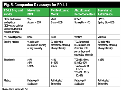
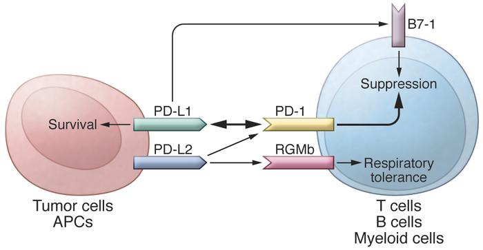
Prologue: The world of cancer care has been shaken up by the news that patients with hard-to-treat tumors benefit from a new type of immunotherapy, called checkpoint inhibition. A key receptor, called programmed death 1 (PD-1), is charged with suppressing the ability of activated T cells and other immune cells to destroy cancer cells, all in the name of preventing damage to normal tissue via autoimmunity. When PD-1 receptors on T cells bind with PD-L1 and PD-L2, complimentary receptors expressed on tumor cells, the immune response (call it the assassination of the cell) is checked and the tumor lives on. The anti PD-1 monoclonal antibodies nivolumab and pembrolizumab keep PD-L1 from turning off T cells, which has produced durable responses in several tumor types including melanoma, lung cancer, and renal cell carcinoma and represents a new hope for many.

Oncologists are excited to relay this news to patients, but is there a way to explain this without putting everyone in the room to sleep? Well, I like to use analogies to make seemingly complicated mechanisms easier to understand and the PD-1/PD-L1 relationship has inspired several colorful examples, to wit:

“Think of T cells as killers that use photographs to identify individual bad guys. Their weakness is that they will not act if the intended victim shakes their hand first. The bad guys used to be born without arms, but over time they evolved to grow arms and hands, thus avoiding elimination. The antibodies are boxing gloves that cover the hands of the T cells. Goodbye, bad guys.”

“Think of T cells as cats specially trained to eliminate mice wherever they hide. Their only weakness is if they smell catnip they will roll over and purr like idiots instead of doing their job. The mice then develop special glands that secrete catnip, thus pacifying the kitties. Solution: plug up the cats’ noses with nivolumab or pembrolizumab. Sayonara, Mr. Mouse.”

“Think of T cells as a fire sprinkler system designed to activate when a metal plug is heated to its melting point, releasing water from a pipe. The fire then emits a toxin that coats the fusible metal, keeping it below its melting point. By fitting a protective shield around the plug we block the toxic molecules and allow the plug to melt in a fire. The shield is the monoclonal antibody against PD-1 and thus the fire is successfully extinguished.”

This is getting exhausting, so I think I will stop, but don’t you agree that the concept of checkpoint inhibition lends itself to a plethora of metaphors? Now for the next lesson: how to explain chimeric antigen receptor T-cell therapy to patients. Hold on—I think I need to explain it to myself first.


As an clinical immunologist, i agree with the concept, looks pretty straightforward but definitely much more complex as our immune system work like a network. I would appreciate clinical trial data with statistical significance.


Much more confusing. Most people understand simplified concepts.

“Some cancer cells turn off your immune systems ability to recognise them. These drugs ramp up the immune system and prevent the cancer cells from hiding. This allows your cells to attack and kill cancer cells”

If i think patient seem to have better ability to understand I say “the drugs block the “off switch” that cancer cells use to escape their detection. This turns your immune systems ability to attack and kill cancer cells back on”

I haven’t had one patient that has looked confused since.


HDAC Inhibitors Enhance Immunotherapy Efficacy in Lung Cancer

http://www.oncotherapynetwork.com/lung-cancer-targets/hdac-inhibitors-enhance-immunotherapy-efficacy-lung-cancer

Histone deacetylase (HDAC) inhibitors like romidepsin might improve the efficacy of programmed cell death-1 (PD-1) blockade in lung cancer, suggest preclinical findings reported in the journal Clinical Cancer Research.

Most lung cancer patients’ tumors do not respond to immune checkpoint blockade agents like those that target PD-1. One possible mechanism underlying tumor resistance to PD-1 blockade is the failure of sufficient numbers of T cells to infiltrate tumor tissue.

Hypothesizing that upregulating T-cell chemokine expression and thereby T-cell infiltration of tumors would improve PD-1 blockade’s efficacy against lung tumors, the research team went hunting for FDA-approved oncology agents that induce chemokine expression. Screening 97 approved agents, they found one class that did: HDAC inhibitors.

The HDAC-inhibiting agent romidepsin significantly increased T-cell tumor infiltration and impacted lung tumor growth in mouse models, the team reported—and when romidepsin was subsequently combined with PD-1 blockade in several lung tumor models, the combination showed greater antitumor activity than either agent on its own.

“These results suggest that combination of HDAC inhibitors with PD-1 blockade represent a promising strategy for lung cancer treatment,” said senior study author Amer A. Beg, PhD, of the Moffitt Cancer Center’s Immunology Program, in a news release.

Romidepsin and other HDAC inhibitors have already been approved by the FDA for use against lymphoma and other hematologic cancers, Dr. Beg noted.

The combination will next be tested in several clinical trials, including a study of patients diagnosed with non-small cell lung cancer (NSCLC) at Moffitt Cancer Center.


HDAC inhibitors enhance T cell chemokine expression and augment response to PD-1 immunotherapy in lung adenocarcinoma

Hong Zheng1,  weipeng zhao2Cihui Yan3Crystina C Watson4,…., Brian Ruffell13, and Amer A Beg4,*

Clin Cancer Res March 10, 2016; http://dx.doi.org:/10.1158/1078-0432.CCR-15-2584

Purpose: A significant limitation of checkpoint blockade immunotherapy is the relatively low response rate (e.g. ~20% with PD-1 blockade in lung cancer). In this study, we tested whether strategies which increase T cell infiltration to tumors can be efficacious in enhancing immunotherapy response. Experimental Design: We performed an unbiased screen to identify FDA-approved oncology agents with ability to enhance T cell chemokine expression with the goal of identifying agents capable of augmenting immunotherapy response. Identified agents were tested in multiple lung tumor models as single agents and in combination with PD-1 blockade. Additional molecular and cellular analysis of tumors was used to define underlying mechanisms. Results: We found that histone deacetylase (HDAC) inhibitors (HDACi) increased expression of multiple T cell chemokines in cancer cells, macrophages and T cells. Using the HDACi romidepsin in vivo, we observed increased chemokine expression, enhanced T cell infiltration, and T cell-dependent tumor regression. Importantly, romidepsin significantly enhanced the response to PD-1 blockade immunotherapy in multiple lung tumor models, including nearly complete rejection in two models. Combined romidepsin and PD-1 blockade also significantly enhanced activation of tumor-infiltrating T cells. Conclusions: These results provide evidence for a novel role of HDACs in modulating T cell chemokine expression in multiple cell types. In addition, our findings indicate that pharmacological induction of T cell chemokine expression represents a conceptually novel approach for enhancing immunotherapy response. Finally, these results suggest that combination of HDAC inhibitors with PD-1 blockade represents a promising strategy for lung cancer treatment.


Cancer Cell Survival Driven by Novel Metabolic Pathway

http://www.genengnews.com/gen-news-highlights/cancer-cell-survival-driven-by-novel-metabolic-pathway/81252584/

Researchers have identified a novel metabolic pathway that helps cancer cells thrive in conditions that are lethal to normal cells. [National Cancer Institute, NIH] http://www.genengnews.com/Media/images/GENHighlight/thumb_28216_large1422477191.jpg

Being attached to the extracellular matrix (ECM) provides cells with numerous advantages for survival, for instance, receiving much needed growth stimuli. However, for malignant cells to function, they must overcome their anchorage-dependent growth—a scenario that is associated with increased production of reactive oxygen species (ROS) and altered glucose metabolism.

Now, researchers at the Children’s Medical Center Research Institute at UT Southwestern (CRI) believe they have uncovered a novel metabolic pathway that helps cancer cells thrive in conditions that would otherwise be lethal to healthy cells.

“It’s long been thought that if we could target tumor-specific metabolic pathways, it could lead to effective ways to treat cancer,” explained senior study author Ralph DeBerardinis, M.D., Ph.D.,  associate professor, and director of CRI’s Genetic and Metabolic Disease Program. “This study finds that two very different metabolic processes are linked in a way that is specifically required for cells to adapt to the stress associated with cancer progression.”

This new study describes an alternate version of two well-known metabolic pathways, the pentose phosphate pathway (PPP) and the Krebs cycle, to defend against ROS that destroy cells via oxidative stress.

The findings from this study were published recently in Nature in an article entitled “Reductive Carboxylation Supports Redox Homeostasis During Anchorage-Independent Growth.”

Previous work from Dr. DeBerardinis’ laboratory found that the Krebs cycle, a series of chemical reactions that cells use to generate energy, could reverse itself under certain conditions to nourish cancer cells.

Dr. DeBerardinis also noted that cells “are dependent on matrix attachment to receive growth-promoting signals and to regulate their metabolism in a way that supports cell growth, proliferation, and survival.” Detachment from the matrix results in a sudden increase in ROS that is lethal to normal cells. Yet, cancer cells seem to have evolved workaround.

A landmark study from 2009 elucidated that healthy cells were destroyed when detached from the ECM. Moreover, in the same study, investigators found that inserting an oncogene into a normal cell caused it to behave like a cancer cell and survive detachment.

“Another Nature study, this one from CRI Director Dr. Sean Morrison’s laboratory in November 2015, found that the rare skin cancer cells that were able to detach from the primary tumor and successfully metastasize to other parts of the body had the ability to keep ROS levels from getting dangerously high,” Dr. DeBerardinis remarked.

Dr. DeBerardinis and his team worked from the premise that the two findings were pieces of the same puzzle and that a crucial part of the picture seemed to be missing.

It had been well known that the PPP was an important source of nicotine adenine dinucleotide phosphate (NADPH), which provides a source of reducing electrons to scavenge ROS; however, the PPP produces NADPH in the cytosol, whereas the ROS are generated primarily in another subcellular structure called the mitochondria.

“If you think of ROS as fire, then NADPH is like the water used by cancer cells to douse the flames,” Dr. DeBerardinis noted.  But how could NADPH from the PPP help deal with the stress of ROS produced in an entirely different part of the cell? “What we did was to discover how this happens.”

The CRI team was able to demonstrate that cancer cells use a “piggybacking” system to carry the reducing electron from the PPP into the mitochondria. This movement involves an unusual reaction in the cytosol that transfers reducing equivalents from NADPH to a molecule called citrate, similar to a reversed reaction of the Krebs cycle.The citrate then enters the mitochondria and stimulates another pathway that results in the release of reducing electrons to produce NADPH right at the location of ROS creation, allowing the cancer cells to survive and grow without the benefit of matrix attachment.

“We knew that both the PPP and Krebs cycle provide metabolic benefits to cancer cells. But we had no idea that they were linked in this unusual fashion,” Dr. DeBerardinis stated. “Strikingly, normal cells were unable to transport NADPH by this mechanism, and died as a result of the high ROS levels.”

The researchers stressed that their findings were based on cultured cell models and more research will be necessary to test the role of the pathway in living organisms.

“We are particularly excited to test whether this pathway is required for metastasis because cancer cells need to survive in a matrix-detached state in the circulation in order to metastasize,” Dr. DeBerardinis concluded.


Reductive carboxylation supports redox homeostasis during anchorage-independent growth

Lei JiangAlexander A. ShestovPamela SwainChendong Yang, …., Brian P. DrankaBenjamin Schwartz & Ralph J. DeBerardinis

Nature(2016)      http://dx.doi.org:/10.1038/nature17393

Cells receive growth and survival stimuli through their attachment to an extracellular matrix (ECM)1. Overcoming the addiction to ECM-induced signals is required for anchorage-independent growth, a property of most malignant cells2. Detachment from ECM is associated with enhanced production of reactive oxygen species (ROS) owing to altered glucose metabolism2. Here we identify an unconventional pathway that supports redox homeostasis and growth during adaptation to anchorage independence. We observed that detachment from monolayer culture and growth as anchorage-independent tumour spheroids was accompanied by changes in both glucose and glutamine metabolism. Specifically, oxidation of both nutrients was suppressed in spheroids, whereas reductive formation of citrate from glutamine was enhanced. Reductive glutamine metabolism was highly dependent on cytosolic isocitrate dehydrogenase-1 (IDH1), because the activity was suppressed in cells homozygous null for IDH1 or treated with an IDH1 inhibitor. This activity occurred in absence of hypoxia, a well-known inducer of reductive metabolism. Rather, IDH1 mitigated mitochondrial ROS in spheroids, and suppressing IDH1 reduced spheroid growth through a mechanism requiring mitochondrial ROS. Isotope tracing revealed that in spheroids, isocitrate/citrate produced reductively in the cytosol could enter the mitochondria and participate in oxidative metabolism, including oxidation by IDH2. This generates NADPH in the mitochondria, enabling cells to mitigate mitochondrial ROS and maximize growth. Neither IDH1 nor IDH2 was necessary for monolayer growth, but deleting either one enhanced mitochondrial ROS and reduced spheroid size, as did deletion of the mitochondrial citrate transporter protein. Together, the data indicate that adaptation to anchorage independence requires a fundamental change in citrate metabolism, initiated by IDH1-dependent reductive carboxylation and culminating in suppression of mitochondrial ROS.


Liquid Biopsy Accurately Detects Mutations in Advanced NSCLC

http://www.oncotherapynetwork.com/lung-cancer/liquid-biopsy-accurately-detects-mutations-advanced-nsclc#sthash.bEPxRkAq.dpuf

Droplet digital polymerase chain reaction (ddPCR)-based plasma genotyping—referred to as liquid biopsy—exhibited perfect specificity in identifying EGFR and KRAS mutations in patients with advanced non–small-cell lung cancer (NSCLC), according to the results of a study published in JAMA Oncology.

“We see plasma genotyping as having enormous potential as a clinical test, or assay—a rapid, noninvasive way of screening a cancer for common genetic fingerprints, while avoiding the challenges of traditional invasive biopsies,” said senior author, Geoffrey Oxnard, MD, thoracic oncologist and lung cancer researcher at Dana-Farber and Brigham and Women’s Hospital, in a press release. “Our study was the first to demonstrate prospectively that a liquid biopsy technique can be a practical tool for making treatment decisions in cancer patients.”

According to the press release, the test proved so reliable in the study that the Dana-Farber/Brigham and Women’s Cancer Center this week became the first medical facility in the country to offer it to all patients with NSCLC, either at the time of first diagnosis or of relapse following previous treatment.

Oxnard and colleagues enrolled 180 patients with advanced NSCLC. Patients were either newly diagnosed with the disease (n = 120) or had acquired resistance to prior EGFR kinase inhibitors (n = 60) and were planned for rebiopsy. Patients underwent initial blood sampling and immediate plasma ddPCR screening for EGFR exon 19 deletion, L858R, the EGFR T790M acquired resistance mutation, or KRAS G12X. In addition, patients underwent biopsy for tissue genotyping used to compare the accuracy of ddPCR.

Among the enrolled patients, 80 had EGFR exon19/L858R mutations, 35 had T790M mutations and 25 had KRAS G12X mutations. The median test turnaround time for liquid biopsy was 3 days. In comparison, the median turnaround time for tissue genotyping was 12 days for newly diagnosed patients and 27 days for patients with acquired EGFR inhibitor resistance.

“This long turnaround time is due largely to the practical reality that many patients with newly diagnosed NSCLC require a repeat biopsy to obtain tissue for genotyping, as do all patients with acquired resistance,” the researchers noted.

The liquid biopsy showed 100% positive predictive value for detecting EGFR 19 deletion, L858R, and KRAS mutations. However, it had only a positive predictive value of 79% for T790M mutations. The sensitivity of the test was lower. ddPCR had a sensitivity of 82% for EGFR 19 deletion, 74% for L858R, 77% for T790M, and 64% for KRAS.

The researchers pointed out that “a key limitation of plasma ddPCR is that although this method is adept at rapidly detecting specific targetable mutations, it cannot easily detect copy number alterations and rearrangements. The ddPCR panel assessed in this study thus cannot currently detect targetable alterations in either ALK or ROS1,” two other common mutations in NSCLC.

In an editorial that accompanied the article, P. Mickey Williams, PhD, of Frederick National Laboratory for Cancer Research, and Barbara A. Conley, MD, from the National Cancer Institute, questioned whether or not these results, and the rapid turnaround time for liquid biopsy, could be replicated widely by other institutions.

“However, if this performance were generally applicable, this would indeed be an advance in clinical care, reducing the proportion of patients requiring biopsy, at least in the resistance setting,” Williams and Conley wrote.

“This study is a step in the right direction of preparing needed clinical validation for the use of ctDNA for detection and serial monitoring of clinically relevant tumor mutations. Owing to low sensitivity and high positive predictive value and specificity, this approach is probably best suited for detection of resistance mutations and for serial plasma testing to assess treatment response, and should not replace tumor biopsy assessment for initial treatment decision-making,” they concluded.


Prospective Validation of Rapid Plasma Genotyping for the Detection of EGFR and KRAS Mutations in Advanced Lung Cancer

Adrian G. Sacher, MD1,2; Cloud Paweletz, PhD3; Suzanne E. Dahlberg, PhD4,5; Ryan S. Alden, BSc1; Allison O’Connell, BSc3; Nora Feeney, BSc3; Stacy L. Mach, BA1; Pasi A. Jänne, MD, PhD1,2,3; Geoffrey R. Oxnard, MD1,2

JAMA Oncol. Published online April 07, 2016.    http://dx.doi.org:/10.1001/jamaoncol.2016.0173

Importance  Plasma genotyping of cell-free DNA has the potential to allow for rapid noninvasive genotyping while avoiding the inherent shortcomings of tissue genotyping and repeat biopsies.

Objective  To prospectively validate plasma droplet digital PCR (ddPCR) for the rapid detection of common epidermal growth factor receptor (EGFR) and KRAS mutations, as well as the EGFR T790M acquired resistance mutation.

Design, Setting, and Participants  Patients with advanced nonsquamous non–small-cell lung cancer (NSCLC) who either (1) had a new diagnosis and were planned for initial therapy or (2) had developed acquired resistance to an EGFR kinase inhibitor and were planned for rebiopsy underwent initial blood sampling and immediate plasma ddPCR for EGFR exon 19 del, L858R, T790M, and/or KRAS G12X between July 3, 2014, and June 30, 2015, at a National Cancer Institute–designated comprehensive cancer center. All patients underwent biopsy for tissue genotyping, which was used as the reference standard for comparison; rebiopsy was required for patients with acquired resistance to EGFR kinase inhibitors. Test turnaround time (TAT) was measured in business days from blood sampling until test reporting.

Main Outcomes and Measures  Plasma ddPCR assay sensitivity, specificity, and TAT.

Results  Of 180 patients with advanced NSCLC (62% female; median [range] age, 62 [37-93] years), 120 cases were newly diagnosed; 60 had acquired resistance. Tumor genotype included 80 EGFR exon 19/L858R mutants, 35 EGFR T790M, and 25 KRASG12X mutants. Median (range) TAT for plasma ddPCR was 3 (1-7) days. Tissue genotyping median (range) TAT was 12 (1-54) days for patients with newly diagnosed NSCLC and 27 (1-146) days for patients with acquired resistance. Plasma ddPCR exhibited a positive predictive value of 100% (95% CI, 91%-100%) for EGFR 19 del, 100% (95% CI, 85%-100%) for L858R, and 100% (95% CI, 79%-100%) for KRAS, but lower for T790M at 79% (95% CI, 62%-91%). The sensitivity of plasma ddPCR was 82% (95% CI, 69%-91%) for EGFR 19 del, 74% (95% CI, 55%-88%) for L858R, and 77% (95% CI, 60%-90%) for T790M, but lower for KRAS at 64% (95% CI, 43%-82%). Sensitivity for EGFR or KRAS was higher in patients with multiple metastatic sites and those with hepatic or bone metastases, specifically.

Conclusions and Relevance  Plasma ddPCR detected EGFR and KRAS mutations rapidly with the high specificity needed to select therapy and avoid repeat biopsies. This assay may also detect EGFR T790M missed by tissue genotyping due to tumor heterogeneity in resistant disease.

Plasma genotyping uses tumor-derived cell-free DNA (cfDNA) to allow for rapid noninvasive genotyping of tumors. This technology is currently poised to transition into a treatment decision-making tool in multiple cancer types. It is particularly relevant to the treatment of advanced non–small-cell lung cancer (NSCLC), in which therapy hinges on rapid and accurate detection of targetable epidermal growth factor receptor (EGFR), anaplastic lymphoma kinase (ALK), and ROS1 alterations.1– 6Plasma genotyping is capable of circumventing many limitations of standard tissue genotyping including slow turnaround time (TAT), limited tissue for testing, and the potential for failed biopsies. It may be particularly useful in directing the rapid use of new targeted therapies for acquired resistance in advanced EGFR-mutant NSCLC, where the need for a repeat biopsy to test for resistance mechanisms has amplified the inherent limitations of traditional genotyping.7,8

The need to carefully validate the test characteristics of each of the myriad individual plasma genotyping assays before use in clinical decision making is paramount. We have previously reported the development of a quantitative droplet digital polymerase chain reaction (ddPCR)-based assay for the detection of EGFR kinase mutations andKRAS codon 12 mutations in plasma.9 The detection of these mutations has the potential to guide treatment by either facilitating targeted therapy with an EGFR tyrosine kinase inhibitor (TKI) or ruling out the presence of other potentially targetable alterations in the case of KRAS.5 Alternative platforms including Cobas, peptide nucleic acid–mediated PCR, multiplexed next-generation sequencing (NGS), high-performance liquid chromatography, and Scorpion–amplified refractory mutation system have also been examined in retrospective analyses of patient samples.10– 22 The test characteristics of these assays have been variable and may be attributable to differences in testing platforms, as well as the retrospective nature of these studies, their smaller size, and the timing of blood collection with respect to disease progression and therapy initiation. The absence of reliable prospective data on the use of specific plasma genotyping assays in advanced NSCLC has left key aspects of its utility largely undefined and slowed its uptake as a tool for clinical care in patients with both newly diagnosed NSCLC and EGFR acquired resistance.

To our knowledge, we have conducted the first prospective study of the use of ddPCR-based plasma genotyping for the detection of EGFR and KRAS mutations. This study was performed in the 2 settings where we anticipate clinical adoption of this assay: (1) patients with newly diagnosed advanced NSCLC and (2) those with acquired resistance to EGFR kinase inhibitors. The primary aim of this study was to prospectively evaluate the feasibility and accuracy of this assay for the detection ofEGFR/KRAS mutations in patients with newly diagnosed NSCLC and EGFR T790M in patients with acquired resistance in a clinical setting. Additional end points included test TAT and the effect of sample treatment conditions on test accuracy.

Key Points
  • Question What is the sensitivity, specificity, turnaround time, and robustness of droplet digital polymerase chain reaction (ddPCR)-based plasma genotyping for the rapid detection of targetable genomic alterations in patients with advanced non–small-cell lung cancer (NSCLC)?

  • Findings In this study of 180 patients with advanced NSCLC (120 newly diagnosed, 60 with acquired resistance to epidermal growth factor receptor [EGFR] kinase inhibitors), plasma genotyping exhibited perfect specificity (100%) and acceptable sensitivity (69%-80%) for the detection of EGFR-sensitizing mutations with rapid turnaround time (3 business days). Specificity was lower for EGFR T790M (63%), presumably secondary to tumor heterogeneity and false-negative tissue genotyping.

  • Meaning The use of ddPCR-based plasma genotyping can detect EGFR mutations with the rigor necessary to direct clinical care. This assay may obviate repeated biopsies in patients with positive plasma genotyping results.

CYP3A7*1C Allele Associated With Poor Outcomes in CLL, Breast, and Lung Cancer

 http://www.oncotherapynetwork.com/breast-cancer-targets/cyp3a71c-allele-associated-poor-outcomes-cll-breast-and-lung-cancer#sthash.7uiD8XFD.dpuf

Patients with the CYP3A7*1C allele suffer higher rates of cancer progression and mortality, possibly because of worse outcomes among patients treated with chemotherapy drugs that are broken down by the enzyme encoded by CYP3A7, according to authors of a retrospective study published in the journal Cancer Research.

“We found that individuals with breast cancer, lung cancer, or CLL [chronic lymphocytic leukemia] who carry one or more copy of the CYP3A7*1C allele tend to have worse outcomes,” said Olivia Fletcher, PhD, a senior investigator at the Breast Cancer Now Toby Robins Research Centre at the Institute of Cancer Research in London, England, in an American Association for Cancer Research (AACR) news release.

Approximately 8% of cancer patients harbor the CYP3A7*1C allele, the coauthors noted. For these patients, it is possible that standard chemotherapy with CYP3A substrates “may not be optimal,” they cautioned.

The team analyzed DNA samples from 1,008 patients with breast cancer, 1,128 patients with lung cancer, and 347 patients with CLL. They found that the CYP3A7*1C-associated single nucleotide polymorphism (SNP) rs45446698 is associated with increased breast cancer mortality (hazard ratio [HR] 1.74; P = .03), all-cause mortality among patients with lung cancer (HR 1.43; P = .009), and progression of CLL (HR 1.62; P = .03). The rs45446698 SNP is one of seven SNPs that form the CYP3A7*1C allele.

The CYP3A7*1C allele is expressed in adults, whereas other variants of CYP3A7 are expressed during fetal development. CYP3A7 encodes an enzyme that degrades estrogen and testosterone, and some anticancer drugs.

“We also found borderline evidence of a statistical interaction between the CYP3A7*1C allele, treatment of patients with a cytotoxic agent that is a CYP3A substrate, and clinical outcome (P = .06),” they noted.

“Even though we did not see a statistically-significant difference when stratifying patients by treatment with a CYP3A7 substrate, the fact that we see the same effect in three very different cancer types suggests to me that it is more likely to be something to do with treatment than the disease itself,” commented Dr. Fletcher. “However, we are looking at ways of replicating these results in additional cohorts of patients and types of cancer, as well as overcoming the limitations of this study.”

Limitations included the retrospective nature of the study and the absence of data on how quickly individual patients metabolized chemotherapeutic agents, she said.


Cytochrome P450 AlleleCYP3A7*1C Associates with Adverse Outcomes in Chronic Lymphocytic Leukemia, Breast, and Lung Cancer

Nichola Johnson1,2Paolo De Ieso3Gabriele Migliorini4,….., Gillian Ross12Richard S. Houlston, and Olivia Fletcher1,2,*

Cancer Res March 10, 2016; http://dx.doi.org:/10.1158/0008-5472.CAN-15-1410

CYP3A enzymes metabolize endogenous hormones and chemotherapeutic agents used to treat cancer, thereby potentially affecting drug effectiveness. Here, we refined the genetic basis underlying the functional effects of a CYP3A haplotype on urinary estrone glucuronide (E1G) levels and tested for an association betweenCYP3A genotype and outcome in patients with chronic lymphocytic leukemia (CLL), breast, or lung cancers. The most significantly associated SNP was rs45446698, an SNP that tags the CYP3A7*1Callele; this SNP was associated with a 54% decrease in urinary E1G levels. Genotyping this SNP in 1,008 breast cancer, 1,128 lung cancer, and 347 CLL patients, we found that rs45446698 was associated with breast cancer mortality (HR, 1.74; P = 0.03), all-cause mortality in lung cancer patients (HR, 1.43; P = 0.009), and CLL progression (HR, 1.62; P= 0.03). We also found borderline evidence of a statistical interaction between the CYP3A7*1C allele, treatment of patients with a cytotoxic agent that is a CYP3A substrate, and clinical outcome (Pinteraction = 0.06). The CYP3A7*1C allele, which results in adult expression of the fetal CYP3A7 gene, is likely to be the functional allele influencing levels of circulating endogenous sex hormones and outcome in these various malignancies. Further studies confirming these associations and determining the mechanism by which CYP3A7*1C influences outcome are required. One possibility is that standard chemotherapy regimens that include CYP3A substrates may not be optimal for the approximately 8% of cancer patients who are CYP3A7*1C carriers. Cancer Res; 76(6); 1–9. ©2016 AACR.


​Specific Form of CYP3A7 Gene Associated With Poor Outcomes for Patients With Several Cancer Types

3/10/2016

PHILADELPHIA — Among patients with breast cancer, lung cancer, or chronic lymphocytic leukemia (CLL), those who had a specific form of the CYP3A7 gene (CYP3A7*1C) had worse outcomes compared with those who did not have CYP3A7*1C, and this may be related to how the patients metabolize, or break down, the therapeutics used to treat them, according to a study published in Cancer Research, a journal of the American Association for Cancer Research.

“The CYP3A7 gene encodes an enzyme that breaks down all sorts of naturally occurring substances—such as sex steroids like estrogen and testosterone—as well as a wide range of drugs that are used in the treatment of cancer,” saidOlivia Fletcher, PhD, a senior investigator at the Breast Cancer Now Toby Robins Research Centre at The Institute of Cancer Research in London. “The CYP3A7 gene is normally turned on in an embryo and then turned off shortly after a baby is born, but individuals who have one or more copy of the CYP3A7*1C form of the gene [the CYP3A7*1C allele] turn on their CYP3A7 gene in adult life.

“We found that individuals with breast cancer, lung cancer, or CLL who carry one or more copy of the CYP3A7*1C allele tend to have worse outcomes,” continued Fletcher. “One possibility is that these patients break down the drugs that they are given to treat their cancer too fast. However, further independent studies that replicate our findings in larger numbers of patients and rule out biases are needed before we could recommend any changes to the treatment that cancer patients with the CYP3A7*1C allele receive.”

To find out whether the CYP3A7*1C allele was associated with outcome for patients with breast cancer, lung cancer, or CLL, Fletcher and colleagues analyzed DNA samples from 1,008 breast cancer patients, 1,142 patients with lung cancer, and 356 patients with CLL for the presence of a single nucleotide polymorphism (SNP), rs45446698. Fletcher explained that a SNP is a type of genetic variant and that because of the way that we inherit our genetic material from our parents, we tend to inherit clusters of genetic variants. She went on to say that rs45446698 is one of seven SNPs that cluster together, forming the CYP3A7*1C allele.

The researchers found that among the breast cancer patients, rs45446698 (and, therefore, the CYP3A7*1C allele) was associated with a 74 percent increased risk of breast cancer mortality. Among the lung cancer patients, it was associated with a 43 percent increased risk of death from any cause, and among the CLL patients, it was associated with a 62 percent increased risk of disease progression.

Patients who were treated with a chemotherapeutic broken down by CYP3A7 tended to have worse outcomes compared with those treated with other chemotherapeutics, but the difference was not statistically significant.

“Even though we did not see a statistically significant difference when stratifying patients by treatment with a CYP3A7 substrate, the fact that we see the same effect in three very different cancer types suggests to me that it is more likely to be something to do with treatment than the disease itself,” said Fletcher. “However, we are looking at ways of replicating these results in additional cohorts of patients and types of cancer, as well as overcoming the limitations of this study.”

Fletcher explained that the main limitation of the study is that the researchers used samples and clinical information collected for other studies. Therefore, they did not have the same clinical information for each patient, and the samples were collected at different time points and for patients treated with various chemotherapeutics. She also noted that the team were not able to determine how quickly the patients broke down the therapeutics they received as treatment.

The study was supported by Breast Cancer Now, Leukaemia and Lymphoma Research (now known as Bloodwise), Cancer Research UK, the Medical Research Council, the Cridlan Trust, the Helen Rollason Cancer Charity, and Sanofi-Aventis. Funding for the authors’ institutions was received from the National Health Service of the United Kingdom. Fletcher declares no conflicts of interest.


Liquid Biopsy for NSCLC

‘Liquid biopsy’ blood test accurately detects key genetic mutations in most common form of lung cancer, study finds.

http://www.technologynetworks.com/Diagnostics/news.aspx?ID=190276

A simple blood test can rapidly and accurately detect mutations in two key genes in non-small cell lung tumors, researchers at Dana-Farber Cancer Institute and other institutions report in a new study – demonstrating the test’s potential as a clinical tool for identifying patients who can benefit from drugs targeting those mutations.

The test, known as a liquid biopsy, proved so reliable in the study that Dana-Farber/Brigham and Women’s Cancer Center (DF/BWCC) expects to offer it soon to all patients with non-small cell lung cancer (NSCLC), either at the time of first diagnosis or of relapse following previous treatment.

…….

“Our study was the first to demonstrate prospectively that a liquid biopsy technique can be a practical tool for making treatment decisions in cancer patients. The trial was such a success that we are transitioning the assay into a clinical test for lung cancer patients at DF/BWCC.”

The study involved 180 patients with NSCLC, 120 of whom were newly diagnosed, and 60 of whom had become resistant to a previous treatment, allowing the disease to recur. Participants’ cell-free DNA was tested for mutations in the EGFR and KRAS genes, and for a separate mutation in EGFR that allows tumor cells to become resistant to front-line targeted drugs. The test was performed with a technique known as droplet digital polymerase chain reaction (ddPCR), which counts the individual letters of the genetic code in cell-free DNA to determine if specific mutations are present. Each participant also underwent a conventional tissue biopsy to test for the same mutations. The results of the liquid biopsies were then compared to those of the tissue biopsies.

The data showed that liquid biopsies returned results much more quickly. The median turnaround time for liquid biopsies was three days, compared to 12 days for tissue biopsies in newly diagnosed patients and 27 days in drug-resistant patients.

Liquid biopsy was also found to be highly accurate. In newly diagnosed patients, the “predictive value” of plasma ddPCR was 100 percent for the primary EGFR mutation and the KRAS mutation – meaning that a patient who tested positive for either mutation was certain to have that mutation in his or her tumor. For patients with the EGFR resistance mutation, the predictive value of the ddPCR test was 79 percent, suggesting the blood test was able to find additional cases with the mutation that were missed using standard biopsies.

Prospective Validation of Rapid Plasma Genotyping for the Detection of EGFRand KRAS Mutations in Advanced Lung Cancer

Adrian G. Sacher, MD1,2; Cloud Paweletz, PhD3; Suzanne E. Dahlberg, PhD, et al.       JAMA Oncol. Published online April 07, 2016.  http://dx.doi.org::/10.1001/jamaoncol.2016.0173

Importance  Plasma genotyping of cell-free DNA has the potential to allow for rapid noninvasive genotyping while avoiding the inherent shortcomings of tissue genotyping and repeat biopsies.

Objective  To prospectively validate plasma droplet digital PCR (ddPCR) for the rapid detection of common epidermal growth factor receptor (EGFR) and KRAS mutations, as well as the EGFR T790M acquired resistance mutation.

Design, Setting, and Participants  Patients with advanced nonsquamous non–small-cell lung cancer (NSCLC) who either (1) had a new diagnosis and were planned for initial therapy or (2) had developed acquired resistance to an EGFR kinase inhibitor and were planned for rebiopsy underwent initial blood sampling and immediate plasma ddPCR for EGFR exon 19 del, L858R, T790M, and/or KRAS G12X between July 3, 2014, and June 30, 2015, at a National Cancer Institute–designated comprehensive cancer center. All patients underwent biopsy for tissue genotyping, which was used as the reference standard for comparison; rebiopsy was required for patients with acquired resistance to EGFR kinase inhibitors. Test turnaround time (TAT) was measured in business days from blood sampling until test reporting.

Main Outcomes and Measures  Plasma ddPCR assay sensitivity, specificity, and TAT.

Results  Of 180 patients with advanced NSCLC (62% female; median [range] age, 62 [37-93] years), 120 cases were newly diagnosed; 60 had acquired resistance. Tumor genotype included 80 EGFR exon 19/L858R mutants, 35 EGFR T790M, and 25 KRASG12X mutants. Median (range) TAT for plasma ddPCR was 3 (1-7) days. Tissue genotyping median (range) TAT was 12 (1-54) days for patients with newly diagnosed NSCLC and 27 (1-146) days for patients with acquired resistance. Plasma ddPCR exhibited a positive predictive value of 100% (95% CI, 91%-100%) for EGFR 19 del, 100% (95% CI, 85%-100%) for L858R, and 100% (95% CI, 79%-100%) for KRAS, but lower for T790M at 79% (95% CI, 62%-91%). The sensitivity of plasma ddPCR was 82% (95% CI, 69%-91%) for EGFR 19 del, 74% (95% CI, 55%-88%) for L858R, and 77% (95% CI, 60%-90%) for T790M, but lower for KRAS at 64% (95% CI, 43%-82%). Sensitivity for EGFR or KRAS was higher in patients with multiple metastatic sites and those with hepatic or bone metastases, specifically.

Conclusions and Relevance  Plasma ddPCR detected EGFR and KRAS mutations rapidly with the high specificity needed to select therapy and avoid repeat biopsies. This assay may also detect EGFR T790M missed by tissue genotyping due to tumor heterogeneity in resistant disease.


In this prospective study, we demonstrate the highly specific and rapid nature of plasma genotyping. No false-positive test results were seen for driver mutations inEGFR or KRAS, and TAT from when the specimen was obtained to result was a matter of days. This assay exhibited 100% positive predictive value for the detection of these mutations. Sensitivity was more modest and was directly correlated with both number of metastatic sites and the presence of liver or bone metastases. This newly demonstrated relationship is likely related to increased cfDNA shed in the setting of more extensive disease where tumor cfDNA shed is the chief driver of assay sensitivity and determines its upper limit. The characteristics of plasma ddPCR prospectively demonstrated in this study were similar or improved compared with previous retrospective reports of other cfDNA genotyping assays.10– 13,15,16,24,25 These retrospective studies are smaller, frequently examined a mix of tumor types and/or stages, and lack the careful prospective design needed to demonstrate the readiness of this technology to transition to a tool for selecting therapy. Studies that use retrospective samples from clinical trials that enrolled only EGFR-mutant patients are further limited by an inability to both blind laboratory investigators to tissue genotype and to generalize their assay test characteristics to a genetically heterogeneous real-world patient population.11 These differences and the multiple platforms examined previously have led to variable test characteristics and uncertainty regarding the clinical application of these technologies. This study is the first to prospectively demonstrate the ability of a ddPCR-based plasma genotyping assay to rapidly and accurately detect EGFR and KRAS mutations in a real-world clinical setting with the rigor necessary to support the assertion that use of this assay is capable of directing clinical care.

Even with a diagnostic sensitivity of less than 100%, such a rapid assay with 100% positive predictive value carries the potential for immense clinical utility. The 2- to 3-day TAT contrasts starkly with the 27-day TAT for tumor genotyping seen in patients needing a new tumor biopsy. This long TAT is due largely to the practical reality that many patients with newly diagnosed NSCLC require a repeat biopsy to obtain tissue for genotyping, as do all patients with acquired resistance. Consider the case of 1 study participant, an octogenarian with metastatic NSCLC who had developed acquired resistance to erlotinib with painful bone metastases (Figure 3). Due to the patient’s age and comorbidities, significant concerns existed about the risks of a biopsy and further systemic therapy. A plasma sample was obtained, and within 24 hours ddPCR demonstrated 806 copies/mL of EGFR T790M. A confirmatory lung biopsy was performed, which confirmed EGFR T790M. Treatment with a third-generation EGFR kinase inhibitor, osimertinib mesylate, was subsequently initiated and the patient had a partial response to therapy that was maintained for more than 1 year. The potential of this technology to obviate repeated biopsy in both patients with newly diagnosed NSCLC with insufficient tissue, as well as patients with acquired resistance, is considerable.

A key limitation of plasma ddPCR is that although this method is adept at rapidly detecting specific targetable mutations, it cannot easily detect copy number alterations and rearrangements. The ddPCR panel assessed in this study thus cannot currently detect targetable alterations in either ALK or ROS1. This limitation may potentially be addressed by using targeted NGS of cfDNA for broad, multiplexed detection of complex genomic alterations including ALK and ROS1 rearrangements, although this method is potentially slower than ddPCR-based methods and has been less thoroughly evaluated.23 The potential exists to use these technologies in tandem in advanced NSCLC to facilitate rapid initiation of therapy. Tissue genotyping and repeated biopsy would be specifically used to direct therapy in cases in which plasma genotyping was uninformative due to limitations of assay sensitivity. This approach would be particularly useful in cases of EGFR acquired resistance in which a repeated biopsy for T790M testing could be avoided entirely in many patients. Beyond detecting targetable alterations in order to drive therapy, the identification of nontargetable oncogenic drivers such as KRAS mutations that preclude the presence of other targetable alterations may guide a clinician to rapidly initiate alternative therapies such as chemotherapy or immunotherapy.5 The finding that assay sensitivity is highest in patients with more extensive metastatic disease suggests that those patients most in need of rapid treatment initiation would also be least likely to have false-negative results.

One surprising result of our study was evidence of recurrent false-positive results forEGFR T790M in patients with acquired resistance, despite no false-positive test results for other mutations studied. The sensitivity of the EGFR T790M assay was comparable to that of the EGFR sensitizing mutation assays and similarly related to both disease burden and the presence of liver or bone metastases, which are likely predictive of increased tumor cfDNA shed. We hypothesize that the lower assay specificity is due to the genomic heterogeneity whereby the T790M status of the biopsied site is not representative of all metastatic sites in a patient, a phenomenon supported by mounting evidence in the acquired resistance setting.26,27 This is consistent with the finding that a minority of patients with apparently EGFR T790M tissue-negative disease respond to therapy with third-generation EGFR kinase inhibitors.7,8,28 These observations raise questions regarding the fallibility of tissue-based genotyping as the reference standard for T790M status. The use of plasma genotyping to detect EGFR T790M thus has great potential to identify patients who would benefit from newly approved third-generation EGFR kinase inhibitors but would be unable to access them based on falsely negative tissue genotyping results. Indeed, plasma genotyping may allow more reliable assessment of both T790M status as well as the mechanisms of resistance across all sites of a heterogeneous cancer as opposed to a tissue biopsy and is likely to be an essential tool for future trials targeting drug resistance. The potential to avoid a repeat biopsy entirely in patients in whom plasma ddPCR detects T790M further strengthens the utility of this technology, although a repeat biopsy would still be needed in patients with uninformative plasma ddPCR due to limitations with respect to assay sensitivity.

This study also examined the potential of the quantitative nature of ddPCR-based plasma genotyping to allow for the early prediction of treatment response. Distinct patterns of change in mutant allele copy number were observed as early as 2 weeks after treatment and were similar to those reported in other tumor types.19,20 We hypothesize that these distinct patterns of change in this study will correlate with specific patterns of radiographic response and emergence of acquired resistance and plan to report these data once mature. The observed differences in treatment discontinuation rates observed in this study comparing patients with complete resolution of detectable mutant cfDNA with those with incomplete resolution support this hypothesis. The use of this technology to monitor disease status in real time has potential utility for both routine clinical care, as well as use as an integrated biomarker in early-phase clinical trials.10

Read Full Post »

Pull at Cancer’s Levers, Volume 2 (Volume Two: Latest in Genomics Methodologies for Therapeutics: Gene Editing, NGS and BioInformatics, Simulations and the Genome Ontology), Part 1: Next Generation Sequencing (NGS)

Pull at Cancer’s Levers

Curator: Larry H. Bernstein, MD, FCAP


Driving Cancer Immunotherapy 

The Stakes in Immuno-Oncology Are Too High for Researchers to Pull at Cancer’s Levers Blindly. Researchers Need a System.

  • Within the past decade or so, a revolutionary idea has emerged in the minds of scientists, physicians, and medical experts. Instead of using man-made chemicals to treat cancer, let us instead unleash the power of our own bodies upon the malignancy.

    This idea is the inspiration behind cancer immunotherapy, which is, according to most experts, a therapeutic approach that involves training the immune system to fight off cancer. In the words of one expert, cancer immunotherapy means “taking the immune system’s inherent properties and turbo charging those to fight cancer.”

    Cancer immunotherapy technologies are being developed to accomplish
    several tasks:

    • Enhance the molecular targeting of cancer cells
    • Report the rate of killing by specific immune agents
    • Direct immune cells toward tumor destruction.

    Since its inception, the field has evolved, and it continues to do so. It began with in vivo investigations of tumor growth and development, and it progressed through laboratory investigations of cellular morphology and survival curves. And now it is adopting pathway analysis to guide therapeutic development and improve patient care.

    To begin to understand cancer immunotherapy, one must understand how the immune system targets tumor cells. One of the prominent adaptive components of the immune system is the T cell, which responds to perceived threats through the massive increase in clonal T cells targeted in some way toward the diseased cell or pathogen.

  • The T-Cell Repertoire

    Adaptive Biotechnologies’ immunoSEQ Assay, a high-throughput research platform for immune system profiling, is designed to generate sequencer-ready libraries using highly optimized primer sets in a multiplex PCR format that targets T- and B-cell receptor genes. This image depicts how the assay’s two-step PCR process can be used to quantify the clonal diversity of immune cells.

    Immunologists call this process VJD rearrangement. It happens during T-lymphocyte development and affects three gene regions, the variable (V), the diversity (D), and the joining (J) regions. This rearrangement of the genetic code allow for the structural diversity in T-cell receptors responsible for antigenic specificity including antigenic targets on tumor cells. In the case of cancer, specificity is complicated because the tumor is actually part of the body itself, one of the reasons cancers naturally evade detection.

    The specificity problem would always hinder attempts to goad the immune system into attacking cancer, scientists realized, unless technologies emerged that could efficiently track the clonal diversity of T cells inside patients. Existing technologies, such as spectratyping, were inadequate.  In 2007, when Dr. Robins and his collaborators began developing the technology, only 10,000 T-cell receptor sequences had been reported in all the literature using older methodologies.

    “The immunology field of the time had no connection with high-throughput sequencing,” notes Dr. Robins, recalling his days as a computational biologist for the Fred Hutchinson Cancer Research Center. “It became clear that instead of using this old technology to look at T-cell receptors, we could just directly sequence them—if we could amplify them correctly.”

    With its first experiment, Dr. Robins’ team ended up with six million T-cell receptor sequences. “Our approach,” Dr. Robins modestly suggests, “kind of changed the scale of what we were able to do.” The team went on to develop advanced multiplex sequencing technology, doing work that essentially started the field of immune sequencing. “Previously,” maintains Dr. Robins, “no one had ever been able to quantitatively do a multiplex PCR.”

    Adaptive Biotechnologies’ product, the ImmunoSEQ® assay, uses several hundred primer pairs to quantify the clonal diversity of T cells. Using this technology, researchers and clinicians can focus on T-cell clones that are expanded specifically in or near a tumor or that are circulating in the blood stream.

    “You obviously can’t get a serial sample of the tumor,” explains Dr. Robins, “but you can get serial samples of blood,” allowing for immune cell repertoire tracking during the progression of a disease. The technology is already being used to assess leukemias in the clinic, directly tracking the leukemia itself based on the massive clonal expansion of a single cancerous B or T cell.

    Eventually, Dr. Robins’ team hopes to monitor serial changes in T cell clones before, during, and after therapeutic intervention. The team has even developed a tumor infiltrating lymphocyte (TIL) assay to examine clones that are attracted to tumors.


Circulating Tumor Cells

“Years ago, they were just interested in what was happening in the tumor,” says Daniel Adams, senior research scientist at Creatv MicroTech. “Now people have realized that the immune system is reacting to the tumor.”

Scientists such as Adams have been tracking tumor cells and tumor-modified stromal cells, as well as components of the non-adaptive immune system, directly within the bloodstream to examine changes that occur over time.

“We can’t go back in to re-biopsy the patient every year, or every time there is a recurrence,” says Adams, “It’s just not feasible.”

That is why Creatv MicroTech, with locations in Maryland and New Jersey, has developed the CellSieve, a mechanical cell filter. The CellSieve, which improves on older technology through better polymers and engineering, isolates circulating tumor cells (CTCs) and stromal cells in order to capture them for further clinical analysis.

Isolation, culture and expansion of cells isolated on CellSieve™. (A) MCF-7 cells spiked into vacutainers, isolated by filtration and cultured on CellSieve for 2-3 weeks. A 3 dimensional cluster attributed to this cell line is seen on the filter. (green=anti-cytokeratin, blue=DAPI) (B) PANC-1 cells spiked into vacutainers, isolated by filtration and grown on CellSieve for 2-3 weeks. PANC-1 is seen growing as a monolayer on the filter. (C) SKBR3 cells are spiked into blood, filtered by CellSieve. The CTCs are identified by presence of anti-cytokeratin and anti-EpCAM, and absence of anti-CD45. After CTCs are counted, cells are subtyped by HER2 FISH. (D) SKBR3 cells are spiked into vacutainers, isolated by filtration and grown on CellSieve for 2-3 weeks. Expanded colonies were directly analyzed as a whole colony and as individual cells, molecularly by HER2/CR17 FISH probes. (E) Circulating stromal cell, e.g. a 70 µm giant cancer associated macrophage can be identified for clinical use, myeloid marker in red. (F) A cell of interest can be identified and restained with immunotherapeutic biomarkers, e.g. PD-L1 (green) and PD-1 (purple). (G) After filtration, cells were identified with histopathological stains (e.g. H&E) for cytological analysis. (H) After H&E, external cell structures were analyzed by SEM. [Creatv MicroTech].


“As a patient goes through therapy, the patient’s resistance builds, and the cancer recurs in different subpopulations,” states Adams. “And after a few years, the original tumor mass is no longer applicable to what is growing in the patient farther down the road.”

Although CTCs are exceedingly rare in the bloodstream, with just one or two in every 5 to 10 mL of blood, and although these cells have a very low viability, the surviving CTCs have a high prognostic value.

“We looked at 30 to 40 breast cancer patients over two years,” reports Adams. “And we showed that if you have a dividing CTC, you have a 90% chance of dying in two years and a 100% chance of dying within two and half years.”

Furthermore, the immune system response can be tracked, says Adams, by examining stromal cells, which can also be collected with the CellSieve filtration device. That is, these cells can be collected serially. Much recent evidence supports the conclusion that stromal cells in the tumor environment co-evolve with the tumor, suggesting that stromal marker changes reflect tumor changes.

“There is this plethora of stromal cells and tumor cells out there in the circulation for you to look at,” declares Adams. “Once the cells are isolated, you can subject them to pathological approaches, biomarker approaches, or molecular approaches—or all of the above.”

A MicroTech Creatv study published in the Royal Society of Chemistry showed the efficacy of following up CTC isolation with techniques such as fluorescence in situ hybridization (FISH), histopathological analysis, and cell culture.

Cancer-Killing Assays

Diverse mechanisms are at play in cancer biology. Our understanding of these mechanisms contributes to a couple of virtuous cycles. It strengthens and is strengthened by diagnostic approaches, such as immune- and tumor-cell monitoring. The same could be said of therapeutic approaches. Cancer biology will inform and be informed by cancer immunotherapies such as adoptive cell transfer. To maintain the virtuous cycle, however, it will be necessary to conduct in vitro testing.

“There is no doubt that immunotherapy is going to play a major role in the treatment of cancer,” says Brandon Lamarche, Ph.D., technical communicator and scientist at ACEA Biosciences. “Regardless of what the route is, what is going to have to happen in terms of the research area is that you need an effective cell-killing assay.”

ACEA Biosciences, a San Diego-based company, has developed a microtiter plate that is coated with gold electrodes across 75% of the well bottoms. When the microtiter plates are placed in the company’s xCELLigence plate reader, the electrodes enable the detection of changes in cell morphology and viability through electrical impedance.

“The instrument provides a weak electric potential to the electrodes on the plate, so you get electrons flowing between these electrodes,” explains Dr. Lamarche. Researchers can then apply reagents or non-adherent immune cell suspensions to adherent cancer cells and examine the effect.

Dr. Lamarche asserts that the xCELLigence system overcomes problems that bedevil competing cell-killing assays. These problems include leaky and radioactive labels, such as chromium 51, and assays that can only provide users with an endpoint for cell killing. “With xCELLigence,” he insists, “you’re getting the full spectrum of what’s happening, and there’s all kinds of subtleties in the cell-killing curves that are very informative in terms of the biology.”

ACEA would like to see the xCELLigence system become the new standard in cell-killing assays from standard research to clinical testing on patient tumors. Dr. Lamarche envisions a day when patient tumor cells are quickly screened with therapeutic scenarios to determine the most efficacious killing option. “xCELLigence technology,” he suggests, “enables you to quickly sample a broad spectrum of conditions with a very simple workflow.”

Bioinformatics of Immuno-Oncology

From monitoring to treatment modalities, the field of cancer immunotherapy is aided by bioinformatics-minded data-mining experts, such as the analysts at Thompson-Reuters who are compiling data archives and applying advanced analytics to find new targets. “Essentially,” says Richard Harrison Ph.D., the company’s chief scientific officer for the life science division, “for every stage within pharmaceutical drug development, we have a database associated with that.”

The analysts at Thompson-Reuters curate and compile databases such as MedaCore and Cortellis, which they provide to their clients to help them with their research and clinical studies. “We can take customer data, and using our tools and our pathway maps, we can help them understand what their data is telling them,” explains Dr. Harrison.

Matt Wampole, Ph.D., a solutions scientist at Thompson-Reuters, spends his days reaching out and working with customers to help them understand and better use the company’s products. “Bench researchers,” he points out, “don’t necessarily know what is upstream of whatever expression change might be leading to a particular change in regulation.” Dr. Wampole indicates that he is part of a “solution team” that aids clients in determining important signaling cascades, regulators, and so on.

“We have a group of individuals who are very ‘skilling’ experts in the field,” Dr. Wampole continues, “including experts in the areas such as biostatistics, data curation, and data analytics. These experts help clients identify models, stratify patients, understand mechanisms, and look into disease mechanisms.”

Dr. Harrison sums up the Thompson-Reuters approach as follows: “We look for master regulators that can serve as both targets and biomarkers.” By examining the gene signatures from both the patient and from curated datasets, in the case of cancer immunotherapy, they hope to segregate patients according to what drugs will work best for them.

  • “We are working with a number of pharmaceutical companies to put our approach into practice for clinical trials,” informs Dr. Harrison. The approach has already been applied in several studies, including one that used data analysis of cell lines to help predict drug response in patients. Another study helped stratify glioblastoma patients.

  • Tumor-Targeted Delivery Platform

    PsiOxus Therapeutics, which is focused on immune therapeutics in oncology, has developed a patented platform for tumor-targeted delivery based on its oncolytic vaccine, Enadenotucirev (EnAd), which can be delivered systemically via intravenous administration.

    According to company officials, EnAd’s anti-cancer scope can be expanded by adding new genes, thereby enabling the creation of a broad range of unique immuno-oncology therapeutics. In a recent study conducted at the University of Oxford, researchers led by Philip G. Jakeman, Ph.D., sought to improve the models for evaluating cancer therapeutics by introducing ex vivo methodologies for research into colorectal cancer.

    The ex vivo approach utilized was able to exploit a major advantage by preserving the three-dimensional architecture of the tumor and its associated compartments, including immune cells. The study, which was presented at the International Summit on Oncolytic Viral Therapeutics in Quebec, showed the tissue slice model can provide a novel means to assessing an oncolytic vaccine in a system that more accurately recapitulates human tumors, provide a more stringent test for oncolytic viruses, such as EnAd, and allow study of the human immune cells within the tumor 3D context.

    By maintaining the components of the tumor immune microenvironment, this new methodology could become useful in analyzing anti-viral responses within tumors, or even in evaluating therapeutics that target immunosuppressive tumor micro-environments, noted the Oxford team.


Deciphering the Cancer Transcriptome

A Rogue’s Gallery of Malignant Outliers May Hide in Transcriptome Profiles That Emphasize Averages

http://www.genengnews.com/gen-articles/deciphering-the-cancer-transcriptome/5729/


The key link between genomic instability and cancer progression is transcriptome dynamics. The shifts in transcriptome dynamics that contribute to cancer evolution may come down to statistical outliers. [iStock/zmeel]

  • In recent years, scientists have adopted a gene-centric view of cancer, a tendency to see each malignant transformation as the consequence of alterations in a discrete number of genes or pathways. These alterations are, fortunately, absent from healthy cells, but they pervert malignant cells.

    The gene-centric view takes in molecular landscapes illuminated by genomic and transcriptomic technologies. For example, genomes can be cost-effectively sequenced within hours. Such capabilities have made it possible to interrogate associations between genotypes and phenotypes for increasing numbers of conditions, and to collect data from progressively larger patient groups.

    As genomic and transcriptomic technologies rise, they reveal much—but much remains hidden, too. Perhaps these technologies are less like the sun and more like the proverbial streetlight, the one that narrows our searches because we’re inclined to stay in the light, even though what we hope to find may lie in the shadows.

    “Each individual study that looks at the cancer transcriptome is impressive and tells a convincing story, but if we put several high-quality papers together, there are very few genes that overlap,” says Henry H. Heng, Ph.D., professor of molecular medicine, genetics, and pathology at Wayne State University. “This shows that something is wrong.”

  • Distinct Karyotypes

    One of the major observations in Dr. Heng’s lab is that the intra- and intertumor cellular heterogeneity results in nearly every cancer cell having a unique, distinct karyotype, that is, an important but often ignored genotype. “Biological systems need a lot of heterogeneity,” notes Dr. Heng. “People like to think that this is noise, but heterogeneity is a fundamental buffer system for biological function to be achievable. Moreover, it is the key agent for cellular adaptation.”

    To capture the degree of genomic heterogeneity at the genome level and its impact on cancer cell growth, Dr. Heng and colleagues performed serial dilutions to isolate single mouse ovarian surface epithelial cells that had undergone spontaneous transformation. Spectral karyotyping revealed that within a short timeframe each of these unstable cells exhibited a very distinct karyotype. In these unstable cells, cloning at the level of the karyotype was not possible.

    Stable cells exhibited a normal growth distribution, i.e., no subset of stable cells contributed disproportionately to the overall growth of the cell population. In contrast, unstable cell populations showed a non-normal growth distribution, with few cells contributing most to the cell population’s growth. For example, a single unstable colony contributed more than 70% to the cell population’s growth. This finding suggests that although average profiles can be used to describe non-transformed cells, they cannot be taken to represent the biology of malignant cells.

    “Most people who study the transcriptome want to get rid of the noise, but the noise is in fact the strategy that cancer uses to be successful,” explains Dr. Heng. “Each individual cancer cell is very weak but together the entity becomes very robust.”

    In a recent model that Dr. Heng and colleagues proposed, system inheritance visualizes chromosomes not merely as the vehicle for transmitting genetic information, but as the genetic network organizer that shapes the physical interactions between genes in the three-dimensional space. Based on this model, individual genes represent parts of the system. The same genes can be reorganized to form different systems, and chromosomal instability becomes more important than the contribution of individual genes and pathways to cancer biology.

    The vital link between genomic instability and cancer progression is transcriptome dynamics, and the shifts in those dynamics that contribute to cancer evolution may come down to statistical outliers.

    “Transcriptome studies rarely focus on single-cell analyses, which means important outliers are frequently ignored,” declares Dr. Heng. “This preoccupation with uninformative averages explains why we have learned so little despite having examined so many transcriptomes.”

  • Chimeras and Fusion Genes

    “Our focus is on chimeric RNA molecules,” says Laising Yen, Ph.D, assistant professor of pathology at Baylor College of Medicine. “This category of RNAs is very special because their sequences come from different genes.”

    In a study that was designed to capture chimeric RNAs in prostate cancer, Dr. Yen and his colleagues performed high-throughput sequencing of the transcriptomes from human prostate cancer samples. “We found far more chimeric RNAs, in terms of abundance, and a number of species that are not seen in normal tissue,” reports Dr. Yen. This approach identified over 2,300 different chimeric RNA species. Some of these chimeras were present in prostate cancer cell lines, but not in primary human prostate epithelium cells, which points toward their relevance in cancer.

    “Most of these chimeric RNAs do not have a genomic counterpart, which means that they could be produced by trans-splicing,” explains Dr. Yen. During trans-splicing, individual RNAs are generated and trans-spliced together as a single RNA, which provides a mechanism for generating a chimera.

    “The other possibility is that in cancer cells, where gene–gene boundaries are known to become broken, chimeras can be formed by cis-splicing from a very long transcript that encodes several neighboring genes located on the same chromosome,” informs Dr. Yen. Chimeric RNAs formed by either of these two mechanisms can potentially translate into fusion proteins, and these aberrant proteins may have oncogenic consequences.

    Another effort in Dr. Yen’s laboratory focuses on chromosomal aberrations in ovarian cancer. One of the hallmarks of ovarian cancer is the high degree of genomic rearrangement and the increased genomic instability.

    “When we looked at ovarian cancers, we did not find as many chimeric RNAs,” notes Dr. Yen. “But we found many fusion genes.” Gene fusions, similarly to chimeric RNAs, increase the diversity of the cellular proteome, which could be used selectively by cancer cells to increase their rates of proliferation, survival, and migration.

    A recent study in Dr. Yen’s lab identified BCAM-AKT2, a recurrent fusion gene that is specific and unique to high-grade serous ovarian cancer. BCAM-AKT2 is the only fusion gene in this malignancy that was proved to be translated into a fusion kinase in patients, which points toward its functional significance and potential therapeutic value.

    “Recurrent fusion genes, which are repeatedly found in many patients in precisely made forms, indicate that there is a reason that they are present,” concludes Dr. Yen. “This might have important therapeutic implications.”

  • Context-Specific Patterns

    “We contributed to a study of tumor gene expresssion that we are currently revisiting because so much more data has become available,” says Barbara Stranger, Ph.D., assistant professor, Institute for Genomics and Systems Biology, University of Chicago. “The data is being processed in homogenized analytic pipelines, and we can look at many more tumor types across the Cancer Genome Atlas than a few years ago.”

    Previously, Dr. Stranger and colleagues performed expression quantitative trait loci (eQTL) analyses to examine mRNA and miRNA expression in breast, colon, kidney, lung, and prostate cancer samples. This approach identified 149 known cancer risk loci, 42 of which were significantly associated with expression of at least one transcript.

    Causal alleles are being prioritized using a fine-mapping strategy that integrated the eQTL analysis with genome-wide DNAseI hypersensitivity profiles obtained from ENCODE data. These analyses are focusing on capturing differences across tumors and on performing comparisons with normal tissue, and one of the challenges is the lack of normal tissue from the same patients.

    “But still there is a lot of power in these analyses because they are based on large-scale genomic datasets. Also, these tumor datasets can be compared with large-scale normal tissue genomics datasets, such as the NIH’s Genotype-Tissue Expression (GTEx) project,” clarifies Dr. Stranger. “This helps us characterize differences between those tumors and normal tissue in terms of the genetics of gene regulation.”

    An ongoing effort in Dr. Stranger’s laboratory involves elucidating how the effect of genetic polymorphisms is shaped by context. Stimulated cellular states, cell-type differences, cellular senescence, and disease are some of the contexts that are known to impact genetic polymorphisms.

    “We have seen a lot of context specificity,” states Dr. Stranger. “Our observations suggest that a genetic polymorphism can have a specific effect in regulating a particular gene or transcript in one context, and another effect in another context.”

    Another example of cellular context is sex, and an active area of investigation in Dr. Stranger’s lab proposes to dissect the manner in which sex differences shape the regulatory effects of genetic polymorphisms.

    “Thinking about sex-specific differences is not very different from thinking about a different cellular environment,” notes Dr. Stranger.

    The expression of specific transcription factors can be determined by sex; consequently, a polymorphism that interacts with a transcription factor may have functional outcomes that can be seen in only one of the sexes.

    “There are gene-level and gene-splicing differences that we see in normal tissues between males and females, and we want to take the same approach and look at the cancer context to see whether the genetic regulation of gene expression and transcript splicing is different between individuals and whether it has a sex bias,” concludes Dr. Stranger. “Finally, we want to see how that differs in cancer relative to normal tissues.”

    Early Clinical Impact

An increasing number of clinicians are adding the cancer transcriptome to their precision medicine program. They have found that the transcriptome is important in identifying clinically impactful results. [iStock/DeoSum]

“Over the last two years,” says Andrew Kung, M.D., Ph.D., chief of the Division of Pediatric Hematology, Oncology, and Stem Cell Transplantation at Columbia University Medical Center, “we have included the cancer transcriptome as part of our precision medicine program.” Dr. Kung and colleagues developed a clinical genomics test that includes whole-exome sequencing of tumors and normal tissue and RNA-seq of the tumor.

“Our results show that the transcriptome is very important in identifying clinically impactful results,” asserts Dr. Kung. “The technology has really moved from a research tool to real clinical application.” In fact, the test has been approved by New York State for use in cancer patients.

The data from transcriptome profiling has enabled identification of translocations, verification of somatic alterations, and assessment of expression levels of cancer genes.  Dr. Kung and his colleagues are using genomic information for initial diagnosis and prognostic decisions, as well as the investigation of potentially actionable alterations and the monitoring of disease response.

To gain insight into gene-expression changes, transcriptome analysis usually compares two different types of tissues or cells. For example, analyses may attempt to identify differentially expressed genes in cancer cells and normal cells.

“In patients with cancer, we usually do not have access to the normal cell of origin, making it harder to identify the genes that are over- or under-expressed,” explains Dr. Kung. “Fortunately, the vast amounts of existing gene-expression data allow us to identify genes whose expression are most changed relative to models built on the expression data aggregated across large existing datasets.”

These genomic technologies were first used to augment the care of pediatric patients at Columbia. The technologies were so successful that they attracted philanthropic funding, which is being used to expand access to genomic testing to all children with high-risk cancer across New York City.

Other related articles published in this Open Access Online Scientific Journal include the following:

Immunotherapy in Combination, 2016 MassBio Annual Meeting  03/31/2016 8:00 AM – 04/01/2016 3:00 PM Royal Sonesta Hotel, Cambridge, MA

Live Press Coverage: Aviva Lev-Ari, PhD, RN

http://pharmaceuticalintelligence.com/2016/04/01/plenary-session-immunotherapy-in-combination-2016-massbio-annual-meeting-03312016-800-am-04012016-300-pm-royal-sonesta-hotel-cambridge-ma/


Read Full Post »

BET Proteins Connect Diabetes and Cancer

Larry H. Bernstein, MD, FCAP, Curator

LPBI


New Proteins Discovered That Link Obesity-Driven Diabetes to Cancer

http://www.dddmag.com/news/2016/03/new-proteins-discovered-link-obesity-driven-diabetes-cancer

 Killer T cells surround a cancer cell. Credit: NIH

Killer T cells surround a cancer cell. Credit: NIH


For the first time, researchers have determined how bromodomain (BRD) proteins work in type 2 diabetes, which may lead to a better understanding of the link between adult-onset diabetes and certain cancers.

The findings, which appear in PLOS ONE, show that reducing levels in pancreatic beta cells of individual BRDs, called BET proteins, previously shown to play a role in cancer, may also help patients who are obese and diabetic.

The research was led by Gerald V. Denis, PhD, associate professor of pharmacology and medicine at Boston University School of Medicine, who was the first to show that BET protein functions are important in cancer development.

Adult-onset diabetes has been known for decades to increase the risk for specific cancers. The three main members of the BET protein family, BRD2, BRD3 and BRD4, are closely related to each other and often cooperate. However at times, they work independently and sometimes against each other.

According to the researchers new small molecule BET inhibitors have been developed that block all three BET proteins in cancer cells, but they interfere with too many functions.

“The BET proteins provide a new pathway to connect adult-onset diabetes with cancer, so properly targeting BET proteins may be helpful for both,” explained Denis, who is the corresponding author of the study.

He believes this discovery shows the need for deeper analysis of individual BET proteins in all human cell types, starting with boosting insulin and improving metabolism in the pancreas of adults who are obese.

“Without better targeted drugs, some ongoing cancer clinical trials for BET inhibitors are premature. These new results offer useful insight into drug treatments that have failed so far to appreciate the complexities in the BET family.”


Epigenetic modulation of type-1 diabetes via a dual effect on pancreatic macrophages and β cells

eLife. 2014; 3: e04631.     doi:  10.7554/eLife.04631

Epigenetic modifiers are an emerging class of anti-tumor drugs, potent in multiple cancer contexts. Their effect on spontaneously developing autoimmune diseases has been little explored. We report that a short treatment with I-BET151, a small-molecule inhibitor of a family of bromodomain-containing transcriptional regulators, irreversibly suppressed development of type-1 diabetes in NOD mice. The inhibitor could prevent or clear insulitis, but had minimal influence on the transcriptomes of infiltrating and circulating T cells. Rather, it induced pancreatic macrophages to adopt an anti-inflammatory phenotype, impacting the NF-κB pathway in particular. I-BET151 also elicited regeneration of islet β-cells, inducing proliferation and expression of genes encoding transcription factors key to β-cell differentiation/function. The effect on β cells did not require T cell infiltration of the islets. Thus, treatment with I-BET151 achieves a ‘combination therapy’ currently advocated by many diabetes investigators, operating by a novel mechanism that coincidentally dampens islet inflammation and enhances β-cell regeneration.

DOI:http://dx.doi.org/10.7554/eLife.04631.001

eLife digest

The DNA inside a cell is often tightly wrapped around proteins to form a compact structure called chromatin. Chemical groups added to the chromatin can encourage nearby genes to either be switched on or off; and several enzymes and other proteins help to read, add, or remove these marks from the chromatin. If these chromatin modifications (or the related enzymes and proteins) are disturbed it can lead to diseases like cancer. It has also been suggested that similar changes may influence autoimmune diseases, in which the immune system attacks the body’s own tissues.

Drugs that target the proteins that read, add, or remove these chromatin modifications are currently being developed to treat cancer. For example, drugs that inhibit one family of these proteins called BET have helped to treat tumors in mice that have cancers of the blood or lymph nodes. However, because these drugs target pathways involved in the immune system they may also be useful for treating autoimmune diseases.

Now Fu et al. have tested whether a BET inhibitor might be a useful treatment for type-1 diabetes. In patients with type-1 diabetes, the cells in the pancreas that produce the insulin hormone are killed off by the immune system. Without adequate levels of insulin, individuals with type-1 diabetes may experience dangerous highs and lows in their blood sugar levels and must take insulin and sometimes other medications.

Using mice that spontaneously develop type-1 diabetes when still relatively young, Fu et al. tested what would happen if the mice received a BET inhibitor for just 2 weeks early on in life. Treated mice were protected from developing type-1 diabetes for the rest of their lives. Specifically, the treatment protected the insulin-producing cells and allowed them to continue producing insulin. The drug reduced inflammation in the pancreas and increased the expression of genes that promote the regeneration of insulin-producing cells.

Diabetes researchers have been searching for drug combinations that protect the insulin-producing cells and boost their regeneration. As such, Fu et al. suggest that these findings justify further studies to see if BET inhibitors may help to treat or prevent type-1 diabetes in humans.

Introduction

Acetylation of lysine residues on histones and non-histone proteins is an important epigenetic modification of chromatin (Kouzarides, 2000). Multiple ‘writers’, ‘erasers’, and ‘readers’ of this modification have been identified: histone acetyltransferases (HATs) that introduce acetyl groups, histone deacetylases (HDACs) that remove them, and bromodomain (BRD)-containing proteins that specifically recognize them. Chromatin acetylation impacts multiple fundamental cellular processes, and its dysregulation has been linked to a variety of disease states, notably various cancers (Dawson and Kouzarides, 2012). Not surprisingly, then, drugs that modulate the activities of HATs or HDACs or, most recently, that block acetyl-lysine:BRD interactions are under active development in the oncology field.

BRDs, conserved from yeast to humans, are domains of approximately 110 amino-acids that recognize acetylation marks on histones (primarily H3 and H4) and certain non-histone proteins (e.g., the transcription factor, NF-κB), and serve as scaffolds for the assembly of multi-protein complexes that regulate transcription (Dawson et al., 2011; Prinjha et al., 2012). The BET subfamily of BRD-containing proteins (BRDs 2, 3, 4 and T) is distinguished as having tandem bromodomains followed by an ‘extra-terminal’ domain. One of its members, Brd4, is critical for both ‘bookmarking’ transcribed loci post-mitotically (Zhao et al., 2011) and surmounting RNA polymerase pausing downstream of transcription initiation (Jang et al., 2005; Hargreaves et al., 2009; Anand et al., 2013; Patel et al., 2013).

Recently, small-molecule inhibitors of BET proteins, for example, JQ1 and I-BET, were found to be effective inhibitors of multiple types of mouse tumors, including a NUT midline carcinoma, leukemias, lymphomas and multiple myeloma (Filippakopoulos et al., 2010; Dawson et al., 2011; Delmore et al., 2011; Zuber et al., 2011). A major, but not the unique, focus of inhibition was the Myc pathway (Delmore et al., 2011; Mertz et al., 2011; Zuber et al., 2011; Lockwood et al., 2012). In addition, BET-protein inhibitors could prevent or reverse endotoxic shock induced by systemic injection of bacterial lipopolysaccharide (LPS) (Nicodeme et al., 2010; Seal et al., 2012; Belkina et al., 2013). The primary cellular focus of action was macrophages, and genes induced by the transcription factor NF-κB were key molecular targets (Nicodeme et al., 2010; Belkina et al., 2013).

Given several recent successes at transposing drugs developed for cancer therapy to the context of autoimmunity, it was logical to explore the effect of BET-protein inhibitors on autoimmune disease. We wondered how they might impact type-1 diabetes (T1D), hallmarked by specific destruction of the insulin-producing β cells of the pancreatic islets (Bluestone et al., 2010). NOD mice, the ‘gold standard’ T1D model (Anderson and Bluestone, 2005), spontaneously and universally develop insulitis at 4–6 weeks of age, while overt diabetes manifests in a subset of individuals beginning from 12–15 weeks, depending on the particular colony. NOD diabetes is primarily a T-cell-mediated disease, but other immune cells—such as B cells, natural killer cells, macrophages (MFs) and dendritic cells (DCs)—also play significant roles. We demonstrate that a punctual, 2-week, treatment of early- or late-stage prediabetic NOD mice with I-BET151 affords long-term protection from diabetes. Mechanistic dissection of this effect revealed important drug influences on both MFs and β cells, in particular on the NF-κB pathway. On the basis of these findings, we argue that epigenetic modifiers are an exciting, emerging option for therapeutic intervention in autoimmune diabetes.

I-BET151 protects NOD mice from development of diabetes

T1D progresses through identifiable phases, which are differentially sensitive to therapeutic intervention (Bluestone et al., 2010). Therefore, we treated NOD mice with the BET-protein inhibitor, I-BET151 (GSK1210151A [Dawson et al., 2011;Seal et al., 2012]) according to three different protocols: from 3–5 weeks of age (incipient insulitis), from 12–14 weeks of age (established insulitis), or for 2 weeks beginning within a day after diagnosis of hyperglycemia (diabetes). Blood-glucose levels of insulitic mice were monitored until 30 weeks of age, after which animals in our colony generally do not progress to diabetes.

I-BET151 prevented diabetes development, no matter whether the treated cohort had incipient (Figure 1A) or established (Figure 1B) insulitis. However, the long-term protection afforded by a 2-week treatment of pre-diabetic mice was only rarely observed with recent-onset diabetic animals. Just after diagnosis, individuals were given a subcutaneous insulin implant, which lowers blood-glucose levels to the normal range within 2 days, where they remain for only about 7 days in the absence of further insulin supplementation (Figure 1C, upper and right panels). Normoglycemia was significantly prolonged in mice treated for 2 weeks with I-BET151; but, upon drug removal, hyperglycemia rapidly ensued in most animals (Figure 1C, lower and right panels). The lack of disease reversal under these conditions suggests that β-cell destruction had proceeded to the point that dampening the autoinflammatory attack was not enough to stem hyperglycemia. However, there was prolonged protection from diabetes in a few cases, suggesting that it might prove worthwhile to explore additional treatment designs in future studies.

I-BET151 inhibits diabetes and insulitis in NOD mice.

…..

BET protein inhibition has a minimal effect on T cells in NOD mice

Given that NOD diabetes is heavily dependent on CD4+ T cells (Anderson and Bluestone, 2005), and that a few recent reports have highlighted an influence of BET-protein inhibitors on the differentiation of T helper (Th) subsets in induced models of autoimmunity (Bandukwala et al., 2012; Mele et al., 2013), we explored the effect of I-BET151 treatment on the transcriptome of CD4+ T cells isolated from relevant sites; that is, the infiltrated pancreas, draining pancreatic lymph nodes (PLNs), and control inguinal lymph nodes (ILNs). Microarray analysis of gene expression revealed surprisingly little impact of the 2-week treatment protocol on any of these populations, similar to what was observed when comparing randomly shuffled datasets (Figure 2A). It is possible that the above protocol missed important effects on T cells because those remaining after prolonged drug treatment were skewed for ‘survivors’. Therefore, we also examined the transcriptomes of pancreas-infiltrating CD4+ T cells at just 12, 24 or 48 hr after a single administration of I-BET151. Again, minimal, background-level, differences were observed in the gene-expression profiles of drug- and vehicle-treated mice (Figure 2B).

Little impact of BET-protein inhibition on CD4+T cells in NOD mice.

I-BET151 induces a regulatory phenotype in the pancreatic macrophage population

I-BET151 treatment promotes an MF-like, anti-inflammatory transcriptional program in pancreatic CD45+ cells.
The NF-κB signaling pathway is a major focus of I-BET151’s influence on NOD leukocytes.

BET-protein inhibition promotes regeneration of NOD β cells

BET-protein inhibition promotes regeneration of islet β cells

The studies presented here showed that treatment of NOD mice with the epigenetic modifier, I-BET151, for a mere 2 weeks prevented the development of NOD diabetes life-long. I-BET151 was able to inhibit impending insulitis as well as clear existing islet infiltration. The drug had a dual mechanism of action: it induced the pancreatic MF population to adopt an anti-inflammatory phenotype, primarily via the NF-κB pathway, and promoted β-cell proliferation (and perhaps differentiation). These findings raise a number of intriguing questions, three of which we address here.

First, why do the mechanisms uncovered in our study appear to be so different from those proposed in the only two previous reports on the effect of BET-protein inhibitors on autoimmune disease? Bandukwala et al. found that I-BET762 (a small-molecule inhibitor similar to I-BET151) altered the differentiation of Th subsets in vitro, perturbing the typical profiles of cytokine production, and reducing the neuropathology provoked by transfer of in-vitro-differentiated Th1, but not Th17, cells reactive to a peptide of myelin oligodendrocyte glycoprotein (Bandukwala et al., 2012). Unfortunately, with such transfer models, it is difficult to know how well the in vitro processes reflect in vivo events, and to distinguish subsidiary effects on cell survival and homing. Mele et al. reported that JQ1 primarily inhibited the differentiation of and cytokine production by Th17 cells, and strongly repressed collagen-induced arthritis and experimental allergic encephalomyelitis (Mele et al., 2013). However, with adjuvant-induced disease models such as these, it is difficult to discriminate influences of the drug on the unfolding of autoimmune pathology vs on whatever the adjuvant is doing. Thus, the very different dual mechanism we propose for I-BET151’s impact on spontaneously developing T1D in NOD mice may reflect several factors, including (but not limited to): pathogenetic differences in induced vs spontaneous autoimmune disease models; our broader analyses of immune target cell populations; and true mechanistic differences between T1D and the other diseases. As concerns the latter, it has been argued that T1D is primarily a Th1-driven disease, with little, or even a negative regulatory, influence by Th17 cells (discussed in [Kriegel et al., 2011]).

Second, how does I-BET151’s effect, focused on MFs and β cells, lead to life-long protection from T1D? MFs seem to play a schizophrenic role in the NOD disease. They were shown long ago to be an early participant in islet infiltration (Jansen et al., 1994), and to play a critical effector role in diabetes pathogenesis, attributed primarily to the production of inflammatory cytokines and other mediators, such as iNOS (Hutchings et al., 1990; Jun et al., 1999a, 1999b; Calderon et al., 2006). More recently, there has been a growing appreciation of their regulatory role in keeping diabetes in check. For example, the frequency of a small subset of pancreatic MFs expressing the complement receptor for immunoglobulin (a.k.a. CRIg) at 6–10 weeks of age determined whether or not NOD diabetes would develop months later (Fu et al., 2012b), and transfer of in-vitro-differentiated M2, but not M1, MFs protected NOD mice from disease development (Parsa et al., 2012).

One normally thinks of immunological tolerance as being the purview of T and B cells, but MFs seem to be playing the driving role in I-BET151’s long-term immunologic impact on T1D. Chronic inflammation (as is the insulitis associated with T1D) typically entails three classes of participant: myeloid cells, in particular, tissue-resident MFs; lymphoid cells, including effector and regulatory T and B cells; and tissue-target cells, that is, islet β cells in the T1D context. The ‘flavor’ and severity of inflammation is determined by three-way interactions amongst these cellular players. One implication of this cross-talk is that a perturbation that targets primarily one of the three compartments has the potential to rebalance the dynamic process of inflammation, resetting homeostasis to a new level either beneficial or detrimental to the individual. BET-protein inhibition skewed the phenotype of pancreatic MFs towards an anti-inflammatory phenotype, whether this be at the population level through differential influx, efflux or death, or at the level of individual cells owing to changes in transcriptional programs. The ‘re-educated’ macrophages appeared to be more potent at inhibiting T cell proliferation. In addition, it is possible that MFs play some role in the I-BET151 influences on β-cell regeneration. The findings on Rag1-deficient mice ruled out the need for adaptive immune cells in the islet infiltrate for I-BET151’s induction of β-cell proliferation, but MFs are not thought to be compromised in this strain. Relatedly, the lack of a consistent I-BET151 effect on cultured mouse and human islets might result from a dearth of MFs under our isolation and incubation conditions (e.g., [Li et al., 2009]). Several recent publications have highlighted a role for MFs, particularly M2 cells, in promoting regeneration of β cells in diverse experimental settings (Brissova et al., 2014; Xiao et al., 2014), a function foretold by the reduced β-cell mass in MF-deficient Csf1op/op mice reported a decade ago (Banaei-Bouchareb et al., 2004).

Whether reflecting a cell-intrinsic or -extrinsic impact of the drug, several pro-regenerative pathways appear to be enhanced in β-cells from I-BET151-treated mice. Increased β-cell proliferation could result from up-regulation of the genes encoding Neurod1 (Kojima et al., 2003), GLP-1R (De Leon et al., 2003), or various of the Reg family members (Unno et al., 2002; Liu et al., 2008), the latter perhaps a consequence of higher IL-22R expression (Hill et al., 2013) (see Figure 6B and Supplementary file 4). Protection of β-cells from apoptosis is likely to be an important outcome of inhibiting the NF-κB pathway (Takahashi et al., 2010), but could also issue from enhanced expression of other known pro-survival factors, such as Cntfr (Rezende et al., 2007) and Tox3 (Dittmer et al., 2011) (see Figures 4 and 6B). Lastly, β-cell differentiation and function should be fostered by up-regulation of genes encoding transcription factors such as Neurod1, Pdx1, Pax6, Nkx6-1 and Nkx2-2. The significant delay in re-onset of diabetes in I-BET151-treated diabetic mice suggests functionally relevant improvement in β-cell function. In brief, the striking effect of I-BET151 on T1D development in NOD mice seems to reflect the fortunate concurrence of a complex, though inter-related, set of diabetes-protective processes.

Lastly, why does a drug that inhibits BET proteins, which include general transcription factors such as Brd4, have such circumscribed effects? A 2-week I-BET151 treatment might be expected to provoke numerous side-effects, but this regimen seemed in general to be well tolerated in our studies. This conundrum has been raised in several contexts of BET-inhibitor treatment, and was recently discussed at length (Shi and Vakoc, 2014). The explanation probably relates to two features of BET-protein, in particular Brd4, biology. First: Brd4 is an important element of so-called ‘super-enhancers’, defined as unusually long transcriptional enhancers that host an exceptionally high density of TFs—both cell-type-specific and general factors, including RNA polymerase-II, Mediator, p300 and Brd4 (Hnisz et al., 2013). They are thought to serve as chromatin depots, collecting TFs and coordinating their delivery to transcriptional start-sites via intra-chromosome looping or inter-chromosome interactions. Super-enhancers are preferentially associated with loci that define and control the biology of particular cell-types, notably developmentally regulated and inducible genes; intriguingly, disease-associated, including T1D-associated, nucleotide polymorphisms are especially enriched in the super-enhancers of disease-relevant cell-types (Hnisz et al., 2013;Parker et al., 2013). Genes associated with super-enhancers show unusually high sensitivity to BET-protein inhibitors (Chapuy et al., 2013; Loven et al., 2013;Whyte et al., 2013). Second: although the bromodomain of Brd4 binds to acetyl-lysine residues on histone-4, and I-BET151 was modeled to inhibit this interaction, it is now known to bind to a few non-histone chromosomal proteins as well, notably NF-κB, a liaison also blocked by BET-protein inhibitors (Huang et al., 2009; Zhang et al., 2012; Zou et al., 2014). Abrogating specific interactions such as these, differing according to the cellular context, might be the dominant impact of BET inhibitors, a scenario that would be consistent with the similar effects we observed with I-BET151 and BAY 11–7082 treatment. Either or both of these explanations could account for the circumscribed effect of I-BET151 on NOD diabetes. Additionally, specificity might be imparted by different BET-family members or isoforms—notably both Brd2 and Brd4 are players in MF inflammatory responses (Belkina et al., 2013). According to either of these explanations, higher doses might unleash a broader array of effects.


Islet inflammation: A unifying target for diabetes treatment?

In the last decade, islet inflammation has emerged as a contributor to the loss of functional β cell mass in both type 1 (T1D) and type 2 diabetes (T2D). Evidence supports that over-nutrition and insulin resistance result in the production of proinflammatory mediators by β cells. In addition to compromising β cell function and survival, cytokines may recruit macrophages into islets, thus augmenting inflammation. Limited, but intriguing, data implies a role of adaptive immune response in islet dysfunction in T2D. Clinical trials validated anti-inflammatory therapies in T2D, while immune therapy for T1D remains challenging. Further research is required to improve our understanding of islet inflammatory pathways, and to identify more effective therapeutic targets for T1D and T2D.
Islet inflammation: an emerging and unifying target for diabetes treatment

The current epidemic of T2D is closely associated with increases in obesity [1]. Excessive energy balance results in insulin resistance that is compensated for by increasing insulin secretion. However, insufficient compensation results in T2D, which is characterized by the reduction in islet mass and function. In recent years, overwhelming evidence defines insulin resistance as a state of chronic inflammation involving both innate and adaptive immune responses [1]. Although the presence of islet inflammation is acknowledged for autoimmune destruction of β cells in T1D, new data implicates overlapping pathogenesis between T1D and T2D. Epidemiologic studies suggest that obesity modifies the risk of T1D development [2, 3]. Importantly, small but seminal human studies have also provided evidences that anti-inflammatory therapy can improve glycemia and β cell function in T2D [4, 5]. Here, we focus on recent discoveries (past five years) to discuss the contribution of inflammatory pathways to islet dysfunction in T2D, and to provide updates on the pathogenesis of T1D.

What triggers inflammation in islets under insulin resistance?

Ample evidence from rodent and human studies indicates that in obesity, adipose tissue (AT) inflammation is a major source of pro-inflammatory mediators, and a primary response to excessive caloric intake. AT contributes to inflammation in obesity by means of increased mass, modified adipocyte phenotype, and increased infiltration of immune cells, which affects islet function through humoral and neuronal pathways [1, 6, 7]. In addition, it is noteworthy that pancreatic islets are under similar stress as adipocytes in T2D. The chronic inflammatory state of T2D is reflected in the elevation of circulatory cytokines that potentially affect islets as well as adipocytes [6, 8]. Both islets and adipocytes are exposed to excess glucose and lipids, especially free fatty acids (FFA). Over-nutrition forces adipose tissue to remodel and accommodate enlarged adipocytes, which results in endoplasmic reticulum (ER) stress, hypoxia, and mechanical stresses [911]. Under insulin resistance, insulin production increases to meet the high demand, resulting in the expansion of islet mass [12]. Recent findings revealed that obesity is associated with the activation of inflammatory pathway in the hypothalamus, which may alter functions of AT and islets through neuronal regulation [13]. Considering the multiple stressors potentially shared by AT and islets, it is plausible that islets exist also in a chronic inflammatory state, in T2D.

Adipose tissue dysfunction in obesity: a contributor to β cell inflammation in T2D?

The relationship between the pancreatic islet and AT was thought to be unidirectional, by placing insulin secretion as the major determinant of adipocyte glucose uptake and triglyceride storage. However, several recent studies suggest that insulin resistance in AT significantly contributes to β cell failure, through altered secretion of humoral factors from adipocytes and signals from the adipocyte sensory nerve (Figure. 1) [6, 7]. Of particular interest are adipocytokines that are uniquely produced by adipocytes, such as leptin, adiponectin, omentin, resistin, and visfatin, which may contribute to β cell dysfunction during insulin resistance (Box 1). Circulating cytokines may also connect AT inflammation to β cell dysfunction. Overnight exposure of mouse islets to tumor necrosis factor-alpha (TNFα), Interleukin beta (IL-1β), plus Interferon-gamma (IFNγ), at levels comparable to those seen in human obesity, disrupts the regulation of intracellular calcium [8]. Although glucose stimulated insulin secretion (GSIS) was maintained in this study, circulating cytokines might contribute to islet dysfunction after a prolonged period of exposure and when combined with other stresses [8]. TNFα, a cytokine implicated in insulin resistance, reportedly increased islet amyloid polypeptide (IAPP, amylin) expression in β cells with no concurrent expression of proinsulin. This may lead to amyloid production and β cell death [14]. Recent findings showed that the enzyme dipeptidyl peptidase-4 (DPPIV) is secreted by human adipocytes, and therefore may reduce the half-life of DPPIV substrate glucagon-like peptide-1 (GLP-1) with important implications on the insulinotropic effects of this gut peptide on the β cells [15]. Although it is not clear if obesity is associated with increased levels of DPPIV, inhibition of the latter by sitagliptin in a rodent model of obesity and insulin resistance reduced inflammatory cytokine production both in islets and in AT, and improved glucose-stimulated insulin secretion (GSIS) in islets in vitro [16]. Collectively, dysfunctional AT in obesity produces cytokines and peptides that affect islet health and potentially contribute to islet inflammation in T2D.

Read Full Post »

Argos starts Phase II clinical trial with Dendritic Cell Immunotherapy AGS-003 in Non Small Cell Lung Cancer in conjunction with standard platinum therapy

Reporter: Stephen J. Williams, Ph.D.

Argos Announces Start of Phase II AGS-003 Trial in NSCLC

Reported from source http://www.oncotherapynetwork.com/lung-cancer-targets/argos-announces-start-phase-ii-ags-003-trial-nsclc?GUID=D63BFB74-A7FD-4892-846F-A7D1FFE0F131&rememberme=1&ts=29032016

by Stephen J. Williams, Ph.D.

News | March 28, 2016 | Lung Cancer Targets
By Bryant Furlow
The Cancer Research Network of Nebraska has initiated a phase II clinical trial of the autologous dendritic cell immunotherapy AGS-003 with standard platinum-doublet chemotherapy, for non-small cell lung cancer (NSCLC), Argos Therapeutics, Inc. has announced.
AGS-003 is produced using RNA from a patient’s tumor sample, and dendritic cells. It is designed to provoke memory T-cell immune responses specifically targeting an individual patient’s tumor neoantigens, which arise from tumor-specific gene mutations.

“The standard of treatment of NSCLC has been chemotherapy after surgery, but now we can offer this exciting new option of individualized immunotherapy,” said co-principal investigator Stephen Lemon, MD, Oncology Associates in Omaha.

The nonrandomized, open-label, phase II safety study will enroll 20 patients newly diagnosed with stage III NSCLC, administering AGS-003 either concurrently or sequentially with standard carboplatin and paclitaxel chemotherapy regimens, with or without radiotherapy. The primary study endpoint is the effect of AGS-003 on the toxicity associated with standard chemotherapy. Secondary endpoints include memory T-cell activation among patients who complete induction therapy and are administered five or more doses of AGS-003.

AGS-003 is also under study in the phase III ADAPT clinical trial for patients with metastatic renal cell carcinoma (mRCC). Argos is an immuno-oncology firm developing and commercializing “truly individualized” anticancer immunotherapies.

– See more at: http://www.oncotherapynetwork.com/lung-cancer-targets/argos-announces-start-phase-ii-ags-003-trial-nsclc?GUID=D63BFB74-A7FD-4892-846F-A7D1FFE0F131&rememberme=1&ts=29032016#sthash.I9FPkdTf.dpuf

Read Full Post »

DNA change with seeding asbestos molecules in living cells

Author: Danut Dragoi, PhD

As a fact, it is well known that some substances are producing DNA changes in human body. One of those substances is asbestos, see link in here, in which debatable reasons are revisited with less convincing arguments. One important aspect of the interaction of asbestos with living cells is its symmetry which is reflected in its physical and chemical properties. One important feature of substances interacting with DNA structure is to have element symmetry in common. DNA macromolecule is a low symmetry molecule and is expected that DNA’s elements of symmetry to be found in asbestos group symmetry. Looking for crystal structure of asbestos we find the powder diffraction file PDF-21-543 that contains that information, see link in here.

From symmetry point of view, we can distinguish the chirality symmetry of asbestos, which requires an axis “2” of symmetry or a mirror plan “m” that both cross the center of the unit cell, see link in here.

A schematic of chemical make up of asbestos can be seen below, see link in here

Asbestos chemical make up

Image SOURCE: https://pubchem.ncbi.nlm.nih.gov/compound/Chrysotile#section=Top

At first glance we see that the chemical formula in the picture above, H4Mg3O9Si2, corresponds to that of chrysotile, one distinguished form of asbestos. We notice the ionic bond between the Silicon tetrahedrons SiO4 using Mg++ ion between two O- ions. Since the ionic bonds are not too strong, we can imagine that a SiO4 tetrahedron can replace a PO4 tetrahedron in DNA back bone with important consequences on DNA functionality in human body. As a fact, not all asbestos structures produce cancers in human body, therefore the chirality symmetry must play an important role. From this point of view we can state, with some degree of certainty, that asbestos of D-form chirality, that fits the DNA chirality, is the culprit in the asbestos induced cancers.

Looking at the rate of cancers over 50 years we can find that the rate of cancers increased, see link in here. How we explain this? Some reasons are linked with aging, see link in here. However, other factors exists such as silicosis and its role on lung cancer, see link in here.

In the studies considered by IARC (NB-International Agency for Research on Cancer), they reported that lung cancer risk tended to increase with cumulative exposure to RCS (NB- respirable crystalline silica), duration of exposure, peak intensity of exposure, the presence of silicosis and length of follow-up time from diagnosis of silicosis. However, the findings were not consistent, i.e. those that observed a relationship with cumulative exposure did not always observe one with duration of exposure and vice versa. Again, here is the symmetry play, where not all silica is in D-form chirality to produce cancers.

As we have seen before, the presence of silicon tetrahedrons in the asbestos structure suggests that Silica is a major player in producing cancers. The main source of dangerous silica is not only in asbestos, it is in any mining and silicate processing industry, in which cement industry uses large quantities of powdered quartz, sometimes in very fine grain size that could be in the nano-size range. The roads development in the last 100 years, based on cement, is suggesting a correlation with cancers development.

Source

http://www.asbestos.com/news/2013/02/01/health-risk-of-chrysotile/

https://books.google.com/books?id=jnqOAgAAQBAJ&pg=PA98&lpg=PA98&dq=asbestos+space+group&source=bl&ots=0TplLnD9Bb&sig=4qQ6LrGbwcFV1EwcIgZKFOzPEr4&hl=en&sa=X&ved=0ahUKEwiBiI3jqdjLAhVBXGMKHW6ZDg4Q6AEIHDAA#v=onepage&q=asbestos%20space%20group&f=false.

http://www.xtal.iqfr.csic.es/Cristalografia/parte_03-en.html.

https://pubchem.ncbi.nlm.nih.gov/compound/Chrysotile#section=Top

https://pubchem.ncbi.nlm.nih.gov/compound/Chrysotile#section=Top

http://www.who.int/mediacentre/news/releases/2003/pr27/en/http://www.who.int/mediacentre/news/releases/2003/pr27/en/

http://scienceblog.cancerresearchuk.org/2015/02/04/why-are-cancer-rates-increasing/

http://occmed.oxfordjournals.org/content/59/2/89.full

https://pubchem.ncbi.nlm.nih.gov/compound/Chrysotile#section=Top

Read Full Post »

Curbing Cancer Cell Growth & Metastasis-on-a-Chip’ Models Cancer’s Spread, Volume 2 (Volume Two: Latest in Genomics Methodologies for Therapeutics: Gene Editing, NGS and BioInformatics, Simulations and the Genome Ontology), Part 1: Next Generation Sequencing (NGS)

Curbing Cancer Cell Growth & Metastasis-on-a-Chip’ Models Cancer’s Spread

Curator: Larry H. Bernstein, MD, FCAP


New Approach to Curbing Cancer Cell Growth

http://www.technologynetworks.com/Metabolomics/news.aspx?ID=189342

Using a new approach, scientists at The Scripps Research Institute (TSRI) and collaborating institutions have discovered a novel drug candidate that could be used to treat certain types of breast cancer, lung cancer and melanoma.

The new study focused on serine, one of the 20 amino acids (protein building blocks) found in nature. Many types of cancer require synthesis of serine to sustain rapid, constant and unregulated growth.

To find a drug candidate that interfered with this pathway, the team screened a large library of compounds from a variety of sources, searching for molecules that inhibited a specific enzyme known as 3-phosphoglycerate dehydrogenase (PHGDH), which is responsible for the first committed step in serine biosynthesis.

“In addition to discovering an inhibitor that targets cancer metabolism, we also now have a tool to help answer interesting questions about serine metabolism,” said Luke L. Lairson, assistant professor of chemistry at TSRI and principal investigator of cell biology at the California Institute for Biomedical Research (CALIBR).

Lairson was senior author of the study, published recently in the Proceedings of the National Academy of Sciences (PNAS), with Lewis Cantley of Weill Cornell Medical College and Costas Lyssiotis of the University of Michigan.

Addicted to Serine

Serine is necessary for nucleotide, protein and lipid biosynthesis in all cells. Cells use two main routes for acquiring serine: through import from the extracellular environment or through conversion of 3-phosphoglycerate (a glycolytic intermediate) by PHGDH.

“Since the late 1950s, it has been known that cancer cells use the process of aerobic glycolysis to generate metabolites needed for proliferative growth,” said Lairson.

This process can lead to an overproduction of serine. The genetic basis for this abundance had remained mysterious until recently, when it was demonstrated that some cancers acquire mutations that increased the expression of PHGDH; reducing PHGDH in these “serine-addicted” cancer cells also inhibited their growth.

The labs of Lewis C. Cantley at Weill Cornell Medical College (in work published in Nature Genetics) and David Sabatini at the Whitehead Institute (in work published in Nature) suggested PHGDH as a potential drug target for cancer types that overexpress the enzyme.

Lairson and colleagues hypothesized that a small molecule drug candidate that inhibited PHGDH could interfere with cancer metabolism and point the way to the development of an effective cancer therapeutic. Importantly, this drug candidate would be inactive against normal cells because they would be able to import enough serine to support ordinary growth.

As Easy as 1-2-800,000

Lairson, in collaboration with colleagues including Cantley, Lyssiotis, Edouard Mullarky of Weill Cornell and Harvard Medical School and Natasha Lucki of CALIBR, screened through a library of 800,000 small molecules using a high-throughput in vitro enzyme assay to detect inhibition of PHGDH. The group identified 408 candidates and further narrowed this list down based on cell-type specific anti-proliferative activity and by eliminating those inhibitors that broadly targeted other dehydrogenases.

With the successful identification of seven candidate inhibitors, the team sought to determine if these molecules could inhibit PHGDH in the complex cellular environment. To do so, the team used a mass spectrometry-based assay (test) to measure newly synthesized serine in a cell in the presence of the drug candidates.

One of the seven small molecules tested, named CBR-5884, was able to specifically inhibit serine synthesis by 30 percent, suggesting that the molecule specifically targeted PHGDH. The group went on to show that CBR-5884 was able to inhibit cell proliferation of breast cancer and melanoma cells lines that overexpress PHGDH.

As expected, CBR-5884 did not inhibit cancer cells that did not overexpress PHGDH, as they can import serine; however, when incubated in media lacking serine, the presence of CBR-5884 decreased growth in these cells.

The group anticipates much optimization work before this drug candidate can become an effective therapeutic. In pursuit of this goal, the researchers plan to take a medicinal chemistry approach to improve potency and metabolic stability.


How Cancer Stem Cells Thrive When Oxygen Is Scarce

(Image: Shutterstock)
image: Shutterstock

Working with human breast cancer cells and mice, scientists at The Johns Hopkins University say new experiments explain how certain cancer stem cells thrive in low oxygen conditions. Proliferation of such cells, which tend to resist chemotherapy and help tumors spread, are considered a major roadblock to successful cancer treatment.

The new research, suggesting that low-oxygen conditions spur growth through the same chain of biochemical events in both embryonic stem cells and breast cancer stem cells, could offer a path through that roadblock, the investigators say.

“There are still many questions left to answer but we now know that oxygen poor environments, like those often found in advanced human breast cancers serve as nurseries for the birth of cancer stem cells,” said Gregg Semenza, M.D., Ph.D., the C. Michael Armstrong Professor of Medicine and a member of the Johns Hopkins Kimmel Cancer Center. “That gives us a few more possible targets for drugs that diminish their threat in human cancer.”

A summary of the findings was published online March 21 in the Proceedings of the National Academy of Sciences.

“Aggressive cancers contain regions where the cancer cells are starved for oxygen and die off, yet patients with these tumors generally have the worst outcome. Our new findings tell us that low oxygen conditions actually encourage certain cancer stem cells to multiply through the same mechanism used by embryonic stem cells.”

All stem cells are immature cells known for their ability to multiply indefinitely and give rise to progenitor cells that mature into specific cell types that populate the body’s tissues during embryonic development. They also replenish tissues throughout the life of an organism. But stem cells found in tumors use those same attributes and twist them to maintain and enhance the survival of cancers.

Recent studies showed that low oxygen conditions increase levels of a family of proteins known as HIFs, or hypoxia-inducible factors, that turn on hundreds of genes, including one called NANOG that instructs cells to become stem cells.

Studies of embryonic stem cells revealed that NANOG protein levels can be lowered by a chemical process known as methylation, which involves putting a methyl group chemical tag on a protein’s messenger RNA (mRNA) precursor. Semenza said methylation leads to the destruction of NANOG’s mRNA so that no protein is made, which in turn causes the embryonic stem cells to abandon their stem cell state and mature into different cell types.

Zeroing in on NANOG, the scientists found that low oxygen conditions increased NANOG’s mRNA levels through the action of HIF proteins, which turned on the gene for ALKBH5, which decreased the methylation and subsequent destruction of NANOG’s mRNA. When they prevented the cells from making ALKBH5, NANOG levels and the number of cancer stem cells decreased. When the researchers manipulated the cell’s genetics to increase levels of ALKBH5 without exposing them to low oxygen, they found this also decreased methylation of NANOG mRNA and increased the numbers of breast cancer stem cells.

Finally, using live mice, the scientists injected 1,000 triple-negative breast cancer cells into their mammary fat pads, where the mouse version of breast cancer forms. Unaltered cells created tumors in all seven mice injected with such cells, but when cells missing ALKBH5 were used, they caused tumors in only 43 percent (six out of 14) of mice. “That confirmed for us that ALKBH5 helps preserve cancer stem cells and their tumor-forming abilities,” Semenza said.

How cancer stem cells thrive when oxygen is scarce    https://www.sciencedaily.com/releases/2016/03/160328100159.htm

The new research, suggesting that low-oxygen conditions spur growth through the same chain of biochemical events in both embryonic stem cells and breast cancer stem cells, could offer a path through that roadblock, the investigators say.

“There are still many questions left to answer but we now know that oxygen poor environments, like those often found in advanced human breast cancers serve as nurseries for the birth of cancer stem cells,” says Gregg Semenza, M.D., Ph.D., the C. Michael Armstrong Professor of Medicine and a member of the Johns Hopkins Kimmel Cancer Center.

Chuanzhao Zhang, Debangshu Samanta, Haiquan Lu, John W. Bullen, Huimin Zhang, Ivan Chen, Xiaoshun He, Gregg L. Semenza.
Hypoxia induces the breast cancer stem cell phenotype by HIF-dependent and ALKBH5-mediated m6A-demethylation of NANOG mRNA.
Proceedings of the National Academy of Sciences, 2016; 201602883     DOI: 10.1073/pnas.1602883113

Significance

Pluripotency factors, such as NANOG, play a critical role in the maintenance and specification of cancer stem cells, which are required for primary tumor formation and metastasis. In this study, we report that exposure of breast cancer cells to hypoxia (i.e., reduced O2 availability), which is a critical feature of the tumor microenvironment, induces N6-methyladenosine (m6A) demethylation and stabilization of NANOG mRNA, thereby promoting the breast cancer stem cell (BCSC) phenotype. We show that inhibiting the expression of AlkB homolog 5 (ALKBH5), which demethylates m6A, or the hypoxia-inducible factors (HIFs) HIF-1α and HIF-2α, which activate ALKBH5 gene transcription in hypoxic breast cancer cells, is an effective strategy to decrease NANOG expression and target BCSCs in vivo.

N6-methyladenosine (m6A) modification of mRNA plays a role in regulating embryonic stem cell pluripotency. However, the physiological signals that determine the balance between methylation and demethylation have not been described, nor have studies addressed the role of m6A in cancer stem cells. We report that exposure of breast cancer cells to hypoxia stimulated hypoxia-inducible factor (HIF)-1α- and HIF-2α–dependent expression of AlkB homolog 5 (ALKBH5), an m6A demethylase, which demethylated NANOG mRNA, which encodes a pluripotency factor, at an m6A residue in the 3′-UTR. Increased NANOG mRNA and protein expression, and the breast cancer stem cell (BCSC) phenotype, were induced by hypoxia in an HIF- and ALKBH5-dependent manner. Insertion of the NANOG 3′-UTR into a luciferase reporter gene led to regulation of luciferase activity by O2, HIFs, and ALKBH5, which was lost upon mutation of the methylated residue. ALKBH5 overexpression decreased NANOG mRNA methylation, increased NANOG levels, and increased the percentage of BCSCs, phenocopying the effect of hypoxia. Knockdown of ALKBH5 expression in MDA-MB-231 human breast cancer cells significantly reduced their capacity for tumor initiation as a result of reduced numbers of BCSCs. Thus, HIF-dependent ALKBH5 expression mediates enrichment of BCSCs in the hypoxic tumor microenvironment.

Specific Proteins Found to Jump Start Spread of Cancer Cells

http://www.genengnews.com/gen-news-highlights/specific-proteins-found-to-jump-start-spread-of-cancer-cells/81252417/

Metastatic breast cancer cells. [National Cancer Institute]
http://www.genengnews.com/Media/images/GENHighlight/thumb_Feb29_2016_NCI_MetastaticBreastCancerCells1797514764.jpg

Scientists at the University of California, San Diego School of Medicine and Moores Cancer Center, with colleagues in Spain and Germany, have discovered how elevated levels of particular proteins in cancer cells trigger hyperactivity in other proteins, fueling the growth and spread of a variety of cancers. Their study (“Prognostic Impact of Modulators of G Proteins in Circulating Tumor Cells from Patients with Metastatic Colorectal Cancer”) is published in Scientific Reports.

Specifically, the international team, led by senior author Pradipta Ghosh, M.D., associate professor at the University of California San Diego School of Medicine, found that increased levels of expression of some members of a protein family called guanine nucleotide exchange factors (GEFs) triggered unsuspected hyperactivation of G proteins and subsequent progression or metastasis of cancer.

The discovery suggests GEFs offer a new and more precise indicator of disease state and prognosis. “We found that elevated expression of each GEF is associated with a shorter, progression-free survival in patients with metastatic colorectal cancer,” said Dr. Ghosh. “The GEFs fared better as prognostic markers than two well-known markers of cancer progression, and the clustering of all GEFs together improved the predictive accuracy of each individual family member.”

In recent years, circulating tumor cells (CTCs), which are shed from primary tumors into the bloodstream and act as seeds for new tumors taking root in other parts of the body, have become a prognostic and predictive biomarker. The presence of CTCs is used to monitor the efficacy of therapies and detect early signs of metastasis.

But counting CTCs in the bloodstream has limited utility, said Dr. Ghosh. “Enumeration alone does not capture the particular characteristics of CTCs that are actually tumorigenic and most likely to cause additional malignancies.”

Numerous efforts are underway to improve the value and precision of CTC analysis. According to Dr. Ghosh the new findings are a step in that direction. First, GEFs activate trimeric G proteins, and second, G protein signaling is involved in CTCs. G proteins are ubiquitous and essential molecular switches involved in transmitting external signals from stimuli into cells’ interiors. They have been a subject of heightened scientific interest for many years.

Dr. Ghosh and colleagues found that elevated expression of nonreceptor GEFs activates Gαi proteins, fueling CTCs and ultimately impacting the disease course and survival of cancer patients.

“Our work shows the prognostic impact of elevated expression of individual and clustered GEFs on survival and the benefit of transcriptome analysis of G protein regulatory proteins in cancer biology,” said Dr. Ghosh. “The next step will be to carry this technology into the clinic where it can be applied directly to deciphering a patient’s state of cancer and how best to treat.”

Metastasis-on-a-Chip’ Models Cancer’s Spread

http://www.mdtmag.com/news/2016/03/metastasis-chip-models-cancers-spread?et_cid=5200644&et_rid=461755519

In the journal Biotechnology Bioengineering, the team reports on its “metastasis-on-a-chip” system believed to be one of the first laboratory models of cancer spreading from one 3D tissue to another.

The current version of the system models a colorectal tumor spreading from the colon to the liver, the most common site of metastasis. Skardal said future versions could include additional organs, such as the lung and bone marrow, which are also potential sites of metastasis. The team also plans to model other types of cancer, such as the deadly brain tumor glioblastoma

To create the system, researchers encapsulated human intestine and colorectal cancer cells inside a biocompatible gel-like material to make a mini-organ. A mini-liver composed of human liver cells was made in the same way. These organoids were placed in a “chip” system made up of a set of micro-channels and chambers etched into the chip’s surface to mimic a simplified version of the body’s circulatory system. The tumor cells were tagged with fluorescent molecules so their activity could be viewed under a microscope.

To test whether the system could model metastasis, the researchers first used highly aggressive cancer cells in the colon organoid. Under the microscope, they saw the tumor grow in the colon organoid until the cells broke free, entered the circulatory system and then invaded the liver tissue, where another tumor formed and grew. When a less aggressive form of colon cancer was used in the system, the tumor did not metastasize, but continued to grow in the colon.

To test the system’s potential for screening drugs, the team introduced Marimastat, a drug used to inhibit metastasis in human patients, into the system and found that it significantly prevented the migration of metastatic cells over a 10-day period. Likewise, the team also tested 5-fluorouracil, a common colorectal cancer drug, which reduced the metabolic activity of the tumor cells.

“We are currently exploring whether other established anti-cancer drugs have the same effects in the system as they do in patients,” said Skardal. “If this link can be validated and expanded, we believe the system can be used to screen drug candidates for patients as a tool in personalized medicine. If we can create the same model systems, only with tumor cells from an actual patient, then we believe we can use this platform to determine the best therapy for any individual patient.”

The scientists are currently working to refine their system. They plan to use 3D printing to create organoids more similar in function to natural organs. And they aim to make the process of metastasis more realistic. When cancer spreads in the human body, the tumor cells must break through blood vessels to enter the blood steam and reach other organs. The scientists plan to add a barrier of endothelial cells, the cells that line blood vessels, to the model.

This concept of modeling the body’s processes on a miniature level is made possible because of advances in micro-tissue engineering and micro-fluidics technologies. It is similar to advances in the electronics industry made possible by miniaturizing electronics on a chip.

Scientists Synthesize Anti-Cancer Agent

A schematic shows a trioxacarcin C molecule, whose structure was revealed for the first time through a new process developed by the Rice lab of synthetic organic chemist K.C. Nicolaou. Trioxacarcins are found in bacteria but synthetic versions are needed to study them for their potential as medications. Trioxacarcins have anti-cancer properties. Source: Nicolaou Group/Rice University
A schematic shows a trioxacarcin C molecule, whose structure was revealed for the first time through a new process developed by the Rice lab of synthetic organic chemist K.C. Nicolaou. Trioxacarcins are found in bacteria but synthetic versions are needed to study them for their potential as medications. Trioxacarcins have anti-cancer properties. Source: Nicolaou Group/Rice University  http://www.dddmag.com/sites/dddmag.com/files/ddd1603_rice-anticancer.jpg

A team led by Rice University synthetic organic chemist K.C. Nicolaou has developed a new process for the synthesis of a series of potent anti-cancer agents originally found in bacteria.

The Nicolaou lab finds ways to replicate rare, naturally occurring compounds in larger amounts so they can be studied by biologists and clinicians as potential new medications. It also seeks to fine-tune the molecular structures of these compounds through analog design and synthesis to improve their disease-fighting properties and lessen their side effects.

Such is the case with their synthesis of trioxacarcins, reported this month in the Journal of the American Chemical Society.

“Not only does this synthesis render these valuable molecules readily available for biological investigation, but it also allows the previously unknown full structural elucidation of one of them,” Nicolaou said. “The newly developed synthetic technologies will allow us to construct variations for biological evaluation as part of a program to optimize their pharmacological profiles.”

At present, there are no drugs based on trioxacarcins, which damage DNA through a novel mechanism, Nicolaou said.

Trioxacarcins were discovered in the fermentation broth of the bacterial strain Streptomyces bottropensis. They disrupt the replication of cancer cells by binding and chemically modifying their genetic material.

“These molecules are endowed with powerful anti-tumor properties,” Nicolaou said. “They are not as potent as shishijimicin, which we also synthesized recently, but they are more powerful than taxol, the widely used anti-cancer drug. Our objective is to make it more powerful through fine-tuning its structure.”

He said his lab is working with a biotechnology partner to pair these cytotoxic compounds (called payloads) to cancer cell-targeting antibodies through chemical linkers. The process produces so-called antibody-drug conjugates as drugs to treat cancer patients. “It’s one of the latest frontiers in personalized targeting chemotherapies,” said Nicolaou, who earlier this year won the prestigious Wolf Prize in Chemistry.

Fluorescent Nanoparticle Tracks Cancer Treatment’s Effectiveness in Hours

Bevin Fletcher, Associate Editor    http://www.biosciencetechnology.com/news/2016/03/fluorescent-nanoparticle-tracks-cancer-treatments-effectiveness-hours

Using reporter nanoparticles loaded with either a chemotherapy or immunotherapy, researchers could distinguish between drug-sensitive and drug-resistant tumors in a pre-clinical model of prostate cancer. (Source: Brigham and Women's Hospital)

Using reporter nanoparticles loaded with either a chemotherapy or immunotherapy, researchers could distinguish between drug-sensitive and drug-resistant tumors in a pre-clinical model of prostate cancer. (Source: Brigham and Women’s Hospital)

Bioengineers at Brigham and Women’s Hospital have developed a new technique to help determine if chemotherapy is working in as few as eight hours after treatment. The new approach, which can also be used for monitoring the effectiveness of immunotherapy, has shown success in pre-clinical models.

The technology utilizes a nanoparticle, carrying anti-cancer drugs, that glows green when cancer cells begin dying. Researchers, using  the “reporter nanoparticles” that responds to a particular enzyme known as caspase, which is activated when cells die, were able to distinguish between a tumor that is drug-sensitive or drug-resistant much faster than conventional detection methods such as PET scans, CT and MRI.  The findings were published online March 28 in the Proceedings of the National Academy of Sciences.

“Using this approach, the cells light up the moment a cancer drug starts working,” co-corresponding author Shiladitya Sengupta, Ph.D., principal investigator in BWH’s Division of Bioengineering, said in a prepared statement.  “We can determine if a cancer therapy is effective within hours of treatment.  Our long-term goal is to find a way to monitor outcomes very early so that we don’t give a chemotherapy drug to patients who are not responding to it.”

Cancer killers send signal of success

Nanoparticles deliver drug, then give real-time feedback when tumor cells die   BY   SARAH SCHWARTZ

New lab-made nanoparticles deliver cancer drugs into tumors, then report their effects in real time by lighting up in response to proteins produced by dying cells. More light (right, green) indicates a tumor is responding to chemotherapy.

Tiny biochemical bundles carry chemotherapy drugs into tumors and light up when surrounding cancer cells start dying. Future iterations of these lab-made particles could allow doctors to monitor the effects of cancer treatment in real time, researchers report the week of March 28 in theProceedings of the National Academy of Sciences.

“This is the first system that allows you to read out whether your drug is working or not,” says study coauthor Shiladitya Sengupta, a bioengineer at Brigham and Women’s Hospital in Boston.

Each roughly 100-nanometer-wide particle consists of a drug and a fluorescent dye linked to a coiled molecular chain. Before the particles enter cells, the dye is tethered to a “quencher” molecule that prevents it from lighting up. When injected into the bloodstream of a mouse with cancer, the nanoparticles accumulate in tumor cells and release the drug, which activates a protein that tears a cancer cell apart. This cell-splitting protein not only kills the tumor cell, but also severs the link between the dye and the quencher, allowing the nanoparticles to glow under infrared light.

Reporter nanoparticle that monitors its anticancer efficacy in real time

Ashish Kulkarnia,b,1,Poornima Raoa,b,Siva Natarajana,b,Aaron Goldman, et al.
http://www.pnas.org/content/early/2016/03/28/1603455113.abstract

The ability to identify responders and nonresponders very early during chemotherapy by direct visualization of the activity of the anticancer treatment and to switch, if necessary, to a regimen that is effective can have a significant effect on the outcome as well as quality of life. Current approaches to quantify response rely on imaging techniques that fail to detect very early responses. In the case of immunotherapy, the early anatomical readout is often discordant with the biological response. This study describes a self-reporting nanomedicine that not only delivers chemotherapy or immunotherapy to the tumor but also reports back on its efficacy in real time, thereby identifying responders and nonresponders early on

The ability to monitor the efficacy of an anticancer treatment in real time can have a critical effect on the outcome. Currently, clinical readouts of efficacy rely on indirect or anatomic measurements, which occur over prolonged time scales postchemotherapy or postimmunotherapy and may not be concordant with the actual effect. Here we describe the biology-inspired engineering of a simple 2-in-1 reporter nanoparticle that not only delivers a cytotoxic or an immunotherapy payload to the tumor but also reports back on the efficacy in real time. The reporter nanoparticles are engineered from a novel two-staged stimuli-responsive polymeric material with an optimal ratio of an enzyme-cleavable drug or immunotherapy (effector elements) and a drug function-activatable reporter element. The spatiotemporally constrained delivery of the effector and the reporter elements in a single nanoparticle produces maximum signal enhancement due to the availability of the reporter element in the same cell as the drug, thereby effectively capturing the temporal apoptosis process. Using chemotherapy-sensitive and chemotherapy-resistant tumors in vivo, we show that the reporter nanoparticles can provide a real-time noninvasive readout of tumor response to chemotherapy. The reporter nanoparticle can also monitor the efficacy of immune checkpoint inhibition in melanoma. The self-reporting capability, for the first time to our knowledge, captures an anticancer nanoparticle in action in vivo.


Cancer Treatment’s New Direction  
Genetic testing helps oncologists target tumors and tailor treatments
http://www.wsj.com/articles/cancer-treatments-new-direction-1459193085

Evan Johnson had battled a cold for weeks, endured occasional nosebleeds and felt so fatigued he struggled to finish his workouts at the gym. But it was the unexplained bruises and chest pain that ultimately sent the then 23-year-old senior at the University of North Dakota to the Mayo Clinic. There a genetic test revealed a particularly aggressive form of acute myeloid leukemia. That was two years ago.

The harrowing roller-coaster that followed for Mr. Johnson and his family highlights new directions oncologists are taking with genetic testing to find and attack cancer. Tumors can evolve to resist treatments, and doctors are beginning to turn such setbacks into possible advantages by identifying new targets to attack as the tumors change.

His course involved a failed stem cell transplant, a half-dozen different drug regimens, four relapses and life-threatening side effects related to his treatment.

Nine months in, his leukemia had evolved to develop a surprising new mutation. The change meant the cancer escaped one treatment, but the new anomaly provided doctors with a fresh target, one susceptible to drugs approved for other cancers. Doctors adjusted Mr. Johnson’s treatment accordingly, knocked out the disease and paved the way for a second, more successful stem cell transplant. He has now been free of leukemia for a year.

Now patients with advanced cancer who are treated at major centers can expect to have their tumors sequenced, in hopes of finding a match in a growing medicine chest of drugs that precisely target mutations that drive cancer’s growth. When they work, such matches can have a dramatic effect on tumors. But these “precision medicines” aren’t cures. They are often foiled when tumors evolve, pushing doctors to take the next step to identify new mutations in hopes of attacking them with an effective treatment.

Dr. Kasi and his Mayo colleagues—Naseema Gangat, a hematologist, and Shahrukh Hashmi, a transplant specialist—are among the authors of an account of Mr. Johnson’s case published in January in the journal Leukemia Research Reports.

Before qualifying for a transplant, a patient’s blasts need to be under 5%.

To get under 5%, he started on a standard chemotherapy regimen and almost immediately, things went south. His blast cells plummeted, but “the chemo just wiped out my immune system,”

Then as mysteriously as it began, a serious mycotic throat infection stopped. But Mr. Johnson couldn’t tolerate the chemo, and his blast cells were on the rise. A two-drug combination that included the liver cancer drug Nexavar, which targets the FLT3 mutation, knocked back the blast cells. But the stem cell transplant in May, which came from one of his brothers, failed to take, and he relapsed after 67 days, around late July.

He was put into a clinical trial of an experimental AML drug being developed by Astellas Pharma of Japan. He started to regain weight. In November 2014, doctors spotted the initial signs in blood tests that Mr. Johnson’s cancer was evolving to acquire a new mutation. By late January, he relapsed again , but there was a Philadelphia chromosome mutation,  a well-known genetic alteration associated with chronic myeloid leukemia. It also is a target of the blockbuster cancer drug Gleevec and several other medicines.

Clonal evolution of AML on novel FMS-like tyrosine kinase-3 (FLT3) inhibitor therapy with evolving actionable targets

Naseema GangatMark R. LitzowMrinal M. PatnaikShahrukh K. HashmiNaseema Gangat

Highlights
•   The article reports on a case of AML that underwent clonal evolution.
•   We report on novel acquisition of the Philadelphia t(9;22) translocation in AML.
•   Next generation sequencing maybe helpful in these refractory/relapse cases.
•   Novel FLT3-inhibitor targeted therapies are another option in patients with AML.
•   Personalizing cancer treatment based on evolving targets is a viable option.

For acute myeloid leukemia (AML), identification of activating mutations in the FMS-like tyrosine kinase-3 (FLT3) has led to the development of several FLT3-inhibitors. Here we present clinical and next generation sequencing data at the time of progression of a patient on a novel FLT3-inhibitor clinical trial (ASP2215) to show that employing therapeutic interventions with these novel targeted therapies can lead to consequences secondary to selective pressure and clonal evolution of cancer. We describe novel findings alongside data on treatment directed towards actionable aberrations acquired during the process. (Clinical Trial: NCT02014558; registered at: 〈https://clinicaltrials.gov/ct2/show/NCT02014558〉)

The development of kinase inhibitors for the treatment of leukemia has revolutionized the care of these patients. Since the introduction of imatinib for the treatment of chronic myeloid leukemia, multiple other tyrosine kinase inhibitors (TKIs) have become available[1]. Additionally, for acute myeloid leukemia (AML), identification of activating mutations in the FMS-like tyrosine kinase-3 (FLT3) has led to the development of several FLT3-inhibitors [2], [3], [4] and [5]. The article herein reports a unique case of AML that underwent clonal evolution while on a novel FLT3-inhibitor clinical trial.

Our work herein presents clinical and next generation sequencing data at the time of progression to illustrate these important concepts stemming from Darwinian evolution [6]. We describe novel findings alongside data on treatment directed towards actionable aberrations acquired during the process.

Our work focuses on a 23-year-old male who presented with 3 months history of fatigue and easy bruising, a white blood count of 22.0×10⁹/L with 51% circulating blasts, hemoglobin 7.6 g/dL, and a platelet count of 43×109/L. A bone marrow biopsy confirmed a diagnosis of AML. Initial cytogenetic studies identified trisomy 8 in all the twenty metaphases examined. Mutational analysis revealed an internal tandem duplication of the FLT3 gene (FLT3-ITD).

He received standard induction chemotherapy (7+3) with cytarabine (ARA-C; 100 mg/m2for 7 days) and daunorubicin (DNM; 60 mg/m2 for 3 days). His induction chemotherapy was complicated by severe palatine and uvular necrosis of indeterminate etiology (possible mucormycosis).

Bone marrow biopsy at day 28 demonstrated persistent disease with 10% bone marrow blasts (Fig. 1). Due to his complicated clinical course and the presence of a FLT3-ITD, salvage therapy with 5-azacitidine (5-AZA) and sorafenib (SFN) was instituted. Table 1.
The highlighted therapies were employed in this particular case at various time points as shown in Fig. 1.

http://ars.els-cdn.com/content/image/1-s2.0-S221304891530025X-gr1.jpg

References

    • [1]
    • J.E. Cortes, D.W. Kim, J. Pinilla-Ibarz, et al.
    • A phase 2 trial of ponatinib in Philadelphia chromosome-positive leukemias
    • New Engl. J. Med., 369 (19) (2013), pp. 1783–1796
    • [2]
    • F. Ravandi, M.L. Alattar, M.R. Grunwald, et al.
    • Phase 2 study of azacytidine plus sorafenib in patients with acute myeloid leukemia and FLT-3 internal tandem duplication mutation
    • Blood, 121 (23) (2013), pp. 4655–4662
    • [3]
    • N.P. Shah, M. Talpaz, M.W. Deininger, et al.
    • Ponatinib in patients with refractory acute myeloid leukaemia: findings from a phase 1 study
    • Br. J. Haematol., 162 (4) (2013), pp. 548–552
    • [4]
    • Y. Alvarado, H.M. Kantarjian, R. Luthra, et al.
    • Treatment with FLT3 inhibitor in patients with FLT3-mutated acute myeloid leukemia is associated with development of secondary FLT3-tyrosine kinase domain mutations
    • Cancer, 120 (14) (2014), pp. 2142–2149
    • [5]
    • C.C. Smith, C. Zhang, K.C. Lin, et al.
    • Characterizing and overriding the structural mechanism of the Quizartinib-Resistant FLT3 “Gatekeeper” F691L mutation with PLX3397
    • Cancer Discov. (2015)
    • [6]
    • M. Greaves, C.C. Maley
    • Clonal evolution in cancer
    • Nature, 481 (7381) (2012), pp. 306–313


Read Full Post »

New Risk Stratification for Breast Cancer

Larry H. Bernstein, MD, FCAP, Curator

LPBI

Updated 01/26/2021

Invasive Lobular Breast Cancer May Have Worse Prognosis than Ductal Cancer

— Analysis highlights need for further research, says Megan Kruse, MD

Source: https://www.medpagetoday.com/meetingcoverage/sabcsvideopearls/90531

An analysis of the largest recorded cohort of patients with invasive lobular breast cancer (ILBC) demonstrates that outcomes are significantly worse when compared with invasive ductal breast cancer, highlighting a significant need for more research and clinical trials on patients with ILBC. The findings were presented at the virtual 2020 San Antonio Breast Cancer Symposium.

In this exclusive MedPage Today video, Megan Kruse, MD, of the Cleveland Clinic, explains the multi-institutional study and the insights in provides for future prognostic research.

This work was spearheaded by clinicians who are interested in lobular breast cancer, and trying to figure out how lobular breast cancer is different than ductal breast cancer in a way that would be meaningful for patient care. And so since lobular breast cancer is about 10-15% of all the breast cancers we see, we knew that this work would have to be done on a multi-institution basis.
What we ultimately found was that for lobular cancer patients, those tumors were often diagnosed at a higher stage — meaning larger tumor size and greater lymph node involvement. These were also diagnosed in patients who were of older age compared to the ductal cancer patients, and the patients in the lobular cancer group were less likely to be HER2-positive and less likely to receive chemotherapy.

MD Anderson Researchers Develop New Breast Cancer Staging System

https://www.genomeweb.com/cancer/md-anderson-researchers-develop-new-breast-cancer-staging-system

NEW YORK (GenomeWeb) – Researchers at the University of Texas MD Anderson Cancer Center have developed a new breast cancer staging system that incorporates tumor biology as a critical prognostic indicator for women who undergo neoadjuvant therapy.

Published this week in JAMA Oncology, the Neo-Bioscore staging system incorporates HER2/ERBB2 status, which allows for more precise prognostic stratification of all breast cancer subtypes.

To date, breast cancer patient staging involved considering the size of the primary tumor, metastasis, or disease in the lymph nodes at the time of presentation as the primary factors.However, this fails to take into account the biology of the tumor, which has shown to be critically important, Elizabeth Mittendorf, associate professor of Breast Surgical Oncology at MD Anderson and corresponding author on the study, said in a statement.

The new system builds on the development of an earlier breast cancer staging system developed by MD Anderson, CPS+EG, that incorporates preclinical stage, estrogen receptor status, grade, and post-treatment pathologic stage. While it was an improvement from previous methods, it is no longer a sufficient staging system because it predates the routine use of trastuzumab in the neoadjuvant setting and therefore had a limited ability to provide prognostic information for HER2/ERBB2-positive patients, Mittendorf said.

To develop the staging system, the researchers conducted a retrospective study that evaluated 2,377 MD Anderson breast cancer patients who all had non-metastatic invasive breast cancer and were treated with neoadjuvant chemotherapy.

Each patient’s clinicopathologic data were recorded, including age, clinical and pathological stage, ER status, HER2/ERBB2 status, and nuclear grade. Patients’ ER status was recorded as a percentage of cells staining positive under immunohistochemical analysis. Their ERBB2 status was defined as positive at a reading of 3+ on immunohistochemical analysis or when gene amplification was shown on fluorescence in situ hybridization.

All patients received an anthracycline and/or taxane-based neoadjuvant chemotherapy regimen. Patients with HER2/ERBB2-positive disease routinely completed one year of trastuzumab therapy. After completing chemotherapy, patients underwent either breast-conserving therapy or mastectomy with axillary evaluation with or without post-mastectomy irradiation.

Patients’ CPS+EG score was determined according to the previously published staging system and was calculated twice (once using 1 percent or higher as the cutoff for ER positivity and again using 10 percent or higher as the cutoff).

Their disease-specific survival (DSS) was also calculated using multiple staging systems: AJCC clinical stage, AJCC pathologic stage, CPS+EG (1 percent cutoff), and CPS+EG (10 percent cutoff). Within each staging system, DSS among subgroups was compared using the log-rank test.

After the researchers determined a CPS+EG score for each patient, they added the patient’s respective HER2/ERBB2 status to the model. They then constructed the novel staging system by adding a point to the CPS+EG score for HER2-negative tumors. In the study cohort, 591 patients were HER2/ERBB2 positive.

The researchers found that in addition to validating previous findings that CPS+EG score improved prognostication of patients, the Neo-Bioscore created a more refined stratification in approximately 75 percent of the study cohort. This shift reflects the number of HER2/ERBB2-negative tumors in the study and demonstrated that adding HER2/ERBB2 standards created a highly significant improvement.

“With this tool, I can give my patients the precise information they are looking for: a more refined prognosis. Also, with this data, we will know which patients are in greatest need of additional therapy,” Mittendorf said. “Hopefully these findings will result in more informed conversations between doctor and patient.”

The Neo-Bioscore Update for Staging Breast Cancer Treated With Neoadjuvant ChemotherapyIncorporation of Prognostic Biologic Factors Into Staging After Treatment 

Elizabeth A. Mittendorf, MD, PhD1; Jose Vila, MD1; Susan L. Tucker, PhD2; ….; W. Fraser Symmans, MD6; Aysegul A. Sahin, MD6; Gabriel N. Hortobagyi, MD3; Kelly K. Hunt, MD
JAMA Oncol. Published online March 17, 2016.              http://dx.doi.org:/10.1001/jamaoncol.2015.6478

Importance  We previously described and validated a breast cancer staging system (CPS+EG, clinical-pathologic scoring system incorporating estrogen receptor–negative disease and nuclear grade 3 tumor pathology) for assessing prognosis after neoadjuvant chemotherapy using pretreatment clinical stage, posttreatment pathologic stage, estrogen receptor (ER) status, and grade. Development of the CPS+EG staging system predated routine administration of trastuzumab in patients with ERBB2-positive disease (formerly HER2 or HER2/neu).

Objective  To validate the CPS+EG staging system using the new definition of ER positivity (≥1%) and to develop an updated staging system (Neo-Bioscore) that incorporates ERBB2 status into the previously developed CPS+EG.

Design, Setting, and Participants  Retrospective review of data collected prospectively from January 2005 through December 2012 on patients with breast cancer treated with neoadjuvant chemotherapy at The University of Texas MD Anderson Cancer Center.

Main Outcomes and Measure  Prognostic scores were computed using 2 versions of the CPS+EG staging system, one with ER considered positive if it measured 10% or higher, the other with ER considered positive if it measured 1% or higher. Fits of the Cox proportional hazards model for the 2 sets of prognostic scores were compared using the Akaike Information Criterion (AIC). Status of ERBB2 was added to the model, and the likelihood ratio test was used to determine improvement in fit.

Results  A total of 2377 patients were included; all were women (median age, 50 years [range, 21-87 years]); ER status was less than 1% in 28.9%, 1% to 9% in 8.3%, and 10% or higher in 62.8%; 591 patients were ERBB2 positive. Median follow-up was 4.2 years (range, 0.5-11.7 years). Five-year disease-specific survival was 89% (95% CI, 87%-90%). Using 1% or higher as the cutoff for ER positivity, 5-year disease-specific survival estimates determined using the CPS+EG stage ranged from 52% to 98%, thereby validating our previous finding that the CPS+EG score facilitates more refined categorization into prognostic subgroups than clinical or final pathologic stage alone. The AIC value for this model was 3333.06, while for a model using 10% or higher as the cutoff for ER positivity, it was 3333.38, indicating that the model fits were nearly identical. The improvement in fit of the model when ERBB2 status was added was highly significant, with 5-year disease-specific survival estimates ranging from 48% to 99% (P < .001). Incorporating ERBB2 into the staging system defined the Neo-Bioscore, which provided improved stratification of patients with respect to prognosis.

Conclusions and Relevance  The Neo-Bioscore improves our previously validated staging system and allows its application in ERBB2-positive patients. We recommend that treatment response and biologic markers be incorporated into the American Joint Committee on Cancer staging system.

Transforming Breast Cancer Treatment

Landmark preclinical study cured lung metastases in 50 percent of breast cancers by making nanoparticles inside the tumor.

http://www.technologynetworks.com/news.aspx?ID=189462

A team of investigators from Houston Methodist Research Institute may have transformed the treatment of metastatic triple negative breast cancer by creating the first drug to successfully eliminate lung metastases in mice.

The majority of cancer deaths are due to metastases to the lung and liver, yet there is no cure. Existing cancer drugs provide limited benefit due to their inability to overcome biological barriers in the body and reach the cancer cells in sufficient concentrations. Houston Methodist nanotechnology and cancer researchers have solved this problem by developing a drug that generates nanoparticles inside the lung metastases in mice.

In this study, 50 percent of the mice treated with the drug had no trace of metastatic disease after eight months. That’s equivalent to about 24 years of long-term survival following metastatic disease for humans.

Due to the body’s own defense mechanisms, most cancer drugs are absorbed into healthy tissue causing negative side effects, and only a fraction of the administered drug actually reaches the tumor, making it less effective, said Mauro Ferrari, Ph.D, president and CEO of the Houston Methodist Research Institute. This new treatment strategy enables sequential passage of the biological barriers to transport the killing agent into the heart of the cancer. The active drug is only released inside the nucleus of the metastatic disease cell, avoiding the multidrug resistance mechanism of the cancer cells. This strategy effectively kills the tumor and provides significant therapeutic benefit in all mice, including long-term survival in half of the animals.

This finding comes 20 years after Ferrari started his work in nanomedicine. Ferrari and Haifa Shen, M.D., Ph.D., are co-senior authors on the paper, which describes the action of the injectable nanoparticle generator (iNPG), and how a complex method of transporting a nano-version of a standard chemotherapy drug led to never before seen results in mice models with triple negative breast cancer that had metastasized to the lungs.

“This may sound like science fiction, like we’ve penetrated and destroyed the Death Star, but what we discovered is transformational. We invented a method that actually makes the nanoparticles inside the cancer and releases the drug particles at the site of the cellular nucleus. With this injectable nanoparticle generator, we were able to do what standard chemotherapy drugs, vaccines, radiation, and other nanoparticles have all failed to do,” said Ferrari.

Houston Methodist has developed good manufacturing practices (GMP) for this drug and plans to fast-track the research to obtain FDA-approval and begin safety and efficacy studies in humans in 2017.

“I would never want to overpromise to the thousands of cancer patients looking for a cure, but the data is astounding,” said Ferrari, senior associate dean and professor of medicine, Weill Cornell Medicine. “We’re talking about changing the landscape of curing metastatic disease, so it’s no longer a death sentence.”

The Houston Methodist team used doxorubicin, a cancer therapeutic that has been used for decades but has adverse side effects to the heart and is not an effective treatment against metastatic disease. In this study, doxorubicin was packaged within the injectable nanoparticle generator that is made up of many components.

Shen, a senior member of the department of nanomedicine at Houston Methodist Research Institute, explains that each component has a specific and essential role in the drug delivery process. The first component is the nanoporous silicon material that naturally degrades in the body. The second component is a polymer made up of multiple strands that contain doxorubicin. Once inside the tumor, the silicon material degrades, releasing the strands. Due to natural thermodynamic forces, these strands curl-up to form nanoparticles that are taken up by the cancer cells. Once inside the cancer cells, the acidic pH close to the nucleus causes the drug to be released from the nanoparticles. Inside the nucleus, the active drug acts to kill the cell.

“If this research bears out in humans and we see even a fraction of this survival time, we are still talking about dramatically extending life for many years. That’s essentially providing a cure in a patient population that is now being told there is none,” said Ferrari, who holds the Ernest Cockrell Jr. Presidential Distinguished Chair and is considered one of the founders of nanomedicine and oncophysics (physics of mass transport within a cancer lesion).

The Houston Methodist team is hopeful that this new drug could help cancer physicians cure lung metastases from other origins, and possibly primary lung cancers as well.

Read Full Post »

Myc and Cancer Resistance

Curator: Larry H. Bernstein, MD, FCAP


Myc (c-Myc) is a regulator gene that codes for atranscription factor. The protein encoded by this gene is a multifunctional, nuclear phosphoprotein that plays a role in cell cycle progression, apoptosis and cellular transformation.[1]

Myc gene was first discovered in Burkitt lymphoma patients. In Burkitt lymphoma, cancer cells showchromosomal translocations, in which Chromosome 8 is frequently involved. Cloning the break-point of the fusion chromosomes revealed a gene that was similar to myelocytomatosis viral oncogene (v-Myc). Thus, the newfound cellular gene was named c-Myc.

http://www.ncbi.nlm.nih.gov/gene/17869


Protein increases signals that protect cancer cells

Researchers have identified a link between the expression of a cancer-related gene and cell-surface molecules that protect tumors from the immune system

http://med.stanford.edu/news/all-news/2016/03/protein-increases-signals-that-protect-cancer-cells.html

Depiction of the Myc protein

http://med.stanford.edu/news/all-news/2016/03/protein-increases-signals-that-protect-cancer-cells/_jcr_content/main/image.img.full.high.jpg

The Myc protein, depicted here, is mutated in more than half of all human cancers.   Petarg/Shutterstock


A cancer-associated protein called Myc directly controls the expression of two molecules known to protect tumor cells from the host’s immune system, according to a study by researchers at the Stanford University School of Medicine.

The finding is the first to link two critical steps in the development of a successful tumor: uncontrolled cell growth — when mutated or misregulated, Myc causes an increase in the levels of proteins that promote cell division — and an ability to outwit the immune molecules meant to stop it.

The study was published online March 10 inScience. Dean Felsher, MD, PhD, a professor of oncology and of pathology, is the senior author. The lead author is postdoctoral scholar Stephanie Casey, PhD. The work was conducted in collaboration with researchers at the University of Wurzburg.

“Our findings describe an intimate, causal connection between how oncogenes like Myc cause cancer and how those cancer cells manage to evade the immune system,” Felsher said.

‘Don’t eat me’ and ‘don’t find me’

One of the molecules is the CD47 protein, which researchers in the Stanford laboratory of Irving Weissman, MD, have discovered serves as a “don’t eat me” signal to ward off cancer-gobbling immune cells called macrophages. Weissman is the Virginia and D.K. Ludwig Professor for Clinical Investigation in Cancer Research and the director of Stanford’s Institute for Stem Cell Biology and Regenerative Medicine.

Nearly all human cancers express high levels of CD47 on their surfaces, and an antibody targeting the CD47 protein is currently in phase-1 clinical trials for a variety of human cancers.

The other molecule is a “don’t find me” protein called PD-L1, known to suppress the immune system during cancer and autoimmune diseases but also in normal pregnancy. It’s often overexpressed on human tumor cells. An antibody that binds to PD-L1 has been approved by the U.S. Food and Drug Administration to treat bladder and non-small-cell lung cancer, but it has been shown to be effective in the treatment of many cancers.

Dean Felsher

Programmed death-ligand 1 (PD-L1): an inhibitory immune pathway exploited by cancer

Image of PD-L1 binding to B7.1 and PD-1, deactivating T cell]

http://www.researchcancerimmunotherapy.com/images/pathways/pd-l1-hero.jpg

In cancer, Myc a usual suspect

Researchers in Felsher’s laboratory have been studying the Myc protein for more than a decade. It is encoded by a type of gene known as an oncogene. Oncogenes normally perform vital cellular functions, but when mutated or expressed incorrectly they become powerful cancer promoters. The Myc oncogene is mutated or misregulated in over half of all human cancers.

In particular, Felsher’s lab studies a phenomenon known as oncogene addiction, in which tumor cells are completely dependent on the expression of the oncogene. Blocking the expression of the Myc gene in these cases causes the complete regression of tumors in animals.

In 2010, Felsher and his colleagues showed that this regression could only occur in animals with an intact immune system, but it wasn’t clear why.

“Since then, I’ve had it in the back of my mind that there must be a relationship between Myc and the immune system,” said Felsher.

Turning off Myc expression

Casey and Felsher decided to see if there was a link between Myc expression and the levels of CD47 and PD-L1 proteins on the surface of cancer cells. To do so, they investigated what would happen if they actively turned off Myc expression in tumor cells from mice or humans. They found that a reduction in Myc caused a similar reduction in the levels of CD47 and PD-L1 proteins on the surface of mouse and human acute lymphoblastic leukemia cells, mouse and human liver cancer cells, human skin cancer cells, and human non-small-cell lung cancer cells. In contrast, levels of other immune regulatory molecules found on the surface of the cells were unaffected.

I’ve had it in the back of my mind that there must be a relationship between Myc and the immune system.

In publicly available gene expression data on tumor samples from hundreds of patients, they found that the levels of Myc expression correlated strongly with expression levels of CD47 and PD-L1 genes in liver, kidney and colorectal tumors.

The researchers then looked directly at the regulatory regions in the CD47 and PD-L1 genes. They found high levels of the Myc protein bound directly to the promoter regions of both CD47 and PD-L1 in mouse leukemia cells, as well as in a human bone cancer cell line. They were also able to verify that this binding increased the expression of the CD47 gene in a human blood cell line.

Possible treatment synergy

Finally, Casey and Felsher engineered mouse leukemia cells to constantly express CD47 or PD-L1 genes regardless of Myc expression status. These cells were better able than control cells to evade the detection of immune cells like macrophages and T cells, and, unlike in previous experiments from Felsher’s laboratory, tumors arising from these cells did not regress when Myc expression was deactivated.

“What we’re learning is that if CD47 and PD-L1 are present on the surfaces of cancer cells, even if you shut down a cancer gene, the animal doesn’t mount an adequate immune response, and the tumors don’t regress,” said Felsher.

The work suggests that a combination of therapies targeting the expression of both Myc and CD47 or PD-L1 could possibly have a synergistic effect by slowing or stopping tumor growth, and also waving a red flag at the immune system, Felsher said.

“There is a growing sense of tremendous excitement in the field of cancer immunotherapy,” said Felsher. “In many cases, it’s working. But it’s not been clear why some cancers are more sensitive than others. Our work highlights a direct link between oncogene expression and immune regulation that could be exploited to help patients.”

The research is an example of Stanford Medicine’s focus on precision health, the goal of which is to anticipate and prevent disease in the healthy and precisely diagnose and treat disease in the ill.

Other Stanford co-authors of the paper are oncology instructor Yulin Li, MD, PhD; postdoctoral scholars Ling Tong, PhD, Arvin Gouw, PhD, and Virginie Baylot, PhD; former research assistant Kelly Fitzgerald; and undergraduate student Rachel Do.

The research was supported by the National Institutes of Health (grants RO1CA089305, CA170378, CA184384, CA105102, P50 CA114747, U56CA112973, U01CA188383, 1F32CA177139 and 5T32AI07290).


The PD-L1 pathway downregulates cytotoxic T-cell activity to maintain immune homeostasis

Under normal conditions, the inhibitory ligands PD-L1 and PD-L2 play an important role in maintaining immune homeostasis.1 PD-L1 and PD-L2 bind to specific receptors on T cells. When bound to their receptors, cytotoxic T-cell activity is downregulated, thereby protecting normal cells from collateral damage.1,2

Image showing PD-L1 binding to B7.1 and PD-1 to deactivate T cells during immune response]

PD-L1

Broadly expressed in multiple tissue types, including hematopoietic, endothelial, and epithelial cells1,4

B7.1

Receptor expressed on activated T cells and dendritic cells3

PD-1

Receptor expressed primarily on activated T cells3

CONVERSELY, PD-L2 BINDS PRIMARILY TO PD-13

Image showing PD-L1 binding to B7.1 and PD-1 to deactivate T cells during immune response]

PD-L2

Restricted expression on immune cells and in some organs, such as the lung and colon1,4,5

PD-1

Receptor expressed primarily on activated T cells3


Many tumors can exploit the PD-L1 pathway to inhibit the antitumor response

In cancer, the PD-L1/B7.1 and PD-L1/PD-1 pathways can protect tumors from cytotoxic T cells, ultimately inhibiting the antitumor immune response in 2 ways.1-3

  • Deactivating cytotoxic T cells in the tumor microenvironment
  • Preventing priming and activation of new T cells in the lymph nodes and subsequent recruitment to the tumor


PD-L1 MAY INHIBIT CYTOTOXIC T-CELL ACTIVITY IN THE TUMOR MICROENVIRONMENT

Upregulation of PD-L1 can inhibit the last stages of the cancer immunity cycle by deactivating cytotoxic T cells in the tumor microenvironment.1

Activated T cells in the tumor microenvironment release interferon gamma.2

As a result, tumor cells and tumor-infiltrating immune cells overexpress PD-L1.2

PD-L1 binds to T-cell receptors B7.1 and PD-1, deactivating cytotoxic T cells. Once deactivated, T cells remain inhibited in the tumor microenvironment.1,2

PD-L1 MAY INHIBIT CANCER IMMUNITY CYCLE PROPAGATION IN THE LYMPH NODES

PD-L1 overexpression can also inhibit propagation of the cancer immunity cycle by preventing the priming and activation of T cells in the lymph nodes.1-3

PD-L1 expression is upregulated on dendritic cells within the tumor microenvironment.2,3

PD-L1–expressing dendritic cells travel from the tumor site to the lymph node.4

PD-L1 binds to B7.1 and PD-1 receptors on cytotoxic T cells, leading to their deactivation.3

http://www.researchcancerimmunotherapy.com/pathways/pd-l1-immune-evasion


The cancer immunity cycle characterizes the complex interactions between the immune system and cancer

The cancer immunity cycle describes a process of how one’s own immune system can protect the body against cancer. When performing optimally, the cycle is self-sustaining. With subsequent revolutions of the cycle, the breadth and depth of the immune response can be increased.1


STEPS 1-3: INITIATING AND PROPAGATING ANTICANCER IMMUNITY1

  • Oncogenesis leads to the expression of neoantigens that can be captured by dendritic cells
  • Dendritic cells can present antigens to T cells, priming and activating cytotoxic T cells to attack the cancer cells

STEPS 4-5: ACCESSING THE TUMOR1

  • Activated T cells travel to the tumor and infiltrate the tumor microenvironment

STEPS 6-7: CANCER-CELL RECOGNITION AND INITIATION OF CYTOTOXICITY1

  • Activated T cells can recognize and kill target cancer cells
  • Dying cancer cells release additional cancer antigens, propagating the cancer immunity cycle


Image of immunity cycle; explore Genentech cancer immunotherapy research on the cancer immunity cycle

http://www.researchcancerimmunotherapy.com/pathways/pd-l1


REFERENCES

  1. Chen DS, Mellman I. Oncology meets immunology: the cancer-immunity cycle. Immunity. 2013;39:1-10. PMID: 23890059
  2. Chen DS, Irving BA, Hodi FS. Molecular pathways: next-generation immunotherapy—inhibiting programmed death-ligand 1 and programmed death-1. Clin Cancer Res. 2012;18:6580-6587. PMID: 23087408
  3. Keir ME, Butte MJ, Freeman GJ, Sharpe AH. PD-1 and its ligands in tolerance and immunity. Annu Rev Immunol. 2008;26:677-704. PMID: 18173375
  4. Motz GT, Coukos G. Deciphering and reversing tumor immune suppression. Immunity. 2013;39:61-73. PMID: 23890064


MYC regulates the antitumor immune response through CD47 and PD-L1

The clinical efficacy of monoclonal antibodies as cancer therapeutics is largely dependent upon their ability to target the tumor and induce a functional antitumor immune response. This two-step process of ADCC utilizes the response of innate immune cells to provide antitumor cytotoxicity triggered by the interaction of the Fc portion of the antibody with the Fc receptor on the immune cell. Immunotherapeutics that target NK cells, γδ T cells, macrophages and dendritic cells can, by augmenting the function of the immune response, enhance the antitumor activity of the antibodies. Advantages of such combination strategies include: the application to multiple existing antibodies (even across multiple diseases), the feasibility (from a regulatory perspective) of combining with previously approved agents and the assurance (to physicians and trial participants) that one of the ingredients – the antitumor antibody – has proven efficacy on its own. Here we discuss current strategies, including biologic rationale and clinical results, which enhance ADCC in the following ways: strategies that increase total target–monoclonal antibody–effector binding, strategies that trigger effector cell ‘activating’ signals and strategies that block effector cell ‘inhibitory’ signals.

Keywords: γδ T cells, ADCC, cancer, cytokines, IMiD, immunocytokines, immunomodulators, interleukins, monoclonal antibodies, NK cells, passive immunotherapy

Monoclonal antibodies (mAbs) can target tumor antigens on the surface of cancer cells and have a favorable toxicity profile in comparison with cytotoxic chemotherapy. Expression of tumor antigens is dynamic and inducible through agents such as Toll-like receptor (TLR) agonists, immunomodulatory drugs (IMiDs) and hypomethylating agents [1]. Following binding of the mAb to the tumor antigen, the Fc portion of the mAb interacts with the Fc receptor (FcR) on the surface of effector cells (i.e., NK cells, γδ T cells and macrophages), leading to antitumor cytotoxicity and/or phagocytosis of the tumor cell. FcR interactions can be stimulatory or inhibitory to the killer cell, depending on which FcR is triggered and on which cell. Stimulatory effects are mediated through FcγRI on macrophages, dendritic cells (DCs) and neutrophils, and FcγRIIIa on NK cells, DCs and macrophages. In murine models, the cytotoxicity resulting from FcR activation on a NK cell, γδ T cell and macrophage is responsible for antitumor activity [2]. The role of DCs should be noted: although not considered to be primary ADCC effector cells, they can respond to mAb-bound tumor cells via their own FcR-mediated activation and probably play a significant role in activating effector cells. Preclinical models have shown that, although not the effector cell, DCs are critical to the efficacy of mAb-mediated tumor elimination [3]. Equally, mAb-activated ADCC effector cells can induce DC activation [4] and the importance of this crosstalk is an increasing focus of study [5].

The antitumor effects of mAbs are caused by multiple mechanisms of action, including cell signaling agonism/antagonism, complement activation and ligand sequestration, although ADCC probably plays a predominant role in the efficacy of some mAbs. In a clinical series, a correlation between the affinity of the receptor FcγRIIIa (determined by inherited FcR polymorphisms) and the clinical response to mAb therapy, supporting the significance of the innate immune response [610]. Several strategies could potentially improve the innate response following FcR activation by a mAb (Figure 1):

Quantitatively increasing the density of the bound target, mAb or the effector cells;

Stimulation of the effector cell by targeting the NK cell, γδ T cell and/or macrophage with small molecules, cytokines or agonistic antibodies;

Blocking an inhibitory interaction between the NK cell or macrophage and the tumor cell.


An external file that holds a picture, illustration, etc. Object name is nihms384451f1.jpg

http://www.ncbi.nlm.nih.gov/pmc/articles/PMC3386352/bin/nihms384451f1.jpg

Enhancing ADCC

FcR: Fc receptor; HDACi: Histone deacetylase inhibitor; IMiD: Immunomodulator; KIR: Killer immunoglobulin-like receptor;

The ability of the combination approaches to enhance ADCC is largely determined by the capacity of the mAb to induce ADCC. Since the approval of the first mAb for the treatment of non-Hodgkin’s lymphoma, rituximab (RTX), in 1997, several mAbs have become standard of care for the treatment of both solid tumors and hematologic malignancies, including trastuzumab (TRAST), alemtuzumab, cetuximab, panitumumab and ofatumumab [11]. As noted above, clinical series among lymphoma patients treated with an anti-CD20 mAb (RTX) [6,7], HER2-expressing breast cancer receiving anti-HER2 mAb therapy (TRAST) [8] or colorectal cancer patients treated with an anti-EGFR mAb (cetuximab) [9,10] observed a correlation between clinical benefit and FcγRIIIa genotype, with patients who have higher-affinity polymorphisms demonstrating superior clinical outcomes. By contrast, the anti-EGFR mAb panitumumab does not induce ADCC, owing to a different Fc isotype that does not bind to the FcγRIIIa. Therefore, when considering enhancement of ADCC, such approaches are limited to combinations with mAbs that activate the FcR. Nonetheless, an advantage of this dual therapy strategy is that mAbs yet to be discovered against currently unknown tumor antigens may be combined with the therapeutics discussed herein.

Increasing target–mAb–effector binding

As the central element in the target–mAb–effector cell unit, the mAb seems to be a probable candidate for improvements, either in its antigen-binding or its Fc-binding domains. This approach has been heavily pursued with some degree of success [1215]. Antibody engineering to improve interaction between the target or FcR requires that each new antibody be individually developed and tested as a new entity.

Increasing the antigen target

Tumor cells with a lower density of antigen targets are less responsive to mAbs than higher antigen-expressing diseases [16]. Therefore, it seems logical to try to increase the expression of the target on tumor cells. Antigen expression can be upregulated by cytokines [17], ionizing radiation [18], natural metabolites [19] and hypomethylating agents such as decitabine [20]. In addition, the family of TLR9 agonists known as CpG oligodeoxynucleotides (CpG ODN) can induce CD20 expression on malignant B cells [2123]. Taken together with data showing the activating effect of CpG ODN on effector cells (discussed below), it seems reasonable that the combination of CpG ODN with mAb might have synergistic efficacy. Clinical series, however, have tested CpG ODN administered intravenously or subcutaneously and have observed little efficacy in Phase I and II studies [2426] in low-grade lymphoma. One possible limitation of these studies has been their application to diseases (primarily follicular and mantle cell lymphoma) known to already have high expression of the relevant antigen (CD20). It is plausible that increasing antigen expression on low antigen-expressing diseases such as chronic lymphocytic leukemia could have a greater increase in relative efficacy. To this end, monotherapy studies have recently been undertaken [27,301] and should lead to combination trials.

……

Effector cells: γδ T cells

The role of NK cells and macrophages in mediating ADCC has been well established; however, only recently have γδ T cells been found to play a role as ADCC effectors. Typically, this population is considered as a minor subset (<5% of circulating T cells), although they may infiltrate tumors of epithelial origin preferentially and constitute a large portion of the tumor-infiltrating lymphocytes in cancers such as breast carcinoma. The combination of HLA-unrestricted cytotoxicity against multiple tumor cell lines of various histologies, secretion of cytolytic granules and proinflammatory cytokines such as TNF-α, IL-17 and IFN-γ make γδ T cells potentially potent antitumor effectors [32,33].

……

TLR agonists    

In addition to its aforementioned induction of CD20, CpG ODN also indirectly augments innate immune function. TLRs are specialized to recognize pathogen-associated molecular patterns; they stimulate plasmacytoid DCs and B cells [53], and one of many plasmacytoid DC responses to stimulation by CpG ODNs is activation of local NK cells, thus improving spontaneous cytotoxicity and ADCC [54]. CpG ODN effects on NK cells appeared to be indirect and IFN-γ production by T cells (possibly in response to plasmacytoid DC activation) has been hypothesized as the intermediary of NK cell activation.

…..

Immunomodulatory drugs

IMiDs have shown clinical activity in multiple hematologic malignancies despite their primary mechanism of action being unclear. Among their biologic effects (particularly lenalidomide) there are demonstrable and pleiotropic effects on immune cells and signaling molecules. These include enhancement of in vitro NK cell- and monocyte-mediated ADCC on RTX-coated [68] as well as TRAST- and cetuximab-coated tumor cells [69]. In vivo studies in a human lymphoma severe combined immune deficiency mouse model demonstrated significant increases in NK cell recruitment to tumors mediated via microenvironment cytokine changes and augmented RTX-associated ADCC [70]. Studies suggest that IMiD activation of NK cells occurs indirectly; partly via IL-2 induction by T cells [71]. Clinically, a recent study noted significant increases in peripheral blood NK cells, NK cell cytotoxicity and serum IL-2, IL-15 and GM-CSF [72], the potential ADCC-promoting effects of which are discussed below.

…..

PD-1

PD-1 is a negative regulatory member of the CD28 superfamily expressed on the surface of activated T cells, B cells, NK cells and macrophages, similar to but more broadly regulatory than CTLA-4. Its two known ligands, PD-L1 and PD-L2, are both expressed on a variety of tumor cell lines. The PD-1–PD-L1 axis modulates the NK cell versus multiple myeloma effect, as seen by its blockade enhancing NK cell function against autologous primary myeloma cells, seemingly through effects on NK cell trafficking, immune complex formation with myeloma cells and cytotoxicity specifically toward PD-L1(+) tumor cells [179]. Two anti-PD-1 mAbs (BMS-936558 and CT-011) are currently in clinical trials, the latter in a combination study with RTX for patients with low-grade follicular lymphoma [314].

ConclusionThe recent approval of an anti-CTLA4 mAb has demonstrated that modulating the immune response can improve patient survival [180,181]. As the immune response is a major determinant of mAb efficacy, the opportunity now exists to combine mAb therapy with IMiDs to enhance their antitumor efficacy. Remarkable advances in the basic science of cellular immunology have increased our understanding of the effector mechanisms of mAb antitumor efficacy. Whereas the earliest iterations of such combinations, for example IL-2 and GM-CSF, may have augmented both effector and suppressive cells, newer approaches such as IL-15 and TLR agonists may more efficiently activate effector cells while minimizing the influence of suppressive cells. Despite these encouraging rationale and preliminary data, clinical evidence is still required to demonstrate whether combination therapies will increase the antitumor effects of mAb.

Still, this approach is unique in combining a tumor-targeting therapy, the mAb, with an immune-enhancing therapy. If successful, these therapies may be combined with multiple mAbs in routine practice, as well as novel mAbs yet to be developed. Various approaches including augmenting antigen expression, stimulating the innate response and blocking inhibitory signals are being explored to determine the optimal synergy with mAb therapies. Therapies targeting NK cells, γδ T cells, macrophages and DCs may ultimately be used in combination to further augment ADCC. Encouraging preclinical studies have led to a number of promising therapeutics, and the results of proof-of-concept clinical trials are eagerly awaited.

PD-L1, other targeted therapies await more standardized IHC

February 2016—Immunohistochemistry is heading down a path toward more standardization, and that’s essential as it plays an increasing role in rapidly expanding immunotherapy, says David L. Rimm, MD, PhD, professor of pathology and of medicine (oncology) and director of translational pathology at Yale University School of Medicine. As a co-presenter of a webinar produced by CAP TODAY in collaboration with Horizon Diagnostics, titled “Immunohistochemistry Through the Lens of Companion Diagnostics” (http://j.mp/ihclens_webinar), he analyzes the core challenges of IHC’s adaptation to the needs of precision medicine: binary versus continuous IHC, measuring as opposed to counting or viewing by the pathologist, automation, and assay performance versus protein measurement.

“Immunohistochemistry is 99 percent binary already,” Dr. Rimm points out. “There are only a few assays in our labs—ER, PR, HER2, Ki-67, and maybe a few more—where we really are looking at a continuous curve or a level of expression.”

Two criteria in the 2010 ASCO/CAP guidelines on ER and PR testing in breast cancer patients are key, he says: 1) the percentage of cells staining and 2) any immunoreactivity. “The first is hard to estimate, but the guidelines recommend the use of greater than or equal to one percent of cells that are immunoreactive. That means they could have a tiny bit of signal or they could have a huge amount of signal and they would be considered immunoreactive, which thereby makes this a binary test.”

Having the test be binary can be a problem for companion diagnostic purposes because any immunoreactivity is dependent on the laboratory threshold and counterstain. For example, if two of the same spots, serial sections on a tissue microarray, were shown side by side, one with and one without the hematoxylin counterstain, “you might see the counterstain make this positive test into a negative by eye, which is a potential problem with IHC when you have a binary stain.” (Fig. 1).

Fig1

http://www.captodayonline.com/wordpress/wp-content/uploads/2016/02/Fig1.jpg

Dr. Rimm describes a small study done with three different CLIA-certified labs, each using a different FDA-approved antibody and measuring about 500 breast cancer cases on a tissue microarray. The study showed there can be fairly significant discordance between labs—between 18 and 30 percent discordance—in terms of the cases that were positive. “In fact, if we look at outcome, 18 percent of the cases were called positive in Lab Two but were negative in Lab Three. Lab Three showed outcomes similar to the double positives whereas Lab Two had false-negatives.” This is an important problem that occurs when we try to binarize our immunohistochemistry, he says.

Counting is more variable in a real-world setting due to the variability of the threshold for considering a case positive. “You can easily calculate that if your threshold was five percent, then you’d have 70 percent positive cells. And you would easily call this positive. But if you added more hematoxylin because that’s how your pathologist liked it, then perhaps you’d only have 30 percent positive. So this is the risk of using thresholds.” (Fig. 2).

Fig2

http://www.captodayonline.com/wordpress/wp-content/uploads/2016/02/Fig2.gif

Although this is done in all of immunohistochemistry today, Dr. Rimm thinks it is an important consideration as IHC transitions to more standardized form. “An H score—intensity times area, which has been attempted many times, can’t be done by human beings. Pathologists try but have failed.”

“We can’t do those intensities by eye. We have to measure them with a machine. But we get a very different piece of information content when we measure intensity, as opposed to measuring the percentage of cells above a threshold. In sum, more information is present in a measurement than in counting.”

Pathologists read slides for a living, so it’s uncomfortable to think about giving that up in order to use a machine to measure the slides. “But I think if we want to serve our clients and our patients, we really owe them the accuracy of the 21st century as opposed to the methods of the 20th century.” (Fig. 3).

A shows comparison of a quantitative fluorescence score on the x axis versus an H-score on the y axis. Note the noncontinuous nature of human estimation of intensity times area (H-score). B) The survival curve in a population of lung cancer cases using the H-score. C) The survival curve in the same population using the quantitative score. (Source: David Rimm, MD, PhD)

http://www.captodayonline.com/wordpress/wp-content/uploads/2016/02/Fig3.gif

A shows comparison of a quantitative fluorescence score on the x axis versus an H-score on the y axis. Note the noncontinuous nature of human estimation of intensity times area (H-score). B) The survival curve in a population of lung cancer cases using the H-score. C) The survival curve in the same population using the quantitative score. (Source: David Rimm, MD, PhD)

Among the currently available quantitative measuring devices are the Visiopharm, VIAS (Ventana), Aperio (Leica), InForm (Perkin-Elmer), and Definiens platforms. “We use the platform invented in my lab, called Aqua [Automated Quantitative Analysis], but this is now owned by Genoptix/Novartis. Genoptix intends to provide commercial tests using Aqua internally,” Dr. Rimm says, “as well as enable platform and commercial testing through partnership with additional reference lab providers.

“There are many quantification platforms,” he adds, “and I believe that any of them, used properly, can be effective in measurement.”

(Of the 265 participants in the CAP PM2 Survey, 2015 B mailing, who reported using an imaging system for quantification, 4.6 percent use VIAS, 4.1 percent use ACIS, 0.8 use Applied Imaging, and 10 percent use “other” imaging systems. Of the 1,359 Survey participants who responded to the question about use of an imaging system to analyze hormone receptor slides, 1,094, or 80.5 percent, reported not using any imaging system for quantification.)

Says Dr. Rimm: “The first platform we used to try to quantitate some DAB stain slides was actually the Aperio Nuclear Image Analysis algorithm. But the problem with DAB is that you can’t see through it. And so inherently it’s physically flawed as a method for accurate measurement.” He compares DAB to looking at stacks of pennies from above, where their height and quantity can’t be surmised, as opposed to from the side, where their numbers can be accurately estimated. “This is why I don’t use, in general, DAB-type technologies or any chromogen.”

Fluorescence doesn’t have this problem, and that is the reason Dr. Rimm began using fluorescence as a quantitative method. “We try to be entirely quantitative without any feature extraction. So we define epithelial tumors using a mask of cytokeratin. We define a mask by bleeding and dilating, filling some holes, and then ultimately measure the intensity of each cell, or of each target we’re looking for. In this case, in a molecularly defined compartment.”

Compartments can be defined by any type of molecular interactions. “We defined DAPI-positive pixels as nuclei, and we measure the intensity of the estrogen receptor within the compartment. And that gives us an intensity over an area or the equivalent of a concentration.” Many other fluorescent tools can be used in this same manner, but he cautions against use of fluorescent tools that group and count. “That’s a second approach that can be used, but the result gives you a count instead of a measurement.”

When comparing a pathologist’s reading versus a quantitative immunofluorescence score, he notes, pathologists actually don’t generate a continuous score. Instead, pathologists tend to use groups. “We tend to use a 100 or a 200 or an even number. We never say, ‘Well, it’s 37 percent positive.’ We say, ‘It’s 40 percent positive,’ because we know we can’t reproducibly tell 37 from 38 from 40 percent positive.”

The result of that is a noncontinuous scoring result, which doesn’t give the information content of quantitative measurement. A comparison between the two methods shows that at times, where quantitative measurement shows a significant difference in outcome, nonquantitative measure or an H-score difference may not show a difference in outcome. (Fig. 3 illustrates this concept.)

“Pathologists tend to group things, and we also tend to overestimate. It’s not that pathologists are bad readers. It’s just the tendency of the human eye because of our ability to distinguish different intensities and the subtle difference between intensities. But even if you compare two quantitative methods, you can see that the method where light absorbance occurs—that is the percent positive nuclei by Aperio, which is a chromogen-based method—tends to saturate. This is, in fact, amplified dramatically when you look at something with a wide dynamic range like HER2.” (Fig. 4).

Fig4

http://www.captodayonline.com/wordpress/wp-content/uploads/2016/02/Fig4.gif

In one study, researchers found less than one percent discordance—essentially no discordance—between two antibodies (Dekker TJ, et al. Breast Cancer Res. 2012;14[3]:R93). But looking at these results graphed quantitatively, you would see a very different result, Dr. Rimm says. “You can see a whole group of cases down below where there’s very low extracellular domain and very high cytoplasmic domain. In fact, some of these cases have essentially no extracellular domain, but high levels of cytoplasmic domain, and other cases have roughly equal levels of each” (Carvajal-Hausdorf DE, et al. J Natl Cancer Inst.2015;107[8]:pii:djv136).

Recent studies by Dr. Rimm’s group have shown this to have clinical implications. He looked at patients treated with trastuzumab in the absence of chemotherapy, in an unusual study called the HeCOG (Hellenic Cooperative Oncology Group) trial.

“We found that patients who had high levels of both extracellular and intracellular domain have much more benefit than patients who are missing the extracellular domain and thereby missing the trastuzumab binding site.” Follow-up studies are being done to validate this finding in larger cohorts.

Preanalytical variables, Dr. Rimm emphasizes, can have significant effects on IHC results, and more than 175 of them have been identified. “These are basically all the things we can’t control, which is the ultimate argument for standardization.”

In a surprising study by Flory Nkoy, et al., he says, it was shown that breast cancer specimens were more likely to be ER negative if the patient’s surgery was on a Friday because there was a higher ER-negative rate on Friday than on Monday. “So how could that be? Well, it was clearly the fact that the tissue was sitting over the weekend. And when it sat over the weekend, the ER positivity rate was going down” (Arch Pathol Lab Med. 2010;134:606–612).

Another study showed that after one hour, four hours, and eight hours of storage at room temperature, you lose significant amounts of staining, Dr. Rimm says. “And perhaps the best nonquantitative study or H-score-based study of this phenomenon was done by Isil Yildiz-Aktas, et al., where a significant decrease in the estrogen receptor score was found after only three hours in delay to fixation” (Mod Pathol. 2012;25:1098–1105).

How long the slide is left to sit after it is cut is another preanalytical variable to be concerned with. “In the clinical lab, that’s not often a problem since we cut them, then stain them right away. But in a research setting, a fresh-cut slide can look very different from a slide that’s two days old, six days old, or 30 days old, where a 2+ spot on a breast cancer patient becomes negative after 30 days sitting on a lab bench. So those are both key variables to be mindful of.”

One solution for those preanalytic variables is trying to prevent delayed time to fixation. “And probably time to fixation is one of the main preanalytic variables, although it’s only one of the many hundreds of variables. The method we use to try to get around this problem is to use core biopsies or allow rapid and complete fixation, and then other things can be done.”

Finally, he warns, don’t cut your tissue until right before you stain it. “If you’re asked to send a tissue out to a collaborator or someone who is going to use it for research purposes later, we recommend coring and re-embedding the core, or sending the whole block. Unstained sections, when not properly stored in a vacuum, will ultimately be damaged by hydration or oxidation, both of which lead to loss of antigenicity.”

The crux of the matter is assay performance versus protein measurement, Dr. Rimm says. “In the last six to nine months, we really are faced with this problem in spades, as PD-L1 has become a very important companion diagnostic.”

There are now four PD-L1 drugs with complementary or companion diagnostic tests (Fig. 5). One of the FDA-approved drugs, nivolumab (Opdivo, Bristol-Myers Squibb), for example, uses a clone called 28-8, which is provided by Dako in an assay, a complementary diagnostic assay, and with the following suggested scoring system: one percent, five percent, or 10 percent. In contrast, pembrolizumab (Keytruda, Merck) is also now FDA-approved but requires a companion diagnostic test that uses a different antibody, although the same Dako Link 48 platform. This diagnostic has a different scoring system of less than one percent, one to 49 percent, and 50 percent and over.

http://www.captodayonline.com/wordpress/wp-content/uploads/2016/02/Fig5.gif

Two other companies, Roche/Genentech and AstraZeneca, also have drugs in trials that may or may not have companion diagnostic testing, though both have already identified a partner and a unique antibody (neither of those listed above) and companion diagnostic testing scores used in their clinical trials.

“So what’s a pathologist to do?” Dr. Rimm says. “Well, there are a few problems with this. First of all, what we really should be doing is measuring PD-L1. That’s the target and that’s what should ultimately predict response. But instead what we’re stuck with, through the intricacies of the way our field has grown and our legacy, is closed-system assays. While these probably do measure PD-L1, we do not know how these compare to each other.” Two parallel large multi-institutional studies are addressing this issue now, he says.

There are solutions for managing these closed-system assays to be sure the assay is working in your lab and that you can get the right answer, Dr. Rimm says. His laboratory uses a closed-system assay for PD-L1, relying not on the defined system but rather on a test system it has developed in doing a study with different investigators.

Sample runs by these different investigators show the potentially high variability, he says. “In a scan of results, no one would deny which spots are the positive spots and which are the negative.” But the difference in staining prevents accurate measurement of these things and shows the variability inherent even in a closed-box system.

A comparison of two closed-box systems, the SP1 run on the Discovery Ultra on Ventana, and the SP1, same antibody, run on the Dako closed-box system, also shows that, in fact, there’s not 100 percent agreement using same-day, same-FDA-cleared antibody staining and different autostainers. So automation may not solve the problem, Dr. Rimm notes (Fig. 6).
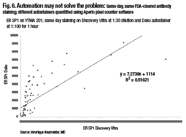
Fig6

“When running these in a quantitative fashion and measuring them quantitatively, there are actually differences in the way these closed-box systems run. And so you, as the pathologist, have to be the one who makes sure your assays are correct, your thresholds are correct, and your measurements are accurate.”

The way to do that, he believes, is to use standardization or index arrays. An index array of HER2 that his laboratory developed has 3+ amplified, 2+ amplified, not amplified, and so on from 80 cases in the lab’s archive, shown stained with immunofluorescence and quantitative and DAB stain. “It was only with this standardization array, run every time we ran our stainer, that we were able to draw the conclusions in the previous study about extracellular versus cytoplasmic domain.”

Companies have realized the importance of this, and specifically companies like NantOmics (formerly OncoPlexDx) have realized they can exactly quantitate the amount of tissue on a slide using a specialized mass spectrometry method, he says. “They can actually give you amol/µg of total protein.”

He and colleagues are working with NantOmics now to try to convert from amols to protein to average quantitative fluorescent scores to help build these standards and make standard arrays more accurate. “This is still a work in progress, but I believe this is ultimately the kind of accuracy that can standardize all of our labs. We have shown that the quantitative fluorescence system is truly linear and quantitative for EGFR measurements when using mass spectrometry as a gold standard.” They are preparing to submit a manuscript with this data.

In the interim, Dr. Rimm’s laboratory has begun working also with Horizon Diagnostics, employing Horizon’s experimental 15-spot positive-control array. “When you use this array and quantitate it with quantitative fluorescence, you get a very interesting profile. If a cut point is set at one point, you would see three clearly positive cells or spots and 12 clearly negative spots with two different antibodies. But is that the threshold?”

“In fact, using a little higher score and a very quantitative test, you might find that the threshold may, in fact, be a little bit lower than that.” It turns out that only three of these 12 spots are true negatives. The others at least have some level of RNA, and some have a lot. “So how do we handle these? And are these behaving the same way with multiple antibodies?” Parallel results, finding nearly the same threshold case, have been found using SP142 from Ventana, E1L3N from Cell Signaling, and SP263 from Ventana.

Studies to address those issues are still in the early stage, he says. He cautions that there is variance in these assays, and more work is being done to reproduce the data. “But I think the important point is that, using these kinds of arrays, you can definitively determine whether your lab has the same cut point as every other lab. And were we to quantitate this with mass spectrometry, we would know exactly the break point for use in the future.”

Dr. Rimm’s laboratory has also built its own PD-L1 index tissue microarray with a number of its own tumor slides ranging from very low to very high expressors, a series of cell lines, and including some placenta-positive controls on normal tumor. He has found that generating an index array has advantages, and he encourages other laboratories to prepare their own index arrays to increase the accuracy and reproducibility of their laboratory-developed tests. “You can produce these in your own lab so that you can be sure you can standardize your tests run in your clinical lab from day to day and week to week as part of an LDT.”

“If we think about it, there really are no clinical antibodies today that are truly quantitative,” Dr. Rimm says. “And when there are, new protocols will be required, but I believe those protocols are now in existence. We just await the clinical trials that require truly quantitative protein measurement or in situ proteomics.”

In that process of moving toward in situ proteomics, suggests web-inar co-presenter Clive Taylor, MD, DPhil, professor of pathology in the Keck School of Medicine at the University of Southern California, FDA approval, per se, will not solve any of the problems discussed in the webinar. (See the January 2016 issue for the full report of Dr. Taylor’s presentation.) “I think what the FDA approval will do is demand that we find solutions to these problems ourselves. The FDA’s attitude is, to a large degree, dependent on the claim. So if we just use immunohistochemistry as a simple stain, then the FDA classes that as sort of class I, level 1. And we can do that [IHC stain] without having to get preapproval by the FDA.

“On the other hand, if we take something like the well-established HercepTest, where based on the result of that test alone, it’s decided whether or not the patient gets treatment, treatment that’s very expensive and treatment that has benefits and…side effects. That claim is, in fact, a very high-level claim. And for that, the FDA is demanding high-level data, which I think is entirely appropriate,” Dr. Taylor says.

Most of these upcoming companion diagnostics, if not all, he says, will be regarded by the FDA as class III, high level or high complexity. They will require a premarket approval study in conjunction with a clinical trial. And the FDA will demand high standards of control and performance, eventually. “There are not many labs that can produce those high standards as in-house or lab-developed tests today. And even the companies currently in trials are not producing the improved performance level for these tests that we are talking about today, as being required for high-quality quantitative and reproducible companion diagnostics. Eventually, I am convinced we will have to do that. It’s just that it will take time to get there.”

The FDA can only approve what is brought to it, Dr. Rimm points out. And so a true, fully quantitative IHC-based assay has presumably never been submitted, or at least never been approved by the FDA. “What we’re seeing instead are the assays that the FDA has approved, which are well defined and rigorously submitted. However, the result is a closed system that we use, which may or may not accurately measure PD-L1 on the slide, depending upon preanalytic variables and individual laboratories’ methods.”

“So questions keep popping up. And I can only say that we, as pathologists, have the final responsibility to our patients. And while it may not be recommended and it may change in the future, right now lab-derived tests or LDTs may be more accurate than FDA-approved platforms.”

“If you think about it, in molecular diagnostics where I’m familiar with EFGR and BRAF and KRAS tests, in that testing setting, less than 25 percent of the labs that do that test actually use the FDA-approved test,” Dr. Rimm says. “The remainder of the labs do their own LDTs, including our labs here at Yale.”

It wouldn’t surprise him if the same thing happens for PD-L1. “I’m aware of at least two labs—and we probably will be the third—that devise our own LDT for PD-L1 testing using the standards I’ve discussed, using array-type controls to be sure that our levels are correct, and then using a scoring system that we derived.”

“We aren’t really in a position to know at the time that we receive a piece of lung cancer tissue whether the oncologist is going to use pembrolizumab, which requires a companion diagnostic, or nivolumab, or the other drugs, which may or may not require a companion diagnostic. So in that sense, we’re almost bound to use an LDT,” Dr. Rimm says, since his lab can’t actually run four different potentially incongruent, though FDA-approved, tests for PD-L1.

Until a truly quantitative approach is developed and submitted to the FDA and approved, Dr. Taylor believes we won’t see things changing. “The algorithms that currently are approved have been approved on the basis that they can produce a similar result to a consensus group of pathologists. So they’re only as good as the pathologists.”

“As Dr. Rimm has discussed, I actually believe we can get a much better result than the pathologists can get with their naked eye. We have to get away from comparing it to what we currently can do and start to try to construct a proper test, just like we did in the clinical lab 30 years ago when we automated the clinical lab,” Dr. Taylor says. “We need to automate anatomic pathology, including the sample preparation, the assay process, and the reading, all three together in a closed system. And we’re nibbling away at the edges of it. We’ll get there, but it’ll take some time.”

Dr. Rimm is skeptical that the diagnostics field has learned any lessons from HercepTest and the companion diagnostics world of almost 20 years ago. “The submissions to the FDA for PD-L1 look very similar to what was submitted in 1998 for the HercepTest, the companion diagnostic test for trastuzumab [Herceptin]. And that’s disappointing. I think that is 20-year-old technology and we can do better. But even if we want to use the 20- or 40-year-old DAB-based technology, we should still be standardizing it and having a mechanism for standardization and having defined thresholds.”

As future FDA submissions come in, Dr. Rimm hopes that “even if they’re not quantitated, they can be standardized as to where the thresholds occur, so that we can be sure we deliver the best possible care to patients. And in the interim, I think we, as pathologists, will have to do that standardization with an LDT to be sure we’re giving our best results.”

Dr. Taylor warns that there is only a limited number of labs in the country and in the world that will be able to produce these LDTs, because of the complexity. “The FDA has already said in a position paper that it believes it may have to regulate LDTs to some extent. And what that will mean is that in the validation process, your own LDT will start to approach what is required for an FDA-approved test. And most labs are in no position to be able to do that.”

“So I think we’re going to come to a blending here, all forced by companion diagnostics. This is in situ proteomics,” Dr. Taylor says. “It’s a new test, essentially. It’s not straightforward immunohistochemistry, but a new test. And I think the fluorescence approach that Dr. Rimm has used has a lot of advantages in relating signal to target in terms of figure out what the best test is and stop comparing it to the pathologists. We should compare it to the best assay we can produce.”

With respect to the PD-L1 problem, Dr. Rimm notes, “I would point out that there is a so-called ‘Blueprint’ for comparison of the different antibodies and the different FDA assays, or potentially FDA-submitted tests anyway, to see how equivalent they are.” Similarly, he adds, the National Comprehensive Cancer Network recently issued a press release describing a multi-institutional study to assess the FDA-approved assay but also including an LDT (the Cell Signaling antibody E1L3N using the Leica Bond staining platform).

He points to a newly published study by his group (McLaughlin J, et al. JAMA Oncol. 2016;2[1]:46–54), finding that objective determination of PD-L1 protein levels in non-small cell lung cancer reveals heterogeneity within tumors and prominent interassay variability or discordance. The authors concluded that future studies measuring PD-L1 quantitatively in patients treated with anti-PD-1 and anti PD-L1 therapies may better address the prognostic or predictive value of these biomarkers. With future rigorous studies, including tissues with known responses to anti-PD-1 and anti-PD-L1 therapies, researchers could determine the optimal assay, PD-L1 antibody, and the best cut point for PD-L1 positivity.

Other work that will probably come out in mid-2016 from Dr. Rimm’s group has shown that expression of PD-L1 is largely bimodal, he says. “That is, there’s a group of patients that express a lot, and then there’s another group of patients that expresses a little or none.”

So time will tell how PD-L1 will be scored. “But if you look at the data from the Merck study and their cut point of greater than 50 percent, or even the cut point from the AstraZeneca studies of greater than 25 percent, you’re really dichotomizing the population into patients who are truly PD-LI positive from patients who are negative or almost negative.”

“Of course, we don’t want to miss patients in that negative to almost-negative group who will respond,” Dr. Rimm says. “On the other hand, we probably will have fairly good specificity and sensitivity with the assay defined by Merck and Dako with 22C3 as was recently published” (Robert C, et al. N Engl J Med. 2015;372[26]:2521–2532).

Many difficulties lie ahead, as researchers try to weigh the merits of different drugs with different approved tests on different platforms, involving different antibodies, Dr. Taylor says. “Does the lab try to set up four different PD-L1s, and if we only have one platform and not another, what do we do about that?” He thinks the tests may often be sent out to larger reference labs or academic centers as a result.

Dr. Rimm confirms that his own lab’s LDT—although literally thousands of PD-L1 tests have been conducted using it—is not yet up and running in the Yale CLIA laboratory, and in the meantime the IHC slides are being sent out to a commercial vendor.

Eventually, Dr. Taylor believes, the pressure of these dilemmas will lead the diagnostics field to develop an immunoassay on tissue sections. “We’ve never been forced to do that before, but once we are, that will produce a huge change in diagnostic capability and research capability.”

Anti–PD-1/PD-L1 therapy of human cancer: past, present, and future

Lieping Chen and  http://www.jci.org/articles/view/80011

The cDNA of programmed cell death 1 (PD-1) was isolated in 1992 from a murine T cell hybridoma and a hematopoietic progenitor cell line undergoing apoptosis (1). Genetic ablation studies showed that deficiencies in PD-1 resulted in different autoimmune phenotypes in various mouse strains (2, 3). PD-1–deficient allogeneic T cells with transgenic T cell receptors exhibited augmented responses to alloantigens, indicating that the PD-1 on T cells plays a negative regulatory role in response to antigen (2).

Several studies contributed to the discovery of the molecules that interact with PD-1. In 1999, the B7 homolog 1 (B7-H1, also called programmed death ligand-1 [PD-L1]) was identified independently from PD-1 using molecular cloning and human expressed-sequence tag database searches based on its homology with B7 family molecules, and it was shown that PD-L1 acts as an inhibitor of human T cell responses in vitro (4). These two independent lines of study merged one year later when Freeman, Wood, and Honjo’s laboratories showed that PD-L1 is a binding and functional partner of PD-1 (5). Next, it was determined that PD-L1–deficient mice (Pdl1 KO mice) were prone to autoimmune diseases, although this strain of mice did not spontaneously develop such diseases (6). It became clear later that the PD-L1/PD-1 interaction plays a dominant role in the suppression of T cell responses in vivo, especially in the tumor microenvironment (7, 8).

In addition to PD-L1, another PD-1 ligand called B7-DC (also known as PD-L2) was also identified by the laboratories of Pardoll (9) and Freeman (10). This PD-1 ligand was found to be selectively expressed on DCs and delivered its suppressive signal by binding PD-1. Mutagenesis studies of PD-L1 and PD-L2 molecules guided by molecular modeling revealed that both PD-L1 and PD-L2 could interact with other molecules in addition to PD-1 and suggested that these interactions had distinct functions (11). The functional predictions from these mutagenesis studies were later confirmed when PD-L1 was found to interact with CD80 on activated T cells to mediate an inhibitory signal (12, 13). This finding came as a surprise because CD80 had been previously identified as a functional ligand for CD28 and cytotoxic T lymphocyte antigen-4 (CTLA-4) (14, 15). PD-L2 was also found to interact with repulsive guidance molecule family member b (RGMb), a molecule that is highly enriched in lung macrophages and may be required for induction of respiratory tolerance (16). With at least five interacting molecules in the PD-1/PD-L1 pathway (referred to as the PD pathway) (Figure 1), further studies will be required to understand the relative contributions of these molecules during activation or suppression of T cells.
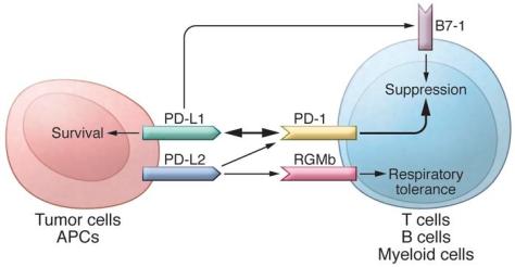
The PD pathway. The PD pathway has at least 5 interacting molecules. PD-...

http://dm5migu4zj3pb.cloudfront.net/manuscripts/80000/80011/medium/JCI80011.f1.jpg

The PD pathway.

The PD pathway has at least 5 interacting molecules. PD-L1 and PD-L2, with different expression patterns, were identified as ligands of PD-1, and the interaction of PD-L1 or PD-L2 with PD-1 may induce T cell suppression. PD-L1 was found to interact with B7-1 (CD80) on activated T cells and inhibit T cell activity. PD-L2 has a second receptor, RGMb; initially, this interaction activates T cells, but it subsequently induces respiratory tolerance. PD-L1 on tumor cells can also act as a receptor, and the signal delivered from PD-1 on T cells can protect tumor cells from cytotoxic lysis.

The discovery of the PD pathway did not automatically justify its application to cancer therapy, especially after the initial PD-1–deficient mouse studies, which suggested that PD-1 deficiency increases the incidence of autoimmune diseases (2, 3). In our initial work to characterize PD-L1 and its function, PDL1 mRNA was found to be broadly expressed in various tissues (17). However, normal human tissues seldom express PD-L1 protein on their cell surface, with the exception of tonsil (17), placenta (18), and a small fraction of macrophage-like cells in lung and liver (17), suggesting that, under normal physiological conditions, PDL1 mRNA is under tight posttranscriptional regulation. In sharp contrast, PD-L1 protein is abundantly expressed on the cell surface in various human cancers, as indicated by immunohistochemistry in frozen human tumor sections. Additionally, the pattern of PD-L1 expression was found to be focal rather than diffuse in most human cancers (17). In fact, the majority of in vitro–cultured tumor lines of both human and mouse origin are PD-L1–negative on the cell surface, despite overwhelming PD-L1 signal in specimens that are freshly isolated from patients with cancer (17, 19). This discrepancy was explained by the finding that IFN-γ upregulates PD-L1 on the cell surface of normal tissues and in various tumor lines (7, 17, 19). It was widely thought that IFN-γ typically promotes, rather than suppresses, T cell responses by stimulating antigen processing and presentation machinery (20, 21); therefore, the role of IFN-γ in downregulating immune responses in the tumor microenvironment via induction of PD-L1 was not well accepted until more recently. This finding is vital to our current understanding of the unique immunology that takes place in the tumor microenvironment and provided an important clue that led to the “adaptive resistance” hypothesis (see below) that explains this pathway’s mechanism of action to evade tumor immunity.

Due to the lack of cell surface expression of PD-L1 on most cultured tumor lines, it is necessary to reexpress PD-L1 on the surface using transfection to recapitulate the effects of cell surface PD-L1 in human cancers and to create models to study how tumor-associated PD-L1 interacts with immune cells. We now know that cancer cells and other cells in the tumor microenvironment can upregulate the expression of PD-L1 after encountering T cells, mostly via IFN-γ, which may make the transfection-mediated expression of PD-L1 unnecessary in some tumor models. Nevertheless, our results demonstrated that PD-L1+ human tumor cells could eliminate activated effector T cells (Teffs) via apoptosis in coculture systems, and this effect could be blocked by inclusion of an anti-human PD-L1 mAb (clone 2H1). Next, we generated a hamster mAb (clone 10B5) against mouse PD-L1 to block its interaction with T cells and test its role in tumor immunity in vitro and in vivo. We demonstrated that progressive growth of PD-L1+ murine P815 tumors in syngeneic mice could be suppressed using anti–PD-L1 mAb (17). Altogether, these studies represented the initial attempt at using mAb to block the PD pathway as an approach for cancer therapy. These proof-of-concept studies (17) were confirmed by several subsequent studies. A study from Nagahiro Minato’s laboratory showed that the J558L mouse myeloma line constitutively expressed high levels of cell surface PD-L1 and the growth of these cells in syngeneic BALB/c mice could be partially suppressed by administering anti–PD-L1 mAb (22). Our laboratory showed that regression of progressively growing squamous cell carcinomas in syngeneic mice could also be suppressed using a combination of adoptively transferred tumor-draining lymphocytes and anti–PD-L1 mAb (23). Furthermore, the Zou laboratory demonstrated that ovarian cancer–infiltrating human T cells could be activated in vitro using DCs, which showed enhanced activity in the presence of anti–PD-L1 mAb; upon transfer, these cells could eliminate established human ovarian cancers in immune-deficient mice (24). These early studies established the concept that the PD pathway could be used by tumors to escape immune attack in the tumor microenvironment. More importantly, these studies built a solid foundation for the development of anti-PD therapy for the treatment of human cancers.  …..

Anti-PD therapy has taken center stage in immunotherapies for human cancer, especially for solid tumors. This therapy is distinct from the prior immune therapeutic agents, which primarily boost systemic immune responses or generate de novo immunity against cancer; instead, anti-PD therapy modulates immune responses at the tumor site, targets tumor-induced immune defects, and repairs ongoing immune responses. While the clinical success of anti-PD therapy for the treatment of a variety of human cancers has validated this approach, we are still learning from this pathway and the associated immune responses, which will aid in the discovery and design of new clinically applicable approaches in cancer immunotherapy.


PD-1 Pathway Inhibitors: Changing the Landscape of Cancer Immunotherapy

Dawn E. Dolan, PharmD, and Shilpa Gupta, MD

Background: Immunotherapeutic approaches to treating cancer have been evaluated during the last few decades with limited success. An understanding of the checkpoint signaling pathway involving the programmed death 1 (PD-1) receptor and its ligands (PD-L1/2) has clarified the role of these approaches in tumor-induced immune suppression and has been a critical advancement in immunotherapeutic drug development. Methods: A comprehensive literature review was performed to identify the available data on checkpoint inhibitors, with a focus on anti–PD-1 and anti–PD-L1 agents being tested in oncology. The search included Medline, PubMed, the ClinicalTrials.gov registry, and abstracts from the American Society of Clinical Oncology meetings through April 2014. The effectiveness and safety of the available anti–PD-1 and anti–PD-L1 drugs are reviewed. Results: Tumors that express PD-L1 can often be aggressive and carry a poor prognosis. The anti–PD-1 and anti–PD-L1 agents have a good safety profile and have resulted in durable responses in a variety of cancers, including melanoma, kidney cancer, and lung cancer, even after stopping treatment. The scope of these agents is being evaluated in various other solid tumors and hematological malignancies, alone or in combination with other therapies, including other checkpoint inhibitors and targeted therapies, as well as cytotoxic chemotherapy. Conclusions: The PD-1/PD-L1 pathway in cancer is implicated in tumors escaping immune destruction and is a promising therapeutic target. The development of anti–PD-1 and anti–PD-L1 agents marks a new era in the treatment of cancer with immunotherapies. Early clinical experience has shown encouraging activity of these agents in a variety of tumors, and further results are eagerly awaited from completed and ongoing studies.

……

Role of PD-1/PD-L1 Pathway PD-1 is an immunoinhibitory receptor that belongs to the CD28 family and is expressed on T cells, B cells, monocytes, natural killer cells, and many tumor-infiltrating lymphocytes (TILs)10; it has 2 ligands that have been described (PD-L1 [B7H1] and PD-L2 [B7-DC]).11 Although PD-L1 is expressed on resting T cells, B cells, dendritic cells, macrophages, vascular endothelial cells, and pancreatic islet cells, PD-L2 expression is seen on macrophages and dendritic cells alone.10 Certain tumors have a higher expression of PD-L1.12 PD-L1 and L2 inhibit T-cell proliferation, cytokine production, and cell adhesion.13 PD-L2 controls immune T-cell activation in lymphoid organs, whereas PD-L1 appears to dampen T-cell function in peripheral tissues.14 PD-1 induction on activated T cells occurs in response to PD-L1 or L2 engagement and limits effector T-cell activity in peripheral organs and tissues during inflammation, thus preventing autoimmunity. This is a crucial step to protect against tissue damage when the immune system is activated in response to infection.15-17 Blocking this pathway in cancer can augment the antitumor immune response.18 Like the CTLA-4, the PD-1 pathway down-modulates Tcell responses by regulating overlapping signaling proteins that are part of the immune checkpoint pathway; however, they function slightly differently.14,16 Although the CTLA-4 focuses on regulating the activation of T cells, PD-1 regulates effector T-cell activity in peripheral tissues in response to infection or tumor progression.16 High levels of CTLA-4 and PD-1 are expressed on regulatory T cells and these regulatory T cells and have been shown to have immune inhibitory activity; thus, they are important for maintaining self-tolerance.16 The role of the PD-1 pathway in the interaction of tumor cells with the host immune response and the PD-L1 tumor cell expression may provide the basis for enhancing immune response through a blockade of this pathway.16 Drugs targeting the PD-1 pathway may provide antitumor immunity, especially in PD-L1 positive tumors. Various cancers, such as melanoma, hepatocellular carcinoma, glioblastoma, lung, kidney, breast, ovarian, pancreatic, and esophageal cancers, as well as hematological malignancies, have positive PD-L1 expression, and this expression has been correlated with poor prognosis.8,19 Melanoma and kidney cancer are prototypes of immunogenic tumors that have historically been known to respond to immunotherapeutic approaches with interferon alfa and interleukin 2. The CTLA-4 antibody ipilimumab is approved by the US Food and Drug Administration for use in melanoma. Clinical activity of drugs blocking the PD-1/PD-L1 pathway has been demonstrated in melanoma and kidney cancer.20-24 In patients with kidney cancer, tumor, TIL-associated PD-L1 expression, or both were associated with a 4.5-fold increased risk of mortality and lower cancer-specific survival rate, even after adjusting for stage, grade, and performance status.18,19,25,26 A correlation between PD-L1 expression and tumor growth has been described in patients with melanoma, providing the rationale for using drugs that block the PD-1/PD-L1 pathway.19,27 Historically, immunotherapy has been ineffective in cases of non–small-cell lung cancer (NSCLC), which has been thought to be a type of nonimmunogenic cancer; nevertheless, lung cancer can evade the immune system through various complex mechanisms.28 In patients with advanced lung cancer, the peripheral and tumor lymphocyte counts are decreased, while levels of regulatory T cells (CD4+), which help suppress tumor immune surveillance, have been found at higher levels.29-32 Immune checkpoint pathways involving the CTLA-4 or the PD-1/PD-L1 are involved in regulating T-cell responses, providing the rationale for blocking this pathway in NSCLC with antibodies against CTLA-4 and the PD-1/PD-L1 pathway.32 Triple negative breast cancer (TNBC) is an aggressive subset of breast cancer with limited treatment options. PD-L1 expression has been reported in patients with TNBC. When PD-L1 expression was evaluated in TILs, it correlated with higher grade and larger-sized tumors.33 Tumor PD-L1 expression also correlates with the infiltration of T-regulatory cells in TNBC, findings that suggest the role of PD-L1–expressing tumors and the PD-1/PD-L1–expressing TILs in regulating immune response in TNBC.34

…….

Preclinical evidence exists for the complementary roles of CTLA-4 and PD-1 in regulating adaptive immunity, and this provides rationale for combining drugs targeting these pathways.44-46 Paradoxically and originally believed to be immunosuppressive, new data allow us to recognize that cytotoxic agents can antagonize immunosuppression in the tumor microenvironment, thus promoting immunity based on the concept that tumor cells die in multiple ways and that some forms of apoptosis may lead to an enhanced immune response.8,15 For example, nivolumab was combined with ipilimumab in a phase 1 trial of patients with advanced melanoma.46 The combination had a manageable safety profile and produced clinical activity in the majority of patients, with rapid and deep tumor regression seen in a large proportion of patients. Based on the results of this study, a phase 3 study is being undertaken to evaluate whether this combination is better than nivolumab alone in melanoma (NCT01844505). Several other early-phase studies are underway to explore combinations of various anti–PD-1/PD-L1 drugs with other therapies across a variety of tumor types (see Tables 1 and 2), possibly paving the way for future combination studies.


Development of PD-1/PD-L1 Pathway in Tumor Immune Microenvironment and Treatment for Non-Small Cell Lung Cancer

Jiabei He, Ying Hu, Mingming Hu & Baolan Li

Lung cancer is currently the leading cause of cancer-related death in worldwide, non-small cell lung cancer (NSCLC) accounts for about 85% of all lung cancers. Surgery, platinum-based chemotherapy, molecular targeted agents and radiotherapy are the main treatment of NSCLC. With the strategies of treatment constantly improving, the prognosis of NSCLC patients is not as good as before, new sort of treatments are needed to be exploited. Programmed death 1 (PD-1) and its ligand PD-L1 play a key role in tumor immune escape and the formation of tumor microenvironment, closely related with tumor generation and development. Blockading the PD-1/PD-L1 pathway could reverse the tumor microenvironment and enhance the endogenous antitumor immune responses. Utilizing the PD-1 and/or PD-L1 inhibitors has shown benefits in clinical trials of NSCLC. In this review, we discuss the basic principle of PD-1/PD-L1 pathway and its role in the tumorigenesis and development of NSCLC. The clinical development of PD-1/PD-L1 pathway inhibitors and the main problems in the present studies and the research direction in the future will also be discussed.

Lung cancer is currently the leading cause of cancer-related death in the worldwide. In China, the incidence and mortality of lung cancer is 5.357/10000, 4.557/10000 respectively, with nearly 600,000 new cases every year1. Non-small cell lung cancer (NSCLC) accounts for about 85% of all lung cancers, the early symptoms of patients with NSCLC are not very obvious, especially the peripheral lung cancer. Though the development of clinic diagnostic techniques, the majority of patients with NSCLC have been at advanced stage already as they are diagnosed. Surgery is the standard treatment in the early stages of NSCLC, for the advanced NSCLC, the first-line therapy is platinum-based chemotherapy. In recent years, patients with specific mutations may effectively be treated with molecular targeted agents initially. The prognosis of NSCLC patients is still not optimistic even though the projects of chemotherapy as well as radiotherapy are continuously ameliorating and the launch of new molecular targeted agents is never suspended, the five-year survival rate of NSCLC patients is barely more than 15%2, the new treatment is needed to be opened up.

During the last few decades, significant efforts of the interaction between immune system and immunotherapy to NSCLC have been acquired. Recent data have indicated that the lack of immunologic control is recognized as a hallmark of cancer currently. Programmed death-1 (PD-1) and its ligand PD-L1 play a key role in tumor immune escape and the formation of tumor microenvironment, closely related with tumor generation and development. Blockading the PD-1/PD-L1 pathway could reverse the tumor microenvironment and enhance the endogenous antitumor immune responses.

In this review, we will discuss the PD-1/PD-L1 pathway from the following aspects: the basic principle of PD-1/PD-L1 pathway and its role in the tumorigenesis and development of NSCLC, the clinical development of several anti-PD-1 and anti-PD-L1 drugs, including efficacy, toxicity, and application as single agent, or in combination with other therapies, the main problems in the present studies and the research direction in the future.


Cancer as a chronic, polygene and often inflammation-provoking disease, the mechanism of its emergence and progression is very complicated. There are many factors which impacted the development of the disease, such as: environmental factors, living habits, genetic mutations, dysfunction of the immune system and so on. At present, increasing evidence has revealed that the development and progression of tumor are accompanied by the formation of special tumor immune microenvironment. Tumor cells can escape the immune surveillance and disrupt immune checkpoint of host in several methods, therefore, to avoid the elimination from the host immune system. Human cancers contain a number of genetic and epigenetic changes, which can produce neoantigens that are potentially recognizable by the immune system3, thus trigger the body’s T cells immune response. The T cells of immune system recognize cancer cells as abnormal primarily, generate a population of cytotoxic T lymphocytes (CTLs) that can traffic to and infiltrate cancers wherever they reside, and specifically bind to and then kill cancer cells. Effective protective immunity against cancer depends on the coordination of CTLs4. Under normal physiological conditions, there is a balance status in the immune checkpoint molecule which makes the immune response of T cells keep a proper intensity and scope in order to minimize the damage to the surrounding normal tissue and avoid autoimmune reaction. However, numerous pathways are utilized by cancers to up-regulate the negative signals through cell surface molecules, thus inhibit T-cell activation or induce apoptosis and promote the progression and metastasis of cancers5. Increasing experiments and clinical trails show that immunotherapeutic approaches utilizing antagonistic antibodies to block checkpoint pathways, can release cancer inhibition and facilitate antitumor activity, so as to achieve the purpose of treating cancer.

The present research of immune checkpoint molecules are mainly focus on cytotoxic T lymphocyte-associated antigen 4 (CLTA-4), Programmed death-1 (PD-1) and its ligands PD-L1 (B7H1) and PD-L2 (B7-DC). CTLA-4 regulates T cell activity in the early stage predominantly, and PD-1 mainly limits the activity of T-cell in the tumor microenvironment at later stage of tumor growth6. Utilizing the immune checkpoint blockers to block the interactions between PD-1 and its ligands has shown benefits in clinical trials, including the NSCLC patients. PD-1 and its ligands have been rapidly established as the currently most important breakthrough targets in the development of effective immunotherapy.

PD-1/PD-L1 pathway and its expression, regulation

PD-1 is a type 1 trans-membrane protein that encoded by the PDCD1 gene7. It is a member of the extended CD28/CTLA-4 immunoglobulin family and one of the most important inhibitory co-receptors expressed by T cells. The structure of the PD-1 includes an extracellular IgV domain, a hydrophobic trans-membrane region and an intracellular domain. The intracellular tail includes separate potential phosphorylation sites that are located in the immune receptor tyrosine-based inhibitory motif (ITIM) and in the immunoreceptor tyrosine-based switch motif (ITSM). Mutagenetic researches indicated that the activated ITSM is essential for the PD-1 inhibitory effect on T cells8. PD-1 is expressed on T cells, B cells, monocytes, natural killer cells, dendritic cells and many tumor-infiltrating lymphocytes (TILs)9. In addition, the research of Francisoet et al. showed that PD-1 was also expressed on regulatory T cells (Treg) and able to facilitate the proliferation of Treg and restrain immune response10.

PD-1 has two ligands: PD-L1 (also named B7-H1; CD274) and PD-L2 (B7-DC; CD273), that are both coinhibitory. PD-L1 is expressed on resting T cells, B cells, dendritic cells, macrophage, vascular endothelial cells and pancreatic islet cells. PD-L2 expression is seen on macrophages and dendritic cells alone and is far less prevalent than PD-L1 across tumor types. It shows much more restricted expression because of its more restricted tissue distribution. Differences in expression patterns suggest distinct functions in immune regulation across distinct cell types. The restricted expression of PD-L2, largely to antigen-presenting cells, is consistent with a role in regulating T-cell priming or polarization, whereas broad distribution of PD-L1 suggests a more general role in protecting peripheral tissues from excessive inflammation.

PD-L1 is expressed in various types of cancers, especially in NSCLC11,12, melanoma, renal cell carcinoma, gastric cancer, hepatocellular as well as cutaneous and various leukemias, multiple myeloma and so on13,14,15. It is present in the cytoplasm and plasma membrane of cancer cells, but not all cancers or all cells within a cancer express PD-L116,17. The expression of PD-L1 is induced by multiple proinflammatory molecules, including types I and II IFN-γ, TNF-α, LPS, GM-CSF and VEGF, as well as the cytokines IL-10 and IL-4, with IFN-γ being the most potent inducer18,19. IFN-γ and TNF-α are produced by activated type 1 T cells, and GM-CSF and VEGF are produced by a variety of cancer stromal cells, the tumor microenvironment upregulates PD-L1 expression, thereby, promotes immune suppression. This latter effect is called “adaptive immune resistance”, because the tumor protects itself by inducing PD-L1 in response to IFN-γ produced by activated T cells17. PD-L1 is regulated by oncogenes, also known as the inherent immune resistance. PD-L1 expression is suppressed by the tumor suppressor gene: PTEN (phosphatase and tension homolog deleted on chromosome ten) gene. Cancer cells frequently contain mutated PTEN, which can activate the S6K1 gene, thus results in PD-L1 mRNA to polysomes increase greatly20, hence increases the translation of PD-L1 mRNA and plasma membrane expression of PD-L1. Parsa et al.’s research also demonstrated that neuroglioma with PTEN gene deletion regulate PD-L1 expression at the translational level by activating the PI3K/AKT downstream mTOR-S6K1signal pathway and, hence increase the PD-L1 expression21. Micro-RNAs also translationally regulate PD-L1 expression. MiRNA-513 is complementary to the 3′ untranslated region of PD-L1 and prevents PD-L1 mRNA translation22. In addition, a later literature reported that in the model of melanoma, the up-regulation of PD-L1 is closely related to the CD8 T cell, independent of regulation by oncogenes13. Noteworthily, the PD-L1 can bind to T cell expressed CD80, and at this point CD80 is a receptor instead of ligand to transmit negative regulated signals23.


PD-1/PD-L1 mediate immune suppression by multiple mechanisms

Like the CTLA-4, the PD-1/PD-L1 pathway down-modulates T-cell response by regulating overlapping signal proteins in the immune checkpoint pathway. However, their functions are slightly different24. The CTLA-4 focuses on regulating the activation of T cells, while PD-1 regulates effector T-cell activity in peripheral tissues in response to infection or tumor progression25. Tregs that high-level expression of PD-1 have been shown to have immune inhibitory activity, thus, they are important for maintaining self-tolerance. In normal human bodies, this is a crucial step to protect against tissue damage when the immune system is activated in response to infection26. However, in response to immune attack, cancer cells overexpress PD-L1 and PD-L2. They bind to PD-1 receptor on T cells, inhibiting the activation of T-cells, thus suppressing T-cell attack and inducing tumor immune escape. Thus tumor cells effectively form a suitable tumor microenvironment and continue to proliferate27. PD-1/PD-L1 pathway regulates immune suppression by multiple mechanisms, specific performance of the following: Induce apoptosis of activated T cells: PD-1 reduces T cell survival by impacting apoptotic genes. During T cell activation, CD28 ligation sustains T cell survival by driving expression of the antiapoptotic gene Bcl-xL. PD-1 prevents Bcl-xL expression by inhibiting PI3K activation, which is essential for upregulation of Bcl-xL. Early studies demonstrated that PD-L1+ murine and human tumor cells induce apoptosis of activated T cells and that antibody blocking of PD-L1 can decrease the apoptosis of T cells and facilitate antitumor immunity28,16. Facilitate T cell anergy and exhaustion: A research shown that the occurrence of tumor is associated with chronic infection29. According to the study of chronic infection, PD-1 overexpressed on the function exhausted T cells, blocking the PD-1/PD-L1 pathway can restore the proliferation, secretion and cytotoxicity30. In addition, later research demonstrated that the exhaustion of TILs in the tumor microenvironment is closely related to the PD-L1 expression of tumor cells, myeloid cells derived from tumor31. Enhance the function of regulatory T cells: PD-L1 can promote the generation of induced Tregs by down-regulating the mTOR, AKT, S6 and the phosphorylation of ERK2 and increasing PTEN, thus restrain the activity of effector T-cell32. Blocking the PD-1/PD-L1 pathway can increase the function of effector CD8 T-cell and inhibt the function of Tregs, bone marrow derived inhibition cells, thus enhance the anti-tumor response. Inhibit the proliferation of T cells: PD-1 ligation also prevents phosphorylation of PKC-theta, which is essential for IL-2 production33, and arrests T cells in the G1 phase, blocking proliferation. PD-1 mediates this effect by activating Smad3, a factor that arrests cycling34. Restrain impaired T cell activation and IL-2 production: PD-1/PD-L1 blocks the downstream signaling events triggered by Ag/MHC engagement of the TCR and co-stimulation through CD28, resulting in impaired T cell activation and IL-2 production. Signaling through the TCR requires phosphorylation of the tyrosine kinase ZAP70. PD-1 engagement reduces the phosphorylation of ZAP70 and, hence, inhibits downstream signaling events. In addition, signaling through PD-1 also prevents the conversion of functional CD8+ T effector memory cells into CD8+ central memory cells35 and, thus, reduces long-term immune memory that might protect against future metastatic disease. PD-L1 also promotes tumor progression by reversing signaling through CD80 into T cells. CD80-PD-L1 interactions restrain self-reactive T cells in an autoimmune setting36, therefore, their inhibition may facilitate antitumor immunity.

Researches on the mechanism of PD-1/PD-L1 pathway mediating immune escape are still ongoing, especially the mechanism of PD-L2 is still unclear. These researches provide the theoretical basis and research direction for the further immunotherapy targets research.


Anti-PD-1 antibodies

Nivolumab

Nivolumab (BMS-936558, Brand name: Opdivo) is a human monoclonal IgG4 antibody that essentially lacks detectable antibody-dependent cellular cytotoxicity (ADCC). Inhibition by monoclonal antibody of PD-1 on CD8+ TILs within lung cancers can restore cytokine secretion and T-cell proliferation48. Results of a larger phase I study in 296 patients (236 patients evaluated) reported that the objective response (complete or partial responses) of patients with NSCLC was 18%. A total of 65% of responders had durable responses lasting for more than 1 year. Stable disease lasting 24 weeks was seen in patients with NSCLC. PD-L1expression was tested in 42 patients: 9 of 25(36%) patients whose PD-L1 expression positive were objectively response to PD-1 blockade treatment, while the remaining 17 nonresponsive patients were negative45.

In another early phase I trial of nivolumab49, an objective response was observed in 22 patients (17%; 95% CI, 11%–25%) in a dose-expansion cohort of 129 previously treated patients with advanced NSCLC. Six additional patients who had an unconventional immune-related response were not included. Moreover, the median duration of response was exceptional for 17 months. Although the median PFS in the cohort was 2.3 months and the median overall survival was 9.9 months, it seemed clear that those who responded had sustained benefit. Specifically, the 2-year overall survival rate was 24%, and many remained in remission after completing 96 weeks of continuous therapy.

Single-agent trials of nivolumab are planning or ongoing on NSCLC (NCT01721759, NCT02066636). In addition, there are clinical randomized trials which focus on the comparison of nivolumab and plain-based combination chemotherapy (NCT02041533, NCT01673867). In March 4, 2015, nivolumab was approved by the US Food and Drug Administration for treatment of patients with metastatic NSCLC (squamous cell carcinoma), when progression of their diseases during or after chemotherapy with platinum-based drugs.

Pembrolizumab

Pembrolizumab (MK-3475) is a highly selective, humanized monoclonal antibody with activity against PD-1 that contains a mutation at C228P designed to prevent Fc-mediated ADCC. It is now in the clinical research phases for patients with advanced solid tumors. Its safety and efficacy were evaluated in a phase I clinical trial of KEYNOTE-001. The best response according of 38 cases of patients which initially accepted pembrolizumab 10 mg/kg q3wwas 21% (based on RECIST1.1 evaluation) and the median PFS of responder still has not reached until 62 weeks. The researchers also found that the antitumor activity of pembrolizumab was associated with the PD-L1expression44,50. The critical values of the expression of PD-L1 will be validated in 300 cases of patients which will soon been rolled into the study.

Clinical trial of pembrolizumab monotherapy is ongoing for patients with NSCLC (NCT01840579). Randomized trials comparing pembrolizumab to combination chemotherapy (NCT02142738) or single-agent docetaxel (NCT01905657) are ongoing in PD-L1 positive patients with NSCLC.

Pidilizumab (CT-011)

Pidilizumab is a humanized IgG-1K recombinant anti-PD-1 monoclonal antibody that has demonstrated antitumor activity in mouse cancer models. In a first-in-human phase I dose-escalation study in patients with only advanced hematologic cancers, there is no clinical trials of NSCLC presently51.


Anti-PD-L1 antibodies

Another therapeutic method based on the PD-1/PD-L1 pathway is by specific binding between antibody and PD-L1, thus preventing its activity. It was speculated that utilizing PD-L1 as therapeutic target maybe accompanied by less toxicity in part by modulating the immune response selectively in the tumor microenvironment. However, since PD-L2 expressed by tumor cells or some other tumor-associated molecules may play a role in mediating PD-1-expressing lymphocytes, it is conceivable that the magnitude of the anti-tumor immune response could also be blunted.

BMS-936559

BMS-936559/MDX1105 is a fully humanized, high affinity, IgG4 monoclonal antibody that react specifically with PD-L1, thus inhibiting the binding of PD-L1 and PD-1, CD80 (which binds not only PD-L1 but also CTLA-4 and CD28). Initial results from a multicenter and dose-escalation phase I trial of 207 patients(including 75 cases of patients with NSCLC) showed durable tumor regression (objective response rate of 6%–17%) and prolonged stabilization of disease (12%–41% at 24weeks) in patients with advanced cancers, including NSCLC, melanoma and kidney cancer. In patients with NSCLC, there were five objective responses (in 4 patients with the nonsquamous subtype and 1 with the squamous subtype) at doses of 3 mg/kg and 10 mg/kg, with response rates of 8% and 16%, respectively. Six additional patients with NSCLC had stable disease lasting at least 24 weeks52.

MPDL3280A

MPDL3280A is a human IgG1 antibody that targets PD-L1. Its Fc component has been engineered to not activate antibody-dependent cell cytotoxicity. In a recently reported phase I study, a 21% response rate was noted in patients with metastatic melanoma, RCC or NSCLC53, including several patients who demonstrated shrinkage of tumor within a few days of initiating treatment.

Fifty-two patients were enrolled in an expansion cohort of the phase I trial of MPDL3280A, 62% of them were heavily pretreated NSCLC (≥3 lines of systemic therapy) and the ORR was 22%54. Analysis of biomarker data from archival tumor samples demonstrated a correlation between PD-L1 status and response and lack of progressive disease55.

MEDI4736

MEDI4736 is a human IgG1 antibody that binds specifically to PD-L1, thus preventing PD-L1 binding to PD-1 and CD80. Interim results from a phase I trial reported no colitis or pneumonitis of any grade, with several durable remissions, including NSCLC patients56. An ongoing phase I dose-escalation study (NCT01693562) of MEDI-4736 in 26 patients, 4 partial responses (3 in patients with NSCLC and 1 with melanoma) were observed and 5 additional patients exhibited lesser degrees of tumor shrinkage. The disease control rate at 12 weeks was 46%57. Expansion cohorts was opened in Sep 2013, 10 mg/kg q2w dose. 151 patients was enrolled so far in the expansion cohorts, tumor shrinkage was reported as early as the first assessment at 6 weeks and among the 13 patients with NSCLC, responses were sustained at 10 or more to 14.9 or more months58. In the NSCLC expansion cohort, the response rate was 16% in 58 evaluable patients and the disease control rate at 12 weeks was 35% with responses seen in all histologic subtypes as well as in a smaller proportion of PD-L1- tumors.

On the basis of the favorable toxicity profile and promising activity in a heavily pretreated NSCLC population, a global Phase III placebo controlled trial using the 10 mg/kg biweekly dose has been launched in Stage III patients who have not progressed following chemo-radiation (NCT02125461). The primary outcome measures are overall survival and progression-free survival.

AMP-224

AMP-224 was a B7-DC-Fc fusion protein which can block the PD-1 receptor competitively59. Some NSCLC patients were included in a first-in-man phase I trial of this fusion protein drug. A dose-dependent reduction in PD-1-high TILs was observed at 4 hours and 2 weeks after drug administration60.


A variety of approaches for combining PD-1/PD-L1 pathway inhibitors with other therapeutic methods have been explored over the past few years in an effort to offer more feasible therapeutic options for clinic to improve treatment outcomes. Approaches have included combinations with other immune checkpoint inhibitors, immunostimulatory cytokines (e.g. IFN-y) cytotoxic chemotherapy, platinum-based chemotherapy, radiotherapy, anti-angiogenic inhibitors, tumor vaccine and small-molecule molecularly targeted therapies many with promising results61,62. Studies indicated that PD-1/PD-L1 pathway inhibitors were most effective when combined with treatments that activating the immune system63.

Preclinical evidence exists for the complementary roles of CTLA-4 and PD-1 in regulating adaptive immunity, and this provides rationale for combining drugs targeting these pathways. In a Phase I study in 46 chemotherapy-naive patients with NSCLC, four cohorts of patients received ipilimumab (3 mg/kg) plus nivolumab for four cycles followed by nivolumab 3 mg/kg intravenously every 2 weeks. The ORR was 22% and did not correlate with PD-L1 status64.

In another Phase I study, 56 patients with advanced NSCLC were assigned based on histology to four cohorts to receive nivolumab (5–10 mg/kg) intravenously every 3 weeks plus one of four concurrent standard “platinum doublet” chemotherapy regimens. No dose de-escalation was required for dose-limiting toxicity. The ORR was 33–50% across arms and the 1-year OS rates were promising at 59–87%65.

…..

The research of cancer immunotherapy provides a new wide space for cancer treatment (including NSCLC), and compared with other therapeutic method, immunotherapy has its unique advantages, such as: relative safety, effectivity, less and low grade side effect and so on. Especially with the discovery and continued in-depth study of PD-1/PD-L1 pathway in the immune regulation mechanism, many significative conclusions were reported. Data from many clinical trails suggest that some patients with NSCLC have been benefited from the drugs of anti-PD-1 and anti-PD-L1 already. However, summarized what have been discussed above, only a small fraction of patients benefit from PD-1 or PD-L1 inhibitors treatment. But with the continuous studies on biomarker and combined treatment in PD-1/PD-L1 pathway, new research progress will be acquired as well. We will make significant progress on treatment and in control of NSCLC.


Prospects for Targeting PD-1 and PD-L1 in Various Tumor Types     

Table 1: Selected Anti–PD-1 and Anti–PD-L1 Antibodies
Table 2: Selected Adverse Events
Table 3: Selected Clinical Trials for Metastatic Melanoma
Table: 4 Selected Trials for Metastatic Renal Cell Carcinoma
Table 5: Selected Trials for Non–Small-Cell Lung Cancer (NSCLC )
Table 6: Selected Trials for Other Tumor Types

Immune checkpoints, such as programmed death ligand 1 (PD-L1) or its receptor, programmed death 1 (PD-1), appear to be Achilles’ heels for multiple tumor types. PD-L1 not only provides immune escape for tumor cells but also turns on the apoptosis switch on activated T cells. Therapies that block this interaction have demonstrated promising clinical activity in several tumor types. In this review, we will discuss the current status of several anti–PD-1 and anti–PD-L1 antibodies in clinical development and their direction for the future.

Several PD-1 and PD-L1 antibodies are in clinical development (Table 1). Overall, they are very well tolerated; most did not reach dose-limiting toxicity in their phase I studies. As listed in Table 2, no clinically significant difference in adverse event profiles has been seen between anti–PD-1 and anti–PD-L1 antibodies. Slightly higher rates of infusion reactions (11%) were observed with BMS-936559 (anti–PD-L1) than with BMS-96558 (nivolumab). In an early stage of a nivolumab phase I study, there was concern about fatal pneumonitis.[7] It has been hypothesized that PD-1 interaction with PD-L2 expressed on the normal parenchymal cells of lung and kidney provides unique negative signaling that prevents autoimmunity.[8] Thus, anti–PD-1 antibody blockage of such an interaction may remove this inhibition, allowing autoimmune pneumonitis or nephritis. Anti–PD-L1 antibody, however, would theoretically leave PD-1–PD-L2 interaction intact, preventing the autoimmunity caused by PD-L2 blockade. With implementation of an algorithm to detect early signs of pneumonitis and other immune-related adverse events, many of these side effects have become manageable. However, it does require discerning clinical attention to detect potentially fatal side effects. In terms of antitumor activity, both anti–PD-1 and anti–PD-L1 antibodies have shown responses in overlapping multiple tumor types. Although limited to a fraction of patients, most responses, when observed, were rapid and durable.

– See more at: http://www.cancernetwork.com/oncology-journal/prospects-targeting-pd-1-and-pd-l1-various-tumor-types#sthash.an8uOYLi.dpuf


Immune Checkpoint Blockade in Cancer Therapy

Michael A. PostowMargaret K. Callahan and Jedd D. Wolchok
http://jco.ascopubs.org/content/early/2015/01/20/JCO.2014.59.4358.full
 http://dx.doi.org:/10.1200/JCO.2014.59.4358

Immunologic checkpoint blockade with antibodies that target cytotoxic T lymphocyte–associated antigen 4 (CTLA-4) and the programmed cell death protein 1 pathway (PD-1/PD-L1) have demonstrated promise in a variety of malignancies. Ipilimumab (CTLA-4) and pembrolizumab (PD-1) are approved by the US Food and Drug Administration for the treatment of advanced melanoma, and additional regulatory approvals are expected across the oncologic spectrum for a variety of other agents that target these pathways. Treatment with both CTLA-4 and PD-1/PD-L1 blockade is associated with a unique pattern of adverse events called immune-related adverse events, and occasionally, unusual kinetics of tumor response are seen. Combination approaches involving CTLA-4 and PD-1/PD-L1 blockade are being investigated to determine whether they enhance the efficacy of either approach alone. Principles learned during the development of CTLA-4 and PD-1/PD-L1 approaches will likely be used as new immunologic checkpoint blocking antibodies begin clinical investigation.

CTLA-4 was the first immune checkpoint receptor to be clinically targeted (Fig 1) Normally, after T-cell activation, CTLA-4 is upregulated on the plasma membrane where it functions to downregulate T-cell function through a variety of mechanisms, including preventing costimulation by outcompeting CD28 for its ligand, B7, and also by inducing T-cell cycle arrest.15 Through these mechanisms and others, CTLA-4 has an essential role in maintaining normal immunologic homeostasis, as evidenced by the fact that mice deficient in CTLA-4 die from fatal lymphoproliferation.6,7 Recognizing the role of CTLA-4 as a negative regulator of immunity, investigators led studies demonstrating that antibody blockade of CTLA-4 could result in antitumor immunity in preclinical models.8,9

Fig 1.

IMAGE SOURCE:

http://ascopubs.org/doi/figure/10.1200/JCO.2014.59.4358

Article SOURCE:

http://jco.ascopubs.org/content/early/2015/01/20/JCO.2014.59.4358/F1.medium.gif

Fig 1.

The cytotoxic T lymphocyte–associated antigen 4 (CTLA-4) immunologic checkpoint. T-cell activation requires antigen presentation in the context of a major histocompatibility complex (MHC) molecule in addition to the costimulatory signal achieved when B7 on an antigen-presenting cell (dendritic cell shown) interacts with CD28 on a T cell. Early after activation, to maintain immunologic homeostasis, CTLA-4 is translocated to the plasma membrane where it downregulates the function of T cells.

On the basis of this preclinical rationale, two antibodies targeting CTLA-4, ipilimumab (Bristol-Myers Squibb, Princeton, NJ) and tremelimumab (formerly Pfizer, currently MedImmune/AstraZeneca, Wilmington, DE), entered clinical development. Early reports of both agents showed durable clinical responses in some patients.1012Unfortunately, despite a proportion of patients experiencing a durable response, tremelimumab did not statistically significantly improve overall survival, which led to a negative phase III study comparing tremelimumab to dacarbazine/temozolomide in patients with advanced melanoma.13 It is possible that the lack of an overall survival benefit was a result of the crossover of patients treated with chemotherapy to an expanded access ipilimumab program or a result of the dosing or scheduling considerations of tremelimumab.

Ipilimumab, however, was successful in improving overall survival in two phase III studies involving patients with advanced melanoma.14,15 Although the median overall survival was only improved by several months in each of these studies, landmark survival after treatment initiation favored ipilimumab; in the first phase III study, 18% of patients were alive after 2 years compared with 5% of patients who received the control treatment of gp100 vaccination.14 More recently reported pooled data from clinical trials of ipilimumab confirm that approximately 20% of patients will have long-term survival of at least 3 years after ipilimumab therapy, with the longest reported survival reaching 10 years.1618

For patients with other malignancies, CTLA-4 antibody therapy has also shown some benefits. Ipilimumab, in combination with carboplatin and paclitaxel in a phased treatment schedule, showed improved progression-free survival compared with carboplatin and paclitaxel alone for patients with non–small-cell lung cancer.19Several patients with pancreatic cancer had declines in CA 19-9 when ipilimumab was given with GVAX (Aduro, Berkeley, CA),20and ipilimumab has also resulted in responses in patients with prostate cancer.21 Unfortunately, a phase III study in patients with castrate-resistant prostate cancer who experienced progression on docetaxel chemotherapy demonstrated that after radiotherapy, ipilimumab did not improve overall survival compared with placebo.22 Although this study is felt to have been a negative study, ipilimumab may have conferred a benefit to patients with favorable prognostic features, such as the absence of visceral metastases, but this requires further study. Another CTLA-4–blocking antibody, tremelimumab, has shown responses in patients with mesothelioma, and ongoing trials are under way.23

CTLA-4 blockade has also been administered together with other immunologic agents, such as the indoleamine 2,3-dioxygenase inhibitor INCB024360,106 the oncolytic virus talimogene laherparepvec,107 and granulocyte-macrophage colony-stimulating factor,108 with encouraging early results. We expect subsequent studies involving engineered T-cell–based therapies and checkpoint blockade.

Other promising data involve CTLA-4 combinations with PD-1 blockade. A phase I study of ipilimumab and nivolumab in patients with melanoma resulted in a high durable response rate and impressive overall survival compared with historical data.109,110Although the most recently reported grade 3 or 4 toxicity rate in patients with melanoma was 64%, which is higher than either ipilimumab or nivolumab individually,111 the vast majority of these irAEs were asymptomatic laboratory abnormalities of unclear clinical consequence. For example, elevations in amylase or lipase were reported in 21% of patients, none of whom met clinical criteria for a diagnosis of pancreatitis. The rate of grade 3 or 4 diarrhea was 7%, which is approximately similar to the rate of grade 3 or 4 diarrhea with ipilimumab monotherapy at 3 mg/kg. Whether ipilimumab and nivolumab improve overall survival compared with either nivolumab or ipilimumab alone remains the subject of an ongoing phase III randomized trial, and investigations of the combination of ipilimumab and nivolumab (and tremelimumab and MEDI4736) are ongoing in many other cancers.

Immunotherapy with checkpoint-blocking antibodies targeting CTLA-4 and PD-1/PD-L1 has improved the outlook for patients with a variety of malignancies. Despite the promise of this approach, many questions remain, such as the optimal management of irAEs and how best to evaluate combination approaches to determine whether they will increase the efficacy of CTLA-4 or PD-1/PD-L1 blockade alone. Themes from the experience with CTLA-4 and PD-1/PD-L1 will likely be relevant for investigations of novel immunologic checkpoints in the future.

This is a very important article, Dr. Larry.

It fits so beautiful with our work on Molecules in Development Table.

Thank you


This image depicts the process of metastasis in a mouse tumor, where tumor cells (green) have helped to reorganize the collagen into aligned fibers (blue) that provide the structural support for motility. This helps the tumor cells to enter blood vessels (red), ultimately leading to the formation of metastases in other organs.

http://news.mit.edu/sites/mit.edu.newsoffice/files/styles/news_article_image_top_slideshow/public/images/2016/MIT-Cancer-Migration-1_0.jpg?itok=aEOCRQpn

This image depicts the process of metastasis in a mouse tumor, where tumor cells (green) have helped to reorganize the collagen into aligned fibers (blue) that provide the structural support for motility. This helps the tumor cells to enter blood vessels (red), ultimately leading to the formation of metastases in other organs.  Image: Madeleine Oudin and Jeff Wyckoff

Paving the way for metastasis

Cancer cells remodel their environment to make it easier to reach nearby blood vessels.

Anne Trafton | MIT News Office     March 15, 2016


A new study from MIT reveals how cancer cells take some of their first steps away from their original tumor sites. This spread, known as metastasis, is responsible for 90 percent of cancer deaths.

Studying mice, the researchers found that cancer cells with a particular version of the Mena protein, called MenaINV (invasive), are able to remodel their environment to make it easier for them to migrate into blood vessels and spread through the body. They also showed that high levels of this protein are correlated with metastasis and earlier deaths among breast cancer patients.

Finding a way to block this protein could help to prevent metastasis, says Frank Gertler, an MIT professor of biology and a member of the Koch Institute for Integrative Cancer Research.

“That’s something that I think would be very promising, because we know that when we genetically remove MenaINV, the tumors become nonmetastatic,” says Gertler, who is the senior author of a paper describing the findings in the journal Cancer Discovery.

Madeleine Oudin, a postdoc at the Koch Institute, is the paper’s lead author.

On the move

For cancer cells to metastasize, they must first become mobile and then crawl through the surrounding tissue to reach a blood vessel. In the new study, the MIT team found that cancer cells follow the trail of fibronectin, a protein that is part of the “extracellular matrix” that provides support for surrounding cells. Fibronectin is found in particularly high concentrations around the edges of tumors and near blood vessels.

“Cancer cells within a tumor environment are constantly faced with differences in fibronectin concentrations, and they need to be able to move from low to high concentrations to reach the blood vessels,” Oudin says.

MenaINV, an alternative form of the normal Mena protein, is key to this process. MenaINV includes a segment not found in the normal version, and this makes it bind more strongly to a receptor known as alpha-5 integrin, which is found on the surfaces of tumor cells and nearby supporting cells, and recognizes fibronectin.

When MenaINV attaches to this receptor, it promotes the binding of fibronectin to the same receptors. Fibronectin is normally a tangled protein, but when it binds to cell surfaces, it gets stretched out into long bundles. This stimulates the organization of collagen, another extracellular matrix protein, into stiff fibrils that radiate from the edges of the tumor.

This pattern, which is typically seen in tumors that are more aggressive, essentially paves the way for tumor cells to move toward blood vessels.

“If you have curly, coiled collagen, that’s associated with a good outcome, but if it gets realigned into these really straight long fibers, that provides highways for these cells to migrate on,” Oudin says.

In studies of mice, cells with the invasive form of Mena were better able to recognize and crawl toward higher concentrations of fibronectin, moving along the collagen pathways, while cells without MenaINV did not move toward the higher concentrations.

Predicting metastasis

The researchers also looked at data from breast cancer patients and found that high levels of MenaINV and fibronectin are associated with metastasis and earlier death. However, there was no link between the normal version of Mena and earlier death.

Gertler’s lab had previously developed antibodies that can detect the normal and invasive forms of Mena, which are now being developed for testing patient biopsy samples. Such tests could help doctors to determine whether a patient’s tumor is likely to spread or not, and possibly to guide the patient’s treatment. In addition, scientists may be able to develop drugs that inhibit MenaINV, which could be useful for treating cancer or preventing it from metastasizing.

The researchers now hope to study how MenaINV may contribute to other types of cancers. Preliminary studies suggest that it plays a similar role in lung and colon cancers as that seen in breast cancer. They are also investigating how the choice between the two forms of the Mena protein is regulated, and how other proteins found in the extracellular matrix might contribute to cancer cell migration.

Facilitating Tumor Cell Migration

Researchers identify a modified form of a migration-regulating protein in cancer cells that remodels the tumor microenvironment to promote metastasis.
By Catherine Offord | March 16, 2016

Emerging evidence suggests that metastasis—the spread of cancer from one organ or tissue to another—is aided by a significant remodeling of the cancer cells’ surroundings. Now, researchers at MIT have made progress toward understanding the mechanisms involved in this process by highlighting the role of a protein that reorganizes the tumor’s extracellular matrix to facilitate cellular migration into blood vessels. The findings were published yesterday (March 15) in Cancer Discovery.

Using a mouse model, the team showed that a cancer-cell-expressed protein called MenaINV—a mutated, “invasive” form of the cell-migration-modulator Mena—binds more strongly than its normal equivalent to a receptor on tumor and nearby support cells. The binding rearranges fibronectin in the tumor microenvironment, which in turn triggers the reorganization of collagen in the extracellular matrix into linear fibers radiating from the tumor.

This collagen restructuring is key in facilitating the migration of tumor cells to the blood vessels, from where they can disseminate throughout the body.

Tumor cell-driven extracellular matrix remodeling enables haptotaxis during metastatic progression

Madeleine J. Oudin1Oliver Jonas1Tatsiana Kosciuk1Liliane C. Broye1Bruna C. Guido1Jeff Wyckoff1, …., James E. Bear2 and Frank B. Gertler1,*
Cancer Discov CD-15-1183  Jan 25, 2016  http://dx.doi.org:/10.1158/2159-8290.CD-15-1183

Fibronectin (FN) is a major component of the tumor microenvironment, but its role in promoting metastasis is incompletely understood. Here we show that FN gradients elicit directional movement of breast cancer cells, in vitro and in vivo. Haptotaxis on FN gradients requires direct interaction between α5β1 integrin and Mena, an actin regulator, and involves increases in focal complex signaling and tumor-cell-mediated extracellular matrix (ECM) remodeling. Compared to Mena, higher levels of the pro-metastatic MenaINV isoform associate with α5, which enables 3D haptotaxis of tumor cells towards the high FN concentrations typically present in perivascular space and in the periphery of breast tumor tissue. MenaINV and FN levels were correlated in two breast cancer cohorts, and high levels of MenaINV were significantly associated with increased tumor recurrence as well as decreased patient survival. Our results identify a novel tumor-cell-intrinsic mechanism that promotes metastasis through ECM remodeling and ECM guided directional migration.


Researchers Find Link Between Death of Tumor-support Cells and Cancer Metastasis       Fri, 02/19/2016
http://www.dddmag.com/news/2016/02/researchers-find-link-between-death-tumor-support-cells-and-cancer-metastasis#.VuatbTol_kI.linkedin

The images show tumors that have metastasized to the lungs (image b) and bones (image d) in mice that had CAFs eliminated after 10 days. (Credit: Biju Parekkadan, Massachusetts General Hospital)

http://www.dddmag.com/sites/dddmag.com/files/20160219-metastasized-cells%20%281%29.jpg

The images show tumors that have metastasized to the lungs (image b) and bones (image d) in mice that had CAFs eliminated after 10 days. (Credit: Biju Parekkadan, Massachusetts General Hospital)

Researchers have discovered that eliminating cells thought to aid tumor growth did not slow or halt the growth of cancer tumors. In fact, when the cancer-associated fibroblasts (CAFs), were eliminated after 10 days, the risk of metastasis of the primary tumor to the lungs and bones of mice increased dramatically. Scientists used bioengineered CAFs equipped with genes that caused those cells to self-destruct at defined moments in tumor progression. The study, published in Scientific Reports on Feb. 19, was conducted by researchers funded by the National Institute of Biomedical Imaging and Bioengineering (NIBIB) at Massachusetts General Hospital (MGH). NIBIB is part of the National Institutes of Health.

What causes cancer to grow and metastasize is not well understood by scientists. CAFs are thought to be fibroblast cells native to the body that cancer cells hijacks and use to sustain their growth. However, because fibroblasts are found throughout the human body, it can be difficult to follow and study cancer effects on these cells.

“This work underscores two important things in solving the puzzle that is cancer,” said Rosemarie Hunziker, Ph.D., program director for Tissue Engineering at NIBIB. “First, we are dealing with a complex disease with so many dimensions that we are really only just beginning to describe it.  Second, this approach shows the power of cell engineering — manipulating a key cell in the cancer environment has led to a significant new understanding of how cancer grows and how it might be controlled in the future.”

Biju Parekkadan, Ph.D., assistant professor of surgery and bioengineering at MGH, and his team designed an experiment with the goal of better understanding the cellular environment in which tumors exist (called tumor microenvironment or TME), and the role of CAFs in tumor growth. In an effort to understand whether targeting CAFs could limit the growth of breast cancer tumors implanted in mice, they bioengineered CAFs with a genetic “kill switch.” The cells were designed to die when exposed to a compound that was not toxic to the surrounding cells.

Parekkadan and his team chose two different stages of tumor growth in which the CAFs were killed off after the tumor was implanted. When the CAFs were eliminated on the third or fourth day, they found no major difference in tumor growth or risk of metastasis compared with the tumors where the CAFs remained. However, there was an increase in tumor-associated macrophages — cells that have been associated with metastasis — in this early stage.

When the team waited to eliminate the CAFs until the 10th or 11th day, they discovered that in addition to the increase in macrophages, the cancer was more likely to spread to the lungs and bones of the mice. The unexpected results from this experiment could spur more research into the role of CAFs in cancer growth and metastasis.

More research may reveal whether or not there is a scientific basis for targeting CAFs for destruction — and if so, the awareness that timing matters when it comes to the response of the tumor. While neither treatment affected the growth of the initial tumor, it is important to understand that most cancer deaths result from metastases to vital organs rather than from the direct effects of the primary tumor.


Read Full Post »

Collaboration With Bristol Myers Squib Led to Successful Launch of Ono Pharmaceutical’s Cancer Immune Therapy (Opdivio®)

Reporter: Stephen J. Williams, Ph.D.

Updated 7/25/2019

Below are excerpts and a great story on the origins on Opdivo and its early marketing troubles and eventual success when Bristol Myers partnered with a small Japanese pharma, Ono Pharmaceuticals.

As seen in Biospace News 

Ten years ago, representatives from Japan’s Ono Pharmaceutical Co. went from hospital to hospital, attempting to convince doctors to test a new product under development: drugs that helped the body’s immune system fight cancer. Nobody would listen.

Immuno-therapy was another fad, they were told. The treatment probably offered no bigger benefit than eating mushrooms to fight cancer, one critic opined. Another said he’d shave his head if it worked.


Read at Bloomberg


Source: http://www.biospace.com/News/how-a-150000-drug-created-with-bristol-myers/411159/source=TopBreaking

From Bloomberg

This $150,000 Cancer Treatment Saved a Pharma Company


By  Natasha Khan natashakhanhk

Ten years ago, representatives from Japan’s Ono Pharmaceutical Co. went from hospital to hospital, attempting to convince doctors to test a new product under development: drugs that helped the body’s immune system fight cancer. Nobody would listen.

Immuno-therapy was another fad, they were told. The treatment probably offered no bigger benefit than eating mushrooms to fight cancer, one critic opined. Another said he’d shave his head if it worked.

Ono’s Chief Executive Officer Gyo Sagara says he received plenty of apologies when Opdivo, the drug the Japanese company worked on with Bristol-Myers Squibb Co., got the green light from regulators. The drug’s approval in Japan 20 months ago was the first worldwide in a new class of cancer treatments called PD-1 inhibitors.

It is among a string of therapies coming to market in the immuno-oncology category – medicines that help the body combat cancer rather than directly attacking the cancer cells themselves. The influential Science journal called cancer immunotherapy the “breakthrough of the year” in 2013, and the biggest global pharmaceutical companies are rushing into the field.

“They found the treasure of the century,” said Fumiyoshi Sakai, a health-care analyst with Credit Suisse, who boosted his target price for the stock to 25,000 yen in mid February. Ono’s shares closed at an all-time high of 22,605 yen on Thursday after climbing more than 70 percent over the past year.

The drug is pumping fresh life into Ono, which for years has battled slumping sales, patent expirations and rising competition from cheaper generics. Analysts now forecast that the Japanese company — among the biggest makers of specialty pharmaceuticals in Asia with a market cap of about $23 billion — will more than double annual revenue to about $3 billion by fiscal year end March 2018.

For the average U.S. patient, Opdivo costs about $12,500 a month, or $150,000 for a year of therapy. Bloomberg Intelligence says that consensus analyst estimates suggest that by 2020, Bristol-Myers and Ono’s Opdivo could have global sales of $9.5 billion and Merck & Co.’s Keytruda $5.1 billion.

Source: http://www.bloomberg.com/news/articles/2016-03-03/drugmaker-wins-big-in-japan-as-its-gamble-leads-to-150-000-drug

Updated 7/25/2019

From cnbc.com

https://www.cnbc.com/2019/07/24/bristol-myers-releases-mixed-opdivo-lung-cancer-results.html

Bristol-Myers releases mixed Opdivo lung cancer results

Bristol-Myers released mixed results on Wednesday from trials testing the survival benefit of its immunotherapy Opdivo in combination with either chemotherapy or its other immuno-oncology drug, Yervoy, as an initial treatment for advanced lung cancer.

The U.S. drugmaker said that Opdivo combined with chemotherapy failed to extend overall survival more than chemotherapy alone in patients with advanced non-squamous non-small cell lung cancer (NSCLC).

The result sent Bristol-Myers shares 3% lower in extended trading, as it is likely to further solidify the domination of rival drug Keytruda from Merck as an initial treatment for advanced lung cancer, by far the most lucrative oncology market.

Both multibillion-dollar sellers are already approved for lung and several other types of cancer.

Opdivo did demonstrate an improvement in overall survival in combination with Yervoy in lung cancer patients whose tumors expressed at least 1% of the PD-L1 protein that the drug is designed to target. That accounts for about 70% of NSCLC patients, the company said.

Official company press release can be seen here:

https://news.bms.com/press-release/rd-news/bristol-myers-squibb-provides-update-part-2-checkmate-227

PRINCETON, N.J.–(BUSINESS WIRE)–Bristol-Myers Squibb Company (NYSE: BMY) today announced that Part 2 of the Phase 3 CheckMate -227 trial did not meet the primary endpoint of overall survival (OS) with Opdivo® (nivolumab) plus chemotherapy versus chemotherapy in patients with first-line non-squamous non-small cell lung cancer (NSCLC), regardless of PD-L1 status (HR 0.86; 95% CI 0.69-1.08). The median OS for patients treated with Opdivo plus chemotherapy was 18.83 months vs. 15.57 months for chemotherapy, and the landmark one-year OS was 67.3 percent vs. 59.2 percent, respectively. In an exploratory analysis of patients with first-line squamous NSCLC, the median OS was 18.27 months for Opdivo plus chemotherapy vs. 11.96 months for chemotherapy (HR 0.69; 95% CI 0.50-0.97). No new safety signals were observed. The company will share complete findings from this trial at an upcoming medical meeting.

“While this is not the outcome we had hoped for, the Opdivo plus chemotherapy one-year landmark overall survival in the non-squamous population was consistent with the experimental arms in previously-reported trials of IO/chemotherapy combination regimens,” said Fouad Namouni, M.D., head, Oncology Development, Bristol-Myers Squibb. “We thank the patients and investigators who participated in this trial.”

Bristol-Myers Squibb also announced that Part 1a of the CheckMate -227 trial met the co-primary endpoint of OS, demonstrating a statistically significant benefit for Opdivo plus low-dose Yervoy® (ipilimumab) versus chemotherapy in patients whose tumors express PD-L1 ≥1%. Additional information can be found at www.bms.com.

Other related articles in this Open Access Journal include:

Immune-Oncology Molecules In Development & Articles on Topic in @pharmaceuticalintelligence.com

Monoclonal Antibody Therapy and Market

PD1 Inhibitor atezolizumab may show promise in bladder cancer in patients with high PDL1 expression

Immune-Oncology Molecules In Development & Articles on Topic in @pharmaceuticalintelligence.com


Read Full Post »

« Newer Posts - Older Posts »