Feeds:
Posts
Comments

Archive for the ‘Neurological Diseases’ Category

Brain and Cognition

Larry H. Bernstein, MD, FCAP, Curator

LPBI

Brain activity may be as unique as fingerprints

Tue, 10/13/2015 – Bill Hathaway, Yale Univ.

http://www.rdmag.com/news/2015/10/brain-activity-may-be-unique-fingerprints?et_cid=4877245&et_rid=535648082

Image: Michael S. Helfenbeing/Shutterstock

A person’s brain activity appears to be as unique as his or her fingerprints, a new Yale Univ.-led imaging study shows. These brain “connectivity profiles” alone allow researchers to identify individuals from the fMRI images of brain activity of more than 100 people, according to the study published in Nature Neuroscience.

“In most past studies, fMRI data have been used to draw contrasts between, say, patients and healthy controls,” said Emily Finn, a PhD student in neuroscience and co-first author of the paper. “We have learned a lot from these sorts of studies, but they tend to obscure individual differences which may be important.”

Finn and co-first author Xilin Shen, under the direction of R. Todd Constable, professor of diagnostic radiology and neurosurgery at Yale, compiled fMRI data from 126 subjects who underwent six scan sessions over two days. Subjects performed different cognitive tasks during four of the sessions. In the other two, they simply rested. Researchers looked at activity in 268 brain regions: specifically, coordinated activity between pairs of regions. Highly coordinated activity implies two regions are functionally connected. Using the strength of these connections across the whole brain, the researchers were able to identify individuals from fMRI data alone, whether the subject was at rest or engaged in a task. They were also able to predict how subjects would perform on tasks.

Finn said she hopes that this ability might one day help clinicians predict or even treat neuropsychiatric diseases based on individual brain connectivity profiles.

Brain Activity Identifies Individuals

By Kerry Grens

Neural connectome patterns differ enough between people to use them as a fingerprint.

New Alzheimer’s Gene Identified

Megan Brooks

http://www.medscape.com/viewarticle/852556

Researchers have identified a new gene involved in the immune system that increases the risk for Alzheimer’s disease (AD), providing a potential new target for prevention and treatment.

They found that older adults at risk for AD and those with the disease who carry a specific variant in the interleukin-1 receptor accessory protein (IL1RAP) had higher rates of amyloid plaque accumulation in the brain over 2 years. The effect of the variant was stronger than the well-known AD risk allele APOE ε4.

“These findings suggest that targeting the IL1RAP immune pathway may be a viable approach for promoting the clearance of amyloid deposits and fighting an important cause of progression in Alzheimer’s disease,” Andrew J. Saykin, PsyD, director of the Indiana Alzheimer Disease Center, Indianapolis, and the national Alzheimer’s Disease Neuroimaging Initiative Genetics Core, said in a statement.

The study was published in the October 1 issue of Brain.

Novel Association

The researchers conducted a genome-wide association study of longitudinal changes in brain amyloid burden measured by florbetapir positron emission tomography (PET) in nearly 500 individuals. They assessed the levels of brain amyloid deposits at an initial visit and again 2 years later.

Study participants came from the Alzheimer’s Disease Neuroimaging Initiative, the Indiana Memory and Aging Study, the Religious Orders Study, and the Rush Memory and Aging Project, all longitudinal studies of older adults representing clinical stages along the continuum from normal aging to AD.

As expected, APOE ε4 was associated with higher rates of amyloid plaque buildup. However, they also identified a novel association between a single nucleotide polymorphism in IL1RAP (rs12053868-G) and higher rates of amyloid accumulation, independent of APOE ε4.

Carriers of the IL1RAP rs12053868-G variant showed accelerated cognitive decline and were more likely to progress from mild cognitive impairment to AD. They also showed greater longitudinal atrophy of the temporal cortex, which is involved in memory and had a lower level of microglial activity as measured by PET scans, the researchers report.

“This was an intriguing finding because IL1RAP is known to play a central role in the activity of microglia, the immune system cells that act as the brain’s ‘garbage disposal system’ and the focus of heavy investigation in a variety of neurodegenerative diseases,” Vijay K. Ramanan, MD, PhD, postdoctoral researcher at the Indiana University School of Medicine, Indianapolis, who worked on the study, said in the statement.

“These results suggest a crucial role of activated microglia in limiting amyloid accumulation and nominate the IL-1/IL1RAP pathway as a potential target for modulating this process,” the investigators write.

The study was supported by the National Institute on Aging and a consortium of private partners through the Foundation for the National Institutes of Health. Several authors disclosed relationships with pharmaceutical companies. A complete list can be found with the original article.

Brain. 2015;138:3076-3088. Abstract

Cognitive Impairments in Elderly Diabetic Patients: Understanding the Risks for Better Management

Medscape Medical News from the

Visit Medscape in Hall B Booth #B13:31

Medscape Diabetes & Endocrinology

COMMENTARY

Lyse Bordier, MD

http://www.medscape.com/viewarticle/852112

Editor’s Note: The following is an edited, translated transcript of a presentation by Professor Lyse Bordier, a diabetologist at Military Hospital Bégin, Saint-Mandé, France, summarizing her lecture at the European Association for the Study of Diabetes (EASD) 2015 AnnualMeeting in Stockholm, Sweden.

Hello. I am Professor Lyse Bordier. I work at the Bégin Military Hospital, in Saint-Mandé, France, and I had the pleasure of participating in a symposium organized by the EASD 2015 conference in Stockholm on elderly patients, specifically on cognitive impairments.

A Public Health Problem

Dementia and cognitive impairments are a major problem; Alzheimer disease accounts for 70% of all cases of dementia. The other main causes are vascular dementias and mixed dementias. They are a real public health problem; it is estimated that, in the United States, 5.2 million people have this condition, and worldwide, every 7 seconds, a new case of dementia is diagnosed.[1,2] In France, for example, it was estimated in 2010 that 750,000-850,000 people had dementia and that this figure will increase by a factor of 2.4 by the year 2050.

Diabetes is an important contributor to the development of cognitive impairments, all the way up to dementia. In Europe, it is estimated that nearly 25% of people over age 85 years have dementia. Its prevalence and incidence are higher in women than in men.[2] We know that the complications of diabetes have changed over the years and that acute metabolic complications are, in the end, much less important. With the improvement in life expectancy in our diabetic patients, who are now better treated thanks to better therapeutic management, new complications have arisen, such as renal failure, heart failure, and, of course, geriatric complications, which are, in large part, cognitive disorders.[3]

Prevalence Underestimated by Physicians

These cognitive impairments are common and largely underestimated. This was clearly shown in the GERODIAB study,[4] which included a cohort of 987 patients over the age of 70 years. At inclusion, the physicians reported that 11% of their patients had cognitive impairments and that 3% had dementia. In actual fact, 25% of the patients had impaired cognitive functions, with a Mini-Mental State Examination (MMSE) score under 25. The prevalence is therefore significantly underestimated by physicians.

Cognitive impairments are more prevalent and more severe in diabetics than in nondiabetics. It is estimated that the risk for cognitive impairments and that for dementia are 20% to 70% and 60% higher, respectively, in the presence of diabetes.[5] Furthermore, the risk for Alzheimer dementia is considerable, it being 40% higher in diabetics. As expected (given the combination of the other cardiovascular risk factors), the increase in the risk is even greater for vascular dementia, with an odds ratio of 2.38.[6]

Mechanisms

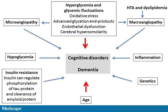
What are the mechanisms in the development of cognitive impairments and dementia? There are many mechanisms, and they are often poorly understood. Hyperglycemia plays a very important role as a direct result of oxidative stress, of advanced glycation end-products, but also as a result of micro- and macroangiopathy, hypertension, and dyslipidemia.[7,8] Other major factors, such as hypoglycemia,[9-12]play an extremely important role in the development of cognitive impairments. As well, a great deal of literature has been published lately on the role of inflammation[13] and genetic factors. Another widely known aspect is insulin resistance, which increases the risk for dementia at a fairly early stage by 40%[14,15]; this already during the metabolic syndrome, even before the onset of type 2 diabetes.

http://img.medscape.com/article/852/112/852112-Figure1.jpg

Figure. Multiple and poorly understood mechanisms of cognitive impairments and dementia. HTA = arterial hypertension. Adapted from Buysschaert M, et al.[16]

What Are the Consequences of Cognitive Impairments?

Cognitive impairments lead to a number of complications, including a reduction in life expectancy. In the GERODIAB cohort, we found, after 2 years of follow-up, that the mortality rate was twice as high in the patients with an MMSE score <24 compared with those with an MMSE score >24. In this study, the patients with a lower MMSE score had less well-controlled diabetes, were usually treated with insulin, and had heart failure and cerebrovascular complications more often. Very surprisingly, hypoglycemia was not more prevalent in these patients, perhaps because, being less independent, they were better managed by care teams.[17]

Cognitive impairments lead to geriatric complications, such as malnutrition, falls, and a loss of autonomy. They also promote social and family isolation and iatrogenic accidents, as well as depression, which can both mask cognitive impairments and exacerbate an underlying dementia. Another important aspect is that cognitive impairments increase the risk for hypoglycemia. This has been shown very clearly in all of the studies. There is, in fact, a bidirectional link between dementia and hypoglycemia: Hypoglycemia doubles the risk for dementia, and dementia triples the risk for hypoglycemia.[18]

Screening and Management

What do we do when a patient presents with cognitive impairments? First, they should be identified so that they can be managed. We need to be vigilant for certain little signs: changes in the patient’s behavior (eg, a patient who forgets his appointments, whose personal hygiene has declined, who is less diligent in keeping his blood glucose diary, and, lastly, who has an unexplained diabetic imbalance). We should also know how to use simple tests, such as the MMSE, which provides an overall assessment of space-time orientation, cognitive functions, language functions, and calculation, and how to assess the patient’s autonomy and loss of autonomy.[19] Next, we should, as per the recommendations of the American Diabetes Association[20] and the EASD, individualize the glycemic goals, taking into account, in the most fragile, elderly patients, cognitive status, the level of autonomy, depression, nutritional status—in particular, sarcopenia, which can coexist with obesity, and the risk for hypoglycemia.[21]

We should therefore avoid overtreating the most fragile patients (those at greatest risk for hypoglycemia), but neither should we undertreat patients who have a long life expectancy and who could develop micro- and macroangiopathic complications.

One last aspect, which is very important, is the family. Help needs to be provided to prevent the patient’s loss of autonomy.[21] Lastly, I think that cognitive decline should be added to the already long list of degenerative complications of diabetes.

PDGFR-ß Plays a Key Role in the Ectopic Migration of Neuroblasts in Cerebral Stroke

Hikari Sato et al.

The neuroprotective agents and induction of endogenous neurogenesis remain as the urgent issues to be established for the care of cerebral stroke. Platelet-derived growth factor receptor beta (PDGFR-ß) is mainly expressed in neural stem/progenitor cells (NSPCs), neurons and vascular pericytes of the brain; however, the role in pathological neurogenesis remains elusive. This review examined the role of PDGFR-ß in the migration and proliferation of NSPCs after stroke.

Read Full Post »

Notable Papers in Neurosciences

Larry H. Bernstein, MD, FCAP, Curator

LPBI

Notable Papers in Neurosciences

 NIH researchers’ new mouse model points to a gene therapy for eye disease

Oliver Worsley

A mouse model has been established for Leber hereditary optic neuropathy (LHON), a vision disorder caused by mutations within genes in the “battery packs” of our cells–the mitochondria. And investigators at the NIH say they were able to develop a gene therapy that could be used to treat it.

Within the mitochondrion are mitochondrial DNA (mtDNA) which carries the instructions for important metabolic processes required to keep the cell topped up with energy. Mutations in genes found in mtDNA can lead to various diseases; one of these is LHON which affects around 1 in 30,000 in England.

“[Until now] there was no efficient way to get DNA into mitochondria,” said John Guy, who is a professor of ophthalmology and is lead author of this study. Their work has been published in the Proceedings of the National Academy of Sciences.

Early symptoms of the disease include blurred vision and eyesight will eventually deteriorate over time. A loss of retinal ganglion cells is at the crux of the pathology and these cells are crucial for carrying visual signals from the retina to the brain, via the optic nerve.

The most prevalent mutation responsible for LHON is in a mitochondrial gene called ND4. Dr. Guy and his lab have been attempting to develop a gene therapy approach to correct this mutation for 15 years now. But one issue with adopting the widely used viral vector is that despite its efficacy in integrating into nuclear DNA–viruses have a harder time penetrating the mitochondria.

In developing his mouse model, Dr. Guy found a way around this. He fixed a virus with the same mutation in ND4 seen in 70% of LHON patients–adding a protein that mitochondria require from outside the organelle, as they cannot produce it on their own.

In the hijacked virus they included a fluorescent tag so they could confirm the future progeny of mice which had the defective gene. The mouse model does what is seen in patients with the same disease and optic nerve atrophy, loss of retinal ganglion cells and a decline in visual response is consistently observed.

The next step was providing a gene therapy to reverse it. The researchers packaged a normal ND4 gene into the same type of virus and injected it directly into the eye–leading to marked visual improvements without any side effects from the virus itself.

Related Articles:
Study: Eyes may signal brain pathology in schizophrenia
Stem cell therapy protects vision in preclinical retinal disease study
Retinas made from embryonic stem cells implanted into mice for the first time

GEN News Highlights

Oct 7, 2015

Stem Cell Advance Brings Vision Repair in Sight

http://www.genengnews.com/gen-news-highlights/stem-cell-advance-brings-vision-repair-in-sight/81251832/

Transplantation of cones produced from stem cells could reverse macular degeneration. A new differentiation approach yields abundant cones from human embryonic stem cells. When allowed to grow to confluence, the cones spontaneously form sheets of organized retinal tissue. [G. Bernier, University of Montreal]

http://www.genengnews.com/Media/images/GENHighlight/thumb_Oct7_2015_UnivMontreal_StemCellVision1867674218.jpg

A dearth of cone cells means degraded vision, so perhaps cone cell numbers could be raised, if only there were a way to produce cone cells in abundance. Then, cone cells could be transplanted en masse, potentially reversing the vision losses due to age-related macular degeneration.

We are born with a fixed number of cone cells. Additional cone cells must be contrived if degradation of the retina, a condition that is accelerated in nearly one out of four people, is to be reversed. Although cone cells have been produced by means of stem cell differentiation, the output has been meager. Now, however, scientists at the University of Montreal report that they have developed an efficient technique for producing cone cells from human embryonic stem cells.

These scientists, led by Gilbert Bernier, Ph.D., essentially closed a number of signaling pathways in stem cells, leaving open a default pathway that led to photoreceptor genesis. The scientists detailed their work in the journal Development, in an article that appeared online October 1. The article—“Differentiation of human embryonic stem cells into cone photoreceptors through simultaneous inhibition of BMP, TGFβ, and Wnt signaling”—is the culmination of years of work.

Bernier has been interested in the genes that code and enable the induction of the retina during embryonic development since completing his doctorate in molecular biology in 1997. “During my post-doc at the Max-Planck Institute in Germany, I developed the idea that there was a natural molecule that must exist and be capable of forcing embryonic stem cells into becoming cones,” he said. Indeed, bioinformatic analysis led him to predict the existence of a mysterious protein: COCO, a “recombinational” human molecule that is normally expressed within photoreceptors during their development.

In 2001, Bernier launched his laboratory in Montreal and immediately isolated the molecule. But it took several years of research to demystify the molecular pathways involved in the photoreceptors development mechanism. The Bernier laboratory’s current work has established that Coco (Dand5), a member of the Cerberus gene family, is expressed in the developing and adult mouse retina.

“Upon exposure to recombinant COCO, human embryonic stem cells (hESCs) differentiated into S-cone photoreceptors, developed an inner segment-like protrusion, and could degrade cGMP when exposed to light,” Bernier and colleagues wrote in the Development article. “Addition of thyroid hormone resulted in a transition from a unique S-cone population toward a mixed M/S-cone population.”

In addition, when the COCO-exposed hESCs were cultured at confluence for a prolonged period of time, they spontaneously developed into a cellular sheet composed of polarized cone photoreceptors. “Within 45 days, the cones that we allowed to grow toward confluence spontaneously formed organized retinal tissue that was 150 microns thick,” Dr. Bernier noted. “This has never been achieved before.”

In order to verify the technique, Dr. Bernier injected clusters of retinal cells into the eyes of healthy mice. The transplanted photoreceptors migrated naturally within the retina of their host.

Although Dr. Bernier acknowledged that the transplantation of photoreceptors in clinical trials was years away, he expressed optimism that his laboratory had made a significant advance, one that could, ultimately, benefit countless patients. “Our method has the capacity to differentiate 80% of the stem cells into pure cones,” Dr. Gilbert explained. “Thanks to our simple and effective approach, any laboratory in the world will now be able to create masses of photoreceptors.”

Beyond the clinical applications, Dr. Bernier’s findings could enable the modeling of human retinal degenerative diseases through the use of induced pluripotent stem cells, offering the possibility of directly testing potential avenues for therapy on the patient’s own tissues. “Our work,” the Development article concluded, “provides a unique platform to produce human cones for developmental, biochemical, and therapeutic studies.”

Neurogenesis in the Mammalian Brain

Neuron nurseries in the adult brains of rodents and humans appear to influence cognitive function.

By Jef Akst | October 1, 2015

http://www.the-scientist.com//?articles.view/articleNo/44047/title/Neurogenesis-in-the-Mammalian-Brain/

In rodents, there are two populations of neural stem cells in the adult brain. The majority of new neurons are born in the subventricular zone along the lateral ventricle wall and migrate through the rostral migratory stream (RMS) to the olfactory bulb. About one-tenth as many new neurons are produced in the subgranular zone of the dentate gyrus (white) of the hippocampus.

In the rodent dentate gyrus, neural stem cells differentiate into neuroblasts before maturing and integrating with hippocampal circuits important in learning and memory.

In the rodent subventricular zone, neural stem cells differentiate into neuroblasts, which make their way to the olfactory bulb, where they complete their development.

Researchers have also demonstrated that neurogenesis occurs in the adult human brain, though the locations and degree of cell proliferation appear to differ somewhat from rodents. Strong evidence now exists that new neurons are born in the dentate gyrus of the hippocampus, where they integrate into existing circuits. But so far, there is no definitive support for the migration of new neurons migrating from the subventricular zone (SVZ) of the lateral ventricle to the olfactory bulb, which is atrophied relative to the olfactory bulb of rodents and other mammals that rely more heavily on smell. However, one study did report signs of neurogenesis in an area next to the SVZ, the striatum, which is important for cognitive function and motor control.

Brain Gain

Young neurons in the adult human brain are likely critical to its function.

By Jef Akst | October 1, 2015

http://www.the-scientist.com/?articles.view/articleNo/44097/title/Brain-Gain/

How the Brain Builds New Thoughts

10/06/2015 Harvard University

http://www.biosciencetechnology.com/news/2015/10/how-brain-builds-new-thoughts?

“One of the big mysteries of human cognition is how the brain takes ideas and puts them together in new ways to form new thoughts,” said postdoctoral fellow Steven Frankland. (Kris Snibbe/Harvard Staff Photographer)Let’s start with a simple sentence: Last week Joe Biden beat Vladimir Putin in a game of Scrabble.

http://www.biosciencetechnology.com/sites/biosciencetechnology.com/files/bt1510_harvard_snibbe.jpg

It’s a strange notion to entertain, certainly, but one humans can easily make sense of, researchers say, thanks to the way the brain constructs new thoughts.

A new study, co-authored by postdoctoral fellow Steven Frankland and Professor of Psychology Joshua Greene, suggests that two adjacent brain regions allow humans to build new thoughts using a sort of conceptual algebra, mimicking the operations of silicon computers that represent variables and their changing values. The study is described in a Sept. 17 paper in the Proceedings of the National Academy of Sciences.

“One of the big mysteries of human cognition is how the brain takes ideas and puts them together in new ways to form new thoughts,” said Frankland, the lead author of the study. “Most people can understand ‘Joe Biden beat Vladimir Putin at Scrabble’ even though they’ve never thought about that situation, because, as long as you know who Putin is, who Biden is, what Scrabble is, and what it means to win, you’re able to put these concepts together to understand the meaning of the sentence. That’s a basic, but remarkable, cognitive ability.”

But how are such thoughts constructed? According to one theory, the brain does it by representing conceptual variables, answers to recurring questions of meaning such as “What was done?” and “Who did it?” and “To whom was it done?” A new thought such as “Biden beats Putin” can then be built by making “beating” the value of the action variable, “Biden” the value of the “agent” variable (“Who did it?”), and “Putin” the value of the “patient” variable (“To whom was it done?”). Frankland and Greene are the first to point to specific regions of the brain that encode such mental syntax.

“This has been a central theoretical discussion in cognitive science for a long time, and although it has seemed like a pretty good bet that the brain works this way, there’s been little direct empirical evidence for it,” Frankland said.

To identify the regions, Frankland and Greene used functional magnetic resonance imaging (fMRI) to scan students’ brains as they read a series of simple sentences such as “The dog chased the man” and “The man chased the dog.”

Equipped with that data, they then turned to algorithms to identify patterns of brain activity that corresponded with “dog” and “boy.”

“What we found is there are two regions in the left superior temporal lobe, one which is situated more toward the center of the head, that carries information about the agent, the one doing an action,” Frankland said. “An immediately adjacent region, located closer to the ear, carries information about the patient, or who the action was done to.”

Importantly, Frankland added, the brain appears to reuse the same patterns across multiple sentences, implying that these patterns function like symbols.

“So we might say ‘the dog chased the boy,’ or ‘the dog scratched the boy,’ but if we use some new verb the algorithms can still recognize the ‘dog’ pattern as the agent,” Frankland said. “That’s important because it suggests these symbols are used over and over again to compose new thoughts. And, moreover, we find that the structure of the thought is mapped onto the structure of the brain in a systematic way.”

That ability to use a series of repeatable concepts to formulate new thoughts may be part of what makes human thought unique ― and uniquely powerful.

“This paper is about language,” Greene said. “But we think it’s about more than that. There’s a more general mystery about how human thinking works.

“What makes human thinking so powerful is that we have this library of concepts that we can use to formulate an effectively infinite number of thoughts,” he continued. “Humans can engage in complicated behaviors that, for any other creature on Earth, would require an enormous amount of training. Humans can read or hear a string of concepts and immediately put those concepts together to form some new idea.”

Unlike models of perception, which put more complex representations at the top of a processing hierarchy, Frankland and Greene’s study supports a model of higher cognition that relies on the dynamic combination of conceptual building blocks to formulate thoughts.

“You can’t have a set of neurons that are there just waiting for someone to say ‘Joe Biden beat Vladimir Putin at Scrabble,’ ” Greene said. “That means there has to be some other system for forming meanings on the fly, and it has to be incredibly flexible, incredibly quick and incredibly precise.” He added, “This is an essential feature of human intelligence that we’re just beginning to understand.”

Source: Harvard Gazette

Predicting Change in the Alzheimer’s Brain

Tue, 10/06/2015 – 9:14am

Larry Hardesty, MIT News Office

http://www.biosciencetechnology.com/news/2015/10/predicting-change-alzheimers-brain?

MIT researchers are developing a computer system that uses genetic, demographic, and clinical data to help predict the effects of disease on brain anatomy.

http://www.biosciencetechnology.com/sites/biosciencetechnology.com/files/bt1510_MIT_Predicting.jpg

In experiments, they trained a machine-learning system on MRI data from patients with neurodegenerative diseases and found that supplementing that training with other patient information improved the system’s predictions. In the cases of patients with drastic changes in brain anatomy, the additional data cut the predictions’ error rate in half, from 20 percent to 10 percent.

“This is the first paper that we’ve ever written on this,” said Polina Golland, a professor of electrical engineering and computer science at MIT and the senior author on the new paper. “Our goal is not to prove that our model is the best model to do this kind of thing; it’s to prove that the information is actually in the data. So what we’ve done is, we take our model, and we turn off the genetic information and the demographic and clinical information, and we see that with combined information, we can predict anatomical changes better.”

First author on the paper is Adrian Dalca, an MIT graduate student in electrical engineering and computer science and a member of Golland’s group at MIT’s Computer Science and Artificial Intelligence Laboratory. They’re joined by Ramesh Sridharan, another Ph.D. student in Golland’s group, and by Mert Sabuncu, an assistant professor of radiology at Massachusetts General Hospital, who was a postdoc in Golland’s group.

The researchers are presenting the paper at the International Conference on Medical Image Computing and Computer Assisted Intervention this week. The work is a project of the Neuroimage Analysis Center, which is based at Brigham and Women’s Hospital in Boston and funded by the National Institutes of Health.

Common denominator

In their experiments, the researchers used data from the Alzheimer’s Disease Neuroimaging Initiative, a longitudinal study on neurodegenerative disease that includes MRI scans of the same subjects taken months and years apart.

Each scan is represented as a three-dimensional model consisting of millions of tiny cubes, or “voxels,” the 3-D equivalent of image pixels.

The researchers’ first step is to produce a generic brain template by averaging the voxel values of hundreds of randomly selected MRI scans. They then characterize each scan in the training set for their machine-learning algorithm as a deformation of the template. Each subject in the training set is represented by two scans, taken between six months and seven years apart.

The researchers conducted two experiments: one in which they trained their system on scans of both healthy subjects and those displaying evidence of either Alzheimer’s disease or mild cognitive impairment, and one in which they trained it only on data from healthy subjects.

In the first experiment, they trained the system twice, once using just the MRI scans and the second time supplementing them with additional information. This included data on genetic markers known as single-nucleotide polymorphisms; demographic data, such as subject age, gender, marital status, and education level; and rudimentary clinical data, such as patients’ scores on various cognitive tests.

The brains of healthy subjects and subjects in the early stages of neurodegenerative disease change little over time, and indeed, in cases where the differences between a subject’s scans were slight, the system trained only on MRI data fared well. In cases where the changes were more marked, however, the addition of the supplementary data made a significant difference.

Counterfactuals

In the second experiment, the researchers trained the system just once, on both the MRI data and the supplementary data of healthy subjects. But they instead used it to predict what the brains of Alzheimer’s patients would have looked like had they not been disfigured by disease.

In this case, there are no clinical data that could validate the system’s predictions. But the researchers believe that exploring this sort of counterfactual could be scientifically useful.

“It would illuminate how changes in individual subjects — for example, with mild cognitive impairment, which is a precursor to Alzheimer’s — evolve along this trajectory of degeneration, as compared to what normal degeneration would be,” Golland said. “We think that there are very interesting research applications of this. But I have to be honest and say that the original motivation was curiosity about how much of anatomy we could predict from genetics and other non-image data.”

“It’s not surprising that clinical and genetic data would help,” said Bruce Rosen, a professor of radiology at Harvard Medical School and director of the Athinoula A. Martinos Center for Biomedical Imaging at Massachusetts General Hospital. “But the fact that it did as well as it did is encouraging.”

“There are lots of ways these tools could be beneficial to the research community,” Rosen adds. “To my mind, the more challenging question is whether they could be useful clinically.”

Some promising experimental Alzheimer’s drugs require early determination of how the disease is likely to progress, Rosen said. Currently, he said, that determination relies on a combination of MRI and PET scan data. “People think MRI is expensive, but it’s only a fraction of what PET scans cost,” Rosen said. “If machine-learning tools can help avoid the need for PET scans in evaluating patients early in the disease course, that will be very impactful.”

Source: Massachusetts Institute of Technology

 Alzheimer’s: Investigators spotlight a pathway for amyloid beta clearance

By John Carroll

There are a variety of theories as to why people develop Alzheimer’s. And one of the best known is that toxic clusters of amyloid beta in the brain wipe out memories and trigger dementia in the elderly.

Now researchers at Indiana University say that they have determined that the IL1RAP immune pathway could provide a promising avenue for drug developers. And they’re quick to add that some experimental therapies that already hit this target could offer a quick way to help determine their utility against Alzheimer’s.

The team confirmed an observation that has been made before: the APOE e4 allele is associated with a significant accumulation of amyloid beta. But they were surprised to find that the IL1RAP gene–which they note codes for the immune signaling factor interleukin-1 receptor accessory protein–“showed an independent and even stronger influence on amyloid accumulation.”

They also determined that the gene was linked to a lower level of microglial activity as measured by PET scans; increased atrophy of the temporal cortex; swift cognitive decline and a “greater likelihood among study participants of progression from mild cognitive impairment to Alzheimer’s disease.”

“This was an intriguing finding because IL1RAP is known to play a central role in the activity of microglia, the immune system cells that act as the brain’s “garbage disposal system” and the focus of heavy investigation in a variety of neurodegenerative diseases,” said Dr. Vijay Ramanan, postdoctoral researcher at the IU School of Medicine.

There are already experimental anti-inflammatories and antibodies that are designed to hit this target, offering a shortcut in determining the impact on patients.

“These findings suggest that targeting the IL1RAP immune pathway may be a viable approach for promoting the clearance of amyloid deposits and fighting an important cause of progression in Alzheimer’s disease,” said Andrew Saykin, director of the Indiana Alzheimer Disease Center and the national Alzheimer’s Disease Neuroimaging Initiative Genetics Core.

It’s also useful to note that while many researchers believe that amyloid beta causes Alzheimer’s, there’s no consensus at the FDA on that point. And while many programs have been put in place to treat the disease, the vast majority have failed in the clinic, including drugs that aim at amyloid beta clearance.

Related Articles:
Mayo Clinic team renews Alzheimer’s feud, fingers tau over amyloid
Alzheimer’s study finds a molecule that might stymie critical stage of the disease
Neuroscience project tries to put the immune system to work against Alzheimer’s

An Accessible Approach to Making a Mini-brain

10/05/2015 – Brown University

http://www.biosciencetechnology.com/news/2015/10/accessible-approach-making-mini-brain


A bioengineering team at Brown University can grow “mini-brains” of neurons and supporting cells that form networks and are electrically active. (Image: Hoffman-Kim lab/Brown University)

http://www.biosciencetechnology.com/sites/biosciencetechnology.com/files/bt1510_brown_minibrain.jpg

If you need a working miniature brain — say for drug testing, to test neural tissue transplants, or to experiment with how stem cells work — a new paper describes how to build one with what the Brown University authors say is relative ease and low expense. The little balls of brain aren’t performing any cogitation, but they produce electrical signals and form their own neural connections — synapses — making them readily producible testbeds for neuroscience research, the authors said.

“We think of this as a way to have a better in vitro [lab] model that can maybe reduce animal use,” said graduate student Molly Boutin, co-lead author of the new paper in the journal Tissue Engineering: Part C. “A lot of the work that’s done right now is in two-dimensional culture, but this is an alternative that is much more relevant to the in vivo [living] scenario.”

Just a small sample of living tissue from a single rodent can make thousands of mini-brains, the researchers said. The recipe involves isolating and concentrating the desired cells with some centrifuge steps and using that refined sample to seed the cell culture in medium in an agarose spherical mold.

The mini-brains, about a third of a millimeter in diameter, are not the first or the most sophisticated working cell cultures of a central nervous system, the researchers acknowledged, but they require fewer steps to make and they use more readily available materials.

“The materials are easy to get and the mini-brains are simple to make,” said co-lead author Yu-Ting Dingle, who earned her Ph.D. at Brown in May 2015. She compared them to retail 3-D printers which have proliferated in recent years, bringing that once-rare technology to more of a mass market. “We could allow all kinds of labs to do this research.”

The spheres of brain tissue begin to form within a day after the cultures are seeded and have formed complex 3-D neural networks within two to three weeks, the paper shows.

25-cent mini-brains

There are fixed costs, of course, but an approximate cost for each new mini-brain is on the order of $0.25, said study senior author Diane Hoffman-Kim, associate professor of molecular pharmacology, physiology and biotechnology and associate professor of engineering at Brown.

“We knew it was a relatively high-throughput system, but even we were surprised at the low cost per mini-brain when we computed it,” Hoffman-Kim said.

Hoffman-Kim’s lab collaborated with fellow biologists and bioengineers at Brown — faculty colleagues Julie Kauer, Jeffrey Morgan, and Eric Darling are all co-authors — to build the mini-brains. She wanted to develop a testbed for her lab’s basic biomedical research. She was interested, for example, in developing a model to test aspects of neural cell transplantation, as has been proposed to treat Parkinson’s disease. Boutin was interested in building working 3-D cell cultures to study how adult neural stem cells develop.

Morgan’s Providence startup company, MicroTissues Inc., makes the 3-D tissue engineering molds used in the study.

The method they developed yields mini-brains with several important properties:

  •     Diverse cell types: The cultures contain both inhibitory and excitatory neurons and several varieties of essential neural support cells called glia.
  •     Electrically active: the neurons fire and spike and form synaptic connections, producing complex networks.
  •     3-D: Cells connect and communicate within a realistic geometry, rather than merely across a flat plane as in a 2-D culture.
  •     Natural density: Experiments showed that the mini-brains have a density of a few hundred thousand cells per cubic millimeter, which is similar to a natural rodent brain.
  •     Physical structure: Cells in the mini-brain produce their own extracellular matrix, producing a tissue with the same mechanical properties (squishiness) as natural tissue. The cultures also don’t rely on foreign materials such as scaffolds of collagen.
  •     Longevity: In testing, cultured tissues live for at least a month.

Hoffman-Kim, who is affiliated with the Brown Institute for Brain Science and the Center for Biomedical Engineering, said she hopes the mini-brains might proliferate to many different labs, including those of researchers who have questions about neural tissue but not necessarily the degree of neuroscience and cell culture equipment required of other methods.

“If you are that person in that lab, we think you shouldn’t have to equip yourself with a microelectronics facility, and you shouldn’t have to do embryonic dissections in order to generate an in vitro model of the brain,” Hoffman-Kim said.

The National Science Foundation, the National Institutes of Health, the Brown Institute for Brain Science, and the U.S. Department of Education funded the research.

Source: Brown University

Rat Brain Simulation Runs Neocortical Maze

http://www.genengnews.com/gen-news-highlights/rat-brain-simulation-runs-neocortical-maze/81251842/

http://www.genengnews.com/Media/images/GENHighlight/thumb_Oct9_2015_BBPEPFL2015_VirtualRatBrain9128157182.jpg

In this depiction of in silico retrograde staining, a digital reconstruction of neocortical microcircuitry, the presynaptic neurons of a layer 2/3 nest basket cell (red) are stained in blue. Only immediate neighboring presynaptic neurons are shown. [© BBP/EPFL 2015]

It’s a piece of rat brain containing about 30,000 neurons and 40 million synaptic connections, and there’s nothing remarkable about it, except that it isn’t real. It’s a digital reconstruction—a representation of a one-third cubic millimeter of rat neocortex—and it seems to work like the real thing.

Needless to say, its many creators are proud. They include 82 scientists and engineers from around the world, collaborators who are aware that their reconstruction represents the culmination of 20 years of biological experimentation and 10 years of computational science work. They are also aware that their work is controversial. It was criticized last year in an open letter. Signed by hundreds of neuroscientists, the letter argued that attempts to digitally reconstruct brain tissue were premature and represented an “overly narrow” approach that risked a misallocation of resources.

Undaunted, the investigators, led by scientists of the École Polytechnique Fédérale de Lausanne (EPFL), ran simulations on supercomputers to show that the electrical behavior of the virtual brain tissue matched the behavior of real rat neocortical tissue. Even though the digital reconstruction was not designed to reproduce any specific circuit phenomenon, a variety of experimental findings emerged. One such simulation examined how different types of neuron would respond if fibers coming into the neocortex were to convey signals encoding touch sensations. The researchers found that the responses of the different types of neurons in the digital reconstruction were very similar to those that had been previously observed in the laboratory.

These findings appeared October 8 in the journal Cell, in an article entitled, “Reconstruction and Simulation of Neocortical Microcircuitry.” This article also described how additional simulations revealed novel insights into the functioning of the neocortex.

“[We] find a spectrum of network states with a sharp transition from synchronous to asynchronous activity, modulated by physiological mechanisms,” wrote the authors. “The spectrum of network states, dynamically reconfigured around this transition, supports diverse information processing strategies.”

The authors even suggested that their work represents the first step toward the digital reconstruction and simulation of a whole brain. “They delivered what they promised,” said Patrick Aebischer, president of EPFL. This statement appeared in an EPFL press release that also indicated that the EPFL, together with the Swiss government, took the “bold step of funding the ambitious and controversial Blue Brain Project.”

The Blue Brain project is the simulation core of the Human Brain Project, a decade-long effort that is being allocated more than $1 billion.

“While a long way from the whole brain, the study demonstrates that it is feasible to digitally reconstruct and simulate brain tissue,” the release continued. “It is a first step and a significant contribution to Europe’s Human Brain Project, which Henry Markram founded, and where EPFL is the coordinating partner.”

Idan Segev, a senior author, sees the paper as building on the pioneering work of the Spanish anatomist, Ramon y Cajal from more than 100 years ago. “Ramon y Cajal began drawing every type of neuron in the brain by hand. He even drew in arrows to describe how he thought the information was flowing from one neuron to the next. Today, we are doing what Cajal would be doing with the tools of the day—building a digital representation of the neurons and synapses and simulating the flow of information between neurons on supercomputers. Furthermore, the digitization of the tissue allows the data to be preserved and reused for future generations.”

Now that the Blue Brain team has published the experimental results and the digital reconstruction, other scientists will be able to use the data and reconstruction to test other theories of brain function.

“The reconstruction is a first draft, it is not complete and it is not yet a perfect digital replica of the biological tissue,” explained Henry Markram. In fact, the current version explicitly leaves out many important aspects of the brain, such as glia, blood vessels, gap-junctions, plasticity, and neuromodulation. According to Sean Hill, a senior author: “The job of reconstructing and simulating the brain is a large-scale collaborative one, and the work has only just begun. The Human Brain Project represents the kind of collaboration that is required.”


Neuronal Waste Removal Gene Found to Prevent Parkinson’s

http://www.genengnews.com/gen-news-highlights/neuronal-waste-removal-gene-found-to-prevent-parkinson-s/81251836/

Researchers at the University of Copenhagen in Denmark say they have discovered that noninheritable Parkinson’s Disease (PD) may be caused by functional changes in the Interferon-beta (IFNβ) gene, which plays a vital role in keeping neurons healthy by regulating waste management. Treatment with IFNβ-gene therapy successfully prevented neuronal death and disease effects in an experimental model of PD.

The team’s study (“Lack of Neuronal IFN-β-IFNAR Causes Lewy Body- and Parkinson’s Disease-like Dementia”) was published in Cell.

“We found that IFNβ is essential for neurons ability to recycle waste proteins,” explained Patrick Ejlerskov, Ph.D., an assistant professor in the lab of Shohreh Issazadeh-Navikas, Ph.D., at the university’s Biotech Research and Innovation Center (BRIC) and first author on the paper. “Without this, the waste proteins accumulate in disease-associated structures called Lewy bodies and with time the neurons die.”

The scientists found that mice missing IFNβ developed Lewy bodies in parts of the brain, which control body movement and restoration of memory, and as a result they developed disease and clinical signs similar to patients with PD and dementia with Lewy bodies (DLB).

While hereditary gene mutations have long been known to play a role in familial PD, the study from BRIC offers one of the first models for so-called nonfamilial PD, which comprises the majority (90-95%) of patients suffering from PD. According to Dr. Issazadeh-Navikas, the new knowledge opens new therapeutic possibilities.

“This is one of the first genes found to cause pathology and clinical features of nonfamilial PD and DLB, through accumulation of disease-causing proteins,” she said. “It is independent of gene mutations known from familial PD and when we introduced IFNβ-gene therapy, we could prevent neuronal death and disease development. Our hope is that this knowledge will enable development of more effective treatment of PD.”

Current treatments are effective at improving the early motor symptoms of the disease. However, as the disease progress, the treatment effect is lost. The next step for the research team will be to gain a better understanding of the molecular mechanisms by which IFNβ protects neurons and thereby prevents movement disorders and dementia.

A review of heterogeneous data mining for brain disorder identification

  • Bokai Cao , Xiangnan Kong, Philip S. Yu

Brain Informatics 30 Sept 2015, pp 1-12

http://dx.doi.org:/10.1007/s40708-015-0021-3

http://link.springer.com/article/10.1007/s40708-015-0021-3/fulltext.html

With rapid advances in neuroimaging techniques, the research on brain disorder identification has become an emerging area in the data mining community. Brain disorder data poses many unique challenges for data mining research. For example, the raw data generated by neuroimaging experiments is in tensor representations, with typical characteristics of high dimensionality, structural complexity, and nonlinear separability. Furthermore, brain connectivity networks can be constructed from the tensor data, embedding subtle interactions between brain regions. Other clinical measures are usually available reflecting the disease status from different perspectives. It is expected that integrating complementary information in the tensor data and the brain network data, and incorporating other clinical parameters will be potentially transformative for investigating disease mechanisms and for informing therapeutic interventions. Many research efforts have been devoted to this area. They have achieved great success in various applications, such as tensor-based modeling, subgraph pattern mining, and multi-view feature analysis. In this paper, we review some recent data mining methods that are used for analyzing brain disorders.

Many brain disorders are characterized by ongoing injury that is clinically silent for prolonged periods and irreversible by the time symptoms first present. New approaches for detection of early changes in subclinical periods will afford powerful tools for aiding clinical diagnosis, clarifying underlying mechanisms, and informing neuroprotective interventions to slow or reverse neural injury for a broad spectrum of brain disorders, including bipolar disorder, HIV infection on brain, Alzheimer’s disease, Parkinson’s disease, etc. Early diagnosis has the potential to greatly alleviate the burden of brain disorders and the ever increasing costs to families and society.

As the identification of brain disorders is extremely challenging, many different diagnosis tools and methods have been developed to obtain a large number of measurements from various examinations and laboratory tests. Especially, recent advances in the neuroimaging technology have provided an efficient and noninvasive way for studying the structural and functional connectivity of the human brain, either normal or in a diseased state [1]. This can be attributed in part to advances in magnetic resonance imaging (MRI) capabilities [2]. Techniques such as diffusion MRI, also referred to as diffusion tensor imaging (DTI), produce in vivo images of the diffusion process of water molecules in biological tissues. By leveraging the fact that the water molecule diffusion patterns reveal microscopic details about tissue architecture, DTI can be used to perform tractography within the white matter and construct structural connectivity networks [3–7]. Functional MRI (fMRI) is a functional neuroimaging procedure that identifies localized patterns of brain activation by detecting associated changes in the cerebral blood flow. The primary form of fMRI uses the blood-oxygenation-level-dependent (BOLD) response extracted from the gray matter [8–10]. Another neuroimaging technique is positron emission tomography (PET). Using different radioactive tracers (e.g., fluorodeoxyglucose), PET produces a three-dimensional image of various physiological, biochemical, and metabolic processes [11].

A variety of data representations can be derived from these neuroimaging experiments, which present many unique challenges for the data mining community. Conventional data mining algorithms are usually developed to tackle data in one specific representation, a majority of which are particularly for vector-based data. However, the raw neuroimaging data are in the form of tensors, from which we can further construct brain networks connecting regions of interest (ROIs). Both of them are highly structured considering correlations between adjacent voxels in the tensor data and that between connected brain regions in the brain network data. Moreover, it is critical to explore interactions between measurements computed from the neuroimaging and other clinical experiments which describe subjects in different vector spaces. In this paper, we review some recent data mining methods for (1) mining tensor imaging data; (2) mining brain networks; and (3) mining multi-view feature vectors.

Tensor imaging analysis

For brain disorder identification, the raw data generated by neuroimaging experiments are in tensor representations [11–13]. For example, in contrast to two-dimensional X-ray images, an fMRI sample corresponds to a four-dimensional array by recording the sequential changes of traceable signals in each voxel.1

Tensors are higher order arrays that generalize the concepts of vectors (first-order tensors) and matrices (second-order tensors), whose elements are indexed by more than two indices. Each index expresses amode of variation of the data and corresponds to a coordinate direction. In an fMRI sample, the first three modes usually encode the spatial information, while the fourth mode encodes the temporal information. The number of variables in each mode indicates the dimensionality of a mode. The order of a tensor is determined by the number of its modes. An mth-order tensor can be represented as X=(xi1,…,im)∈RI1×⋯×Im, where Ii is the dimension of X along the i-th mode.

Definition 1

(Tensor product) The tensor product of three vectors a∈RI1, b∈RI2, and c∈RI3, denoted by a⊗b⊗c, represents a third-order tensor with the elements (a⊗b⊗c)i1,i2,i3 = ai1bi2ci3.

Tensor product is also referred to as outer product in some literature [11, 12]. An mth-order tensor is a rank-one tensor if it can be defined as the tensor product of m vectors.

Definition 2

Given a third-order tensor X∈RI1×I2×I3 and an integer R, as illustrated in Fig. 1, a tensor factorization of X can be expressed as

X=X1+X2+⋯+XR=∑r=1Rar⊗br⊗cr

(1)

Fig. 1

Tensor factorization of a third-order tensor

http://static-content.springer.com/image/art%3A10.1007%2Fs40708-015-0021-3/MediaObjects/40708_2015_21_Fig1_HTML.gif

One of the major difficulties brought by the tensor data is the curse of dimensionality. The total number of voxels contained in a multi-mode tensor, say, X=(xi1,…,im)∈RI1×⋯×Im is I1×⋯×Im which is exponential to the number of modes. If we unfold the tensor into a vector, the number of features will be extremely high [14]. This makes traditional data mining methods prone to overfitting, especially with a small sample size. Both computational scalability and theoretical guarantee of the traditional models are compromised by such high dimensionality [13].

On the other hand, complex structural information is embedded in the tensor data. For example, in the neuroimaging data, values of adjacent voxels are usually correlated with each other [2]. Such spatial relationships among different voxels in a tensor image can be very important in neuroimaging applications. Conventional tensor-based approaches focus on reshaping the tensor data into matrices/vectors, and thus, the original spatial relationships are lost. The integration of structural information is expected to improve the accuracy and interpretability of tensor models.

2.1 Supervised learning

Suppose we have a set of tensor data D={(Xi,yi)}ni=1 for classification problem, where Xi∈RI1×⋯×Im is the neuroimaging data represented as an mth-order tensor and yi∈{−1,+1} is the corresponding binary class label of Xi. For example, if the i-th subject has Alzheimer’s disease, the subject is associated with a positive label, i.e., yi=+1. Otherwise, if the subject is in the control group, the subject is associated with a negative label, i.e., yi=−1.

Supervised tensor learning can be formulated as the optimization problem of support tensor machines (STMs) [15] which is a generalization of the standard support vector machines (SVMs) from vector data to tensor data. The objective of such learning algorithms is to learn a hyperplane by which the samples with different labels are divided as wide as possible. However, tensor data may not be linearly separable in the input space. To achieve a better performance on finding the most discriminative biomarkers or identifying infected subjects from the control group, in many neuroimaging applications, nonlinear transformation of the original tensor data should be considered. He et al. study the problem of supervised tensor learning with nonlinear kernels which can preserve the structure of tensor data [13]. The proposed kernel is an extension of kernels in the vector space to the tensor space which can take the multidimensional structure complexity into account. However, it cannot automatically consider the abundant and complicated information of the neuroimaging data in an integral manner. Han et al. apply a deep learning-based algorithm, the hierarchical convolutional sparse auto-encoder, to extract efficient and robust features and conserve abundant detail information for the neuroimaging classification [16].

Slightly different from classifying disease status (discrete label), another family of problems uses tensor neuroimages to predict cognitive outcome (continuous label). The problems can be formulated in a regression setup by treating clinical outcome as the real label, i.e., yi∈R, and treating tensor neuroimages as the input. However, most classical regression methods take vectors as input features. Simply reshaping a tensor into a vector is clearly an unsatisfactory solution.

Zhou et al. exploit the tensor structure in imaging data and integrate tensor decomposition within a statistical regression paradigm to model multidimensional arrays [14]. By imposing a low-rank approximation to the extremely high-dimensional complex imaging data, the curse of dimensionality is greatly alleviated, thereby allowing development of a fast estimation algorithm and regularization. Numerical analysis demonstrates its potential applications in identifying ROI in brains that are relevant to a particular clinical response. In scenarios where the objective is to predict a set of dependent variables, Cichocki et al. introduce a generalized multilinear regression model, higher order partial least squares, which projects the electrocorticogram data into a latent space and performs regression on the corresponding latent variables [17, 18].

2.2 Unsupervised learning

Modern imaging techniques have allowed us to study the human brain as a complex system by modeling it as a network [19]. For example, the fMRI scans consist of activations of thousands of voxels over time embedding a complex interaction of signals and noise [20], which naturally presents the problem of eliciting the underlying network from brain activities in the spatio-temporal tensor data. A brain connectivity network, also called a connectome [21], consists of nodes (gray matter regions) and edges (white matter tracts in structural networks or correlations between two BOLD time series in functional networks).

Although the anatomical atlases in the brain have been extensively studied for decades, task/subject specific networks have still not been completely explored with consideration of functional or structural connectivity information. An anatomically parcellated region may contain subregions that are characterized by dramatically different functional or structural connectivity patterns, thereby significantly limiting the utility of the constructed networks. There are usually trade-offs between reducing noise and preserving utility in brain parcellation [2]. Thus, investigating how to directly construct brain networks from tensor imaging data and understanding how they develop, deteriorate, and vary across individuals will benefit disease diagnosis [12].

Davidson et al. pose the problem of network discovery from fMRI data which involves simplifying spatio-temporal data into regions of the brain (nodes) and relationships between those regions (edges) [12]. Here the nodes represent collections of voxels that are known to behave cohesively over time; the edges can indicate a number of properties between nodes such as facilitation/inhibition (increases/decreases activity) or probabilistic (synchronized activity) relationships; and the weight associated with each edge encodes the strength of the relationship.

A tensor can be decomposed into several factors. However, unconstrained tensor decomposition results of the fMRI data may not be good for node discovery because each factor is typically not a spatially contiguous region nor does it necessarily match an anatomical region. That is to say, many spatially adjacent voxels in the same structure are not active in the same factor which is anatomically impossible. Therefore, to achieve the purpose of discovering nodes while preserving anatomical adjacency, known anatomical regions in the brain are used as masks and constraints are added to enforce that the discovered factors should closely match these masks [12].

Yang et al. investigate the inference of mouse brain networks and propose a hierarchical graphical model framework with tree-structural regularization [22]. In the hierarchical structure, voxels serve as the leaf nodes of the tree, and a node in the intermediate layer represents a region formed by voxels in the subtree rooted at that node. For edge discovery problem, Papalexakis et al. leverage control theory to model the dynamics of neuron interactions and infer the functional connectivity [23]. It is assumed that in addition to the linear influence of the input stimulus, there are hidden neuron regions of the brain, which interact with each other, causing the voxel activities. Veeriah et al. propose a deep learning algorithm for predicting if the two brain neurons are causally connected given their activation time-series data [24]. It reveals that the exploitation of the deep architecture is critical, which jointly extracts sequences of salient patterns of activation and aligns them to predict neural connections.

Overall, current research on tensor imaging analysis presents two directions: (1) supervised: for a particular brain disorder, a classifier can be trained by modeling the relationship between a set of neuroimages and their associated labels (disease status or clinical response); (2) unsupervised: regardless of brain disorders, a brain network can be discovered from a given neuroimage.

3 Brain network analysis

We have briefly introduced that brain networks can be constructed from neuroimaging data where nodes correspond to brain regions, e.g., insula, hippocampus, thalamus, and links correspond to the functional/structural connectivity between brain regions. The linkage structure in brain networks can encode tremendous information about the mental health of human subjects. For example, in brain networks derived from fMRI, functional connections can encode the correlations between the functional activities of brain regions. While structural links in DTI brain networks can capture the number of neural fibers connecting different brain regions. The complex structures and the lack of vector representations for the brain network data raise major challenges for data mining.

Next, we will discuss different approaches on how to conduct further analysis for constructed brain networks, which are also referred to as graphs hereafter.

Definition 3

(Binary graph) A binary graph is represented as G=(V,E), where V={v1,…,vnv} is the set of vertices, and E⊆V×V is the set of deterministic edges.

3.1 Kernel learning on graphs

In the setting of supervised learning on graphs, the target is to train a classifier using a given set of graph data D={(Gi,yi)}ni=1, so that we can predict the label y^ for a test graph G. With applications to brain networks, it is desirable to identify the disease status for a subject based on his/her uncovered brain network. Recent development of brain network analysis has made characterization of brain disorders at a whole-brain connectivity level possible, thus providing a new direction for brain disease classification.

Due to the complex structures and the lack of vector representations, graph data cannot be directly used as the input for most data mining algorithms. A straightforward solution that has been extensively explored is to first derive features from brain networks and then construct a kernel on the feature vectors.

Wee et al. use brain connectivity networks for disease diagnosis on mild cognitive impairment (MCI), which is an early phase of Alzheimer’s disease (AD) and usually regarded as a good target for early diagnosis and therapeutic interventions [25–27]. In the step of feature extraction, weighted local clustering coefficients of each ROI in relation to the remaining ROIs are extracted from all the constructed brain networks to quantify the prevalence of clustered connectivity around the ROIs. To select the most discriminative features for classification, statistical t test is performed and features with p values smaller than a predefined threshold are selected to construct a kernel matrix. Through the employment of the multi-kernel SVM, Wee et al. integrate information from DTI and fMRI and achieve accurate early detection of brain abnormalities [27].

However, such strategy simply treats a graph as a collection of nodes/links, and then extracts local measures (e.g., clustering coefficient) for each node or performs statistical analysis on each link, thereby blinding the connectivity structures of brain networks. Motivated by the fact that some data in real-world applications are naturally represented by means of graphs, while compressing and converting them to vectorial representations would definitely lose structural information, kernel methods for graphs have been extensively studied for a decade [28].

A graph kernel maps the graph data from the original graph space to the feature space and further measures the similarity between two graphs by comparing their topological structures [29]. For example, product graph kernel is based on the idea of counting the number of walks in product graphs [30]; marginalized graph kernel works by comparing the label sequences generated by synchronized random walks of labeled graphs [31]; and cyclic pattern kernels for graphs count pairs of matching cyclic/tree patterns in two graphs [32].

To identify individuals with AD/MCI from healthy controls, instead of using only a single property of brain networks, Jie et al. integrate multiple properties of fMRI brain networks to improve the disease diagnosis performance [33]. Two different yet complementary network properties, i.e., local connectivity and global topological properties are quantified by computing two different types of kernels, i.e., a vector-based kernel and a graph kernel. As a local network property, weighted clustering coefficients are extracted to compute a vector-based kernel. As a topology-based graph kernel, Weisfeiler-Lehman subtree kernel [29] is used to measure the topological similarity between paired fMRI brain networks. It is shown that this type of graph kernel can effectively capture the topological information from fMRI brain networks. The multi-kernel SVM is employed to fuse these two heterogeneous kernels for distinguishing individuals with MCI from healthy controls.

3.2 Subgraph pattern mining

In brain network analysis, the ideal patterns we want to mine from the data should take care of both local and global graph topological information. Graph kernel methods seem promising, which, however, are not interpretable. Subgraph patterns are more suitable for brain networks, which can simultaneously model the network connectivity patterns around the nodes and capture the changes in local area [2].

Definition 4

(Subgraph) Let G′=(V′,E′) and G=(V,E) be two binary graphs. G′ is a subgraph of G (denoted as G′⊆G) iff V′⊆V and E′⊆E. If G′ is a subgraph of G, then G is supergraph of G′.

A subgraph pattern, in a brain network, represents a collection of brain regions and their connections. For example, as shown in Fig. 2, three brain regions should work collaboratively for normal people and the absence of any connection between them can result in Alzheimer’s disease in different degrees. Therefore, it is valuable to understand which connections collectively play a significant role in disease mechanism by finding discriminative subgraph patterns in brain networks.

Mining subgraph patterns from graph data has been extensively studied by many researchers [34–37]. In general, a variety of filtering criteria are proposed. A typical evaluation criterion is frequency, which aims at searching for frequently appearing subgraph features in a graph dataset satisfying a prespecified threshold. Most of the frequent subgraph mining approaches are unsupervised. For example, Yan and Han develop a depth-first search algorithm: gSpan [38]. This algorithm builds a lexicographic order among graphs, and maps each graph to a unique minimum DFS code as its canonical label. Based on this lexicographic order, gSpan adopts the depth-first search strategy to mine frequent connected subgraphs efficiently. Many other approaches for frequent subgraph mining have also been proposed, e.g., AGM [39], FSG [40], MoFa [41], FFSM [42], and Gaston [43].

Fig. 2

An example of discriminative subgraph patterns in brain networks

http://static-content.springer.com/image/art%3A10.1007%2Fs40708-015-0021-3/MediaObjects/40708_2015_21_Fig2_HTML.gif

Moreover, the problem of supervised subgraph mining has been studied in recent work which examines how to improve the efficiency of searching the discriminative subgraph patterns for graph classification. Yan et al. introduce two concepts structural leap search and frequency-descending mining, and propose LEAP [37] which is one of the first work in discriminative subgraph mining. Thoma et al. propose CORK which can yield a near-optimal solution using greedy feature selection [36]. Ranu and Singh propose a scalable approach, called GraphSig, that is capable of mining discriminative subgraphs with a low-frequency threshold [44]. Jin et al. propose COM which takes into account the co-occurrences of subgraph patterns, thereby facilitating the mining process [45]. Jin et al. further propose an evolutionary computation method, called GAIA, to mine discriminative subgraph patterns using a randomized searching strategy [34]. Zhu et al. design a diversified discrimination score based on the log ratio which can reduce the overlap between selected features by considering the embedding overlaps in the graphs [46].

Conventional graph mining approaches are best suited for binary edges, where the structure of graph objects is deterministic, and the binary edges represent the presence of linkages between the nodes [2]. In fMRI brain network data, however, there are inherently weighted edges in the graph linkage structure, as shown in Fig. 3 (left). A straightforward solution is to threshold weighted networks to yield binary networks. However, such simplification will result in great loss of information. Ideal data mining methods for brain network analysis should be able to overcome these methodological problems by generalizing the network edges to positive and negative weighted cases, e.g., probabilistic weights in fMRI brain networks and integral weights in DTI brain networks.

Fig. 3

An example of fMRI brain networks (left) and all possible instantiations of linkage structures between red nodes (right) [47]. (Color figure online)

http://static-content.springer.com/image/art%3A10.1007%2Fs40708-015-0021-3/MediaObjects/40708_2015_21_Fig3_HTML.gif

Definition 5

A weighted graph is represented as G˜=(V,E,p), where V={v1,…,vnv} is the set of vertices, and E⊆V×V is the set of nondeterministic edges. p:E→(0,1] is a function that assigns a probability of existence to each edge in E.

fMRI brain networks can be modeled as weighted graphs where each edge e∈E is associated with a probability p(e) indicating the likelihood of whether this edge should exist or not [47, 48]. It is assumed thatp(e) of different edges in a weighted graph are independent from each other. Therefore, by enumerating the possible existence of all edges in a weighted graph, we can obtain a set of binary graphs. For example, in Fig. 3 (right), consider the three red nodes and links between them as a weighted graph. There are 23=8binary graphs that can be implied with different probabilities. For a weighted graph G˜, the probability of G˜containing a subgraph feature G′ is defined as the probability that a binary graph G implied by G˜ contains subgraph G′. Kong et al. propose a discriminative subgraph feature selection method based on dynamic programming to compute the probability distribution of the discrimination scores for each subgraph pattern within a set of weighted graphs [48].

For brain network analysis, usually we only have a small number of graph instances [48]. In these applications, the graph view alone is not sufficient for mining important subgraphs. Fortunately, the side information is available along with the graph data for brain disorder identification. For example, in neurological studies, hundreds of clinical, immunologic, serologic, and cognitive measures may be available for each subject, apart from brain networks. These measures compose multiple side views which contain a tremendous amount of supplemental information for diagnostic purposes. It is desirable to extract valuable information from a plurality of side views to guide the process of subgraph mining in brain networks.

Fig. 4

Two strategies of leveraging side views in feature selection process for graph classification: late fusion and early fusion

http://static-content.springer.com/image/art%3A10.1007%2Fs40708-015-0021-3/MediaObjects/40708_2015_21_Fig4_HTML.gif

Figure 4 illustrates two strategies of leveraging side views in the process of selecting subgraph patterns. Conventional graph classification approaches treat side views and subgraph patterns separately and may only combine them at the final stage of training a classifier. Obviously, the valuable information embedded in side views is not fully leveraged in the feature selection process. In order to fuse heterogeneous data sources at an early stage thereby exploring their correlations, Cao et al. introduce an effective algorithm for discriminative subgraph selection using multiple side views as guidance [49]. Side information consistency is first validated via statistical hypothesis testing which suggests that the similarity of side view features between instances with the same label should have higher probability to be larger than that with different labels. Based on such observations, it is assumed that the similarity/distance between instances in the space of subgraph features should be consistent with that in the space of a side view. That is to say, if two instances are similar in the space of a side view, they should also be close to each other in the space of subgraph features. Therefore the target is to minimize the distance between subgraph features of each pair of similar instances in each side view [49]. In contrast to existing subgraph mining approaches that focus on the graph view alone, the proposed method can explore multiple vector-based side views to find an optimal set of subgraph features for graph classification.

For graph classification, brain network analysis approaches can generally be put into three groups: (1) extracting some local measures (e.g., clustering coefficient) to train a standard vector-based classifier; (2) directly adopting graph kernels for classification; and (3) finding discriminative subgraph patterns. Different types of methods model the connectivity embedded in brain networks in different ways.

4 Multi-view feature analysis

Medical science witnesses everyday measurements from a series of medical examinations documented for each subject, including clinical, imaging, immunologic, serologic, and cognitive measures [50], as shown in Fig. 5. Each group of measures characterizes the health state of a subject from different aspects. This type of data is named as multi-view data, and each group of measures form a distinct view quantifying subjects in one specific feature space. Therefore, it is critical to combine them to improve the learning performance, while simply concatenating features from all views and transforming a multi-view data into a single-view data, as the method (a) shown in Fig. 6, would fail to leverage the underlying correlations between different views.

4.1 Multi-view learning and feature selection

Suppose we have a multi-view classification task with n labeled instances represented from m different views: D={(x(1)i,x(2)i,…,x(m)i,yi)}ni=1, where x(v)i∈RIv, Iv is the dimensionality of the v-th view, and yi∈{−1,+1} is the class label of the i-th instance.

Fig. 5

An example of multi-view learning in medical studies [51]

http://static-content.springer.com/image/art%3A10.1007%2Fs40708-015-0021-3/MediaObjects/40708_2015_21_Fig5_HTML.gif

Representative methods for multi-view learning can be categorized into three groups: co-training, multiple kernel learning, and subspace learning [52]. Generally, the co-training style algorithm is a classic approach for semi-supervised learning, which trains in alternation to maximize the mutual agreement on different views. Multiple kernel learning algorithms combine kernels that naturally correspond to different views, either linearly [53] or nonlinearly [54, 55] to improve learning performance. Subspace learning algorithms learn a latent subspace, from which multiple views are generated. Multiple kernel learning and subspace learning are generalized as co-regularization style algorithms [56], where the disagreement between the functions of different views is taken as a part of the objective function to be minimized. Overall, by exploring the consistency and complementary properties of different views, multi-view learning is more effective than single-view learning.

In the multi-view setting for brain disorders, or for medical studies in general, a critical problem is that there may be limited subjects available (i.e., a small n) yet introducing a large number of measurements (i.e., a large ∑mi=1Ii). Within the multi-view data, not all features in different views are relevant to the learning task, and some irrelevant features may introduce unexpected noise. The irrelevant information can even be exaggerated after view combinations thereby degrading performance. Therefore, it is necessary to take care of feature selection in the learning process. Feature selection results can also be used by researchers to find biomarkers for brain diseases. Such biomarkers are clinically imperative for detecting injury to the brain in the earliest stage before it is irreversible. Valid biomarkers can be used to aid diagnosis, monitor disease progression, and evaluate effects of intervention [48].

Conventional feature selection approaches can be divided into three main directions: filter, wrapper, and embedded methods [57]. Filter methods compute a discrimination score of each feature independently of the other features based on the correlation between the feature and the label, e.g., information gain, Gini index, Relief [58, 59]. Wrapper methods measure the usefulness of feature subsets according to their predictive power, optimizing the subsequent induction procedure that uses the respective subset for classification [51,60–63]. Embedded methods perform feature selection in the process of model training based on sparsity regularization [64–67]. For example, Miranda et al. add a regularization term that penalizes the size of the selected feature subset to the standard cost function of SVM, thereby optimizing the new objective function to conduct feature selection [68]. Essentially, the process of feature selection and learning algorithm interact in embedded methods which means the learning part and the feature selection part cannot be separated, while wrapper methods utilize the learning algorithm as a black box.

However, directly applying these feature selection approaches to each separate view would fail to leverage multi-view correlations. By taking into account the latent interactions among views and the redundancy triggered by multiple views, it is desirable to combine multi-view data in a principled manner and perform feature selection to obtain consensus and discriminative low-dimensional feature representations.

4.2 Modeling view correlations

Recent years have witnessed many research efforts devoted to the integration of feature selection and multi-view learning. Tang et al. study multi-view feature selection in the unsupervised setting by constraining that similar data instances from each view should have similar pseudo-class labels [69]. Considering brain disorder identification, different neuroimaging features may capture different but complementary characteristics of the data. For example, the voxel-based tensor features convey the global information, while the ROI-based automated anatomical labeling (AAL) [70] features summarize the local information from multiple representative brain regions. Incorporating these data and additional nonimaging data sources can potentially improve the prediction. For Alzheimer’s disease (AD) classification, Ye et al. propose a kernel-based method for integrating heterogeneous data, including tensor and AAL features from MRI images, demographic information, and genetic information [11]. The kernel framework is further extended for selecting features (biomarkers) from heterogeneous data sources that play more significant roles than others in AD diagnosis.

Huang et al. propose a sparse composite linear discriminant analysis model for identification of disease-related brain regions of AD from multiple data sources [71]. Two sets of parameters are learned: one represents the common information shared by all the data sources about a feature, and the other represents the specific information only captured by a particular data source about the feature. Experiments are conducted on the PET and MRI data which measure structural and functional aspects, respectively, of the same AD pathology. However, the proposed approach requires the input as the same set of variables from multiple data sources. Xiang et al. investigate multi-source incomplete data for AD and introduce a unified feature learning model to handle block-wise missing data which achieves simultaneous feature-level and source-level selection [72].

For modeling view correlations, in general, a coefficient is assigned for each view, either at the view-level or feature-level. For example, in multiple kernel learning, a kernel is constructed from each view and a set of kernel coefficients are learned to obtain an optimal combined kernel matrix. These approaches, however, fail to explicitly consider correlations between features.

4.3 Modeling feature correlations

One of the key issues for multi-view classification is to choose an appropriate tool to model features and their correlations hidden in multiple views, since this directly determines how information will be used. In contrast to modeling on views, another direction for modeling multi-view data is to directly consider the correlations between features from multiple views. Since taking the tensor product of their respective feature spaces corresponds to the interaction of features from multiple views, the concept of tensor serves as a backbone for incorporating multi-view features into a consensus representation by means of tensor product, where the complex multiple relationships among views are embedded within the tensor structures. By mining structural information contained in the tensor, knowledge of multi-view features can be extracted and used to establish a predictive model.

Smalter et al. formulate the problem of feature selection in the tensor product space as an integer quadratic programming problem [73]. However, this method is computationally intractable on many views, since it directly selects features in the tensor product space resulting in the curse of dimensionality, as the method (b) shown in Fig. 6. Cao et al. propose to use a tensor-based approach to model features and their correlations hidden in the original multi-view data [51]. The operation of tensor product can be used to bringm-view feature vectors of each instance together, leading to a tensorial representation for common structure across multiple views, and allowing us to adequately diffuse relationships and encode information among multi-view features. In this manner, the multi-view classification task is essentially transformed from an independent domain of each view to a consensus domain as a tensor classification problem.

By using Xi to denote ∏mv=1⊗x(v)i, the dataset of labeled multi-view instances can be represented as D={(Xi,yi)}ni=1. Note that each multi-view instance Xi is an mth-order tensor that lies in the tensor product space RI1×⋯×Im. Based on the definitions of inner product and tensor norm, multi-view classification can be formulated as a global convex optimization problem in the framework of supervised tensor learning [15]. This model is named as multi-view SVM [51], and it can be solved with the use of optimization techniques developed for SVM.

Fig. 6

Schematic view of the key differences among three strategies of multi-view feature selection [51]

http://static-content.springer.com/image/art%3A10.1007%2Fs40708-015-0021-3/MediaObjects/40708_2015_21_Fig6_HTML.gif

Furthermore, a dual method for multi-view feature selection is proposed in [51] that leverages the relationship between original multi-view features and reconstructed tensor product features to facilitate the implementation of feature selection, as the method (c) in Fig. 6. It is a wrapper model which selects useful features in conjunction with the classifier and simultaneously exploits the correlations among multiple views. Following the idea of SVM-based recursive feature elimination [60], multi-view feature selection is consistently formulated and implemented in the framework of multi-view SVM. This idea can extend to include lower order feature interactions and to employ a variety of loss functions for classification or regression [74].

5 Future work

The human brain is one of the most complicated biological structures in the known universe. While it is very challenging to understand how it works, especially when disorders and diseases occur, dozens of leading technology firms, academic institutions, scientists, and other key contributors to the field of neuroscience have devoted themselves to this area and made significant improvements in various dimensions.2 Data mining on brain disorder identification has become an emerging area and a promising research direction.

This paper provides an overview of data mining approaches with applications to brain disorder identification, which have attracted increasing attention in both data mining and neuroscience communities in recent years. A taxonomy is built based upon data representations, i.e., tensor imaging data, brain network data, and multi-view data, following which the relationships between different data mining algorithms and different neuroimaging applications are summarized. We briefly present some potential topics of interest in the future.

5.1 Bridging heterogeneous data representations

As introduced in this paper, we can usually derive data from neuroimaging experiments in three representations, including raw tensor imaging data, brain network data, and multi-view vector-based data. It is critical to study how to train a model on a mixture of data representations, although it is very challenging to combine data that are represented in tensor space, vector space, and graph space, respectively. There is a straightforward idea of defining different kernels on different feature spaces and combing them through multi-kernel algorithms. However, it is usually hard to interpret the results. The concept of side view has been introduced to facilitate the process of mining brain networks, which may also be used to guide supervised tensor learning. It is even more interesting if we can learn on tensors and graphs simultaneously.

5.2 Integrating multiple neuroimaging modalities

There are a variety of neuroimaging techniques available characterizing subjects from different perspectives and providing complementary information. For example, DTI contains local microstructural characteristics of water diffusion; structural MRI can be used to delineate brain atrophy; fMRI records BOLD response related to neural activity; and PET measures metabolic patterns [27]. Based on such multimodality representation, it is desirable to find useful patterns with rich semantics. For example, it is important to know which connectivity between brain regions is significant in the sense of both structure and functionality. On the other hand, by leveraging the complementary information embedded in the multimodality representation, better performance on disease diagnosis can be expected.

Fig. 7

A bioinformatics heterogeneous information network schema

http://static-content.springer.com/image/art%3A10.1007%2Fs40708-015-0021-3/MediaObjects/40708_2015_21_Fig7_HTML.gif

5.3 Mining bioinformatics information networks

Bioinformatics network is a rich source of heterogeneous information involving disease mechanisms, as shown in Fig. 7. The problems of gene-disease association and drug-target binding prediction have been studied in the setting of heterogeneous information networks [75, 76]. For example, in gene-disease association prediction, different gene sequences can lead to certain diseases. Researchers would like to predict the association relationships between genes and diseases. Understanding the correlations between brain disorders and other diseases and the causality between certain genes and brain diseases can be transformative for yielding new insights concerning risk and protective relationships, for clarifying disease mechanisms, for aiding diagnostics and clinical monitoring, for biomarker discovery, for identification of new treatment targets, and for evaluating effects of intervention.

Footnotes

1  A voxel is the smallest three-dimensional point volume referenced in a neuroimaging of the brain.

2  http://www.whitehouse.gov/BRAIN

References

  1. 1.

Rubinov M, Sporns O (2010) Complex network measures of brain connectivity: uses and interpretations. Neuroimage 52(3):1059–1069CrossRef

  1. 2.

Kong X, Yu PS (2014) Brain network analysis: a data mining perspective. ACM SIGKDD Explor Newsl 15(2):30–38MathSciNetCrossRef

  1. 3.

Basser PJ, Pierpaoli C (1996) Microstructural and physiological features of tissues elucidated by quantitative-diffusion-tensor MRI. J Magn Reson Ser B 111(3):209–219CrossRef

  1. 4.

Le Bihan D, Breton E, Lallemand D, Grenier P, Cabanis E, Laval-Jeantet M (1986) MR imaging of intravoxel incoherent motions: application to diffusion and perfusion in neurologic disorders. Radiology 161(2):401–407CrossRef

  1. 5.

Chenevert TL, Brunberg JA, Pipe J (1990) Anisotropic diffusion in human white matter: demonstration with mr techniques in vivo. Radiology 177(2):401–405CrossRef

  1. 6.

McKeown MJ, Makeig S, Brown GG, Jung T-P, Kindermann SS, Bell AJ, Sejnowski TJ (1998) Analysis of fMRI data by blind separation into independent spatial components. Hum Brain Mapp 6:160–188CrossRef

  1. 7.

Moseley ME, Cohen Y, Kucharczyk J, Mintorovitch J, Asgari H, Wendland M, Tsuruda J, Norman D (1990) Diffusion-weighted MR imaging of anisotropic water diffusion in cat central nervous system. Radiology 176(2):439–445CrossRef

  1. 8.

Biswal B, Yetkin FZ, Haughton VM, Hyde JS (1995) Functional connectivity in the motor cortex of resting human brain using echo-planar MRI. Magn Reson Med 34(4):537–541CrossRef

  1. 9.

Ogawa S, Lee T, Kay A, Tank D (1990) Brain magnetic resonance imaging with contrast dependent on blood oxygenation. Proc Natl Acad Sci 87(24):9868–9872CrossRef

  1. 10.

Ogawa S, Lee T-M, Nayak AS, Glynn P (1990) Oxygenation-sensitive contrast in magnetic resonance image of rodent brain at high magnetic fields. Magn Reson Med 14(1):68–78CrossRef

  1. 11.

Ye J, Chen K, Wu T, Li J, Zhao Z, Patel R, Bae M, Janardan R, Liu H, Alexander G et al (2008) Heterogeneous data fusion for Alzheimer’s disease study. In: KDD. ACM, pp 1025–1033

  1. 12.

Davidson I, Gilpin S, Carmichael O, Walker P (2013) Network discovery via constrained tensor analysis of fMRI data. In: KDD. ACM, pp 194–202

  1. 13.

He L, Kong X, Yu PS, Ragin AB, Hao Z, Yang X (2014) Dusk: a dual structure-preserving kernel for supervised tensor learning with applications to neuroimages. In: SDM. SIAM

  1. 14.

Zhou H, Li L, Zhu H (2013) Tensor regression with applications in neuroimaging data analysis. J Am Stat Assoc 108(502):540–552MATHMathSciNetCrossRef

  1. 15.

Tao D, Li X, Wu X, Hu W, Maybank SJ (2007) Supervised tensor learning. Knowl Inf Syst 13(1):1–42CrossRef

  1. 16.

Han X, Zhong Y, He L, Philip SY, Zhang L (2015) The unsupervised hierarchical convolutional sparse auto-encoder for neuroimaging data classification. In: Brain informatics and health. Springer, pp 156–166

  1. 17.

Cichocki A, Mandic D, De Lathauwer L, Zhou G, Zhao Q, Caiafa C, Phan HA (2015) Tensor decompositions for signal processing applications: from two-way to multiway component analysis. Signal Process Mag 32(2):145–163CrossRef

  1. 18.

Zhao Q, Caiafa CF, Mandic DP, Chao ZC, Nagasaka Y, Fujii N, Zhang L, Cichocki A (2013) Higher order partial least squares (HOPLS): a generalized multilinear regression method. Pattern Anal Mach Intell 35(7):1660–1673CrossRef

  1. 19.

Ajilore O, Zhan L, GadElkarim J, Zhang A, Feusner JD, Yang S, Thompson PM, Kumar A, Leow A (2013) Constructing the resting state structural connectome. Front Neuroinform 7:30CrossRef

  1. 20.

Genovese CR, Lazar NA, Nichols T (2002) Thresholding of statistical maps in functional neuroimaging using the false discovery rate. Neuroimage 15(4):870–878CrossRef

… more…

Multimodal neuroimaging computing: a review of the applications in neuropsychiatric disorders

Sidong Liu , Weidong Cai, Siqi Liu, Fan Zhang, Michael Fulham, Dagan Feng, Sonia Pujol, Ron Kikinis

Brain Informatics Sept 2015; 2(3): 167-180

http://dx.doi.org:/10.1007/s40708-015-0019-x

Multimodal neuroimaging is increasingly used in neuroscience research, as it overcomes the limitations of individual modalities. One of the most important applications of multimodal neuroimaging is the provision of vital diagnostic data for neuropsychiatric disorders. Multimodal neuroimaging computing enables the visualization and quantitative analysis of the alterations in brain structure and function, and has reshaped how neuroscience research is carried out. Research in this area is growing exponentially, and so it is an appropriate time to review the current and future development of this emerging area. Hence, in this paper, we review the recent advances in multimodal neuroimaging (MRI, PET) and electrophysiological (EEG, MEG) technologies, and their applications to the neuropsychiatric disorders. We also outline some future directions for multimodal neuroimaging where researchers will design more advanced methods and models for neuropsychiatric research.

Neuroimaging has advanced rapidly in the past two decades. The advanced non-invasive neuroimaging techniques, e.g., magnetic resonance imaging (MRI), positron emission tomography (PET), electroencephalography (EEG), and magnetoencephalography (MEG), have enabled the visualization and analysis of the brain function and structure in unprecedented detail and transformed the way we study the nervous system under normal and pathological conditions  [1], particularly neuropsychiatric disorders including neurological and psychiatric disorders that affect the nervous system  [2–4].

In the US, President Obama’s announcement of the ‘Brain Research through Advancing Innovative Neurotechnologies (BRAIN) Initiative’ on his state of the union address on April 2013 fueled resurgent interest in the neuroscience with a bold commitment to better understand the brain over the forthcoming decade [4]. Similar projects have been undertaken in the European Union [5] and Asia  [6].

Multimodal neuroimaging, which we declare as the summation of information from different neuroimaging modalities, has become one of the major drivers in neuroimaging research due to the recognition of the clinical benefits of multimodal data [7, 8], and the better access to hybrid devices, e.g., PET/CT   [9, 10], PET/MRI  [11], and PET/MRI/EEG [12]. Multimodal neuroimaging data can either be obtained from simultaneous imaging measurement (EEG/fMRI [13], PET/CT[14]), or integration of separate measurements (PET and sMRI [15], sMRI and dMRI [16], fMRI and dMRI [17]).

Multimodal neuroimaging advances neuroscience research, i.e., neurology, psychiatry, neurophysiology, and neurosurgery, by overcoming the limitation of individual modalities and by allowing a more comprehensive picture of the brain. For instance, we can jointly analyze the structure and function using the data provided by PET/CT and PET/MRI; EEG combined with functional MRI (fMRI) improves the spatiotemporal resolution that cannot be achieved by the single modality alone. Multimodal neuroimaging can also cross-validate findings from different sources and identify associations and patterns, e.g., causality of brain activity can be deduced by linking dynamics in different imaging readings. It can provide access, in an experimental setting, to determine the roles of different brain areas from multiple perspectives.

The growth of neuroimaging has spurred a parallel development of multimodal neuroimaging computing, which focuses on computational analysis of multimodal neuroimaging data, including pre-processing, feature extraction, image fusion, machine learning, visualization, and post-processing. These computational advances help to address the variations in spatiotemporal resolution and merge the biophysical/biochemical information in images  [18].

Fig. 1

The explosive growth of multimodal neuroimaging studies over the past two decades. (Color figure online)

http://static-content.springer.com/image/art%3A10.1007%2Fs40708-015-0019-x/MediaObjects/40708_2015_19_Fig1_HTML.gif

We conducted a search on PubMed using the keywords ‘multimodal AND neuroimaging’ up to ‘31 Dec 2014.’ There were 1461 relevant publications retrieved from the database. Figure 1 illustrates how multimodal neuroimaging in neuroscience research has rapidly expanded over the past 10 years. In 2004, there were 30 publications, and in 2014, there were close to 300 (indicated by the green area). There is a wide range of applications of multimodal neuroimaging, clinical and non-clinical, including building a brain machine interface (BMI)  [19], tracing neural activities and information pathways  [20], mapping mind and behavior to brain regions [21–23], evaluating the effects of pharmacological treatments  [24, 25], and image-guided therapy (IGT)  [26–28].

An important clinical application is the provision of functional and anatomical data for diagnosis of neuropsychiatric disorders  [3, 4]. In another PubMed search on these 1461 publications, using the keywords ‘(multimodal AND neuroimaging) AND (neuropsychiatric OR neurological OR psychiatric),’ a substantial proportion (over 30%) of the relevant results focused on the neuropsychiatric disorders (see blue area in Fig. 1). The number of publications dramatically increased each year from 10 to 121 in the period 2004–2014.

Previous reviews mainly focused on a single neuropsychiatric disorder, and summarize the image-based findings of them. For Alzheimer’s disease (AD), for example, Perrin briefly reviewed the multimodal techniques, including PET, fMRI, structural MRI (sMRI), and biochemical examination of cerebrospinal fluid (CSF), to detect AD pathology  [29]. Ewers et al. integrated the findings on changes in cortical gray matter volume, white matter fiber tracts, and brain metabolism of patients  [30], and discussed the sequential changes in neuroimaging biomarkers during different disease stages  [31], similar to the review of Lin et al. [32]. In a more recent review, Nasrallah et al. extended a review to other forms of neurodegenerative dementia  [33]. More in-depth reviews on other neuropsychiatric disorders can be found in Sect. 3.

The goal of this review differs from those above in that our interest is to review the recent advances in multimodal neuroimaging and evaluate its applications in neuropsychiatric disorders. Such a review will provide a clearer picture of the current status and offer insights and inspiration to researchers as they design better models/methods for future research.

An extensive review of the image-based findings in neuropsychiatric disorders is beyond the scope of this paper, and we instead review recent studies with a focus on the applications of multimodal neuroimaging, and refer the readers to other reviews for the detailed findings. In Sect. 2, we provide an overview of the common multimodal neuroimaging techniques, and analyze the spatial/temporal resolution, functional/structural connectivity, sensitivity/specificity to brain changes, risks/benefits for clinical applications, computing workflows, and future potential. In Sect. 3, we discuss how these neuroimaging techniques can complement each other, and how they are applied in neuropsychiatric disorders. In Sect. 4, we outline future directions for multimodal neuroimaging in neuropsychiatric research.

An overview of neuroimaging techniques

The different neuroimaging techniques have different biophysical/biochemical mechanisms, and vary in imaging capabilities. Current neuroimaging techniques could be broadly classified into functional and structural neuroimaging. For example, sMRI reveals the detailed anatomy of the brain, and diffusion MRI (dMRI) provides information about fiber tracts. Functional modalities, including fMRI, PET, and EEG/MEG, provide data in brain metabolism and neural activity.

In the following paragraphs, we briefly summarize these neuroimaging techniques with respect to

  • spatial resolution; exploring the brain anatomy and detecting morphological changes
  • temporal resolution; monitoring neural activities and interactions, tracing information pathways
  • structural connectivity; tracing the major brain white matter pathways
  • functional connectivity; recording the neural co-activation, in the resting state
  • molecular imaging; detecting the molecular activity using agents to target specific functions
  • safety and risks
  • clinical availability, accessibility, and ease of use
  • future developments

Fig. 2

The overview of the properties of sMRI (blue), dMRI (green), fMRI (orange), PET (red), EEG (violet), and multimodal neuroimaging (gray), as indicated by the polar diagrams. Each axis in the diagram represents an attribute, and greater distance from the origin means better performance. Note the indexes in the diagrams are merely indicative and should not be interpreted in a quantitative way. (Color figure online)

http://static-content.springer.com/image/art%3A10.1007%2Fs40708-015-0019-x/MediaObjects/40708_2015_19_Fig2_HTML.gif

……

Applications to neuropsychiatric disorders

Neuropsychiatric disorders represent the most disabling and costly category, based on the systematic analysis of descriptive epidemiology of 291 diseases and injuries from 1990 to 2010 for 187 countries  [58]. As shown in Fig. 3, neuropsychiatric disorders caused the largest number of years lost due to illness, disability, and early death measured by disability-adjusted life years (DALYs) in US, and the socioeconomic burden of neuropsychiatric disorders will be aggravated as people live longer.

Fig. 3

The disability-adjusted life years (DALYs) of 291 diseases and injuries based on the systematic analysis of descriptive epidemiology from 1990 to 2010 in US [58]. (Color figure online)

http://static-content.springer.com/image/art%3A10.1007%2Fs40708-015-0019-x/MediaObjects/40708_2015_19_Fig3_HTML.gif

Neuroimaging techniques have expanded beyond a traditional diagnostic role to have a fundamental role in patient management from diagnosis, to selection and assessment of treatment and to prognosis stratification. There is a rising trend of using the multimodal neuroimaging approaches in neuropsychiatric disorders, as shown in Fig. 1. In this section, we summarize how these neuroimaging techniques can be integrated using the multimodal computing methods, and further demonstrate their applications in neuropsychiatric disorders as well as in stroke, traumatic brain injury (TBI), brain tumors, and the brain connectome (Fig. 4).

Fig. 4

The applications of the multimodal neuroimaging approaches in a variety of neuropsychiatric disorders, as well as in stroke, brain injury, brain tumor, and connectome. The color of circle indicates various neuroimaging techniques, same as in Fig. 2. The size of the circle indicates the prevalence of use the technique in specific applications. Note the sizes are only indicative and should not be interpreted in a quantitative way. (Color figure online)

http://static-content.springer.com/image/art%3A10.1007%2Fs40708-015-0019-x/MediaObjects/40708_2015_19_Fig4_HTML.gif

These multimodal approaches can be separated into categories that include a structural–structural combination, a functional–functional combination, and a structural–functional combination. Each category has different applications, and requires different computing workflows. In brief, a structural–structural combination, e.g., sMRI-dMRI, is used to extract and fuse various morphological features and is applied to disorders that affect both gray matter and white matter, such as TBI and stroke. The functional–functional combination can be used to explore brain activation/metabolism patterns and is mainly applied to cognition and consciousness-related disorders, e.g., epilepsy and obsessive-compulsive disorder (OCD). The structural–functional combination is virtually applicable to all disorders, but more frequently used for identifying the structure–function associations in neurodegenerative disorders, neurodevelopmental disorders, multiple sclerosis, schizophrenia, bipolar disorder, brain tumors, and the brain connectome.

Structural–structural combination

sMRI-dMRI methods dominate the structural–structural category, as they take clinical benefits of sMRI and dMRI by integrating the gray matter and white matter morphometry. It has become a useful tool to detect lesions and evaluate treatments for various neuropsychiatric disorders that cause brain morphological changes. Here, we list a few examples of clinical uses of sMRI-dMRI.

Traumatic brain injury (TBI) has very high incidence, resulting in 6.8 million TBI cases every year in the US, and causes impairment of memory, information processing, attention, and executive function  [59]. Multimodal structural neuroimaging can assist neurosurgeons, intensive care specialists, neurologists, and rehabilitation specialists in the management of TBI  [60]. Conventional brain CT usually fails to detect the subtle structural abnormalities in mild TBI, and sMRI and dMRI are the methods of choice to evaluate and predict outcome in TBI. The sMRI sequences (T1, T2, FLAIR, susceptibility-weighted imaging (SWI) and gradient-recalled echo (GRE)) provide highly accurate depiction of pathological lesions, and dMRI detects the effects of TBI on brain connectivity and non-hemorrhagic diffuse axonal injury (DAI), which are not detected by CT. The sMRI-dMRI methods are widely used in TBI  [61, 62]. There are also some studies that have used dMRI and fMRI to validate the connectivity information in TBI patients in the recovery phase  [63,64].

The sMRI-dMRI methods have been routinely used in the assessment and treatment planning for stroke. Stroke is a leading cause of death worldwide. There are different types of stroke, and each requires a different diagnostic approach and treatment. T2*-weighted sMRI, e.g., SWI and GRE, is primarily used to detect hemorrhagic stroke, and has equal sensitivity to standard CT methods. However, dMRI is 4-5 times more sensitive in detecting acute ischemic stroke than CT. Other structural imaging techniques, such as perfusion CT (PCT), CT angiography (CTA), digital subtraction angiography (DSA), perfusion-weighted imaging (PWI), and MR angiography (MRA), can also be used to evaluate suspected vascular occlusion, edema, and cerebral infarction. Tong et al. [65] recently published a comprehensive comparison of these methods in the evaluation and management of stroke. Another review on multimodal neuroimaging in stroke is given by Copen et al.  [66].

sMRI-dMRI methods have also been used to analyze the gray and white matter alterations in schizophrenia  [67] and Autism spectrum disorders (ASDs)  [16, 68], neurodegeneration simulation  [69], classification of AD and frontotemporal dementia (FTD)  [70], and Parkinson’s Disease (PD) staging  [71].

Functional–functional combination

EEG-fMRI is valued in functional brain research due to the complementary nature of EEG and fMRI. EEG-fMRI can provide simultaneous cortical and subcortical recording of brain activity with high spatiotemporal resolution.

Epilepsy is one of the most prevalent neurological disorders worldwide. EEG-fMRI is increasingly used to provide clinical support for the diagnosis of epilepsy, in addition to the routinely used sMRI  [72] and PET  [14, 73]. Researches have used EEG-fMRI to identify a set of brain functional regions that collectively form ‘consciousness,’ including contributions from the DMN, ascending arousal systems, and the thalamus, as summarized by Bagshaw et al.  [74]. The activation of these regions and the connection of the networks are important in the evaluation of epilepsy, and together may provide a more fundamental understanding of the alterations of consciousness experienced in epilepsy. Abela et al.  [75] focused on altered network compositions in epilepsy, and identified the specific connectivity pathways that characterize the underlying epilepsy syndromes, such as mesial temporal lobe epilepsy (MTLE), lateral temporal lobe epilepsy (LTLE), frontal lobe epilepsy (FLE), idiopathic generalized epilepsy (IGE), and absence epilepsy (AE). A substantial proportion of patients have refractory epilepsy and surgery offers the potential to reduce seizure frequency. Successful surgical treatments, however, require accurate localization of the seizure onset zones and an understanding of surrounding functional cortex to avoid iatrogenic disability. PET, MRI, and intracranial EEG (iEEG) are all needed for optimal surgical planning and treatment evaluation of refractory epilepsy  [76, 77].

Another important application of EEG-fMRI is to evaluate patients with obsessive-compulsive disorder (OCD). OCD is a chronic and relatively common neuropsychiatric disorder that characterized by stereotyped and repetitive behaviors. Patients with OCD feel intense need to carry out these behaviors, and have impaired ability to recognize an error and to adjust future responses. OCD may result in social disability. Two neuroimaging biomarkers of error commission, the error-related negativity (ERN) and the dorsal anterior cingulate cortex activation, have been identified using EEG and fMRI, respectively  [78]. However, Agam et al.  [79] recently suggested that these biomarkers have different neural and genetic mediation. dMRI is also increasingly being used to examine the microstructural integrity of white matter in OCD patients, since white matter abnormalities have long been suspected in OCD, but the findings are inconsistent. For example, one recent study indicated that patients with OCD had decreased fractional anisotropy in the anterior cingulum bundle [80], but in another recent study, the OCD patients showed increased fractional anisotropy of the cingulum bundle [81]. Further investigation on large datasets is needed to confirm these findings.

Structural–functional combination

sMRI-dMRI-fMRI has been ubiquitously used in neuropsychiatric research largely because of high clinical availability, and partially due to its capability to link brain function, structure, and connectivity. It has been increasingly used in research in attention-deficit hyperactivity disorder (ADHD), Autism spectrum disorder (ASD), bipolar disorder, schizophrenia, and clinically in multiple Sclerosis (MS).

ADHD is one of the most commonly diagnosed childhood behavioral disorders. It is characterized by persistent inattention (ADHD-I), hyperactivity-impulsivity (ADHD-H), or a combination of both (ADHD-C). ADHD affects at least 5–11% of school-age children, and symptoms may persist into adulthood  [82]. Previous studies using sMRI have reported various findings, such as decreased total brain volume and abnormalities in specific brain regions. The task-evoked and resting-state fMRI approaches were also used in ADHD studies to detect the abnormal brain activation. The use of sMRI and fMRI was reported recently in ADHD  [83, 84]. It is only quite recently that dMRI has been applied to ADHD to characterize the disrupted interconnected structural networks in the brain. Shenton et al. provided a brief summary of the latest studies  [85]. For example, Hong et al. used dMRI and whole-brain tractography to investigate the altered white matter connectivity in 71 children with ADHD, and identified a single network (comprising 23 brain regions and 25 links) that differentiates the ADHD group from the normal control group  [86].

ASDs are neurodevelopmental disorders characterized by deficits in social reciprocity, impaired communication, and restricted interests and repetitive behaviors. Previous studies using sMRI have shown that infants with ASD might have excessive brain growth followed by abnormally slow or even arrested growth as compared to normal developing control infants in early childhood  [87]. Subsequent research indicated ASD affects both gray and white matter volumes. Therefore, dMRI has been exploited to describe the microstructural integrity and orientation of white matter. fMRI has enhanced the understanding of the neural circuity of ASDs by demonstrating the convergent structural and functional changes  [88, 89]. For example, Mueller et al. used sMRI-dMRI-fMRI approach and identified three brain areas with strong correlations between the structural and functional abnormalities: right temporoparietal junction and the left frontal lobe, bilateral superior temporal gyri, and the right temporoparietal region  [90].

MS is a demyelinating disease commonly seen in young people. The cause of MS is unknown. Symptoms and signs vary across patients and can include cognitive impairment, fatigue, vertigo, diplopia, ataxia, hemiparesis, and paraparesis in severe MS patients. Histopathologic and neuroimaging examinations suggest that both white matter and gray matter are affected. In particular, the thalamus can be affected frequently in MS  [91], which can lead to impaired cognition. sMRI can detect the thalamic atrophy; dMRI can be used to demonstrate the altered thalamocortical white matter pathways, and fMRI can be used to show the association between the resting-state thalamocortical functional connectivity and cognitive impairment. Recently, sMRI-dMRI-fMRI was jointly used in several studies  [92, 93].

Bipolar disorder is a psychotic disorder that characterized states of depression and mania, and sometimes with symptoms common to schizophrenia. It is therefore difficult to conceptualize bipolar disorder and its subtypes, and differentiate it from other psychiatric disorders. The multimodal MRI methods have been applied to bipolar disorder and clearly demonstrate abnormalities in brain networks associated with emotion processing, emotion regulation, and reward processing. In a recent study, Sui et al. proposed a joint analysis model for fMRI and DTI for discriminating bipolar disorder from schizophrenia  [94]. Common abnormalities were seen in dorsolateral prefrontal cortex, thalamus, and uncinate fasciculus, whereas differences were found in medial frontal and visual cortex, as well as occipitofrontal white matter tracts. Phillips and Swartz recently published an extensive review of these neuroimaging findings and further pointed out the future directions of neuroimaging research in bipolar disorder  [95].

Schizophrenia is a major psychosis that is characterized by altered perception, thought processes, and behaviors. It can be highly heritable disorder  [96], and can be triggered by a combination of genetic factors and environmental interactions  [97]. Disconnection in white matter pathways and alteration of cortex are assumed to underlie the cognitive abnormalities in schizophrenia, although this is a hypothesis and as yet there is no direct proof. The approaches used for characterizing schizophrenia are very similar to those for bipolar disorder, primarily using sMRI-dMRI-fMRI. Various findings in schizophrenia studies have been reported, based on the investigation on microstructure of white matter  [98] or gray matter  [97], or the connectivity between different brain regions  [67, 99].

The study of brain networks, the connectome, is the focus of intense current neuroscience research [100]. Exploration on the neural systems and brain connections is critical to advance our understanding of normal brain reaction and is one of the greatest challenges of the twenty first century. The Human Connectome Project1 is directed at tackling this challenge using the highest quality imaging data available today, predominantly MRI data, complemented by EEG and MEG. The information about brain anatomy, structural connectivity, and functional connectivity is being obtained using dMRI and resting-state fMRI. Additional information about brain function is being obtained using task-evoked fMRI, EEG, and MEG to record the brain activity.

sMRI-PET is a new structural–functional combination that is being applied to neurodegenerative diseases and brain tumors to improve the localization and targeting of diseased tissue with high accuracy and sensitivity. AD is the most common neurodegenerative disorder among aging people, and it accounts for close to 70% of all dementia cases. In AD, activities of daily living deteriorate over a number of years, ultimately leading to death. There is no cure [101]. AD neuroimaging biomarkers can detect the changes in brain structure (e.g., atrophy on sMRI) and function (e.g., hypometabolism, amyloid plaque, and NFT formation on PET) before there is cognitive impairment. As a result, sMRI and PET with 18F-FDG and amyloid tracers are being increasingly used in the evaluation of patients with early dementia in the research setting  [8, 102–106]. These studies also demonstrated clear benefits of multimodal neuroimaging over any single technique alone. Recently, dMRI  [107, 108] and fMRI  [109] have also been used in the evaluation of dementia as there is evidence that suggests the functional connection between networks is disrupted  [110–112]. There are many extensive reviews which summarized these imaging techniques and the image-based findings [29–31, 33].

Over 200,000 individuals are diagnosed with primary or metastatic brain tumors in the US each year  [28]. The primary use of sMRI-PET in brain tumors is to accurately localize and label the lesion, e.g., tumor and edema. PET has the potential to more accurately detect the peripheral tumor boundary than using sMRI alone  [11, 113]. For brain tumor surgery, dMRI is usually combined with sMRI and PET for pre-operative surgical planning and intra-operative surgical navigation. For example, Durst et al. used dMRI to predict tumor infiltration in patients with gliomas  [114]. Tempany et al. used sMRI and dMRI tractography to display a complete brain map for surgical planning  [28]. They further demonstrated how to optimize the separation between tumor and normal brain in intrinsic brain tumors with sMRI, and how to avoid inadequate resection of the tumor.

Future directions

Multimodal neuroimaging approaches have been increasingly used in detection, diagnosis, prognosis, and treatment planning of neuropsychiatric disorders. In this paper, we have briefly summarized the recent advances in neuroimaging techniques, and reviewed their applications to neuropsychiatric disorders to provide an overview of the current status. We have also outlined some future directions for multimodal neuroimaging research.

Improved neuroimaging capabilities Neuroimaging techniques will continue to advance rapidly, with higher spatial/temporal/angular resolutions, shorter scan time, and better image contrast. In particular, hybrid scanners, e.g., PET/CT and PET/MRI, will become more clinically accessible. These technologies will enable more discoveries in the neuropsychiatric disorders. The improved imaging capabilities will offer better neuroimaging biomarkers to evaluate neuropsychiatric disorders, and various subtypes or different stages of the same disorder with higher statistical power. These biomarkers will be standardized so they can be widely used clinically and evaluated in large-scale sample sets. In addition, once the biomarkers reach a satisfactory level or the treatment, appropriate clinical guidelines must be developed to support and encourage widespread clinical testing.

Enhanced neuroimaging computing models and methods The continued growth in the complexity and dimensionality of the neuroimaging data will spur the parallel advances of computation models and methods to analyze such complex data. Future neuroimaging analysis models will integrate the longitudinal information to track the long-term changes in the biomarkers [115]. This is essential for us to understand the pathology of the disorders and its degeneration trajectory. With sufficiently large longitudinal datasets, we may be able to identify the causes and detect the early signs, as well as predict the course of the disorders. Future studies will also focus on subject-centered therapy. However, no matter how large the datasets are, they cannot include the entire population, and there will always be inter-subject variations. Personalized/patient-centered care is highly demanded and is the ultimate goal of neuroimaging studies [116]. Neuroimaging computing models and methods also need to keep increasing the degree of automation, accuracy, reproducibility, and robustness, and eventually need to be integrated into the clinical workflow to facilitate clinical testing of the new neuroimaging biomarkers.

Converged neurotechnologies Another future direction will be to combine imaging with non-imaging studies. The multidisciplinary nature of neuroimaging computing will keep bringing together clinicians, biologists, computer scientists, engineers, physicists, and other researchers. Imaging genetics is a very promising area for the future, where the aim is to identify the genetic basis of anatomical and functional abnormalities of the human brain and show how this is connected with neuropsychiatric disorders. There is a trend to use imaging findings in brain disorders to reveal the endophenotypes for various gene mutations. By converting the endophenotype data to novel genetic biomarkers, it may be possible to identify individuals at greater risk of developing brain disorders, and in the near future provide treatment options before the symptoms appear.

Footnotes

1 http://​www.​neuroscienceblue​print.​nih.​gov/​connectome

References

  1. 1.

Kikinis R, Pieper SD, Vosburgh K (2014) 3D Slicer: a platform for subject-specific image analysis, visualization, and clinical support. Intraoper Imaging Image-Guided Ther 3(19):277–289

  1. 2.

Alzheimer’s Association (2015) Changing the trajectory of Alzheimer’s disease: how a treatment by 2025 saves lives and dollars. http://​www.​alz.​org/​alzheimers_​disease_​trajectory.​asp

  1. 3.

Brookmeyer B, Johnson E, Ziegler-Graham K, Arrighi H (2007) Forecasting the global burden of Alzheimer’s disease. Alzheimer’s Dement 3(3):186–191

  1. 4.

Insel TR, Landis SC, Collins FS (2013) The NIH BRAIN initiative. Science 340(6133):687–688

  1. 5.

Amunts K, Linder A, Zilles K (2014) The human brain project: neuroscience perspectives and German contributions. e-Neuroforum 5(2):43–50

  1. 6.

Jiang T (2013) Brainnetome: a new-ome to understand the brain and its disorders. NeuroImage 80:263–272

  1. 7.

Hinrichs C, Singh V, Xu G, Johnson S (2011) Predictive markers for AD in a multi-modality framework: an analysis of MCI progression in the ADNI population. NeuroImage 55:574–589

  1. 8.

Zhang D, Wang Y, Zhou L, Yuan H, Shen D (2011) Multimodal classification of Alzheimer’s disease and mild cognitive impairment. NeuroImage 55(3):856–867

  1. 9.

Beyer T, Townsend DW, Brun T, Kinahan PE, Charron M, Robby R et al (2000) A combined PET/CT scanner for clinical oncology. J Nucl Med 41(8):1369–1379

  1. 10.

Townsend DW (2001) A combined PET/CT scanner: the choices. J Nucl Med 42(3):533–534

  1. 11.

Bisdas S, Nagele T, Schlemmer P, Boss A, Claussen C, Pichler B, Ernemann U (2010) Switching on the lights for real-time multimodality tumor neuroimaging: the integrated positron-emission tomography/MR imaging system. Am J Neuroradiol 31(4):610–614

  1. 12.

Shah NJ, Oros-Peusquens AM, Arrbula J, Zhang K, Warbrick T et al (2013) Advances in multimodal neuroimaging: hybrid MR-PET and MR-PET-EEG at 3 T and 9.4 T. J Magn Reson 229:101–115

  1. 13.

He B, Liu Z (2008) Multimodal functional neuroimaging: integrating functional MRI and EEG/MEG. IEEE Rev Biomed Eng 1:23–40

  1. 14.

Knopman AA, Wong CH, Stevenson RJ et al (2015) The relationship between neuropsychological functioning and FDG-PET hypometabolism in intractable mesial temporal lobe epilepsy. Epilepsy Behav 44:136–142

  1. 15.

Liu S, Zhang L, Cai W, Song Y, Wang Z, Wen L, Feng D (2013b) A supervised multiview spectral embedding method for neuroimaging classification. In: The 20th IEEE international conference on image processing (ICIP), IEEE, pp 601–605

…more…

Read Full Post »

Protein misfolding and prions

Larry H/ Bernstein, MD, FCAP, Curator

Leaders in Pharmaceutical Intelligence

Series E. 2; 4.9

Revised 9/30/2015

Susan L. Lindquist, Stanley B. Prusiner

Whitehead Member Susan Lindquist is a pioneer in the study of protein folding. She has shown that changes in protein folding can have profound and unexpected influences in fields as wide-ranging as human disease, evolution and nanotechnology.

Protein misfolding has been implicated as a major mechanism in many severe neurological disorders including Parkinson’s and Huntington’s diseases. Lindquist and colleagues have developed yeast strains that serve as living test tubes in which to study these disorders, unraveling how protein folding contributes to them.

Prions are proteins that can change into a self-perpetuating form. They have only been discovered recently, but one of them is already well known as the cause of mad cow disease. The Lindquist lab investigates both how prions form and the diseases they cause. In addition, Lindquist is convinced that other prion proteins play many important and positive roles in biological processes. The first evidence for this was shown in her work with Nobel Laureate Eric Kandel, which demonstrated that prions may be integral to memory storage in the brain.

Heat shock proteins are a group of molecular chaperone proteins that, as their name might suggest, guide other proteins to fold and mature correctly. Lindquist has established that heat shock protein 90 (Hsp90) can reveal hidden genetic variation in fruit flies and in cress plants (Arabidopsis) under certain environmental conditions.

Lindquist is a Member and former Director (2001-2004) of Whitehead Institute, a Professor of Biology at MIT, and a Howard Hughes Medical Institute investigator. Previously she was the Albert D. Lasker Professor of Medical Sciences from 1999-2001, and a Professor in the Department of Molecular Biology, University of Chicago, since 1978. She received a PhD in Biology from Harvard University in 1976, and was elected to the American Academy of Arts and Sciences in 1997, the National Academy of Sciences in 1997 and the Institute of Medicine in 2006.

QnAs with Susan L. Lindquist

http://www.pnas.org/content/108/50/19861.full  Dec 13, 2011 … Susan L. Lindquist. PNAS: How did you suspect that prions might have a positive role in cells?
Lindquist: We were working on a heat shock …

Dr. Susan Lindquist – “Alzheimer’s Disease: An Entirely New Point of …

http://www.youtube.com/watch%3Fv%3DZ3tK50LQH_c  Nov 15, 2011 …
Whitehead Institute Member Susan Lindquist’s keynote from the 2011 Whitehead Colloquium, November 5, 2011.

Susan Lindquist Lab uploaded a video 1 year ago

 1:03:11

Sue Lindquist Plenary Lecture at AAAS Annual Meeting 2014

by Susan Lindquist Lab

From Yeast Cells to Patient Neurons: A Powerful Discovery Platform for Parkinson’s and Alzheimer’s Disease

Susan Lindquist Lab uploaded a video 8 months ago

 45:12

Susan Lindquist ISSCR 6 21 14

by Susan Lindquist Lab

Susan Lindquist Keynote Lecture at ISSCR in Vancouver, June 2014

Stanley B. Prusiner, MD

Director, Institute for Neurodegenerative Diseases
Professor, Department of Neurology

Prusiner discovered an unprecedented class of pathogens that he named prions. Prions are proteins that acquire an alternative shape that becomes self-propagating. As prions accumulate, they cause neurodegenerative diseases in animals and humans. Prusiner’s discovery lead him to develop a novel disease paradigm: prions cause disorders such as Creutzfeldt-Jakob disease (CJD) in humans that manifest as (1) sporadic, (2) inherited and (3) infectious illnesses.  Based on his seminal discovery that prions can assemble into amyloid fibrils, Prusiner proposed that the more common neurodegenerative diseases including Alzheimer’s and Parkinson’s diseases may be caused by prions.

Prusiner’s contributions to scientific research have been internationally recognized: He is a member of the National Academy of Sciences, the Institute of Medicine, the American Academy of Arts and Sciences and the American Philosophical Society, and a foreign member of the Royal Society, London. He is the recipient of numerous prizes, including the Potamkin Prize for Alzheimer’s Disease Research from the American Academy of Neurology (1991); the Richard Lounsbery Award for Extraordinary Scientific Research in Biology and Medicine from the National Academy of Sciences (1993); the Gairdner Foundation International Award (1993); the Albert Lasker Award for Basic Medical Research (1994); the Wolf Prize in Medicine from the State of Israel (1996); the Nobel Prize in Physiology or Medicine (1997); and the United States Presidential National Medal of Science (2009).

Stanley Prusiner – National Medal of Science – YouTube

http://www.youtube.com/watch%3Fv%3DkghMfXrtvAY
Nov 29, 2010 … 2009 Medal of Science Laureate for his discovery of prions — a new class of infectious agents comprised only of proteins. Produced by Evolving ..

2011 Bay Area Council Outlook Conference – Dr. Stanley Prusiner …

http://www.youtube.com/watch%3Fv%3DcSZA8VUXxZ8
Apr 27, 2011 … 2011 Bay Area Council Outlook Conference – Dr. Stanley Prusiner …. President Obama Awards National Medal of Scienceand Medal of …

By Jennifer O’Brien on October 15, 2010

UCSF Nobel laureate Stanley B. Prusiner, MD, UCSF professor of neurology and director of the Institute for Neurodegenerative Diseases, today (Oct. 15, 2010) was named to receive the National Medal of Science, the nation’s highest honor for science and technology.

Prusiner received the medal for his discovery of and ongoing research on a novel infectious agent, which he named the prion (PREE-on). The prion, composed solely of protein, causes bovine spongiform encephalopathy, or “mad cow” disease, and other related fatal neurodegenerative diseases in animals and humans.

Prions

Stanley B. Prusiner†

PNAS Nov 10, 1998; 95(23):13363–13383,    http://dx.doi.org:/10.1073/pnas.95.23.13363

Prions are unprecedented infectious pathogens that cause a group of invariably fatal neurodegenerative diseases by an entirely novel mechanism. Prions are transmissible particles that are devoid of nucleic acid and seem to be composed exclusively of a modified protein (PrPSc). The normal, cellular PrP (PrPC) is converted into PrPSc through a posttranslational process during which it acquires a high β-sheet content. The species of a particular prion is encoded by the sequence of the chromosomal PrP gene of the mammals in which it last replicated. In contrast to pathogens carrying a nucleic acid genome, prions appear to encipher strain-specific properties in the tertiary structure of PrPSc.

The torturous path of the scientific investigation that led to an understanding of familial Creutzfeldt–Jakob disease (CJD) chronicles a remarkable scientific odyssey. By 1930, the high incidence of familial (f) CJD in some families was known (1, 2). Almost 60 years were to pass before the significance of this finding could be appreciated (3–5). CJD remained a curious, rare neurodegenerative disease of unknown etiology throughout this period of three score years (6)(7).

Once CJD was shown to be an infectious disease, relatively little attention was paid to the familial form of the disease since most cases were not found in families(8–12). Libyan Jews living in Israel developed CJD about 30 times more frequently than other Israelis (13). This finding prompted some investigators to propose that the Libyan Jews had contracted CJD by eating lightly cooked brain from scrapie-infected sheep when they lived in Tripoli prior to emigration. Subsequently, the Libyan Jewish patients were all found to carry a mutation at codon 200 in their prion protein (PrP) gene (14–16).

Slow Viruses.

The term “slow virus” had been coined by Bjorn Sigurdsson in 1954 while he was working in Iceland on scrapie and visna of sheep (17). Five years later, William Hadlow had suggested that kuru, a disease of New Guinea highlanders, was similar to scrapie and thus, it, too, was caused by a slow virus (18). Seven more years were to pass before the transmissibility of kuru was established by passaging the disease to chimpanzees inoculated intracerebrally (19). Just as Hadlow had made the intellectual leap between scrapie and kuru, Igor Klatzo made a similar connection between kuru and CJD (20). Neuropathologists were struck by the similarities in light microscopic pathology of the central nervous system (CNS) that kuru exhibited with scrapie or CJD. In 1968, the transmission of CJD to chimpanzees after intracerebral inoculation was reported (7).

In scrapie, kuru, CJD, and all of the other disorders now referred to as prion diseases (Table 1), spongiform degeneration and astrocytic gliosis is found upon microscopic examination of the CNS (Fig. 1) (21–22).

Table 1

The prion disease

Figure 1

http://www.pnas.org/content/95/23/13363/F1.medium.gif

Neuropathologic changes in Swiss mice after inoculation with RML scrapie prions. (a) Hematoxylin and eosin stain of a serial section of the hippocampus shows spongiform degeneration of the neuropil, with vacuoles 10–30 μm in diameter.

Prions: A Brief Overview.

Prions are unprecedented infectious pathogens that cause a group of invariably fatal neurodegenerative diseases mediated by an entirely novel mechanism. Prion diseases may present as genetic, infectious, or sporadic disorders, all of which involve modification of the prion protein (PrP), a constituent of normal mammalian cells (23). CJD generally presents as progressive dementia, whereas scrapie of sheep and bovine spongiform encephalopathy (BSE) are generally manifest as ataxic illnesses (Table 1) (24).

Prions are devoid of nucleic acid and seem to be composed exclusively of a modified isoform of PrP designated PrPSc.‡ The normal, cellular PrP, denoted PrPC, is converted into PrPSc through a process whereby a portion of its α-helical and coil structure is refolded into β-sheet (25). This structural transition is accompanied by profound changes in the physicochemical properties of the PrP. The amino acid sequence of PrPSc corresponds to that encoded by the PrP gene of the mammalian host in which it last replicated. In contrast to pathogens with a nucleic acid genome that encode strain-specific properties in genes, prions encipher these properties in the tertiary structure of PrPSc (26–28). Transgenetic studies argue that PrPScacts as a template upon which PrPC is refolded into a nascent PrPSc molecule through a process facilitated by another protein.

More than 20 mutations of the PrP gene are now known to cause the inherited human prion diseases, and significant genetic linkage has been established for five of these mutations (4, 16, 29–31). The prion concept readily explains how a disease can be manifest as a heritable as well as an infectious illness.

Resistance of Scrapie Agent to Radiation.

My fascination with CJD quickly shifted to scrapie once I learned of the remarkable radiobiological data that Tikvah Alper and her colleagues had collected on the scrapie agent (32–34). The scrapie agent had been found to be extremely resistant to inactivation by UV and ionizing radiation, as was later shown for the CJD agent (35). It seemed to me that the most intriguing question was the chemical nature of the scrapie agent. Suggestions as to the nature of the scrapie agent  (36–42).

Scrapie of sheep and goats possesses a history no less fascinating than that of CJD. The resistance of the scrapie agent to inactivation by formalin and heat treatments (43), suggested that the scrapie agent might be different from viruses. British scientists had argued for many years about whether natural scrapie was a genetic or an infectious disease (44–46). Scrapie, like kuru and CJD, produced death of the host without any sign of an immune response to a “foreign infectious agent.”

From studies on the sedimentation properties of scrapie infectivity in mouse spleens and brains  I concluded that hydrophobic interactions were responsible for the nonideal physical behavior of the scrapie particle (47, 48). The scrapie agent demanded new approaches and better assays (49).

Bioassays.

In 1972, when I became fascinated by the enigmatic nature of the scrapie agent, I thought that the most direct path to determining the molecular structure of the scrapie agent was purification. Few systematic investigations had been performed on the fundamental characteristics of the infectious scrapie particle (42). Knowledge of the sedimentation properties of the scrapie agent under defined conditions seemed mandatory.

Incubation time assays in hamsters.

In view of these daunting logistical problems, the identification of an inoculum that produced scrapie in the golden Syrian hamster (SHa) in ≈70 days after intracerebral inoculation proved to be an important advance (53, 54) once an incubation time assay was developed (55,56). In earlier studies, SHa had been inoculated with prions, but serial passage with short incubation times was not reported (57). Development of the incubation time bioassay reduced the time required to measure prions in samples with high titers by a factor of 5: only 70 days were required instead of the 360 days previously needed. Equally important, 4 animals could be used in place of the 60 that were required for endpoint titrations, making possible a large number of parallel experiments. With this bioassay, research on the nature of the scrapie agent was accelerated nearly 100-fold and the hamster with high prion titers in its brain became the experimental animal of choice for biochemical studies.

The incubation time assay enabled development of effective purification schemes for enriching fractions for scrapie infectivity. It provided a means to assess quantitatively those fractions that were enriched for infectivity and those that were not.  With a ≈100-fold purification of infectivity relative to protein, >98% of the proteins and polynucleotides were eliminated.

The Prion Concept.

As reproducible data began to accumulate indicating that scrapie infectivity could be reduced by procedures that hydrolyze or modify proteins but was resistant to procedures that alter nucleic acids, a family of hypotheses about the molecular architecture of the scrapie agent began to emerge (58). These data established, for the first time, that a particular macromolecule was required for infectivity and that this macromolecule was a protein. The experimental findings extended earlier observations on resistance of scrapie infectivity to UV irradiation at 250 nm (33) in that the four different procedures used to probe for a nucleic acid are based on physical principles that are independent of UV radiation damage.

Once the requirement of protein for infectivity was established, I thought that it was appropriate to give the infectious pathogen of scrapie a provisional name that would distinguish it from both viruses and viroids. After some contemplation, I suggested the term “prion,” derived from proteinaceous and infectious (58). At that time, I defined prions as proteinaceous infectious particles that resist inactivation by procedures that modify nucleic acids. I never imagined the irate reaction of some scientists to the word “prion”—it was truly remarkable!

Current definitions.

Perhaps the best current working definition of a prion is a proteinaceous infectious particle that lacks nucleic acid (28). Because a wealth of data supports the contention that scrapie prions are composed entirely of a protein that adopts an abnormal conformation, it is not unreasonable to define prions as infectious proteins (25, 27, 59, 60). But I hasten to add that we still cannot eliminate a small ligand bound to PrPSc as an essential component of the infectious prion particle.  (61). From a broader perspective, prions are elements that impart and propagate conformational variability.

Although PrPSc is the only known component of the infectious prion particles, these unique pathogens share several phenotypic traits with other infectious entities such as viruses. (62–67).

Families of hypotheses.

Once the requirement for a protein was established, it was possible to revisit the long list of hypothetical structures that had been proposed for the scrapie agent and to eliminate carbohydrates, lipids, and nucleic acids as the infective elements within a scrapie agent devoid of protein (58) (58, 68).

The family of hypotheses that remained after identifying a protein component was still large and required a continued consideration of all possibilities in which a protein was a critical element (49). The prion concept evolved from a family of hypotheses in which an infectious protein was only one of several possibilities. With the accumulation of experimental data on the molecular properties of the prion, it became possible to discard an increasing number of hypothetical structures. (69).

Genes and DNA.

In some respects, the early development of the prion concept mirrors the story of DNA (70–75). Those who attacked the hypothesis that the prion is composed only of protein had more than 30 years of cumulative evidence showing that genetic information in all organisms on our planet is encoded in DNA and that biological diversity resides in DNA. Studies of viruses and eventually viroids extended this concept to these small infectious pathogens (76) and showed that genes could also be composed of RNA (77, 78).

Discovery of the Prion Protein.

The discovery of the prion protein transformed research on scrapie and related diseases (79, 80). It provided a molecular marker that was subsequently shown to be specific for these illnesses as well as the major, and very likely the only, constituent of the infectious prion.

PrP 27–30 was discovered by enriching fractions from SHa brain for scrapie infectivity (79, 80). This protein is the protease-resistant core of PrPSc and has an apparent molecular mass of 27–30 kDa (81, 82). Although resistance to limited proteolysis proved to be a convenient tool for many but not all studies, use of proteases to enrich fractions for scrapie infectivity created a problem when the NH2-terminal sequence of PrP 27–30 was determined (81). The ragged NH2 terminus of PrP 27–30 yielded three sets of signals in almost every cycle of the Edman degradation. Only after these signals were properly interpreted and placed in correct register could a unique sequence be assigned for the NH2 terminus of PrP 27–30. The determination of the amino acid sequence of the NH2 terminus of PrP 27–30 made subsequent molecular cloning studies of the PrP gene possible (83, 84).

The finding that PrP mRNA levels were similar in normal uninfected and scrapie-infected tissues caused some investigators to argue that PrP 27–30 was not related to the infectious prion particle (83). An alternate interpretation prompted a search for a prion protein in uninfected animals that was found to be protease sensitive and soluble in nondenaturing detergents, unlike PrP 27–30. This isoform was designated PrPC(Fig. 2) (84, 85). Deduced amino acid sequences from PrP cDNA as well as immunoblotting studies revealed that PrP 27–30 was NH2-terminally truncated and was derived from a larger molecule, designated PrPSc, that was unique to infected animals (81, 82, 84–86).

Figure 2

http://www.pnas.org/content/95/23/13363/F2.medium.gif

With the discovery of PrP 27–30 and the production of antiserum (87), brains from humans and animals with putative prion diseases were examined for the presence of this protein. In each case, PrP 27–30 was found, and it was absent in other neurodegenerative disorders such as Alzheimer’s disease, Parkinson’s disease, and amyotrophic lateral sclerosis (88–91). The extreme specificity of PrPSc for prion disease is an important feature of the protein and is consistent with the postulated role of PrPSc in both the transmission and pathogenesis of these illnesses (Table 2) (92).

Table 2

Arguments for prions being composed largely, if not entirely, of PrPSc molecules and devoid of nucleic acid

The accumulation of PrPSc contrasts markedly with that of glial fibrillary acidic protein (GFAP) in prion disease. In scrapie, GFAP mRNA and protein levels rise as the disease progresses (93), but the accumulation of GFAP is neither specific nor necessary for either the transmission or the pathogenesis of disease. Mice deficient for GFAP show no alteration in their incubation times (94, 95).

Except for PrPSc, no macromolecule has been found in tissues of patients dying of the prion diseases that is specific for these encephalopathies. In searches for a scrapie-specific nucleic acid, cDNAs have been identified that are complementary to mRNAs encoding other proteins with increased expression in prion disease (96–98). Yet none of the proteins has been found to be specific for prion disease.

Attempts to Falsify the Prion Hypothesis.

Numerous attempts to disprove the prion hypothesis over the past 15 years have failed (99), and no replication of prions in PrP-deficient (Prnp0/0) mice was found (100–104).

Copurification of PrP 27–30 and scrapie infectivity demands that the physicochemical properties as well as antigenicity of these two entities be similar (105) (Table 2). The results of a wide array of inactivation experiments demonstrated the similarities in the properties of PrP 27–30 and scrapie infectivity (61,106–109). To explain these findings in terms of the virus hypothesis, the PrP-like coat protein or the PrPSc/virus complex must display properties indistinguishable from PrPSc alone. With each species that the putative virus invades, it must incorporate a new PrP sequence during replication.

Search for a scrapie-specific nucleic acid.

The inability to inactivate preparations highly enriched for scrapie infectivity by procedures that modify nucleic acids militates against the existence of a scrapie-specific nucleic acid (58, 110, 111). Both UV and ionizing radiation inactivation studies as well as physical studies have eliminated the possibility of a large nucleic acid hiding within purified preparations of prions (110–112, 63, 115).

PrP amyloid.

In preparations highly enriched for scrapie infectivity and containing only PrP 27–30 by silver staining of gels after SDS/PAGE, numerous rod-shaped particles were seen by electron microscopy after negative staining (Fig. 3) (107),(116). These irregular rods, composed largely, if not entirely, of PrP 27–30, were indistinguishable morphologically from many other purified amyloids (117). Studies of the prion rods with Congo red dye demonstrated that the rods also fulfilled the tinctorial criteria for amyloid (107), and immunostaining later showed that PrP is a major component of amyloid plaques in some animals and humans with prion disease (118–120). Subsequently, it was recognized that the prion rods were not required for scrapie infectivity (121); (Fig. 3) (122). 

Figure 3

http://www.pnas.org/content/95/23/13363/F3.medium.gif

The idea that scrapie prions were composed of an amyloidogenic protein was truly heretical when it was introduced (107). Since the prevailing view at the time was that scrapie is caused by an atypical virus, many argued that amyloid proteins are mammalian polypeptides and not viral proteins!

Some investigators have argued that the prion rods are synonymous with scrapie-associated fibrils (123–125) even though morphologic and tinctorial features of these fibrils clearly differentiated them from amyloid and as such from the prion rods (126, 127). The scrapie-associated fibrils were identified by their unique ultrastructure in which two or four subfilaments were helically wound around each other (126) and were proposed to represent the first example of a filamentous animal virus (128).

Search for the ubiquitous “scrapie virus.”

When PrP gene mutations were discovered to cause familial prion diseases (4), it was postulated that PrPC is a receptor for the ubiquitous scrapie virus that binds more tightly to mutant than to wt PrPC (131). A similar hypothesis was proposed to explain why the length of the scrapie incubation time was found to be inversely proportional to the level of PrP expression in transgenic (Tg) mice and why Prnp0/0 mice are resistant to scrapie (132). The higher the level of PrP expression, the faster the spread of the putative virus, which results in shorter incubation times; conversely, mice deficient for PrP lack the receptor required for spread of the virus (63). The inability to find virus-like particles in purified preparations of PrPSc was attributed to these particles being hidden (115) even though tobacco mosaic viruses could be detected when one virion was added per ID50 unit of scrapie prions (121).

Recent studies on the transmission of mutant prions from FFI and fCJD(E200K) to Tg(MHu2M) mice, which results in the formation of two different PrPSc molecules (27), has forced a corollary to the ubiquitous virus postulate. To accommodate this result, at least two different viruses must reside worldwide, each of which binds to a different mutant HuPrPC and each of which induces a different MHu2M PrPSc conformer when transferred to Tg mice. Even more difficult to imagine is how one ubiquitous virus might acquire different mutant PrPSc molecules corresponding to FFI or fCJD(E200K) and then induce different MHu2M PrPScconformers upon transmission to Tg mice.

Artificial prions.

To explain the production of artificial prions from chimeric or mutant PrP transgenes in terms of a virus (133–135), mutated PrPSc molecules must be incorporated into the virus. In the case of mice expressing chimeric PrP transgenes, artificial prions are produced with host ranges not previously found in nature. Similarly, deleting specific regions of PrP resulted in the formation of “miniprions” with a unique host range and neuropathology as described below. The production of artificial prions that were generated by modifying the PrP gene sequence and exhibit unique biological properties is another compelling argument against the proposition that scrapie and CJD are caused by viruses.

Prions Defy Rules of Protein Structure.

Once cDNA probes for PrP became available, the PrP gene was found to be constitutively expressed in adult, uninfected brain (83, 84). This finding eliminated the possibility that PrPSc stimulated production of more of itself by initiating transcription of the PrP gene as proposed nearly two decades earlier (37). Determination of the structure of the PrP gene eliminated a second possible mechanism that might explain the appearance of PrPSc in brains already synthesizing PrPC. Since the entire protein coding region was contained within a single exon, there was no possibility for the two PrP isoforms to be the products of alternatively spliced mRNAs (82). Next, a posttranslational chemical modification that distinguishes PrPScfrom PrPC was considered, but none was found in an exhaustive study (59) and we considered it likely that PrPC and PrPSc differed only in their conformation, a hypothesis also proposed earlier (37). However, this idea was no less heretical than that of an infectious protein.

For more than 25 years, it had been widely accepted that the amino acid sequence specifies one biologically active conformation of a protein (136). Yet in scrapie we were faced with the possibility that one primary structure for PrP might adopt at least two different conformations to explain the existence of both PrPC and PrPSc. When the secondary structures of the PrP isoforms were compared by optical spectroscopy, they were found to be markedly different (25). Fourier-transform infrared (FTIR) and circular dichroism (CD) studies showed that PrPC contains about 40% α-helix and little β-sheet, whereas PrPSc is composed of about 30% α-helix and 45% β-sheet (25, 137). Nevertheless, these two proteins have the same amino acid sequence!

Prior to comparative studies on the structures of PrPC and PrPSc, we found by metabolic labeling studies that the acquisition of PrPSc protease resistance is a posttranslational process (138). In our quest for a chemical difference that would distinguish PrPSc from PrPC, we found ethanolamine in hydrolysates of PrP 27–30, which signaled the possibility that PrP might contain a GPI anchor (139). Both PrP isoforms were found to carry GPI anchors, and PrPC was found on the surface of cells where it could be released by cleavage of the anchor. Subsequent studies showed that PrPSc formation occurs after PrPC reaches the cell surface (140, 141) and is localized to caveolae-like domains (142–145).

Modeling PrP structures.

Molecular modeling studies predicted that PrPC is a four-helix bundle protein containing four regions of secondary structure denoted H1, H2, H3, and H4 (Fig. 4) (146, 147). Subsequent NMR studies of a synthetic PrP peptide containing residues 90–145 provided good evidence for H1 (148). This peptide contains the residues 113–128, which are most highly conserved among all species studied (Fig. 4A) (147, 149, 150) and correspond to a transmembrane region of PrP that was delineated in cell-free translation studies (151, 152). Recent studies show that a transmembrane form of PrP accumulates in GSS caused by the A117V mutation and in Tg mice overexpressing either mutant or wild-type (wt)PrP (153). The paradoxical lack of evidence for an α-helix in this region from NMR studies of recombinant PrP in aqueous buffers (154–156) could be explained if the recombinant PrPs correspond to the secreted form of PrP that was also identified in cell-free translation studies. This contention is supported by studies with recombinant antibody fragments (Fabs) showing the GPI-anchored PrPC on the surface of cells exhibits an immunoreactivity similar to that of recombinant PrP that was prepared with an α-helical conformation (157,158). GPI-anchored PrPC is synthesized within the secretory pathway and transported to the surface of the cell (139, 159).

Figure 4

http://www.pnas.org/content/95/23/13363/F4.medium.gif

Species variations and mutations of the prion protein gene. (A) Species variations. The x-axis represents the human PrP sequence, with the five octarepeats and H1–H4 regions of putative secondary structure shown as well as the three α-helices A, B, and C and the two β-strands S1 and S2 as determined by NMR. The precise residues corresponding to each region of secondary structure are given in Fig. 5. Vertical bars above the axis indicate the number of species that differ from the human sequence at each position. Below the axis, the length of the bars indicates the number of alternative amino acids at each position in the alignment. (B) Mutations causing inherited human prion disease and polymorphisms in human, mouse, and sheep. Above the line of the human sequence are mutations that cause prion disease. Below the lines are polymorphisms, some but not all of which are known to influence the onset as well as the phenotype of disease. Data were compiled by Paul Bamborough and Fred E. Cohen.

Optical spectroscopic measurements of PrPC provided the necessary background for more detailed structural studies (25). Unable to produce crystals of PrP, we and others utilized NMR to determine the structure of an α-helical form of a recombinant PrP. The NMR structure of a COOH-terminal fragment of MoPrP consisting of 111 residues showed three helices, two of which corresponded to H3 and H4 in the PrPC model, and two small β-strands each consisting of three residues (154). How the structure of this MoPrP(121–231) fragment differs from PrPC is of interest because this fragment is lethal when expressed in Tg mice (160). Subsequently, structural studies were performed on a longer fragment of PrP containing residues 90–231 and corresponding to SHaPrP 27–30 (155, 161, 162). Expression of PrP(90–231) in Tg mice did not produce spontaneous disease (163, 164). More recently, NMR structures of recombinant full-length PrP have been reported (156, 165).

Models of PrPSc suggested that formation of the disease-causing isoform involves refolding of residues within the region between residues 90 and 140 into β-sheets (166); the single disulfide bond joining COOH-terminal helices would remain intact because the disulfide is required for PrPSc formation (Fig. 5E) (167,168). The high β-sheet content of PrPSc was predicted from the ability of PrP 27–30 to polymerize into amyloid fibrils (107). Subsequent optical spectroscopy confirmed the presence of β-sheet in both PrPSc and PrP 27–30 (25, 169–171). Deletion of each of the regions of putative secondary structure in PrP, except for the NH2-terminal 66 amino acids (residues 23–88) (163, 172) and a 36-amino acid region (mouse residues 141–176) prevented formation of PrPSc as measured in scrapie–infected cultured neuroblastoma cells (168). With anti-PrP Fabs selected from phage display libraries (157) and two monoclonal antibodies (mAbs) derived from hybridomas (173–175), the major conformational change that occurs during conversion of PrPC into PrPSc has been localized largely, but not entirely, to a region bounded by residues 90 and 112 (158). Similar conclusions were drawn from studies with an anti-PrP IgM mAb (176). While these results indicate that PrPSc formation involves primarily a conformational change in the domain composed of residues 90–112, mutations causing inherited prion diseases have been found throughout the protein (Fig. 4B). Interestingly, all of the known point mutations in PrP with biological significance occur either within or adjacent to regions of putative secondary structure in PrP and as such, appear to destabilize the structure of PrP (147, 148, 154). 

Figure 5

http://www.pnas.org/content/95/23/13363/F5.medium.gif

Structures of prion proteins. (A) NMR structure of SHa recombinant (r) PrP(90–231). Presumably, the structure of the α-helical form of rPrP(90–231) resembles that of PrPC. rPrP(90–231) is viewed from the interface where PrPSc is thought to bind to PrPC. The color scheme is as follows: α-helices A (residues 144–157), B (172–193), and C (200–227) in pink; disulfide between Cys-179 and Cys-214 in yellow; conserved hydrophobic region composed of residues 113–126 in red; loops in gray; residues 129–134 in green encompassing strand S1 and residues 159–165 in blue encompassing strand S2; the arrows span residues 129–131 and 161–163, as these show a closer resemblance to β-sheet (155). (B) NMR structure of rPrP(90–231) is viewed from the interface where protein X is thought to bind to PrPC. Protein X appears to bind to the side chains of residues that form a discontinuous epitope: some amino acids are in the loop composed of residues 165–171 and at the end of helix B (Gln-168 and Gln-172 with a low-density van der Waals rendering), whereas others are on the surface of helix C (Thr-215 and Gln-219 with a high-density van der Waals rendering) (178). (C) PrP residues governing the transmission of prions (180). NMR structure of recombinant SHaPrP region 121–231 (155) shown with the putative epitope formed by residues 184, 186, 203, and 205 highlighted in red. Residue numbers correspond to SHaPrP. Additional residues (138, 139, 143, 145, 148, and 155) that might participate in controlling the transmission of prions across species are depicted in green. Residues 168, 172, 215, and 219 that form the epitope for the binding of protein X are shown in blue. The three helices (A, B, and C) are highlighted in pink. (D) Schematic diagram showing the flexibility of the polypeptide chain for PrP(29–231) (156). The structure of the portion of the protein representing residues 90–231 was taken from the coordinates of PrP(90–231) (155). The remainder of the sequence was hand-built for illustration purposes only. The color scale corresponds to the heteronuclear {1H}-15N nuclear Overhauser enhancement data: red for the lowest (most negative) values, where the polypeptide is most flexible, to blue for the highest (most positive) values in the most structured and rigid regions of the protein. (E) Plausible model for the tertiary structure of HuPrPSc(166). Color scheme is as follows: S1 β-strands are 108–113 and 116–122 in red; S2 β-strands are 128–135 and 138–144 in green; α-helices H3 (residues 178–191) and H4 (residues 202–218) in gray; loop (residues 142–177) in yellow. Four residues implicated in the species barrier are shown in ball-and-stick form (Asn-108, Met-112, Met-129, Ala-133).

NMR structure of recombinant PrP.

The NMR structure of recombinant (r) SHaPrP(90–231) derived from Escherichia coli was determined after the protein was purified and refolded (Fig. 5A). Residues 90–112 are not shown because marked conformational heterogeneity was found in this region, while residues 113–126 constitute the conserved hydrophobic region, which also displays some structural plasticity (162). Although some features of the structure of rPrP(90–231) are similar to those reported earlier for the smaller recombinant MoPrP(121–231) fragment (154, 177), substantial differences were found. For example, the loop at the NH2 terminus of helix B is well defined in rPrP(90–231) but is disordered in MoPrP(121–231); in addition, helix C is composed of residues 200–227 in rPrP(90–231) but extends only from 200–217 in MoPrP(121–231). The loop and the COOH-terminal portion of helix C are particularly important because they form the site to which protein X binds as described below (Fig. 5B) (178). Whether the differences between the two recombinant PrP fragments are because of (i) their different lengths, (ii) species-specific differences in sequences, or (iii) the conditions used for solving the structures remains to be determined.

Studies of chimeric SHa/Mo and Hu/Mo PrP transgenes identified a domain composed of residues 95–170, where PrPC binds to PrPSc (133, 179). When chimeric bovine (Bo)/Mo PrP transgenes failed to render mice sensitive to BSE prions, we examined the differences among the sequences in the chimeric and parent PrP genes (180). The findings identified a second domain in PrP composed of residues 180–205 that seems to modulate the interaction between PrPC and PrPSc (Fig. 5C).

Recent NMR studies of full-length MoPrP(23–231) and SHaPrP(29–231) have shown that the NH2 termini are highly flexible and lack identifiable secondary structure under the experimental conditions employed (Fig. 5D) (156, 165). Studies of SHaPrP(29–231) indicate transient interactions between the COOH terminus of helix B and the highly flexible, NH2-terminal random-coil containing the octareapeats (residues 29–125) (156); such interactions were not reported for MoPrP(23–231) (165).

PrP appears to bind copper.

The highly flexible NH2 terminus of recombinant PrP may be more structured in the presence of copper. Each SHaPrP(29–231) molecule was found to bind two atoms of Cu2+; other divalent cations did not bind to PrP (181). Earlier studies with synthetic peptides corresponding to the octarepeat sequence demonstrated the binding of Cu2+ ions (182, 183), and optical spectroscopy showed that Cu2+ induced an α-helix formation in these peptides (184). More recently, PrP-deficient (Prnp0/0) mice were found to have lower levels of Zn/Cu superoxide dismutase (SOD) activity than do controls (185); SOD activity has been shown to mirror the state of copper metabolism (186). Measurements of membrane extracts from brains of Prnp0/0 mice showed low levels of Cu, whereas Fe and Zn were unchanged suggesting PrPC might function as a Cu2+-binding protein (187).

Disturbances in Cu2+ homeostasis leading to dysfunction of the CNS are well documented in humans and animals but are not known to be due to abnormalities in PrP metabolism: Menkes disease is manifest at birth and is due to a mutation of the MNK gene on the X chromosome, whereas Wilson’s disease appears in childhood and is due to a mutation of the WD gene on chromosome 13 (188–191). Both the MNK and WDgenes encode copper-transporting ATPases. While both Menkes and Wilson’s diseases are recessive disorders, only Wilson’s disease can be treated with copper-chelating reagents. Interestingly, cuprizone, a Cu2+-chelating reagent, has been used in mice to induce neuropathological changes similar to those found in the prion diseases (192, 193).

PrP Gene Structure and Expression.

The entire open reading frame (ORF) of all known mammalian and avian PrP genes resides within a single exon (4, 82, 194, 195). The mouse, sheep, cattle, and rat PrP genes contain three exons with the ORFs in exon 3 (196–200) which is analogous to exon 2 of the SHa gene (82). The two exons of the SHaPrP gene are separated by a 10-kb intron: exon 1 includes a portion of the 5′ untranslated leader sequence, while exon 2 includes the ORF and 3′ untranslated region (82). Recently, a low abundance SHaPrP mRNA containing an additional small exon in the 5′ untranslated region was discovered that is encoded by the SHaPrP gene (201). Comparative sequencing of sheep and Hu cosmid clones containing PrP genes revealed an additional putative, small untranslated 5′ exon in the HuPrP gene (202). The promoters of both the SHa- and MoPrP genes contain multiple copies of G+C-rich repeats and are devoid of TATA boxes. These G+C nonamers represent a motif that may function as a canonical binding site for the transcription factor Sp1 (203). Mapping of PrP genes to the short arm of Hu chromosome 20 and to the homologous region of Mo chromosome 2 argues for the existence of PrP genes prior to the speciation of mammals (204,205).

Although PrP mRNA is constitutively expressed in the brains of adult animals (83, 84), it is highly regulated during development. In the septum, levels of PrP mRNA and choline acetyltransferase were found to increase in parallel during development (206). In other brain regions, PrP gene expression occurred at an earlier age. In situ hybridization studies show that the highest levels of PrP mRNA are found in neurons (207).

PrPC expression in brain was defined by standard immunohistochemistry (208) and by histoblotting in the brains of uninfected controls (209). Immunostaining of PrPC in the SHa brain was most intense in the stratum radiatum and stratum oriens of the CA1 region of the hippocampus and was virtually absent from the granule cell layer of the dentate gyrus and the pyramidal cell layer throughout Ammon’s horn. PrPScstaining was minimal in those regions that were intensely stained for PrPC. A similar relationship between PrPC and PrPSc was found in the amygdala. In contrast, PrPSc accumulated in the medial habenular nucleus, the medial septal nuclei, and the diagonal band of Broca; in contrast, these areas were virtually devoid of PrPC. In the white matter, bundles of myelinated axons contained PrPSc but were devoid of PrPC. These findings suggest that prions are transported along axons and are in agreement with earlier findings in which scrapie infectivity was found to migrate in a pattern consistent with retrograde transport (210–212).

Molecular Genetics of Prion Diseases.

Independent of enriching brain fractions for scrapie infectivity that led to the discovery of PrPSc, the PrP gene was shown to be genetically linked to a locus controlling scrapie incubation times (213). Subsequently, mutation of the PrP gene was shown to be genetically linked to the development of familial prion disease (4). At the same time, expression of a SHaPrP transgene in mice was shown to render the animals highly susceptible to SHa prions, demonstrating that expression of a foreign PrP gene could abrogate the species barrier (214). Later, PrP-deficient (Prnp0/0) mice were found to be resistant to prion infection and failed to replicate prions, as expected (100, 101). The results of these studies indicated PrP must play a central role in the transmission and pathogenesis of prion disease, but equally important, they argued that the abnormal isoform is an essential component of the prion particle (23).

PrP gene dosage controls length of incubation time.

Scrapie incubation times in mice were used to distinguish prion strains and to identify a gene controlling its length (135, 215).  With a PrP cDNA probe, we demonstrated genetic linkage between the PrP gene and a locus controlling the incubation time in crosses between NZW/LacJ and I/Ln mice (213). We provisionally designated these genes as components of the Prn complex but eventually found that the incubation time gene, Prn-i, is either congruent with or closely linked to the PrP gene, Prnp (195).

Although the amino acid substitutions in PrP that distinguish NZW (Prnpa ) from I/Ln (Prnpb) mice argued for congruency of Prnp and Prn-i, experiments with Prnpa mice expressing Prnpb transgenes demonstrated a “paradoxical” shortening of incubation times (196). We had predicted that these Tg mice would exhibit a prolongation of the incubation time after inoculation with RML prions on the basis of (Prnpa × Prnpb)F1mice, which do exhibit long incubation times. We described those findings as “paradoxical shortening” because we and others had believed for many years that long incubation times are dominant traits (213,215). From studies of congenic and transgenic mice expressing different numbers of the a and b alleles ofPrnp, we learned that these findings were not paradoxical; indeed, they resulted from increased PrP gene dosage (218). We concluded that both Sincand Prn-i are congruent with PrP (218), and recent gene targeting studies have confirmed this view (219).

Overexpression of wtPrP transgenes.

Mice were constructed expressing different levels of the wt SHaPrP transgene (214). Inoculation of these Tg(SHaPrP) mice with SHa prions demonstrated abrogation of the species barrier, resulting in abbreviated incubation times (220). The length of the incubation time after inoculation with SHa prions was inversely proportional to the level of SHaPrPC in the brains of Tg(SHaPrP) mice (220). Bioassays of brain extracts from clinically ill Tg(SHaPrP) mice inoculated with Mo prions revealed that only Mo prions but no SHa prions were produced. Conversely, inoculation of Tg(SHaPrP) mice with SHa prions led only to the synthesis of SHa prions. Although the rate of PrPScsynthesis appears to be a function of the level of PrPC expression in Tg mice, the level to which PrPSc finally accumulates seems to be independent of PrPC concentration (220).

During transgenetic studies, we discovered that uninoculated older mice harboring numerous copies of wtPrP transgenes derived from Syrian hamsters, sheep, and Prnpb mice spontaneously developed truncal ataxia, hind-limb paralysis, and tremors (198). These Tg mice exhibited a profound necrotizing myopathy involving skeletal muscle, a demyelinating polyneuropathy, and focal vacuolation of the CNS. Development of disease was dependent on transgene dosage. For example, Tg(SHaPrP+/+)7 mice homozygous for the SHaPrP transgene array regularly developed disease between 400 and 600 days of age, whereas hemizygous Tg(SHaPrP+/0)7 mice also developed disease, but only after >650 days.

PrP-deficient mice.

The development and lifespan of two lines of PrP-deficient (Prnp0/0) mice were indistinguishable from those of controls (221, 222), whereas two other lines exhibited ataxia and Purkinje cell degeneration at ≈70 weeks of age (223) (R. Moore and D. Melton, personal communication). In the former two lines with normal development, altered sleep–wake cycles have been reported (224), and altered synaptic behavior in brain slices was reported (225, 226) but could not be confirmed by others (227,228).

Prnp0/0 mice are resistant to prions (100, 101). Prnp0/0 mice were sacrificed 5, 60, 120, and 315 days after inoculation with RML prions, and brain extracts were bioassayed in CD-1 Swiss mice. Except for residual infectivity from the inoculum detected at 5 days after inoculation, no infectivity was detected in the brains ofPrnp0/0 mice (101). One group of investigators found that Prnp0/0 mice inoculated with RML prions and sacrificed 20 weeks later had 103.6 ID50 units/ml of homogenate by bioassay (100). Others have used this report to argue that prion infectivity replicates in the absence of PrP (67, 132). Neither we nor the authors of the initial report could confirm the finding of prion replication in Prnp0/0 mice (101, 103).

Prion Species Barrier and Protein X.

The passage of prions between species is often a stochastic process, almost always characterized by prolonged incubation times during the first passage in the new host (36). This prolongation is often referred to as the “species barrier” (36, 229). Prions synthesized de novo reflect the sequence of the host PrP gene and not that of the PrPSc molecules in the inoculum derived from the donor (90). On subsequent passage in a homologous host, the incubation time shortens to that recorded for all subsequent passages. From studies with Tg mice, three factors have been identified that contribute to the species barrier: (i) the difference in PrP sequences between the prion donor and recipient, (ii) the strain of prion, and (iii) the species specificity of protein X, a factor defined by molecular genetic studies that binds to PrPC and facilitates PrPSc formation. This factor is likely to be a protein, hence the provisional designation protein X (134, 178). The prion donor is the last mammal in which the prion was passaged and its PrP sequence represents the “species” of the prion. The strain of prion, which seems to be enciphered in the conformation of PrPSc, conspires with the PrP sequence, which is specified by the recipient, to determine the tertiary structure of nascent PrPSc. These principles are demonstrated by studies on the transmission of SHa prions to mice showing that expression of a SHaPrP transgene in mice abrogated the species barrier (Table 3) (214). Besides the PrP sequence, the strain of prion modified transmission of SHa prions to mice (Table 3) (135, 230, 231).

Table 3

Influence of prion species and strains on transmission across a species barrier

Transmission of Hu prions.

Protein X was postulated to explain the results on the transmission of Hu prions to Tg mice (Table 4) (134, 179). Mice expressing both Mo and HuPrP were resistant to Hu prions, whereas those expressing only HuPrP were susceptible. These results argue that MoPrPC inhibited transmission of Hu prions—i.e., the formation of nascent HuPrPSc. In contrast to the foregoing studies, mice expressing both MoPrP and chimeric MHu2MPrP were susceptible to Hu prions and mice expressing MHu2MPrP alone were only slightly more susceptible. These findings contend that MoPrPC has only a minimal effect on the formation of chimeric MHu2MPrPSc.

Table 4

Evidence for protein X from transmission studies of human prions

Genetic evidence for protein X.

When the data on Hu prion transmission to Tg mice were considered together, they suggested that MoPrPC prevented the conversion of HuPrPC into PrPSc but had little effect on the conversion of MHu2M into PrPSc by binding to another Mo protein. We interpreted these results in terms of MoPrPC binding to this Mo protein with a higher affinity than does HuPrPC. We postulated that MoPrPC had little effect on the formation of PrPSc from MHu2M (Table 4) because MoPrP and MHu2M share the same amino acid sequence at the COOH terminus. We hypothesized that MoPrPC only weakly inhibited transmission of SHa prions to Tg(SHaPrP) mice (Table 3) because SHaPrP is more closely related to MoPrP than is HuPrP.

Using scrapie-infected Mo neuroblastoma cells transfected with chimeric Hu/Mo PrP genes, we extended our studies of protein X. Substitution of a Hu residue at position 214 or 218 prevented PrPSc formation (Fig.5B) (178). The side chains of these residues protrude from the same surface of the COOH-terminal α-helix, forming a discontinuous epitope with residues 167 and 171 in an adjacent loop. Substitution of a basic residue at position 167, 171, or 218 prevented PrPSc formation; these mutant PrPs appear to act as “dominant negatives” by binding protein X and rendering it unavailable for prion propagation. Our findings within the context of protein X explain the protective effects of basic polymorphic residues in PrP of humans and sheep (199, 232, 233).

Is protein X a molecular chaperone?

Since PrP undergoes a profound structural transition during prion propagation, it seems likely that other proteins such as chaperones participate in this process. Whether protein X functions as a molecular chaperone is unknown. Interestingly, scrapie-infected cells in culture display marked differences in the induction of heat-shock proteins (234, 235), and Hsp70 mRNA has been reported to increase in scrapie of mice (236). While attempts to isolate specific proteins that bind to PrP have been disappointing (237), PrP has been shown to interact with Bcl-2 and Hsp60 by two-hybrid analysis in yeast (238, 239). Although these studies are suggestive, no molecular chaperone involved in prion formation in mammalian cells has been identified.

Miniprions.

By using the four-helix bundle model of PrPC (Fig. 4A) (147), each region of proposed secondary structure was systematically deleted and the mutant constructs were expressed in scrapie-infected neuroblastoma (ScN2a) cells and Tg mice (164, 168). Deletion of any of the four putative helical regions prevented PrPScformation, whereas deletion of the NH2-terminal region containing residues 23–89 did not affect the yield of PrPSc. In addition to the 67 residues at the NH2 terminus, 36 residues from positions 141–176 could be deleted without altering PrPSc formation (Figs. 6 and 7). The resulting PrP molecule of 106 amino acids was designated PrP106. In this mutant PrP, helix A as well as the S2 β-strand were removed. When PrP106 was expressed in ScN2a cells, PrPSc106 was soluble in 1% Sarkosyl. Whether the structure of PrPSc106 can be more readily determined than that of full-length PrPSc remains uncertain. 

Figure 6

http://www.pnas.org/content/95/23/13363/F6.medium.gif

Miniprions produced by deleting PrP residues 23–89 and 141–176. The deletion of residues 141–176 (green) containing helix A and the S2 β-strand is shown. Side chains of residues 168, 172, 215, and 219, which are thought to bind protein X, are shown in cyan. 

Figure 7

http://www.pnas.org/content/95/23/13363/F7.medium.gif

Transgene-specified susceptibility.

Tg(PrP106)Prnp0/0 mice that expressed PrP106 developed neurological dysfunction ≈300 days after inoculation with RML prions previously passaged in CD-1 Swiss mice (S. Supattapone, T. Muramoto, D. Peretz, S. J. DeArmond, A. Wallace, F. E. Cohen, S.B.P., and M. R. Scott, unpublished results). The resulting prions containing PrPSc106 produced CNS disease in ≈66 days upon subsequent passage in Tg(PrP106)Prnp0/0 mice (Table 5). Besides widespread spongiform degeneration and PrP deposits, the pyramidal cells of the hippocampus constituting the CA-1, CA-2, and CA-3 fields disappeared in Tg(PrP106)Prnp0/0 mice inoculated with prions containing PrPSc106 (Fig. 7). In no previous study of Tg mice have we seen similar neuropathological lesions. The Tg(MoPrP-A) mice overexpressing MoPrP are resistant to RML106 miniprions but are highly susceptible to RML prions. These mice require more than 250 days to produce illness after inoculation with miniprions but develop disease in ≈50 days when inoculated with RML prions containing full-length MoPrPSc.

Table 5

Susceptibility and resistance of Tg mice to artificial miniprions

Smaller prions and mythical viruses.

The unique incubation times and neuropathology in Tg mice caused by miniprions are difficult to reconcile with the notion that scrapie is caused by an as-yet-unidentified virus. When the mutant or wt PrPC of the host matched PrPSc in the inoculum, the mice were highly susceptible (Table 5). However, when there was a mismatch between PrPC and PrPSc, the mice were resistant to the prions. This principle of homologous PrP interactions, which underlies the species barrier (Table 3), is recapitulated in studies of PrP106 where the amino acid sequence has been drastically changed by deleting nearly 50% of the residues. Indeed, the unique properties of the miniprions provide another persuasive argument supporting the contention that prions are infectious proteins.

Human Prion Diseases.

Most humans afflicted with prion disease present with rapidly progressive dementia, but some manifest cerebellar ataxia. Although the brains of patients appear grossly normal upon postmortem examination, they usually show spongiform degeneration and astrocytic gliosis under the microscope. The human prion diseases can present as sporadic, genetic, or infectious disorders (5) (Table 1).

Sporadic CJD.

Sporadic forms of prion disease constitute most cases of CJD and possibly a few cases of Gerstmann–Sträussler–Scheinker disease (GSS) (Table 1) (4, 240, 241). In these patients, mutations of the PrP gene are not found. How prions causing disease arise in patients with sporadic forms is unknown; hypotheses include horizontal transmission of prions from humans or animals (242), somatic mutation of the PrP gene, and spontaneous conversion of PrPC into PrPSc (5, 15). Because numerous attempts to establish an infectious link between sporadic CJD and a preexisting prion disease in animals or humans have been unrewarding, it seems unlikely that transmission features in the pathogenesis of sporadic prion disease (9–12, 243).

Inherited prion diseases.

To date, 20 different mutations in the human PrP gene resulting in nonconservative substitutions have been found that segregate with the inherited prion diseases (Fig. 4B). Familial CJD cases suggested that genetic factors might influence pathogenesis (1, 2, 244), but this was difficult to reconcile with the transmissibility of fCJD and GSS (3). The discovery of genetic linkage between the PrP gene and scrapie incubation times in mice (213) raised the possibility that mutation might feature in the hereditary human prion diseases. The P102L mutation was the first PrP mutation to be genetically linked to CNS dysfunction in GSS (Fig. 4B) (4) and has since been found in many GSS families throughout the world (245–247). Indeed, a mutation in the protein coding region of the PrP gene has been found in all reported kindred with familial human prion disease; besides the P102L mutation, genetic linkage has been established for four other mutations (16, 29–31).

Tg mouse studies confirmed that mutations of the PrP gene can cause neurodegeneration. The P102L mutation of GSS was introduced into the MoPrP transgene, and five lines of Tg(MoPrP-P101L) mice expressing high levels of mutant PrP developed spontaneous CNS degeneration consisting of widespread vacuolation of the neuropil, astrocytic gliosis, and numerous PrP amyloid plaques similar to those seen in the brains of humans who die from GSS(P102L) (248–250). Brain extracts prepared from spontaneously ill Tg(MoPrP-P101L) mice transmitted CNS degeneration to Tg196 mice but contained no protease-resistant PrP (249, 250). The Tg196 mice do not develop spontaneous disease but express low levels of the mutant transgene MoPrP-P101L and are deficient for mouse PrP (Prnp0/0) (221). These studies, combined with the transmission of prions from patients who died of GSS to apes and monkeys (3) or to Tg(MHu2M-P101L) mice (134), demonstrate that prions are generated de novo by mutations in PrP. Additionally, brain extracts from patients with some other inherited prion diseases, fCJD(E200K) or FFI, transmit disease to Tg(MHu2M) mice (27), (153, 251).

Infectious prion diseases.

The infectious prion diseases include kuru of the Fore people in New Guinea, where prions were transmitted by ritualistic cannibalism (242, 252, 253). With the cessation of cannibalism at the urging of missionaries, kuru began to decline long before it was known to be transmissible (Fig. 8). Sources of prions causing infectious CJD on several different continents include improperly sterilized depth electrodes, transplanted corneas, human growth hormone (HGH) and gonadotropin derived from cadaveric pituitaries, and dura mater grafts (254). Over 90 young adults have developed CJD after treatment with cadaveric HGH; the incubation periods range from 3 to more than 20 years (255, 256). Dura mater grafts implanted during neurosurgical procedures seem to have caused more than 60 cases of CJD; these incubation periods range from 1 to more than 14 years (257–259). 

Figure 8

http://www.pnas.org/content/95/23/13363/F8.medium.gif

Disappearance of the kuru and BSE epidemics. (A) Annual number of cases of BSE in cattle in Great Britain. (B) Biannual number of cases of kuru in Papua New Guinea. Data were compiled for BSE by John Wilesmith and for kuru by Michael Alpers.

Prion Diversity.

The existence of prion strains raises the question of how heritable biological information can be enciphered in a molecule other than nucleic acid (131, 215, 260–265, 231, 266, 267). The typing of prion strains in C57BL, VM, and (C57BL × VM)F1 inbred mice began with isolates from sheep with scrapie. The prototypic strains called Me7 and 22A gave incubation times of ≈150 and ≈400 days in C57BL mice, respectively (215, 268, 269). The PrP genes of C57BL and I/Ln (and later VM) mice encode proteins differing at two residues and control scrapie incubation times (195, 213, 217–219, 270).

Until recently, support for the hypothesis that the tertiary structure of PrPSc enciphers strain-specific information (23) was minimal except for the DY strain isolated from mink with transmissible encephalopathy by passage in Syrian hamsters (26, 271, 272).  Also notable was the generation of new strains during passage of prions through animals with different PrP genes (135, 230).

PrPSc conformation enciphers diversity.

Persuasive evidence that strain-specific information is enciphered in the tertiary structure of PrPSc comes from transmission of two different inherited human prion diseases to mice expressing a chimeric MHu2M PrP transgene (27). In FFI, the protease-resistant fragment of PrPSc after deglycosylation has a mass of 19 kDa, whereas in fCJD(E200K) and most sporadic prion diseases it is 21 kDa (Table 6) (273, 274). This difference in molecular size was shown to be due to different sites of proteolytic cleavage at the NH2 termini of the two human PrPSc molecules reflecting different tertiary structures (273).

Table 6

Distinct prion strains generated in humans with inherited prion diseases and transmitted to Tg mice

Extracts from the brains of FFI patients transmitted disease to mice expressing a chimeric MHu2M PrP gene about 200 days after inoculation and induced formation of the 19-kDa PrPSc, whereas fCJD(E200K) and sCJD produced the 21-kDa PrPSc in mice expressing the same transgene (27). On second passage, Tg(MHu2M) mice inoculated with FFI prions showed an incubation time of ≈130 days and a 19-kDa PrPSc, whereas those inoculated with fCJD(E200K) prions exhibited an incubation time of ≈170 days and a 21-kDa PrPSc (28). The experimental data demonstrate that MHu2MPrPSc can exist in two different conformations based on the sizes of the protease-resistant fragments, yet the amino acid sequence of MHu2MPrPSc is invariant.

The results of our studies argue that PrPSc acts as a template for the conversion of PrPC into nascent PrPSc. Imparting the size of the protease-resistant fragment of PrPSc through conformational templating provides a mechanism for both the generation and propagation of prion strains.

Interestingly, the protease-resistant fragment of PrPSc after deglycosylation with a mass of 19 kDa has been found in a patient who developed a sporadic case of prion disease similar to FFI but with no family history. Because both PrP alleles encoded the wt sequence and a Met at position 129, we labeled this case fatal sporadic insomnia (FSI). At autopsy, the spongiform degeneration, reactive astrogliosis, and PrPScdeposition were confined to the thalamus (275). These findings argue that the clinicopathologic phenotype is determined by the conformation of PrPSc in accord with the results of the transmission of human prions from patients with FFI to Tg mice (27).

Selective neuronal targeting.

Besides incubation times, profiles of spongiform change (Fig. 1) have been used to characterize prion strains (276), but recent studies argue that such profiles are not an intrinsic feature of strains (277, 278). The mechanism by which prion strains modify the pattern of spongiform degeneration was perplexing, since earlier investigations had shown that PrPSc deposition precedes neuronal vacuolation and reactive gliosis (212, 231). When FFI prions were inoculated into Tg(MHu2M) mice, PrPSc was confined largely to the thalamus (Fig. 9A) as is the case for FFI in humans (27, 279). In contrast, fCJD(E200K) prions inoculated into Tg(MHu2M) mice produced widespread deposition of PrPScthroughout the cortical mantle and many of the deep structures of the CNS (Fig. 9B) as is seen in fCJD(E200K) of humans. To examine whether the diverse patterns of PrPSc deposition are influenced by Asn-linked glycosylation of PrPC, we constructed Tg mice expressing PrPs mutated at one or both of the Asn-linked glycosylation consensus sites (278). These mutations resulted in aberrant neuroanatomic topologies of PrPC within the CNS, whereas pathologic point mutations adjacent to the consensus sites did not alter the distribution of PrPC. Tg mice with mutation of the second PrP glycosylation site exhibited prion incubation times of >500 days and unusual patterns of PrPSc deposition. These findings raise two possible scenarios. First, glycosylation can modify the conformation of PrPC and affect its affinity for a particular conformer of PrPSc, which results in specific patterns of PrPSc deposition; such interactions between PrPScand PrPC are likely to determine the rate of nascent PrPSc formation. Second, glycosylation modifies the stability of PrPSc and, hence, the rate of PrPSc clearance. This latter explanation is consistent with the proposal that the binding of PrPC to protein X is the rate-limiting step in PrPSc formation under most circumstances (280, 345).

Figure 9

http://www.pnas.org/content/95/23/13363/F9.medium.gif

BSE.

Prion strains and the species barrier are of paramount importance in understanding the BSE epidemic in Great Britain, in which it is estimated that almost one million cattle were infected with prions (281, 282). The mean incubation time for BSE is about 5 years. Most cattle therefore did not manifest disease because they were slaughtered between 2 and 3 years of age (283). Nevertheless, more than 160,000 cattle, primarily dairy cows, have died of BSE over the past decade (Fig. 8A) (281). BSE is a massive common-source epidemic caused by meat and bone meal (MBM) fed primarily to dairy cows (282, 284). The MBM was prepared from the offal of sheep, cattle, pigs, and chickens as a high-protein nutritional supplement. In the late 1970s, the hydrocarbon-solvent extraction method used in the rendering of offal began to be abandoned, resulting in MBM with a much higher fat content (284). It is now thought that this change in the rendering process allowed scrapie prions from sheep to survive rendering and to be passed into cattle. Alternatively, bovine prions were present at low levels prior to modification of the rendering process and with the processing change survived in sufficient numbers to initiate the BSE epidemic when inoculated back into cattle orally through MBM. Against the latter hypothesis is the widespread geographical distribution throughout England of the initial 17 cases of BSE, which occurred almost simultaneously (282, 285, 286). Furthermore, there is no evidence of a preexisting prion disease of cattle, either in Great Britain or elsewhere.

Origin of BSE prions?

Brain extracts from BSE cattle cause disease in cattle, sheep, mice, pigs, and mink after intracerebral inoculation (291–295), but prions in brain extracts from sheep with scrapie fed to cattle produced illness substantially different from BSE (296). However, no exhaustive effort has been made to test different strains of sheep prions or to examine the disease after bovine-to-bovine passage. In July 1988, the practice of feeding MBM to sheep and cattle was banned. Recent statistics argue that the epidemic is now disappearing as a result of this ruminant feed ban (Fig. 8A) (281), reminiscent of the disappearance of kuru in the Fore people of New Guinea (242, 253) (Fig. 8B).

Monitoring cattle for BSE prions.

Although many plans have been offered for the culling of older cattle to minimize the spread of BSE (281), it seems more important to monitor the frequency of prion disease in cattle as they are slaughtered for human consumption. No reliable, specific test for prion disease in live animals is available, but immunoassays for PrPSc in the brainstems of cattle might provide a reasonable approach to establishing the incidence of subclinical BSE in cattle entering the human food chain (176, 209,289, 297–299, 345). Determining how early in the incubation period PrPSc can be detected by immunological methods is now possible, since a reliable bioassay has been created by expressing the BoPrP gene in Tg mice (180). Prior to development of Tg(BoPrP)Prnp0/0 mice, non-Tg mice inoculated intracerebrally with BSE brain extracts required more than 300 days to develop disease (67, 295, 300, 301). Depending on the titer of the inoculum, the structures of PrPC and PrPSc, and the structure of protein X, the number of inoculated animals developing disease can vary over a wide range (Table 3).

Have bovine prions been transmitted to humans?

In 1994, the first cases of CJD in teenagers and young adults that were eventually labeled new variant (v) CJD occurred in Britain (304). Besides these patients being young (305, 306), their brains showed numerous PrP amyloid plaques surrounded by a halo of intense spongiform degeneration (Fig. 10) (307 – 309).

Figure 10

http://www.pnas.org/content/95/23/13363/F10.medium.gif

The restricted geographical occurrence and chronology of vCJD have raised the possibility that BSE prions have been transmitted to humans. That only ≈25 vCJD cases have been recorded and the incidence has remained relatively constant make establishing the origin of vCJD difficult. It is noteworthy that epidemiological studies over the past three decades have failed to find evidence for transmission of sheep prions to humans (9–12). Attempts to predict the future number of cases of vCJD, assuming exposure to bovine prions prior to the offal ban, have been uninformative because so few cases of vCJD have occurred (312–314). Are we at the beginning of a human prion disease epidemic in Britain like those seen for BSE and kuru (Fig. 8), or will the number of vCJD cases remain small as seen with iCJD caused by cadaveric HGH (255, 256)?

Strain of BSE prions.

Was a particular conformation of bovine PrPSc selected for heat resistance during the rendering process and then reselected multiple times as cattle infected by ingesting prion-contaminated MBM were slaughtered and their offal rendered into more MBM? Recent studies of PrPSc from brains of patients who died of vCJD show a pattern of PrP glycoforms different from those found for sCJD or iCJD (315, 316). But the utility of measuring PrP glycoforms is questionable in trying to relate BSE to vCJD (317,318) because PrPSc is formed after the protein is glycosylated (138, 140) and enzymatic deglycosylation of PrPSc requires denaturation (319, 320). Alternatively, it may be possible to establish a relationship between the conformations of PrPSc from cattle with BSE and those from humans with vCJD by using Tg mice as was done for strains generated in the brains of patients with FFI or fCJD (27, 180).

Yeast and Other Prions.

Although prions were originally defined in the context of an infectious pathogen (58), it is now becoming widely accepted that prions are elements that impart and propagate variability through multiple conformers of a normal cellular protein. Such a mechanism must surely not be restricted to a single class of transmissible pathogens. Indeed, it is likely that the original definition will need to be extended to encompass other situations in which a similar mechanism of information transfer occurs.

Two notable prion-like determinants, [URE3] and [PSI], have already been described in yeast and one in another fungus denoted [Het-s*] (321–326). Studies of candidate prion proteins in yeast may prove particularly helpful in the dissection of some of the events that feature in PrPSc formation. Interestingly, different strains of yeast prions have been identified (327). Conversion to the prion-like [PSI] state in yeast requires the molecular chaperone Hsp104; however, no homolog of Hsp104 has been found in mammals (322, 328). The NH2-terminal prion domains of Ure2p and Sup35 that are responsible for the [URE3] and [PSI] phenotypes in yeast have been identified. In contrast to PrP, which is a GPI-anchored membrane protein, both Ure2p and Sup35 are cytosolic proteins (329). When the prion domains of these yeast proteins were expressed in E. coli, the proteins were found to polymerize into fibrils with properties similar to those of PrP and other amyloids (323–325).

Prevention of and Therapeutics for Prion Diseases.

As our understanding of prion propagation increases, it should be possible to design effective therapeutics. Because people at risk for inherited prion diseases can now be identified decades before neurologic dysfunction is evident, the development of an effective therapy for these fully penetrant disorders is imperative (334, 335). Although we have no way of predicting the number of individuals who may develop neurologic dysfunction from bovine prions in the future (313), it would be prudent to seek an effective therapy now (28, 336). Interfering with the conversion of PrPC into PrPSc seems to be the most attractive therapeutic target (60). Either stabilizing the structure of PrPC by binding a drug or modifying the action of protein X, which might function as a molecular chaperone (Fig. 5B), is a reasonable strategy. Whether it is more efficacious to design a drug that binds to PrPC at the protein X binding site or one that mimics the structure of PrPC with basic polymorphic residues that seem to prevent scrapie and CJD remains to be determined (178, 233).

The production of domestic animals that do not replicate prions may also be important with respect to preventing prion disease. Sheep encoding the R/R polymorphism at position 171 seem to be resistant to scrapie (199, 232, 338–344); presumably, this was the genetic basis of Parry’s scrapie eradication program in Great Britain 30 years ago (44, 46). A more effective approach using dominant negatives for producing prion-resistant domestic animals, including sheep and cattle, is probably the expression of PrP transgenes encoding R171 as well as additional basic residues at the putative protein X binding site (Fig. 5B) (178).

Whether gene therapy for the human prion diseases by using the dominant-negative approach described above for prion-resistant animals will prove feasible depends on the availability of efficient vectors for delivery of the transgene to the CNS.

Concluding Remarks—Looking to the Future.

While learning the details of the structures of PrPs and deciphering the mechanism of PrPC transformation into PrPSc will be important, the fundamental principles of prion biology have become reasonably clear. Though some investigators prefer to view the composition of the infectious prion particle as unresolved (336, 346). But the discovery of prion-like phenomena mediated by proteins unrelated to PrP in yeast and fungi serves not only to strengthen the prion concept but also to widen it (329).

Hallmark of prion diseases.

The hallmark of all prion diseases—whether sporadic, dominantly inherited, or acquired by infection—is that they involve the aberrant metabolism and resulting accumulation of the prion protein (Table 1) (23). The conversion of PrPC into PrPSc involves a conformation change whereby the α-helical content diminishes and the amount of β-sheet increases (25).

Understanding how PrPC unfolds and refolds into PrPSc will be of paramount importance in transferring advances in the prion diseases to studies of other degenerative illnesses. The mechanism by which PrPSc is formed must involve a templating process whereby existing PrPSc directs the refolding of PrPC into a nascent PrPSc with the same conformation. Indeed, the expanding list of prion diseases and their novel modes of transmission and pathogenesis (Table1), as well as the unprecedented mechanisms of prion propagation and information transfer (Tables 5 and6), indicate that much more attention to these fatal disorders of protein conformation is urgently needed.

Multiple conformers.

The discovery that proteins may have multiple biologically active conformations may prove no less important than the implications of prions for diseases. How many different tertiary structures can PrPSc adopt? This query not only addresses the issue of the limits of prion diversity (345) but also applies to proteins as they normally function within the cell or act to affect homeostasis in multicellular organisms. The expanding list of chaperones that assist the folding and unfolding of proteins promises much new knowledge about this process. For example, it is now clear that proproteases can carry their own chaperone activity where the pro portion of the protein functions as a chaperone in cis to guide the folding of the proteolytically active portion before it is cleaved (347). Such a mechanism might well feature in the maturation of polypeptide hormones. Interestingly, mutation of the chaperone portion of prosubtilisin resulted in the folding of a subtilisin protease with different properties than the one folded by the wild-type chaperone. Such chaperones have also been shown to work in trans (347). Additionally, apoptosis during development and throughout adult life might also be regulated, at least in part, by alternative tertiary structures of proteins.

http://www.pnas.org/content/95/23/13363.full

Copper and its role on “progressive neurodegeneration” and death

Reported by: Dr. Venkat S. Karra, Ph.D.

Many of us are familiar with prion disease from its most startling and unusual incarnations—the outbreaks of “mad cow” disease (bovine spongiform encephalopathy) that created a crisis in the global beef industry. Or the strange story of Kuru, a fatal illness affecting a tribe in Papua New Guinea known for its cannibalism. Both are forms of prion disease, caused by the abnormal folding of a protein and resulting in progressive neurodegeneration and death.

While exactly how the protein malfunctions has been shrouded in mystery, scientists at The Scripps Research Institute now report in the journal Proceedings of the National Academy of Sciences (PNAS) that reducing copper in the body delays the onset of disease. Mice lacking a copper-transport gene lived significantly longer when infected with a prion disease than did normal mice.

“This conclusively shows that copper plays a role in the misfolding of the protein, but is not essential to that misfolding,” said Scripps Research Professor Michael Oldstone, who led the new study.

“We’ve known for many years that prion proteins bind copper,” said Scripps Research graduate student Owen Siggs, first author of the paper with former Oldstone lab member Justin Cruite. “But what scientists couldn’t agree on was whether this was a good thing or a bad thing during prion disease. By creating a mutation in mice that lowers the amount of circulating copper by 60%, we’ve shown that reducing copper can delay the onset of prion disease.”

Zombie Proteins
Unlike most infections, which are caused by bacteria, viruses, or parasites, prion disease stems from the dysfunction of a naturally occurring protein.

“We all contain a normal prion protein, and when that’s converted to an abnormal prion protein, you get a chronic nervous system disease,” said Oldstone. “That occurs genetically (spontaneously in some people) or is acquired by passage of infectious prions. Passage can occur by eating infected meat; in the past, by cannibalism in the Fore population in New Guinea through the ingestion or smearing of infectious brains; or by introduction of infectious prions on surgical instruments or with medical products made from infected individuals.”

When introduced into the body, the abnormal prion protein causes the misfolding of other, normal prion proteins, which then aggregate into plaques in the brain and nervous system, causing tremors, agitation, and failure of motor function, and leads invariably to death.

Like a prion, Alzheimer’s protein seeds itself in the brain

Misshapen amyloid-beta self-propagates in mice

BY LAURA SANDERS  JUNE 19, 2012

The Alzheimer’s-related protein amyloid-beta is an infectious instigator in the brain, gradually contorting its harmless brethren into dangerous versions, new evidence suggests. The study adds to the argument that A-beta is a prion, a misfolded protein that behaves like the contagious culprits behind Creutzfeldt-Jakob disease in people, scrapie in sheep and mad cow disease.

https://www.sciencenews.org/article/prion-alzheimers-protein-seeds-itself-brain

Memories Cemented by Flux of Prion-Like Proteins

Persistent memories are doubly paradoxical. They are stable because they are built on physical connections that are dynamically maintained. Also, long-term memories are maintained through the work of prion-like proteins, even though prions are notorious for their contribution to neurodegenerative diseases—Alzheimer’s, Parkinson’s, and Huntington’s—and the destruction of memory.

Prion-like proteins, assert researchers at Columbia University, can have a functional role within neurons instead of contributing to disease. These researchers first identified a functional prion within Aplysia, a giant sea slug. Then they found a similar prion within mice. And now, in a new study, the researchers have proposed a mechanism for how this prion maintains long-term memories.

The new study—“The Persistence of Hippocampal-Based Memory Requires Protein Synthesis Mediated by the Prion-like Protein CPEB3”—appeared June 17 in the journal Neuron. It explains how CPEB3, which stands for cytoplasmic polyadenylation element-binding protein, can account for the persistence of memory even though memories are built on molecular substrates that undergo rapid turnover.

A similar protein exists in humans, suggesting that human memories, too, rely on functional prions. This protein, said Eric Kandel, M.D., the leader of the Columbia team, may have the same role in memory. “Until this has been examined,” he prudently added, “we won’t know.”

When disease-causing prions form within a neuron, they cause damage by grouping together in sticky aggregates that disrupt cellular processes. Prion aggregates are highly stable and accumulate in infected tissue, causing tissue damage and cell death. The dying cell releases the prion proteins, which are then taken up by other cells—and are thus considered infectious.

Sourced through Scoop.it from: www.genengnews.com

PLoS One. 2014; 9(2): e89286. doi:  10.1371/journal.pone.0089286

Increasing Prion Propensity by Hydrophobic Insertion

Aaron C. Gonzalez Nelson,# Kacy R. Paul,# Michelina Petri, Noe Flores, Ryan A. Rogge, Sean M. Cascarina, and Eric D. Ross*

Prion formation involves the conversion of proteins from a soluble form into an infectious amyloid form. Most yeast prion proteins contain glutamine/asparagine-rich regions that are responsible for prion aggregation. Prion formation by these domains is driven primarily by amino acid composition, not primary sequence, yet there is a surprising disconnect between the amino acids thought to have the highest aggregation propensity and those that are actually found in yeast prion domains. Specifically, a recent mutagenic screen suggested that both aromatic and non-aromatic hydrophobic residues strongly promote prion formation. However, while aromatic residues are common in yeast prion domains, non-aromatic hydrophobic residues are strongly under-represented. Here, we directly test the effects of hydrophobic and aromatic residues on prion formation. Remarkably, we found that insertion of as few as two hydrophobic residues resulted in a multiple orders-of-magnitude increase in prion formation, and significant acceleration of in vitro amyloid formation. Thus, insertion or deletion of hydrophobic residues provides a simple tool to control the prion activity of a protein.

Protein disorder, prion propensities, and self-organizing macromolecular collectives.

Malinovska L1, Kroschwald S, Alberti S.

Eukaryotic cells are partitioned into functionally distinct self-organizing compartments. But while the biogenesis of membrane-surrounded compartments is beginning to be understood, the organizing principles behind large membrane-less structures, such as RNA-containing granules, remain a mystery. Here, we argue that protein disorder is an essential ingredient for the formation of such macromolecular collectives. Intrinsically disordered regions (IDRs) do not fold into a well-defined structure but rather sample a range of conformational states, depending on the local conditions. In addition to being structurally versatile, IDRs promote multivalent and transient interactions. This unique combination of features turns intrinsically disordered proteins into ideal agents to orchestrate the formation of large macromolecular assemblies. The presence of conformationally flexible regions, however, comes at a cost, for many intrinsically disordered proteins are aggregation-prone and cause protein misfolding diseases. This association with disease is particularly strong for IDRs with prion-like amino acid composition.

PAPA – a method for predicting prion forming propensity

Biologists Find Unexpected Role for Amyloid-Forming Protein
http://www.technologynetworks.com/Proteomics/news.aspx?ID=183415
Yeast protein could offer clues to how Alzheimer’s plaques form in the brain.
Fibrous protein clumps known as amyloids are most often associated with diseases such as Alzheimer’s disease, where they form characteristic plaques in the brain.

Scientists first described amyloids about 150 years ago; they have since been tagged as key players in Parkinson’s disease, Huntington’s disease, and rheumatoid arthritis, as well as Alzheimer’s. However, recent findings suggest that this class of proteins may also have critical biological functions in healthy cells.

MIT biologists have discovered that yeast cells need to build amyloid-like structures during the production of reproductive cells called spores. Learning more about how yeast build and then break down these protein structures could help scientists develop drugs that destroy disease-causing amyloids, the researchers say.

http://www.technologynetworks.com/images/videos/News%20Images/PT/MIT-Amyloid-Protein_0.jpg

“Amyloids in the brain persist for decades. We just can’t get rid of them, yet yeast cells seem to have a mechanism for getting rid of them in 15 minutes,” says Luke Berchowitz, a postdoc at MIT’s Koch Institute for Integrative Cancer Research and the paper’s lead author. “If we can harness that mechanism, and really understand it, that could lead to anti-amyloid therapeutic opportunities.”

Reproductive role

Berchowitz and colleagues came across the yeast amyloid-forming protein known as Rim4 while investigating how sexual reproduction works in yeast. Rim4 is a protein containing long regions of disorder and stretches rich in the amino acid asparagine, which is a hallmark of a type of amyloid-forming proteins known as prions.

Berchowitz and Amon had previously discovered that Rim4 latches onto messenger RNA (mRNA) molecules, which carry genetic information to the cell’s protein-building machinery. In the new Cell paper, the researchers found that Rim4 uses amyloid-like clusters to prevent these mRNA molecules from being transcribed into proteins.

This process regulates the formation of spores — reproductive cells that are analogous to eggs and sperm, the researchers found.

Yeast usually reproduce asexually, through a process called budding, but under certain high-stress conditions, they can also undergo sexual reproduction through creation of spores that fuse to form new cells. The MIT team found that as yeast cells near completion of sexual reproduction, Rim4 amyloid-like clusters are broken down, releasing mRNA required for the cells to complete meiosis — the specialized type of cell division that produces spores.

“None of us anticipated that the way Rim4 actually works is by formation of these aggregates,” says Scott Keeney, a member of the Memorial Sloan Kettering Cancer Center, who was not involved in the research. “We’re used to thinking of these as toxic aggregates, so to demonstrate that they actually have a useful function in cells is intriguing.”

The researchers also found preliminary data suggesting that an amyloid protein known as DAZL plays the same role in sperm formation in mice; they believe that similar proteins are probably found in every organism that reproduces sexually, including humans.

Berchowitz says it is still unclear why cells rely on amyloid-forming proteins for this type of regulation, but one advantage amyloids offer is their ability to withstand the harsh environments in which sexual reproduction cells are formed. “Amyloids are stable and they’re able to sequester things,” he says. “They’re very tough guardians.”

“A great opportunity”

Previously, scientists have found a few other examples of amyloid-forming proteins that have critical roles in normal cell functions: In fruit flies, persistence of memory can rely on formation of amyloid-like structures in the brain, and amyloids are also involved in the formation of the skin pigment melanin in humans.

Learning more about how cells break down those amyloids could help scientists develop new drugs for disease such as Alzheimer’s, Parkinson’s, Huntington’s, and rheumatoid arthritis. Berchowitz is now working on figuring out how yeast cells regulate the breakdown of Rim4 aggregates.

“It’s a great opportunity to study assembly, regulation, and function of amyloids in living cells,” he says. “It’s pretty exciting that we can form them rapidly, synchronously, and abundantly.”

Read Full Post »

To reduce symptoms of mental illness and retrain the brain

Larry H. Bernstein, MD, FCAP, Curator

Leaders in Pharmaceutical Intelligence

Series E. 2; 5.11

Researchers have found that by specifically targeting a central signaling pathway in the brain, they can improve the innate behavioral response to stress in mice. Stress-induced behaviors in rodents reflect many of the symptoms that affect people suffering from major depression and other clinical conditions associated with stress. The findings, published July 20th online in the journalNature Neuroscience, suggest a new strategy for treating depression and other stress-associated disorders.

The study was led by James A. Bibb, Ph.D., at The University of Texas Southwestern Medical Center, who received NARSAD Young Investigator grants in 2000 and 2003, and lead authorFlorian Plattner, Ph.D. The scientific team also included Paul Greengard, Ph.D., a member of BBRF’s Scientific Council and a 1992, 2002, and 2008 Distinguished Investigator; Eric J. Nestler, M.D., Ph.D., a Scientific Council Member and a 1996 Distinguished Investigator; 2006 Young Investigator Kanehiro Hayashi, Ph.D.; 2007 Young Investigator Eunice Y. Yuen, Ph.D.; 1999 and 2004 Young Investigator Zhen Yan, Ph.D.; and 1999 Independent Investigator and 2006 Distinguished Investigator Angus C. Nairn, Ph.D.

The team’s findings are a result of a detailed investigation into a “signaling cascade,” called the cAMP/PKA pathway, which regulates a wide range of processes in the central nervous system. Disruption of the pathway has been linked with several mental disorders, including depression. Some existing antidepressant medications are known to boost cAMP signaling, but better understanding how this signaling network works could help researchers develop treatments that are more effective or cause fewer side effects.

Two new small-molecule compounds tested in mice can alleviate some symptoms of schizophrenia-like behaviors, including movement abnormalities, social avoidance, and cognitive performance. As reported in the July 1st issue of Neuropsychopharmacology, these research results may point the way toward new kinds of medications that treat specific aspects ofschizophrenia behaviors.

Current antipsychotic drugs used to treat the disorder target the dopamine D2 receptor, an important communication port for some neurons in the brain. These drugs are used primarily to treat schizophrenia “positive” symptoms such as delusions and hallucinations. They are less effective in treating “negative” symptoms such as a lack of pleasure in everyday life, or concentration and memory problems (“cognitive symptoms”), according to Duke University Medical Center scientists William C. Wetsel, Ph.D., a 1998 NARSAD Independent Investigator grantee, and Marc G. Caron, Ph.D., a Foundation Scientific Council member and 2005 NARSAD Distinguished Investigator grantee.

Current antipsychotic drugs were developed to bind to and block one specific type of communication pathway through dopamine D2 receptors, but the receptors are involved in more than one type of signaling pathway. Dr. Wetsel and colleagues decided to look for drug candidates that would block other pathways related to the dopamine D2 receptor, in the hope that this might reveal novel ways to treat a wider variety of schizophrenia symptoms.

They tested two dopamine D2 receptor-targeting compounds called UNC9975 and UNC9994 (developed by Jian Jin, Ph.D., of the University of North Carolina) that influence the beta-arrestin communication pathway, a different pathway than the one typically affected by antipsychotic drugs. The researchers showed that the compounds could normalize schizophrenia-like symptoms in mice by reducing their hyperactive movements, improving their memory for novel stimuli, and making them more social around other mice, among other improvements.

The new compounds also produced a much lower level of catalepsy—the rigid muscle, “trance-like” state that is sometimes a side effect of schizophrenic treatment—than traditional antipsychotic drugs such as haloperidol. Targeting different pathways connected to the dopamine D2 receptors, the researchers say, may increase the possibilities for treating people with schizophrenia in more individualized, fine-tuned ways that match their exact symptoms and vulnerabilities to side effects.

Effects of β-Arrestin-Biased Dopamine D2 Receptor Ligands on Schizophrenia-Like Behavior in Hypoglutamatergic Mice

Su M Park1, Meng Chen1, Claire M Schmerberg1, Russell S Dulman1, Ramona M Rodriguiz1,2, Marc G Caron3, Jian Jin4 and William C Wetsel1,2,3,5
Neuropsychopharmacology 2015; http://dx.doi.org:/10.1038/npp.2015.196

Current antipsychotic drugs (APDs) show efficacy with positive symptoms, but are limited in treating negative or cognitive features of schizophrenia. Whereas all currently FDA-approved medications target primarily the dopamine D2 receptor (D2R) to inhibit Gi/o-mediated adenylyl cyclase, a recent study has shown that many APDs affect not only Gi/o– but they can also influence β-arrestin- (βArr)-mediated signaling. The ability of ligands to differentially affect signaling through these pathways is termed functional selectivity. We have developed ligands that are devoid of D2R-mediated Gi/o protein signaling, but are simultaneously partial agonists for D2R/βArr interactions. The purpose of this study was to test the effectiveness of UNC9975 or UNC9994 on schizophrenia-like behaviors in phencyclidine-treated or NR1-knockdown hypoglutamatergic mice. We have found the UNC compounds reduce hyperlocomotion in the open field, restore PPI, improve novel object recognition memory, partially normalize social behavior, decrease conditioned avoidance responding, and elicit a much lower level of catalepsy than haloperidol. These preclinical results suggest that exploitation of functional selectivity may provide unique opportunities to develop drugs with fewer side effects, greater therapeutic selectivity, and enhanced efficacy for treating schizophrenia and related conditions than medications that are currently available.

The estrogen-related drug raloxifene can improve attention and memory in men and women with schizophrenia, according to a new study published in the journal Molecular Psychiatry.

University of New South Wales researcher Cynthia S. Weickert, Ph.D., a 1999 and 2001 NARSAD Young Investigator and 2004 Independent Investigator grantee, and her colleagues say their raloxifene findings could help improve some cognitive problems related to schizophrenia that have been the most difficult to treat with drugs. Dr. Weickert’s research team included NARSAD Young Investigator grantees Rhoshel K. Lenroot, M.D., (2003), and Ans Vercammen, Ph.D., (2010), along with Independent Investigator grantee Jayashri Kulkarni, Ph.D., (2000), and her husband and first author Tom Weickert, Ph.D.

A growing body of evidence suggests that estrogen plays a beneficial role in the brain, supporting growth and protecting neurons from damage. From work supported by her NARSAD Young Investigator awards, Dr. Weickert and her colleagues found that brain estrogen receptors are altered in some people with schizophrenia, blunting their ability to respond to estrogen’s beneficial effects. Raloxifene stimulates estrogen receptors and can help overcome a blunted estrogen response. Raloxifene is probably best known as a treatment for osteoporosis in women, where it mimics estrogen’s beneficial action in bones. The drug also stimulates estrogen receptors in the brain and may guard against memory loss in aging, making it potentially useful for cognitive problems in schizophrenia patients.

The research team examined the drug’s effect in 98 people diagnosed with schizophrenia or schizoaffective disorder (which combines symptoms of schizophrenia and depression). All of the patients received a daily dose of raloxifene along with their usual antipsychotic medications in one phase of the clinical trial and a placebo in another phase.

After the first six-week period, patients taking raloxifene had improved scores on memory and attention, compared to those taking placebo. When considering all the people in the study during both phases, raloxifene treatment significantly improved attention and thought processing speed. Raloxifene didn’t reduce the severity of schizophrenia symptoms more than the placebo did, but both groups showed less symptoms overall during the study, and none of the patients had severe side effects from the treatment.

Dr. Weickert and colleagues did detect some signs that the positive impact of raloxifene lasted more than one month after the treatment stopped. Although the researchers do not know the exact reasons for the lasting effects, they note that stimulating estrogen receptors might protect neurons, reduce inflammation, and increase connections between nerve cells in the brain over a longer time frame than drugs working on other neurotransmitter receptors. In light of their findings, they suggest future studies should replicate these results in a larger group of schizophrenia patients and also determine how long the cognitive benefits of a six-week treatment with raloxifene may last.

An injectable antipsychotic medication whose effects last for three months has successfully delayed the return of schizophrenia symptoms, researchers have found. Taking the drug in this form may help people with schizophrenia who struggle to stay on treatment by enabling them to take medication less frequently.

The research team, which included Adam J. Savitz, M.D., Ph.D., recipient of a NARSAD Young Investigator Grant in 2001, examined use of the long-acting antipsychotic medication paliperidone palmitate (Invega) in treating symptoms of schizophrenia such as hallucinations, delusions, and strong feelings of suspicion. The research was published online March 29th inJAMA Psychiatry.

When a person with schizophrenia cannot maintain a daily medication schedule, the drug level in his or her body can dip too low to combat symptoms, leading to relapse (return of symptoms) and an increased risk of being hospitalized. This study aimed to determine whether a dosage that would only have to be taken once every three months, rather than every day, would effectively hold off symptoms. After starting with a once-monthly dosage, patients took a three-month dosage to maintain symptom prevention and then were given randomly either placebo or the same three-month dosage every 12 weeks to see whether the medication’s positive effects would persist. (A placebo is a look-alike with inactive ingredients.)

Significantly fewer people who got the second three-month dosage experienced symptom relapse during the experimental phase of the study, compared to those taking a placebo. During this phase, the placebo group also reported more severe symptoms, while the paliperidone palmitate group’s symptoms remained constant.

The two groups also showed different patterns of side effects. The more serious side effects occurred in the placebo group: anxiety and return of other symptoms of schizophrenia. The paliperidone palmitate group more frequently experienced headaches, weight gain, common colds, as well as so-called extrapyramidal symptoms, which involve disruptions to movement.

This study did not include people with a history of substance dependence, major active medical problems, or other serious mental disorders. More research is needed to know whether less frequent dosage of long-acting injectable medication like paliperidone palmitate can help prevent relapse in these groups.

Efficacy and Safety of the 3-Month Formulation of Paliperidone Palmitate vs Placebo for Relapse Prevention of Schizophrenia – A Randomized Clinical Trial

Joris Berwaerts, MD1; Yanning Liu, MS2; Srihari Gopal, MD, MHS1; Isaac Nuamah, PhD1; Haiyan Xu, PhD1; Adam Savitz, MD, PhD1; Danielle Coppola, MD1; Alain Schotte, PhD3; Bart Remmerie, Chem Eng3; Nataliya Maruta, MD, PhD4; David W. Hough, MD1
JAMA Psychiatry. 2015; 72(8):830-839. http://dx.doi.org:/10.1001/jamapsychiatry.2015.0241.

Design, Setting, and Participants  This randomized, multicenter trial conducted from April 26, 2012, through April 9, 2014, in 8 countries consisted of 4 phases: 3-week screening phase, flexible-dose 17-week open-label transition phase, 12-week open-label maintenance phase, and open-ended double-blind (DB) phase. Of the 506 patients enrolled (aged 18-70 years; DSM-IV-TR diagnosis of schizophrenia), 305 were randomized to 3-month paliperidone palmitate (n = 160) or placebo (n = 145) in the DB phase.

Interventions  Patients received once-monthly doses of the 1-month formulation of paliperidone palmitate (50, 75, 100, or 150 mg eq) during the transition phase, followed by a single dose of the 3-month formulation (3.5 times the stabilized dose of once-monthly paliperidone palmitate) during the maintenance phase. Stabilized patients were randomized to receive either a fixed dose of 3-month paliperidone palmitate (175, 263, 350, or 525 mg eq) or placebo once every 3 months during the DB phase.

Main Outcomes and Measures  Time from randomization to the first relapse event (time to relapse) in the DB phase.

Results  In the interim analysis, time to first relapse was significantly different in favor of the paliperidone palmitate group vs the placebo group (hazard ratio = 3.45; 95% CI, 1.73-6.88; P < .001); median time to relapse was 274 days for placebo but not estimable for 3-month paliperidone palmitate. An independent data monitoring committee recommended early study termination due to efficacy. In the DB phase, 183 of 305 patients (62% with 3-month paliperidone palmitate; 58% with placebo) had at least 1 treatment-emergent adverse event; those noted more frequently in the group receiving paliperidone palmitate than in the placebo group were headache (9% vs 4%), weight increased (9% vs 3%), nasopharyngitis (6% vs 1%), and akathisia (4% vs 1%).

Conclusions and Relevance  Compared with placebo, the 3-month formulation of paliperidone palmitate administered 4 times yearly significantly delayed time to relapse in patients with schizophrenia. The 3-month formulation was generally tolerable and has a safety profile consistent with other marketed paliperidone formulations.

Trial Registration  clinicaltrials.gov Identifier:NCT01529515


Tracking Down the Causes of Alzheimer’s

University of Basel
http://www.biosciencetechnology.com/news/2015/09/tracking-down-causes-alzheimers?et_cid=4792750&et_rid=535648082&location=top


Researchers from the University of Basel were able to show that memory function (image shows the hippocampus highlighted) depends on calcium-regulating genes. (Source: MCN University of Basel

http://www.biosciencetechnology.com/sites/biosciencetechnology.com/files/bt1509_basel_brain.jpg

Genes are not only important for regular memory performance, but also for the development of Alzheimer’s disease. Researchers at the University of Basel now identified a specific group of genes that plays a central role in both processes. This group of molecules controls the concentration of calcium ions inside the cell. Their results appear in the current issue of the journal JAMA Psychiatry.

Intact memory capacity is crucial for everyday life. This fact becomes apparent once a memory disorder has developed. Alzheimer’s disease is the most common cause of age-associated memory disorders. Due to increasing life expectancy, the disease is on the rise in Switzerland and worldwide. Unfortunately, there is no effective treatment to cure or even slow down Alzheimer’s yet. Thus, understanding the origins of this neurodegenerative disorder is key to the development of much needed treatments.

Scientists have known for some years now, that genes do not only play a crucial role in normal memory performance, but also in the development of Alzheimer’s. However, it was so far unclear if specific genes are involved in both these processes.

Researchers at the transfaculty research platform at the Psychiatric University Clinics Basel and the Faculty of Psychology at the University of Basel were now able to show in a large scale study that a specific group of genes controls several processes that are central for regular brain functions as well as for the development of Alzheimer. First author Dr. Angela Heck collected and analyzed data of over 57,000 participants for this study.

Calcium is crucial

The study identified genes responsible for the concentration of calcium ions in the cell as central players of physiological and disease processes in the brain. Calcium genes stand in mutual relationship with memory performance of young and older healthy adults as well as with the function of the hippocampus, a brain region that is central to intact memory. Furthermore, calcium genes correlate with the risk for Alzheimer disease. The results contribute to the understanding of the complex processes that lead to memory disorders, such as Alzheimer’s.


Read Full Post »

To understand what happens in the brain to cause mental illness

Larry H Bernstein, MD, FCAP, Curator

Leaders in Pharmaceutical Intelligence

Series E. 2; 5.10

https://bbrfoundation.org/research/basic-research

Fernando Sampaio Goes, M.D., a 2008 NARSAD Young Investigator at Johns Hopkins School of Medicine, and his colleagues took an alternative approach to the ongoing genome-wide association studies (GWAS) that hunt for these factors by scouring the complete genomes of tens of thousands of individuals. The team––which included 2005 Young InvestigatorDimitrios Avramopoulos, M.D., Ph.D.; 2000 Young Investigator, 2008 Independent Investigator, and BBRF Scientific Council member James B. Potash, M.D., M.P.H.; and 2004 Young Investigator Peter P. Zandi, Ph.D.––conceived the study to detect rare genetic variations that GWAS are not designed to find.

Rather than scanning entire genomes for depression-associated variations, Goes’s team narrowed its search to a set of genes in which they already suspected alterations might contribute to depression: those that encode proteins found at or near the junctions between neurons, where cell-to-cell communication takes place. Based on previous surveys of these synaptic proteins, the scientists chose 1,742 genes to include in their analysis.

They compared the protein-coding sequences of that set of genes in 259 people with major depression to the same set in 334 unaffected individuals. To increase the chance of finding relevant genetic factors, all the patients with depression were selected based on the criterion of early-onset, recurrent depression, which is suspected by some to be a more heritable form of the illness. (An important component of depression causation is environmental, and reflects the particular life circumstances of those affected, who may or may not be naturally resilient when faced with stress and other environmental factors.)

The team’s analysis pointed to two sets of genes in which variations were linked with major depression. One includes genes that control the growth of dendritic spines (tiny knob-like protrusions from a neuron’s surface that receive inputs from other neurons). Other research has suggested that the size, density, and shape of these structures may be involved in mood disorders and other mental illnesses. The second gene set includes genes linked with the entry of calcium into neurons, which regulates a variety of processes, including the release of message-propagating neurotransmitters. Variations within this gene set have also been linked to autism and epilepsy.

Researchers have identified unique characteristics of emotional processing in young people with post-traumatic stress disorder (PTSD), showing for the first time how that processing might be disrupted at different ages.

Publishing their findings online August 5th in Neuropsychopharmacology, were 2012 NARSAD Young Investigator grantee Ryan J. Herringa, M.D., Ph.D., of the University of Wisconsin School of Medicine and Public Health and Richard C. Wolf, a Ph.D. candidate at the university. Together they  looked at brain activity during an emotion-related task in children aged 8 to 18, both with and without PTSD. The children with PTSD had experienced trauma such as sexual abuse, the death of a loved one, a physical accident, or witnessing violence.

The children viewed emotionally “threatening” and “neutral” pictures. During this task, the researchers used imaging to measure activity in brain regions associated in PTSD with an increased fear response and sense of threat. These regions include the amygdala, important for processing emotions; the dorsal anterior cingulate cortex (dACC), which helps to gauge threat levels; and the medial portion of the prefrontal cortex (PFC), crucial for dialing back fear responses and putting perceived threats in context.

The researchers found higher threat-related dACC activity in youths with PTSD, as well as weaker connections between the amygdala and mPFC. The findings suggest these brain regions contribute to the difficulty young people with PTSD have in assessing perceptions of threat.  The study also found that amygdala-PFC connections followed a different developmental path for youths with PTSD. Whereas those connections were stronger at older ages in those without PTSD, the same connections grew weaker for children with PTSD as they aged. This may reflect a progressive weakening in the ability of the PFC to reduce fear.

In research reported June 17th in the journal Neuron, scientists have shown that a protein called CPEB3 is critical for the stabilization and storage of long-term memories in mice. Three-timeNARSAD Distinguished Investigator and BBRF Scientific Council member Eric Kandel, M.D., led the research. Also on the team is 2013 NARSAD Young Investigator Pierre Trifilieff, Ph.D.

CPEB3 is a “prion-like” protein. Prions––infectious, misshapen proteins best known for the devastation they cause––clump together and lead to brain damage in people and animals with mad cow disease and related conditions. Similar protein-clumping mechanisms may also contribute to neurodegenerative diseases including Alzheimer’s, Parkinson’s, and amyotrophic lateral sclerosis. (Curiously, certain proteins with prion-like properties have an important role in the healthy brain.)

The new finding extends previous work showing that prion-like proteins are vital for the stabilization of long-term term memory in sea slugs and fruit flies. Although further work is needed to understand whether the same mechanism is at work in humans, humans do produce a protein similar to the mouse protein CPEB3.

Memories are stored in the connections between neurons, and proteins play a role in the long-term storage of the information. But since proteins degrade over time, scientists had wondered how a memory can persist long after a new experience triggers neurons to make memory-specific proteins. Prion-like proteins, which are self-perpetuating because they can convert normal proteins to their own misshapen form, appear to be the answer.

Genetic studies have recently yielded large numbers of “hits” for genes that subtly increase or decrease risk for disorders, including for schizophrenia. However, there have been no hits for major depression, perhaps because the studies are not yet large enough or because depression is less heritable. Estimates put the heritability of major depression at around 50%, with the remaining contribution coming from environmental and experiential causes.

However, a new approach has paid off: a study published online July 15th at the journal Natureidentifies two genomic regions that harbor genes that increase risk of major depression. A multinational collaboration employed a strategy of narrowing the pool of subjects to women in China with the most severe and stubborn form of depression, with the hope that a more homogenous population would yield results.

In an accompanying News and Views, 2014 Lieber Prizewinner for Outstanding Achievement in Schizophrenia Research,Patrick Sullivan, M.D., of the University of North Carolina, writes, “This first identification of replicable, significant genome-wide associations for MDD is exceptional.”

Qi Xu, Ph.D., of Peking Union Medical College and Jun Wang, Ph.D., of BGI-Shenzhen, who led the China components of the study, along with Foundation Scientific Council Member Kenneth Kendler, M.D., of Virginia Commonwealth University and 2007 NARSAD Distinguished Investigator Grantee Jonathan Flint, M.D., of the University of Oxford in the United Kingdom focused exclusively on women with severe, recurrent depression (an average of more than five episodes), building a sample of 5,303 cases and 5,337 controls. The results were replicated in a separate group of 3,231 Chinese women with major depression and 3,186 mixed male/female controls.

“I think this paper is groundbreaking because it really demonstrates that we can make progress in reducing genetic heterogeneity by paying attention to key clinical indicators,” said three-time NARSAD Grantee, Francis McMahon, M.D., of the National Institute of Mental Health (NIMH), who was not an author on the paper.

A team based at the University of Edinburgh analyzed data from thousands of Scottish adults to see whether they had genetic mutations either linked with obesity or major depressive disorder. They tested for relationships between those genetic profiles, the presence of depression or other psychological distress, and body mass index, a measure used to determine obesity. A genetic predisposition for obesity more strongly predicted actual obesity among those adults who were also depressed.

The findings were reported June 30th in Translational Psychiatry by a team including 2008NARSAD Distinguished Investigator Grantee David J. Porteous, Ph.D., and 2010 Independent Investigator Andrew M. McIntosh, Ph.D.

The study results show people becoming obese in part because of their depression, rather than becoming depressed because they are already obese. The experience of depression may drive disordered eating habits. It may also trigger chemical responses in the body (such as the release of the stress hormone cortisol) that promote weight gain, the researchers hypothesize.

The team also found some degree of association between a genetic profile linked to obesity and current psychological distress, even among individuals who were not obese. Obesity-linked genes also more closely predicted actual obesity among people experiencing distress even if they were not diagnosed with depression. This indicates that psychological strain, and not just depression per se, contributes to obesity.

Read Full Post »

Outstanding Achievement in Schizophrenia Research

Larry H. Bernstein, MD, FCAP, Curator

Leaders in Pharmaceutical Innovation

Series E. 2; 5.9

2014 Prizewinner:
David Braff, M.D.
 – University of California, San Diego School of Medicine – Watch Video

David Braff M.D. named Lieber Prize winner

David Braff MD, Distinguished Professor at UCSD’s Department of Psychiatry has been named this year’s Lieber Prize winner by the National Brain and Behavior Research Foundation (NBBRF) and the National Association for Research in Schizophrenia and Depression (NARSAD). This Prize is given to an outstanding neuropsychiatric researcher who has enhanced our fundamental understanding of schizophrenia, a devastating no fault heritable clinical brain disorder affecting 1% of the world’s population. Schizophrenia causes psychosis, cognitive dysfunction and profound disability in many patients. It also affects the families of the patients, since the disability-often strikes early in young adult life. Past Lieber Prize winners include two Nobel Laureates and many other world leading distinguished neuroscientists. The Award will be presented at a scientific meeting, ceremony and dinner in New York at the NBBRF-NARSAD Annual Gala at Lincoln Center in late October.

Dr. Braff has pursued and extended our understanding of schizophrenia via a number of major research projects. The Consortium on the Genomics of Schizophrenia (COGS) is a 30 million-dollar 10 year NIMH-funded consortium on the neurocognitive, neurophysiological, neural circuit and dysfunctional genomic architecture of schizophrenia. Dr Braff has been Principal Scientist and Director of this 7-site study: COGS 1 examined clinical features, neurocognitive and neurophysiological, and other familial endophenotypes or biomarkers in schizophrenia patients and their families as well as healthy control subjects. The follow up COGS 2 is studying 2500 schizophrenia patients and case-control subjects. Genomic and related methods include extensive behavioral, candidate gene, genome wide association, sequencing, methylation and stem cell projects from COGS and UCSD grants. This has led to an increased understanding of both the neural network and underlying genomic network bases of schizophrenia and has enhanced our understanding of risk and vulnerability markers which may provide targets for very early intervention and even, in the longer run, prevention. This neurodevelopmental psychotic process starts early in life but usually manifests itself in late teenagers and young adults. In addition, because of the underlying complex neural and genomic networks that have been identified by this work and the studies of other scientific projects, we have hope of finding novel therapeutic targets for pharmacological and sensory training- cognitive behavioral therapies for this devastating disorder.

Dr. Braff also has conducted longstanding translational (TRANS) and genomic studies over 30 years of continuously funded projects supported by NIMH, NARSAD and the Brain and Behavior Foundation. This research, conducted with many essential colleagues, including TRANS Co-PIs Mark Geyer, Ph.D. and Neal Swerdlow, M.D. Ph.D., has led to cross-species translational advances in understanding the neurobiology of schizophrenia and created powerful tools for screening candidate antipsychotic compounds to treat schizophrenia. Braff has also Directed the VA VISN 22 Mental Illness Research Center Clinical Neuroscience and Genomics Project, which provides crucial infrastructure and intellectual support for this work.

In recent years, Dr. Braff, a NARSAD Distinguished Investigator, has consistently been ranked by “ISI” in the top half of the top percent of all cited neuropsychiatric researchers based on the frequency with which his 300 publications are cited. He has also been awarded the Warren Award from the International Congress of Schizophrenia, the Inspiration Pioneer Research Award from NAMI, the Dean Award from the American College of Psychiatrists and the Marmor Award from the American Psychiatric Association, all given to honor an outstanding neuropsychiatric researcher. Dr. Braff’s colleagues at UCSD have included crucial contributions and context from TRANS Co-PIs, Mark Geyer Ph.D. and Neal Swerdlow MD Ph.D., Department Vice Chair (Also Deputy Director of COGS) as well as more recent faculty appointees Gregory Light, Ph.D (UCSD COGS Director and San Diego VA VISN 22 MIRECC Director) and Tiffany Greenwood, Ph.D. (Genetics And Statistical Genetics Lead Scientist) and many others.

Dr. Igor Grant, Chair of the Department commented, “Professor David Braff has been at the forefront of research into the neurobiology of schizophrenia. Beginning with observations on neurophysiologic biomarkers related to aberrant attentional and other cognitive mechanisms in those afflicted with schizophrenia, his work has progressed to linking such biomarkers to genetic underpinnings of this serious disorder, which affects 1% of our population, and causes great disruptions both for the person affected, and their families and loved ones. Professor Braff’s innovative work has opened better understanding of the interplay of genetic and neurodevelopmental factors in the evolution of schizophrenia, as well as promise of specific diagnostic markers that may help with early identification of people vulnerable to this disorder, at a time when preventive strategies may be most useful. As such his work will inform both improved treatment and prevention. Dr. Braff has also been a generative mentor to younger scientists, a fine educator, and helped the Department establish a modern inpatient psychiatric unit to care for people with severe mental disorders. The Department is very proud that Professor Braff was recognized with the Lieber Prize, reserved for the very finest psychiatrist scientists.”

https://youtu.be/c1_FtkMLJc4

“We are entering a new era of the neuropsychiatric and genomic revolutions, where advanced bioinformatics and other evolving technologies will help us to integrate brain, behavioral and genomic data about schizophrenia that we only imagined was possible in the past. Receiving this prize will serve to enhance all of our endeavors.”


Patrick F. Sullivan, M.D., Karolinska Institutet & University of North Carolina – Watch Video

Patrick F. Sullivan Awarded 2014 Lieber Prize for Outstanding Achievement in Schizophrenia Research

Patrick F. Sullivan, MD, FRANZCP, M. Hayworth & Family Distinguished Professor of Psychiatry and Professor of Genetics and Psychiatry at the University of North Carolina School of Medicine, is one of two researchers awarded the 2014 Lieber Prize for Outstanding Achievement in Schizophrenia Research.

The $50,000 cash award from the Brain & Behavior Research Foundation is given in recognition of a research scientist who has made distinguished contributions to the understanding of schizophrenia. It rewards past achievement and provides further incentive for an outstanding working scientist to continue to do exceptional research into the causes, prevention, and treatment of schizophrenia.

“The 2014 Outstanding Achievement Prize winners have dedicated their lives to solving some of the most intractable psychiatric problems in order to improve the lives of millions of people and their families,” said Jeffrey Borenstein, MD, CEO of the Brain & Behavior Research Foundation. “We applaud their past and future accomplishments.”

As a psychiatric geneticist, Dr. Sullivan works to decode the molecular and cellular consequences of genetic variations underlying schizophrenia. He heads large, multinational projects across a range of disorders, dividing his time between Sweden, where he is a Professor at the Karolinska Institutet, and UNC, where he is the Director of the Center for Psychiatric Genomics.

As founder and lead investigator of the Psychiatric Genomics Consortium (PGC), Dr. Sullivan directs 300 scientists from 70 institutions in 19 countries who are conducting mega-analyses, involving 90,000 participants, of genetic risk for schizophrenia, depression, autism, bipolar disorder and attention-deficit hyperactivity disorder. He is also the principal investigator for a Swedish genetic study of 10,000 patients with schizophrenia and bipolar disorder, one of the few projects looking into the impact of environmental factors in these disorders.

http://www.youtube.com/watch%3Fv%3DPzoEgKv5yKQ    Feb 5, 2015 …

A Stockholm Psychiatry Lecture held at Karolinska Institutet Feb 3 2015 by Prof. Patrick F Sullivan, UNC and KI.

http://www.dana.org/Briefing_Papers/Schizophrenia__Q_A_with_Patrick_F__Sullivan/Jul 14, 2015 …

Baby steps may become giant steps in the next ten years for schizophrenia, an illness which impacts an estimated 2.4 million Americans

 https://youtu.be/uJmA0gGTUnk 

“It is my deep hope that the work that led to my selection of this prize will continue so that we can greatly expand our knowledge of the genetic basis of schizophrenia so that these new findings will, in turn, lead to advances that improve the lives of people living with schizophrenia and other serious mental disorders.”

SIDNEY R. BAER, JR. PRIZE FOR INNOVATIVE AND PROMISING SCHIZOPHRENIA RESEARCH

Gregory Light, Ph.D.
University of California, San Diego

https://youtu.be/XqtcBkyz7vE

“This award strengthens my resolve to continue to develop treatment strategies that will ameliorate, prevent, and perhaps even cure schizophrenia and related psychotic illnesses in the next stages of my career.”

Stephan Ripke, M.D.
Broad Institute

https://youtu.be/nMfKsWFdM-k

“As a result of the immense collaborative efforts of the PGC, I believe that we are now on the path towards making seminal discoveries into the biology of this devastating disease. New therapeutic targets are imminent and the support of this prize will have a great impact on realizing this goal.”


Colvin Prize for Outstanding Achievement in Mood Disorders Research:

Wayne C. Drevets, M.D.
Janssen Pharmaceutica

https://youtu.be/xs7c3u3_pjk

“This prize not only affirms the significance of our past work, it also inspires and invigorates our current and future research, which we hope will improve the lives of people affected by bipolar disorder by leading to the discovery and development of new treatments.”

Fritz A. Henn, M.D., Ph.D.
Cold Spring Harbor Laboratory

https://youtu.be/-HVF-Lxpx14

“That my peers feel our work merits recognition is the greatest reward after a lifetime of work aimed at understanding and better treating major mental illness.”

2013 Marc G. Caron, Ph.D.
Video Clip
Duke University School of Medicine
2012 Michael J. Owen, M.D., Ph.D.
Video Clip
Cardiff University
2012 Michael O’Donovan, M.D., Ph.D.
Video Clip
Cardiff University
2011 Carol A. Tamminga, M.D.
Video Clip
UT Southwestern Medical Center at Dallas
2011 Joel E. Kleinman, M.D., Ph.D.
Video Clip
National Institute of Mental Health (NIMH)
2010 Ming T. Tsuang, M.D., Ph.D., D.Sc.
Video Clip


Goldman-Rakic Prize for Outstanding Achievement in Cognitive Neuroscience

2014 Prizewinner:

Richard L. Huganir, Ph.D. – The Johns Hopkins University School of Medicine – Watch Video

Department Affiliation: Primary: Neuroscience; Secondary: Biological Chemistry; Howard Hughes Medical Institute
Degree: Ph.D., Cornell University
Rank: Professor/Director, Department of Neuroscience

Regulation of Neurotransmitter Receptors and Brain Function in Heath and Disease

Neurotransmitter receptors mediate signal transduction at the postsynaptic membrane of synaptic connections between neurons in the nervous system. We have been studying the molecular mechanisms in the regulation of neurotransmitter receptor function. Recently we have focused on glutamate receptors, the major excitatory receptors in the brain. Glutamate receptors can be divided into two major classes: AMPA and NMDA receptors. AMPA receptors mediate rapid excitatory synaptic transmission while NMDA receptors play important roles in neuronal plasticity and development. Studies in our laboratory have found that both AMPA and NMDA receptors are multiply phosphorylated by a variety of protein kinases. Phosphorylation regulates several functional properties of these receptors including conductance and membrane targeting. Recent studies in our lab have demonstrated that the phosphorylation of AMPA receptors is regulated during cellular models of learning and memory such as long-term potentiation (LTP) and long-term depression (LTD). Moreover, phosphorylation of the AMPA receptor GluR1 subunit is required for the expression of these forms of plasticity and for the retention of spatial memory and also regulates emotional memory formation and erasure.

We have also been examining the mechanisms of the subcellular targeting and clustering of glutamate receptors at synapses. We have recently identified a variety of proteins that directly or indirectly interact with AMPA and NMDA receptors. We have found a novel family of proteins that we call GRIPs (Glutamate Receptor Interacting Proteins) that directly bind to the C-termini of the GluR2/3 subunits of AMPA receptors. GRIPs contain seven PDZ domains, protein-protein interaction motifs, which crosslink AMPA receptors to each other or link them to other proteins. In addition, we have found that the C-termini of GluR2 also interacts with the PDZ domain of PICK1, a protein kinase C-binding protein that is found at excitatory synapses. The GluR2 subunit also interacts with the NSF protein, a protein involved in the regulation of membrane fusion events. These AMPA receptor interacting proteins are critical in the proper membrane trafficking and synaptic targeting of these receptors. We have shown that the binding of PICK1 and GRIP is required for a specific form of LTD in the cerebellum that is a cellular model for motor learning. Moreover, we have found that this receptor complex is critical for hippocampal LTP and LTD and spatial learning.

In addition to these studies on AMPA receptors, we have been characterizing a separate NMDA receptor associated protein complex that is important in synaptic targeting and downstream signaling of NMDA receptors. We have identified an excitatory synapse specific rasGAP, which we call synGAP that regulates synaptic Ras signaling and has profound effects on synaptic plasticity.

Importantly, recent evidence has implicated glutamate receptor associated complexes in several neurological and psychiatric disorders including Alzheimer’s disease, schizophrenia, autism, mental retardation as well as in chronic pain and drug addiction.

In summary, we have examined the molecular mechanisms underlying the regulation of neurotransmitter receptor function. Our studies have suggested that regulation of receptor function may be a major mechanism for the regulation of synaptic plasticity in the nervous system in health and disease and may be an important determinant of animal behavior.

Representative Publications:

  • Volk, L.J., Bachman, J.L., Johnson, R., Yu, Y., Huganir, R.L. (2013) PKM-Z is not required for hippocampal synaptic plasticity, learning and memory. Nature 493(7432): 420-3. Pub Med Reference
  • Thomas, G.M., Hayashi, T., Chiu, S.L., Chen, C.M., Huganir, R.L. (2012) Palmitoylation by DHHC5/8 targets GRIP1 to dendritic endosomes to regulate AMPA-R trafficking. Neuron73(3):482-96. Pub Med Reference
  • Makuch, L., Volk, L., Anggono, V., Johnson, R.C., Yu, Y., Duning, K., Kremerskothen, J., Xia, J., Takamiya, K., Huganir, R.L. (2011) Regulation of AMPA receptor function by the human memory-associated gene KIBRA. Neuron 71(6):1022-9. Pub Med Reference
  • Makino, Y., Johnson, R.C., Yu, Y., Takamiya, K., Huganir, R.L. (2011) Enhanced synaptic plasticity in mice with phosphomimetic mutation of the GluA1 AMPA receptor. Proc Natl Acad Sci USA. 108(20):8450-5. Epub 2011 May 2. Pub Med Reference
  • Mejias, R., Adamczyk, A., Anggono, V., Niranjan, T., Thomas, G.M., Sharma, K., Skinner, C., Schwartz, C.E., Stevenson, R.E., Fallin, M.D., Kaufmann, W., Pletnikov, M., Valle, D.,Huganir, R.L., Wang, T.  (2011) Gain-of-function glutamate receptor interacting protein 1 variants alter GluA2 recycling and surface distribution in patients with autism. Proc Natl Acad Sci USA. 108(12):4920-5. Pub Med Reference 
  • Clem, R. and Huganir, R.L. (2010) Calcium-permeable AMPA receptor dynamics mediate fear memory erasure, Science 330(6007): 1108-12.  Pub Med Reference
  • Hayashi, T., Thomas, G., Huganir, R.L. (2009) Dual palmitoylation of NR2 subunits regulates NMDA receptor trafficking. Neuron 64(2): 213-226. Pub Med Reference
  • Lin, D.T., Makino, Y., Sharma, K., Hayashi, T., Neve, R., Takamiya, K., Huganir, R.L. (2009) Regulation of AMPA receptor GluR1 subunit extrasynaptic insertion events by 4.1N, phosphorylation and palmitoylation. Nat. Neurosci. 12(7): 879-87.� PMCID:� PMC2712131. Pub Med Reference
  • Sia, G.-M., Beique, J.-C., Rumbaugh, G., Cho, R., Worley, P.F., Huganir, R.L. (2007) Interaction of the N-terminal domain of the AMPA Receptor GluR4 subunit with the Neuronal Pentraxin NP1 mediates GluR4 synaptic recruitment. Neuron 55(1): 87-102. Pub Med Reference
  • Steinberg, J.P., Takamiya, K., Shen, Y., Xia, J., Rubio, M.E., Yu, S., Jin, W., Thomas, G.M., Linden, D.J., Huganir, R.L. (2006) Targeted In Vivo Mutations of the AMPA Receptor Subunit GluR2 and its Interacting Protein PICK1 Eliminate Cerebellar Long-Term Depression.Neuron. 49(6): 845-60. Pub Med Reference
  • Chung, H.J., Steinberg, J.P., Huganir, R.L. and Linden, D.J. Requirement of AMPA receptor GluR2 phosphorylation for cerebellar long-term depression. Science 300:1751-1755, 2003.Pub Med Reference
  • Lee, H-K., Takamiya, K., Han, J-S., Man, H., Kim, C.-H., Rumbaugh, G., Yu, S., Ding, L., He, C., Petralia, R.S., Wenthold, R.J., Gallagher, M., and Huganir, R.L. (2003) Phosphorylation of the AMPA receptor GluR1 subunit is required for synaptic plasticity and retention of spatial memory. Cell 112:631-643.  Pub Med Reference
  • Lee, H.K., Barbarosie, M., Kameyama, K., Bear, M.F., and Huganir, R.L. (2000) Regulation of distinct AMPA receptor phosphorylation sites during bidirectional synaptic plasticity. Nature405:955-959. Pub Med Reference
  • Ehlers, M.D., Zhang, S., Bernhardt, J.P., and Huganir R.L. (1996) Inactivation of NMDA receptors by direct interaction of calmodulin with NR1 subunit. Cell 84:745-755. Chung, H.J., Steinberg, J.P., Huganir, R.L. and Linden, D.J. Requirement of AMPA receptor GluR2 phosphorylation for cerebellar long-term depression. Science 300:1751-1755, 2003. Pub Med Reference

GOLDMAN-RAKIC PRIZE FOR OUTSTANDING ACHIEVEMENT IN COGNITIVE NEUROSCIENCE

https://youtu.be/4alesBVcM3s

“This has been my life long career goal—to understand how memory is encoded in the brain and how these mechanisms are disrupted in cognitive disorders. I am honored to be associated with Dr. Goldman- Rakic’s legacy.”

2013 Karl Deisseroth, M.D., Ph.D.
Video Clip
Stanford University
2012 Larry R. Squire, Ph.D.
Video Clip
University of California, San Diego
2011 Michael E. Goldberg, M.D.
Video Clip
Columbia University/NYSPI
2010 Robert Malenka, M.D., Ph.D.
Video Clip
Stanford University

Read Full Post »

The structure of our visual and auditory system

Larry H. Bernstein, MD, FCAP, Curator

Leaders in Pharmaceutical Intelligence

Series E. 2; 5.8

Revised 9/30/2015

Torsten N. Wiesel (1921— )
President Emeritus
Vincent and Brooke Astor Professor Emeritus – Rockefeller Univerity
1981 Nobel Prize in Physiology or Medicine

Torsten N. Wiesel

The structure of our visual system, beginning at the eyes and ending at the primary visual cortex at the back of the brain, is a little like a maze, intricately constructed to send visual signals through myriad portals and passageways to reach just the right neurons at the end of the path. In the 1950s, H. Keffer Hartline, a member of The Rockefeller Institute for Medical Research, charted the first avenues of that maze when he revealed how the visual stimulus received by the retina is divided, altered and sharpened by the optic nerve network in order to send a more useful picture to the brain. Former Rockefeller president Torsten N. Wiesel, along with his colleague David H. Hubel, continued Dr. Hartline’s exploration at their Harvard Medical School laboratory by delving further back, into the brain, and described for the first time how the system develops innately, how experience shapes it further and how it analyzes visual signals. For this work, Drs. Wiesel and Hubel shared the 1981 Nobel Prize in Physiology or Medicine.

The complex array of stimuli in our visual field passes first through several distinct layers of cells known collectively as the retina. Next they are analyzed by the optic nerve and make their way to the lateral geniculate nucleus (LGN), the first visual processing center in the brain, located in the thalamus of each brain hemisphere. From the LGN, the signals are sent to the primary visual cortex, also known as the striate cortex. Working with cats and rhesus macaque monkeys, Drs. Wiesel and Hubel recorded the electrical impulses of cortical cells in response to various patterns flashed before the eyes. They coined the terms “simple” and “complex” for cells that respond to only one type of stimulus and those that respond to multiple and opposite stimuli.

To understand the differentiation, the scientists conducted a series of experiments to observe the brain’s response when one eye is kept closed for different periods of time. They discovered that animals with one eye closed for the first three months of life become blind in that eye. Examinations revealed no change in the eye itself or in the retina; the LGN cells devoted to that eye had shrunken but still responded to stimulation of the deprived eye as efficiently as those for the normal eye. The difference, they concluded, must therefore be in the striate cortex.

Awards: Nobel Prize in Physiology or Medicine, Louisa Gross Horwitz Prize, National Medal of Science for Biological Sciences

Swedish-born American neurobiologist Torsten N. Wiesel was raised at Beckomberga Hospital, the mental institute where his father was chief psychiatrist. He described himself as a lazy student until his late teens, before embarking on a career of research into the physiology of vision. Wiesel was awarded the Nobel Prize for Medicine in 1981, along with his long-time collaborator David H. Hubel, for mapping the visual or striate cortex, the posterior section of the cerebral cortex. Roger W. Sperry shared that year’s Nobel honors, for work conducted at CalTech. Wiesel also demonstrated the importance of early diagnosis of childhood visual problems.

When a reporter informed him he had won the Nobel Prize, Wiesel’s first response was, “Oh, no, I was afraid of that”, explaining that he feared the hubbub might prove a distraction from his work. In the 1990s he was President of Rockefeller University, and since 2000, he has been Secretary-General of the Human Frontier Science Program, a group which supports collaboration across different scientific fields. He also served more than a decade as Chair of the Human Rights Committee for the National Academy of Sciences.

In 2001, he was named to a high-level post at the National Institutes of Health, but his nomination was scuttled by then-Secretary of Health and Human Services Tommy Thompson, with the official explanation that he had “signed too many full-page letters in the New York Times critical of President Bush.” Wiesel responded, “I have not signed a statement against Bush, but nonetheless for some reason I am on the administration’s blacklist. Perhaps [it is because of] my human rights activities and being contrary in general.”

Structure and evolution of hearing

9.2015 The Scientist

Inner Ear Cartography

Scientists map the position of cells within the organ of Corti.  by Ruth Williams

Age-related hearing loss caused by damage to the sensory hair cells within the cochlea is extremely common, but studying the inner ear is tough. “It’s in the densest bone in the body, so you don’t have access,” says John Brigande of Oregon Health and Science University in Portland. Even if you can extract cells, he says, “there are so darn few of them.” Despite these technical difficulties, researchers have gleaned gene-expression information about different cell types within the organ of Corti—home to the sensory cells within the cochlea. But “it’s not only important to know what a cell expresses,” says Robert Durruthy-Durruthy, a postdoc in the Stanford University lab of Stefan Heller. “It’s also important to know where it can be found within a tissue.” To this end, Durruthy-Durruthy, Heller, and postdoc Jörg Waldhaus have derived a 2-D map of organ of Corti cells from neonatal mice. First, the team sorted all cell types across the medial-to-lateral axis (or width) of the organ based on marker gene expression. The approximately 900 sorted cells, representing nine cell types, were then each quantitatively analyzed for the expression of 192 selected genes. Computational analysis of these expression data then enabled reconstruction of the cells’ positions along the organ’s apical-to-basal (length) and medial-to-lateral axes. In principle, the technique, which harnesses gene-expression information to determine cells’ spatial organization, could be applied to generate 2-D maps of any complex tissue, says Durruthy-Durruthy. Within the mammalian cochlea, apical cells retain regenerative capacity for a few weeks after birth, but basal cells do not. “Spatial mapping allows us to get at the differences [between these cells],” says Brigande, and that could ultimately highlight possible ways to reinstate regeneration in the adult ear. (Cell Reports, 11:1385-99, 2015) To build a map of cells within the organ of Corti—where sound is translated to neural activity— scientists divide the cochlea in two. Each half of the organ of Corti is then broken up into its constituent cells, which comprise nine cell types (represented by the nine colors) spanning the organ’s medial-to-lateral axis.

FROM CELLS TO GENE-EXPRESSION: Each cell is analyzed for the expression of 192 selected genes. Based on the pattern of expression, a cell is given a position within the organ of Corti along both the basal-apical and the medial-lateral axes. Each column represents one of the nine cell types.

Human Hearing: A Primer

Semicircular canals of the vestibular system OSSICLES Ear canal Tympanic membrane (eardrum) When sound enters the ear canal, it vibrates the tympanic membrane, or eardrum. These vibrations are passed through the inner ear via three small bones called ossicles: the malleus, the incus, and the stapes. Finally, vibrations of the stapes stimulate the movement of a fluid called perilymph within the bony labyrinth of the inner ear.

Cochlea – How the human ear translates sound waves into nervous impulses.

Perilymph fills the both the vestibular and tympanic ducts of the cochlea. Between these two channels lies the cochlear duct, which is home to the organ of Corti. There, the soundinduced movement of perilymph in the cochlea is translated to an electrical signal that is sent to the brain for processing.

The organ of Corti sits on the basilar membrane, which separates the cochlear duct from the tympanic duct. As the basilar membrane vibrates in response to fluid movement, it pushes the organ along the tectorial membrane, which shifts laterally over the hair cells. This shift bends projections at the tips of the cells, called stereocilia, resulting in the generation of electrical signals.

The bending of the stereocilia results in the depolarization of the inner hair cell and initiates a nerve impulse through the spinal ganglion neuron at the base of the cell. A series of outer hair cells serves to mechanically amplify the vibrations that trigger the inner hair cells to fire. High-frequency sounds stimulate hair cells at the base of the cochlea, while low-frequency sounds stimulate hair cells at the apex.

Aural History -The form and function of the ears of modern land vertebrates cannot be understood without knowing how they evolved.

by Geoffrey A. Manley

Unlike eyes, which are generally instantly recognizable, ears differ greatly in their appearance throughout the animal kingdom. Some hearing structures may not be visible at all. For example, camouflaged in the barn owl’s facial ruff—a rim of short, brown feathers surrounding the bird’s white face—are clusters of stiff feathers that act as external ears on either side of its head. These feather structures funnel sound collected by two concave facial disks to the ear canal openings, increasing the bird’s hearing sensitivity by 20 decibels—approximately the difference between normal conversation and shouting.

Similar increases in sensitivity result from the large and often mobile external structures, or pinnae, of many mammals, such as cats and bats. Internally, the differences among hearing organs are even more dramatic.

Although fish can hear, only amphibians and true land vertebrates—including the aquatic species that descended from them, such as whales and pinnipeds—have dedicated hearing organs. In land vertebrates belonging to the group Amniota, including lizards, birds, and mammals, sound usually enters through an external canal and impinges on an eardrum that is connected through middle-ear bones to the inner ear. There, hundreds or thousands of sensory hair cells are spread along an elongated membrane that acts as a spectral analyzer, with the result that each local group of hair cells responds best to a certain range of pitches, or sound frequencies. The hair cells then feed this information into afferent nerve fibers that carry the information to the brain. (See “Hearing Primer” on page 34.)

Together, these hair cells and nerve fibers encode a wide range of sounds that enter the ear on that side of the head. Two ears complete the picture, allowing animals’ brains to localize the source of the sounds they hear by comparing the two inputs. Although it seems obvious that the ability to process nearby sounds would be enormously useful, modern amniote ears in fact arose quite late in evolutionary history, and to a large extent independently in different lineages. As a result, external, middle, and inner ears of various amniotes are characteristically different.1

Moreover, the early evolution of these dedicated auditoryorgans in land vertebrates led to the loss of the heavy otolithic membrane that overlies the hair-cell bundles of vestibular organs and is responsible for their slow responses. What remains is the watery macromolecular gel known as the tectorial membrane, which assures that local groups of hair cells move synchronously, resulting in greater sensitivity.

Good high-frequency hearing did not exist from the start, however. For a period of at least 50 million years after amniotes arose, the three main lineages were most likely quite hard of hearing. They had not yet evolved any mechanism for absorbing sound energy from air; they lacked the middle ear and eardrum that are vital for the function of modern hearing organs. As such, ancestral amniotes most likely perceived only sounds of relatively low frequency and high amplitude that reached the inner ear via the limbs or, if the skull were rested on the ground, through the tissues of the head. It is unclear what kind of stimuli could have existed that would have led to the retention of such hearing organs for such a long time.

The magnificent middle ear

During the Triassic period, some 250 to 200 million years ago, a truly remarkable thing happened. Independently, but within just 20 million to 30 million years of one another, all three amniote lineages evolved a tympanic middle ear from parts of the skull and the jaws.2 The tympanic middle ear is the assemblage of tiny bones that connects at one end to an eardrum and at the other end to the oval window, an aperture in the bone of the inner ear. Despite the temporal coincidence in the evolution of these structures in the three amniote lineages and the functional similarities of the adaptations, the groups were by this time so far separated that the middle ears evolved from different structures into two different configurations. The single middle-ear bone, the columella, of archosaurs and lepidosaurs derived from the hyomandibular, a bone that earlier had formed a large strut connecting the braincase to the outer skull.

Research on hearing organs have revealed the remarkable history of this unexpected diversity of ears. Divergence from a common origin Amniote vertebrates comprise three lineages of extant groups that diverged roughly 300 million years ago: the lepidosaurs, which include lizards and snakes; the archosaurs, which include crocodilians and birds; and mammals, which include egg-laying, pouched, and placental mammals. By comparing the skulls of the extinct common ancestors of these three lineages, as well as the ears of the most basal modern amniotes, researchers have concluded that ancestral amniotes had a small (perhaps less than 1 millimeter in length) but dedicated hearing organ: a sensory epithelium called a basilar papilla, with perhaps a few hundred sensory hair cells supported by a thin basilar membrane that is freely suspended in fluid. These rudimentary structures evolved from the hair cells of vestibular organs, which help organisms maintain their balance by responding to physical input, such as head rotation or gravity. Initially, the hearing organ only responded to low-frequency sounds. On their apical surface, all hair cells have tight tufts or bundles of large, hairlike villi known as stereovilli (or, more commonly stereocilia, even though they are not true cilia), which give hair cells their name. Between these stereovilli are proteinaceous links, most of which are closely coupled to sensory transduction channels that respond to a tilting of the stereovilli bundles caused by sound waves.

The amniote hearing organ evolved as a separate group of hair cells that lay between two existing vestibular epithelia. Low-frequency vestibular hair cells became specialized to transduce higher frequencies, requiring much faster response rates. This change is attributable in part to modifications in the ion channels of the cell membrane, such that each cell is “electrically tuned” to a particular frequency, a phenomenon still observed in some modern amniote ears.

MODERN INNER EARS

Arose starting about 200 million years ago In all three lineages, hair cells are arranged along the auditory papilla from low- to high-frequency sensitivity, called a tonotopic organization. In both archosaurs and mammals, one type of hair cell serves to amplify the sound signal received by the other type. In lepidosaurs, the auditory papilla ranges from a few hundred micrometers to 2 millimeters in length and contains two types of hair cells: one with taller bundles and fewer stereovilli that responds to sounds below 1 kHz and another with shorter, thicker bundles that responds to higher-frequency pitches.

The archosaur papilla, which reaches lengths of up to 10 millimeters in some owls, contains many thousands of hair cells of types: tall hair cells, which serve to detect sound, and short hair cells, which amplify the signal. In most mammals, the auditory papilla, called the organ of Corti, evolved to be so long that it began to coil on top of itself. The papilla ranges from 1.5 to 4 coils and 7 millimeters (mouse) to 75 millimeters (blue whale) in length. Mammals have two types of hair cells: covering the oval window, which detect sound, and outer hair cells, which inner hair cells amplify it.

Tectorial membrane

Tall hair cell

Short hair cell

Basilar membrane

Inner

hair cell

Outer

hair cell

In modern representatives, the columella is long and thin, with several, usually cartilaginous extensions known as the extracolumella. One of these, the “inferior process,” connects the inner surface of the eardrum and the columella, which then connects to the footplate that covers the oval window of the inner ear. This two-part system forms a lever that, together with the pressure increase incurred by transmitting from the much larger eardrum to the footplate, greatly magnifies sound entering the inner ear.

In the mammals of the Triassic, the equivalent events were more complex, but the functional result was remarkably similar. Mammal ancestors reduced the number of bones in the lower jaw from seven to one and, in the process, formed a new jaw joint. Initially, the old and new jaw structures existed in parallel, but over time the old joint moved towards the rear of the head. This event, which at any other time would likely have led to the complete loss of the old joint bones, occurred simultaneously with the origin of the mammalian tympanic middle ear. Older paleontological and newer developmental evidence from Shigeru Kuratani’s lab at RIKEN in Japan indicate that the mammalian eardrum evolved at a lower position on the skull relative to that of the other amniotes, a position outside the old jaw joint.3 In time, the bones of this old joint, together with the hyomandibula, became the three bony ossicles (malleus, incus, and stapes) of the new middle ear. Like the middle ear of archosaurs and lepidosaurs, these ossicles form a lever system that, along with the large area difference between eardrum and footplate, greatly magnifies sound input. Thus, remarkably, these complex events led independently to all modern amniotes possessing a middle ear that, at frequencies below 10 kHz, works equally effectively despite the diverse structures and origins. There is also evidence that the three-ossicle mammalian middle ear itself evolved at least twice—in egg-laying mammals such as the platypus, and in therians, which include marsupials and placentals—with similar outcomes.

More…

Aural History

The form and function of the ears of modern land vertebrates cannot be understood without knowing how they evolved.

http://www.the-scientist.com//?articles.view/articleNo/43806/title/Aural-History/

By Geoffrey A. Manley | September 1, 2015

PHOTO CREDITS SEE END OF ARTICLE
http://www.the-scientist.com/Sept2015/feature1.jpg

Unlike eyes, which are generally instantly recognizable, ears differ greatly in their appearance throughout the animal kingdom. Some hearing structures may not be visible at all. For example, camouflaged in the barn owl’s facial ruff—a rim of short, brown feathers surrounding the bird’s white face—are clusters of stiff feathers that act as external ears on either side of its head. These feather structures funnel sound collected by two concave facial disks to the ear canal openings, increasing the bird’s hearing sensitivity by 20 decibels—approximately the difference between normal conversation and shouting. Similar increases in sensitivity result from the large and often mobile external structures, or pinnae, of many mammals, such as cats and bats. Internally, the differences among hearing organs are even more dramatic.

Although fish can hear, only amphibians and true land vertebrates—including the aquatic species that descended from them, such as whales and pinnipeds—have dedicated hearing organs. In land vertebrates belonging to the group Amniota, including lizards, birds, and mammals, sound usually enters through an external canal and impinges on an eardrum that is connected through middle-ear bones to the inner ear. There, hundreds or thousands of sensory hair cells are spread along an elongated membrane that acts as a spectral analyzer, with the result that each local group of hair cells responds best to a certain range of pitches, or sound frequencies. The hair cells then feed this information into afferent nerve fibers that carry the information to the brain. (See “Human Hearing: A Primer.”)

For a period of at least 50 million years after amniotes arose, the three main lineages were most likely quite hard of hearing.

Together, these hair cells and nerve fibers encode a wide range of sounds that enter the ear on that side of the head. Two ears complete the picture, allowing animals’ brains to localize the source of the sounds they hear by comparing the two inputs. Although it seems obvious that the ability to process nearby sounds would be enormously useful, modern amniote ears in fact arose quite late in evolutionary history, and to a large extent independently in different lineages. As a result, external, middle, and inner ears of various amniotes are characteristically different.1 New paleontological studies and comparative research on hearing organs have revealed the remarkable history of this unexpected diversity of ears.

Divergence from a common origin

Amniote vertebrates comprise three lineages of extant groups that diverged roughly 300 million years ago: the lepidosaurs, which include lizards and snakes; the archosaurs, which include crocodilians and birds; and mammals, which include egg-laying, pouched, and placental mammals. By comparing the skulls of the extinct common ancestors of these three lineages, as well as the ears of the most basal modern amniotes, researchers have concluded that ancestral amniotes had a small (perhaps less than 1 millimeter in length) but dedicated hearing organ: a sensory epithelium called a basilar papilla, with perhaps a few hundred sensory hair cells supported by a thin basilar membrane that is freely suspended in fluid. These rudimentary structures evolved from the hair cells of vestibular organs, which help organisms maintain their balance by responding to physical input, such as head rotation or gravity. Initially, the hearing organ only responded to low-frequency sounds. On their apical surface, all hair cells have tight tufts or bundles of large, hairlike villi known as stereovilli (or, more commonly stereocilia, even though they are not true cilia), which give hair cells their name. Between these stereovilli are proteinaceous links, most of which are closely coupled to sensory transduction channels that respond to a tilting of the stereovilli bundles caused by sound waves.

The amniote hearing organ evolved as a separate group of hair cells that lay between two existing vestibular epithelia. Low-frequency vestibular hair cells became specialized to transduce higher frequencies, requiring much faster response rates. This change is attributable in part to modifications in the ion channels of the cell membrane, such that each cell is “electrically tuned” to a particular frequency, a phenomenon still observed in some modern amniote ears. Moreover, the early evolution of these dedicated auditory organs in land vertebrates led to the loss of the heavy otolithic membrane that overlies the hair-cell bundles of vestibular organs and is responsible for their slow responses. What remains is the watery macromolecular gel known as the tectorial membrane, which assures that local groups of hair cells move synchronously, resulting in greater sensitivity.

Good high-frequency hearing did not exist from the start, however. For a period of at least 50 million years after amniotes arose, the three main lineages were most likely quite hard of hearing. They had not yet evolved any mechanism for absorbing sound energy from air; they lacked the middle ear and eardrum that are vital for the function of modern hearing organs. As such, ancestral amniotes most likely perceived only sounds of relatively low frequency and high amplitude that reached the inner ear via the limbs or, if the skull were rested on the ground, through the tissues of the head. It is unclear what kind of stimuli could have existed that would have led to the retention of such hearing organs for such a long time.

The magnificent middle ear

http://www.the-scientist.com/Sept2015/Manleyonline.jpg

CONVERGING ON THE EAR: Starting around 250 million years ago, the three amniote lineages—lepidosaurs (lizards and snakes), archosaurs (crocodilians and birds), and mammals—separately evolved a tympanic middle ear, followed by evolution of the inner ear, both of which served to increase hearing sensitivity. Despite the independent origin of hearing structures in the three lineages, the outcomes were functionally quite similar, serving as a remarkable example of convergent evolution.
See full infographic: JPG

ILLUSTRATIONS: PHEBE LI FOR THE SCIENTIST. ICONS: ISTOCK.COMDuring the Triassic period, some 250 to 200 million years ago, a truly remarkable thing happened. Independently, but within just 20 million to 30 million years of one another, all three amniote lineages evolved a tympanic middle ear from parts of the skull and the jaws.2

The tympanic middle ear is the assemblage of tiny bones that connects at one end to an eardrum and at the other end to the oval window, an aperture in the bone of the inner ear. Despite the temporal coincidence in the evolution of these structures in the three amniote lineages and the functional similarities of the adaptations, the groups were by this time so far separated that the middle ears evolved from different structures into two different configurations. The single middle-ear bone, the columella, of archosaurs and lepidosaurs derived from the hyomandibular, a bone that earlier had formed a large strut connecting the braincase to the outer skull. In modern representatives, the columella is long and thin, with several, usually cartilaginous extensions known as the extracolumella. One of these, the “inferior process,” connects the inner surface of the eardrum and the columella, which then connects to the footplate that covers the oval window of the inner ear. This two-part system forms a lever that, together with the pressure increase incurred by transmitting from the much larger eardrum to the footplate, greatly magnifies sound entering the inner ear.

In the mammals of the Triassic, the equivalent events were more complex, but the functional result was remarkably similar. Mammal ancestors reduced the number of bones in the lower jaw from seven to one and, in the process, formed a new jaw joint. Initially, the old and new jaw structures existed in parallel, but over time the old joint moved towards the rear of the head. This event, which at any other time would likely have led to the complete loss of the old joint bones, occurred simultaneously with the origin of the mammalian tympanic middle ear. Older paleontological and newer developmental evidence from Shigeru Kuratani’s lab at RIKEN in Japan indicate that the mammalian eardrum evolved at a lower position on the skull relative to that of the other amniotes, a position outside the old jaw joint.3 In time, the bones of this old joint, together with the hyomandibula, became the three bony ossicles (malleus, incus, and stapes) of the new middle ear. Like the middle ear of archosaurs and lepidosaurs, these ossicles form a lever system that, along with the large area difference between eardrum and footplate, greatly magnifies sound input.

Thus, remarkably, these complex events led independently to all modern amniotes possessing a middle ear that, at frequencies below 10 kHz, works equally effectively despite the diverse structures and origins. There is also evidence that the three-ossicle mammalian middle ear itself evolved at least twice—in egg-laying mammals such as the platypus, and in therians, which include marsupials and placentals—with similar outcomes.

Inner-ear evolution

http://www.the-scientist.com/Sept2015/piano_med.jpg

PITCH PERFECT: The hearing organs of amniotes are organized tonotopically, with hair cells sensitive to high frequencies at the basal end of the papilla, grading into low-frequency hair cells at the apical end.BASED ON MED-EL WWW.MEDEL.COMThe evolution of tympanic middle ears kick-started the evolution of modern inner ears, where sound waves are converted into the electrical signals that are sent to the brain. The inner ear is least developed in the lepidosaurs, most of which retained a relatively small auditory papilla, in some just a few hundred micrometers long. Many lepidosaurs, predominantly diurnal species, also lost their eardrum. Snakes reduced their middle ear, limiting their hearing to frequencies less than 1 kHz, about two octaves above middle C. (For comparison, humans can hear sounds up to about 15 or 16 kHz.) Clearly, hearing was not under strong selective pressure in this group. There are a few exceptions, however. In geckos, for example, which are largely nocturnal, the papillar structure shows unique specializations, accompanied by high sensitivity and strong frequency selectivity. Indeed, the frequency selectivity of gecko auditory nerve fibers exceeds that of many mammals.

One part of the inner ear that did improve in lizards (but not in snakes) is the hair cells, with the papillae developing different areas occupied by two structural types of these sound-responsive cells. One of these hair cell groups responds to sounds below 1 kHz and perhaps corresponds to the ancestral version. The higher-frequency hair cells have a more specialized structure, particularly with regard to the size and height of the stereovilli, with bundle heights and stereovillus numbers varying consistently along the papilla’s length. Taller bundles with fewer stereovilli, which are much less stiff and therefore respond best to low frequencies, are found at one end of the membrane, while shorter, thicker bundles with more stereovilli that respond best to higher frequencies are found at the other end—a frequency distribution known as a tonotopic organization. Still, with the exception of one group of geckos, lizard hearing is limited to below 5 to 8 kHz.

In contrast to the relatively rudimentary lepidosaur inner ear, the auditory papilla of archosaurs (birds, crocodiles, and their relatives) evolved much greater length. Owls, highly proficient nocturnal hunters, boast the longest archosaur papilla, measuring more than 10 millimeters and containing many thousands of hair cells. As in lizards, archosaur hair cells show strong tonotopic organization, with a gradual change in the diameter and height of the stereovillar bundles contributing to the gradually changing frequency sensitivity along the papilla. In addition, the hair cells are divided along and across the basilar membrane, with tall hair cells (THCs) resting on the inner side and the apical end, most distant from the middle ear, grading into short hair cells (SHCs) on the outer side and at the basal end. Interestingly, many SHCs completely lack afferent innervation, which is the only known case of sensory cells lacking a connection to the brain. Instead of transmitting sensory information to the brain, these hair cells likely amplify the signal received by the inner ear. Despite the more complex anatomy, however, bird hearing is also generally limited to between 5 and 8 kHz, with the exception of some owls, which can hear up to 12 kHz.

The mammalian papilla, called the organ of Corti, also evolved to be larger—generally, but not always, longer than those of birds—but the extension in length varies in different lineages.4 Mammalian papillae also have a unique cellular arrangement. The papillae of modern egg-laying monotremes, which likely resemble those of the earliest mammals, include two groups of hair cells separated by numerous supporting pillar cells that form the tunnel of Corti. In any given cross section, there are approximately five inner hair cells (IHCs) on the inner side of the pillar cells, closer to the auditory nerve, and eight outer hair cells (OHCs) on the outer side. In therian mammals (marsupials and placentals), the numbers of each cell group have been much reduced, with only two pillar cells forming the tunnel in any given cross-section, and generally just a single IHC and three or four OHCs, though the functional consequences of this reduction remain unclear. About 90 percent of afferent fibers innervate IHCs, while only 10 percent or fewer innervate OHCs, despite the fact that OHCs account for some 80 percent of all hair cells. As with bird SHCs that lack afferent innervation, there are indications that the main function of OHCs is to amplify the physical sound signal at very low sound-pressure levels.

Therian mammals also evolved another key hearing adaptation: the cochlea. Shortly before marsupial and placental lineages diverged, the elongating hearing organ, which had always been curved, reached full circle. The only way to further increase its length was to form more than one full coil, a state that was reached roughly 120 million years ago. The result is hearing organs with 1.5 to 4 coils and lengths from 7 millimeters (mouse) to 75 millimeters (blue whale). Hearing ranges also diverged, partly depending on the size of the animal (larger mammals tend to have lower upper-frequency limits), but with a number of remarkable specializations, as expected in a lineage that radiated greatly during several evolutionary episodes.

As a result of these adaptations, most mammals have an upper frequency-response limit that well exceeds those of lepidosaurs and archosaurs. Human hearing extends to frequencies of about 15 kHz; a guinea pig can hear sounds up to about 45 kHz; and in the extreme cases of many bats and toothed whales, hearing extends into ultrasonic frequencies, sometimes as high as 180 kHz, allowing these animals to echolocate in air and water. This impressive increase in frequency limits is due to an extremely stiff middle ear, as well as a stiff cochlea. During early therian evolution, the bone of the canal surrounding the soft tissues invaded the supporting ridges of the basilar membrane, creating stiff laminae. Such bony ridges were retained in species perceiving ultrasonic frequencies, but tended to be reduced and replaced by softer connective-tissue supports in those with lower-frequency limits, such as humans.

Amplification within the ear

http://www.the-scientist.com/Sept2015/ear_hair.jpg

HAIRS OF THE EAR: Rows of inner-ear hair cells have villous bundles (blue) on their apical surface that convert sound waves to nervous signals sent to the brain.© STEVE GSCHMEISSNER/SCIENCE SOURCEIn addition to the specialized structures of the middle and inner ears of amniotes that served to greatly increase hearing sensitivity, the hair cells themselves can produce active movements that further amplify sound stimuli. The evolutionarily oldest such active mechanism was discovered in the late 1980s by Jim Hudspeth’s group, then at the University of California, San Francisco, School of Medicine, working with frogs,5 and Andrew Crawford and Robert Fettiplace, then at the University of Cambridge, working with turtles.6 The amplification mechanism, called the active bundle mechanism, probably evolved in the ancestors of vertebrates and helped overcome the viscous forces of the surrounding fluids, which resist movement. When sound stimuli move the hair-cell bundle and thus open transduction channels to admit potassium ions, some calcium ions also enter the cell. These calcium ions bind to and influence the open transduction channels, increasing the speed with which these channels close. Such closing forces are exerted in phase with the incoming sound waves, increasing the distance that the hair cells move in response, and thereby increasing their sensitivity. It is likely that this mechanism operates in all vertebrate hair cells.5 In lizards, my group provided evidence that this bundle mechanism really does operate in the living animal.7

In 1986, a second mechanism of hair cell–driven sound amplification was discovered in mammalian OHCs by Bill Brownell’s group, then at the University of Florida School of Medicine. Brownell and his colleagues showed that mammalian OHCs, but not IHCs, changed their length very rapidly in phase with the signal if exposed to an alternating electrical field.8 Such fields occur when hair cells respond to sound. Subsequent experiments showed that the change in cell length is due to changes in the molecular configuration of a protein, later named prestin, which occurs in high density along the lateral cell membrane of OHCs. In mammals, the force produced by the OHCs is so strong that the entire organ of Corti, which includes all cell types that surround the hair cells and the basilar membrane itself, is driven in an up-and-down motion. This movement can amplify sounds by at least 40dB, allowing very quiet noises to be detected. There is evidence for the independent evolution of specific molecular configurations of prestins that allow for the amplification of very high ultrasonic frequencies in bats and whales.9

Bird ears also appear to produce active forces that amplify sound. The SHCs have bundles comprising up to 300 stereovilli (about three times as many as the bundles of mammalian OHCs),10 and the movement of these bundles probably drives the movement of THCs indirectly via the tectorial membrane. Also, very recent data from the lab of Fettiplace, now at the University of Wisconsin–Madison, suggests that in birds, prestin (albeit in a different molecular form) may work in the plane across the hearing organ (i.e., not up and down as in mammals), perhaps reinforcing the influence of the bundle active mechanism on the THCs via the tectorial membrane.11

Three hundred million years of evolution have resulted in a fascinating variety of ear configurations that, despite their struc­tural diversity, show remarkably similar physiological responses.

In addition to amplifying hair-cell activity, these active mechanisms manifest as spontaneous movements of the hearing organ, oscillating even in the absence of sound stimuli. Such spontaneous movements actually produce sound that is emitted through the middle ear to the outside world and can be measured in the ear canal. These spontaneous otoacoustic emissions (SOAEs) enable remote sensing of what is going on within the inner ear and have permitted increasingly important research on inner-ear mechanisms and new clinical diagnostic methods to monitor the health of the ear’s sensory epithelium. We recently showed that spectral patterns of SOAEs in lizards, birds, and mammals are remarkably similar, despite up to 70-fold differences in the size of the hearing organs, suggesting that there are profound commonalities among the inner ears of amniotes that we still do not really understand.12

Remarkable convergence

Three hundred million years of evolution have resulted in a fascinating variety of ear configurations that, despite their structural diversity, show remarkably similar physiological responses. There are hardly any differences in sensitivity between the hearing of endothermal birds and mammals, and the frequency selectivity of responses is essentially the same in most lizards, birds, and mammals. The combined research efforts of paleontologists, anatomists, physiologists, and developmental biologists over several decades have clarified the major evolutionary steps in all lineages that modified the malleable middle and inner ears into their present-day kaleidoscopic variety of form, yet a surprising consensus in their function.

Geoffrey A. Manley is a retired professor from the Institute of Zoology at the Technical University in Munich, Germany. He is currently a guest scientist in the laboratory of his wife, Christine Köppl, at Oldenburg University in Germany.

References

  1. G.A. Manley, C. Köppl, “Phylogenetic development of the cochlea and its innervation,” Curr Opin Neurobiol, 8:468-74, 1998.
  2. J.A. Clack, “Patterns and processes in the early evolution of the tetrapod ear,” J Neurobiol, 53:251-64, 2002.
  3. T. Kitazawa et al., “Developmental genetic bases behind the independent origin of the tympanic membrane in mammals and diapsids,” Nat Commun, 6:6853, 2015.
  4. G.A. Manley, “Evolutionary paths to mammalian cochleae,” JARO, 13:733-43, 2012.
  5. A.J. Hudspeth, “How the ear’s works work: Mechanoelectrical transduction and amplification by hair cells,” C R Biol, 328:155-62, 2005.
  6. A.C. Crawford, R. Fettiplace, “The mechanical properties of ciliary bundles of turtle cochlear hair cells,” J Physiol, 364:359-79, 1985.
  7. G.A. Manley et al., “In vivo evidence for a cochlear amplifier in the hair-cell bundle of lizards,” PNAS, 98:2826-31, 2001.
  8. B. Kachar et al., “Electrokinetic shape changes of cochlear outer hair cells,” Nature, 322:365-68, 1986.
  9. Y. Liu et al., “Convergent sequence evolution between echolocating bats and dolphins,” Curr Biol, 20:R53-R54, 2010.
  10. C. Köppl et al., “Big and powerful: A model of the contribution of bundle motility to mechanical amplification in hair cells of the bird basilar papilla,” in Concepts and Challenges in the Biophysics of Hearing, ed. N.P. Cooper, D.T. Kemp (Singapore: World Scientific, 2009), 444-50.
  11. M. Beurg et al., “A prestin motor in chicken auditory hair cells: Active force generation in a nonmammalian species,” Neuron, 79:69-81, 2013.
  12. C. Bergevin et al., “Salient features of otoacoustic emissions are common across tetrapod groups and suggest shared properties of generation mechanisms,” PNAS, 112:3362-67, 2015.

Sea Lion: © iStock/LFStewart; Squirrel: © Erik Mandre/Shutterstock; Frog: ©Frank B. Yuwono/Shutterstock; Owl: ©XNature.Photography/Shutterstock; Lizard: ©Andrew Wijesuriya/Shutterstock; Bat: © iStock/GlobalP; Ostrich: © Jamen Percy/Shutterstock; Dog: ©Annette Shaff/Shutterstock; Lynx: © Dmitri Gomon/Shutterstock

Correction (September 15, 2015): Citation #8 of this story has been updated to accurately reflect the research referenced in the text. The Scientist regrets the error.

Tags

vertebrates, sensory biology, hearing, evolutionary biology, evolution and amniotes

Early Hominin Hearing

http://www.the-scientist.com//?articles.view/articleNo/44119/title/Early-Hominin-Hearing/

Based on the structure of fossilized skulls and ear bones, researchers learn that early hominins heard sounds best between the frequencies that humans and chimpanzees do.

By Karen Zusi | September 29, 2015

africanus skullWIKIMEDIA, JOSÉ BRAGA

http://www.the-scientist.com/images/Nutshell/Sept2015/hominin310.jpg

Early hominin species Australopithecus africanus andParanthropus robustus, which lived around 2 million years ago, possessed hearing capabilities largely similar to modern-day chimpanzees but with a few differences that made their sense more akin to that of humans, according to a recent study. The results were reported last week (September 25) in Science Advances.

“We know that the hearing patterns, or audiograms, in chimpanzees and humans are distinct because their hearing abilities have been measured in the laboratory in living subjects,” study coauthor Rolf Quam of Binghamton University in New York said in a press release. “So we were interested in finding out when this human-like hearing pattern first emerged during our evolutionary history.”

Quam and an international team of researchers studied the anatomy of the ear in three complete fossilized specimens, as well as several partial specimens, from South Africa. The team reconstructed the size and relative proportions of up to six different structures—such as the stapes, a middle ear bone—using 3-D CT scans. The researchers then used a published model to predict how the early hominins may have heard, based on these measurements.

Both species of early hominin evolved an anatomy that allowed them to hear sounds at slightly higher frequencies than chimpanzees, best in the 1.0 kHz to 3.5 kHz range. In comparison, chimpanzees can hear sounds best between 1.0 kHz and 3.0 kHz. Humans can typically hear sounds best between 1.0 kHz and 4.5 kHz; this range encompasses most sounds formed in spoken language.

“[The early hominins] didn’t hear as well as humans, and they are more like chimps,” Quam told The New York Times. But the researchers speculated that the changes in hearing anatomy over time were driven by a lifestyle spent on the open savanna, where short-range communication would have been favored.

“Hearing abilities are closely tied with verbal communication,” Quam wrote at The Conversation. “By figuring out when certain hearing capacities emerged during our evolutionary history, we might be able to shed some light on when spoken language started to evolve.”

Tags

paleontology, human evolution, hominin, hearing, fossils, CT scan and chimpanzee

Hearing Explained

Observe the ins and outs of how our ears perceive sound.

By The Scientist Staff | September 1, 2015

http://www.the-scientist.com//?articles.view/articleNo/43884/title/Hearing-Explained/

Human Hearing: A Primer

How the human ear translates sound waves into nervous impulses

By The Scientist Staff | September 1, 2015

https://youtu.be/46aNGGNPm7s

When sound enters the ear canal, it vibrates the tympanic membrane, or eardrum. These vibrations are passed through the inner ear via three small bones called ossicles: the malleus, the incus, and the stapes. Finally, vibrations of the stapes stimulate the movement of a fluid called perilymph within the bony labyrinth of the inner ear.

See labeled infographic: JPG© CATHERINE DELPHIA

hearing

http://www.the-scientist.com/images/August2015/Primer_2.jpg

Perilymph fills the both the vestibular and tympanic ducts of the cochlea. Between these two channels lies the cochlear duct, which is home to the organ of Corti. There, the sound induced movement of perilymph in the cochlea is translated to an electrical signal that is sent to the brain for processing.

An electrical signal is generated by inner hair cells that sit above the basilar membrane, which separates the cochlear duct from the tympanic duct. As the basilar membrane vibrates in response to fluid movement, it pushes the hair cells along another membrane, known as the tectorial membrane, which shifts laterally to bend projections at the tips of the cells, called stereocilia.

The bending of the stereocilia results in the depolarization of the inner hair cell and initiates a nerve impulse through the spiral ganglion neuron at the base of the cell. A series of outer hair cells serves to mechanically amplify the vibrations that trigger the inner hair cells to fire. High-frequency sounds stimulate hair cells at the base of the cochlea, while low-frequency sounds stimulate hair cells at the apex.

See labeled infographic: JPG© CATHERINE DELPHIA

HAIRS OF THE EAR ear_hair

http://www.the-scientist.com/images/August2015/Primer_1.jpg

Tags

spiral ganglion neurons, sensory biology, neuroscience, neurons, mechanotransduction, mechanoreception and hearing

Author of books:
Brain Mechanisms of Vision (1991, with David H. Hubel)
Colloquium on Vision: From Photon to Perception (200, with John Dowling and Lubert Stryer)
Brain and Visual Perception: The Story of a 25-Year Collaboration (2005, with David H. Hubel)

Professor: Physiology, Harvard University (1964-74)
Professor: Neurobiology, Harvard University (1974-84)
Professor: Neurobiology, Rockefeller University (1984-98)
Administrator: President, Rockefeller University (1991-98)

The interplay of light and life

Lubert Stryer (born March 2, 1938, in Tianjin, China) is the Mrs. George A. Winzer Professor of Cell Biology, Emeritus, at the Stanford University School of Medicine.[1][2] His research over more than four decades has been centered on the interplay of light and life. In 2007 he received the National Medal of Science for elucidating the biochemical basis of signal amplification in vision, pioneering the development of high density micro-arrays for genetic analysis, and authoring the biochemistry textbook.[3]

Stryer received his B.S. degree from the University of Chicago in 1957 and his M.D. degree from Harvard Medical School. He was a Helen Hay Whitney Research Fellow[4] in the Department of Physics at Harvard and then at the MRC Laboratory of Molecular Biology[5] in Cambridge, England, before joining the faculty of the Department of Biochemistry at Stanford in 1963. In 1969 he moved to Yale to become Professor of Molecular Biophysics and Biochemistry, and in 1976, he returned to Stanford to head a new Department of Structural Biology.[6]

Stryer and coworkers pioneered the use of fluorescence spectroscopy, particularly Förster resonance energy transfer (FRET), to monitor the structure and dynamics of biological macromolecules.[7][8] In 1967, Stryer and Haugland showed that the efficiency of energy transfer depends on the inverse sixth power of the distance between the donor and acceptor,[9][10] as predicted by Förster’s theory. They proposed that energy transfer can serve as a spectroscopic ruler to reveal proximity relationships in biological macromolecules.

A second contribution was Stryer’s discovery of the primary stage of amplification in visual excitation.[11][12] Stryer, together with Fung and Hurley, showed that a single photoexcited rhodopsin molecule activates many molecules of transducin, which in turn activate many molecules of a cyclic GMP phosphodiesterase. Stryer’s laboratory has also contributed to our understanding of the role of calcium in visual recovery and adaptation.[13][14][15]

Stryer participated in developing light-directed, spatially addressable parallel chemical synthesis for the synthesis of peptides and polynucleotides.[16][17][18] Light-directed combinatorial synthesis has been used by Stephen Fodor and coworkers at Affymetrix to make DNA arrays containing millions of different sequences for genetic analyses.

Download Biochemistry Jeremy M Berg John L Tymoczko …

Dec 13, 2014 – Uploaded by Marquita Iraely

Download Biochemistry Jeremy M Berg John L Tymoczko Lubert Stryer PDF. Marquita Iraely …

Macmillan Higher Education: Biochemistry Seventh Edition …

http://www.macmillanhighered.com/Catalog/…/biochemistry-seventhedition-be…

Jeremy M. Berg , John L. Tymoczko (Carleton College) , Lubert Stryer (Stanford … this extraordinary textbook has helped shape the way biochemistry is taught, …

Loose-leaf Version for Biochemistry Seventh Edition Edition

Read Full Post »

Role of the basal ganglia

Larry H Bernstein, MD, FCAP, Curator

Leaders in Pharmaceutical Intelligence

Article ID #181: Role of the basal ganglia. Published on 9/6/2015

WordCloud Image Produced by Adam Tubman


Series E. 2; 5.8


Ann Graybiel
Investigator, McGovern Institute
Professor, Department of Brain and Cognitive Science

Ann Martin Graybiel (born 1942) is an Institute Professor and a faculty member in the Department of Brain and Cognitive Sciences at the Massachusetts Institute of Technology. She is also an investigator at the McGovern Institute for Brain Research. She is an expert on the basal ganglia and the neurophysiology of habit formation, and her work is relevant to Parkinson’s disease, Huntington’s disease, obsessive–compulsive disorder, substance abuse and other disorders that affect the basal ganglia.

Graybiel’s work is uncovering neural deficits related to these disorders, as well as the role the basal ganglia play in guiding normal behavior.

Graybiel receives the Medal of Science

For much of her career, Graybiel has focused on the physiology of the striatum, a basal ganglia structure implicated in the control of movement, cognition, habit formation, and decision-making. In the late 1970s, Graybiel discovered that while striatal neurons appeared to be an amorphous mass, they were in fact organized into chemical compartments, which she termed striosomes.[1] Later research revealed links between striosomal abnormalities and neurological disorders, such as mood dysfunction in Huntingdon’s disease[2] and depletion of dopamine in Parkinson’s disease.[3]

Graybiel’s subsequent research demonstrated how modular organization of the striatum relates to cognition, learning, and habit formation. She found that neurons project from areas in the sensory and motor cortices governing the same body part and cluster together in the striatum, forming matrisomes.[4] Graybiel went on to show that matrisomes exist for each body part and were organized into loops connecting the neocortex, a region responsible for cognition, perception and motor control, to the brain stem, a region coordinating movement.[5]Studies of rodents and primates revealed that matrisomes were crucial to habit formation.[6][7]

In later work, Graybiel demonstrated, first in the striatum and later in the infralimbic cortex, that a task-bracket or “chunking” pattern of neuronal activity emerges when a habit is formed, wherein neurons activate when a habitual task is initiated, show little activity during the task, and reactivate when the task is completed.[7][8]

In more recent work, Graybiel has focused on identifying specific pathways underlying aspects of behavior such as habit formation, learning and cognition, and decision-making, including being the first to analyze the effect of dopamine depletion on the activity of neurons affected by Parkinson’s disease during behavioral tasks.[9][10]

Beyond movement

The basal ganglia are best known for their control of movement. Parkinson’s disease, for example, results from the degeneration of neurons that release the neurotransmitter dopamine within the striatum, the largest part of the basal ganglia. It is becoming clear, however, that the basal ganglia do more than just control movement. These structures have been implicated in psychiatric diseases and addiction, and damage to the basal ganglia can affect not only movement but also mood and cognition. Graybiel believes that this broad range of functions reflects the capacity of the basal ganglia to influence how we select actions–motor actions as well as actions of thought.

Career

Graybiel majored in biology and chemistry at Harvard University, receiving her bachelor’s degree in 1964.[11] After receiving an MA in biology from Tufts University in 1966, she began doctoral study in psychology and brain science at MIT under the direction of Hans-Lukas Teuber and Walle Nauta.[11] She received her PhD in 1971 and joined the MIT faculty in 1973.[12]

In 1994, she was named the Walter A. Rosenblith Professor Neuroscience in the Department of Brain and Cognitive Science and was named an Investigator at the MIT McGovern Institute for Brain research in 2001.[12] She was named Institute Professor in 2008.[13]

In 2001, Graybiel was awarded the President’s National Medal of Science for “her pioneering contributions to the understanding of the anatomy and physiology of the brain, including the structure, chemistry, and function of the pathways subserving thought and movement.”[14] In 2012, she was awarded the Kavli Prize in Neuroscience, along with Cornelia Bargmann and Winfried Denk, “for elucidating basic neuronal mechanisms underlying perception and decision.” [15]

She is a member of the US National Academy of Sciences, the American Academy of Arts and Sciences, and the Institute of Medicine.[12]

References[edit]

Wikimedia Commons has media related to Ann Graybiel.
  1. Jump up^ Graybiel, AM; Ragsdale, Jr., CW (November 1978). “Histochemically distinct compartments in the striatum of human, monkey, and cat demonstrated by acetylthiocholinesterase staining”. Proc Natl Acad Sci U S A (1978) 75 (11): 5723–26. doi:10.1073/pnas.75.11.5723. PMID 103101. Retrieved 23 October 2014.
  2. Jump up^ Tippet, LJ; Waldvogel, HJ; Thomas, SJ; Hogg, VM; van Roon-Mom, W; Synek, BJ; Graybiel, AM; Faull, RL (Jan 2007). “Striosomes and mood dysfunction in Huntington’s disease”.Brain 130 (1): 206–21. doi:10.1093/brain/awl243. PMID 17040921. Retrieved 23 October 2014.
  3. Jump up^ Roffler-Tarlov, S; Graybiel, AM (5 Jan 1984). “Weaver mutation has differential effects on the dopamine-containing innervation of the limbic and nonlimbic striatum”. Nature 307: 62–66. doi:10.1038/307062a0. Retrieved23 October 2014.
  4. Jump up^ Flaherty, AW; Graybiel, AM (1991). “Corticostriatal transformations in the primate somatosensory system. Projections from physiologically mapped body-part representations.”. J Neurophysiol 66: 1249–63. Retrieved 23 October2014.
  5. Jump up^ Graybiel, AM; Toshihiko, A; Flaherty, AW; Kimura, M (1994). “The basal ganglia and adaptive motor control”. Science 265 (5180): 1826–31. doi:10.1126/science.8091209. JSTOR 2884650.
  6. Jump up^ Illing, R.-B.; Graybiel, AM (1994). “Pattern formation in the developing superior colliculus: Ontogeny of the periodic architecture in the intermediate layers”. Journal of Comparative Neurology 340 (3): 311–27.doi:10.1002/cne.903400303. Retrieved 23 October 2014.
  7. ^ Jump up to:a b Graybiel, AM (1998). “The Basal Gangila and Chunking of Action Repertoires”. Neurobiology of Learning and Memory 340 (3): 119–36. doi:10.1006/nlme.1998.3843. PMID 9753592. Retrieved 23 October 2014.
  8. Jump up^ Smith, KS; Graybiel, AM (July 2013). “A dual operator view of habitual behavior reflecting cortical and striatal dynamics”. Neuron 79 (2): 361–74. doi:10.1016/j.neuron.2013.05.038. Retrieved 23 October 2014.
  9. Jump up^ Hernandez, LF; Kubota, Y; Hu, D; Howe, MW; Lemaire, N; Graybiel, AM (2013). “Selective effects of dopamine depletion and L-DOPA therapy on learning-related firing dynamics of striatal neurons”. Journal of Neuroscience 33(11): 4782–95. doi:10.1523/JNEUROSCI.3746-12.2013. Retrieved 23 October 2014.
  10. Jump up^ Trafton, Anne (12 March 2013). “MIT News”. Breaking down the Parkinson’s pathway. Retrieved 23 October 2014.
  11. ^ Jump up to:a b “Neuroscience Laureate Biographies”. The Kavli Foundation. Retrieved 23 October 2014.
  12. ^ Jump up to:a b c “Ann Graybiel”. McGovern Institute for Brain Research at MIT. Retrieved 23 October 2014.
  13. Jump up^ Ann Graybiel named Institute Professor – MIT News Office. Web.mit.edu (2008-11-03). Retrieved on 2012-06-25.
  14. Jump up^ US NSF – The President’s National Medal of Science: Recipient Details. Nsf.gov. Retrieved on 2012-06-25.
  15. Jump up^ The Kavli Prize. Kavliprize.no. Retrieved on 2012-06-25.

Ann Graybiel: McGovern Institute Investigator

https://www.youtube.com/user/mittechtv

http://www.youtube.com/watch%3Fv%3D0Qi0B_jAMmw


https://www.youtube.com/playlist%3Flist%3DPL6D9E1BD5963BBF0C


Why We Do What We Do

http://www.technologyreview.com/article/522521/why-we-do-what-we-do/

Institute Professor Ann Graybiel, PhD ’71, has transformed our understanding of a “primitive” area of the brain.

Institute Professor Ann Graybiel, PhD ’71, is at the forefront of this research, having devoted much of a career now in its fifth decade to understanding a seemingly humble set of brain structures called the basal ganglia. Once known only for helping to control movement, this region deep within the brain is now believed to play fundamental roles in how we learn, process emotions, make decisions, and adopt habits. And that shift in thinking is due in no small part to the research done in Graybiel’s lab.

Her work has already yielded insights into patterns of brain activity associated with movement disorders and psychiatric diseases. Recent studies using light to control individual brain cells, for instance, show how shutting off some of this activity can control habit formation or pessimistic decision-­making. Although this technique, known as optogenetics, is still just a research tool, she is convinced that such technological advances hold therapeutic promise—and that learning about these deep patterns in the brain will also be important for everyone who wonders: What makes me do what I do?

http://www.scientificamerican.com/author/ann-m-graybiel/


QnAs with Ann M. Graybiel

Nicholette Zeliadt, Science Writer

PNAS Oct 2013; 110(43).17166, http://dx.doi.org:/10.1073/pnas.1315012110

Ann M. Graybiel, a neuroscientist at the Massachusetts Institute of Technology and a National Academy of Sciences member, uncovers the neurobiological basis of how habits form and provides clues as to how habits can be changed.

PNAS: What is a habit?

Graybiel: A habit is something that we do routinely, that seems almost automatic, and that is fairly repetitive under certain circumstances. There are also extreme habits—such as addictions—and it’s an unresolved issue in the field whether the mechanisms underlying these are similar to those of our normal habits. Psychologists define habits as the kinds of behavior that a person does when acting as though no longer mindful of the outcome of the behavior. If we get to the point in a behavior at which it doesn’t matter whether the outcome is good or bad, then the behavior is almost automatic, not outcome-determined. We are using this outcome-sensitivity index to try to help us “define” a habit in our experiments.


PNAS: Many of your studies have involved recording the electrical activity in the striatum (a region of the basal ganglia) in rodents as they learn to navigate a maze to reach a reward and eventually develop a habit. What have you learned from these experiments?

Graybiel: We recorded in the part of the striatum that is active during motor behaviors and found that as animals learn to run mazes habitually, the neurons in this part of the striatum at first are active pretty much all during the runs; but then, as the animals become more automatic, the striatal neurons are mainly active at the beginning and end of the maze runs. It’s as though that entire habitual behavior becomes packaged in the neural representation, as though these beginning and end signals form a bracketing code for the learned behavior. And that reminded me of a very famous analogy that the psychologist George Miller made years ago. He said that it’s hard for us to remember long sequences of letters or numbers—for example, a really long phone number—and so what we do is “chunk” all those numbers together, and then all we have to do is remember the package. We think that the task-bracketing pattern we see might be a sign that once the behavior becomes habitual, it becomes chunked by the sensorimotor part of the striatum.

When we then recorded neural activity in the part of the striatum that former researchers had found to function in relation to being mindful of outcome, we didn’t find that bracketing pattern. Instead, what Katy Thorn in the laboratory found was that the strongest activity occurred when the animals had to make a decision about which way to turn in the maze. So we gradually built up the idea that there are different striatum-based neural circuits; some of them take care of one control feature, like forming the chunking pattern, at the same time that another circuit is encoding the deliberative part. Most interesting, these functional assignments actually have signatures in the spike patterns of the neurons. We hope that we are beginning to be able to link global population activation patterns to behaviors, and that this could turn out to be quite important when we face the issue of how to modify habits.

PNAS :In a 2012 PNAS paper, you disrupted the activity in one of these circuits. What did you find?

Graybiel: Kyle Smith and I recorded in two regions of the brain that are known to be habit-related: one was in the striatum, in its sensorimotor part where we had found that task-bracketing pattern, and the other one was in the prefrontal cortex. We found the same task-bracketing pattern in the sensorimotor striatum that we’ve now found over and over. However, in the prefrontal cortex we found that a bracketing pattern only formed very late in the learning process, when the behavioral habit became engrained. This made us think that the final settling of the bracketing pattern in the cortex might be necessary for the behavior to get set as a habit. So we tried to inhibit that small cortical zone to see whether the animals still would have a habit. We found they didn’t. Then, when we allowed the animal to develop a new habit, and we again inhibited that piece of cortex, the animal reverted to the original habit—it wasn’t gone after all! What the paper shows is that even though a behavior seems to be automatic, there is a bit of brain—in the case of our paper, it’s in the medial prefrontal cortex—that is monitoring things online. So, in fact, a behavior that seems automatic isn’t really automatic, because there’s a piece of brain that cannot only monitor the habit but also can veto the performance of the habit.

PNAS: How does understanding the neurobiology of the habit system advance our understanding of neurologic and psychiatric disorders?

Graybiel: In a number of neuropsychiatric conditions—for example, obsessive-compulsive disorder, Tourette syndrome, many forms of autism, schizophrenia—patients can develop repetitive behaviors, and these stereotyped behaviors are classic features of these disorders. These are behaviors that are repeated, semiautomatically, and they are under very poor volitional, deliberative, attentional control. These repetitive, overly focused behaviors may not rely on exactly the same circuits as “normal” habits, but the fundamental operation of the involved circuits may be similar. This is something we are intensely interested in.


2012 Neuroscience Laureate Biographies – CI Bargmann

Cornelia Isabella Bargmann was born in 1961 in Virginia and raised in Athens, Georgia, where she attended the University of Georgia. She then went north to study cancer-signalling genes and cloned the oncogene HER2, a key factor in breast cancer, in the laboratory of Robert Weinberg at the Whitehead Institute, Massachusetts Institute of Technology.

After receiving her Ph.D. in 1987, Professor Bargmann transferred to the laboratory of H. Robert Horvitz, at MIT, where she became acquainted with the tiny worm C. elegans. Professor Horvitz had already made major contributions to understanding neural development using C. elegans as a simple model organism. For this he shared the 2002 Nobel Prize in Physiology or Medicine with Sydney Brenner and John Sulston “for their discoveries concerning genetic regulation of organ development and programmed cell death”. Professor Bargmann then embarked upon what was to become a lifetime mission to define how genes and the environment influence behavior by dissecting the neural circuitry of C. elegans and the genes, receptors and signaling molecules involved in such behaviour as feeding and responses to odours.

In 1995 California beckoned, and Cornelia Bargmann took up an appointment as assistant professor at the University of California, San Francisco. In 1998, she was promoted to Professor, and in 1999 was named vice chair of the Department of Anatomy. In 2004 she returned to the east coast to take up the position of head of the Lulu and Anthony Wang Laboratory of Neural Circuits and Behaviour at Rockefeller University, New York, where she is now Torsten N. Weasel Professor and a Howard Hughes Medical Investigator. Rockefeller University president Paul Nurse welcomed her arrival saying, “Cori Bargmann typifies the Rockefeller scientist: she is bold and highly original in her thinking and her approach to studying the brain and other components of the nervous system”.

Professor Bargmann has received numerous awards, including the Charles Judson Herrick Award for comparative neurology in 2000, the Dargut and Milena Kemali International Prize for Research in the Field of Basic and Clinical Neurosciences in 2004, and the Richard Lounsbery Award from the US and French National Academies of Sciences in 2009. She is a member of the National Academy of Sciences and the American Academy of Arts and Sciences, and the European Molecular Biology Organisation.

Professor Bargmann has trained many students and postdocs in cutting-edge techniques and encouraged them to share her enthusiasm for research. She is as renowned for the quality of her presentations and breadth of knowledge as for her research.


Read Full Post »

A new way of moving – Michael Sheetz, James Spudich, Ronald Vale

Larry H Bernstein, MD, FCAP, Curator

Leaders in Pharmaceutical Intelligence

Series E. 2; 5.5


J Clin Invest. 2012 Oct 1; 122(10): 3374–3377.

http://dx.doi.org:/10.1172/JCI66361

The MBI community congratulates Michael Sheetz upon winning the prestigious Albert Lasker Basic Medical Research Award. Michael Sheetz, Director of the Mechanobiology Institute, Singapore, Distinguished Professor of the Department of Biological Sciences, NUS and William R Kenan, Junior Professor at Columbia University, shares this award with two of his collaborators, James Spudich (Stanford University) and Ronald Vale (University of California, San Francisco). The Award was presented at a ceremony on Friday, September 21, 2012, in New York City.


The Albert Lasker Basic Medical Research Award was given to Prof Sheetz, Prof Spudich and Prof Vale in honor of seminal contributions made in establishing methods to study cytoskeletal motor proteins. These developments paved the way to study molecular motors and enabled the discovery of the motor protein, kinesin. The landmark achievements in deciphering new components of cellular motors, which helped explain how these motors worked, were pivotal in understanding the basic fundamental process of energy conversion within the cell. These have led to explorations of these physiologically relevant molecules, as potential drug targets in a variety of disease conditions.


Many basic cellular functions depend on the directed movement of cytoplasmic organelles, macromolecules, membranes or chromosomes from one place to another within the cell. The transport of this intracellular cargo is achieved by molecular motor proteins, such as myosin and kinesin, which provide force and movement through the conversion of chemical energy (ATP) into mechanical energy. Molecular motor proteins move along scaffolds made of specific protein polymers, with kinesins moving along microtubules and myosins along actin filaments, in order to carry their cargo to the appropriate destination within the cell.
Subsequently, Sheetz and Spudich worked out an in-vitro method for visualizing actin filaments creeping along myosin coated surfaces, and this system still remains the gold-standard assay for studying myosin movement. With a read-out in hand, many details of the mechanism of action of the motor molecules within the cell were worked out. Michael Sheetz and colleagues, Ronald Vale and Thomas Reese, carried out pivotal experiments that ultimately led to the discovery of kinesins, a novel and hitherto unknown family of motor proteins. These experiments involved the development of an in vitromotility assay, whereby proteins from the cytoplasm of neuronal cells were shown to power the movement of microtubules across the surface of glass coverslips. This technique was found to be a sensitive and rapid assay for testing the activity of kinesin and was adopted by numerous labs following these crucial initial experiments.

For more details of the award winning contribution towards understanding the basics of cellular machinery, please go to http://www.laskerfoundation.org/media/index.htm and also http://www.laskerfoundation.org/media/pdf/2012citation_basic.pdf


Michael Sheetz, along with James Spudich and Ronald Vale strongly believe in an open culture of scientific exchange. ‘The most interesting scientific insights result from collaborative, interdisciplinary adventures’, has been the one common theme of Michael Sheetz career. A firm believer of an open laboratory concept where students from different labs and backgrounds, share bench space and often ideas, he emulated the Open Lab model (learn more about MBI’s open labs) at the Mechanobiology Institute, Singapore. This new model of open laboratory environment in interdisciplinary institutes provides an excellent way to encourage fast paced discovery process.

“My greatest excitement comes from considering the puzzle provided by an unexpected result when new technology is applied to an old problem, says Professor Sheetz.„

In his acceptance essay, which can be read here (PDF), Michael Sheetz refers to the importance of collaborations, where the parties are learning from each other and also ‘encourages young scientists to perform speculative experiments whenever they have such an idea, even if most of them fail; since an experiment, even a flawed on, can reveal the solution to an important problem’.


The Mechanobiology Institute is delighted to announce that Michael Sheetz has been selected as a Massry Prize Laureate for 2013.

michaelSheetz_WB_9079Shared with James Spudich (Stanford University) and Ronald Vale (University of California, San Francisco), the award to the trio is a recognition of their work defining molecular mechanisms of ‘intracellular motility.’

This process involves the deployment of molecular machines to move cargo on molecular tracks which are a part of the cellular skeleton.

The discovery of a novel family of motor proteins, the kinesins, by Michael Sheetz, Ronald Vale and Thomas Reese and the methodology developed for the same, proved to be pivotal and the technique developed led to many further discoveries by different laboratories.

Subsequently, Sheetz and Spudich worked out an in-vitro method for visualizing actin filaments creeping along myosin coated surfaces, and this system still remains the gold-standard assay for studying myosin movement. With a read-out in hand, many details of the mechanism of action of the motor molecules within the cell were worked out.


The Lasker Awards: motors take centre stage

Nature Cell Biology | Editorial
Nature Cell Biology 14,1113(2012)  http://dx.doi.org:/10.1038/ncb2618

Michael Sheetz, James Spudich and Ronald Vale have now joined the list of Lasker laureates, having jointly received the 2012 Albert Lasker Basic Medical Research Award for their “discoveries concerning cytoskeletal motor proteins, machines that move cargoes within cells, contract muscles, and enable cell movements”1.

Although the mechanism of action and the cellular functions performed by force-generating cytoskeletal motors, including their roles in intracellular trafficking, cell migration, cell division and muscle contraction, are now a fundamental part of cell biology, in the 1970s and 1980s they were still mostly a mystery. Following a postdoctoral fellowship under the guidance of Hugh Huxley, a pioneer of muscle contraction studies, James Spudich established his independent work at the University of California San Francisco (UCSF) and later at Stanford University on what was, at the time, the largely unchartered territory of myosin activity and function. A fortuitous crossing of paths occurred in 1982, when Michael Sheetz joined the Spudich laboratory on sabbatical from his own independent research at the University of Connecticut Health Center. Working together, Spudich and Sheetz demonstrated myosin movement on actin filaments using the Nitella axillaris alga as a model, and later established an in vitro reconstitution system that demonstrated the ability of purified myosin to move on purified actin filaments in the presence of adenosine triphosphate at rates consistent with those of muscle contraction. This seminal work was published in Nature in 19832 and 19853.

Spurred by the exciting work on myosin-based movement, Ronald Vale, then a graduate student at Stanford University, decided with Michael Sheetz to define the particle movement observed in squid axons. Their experiments at the Marine Biology Laboratory in Woods Hole led to a series of groundbreaking Cell publications in 19854, 5, 6, 7, 8, which determined that axonal movement was not driven by myosin on actin filaments as they had anticipated, but was instead occurring on microtubules and was powered by a then-uncharacterized factor that they purified and named kinesin.

These initial efforts investigating myosin- and kinesin-powered motility, and the in vitro assays developed to characterize cytoskeletal motor activities, opened up new and fascinating avenues of research and have become a corner-stone of cell biology today. Following these key discoveries, Spudich went on to define many other aspects of myosin activity and function. Vale continued his work on molecular motors and their cellular roles in his independent research at UCSF, and Sheetz followed a varied research career ranging from motility studies to work on cell adhesion and mechanosensing at Columbia University and the Mechanobiology Institute in Singapore.

In honouring the early work of Sheetz, Spudich and Vale, the Lasker Foundation recognizes the significance of the cytoskeletal motor field in biology, and also the importance of understanding the principles underlying cellular motor function in human diseases in which such activities are deregulated. Indeed, the characterization of normal myosin and kinesin activity and function has served as the spring-board for studies on their impaired or aberrant action in disease, with the goal of developing therapies for heart conditions in the case of myosins, and neurological disorders and malignancy in the case of kinesins.

It should also be noted that the discoveries acknowledged by the Lasker Award and the subsequent scientific careers of the three awardees were the outcome of an inspired mix of cell and molecular biology, biochemistry and physics, among other disciplines, and are thus a testament to the importance of fostering multidisciplinary science. Moreover, as the three award recipients eloquently noted in their Lasker Award acceptance remarks, the motivating force during the exciting times of their initial research on motors was not only a thirst for discovery and a passion for science, but also a strong collaborative spirit. As a fundamentally creative and adventurous endeavour, science is often seen by outsiders as a solitary pursuit of inquiry and testing one’s own ideas. However, the reality of a bustling laboratory reveals that teamwork, discussion and brainstorming, and a successful combination of different personalities, are just as important as individual intellect and drive. In that respect, the dedication, creativity and collaborative efforts of Sheetz, Spudich and Vale should be an inspiration to scientists everywhere.

References

  1. www.laskerfoundation.org/awards/2012_b_description.htm
  2. Sheetz, M. P. & Spudich, J. A. Nature 303, 31–35 (1983).
  3. Spudish, J. A., Kron, S. J. & Sheetz, M. P. Nature 315, 584–586 (1985).
  4. Vale, R. D., Schnapp, B. J., Reese, T. S. & Sheetz, M. P. Cell 40, 449–454 (1985).

One path to understanding energy transduction in biological systems

James A Spudich

http://www.laskerfoundation.org/awards/pdf/2012_b_spudich.pdf

Who is not fascinated by the myriad biological movements that define life? From cell migration, cell division and a network of translocation activities within cells to highly specialized muscle contraction, molecular motors operate by burning ATP as fuel. Three types of molecular motors—myosin, kinesin and dynein— and nearly 100 different subtypes transduce that chemical energy into mechanical movements to carry out a wide variety of cellular tasks. Understanding the molecular basis of energy transduction by these motors has taken decades. Our understanding of molecular motors could be viewed as beginning with the two 1954 papers in Nature by Hugh Huxley and Jean Hanson and Andrew Huxley and Rolf Niedergerke, respectively, where the authors proposed the sliding-filament theory of muscle contraction. But a good place to start my story is 1969, when Hugh Huxley, on the basis of his remarkable X-ray diffraction experiments on live muscle coupled with electron microscopy, postulated the swinging crossbridge hypothesis of muscle contraction1. Thus, more than 40 years ago, he proposed the basic concepts of how the myosin molecule produces the sliding of actin filaments to produce contraction. Hugh Huxley laid the foundation for the molecular motor field, and we are all indebted to him. My beginnings in myosin research began as a postdoctoral fellow in Hugh’s laboratory at the Medical Research Council Laboratory of Molecular Biology in Cambridge, England, coincidentally in 1969. But my fascination with science began much earlier.

Neither of my parents was college educated, but they both had keen intellects, positive and enthusiastic outlooks and profound work ethics. My father was intrigued by how things work and shared that interest with my brother John and me. After the coal mines closed, my father taught himself electrical engineering, founded the Spudich Electric Company and patented one of his inventions. He often told John and me, “do whatever excites you, but do it well and be respectful of people you interact with.”

I was captivated with chemistry from a young age. Beginning at the age of six, I mastered every chemistry set I could get. The myriad chemical reactions that could be created using everyday materials, sometimes with marvelously explosive results, fed my excitement for chemistry. It was a world unfamiliar to my parents, but they respected my preoccupations and cleared the pantry of our modest home for me to set up a lab with discarded equipment given to me by my high school chemistry teacher Robert Brandsmark. My brother John has also followed the allure of science into an exciting and distinguished career in basic research. His work has established the molecular basis of signaling in an important class of rhodopsins that he discovered in 1982 (ref. 2). John was my first collaborator.

A chance encounter with Woody Hastings at the University of Illinois launched my experimental-science career. Throughout my undergraduate years, I worked with Woody on bioluminescence in Vibrio fischeri3. I was inspired by his high-spirited fascination with biology and was fortunate to be invited to help him teach in the physiology course at the Marine Biological Laboratory (MBL) in Woods Hole (Fig. 1). At the MBL, I was introduced to the breadth and potential of many biological systems, including muscle contraction.

In 1963 I joined the PhD program in the new Department of Biochemistry at Stanford University, founded by Arthur Kornberg. One of the many remarkable aspects of the biochemistry department was that, although Arthur was my thesis advisor, all the faculty members were my mentors. This unique environment shaped the way I do research and taught me how to be a responsible colleague and a mentor to others (Fig. 2). I learned how important it is to reduce complex biological systems to their essential components and create quantitative in vitro assays for the function of interest. Those years also made it clear to me that interdisciplinary approaches would be key to understanding complex biological processes. So I decided to do postdoctoral work in both genetics and structural biology. I spent one year at Stanford with another influential role model, Charley Yanofsky, working on the genetics of the Escherichia coli tryptophan operon. I then joined Hugh Huxley’s laboratory in Cambridge.

I chose to study the  unanswered questions in cell biology at the time when I established my own laboratory – how the chemical energy of ATP hydrolysis brings about mechanical movement and what roles a myosin-like motor might have in nonmuscle cells.

The essential first steps were to develop a quantitative in vitro motility assay for myosin movement on actin, which is crucial for understanding the molecular mechanism of energy transduction by this system, and to develop a model organism to unravel the molecular basis of the myriad nonmuscle-cell movements that are apparent by light microscopy. We explored Neurospora crassa, Saccharomyces cerevisiae, Physarum polycephalum, Dictyostelium discoideum, Nitella axillaris and other organisms, all unfamiliar to me at the time. The giant cells of the alga Nitella were particularly intriguing because of their striking intracellular cytoplasmic streaming that was visible under a simple light microscope. Although not suitable for biochemistry or genetics, Nitella would assume an important role in my lab a decade later, after Yolande Kersey in Norm Wessells’s laboratory in the Department of Biological Sciences at Stanford showed oriented actin cables lying along chloroplast rows in these cells 5. The slime mold Dictyostelium proved best for our initial biochemical approach 6. Margaret Clarke, my postdoctoral fellow, identified a myosin in Dictyostelium. We also showed that actin is associated with the cell membrane in this organism, and we isolated membranecoated polystyrene beads with actin filaments emanating from them. We were tremendously excited about the possibilities these results presented as a small step along the way to an in vitro motility assay where these actin-coated particles could move along a myosin-coated surface (Fig. 3).

Figure 3 Dictyostelium has a muscle-like myosin and membrane-associated actin. (a) A possible scheme for pulling two membranes together (redrawn from ref. 6). (b) Margaret Clarke discovered myosin II in Dictyostelium and showed that it forms bipolar thick filaments, similar to muscle myosin. (c) Phagocytized polystyrene beads offered an opportunity to explore one version of an in vitro motility assay where the beads may be pulled along by myosin. Taken from my laboratory notebook, 21 January 1973.

Figure 4 One approach to an in vitro motility assay from a totally defined system. (a) The concept was to observe myosin-coated beads moving along fixed actin filaments oriented by buffer flow. The actin filaments had biotinylated severin bound to their barbed ends; the barbed ends were attached to an avidin-coated surface by way of the tight avidin-biotin link. The filaments were oriented by buffer flow. B, biotin; S, severin. (b) Myosin-coated beads were observed by light microscopy to move upstream toward the barbed end of the surface-attached actin filaments. The position of each of the three bead aggregates is shown as a function of time. This was the first demonstration of quantitative, directed movement of myosin along actin with a totally defined system (taken from ref. 11). ATP binding releases the myosin Myosin binds to actin ATP ADP Pi ADP .

In 1977 I joined the Department of Structural Biology at Stanford. In the next years we extensively characterized the actin-myosin system in Dictyostelium. My student Arturo De Lozanne made the chance discovery that genes in Dictyostelium can be knocked out by homologous recombination and provided the first genetic proof that myosin II is essential for time that myosin II drove the forward movement of cells. Dietmar Manstein, Meg Titus and Arturo then extended these experiments to create a myosin-null cell8, which was crucial to our later work using mutational analysis to define the structure-function relationships of the myosin molecule and for important experiments in support of the swinging cross-bridge hypothesis9. Interestingly, reports from a number of laboratories between 1969 and 1980 did not support the swinging cross-bridge model, and it was more imperative than ever to develop a quantitative in vitro motility system to test the various models under consideration. In 1981 we identified and purified Dictyostelium severin, a protein that tightly binds the ‘barbed ends’ of actin filaments. This provided an opportunity to try another version of an in vitro motility assay. Using biotinylated severin, we attached the actin filament barbed ends to an avidin-coated slide and flowed aqueous solution over them. Long filaments attached to the surface at one end would be expected to orient in the direction of the flowing solution (Fig. 4a). We placed myosin-coated beads on these actin-coated slides and added ATP but saw only sporadic movements. In retrospect, we probably did not have sufficient alignment of filaments; we were not monitoring filament alignment at that time by electron microscopy, as we did later.

A key breakthrough occurred in 1982 when Mike Sheetz came to my laboratory on sabbatical. Not certain what component of our system might be limiting our approach, we took advantage of the known orientation of actin filaments in Nitella5 to overcome the actin filament alignment problem. Peter Sargent, a neurobiologist in the Structural Biology Department at that time, helped us cut open a Nitella cell, and we attached it to a surface to expose the actin fibers. We added myosin-coated beads and eureka! We saw robust ATP-dependent unidirectional movement along chloroplast rows, which mark the actin fibers 10.

Armed with the Nitella results, Mike left my lab and went to the MBL to explore whether myosin-coated vesicles may account for the particle movements observed in squid axons. Ron Vale, then a graduate student at Stanford with Eric Shooter, was fascinated by the movement of organelles in nerve axons and joined Mike at the MBL. To their great surprise, they found that movement in axons is not myosin driven. Instead, they discovered the new molecular motor kinesin, a discovery that completely energized the field and opened up years of exciting work from their laboratories and many others. ..


The combination of the in vitro motility assay and the Dictyostelium myosin-null cell provided powerful tools for Kathy Ruppel, Taro Uyeda, Dietmar Manstein, William Shih, Coleen Murphy, Meg Titus, Tom Egelhoff and others in my lab to use mutations along myosin to define the biochemical, biophysical and assembly properties of the molecule. Our results were consistent with the proposed actin-activated myosin chemomechanical cycle derived largely from the elegant biochemical kinetic studies from Edward Taylor’s laboratory in the early 1970’s (ref. 15) (Fig. 5). Then, in 1993, Ivan Rayment and his colleagues16 obtained a high-resolution crystal structure of myosin S1. Ivan’s pivotal work allowed us to place our mutational analyses in a myosin structure-function context.

Figure 5 The actin-activated myosin chemomechanical cycle. This cycle, extensively studied by many researchers over several decades, was derived from kinetic studies of Lymn and Taylor 15. A mechanical stroke only occurs when the myosin is strongly bound to actin. Our mutational analyses of Dictyostelium myosin II probed each of the steps shown and provided structure-function analyses that helped define how the myosin motor works. ADP-Pi , ADP and inorganic phosphate, the products of ATP hydrolysis, remain bound to the active site until actin binds to the myosin.

Figure 6 In vitro motility taken to the single-molecule level using the physics of laser trapping. (a) The Kron in vitro motility assay observing fluorescent actin filaments (yellow) moving on a myosin-coated (red) surface. (b) Two polystyrene beads attached to the ends of a single actin filament are trapped in space by laser beams. The filament is lowered onto a single myosin molecule on a bump on the surface (gray sphere). (c) Jeff Finer building the dual-beam laser trap in around 1990.

Fundamental issues still remained— primarily to establish the step size that the myosin takes for each ATP hydrolysis, which was under considerable debate.

more…

One of my great satisfactions is that the more detailed understanding of energy transduction by myosin has led to potential clinical therapies. A small molecule that binds and activates b-cardiac myosin is now in clinical trials for the treatment of heart failure, and another small molecule currently in clinical trials activates skeletal muscle contraction and may aid patients with amyotropic lateral sclerosis and other diseases.

…


Read Full Post »

Proteins that control neurotransmitter release

Author & Curator: Larry H. Bernstein, MD, FCAP

Richard H. Scheller, PhD

The sec6/8 Complex Is Located at Neurite Outgrowth and Axonal Synapse-Assembly Domains

Christopher D. Hazuka, Davide L. Foletti, Shu-Chan Hsu, Yun Kee, F. Woodward Hopf, and Richard H. Scheller
Department of Molecular and Cellular Physiology, Howard Hughes Medical Institute, Stanford University School of Medicine, Stanford, California 94305-5428

The Journal of Neuroscience, February 15, 1999, 19(4):1324–1334   http://www.jneurosci.org/content/19/4/1324.full.pdf

The molecules that specify domains on the neuronal plasma membrane for the delivery and accumulation of vesicles during neurite outgrowth and synapse formation are unknown. We investigated the role of the sec6/8 complex, a set of proteins that specifies vesicle targeting sites in yeast and epithelial cells, in neuronal membrane trafficking. This complex was found in layers of developing rat brain undergoing synaptogenesis. In cultured hippocampal neurons, the sec6/8 complex was present in regions of ongoing membrane addition: the tips of growing neurites, filopodia, and growth cones. In young axons, the sec6/8 complex was also confined to periodic domains of the plasma membrane. The distribution of synaptotagmin, synapsin1, sec6, and FM1–43 labeling in cultured neurons suggested that the plasma membrane localization of the sec6/8 complex preceded the arrival of synaptic markers and was downregulated in mature synapses. We propose that the sec6/8 complex specifies sites for targeting vesicles at domains of neurite outgrowth and potential active zones during synaptogenesis. Key words: synaptogenesis; neurotransmission; secretion; exocytosis; synaptic vesicle; vesicle targeting

Targeting of vesicles to synaptic sites during development may use similar mechanisms as those involved in vesicle fusion underlying membrane outgrowth. Before contact with a postsynaptic target, axons possess mobile vesicle clusters bearing synaptotagmin, which fuse with the plasma membrane after stimulation (Matteoli et al., 1992; Kraszewski et al., 1995; Dai and Peng, 1996). Thus, growing axons must contain the molecular machinery required for constitutive exocytosis, endocytosis, and activitydependent vesicle release. However, it is unclear how vesicles become clustered at synapses. Although vesicle fusion in axons might occur anywhere along the plasma membrane, there must be membrane targets that signal the clustering of vesicles for synapse formation. Furthermore, it is unclear how sites of vesicle exocytosis are modified as the neuron forms stable contacts with postsynaptic partners.

Identification of a Novel Rab11/25 Binding Domain Present in Eferin and Rip Proteins

Rytis Prekeris*, Jason M. Davies*, and Richard H. Scheller#
JBC Papers in Press. Published on July 31, 2001 as Manuscript M106133200
http://www.jbc.org/content/early/2001/07/31/jbc.M106133200.full.pdf

Rab11, a low molecular weight GTP binding protein, has been shown to play a key role in a variety of cellular processes, including endosomal recycling, phagocytosis, and transport of secretory proteins from the TGN. In this study we describe a novel Rab11 effector, EF hands containing Rab11 interacting protein (eferin). In addition, we identify a 20 amino acid domain that is present at the C-terminus of eferin and other Rab11/25 interacting proteins, such as Rip11 and nRip11. Using biochemical techniques we demonstrate that this domain is necessary and sufficient for Rab11 binding in vitro and that it is required for localization of Rab11 effector proteins in vivo. The data suggest that various Rab effectors compete with each other for the binding to Rab11/25 possibly accounting for the diversity of Rab11 functions.

Members of the Rab/Ypt GTPase family have emerged as important regulators of vesicular trafficking (1). Rab proteins have been proposed to mediate a variety of functions, including vesicle translocation and docking at a specific fusion sites. Like all small GTPases, Rabs cycle between active (GTP bound) and inactive (GDP bound) conformations (2). In the GTP bound state, Rab proteins can bind a variety of downstream effector proteins, while GTP hydrolysis leads to a conformational change in the “switch” region that renders the Rab GTPase unrecognizable to its effector proteins (3,4). A key question in understanding the interactions between Rabs and their effectors concerns the mechanisms by which Rab GTPases specifically bind a diverse spectrum of effectors and how this is regulated by the common structural motif used as a GTP switch. Biochemical and genetic studies have identified several hypervariable regions that might be involved in determining Rab specificity, including N- and C-termini, as well as α3/β5 by guest on September 6, 2015 http://www.jbc.org/ Downloaded from loop (5,6). Indeed, the recently reported structure of Rab3a bound to a putative effector, rabphillin-3a, revealed that Rab3a/rabphillin-3a complex interacts through two main regions (7). The first consists of conformationally sensitive “switch” regions of Rab3a bound to the a1 helix and the C-terminal part of rabphillin-3a. The second involves the SGAWFF domain of rabphillin-3a which fits into a pocket formed by the three hypervariable complementary determining regions (CDRs) of Rab3a, corresponding to the N- and C-termini and α3/β5 loop. Thus, it appears that the hypervariable RabCDR are involved in determining the specificity of effector binding, while the conserved “switch” regions impart GTP dependency and binding. It remains to be determined, however, whether this paradigm also applies to other Rab/effector complexes. Rab11a, -11b, and -25 are closely related members of Rab GTPase family that have been implicated in regulating a variety of different post-Golgi trafficking pathways, such as protein recycling (8), phagocytosis (9), insulin-stimulated Glut4 insertion in the plasma membrane (10), and membrane trafficking from early endosomes to the transGolgi network (11). During the last few years several Rab11/25 interacting proteins have been identified, including Rab11BP/Rabphilin-11, Rip11, nRip11, and myosin Vb (12- 15). However, the mechanisms of their function, as well as molecular aspects of their interactions with Rab11, remain to be fully understood. In the present study, we report the identification of EF-hands containing Rab11/25 interacting protein (eferin). Furthermore, we characterized a Rab binding domain (RBD11) which is present at the Cterminus of eferin as well as other Rab11/25 binding proteins, such as Rip11 and nRip11. Using biochemical techniques we demonstrated that RBD11 is the region which encodes the specificity for Rab11/25, but is distinct from the region interacting with Rab “switch” domain, since its interactions with the Rab11/25 are not GTP-dependent.

The functional significance of the differences in Rip and eferin interactions with Rab11/25 remains to be determined. One possibility is that additional cellular factors can regulate the affinity of Rab11/25 binding to its effectors. Indeed, the recombinant full length Rip11 binds poorly to Rab11a in pull down and yeast-two hybrid assays as compared to full length endogenous Rip11 from cellular TX-100 extracts (data not shown). Furthermore, it has been previously shown that Rip11 can also interact with γSNAP and cytoskeleton (13,24). Thus, the interactions of Rips and eferin with different factors could be used as a means of differentially regulating Rab11/25 binding. Alternatively, the Rab11/25 binding motif in eferin and Rip11 might be conformationlly hidden and require activation before binding to Rab11/25. We have previously demonstrated that phosphorylation of Rip11 plays an important role in its trafficking (13). Thus, differential phosphorylation on Rab11/25 binding motifs could also play a role in regulating the binding of Rip11 and eferin to Rab GTPases. Despite to recent progress in understanding the roles of Rabs and their effectors in regulating membrane trafficking, we are only beginning to unravel the structural determinants of their function. Identification and characterization of the Rab11/25 binding regions in Rip and Eferin proteins will be of a crucial importance in understanding the molecular mechanisms involved in differential regulation of the variety of Rab11-dependent trafficking pathways.

J. Immunol. Methods
J Immunol Methods 2008 Mar 14;332(1-2):41-52. Epub 2008 Jan 14.
Genentech Inc., 1DNA Way, South San Francisco, California, 94080, United States. jagath@gene.com
Cysteines with reactive thiol groups are attractive tools for site-specific labeling of proteins. Engineering a reactive cysteine residue into proteins with multiple disulfide bonds is often a challenging task as it may interfere with structural and functional properties of the protein. Here we developed a phage display-based biochemical assay, PHESELECTOR (Phage ELISA for Selection of Reactive Thiols) to rapidly screen reactive thiol groups on antibody fragments without interfering with their antigen binding, using trastuzumab-Fab (hu4D5Fab) as a model system

Antibody-drug conjugates enhance the antitumor effects of antibodies and reduce adverse systemic effects of potent cytotoxic drugs. However, conventional drug conjugation strategies yield heterogenous conjugates with relatively narrow therapeutic index (maximum tolerated dose/curative dose). Using leads from our previously described phage display-based method to predict suitable conjugation sites, we engineered cysteine substitutions at positions on light and heavy chains that provide reactive thiol groups and do not perturb immunoglobulin folding and assembly, or alter antigen binding.

Neuron
Neuron 2008 Nov;60(3):400-1

Antibody drug conjugates (ADCs) combine the ideal properties of both antibodies and cytotoxic drugs by targeting potent drugs to the antigen-expressing tumor cells, thereby enhancing their antitumor activity. Successful ADC development for a given target antigen depends on optimization of antibody selection, linker stability, cytotoxic drug potency, and mode of linker-drug conjugation to the antibody. Here, we systematically examined the in vitro potency as well as in vivo preclinical efficacy and safety profiles of a heterogeneous preparation of conventional trastuzumab-mcc-DM1 (TMAb-mcc-DM1) ADC with that of a homogeneous engineered thio-trastuzumab-mpeo-DM1 (thioTMAb-mpeo-DM1) conjugate.

Sensory and signaling pathways are exquisitely organized in primary cilia. Bardet-Biedl syndrome (BBS) patients have compromised cilia and signaling. BBS proteins form the BBSome, which binds Rabin8, a guanine nucleotide exchange factor (GEF) activating the Rab8 GTPase, required for ciliary assembly.

The reactive thiol in cysteine is used for coupling maleimide linkers in the generation of antibody conjugates. To assess the impact of the conjugation site, we engineered cysteines into a therapeutic HER2/neu antibody at three sites differing in solvent accessibility and local charge. The highly solvent-accessible site rapidly lost conjugated thiol-reactive linkers in plasma owing to maleimide exchange with reactive thiols in albumin, free cysteine or glutathione.

The intracellular pathogenic bacterium Salmonella enterica serovar typhimurium (Salmonella) relies on acidification of the Salmonella-containing vacuole (SCV) for survival inside host cells. The transport and fusion of membrane-bound compartments in a cell is regulated by small GTPases, including Rac and members of the Rab GTPase family, and their effector proteins. However, the role of these components in survival of intracellular pathogens is not completely understood.

Nat. Med.
Nat Med 2013 Oct;19(10):1232-5
Genentech Research and Early Development, 1 DNA Way, San Francisco, California, USA.
MAbs
MAbs 2014 Jan-Feb;6(1):95-107
Multi-transmembrane proteins are especially difficult targets for antibody generation largely due to the challenge of producing a protein that maintains its native conformation in the absence of a stabilizing membrane. Here, we describe an immunization strategy that successfully resulted in the identification of monoclonal antibodies that bind specifically to extracellular epitopes of a 12 transmembrane protein, multi-drug resistant protein 4 (MRP4). These monoclonal antibodies were developed following hydrodynamic tail vein immunization with a cytomegalovirus (CMV) promoter-based plasmid expressing MRP4 cDNA and were characterized by flow cytometry.

Antibody-drug conjugates (ADCs) have a significant impact toward the treatment of cancer, as evidenced by the clinical activity of the recently approved ADCs, brentuximab vedotin for Hodgkin lymphoma and ado-trastuzumab emtansine (trastuzumab-MCC-DM1) for metastatic HER2+ breast cancer. DM1 is an analog of the natural product maytansine, a microtubule inhibitor that by itself has limited clinical activity and high systemic toxicity. However, by conjugation of DM1 to trastuzumab, the safety was improved and clinical activity was demonstrated.

Richard H Scheller, PhD

Published on 16 Sep 2014

The Keck School of Medicine of USC is the first medical school in the nation to host the Lasker Lectures, featuring recipients of the prestigious 2013 Albert Lasker Basic Medical Research Award. In this installment, Richard H. Scheller, PhD, executive vice president of Genentech research and early development, discusses breakthoughs in drug development that are turning the tide in the war against cancer.

https://www.youtube.com/watch?v=Fx54EVJMcxM

Kavli Prize 2015

Xenon Pharmaceuticals Appoints Dr. Richard H. Scheller to Its Board of Directors

Biopharmaceutical company Xenon Pharmaceuticals (NasdaqGM:XENE) reported on Monday the addition of Richard H. Scheller, PhD to its board of directors.

Most recently, Dr Scheller has served as chief science officer and head of Therapeutics at 23andMe.

Previously Dr Scheller was the executive vice president at Genentech Research and Early Development & a member of the Roche Corporate Executive Committee; chief scientific officer, executive vice president of Research and senior vice president of Research at Genentech; as well as a professor of Molecular and Cellular Physiology and of Biological Sciences at Stanford University Medical Center and an investigator of the Howard Hughes Medical Institute.

Dr Scheller is currently an adjunct professor in the Department of Biochemistry and Biophysics, School of Medicine at the University of California, San Francisco.

He has been a Director at Xenon Pharmaceuticals Inc. since March 16, 2015 and Medrio, Inc. since November 2012. He serves as a Member of the Medical and Scientific Review Board of Evotec (US), Inc. (Renovis Inc.). In 2014, he was named a trustee of Caltech. He served as a Member of Scientific Advisory Board of Intra-Cellular Therapies, Inc. and Rinat Neuroscience Corporation.

He served on numerous advisory boards including the National Advisory Mental Health Council of the National Institutes of Health. Dr. Scheller served as chairman of the Genentech Foundation’s board of directors. He is a globally recognized leader in biomedical research.

He has published over 200 papers in scientific journals, and worked in cell biology. He has received several additional awards for his work elucidating the molecular mechanisms governing neurotransmitter release, including the 2013 Albert Lasker Basic Medical Research Award, the 2014 California Institute of Technology’s Caltech Distinguished Alumni Award, the 2010 Kavli Prize in Neuroscience, and the 1997 U.S. National Academy of Sciences Award in Molecular Biology. He is a Fellow of the American Academy of Arts and Sciences. Dr. Scheller holds a Doctorate in Chemistry from the California Institute of Technology in 1980, where he was also a Postdoctoral Fellow, Division of Biology. He was also a Postdoctoral Fellow at Columbia University, College of Physicians & Surgeons. He has Bachelor’s Degree in Biochemistry in 1975 at the University of Wisconsin, Madison.

Education: 1971-1975 University of Wisconsin-Madison B.S. – Biochemistry with Honors 1975-1980 California Institute of Technology Ph.D. – Chemistry – Advisor: Eric H. Davidson 1980-1981 California Institute of Technology Postdoctoral Fellow-Division of Biology Advisor: Eric H. Davidson 1981-1982 Columbia University-College of Physicians & Surgeons Postdoctoral Fellow-Molecular Neurobiology Advisors: Richard Axel and Eric R. Kandel Industry Positions: 2001-2003 Senior Vice President – Research Genentech, Inc. 2003-2009 Executive Vice President – Research Genentech, Inc. 2008-2009 Chief Scientific Officer and Executive Vice President – Research Genentech, Inc. 2009- Executive Vice President – Genentech Research and Early Development (gRED) and Member of the Enlarged Roche Corporate Executive Committee Academic Appointments: 1982-1987 Assistant Professor, Department of Biological Sciences, Stanford University 1987-1990 Associate Professor, Department of Biological Sciences, Stanford University 1990-1993 Associate Professor, Department of Molecular and Cellular Physiology, Stanford University Associate Professor (by courtesy), Department of Biological Sciences, Stanford University

Read Full Post »

« Newer Posts - Older Posts »