Feeds:
Posts
Comments

Archive for May, 2013

Dr. Irwin Jacobs, Co-Founder, Chairman and CEO Emeritus of Qualcomm, was honored on May 20 with the Technion Medal

Reporter: Aviva Lev-Ari, PhD, RN

Article ID #53: Dr. Irwin Jacobs, Co-Founder, Chairman and CEO Emeritus of Qualcomm, was honored on May 20 with the Technion Medal. Published on 5/22/2013

WordCloud Image Produced by Adam Tubman

Qualcomm Co-Founder Awarded the Technion Medal

Tuesday, May 21, 2013
By: Jennifer Frey

Dr. Irwin Jacobs, Co-Founder, Chairman and CEO Emeritus of Qualcomm, was honored on May 20 with the Technion Medal, the greatest recognition by the Technion-Israel Institute of Technology, awarded only every three to five years. He received the medal during a festive event in Haifa, marking 20 years of Qualcomm activities in Israel.

Technion President Professor Peretz Lavie spoke about the long-standing friendship with the Technion and generous philanthropic activities of Dr. Jacobs and his wife Joan. The Technion’s Graduate School is named for them, as is the Center for Communications and Information Technologies (CCIT). Those gifts have supported Technion graduate students — arguably the engine behind any successful university— and have helped the CCIT promote cooperation and information flow between academia and industry. Recently, they made a $133 million gift to name the Joan and Irwin Jacobs Technion-Cornell Innovation Institute (JTCII), a key component of the new applied science campus in New York.

Lavie and Jacobs, Technion Medal
(From left) Technion President Peretz Lavie, and Joan and Irwin Jacobs


President Lavie expressed his appreciation to Dr. Jacobs: “Thank you so very much for all you have done for the Technion, engineering, the field of telecom, academia, Israel, and future scientists. You are truly a great leader, model citizen, and a real ‘mensch.’”

Dr. Jacobs returned the gratitude, saying it is not the Technion that needs to thank him, but rather he who needs to thank the Technion. “Many of Qualcomm’s employees are Technion graduates,” he said. “The company would not have attained many of its achievements if it hadn’t been for its brilliant employees.”

In 1993, Dr. Jacobs directed the then still young, San Diego-based digital wireless telecommunications company to launch Qualcomm Israel in Haifa to take advantage of Technion brainpower (the Mt. Carmel campus is about a 15-minute drive). Since then, Qualcomm Israel has become a key source of high-tech innovation in Israel, moving into such creative ventures as “Tagg,” a device that allows pet owners to track their pet’s location and activity level. Qualcomm’s recent investments in Israeli start-ups rival similar activities in all of Europe.

The Technion Medal was established in 1996 to award “exceptional individuals who have made unstinting efforts to advance humanity; … contribute to the welfare of the Jewish people and the State of Israel; and … strengthen the industrial, scientific and economic infrastructure of Israel.” Irwin Jacobs joins a short list of just 12 other Technion Medal recipients that includes Israel Supreme Court Justice Moshe Landau and Israeli war hero Gen. (Res.) Amos Horev — both former Technion Presidents; Technion graduate Uzia Galil, one of the founders of Israel’s high-tech industry, and the late Henry Taub, who held almost every honor and position within the American Technion Society (ATS), including national President and Chair of the Technion International Board of Governors for 13 years.

Dr. Jacobs earned his bachelor’s degree in electrical engineering from Cornell University and his master’s and doctorate degrees in electrical engineering and computer science from the Massachusetts Institute of Technology (MIT). He taught at both MIT and at University of California, San Diego, co-authored an engineering textbook and co-founded Linkabit Corporation, before helping start Qualcomm. The Technion recognized Dr. Jacobs with an honorary doctorate in 2000, and in 1996, the American Technion Society (ATS) granted him its highest honor, the Albert Einstein Award. He and his wife are Technion Guardians — a designation reserved for those who have reached the highest level of support.

The Technion-Israel Institute of Technology is a major source of the innovation and brainpower that drives the Israeli economy, and a key to Israel’s renown as the world’s “Start-Up Nation.” Its three Nobel Prize winners exemplify academic excellence. Technion people, ideas and inventions make immeasurable contributions to the world including life-saving medicine, sustainable energy, computer science, water conservation and nanotechnology. The Joan and Irwin Jacobs Technion-Cornell Innovation Institute is a vital component of Cornell NYC Tech, and a model for graduate applied science education that is expected to transform New York City’s economy.

American Technion Society (ATS) donors provide critical support for the Technion—more than $1.9 billion since its inception in 1940. Based in New York City, the ATS and its network of chapters across the U.S. provide funds for scholarships, fellowships, faculty recruitment and chairs, research, buildings, laboratories, classrooms and dormitories, and more.

http://www.ats.org/site/News2?page=NewsArticle&id=7845&news_iv_ctrl=1161


Read Full Post »

Cilia and the Oviduct

Author: Aashir Awan, PhD

In a previous article, there was a discussion on the role of primary cilia in ovarian cancers with specific context to the hedgehog signal transduction system.  The article helped to highlight not only the role that this organelle plays in ovarian cancer tumorigenesis but also hints at perhaps a mechanistic explanation at the molecular level (Egeberg et al., 2012).  In this review, we focus on primary cilia and some of the signal transduction pathways it helps to coordinate within the oviduct.  Motile cilia are probably better known in their roles  aiding in the movement of the oocyte.  But, in the last few years, research has been undertaken to study the sensory role of the cilium in the female reproductive system.  As such, Drs. Christensen and Stefan Teilmann (University of Copenhagen) undertook a few studies to show the importance of three different signal transduction systems that are being coordinated by the cilium in this particular tissue.

Fig2Their first paper demonstrated that progesterone receptor was localized to the cilia on the epithelial layer of cells surrounding the oviduct and specifically to the lower half of the ciliary length which can be seen in the immunofluorescence analysis of the progesterone receptor profile in the left hand-side figure (Teilmann et al., 2006).  Furthermore, the expression of this receptor is markedly increased upon exposure to gonadotrophin hormones indicating that there is a feedback loop that is sensitive to hormonal regulation.  Previously, it had been shown that progesterone regulates the activity of the outer dynein arms of the cilium through specific effector molecules (Fliegauf et al., 2005).  Thus, the progesterone released upon ovulation would be thought to directly affect the ciliated epiethlium in order to help facilitate the movement of the oocyte through the oviduct thereby highlighting the important role of the cilium (and the signal transduction pathway) to the overall physiology of the female reproductive system.  This work has recently been reproduced by Dr. Larrson’s group in Sweden (Bylander et al., 2013).

Fig1

The Christensen group continued further studies by localizing the angipoeiten receptors, Tie-1 and Tie-2, to the primary cilia of the ovarian surface epithelial as well as the oviduct as seen in the figure on right showing an immunoflourescent micrograph of the infundibulum (Teilmann and Christensen 2005).  Since the expression of their agonist, Ang1, increases during ovulation (Hazzard et al., 1999), both these receptors are thought to play a role in vascularization of the tissue surrounding the developing follicles.  Also, using this  reasoning, the paper argues that the Ang/Tie signaling axis plays an important and general role by serving as an anti-apoptotic system to maintain a dedifferentiated phenotype of both endothelial cells.

Fig3

Finally, Dr. Christensen’s group also demonstrated a unique localization of polycystins 1 and 2 to the primary cilia of ovarian granulose cells (Teilmann et al., 2005).  These calcium cation channels have been shown to sense the flow of urine in the kidney in monitoring general homeostasis and whose mutations have been shown to cause polycystic kidney disesase (Pazour et al.,2002; Yoder et al., 2002).  As with the progesterone receptor, there is a marked effect on polycystins concentration upon gonadrotrophin stimulation as clearly seen on the left-hand side figure (the arrow show ciliary localization of the polycystin 2 receptor; also, note the dramatic increase in polycystin 2 immunofluorescence in the infundibulum).  Further, the Ca2+ permeable cation channel, TRP vaniloid 4 (TRPV4) was found to be localized to the motile cilia in specific subpopulation of epithelial cells within the ampulla and isthmus.  Thus, the localization of these receptors  indicates that the primary cilia would again be involved in a sensory role perhaps by affecting the differentiation and maturation of the emerging oocyte and in relaying physiological information upon ovulatation to the epithelial cells of the surrounding oviduct.

One can imagine that these are probably only a partial list of the important receptor molecules localized thus far to the  cilia that exist within the female reproductive system.  Since more and more receptor molecules are being found within the relatively small confines of this organelle, one can hypothesize that perhaps the signal transduction mechanism between different receptor molecules is ocurring within the cilium itself perhaps even independent of what may be occurring in the cell body. Since reproductive and fertility issues remain a problem in the medical field, it behooves us to continue research into the overall contributions of  this organelle within the female reproductive system.

REFERENCES

Bylander ALind KGoksör MBillig HLarsson DJ. 2013 The classical progesterone receptor mediates the rapid reduction of fallopian tube ciliary beat frequency by progesterone. Reprod Biol Endocrinol. 11:33.

Egeberg DLLethan MManguso RSchneider LAwan AJørgensen TSByskov AGPedersen LBChristensen ST. 2012 Primary cilia and aberrant cell signaling in epithelial ovarian cancer. Cilia. 1:15.

Fliegauf MOlbrich HHorvath JWildhaber JHZariwala MAKennedy MKnowles MROmran H. 2005 Mislocalization of DNAH5 and DNAH9 in respiratory cells from patients with primary ciliary dyskinesia. Am J Respir Crit Care Med. 171:1343-1349.

Hazzard TMMolskness TAChaffin CLStouffer RL. 1999 Vascular endothelial growth factor (VEGF) and angiopoietin regulation by gonadotrophin and steroids in macaque granulosa cells during the peri-ovulatory interval. Mol Hum Reprod. 5:1115-1121.

Pazour GJ, San Agustin JT, Follit JA, Rosenbaum JL, Witman GB.20002 Polycystin-2 localizes to kidney cilia and the ciliary level is elevated in orpk mice with polycystic kidney disease. Curr Biol. 12:R378-R380.

Teilmann SC, Christensen ST. 2005 Localization of the angiopoietin receptors Tie-1 and Tie-2 on the primary cilia in the femalereproductive organs. Cell Biol Int.29:340-346.

Teilmann SCByskov AGPedersen PAWheatley DNPazour GJChristensen ST. 2005 Localization of transient receptor potential ion channels in primary and motile cilia of the female murine reproductive organs. Mol Reprod Dev. 71:444-452.

Teilmann SCClement CAThorup JByskov AGChristensen ST. 2006 Expression and localization of the progesterone receptor in mouse and human reproductive organs. J Endocrinol. 191:525-535.

Yoder BKHou XGuay-Woodford LM. 2002 The polycystic kidney disease proteins, polycystin-1, polycystin-2, polaris, and cystin, are co-localized in renal cilia. J Am Soc Nephrol. 13:2508-2516.


Read Full Post »

Author: Ziv Raviv, PhD

Introduction

Sarcoma is a general class of cancers of mesenchymal cells that form connective tissues. Sarcoma can start in any part of the body and can be formed in the bones or in soft tissues. Sarcomas are rare cancers as compared to the more common epithelial cancers (carcinomas). Around 15,000 new cases of sarcomas diagnosed in the United States every year. Both children and adults can develop a sarcoma, however, while in adults it accounts for only about 1% of all cancers, sarcoma represents around 15% of all cancers in children.

There are tens of different types of sarcomas. This fact makes a particular type of sarcoma to be even rarer. Being sarcoma an uncommon cancer, it is strongly recommended for patients diagnosed with sarcoma to get consultant and treatment for the disease in sarcoma centers, or at list be treated by an oncologist physician that had experienced with sarcomas.

As stated, sarcomas are cancers of connective tissues, namely tissues that connect the body, holding it together. These tissues include: bones, cartilage, muscle, nerve, blood and lymph vessels, and fat. Therefore, sarcomas nomenclature is based according to the normal tissue type they most closely resemble (as opposed to carcinomas where the nomenclature is based upon the organ or part of the body where cancer is originated). Few examples: Osteosarcoma (OS) – cancer of bones origin; Chondrosarcoma – cancer of cells that produce cartilage; Fibrosarcoma – cancer derived from fibrous connective tissues cells; Rhabdomyosarcoma (RMS) –  cancer from skeletal muscle progenitors; Liposarcoma – cancer that arises in fat cells, etc.

  • Watch a Dana-Farber Cancer Institute – About Sarcoma Video

Soft tissues sarcoma (STS)

Among sarcomas, the group of soft tissues sarcoma (STS) is the largest one, consists of many different types of cancers that origin in soft connective tissues that support and connect overall body parts. STSs account for less than 1% of all new cancer cases where about 11,000 new cases are diagnosed each year in the US, and about 4,000 people are dying from it each year.  STS can occur almost anywhere in the body: about 60% of STSs occur in an arm or leg, 30% in the trunk (torso) or abdomen, and 10% in the head or neck. Because there are many different types of STS, it is more of a family of related cancer diseases then a single one. The specific types of STS are often named according to the normal tissue cells they most closely resemble (see introduction), however, some STSs do not look like any type of normal tissue and are thought to arise from stem cells.  In addition to their tissue resemblance name, STS are characterized with grades and stages (Table I) where low-grade STSs are often local tumors that grow more slowly and are treated surgically (although radiation therapy or chemotherapy may be used occasionally), and intermediate – and high-grade STSs are tumors that are more likely to metastasize and are treated with a combination of surgery, chemotherapy and/or radiation therapy.

Figure 1. STS of the thigh muscle just above the knee.

soft_tissue_sarcoma_leg

Taken from the Mayo Clinic webpage.

Table I: Sarcoma Staging System according to AJCC

Stage

Grade

Size

Location 

Metastasis

IA

Low

< 5cm

Superficial or Deep

No

IB

Low

≥ 5cm

Superficial

No

IIA

Low

≥ 5cm

Deep

No

IIB

High

< 5cm

Superficial or Deep

No

IIC

High

≥ 5cm

Superficial

No

III

High

≥ 5cm

Deep

No

IV

Any

Any

Any

Yes

Adapted from sarcomahelp.org

Diagnosis

In their early stages, STSs usually do not stimulate any symptoms and can grow unnoticed. This is because STSs are grown within soft connective tissues which are elastic and flexible, thus the tumor can develop quite large before being felt and cause any symptoms. The first noticeable symptom is usually a painless lump or swelling, however, since most lumps are not sarcoma they are often misdiagnosed. Eventually, the tumor interferes with normal body activities and cause pain by pressing against nerves and muscles, or if the sarcoma is located at the abdomen the tumor can induce abdominal pains or constipation. Therefore, when STS is suspected it should be examined for any unusual lumps growing to define whether they are malignant even if symptoms are not present, preferred by a sarcoma specialist. There are no standard screening tests for sarcoma. Usually a biopsy of the suspected tumor is taken to evaluate if indeed it is malignant and to define its type and grade. In addition, molecular testing of the tumor could be performed to identify specific genes unique to the tumor. Finally, imaging tests may be used to find out whether the cancer has metastasized.

Prognosis and current treatment

The five-year survival rate for localized-low grade sarcomas is 83%; 54% for intermediate sarcomas (spread to regional lymph nodes); and 16% for high grade STSs that have spread to distant parts of the body to form metastasis. Survival is depended also on tumor size, location, type, mitotic rate, and whether it is superficial or deep.

Surgery

Treatment options depend on the type and stage of cancer, possible side effects, and the patient’s preferences and overall health. Treatment can be a long and arduous process for many patients. Usually STSs are treated with surgery whenever it is possible. Should the tumor is not removable by surgery it may be possible to control its growth with radiation therapy. For a sarcoma that can be surgically removed, radiation therapy and/or chemotherapy may be given before or after surgery to reduce tumor recurrence. Small STSs can usually be effectively eliminated by surgery alone. However, sarcomas larger than 5 cm are often treated with a combination of surgery and radiation therapy or chemotherapy before surgery – to shrink the tumor and make its removal easier, or during and after surgery – to eradicate any remaining microscopic tumor cells. In addition, radiation and chemotherapy pre-surgical treatment might facilitate less surgery, preserving the limbs if the tumor is located in the arms or legs (limb-sparing surgery). Historically, STSs were treated with amputation; however, nowadays at least 90% of tumors are removed using limb-sparing surgery. In intermediate-high stages, chemotherapy and radiation therapy may also be used to reduce the size of the sarcoma or relieve pain and other symptoms.

Radiotherapy

The most commonly used radiation form is external beam radiation. Another mean of post surgically radiation is brachytherapy. This technique allows for high doses of radiation over a short period of time. The decision to use radiation before and/or after surgery is not standardized and may be changed on an individual case basis; Table II describes the choices of using radiation with surgery.

Table II: The advantages and disadvantages of the timing of radiotherapy

T2_a
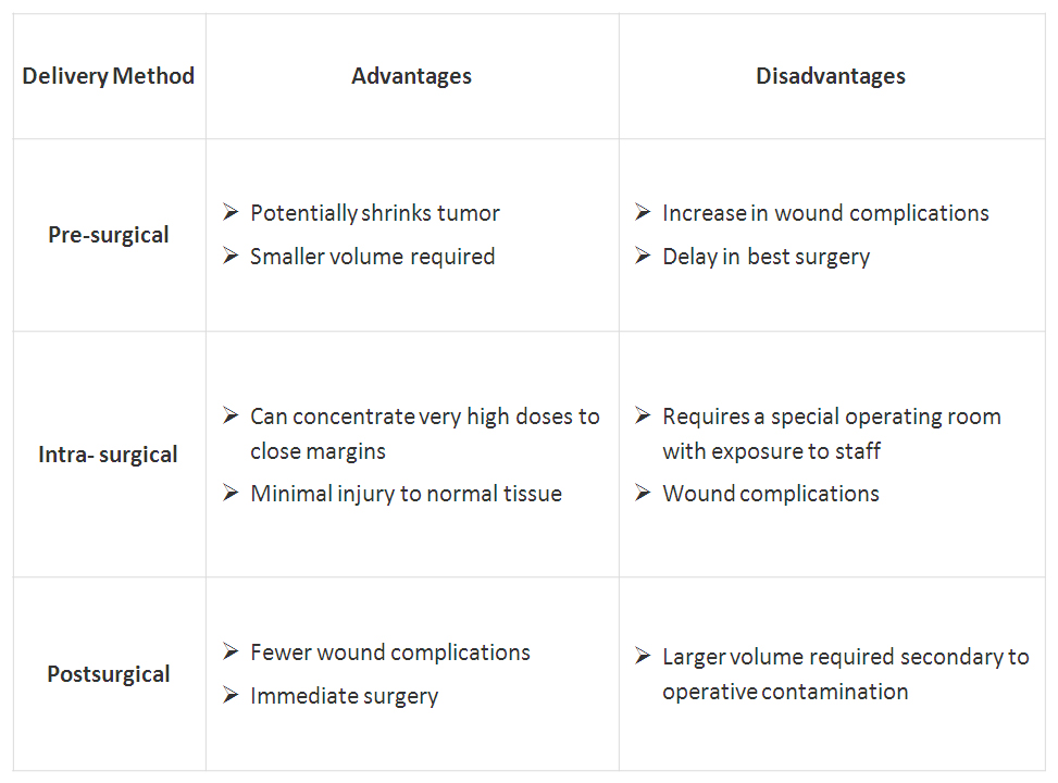
Click on table to enlarge

Adapted from sarcomahelp.org

Proton therapy (also called proton beam therapy), a type of radiation treatment that uses protons rather than x-rays is also being adapted to treat sarcoma. This mode of radiotherapy allows target the radiation much more focused at the tumor site and thus is much protective to surrounding healthy tissue. This procedure however, is currently only available in a few specialized cancer centers in the US. In addition, particle therapy treatment with heavier charged particles such as carbon ions is being used and studied for the treatment of sarcomas in Japan and Germany.

Chemotherapy

Chemotherapy is often used when a sarcoma has already spread and can be given before surgery or, after surgery as adjuvant chemotherapy to destroy any microscopic tumor cells remained after surgery.  In addition, when a tumor is considered non-operable, cycles of chemotherapy could be performed in order to shrink the tumor and make it necrotic to enable its removal by operation.

  • Watch a STS chemo + surgery Video

Different drugs are used to treat different subtypes of sarcoma. The types of chemotherapy that are used alone or in combination for most STSs include doxorubicin and ifosfamide that are the most common chemotherapy drugs employed for STS, as well as other ordinary chemotherapy drugs. The drug trabectedin, approved for use in Europe, is given for patients with advanced STS when conventional chemotherapy fails. Trabectedin has been shown to have high activity levels in the treatment of a specific subtype of liposarcoma (myxoid/round cell liposarcoma). Other chemotherapy drugs that are only used for certain subtypes of STS include: paclitaxel, docetaxel for Angiosarcoma; as well as vincristine, etoposide, actinomycin, and cyclophosphamide for Rhabdomyosarcoma and Ewing sarcoma.

Experimental chemotherapy drugs include Eribulin, a drug approved for treatment of breast cancer that has shown promising results in early clinical trials. In addition, new versions of sarcoma standard chemotherapy that cause fewer side effects are being studied in ongoing clinical trials. For instance, the three new versions of ifosfamide: palifosfamide, glufosfamide, and TH-302.

Targeted therapy

As genetic and molecular cancer research has evolved, targeted treatment to sarcoma became available. Targeted treatment to sarcoma intends to inhibit the growth and spread of cancer cells by hitting specific proteins, mainly by blocking the action of protein kinases.

Imatinib, a tyrosine-kinase inhibitor was approved in 2002 by the FDA for the treatment of gastrointestinal stromal tumor (GIST) in advanced stages and it is now the standard first-line treatment for GIST. In 2006, sunitinib multi-target receptor tyrosine kinase (RTK) inhibitor was also approved for the treatment of GIST when imatinib fails. Imatinib has been approved recently for use for patients with GIST after initial surgery, to try to prevent recurrence of the tumor. Imatinib is approved also for the treatment of advanced stage dermatofibrosarcoma protuberans (DFSP). Pazopanib, another multi-targeted inhibitor of receptor tyrosine kinase, has also been approved for patients with advanced STS as well as for use in sarcomas other than liposarcoma and GIST in conditions where standard chemotherapy is not working. Regorafenib is a new kinase inhibitor with significant activity in patients with advanced GIST who have already been treated with imatinib and suntinib. The FDA is currently reviewing a phase III clinical trial of this drug.

Closing remarks

Research efforts are made in order to elucidate new sarcoma-specific molecular targets. Studying sarcomas unique genetic fingerprints and understanding their value to sarcoma, not only can assist developing new drugs, but also may help better prediction of patients’ prognosis. To find the most effective treatment, tests to identify the genes, proteins, and other sarcoma-associated factors need to be developed and performed to give a better matched treatment for each patient.  However, being sarcoma a highly diverse group of cancers make these efforts a hard task. These issues will be discussed further in future post(s) to be published in Pharmaceutical Intelligence.

Resources

  1. http://www.cancer.net
  2. http://www.sarcomahelp.org
  3. http://www.cancer.gov
  4. http://sarcomaalliance.org
  5. http://www.sarcoma.org.uk
  6. http://www.mayoclinic.com

Additional related references

  1. Soft tissue sarcomas: ESMO Clinical Practice Guidelines for diagnosis, treatment and follow-up. Casali, PG & Blay, JY. Ann Oncol. 2010 May;21 Suppl 5:v198-203.
  2. Chemotherapy in adult soft tissue sarcoma. Jain A, Sajeevan KV, Babu KG, Lakshmaiah KC. Indian J. Cancer. 2009 Oct-Dec;46(4):274-87.
  3. State-of-the-art approach in selective curable tumours: soft tissue sarcoma. Judson I. Ann Oncol. 2008 Sep;19 Suppl 7:vii166-9.
  4. Soft tissue sarcomas of adults: state of the translational science. Borden EC, et al. Clin Cancer Res. 2003 Jun;9(6):1941-56.
  5. Management of soft-tissue sarcomas: an overview and update. Singer S, Demetri GD, Baldini EH, Fletcher CD. Lancet Oncol. 2000 Oct;1:75-85.

Videos

  1. http://www.youtube.com/watch?v=J35GBjTxzIE
  2. http://www.youtube.com/watch?v=f97oWMANXDw

Related articles on this Open Access Online Scientific Journal

  1. Clear Cell Sarcoma – Soft Tissue Melanoma: Patient’s Experience with Disease. Reporter: Aviva Lev-Ari, Ph.D., RN

Read Full Post »

Acute and Chronic Myocardial Infarction: Quantification of Myocardial Perfusion Viability – FDG-PET/MRI vs. MRI or PET alone

Author, and Content Consultant to e-SERIES A: Cardiovascular Diseases: Justin Pearlman, MD, PhD, FACC

and

Reporter: Aviva Lev-Ari, PhD, RN

The Voice of  Justin Pearlman, MD, PhD, FACC

While working on angiogenesis imaging support at Harvard, the author discovered that injured heart muscle retains gadolinium-based contrast used for perfusion testing (1992). Whereas the gadolinium passes through normal tissue in less than 20 minutes and redistributes mostly to body fat, it stays where cell membranes are damaged, even if the damage is old. The gadolinium then “lights up” the damaged zone when normal heart muscle tissues appear dark by magnetic resonance imaging (MRI). Thus “scar mapping” was born. Prior to that discovery, the standard test for “viability” was a positron emission tomography (PET) scan reporting the presents of absence of normal sugar metabolism in damaged or ischemic heart muscle. PET relies on detection of a pair of gamma rays emitted by a radioactive label in a metabolite. The pair are emitted simultaneously at nearly 180 degrees apart, with a small angulation offset from the momentum of the emitter. The emission event requires a positron finds an electron, so it does not occur precisely where the metabolite sits, and thus has inherently a poor “resolution” (minimal distance where two distinct sources are identifiable as distinct).  The protocol for PET assessment of heart muscle viability utilized by the author and other investigators required intravenous infusion of glucose, insulin, and potassium, to assure good delivery of sugar to the healthy viable heart muscle, coupled to repeat blood tests to make sure the blood sugar and serum potassium levels were adequately maintained (otherwise the patient could suffer from low sugar or a potassium-related arrhythmia). Numerous investigatores followed up on this discovery, and determined that a gadolinium demarcated defect less than half the heart wall thickness corresponded clinically to viable myocardium (meaning one could expect improved function after revascularization of a blocked blood supply) whereas a defect more than half the wall thickness corresponded clinically to non-viable myocardium (no expected significant functional gain from revascularizing that region). Subsequently, a third option for assessment of viability was developed: combined PET and MRI.

PET/MRI device produced “high-quality cardiac MR imaging acquisitions,” overcoming any technical issues of having the PET detector within the MRI’s 3-tesla magnet field, Nensa and colleagues concluded.

“No negative side effects from the integrated imaging system design were observed,” they noted.

The researchers were able to show “a close match” between FDG-PET and MRI in assessing myocardial viability and infarct quantification among patients with acute and chronic myocardial infarction.

“These findings demonstrate the feasibility of clinical cardiac MR imaging with an integrated PET/MRI device,” they added. “However, to prove that the integrated design does not interfere with the performance of the device, a systematic intraindividual comparison with a comparable 3-tesla MRI system and identical sequence parameters is still needed.”

Future studies should investigate whether hybrid FDG-PET/MRI of myocardial infarction can provide additional information compared with MRI or PET alone, according to the authors.

http://www.auntminnie.com/index.aspx?sec=sup&sub=mri&pag=dis&ItemID=103390&wf=1236

Study shows feasibility of cardiac PET/MRI — with caveats

By Wayne Forrest, AuntMinnie.com staff writer
May 9, 2013

Cardiac FDG-PET/MRI is feasible on an integrated whole-body PET/MRI system, but the hybrid modality still must prove it adds clinical relevance to cases of ischemic heart disease, according to a study published online May 7 in Radiology.

The study from University Hospital Essen in Germany found good concordance with the simultaneous acquisition of FDG-PET and MR images regarding both cine and late gadolinium-enhanced imaging in patients with myocardial infarction.

However, despite the simultaneous MRI and PET acquisition, “consolidated cardiac PET/MRI protocols need to be established, as long examination times associated with fasting seem to compromise patient compliance” with the exams, wrote lead author Dr. Felix Nensa, from the department of diagnostic and interventional radiology and neuroradiology, and colleaguesRadiology, May 7, 2013).

Cardiac feasibility study

The purpose of the study was to determine the feasibility of simultaneous acquisition of cardiac images on an integrated 3-tesla PET/MRI system, and to determine if the placement of the PET detector within the MRI’s field of magnet strength would adversely affect clinical results.

The researchers evaluated 20 consecutive patients with ischemic heart disease who were referred for FDG-PET/MRI between May and December 2012. Among the 20 patients, 14 had confirmed acute ST-elevation myocardial infarction within four to 15 days after interventional revascularization, one had suspected non-ST-elevation myocardial infarction, and five had chronic myocardial infarction.

Ten of the 20 patients underwent additional cardiac PET/CT before their PET/MRI scan.

Individuals with contraindications for gadolinium-based contrast agents and general MRI conditions, such as claustrophobia, were excluded from the study. All patients were asked to detail any personal discomfort or side effects that occurred during the PET/MRI exam.

All imaging studies were performed with an integrated whole-body PET/MRI system with 3-tesla field strength (Biograph mMR, Siemens Healthcare) and the PET insert inside the MRI scanner. All MRI sequences were performed with phased-array body surface coils designed for the PET/MRI system.

For late gadolinium-enhanced qualitative imaging, patients received gadobutrol (GadovistBayer HealthCare Pharmaceuticals) based on a dosage of 0.2 mmol/kg of body weight.

FDG-PET/MRI studies were performed after a fasting period of at least six hours, with FDG administered one hour before imaging with a mean of 202 (± 21) MBq. The scans began at a mean of 129 (± 41) minutes after FDG injection and included an electrocardiographically gated cardiac PET scan with one bed position and 3D image reconstruction.

For the FDG-PET/CT scans, an electrocardiographically gated cardiac PET/CT study was performed with a 128-slice CT unit (Biograph mCT, Siemens). PET scans began approximately 70 (± 12) minutes after FDG injection, with a mean of 211 (± 55) MBq.

Image comparisons

To compare the identification and characteristics of the infarcts between the two hybrid modalities, the researchers mapped the left ventricle with a 17-segment model, as recommended by the American Heart Association. Two-point scoring systems were used to assess myocardial tracer uptake, myocardial wall motion, and myocardial late enhancement in each segment.

In addition, the researchers measured the size of a patient’s infarct zone by drawing regions on the late gadolinium-enhanced MR images and PET images, and it was expressed as a percentage of the entire left ventricular myocardium.

Nensa and colleagues were able to complete 19 of 20 cardiac PET/MRI scans. One patient with ST-elevation myocardial infarction did not finish due to claustrophobia. Total PET/MRI scan time without patient preparation and positioning was 53 (± 3) minutes, and all cardiac MR images were rated as diagnostic in quality.

The analysis of FDG-PET and MRI with the 17-segment model found “good concordance” of the left ventricle with both cine imaging and late gadolinium-enhanced imaging in 18 of the 19 patients.

Of the 306 segments evaluated, 97 (32%) were rated as infarcted on PET images, compared with 93 (30%) rated as infarcted on late gadolinium-enhanced images and 90 (29%) on cine images.

Two-chamber views show “stunned myocardium” in a 66-year-old patient with ST-elevation myocardial infarction and acute occlusion of the left anterior descending artery. Cardiac PET/MRI was performed seven days after intervention. Late gadolinium-enhanced image (top left) shows no infarction zone. Fused late gadolinium-enhanced and PET images (top right) show that tracer uptake was reduced in segments 13-15 and 17. T2-weighted MR image (bottom left) shows myocardial edema (arrows) that corresponded well with the area of reduced tracer uptake on the bottom right image. All images courtesy of Radiology.

The size of the infarct zones averaged 22% of the entire left ventricular myocardium on PET images, compared with an average of 20% on late gadolinium-enhanced images.

Among the subgroup of 10 patients with an additional PET/CT scan, no significant difference in myocardial tracer uptake between PET/CT and PET/MR images was found.

In patient exit interviews, 16 patients cited long examination times (including patient preparation) as a source of discomfort. In addition, 11 patients cited the PET/MRI exam itself, i.e., noise, narrowness, and immobility, while 15 patients did not like having to fast.

Final conclusions

In summary, the PET/MRI device produced “high-quality cardiac MR imaging acquisitions,” overcoming any technical issues of having the PET detector within the MRI’s 3-tesla magnet field, Nensa and colleagues concluded.

“No negative side effects from the integrated imaging system design were observed,” they noted.

The researchers were able to show “a close match” between FDG-PET and MRI in assessing myocardial viability and infarct quantification among patients with acute and chronic myocardial infarction.

“These findings demonstrate the feasibility of clinical cardiac MR imaging with an integrated PET/MRI device,” they added. “However, to prove that the integrated design does not interfere with the performance of the device, a systematic intraindividual comparison with a comparable 3-tesla MRI system and identical sequence parameters is still needed.”

Future studies should investigate whether hybrid FDG-PET/MRI of myocardial infarction can provide additional information compared with MRI or PET alone, according to the authors.

Related Reading

MRI motion correction improves PET/MR image quality, July 6, 2012

SNM: PET/MRI for myocardial perfusion feasible but challenging, June 11, 2012

SNM: Hybrid PET/MRI study among top 5 research papers, June 7, 2011

Copyright © 2013 AuntMinnie.com

SOURCE:

http://www.auntminnie.com/index.aspx?sec=sup&sub=mri&pag=dis&ItemID=103390&wf=1236

SNM: Hybrid PET/MRI study among top 5 research papers

By Wayne Forrest, AuntMinnie.com staff writer

June 7, 2011 — SAN ANTONIO – The first-ever study on the clinical use of PET/MRI and a breakthrough on the use of FDG-PET to detect fevers of unknown origin were among the top research papers outlined Monday at this week’s Society of Nuclear Medicine (SNM) annual meeting.

More than 1,000 papers were submitted for consideration and presentation at this year’s meeting, with many studies showing how molecular imaging is gaining influence in the early detection of Alzheimer’s disease. Other submissions included a first-of-its-kind study on the use of near-infrared fluorescence and a new synthesized imaging agent to discover hidden blood clots in veins and arteries.

Hybrid PET/MRI

Early results from the clinical use of PET/MRI indicate that the hybrid modality can provide important diagnostic information about soft tissues and physiological functions throughout a patient’s body. The technology’s ability to find suspicious lesions and potential cancer already appears comparable to that of conventional molecular imaging methods.

In a German study, 11 patients with cancer underwent single-injection PET/CT followed by PET/MRI (Biograph mMR, Siemens Healthcare). Simultaneous PET/MRI acquisition was feasible and offered good-quality PET and MRI diagnostic data.

The analysis found that all 13 lesions detected at PET/CT were also identified by PET/MRI, with no significant difference between PET/CT and PET/MRI regarding the uptake ratios.

The study “demonstrates for the first time that newly introduced integrated whole-body MR/PET technology allows simultaneous acquisition of high-quality MR and PET data in a clinical setting within an acceptable time frame,” wrote lead study author Dr. Alexander Drzezga from TU München.

The hybrid technology could result in the development of new imaging agents that combine the diagnostic prowess of PET and MRI, Drzezga said. With the ability to image physiologic and pathophysiologic processes at the same time, the technology could open a new imaging discipline within nuclear medicine.

Carcinoma is compared in a patient who received a PET/CT scan 80 minutes after contrast injection (above), followed by a PET/MRI scan 160 minutes after contrast injection (below). All images courtesy of SNM.

FDG-PET and fever of unknown origin

Japanese researchers broke new ground in their study, which concluded that FDG-PET provided additional diagnostic information in cases of fever of unknown origin. The use of FDG-PET also had a high clinical impact, especially among patients with infectious diseases.

The retrospective study evaluated 81 consecutive patients with fever of unknown origin. They underwent FDG-PET at six Japanese institutions between July 2006 and December 2007.

Results were divided into four groups for final diagnoses: infection, arthritis/vasculitis/autoimmune/collagen disease, tumor/granuloma, and other/unknown.

The analysis found that sensitivity was highest in the tumor/granuloma group at 100% (seven of seven cases), followed by infection at 89% (24 of 27 cases) and arthritis/vasculitis/autoimmune/collagen disease at 65% (11 of 17 cases). Sensitivity was 0% (zero of one case) in the other/unknown category. Overall sensitivity was 81% and overall specificity was 75%.

Additional information provided by FDG-PET was highest in the infection group, at 76% of the cases (22 of 29), followed by tumor/granuloma at 75% (six of eight), arthritis/vasculitis/autoimmune/collagen disease at 43% (nine of 21), and other/unknown at 23% (five of 22).

The other/unknown group showed a high specificity of 84% (16 of 19 cases) and accurately excluded active focal inflammatory diseases and malignancy.

Lead study author Dr. Kozuo Kubota, PhD, chief of nuclear medicine at the National Center for Global Health and Medicine in Tokyo, said that until now, conventional modalities have produced low imaging resolution and very poor detectability for the fever’s cause.

“If the CT scan, ultrasound, or other conventional imaging technique fails, it is very difficult to find ways to find the focus of the fever,” Kubota said. “If we use FDG-PET, we can scan from head to thigh in only one scan to detect where the truly active lesion is. FDG is very sensitive both for inflammation and the tumor.”

With the addition of FDG-PET, physicians discovered a graft infection in a 50-year-old male with kidney failure and fever of unknown origin, with high FDG uptake illustrating the malady.

“I view this [study] as extraordinary,” said Dr. Michael Graham, PhD, director of nuclear medicine at the University of Iowa, who announced the top five papers. “This is in a setting where modern medicine is unable to come up with the answer, even after weeks. In about an hour-and-a-half, an FDG-PET scan came up with the answer with excellent sensitivity. We don’t get it every single time, but if it weren’t done, it would be mysterious what the patient had. It would be treated with antibiotics and hope for the best.”

“This is a huge step forward and I think it will change how we approach this problem,” he said.

PET and Alzheimer’s detection

Three studies presented at SNM 2011 added to the growing evidence that PET is an effective method to detect Alzheimer’s disease in its early stages, and that it provides a pathway to future clinical screening and treatments.

One lead study author, Dr. Kevin Ong, research scientist at Austin Hospital in Heidelberg, Australia, said that amyloid imaging with PET scans can help ascertain the likelihood that individuals will deteriorate cognitively within a few years, enabling more efficient channeling of healthcare resources.

Molecular imaging of Alzheimer’s disease has focused on detecting and analyzing the formation of the protein beta amyloid in the brain, which researchers say is directly involved in the pathology of Alzheimer’s. The presence of significant amyloid buildup is also linked to more rapid memory decline and brain atrophy.

Increased amyloid is bad for cognition even in the healthy elderly, noted lead study author Michael Devous Sr., PhD, director of neuroimaging for the Alzheimer’s Disease Center at the University of Texas Southwestern Medical Center.

The three ongoing studies involve several years of research based on hundreds of participants ranging widely in age, cognitive ability, and stage of disease.

Collective results showed that amyloid plaques build up at an estimated rate of 2% to 3% per year, and they often are already present in healthy older individuals. Amyloid plaque was present in 12% of people in their 60s, 30% of those in their 70s, and 55% of those older than 80.

In one study, approximately 25% of subjects older than 60 had amyloid plaques.

For individuals who have already developed a measurable memory decline, a positive scan for amyloid is the most accurate predictor of progression to Alzheimer’s disease, said Dr. Christopher Rowe, a lead investigator for the Australian Imaging, Biomarkers, and Lifestyle study of aging and professor of nuclear medicine at Austin Hospital.

Amyloid imaging with PET scans, he added, will be an important new tool in the assessment of cognitive decline.

Several studies have used carbon-11-labeled Pittsburgh Compound B (C-11 PIB), a PET imaging agent that binds to beta amyloid in brain tissue, but two of the current studies are assessing the benefit of F-18 florbetaben and F-18 florbetapir, which are designed for routine clinical use.

Both F-18 florbetaben and F-18 florbetapir are showing promise as reliable predictors of progression to Alzheimer’s disease, and F-18 amyloid imaging agents are considered most likely to move into clinical practice in the near future, perhaps as soon as next year.

NIRF for blood clot detection

In another novel study at SNM 2011, researchers from Massachusetts General Hospital are using near-infrared fluorescence (NIRF) and a new synthesized imaging agent to detect blood clots inside elusive veins, often within the deep tissues of the thighs and pelvis.

The agent uses a biomarker that seeks out a peptide — called fibrin — that is actively involved in the formation of blood clots. Combined with NIRF, which uses light energy to gather information from cells and tissues, the technique could also be used for coronary arteries. The fibrin peptide agent (EP-2104R) has already been tested in phase II clinical trials.

Lead study author Dr. Tetsuya Hara, PhD, noted that the availability of a high-resolution fibrin sensor is important for two reasons: intravascular NIRF imaging of coronary-sized arteries is now possible, and coupling the fibrin peptide with this technique may allow researchers to study coronary artery plaques and stents, which could potentially help identify patients at increased risk of heart attack.

The researchers were able to successfully detect fibrin-rich deep vein thrombosis with both intravital fluorescence microscopy and noninvasive fluorescence molecular tomography, allowing information to be acquired about tissues by analyzing how light is absorbed and scattered.

By coupling the fibrin peptide agent with intravascular NIRF imaging, researchers can now study microthrombi on coronary artery plaques and coronary stents that are at high risk for thrombosis and vessel occlusion.

This advance could help clinicians predict potential heart attacks and other major cardiovascular events, potentially saving more patients’ lives.

Related Reading

SNM exceeds fundraising goal, June 6, 2011

SNM: PET/MRI must prove its worth to ensure clinical adoption, June 6, 2011

PET/CT with NaF bone agent takes SNM’s Image of the Year, June 6, 2011

SNM proposes name change, May 3, 2011

SNM’s Clinical Trials Network: Progress despite growing pains, April 29, 2011

SOURCE:

http://www.auntminnie.com/index.aspx?sec=sup&sub=mol&pag=dis&ItemID=95494

Copyright © 2011 AuntMinnie.com

Other related articles published on this Open Access Online Scientific Journal, Include the following:

Sudden Cardiac Death invisible at Autopsy: Forensic Power of Postmortem MRI

Aviva Lev-Ari, PhD, RN 4/18/2013

http://pharmaceuticalintelligence.com/2013/04/18/sudden-cardiac-death-invisible-at-autopsy-forensic-power-of-postmortem-mri/

Hypervascular Hepatocellular Carcinoma: Important clues from Gadoxetic acid-based MRI imaging

Ritu Saxena, PhD 4/10/2013

http://pharmaceuticalintelligence.com/2013/04/10/hypervascular-hepatocellular-carcinoma-important-clues-from-gadoxetic-acid-based-mri-imaging/

Nanotechnology and MRI imaging

Tilda Barliya PhD 10/17/2012

http://pharmaceuticalintelligence.com/2012/10/17/nanotechnology-and-mri-imaging/

MRI Cortical Thickness Biomarker Predicts AD-like CSF and Cognitive Decline in Normal Adults

Aviva Lev-Ari, PhD, RN 7/18/2012

http://pharmaceuticalintelligence.com/2012/07/18/mri-cortical-thickness-biomarker-predicts-ad-like-csf-and-cognitive-decline-in-normal-adults/

Early Detection of Prostate Cancer: American Urological Association (AUA) Guideline

Dror Nir, PhD 5/21/2013

http://pharmaceuticalintelligence.com/2013/05/21/early-detection-of-prostate-cancer-aua-guideline/

 Whole-body imaging as cancer screening tool; answering an unmet clinical need?

Dror Nir, PhD 1/3/2013

http://pharmaceuticalintelligence.com/2013/01/03/whole-body-imaging-as-cancer-screening-tool-answering-an-unmet-clinical-need/

Read Full Post »

Prostate Cancer Molecular Diagnostic Market – the Players are: SRI Int’l, Genomic Health w/Cleveland Clinic, Myriad Genetics w/UCSF, GenomeDx and BioTheranostics

Curator: Aviva Lev-Ari, PhD, RN

On February 6, 2013 we reported that DR. MARK RUBIN, LEADING PROSTATE CANCER AND GENOMICS EXPERT, TO LEAD CUTTING-EDGE CENTER FOR TARGETED, INDIVIDUALIZED PATIENT CARE BASED ON EACH PATIENT’S GENETICS

Genomically Guided Treatment after CLIA Approval: to be offered by Weill Cornell Precision Medicine Institute

On May 16, 2013 we reported a major breakthrough in the Prostate Cancer Screening

A Blood Test to Identify Aggressive Prostate Cancer: a Discovery @ SRI International, Menlo Park, CA

After nearly a decade, my collaborators and I have found the first marker that specifically identifies the approximately six to eight percent of prostate cancers that are considered “aggressive,” meaning they will migrate to other parts of the body, at which point they are very difficult to treat. Although we have confirmed this marker, there is much to be done before a clinical application can be developed.

http://pharmaceuticalintelligence.com/2013/05/16/a-blood-test-to-identify-aggressive-prostate-cancer-a-discovery-sri-international-menlo-park-ca/

Prostate Cancer MDx Competition Heating Up; New Data from Genomic Health, Myriad

May 15, 2013

Life sciences companies are gearing up for battle to capture the profitable prostate cancer molecular diagnostic market.

Genomic Health and Myriad Genetics both made presentations to the investment community last week about their genomic tests that gauge a man’s risk of prostate cancer aggressiveness. As part of its annual investor day, Myriad discussed new data on its Prolaris test, which analyzes the expression level of 46 cell cycle progression genes and stratifies men’s risk of biochemical recurrence of prostate cancer. If the test reports low gene expression, then the patient is at low risk of disease progression, while high gene expression is associated with disease progression.

Meanwhile, around the same time last week, Genomic Health launched its Oncotype DX prostate cancer test and presented data from the first validation study involving the diagnostic. The Oncotype DX prostate cancer test analyzes the expression of 17 genes within four biological pathways to gauge prostate cancer aggressiveness. The test reports a genomic prostate score from 0 to 100; the lower the score the more certain a patient can be that they can avoid treatment and continue with active surveillance. Prostate cancer patients who are deemed to be at very low risk, low risk, or intermediate risk of progressing are eligible to be tested with the Oncotype Dx test. If, based on standard clinical measures, a person’s prostate cancer is considered high risk, then he is not a candidate for Genomic Health’s test.

These molecular tests are entering the market at a time when currently available tools aren’t specific enough to distinguish between men who have an aggressive form of prostate cancer and therefore, need invasive treatments, and those that are low risk and can do well with active surveillance. According to an NIH estimate, in 2010, the annual medical costs associated with prostate cancer in the US were $12 billion.

It is estimated that each year 23 million men undergo testing for prostate specific antigen, a protein produced by the prostate gland that increases when a man has prostate cancer. Additionally, one million men get a prostate biopsy annually, while 240,000 men end up with a diagnosis for prostate cancer, and around 30,000 die from the disease. Although most of the men diagnosed with prostate cancer end up receiving surgery or radiation treatment, as many as half of these men will probably not progress, and their disease isn’t life threatening.

While PSA testing has been shown to reduce prostate cancer deaths, a man’s PSA level may be increased for reasons other than cancer. As such, broadly screening men for PSA has been controversial in the healthcare community since the test isn’t specific enough to gauge which men are at low risk of developing aggressive prostate cancer and can forgo unnecessary treatments that can have significant side effects.

Both Myriad and Genomic Health are hoping their tests will further refine prostate cancer diagnosis and help doctors gain more confidence in determining which of their patients have aggressive disease and which are at low risk.

Myriad’s advantage

In this highly competitive space, Myriad has the first mover advantage, having launched Prolaris three years ago. The company has published four studies involving the test and conducted a number of trials analyzing around 3,000 patient samples.

Researchers from UCSF and Myriad recently published the fourth validation study in the Journal of Clinical Oncology, which analyzed samples from 400 men who had undergone a radical prostatectomy. In the published study, researchers reported that 100 percent of the men whom Prolaris deemed to be at “low risk” of progression did not experience a recurrence within the five years the study was ongoing. Meanwhile, 50 percent of those the test deemed to be a “high risk” did experience recurrence during that time (PGx Reporter 3/6/2013).

At a major medical conference recently, Myriad presented data from a study which tested biopsy samples from 141 patients treated with electron beam radiation therapy and found that the test score was significantly associated with patients’ outcome and provided information about disease progression beyond standard clinical measures. Although this finding needs to be further validated in a larger patient cohort, the researchers concluded that Prolaris “could be used to select high-risk men undergoing electron beam radiation therapy who may need combination therapy for their clinically localized prostate cancer.” In this study, around half of the cohort was African American.

Myriad has also shown in studies that its test can make accurate predictions from tissue from an initial prostate biopsy and from post-prostatectomy. The test has also shown in studies to be superior to the Gleason score, baseline PSA levels, and other prognostic factors in predicting prostate cancer-specific mortality.

Myriad has nearly completed hiring a 24-person sales force to drive sales of the test. Over the last year, Myriad has received more than 3,000 orders for its Prolaris test and 350 urologists have ordered it. The test carries a $3,400 price tag.

Although the company doesn’t have Medicare coverage yet for Prolaris, Myriad is conducting a study, called PROCEED, that it hopes will sway Medicare contractor Noridian to cover the diagnostic. The company has said it is on track to submit data from this registry to Medicare by late summer and expects to hear a decision about test coverage in calendar year 2014 (PGx Reporter 5/8/2013).

During the annual investor day last week, Myriad officials highlighted the gene panel for Prolaris, which features genes involved in cell cycle progression, and noted this as one of the advantages of its test over standard methods. “The Prolaris score measures how fast the tumor is growing. We look at the cell proliferation to look at a component of cancer that is not looked at by current clinical pathologic features,” Bill Rusconi, head of Myriad’s urology division, said.

“So, pathology like PSA score … only look at how far the tumor is progressed … [and] how advanced that tumor is. So, that’s only half of the picture because an advanced tumor could have been smoldering for 20 years, and may not go much further in the short term,” he noted. On the other hand, Rusconi added that a less advanced tumor could be progressing very quickly.

Another distinguishing point for the Prolaris test, according to Myriad, is that it is indicated for patients who are deemed to be at low and high risk by standard measures. Prostate cancer patients deemed to be at high risk of progression by standard clinical measures wouldn’t qualify for testing by Genomic Health’s test. Rusconi estimated that if Prolaris tested around 200,000 patients with localized prostate cancer to gauge the aggressiveness of their disease, the market opportunity for the test would be $700 million.

Myriad executives declined to comment on competing prostate cancer molecular tests, particularly Genomic Health’s product, noting that there isn’t a lot of published data to make any judgments. “We haven’t really seen any published data from any other competitor product. And so, I think in the absence of that, until data have made it through the peer review process and been in publication, it’s always difficult to understand exactly what type of information is available,” Mark Capone, president of Myriad Genetics Laboratories, told investors.

New competition

Like Myriad’s BRACAnalysis test, which comprises more than 80 percent of its product revenues, Genomic Health’s Oncotype DX breast cancer recurrence tests is bringing in the majority of its product revenues. However, the company believes that its newly launched Oncotype DX prostate cancer test stands to be its largest market opportunity to date.

Last week, researchers from University of California, San Francisco, presented data from the first validation study involving the Oncotype DX prostate cancer test. The study involved nearly 400 prostate cancer patients considered low or intermediate risk by standard methods such as Gleason score and showed that when the Oncotype DX score was used in conjunction with other measures, investigators identified more patients as having very low risk disease who were appropriate for active surveillance than when they diagnosed patients without the test score.

More than one third of patients classified as low risk by standard measures in the study were deemed to be “very low risk” by Oncotype DX and therefore could choose active surveillance. Meanwhile, 10 percent of patients in the study were found by clinical measures to be at very low risk or low risk, but the Oncotype DX test deemed them as having aggressive disease that needed treatment.

Matthew Cooperberg of UCSF, who presented data from this validation study at the American Urological Association’s annual meeting last week, highlighted this feature of the Oncotype DX prostate cancer test to investors during a conference call last week. He noted that the test not only gauges which low-risk patients can confidently remain with active surveillance, but it also finds those patients who didn’t receive an accurate risk assessment based on standard clinical measures. “It’s also equally important that we identify the man who frankly should not be on active surveillance, because they’re out there,” he said.

Genomic Health has aligned its test with guidelines from the National Comprehensive Cancer Network, which has expressed concern about over-diagnosis and over-treatment in prostate cancer patients. In 2010, NCCN guidelines established a new “very low risk” category for men with clinically insignificant prostate cancer and recommended that men who fall into this category and have a life expectancy of more than 20 years should only be followed with active surveillance. In 2011, NCCN made the active surveillance criteria more stringent for men in the “very low risk” category.

In order to develop the prostate cancer test, Genomic Health collaborated with the Cleveland Clinic on six feasibility studies and selected the gene expression panel after analyzing 700 genes on tissue samples from 700 patients. The commercial test analyzes the expression of 17 genes across four biological pathways.

Genomic Health executives suggested to investors that in determining the aggressiveness of prostate cancer a test that gauges critical genes in multiple pathways involved in the disease, as opposed to just one pathway, may be the better bet.

“After we selected those 700 [candidate] genes, we were completely agnostic as to what the best predictors would be. So, we let the genes do their thing and picked out the best performance,” said Eric Klein, chairman of Glickman Urological and Kidney Institute at the Cleveland Clinic and principal investigator for the original development studies for the Oncotype DX prostate cancer test. Referring to Myriad’s test, which assessed 46 cell cycle progression genes, Klein noted that while cell proliferation is important, it’s not the only pathway.

“So, I think one of the strengths of this assay is that it surveys the biology of the cancer better because it surveys other pathways,” he said. If a test only looks at genes in only one particular pathway, and the “score is low, you don’t know if you have missed the other underlying biology.”

This strategy of picking critical cancer-linked genes from multiple pathways has proven successful when launching Oncotype DX tests for breast cancer and colon cancer recurrence, company officials noted. Genomic Health’s prior experience launching molecular tests for cancer recurrence and the strength of the Oncotype DX brand will likely be advantages for the company.

Kim Popovits, CEO of Genomic Health, noted that the company has hired a “small sales force” to drive uptake of the prostate cancer test and reps will be targeting high-volume practices. “We have medical science liaisons that will be out there working to educate key opinion leaders with a similar approach to what we did in breast [cancer],” Popovits told investors. “We will begin to add to the sales organization as time goes on, as we see traction taking place, and as we move more towards payor reimbursement.”

The company plans to conduct a decision impact study as part of its effort to gain reimbursement coverage for the test. Genomic Health is also planning to do additional studies that will explore what level of active surveillance doctors should perform on patients who are deemed by the Oncotype DX test to be at very low or low risk.

The list price for the test is $3,820.

Other players

Although Myriad and Genomic Health are currently the main players in the prostate cancer molecular diagnostics space, the market will become an increasingly crowded one in the coming months.

Canadian firm GenomeDx is planning to launch a prostate cancer molecular diagnostic later this year, called Decipher. The company recently presented data at a medical conference on the test’s clinical validity and utility in predicting which patients are at risk of recurrence and metastasis after prostate cancer surgery. The company has said it has 22 studies underway with the Decipher test involving 4,000 patients (PGx Reporter 2/20/2013).

BioTheranostics recently published a study in the Proceedings of the National Academy of Sciences about its new 32-gene signature test, dubbed Prostate Cancer Index, which gauges PSA recurrence. In the study, which involved 270 tumor samples for patients treated with radical prostatectomy, the RT-PCR test (developed in collaboration with Massachusetts General Hospital) predicted PSA recurrence and had added value over standard measures such as Gleason score, tumor stage, surgical margin status, and pre-surgery PSA levels. The only other measure with significant prognostic value was surgical margin status.

The test could separate patients into groups based on PSA recurrence and whether they would develop metastatic disease within a 10-year period. PCI found that patients with a high risk score had a 14 percent risk of metastasis, while those in the low-risk group had a zero percent risk of metastasis. “In particular, this information may be useful at the biopsy stage, so that clinicians can better assess which patients can consider active surveillance versus those who should consider immediate treatment,” BioTheranostics CEO Richard Ding told PGx Reporter.

BioTheranostics has not yet determined when it will launch PCI. However, the company is planning additional follow-on studies to demonstrate the clinical utility of the test, including one study involving patients on active surveillance after having an initial prostate biopsy.

      Turna Ray is the editor of GenomeWeb’s Pharmacogenomics Reporter. She covers pharmacogenomics, personalized medicine, and companion diagnostics. E-mail Turna Ray or follow her GenomeWeb Twitter account at @PGxReporter.

Read Full Post »

Early Detection of Prostate Cancer: American Urological Association (AUA) Guideline

Author-Writer: Dror Nir, PhD

When reviewing the DETECTION OF PROSTATE CANCER section on the AUA website , The first thing that catches one’s attention is the image below; clearly showing two “guys” exploring with interest what could be a CT or MRI image…..

 fig 1

But, if you bother to read the review underneath this image regarding EARLY DETECTION OF PROSTATE CANCER: AUA GUIDELINE produced by an independent group that was commissioned by the AUA to conduct a systematic review and meta-analysis of the published literature on prostate cancer detection and screening; Panel Members: H. Ballentine Carter, Peter C. Albertsen, Michael J. Barry, Ruth Etzioni, Stephen J. Freedland, Kirsten Lynn Greene, Lars Holmberg, Philip Kantoff, Badrinath R. Konety, Mohammad Hassan Murad, David F. Penson and Anthony L. Zietman – You are bound to be left with a strong feeling that something is wrong!

The above mentioned literature review was done using rigorous approach.

“The AUA commissioned an independent group to conduct a systematic review and meta-analysis of the published literature on prostate cancer detection and screening. The protocol of the systematic review was developed a priori by the expert panel. The search strategy was developed and executed

by reference librarians and methodologists and spanned across multiple databases including Ovid Medline In-Process & Other Non-Indexed Citations, Ovid MEDLINE, Ovid EMBASE, Ovid Cochrane Database of Systematic Reviews, Ovid Cochrane Central Register of Controlled Trials and Scopus. Controlled vocabulary supplemented with keywords was used to search for the relevant concepts of prostate cancer, screening and detection. The search focused on DRE, serum biomarkers (PSA, PSA Isoforms, PSA kinetics, free PSA, complexed PSA, proPSA, prostate health index, PSA velocity, PSA

doubling time), urine biomarkers (PCA3, TMPRSS2:ERG fusion), imaging (TRUS, MRI, MRS, MR-TRUS fusion), genetics (SNPs), shared-decision making and prostate biopsy. The expert panel manually identified additional references that met the same search criteria”

While reading through the document, I was looking for the findings related to the roll of imaging in prostate cancer screening; see highlighted above. The only thing I found: “With the exception of prostate-specific antigen (PSA)-based prostate cancer screening, there was minimal evidence to assess the outcomes of interest for other tests.

This must mean that: Notwithstanding hundreds of men-years and tens of millions of dollars which were invested in studies aiming to assess the contribution of imaging to prostate cancer management, no convincing evidence to include imaging in the screening progress was found by a group of top-experts in a thorough and rigorously managed literature survey! And it actually  lead the AUA to declare that “Nothing new in the last 20 years”…..

My interpretation of this: It says-it-all on the quality of the clinical studies that were conducted during these years, aiming to develop an improved prostate cancer workflow based on imaging. I hope that whoever reads this post will agree that this is a point worth considering!

For those who do not want to bother reading the whole AUA guidelines document here is a peer reviewed summary:

Early Detection of Prostate Cancer: AUA Guideline; Carter HB, Albertsen PC, Barry MJ, Etzioni R, Freedland SJ, Greene KL, Holmberg L, Kantoff P, Konety BR, Murad MH, Penson DF, Zietman AL; Journal of Urology (May 2013)”

It says:

“A systematic review was conducted and summarized evidence derived from over 300 studies that addressed the predefined outcomes of interest (prostate cancer incidence/mortality, quality of life, diagnostic accuracy and harms of testing). In addition to the quality of evidence, the panel considered values and preferences expressed in a clinical setting (patient-physician dyad) rather than having a public health perspective. Guideline statements were organized by age group in years (age<40; 40 to 54; 55 to 69; ≥70).

RESULTS: With the exception of prostate-specific antigen (PSA)-based prostate cancer screening, there was minimal evidence to assess the outcomes of interest for other tests. The quality of evidence for the benefits of screening was moderate, and evidence for harm was high for men age 55 to 69 years. For men outside this age range, evidence was lacking for benefit, but the harms of screening, including over diagnosis and over treatment, remained. Modeled data suggested that a screening interval of two years or more may be preferred to reduce the harms of screening.

CONCLUSIONS: The Panel recommended shared decision-making for men age 55 to 69 years considering PSA-based screening, a target age group for whom benefits may outweigh harms. Outside this age range, PSA-based screening as a routine could not be recommended based on the available evidence. The entire guideline is available at www.AUAnet.org/education/guidelines/prostate-cancer-detection.cfm.”


Other research papers related to the management of Prostate cancer were published on this Scientific Web site:

From AUA2013: “Histoscanning”- aided template biopsies for patients with previous negative TRUS biopsies

Imaging-biomarkers is Imaging-based tissue characterization

On the road to improve prostate biopsy

State of the art in oncologic imaging of Prostate

Imaging agent to detect Prostate cancer-now a reality

Scientists use natural agents for prostate cancer bone metastasis treatment

Today’s fundamental challenge in Prostate cancer screening

ROLE OF VIRAL INFECTION IN PROSTATE CANCER

Men With Prostate Cancer More Likely to Die from Other Causes

New Prostate Cancer Screening Guidelines Face a Tough Sell, Study Suggests

New clinical results supports Imaging-guidance for targeted prostate biopsy

Prostate Cancer: Androgen-driven “Pathomechanism” in Early-onset Forms of the Disease

Prostate Cancer and Nanotecnology

Prostate Cancer Cells: Histone Deacetylase Inhibitors Induce Epithelial-to-Mesenchymal Transition

Imaging agent to detect Prostate cancer-now a reality

Scientists use natural agents for prostate cancer bone metastasis treatment

ROLE OF VIRAL INFECTION IN PROSTATE CANCER

Prostate Cancers Plunged After USPSTF Guidance, Will It Happen Again?

Read Full Post »

Reporter: Aviva Lev-Ari, PhD, RN


In this Journal Stent technology was researched thoroughly, the reader is advised to enrich his/hers knowledge on Re-vascularization technology by reviewing the following articles and the bibliography in each of them:

Coronary Artery Disease – Medical Devices Solutions: From First-In-Man Stent Implantation, via Medical Ethical Dilemmas to Drug Eluting Stents

Aviva Lev-Ari, PhD, RN 8/13/2012

Drug Eluting Stents: On MIT’s Edelman Lab’s Contributions to Vascular Biology and its Pioneering Research on DES

Larry H Bernstein, MD, FACP, Author and  Aviva Lev-Ari, PhD, RN, Curator 4/25/2013

Vascular Repair: Stents and Biologically Active Implants

Larry H Bernstein, MD, FACP, Author and  Aviva Lev-Ari, PhD, RN, Curator 5/4/2013

Biomaterials Technology: Models of Tissue Engineering for Reperfusion and Implantable Devices for Revascularization 5/5/2013

Revascularization: PCI, Prior History of PCI vs CABG

Aviva Lev-Ari, PhD, RN 4/25/2013

To Stent or Not? A Critical Decision

Aviva Lev-Ari, PhD, RN 10/23/2012

New Drug-Eluting Stent Works Well in STEMI

Aviva Lev-Ari, PhD, RN 8/22/2012

OrbusNeich seizes Boston Scientific stents in Germany as part of patent infringement proceedings

May 21, 2013

OrbusNeich seizes Boston Scientific stents in Germany as part of patent infringement proceedings

WIESBADEN, Germany, May 21, 2013 /PRNewswire/ — Medical device manufacturer OrbusNeich Medical Inc. and its subsidiary, OrbusNeich Medical GmbH (collectively “OrbusNeich”) today announced that it has enforced the seizure of over 190 stent systems from Boston Scientific Corporation (NYSE: BSX) in connection with its patent infringement proceedings in the Dusseldorf Regional Court. The products were found on May 15, 2013, at the premises of Boston Scientific Medizintechnik GmbH in Ratingen (Germany), the German subsidiary of Boston Scientific Corporation (collectively “Boston Scientific”).

In violation of the Court’s April 30, 2013 Preliminary Injunction, Boston Scientific initially denied access to search its premises – the court’s decision grants OrbusNeich the right to seize stents in the possession of Boston Scientific that have been commercially distributed but not yet used. Boston Scientific claimed that none of the concerned stent systems were in its possession at the location in Ratingen. Only after the Police were called did Boston Scientific allow the bailiff to search the building and seize the products.

The April 30, 2013, ruling, which Boston Scientific has appealed, allows OrbusNeich to prevent Boston Scientific from marketing and selling the affected stent lines in Germany, which include the Small Vessel, Small Workhorse and Workhorse Stents of Boston Scientific’s PROMUS Element™, PROMUS Element Plus™, OMEGA™, TAXUS Element™, SYNERGY™ and Promus PREMIER™ product lines. In this decision, the Regional Court found that the geometric pattern of these stents infringe OrbusNeich’s patent EP 1 341 482.

On May 13, 2013, OrbusNeich obtained a second Preliminary Injunction against Boston Scientific following Boston Scientific’s attempt to circumvent the first Injunction by transferring the German distribution of the affected products to Boston Scientific (UK) Ltd. and Boston Scientific Ltd. Boston Scientific may appeal this decision.

In addition to the Preliminary Injunctions, OrbusNeich’s principal patent infringement proceedings are also before the Dusseldorf Regional Court. In these proceedings, OrbusNeich is seeking damages, a permanent injunction and other relief for alleged infringement of the German parts of the EP 1 341 412 and ‘482 patents by the affected stent lines. A hearing in this main proceeding is scheduled for May 2014.

Similar infringement proceedings have also been filed in The Netherlands and Ireland.

The proceedings follow a favorable ruling for OrbusNeich by the European Patent Office (EPO) on February 11, 2013, in connection with the ‘482 patent. The EPO decision, which has been appealed, upheld the claim of the ‘482 patent, as amended, against an opposition by Boston Scientific and Terumo, claiming the patent was invalid.

About OrbusNeich

OrbusNeich is a global company that designs, develops, manufactures and markets innovative medical devices for the treatment of vascular diseases. Current products are the world’s first pro-healing stent, the Genous™ Stent, as well as other stents and balloons marketed under the names of Azule™, R stent™, Scoreflex™, Sapphire™, Sapphire II™ and Sapphire NC™. Development stage products include the COMBO Dual Therapy Stent™, the world’s first dual therapy stent. OrbusNeich is headquartered in Hong Kong and has operations in Shenzhen, China; Fort Lauderdale, Fla.; Hoevelaken, The Netherlands; and Tokyo, Japan. OrbusNeich supplies medical devices to interventional cardiologists in more than 60 countries. For more information, visit http://www.OrbusNeich.com.

Media Contact:
Jed Repko – Bryan Darrow – Taylor Ingraham
Joele Frank , Wilkinson Brimmer Katcher
212-355-4449

SOURCE: OrbusNeich Medical Inc.

Read more: OrbusNeich seizes Boston Scientific stents in Germany as part of patent infringement proceedings – FierceMedicalDevices http://www.fiercemedicaldevices.com/press-releases/orbusneich-seizes-boston-scientific-stents-germany-part-patent-infringement#ixzz2TwxCAgth

http://www.fiercemedicaldevices.com/press-releases/orbusneich-seizes-boston-scientific-stents-germany-part-patent-infringement?utm_medium=nl&utm_source=internal


Read Full Post »

See on Scoop.itCardiotoxicity

Top 10 Cardio Drugs 2012
FiercePharma
According to EvaluatePharma’s World Preview 2018 report, combined sales of antihypertensive drugs and antihyperlipidemics were more than $70 billion in 2011. That would put them at the top of the heap.

See on www.fiercepharma.com

Read Full Post »

See on Scoop.itCardiovascular Disease: PHARMACO-THERAPY

Wellcome Trust Scientists unite to solve mystery of mental illness and neurological conditions Wellcome Trust Professor Adrian Harwood, who leads research into the emerging field of cellular pharmacology, which examines the interaction of…

See on www.wellcome.ac.uk

Read Full Post »

See on Scoop.itCardiovascular Disease: PHARMACO-THERAPY

Resistant Hypertension Responds to Renal Denervation
Renal and Urology News
During the studies, all of the patients remained on their antihypertensive regimen; however, changes in their regimens were allowed as clinically indicated.

See on www.renalandurologynews.com

Read Full Post »

« Newer Posts - Older Posts »