Feeds:
Posts
Comments

Archive for the ‘Neurological Diseases’ Category

Physical activity enhances learning

Larry H. Bernstein, MD, FCAP, Curator

LPBI


Can physical activity make you learn better?

Apparently so — at least for speed of recovery of vision after an eye-patch test; may offer hope for people with traumatic brain injury or eye conditions such as amblyopia
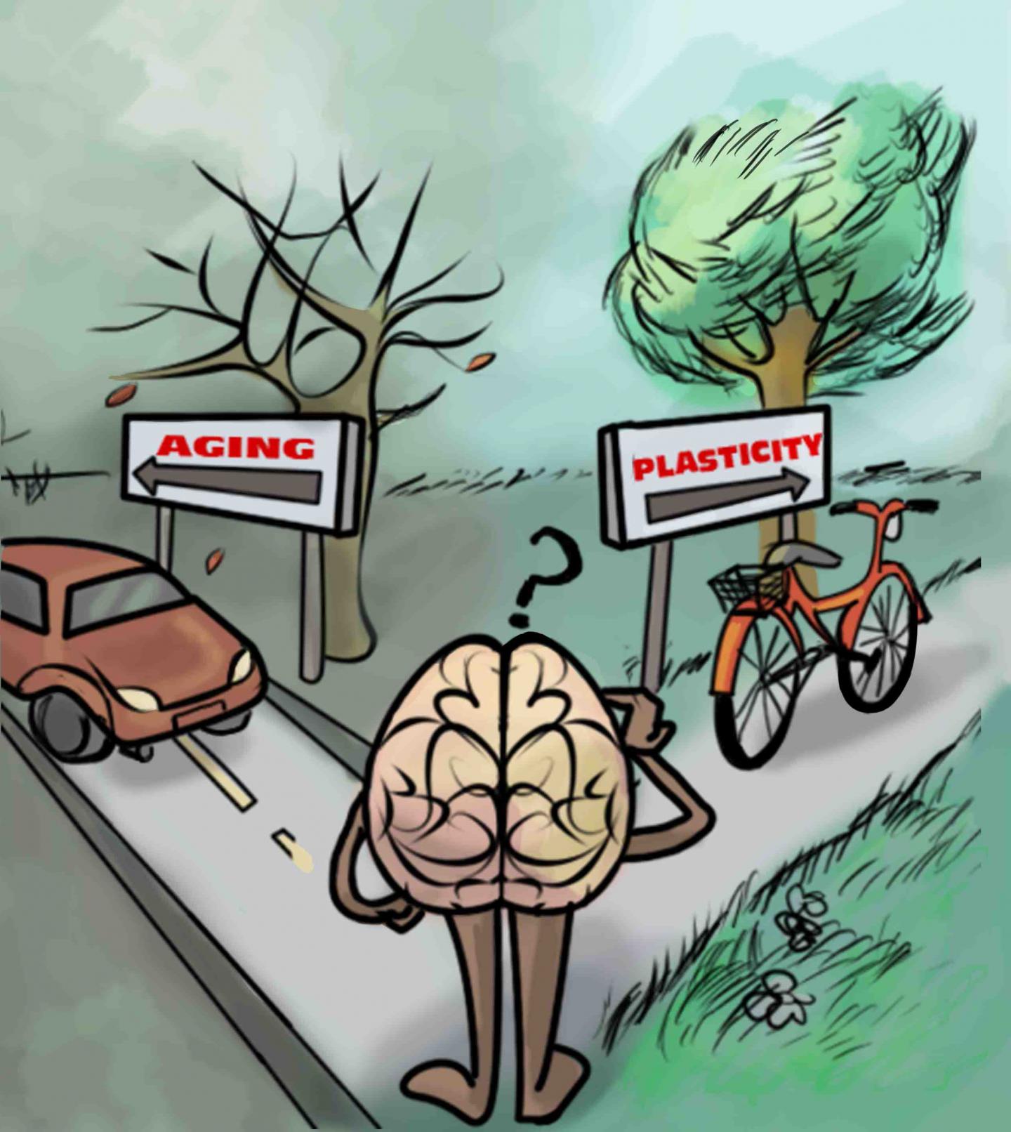
This is an artistic representation of the take home messages in Lunghi and Sale: “A cycling lane for brain rewiring,” which is that physical activity (such as cycling) is associated with increased brain plasticity. (credit: Dafne Lunghi Art)

Exercise may enhance plasticity of the adult brain — the ability of our neurons to change with experience — which is essential for learning, memory, and brain repair, Italian researchers report in an open-access paper in the Cell Press journal Current Biology.

Their research, which focused on the the visual cortex, may offer hope for people with traumatic brain injury or eye conditions such as amblyopia, the researchers suggest. “We provide the first demonstration that moderate levels of physical activity enhance neuroplasticity in the visual cortex of adult humans,” says Claudia Lunghi of the University of Pisa in Italy.

Brain plasticity is generally thought to decline with age, especially in the sensory region of the brain (such as vision). But previous studies by research colleague Alessandro Sale of the National Research Council’s Neuroscience Institute  showed that animals performing physical activity — for example rats running on a wheel — showed elevated levels of plasticity in the visual cortex and had improved recovery from amblyopia compared  to more sedentary animals.

Binocular rivalry test


http://www.kurzweilai.net/images/binocular-rivaltry-test.jpg

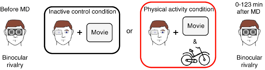
Binocular rivalry before and after “monocular deprivation” (reduced vision due to a patch) for inactive and active groups (credit: Claudia Lunghi and Alessandro Sale/Current Biology)


To find out whether the same might hold true for people, the researchers used a simple test of binocular rivalry. When people have one eye patched for a short period of time, the closed eye becomes stronger as the visual brain attempts to compensate for the lack of visual input. This recovered strength (after the eye patch is removed) is a measure of the brain’s visual plasticity.

In the new study, Lunghi and Sale put 20 adults through this test twice. In one test, participants with the dominant eye patched with a translucent material watched a movie while relaxing in a chair. In the other test, participants with one eye patched also watched a movie, but while exercising on a stationary bike for ten-minute intervals during the movie.

Exercise enhances brain plasticity (at least for vision)

Result: brain plasticity in the patched eye was enhanced by the exercise. After physical activity, the patched eye was strengthened more quickly (indicating increased levels of brain plasticity) than with the couch potatoes.

While further study is needed, the researchers think this stronger vision may have resulted from a decrease in an inhibitory neurotransmitter called GABA caused by exercise, allowing the brain to become more responsive.

The findings suggest that exercise may play an important role in brain health and recovery. This could be especially good news for people with amblyopia (called “lazy eye” because the brain “turns off” the visual processing of the weak eye to prevent double vision) — generally considered to be untreatable in adults.

Lunghi and Sale say they now plan to investigate the effects of moderate levels of physical exercise on visual function in amblyopic adult patients and to look deeper into the underlying neural mechanisms.

Time for a walk or bike ride?

UPDATE Dec. 10, 2o15: title wording changed from “smarter” to “learn better.”


Abstract of A cycling lane for brain rewiring

Brain plasticity, defined as the capability of cerebral neurons to change in response to experience, is fundamental for behavioral adaptability, learning, memory, functional development, and neural repair. The visual cortex is a widely used model for studying neuroplasticity and the underlying mechanisms. Plasticity is maximal in early development, within the so-called critical period, while its levels abruptly decline in adulthood. Recent studies, however, have revealed a significant residual plastic potential of the adult visual cortex by showing that, in adult humans, short-term monocular deprivation alters ocular dominance by homeostatically boosting responses to the deprived eye. In animal models, a reopening of critical period plasticity in the adult primary visual cortex has been obtained by a variety of environmental manipulations, such as dark exposure, or environmental enrichment, together with its critical component of enhanced physical exercise. Among these non-invasive procedures, physical exercise emerges as particularly interesting for its potential of application to clinics, though there has been a lack of experimental evidence available that physical exercise actually promotes visual plasticity in humans. Here we report that short-term homeostatic plasticity of the adult human visual cortex induced by transient monocular deprivation is potently boosted by moderate levels of voluntary physical activity. These findings could have a bearing in orienting future research in the field of physical activity application to clinical research.


Read Full Post »

Serum Folate and Homocysteine, Mood Disorders, and Aging

Larry H. Bernstein, MD, FCAP, Curator

LPBI


Dietary Folate and the Risk of Depression in Finnish MiddleAged Men

Tolmunen T, et al.
PSYCHOTHER AND PSYCHOSOM · OCT 2004; 73:334-339    DOI: http://dx.doi.org:/10.1159/000080385


Serum Folate, Vitamin B-12, and Homocysteine and Their Association With Depressive Symptoms Among U.S. Adults

M.A. BEYDOUN, M.R. SHROFF, H.A. BEYDOUN AND A.B. ZONDERMAN
PSYCHOSOM MED · NOV 2010;             DOI: http://dx.doi.org:/10.1097/PSY.0b013e3181f61863

Objective: To examine, in a nationally representative sample of U.S. adults, the associations of serum folate, vitamin B-12, and total homocysteine (tHcy) levels with depressive symptoms. Several nutritional and physiological factors have been linked to depression in adults, including low folate and vitamin B-12 and elevated tHcy levels.
Methods: Data on U.S. adults (age, 20–85 years; n 2524) from the National Health and Nutrition Examination Survey during the period 2005 to 2006 were used. Depressive symptoms were measured with the Patient Health Questionnaire (PHQ), and elevated symptoms were defined as a PHQ total score of 10. Serum folate, vitamin B-12, and tHcy were mainly expressed as tertiles. Multiple ordinary least square (OLS), logistic, and zero-inflated Poisson regression models were conducted in the main analysis.
Results: Overall, mean PHQ score was significantly higher among women compared with men. Elevated depressive symptoms (PHQ score of 10) were inversely associated with folate status, particularly among women (fully adjusted odds ratio [tertiles T3 versus T1] 0.37; 95% confidence interval, 0.17–0.86), but not significantly related to tHcy or vitamin B-12. No interaction was noted between the three exposures in affecting depressive symptoms. In older adults (50 years) and both sexes combined, tHcy was positively associated with elevated depressive symptoms (fully adjusted odds ratio [tertiles T2 versus T1] 3.01; 95% confidence interval, 1.01–9.03), although no significant dose-response relationship was found. Conclusions: Future interventions to improve mental health outcomes among U.S. adults should take into account dietary and other factors that would increase levels of serum folate.
Key words: depression, folate, vitamin B-12, homocysteine, adults.


Relationship of homocysteine, folic acid and vitamin B12 depression in a middle-aged community sample

P.S. SACHDEV, et al.   PSYCHOL MED · MAY 2005;   35, 529–538         http://dx.doi.org: /10.1017/S0033291704003721 

Background. Case control studies have supported a relationship between low folic acid and vitamin B12 and high homocysteine levels as possible predictors of depression. The results from epidemiological studies are mixed and largely from elderly populations.
Method. A random subsample of 412 persons aged 60–64 years from a larger community sample underwent psychiatric and physical assessments, and brain MRI scans. Subjects were assessed using the PRIME-MD Patient Health Questionnaire for syndromal depression and severity of depressive symptoms. Blood measures included serum folic acid, vitamin B12, homocysteine and creatinine levels, and total antioxidant capacity. MRI scans were quantified for brain atrophy, subcortical atrophy, and periventricular and deep white-matter hyperintensity on T2-weighted imaging.
Results. Being in the lowest quartile of homocysteine was associated with fewer depressive symptoms, after adjusting for sex, physical health, smoking, creatinine, folic acid and B12 levels. Being in the lowest quartile of folic acid was associated with increased depressive symptoms, after adjusting for confounding factors, but adjustment for homocysteine reduced the incidence rate ratio for folic acid to a marginal level. Vitamin B12 levels did not have a significant association with depressive symptoms. While white-matter hyperintensities had significant correlations with both homocysteine and depressive symptoms, the brain measures and total antioxidant capacity did not emerge as significant mediating variables. Conclusions. Low folic acid and high homocysteine, but not low vitamin B12 levels, are correlates of depressive symptoms in community-dwelling middle-aged individuals. The effects of folic acid and homocysteine are overlapping but distinct.


Association of folate intake with the occurrence of depressive episodes in middle-aged French men and women

P. Astorg, et al.    BRIT J  NUTR · AUG 2008; 100, 183–18       http://dx.doi.org:/10.1017/S0007114507873612

A low folate intake or a low folate status have been found to be associated with a higher frequency of depression in populations, but the existence and the direction of a causal link between folate intake or status and depression is still uncertain. The aim of this study was to seek the relation between the habitual folate intake in middle-aged men and women and the occurrence of depressive episodes. In a subsample of 1864 subjects (809 men and 1055 women) from the French SU.VI.MAX cohort, dietary habits have been measured at the beginning of the follow-up (six 24 h records) and declarations of antidepressant prescription, taken as markers of depressive episodes, have been recorded during the 8-year follow-up. No significant association was observed between folate intake and the risk of any depressive episode or of a single depressive episode during the follow-up, in both men and women. In contrast, the risk of experiencing recurrent depressive episodes (two or more) during the follow-up was strongly reduced in men with high folate intake (OR 0·25 (95% CI 0·06, 0·98) for the highest tertile v. the lowest, P for trend 0·046). This association was not observed in women. These results suggest that a low folate intake may increase the risk of recurrent depression in men.   Folate: Depression: Cohort studies


Homocysteine, vitamin B12, and folic acid levels in Alzheimer’s disease, mild cognitive impairment, and healthy elderly: baseline characteristics in subjects of the Australian Imaging Biomarker Lifestyle study.

Faux NG1, Ellis KA, Porter L, Fowler CJ,…, Ames D, Masters CL, Bush AI.
J Alzheimers Dis. 2011; 27(4):909-22.    http://dx.doi.org:/10.3233/JAD-2011-110752.

There is some debate regarding the differing levels of plasma homocysteine, vitamin B12 and serum folate between healthy controls (HC), mild cognitive impairment (MCI), and Alzheimer’s disease (AD). As part of the Australian Imaging Biomarker Lifestyle (AIBL) study of aging cohort, consisting of 1,112 participants (768 HC, 133 MCI patients, and 211 AD patients), plasma homocysteine, vitamin B12, and serum and red cell folate were measured at baseline to investigate their levels, their inter-associations, and their relationships with cognition. The results of this cross-sectional study showed that homocysteine levels were increased in female AD patients compared to female HC subjects (+16%, p-value < 0.001), but not in males. Red cell folate, but not serum folate, was decreased in AD patients compared to HC (-10%, p-value = 0.004). Composite z-scores of short- and long-term episodic memory, total episodic memory, and global cognition all showed significant negative correlations with homocysteine, in all clinical categories. Increasing red cell folate had a U-shaped association with homocysteine, so that high red cell folate levels were associated with worse long-term episodic memory, total episodic memory, and global cognition. These findings underscore the association of plasma homocysteine with cognitive deterioration, although not unique to AD, and identified an unexpected abnormality of red cell folate.


Homocysteine and folate as risk factors for dementia and Alzheimer disease1,2,3

Giovanni Ravaglia, Paola Forti, Fabiola Maioli, …., Nicoletta Brunetti, Elisa Porcellini, and Federico Licastro
Am J Clin Nutr Sept 2005; 82(3): 636-643


Background: In cross-sectional studies, elevated plasma total homocysteine (tHcy) concentrations have been associated with cognitive impairment and dementia. Incidence studies of this issue are few and have produced conflicting results.

Objective: We investigated the relation between high plasma tHcy concentrations and risk of dementia and Alzheimer disease (AD) in an elderly population.

Design: A dementia-free cohort of 816 subjects (434 women and 382 men; mean age: 74 y) from an Italian population-based study constituted our study sample. The relation of baseline plasma tHcy to the risk of newly diagnosed dementia and AD on follow-up was examined. A proportional hazards regression model was used to adjust for age, sex, education, apolipoprotein E genotype, vascular risk factors, and serum concentrations of folate and vitamin B-12.

Results: Over an average follow-up of 4 y, dementia developed in 112 subjects, including 70 who received a diagnosis of AD. In the subjects with hyperhomocysteinemia (plasma tHcy > 15 μmol/L), the hazard ratio for dementia was 2.08 (95% CI: 1.31, 3.30; P = 0.002). The corresponding hazard ratio for AD was 2.11 (95% CI: 1.19, 3.76; P = 0.011). Independently of hyperhomocysteinemia and other confounders, low folate concentrations (≤11.8 nmol/L) were also associated with an increased risk of both dementia (1.87; 95% CI: 1.21, 2.89; P = 0.005) and AD (1.98; 95% CI: 1.15, 3.40; P = 0.014), whereas the association was not significant for vitamin B-12.

Conclusions: Elevated plasma tHcy concentrations and low serum folate concentrations are independent predictors of the development of dementia and AD.


In Western societies, the prevalence and economic costs of Alzheimer disease (AD) are soaring in step with the increased number of elders in the population (1). Therefore, it is important to identify modifiable risk factors for this disease. The sulfur amino acid homocysteine is a unique candidate for this role because of its direct neurotoxicity (2–4) and its association with cerebrovascular disease (5), which is currently believed to play a significant role in AD etiology (6). Moreover, elevated concentrations of plasma total homocysteine (tHcy) are an indicator of inadequate folate and vitamin B-12 status (7) and can directly affect brain function via altered methylation reactions (8).

An association between AD and elevated tHcy concentrations has been reported in case-control (9, 10) and cross-sectional (11, 12) studies. Moreover, in nondemented elderly populations, plasma tHcy is inversely associated with poor performance at simultaneously performed tests of global cognitive function (13–15) and specific cognitive skills (13, 16). However, cross-sectional studies cannot determine causality. Only 2 longitudinal studies investigated the relation between hyperhomocysteinemia and risk of incident AD, but their results were inconsistent; the Framingham Study reported a strong association (17), and the Washington Heights–Inwood Columbia Ageing Project (WHICAP) reported no association (18). Clarification of this issue is important because consistent evidence of a prospective association between homocysteine and AD would more strongly support the need for intervention trials testing the effectiveness of homocysteine-lowering vitamin therapy in preventing dementia.

Therefore, we examined baseline plasma tHcy in relation to risk of incident dementia and AD in the Conselice Study of Brain Aging (CSBA), an Italian population-based study of older persons.

Study population

The CSBA is a population-based survey, already described in detail elsewhere (19,20), the principal aim of which is to provide data about epidemiology and risk factors for dementia in the elderly. Its design includes both cross-sectional and longitudinal components. The study was approved by the Institutional Review Board of the Department of Internal Medicine, Cardioangiology, and Hepatology, University of Bologna, and written informed consent was obtained from all participants.

Briefly, in 1999–2000, 1016 (75%) of the 1353 individuals aged ≥65 y residing in the Italian municipality of Conselice (province of Ravenna, Emilia Romagna region) participated in the prevalence study. Data on cognitive status at the follow-up examination in 2003–2004 were collected for 861 of the 937 participants free of dementia at baseline. A flow chart detailing the derivation of the incidence sample used in this study is reported in Figure 1⇓.

This prospective population-based study was the first to replicate previous findings from the Framingham Study (17), indicating that hyperhomocysteinemia doubles the risk of developing dementia and AD independently of several major confounders. Our results disagree with the negative findings recently reported in the WHICAP study (18). Possible explanations for this difference are the acknowledged insufficient statistical power of the WHICAP study, the rather homogeneously high tHcy concentrations of its sample—which did not permit enough variability to detect an association—and methodologic issues related to the prolonged time between blood sample collection and processing, which could have affected tHcy measurements.

Inconsistent results were also given by the only 2 studies that examined the association between homocysteine and cognitive decline at follow-up as measured with the MMSE (30, 31). These studies, however, differed in sample size and in which confounders were taken into account. Moreover, MMSE is a reliable global screening measure of cognitive function but was not developed to estimate changes in cognitive function or to diagnose dementia (32).

The substantial evidence that tHcy is an independent vascular risk factor (5) supports the role of hyperhomocysteinemia in AD. Subjects with vascular risk factors and cerebrovascular disease have an increased risk of AD (6), and hyperhomocysteinemia has been related to cerebral macro- and microangiopathy, endothelial dysfunction, impaired nitric oxide activity, and increased oxidative stress (33–35). Moreover, as shown in cell cultures, homocysteine can directly cause brain damage through several mechanisms: increased glutamate excitoxicity via activation of N-methyl-D-aspartate receptors (2), enhancement of β-amyloid peptide generation (4), impairment of DNA repair, and sensitization of neurons to amyloid toxicity (3).

On the basis of cross-sectional observations, some authors have suggested that elevated plasma tHcy concentrations are not a causative factor in dementia and AD but are only a marker for concomitant vascular disease, independently of cognitive status (36, 37). Results from other cross-sectional investigations (9, 12, 38), as well as those from the present investigation and the Framingham Study (17), argue against this interpretation, but only intervention trials can give the ultimate proof of a causal relation between hyperhomocysteinemia and AD.

In contrast with both the Framingham (17) and WHICAP (18) studies, we also found that, independent of homocysteine and other confounders (including vitamin B-12), low serum folate is associated with an increased risk of incident dementia and AD. Mandatory folate fortification of food might partially explain the negative results of the US studies, whereas in Italy, where folate fortification is not practiced, relative folate deficiency may be endemic among the elderly population. Nondemented patients with poor cognitive performance and AD patients often exhibit poor folate status (reviewed in 8), but only one study specifically examined B vitamins in relation to incident dementia. In a selected sample of nondemented Swedish elderly participants in the Kungsholmen Study, low serum folate and vitamin B-12 were predictive of AD at 3 y of follow-up (39). The sample, however, was small (370 subjects), and a clear association was detected only when both vitamins were taken into account.

Biologic explanatory mechanisms relating folate deficiency to dementia include impaired methylation reactions in the central nervous system, with a consequent insufficient supply of methyl groups, which are required for the synthesis of myelin, neurotransmitters, membrane phospholipids, and DNA (8). However, because of the study design and the relatively short follow-up time, we cannot definitely establish whether the independent association between low folate and dementia risk indicates an actual effect of folate status on cognitive function or, on the contrary, that subtle functional alterations may affect the dietary intake of folate in the early preclinical stages of dementia.


Neurotoxicity associated with dual actions of homocysteine at the N-methyl-D-aspartate receptor

Stuart A. Lipton*†, Won-Ki Kim‡, Yun-Beom Choi*,…, Derrick R. Arnelle§, and Jonathan S. Stamler
P
NAS 1997; 94(11):5923–5928    http://www.pnas.org/content/94/11/5923.abstract

Severely elevated levels of total homocysteine (approximately millimolar) in the blood typify the childhood disease homocystinuria, whereas modest levels (tens of micromolar) are commonly found in adults who are at increased risk for vascular disease and stroke. Activation of the coagulation system and adverse effects of homocysteine on the endothelium and vessel wall are believed to underlie disease pathogenesis. Here we show that homocysteine acts as an agonist at the glutamate binding site of the N-methyl-D-aspartate receptor, but also as a partial antagonist of the glycine coagonist site. With physiological levels of glycine, neurotoxic concentrations of homocysteine are on the order of millimolar. However, under pathological conditions in which glycine levels in the nervous system are elevated, such as stroke and head trauma, homocysteine’s neurotoxic (agonist) attributes at 10–100 μM levels outweigh its neuroprotective (antagonist) activity. Under these conditions neuronal damage derives from excessive Ca2+ influx and reactive oxygen generation. Accordingly, homocysteine neurotoxicity through overstimulation of N-methyl-D-aspartate receptors may contribute to the pathogenesis of both homocystinuria and modest hyperhomocysteinemia.


Vitamin B12 and folate in relation to the development of Alzheimer’s disease

H-X. Wang, Å. Wahlin, H. Basun, …, B. Winblad, and L. Fratiglioni
Neurology May 8, 2001; 56(9):1188-1194    http:/​/​dx.​doi.​org/​10.​1212/​WNL.​56.​9.​1188

Objective: To explore the associations of low serum levels of vitamin B12 and folate with AD occurrence.

Methods: A population-based longitudinal study in Sweden, the Kungsholmen Project. A random sample of 370 nondemented persons, aged 75 years and older and not treated with B12 and folate, was followed for 3 years to detect incident AD cases. Two cut-off points were used to define low levels of vitamin B12 (≤150 and ≤250 pmol/L) and folate (≤10 and ≤12 nmol/L), and all analyses were performed using both definitions. AD and other types of dementia were diagnosed by specialists according to DSM-III-R criteria.

Results: When using B12 ≤150pmol/L and folate ≤10 nmol/L to define low levels, compared with people with normal levels of both vitamins, subjects with low levels of B12or folate had twice higher risks of developing AD (relative risk [RR] = 2.1, 95% CI = 1.2 to 3.5). These associations were even stronger in subjects with good baseline cognition (RR = 3.1, 95% CI = 1.1 to 8.4). Similar relative risks of AD were found in subjects with low levels of B12or folate and among those with both vitamins at low levels. A comparable pattern was detected when low vitamin levels were defined as B12 ≤250 pmol/L and folate ≤12 nmol/L.

Conclusions: This study suggests that vitamin B12 and folate may be involved in the development of AD. A clear association was detected only when both vitamins were taken into account, especially among the cognitively intact subjects. No interaction was found between the two vitamins. Monitoring serum B12 and folate concentration in the elderly may be relevant for prevention of AD.


Assessing the association between homocysteine and cognition: reflections on Bradford Hill, meta-analyses, and causality

,
Hyperhomocysteinemia is a recognized risk factor for cognitive decline and incident dementia in older adults. Two recent reports addressed the cumulative epidemiological evidence for this association but expressed conflicting opinions. Here, the evidence is reviewed in relation to Sir Austin Bradford Hill’s criteria for assessing “causality,” and the latest meta-analysis of the effects of homocysteine-lowering on cognitive function is critically examined. The meta-analysis included 11 trials, collectively assessing 22 000 individuals, that examined the effects of B vitamin supplements (folic acid, vitamin B12, vitamin B6) on global or domain-specific cognitive decline. It concluded that homocysteine-lowering with B vitamin supplements has no significant effect on cognitive function. However, careful examination of the trials in the meta-analysis indicates that no conclusion can be made regarding the effects of homocysteine-lowering on cognitive decline, since the trials typically did not include individuals who were experiencing such decline. Further definitive trials in older adults experiencing cognitive decline are still urgently needed.
Mouse model for deficiency of methionine synthase reductase exhibits short-term memory impairment and disturbances in brain choline metabolism
, , , , , , ,
Biochem. J. 2014 461: 205–212    http://dx.doi.org:/10.1042/BJ20131568
Hyperhomocysteinaemia can contribute to cognitive impairment and brain atrophy. MTRR (methionine synthase reductase) activates methionine synthase, which catalyses homocysteine remethylation to methionine. Severe MTRR deficiency results in homocystinuria with cognitive and motor impairments. An MTRR polymorphism may influence homocysteine levels and reproductive outcomes. The goal of the present study was to determine whether mild hyperhomocysteinaemia affects neurological function in a mouse model with Mtrr deficiency. Mtrr+/+, Mtrr+/gt and Mtrrgt/gtmice (3 months old) were assessed for short-term memory, brain volumes and hippocampal morphology. We also measured DNA methylation, apoptosis, neurogenesis, choline metabolites and expression of ChAT (choline acetyltransferase) and AChE (acetylcholinesterase) in the hippocampus. Mtrrgt/gt mice exhibited short-term memory impairment on two tasks. They had global DNA hypomethylation and decreased choline, betaine and acetylcholine levels. Expression of ChAT and AChE was increased and decreased respectively. At 3 weeks of age, they showed increased neurogenesis. In the cerebellum, mutant mice had DNA hypomethylation, decreased choline and increased expression of ChAT. Our work demonstrates that mild hyperhomocysteinaemia is associated with memory impairment. We propose a mechanism whereby a deficiency in methionine synthesis leads to hypomethylation and compensatory disturbances in choline metabolism in the hippocampus. This disturbance affects the levels of acetylcholine, a critical neurotransmitter in learning and memory.

Read Full Post »

Remyelination of axon requires Gli1 inhibition

Larry H. Bernstein, MD, FCAP, Curator

LPBI


Inhibition of Gli1 Enhances Remyelination Abilities of Endogenous Stem Cell Populations


nodes_of_ranvier

https://beyondthedish.files.wordpress.com/2015/12/nodes_of_ranvier.jpg


If myelin is damaged, the speed of nerve impulse transmission slows substantially. Multiple sclerosis is one example of a disease that causes systematic loss of the myelin sheath. Inflammatory demyelinating diseases also cause progressive damage and loss of the myelin sheath. Regenerating the myelin sheath in these patients is one of the goals of regenerative medicine.

A good deal of data tells us that endogenous remyelination does occur. Unfortunately, this process is overwhelmed by the degree of demyelination in these diseases. A stem cell population called the parenchymal oligodendrocyte progenitor cells and endogenous adult neural stem cells in the brain are known to remyelinate demyelinated axons.

The Salzer laboratory at the New York Neuroscience Institute examined the ability of a specific adult neural stem cell population to remyelinate axons. These stem cells expressed the transcription factor Gli1.

Salzer and his team showed that this subventricular zone-specific group of neural stem cells were efficiently recruited to demyelinated portions of the brain. This same neural stem cell population was never observed entering healthy axon tracts. This finding shows that these cells seem to specialize in making new myelin sheaths for damaged axon tracts.

Since these neural stem cells expressed Gli1, and since there are drugs that can inhibit Gli1 activity, Salzer’s group wanted to show that Gli1 was a necessary factor for neural stem cell activity. Surprisingly, differentiation of these neural stem cells into oligodendrocytes (which make myelin and remyelinate axons) is significantly enhanced by inhibition of Gli1.

A specific signaling pathway called the hedgehog pathway is known to activate Gli1 and other members of the Gli gene family. However, when the hedgehog pathway in these neural stem cells was completely inhibited, it did not have the same effect and Gli1 inhibition. This suggests that Gli1 is doing more than responding to the hedgehog pathway in these neural stem cells.

Salzer and his colleagues showed that Gli1 inhibition improved myelin deposition in an animal model of experimental autoimmune encephalomyelitis; an inflammatory demyelination disease. Thus, inhibition of Gli1 activity in this preclinical model system increase regeneration of the myelin sheath in demyelinated neurons.

This work elegantly showed that endogenous neural stem cells that can remyelinate axons are present and can be activated by inhibiting Gli1. Furthermore, this activation will nicely enhance the therapeutic capacity of these endogenous cells. This potentially identifies a new therapeutic avenue for the treatment of demyelinating disorders.

This work was published in Nature. 2015 Oct 15;526(7573):448-52. doi: 10.1038/nature14957.


Inhibition of Gli1 mobilizes endogenous neural stem cells for remyelination
Inhibition of Gli1 mobilizes endogenous neural stem cells for remyelination.

 Nature. 2015 Oct 15;526(7573):448-52.   http://dx.doi.org:/10.1038/nature14957   Epub 2015 Sep 30.

Enhancing repair of myelin is an important but still elusive therapeutic goal in many neurological disorders. In multiple sclerosis, an inflammatory demyelinating disease, endogenous remyelination does occur but is frequently insufficient to restore function. Both parenchymal oligodendrocyte progenitor cells and endogenous adult neural stem cells resident within the subventricular zone are known sources of remyelinating cells. Here we characterize the contribution to remyelination of a subset of adult neural stem cells, identified by their expression of Gli1, a transcriptional effector of the sonic hedgehog pathway. We show that these cells are recruited from the subventricular zone to populate demyelinated lesions in the forebrain but never enter healthy, white matter tracts. Unexpectedly, recruitment of this pool of neural stem cells, and their differentiation into oligodendrocytes, is significantly enhanced by genetic or pharmacological inhibition of Gli1. Importantly, complete inhibition of canonical hedgehog signalling was ineffective, indicating that the role of Gli1 both in augmenting hedgehog signalling and in retarding myelination is specialized. Indeed, inhibition of Gli1 improves the functional outcome in a relapsing/remitting model of experimental autoimmune encephalomyelitis and is neuroprotective. Thus, endogenous neural stem cells can be mobilized for the repair of demyelinated lesions by inhibiting Gli1, identifying a new therapeutic avenue for the treatment of demyelinating disorders.


Read Full Post »

Retromer in neurological disorders

Larry H. Bernstein, MD, FCAP, Curator

LPBI


Retromer in Alzheimer disease, Parkinson disease and other neurological disorders.

Scott A. Small and Gregory A. Petsko
Nature Reviews Neuroscience 16; 126–132 (2015)      http://dx.doi.org:/10.1038/nrn3896


As discussed in the forum (see video here), there are many cellular pathways which are believed to be perturbed in Alzheimer’s Disease. Recent work has suggested that deficits in retromer complex function may underlie impairment of endosomal trafficking in neurons and may contribute to AD pathogenesis. This recent review illustrates the function of the retromer complex and discusses how its dysfunction may contribute to neurodegeneration.

By Tim Spencer on 24 Nov, 2015


Retromer is a protein assembly that has a central role in endosomal trafficking, and retromer dysfunction has been linked to a growing number of neurological disorders. First linked to Alzheimer disease, retromer dysfunction causes a range of pathophysiological consequences that have been shown to contribute to the core pathological features of the disease. Genetic studies have established that retromer dysfunction is also pathogenically linked to Parkinson disease, although the biological mechanisms that mediate this link are only now being elucidated. Most recently, studies have shown that retromer is a tractable target in drug discovery for these and other disorders of the nervous system.


Yeast has proved to be an informative model organism in cell biology and has provided early insight into much of the molecular machinery that mediates the intracellular transport of proteins1,2. Indeed, the term ‘retromer’ was first introduced in a yeast study in 1998 (Ref. 3). In this study, retromer was referred to as a complex of proteins that was dedicated to transporting cargo in a retrograde direction, from the yeast endosome back to the Golgi.

By 2004, a handful of studies had identified the molecular4 and the functional5, 6 homologies of the mammalian retromer, and in 2005 retromer was linked to its first human disorder, Alzheimer disease (AD)7. At the time, the available evidence suggested that the mammalian retromer might match the simplicity of its yeast homologue. Since then, a dramatic and exponential rise in research focusing on retromer has led to more than 300 publications. These studies have revealed the complexity of the mammalian retromer and its functional diversity in endosomal transport, and have implicated retromer in a growing number of neurological disorders.

New evidence indicates that retromer is a ‘master conductor’ of endosomal sorting and trafficking8. Synaptic function heavily depends on endosomal trafficking, as it contributes to the presynaptic release of neurotransmitters and regulates receptor density in the postsynaptic membrane, a process that is crucial for neuronal plasticity9. Therefore, it is not surprising that a growing number of studies are showing that retromer has an important role in synaptic biology10, 11, 12, 13. These observations may account for why the nervous system seems particularly sensitive to genetic and other defects in retromer. In this Progress article, we briefly review the molecular organization and the functional role of retromer, before discussing studies that have linked retromer dysfunction to several neurological diseases — notably, AD and Parkinson disease (PD).


The endosome is considered a hub for intracellular transport. From the endosome, transmembrane proteins can be actively sorted and trafficked to various intracellular sites via distinct transport routes (Fig. 1a). Studies have shown that the mammalian retromer mediates two of the three transport routes out of endosomes. First, retromer is involved in the retrieval of cargos from endosomes and in their delivery, in a retrograde direction, to the trans-Golgi network (TGN)5,6. Retrograde transport has many cellular functions but, as we describe, it is particularly important for the normal delivery of hydrolases and proteases to the endosomal–lysosomal system. The second transport route in which retromer functions is the recycling of cargos from endosomes back to the cell surface14, 15 (Fig. 1a). It is this transport route that is particularly important for neurons, as it mediates the normal delivery of glutamate and other receptors to the plasma membrane during synaptic remodelling and plasticity10, 11, 12, 13.

Figure 1: Retromer’s endosomal transport function and molecular organization.
Retromer's endosomal transport function and molecular organization.

a | Retromer mediates two transport routes out of endosomes via tubules that extend out of endosomal membranes. The first is the retrograde pathway in which cargo is retrieved from the endosome and trafficked to the trans-Golgi network (TGN). The second is the recycling pathway in which cargo is trafficked back from the endosome to the cell surface. The degradation pathway, which is not mediated by retromer, involves the trafficking of cargo from endosomes to lysosomes for degradation. b | The retromer assembly of proteins can be organized into distinct functional modules, all of which work together as part of retromer’s transport role. The ‘cargo-recognition core’ is the central module of the retromer assembly and comprises a trimer of proteins, in which vacuolar protein sorting-associated protein 26 (VPS26) and VPS29 bind VPS35. The ‘tubulation’ module includes protein complexes that bind the cargo-recognition core and aid in the formation and stabilization of tubules that extend out of endosomes, directing the transport of cargos towards their final destinations. The ‘membrane-recruiting’ proteins recruit the cargo-recognition core to the endosomal membrane. The WAS protein family homologue (WASH) complex of proteins also binds the cargo-recognition core and is involved in endosomal ‘actin remodelling’ to form actin patches, which are important for directing cargos towards retromer’s transport pathways. Retromer cargos includes a range of receptors — which bind the cargo-recognition core — and their ligands. PtdIns3P, phosphatidylinositol-3-phosphate.

As well as extending the endosomal transport routes, recent studies have considerably expanded the number of molecular constituents and what is known about the functional organization of the mammalian retromer. Following this expansion in knowledge of the molecular diversity and organizational complexity, retromer might be best described as a multimodular protein assembly. The protein or group of proteins that make up each module can vary, but each module is defined by its distinct function, and the modules work in unison in support of retromer’s transport role.

Two modules are considered central to the retromer assembly. First and foremost is a trimeric complex that functions as a ‘cargo-recognition core’, which selects and binds to the transmembrane proteins that need to be transported and that reside in endosomal membranes5, 6. This trimeric core comprises vacuolar protein sorting-associated protein 26 (VPS26), VPS29 and VPS35; VPS35 functions as the core’s backbone to which the other two proteins bind16. VPS26 is the only member of the core that has been found to have two paralogues, VPS26a and VPS26b17,18, and studies suggest that VPS26b might be differentially expressed in the brain19, 20. Some studies suggest that VPS26a and VPS26b are functionally redundant21, whereas others suggest that they might form distinct cargo-recognition cores20, 22.

The second central module of the retromer assembly is the ‘tubulation’ module, which is made up of proteins that work together in the formation and the stabilization of tubules that extend out of endosomes and that direct the transport of cargo towards its final destination (Fig. 1b). The proteins in this module, which directly binds the cargo-recognition core, are members of the subgroup of the sorting nexin (SNX) family that are characterized by the inclusion of a carboxy-terminal BIN–amphiphysin–RVS (BAR) domain23. These members include SNX1, SNX2, SNX5 and SNX6 (Refs 24,25). As part of the tubulation module, these SNX-BAR proteins exist in different dimeric combinations, but typically SNX1 interacts with SNX5 or SNX6, and SNX2 interacts with SNX5 or SNX6 (Refs 26,27). The EPS15-homology domain 1 (EHD1) protein can be included in this module, as it is involved in stabilizing the tubules formed by the SNX-BAR proteins28.

A third module of the retromer assembly functions to recruit the cargo-recognition core to endosomal membranes and to stabilize the core once it is there (Fig. 1b). Proteins that are part of this ‘membrane-recruiting’ module include SNX3 (Ref. 29), the RAS-related protein RAB7A30, 31,32 and TBC1 domain family member 5 (TBC1D5), which is a member of the TRE2–BUB2–CDC16 (TBC) family of RAB GTPase-activating proteins (GAPs)28. In addition, the lipid phosphatidylinositol-3-phosphate (PtdIns3P), which is found on endosomal membranes, contributes to recruiting most of the retromer-related SNXs through their phox homology domains33. Interestingly, another SNX with a phox homology domain, SNX27, was recently linked to retromer and its function15, 34. SNX27 functions as an adaptor for binding to PDZ ligand-containing cargos that are destined for transport to the cell surface via the recycling pathway. Thus, according to the functional organization of the retromer assembly, SNX27 belongs to the module that engages in cargo recognition and selection.

Recent studies have identified a fourth module of the retromer assembly. The five proteins in this module — WAS protein family homologue 1 (WASH1), FAM21, strumpellin, coiled-coil domain-containing protein 53 (CCDC53) and KIAA1033 (also known as WASH complex subunit 7) — form the WASH complex and function as an ‘actin-remodelling’ module28, 35, 36 (Fig. 1b). Specifically, the WASH complex functions in the rapid polymerization of actin to create patches of actin filaments on endosomal membranes. The complex is recruited to endosomal membranes by binding VPS35 (Ref. 28), and together they divert cargo towards retromer transport pathways and away from the degradation pathway.

The cargos that are transported by retromer include the receptors that directly bind the cargo-recognition core and the ligands of these receptors that are co-transported with the receptors. The receptors that are transported by retromer that have so far been identified to be the most relevant to neurological diseases are the family of VPS10 domain-containing receptors (including sortilin-related receptor 1 (SORL1; also known as SORLA), sortilin, and SORCS1, SORCS2 and SORCS3)7; the cation-independent mannose-6-phosphate receptor (CIM6PR)6, 5; glutamate receptors10; and phagocytic receptors that mediate the clearing function of microglia37. The most disease-relevant ligand to be identified that is trafficked as retromer cargo is the β-amyloid precursor protein (APP)7, 38, 39, 40, 41, which binds SORL1 and perhaps other VPS10 domain-containing receptors42 at the endosomal membrane.

Retromer dysfunction

Guided by retromer’s established function, and on the basis of empirical evidence, there are three well-defined pathophysiological consequences of retromer dysfunction that have proven to be relevant to AD and nervous system disorders. First, retromer dysfunction can cause cargos that typically transit rapidly through the endosome to reside in the endosome for longer than normal durations, such that they can be pathogenically processed into neurotoxic fragments (for example, APP, when stalled in the endosome, is more likely to be processed into amyloid-β, which is implicated in AD43 (Fig. 2a)). Second, by reducing endosomal outflow via impairment of the recycling pathway, retromer dysfunction can lead to a reduction in the number of cell surface receptors that are important for brain health (for example, microglia phagocytic receptors37 (Fig. 2b)).

Figure 2: The pathophysiology of retromer dysfunction.
The pathophysiology of retromer dysfunction.

Retromer dysfunction has three established pathophysiological consequences. In the examples shown, the left graphic represents a cell with normal retromer function and the right graphic represents a cell with a deficit in retromer function. a | Retromer dysfunction causes increased levels of cargo to reside in endosomes. For example, in primary neurons, retromer transports the β-amyloid precursor protein (APP) out of endosomes. Accordingly, retromer dysfunction increases APP levels in endosomes, leading to accelerated APP processing, resulting in an accumulation of neurotoxic fragments of APP (namely, β-carboxy-terminal fragment (βCTF) and amyloid-β) that are pathogenic in Alzheimer disease. b | Retromer dysfunction causes decreased cargo levels at the cell surface. For example, in microglia, retromer mediates the transport of phagocytic receptors to the cell surface and retromer dysfunction results in a decrease in the delivery of these receptors. Studies suggest that this cellular phenotype might have a pathogenic role in Alzheimer disease. c | Retromer dysfunction causes decreased delivery of proteases to the endosome. Retromer is required for the normal retrograde transport of the cation-independent mannose-6-phosphate receptor (CIM6PR) from the endosome back to the trans-Golgi network (TGN). It is in the TGN that this receptor binds cathepsin D and other proteases, and transports them to the endosome, to support the normal function of the endosomal–lysosomal system. By impairing the retrograde transport of the receptor, retromer dysfunction ultimately leads to reduced delivery of cathepsin D to this system. Cathepsin D deficiency has been shown to disrupt the endosomal–lysosomal system and to trigger tau pathology either within endosomes or secondarily in the cytosol.

The third consequence (Fig. 2c) is a result of the established role that retromer has in the retrograde transport of receptors, such as CIM6PR5, 6 or sortilin44, after these receptors transport proteases from the TGN to the endosome. Once at the endosome, the proteases disengage from the receptors, are released into endosomes and migrate to lysosomes. These proteases function in the endosomal–lysosomal system to degrade proteins, protein oligomers and aggregates45. Retromer functions to transfer the ‘naked’ receptor from the endosome back to the TGN via the retrograde pathway5, 6, allowing the receptors to continue in additional rounds of protease delivery. Accordingly, by reducing the normal retrograde transport of these receptors, retromer dysfunction has been shown to reduce the proper delivery of proteases to the endosomal–lysosomal system5,6, which, as discussed below, is a pathophysiological state linked to several brain disorders.

Although requiring further validation, recent studies suggest that retromer dysfunction might be involved in two other mechanisms that have a role in neurological disease. One study suggested that retromer might be involved in trafficking the transmembrane protein autophagy-related protein 9A (ATG9A) to recycling endosomes, from where it can then be trafficked to autophagosome precursors — a trafficking step that is crucial in the formation and the function of autophagosomes46. Autophagy is an important mechanism by which neurons clear neurotoxic aggregates that accumulate in numerous neurodegenerative diseases47. A second study has suggested that retromer dysfunction might enhance the seeding and the cell-to-cell spread of intracellular neurotoxic aggregates48, which have emerged as novel pathophysiological mechanisms that are relevant to AD49, PD50 and other neurodegenerative diseases.

Alzheimer disease

Retromer was first implicated in AD in a molecular profiling study that relied on functional imaging observations in patients and animal models to guide its molecular analysis7. Collectively, neuroimaging studies confirmed that the entorhinal cortex is the region of the hippocampal circuit that is affected first in AD, even in preclinical stages, and suggested that this effect was independent of ageing (as reviewed in Ref. 51). At the same time, neuroimaging studies identified a neighbouring hippocampal region, the dentate gyrus, that is relatively unaffected in AD52. Guided by this information, a study was carried out in which the two regions of the brain were harvested post mortem from patients with AD and from healthy individuals, intentionally covering a broad range of ages. A statistical analysis was applied to the determined molecular profiles of the regions that was designed to address the following question: among the thousands of profiled molecules, which are the ones that are differentially affected in the entorhinal cortex versus the dentate gyrus, in patients versus controls, but that are not affected by age? The final results led to the determination that the brains of patients with AD are deficient in two core retromer proteins — VPS26 and VPS35 (Ref. 7).

Little was known about the receptors of the neuronal retromer, so to understand how retromer deficiency might be mechanistically linked to AD, an analysis was carried out on the molecular data set that looked for transmembrane molecules for which expression levels correlated with VPS35 expression. The top ‘hit’ was the transcript encoding the transmembrane protein SORL1 (Ref. 43). As SORL1 belongs to the family of VPS10-containing receptors and as VPS10 is the main retromer receptor in yeast3, it was postulated that SORL1 and the family of other VPS10-containing proteins (sortillin, SORCS1, SORCS2 and SORCS3) might function as retromer receptors in neurons7. In addition, SORL1 had recently been reported to bind APP53, so if SORL1 was assumed to be a receptor that is trafficked by retromer, then APP might be the cargo that is co-trafficked by retromer. This led to a model in which retromer traffics APP out of endosomes7, which are the organelles in which APP is most likely to be cleaved by βAPP-cleaving enzyme 1 (BACE1; also known as β-secretase 1)43; this is the initial enzymatic step in the pathogenic processing of APP.

Subsequent studies were required to further establish the pathogenic link between retromer and AD, and to test the proposed model. The pathogenic link was further supported by human genetic studies. First, a genetic study investigating the association between AD, the genes encoding the components of the retromer cargo-recognition core and the family of VPS10-containing receptors found that variants of SORL1 increase the risk of developing AD38. This finding was confirmed by numerous studies, including a recent large-scale AD genome-wide association study54. Other genetic studies identified AD-associated variants in genes encoding proteins that are linked to nearly all modules of the retromer assembly55, including genes encoding proteins of the retromer tubulation module (SNX1), genes encoding proteins of the retromer membrane-recruiting module (SNX3 and RAB7A) and genes encoding proteins of the retromer actin-remodelling module (KIAA1033). In addition, nearly all of the genes encoding the family of VPS10-containing retromer receptors have been found to have variants that associate with AD56. Finally, a study found that brain regions that are differentially affected in AD are deficient in PtdIns3P, which is the phospholipid required for recruiting many sorting nexins to endosomal membranes57. Thus, together with the observation that the brains of patients with AD are deficient in VPS26a and VPS35 (Refs 7,37), all modules in the retromer assembly are implicated in AD.

Studies in mice39, 58, 59, flies39 and cells in culture34, 40, 41, 60, 61 have investigated how retromer dysfunction leads to the pathogenic processing of APP. Although rare discrepancies have been observed among these studies62, when viewed in total, the most consistent findings are that retromer dysfunction causes increased pathogenic processing of APP by increasing the time that APP resides in endosomes. Moreover, these studies have confirmed that SORL1 and other VPS10-containing proteins function as APP receptors that mediate APP trafficking out of endosomes.

Retromer has unexpectedly been linked to microglial abnormalities37 — another core feature of AD — which, on the basis of recent genetic findings, seem to have an upstream role in disease pathogenesis54, 63. A recent study found that microglia harvested from the brains of individuals with AD are deficient in VPS35 and provided evidence suggesting that retromer’s recycling pathway regulates the normal delivery of various phagocytic receptors to the cell surface of microglia37, including the phagocytic receptor triggering receptor expressed on myeloid cells 2 (TREM2) (Fig. 2b). Mutations in TREM2 have been linked to AD63, and a recent study indicates that these mutations cause a reduction in its cell surface delivery and accelerate TREM2 degradation, which suggests that the mutations are linked to a recycling defect64. While they are located at the microglial cell surface, these phagocytic receptors function in the clearance of extracellular proteins and other molecules from the extracellular space65. Taken together, these recent studies suggest that defects in the retromer’s recycling pathway can, at least in part, account for the microglial defects observed in the disease.

The microtubule-associated protein tau is the key element of neurofibrillary tangles, which are the other hallmark histological features of AD. Although a firm link between retromer dysfunction and tau toxicity remains to be established, recent insight into tau biology suggests several plausible mechanisms that are worth considering. Tau is a cytosolic protein, but nonetheless, through mechanisms that are still undetermined, it is released into the extracellular space from where it gains access to neuronal endosomes via endocytosis66, 67. In fact, recent studies suggest that the pathogenic processing of tau is triggered after it is endocytosed into neurons and while it resides in endosomes67. Of note, it still remains unknown which specific tau processing step — its phosphorylation, cleavage or aggregation — is an obligate step towards tau-related neurotoxicity. Accordingly, if defects in microglia or in other phagocytic cells reduce their capacity to clear extracellular tau, this would accelerate tau endocytosis in neurons and its pathogenic processing.

A second possibility comes from the established role retromer has in the proper delivery of cathepsin D and other proteases to the endosomal–lysosomal system via CIM6PR or sortilin (Fig. 2c). Studies in sheep, mice and flies68 have shown that cathepsin D deficiency can enhance tau toxicity and that this is mediated by a defective endosomal–lysosomal system68. Whether this mechanism leads to abnormal processing of tau within endosomes or in the cytosol via caspase activation68 remains unclear. As discussed above, retromer dysfunction will lead to a decrease in the normal delivery of cathepsin D to the endosome and will result in endosomal–lysosomal system defects. Retromer dysfunction can therefore be considered as a functional phenocopy of cathepsin D deficiency, which suggests a plausible link between retromer dysfunction and tau toxicity. Nevertheless, although these recent insights establish plausibility and support further investigation into the link between retromer and tau toxicity, whether this link exists and how it may be mediated remain open and outstanding questions.

Parkinson disease

The pathogenic link between retromer and PD is singular and straightforward: exome sequencing has identified autosomal-dominant mutations in VPS35 that cause late-onset PD69, 70, one of a handful of genetic causes of late-onset disease. However, the precise mechanism by which these mutations cause the disease is less clear.

Among a group of recent studies, all46, 48, 71, 72, 73, 74, 75, 76 but one77 strongly suggest that these mutations cause a loss of retromer function. At the molecular level, the mutations do not seem to disrupt mutant VPS35 from interacting normally with VPS26 and VPS29, and from forming the cargo-recognition core. Rather, two studies suggest that the mutations have a restricted effect on the retromer assembly but reduce the ability of VPS35 to associate with the WASH complex46, 75. Studies disagree about the pathophysiological consequences of the mutations. Four studies suggest that the mutations affect the normal retrograde transport of CIM6PR71, 73, 75, 76 from the endosome back to the TGN (Fig. 2c). In this scenario, the normal delivery of cathepsin D to the endosomal–lysosomal system should be reduced and this has been empirically shown73. Cathepsin D has been shown to be the dominant endosomal–lysosomal protease for the normal processing of α-synuclein76, and mutations could therefore lead to abnormal α-synuclein processing and to the formation of α-synuclein aggregates, which are thought to have a key pathogenic role in PD.

A separate study suggested that the mutation might cause a mistrafficking of ATG9, and thereby, as discussed above, reduce the formation and the function of autophagosomes46. Autophagosomes have also been implicated as an intracellular site in which α-synuclein aggregates are cleared. Thus, although future studies are needed to resolve these discrepant findings (which may in fact not be mutually exclusive), these studies are generally in agreement that retromer defects will probably increase the neurotoxic levels of α-synuclein aggregates48.

Several studies in flies71, 74 and in rat neuronal cultures71 provide strong evidence that increasing retromer function by overexpressing VPS35 rescues the neurotoxic effects of the most common PD-causing mutations in leucine-rich repeat kinase 2 (LRRK2). Moreover, a separate study has shown that increasing retromer levels rescues the neurotoxic effect of α-synuclein aggregates in a mouse model48. These findings have immediate therapeutic implications for drugs that increase VPS35 and retromer function, as discussed in the next section, but they also offer mechanistic insight. LRRK2 mutations were found to phenocopy the transport defects caused either by theVPS35 mutations or by knocking down VPS35 (Ref. 71). Together, this and other studies78suggest that LRRK2 might have a role in retromer-dependent transport, but future studies are required to clarify this role.

Other neurological disorders

Besides AD and PD, in which a convergence of findings has established a strong pathogenic link, retromer is being implicated in an increasing number of other neurological disorders. Below, we briefly review three disorders for which the evidence of the involvement of retromer in their pathophysiology is currently the most compelling.

The first of these disorders is Down syndrome (DS), which is caused by an additional copy of chromosome 21. Given the hundreds of genes that are duplicated in DS, it has been difficult to identify which ones drive the intellectual impairments that characterize this condition. A recent elegant study provides strong evidence that a deficiency in the retromer cargo-selection protein SNX27 might be a primary driver for some of these impairments79. This study found that the brains of individuals with DS were deficient in SNX27 and that this deficiency may be caused by an extra copy of a microRNA (miRNA) encoded by human chromosome 21 (the miRNA is produced at elevated levels and thereby decreases SNX27 expression). Consistent with the known role of SNX27 in retromer function, decreased expression of this protein in mice disrupted glutamate receptor recycling in the hippocampus and led to dendritic dysfunction. Importantly, overexpression of SNX27 rescued cognitive and other defects in animal models79, which not only strengthens the causal link between retromer dysfunction and cognitive impairment in DS but also has important therapeutic implications.

Hereditary spastic paraplegia (HSP) is another disorder linked to retromer. HSP is caused by genetic mutations that affect upper motor neurons and is characterized by progressive lower limb spasticity and weakness. Although there are numerous mutations that cause HSP, most are unified by their effects on intracellular transport80. One HSP-associated gene in particular encodes strumpellin81, which is a member of the WASH complex.

The third disorder linked to retromer is neuronal ceroid lipofuscinosis (NCL). NCL is a young-onset neurodegenerative disorder that is part of a larger family of lysosomal storage diseases and is caused by mutations in one of ten identified genes — nine neuronal ceroid lipofuscinosis (CLN) genes and the gene encoding cathepsin D82. Besides cathepsin D, for which the link to retromer has been discussed above, CLN3 seems to function in the normal trafficking of CIM6PR83. However, the most direct link to retromer has been recently described for CLN5, which seems to function, at least in part, as a retromer membrane-recruiting protein84.

Retromer as a therapeutic target

As suggested by the first study implicating retromer in AD7, and in several subsequent studies71,85, increasing the levels of retromer’s cargo-recognition core enhances retromer’s transport function. Motivated by this observation and after a decade-long search86, we identified a novel class of ‘retromer pharmacological chaperones’ that can bind and stabilize retromer’s cargo-recognition core and increase retromer levels in neurons61.

Validating the motivating hypothesis, the chaperones were found to enhance retromer function, as shown by the increased transport of APP out of endosomes and a reduction in the accumulation of APP-derived neurotoxic fragments61. Although there are numerous other pharmacological approaches for enhancing retromer function, this success provides the proof-of-principle that retromer is a tractable therapeutic target.

As retromer functions in all cells, a general concern is whether enhancing its function will have toxic adverse effects. However, studies have found that in stark contrast to even mild retromer deficiencies, increasing retromer levels has no obvious negative consequences in yeast, neuronal cultures, flies or mice40, 48, 61, 71. This might make sense because unlike drugs that, for example, function as inhibitors, simply increasing the normal flow of transport through the endosome might not be cytotoxic.

If retromer drugs are safe and can effectively enhance retromer function in the nervous system — which are still outstanding issues — there are two general indications for considering their clinical application. One rests on the idea that these agents will only be efficacious in patients who have predetermined evidence of retromer dysfunction. The most immediate example is that of individuals with PD that is caused by LRRK2 mutations. As discussed above, several ‘preclinical’ studies in flies and neuronal cultures have already established that increasing retromer levels71, 74can reverse the neurotoxic effects of such mutations and, thus, if this approach is proven to be safe, LRRK2-linked PD might be an appropriate indication for clinical trials.

Alternatively, the pathophysiology of a disease might be such that retromer-enhancing drugs would be efficacious regardless of whether there is documented evidence of retromer dysfunction. AD illustrates this point. As reviewed above, current evidence suggests that retromer-enhancing drugs will, at the very least, decrease pathogenic processing of APP in neurons and enhance microglial function, even if there are no pre-existing defects in retromer.

More generally, histological studies comparing the entorhinal cortex of patients with sporadic AD to age-matched controls have documented that enlarged endosomes are a defining cellular abnormality in AD87, 88. Importantly, enlarged endosomes are uniformly observed in a broad range of patients with sporadic AD, which suggests that enlarged endosomes reflect an intracellular site at which molecular aetiologies converge87. In addition, because they are observed in early stages of the disease in regions of the brain without evidence of amyloid pathology87, enlarged endosomes are thought to be an upstream event. Mechanistically, the most likely cause of enlarged endosomes is either too much cargo flowing into endosomes — as occurs, for example, with apolipoprotein E4 (APOE4), which has been shown to accelerate endocytosis89, 90 — or too little cargo flowing out, as observed in retromer dysfunction40, 61 and related transport defects57. By any mechanism, retromer-enhancing drugs might correct this unifying cellular defect and might be expected to be beneficial regardless of the specific aetiology.

Conclusions

The fact that retromer defects, including those derived from bona fide genetic mutations, seem to differentially target the nervous system suggests that the nervous system is differentially dependent on retromer for its normal function. We think that this reflects the unique cellular properties of neurons and how synaptic biology heavily depends on endosomal transport and trafficking. Although plausible, future studies are required to confirm and to test the details of this hypothesis.

However, currently, it is the clinical rather than the basic neuroscience of retromer that is much better understood, with the established pathophysiological consequences of retromer dysfunction providing a mechanistic link to the disorders in which retromer has been implicated. Nevertheless, many questions remain. The two most interesting questions, which are in fact inversions of each other, relate to regional vulnerability in the nervous system. First, why does retromer dysfunction target specific neuronal populations? Second, how can retromer dysfunction cause diseases that target different regions of the nervous system? Recent evidence hints at answers to both questions, which must somehow be rooted in the functional and molecular diversity of retromer.

The type and the extent of retromer defects linked to different disorders might provide pathophysiological clues as well as reasons for differential vulnerability. As discussed, in AD there seem to be across-the-board defects in retromer, such that each module of the retromer assembly as well as multiple retromer cargos have been pathogenically implicated. By contrast, the profile of retromer defects in PD seems to be more circumscribed, involving selective disruption of the interaction between VPS35 and the WASH complex. These insights might agree with histological87, 88 and large-scale genetic studies54 that suggest that endosomal dysfunction is a unifying focal point in the cellular pathogenesis of AD. In contrast, genetics and other studies91suggest that the cellular pathobiology of PD is more distributed, implicating the endosome but other organelles as well, in particular the mitochondria.

Interestingly, studies suggest that the entorhinal cortex — a region that is differentially vulnerable to AD — has unique dendritic structure and function92, which are highly dependent on endosomal transport. We speculate that it is the unique synaptic biology of the entorhinal cortex that can account for why it might be particularly sensitive to defects in endosomal transport in general and retromer dysfunction in particular, and for why this region is the early site of disease. Future studies are required to investigate this hypothesis, as well as to understand why the substantia nigra or other regions that are differentially vulnerable to PD would be particularly sensitive to the more circumscribed defect in retromer.

Perhaps the most important observation for clinical neuroscience is the now well-established fact that increasing levels of retromer proteins enhances retromer function and has already proved capable of reversing defects associated with AD, PD and DS in either cell culture or in animal models. The relationships between protein levels and function are not always simple, but emerging pharmaceutical technologies that selectively and safely increase protein levels are now a tractable goal in drug discovery93. With the evidence mounting that retromer has a pathogenic role in two of the most common neurodegenerative diseases, we think that targeting retromer to increase its functional activity is an important goal that has strong therapeutic promise.

References

  1. Schekman, R. Charting the secretory pathway in a simple eukaryote. Mol. Biol. Cell 21,3781–3784 (2010).
  2. Henne, W. M., Buchkovich, N. J. & Emr, S. D. The ESCRT pathway. Dev. Cell 21, 77–91(2011).
  3. Seaman, M. N., McCaffery, J. M. & Emr, S. D. A membrane coat complex essential for endosome-to-Golgi retrograde transport in yeast. J. Cell Biol. 142, 665–681 (1998).
  4. Haft, C. R. et al. Human orthologs of yeast vacuolar protein sorting proteins Vps26, 29, and 35: assembly into multimeric complexes. Mol. Biol. Cell 11, 4105–4116 (2000).
  5. Seaman, M. N. Cargo-selective endosomal sorting for retrieval to the Golgi requires retromer. J. Cell Biol. 165, 111–122 (2004).

Read Full Post »

Neurovascular pathways to neurodegeneration

Larry H. Bernstein, MD, FCAP, Curator

LPBI


In addition to the many cellular insults which may contribute to neurodegeneration, there is also a wealth of evidence which suggests that dysfunction of the blood-brain barrier and other CNS vascular insults may also play a key role in Alzheimer’s Disease pathogenesis. This review from Berislav Zlokovic describes much of the recent work into understand how BBB dysfunction contributes to neurodegeneration.    By Tim Spencer on 24 Nov, 2015


Neurovascular pathways to neurodegeneration in Alzheimer’s disease and other disorders.

Berislav V. Zlokovic    About the author

Nat Rev Neurosci. 2011 Nov 3;12(12):723-38.        http://dx.doi.org:/10.1038/nrn3114


The neurovascular unit (NVU) comprises brain endothelial cells, pericytes or vascular smooth muscle cells, glia and neurons. The NVU controls blood–brain barrier (BBB) permeability and cerebral blood flow, and maintains the chemical composition of the neuronal ‘milieu’, which is required for proper functioning of neuronal circuits. Recent evidence indicates that BBB dysfunction is associated with the accumulation of several vasculotoxic and neurotoxic molecules within brain parenchyma, a reduction in cerebral blood flow, and hypoxia. Together, these vascular-derived insults might initiate and/or contribute to neuronal degeneration. This article examines mechanisms of BBB dysfunction in neurodegenerative disorders, notably Alzheimer’s disease, and highlights therapeutic opportunities relating to these neurovascular deficits.


Neurons depend on blood vessels for their oxygen and nutrient supplies, and for the removal of carbon dioxide and other potentially toxic metabolites from the brain’s interstitial fluid (ISF). The importance of the circulatory system to the human brain is highlighted by the fact that although the brain comprises ~2% of total body mass, it receives up to 20% of cardiac output and is responsible for ~20% and ~25% of the body’s oxygen consumption and glucose consumption, respectively1. To underline this point, when cerebral blood flow (CBF) stops, brain functions end within seconds and damage to neurons occurs within minutes2.

Neurodegenerative disorders such as Alzheimer’s disease and amyotrophic lateral sclerosis (ALS) are associated with microvascular dysfunction and/or degeneration in the brain, neurovascular disintegration, defective blood–brain barrier (BBB) function and/or vascular factors1, 3, 4, 5, 6, 7, 8, 9, 10, 11, 12. Microvascular deficits diminish CBF and, consequently, the brain’s supply of oxygen, energy substrates and nutrients. Moreover, such deficits impair the clearance of neurotoxic molecules that accumulate and/or are deposited in the ISF, non-neuronal cells and neurons. Recent evidence suggests that vascular dysfunction leads to neuronal dysfunction and neurodegeneration, and that it might contribute to the development of proteinaceous brain and cerebrovascular ‘storage’ disorders. Such disorders include cerebral β-amyloidosis and cerebral amyloid angiopathy (CAA), which are caused by accumulation of the peptide amyloid-β in the brain and the vessel wall, respectively, and are features of Alzheimer’s disease1.

In this Review, I will discuss neurovascular pathways to neurodegeneration, placing a focus on Alzheimer’s disease because more is known about neurovascular dysfunction in this disease than in other neurodegenerative disorders. The article first examines transport mechanisms for molecules to cross the BBB, before exploring the processes that are involved in BBB breakdown at the molecular and cellular levels, and the consequences of BBB breakdown, hypoperfusion, and hypoxia and endothelial metabolic dysfunction for neuronal function. Next, the article reviews evidence for neurovascular changes during normal ageing and neurovascular BBB dysfunction in various neurodegenerative diseases, including evidence suggesting that vascular defects precede neuronal changes. Finally, the article considers specific mechanisms that are associated with BBB dysfunction in Alzheimer’s disease and ALS, and therapeutic opportunities relating to these neurovascular deficits.

The neurovascular unit

The neurovascular unit (NVU) comprises vascular cells (that is, endothelium, pericytes and vascular smooth muscle cells (VSMCs)), glial cells (that is, astrocytes, microglia and oliogodendroglia) and neurons1, 2, 13 (Fig. 1). In the NVU, the endothelial cells together form a highly specialized membrane around blood vessels. This membrane underlies the BBB and limits the entry of plasma components, red blood cells (RBCs) and leukocytes into the brain. The BBB also regulates the delivery into the CNS of circulating energy metabolites and essential nutrients that are required for proper neuronal and synaptic function. Non-neuronal cells and neurons act in concert to control BBB permeability and CBF. Vascular cells and glia are primarily responsible for maintenance of the constant ‘chemical’ composition of the ISF, and the BBB and the blood–spinal cord barrier (BSCB) work together with pericytes to prevent various potentially neurotoxic and vasculotoxic macromolecules in the blood from entering the CNS, and to promote clearance of these substances from the CNS1.

Figure 1 | Cerebral microcirculation and the neurovascular unit.

Neurovascular pathways to neurodegeneration in Alzheimer's disease and other disorders

In the brain, pial arteries run through the subarachnoid space (SAS), which contains the cerebrospinal fluid (CSF). These vessels give rise to intracerebral arteries, which penetrate into brain parenchyma. Intracerebral arteries are separated from brain parenchyma by a single, interrupted layer of elongated fibroblast-like cells of the pia and the astrocyte-derived glia limitans membrane that forms the outer wall of the perivascular Virchow–Robin space. These arteries branch into smaller arteries and subsequently arterioles, which lose support from the glia limitans and give rise to pre-capillary arterioles and brain capillaries. In an intracerebral artery, the vascular smooth muscle cell (VSMC) layer occupies most of the vessel wall. At the brain capillary level, vascular endothelial cells and pericytes are attached to the basement membrane. Pericyte processes encase most of the capillary wall, and they communicate with endothelial cells directly through synapse-like contacts containing connexins and N-cadherin. Astrocyte end-foot processes encase the capillary wall, which is composed of endothelium and pericytes. Resting microglia have a ‘ramified’ shape and can sense neuronal injury.

Transport across the blood–brain barrier. The endothelial cells that form the BBB are connected by tight and adherens junctions, and it is the tight junctions that confer the low paracellular permeability of the BBB1. Small lipophilic molecules, oxygen and carbon dioxide diffuse freely across the endothelial cells, and hence the BBB, but normal brain endothelium lacks fenestrae and has limited vesicular transport.

The high number of mitochondria in endothelial cells reflects a high energy demand for active ATP-dependent transport, conferred by transporters such as the sodium pump ((Na++K+)ATPase) and the ATP-binding cassette (ABC) efflux transporters. Sodium influx and potassium efflux across the abluminal side of the BBB is controlled by (Na++K+)ATPase (Fig. 2). Changes in sodium and potassium levels in the ISF influence the generation of action potentials in neurons and thus directly affect neuronal and synaptic functions1, 12.

Figure 2 | Blood–brain barrier transport mechanisms.

Neurovascular pathways to neurodegeneration in Alzheimer's disease and other disorders

Small lipophilic drugs, oxygen and carbon dioxide diffuse across the blood–brain barrier (BBB), whereas ions require ATP-dependent transporters such as the (Na++K+)ATPase. Transporters for nutrients include the glucose transporter 1 (GLUT1; also known as solute carrier family 2, facilitated glucose transporter member 1 (SLC2A1)), the lactate transporter monocarboxylate transporter 1 (MCT1) and the L1 and y+ transporters for large neutral and cationic essential amino acids, respectively. These four transporters are expressed at both the luminal and albuminal membranes. Non-essential amino acid transporters (the alanine, serine and cysteine preferring system (ASC), and the alanine preferring system (A)) and excitatory amino acid transporter 1 (EAAT1), EAAT2 and EAAT3 are located at the abluminal side. The ATP-binding cassette (ABC) efflux transporters that are found in the endothelial cells include multidrug resistance protein 1 (ABCB1; also known as ATP-binding cassette subfamily B member 1) and solute carrier organic anion transporter family member 1C1 (OATP1C1). Finally, transporters for peptides or proteins include the endothelial protein C receptor (EPCR) for activated protein C (APC); the insulin receptors (IRs) and the transferrin receptors (TFRs), which are associated with caveolin 1 (CAV1); low-density lipoprotein receptor-related protein 1 (LRP1) for amyloid-β, peptide transport system 1 (PTS1) for encephalins; and the PTS2 and PTS4–vasopressin V1a receptor (V1AR) for arginine vasopressin.

Brain endothelial cells express transporters that facilitate the transport of nutrients down their concentration gradients, as described in detail elsewhere1, 14 (Fig. 2). Glucose transporter 1 (GLUT1; also known as solute carrier family 2, facilitated glucose transporter member 1 (SLC2A1)) — the BBB-specific glucose transporter — is of special importance because glucose is a key energy source for the brain.

Monocarboxylate transporter 1 (MCT1), which transports lactate, and the L1 and y+ amino acid transporters are expressed at the luminal and abluminal membranes12, 14. Sodium-dependent excitatory amino acid transporter 1 (EAAT1), EAAT2 and EAAT3 are expressed at the abluminal side of the BBB15 and enable removal of glutamate, an excitatory neurotransmitter, from the brain (Fig. 2). Glutamate clearance at the BBB is essential for protecting neurons from overstimulation of glutaminergic receptors, which is neurotoxic16.

ABC transporters limit the penetration of many drugs into the brain17. For example, multidrug resistance protein 1 (ABCB1; also known as ATP-binding cassette subfamily B member 1) controls the rapid removal of ingested toxic lipophilic metabolites17 (Fig. 2). Some ABC transporters also mediate the efflux of nutrients from the endothelium into the ISF. For example, solute carrier organic anion transporter family member 1C1 (OATP1C1) transports thyroid hormones into the brain. MCT8 mediates influx of thyroid hormones from blood into the endothelium18 (Fig. 2).

The transport of circulating peptides across the BBB into the brain is restricted or slow compared with the transport of nutrients19. Carrier-mediated transport of neuroactive peptides controls their low levels in the ISF20, 21, 22, 23, 24 (Fig. 2). Some proteins, including transferrin, insulin, insulin-like growth factor 1 (IGF1), leptin25, 26, 27 and activated protein C (APC)28, cross the BBB by receptor-mediated transcytosis (Fig. 2).

Circumventricular organs. Several small neuronal structures that surround brain ventricles lack the BBB and sense chemical changes in blood or the cerebrospinal fluid (CSF) directly. These brain areas are known as circumventricular organs (CVOs). CVOs have important roles in multiple endocrine and autonomic functions, including the control of feeding behaviour as well as regulation of water and salt metabolism29. For example, the subfornical organ is one of the CVOs that are capable of sensing extracellular sodium using astrocyte-derived lactate as a signal for local neurons to initiate neural, hormonal and behavioural responses underlying sodium homeostasis30. Excessive sodium accumulation is detrimental, and increases in plasma sodium above a narrow range are incompatible with life, leading to cerebral oedema (swelling), seizures and death29.

Vascular-mediated pathophysiology

The key pathways of vascular dysfunction that are linked to neurodegenerative diseases include BBB breakdown, hypoperfusion–hypoxia and endothelial metabolic dysfunction (Fig. 3). This section examines processes that are involved in BBB breakdown at the molecular and cellular levels, and explores the consequences of all three pathways for neuronal function and viability.

Figure 3 | Vascular-mediated neuronal damage and neurodegeneration.

Neurovascular pathways to neurodegeneration in Alzheimer's disease and other disorders

a | Blood–brain barrier (BBB) breakdown that is caused by pericyte detachment leads to leakage of serum proteins and focal microhaemorrhages, with extravasation of red blood cells (RBCs). RBCs release haemoglobin, which is a source of iron. In turn, this metal catalyses the formation of toxic reactive oxygen species (ROS) that mediate neuronal injury. Albumin promotes the development of vasogenic oedema, contributing to hypoperfusion and hypoxia of the nervous tissue, which aggravates neuronal injury. A defective BBB allows several potentially vasculotoxic and neurotoxic proteins (for example, thrombin, fibrin and plasmin) to enter the brain. b | Progressive reductions in cerebral blood flow (CBF) lead to increasing neuronal dysfunction. Mild hypoperfusion, oligaemia, leads to a decrease in protein synthesis, whereas more-severe reductions in CBF, leading to hypoxia, cause an array of detrimental effects.


Blood–brain barrier breakdown. Disruption to tight and adherens junctions, an increase in bulk-flow fluid transcytosis, and/or enzymatic degradation of the capillary basement membrane cause physical breakdown of the BBB.

The levels of many tight junction proteins, their adaptor molecules and adherens junction proteins decrease in Alzheimer’s disease and other diseases that cause dementia1, 9, ALS31, multiple sclerosis32 and various animal models of neurological disease8, 33. These decreases might be partly explained by the fact that vascular-associated matrix metalloproteinase (MMP) activity rises in many neurodegenerative disorders and after ischaemic CNS injury34, 35; tight junction proteins and basement membrane extracellular matrix proteins are substrates for these enzymes34. Lowered expression of messenger RNAs that encode several key tight junction proteins, however, has also been reported in some neurodegenerative disorders, such as ALS31.

Endothelial cell–pericyte interactions are crucial for the formation36, 37 and maintenance of the BBB33, 38. Pericyte deficiency can lead to a reduction in expression of certain tight junction proteins, including occludin, claudin 5 and ZO1 (Ref. 33), and to an increase in bulk-flow transcytosis across the BBB, causing BBB breakdown38. Both processes can lead to extravasation of multiple small and large circulating macromolecules (up to 500 kDa) into the brain parenchyma33, 38. Moreover, in mice, an age-dependent progressive loss of pericytes can lead to BBB disruption and microvasular degeneration and, subsequently, neuronal dysfunction, cognitive decline and neurodegenerative changes33. In their lysosomes, pericytes concentrate and degrade multiple circulating exogenous39and endogenous proteins, including serum immunoglobulins and fibrin33, which amplify BBB breakdown in cases of pericyte deficiency.

BBB breakdown typically leads to an accumulation of various molecules in the brain. The build up of serum proteins such as immunoglobulins and albumin can cause brain oedema and suppression of capillary blood flow8, 33, whereas high concentrations of thrombin lead to neurotoxicity and memory impairment40, and accelerate vascular damage and BBB disruption41. The accumulation of plasmin (derived from circulating plasminogen) can catalyse the degradation of neuronal laminin and, hence, promote neuronal injury42, and high fibrin levels accelerate neurovascular damage6. Finally, an increase in the number of RBCs causes deposition of haemoglobin-derived neurotoxic products including iron, which generates neurotoxic reactive oxygen species (ROS)8, 43 (Fig. 3a). In addition to protein-mediated vasogenic oedema, local tissue ischaemia–hypoxia depletes ATP stores, causing (Na++K+)ATPase pumps and Na+-dependent ion channels to stop working and, consequently, the endothelium and astrocytes to swell (known as cytotoxic oedema)44. Upregulation of aquaporin 4 water channels in response to ischaemia facilitates the development of cytotoxic oedema in astrocytes45.

Hypoperfusion and hypoxia. CBF is regulated by local neuronal activity and metabolism, known as neurovascular coupling46. The pial and intracerebral arteries control the local increase in CBF that occurs during brain activation, which is termed ‘functional hyperaemia’. Neurovascular coupling requires intact pial circulation, and for VSMCs and pericytes to respond normally to vasoactive stimuli33, 46, 47. In addition to VSMC-mediated constriction and vasodilation of cerebral arteries, recent studies have shown that pericytes modulate brain capillary diameter through constriction of the vessel wall47, which obstructs capillary flow during ischaemia48. Astrocytes regulate the contractility of intracerebral arteries49, 50.

Progressive CBF reductions have increasingly serious consequences for neurons (Fig. 3b). Briefly, mild hypoperfusion — termed oligaemia — affects protein synthesis, which is required for the synaptic plasticity mediating learning and memory46. Moderate to severe CBF reductions and hypoxia affect ATP synthesis, diminishing (Na++K+)ATPase activity and the ability of neurons to generate action potentials9. In addition, such reductions can lower or increase pH, and alter electrolyte balances and water gradients, leading to the development of oedema and white matter lesions, and the accumulation of glutamate and proteinaceous toxins (for example, amyloid-β and hyperphopshorylated tau) in the brain. A reduction of greater than 80% in CBF results in neuronal death2.

The effect of CBF reductions has been extensively studied at the molecular and cellular levels in relation to Alzheimer’s disease. Reduced CBF and/or CBF dysregulation occurs in elderly individuals at high risk of Alzheimer’s disease before cognitive decline, brain atrophy and amyloid-β accumulation10, 46, 51, 52, 53, 54. In animal models, hypoperfusion can induce or amplify Alzheimer’s disease-like neuronal dysfunction and/or neuropathological changes. For example, bilateral carotid occlusion in rats causes memory impairment, neuronal dysfunction, synaptic changes and amyloid-β oligomerization55, leading to accumulation of neurotoxic amyloid-β oligomers56. In a mouse model of Alzheimer’s disease, oligaemia increases neuronal amyloid-β levels and neuronal tau phosphophorylation at an epitope that is associated with Alzheimer’s disease-type paired helical filaments57. In rodents, ischaemia leads to the accumulation of hyperphosphorylated tau in neurons and the formation of filaments that resemble those present in human neurodegenerative tauopathies and Alzheimer’s disease58. Mice expressing amyloid-β precursor protein (APP) and transforming growth factor β1 (TGFβ1) develop deficient neurovascular coupling, cholinergic denervation, enhanced cerebral and cerebrovascular amyloid-β deposition, and age-dependent cognitive decline59.

Recent studies have shown that ischaemia–hypoxia influences amyloidogenic APP processing through mechanisms that increase the activity of two key enzymes that are necessary for amyloid-β production; that is, β-secretase and γ-secretase60, 61, 62, 63. Hypoxia-inducible factor 1α (HIF1α) mediates transcriptional increase in β-secretase expression61. Hypoxia also promotes phosphorylation of tau through the mitogen-activated protein kinase (MAPK; also known as extracellular signal-regulated kinase (ERK)) pathway64, downregulates neprilysin — an amyloid-β-degrading enzyme65 — and leads to alterations in the expression of vascular-specific genes, including a reduction in the expression of the homeobox protein MOX2 gene mesenchyme homeobox 2 (MEOX2) in brain endothelial cells5 and an increase in the expression of the myocardin gene (MYOCD) in VSMCs66. In patients with Alzheimer’s disease and in models of this disorder, these changes cause vessel regression, hypoperfusion and amyloid-β accumulation resulting from the loss of the key amyloid-β clearance lipoprotein receptor (see below). In addition, hypoxia facilitates alternative splicing of Eaat2 mRNA in Alzheimer’s disease transgenic mice before amyloid-β deposition67 and suppresses glutamate reuptake by astrocytes independently of amyloid formation68, resulting in glutamate-mediated neuronal injury that is independent of amyloid-β.

In response to hypoxia, mitochondria release ROS that mediate oxidative damage to the vascular endothelium and to the selective population of neurons that has high metabolic activity. Such damage has been suggested to occur before neuronal degeneration and amyloid-β deposition in Alzheimer’s disease69, 70. Although the exact triggers of hypoxia-mediated neurodegeneration and the role of HIF1α in neurodegeneration versus preconditioning-mediated neuroprotection remain topics of debate, mitochondria-generated ROS seem to have a primary role in the regulation of the HIF1α-mediated transcriptional switch that can activate an array of responses, ranging from mechanisms that increase cell survival and adaptation to mechanisms inducing cell cycle arrest and death71. Whether inhibition of hypoxia-mediated pathogenic pathways will delay onset and/or control progression in neurodegenerative conditions such as Alzheimer’s disease remains to be determined.

When comparing the contributions of BBB breakdown and hypoperfusion to neuronal injury, it is interesting to consider Meox2+/− mice. Such animals have normal pericyte coverage and an intact BBB but a substantial perfusion deficit5 that is comparable to that found in pericyte-deficient mice that develop BBB breakdown33 Notably, however,Meox2+/− mice show less pronounced neurodegenerative changes than pericyte-deficient mice, indicating that chronic hypoperfusion–hypoxia alone can cause neuronal injury, but not to the same extent as hypoperfusion–hypoxia combined with BBB breakdown.

Endothelial neurotoxic and inflammatory factors. Alterations in cerebrovascular metabolic functions can lead to the secretion of multiple neurotoxic and inflammatory factors72, 73. For example, brain microvessels that have been isolated from individuals with Alzheimer’s disease (but not from neurologically normal age-matched and young individuals) and brain microvessels that have been treated with inflammatory proteins release neurotoxic factors that kill neurons74, 75. These factors include thrombin, the levels of which increase with the onset of Alzheimer’s disease76. Thrombin can injure neurons directly40 and indirectly by activating microglia and astrocytes73. Compared with those from age-matched controls, brain microvessels from individuals with Alzheimer’s disease secrete increased levels of multiple inflammatory mediators, such as nitric oxide, cytokines (for example, tumour necrosis factor (TNF), TGFβ1, interleukin-1β (IL-1β) and IL-6), chemokines (for example, CC-chemokine ligand 2 (CCL2; also known as monocyte chemoattractant protein 1 (MCP1)) and IL-8), prostaglandins, MMPs and leukocyte adhesion molecules73. Endothelium-derived neurotoxic and inflammatory factors together provide a molecular link between vascular metabolic dysfunction, neuronal injury and inflammation in Alzheimer’s disease and, possibly, in other neurodegenerative disorders.

Neurovascular changes

This section examines evidence for neurovascular changes during normal ageing and for neurovascular and/or BBB dysfunction in various neurodegenerative diseases, as well as the possibility that vascular defects can precede neuronal changes.

Age-associated neurovascular changes. Normal ageing diminishes brain circulatory functions, including a detectable decay of CBF in the limbic and association cortices that has been suggested to underlie age-related cognitive changes77. Alterations in the cerebral microvasculature, but not changes in neural activity, have been shown to lead to age-dependent reductions in functional hyperaemia in the visual system in cats78 and in the sensorimotor cortex in pericyte-deficient mice33. Importantly, a recent longitudinal CBF study in neurologically normal individuals revealed that people bearing the apolipoprotein E (APOE) ɛ4 allele — the major genetic risk factor for late-onset Alzheimer’s disease79, 80, 81 — showed greater regional CBF decline in brain regions that are particularly vulnerable to pathological changes in Alzheimer’s disease than did people without this allele82.

A meta-analysis of BBB permeability in 1,953 individuals showed that neurologically healthy humans had an age-dependent increase in vascular permeability83. Moreover, patients with vascular or Alzheimer’s disease-type dementia and leucoaraiosis — a small-vessel disease of the cerebral white matter — had an even greater age-dependent increase in vascular permeability83. Interestingly, an increase in BBB permeability in brain areas with normal white matter in patients with leukoaraiosis has been suggested to play a causal part in disease and the development of lacunar strokes84. Age-related changes in the permeability of the blood–CSF barrier and the choroid plexus have been reported in sheep85.

Vascular pathology. Patients with Alzheimer’s disease or other dementia-causing diseases frequently show focal changes in brain microcirculation. These changes include the appearance of string vessels (collapsed and acellular membrane tubes), a reduction in capillary density, a rise in endothelial pinocytosis, a decrease in mitochondrial content, accumulation of collagen and perlecans in the basement membrane, loss of tight junctions and/or adherens junctions3, 4, 5, 6, 9, 46, 86, and BBB breakdown with leakage of blood-borne molecules4, 6, 7, 9. The time course of these vascular alterations and how they relate to dementia and Alzheimer’s disease pathology remain unclear, as no protocol that allows the development of the diverse brain vascular pathology to be scored, and hence to be tracked with ageing, has so far been developed and widely validated87. Interestingly, a recent study involving 500 individuals who died between the ages of 69 and 103 years showed that small-vessel disease, infarcts and the presence of more than one vascular pathological change were associated with Alzheimer’s disease-type pathological lesions and dementia in people aged 75 years of age87. These associations were, however, less pronounced in individuals aged 95 years of age, mainly because of a marked ageing-related reduction in Alzheimer’s disease neuropathology relative to a moderate but insignificant ageing-related reduction in vascular pathology87.

Accumulation of amyloid-β and amyloid deposition in pial and intracerebral arteries results in CAA, which is present in over 80% of Alzheimer’s disease cases88. In patients who have Alzheimer’s disease with established CAA in small arteries and arterioles, the VSMC layer frequently shows atrophy, which causes a rupture of the vessel wall and intracerebral bleeding in about 30% of these patients89, 90. These intracerebral bleedings contribute to, and aggravate, dementia. Patients with hereditary cerebral β-amyloidosis and CAA of the Dutch, Iowa, Arctic, Flemish, Italian or Piedmont L34V type have accelerated VSMC degeneration resulting in haemorrhagic strokes and dementia91. Duplication of the gene encoding APP causes early-onset Alzheimer’s disease dementia with CAA and intracerebral haemorrhage92.

Early studies of serum immunoglobulin leakage reported that patients with ALS had BSCB breakdown and BBB breakdown in the motor cortex93. Microhaemorrhages and BSCB breakdown have been shown in the spinal cord of transgenic mice expressing mutant variants of human superoxide dismutase 1 (SOD1), which in mice cause an ALS-like disease8, 94, 95. In mice with ALS-like disease and in patients with ALS, BSCB breakdown has been shown to occur before motor neuron degeneration or brain atrophy8, 11, 95.

BBB breakdown in the substantia nigra and the striatum has been detected in murine models of Parkinson’s disease that are induced by administration of 1-methyl-4-phenyl-1,2,3,6-tetrahydropyridine (MPTP)96, 97, 98. However, the temporal relationship between BBB breakdown and neurodegeneration in Parkinson’s disease is currently unknown. Notably, the prevalence of CAA and vascular lesions increases in Parkinson’s disease99,100. Vascular lesions in the striatum and lacunar infarcts can cause vascular parkinsonism syndrome101. A recent study reported BBB breakdown in a rat model of Huntington’s disease that is induced with the toxin 3-nitropropionic acid102.

Several studies have established disruption of BBB with a loss of tight junction proteins during neuroinflammatory conditions such as multiple sclerosis and its murine model, experimental allergic encephalitis. Such disruption facilitates leukocyte infiltration, leading to oliogodendrocyte death, axonal damage, demyelination and lesion development32.

Functional changes in the vasculature. In individuals with Alzheimer’s disease, GLUT1 expression at the BBB decreases103, suggesting a shortage in necessary metabolic substrates. Studies using 18F-fluorodeoxyglucose (FDG) positron emission tomography (PET) have identified reductions in glucose uptake in asymptomatic individuals with a high risk of dementia104, 105. Several studies have suggested that reduced glucose uptake across the BBB, as seen by FDG PET, precedes brain atrophy104, 105, 106, 107, 108.

Amyloid-β constricts cerebral arteries109. In a mouse model of Alzheimer’s disease, impairment of endothelium-dependent regulation of neocortical microcirculation110, 111occurs before amyloid-β accumulation. Recent studies have shown that CD36, a scavenger receptor that binds amyloid-β, is essential for the vascular oxidative stress and diminished functional hyperaemia that occurs in response to amyloid-β exposure112. Neuroimaging studies in patients with Alzheimer’s disease have shown that neurovascular uncoupling occurs before neurodegenerative changes10, 51, 52, 53. Moreover, cognitively normal APOE ɛ4 carriers at risk of Alzheimer’s disease show impaired CBF responses to brain activation in the absence of neurodegenerative changes or amyloid-β accumulation54. Recently, patients with Alzheimer’s disease as well as mouse models of this disease with high cerebrovascular levels of serum response factor (SRF) and MYOCD, the two transcription factors that control VSMC differentiation, have been shown to develop a hypercontractile arterial phenotype resulting in brain hypoperfusion, diminished functional hyperaemia and CAA66, 113. More work is needed to establish the exact role of SRF and MYOCD in the vascular dysfunction that results in the Alzheimer’s disease phenotype and CAA.

PET studies with 11C-verapamil, an ABCB1 substrate, have indicated that the function of ABCB1, which removes multiple drugs and toxins from the brain, decreases with ageing114 and is particularly compromised in the midbrain of patients with Parkinson’s disease, progressive supranuclear palsy or multiple system atrophy115. More work is needed to establish the exact roles of ABC BBB transporters in neurodegeneration and whether their failure precedes the loss of dopaminergic neurons that occurs in Parkinson’s disease.

In mice with ALS-like disease and in patients with ALS, hypoperfusion and/or dysregulated CBF have been shown to occur before motor neuron degeneration or brain atrophy8, 116. Reduced regional CBF in basal ganglia and reduced blood volume have been reported in pre-symptomatic gene-tested individuals at risk for Huntington’s disease117. Patients with Huntington’s disease display a reduction in vasomotor activity in the cerebral anterior artery during motor activation118.

Vascular and neuronal common growth factors. Blood vessels and neurons share common growth factors and molecular pathways that regulate their development and maintenance119, 120. Angioneurins are growth factors that exert both vasculotrophic and neurotrophic activities121. The best studied angioneurin is vascular endothelial growth factor (VEGF). VEGF regulates vessel formation, axonal growth and neuronal survival120. Ephrins, semaphorins, slits and netrins are axon guidance factors that also regulate the development of the vascular system121. During embryonic development of the neural tube, blood vessels and choroid plexus secrete IGF2 into the CSF, which regulates the proliferation of neuronal progenitor cells122. Genetic and pharmacological manipulations of angioneurin activity yielded various vascular and cerebral phenotypes121. Given the dual nature of angioneurin action, these studies have not been able to address whether neuronal dysfunction results from a primary insult to neurons and/or whether it is secondary to vascular dysfunction.

Increased levels of VEGF, a hypoxia-inducible angiogenic factor, were found in the walls of intraparenchymal vessels, perivascular deposits, astrocytes and intrathecal space of patients with Alzheimer’s disease, and were consistent with the chronic cerebral hypoperfusion and hypoxia that were observed in these individuals73. In addition to VEGF, brain microvessels in Alzheimer’s disease release several molecules that can influence angiogenesis, including IL-1β, IL-6, IL-8, TNF, TGFβ, MCP1, thrombin, angiopoietin 2, αVβ3 and αVβ5 integrins, and HIF1α73. However, evidence for increased vascularity in Alzheimer’s disease is lacking. On the contrary, several studies have reported that focal vascular regression and diminished microvascular density occur in Alzheimer’s disease4, 5, 73 and in Alzheimer’s disease transgenic mice123. The reason for this discrepancy is not clear. The anti-angiogenic activity of amyloid-β, which accumulates in the brains of individuals with Alzheimer’s disease and Alzheimer’s disease models, may contribute to hypovascularity123. Conversely, genome-wide transcriptional profiling of brain endothelial cells from patients with Alzheimer’s disease revealed that extremely low expression of vascular-restricted MEOX2 mediates aberrant angiogenic responses to VEGF and hypoxia, leading to capillary death5. This finding raises the interesting question of whether capillary degeneration in Alzheimer’s disease results from unsuccessful vascular repair and/or remodelling. Moreover, mice that lack one Meox2allele have been shown to develop a primary cerebral endothelial hypoplasia with chronic brain hypoperfusion5, resulting in secondary neurodegenerative changes33.

Does vascular dysfunction cause neuronal dysfunction? In summary, the evidence that is discussed above clearly indicates that vascular dysfunction is tightly linked to neuronal dysfunction. There are many examples to illustrate that primary vascular deficits lead to secondary neurodegeneration, including CADASIL (cerebral autosomal dominant arteriopathy with subcortical infarcts), an hereditary small-vessel brain disease resulting in multiple small ischaemic lesions, neurodegeneration and dementia124; mutations in SLC2A1 that cause dysfunction of the BBB-specific GLUT1 transporter in humans resulting in seizures; cognitive impairment and microcephaly125; microcephaly and epileptiform discharges in mice with genetic deletion of a single Slc2a1 allele126; and neurodegeneration mediated by a single Meox2 homebox gene deletion restricted to the vascular system33. Patients with hereditary cerebral β-amyloidosis and CAA of the Dutch, Iowa, Arctic, Flemish, Italian or Piedmont L34V type provide another example showing that primary vascular dysfunction — which in this case is caused by deposition of vasculotropic amyloid-β mutants in the arterial vessel wall — leads to dementia and intracerebral bleeding. Moreover, as reviewed in the previous sections, recent evidence suggests that BBB dysfunction and/or breakdown, and CBF reductions and/or dysregulation may occur in sporadic Alzheimer’s disease and experimental models of this disease before cognitive decline, amyloid-β deposition and brain atrophy. In patients with ALS and in some experimental models of ALS, CBF dysregulation, BSCB breakdown and spinal cord hypoperfusion have been reported to occur before motor neuron cell death. Whether neurological changes follow or precede vascular dysfunction in Parkinson’s disease, Huntington’s disease and multiple sclerosis remains less clear. However, there is little doubt that vascular injury mediates, amplifies and/or lowers the threshold for neuronal dysfunction and loss in several neurological disorders.

Disease-specific considerations

This section examines how amyloid-β levels are kept low in the brain, a process in which the BBB has a central role, and how faulty BBB-mediated clearance mechanisms go awry in Alzheimer’s disease. On the basis of this evidence and the findings discussed elsewhere in the Review, a new hypothesis for the pathogenesis of Alzheimer’s disease that incorporates the vascular evidence is presented. ALS-specific disease mechanisms relating to the BBB are then examined.

Alzheimer’s disease. Amyloid-β clearance from the brain by the BBB is the best studied example of clearance of a proteinaceous toxin from the CNS. Multiple pathways regulate brain amyloid-β levels, including its production and clearance (Fig. 4). Recent studies127,128, 129 have confirmed earlier findings in multiple rodent and non-human primate models demonstrating that peripheral amyloid-β is an important precursor of brain amyloid-β130, 131, 132, 133, 134, 135, 136. Moreover, peripheral amyloid-β sequestering agents such as soluble LRP1 (ref.137), anti-amyloid-β antibodies138, 139,140, gelsolin and the ganglioside GM1 (Ref. 141), or systemic expression of neprilysin142, 143 have been shown to reduce the amyloid burden in Alzheimer’s disease mice by eliminating contributions of the peripheral amyloid-β pool to the total brain pool of this peptide.

Figure 4 | The role of blood–brain barrier transport in brain homeostasis of amyloid-β.

Neurovascular pathways to neurodegeneration in Alzheimer's disease and other disorders

Amyloid-β (Aβ) is produced from the amyloid-β precursor protein (APP), both in the brain and in peripheral tissues. Clearance of amyloid-β from the brain normally maintains its low levels in the brain. This peptide is cleared across the blood–brain barrier (BBB) by the low-density lipoprotein receptor-related protein 1 (LRP1). LRP1 mediates rapid efflux of a free, unbound form of amyloid-β and of amyloid-β bound to apolipoprotein E2 (APOE2), APOE3 or α2-macroglobulin (not shown) from the brain’s interstitial fluid into the blood, and APOE4 inhibits such transport. LRP2 eliminates amyloid-β that is bound to clusterin (CLU; also known as apolipoprotein J (APOJ)) by transport across the BBB, and shows a preference for the 42-amino-acid form of this peptide. ATP-binding cassette subfamily A member 1 (ABCA1; also known as cholesterol efflux regulatory protein) mediates amyloid-β efflux from the brain endothelium to blood across the luminal side of the BBB (not shown). Cerebral endothelial cells, pericytes, vascular smooth muscle cells, astrocytes, microglia and neurons express different amyloid-β-degrading enzymes, including neprilysin (NEP), insulin-degrading enzyme (IDE), tissue plasminogen activator (tPA) and matrix metalloproteinases (MMPs), which contribute to amyloid-β clearance. In the circulation, amyloid-β is bound mainly to soluble LRP1 (sLRP1), which normally prevents its entry into the brain. Systemic clearance of amyloid-β is mediated by its removal by the liver and kidneys. The receptor for advanced glycation end products (RAGE) provides the key mechanism for influx of peripheral amyloid-β into the brain across the BBB either as a free, unbound plasma-derived peptide and/or by amyloid-β-laden monocytes. Faulty vascular clearance of amyloid-β from the brain and/or an increased re-entry of peripheral amyloid-β across the blood vessels into the brain can elevate amyloid-β levels in the brain parenchyma and around cerebral blood vessels. At pathophysiological concentrations, amyloid-β forms neurotoxic oligomers and also self-aggregates, which leads to the development of cerebral β-amyloidosis and cerebral amyloid angiopathy.

The receptor for advanced glycation end products (RAGE) mediates amyloid-β transport in brain and the propagation of its toxicity. RAGE expression in brain endothelium provides a mechanism for influx of amyloid-β144, 145 and amyloid-β-laden monocytes146 across the BBB, as shown in Alzheimer’s disease models (Fig. 4). The amyloid-β-rich environment in Alzheimer’s disease and models of this disorder increases RAGE expression at the BBB and in neurons147, 148, amplifying amyloid-β-mediated pathogenic responses. Blockade of amyloid-β–RAGE signalling in Alzheimer’s disease is a promising strategy to control self-propagation of amyloid-β-mediated injury.

Several studies in animal models of Alzheimer’s disease and, more recently, in patients with this disorder149 have shown that diminished amyloid-β clearance occurs in brain tissue in this disease. LRP1 plays an important part in the three-step serial clearance of this peptide from brain and the rest of the body150 (Fig. 4). In step one, LRP1 in brain endothelium binds brain-derived amyloid-β at the abluminal side of the BBB, initiating its clearance to blood, as shown in many animal models151, 152, 153, 154, 155, 156 and BBB models in vitro151, 157, 158. The vasculotropic mutants of amyloid-β that have low binding affinity for LRP1 are poorly cleared from the brain or CSF151, 159, 160. APOE4, but not APOE3 or APOE2, blocks LRP1-mediated amyloid-β clearance from the brain and, hence, promotes its retention161, whereas clusterin (also known as apolipoprotein J (APOJ)) mediates amyloid-β clearance across the BBB via LRP2 (Ref. 153). APOE and clusterin influence amyloid-β aggregation162, 163. Reduced LRP1 levels in brain microvessels, perhaps in addition to altered levels of ABCB1, are associated with amyloid-β cerebrovascular and brain accumulation during ageing in rodents, non-human primates, humans, Alzheimer’s disease mice and patients with Alzheimer’s disease66,151, 152, 164, 165, 166. Moreover, recent work has shown that brain LRP1 is oxidized in Alzheimer’s disease167, and may contribute to amyloid-β retention in brain because the oxidized form cannot bind and/or transport amyloid-β137. LRP1 also mediates the removal of amyloid-β from the choroid plexus168.

In step two, circulating soluble LRP1 binds more than 70% of plasma amyloid-β in neurologically normal humans137. In patients with Alzheimer’s disease or mild cognitive impairment (MCI), and in Alzheimer’s disease mice, amyloid-β binding to soluble LRP1 is compromised due to oxidative changes137, 169, resulting in elevated plasma levels of free amyloid-β isoforms comprising 40 or 42 amino acids (amyloid-β1–40 and amyloid-β1–42). These peptides can then re-enter the brain, as has been shown in a mouse model of Alzheimer’s disease137. Rapid systemic removal of amyloid-β by the liver is also mediated by LRP1 and comprises step three of the clearance process170.

In brain, amyloid-β is enzymatically degraded by neprilysin171, insulin-degrading enzyme172, tissue plasminogen activator173 and MMPs173, 174 in various cell types, including endothelial cells, pericytes, astrocytes, neurons and microglia. Cellular clearance of this peptide by astrocytes and VSMCs is mediated by LRP1 and/or another lipoprotein receptor66, 175. Clearance of amyloid-β aggregates by microglia has an important role in amyloid-β-directed immunotherapy176 and reduction of the amyloid load in brain177. Passive ISF–CSF bulk flow and subsequent clearance through the CSF might contribute to 10–15% of total amyloid-β removal152, 153, 178. In the injured human brain, increasing soluble amyloid-β concentrations in the ISF correlated with improvements in neurological status, suggesting that neuronal activity might regulate extracellular amyloid-β levels179.

The role of BBB dysfunction in amyloid-β accumulation, as discussed above, underlies the contribution of vascular dysfunction to Alzheimer’s disease (see Fig. 5 for a model of vascular damage in Alzheimer’s disease). The amyloid hypothesis for the pathogenesis of Alzheimer’s disease maintains that this peptide initiates a cascade of events leading to neuronal injury and loss and, eventually, dementia180, 181. Here, I present an alternative hypothesis — the two-hit vascular hypothesis of Alzheimer’s disease — that incorporates the vascular contribution to this disease, as discussed in this Review (Box 1). This hypothesis states that primary damage to brain microcirculation (hit one) initiates a non-amyloidogenic pathway of vascular-mediated neuronal dysfunction and injury, which is mediated by BBB dysfunction and is associated with leakage and secretion of multiple neurotoxic molecules and/or diminished brain capillary flow that causes multiple focal ischaemic or hypoxic microinjuries. BBB dysfunction also leads to impairment of amyloid-β clearance, and oligaemia leads to increased amyloid-β generation. Both processes contribute to accumulation of amyloid-β species in the brain (hit two), where these peptides exert vasculotoxic and neurotoxic effects. According to the two-hit vascular hypothesis of Alzheimer’s disease, tau pathology develops secondary to vascular and/or amyloid-β injury.

Figure 5 | A model of vascular damage in Alzheimer’s disease.

Neurovascular pathways to neurodegeneration in Alzheimer's disease and other disorders

a | In the early stages of Alzheimer’s disease, small pial and intracerebral arteries develop a hypercontractile phenotype that underlies dysregulated cerebral blood flow (CBF). This phenotype is accompanied by diminished amyloid-β clearance by the vascular smooth muscle cells (VSMCs). In the later phases of Alzheimer’s disease, amyloid deposition in the walls of intracerebral arteries leads to cerebral amyloid angiopathy (CAA), pronounced reductions in CBF, atrophy of the VSMC layer and rupture of the vessels causing microbleeds. b | At the level of capillaries in the early stages of Alzheimer’s disease, blood–brain barrier (BBB) dysfunction leads to a faulty amyloid-β clearance and accumulation of neurotoxic amyloid-β oligomers in the interstitial fluid (ISF), microhaemorrhages and accumulation of toxic blood-derived molecules (that is, thrombin and fibrin), which affect synaptic and neuronal function. Hyperphosphorylated tau (p-tau) accumulates in neurons in response to hypoperfusion and/or rising amyloid-β levels. At this point, microglia begin to sense neuronal injury. In the later stages of the disease in brain capillaries, microvascular degeneration leads to increased deposition of basement membrane proteins and perivascular amyloid. The deposited proteins and amyloid obstruct capillary blood flow, resulting in failure of the efflux pumps, accumulation of metabolic waste products, changes in pH and electrolyte composition and, subsequently, synaptic and neuronal dysfunction. Neurofibrillary tangles (NFTs) accumulate in response to ischaemic injury and rising amyloid-β levels. Activation of microglia and astrocytes is associated with a pronounced inflammatory response. ROS, reactive oxygen species.


Amyotrophic lateral sclerosis. The cause of sporadic ALS, a fatal adult-onset motor neuron neurodegenerative disease, is not known182. In a relatively small number of patients with inherited SOD1 mutations, the disease is caused by toxic properties of mutant SOD1 (Ref. 183). Mutations in the genes encoding ataxin 2 and TAR DNA-binding protein 43 (TDP43) that cause these proteins to aggregate have been associated with ALS182, 184. Some studies have suggested that abnormal SOD1 species accumulate in sporadic ALS185. Interestingly, studies in ALS transgenic mice expressing a mutant version of human SOD1 in neurons, and in non-neuronal cells neighbouring these neurons, have shown that deletion of this gene from neurons does not influence disease progression186, whereas deletion of this gene from microglia186 or astrocytes187substantially increases an animal’s lifespan. According to an emerging hypothesis of ALS that is based on studies in SOD1 mutant mice, the toxicity that is derived from non-neuronal neighbouring cells, particularly microglia and astrocytes, contributes to disease progression and motor neuron degeneration186, 187, 188, 189, 190, whereas BBB dysfunction might be critical for disease initiation8, 43, 94, 95. More work is needed to determine whether this concept of disease initiation and progression may also apply to cases of sporadic ALS.

Human data support a role for angiogenic factors and vessels in the pathogenesis of ALS. For example, the presence of VEGF variations (which were identified in large meta-analysis studies) has been linked to ALS191. Angiogenin is another pro-angiogenic gene that is implicated in ALS because heterozygous missense mutations in angiogenin cause familial and sporadic ALS192. Moreover, mice with a mutation that eliminates hypoxia-responsive induction of the Vegf gene (Vegfδ/δ mice) develop late-onset motor neuron degeneration193. Spinal cord ischaemia worsens motor neuron degeneration and functional outcome in Vegfδ/δ mice, whereas the absence of hypoxic induction of VEGF in mice that develop motor neuron disease from expression of ALS-linked mutant SOD1G93A results in substantially reduced survival191.

Therapeutic opportunities

Many investigators believe that primary neuronal dysfunction resulting from an intrinsic neuronal disorder is the key underlying event in human neurodegenerative diseases. Thus, most therapeutic efforts for neurodegenerative diseases have so far been directed at the development of so-called ‘single-target, single-action’ agents to target neuronal cells directly and reverse neuronal dysfunction and/or protect neurons from injurious insults. However, most preclinical and clinical studies have shown that such drugs are unable to cure or control human neurological disorders2, 181, 183, 194, 195. For example, although pathological overstimulation of glutaminergic NMDA receptors (NMDARs) has been shown to lead to neuronal injury and death in several disorders, including stroke, Alzheimer’s disease, ALS and Huntington’s disease16, NMDAR antagonists have failed to show a therapeutic benefit in the above-mentioned human neurological disorders.

Recently, my colleagues and I coined the term vasculo-neuronal-inflammatory triad195to indicate that vascular damage, neuronal injury and/or neurodegeneration, and neuroinflammation comprise a common pathological triad that occurs in multiple neurological disorders. In line with this idea, it is conceivable that ‘multiple-target, multiple-action’ agents (that is, drugs that have more than one target and thus have more than one action) will have a better chance of controlling the complex disease mechanisms that mediate neurodegeneration than agents that have only one target (for example, neurons). According to the vasculo-neuronal-inflammatory triad model, in addition to neurons, brain endothelium, VSMCs, pericytes, astrocytes and activated microglia are all important therapeutic targets.

Here, I will briefly discuss a few therapeutic strategies based on the vasculo-neuronal-inflammatory triad model. VEGF and other angioneurins may have multiple targets, and thus multiple actions, in the CNS120. For example, preclinical studies have shown that treatment of SOD1G93A rats with intracerebroventricular VEGF196 or intramuscular administration of a VEGF-expressing lentiviral vector that is transported retrogradely to motor neurons in SOD1G93A mice197 reduced pathology and extended survival, probably by promoting angiogenesis and increasing the blood flow through the spinal cord as well as through direct neuronal protective effects of VEGF on motor neurons. On the basis of these and other studies, a phase I–II clinical trial has been initiated to evaluate the safety of intracerebroventricular infusion of VEGF in patients with ALS198. Treatment with angiogenin also slowed down disease progression in a mouse model of ALS199.

IGF1 delivery has been shown to promote amyloid-β vascular clearance and to improve learning and memory in a mouse model of Alzheimer’s disease200. Local intracerebral implantation of VEGF-secreting cells in a mouse model of Alzheimer’s disease has been shown to enhance vascular repair, reduce amyloid burden and improve learning and memory201. In contrast to VEGF, which can increase BBB permeability, TGFβ, hepatocyte growth factor and fibroblast growth factor 2 promote BBB integrity by upregulating the expression of endothelial junction proteins121 in a similar way to APC43. However, VEGF and most growth factors do not cross the BBB, so the development of delivery strategies such as Trojan horses is required for their systemic use25.

A recent experimental approach with APC provides an example of a neurovascular medicine that has been shown to favourably regulate multiple pathways in non-neuronal cells and neurons, resulting in vasculoprotection, stabilization of the BBB, neuroprotection and anti-inflammation in several acute and chronic models of the CNS disorders195 (Box 2).

Box 2 | A model of multiple-target, multiple-action neurovascular medicine

The recognition of amyloid-β clearance pathways (Fig. 4), as discussed above, opens exciting new therapeutic opportunities for Alzheimer’s disease. Amyloid-β clearance pathways are promising therapeutic targets for the future development of neurovascular medicines because it has been shown both in animal models of Alzheimer’s disease1 and in patients with sporadic Alzheimer’s disease149 that faulty clearance from brain and across the BBB primarily determines amyloid-β retention in brain, causing the formation of neurotoxic amyloid-β oligomers56 and the promotion of brain and cerebrovascular amyloidosis3. The targeting of clearance mechanisms might also be beneficial in other diseases; for example, the clearance of extracellular mutant SOD1 in familial ALS, the prion protein in prion disorders and α-synuclein in Parkinson’s disease might all prove beneficial. However, the clearance mechanisms for these proteins in these diseases are not yet understood.

Conclusions and perspectives

Currently, no effective disease-modifying drugs are available to treat the major neurodegenerative disorders202, 203, 204. This fact leads to a question: are we close to solving the mystery of neurodegeneration? The probable answer is yes, because the field has recently begun to recognize that, first, damage to neuronal cells is not the sole contributor to disease initiation and progression, and that, second, correcting disease pathways in vascular and glial cells may offer an array of new approaches to control neuronal degeneration that do not involve targeting neurons directly. These realizations constitute an important shift in paradigm that should bring us closer to a cure for neurodegenerative diseases. Here, I raise some issues concerning the existing models of neurodegeneration and the new neurovascular paradigm.

The discovery of genetic abnormalities and associations by linkage analysis or DNA sequencing has broadened our understanding of neurodegeneration204. However, insufficient effort has been made to link genetic findings with disease biology. Another concern for neurodegenerative research is how we should interpret findings from animal models202. Genetically engineered models of human neurodegenerative disorders inDrosophila melanogaster and Caenorhabditis elegans have been useful for dissecting basic disease mechanisms and screening compounds. However, in addition to having much simpler nervous systems, insects and avascular species do not have cerebrovascular and immune systems that are comparable to humans and, therefore, are unlikely to replicate the complex disease pathology that is found in people.

For most neurodegenerative disorders, early steps in the disease processes remain unclear, and biomarkers for these stages have yet to be identified. Thus, it is difficult to predict whether mammalian models expressing human genes and proteins that we know are implicated in the intermediate or later stages of disease pathophysiology, such as amyloid-β or tau in Alzheimer’s disease7, 181, will help us to discover therapies for the early stages of disease and for disease prevention, because the exact role of these pathological accumulations during disease onset remains uncertain. According to the two-hit vascular hypothesis of Alzheimer’s disease, incorporating vascular factors that are associated with Alzheimer’s disease into current models of this disease may more faithfully replicate dementia events in humans. Alternatively, by focusing on the comorbidities and the initial cellular and molecular mechanisms underlying early neurovascular dysfunction that are associated with Alzheimer’s disease, new models of dementia and neurodegeneration may be developed that do not require the genetic manipulation of amyloid-β or tau expression.

The proposed neurovascular triad model of neurodegenerative diseases challenges the traditional neurocentric view of such disorders. At the same time, this model raises a set of new important issues that require further study. For example, the molecular basis of the neurovascular link with neurodegenerative disorders is poorly understood, in terms of the adhesion molecules that keep the physical association of various cell types together, the molecular crosstalk between different cell types (including endothelial cells, pericytes and astrocytes) and how these cellular interactions influence neuronal activity. Addressing these issues promises to create new opportunities not only to better understand the molecular basis of the neurovascular link with neurodegeneration but also to develop novel neurovascular-based medicines.

The construction of a human BBB molecular atlas will be an important step towards understanding the role of the BBB and neurovascular interactions in health and disease. Achievement of this goal will require identifying new BBB transporters by using genomic and proteomic tools, and by cloning some of the transporters that are already known. Better knowledge of transporters at the human BBB will help us to better understand their potential as therapeutic targets for disease.

Development of higher-resolution imaging methods to evaluate BBB integrity, key transporters’ functions and CBF responses in the microregions of interest (for example, in the entorhinal region of the hippocampus) will help us to understand how BBB dysfunction correlates with cognitive outcomes and neurodegenerative processes in MCI, Alzheimer’s disease and related disorders.

The question persists: are we missing important therapeutic targets by studying the nervous system in isolation from the influence of the vascular system? The probable answer is yes. However, the current exciting and novel research that is based on the neurovascular model has already begun to change the way that we think about neurodegeneration, and will continue to provide further insights in the future, leading to the development of new neurovascular therapies.

References
  1. Zlokovic, B. V. The blood–brain barrier in health and chronic neurodegenerative disorders. Neuron 57, 178–201 (2008).

  2. Moskowitz, M. A., Lo, E. H. & Iadecola, C. The science of stroke: mechanisms in search of treatments. Neuron 67, 181–198 (2010).
    A comprehensive review describing mechanisms of ischaemic injury to the neurovascular unit.

  3. Zlokovic, B. V. Neurovascular mechanisms of Alzheimer’s neurodegeneration.Trends Neurosci. 28, 202–208 (2005).

  4. Brown, W. R. & Thore, C. R. Review: cerebral microvascular pathology in ageing and neurodegeneration. Neuropathol. Appl. Neurobiol. 37, 56–74 (2011).

  5. Wu, Z. et al. Role of the MEOX2 homeobox gene in neurovascular dysfunction in Alzheimer disease. Nature Med. 11, 959–965 (2005).
    A study demonstrating that low expression of MEOX2 in brain endothelium leads to aberrant angiogenesis and vascular regression in Alzheimer’s disease.


Read Full Post »

Identical Twin Brother Develops Schizophrenia

Author: Larry H. Bernstein, MD, FCAP


My identical twin brother develops schizophrenia

I graduated college as a chemistry undergraduate prior to entrance into medical school in 1973, and my brother had not exhibited signs of serious mental distress until that point.  He was dating a young woman who rejected him, and he was also under pressure from our father, who thought he did not have direction. The oldest daughter was married and was well prepared in piano and in mathematics, and she has prepared students in piano to the present day. The triplet sister was married to a medical student who went on to become a psychiatrist. The two sons were still living at home.  There was considerable pressure on my brother to complete his studies.   The trigger seemed to be the breakup of his relationship. It was in the months prior to my graduation that my mother was deeply concerned and our parents pursued a psychiatric evaluation.   He was put on chlorpromazine, but then developed jaundice. Schizophrenia was not understood in those years, and for many years was an illness that brought shame to the parents.  I shared a bedroom with my brother for all the years prior to this event and I only saw it develop at the surface.

We both had worked as waiters at a resort on Lake Michigan for some years prior to my entrance to medical school, and Leslie had an interest in biology.  He was closer to our younger sister, and I was trying to keep up with Sharon, who was 2 years older and had an infant that I visited often.  I had a close friend who was my buddy.  I could talk to him often, and we compared notes after a double date.  Leslie had a friend who we had played chess with in high school.  My brother showed no progress and his psychiatric visits were costly. My father was a dental technician who was skilled at making dentures.

It was the summer prior to my entry into medical school that I worked in a biochemistry research laboratory under the supervision of my brother in law.  The first year medical studies were pressured with anatomy, biochemistry, inborn errors of metabolism, neuroanatomy and embryology, and dissection of cadavers.  Leslie was admitted to the Lafayette Clinic at Wayne State University. He was now receiving the best care available. I visited him at that time, and he played chess with the attendant.
It was also during the first year of medical school that the progressive Rabbi Adler, at Rodeph Shalom who had a national reputation was shot in front of the Bima by Richard Wishnetsky, a troubled man our age who was mentally ill, probably with a mood disorder. My good friend was home from Berkeley and tried to avoid the problem, but he was released by a law school student. Richard’s parents were leaders in the congregation.  My friend and I knew there was a problem early because Richard had received a Woodrow Wilson scholarship, and he considered graduate studies under a faculty member at the Catholic University in Detroit, but he did go to the University of Michigan.

At the end of the first year, the triplets went to Washington, DC to participate in an ongoing Schizophrenia twin study.  I was engaged in studies of radiation on virus in an NIH lab during the study. Three years later, when I was rotating through psychiatry at Herman Kiefer Hospital in my third year having taking time out for a Master of Science degree in Anatomy (the evolution of the proteins of the eye lens), I found myself in the Detroit riots.


My brother grew a beard and became somewhat disheveled. He had hallucinations, and he could tell about his dreams.  For instance, so and so visited him.  He began living in an apartment on Woodward Avenue, the largest street in Detroit. He became very spiritual, and he wrote poetry.  One day he stood in the middle of Woodward Avenue wearing a tallis (prayer shawl) and directed traffic.  He did manage to finish his undergraduate studies, but when he took a job teaching biology, he just couldn’t. He also knew that a Croatian girl who graduated high school with us, who was a talented dancer, developed schizophrenia.

My mother was very stressed. She was helping to care for my older sister’s daughter, and she was grieving over her son.  She developed abdominal pain in 1978, when I graduated and went to my residency in Pathology at University of Kansas Medical Center in Kansas City, in what was to be a residency and PhD program. I had joined the Berry Plan during my medical school years and when I graduated I was randomly selected to go into the Navy, but got a deferment to complete my studies.

It was during that time that I learned that my mother had had an exploratory laparotomy for what turned out to be an infiltrating carcinoma of the stomach, through the wall and on the peritoneal surface (linitis plastic).  No biopsy was taken.  I flew home frequently until the end.  She was on morphine to ease the pain.  I began seeing a woman I had known in high school, who was now teaching. We were married in December of 1979, after my mother died.  My mother’s father had always been well and was a mechanic in Cleveland. I was told that he died of a broken heart with the loss of my mother.

I went to University of California, San Diego in January, 1980, to work in Enzymology, the inhibition of the pyridine nucleotide linked malate dehydrogenase reaction, under Nathan O. Kaplan, and there I also completed my residency.  It happens at that time, my brother had moved to San Diego, and he was looked after our triplet sister.  It was a fortunate circumstance for the triplets.


Read Full Post »

Kurzweill Reports in Medical Science I

Curator: Larry H. Bernstein, MD, FCAP


E-coli bacteria found in some China farms and patients cannot be killed with antiobiotic drug of last resort

“One of the most serious global threats to human health in the 21st century” — could spread around the world, requiring “urgent coordinated global action”
November 20, 2015

http://www.kurzweilai.net/e-coli-bacteria-found-in-some-china-farms-and-patients-cannot-be-killed-with-antiobiotic-drug-of-last-resort

Colistin antibiotic overused in farm animals in China apparently caused E-coli bacteria to become completely resistant to treatment; E-coli strain has already spread to Laos and Malaysia (credit: Yi-Yun Liu et al./Lancet Infect Dis)

Widespread E-coli bacteria that cannot be killed with the antiobiotic drug of last resort — colistin — have been found in samples taken from farm pigs, meat products, and a small number of patients in south China, including bacterial strains with epidemic potential, an international team of scientists revealed in a paper published Thursday Nov. 19 in the journal The Lancet Infectious Diseases.

The scientists in China, England, and the U.S. found a new gene, MCR-1, carried in E-coli bacteria strain SHP45. MCR-1 enables bacteria to be highly resistant to colistin and other polymyxins drugs.

“The emergence of the MCR-1 gene in China heralds a disturbing breach of the last group of antibiotics — polymixins — and an end to our last line of defense against infection,” said Professor Timothy Walsh, from the Cardiff University School of Medicine, who collaborated on this research with scientists from South China Agricultural University.

Walsh, an expert in antibiotic resistance, is best known for his discovery in 2011 of the NDM-1 disease-causing antibiotic-resistant superbug in New Delhi’s drinking water supply. “The rapid spread of similar antibiotic-resistant genes such as NDM-1 suggests that all antibiotics will soon be futile in the face of previously treatable gram-negative bacterial infections such as E.coli and salmonella,” he said.

Likely to spread worldwide; already found in Laos and Malaysia

The MCR-1 gene was found on plasmids — mobile DNA that can be easily copied and transferred between different bacteria, suggesting an alarming potential to spread and diversify between different bacterial populations.

Structure of plasmid pHNSHP45 carrying MCR-1 from Escherichia coli strain SHP45 (credit: Yi-Yun Liu et al./Lancet Infect Dis)

“We now have evidence to suggest that MCR-1-positive E.coli has spread beyond China, to Laos and Malaysia, which is deeply concerning,” said Walsh.  “The potential for MCR-1 to become a global issue will depend on the continued use of polymixin antibiotics, such as colistin, on animals, both in and outside China; the ability of MCR-1 to spread through human strains of E.coli; and the movement of people across China’s borders.”

“MCR-1 is likely to spread to the rest of the world at an alarming rate unless we take a globally coordinated approach to combat it. In the absence of new antibiotics against resistant gram-negative pathogens, the effect on human health posed by this new gene cannot be underestimated.”

“Of the top ten largest producers of colistin for veterinary use, one is Indian, one is Danish, and eight are Chinese,” The Lancet Infectious Diseases notes. “Asia (including China) makes up 73·1% of colistin production with 28·7% for export including to Europe.29 In 2015, the European Union and North America imported 480 tonnes and 700 tonnes, respectively, of colistin from China.”

Urgent need for coordinated global action

“Our findings highlight the urgent need for coordinated global action in the fight against extensively resistant and pan-resistant gram-negative bacteria,” the journal paper concludes.

“The implications of this finding are enormous,” an associated editorial comment to the The Lancet Infectious Diseases paper stated. “We must all reiterate these appeals and take them to the highest levels of government or face increasing numbers of patients for whom we will need to say, ‘Sorry, there is nothing I can do to cure your infection.’”

Margaret Chan, MD, Director-General of the World Health Organization, warned in 2011 that “the world is heading towards a post-antibiotic era, in which many common infections will no longer have a cure and, once again, kill unabated.”

“Although in its 2012 World Health Organization Advisory Group on Integrated Surveillance of Antimicrobial Resistance (AGISAR) report the WHO concluded that colistin should be listed under those antibiotics of critical importance, it is regrettable that in the 2014 Global Report on Surveillance, the WHO did not to list any colistin-resistant bacteria as part of their ‘selected bacteria of international concern,’” The Lancet Infectious Diseases paper says, reflecting WHO’s inaction in Ebola-stricken African countries, as noted last September by the international medical humanitarian organization Médecins Sans Frontières.

Funding for the E-coli bacteria study was provided by the Ministry of Science and Technology of China and National Natural Science Foundation of China.


Abstract of Emergence of plasmid-mediated colistin resistance mechanism MCR-1 in animals and human beings in China: a microbiological and molecular biological study

Until now, polymyxin resistance has involved chromosomal mutations but has never been reported via
horizontal gene transfer. During a routine surveillance project on antimicrobial resistance in commensal Escherichia coli from food animals in China, a major increase of colistin resistance was observed. When an E coli strain, SHP45, possessing colistin resistance that could be transferred to another strain, was isolated from a pig, we conducted further analysis of possible plasmid-mediated polymyxin resistance. Herein, we report the emergence of the first plasmid-mediated polymyxin resistance mechanism, MCR-1, in Enterobacteriaceae.

The mcr-1 gene in E coli strain SHP45 was identified by whole plasmid sequencing and subcloning. MCR-1 mechanistic studies were done with sequence comparisons, homology modelling, and electrospray ionisation mass spectrometry. The prevalence of mcr-1 was investigated in E coli and Klebsiella pneumoniae strains collected from five provinces between April, 2011, and November, 2014. The ability of MCR-1 to confer polymyxin resistance in vivo was examined in a murine thigh model.

Polymyxin resistance was shown to be singularly due to the plasmid-mediated mcr-1 gene. The plasmid carrying mcr-1 was mobilised to an E coli recipient at a frequency of 10−1 to 10−3 cells per recipient cell by conjugation, and maintained in K pneumoniae and Pseudomonas aeruginosa. In an in-vivo model, production of MCR-1 negated the efficacy of colistin. MCR-1 is a member of the phosphoethanolamine transferase enzyme family, with expression in E coli resulting in the addition of phosphoethanolamine to lipid A. We observed mcr-1 carriage in E coli isolates collected from 78 (15%) of 523 samples of raw meat and 166 (21%) of 804 animals during 2011–14, and 16 (1%) of 1322 samples from inpatients with infection.

The emergence of MCR-1 heralds the breach of the last group of antibiotics, polymyxins, by plasmid-mediated resistance. Although currently confined to China, MCR-1 is likely to emulate other global resistance mechanisms such as NDM-1. Our findings emphasise the urgent need for coordinated global action in the fight against pan-drug-resistant Gram-negative bacteria.


Researchers discover signaling molecule that helps neurons find their way in the developing brain

November 20, 2015

http://www.kurzweilai.net/researchers-discover-signaling-molecule-that-helps-neurons-find-their-way-in-the-developing-brain

This image shows a section of the spinal cord of a mouse embryo. Neurons appear green. Commissural axons (which connect the two sides of the brain) appear as long, u-shaped threads, and the bottom, yellow segment of the structure represents the midline (between brain hemispheres). (credit: Laboratory of Brain Development and Repair/ The Rockefeller University)

Rockefeller University researchers have discovered a molecule secreted by cells in the spinal cord that helps guide axons (neuron extensions) during a critical stage of central nervous system development in the embryo. The finding helps solve the mystery: how do the billions of neurons in the embryo nimbly reposition themselves within the brain and spinal cord, and connect branches to form neural circuits?

Working in mice, the researchers identified an axon guidance factor, NELL2, and explained how it makes commissural axons (which connect the two sides of the brain).

The findings could help scientists understand what goes wrong in a rare disease called horizontal gaze palsy with progressive scoliosis. People affected by the condition often suffer from abnormal spine curvature, and are unable to move their eyes horizontally from side to side. The study was published Thursday Nov. 19 in the journal Science.


Abstract of Operational redundancy in axon guidance through the multifunctional receptor Robo3 and its ligand NELL2

Axon pathfinding is orchestrated by numerous guidance cues, including Slits and their Robo receptors, but it remains unclear how information from multiple cues is integrated or filtered. Robo3, a Robo family member, allows commissural axons to reach and cross the spinal cord midline by antagonizing Robo1/2–mediated repulsion from midline-expressed Slits and potentiating deleted in colorectal cancer (DCC)–mediated midline attraction to Netrin-1, but without binding either Slits or Netrins. We identified a secreted Robo3 ligand, neural epidermal growth factor-like-like 2 (NELL2), which repels mouse commissural axons through Robo3 and helps steer them to the midline. These findings identify NELL2 as an axon guidance cue and establish Robo3 as a multifunctional regulator of pathfinding that simultaneously mediates NELL2 repulsion, inhibits Slit repulsion, and facilitates Netrin attraction to achieve a common guidance purpose.

A sensory illusion that makes yeast cells self-destruct

A possible tactic for cancer therapeutics
November 20, 2015

http://www.kurzweilai.net/a-sensory-illusion-that-makes-yeast-cells-self-destruct


Effects of osmotic changes on yeast cell growth. (A) Schematic of the flow chamber used to create osmotic level oscillations for different periods of time. (B) Cell growth for these periods. The graphs show the average number of progeny cells (blue) before and after applying stress for different periods (gray shows orginal “no stress” line). The inset shows representative images of cells for two periods. (credit: Amir Mitchell et al./Science)

UC San Francisco researchers have discovered that even brainless single-celled yeast have “sensory biases” that can be hacked by a carefully engineered illusion — a finding that could be used to develop new approaches to fighting diseases such as cancer.

In the new study, published online Thursday November 19 in Science Express, Wendell Lim, PhD, the study’s senior author*, and his team discovered that yeast cells falsely perceive a pattern of osmotic levels (by applying potassium chloride) that alternate in eight minute intervals as massive, continuously increasing stress. In response, the microbes over-respond and kill themselves. (In their natural environment, salt stress normally gradually increases.)

The results, Lim says, suggest a whole new way of looking at the perceptual abilities of simple cells and this power of illusion could even be used to develop new approaches to fighting cancer and other diseases.

“Our results may also be relevant for cellular signaling in disease, as mutations affecting cellular signaling are common in cancer, autoimmune disease, and diabetes,” the researchers conclude in the paper. “These mutations may rewire the native network, and thus could modify its activation and adaptation dynamics. Such network rewiring in disease may lead to changes that can be most clearly revealed by simulation with oscillatory inputs or other ‘non-natural’ patterns.

“The changes in network response behaviors could be exploited for diagnosis and functional profiling of disease cells, or potentially taken advantage of as an Achilles’ heel to selectively target cells bearing the diseased network.”

https://youtu.be/CuDjZrM8xtA
UC San Francisco (UCSF) | Sensory Illusion Causes Cells to Self-Destruct

* Chair of the Department of Cellular and Molecular Pharmacology at UCSF, director of the UCSF Center for Systems and Synthetic Biology, and a Howard Hughes Medical Institute (HHMI) investigator.

** Normally, sensor molecules in a yeast cell detect changes in salt concentration and instruct the cell to respond by producing a protective chemical. The researchers found that the cells were perfectly capable of adapting when they flipped the salt stress on and off every minute or every 32 minutes. But to their surprise, when they tried an eight-minute oscillation of precisely the same salt level the cells quickly stopped growing and began to die off.


Abstract of Oscillatory stress stimulation uncovers an Achilles’ heel of the yeast MAPK signaling network

Cells must interpret environmental information that often changes over time. We systematically monitored growth of yeast cells under various frequencies of oscillating osmotic stress. Growth was severely inhibited at a particular resonance frequency, at which cells show hyperactivated transcriptional stress responses. This behavior represents a sensory misperception—the cells incorrectly interpret oscillations as a staircase of ever-increasing osmolarity. The misperception results from the capacity of the osmolarity-sensing kinase network to retrigger with sequential osmotic stresses. Although this feature is critical for coping with natural challenges—like continually increasing osmolarity—it results in a tradeoff of fragility to non-natural oscillatory inputs that match the retriggering time. These findings demonstrate the value of non-natural dynamic perturbations in exposing hidden sensitivities of cellular regulatory networks.

Google Glass helps cardiologists complete difficult coronary artery blockage surgery

November 20, 2015

http://www.kurzweilai.net/google-glass-helps-cardiologists-in-challenging-coronary-artery-blockage-surgery


Google Glass allowed the surgeons to clearly visualize the distal coronary vessel and verify the direction of the guide wire advancement relative to the course of the occluded vessel segment. (credit: Maksymilian P. Opolski et al./Canadian Journal of Cardiology

Cardiologists from the Institute of Cardiology, Warsaw, Poland have used Google Glass in a challenging surgical procedure, successfully clearing a blockage in the right coronary artery of a 49-year-old male patient and restoring blood flow, reports the Canadian Journal of Cardiology.

Chronic total occlusion, a complete blockage of the coronary artery, sometimes referred to as the “final frontier in interventional cardiology,” represents a major challenge for catheter-based percutaneous coronary intervention (PCI), according to the cardiologists.

That’s because of the difficulty of recanalizing (forming new blood vessels through an obstruction) combined with poor visualization of the occluded coronary arteries.

Coronary computed tomography angiography (CTA) is increasingly used to provide physicians with guidance when performing PCI for this procedure. The 3-D CTA data can be projected on monitors, but this technique is expensive and technically difficult, the cardiologists say.

So a team of physicists from the Interdisciplinary Centre for Mathematical and Computational Modelling of theUniversity of Warsaw developed a way to use Google Glass to clearly visualize the distal coronary vessel and verify the direction of the guide-wire advancement relative to the course of the blocked vessel segment.

Three-dimensional reconstructions displayed on Google Glass revealed the exact trajectory of the distal right coronary artery (credit: Maksymilian P. Opolski et al./Canadian Journal of Cardiology)

The procedure was completed successfully, including implantation of two drug-eluting stents.

“This case demonstrates the novel application of wearable devices for display of CTA data sets in the catheterization laboratory that can be used for better planning and guidance of interventional procedures, and provides proof of concept that wearable devices can improve operator comfort and procedure efficiency in interventional cardiology,” said lead investigatorMaksymilian P. Opolski, MD, PhD, of the Department of Interventional Cardiology and Angiology at the Institute of Cardiology, Warsaw, Poland.

“We believe wearable computers have a great potential to optimize percutaneous revascularization, and thus favorably affect interventional cardiologists in their daily clinical activities,” he said. He also advised that “wearable devices might be potentially equipped with filter lenses that provide protection against X-radiation.


Abstract of First-in-Man Computed Tomography-Guided Percutaneous Revascularization of Coronary Chronic Total Occlusion Using a Wearable Computer: Proof of Concept

We report a case of successful computed tomography-guided percutaneous revascularization of a chronically occluded right coronary artery using a wearable, hands-free computer with a head-mounted display worn by interventional cardiologists in the catheterization laboratory. The projection of 3-dimensional computed tomographic reconstructions onto the screen of virtual reality glass allowed the operators to clearly visualize the distal coronary vessel, and verify the direction of the guide wire advancement relative to the course of the occluded vessel segment. This case provides proof of concept that wearable computers can improve operator comfort and procedure efficiency in interventional cardiology.

Modulating brain’s stress circuity might prevent Alzheimer’s disease

Drug significantly prevented onset of cognitive and cellular effects in mice
November 17, 2015

http://www.kurzweilai.net/modulating-brains-stress-circuity-might-prevent-alzheimers-disease


Effect of drug treatment on AD mice in control group (left) or drug (right) on Ab plaque load. (credit: Cheng Zhang et al./Alzheimer’s & Dementia: The Journal of the Alzheimer’s Association)

In a novel animal study design that mimicked human clinical trials, researchers at University of California, San Diego School of Medicine report that long-term treatment using a small-molecule drug that reduces activity of  the brain’s stress circuitry significantly reduces Alzheimer’s disease (AD) neuropathology and prevents onset of cognitive impairment in a mouse model of the neurodegenerative condition.

The findings are described in the current online issue of the journal Alzheimer’s & Dementia: The Journal of the Alzheimer’s Association.

Previous research has shown a link between the brain’s stress signaling pathways and AD. Specifically, the release of a stress-coping hormone called corticotropin-releasing factor (CRF), which is widely found in the brain and acts as a neurotransmitter/neuromodulator, is dysregulated in AD and is associated with impaired cognition and with detrimental changes in tau protein and increased production of amyloid-beta protein fragments that clump together and trigger the neurodegeneration characteristic of AD.

“Our work and that of our colleagues on stress and CRF have been mechanistically implicated in Alzheimer’s disease, but agents that impact CRF signaling have not been carefully tested for therapeutic efficacy or long-term safety in animal models,” said the study’s principal investigator and corresponding author Robert Rissman, PhD, assistant professor in the Department of Neurosciences and Biomarker Core Director for the Alzheimer’s Disease Cooperative Study (ADCS).

The researchers determined that modulating the mouse brain’s stress circuitry mitigated generation and accumulation of amyloid plaques widely attributed with causing neuronal damage and death. As a consequence, behavioral indicators of AD were prevented and cellular damage was reduced.  The mice began treatment at 30-days-old — before any pathological or cognitive signs of AD were present — and continued until six months of age.

One particular challenge, Rissman noted, is limiting exposure of the drug to the brain so that it does not impact the body’s ability to respond to stress. “This can be accomplished because one advantage of these types of small molecule drugs is that they readily cross the blood-brain barrier and actually prefer to act in the brain,” Rissman said.

“Rissman’s prior work demonstrated that CRF and its receptors are integrally involved in changes in another AD hallmark, tau phosphorylation,” said William Mobley, MD, PhD, chair of the Department of Neurosciences and interim co-director of the Alzheimer’s Disease Cooperative Study at UC San Diego. “This new study extends those original mechanistic findings to the amyloid pathway and preservation of cellular and synaptic connections.  Work like this is an excellent example of UC San Diego’s bench-to-bedside legacy, whereby we can quickly move our basic science findings into the clinic for testing,” said Mobley.

Rissman said R121919 was well-tolerated by AD mice (no significant adverse effects) and deemed safe, suggesting CRF-antagonism is a viable, disease-modifying therapy for AD. Drugs like R121919 were originally designed to treat generalized anxiety disorder, irritable bowel syndrome and other diseases, but failed to be effective in treating those disorders.

Rissman noted that repurposing R121919 for human use was likely not possible at this point. He and colleagues are collaborating with the Sanford Burnham Prebys Medical Discovery Institute to design new assays to discover the next generation of CRF receptor-1 antagonists for testing in early phase human safety trials.

“More work remains to be done, but this is the kind of basic research that is fundamental to ultimately finding a way to cure — or even prevent —Alzheimer’s disease,” said David Brenner, MD, vice chancellor, UC San Diego Health Sciences and dean of UC San Diego School of Medicine. “These findings by Dr. Rissman and his colleagues at UC San Diego and at collaborating institutions on the Mesa suggest we are on the cusp of creating truly effective therapies.”


Abstract of Corticotropin-releasing factor receptor-1 antagonism mitigates beta amyloid pathology and cognitive and synaptic deficits in a mouse model of Alzheimer’s disease

Introduction: Stress and corticotropin-releasing factor (CRF) have been implicated as mechanistically involved in Alzheimer’s disease (AD), but agents that impact CRF signaling have not been carefully tested for therapeutic efficacy or long-term safety in animal models.

Methods: To test whether antagonism of the type-1 corticotropin-releasing factor receptor (CRFR1) could be used as a disease-modifying treatment for AD, we used a preclinical prevention paradigm and treated 30-day-old AD transgenic mice with the small-molecule, CRFR1-selective antagonist, R121919, for 5 months, and examined AD pathologic and behavioral end points.

Results: R121919 significantly prevented the onset of cognitive impairment in female mice and reduced cellular and synaptic deficits and beta amyloid and C-terminal fragment-β levels in both genders. We observed no tolerability or toxicity issues in mice treated with R121919.

Discussion: CRFR1 antagonism presents a viable disease-modifying therapy for AD, recommending its advancement to early-phase human safety trials.

Allen Institute researchers decode patterns that make our brains human
Conserved gene patterning across human brains provide insights into health and disease
November 17, 2015

http://www.kurzweilai.net/allen-institute-researchers-decode-patterns-that-make-our-brains-human


Percentage of known neuron-, astrocyte- and oligodendrocyte-enriched genes in 32 modules, ordered by proportion of neuron-enriched gene membership. (credit: Michael Hawrylycz et al./Nature Neuroscience)

Allen Institute researchers have identified a surprisingly small set of just 32 gene-expression patterns for all 20,000 genes across 132 functionally distinct human brain regions, and these patterns appear to be common to all individuals.

In research published this month in Nature Neuroscience, the researchers used data for six brains from the publicly available Allen Human Brain Atlas. They believe the study is important because it could provide a baseline from which deviations in individuals may be measured and associated with diseases, and could also provide key insights into the core of the genetic code that makes our brains distinctly human.

While many of these patterns were similar in human and mouse, many genes showed different patterns in human. Surprisingly, genes associated with neurons were most conserved (consistent) across species, while those for the supporting glial cells showed larger differences. The most highly stable genes (the genes that were most consistent across all brains) include those associated with diseases and disorders like autism and Alzheimer’s, and these genes include many existing drug targets.

These patterns provide insights into what makes the human brain distinct and raise new opportunities to target therapeutics for treating disease.

The researchers also found that the pattern of gene expression in cerebral cortex is correlated with “functional connectivity” as revealed by neuroimaging data from the Human Connectome Project.

“The human brain is phenomenally complex, so it is quite surprising that a small number of patterns can explain most of the gene variability across the brain,” says Christof Koch, Ph.D., President and Chief Scientific Officer at the Allen Institute for Brain Science. “There could easily have been thousands of patterns, or none at all. This gives us an exciting way to look further at the functional activity that underlies the uniquely human brain.”


Abstract of Canonical genetic signatures of the adult human brain

The structure and function of the human brain are highly stereotyped, implying a conserved molecular program responsible for its development, cellular structure and function. We applied a correlation-based metric called differential stability to assess reproducibility of gene expression patterning across 132 structures in six individual brains, revealing mesoscale genetic organization. The genes with the highest differential stability are highly biologically relevant, with enrichment for brain-related annotations, disease associations, drug targets and literature citations. Using genes with high differential stability, we identified 32 anatomically diverse and reproducible gene expression signatures, which represent distinct cell types, intracellular components and/or associations with neurodevelopmental and neurodegenerative disorders. Genes in neuron-associated compared to non-neuronal networks showed higher preservation between human and mouse; however, many diversely patterned genes displayed marked shifts in regulation between species. Finally, highly consistent transcriptional architecture in neocortex is correlated with resting state functional connectivity, suggesting a link between conserved gene expression and functionally relevant circuitry.

Read Full Post »

Muscular dystrophy has deficient stem cell dystrophin

Larry H. Bernstein, MD, FCAP, Curator

LPBI

Article ID #198: Muscular dystrophy has deficient stem cell dystrophin. Published on 11/21/2015

WordCloud Image Produced by Adam Tubman

Dystrophin Deficient Stem Cell Pathology

Muscular Dystrophy is a Stem Cell-Based Disease

Because DMD results from mutations in the dystrophin gene, the vast majority of muscular dystrophy research was based on a simple model in which the Dystrophin protein played a structural role in the structural integrity of muscle fibers. Abnormal versions of the Dystrophin protein caused the muscle fibers to become damaged and die as a result of contraction.  Dystrophin anchors the cytoskeleton of the muscle fibers, which are essential for muscle contraction, to the muscle cell membrane, and then to the extracellular matrix outside the cell that serves as a foundation upon which the muscle cells are built.

gb-2001-2-4-reviews3006-3

However in this current study, Rudnicki and his team discovered that muscle stem cells also express the dystrophin protein. This is a revelation because Dystrophin was thought to be protein that ONLY appeared in mature muscle. However, in this study, it became exceedingly clear that in the absence of Dystrophin, muscle stem cells generated ten-fold fewer muscle precursor cells, and, consequently, far fewer functional muscle fibers. Dystrophin is also a component of a signal transduction pathway that allows muscle stem cells to properly ascertain if they need to replace dead or dying muscle.  Muscle stem cells repair the muscle in response to injury or exercise by dividing to generate precursor cells that differentiate into muscle fibers.

Even though Rudnicki used mice as a model system in these experiments, the Dystrophin protein is highly conserved in most vertebrate animals. Therefore, it is highly likely that these results will also apply to human muscle stem cells.

Gene therapy experiments and trials are in progress and even show some promise, but Rudnicki’s work tells us that gene therapy approaches must target muscle stem cells as well as muscle fibers if they are to work properly.

“We’re already looking at approaches to correct this problem in muscle stem cells,” said Dr. Rudnicki.

This paper has received high praise from the likes of Ronald Worton, who was one of the co-discovers of the dystrophin gene with Louis Kunkel in 1987.

Early pathogenesis of Duchenne muscular dystrophy modelled in patient-derived human induced pluripotent stem cells

Emi Shoji, Hidetoshi Sakurai, Tokiko Nishino, Tatsutoshi Nakahata, Toshio Heike, Tomonari Awaya, Nobuharu Fujii, Yasuko Manabe, Masafumi Matsuo & Atsuko Sehara-Fujisawa

Scientific Reports 5, Article number: 12831 (2015)   http://dx.doi.org:/10.1038/srep12831

Duchenne muscular dystrophy (DMD) is a progressive and fatal muscle degenerating disease caused by a dystrophin deficiency. Effective suppression of the primary pathology observed in DMD is critical for treatment. Patient-derived human induced pluripotent stem cells (hiPSCs) are a promising tool for drug discovery. Here, we report an in vitro evaluation system for a DMD therapy using hiPSCs that recapitulate the primary pathology and can be used for DMD drug screening. Skeletal myotubes generated from hiPSCs are intact, which allows them to be used to model the initial pathology of DMD in vitro. Induced control and DMD myotubes were morphologically and physiologically comparable. However, electric stimulation of these myotubes for in vitro contraction caused pronounced calcium ion (Ca2+) influx only in DMD myocytes. Restoration of dystrophin by the exon-skipping technique suppressed this Ca2+ overflow and reduced the secretion of creatine kinase (CK) in DMD myotubes. These results suggest that the early pathogenesis of DMD can be effectively modelled in skeletal myotubes induced from patient-derived iPSCs, thereby enabling the development and evaluation of novel drugs.

Duchenne muscular dystrophy (DMD) is characterised by progressive muscle atrophy and weakness that eventually leads to ambulatory and respiratory deficiency from early childhood1. It is an X-linked recessive inherited disease with a relatively high frequency of 1 in 3500 males1,2.DMD, which is responsible for DMD, encodes 79 exons and produces dystrophin, which is one of the largest known cytoskeletal structural proteins3. Most DMD patients have various types of deletions or mutations in DMD that create premature terminations, resulting in a loss of protein expression4. Several promising approaches could be used to treat this devastating disease, such as mutation-specific drug exon-skipping5,6, cell therapy7, and gene therapy1,2.

Myoblasts from patients are the most common cell sources for assessing the disease phenotypes of DMD11,12. …Previous reports have shown that muscle cell differentiation from DMD patient myoblasts is delayed and that these cells have poor proliferation capacity compared to those of healthy individuals11,12. Our study revealed that control and DMD myoblasts obtained by activating tetracycline-dependent MyoD transfected into iPS cells (iPStet-MyoD cells) have comparable growth and differentiation potential and can produce a large number of intact and homogeneous myotubes repeatedly.

The pathogenesis of DMD is initiated and progresses with muscle contraction. The degree of muscle cell damage at the early stage of DMD can be evaluated by measuring the leakage of creatine kinase (CK) into the extracellular space15. Excess calcium ion (Ca2+) influx into skeletal muscle cells, together with increased susceptibility to plasma membrane injury, is regarded as the initial trigger of muscle damage in DMD19,20,21,22,23,24. Targeting these early pathogenic events is considered essential for developing therapeutics for DMD.

In this study, we established a novel evaluation system to analyse the cellular basis of early DMD pathogenesis by comparing DMD myotubes with the same clone but with truncated dystrophin-expressing DMD myotubes, using the exon-skipping technique. We demonstrated through in vitro contraction that excessive Ca2+ influx is one of the earliest events to occur in intact dystrophin-deficient muscle leading to extracellular leakage of CK in DMD myotubes.

Generation of tetracycline-inducible MyoD-transfected DMD patient-derived iPSCs (iPStet-MyoD cells)

Figure 1: Generation and characterization of control and DMD patient-derived Tet-MyoD-transfected hiPS cells.   Full size image

Morphologically and physiologically comparable intact myotubes differentiated from control and DMD-derived hiPSCs

Figure 2: Morphologically and physiologically comparable skeletal muscle cells differentiated from Control-iPStet-MyoD and DMD-iPStet-MyoD.   Full size image

Exon-skipping with AO88 restored expression of Dystrophin in DMD myotubes differentiated from DMD-iPStet-MyoD cells


Figure 3: Restoration of dystrophin protein expression by AO88.   Full size image

 
Restored dystrophin expression attenuates Ca2+ overflow in DMD-Myocytes


Figure 4: Restored expression of dystrophin diminishes Ca2+ influx in DMD muscle in response to electric stimulation.   Full size image


Ca2+ influx provokes skeletal muscle cellular damage in DMD muscle


Figure 5: Ca2+ influx induces prominent skeletal muscle cellular damage in DMD-Myocytes.   Full size image


Skeletal muscle differentiation in myoblasts from DMD patients is generally delayed compared to that in healthy individuals11,36,37.  Our differentiation system successfully induced the formation of myotubes from DMD patients, and the myotubes displayed analogous morphology and maturity compared with control myotubes (Fig. 2a–c).  Comparing myotubes generated from patient-derived iPS cells with those derived from the same DMD clones but expressing dystrophin by application of the exon-skipping technique enabled us to demonstrate the primary cellular phenotypes in skeletal muscle solely resulting from the loss of the dystrophin protein (Fig. 4b).  Our results demonstrate that truncated but functional dystrophin protein expression improved the cellular phenotype of DMD myotubes.

In DMD, the lack of dystrophin induces an excess influx of Ca2+ , leading to pathological dystrophic changes22. We consistently observed excess Ca2+ influx in DMD-Myocytes compared to Control-Myocytes (Supplementary Figure S3a and S3b) in response to electric stimulation. TRP channels, which are mechanical stimuli-activated Ca2+ channels40that are expressed in skeletal muscle cells41, can account for this pathogenic Ca2+ influx…

In conclusion, our study revealed that the absence of dystrophin protein induces skeletal muscle damage by allowing excess Ca2+ influx in DMD myotubes. Our experimental system recapitulated the early phase of DMD pathology as demonstrated by visualisation and quantification of Ca2+ influx using intact myotubes differentiated from hiPS cells.  This evaluation system significantly expands prospective applications with regard to assessing the effectiveness of exon-skipping drugs and also enables the discovery of drugs that regulate the initial events in DMD.

Duchenne muscular dystrophy affects stem cells, University of Ottawa study finds  

New treatments could one day be available for the most common form of muscular dystrophy after a study suggests the debilitating genetic disease affects the stem cells that produce healthy muscle fibres.

The findings are based on research from the University of Ottawa and The Ottawa Hospital, published Monday in the journal Nature Medicine.

For nearly two decades, doctors had thought the muscular weakness that is the hallmark of the disease was due to problems with human muscle fibers, said Dr. Michael Rudnicki, the study’s senior author.

The new research shows the specific protein characterized by its absence in Duchenne muscular dystrophy normally exists in stem cells.

Dystrophin protein found in stem cells

“The prevailing notion was that the protein that’s missing in Duchenne muscular dystrophy — a protein called dystrophin — was not involved at all in the function of the stem cells.”

http://soundcloud.com/cbcottawa1

When the genetic mutations caused by Duchenne muscular dystrophy inhibit the production of dystrophin in stem cells, those stem cells produce significantly fewer precursor cells — and thus fewer properly functioning muscle fibres.  Further, stem cells need dystrophin to sense their environment to figure out if they need to divide to produce more stem cells or perform muscle repair work.

Genetic repair might treat Duchenne muscular dystrophy

July 25, 2011|By Thomas H. Maugh II, Los Angeles Times
 

A genetic technique that allows the body to work around a crucial mutation that causes Duchenne muscular dystrophy increased the mass and function of muscles in a small group of patients with the devastating disease, paving the way for larger clinical trials of the drug. The study in a handful of boys age 5 to 15 showed that patients receiving the highest level of the drug, called AVI-4658 or eteplirsen, had a significant increase in production of a missing protein and increases in muscle fibers. The study demonstrated that the drug is safe in the short term. Results were reported Sunday in the journal Lancet.

Duchenne muscular dystrophy affects about one in every 3,500 males worldwide. It is caused by any one of several different mutations that affect production of a protein called dystrophin, which is important for the production and maintenance of muscle fibers. Affected patients become unable to walk and must use a wheelchair by age 8 to 12. Deterioration continues through their teens and 20s, and the condition typically proves fatal as muscle failure impairs their ability to breathe.

This study is designed to assess the efficacy, safety, tolerability, and pharmacokinetics (PK) of AVI-4658 (eteplirsen) in both 50.0 mg/kg and 30.0 mg/kg doses administered over 24 weeks in subjects diagnosed with Duchenne muscular dystrophy (DMD).


Condition Intervention Phase
Duchenne Muscular Dystrophy Drug: AVI-4658 (Eteplirsen)
Other: Placebo
Phase 2
Study Type:Interventional
Study Design:Allocation: Randomized
Endpoint Classification: Safety/Efficacy Study
Intervention Model: Parallel Assignment
Masking: Double Blind (Subject, Caregiver, Investigator, Outcomes Assessor)
Primary Purpose: Treatment
Official Title:A Randomized, Double-Blind, Placebo-Controlled, Multiple Dose Efficacy, Safety, Tolerability and Pharmacokinetics Study of AVI-4658(Eteplirsen),in the Treatment of Ambulant Subjects With Duchenne Muscular Dystrophy
 
 
Resource links provided by NLM:
 
 
Dystrophin expression in muscle stem cells regulates their polarity and asymmetric division

Nature Medicine(2015)   http://dx.doi.org:/10.1038/nm.3990

Dystrophin is expressed in differentiated myofibers, in which it is required for sarcolemmal integrity, and loss-of-function mutations in the gene that encodes it result in Duchenne muscular dystrophy (DMD), a disease characterized by progressive and severe skeletal muscle degeneration. Here we found that dystrophin is also highly expressed in activated muscle stem cells (also known as satellite cells), in which it associates with the serine-threonine kinase Mark2 (also known as Par1b), an important regulator of cell polarity. In the absence of dystrophin, expression of Mark2 protein is downregulated, resulting in the inability to localize the cell polarity regulator Pard3 to the opposite side of the cell. Consequently, the number of asymmetric divisions is strikingly reduced in dystrophin-deficient satellite cells, which also display a loss of polarity, abnormal division patterns (including centrosome amplification), impaired mitotic spindle orientation and prolonged cell divisions. Altogether, these intrinsic defects strongly reduce the generation of myogenic progenitors that are needed for proper muscle regeneration. Therefore, we conclude that dystrophin has an essential role in the regulation of satellite cell polarity and asymmetric division. Our findings indicate that muscle wasting in DMD not only is caused by myofiber fragility, but also is exacerbated by impaired regeneration owing to intrinsic satellite cell dysfunction.

Read Full Post »

Philip Seymour Hoffman’s death

Larry H. Bernstein, MD, FCAP, Curator

LPBI


Philip Seymour Hoffman, Actor of Depth, Dies at 46

Read Full Post »

Robin Williams death

Larry H. Bernstein, MD, FCAP, Curator

LPBI


Lewy body: The ‘monster’ dementia blamed for Robin Williams’s death

Schneider says depression didn’t cause Williams’s death: “Lewy body dementia killed Robin. That’s what took his life.”

Strikingly, LBD – sometimes referred to as dementia with Lewy bodies or Parkinson’s with Lewy bodies, depending on symptoms – is the second-most common dementia after Alzheimer’s and affects more than 127,000 Britons. Yet most people have never heard of it.

Robin Williams who suffered from Lewy Body Dementia.

James Galvin, a neurology and psychiatry professor at Florida Atlantic University, says: “It’s the most common disease you’ve never heard of.”

“This disease is a sea monster with 50 tentacles of symptoms that show when they want,” Schneider said.

Williams suffered hallucinations, anxiety, depression, loss of motor control and problem-solving skills, sleep, balance and spatial awareness problems, and delusions.

Schneider describes one incident just weeks before Williams’s death, when she was in the shower and he was standing by the sink.

“Something didn’t seem right,” she recalls, so Schneider got out of the shower to find her husband’s head covered in blood. “He pointed to the door and I said, ‘Did you hit your head?’ and he nodded.” The incident confused her at the time. “But now, finding out all about Lewy body disease, lo and behold, their vision is affected, as is the ability to recognise and identify objects,” she says. “Now I get it.”


Lewy bodies are protein deposits in the brain, explains Professor David Burn, consultant neurologist and director of the biomedical research unit in LBD at the UK National Institute of Health Research (NIHR).

Discovered by Dr Frederic Lewy, a colleague of Dr Alzheimer’s, in 1912, the deposits develop inside nerve cells (neurons) in the brain, interrupting messaging and causing neurons to die. A patient’s symptoms will depend on which part of the brain is affected.

“When neurons die in the cortex, it causes dementia, but when it occurs in the brainstem, it causes motor symptoms (Parkinsonism),” says Burn.

“LBD patients face a rapid deterioration in their cognitive, physical and psychiatric function, and it tends to progress faster than other dementias,” he says.

When Paul Moynagh’s wife, Imogen, couldn’t find her way from a cafe table to the counter on a visit to a National Trust house in Devon in 2006, he thought little of it.Paul, 78, couldn’t have predicted the confusing set of symptoms that Imogen, 74, would experience. LBD is often misdiagnosed as Parkinson’s or Alzheimer’s, and it took doctors almost seven years to confirm her illness.

“Looking back, it began with little signs – loss of spatial awareness is an early symptom –  but they were so inconsistent,” recalls Paul, a retired surgeon.

First, there was a minor trembling in Imogen’s hands, then severe sleepiness during the day, along with spasms that made her right foot turn in when she walked. Then she developed depression and suffered panic attacks.

“Imogen has a pragmatic personality,” says Paul. “She used to play sports, was a keen gardener, walked everywhere and looked after our two children, Mark and Rachel. Ten years ago, if you had told her she would be afraid of being left alone, she would have laughed.”

By 2010, Imogen’s reasoning and planning skills were suffering – a key sign of LBD.

A keen bridge player, Imogen recalls: “I stopped winning, so I knew something was wrong.” (Though her speech is now slow, her sense of humour remains.)


Like Schneider, Paul Moynagh was also baffled by his wife’s repeated falls in the years preceding her diagnosis. “She’d had nine different broken bones, breaking her wrist twice, her ankle, and once, when she’d fallen down some stairs, her elbow.”

Imogen, like Williams and many LBD sufferers, was initially diagnosed with Parkinson’s disease.

 “She began shuffling when she walked and her voice became weak, both symptoms of Parkinson’s,” says Paul. Meanwhile, her depression was getting worse, not least because Imogen was so aware of what was happening to her.

“It’s different from Alzheimer’s in that people know exactly what’s happening, and one day can be completely lucid and the next be experiencing terrible anxiety and delusions. The more Imogen is aware of her situation, the more she gets depressed.”

When Paul Moynagh’s wife began experiencing hallucinations – a tell-tale sign of LBD – he knew that there was more to her illness than Parkinson’s. “She would see people in the windows of the conservatory and in our floor – which we made look like natural stone – she saw figures speaking to her.

“In my desperation, I would spend hours Googling Imogen’s symptoms until I stumbled on Lewy body dementia,” he says.

Brown says Robert had paranoia and hallucinations – he was frightened by faces he would see in the windows of a summer house he had built at the bottom of the garden. “One evening we were watching the Baftas on television and the camera panned, settling on various stars, and Bob turned to me and said: ‘I think Judi saw us.’ He meant Dame Judi Dench. He thought we were there and became very distressed because he wasn’t correctly dressed.”

“I’m a doctor and I had never even heard of it, and the neurologist was reluctant to accept it, but Imogen ticked all the boxes.” By 2013, a locum psychiatrist finally diagnosed Imogen with LBD. “I went along with my Google list, and she finally made the diagnosis. Two months later, the neurologist finally agreed.”


June Brown, who plays Dot Cotton in EastEnders, lost her husband, actor Robert Arnold, to LBD in 2003.

In a moving video made for the charity Lewy Body Society, Brown recalls: “Bob knew what was happening to him and he hated it. He once said: ‘I never thought I would go like this.'”

Unlike Alzheimer’s sufferers, LBD patients often have lucid memories. “Bob never lost his memory for people’s names. It’s the most strange disease because he would have moments of confusion and moments of clarity. It’s worse than Alzheimer’s because of this awareness of what you’re going through.”


Now, the only way to know that someone had Lewy body dementia is when a post-mortem examination finds Lewy bodies in the brain.

According to LBD specialist Ian McKeith, professor of old age psychiatry at the Newcastle University Institute for Ageing, LBD often gets misdiagnosed because doctors don’t know which questions to ask. He is in the middle of a study funded by the NIHR to develop a diagnostic toolkit for use in NHS practices.

Although there is no cure for LBD, doctors can treat symptoms using drugs that work on the brain’s messaging system, says McKeith. But correct diagnosis is essential. “If antipsychotic or anti-Parkinson’s drugs are given to patients with LBD, they can be fatal,” he says.

“We were living a nightmare,” Susan Schneider said of Robin Williams’s final months.

McKeith says one study found that when carers looking after someone with LBD were asked to rate their quality of life on a scale of zero to one (where zero was as bad as it could be), one in four rated it as below zero.

Still, Paul Moynagh refuses to refer to life with Imogen as a nightmare. She now needs 24-hour attention and help feeding. They recently celebrated their 50th wedding anniversary. During our interview, she turns to her husband and says slowly, with the difficulty she now has in getting words out: “Without your care, I don’t know where I would be.”

“Underneath it all, she is still the lovely person that I married,” he says.

“We still love each other as much as we did before – that hasn’t changed. If anything, I love her more.”


Read Full Post »

« Newer Posts - Older Posts »