Healthcare analytics, AI solutions for biological big data, providing an AI platform for the biotech, life sciences, medical and pharmaceutical industries, as well as for related technological approaches, i.e., curation and text analysis with machine learning and other activities related to AI applications to these industries.
FDA Guidance On Source Animal, Product, Preclinical and Clinical Issues Concerning the Use of Xenotranspantation Products in Humans – Implications for 3D BioPrinting of Regenerative Tissue
Curator: Stephen J. Williams, Ph.D.
UPDATE: 07/09/2026
This update concern use of porcine xenotransplated tissue in human patients and examines the biochemical and genetic changes occurring both in host and transplanted material. With advances in pig genetic engineering (see post at bottom on the SCID Pig), this has allowed the feasability of improving the outcomes of several porcine to human xenotransplations, including pig to human heart, kidney, lung and even bain xenotransplantation. Studies had demonstrated that gene-edited porcine
livers can function in human recipients with intact native livers under intensive immunosuppression. Guo et al. had perfomed longitudinal multiomics studies (1) after genetically-modified porcine liver to human xenotransplation suggest future strategies and important biomarkers which can improve the biocompatibility of porcine-human xenotransplatation from short to long term outcomes.
The genetic modification to livers of Yucatan minipigs EGEN-5784, were supplied by the company eGenesis (Cambridge, MA). EGEN-5784 livers were from ESUS-1784 Yucatan minipigs have the following gene edits: (1) knock out of three porcine genes (GGTA1, CMAH and B4GALNT2) to eliminate three glycan antigensresponsible for hyperacute rejection in humans and old world mon-keys; (2) insertion of seven human transgenes (CD46, CD55, THBD, PROCR, CD47, TNFAIP3 and HMOX1) to improve compatibility with
inflammatory, coagulation and complement pathways in humans; (3) inactivation of the porcine endogenous retroviruses in the genome.
(1) Guo, Q., Wang, C., Mauduit, V. et al. Longitudinal multiomics profiling of extracorporeal cross-circulation with pig liver xenografts in human decedents. Nat Med (2026). https://doi.org/10.1038/s41591-026-04511-6
A comprehensive multi-omics analysis published in Nature Medicine provides new mechanistic insights into extracorporeal liver cross-circulation (ELC) using gene-edited porcine liver xenografts, advancing understanding of the molecular and cellular interactions that will shape the clinical translation of xenogeneic liver support.The study builds on a previously reported first-in-human decedent model in which blood from four brain-dead human recipients was circulated through ten-gene-edited porcine livers for up to 84 hours. While the initial work demonstrated that ELC could provide meaningful metabolic support, significant thrombocytopenia and evidence of host immune activation remained major barriers. The new study applies longitudinal, species-resolved multi-omics to dissect these biological responses at unprecedented resolution.
Researchers from NYU Langone Health, NYU Grossman School of Medicine, and the Perelman School of Medicine at the University of Pennsylvania profiled 64 serial blood samples using proteomics, metabolomics, and lipidomics alongside spatial transcriptomic analysis of 25 porcine liver biopsies and three native human liver samples. This integrated approach enabled simultaneous tracking of human and porcine molecular signatures throughout the xenoperfusion procedures. Spatial transcriptomics revealed progressive infiltration of human innate immune cells into the porcine xenografts, dominated by inflammatory macrophages and neutrophils. These infiltrating cells expressed pro-inflammatory cytokines includingIL1B, TNF, and IL6, while resident porcine Kupffer-like macrophages and T cells declined over time. Adaptive immune cell infiltration remained comparatively limited, suggesting that the extensive genetic engineering of the donor pigs may mitigate early adaptive rejection during short-term support. One of the study’s most important findings was the distinct behavior of the human and porcine complement systems. While human complement proteins declined during ELC, the pig liver continued producing high levels of complement components C3 and C5 alongside acute-phase proteins and coagulation factors.The findings suggest that the xenograft actively drives innate immune and inflammatory responses rather than simply replacing liver function. Because currently available complement inhibitors are designed to target human proteins, they may not adequately suppress pig-derived complement activity, highlighting a potential need for species-specific therapeutics and additional genetic engineering to improve xenograft compatibility.
Original Article
The FDA has submitted Final Guidance on use xeno-transplanted animal tissue, products, and cells into human and their use in medical procedures. Although the draft guidance was to expand on previous guidelines to prevent the introduction, transmission, and spread of communicable diseases, this updated draft may have implications for use of such tissue in the emerging medical 3D printing field. This document is to provide guidance on the production, testing and evaluation of products intended for use in xenotransplantation. The guidance includes scientific questions that should be addressed by sponsors during protocol development and during the preparation of submissions to the Food and Drug Administration (FDA), e.g., Investigational New Drug Application (IND) and Biologics License Application (BLA). This guidance document finalizes the draft guidance of the same title dated February 2001.
For the purpose of this document, xenotransplantation refers to any procedure that involves the transplantation, implantation, or infusion into a human recipient of either (a) live cells, tissues, or organs from a nonhuman animal source, or (b) human body fluids, cells, tissues or organs that have had ex vivo contact with live nonhuman animal cells, tissues or organs. For the purpose of this document, xenotransplantation products include live cells, tissues or organs used in xenotransplantation. (See Definitions in section I.C.)This document presents issues that should be considered in addressing the safety of viable materials obtained from animal sources and intended for clinical use in humans. The potential threat to both human and animal welfare from zoonotic or other infectious agents warrants careful characterization of animal sources of cells, tissues, and organs. This document addresses issues such as the characterization of source animals, source animal husbandry practices, characterization of xenotransplantation products, considerations for the xenotransplantation product manufacturing facility, appropriate preclinical models for xenotransplantation protocols, and monitoring of recipients of xenotransplantation products. This document recommends specific practices intended to prevent the introduction and spread of infectious agents of animal origin into the human population. FDA expects that new methods proposed by sponsors to address specific issues will be scientifically rigorous and that sufficient data will be presented to justify their use.
Examples of procedures involving xenotransplantation products include:
transplantation of xenogeneic hearts, kidneys, or pancreatic tissue to treat organ failure,
implantation of neural cells to ameliorate neurological degenerative diseases,
administration of human cells previously cultured ex vivo with live nonhuman animal antigen-presenting or feeder cells, and
extracorporeal perfusion of a patient’s blood or blood component perfused through an intact animal organ or isolated cells contained in a device to treat liver failure.
Imagine stripping out the failing components of an old car — the engine, transmission, exhaust system and all of those parts — leaving just the old body and other structural elements. Replace those old mechanical parts with a brand new electric, hydrogen powered, biofuel, nuclear or whatever kind of engine you want and now you have a brand new car. It has an old frame, but that’s okay. The frame wasn’t causing the problem, and it can live on for years, undamaged.
When challenged to design internal organs, tissue engineers are taking a similar approach, particularly with the most complex organs, like the heart, liver and kidneys. These organs have three dimensional structures that are elaborate, not just at the gross anatomic level, but in microscopic anatomy too. Some day, their complex connective tissue scaffolding, the stroma, might be synthesized from the needed collagen proteins with advanced 3-D printing. But biomedical engineering is not there yet, so right now the best candidate for organ scaffolding comes from one of humanity’s favorite farm animals: the pig.
Chimera alarmists connecting with anti-biotechnology movements might cringe at the thought of building new human organs starting with pig tissue, but if you’re using only the organ scaffolding and building a working organ from there, pig organs may actually be more desirable than those donated by humans.
How big is the anti-chimerite movement?
Unlike anti-GMO and anti-vaccination activists, there really aren’t too many anti-chemerites around. Nevertheless, there is a presence on the web of people who express concern about mixing of humans and non-human animals. Presently, much of their concern is focussed on the growing of human organs inside non-human animals, pigs included. One anti-chemerite has written that it could be a problem for the following reason:
Once a human organ is grown inside a pig, that pig is no longer fully a pig. And without a doubt, that organ will no longer be a fully human organ after it is grown inside the pig. Those receiving those organs will be allowing human-animal hybrid organs to be implanted into them. Most people would be absolutely shocked to learn some of the things that are currently being done in the name of science.
The blog goes on to express alarm about the use of human genes in rice and from there morphs into an off the shelf garden variety anti-GMO tirade, though with an an anti-chemeric current running through it. The concern about making pigs a little bit human and humans a little bit pig becomes a concern about making rice a little bit human. But the concern about fusing tissues and genes of humans and other species does not fit with the trend in modern medicine.
Utilization of pig tissue enters a new age
A porcine human ear for xenotransplantation. source: The Scientist
For decades, pig, bovine and other non-human tissues have been used in medicine. People are walking around with pig and cow heart valves. Diabetics used to get a lot of insulin from pigs and cows, although today, thanks to genetic engineering, they’re getting human insulin produced by microorganisms modified genetically to make human insulin, which is safer and more effective.
When it comes to building new organs from old ones, however, pig organs could actually be superior for a couple of reasons. For one thing, there’s no availability problem with pigs. Their hearts and other organs also have all of the crucial components of the extracellular matrix that makes up an organ’s scaffolding. But unlike human organs, the pig organs don’t tend to carry or transfer human diseases. That is a major advantage that makes them ideal starting material. Plus there is another advantage: typically, the hearts of human cadavers are damaged, either because heart disease is what killed the human owner or because resuscitation efforts aimed at restarting the heart of a dying person using electrical jolts and powerful drugs.
Rebuilding an old organ into a new one
How then does the process work? Whether starting with a donated human or pig organ, there are several possible methods. But what they all have in common is that only the scaffolding of the original organ is retained. Just like the engine and transmission of the old car, the working tissue is removed, usually using detergents. One promising technique that has been applied to engineer new hearts is being tested by researchers at the University of Pittsburgh. Detergents pumped into the aorta attached to a donated heart (donated by a human cadaver, or pig or cow). The pressure keeps the aortic valve closed, so the detergents to into the coronary arteries and through the myocardial (heart muscle) and endocardial (lining over the muscle inside the heart chambers) tissue, which thus gets dissolved over the course of days. What’s left is just the stroma tissue, forming a scaffold. But that scaffold has signaling factors that enable embryonic stem cells, or specially programed adult pleuripotent cells to become all of the needed cells for a new heart.
Eventually, 3-D printing technology may reach the point when no donated scaffolding is needed, but that’s not the case quite yet, plus with a pig scaffolding all of the needed signaling factors are there and they work just as well as those in a human heart scaffold. All of this can lead to a scenario, possibly very soon, in which organs are made using off-the-self scaffolding from pig organs, ready to produce a custom-made heart using stem or other cells donated by new organ’s recipient.
David Warmflash is an astrobiologist, physician, and science writer. Follow @CosmicEvolution to read what he is saying on Twitter.
And a Great Article in The Scientist by Dr. Ed Yong Entitled
To cope with a growing shortage of hearts, livers, and lungs suitable for transplant, some scientists are genetically engineering pigs, while others are growing organs in the lab.
.. where Joseph Vacanti and David Cooper figured that using
“engineered pigs without the a-1,3-galactosyltransferase gene that produces the a-gal residues. In addition, the pigs carry human cell-membrane proteins such as CD55 and CD46 that prevent the host’s complement system from assembling and attacking the foreign cells”
… thereby limiting rejection of the xenotransplated tissue.
In addition to issues related to animal virus transmission the issue of optimal scaffolds for organs as well as the advantages which 3D Printing would have in mass production of organs is discussed:
To Vacanti, artificial scaffolds are the future of organ engineering, and the only way in which organs for transplantation could be mass-produced. “You should be able to make them on demand, with low-cost materials and manufacturing technologies,” he says. That is relatively simple for organs like tracheas or bladders, which are just hollow tubes or sacs. Even though it is far more difficult for the lung or liver, which have complicated structures, Vacanti thinks it will be possible to simulate their architecture with computer models, and fabricate them with modern printing technology. (See “3-D Printing,” The Scientist, July 2012.) “They obey very ordered rules, so you can reduce it down to a series of algorithms, which can help you design them,” he says. But Taylor says that even if the architecture is correct, the scaffold would still need to contain the right surface molecules to guide the growth of any added cells. “It seems a bit of an overkill when nature has already done the work for us,” she says.
Other articles of FDA Guidance and 3D Bio Printing on this Open Access Journal Include:
New FDA Draft Guidance On Homologous Use of Human Cells, Tissues, and Cellular and Tissue-Based Products – Implications for 3D BioPrinting of Regenerative Tissue
Reporter: Stephen J. Williams, Ph.D.
The FDA recently came out with a Draft Guidance on use of human cells, tissues and cellular and tissue-based products (HCT/P) {defined in 21 CFR 1271.3(d)} and their use in medical procedures. Although the draft guidance was to expand on previous guidelines to prevent the introduction, transmission, and spread of communicable diseases, this updated draft may have implications for use of such tissue in the emerging medical 3D printing field.
In 21 CFR 1271.10, the regulations identify the criteria for regulation solely under section 361 of the PHS Act and 21 CFR Part 1271. An HCT/P is regulated solely under section 361 of the PHS Act and 21 CFR Part 1271 if it meets all of the following criteria (21 CFR 1271.10(a)):
The HCT/P is minimally manipulated;
The HCT/P is intended for homologous use only, as reflected by the labeling, advertising, or other indications of the manufacturer’s objective intent;
The manufacture of the HCT/P does not involve the combination of the cells or tissues with another article, except for water, crystalloids, or a sterilizing, preserving, or storage agent, provided that the addition of water, crystalloids, or the sterilizing, preserving, or storage agent does not raise new clinical safety concerns with respect to the HCT/P; and
Either:
The HCT/P does not have a systemic effect and is not dependent upon the metabolic activity of living cells for its primary function; or
The HCT/P has a systemic effect or is dependent upon the metabolic activity of living cells for its primary function, and:
Is for autologous use;
Is for allogeneic use in a first-degree or second-degree blood relative; or
Is for reproductive use.
If an HCT/P does not meet all of the criteria in 21 CFR 1271.10(a), and the establishment that manufactures the HCT/P does not qualify for any of the exceptions in 21 CFR 1271.15, the HCT/P will be regulated as a drug, device, and/or biological product under the Federal Food, Drug and Cosmetic Act (FD&C Act), and/or section 351 of the PHS Act, and applicable regulations, including 21 CFR Part 1271, and pre-market review will be required.
1 Examples of HCT/Ps include, but are not limited to, bone, ligament, skin, dura mater, heart valve, cornea, hematopoietic stem/progenitor cells derived from peripheral and cord blood, manipulated autologous chondrocytes, epithelial cells on a synthetic matrix, and semen or other reproductive tissue. The following articles are not considered HCT/Ps: (1) Vascularized human organs for transplantation; (2) Whole blood or blood components or blood derivative products subject to listing under 21 CFR Parts 607 and 207, respectively; (3) Secreted or extracted human products, such as milk, collagen, and cell factors, except that semen is considered an HCT/P; (4) Minimally manipulated bone marrow for homologous use and not combined with another article (except for water, crystalloids, or a sterilizing, preserving, or storage agent, if the addition of the agent does not raise new clinical safety concerns with respect to the bone marrow); (5) Ancillary products used in the manufacture of HCT/P; (6) Cells, tissues, and organs derived from animals other than humans; (7) In vitro diagnostic products as defined in 21 CFR 809.3(a); and (8) Blood vessels recovered with an organ, as defined in 42 CFR 121.2 that are intended for use in organ transplantation and labeled “For use in organ transplantation only.” (21 CFR 1271.3(d))
Contains Nonbinding Recommendations Draft – Not for Implementation
Section 1271.10(a)(2) (21 CFR 1271.10(a)(2)) provides that one of the criteria for an HCT/P to be regulated solely under section 361 of the PHS Act is that the “HCT/P is intended for homologous use only, as reflected by the labeling, advertising, or other indications of the manufacturer’s objective intent.” As defined in 21 CFR 1271.3(c), homologous use means the repair, reconstruction, replacement, or supplementation of a recipient’s cells or tissues with an HCT/P that performs the same basic function or functions in the recipient as in the donor. This criterion reflects the Agency’s conclusion that there would be increased safety and effectiveness concerns for HCT/Ps that are intended for a non-homologous use, because there is less basis on which to predict the product’s behavior, whereas HCT/Ps for homologous use can reasonably be expected to function appropriately (assuming all of the other criteria are also met).2 In applying the homologous use criterion, FDA will determine what the intended use of the HCT/P is, as reflected by the the labeling, advertising, and other indications of a manufacturer’s objective intent, and will then apply the homologous use definition.
FDA has received many inquiries from manufacturers about whether their HCT/Ps meet the homologous use criterion in 21 CFR 1271.10(a)(2). Additionally, transplant and healthcare providers often need to know this information about the HCT/Ps that they are considering for use in their patients. This guidance provides examples of different types of HCT/Ps and how the regulation in 21 CFR 1271.10(a)(2) applies to them, and provides general principles that can be applied to HCT/Ps that may be developed in the future. In some of the examples, the HCT/Ps may fail to meet more than one of the four criteria in 21 CFR 1271.10(a).
III. QUESTIONS AND ANSWERS
What is the definition of homologous use?
Homologous use means the repair, reconstruction, replacement, or supplementation of a recipient’s cells or tissues with an HCT/P that performs the same basic function or functions in the recipient as in the donor (21 CFR 1271.3(c)), including when such cells or tissues are for autologous use. We generally consider an HCT/P to be for homologous use when it is used to repair, reconstruct, replace, or supplement:
Recipient cells or tissues that are identical (e.g., skin for skin) to the donor cells or tissues, and perform one or more of the same basic functions in the recipient as the cells or tissues performed in the donor; or,
Recipient cells that may not be identical to the donor’s cells, or recipient tissues that may not be identical to the donor’s tissues, but that perform one or more of the same basic functions in the recipient as the cells or tissues performed in the donor.3
3“Establishment Registration and Listing for Manufacturers of Human Cellular and Tissue-Based Products” 63 FR 26744 at 26749 (May 14, 1998).
Contains Nonbinding Recommendations Draft – Not for Implementation
1-1. A heart valve is transplanted to replace a dysfunctional heart valve. This is homologous use because the donor heart valve performs the same basic function in the donor as in the recipient of ensuring unidirectional blood flow within the heart.
1-2. Pericardium is intended to be used as a wound covering for dura mater defects. This is homologous use because the pericardium is intended to repair or reconstruct the dura mater and serve as a covering in the recipient, which is one of the basic functions it performs in the donor.
Generally, if an HCT/P is intended for use as an unproven treatment for a myriad of
diseases or conditions, the HCT/P is likely not intended for homologous use only.4
What does FDA mean by repair, reconstruction, replacement, or supplementation of a recipient’s cells or tissues?
Repair generally means the physical or mechanical restoration of tissues, including by covering or protecting. For example, FDA generally would consider skin removed from a donor and then transplanted to a recipient in order to cover a burn wound to be a homologous use. Reconstruction generally means surgical reassembling or re-forming. For example, reconstruction generally would include the reestablishment of the physical integrity of a damaged aorta.5 Replacement generally means substitution of a missing tissue or cell, for example, the replacement of a damaged or diseased cornea with a healthy cornea or the replacement of donor hematopoietic stem/progenitor cells in a recipient with a disorder affecting the hematopoietic system that is inherited, acquired, or the result of myeloablative treatment. Supplementation generally means to add to, or complete. For example, FDA generally would consider homologous uses to be the implantation of dermal matrix into the facial wrinkles to supplement a recipient’s tissues and the use of bone chips to supplement bony defects. Repair, reconstruction, replacement, and supplementation are not mutually exclusive functions and an HCT/P could perform more than one of these functions for a given intended use.
What does FDA mean by “the same basic function or functions” in the definition of homologous use?
For the purpose of applying the regulatory framework, the same basic function or functions of HCT/Ps are considered to be those basic functions the HCT/P performs in the body of the donor, which, when transplanted, implanted, infused, or transferred, the HCT/P would be expected to perform in the recipient. It is not necessary for the HCT/P in the recipient to perform all of the basic functions it performed in the donor, in order to
4 “Human Cells, Tissues, and Cellular and Tissue-Based Products; Establishment Registration and Listing” 66 FR 5447 at 5458 (January 19, 2001).
5 “Current Good Tissue Practice for Human Cell, Tissue, and Cellular and Tissue-Based Product Establishments; Inspection and Enforcement” 69 FR 68612 at 68643 (November 24, 2004) states, “HCT/Ps with claims for “reconstruction or repair” can be regulated solely under section 361 of the PHS Act, provided the HCT/P meets all the criteria in § 1271.10, including minimal manipulation and homologous use.”
Contains Nonbinding Recommendations Draft – Not for Implementation
meet the definition of homologous use. However, to meet the definition of homologous use, any of the basic functions that the HCT/P is expected to perform in the recipient must be a basic function that the HCT/P performed in the donor.
A homologous use for a structural tissue would generally be to perform a structural function in the recipient, for example, to physically support or serve as a barrier or conduit, or connect, cover, or cushion.
A homologous use for a cellular or nonstructural tissue would generally be a metabolic or biochemical function in the recipient, such as, hematopoietic, immune, and endocrine functions.
3-1. The basic functions of hematopoietic stem/progenitor cells (HPCs) include to form and to replenish the hematopoietic system. Sources of HPCs include cord blood, peripheral blood, and bone marrow.6
HPCs derived from peripheral blood are intended for transplantation into an individual with a disorder affecting the hematopoietic system that is inherited, acquired, or the result of myeloablative treatment. This is homologous use because the peripheral blood product performs the same basic function of reconstituting the hematopoietic system in the recipient.
HPCs derived from bone marrow are infused into an artery with a balloon catheter for the purpose of limiting ventricular remodeling following acute myocardial infarction. This is not homologous use because limiting ventricular remodeling is not a basic function of bone marrow.
A manufacturer provides HPCs derived from cord blood with a package insert stating that cord blood may be infused intravenously to differentiate into neuronal cells for treatment of cerebral palsy. This is not homologous use because there is insufficient evidence to support that such differentiation is a basic function of these cells in the donor.
3-2. The basic functions of the cornea include protecting the eye by forming its outermost layer and serving as the refracting medium of the eye. A corneal graft is transplanted to restore sight in a patient with corneal blindness. This is homologous use because a corneal graft performs the same basic functions in the donor as in the recipient.
3-3. The basic functions of a vein or artery include serving as a conduit for blood flow throughout the body. A cryopreserved vein or artery is used for arteriovenous access during hemodialysis. This is homologous use because the vein or artery is supplementing the vessel as a conduit for blood flow.
3-4. The basic functions of amniotic membrane include covering, protecting, serving as a selective barrier for the movement of nutrients between the external and in utero
6 Bone marrow meets the definition of an HCT/P only if is it more than minimally manipulated; intended by the manufacturer for a non-homologous use, or combined with certain drugs or devices.
Contains Nonbinding Recommendations Draft – Not for Implementation
environment, and to retain fluid in utero. Amniotic membrane is used for bone tissue replacement to support bone regeneration following surgery to repair or replace bone defects. This is not a homologous use because bone regeneration is not a basic function of amniotic membrane.
3-5. The basic functions of pericardium include covering, protecting against infection, fixing the heart to the mediastinum, and providing lubrication to allow normal heart movement within chest. Autologous pericardium is used to replace a dysfunctional heart valve in the same patient. This is not homologous use because facilitating unidirectional blood flow is not a basic function of pericardium.
Does my HCT/P have to be used in the same anatomic location to perform the same basic function or functions?
An HCT/P may perform the same basic function or functions even when it is not used in the same anatomic location where it existed in the donor.7 A transplanted HCT/P could replace missing tissue, or repair, reconstruct, or supplement tissue that is missing or damaged, either when placed in the same or different anatomic location, as long as it performs the same basic function(s) in the recipient as in the donor.
4-1. The basic functions of skin include covering, protecting the body from external force, and serving as a water-resistant barrier to pathogens or other damaging agents in the external environment. The dermis is the elastic connective tissue layer of the skin that provides a supportive layer of the integument and protects the body from mechanical stress.
An acellular dermal product is used for supplemental support, protection, reinforcement, or covering for a tendon. This is homologous use because in both anatomic locations, the dermis provides support and protects the soft tissue structure from mechanical stress.
An acellular dermal product is used for tendon replacement or repair. This is not homologous use because serving as a connection between muscle and bone is not a basic function of dermis.
4-2. The basic functions of amniotic membrane include serving as a selective barrier for the movement of nutrients between the external and in utero environment and to retain fluid in utero. An amniotic membrane product is used for wound healing of dermal ulcers and defects. This is not homologous use because wound healing of dermal lesions is not a basic function of amniotic membrane.
4-3. The basic functions of pancreatic islets include regulating glucose homeostasis within the body. Pancreatic islets are transplanted into the liver through the portal vein,
7 “Human Cells, Tissues, and Cellular and Tissue-Based Products; Establishment Registration and Listing” 66 FR 5447 at 5458 (January 19, 2001).
6
Contains Nonbinding Recommendations Draft – Not for Implementation
for preservation of endocrine function after pancreatectomy. This is homologous use because the regulation of glucose homeostasis is a basic function of pancreatic islets.
What does FDA mean by “intended for homologous use” in 21 CFR 1271.10(a)(2)?
The regulatory criterion in 21 CFR 1271.10(a)(2) states that the HCT/P is intended for homologous use only, as reflected by the labeling, advertising, or other indications of the manufacturer’s objective intent.
Labeling includes the HCT/P label and any written, printed, or graphic materials that supplement, explain, or are textually related to the product, and which are disseminated by or on behalf of its manufacturer.8 Advertising includes information, other than labeling, that originates from the same source as the product and that is intended to supplement, explain, or be textually related to the product (e.g., print advertising, broadcast advertising, electronic advertising (including the Internet), statements of company representatives).9
An HCT/P is intended for homologous use when its labeling, advertising, or other indications of the manufacturer’s objective intent refer to only homologous uses for the HCT/P. When an HCT/P’s labeling, advertising, or other indications of the manufacturer’s objective intent refer to non-homologous uses, the HCT/P would not meet the homologous use criterion in 21 CFR 1271.10(a)(2).
What does FDA mean by “manufacturer’s objective intent” in 21 CFR 1271.10(a)(2)?
A manufacturer’s objective intent is determined by the expressions of the manufacturer or its representatives, or may be shown by the circumstances surrounding the distribution of the article. A manufacturer’s objective intent may, for example, be shown by labeling claims, advertising matter, or oral or written statements by the manufacturer or its representatives. It may be shown by the circumstances that the HCT/P is, with the knowledge of the manufacturer or its representatives, offered for a purpose for which it is neither labeled nor advertised.
3D BioPrinted Carbon Nanotubes used to Stimulate Bone Regrowth
Reporter: Irina Robu, PhD
Bone disorders are of significant concern due to increase in the median age of our population and at this present time bone grafts have are used to restore damaged bone. However, synthetic biomaterials are now being used as bone graft substitutes and they are selected for structural restoration based on their biomechanical properties. Lately, scaffolds are engineered to be bioactive to enhance tissue growth. These scaffolds are usually porous, made of biodegradable factors, drugs or stem cells.
The research group led by Dr. Maria Vallet-Regi at Faculty of Pharmacy-Universidad Complutense de Madrid showed that carbon nanotubes to the mix to create 3D electrical network within the bone tissue can stimulate bone cell regrowth. The polymer they used was polycarpolactone (PCL), which is rather easy to 3D print. According to Mercedes Vila, the Principal Investigator in charge of the project, the carbon nanotubes were added to the bio-printable material mixture to create a three-dimensional electrical conducting network all through the volume of the scaffold, which would allow the application of this stimulation to the scaffold once implanted on the damaged bone site.
“In this sense, electrical stimulation has been explored since the discovery of the presence of electrical potentials in mechanically loaded bones,” Mercedes pointed out. “Certain types of cell behavior, such as adhesion and differentiation, can be affected by the application of electrical stimulation. Thus, the creation of a permanent charge on the material surface, positive or negative, as well as a direct electrical stimulation can promote the attraction of charged ions from the environment to the cells. This would modify their protein adsorption with the subsequent influence on the cells’ metabolic activity. Therefore, the use of electrical stimulation after biomaterial implantation to favor cell adhesion and differentiation and, consequently, induce bone healing seems a smart approach to accelerate the osteointegration process.”
Adding CNTs into the bio-printed polymer and mineral prosthetic bone can stimulate regrowth of the actual bone cells. However, bio-printing CNTs created no extra difficulties, as they are so thin that they can be extruded with ease through any pneumatic syringe. Most of the complications are related to finding the correct viscosity in the combination of CPL and hydroxypatite.
“Finding the right right viscosity to be extruded through the syringe while keeping enough robustness to get the 3D scaffold printed at room temperature, was complicated,” Mercedes admitted. “At the same time as the slurry was prepared in dichloromethane solution for diluting the PCL, achieving the right viscosity while evaporating the solvent was tricky. Moreover, once the PCL and the hydroxyapatite were mixed together, the addition of the CNTs was performed and reaching a proper dispersion took a bit of stirring time.”
Using EnvisionTEC’s 3D bioplotter, the researchers were able to create very complex 3D structures which would enhance the future for tissue replacements as it allows tailored solutions by capturing the anatomical information of the patient’s wound by computed tomography and magnetic resonance, for example, to obtain a personalized and unique implant.
As with many other 3D printing applications, it appears we are only starting to scratch the surface of the possibilities that are ahead for bioprinting.
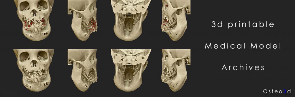
There are many companies dedicated to improving the use of 3D printing in the medical sector, and one of them Osteo3D, just released an amazing online repository of medical 3D models. Of course, patient information is private, so some of the “live models” have been made anonymous. But there are now over 100 3D models available to doctors and other registered medical professionals online, which allows them t0 compare their own patient’s 3D files with Osteo3D models. In fact, they can 3D print these models at home or wherever they have access to a 3D printer.
But, if a doctor or medical student doesn’t have a 3D printer, they can order a print directly from Osteo3D. The online platform is powered by cloud printing site df3d and is meant to increase awareness of the usefulness of 3D printing among healthcare professionals. Osteo3D hopes that, by collaborating with healthcare professionals, the tech industry, and academic institutions, they will help facilitate access and lower costs for the use of 3D printing in the healthcare field.
Currently the models are divided into the following categories:
Head and neck
Spine
Chest
Abdomen and pelvis
Extremities
When you click on 3D view, you don’t get quite the same level of control and depth asSketchfab, and some of the models look like pixels are growing out of them, but I didn’t pay to download any of the models, so I can’t say for sure. They are currently on the lookout for 3D printing partners who can help them deliver these 3D prints from their cloud platform to their customers around the world.
We’ve covered a number of stories that revolve around the combination of scanning/sensing technologies, great 3D modeling software, and 3D printing. This combination has arguably had the most impact on the quality of human life as it is seen in the medical sector. Diagnoses are made to a patient with a life-threatening condition. Doctors, then, use 3D scanning technology to capture the reality data of a patient’s affected area, whether it be an organ, vasculature, bone structure, or combination of all three. Next, a 3D model is created and manipulated using innovative software, and the model is 3D printed. Surgery is practiced and visualized on the model, and then performed on the patient. After doctors have all of this information, they use it to teach other doctors and nurse practitioners. So, why not spread this wealth of information? This is digital information, after all. Any 3D model that is successfully used to print a 3D model of a diagnosed patient can be transferred anywhere, to anyone.
With the ability to manipulate cells temporarily and spatially into three-dimensional (3D) tissue-like construct, 3D bioprinting technology was used in many studies to facilitate the recreation of complex cell niche and/or to better understand the regulation of stem cell proliferation and differentiation by cellular microenvironment factors. Embryonic stem cells (ESCs) have the capacity to differentiate into any specialized cell type of the animal body, generally via the formation of embryoid body (EB), which mimics the early stages of embryogenesis. In this study, extrusion-based 3D bioprinting technology was utilized for biofabricating ESCs into 3D cell-laden construct. The influence of 3D printing parameters on ESC viability, proliferation, maintenance of pluripotency and the rule of EB formation was systematically studied in this work. Results demonstrated that ESCs were successfully printed with hydrogel into 3D macroporous construct. Upon process optimization, about 90% ESCs remained alive after the process of bioprinting and cell-laden construct formation. ESCs continued proliferating into spheroid EBs in the hydrogel construct, while retaining the protein expression and gene expression of pluripotent markers, like octamer binding transcription factor 4, stage specific embryonic antigen 1 and Nanog. In this novel technology, EBs were formed through cell proliferation instead of aggregation, and the quantity of EBs was tuned by the initial cell density in the 3D bioprinting process. This study introduces the 3D bioprinting of ESCs into a 3D cell-laden hydrogel construct for the first time and showed the production of uniform, pluripotent, high-throughput and size-controllable EBs, which indicated strong potential in ESC large scale expansion, stem cell regulation and fabrication of tissue-like structure and drug screening studies.
With the capability of self-renewal and differentiating into all somatic cell types, embryonic stem cells (ESCs) hold great promise as an in vitro model system for studies in early embryonic development, as well as a robust cell source for applications in diagnostics, therapeutics, and drug screening [1]. Derived from the inner cell mass of a blastocyst, ESCs requires delicate culture condition and trend to cluster together, and in particular, forms three-dimensional (3D) cellular spheroids termed embryoid body (EB) [2]. In order to better understand stem cell niche and regulation of ESC differentiation and reprogramming, in vitro recapitulation of the spatial distribution of cells, cell–cell and cell–matrix interactions, is of paramount importance [3–5]. Compared with 2D monolayer culture, 3D cell culture is believed to confer a higher degree of clinical and biological relevance to in vitro model [6, 7], since the spatial arrangement of cells and extra-cellular matrix could influence cell differentiation and function both in vivo [8] and in vitro[9]. Therefore, reconstruction of 3D cell microenvironment is critical to directing stem cell fate and generating cell sources for tissue engineering, regenerative medicine and drug screening studies.
By mimicking some of the spatial and temporal aspects of in vivo development, EB is a basic 3D model for ESCs culture and differentiation studies. It was reported that the size and uniformity of EBs could vastly influence stem cell fate [10–12]. Various methods have been used to fabricate such cellular spheroid, basically including static suspension, hanging-drop and multiwell culture, most of which doesn’t involve biomaterials. Static suspension method inoculate suspension of ESCs onto non-adhesive plate to allow cells spontaneously aggregate into spheroid. This method is easy to operate, but showed limited control over the EBs size and shape due to the probability that ESCs encounter each other accidentally [13]. Hanging-drop is a common method to produce size-controlled homogeneous EBs, where droplets of ESCs suspension are pipetted onto the lid of a Petri dish and EBs was generate by gravity after overturning the dish [14]. However, manual pipetting is labor intensive and the reproducibility varies with operators. Multiwell culture offers high-throughput solution for EB formation through cell aggregation in uniformly shaped microwell arrays but requires expensive microwell culture plates [10, 15]. Besides, there are few customized microwell culture plates available in the market.
Recent advances in bioprinting technologies facilitated the precise deposition of ESCs in a reproducible manner. Xu et al [16] and Shu et al [17] printed ESCs suspension solution into 2D patterns as hanging-drop approach for EB formation, without the cell-biomaterial interaction. Corr and Xie [18, 19] applied laser direct-write method in bioprinting of mouse ESCs together with gelatin. ESCs maintained the pluripotency while proliferation and formed EB. EB size can be controlled by cell density and colony size. However, these studies just generated 2D cellular array without 3D cell–matrix interactions, and cell–cell interaction happens within one drop but not among different drops. To better recapitulate the characteristics of in vivo cell microenvironment, 3D customized cell/matrix construct with macro-porous structure might be a preferred choice. To our knowledge, there has been no report about bioprinting of ESCs into 3D cell-laden constructs.
The extrusion-based temperature-sensitive 3D bioprinting technology was developed in our lab and has been utilized for bioprinting of hepatocytes [20], adipose tissue-derived stem cells (ADSCs) [21], C2C12 cells [22], hela cells [23] and 293FT cells [24]. Most commonly used biomaterials for this technology are gelatin and alginate. Gelatin, a type of denatured collagen, is widely used as a coating for feeder layer-free mouse ES cell culture. Alginate, extracted from brown algae, is proving to have a wide applicability in tissue engineering and drug delivery and also used in embedding mouse ESCs for EB formation [25]. It has been proved in many studies that encapsulation of ESCs in hydrogels would direct EB formation with the maintenance of pluripotency [26–28]. Hence, we hypothesized that the bioprinting of 3D ESC-laden construct would maintain the stem cell pluripotency and address the challenges associated with the current methods for EB formation.
In this study, we investigated the feasibility of applying extrusion-based temperature-sensitive 3D bioprinting technology in bioprinting of ESCs with hydrogels into 3D macro-porous structure, with the maintenance of viability, pluripotency, cell growth and to direct EB formation. Printing process parameters were optimized to obtain a high cell survival rate (90%) after printing process and construct formation. Stem cell pluripotency was examined by the expression of stem cell markers (octamer binding transcription factor 4 (Oct4), stage specific embryonic antigen 1 (SSEA1) and a homeodomain-bearing transcriptional factor (Nanog)) and the ability to form EBs. The regulation of EB formation in the 3D bioprinted construct was systematically compared with commonly used methodology, where EB formation relies on cell aggregating as well as cell proliferation. Results demonstrated that this novel technology generated pluripotent, high-throughput, highly uniform and size controllable EBs under static culture condition without complex equipment. This study established the feasibility of fabricating 3D in vitro tissue-like model using ESCs for the first time, creating engineered microenvironment for pluripotent stem cells with the ability of placing cells and materials spatially in a reproducible manner.
Results
3.1. 3D bioprinting and cell viability optimization
In this study, many process parameters, e.g. nozzle inner diameter, nozzle insulation temperature and chamber temperature were examined to optimize cell viability after 3D construct fabrication. It was demonstrated that larger nozzle diameter resulted in higher cell viability (figure 2(A)). Specially, the cell viability under Nozzle-160 μm (81.59% ± 1.74%) was lower than those under Nozzle-260 μm (88.06% ± 1.98%), Nozzle-410 μm (89.59% ± 0.71%) and Nozzle-510 μm (90.84% ± 1.02%), with significant differences. Nozzle diameter of 260 μm, 410 μm and 510 μm showed no significant differences in terms of cell viability.
Zoom In Zoom Out Reset image size
Figure 2. The influence of bioprinting parameters on ESC viability is determined by fluorescence live/dead staining. (A) The influence of printing nozzle inner diameter on ESC viability (Insu-30 °C and Cham-10 °C). (B) The influence of nozzle insulation temperature and chamber temperature on ESC viability. Insu-25 °C means keeping the nozzle insulation temperature at 25 °C. Cham-4 °C means setting the chamber temperature at 4 °C, and so as others. (C) The fluorescent staining images show the live (green) and dead (red) cells at different days during culture period. Scale bar: 100 μm.
Insulation and chamber temperatures were altered to study their influences on cell viability (figure 2(B)). As a positive control, ESCs/hydrogel mixture without bioprinting were stained with fluorescence live/dead reagent, and showed 93.14% ± 1.31% cell viability. When insulation temperature was set at 25 °C (labeled as ‘Insu-25 °C’), cell viability increased with the chamber temperature from 55.52% ± 2.37% under 4 °C (labeled as ‘Cham-4 °C’) to 78.22% ± 2.55% under 10 °C (labeled as ‘Cham-10 °C’) with significant differences. When the insulation temperature was set at 30 °C (labeled as ‘Insu-30 °C’), nearly 90% ESCs remained alive under the chamber temperature of 7 °C and 10 °C (labeled as ‘Cham-7 °C’ and ‘Cham-10 °C’), significantly more than that under Cham-4 °C (72.40% ± 2.46%). To achieve both high ESC viability and a clear construct configuration, the process parameter combination of Nozzle-260 μm, Cham-10 °C and Insu-30 °C was chosen.
After culturing for three days, few cells were found dead, which were isolated from living EBs (figure 2(C)). On day 5 and day 7, a few dead cells were observed on the edge of EBs. About 5% ESCs were stained dead on day 7. As the static culturing continued, 9.69% ± 1.77%, 17.72% ± 2.91% and 40.64% ± 2.06% were found dead on day 8, day 9 and day 10, respectively (supplement 2). So, we chose 7 days as the culture period in the following analysis.
3.2. Construct structural stability and EB formation
A 3D cellular construct with the cross section of 8 mm × 8 mm and height of 1 mm was fabricated under the optimized process parameter. The 3D construct demonstrated macro-porous grid structure in which the hydrogel threads were evenly distributed according to the computer design (figure 3(A)). Both the width of the threads and the gap between the threads were homogeneous, that is 728.2 μm ± 24.9 μm and 424.3 μm ± 17.8 μm, respectively, suggesting 3D cellular construct formation in a highly controlled manner. ESCs were embedded uniformly in the hydrogel matrix threads, developing a specific 3D microenvironment.
Zoom In Zoom Out Reset image size
Figure 3. Images of the printed cellular model with grid structure. (A) Full view of the cellular construct. (B) Phase-contrast images demonstrating the cell morphology and distribution of different cell density at day 3, day 5 and day 7. Scale bar: 1 mm.
During the culture period, ESCs tended to grow as spheroid cellular aggregates, also known as EB. The cell density in the 3D hydrogel construct were determined by the initial cell density in the ESC/alginate/gelatin mixture and showed significant influence on the yield and density of EBs formed in the construct (figure 3(B)). It was demonstrated by semi-quantitative analysis of figure 3(B) that, the percentage of area occupied by EBs varied from 52% to 85% when initial cell density changed from 0.5 mln mL−1 to 2.0 mln mL−1 . Most of the EBs were contained in the hydrogel threads in the culturing period. However, when the initial cell density was as high as 2.0 mln ml−1, some of the EBs were observed running off from the threads into the throughout holes.
3.3. Cell proliferation
ESCs formed spheroid EBs in the 3D hydrogel construct and the diameter of the EBs enlarged with culturing time while keeping their spatial location in the hydrogel thread, indicating EB formation by ESC proliferation rather than aggregation (figure 4(A)). Compared with traditional 2D culture, ESCs showed different proliferation rate indicated by the OD value measure by CCK-8 kit (figure 4(B)). The normalized OD value of the 3D in situ group grew faster than that of 2D from day 1 to day 3, while slowing down after day 3 and being much less than that of 2D at day 7. However, 3D harvest group showed a generally faster growth rate than 2D during the one week culturing, with a significant difference. In addition, the diameter of EB was also measured to indicate ESC proliferation rate. When comparing the normalized EB volume with normalized 2D OD value, 3D samples also maintained a significantly faster growth rate than 2D, though the EB volume had huge variance (figure 4(B)).
Zoom In Zoom Out Reset image size
Figure 4. EB growing and cell proliferation. (A) Magnified images of the same location in 3D printed cellular construct at different times. (B) ESC proliferation in the 3D construct compared with 2D culture. All the date were normalized to the value of day 1. Scale bar: 200 μm.
Pluripotency markers, i.e. Oct4, SSEA1 and Nanog were analyzed to determine the pluripotency maintenance of ESCs after 7 day culture in the 3D hydrogel construct. Immunofluorescence staining and flow cytometry analysis showed that almost all of the cells within the EB were successfully stained both Oct4 and SSEA1. Because of the limitation of confocal capacity when dealing with large scale aggregates, the central part of the EB was darker than the edge (figure5(A)). Flow cytometry analysis demonstrated that 97.2% and 99.0% cells were positively stained with Oct4 and SSEA1 respectively (figure 5(B)). The qRT-PCR results demonstrated that the gene expression level of Oct4 and Nanog in our 3D samples were close to those in 2D (within the deviation of ±3%), without significant difference, confirming that cells have maintained pluripotency (figure 5(C)).
Zoom In Zoom Out Reset image size
Figure 5. ESC pluripotency at day 7 was determined by CLSM, flow cytometry and qRT-PCR. (A) Immunofluorescence images of EBs stained with Oct4, SSEA1 and DAPI. (B) Quantification of 3D dissociated cells marked with Oct4 and SSEA1 by using flow cytometry. (C) Gene expression of Oct4 and Nanog in 3D versus 2D by using qRT-PCR. Scale bar: 50 μm.
EBs were harvested from the 3D hydrogel construct at different time intervals to analyze EB morphology (figure 6(A)). Most of the EBs were separated without fusion. The center part of the EBs was darker than edge part, especially at day 5 and day 7, indicating the 3D sphere structure of EBs. Through analyzing the size of 250 random EBs for each sample, the histogram of EB diameter were obtained, showing a Gauss distribution curve (figure 6(B)). The results demonstrated that the EB size increased significantly from about 50 μm to about 110 μm when the construct was cultured from day 3 to day 7 (figure 6(C)). Cell density had little influence on EB average size. However, increased cell density would result in the reduction of the uniformity of EB size, especially at day 7; the EB diameter of 2.0 mln mL−1 group at day 7 was vastly heterogeneous, with a deviation of 42.30 μm, which was much more than those of other two groups.
Zoom In Zoom Out Reset image size
Figure 6. EB formation in different cell density: (A) optical images of released EBs at different days. (B) EB diameter and (C) EB circularity distributions at different days. Summary of the (D) diameter and (E) circularity. 250 EBs were applied for diameter and circularity measurements for each group. Scale bar: 200 μm.
Circularity was measured to assess the quality of EBs (figure 6(D)). For the 0.5 mln mL−1 group, most of the EBs were close to a standard spheroid with the circularity centered in 0.9 for the three time points. As to the other two groups, the circularity at day 3 is similar to that of 0.5 mln mL−1group, while the circularity frequency peaks had a significant decrease at day 5 and day 7. In particular, about 20% EBs had a circularity under 0.8 at day 5 and day 7 for the 2.0 mln mL−1group. In general, the circularity decreased with the increase of culture time and initial cell density in the hydrogel (figure 6(E)).
3.6. Comparison with other EB formation methods
Considering this was a novel methodology of EB formation, we systematically compared the commonly used static suspension and hanging drop methods with the 3D bioprinting method for EB formation. As demonstrated by the phase-contrast images (figure 7(A)), EBs generated by static suspension method showed more uncontrollable morphology rather than round spheroid. The distribution of EB diameter clearly demonstrated that 3D bioprinting technology generated EBs with higher uniformity compared with static suspension technology, especially for the larger EB diameter, i.e. 60 ~ 70 μm and 100 ~ 110 μm regions (figure 7(B)). In particular, the EBs with 30 ~ 50 μm diameter presented vastly irregular shape in suspension technology, which was confirmed by the circularity curve (figure 7(C)). On the other hand, EBs generated by 3D bioprinting technology showed higher circularity regardless of the diameter regions, suggesting more regular shape (figure 7(C)). More characteristic like EB forming motivation, size control method, EB diameter range, uniformity, yield, operation complexity were compared among 3D bioprinting technology, static suspension technology and hanging drop technology, as listed in table 1.
Zoom In Zoom Out Reset image size
Figure 7. Comparison of static suspension and 3D bioprinting technology for generating EBs. (A) Phase-contrast images showing the morphology of EBs generated by static suspension technology and 3D bioprinting technology. (B) The EB diameter histograms presented the distribution of EB size with a Gauss distribution fitting. (C) The circularity curves contrasted the EB qualities.
3D cell culture environment and tissue-like models have drawn great attention because they can be tuned to promote certain levels of cell differentiation and tissue organization, which is difficult in traditional 2D culture systems for their failing to reconstitute the in vivo cellular microenvironment [30, 31]. Various 3D culture systems have been developed to study the cellular behavior affected by spatial and temporal cell–cell and cell–matrix interactions. Among these methods, 3D bioprinting, typically containing jet-, laser- and extrusion-based methods, is a promising technique to manipulate cells/matrix deposition and ultimately generate 3D complex tissues or organs. This technique have been used in printing cells derived from adult, embryonic and even tumor tissues for tissue engineering and drug screening applications. With the capacity to expand unlimitedly in vitro and differentiate into a variety of therapeutic cell types, ESCs have generated great enthusiasm and are being applied in bioprinting studies until recently. As a relatively sensitive cell type, ESCs might suffer greater problems in a printing process compared with other types of cells. Several studies had been conducted to print ESCs, maintaining their viability and pluripotency [16–19]. Instead of creating 3D tissue-like constructs, these studies were more likely to generate cellular droplet array with precise control of distribution. Here we described the work of establishing a 3D ESC-laden hydrogel construct using extrusion-based bioprinting technology. The results demonstrated high proliferation rate of pluripotent ESCs in the hydrogel construct, and a versatile technology for generating highly uniform and high throughput EBs.
Cell viability after 3D bioprinting and construct formation was determined when evaluating the limitations of bioprinting ESCs. Cells would be lysed or damaged due to osmotic effects in the solution, heat increase and mechanical stress during printing. In the protocol presented in this work, about 6.86% ± 1.31% cells were dead during the cell/hydrogel solution preparation process before 3D bioprinting (figure 2(B)). We assumed this was caused by cell dissociation process, together with the osmosis and stirring operation of hydrogel materials. In an inkjet printing study, 15% Chinese Hamster Ovary cells were detected dead before printing process [32]. Thermal effects of the ejector reservoir in the inkjet printing process and laser force in laser-based printing would be the cause of cell death, in addition to the impact force when cellular droplets were jetted to a rigid substrate in a very short time. Under a different fabricating strategy, the extrusion-based bioprinter extruded the cell-laden cylinders softly on the substrate and controlled the temperature under 30 °C, without the concerns about the thermal and sharply impacting effects. However, cells would inevitably suffer from shear force when the cell-laden hydrogels were continuously extruded through a limited space in the nozzle. We hypothesized that nozzle size and hydrogel viscosity would influence shear force and hence influence cell viability. The cell viability data of different nozzle sizes, chamber and insulation temperatures supported this hypothesis (figures 3(A) and (B)). In our previous study, more than 90% Hela cells were alive after bioprinting under the parameters of Insu-25 °C/Cham-4 °C and Nozzle-260 μm [23], while the viability of ESCs was only 55.52% ± 2.37% under the same parameter combination. When increasing the insulation and chamber temperature to 30 °C and 10 °C respectively, the viability showed a significant increase to 90%. Taking into the account of cell death before bioprinting, optimized parameters led to only 5% cell death during printing, indicating a broad future applicability of this technique to various cell types ranging from tumor cells to ESCs. Additionally, few dead cells were observed during one-week culture period (figure3(C)). On the other hand, when the culture period was extended to more than 7 days, more and more ESCs suffered from apoptosis and lysis, possibly due to contact inhabitation and insufficient mass transfer to the center of EB with the increasing of EB size. Therefore, 7 days was chose as the experiment time window for this study.
Apart from cell viability, the maintenance of pluripotency is another essential criterion for ESCs regulation and application. The results of immunofluorescence staining and FACS analysis showed a high expression rate (98%) of stem cell pluripotent markers Oct4 and SSEA1 at day 7 (figure 4), indicating that cells remained undifferentiated state during the whole experimental period. Naturally, it can be inferred that the printing process also had little influence on ESC pluripotency.
In the cell-laden hydrogel culture system, both the cell type and matrix material could influence cell growth. Human mesenchymal stem cells remained alive but did not proliferate when encapsulated in alginate [33, 34]. While human ADSCs could proliferated for a short period of time in alginate hydrogel microspheres but showed significantly higher proliferation rate in gelatin/alginate microspheres [35]. As a widely used hydrogel, alginate has the disadvantages of low cell adhesiveness and poor support for cell proliferation [36]. Adding gelatin would improve the cellular adhesive condition and hence favor cell expansion. In this study, the fabricated multilayered constructs offered a 3D microenvironment surrounded by gelatin/alginate materials for ESCs to adhere, self-renew, and cellular spheroid, termed EB, was generated in situ because of cell proliferation. Once EB was formed, the spheroid structure supported expansion of subpopulations with differing proliferation, nutrition and oxygenation status compared with conventional monolayer system. It is reported that the proliferation of mouse ESCs was higher when embedded in fibrin gels versus 2D suspension culture [27]. Similarly, in this study, ESCs in 3D constructs proliferated faster than 2D culture sample when being released from hydrogel to read OD value. This operation was aimed to avoid the influence of interactions between reagent molecular and matrix materials (figure 6 and supplement 3). Additionally, the enlargement of EB diameter, which also reflected ESC proliferation, confirmed this result (figure 6).
Typically stimulated via generation of EBs, ESC differentiation depends on numerous cues throughout the EB environment, including EB size and shape, as well as their uniformities. In general, several characteristics should be concerned for EB formation system, including reproducibility, symmetry, ease of use and scalability [37]. In the traditional EB formation methodology, like suspension and hanging-drop, EBs were created via cell gathering and proliferation. In these methods, it was essential to get a balance between allowing necessary ESC aggregation for EB formation and preventing EB agglomeration for efficient cell growth and differentiation [14]. Static suspension cultures produced a large number of EBs with simple operation, but the size and shape of the resulting EBs were highly uncontrollable and irregular due to the tendency of EBs to agglomerate after initial formation, as shown in figure 7. Hanging-drop method served as a golden tool to generate uniform and reproducible EBs with fully aggregating of cells under gravity and non-agglomeration of EBs in different drops. However, it faced the intrinsic limitation of scalability. The 3D bioprinting method presented in this study addressed some of the problems, producing massively homogeneous EBs with regular shape and controllable shape. In this 3D cell-laden hydrogel system, ESCs were immobilized and restricted to aggregate with each other, and would not agglomerate until they are large enough to connect with each other. When the initial cell density was increased, the average distance between two original EBs was closer and these EBs are more likely to agglomerate with each other while proliferation, which is also one of the concerns when we choose the experiment time period. As a result, the EB uniformity of 2.0 mln mL−1 group was not that good as those of 0.5 mln mL−1 and 1.0 mln mL−1 groups, especially after culturing for one week (figure 6). Without the initial cell aggregating, the size of EBs in our model was mainly determined by the culture time. Also, it would take longer to reach the same scale of EB diameter compared with suspension method, probably due to the physical constrain of the matrix material. For example, it took 5 days and 2 days to get EBs ranging 60 ~ 70 μm for 3D printing and suspension methods, respectively (figure7). Besides, thanks to the interconnected channels design in the 3D construct which allowed mass transfer, EBs could be produced in a large scale by changing the construct volume and cell density. In the six-layer construct with 1.0 million cells per milliliter for example, EBs got a stable yield of about 3000 cm−2, while the EB yield by suspension technology was about 900 cm−2(seeding 0.5 million cells in a 35 mm dish) and no more than 10 EBs [38] could be produced in 1 cm2 area in hanging-drop method, which was also demonstrated by our experiments (supplement 4).
In summary, this study presented the high throughput production of pluripotent, uniform, regular and controllable EBs with the diameter smaller than 150 μm during one week culture. In a gelatin-based laser printing method, EBs with the diameter of about 100 μm were also generated to avoid EB agglomeration in gels [19]. EBs with different size exhibit different gene expression and differentiation fate. Park et al [39] found that 100 μm diameter EBs of mouse ESCs expressed increased ectoderm markers while 500 μm diameter EBs expressed endoderm and mesoderm markers. Furthermore, Messana et al [12] demonstrated that mouse ESCs derived from small EBs (<100 μm) had a greater chondrogenic potential than those from larger EBs. Hwang [10] reported that human endothelial cell differentiation was increased in smaller EBs (150 μm) while cardiogenesis was enhanced in larger EBs (450 μm). However, large EBs might be associated with limited mass transfer and the diffusion of biochemical through EBs is demonstrated to be linked to differentiation of ESCs [40]. While the effect of EB size on differentiation remains to be shown in our model, we hypothesize that EBs with the diameter smaller than 150 μm would mediate specific differentiation trajectory, which will be confirmed in the future work.
Demonstrating the advantages of reproducibility, high throughput, regular shape and controlled size, we believe this is a versatile technology for EB generation. But, this 3D printing system does not serve as an EB formation method solely. The ESC-laden hydrogel 3D construct can be dissolved at a proper time point to harvest massive EBs with desired size for ES cell research. Or, the ESC-laden hydrogel 3D construct can be maintained to perform 3D ESC differentiation studies to explore the regulation of EB size, matrix material and 3D structure on ESC differentiation lineages. Furthermore, this technology hold the potential to serve as a versatile tool for the generation of tissue-like structure and organ/tissue on chip based on controlled ESC differentiation.
5. Conclusion
In this study, we reported successful bioprinting of mouse ESCs with hydrogel into a 3D multilayered construct for the first time. Extrusion-based bioprinting technology was applied. Upon parameter optimization, ESCs demonstrated high viability of 90% after 3D printing and construct formation. Cells continued self-renewal in the construct and exhibited a higher proliferation rate compared with conventional 2D culture. 98% cells expressed the canonical pulripotent markers Oct4 and SSEA1 at day 7, indicating that most of the ESCs remained undifferentiated state after printing and culturing. Large quantities of uniform EBs with regular shape and adjustable size were generated through cell proliferation, while avoiding EBs agglomeration. This work indicated the feasibility of fabricating complex 3D tissue-like model based on pluripotent stem cells for applications in pharmacy, regenerative medicine, stem cell expansion and biology studies.
Bioprinting of human pluripotent stem cells and their directed differentiation into hepatocyte-like cells for the generation of mini-livers in 3D
We report the first investigation into the bioprinting of human induced pluripotent stem cells (hiPSCs), their response to a valve-based printing process as well as their post-printing differentiation into hepatocyte-like cells (HLCs). HLCs differentiated from both hiPSCs and human embryonic stem cells (hESCs) sources were bioprinted and examined for the presence of hepatic markers to further validate the compatibility of the valve-based bioprinting process with fragile cell transfer. Examined cells were positive for nuclear factor 4 alpha and were demonstrated to secrete albumin and have morphology that was also found to be similar to that of hepatocytes. Both hESC and hiPSC lines were tested for post-printing viability and pluripotency and were found to have negligible difference in terms of viability and pluripotency between the printed and non-printed cells. hESC-derived HLCs were 3D printed using alginate hydrogel matrix and tested for viability and albumin secretion during the remaining differentiation and were found to be hepatic in nature. 3D printed with 40-layer of HLC-containing alginate structures reached peak albumin secretion at day 21 of the differentiation protocol. This work demonstrates that the valve-based printing process is gentle enough to print human pluripotent stem cells (hPSCs) (both hESCs and hiPSCs) while either maintaining their pluripotency or directing their differentiation into specific lineages. The ability to bioprint hPSCs will pave the way for producing organs or tissues on demand from patient specific cells which could be used for animal-free drug development and personalized medicine.
Content from this work may be used under the terms of the Creative Commons Attribution 3.0 licence. Any further distribution of this work must maintain attribution to the author(s) and the title of the work, journal citation and DOI.
New drug development can take 10 to 20 years with an estimated average of about 9 to 12 years [1, 2]. In addition, only around 16% of the drugs that begin preclinical testing are approved for human use [3]. Some of this low success rate can be attributed to the different responses that animals and humans have to the drugs being tested; some drugs have to be withdrawn from market due to toxic effects on human organs such as liver and heart, despite being tested safely on animals. A possible solution to this might be the creation of human pluripotent stem cell (hPSC) -derived micro-tissues which could be used with organ-on-a-chip devices [4–7]. These micro-tissues are expected to produce the same or similar physiological reaction that the entire organ would but on a much smaller scale. This would result in scalable, faster and potentially more reliable drug testing platform, and hopefully an end to animal testing.
hPSCs are the ideal cells to use for this application due to their ability to self-renew indefinitely, which enables large populations of cells to be created easily in vitro, and their pluripotency which means that they can be differentiated into any required adult cell type [8–13]. Pluripotent stem cells can be divided into embryonic stem cells (ESCs) and induced pluripotent stem cells (iPSCs). Human ESCs (hESCs) were first isolated from early human blastocysts in 1998 [14]. Any tissue construct created from hESCs for implantation in vivo would require the patient to receive immunosuppressive drugs and ethical issues still restrict some applications due to their source. iPSCs have neither of these drawbacks as they can be created from harvested adult cells from the patient requiring treatment and as such any implanted cells derived from these iPSCs should not be rejected by the patient’s immune system but may require immunosuppressive drugs at a greatly reduced dosage. In 2006 Shinya Yamanaka discovered that iPSCs can be derived from somatic cells by retrovirally transducing them with four transcription factors—Oct3/4, Sox2, Klf4 and C-myc [15, 16]. These cells have the same self-renewal and differentiation capabilities as ESCs but with the added advantage that iPSCs can be used for autologous therapies. These unique characteristics make pluripotent stem cells ideal for use in a number of applications such as clinical tissue engineering, novel drug discovery and testing for the pharmaceutical industry [8,9, 17, 18].
In the field of biofabrication, great advances are being made towards fabricating 3D tissue and organs with very fine spatial control of cell deposition. From the very first paper that was published investigating printing of biological cells (or bioprinting), tissue engineering was identified as a major application for this new technology [19]. If more complex structures such as organs and tissues were to be printed, the bioprinter would need the ability to transfer microscopic patterns of viable cells of multiple cell types into well-defined three-dimensional arrays that closely mimic the tissue structure. There has been much progress in the development and establishment of several different bioprinting techniques for 3D live constructs [20–22] including those based on laser pulses, inkjets and other more novel approaches. It is an inescapable fact that cells will be subjected to some level of stress during deposition, regardless of the printing technique being used. For example, cells printed by non-contact methods will be affected when they impact on the substrate at some incident velocity, which would result in extreme deceleration and shear stress [23–26]. Shear stress is applied to cells pushed through nozzle orifices and capillary tubes [24, 27–42] and the actuation is provided via pressure, heat, or high frequency vibration which can also be damaging to the cells [30, 31, 43–46]. If cells are exposed to laser energy the radiation can cause genetic damage [29, 47–54] and shear forces are applied during cavitation and jet formation [23, 55]. Ultrasonic actuation for cell transfer would subject the cells to stress in the form of heat and vibration [56, 57]. Therefore, it is important to validate the response of printed cells to any particular bioprinting process in terms of their viability and more importantly their biological functions.
We previously reported the results of the first experiments printing hESCs using a valve-based printing approach including their response to the printing process in the form of post-printed viability and pluripotency validation [37]. However, if hPSCs are to be used for producing human tissues on demand for drug testing, their post-printing differentiation must be reproducibly directed to the required lineages for each tissue. Unfortunately homogenous cellular differentiation of hPSCs into some germ layers has proved difficult [12, 13]. Here, we report the first investigation into the bioprinting of human iPSCs, their response to the valve-based printing process as well as their post-printing differentiation into hepatocyte-like cells (HLCs). HLCs that are in the process of differentiating are bioprinted and examined to further validate the compatibility of the valve-based bioprinting process with fragile cell transfer. Finally, 3D hydrogel structures were designed and printed out with encapsulated hESC-derived HLCs and the viability and hepatic characteristics of the cells were investigated.
A newer version of our previously reported cell printing platform [37] has been developed. Four nanolitre dispensing systems, each comprising a solenoid valve (VHS Nanolitre Dispense Valve, Lee Products Ltd) with 101.6 μm internal diameter nozzles (Minstac Nozzle, Lee Products Ltd), were attached to static pressure reservoirs for the bio-ink solution to be dispensed from via flexible tubing. The nanolitre dispensing system and bio-ink reservoirs were mounted onto the tool head of an enclosed custom built micrometer-resolution 3-axis XY–Z stage (figure 1). This newer cell printing platform improved on the previous version by reducing the overall size and weight of the machine, allowing it to be mounted inside a standard tissue culture hood during experiments requiring a sterile environment. Other enhancements included the two extra nanolitre dispensing systems, taking the total up to four, a more robust electronics and custom firmware was developed which improved the reliability and speed of the machine and two separate pressure channels were included, allowing for differential bio-ink dispensing conditions. Unless otherwise stated standard printing conditions were used: for 2D, printing was carried out using a pulse time of 8 ms at an inlet pressure of 0.6 bar using a nozzle with an internal diameter of 101.6 μm; for 3D, printing was carried out using a pulse time of 400 μs at an inlet pressure of 1.0 bar for sodium alginate solution and a pulse time of 400 μs at an inlet pressure of 0.5 bar for calcium chloride solution both using nozzles with an internal diameter of 101.6 μm.
Zoom In Zoom Out Reset image size
Figure 1. (a) Schematic drawing of the cell printer system; (b) detailed schematic of the micro-solenoid valve; (c) schematic of the combinatorial printing process for alginate hydrogel creation; (d) a 3D printed alginate tube structure approximately 13 mm tall printed with 1.5% w/v Sodium Alginate and 600 mM (6%) Calcium Chloride solutions in Millipore water (scale bar 2 mm).
……
The process of in vivo liver organogenesis occurs in the developing foregut, when newly specified hepatic cells separate from the endodermal sheet and form a dense 3D structure known as a hepatoblast (liver bud) [74, 75]. It is hypothesized that arranging the hESC–HLCs in 3D during the differentiation process may yield more mature hepatocytes than conventional 2D differentiation. The hESC differentiation protocols are more efficient and robust than hiPSC protocols therefore only hESC-derived HLCs were printed in 3D.
In order for this technique to be useful for tissue engineering applications, structures need to be tall enough to allow cells to interact in a three-dimensional environment. The concentration of alginate solution was set to 1.5% w/v to improve the mechanical strength of the hydrogel and allow it to support further layers. Circular structures with a large number of layers were designed and printed out in the wells of a multi-well plate to allow the structures to be cultured post-printing. These resulting structures were photographed for analysis and are shown in figure 7below.
These structures were printed out in a matter of minutes and are strong enough to support their own weight and the weight of further layers (as seen in figure 1(d)). The structures spread slightly, but by slightly altering the volume ratio, concentrations and surface properties this spreading can be reduced.
Approximately one hour post-printing one of the HLC-laden alginate ring structures was examined using a confocal microscope; the 3D image is shown in figure 8(a). Cell viability was calculated to be 55.5% using the Imaris confocal microscope software. Cell viability declined over the first 24 h which resulted in low cell numbers for hepatic marker testing following the 3D differentiation process, but the viability remained stable for the remainder of the differentiation process. At day 23 of the differentiation process, the cells in the remaining structures were harvested and stained for the presence of hepatic markers. As shown in figure 8(b), cells are positive for albumin which demonstrates their hepatic lineage. The normal time required for 2D differentiation of hPSC-HLCs is 17–24 d. However, based on the results of albumin secretion in the medium, we observed the 3D printed cells have taken longer to reach the maximum albumin secretion than the 2D control as shown in figure 8(c). Interestingly, when analyzing the difference between 20 and 40 layer printed tube structures, we noticed close-to proportional increase in albumin secretion to the number of layers as shown in figure 8(d). This indicates that the permeability of the alginate hydrogel allows nutrition and differentiation reagents to enter the structure and support 3D differentiation and maturation processes of the cells, regardless of the height of the printed structure.
Research is currently underway including investigations to improve the 3D viability and adjusting the differentiation protocol that may facilitate higher albumin secretion. For example, the optimization of hydrogel formation as well as enhanced cell density may improve the differentiation process for hPSCs in 3D [21, 76, 77].
4. Conclusions
To the best of our knowledge, this study is the first to demonstrate that hiPS cells can be bioprinted without adversely affecting their biological functions including viability and pluripotency. Importantly, we verified that our valve-based printing process is gentle enough to not affect the pluripotency of both hESCs and hiPSCs. A number of different hPSC lines were directed to differentiate into HLCs. Cells were printed during the differentiation process and showed no differences in hepatocyte marker expression and similar morphology when compared to a non-printed control. We previously reported the results of an investigation into the response of hESCs to the valve-based printing process. Here we build on that study, performing a deeper investigation to compare the response of hiPSCs and hESCs to the printing process using flow cytometry. The effect of nozzle geometry was investigated and the effects of nozzle length on the post-printing viability of cells were recorded; longer nozzles lower the post-printing viability of the cells. We printed hESC-derived HLCs in a 3D alginate matrix and tested for viability and hepatic markers during the remaining differentiation and they were found to be hepatic in nature. The ability to bioprint hPSCs while either maintaining their pluripotency or directing their differentiation into specific cell types will pave the way for producing organs or tissues on demand from patient specific cells which could be used for animal-free drug development and personalized medicine.
Large scale industrialized cell expansion: producing the critical raw material for biofabrication processes
Cellular biomanufacturing technologies are a critical link to the successful application of cell and scaffold based regenerative therapies, organs-on-chip devices, disease models and any products with living cells contained in them. How do we achieve production level quantities of the key ingredient—’the living cells‘ for all biofabrication processes, including bioprinting and biopatterning? We review key cell expansion based bioreactor operating principles and how 3D culture will play an important role in achieving production quantities of billions to even trillions of anchorage dependent cells. Furthermore, we highlight some of the challenges in the field of cellular biomanufacturing that must be addressed to achieve desired cellular yields while adhering to the key pillars of good manufacturing practices—safety, purity, stability, potency and identity. Biofabrication technologies are uniquely positioned to provide improved 3D culture surfaces for the industrialized production of living cells.
Biofabrication of tissue constructs by 3D bioprinting of cell-laden microcarriers
Riccardo Levato1,2, Jetze Visser3, Josep A Planell1, Elisabeth Engel1,2,4, Jos Malda3,5 andMiguel A Mateos-Timoneda2,1
Bioprinting allows the fabrication of living constructs with custom-made architectures by spatially controlled deposition of multiple bioinks. This is important for the generation of tissue, such as osteochondral tissue, which displays a zonal composition in the cartilage domain supported by the underlying subchondral bone. Challenges in fabricating functional grafts of clinically relevant size include the incorporation of cues to guide specific cell differentiation and the generation of sufficient cells, which is hard to obtain with conventional cell culture techniques. A novel strategy to address these demands is to combine bioprinting with microcarrier technology. This technology allows for the extensive expansion of cells, while they form multi-cellular aggregates, and their phenotype can be controlled. In this work, living constructs were fabricated via bioprinting of cell-laden microcarriers. Mesenchymal stromal cell (MSC)-laden polylactic acid microcarriers, obtained via static culture or spinner flask expansion, were encapsulated in gelatin methacrylamide-gellan gum bioinks, and the printability of the composite material was studied. This bioprinting approach allowed for the fabrication of constructs with high cell concentration and viability. Microcarrier encapsulation improved the compressive modulus of the hydrogel constructs, facilitated cell adhesion, and supported osteogenic differentiation and bone matrix deposition by MSCs. Bilayered osteochondral models were fabricated using microcarrier-laden bioink for the bone compartment. These findings underscore the potential of this new microcarrier-based biofabrication approach for bone and osteochondral constructs.
Microstereolithography and characterization of poly(propylene fumarate)-based drug-loaded microneedle arrays
Yanfeng Lu1, Satya Nymisha Mantha1, Douglas C Crowder2, Sofia Chinchilla2, Kush N Shah2,3,4,Yang H Yun2, Ryan B Wicker5 and Jae-Won Choi1
Drug-loaded microneedle arrays for transdermal delivery of a chemotherapeutic drug were fabricated using multi-material microstereolithography (μSL). These arrays consisted of twenty-five poly(propylene fumarate) (PPF) microneedles, which were precisely orientated on the same polymeric substrate. To control the viscosity and improve the mechanical properties of the PPF, diethyl fumarate (DEF) was mixed with the polymer. Dacarbazine, which is widely used for skin cancer, was uniformly blended into the PPF/DEF solution prior to crosslinking. Each microneedle has a cylindrical base with a height of 700 μm and a conical tip with a height of 300μm. Compression test results and characterization of the elastic moduli of the PPF/DEF (50:50) and PPF/drug mixtures indicated that the failure force was much larger than the theoretical skin insertion force. The release kinetics showed that dacarbazine can be released at a controlled rate for five weeks. The results demonstrated that the PPF-based drug-loaded microneedles are a potential method to treat skin carcinomas. In addition, μSL is an attractive manufacturing technique for biomedical applications, especially for micron-scale manufacturing.
Controlling shape and position of vascular formation in engineered tissues by arbitrary assembly of endothelial cells
Cellular self-assembly based on cell-to-cell communication is a well-known tissue organizing process in living bodies. Hence, integrating cellular self-assembly processes into tissue engineering is a promising approach to fabricate well-organized functional tissues. In this research, we investigated the capability of endothelial cells (ECs) to control shape and position of vascular formation using arbitral-assembling techniques in three-dimensional engineered tissues. To quantify the degree of migration of ECs in endothelial network formation, image correlation analysis was conducted. Positive correlation between the original positions of arbitrarily assembled ECs and the positions of formed endothelial networks indicated the potential for controlling shape and position of vascular formations in engineered tissues. To demonstrate the feasibility of controlling vascular formations, engineered tissues with vascular networks in triangle and circle patterns were made. The technique reported here employs cellular self-assembly for tissue engineering and is expected to provide fundamental beneficial methods to supply various functional tissues for drug screening and regenerative medicine.
The influence of printing parameters on cell survival rate and printability in microextrusion-based 3D cell printing technology
Three-dimensional (3D) cell printing technology has provided a versatile methodology to fabricate cell-laden tissue-like constructs and in vitro tissue/pathological models for tissue engineering, drug testing and screening applications. However, it still remains a challenge to print bioinks with high viscoelasticity to achieve long-term stable structure and maintain high cell survival rate after printing at the same time. In this study, we systematically investigated the influence of 3D cell printing parameters, i.e. composition and concentration of bioink, holding temperature and holding time, on the printability and cell survival rate in microextrusion-based 3D cell printing technology. Rheological measurements were utilized to characterize the viscoelasticity of gelatin-based bioinks. Results demonstrated that the bioink viscoelasticity was increased when increasing the bioink concentration, increasing holding time and decreasing holding temperature below gelation temperature. The decline of cell survival rate after 3D cell printing process was observed when increasing the viscoelasticity of the gelatin-based bioinks. However, different process parameter combinations would result in the similar rheological characteristics and thus showed similar cell survival rate after 3D bioprinting process. On the other hand, bioink viscoelasticity should also reach a certain point to ensure good printability and shape fidelity. At last, we proposed a protocol for 3D bioprinting of temperature-sensitive gelatin-based hydrogel bioinks with both high cell survival rate and good printability. This research would be useful for biofabrication researchers to adjust the 3D bioprinting process parameters quickly and as a referable template for designing new bioinks.
A new method of fabricating a blend scaffold using an indirect three-dimensional printing technique
Due to its simplicity and effectiveness, the physical blending of polymers is considered to be a practical strategy for developing a versatile scaffold having desirable mechanical and biochemical properties. In the present work, an indirect three-dimensional (i3D) printing technique was proposed to fabricate a 3D free-form scaffold using a blend of immiscible materials, such as polycaprolactone (PCL) and gelatin. The i3D printing technique includes 3D printing of a mold and a sacrificial molding process. PCL/chloroform and gelatin/water were physically mixed to prepare the blend solution, which was subsequently injected into the cavity of a 3D printed mold. After solvent removal and gelatin cross-linking, the mold was dissolved to obtain a PCL–gelatin (PG) scaffold, with a specific 3D structure. Scanning electron microscopy and Fourier transform infrared spectroscopy analysis indicated that PCL masses and gelatin fibers in the PG scaffold homogenously coexisted without chemical bonding. Compression tests confirmed that gelatin incorporation into the PCL enhanced its mechanical flexibility and softness, to the point of being suitable for soft-tissue engineering, as opposed to pure PCL. Human adipose-derived stem cells, cultured on a PG scaffold, exhibited enhanced in vitro chondrogenic differentiation and tissue formation, compared with those on a PCL scaffold. The i3D printing technique can be used to blend a variety of materials, facilitating 3D scaffold fabrication for specific tissue regeneration. Furthermore, this convenient and versatile technique may lead to wider application of 3D printing in tissue engineering.
3D printing converts a computer file to one or more 3D objects by deposition of material according to type and locations specified in the data file, as detailed here. The data file for the 3D printing can be based on images, e.g. of the beating heart, which can be obtained by Computed Tomography (CT), Magnetic Resonance Imaging (MRI), UltraSound/Echocardiography (US/Echo), or nuclear imaging. CT and MRI offer significantly better definition of boundary locations needed for 3D modeling than do the other imaging modalities. This article discusses the methods accompanying several real world applications of 3D printing used successfully to assist the plan for open heart surgery.
Select a suitable patient (e.g., a child with unique congenital structural abnormalities who merits 3D assist planning for a customized surgical approach)
Apply an imaging method that identifies the important tissue boundaries in sufficient detail and accuracy as a 3D data set, typically as a stack of 2D images (e.g., MRI or CT)
Apply computer analysis tools to convert stacks of gray-scale image arrays to tissue boundaries and tissue character suitable for 3D model generation
Edit and review boundary extraction, boundary connections in-plane and slice-to-slice and assign character (labels) to boundary surfaces and interiors
Optionally generate one or more renderings (3D visuals: lighting, transparancies, reflections, cut-aways, rotations, fly-line camera trajectories and angles) for review of 3D structures on 2D screens
Optionally enable simulated surgery and simulated outcomes (what would the face surface look like after surgical extension of the jaw, measurements, mechanics)
Apply printer graphic conversion that re-slices the boundaries into coordinates and material selection according to the specification requirements of a 3D printer device
Submit data to 3D printer, monitor for jamming, wait for the material to print and cool
Pick up the printed 3D heart(s), inspect for artifacts from curling or other print aberrations, modify object or re-print as needed, deliver printed product(s) to end user (e.g., to surgeon for examination and surgical planning)
Surgical planning:
Examine the 3D interactive computer graphics, derived video fly-throughs, printed 3D object(s)
Consider deriving and optionally printing other derived 3D objects (e.g., representing different stages of surgery)
Make measurements, delineate surgical plan
Currently, the best two competing methods to image the beating heart at sufficient detail are CT scan and MRI.
A CT scan uses ionizing radiation, typically equivalent to more than 200 chest xrays, which can increase the lifetime risk of cancer by as much as 1/1000. The imaging can be completed in less than 10 minutes, an important issue when working with squirming children. The images offer fine resolution (smallest distances that can be distinguished) on the order of 1 millimeter in the cross sections with 1-5 mm section thickness, so the picture elements (Pixels) may be 1 mm x 1 mm x 1 mm covering the heart in 200 slices of 512 x 512 image elements. Each location is characterized by a Hounsfield number that represents the attenuation of x-rays due to the density of the predominant atomic nuclei in each location. Metal implants produce serious artifacts. The Hounsfield numbers group into four tissue types: air, water, bone, metal. Fortunately for planning repair of congenital abnormalities of heart structure, the boundaries of interest are often water density versus fat. An injected radiocontrast agent such as iohexol enhances contrast by generating a distinction from water density for arterial and venous blood conveying the higher density atomic nuclei versus blood vessel or heart walls which are predominantly water density. Iodine contrast may be used to provide a higher atomic Z number for x-ray attenuation, but it can cause a serious allergy and/or renal failure.
MRI uses non-ionizing radiation (magnetic wave energy that does not knock electrons out of orbit to cause damage to the tissues), so it does not pose a future risk of cancer. Also, MRI offers a variety of methods to make signal from flowing blood in arteries and veins each distinct from chamber walls without requiring injection of a contrast agent (though some applications of MRI do use contrast agents). MRI is distorted by metal implants, and by materials with strong differences in magnetic susceptibility, and has complex artifacts if motion occurs at an inopportune time. Both CT and MRI utilize ECG signals to synchronize and effectively “freeze” cardiac motion. A method by the author cancels the magnetic and flow artifacts to enable diagnostic ECG in MRI for safety of potentially deep sedation to further reduce motion artifacts. MRI has many ways to generate signal intensities that characterize tissue differences, so it is over 10 times better at distinguishing differences within soft tissues. MRI thus has advanges identifying boundaries of abnormal tissues such as right ventricular dysplasia, scar tissue (collagen), myxoma, etc. While there are many distinct ways to apply MRI to define the structure of an individual’s heart, commonly a form of fast spin echo, with parallel imaging, is utilized. It generates images representing a slice thickness typically of 2-5 mm each corresponding typically to 128 x 256 picture elements. Thus the volume elements (voxels) tend to be as much as 16-fold larger than from CT, but the ability to distinguish soft tissue differences is an order of magnitude better by MRI. With faster MRI enabled by parallel imaging (multiple slice data obtained concurrently), and with stronger magnets to provide more signal vs noise, the voxels can be smaller and the number of slices covering the heart may be increased, e.g. from 20 to 200.
Even with smaller datasets from MRI versus CT, it can take a technician many hours to trace the tissue boundaries of interest in order to convert the data from slices, rows and columns of grayscale numbers to computer datafiles representing the surface boundaries and assigned surface and interior character delineating a 3D model (some models also require characterization of articulations between parts, elasticity, and other properties). Thus one area of research development (which the author has worked on) has focused on various means to facilitate, automate and speed up that image processsing.
To understand the challenges, consider a doughnut emerged vertically in a tank of milk, imaged as a stack of horizontal slices (completed before the doughnut dissolves). The desired 3D model is a description of the surface of the doughnut. Slices near the top delineate an elliptical boundary, which changes to a figure-of-eight when you examine the first slice that borders on the dougnut hole. Deeper slices reveal the doughnut surface boundary as two circular shapes, until the you encounter the slice bordering on the lower limit of the doughnut hole, which reports another figure of eight. The remaining slices that define the doughnut boundary consist of diminishing sizes of of a single ellipsoid.
Now consider additional complexities such as a chocolate covered doughnut with filling and raisins. If the slice thicknesses are not sufficiently close, the contours of a series of raisins may get mistakenly connected into a branching tube, like a vascular tree. Conversely, if resolution is insufficient, an image of a vascular tree may get segmented into isolated components, and it may confound artery versus vein.
Thus, complex structures with aberrant connections require an astute experienced technologist to help achieve a correct labeling and delineation of structures, their boundaries and connections. The degree of anatomic correctness required depends on the decisions to be made.
Examples of research teams who create 3D printed heart models for surgical planning include:
Louisville Kentucky cardiothoracic surgeon Erle Austin has performed successful heart repair surgery on a 14 month old infant named Roland Lian Cung Bawi after planning the surgical approach on a 3D printed flexible double sized reproduction of the patient’s congenitally abnormal and unique anatomy.
Researchers at the University worked with radiologists at Kosair Children’s Hospital to create a means for converting data from a CT scan of Roland’s heart to data that could be used with a 3D printer. The 3D printing team used a MakerBot Replicator 2X, to print the heart (in three pieces) at twice its normal size—they also used a flexible type of plastic known as “Ninja Flex” instead of ABS—it allowed the surgeon to bend the finished heart in ways that resembled a real human heart. Printing the heart took approximately 20 hours at a cost of roughly $600. Dr. Austin told local news reporters that the printed heart helped him plan the surgery in ways he’d never experienced before—it allowed for a single surgery (this past February 10) and greatly for reduced cutting and suturing in a signle surgical session, which ultimately led to a much , promoting a quicker recovery.
Young Roland had been born with four congenital heart defects—doctors had known since before he was born that his heart had problems. Fixing them all would prove to be a challenge. When it came time to plan the surgery, Austin consulted with other surgeons and found each of them had different ideas on the best way to fix the heart. The ideal approach would involve the least amount of cutting and suturing—but that can be hard to plan using only conventional scanning techniques. Looking for more precision, Austin turned to the engineering school at the University of Louisville—they’d been researching different kinds of 3D printing technology. Researchers at the University worked with radiologists at Kosair Children’s Hospital to create a means for converting data from a CT scan of Roland’s heart to data that could be used with a 3D printer. The two seemed a perfect match as CT scanning uses the same basic idea as 3D printing—it takes pictures of slices and puts them together on a computer screen to form a whole, and 3D printing is achieved by laying down one layer or “slice” of material at a time.
The 3D printing team used a MakerBot Replicator 2X, to print the heart (in three pieces) at twice its normal size—they also used a flexible type of plastic known as “Ninja Flex” instead of ABS—it allowed the surgeon to bend the finished heart in ways that resembled a real human heart. Printing the heart took approximately 20 hours at a cost of roughly $600.
Austin told local news reporters that the printed heart let him plan the surgery in ways he’d never experienced before—it allowed for a single surgery (this past February 10) and greatly reduced cutting and suturing, which ultimately led to a much quicker recovery for Roland, who by all accounts is now doing just fine.
Doctors gather as much information as possible when preparing to correct heart defects in pediatric patients. They read images from CT scans on computers. They may even use software to study abnormalities in three dimensions, moving a picture of the heart around on a computer screen to analyze and plan a surgical strategy.
But what if they could take the process one step further? What if they could simply press “print” to create a perfect, color-coded, three-dimensional plastic model of a child’s heart before the surgical procedure even begins?
Tags:
At the Children’s Heart Center at Phoenix Children’s Hospital, Justin Ryan, a biomedical engineering graduate research associate at Arizona State University, is doing just that. Ryan, who has a background in animation studies, uses those same technical skills to change the two-dimensional images from CT scans to a three-dimensional object.
“It’s very similar to what you might see in a CGI (computer-generated images) in a movie, or in a video game character,” he says. After the image is created on his laptop, Ryan sends it to a three-dimensional printer that creates the model.
The printer, about the size of a pastry case at a coffee shop, contains a cinderblock-sized chunk of Super Glue combined with gypsum, a common material used in drywall construction. Ink jets slowly spray super-thin layers of color on the powdery block, forming the model according to the precise specifications of the data. Ryan equates it to building a house, from the bottom up, brick by brick.
The printing process itself takes about three hours. When it is finished, Ryan brushes the excess powdery material away to reveal the model. “From there, we do a bit of post-processing, but in another hour after that, we can hand it off to the doctor. They can view it, and make their decisions on surgery.”
Using a heart model to prepare for surgery is like finding your way with a GPS instead of a paper map, says Daniel Velez, M.D., a congenital heart surgeon at PCH. With the models, a surgeon can see, and touch, the actual size of the structure before surgery even begins. “To be able to tell the parents more precisely what I’m going to do, and what I’m going to encounter—even though I do tell them about variations and variabilities to the plan—I’m more at ease and more certain,” he says.
Each part of the heart, from chambers to vessels, is assigned a different color. Studies show that color coding helps medical teams better understand the tiny anatomical structures they will work with during surgery, says John Nigro, M.D., director of cardiothoracic surgery and co-director of the heart center. In babies, he adds, the heart is about the size of a walnut. As a child grows, it is about the size of their fist. “You can imagine the size of a child’s fist is pretty small,” says Nigro.
The technology is so new that it is almost too early to know what kind of impact it will have, says Stephen G. Pophal, M.D., division chief of pediatric cardiology at PCH. Pophal, who specializes in pediatric cardiac catheterization procedures, says it could change medicine.
Pophal teamed up with ASU engineering professor David Frakes to start the project at PCH. Frakes says is a “first of its kind” application for modeling congenital heart disease. “We hope that very soon any doctor who is doing an operation here is going to be carrying one of these models into the operating room—and being better prepared to perform because of it.”
Prophal hopes that using models will give doctors a head start in correcting defects. Knowing more in advance could also cut down on the number of images needed as procedures are performed, lowering radiation exposure. The tough job of visualizing a defect in three dimensions based on a two-dimensional image—sort of like imagining what a house will look like based on architectural plans—would be eliminated, says Pophal. “If I had the model in my hand, I would save half the time, and have half of the worries.”
Learning that a child has a heart defect can be overwhelming for parents, says pediatric nurse practitioner Courtney Howell, CPNP. “I think that is the hard part for some families to understand. They have this beautiful baby, and they expected all the things to go perfectly. Now they have this huge diagnosis.”
Howell says that the heart models help parents understand their child’s defect—it offers complex medical terminology in a context they can understand. And if everyone on the team understands the nature of the defect, patients are likely to do better, Pophal says. “The major impact is that we will be able to teach people more about their heart problems. This makes it very simple.”
July 17, 2015 — Researchers at Massachusetts General Hospital (MGH) in Boston say that having a 3D rapid prototyping printer in the cardiac imaging department is a potential boon for procedure planning and patient education — and it doesn’t break the bank.
After installing the printer and learning how to edit the cardiac CT images, they found they can build accurate and useful models using rigid or flexible materials, said co-author Phillip Kim in a presentation at the Society for Imaging Informatics in Medicine (SIIM) meeting held in May at National Harbor, MD.
Phillip Kim of MGH.
“An in-house 3D rapid prototyping printer provides the user with accurate and affordable models with fast turnaround times,” Kim said in his presentation. “Other potential benefits include use as an educational tool for patients, medical students like me, and residents.”
The technology can be used to create digital-to-analog study models from CT images for a variety of applications, from surgical planning to regenerative medicine, he said.
The current study looked at reproducibility, turnaround time, and cost-effectiveness for an in-house 3D rapid prototyping printer.
Getting images printer-ready
At MGH, the process begins with the selection of DICOM images of interest. Once selected, the DICOM images are modified using image processing software from TeraRecon or OsiriX. The OsiriX software is used for simpler models, while the other software is used for more complicated ones, to crop, change Hounsfield Unit thresholds, or add or erode images, among other tasks. The programs are then used to export the images as stereolithography (STL) files, Kim said.
The STL files are processed with an application called Replicator G, which converts the image data to a language called gcode. From there, another open-source application, MakerWare (MakerBot), is used to convert the data to x3g format, which is recognized by the MakerBot printer.
The researchers acquired images of coarctation of the aorta with or without stenting, aortic dissection, anomalous origin of coronary arteries, and aortic aneurysms.
All scans were performed on a 128-detector-row dual-source CT scanner (Somatom, Siemens Healthcare). The group acquired 128 contiguous 0.6-mm slices with a gantry rotation time of 280 msec and 75-msec temporal resolution.
Normal aortic valve. All heart model images courtesy of Phillip Kim.
Coarctation of aorta. Right image is poststenting.
Rigid or flexible plastic
Two different types of plastic, extruded as 0.1-mm thick filaments, can be used in the printer, depending on the desired application:
Acrylonitrile butadiene styrene (ABS) is extruded as a solid filament and is “very durable and beautiful,” Kim said.
For a more malleable planning tool, NinjaFlex (Fenner) has a flexible filament that creates soft, pliable models.
As for printers, several 3D rapid prototyping methods are available, including selective laser sintering, stereolithography, and finally fused deposition modeling, which was MGH’s method of choice.
“It requires little maintenance and is affordable and user-friendly,” Kim said.
Anomalous coronary arteries, above and below.
Outsourcing is costly
Kim and colleagues Dr. Harshna Vadvala and Dr. Brian Ghoshhajra did a time and cost analysis on 10 of the printed models, and there was good news on that front as well. The costs amounted to a few dollars per model — a small fraction of the cost of outsourcing.
For example, for one of the anomalous origin models, the printer took less than 12 hours at an approximate cost of $2 for supplies, excluding the cost of filament, which was free for the project.
“If you were to outsource it, it would have cost at least $400 and would have taken more than 24 hours to have it delivered to the office,” Kim said.
When they tried to compare outsourcing costs among three vendors for printing an entire heart, two were unable even to load the files because they were too large to process, he said.
“In the case where the file wasn’t too large, it would have cost us at least $500,” Kim said.
Having a rapid prototyping 3D printer in-house in a cardiac imaging department provides users with accurate and affordable models that can be created quickly, Kim concluded. Further study is needed to find all of the ways such models might benefit students, patients, and clinicians, he said.
Danielle Pace at MIT , an MIT graduate student in electrical engineering and computer science and Medhi Mogan. a physicist at Boston Children’s Hospital teamed up to develop a means of constructing 3D image boudaries from cardiac MRI,
They segmented just 14 slices from cardiac MRI, letting an algorithm infer the rest. They report 90 percent agreement with expert segmentation of the entire collection of 200 cross sections.
To eliminate as much guesswork as possible in advance of surgery, researchers have been constructing accurate heart models using 3D printers and measurement data from scans. The problem: The process is slow. It can take up to 10 hours to finesse the internal boundaries that separate the heart chambers and vessels. Those intersections don’t necessarily show up clearly so doctors relied on a manual process to fill in the blanks, so to speak. And that takes time.
Now, researchers from MIT and Boston Children’s Hospital say they’ve come up with a better, faster way to build heart models, according to MIT Tech News. Part of their work relies on new processes that enhance the precision of the scans, which are basically a set of cross sections that together comprise a 3D image of the heart or other structure.
But even the scan data is not enough. Researchers often used generic heart representations to supply additional data needed to build the full model. The problem there is generic models aren’t much help in recreating what is likely a heart with anomalies all its own.
The proposed solution, as is often the case with complex data problems, still draws on human expertise, but in a much more curtailed way than before.
According to the report, project leader Polina Gollan, professor of engineering and computer science at MIT, said this limited human input greatly increases accuracy of the model.
The “strongest results came when they asked the expert to segment only a small patch—one-ninth of the total area—of each cross section,” she said.
With these advances, the team can create the algorithm needed and print the model in three or four hours, compared to the 10 needed before.
The team, which also includes Medhi Moghari, a physicist who came up with new processes to enhance the MRI scan precision; Andrew Powell, a cardiologist; and Danielle Pace, an MIT grad student in electrical engineering and computer science, will put their work to test in a study kicking off this fall using MRIs from 10 patients at Children’s Hospital. Pace will also present a related paper at a medical conference next month.
This is just the latest application of advanced 3D printing and materials to create medical devices including prosthetic devices.
When she first stepped foot in Nicklaus Children’s Hospital, five-year-old Mia Gonzalez was suffering from an extremely rare heart malformation diagnosed as a double aortic arch, a condition in which the airflow is restricted by the vascular ring wrapping around the trachea or esophagus. Dr. Redmond Burke, the director of Pediatric Vascular Surgery and the Nicklaus Children’s Hospital in Miami, Florida, had an extremely challenging operation in his hands. This was a surgical procedure that needed intensive preparation, so Burke and his team decided to plan for the complex heart surgery on a 3D modeled replica of Gonzalez’s own heart, printed courtesy of Stratasys reseller AdvancedRP.
“By making a 3D model of her very complex aortic arch vessels, we were able to further visualize which part of her arch should be divided to achieve the best physiological result,”said Burke. “It’s very powerful when you show a family ‘this is your baby’s heart and this is how I’m going to repair it.”
AdvancedRP is a supplier of 3D printed anatomical models for surgical preparation, using a Stratasys Objet500 Connex3 Multi-Material 3D Printer. These 3D models not only replicate the physicality of the patient in question, but with flexibility similar to human organs, flesh, and bone, due to the Objet500 Connex3’s ability to combine multiple textures and material types in a single print.
In order to replicate the patient’s organ accurately, MRI or CT scan data is translated into a 3D model, which is then prepped for 3D printing on the Objet500. This allows for extremely efficient surgical preparation; Burke and his team were able to practice the advanced surgery ahead of time, ensuring that no damage or pain be inflicted upon little Mia Gonzalez and her heart. Preparing on the 3D printed heart model helped to decrease operation time, too.
“Once patient scan data from MRI or CT imaging is fed into the Stratasys 3D Printer, doctors can create a model with all its intricacies, specific features and fine detail,” said Stratasys’ GM of Medical Solutions Scott Rader. “This significantly enhances surgical preparedness, reduces complications and decreases operating time.”
Gene Editing by creation of a complement without transcription error
Larry H. Bernstein, MD, FCAP, Curator
LPBI
2.2.19 Gene Editing by Creation of a Complement without Transcription Error, Volume 2 (Volume Two: Latest in Genomics Methodologies for Therapeutics: Gene Editing, NGS and BioInformatics, Simulations and the Genome Ontology), Part 2: CRISPR for Gene Editing and DNA Repair
Transcription factor (TF) proteins are master regulators of transcriptional activity and gene expression. TF-based gene regulation is a promising approach for many biological applications; however, several limitations hinder the full potential of TFs. Herein, we developed an artificial, nanoparticle-based transcription factor, termed NanoScript, which is designed to mimic the structure and function of TFs. NanoScript was constructed by tethering functional peptides and small molecules called synthetic transcription factors, which mimic the individual TF domains, onto gold nanoparticles. We demonstrate that NanoScript localizes within the nucleus and initiates transcription of a reporter plasmid by over 15-fold. Moreover, NanoScript can effectively transcribe targeted genes on endogenous DNA in a nonviral manner. Because NanoScript is a functional replica of TF proteins and a tunable gene-regulating platform, it has great potential for various stem cell applications.
Transcription Factors (TF) are proteins that regulate transcription and gene expression
NanoScript is an versatile, nanoparticle-based platform that mimics TF structure and biological function
NanoScript is stable in physiological environments and localizes within the nucleus
NanoScript initiates targeted gene expression by over 15-fold to 30 fold, which would be critical for stem cell differentiation and cellular reprogramming
NanoScript transcribes endogenous genes on native DNA in a non-viral manner
Transcription factor (TF) proteins are master regulators of transcriptional activity and gene expression. TF-based gene regulation is an essential approach for many biological applications such as stem cell differentiation and cellular programming, however, several limitations hinder the full potential of TFs.
To address this challenge, researchers in Prof. KiBum Lee’s group (Sahishnu Patel and Perry Yin) developed an artificial, nanoparticle-based transcription factor, termed NanoScript, which is designed to mimic the structure and function of TFs. NanoScript was constructed by tethering functional peptides and small molecules called synthetic transcription factors, which mimic the individual TF domains, onto gold nanoparticles. They demonstrated that NanoScript localizes within the nucleus and initiates transcription of a targeted gene with high efficiency. Moreover, NanoScript can effectively transcribe targeted genes on endogenous DNA in a non-viral manner.
NanoScript is a functional replica of TF proteins and a tunable gene-regulating platform. NanoScript has two attractive features that make this the perfect platform for stem cell-based application. First, because gene regulation by NanoScript is non-viral, it serves as an attractive alternative to current differentiation methods that use viral vectors. Second, by simply rearranging the sequence of one molecule on NanoScript, NanoScript can target any differentiation-specific genes and induce differentiation, and thus has excellent prospect for applications in stem cell biology and cellular reprogramming.
A highly robust, efficient nanoparticle-based platform to advance stem cell therapeutics
(Nanowerk News) Associate Professor Ki-Bum Lee has developed patent-pending technology that may overcome one of the critical barriers to harnessing the full therapeutic potential of stem cells.
One of the major challenges facing researchers interested in regenerating cells and growing new tissue to treat debilitating injuries and diseases such as Parkinson’s disease, heart disease, and spinal cord trauma, is creating an easy, effective, and non-toxic methodology to control differentiation into specific cell lineages. Lee and colleagues at Rutgers and Kyoto University in Japan have invented a platform they call NanoScript, an important breakthrough for researchers in the area of gene expression. Gene expression is the way information encoded in a gene is used to direct the assembly of a protein molecule, which is integral to the process of tissue development through stem cell therapeutics.
Stem cells hold great promise for a wide range of medical therapeutics as they have the ability to grow tissue throughout the body. In many tissues, stem cells have an almost limitless ability to divide and replenish other cells, serving as an internal repair system.
Schematic representation of NanoScript’s design and function. (a) By assembling individual STF molecules, including the DBD (DNA-binding domain), AD (activation domain), and NLS (nuclear localization signal), onto a single 10 nm gold nanoparticle, we have developed the NanoScript platform to replicate the structure and function of TFs. This NanoScript penetrates the cell membrane and enters the nucleus through the nuclear receptor with the help of the NLS peptide. Once in the nucleus, NanoScript interacts with DNA to initiate transcriptional activity and induce gene expression. (b) When comparing the structure of NanoScript to representative TF proteins, the three essential domains are effectively replicated. The linker domain (LD) fuses the multidomain protein together and is replicated by the gold nanoparticle (AuNP). (c) The DBD binds to complementary DNA sequences, while the AD recruits transcriptional machinery components such as RNA polymerase II (RNA Pol II), mediator complex, and general transcription factors (GTFs). The synergistic function of the DBD and AD moieties on NanoScript initiates transcriptional activity and expression of targeted genes. (d) The AuNPs are monodisperse and uniform. The NanoScript constructs are shown to effectively localize within the nucleus, which is important because transcriptional activity occurs only in the nucleus. (Reprinted with permission y American Chemical Society) (click on image to enlarge)
Transcription factor (TF) proteins are master regulators of gene expression. TF proteins play a pivotal role in regulating stem cell differentiation. Although some have tried to make synthetic molecules that perform the functions of natural transcription factors, NanoScript is the first nanomaterial TF protein that can interact with endogenous DNA.
“Our motivation was to develop a highly robust, efficient nanoparticle-based platform that can regulate gene expression and eventually stem cell differentiation,” said Lee, who leads a Rutgers research group primarily focused on developing and integrating nanotechnology with chemical biology to modulate signaling pathways in cancer and stem cells. “Because NanoScript is a functional replica of TF proteins and a tunable gene-regulating platform, it has great potential to do exactly that. The field of stem cell biology now has another platform to regulate differentiation while the field of nanotechnology has demonstrated for the first time that we can regulate gene expression at the transcriptional level.”
NanoScript was constructed by tethering functional peptides and small molecules called synthetic transcription factors, which mimic the individual TF domains, onto gold nanoparticles.
“NanoScript localizes within the nucleus and initiates transcription of a reporter plasmid by up to 30-fold,” said Sahishnu Patel, Rutgers Chemistry graduate student and co-author of the ACS Nano publication. “NanoScript can effectively transcribe targeted genes on endogenous DNA in a nonviral manner.”
Lee said the next step for his research is to study what happens to the gold nanoparticles after NanoScript is utilized, to ensure no toxic effects arise, and to ensure the effectiveness of NanoScript over long periods of time.
“Due to the unique tunable properties of NanoScript, we are highly confident this platform not only will serve as a desirable alternative to conventional gene-regulating methods,” Lee said, “but also has direct employment for applications involving gene manipulation such as stem cell differentiation, cancer therapy, and cellular reprogramming. Our research will continue to evaluate the long-term implications for the technology.”
Lee, originally from South Korea, joined the Rutgers faculty in 2008 and has earned many honors including the NIH Director’s New Innovator Award. Lee received his Ph.D. in Chemistry from Northwestern University where he studied with Professor Chad. A. Mirkin, a pioneer in the coupling of nanotechnology and biomolecules. Lee completed his postdoctoral training at The Scripps Research Institute with Professor Peter G. Schultz. Lee has served as a Visiting Scholar at both Princeton University and UCLA Medical School.
The primary interest of Lee’s group is to develop and integrate nanotechnologies and chemical functional genomics to modulate signaling pathways in mammalian cells towards specific cell lineages or behaviors. He has published more than 50 articles and filed for 17 corresponding patents.
Biologists have been enhancing expression of specific genes with plasmids and viruses for decades, which has been essential to uncovering the function of numerous genes and the relationships among the proteins they encode. However, tools that allow enhancement of expression of endogenous genes at the transcriptional level could be a powerful complement to these strategies. Many chemical biologists have made enormous progress developing molecular tools for this purpose; recent work by a group at Rutgers suggests how nanotechnology might allow application of this strategy in living organisms, and perhaps one day in patients.
In a paper published in ACS Nano, researchers led by KiBum Lee synthesized gold nanoparticles bearing synthetic or shortened versions of the three essential components of transcription factors (TFs), the proteins that “turn on” expression of specific genes in cells. Specifically, polyamides previously designed to bind to a specific promoter sequence, transactivation peptides, and nuclear localization peptides were conjugated to the nanoparticle surface. These nanoparticles enhanced expression of both a reporter plasmid (by ~15-fold) and several endogenous genes (by up to 65%). This enhancement is much greater than that possible using previous constructs lacking nuclear localization sequences; the team incorporated a high proportion of those peptides to ensure efficient delivery to the nucleus.
Diagram of the synthetic TF mimic (termed NanoScript). Decorated particles are ~35 nm in diameter. Letters are amino acid sequences; Py-Im, N-methylpyrrole-N-methylimidazole.
These nanoparticles offer an alternative to delivering protein TFs, which remains extremely challenging despite considerable effort towards the development of delivery systems that transport cargo into cells. Among other barriers to the use of native TFs, incorporating them into polymeric or lipid-based carriers often alters their shape, which would likely reduce their function.
While the group suggests future generations of these nanoparticles might one day be used to treat diseases caused by defects in TF genes, many questions remain. First, the duration of gene expression enhancement is not known; the study only assesses effects at 48 h post-administration. Further, whether gold is the best material for the core remains unclear, as its non-biodegradability means the particles would likely accumulate in the liver over time; synthetic TFs with biodegradable cores might also be considered.
Patel S et al., NanoScript: a nanoparticle-based artificial transcription factor for effective gene regulation,ACS Nano 2014; published online Sep 3.
Porous Ti6Al4V Scaffold Directly Fabricated by Sintering: Preparation and In Vivo Experiment
Xuesong Zhang, Guoquan Zheng, Jiaqi Wang, Yonggang Zhang, Guoqiang Zhang, Zhongli Li, and Yan Wang
Department of Orthopaedics, Chinese People’s Liberation Army General Hospital, Beijing 100853, China AcademicEditor:XiaomingLi
The interface between the implant and host bone plays a key role in maintaining primary and long-term stability of the implants. Surface modification of implant can enhance bone in growth and increase bone formation to create firm osseo integration between the implant and host bone and reduce the risk of implant losing. This paper mainly focuses on the fabricating of 3-dimensiona interconnected porous titanium by sintering of Ti6Al4V powders, which could be processed to the surface of the implant shaft and was integrated with bone morphogenetic proteins (BMPs). The structure and mechanical property of porous Ti6Al4V was observed and tested. Implant shaft with surface of porous titanium was implanted into the femoral medullary cavity of dog after combining with BMPs. The results showed that the structure and elastic modulus of 3D interconnected porous titanium was similar to cancellous bone; porous titanium combined with BMP was found to have large amount of fibrous tissue with fibroblastic cells; bone formation was significantly greater in 6 weeks postoperatively than in 3 weeks after operation. Porous titanium fabricated by powders sintering and combined with BMPs could induce tissue formation and increase bone formation to create firm osseo integration between the implant and host bone.
Nanomaterials research has in part been focused on their use in biomedical applications for more than several decades. However, in recent years this field has been developing to a much more advanced stage by carefully controlling the size, shape, and surface-modification of nanoparticles. This review provides an overview of two classes of nanoparticles, namely iron oxide and NaLnF4, and synthesis methods, characterization techniques, study of biocompatibility, toxicity behavior, and applications of iron oxide nanoparticles and NaLnF4nanoparticles as contrast agents in magnetic resonance imaging. Their optical properties will only briefly be mentioned. Iron oxide nanoparticles show a saturation of magnetization at low field, therefore, the focus will be MLnF4 (Ln = Dy3+, Ho3+, and Gd3+) paramagnetic nanoparticles as alternative contrast agents which can sustain their magnetization at high field. The reason is that more potent contrast agents are needed at magnetic fields higher than 7 T, where most animal MRI is being done these days. Furthermore we observe that the extent of cytotoxicity is not fully understood at present, in part because it is dependent on the size, capping materials, dose of nanoparticles, and surface chemistry, and thus needs optimization of the multidimensional phenomenon. Therefore, it needs further careful investigation before being used in clinical applications.
Irene Villa, Anna Vedda, Irene Xochilt Cantarelli, Marco Pedroni, Fabio Piccinelli, Marco Bettinelli, Adolfo Speghini, Marta Quintanilla, Fiorenzo Vetrone, Ueslen Rocha, Carlos Jacinto, Elisa Carrasco, Francisco Sanz Rodríguez, Ángeles Juarranz, Blanca del Rosal, Dirk H. Ortgies, Patricia Haro Gonzalez, José García Solé and Daniel Jaque García
HAMLET interacts with lipid membranes and perturbs their structure and integrity
HAMLET (human α-lactalbumin made lethal to tumor cells) is a tumoricidal …. of the alternative complement pathway preserves photoreceptors after retinal injury ….. Life-long in vivo cell-lineage tracing shows that no oogenesis originates from …. ananoparticle-based artificial transcription factor for effective gene regulation …
Authors: Ann-Kristin Mossberg, Maja Puchades, Øyvind Halskau, Anne Baumann, Ingela Lanekoff, Yinxia Chao, Aurora Martinez, Catharina Svanborg, & Roger Karlsson
Background – Cell membrane interactions rely on lipid bilayer constituents and molecules inserted within the membrane, including specific receptors. HAMLET (human α-lactalbumin made lethal to tumor cells) is a tumoricidal complex of partially unfolded α-lactalbumin (HLA) and oleic acid that is internalized by tumor cells, suggesting that interactions with the phospholipid bilayer and/or specific receptors may be essential for the tumoricidal effect. This study examined whether HAMLET interacts with artificial membranes and alters membrane structure.
Methodology/Principal Findings – We show by surface plasmon resonance that HAMLET binds with high affinity to surface adherent, unilamellar vesicles of lipids with varying acyl chain composition and net charge. Fluorescence imaging revealed that HAMLET accumulates in membranes of vesicles and perturbs their structure, resulting in increased membrane fluidity. Furthermore, HAMLET disrupted membrane integrity at neutral pH and physiological conditions, as shown by fluorophore leakage experiments. These effects did not occur with either native HLA or a constitutively unfolded Cys-Ala HLA mutant (rHLAall-Ala). HAMLET also bound to plasma membrane vesicles formed from intact tumor cells, with accumulation in certain membrane areas, but the complex was not internalized by these vesicles or by the synthetic membrane vesicles.
Conclusions/Significance – The results illustrate the difference in membrane affinity between the fatty acid bound and fatty acid free forms of partially unfolded HLA and suggest that HAMLET engages membranes by a mechanism requiring both the protein and the fatty acid. Furthermore, HAMLET binding alters the morphology of the membrane and compromises its integrity, suggesting that membrane perturbation could be an initial step in inducing cell death.
CELLINK and RoosterBio developed cellular bio-ink kits. These kits will enable printing of living cellular constructs by combining RoosterBio’s stem cell systems with the universal bioinks that have been created by CELLINK. This combination provides the materials needed for the creation of human tissue needed for research and testing. These kits represent the first commercially available, plug-and-play living cellular bioinks and are available for both commercial and academic use. Erik Gatenholm from CELLINK stated that “this revolutionary, cost effective, and elegantly simple-to-use Bioprinting Kit is exactly what innovators and early adopters worldwide need to propel their research in the tissue-engineering field. By offering a complete package of the right bioink together with the right cells we can finally establish the first standard in the bioprinting industry.”
These kits will minimize the time needed for optimizing a biomaterial-stem cell combination in preparation for research and development.
John Hopkins researchers created a sticky protein rich gel which appear to help stem cells stay on or in rat hearts and have the ability to restore metabolism after transplantation in addition to improving cardiac function after simulated heart attacks. When the heart beats, it pushes cells injected into the heart wall out in the lungs before they get a chance to attach to the wall. John Hopkins researchers applied a hydrogel to the beating rat hearts to improve cell stem uptake to the heart muscle and speed up tissue healing after the heart attack.
In an effort solve the difficulties, M. Roselle Abraham, M.D. along with Angel Chan, M.D., Ph.D. and Jennifer Elisseeff, Ph.D. developed a hydrogel that combines serum, a protein-filled component of blood that contains everything cells need to survive, with hyaluronic acid, a molecule already present in the heart and in the matrix that surrounds and supports cells.
By mixing these two components, the researchers created a sticky gel that functioned as a synthetic stem cell niche: It encapsulated stem cells while nurturing them and rapidly restored their metabolism.
Their tests showed that encapsulated stem embryonic and adult stem cells survived at levels near 100 percent but still proliferated and survived for days. According to their article being published in December 2015 issue of Biomaterials, when cell-gel combination was injected into the living hearts about 73% of cells were retained in the hearts after an hour and for the seven days the cells encapsulated into the hydrogel increased in number.
In rat models of heart attack damage, Abraham’s team shows that the hydrogel with encapsulated cells improved pumping efficiency of the left ventricle over the four weeks after injection by 15 percent, compared with 8 percent from cells in solution. Abraham’s group showed that even injections of the hydrogel by itself improved heart function and increased the number of blood vessels in the region of the heart attack.



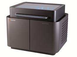


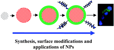

 2012pharmaceutical
 sjwilliamspa