Advances in 3D Printing of the human heart for surgical planning: MRI vs CT
Curator and Author: Justin Pearlman MD PhD
3D printing converts a computer file to one or more 3D objects by deposition of material according to type and locations specified in the data file, as detailed here. The data file for the 3D printing can be based on images, e.g. of the beating heart, which can be obtained by Computed Tomography (CT), Magnetic Resonance Imaging (MRI), UltraSound/Echocardiography (US/Echo), or nuclear imaging. CT and MRI offer significantly better definition of boundary locations needed for 3D modeling than do the other imaging modalities. This article discusses the methods accompanying several real world applications of 3D printing used successfully to assist the plan for open heart surgery.
3D Printing File Formats:
Steps for obtaining 3D printed hearts:
- Select a suitable patient (e.g., a child with unique congenital structural abnormalities who merits 3D assist planning for a customized surgical approach)
- Apply an imaging method that identifies the important tissue boundaries in sufficient detail and accuracy as a 3D data set, typically as a stack of 2D images (e.g., MRI or CT)
- Apply computer analysis tools to convert stacks of gray-scale image arrays to tissue boundaries and tissue character suitable for 3D model generation
- Edit and review boundary extraction, boundary connections in-plane and slice-to-slice and assign character (labels) to boundary surfaces and interiors
- Optionally generate one or more renderings (3D visuals: lighting, transparancies, reflections, cut-aways, rotations, fly-line camera trajectories and angles) for review of 3D structures on 2D screens
- Optionally enable simulated surgery and simulated outcomes (what would the face surface look like after surgical extension of the jaw, measurements, mechanics)
- Apply printer graphic conversion that re-slices the boundaries into coordinates and material selection according to the specification requirements of a 3D printer device
- Submit data to 3D printer, monitor for jamming, wait for the material to print and cool
- Pick up the printed 3D heart(s), inspect for artifacts from curling or other print aberrations, modify object or re-print as needed, deliver printed product(s) to end user (e.g., to surgeon for examination and surgical planning)
Surgical planning:
- Examine the 3D interactive computer graphics, derived video fly-throughs, printed 3D object(s)
- Consider deriving and optionally printing other derived 3D objects (e.g., representing different stages of surgery)
- Make measurements, delineate surgical plan
Currently, the best two competing methods to image the beating heart at sufficient detail are CT scan and MRI.
A CT scan uses ionizing radiation, typically equivalent to more than 200 chest xrays, which can increase the lifetime risk of cancer by as much as 1/1000. The imaging can be completed in less than 10 minutes, an important issue when working with squirming children. The images offer fine resolution (smallest distances that can be distinguished) on the order of 1 millimeter in the cross sections with 1-5 mm section thickness, so the picture elements (Pixels) may be 1 mm x 1 mm x 1 mm covering the heart in 200 slices of 512 x 512 image elements. Each location is characterized by a Hounsfield number that represents the attenuation of x-rays due to the density of the predominant atomic nuclei in each location. Metal implants produce serious artifacts. The Hounsfield numbers group into four tissue types: air, water, bone, metal. Fortunately for planning repair of congenital abnormalities of heart structure, the boundaries of interest are often water density versus fat. An injected radiocontrast agent such as iohexol enhances contrast by generating a distinction from water density for arterial and venous blood conveying the higher density atomic nuclei versus blood vessel or heart walls which are predominantly water density. Iodine contrast may be used to provide a higher atomic Z number for x-ray attenuation, but it can cause a serious allergy and/or renal failure.
MRI uses non-ionizing radiation (magnetic wave energy that does not knock electrons out of orbit to cause damage to the tissues), so it does not pose a future risk of cancer. Also, MRI offers a variety of methods to make signal from flowing blood in arteries and veins each distinct from chamber walls without requiring injection of a contrast agent (though some applications of MRI do use contrast agents). MRI is distorted by metal implants, and by materials with strong differences in magnetic susceptibility, and has complex artifacts if motion occurs at an inopportune time. Both CT and MRI utilize ECG signals to synchronize and effectively “freeze” cardiac motion. A method by the author cancels the magnetic and flow artifacts to enable diagnostic ECG in MRI for safety of potentially deep sedation to further reduce motion artifacts. MRI has many ways to generate signal intensities that characterize tissue differences, so it is over 10 times better at distinguishing differences within soft tissues. MRI thus has advanges identifying boundaries of abnormal tissues such as right ventricular dysplasia, scar tissue (collagen), myxoma, etc. While there are many distinct ways to apply MRI to define the structure of an individual’s heart, commonly a form of fast spin echo, with parallel imaging, is utilized. It generates images representing a slice thickness typically of 2-5 mm each corresponding typically to 128 x 256 picture elements. Thus the volume elements (voxels) tend to be as much as 16-fold larger than from CT, but the ability to distinguish soft tissue differences is an order of magnitude better by MRI. With faster MRI enabled by parallel imaging (multiple slice data obtained concurrently), and with stronger magnets to provide more signal vs noise, the voxels can be smaller and the number of slices covering the heart may be increased, e.g. from 20 to 200.
Even with smaller datasets from MRI versus CT, it can take a technician many hours to trace the tissue boundaries of interest in order to convert the data from slices, rows and columns of grayscale numbers to computer datafiles representing the surface boundaries and assigned surface and interior character delineating a 3D model (some models also require characterization of articulations between parts, elasticity, and other properties). Thus one area of research development (which the author has worked on) has focused on various means to facilitate, automate and speed up that image processsing.
To understand the challenges, consider a doughnut emerged vertically in a tank of milk, imaged as a stack of horizontal slices (completed before the doughnut dissolves). The desired 3D model is a description of the surface of the doughnut. Slices near the top delineate an elliptical boundary, which changes to a figure-of-eight when you examine the first slice that borders on the dougnut hole. Deeper slices reveal the doughnut surface boundary as two circular shapes, until the you encounter the slice bordering on the lower limit of the doughnut hole, which reports another figure of eight. The remaining slices that define the doughnut boundary consist of diminishing sizes of of a single ellipsoid.
Now consider additional complexities such as a chocolate covered doughnut with filling and raisins. If the slice thicknesses are not sufficiently close, the contours of a series of raisins may get mistakenly connected into a branching tube, like a vascular tree. Conversely, if resolution is insufficient, an image of a vascular tree may get segmented into isolated components, and it may confound artery versus vein.
Thus, complex structures with aberrant connections require an astute experienced technologist to help achieve a correct labeling and delineation of structures, their boundaries and connections. The degree of anatomic correctness required depends on the decisions to be made.
Examples of research teams who create 3D printed heart models for surgical planning include:
- Louisville Kentucky – Kosair Children’s Hospital
- Boston MA – Children’s Hospital
- Phoenix AZ – Children’s Hospital
- Boston MA – Mass General Hospital
- Miami FL – Nicklaus Children’s Hospital
3D Printing of Congenital Heart Disease at Kosair Children’s Hospital in Louisville Kentucky
Louisville Kentucky cardiothoracic surgeon Erle Austin has performed successful heart repair surgery on a 14 month old infant named Roland Lian Cung Bawi after planning the surgical approach on a 3D printed flexible double sized reproduction of the patient’s congenitally abnormal and unique anatomy.
Researchers at the University worked with radiologists at Kosair Children’s Hospital to create a means for converting data from a CT scan of Roland’s heart to data that could be used with a 3D printer. The 3D printing team used a MakerBot Replicator 2X, to print the heart (in three pieces) at twice its normal size—they also used a flexible type of plastic known as “Ninja Flex” instead of ABS—it allowed the surgeon to bend the finished heart in ways that resembled a real human heart. Printing the heart took approximately 20 hours at a cost of roughly $600. Dr. Austin told local news reporters that the printed heart helped him plan the surgery in ways he’d never experienced before—it allowed for a single surgery (this past February 10) and greatly for reduced cutting and suturing in a signle surgical session, which ultimately led to a much , promoting a quicker recovery.
Young Roland had been born with four congenital heart defects—doctors had known since before he was born that his heart had problems. Fixing them all would prove to be a challenge. When it came time to plan the surgery, Austin consulted with other surgeons and found each of them had different ideas on the best way to fix the heart. The ideal approach would involve the least amount of cutting and suturing—but that can be hard to plan using only conventional scanning techniques. Looking for more precision, Austin turned to the engineering school at the University of Louisville—they’d been researching different kinds of 3D printing technology. Researchers at the University worked with radiologists at Kosair Children’s Hospital to create a means for converting data from a CT scan of Roland’s heart to data that could be used with a 3D printer. The two seemed a perfect match as CT scanning uses the same basic idea as 3D printing—it takes pictures of slices and puts them together on a computer screen to form a whole, and 3D printing is achieved by laying down one layer or “slice” of material at a time.
The 3D printing team used a MakerBot Replicator 2X, to print the heart (in three pieces) at twice its normal size—they also used a flexible type of plastic known as “Ninja Flex” instead of ABS—it allowed the surgeon to bend the finished heart in ways that resembled a real human heart. Printing the heart took approximately 20 hours at a cost of roughly $600.
Austin told local news reporters that the printed heart let him plan the surgery in ways he’d never experienced before—it allowed for a single surgery (this past February 10) and greatly reduced cutting and suturing, which ultimately led to a much quicker recovery for Roland, who by all accounts is now doing just fine.
video: http://bcove.me/zlxvd038
3D Printing of Congenital Heart Disease at the Children’s Heart Center at Phoenix Children’s Hospital in Phoenix Arizona
Doctors gather as much information as possible when preparing to correct heart defects in pediatric patients. They read images from CT scans on computers. They may even use software to study abnormalities in three dimensions, moving a picture of the heart around on a computer screen to analyze and plan a surgical strategy.
But what if they could take the process one step further? What if they could simply press “print” to create a perfect, color-coded, three-dimensional plastic model of a child’s heart before the surgical procedure even begins?
Tags: At the Children’s Heart Center at Phoenix Children’s Hospital, Justin Ryan, a biomedical engineering graduate research associate at Arizona State University, is doing just that. Ryan, who has a background in animation studies, uses those same technical skills to change the two-dimensional images from CT scans to a three-dimensional object.
“It’s very similar to what you might see in a CGI (computer-generated images) in a movie, or in a video game character,” he says. After the image is created on his laptop, Ryan sends it to a three-dimensional printer that creates the model.
The printer, about the size of a pastry case at a coffee shop, contains a cinderblock-sized chunk of Super Glue combined with gypsum, a common material used in drywall construction. Ink jets slowly spray super-thin layers of color on the powdery block, forming the model according to the precise specifications of the data. Ryan equates it to building a house, from the bottom up, brick by brick.
The printing process itself takes about three hours. When it is finished, Ryan brushes the excess powdery material away to reveal the model. “From there, we do a bit of post-processing, but in another hour after that, we can hand it off to the doctor. They can view it, and make their decisions on surgery.”
Using a heart model to prepare for surgery is like finding your way with a GPS instead of a paper map, says Daniel Velez, M.D., a congenital heart surgeon at PCH. With the models, a surgeon can see, and touch, the actual size of the structure before surgery even begins. “To be able to tell the parents more precisely what I’m going to do, and what I’m going to encounter—even though I do tell them about variations and variabilities to the plan—I’m more at ease and more certain,” he says.
Each part of the heart, from chambers to vessels, is assigned a different color. Studies show that color coding helps medical teams better understand the tiny anatomical structures they will work with during surgery, says John Nigro, M.D., director of cardiothoracic surgery and co-director of the heart center. In babies, he adds, the heart is about the size of a walnut. As a child grows, it is about the size of their fist. “You can imagine the size of a child’s fist is pretty small,” says Nigro.
The technology is so new that it is almost too early to know what kind of impact it will have, says Stephen G. Pophal, M.D., division chief of pediatric cardiology at PCH. Pophal, who specializes in pediatric cardiac catheterization procedures, says it could change medicine.
Pophal teamed up with ASU engineering professor David Frakes to start the project at PCH. Frakes says is a “first of its kind” application for modeling congenital heart disease. “We hope that very soon any doctor who is doing an operation here is going to be carrying one of these models into the operating room—and being better prepared to perform because of it.”
Prophal hopes that using models will give doctors a head start in correcting defects. Knowing more in advance could also cut down on the number of images needed as procedures are performed, lowering radiation exposure. The tough job of visualizing a defect in three dimensions based on a two-dimensional image—sort of like imagining what a house will look like based on architectural plans—would be eliminated, says Pophal. “If I had the model in my hand, I would save half the time, and have half of the worries.”
Learning that a child has a heart defect can be overwhelming for parents, says pediatric nurse practitioner Courtney Howell, CPNP. “I think that is the hard part for some families to understand. They have this beautiful baby, and they expected all the things to go perfectly. Now they have this huge diagnosis.”
Howell says that the heart models help parents understand their child’s defect—it offers complex medical terminology in a context they can understand. And if everyone on the team understands the nature of the defect, patients are likely to do better, Pophal says. “The major impact is that we will be able to teach people more about their heart problems. This makes it very simple.”
SOURCE
http://www.raisingarizonakids.com/2013/02/modeling-the-heart/
3D Printing of Congenital Heart Disease at Mass General Hospital in Boston Massachusetts
http://www.auntminnie.com/index.aspx?sec=ser&sub=def&pag=dis&ItemID=111445
MGH saves money and time printing 3D heartsBy Eric Barnes, AuntMinnie.com staff writer
July 17, 2015 — Researchers at Massachusetts General Hospital (MGH) in Boston say that having a 3D rapid prototyping printer in the cardiac imaging department is a potential boon for procedure planning and patient education — and it doesn’t break the bank.
After installing the printer and learning how to edit the cardiac CT images, they found they can build accurate and useful models using rigid or flexible materials, said co-author Phillip Kim in a presentation at the Society for Imaging Informatics in Medicine (SIIM) meeting held in May at National Harbor, MD.
Phillip Kim of MGH.“An in-house 3D rapid prototyping printer provides the user with accurate and affordable models with fast turnaround times,” Kim said in his presentation. “Other potential benefits include use as an educational tool for patients, medical students like me, and residents.”
The technology can be used to create digital-to-analog study models from CT images for a variety of applications, from surgical planning to regenerative medicine, he said.
The current study looked at reproducibility, turnaround time, and cost-effectiveness for an in-house 3D rapid prototyping printer.
Getting images printer-ready
At MGH, the process begins with the selection of DICOM images of interest. Once selected, the DICOM images are modified using image processing software from TeraRecon or OsiriX. The OsiriX software is used for simpler models, while the other software is used for more complicated ones, to crop, change Hounsfield Unit thresholds, or add or erode images, among other tasks. The programs are then used to export the images as stereolithography (STL) files, Kim said.
The STL files are processed with an application called Replicator G, which converts the image data to a language called gcode. From there, another open-source application, MakerWare (MakerBot), is used to convert the data to x3g format, which is recognized by the MakerBot printer.
The researchers acquired images of coarctation of the aorta with or without stenting, aortic dissection, anomalous origin of coronary arteries, and aortic aneurysms.
All scans were performed on a 128-detector-row dual-source CT scanner (Somatom, Siemens Healthcare). The group acquired 128 contiguous 0.6-mm slices with a gantry rotation time of 280 msec and 75-msec temporal resolution.
Normal aortic valve. All heart model images courtesy of Phillip Kim.
Coarctation of aorta. Right image is poststenting.Rigid or flexible plastic
Two different types of plastic, extruded as 0.1-mm thick filaments, can be used in the printer, depending on the desired application:
- Acrylonitrile butadiene styrene (ABS) is extruded as a solid filament and is “very durable and beautiful,” Kim said.
- For a more malleable planning tool, NinjaFlex (Fenner) has a flexible filament that creates soft, pliable models.
As for printers, several 3D rapid prototyping methods are available, including selective laser sintering, stereolithography, and finally fused deposition modeling, which was MGH’s method of choice.
“It requires little maintenance and is affordable and user-friendly,” Kim said.
Anomalous coronary arteries, above and below.Outsourcing is costly
Kim and colleagues Dr. Harshna Vadvala and Dr. Brian Ghoshhajra did a time and cost analysis on 10 of the printed models, and there was good news on that front as well. The costs amounted to a few dollars per model — a small fraction of the cost of outsourcing.
For example, for one of the anomalous origin models, the printer took less than 12 hours at an approximate cost of $2 for supplies, excluding the cost of filament, which was free for the project.
“If you were to outsource it, it would have cost at least $400 and would have taken more than 24 hours to have it delivered to the office,” Kim said.
When they tried to compare outsourcing costs among three vendors for printing an entire heart, two were unable even to load the files because they were too large to process, he said.
“In the case where the file wasn’t too large, it would have cost us at least $500,” Kim said.
Having a rapid prototyping 3D printer in-house in a cardiac imaging department provides users with accurate and affordable models that can be created quickly, Kim concluded. Further study is needed to find all of the ways such models might benefit students, patients, and clinicians, he said.


Source: http://www.auntminnie.com/index.aspx?sec=ser&sub=def&pag=dis&ItemID=111445
3D Printing of Congenital Heart Disease at Children’s Hospital and MIT in Boston Massachusetts
http://fortune.com/2015/09/17/better-heart-models-save-lives/
http://scitechdaily.com/tag/harvard-university/

Danielle Pace at MIT , an MIT graduate student in electrical engineering and computer science and Medhi Mogan. a physicist at Boston Children’s Hospital teamed up to develop a means of constructing 3D image boudaries from cardiac MRI,
They segmented just 14 slices from cardiac MRI, letting an algorithm infer the rest. They report 90 percent agreement with expert segmentation of the entire collection of 200 cross sections.
To eliminate as much guesswork as possible in advance of surgery, researchers have been constructing accurate heart models using 3D printers and measurement data from scans. The problem: The process is slow. It can take up to 10 hours to finesse the internal boundaries that separate the heart chambers and vessels. Those intersections don’t necessarily show up clearly so doctors relied on a manual process to fill in the blanks, so to speak. And that takes time.
Now, researchers from MIT and Boston Children’s Hospital say they’ve come up with a better, faster way to build heart models, according to MIT Tech News. Part of their work relies on new processes that enhance the precision of the scans, which are basically a set of cross sections that together comprise a 3D image of the heart or other structure.
But even the scan data is not enough. Researchers often used generic heart representations to supply additional data needed to build the full model. The problem there is generic models aren’t much help in recreating what is likely a heart with anomalies all its own.
The proposed solution, as is often the case with complex data problems, still draws on human expertise, but in a much more curtailed way than before.
According to the report, project leader Polina Gollan, professor of engineering and computer science at MIT, said this limited human input greatly increases accuracy of the model.
The “strongest results came when they asked the expert to segment only a small patch—one-ninth of the total area—of each cross section,” she said.
With these advances, the team can create the algorithm needed and print the model in three or four hours, compared to the 10 needed before.
The team, which also includes Medhi Moghari, a physicist who came up with new processes to enhance the MRI scan precision; Andrew Powell, a cardiologist; and Danielle Pace, an MIT grad student in electrical engineering and computer science, will put their work to test in a study kicking off this fall using MRIs from 10 patients at Children’s Hospital. Pace will also present a related paper at a medical conference next month.
This is just the latest application of advanced 3D printing and materials to create medical devices including prosthetic devices.
3D printed heart.
Photograph by Bryce Vickmark
3D Printing of Congenital Heart Disease at Nicklaus Children’s Hospital in Miami Florida
3D Printing Aids Surgery to Repair 5-Year-Old Mia’s Heart
BY TYLER KOSLOW ON THU, OCTOBER 8, 2015 · 3D MODELING, 3D PRINTERS, 3D PRINTING, 3DP APPLICATIONS, MEDICAL & DENTAL, NEWS,RESEARCH
When she first stepped foot in Nicklaus Children’s Hospital, five-year-old Mia Gonzalez was suffering from an extremely rare heart malformation diagnosed as a double aortic arch, a condition in which the airflow is restricted by the vascular ring wrapping around the trachea or esophagus. Dr. Redmond Burke, the director of Pediatric Vascular Surgery and the Nicklaus Children’s Hospital in Miami, Florida, had an extremely challenging operation in his hands. This was a surgical procedure that needed intensive preparation, so Burke and his team decided to plan for the complex heart surgery on a 3D modeled replica of Gonzalez’s own heart, printed courtesy of Stratasys reseller AdvancedRP.
“By making a 3D model of her very complex aortic arch vessels, we were able to further visualize which part of her arch should be divided to achieve the best physiological result,”said Burke. “It’s very powerful when you show a family ‘this is your baby’s heart and this is how I’m going to repair it.”
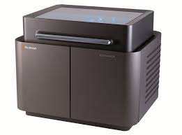
AdvancedRP is a supplier of 3D printed anatomical models for surgical preparation, using a Stratasys Objet500 Connex3 Multi-Material 3D Printer. These 3D models not only replicate the physicality of the patient in question, but with flexibility similar to human organs, flesh, and bone, due to the Objet500 Connex3’s ability to combine multiple textures and material types in a single print.
In order to replicate the patient’s organ accurately, MRI or CT scan data is translated into a 3D model, which is then prepped for 3D printing on the Objet500. This allows for extremely efficient surgical preparation; Burke and his team were able to practice the advanced surgery ahead of time, ensuring that no damage or pain be inflicted upon little Mia Gonzalez and her heart. Preparing on the 3D printed heart model helped to decrease operation time, too.
“Once patient scan data from MRI or CT imaging is fed into the Stratasys 3D Printer, doctors can create a model with all its intricacies, specific features and fine detail,” said Stratasys’ GM of Medical Solutions Scott Rader. “This significantly enhances surgical preparedness, reduces complications and decreases operating time.”