Healthcare analytics, AI solutions for biological big data, providing an AI platform for the biotech, life sciences, medical and pharmaceutical industries, as well as for related technological approaches, i.e., curation and text analysis with machine learning and other activities related to AI applications to these industries.
Scientists at MIT and Massachusetts General Hospital have discovered how cancer cells latch onto blood vessels and invade tissues to form new tumors — a finding that could help them develop drugs that inhibit this process and prevent cancers from metastasizing.
Cancer cells circulating in the bloodstream can stick to blood vessel walls and construct tiny “bridges” through which they inject genetic material that transforms the endothelial cells lining the blood vessels, making them much more hospitable to additional cancer cells, according to the new study.
The researchers also found that they could greatly reduce metastasis in mice by inhibiting the formation of these nanobridges. “Endothelial cells line every blood vessel and are the first cells in contact with any blood-borne element. They serve as the gateway into and out of tumors and have been the focus of intense research in vascular and cancer biology.
Building bridges
Metastasis is a multistep process that allows cancer to spread from its original site and form new tumors elsewhere in the body. Certain cancers tend to metastasize to specific locations; for example, lung tumors tend to spread to the brain, and breast tumors to the liver and bone.
To metastasize, tumor cells must first become mobile so they can detach from the initial tumor. Then they break into nearby blood vessels so they can flow through the body, where they become circulating tumor cells (CTCs). These CTCs must then find a spot where they can latch onto the blood vessel walls and penetrate into adjacent tissue to form a new tumor.
Blood vessels are lined with endothelial cells, which are typically resistant to intruders.
The researchers first spotted tiny bridges between cancer cells and endothelial cells while using electron microscopy to study the interactions between those cell types. They speculated that the cancer cells might be sending some kind of signal to the endothelial cells.
“Once we saw that these structures allowed for a ubiquitous transfer of a lot of different materials, microRNAs were an obvious interesting molecule because they’re able to very broadly control the genome of a cell in ways that we don’t really understand,” Connor says. “That became our focus.”
MicroRNA, discovered in the early 1990s, helps a cell to fine-tune its gene expression. These strands of RNA, about 22 base pairs long, can interfere with messenger RNA, preventing it from being translated into proteins.
In this case, the researchers found, the injected microRNA makes the endothelial cells “sticky.” That is, the cells begin to express proteins on their surfaces that attract other cells to adhere to them. This allows additional CTCs to bind to the same site and penetrate through the vessels into the adjacent tissue, forming a new tumor.
Non-metastatic cancer cells did not produce these invasive nanobridges when grown on endothelial cells.
Shutting down metastasis
The nanobridges are made from the proteins actin and tubulin (NB-the protein actin is abundant in all eukaryotic cells. It was first discovered in skeletal muscle, where actin filaments slide along filaments of another protein called myosin to make the cells contract. In non-muscle cells, actinfilaments are less organized and myosin is much less prominent, and a tubulin is a protein that is the main constituent of the micro-tubules of living cells, which also form the cytoskeleton that gives cells their structure). The researchers found that they could inhibit the formation of these nanobridges, which are about 300 microns long, by giving low doses of drugs that interfere with actin.
When the researchers gave these drugs to mice with tumors that normally metastasize, the tumors did not spread.
Sengupta’s lab is now trying to figure out the mechanism of nanobridge formation in more detail, with an eye toward developing drugs that act more specifically to inhibit the process.
The SEM picture below,
is a rounded cancer cell (top left) that sends out nanotubes connecting with endothelial cells. Genetic material can be injected via these nanotubes, transforming the endothelial cells and making them more hospitable to additional cancer cells. Image credit: Sengupta Lab. The second picture below,
is showing a cancer cell (bottom center) that creates a gap and enters the endothelial tube. Another cancer cell (middle right) sends out nanotubes to connect with endothelial cells. Both image are credited toSengupta Lab
An interesting comment on why plants do not develop cancer is given here, The article states that in plants, as in animals, most cells that constitute the organism limit their reproductive potential in order to provide collective support for the immortal germ line. And, as in animals, the mechanisms that restrict the proliferation of somatic cells in plants can fail, leading to tumors. There are intriguing similarities in tumorigenesis between plants and animals, including the involvement of the retinoblastoma pathway as well as overlap with mechanisms that are used for stem cell maintenance. However, plant tumors are less frequent and are not as lethal as those in animals. The authors of the article argue that fundamental differences between plant and animal development make it much more difficult for individual plant cells to escape communal controls.
The structure of the endothelium, the thin layer of cells that line our arteries and veins, is visible here. The endothelium is like a gatekeeper, controlling the movement of materials into and out of the bloodstream. Endothelial cells are held tightly together by specialized proteins that function like strong ropes (red) and others that act like cement (blue). In the picture here, the cell is preparing to divide. Two copies of each chromosome (blue) are lined
up next to each other in the center of the cell. Next, protein strands (red) will pull apart these paired
chromosomes and drag them to opposite sides of the cell. The cell will then split to form two daughter cells, each with a single, complete set of chromosomes. It is interesting that protein strands (red) in this picture are implicated on pulling apart the two copies of chromosomes in the same way the proteins actin and tubules do in the cancer cell interaction with the veins and arteries. It looks like the proteins shaped as strings, the nanobridges, due their shape to the tension development between cancer cells and the veins.
Adicet Bio, Inc. (formerly resTORbio, Inc. (resTORbio)), together with its subsidiaries, (the Company) is a clinical stage biotechnology company discovering and developing allogeneic gamma delta T cell therapies for cancer. The Company is advancing a pipeline of off-the-shelf gamma delta T cells, engineered with chimeric antigen receptors (CARs) and T cell receptor-like antibodies to enhance selective tumor targeting, facilitate innate and adaptive anti-tumor immune response, and improve persistence for durable activity in patients. The Company’s approach to activate, engineer, and manufacture allogeneic gamma delta T cell product candidates derived from the peripheral blood cells of unrelated donors allows it to generate new product candidates in a rapid and cost efficient manner. The Company was incorporated in November 2014 in Delaware. The principal executive offices are located in Boston, Massachusetts. The Company also has another office in Menlo Park, California. Adicet Bio, Inc. (when referred to prior to the Merger (as defined below), (Former Adicet)) was incorporated in November 2014 in Delaware and was headquartered in Menlo Park, CA. Adicet Bio Israel Ltd. (formerly Applied Immune Technologies Ltd.) (Adicet Israel) is a wholly owned subsidiary of Former Adicet and is located in Haifa, Israel. Adicet Israel was founded in 2006. During 2019, Former Adicet consolidated its operations, including research and development activities, in the United States and as a result substantially reduced its operations in Israel.
They had a MERGER,
Merger with resTORbio
On September 15, 2020, the Company completed the Merger pursuant to the Merger Agreement (the Effective Time). In connection with the Merger, and immediately prior to the Effective Time, resTORbio effected a reverse stock split of its common stock at a ratio of 1-for-7 (the Reverse Stock Split). Also, in connection with the Merger, the Company changed its name from “resTORbio, Inc.” to “Adicet Bio, Inc.” (the Name Change), Former Adicet changed its name from “Adicet Bio, Inc.” to “Adicet Therapeutics, Inc.” and the business conducted by the Company became primarily the business, which was previously conducted by Former Adicet, which is a biotechnology company discovering and developing allogeneic gamma delta T cell therapies for cancer and other diseases.
an IPO, and
they are Traded on NASDAC – no profits !!!!!!!!!
Updated on 1/27/2016
On 1/27/2016 Adicet Bio announced the acquisition of Applied Immune Technologies (AIT)
Applied Immune Technologies (AIT) was acquired by Adicet, 1/27/2016.
AIT is a drug development company specializing in T-Cell Receptor-Like (TCRL) antibodies that are targeted to intracellular-derived peptides for a variety of therapeutic and diagnostic applications. AIT is also focused on identification and validation of novel therapeutic targets.
Therapeutic Antibodies
AIT’s core technology platforms encompass the identification and validation of novel MHC-based targets, as well as development of therapeutic Human Recombinant T-Cell Receptor-Like (TCRL) antibodies with the unique ability to bind with these intracellular peptide/MHC complexes with the specificity of cytotoxic T-cell killer cells.
AIT’s technologies enable the generation of a rich pipeline of therapeutic TCRL antibodies for intracellularly-derived, disease-specific targets which normally are not accessible to conventional antibodies. TCRL antibodies also have diagnostic applications in vaccine design, validation and monitoring, as well as analysis of antigen presentation in disease.
Immune System Explained
Our immune system is composed of two arms: antibodies and T cells.
Soluble antibody molecules can bind to cell surface expressed proteins with high affinity and specificity. Upon binding, they can recruit effector cells of the immune system such as macrophages and Natural Killer (NK) cells, or mediate a biological signal into cells. Antibodies constitute today the most important class of targeted therapeutics in the bio-pharmaceutical industry.HOWEVER, analysis of the human proteome reveals that only 20% of human proteins are expressed on the cell surface. The remaining 80% of the human proteome is intracellular, and is therefore not accessible by conventional antibodies for therapeutic targeting clinical applications. Thus, there is an urgent need for the development of novel therapeutic antibodies against these intracellular based targets.
T cells mediate cellular immunity. CD8+ Cytotoxic T cells (CTLs) are the most potent effector cells of the immune system because they can recognize and kill diseased cells in a highly specific manner. They recognize intracellular proteins due to their ability to bind to the cell surface-expressed MHC-peptide complex, which presents peptides derived from intracellular proteins. Upon specific recognition of the MHC-peptide complex by the T-cell receptor (TCR), the CTLs undergo activation, proliferation and expansion, leading to destruction of the target diseased cells. HOWEVER, T cells are very difficult to manipulate for therapeutic applications and thus their advantage in recognizing intracellular targets is very difficult to apply for clinical therapeutic purposes.
AIT’s innovative solution is a platform technology for the development of human recombinant T-Cell Receptor-Like (TCRL) antibodies capable of targeting intracellular-derived peptides. These 3rd generation antibodies combine the advantages of both arms of the immune system. They are capable of binding to targets with high affinity and specificity like conventional antibodies. In addition, they can recognize and bind to intracellular-derived peptides presented on the MHC complex with the same degree of specificity as T cells, but without being limited by the regulatory mechanisms imposed on T cells.
TCRL antibodies enable us to identify and validate intracellular-expressed disease-specific targets and make them available for cell surface targeting. AIT’s platform technology further enables the generation of highly specific therapeutic antibodies against these intracellular-derived targets, which can bind to them on the cell surface and kill the diseased cells only, without effecting healthy cells. Thus, disease-specific targets that are expressed inside diseased cells are transformed into targets that can be recognized by soluble antibodies on the cell surface.
This breakthrough harnesses the power of the cellular arm of the immune system to attack diseased cells with soluble, readily made human monoclonal antibodies. Distinct from conventional monoclonal approaches that only attack cell surface-associated proteins, AIT’s TCRL technology addresses the far more abundant intracellular proteome. The combination of these features opens up entirely new vistas for the development of highly specific 3rdgeneration antibodies for the treatment of cancer, viral, and autoimmune diseases.
EpiTarget is a unique approach for the discovery and validation of novel therapeutic MHC-based targets that can be applied to the isolation and characterization of new TCRL antibodies against a variety of disease-related intracellular targets.
The EpiTarget approach combines bioinformatic analysis and mass spectroscopy strategies to identify target peptides presented on MHC molecules that are differentially expressed on diseased cells of various histological origins.
The large intracellular proteome, which is not accessible for antibody-based recognition, can serve as a huge pool for new target discovery and a strong pipeline for therapeutic and diagnostic TCRL antibodies by integration of proteomic strategies with the TCRL technology.
Makler, O., Oved, K., Netzer, N., Wolf, D., Reiter, Y. Direct visualization of the dynamics of antigen presentation in human cells infected with cytomegalovirus revealed by antibodies mimicking TCR specificity. Eur. J. Immunol. 40: 1552-1562, 2010.
Klechevsky, E., Flamar, A.L., Cao, Y., Liu, M., Thompson-Snipes, L., O’Bar, A., Zurawski, S., Reiter, Y., Zurawski, G., Banchereau, J. Cross-priming CD8+ T cells by targeting antigens to human dendritic cells through DCIR. Blood 116:1685-97, 2010.
Dahan, R., Tabul, M., Chou, Y.K., Meza-Romero, R., Andrew, S., Ferro, A.J., Burrows, G.G., Offner, H., Vandenbark, A.A., Reiter, Y. TCR-like antibodies distinguish conformational and functional differences in auto-reactive idiotopes present on two vs. four-domain MHC class II/peptide complexes. Eur. J. Immunol. 41:1465-79,2011.
Dahan, R, Reiter, Y. T-cell-receptor-like antibodies – generation, function and applications. Expert Rev Mol Med. 14:e6, 2012.
Noy, R., Epel, M., Haus-Cohen, M., Klechevsky, E., Makler, O., Michaeli, Y., Denkberg, G., Reiter, Y. T-cell receptor-like antibodies: novel reagents for clinical cancer immunology and immunotherapy. Exp. Rev. Anticancer Ther. 5(3) 2005.
Michaeli, Y., Sinik, K., Cohen, M., Reiter, Y:. Protein Instability and Aberrant Intracellular Processing of Tyrosinase Lead to High Presentation of HLA-A2/Tyrosinase Complexes on the Surface of Melanoma Cells. Eur. J. Immunol. In press 2012.
Aya Jakobovits, former founder and CEO of Kite Pharma and current venture partner at OrbiMed, is heading up a new, next-gen immunotherapy startup – backed by an impressive $51 million Series A.
The Bay Area upstart, called Adicet Bio, is keeping very quiet about its underlying platform. Jakobovits emphasized in a phone interview, however, that the platform is meant to develop “universal immune cell therapy” – meaning it should be broadly applicable for a number of diseases, including cancer, autoimmune disease and inflammation.
It’s launching with news of an acquisition, however: Adicet just bought Israeli immunotherapy company Applied Immune Technologies, which focuses on the intracellular proteome.
“There’s a very good correlation between AIT and Adicet,” Jakobovits said.
AIT develops T-Cell Receptor-like antibodies that are targeted, through the Major Histocompatibility Complex (MHC), toward disease-linked peptides in cells. Adicet’s plan is to generate monoclonal antibodies that have affinity and high specificity to this MHC complex, she said.
Jakobovits said there’s no correlation or link between Kite Pharma and Adicet – in technology or in business dealings.
The financing round was led by OrbiMed with participation from Novartis Venture Fund and Pontifax.
Adicet Bio Announces Closing of $51 Million Series A Financing and Acquisition of
Applied Immune Technologies
Menlo Park, CA (January 27, 2016): Adicet Bio, Inc. (“Adicet”), a biopharmaceutical company focused on the development of next-generation cell immunotherapies, announced today that it closed a $51 million Series A financing. Adicet also announced the acquisition of Applied Immune Technologies, Ltd. (“AIT”), an Israel-based company that develops immunotherapies directed to the intracellular proteome.
The financing was led by OrbiMed and also included Novartis Venture Fund and Pontifax.
“These significant financial resources will allow Adicet to progress its universal immune cell therapy (“uICT”) platform technology and related products and advance AIT’s programs and product pipeline,” said Aya Jakobovits, Ph.D., Founder, President and Chief Executive Officer of Adicet. “AIT’s technologies, capabilities, and intellectual property highly complement those of Adicet and position the combined company to become a leader in next-generation immunotherapy products for cancer and other indications.
Adicet was founded by Aya Jakobovits and OrbiMed. Previously, Dr. Jakobovits served as the President and founding CEO of Kite Pharma, Inc. Before joining Kite Pharma, she served as Executive Vice President, Head of Research and Development at Agensys, Inc., which became an affiliate of Astellas Pharma Inc. in a deal valued at up to $537 million. Before its acquisition, she served as Agensys’ Senior Vice President, Technology and Corporate Development and Chief Scientific Officer. Prior to Agensys, Dr. Jakobovits served as the Director, Discovery Research and Principal Scientist at Abgenix, Inc., which was spun out of Cell Genesys, Inc. and based on the XenoMouse® technology developed under her leadership. Abgenix was acquired by Amgen Inc. for $2.2 billion.
AIT specializes in generating and developing T-Cell Receptor-Like (“TCRL™”) antibodies with high affinity and specificity to disease-specific intracellular peptides presented on the cell surface by the major histocompatibility complex (“MHC”). AIT also established Epitarget™, a proprietary technology to identify and validate novel disease-specific peptide targets. AIT technology is based on work by Prof. Yoram Reiter, a world leader in the research of immunotherapies directed to the intracellular proteome. AIT will continue its operations in Israel as Adicet’s wholly-owned subsidiary.
Following the financing and acquisition, the Adicet Board of Directors will include Jonathan Silverstein and Carl Gordon, General Partners and Co-Heads of Global Private Equity at OrbiMed, Aya Jakobovits, Florent Gros, Managing Director at Novartis Venture Fund, and Erez Chimovits, Managing Director at OrbiMed Israel.
“We are excited to join forces again with Aya, a prominent figure in the field of immunotherapy with a track record of growing successful biotechnology companies,” said Carl Gordon.
“We look forward to building a leading immunotherapy company,” said Jonathan Silverstein. “The AIT acquisition expands Adicet’s platform technologies and its product pipeline.”
About Applied Immune Technologies, Ltd.
Applied Immune Technologies Ltd. (“AIT”) pioneered and advanced the generation and development of TCRLs for therapeutic and diagnostic applications in cancer, inflammation, autoimmune, and infectious diseases. AIT’s TCRLs are directed to disease-specific peptide-MHC complexes and are aimed at delivering potent payloads specifically to the diseased cells. AIT’s pipeline includes TCRLs directed to different disease indications. AIT also established a robust and proprietary technology for identification and validation of novel MHC-based targets. AIT was founded in 2006 by Prof. Yoram Reiter, Head of the Laboratory of Molecular Immunology at the Technion, Israel Institute of Technology, and Mira Peled-Kamar, Ph.D., AIT Chief Executive Officer. AIT is located in Haifa, Israel.
About Adicet Bio, Inc.
Adicet Bio, Inc. is a privately held, pre-clinical stage biotechnology company engaged in the design and development of cutting-edge immunotherapies for cancer and other disease indications, with a focus on novel universal immune cell therapies (uICT). Adicet Bio is located in Menlo Park, California.
# # #
Contact:
Aya Jakobovits, Ph.D. President and Chief Executive Officer
Adicet Bio, Inc.
Tel 310.990.3832 ajakobovits@adicetbio.com
For Media:
Joan Kureczka Kureczka/Martin Associates
Tel 415.821.2413
Mobile 415.690.0210 joan@kureczka-martin.com
Dr. Peled-Kamar, AIT’s Co-Founder and CEO, has extensive academic and biotech research experience in basic sciences as well as biotechnology and biomedical devices. Dr. Peled has worked for leading Israeli biotech companies including Biotechnology General and Interpharm, was the Co-Founder and CEO of BioMimic Pharma, and has been involved in the establishment of a number of start-ups. Dr. Peled received her PhD in Biochemistry and Molecular Biology from the Weizmann Institute of Science and completed her post doctorate at the University of California, Berkeley.
Yoram Reiter, PhD – Chief Scientific Officer
AIT Co-Founder and CSO, Professor Reiter is a world leader in the isolation of human TCRL molecules. Head of the Laboratory of Molecular Immunology at the Technion Institute, Prof. Reiter established a cutting-edge research program in the molecular immunology of cancer. His major work involves the development of novel immunotherapeutic approaches, as well as the study of molecular mechanisms in anti-tumor and anti-viral immunity. Prof. Reiter received his PhD from the Department of Immunology at the Weizmann Institute and subsequently spent 5 years at the NCI Laboratory of Molecular Biology headed by Dr. Ira Pastan, where he developed new approaches in antibody engineering. Prof. Reiter has published over 100 scientific papers and reviews, and holds 15 patents. He currently serves as an advisor to several pharmaceutical and biotechnology companies.
Galit Denkberg, PhD – R&D Manager
Dr. Denkberg leads AIT’s TCRL development team. A graduate of the Technion’s Faculty of Biology, she worked with Prof. Yoram Reiter on the design and construction of the single-chain MHC molecules and was the first in the group to isolate TCRL antibodies from both immunized and naïve phage-display libraries. Through the years Dr. Denkberg has mastered a variety of molecular immunology, cellular immunology and molecular biology techniques and has become an expert in design, application, and characterization of TCRL antibodies. Dr. Denkberg is the author of 16 scientific papers related to the TCRL technology in leading peer-reviewed journals.
contains a wonderful curation on a new TGF-beta antibody fresolimumab which inhibits multiple isoforms of TGF-beta and may have indications in cancer and inflammation. The curation includes informative diagrams on mechanism of action and TGF-beta signaling as well as pertinent PK/PD data.
AstraZeneca’s WEE1 protein inhibitor AZD1775 Shows Success Against Tumors with a SETD2 mutation
Stephen J. Williams, Ph.D., Curator
There have been multiple trials investigating the utility of cyclin inhibitors as anti-tumoral agents (see post) with the idea of blocking mitotic entry however another potential antitumoral mechanism has been to drive the cell into mitosis in the presence of DNA damage or a defective DNA damage repair capacity. A recent trial investigating an inhibitor or the cell cycle checkpoint inhibitor Wee1 showed positive results in select cohorts of patients with mutations in DNA repair, indicating the therapeutic advantage of hijacking the cell’s own DNA damage response, much like how PARP inhibitor Olaparib works in BRCA1 mutation positive ovarian cancer patients.
Investigators at Oxford University say that one of AstraZeneca’s ($AZN) pipeline drugs proved particularly effective in killing cancer cells with a particular genetic mutation.
The ex-Merck ($MRK) drug is AstraZeneca’s WEE1 protein inhibitor AZD1775, which proved particularly lethal to genes with a SETD2 mutation, which the researchers see as a potential ‘Achilles heel’ often found in kidney cancer and childhood brain tumors.
“When WEE1 was inhibited in cells with a SETD2 mutation, the levels of deoxynucleotides, the components that make DNA, dropped below the critical level needed for replication,” noted Oxford’s Andy Ryan. “Starved of these building blocks, the cells die. Importantly, normal cells in the body do not have SETD2 mutations, so these effects of WEE1 inhibition are potentially very selective to cancer cells.”
AstraZeneca landed rights to the drug back in 2013, when incoming Merck R&D chief Roger Perlmutter opted to spin it out while focusing an immense effort around the development of its PD-1 checkpoint inhibitor KEYTRUDA® (pembrolizumab). Since then, AstraZeneca has made it available to academic investigators through their open innovation program.
Wee1, DNA damage checkpoint and cell cycle regulation
In fission yeast, Wee1 delays entry into mitosis by inhibiting the activity of Cdk1, the cyclin-dependent kinase that promotes entry into mitosis (Cdk1 is encoded by the cdc2+ gene in fission yeast and the CDC28 gene in budding yeast) (Russell and Nurse, 1987a). Wee1 inhibits Cdk1 by phosphorylating a highly conserved tyrosine residue at the N-terminus (Featherstone and Russell, 1991; Gould and Nurse, 1989; Lundgren et al., 1991; Parker et al., 1992; Parker and Piwnica-Worms, 1992). The phosphatase Cdc25 promotes entry into mitosis by removing the inhibitory phosphorylation (Dunphy and Kumagai, 1991; Gautier et al., 1991; Kumagai and Dunphy, 1991; Millar et al., 1991; Russell and Nurse, 1986; Strausfeld et al., 1991). Loss of Wee1 activity causes cells to enter mitosis before sufficient growth has occurred and cytokinesis therefore produces two abnormally small daughter cells (Fig. 1A) (Nurse, 1975). Conversely, increasing the gene dosage of wee1 causes delayed entry into mitosis and an increase in cell size, indicating that the levels of Wee1 activity determine the timing of entry into mitosis and can have strong effects on cell size (Russell and Nurse, 1987a). Similarly, cdc25– mutants undergo delayed entry into mitosis, producing abnormally large cells, and an increase in the gene dosage of cdc25 causes premature entry into mitosis and decreased cell size (Russell and Nurse, 1986). Despite these difficulties, early work in fission yeast suggested that the Wee1 kinase plays an important role in a checkpoint that coordinates cell growth and cell division at the G2/M transition (Fantes and Nurse, 1978; Nurse, 1975; Thuriaux et al., 1978). WEE1 is an evolutionarily conserved nuclear tyrosine kinase (Table 2) that is markedly active during the S/G2 phase of the cell cycle [24, 25]. It was first discovered 25 years ago as a cell division cycle (cdc) mutant-wee1– in the fission yeast, Schizosaccharomyces pombe [26]. Fission yeast lacking WEE1 are characterized by a smaller cell size, and this phenotype has been attributed to the ability of WEE1 to negatively regulate the activity of cyclin dependent kinase, Cdc2 (Cdc28 in budding yeast and CDK1 in human), in the Cdc2/CyclinB complex [27]. Recently, WEE1 was shown to directly phosphorylate the mammalian core histone H2B at tyrosine 37 in a cell cycle dependent manner. Inhibition of WEE1 kinase activity either by a specific inhibitor (MK-1775) or suppression of its expression by RNA interference abrogated H2B Y37-phosphorylation with a concurrent increase in histone transcription [17].
As shown in the Below figure Wee1 is a CDK cyclin kinase which results in an inactivating phosphorylation event on CDK/Cyclin complexes
Figure 1. Schematic representation of the effects of Chk1 and Wee1 inhibition on CDK-CYCLIN complex regulation, that gets more activated being unphosphorylated from Cell cycle, checkpoints and cancer by Laura Carrassa.
Figure 2. Schematic representation of the role of Chk1 and Wee1 in regulation of the CDK-cyclin complexes involved in S phase and M phase entry from Cell cycle, checkpoints and cancer by Laura Carrassa.
The following articles discuss how Wee1 can be a target and synergize with current chemotherapy
p53 mutation Frequency in Ovarian Cancer and contribution to chemo-resistance
The following is from the curated database TCGA and cBioPortal TCGA Data Viewer for mutations found in ovarian cancer sequencing studies in the literature
Confirmed that mutations in gene TP53 are present in more than 96 percent of ovarian cases (>57% mutation frequency) while SETD2 mutations are present in only 1% of cases (1.1% mutation frequency).
In general, ovarian cancers with TP53 are considered to have increased resistance to commonly used cytotoxic agents used for this neoplasm, for example cisplatin and taxol, as TP53 is a major tumor suppressor/transcription factor involved in cell cycle, DNA damage response, and other chemosensitivity mechanisms. One subtype of TP53 mutations, widely termed gain-of-function (GOF) mutations, surprisingly converts this protein from a tumor suppressor to an oncogene. We term the resulting change an oncomorphism. In this review, we discuss particular TP53 mutations, including known oncomorphic properties of the resulting mutant p53 proteins. For example, several different oncomorphic mutations have been reported, but each mutation acts in a distinct manner and has a different effect on tumor progression and chemoresistance.
Figure 1. The spectrum of protection against cancer provided by WT p53. As copies of WT p53 (TP53+/+) are lost, cancer protection decreases. When oncomorphic mutations are acquired, cancer susceptibility is increased.
Oncomorphic p53 proteins were first identified over two decades ago, when different TP53 mutants were introduced into cells devoid of endogenous p53 [38,39]. Among all cancers, the most common oncomorphic mutations are at positions R248, R273, and R175, and in ovarian cancers the most common oncomorphic TP53 mutations are at positions R273, R248, R175, and Y220 at frequencies of 8.13%, 6.02%, 5.53%, and 3.74%, respectively [33,34]. In in vitro studies, cells with oncomorphic p53 demonstrate increased invasion, migration, angiogenesis, survival, and proliferation as well as resistance to chemotherapy [35,37,40,41].
Figure 2. Hotspots for TP53 mutations. Mutations that occur at a frequency greater than 3% are highlighted. Certain p53 mutants have oncomorphic activity (denoted by *), functioning through novel protein interactions as well as novel transcriptional targets to promote cell survival and potentially chemoresistance. Codons in the “other” category include those that produce non-functional p53 or have not been characterized to date.
Osman AA, Monroe MM, Ortega Alves MV, Patel AA, Katsonis P, Fitzgerald AL, Neskey DM, Frederick MJ, Woo SH, Caulin C, Hsu TK, McDonald TO, Kimmel M, Meyn RE, Lichtarge O, Myers JN.
Mol Cancer Ther. 2015 Feb;14(2):608-19. doi: 10.1158/1535-7163.MCT-14-0735-T. Epub 2014 Dec 10.
Mol Cancer Ther. 2015 Jan;14(1):90-100. doi: 10.1158/1535-7163.MCT-14-0496. Epub 2014 Nov 5.
Mol Cancer Ther. 2013 Aug;12(8):1442-52. doi: 10.1158/1535-7163.MCT-13-0025. Epub 2013 May 22.
1Laboratory of Cell Biology, Center for Cancer Research, National Cancer Institute, NIH, Bethesda, Maryland.
2Laboratory of Cancer Biology and Genetics, Center for Cancer Research, National Cancer Institute, NIH, Bethesda, Maryland.
3Surgical Neurology Branch, National Institute of Neurological Disorders and Stroke, NIH, Bethesda, Maryland.
4Laboratory of Cell Biology, Center for Cancer Research, National Cancer Institute, NIH, Bethesda, Maryland. mgottesman@nih.gov.
Abstract
Despite early positive response to platinum-based chemotherapy, the majority of ovarian carcinomas develop resistance and progress to fatal disease. Protein phosphatase 2A (PP2A) is a ubiquitous phosphatase involved in the regulation of DNA-damage response (DDR) and cell-cycle checkpoint pathways. Recent studies have shown that LB100, a small-molecule inhibitor of PP2A, sensitizes cancer cells to radiation-mediated DNA damage. We hypothesized that LB100 could sensitize ovarian cancer cells to cisplatin treatment. We performed in vitro studies in SKOV-3, OVCAR-8, and PEO1, -4, and -6 ovarian cancer lines to assess cytotoxicity potentiation, cell-death mechanism(s), cell-cycle regulation, and DDR signaling. In vivo studies were conducted in an intraperitoneal metastatic mouse model using SKOV-3/f-Luc cells. LB100 sensitized ovarian carcinoma lines to cisplatin-mediated cell death. Sensitization via LB100 was mediated by abrogation of cell-cycle arrest induced by cisplatin. Loss of the cisplatin-induced checkpoint correlated with decreased Wee1 expression, increased cdc2 activation, and increased mitotic entry (p-histone H3). LB100 also induced constitutive hyperphosphorylation of DDR proteins (BRCA1, Chk2, and γH2AX), altered the chronology and persistence of JNK activation, and modulated the expression of 14-3-3 binding sites. In vivo, cisplatin sensitization via LB100 significantly enhanced tumor growth inhibition and prevented disease progression after treatment cessation. Our results suggest that LB100 sensitizes ovarian cancer cells to cisplatin in vitro and in vivo by modulation of the DDR pathway and cell-cycle checkpoint abrogation.
So Why SETD2 Mutations?
SETD2 is a histone methyltransferase that is specific for lysine-36 of histone H3, and methylation of this residue is associated with active chromatin and chromatin remodeling.
Kanu N, Grönroos E, Martinez P, Burrell RA, Yi Goh X, Bartkova J, Maya-Mendoza A, Mistrík M, Rowan AJ, Patel H, Rabinowitz A, East P, Wilson G, Santos CR, McGranahan N, Gulati S, Gerlinger M, Birkbak NJ, Joshi T, Alexandrov LB, Stratton MR, Powles T, Matthews N, Bates PA, Stewart A, Szallasi Z, Larkin J, Bartek J, Swanton C.
Oncogene. 2015 Mar 2. doi: 10.1038/onc.2015.24. [Epub ahead of print]
Ahn JW, Kim HS, Yoon JK, Jang H, Han SM, Eun S, Shim HS, Kim HJ, Kim DJ, Lee JG, Lee CY, Bae MK, Chung KY, Jung JY, Kim EY, Kim SK, Chang J, Kim HR, Kim JH, Lee MG, Cho BC, Lee JH, Bang D.
Genome Med. 2014 Feb 27;6(2):18. doi: 10.1186/gm535. eCollection 2014.
#2. Gemcitabine Hydrochloride With or Without WEE1 Inhibitor MK-1775 in Treating Patients With Recurrent Ovarian, Primary Peritoneal, or Fallopian Tube Cancer
This randomized phase II clinical trial studies how well gemcitabine hydrochloride and WEE1 inhibitor MK-1775 work compared to gemcitabine hydrochloride alone in treating patients with ovarian, primary peritoneal, or fallopian tube cancer that has come back after a period of time. Gemcitabine hydrochloride may prevent tumor cells from multiplying by damaging their deoxyribonucleic acid (DNA, molecules that contain instructions for the proper development and functioning of cells), which in turn stops the tumor from growing. The protein WEE1 may help to repair the damaged tumor cells, so the tumor continues to grow. WEE1 inhibitor MK-1775 may block the WEE1 protein activity and may increase the effectiveness of gemcitabine hydrochloride by preventing the WEE1 protein from repairing damaged tumor cells without causing harm to normal cells. It is not yet known whether gemcitabine hydrochloride with or without WEE1 inhibitor MK-1775 may be an effective treatment for recurrent ovarian, primary peritoneal, or fallopian tube cancer.
Primary Outcome Measures:
PFS evaluated using RECIST version 1.1 [ Time Frame: Time from start of treatment to time to progression or death, whichever occurs first, assessed up to 1 year ] [ Designated as safety issue: No ]
Secondary Outcome Measures:
GCIG CA125 response rate [ Time Frame: Up to 1 year ] [ Designated as safety issue: No ]
Incidence of grade 3 or 4 serious adverse events, graded according to the National Cancer Institute CTCAE version 4.0 [ Time Frame: Up to 1 year ] [ Designated as safety issue: Yes ]
Objective response by RECIST version 1.1 [ Time Frame: Up to 1 year ] [ Designated as safety issue: No ]
Overall survival [ Time Frame: Up to 1 year ] [ Designated as safety issue: No ]
Survival estimates will be computed using the Kaplan-Meier method.
p53 protein expression in archival tumor tissue by immunohistochemistry (IHC) [ Time Frame: Baseline ] [ Designated as safety issue: No ]
TP53 mutations (presence and type of mutation) by Sanger sequencing [ Time Frame: Baseline ] [ Designated as safety issue: No ]
These Trials Are Not Investigating TP53 Status of Patient Cohorts
To establish the safety and tolerability of single-agent MK-1775 in patients with refractory solid tumors
To determine the pharmacokinetics of MK-1775 in patients with refractory solid tumors
SECONDARY OBJECTIVES:
To determine the effect of MK-1775 on markers of DNA damage and apoptosis in tumor tissue and circulating tumor cells
To evaluate the antitumor activity of MK-1775 in patients with refractory solid tumors
Note: A further expansion cohort of 6 additional patients with documented tumors harboring BRCA-1 or -2 mutations will lso be enrolled at the MTD to further explore the safety of the agent and obtain preliminary evidence of activity in this patient population
To estimate the maximum tolerated dose (MTD) and/or recommended Phase 2 dose of MK-1775 (WEE1 inhibitor MK-1775) administered on days 1 through 5 every 21 days, in combination with oral irinotecan (irinotecan hydrochloride), to children with recurrent or refractory solid tumors.
To define and describe the toxicities of MK-1775 in combination with oral irinotecan administered on this schedule.
III. To characterize the pharmacokinetics of MK-1775 in children with refractory cancer.
SECONDARY OBJECTIVES:
To preliminarily define the antitumor activity of MK-1775 and irinotecan within the confines of a Phase 1 study.
To obtain initial Phase 2 efficacy data on the anti-tumor activity of MK-1775 in combination with irinotecan administered to children with relapsed or refractory neuroblastoma and in children with relapsed or refractory medulloblastoma/CNS PNET (central nervous system primitive neuroectodermal tumor).
III. To investigate checkpoint over-ride by MK-1775 via the mechanism-based pharmacodynamic (PD) biomarker of decreased cyclin-dependent kinase 1 (CDK1) phosphorylation in correlative and exploratory studies.
To evaluate potential predictive biomarkers of MK-1775 sensitivity, including v-myc avian myelocytomatosis viral oncogene homolog (MYC), v-myc avian myelocytomatosis viral oncogene neuroblastoma derived homolog (MYCN), phosphorylated-WEE1 G2 checkpoint kinase (p-Wee1), enhancer of zeste homolog 2 (Drosophila) (EZH2) and gamma-H2A histone family, member X (H2AX) in tumor tissues in correlative and exploratory studies.
New Technique Allows Scientists to Read Minds at Nearly the Speed of Thought
Reporter: Aviva Lev-Ari, PhD, RN
An experiment by University of Washington researchers is setting the stage for advances in mind reading technology. Using brain implants and sophisticated software, researchers can now predict what their subjects are seeing with startling speed and accuracy.
FDA Pulls 47 Outdated Draft Guidance DocumentsThe FDA recently withdrew 47 outdated draft guidance documents that were never finalized after they were initially published, all prior to December 31, 2013. The documents cover many areas, including medical…
FDA Clarifies Medical Device UDI Marking ProcessThe U.S. Food and Drug Administration (FDA) has released new draft guidance pertaining to the direct marking of unique device identifiers (UDI) on medical devices. The document contains compliance dates…
The rate of startup failure remains depressingly high:
55% of startups close before raising $1M in funding, and
almost 70% of them die having raised less than $5M.
So the question “Why do startups fail?”–or succeed, if you prefer a positive spin–is far from being purely academic, given the important role small businesses play in the global economy.
The lack of market demand,
insufficient funding and
incompetent team
are routinely mentioned to account for the death of yet another startup project.
One the other hand, factors making startups more successful have begun to emerge too. For example, the U.S. Small Business Administration reports that
small businesses receiving mentoring services survive longer than non-mentored entrepreneurs,
the fact pointing to potential value of startup accelerators and incubators. It was also noticed that startups that were funded by at least
one corporate VC investor outperformed those funded exclusively by traditional VCs (here and here).
And then, there is a perennially debated question of
the importance of the original idea behind any startup.
One can often hear that ideas “are a dime a dozen” and that “startups are all about execution;” but a recent study paints more nuanced picture. The authors of the study took a look at a unique entrepreneurial program, the Massachusetts Institute of Technology’s Venture Mentoring Service (VMS). A peculiar feature of this program is that
when an entrepreneur joins the VMS, a select group of advisors reviews
a summary of the proposed venture,
a document that describes technology,
business model,
key customers,etc.,
but provides little information about the founding team. Based on this summary, which is essentially just a “naked” idea behind the venture, VMS advisors decide whether to work with it.
Having analyzed the eventual outcomes of 652 ventures gone through VMS in 2005-2012, the authors of the study showed a positive correlation between the number of advisors who wanted to mentor a given venture–a signal of the quality of the original idea–and the likelihood that the startup would eventually reach the commercialization phase.
But there was a twist. The correlation between the advisor interest and startup success was especially strong for ventures with documented intellectual capital in R&D-intense sectors, such as life sciences and medical devices. No such a correlation was seen for non-R&D-intense sectors, such as consumer web and enterprise software.
The significance of the study is in pointing out that in different industries, there are different factors defining the ultimate success of newly emerging companies. These factors need to be further identified, industry by industry (a nice example of an “industry-specific” mentorship can be found here), and used as a tool by everyone working with startups:
BOULDER, Colo., Jan. 20, 2016 — A technique for treating bacterial infections has successfully used light-activated quantum dots (QDs) to kill multiple multidrug-resistant strains.
Modified atomic force micrograph of multidrug-resistant E. coli. Courtesy of the Nagpal Group/University of Colorado Boulder.
The approach is adaptive to constantly evolving drug-resistant bacteria and avoids damage to surrounding cells, an issue encountered in earlier attempts that deployed metal nanoparticles such as gold and silver to combat bacteria.
“By shrinking these [QD] semiconductors down to the nanoscale, we’re able to create highly specific interactions within the cellular environment that only target the infection,” said professor Prashant Nagpal of the University of Colorado Boulder.
The QDs — which are inactive in darkness — were tailored to target particular infections thanks to their light-activated properties. The researchers said that by modifying the wavelength of light applied, they could activate the QDs to alter and kill infected cells with specificity.
Napgal and his team tested the QD therapy on mammalian tissue containing bacterial cells in mono- and cocultures. The bacteria under investigation were ethicillin-resistant Staphylococcus aureus, carbapenem-resistant E. coli, and extended-spectrum ß-lactamase-producing Klebsiella pneumoniae and Salmonella typhimurium.
They reported 92 percent of bacterial cells were killed, while leaving mammalian cells intact. The QDs could also be tuned to increase bacterial proliferation.
Plated antibiotic resistant ‘superbugs’ before and after treatment with nanoparticles. Courtesy of the Nagpal Group/University of Colorado Boulder.
The team said the killing effect was independent of the QD material used; rather, it was controlled by the redox potentials of the photogenerated charge carriers, which selectively altered cellular redox states. Photoexcited QDs could be used in the study of the effect of redox states on living systems, and lead to clinical phototherapy for the treatment of infections, the researchers said.
The specificity of the treatment could help reduce or eliminate the potential side effects of other treatment methods, as well as provide a path forward for future development and clinical trials.
“Antibiotics are not just a baseline treatment for bacterial infections, but HIV and cancer as well,” said professor Anushree Chatterjee. “Failure to develop effective treatments for drug-resistant strains is not an option, and that’s what this technology moves closer to solving.”
Nagpal and Chatterjee are the cofounders of Praan Biosciences Inc., a startup that can sequence genetic profiles using a single molecule, and have filed a patent on the QD therapy technology.
The research was published in Nature Materials (doi: 10.1038/nmat4542).
Photoexcited quantum dots for killing multidrug-resistant bacteria
Multidrug-resistant bacterial infections are an ever-growing threat because of the shrinking arsenal of efficacious antibiotics1, 2, 3, 4. Metal nanoparticles can induce cell death, yet the toxicity effect is typically nonspecific5, 6, 7, 8. Here, we show that photoexcited quantum dots (QDs) can kill a wide range of multidrug-resistant bacterial clinical isolates, including methicillin-resistant Staphylococcus aureus, carbapenem-resistant Escherichia coli, and extended-spectrum β-lactamase-producingKlebsiella pneumoniae and Salmonella typhimurium. The killing effect is independent of material and controlled by the redox potentials of the photogenerated charge carriers, which selectively alter the cellular redox state. We also show that the QDs can be tailored to kill 92% of bacterial cells in a monoculture, and in a co-culture of E. coli and HEK 293T cells, while leaving the mammalian cells intact, or to increase bacterial proliferation. Photoexcited QDs could be used in the study of the effect of redox states on living systems, and lead to clinical phototherapy for the treatment of infections.
Figure 2: The effect of CdTe-2.4 is specific to the reduction and oxidation potentials.close
a, Absorbance spectra for CdTe and CdSe of several sizes. Insets show transmission electron microscopy (TEM) images with colour-coded scale bars (50nm except for CdTe-2.4, which is 25nm). b, Scanning tunnelling spectroscopy (STS) meas…
Research could lead to nanosensors that recognize fibrinogen, insulin, or other biomarkers
Using carbon nanotubes, MIT chemical engineers have devised a new method for detecting proteins, including fibrinogen, one of the coagulation factors critical to the blood-clotting cascade.
This approach, if developed into an implantable sensor, could be useful for monitoring patients who are taking blood thinners, allowing doctors to make sure the drugs aren’t interfering too much with blood clotting.
The new method is the first to create synthetic recognition sites (similar to natural antibodies) for proteins and to couple them directly to a powerful nanosensor such as a carbon nanotube. The researchers have also made significant progress on a similar recognition site for insulin, which could enable better monitoring of patients with diabetes. It may also be possible to use this approach to detect proteins associated with cancer or heart disease, says Michael Strano, the Carbon P. Dubbs Professor in Chemical Engineering at MIT.
A targeted search
The new sensor is the latest example of a method developed in Strano’s lab, known asCorona Phase Molecular Recognition (CoPhMoRe).
This technique takes advantage of the interactions between a given polymer and a nanoparticle surface such as that of a fluorescent single-walled carbon nanotube, when the polymer is wrapped around the nanotube.
Certain regions of the polymers latch onto the nanoparticle surface like anchors, while other regions extend outwards into their environment. This outward-facing region, also known as the adsorbed phase or corona, has a 3-D structure that depends on the composition of the polymer.
CoPhMoRe works when a specific polymer adsorbs to the nanoparticle surface and creates a corona that recognizes the target molecule. These interactions are very specific, just like the binding between an antibody and its target. Binding of the target alters the carbon nanotubes’ natural fluorescence, allowing the researchers to measure how much of the target molecule is present.
Strano’s lab has previously used this approach to find recognition sites and develop nansensors for estradiol and riboflavin, among other molecules. The new paper represents their first attempt to identify corona phases that can detect proteins, which are larger, more complex, and more fragile than the molecules identified by their previous sensors.
For this study, Bisker began by screening carbon nanotubes wrapped in 20 different polymers including DNA, RNA, and polyethylene glycol (PEG), a polymer often added to drugs to increase their longevity in the bloodstream.
On their own, none of the polymers had any affinity for the 14 proteins tested, all taken from human blood. However, when the researchers tested polymer-wrapped nanotubes against the same proteins, they turned up a match between one of the modified nanotubes and fibrinogen.
“A chemist or a biologist would not be able to predict ahead of time that there should be any kind of affinity between fibrinogen and this corona phase,” Strano says. “It really is a new kind of molecular recognition.”
Fibrinogen, one of the most abundant proteins in human blood, is part of the blood-clotting cascade. When a blood vessel is damaged, an enzyme called thrombin converts fibrinogen into fibrin, a stringy protein that forms clots to seal the wound.
A sensor for fibrinogen could help doctors determine if patients who are taking blood thinners still have enough clotting capability to protect them from injury, and could allow doctors to calculate more finely tuned dosages. It could also be used to test patients’ blood clotting before they go into surgery, or to monitor wound healing, Bisker says.
Synthetic antibodies
The researchers believe their synthetic molecular recognition agents are an improvement over existing natural systems based on antibodies or DNA sequences known as aptamers, which are more fragile and tend to degrade over time.
“One of the advantages of this is that it’s a completely synthetic system that can have a much longer lifetime within the body,” Bisker says.
In 2013, researchers in Strano’s lab demonstrated that carbon nanotube sensors can remain active in mice for more than a year after being embedded in a polymer gel and surgically implanted under the skin.
In addition to insulin, the researchers are also interested in detecting troponin, a protein that is released by dying heart cells, or detecting proteins associated with cancer, which would be useful for monitoring the success of chemotherapy. These and other protein sensors could become critical components of devices that deliver drugs in response to a sign of illness.
“By measuring therapeutic markers in the human body in real time, we can enable drug delivery systems that are much smarter, and release drugs in precise quantities,” Strano says. “However, measurement of those biomarkers is the first step.”
Nanotube “forest” in a microfluidic channel may help detect rare proteins and viruses.
Engineers at MIT have devised a new technique for trapping hard-to-detect molecules, using forests of carbon nanotubes.
The team modified a simple microfluidic channel with an array of vertically aligned carbon nanotubes — rolled lattices of carbon atoms that resemble tiny tubes of chicken wire. The researchers had previously devised a method for standing carbon nanotubes on their ends, like trees in a forest. With this method, they created a three-dimensional array of permeable carbon nanotubes within a microfluidic device, through which fluid can flow.
Now the researchers have given the nanotube array the ability to trap certain particles. To do this, the team coated the array, layer by layer, with polymers of alternating electric charge.
“You can think of each nanotube in the forest as being concentrically coated with different layers of polymer,” says Brian Wardle, professor of aeronautics and astronautics at MIT. “If you drew it in cross-section, it would be like rings on a tree.”
Depending on the number of layers deposited, the researchers can create thicker or thinner nanotubes and thereby tailor the porosity of the forest to capture larger or smaller particles of interest.
The nanotubes’ polymer coating may also be chemically manipulated to bind specific bioparticles flowing through the forest. To test this idea, the researchers applied an established technique to treat the surface of the nanotubes with antibodies that bind to prostate specific antigen (PSA), a common experimental target. The polymer-coated arrays captured 40 percent more antigens, compared with arrays lacking the polymer coating.
Wardle says the combination of carbon nanotubes and multilayer coatings may help finely tune microfluidic devices to capture extremely small and rare particles, such as certain viruses and proteins.
“There are smaller bioparticles that contain very rich amounts of information that we don’t currently have the ability to access in point-of-care [medical testing] devices like microfluidic chips,” says Wardle, who is a co-author on the paper. “Carbon nanotube arrays could actually be a platform that could target that size of bioparticle.”
The paper’s lead author is Allison Yost, a former graduate student who is currently an engineer at Accion Systems. Others on the paper include graduate student Setareh Shahsavari; postdoc Roberta Polak; School of Engineering Professor of Teaching Innovation Gareth McKinley; professor of materials science and engineering Michael Rubner, and Raymond A. And Helen E. St. Laurent Professor of Chemical Engineering Robert Cohen.
A porous forest
Carbon nanotubes have been a subject of intense scientific study, as they possess exceptional electrical, mechanical, and optical properties. While their use in microfluidics has not been well explored, Wardle says carbon nanotubes are an ideal platform because their properties may be manipulated to attract certain nanometer-sized molecules. Additionally, carbon nanotubes are 99 percent porous, meaning a nanotube is about 1 percent carbon and 99 percent air.
“Which is what you need,” Wardle says. “You need to flow quantities of fluid through this material to shed all the millions of particles you don’t want to find and grab the one you do want to find.”
What’s more, Wardle says, a three-dimensional forest of carbon nanotubes would provide much more surface area on which target molecules may interact, compared with the two-dimensional surfaces in conventional microfluidics.
“The capture efficiency would scale with surface area,” Wardle notes.
A versatile array
The team integrated a three-dimensional array of carbon nanotubes into a microfluidic device by using chemical vapor deposition and photolithography to grow and pattern carbon nanotubes onto silicon wafers. They then grouped the nanotubes into a cylinder-shaped forest, measuring about 50 micrometers tall and 1 millimeter wide, and centered the array within a 3 millimeter-wide, 7-millimeter long microfluidic channel.
The researchers coated the nanotubes in successive layers of alternately charged polymer solutions in order to create distinct, binding layers around each nanotube. To do so, they flowed each solution through the channel and found they were able to create a more uniform coating with a gap between the top of the nanotube forest and the roof of the channel. Such a gap allowed solutions to flow over, then down into the forest, coating each individual nanotube. In the absence of a gap, solutions simply flowed around the forest, coating only the outer nanotubes.
After coating the nanotube array in layers of polymer solution, the researchers demonstrated that the array could be primed to detect a given molecule, by treating it with antibodies that typically bind to prostate specific antigen (PSA). They pumped in a solution containing small amounts of PSA and found that the array captured the antigen effectively, throughout the forest, rather than just on the outer surface of a typical microfluidic element.
Wardle says that the nanotube array is extremely versatile, as the carbon nanotubes may be manipulated mechanically, electrically, and optically, while the polymer coatings may be chemically altered to capture a wide range of particles. He says an immediate target may be biomarkers called exosomes, which are less than 100 nanometers wide and can be important signals of a disease’s progression.
“Science is really picking up on how much information these particles contain, and they’re sort of everywhere, but really hard to find, even with large-scale equipment,” Wardle says. “This type of device actually has all the characteristics and functionality that would allow you to go after bioparticles like exosomes and things that really truly are nanometer scale.”
This research was funded, in part, by the National Science Foundation.
A Natural Light Switch
MIT scientists identify and map the protein behind a light-sensing mechanism.
MIT scientists, working with colleagues in Spain, have discovered and mapped a light-sensing protein that uses vitamin B12 to perform key functions, including gene regulation.
The result, derived from studying proteins from the bacterium Thermus thermophilus, involves at least two findings of broad interest. First, it expands our knowledge of the biological role of vitamin B12, which was already understood to help convert fat into energy, and to be involved in brain formation, but has now been identified as a key part of photoreceptor proteins — the structures that allow organisms to sense and respond to light.
Second, the research describes a new mode of gene regulation, in which the light-sensing proteins play a key role. In so doing, the scientists observe, the bacteria have repurposed existing protein structures that use vitamin B12, and put them to work in new ways.
“Nature borrowed not just the vitamin, but really the whole enzyme unit, and modified it … and made it a light sensor,” says Catherine Drennan, a professor of chemistry and biology at MIT
The paper describes the photoreceptors in three different states: in the dark, bound to DNA, and after being exposed to light.
“It’s wonderful that we’ve been able to get all the series of structures, to understand how it works at each stage,” Drennan says.
The paper has nine co-authors, including Drennan; graduate students Percival Yang-Ting Chen, Marco Jost, and Gyunghoon Kang of MIT; Jesus Fernandez-Zapata and S. Padmanabhan of the Institute of Physical Chemistry Rocasolano, in Madrid; and Monserrat Elias-Arnanz, Juan Manuel Ortiz-Guerreo, and Maria Carmen Polanco, of the University of Murcia, in Murcia, Spain.
The researchers used a combination of X-ray crystallography techniques and in-vitro analysis to study the bacteria. Drennan, who has studied enzymes that employ vitamin B12 since she was a graduate student, emphasizes that key elements of the research were performed by all the co-authors.
Jost performed crystallography to establish the shapes of the structures, while the Spanish researchers, Drennan notes, “did all of the control experiments to show that we were really thinking about this right,” among other things.
By studying the structures of the photoreceptor proteins in their three states, the scientists developed a more thorough understanding of the structures, and their functions, than they would have by viewing the proteins in just one state.
Microbes, like many other organisms, benefit from knowing whether they are in light or darkness. The photoreceptors bind to the DNA in the dark, and prevent activity pertaining to the genes of Thermus thermophilus. When light hits the microbes, the photoreceptor structures cleave and “fall apart,” as Drennan puts it, and the bacteria start producing carotenoids, which protect the organisms from negative effects of sunlight, such as DNA damage.
The research also shows that the exact manner in which the photoreceptors bind to the DNA is novel. The structures contain tetramers, four subunits of the protein, of which exactly three are bound to the genetic material — something Drennan says surprised her.
“That’s the best part about science,” Drennan says. “You see something novel, then you think it’s not really going to be that novel, but you do the experiments [and it is].”
Other scientists say the findings are significant. “It’s a very exciting development,” says Rowena Matthews, a professor emerita of biological chemistry at the University of Michigan, who has read the paper. Of the newly discovered use of vitamin B12 and a derivative of it, adenosylcobalamin, Matthews adds, “There was very limited knowledge of its versatility.”
Drennan adds that in the long run, the finding could have practical applications, such as the engineering of light-directed control of DNA transcription, or the development of controlled interactions between proteins.
“I would be very interested in … thinking about whether there could be practical applications of this,” Drennan says.
HIV Protein Manipulates Hundreds of Human Genes
Findings search for new or improved treatments for patients with AIDS.
UT Southwestern Medical Center researchers have deciphered how a small protein made by the human immunodeficiency virus (HIV) that causes AIDS manipulates human genes to further its deadly agenda.
The findings, published in the online journal eLife, could aid in the search for new or improved treatments for patients with AIDS, or to the development of preventive strategies.
“We have identified the molecular mechanisms by which the Tat protein made by HIV interacts with the host cell to activate or repress several hundred human genes,” said Dr. Iván D’Orso, Assistant Professor of Microbiology at UT Southwestern and senior author of the study. “The findings clearly suggest that blocking Tat activity may be of therapeutic value to HIV patients.”
It has long been known that HIV causes AIDS by hijacking the body’s immune cells, transforming them into HIV factories and killing other immune cells that normally fight disease. HIV also hides in cells and continues to undermine the host’s immune system despite antiretroviral therapy that has improved the outlook of those with AIDS.
The latest data from the Centers for Disease Control and Prevention (CDC), in 2012, estimated 1.2 million Americans were living with HIV, including 156,300 whose infections had not been diagnosed. About 50,000 people in the U.S. are newly infected with HIV annually, the CDC projects. In 2013, the CDC estimated that over 26,000 Americans had the advanced form of HIV infection, AIDS.
Like all retroviruses, HIV has very few genes of its own and must take over the host’s cellular machinery in order to propagate and spread throughout the body. Although the broad aspects of that cellular hijacking were known, the nuances remain to be explored, Dr. D’Orso said.
“We observed that HIV methodically and precisely manipulates the host’s genes and cellular machinery. We also observed that HIV rewires cellular defensive pathways to benefit survival of the virus,” he added.
The study provides insights into HIV’s ability to survive despite antiretroviral therapy, findings that could lead to new therapeutic targets or ways to make current therapies more effective, he said.
“Our study indicates that this small viral protein, Tat, directly binds to about 400 human genes to generate an environment in which HIV can thrive. Then, this protein precisely turns off the body’s immune defense. It is striking that such a small viral protein has such a large impact,” Dr. D’Orso said. “The human genes and pathways that Tat manipulates correlate well with symptoms observed in these patients, such as immune system hyperactivation, then weakening, and accelerated aging,” Dr. D’Orso said, describing the situation in which HIV infection leads to AIDS.
Italy’s National Institute of Health in Rome recently completed a phase II clinical trial of an experimental vaccine that targets the Tat protein. That trial, which followed 87 HIV-positive patients for up to three years, reported that the vaccine was well-tolerated without significant side effects. However, it will take several years to determine if the vaccine works, Dr. D’Orso said.
Although someone can have HIV for years without showing symptoms, AIDS occurs when HIV blocks the body’s ability to fight off illness. The person then becomes overrun by the opportunistic infections and specific cancers that are hallmarks of AIDS.
New Light Shed on Genetic Regulation
A team of scientists has uncovered greater intricacy in protein signaling than was previously understood, shedding new light on the nature of genetic production.
Christine Vogel, an assistant professor in New York University’s Department of Biology and one of the study’s senior authors, explains that “to make a protein, we need to make a messenger RNA molecule from the gene encoded in the DNA, and then, in a second process, make proteins from these RNA molecules. Both processes are highly regulated and coupled.”
This coupling is similar to the coupling between a moving escalator and a person walking on it at the same time.
The research takes a closer look at how the two coupled processes change in the cell responding to an outside stimulus.
“Until recently, it has been very difficult to study these systems and researchers have thought that the movement of the escalator is most important during the cellular response,” Vogel explains. “We now show that is not necessarily the case, and under some circumstances, the person’s walking determines the overall outcome.”
In biology, this means that both of the processes—to make RNAs and proteins—play important roles, but with different patterns.
In their study, the scientists, who also included researchers from National University Singapore and Berlin’s Max Delbruck Center, took a closer look at how the two processes exactly respond over time.
Their results showed notable distinctions between DNA and mRNA in the nature of their signaling. Notably, the process of making RNA from DNA was pulse-like—a brief messaging over the studied period that returned to the normal levels by the end of the measurements. By contrast, the process of making a protein from RNA was akin to an on/off switch: once started, levels remained constant for consistent periods before reverting back to long stretches of dormancy.
While the reasons for these differences in cell behavior remain unknown, the researchers believe the answer may lie in the nature of the two tasks.
“It is very costly for the cell to make proteins, but making RNA messages from DNA is a relatively low-energy and simple process, so it makes sense that we see frequent, or pulsating, signaling at this stage,” observes Vogel. “By contrast, creating proteins is an intricate undertaking, requiring a great deal of time and energy. This may be why, once you decided to stop production of proteins, you do not turn it back on that easily—and the other way around.”
Where Cancer Cells May Begin
Scientists use fruit fly genetics to understand how things could go wrong in cancer.
Cancer cells are normal cells that go awry by making bad developmental decisions during their lives. In a study involving the fruit fly equivalent of an oncogene implicated in many human leukemias, Northwestern University researchers have gained insight into how developing cells normally switch to a restricted, or specialized, state and how that process might go wrong in cancer.
The fruit fly’s eye is an intricate pattern of many different specialized cells, such as light-sensing neurons and cone cells. Because flies share with humans many of the same cancer-causing genes, scientists use the precisely made compound eye of Drosophila melanogaster (the common fruit fly) as a workhorse to study what goes wrong in human cancer.
A multidisciplinary team co-led by biologist Richard W. Carthew and engineer Luís A.N. Amaral studied normal cell behavior in the developing eye. The researchers were surprised to discover that the levels of an important protein called Yan start fluctuating wildly when the cell is switching from a more primitive, stem-like state to a more specialized state. If the levels don’t or can’t fluctuate, the cell doesn’t switch and move forward.
“This mad fluctuation, or noise, happens at the time of cell transition,” said Carthew, professor of molecular biosciences in Northwestern’s Weinberg College of Arts and Sciences. “For the first time, we see there is a brief time period as the developing cell goes from point A to point B. The noise is a state of ‘in between’ and is important for cells to switch to a more specialized state. This limbo might be where normal cells take a cancerous path.”
The researchers also found that a molecular signal received by a cell receptor called EGFR is important for turning the noise off. If that signal is not received, the cell remains in an uncontrolled state.
By pinpointing this noise and its “off” switch as important points in the normal process of cell differentiation, the Northwestern researchers provide targets for scientists studying how cells can go out of control and transform into cancer cells.
The “noisy” protein the Northwestern researchers studied is called Yan in the fly and Tel-1 in humans. (The protein is a transcription factor.) The Tel-1 protein instructs cells to turn into white blood cells; the gene that produces the protein, oncogene Tel-1, is frequently mutated in leukemia.
The EGFR protein that turns off the noise in flies is called Her-2 in humans. Her-2 is an oncogene that plays an important role in human breast cancer.
“On the surface, flies and humans are very different, but we share a remarkable amount of infrastructure,” said Carthew, a member of the Robert H. Lurie Comprehensive Cancer Center of Northwestern University. “We can use fruit fly genetics to understand how humans work and how things go wrong in cancer and other diseases.”
Fruit fly cells are small and closely packed together, making study of them challenging. Carthew and Amaral’s team of biologists, chemical and biological engineers, computer scientists and chemists together figured out how to identify and analyze thousands and thousands of individual cells in the flies’ eyes.
“In the past, people have built models of regulatory networks that control cell differentiation mostly by genetically perturbing one or two components of the network at a time and then compiling those results into models,” said Amaral, professor of chemical and biological engineering at the McCormick School of Engineering. “We instead measured the retina as it developed and found the unexpected behavior of the key regulatory factors Yan and EGFR.”
Nicolás Peláez, first author of the study and a Ph.D. candidate in interdisciplinary biological sciences working with Amaral and Carthew, built new tools to study this strange feature of noise in developing flies. His methods enabled the researchers to easily measure both the concentration of the Yan protein and its fluctuation (noise).
It takes 15 to 20 hours for a fruit fly cell to go from being an unrestricted cell to a restricted cell, Carthew said. Peláez determined the Yan protein is noisy, or fluctuating, for six to eight of those hours.
“Studying the dynamics of molecules regulating fly-eye patterning can inform us about human disease,” Peláez said. “Using model organisms such as fruit flies will help us understand quantitatively the basic biological principles governing differentiation in complex animals.”
Mechanism of Tumor Suppressing Gene Uncovered
The most commonly mutated gene in cancer,p53, works to prevent tumor formation by keeping mobile elements in check that otherwise lead to genomic instability, UT Southwestern Medical Center researchers have found.
The p53 gene long has been known to suppress tumor formation, but the mechanisms behind this function – and why disabling the gene allows tumors to form – were not fully understood.
Findings from the study answer some of these questions and could one day lead to new ways of diagnosing and treating cancer, said the study’s senior author, Dr. John Abrams, Professor of Cell Biology at UT Southwestern.
The investigators found that normal p53 gene action restrains transposons, mobile genetic elements called retroelements that can make copies of themselves and move to different positions on chromosomes. But, they discovered, when p53 is disabled by mutation, dramatic eruptions of these mobile elements occur. The study revealed that in mice with cancer and in human samples of two types of cancer (Wilms’ tumors and colon tumors) disabled for p53, transposons became very active.
In a healthy state, certain mechanisms work to keep these retroelements quiet and inactive, explained Dr. Abrams. One of those mechanisms is p53 action. Conversely, when p53 is mutated, retroelements can erupt.
“If you take the gene away, transposons can wreak havoc throughout the genome by causing it to become highly dysregulated, which can lead to disease,” Dr. Abrams said. “Our findings help explain why cancer genomes are so much more fluid and destabilized than normal genomes. They also provide a novel framework for understanding how normal cells become tumors.”
Although much more research is needed, Dr. Abrams said, the potential clinical implications of the team’s findings are significant.
“Understanding how p53 prevents tumors raises the prospect of therapeutic interventions to correct cases in which p53 is disabled,” Dr. Abrams said. “If retroelements are at the heart of certain p53-driven cancers, finding ways to suppress them could potentially allow us to prevent those cancers or intervene to keep them from progressing.”
This understanding also could lead to advances in diagnosing some cancers through biomarkers related to p53 and transposon activity.
“One possibility is that perhaps blood or urine tests could detect dysregulated retroelements that could be indicative of certain types of cancer,” Dr. Abrams said.
A Magnetically controlled Mechanical Propeller for Immotile Sperm
Reporter and Curator: Dr. Sudipta Saha, Ph.D.
Researchers from the Institute for Integrative Nanosciences, IFW Dresden, Germany and Material Systems for Nanoelectronics, Chemnitz University of Technology, Germany have developed something known as the spermbot, a remotely controlled sperm movement controlling robot that could help create babies of the future. It is a magnetically powered robotic “suit” that can strap itself to individual sperm and help guide it faster towards the egg. According to the inventors all the initial tests with the spermbot have delivered promising results.
The purpose of the spermbot is to solve one of the widely talked about causes of infertility in men which is poor motility of sperm. Low sperm motility, or otherwise healthy sperm that just can’t swim, can be a big factor in infertility. While the development of the spermbot is in its early stages, this is already being talked about as a promising alternative to existing popular techniques that are expensive and come with a high failure rate. These include methods like in-vitro fertilization and artificial insemination. Only 30 percent of the traditional “spray-and-pray” approach ends up with success, which warranted the need for an alternative procedure like the spermbot. According to the report, initial experiments show a marked increase in the probability of the spermbot-assisted sperm to reach its intended destination. The process of fertilization can be completed inside the body or in the lab, inside a petri-dish.
The spermbot is a coat of microscopic metal polymers shaped into a helix. It can attach itself to the tail of the spermatozoid, and then, using a hybrid micromotor, it can help propel the sperm faster towards the egg. The direction the sperm needs to take is controlled using a rotating magnetic field. In fact, even the motion of the sperm can be remote-controlled by simply adjusting this magnetic field. Once the spermbot propels the sperm towards the egg and the sperm manages to implant itself into the egg, the bionic part of the spermbot detaches itself from the tail.
While the initial experiments look promising, there is still some way to go before the spermbot technique is regularly used. To start off, scientists have very few sample size to correctly evaluate the results, and unless more comprehensive tests are carried out, it would not be possible to start using them on human subjects. Another major stumbling block is that there is currently no way to film the spermbot in action while it is moving inside the body. This also means that doctors would not be able to correctly direct it towards the egg. Another concern is the response of the body’s own immune system to the spermbot. The use of the spermbot could trigger a reaction from the body’s immune system, the results of which cannot be predicted without comprehensive clinical trials. The idea of the spermbot looks promising right now, but it is still too early to call it a replacement to the tried and tested methods like in-vitro fertilization and artificial insemination. In fact, it would take a few years for the procedure to be made available to patients if clinical trials are successfully completed.


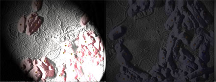
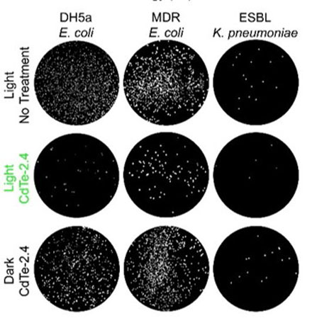
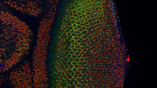