Feeds:
Posts
Comments

Archive for the ‘Medical Imaging Technology, Image Processing/Computing, MRI, CT, Nuclear Medicine, Ultra Sound’ Category

3D Cardiovascular Theater – Hybrid Cath Lab/OR Suite, Hybrid Surgery, Complications Post PCI and Repeat Sternotomy

Curator: Aviva Lev-Ari, PhD, RN

Article ID #70: Cardiovascular Original Research: Cases in Methodology Design for Content Curation and Co-Curation. Published on 7/19/2013

WordCloud Image Produced by Adam Tubman

This article has THREE Parts: 

Part One:  Hybrid Cath Lab/OR Suite for Hybrid Surgery

Part Two: Cardiac Surgery 

Part Three: Invasive Interventions with Complications

1. Repeat Sternotomy Post CABG and/or Aortic Valve Replacement

2. Complications Post PCI – Pump Catheter in Use

The voice of Series A Content Consultant, Justin D Pearlman, MD, PhD, FACC

The leading cause of death and disability from any cause is cardiovascular disease, principally, heart attacks and strokes. Both the heart and brain typically allow only 10 minutes or so of inadequate blood supply before starting a committed course of permanent tissue injury, progressing in severity as time goes by without successful interruption of the disease process. Thus there is great time urgency to get patients to a definitive treatment that can stop the injury and restore adequate nutrient blood supply. Many patients can benefit from a catheterization to identify blockages and insert a small balloon within the blockage to expand the narrow channel, often followed by placement of a stent (wire cage) to maintain the expanded vessel diameter. Chemicals released over time from drug-eluting stents can prevent tissue in growth that may obstruct stents. These emergeny interventions are not always successful. There may be complications from the attempt to access an entry artery, and the blockages may not be amenable to a balloon. When such limitations are encountered, the next chance to help is surgical, with continued time pressure.

The fastest way to make the transition from a diagnostic catheterization to a timely intervention is a hybrid intervention suite that offers non-invasive imaging, catheterization and surgery all in one location. The following articles present the current state of hybrid “do it all” intervention suites. Additional articles address the risks of bad outcomes from such interventions.

Part One 

Hybrid Cath Lab/OR Suite for Hybrid Surgery

In ACC.10 and i2 Summit, 59th Annual Scientific Session, 3/14-3/16, 2010, Alfred A. Bove, M.D., Ph.D., F.A.C.C., ACC President addressed the conference attendees:

Welcome to the all-new Hybrid Cath Lab/OR and 3D CV Theater. Recent developments in cardiac surgery and interventional cardiology have led to the creation of integrated, hybrid cath lab/operating rooms (OR), which provide significant advantages in the diagnosis and treatment of patients requiring cardiac procedures—helping to facilitate a rapid-response approach. These multimodality rooms are designed to support a variety of integrated surgical and endovascular procedures. We are excited to provide you with this opportunity to get a first-hand look—and feel—of the latest technologies. We hope you take the time to explore this interactive, multivendor venue. Learning is at the core of the ACC Annual Scientific Session and we invite you to expand your educational experience in this dynamic learning environment.

In the Hybrid Cath Lab/OR Suite, you’ll discover how integrating cutting edge angiographic and surgical equipment and technologies can facilitate a broad range of procedures within one location. Additionally, you will learn how hybrid suites are providing solutions that enable interventionalists and surgeons to work collaboratively to provide the best treatment options for patients. The adjoining 3D CV Theater features presentations by physicians currently performing intravascular and surgical procedures in hybrid suites. Each live presentation pairs a cardiologist with a surgeon, allowing you to hear perspectives from both sides on a variety of hybrid suite procedures and cases. In addition, the Theater offers video presentations of cases from around the world.

The ACC thanks the supporters of the Hybrid Suite for providing us with the opportunity to share this unique learning destination with you.

http://www.expo.acc.org/acc12/CUSTOM/images/ACC12/ACC.10%20Hybrid%20Suite%20Directory.pdf

Hybrid Cath Lab/OR Suite for Hybrid Surgery

Procedures Performed in a Hybrid Suite

The treatment of cardiovascular diseases has undergone a paradigm shift within the last few years, from

  • open surgery to minimally invasive surgical procedures and from
  • limited percutaneous catheter-based interventions to hybrid interventions for the entire cardiovascular tree.

The Hybrid Suite

are perfect examples of procedures that could, and should, be carried out in a hybrid OR. High-risk patients who require less invasive interventions are the best candidates for treatment in a hybrid suite.

As cardiac surgery becomes less invasive, incisions are becoming smaller and smaller, and even totally endoscopic heart surgery is now possible. Cardiac surgeons have started to perform procedures that include catheter-based skills, such as transapical valve replacement. For these operations, surgeons need more sophisticated imaging techniques, fluoroscopy and contrast injections. The hybrid OR offers all these facilities. Perhaps the most obvious and easiest procedure that can be performed in a hybrid OR is coronary revascularization combining coronary artery bypass grafting with on-table intra-operative completion angiography for quality control. If the surgeon detects a problem during the procedure, he or she can revise the graft immediately and thereby prevent potential perioperative and long-term complications. Currently, cardiologists and cardiovascular surgeons have shown special interest in so-called hybrid coronary interventions, which are combinations of minimally invasive coronary artery bypass grafting and percutaneous coronary interventions. In these procedures, cardiovascular surgeons place a left-internal mammary artery bypass graft to the left-anterior descending artery through small incisions (MIDCAB) or completely endoscopically (TECAB), while any remaining obstructed coronary arteries are treated with stents by an interventional cardiologist. This procedure is an attractive alternative to multivessel open coronary artery bypass grafting. Transcatheter heart-valve replacement and repair are especially suited to a hybrid suite because percutaneous transfemoral and transapical aortic valve repairs include risks that can only be treated successfully by immediate surgical intervention, such as coronary artery obstruction, aortic dissection and aortic perforations.

In addition, endovascular aortic stent grafting for the repair of abdominal aortic aneurysms is a suitable procedure for a hybrid operating room. Endovascular aneurysm repair has become an established alternative to open repair and is increasingly used for thoracic aorta repair as well. Some

  • emergency procedures for traumatic lesions of the thoracic aorta and
  • fulminant pulmonary embolism may also be performed in a hybrid OR. Several
  • pediatric interventions can be carried out in a hybrid suite as well, such as implantation of closure devices for atrial and ventricular septal defects in small children and
  • treatments for hypoplastic left-heart syndrome.

http://www.expo.acc.org/acc12/CUSTOM/images/ACC12/ACC.10%20Hybrid%20Suite%20Directory.pdf

In a recent article we reported on the Change in Requirement for Surgical Support by Cath Labs for performance of Nonemergent PCI without Surgical Backup, that increases the autonomy of Interventional Cardiologists. In the Hybrid OR that change is irrelevant since the presence of a Cardiac Surgeon is a fact of the division of labor between the two types of specialties. Cardiac Surgeons are involved with  percutaneous transfemoral and transapical aortic valve repairs and intervention for endoscopic aorta, AAA and Thoracic AA grafting.

AHA, ACC Change in Requirement for Surgical Support:  Class IIb -> Class III, Level of Evidence A: Supports Nonemergent PCI without Surgical Backup (Change of class IIb, level of Evidence B).

What is a Cardiovascular Hybrid Suite?

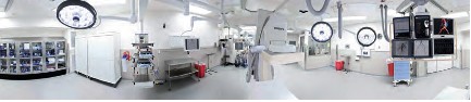
Cardiovascular hybrid suite is a state-of-the-art operating room equipped with a fully functional catheterization laboratory, thus allowing surgical procedures and catheter-based interventions to be carried out in the same room. Hybrid suites provide a place where treatments traditionally available only in a cath lab and procedures only available in an operating room can be performed together to provide patients with the best available combination of therapies for cardiovascular disease. These multidisciplinary, integrated cardiovascular procedural suites bring the best of two worlds together by combining all the advantages of a modern cath lab with an up-to-date cardiovascular surgery operating room (OR).

Hybrid suites began to evolve in the mid to late 1990s, when some groups of interventional cardiologists started sharing operating rooms with cardiovascular surgeons. The appeal of the hybrid suite concept has grown as have catheter based devices (stents, coils, balloons and lasers) have been developed that enable interventional cardiologists to advance the invasiveness and effectiveness and applications of percutaneous transcatheter interventions. The interest in these suites has also increased as cardiovascular surgeons have developed a variety of techniques for

  • Minimally invasive procedures, such as minimally invasive direct coronary artery bypass grafting (MIDCAB) or
  • Totally endoscopic coronary artery bypass grafting (TECAB).

With the advent of more tools, interventional cardiologists are becoming more like surgeons, and with less invasive tools, cardiovascular surgeons are becoming more like interventionalists. Rather than separating surgical procedures from interventional procedures performed in traditional operating rooms and cath labs, hybrid suites provide a high-tech environment that allows cardiologists and surgeons to work together to offer patients complex, minimally invasive therapies.

Some experts believe that hybrid suites represent the wave of the future in cardiovascular care and that most heart centers will eventually install hybrid suites to offer patients the latest cardiovascular procedures safely and effectively with minimal surgical trauma. The rooms can be costly to build and equip, but if a medical center is considering building a new operating room or cath lab, setting up a hybrid suite makes sense. Medical centers that have a hybrid suite available can clearly differentiate themselves in a positive way from centers that do not have such capabilities.

The Benefits of a Hybrid Suite for Medical Centers

While building a hybrid suite is more expensive than building a traditional operating room or cath lab, a hybrid suite can potentially be used for all types of cardiovascular procedures, including

  • traditional cardiac and vascular surgery,
  • interventional coronary procedures,
  • endovascular aortic procedures and
  • electrophysiology procedures.

Hybrid suites reinforce the trend in cardiovascular care toward less invasive, comprehensive hybrid procedures. Once a hybrid suite is in place, the demand for its use will likely grow due to increasing indications and referrals for these innovative treatments, many of which are increasingly covered by third-party payers.

http://www.expo.acc.org/acc12/CUSTOM/images/ACC12/ACC.10%20Hybrid%20Suite%20Directory.pdf

What Equipment is Needed?

Interventional cath labs usually have excellent imaging capabilities but lack the sterile facilities and staff needed for a formal OR, while operating rooms frequently lack high-level imaging equipment. Some of the essential equipment for a hybrid suite includes:

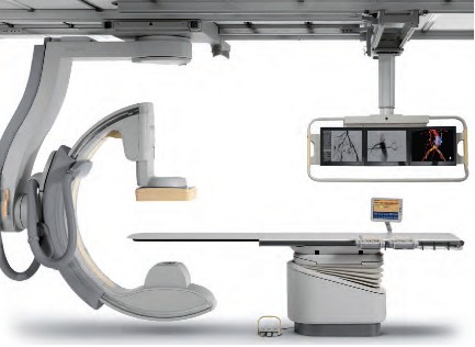
• A state-of-the-art imaging system capable of performing 3D rotational angiography, CT scanning, and ultrasound is advantageous. Floor-mounted and ceiling-mounted systems are available, but many hospitals use ceiling-mounted systems because access to the patient is slightly easier. Some ceiling-mounted systems provide 3D imaging from the surgeon’s position perpendicular to the patient. However, some hospitals prefer floor-mounted systems because having mechanical parts running above the operative field may cause dust to fall, resulting in infections. An important aspect is that the C-arm can be parked away when it is not used. This especially enhances access of the anesthesia team to the patient.

• An operating table that meets the needs of both surgeons and interventionalists by electronically integrating the table with the imaging system is also essential. These tables should have retractable rails for retractors and other surgical tools. To perform 3D imaging on the operating table, the C-arm of the imaging system should allow fast and precise rotation around the patient.

• A variety of other surgical and interventional systems and equipment may also be needed, including a robotic surgical system, a heart-lung machine, an image integration system, an endoscopic imaging system, a radiology display system, an audiovisual system to move images to different monitors and an anesthesia monitoring system, including transesophageal echocardiography. Some equipment like the integrated OR table and the angiography unit need to be fixed parts of the hybrid OR. Some equipment will be mobile in order to maintain some flexibility in workflow.

Hybrid1

Hybrid2

Hybrid3

Hybrid4

Hybrid5

Who are the Equipment Vendors?

Philips Healthcare

Phone: 800-934-7372

Email: healthcare@philips.com

Web: http://www.philips.com/healthcare

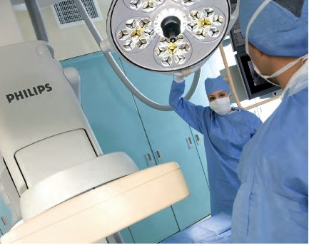
Philips is one of the world’s leading technology companies, with a long history of practical innovation and visionary design. In healthcare, we are committed to understanding the human and technological needs of patients and caregivers. We believe this understanding will help us deliver solutions that not only enable more confident diagnoses and more efficient delivery of care, but also improve the overall experience of care. We offer equipment, software and services for imaging, patient monitoring, resuscitation and much more.  A Hybrid OR can help make life simpler for the interdisciplinary teams who operate in this environment every day. As a world leader in cardiovascular X-ray, Philips has the experience and expertise to deliver the first class technology you need to perform minimally invasive procedures with speed, accuracy and confidence. A long history of innovation has enabled Philips to develop pioneering imaging solutions that really make a difference.

For example, Philips Allura Xper cardiovascular X-ray systems are designed to deliver enhanced imaging with superb performance for all cardiac projections, and our iE33 ultrasound system with Live 3D TEE and QLAB can assist interventional procedures and provide comprehensive quantitative information to support critical decisions. Our cardiology informatics solutions help you manage patient information throughout the cardiovascular care continuum. Philips solutions allow minimally invasive and catheter-based procedures to take place in the same suite as conventional cardiac surgery.

Phillips EchoNavigator – X-Ray and 3-D Ultrasound is described in:

Minimally Invasive Structural CVD Repairs: FDA grants 510(k) Clearance to Philips’ EchoNavigator – X-ray and 3-D Ultrasound Image Fused.

Intuitive Surgical, Inc. 

da Vinci.Surgery by Intuitive Surgical, Inc. 

Phone: 800-876-1310

Email: info@intusurg.com

Web: http://www.intuitivesurgical.com

Intuitive Surgical, Inc. is the global technology leader in robotic-assisted, minimally invasive surgery. The company’s da Vinci® Surgical System offers breakthrough capabilities that enable cardiac surgeons to use a minimally invasive approach and avoid median sternotomy.

Content of FDA Warning Letter, following  FDA Inspection on dates 04/01/2013 – 05/30/2013 – it discussed in

Hybrid Cath Lab/OR Suite’s da Vinci Surgical Robot of Intuitive Surgical gets FDA Warning Letter on Robot Track Record


MAVIG GmbH 

Phone: 631-266-2229,

585-247-1212 ext. 60

Email: info@mavig.com

Web: http://www.mavig.com

MAVIG’s specialty is ceiling/boom suspension systems for lighting (exam, surgical and LED), monitor-suspension systems—single, multibank (one to eight) systems and widescreen, overhead radiation shielding and contrast injector adapters. MAVIG also manufactures radiation protection products such as aprons, gloves, table-attachable lower body shields, adjustable- and fixed-height mobile and modular barriers.

Toshiba America Medical Systems, Inc.

Phone: 714-730-5000

Email: mktgcomm@tams.com

Web: http://www.medical.toshiba.com

Creating a hybrid lab may be complicated, but having an experienced partner that listens makes all the difference. Toshiba’s unique blend of hybrid experience and industry recognized Infinix™-i imaging systems for the Cath Lab.

Hybrid Cath Lab/OR Suite in Leading Hospitals in the US

  • The  Hybrid Cath Lab/OR Suite at New York Presbyterian Hospital/Columbia University Medical Center, New York, NY is presented in

Becoming a Cardiothoracic Surgeon: An Emerging Profile in the Surgery Theater and through Scientific Publications

  • The  Hybrid Cath Lab/OR Suite at Cleveland Clinic, Cleveland, Ohio is presented in

Heart Transplant (HT) Indication for Heart Failure (HF): Procedure Outcomes and Research on HF, HT @ Two Nation’s Leading HF & HT Centers

Speakers at 3D CV Theater, 2010 are working in Hospitals where Hybrid Cath Lab/OR Suite are in operations at the present time. The list include the following Hospitals with a Hybrid Cath Lab/OR Suite:

  • Vanderbilt Medical Center, Nashville, TN
  • University of Maryland Heart Center, Baltimore, MD
  • The Heart Center at Nationwide Children’s Hospital, Columbus, Ohio
  • The Robotic Surgical Center, East Carolina University Department of Surgery, Greenville, N.C.
  • University of Washington Medicine Regional Heart Center, Seattle, WA
  • Brigham and Women’s Hospital, Boston, MA
  • Saint Joseph’s Hospital and Peachtree Cardiovascular and Thoracic Surgery, Atlanta, GA
  • Emory University Hospital, Atlanta, GA
  • Beth Israel Deaconess Medical Center, Boston, MA
  • Boston Medical Center, Boston, MA
  • Mayo Graduate School of Medicine, Mayo Clinic, Rochester, MN
  • Lankenau Hospital, Lancaster, PA
  • Cardiac Non-Invasive Laboratory at Cedars-Sinai Medical Center, Los Angeles, CA
  • Robotic Surgery at St. Joseph’s Hospital, Atlanta, GA

Speakers at 3D CV Theater, 2010, included the following Cardiovascular Interventionists leading the adoption process of Hybrid Surgery in Hybrid Cath Lab/OR Suite into care modalities for cardiovascular disease:

Johannes O. Bonatti, M.D., is professor of surgery and director of coronary surgery and advanced coronary interventions at the University of Maryland Heart Center, Baltimore. He received his training in general surgery and cardiac surgery at the department of surgery at Innsbruck Medical University in Austria. Prior to his arrival at the University of Maryland, he worked at this institution as an attending surgeon and associate professor. Dr. Bonatti’s main interest is the development of minimally invasive, totally endoscopic coronary artery bypass grafting (TECAB) procedures using robotic technology.

As one of the international leaders in this field, he performed the largest series of robotic TECAB on the arrested heart, including single-, double- and triple-vessel TECAB. He has published significantly on procedure development and the implementation process of completely endoscopic coronary surgery using the da Vinci robotic system. Together with colleagues from interventional cardiology, Dr. Bonatti is working on integrated concepts for treatment of coronary artery disease. He was the first to perform a simultaneous hybrid coronary intervention using TECAB and placement of a coronary stent. He is organizing international meetings on hybrid interventions in cardiovascular medicine (http://www.icrworkshop.com). He has trained heart surgeons from around the world in the use of the da Vinci robot for heart surgery and he has introduced TECAB procedures in Austria, the Czech Republic, Greece, Turkey, India and Australia.

John G. Byrne, M.D., is the William S. Stoney Professor of Cardiac Surgery at Vanderbilt University School of Medicine and chair of the department of cardiac surgery at Vanderbilt Medical Center, Nashville, TN.

Before moving to Vanderbilt, he was associate chief and residency program director in the division of cardiac surgery at Brigham and Women’s Hospital, and associate professor of surgery at Harvard Medical School, Cambridge, MA. A graduate of the University of California, Davis, he received his medical degree in 1987 from Boston University. His postdoctoral training was completed at the University of Illinois affiliated hospitals and Brigham and Women’s Hospital in Boston.

Dr. Byrne is the author of more than 100 scientific articles on cardiac surgery and related areas. His patient care emphasis is

  • aortic root surgery,
  • coronary artery disease and
  • valve surgery

He is board-certified in general surgery and thoracic surgery.

John P. Cheatham, M.D., is director of cardiac catheterization and interventional therapy and codirector of The Heart Center at Nationwide Children’s Hospital, Columbus, Ohio. He is also the George H. Dunlap Endowed Chair in Interventional Cardiology and professor of pediatrics and internal medicine at The Ohio State University College of Medicine. Dr. Cheatham’s area of expertise is transcatheter intervention and hybrid therapy of newborns, children and adults with complex congenital heart disease. He has pioneered several new techniques and devices in non-surgical intervention and is a leader in developing hybrid therapies. He has been a principal investigator in numerous FDA-sponsored clinical trials evaluating non-surgical closure devices and stent therapy over the past two decades. Additionally, Dr. Cheatham designed the first hybrid cardiac catheterization suites and advanced imaging equipment at Nationwide Children’s Hospital. He serves as a consultant to various medical companies and proctors new transcatheter techniques and devices to other physicians around the world. Dr. Cheatham has implemented a formal physician exchange program with two of the leading medical institutions in China. In cooperation with China Red Cross, he is also the foreign director of the International Training Center for treatment of congenital heart disease in poor children. Dr. Cheatham has written more than 120 manuscripts, 16 book chapters, 300 national and international presentations and is co-editor of the book, Complications in Percutaneous Interventions for Congenital and Structural Heart Disease. After graduating from the University of Oklahoma College of Medicine, he completed his residency at Boston Children’s Hospital, followed by a fellowship in Pediatric Cardiology at Texas Children’s Hospital in Houston.

W. Randolph Chitwood, Jr., M.D., is senior associate vice chancellor for health sciences and chief of cardiovascular services at East Carolina University Department of Surgery, Greenville, N.C. Dr. Chitwood is a leading international pioneer in minimally invasive and robotic heart surgery. The Robotic Surgical Center at East Carolina University has trained more than 350 surgeons. His research activities relate to myocardial preservation, simulation in surgery and endoscopic/robotic cardiac surgery. He was the principal investigator of the FDA robotic mitral valve trials that led to approval for use in the U.S. He is the son and grandson of “southwestern Virginia mountain doctors” who set the guidelines for his professional life. He graduated from Hampden-Sydney College and received his medical degree from the University of Virginia. After medical school, he completed the surgical residency at Duke University Medical Center under David C. Sabiston, M.D., an influential surgical educator of the era. At Duke he spent 10 years training in general and cardiothoracic surgery, as well as basic science research.

After his chief residency at Duke in 1984, he was selected to begin and head the new cardiac surgery program at the East Carolina University School of Medicine. Because of his prolific publication record as a resident and clinical acumen, his initial appointment was as a full professor of surgery. Except for a two-year hiatus as the chief of cardiothoracic surgery at the University of Kentucky, he has spent his entire career at East Carolina University, where he also served as chairman of the department of surgery. In 2003, he was named to be in charge of the development of the East Carolina Heart Institute, which now includes an integrated department of cardiovascular sciences as well as a $200 million heart hospital, outpatient, research and education center.

Larry S. Dean, M.D., is director of the University of Washington Medicine Regional Heart Center and is professor of medicine and of surgery at the University of Washington School of Medicine, Seattle. In addition to general cardiology, he is an expert in cardiac catheterization and interventional cardiology. He also conducts research on stents to keep blocked heart arteries open and on ways to prevent restenosis after stents are inserted. He is currently involved in the evaluation of percutaneous aortic valve replacement. Dr. Dean earned his M.D. from the University of Alabama School of Medicine, Birmingham, and served his internship and residency at the University of Washington. He then returned to the University of Alabama Hospital for fellowships in cardiovascular disease and in angioplasty. After nearly 15 years as a faculty member at the University of Alabama, he returned to the University of Washington to direct the Regional Heart Center. He is a fellow of the American College of Cardiology and is board-certified in internal medicine, cardiovascular disease and interventional cardiology. He is also a fellow of the American Heart Association and president-elect of the Society of Cardiovascular Angiography and Interventions.

Andrew Craig Eisenhauer, M.D., is director of the interventional cardiovascular medicine service at Brigham and Women’s Hospital and assistant professor of medicine at Harvard Medical School. His specialties are

  • interventional cardiology,
  • vascular medicine and
  • congenital and inherited diseases.

He earned his medical degree at New York University School of Medicine and served a residency at Peter Bent Brigham Hospital and a fellowship at Massachusetts General Hospital. He is certified in internal medicine, cardiovascular disease and interventional cardiology. His clinical interests are

  • endovascular therapy,
  • complex coronary disease,
  • peripheral vascular disease,
  • cerebrovascular disease,
  • congenital heart disease and structural heart disease

Douglas A. Murphy, M.D., is chief of cardiothoracic surgery at Saint Joseph’s Hospital and a cardiothoracic surgeon at Peachtree Cardiovascular and Thoracic Surgery, Atlanta. His areas of interest are robotically assisted heart surgery with an emphasis on repairing the mitral valve rather than replacing it. A graduate of the University of Pennsylvania Medical School, Philadelphia, he served an internship and residency at Massachusetts General Hospital, Boston, and at Emory University, Atlanta.

Khusrow Niazi, M.D., is an assistant professor at Emory University School of Medicine and director of peripheral and carotid intervention at Emory University Hospital Midtown, Atlanta. He earned his medical degree at King Edward Medical College, Lahore, Pakistan, and served an internship at Kettering Medical Center, Dayton, Ohio, and a fellowship at William Beaumont Hospital, Royal Oak, MI. He has published papers on stenting following rotational atherectomy, small vessel stenting for coronary arteries, imaging of lower extremities and treatment of peripheral arterial disease.

Jeffrey J. Popma, M.D., is director of innovations in interventional cardiology, a senior attending physician at Beth Israel Deaconess Medical Center and an associate professor of medicine at Harvard Medical School in Boston. Dr. Popma received his bachelor’s degree in economics from Stanford University, and his M.D. from Indiana University School of Medicine. He completed his internship, residency, chief residency and fellowship at University of Texas Southwestern Medical Center. He also completed an interventional cardiology fellowship at the University of Michigan. Dr. Popma is the past president of the Society for Cardiac Angiography and Intervention and is the co-chair of the ACC Interventional Council. He sits on the editorial boards of several publications, and reviews for several cardiology periodicals. Dr. Popma has more than 300 published peer-reviewed manuscripts.

Dr. Popma also directs the BIDMC Angiographic Core Laboratory and is principal investigator for more than 65 ongoing multicenter device studies within the research laboratory. Over the past 15 years, these trials have included a broad array of new technology, including bare-metal stents, drug-eluting stents, distal-protection devices, total-occlusion devices and carotid and peripheral revascularization procedures. His primary clinical interest currently is the use of percutaneous aortic valve replacement for patients with high-risk aortic stenosis.

Robert S. Poston, M.D., is chief of cardiac surgery at Boston Medical Center and associate professor of cardiothoracic surgery at Boston University School of Medicine. He has a strong background in minimally invasive cardiac bypass surgery and is a pioneer in using robotics, specifically the da Vinci Surgical System, to treat coronary artery disease. A graduate of the Johns Hopkins School of Medicine, Baltimore, Dr. Poston completed a residency in general surgery at the University of California, San Francisco, and continued his training with a research fellowship in cardiothoracic surgery at Stanford University School of Medicine, Palo Alto, CA, and a cardiothoracic residency at the University of Pittsburgh Medical Center.

Charanjit S. Rihal, M.D., is professor of medicine and director of the cardiac catheterization laboratory at Mayo Graduate School of Medicine, Mayo Clinic, Rochester, MN. A graduate of the University of Winnipeg, Dr. Rihal did his residency and fellowship at the Mayo Graduate School of Medicine and also earned an MBA at the Carlson School of Management, University of Minnesota. His medical interests are interventional cardiology, structural heart disease interventions and the management of quality and costs in healthcare.

Timothy A. Shapiro, M.D., is director of the Interventional Cardiology Fellowship Program and campus chief, interventional cardiology, at Lankenau Hospital, Lancaster, PA. A graduate of Yale University School of Medicine, he served his residency and a fellowship at the Hospital of the University of Pennsylvania.

Robert J. Siegel, M.D., is director of the Cardiac Non-Invasive Laboratory at Cedars-Sinai Medical Center, cardiology director of the Cedars-Sinai Marfan Center, and Rexford S. Kennamer, M.D., chair in cardiac ultrasound at Cedars-Sinai Medical Center, Los Angeles. Dr. Siegel is also professor of medicine in residence at the David Geffen School of Medicine at University of California, Los Angeles. He previously served as senior staff fellow in cardiac pathology at the Heart, Lung and Blood Institute of the National Institutes of Health, Bethesda, MD. Internationally recognized as one of the leading experts in the field of cardiovascular ultrasound, Dr. Siegel specializes in cardiovascular ultrasound, including transthoracic, transesophageal and intravascular methodologies. His research interests include

  • valvular heart disease,
  • therapeutic applications of ultrasound energy,
  • transesophageal and intraoperative echocardiography, and the
  • development and use of hand-held portable echocardiographic systems for clinical innovations.

In addition, he is involved with clinical research studies related to the diagnosis, assessment and management of patients with

  • Marfan syndrome,
  • hypertrophic cardiomyopathy and
  • pericardial and valvular heart disease.

Dr. Siegel is a fellow, and has previously served as the president of the California Chapter of the American College of Cardiology and president of the Los Angeles Society of Echocardiography. He has been active in numerous cardiovascular societies, including the American Heart Association, the American College of Cardiology and the American Society of Echocardiography. Dr. Siegel received his medical degree at Baylor College of Medicine, Houston, where he developed an interest in cardiology. He completed his medical residency at Emory University and at Los Angeles County + USC Medical Center. He completed his cardiology fellowship at Harbor-UCLA Medical Center.

Over the last two years Dr. Siegel has worked extensively with live 3D transesophageal echo in the cardiac intervention center and the operating room. He and his echocardiologist colleagues, doctors Shiota, Biner, Tolstrup and Gurudevan, have worked closely at Cedars-Sinai Medical Center in Los Angeles with the interventional cardiologists, doctors Kar and Makkar, as well as with the cardiac surgeons, doctors Trento and Fontana. They use live 3D TEE extensively for the assessment of structural heart disease. In addition, it is used on a regular basis for the guidance of percutaneous procedures for mitral valve e-clip repair, mitral balloon valvuloplasty, aortic and pulmonic valve replacement, left atrial appendage exclusion by the Watchman device as well as for ASD closure.

Sudhir P. Srivastava, M.D., president of the International College of Robotic Surgery at St. Joseph’s Hospital, Atlanta, is a pioneer in performing beating heart totally endoscopic coronary artery bypass surgeries. Previously, he was assistant professor of surgery and director of robotic and minimally invasive cardiac surgery at the University of Chicago Medical Center. Dr. Srivastava specializes in robotically assisted totally endoscopic coronary artery bypass surgery. He has performed approximately 1,000 robotic cardiothoracic surgical procedures, of which 450  have been single- and multivessel beating heart totally endoscopic coronary bypass (BH TECAB) procedures. He has keen interest in hybrid coronary revascularization in TECAB patients to achieve complete revascularization.

Dr. Srivastava has helped launch robotic revascularization programs throughout the world. He has performed numerous live BH TECAB demonstrations both in the U.S. and abroad, and continues to be a presenter and invited speaker at numerous national and international scientific meetings. He earned his medical degree at the Jawahar Lal Nehru Medical College in Ajmer, India and immigrated to the U.S. in 1972. He completed his cardiothoracic surgery residency at the hospitals associated with the University of British Columbia, Vancouver, Canada.

Francis P. Sutter, D.O., F.A.C.S., is clinical professor of surgery at Thomas Jefferson University-Jefferson Medical College, Philadelphia, and chief of cardiothoracic surgery at Lankenau Hospital, Main Line Health System, Wynnewood, PA. A graduate of Philadelphia College of Osteopathic Medicine, his surgical residency and a cardiothoracic fellowship were completed at Thomas Jefferson University Hospital.

Mark R. Vesely, M.D., is an assistant professor of medicine at the University of Maryland School of Medicine. He completed medical school at the George Washington University and postgraduate training—an internal medicine residency and fellowships in cardiovascular disease and interventional cardiology—at the University of Maryland. He is board-certified in internal medicine, cardiovascular disease, nuclear cardiology and interventional cardiology. Dr. Vesely is the associate program director of the Interventional Cardiology fellowship at University of Maryland. His special interests include the partnered approach (interventional cardiologists and cardiac surgeons) for hybrid coronary revascularization and structural heart disease interventions. Additional research interests include investigation of techniques to minimize acute myocardial infarction injury with ventricular-assist devices and adult stem cell therapies.

David X. M. Zhao, M.D., Ph.D., is an associate professor of medicine and cardiac surgery, Harry and Shelley Page Chair in Interventional Cardiology, director of the Cardiac Catheterization Laboratories and interventional cardiology director of the Interventional Cardiology Fellowship Training Program, Vanderbilt University School of Medicine, Nashville, TN. He earned his medical degree at Shanghai Medical University, Shanghai, P.R. China, and his Ph.D. in immunology at Queensland University, Brisbane, Australia. His postdoctoral training was at Zhongshan Hospital, Shanghai Medical University, Shanghai, P.R. China, The Prince Charles Hospital, Brisbane, Australia, and Brigham and Women’s Hospital, Boston.

http://www.expo.acc.org/acc12/CUSTOM/images/ACC12/ACC.10%20Hybrid%20Suite%20Directory.pdf

Part Two

Cardiac Surgery


Cardiac Surgery @ Cleveland Clinic: Traditional OR & Hybrid Cath Lab/OR Suite

Nation #1 for 19 consecutive years – The Heart Center: Miller Family Heart & Vascular Institute @ Cleveland Clinic

The Sydell and Arnold Miller Family Heart & Vascular Institute is one of the largest, most experienced cardiovascular specialty groups in the world. Our physicians are committed to providing the most advanced diagnostic and treatment options, better outcomes and improved quality of life. U.S.News & World Reporthas ranked Cleveland Clinic as the No.1 heart program in America every year since 1995.

Departments & Centers:

Below we present two articles on Cardiac Surgery @ Mayo Clinic 

Cardiac Surgery @ Mayo Clinic: Traditional OR & Hybrid Cath Lab/OR Suite 

Coronary Reperfusion Therapies: CABG vs PCI – Mayo Clinic preprocedure Risk Score (MCRS) for Prediction of in-Hospital Mortality after CABG or PCI

Larry H. Bernstein, MD, FCAP and Aviva Lev-Ari, PhD, RN

Comparison of the 10-year and 15-year survivals after CABG demonstrated benefit from a change in graft sources used at the Mayo Clinic and widely adapted by others: vascular grafts from the left internal mammary artery (LIMA) instead of just leg veins, for multiple grafts (up to 3), LIMA-to-LAD plus grafts using LIMA or radial artery vs LIMA/saphenous vein (SV).

CABG Survival in Multivessel Disease Patients: Comparison of Arterial Bypass Grafts vs Saphenous Venous Grafts

Larry H. Bernstein, MD, FCAP and Aviva Lev-Ari, PhD, RN

Part Three 

Invasive Interventions with Complications

In the following article we covered multiple etiologies for cardiovascular complications related to invasive interventions: cardiovascular and peripheral arterial or peri- and post- cardiac surgery of the open heart type.

Cardiovascular Complications: Death from Reoperative Sternotomy after prior CABG, MVR, AVR, or Radiation; Complications of PCI; Sepsis from Cardiovascular Interventions

Justin D Pearlman, MD, PhD, FACC and Aviva Lev-Ari, PhD, RN

http://pharmaceuticalintelligence.com/2013/07/23/cardiovascular-complications-of-multiple-etiologies-repeat-sternotomy-post-cabg-or-avr-post-pci-pad-endoscopy-andor-resultant-of-systemic-sepsis/

This article covers types of Cardiovascular Complications derived from the following THREE types of assault on the Human body, two related to cardiac invasive interventions, the last due to its systemic nature is taking a fatal Cardiac toll: the Sepsis condition causing cardiac failure.

Three types of Cardiovascular Complications:

I. Risk of Injury During Repeat Sternotomy – following CABG orAortic Valve Replacement, both done in Open Heart Surgery

II. Complications After Percutaneous Coronary intervention (PCI) and endovascular surgery for Peripheral Artery Disease (PAD)

  • (a) Post PCI, and
  • (b) PAD Endovascular Interventions: Carotid Artery Endarterectomy

III. Cardiac Failure During Systemic Sepsis

This article does NOT cover the following two types of Cardiovascular Complications:

1. Trauma Injury causing cardiac arrest, lung collapse or cardiogenic shock

2. Surgical Complication of Non-cardiac surgery type causing cardiac arrest, i.e, Surgery of Joint Replacement causing sepsis causing death or death caused by complications of surgery i.e., blood loss, viral infection, emboli, thrombus, stroke, or cardiogenic shock not related to Cardiovascular and Cardiac invasive interventions

The e-Reader is advised to consider the following expansion on the subject matter carrying the discussion to additional related clinical issues:

Larry H Bernstein, Advanced Topics in Sepsis and the Cardiovascular System at its End Stage

http://pharmaceuticalintelligence.com/2013/08/18/advanced-topics-in-sepsis-and-the-cardiovascular-system-at-its-end-stage/

Bernstein, HL, Pearlman, JD and A. Lev-Ari  Alternative Designs for the Human Artificial Heart: The Patients in Heart Failure – Outcomes of Transplant (donor)/Implantation (artificial) and Monitoring Technologies for the Transplant/Implant Patient in the Community

http://pharmaceuticalintelligence.com/2013/08/05/alternative-designs-for-the-human-artificial-heart-the-patients-in-heart-failure-outcomes-of-transplant-donorimplantation-artificial-and-monitoring-technologies-for-the-transplantimplant-pat/

Pearlman, JD and A. Lev-Ari Cardiac Resynchronization Therapy (CRT) to Arrhythmias: Pacemaker/Implantable Cardioverter Defibrillator (ICD) Insertion

http://pharmaceuticalintelligence.com/2013/07/22/cardiac-resynchronization-therapy-crt-to-arrhythmias-pacemakerimplantable-cardioverter-defibrillator-icd-insertion/

Read Full Post »

Emerging Clinical Applications for Cardiac CT: Plaque Characterization, SPECT Functionality, Angiogram’s and Non-Invasive FFR

Author, and Content Consultant to e-SERIES A: Cardiovascular Diseases: Justin Pearlman, MD, PhD, FACC

and

Article Curator: Aviva Lev-Ari, PhD, RN

Article ID #69: Emerging Clinical Applications for Cardiac CT: Plaque Characterization, SPECT Functionality, Angiogram’s and Non-Invasive FFR. Published on 7/17/2013

WordCloud Image Produced by Adam Tubman


UPDATED on 7/25, 2018

VIDEOS | CT ANGIOGRAPHY (CTA) | JULY 19, 2018

VIDEO: Using FFR-CT in Everyday Practice

Kavitha Chinnaiyan, M.D., FACC, FSCCT, associate professor, Oakland University, William Beaumont School of Medicine, Royal Oak, Mich. She presented at the Society of Cardiovascular Computed Tomography (SCCT) 2018 meeting.

VIEW VIDEO 

https://www.dicardiology.com/videos/video-using-ffr-ct-everyday-practice?eid=333021707&bid=2184627

Related FFR-CT Content:

Clinical Applications of FFR-CT

VIDEO: Implementation and the Science Behind FFR-CT — interview with James Min, M.D.

VIDEO: Early U.S. Experience With FFR-CT in Evaluating ED Chest Pain Presentation — interview with Simon Dixon, M.D.

VIDEO: Status of FFR-CT Adoption in the United States — interview with Campbell Rogers, M.D.

Clinical studies of coronary anatomy by computed tomography use equipment with various numbers of concurrent slices through the heart: 1, 4, 16, 32, 64, 128, and recently 256 or more. Like interventional catheterization, iodine is injected to make the inside of the coronary arteries opaque to xray transmission, to create contrast (otherwise the xray of the coronary tree would be like a photograph of a white polar bear in a snow storm; the contrast acts like spray paint). Computed tomographic angiography (CTA) uses a similar or higher dye load than catheterization, and provides generally lower imaging quality than catheterization but with 3-dimensional reconstruction instead of flat projection (hundreds of linear views at different angles versus one or two image planes at a time). The results from CTA are generally deemed qualitative: whther or not there are potentially flow-limiting lesions in the major branch arteries that supply the heart (with exception: the posterior descending artery to the inferior wall of the heart is not reliably seen). Catheter-based projection coronary angiography sees smaller branches with finer ability to measure the degree of lumen narrowing. However, other imaging methods show greater promise in identifying plaque character. The following examines initial enthusiasm for improvements in CTA which offer better results compared to current clinical CTA and hope to offer advantages over catheter-based methods beyond the avoidance of catheters.

I. Cardiac CT Challenging Functionality of SPECT and Angiogram

Noninvasive computed tomography (CT) perfusion imaging added to CT angiography accurately identifies flow-limiting coronary lesions that need to be treated, results of the CORE320 trial show.

Dr João AC Lima (Johns Hopkins University, Baltimore, MD) presented results of the 381-patient, 16-center trial, which showed that stress CT myocardial perfusion analysis (CTP) significantly improves the diagnostic power of rest CT angiography (CTA) alone. The study also showed that the CTA+CTP strategy has about the same power to identify patients who need revascularization within 30 days as the current standard strategy of invasive angiography plus a single photon-emission computed tomography (SPECT) myocardial perfusion imaging (MPI) test.

Lima explained that the potential advantage of the CT-based approach is that it can obtain information on myocardial perfusion and coronary flow in two scans about 10 minutes apart and is noninvasive.

All patients in the study had been referred for an invasive angiogram to investigate suspected or known coronary artery disease (CAD), but all patients underwent a rest CTA, stress CTP, and SPECT-MPI test in addition to the invasive angiogram. Invasive angiography alone identified apparently obstructive coronary disease in 59% of patients, but adding the SPECT-MPI information reduced that number to 38%.

The accuracy of the CTA+CTP approach was measured as the area under the receiver-operating-characteristic curve. When 50% greater stenosis on invasive angiography was set as the reference standard for a flow-limiting stenosis, the accuracy of the CTA+CTP approach for detecting flow-limiting CAD was 0.87 on a per-patient basis. When the standard was >70% stenosis, the accuracy of the CTA+CTP approach was 0.89.


 VIEW VIDEO on CORE320 with Dr João Lima
Flow Limiting Lesion (low perfusion) vs. Anatomic Stenosis Severity
SOURCES

II. FFR-CT

Results of the Diagnosis of Ischemia-Causing Stenoses Obtained via Noninvasive Fractional Flow Reserve (DISCOVER FLOW) study show that the coronary stenoses that cause ischemia can be identified noninvasively with computer analysis of coronary computed tomography angiograms (CCTAs) [1].

“I think it’s a potential game-changer, because for the first time you have the ability to look at coronary stenosis and ischemia simultaneously, [and] you have the ability to pinpoint the lesion that is causing the ischemia,” DISCOVER FLOW senior investigator Dr James Min (Cedars-Sinai Medical Center, Los Angeles, CA) told heartwire. “You can imagine a scenario where somebody has an abnormal stress test and then you go in and you do an angiogram and see four or five stenoses, but you don’t really know which one caused the ischemia.” But this new “virtual fractional flow reserve” process—or FFRCT—can quantify the fractional flow reserve for each lesion with the data taken from a CCTA, thereby revealing which stenoses are causing ischemia and ought to be treated, as well as which stenoses do not need to be treated. “We’ve never before had this one-stop shop to . . . pinpoint the lesions that cause the ischemia noninvasively.”

As reported by heartwire at EuroPCR 2011, in DISCOVER FLOW, Dr Bon-Kwon Koo (Seoul National University Hospital, Korea) and colleagues used computation of FFRCT to assess 159 vessels in 103 patients undergoing CCTA. Results of the study are published in the November 1, 2011 issue of the Journal of the American College of Cardiology.

All of the patients also underwent invasive CCTA and invasive catheter FFR imaging. Ischemia was defined as an FFR of <0.80 and anatomically obstructive coronary disease was defined as stenosis >50% as measured on the CCTA scan. The diagnostic performance of FFRCT and CCTA were assessed against invasive FFR as the reference standard. Of the patients in the study, 56% had at least one vessel with an FFR of <0.80.

Because only about half of stenoses over 50% actually cause ischemia, the specificity of traditional assessment of a stenosis by CCTA is below 50%. “The concern there is that you identify some high-grade stenoses that are angiographically confirmed, but the lesions don’t actually cause ischemia.” Fractional flow reserve measures how much of the blood flow is being blocked by a lesion, so it is about 25% more accurate than traditional CCTA at picking out lesions that cause ischemia, Min explained.

Per vessel diagnostic accuracy FFRCT and CCTA (reference for both was invasive FFR) 

Imaging technology Accuracy(%) Sensitivity(%) Specificity(%) Positive predictive value (%) Negative predictive value (%)
FFRCTa  84.3 87.9 82.2 73.9 92.2
CCTAb 58.5 91.4 39.6 46.5 88.9

a. Ischemic defined as <0.80

b. Ischemia defined as stenosis >50%

FFRCT can assess stenoses from any CCTA scan—prospectively gated or retrospectively gated—without any additional imaging techniques or changes to the acquisition parameters. Just as computational fluid dynamics can predict the behavior of an airplane wing under different environmental parameters, FFRCT can measure the flow of blood through a stenotic coronary based on the specific geometry of the patient’s coronaries and myocardium.

At the American Heart Association meeting in Orlando next month, Min will present results of a substudy from DISCOVER FLOW looking specifically at intermediate-grade stenoses (40%-69%), which present the most difficult treatment decisions. “If somebody sees a 90% stenosis or 10% stenosis, they are comfortable with what to do with that. But when you hit that 40% to 70% range—it’s possible that those lesions are ischemic, but you don’t know until you actually assess them,” Min said.

DISCOVER FLOW was designed to evaluate the accuracy of FFRCT on a per-vessel basis, but the more important demonstration of its value will be its ability to guide treatment decisions for each patient. TheDEFACTO trial, which finished enrollment at 17 centers about three weeks ago, is evaluating FFRCT per patient. “That’s the big one,” Min said. “DEFACTO will be the pivotal trial.” Specifically, the 285-patient DEFACTO trial is assessing the ability of CCTA plus FFRCT to determine the presence or absence of at least one hemodynamically significant coronary stenosis in each trial subject. Invasive catheter FFR is the reference standard. Min expects that study to be completed in the first quarter of 2012.

http://www.theheart.org/article/1299631.do

SOURCES

III. Ten Emerging Uses for Cardiac CT from SCCT 2013

July 11-14, 2013
Palais des congrès
Montréal, Québec, Canada

JULY 16, 2013  – heartwire

Dr Matthew Budoff (Los Angeles Biomedical Research Institute, CA), a longtime researcher in the use of cardiac CT, described what he believes to be the most important uses for CT today [1].

First, CT angiography is emerging as “a single tool that gives us [information about] function and anatomy,” he told the audience.

Second, it is now known that patients are more likely to have a cardiovascular event if they have low-attenuation plaque (soft plaque), positive remodeling, and spotty calcification, he explained. If a clinician were limited to looking only at plaque or stenosis, he would advise him or her to “just read the CTA for plaque and plaque characteristics and [don’t] read it for stenosis severity, and you’ll probably serve your patients better in predicting risk” of a cardiovascular event. “I think in future we’re going to be using plaque characterization in every case,” he added. “I certainly don’t advocate stenting these patients [who have vulnerable plaque] yet, but . . . I do treat these patients more aggressively.”

Third, coronary CT angiography is a noninvasive way to identify complex aortic-valve geometry and guide TAVR.

“With perfusion imaging, TAVR, and plaque assessment leading the way, the increased utilization of CTA is certain,” Budoff concluded. “However, more validation work is needed to ensure that industry and payers accept these applications.”

Speaking to heartwire, Budoff singled out TAVR as “an easy launching point for doctors to get familiar with” CT angiography. He also believes that using CT for “heart-failure assessment or even plaque assessment . . . will really add value to their practice.” CT also allows clinicians to “start getting a handle on what’s causing stenosis [in a patient], what it looks like, and . . . how severe the stenosis is.”

In a separate presentation [2], Dr James K Min (Cedars-Sinai, Los Angeles, CA) identified the same three clinical applications as Budoff in his “top 10 things to watch” in coronary CT in the coming year. He identified his “up-and-coming areas to watch” in the following order:

  • Dual-energy CT scanners. This hardware, when combined with new software, is producing enhanced image quality that allows, for example, a “plaque biopsy,” which provides detailed information about plaque characteristics.
  • Myocardial CT perfusion. “We’ve looked at this for seven years, and I think it’s starting to become ready for prime time,” said Min. In the next year, he expects investigators to figure out exactly how to use CT to look at coronary flow reserve.
  • Computational fluid dynamics. Exciting work is being done, for example, using a virtual stent to see how a real stent would potentially resolve a patient’s ischemia.
  • PlaqueCoronary CT can do more than identify how many vessels are blocked, he said, echoing Budoff’s words. It is enabling investigators to study the pathogenesis of atherosclerosis. “We’re going to be able to identify plaque characteristics beyond stenosis for the prediction of acute MI,” Min said.
  • Structural heart disease. CT is already being used to help guide TAVR to reduce postsurgery complications.
  • Radiation-dose reduction. Min weighed in and said, “I think it’s becoming a nonissue.” He noted that during the past year, investigators reported how coronary CT angiography can be used with radiation doses as low as 0.01 mSv, (should be 1mSv) whereas a screening mammogram exposes a woman to 0.05 mSv of radiation. (1/5 of mammography)
  • Contrast-agent reduction.”I think we will see improvements—we will get to the 10-cc scan,” Min predicted.
  • Appropriate-use criteria. Physicians are continuing to identify which patients benefit from cardiac CT, as the technology is advancing.
  • Two trialsAmong the many ongoing trials in the field, Min identified two to watch. The PROMISEstudy is comparing functional vs anatomic testing to identify heart disease. The Coronary Computed Tomographic Angiography for Selective Cardiac Catheterization (CONSERVE) trial is looking at using CT as a “gatekeeper” to the cath lab, to identify which patients should be sent for invasive coronary angiography and which ones have only have mild stenosis and could be sent home and treated with medical therapy .
  • Worldwide growth in CT. Collaboration with investigators around the world is growing, and the SCCT meetings next year in Hawaii and China will offer more opportunities for this.
Budoff has received research/grant support from HeartFlow, study funding from Wakunaga of America and GE Healthcare and has been a consultant and speaker for GE Healthcare. Min has received research/grant support fromGE Healthcare, Phillips Healthcare, and Vital Images and study funding from Astellas. He has been a consultant for GE Healthcare and Arineta and on the speaker’s bureau for GE Healthcare. He holds equity interest in TC3 and MDXX.

Sources

  1. Budoff MJ. Emerging Clinical applications for cardiac CT. Society of Cardiovascular Computed Tomography 2013 Annual Scientific Meeting; July 12, 2013; Montreal, QC.
  2. Min JK. The future of cardiac CT. What will the next 12 months bring? Society of Cardiovascular Computed Tomography 2013 Annual Scientific Meeting; July 12, 2013; Montreal, QC.

Related links

SOURCE

http://www.theheart.org/article/1561163.do?utm_medium=email&utm_source=20130717_heartwire&utm_campaign=newsletter

IV. Stress CT Perfusion matches SPECT for detecting Myocardial Ischemia

Montreal, QC – In stress testing using regadenoson (Lexiscan, Astellas), detection rates of myocardial ischemia were similar with less invasive computed-tomography (CT) perfusion imaging compared with the reference method, single-photon-emission CT (SPECT) imaging, in a phase 2 trial [1].

JULY 18, 2013 

Regadenoson, a selective adenosine-receptor agonist that produces coronary vasodilation in patients unable to undergo exercise stress testing, is the most common agent used to induce pharmaceutical stress in SPECT tests in the US; it was used off-label for the CT imaging.

Dr Ricardo C Cury (Baptist Hospital of Miami, FL) presented the trial results here at a late-breaking clinical-trials session at the Society of Cardiovascular Computed Tomography (SCCT) 2013 Scientific Meeting.

To heartwire, Cury noted that this trial established noninferiority of regadenoson stress CT perfusion to the reference method, regadenoson SPECT, to detect or exclude myocardial ischemia, which was the primary study outcome.

“This is the second multicenter trial validating [regadenoson] stress CT perfusion, which [builds on the accumulating supporting data from] many single-center studies,” he said, adding that it is still too early, however, to implement these findings into clinical practice.

To heartwire, session moderator Dr John Hoe (Parkway Health Radiology, Singapore) commented that “this is quite an important multicenter trial . . . and the results look very good.” Echoing Cury, he added that “this [research] is slowly [progressing] along the path to validate [regadenoson] CT perfusion as a technique to assess myocardial ischemia.”

In study, 39% of patients had suspected CAD

This was a crossover study conducted at 11 sites in the US, using six types of CT scanners, including 64-, 128-, 256-, and 320-slice machines.

A total of 124 individuals with known (39%) or suspected CAD were randomized to either rest and stress SPECT using regadenoson on day 1, followed by rest and combined stress CT perfusion using regadenoson and coronary CT angiography on day 2; or the same tests in the reverse order.

At baseline, the subjects had a mean age of about 62 and an average body-mass index (BMI) of close to 30. Their average heart rate increased from 64 to 84 beats per minute with the stress-CT perfusion test.

Myocardial ischemia was defined as having two or more reversible defects.

High agreement, specificity, and sensitivity

When it came to detecting myocardial ischemia, CT perfusion imaging agreed with the findings of the reference method, SPECT, 87% of the time (95% CI 0.77-0.97).

“This was well above the specified primary end point for the agreement rate between SPECT and CT perfusion for the detection of ischemia,” Cury said.

Stress CT perfusion imaging also had a high specificity (84%) and sensitivity (90%) for detecting or excluding myocardial ischemia.

Similarly, when it came to detecting the presence or absence of one or more fixed myocardial defects, CT perfusion imaging agreed with the results of the reference method, SPECT, 86% of the time (95% CI 0.74-0.98).

Again, stress CT perfusion imaging had a high specificity (95%) and sensitivity (77%) for detecting or excluding fixed defects.

Used alone, compared with the reference standard of SPECT, stress CT perfusion diagnosed or excluded ischemia accurately in 85% of cases, whereas CT angiography alone made the correct diagnosis in 69% of cases. Thus, “stress CT perfusion may add significant [diagnostic] value to CT angiography alone,” Cury noted.

Regadenoson was well tolerated, and the most common adverse events were flushing or headache.

The study was funded by Astellas. Cury is a consultant for Astellas and has received research grants from Astellas and GE Healthcare. Hoe has received grant and research support and travel funding from Toshiba Medical Systems and is on its speaker’s bureau. 

SOURCE

http://www.theheart.org/article/1561685.do

V. New Protocol Limits Use Of SPECT MPI For Angina

Article Date: 07 May 2013 – 1:00 PDT

A new stress test protocol that investigates reducing the use of perfusion imaging in low risk patients undergoing SPECT myocardial perfusion imaging for possible anginasymptoms was found to be diagnostically safe, revealed a US retrospective analysis. The study, reported as an abstract¹ at the International Conference on Nuclear Cardiology and Cardiac CT (ICNC11) May 5 to May 8 in Berlin, Germany, predicted that using exercise ECG stress testing alone in patients with high exercise capacity would have had no adverse effects on their prognosis at five years.

“Our results are reassuring in that there are few patients whose diagnosis of coronary artery disease (CAD) would be missed,” said Milena Henzlova, the first author of the study. “Not only would widespread adoption of this approach reduce radiation exposure, it would also save considerable amounts of time and money.”

Single-photon emission computed tomography (SPECT) myocardial perfusion imaging (MPI) has been used for over 30 years to detect ischemia in patients with suspected CAD. In SPECT MPI patients are injected with radioactive agents (such as Tc-99m or Thallium 201) whose passage through the heart is viewed with a SPECT camera. By comparing the heart’s blood flow at rest and during stress (patients exercise on a treadmill, cycle ergometers or undergo pharmacological stress with vasodilators or dobutamine), cardiologists can determine if the myocardium receives sufficient blood supply, as well as the location and extent of underlying CAD.

“Because it’s non invasive and many patients with a chest pain syndrome don’t have coronary disease, SPECT MPI is often viewed as a ‘gate keeper’ to coronary angiography,” explained Lane Duvall, an investigator in the study.

While SPECT MPI represents a well established technique, the main disadvantage is that patients are exposed to diagnostic levels of radiation. In recent years intensive efforts have been made to reduce ionizing radiation associated with cardiac imaging due to concerns that it damages DNA in cells and may ultimately give rise to cancer. Indeed, extrapolating data from the survivors of the Hiroshima and Nagasaki atomic bombs, Andrew Einstein, from Columbia University Medical Center, New York, has estimated that the low levels of radiation encountered during medical imaging might lead to a 2% excess relative risk for future cancers.

Other studies have suggested that exercise treadmill testing alone may be sufficient to predict CVD outcome without use of SPECT MPI in low risk patients. In 2011, Bourque and colleagues from the University of Virginia, Charlottesville, reported that patients who exercise at >10 metabolic equivalents (METS), [the unit used to estimate the amount of oxygen used by the body during physical activity] during stress testing had a very low prevalence of significant ischemia and very low rates of cardiac events during follow-up².

The advantage of exercise treadmill testing is that it offers a quicker study that involves no radiation exposure, with prognostic information provided via a variety of treadmill scores, most notably the Duke Treadmill score. “This has led to investigators questioning the added value of SPECT MPI over exercise testing alone. There’s growing recognition that patients need to be treated as individuals and that those in whom the CVD risks are considered negligible shouldn’t be undergoing the risks of radiation exposure,” said Duvall.

In the current abstract, Henzlova, Duvall and colleagues, from the Mount Sinai School of Medicine, New York, US, set out to investigate retrospectively if a provisional injection protocol in which patients where they met certain criteria were converted to exercise treadmill tests without imaging maintained diagnostic accuracy and prognostic ability. For the retrospective study, data was reviewed from a total of 24,689 patients who had undergone SPECT MPI between February 2004 and June 2010. After exclusion of patients older than 65 years of age, who had known CAD and uninterruptable resting ECGs, 5,352 subjects were identified for analysis.

Subjects were divided into those who would have met all the criteria for not undergoing SPECT MPI (the No injection group n= 1,561 [29.2%]) and those who met the criteria for undergoing SPECT MPI (the Yes injection group, n=3,791, [70.8%]). For the study the criteria laid down for patients considered eligible for not undergoing SPECT MPI included achieving a maximal predicted heart rate >85%, > 10 METs of exercise, no symptoms of chest pain or significant shortness of breath during stress, and no ECG changes (ST depression or arrhythmia). Outcomes for the two groups at five years were then compared based on their actual myocardial perfusion imaging results and all-cause mortality that had been retrospectively identified from the National Death Index.

At a mean follow-up of 60.6 months, 1.1% of patients had died in the No-injection cohort compared to 2.2% Yes injection cohort (P=.01). Furthermore perfusion results were abnormal in 5.9% of the No injection group compared to 14.4% in the Yes injection group (P<.0001). The risk adjusted survival at the end of the follow up was 98.8% in the No injection group compared to 97.2% for patients found to have normal perfusion in the Yes injection group (P=0.009).

“Withholding isotope injections in these selected patients was found to be diagnostically safe with a small percentage of ‘missed’ abnormal perfusion studies, a very low rate of significant stress perfusion defects and left ventricular ischemia, and a prognosis which was better than their counterparts who were injected with the isotope,” said Duvall.

Eliminating the need for imaging in 6% of the 9 million SPECT MPI studies performed annually in the US, the authors added, would result in significant cost savings and the total test time would be halved from three hours to roughly one hour. “There’s a need to accept that less can be more. By individualizing therapy we can reduce radiation exposure and costs without jeopardizing the quality, the diagnostic utility or missing something important,” said Henzlova. 

REFERENCES
1. M Henzlova, EJ Levine, S Moonthungal, et al. A protocol for the provisional use of perfusion imaging with exercise stress testing. Abstract no 70123.
2. Bourque JM, Charlton GT, Holland BH, et al. Prognosis in patients achieving >10 METS on exercise stress testing: was SPECT imaging useful? J Nucl Cardiol 2011, 2 230-7.
European Society of Cardiology
SOURCE

VI. Contemporary Stress Echo good for Risk Stratification in Chest-Pain Units

12/20/2012, Lisa Nainggolan

London, UK – Doctors in a London chest-pain unit have shown that employing contemporary stress echocardiography in patients with suspected acute coronary syndrome (ACS) but normal ECG and negative troponin is a successful approach for risk stratification [1].

Stress echo is feasible and safe and allows early triage and rapid discharge of patients, plus it is a good predictor of hard events, say Dr Benoy N Shah (Royal Brompton Hospital, London, UK) and colleagues in their paper published online December 18, 2012 in Circulation: Cardiovascular Imaging. Those with an abnormal stress echo had a 13- to15-fold increased risk of MI or death compared with those who had a normal stress echo, they report.

“Stress echo is a very effective gatekeeper for patients undergoing further risk stratification,” senior author Dr Roxy Senior (Royal Brompton Hospital) told heartwire. “It helps select patients for coronary angiography [those with a positive stress echo] and allows immediate discharge of those patients with a negative result.”

Stress echo is perceived to be a technique that is difficult, but that is a misconception.

But Senior says his chest-pain unit is the only one in the UK using this approach. “It is perceived to be a technique that is difficult, but that is a misconception. We have nine stress-echo operators, and it’s easy to train people. With contemporary techniques, which employ contrast in around 50% of cases, the images are quite clear and quick and easy to interpret. It’s very user-friendly. We want to show people around the world that it’s a very doable technique, so why don’t you use it?”

Stress echo also compares favorably with other tests used or proposed for risk stratification of such patients, he says. Exercise ECG is perhaps the most basic technique, “and we have shown that the downstream costs are lower with stress echo than with exercise ECG,” given that the latter provides such equivocal results [2], he explained. And with regard to other imaging modalities that have been employed in this way, computed tomography coronary angiography (CTCA) and single-photon-emission computed tomography (SPECT) require the use of ionizing radiation and have other drawbacks, he notes.

Nevertheless, he and his colleagues say that further, multicenter studies comparing stress echo with CTCA, SPECT, and other imaging techniques for this purpose “will help determine the most cost-effective means of investigating this acute patient population.”

Stress echo performed within 24 hours of admission

Shah and colleagues say that after they showed in 2007 that stress echo was more cost-effective than exercise ECG, they have been employing the former in day-to-day practice in their unit to assess patients who come in with severe chest pain, but whose troponin is negative at 12 hours and whose ECG is “nondiagnostic” (ie, does not suggest any abnormality or shows only minor changes).

The current study is a retrospective look at the patients they have seen so far and is the first evaluation of the clinical impact of incorporating stress echo in a real-world chest-pain unit for the assessment of both short- and long-term prediction of hard events, they say.

“This was sort of an audit; we wanted to know, ‘Is this right? Or are we overcalling it?’ ” Senior explains.

He says the stress echos are performed, for the most part, “within 24 hours” of admission to the chest-pain unit, from 9 am-5 pm Monday to Friday. Those admitted on a weekend will wait slightly longer for a stress echo, he acknowledged. The stress echo is performed on a treadmill if the patient is capable of exercise; if not, a pharmacological stress test is performed using dobutamine. Approximately 30% of the patients in this study performed the test on a treadmill, Senior noted.

Results of the stress echo are available quickly and, if negative, the patient is discharged immediately. If they are positive, the patient is investigated further.

Event rate much higher for those with a positive stress echo

In the study, 839 consecutive patients were assessed; 802 were available for follow-up. Approximately 75% of them had a normal stress echo and were discharged.

“The 30-day readmission rate for all patients was extremely low,” Senior notes, but for those with a negative stress echo it was exceedingly low (at 0.3% compared with 1.1% for those with an abnormal stress echo).

A normal stress echo carried a 99.7% event-free survival for death and 99.5% event-free survival for all hard events in the first year of follow-up; these event rates increased 15-fold and 13-fold respectively if the stress echo was abnormal.

There were 15 “hard” events, 0.5% in the normal stress echo group and 6.6% in the abnormal stress echo group in the first year. At two years, 2.3% of those in the normal stress echo group had died or had a nonfatal MI compared with 9.6% in the stress echo abnormal group, and at three years these figures were 5.1% and 21.1%, respectively. The median follow-up for the study was 27 months.

“For the patients who had a positive stress echo, the event rate was much higher,” Senior notes. Of these 184 patients, 98 had ischemia and most of these underwent coronary angiography, with 57 demonstrating flow-limiting coronary artery disease and 30 subsequently undergoing revascularization.

Among all prognostic variables, only abnormal stress echo (hazard ratio 4.08) and advancing age (HR 1.78) predicted hard events in multivariable regression analysis.

Stress echo should be much more widely used in chest-pain units

“This study demonstrates the excellent feasibility and safety of stress echo in a real-world chest-pain-unit setting, with rapid early triaging and discharge and accurate risk stratification,” the researchers say.

“The two most important outcomes for patients reassured and discharged from the emergency department are that they do not suffer early mortality or early readmission with the same complaint. Our study highlights the excellent negative predictive value of stress echo and very low 30-day readmission rate.”

In addition, the results show that stress echo “appropriately influences the use of coronary angiography and subsequent revascularization” and overall support the wider use of this technique in chest-pain units, they conclude.

Senior has previously received consultancy fees from Lantheus Medical. The coauthors report they have no conflicts of interest.
REFERENCES

Sources

  1. Shah BN, Balaji G, Alhajiri A, et al. The incremental diagnostic and prognostic value of contemporary stress echo in a chest pain unit: mortality and morbidity outcomes from a real-world setting. Circ Cardiovasc Imaging 2012; DOI:10.1161/CIRCIMAGING.112.980797. Available at: http://circimaging.ahajournals.org.
  2. Jeetley P, Burden L, Stoykova B, Senior R. Clinical and economic impact of stress echocardiography compared with exercise electrocardiography in patients with suspected acute coronary syndrome but negative troponin: a prospective randomized controlled study. Eur Heart J. 2007; 28:204-211.

http://www.theheart.org/article/1490677.do

VII. PET Perfusion Imaging Improves Risk Estimates

12/5/2012 Reed Miller

Boston, MA – New data from a large multicenter registry suggest that positron-emission-tomography (PET) myocardial perfusion imaging (MPI) can greatly improve the accuracy of risk estimation in coronary disease patients compared with a model based on traditional risk factors [1].

Only small single-center studies have demonstrated the prognostic value of PET MPI in predicting which patients are at greatest risk for coronary disease events. So Dr Sharmila Dorbala (Brigham and Women’s Hospital, Boston) and colleagues analyzed outcomes from 7061 patients from four centers who underwent a clinically indicated rest/stress rubidium-82 PET MPI test.

Results of the study are published online December 5, 2012 in the Journal of the American College of Cardiology. “The results of the current study are critical to advance the field and guide more effective use of PET MPI in clinical practice,” Dorbala et al state.

Median follow-up was 2.2 years. During follow-up, there were 169 cardiac arrests and 570 all-cause deaths. Net reclassification improvement and integrated discrimination analyses showed that the risk-adjusted hazard of cardiac death increases as the percentage of abnormal myocardium increases. A mildly abnormal stress test is associated with a 2.3 times greater risk of cardiac death than a normal test. The hazard ratio for a severely abnormal test is 4.9.

The addition of PET MPI measurements of myocardial ischemia and myocardial scarring to traditional clinical information improves the performance of a risk prediction model based on traditional risk factors (C statistic 0.805-0.839) as well as risk reclassification for cardiac death, with small improvements in risk assessments for all-cause death. The assessment of the magnitude of ischemia and scar added to the reclassification of risk for cardiac death in one in every nine patients who underwent clinical PET MPI in the study.

Unlike computed-tomography (CT) coronary angiography, perfusion imaging provides information about myocardial blood flow and accounts for underlying coronary disease, collateral flow, and myocardial adaptation to wall stress and can be used in patients with renal insufficiency, the authors point out. Compared with single-photon-emission computed tomography (SPECT) perfusion imaging, PET MPI offers better image quality, test specificity for the diagnosis of obstructive coronary disease, and identification of scar and ischemia, according to Dorbala et al, and PET MPI uses a lower effective radiation dose. However, while the prognostic value of SPECT MPI has been described in tens of thousands of patients, the prognostic value of PET MPI has been studied in only a few thousand patients.

Does more risk information help?

The value of the prognostic information offered by PET MPI is not yet clear, according to an accompanying editorial by Drs Paul Schoenhagen and Rory Hachamovitch (Cleveland Clinic, OH) [2]. “Rather than assessing whether a test yields improvement in risk assessment, the focus [should be] shifted to whether a test can identify which patients will gain a benefit from a specific therapeutic approach,” they write. “The role of testing [should be] defined in the context of a specific intervention and whether the effectiveness of the intervention is improved by the use of an imaging study to identify optimal candidates for treatment.

“However, this process is neither simple nor inexpensive and will require prospective randomized clinical trials, validating the results and hypotheses generated by observational data,” the editorialists conclude.

Commenting on the study, Dr Kavitha Chinnaiyan (William Beaumont Hospital, Royal Oak, MI) toldheartwire, “While the details of downstream management of these patients are unclear in this paper, the association of ischemia with mortality is clear, as is the reclassification of risk. The next step in terms of management of ischemic patients is really the question here.” She also pointed out that the ongoingISCHEMIA trial, comparing angiography and revascularization plus optimal medical therapy with optimal medical therapy only, may provide more insights on the best option for patients who show more than mild ischemia on stress studies.

Dorbala has received research grants from Astellas Pharma and Bracco Diagnostics; has served on advisory boards for Astellas Pharma; and has received honoraria from MedXcelDisclosures for the coauthors are listed in the paper.Schoenhagen and Hachamovitch report that they have no relationships relevant to the contents of this paper to discloseChinnaiyan has no relevant disclosures.

REFERENCES

  1. Dorbala S, Di Carli M, Beanlands RS, et al. Prognostic value of stress myocardial perfusion positron emission tomography. J Am Coll Cardiol 2013; DOI:10.1016/j.jacc.2012.09.044. Available at:http://content.onlinejacc.org.
  2. Schoenhagen P and Hachamovitch R. Evaluating the clinical impact of cardiovascular imaging: Is a risk-based stratification paradigm relevant. J Am Coll Cardiol 2013; DOI:10.1016/j.jacc.2012.09.044. Available at:http://content.onlinejacc.org.

SOURCE

Other related articles published on this Open Access Online Scientific Journal, include the following:

Fractional Flow Reserve (FFR) & Instantaneous wave-free ratio (iFR): An Evaluation of Catheterization Lab Tools for Ischemic Assessment

Justin D Pearlman, MD, PhD, FACC and Aviva Lev-Ari, PhD, RN

http://pharmaceuticalintelligence.com/2013/07/04/fractional-flow-reserve-ffr-instantaneous-wave-free-rario-ifr-an-evaluation-of-catheterization-lab-tools-for-ischemic-assessment/


CT Angiography (CCTA) Reduced Medical Resource Utilization compared to Standard Care reported in JACC

Aviva Lev-Ari, PhD, RN

http://pharmaceuticalintelligence.com/2013/05/16/ct-angiography-ccta-reduced-medical-resource-utilization-compared-to-standard-care-reported-in-jacc/


Acute Chest Pain/ER Admission: Three Emerging Alternatives to Angiography and PCI – Corus CAD, hs cTn, CCTA

Curator: Aviva Lev-Ari, PhD, RN

http://pharmaceuticalintelligence.com/2013/03/10/acute-chest-painer-admission-three-emerging-alternatives-to-angiography-and-pci/


Acute and Chronic Myocardial Infarction: Quantification of Myocardial Perfusion Viability – FDG-PET/MRI vs. MRI or PET alone

Justin D. Pearlman, MD, PhD and Aviva Lev-Ari, PhD, RN

http://pharmaceuticalintelligence.com/2013/05/22/acute-and-chronic-myocardial-infarction-quantification-of-myocardial-viability-fdg-petmri-vs-mri-or-pet-alone/

Read Full Post »

AHA, ACC Change in Requirement for Surgical Support for PCI Performance: Class IIb -> Class III, Level of Evidence A: Support Nonemergent PCI without Surgical Backup (Change of class IIb, Level of evidence B).

AHA, ACC Change in Requirement for Surgical Support:  Class IIb -> Class III, Level of Evidence A: Supports Nonemergent PCI without Surgical Backup (Change of class IIb, Level of Evidence B).

Larry H Bernstein, MD, FCAP, Author, Curator, Volumes 1,2,3,4,5,6 Co-Editor and Author, Volume Two & Five, Co-Editor and Justin Pearlman, MD, PhD, FACC, Content Consultant to Six-Volume e-SERIES A: Cardiovascular Diseases

Article ID #68: AHA, ACC Change in Requirement for Surgical Support for PCI Performance: Class IIb -> Class III, Level of Evidence A: Support Nonemergent PCI without Surgical Backup (Change of class IIb, Level of evidence B). Published on 7/17/2013

WordCloud Image Produced by Adam Tubman


Voice of content consultant: Justin Pearlman, MD, PhD, FACC

The American Heart Association (AHA) and the American College of Cardiology (ACC) have convened teams of experts to summarize evidence and opinion regarding a wide range of decisions relevant to cardiovascular disease. The system accounts for some of the short comings of “evidence based medicine” by allowing for expert opinion in areas where evidence is not sufficient. The main argument for evidence-based medicine is the existence of surprises, where a plausible decision does not actually appear to work as desired when it is tested. A major problem with adhesion to evidence based medicine is that it can impede adaptation to individual needs (we are all genetically and socially/environmentally unique) and impede innovation. Large studies carry statistical weight but do not necessary consider all relevant factors. Commonly, the AFFIRM trial is interpreted as support that rate control suffices for most atrial fibrillation (AFIB), but half of those randomized to rhythm control were taken off anticoagulation without teaching patients to check their pulse daily for recurrence of AFIB. Thus the endorsed “evidence” may have more to do with the benefits of anticoagulation for both persisting and recurring AFIB and rhythm control may yet prove better than rate control. However, with wide acceptance of a particular conclusion, randomizing to another treatment may be deemed unethical, or may simply not get a large trial due to lack of economic incentive, leaving only the large trial products as the endorsed options. A medication without patent protection, such as bismuth salts for H Pylori infection, lacks financial backing for large trials.

The American Heart Association Evidence-Based Scoring System
Classification of Recommendations

● Class I: Conditions for which there is evidence, general

agreement, or both that a given procedure or treatment is

useful and effective.

● Class II: Conditions for which there is conflicting evidence,

a divergence of opinion, or both about the usefulness/

efficacy of a procedure or treatment.

● Class IIa: Weight of evidence/opinion is in favor of

usefulness/efficacy.

● Class IIb: Usefulness/efficacy is less well established by

evidence/opinion.

● Class III: Conditions for which there is evidence, general

agreement, or both that the procedure/treatment is not useful/

effective and in some cases may be harmful.

Level of Evidence

● Level of Evidence A: Data derived from multiple randomized

clinical trials

● Level of Evidence B: Data derived from a single randomized

trial or nonrandomized studies

● Level of Evidence C: Consensus opinion of experts

Circulation 2006 114: 1761 – 1791.

Assessment of Coronary Artery Disease by Cardiac Computed Tomography

A Scientific Statement From the American Heart Association Committee on Cardiovascular Imaging and Intervention, Council on Cardiovascular Radiology and Intervention, and Committee on Cardiac Imaging, Council on Clinical Cardiology

Reported by Chris Kaiser, Cardiology Editor, MedPage  7/2013  


Action Points

  1. Patients with indications for nonemergency PCI who presented at hospitals without on-site cardiac surgery, were randomly assigned to undergo PCI at a hospital without on-site cardiac surgery or at a hospital with on-site cardiac surgery.
  2. The rates of death, myocardial infarction, repeat revascularization, and stroke did not differ significantly between the groups.
  3. Community hospitals without surgical services can safely perform percutaneous coronary intervention (PCI) in low-risk patients — and not refuse higher-risk patients either, the MASS COMM trial found.

Summary

  • The co-primary endpoint of major adverse cardiac events (MACE) at 30 days occurred at a rate of 9.5% in the 10 hospitals without surgical backup versus 9.4% in the seven hospitals with onsite surgery (P<0.001 for noninferiority), Alice K. Jacobs, MD, of Boston University School of Medicine, and colleagues found.
  • The other co-primary endpoint of MACE at 12 months was also significant, occurring in 17.3% of patients in hospitals without backup versus 17.8% in centers with surgical services (P<0.001 for non-inferiority), they reported in the study published online by the New England Journal of Medicine. The findings were also reported at the American College of Cardiology meeting.

Study Characteristics and Results

Primary Endpoints

  1. death
  2. myocardial infarction
  3. repeat revascularization
  4. stroke
no significant differences between the two groups at 30 days and at 12 months.

Rate of stent thrombosis at 30 days

similar in both groups (0.6% versus 0.8%) and at 12 months (1.1% versus 2.1%).
Jacobs and colleagues noted that the 2011 PCI guidelines lacked evidence to fully support nonemergent PCI without surgical backup (class IIb, level of evidence B).

CPORT – E trial

Even though those guidelines came out before the results of the CPORT-E trial were published, CPORT-E trial showed similar non-inferiority at 9 months between centers that perform PCI with or without surgical backup in a cohort of nearly 19,000 non-emergent patients. The CPORT-E results were published in the March 2012 issue of the New England Journal of Medicine, and in May three cardiology organizations published an update to cath lab standards allowing for PCI without surgical.

 MASS COMM study

To further the evidence, Jacobs and colleagues in 2006  had designed and carried out the Randomized Trial to Compare Percutaneous Coronary Intervention between Massachusetts Hospitals with Cardiac Surgery On-Site and Community Hospitals without Cardiac Surgery On-Site (MASS COMM) in collaboration with the Massachusetts Department of Public Health who collaborated to obtain “evidence on which to base regulatory policy decisions about performing non-emergent PCI in hospitals without on-site cardiac surgery.”

  • Hospitals without backup surgery were required to perform at least 300 diagnostic catheterizations per year, and operators were mandated to have performed a minimum of 75 PCI procedures per year.
  • The researchers randomized 3,691 patients to each arm in a 3:1 ratio (without/with backup). The median follow-up was about 1 year.
  • The median age of patients was 64, one-third were women, and 92% were white. Both groups had similar median ejection fractions at baseline (55%).
  • The mean number of vessels treated was 1.17 and most patients (84%) had one vessel treated. The mean number of lesions treated was 1.45 and most patients (67%) had one lesion treated.

The indications for PCI were:

1. ST-segment elevated MI (>72 hours before PCI of infarct-related or non–infarct-related artery — 19% and 17%
2. Unstable angina — 45% and 47%
3. Stable angina — 27% and 28%
4. Silent ischemia — 5% and 6%
5. Other — 2.5% and 2.8%
Regarding secondary endpoints, both groups had similar rates of emergency CABG and urgent or emergent PCI at 30 days. Results at 30 days and 12 months were similar for rates of ischemia-driven target-vessel revascularization and target-lesion revascularization. Other endpoints as well were similar at both time points, including
  • all-cause death
  • repeat revascularization
  • stroke
  • definite or probable stent thrombosis
  • major vascular complications
Researchers adjusted for a 1.3 greater chance of MACE occurring at a randomly selected hospital compared with another randomly selected hospital and found
  • the relative risks at 30 days and 12 months “were consistent with those of the primary results” (RR 1.02 and 0.98, respectively).

However, they cautioned that new sites perhaps should be monitored as they gain experience.

A prespecified angiographic review of 376 patients who were in the PCI-without-backup arm and 87 in the other arm showed no differences in
  1. rates of procedural success,
  2. proportion with complete revascularization, or
  3. the proportion of guideline-indicated appropriate lesions for PCI.
Such results show consistent practice patterns between the groups, they noted.
The study had several limitations including the
  • loss of data for 13% of patients, the
  • exclusion of some patients for certain clinical and anatomical features, and
  • not having the power to detect non-inferiority in the separate components of the primary endpoint, researchers wrote.

Cardio Notes: Score Predicts PCI Readmission

Published: Jul 15, 2013

By Chris Kaiser, Cardiology Editor, MedPage Today
  

A simple calculation of patient variables before PCI may help stem the tide of readmission within the first month. Also this week, two blood pressure drugs that benefit diabetics and imaging cardiac sympathetic innervation.

Pre-PCI Factors Predict Return Trip

A new 30-day readmission risk prediction model for patients undergoing percutaneous coronary intervention (PCI) showed it’s possible to predict risk using only variables known before PCI, according to a study published online in Circulation: Cardiovascular Quality and Outcomes.

After multivariable adjustment, the 10 pre-PCI variables that predicted 30-day readmission were older age (mean age 68 in this study), female sex, insurance type (Medicare, state, or unknown), GFR category (less than 30 and 30-60 mL/min per 1.73m2), current or history of heart failure, chronic lung disease, peripheral vascular disease, cardiogenic shock at presentation, admit source (acute and non-acute care facility or emergency department), and previous coronary artery bypass graft surgery.

Additional significant variables post-discharge that predicted 30-day readmission were beta-blocker prescribed at discharge, post-PCI vascular or bleeding complications, discharge location, African American race, diabetes status and modality of treatment, any drug-eluting stent during the index procedure, and extended length of stay.

A risk score calculator using the pre-PCI variables will be available online soon, according to Robert W. Yeh, MD, MSc, of Massachusetts General Hospital in Boston, and colleagues.

RELATED ARTICLES
English: Patient Positioning for a Left Anteri...

English: Patient Positioning for a Left Anterior Thoracotomy (Photo credit: Wikipedia)

A coronary angiogram that shows the LMCA, LAD ...

A coronary angiogram that shows the LMCA, LAD and LCX. (Photo credit: Wikipedia)

Read Full Post »

Vascular Surgery: International, Multispecialty Position Statement on Carotid Stenting, 2013 and Contributions of a Vascular Surgeon at Peak Career – Richard Paul Cambria, MD

Vascular Surgery: International, Multispecialty Position Statement on Carotid Stenting, 2013 and Contributions of a Vascular Surgeon at Peak Career – Richard Paul Cambria, MD

Author and Curator: Aviva Lev-Ari, PhD, RN

Article ID #66: Vascular Surgery: International, Multispecialty Position Statement on Carotid Stenting, 2013 and Contributions of a Vascular Surgeon at Peak Career – Richard Paul Cambria, MD. Published on 7/14/2013

WordCloud Image Produced by Adam Tubman

Part One:

Vascular Surgery International, Multispecialty Position Statement on Carotid Stenting, 2013

Part Two:

Contributions of a Vascular Surgeon at Peak Career – Richard Paul Cambria, MD, Chief, Division of Vascular and Endovascular Surgery Co-Director, Thoracic Aortic Center @ MGH

I. Recollection of a visit at Dr. Cambria’s Office, 2004

II. Shadowing Dr. Cambria in OR @MGH

III. Dr. Cambria: Selection of Contributions to Scientific Research on Vascular Surgery

IV. Cardiovascular Clinical Observational Experience – Aviva Lev-Ari, PhD, RN 

V. Cases with Complications: CEA and CAS

Part Three:

On 8/1/2013, Cleveland Clinic Reports Equivalence between carotid endarterectomy (CEA) and open-heart surgery (OHS) and carotid artery stenting (CAS) followed by coronary artery bypass graft (CABG) surgery or non-CABG cardiac surgery


Part One:

Vascular Surgery International, Multispecialty Position Statement on Carotid Stenting, 2013 Part

No other invasive intervention procedure in the history of Vascular Surgery has stormed the profession more than the two treatment options for carotid artery partial to complete blockage than Carotid endarterectomy (CEA) and Carotid angioplasty and stenting (CAS).

The debate required evidence based resolution for the two treatment options in terms of patient outcomes and adverse events. As the title of the Position statement explained below, the verdict is non equivocal: Routine Carotid Stenting is inferior to Carotid endarterectomy (CEA) from a patient safety and outcomes.

A special Report was published in

Stroke. 2013;44:1186-1190; originally published online March 19, 2013

Why Calls for More Routine Carotid Stenting Are Currently Inappropriate : An International, Multispecialty, Expert Review and Position Statement

Anne L. Abbott, MD, PhD, FRACP; Mark A. Adelman, MD; Andrei V. Alexandrov, MD;

P. Alan Barber, PhD, MBChB, FRACP; Henry J.M. Barnett, CC, MD; Jonathan Beard, FRCS, ChM, MEd;

Peter Bell, FRCS, MD, DSC, KBE; Martin Björck, MD, PhD; David Blacker, MD, FRACP;

Leo H. Bonati, MD; Martin M. Brown, MD, FRCP; Clifford J. Buckley, MD, FACS;

Richard P. Cambria, MD; John E. Castaldo, MD; Anthony J. Comerota, MD, FACS, RVT;

E. Sander Connolly, Jr, MD; Ronald L. Dalman, MD, FACS;

Alun H. Davies, MA, DM, FRCS, FHEA, FEBVS, FACPh; Hans‐Henning Eckstein, MD, PhD;

Rishad Faruqi, MD, FRCS (Eng), FRCS (Ed), FACS; Thomas E. Feasby, MD; Gustav Fraedrich, MD;

Peter Gloviczki, MD; Graeme J. Hankey, MD, FRACP; Robert E. Harbaugh, MD, FAANS, FACS;

Eitan Heldenberg, MD; Michael G. Hennerici, MD; Michael D. Hill, MD, MSc, FRCPC;

Timothy J. Kleinig, PhD FRACP, MBBS (Hons), BA;

Dimitri P. Mikhailidis, BSc, MSc, MD, FRSPH, FCP, FFPM, FRCP, FRCPath;

Wesley S. Moore, MD; Ross Naylor, MD, FRCS; Andrew Nicolaides, MS, FRCS, PhD (Hon);

Kosmas I. Paraskevas, MD, PhD; David M. Pelz, MD, FRCPC; James W. Prichard, MD;

Grant Purdie, MD, FRACP; Jean‐Baptiste Ricco, MD, PhD; Peter A. Ringleb, MD, PhD;

Thomas Riles, MD; Peter M. Rothwell, MD, PhD, FRCP, FMedSci;

Peter Sandercock, MA, DM, FRCPE, FMedSci; Henrik Sillesen, MD, DMSc;

J. David Spence, BA, MBA, MD, FRCPC, FCAHS; Francesco Spinelli, MD;

Jonathon Sturm, MBChB, PhD; Aaron Tan, MD, FRACP; Ankur Thapar, BSc, MBBS, MRCS;

Frank J. Veith, MD; Tissa Wijeratne, MD, FRACP; Wei Zhou, MD

[DISCLOSURE for Richard Cambria: He is co‐PI for a future Transcervical Carotid Stenting/Flow Reversal Trial (ROADSTER).]

Special Reports Main Points

Key Words: carotid angioplasty/stenting ◼ carotid endarterectomy ◼ carotid

stenosis ◼ health policy ◼ stroke prevention

In conclusion, current global evidence shows that, even in the best academic centers, CAS is less effective (causing more strokes) and more expensive than CEA. It is premature that some guidelines have recently added support for routine practice CAS as an alternative to CEA for

  • asymptomatic43,44 and
  • low/ average surgical risk symptomatic patients43–45

because CAS may easily be misinterpreted by readers as being equivalent for

  • stroke prevention46 and
  • historical procedural standards were cited.

CAS, for these patients, should still only be performed and paid for within well‐designed, adequately powered trials. The US Center for Medicare and Medicaid Services is doing its job and setting an excellent global example. It is protecting Medicare beneficiaries from routine practice procedures, which are currently more likely to harm them and waste finite resources47 that could be used for their advantage. Meanwhile, we need to reassess the current routine practice role of CEA and deliver optimal current medical treatment to all who need it.

 Clinical Trials Results

To avoid misguidance from calls for more routine practice (nontrial) carotid angioplasty/stenting (CAS), we need to distinguish relevant facts and patients’ best interests from all else (distractions). A recent editorial by White and Jaff1 is one publication which illustrates this need particularly well. First, these authors are correct in reminding us that the responsibility of physicians is to provide best patient care, putting aside personal interest. This is inherent in any profession.2 However, misconception, bias, and conflict of interest exist. Therefore, healthcare payment organizations, such as the US Center for Medicare and Medicaid Services are important gatekeepers to facilitate patient access to interventions that are likely to help them, as opposed to all others.

It is also true that CAS and carotid endarterectomy (CEA) result in better outcomes when patients are carefully selected and skilled operators perform the procedures in experienced centers.1 We would add that key indicators (such as 30‐day periprocedural stroke/death rates) must be accurately measured in routine (real‐world) practice, particularly as stroke and death rates here may be unacceptably higher than in trials. 3–5 Therefore, it is most appropriate, as suggested by White and Jaff,1 that coverage for carotid procedures be dependent on facility accreditation and audited measurement of key standards indicators in all practices performing these procedures.

This is a priority issue. White and Jaff1 also correctly state “a major change in evidence based stroke prevention strategies will require clinical trial data. ,7,8 meta‐analyses, and routine practice.9–14 Most of these data relate to low/average risk symptomatic patients and demonstrate that, for these patients, even in the best academic centers, CAS is consistently associated with significantly higher rates of stroke or death (during or after the periprocedural period) compared with CEA.

It is incorrect that CREST “failed to show a difference in overall stroke rate between CAS and CEA” as stated by White and Jaff.1 In CREST, for average surgical risk symptomatic patients, the periprocedural stroke and death rates were 6.0% for CAS versus 3.2% for CEA (hazard ratio, 1.89; 95% confidence interval, 1.11–3.21; P=0.02).8

The higher periprocedural risk of stroke or death with CAS is particularly evident in the most senior patients (>68–70 years),13,15,16 those undergoing the procedure <7 days of incident cerebral or retinal ischemic symptoms17 (when CEA has the highest stroke prevention potential),18 those undergoing CAS outside clinical trials,19 and those with certain anatomic features.20 No study has shown that CAS is more effective than CEA in preventing stroke. Further, most analyses show that CAS costs considerably more,21–24 despite calculations derived from CREST results.25 No randomized trial has been adequately powered to compare the procedural and longer term risk of CAS on stroke or death in low/average risk asymptomatic patients. However, in CREST, the direction of effect was toward nearly twice the risk (periprocedural stroke/death rate was 2.5% for CAS versus 1.4% for CEA; hazard ratio, 1.88; 95% confidence interval, 0.79–4.42; P=0.15).8 This was consistent with the significantly higher periprocedural stroke rates seen in CREST CAS‐treated symptomatic patients8 and nontrial CAS‐treated asymptomatic patients.9,26

Meanwhile, medical treatment for asymptomatic carotid disease has improved significantly since past randomized trials of medical treatment alone versus additional CEA.27–32 Medical treatment consists of identification of risk factors for heart and vascular disease and risk reduction using healthy lifestyles and appropriate drugs. Improvement in medical treatment is clear from robust analyses of all published comparable, quality stroke rate calculations (including from, and within, randomized surgical trials) of patients with 50% to 99% asymptomatic carotid stenosis. This knowledge is not, as claimed by White and Jaff,1 derived from short‐cut extrapolation from coronary artery trials. Using the same standardized rate calculations, we are now seeing an average annual rate of ipsilateral stroke of ≈0.5% with medical treatment alone.30,33,34 This is about 3X— lower than that of asymptomatic CREST CAS‐treated patients and about half the rate of asymptomatic CREST CEA‐treated patients.7,9 This low rate with medical treatment is likely to fall further with improvements in efficacy, definition, and implementation.

However, recently published rate calculations indicate that, at most, only ≈2.5% of low/average CEA risk patients with 50% to 99% asymptomatic carotid stenosis will receive a stroke prevention benefit from CEA or CAS during their remaining average 10‐year lifetime if they receive good, current medical treatment (assuming the procedural risk of stroke/death is always zero).35 This indicates that a one‐size‐fits‐all procedural approach for these asymptomatic patients is now unlikely to be beneficial overall. We need to be much more selective. Research is required to determine which asymptomatic subgroups now benefit from carotid procedures in addition to current optimal medical treatment.

We have found no direct information about the influence of current medical treatment in patients with low/average CEA risk symptomatic carotid stenosis. However, improving results for medically treated asymptomatic patients27–32 and procedural trial asymptomatic and symptomatic patients8 indicate that a 6% periprocedural risk of

  • stroke or
  • death (the current standard) is now too high.

New randomized and risk stratification studies are required using current optimal medical treatment and procedural methods.36 For example,

  • improved plaque37 and
  • thrombus identification38 or
  • embolic signal detection39 above and below the stenosis

may help better identify carotid plaques responsible for carotid territory ischemic symptoms. Further, the best approach for patients with high surgical risk carotid stenosis remains uncertain because risk of stroke or death has not been measured with any standard of medical treatment or adequate procedural trials. However, some registries show significantly higher risks of stroke/death with CAS compared with CEA in asymptomatic and symptomatic high surgical risk patients.40

 Incidence of MI

Calls from other authors for more routine CAS on the grounds of lower periprocedural myocardial infarction (MI) rates compared with CEA are distracting.41 MI is not a measure of stroke prevention efficacy, even though it is an important procedural complication. The inclusion of periprocedural MI with stroke and death in the primary outcome measure in CREST resulted in primary outcome equivalence between CAS and CEA. However, it did not result in efficacy equivalence. In CREST, 1.1% (14/1262) of CAS patients had periprocedural clinical MI (biomarkers plus chest pain/ECG evidence) compared with 2.3% (28/1240) of CEA patients7 (P=0.03). However, periprocedural stroke was nearly twice as common (81/2502; 3.2%)7 as periprocedural clinical MI (42/2502; 1.7%) and, as mentioned above, CAS caused almost twice as many of these strokes as CEA. Further, in CREST, the mortality rate up to 4 years was equally poor for CREST patients with periprocedural stroke (20%),42 periprocedural clinical MI (19%),41 or periprocedural biomarker‐positive only MI (25%).41 Finally, nonfatal stroke was associated with a poorer quality of life at 1 year than nonfatal MI.7 Therefore, MI is a measure of carotid procedural risk (not benefit) and must be considered separately from stroke risk.  Moreover, in CREST, CAS‐associated stroke was more troublesome for patients than CEA‐associated MI.

 Conclusion

Calls for More Routine Carotid Stenting Are Currently Inappropriate, 3/2013

SOURCE

Stroke. 2013;44:1186-1190

Carotid Artery Disease

What is carotid artery disease?

Carotid artery disease, also called carotid artery stenosis, occurs when the carotid arteries, the main blood vessels that carry oxygenated blood to the brain, become narrowed. The narrowing of the carotid arteries is most commonly related to atherosclerosis (a buildup of plaque, which is a deposit of fatty substances, cholesterol, cellular waste products, calcium, and fibrin in the inner lining of an artery). Atherosclerosis, or “hardening of the arteries,” is a vascular disease (disease of the arteries and veins). Carotid artery disease is similar to coronary artery disease, in which blockages occur in the arteries of the heart, and may cause a heart attack.

Illustration of a normal and diseased artery

Click Image to Enlarge

To better understand how carotid artery disease affects the brain, a basic review of the anatomy of the circulation system of the brain follows.

What are the carotid arteries?

The main supply of blood to the brain is carried by the carotid arteries. The carotid arteries branch off from the aorta (the largest artery in the body) a short distance from the heart, and extend upward through the neck carrying oxygen-rich blood to the brain.

There are four carotid arteries: the right and left internal carotid arteries and the right and left external carotid arteries. One pair (external and internal) is located on each side of the neck. Just as a pulse can be felt in the wrists, a pulse can also be felt on either side of the neck over the carotid arteries.

Illustration of the arteries in the brain

Click to Enlarge

Why are the carotid arteries important?

Because the carotid arteries deliver blood to the brain, carotid artery disease can have serious implications by reducing the flow of oxygen to the brain. The brain needs a constant supply of oxygen in order to function. Even a brief interruption in blood supply can cause problems. Brain cells begin to die after just a few minutes without blood or oxygen. If the narrowing of the carotid arteries becomes severe enough to block blood flow, or a piece of atherosclerotic plaque breaks off and obstructs blood flow to the brain, a stroke may occur.

What causes carotid artery disease?

Atherosclerosis is the most common cause of carotid artery disease. It is unknown exactly how atherosclerosis begins or what causes it. Atherosclerosis is a slow, progressive, vascular disease that starts as early as childhood. However, the disease has the potential to progress rapidly. It is generally characterized by the accumulation of fatty deposits along the innermost layer of the arteries. If the disease process progresses, plaque formation may take place. Plaque is made up of deposits of smooth muscle cells, fatty substances, cholesterol, calcium, and cellular waste products. This thickening narrows the arteries and can decrease blood flow or completely block the flow of blood to the brain.

Risk factors associated with atherosclerosis include:

  • Older age
  • Male
  • Family history
  • Race or ethnicity
  • Genetic factors
  • Hyperlipidemia (elevated fats in the blood)
  • Hypertension (high blood pressure)
  • Smoking
  • Diabetes
  • Obesity
  • Diet high in saturated fat
  • Lack of exercise

A risk factor is anything that may directly increase or be associated with a person’s chance of developing a disease. It may be an activity, such as smoking, diet, family history, or many other things. Different diseases have different risk factors.

Although these risk factors increase a person’s risk, they do not necessarily cause the disease. Some people with one or more risk factors never develop the disease, while others develop disease and have no known risk factors. Knowing your risk factors to any disease can help to guide you into the appropriate actions, including changing behaviors and being clinically monitored for the disease.

What are the symptoms of carotid artery disease?

Carotid artery disease may be asymptomatic (without symptoms) or symptomatic (with symptoms). Asymptomatic carotid disease is the presence of a significant amount of atherosclerotic buildup without obstructing enough blood flow to cause symptoms. However, a sufficiently tight stenosis will not always cause symptoms. Symptomatic carotid artery disease may result in either a transient ischemic attack (TIA) and/or a stroke (brain attack).

A transient ischemic attack (TIA) is a sudden or temporary loss of blood flow to an area of the brain, usually lasting a few minutes to one hour. Symptoms go away entirely within 24 hours, with complete recovery. Symptoms of a TIA may include, but are not limited to, the following:

  • Sudden weakness or clumsiness of an arm and/or leg on one side of the body
  • Sudden paralysis (inability to move) of an arm and/or leg on one side of the body
  • Loss of coordination or movement
  • Confusion, decreased ability to concentrate, dizziness, fainting, and/or headache
  • Numbness or loss of sensation (feeling) in the face
  • Numbness or loss of sensation in an arm and/or leg
  • Temporary loss of vision or blurred vision
  • Inability to speak clearly or slurred speech

TIA may be related to severe narrowing or blockage or from small pieces of an atherosclerotic plaque breaking off, traveling through the bloodstream, and lodging in small blood vessels in the brain. With TIA, there is rarely permanent brain damage.

Call for medical help immediately if you suspect a person is having a TIA, as it may be a warning sign that a stroke is about to occur. Not all strokes, however, are preceded by TIAs.

Stroke is another indicator of carotid artery disease. The symptoms of a stroke are the same as for a TIA. A stroke is loss of blood flow (ischemia) to the brain that continues long enough to cause permanent brain damage. Brain cells begin to die after just a few minutes without oxygen. The area of dead cells in tissues is called an infarct.

The area of the brain that suffered the loss of blood flow will determine what the physical or mental disability may be. This may include impaired ability with movement, speech, thinking and memory, bowel and bladder function, eating, emotional control, and other vital body functions. Recovery from the specific ability affected depends on the size and location of the stroke. A stroke may result in problems, such as weakness in an arm or leg or may cause paralysis, loss of speech, or even death.

The symptoms of carotid artery disease may resemble other medical conditions or problems. Always consult your doctor for a diagnosis.

How is carotid artery disease diagnosed?

In addition to a complete medical history and physical examination, diagnostic procedures for carotid artery disease may include any, or a combination, of the following:

  • Auscultation (listening to) of carotid arteries. Placement of a stethoscope over the carotid artery to listen for a particular sound called a bruit (pronounced brew-ee). A bruit is an abnormal sound that is produced by blood passing through a narrowed artery. A bruit is generally considered a sign of an atherosclerotic artery; however, an artery may be diseased without producing this sound.
  • Carotid artery duplex scan. A type of vascular ultrasound study performed to assess the blood flow of the carotid arteries. A carotid artery duplex scan is a noninvasive (the skin is not pierced) procedure. A probe called a transducer sends out ultrasonic sound waves at a frequency too high to be heard. When the transducer (like a microphone) is placed on the carotid arteries at certain locations and angles, the ultrasonic sound waves move through the skin and other body tissues to the blood vessels, where the waves echo off of the blood cells. The transducer picks up the reflected waves and sends them to an amplifier, which makes the ultrasonic sound waves audible. Absence or faintness of these sounds may indicate an obstruction to the blood flow.
  • Magnetic resonance imaging (MRI). A diagnostic procedure that uses a combination of large magnets, radiofrequencies, and a computer to produce detailed images of organs and structures within the body. To have this test done, you lie inside a big tube while magnets pass around your body. It is very loud. Sometimes it is done with IV contrast injected into your veins and sometimes not.
  • Magnetic resonance angiography (MRA). A noninvasive diagnostic procedure that uses a combination of magnetic resonance technology (MRI) and intravenous (IV) contrast dye to visualize blood vessels. Contrast dye causes blood vessels to appear opaque on the MRI image, allowing the doctor to visualize the blood vessels being evaluated.
  • Computed tomography scan (also called a CT or CAT scan). A diagnostic imaging procedure that uses a combination of X-rays and computer technology to produce horizontal, or axial, images (often called slices) of the body. A CT scan shows detailed images of any part of the body, including the bones, muscles, fat, and organs. CT scans are more detailed than general X-rays. Like an MRI, it is sometimes done with IV contrast injected into your veins and sometimes not.
  • Angiography. An invasive procedure used to assess the degree of blockage or narrowing of the carotid arteries by taking X-ray images while a contrast dye in injected. The contrast dye helps to visualize the shape and flow of blood through the arteries as X-ray images are made.

Treatment for carotid artery disease

Specific treatment for carotid artery disease will be determined by your doctor based on:

  • Your age, overall health, and medical history
  • Extent of the disease
  • Your signs and symptoms
  • Your tolerance of specific medications, procedures, or therapies
  • Expectations for the course of the disease
  • Your opinion or preference

Carotid artery disease (asymptomatic or symptomatic) in which the narrowing of the carotid artery is less than 50 percent is most often treated medically. Asymptomatic disease with less than 70 percent narrowing may also be treated medically, depending on the individual situation.

Medical treatment for carotid artery disease may include:

  • Modification of risk factors. Risk factors that may be modified include smoking, elevated cholesterol levels, elevated blood glucose levels, lack of exercise, poor dietary habits, and elevated blood pressure.
  • Medications. Medications that may be used to treat carotid artery disease include:
    • Antiplatelet medications. Medications used to decrease the ability of platelets in the blood to stick together and cause clots. Aspirin, clopidogrel, and dipyridamole are examples of antiplatelet medications.
    • Antihyperlipidemics. Medications used to lower lipids (fats) in the blood, particularly cholesterol. Statins are a group of antihyperlipidemic medications, and include simvastatin, atorvastatin, and pravastatin, among others. Studies have shown that certain statins can decrease the thickness of the carotid artery wall and increase the size of the lumen (opening) of the artery.
    • Antihypertensives. Medications used to lower blood pressure. There are several different groups of medications which act in different ways to lower blood pressure.

In people with narrowing of the carotid artery greater than 50 to 69 percent, a more aggressive treatment may be recommended, particularly in people with symptoms. Surgical treatment decreases the risk for stroke after symptoms such as TIA or minor stroke, especially in people with an occlusion (blockage) of more than 70 percent who are good candidates for surgery.

Surgical treatment of carotid artery disease includes:

Carotid endarterectomy (CEA). Carotid endarterectomy is a procedure used to remove plaque and clots from the carotid arteries, located in the neck. Endarterectomy may help prevent a stroke from occurring in people with symptoms with a carotid artery narrowing of 70 percent of more.

Illustration of Carotid Endarterectomy

Illustration of Carotid Endarterectomy (Click to Enlarge)

Carotid artery angioplasty with stenting (CAS). Carotid angioplasty with stenting is an option for patients who are high risk for carotid endarterectomy. This is a minimally invasive procedure in which a very small hollow tube, or catheter, is advanced from a blood vessel in the groin to the carotid arteries. Once the catheter is in place, a balloon may be inflated to open the artery and a stent is placed. A stent is a cylinder-like tube made of thin metal-mesh framework used to hold the artery open. Because there is a risk of stroke from bits of plaque breaking off during the procedure, an apparatus, called an embolic protection device, may be used. An embolic protection device is a filter (like a small basket) that is attached on a guidewire to catch any debris that may break off during the procedure.

Carotid artery angioplasty with stenting

Carotid Artery Angioplasty with Stenting (CAS) Click to Enlarge

 http://www.massgeneral.org/conditions/condition.aspx?id=82

VIEW VIDEO – 

Carotid Artery Disease and Stroke: Prevention and Treatment – John Hopkins

VIEW VIDEO –

Carotid Endarterectomy with Temporary Bypass – A Fifty year old procedure

Docteur Jean VALLA 
Chirurgien Cardiovasculaire et Thoracique
AIHR/ACCA – Ancien Chirurgien des Hôpitaux Universitaires.
Membre de la Société de Chirurgie Thoracique et Cardiovasculaire de Langue Française Conventionné

Carotid artery stenosis is the narrowing of the carotid arteries. These are the main arteries in the neck that supply blood to the brain. Carotid artery stenosis, also called carotid artery disease, is a major risk factor for ischemic stroke.The narrowing is usually caused by plaque in a blood vessel. Plaque forms when cholesterol, fat and other substances build up in the inner lining of an artery.Depending on the degree of stenosis and the patient’s overall condition, carotid artery stenosis can usually be treated with surgery. The procedure is called carotid endarterectomy. It removes the plaque that caused the carotid artery to narrow. Carotid endarterectomy has proven to benefit patients with arteries stenosed (narrowed) by 70 percent or more. For people with arteries narrowed less than 50 percent, anti-clotting medicine is usually prescribed to reduce the risk of ischemic stroke.

VIEW VIDEO –

Carotid angioplasty and stenting (CAS) – Mayo Clinic

In carotid angioplasty and stenting, a long hollow tube called a catheter is inserted in the femoral artery in the groin area. The catheter is then maneuvered through the arteries until it reaches the narrowing in the carotid artery in the neck. An umbrella-shaped filter is inserted beyond the narrowing to catch any plaque or debris that may break off during the procedure. Then, a tiny balloon at the end of the catheter is inflated to push the plaque to the side and widen the vessel. A small metal coil called a stent is inserted into the vessel. The stent serves as a scaffold to help prevent the artery from narrowing again.

Carotid Artery Stenting

Part Two:

Contributions of a Vascular Surgeon at Peak Career – Richard Paul Cambria, MD, Chief, Division of Vascular and Endovascular Surgery Co-Director, Thoracic Aortic Center @ MGH

I. Recollection of a visit at Dr. Cambria’s Office @MGH, 2004

The author arrived for a 4PM appointment @ MGH with a referral from NWH for a Carotid artery duplex scan that in 2004 was not performed at NWH. The consultation appointment with Dr. Kwolek CJ, a vascular surgeon trained under Dr. RP Cambria, took place in Dr. Cambria’s Office. Few minutes into the patient Medical History interview, Dr. Kwolek was called for an emergency in the OR and asked me to wait for him till he comes back. I looked around and found myself in a 14’x22′ Room, the Office of Dr. Richard Cambria @ MGH, Chief Vascular Surgery and among the Top ten in the World. Except for the glass entrance door and the wide window to the right of the entrance – 3 1/2 walls from the ceiling to one yard above the floor where completely covered with framed Awards, licenses, renewed licenses, Pictures with graduating Medical Students, Pictures with Faculty, with Patients and in the OR. I waited for Dr. Kwolek’s return for the completion of my Medical History Interview about 30 minutes. I used that time to walk along the walls in Dr. Cambria’s Office and read the framed Exhibits. It was clear to me that this Office will need, one day, in the future, to become a Museum @MGH, for most significant milestones in Vascular Surgery, a branch of Cardiothoracic Surgery. Dr. Kwolek returned and completed the interview, scheduled my Lab appointment and the next appointment to discuss the duplex scan results.

II. Shadowing Dr. Cambria in OR @MGH

Per section IV, below which described the author’s Cardiovascular Clinical Observational Experience, I recorded my Shadowing experience at the OR @MGH, including Dr. Cambria performing a CEA on a 84 year old women under going aorta valve replacement (performed by Dr. Walker) priot to a CEA performed by Dr. Cambria. It was all captivating to watch his double gloved hands performing sutures on a  >95% blocked carotid artery prior to incision.

The dexterity and the speed of  Dr. Cambria’s fingers’ movement, could only have reminded me of World #1 Harp Player: Nicanor Zabaleta, which I met in person, in the presence of my prominent Harp teacher, on his US Tour in 11/1989. He was awarded the Premio Nacional de Música of Spain in 1982 and six years later, in 1988, he was elected to the Real Academia de Bellas Artes de San Fernando. Dr. Cambria’s and Mr. Zabaleta’s fingers dexterity and eye hand coordination, both are of the rarest endowments in fine motor precision and perfection with Worldly finest outcomes in art, Surgery is Art, the mastering of the Harp is Art, too.

The Author in the OR — Mass General Hospital, Boston

Cardiac Surgery – Operating Room

Supervisor:             Dr. J. Walker, Cardiac Surgeon

Experience: Shadowing Open Heart Surgery at MGH

1/24/2005: Carotid Artery endarterectomy operation by Dr. Richard Cambria

1/24/2005: Mitral Valve Replacement by Dr. Jennifer Walker

1/26/2005: Aorta Valve Replacement and Coronary Artery Bypass Grafting by Dr. Jennifer Walker

[Saphenous vein harvested from the leg and Radial vein harvested from the right arm]

III. Dr. Cambria: Selection of Contributions to Scientific Research on Vascular Surgery

The Author covered In Part One, Dr. Cambria’s participation in and contribution to the International, Multispecialty Position Statement on Carotid Stenting, 2013.

In Part Two Section II, I share with the e-Reader watching Dr. Cambria in the Surgical Theater performing CEA

In Part Two Section III, I am carrying with me the heavy weight of my Recollections from a Visit to his Office in 2004, my experience shadowing Dr. Cambria in the OR @MGH on 1/24/2005. Now I am giving back.

I became aware that both events have impacted  favorably my 7/2013, Editorial decision, for a forthcoming book on Cardiovascular Disease in 2013. The Editorial decision is two fold:

  • the selection and representation of a prominent Vascular Surgery Center in the US, @MGH, and
  • my personal decision to select a Vascular Surgeon at Peak Career – Richard Paul Cambria, MD @MGH.

The decision to focus on Peripheral Vascular Surgery @MGH as described in Dr. Richard P Cambria’s research had yielded one Sub-Chapter (5.5) in Chapter 5

Chapter 5

Invasive Procedures by Surgery versus Catheterization

in Volume Three in a forthcoming three volume Series of e-Books on Cardiovascular Diseases

Cardiovascular Diseases: Causes, Risks and Management

This very Sub-Chapter, 5.5, represents milestones in Dr. Cambria as a Vascular Surgeon. His eminent profile as a Vascular Surgery Researcher, is now in: 


Volume Three

Management of Cardiovascular Diseases

Justin D. Pearlman MD ME PhD MA FACC, Editor

Leaders in Pharmaceutical Business Intelligence, Los Angeles

Aviva Lev-Ari, PhD, RN

Editor-in-Chief BioMed E-Book Series

Leaders in Pharmaceutical Business Intelligence, Boston

avivalev-ari@alum.berkeley.edu

5.5 Peripheral Vascular Disease and Vascular Surgery 

5.5.1 Vascular Surgery: International, Multispecialty Position Statement on Carotid Stenting, 2013 and Contributions of a Vascular Surgeon at Peak Career – Richard Paul Cambria, MD @MGH

Aviva Lev-Ari, PhD, RN

5.5.2 Carotid Stenting: Vascular surgeons have pointed to more minor strokes in the stenting group and cardiologists to more myocardial infarctions in the CEA cohort.

Aviva Lev-Ari, PhD, RN

5.5.3 Carotid Endarterectomy (CAE) vs. Carotid Artery Stenting (CAS): Comparison of CMMS high-risk criteria on the Outcomes after Surgery:  Analysis of the Society for Vascular Surgery (SVS) Vascular Registry Data

Larry H. Bernstein, MD, FCAP and Aviva Lev-Ari, PhD, RN

Similarly, catheter-based interventions offer less invasive alternatives to open surgery for the abdomenal aorta.

5.5.4 Open Abdominal Aortic Aneurysm (AAA) repair (OAR) vs. Endovascular AAA Repair (EVAR) in Chronic Kidney Disease (CKD) Patients –  Comparison of Surgery Outcomes

Larry H. Bernstein, MD, FCAP and Aviva Lev-Ari, PhD, RN

5.5.5 Effect of Hospital Characteristics on Outcomes of Endovascular Repair of Descending Aortic Aneurysms in US Medicare Population

Larry H. Bernstein, MD, FCAP and Aviva Lev-Ari, PhD, RN

5.5.6 Improved Results for Treatment of Persistent type 2 Endoleak after Endovascular Aneurysm Repair: Onyx Glue Embolization

Larry H Bernstein, MD, FCAP and Aviva Lev-Ari, PhD, RN

5.5.7 Endovascular Lower-extremity Revascularization Effectiveness: Vascular Surgeons (VSs), Interventional Cardiologists (ICs) and Interventional Radiologists (IRs)

Aviva Lev-Ari, PhD, RN

IV. Cardiovascular Clinical Observational Experience – Aviva Lev-Ari, PhD, RN 

  • Brigham and Women’s Hospital, Boston. MA

Cardiac ICU, Coronary Care Unit, Medical Rounds [100 hours]            June 2006-November 2006

  • Brigham and Women’s Hospital, Boston. MA

CDIC – Cardiovascular Diagnostic and Interventional Center

Angiography & Interventional Radiology [100 hours]            March 2006-August 2006

Experience shadowing the daily activities of three Physician Assistants
1. attended consultation appointments with patient candidate for procedures: fibroid embolization
2. patient candidate for intra-vertebral cement injection in fractured vertebrae in spinal column, L-9 – Kyphoplasty vertebral augmentation
3. drainage of bile leakage – biliary duct obstruction
4. attended invasive procedures in the Angiography Lab
5. attended 7:30AM department meeting on all cases scheduled for procedures in the Lab for the day
6. discussed procedure outcomes and patient follow ups with PAs
7. Shadowing PAs and Interventional Radiologists performing angiography.
– VENOUS ACCESS PROCEDURES – TUNNELED CATHETER AND PORT PLACEMENT
– DIALYSIS ACCESS MANAGEMENT – ARTERIOVENOUS FISTULA/GRAFT.
ANGIOGRAMS/ANGIOPLASTIES

Mass General Hospital, Boston

  • Cardiac Catheterization Lab

Supervisor:             Dr. Igor Palacios, Director, Cath Lab

Experience Shadowing in the Cath Lab at MGH

1/19/2005: stenting – MI case, mitral valve opening with balloon

1/20/2005: multiple stenting case, Mitral valve opening, circumflex artery opening with catheter

1/25/2005: stenting case

1/25/2005: Vascular case: Saphenous vein plaque removal (Room 5)

Mass General Hospital, Boston

  • Cardiac Surgery – Operating Room

Supervisor:             Dr. J. Walker, Cardiac Surgeon

Experience: Shadowing Open Heart Surgery at MGH

1/24/2005: Carotid Artery endarterectomy operation by Dr. Richard Cambria

1/24/2005: Mitral Valve Replacement by Dr. Jennifer Walker

1/26/2005: Aorta Valve Replacement and Coronary Artery Bypass Grafting by Dr. Jennifer Walker

[Saphenous vein harvested from the leg and Radial vein harvested from the right arm]

  • Texas Heart Institute, Houston, TX

Cardiac Surgery – Operating Room at THI

Supervisor:             Terry Crane

Experience: Shadowing Open Heart Surgery at THI

Scheduled for an Interview at THI in the Perfusion Program.

Spent 6 hours in the dome above the Cardiac OR when open-heart surgery on pump was performed, 2/19/2005.

  • Faulkner Hospital – BWH, Boston, MA — ICU Unit

Practicum Staff Nurse, Clinical Comprehensive Practicum, Sept 2007 – December 2007

V. Cases with Complications: CEA and CAS

#1: Case on Cerebral Hyperperfusion Syndrome following Protected Carotid Artery Stenting

Case Reports in Vascular Medicine
Volume 2013 (2013), Article ID 207602, 4 pages
http://dx.doi.org/10.1155/2013/207602

Cerebral Hyperperfusion Syndrome following Protected Carotid Artery Stenting

Department of Cardiology and Angiology, Allgemeines Krankenhaus Viersen, Hoserkirchweg 63, 47147 Viersen, Germany

Received 2 May 2013; Accepted 26 June 2013

Academic Editors: K. A. Filis and N. Papanas

Copyright © 2013 Rainer Knur. This is an open access article distributed under the Creative Commons Attribution License, which permits unrestricted use, distribution, and reproduction in any medium, provided the original work is properly cited.

Abstract

The cerebral hyperperfusion syndrome is a very rare complication after revascularization of the carotid artery and accompanied by postoperative or postinterventional hypertension in almost all patients. We report a case of a 77-year-old man who developed a complete aphasia and increased right-sided weakness following endovascular treatment of severe occlusive disease of the left internal carotid artery. We discuss the risk and management of cerebral hyperperfusion syndrome after carotid artery stenting.

Introduction

Neurological complications following carotid artery stenting (CAS) are usually ischemic in nature, due to embolization or occlusion of the carotid artery. However, in a small subset of patients, cerebral hyperperfusion causes postinterventional neurological dysfunction, characterized by ipsilateral headache, focal seizure activity, focal neurological deficit, and ipsilateral intracerebral edema or hemorrhage. A high clinical suspicion and early diagnosis will allow early initiation of therapy and preventing fatal brain swelling or bleeding in patients with peri- and postinterventional cerebral hyperperfusion syndrome (CHS).

Discussion

In 1981, Sundt et al. [1] described a triad of complications that included atypical migrainous phenomena, transient focal seizure activity, and intracerebral hemorrhage after CEA and used the term cerebral hyperperfusion syndrome (CHS). The first report on CHS after CAS was published by Schoser et al. [2]. They described a 59-year-old woman with ipsilateral putaminal hemorrhage that was diagnosed on the 3rd day after CAS of a high-grade stenosis of the left ICA. Outcome in this case was not fatal. The patient recovered with a mild upper limb paresis. McCabe et al. [3] were the first to report the occurrence of fatal ICH soon after CAS. Only a few hours after the procedure, neurological symptoms occurred without any prodromata (severe headache, nausea, and seizures) postulated by Sundt et al. [1] to be an obligate component of CHS. CT of the brain revealed extensive ICH and the patient died 18 days later. Abou-Chebl et al. [4] reported a retrospective single-center study on 450 patients who had been treated with CAS. Three patients (0.67%) developed ICH after the intervention. Further reports on results and complications after CAS have been published [5]. Nearly all reports on CHS after carotid revascularizations in general and CAS in particular have in common patients who had high-grade stenoses in the treated vessel.

CHS following surgical or endovascular treatment of severe carotid occlusive disease is thought to be the result of impaired cerebral autoregulation, hypertension, ischemia-reperfusion injury, oxygen-derived free radicals, baroreceptor-dysfunction, and intraprocedural ischemia [6]. Chronic cerebral hypoperfusion due to critical stenosis leads to production of vasodilatory substances. Autoregulatory failure results in the cerebral arterioles being maximally dilated over a long period of time, with subsequent loss of their ability to constrict when normal perfusion pressure is restored. The degree of microvascular dysautoregulation is proportional to the duration and severity of ischemia determined by the severity of ipsilateral stenosis and poor collateral flow.

Hypertension plays an important role in the development of CHS. In the absence of cerebral autoregulation, cerebral blood flow is directly dependent on the systemic blood pressure. The restoration of normal blood flow to chronically underperfused brain can result in edema, capillary breakthrough, and perivascular and macroscopic hemorrhages aggravated by peri- and postinterventional hypertension [67]. The risk factors for CHS after CAS are summarized in Table 1.

tab1
Table 1: Risk factors for CHS [68].

The classic clinical presentation includes ipsilateral headache, seizures or focal neurological deficit, and ipsilateral intracerebral edema or hemorrhage. The diagnosis can be made readily with color Doppler ultrasound of the carotid artery and especially with transcranial Doppler (TCD) of the middle cerebral artery [9]. An increase in peak blood flow velocity of >100% is predictive of postinterventional hyperperfusion. Diffusion weighted MRI or single photon emission computed tomography (SPECT) could also be performed for diagnosis [10]. Angiography normally shows normal findings.

The prognosis of CHS depends on timely recognition of hyperperfusion and adequate treatment of hypertension before cerebral edema or hemorrhage develops. The prognosis following intracerebral bleeding is very poor, with mortality over 50% and significant morbidity of 80% in the survivors [46]. The prognosis of CHS in patients without cerebral edema or hemorrhage is clearly better especially when they are identified and treated early. The most important aspects in preventing and treating this syndrome are early identification, careful monitoring, and control of blood pressure ideally in a high-dependency unit setting. In our special case, early diagnosis of CHS and immediate intensive medical treatment of blood pressure could prevent devastating cerebral edema or hemorrhage following CAS.

Conclusion

CHS, which is characterized by ipsilateral headache, hypertension, seizures, and focal neurological deficits, is a rare but devastating complication following carotid artery stenting. Hypertension is the most important risk factor. The diagnosis can be confirmed quickly by TCD, DWI, or SPECT. Especially peri- or postinterventional TCD monitoring should be available to identify patients with hyperperfusion who may benefit from intensive blood pressure management ideally in a specialized intensive care unit.

Abbreviations

CAS: Carotid artery stenting
CCA: Common carotid artery
CEA: Carotid endarterectomy
CHS: Cerebral hyperperfusion syndrome
CT: Computed tomography
CVR: Cerebrovascular reactivity
DWI: Diffusion-weighted imaging
ICA: Internal carotid artery
ICH: Intracerebral haemorrhage
MRI: Magnetic resonance imaging
SPECT: Single photon emission computed tomography
TCD: Transcranial Doppler.

REFERENCES

  1. T. M. Sundt Jr., F. W. Sharbrough, and D. G. Piepgras, “Correlation of cerebral blood flow and electroencephalographic changes during carotid endarterectomy. With results of surgery and hemodynamics of cerebral ischemia,” Mayo Clinic Proceedings, vol. 56, no. 9, pp. 533–543, 1981.View at Scopus
  2. B. G. H. Schoser, C. Heesen, B. Eckert, and A. Thie, “Cerebral hyperperfusion injury after percutaneous transluminal angioplasty of extracranial arteries,” Journal of Neurology, vol. 244, no. 2, pp. 101–104, 1997. View at Publisher · View at Google Scholar · View at Scopus
  3. D. J. H. McCabe, M. M. Brown, and A. Clifton, “Fatal cerebral reperfusion hemorrhage after carotid stenting,” Stroke, vol. 30, no. 11, pp. 2483–2486, 1999. View at Scopus
  4. A. Abou-Chebl, J. S. Yadav, J. P. Reginelli, C. Bajzer, D. Bhatt, and D. W. Krieger, “Intracranial hemorrhage and hyperperfusion syndrome following carotid artery stenting: risk factors, prevention, and treatment,” Journal of the American College of Cardiology, vol. 43, no. 9, pp. 1596–1601, 2004. View at Publisher · View at Google Scholar · View at Scopus
  5. J.-H. Buhk, L. Cepek, and M. Knauth, “Hyperacute intracerebral hemorrhage complicating carotid stenting should be distinguished from hyperperfusion syndrome,” American Journal of Neuroradiology, vol. 27, no. 7, pp. 1508–1513, 2006. View at Scopus
  6. V. Adhiyaman and S. Alexander, “Cerebral hyperperfusion syndrome following carotid endarterectomy,” QJM, vol. 100, no. 4, pp. 239–244, 2007. View at Publisher · View at Google Scholar · View at Scopus
  7. W. F. Morrish, S. Grahovac, A. Douen et al., “Intracranial hemorrhage after stenting and angioplasty of extracranial carotid stenosis,” American Journal of Neuroradiology, vol. 21, no. 10, pp. 1911–1916, 2000. View at Scopus
  8. R. Gupta, A. Abou-Chebl, C. T. Bajzer, H. C. Schumacher, and J. S. Yadav, “Rate, predictors, and consequences of hemodynamic depression after carotid artery stenting,” Journal of the American College of Cardiology, vol. 47, no. 8, pp. 1538–1543, 2006. View at Publisher · View at Google Scholar · View at Scopus
  9. M. B. Sánchez-Arjona, G. Sanz-Fernández, E. Franco-Macias, and A. Gil-Peralta, “Cerebral hemodynamic changes after carotid angioplasty and stenting,” American Journal of Neuroradiology, vol. 28, pp. 640–644, 2007.
  10. Y. Kaku, S. I. Yoshimura, and J. Kokuzawa, “Factors predictive of cerebral hyperperfusion after carotid angioplasty and stent placement,” American Journal of Neuroradiology, vol. 25, pp. 1403–1408, 2004.

SOURCE

http://www.hindawi.com/crim/vasmed/2013/207602/?goback=%2Egde_1503357_member_256054772%2Egde_1503357_member_257761884

#2: Case Narrative: Carotid Artery Duplex

Patient came to her appointment as part of a standard pre-operative evaluation for removal of a uterine myoma. She had a history of stroke with residual slurred speech, making it difficult to understand her. Accordingly, I assumed I would see some carotid stenosis, but her ultrasound showed a stunning 70-99% stenosis in her right internal carotid artery and full occlusion of her left internal carotid artery.

Flow in the common carotid arteries looked fine. The plaque itself in the internal carotid arteries was relatively hypoechoic and not easily visualized in brightness mode, so bidirectional color flow at the proximal internal carotid arteries was surprising. Adding power Doppler allowed me to conclude that there was presence of flow on the right, though minimal, and absolutely no flow in the left internal carotid artery.

Upon completion of the exam, I called the ER and spoke with the doctor, who asked me to bring Rose to the ER. Unfortunately, due to the location of the right internal carotid artery stenosis in the bony canal and total occlusion of the left internal carotid artery, surgery was not an option for clearing out the carotid plaque, but doctors believed she could continue functioning well with collateral vasculature carrying blood to her brain.

Thankfully, the patient passed her other pre-operative tests, consented to her surgery, and underwent general anesthesia with no complications. An 8-cm malignant mass was removed from her uterus and her prognosis is good.


case-study-carotid-artery-02

case-study-carotid-artery-03
case-study-carotid-artery-04

SOURCE

http://mintmedicaleducation.com/portfolio-view/carotid-artery-duplex/

REFERENCES

1. White CJ, Jaff MR. Catch-22: Carotid stenting is safe and effective (Food

and Drug Administration) but is it reasonable and necessary (Centers for

Medicare and Medicaid Services)? J Am Coll Cardiol Cardiovasc Interv.

2012;5:694–696.

2. Australian Consumer Competition and Consumer Commission Website.

Definition of a profession. Australian Council of Professions: http://

http://www.accc.gov.au/content/index.phtml/itemId/277772. Last accessed 8

Jan, 2013.

3. Abbott AL, Bladin CF, Levi CR, Chambers BR. What should we do with

asymptomatic carotid stenosis? Int J Stroke. 2007;2:27–39.

4. Björck M, Bergqvist D, Eliasson K, Jansson I, Karlstrom L, Kragsterman

B, et al. Twenty years with the Swedvasc Registry. Eur J Vasc Endovasc

Surg. 2008;35:129–130.

5. Nallamothu BK, Gurm HS, Ting HH, Goodney PP, Rogers MA, Curtis

JP, et al. Operator experience and carotid stenting outcomes in Medicare

beneficiaries. JAMA. 2011;306:1338–1343.

6. Centers for Medicare & Medicaid Services. National Coverage

Determination (NDC) for Percutaneous Transluminal Angioplasty

(PTA) (20.7), 2010. US Department of Health & Human Services:

http://www.cms.gov/medicare-coverage-database/details/ncd-details.

aspx?NCDId=201&ver=9. Last accessed Jan 8, 2013.

7. Brott TG, Hobson RW II, Howard G, Roubin GS, Clark WM, Brooks W,

et al.; CREST Investigators. Stenting versus endarterectomy for treatment

of carotid-artery stenosis. N Engl J Med. 2010;363:11–23.

8. Silver FL, Mackey A, Clark WM, Brooks W, Timaran CH, Chiu D,

et al. Safety of stenting and endarterectomy by symptomatic status in

the Carotid Revascularization Endarterectomy versus Stenting Trial

(CREST). Stroke. 2011;42:675–680

9. Abbott AL, Adelman MA, Alexandrov AV, Barnett HJ, Beard J, Bell P,

et al. Why the United States Center for Medicare and Medicaid Services

(CMS) should not extend reimbursement indications for carotid

artery angioplasty/stenting. Eur J Vasc Endovasc Surg. 2012;43:

247–251.

10. Meier P, Knapp G, Tamhane U, Chaturvedi S, Gurm HS. Short term

and intermediate term comparison of endarterectomy versus stenting

for carotid artery stenosis: systematic review and meta-analysis of randomised

controlled clinical trials. BMJ. 2010;340:c467.

11. Murad MH, Shahrour A, Shah ND, Montori VM, Ricotta JJ. A systematic

review and meta-analysis of randomized trials of carotid endarterectomy

vs stenting. J Vasc Surg. 2011;53:792–797.

12. Bangalore S, Kumar S, Wetterslev J, Bavry AA, Gluud C, Cutlip DE,

et al. Carotid artery stenting vs carotid endarterectomy: meta-analysis

and diversity-adjusted trial sequential analysis of randomized trials. Arch

Neurol. 2011;68:172–184.

13. Bonati LH, Lyrer P, Ederle J, Featherstone R, Brown MM. Percutaneous

transluminal balloon angioplasty and stenting for carotid artery stenosis.

Cochrane Database Syst Rev. 2012;12:CD000515.

14. Brown MM, Dobson J, Doig D, Featherstone RL, Turner EL; ICSS

Collaborators. Primary analysis of the International Carotid Stenting

Study: A randomised comparison of the effectiveness of carotid stenting

and endarterectomy in preventing long-term stroke in patients with

symptomatic carotid stenosis. Abstract. Cerebrovasc Dis 2012;33:15–16.

15. Bonati LH, Fraedrich G. Age modifies the relative risk of stenting versus

endarterectomy for symptomatic carotid stenosis: a pooled analysis

of EVA-3s, SPACE and ICSS. Eur J Vasc Endovasc Surg. 2011;41:

153–158.

16. Economopoulos KP, Sergentanis TN, Tsivgoulis G, Mariolis AD,

Stefanadis C. Carotid artery stenting versus carotid endarterectomy: a

comprehensive meta-analysis of short-term and long-term outcomes.

Stroke. 2011;42:687–692.

17. Carotid Stenting Trialists’ Collaboration. The risk of carotid artery stenting

compared with carotid endarterectomy is greatest in patients treated

within 7 days of symptoms. J Vasc Surg. 2012: Published online ahead

of print, Dec 10, 2012.

18. Rothwell PM, Eliasziw M, Gutnikov SA, Warlow CP, Barnett HJ.

Endarterectomy for symptomatic carotid stenosis in relation to clinical

subgroups and timing of surgery. Lancet. 2004;363:915–924.

19. Qureshi AI, Chaudhry SA, Hussein HM, Majidi S, Khatri R, Rodriguez

GJ, et al. A comparison of outcomes associated with carotid artery stent

placement performed within and outside clinical trials in the United

States. J Vasc Surg. 2012;56:317–323.

20. Naggara O, Touze E, Beyssen B, Trinquart L, Chatellier G, Meder JF,

et al. Anatomical and technical factors associated with stroke or death

during carotid angioplasty and stenting: Results from the endarterectomy

versus angioplasty in patients with symptomatic severe carotid stenosis

(EVA-3S) trial and systematic review. Stroke. 2011;42:380–388.

21. Eslami MH, McPhee JT, Simons JP, Schanzer A, Messina LM. National

trends in utilization and postprocedure outcomes for carotid artery revascularization

2005 to 2007. J Vasc Surg. 2011;53:307–315.

22. Khan AA, Chaudhry SA, Sivagnanam K, Hassan AE, Suri MF, Qureshi

AI. Cost-effectiveness of carotid artery stent placement versus endarterectomy

in patients with carotid artery stenosis. J Vasc Surg.

2012;117:89–93.

23. Sternbergh WC III, Crenshaw GD, Bazan HA, Smith TA. Carotid endarterectomy

is more cost-effective than carotid artery stenting. J Vasc Surg.

2012;55:1623–1628.

24. McDonald RJ, Kallmes DF, Cloft HJ. Comparison of hospitalization

costs and Medicare payments for carotid endarterectomy and carotid

stenting in asymptomatic patients. Am J Neuroradiol. 2012;33:420–425.

25. Vilain KR, Magnuson EA, Li H, Clark WM, Begg RJ, Sam AD, II,

et al. Costs and cost-effectiveness of carotid stenting versus endarterectomy

for patients at standard surgical risk: Results from the Carotid

Revascularization Endarterectomy versus Stenting Trial (CREST).

Stroke. 2012;43:2408–2416.

26. McPhee JT, Schanzer A, Messina LM, Eslami MH. Carotid artery stenting

has increased rates of postprocedure stroke, death, and resource utilization

than does carotid endarterectomy in the United States, 2005. J

Vasc Surg. 2008;48:1442–1450.

27. Abbott AL. Medical (nonsurgical) intervention alone is now best for prevention

of stroke associated with asymptomatic severe carotid stenosis:

results of a systematic review and analysis. Stroke. 2009;40:e573–e583.

28. Naylor AR, Gaines PA, Rothwell PM. Who benefits most from intervention

for asymptomatic carotid stenosis: patients or professionals? Eur J

Vasc Endovasc Surg. 2009;37:625–632.

29. Spence JD, Coates V, Li H, Tamayo A, Muñoz C, Hackam DG, et al.

Effects of intensive medical therapy on microemboli and cardiovascular

risk in asymptomatic carotid stenosis. Arch Neurol. 2010;67:180–186.

30. Marquardt L, Geraghty OC, Mehta Z, Rothwell PM. Low risk of ipsilateral

stroke in patients with asymptomatic carotid stenosis on best

medical treatment: a prospective, population-based study. Stroke.

2010;41:e11–e17.

31. Naylor AR. Time to rethink management strategies in asymptomatic

carotid artery disease. Nat Rev Cardiol. 2011;9:116–124.

32. Raman G, Kitsios GD, Moorthy D, Hadar N, Dahabreh IJ, O’Donnell

TF, et al. Management of asymptomatic carotid stenosis: Technology

Assessment Report. Tufts Evidence-based Practice Center: Project ID:

CRDT0510. Available at http://www.ahrq.gov/clinic/ta/carotidstenosis.

pdf. Last accessed 8 Jan, 2013.

33. Goessens BM, Visseren FL, Kappelle LJ, Algra A, van der Graaf Y.

Asymptomatic carotid artery stenosis and the risk of new vascular events

in patients with manifest arterial disease: the SMART study. Stroke.

2007;38:1470–1475.

34. Markus HS, King A, Shipley M, Topakian R, Cullinane M, Reihill

S, et al. Asymptomatic embolisation for prediction of stroke in the

Asymptomatic Carotid Emboli Study (ACES): a prospective observational

study. Lancet Neurol. 2010;9:663–671.

35. Abbott AL. Proximal internal carotid artery stenosis: Time to capitalise

on current knowledge. In: Davies AH, ed. Fast Facts. 2012.

36. European Carotid Surgery Trial 2 (ECST‐2) website. 2012. Available at

http://www.ecst2.com. Last accessed 8 Jan, 2013.

37. Nicolaides AN, Kakkos SK, Kyriacou E, Griffin M, Sabetai M, Thomas

DJ, et al. Asymptomatic internal carotid artery stenosis and cerebrovascular

risk stratification. J Vasc Surg. 2010;52:1486–1496 e1481–1485

38. Wang X, Hagemeyer CE, Hohmann JD, Leitner E, Armstrong PC,

Jia F, et al. Novel single-chain antibody-targeted microbubbles for

molecular ultrasound imaging of thrombosis: validation of a unique

noninvasive method for rapid and sensitive detection of thrombi and

monitoring of success or failure of thrombolysis in mice. Circulation.

2012;125:3117–3126.

39. Abbott AL. Transcranial doppler and risk stratification in patients

with internal carotid stenosis. . In: Nicolaides A, Beech K, Pattichis C,

Kyriacou E, eds. Ultrasound and Carotid Bifurcation Atherosclerosis.

London: Springer; 2011.

40. Giles KA, Hamdan AD, Pomposelli FB, Wyers MC, Schermerhorn ML.

Stroke and death after carotid endarterectomy and carotid artery stenting

with and without high risk criteria. J Vasc Surg. 2010;52:1497–1504.

41. Blackshear JL, Cutlip DE, Roubin GS, Hill MD, Leimgruber PP, Begg

RJ, et al.; CREST Investigators. Myocardial infarction after carotid stenting

and endarterectomy: results from the carotid revascularization endarterectomy

versus stenting trial. Circulation. 2011;123:2571–2578.

42. Brooks W, Mohr JP, Voeks JH, Clark WM, Silver FL, Mackey A, et al; for

the CREST Investigators. Stroke type, laterality and severity following

carotid artery stenting (CAS) and carotidendarterectomy (CEA) in the

carotid revascularization endarterectomy versus stenting trial (CREST).

Stroke. 2011;42:e42–e110 (abstract).

43. Brott TG, Halperin JL, Abbara S, Bacharach JM, Barr JD, Bush RL, et al.

2011 ASA/ACCF/AHA/AANN/AANS/ACR/ASNR/CNS/SAIP/SCAI/

SIR/SNIS/SVM/SVS guideline on the management of patients with extracranial

carotid and vertebral artery disease. Stroke. 2011;42:e420–463.

44. Tendera M, Aboyans V, Bartelink ML, Baumgartner I, Clément D, Collet

JP, et al.; European Stroke Organisation; ESC Committee for Practice

Guidelines. ESC Guidelines on the diagnosis and treatment of peripheral

artery diseases: Document covering atherosclerotic disease of extracranial

carotid and vertebral, mesenteric, renal, upper and lower extremity

arteries: the Task Force on the Diagnosis and Treatment of Peripheral

Artery Diseases of the European Society of Cardiology (ESC). Eur

Heart J. 2011;32:2851–2906.

45. Furie KL, Kasner SE, Adams RJ, Albers GW, Bush RL, Fagan SC, et

al. Guidelines for the prevention of stroke in patients with stroke or

transient ischemic attack: A guideline for healthcare professionals from

the American Heart Association/American Stroke Association. Stroke.

2011;42:227–276.

46. Paraskevas KI, Veith FJ, Riles TS, Moore WS. Is carotid artery stenting a

fair alternative to carotid endarterectomy for symptomatic carotid artery

stenosis? A commentary on the AHA/ASA guidelines. J Vasc Surg.

2011;54:541–543; discussion 543.

47. Redberg RF. Squandering medicare’s money. New York Times.

25th May 2011. Available at http://www.nytimes.com/2011/05/26/

opinion/26redberg.html. Last accessed Jan 8, 2013.

Part Three:

Cleveland Clinic Reports Equivalence between carotid endarterectomy (CEA) and open-heart surgery (OHS) and carotid artery stenting (CAS) followed by coronary artery bypass graft (CABG) surgery or non-CABG cardiac surgery

Stent first, then heart surgery, for patients with severe carotid/coronary disease

AUGUST 1, 2013

Cleveland, OH – With the absence of randomized, controlled clinical trials to address the optimal management of patients with severe carotid and coronary artery disease, a new retrospective study suggests the best tactic is a staged approach that sees the patient undergo carotid artery stenting (CAS) followed by coronary artery bypass graft (CABG) surgery or non-CABG cardiac surgery [1].

Investigators report that a combined approach that includes carotid endarterectomy (CEA) and open-heart surgery (OHS) is equivalent in terms of short-term outcomes with the staged CAS-OHS procedure. Beyond one year, however, the staged CAS-OHS approach resulted in the lowest risk of all-cause mortality, stroke, and MI when compared with a combined CEA-OHS procedure and staged CEA-OHS.

“The surgeons get very worried about doing operations on these patients because they don’t want to do a beautiful job on the bypass only to have the patient have a stroke,” lead investigator Dr Mehdi Shishehbor(Cleveland Clinic, OH) told heartwire.

Shishehbor said that when patients are undergoing open-heart surgery, whether it’s CABG or valve surgery, they are screened for carotid artery disease, given the heightened risk of stroke when undergoing heart surgery. As a result, various teams from neurology, vascular surgery, and interventional cardiology are called to address the safety of the surgery in the setting of severe carotid disease, said Shishehbor.

“These patients are the sickest of the sick in the sense that they have two conditions that are occurring concomitantly,” he said. “These are not patients who just have carotid disease. There are many patients who have moderate or mild carotid disease who undergo open-heart surgery with no problem. These are people with severe disease, those with more than 80% stenosis in one of their carotid arteries or maybe both. They also have severe coronary artery disease. These are people with left-main or three-vessel disease who are destined to undergo bypass.”

The whole point is to prevent stroke

In the study, published this week in the Journal of the American College Cardiology, the investigators reported data on 350 patients who underwent carotid revascularization and cardiac surgery. These included 45 patients who were treated with a staged CEA-OHS approach (OHS performed a median of 14 days after CEA), 110 who were treated with a staged CAS-OHS procedure (OHS performed a median of 47 days after CEA), and 195 patients treated with a combined CEA-OHS procedure. OHS is defined as CABG, CABG plus other cardiac procedures, or non-CABG cardiac surgery (isolated valve or aortic-repair surgery). In total, just 8% of procedures were non-CABG surgeries.

In a propensity-adjusted analysis analyzed by intention-to-treat, the 30-day risk of death, stroke, and MI was similar between the staged CAS-OHS and combined CEA-OHS procedures. The highest risk of the composite end point was observed in patients who underwent staged CEA-OHS.

At one year and beyond (median follow-up was 3.7 years), the staged CAS-OHS patients had the lowest risk of death, stroke, and MI. Compared with staged CEA-OHS, those treated with CAS-OHS had a 67% lower risk of death, stroke, and MI and a 65% lower risk compared with combined CEA-OHS.

Unadjusted comparison of primary/secondary end points

Event Staged CEA-OHS,n=45 (%) Combined CEA-OHS,n=195 (%) Staged CAS-OHS,n=110 (%) p
Overall 30-d risk post-OHS  31 10 10 0.003
Death 7 5 6 0.75
Stroke 2 7 2 0.11
MI 24 0.5 3 <0.001
Overall composite risk 1 y and beyond 27 39 12 <0.001
Death 38 39 11 <0.001
Stroke 2.2 1.5 0 0.37
MI 0 3.1 2.7 0.5

“In the long term, stenting [followed by OHS] definitely did better than the combined approach,” said Shishehbor. “What’s also important is that with the combined approach, the reason they didn’t do very well is because they had a higher rate of stroke in the perioperative period. . . . Remember the whole point of doing this is to prevent stroke. This is why we feel the combined approach is a little bit inferior to the staged CAS/open-heart-surgery approach. If you have a 7% risk of stroke in the 30-day perioperative period, that doesn’t appear to be the best option for the majority of patients.”

To heartwire, Shishehbor said that while the patients were well matched, the patients undergoing stenting tended to be sicker. For example, they were more likely to have symptomatic carotid stenosis and were more likely to have undergone a previous carotid revascularization. Shishehbor also said that clinical events occurring between the initial carotid artery revascularization procedure and OHS were included in the analysis. These deaths, strokes, and MIs were identified and accounted for in the data.

In an editorial accompanying the study [2], Drs Ehtisham Mahmud and Ryan Reeves (University of California, San Diego) say the work by the Cleveland Clinic group is strengthened by the propensity-adjusted analysis and long follow-up beyond the perioperative period. Most important, they say the study provides clarity for the management of patients with carotid and coronary disease.

  • “For patients presenting with an acute coronary syndrome requiring urgent coronary revascularization in whom waiting three to four weeks is not safe, combined CEA-OHS is the optimum revascularization strategy, though associated with higher neurological ischemic events,” write Mahmud and Reeves.
  • “However, for patients with a stable or an accelerating anginal syndrome who can wait three to four weeks to complete dual antiplatelet therapy [DAPT] after carotid stenting, staged CAS followed by OHS leads to superior early and long-term outcomes.”

Since completing the analysis, Shishehbor said there have been discussions with colleagues in vascular surgery, vascular medicine, cardiac surgery, and cardiology to establish the optimum way to treat patients with severe carotid and coronary disease. “The bottom line is that there will never be a randomized, clinical trial in this setting,” he told heartwire. “I hope there would be, but I doubt it. So I think papers like this are critical because we’re doing these procedures to prevent stroke. It’s important that we pick the right procedure for the right patient.”

Confounded by registry requirements
Shishehbor is also concerned about the scrutiny carotid stenting is under from the Centers for Medicare & Medicaid Services (CMS). Currently, the CMS reimburses procedures for asymptomatic patients only if they are included in one of the industry-funded and -maintained registries. He believes the scrutiny has led to a dwindling number of clinicians with the expertise capable of doing the procedure, and this is concerning, since the present analysis shows there are cohorts of asymptomatic patients who would benefit from the treatment.In addition, to be included in a registry, an asymptomatic patient must receive DAPT with aspirin andclopidogrel for four weeks. If the patient does not meet the DAPT requirements, they can’t be included in the registry. However, Shishehbor said, many of these patients have significant coronary disease and can’t wait four weeks. As a result, they are treated with a combined CEA-OHS approach, an approach that is associated with a higher risk of stroke.
Shishehbor reports serving as a speaker and consultant for Abbot VascularMedtronicand Gore but waives all compensation for his work. Mahmud reports trial support from Boston Scientific and Abbott Vascular. In addition,he consults for Cordis and the Medicines Company and serves on the speakers bureau for Medtronic. Disclosures for the coauthors are listed in the paper.

 Sources

  1. Shishehbor MH, Venkatachalam S, Sun Z, et al. A direct comparison of early and late outcomes with three approaches to carotid revascularization and open heart surgery. J Am Coll Cardiol 2013; available at: http://content.onlinejacc.org.
  2. Mahmud E, Reeves R. Carotid revascularization prior to open heart surgery: The data driven treatment strategy. J Am Coll Cardiol 2013; available at: http://content.onlinejacc.org.

Related links


Read Full Post »

Heart Transplant (HT) Indication for Heart Failure (HF): Procedure Outcomes and Research on HF, HT @ Two Nation’s Leading HF & HT Centers

Heart Transplant (HT) Indication for Heart Failure (HF) – Procedure Outcomes and Research on HF, HT @ Two Nation’s Leading HF & HT Centers:

Curator: Aviva Lev-Ari, PhD, RN

UPDATED on 10/15/2013

http://archive.is/5kQgj

Practice Guideline | October 2013

2013 ACCF/AHA Guideline for the Management of Heart FailureA Report of the American College of Cardiology Foundation/American Heart Association Task Force on Practice Guidelines

Clyde W. Yancy, MD, MSc, FACC, FAHA; Mariell Jessup, MD, FACC, FAHA; Biykem Bozkurt, MD, PhD, FACC, FAHA; Javed Butler, MBBS, FACC, FAHA; Donald E. Casey, MD, MPH, MBA, FACP, FAHA; Mark H. Drazner, MD, MSc, FACC, FAHA; Gregg C. Fonarow, MD, FACC, FAHA; Stephen A. Geraci, MD, FACC, FAHA, FCCP; Tamara Horwich, MD, FACC; James L. Januzzi, MD, FACC; Maryl R. Johnson, MD, FACC, FAHA; Edward K. Kasper, MD, FACC, FAHA; Wayne C. Levy, MD, FACC; Frederick A. Masoudi, MD, MSPH, FACC, FAHA; Patrick E. McBride, MD, MPH, FACC; John J.V. McMurray, MD, FACC; Judith E. Mitchell, MD, FACC, FAHA; Pamela N. Peterson, MD, MSPH, FACC, FAHA; Barbara Riegel, DNSc, RN, FAHA; Flora Sam, MD, FACC, FAHA; Lynne W. Stevenson, MD, FACC; W.H. Wilson Tang, MD, FACC; Emily J. Tsai, MD, FACC; Bruce L. Wilkoff, MD, FACC, FHRS


This article has THREE Parts:

Part One: National Organizations Addressing the Heart Transplant (HT) Indication for Heart Failure (HF)

Part Two: Procedure Outcomes of Heart Transplant (HT) Indication for Heart Failure (HF)

  • Center for Heart Failure @Cleveland Clinic, and
  • Transplant Center @Mayo Clinic

Part Three: Research  on Heart Transplant (HT) and Alternative Solutions Indicated for Heart Failure (HF)

  • Center for Heart Failure @Cleveland Clinic, and
  • Transplant Center @Mayo Clinic

Part One

National Organizations Addressing the 

Heart Transplant (HT) Indication for Heart Failure (HF)

The Clinical Deliberation of the Heart Failure Diagnosis and the Heart Transplant Treatment Decision

have taken central stage as it is related to

  • patient safety
  • prolongation of life
  • quality of life post procedure
  • procedure outcomes, and
  • cost of care for the patient diagnosed with Heart  Failure

VIEW VIDEO –  Sudden Cardiac Death in Heart Failure

http://theheart.medscape.org/viewarticle/803124

We present below four National institutions with pubic mandate to promote all Healthcare aspects of Cardiovascular Diseases.

A.            2020 Vision of the Heart Failure Society of America (HFSA)

Special Communication: The Heart Failure Society of America in 2020: A Vision for the Future

Journal of Cardiac Failure Vol. 18 No. 2 2012 written by BARRY H. GREENBERG, MD,1,3 INDER S. ANAND, MD, PhD,2 JOHN C. BURNETT JR, MD,2,3 JOHN CHIN, MD,2,3 KATHLEEN A. DRACUP, RN, DNSc,3 ARTHUR M. FELDMAN, MD, PhD,3 THOMAS FORCE, MD,2,3 GARY S. FRANCIS, MD,3 STEVEN R. HOUSER, PhD,2 SHARON A. HUNT, MD,2 MARVIN A. KONSTAM, MD,3 JOANN LINDENFELD, MD,2,3 DOUGLAS L. MANN, MD,2,3 MANDEEP R. MEHRA, MD,2,3 SARA C. PAUL, RN, DNP, FNP,2,3 MARIANN R. PIANO, RN, PhD,2 HEATHER J. ROSS, MD,2 HANI N. SABBAH, PhD,2 RANDALL C. STARLING, MD, MPH,2 JAMES E. UDELSON, MD,2 CLYDE W. YANCY, MD, MSc,3 MICHAEL R. ZILE, MD,2 AND BARRY M. MASSIE, MD2,3

From the 1Chair, ad hoc Committee for Strategic Development, Heart Failure Society of America; 2Member of Executive Council, Heart Failure Society of America and 3Member, ad hoc Committee for Strategic Development, Heart Failure Society of America.

They write:

The preceding 2 decades had been marked by unprecedented insights into the underlying pathophysiology of cardiac dysfunction that were paralleled by therapeutic advances that, for the first time, were shown to clearly improve outcomes in heart failure patients. At the same time, heart failure prevalence was rapidly increasing throughout the world because of the aging of the population, improved survival of patients with myocardial infarction and other cardiac conditions, and inadequate treatment of common risk factors such as hypertension.

More recently the Heart Failure Society successfully promoted establishment of Advanced Heart Failure and Transplant Cardiology as an American Board of Internal Medicine recognized secondary subspecialty of cardiology developed a board review course to help physicians prepare for the certification examination for the new subspecialty and created a national heart failure review course.

The Society has Advocacy goals, membership goals – to increase by 10% per year for 3 years from all disciplines of Heart Failure.

Education Goals:

The Heart Failure Society of America will be recognized for its innovative approaches to educating and content dissemination on heart failure targeting

  • healthcare professionals and patients
  • Grow and enhance the annual meeting through innovative approaches
  • Continue board review course
  • Increase web-based programs for patients and health care providers
  • Enhance the website as a portal for information dissemination for health care professionals and patients
  • Grow and enhance the relevance and value of the Journal of Cardiac Failure

Journal of Cardiac Failure Vol. 18 No. 2 2012

B.            American Heart Association Research on the National Cost of Care of Heart Failure

Conceptual analysis of projection done by the AHA regarding the increase in the Cost of Care for the the American Patient in Heart Failure were developed in the following two articles:

Economic Toll of Heart Failure in the US: Forecasting the Impact of Heart Failure in the United States -A Policy Statement From the American Heart Association (Aviva Lev-Ari)

Diagnosis of Cardiovascular Disease, Treatment and Prevention: Current & Predicted Cost of Care and the Promise of Individualized Medicine Using Clinical Decision Support Systems (Justin Pearlman, Larry H Bernstein and Aviva Lev-Ari)

C. National Heart, Lung, And Blood Institute  (NHLBI)’s Ten year Strategic Research Plan

Heart Transplantation: NHLBI’s Ten year Strategic Research Plan to Achieving Evidence-based Outcomes (Larry H Bernstein and Aviva Lev-Ari)

National Heart, Lung, And Blood Institute Working Group identified the most urgent knowledge gaps in Heart Transplantation Research. These gaps require to address the following 4 specific research directions:

  • enhanced phenotypic characterization of the pre-transplant population
  • donor-recipient optimization strategies
  • individualized immunosuppression therapy, and
  • investigations of immune and non-immune factors affecting late cardiac allograft outcomes.

D. Donor-Recipient Optimization Strategies – 33,640 Cases in the United Network for Organ Sharing database – Organ Donor’s Age is BEST predictor for survival after Heart Transplant

IF the donor age is in the 0- to 19-year-old group the median survival of 11.4 years follows the Heart Transplant.

The effect of ischemic time on survival after heart transplantation varies by donor age: An analysis of the United Network for Organ Sharing database

The Journal of Thoracic and Cardiovascular Surgery ● February 2007

J Thorac Cardiovasc Surg 2007;133:554-9

Mark J. Russo, MD, MS,a,b Jonathan M. Chen, MD,a Robert A. Sorabella, BA,a Timothy P. Martens, MD,a

Mauricio Garrido, MD,a Ryan R. Davies, MD,a Isaac George, MD,a Faisal H. Cheema, MD,a Ralph S. Mosca, MD,a Seema Mital, MD,c Deborah D. Ascheim, MD,b,d Michael Argenziano, MD,a Allan S. Stewart, MD,a Mehmet C. Oz, MD,a and Yoshifumi Naka, MD, PhDa

Objectives:

(1) To examine the interaction of donor age with ischemic time and their effect on survival and

(2) to define ranges of ischemic time associated with differences in survival.

Methods: The United Network for Organ Sharing provided de-identified patientlevel data. The study population included 33,640 recipients undergoing heart transplantation between October 1, 1987, and December 31, 2004. Recipients were divided by donor age into terciles: 0 to 19 years (n  10,814; 32.1%), 20 to 33 years (11,410, 33.9%), and 34 years or more (11,416, 33.9%). Kaplan-Meier survival functions and Cox regression were used for time-to-event analysis. Receiver operating characteristic curves and stratum-specific likelihood ratios were generated to compare 5-year survival at various thresholds for ischemic time.

Results: In univariate Cox proportional hazards regression, the effect of ischemic time on survival varied by donor age tercile: 0 to 19 years (P .141), 20 to 33 years (P .001), and 34 years or more (P .001). These relationships persisted in multivariable regression. Threshold analysis generated a single stratum (0.37-12.00 hours) in the 0- to 19-year-old group with a median survival of 11.4 years. However, in the 20- to 33-year-old-group, 3 strata were generated: 0.00 to 3.49 hours (limited), 3.50 to 6.24 hours (prolonged), and 6.25 hours or more (extended), with median survivals of 10.6, 9.9, and 7.3 years, respectively. Likewise, 3 strata were generated in the group aged 34 years or more: 0.00 to 3.49 (limited), 3.50 to 5.49 (prolonged), and 5.50 or more (extended), with median survivals of 9.1, 8.5, and 6.3 years, respectively.

Conclusions: The effect of ischemic time on survival after heart transplantation is dependent on donor age, with greater tolerance for prolonged ischemic times among grafts from younger donors. Both donor age and anticipated ischemic time must be considered when assessing a potential donor.

J Thorac Cardiovasc Surg 2007;133:554-9

Part Two

Procedures Outcomes of Heart Transplant (HT) Indication for Heart Failure (HF)

  • Center for Heart Failure @Cleveland Clinic, and

  • Transplant Center @Mayo Clinic


Center for Heart Failure @Cleveland Clinic: Institution Profile

Heart failure (sometimes called congestive heart failure or ventricular dysfunction) means your heart muscle is not functioning as well as it should. Either the left ventricle (lower chamber of the heart) is not contracting with enough force (systolic heart failure), or the ventricles are stiff and do not relax and fill properly (diastolic heart failure). The treatment of heart failure requires a specialized multidisciplinary approach to manage the overall patient care plan.

The George M and Linda H Kaufman Center for Heart Failure is one of the premier facilities in the United States for the care of people with heart failure.

  • The Kaufman Center Heart Failure Intensive Care was the recipient of the Beacon Award of Excellence for continuing improvements in providing the highest quality of care for patients. With over 6,000 ICUs in the Unites States, the Center joins a distinguished group of just 300 to receive this honor that recognizes the highest level of standards in patient safety and quality in acute and critical care.
  • In 2011, Cleveland Clinic received the American Heart Association’s Get With The Guidelines Heart Failure GOLD Plus Certification for improving the quality of care for heart failure patients. Gold Plus distinction recognizes hospitals for their success in using Get With The Guidelines treatment interventions. This quality improvement program provides tools that follow proven, evidence-based guidelines and procedures in caring for heart failure patients to prevent future hospitalizations.

http://my.clevelandclinic.org/heart/departments-centers/heart-failure.aspx

The Kaufman Center for Heart Failure Team brings together clinicians that specialize in cardiomyopathies and ischemic heart failure. The team includes physicians and nurses from Cardiovascular Medicine, Cardiothoracic Surgery, Radiology, Infectious Disease, Immunology, Pathology, Pharmacy, Biothetics and Social Work with expertise in diagnostic testing, medical and lifestyle management, surgical procedures, and psychosocial support for patients with:

Please note Hypertrophic Cardiomyopathy is treated by our Hypertrophic Cardiomyopathy Center.

Patients at Cleveland Clinic Kaufman Center for Heart Failure have available to them the full array of diagnostic testing, treatments and specialized programs.

»Services Provided for Heart Failure Patients
»Specialized Programs for Heart Failure
http://my.clevelandclinic.org/heart/departments-centers/heart-failure.aspx

Outcomes of Heart Failure and Heart Transplant @Cleveland Clinic

1,570 Number of heart transplants performed at Cleveland Clinic since inception of the Cardiac Transplant Program in 1984.

The survival rates among patients who have heart transplants at Cleveland Clinic exceeds the expected rates. Of the 150 transplant centers in the United States, Cleveland Clinic is one of only three that had better-than-expected one-year survival rates in 2011.

Ventricular Assist Device Volume 2007 – 2011

2007 – N = 23

2008 – N = 48

2009 – N = 76

2010 – N = 51

2011 – N = 56

Mechanical circulatory support (MCS) devices are used in patients with heart failure to preserve heart function until transplantation (bridge-to-transplant) or as a final treatment option (destination therapy). Cleveland Clinic has more than 20 years of experience with MCS devices for both types of therapy.

LVAD In-Hospital Mortality 2007 – 2011

Cleveland Clinic continues to make improvements to reduce mortality rates among patients who are placed on mechanical circulatory support. The mortality rate among patients who have a left ventricular assist device (LVAD) has been drastically reduced over the past five years.5% in 2011

VAD Mortality 2011

The mortality rate among Cleveland Clinic patients placed on ventricular assist devices (VADs) was much lower than expected in 2011. Observed 10%, Expected 17.5%

Heart Failure – National Hospital Quality Measures

This composite metric, based on four heart failure hospital quality process measures developed by the Centers for Medicare and Medicaid Services (CMS), shows the percentage of patients who received all the recommended care for which they were eligible. Cleveland Clinic has set a target of UHC’s 90th percentile.

Cleveland Clinic, 2010 (N = 1,194) 93.9%

Cleveland Clinic, 2011 (N = 1,163) 96.9%

UHC Top Decile, 2011 99.2%

SOURCE

University HealthSystem Consortium (UHC) Comparative Database, January through November 2011 discharges.

The Centers for Medicare and Medicaid Services (CMS) calculates two heart failure outcome measures: all-cause mortality and all-cause readmission rates, each based on Medicare claims and enrollment information. Cleveland Clinic’s performance appears below.

Heart Failure All-Cause 30-Day Mortality (N = 762)  July 2008 – June 2011

Cleveland Clinic 9.2%

National Average 11.6%

Heart Failure All-Cause 30-Day Readmission (N = 1,029)  July 2008 – June 2011

Cleveland Clinic 27.3%

National Average 24.7%

SOURCE:

hospitalcompare.hhs.gov

Cleveland Clinic’s heart failure risk-adjusted 30-day mortality rate is below the national average; the difference is statistically significant. Our heart failure risk-adjusted readmission rate is higher than the national average; that difference is also statistically significant. To further reduce this rate, a multidisciplinary team was tasked with improving transitions from hospital to home or post-acute care facility. Specific initiatives have been implemented in each of these focus areas: communication, education and follow-up.

http://my.clevelandclinic.org/Documents/outcomes/2011/outcomes-hvi-2011.pdf

Lung and Heart-Lung Transplant

In 2011, 51% of lung transplant patients were from outside the state of Ohio.

Cleveland Clinic surgeons transplanted 111 lungs in 2011. Our Lung and Heart-Lung Transplant

Program is the leader in Ohio and among the best programs in the country.

July 2010 – June 2011

160 Performed in 2009

Liver-Lung

Heart-Lung

Double Lung

Single Lung

53.5% Idiopathic

Primary Disease of Lung Transplant Recipients (N = 101)

Source: Scientific Registry of Transplant Recipients. March 2011. Ohio, Lung Centers, Cleveland Clinic. Table 7

Cleveland Clinic surgeons transplanted 111 lungs in 2011. Our Lung and Heart-Lung Transplant Program is the leader in Ohio and among the best programs in the country.

July 2010 – June 2011

53.5% Idiopathic Pulmonary Fibrosis (N = 54)

26.7% Emphysema/Chronic Obstructive Pulmonary Disease (N = 27)

9.9% Cystic Fibrosis (N = 10)

6.9% Idiopathic Pulmonary Arterial Hypertension (N = 7)

3.0% Other (N = 3)

Peripheral Vascular Diseases

Lower Extremity Interventional

Procedure Volume

2011

Angioplasty 451

Atherectomy 74

Stenting 260

Thrombolysis 91

Lower Extremity Surgery Volume and Mortality (N = 303)

A total of 229 lower extremity bypass surgeries were performed in 2011. The 30-day

mortality rate was 0 percent. Cleveland Clinic’s vascular surgeons have expertise in this area

and strive to use autologous vein grafts.

2011 Volume

Bypass 229

Thrombectomy 74

2011 30-Day Mortality (%)

Bypass 0%

Noninvasive Vascular Lab Ultrasound Study Distribution (N = 36,775)

2011

The Noninvasive Vascular Laboratory provides service seven days a week to diagnose arterial and

venous disorders throughout the vascular tree and for follow-up after revascularization procedures,

such as bypass grafts and stents. In 2011, 36,775 vascular lab studies were performed.

47% Venous Duplex (N = 17,284)

36% Arterial Duplex (N = 13,239)

17% Physiologic Testing (N = 6,252)

http://my.clevelandclinic.org/Documents/outcomes/2011/outcomes-hvi-2011.pdf

Transplant Center @Mayo Clinic: Heart Transplant Procedures Outcomes

Mayo Clinic History

Dr. W.W. Mayo with a horse and carriage.

Dr. W.W. Mayo

Portrait of the two Mayo brothers.

Drs. William (left) and Charles Mayo

Mayo Clinic developed gradually from the medical practice of a pioneer doctor, Dr. William Worrall Mayo, who settled in Rochester, Minn., in 1863. His dedication to medicine became a family tradition when his sons, Drs. William James Mayo and Charles Horace Mayo, joined his practice in 1883 and 1888, respectively.

From the beginning, innovation was their standard and they shared a pioneering zeal for medicine. As the demand for their services increased, they asked other doctors and basic science researchers to join them in the world’s first private integrated group practice.

Although the Mayo doctors were initially viewed as unconventional for practicing medicine through this teamwork approach, the benefits of a private group practice were undeniable.

As the success of their method of practice became evident, so did its acceptance. Patients discovered the advantages to a “pooled resource” of knowledge and skills among doctors. In fact, the group practice concept that the Mayo family originated has influenced the structure and function of medical practice throughout the world.

Along with its recognition as a model for integrated group practice, “the Mayos’ Clinic” developed a reputation for excellence in individual patient care. Doctors and students came from around the world to learn new techniques from the Mayo doctors, and patients came from around the world for diagnosis and treatment. What attracted them was not only technologically advanced medicine, but also the caring attitude of the doctors.

Through the years, Mayo Clinic has nurtured and developed its founders’ style of working together as a team. Shared responsibility and consensus still provide the framework for decision making at Mayo.

That teamwork in medicine is carried out today by more than 55,000 doctors, nurses, scientists, students and allied health staff at Mayo Clinic locations in the Midwest, Arizona and Florida.

http://www.mayoclinic.org/history/

http://www.mayoclinic.org/tradition-heritage-artifacts/2-1.html

2013 – Transplant Center @ Mayo Clinic:

Alternative Solutions to Treatment of Heart Failure

Mayo Clinic, with transplant services in Arizona, Florida and Minnesota, performs more transplants than any other medical center in the world. Mayo Clinic has pre-eminent adult and pediatric transplant programs, offering cardiac, liver, kidney, pancreas and bone marrow transplant services. Since performing the first clinical transplant in 1963, Mayo’s efforts to continually improve and expand organ transplantation have placed Mayo at the leading edge of clinical and basic transplant research worldwide. Research activities in the Transplant Center at Mayo Clinic have contributed significantly to the current successful outcomes of organ transplantation.

Transplant research articles

  1. Innovation in transplant surgical techniques
  2. Intestinal transplantation
  3. Laparoscopic donor nephrectomy
  4. Living-donor transplantation
  5. Mayo Clinic launches hand transplant program
  6. Multidisciplinary team approach
  7. Multiorgan transplants
  8. Paired kidney donation
  9. Pediatric services in transplant
  10. Regenerative medicine
  11. Toward a bioartificial liver: Buying time, boosting hope

VIEW VIDEO on LVAD

VIEW VIDEO on  Mayo Clinic Heart Attack Study
People who survive a heart attack face the greatest risk of dying from sudden cardiac death (SCD) during the first month after leaving the hospital, according to a long-term community study by Mayo Clinic researchers of nearly 3,000 heart attack survivors.
Sudden cardiac death can happen when the hearts electrical system malfunctions; if treatment — cardiopulmonary resuscitation and defibrillation — does not happen fast, a person dies.
After that first month, the risk of sudden cardiac death drops significantly — but rises again if a person experiences signs of heart failure. The research results appear in the Nov. 5 edition of Journal of the American Medical Association.
Veronique Roger, M.D., a Mayo Clinic cardiologist provides an overview of the study and it’s findings.
For more information on heart attacks, click on the following link:http://www.mayoclinic.org/heart-attack/

VIEW VIDEO on Mayo Clinic Regenerative Medicine Consult Service – Stem Cell Transplantation post MI

In a proof-of-concept study, Mayo Clinic investigators have demonstrated that induced pluripotent stem (iPS) cells can be used to treat heart disease. iPS cells are stem cells converted from adult cells. In this study, the researchers reprogrammed ordinary fibroblasts, cells that contribute to scars such as those resulting from a heart attack, converting them into stem cells that fix heart damage caused by infarction. The findings appear in the current online issue of the journal Circulation.
Timothy Nelson, M.D., Ph.D., first author on the Mayo Clinic study, talks about the study and it’s findings.

Heart Transplant: Volumes and success measures Transplant Center@ Mayo Clinic

Mayo Clinic doctors’ experience and integrated team approach results in transplant outcomes that compare favorably with national averages. Teams work with transplant recipients before, during and after surgery to ensure the greatest likelihood of superior results.

Volumes and statistics are maintained separately for the three Mayo Clinic locations. Taken together or separately, transplant recipients at Mayo Clinic enjoy excellent results.

Volumes

Arizona

More than 100 heart transplants have been completed since the program began in 2005.

Florida

Surgeons at Mayo Clinic in Florida have performed more than 167 heart transplants and eight heart-lung transplants since the program began in 2001. Mayo surgeons have performed combined transplants, such as heart-kidney and heart-lung-liver transplants.

Minnesota

Mayo Clinic’s outcomes for heart transplantation compare favorably with national norms. Doctors at Mayo Clinic in Minnesota have transplanted more than 450 adult and pediatric patients, including both isolated heart transplants and combined transplants such as heart-liver, heart-kidney and others.

Success Measures

Heart Transplant Patient Survival — Adult

  1. Arizona

Mayo Clinic Hospital
(Phoenix, AZ)

  1. 1-month survival: 97.50%(n=40) • 2009-2011
  2. 1-year survival: 94.63%(n=40) • 2009-2011
  3. 3-year survival: 82.22%(n=45) • 2006-2008
  4. n = number of patients

National Average

  1. 1-month survival: 95.89%
  2. 1-year survival: 90.21%
  3. 3-year survival: 81.79%

Source: Scientific Registry of Transplant Recipients, July 2012

  1. Florida

Mayo Clinic Hospital**
(Jacksonville, FL)

  1. 1-month survival: 95.08%(n=61) • 2009-2011
  2. 1-year survival: 91.50%(n=61) • 2009-2011
  3. 3-year survival: 81.82%(n=44) • 2006-2008
  4. n = number of patients
  5. **Surgeries before April 11, 2008, were performed at St. Luke’s Hospital in Jacksonville, FL.

National Average

  1. 1-month survival: 95.89%
  2. 1-year survival: 90.21%
  3. 3-year survival: 81.79%

Source: Scientific Registry of Transplant Recipients, July 2012

  1. Minnesota

Saint Marys Hospital
(Mayo Clinic)

  1. 1-month survival: 95.83%(n=48) • 2009-2011
  2. 1-year survival: 95.83%(n=48) • 2009-2011
  3. 3-year survival: 82.61%(n=46) • 2006-2008
  4. n = number of patients

National Average

  1. 1-month survival: 95.89%
  2. 1-year survival: 90.21%
  3. 3-year survival: 81.79%

Source: Scientific Registry of Transplant Recipients, July 2012

Heart Transplant Patient Survival — Children

  1. Minnesota

Saint Marys Hospital
(Mayo Clinic)

  1. 1-month survival: 100.00%(n=5) • 2009-2011
  2. 1-year survival: 100.00%(n=5) • 2009-2011
  3. 3-year survival: 60.00%(n=5) • 2006-2008
  4. n = number of patients

National Average

  1. 1-month survival: 96.38%
  2. 1-year survival: 91.31%
  3. 3-year survival: 82.93%

Source: Scientific Registry of Transplant Recipients, July 2012

Heart Donor Organ (Graft) Survival — Adult

  1. Arizona

Mayo Clinic Hospital
(Phoenix, AZ)

  1. 1-month survival: 97.56%(n=41) • 2009-2011
  2. 1-year survival: 94.77%(n=41) • 2009-2011
  3. 3-year survival: 82.22%(n=45) • 2006-2008
  4. n = number of patients

National Average

  1. 1-month survival: 95.71%
  2. 1-year survival: 89.91%
  3. 3-year survival: 80.92%

Source: Scientific Registry of Transplant Recipients, July 2012

  1. Florida
  2. Mayo Clinic Hospital**
    (Jacksonville, FL)

    1. 1-month survival: 95.08%(n=61) • 2009-2011
    2. 1-year survival: 91.50%(n=61) • 2009-2011
    3. 3-year survival: 80.00%(n=45) • 2006-2008
    4. n = number of patients
    5. **Surgeries before April 11, 2008, were performed at St. Luke’s Hospital in Jacksonville, FL.

    National Average

    1. 1-month survival: 95.71%
    2. 1-year survival: 89.91%
    3. 3-year survival: 80.92%

Source: Scientific Registry of Transplant Recipients, July 2012

  1. Minnesota

Saint Marys Hospital
(Mayo Clinic)

  1. 1-month survival: 93.88%(n=49) • 2009-2011
  2. 1-year survival: 93.88%(n=49) • 2009-2011
  3. 3-year survival: 82.61%(n=46) • 2006-2008
  4. n = number of patients

National Average

  1. 1-month survival: 95.71%
  2. 1-year survival: 89.91%
  3. 3-year survival: 80.92%

Source: Scientific Registry of Transplant Recipients, July 2012

Heart-Lung Transplant Patient Survival — Adult

  1. Florida

Mayo Clinic Hospital**
(Jacksonville, FL)

  1. 1-month survival: 0.00%(n=0) • 2009-2011
  2. 1-year survival: 0.00%(n=0) • 2009-2011
  3. 3-year survival: 0.00%(n=1) • 2006-2008
  4. n = number of patients
  5. **Surgeries before April 11, 2008, were performed at St. Luke’s Hospital in Jacksonville, FL.

National Average

  1. 1-month survival: 89.04%
  2. 1-year survival: 80.12%
  3. 3-year survival: 56.36%

Source: Scientific Registry of Transplant Recipients, July 2012

  1. Minnesota

Saint Marys Hospital
(Mayo Clinic)

  1. 1-month survival: 100.00%(n=2) • 2009-2011
  2. 1-year survival: 100.00%(n=2) • 2009-2011
  3. 3-year survival: 100.00%(n=1) • 2006-2008
  4. n = number of patients

National Average

  1. 1-month survival: 89.04%
  2. 1-year survival: 80.12%
  3. 3-year survival: 56.36%

Source: Scientific Registry of Transplant Recipients, July 2012

Heart-Lung Donor Organ (Graft) Survival — Adult

  1. Florida

Mayo Clinic Hospital**
(Jacksonville, FL)

  1. 1-month survival: 0.00%(n=0) • 2009-2011
  2. 1-year survival: 0.00%(n=0) • 2009-2011
  3. 3-year survival: 0.00%(n=1) • 2006-2008
  4. n = number of patients
  5. **Surgeries before April 11, 2008, were performed at St. Luke’s Hospital in Jacksonville, FL.

National Average

  1. 1-month survival: 89.04%
  2. 1-year survival: 80.02%
  3. 3-year survival: 57.93%

Source: Scientific Registry of Transplant Recipients, July 2012

  1. Minnesota

Saint Marys Hospital
(Mayo Clinic)

  1. 1-month survival: 100.00%(n=2) • 2009-2011
  2. 1-year survival: 100.00%(n=2) • 2009-2011
  3. 3-year survival: 100.00%(n=1) • 2006-2008
  4. n = number of patients

National Average

  1. 1-month survival: 89.04%
  2. 1-year survival: 80.02%
  3. 3-year survival: 57.93%

Source: Scientific Registry of Transplant Recipients, July 2012


Part Three

Research  on Heart Transplant (HT) and Alternative Solutions Indicated for Heart Failure (HF)

  • Center for Heart Failure @Cleveland Clinic, and

  • Transplant Center @Mayo Clinic

The Editorial decision to focus on Research on Heart Transplant (HT) and Alternative Solutions Indicated for Heart Failure (HF) is covered in 

Chapter 5

Invasive Procedures by Surgery versus Catheterization

and had yielded one Sub-Chapter (5.8)  The Human Heart & Heart-Lung Transplant. This Sub-Chapter deals with

  • Heart Failure – Organ Transplant: The Human Heart & Heart-Lung Transplant,
  • Implantable Assist Devices and the Artificial Heart,

This Chapter 5 is in Volume Three in a forthcoming three volume Series of e-Books on Cardiovascular Diseases

Cardiovascular Diseases: Causes, Risks and Management

The Center for Heart Failure @Cleveland Clinic’s, and the Transplant Center @Mayo Clinic’s Institutions Profiles, Procedures Outcomes and Selection of their Research are  now in: 

Volume Three

Management of Cardiovascular Diseases

Justin D. Pearlman MD ME PhD MA FACC, Editor

Leaders in Pharmaceutical Business Intelligence, Los Angeles

Aviva Lev-Ari, PhD, RN

Editor-in-Chief BioMed E-Book Series

Leaders in Pharmaceutical Business Intelligence, Boston

avivalev-ari@alum.berkeley.edu

5.8  The Human Heart & Heart-Lung Transplant, Implantable Assist Devices and the Artificial Heart

Aviva Lev-Ari, PhD, RN

5.8.3 Mechanical Circulatory Assist Devices as a Bridge to Heart Transplantation or as “Destination Therapy“: Options for Patients in Advanced Heart Failure

Larry H. Bernstein, MD, FCAP and Aviva Lev-Ari, PhD, RN

5.8.4 Heart Transplantation: NHLBI’s Ten year Strategic Research Plan to Achieving Evidence-based Outcomes

Larry H Bernstein, MD, FCAP and Aviva Lev-Ari, PhD, RN

5.8.5 Orthotropic Heart Transplant (OHT): Effects of Autonomic Innervation / Denervation on Atrial Fibrillation (AF) Genesis and Maintenance

Larry H. Bernstein, MD, FCAP and Aviva Lev-Ari, PhD, RN

5.8.6 After Cardiac Transplantation: Sirolimus acts asimmunosuppressant Attenuates Allograft Vasculopathy

Larry H Bernstein, MD, FCAP and Aviva Lev-Ari, PhD, RN

5.8.7 Prognostic Marker Importance of Troponin I in Acute Decompensated Heart Failure (ADHF)

Larry H Bernstein, MD, FCAP and Aviva Lev-Ari, PhD, RN

5.8.8 Alternative Models of Artificial Hearts PENDING 

Larry H. Bernstein, Justin D. Pearlman, and A. Lev-Ari

From other Sub-Chapters in Chapter 5:

5.6.1 The Cardio-Renal Syndrome (CRS) in Heart Failure (HF)

Larry H. Bernstein, MD, FCAP and Aviva Lev-Ari, PhD, RN

5.4.3 Heart Remodeling by Design – Implantable Synchronized Cardiac Assist Device:Abiomed’s Symphony | Comments

Aviva Lev-Ari, PhD, RN


Read Full Post »

Fractional Flow Reserve (FFR) & Instantaneous wave-free ratio (iFR): An Evaluation of Catheterization Lab Tools (Software Validation) for Ischemic Assessment (Diagnostics) – Change in Paradigm: The RIGHT vessel not ALL vessels

Reporters: Justin D Pearlman, MD, PhD, FACC and Aviva Lev-Ari, PhD, RN

The evaluation of coronary artery disease (blocked arterial blood supply to heart muscle) by stress tests is functional: is there enough blockage to starve a region of muscle when demand is high? By starvation we mean ischemia – insufficient supply to meet metabolic demands as expressed by consequent functional impairment, e.g., metabolic, electric, mechanical. In the catheterization laboratory, the evaluation is primarily anatomic – is there a bite missing in the silhouette of a coronary artery consistent with a significant impediment to blood delivery beyond the lesion? Half of all heart attacks are due to such lesions. The other half derive from non-obstructive but unstable lesions that may crack, bleed into the vessel wall, and suddenly clot,  blocking the blood flow. Coronary lesions that restrict blood delivery sufficient to cause demand ischemia (insufficient blood supply to meet high demands) cause angina pectoris.

The focus of flow reserve is to add an assessment of functional significance to anatomic lesions observed at catheterization. The widespread practice of deciding on intervention based on percent diameter reduction imposed by a lesion is obviously flawed. The flow limitation imposed by a lesion depends on its length and shape (entrance and exit effects on flow pattern), not merely the diameter reduction expressed as a percent, that is currently deemed the decision-making “degree of stenosis.” In the midst of a medical emergency heart attack, the target of intervention is the culprit lesion, the one that best explains why a region of muscle is dying. In that case, timely intervention is potentially life saving and does not depend on or wait for measurements. In the non-emergent setting, it is much harder to establish benefit from intervention. If a lesion is flow limiting and explanatory for angina pectoris, then intervention to relieve obstruction offers pain relief and may improve exertion tolerance. In relatively rare circumstances (3-vessel obstruction, left main obstruction) intervention may avoid heart attack and extend life expectancy, but with as good or better outcomes from bypass surgery. Most elective catheter interventions (balloon angioplasty, stent placement) have failed  to establish improved life expectancy or even superiority over medication. Furthermore, stent placement obligates use of strong anti-platelet medications (e.g., aspirin plus clopidogrel) that elevate risk of serious bleeding and stroke. Therefore it is reasonable to require further evidence that a coronary lesion is obstructive and consequential that just percent stenosis (narrowing) as indication for intervention. Fractional flow reserve offers such confirmation of lesion significance.

Fractional flow reserve may prove useful also in the definition of heart attack (myocardial infarction): New Definition of MI Unveiled, Fractional Flow Reserve (FFR) CT for Tagging Ischemia

June 21, 2013

Recorded: May 23, 2013

VIEW VIDEO

http://www.theheart.org/editorial-program/1550783.do?utm_medium=email&utm_source=20130704_heartwire&utm_campaign=newsletter

The use of FFR is relatively widespread in Europe, while its usage is beginning to catch up in the US. But for what reasons? Drs Roxana Mehran,Justin Davies, and Ron Waksman gathered recently to share their thoughts on the role of functional assessment in the cath lab, to evaluate FFR and its alternatives, and discuss what testing the future might hold for the optimal detection of culprit lesions.

Host

Roxana Mehran MD
Professor of Medicine, Divisions of Cardiology and Health Evidence and Policy
Director, Interventional Cardiovascular Research and Clinical Trials
The Zena and Michael A Wiener Cardiovascular Institute
Icahn School of Medicine at Mount Sinai
New York, NY

Dr Mehran has served as an advisor or consultant for AstraZeneca Pharmaceuticals, Regado Biosciences, Abbott Cardiovascular Systems, Janssen (Johnson & Johnson), Merck, and Maya Medical. She has received grants for clinical research from Bristol-Myers Squibb, Sanofi, the Medicines Company, and Lilly/DSI.

Guests

Justin Davies MBBS PhD
Consultant Interventional Cardiologist
Hammersmith Hospital
Imperial College
London, United Kingdom

Dr Davies has served as an advisor or consultant for Volcano and Medtronic. He has served as a speaker or a member of a speakers’ bureau for Medtronic and has received grants for clinical research from Volcano, Medtronic, and Abbott.

Ron Waksman MD
Director, Clinical Research and Advanced Education
MedStar Cardiovascular Research Network/Cleveland Clinic Heart and Vascular Institute
Clinical Professor of Medicine (Cardiology)
Georgetown University
Washington, DC

Dr Waksman has served as a speaker or a member of a speakers’ bureau for AstraZeneca Pharmaceuticals, Boston Scientific, and Medtronic.

Roxana Mehran, MD: Hello. My name is Roxana Mehran from Mount Sinai

School of Medicine in New York. It’s my pleasure to welcome you to this editorial

program in which we will look into how FFR usage differs in Europe vs the United

States.

I’m joined by my colleagues Justin Davies from Imperial College London and, of

course, Ron Waksman, an old friend, from the Washington Hospital Center in

DC. Welcome.

Ron Waksman, MD: Thank you.

Dr Mehran: We thought today we’d have a conversation about fractional flow

reserve and the use of functional studies in guiding our interventional

procedures. This has become a very interesting and important modality that has

been incorporated, now, more and more into the cath lab.

Let’s begin, Justin. Maybe you could tell us: what is FFR? How do we use it?

What do you think about it? How important is it to have? Does every cath lab

need to have FFR?

Justin E Davies, MD: I think that it provides cath labs with an objective measure

of stenosis assessment that is quick and relatively easy to use. Often you find

people don’t come to cath labs with preprocedural ischemic assessment, so it

enables a physician in the lab to see if there is ischemia and to effectively

document that. It has clearly got a good, strong evidence base, which has been

developed over a number of years with the founding studies of DEFER and

FAME—and FAME-2, now, which has led to the technique getting into guidelines

and which has propelled its use to widespread practice.

Dr Mehran: There’s no question that functional assessment is quite important,

but what do you think about patients coming into the cath lab, especially in the

United States, without an ischemic assessment? How often are you seeing that

and would you even use [FFR] in a patient who has got a 90% stenosis or 80%

stenosis, Ron?

Dr Waksman: I think we are in a period of transformational culture, now, in terms

of appropriate-use criteria and justifying every lesion that we are doing. I think

that interventional cardiologists are on the defensive—because we have to justify

almost any angioplasty that we are doing, especially with an intermediate lesion.

I think physicians are more flexible in using FFR and looking it as guidance to

support their decision-making.

I would say, still, if it is a 90% lesion even without a functional test I don’t think it

is required, but the 90% is on the eyes of the beholder, so you know that if you

take it to the core lab it is not going to be probably 90%, it is usually going to be

less than that.

And as you know, we have been scrutinized by looking at films and, again, [we

need to] justify. It is true that in the past the algorithm was having a functional

test and going to the cath lab. But this [step] has delayed things, and now people

presenting with some chest discomfort, [who] have risk factors, they sometimes

would be sent to the cath lab as the first method of assessment. I think we have

to take this more carefully and incorporate a functional ischemic assessment in

the cath lab—especially when those lesions are not necessarily unambiguous,

we don’t clearly know that they would derive ischemia.

Dr Mehran: It seems like that is really the way to go. You talked to us about

FAME, FAME-2, [that FFR is] the only modality in the cath lab that actually

improves hard end points like death and MI. I think that was how it got into the

guidelines, obviously. Very important studies. But at what cost? Can we afford to

do this in every single patient who presents with, let’s say, multivessel disease,

as they did in FAME?

Dr Davies: I think, actually, that this is a tremendous opportunity because I think

for us as cardiologists, as Ron said, it is very easy to get yourself into a little bit of

a hole stenting lesions that are not as significant as you may think, and I think

this obviously provides a justification and a safety net for people to deploy stents.

But also I think increasingly going forward when you are taking on potentially

more challenging techniques and you have got three-vessel disease, [FFR]

enables us to assess multivessel disease and perhaps convert a three-vessel

PCI into a two- or [even] a single-vessel PCI, which may move them from getting

a CABG to angioplasty.

Dr Mehran: So actually decreasing the number of stents. Reducing your

devices—hopefully even radiation exposure and contrast media. If you actually

just do an FFR and say, okay, I am done. But you know that we have all

[discussed]: if you want to treat the lesion you use an IVUS; if you don’t want to

treat the lesion you use an FFR. What do you think about that, Ron? Is that

something that is going on in your lab?

Dr Waksman: No. We try to stay away [from that]. I know that is said. But I

would still think that FFR is oversold. For the controversy, I would [argue] that

those studies were investigator-sponsored studies and I don’t think the data are

so relevant today. In DEFER there was a balloon angioplasty. Even in FAME-1, it

was with first-generation stents, and it was not really practice to go after every

lesion.

I think we have to take these [data] with a grain of salt. To my view, not

everything is definitive. Nevertheless, we do see uptake of the FFR usage in the

lab and for something that used to be under 3% in the US—only a couple of

years ago—it is now reaching up to 20%. More and more people are using [FFR].

There are other modalities that you can use. I think that we try to do a conversion

between the anatomical [minimum lumen area] (MLA) to the IVUS / FFR. It is

controversial, but it is another option. I think we need to learn to use the tool

when we really need it, not to be obsessed with it.

Often, we would have a scenario [where] a patient presents with chest pain. It is

classical angina. It is relieved by nitroglycerin. You have what you think is about

70% lesion in the proximal LAD and then you stick [in] the FFR wire and you get

0.81. Then you have a problem. You can repeat the study. You shoot another

adenosine and now it is 0.79. Then you shoot another one and it is 0.80. It is very

hard for me when you have a binary number to make a decision [based] on that

number. I don’t think we should lose our clinical judgment. It is a nice tool, but

don’t abuse it. Use it when it is really helpful.

Dr Davies: I will share my view. I have to say I agree wholeheartedly with Ron.

To me, if you ask them what are the most important numbers in FFR, people will

say: 0.80 or 0.75. Actually, I think the two most important numbers are 0 when it

is completely occluded and 1. As you get nearer to 0.80 you know that you are

approaching a place where it is going to be likely ischemia and a high probability

of events. Ron is absolutely right. If you have a type A, 90% lesion and you have

an FFR of 0.81—I know [that] in the US you are in difficulties, at the moment,

with these kinds of lesions. I think with the commonsense kind of medical

entirety/holistic approach that would say you should probably stent these people.

Dr Mehran: But isn’t that just so important? Those are really important points

because it is not about the dichotomous number of 0.80 or 0.75. It is about the

clinician and what they feel the scenario is and how it all fits together.

Dr Waksman: I would say even though it is getting very hard to support by

studies but I may not have an 80% or 70% lesion. I would rather have an FFR of

0.92 or 0.96 than 0.81.

Dr Davies: Absolutely.

Dr Mehran: Of course.

Dr Waksman: If I have the choice. You also have to realize that the stents of

today are not the stents of yesterday. I think we see [many fewer] events. I think

that the price of stenting and the likelihood that we would have events is much

lower than in the past. So even if we deviate a little bit, it I don’t think we do an

injustice to the patients [or] put them at high risk. I would challenge that if you

would [do] the same study today as DEFER, with the new second-generation

stents, I am not sure that the results would be the same—as robust—as they

were in the past. As a matter of fact, if you are looking even [at] FAME-2, at the

two groups—[those who] were medically treated and those who were

[interventionally] treated, the curves were actually very similar. I would challenge

that you [would not be replicating these results] with second-generation [stents].

You have to be taking [FFR] when you really need. I don’t think [that]

systematically you go [to] every lesion and if it meets the criteria of 0.80, you

don’t treat. If it is less, you treat.

Dr Davies: And there are some people who see this as a weakness, but we

don’t do that in any other form of medicine we practice, and I see it as a strength

that you get a continuous range of values. I think the one thing, which we have

also done, is using these techniques purely as an outcome base. But really if you

look back they were designed to describe ischemia and chest pain, so really it is

a very good tool for seeing if chest pain is genuine and if it is likely to benefit from

a stent.

Dr Mehran: That’s right.

Dr. Davies: And that hasn’t been thoroughly explored since the original studies.

Dr Mehran: Those are really excellent points. Now, we have alternatives to FFR.

We talked a little bit about IVUS, but we also know now that, Justin, you have

done a lot of the work on [instantaneous wave-free ratio] iFR. Maybe you can just

tell us: what is iFR? How is it different from FFR and where are we in that? Do

you believe it will replace FFR?

Dr Davies: iFR is a technique which is very similar to perform as FFR. You use

the same pressure wire. It is a software change in the console that essentially

allows us to make a measurement of stenosis severity over a particular phase of

the cardiac cycle without the need for a drug. It typically takes a few seconds to

measure and is very quick.

There have been, to date, about 3000 patients studied, in five clinical trials,

which—with the exception of one study—have all shown, essentially, the same

findings.

And we know at the EuroPCR meeting this was, again, replicated this week. At

the moment, we are in a situation where we are advocating the use of a hybrid

approach, similar to the big RESOLVE study, which essentially says that if you

are above an iFR threshold of 0.93 you are safe to defer and below 0.86, to treat.

That gives you about a 90% to 95% agreement with FFR and overall

classification, and the ADVISE-II study shows it saves about 70% of adenosine.

There are potentially quite marked savings in the cath lab.

I think this is out there in clinical practice—in a limited release, in terms of certain

labs around the world on three continents. The general experience has been

very, very good from people in terms of just facilitating the use of physiology.

What I mean by that: I take centers that were relatively small users of FFR and

they found they have done the same number of cases in three months as they

would have done over the whole year. If you ask them why, it’s because it lowers

the burden of doing [the cases]. I think if we then move on to doing triple-vesseldisease

assessment I think it takes five seconds of each.

Dr Mehran: I think there is no question that taking away the adenosine is music

to a lot of people’s ears. We all know that adenosine is not being given perfectly

right in certain laboratories. It really should be an intravenous injection. There is

time needed for nurses to put it together, to put in the IV, the intra-arterial

[injection] has been refuted, etc.

But when I look at iFR I start to think that we are pushing ourselves toward what

Ron was just talking about. I think the validations need to take place. It would be

great to have technology that is well validated, studied, that actually correlates

with events without adenosine. I think that part of it is brilliant. But are we there

today?

Dr Davies: We have had a very good response taking the stuff from the research

lab into the cath lab, so this is what we are using this as a tool, certainly, within

the framework of studies. I think now we are in a position to do large studies. I

will give you an example: we asked all of the investigators who have got these

machines if they are willing to contribute to analysis at the time of PCI. [In the

space of] for four weeks—most of them only had the device that length of time,

they managed to get together 400 cases. [This shows that] doing very large

studies of 1500 or 2000 patients is extremely feasible and very easy to do.

Dr Mehran: I hope you are designing them and actually performing them.

Ron, what do you think about iFR? I love to hear your scrutiny.

Dr Waksman: I think it hasn’t been validated, obviously. I [would] like to get rid of

the adenosine. But I like to see reproducibility of any test. Again, I would say, we

don’t have to lose our brains just because we have numbers. We have a patient

in front of us. He has symptoms and we have lesions that we have to treat.

Obviously if you have a proximal lesion, it’s going to behave differently than a mid

or distal vessel. We know that, for example, if you look at most of the studies, at

just a circumflex of FFR. Most of them will be above 8.0. But you take most

proximal LADs, they probably would fit more into the predictability of ischemia vs

nonischemia. We have to, again, use our brains when we use the numbers and

understand what they mean.

I think that there will be other technologies that [will] try to be alternatives to

FFR—not that FFR is necessarily bad, but there are other ways that you can do

it. There is the heart flow option with a CT. I still think that IVUS is an option.

Not all of them are ideal, but it gives you a variety of options. The message is: we

are trying to treat only the vessels that need to be treated. I think that can also

change the paradigm of treatment. For example, we may turn “three vessel” to

“one vessel” and change the whole syntax score and move patients from CABG

to PCI—which is very attractive for interventional cardiologists.

One other thing that is interesting: recently I heard that SJ Park was presenting a

systematic use on all patients with FFR—which is amazing! It is over 70%. It was

not a randomized study but what he did show by systematically using FFR in his

practice [is that] he reduced, by a lot, the number of PCIs, the number of stents,

and the outcome of those patients was good. You have to compare it in a

randomized fashion. What would be the alternative? And that is the challenge.

You really have to show [efficacy] in a randomized clinical trial. I recognize there

were studies in the past, but they have limitations. I think we [are] moving to

another phase that this has to be tested.

Dr Mehran: Quickly touching on what you just said about noninvasive functional

assessments. More and more we are getting patients who come in with a

multislice CT. Can we use that technology to actually do some of the functional

assessment right then and there? The DEFACTO trial, in my mind, is a negative

study. Where are we with that technology?

Dr Davies: You are absolutely right to say a lot of these patients have CTs and it

is a question of whether we can use information from that CT. As Ron said, there

is HeartFlow technology, which enables you to effectively get a noninvasive

preprocedural virtual FFR measurement. Certainly from a theoretical perspective,

it should be possible to do these calculations. I think the problem that the

HeartFlow team has is translating the computational flow dynamic theory in a

perfect research environment into the clinical practice of getting good-quality

CTs. I think there is probably more work in progress to see that really translate.

Dr Mehran: That’s right.

Dr Waksman: But what we are seeing in the US right now is [that] there is a

decline in the nuclear test and there is increased uptake in FFR. There is a

change in paradigm because of many reasons. Some of them have nothing to do

with medicine. It is more the reimbursement. Because reimbursement went down

on nuclear tests, we see less nuclear tests being performed. Now we are getting

the patients actually to be assessed in the lab and we get [to have] more

confidence with FFR or other technologies. I think we are shifting the traditional

assessment of ischemia, which was in the old days was nuclear or dobutamine

echo, more into those [tests performed] in the lab. And I do believe that the fact

that studies were negative is not the end of the story. We still have to fine-tune.

This is all about software validation and finding the sweet spot. What is the

window that allows you to get good matching? That you can feel comfortable

[with]?

Dr Mehran: So great technology to look forward to in the future. We are looking

for that kind of noninvasive assessment of functional studies. Let’s now turn to

why we are really here, which is about the regional differences of FFR. In the UK,

in Europe, in the United States, are there regional differences? Let’s better

understand that. And, if so, why? Justin, maybe you could tell us about the UK

and Europe?

Dr Davies: I think [FFR penetration] is somewhere between 15% and 20% of

cases in the UK, which is very high on a worldwide basis. I think some of that has

to do with reimbursement and some of it is to do with the way that doctors are

reimbursed, as well. In terms of the UK, if we put a stent in or not, it has no net

effect on the income to ourselves. So it is very easy for us to follow guidelines

and, in fact, if we don’t, [we] get rapped around the knuckles and told off for not

doing so. I think that is a strong incentive to do it.

I think there are obviously differences from us in other parts of the world with

regard to the reimbursement—the cost of the bits of kit and the availability of the

kit. In some labs around the world, and some territories, getting adenosine is

simply not possible or it is [so] outrageously expensive that people would just say

I am not going to make this measurement and they defer to angiography or, as

Ron said, to IVUS.

Dr Mehran: It seems like the penetration is a little bit the same between the UK

and US? What do you think about the United States?

Dr Waksman: Not yet. I would actually take from what Justin just said. I think

that the main motivation in Europe for the penetration of FFR was monetary. It

was actually to save money to the operator, to the cath lab. This never was the

case in the US. I think the in the US of the uptick is more related to

the appropriateness[-criteria guidelines] and to be on the defensive. The

interventionalist now has to defend himself for every procedure [he is] doing and

to have a backup [as to] why they did this procedure. That was not the case in

the European continent, [where] the main drive was to reduce overall costs on

the capitation system. I always had a problem with that because this is the way

that it was presented and I think that we should give the best to our patients.

We also have to realize that the reason for the uptick could be because of

appropriateness. We actually learned to turn this into a helpful tool for us [and] to

use it not just for those ancillary decisions—that probably should not be related to

the patient (whether it is a cost or whether it is appropriateness), but [also for]

what [it is] really good [for]: to see how we can utilize [it] to do the right procedure

to the right vessel. So it’s another tool.

But as I mentioned before, I think we are seeing an uptick. I don’t think we are

crossing the 20% and we are not as broad as in Europe. When are we going to

get there? It is a question of how much push we are going to see, but one thing

you see [now is] more companies providing FFR systems. That means that there

will be more reps in the labs and more opportunities, and that is usually what will

populate the usage of the device. I have no doubt that we are going to continue

to see an increase.

Dr Davies: It is interesting. I know from the US, and some of the data there,

there is a big difference between diagnostic use of FFR and actually the PCI use.

It is almost used in the US to justify PCI, and I think the angiography use is

somewhere around 3%. If you take that study that SJ Park has just done and you

compare that 70% percent that he was doing with the 3%, there is obviously a

huge potential.

Dr Mehran: Isn’t it interesting that maybe the driving force of doing a functional

assessment in the lab is different in the UK vs the US or Europe vs the United

States? I believe that at the end of the day they both will come to the same

conclusion of doing the right procedure to the right patient, making the correct

diagnosis, treating the right lesion for the right patient, but at the end of the day

actually decreasing costs. While maybe we are seeing in the United States that

appropriate-use criteria is why we are doing this, it has, perhaps, to do with

capitation, as well, for us in the United States, and enhancing the cost in the

system, hopefully, with this kind of functional assessment?

Dr Waksman: I think there is one more important collateral benefit from using

the FFR. That I would say is that we are changing the paradigm. In the old days

we thought we have to treat all the three vessels; we have to have complete

revascularization. I think FFR taught us that actually we may not need to treat all

the three vessels. That is a big advantage of technology. As we are using it we

are starting to see maybe we just have to treat the culprit lesion and move on

and then leave the others either on medical therapy or not treat them at all. That

is a huge change in paradigm.

Dr Mehran: This has been a fantastic conversation among the three of us, and I

just want to close and I want you each to close for me. What do you think is the

future of FFR? What should we be looking forward to as alternatives and what

incorporation of functional assessment in the cath lab as we move to the next

decade of interventional cardiology. Ron?

Dr Waksman: I think that the FFR will continue to grow. I think that there is a

good future for iFR without the adenosine, the wireless, and better wires that you

can use. I think you [could] incorporate an IVUS probe in them—FFR on an IVUS

probe, so you can do both. I think that the combination of anatomical and

physiological [testing] is important. We learned that with IVUS you can optimize

the outcome of the PCI, not only just determine whether you treat or not. So the

future is there. We are coming to do more sophisticated PCIs, and these data will

help us to get better outcomes and also to triage the patients to what should be

the treatment of choice. In the long run—even though the short run shows

reduction of the PCIs—if we use [FFR] carefully it will open us or enable us to do

more complex patients and meet the outcome that is expected.

Dr Mehran: That’s great. Justin?

Dr Davies: I would agree with Ron’s thoughts and also extend them to say I think

we will be doing more of these measurements, but I also think we should be

doing more smartly. As we discussed earlier, if you get these very borderline

lesions in patients who clearly have angina, then this is an indication for treating

your patient and looking at the patient as a whole.

I think we are really going to embrace technology. Medicine is always a little bit

behind the kind of technological leaps compared with smartphones, for instance.

I think techniques such as the HeartFlow technique, techniques such as the ones

we have been working on with iFR, I think will continue to move forward. I think

whereas we only today have discussed things from the purely diagnostic single

ischemic perspective, I think within one or two years you are going to have

techniques freely available in the cath lab that enable us to coregister the

FFR/iFR images onto angiogram in real time, enable you to plan PCI by selecting

which lesions may or may not benefit from therapy, even before you deploy a

stent. I think this, in the SYNTAX era, where we know the potential benefits of

minimizing angioplasty, like Ron said, will really facilitate our practice, and I

suppose the most important thing is lead to the better results for our patients.

Dr Mehran: I think that you both did a beautiful job telling us about the current

and the future technology and even if there are regional differences, at the end of

the day what we are trying to do is use the functional assessment to enhance

outcomes for our patients with cardiovascular disease, to make the right

diagnostic and therapeutic choices in these patients. And the combination of

these technologies that currently exists and hopefully will exist in the future will

absolutely get us there.

Thank you so much for your time this morning and I hope our audience enjoys

this conversation as I did. Thank you.

SOURCES

http://media.theheart.org/pdf/FFR-state-of-the-nation.pdf

http://www.theheart.org/editorial-program/1550783.do?utm_medium=email&utm_source=20130704_heartwire&utm_campaign=newsletter

On this Open Access Online Scientific Journal the following articles published cases and results on Tools for Ischemic Assessment

 Advanced CT Reconstruction: Plaque Estimation Algorithm for Fewer Errors and Semiautomation

http://pharmaceuticalintelligence.com/2013/04/18/advanced-ct-reconstruction-plaque-estimation-algorithm-for-fewer-errors-and-semiautomation/

Detection and quantification of myocardial perfusion … – MDLinx

http://pharmaceuticalintelligence.com/2013/05/16/detection-and-quantification-of-myocardial-perfusion-mdlinx/

CT Angiography (CCTA) Reduced Medical Resource Utilization compared to Standard Care reported in JACC

http://pharmaceuticalintelligence.com/2013/05/16/ct-angiography-ccta-reduced-medical-resource-utilization-compared-to-standard-care-reported-in-jacc/

Acute Chest Pain/ER Admission: Three Emerging Alternatives to Angiography and PCI – Corus CAD, hs cTn, CCTA

http://pharmaceuticalintelligence.com/2013/03/10/acute-chest-painer-admission-three-emerging-alternatives-to-angiography-and-pci/

Coronary CT Angiography versus Standard Evaluation in Acute Chest Pain

http://pharmaceuticalintelligence.com/2012/08/09/coronary-ct-angiography-versus-standard-evaluation-in-acute-chest-pain/

Stress CMR for CAD Matches Prognostic Utility of More Established Techniques – TCTMD

http://pharmaceuticalintelligence.com/2013/06/26/stress-cmr-for-cad-matches-prognostic-utility-of-more-established-techniques-tctmd/

Accurate Identification and Treatment of Emergent Cardiac Events

http://pharmaceuticalintelligence.com/2013/03/15/accurate-identification-and-treatment-of-emergent-cardiac-events/

Drug Eluting Stents: On MIT’s Edelman Lab’s Contributions to Vascular Biology and its Pioneering Research on DES

http://pharmaceuticalintelligence.com/2013/04/25/contributions-to-vascular-biology/

Revascularization: PCI, Prior History of PCI vs CABG

http://pharmaceuticalintelligence.com/2013/04/25/revascularization-pci-prior-history-of-pci-vs-cabg/

Accurate Identification and Treatment of Emergent Cardiac Events

http://pharmaceuticalintelligence.com/2013/03/15/accurate-identification-and-treatment-of-emergent-cardiac-events/

Read Full Post »

Improved Results for Treatment of Persistent type 2 Endoleak after Endovascular Aneurysm Repair: Onyx Glue Embolization

Writer, Curator: Larry H Bernstein, MD, FCAP

and

Curator: Aviva Lev-Ari, PhD, RN 


This report is an evaluation of onyx glue use in endovascular aneurysm repair. Onyx® is a non-adhesive liquid embolic agent used for the pre-surgical embolization of brain Arteriovenous malformations (bAVM).
Onyx is comprised of EVOH (ethylene vinyl alcohol) copolymer dissolved in DMSO (dimethyl sulfoxide), and suspended micronized tantalum powder to provide contrast for visualization under fluoroscopy.
A DMSO compatible delivery micro catheter that is indicated for use in the neuro vasculature (e.g. Marathon™, Rebar® or UltraFlow™ HPC catheters) is used to access the embolization site.
Onyx is available in two product formulations, Onyx 18 (6% EVOH) and Onyx 34 (8% EVOH).
ONYX glue

Improved results using Onyx glue for the treatment of persistent type 2 endoleak after endovascular aneurysm repair. 

Abularrage CJ, Patel VI, Conrad MF, Schneider EB, Cambria RP, Kwolek CJ
Division of Vascular and Endovascular Surgery, Massachusetts General Hospital and Harvard Medical School, Boston, Mass 02114, USA.
J Vasc Surg. 2012 Sep;56(3):630-6.  http://dx.doi.org/10.1016/j.jvs.2012.02.038.  Epub 2012 May 8.
Persistent type 2 (PT2) endoleaks (present ≥ 6 months) after endovascular aneurysm repair are associated with adverse outcomes, and
  • selective secondary intervention is indicated in those patients with an expanding aneurysm sac.

This study evaluated the outcomes of secondary intervention for PT2.

From 1999 to 2007, 136 patients who underwent endovascular aneurysm repair developed PT2 and comprised the study cohort. Primary end points included
  • PT2 resolution (secondary interventional success) and
  • survival
 both  were evaluated using multiple logistic regression and Kaplan-Meier analyses
Fifty-one patients underwent a total of 68 secondary interventions for PT2 with expanding aneurysm sacs
  • with a median postsecondary interventional follow-up of 13.7 months.

Secondary interventions included

  • 20 inferior mesenteric artery coil embolizations,
  • 17 Onyx glue embolizations,
  • 11 aneurysm sac coil embolizations,
  • 10 non-Onyx glue embolizations,
  • 7 lumbar artery coil embolizations,
  • 2 open lumbar ligations, and 1 graft explant.
The overall secondary interventional success rate was 43% (29 of 68). Onyx glue embolization was associated with
  • a greater success rate when used as the initial secondary intervention (odds ratio, 59.61; 95% confidence interval, 4.78-742.73; P < .001). 
There was no difference in success between the different techniques when multiple secondary interventions were required. Five-year survival was 72% ± 0.08% and
  • was unrelated to any of the secondary interventional techniques.
Secondary intervention for PT2 is associated with success in less than half of all cases. Onyx glue embolization was associated with greater long-term success
  • when used as the initial secondary intervention.
Competition in the Ecosystem of Medical Devices in Cardiac and Vascular Repair: Heart Valves, Stents, Catheterization Tools and Kits for Open Heart and Minimally Invasive Surgery (MIS)  (Aviva Lev-Ari)
Vascular Repair: Stents and Biologically Active Implants (larryhbern)
Drug Eluting Stents: On MIT’s Edelman Lab’s Contributions to Vascular Biology and its Pioneering Research on DES  (larryhbern)
Coronary Artery Disease – Medical Devices Solutions: From First-In-Man Stent Implantation, via Medical Ethical Dilemmas to Drug Eluting Stents  (Aviva Lev-Ari)
Trans-apical Transcatheter Aortic Valve Replacement in a Patient with Severe and Complex Left Main Coronary Artery Disease (LMCAD) (larryhbern)
Transcatheter Aortic Valve Replacement (TAVR): Postdilatation to Reduce Paravalvular Regurgitation During TAVR with a Balloon-expandable Valve  (larryhbern)
Svelte Medical Systems’ Drug-Eluting Stent: 0% Clinically-Driven Events Through 12-Months in First-In-Man Study  (Aviva Lev-Ari)
Acute and Chronic Myocardial Infarction: Quantification of Myocardial Perfusion Viability – FDG-PET/MRI vs. MRI or PET alone  (Justin Pearlman, Aviva Lev-Ari)
Biomaterials Technology: Models of Tissue Engineering for Reperfusion and Implantable Devices for Revascularization (larryhbern)
Revascularization: PCI, Prior History of PCI vs CABG  (A Lev-Ari)
The ACUITY-PCI score: Will it Replace Four Established Risk Scores — TIMI, GRACE, SYNTAX, and Clinical SYNTAX  (A Lev-Ari)
Absorb™ Bioresorbable Vascular Scaffold: An International Launch by Abbott Laboratories (Aviva Lev-Ari)
Carotid Stenting: Vascular surgeons have pointed to more minor strokes in the stenting group and cardiologists to more myocardial infarctions in the CEA cohort. (A Lev-Ari)
Endovascular repair of cerebral aneurysm.

Endovascular repair of cerebral aneurysm. (Photo credit: Wikipedia)

Read Full Post »

Carotid Endarterectomy (CEA) vs. Carotid Artery Stenting (CAS): Comparison of CMMS high-risk criteria on the Outcomes after Surgery:  Analysis of the Society for Vascular Surgery (SVS) Vascular Registry Data

Writer and Curator: Larry H. Bernstein, MD, FCAP

and

Curator: Aviva Lev-Ari, PhD, RN 

UPDATED on 1/30/2024

The Texas Heart Institute

WATCH Video

https://youtu.be/KobPZLWmLfQ?si=LUxy1gD9fCptj1E7

This week on Inside the Studio, both Dr. Joseph Rogers and Dr. Zvonimir Krajcer sit down with the 2024 Ray C. Fish Award Recipient Dr. Gary S. Roubin to discuss “Carotid Stenting: State of the Art.” Don’t miss out on our upcoming live talks, or catch up on previous recordings at https://www.texasheart.org/grandrounds.

Show the Transcript

UPDATED on 9/25/2021

1-Year Results From a Prospective Experience on CAS Using the CGuard Stent System: The IRONGUARD 2 Study

Peripheral

J Am Coll Cardiol Intv, 14 (17) 1917–1923

Abstract

Objectives

The aim of this study was to evaluate the 1-year safety and efficacy of a dual-layered stent (DLS) for carotid artery stenting (CAS) in a multicenter registry.

Background

DLS have been proved to be safe and efficient during short-term follow-up. Recent data have raised the concern that the benefit of CAS performed with using a DLS may be hampered by a higher restenosis rate at 1 year.

Methods

From January 2017 to June 2019, a physician-initiated, prospective, multispecialty registry enrolled 733 consecutive patients undergoing CAS using the CGuard embolic prevention system at 20 centers. The primary endpoint was the occurrence of death and stroke at 1 year. Secondary endpoints were 1-year rates of transient ischemic attack, acute myocardial infarction, internal carotid artery (ICA) restenosis, in-stent thrombosis, and external carotid artery occlusion.

Results

At 1 year, follow-up was available in 726 patients (99.04%). Beyond 30 days postprocedure, 1 minor stroke (0.13%), four transient ischemic attacks (0.55%), 2 fatal acute myocardial infarctions (0.27%), and 6 noncardiac deaths (1.10%) occurred. On duplex ultrasound examination, ICA restenosis was found in 6 patients (0.82%): 2 total occlusions and 4 in-stent restenoses. No predictors of target ICA restenosis and/or occlusion could be detected, and dual-antiplatelet therapy duration (90 days vs 30 days) was not found to be related to major adverse cardiovascular event or restenosis occurrence.

Conclusions

This real-world registry suggests that DLS use in clinical practice is safe and associated with minimal occurrence of adverse neurologic events up to 12-month follow-up.

SOURCE

UPDATED on 8/5/2020

USPSTF advises against carotid artery stenosis screening

By Theresa Pablos, AuntMinnie staff writer

August 5, 2020 — The U.S. Preventive Services Task Force (USPSTF) is poised to once again recommend against screening for asymptomatic carotid artery stenosis. The task force reaffirmed its D rating in a draft recommendation statement published on August 4.

The USPSTF last weighed in on the topic in 2014, concluding with moderate certainty that the harms of screening for carotid artery stenosis in the general population outweighed the benefits. In its new draft recommendation statement, the agency reaffirmed that position, stating there was not enough new evidence to change its previous recommendation against screening with either carotid duplex ultrasound, CT angiography, or MR angiography.

“The USPSTF found no new substantial evidence that could change its recommendation and therefore reaffirms its recommendation,” the task force wrote.

In theory, screening the general population for stenosis could lead to early detection of narrowed blood vessels, thus enabling medical professionals to conduct potentially life-saving interventions, such as carotid endarterectomy (CEA) and carotid artery stenting (CAS). But the USPSTF concluded that the evidence it reviewed didn’t readily support that hypothesis.

The task force has consistently found limited evidence in favor of asymptomatic carotid artery stenosis screening, especially when compared with other medical therapies, such as statins and antihypertensive agents. And the evidence has been particularly lacking since the USPSTF’s last review in 2014.

USPSTF draft recommendation rationale for asymptomatic carotid artery stenosis
Detection Ultrasonography has reasonable sensitivity and specificity for detecting clinically relevant carotid artery stenosis, but it also yields many false-positive results in the general population.
Scanning the neck for carotid bruits has poor accuracy for clinically relevant carotid artery stenosis.
Benefits Direct evidence does not indicate that screening for asymptomatic carotid artery stenosis can improve stroke, mortality, or other adverse health outcomes.
Carotid endarterectomy (CEA) or carotid artery angioplasty and stenting (CAS) provides little or no benefit for improving stroke, myocardial infarction, mortality, or other adverse outcomes compared with current medical therapy.
Harms While direct evidence does not show that screening for asymptomatic carotid artery stenosis can cause harm, there are known harms with confirmatory testing and interventions.
Direct evidence supports that treating asymptomatic patients with CEA or CAS could cause harms, including stroke or death.
Harms related to screening and treating asymptomatic carotid artery stenosis have small-to-moderate magnitude.

After searching the scientific literature, USPSTF investigators found no recent eligible studies that directly investigated the benefits or harms of asymptomatic carotid artery stenosis screening. The two studies that were conducted on the topic in the past six years were both prematurely terminated and produced mixed results.

When looking at the benefits and harms of CEA or CAS, the authors found an additional two national datasets and three surgical registries that met their inclusion criteria. Rates of 30-day postoperative stroke or death after CEA ranged from 1.4% to 3.5% depending on the registry or database. Similarly, 30-day stroke or death after CAS ranged from 2.6% to 5.1%.

Based on the evidence — or lack thereof — the investigators concluded there wasn’t enough new information to change the D rating for asymptomatic carotid artery stenosis screening. However, they pointed out that two clinical trials are currently underway, which may shed light on the topic in the future.

“There were few new trials, all with methodologic concerns, examining the important question of the comparative effectiveness and harms of revascularization plus best medical treatment compared with best medical treatment alone,” they wrote. “The ongoing CREST-2 and ECST-2 trials will be the largest trials to address this issue.”

The draft recommendation is available for public comment through August 31. After the comment period has ended, the task force will publish its final recommendation.

USPSTF opens review of carotid stenosis screening
The U.S. Preventive Services Task Force (USPSTF) has posted a draft research plan on screening for asymptomatic carotid artery stenosis, an exam that…
USPSTF still against US carotid artery stenosis screening
The U.S. Preventive Services Task Force (USPSTF) has finalized its draft recommendation advising against the use of widespread ultrasound screening for…
USPSTF advises against carotid artery screening
The U.S. Preventive Services Task Force (USPSTF) has issued a draft recommendation against ultrasound screening for asymptomatic carotid artery stenosis…
USPSTF to revisit carotid artery stenosis screening
The U.S. Preventive Services Task Force (USPSTF) plans to review its guidelines on the use of imaging to screen patients for asymptomatic carotid artery…

SOURCE

https://www.auntminnie.com/index.aspx?sec=sup&sub=ult&pag=dis&ItemID=129787

UPDATED on 8/20/2018

Transcarotid Artery Revascularization Shows Favorable Outcomes in Patients With Carotid Artery Disease

First large body of real-world clinical evidence showing benefits of TCAR versus surgery presented at SVS 2018 Annual Meeting

Transcarotid Artery Revascularization Shows Favorable Outcomes in Patients With Carotid Artery Disease

July 30, 2018 — Silk Road Medical Inc. recently announced the presentation of real-world data for the treatment of patients with carotid artery disease at risk for stroke at the Society for Vascular Surgery 2018 Vascular Annual Meeting (VAM), June 20-23 in Boston. In a headline presentation, Marc Schermerhorn, M.D., of Beth Israel Deaconess Medical Center (Boston) shared, for the first time, results from the ongoing TransCarotid Artery Revascularization (TCAR) Surveillance Project, a key initiative of the Society for Vascular Surgery’s Vascular Quality Initiative (VQI).

The trial evaluated patients over a two-year period, with 1,182 patients receiving TCAR compared to 10,797 patients receiving carotid endarterectomy (CEA).

“Our overall findings showed that while patients receiving TCAR were sicker and more likely to be symptomatic with a higher degree of stenosis, the stroke and death rate compared to CEA was the same,” Schermerhorn said. “With TCAR, there were significantly lower cranial nerve injuries, less time spent in the operating room and fewer patients with a prolonged length of stay. I believe that clinicians should more widely adopt the TCAR technology as it has demonstrated both safety and efficacy and is an excellent alternative to CEA.”

Significant findings from the study showed TCAR to have:

  • Comparable rates of in-hospital stroke or death to CEA (TCAR, 1.6 percent; CEA, 1.4 percent, p=.33);
  • Lower rates of acute cranial nerve injury (TCAR, 0.6 percent; CEA, 1.8 percent, p<.001);
  • Shorter operative times (TCAR, 78 min; CEA, 111 min, p<.001); and
  • Shorter hospital stays, despite patients being older and sicker (percent of hospitals stays longer than one night: TCAR, 27%; CEA, 30%, p=0.046).

TCAR is a clinically proven procedure combining surgical principles of neuroprotection with minimally invasive endovascular techniques to treat blockages in the carotid artery at risk of causing a stroke. The TCAR Surveillance Project is the largest single body of evidence reported since the launch of TCAR in 2016.

Additional TCAR presentations highlighted at SVS VAM 2018 demonstrated similar results:

“Vascular Live: Latest Stroke Prevention Data Signals Standard of Care Potential in Carotid Revascularization” provided an interim update on the ROADSTER 2 Per Protocol data set. The ROADSTER 2 trial is a post-market study intended to enroll a minimum of 600 patients and with at least 70 percent enrollment completed by newly trained operators. Peter Schneider, M.D., of Kaiser Permanente (Honolulu) and co-principal investigator for the ROADSTER 2 trial, presented interim results on 470 patients. Schneider highlighted a 30-day stroke rate of 0.6 percent and a stroke/death rate of 0.9 percent, consistent with the outcomes seen in the pivotal ROADSTER trial.

“A Multi-Institutional Analysis of Contemporary Outcomes after TransCarotid Artery Revascularization versus Carotid Endarterectomy” compared outcomes of TCAR to CEA across four institutions. Alex King of University Hospitals Cleveland Medical Center (Ohio) presented results showing that patients undergoing TCAR (n=292), had similar 30-day stroke rates (TCAR, 1 percent; CEA, 1.1 percent, p=1.00) compared with patients undergoing CEA (n=371), despite being more likely to have significant comorbidities. Acute (TCAR, 0.3 percent; CEA, 4.1 percent, p<.01) and six-month cranial nerve injury rates (TCAR, 0 percent; CEA: 1.9 percent, p=0.02) were shown to be lower with TCAR vs CEA.

The Enroute Transcarotid Stent is intended to be used in conjunction with the Enroute Transcarotid Neuroprotection System (NPS) during the TCAR procedure. The Enroute Transcarotid NPS is used to directly access the common carotid artery and initiate high rate temporary blood flow reversal to protect the brain from stroke while delivering and implanting the Enroute Transcarotid Stent.

For more information: www.silkroadmed.com

This is a review of the impact of the Centers for Medair and Medicaid Services on carotid artery endovascular outcomes carried out by the Division of Vascular and Endovascular Surgery at Harvard Medical School, Partners.

The impact of Centers for Medicare and Medicaid Services high-risk criteria on outcome after carotid endarterectomy and carotid artery stenting in the SVS Vascular Registry.

Schermerhorn ML, Fokkema M, Goodney P, Dillavou ED, Jim J, Kenwood CT, Siami FS, White RA; SVS Outcomes Committee.
 J Vasc Surg. 2013 May;57(5):1318-24.   http://dx.doi.org/10.1016/j.jvs.2012.10.107. Epub 2013 Feb 11.
The Centers for Medicare and Medicaid Services (CMS) require high-risk (HR) criteria for carotid artery stenting (CAS) reimbursement. The impact of these criteria on outcomes after carotid endarterectomy (CEA) and CAS remains uncertain. Additionally, if these HR criteria are associated with more adverse events after CAS, then existing comparative effectiveness analysis of CEA vs CAS may be biased. We sought to elucidate this using data from the SVS Vascular Registry.
We analyzed 10,107 patients undergoing CEA (6370) and CAS (3737), stratified by CMS HR criteria. The primary endpoint was composite death, stroke, and myocardial infarction (MI) (major adverse cardiovascular event [MACE]) at 30 days. We compared baseline characteristics and outcomes using univariate and multivariable analyses.
CAS patients were more likely than CEA to have
  • preoperative stroke (26% vs 21%) or
  • transient ischemic attack (23% vs 19%) .
Although age ≥ 80 years was similar, CAS patients were more likely to have all other HR criteria.
For CEA, HR patients had higher MACEs than normal risk in both
  • symptomatic (7.3% vs 4.6%; P < .01) and
  • asymptomatic patients (5% vs 2.2%; P < .0001).
For CAS, HR status was not associated with a significant increase in MACE for
  • symptomatic (9.1% vs 6.2%; P = .24) or
  • asymptomatic patients (5.4% vs 4.2%; P = .61).
All CAS patients had MACE rates similar to HR CEA. After multivariable risk adjustment, CAS had higher rates than CEA
  • for MACE (odds ratio [OR], 1.2; 95% confidence interval [CI], 1.0-1.5),
  • death (OR, 1.5; 95% CI, 1.0-2.2), and
  • stroke (OR, 1.3; 95% CI,1.0-1.7),
whereas there was no difference in MI (OR, 0.8; 95% CI, 0.6-1.3).
Among CEA patients, MACE was predicted by:
  • age ≥ 80 (OR, 1.4; 95% CI, 1.02-1.8),
  • congestive heart failure (OR, 1.7; 95% CI, 1.03-2.8),
  • EF <30% (OR, 3.5; 95% CI, 1.6-7.7),
  • angina (OR, 3.9; 95% CI, 1.6-9.9),
  • contralateral occlusion (OR, 3.2; 95% CI, 2.1-4.7), and
  • high anatomic lesion (OR, 2.7; 95% CI, 1.33-5.6).
Among CAS patients, recent MI (OR, 3.2; 95% CI, 1.5-7.0) was predictive, and
  • radiation (OR, 0.6; 95% CI, 0.4-0.8) and
  • restenosis (OR, 0.5; 95% CI, 0.3-0.96) …..were protective for MACE
Although CMS HR criteria can successfully discriminate a group of patients at HR for adverse events after CEA, certain CMS HR criteria are more important than others. However, CEA appears safer for the majority of patients with carotid disease. Among patients undergoing CAS, non-HR status may be limited to restenosis and radiation.
This study was preceded by another publication 5-years earlier involving ML Schermerhorn, of the study above.

Risk-adjusted 30-day outcomes of carotid stenting and endarterectomy: results from the SVS Vascular Registry.

Sidawy AN, Zwolak RM, White RA, Siami FS, Schermerhorn ML, Sicard GA; Outcomes Committee for the Society for Vascular Surgery.
Department of Surgery, Washington VA Medical Center, Washington, DC, USA.
J Vasc Surg. 2009 Jan;49(1):71-9. http:/dx.doi.org/10.1016/j.jvs.2008.08.039. Epub 2008 Nov 22.
As of December 26, 2007, 6403 procedures with discharge data were entered by 287 providers at 56 centers on 2763 CAS patients (1450 with 30-day outcomes, 52.5%) and 3259 CEA patients (1368 with 30-day outcomes, 42%).
Of the total cohort, 98% of CEA and 70.7% of CAS (P < .001) were performed for atherosclerotic disease.
  • Restenosis accounted for 22.3% and
  • post-radiation induced stenosis in 4.5% of CAS patients.
Preprocedure lateralizing neurologic symptoms were present in a greater proportion of – CAS patients (49.2%) than CEA patients (42.4%, P < .001).
CAS patients also had higher preprocedure prevalence of
  1. coronary artery disease (CAD),
  2. MI,
  3. congestive heart failure (CHF),
  4. chronic obstructive pulmonary disease (COPD), and
  5. cardiac arrhythmia.
For CAS, death/stroke/MI at 30 days was
  • 7.13% for symptomatic patients and 4.60% for asymptomatic patients (P = .04).
For CEA, death/stroke/MI at 30 days was
  • 3.75% in symptomatic patients and 1.97% in asymptomatic patients (P = .05).
After risk-adjustment for age, history of stroke, diabetes, and American Society of Anesthesiologists (ASA) grade (ie, factors found to be significant confounders in outcomes using backwards elimination),
logistic regression analysis suggested better outcomes following CEA.
When CAS and CEA were compared in the treatment of atherosclerotic disease only, the difference in outcomes between the two procedures was more pronounced, with
  • death/stroke/MI 6.42% after CAS vs 2.62% following CEA, P < .0001.
With continued enrollment and follow-up, analysis of SVS-VR will supplement randomized trials by providing real-world comparisons of CAS and CEA with sufficient numbers to serve as an outcome assessment tool of important patient subsets and across the spectrum of peripheral vascular procedures.
J Vasc Surg. 2012 May;55(5):1313-20; discussion 1321. doi: 10.1016/j.jvs.2011.11.128. Epub 2012 Mar 28.

Society for Vascular Surgery (SVS) Vascular Registry evaluation of comparative effectiveness of carotid revascularization procedures stratified by Medicare age.

Jim JRubin BGRicotta JJ 2ndKenwood CTSiami FSSicard GASVS Outcomes Committee.

Source

Washington University School of Medicine, St. Louis, Mo., USA.

Abstract

OBJECTIVE:

Recent randomized controlled trials have shown that age significantly affects the outcome of carotid revascularization procedures. This study used data from the Society for Vascular Surgery Vascular Registry (VR) to report the influence of age on the comparative effectiveness of carotid endarterectomy (CEA) and carotid artery stenting (CAS).

METHODS:

VR collects provider-reported data on patients using a Web-based database. Patients were stratified by age and symptoms. The primary end point was the composite outcome of death, stroke, or myocardial infarction (MI) at 30 days.

RESULTS:

As of December 7, 2010, there were 1347 CEA and 861 CAS patients aged < 65 years and 4169 CEA and 2536 CAS patients aged ≥ 65 years. CAS patients in both age groups were more likely to have a disease etiology of radiation or restenosis, be symptomatic, and have more cardiac comorbidities. In patients aged <65 years, the primary end point (5.23% CAS vs 3.56% CEA; P = .065) did not reach statistical significance. Subgroup analyses showed that CAS had a higher combined death/stroke/MI rate (4.44% vs 2.10%; P < .031) in asymptomatic patients but there was no difference in the symptomatic (6.00% vs 5.47%; P = .79) group. In patients aged ≥ 65 years, CEA had lower rates of death (0.91% vs 1.97%; P < .01), stroke (2.52% vs 4.89%; P < .01), and composite death/stroke/MI (4.27% vs 7.14%; P < .01). CEA in patients aged ≥ 65 years was associated with lower rates of the primary end point in symptomatic (5.27% vs 9.52%; P < .01) and asymptomatic (3.31% vs 5.27%; P < .01) subgroups. After risk adjustment, CAS patients aged ≥ 65 years were more likely to reach the primary end point.

CONCLUSIONS:

Compared with CEA, CAS resulted in inferior 30-day outcomes in symptomatic and asymptomatic patients aged ≥ 65 years. These findings do not support the widespread use of CAS in patients aged ≥ 65 years.

Related articles

Other related articles published in this Open Access Online Scientific Journal

Abdominal Aortic Aneurysm: Endovascular repair and open repair resulted in similar long-term survival  (Aviva Lev-Ari)
Competition in the Ecosystem of Medical Devices in Cardiac and Vascular Repair: Heart Valves, Stents, Catheterization Tools and Kits for Open Heart and Minimally Invasive Surgery (MIS)  (Aviva Lev-Ari)
Bioabsorbable Drug Coating Scaffolds, Stents and Dual Antiplatelet Therapy (Aviva Lev-Ari)
Vascular Repair: Stents and Biologically Active Implants (larryhbern)
Drug Eluting Stents: On MIT’s Edelman Lab’s Contributions to Vascular Biology and its Pioneering Research on DES  (larryhbern)
Transcatheter Aortic Valve Replacement (TAVR): Postdilatation to Reduce Paravalvular Regurgitation During TAVR with a Balloon-expandable Valve  (larryhbern)
Acute and Chronic Myocardial Infarction: Quantification of Myocardial Perfusion Viability – FDG-PET/MRI vs. MRI or PET alone  (Justin Pearlman, Aviva Lev-Ari)
Biomaterials Technology: Models of Tissue Engineering for Reperfusion and Implantable Devices for Revascularization (larryhbern)
Revascularization: PCI, Prior History of PCI vs CABG  (A Lev-Ari)
Accurate Identification and Treatment of Emergent Cardiac Events (larryhbern)
FDA Pending 510(k) for The Latest Cardiovascular Imaging Technology (A Lev-Ari)
The ACUITY-PCI score: Will it Replace Four Established Risk Scores — TIMI, GRACE, SYNTAX, and Clinical SYNTAX  (A Lev-Ari)
http://pharmaceuticalintelligence.com/2013/01/03/the-acuity-pci-score-will-it-replace-four-established-risk-scores-timi-grace-syntax-and-clinical-syntax/
Absorb™ Bioresorbable Vascular Scaffold: An International Launch by Abbott Laboratories (Aviva Lev-Ari)
Carotid Stenting: Vascular surgeons have pointed to more minor strokes in the stenting group and cardiologists to more myocardial infarctions in the CEA cohort. (A Lev-Ari)
Global Supplier Strategy for Market Penetration & Partnership Options (Niche Suppliers vs. National Leaders) in the Massachusetts Cardiology & Vascular Surgery Tools and Devices Market for Cardiac Operating Rooms and Angioplasty Suites (A Lev-Ari)
English: FIG. 513 – The internal carotid and v...

English: FIG. 513 – The internal carotid and vertebral arteries. Right side. Deutsch: Rechte Arteria carotis (Photo credit: Wikipedia)

Carotid Plaque Atherosclerotic plaque from a c...

Carotid Plaque Atherosclerotic plaque from a carotid endarterectomy specimen. This shows the bifurcation of the common into the internal and external carotid arteries. (Photo credit: Wikipedia)

Right common carotid artery - The Anatomy of t...

Right common carotid artery – The Anatomy of the Arteries Visual Guide, page 5 (of 57) (Photo credit: Rob Swatski)

Read Full Post »

Open Abdominal Aortic Aneurysm (AAA) repair (OAR) vs. Endovascular AAA Repair (EVAR) in Chronic Kidney Disease (CKD) Patients –  Comparison of Surgery Outcomes

Writer and Curator: Larry H. Bernstein, MD, FCAP

and

Curator: Aviva Lev-Ari, PhD, RN 

This is a review of the effects of CKD on increased morbidity and mortality of abdominal aortic aneurysm repair.   The abdominal aorta has branches to the superior mesenteric arteries proximally, and below that both renal arteries, which also supply the adrenals (suprarenal).
Severe atherosclerosis with plaque buildup and separation of the media from the endothelium, can migrate down the addominal aorta before frank rupture of an aneurysm.   Abdominal aortic aneurysm often extends from below the the renal arteries, to the internal spermatic vessels, or as far as the iliacs.

220px-Aortadiagramgray           Contrast-enhanced_CT_scan_demonstrating_abdominal_aortic_aneurysm

http://upload.wikimedia.org/wikipedia/commons/thumb/4/4a/Contrast-enhanced_CT_scan_demonstrating_abdominal_aortic_aneurysm.jpg/120px-Contrast-enhanced_CT_scan_demonstrating_abdominal_aortic_aneurysm.jpg

Of the visceral branches, the celiac artery and the superior and inferior mesenteric arteries are unpaired, while the suprarenals, renals, internal spermatics, and ovarian are paired. Of the parietal branches the inferior phrenics and lumbars are paired; the middle sacral is unpaired. The terminal branches are paired.
AAA is most common in men over age 65 years.  If it is expanding AAA causes sudden, severe, and constant low back, flank, abdominal, or groin pain (internal spermatic branch).  The presence of a pulsatile abdominal mass is virtually diagnostic but is found in less than half of all cases.  At least 65% of patients with a ruptured AAA die from sudden cardiovascular collapse before arriving at a hospital.
670px-RupturedAAA

EVAR for ruptured AAA

A study by Mehta et al assessed the effect of hemodynamic status on outcomes in 136 patients undergoing EVAR for ruptured AAAs.[1] The patients were divided into 2 groups:
(1) Hd-stable (systolic BP ≥80 mm Hg; n = 92 [68%]) and
(2) Hd-unstable (systolic BP < 80 mm Hg for >10 minutes; n = 44 [32%]).
The 30-day mortality, postoperative complications, need for secondary reinterventions, and midterm mortality were recorded. The 2 groups were found to be similar with respect to
  • comorbidities,
  • mean AAA maximum diameter (6.6 vs 6.4 cm),
  • need for on-the-table conversion to open repair (3% vs 7%), and
  • incidence of nonfatal complications (43% vs 38%) and secondary interventions (23% vs 25%).
  1. intraoperative need for aortic occlusion balloon,
  2.  mean estimated blood loss,
  3. incidence of developing abdominal compartment syndrome (ACS), and
  4. mortality
were all increased in the Hd-unstable group ([1]40% vs 6%, [2]744 vs 363 mL,[3] 29% vs 4%, and [4]33% vs 18%, respectively).

Open Surgery

Requires direct access to the aorta through an abdominal or retroperitoneal approach
Endovascular: Involves gaining access to the lumen of the abdominal aorta, usually via small incisions over the femoral vessels; an endograft, typically a cloth graft with a stent exoskeleton, is placed within the lumen of the AAA, extending distally into the iliac arteries.  Approximately 90% of abdominal aortic aneurysms are infrarenal.
The important surgical and endovascular anatomic considerations include associated renal and visceral artery involvement (either occlusive disease or involved in the aneurysm process) and the iliac artery (either occlusive disease or aneurysms). The length of the infrarenal aortic neck is important in helping determine the surgical approach (retroperitoneal vs transabdominal) and the location of the aortic cross clamp.

Endovascular Aneurysm Repair

Endovascular repair first became practical in the 1990s and although it is now an established alternative to open repair, its role is yet to be clearly defined. It is generally indicated in older, high-risk patients or patients unfit for open repair. However, endovascular repair is feasible for only a proportion of AAAs, depending on the morphology of the aneurysm. The main advantages over open repair are that there is less peri-operative mortality, less time in intensive care, less time in hospital overall and earlier return to normal activity. Disadvantages of endovascular repair include a requirement for more frequent ongoing hospital reviews, and a higher chance of further procedures being required.  According to the latest studies, the EVAR procedure does not offer any benefit for overall survival or health-related quality of life compared to open surgery, although aneurysm-related mortality is lower.

Aorta Anatomy and Pathology in AAA

The diameter of the aorta decreases in size from its thoracic portion to the abdominal and infrarenal portions. A normal aorta shows a reduction in medial elastin layers from the thoracic area to the abdominal portion. Elastin and collagen content are also reduced.  AAAs develop following degeneration of the media. The degeneration ultimately may lead to widening of the vessel lumen and loss of structural integrity.  
A multidisciplinary research program supported by the US National Heart, Lung, and Blood Institute identified proteolytic degradation of aortic wall connective tissue, inflammation and immune responses, biomechanical wall stress, and molecular genetics as mechanisms important in the development of AAA.  Similarly, surgical specimens of AAA reveal inflammation, with infiltration by lymphocytes and macrophages; thinning of the media; and marked loss of elastin.
Through gene microarray analysis, various genes involved in extracellular matrix degradation, inflammation, and other processes observed in AAA formation have been shown to be up-regulated, while others that may serve to prevent this occurrence are down-regulated. The combination of proteolytic degradation of aortic wall connective tissue, inflammation and immune responses, biomechanical wall stress, and molecular genetics represents a dynamic process that leads to aneurysmal deterioration of aortic tissue.
mortality caused by aortic aneurysm
1.  Mehta M, Paty PS, Byrne J, Roddy SP, Taggert JB, Sternbach Y, et al. The impact of hemodynamic status on outcomes of endovascular abdominal aortic aneurysm repair for rupture. J Vasc Surg. May 2013;57(5):1255-60. [Medline].
2.  Blanchard JF, Armenian HK, Friesen PP. Risk factors for abdominal aortic aneurysm: results of a case-control study. Am J Epidemiol. Mar 15 2000;151(6):575-83. [Medline].
3.  Lederle FA, Johnson GR, Wilson SE, Chute EP, Littooy FN, Bandyk D, et al. Prevalence and associations of abdominal aortic aneurysm detected through screening. Aneurysm Detection and Management (ADAM) Veterans Affairs Cooperative Study Group. Ann Intern Med. Mar 15 1997;126(6):441-9. [Medline].
4.   Wassef M, Baxter BT, Chisholm RL, Dalman RL, Fillinger MF, Heinecke J, et al. Pathogenesis of abdominal aortic aneurysms: a multidisciplinary research program supported by the National Heart, Lung, and Blood Institute. J Vasc Surg. Oct 2001;34(4):730-8. [Medline].
5.   [Guideline] U.S. Preventive Services Task Force. Screening for abdominal aortic aneurysm: recommendation statement. Ann Intern Med. Feb 1 2005;142(3):198-202. [Medline]. [Full Text].

Impact of chronic kidney disease on outcomes after abdominal aortic aneurysm repair

Patel VI, Lancaster RT, Mukhopadhyay S, Aranson NJ, Conrad MF, et al.
J Vasc Surg. 2012 Nov;56(5):1206-13.      http://dx.doi.org/10.1016/j.jvs.2012.04.037. Epub 2012 Aug 1.
Chronic kidney disease (CKD) is associated with increased morbidity and death after open abdominal aortic aneurysm (AAA) repair (OAR). This study highlights the effect of CKD on outcomes after endovascular AAA (EVAR) and OAR in contemporary practice.
The National Surgical Quality Improvement Program (NSQIP) Participant Use File (2005-2008) was queried by Current Procedural Terminology (American Medical Association, Chicago, Ill) code to identify EVAR or OAR patients, who were grouped by CKD class as having mild (CKD class 1 or 2), moderate (CKD class 3), or severe (CKD class 4 or 5) renal disease. Propensity score analysis was performed to match OAR and EVAR patients with mild CKD with those with moderate or severe CKD. Comparative analysis of mortality and clinical outcomes was performed based on CKD strata.
We identified 8701 patients who were treated with EVAR (n = 5811) or OAR (n = 2890) of intact AAAs. Mild, moderate, and severe CKD was present in 63%, 30%, and 7%, respectively. CKD increased (P < .01) overall mortality, with rates of 1.7% (mild), 5.3% (moderate), and 7.7% (severe) in unmatched patients undergoing EVAR or OAR. Operative mortality rates in patients with severe CKD were as high as 6.2% for EVAR and 10.3% for OAR.
Severity of CKD was associated with increasing frequency of risk factors; therefore, propensity matching to control for comorbidities was performed, resulting in similar baseline clinical and demographic features of patients with mild compared with those with moderate or severe disease.
In propensity-matched cohorts, moderate CKD increased the risk of 30-day mortality
  • for EVAR (1.9% mild vs 3.2% moderate; P = .013) and
  • OAR (3.1% mild vs 8.4% moderate; P < .0001).
Moderate CKD was also associated with increased morbidity in patients treated with
  • EVAR (8.3% mild vs 12.8% moderate; P < .0001) or
  • OAR (25.2% mild vs 32.4% moderate; P = .001).
Similarly, severe CKD increased the risk of 30-day mortality
  • for EVAR (2.6% mild vs 5.7% severe; P = .0081) and
  • OAR (4.1% mild vs 9.9% severe; P = .0057).
Severe CKD was also associated with increased morbidity in patients treated with
  • EVAR (10.6% mild vs 19.2% severe; P < .0001) or
  • OAR (31.1% mild vs 39.6% severe; P = .04).
The presence of moderate or severe CKD in patients considered for AAA repair is associated with significantly increased mortality and therefore should figure prominently in clinical decision making. The high mortality of AAA repair in patients with severe CKD is such that elective repair in such patients is not advised, except in extenuating clinical circumstances.

Related articles published on this Open Access Online Scientific Journal 

Effect of Hospital Characteristics on Outcomes of Endovascular Repair of Descending Aortic Aneurysms in US Medicare Population

Larry H. Bernstein, MD, FCAP 

http://pharmaceuticalintelligence.com/2013/06/27/effect-of-hospital-characteristics-on-outcomes-of-endovascular-repair-of-descending-aortic-aneurysms-in-us-medicare-population/

Abdominal Aortic Aneurysms (AAA): Albert Einstein’s Operation by Dr. Nissen
Aviva Lev-Ari, PhD, RN
No Early Symptoms – An Aortic Aneurysm Before It Ruptures – Is There A Way To Know If I Have it?
Justin D Pearlman, MD, PhD, FACC and Aviva Lev-Ari, PhD, RN
First-of-Its-Kind FDA Approval for ‘AUI’ Device with Endurant II AAA Stent Graft: Medtronic Expands in Endovascular Aortic Repair in the United States
Aviva Lev-Ari, PhD, RN
Abdominal Aortic Aneurysm: Endovascular repair and open repair resulted in similar long-term survival
Aviva Lev-Ari, PhD, RN
EUROPCR 2013, Paris 5/21-5/24, 2013 Conference for Cardiolovascular Intervention and Interventional Medicine
Aviva Lev-Ari, PhD, RN
Genomics & Genetics of Cardiovascular Disease Diagnoses: A Literature Survey of AHA’s Circulation Cardiovascular Genetics, 3/2010 – 3/2013
Aviva Lev-Ari, PhD, RN and Larry Bernstein, MD, FCAP
Competition in the Ecosystem of Medical Devices in Cardiac and Vascular Repair: Heart Valves, Stents, Catheterization Tools and Kits for Open Heart and Minimally Invasive Surgery (MIS)
Aviva Lev-Ari, PhD, RN
Bioabsorbable Drug Coating Scaffolds, Stents and Dual Antiplatelet Therapy
Aviva Lev-Ari, PhD, RN
Vascular Repair: Stents and Biologically Active Implants
Larry  Bernstein, MD, FCAP
Drug Eluting Stents: On MIT’s Edelman Lab’s Contributions to Vascular Biology and its Pioneering Research on DES
Larry H. Bernstein, MD, FCAP 
Coronary Artery Disease – Medical Devices Solutions: From First-In-Man Stent Implantation, via Medical Ethical Dilemmas to Drug Eluting Stents
Aviva Lev-Ari, PhD, RN
Survivals Comparison of Coronary Artery Bypass Graft (CABG) and Percutaneous Coronary Intervention (PCI) / Coronary Angioplasty
Larry Bernstein, MD, FCAP and Aviva Lev-Ari, PhD, RN
Trans-apical Transcatheter Aortic Valve Replacement in a Patient with Severe and Complex Left Main Coronary Artery Disease (LMCAD)
Larry Bernstein, MD, FCAP and Aviva Lev-Ari, PhD, RN
Transcatheter Aortic Valve Replacement (TAVR): Postdilatation to Reduce Paravalvular Regurgitation During TAVR with a Balloon-expandable Valve
Larry Bernstein, MD, FCAP and Aviva Lev-Ari, PhD, RN
Svelte Medical Systems’ Drug-Eluting Stent: 0% Clinically-Driven Events Through 12-Months in First-In-Man Study
Aviva Lev-Ari, PhD, RN
Acute and Chronic Myocardial Infarction: Quantification of Myocardial Perfusion Viability – FDG-PET/MRI vs. MRI or PET alone  (Justin Pearlman, Aviva Lev-Ari)
Biomaterials Technology: Models of Tissue Engineering for Reperfusion and Implantable Devices for Revascularization
Larry Bernstein, MD, FCAP and Aviva Lev-Ari, PhD, RN
Revascularization: PCI, Prior History of PCI vs CABG
Aviva Lev-Ari, PhD, RN
Accurate Identification and Treatment of Emergent Cardiac Events
Larry Bernstein, MD, FCAP
FDA Pending 510(k) for The Latest Cardiovascular Imaging Technology
Aviva Lev-Ari, PhD, RN
The ACUITY-PCI score: Will it Replace Four Established Risk Scores — TIMI, GRACE, SYNTAX, and Clinical SYNTAX
Aviva Lev-Ari, PhD, RN
Nitric Oxide and it’s impact on Cardiothoracic Surgery
Tilda Barliya, PhD
CABG or PCI: Patients with Diabetes – CABG Rein Supreme
Aviva Lev-Ari, PhD, RN
To Stent or Not? A Critical Decision
Aviva Lev-Ari, PhD, RN
Endothelin Receptors in Cardiovascular Diseases: The Role of eNOS Stimulation
Aviva Lev-Ari, PhD, RN
Absorb™ Bioresorbable Vascular Scaffold: An International Launch by Abbott Laboratories
Aviva Lev-Ari, PhD, RN
Carotid Stenting: Vascular surgeons have pointed to more minor strokes in the stenting group and cardiologists to more myocardial infarctions in the CEA cohort.
Aviva Lev-Ari, PhD, RN
New Drug-Eluting Stent Works Well in STEMI
Aviva Lev-Ari, PhD, RN
Global Supplier Strategy for Market Penetration & Partnership Options (Niche Suppliers vs. National Leaders) in the Massachusetts Cardiology & Vascular Surgery Tools and Devices Market for Cardiac Operating Rooms and Angioplasty Suites
Aviva Lev-Ari, PhD, RN
Abdominal Aortic Aneurysm in Computer Tomography

Abdominal Aortic Aneurysm in Computer Tomography (Photo credit: Wikipedia)

Read Full Post »

Effect of Hospital Characteristics on Outcomes of Endovascular Repair of Descending Aortic Aneurysms in US Medicare Population

Writer and Curator: Larry H. Bernstein, MD, FCAP 

and

Curator: Aviva Lev-Ari, PhD, RN 

Impact of hospital volume and type on outcomes of open and endovascular repair of descending thoracic aneurysms in the United States Medicare population.

Patel VI, Mukhopadhyay S, Ergul E, Aranson N, …., Cambria RP.
Journal of vascular surgery 2013;    http://dx.doi.org/10.1016/j.jvs.2013.01.035


Open surgery for thoracic aortic aneurysm has had success, but it carries complication risks.  In 2004, a much less invasive procedure, thoracic endovascular repair (TEVAR) was introduced. It eliminated a need for open surgery in many patients, but not all were suitable candidtes .  The advances in endovascular technology and procedural breakthroughs  since it was introduced has contributed to a dramatic transformation of the specialty of thoracic aortic surgery. The decision of which patients require open surgery is necessarily determined by the limitations of the procedure and the condition of the patient.
Thoracic endovascular aortic repair (TEVAR) is a minimally invasive alternative to conventional open surgical reconstruction for the treatment of thoracic aortic aneurysm. TEVAR procedures can be challenging and, at times, extraordinarily difficult.  Meticulous assessment of anatomy and preoperative procedure planning are absolutely paramount to produce optimal outcomes. The rapidly Increased use of TEVAR has produced favorable outcomes of TEVAR compared with open abdominal repair for descending thoracic aortic aneurysms (DTAs).   But the success of these procedure depends on requisite skills, and following guidelines intended for use in quality-improvement programs that assess the standard of care expected from all physicians who perform TEVAR procedures.
Currently, there is a diverse array of endografts that are commercially available to treat the thoracic aorta. Multiple studies have demonstrated excellent outcomes of thoracic endovascular aortic repair for the treatment of thoracic aortic aneurysms, with less reported perioperative morbidity and mortality in comparison with conventional open repair. Additionally, similar outcomes have been demonstrated for the treatment of type B dissections. However, the technology remains relatively novel, and larger studies with longer term outcomes are necessary to more fully evaluate the role of endovascular therapy for the treatment of thoracic aortic disease.
The MGH/Partners vascular surgeons evaluated the effect of case volume and hospital teaching status on clinical outcomes of intact DTA repair to gain an insight into whether there was a variability in DTAs outcomes based on hospital size, patient mix, number of procedures, staff characteristics, and teaching status.  This study was needed for establishing the type of procedure most suited to the type of patient, and to obtain the most accurate analysis of cost requirements based on resource allocation for reimbursement purposes.
The Medicare Provider Analysis and Review (MEDPAR) data set (2004 to 2007) was queried to identify open repair or TEVAR for DTA. Hospitals were stratified by DTA volume into high volume (HV; ≥8 cases/y) or low volume (LV; <8 cases/y) and teaching or nonteaching. The effect of hospital variables on the primary study end point of 30-day mortality and secondary end points of 30-day complications and long-term survival after open repair and TEVAR DTA repair were studied using univariate testing, multivariable regression modeling, Kaplan-Meier survival analysis, and Cox proportional hazards regression modeling.
They identified 763 hospitals performing 3554 open repairs and 3517 TEVARs. Overall DTA repair increased (P < .01) from 1375 in 2004 to 1987 in 2007. The proportion of hospitals performing open repair significantly decreased from 95% in 2004 to 57% in 2007 (P < .01), whereas
  • those performing TEVAR increased (P < .01) from 24% to 76%.
Overall repair type shifted from open (74% in 2004, the year before initial commercial availability of TEVAR) to TEVAR (39% open in 2007; P < .01). The fraction of open repairs at LV hospitals
  • decreased from 56% in 2004 to 44% in 2007 (P < .01), whereas
  • TEVAR increased from 24% in 2004 to 51% in 2007 (P < .01).
Overall mortality during the study interval for
  •  open repair was 15% at LV hospitals vs 11% at HV hospitals (P < .01), whereas
  • TEVAR mortality was similar, at 3.9% in LV vs 5.5% in HV hospitals (P = .43).
LV was independently associated with increased mortality after open repair (odds ratio, 1.4; 95% confidence interval, 1.1-1.8; P < .01) but not after TEVAR. There was no independent effect of hospital teaching status on mortality or complications after open repair or TEVAR repair.
The total number of DTA repairs significantly increased after the introduction of TEVAR for DTA. Operative mortality for TEVAR is independent of hospital volume and type, whereas
  • mortality after open surgery is lower at HV hospitals.
While the TEVAR mortality is significantly less than that of open surgery, the mortality in open surgery is higher for LV hospitals.  The data suggests that TEVAR can be safely performed across a spectrum of hospitals, whereas open surgery should be performed only at HV hospitals.
  1. Standard of Practice for the Endovascular Treatment of Thoracic Aortic Aneurysms and Type B Dissections. Fanelli F, and  Dake MD.  Cardiovasc Intervent Radiol. 2009 September; 32(5): 849–860.  http://dx.doi.org/10.1007/s00270-009-9668-6  PMCID: PMC2744786
  2. Thoracic aortic aneurysms and dissections: endovascular treatment. Baril DT, Cho JS, Chaer RA, Makaroun MS. Division of Vascular Surgery, University of Pittsburgh Medical Center, Pittsburgh, PAMt Sinai J Med. 2010 May-Jun;77(3):256-69.  http://dx.doi.org/10.1002/msj.20178.

Related Articles in Pharmaceuticval Intelligence

Abdominal Aortic Aneurysms (AAA): Albert Einstein’s Operation by Dr. Nissen   (Aviva Lev-Ari)
http://pharmaceuticalintelligence.com/2013/06/11/abdominal-aortic-aneurysms-aaa-albert-einsteins-operation-by-dr-nissen/

The Heart Surgery Specialty: heart transplant, lung transplant, heart-lung transplantation, aortic valve surgery, bypass surgery, minimally invasive cardiac surgery, heart valve surgery, removal of cardiac tumors, reoperation valve surgery  (Aviva Lev-Ari)
http://pharmaceuticalintelligence.com/?p=14092&preview=true

No Early Symptoms – An Aortic Aneurysm Before It Ruptures – Is There A Way To Know If I Have it?
(Aviva Lev-Ari)
http://pharmaceuticalintelligence.com/2013/06/10/no-early-symptoms-an-aortic-aneurysm-before-it-ruptures-is-there-a-way-to-know-if-i-have-it/

First-of-Its-Kind FDA Approval for ‘AUI’ Device with Endurant II AAA Stent Graft: Medtronic Expands in Endovascular Aortic Repair in the United States   (Aviva Lev-Ari)
http://pharmaceuticalintelligence.com/2013/05/30/first-of-its-kind-fda-approval-for-aui-device-with-endurant-ii-aaa-stent-graft-medtronic-expands-in-endovascular-aortic-repair-in-the-united-states/

Abdominal Aortic Aneurysm: Endovascular repair and open repair resulted in similar long-term survival
(Aviva Lev-Ari)
http://pharmaceuticalintelligence.com/2012/12/03/abdominal-aortic-aneurysm-endovascular-repair-and-open-repair-resulted-in-similar-long-term-survival/

EUROPCR 2013, Paris 5/21-5/24, 2013 Conference for Cardiolovascular Intervention and Interventional Medicine  (Aviva Lev-Ari)
http://pharmaceuticalintelligence.com/2013/05/29/europcr-2013-paris-521-524-2013-conference-for-cardiolovascular-intervention-and-interventional-medicine/

Genomics & Genetics of Cardiovascular Disease Diagnoses: A Literature Survey of AHA’s Circulation Cardiovascular Genetics, 3/2010 – 3/2013  (Aviva Lev-Ari)
http://pharmaceuticalintelligence.com/2013/03/07/genomics-genetics-of-cardiovascular-disease-diagnoses-a-literature-survey-of-ahas-circulation-cardiovascular-genetics-32010-32013/

Competition in the Ecosystem of Medical Devices in Cardiac and Vascular Repair: Heart Valves, Stents, Catheterization Tools and Kits for Open Heart and Minimally Invasive Surgery (MIS)  (Aviva Lev-Ari)
http://pharmaceuticalintelligence.com/2012/06/22/competition-in-the-ecosystem-of-medical-devices-in-cardiac-and-vascular-repair-heart-valves-stents-catheterization-tools-and-kits-for-open-heart-and-minimally-invasive-surgery-mis/

Bioabsorbable Drug Coating Scaffolds, Stents and Dual Antiplatelet Therapy (Aviva Lev-Ari)
http://pharmaceuticalintelligence.com/2013/05/29/bioabsorbable-drug-coating-scaffolds-stents-and-dual-antiplatelet-therapy/

Vascular Repair: Stents and Biologically Active Implants (larryhbern)
http://pharmaceuticalintelligence.com/2013/05/04/stents-biologically-active-implants-and-vascular-repair/

Drug Eluting Stents: On MIT’s Edelman Lab’s Contributions to Vascular Biology and its Pioneering Research on DES  (larryhbern)
http://pharmaceuticalintelligence.com/2013/04/25/contributions-to-vascular-biology/

Coronary Artery Disease – Medical Devices Solutions: From First-In-Man Stent Implantation, via Medical Ethical Dilemmas to Drug Eluting Stents  (Aviva Lev-Ari)
http://pharmaceuticalintelligence.com/2012/08/13/coronary-artery-disease-medical-devices-solutions-from-first-in-man-stent-implantation-via-medical-ethical-dilemmas-to-drug-eluting-stents/

Survivals Comparison of Coronary Artery Bypass Graft (CABG) and Percutaneous Coronary Intervention (PCI) / Coronary Angioplasty (larryhbern)
http://pharmaceuticalintelligence.com/2013/06/23/comparison-of-cardiothoracic-bypass-and-percutaneous-interventional-catheterization-survivals/

Trans-apical Transcatheter Aortic Valve Replacement in a Patient with Severe and Complex Left Main Coronary Artery Disease (LMCAD) (larryhbern)
http://pharmaceuticalintelligence.com/2013/06/17/management-of-difficult-trans-apical-transcatheter-aortic-valve-replacement-in-a-patient-with-severe-and-complex-arterial-disease/

Transcatheter Aortic Valve Replacement (TAVR): Postdilatation to Reduce Paravalvular Regurgitation During TAVR with a Balloon-expandable Valve  (larryhbern)
http://pharmaceuticalintelligence.com/2013/06/17/postdilatation-to-reduce-paravalvular-regurgitation-during-transcatheter-aortic-valve-replacement/

Svelte Medical Systems’ Drug-Eluting Stent: 0% Clinically-Driven Events Through 12-Months in First-In-Man Study  (Aviva Lev-Ari)
http://pharmaceuticalintelligence.com/2013/05/28/svelte-medical-systems-drug-eluting-stent-0-clinically-driven-events-through-12-months-in-first-in-man-study/

Acute and Chronic Myocardial Infarction: Quantification of Myocardial Perfusion Viability – FDG-PET/MRI vs. MRI or PET alone  (Justin Pearlman, Aviva Lev-Ari)
http://pharmaceuticalintelligence.com/2013/05/22/acute-and-chronic-myocardial-infarction-quantification-of-myocardial-viability-fdg-petmri-vs-mri-or-pet-alone/

Biomaterials Technology: Models of Tissue Engineering for Reperfusion and Implantable Devices for Revascularization (larryhbern)
http://pharmaceuticalintelligence.com/2013/05/05/bioengineering-of-vascular-and-tissue-models/

Revascularization: PCI, Prior History of PCI vs CABG  (A Lev-Ari)
http://pharmaceuticalintelligence.com/2013/04/25/revascularization-pci-prior-history-of-pci-vs-cabg/

Accurate Identification and Treatment of Emergent Cardiac Events (larryhbern)
http://pharmaceuticalintelligence.com/2013/03/15/accurate-identification-and-treatment-of-emergent-cardiac-events/

FDA Pending 510(k) for The Latest Cardiovascular Imaging Technology (A Lev-Ari)
http://pharmaceuticalintelligence.com/2013/01/28/fda-pending-510k-for-the-latest-cardiovascular-imaging-technology/

The ACUITY-PCI score: Will it Replace Four Established Risk Scores — TIMI, GRACE, SYNTAX, and Clinical SYNTAX  (A Lev-Ari)
http://pharmaceuticalintelligence.com/2013/01/03/the-acuity-pci-score-will-it-replace-four-established-risk-scores-timi-grace-syntax-and-clinical-syntax/

Nitric Oxide and it’s impact on Cardiothoracic Surgery  (tildabarliya)
http://pharmaceuticalintelligence.com/2012/12/15/nitric-oxide-and-its-impact-on-cardiothoracic-surgery/

CABG or PCI: Patients with Diabetes – CABG Rein Supreme (A Lev-Ari)
http://pharmaceuticalintelligence.com/2012/11/05/cabg-or-pci-patients-with-diabetes-cabg-rein-supreme/

To Stent or Not? A Critical Decision (A Lev-Ari)
http://pharmaceuticalintelligence.com/2012/10/23/to-stent-or-not-a-critical-decision/

Endothelin Receptors in Cardiovascular Diseases: The Role of eNOS Stimulation (A Lev-Ari)
http://pharmaceuticalintelligence.com/2012/10/04/endothelin-receptors-in-cardiovascular-diseases-the-role-of-enos-stimulation/

Absorb™ Bioresorbable Vascular Scaffold: An International Launch by Abbott Laboratories
(Aviva Lev-Ari)
http://pharmaceuticalintelligence.com/2012/09/29/absorb-bioresorbable-vascular-scaffold-an-international-launch-by-abbott-laboratories/

Carotid Stenting: Vascular surgeons have pointed to more minor strokes in the stenting group and cardiologists to more myocardial infarctions in the CEA cohort. (A Lev-Ari)
http://pharmaceuticalintelligence.com/2012/09/21/carotid-stenting-vascular-surgeons-have-pointed-to-more-minor-strokes-in-the-stenting-group-and-cardiologists-to-more-myocardial-infarctions-in-the-cea-cohort/

New Drug-Eluting Stent Works Well in STEMI (A Lev-Ari)
http://pharmaceuticalintelligence.com/2012/08/22/new-drug-eluting-stent-works-well-in-stemi/

lobal Supplier Strategy for Market Penetration & Partnership Options (Niche Suppliers vs. National Leaders) in the Massachusetts Cardiology & Vascular Surgery Tools and Devices Market for Cardiac Operating Rooms and Angioplasty Suites (A Lev-Ari)
http://pharmaceuticalintelligence.com/2012/06/22/global-supplier-strategy-for-market-penetration-partnership-options-niche-suppliers-vs-national-leaders-in-the-massachusetts-cardiology-vascular-surgery-tools-and-devices-market-for-car/

Histopathological image of dissecting aneurysm...

Histopathological image of dissecting aneurysm of thoracic aorta in a patient without evidence of Marfan syndrome. The damaged aorta was surgically removed and replaced by artificial vessel. Victoria blue & HE stain. (Photo credit: Wikipedia)

Diagram of aortic aneurysm Figure A shows a no...

Diagram of aortic aneurysm Figure A shows a normal aorta. Figure B shows a thoracic aortic aneurysm (which is located behind the heart). Figure C shows an abdominal aortic aneurysm located below the arteries that supply blood to the kidneys. (Photo credit: Wikipedia)

Thoracic aorta

Thoracic aorta (Photo credit: Wikipedia)

Open Heart Surgery

Open Heart Surgery (Photo credit: Wikipedia)

Read Full Post »

« Newer Posts - Older Posts »