Feeds:
Posts
Comments

Archive for 2016

Signaling of Immune Response in Colon Cancer, Volume 2 (Volume Two: Latest in Genomics Methodologies for Therapeutics: Gene Editing, NGS and BioInformatics, Simulations and the Genome Ontology), Part 1: Next Generation Sequencing (NGS)

Signaling of Immune Response in Colon Cancer

Larry H. Bernstein, MD, FCAP, Curator

LPBI


Revised 1/13/2016

STING Protein May Serve as Biomarker for Colorectal and Other Cancers

http://www.genengnews.com/gen-news-highlights/sting-protein-may-serve-as-biomarker-for-colorectal-and-other-cancers/81252165/


Scientists at University of Miami Miller School of Medicine’s Sylvester Comprehensive Cancer Center say they have discovered how the stimulator of interferon genes (STING) signaling pathway may play an important role in alerting the immune system to cellular transformation. They believe their finding will shed further light on the immune system’s response to cancer development.

In 2008, Glen N. Barber, Ph.D., leader of the viral oncology program at Sylvester, and professor and chairman of cell biology at the Miller School of Medicine, and colleagues published in Nature (“STING is an endoplasmic reticulum adaptor that facilitates innate immune signalling”) the discovery of STING as a new cellular molecule that recognizes virus and bacteria infection to initiate host defense and immune responses. In the new study, published in Cell Reports (“Deregulation of STING Signaling in Colorectal Carcinoma Constrains DNA Damage Responses and Correlates With Tumorigenesis”), they describe STING’s role in the potential suppression of colorectal cancer.

“Since 2008 we’ve known that STING is crucial for antiviral and antibacterial responses,” said Dr. Barber. “But until now, little had been known about its function in human tumors. In this study we show, for the first time, that STING signaling is repressed in colorectal carcinoma and other cancers, an event which may enable transformed cells to evade the immune system.”

Colorectal cancer currently affects around 1.2 million people in the U.S. and 150,000 new cases are diagnosed every year, making it the third most common cancer in both men and women. Since most colon cancers develop from benign polyps, they can be treated successfully when detected early. However, if the tumor has already spread, survival rates are generally low.

Using disease models of colorectal cancer, the team of Sylvester scientists showed that loss of STING signaling negatively affected the body’s ability to recognize DNA-damaged cells. In particular, certain cytokines that facilitate tissue repair and antitumor priming of the immune system were not sufficiently produced to initiate a significant immune response to eradicate the colorectal cancer.

“We were able to show that impaired STING responses may enable damaged cells to elude the immune system,” continued Dr. Barber. “And if the body doesn’t recognize and attack cancer cells, they will multiply and, ultimately, spread to other parts of the body.”

He and his colleagues suggest evaluating STING signaling as a prognostic marker for the treatment of colorectal as well as other cancers. For example, Dr. Barber’s study showed that cancer cells with defective STING signaling were particularly prone to attack by oncolytic viruses presently being used as cancer therapies.

“Impaired STING responses may enable damaged cells to evade host immunosurveillance processes, although they provide a critical prognostic measurement that could help predict the outcome of effective oncoviral therapy,” wrote the investigators.

STING Protein Could be Used for Cancer Diagnosis

http://www.technologynetworks.com/Proteomics/news.aspx?ID=186674


This is the first detailed examination of how the stimulator of interferon genes (STING) signaling pathway, discovered by Glen N. Barber, Ph.D., Leader of the Viral Oncology Program at Sylvester Comprehensive Cancer Center, may play an important role in alerting the immune system to cellular transformation.

In 2008, Barber, who is also Professor and Chairman of Cell Biology at the University of Miami Miller School of Medicine, and colleagues published in Nature the discovery of STINGas a new cellular molecule that recognizes virus and bacteria infection to initiate host defense and immune responses. In the new study they describe STING’s role in the potential suppression of colorectal cancer.

“Since 2008 we’ve known that STING is crucial for antiviral and antibacterial responses,” said Barber. “But until now, little had been known about its function in human tumors. In this study we show, for the first time, that STING signaling is repressed in colorectal carcinoma and other cancers, an event which may enable transformed cells to evade the immune system.”

Colorectal cancer currently affects around 1.2 million people in the United States and 150.000 new cases are diagnosed every year, making it the third most common cancer in both men and women. Since most colon cancers develop from benign polyps, they can be treated successfully when detected early. However, if the tumor has already spread, survival rates are generally low.

Using disease models of colorectal cancer, the team of Sylvester scientists showed that loss of STING signaling negatively affected the body’s ability to recognize DNA-damaged cells. In particular, certain cytokines – small proteins important for cell signaling – that facilitate tissue repair and anti-tumor priming of the immune system were not sufficiently produced to initiate a significant immune response to eradicate the colorectal cancer.

“We were able to show that impaired STING responses may enable damaged cells to elude the immune system,” added Barber. “And if the body doesn’t recognize and attack cancer cells, they will multiply and, ultimately, spread to other parts of the body.”

Barber and his colleagues suggest evaluating STING signaling as a prognostic marker for the treatment of colorectal as well as other cancers. For example, Barber’s study showed that cancer cells with defective STING signaling were particularly prone to attack by oncolytic viruses presently being used as cancer therapies. Alternate studies with colleagues have also shown that activators of STING signaling are potent stimulators of anti-tumor immune responses. Collectively, the control of STING signaling may have important implications for cancer development as well as cancer treatment.


Every step you take: STING pathway key to tumor immunity

http://sciencelife.uchospitals.edu/2014/11/20/every-step-you-take-sting-pathway-key-to-tumor-immunity/

A recently discovered protein complex known as STING plays a crucial role in detecting the presence of tumor cells and promoting an aggressive anti-tumor response by the body’s innate immune system, according to two separate studies published in the Nov. 20 issue of the journal Immunity.

The studies, both from University of Chicago-based research teams, have major implications for the growing field of cancer immunotherapy. The findings show that when activated, the STING pathway triggers a natural immune response against the tumor. This includes production of chemical signals that help the immune system identify tumor cells and generate specific killer T cells. The research also found that targeted high-dose radiation therapy dials up the activation of this pathway, which promotes immune-mediated tumor control.

These findings could “enlarge the fraction of patients who respond to immunotherapy with prolonged control of the tumor,” according to a commentary on the papers by the University of Verona’s Vincenzo Bronte, MD. “Enhancing the immunogenicity of their cancers might expand the lymphocyte repertoire that is then unleashed by interference with checkpoint blockade pathways,” such as anti-PD-1.

STING, short for STimulator of INterferon Genes complex, is a crucial part of the process the immune system relies on to detect threats — such as infections or cancer cells — that are marked by the presence of DNA that is damaged or in the wrong place, inside the cell but outside the nucleus.

Detection of such “cytosolic” DNA initiates a series of interactions that lead to the STING pathway. Activating the pathway triggers the production of interferon-beta, which in turn alerts the immune system to the threat, helps the system detect cancerous or infected cells, and ultimately sends activated T cells into the battle.

“We have learned

“Innate immune sensing via the host STING pathway is critical for tumor control by checkpoint blockade,” Gajewski’s team noted in their paper. They found promising drugs known as checkpoint inhibitors — such as anti PD-1 or anti PD-L1, which can take the brakes off of an immune response — were not effective in STING-deficient mice. New agents that stimulate the STING pathway are being developed as potential cancer therapeutics.

A second University of Chicago team, led by cancer biologistYang-Xin Fu, MD, PhD, professor of pathology, and Ralph Weichselbaum, MD, chairman of radiation and cellular oncology and co-director of the Ludwig Center for Metastasis Research, found that high-dose radiation therapy not only kills targeted cancer cells but the resulting DNA damage drives a systemic immune response.

a great deal recently about what we call checkpoints, the stumbling blocks that prevent the immune system from ultimately destroying cancers,” said Thomas Gajewski, MD, PhD, professor of medicine and pathology at the University of Chicago and senior author of one of the studies. “Blockade of immune checkpoints, such as with anti-PD-1, is therapeutic in a subset of patients, but many individuals still don’t respond. Understanding the role of the STING pathway provides insights into how we can ‘wake up’ the immune response against tumors. This can be further boosted by checkpoint therapies.”

The two published studies, he said, help move this approach forward.

In a series of experiments in mice, both research teams found tumor cell-derived DNA could initiate an immune response against cancers. But when tested in mice that lacked a functional gene for STING, the immune system did not effectively respond.

“This result unifies traditional studies of DNA damage with newly identified DNA sensing of immune responses,” Fu said.

“This is a previously unknown mechanism,” Weichselbaum added.

In mice that lacked STING, however, there was no therapeutic immune response. The induction of interferons by radiation and consequent cancer cell killing, they conclude, depends on STING-pathway signaling.

They did find that combining cyclic guanosine monophosphate-adenosine monophosphate (cGAMP), an earlier step in the STING pathway, with radiation, could greatly enhance the antitumor efficacy of radiation.

“This opens a new avenue to develop STING-related agonists for patients with radiation-resistant cancers,” Fu said.


STING-Dependent Cytosolic DNA Sensing Mediates Innate Immune Recognition of Immunogenic Tumors

Seng-Ryong Woo1Mercedes B. Fuertes1Leticia Corrales1, …., Maria-Luisa Alegre2Thomas F. Gajewski1, 2   

Immunity 20 Nov 2014; 41(5): 830–842    http://dx.doi.org:/10.1016/j.immuni.2014.10.017


Highlights

• Spontaneous T cell responses against tumors require the host STING pathway in vivo
• Tumor-derived DNA can induce type I interferon production via STING
• Tumor DNA can be identified in host APCs in the tumor microenvironment in vivo

Summary

Spontaneous T cell responses against tumors occur frequently and have prognostic value in patients. The mechanism of innate immune sensing of immunogenic tumors leading to adaptive T cell responses remains undefined, although type I interferons (IFNs) are implicated in this process. We found that spontaneous CD8+ T cell priming against tumors was defective in mice lacking stimulator of interferon genes complex (STING), but not other innate signaling pathways, suggesting involvement of a cytosolic DNA sensing pathway. In vitro, IFN-β production and dendritic cell activation were triggered by tumor-cell-derived DNA, via cyclic-GMP-AMP synthase (cGAS), STING, and interferon regulatory factor 3 (IRF3). In the tumor microenvironment in vivo, tumor cell DNA was detected within host antigen-presenting cells, which correlated with STING pathway activation and IFN-β production. Our results demonstrate that a major mechanism for innate immune sensing of cancer occurs via the host STING pathway, with major implications for cancer immunotherapy.


Image for unlabelled figure

http://ars.els-cdn.com/content/image/1-s2.0-S1074761314003938-fx1.jpg


Immunity Erratum STING-Dependent Cytosolic DNA Sensing Mediates Innate Immune Recognition of Immunogenic Tumors

Seng-Ryong Woo, Mercedes B. Fuertes, Leticia Corrales, Stefani Spranger, Michael J. Furdyna, Michael Y.K. Leung, Ryan Duggan, Ying Wang, Glen N. Barber, Katherine A. Fitzgerald, Maria-Luisa Alegre, and Thomas F. Gajewski* *Correspondence: tgajewsk@medicine.bsd.uchicago.edu http://dx.doi.org/10.1016/j.immuni.2014.12.015 (Immunity 41, 830–842; November 20, 2014)

The original Figure 3C accidentally contained a duplicated panel in the bright-field column, third row down, and this has now been replaced with the correct data. The change does not alter the conclusions of the paper. This mistake has now been corrected online, and the authors regret the error.


Cytosolic DNA Sensors (CDSs): a STING in the tail – Review

November 2012   http://www.invivogen.com/review-cds-ligands

The innate immune system provides the first line of defense against infectious pathogens and serves to limit their early proliferation. It is also vital in priming and activating the adaptive immune system.

Innate immune detection of intracellular DNA derived from viruses and invasive bacteria is important to initiate an effective protective response. This crucial step depends on cytosolic DNA sensors (CDSs), which upon activation trigger the production of type I interferons (IFNs) and the induction of IFN-responsive genes and proinflammatory chemokines.
Although the identity of these CDSs is not fully uncovered, much progress has been made in understanding the signaling pathways triggered by these sensors.

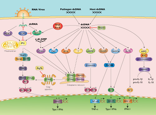
Cytosolic DNA-mediated production of type I IFNs (mainly IFN-β) requires the transcription factor IFN regulatory factor 3 (IRF3), which is activated upon phosphorylation by TANK-binding-kinase-1 (TBK1) [1].

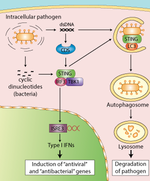
STING in DNA sensing

Recently, a new molecule, STING (stimulator of IFN genes), has been shown to be essential for the TBK1-IRF3- dependent induction of IFN-β by transfected DNA ligands and intracellular DNA produced by pathogens after infection [2, 3].
STING (also known as MITA, MPYS and ERIS) is a transmembrane protein that resides in the endoplasmic reticulum (ER) [2-6]. In response to cytosolic DNA, STING forms dimers and translocates from the ER to the Golgi then to punctate cytosolic structures where it colocalizes with TBK-1, leading to the phosphorylation of IRF3.
How STING stimulates TBK1-dependent IRF3 activation was recently elucidated by Tanaka and Chen. They found that, upon cytosolic DNA sensing, the C-terminal tail of STING acts as a scaffold protein to promote the phosphorylation of IRF3 by TBK1 [7].

STING in the host response to intracellular pathogens. Linking type I IFN response and autophagy for better defense

STING in the host response to intracellular pathogens

http://www.invivogen.com/images/STING-autophagy.png


STING activates the IFN response

Until very recently, STING in addition to its role as an adaptor protein was also thought to function as a sensor of cyclic dinucleotides.
Burdette et al. first demonstrated that STING binds directly to the bacterial molecule cyclic diguanylate monophosphate (c-di-GMP) [8]. This finding was confirmed by several teams who examined the structure of STING bound to c-di-GMP [9-11], including Cheng and colleagues, however their data suggest that STING is not the primary sensor of c-di-GMP [12]. Rather, they indicate that DDX41, an identified CDS, functions as a direct receptor for cyclic dinucleotides upstream of STING. The authors hypothesized that DDX41 binds to c-di-GMP then forms a complex with STING to activate the IFN response.

STING induces autophagy

Exciting new developments reveal that STING participates in another aspect of innate immunity, autophagy.
Autophagy plays a critical role in host defense responses to pathogens by promoting the elimination of microbes that enter into the cytosol by their sequestration into autophagosomes and their delivery to the lysosome.


CDS pathway

http://www.invivogen.com/images/STING-CDS_pathway_small.jpg

Recent studies have reported that DNA viruses and intracellular bacteria induce autophagy and that this process is dependent on cytosolic genomic DNA and STING [13-15]. Robust induction of autophagy was also observed after transfection of various double stranded (ds) DNA species, such as poly(dA:dT), poly(dG:dC) or plasmid DNA, but not single stranded (ss) DNA, dsRNA or ssRNA [16].

Interestingly, activated STING was shown to relocate to unidentified membrame-bound compartments where it colocalizes with LC3, a hallmark of autophagy, and ATg9a. The latter protein was reported to regulate the interaction between STING and TBK1 after dsDNA stimulation [16]. The E3 ubiquitin ligases TRIM56 and TRIM32
were also found to regulate STING by mediating its dimerization through K63-linked ubiquitination [17, 18].

Several cytosolic DNA sensors upstream of STING have been proposed.
DNA-dependent activator of IRFs (DAI) was the first CDS discovered based on the ability of transfected poly(dA:dT) to induce IFN-β [19]. However, the role of DAI has been shown to be very cell-type specific and cells derived from DAI-deficient mice responded normally to dsDNA ligands [20].

While analyzing immune responses to dsDNA regions derived from vaccinia virus (VACV-70) or Herpes simplex virus 1 (HSV-60) genomes, Unterholzner et al. identified IFI16 as a DNA binding protein mediating IFN-β induction [21]. Interestingly, IFI16 belongs to a new family of pattern recognition receptors that contain the pyrin and HIN domain (PYHIN), termed AIM2-like receptors (ALRs).

AIM2 is a STING-independent cytosolic DNA sensor that forms an inflammasome with ASC to trigger caspase-1 activation and the secretion of the proinflammatory cytokines IL-1β and IL-18 [20].

Members of the DExD/H-box helicase superfamily have also been reported to function as cytosolic DNA sensors. While DHX36 and DHX9 were identified as STING-independent but MyD88-dependent sensors of CpG-containing DNA in plasmacytoid dendritic cells, DDX41 was found to bind various dsDNA ligands and localize with STING to promote IFN-β expression [22]. Other CDSs have been reported to function independently of STING: RNA Pol III, LRRFIP1 and Ku70 [20].

Unlike cytosolic RNA sensors (RIG-I, MDA-5), which detect structural moieties specific to pathogen RNA, such as 5’-triphosphates, it is not clear whether cytosolic DNA sensors can recognize any particular structural motif of DNA that would discriminate between self and non-self. This suggests that CDSs may have a role not only in anti-microbial innate immune responses but also in autoimmunity. A multitude of CDSs have been described but whether they are all true receptors remains an open question.

1. Stetson DB & Medzhitov R. 2006. Recognition of cytosolic DNA activates an IRF3-dependent innate immune response. Immunity. 24(1):93-103.
2. Ishikawa H. & Barber GN., 2008. STING is an endoplasmic reticulum adaptor that facilitates innate immune signalling. Nature. 455(7213):674-8.
3. Ishikawa H. et al., 2009. STING regulates intracellular DNA-mediated, type I interferon-dependent innate immunity. Nature. 461(7265):788-92.
4. Zhong B. et al., 2008. The adaptor protein MITA links virus-sensing receptors to IRF3 transcription factor activation. Immunity. 29(4):538-50.
5. Jin L. et al., 2008. MPYS, a novel membrane tetraspanner, is associated with major histocompatibility complex class II and mediates transduction of apoptotic signals. Mol Cell Biol. 28(16):5014-26.


UV Light Potentiates STING (Stimulator of Interferon Genes)-dependent Innate Immune Signaling through Deregulation of ULK1 (Unc51-like Kinase 1).

 J Biol Chem. 2015 May 8;290(19):12184-94.  http://dx.doi.org:/10.1074/jbc.M115.649301. Epub 2015 Mar 19.

The mechanism by which ultraviolet (UV) wavelengths of sunlight trigger or exacerbate the symptoms of the autoimmune disorder lupus erythematosus is not known but may involve a role for the innate immune system. Here we show that UV radiation potentiates STING (stimulator of interferon genes)-dependent activation of the immune signaling transcription factor interferon regulatory factor 3 (IRF3) in response to cytosolic DNA and cyclic dinucleotides in keratinocytes and other human cells. Furthermore, we find that modulation of this innate immune response also occurs with UV-mimetic chemical carcinogens and in a manner that is independent of DNA repair and several DNA damage and cell stress response signaling pathways. Rather, we find that the stimulation of STING-dependent IRF3 activation by UV is due to apoptotic signaling-dependent disruption of ULK1 (Unc51-like kinase 1), a pro-autophagic protein that negatively regulates STING. Thus, deregulation of ULK1 signaling by UV-induced DNA damage may contribute to the negative effects of sunlight UV exposure in patients with autoimmune disorders.


STING and the innate immune response to nucleic acids in the cytosol

Dara L Burdette & Russell E Vance

https://mcb.berkeley.edu/labs/vance/Resources/Burdette%20(2013)%20review.pdf

Cytosolic detection of pathogen-derived nucleic acids is critical for the initiation of innate immune defense against diverse bacterial, viral and eukaryotic pathogens. Conversely, inappropriate responses to cytosolic nucleic acids can produce severe autoimmune pathology. The host protein STING has been identified as a central signaling molecule in the innate immune response to cytosolic nucleic acids. STING seems to be especially critical for responses to cytosolic DNA and the unique bacterial nucleic acids called ‘cyclic dinucleotides’. Here we discuss advances in the understanding of STING and highlight the many unresolved issues in the field.

The detection of pathogen-derived nucleic acids is a central strategy by which the innate immune system senses microbes to then initiate protective responses1. Conversely, inappropriate recognition of self nucleic acids can result in debilitating autoimmune diseases such as systemic lupus erythematosus2. It is therefore important to understand the molecular basis of the detection of nucleic acids by the innate immune system. Studies have established that nucleic acids derived from extracellular sources are sensed mainly by endosomal Toll-like receptors (TLRs), such as TLR3, TLR7 and TLR9, whereas cytosolic nucleic acids are detected independently of TLRs by a variety of less-well-characterized mechanisms1.

Studies have identified STING (‘stimulator of interferon genes’; also known as TMEM173, MPYS, MITA and ERIS) as a critical signaling molecule in the innate response to cytosolic nucleic-acid ligands. STING was first described as a protein that interacts with major histocompatibility complex class II molecules3, but the relevance of this interaction remains unclear. Subsequent studies have instead focused on the role of STING in the transcriptional induction of type I interferons and coregulated genes in response to nucleic acids in the cytosol. Several groups have independently isolated STING by screening for proteins able to induce interferon-B (IFN-B) when overexpressed4–6. Studies of STING-deficient mice have subsequently confirmed the essential role of STING in innate responses to cytosolic nucleic-acid ligands, particularly double-stranded DNA (dsDNA) and unique bacterial nucleic acids called ‘cyclic dinucleotides’7–9. Several studies have also linked STING to the interferon response to cytosolic RNA5–7, but this has not been found consistently7,8,10,11; thus, we focus here on the role of STING in response to DNA and cyclic dinucleotides.


Protein Stimulator of interferon genes protein
Gene TMEM173
Organism Homo sapiens (Human)
Facilitator of innate immune signaling that acts as a sensor of cytosolic DNA from bacteria and viruses and promotes the production of type I interferon (IFN-alpha and IFN-beta). Innate immune response is triggered in response to non-CpG double-stranded DNA from viruses and bacteria delivered to the cytoplasm. Acts by recognizing and binding cyclic di-GMP (c-di-GMP), a second messenger produced by bacteria, and cyclic GMP-AMP (cGAMP), a messenger produced in response to DNA virus in the cytosol: upon binding of c-di-GMP or cGAMP, autoinhibition is alleviated and TMEM173/STING is able to activate both NF-kappa-B and IRF3 transcription pathways to induce expression of type I interferon and exert a potent anti-viral state. May be involved in translocon function, the translocon possibly being able to influence the induction of type I interferons. May be involved in transduction of apoptotic signals via its association with the major histocompatibility complex class II (MHC-II). Mediates death signaling via activation of the extracellular signal-regulated kinase (ERK) pathway. Essential for the induction of IFN-beta in response to human herpes simplex virus 1 (HHV-1) infection. Exhibits 2′,3′ phosphodiester linkage-specific ligand recognition. Can bind both 2′-3′ linked cGAMP and 3′-3′ linked cGAMP but is preferentially activated by 2′-3′ linked cGAMP (PubMed:26300263)
Stimulator of interferon genes protein (IPR029158)
Transmembrane protein 173, also known as stimulator of interferon genes protein (STING) or endoplasmic reticulum interferon stimulator (ERIS), is a transmembrane adaptor protein which is involved in innate immune signalling processes. It induces expression of type I interferons (IFN-alpha and IFN-beta) via the NF-kappa-B and IRF3, pathways in response to non-self cytosolic RNA and dsDNA [PMID: 18724357, PMID: 19776740,PMID: 18818105, PMID: 19433799].

Read Full Post »

Periodic table of protein complexes, Volume 2 (Volume Two: Latest in Genomics Methodologies for Therapeutics: Gene Editing, NGS and BioInformatics, Simulations and the Genome Ontology), Part 1: Next Generation Sequencing (NGS)

Periodic table of protein complexes

Larry H. Bernstein, MD, FCAP, Curator

LPBI


Periodic Table of Protein Complexes

http://www.technologynetworks.com/Proteomics/news.aspx?ID=186248

New tool helps to visualise, understand and predict how proteins combine to drive biological processes

A new ‘periodic table’ of protein complexes has been developed that provides a unified way to classify and visualise protein complexes, providing a valuable tool for biotechnology and the engineering of novel complexes.

This study also provides insights into evolutionary distribution of different types of existing protein complexes.

The Periodic Table of Protein Complexes offers a new way of looking at the enormous variety of structures that proteins can build in nature, which ones might be discovered next, and predicting how entirely novel structures could be engineered. Created by an interdisciplinary team led by researchers at the Wellcome Genome Campus and the University of Cambridge, the Table provides a valuable tool for research into evolution and protein engineering.

Almost every biological process depends on proteins interacting and assembling into complexes in a specific way, and many diseases are associated with problems in complex assembly. The principles underpinning this organisation are not yet fully understood, but by defining the fundamental steps in the evolution of protein complexes, the new ‘periodic table’ presents a systematic, ordered view on protein assembly, providing a visual tool for understanding biological function.

“Evolution has given rise to a huge variety of protein complexes, and it can seem a bit chaotic. But if you break down the steps proteins take to become complexes, there are some basic rules that can explain almost all of the assemblies people have observed so far.”


Dr Joe Marsh, formerly of the Wellcome Genome Campus and now of the MRC Human Genetics Unit at the University of Edinburgh.

Different ballroom dances can be seen as an endless combination of a small number of basic steps. Similarly, the ‘dance’ of protein complex assembly can be seen as endless variations on dimerization (one doubles, and becomes two), cyclisation (one forms a ring of three or more) and subunit addition (two different proteins bind to each other). Because these happen in a fairly predictable way, it’s not as hard as you might think to predict how a novel protein would form.

“We’re bringing a lot of order into the messy world of protein complexes. Proteins can keep go through several iterations of these simple steps, adding more and more levels of complexity and resulting in a huge variety of structures. What we’ve made is a classification based on these underlying principles that helps people get a handle on the complexity.”

Dr Sebastian Ahnert of the Cavendish Laboratory at the University of Cambridge

The exceptions to the rule are interesting in their own right, as are the subject of on-going studies.

“By analysing the tens of thousands of protein complexes for which three-dimensional structures have already been experimentally determined, we could see repeating patterns in the assembly transitions that occur – and with new data from mass spectrometry we could start to see the bigger picture.”

Dr Joe Marsh

“The core work for this study is in theoretical physics and computational biology, but it couldn’t have been done without the mass spectrometry work by our colleagues at Oxford University. This is yet another excellent example of how extremely valuable interdisciplinary research can be.”

Dr Sarah Teichmann, Research Group Leader at the Wellcome Trust Sanger Institute and the European Bioinformatics Institute (EMBL-EBI)

Read Full Post »

Guanosine triphosphate (GTP) cancer link

Larry H. Bernstein, MD, FCAP, Curator

LPBI


Energy-Sensing Enzyme May Serve as Cancer Therapy Target

http://www.genengnews.com/gen-news-highlights/energy-sensing-enzyme-may-serve-as-cancer-therapy-target/81252200/


A scientific team led by a University of Cincinnati (UC) cancer researcher has shown that a specific enzyme is responsible for sensing the available supply of guanosine triphosphate (GTP) that can fuel the uncontrolled growth of cancer cells. The research underscores the enzyme’s potential to become a therapeutic target for future cancer drugs, according to the investigators.

Atsuo Sasaki, Ph.D., assistant professor in the division of hematology oncology at the UC College of Medicine, Toshiya Senda, Ph.D., professor at the High Energy Accelerator Research Organization in Tsukuba, Japan, and colleagues showed that PI5P4Kβ (phosphatidylinositol-5-phosphate 4-kinase-β) acts like the arrow on a fuel gauge. The enzyme senses and communicates via a second messenger the amount of GTP fuel that is available to a cell at any given time. Until now, the molecular identity of a GTP sensor has remained unknown.

“Energy sensing is vital to the successful proliferation of cancer cells,” says Dr. Sasaki. “A large amount of GTP is required in rapidly dividing cells, and cells need to know that the fuel is available to them. If we can interfere with the ability of PI5P4Kβ to sense fuel availability and communicate that information, we may be able to slow or halt the growth of cancers, including the aggressive brain cancer glioblastoma multiforme and cancers that have metastasized to the brain.”

The study (“The Lipid Kinase PI5P4Kβ Is an Intracellular GTP Sensor for Metabolism and Tumorigenesis”), published in Molecular Cell, is Dr. Sasaki’s first to address PI5P4Kβ as a molecular sensor of GTP concentration. Initially, he and his team reportedly faced skepticism regarding the existence of GTP energy-sensing.

GTP is one of two energy molecules used by cells. The other is adenosine triphosphate.  ATP handles the bulk of a cell’s energy requirements, while GTP is required for protein synthesis and is a signaling molecule that helps direct processes within the cell. When GTP levels are increased and utilized as fuel by cancer cells, its ability to perform its primary goals is compromised.

Dr. Sasaki and his team identified PI5P4Kβ as a GTP sensor by demonstrating, in a laboratory setting its ability to bind to GTP and by showing, at the atomic level by X-ray structural analysis, the molecular mechanism by which it recognizes GTP. They then designed PI5P4Kβ mutant cells that were unable to sense GTP concentration and, as a result, impaired the ability of PI5P4Kβ to promote tumor growth.

His next step is to use both pharmacological and molecular approaches that target PI5P4Kβ in a cell culture and in animal tumor models.

“By unveiling PI5P4Kβ’s role as a GTP sensor, we now have a potential new therapeutic target for patients,” explains Dr. Sasaki. “If we can find drugs that stop PI5P4Kβ from acting as the fuel indicator, we could get these aggressive and tragic cancers into energy-depleted status.”

Read Full Post »

Reinforced disordered cell expression

Larry H. Bernstein, MD, FCAP, Curator

LPBI


Diabetes, Alzheimer’s Share Molecular Pathways, Part of Same Vicious Cycle

http://www.genengnews.com/gen-news-highlights/diabetes-alzheimer-s-share-molecular-pathways-part-of-same-vicious-cycle/81252206/

http://www.genengnews.com/Media/images/GENHighlight/thumb_Jan8_2016_Fotolia_30836005_JigsawPuzzleBrainAndHead1904910113.jpg

A molecular-level link has been found that helps explain the poorly understood association between diabetes and Alzheimer’s disease. Both disorders can drive and be driven by the same pathological process, the disruption of a particular kind of post-translational modification called S-nitrosylation. Thus, the disorders can reinforce each other. [© freshidea/Fotolia]


Though they appear to be distinct, diabetes and Alzheimer’s disease have much in common at the molecular level. In fact, recent findings indicate that either disease can worsen the other by disrupting the same chemical process—S-nitrosylation, a form of post-translational modification that is necessary for the proper functioning of multiple enzymes.

S-nitrosylation, it turns out, can be disrupted by excess sugar or β-amyloid protein, either of which can wreak havoc by increasing the levels of nitric oxide and other free radical species. Once S-nitrosylation is disturbed and poorly functioning enzymes are produced, the downstream effects include abnormal increases in both insulin and β-amyloid protein.

Thus, diabetes and Alzheimer’s can drive, and be driven by, the same vicious cycle. Furthermore, either can contribute to the other’s progress. These results emerged from a study completed by researchers based at the Sanford Burnham Prebys Medical Discovery Institute and the Scintillon Institute. The research team was led by Stuart A. Lipton, M.D., Ph.D., a physician-scientist affiliated with both institutions.

“This work points to a new common pathway to attack both type 2 diabetes, along with its harbinger, metabolic syndrome, and Alzheimer’s disease,” stated Dr. Lipton.

The researchers published their work January 8 in the journal Nature Communications in an article entitled, “Elevated glucose and oligomeric β-amyloid disrupt synapses via a common pathway of aberrant protein S-nitrosylation.” This article describes how the scientists used a so-called “disease-in-a-dish” model to discover molecular pathways that are in common in both diabetes and Alzheimer’s.

Specifically, the scientists genetically reprogrammed the skin of human patients to make induced pluripotent stem cells, which were then used to derive nerve cells. They also used mouse models of each disease to analyze the combined effects of high blood sugar and β-amyloid protein in living animals.

“[We] report in human and rodent tissues that elevated glucose, as found in [metabolic syndrome and type 2 diabetes] and oligomeric β-amyloid (Aβ) peptide, thought to be a key mediator of [Alzheimer’s disease], coordinately increase neuronal Ca2+ and nitric oxide (NO) in an NMDA receptor-dependent manner,” wrote the authors of the Nature Communications article. “The increase in NO results in S-nitrosylation of insulin-degrading enzyme (IDE) and dynamin-related protein 1 (Drp1), thus inhibiting insulin and Aβ catabolism as well as hyperactivating mitochondrial fission machinery.”

The scientists also found that the changes in enzyme activity led to damage of synapses, the region where nerve cells communicate with one another in the brain. The combination of high sugar and β-amyloid protein caused the greatest loss of synapses. Since loss of synapses correlates with cognitive decline in Alzheimer’s, high sugar and β-amyloid coordinately contribute to memory loss.

“The NMDA receptor antagonist memantine attenuates these effects,” the authors continued. “Our studies show that redox-mediated posttranslational modification of brain proteins link Aβ and hyperglyaemia to cognitive dysfunction in [metabolic syndrome/type 2 diabetes] and [Alzheimer’s disease].”

“[Our work] means that we now know these diseases are related on a molecular basis, and hence, they can be treated with new drugs on a common basis,” stated Dr. Ambasudhan, a senior author of the study and an assistant professor at Scintillon.

Read Full Post »

Cell line expression and diversity

Larry H. Bernstein, MD, FCAP, Curator

LPBI


New Method Allows Gene Expression Tracking over Generations of Cells

http://www.genengnews.com/gen-news-highlights/new-method-allows-gene-expression-tracking-over-generations-of-cells/81252188/

https://youtu.be/O7oW9xrEQ3A


  • MIT researchers engineered a microfluidic device that traces detailed family histories for several generations of cells descended from one “ancestor.” [Video: Melanie Gonick/MIT (additional video courtesy of the Manalis Lab)]

  • Scientists at MIT said they can now trace detailed family histories for several generations of cells descended from one ancestor after combining RNA sequencing with a novel device that isolates single cells and their progeny. This technique, which can track changes in gene expression as cells differentiate, could be particularly useful for studying how stem cells or immune cells mature, noted the researchers, adding that it could also shed light on how cancer develops.

    “Existing methods allow for snapshot measurements of single-cell gene expression, which can provide very in-depth information. What this new approach offers is the ability to track cells over multiple generations and put this information in the context of a cell’s lineal history,” says Robert Kimmerling, a graduate student in biological engineering and the lead author of a paper (“A microfluidic platform enabling single-cell RNA-seq of multigenerational lineages”) describing the technique in Nature Communications.

    The new method incorporates single-cell RNA-seq, which sequences a single cell’s transcriptome and reveals which genes are being transcribed inside a cell at a given point in time. This helps scientists understand, for example, what makes a skin cell so different from a heart cell even though the cells share the same DNA.

    “Scientists have well-established methods for resolving diverse subsets of a population, but one thing that’s not very well worked out is how this diversity is generated. That’s the key question we were targeting: how a single founding cell gives rise to very diverse progeny,” points out Kimmerling.

    To try to answer that question, the researchers designed a microfluidic device that traps first an individual cell and then all of its descendants. The device has several connected channels, each of which has a trap that can capture a single cell. After the initial cell divides, its daughter cells flow further along the device and get trapped in the next channel. The researchers showed that they can capture up to five generations of cells this way and keep track of their relationships.

    To get the cells off the chip, the researchers temporarily reverse the direction of the fluid flowing across the chip, allowing them to remove the cells one at a time to perform single-cell RNA-seq.

    In this study, the team captured and sequenced T cells that—when they encounter a cell infected with a virus or bacterium—create effector T cells, which seek and destroy infected cells, as well as memory T cells that retain a “memory” of the encounter and circulate in the body indefinitely in case of a subsequent encounter.

    “A single founding cell can give rise to both effector and memory cell subtypes, but how that diversity is generated isn’t very clear,” explain Kimmerling.

    The scientists analyzed RNA from recently activated T cells and two subsequent generations. When comparing genes with functions related to T-cell activation and differentiation, they found that sister cells produced from the same division event are much more similar in their gene expression profiles than two unrelated cells. They also found that “cousin” cells, which have the same “grandmother,” are more similar than unrelated cells. This suggests unique, family-specific transcriptional profiles for single T cells.

    The researchers hope that future studies with this device could help to resolve the long-standing debate over how T cells differentiate into effector cells and memory cells. One theory is that the distinction occurs as early as the first T cell division following activation, while a competing theory suggests that the distinction happens later on, as a result of changes in the cells’ microenvironment.

    To address this question, the researchers believe they would need to analyze the development of T cells taken from a mouse that had been exposed to a foreign pathogen, providing a useful model of T cell activation in response to infection.

Read Full Post »

A hospitalist expert discusses bridging anticoagulation, surgery delays and periop beta-blockers

Sourced through Scoop.it from: www.todayshospitalist.com

See on Scoop.itCardiovascular Disease: PHARMACO-THERAPY

Read Full Post »

Effects of Preoperative β-Blocker Use on Clinical Outcomes after Coronary Artery Bypass Grafting:A Report from the Japanese Cardiovascular Surgery Database

Reporter: Aviva Lev-Ari, PhD, RN


In a nationwide registry, preoperative beta-blockers did not affect short-term mortality or morbidity in CABG pts. https://t.co/xEtJxTlBXQ

Sourced through Scoop.it from: anesthesiology.pubs.asahq.org

See on Scoop.itCardiovascular Disease: PHARMACO-THERAPY

Read Full Post »

What Are Your Medication Options for Heart Arrhythmias?

Reporter: Aviva Lev-Ari, PhD, RN


Heart arrhythmias can be treated with antiarrhythmic drugs, AV nodal blocking drugs, beta blockers, statins, and omega-3 fatty acids.

Sourced through Scoop.it from: heartdisease.about.com

See on Scoop.itCardiovascular Disease: PHARMACO-THERAPY

See our Book

https://www.amazon.com/dp/B07MGSFDWR


Read Full Post »

Applying Pharmacology to New Drug Discovery, April 22, 2016 in San Diego, CA by CHI

Reporter: Aviva Lev-Ari, PhD, RN


Applying Pharmacology to New Drug Discovery, April 22, 2016 in San Diego, CA by CHI

The system-independent quantification of molecular drug properties for prediction of therapeutic utility

April 22, 2016

Over the past 6 six years, the primary cause of new drug candidate failures (50%) has been failure of therapeutic efficacy. Put another way, drug discovery programs do everything right, get the defined candidate molecule, only to have it fail in therapeutic trials. Among the most prevalent reasons proposed for this shortcoming is the lack of translation of in vitro and recombinant drug activity to therapeutic in vivo whole systems. Drug activity in complete systems can be characterized with the application of pharmacological principles which translate drug behaviors in various organs with molecular scales of affinity and efficacy.

Pharmacological techniques are unique in that they can convert descriptive data (what we see, potency, activity in a given system) to predictive data (molecular scales of activity that can be used to predict activity in all systems including the therapeutic one, i.e. affinity, efficacy). The predicted outcome of this process is a far lower failure rate as molecules are progressed toward clinical testing.

Instructor

Terry Kenakin presently is a Professor of Pharmacology in the Department of Pharmacology, University of North Carolina School of Medicine. The course is taught from the perspective of industrial drug discovery; Dr. Kenakin has worked in drug industry for 32 years (7 at Burroughs-Wellcome, RTP, NC and 25 at GlaxoSmithKline, RTP. NC). He is Editor-in-Chief of the Journal of Receptors and Signal Transduction and Co-Editor-in-Chief of Current Opinion in Pharmacology and is on numerous journal Editorial Boards. In addition, he has authored over 200 peer reviewed papers and reviews and has written 10 books on Pharmacology.

Course Material

Summary sheets, exercises with answers, relevant papers are included as well as a pdf of all slides. The course is based on the book A Pharmacology Primer: Techniques for More Effective and Strategic Drug Discovery. 4th Edition, Elsevier/Academic Press, 2014.

This course will describe pharmacological principles and procedures to quantify affinity, efficacy, biased signaling and allostery to better screen for new drugs and characterize drug candidates in lead optimization assays.

1. Assay Formats/Experimental Design

  • Binding
  • Functional Assays
  • Null Method Assays

2. Agonism

  • Agonist Affinity/Efficacy
  • Black/Leff Operational model

3. Biased Signaling (Agonism)

  • Mechanism of Biased Signaling
  • Quantifying Biased Agonism
  • Therapeutic application(s)

4. Orthosteric Antagonism (I)

  • Competitive
  • Non-Competitive/Irreversible

5. Orthosteric Antagonism (II)

  • Partial Agonism
  • Inverse Agonism

6. Allosteric Modulation (I)

  • Functional Allosteric Model
  • Negative Allosteric Modulators (NAMs)

7. Allosteric Modulation (II)

  • Positive Allosteric Modulators (PAMs)
  • Allosteric Agonism

8. Drug-Receptor Kinetics

  • Measuring Target Coverage
  • Allosteric Proof-of-Concept
  • Application of Real-Time Kinetics

9. Drug Screening

  • Design of Screening Assays
  • Screening for Allosteric Modulators

Cambridge Healthtech Institute’s Eleventh Annual Drug Discovery Chemistry is a dynamic conference for medicinal chemists working in pharma and biotech. Focused on discovery and optimization challenges of small molecule drug candidates, this event provides many exciting opportunities for scientists to create a unique program by going back and forth between concurrent meeting tracks to hear presentations most suited to one’s personal interests. New for 2016 is the addition of three symposia on Friday covering the blood-brain barrier, biophysical approaches for drug discovery, and antivirals.

Plenary Keynotes


A New Model for Academic Translational Research

Peter G. Schultz, Ph.D., The Scripps Research Institute

Cell-Penetrating Miniproteins

Gregory L. Verdine, Ph.D., Harvard University

April 19-20

April 20-21

April 22

Inflammation Inhibitors

Kinase Inhibitor Chemistry

Brain Penetrant Inhibitors

Protein-Protein Interactions

Macrocyclics & Constrained Peptides

Biophysical Approaches

Epigenetic Inhibitor Discovery

Fragment-Based Drug Discovery

Antivirals

Short Courses

Make the most of your time in San Diego by adding on one or more short courses*. Topics include trends in physical properties, GPCRs, peptide therapeutics, immunology, phenotypic screening, crystallography, ligand-receptor molecular interactions, inhibitor design, macrocycles, FBDD, and covalent inhibitors.

* separate registration required for short courses

SOURCE

From: Deborah Shear <pete@healthtech.com>

Date: Friday, January 8, 2016 at 11:42 AM

To: Aviva Lev-Ari <AvivaLev-Ari@alum.berkeley.edu>

Subject: Training Seminar: Applying Pharmacology to New Drug Discovery

Read Full Post »

Early Cancer Diagnosis by the Detection of Circulating Tumor Cells using Drop-based Microfluidics @Harvard

Reporter: Aviva Lev-Ari, PhD, RN


Davey received a silver medal at the Collegiate Inventors Competition for the microfluidic technique he developed. (Photo courtesy of the Collegiate Inventors Convention.)

davey_neil4_0

Davey, who developed an interest in biology while working as a high school intern at the Food and Drug Administration, is looking forward to studying additional applications of microfluidics. The technology can be used to isolate specific cancer genomes by sorting bright drops out of the mix, enabling scientists to learn more about the nature of the disease. It can also be used to identify specific mutations of the cancer genome, which could help doctors determine which medications would be most effective for a patient.


Davey worked in the lab of mentor of David Weitz, Mallinckrodt Professor of Physics and Applied Physics at the John A. Paulson School of Engineering and Applied Sciences, to develop the technique.

His technique involves injecting a tiny amount of blood into a microfluidic device to encapsulate single cells from the blood stream in individual microfluidic drops. Once the cells have been encapsulated, Davey uses a polymerase chain reaction (PCR), a common technique in molecular biology, to target and amplify fragments of cancer DNA within the drops.


SOURCE

http://www.seas.harvard.edu/news/2016/01/harvard-student-develops-technique-to-diagnose-cancer-from-single-drop-of-blood?utm_source=SilverpopMailing&utm_medium=email&utm_campaign=01.08.2016%20(1)

Read Full Post »

« Newer Posts - Older Posts »