Healthcare analytics, AI solutions for biological big data, providing an AI platform for the biotech, life sciences, medical and pharmaceutical industries, as well as for related technological approaches, i.e., curation and text analysis with machine learning and other activities related to AI applications to these industries.
Reversing Heart Disease: Combination of PCSK9 Inhibitors and Statins – Opinion by Steven Nissen, MD, Chairman of Cardiovascular Medicine at Cleveland Clinic
Lipoprotein(a) (Lp(a)) is recognized as a causal factor for coronary heart disease (CHD) but its atherogenicity relative to that of low-density lipoprotein (LDL) on a per-particle basis is indeterminate.
Conclusions
We conclude that the atherogenicity of Lp(a) (CHD risk quotient per unit increase in particle number) is substantially greater than that of LDL. Therefore, Lp(a) represents a key target for drug-based intervention in a significant proportion of the at-risk population.
The term “atherogenicity” is used since 1986 [5] referring to the accumulation of intracellular lipids, which is a trigger of cellular atherogenesis
Atherogenic dyslipidemia (AD) refers to elevated levels of triglycerides (TG) and small-dense low-density lipoprotein and low levels of high-density lipoprotein cholesterol (HDL-C). In addition, elevated levels of large TG rich very low-density lipoproteins, apolipoprotein B and oxidised low-density lipoprotein (LDL), and reduced levels of small high-density lipoproteins plays a critical role in AD. All three elements of AD per se have been recognised as independent risk factor for cardiovascular disease. LDL-C/HDL-C ratio has shown excellent risk prediction of coronary heart disease than either of the two risk markers. Asian Indians have a higher prevalence of AD than western population due to higher physical inactivity, low exercise and diet deficient in polyunsaturated fatty acids (PUFA).
While nearly 10% of middle-age adults in China have high risk for cardiovascular disease, only 0.6% of these high-risk individuals use statins and 2.4% take aspirin, a national screening project reported in the Annals of Internal Medicine.
UPDATED on 5/5/2017
Europeans Mull PCSK9i Post-FOURIER Fallout on Clinical Practice
Patrice Wendling, May 04, 2017
But it was panelist Dr Stephen Nicholls (University of Adelaide, Australia) who took aim at the elephant in the packed auditorium. At an annual cost of about $14,100 for evolocumab and $14,600 for alirocumab (Praluent, Sanofi/Regeneron), the important question facing cardiologists is who will be eligible for these drugs “in a world where we can’t just write a scrip for every FOURIER-type patient; we won’t be allowed to.”
He suggested initially this will include patients with familial hypercholesterolemia and only those with established atherosclerotic CVD whose LDL-C remains unacceptably high despite therapy. Future FOURIER subanalyses may define other eligible high-risk groups.
PCSK9 Inhibitor Access Snarled in Red Tape, Rejections
Patrice Wendling, March 21, 2017
To determine whether this experience is happening nationwide, Navar and colleagues examined first PCSK9 prescriptions in 45,029 patients (median age 66 years; 51% female) between August 1, 2015 and July 31, 2016 in the Symphony Health Solutions database, which covers 90% of retail, 70% of specialty, and 60% of mail-order pharmacies in the US.
Nearly half (48%) of prescribers were cardiologists, and 37% were general practitioners. Most patients (52%) had government insurance, typically Medicare, and 40% had commercial insurance.
In the first 24 hours after being submitted to the pharmacy, 79.2% of prescriptions were rejected. Ultimately, 52.8% of all PCSK9 prescriptions were rejected.
Of special note, 34.7% of prescriptions for the pricy lipid-lowering drugs were abandoned at the pharmacy.
How 2 Drugs Lower Cholesterol Remarkably — and Reverse Heart Disease
Study shows promise for combination of newer drug and statins
Newer cholesterol-lowering drugs combined with more conventional medicine reduces bad cholesterol to incredibly low levels, a new study shows. Perhaps even more important, the combination also reduces the heart-attack-inducing plaque that forms inside the arteries, the study says.
The study was led by cardiologist Steven Nissen, MD, Chairman of Cardiovascular Medicine at Cleveland Clinic. Results appeared recently in the Journal of the American Medical Association (JAMA).
The study looked at the use of a drug called evolocumab by people who took statins to lower the amount of LDL, or bad, cholesterol in their blood. Evolocumab is a drug called a PCSK9 inhibitor. This is a newer kind of medicine that can make LDL cholesterol levels plummet.
The people who took statins and evolocumab had greater reductions in atherosclerosis compared with people who took statins and a placebo. Atherosclerosis is a disease in which plaque builds up inside your arteries. The condition can lead to serious problems, including heart attack, stroke, or even death.
The results are an intriguing indicator — rather than definite proof — that evolocumab may have benefit for patients taking statins, Dr. Nissen says. Researchers are still awaiting the results of large clinical trials investigating whether evolocumab is safe and will prevent heart attack, stroke or death. The first results of these studies are expected in April 2017.
Special ultrasound
In the study, researchers treated for 18 months 968 high-risk people who had extremely high levels of blood cholesterol.
Participants were randomly assigned to take either a statin and a placebo, or a statin and evolocumab.
Researchers monitored the participants’ cholesterol levels. They also used a special ultrasound probe to measure the amount of plaque in their arteries at the beginning and the end of the study.
“We were able to show that getting the bad cholesterol levels down to really low levels, down to the 20s and 30s, can actually remove plaque from the coronary arteries,” Dr. Nissen says. “This going to levels that we’ve never been able to achieve before.”
Low cholesterol, less plaque
Results show the group treated with statins and a placebo reduced their LDL cholesterol levels to 93 on average. At the same time, the group treated with the combination of the statin plus evolocumab got down to an average bad cholesterol level of 36.6.
“No one’s ever reached levels that low in a clinical trial,” Dr. Nissen says.
Participants who took evolocumab also had less plaque in their arteries at the end of the study — essentially reversing their heart disease.
“We, for the first time now, have shown that this new class of drugs, the PCSK9 inhibitors, has a favorable effect on the development of plaques on the coronary artery and can actually regress those plaques,” Dr. Nissen says. “And it turns out about two-thirds of patients actually had less plaque at the end of 18 months than they started with.”
PCSK9 inhibitors, which are expensive, are not for everybody, Dr. Nissen says. Currently, the drug is used in addition to statins for the highest-risk patients with particularly high cholesterol.
Once thought to be little more than a way for cells to offload waste, the past decade has seen a huge shift in the way we think about exosomes. We’ve begun to recognize that exosomes are deliberately released from the cell, functioning as signal carriers and tissue reshapers through their cargo of RNA, proteins, lipids, and DNA. Involved in a wide range of healthy and pathogenic processes such as cancer, inflammation, immunity, CNS function, cardiac cell function, to name a few – exosomes are being studied for their role in these basic biological processes as well as for their use as biomarkers (see Applications) and even as tools for targeted delivery of biomolecules such as therapeutics (see Engineering).
What are exosomes?
Exosomes are 60 – 180 nm membrane vesicles secreted by most cell types in vivo and in vitro. These extracellular vesicles are endocytic in origin, produced by the inward budding of multivesicular bodies (MVBs). They are released from the cell into the microenvironment following fusion of MVBs with the plasma membrane.
Exosomes are not the only small, membrane-bound extracellular vesicle that can be found. They are distinct in origin from apoptotic blebs or apoptotic bodies, which are 50 nm to 5 um in size, carry DNA, RNA, and histones, and display surface markers targeting them for clearance by macrophages. And they are also different from microparticles (also known as microvesicles, ectosomes, shedding vesicles, microparticles, plasma membrane-derived vesicles, and exovesicles), which can range from 50-1000 nm in size and are derived directly from the plasma membrane rather than endocytic bodies within the cell.8 These distinctions and labeling conventions are not always used consistently in the literature and between different groups, leading to some ambiguity in the literature. When isolating exosomes, it’s important to remember that these other types of vesicles may also be present and interpret results accordingly.
What else are exosomes called?
Adding to the confusion, exosomes are sometimes referred to by the source of the sample material. For example, dendritic cell exosomes are also called dexosomes, and cancer cell exosomes may be called oncosomes. Researchers are starting to move towards more standardized nomenclature, but those searching through older literature should be aware of other names for exosomes.
Where are exosomes normally found?
Exosomes have been found in blood, urine, amniotic fluid, breast milk, malignant ascites fluids, and seminal fluid. They contain distinct subsets of molecules depending upon the cell type from which they are secreted, making them useful for biomarker discovery.
How do I study exosomes?
SBI is the only vendor to offer reagents and kits that support all apsects of exosome research-covering isolation, detection and measurement, discovery (characterization and analysis), and even exosome engineering. With a comprehensive set of tools and services to accelerate the study of exosomes and exosome RNA biomarkers, SBI puts the power of exosomes into researchers’ hands.
For many years, American physicians have screened their older male patients for prostate cancer by looking at the level of a particular protein in the blood. The protein, called prostate-specific antigen (P.S.A.), can indicate the presence of a tumor long before any symptoms materialize. Recently, though, there has been a movement within the medical community against P.S.A. testing; since prostate cancers typically grow very slowly and rarely cause discomfort, the thinking goes, early screening may not be all that useful. The U.S. Preventive Services Task Force, based on data from two large clinical trials, currently recommends against routine screening, but other expert groups (using the same evidence) have countered that men should be allowed to choose for themselves.
Now the dispute has become even more fraught. In October, TheNew England Journal of Medicinepublished a study by a group of British researchers that examined three classes of prostate-cancer patients: those who had received surgery, those who had received radiation therapy, and those whose disease had been carefully monitored without intervention. After ten years, there was no difference in survival rates among the three groups. Active treatment does not change the over-all risk of death, and this was the headline in most news reports. But largely overlooked in the press was that metastases, meaning spread of the cancer beyond the prostate gland to tissues in the pelvis and to bone, occurred three times more frequently in those being monitored than in those who received surgery or radiation. Not surprisingly, the cancer also progressed more quickly in these men.
In an editorial that accompanied the study, Anthony D’Amico, a radiation oncologist at Boston’s Dana-Farber Cancer Institute, argued that men should be informed of the risk of metastasis and of its consequences, particularly pelvic tumors and bone pain and fracture. D’Amico advises that men who wish to avoid metastases should consider monitoring, rather than surgery or radiation, only if their life expectancy is less than a decade. Having cared for many men with prostate cancer that metastasized—an incurable situation often marked by severe suffering—I strongly concur.
10-Year Outcomes after Monitoring, Surgery, or Radiotherapy for Localized Prostate Cancer
Freddie C. Hamdy, F.R.C.S.(Urol.), F.Med.Sci., Jenny L. Donovan, Ph.D., F.Med.Sci., J. Athene Lane, Ph.D., Malcolm Mason, M.D., F.R.C.R., Chris Metcalfe, Ph.D., Peter Holding, R.G.N., M.Sc., Michael Davis, M.Sc., Tim J. Peters, Ph.D., F.Med.Sci., Emma L. Turner, Ph.D., Richard M. Martin, Ph.D., Jon Oxley, M.D., F.R.C.Path., Mary Robinson, M.B., B.S., F.R.C.Path., John Staffurth, M.B., B.S., M.D., Eleanor Walsh, M.Sc., Prasad Bollina, M.B., B.S., F.R.C.S.(Urol.), James Catto, Ph.D., F.R.C.S.(Urol.), Andrew Doble, M.S., F.R.C.S.(Urol.), Alan Doherty, F.R.C.S.(Urol.), David Gillatt, M.S., F.R.C.S.(Urol.), Roger Kockelbergh, D.M., F.R.C.S.(Urol.), Howard Kynaston, M.D., F.R.C.S.(Urol.), Alan Paul, M.D., F.R.C.S.(Urol.), Philip Powell, M.D., F.R.C.S., Stephen Prescott, M.D., F.R.C.S.(Urol.), Derek J. Rosario, M.D., F.R.C.S.(Urol.), Edward Rowe, M.D., F.R.C.S.(Urol.), and David E. Neal, F.R.C.S., F.Med.Sci., for the ProtecT Study Group*
Matthew R. Cooperberg. . (2016) Re: 10-Year Outcomes After Monitoring, Surgery, or Radiotherapy for Localized Prostate Cancer. European Urology. CrossRef
Jean-Jacques Mazeron. . (2016) Cancer de la prostate : to treat or not to treat ?. Bulletin du Cancer. CrossRef
MPM Capital, which has around $2 billion in assets, is already working with drugmakers on funding healthcare startups. This includes a venture fund it closed last year at $400 million that included Novartis ($NVS) Astellas Pharma as investors.
(with equal contributions from MPM BV2014 and the Oncology Impact Fund), and included Deerfield Management, Arkin Bio Ventures, Celgene, Inc., Excelyrate Capital, Long March Investment Fund and MPM’s SunStates Fund.
MPM BV2014 – OIF will co-invest with MPM’s BV2014 venture fund in private oncology company investments.
Industry veteran and Oncorus co-founder Mitchell H. Finer, Ph.D., leads the Oncorus management team as Chief Executive Officer and Chief Scientific Officer and
Cyrus D. Mozayeni, M.D., serves as President and Chief Business Officer.
Finer serves on Oncorus’s Board of Directors along with:
– Luke Evnin, Ph.D., MPM Capital co-founder and Chairman,
– Briggs Morrison, CEO, Syndax;
– Cameron Wheeler, Ph.D., Principal, Deerfield Management; and,
– Alon Lazarus, Ph.D., Biotech Investment Manager, Arkin Bio Ventures.
This additional funding will help advance our immunotherapy platform as we discover innovative new therapies which we hope will be of benefit to patients in need and the physicians who treat them,” said Dr. Finer.
Oncorus licensed certain patent rights from the University of Pittsburgh based upon the work of renowned scientists Joseph Glorioso III, Ph.D., and Paola Grandi, Ph.D., who will join Oncorus’s Scientific Advisory Board.
The company will invest in researching and developing oncolytic viral constructs which will move through preclinical development and ultimately into clinical trials. Currently, the company’s lead candidate is in preclinical development for GBM. The company will also expand and improve its technology platform and accelerate the development of pipeline programs in other forms of cancer.
About AVM Astellas Venture Management LLC., based in Menlo Park, California, is a corporate venture capital of Astellas Pharma Inc. (“API”), headquartered in Tokyo, Japan. For over 15 years, AVM has been making strategic investments to achieve its mission to explore emerging innovative companies, which have potential to become API’s collaboration partners in R&D. For more information, please visit the website at www.astellasventure.com.
About Oncorus, Inc. Oncorus, Inc. is an early-stage biotechnology company developing a next-generation immunotherapy platform of oncolytic viruses to treat several types of cancer, including highly malignant and aggressive cancers. A leader in corporate philanthropy, Oncorus has taken a pledge to donate a portion of product sales revenue to fund promising cancer research and to support cancer care in the developing world. The company is located in Kendall Square, Cambridge, Massachusetts. Visit www.oncorus.com, for more information.
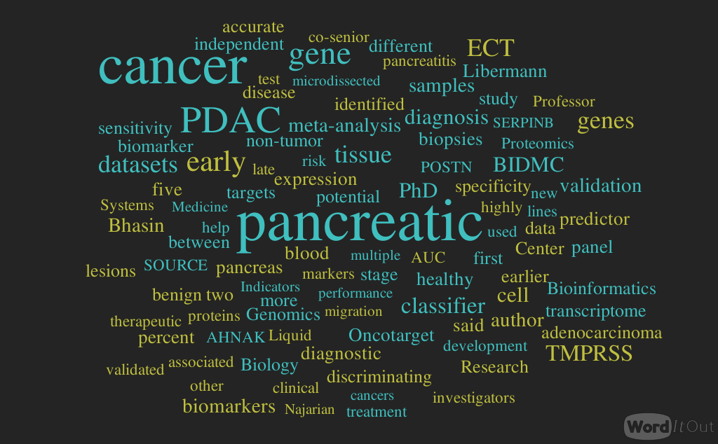
A novel 5-gene pancreatic adenocarcinoma classifier: Meta-analysis of transcriptome data – Clinical Genomics Research @BIDMC, Volume 2 (Volume Two: Latest in Genomics Methodologies for Therapeutics: Gene Editing, NGS and BioInformatics, Simulations and the Genome Ontology), Part 1: Next Generation Sequencing (NGS)
A novel 5-gene pancreatic adenocarcinoma classifier: Meta-analysis of transcriptome data – Clinical Genomics Research @BIDMC
Curator: Tilda Barliya, PhD
Analysis of Bhasin et al paper and Literature search
Table 1: 5-genes classifiers as biomarkers for PDAC:
Gene symbol
Gene name
Subcellular localization
ECT2
Epithelial cell transforming sequence 2 oncogene
Nucleus, cytoplasm
AHNAK2
AHNAKE nucleoprotein 2
Plasma membrane, cytoplasm
POSTN
Periostin, osteoblast specific factor
Extracellular space
TMPRSS4
Transmembrane protease, serine 4
Plasma membrane
SERPINB5
Serpin peptidase inhibitor, clade B (ovalbumin) member 5
Extracellular space
Introduction:
Bhasin et al, conducted a beautiful study using a powerful meta-analysis from different sources to identify the “important/classifier” genes associated with Pancreatic Cancer (PDAC).
The authors identified 5 genes that were considered as good classifiers (table 1).
It is important to note that the meta-analysis was performed on tissue and microdissection samples.
In their summary, the authors aim to validate these genes in blood/urine samples.
While these genes might be over expressed in tissue samples it may not be true to their existence in blood and careful examination and validation is required.
Liquid biopsies are emerging as the go-to use tools for disease detection, mostly aimed for early diagnosis.
Liquid biopsies are non-invasive biopsies of blood, urine (potentially saliva) and their “exotic” components, i.e miRNA, exosomes etc.
Since Liquid biopsies are non-invasive, they are painless and patients are more complied.
It is important to note that there is a gap between the expression of a gene or a protein in tissue section and their expression in the blood and may not necessarily correlate.
It will be very interesting to follow their research and future outcomes.
Additional References:
TMPRSS4: an emerging potential therapeutic target in cancer.
I was wondering if these same 5 genes were upregulated in the pancreatic ductal adenocarcinoma cell lines that are available out there. In doing cell biology work, there is always a dilemma whether cancer cell lines correctly re-capitulate in vivo tumors or not. Personally, I prefer primary cell lines to do analysis but this finding can be used to test primary vs cell line. In addition, I’ve attached the gene network for Ect2. If you look carefully, the two big proteins that jump out are RACGAP1 and KIF23. I think in designing therapies, combinatorial targets can yield the best outcomes. Drugs directed towards two or more targets would seem ideal in my opinion.
Pancreatic ductal adenocarcinoma (PDAC) is largely incurable due to late diagnosis. Superior early detection biomarkers are critical to improving PDAC survival and risk stratification.
EXPERIMENTAL DESIGN:
Optimized meta-analysis of PDAC transcriptome datasets identified and validated key PDAC biomarkers. PDAC-specific expression of a 5-gene biomarker panel was measured by qRT-PCR in microdissected patient-derived FFPE tissues. Cell-based assays assessed impact of two of these biomarkers, TMPRSS4 and ECT2, on PDAC cells.
RESULTS:
A 5-gene PDAC classifier (TMPRSS4, AHNAK2, POSTN, ECT2, SERPINB5) achieved on average 95% sensitivity and 89% specificity in discriminating PDAC from non-tumor samples in four training sets and similar performance (sensitivity = 94%, specificity = 89.6%) in five independent validation datasets. This classifier accurately discriminated PDAC from chronic pancreatitis (AUC = 0.83), other cancers (AUC = 0.89), and non-tumor from PDAC precursors (AUC = 0.92) in three independent datasets. Importantly, the classifier distinguished PanIN from healthy pancreas in the PDX1-Cre;LSL-KrasG12D PDAC mouse model. Discriminatory expression of the PDAC classifier genes was confirmed in microdissected FFPE samples of PDAC and matched surrounding non-tumor pancreas or pancreatitis. Notably, knock-down of TMPRSS4 and ECT2 reduced PDAC soft agar growth and cell viability and TMPRSS4 knockdown also blocked PDAC migration and invasion.
CONCLUSIONS:
This study identified and validated a highly accurate 5-gene PDAC classifier for discriminating PDAC and early precursor lesions from non-malignant tissue that may facilitate early diagnosis and risk stratification upon validation in prospective clinical trials. Cell-based experiments of two overexpressed proteins encoded by the panel, TMPRSS4 and ECT2, suggest a causal link to PDAC development and progression, confirming them as potential therapeutic targets.
In photo: First author Manoj Bhasin, PhD, and co-senior author Towia Libermann, PhD, Co-Director and Director of BIDMC’s Genomics, Proteomics, Bioinformatics and Systems Biology Center.
BOSTON – Pancreatic cancer, the fourth leading cause of cancer death in the United States, is often diagnosed at a late stage, when curative treatment is no longer possible. A team led by investigators at Beth Israel Deaconess Medical Center (BIDMC) has now identified and validated an accurate 5-gene classifier for discriminating early pancreatic cancer from non-malignant tissue. Described online in the journal Oncotarget, the finding is a promising advance in the fight against this typically fatal disease.
“Pancreatic cancer is a devastating disease with a death rate close to the incidence rate,” said co-senior author Towia Libermann, PhD, Director of the Genomics, Proteomics, Bioinformatics and Systems Biology Center at BIDMC and Associate Professor of Medicine at Harvard Medical School (HMS). “Because more than 90 percent of pancreatic cancer cases are diagnosed at the metastatic stage, when there are only limited therapeutic options, earlier diagnosis is anticipated to have a major impact on extending life expectancy for patients. There has been a lack of reliable markers, early indicators and risk factors associated with pancreatic cancer, but this new way of differentiating between healthy and malignant tissue offers hope for earlier diagnosis and treatment.”
The investigators used a number of publicly available gene expression datasets for pancreatic cancer and developed a strategy to reanalyze these datasets together, applying rigorous statistical criteria to compare different datasets from different laboratories and different platforms with each other. The team then selected a subset of data for developing a panel for differentiating between pancreatic cancer and healthy pancreas tissue and thereafter applied this “Pancreatic Cancer Predictor” to the remaining datasets for independent validation to confirm the accuracy of the markers.
After demonstrating and independently validating that a 5-gene pancreatic cancer predictor discriminated between cancerous and healthy tissue, the researchers applied the predictor to datasets that also included benign lesions of the pancreas, including pancreatitis and early stage cancer. The predictor accurately differentiated pancreatic cancer, benign pancreatic lesions, early stage pancreatic cancer and healthy tissue. The predictor achieved on average 95 percent sensitivity and 89 percent specificity in discriminating pancreatic cancer from non-tumor samples in four training sets and similar performance (94 percent sensitivity, 90 percent specificity) in five independent validation datasets.
“Using innovative data normalization and gene selection approaches, we combined the statistical power of multiple genomic studies and masked their variability and batch effects to identify robust early diagnostic biomarkers of pancreatic cancer,” said first author Manoj Bhasin, PhD, Co-Director of BIDMC’s Genomics, Proteomics, Bioinformatics and Systems Biology Center and Assistant Professor of Medicine at HMS.
“The identification and initial validation of a highly accurate 5-gene pancreatic cancer biomarker panel that can discriminate late and early stages of pancreatic cancer from normal pancreas and benign pancreatic lesions could facilitate early diagnosis of pancreatic cancer,” said co-senior author Roya Khosravi-Far, PhD, Associate Professor of Pathology at BIDMC. “Our findings may open a window of opportunity for earlier diagnosis and, consequently, earlier intervention and more effective treatment of this deadly cancer, leading to higher survival rates.”
The first diagnostic application of the panel may be for analyses of fine needle biopsies routinely used for diagnosing pancreatic cancer and for determining the malignant potential of mostly benign pancreatic cysts that can sometimes be precursors of pancreatic cancer. In addition to providing a new tool for diagnoses, the research may also lead to new insights into how pancreatic cancer arises.
“Because these five genes are ‘turned on’ so early in the development of pancreatic cancer, they may play roles as drivers of this disease and may be exciting targets for therapies,” said Libermann. Most of the five genes—named TMPRSS4, AHNAK2, POSTN, ECT2 and SERPINB5—have been linked to migration, invasion, adhesion, and metastasis of pancreatic or other cancers.
The scientists next plan to evaluate the precise roles of the five genes and to validate the accuracy of their diagnostic assay in a prospective clinical study. “Moving forward, we will explore the potential to convert this tissue-based diagnostic into a noninvasive blood or urine test,” Libermann said.
“To further enhance the diagnostic power of this biomarker, we plan to expand it by including non-coding RNAs, proteins, metabolites and mutations associated with pancreatic cancer. This will result in development of the first of its kind biomarker that gauges pancreatic cancer alterations from multiple genomic angles for making highly accurate diagnoses,” added Bhasin. Such an inexpensive and simple test could help transform the landscape of pancreatic cancer and help prevent many of the estimated 330,000 deaths that it causes worldwide each year.
Study coauthors include BIDMC investigators Kenneth Ndebele, Octavian Bucur, Eric Yee, Jessica Plati, Andrea Bullock, Xuesong Gu, Eduardo Castan, Peng Zhang, Robert Najarian, Maria Muraru and Rebecca Miksad, and the University of Nebraska-Lincoln’s Hasan H. Otu. The work was supported by the National Institutes of Health, National Cancer Institute and Ben and Rose Cole Charitable Pria Foundation.
Liquid Biopsy Chip detects an array of metastatic cancer cell markers in blood – R&D @Worcester Polytechnic Institute, Micro and Nanotechnology Lab
Reporters: Tilda Barliya, PhD and Aviva Lev-Ari, PhD, RN
bold face added by ALA
Static micro-array isolation, dynamic time series classification, capture and enumeration of spiked breast cancer cells in blood: the nanotube–CTC chip
Farhad Khosravi1, Patrick J Trainor2, Christopher Lambert3, Goetz Kloecker4, Eric Wickstrom5, Shesh N Rai2,6 and Balaji Panchapakesan1
Researchers build liquid biopsy chip that detects metastatic cancer cells in blood: One blood sample can be tested for a comprehensive array of cancer cell markers.
“Imagine going to the doctor for your yearly physical,” he said. “You have blood drawn and that one blood sample can be tested for a comprehensive array of cancer cell markers. Cancers would be caught at their earliest stage and other stages of development, and doctors would have the necessary protein or genetic information from these captured cells to customize your treatment based on the specific markers for your cancer. This would really be a way to put your health in your own hands.”
[T]he WPI device is also highly effective in separating cancer cells from the other cells and material in the blood through differential settling.
“White blood cells, in particular, are a problem, because they are quite numerous in blood and they can be mistaken for cancer cells,” he said. “Our device uses what is called a passive leukocyte depletion strategy. Because of density differences, the cancer cells tend to settle to the bottom of the wells (and this only happens in a narrow window), where they encounter the antibodies. The remainder of the blood contents stays at the top of the wells and can simply be washed away.”
In addition to capturing tumor cells, Panchapakesan says the chip will also latch on to tiny structures called exosomes, which are produced by cancers cells and carry the same markers. “These highly elusive 3-nanometer structures are too small to be captured with other types of liquid biopsy devices, such as microfluidics, due to shear forces that can potentially destroy them,” he noted. “Our chip is currently the only device that can potentially capture circulating tumor cells and exosomes directly on the chip, which should increase its ability to detect metastasis. This can be important because emerging evidence suggests that tiny proteins excreted with exosomes can drive reactions that may become major barriers to effective cancer drug delivery and treatment.”
The device developed by Panchapakesan’s team includes an array of tiny elements, each about a tenth of an inch (3 millimeters) across. Each element has a well, at the bottom of which are antibodies attached to carbon nanotubes. Each well holds a specific antibody that will bind selectively to one type of cancer cell type, based on genetic markers on its surface. By seeding elements with an assortment of antibodies, the device could be set up to capture several different cancer cells types using a single blood sample. In the lab, the researchers were able to fill a total of 170 wells using just under 0.3 fluid ounces (0.85 milliliter) of blood. Even with that small sample, they captured between one and a thousand cells per device, with a capture efficiency of between 62 and 100 percent.
The carbon nanotubes used in the device act as semiconductors. When a cancer cell binds to one of the attached antibodies, it creates an electrical signature that can be detected. These signals can be used to identify which of the elements in the array have captured cancer cells. Those individual arrays can then be removed and taken to a lab, where the captured cells can be stained and identified under a microscope. In the lab, the binding and electrical signature generation process took just a few minutes, suggesting the possibility of getting same-day results from a blood test using the chip, Panchapakesan says.
Perioperative Statins at Noncardiac Surgery: Survival Up, Complications Down
Reporter: Aviva Lev-Ari, PhD, RN
UPDATED on 11/26/2018
Clinical Trial: Statins Evaluation in Coronary Procedures and Revascularization (SECURE-PCI)
A Cardio-Endo Connection selection
by Dharam J. Kumbhani, MD, SM (Trial Summary); Deepak L. Bhatt, MD, MPH (Summary Reviewer)
Contribution to Literature:
The Statins Evaluation in Coronary Procedures and Revascularization (SECURE-PCI) trial showed that routine administration of two early doses of high-dose atorvastatin is not superior to placebo in reducing cardiovascular events at 30 days among patients presenting with acute coronary syndrome (ACS) and scheduled to undergo an early invasive approach.
Description:
The goal of the trial was to compare the safety and efficacy of two loading doses of atorvastatin (80 mg) given early among patients presenting with ACS for whom an early invasive approach was planned.
Study Design
Patients presenting with ACS were randomized in a 1:1 fashion to receive either two loading doses of atorvastatin 80 mg before and 24 hours after a planned early invasive approach (n = 2,087) or placebo (n = 2,104). All patients in both groups were to receive 40 mg/d of atorvastatin after the procedure through 30 days.
Interpretation:
The results of this trial indicate that routine administration of two early doses of high-dose atorvastatin is not superior to placebo in reducing cardiovascular events at 30 days among patients presenting with ACS and scheduled to undergo an early invasive approach. Among patients who underwent PCI, there were significant reductions in MACE and non–PCI-related MI. This benefit was maintained irrespective of timing of administration of statin prior to PCI.
Considering that LDL-C levels were similar in both arms (both arms received 40 mg of atorvastatin daily after the initial load), and the benefit in the PCI patients occurred early, the mechanism for benefit in these patients is likely due to the pleiotropic effects of statins. The study also highlights how heterogeneous an ACS population can be, both from a risk and a clinical response perspective.
References:
Lopes RD, et al “Timing of loading dose of atorvastatin in patients undergoing percutaneous coronary intervention for acute coronary syndromes: Insights from the SECURE-PCI Randomized Clinical Trial” JAMA Cardiol 2018; Sep 24 [Epub ahead of print].
Berwanger O, et al on behalf of the SECURE-PCI Investigators “Effect of loading dose of atorvastatin prior to planned percutaneous coronary intervention on major adverse cardiovascular events in acute coronary syndrome: The SECURE-PCI Randomized Clinical Trial” JAMA 2018; 319: 1331-1340.
Editorial: Nicholls SJ, Psaltis PJ “Lipid lowering in acute coronary syndrome: Is treatment early enough?” JAMA 2018; 319: 1325-1326.
Presented by Dr. Otavio Berwanger at the American College of Cardiology Annual Scientific Session (ACC 2018), Orlando, Florida, March 11, 2018.
Perioperative Statins at Noncardiac Surgery: Survival Up, Complications Down
Conclusions and Relevance Early perioperative exposure to a statin was associated with a significant reduction in all-cause perioperative mortality and several cardiovascular and noncardiovascular complications. However, the potential for selection biases in these results must be considered.
Question Is exposure to a statin in the early perioperative period associated with reduced postoperative complications after noncardiac surgery?
Findings This observational cohort analysis of veterans linked risk and outcome data from the Veterans Affairs Surgical Quality Improvement Program database to statin prescriptions in 180 478 patients and evaluated the associations of early statin exposure on 30-day mortality. After adjustment for risk, other medications used, and potential selection biases, 30-day mortality was significantly reduced in the statin-exposed group.
Meaning Perioperative statin use may be beneficial in reducing 30-day mortality, although the effects of selection biases cannot be excluded.
Abstract
Importance The efficacy of statins in reducing perioperative cardiovascular and other organ system complications in patients undergoing noncardiac surgery remains controversial. Owing to a paucity of randomized clinical trials, analyses of large databases may facilitate informed hypothesis generation and more efficient trial design.
Objective To evaluate associations of early perioperative statin use with outcomes in a national cohort of veterans undergoing noncardiac surgery.
Design, Setting, and Participants This retrospective, observational cohort analysis included 180 478 veterans undergoing elective or emergent noncardiac surgery (including vascular, general, neurosurgery, orthopedic, thoracic, urologic, and otolaryngologic) who were admitted within 7 days of surgery and sampled by the Veterans Affairs Surgical Quality Improvement Program (VASQIP). Patients were admitted to Department of Veterans Affairs hospitals and underwent 30-day postoperative follow-up. Data were collected from October 1, 2005, to September 30, 2010, and analyzed from November 28, 2013, to October 31, 2016.
Exposure Statin use on the day of or the day after surgery.
Main Outcomes and Measures All-cause 30-day mortality (primary outcome) and standardized 30-day cardiovascular and noncardiovascular outcomes captured by VASQIP. Use of statins and other perioperative cardiovascular medications was ascertained from the Veterans Affairs Pharmacy Benefits Management research database.
Results A total of 180 478 eligible patients (95.6% men and 4.4% women; mean [SD] age, 63.8 [11.6] years) underwent analysis, and 96 486 were included in the propensity score–matched cohort (96.3% men; 3.7% women; mean [SD] age, 65.9 [10.6] years). At the time of hospital admission, 37.8% of patients had an active outpatient prescription for a statin, of whom 80.8% were prescribed simvastatin and 59.5% used moderate-intensity dosing. Exposure to a statin on the day of or the day after surgery based on an inpatient prescription was noted in 31.5% of the cohort. Among 48 243 propensity score–matched pairs of early perioperative statin-exposed and nonexposed patients, 30-day all-cause mortality was significantly reduced in exposed patients (relative risk, 0.82; 95% CI, 0.75-0.89; P < .001; number needed to treat, 244; 95% CI, 170-432). Of the secondary outcomes, a significant association with reduced risk of any complication was noted (relative risk, 0.82; 95% CI, 0.79-0.86; P < .001; number needed to treat, 67; 95% CI, 55-87); all were significant except for the central nervous system and thrombosis categories, with the greatest risk reduction (relative risk, 0.73; 95% CI, 0.64-0.83) for cardiac complications.
Conclusions and Relevance Early perioperative exposure to a statin was associated with a significant reduction in all-cause perioperative mortality and several cardiovascular and noncardiovascular complications. However, the potential for selection biases in these results must be considered.
This is a very interesting topic addressed by the chemistry Nobel laureates in their lecture. It is rather important to stress the numbers presented in this lecture. ATP use and production in 24 hours in a 70 Kg bw human may vary from 35 Kg to 1 ton in two extreme conditions (Basal metabolism or very heavy work). This very large range calls for the very fast regulatory mechanism that I always call attention upon, or better saying those regulatory mechanisms that must occur without any change in gene expression.
Furthermore, the effect of Oligomicin shows a clear mechanism of induced fit ince it binds to FO and affects F1.
It is interesting that there was more than a decade of debate about high energy phosphate bond and the role of ATP, which Boyer tried to moderate. Britten Chance proposed a complicated mechanism, but even though he was not awarded a Nobel Prize, few biochemists made so many large contributions to our understanding of respiration. My medical school biochemistry exam required a response to whether Chance deserved a Nobel Prize. My mentor who identified that the liver adenylate kinase was different than muscle AK (myokinase) surmised that Chance’s work was phenomenally on instrumentation.
The electron transport chain was proposed by Peter Mitchell, who did work in a home laboratory.
An important role was that played by the 1953 Nobel laureate in Medicine Fritz Lipmann when he during the years 1939-41 showed that ATP is the universal carrier of chemical energy in the cell and coined the expression “energy-rich phosphate bonds”.
Kaplan had a significant role in the discovery. He was subsequently recognized for his contribution, not in the Nobel Prize. In 1970, he rivaled Art Karmen at Stanford. His Harvard lecture on the transhydrogenases in 1971 was hugely important.
Part Two:
ATP – the universal energy carrier in the living cell: Reflections on the discoveries
1997 Nobel Prize in Chemistry – for their elucidation of the enzymatic mechanism underlying the synthesis of adenosine triphosphate (ATP)
Professor Paul D. Boyer, University of California, Los Angeles, USA, and
Dr. John E. Walker, Medical Research Council Laboratory of Molecular Biology, Cambridge, United Kingdom
for their elucidation of the enzymatic mechanism underlying the synthesis of adenosine triphosphate (ATP)
and with one half to
Professor Jens C. Skou, Aarhus University, Denmark
for the first discovery of an ion-transporting enzyme, Na + , K + -ATPase.
The three laureates have performed pioneering work on enzymes that participate in the conversion of the “high-energy” compound adenosine triphosphate (ATP).
Paul D. Boyer and John E. Walker receive half the prize for their work on how the enzyme ATP synthase catalyses the formation of ATP. Boyer and his co-workers have proposed, on the basis of biochemical data, a mechanism for how ATP is formed from adenosine diphosphate (ADP) and inorganic phosphate. Walker and his co-workers have established the structure of the enzyme and verified the mechanism proposed by Boyer.
Jens C. Skou receives his half of the prize for the discovery of the enzyme sodium, potassium-stimulated adenosine triphosphatase (Na + , K + -ATPase). This enzyme maintains the balance of sodium and potassium ions in the living cell.
Both enzymes are bound to membranes in the cell and linked with the transport of ions through these – but for different reasons.
ATP – the universal energy carrier in the living cell
The German chemist Karl Lohmann discovered ATP in 1929. Its structure was clarified some years later and in 1948 the Scottish Nobel laureate of 1957 Alexander Todd synthesised ATP chemically. An important role was that played by the 1953 Nobel laureate in Medicine Fritz Lipmann when he during the years 1939-41 showed that ATP is the universal carrier of chemical energy in the cell and coined the expression “energy-rich phosphate bonds”.
ATP functions as a carrier of energy in all living organisms from bacteria and fungi to plants and animals including humans. ATP captures the chemical energy released by the combustion of nutrients and transfers it to reactions that require energy, e.g. the building up of cell components, muscle contraction, transmission of nerve messages and many other functions. ATP has been termed the cell’s energy currency.
Adenosine triphosphate (ATP) consists of the nucleoside adenosine linked to three phosphate groups. On removal of the outermost phosphate group, adenosine diphosphate (ADP) is formed while at the same time the energy released can be employed for other reactions. Conversely, with the help of energy, an inorganic phosphate group can be bound to ADP and form ATP. Considerable quantities of ATP are formed and consumed. At rest, an adult converts daily a quantity of ATP corresponding to about one half body-weight, and during hard work the quantity can rise to almost a ton. Most of the ATP synthesis is carried out by the enzyme ATP synthase. At rest Na + , K + -ATPase uses up a third of all ATP formed.
ATP synthase – an exceptional molecular machine
During the 1940s and 1950s it was clarified that the bulk of ATP is formed in cell respiration in the mitochondria and photosynthesis in the chloroplasts of plants. In 1960 the American scientist Efraim Racker and co-workers isolated, from mitochondria, the enzyme “F o F 1 ATPase” which we now call ATP synthase. The enzyme can be divided into an F 1 part containing the catalytic center and the F o part coupling the F 1 part to the membrane. The same enzyme exists in chloroplasts and bacteria. In 1961 Peter Mitchell presented what is termed the chemiosmotic hypothesis for which he received the Nobel Prize in 1978. He showed that cell respiration leads to a difference in hydrogen ion concentration (pH) inside and outside the mitochondrial membrane, and that a stream of hydrogen ions drives the formation of ATP. The same applies to the chloroplast membrane. The coupling of ATP synthase to hydrogen ion transport takes place via the F opart.
Paul D. Boyer began his studies of ATP formation in the early 1950s and is still highly active as a scientist. His chief interest has been to find out by isotope techniques how ATP synthase functions and particularly how it uses energy to create new ATP. His work has been crowned with unusual success in the past few years. ATP synthase has a mode of function that is unusual for enzymes, and this required much time and extensive studies to establish. John E. Walker made his first studies of ATP synthase at the beginning of the 1980s. His starting point was that a detailed chemical and structural knowledge of an enzyme is required to understand in detail how it functions. He therefore determined the amino acid sequences in the constituent protein units. During the 1990s he has collaborated with crystallographers to clarify the three-dimensional structure of ATP synthase. So far, the structure of the enzyme’s F 1 part has been established. Walker’s work complements Boyer’s in a remarkable manner and further studies based on this structure demonstrate the correctness of the mechanism proposed by Boyer.
Figure 1.
Simplified picture of ATP syntase
The Fo part through which hydrogen ions (H+ ) stream is located in the membrane. The F1part which synthesises ATP is outside the membrane. When the hydrogen ions flow through the membrane via the disc of c subunits in the Fo part, the disc is forced to twist around. The gamma subunit in the F1 part is attached to the disc and therefore rotates with it. The three alpha and three beta subunits in the F1 part cannot rotate, however. They are locked in a fixed position by the b subunit. This in turn is anchored in the membrane. Thus the gamma subunit rotates inside the cylinder formed by the six alpha and beta subunits. Since the gamma subunit is asymmetrical it compels the beta subunits to undergo structural changes. This leads to the beta subunits binding ATP and ADP with differing strengths (see Figure 2).
As mentioned above, ATP synthase (Fig. 1) consists of a membrane-bound part, F o , which transports hydrogen ions, and a protruding part (F 1 ) which can be released from the membrane. (The terms are historical, and F 1stands for factor 1 and F o for oligomycin-sensitive factor). Each F o part consists of three types of subunits in differing numbers, the proteins a (1), b (2) and c (9-12). The F 1 part consists of five subunits, alpha, beta, gamma, delta and epsilon. While there are three each of alpha and beta, there is only one unit of each of the others. It has been shown that it is on the beta units where the synthesis of ATP occurs. The analysis of the amino acid sequences that Walker and co-workers did at the beginning of the 1980s showed that subunits gamma, delta and epsilon are not symmetrical, a feature of importance for our understanding of how ATP synthase functions.
The most detailed studies of ATP synthase concern the F 1 part and how it functions. Boyer and co-workers clarified that the enzyme functions in a very particular way. They found that, as opposed to the view generally held, the step requiring energy was not the synthesis of ATP from ADP and inorganic phosphate, but that energy was required to bind ADP and the phosphate to the enzyme and to release ATP. Nevertheless an energy surplus was stored in the ATP. In this respect ATP synthase differs from the majority of all enzymes, which bind and release substrates and products spontaneously, but for which the overall catalytic reaction requires energy. A further observation was that despite the asymmetrical character of F 1 , there is only one way for the enzyme to react. But how then can the three beta subunits function in the same way if they have different couplings to subunits gamma, delta and epsilon? Boyer found the answer to this question by clarifying that gamma, delta and epsilon rotate in a cylinder formed of alternating alpha and beta subunits. This rotation induces structural changes in beta which lead to differences in bonding ability during a cyclical course (see Figure 2). This mechanism is called Boyer’s “Binding Change Mechanism”. Boyer also proposed that this rotation is driven by the above-mentioned hydrogen ion flow through the membrane.
Figure 2.
Boyer’s “Binding Change Mechanism”
The picture shows the cylinder with alternating alpha and beta subunits at four different stages of ATP synthesis. The asymmetrical gamma subunit that causes changes in the structure of the beta subunits can be seen in the centre. The structures are termed open betaO (light grey sector), loose betaL (grey sector) and tight betaT (black sector). At stage A we see an already-fully-formed ATP molecule bound to betaT. In the step to stage B betaLbinds ADP and inorganic phosphate (Pi ). At the next stage, C, we see how the gamma subunit has twisted due to the flow of hydrogen ions (see Figure 1). This brings about changes in the structure of the three beta subunits. The tight beta subunit now becomes open and the bound ATP molecule is released. The loose beta subunit becomes tight and the open becomes loose. In the last stage the chemical reaction takes place in which phosphate ions react with the ADP molecule to form a new ATP molecule. We are back at the first stage.
Boyer has called ATP synthase a molecular machine. It may be compared to a water-driven hammer minting coins. The F o part is the wheel, the flow of protons is the waterfall and the structural changes in F 1 lead to three coins in the ATP currency being minted for each turn of the wheel.
Walker clarified the structural conditions of the enzyme’s molecular machinery and thereby verified Boyer’s mechanism. The crystallographic structure of the F 1 part of ATP synthase from cows, determined chiefly in collaboration with the Dutchman J.P. Abrahams and the Englishman A. Leslie, shows partly that the alpha and beta subunits are related in terms of structure and evolution and partly that they have clearly differing structures and therefore differing abilities to bind ADP and ATP. The gamma subunit is placed as an asymmetrical axle in the cylinder formed by the three alpha and the three beta subunits and has unique contacts with the beta units and forces their active surfaces to assume different three-dimensional structures. These results can be interpreted according to Boyer’s mechanism to mean that the enzyme functions through rotation of the gamma subunits. It has been difficult to demonstrate this rotation experimentally but several groups have now succeeded. Wolfgang Junge in Germany used spectroscopic techniques and the American scientist Richard Cross chemical cross-bonding. Recently a Japanese group under Masasuke Yoshida succeeded in visualising the rotation in the F 1 part of ATP synthase. They attached a fibre of the muscle protein actin to the gamma subunit, and the beta units were attached to the substratum. Depending on the ATP concentration in the surrounding liquid it was possible to show under a microscope how the actin fibre rotated at increasing speed with increasing ATP concentration.
Na+, K+-ATPase, the first molecular pump to be discovered It was known as early as the 1920s that the ion composition within living cells is different from that in the surroundings. Within the cells the sodium concentration is lower and the potassium concentration higher than in the liquid outside. Through the work of the Englishmen Richard Keynes and Alan Hodgkin at the beginning of the 1950s (Hodgkin received the Nobel Prize in 1963) it was known that when a nerve is stimulated sodium ions pour into the nerve cell. The difference in concentration is restored by sodium being transported out once again. That this transport required ATP was probable since the transport could be inhibited in the living cell by inhibiting the formation of ATP.
With this as the starting point Jens C. Skou searched for an ATP-degrading enzyme in the nerve membrane that could be associated with ion transport. In 1957 he published the first article on an ATPase, which was activated by sodium and potassium ions (Na + , K + -ATPase). He was the first to describe an enzyme that can promote directed (vectored) transport of substances through a cell membrane, a fundamental property of all living cells. Numerous enzymes have since been demonstrated to have essentially similar functions.
Skou used as experimental material finely ground crab nerve membranes. The ATP-degrading enzyme found in the preparation required the presence of magnesium ions and was stimulated with increasing quantities of sodium ions up to a certain limit. Above this Skou was able to obtain further stimulation if he added small quantities of potassium ions. An indication that the enzyme was coupled to the ion pump was that maximal stimulation was obtained at the concentrations of sodium and potassium that normally occur in the nerve. In his further studies of the enzyme mechanism Skou showed that sodium ions and potassium ions bind with high affinity to different places in the enzyme. In addition he showed that the phosphate group separated from ATP also binds to ATPase. This is described as a phosphorylation of the enzyme. The enzyme is dependent on sodium ions when it is phosphorylated and on potassium ions when it is dephosphorylated. Substances known to inhibit sodium/potassium transport are certain digitalis alkaloids, e.g. oubain, and Skou showed that oubain interferes in the enzyme’s activation by sodium.
The picture that slowly emerged from Skou’s and others’ work is that the enzyme consists of two subunits, alpha and beta. The first carries the enzyme’s activity and the other presumably stabilises the structure. The enzyme molecules are located in the cell membrane, often in twos, and expose surfaces to the outside as well as the inside. Three sodium ions and ATP bind to the interior surface. A phosphate is then transferred from ATP to an amino acid in the enzyme, aspartic acid, whereupon the ADP is liberated and the enzyme changes form so that the sodium ions are transported to the outside. Here they are released and two potassium ions attach instead. When the phosphorus that is bound to the enzyme is removed the potassium ions are transported into the cell and when new ATP binds to the enzyme they are rejected.
As a result of the action of the Na + , K + -ATPase, the cell keeps a high concentration of potassium in its inside. As the cell membrane is rather permeable for potassium ions, a few of these potassium ions leak out, leaving unpermeable, negative charges on the inside of the cell. Therefore, the inside of the cell membrane becomes electrically negatively charged, as compared to the outside.
This difference in potential across the membrane is necessary for a nerve stimulation to propagate along a nerve fibre or a muscle cell. This is why a shortage of nourishment or oxygen in the brain rapidly leads to unconsciousness since the ATP formation ceases and the ion pump stops. The pump is also important for maintaining cell volume. If the pump stops, the cell swells. The difference in sodium concentration between the interior and the exterior is the driving force in the uptake of important nutrients necessary to the cell, e.g. glucose and amino acids. It can also be used for transport of other ions through the cell membrane. Thus sodium ions that enter can be exchanged for calcium ions that exit.
Following the discovery of Na + , K + -ATPase other ion pumps have been discovered with similar structures and functions. Examples are Ca 2+ >-ATPase in skeletal muscle, which participates in the control of muscle contraction and H + , K + -ATPase which produces hydrochloric acid in the stomach. It is the latter enzyme that is specifically inhibited in modern treatment of stomach ulcers. Corresponding enzymes are also found in lower organisms, for example in yeast where an H + -ATPase secretes hydrogen ions formed during fermentation. As a common name these enzymes are nowadays termed P-type ATPases since they are phosphorylated during the course of the reaction.
Further reading
Paul D. Boyer and John E. Walker
Boyer, P.D., The binding change mechanism for ATP synthase – Some probabilities and possibilities, Biochimica et Biophysica Acta (1993) 1140, 215-250.
Abrahams, J.P., Leslie, A.G., Lutter, R., and Walker J.E., Structure at 2.8 Å resolution of F 1 -ATPase from bovine heart mitochondria, Nature (1994) 370, 621-628.
Boyer, P.D., The ATP synthase – a splendid molecular machine, Annual Review in Biochemistry (1997) 66, 717-749.
Jens C. Skou
Skou, J.C., The influence of some cations on an adenosine triphosphatase from peripheral nerves, Biochimica et Biophysica Acta (1957) 23, 394-401.
Skou, J.C., and Esmann, M., The Na, K-ATPase, Journal of Bioenergetics and Biomembranes (1992) 24, 249-261.
Lingrel, J.B., Na-K-ATPase: Isoform Structure, Function, and Expression, Journal of Bioenergetics and Biomembranes (1992) 24, 263-270.
Möller, J.V., Juul, B., and le Maire, M., Structural organization, ion transport, and energy transduction of P-type ATPases, Biochimica et Biophysica Acta (1996) 1286, 1-51.
Lutsenko, S. and Kaplan, J.H., Organization of P-type ATPases: Significance of structural diversity, Biochemistry (1996) 34, 15607-15613.
Professor Paul D. Boyer was born 1918 in Provo, Utah, USA. Ph.D. in Biochemistry 1943, University of Wisconsin, Madison, USA. From 1963 to 1989 he was Professor of Chemistry at Department of Chemistry and Biochemistry, University of California at Los Angeles (UCLA), and from 1965 to 1983 Director of the Molecular Biology Institute, UCLA. Since 1990 he has been Professor Emeritus at Department of Chemistry and Biochemistry, UCLA. Boyer has been a member of the National Academy of Sciences since 1970. He received an honorary doctorate from Stockholm University in 1974 and in 1989 he was awarded the Rose Award of the American Society of Biochemistry and Molecular Biology.
Professor Paul D. Boyer
Department of Chemistry and Biochemistry
University of California
Los Angeles, CA 90024, USA
Dr. John E. Walker was born 1941 in Halifax, Great Britain. He received M.A. and Dr.Phil. at Oxford University, Great Britain. Since 1982 Walker has been Senior Scientist at the Medical Research Council Laboratory of Molecular Biology, Cambridge, Great Britain. He was elected to the Royal Society, London, in 1995.
Dr. John E. Walker
Medical Research Council Laboratory of Molecular Biology
Hills Road
Cambridge, CB2 2QH
UK
Professor Jens C. Skou was born 1918 in Denmark. He received his medical training at Copenhagen University. In 1954 Skou received his doctoral degree at Aarhus University, where he became Professor of Physiology in 1963. He was appointed Professor of Biophysics in 1977 at the same university. Skou is a member of the Danish Academy of Sciences.
Representation of 13 ventures for the purpose of prospecting for funding. Seeking business partner to developing FUNDING of the following Ventures, if interested to further explore jointly, please e-mail me:
University Children’s Hospital Zurich (Universitäts-Kinderspital Zürich), Switzerland – A Prominent Center of Pediatric Research and Medicine
Author: Gail S. Thornton, M.A.
Co-Editor: The VOICES of Patients, Hospital CEOs, HealthCare Providers, Caregivers and Families: Personal Experience with Critical Care and Invasive Medical Procedures
Article ID #227: University Children’s Hospital Zurich (Universitäts-Kinderspital Zürich), Switzerland – A Prominent Center of Pediatric Research and Medicine. Published on 12/21/2016
WordCloud Image Produced by Adam Tubman
University Children’s Hospital Zurich (Universitäts-Kinderspital Zürich — http://www.kispi.uzh.ch), in Switzerland, is the largest specialized, child and adolescent hospital in the country and one of the leading research centers for pediatric and youth medicine in Europe. The hospital, which has about 220 beds, numerous outpatient clinics, a day clinic, an interdisciplinary emergency room, and a specialized rehabilitation center, is a non-profit private institution that offers a comprehensive range of more than 40 medical sub-specializations, including heart conditions, bone marrow transplantation and burns. There are approximately 2,200 physicians, nurses, and other allied health care and administrative personnel employed at the hospital.
Just as important, the hospital houses the Children’s Research Center (CRC), the first research center in Switzerland that is solely dedicated to pediatric research, and is on par with the largest children’s clinics in the world. The research center provides a strong link between research and clinical experience to ensure that the latest scientific findings are made available to patients and implemented in life-saving therapies. By developing highly precise early diagnoses, innovative therapeutic approaches and effective new drugs, the researchers aim to provide a breakthrough in prevention, treatment and cure of common and, especially, rare diseases in children.
Several significant milestones have been reached over the past year. One important project under way is approval by the hospital management board and Zurich city council to construct a new building, projected to be completed in 2021. The new Children’s Hospital will constitute two main buildings; one building will house the hospital with around 200 beds, and the other building will house university research and teaching facilities.
In the ongoing quest for growing demands for quality, safety and efficiency that better serve patients and their families, the hospital management established a new role of Chief Operating Officer. This new position is responsible for the daily operation of the hospital, focusing on safety and clinical results, building a service culture and producing strong financial results. Greater emphasis on clinical outcomes, patient satisfaction and partnering with physicians, nurses, and other medical and administrative staff is all part of developing a thriving and lasting hospital culture.
Recently, the hospital’s Neurodermatitis Unit in cooperation with Christine Kuehne – Center for Allergy Research and Education (CK-Care), one of Europe’s largest private initiatives in the field of allergology, has won the “Interprofessionality Award” from the Swiss Academy of Medical Sciences. This award highlights best practices among doctors, nurses and medical staff in organizations who work together to diagnose and treat the health and well-being of patients, especially children with atopic dermatitis and their families.
At the northern end of Lake Zurich and between the mountain summit of the Uetliberg and Zurichberg, Children’s Hospital is located in the center of the residential district of Hottingen.
Image SOURCE: Photograph courtesy of Children’s Hospital Zurich (Universitäts-Kinderspital Zürich), Switzerland. Interior and exterior photographs of the hospital.
Below is my interview with Hospital Director and Chief Executive Officer Markus Malagoli, Ph.D., which occurred in December, 2016.
How do you keep the spirit of innovation alive?
Dr. Malagoli: Innovation in an organization, such as the University Children’s Hospital, correlates to a large extent on the power to attract the best and most innovative medical team and administrative people. It is our hope that by providing our employees with the time and financial resources to undertake needed research projects, they will be opened to further academic perspectives. At first sight, this may seem to be an expensive opportunity. However, in the long run, we have significant research under way in key areas which benefits children ultimately. It also gives our hospital the competitive edge in providing quality care and helps us recruit the best physicians, nurses, therapists, social workers and administrative staff.
The Children’s Hospital Zurich is nationally and internationally positioned as highly specialized in the following areas:
Neonatal and malformation surgery as well as fetal surgery,
Neurology and neurosurgery as well as neurorehabilitation,
Oncology, hematology and immunology as well as oncology and stem cell transplants,
Metabolic disorders and endocrinology as well as newborn screening, and
Combustion surgery and plastic reconstructive surgery.
We provide patients with our special medical expertise, as well as an expanded knowledge and new insights into the causes, diagnosis, treatment and prophylaxis of diseases, accidents or deformities. We have more than 40 medical disciplines that cover the entire spectrum of pediatrics as well as child and youth surgery.
As an example, for many years, we have treated all congenital and acquired heart disease in children. Since 2004, specialized heart surgery and post-operative care in our cardiac intensive care unit have been carried out exclusively in our child-friendly hospital. A separate heart operation area was set up for this purpose. The children’s heart center also has a modern cardiac catheter laboratory for children and adolescents with all diagnostic and catheter-interventional therapeutic options. Heart-specific non-invasive diagnostic possibilities using MRI are available as well as a large cardiology clinic with approximately 4,500 outpatient consultations per year. In April 2013, a special ward only for cardiac patients was opened and our nursing staff is highly specialized in the care of children with heart problems.
In addition to the advanced medical diagnostics and treatment of children, we also believe in the importance of caring and supporting families of sick children with a focus on their psychosocial well-being. For this purpose, a team of specialized nurses, psychiatrists, psychologists, and social workers are available. Occasionally, the children and their families need rehabilitation and we work with a team of specialists to plan and organize the best in-house or out-patient rehabilitation for the children and their families.
We also provide therapeutic, rehabilitation and social services that encompass nutritional advice, art and expression therapy, speech therapy, physical therapy, psychomotor therapy, a helpline for rare diseases, pastoral care, social counseling, and even hospital clowns. Our hospital teams work together to provide our patients with the best care so they are on the road to recovery in the fastest possible way.
What draws patients to Children’s Hospital?
Dr. Malagoli: Our hospital depends heavily on complex, interdisciplinary cases. For many diagnosis and treatments, our hospital is the last resort for children and adolescents in Switzerland and even across other countries. Our team is fully committed to the welfare of the patients they treat in order to deal with complex medical cases, such as diseases and disorders of the musculo-skeletal system and connective tissue, nervous system, respiratory system, digestive system, and ear, nose and throat, for example.
Most of our patients come from Switzerland and other cantons within the country, yet other patients come from as far away as Russia and the Middle East. Our hospital sees about 80,000 patients each year in the outpatient clinic for conditions, such as allergic pulmonary diseases, endocrinology and diabetology, hepatology, and gastroenterology; about 7,000 patients a year are seen for surgery; and about 37,000 patients a year are treated in the emergency ward.
We believe that parents are not visitors; they belong to the sick child’s healing, growth, and development. This guiding principle is a challenge for us, because we care not only for sick children, but also for their families, who may need personal or financial resources. Many of our services for parents, for example, are not paid by the Swiss health insurance and we depend strongly on funds from private institutions. We want to convey the feeling of security to children and adolescents of all ages and we involve the family in the recovery process.
What are the hospital’s strengths?
Dr. Malagoli: A special strength of our hospital is the interdisciplinary thinking of our teams. In addition to the interdisciplinary emergency and intensive care units, there are several internal institutionalized meetings, such as the uro-nephro-radiological conference on Mondays, the oncological conference and the gastroenterological meeting on Tuesdays, and the pneumological case discussion on Wednesdays, where complex cases are discussed among our doctors. Foreign doctors are welcome to these meetings, and cases are also discussed at the appropriate external medical conferences.
Can you discuss some of the research projects under way at the Children’s Research Center (CRC)?
Dr. Malagoli: Our Children’s Research Center, the first research center in Switzerland focused on pediatric research, works closely with our hospital team. From basic research to clinical application, the hospital’s tasks in research and teaching is at the core of the Children’s Research Center for many young and established researchers and, ultimately, also for patients.
Our research projects focus on:
Behavior of the nervous, metabolic, cardiovascular and immune system in all stages of growth and development of the child’s condition,
Etiology (causes of disease) and treatment of genetic diseases,
Tissue engineering of the skin and skin care research: from a few cells of a child, complex two-layered skin is produced in the laboratory for life-saving measures after severe burns and treatment of congenital anomalies of the skin,
Potential treatment approaches of the most severe infectious diseases, and
Cancer diseases of children and adolescents.
You are making great strides in diagnostic work in the areas of Hematology, Immumology, Infectiology and Oncology. Would you elaborate on this particular work that is taking place at the hospital?
Dr. Malagoli: The Department of Image Diagnostics handles radiological and ultrasonographic examinations, and the numerous specialist labs offer a complete range of laboratory diagnostics.
The laboratory center makes an important contribution to the clarification and treatment of disorders of immune defense, blood and cancer, as well as infections of all kinds and severity. Our highly specialized laboratories offer a large number of analyzes which are necessary in the assessment of normal and pathological cell functions and take into account the specifics and requirements of growth and development in children and infants.
The lab center also participates in various clinical trials and research projects. This allows on-going validation and finally introducing the latest test methods.
The laboratory has been certified as ISO 9001 by the Swiss Government since 2002 and has met the quality management system requirements on meeting patient expectations and delivering customer satisfaction. The interdisciplinary cooperation and careful communication of the laboratory results are at the center of our activities. Within the scope of our quality assurance measures, we conduct internal quality controls on a regular basis and participate in external tests. Among other things, the work of the laboratory center is supervised by the cantonal medicine committee and Swissmedic organization.
Additionally, the Metabolism Laboratory offers a wide variety of biochemical and molecular diagnostic analysis, including those for the following areas:
Disorders in glycogen and fructose metabolism,
Lysosomal disorders,
Disorders of biotin and vitamin B12 metabolism,
Urea cycle disorders and Maple Syrup Urine Disease (MSUD),
Congenital disorders of protein glycosylation, and
Hereditary disorders of connective tissue, such as Ehlers-Danlos Syndrome and Marfan Syndrome.
Screening for newborn conditions is equally important. The Newborn Screening Laboratory examines all newborn children in Switzerland for congenital metabolic and hormonal diseases. Untreated, the diseases detected in the screening lead in most cases to serious damage to different organs, but especially to the development of the brain. Thanks to the newborn screening, the metabolic and hormonal diseases that are being sought can be investigated by means of modern methods shortly after birth. For this, only a few drops of blood are necessary, which are taken from the heel on the third or fourth day after birth. On a filter paper strip, these blood drops are sent to the laboratory of the Children’s Hospital Zurich, where they are examined for the following diseases:
Phenylketonuria (PKU),
Hypothyroidism,
MCAD deficiency,
Adrenogenital Syndrome (AGS),
Galactosemia,
Biotinide deficiency,
Cystic Fibrosis (CF),
Glutaraziduria Type 1 (GA-1), and
Maple Syrup Urine Disease (MSUD).
Ongoing physician medical education and executive training is important for the overall well-being of the hospital. Would you describe the program and the courses?
Dr. Malagoli: We place a high priority on medical education and training with a focus on children, youth, and their families. The various departments of the hospital offer regular specialist training courses for interested physicians at regular intervals. Training is available in the following areas:
Anesthesiology,
Surgery,
Developmental Pediatrics,
Cardiology,
Clinical Chemistry and Biochemistry,
Neuropediatrics,
Oncology,
Pediatrics, and
Rehabilitation.
As a training hospital, we have built an extensive network or relationships with physicians in Switzerland as well as other parts of the world, who take part in our ongoing medical education opportunities that focus on specialized pediatrics and pediatric surgery. Also, newly trained, young physicians who are in private practice or affiliated with other children’s hospitals often take part in our courses.
We also offer our hospital management and leaders from other organizations professional development in the areas of leadership or specialized competence training. We believe that all executives in leadership or management roles contribute significantly to our success and to a positive working climate. That is why we have developed crucial training in specific, work-related courses, including planning and communications skills, professional competence, and entrepreneurial development.
How is Children’s Hospital transforming health care?
Dr. Malagoli: The close cooperation between doctors, nurses, therapists and social workers is a key success factor in transforming health care. We strive for comprehensive child care that does not only focus on somatic issues but also on psychological support for patients and their families and social re-integration. However, it becomes more and more difficult to finance all the necessary support services.
Many supportive services, for example, for parents and families of sick children are not paid by health insurance in Switzerland and we do not receive financial support from the Swiss Government. Since 2012, we have the Swiss Diagnosis Related Groups (DRG) guidelines, a new tariff system for inpatient hospital services, that regulates costs for treatment in hospitals all over the country and those costs do not consider the amount of extra services we provide for parents and families as a children’s hospital. Those DRG principles mostly are for hospitals who treat adult patients.
Since you stepped into your role as CEO, how have you changed the way that you deliver health care?
Dr. Malagoli: I have definitely not reinvented health care! Giving my staff the space for individual development and the chance to realize their ideas is probably my main contribution to our success. Working with children is for many people motivating and enriching. We benefit from that, too. Moreover, we have managed to build up a culture of confidence and mutual respect – we call it the “Kispi-spirit”. “Kispi” as abbreviation of “Kinderspital.” Please visit our special recruiting site, which is www.kispi-spirit.ch.
I can think of a few examples where our doctors and medical teams have made a difference in the lives of our patients. Two of our physicians – PD (Privatdozent, a private university teacher) Dr. med. Alexander Moller and Dr. med. Florian Singer, Ph.D. – are involved in the development of new pulmonary functions tests which allow us to diagnose chronic lung diseases at an early stage in young children.
Often times, newly born babies have a lung disease but do not show any specific symptoms, such as coughing. One of these new tests measures lung function based on inhaling and exhaling pure oxygen, rather than using the standard spirometry test used in children and adults to assess how well an infant’s lungs work by measuring how much air they inhale, how much they exhale and how quickly they exhale. The new test is currently part of a clinical routine in children with cystic fibrosis as well as in clinical trials in Europe. The test is so successful that the European Respiratory Society presented Dr. med. Singer, Ph.D., with the ‘Pediatric Research Award’ in 2015.
Another significant research question among the pediatric pulmonary disease community is how asthma can be diagnosed reliably and at an earlier stage. PD Dr. med. Moller, chief physician of Pneumology at the hospital, has high hopes in a new way to measure exhaled air via mass spectrometry. If it succeeds, it will be able to evaluate changes in the lungs of asthmatics or help with more specific diagnoses of pneumonia.
In what ways have you built greater transparency, accountability and quality improvement for the benefit of patients?
Dr. Malagoli: Apart from the quality measures which are prescribed by Swiss law, we have decided not to strive for quality certifications and accreditations. We focus on outcome quality, record our results in quality registers and compare our outcome internationally with the best in class.
Our team of approximately 2,200 specialized physicians largely comes from Switzerland, although we have attracted a number of doctors from countries such as Germany, Portugal, Italy, Austria, and even Serbia, Turkey, Macedonia, Slovakia, and Croatia.
We recently conducted an employee satisfaction survey, which showed about 88 percent of employees were very satisfied or satisfied with their working conditions at the hospital and the job we are doing with patients and their families. This ranking is particularly gratifying for us as a service provider for the children and families we serve.
How does your volunteer program help families better deal with hospitalized children?
Dr. Malagoli: We have an enormous commitment from volunteers to care for hospitalized children and we are grateful to them. We offer our patients and their families child care, dog therapy, and even parenting by the Aladdin Foundation, a volunteer visiting service for hospitalized children to relieve parents and relatives and help young patients stay in hospital to recover quickly. The volunteers visit the child in the absence of the parents and are fully briefed on the child’s condition and care plan. The handling of care request usually takes no more than 24 hours and is free of charge. The assignments range from one-off visits to daily care for several weeks.
Image SOURCE: Photograph of Hospital Director and Chief Executive Officer Markus Malagoli, Ph.D., courtesy of Children’s Hospital Zurich (Universitäts-Kinderspital Zürich), Switzerland.
Markus Malagoli, Ph.D. Director and Chief Executive Officer
Markus Malagoli, Ph.D., has been Hospital Director and Chief Executive Officer of the University Children’s Hospital Zurich (Universitäts-Kinderspital Zürich), since 2007.
Prior to his current role, Dr. Malagoli served as Chairman of Hospital Management and Head of Geriatrics of the Schaffhausen-Akutspital, the only public hospital in the Canton of Schaffhausen, from 2003 through 2007, where he was responsible for 10 departments, including surgery, internal medicine, obstetrics/gynecology, rheumatology/rehabilitation, throat and nose, eyes, radiology, anesthesia, hospital pharmacy and administration. The hospital employs approximately 1,000 physicians, nursing staff, other medical personal, as well as administration and operational services employees. On average, around 9,000 individuals are treated in the hospital yearly. Previously, he was Administrative Director at the Hospital from 1996 through 2003.
Dr. Malagoli began his career at Ciba-Geigy in 1985, spending 11 years in the company. He worked in Business Accounting in Basel, and a few years later, became Head of the Production Information System department in Basel. He then was transferred to Ciba-Geigy in South Africa as Controller/Treasurer and returned to Basel as Project Manager for the SAP Migration Project in Accounting.
Dr. Malagoli received his B.A. degree in Finance and Accounting and a Ph.D. in Business Administration at the University of St. Gallen.
He is a member of the Supervisory Board of Schaffhausen-Akutspital and President of the Ungarbühl in Schaffhausen, a dormitory for individuals with developmental impairments.
Editor’s note:
We would like to thank Manuela Frey, communications manager, University Children’s Hospital Zurich, for the help and support she provided during this interview.
REFERENCE/SOURCE
University Children’s Hospital Zurich (Universitäts-Kinderspital Zürich — http://www.kispi.uzh.ch)
Nation’s Biobanks: Academic institutions, Research institutes and Hospitals – vary by Collections Size, Types of Specimens and Applications: Regulations are Needed